Ketone bodies as signaling metabolites
11
Feature Review Ketone bodies as signaling metabolites John C. Newman and Eric Verdin Gladstone Institutes and University of California San Francisco, 1650 Owens Street, San Francisco, CA 94158, USA Traditionally, the ketone body b-hydroxybutyrate (bOHB) has been looked upon as a carrier of energy from liver to peripheral tissues during fasting or exercise. However, bOHB also signals via extracellular receptors and acts as an endogenous inhibitor of histone deace- tylases (HDACs). These recent findings support a model in which bOHB functions to link the environment, in this case the diet, and gene expression via chromatin mod- ifications. We review the regulation and functions of ketone bodies, the relationship between ketone bodies and calorie restriction, and the implications of HDAC inhibition by the ketone body bOHB in the modulation of metabolism and in diseases of aging. Metabolites in aging pathways The past two decades have witnessed an explosion of knowledge of the genetic and metabolic factors that affect aging and lifespan. Calorie restriction (CR; see Glossary) remains the surest path to increased longevity and resil- ience to diseases of aging across many organisms, from yeast to monkeys and perhaps humans [1]. Many of the beneficial effects of CR appear to be due to modification of specific nutrient-responsive pathways such as the insulin/ insulin-like growth factor (IGF-1) pathway, the target of rapamycin (TOR) signaling pathway, and the NAD + -de- pendent deacetylases sirtuins. For example, genetic mod- ulation of any one step in the IGF-1 signaling pathway, from ligand to receptor, to downstream kinase cascades and target transcription factors, enhances lifespan in worms and mice [2]. Rapamycin, the first small molecule found to extend lifespan in mammals, works by inhibiting the nutrient-responsive TOR pathway [3]. Finally, the mitochondrial NAD + -dependent protein deacetylase sir- tuin 3 (SIRT3) is required for at least one of the benefits of CR in mice – prevention of age-related hearing loss [4]. Intriguingly, the ketone body bOHB might also be a metabolic intermediary of the benefits of CR and fasting. Long viewed as a simple carrier of energy from the liver to peripheral tissues during prolonged fasting or exercise, bOHB also possesses signaling activities, perhaps most excitingly as an endogenous inhibitor of HDACs [5]. It therefore joins a small but growing list of metabolic intermediaries that affect gene expression via chromatin modifications [6]. These intermediaries may be key links between variations in the cellular environment and the epigenetic changes associated with increased healthspan and lifespan. Environmental factors such as nutrition dramatically alter cellular metabolism, and many also alter the epigenetic regulation of gene expression. Overall, energy balance controls the NAD/NADH ratio, which affects the activity of sirtuins [7]. Lipid-burning states, such as fasting, increase both acetyl-CoA production and levels of histone acetylation [5]. Intake of threonine affects the levels of the methyl donor S-adenosylmethionine, which in turn promotes histone methylation [8]. As dis- cussed below, the activity of HDACs has already been linked to the regulation of lifespan (Box 1) and to diseases of aging such as diabetes and cancer. Here we review the metabolism, regulation, and func- tions of ketone bodies, and how the newly discovered activity of bOHB as an endogenous HDAC inhibitor opens a broad new vista into its potential roles in the regulation of lifespan and diseases of aging. Metabolism, regulation, and function of ketone bodies Ketone bodies are small lipid-derived molecules that serve as a circulating energy source for tissues in times of fasting or prolonged exercise. Fatty acids in adipose tissue contain Review Glossary b-Hydroxybutyrate (bOHB): a molecule that can be used as an energy source by the brain when blood glucose is low. It is one of three metabolically related molecules known collectively as ketone bodies but is itself technically a carboxylic acid. It can also be used for the synthesis of biodegradable plastics, such as poly(3-hydroxybutyrate). Calorie restriction (CR): is defined as reduced calorie intake. CR without malnutrition slows the aging process, resulting in increased lifespan in a variety of species including yeast, flies, and rodents. Ketogenic diet: a diet high in fat, low in carbohydrate, and with adequate but often variable amounts of protein. The ketogenic diet has been used extensively to treat epilepsy in children. Owing to its low amount of carbohydrates, the body switches to fatty acid oxidation as energy source that also results in the formation of ketone bodies. Elevated levels of ketone bodies in the blood, a state known as ketosis, have been shown to lead to a reduction in the frequency of epileptic seizures. Ketone bodies: refers to three distinct molecules, acetone, acetoacetic acid, and bOHB, that are byproducts of fatty acid oxidation in the liver under fasting conditions. Histone deacetylases (HDACs): a class of enzyme that removes acetyl groups from lysine residues residing on histones, as well as on non-histone proteins, often resulting in transcriptional repression. Histone deacetylase inhibitors (HDIs): a group of compounds that inhibit the action of histone deacetylases. Some common HDAC inhibitors are valproic acid, sodium butyrate, and trichostatin A. HDIs are being investigated as possible treatments for cancers and inflammatory diseases. Rapamycin: an immunosuppressant drug and inhibitor of mTOR, the first compound found to extend lifespan in healthy mammals. 1043-2760/$ – see front matter ß 2013 Elsevier Ltd. All rights reserved. http://dx.doi.org/10.1016/j.tem.2013.09.002 Corresponding author: Verdin, E. ([email protected]). Keywords: acetylation; HDAC; calorie restriction; longevity; epigenetics. TEM-908; No. of Pages 11 Trends in Endocrinology and Metabolism xx (2013) 1–11 1
-
Upload
independent -
Category
Documents
-
view
2 -
download
0
Transcript of Ketone bodies as signaling metabolites

TEM-908; No. of Pages 11
Feature Review
Ketone bodies as signaling metabolitesJohn C. Newman and Eric Verdin
Gladstone Institutes and University of California San Francisco, 1650 Owens Street, San Francisco, CA 94158, USA
Review
Glossary
b-Hydroxybutyrate (bOHB): a molecule that can be used as an energy source
by the brain when blood glucose is low. It is one of three metabolically related
molecules known collectively as ketone bodies but is itself technically a
carboxylic acid. It can also be used for the synthesis of biodegradable plastics,
such as poly(3-hydroxybutyrate).
Calorie restriction (CR): is defined as reduced calorie intake. CR without
malnutrition slows the aging process, resulting in increased lifespan in a
variety of species including yeast, flies, and rodents.
Ketogenic diet: a diet high in fat, low in carbohydrate, and with adequate but
often variable amounts of protein. The ketogenic diet has been used
extensively to treat epilepsy in children. Owing to its low amount of
carbohydrates, the body switches to fatty acid oxidation as energy source that
also results in the formation of ketone bodies. Elevated levels of ketone bodies
in the blood, a state known as ketosis, have been shown to lead to a reduction
in the frequency of epileptic seizures.
Ketone bodies: refers to three distinct molecules, acetone, acetoacetic acid,
and bOHB, that are byproducts of fatty acid oxidation in the liver under fasting
Traditionally, the ketone body b-hydroxybutyrate(bOHB) has been looked upon as a carrier of energy fromliver to peripheral tissues during fasting or exercise.However, bOHB also signals via extracellular receptorsand acts as an endogenous inhibitor of histone deace-tylases (HDACs). These recent findings support a modelin which bOHB functions to link the environment, in thiscase the diet, and gene expression via chromatin mod-ifications. We review the regulation and functions ofketone bodies, the relationship between ketone bodiesand calorie restriction, and the implications of HDACinhibition by the ketone body bOHB in the modulationof metabolism and in diseases of aging.
Metabolites in aging pathwaysThe past two decades have witnessed an explosion ofknowledge of the genetic and metabolic factors that affectaging and lifespan. Calorie restriction (CR; see Glossary)remains the surest path to increased longevity and resil-ience to diseases of aging across many organisms, fromyeast to monkeys and perhaps humans [1]. Many of thebeneficial effects of CR appear to be due to modification ofspecific nutrient-responsive pathways such as the insulin/insulin-like growth factor (IGF-1) pathway, the target ofrapamycin (TOR) signaling pathway, and the NAD+-de-pendent deacetylases sirtuins. For example, genetic mod-ulation of any one step in the IGF-1 signaling pathway,from ligand to receptor, to downstream kinase cascadesand target transcription factors, enhances lifespan inworms and mice [2]. Rapamycin, the first small moleculefound to extend lifespan in mammals, works by inhibitingthe nutrient-responsive TOR pathway [3]. Finally, themitochondrial NAD+-dependent protein deacetylase sir-tuin 3 (SIRT3) is required for at least one of the benefitsof CR in mice – prevention of age-related hearing loss [4].
Intriguingly, the ketone body bOHB might also be ametabolic intermediary of the benefits of CR and fasting.Long viewed as a simple carrier of energy from the liver toperipheral tissues during prolonged fasting or exercise,bOHB also possesses signaling activities, perhaps mostexcitingly as an endogenous inhibitor of HDACs [5]. Ittherefore joins a small but growing list of metabolic
1043-2760/$ – see front matter
� 2013 Elsevier Ltd. All rights reserved. http://dx.doi.org/10.1016/j.tem.2013.09.002
Corresponding author: Verdin, E. ([email protected]).Keywords: acetylation; HDAC; calorie restriction; longevity; epigenetics.
intermediaries that affect gene expression via chromatinmodifications [6]. These intermediaries may be key linksbetween variations in the cellular environment and theepigenetic changes associated with increased healthspanand lifespan. Environmental factors such as nutritiondramatically alter cellular metabolism, and many alsoalter the epigenetic regulation of gene expression. Overall,energy balance controls the NAD/NADH ratio, whichaffects the activity of sirtuins [7]. Lipid-burning states,such as fasting, increase both acetyl-CoA production andlevels of histone acetylation [5]. Intake of threonine affectsthe levels of the methyl donor S-adenosylmethionine,which in turn promotes histone methylation [8]. As dis-cussed below, the activity of HDACs has already beenlinked to the regulation of lifespan (Box 1) and to diseasesof aging such as diabetes and cancer.
Here we review the metabolism, regulation, and func-tions of ketone bodies, and how the newly discoveredactivity of bOHB as an endogenous HDAC inhibitor opensa broad new vista into its potential roles in the regulationof lifespan and diseases of aging.
Metabolism, regulation, and function of ketone bodiesKetone bodies are small lipid-derived molecules that serveas a circulating energy source for tissues in times of fastingor prolonged exercise. Fatty acids in adipose tissue contain
conditions.
Histone deacetylases (HDACs): a class of enzyme that removes acetyl groups
from lysine residues residing on histones, as well as on non-histone proteins,
often resulting in transcriptional repression.
Histone deacetylase inhibitors (HDIs): a group of compounds that inhibit the
action of histone deacetylases. Some common HDAC inhibitors are valproic
acid, sodium butyrate, and trichostatin A. HDIs are being investigated as
possible treatments for cancers and inflammatory diseases.
Rapamycin: an immunosuppressant drug and inhibitor of mTOR, the first
compound found to extend lifespan in healthy mammals.
Trends in Endocrinology and Metabolism xx (2013) 1–11 1
Box 1. HDACs in longevity and aging: lessons from model organisms
The association of class I HDACs with the regulation of lifespan in
model organisms suggests that bOHB might regulate longevity as
well. Deletion of Rpd3, the yeast and fly homolog of mammalian class
I HDACs (e.g., HDACs 1 and 2), extends replicative lifespan in yeast by
40–50% [128]. Rpd3 deletion enhances ribosomal DNA (rDNA)
silencing [128], similar to the mechanism by which overexpression
of the sirtuin Sir2 enhances yeast replicative longevity [129]. However,
co-deletion of Hda1, the yeast homolog of class II HDACs that partially
overlaps with Rpd3 function, actually increases yeast mortality – one
example of a ‘Goldilocks’ zone of HDAC function [128]. Another
possible mechanism of increased longevity of yeast Rpd3 mutants is
through increased autophagy, which is regulated by histone acetyla-
tion of specific genes [130].
In Drosophila, flies heterozygous for a null or hypomorphic Rpd3
allele show a 30–40% extension of lifespan, with no further increase
with CR [131]. Both CR and reduced Rpd3 activity increase expression
of Sir2 [131]. Conversely, mutations in Sir2 block lifespan extension
by either CR or Rpd3 mutations [132]. Together, this indicates that CR,
Rpd3, and Sir2 all function in the same longevity pathway in
Drosophila. Notably, although these modest reductions in Rpd3
activity enhance lifespan, strong hypomorphic alleles are embryonic
lethal [133]. The small molecule HDAC inhibitors trichostatin A and
butyrate also extend lifespan in Drosophila, perhaps via increased
expression of heat-shock proteins hsp22 and hsp70 [134]. Feeding 4-
phenylbutyrate throughout adulthood increases lifespan in Drosophi-
la, although high doses are toxic. Interestingly, it also increases stress
resistance and climbing ability, and works even when given later in
adult life [135]. Valproic acid, another HDAC inhibitor, extends
lifespan in Caenorhabditis elegans, although again high doses are
toxic [136].
HDAC knockouts in mammals highlight their importance in
longevity and age-related diseases. Although HDAC1 knockout in
mouse is embryonic lethal [137], similarly to fly Rpd3 knockout,
HDAC2 knockout mice are viable but 25% smaller than normal, with
impaired IGF-1 signaling and reduced tumor formation when
crossed with oncogenic adenomatous polyposis coli (Apc) gene
knockout mouse models [138]. Conditional knockouts in mouse
embryonic fibroblasts and embryonic stem cells demonstrated
roles for HDACs 1 and 2 in hematopoiesis [139] and stem cell
differentiation [140]. Lifespan has not been rigorously reported for
class I HDAC mutant mice, nor for HDAC inhibitor treatment in
mammals. By analogy with yeast and fly studies, a positive effect
might require careful calibration of gene dosage or function, or
inhibitor concentration.
Review Trends in Endocrinology and Metabolism xxx xxxx, Vol. xxx, No. x
TEM-908; No. of Pages 11
over 80% of the stored energy of the human body [9].During fasting, muscle and liver stores of glycogen aredepleted first. Then, fatty acids are mobilized from adipo-cytes and transported to the liver for conversion to ketonebodies. Ketone bodies are then distributed via the circula-tion to metabolically active tissues, such as muscle orbrain, where they are converted to acetyl-CoA and usedas a glucose-sparing energy source [9]. In humans, basalserum levels of bOHB are in the low micromolar range, butbegin to rise to a few hundred micromolar after 12–16 h offasting, reaching 1–2 mM after 2 days of fasting [10,11],and 6–8 mM with prolonged starvation [12]. Similar1–2 mM levels of bOHB can be reached after 90 min ofintense exercise [13]. Consistent levels above 2 mM arealso reached with a ketogenic diet that is almost devoid ofcarbohydrates [14]. Children produce and utilize bOHBmore efficiently than adults, a capability crucial in the daysimmediately after birth when the brain depends on ketonebodies as an energy source, and serum levels can reach2–3 mM [12]. At the other end of life, the elderly generateketone bodies after a fast or ketogenic meal to the sameextent as younger adults [15,16].
Ketone body production and utilization
Most ketone body production occurs in the liver [9], al-though smaller amounts may be produced in other tissuesthrough aberrant expression of ketogenic enzymes [17,18]or reversal of the ketolysis pathway [19,20]. In hepaticketogenesis (Figure 1), fatty acids are first metabolized toacetyl-CoA via mitochondrial b-oxidation. Mitochondrialhydroxymethyl glutaryl (HMG)-CoA synthase (HMGCS2,EC 2.3.3.10) condenses acetyl-CoA with acetoacetyl-CoA toform HMG-CoA, from which acetoacetate is liberated byHMG-CoA lyase (HMGCL, EC 4.1.3.4) (Figure 1). Acetoa-cetate is the common precursor of the two other circulatingketone bodies, acetone and bOHB. Most acetoacetate isfurther metabolized by b-hydroxybutyrate dehydrogenase(BDH1, EC 1.1.1.30) to bOHB. bOHB is the most abundantcirculating ketone body and is less likely to degrade
2
spontaneously into acetone than acetoacetate. Once takenup by a target tissue, bOHB is converted back into acet-oacetate by the same enzyme, but from there the pathwayof ketone body utilization diverges from the syntheticpathway. Succinyl-CoA donates its CoA to acetoacetateto form acetoacetyl-CoA, a reaction catalyzed in mosttissues by succinyl-CoA:3-ketoacid coenzyme A transferase(OXCT1, also known as SCOT, EC 2.8.3.5). This reactionbypasses the essentially irreversible reaction catalyzed byHMGCS2. The differing enzymatic routes of synthesis andutilization prevent a futile cycle of bOHB synthesis andutilization in the liver because OXCT1 is not expressed inthe liver [21]. Acetoacetyl-CoA can then be converted to twoacetyl-CoA and fed into the tricarboxylic acid cycle foroxidation and ATP production [22].
Transcriptional and post-translational regulation of
bOHB metabolism
The rate-limiting step of ketone body synthesis is thecondensation of acetyl-CoA and acetoacetyl-CoA intoHMG-CoA by mitochondrial HMGCS2 [23]. HMGCS2,and therefore the production of ketone bodies, is transcrip-tionally regulated by at least two nutrient-responsivepathways (Figure 2). The first involves the forkhead boxtranscription factor FOXA2, which binds to the Hmgcs2promoter and activates transcription [24]. FOXA2 itself isregulated by dueling hormonal signals: insulin signalingleads to inactivation of FOXA2 via phosphorylation andnuclear export [25], whereas glucagon activates FOXA2 viap300 acetylation [26]. FOXA2 deacetylation is controlledby a further nutrient-responsive enzyme, SIRT1, workingin cooperation with class I or II HDACs [26]. The secondpathway of Hmgcs2 transcriptional regulation involvesmTORC1 (mammalian target of rapamycin complex 1),PPARa (peroxisome proliferator-activated receptor a),and finally FGF21 (fibroblast growth factor 21) [23,27–29]. Both PPARa and its target gene Fgf21 are dramati-cally upregulated in liver after fasting or by ketogenicdiet, and mice lacking either one have reduced levels of

Liver Extrahepa�corgans
FOXA2mTORPPARαFGF21SIRT3Succinyla�onAcetyla�ony
Acetyla�onSIRT3?
SuccinylCoA
NAD/NADH2Acetyla�on
SIRT3?
NAD/NADH2Acetyla�onSIRT3?
Fa�y acidoxida�on
Fa�yacids
Acetyl-CoA
3-Hydroxy-3-Methylglutaryl-CoA
Acetoacetate Aceto-acetate
βOHB
Aceto-acetyl-CoA
SLC16A6
HMGCL
BDH1
HMGCS2
Aceto-acetyl-CoA
TCA Cycle
Acetyl-CoA
Acetoacetate
βOHBMCT1/MCT2
OXCT1
BDH1
βOHB
TRENDS in Endocrinology & Metabolism
Figure 1. Outline of ketone body metabolism and regulation. The key irreversible step in ketogenesis is synthesis of 3-hydroxy-3-methylglutaryl-CoA by HMGCS2.
Conversely, the rate limiting step in ketolysis is conversion of acetoacetate to acetoacetyl-CoA by OXCT1. HMGCS2 transcription is heavily regulated by FOXA2, mTOR,
PPARa, and FGF21. HMGCS2 activity is post-translationally regulated by succinylation and acetylation/SIRT3 deacetylation. Other enzymes are regulated by cofactor
availability (e.g., NAD/NADH2 ratio for BDH1). All enzymes involved in ketogenesis are acetylated and contain SIRT3 deacetylation targets, but the functional significance of
this is unclear other than for HMGCS2. Although ketone bodies are thought to diffuse across most plasma membranes, the transporter SLC16A6 may be required for liver
export, whereas several monocarboxylic acid transporters assist with transport across the blood–brain barrier. Abbreviations: BDH1, b-hydroxybutyrate dehydrogenase;
FGF21, fibroblast growth factor 21; FOXA2, forkhead box A2; HMGCS2, 3-hydroxy-3-methylglutaryl (HMG)-CoA synthase 2; HMGCL, HMG-CoA lyase; MCT1/2,
monocarboxylic acid transporters 1/2; mTOR, mechanistic target of rapamycin; OXCT1, succinyl-CoA:3-ketoacid coenzyme A transferase; PPARa, peroxisome proliferator-
activated receptor a; SIRT3, sirtuin 3; SLC16A6, solute carrier family 16 (monocarboxylic acid transporter), member 6; TCA cycle, tricarboxylic acid cycle.
Review Trends in Endocrinology and Metabolism xxx xxxx, Vol. xxx, No. x
TEM-908; No. of Pages 11
ketogenesis [27,28]. The mTORC1 complex suppressesPPARa, thus inhibition of mTORC1 is required for theinduction of PPARa [29], and in turn PPARa is required toinduce FGF21 [27].
The activity of HMGCS2 is also post-translationallyregulated by succinylation and acetylation. HMGCS2 isdeacetylated and activated by the primary mitochondrialdeacetylase SIRT3 [30]. SIRT3 regulates many pathwaysinvolved in fasting metabolism, and mice lacking SIRT3have reduced levels of bOHB upon fasting [30]. Interest-ingly, all of the enzymes involved in the generation ofketone bodies from lipids are acetylated, many of themheavily, and contain at least one site for SIRT3 deacetyla-tion [31,32]. Similarly to acetylation, succinylation ofHMGCS2 reduces its activity [33]. The mechanism thatdrives succinylation is not known, but some degree ofdependence on succinyl-CoA levels is suggested by the factthat both liver succinyl-CoA abundance and succinylationof HMGCS2 are reduced after treatment of rats withglucagon [33,34]. Lysine succinylation is removed fromother proteins by the mitochondrial desuccinylase SIRT5,which regulates a variety of mitochondrial pathways in-volved in fasting metabolism [35], although it is not yetknown if HMGCS2 is a target of SIRT5 desuccinylation. Bycontrast, the interconversion of acetoacetate and bOHB byBDH1 appears to be readily reversible and is regulated
primarily by the ratios of substrates and cofactors (NAD/NADH2) [22]. BDH1 contains several SIRT3-regulatedacetylation sites, although their functional significanceis not yet known [31,32]. OXCT1 activity may be inhibitedby tyrosine nitration [36], but little else is known of itsregulation.
bOHB transport, utilization, and conservation
bOHB transport is relatively less well understood than itssynthesis and utilization. A small, polar molecule, bOHB isreadily soluble in water and blood [9]. Several monocar-boxylic acid transporters, including MCT1 and MCT2,carry it across the blood–brain barrier [37]. Of interest,upregulation of MCT1 in particular is associated with highutilization of ketone bodies in the neonatal period and on aketogenic diet [38]. Recently, the monocarboxylate trans-porter SLC16A6 was identified as the key transporter forexporting bOHB from the liver in model organisms: zebra-fish lacking Slc16a6a develop fatty liver during fastingmainly due to the diversion of acetyl-CoA to lipid synthesisrather than to ketone bodies [39].
Interestingly, the use of bOHB as a fasting energysource is evolutionarily ancient. Many species of bacteriasynthesize polymers of bOHB to store energy [12], a reac-tion that is utilized in the production of biopolymers as aplastic substitute [40]. A complete ‘suite’ of ancestral
3

?
FOXO3SIRT3/4/5
PGC1A
SIRT1ClassI/II HDACs
p300
RapamycinTSC1
NCoR1–HDAC3βOHB
Raptor
Lifespan
mTORC1
PPARα
βOHBproduc�on
FGF21
FOXA2
βOHB
TRENDS in Endocrinology & Metabolism
Figure 2. Intersection of longevity pathways and regulation of b-hydroxybutyrate
(bOHB) production. bOHB production is controlled by at least two nutrient-
responsible pathways that are implicated in longevity and may be subject to
regulation by bOHB via histone deacetylase (HDAC) inhibition. Rapamycin and
downregulation of the mTOR pathway promote ketogenesis; rapamycin and
FGF21 enhance mammalian longevity. FOXA2 also enhances ketogenesis, and its
activation is regulated by both class III (sirtuins) and class I/II HDACs.
Abbreviations: NCoR1, nuclear receptor corepressor 1; PGC1A (PPARGC1A),
peroxisome proliferator-activated receptor g, coactivator 1a; TSC1, tuberous
sclerosis 1; for other abbreviations see Figure 1 legend.
Review Trends in Endocrinology and Metabolism xxx xxxx, Vol. xxx, No. x
TEM-908; No. of Pages 11
bOHB biosynthetic enzymes, from HMGCS through bOHBdehydrogenase, emerged early in eukarya and is presenteven in plants. This deep conservation likely reflects im-portant roles in cholesterol biosynthesis because theseancient cytoplasmic enzymes are not known to participatein ketogenesis in vivo. Specialization for ketone body me-tabolism, together with mitochondria- and tissue-localiza-tion, emerged more recently and gradually. MitochondrialHMGCS2 was the latest enzyme involved in ketone bodymetabolism to diverge from its cytoplasmic counterpart,and is conserved throughout amniota (including birds andhumans) [41].
Signaling functions of bOHBAlthough bOHB has long been known to be a circulatingsource of energy in the fasting state, its signaling functionswere only recognized much more recently. In addition to itspredictable effects on cellular energy balance and metabo-lites, bOHB acts through at least two cell surface receptorsand as an endogenous inhibitor of HDACs.
bOHB receptors
bOHB is a ligand for at least two G-protein-coupled recep-tors (GPCRs) that bind short-chain fatty acids. HCAR2(hydroxycarboxylic acid receptor 2; also known as PUMA-Gor Gpr109), a Gi/o-coupled GPCR, first identified as anicotinic acid receptor [42], was recently shown to bindand be activated by bOHB [43]. HCAR2 activation bybOHB reduces lipolysis in adipocytes, perhaps as a feed-back mechanism to regulate availability of the fatty acidprecursors of ketone body metabolism [43,44]. bOHB alsobinds to and antagonizes the free fatty acid receptor 3(FFAR3, also known as GPR41), another Gi/o protein-coupled receptor that is present in sympathetic ganglions,thereby suppressing sympathetic activity and, in turn,overall metabolic rate in mice [45]. Thus, throughits actions on HCAR2 and FFAR3, bOHB may reduce
4
lipolysis, reduce sympathetic tone, and lower metabolicrate (Figure 3) [43–45]. These receptors are part of agrowing family of GPCRs, many with fatty acid ligandsthat have important roles in metabolism and metabolicdisease [46,47].
bOHB binds to and inhibits class I HDACs
It was recently discovered that bOHB inhibits class IHDACs [48], a family of proteins that suppress gene ex-pression by deacetylating lysine residues on histone andnon-histone proteins (reviewed in [49–51]); histone hyper-acetylation is generally associated with activation of geneexpression. Although histones were the first known tar-gets, many non-histone proteins are also subject to HDAC-mediated deacetylation, including p53, c-Myc, MyoD, andothers [52]. HDACs belong to four separate classes: class IHDACs (e.g., HDACs 1, 2, 3 and 8) are short primarilynuclear proteins that consist of mainly the deacetylasedomain and are usually found in large regulatory multi-protein complexes; class IIa HDACs (e.g., HDAC4, 5, 7 and9) are larger proteins with extensive regulatory domains intheir N-termini, and shuttle between the nucleus andcytoplasm; class IIb HDACs (e.g., HDAC6 and 10) areprimarily cytoplasmic proteins containing tandem deace-tylase domains; class III HDACs, the sirtuins, are a struc-turally distinct group of NAD-dependent deacylases; andclass IV contains only a single and poorly understoodrepresentative (HDAC11) [49].
The short-chain fatty acid butyrate, differing structur-ally from bOHB by only a hydroxyl group, was the firstHDAC inhibitor to be identified [53]. Since then at leastfour major structurally distinct classes of HDAC inhibitorshave been discovered [54]. Crystal structures of the humanclass I HDAC8 bound to several hydroxamic acid inhibitors[55,56], as well as modeling of other inhibitors, show that acarboxylic or hydroxamic acid group of an inhibitor iscommonly bound to the catalytic zinc at the bottom ofthe hydrophobic active-site channel of the HDAC [57].The kinetics of butyrate suggest that it is a competitiveinhibitor [58], supporting the notion that both the carbox-ylic acid groups of butyrate and bOHB chelate zinc in asimilar manner, with the b-hydroxyl group of bOHB beingsufficiently inconspicuous to fit within the deep hydropho-bic channel. Notably, butyrate, which is aliphatic beyondthe carboxylic acid moiety, is more potent than bOHB; andacetoacetate, with a second carbonyl group instead of thebOHB hydroxyl group, is much less potent than bOHB[5,58].
Exciting new data show that bOHB inhibits HDACs 1,3, and 4 (class I and IIa) in vitro with an IC50 of 2–5 mM.Treating cultured cells with bOHB induces dose-depen-dent histone hyperacetylation, particularly at histone H3lysines 9 and 14. Interestingly, fasting also induces promi-nent histone hyperacetylation in many mouse tissues andespecially in the kidney. Along these lines, treating micewith bOHB via osmotic pump causes kidney histone hyper-acetylation that is associated with specific changes in geneexpression, including induction of forkhead box O3 (Foxo3),the mammalian ortholog of the stress-responsive tran-scriptional factor DAF16 that regulates lifespan in worms[2]. Induction of Foxo3 appears to be a direct effect of HDAC

FFAR3HCAR2
Gi/o Gi/o
+
+
–
–
βOHB
βOHB
βOHB
Acetyl-CoA
Citrate
CitrateLipogenesis
Acetyl-CoAACLY
CS
Second messengers?cAMP, Ca2+, ?
Mitochondrion
Nucleus
Effectsof βOHBsignaling
• Histone hyperacetyla�on• Non-histone protein hyperacetyla�on• Gene expression changes• Foxo3 induc�on• Resistance to oxoda�ve stress
• Reduced lipolysis• Lower metabolic rate• Reduced sympathe�c tone
Histonedeacetylases
Histoneacetyltransferases
r
y
TRENDS in Endocrinology & Metabolism
Figure 3. Cellular signaling mediated by b-hydroxybutyrate (bOHB). bOHB is a ligand for at least two cell-surface G-protein-coupled receptors that modulate lipolysis,
sympathetic tone, and metabolic rate. In addition, bOHB alters protein acetylation through at least two mechanisms: increasing the cellular pool of acetyl-CoA that is a
substrate for histone acetyltransferases, and directly inhibiting the activity of class I histone deacetylases. Abbreviations: ACLY, ATP citrate lyase; CS, citrate synthase;
FFAR3, free fatty acid receptor 3; Foxo3, forkhead box O3; HCAR2, hydroxycarboxylic acid receptor 2.
Review Trends in Endocrinology and Metabolism xxx xxxx, Vol. xxx, No. x
TEM-908; No. of Pages 11
inhibition because HDAC1 and HDAC2 are found on itspromoter, knockdown of both relieves HDAC-mediatedFoxo3 repression, and bOHB causes hyperacetylation ofhistones at the Foxo3 promoter that results in increasedFOXO3 expression [5].
bOHB indirectly promotes protein hyperacetylation
bOHB may also promote protein hyperacetylation moreindirectly by increasing the intracellular pools of acetyl-CoA (Figure 3). Metabolism of bOHB into acetyl-CoAshould raise intracellular acetyl-CoA levels, providing ad-ditional substrate for both enzymatic and non-enzymaticprotein acetylation, thus driving the reaction equilibriatowards acetylation. For example, CR, fasting, and high-fat diets, all states associated with increased lipid utiliza-tion and therefore high acetyl-CoA production, cause in-creased mitochondrial protein acetylation, even though theHDACs that are inhibited by bOHB are not known to enterthe mitochondria [35]. Mitochondrial acetylcarnitine isknown to be a source of acetyl-CoA for histone acetylation[59]. Export of acetyl-CoA from the mitochondria is anactive process primarily mediated by citrate synthaseand ATP citrate lyase [60]. ATP citrate lyase is a keyenzyme in fatty acid biosynthesis, but its role in facilitatingacetyl-CoA export from mitochondria is also requiredfor the increase in histone acetylation that occurs withgrowth factor stimulation [60]. An alternative pathway for
acetyl-CoA export from mitochondria is via the enzymescarnitine acetyltransferase (CAT) and carnitine/acylcarni-tine translocase [61]. Indeed, muscle-specific knockout ofCAT in mice compromises glucose tolerance and decreasesmetabolic flexibility [61], demonstrating the importance ofintracellular acetyl-CoA transport to overall metabolichealth.
Ketone bodies, fasting metabolism, and the ketogenicdietEnergy-restricted metabolic states, such as CR or inter-mittent fasting (every other day), extend lifespan in ani-mals [1]. All such states in vertebrates are necessarilyassociated with elevations in ketone bodies, whether con-sistent and modest as in CR or periodic and substantial asin intermittent fasting (see above). Surprisingly, the healthbenefits of intermittent fasting do not require overallreduced caloric intake. Mice fed every other day haveincreased longevity [62], and mice fed only during 8 h atnight are resistant to diet-induced obesity [63], both with-out altering overall calorie intake. With our growing un-derstanding of bOHB non-energy functions, bOHB mightbe an intermediary of some of the benefits of energy-restricted states. As described below, many of the dataon the metabolic effects of ketone bodies come from studiesof ketogenic diets, particularly in rodents. Ketogenic dietsin rodents are not a restricted-energy state, but phenocopy
5

Table 1. Comparison of longevity pathways regulated by ketogenic diets and CR
Ketogenic dieta Calorie restrictiona Refs
Glucose content of diet ¯ � [141]
Energy content of diet � # [141]
bOHB production ˜ " [141]
Insulin levels ¯ # [2,64–68]
IGF signaling # # [2,69–71]
AMPK activity " " [2,71,72]
mTOR activity # # [2,71]
bOHB FOXO3 " " [5]
Protein acetylation " " [5,142]
Stress resistance " " [5,84,98–106,108–114]
Longevity ? " [1]
a", increased; #, decreased; �, unchanged.
Review Trends in Endocrinology and Metabolism xxx xxxx, Vol. xxx, No. x
TEM-908; No. of Pages 11
some of the biochemical characteristics of fasting, includ-ing several that are associated with longevity. In particu-lar, ketogenic diets are associated with low insulin levels[64–68], reduced IGF signaling [69–71] and Foxo3 induc-tion [5], AMP-activated protein kinase (AMPK) activation[71,72], mTOR repression [71], and induction of antioxi-dant genes [5] (Table 1).
Impact of a ketogenic diet on energy homeostasis
Apart from inducing metabolic changes characteristics offasting, ketogenic diets represent a high-fat, high-energystate and are therefore in some ways similar to non-keto-genic high-fat or Western diets. A ketogenic diet increasesfasting leptin [67] and consistently causes hyperglycemiaand insulin resistance, although basal insulin levels arelower [64–68]. It also promotes liver endoplasmic reticu-lum (ER) stress (based on the expression levels of genesinvolved in unfolded protein response), a phenotype asso-ciated with increased gluconeogenesis and insulin resis-tance in diabetic mice [68]. Similarly to high-fat diets,there is strong induction of hepatic genes involved in fattyacid oxidation, including acyl CoA dehydrogenases andtrifunctional enzyme components [68,72,73].
Isolated studies have found ketogenic diets to be obeso-genic in mice [65,67], although the majority of studies havenot [68,69,72–75]. Even in the same strain (C57BL/6), miceon a ketogenic diet have been reported to both gain weight[67] and lose weight [72], relative to controls, despiteingesting equal calories. This discrepancy may be due todifferences in diet composition or genetic background. Forexample, in particular rodent genetic backgrounds, ketonebodies can suppress appetite through central effects in thehypothalamus (reviewed in [76]). The details of diet for-mulation are important as well. Two studies which foundketogenic diets to be obesogenic both used diets containing>20% calories from protein, similar to typical control diets[65,67]. Meanwhile, the most popular ketogenic diet (Bio-serv F3666), often found to be non-obesogenic, contains avery low 5% of calories from protein [68,72–75] as well aslow methionine content (0.22% vs >0.4% for a typicalcontrol diet) [77]. Both protein restriction and methioninerestriction extend lifespan in rodents [78,79], and methio-nine restriction also reduces obesity from a high-fat diet[80].
However, the ketogenic diet is unusual in that it simul-taneously activates ‘fasting’ pathways despite being a
6
high-energy state. For example, a high-fat, non-ketogenicdiet induces not only fatty acid oxidation enzymes but alsoenzymes involved in fatty acid synthesis. Knockdown inliver of the fatty acid synthesis enzyme stearoyl-CoA desa-turase-1 (SCD1) ameliorates the development of metabolicsyndrome in SIRT3 knockout mice fed a high-fat diet [81].By contrast, ketogenic diets suppress fatty acid synthesisenzymes including SCD1 [72,73]. Instead, PPARa, a regu-lator of ketogenic genes, is strongly induced [73], as is oneof its crucial downstream targets, Fgf21 [68,73,74], result-ing in increased transcription of ketogenic enzymes such asHMGCS2 [68,72,73]. A ketogenic diet also increases ex-pression of peroxisome proliferator-activated receptor g
coactivator 1a (PGC1a), a master regulator of mitochon-drial function [74,75], in mouse liver and brown adiposetissue, and this may explain how the ketogenic diet pro-motes mitochondrial biogenesis in a mouse myopathymodel [82]. Finally, one study also reported increasedexpression of SIRT1 [75], a mammalian homolog of theyeast Sir2 deacetylase that is increased during calorierestriction and regulates a variety of aging-related path-ways (reviewed in [83]).
Mouse knockout models have shown that these ‘fasting’adaptations are driven by the actions of specific nutrient-regulated genes, including leptin (Lep), PPARa (Ppara),and Fgf21. For example, leptin-deficient ob/ob mice havea defective response to the ketogenic diet: they haveelevated hepatic PPARa at baseline but do not increasehepatic FGF21 [73]. PPARa knockout mice on a ketogenicdiet show reduced ketonemia, as well as fatty liver andlipemia, and suppressed hepatic fatty acid oxidation andketogenic gene expression [27]. Similarly, FGF21 knock-out mice on a ketogenic diet have less ketosis, higher levelsof insulin and leptin, and more weight gain, together withreduced PGC1a and lipolytic gene expression in adipo-cytes [28]. Interestingly, some of these genes responddifferently to fasting and a ketogenic diet: for example,hepatic FGF21 is induced by fasting but not by ketogenicdiet in ob/ob mice [73], whereas PPARa-knockout miceinduce FGF21 on a ketogenic diet but not during fasting[27].
Ketogenic diet and longevity
Ketogenic diets alter other nutrient-sensitive pathwaysthat are implicated in longevity. For example, a ketogenicdiet is associated with high activity of AMPK in mouse

Review Trends in Endocrinology and Metabolism xxx xxxx, Vol. xxx, No. x
TEM-908; No. of Pages 11
muscle and liver [71,72], and inhibition of the mTORpathway including reduced phosphorylation of ribosomalprotein S6 kinase in rat liver and hippocampus [71], al-though the latter could be due in part to the low proteincontent of the ketogenic diet used. A ketogenic diet alsolowers the serum ratio of IGF to IGF-binding protein 3(IGFBP3) in mice [69,70], that has been associated withmetabolic syndrome and cancer, and reduces pAkt in ratliver and in mouse prostate tumor xenografts [70,71].Together, these crucial differences between a ketogenicand non-ketogenic high-fat diet may explain the beneficialmetabolic effects of a ketogenic diet and have potentiallyimportant implications for longevity.
Benefits of a ketogenic diet
Both obese mice and obese humans show improved meta-bolic measures when placed on a ketogenic diet. In contrastto the effects of a ketogenic diet on lean mice, obese ordiabetic mice show improved glucose tolerance and insulinsensitivity [69,73,84]. A ketogenic diet can even reversediabetic nephropathy in mice [84]. Nevertheless, amplecaution must be exercised when extending laboratory ro-dent findings to humans. Ketogenic diets in adult humansshould only be used under physician supervision or in thecontext of a clinical trial. Even so, such trials to date havehad suggestive results; intermittent severe energy restric-tion, presumably ketogenic, is more effective than dailyenergy restriction at improving insulin sensitivity andpromoting weight loss [85], and a recent clinical trial ofa ketogenic diet found that obese, diabetic humans lostmore weight with greater improvement in glucose controlthan a lower-calorie low-fat diet [86], a result echoing thefindings of a recent meta-analysis [87].
Ketone bodies are neuroprotective and cytoprotectiveFasting has been used as an anticonvulsive therapy sinceancient times, and the ketogenic diet has been in clinicaluse for over a century. It continues to be an effectivetherapy, particularly for some childhood epilepsies thatare resistant to anticonvulsant medications [88]. Ketogenicdiets are clinically beneficial in mouse models of severalcommon human neurodegenerative diseases, with promis-ing early data from limited human clinical trials. In thetriple-transgenic (3 � TgAD) mouse model of Alzheimer’sdisease, bOHB delivered via the diet in the form of asynthetic ester suppresses b-amyloid pathology andimproves learning and memory [89]. Two small clinicaltrials of C8 medium chain triglycerides improved cognitivefunction in patients with mild to moderate Alzheimer’sdisease [90,91]. bOHB infusion ameliorates the phenotypeof drug-induced Parkinsonism in mice [92], and a prelimi-nary uncontrolled study of ketogenic diet in humans withParkinson’s disease showed improvement in disease sever-ity scale [93]. Ketogenic diets improve motor performanceand motor neuron number in a mouse model of amyo-trophic lateral sclerosis [94] and are neuroprotective inmouse models of chronic epilepsy [95]. They also improvemotor function and clinical measures in mouse models ofgenetic and drug-induced Huntington’s disease [96], adisease where aberrant histone hypoacetylation may becrucial to pathogenesis [97].
Neuroprotective actions of bOHB
bOHB appears to have broadly neuroprotective effects inthese and other neurodegenerative disease models, but itsmechanism of action has not been established. In vitro,bOHB protects cultured neurons from MPP+ (1-methyl-4-phenylpyridinium, a chemical used to induce Parkinson-ism in mice) and b-amyloid (Ab42) toxicity (the peptidethat accumulates in Alzheimer’s amyloid plaques) [98].bOHB infusions or ketogenic diets protect against neuro-nal death in several animal models of brain injury. In ratmodels of traumatic brain injury, ketogenic diets reduceneuronal apoptosis, reduce brain edema, and improvesensorimotor and cognitive outcomes [99,100]. Similarly,in rat models of stroke by either cerebral artery occlusion[101] or cardiac arrest [102], a ketogenic diet reducesneuronal loss and infarct size. In vitro, bOHB reducesapoptosis after hypoxia in rat hippocampal neuron cul-tures [103] and protects hippocampal cultures from avariety of insults including hypoglycemia, hypoxia andN-methyl-D-aspartate-induced excitotoxicity [104]. BothbOHB and acetoacetate also inhibit uptake of glutamateinto synaptic vesicles by competing with chloride, an allo-steric activator of vesicular glutamate transporters [105].Acetoacetate is 10-fold more potent in this effect, andreduces both glutamate release and seizure severity in amouse model of epilepsy [105]. In vitro, bOHB enhancessurvival of cultured cortical neurons from exposure tohydrogen peroxide, both under no glucose (high cell death)and normal glucose (low cell death) conditions [84].
Resistance to cellular stressors by bOHB
One hallmark of longevity-enhancing pathways in modelorganisms is the induction of resistance to multiple formsof cellular stress [106]. In the roundworm C. elegans, forexample, genetic mutations that confer enhanced longevityactivate an array of cytoprotective pathways that arerequired for longevity extension [107]. Although a longevi-ty effect for bOHB has not been established, the resistanceprovided to multiple cellular stressors by bOHB is remi-niscent of longevity factors, and is not restricted to neu-rons. bOHB in Ringer’s solution reduces lung injury afterhemorrhage and fluid resuscitation in rat models [108–111], as does bOHB alone [112,113]. bOHB also reducesmyocardial damage in a rat model of cardiac ischemia[114]. Administration of bOHB via osmotic pump enhancesresistance of mouse kidney to oxidative stress from para-quat, with reduced accumulation of lipid peroxides andprotein carbonylation. This resistance to oxidative stressmay be due to induction of antioxidant genes, includingFoxo3, metallothionein 2 (Mt2), superoxide dismutase 1(Sod1), and catalase (Cat). Mt2 and Foxo3 in particular areboth upregulated by bOHB via its effect on HDAC inhibi-tion and promoter hyperacetylation [5]. The full suite ofgenes regulated by bOHB via HDAC inhibition is not yetknown, but HDAC inhibition provides a possible mecha-nism by which multiple stress-response pathways could beactivated by bOHB.
HDACs in longevity and diseases of agingAs discussed above, inhibition of HDACs by bOHB indi-cates that bOHB has specific regulatory effects in addition
7
Autophagy
FOXO
Heat-shockproteins
Cell cycleregula�on
DNAdamagerepair
Metabolichealth
Longevity
An�-neoplas�c
StressresistanceHistone
hyperacetyla�ongene
expression
Non-histonehyperacetyla�on
HDACinhibi�on
TRENDS in Endocrinology & Metabolism
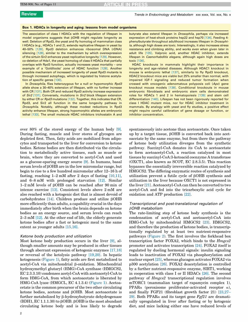
Figure 4. Histone deacetylase (HDAC) regulation of longevity pathways. HDACs deacetylate both histone and non-histone proteins, regulating gene transcription and the
post-translational function of proteins. HDACs regulate a variety of pathways implicated in longevity and age-related disease, and modulation of HDAC activity regulates
lifespan in model organisms.
Review Trends in Endocrinology and Metabolism xxx xxxx, Vol. xxx, No. x
TEM-908; No. of Pages 11
to its metabolic effects, particularly on HDACs and histoneacetylation. Interestingly, reduced HDAC activity, eitherby genetic or pharmacologic means, also has beneficialmetabolic and cytoprotective effects similar to those ofbOHB. Moreover, HDACs regulate a variety of pathwaysimplicated in longevity, including autophagy and IGFsignaling (Figure 4), and modulation of HDAC activityregulates lifespan in model organisms (Box 1).
HDACs have a key role in regulating metabolic disease,and loss or inhibition of class I HDAC function appears tophenocopy some of the benefits of a ketogenic diet. HDAC3regulates the expression of gluconeogenic genes [115], andHDAC3 knockout mice have lower fasting glucose andinsulin [116–118]. In fact, chronic treatment with butyratekeeps mice essentially metabolically normal on a high-fatdiet, with lower glucose and insulin, better glucose toler-ance, prevention of weight gain, and improved respiratoryefficiency; butyrate also provides some of these benefitseven to mice already obese from being fed a high-fat diet[119]. One mechanism for this may be upregulation ofPGC1a in liver, brown adipose tissue, and muscle bybutyrate [119], as seen in ketogenic diets.
Also reminiscent of bOHB, HDAC inhibitors are cyto-protective in animal models of tissue injury. Butyrateimproves overall survival in a rat sepsis model [120], aswell as reducing lung injury after lipopolysaccharide infu-sion in mice [121].
There is a growing literature on the importance ofepigenetic regulation in learning and memory, and specifi-cally in mouse models of dementia. In the severely affectedCK-p25 mouse model of neurodegeneration (with inducibleaccumulation of p25, the cleaved isoform of cyclin depen-dent kinase 5 activator 1), environmental enrichment thatimproves learning and memory is associated with in-creased histone H3 and H4 acetylation in the cortex andhippocampus [122]. Treatment with the HDAC inhibitorbutyrate also improves learning and memory [122]. Age-
8
related impairments in learning and memory in wild typemice are associated with alterations in histone acetylation[123], and treatment with HDAC inhibitors improvesmemory performance in both young and aged mice[123,124]. HDAC2 appears to be the crucial mediator ofthese effects because as overexpression of HDAC2, but notof HDAC1, impairs learning and memory in wild type mice[124]. Conversely, HDAC2 knockout mice show improvedmemory formation, which is not further improved byHDAC inhibitors [124]. HDAC2 expression is increasedin the brains of two mouse dementia models as well as inthe brains of humans with Alzheimer’s disease, and knock-down of HDAC2 improves memory in the CK-p25 dementiamouse model [125].
Although HDAC2 seems to have a memory-impairingrole, HDAC1 – another class I HDAC – has been reported tobe neuroprotective [126]. HDAC1 activity is important forthe repair of double-stranded DNA breaks in neurons, andits own deacetylase activity is enhanced by SIRT1 viadeacetylation [127]. This provides a fascinating exampleof how multiple metabolically responsive pathways (e.g.,SIRT1 and bOHB) might intersect to provide overlappingepigenetic regulation: although bOHB inhibition ofHDAC2 may be broadly beneficial, the potentially detri-mental inhibition of HDAC1 by bOHB is offset by SIRT1activation. Such crosstalk may be common, where fasting-activated sirtuins provide tissue- or subcellular-specificfine-tuning of the broad effects of bOHB. Alternatively,different target specificities of class I and class III HDACscould provide ample opportunity for coordinated regula-tion of targets, even perhaps via different lysines on thesame protein. It remains to be determined whether inhibi-tion of HDACs by bOHB has similar effects on learning andmemory in rodent models as chemical HDAC inhibition orgenetic manipulation, and precisely how HDAC inhibitionby bOHB intersects with other fasting-related mechanismsof epigenetic regulation.

Box 2. Outstanding questions
� What are the molecular targets of HDAC inhibition by bOHB in
specific tissues and metabolic states?
� Does bOHB regulate HDAC-targeted pathways such as autop-
hagy?
� Do pathways downstream of bOHB actions increase longevity in
mammals?
� Are mammalian sirtuins downstream targets of bOHB and
HDACs?
� If fasting or ketogenic conditions promote inhibition of class I
HDACs (via bOHB) but activation of class III HDACs (sirtuins), how
are these potentially opposing activities coordinated?
� Is there an acetylation code that may trigger the metabolic
reprogramming of dietary restriction?
Review Trends in Endocrinology and Metabolism xxx xxxx, Vol. xxx, No. x
TEM-908; No. of Pages 11
Concluding remarks and future perspectivesKetone bodies are emerging as crucial regulators of meta-bolic health and longevity via their ability to regulateHDAC activity and thereby epigenetic gene regulation.Ketogenic diets provide a partial phenocopy of CR throughtheir effects on insulin, IGF, FOXO3, fatty acid metabo-lism, AMPK, and mTOR. The finding that bOHB is aninhibitor of HDACs, together with the coincidence of bio-logical effects of ketone bodies and HDAC inhibition, sug-gests the fascinating possibility that bOHB could be anendogenous avenue to attain some of the benefits of life-span extension seen with HDAC inhibition in model organ-isms. However, several outstanding questions remain(Box 2). It will be of great interest to define the moleculartargets of HDAC inhibition by bOHB in specific tissues andmetabolic states, investigate whether bOHB regulatesHDAC-targeted pathways such as autophagy, and deter-mine if these effects culminate in enhancement of longevityby bOHB in mammals.
AcknowledgmentsWe thank John Carroll for artistic assistance. E.V. is supported by fundsfrom the National Institutes of Health (NIH)/National Institute ofDiabetes and Digestive and Kidney Diseases (NIDDK) and the GladstoneInstitutes. J.C.N. is supported by funds from the Larry L. HillblomFoundation, the John A. Hartford Foundation, the Glenn Foundation forMedical Research, and an NIH/National Institute on Aging (NIA) T32training grant.
References1 Fontana, L. (2006) Excessive adiposity, calorie restriction, and aging.
JAMA 295, 1577–15782 Kenyon, C.J. (2010) The genetics of ageing. Nature 464, 504–5123 Bjedov, I. and Partridge, L. (2011) A longer and healthier life with
TOR down-regulation: genetics and drugs. Biochem. Soc. Trans. 39,460–465
4 Someya, S. et al. (2010) Sirt3 mediates reduction of oxidative damageand prevention of age-related hearing loss under caloric restriction.Cell 143, 802–812
5 Shimazu, T. et al. (2013) Suppression of oxidative stress by beta-hydroxybutyrate, an endogenous histone deacetylase inhibitor.Science 339, 211–214
6 Katada, S. et al. (2012) Connecting threads: epigenetics andmetabolism. Cell 148, 24–28
7 Stein, L.R. and Imai, S. (2012) The dynamic regulation of NADmetabolism in mitochondria. Trends Endocrinol. Metab. 23,420–428
8 Shyh-Chang, N. et al. (2013) Influence of threonine metabolism on S-adenosylmethionine and histone methylation. Science 339, 222–226
9 Berg, J.M. et al. (2012) Biochemistry, W.H. Freeman10 Cahill, G.F., Jr et al. (1966) Hormone–fuel interrelationships during
fasting. J. Clin. Invest. 45, 1751–1769
11 Robinson, A.M. and Williamson, D.H. (1980) Physiological roles ofketone bodies as substrates and signals in mammalian tissues.Physiol. Rev. 60, 143–187
12 Cahill, G.F., Jr (2006) Fuel metabolism in starvation. Annu. Rev.Nutr. 26, 1–22
13 Koeslag, J.H. et al. (1980) Post-exercise ketosis. J. Physiol. 301, 79–9014 Kim do, Y. and Rho, J.M. (2008) The ketogenic diet and epilepsy. Curr.
Opin. Clin. Nutr. Metab. Care 11, 113–12015 London, E.D. et al. (1986) Effects of fasting on ketone body
concentrations in healthy men of different ages. J. Gerontol. 41,599–604
16 Freemantle, E. et al. (2009) Metabolic response to a ketogenicbreakfast in the healthy elderly. J. Nutr. Health Aging 13, 293–298
17 Thumelin, S. et al. (1993) Developmental changes in mitochondrial 3-hydroxy-3-methylglutaryl-CoA synthase gene expression in rat liver,intestine and kidney. Biochem. J. 292 (Pt 2), 493–496
18 Zhang, D. et al. (2011) Proteomics analysis reveals diabetic kidney asa ketogenic organ in type 2 diabetes. Am. J. Physiol. Endocrinol.Metab. 300, E287–E295
19 Fink, G. et al. (1988) Pseudoketogenesis in the perfused rat heart. J.Biol. Chem. 263, 18036–18042
20 Weidemann, M.J. and Krebs, H.A. (1969) The fuel of respiration of ratkidney cortex. Biochem. J. 112, 149–166
21 Fukao, T. et al. (1997) Enzymes of ketone body utilization in humantissues: protein and messenger RNA levels of succinyl-coenzyme A(CoA):3-ketoacid CoA transferase and mitochondrial and cytosolicacetoacetyl-CoA thiolases. Pediatr. Res. 42, 498–502
22 Fukao, T. et al. (2004) Pathways and control of ketone bodymetabolism: on the fringe of lipid biochemistry. ProstaglandinsLeukot. Essent. Fatty Acids 70, 243–251
23 Hegardt, F.G. (1999) Mitochondrial 3-hydroxy-3-methylglutaryl-CoA synthase: a control enzyme in ketogenesis. Biochem. J. 338,569–582
24 Wolfrum, C. et al. (2004) Foxa2 regulates lipid metabolism andketogenesis in the liver during fasting and in diabetes. Nature 432,1027–1032
25 Wolfrum, C. et al. (2003) Insulin regulates the activity offorkhead transcription factor Hnf-3beta/Foxa-2 by Akt-mediatedphosphorylation and nuclear/cytosolic localization. Proc. Natl.Acad. Sci. U.S.A. 100, 11624–11629
26 von Meyenn, F. et al. (2013) Glucagon-induced acetylation of Foxa2regulates hepatic lipid metabolism. Cell Metab. 17, 436–447
27 Badman, M.K. et al. (2007) Hepatic fibroblast growth factor 21 isregulated by PPARalpha and is a key mediator of hepatic lipidmetabolism in ketotic states. Cell Metab. 5, 426–437
28 Badman, M.K. et al. (2009) Fibroblast growth factor 21-deficient micedemonstrate impaired adaptation to ketosis. Endocrinology 150,4931–4940
29 Sengupta, S. et al. (2010) mTORC1 controls fasting-inducedketogenesis and its modulation by ageing. Nature 468, 1100–1104
30 Shimazu, T. et al. (2010) SIRT3 deacetylates mitochondrial 3-hydroxy-3-methylglutaryl CoA synthase 2 and regulates ketonebody production. Cell Metab. 12, 654–661
31 Hebert, A.S. et al. (2013) Calorie restriction and SIRT3 trigger globalreprogramming of the mitochondrial protein acetylome. Mol. Cell 49,186–199
32 Rardin, M.J. et al. (2013) Label-free quantitative proteomics of thelysine acetylome in mitochondria identifies substrates of SIRT3 inmetabolic pathways. Proc. Natl. Acad. Sci. U.S.A. 110, 6601–6606
33 Quant, P.A. et al. (1990) Glucagon activates mitochondrial 3-hydroxy-3-methylglutaryl-CoA synthase in vivo by decreasing the extent ofsuccinylation of the enzyme. Eur. J. Biochem. 187, 169–174
34 Quant, P.A. et al. (1989) Treatment of rats with glucagon ormannoheptulose increases mitochondrial 3-hydroxy-3-methylglutaryl-CoA synthase activity and decreases succinyl-CoA content in liver.Biochem. J. 262, 159–164
35 He, W. et al. (2012) Mitochondrial sirtuins: regulators of proteinacylation and metabolism. Trends Endocrinol. Metab. 23, 467–476
36 Turko, I.V. et al. (2001) Diabetes-associated nitration of tyrosine andinactivation of succinyl-CoA:3-oxoacid CoA-transferase. Am. J.Physiol. Heart Circ. Physiol. 281, H2289–H2294
37 Pellerin, L. et al. (2005) Cellular and subcellular distribution ofmonocarboxylate transporters in cultured brain cells and in theadult brain. J. Neurosci. Res. 79, 55–64
9

Review Trends in Endocrinology and Metabolism xxx xxxx, Vol. xxx, No. x
TEM-908; No. of Pages 11
38 Balietti, M. et al. (2010) Ketogenic diets: an historical antiepileptictherapy with promising potentialities for the aging brain. Ageing Res.Rev. 9, 273–279
39 Hugo, S.E. et al. (2012) A monocarboxylate transporter required forhepatocyte secretion of ketone bodies during fasting. Genes Dev. 26,282–293
40 Hankermeyer, C.R. and Tjeerdema, R.S. (1999) Polyhydroxybutyrate:plastic made and degraded by microorganisms. Rev. Environ.Contam. Toxicol. 159, 1–24
41 NCBI Resource Coordinators (2013) Database resources of theNational Center for Biotechnology Information. Nucleic Acids Res.41, D8–D20
42 Tunaru, S. et al. (2003) PUMA-G and HM74 are receptors for nicotinicacid and mediate its anti-lipolytic effect. Nat. Med. 9, 352–355
43 Taggart, A.K. et al. (2005) (D)-beta-Hydroxybutyrate inhibitsadipocyte lipolysis via the nicotinic acid receptor PUMA-G. J. Biol.Chem. 280, 26649–26652
44 Offermanns, S. et al. (2011) International Union of Basic and ClinicalPharmacology. LXXXII: nomenclature and classification of hydroxy-carboxylic acid receptors (GPR81, GPR109A, and GPR109B).Pharmacol. Rev. 63, 269–290
45 Kimura, I. et al. (2011) Short-chain fatty acids and ketones directlyregulate sympathetic nervous system via G protein-coupled receptor41 (GPR41). Proc. Natl. Acad. Sci. U.S.A. 108, 8030–8035
46 Blad, C.C. et al. (2012) G protein-coupled receptors for energymetabolites as new therapeutic targets. Nat. Rev. Drug Discov. 11,603–619
47 Layden, B.T. et al. (2013) Short chain fatty acids and their receptors:new metabolic targets. Transl. Res. 161, 131–140
48 Gregoretti, I.V. et al. (2004) Molecular evolution of the histonedeacetylase family: functional implications of phylogenetic analysis.J. Mol. Biol. 338, 17–31
49 Yang, X.J. and Seto, E. (2008) The Rpd3/Hda1 family of lysinedeacetylases: from bacteria and yeast to mice and men. Nat. Rev.Mol. Cell Biol. 9, 206–218
50 Mihaylova, M.M. and Shaw, R.J. (2013) Metabolic reprogramming byclass I and II histone deacetylases. Trends Endocrinol. Metab. 24, 48–57
51 New, M. et al. (2012) HDAC inhibitor-based therapies: can weinterpret the code? Mol. Oncol. 6, 637–656
52 Glozak, M.A. et al. (2005) Acetylation and deacetylation of non-histoneproteins. Gene 363, 15–23
53 Cousens, L.S. et al. (1979) Different accessibilities in chromatin tohistone acetylase. J. Biol. Chem. 254, 1716–1723
54 Bolden, J.E. et al. (2006) Anticancer activities of histone deacetylaseinhibitors. Nat. Rev. Drug Discov. 5, 769–784
55 Vannini, A. et al. (2004) Crystal structure of a eukaryotic zinc-dependenthistone deacetylase, human HDAC8, complexed with a hydroxamic acidinhibitor. Proc. Natl. Acad. Sci. U.S.A. 101, 15064–15069
56 Somoza, J.R. et al. (2004) Structural snapshots of human HDAC8provide insights into the class I histone deacetylases. Structure 12,1325–1334
57 Wang, D.F. et al. (2005) Toward selective histone deacetylaseinhibitor design: homology modeling, docking studies, andmolecular dynamics simulations of human class I histonedeacetylases. J. Med. Chem. 48, 6936–6947
58 Sekhavat, A. et al. (2007) Competitive inhibition of histonedeacetylase activity by trichostatin A and butyrate. Biochem. CellBiol. 85, 751–758
59 Madiraju, P. et al. (2009) Mitochondrial acetylcarnitine providesacetyl groups for nuclear histone acetylation. Epigenetics 4, 399–403
60 Wellen, K.E. et al. (2009) ATP-citrate lyase links cellular metabolismto histone acetylation. Science 324, 1076–1080
61 Muoio, D.M. et al. (2012) Muscle-specific deletion of carnitineacetyltransferase compromises glucose tolerance and metabolicflexibility. Cell Metab. 15, 764–777
62 Anson, R.M. et al. (2005) The diet restriction paradigm: a brief reviewof the effects of every-other-day feeding. Age 27, 17–25
63 Hatori, M. et al. (2012) Time-restricted feeding without reducingcaloric intake prevents metabolic diseases in mice fed a high-fatdiet. Cell Metab. 15, 848–860
64 Bainbridge, H.W. (1925) The reduced sensitivity to insulin of ratsand mice fed on a carbohydrate-free, excess-fat diet. J. Physiol. 60,293–300
10
65 Burcelin, R. et al. (2002) Heterogeneous metabolic adaptation ofC57BL/6J mice to high-fat diet. Am. J. Physiol. Endocrinol. Metab.282, E834–E842
66 Kinzig, K.P. et al. (2010) Insulin sensitivity and glucose toleranceare altered by maintenance on a ketogenic diet. Endocrinology 151,3105–3114
67 Borghjid, S. and Feinman, R.D. (2012) Response of C57Bl/6 mice to acarbohydrate-free diet. Nutr. Metab. (Lond.) 9, 69
68 Garbow, J.R. et al. (2011) Hepatic steatosis, inflammation, and ERstress in mice maintained long term on a very low-carbohydrateketogenic diet. Am. J. Physiol. Gastrointest. Liver Physiol. 300,G956–G967
69 Freedland, S.J. et al. (2008) Carbohydrate restriction, prostatecancer growth, and the insulin-like growth factor axis. Prostate68, 11–19
70 Mavropoulos, J.C. et al. (2009) The effects of varying dietarycarbohydrate and fat content on survival in a murine LNCaPprostate cancer xenograft model. Cancer Prev. Res. (Phila.) 2, 557–565
71 McDaniel, S.S. et al. (2011) The ketogenic diet inhibits themammalian target of rapamycin (mTOR) pathway. Epilepsia 52,e7–e11
72 Kennedy, A.R. et al. (2007) A high-fat, ketogenic diet induces a uniquemetabolic state in mice. Am. J. Physiol. Endocrinol. Metab. 292,E1724–E1739
73 Badman, M.K. et al. (2009) A very low carbohydrate ketogenic dietimproves glucose tolerance in ob/ob mice independently of weight loss.Am. J. Physiol. Endocrinol. Metab. 297, E1197–E1204
74 Jornayvaz, F.R. et al. (2010) A high-fat, ketogenic diet causes hepaticinsulin resistance in mice, despite increasing energy expenditure andpreventing weight gain. Am. J. Physiol. Endocrinol. Metab. 299,E808–E815
75 Srivastava, S. et al. (2013) A ketogenic diet increases brown adiposetissue mitochondrial proteins and UCP1 levels in mice. IUBMB Life65, 58–66
76 Laeger, T. et al. (2010) Role of beta-hydroxybutyric acid in the centralregulation of energy balance. Appetite 54, 450–455
77 Schugar, R.C. and Crawford, P.A. (2012) Low-carbohydrate ketogenicdiets, glucose homeostasis, and nonalcoholic fatty liver disease. Curr.Opin. Clin. Nutr. Metab. Care 15, 374–380
78 Orentreich, N. et al. (1993) Low methionine ingestion by rats extendslife span. J. Nutr. 123, 269–274
79 Pamplona, R. and Barja, G. (2006) Mitochondrial oxidative stress,aging and caloric restriction: the protein and methionine connection.Biochim. Biophys. Acta 1757, 496–508
80 Ables, G.P. et al. (2012) Methionine-restricted C57BL/6J mice areresistant to diet-induced obesity and insulin resistance but have lowbone density. PLoS ONE 7, e51357
81 Hirschey, M.D. et al. (2011) SIRT3 deficiency and mitochondrialprotein hyperacetylation accelerate the development of themetabolic syndrome. Mol. Cell 44, 177–190
82 Ahola-Erkkila, S. et al. (2010) Ketogenic diet slows downmitochondrial myopathy progression in mice. Hum. Mol. Genet. 19,1974–1984
83 Houtkooper, R.H. et al. (2012) Sirtuins as regulators of metabolismand healthspan. Nat. Rev. Mol. Cell Biol. 13, 225–238
84 Poplawski, M.M. et al. (2011) Reversal of diabetic nephropathy by aketogenic diet. PLoS ONE 6, e18604
85 Harvie, M. et al. (2013) The effect of intermittent energy andcarbohydrate restriction v. daily energy restriction on weight lossand metabolic disease risk markers in overweight women. Br. J. Nutr.1–14
86 Westman, E.C. et al. (2008) The effect of a low-carbohydrate, ketogenicdiet versus a low-glycemic index diet on glycemic control in type 2diabetes mellitus. Nutr. Metab. (Lond.) 5, 36
87 Nordmann, A.J. et al. (2006) Effects of low-carbohydrate vs low-fatdiets on weight loss and cardiovascular risk factors: a meta-analysis ofrandomized controlled trials. Arch. Intern. Med. 166, 285–293
88 Hartman, A.L. and Vining, E.P. (2007) Clinical aspects of theketogenic diet. Epilepsia 48, 31–42
89 Kashiwaya, Y. et al. (2013) A ketone ester diet exhibits anxiolytic andcognition-sparing properties, and lessens amyloid and tau pathologiesin a mouse model of Alzheimer’s disease. Neurobiol. Aging 34,1530–1539
Review Trends in Endocrinology and Metabolism xxx xxxx, Vol. xxx, No. x
TEM-908; No. of Pages 11
90 Henderson, S.T. et al. (2009) Study of the ketogenic agent AC-1202 inmild to moderate Alzheimer’s disease: a randomized, double-blind,placebo-controlled, multicenter trial. Nutr. Metab. (Lond.) 6, 31
91 Reger, M.A. et al. (2004) Effects of beta-hydroxybutyrate on cognitionin memory-impaired adults. Neurobiol. Aging 25, 311–314
92 Tieu, K. et al. (2003) D-beta-hydroxybutyrate rescues mitochondrialrespiration and mitigates features of Parkinson disease. J. Clin.Invest. 112, 892–901
93 Vanitallie, T.B. et al. (2005) Treatment of Parkinson disease with diet-induced hyperketonemia: a feasibility study. Neurology 64, 728–730
94 Zhao, Z. et al. (2006) A ketogenic diet as a potential novel therapeuticintervention in amyotrophic lateral sclerosis. BMC Neurosci. 7, 29
95 Noh, H.S. et al. (2008) Neuroprotective effects of the ketogenic diet.Epilepsia 49 (Suppl. 8), 120–123
96 Lim, S. et al. (2011) D-beta-hydroxybutyrate is protective in mousemodels of Huntington’s disease. PLoS ONE 6, e24620
97 Sadri-Vakili, G. and Cha, J.H. (2006) Mechanisms of disease: histonemodifications in Huntington’s disease. Nat. Clin. Pract. Neurol. 2,330–338
98 Kashiwaya, Y. et al. (2000) D-beta-hydroxybutyrate protects neuronsin models of Alzheimer’s and Parkinson’s disease. Proc. Natl. Acad.Sci. U.S.A. 97, 5440–5444
99 Hu, Z.G. et al. (2009) Ketogenic diet reduces cytochrome c release andcellular apoptosis following traumatic brain injury in juvenile rats.Ann. Clin. Lab. Sci. 39, 76–83
100 Appelberg, K.S. et al. (2009) The effects of a ketogenic diet onbehavioral outcome after controlled cortical impact injury in thejuvenile and adult rat. J. Neurotrauma 26, 497–506
101 Puchowicz, M.A. et al. (2008) Neuroprotection in diet-induced ketotic ratbrain after focal ischemia. J. Cereb. Blood Flow Metab. 28, 1907–1916
102 Tai, K.K. et al. (2008) Ketogenic diet prevents cardiac arrest-induced cerebral ischemic neurodegeneration. J. Neural Transm.115, 1011–1017
103 Masuda, R. et al. (2005) D-beta-hydroxybutyrate is neuroprotectiveagainst hypoxia in serum-free hippocampal primary cultures. J.Neurosci. Res. 80, 501–509
104 Samoilova, M. et al. (2010) Chronic in vitro ketosis is neuroprotectivebut not anti-convulsant. J. Neurochem. 113, 826–835
105 Juge, N. et al. (2010) Metabolic control of vesicular glutamatetransport and release. Neuron 68, 99–112
106 Miller, R.A. (2009) Cell stress and new emphasis on multiplexresistance mechanisms. J. Gerontol. Ser. A: Biol. Sci. Med. Sci. 64,179–182
107 Shore, D.E. et al. (2012) Induction of cytoprotective pathways iscentral to the extension of lifespan conferred by multiple longevitypathways. PLoS Genet. 8, e1002792
108 Alam, H.B. et al. (2001) Resuscitation-induced pulmonary apoptosisand intracellular adhesion molecule-1 expression in rats areattenuated by the use of Ketone Ringer’s solution. J. Am. Coll.Surg. 193, 255–263
109 Koustova, E. et al. (2003) Ketone and pyruvate Ringer’s solutionsdecrease pulmonary apoptosis in a rat model of severe hemorrhagicshock and resuscitation. Surgery 134, 267–274
110 Ayuste, E.C. et al. (2006) Hepatic and pulmonary apoptosis afterhemorrhagic shock in swine can be reduced through modifications ofconventional Ringer’s solution. J. Trauma 60, 52–63
111 Jaskille, A. et al. (2006) Hepatic apoptosis after hemorrhagic shock inrats can be reduced through modifications of conventional Ringer’ssolution. J. Am. Coll. Surg. 202, 25–35
112 Klein, A.H. et al. (2010) Small-volume D-beta-hydroxybutyratesolution infusion increases survivability of lethal hemorrhagicshock in rats. Shock 34, 565–572
113 Mulier, K.E. et al. (2012) Treatment with beta-hydroxybutyrate andmelatonin is associated with improved survival in a porcine model ofhemorrhagic shock. Resuscitation 83, 253–258
114 Zou, Z. et al. (2002) DL-3-Hydroxybutyrate administration preventsmyocardial damage after coronary occlusion in rat hearts. Am. J.Physiol. Heart Circ. Physiol. 283, H1968–H1974
115 Mihaylova, M.M. et al. (2011) Class IIa histone deacetylases arehormone-activated regulators of FOXO and mammalian glucosehomeostasis. Cell 145, 607–621
116 Knutson, S.K. et al. (2008) Liver-specific deletion of histonedeacetylase 3 disrupts metabolic transcriptional networks. EMBOJ. 27, 1017–1028
117 Fajas, L. et al. (2002) The retinoblastoma-histone deacetylase 3complex inhibits PPARgamma and adipocyte differentiation. DevCell 3, 903–910
118 Bhaskara, S. et al. (2010) Hdac3 is essential for the maintenance ofchromatin structure and genome stability. Cancer Cell 18, 436–447
119 Gao, Z. et al. (2009) Butyrate improves insulin sensitivity andincreases energy expenditure in mice. Diabetes 58, 1509–1517
120 Zhang, L.T. et al. (2007) Sodium butyrate prevents lethality of severesepsis in rats. Shock 27, 672–677
121 Ni, Y.F. et al. (2010) Histone deacetylase inhibitor, butyrate,attenuates lipopolysaccharide-induced acute lung injury in mice.Respir. Res. 11, 33
122 Fischer, A. et al. (2007) Recovery of learning and memory is associatedwith chromatin remodelling. Nature 447, 178–182
123 Peleg, S. et al. (2010) Altered histone acetylation is associated withage-dependent memory impairment in mice. Science 328, 753–756
124 Guan, J.S. et al. (2009) HDAC2 negatively regulates memoryformation and synaptic plasticity. Nature 459, 55–60
125 Graff, J. et al. (2012) An epigenetic blockade of cognitive functions inthe neurodegenerating brain. Nature 483, 222–226
126 Kim, D. et al. (2008) Deregulation of HDAC1 by p25/Cdk5 inneurotoxicity. Neuron 60, 803–817
127 Dobbin, M.M. et al. (2013) SIRT1 collaborates with ATM andHDAC1 to maintain genomic stability in neurons. Nat. Neurosci.16, 1008–1015
128 Kim, S. et al. (1999) Modulation of life-span by histone deacetylasegenes in Saccharomyces cerevisiae. Mol. Biol. Cell 10, 3125–3136
129 Kaeberlein, M. (2010) Lessons on longevity from budding yeast.Nature 464, 513–519
130 Yi, C. et al. (2012) Function and molecular mechanism of acetylationin autophagy regulation. Science 336, 474–477
131 Rogina, B. et al. (2002) Longevity regulation by Drosophila Rpd3deacetylase and caloric restriction. Science 298, 1745
132 Rogina, B. and Helfand, S.L. (2004) Sir2 mediates longevity in the flythrough a pathway related to calorie restriction. Proc. Natl. Acad. Sci.U.S.A. 101, 15998–16003
133 Mannervik, M. and Levine, M. (1999) The Rpd3 histone deacetylase isrequired for segmentation of the Drosophila embryo. Proc. Natl. Acad.Sci. U.S.A. 96, 6797–6801
134 Zhao, Y. et al. (2005) Lifespan extension and elevated hsp geneexpression in Drosophila caused by histone deacetylase inhibitors.J. Exp. Biol. 208, 697–705
135 Kang, H.L. et al. (2002) Life extension in Drosophila by feeding a drug.Proc. Natl. Acad. Sci. U.S.A. 99, 838–843
136 Evason, K. et al. (2008) Valproic acid extends Caenorhabditis eleganslifespan. Aging Cell 7, 305–317
137 Lagger, G. et al. (2002) Essential function of histone deacetylase 1 inproliferation control and CDK inhibitor repression. EMBO J. 21,2672–2681
138 Zimmermann, S. et al. (2007) Reduced body size and decreasedintestinal tumor rates in HDAC2-mutant mice. Cancer Res. 67,9047–9054
139 Wilting, R.H. et al. (2010) Overlapping functions of Hdac1 and Hdac2in cell cycle regulation and haematopoiesis. EMBO J. 29, 2586–2597
140 Dovey, O.M. et al. (2010) Histone deacetylase 1 (HDAC1), but notHDAC2, controls embryonic stem cell differentiation. Proc. Natl.Acad. Sci. U.S.A. 107, 8242–8247
141 Kossoff, E.H. and Hartman, A.L. (2012) Ketogenic diets: newadvances for metabolism-based therapies. Curr. Opin. Neurol. 25,173–178
142 Schwer, B. et al. (2009) Calorie restriction alters mitochondrialprotein acetylation. Aging Cell 8, 604–606
11