Dietary Phytoestrogens and Their Metabolites as Epigenetic ...
48
antioxidants Review Dietary Phytoestrogens and Their Metabolites as Epigenetic Modulators with Impact on Human Health Victor Stefan Ionescu 1 , Alexandra Popa 1 , Andrei Alexandru 2 , Emilia Manole 1 , Mihaela Neagu 3 and Sevinci Pop 1, * Citation: Ionescu, V.S.; Popa, A.; Alexandru, A.; Manole, E.; Neagu, M.; Pop, S. Dietary Phytoestrogens and Their Metabolites as Epigenetic Modulators with Impact on Human Health. Antioxidants 2021, 10, 1893. https://doi.org/10.3390/antiox10121893 Academic Editor: Stanley Omaye Received: 25 October 2021 Accepted: 25 November 2021 Published: 26 November 2021 Publisher’s Note: MDPI stays neutral with regard to jurisdictional claims in published maps and institutional affil- iations. Copyright: © 2021 by the authors. Licensee MDPI, Basel, Switzerland. This article is an open access article distributed under the terms and conditions of the Creative Commons Attribution (CC BY) license (https:// creativecommons.org/licenses/by/ 4.0/). 1 Cell Biology Laboratory, “Victor Babes” National Institute of Pathology, 050096 Bucharest, Romania; [email protected] (V.S.I.); [email protected] (A.P.); [email protected] (E.M.) 2 Faculty of Medicine, “Carol Davila” University of Medicine and Pharmacy, 050047 Bucharest, Romania; [email protected] 3 SC Hofigal Export-Import SA, 042124 Bucharest, Romania; mihaela.neagu@hofigal.eu * Correspondence: [email protected] Abstract: The impact of dietary phytoestrogens on human health has been a topic of continuous debate since their discovery. Nowadays, based on their presumptive beneficial effects, the amount of phytoestrogens consumed in the daily diet has increased considerably worldwide. Thus, there is a growing need for scientific data regarding their mode of action in the human body. Recently, new insights of phytoestrogens’ bioavailability and metabolism have demonstrated an inter-and intra-population heterogeneity of final metabolites’ production. In addition, the phytoestrogens may have the ability to modulate epigenetic mechanisms that control gene expression. This review highlights the complexity and particularity of the metabolism of each class of phytoestrogens, pointing out the diversity of their bioactive gut metabolites. Futhermore, it presents emerging scientific data which suggest that, among well-known genistein and resveratrol, other phytoestrogens and their gut metabolites can act as epigenetic modulators with a possible impact on human health. The interconnection of dietary phytoestrogens’ consumption with gut microbiota composition, epigenome and related preventive mechanisms is discussed. The current challenges and future perspectives in designing relevant research directions to explore the potential health benefits of dietary phytoestrogens are also explored. Keywords: phytoestrogens; epigenome; gut metabolites; microbiota; S-equol; icariin; arctigenin; enterolignans; resveratrol; dietary phytochemicals 1. Introduction In the past decades, the research regarding the beneficial or adverse effects of phytoe- strogens present in the human diet has intensified, due to their estrogenic or anti-estrogenic potential in humans and animals [1–3]. Phytoestrogens are found in a wide variety of foods, including soy-based products, fruits, vegetables and dairy products [4–6]. A huge list of health benefits, including a reduced risk of osteoporosis, hormone-dependent cancers, cardiovascular diseases and brain disorders, as well as decreased menopause symptoms, is often ascribed to phytoestrogens. However, they can act as endocrine disruptors, being able to induce adverse health effects, as well [7,8]. Based on their presumptive beneficial effects on human health, the amount of phytoestrogens consumed in the daily diet has increased considerably worldwide [9,10]. Nowadays, a plethora of dietary supplements based on phytoestrogens has overflowed the market, and their consumption has reached high levels, especially within the female population. Thus, there is a growing need for relevant scientific data regarding their impact on human health. Indeed, new insights of phytoestrogens’ bioavailability and metabolism have been unveiled, and a more complex image of biological activities of absorbed phytoestrogens and their metabolites has emerged [11–13]. The phytoestrogens can be active in the human Antioxidants 2021, 10, 1893. https://doi.org/10.3390/antiox10121893 https://www.mdpi.com/journal/antioxidants
-
Upload
khangminh22 -
Category
Documents
-
view
2 -
download
0
Transcript of Dietary Phytoestrogens and Their Metabolites as Epigenetic ...

antioxidants
Review
Dietary Phytoestrogens and Their Metabolites as EpigeneticModulators with Impact on Human Health
Victor Stefan Ionescu 1, Alexandra Popa 1, Andrei Alexandru 2, Emilia Manole 1, Mihaela Neagu 3
and Sevinci Pop 1,*
�����������������
Citation: Ionescu, V.S.; Popa, A.;
Alexandru, A.; Manole, E.; Neagu, M.;
Pop, S. Dietary Phytoestrogens and
Their Metabolites as Epigenetic
Modulators with Impact on Human
Health. Antioxidants 2021, 10, 1893.
https://doi.org/10.3390/antiox10121893
Academic Editor: Stanley Omaye
Received: 25 October 2021
Accepted: 25 November 2021
Published: 26 November 2021
Publisher’s Note: MDPI stays neutral
with regard to jurisdictional claims in
published maps and institutional affil-
iations.
Copyright: © 2021 by the authors.
Licensee MDPI, Basel, Switzerland.
This article is an open access article
distributed under the terms and
conditions of the Creative Commons
Attribution (CC BY) license (https://
creativecommons.org/licenses/by/
4.0/).
1 Cell Biology Laboratory, “Victor Babes” National Institute of Pathology, 050096 Bucharest, Romania;[email protected] (V.S.I.); [email protected] (A.P.); [email protected] (E.M.)
2 Faculty of Medicine, “Carol Davila” University of Medicine and Pharmacy, 050047 Bucharest, Romania;[email protected]
3 SC Hofigal Export-Import SA, 042124 Bucharest, Romania; [email protected]* Correspondence: [email protected]
Abstract: The impact of dietary phytoestrogens on human health has been a topic of continuousdebate since their discovery. Nowadays, based on their presumptive beneficial effects, the amountof phytoestrogens consumed in the daily diet has increased considerably worldwide. Thus, thereis a growing need for scientific data regarding their mode of action in the human body. Recently,new insights of phytoestrogens’ bioavailability and metabolism have demonstrated an inter-andintra-population heterogeneity of final metabolites’ production. In addition, the phytoestrogensmay have the ability to modulate epigenetic mechanisms that control gene expression. This reviewhighlights the complexity and particularity of the metabolism of each class of phytoestrogens,pointing out the diversity of their bioactive gut metabolites. Futhermore, it presents emergingscientific data which suggest that, among well-known genistein and resveratrol, other phytoestrogensand their gut metabolites can act as epigenetic modulators with a possible impact on human health.The interconnection of dietary phytoestrogens’ consumption with gut microbiota composition,epigenome and related preventive mechanisms is discussed. The current challenges and futureperspectives in designing relevant research directions to explore the potential health benefits ofdietary phytoestrogens are also explored.
Keywords: phytoestrogens; epigenome; gut metabolites; microbiota; S-equol; icariin; arctigenin;enterolignans; resveratrol; dietary phytochemicals
1. Introduction
In the past decades, the research regarding the beneficial or adverse effects of phytoe-strogens present in the human diet has intensified, due to their estrogenic or anti-estrogenicpotential in humans and animals [1–3]. Phytoestrogens are found in a wide variety of foods,including soy-based products, fruits, vegetables and dairy products [4–6]. A huge listof health benefits, including a reduced risk of osteoporosis, hormone-dependent cancers,cardiovascular diseases and brain disorders, as well as decreased menopause symptoms, isoften ascribed to phytoestrogens. However, they can act as endocrine disruptors, beingable to induce adverse health effects, as well [7,8]. Based on their presumptive beneficialeffects on human health, the amount of phytoestrogens consumed in the daily diet hasincreased considerably worldwide [9,10]. Nowadays, a plethora of dietary supplementsbased on phytoestrogens has overflowed the market, and their consumption has reachedhigh levels, especially within the female population. Thus, there is a growing need forrelevant scientific data regarding their impact on human health.
Indeed, new insights of phytoestrogens’ bioavailability and metabolism have beenunveiled, and a more complex image of biological activities of absorbed phytoestrogensand their metabolites has emerged [11–13]. The phytoestrogens can be active in the human
Antioxidants 2021, 10, 1893. https://doi.org/10.3390/antiox10121893 https://www.mdpi.com/journal/antioxidants

Antioxidants 2021, 10, 1893 2 of 48
body as free molecules or as gastrointestinal tract (gut) metabolites, being able to interferewith the endogenous estrogen signaling and associated cellular processes [2].
In general, the key factors that are affecting the bioavailability of phytoestrogensare the age and gender of individuals, food matrices, dose frequency and the ADME(absorption, tissue distribution, metabolism and excretion process) properties. Each classof dietary phytoestrogens (e.g., isoflavones, coumestans, prenylflavonoids, lignans and stil-benes) has its own structural particularities, and studies regarding their bioavailability andmetabolism are still far from being completed. An important inter-and intra-population het-erogeneity of final metabolites production have been observed in human population [14,15].Their metabolism is mediated both by tissue enzymes and gut microbiota, either prior toabsorption or during enterohepatic circulation [16,17]. Only a small percentage (5–10%)of ingested phytoestrogens can reach the small intestine and are available for absorptioninto enterocytes and then enters into systematic circulation towards target tissues [18,19].A substantial part of them undergoes extensive metabolization across the liver, smalland large intestines, where they are transformed into metabolites with various chemicalstructures and bioactivities [12,20,21]. The beneficial effects of phytoestrogens on humanhealth are now considered to be partially influenced by their metabolites, such as S-equol,O-demethylangolensin (O-DMA), enterolignans and stilbenes derivatives. In some cases,these metabolites have greater biological activities and sometimes have different impactson targeted tissues than their precursors [12,21].
On the one hand, the composition of gut microbiota can influence the metabolism ofphytoestrogens; on the other hand, phytoestrogens and their metabolites can modulateand reshape the gut microbial composition [13]. Understanding their reciprocal influence,and by deciphering the molecular basis of phytoestrogens and microbiome interaction, itcan be the key to elucidate their influence on human health.
For many decades, epidemiologic studies have been trying to find associations be-tween dietary components and disease risks. Nevertheless, multiple factors, such asvariations in daily consumption patterns of individuals, results based on empirical anal-yses on predetermined sets of dietary components and human genetics and metabolismheterogeneity have led to inconsistent results. Despite strong preclinical evidence, theassociation of dietary phytoestrogens with human disease risks has not yet clearly demon-strated [1,7]. Recently, the European Food Safety Authority has indicated that isoflavonesintake of 35–150 mg/day is safe and that there is no concern of possible adverse effectsin peri-and post-menopausal women [22]. Moreover, new reports have concluded thatconsumption of phytoestrogens and its correlation with circulating metabolites is not reli-able because of high inter-individual variabilities of microbiota composition and geneticpolymorphism of phase-I and -II enzymes [12,23].
In recent years, emerging evidence has highlighted the role of dietary bioactive com-pounds in modifying the epigenome by directly or indirectly engaging in epigenetic mech-anisms controling gene expression [24]. Epigenetic mechanisms, such as DNA methylation,histone post-translational modifications, chromatin remodeling and noncoding RNAs ex-pression, represent the link between genotype and phenotype. Each epigenetic mechanismis reversible and is controlled by specific protein classes which attach, dislodge or maintainspecific chemical groups that can signal the initiation or inhibition of gene transcription [25].These proteins, along with chemical marks attached to DNA, RNA and histones, representthe epigenome, a complex regulatory network that modulates chromatin structure andgenome function [26]. When this regulatory circuit is discontinued, normal physiologicalfunctions are affected, leading to carcinogenesis or occurrence of other chronic diseases [27].The epigenome is a dynamic network that undergoes continuously modifications in spaceand time, stimulated by internal and external factors [26,28]. Environmental factors, in-cluding diet, can remodel the epigenome during lifespan from embryonic stage until agingin a beneficial or detrimental way. Many dietary components display several biologicalactivities, including antioxidant, anti-inflammatory and anticancer properties that mightplay a significant role in chronic disease prevention [24]. Importantly, strong scientific

Antioxidants 2021, 10, 1893 3 of 48
evidence implies that consumption of dietary phytochemicals can maintain the epigenomeat normal parameters that support a healthy phenotype, and also it can reverse abnormalgene expression [24,29]. For example, it has been demonstrated that dietary phytochemicalscan influence the DNA methylation patterns by altering the substrates and cofactors of5-Methylcytosine (5mC) reaction, or by inhibiting the enzymes of one-carbon metabolismand by blocking the proteins involved in DNA methylation/demethylation activity [29,30].
The present review discusses the new insights regarding dietary phytoestrogens’bioavailability and metabolism, pointing out the diversity of their gut metabolites andthe complexity of metabolic pathways, followed by each class of phytoestrogens (e.g.,isoflavones, coumestans, prenylflavonoids, lignans and stilbenes). In addition, it drawsattention to emerging scientific data that sustain the epigenetic modulator capacity of eachclass of dietary phytoestrogens and their metabolites and the generated impact on severalcellular and molecular processes connected with human health. The interconnection ofdietary phytoestrogens with gut microbiota, epigenome and related oxidative stress eventsis presented. In-depth studies of dietary phytoestrogens and their metabolites mechanismsof action at the molecular level can represent an effective approach to undestand how toreverse aberrant epigenetic modifications, reshape gut microbiota and suspend abnormalcellular functions, which consequently will prevent and/or attenuate chronic diseases.
2. Phytoestrogens—General Data
Phytoestrogens are synthesized in plants as secondary metabolites during stress-cultivation conditions and UV radiation, and as a response to pathogens attack. They haveantibacterial, antifungal, antiviral and antioxidant properties in plants [31]. The amountsof phytoestrogens produced by a plant increases significantly during extreme growingconditions [32], and by growing the plant in an organic environment [33]. In general, asingle plant often contains more than one class of phytoestrogens. For example, the soybean is rich in isoflavones, whereas the soy sprout is a potent source of coumestrol, the mainrepresentative of coumestans class [34]. In terms of chemical structure, phytoestrogensare nonsteroidal polyphenols having several common characteristics with mammalianhormone, estradiol. The structural prerequisite for phytoestrogen molecule to bind theestrogen receptor is the presence of phenolic rings and a pair of hydroxyl groups separatedwith a similar distance, as in the case of estradiol molecule [35]. The phytoestrogens havebeen categorized in flavonoids and non-flavonoids. Flavonoids consist of a fifteen-carbonskeleton organized in two benzene rings (A and B) linked by a heterocyclic pyran structure(C) as C6–C3–C6, as Figure 1 shows. The basic flavonoid skeleton can have numeroussubstituents, including hydroxyl groups usually present at the 4′, 5′ and 7 positions. Mostof plant flavonoids have sugar molecules attached to their aglycones, so they mainly existas glycosides [36]. Further subclassification of flavonoids in isoflavones, coumestans andprenylflavonoids is based on structural differences in the connection between the B and Crings, as well as the degrees of saturation, oxidation and hydroxylation of the C ring [36].Non-flavonoid phytoestrogen’s structure consists of phenolic acids in either C6–C1 (benzoicacid) or C6–C3 (cinnamic acid) conformations, and are represented mainly by lignans andstilbenes [36].

Antioxidants 2021, 10, 1893 4 of 48Antioxidants 2021, 10, x FOR PEER REVIEW 4 of 52
Figure 1. Chemical structure of representative dietary phytoestrogens.
2.1. Isoflavones Isoflavones are the main subgroup of plant flavonoids that is found in the Legumi-
nosae family, including soy (Glycine max L.), red clover (Trifolium pratense L.), alfalfa (Medi-cago sativa L.) and species of Genista [37]. They have diphenol structures and are produced in higher plant through the phenylpropanoid pathway. Two of most important isofla-vones have similar structures; thus, daidzein differs from genistein by lacking a hydroxyl group at C5 position [38]. The representative dietary phytoestrogens’ chemical structures are presented in Figure 1.
Isoflavones are found often in plants as glycosides (genistin, daidzin and glycitin) and in a lower amount as aglycones (genistein, daidzein, glycitein), or as 4′ methylated derivative aglycones (biochanin A and formononetin) [36,39]. The presence of hydroxyl groups and sugars increases their solubility in water, whilst methyl groups confer to them lipophilicity [40]. In the human diet, the main source of isoflavones are soy and soy-de-rived products, but small quantities of isoflavones are also found in chickpeas, beans, fruits, vegetables and nuts [6,41]. In Western countries, the cow’s milk and dairy products contain significant amounts of isoflavones [5]. Due to their pleiotropic activities, isofla-vones are considered as a natural alternative for the treatment of estrogen decrease-related conditions during menopause, cardiovascular diseases and other hormone disorders [42].
Figure 1. Chemical structure of representative dietary phytoestrogens.
2.1. Isoflavones
Isoflavones are the main subgroup of plant flavonoids that is found in the Leguminosaefamily, including soy (Glycine max L.), red clover (Trifolium pratense L.), alfalfa (Medicagosativa L.) and species of Genista [37]. They have diphenol structures and are produced inhigher plant through the phenylpropanoid pathway. Two of most important isoflavoneshave similar structures; thus, daidzein differs from genistein by lacking a hydroxyl groupat C5 position [38]. The representative dietary phytoestrogens’ chemical structures arepresented in Figure 1.
Isoflavones are found often in plants as glycosides (genistin, daidzin and glycitin)and in a lower amount as aglycones (genistein, daidzein, glycitein), or as 4′ methylatedderivative aglycones (biochanin A and formononetin) [36,39]. The presence of hydroxylgroups and sugars increases their solubility in water, whilst methyl groups confer to themlipophilicity [40]. In the human diet, the main source of isoflavones are soy and soy-derivedproducts, but small quantities of isoflavones are also found in chickpeas, beans, fruits,vegetables and nuts [6,41]. In Western countries, the cow’s milk and dairy products containsignificant amounts of isoflavones [5]. Due to their pleiotropic activities, isoflavones areconsidered as a natural alternative for the treatment of estrogen decrease-related conditionsduring menopause, cardiovascular diseases and other hormone disorders [42].

Antioxidants 2021, 10, 1893 5 of 48
2.2. Prenylflavonoids
Prenylflavonoids structure contains a flavonoid skeleton that has attached at position8 of A ring a lipophilic prenyl chain [43]. Prenyl chains appear in various forms, andthe most notable are 3,3-dimethylallyl substituent, geranyl, 1,1-dimethylallyl and theirmoieties. The presence of hydrophobic prenyl radical increases prenylflavonoids cellularuptake and biological functions by accelerating interactions with the phospholipid layersof cellular membranes or hydrophobic target proteins. Therefore, they are consideredto be more biologically active than corresponding flavonoids. Prenylflavonoids have anarrow distribution in plants in several families, including Families of Leguminosae (Gly-cyrrhiza glabra), Cannabaceae (Humulus lupulus L.), Berberidaceae (Epimedium brevicornumM.), Rutaceae and Moraceae [44]. The most studied prenylflavonoids are those found inhops (Humulus lupulus L.), the main raw material for beer production. The representativehop’s prenylflavonoids are xanthohumol (XN); isoxanthohumol (IX), which is producedduring the brewing process from XN; 6-prenylnaringenin (6-PN); and 8-prenylnaringenin(8-PN) [43]. In particular, 8-PN can be derived from desmethylxanthohumol (DMX) inthe brew kettle, yet it can be converted from IX by human microbiota and by liver en-zymes [45]. Other prenylflavonoids which have aroused interest in recent years are icariin,a prenylated flavonol glycoside present in Herba epimedii that has been used in Chinese tra-ditional medicine for centuries [46]; and glabridin, which is considered a Glycyrrhiza glabraspecies-specific compound. Glabridin is a prenylated isoflavan with a pyran-substitutionat the A-ring and with a high content in dried roots of licorice [47]. As hops are usedin beer production, so beer is representing the main source of dietary prenylflavonoids,with IX the major hop prenylflavonoid present in human diet up to 3.44 mg/L [48]. Hopsextracts are used in traditional medicine as an antifungal and antibacterial remedy, alsoto treat insomnia or stomach pain. Recently the hop’s phytoestrogens have gained in-creasing interest due to their stronger biological activities compared with isoflavones [49].The presence of prenyl chains allows the prenylflavonoid molecules to interact with thehydrophobic pocket of the estrogen receptors based on in silico modeling studies [50].The 8-PN is a selective phytoestrogen which has a higher affinity for ERα, having only a70-times-lower affinity compared to 17β-estradiol [48]. Similar to all other phytoestrogens,prenylflavonoids exert also antioxidant and antitumor activities with greater potential thanisoflavones or their flavonoids precursors [51].
2.3. Coumestans
Coumestans are produced by oxidation of pterocarpan, a precursor of isoflavonoidphytoalexins from plants, and consist of a benzoxazole fused to a chromen-2-one struc-ture [52]. The coumestans are found mainly in Leguminosae family, including alfalfa (Med-icago sativa), red and white clover (Trifolium pratense or repens) and soybean (Glycine max) [34].The most-documented coumestans is coumestrol, which is abundant in all species men-tioned above. Coumestrol, in addition to flavonoid structure, has a furan ring in the junctionbetween the C and B rings and hydroxyl groups at the C4 and C7 carbons, similar to thestructure of estradiol [53]. Interestingly, the coumestrol can be produced in plants fromdaidzein under stresses conditions such as germination, fungal infection, or chemical elici-tors [54]. Other coumestans present in food or medicinal plants are 4′-methoxycoumestrol,repensol, wedelolactone and their derivates [52]. Wedelolactone, the active ingredient ofherbal medicine derived from Asteraceae family, has been extensively used in South Ameri-can native medicine as snake antivenom [55]. In traditional Chinese medicine, coumestansare used to treat septic shock and in Indian Ayurvedic medicine as a treatment for liverdiseases, skin disorders and viral infections [56]. Coumestrol is considered the most potentphytoestrogen with an affinity for mammalian estrogen receptors only 10–20 times lowerthan 17β-estradiol [53]. In addition, its antioxidant activity is considerably higher thangenistein and daidzein [57]. Coumestans are less common in human diet than isoflavones,but they are present in food plants, including split peas, pinto and lima beans, spinach,broccoli, brussels and soybean sprouts with amounts between 0.025 and 281 mg/kg fw [4].

Antioxidants 2021, 10, 1893 6 of 48
2.4. Lignans
Lignans have a wide distribution in plants, being present in more than 55 plantfamilies, including Lauraceae family, especially genera of Machilus, Ocotea and Nectandra;and others such as Annonaceae, Orchidaceae, Berberidaceae and Schisandraceae [58]. They arefound throughout the plant tissue, namely in roots, rhizomes, fruit, stems, leaves andseeds, with the highest concentrations found in flaxseed [58]. Their biosynthesis originatesfrom the metabolism of phenylalanine with the production of monolignol, the lignan andlignin precursor. Even though lignans are not considered to be dietary fiber, by shar-ing the same precursor with lignin, an insoluble fiber present in all plant cell walls, caninfluence lignans’ metabolization [59]. Structurally, lignans are stereospecific dimers ofmonolignol interconnected between the C8 and C8′ positions, and further linked to either,lactone or carbon bonds. They possess a large structural diversity and are present inplants as aglycones, glycosides with one or more sugar groups, esterified glycosides or asbio-oligomers [60]. Pinoresinol (PINO) is the precursor of the most abundant plant lignanssecoisolariciresinol (SECO) and of matairesinol (MAT), which is a dibenzylbutyrolactone.SECO has a dibenzylbutane structure and its diglucoside form, Secoisolariciresinol digluco-side (SDG) accounts for over 95% of the total lignans found in flax [61]. Several other lignanscharacterize plant foods, including lariciresinol (LARI), medioresinol (in sesame seeds, ryeand lemons), syringaresinol (in grains), sesamin and sesamolin (in sesame seeds) [4]. Otherlignans, such as arctigenin, have a dibenzylbutyrolactone structure, which is the maincomponent of Arctium lappa, being used in Japanese Kampo medicine for its antioxidant,anti-inflammatory and antiviral activity [62]. Plant lignans are known to display a widerange of biological functions, including weak estrogenic and cardioprotective activities, aswell as anti-estrogenic and anticarcinogenic properties [17]. In general, plant lignans areconsidered to be precursors of more bioactive molecules, known as mammalian lignans,enterolactone (ENL) and enterodiol (END), which are produced by colonic microbiota.Plant lignans are the principal source of dietary phytoestrogens of Western diet [63].
2.5. Stilbenes
Stilbenes are non-flavonoids containing two phenyl moieties connected by an ethylenebridge that generates two isomers (cis and trans), with trans-isomer as the most stableand biologically active [64]. They are synthetized through the phenylpropanoid-acetatepathway in response of plant’s defense system, as in the case of flavonoids. More than400 stilbene compounds have been identified in plants, with various structures frommonomers to octamers with different substituents, such as glycosyl, hydroxyl, methylor isopropyl radicals. A high content of stilbenes has been found in species such as Gne-taceae, Pinaceae, Cyperaceae, Fabaceae, Moraceae and Vitaceae [64]. The most studied stilbenesare the monomeric ones, including resveratrol, pterostilbene and piceatannol. They arenaturally occurring in fruits, mostly in grapes, berries and peanuts [65]. In general, theoccurrence of stilbenes in human diet is limited, but represents an important part ofphytoestrogens intake by people consuming a Mediterranean diet or who regularly aredrinking wines. Resveratrol is a trans 3,5,4′-trihydroxystilbene and exists mostly as piceid,its glycosidic form, in red and white grape juice [66]. The red-grape juices contain highamounts of trans-piceid, followed by cis-piceid and trans-resveratrol [66]. Piceatannol as atrans 3,4,3′,5′-tetrahydroxystilben that is naturally present in both red and white grapes,berries, passion-fruit seeds and white tea [67]. During the wine fermentation processthrough hydroxylation, the resveratrol is converted to piceatannol [68]. Pterostilbene is the3,5-dimethoxy analogue of resveratrol which is found in Dalbergia and Vaccinium species.The presence of the two methoxy groups makes pterostilbene molecule more liposoluble,increasing its bioavailability as compared to resveratrol [69]. The stilbenes are known fortheir antibacterial, antioxidant, anti-inflammatory, cardiovascular and neuroprotectionproperties [70].

Antioxidants 2021, 10, 1893 7 of 48
3. Bioavailability and Metabolism of Dietary Phytoestrogens
The absorption rate of dietary phytoestrogens is determined primarily by their chemi-cal structure and by factors such as molecular size and solubility, extent of glycosylation,hydroxylation, acylation and degree of polymerization [71]. In general, their absorptionrate is low, signaling intense metabolism with the formation of metabolites by gut micro-biota or by enzymes from liver. Most of ingested phytoestrogens are in glycosidic forms(e.g., isoflavones, lignans and stilbenes), and the first step of their metabolism is their con-version into corresponding aglycones. Metabolism of dietary phytoestrogens in humansfollows the detoxification steps of drugs through two phases. Phase I consists of mainlyoxidation and hydroxylation reactions catalyzed by enzymes such as cytochrome P450s andflavin-containing monooxygenases [72]. Phase II consists of conjugation reactions, resultingin metabolites with small polar molecules attached that facilitate their excretion in urineor bile [73]. Most of the phase-II metabolites are usually less active or completely inactivethan phase I metabolites. Further, the free aglycones and part of gut metabolites can bere-conjugated subsequently by phase-I and -II enzymes within enterocytes and hepatocytesto increase their solubility in body’s fluids [74]. From this reason, there is a high percentageof conjugated metabolites in human plasma. In the case of isoflavones, almost 75% ofthem are glucuronide conjugates, approximatively 24% are sulfated and only 1% are freeaglycones [15]. Once in the bloodstream, phytoestrogens and their metabolites can reachtarget tissues and, later on, are excreted in urine or bile. Moreover, the metabolites can bede-conjugated by microbiota to release the free aglycones, which are absorbed by the intes-tine via enterohepatic re-circulation or finally are excreted in feces [20,72]. Bacterial strainsfrom gut are able to catalyze an array of reactions that play key roles in the metabolism ofphytoestrogens, including hydrolysis of esterified and conjugated bonds, deglycosylation(removal of sugar moieties), demethylation (substitution of a methyl by a hydroxyl group),dehydroxylation (reduction of hydroxyl groups), dehydrogenation and reduction. In thefollowing subsections, the metabolism of each class of dietary phytoestrogens is presented,pointing out the relevant scientific data gained in the past years.
3.1. Isoflavones
Generally, the isoflavones are present in food as glycosides and, to a less extent, asdeglycosylated molecules. However, the fermentation process used to obtain specific soyproducts can increase the concentration of aglycones in processed soy. Once ingested, theglycosidic isoflavones can be hydrolyzed along whole gut by either brush-border enzymeof gut mucosa [75] or by β-glucosidases of different bacterial species, such as Bifidobacteria,Escherichia coli and Lactobacillus. As studies with human subjects have revealed, agly-cones are more likely to be absorbed in the small and large intestines, due to their higherlipophilicity and lower molecular weight than the parent glycosides [76,77]. The occurrenceof biphasic appearance of isoflavones in plasma, as well as in urine, has been reported [18].The first peak appears at two hours after isoflavones intake and may represent the rapidtransformation of glycosides into aglycones. The second peak appears 6–8 h later and couldbe accounted for 90% of total isoflavones, corresponding to further biotransformation bygut microbiota of unabsorbed isoflavones [78]. Once the aglycones reached the colon, theycan be converted into more or less bioactive metabolites than their precursors. For example,daidzein is hydrogenated to dihydrodaidzein and further converted to O-DMA and/or S(−) equol, depending on the presence of specific bacteria strains in the human colon [18].The S-equol is structurally similar to 17β-estradiol and has a higher estrogenic activity incomparison with daidzein or other isoflavones [79]. In contrast, O-DMA is less similarto endogenous estradiol and consequently has a lower estrogenic activity and seems tobe less biological active than S-equol or daidzein [80]. Genistein is first reduced to dihy-drogenistein, and then to 6′-hydroxy-O-DMA (6′-OH-O-DMA), which can be degraded to2-(4-hydroxyphenyl)-propanoic acid [81]. However, it should be noted that one bacterialstrain, Slackia isoflavoniconverten, can convert genistein to 5-hydroxy-S-equol, a compoundthat shows a higher antioxidant activity than genistein [82]. Notably, some of isoflavone’s

Antioxidants 2021, 10, 1893 8 of 48
metabolites can be reconverted to aglycones in blood, assisted by efflux transporters. Whilegenistein glucuronide can be re-transformed to genistein, the sulfate conjugates cannot bemodified [83]. While the formation of S-equol is well documented, little is known regardingthe relevance of the degradation of genistein to 2-(4-hydroxyphenyl)-propanoic acid or totrihydroxybenzene in humans [19].
Formononetin and biochanin A can be demethylated by the intestinal microbiota orby hepatic microsomal enzymes to corresponding free aglycones [39]. Moreover, smallamounts of their glucuronide and sulfate metabolites with methoxy group at the 4′-positionwere identified in plasma and bile of animals and in human cells [84]. In addition, in vitroand in vivo studies have demonstrated that the red clover’s isoflavones have differentbiological activities in comparison with their demethoxylated aglycones [85].
Not all human individuals harbor intestinal bacteria that are capable of metabolizingdaidzein to biological active S-equol [79], whilst the majority of animal species consumingplants rich in isoflavones can produce S-equol [86]. Observational studies showed that only30% of the Western population is able to produce S-equol, in comparison with the Asianpopulation, where approximatively 60–70% of individuals are S-equol producers [15,87].Hitherto, several species of bacteria capable of producing S-equol have been identified,including Streptococcus intermedius, Ruminococcus productus, Eggerthella sp. Julong732, Adler-creutzia and Slackia equolifaciens [12,88]; however, the abundance of Asaccharobacter celatusand Slackia isoflavoniconvertens in the individual’s gut microbiota might play a significantrole [89]. One study found that a consortium of Lactobacillus mucosae, Enterococcus faeciumand Finegoldia magna EPI3 Veillonella sp. was able to produce S-equol in the presence ofcolonic fermentation products, such as poorly digestible carbohydrates, but not whenfructo-oligosaccharides were added in culture [90]. The Clostridium species, which arewidespread in human population, are considered to be responsible for isoflavones’ degrada-tion, including daidzein conversion to O-DMA [13]. Interestingly, the probiotic Lactobacilusrhamnosus JCM 2771 has the capacity to produce genistein from daidzin, affecting the pro-duction of S-equol [91]. Human dietary-intervention studies using prebiotics or probioticsin order to increase the S-equol production have shown inconsistent results [87].
S-equol first appearance in plasma is at eight hours after isoflavones ingestion andremains present even 48 hours after intake [77]. In humans, plasma or serum levels of freeisoflavones are different depending on the duration and the type of diet. Plasma levels ofgenistein have been reported to be at 7–18 nM in individuals consuming standard Westerndiets, with a measurably five-times-higher level in individuals consuming vegetarian dietsand for high-soy-diet consumers, reaching hundreds of nanomolars [92]. For example,the serum concentration in Japanese postmenopausal women is, on average, 500 nM forgenistein, 250 nM for daidzein and 58 nM for S-equol [93]. Importantly, the apparentisoflavones bioavailability is higher in children than adults, higher in healthy people incomparison with individuals with chronic illness and increased in adults who were exposedto isoflavones rich diet during early periods of life [78].
Pharmacokinetics studies of S-equol in animals and humans have proven similarmetabolism, including rapid absorption [79]. S-equol has the lowest affinity for serumprotein, a high affinity for the estrogen receptors and the highest antioxidant activity of allthe isoflavones and their metabolites studied until now. More investigations are neededto characterize the impact of different forms of equol, of racemic equol from commercialnutritive supplements versus intestinal production, as well as the effect of equol conjugateson human health.
3.2. Prenylflavonoids
Hop’s prenylflavonoids have shown a slow to moderate rate of absorption throughthe intestinal epithelium in animal and human studies [94]. In stomach, chalcone XN canbe converted to IX by gastric acid. After that, unaltered XN or IX can reach the smallintestine where they accumulate into enterocytes and enter the systemic circulation moreslowly than 8-PN [51,94]. In vitro studies have indicated that the phase-II conjugation as

Antioxidants 2021, 10, 1893 9 of 48
glucuronidation and, to lesser extent, sulfation predominates over phase-I metabolism forall tested prenylflavonoids [51]. Moreover, IX can be transformed by liver microsomes to8-PN at a lower rate, but in the colon this transformation by microbiota is higher, with aconversion efficiency close to 35% [95]. Therefore, the demethylation of IX by the microbiotais the predominant pathway of 8-PN production in humans. The human bacterial strain ofEubacterium limosum has been found responsible for 8-PN production. In germ free ratsthe intestinal administration of this bacterial strain resulted in an increase of up to 80%in 8-PN production after IX ingestion [96]. Notably, Eubacterium species are also butyrateproducers in humans [97], the butyrate being a metabolite capable to act as inhibitor ofhistone deacetylases, important proteins part of epigenome [98]. Indeed, the particularstrain of E. limosum was observed to increase the butyrate production along with 8-PN onesin animal studies [96]. As in the case of daidzein conversion to S-equol, there is an inter-individual variation in humans producing 8-PN from IX. So, individuals can be categorizedas poor, moderate and strong producers of 8-PN depending on their phenotypic differencesthat might affect the pathways of biotransformation of prenylflavonoids [16]. Moreover,the polymorphism of metabolic enzymes can influence the 8-PN biotransformation. Forexample, enzyme CYP1A2, which is responsible for O-demethylation of IX to generate 8-PN,presents a high genetic polymorphism in human population [23]. As a consequence, theplasma concentration of prenylflavonoids and their metabolites varies between individuals.The 8-PN is the most biological active prenylflavonoid, and its production by humanmicrobiota represents an additional contribution to overall phytoestrogens content inhumans after beer ingestion.
The common structure of icariin is 8-prenylkaempferol, with two radicals attached,in which one radical is rhamnose and the other glucose. Removal of rhamnose resultsin icariside I, while removal of glucose radical produces icariside II. The aglycone formof icariin is icaritin, which can be metabolized into desmethylicaritin by demethylationreaction. In animal models the formation of icariin metabolites depended on the route ofadministration, icariside II being the main metabolite after oral intake and it is less present ificariin was intravenously administrated [46]. Interestingly, in human studies icariside I wasnot detected, the only metabolites produced by human bacteria were icariside II, icaritinand desmethylicaritin [99]. Moreover, the pharmacokinetic reports showed a peak oficaritin at 8 h after Epimedium extract intake, suggesting that the conversion of icariin to itsaglycone takes place primarily at intestinal level [100]. Indeed, under anaerobic conditionsthe intestinal bacteria (Streptococus sp. and Enterococcus sp.) transform icariin to icarisideII [101], and the Blautia sp. is responsible for producing hydrolyzed metabolites, icaritin anddesmethylicaritin, which both exhibit estrogenic properties [99]. The tissues distributionhas been studied only in animal models. A dependence of gender was observed, withhigh accumulation of absorbed icariin in liver and lung of male rats, and in females, theaccumulation was mainly in uterus [46]. Nevertheless, more investigations should be madein order to clarify the metabolism of Epimedium bioactive compounds, the plant beingconsidered to have a strong therapeutic potential for human health.
Glabridin is the prenylated isoflavonoid from licorice which binds to the human ERwith about the same affinity as genistein [47]. It is highly unstable under basic conditionsand has an inhibitory activity on several human cytochrome P450 enzymes [102]. In re-constituted cytochrome P450 isoforms experiments the glabridin inhibition was a time-,concentration-and NADPH-dependent process, with 50% inhibition at 7 and 12 µM concen-tration [102]. The inhibition was found to be irreversible through dialysis, and for one iso-form the inhibition was associated with the destruction of the heme moiety [102]. Moreover,reports showed that the glabridin is a substrate of the intestinal p-glycoprotein P-gp/MCR1and this along with hepatic glucuronidation could explain its very low bioavailabilitycompared with other phytoestrogens, even in small rodents (7.5%) [103]. Human studieshave showed that a dose of standardized licorice extract up to 1200 mg/day for 4 weeksis safe, and pharmacokinetics of glabridin was linear through all investigated period oftime [104]. The inactivation of the major cytochrome P450s by glabridin were supposed to

Antioxidants 2021, 10, 1893 10 of 48
be minimal [104], but the presence of other flavonoids in the licorice extract may additivelyor synergistically inactivate the phase-I enzymes. Despite several documented studies, thepharmacokinetic parameters of glabridin and its metabolism are far from being elucidate,and further studies will be necessary to better define its bioavailability, the existence ofpotential bioactive metabolites and the precise profile of its P450 interactions.
3.3. Coumestans
There are no systematic studies on coumestans absorption and metabolism in hu-mans, and the few in vivo studies have reported that coumestrol and wedelolactone havelow bioavailability in comparison with genistein and daidzein [105,106]. However, thecoumestans could go through an intense metabolic process in the human gut, similar toisoflavones. In rats orally feed with wedelolactone for 3 weeks, approximatively 15–20%of wedelolactone was in unconjugated form and an extensive phase-I metabolism wasobserved [107]. For coumestrol a maximum concentration of unconjugated moleculeswas detected 4 h after single oral dose, with approximatively of 70 nM/L in plasma ofrats which dropped to 15 nM/L almost 8 h after feeding [105]. Moreover, in vitro experi-ments have revealed that wedelolactone undergoes glucuronidation, methylation, sulfationand oxidative metabolism after 3 h of incubation with rat hepatocytes [107]. Even thatwedelolactone has three phenolic hydroxyl groups attached on its skeleton, glucuronatemetabolites were preferentially formed [107]. No specific gut metabolites of coumestanshave been reported until now, but the future research probably will bring new insides.
3.4. Lignans
The beneficial effect of lignans on human health is stemmed from bioactivities ofenterolignans END and ENL which are exclusively produced by the gut microbiota. TheEND and its oxidation product ENL exert numerous health benefits against breast, colonand prostate cancer, osteoporosis, cardiovascular diseases, hyperlipidemia and menopausalsyndrome [108,109]. The complexity and diversity of lignan molecules require a supple-mental series of reactions in order to facilitate their absorption in humans, in comparisonwith flavonoids. In contrast to isoflavones, lignans did not appear in blood immediatelyafter ingestion which suggests a slower rate of absorption and more intense metabolization.Reports based on in vitro experiments, in simulating conditions of the stomach and smallintestine, have been showed that lignans such as SDG are resistant to acid hydrolysis [110].Indeed, the majority of plant lignans suffers marginal alteration during gastric and smallintestine passage [111]. However, their deglycosylation may occur via the action of brushborder enzymes of small intestine as suggested by the in vivo appearance of SECO inplasma, 5–7 h after the intake of food rich in SDG. Moreover, maximum serum concentra-tions of END and ENL were attained after 12–24 and 24–36 h, respectively [112]. Therefore,a little amount of aglycones is absorbed in small intestine, with a significant portion ofingested lignans reaching the large intestine for further transformation by the local micro-biota [17]. The lignans metabolism has proved to be a multiple-step process catalyzed by adiversity of microbacterial strains. In vitro fermentation experiments with human bacteriaspecies and in vivo studies including dietary interventions in humans have identified aconsortium of at least 28 species of bacteria involved in enterolignans production [17,71].For example, the initial step of SDG metabolism, the deglycosylation can be catalysed bythree Bacteroides sp. (B. distasonis, B. fragilis and B. ovatus) and two strains of Clostridium(C. cocleatum and C. saccharogumia) [113]. Demethylation of its aglycone requires otherbacterial strains, including Butyribacterium methylotrophicum, Eubacterium (E. callanderi andE. limosum), Blautia producta and Peptostreptococcus productus [88,111]. Dehydroxylation ofSECO is catalysed by Clostridium scindens and Eggerthella lenta and the dehydrogenationof END to ENL, and closure of the lactone ring involves bacterial strain of Lactonifactorlongoviformis [[88,111]. In general, the diglucosides or glycated lignans are following themulti steps metabolism of SDG and for most of them SECO is the intermediate aglyconeform. The transformation of arctiin, to its aglycon arctigenin, and then to ENL requires an

Antioxidants 2021, 10, 1893 11 of 48
extra demethylation reaction [17]. Interestingly, the bacteria Ruminococcus R. sp. END-1isolated from human has been able to oxidize enantioselectively (−)-END to (−)-ENL.Moreover, the bacterial strain showed demethylation and deglycosylation activities, andby co-incubation with Eggerthella sp. SDG-2 were able to transform arctiin and SDG to(−)-ENL and (+)-END, respectively [114]. Although lignans are widely present in humandiet, only few of them can be converted with high efficiency into enterolignans, mainlythose with lactone and furan-based structures [115,116]. MAT, LARI and PINO have sim-ilar rate of conversion, around 55–65%, in comparison with SDG and SECO which havethe highest rate of conversion [115]. In contrast isolariciresinol, also a flaxseed lignanis not converted to either END or ENL [115]. Notably, the bacterial strains capable togenerate enterolignans are generally widespread in human population, so no significantinter-population variability has been observed, as in the case of S-equol or 8-PN produc-tion. The main factors controlling the plant lignan’s bioactivation in humans are diet,transit time, intestinal redox state and drugs uptake. All these can affect the compositionof microbiota and the activities of bacterial strains responsible for enterolignans produc-tion [17,106]. Importantly, the microbial dehydrogenation of END to generate ENL isthe crucial step in dietary lignans metabolism and the shift toward a major productionof ENL is desirable because its stronger association with health benefits [17]. Serum orurinary ENL concentration varies considerably in humans depending mainly on dietarypreference and ranges typically between 0.1 and 10 µM [117]. There is a relatively limitedinformation of their tissue distribution and largely it comes from preclinical evaluations ofrodent models. In general, all dietary phytoestrogens and their metabolites accumulatein highly perfused tissues such as the liver, intestine, kidney and lung and are presentpredominantly in their conjugated forms [118]. The preclinical data on mice and rats haverevealed that liver contains the majority of the tissue lignans, approximatively 55% of totalabsorbed lignans. After prolonged exposure to SDG, the concentrations of lignans in skinand kidneys have increased, indicating tissue accumulation. For females, a higher lignanconcentrations in heart and thymus has been observed [119]. Moreover, other reports hadshown that flaxseed lignans co-administrated with isoflavones can produce more ENDin plasma than daidzein, and the enterolignans were also present in prostate and breasttissues [119]. Furthermore, clinical studies have demonstrated that the levels of ENL incancer-free patients are significantly higher than those measured in patients with breastcancer [63,109]. This observation along with other evidence strongly suggests that stableENL levels can be associated with a reduced risk of hormone-dependent cancers [108,120].
3.5. Stilbenes
Resveratrol is a lipid-soluble compound with a high cellular membrane permeability,but its low water solubility (< 0.05 mg/mL) affects its oral bioavailability [121]. Eventhough its systemic bioavailability is low, detectable level of resveratrol in epithelial cellsalong aerodigestive tract has been observed [122]. At intestinal level, resveratrol can un-dergo passive diffusion or can bind to membrane transporters [121]. If it is present inbloodstream as free molecule, almost 90% of them can form complexes with albumin andlipoproteins based on in vitro and in vivo experiments and human studies [123]. However,these complexes can be dissociated by cellular receptors of albumin and lipoproteins allow-ing free resveratrol to pass cellular membranes and so to improve its absorption and tissuedistribution. The stilbenes are present in wine and grape juice, mainly as trans-piceid, aglycosidic compound [66]. Whilst trans-resveratrol can passively diffuse the cell mem-brane, trans-piceid has seen accumulating in cells and tissues to a lesser extent, due to thepresence of its sugar radical. Just after passing the brush border membrane, trans-piceidis hydrolyzed by cytosolic or bacterial β-glucosidases releasing trans-resveratrol [124].An extremely rapid resveratrol conjugation takes place in the intestine and liver, and thisintense metabolism seems to be the rate-limiting step of resveratrol’s bioavailability. Morethan 20 of its derived metabolites have been identified in animals and humans beingproduced by the major metabolic pathways [125]. The glucuronide and sulphate conju-

Antioxidants 2021, 10, 1893 12 of 48
gates from phase-II metabolism are the most abundant [122,125]. Their plasma levels werereported to be higher compared with the ingested resveratrol, according with data fromanimal and in human studies [21]. The most studied is resveratrol’s reduced derivate,dihydro-resveratrol (DHR), which has a double bond hydrogenated placed between thetwo phenolic rings. In addition to DHR, two other metabolites have also been identified inhuman urine: 3,4′-dihydroxy-trans-stilbene and 3,4′-dihydroxybibenzyl (lunularin) [14].As in the case of the other phytoestrogens there is a large variation between individuals,some are exclusively lunularin or DHR producers, and others are capable to produce bothmetabolites, and they are called mixed producers [14]. In vitro fermentation experimentshave associated the lunularin producers with a higher abundance of Bacteroidetes, Actinobac-teria, Verrucomicrobia and Cyanobacteria species and individuals with a lower abundanceof Firmicutes could be either DHR or mixed producers [14]. Two bacterial strains Slackiaequolifaciens and Adlercreutzia equolifaciens, which can produce S-equol from daidzein, havebeen found to be able to convert trans-resveratrol to DHR [14]. The biological activities ofresveratrol metabolites have recently begun to be investigated and DHR seems to be moreeffective antioxidant than Vitamin E analogue, Trolox [126]. Further studies of resveratrolmetabolism and the biological relevance of its metabolites are considered to be crucial forelucidating the mechanism behind resveratrol health benefits.
Another interesting resveratrol metabolite is piceatannol, which is resulting fromhydroxylation reaction catalyzed by phase-I enzymes in liver microsomes and humanlymphoblasts [127]. Additionally, piceatannol can be taken directly from diet, being presentin high amount in fruits, including grapes, and white tea [67]. Furthermore, followingresveratrol administration in mice models the piceatannol was present in high amount asresveratrol metabolite in plasma, skin, and liver tissues [128]. Moreover, after 5 weeks ofresveratrol intake, piceatannol was found as a product of phase-I metabolism in the smallintestine of mice [129]. The piceatannol is more stable than resveratrol during metabolismprobably due to the presence of an additional hydroxyl group located at the 3′-carbon.Furthermore, piceatannol has similar biological effects as resveratrol, and some data haveshown that is more potent than its precursor [68].
Pterostilbene has a higher bioavailability compared to resveratrol (80% versus 20%)due to the presence of two methoxy groups on its structure, which confers an increasedlipophilicity and a better oral absorption [69]. Notably, pterostilbene through phase-IIconjugation is transformed mainly in sulfate metabolites [130]. Using human liver micro-somes to assess resveratrol and pterostilbene glucuronidation, most pterostilbene (75%)was unchanged in comparison with 32% of resveratrol which remained unconjugated [131].Pterostilbene has shown suitable pharmacokinetic parameters with no significant toxiceffects. Moreover, a high content of pterostilbene has been detected in various tissues,including in brain, proving that it has good blood–brain partition coefficient [69]. Overall,the bioavailability and organs and tissues’ distribution of pterostilbene is higher thanresveratrol; thus, it can be considered to be a more bioactive molecule even at a low bloodand plasma concentration.
All the aforementioned compounds with existing Chemical Abstracts Services (CAS)numbers are listed in Table S1, available in the Supplementary Materials section.
4. The Relationship between Dietary Phytoestrogens and Gut Microbiota: Impact onHuman Health
The association between dietary patterns and prevention of disease is probably dueto the biological effects (either synergistic or cumulative) of the various components fromdiet. A number of interrelated biological processes, such as inflammation or immunefunction, microbiome and metabolites profiles, epigenetic mechanisms, oxidative stress,and metabolic and hormonal responses, have been reported to be modulated by specificdiet constituents [2]. The impact of dietary patterns on these biological mechanisms juststarted to be characterized, and the accumulating evidence suggests that bioactive nutrientscan modulate them and consequently can influence human health [2,3,7].

Antioxidants 2021, 10, 1893 13 of 48
As presented in the previous section, each class of phytoestrogens can be transformedby gut microbiota generating bioactive metabolites and some of them will exert differentor stronger biological activities than their parent precursors [85]. In terms of estrogeniccapacity of ingested phytoestrogens, their bioconversion may increase the estrogenic po-tency up to tens or hundreds of times. Regularly, the human diet contains only smallamounts of prenylflavonoids, such as 8-PN, but gut microbiota can transform an amountof 4 mg/L of IX (from beer) into 8-PN, resulting in a approximatively 100 times higherexposure of the host to estrogenic metabolites [132]. A diet rich in lignans can exposeindividuals to up to 75 times to more bioactive metabolites as enterolignans, with potentialestrogenic activity [132]. Ingestion of 13.5 g of flaxseed per day for 6 weeks has beenreported to lead to micromolar concentrations of ENL and END conjugates in humanplasma, being up to 1000–10,000 times higher than the plasma level of the circulating en-dogenous estrogens [106,116]. Although phytoestrogens are acting as weaker estrogensor anti-estrogenic compounds, their plasma concentrations can be three times higher thanendogenous estradiol after daily consumption of two meals based on soy products [133].In this case, the dietary phytoestrogens more likely may act as endocrine-disrupting agents,inducing negative effects on human health. Therefore, it is important to know the contentof phytoestrogens in human diet, and how the phytoestrogens and their metabolites caninfluence biological processes in human body. In addition to estrogenic or anti-estrogenicactivities, phytoestrogens might exert beneficial antioxidant, anti-inflammatory and an-titumor activities, yet the knowledge of possible adverse effects induced by the ingestedamount are required. Thus, the content of phytoestrogens from our daily meals should beknown, and in the next section, we present the latest information about total phytoestrogensamount from some dietary sources.
4.1. The Content of Phytoestrogens in Food
More than 300 phytoestrogens have been detected in a large range of legumes, veg-etables, fruits and berries, cereals, nuts, alcoholic and non-alcoholic beverage, as well asin processed food products or dairy products [4,5,37]. The phytoestrogens content in rawfood varies substantially, being typically as low as a few micrograms per 100 g, yet some-times can reach levels of hundreds of milligrams per 100 g, as presented in Table 1. It isboteworthy that the reported data are in a broad range mainly because of different methodsused for quantification of each class of phytoestrogens. Moreover, as mentioned before theplants are producing phytoestrogens in variable quantities depending on stress conditionsand cultivars [31]. Moreover, the data on the content of coumestans, prenylflavonoids andstilbenes in raw or processed food are rather limited, and more information is needed inthis regard.
As Table 1 shows, one food source can contain several classes of phytoestrogens, yetother phytochemicals with biologic activities are presumed to be present. Whole soybeancan contain large amounts of isoflavones, along with coumestans and lignans [4]. The flaxand sesame seeds have the highest concentration of lignans, mainly SDG and sesamin, butalso isoflavones and traces of coumestans [134,135]. Notably, relatively large quantities ofphytoestrogens have been found in dairy products, mostly microbiota’s metabolites suchas S-equol and ENL from cattle feed with red clover and forages rich in lignans [71]. Whenfeeding cattle with red clover, the level of S-equol in milk can range between 15 and 650g/L; moreover, larger quantities of S-equol were identified in organic milk in comparisonwith conventionally produced milk [5,6].

Antioxidants 2021, 10, 1893 14 of 48
Table 1. Dietary source of phytoestrogens (mg/100 g or mg/100 mL).
Source Isoflavones Coumestans Prenylflavonoids Lignans Stilbenes Total References
I. Soy and processed soy productswhole soybean 5.47–159.61 0.0015–0.225 N/D 0.154–0.270 N/D 5.625–160.11 [4,65]soybean sprout 0.674–14.05 0.1–0.34 N/D 0.12–0.15 N/D 0.894–14.54 [4,65]roasted soybean 148.5–246.95 0.02–0.03 N/D 0.09 N/D 148.61–247.07 [4,65]
tofu (incl.fermented) 22.7–48.51 0.0007–0.12 N/D 0.09–0.16 N/D 22.79–48.79 [4,63,134]
edamame (freshsoybeans) 44.99–48.95 N/D N/D 0.3 N/D 45.29–49.25 [4,65,134]
tempe (fermentedsoybeans) 60.61–147.74 0.0006 N/D 0.01–0.02 N/D 60.62–147.76 [4,65,134]
miso paste(fermentedsoybeans)
41.15–63.09 0.00024–0.04 N/D 0.02–0.03 N/D 41.17–63.16 [4,134]
II. Seeds, bread and cerealflax seeds 0.07–0.321 0.03–0.047 N/D 0.013–301.13 N/D 0.113–301.497 [4,134,135]
sesame seeds N/D 0.0004 N/D 39.348–74.95 N/D 39.35–74.95 [134]granola 0.02–93.9 N/ D N/D 0.21–0.764 N/D 0.23–94.664 [134,136]
sunflower seeds N/D 0.002–0.01 N/D 0.891–2.10 N/D 0.893–2.11 [63,134]pumpkin seeds 0.017–0.018 N/D N/D 0.52 N/D 0.537–0.538 [135]flax and/or soy-containing bread 0.297–14.67 0.01–0.09 N/D 0.01–1.379 N/D 0.317–16.139 [134,136]
III. Nutsalmond 0.01–0.044 0.02 N/D 0.112–0.92 N/D 0.142–0.984 [63,65]
Brazil nuts 0.105–0.109 0.01–0.02 N/D 0.77–0.782 N/D 0.885–0.911 [63,135]cashew 0.01–0.023 0.0004 N/D 0.17–56.33 N/D 0.180–56.353 [4,134,135]peanut 0.02–0.57 0.0001–0.002 N/D 0.026–6.803 0.071–0.178 0.117–7.553 [135]
pistachios 0.033–3.63 0.007–0.01 N/D 0.029–0.19 0.009–0.167 0.078–3.997 [135]walnut 0.03–7.9 0.0006 N/D 0.086–0.13 N/D 0.117–8.031 [63,135]
IV. Vegetablesalfalfa sprout 0.39–5.507 0.0025–0.105 0.045 0.02–0.0448 N/D 0.458–5.702 [4]
broccoli 0.044–0.134 N/D N/D 0.787–130.01 N/D 0.831–130.14 [4,134]Brussel sprout N/D 0.04 N/D 50.36–72.36 N/D 50.40–72.40 [136,137]
cabbage 0.0031 N/D N/D 0.03–78.31 N/D 0.033–78.31 [4,65]carrot 0.0052–0.0066 0.001–0.0014 N/D 0.006–7.66 N/D 0.012–7.67 [65,137]
cauliflower 2.3–6.7 0.8–1.8 N/D 25.2–145.1 N/D 28.30–153.60 [65]green bean 0.04–0.718 N/D N/D 0.065–23.07 N/D 0.105–23.79 [63,65]sauerkraut N/D N/D N/D 18.3–31.6 N/D 18.3–31.6 [65]
V. Fruitsapple 0.0009–0.067 0.0011 N/D 0.0012–0.1 0.0395–0.0956 0.0427–0.2637 [134]
apricot 0.0070 N/D N/D 0.22–42.97 N/D 0.227–42.977 [4,65]dried apricot 0.0390 0.0042 N/D 0.4 N/D 0.443 [4]
coconut 0.02–44.107 N/D N/D 0.023–0.032 N/D 0.043–44.139 [135]grape 0.0350 N/D N/D 0.029–5.4 0.345 0.409–5.780 [4]
orange 0.002–0.0024 0.013–0.053 N/D 0.078–2.71 N/D 0.093–2.765 [65]pear 0.0007–0.027 N/D N/D 0.038–18.96 0.0276–0.0502 0.066–19.037 [4,65]plum 0.007–0.015 N/D N/D 0.005–0.589 0.019–0.048 0.031–0.652 [65,134]
dried plums 0.0042 0.0018 N/D 0.1775 N/D 0.184 [4]
VI. Beveragesbeer 0.0015–0.13 N/D 0.044–5.189 0.01–0.63 0.01–1.479 0.066–7.428 [65,134]
cocoa N/D N/D N/D 0.03–0.3 0.14–0.23 0.170–0.530 [63]coffee 0.04–0.051 0.03 N/D 0.01–6.41 N/D 0.08–6.491 [134,135]
tea leaves/infusion 0.007–0.05 0.03 N/D 0.022–3.12 N/D 0.059–3.2 [65]red wine 0.001–0.02 0.0001 0.85 0.134–1.2 0.78–4.35 1.765–6.420 [65,134]
white wine 0.001–0.024 0.0001 0.23 0.008–0.4 1.05 1.289–1.704 [65,134]
N/D = compounds not detected; single values should be considered as the maximum detected quantity.
An extensive European study has described the phytoestrogens intake and the foodsources, highlighting their variability in European diet as depending on regions and lifestylecharacteristics [9]. In the Mediterranean diet, which is considered to be the healthiestone, the main contributing dietary phytoestrogens were lignans (58.1–67.3%), isoflavones(30.4–37.9%) and coumestans (1.5–3.3%), followed by enterolactone (0.7–0.8%) and S-equol(0.2–0.3%) from dairy products [9]. In Europe, USA and Canada, the consumption ofphytoestrogens comes mainly from tea, coffee, wine, fruits, and vegetables with an average

Antioxidants 2021, 10, 1893 15 of 48
intake of 2 mg/day [19]. Whilst, in Asian populations isoflavones from soy-based food andlignans from green tea are the main source of phytoestrogens, with a daily intake from 16to 70 mg/day, but sometimes can reach 120 mg/day [42].
As mentioned, the food matrices have strong influence on the absorption of phytoe-strogens [81]. They are present mostly in glycosidic forms in raw food, so are being lessabsorbed in humans [76,112]. Western diet consumers are eating more non-fermented soyproducts, such as soy milk and tofu containing primarily the glycosidic isoflavones, whilstin traditional Asian diet predominates the fermented soy products with high content ofaglycones, which are absorbed rapidly [4]. Moreover, the simple preparation of food candestroy the chemical structures of phytoestrogens and consequently alter their biologicalactivity. For lignans, a high roasting temperature had caused degradation of both aglyconeand glycosidic structures of rye and sesame seeds [138]. To conserve the nutritionallycontent of lignans during production of processed foods, several aspects should be takeninto consideration such as their chemical structures, water content and the applied tem-peratures [138]. Whether mixtures of phytoestrogens present in daily meals at relevantdoses can synergize or antagonize with endogenous estrogens is still under debate, yet theimportant role of the gut microbiota with regard to the bioavailability and bioactivity ofphytoestrogens just started to be unveiled [132,139].
4.2. Reciprocal Modulation between Dietary Phytoestrogens and Gut Microbiota
The community of microorganisms populating human guts represents microbiota andtheir collective genomes form the microbiome which encodes a number of genes with morethan 100 times larger than human genome [140]. Moreover, the microbiome of two differentindividuals is highly different compared to their genomic variation; thus, the genomeidentity in humans is about 99.9% [141], and gut microbiome can be just up to 10–20%identical [142]. Diet composition has a definite role in the taxonomic and functional profileof the microbiota, and it consequently influences the microbiome. Furthermore, manydietary components are metabolized by commensal or symbiotic gut microbes into bioactivemolecules that could support cellular mechanisms for disease prevention. Increasingevidence suggests that the gut microbiome can influence chronic disease predispositionand has a definite role for maintaining the well-being of individuals [2]. A plant-baseddiet can induce the development of diverse and more stable microbial strains with afavorable impact on human health. Phytoestrogens, as well as plant polyphenols, haveincreased Bifidobacterium and Lactobacillus population in human guts, which can induceanti-pathogenic and anti-inflammatory effects, along with cardiovascular protection [11].Interventional studies have also established that changing from a carnivorous diet toa plant-based one is resulting in a gradual decrease in abundance of Alistipes, Bilophilaand Bacteroides, as well as all bile-tolerant symbiotic microorganisms; and an increase inFirmicutes, which preferentially metabolizes plant polysaccharides [11]. Importantly, theFirmicutes species are responsible for converting trans-resveratrol to lunularin [14]. Byassuming a short-term consumption of diets based exclusively on animal or plant products,the microbial community structure can be rapidly altered, and thus the overall microbialgene expression is changed with impact on human health [143]. Therefore, a diet shouldbe balanced, containing different nutritional components able to support the needs of ahealthy human body.
The consumption of phytoestrogens can modulate the gut microbiota composition. Avariation in microflora species between S-equol and non-S-equol producers has been ob-served in several human studies [13]. In addition, an association of S-equol production withthe quantitative increase of Faecalibacterium prausnitzii and Lactobacillus–Enterococcus, whichare two dominant species of human colonic bacteria, is indicating that some phytoestrogensmight selectively modulate intestinal environment through their metabolites [13]. Recentdata have shown that a diet enriched in fibers (e.g., flaxseed gums) induces an anti-obesityeffect, along with alteration of microflora community in obese rats and mice [144,145].The lignan based diet has decreased the relative abundance of Clostridiales, and enriched

Antioxidants 2021, 10, 1893 16 of 48
the colonic microbiota with species such as Clostridium and Enterobacteriaceae [144]. Theanti-obesity effect in mice has been associated with an increased abundance of the Clostridiagenus, which is capable of producing a high level of butyrate [145]. The resulting shift ingut microbiota composition has restored the necessary levels of butyrate and lactic acid,leading to the modulation of gene expression of colonic enteroendocrine cells [145]. Lignan-based diets have improved the colon health, and as several in vivo and interventionalstudies have demonstrated, they have induced the modulation of gut microbial structureand increased the production of short-chain fatty acids (SCFAs) [13]. Similar to lignans,the stilbenes intake can modulate gut microbiota composition, specifically by increasingthe ratio of Bacteroidetes to Firmicutes species, along with decreasing the abundance ofClostridium genus and Lachnospiraceae family [146]. Along with gut modulation, a decreaseof expression of genes involved in fatty acid synthesis, lipogenesis and adipogenesis; im-proved carbohydrate metabolism; glucose homeostasis; and lower diabetes risk can beachieved [146,147]. However, there are in vivo studies which have reported no impact ofresveratrol supplementation on the ratio of Bacteroidetes to Firmicutes species, and yet thebioactivity of resveratrol on metabolic syndromes has been observed [148].
Moreover, phytoestrogens, as phenolic compounds, may have antimicrobial activityand can interact with the pathogen bacterial strains [149]. Consequently, they might modu-late the diversity of the colonic microflora by inhibiting the pathogens, or by increasing thebeneficial bacterial populations, thus contributing to the improved health of the individual.
Convincing evidence to support a link between diet and microbiome in cancer pre-vention comes from studies on dietary patterns with a high intake of fibers. Dietaryfibers undergo bacterial fermentation in the colon to yield butyrate, which acts as a hi-stone deacetylase inhibitor and is able to suppress the growth of colorectal cancer cellsin vitro [150]. High fiber intake also encourages the growth of bacteria species that cantransform the fibers into other SCFAs, such as acetate and propionate, which, along withbutyrate, have an impact on human epigenome [151,152]. As mentioned, the Eubacteriumlimosum strain converts IX to 8-PN [95], but it is capable of producing butyrate in hu-mans [97]. The SCFAs have positive health effects, such as improved immunity response,blood–brain barrier integrity, provision of energy substrates, intestine homeostasis andepigenome modulation [11]. Furthermore, the SCFAs can act as endogenous ligands fortwo group of orphan G-protein-coupled receptors (GPCRs), also known as free fatty acidreceptors (FFARs). The discovery of SCFAs’ capacity to bind and activate FFAR2/GPR43and FFAR3/GPR41 unveiled the multiple ways of which metabolism and immune sys-tem are interconnected [153]. These two free fatty acid receptors are expressed in severalhuman cell types related to the immune system, adipose tissues, gut and in pancreaticβ-cells [154]. Moreover, FFAR3/GPR41 appears to be highly expressed in the neurons ofsympathetic and enteric nervous system, where upon activation by SCFAs accumulating inthe intestine, FFAR3/GPR41 can be signaling to the brain via neural circuits to regulateintestinal gluconeogenesis [154]. Functional expression of FFARs in the nervous systemimplies a possible connection between nutritional status and autonomic nervous systemfunction. These recent findings demonstrate that free fatty acid receptors might mediate thebeneficial effects of SCFAs, with an impact on intestinal homeostasis, energy metabolism,immune system function and neuronal signaling.
Furthermore, phytoestrogens have a direct influence on the aryl hydrocarbon receptor(AhR), which is an important factor in intestinal homeostasis [155]. The AhR, known asa ligand-activated transcription factor, is able to integrate microbial and gut metabolites’signals, along with environmental and dietary stimuli into complex transcriptional net-works in a cell-type and context-specific approach [156]. Isoflavones are natural ligands ofAhR, with biochanin A and formononetin having the more potent agonist activities [157].Moreover, prenylated flavonoids, such as icaritin, 6-PN and 8-PN, but not IX, are exhibitinga unique agonist potential in comparison with their parent precursors by selective upregu-lation of the P450 1A1-mediated estrogen detoxification pathway [158]. Phytoestrogenscan produce ligands for AhR, therefore connecting gut lumen environment and cellular

Antioxidants 2021, 10, 1893 17 of 48
processes with the impact on human health by activating cyto-protective and antioxidantresponses [159].
In conclusion, the phytoestrogens-based diet might exert health benefits or adverseeffects to the host via modulation of gut microbiota. Likewise, the gut microbiota caninfluence the phytoestrogen metabolites production, with impact on human health, asFigure 2 shows.
Antioxidants 2021, 10, x FOR PEER REVIEW 17 of 52
[154]. Moreover, FFAR3/GPR41 appears to be highly expressed in the neurons of sympa-thetic and enteric nervous system, where upon activation by SCFAs accumulating in the intestine, FFAR3/GPR41 can be signaling to the brain via neural circuits to regulate intes-tinal gluconeogenesis [154]. Functional expression of FFARs in the nervous system implies a possible connection between nutritional status and autonomic nervous system function. These recent findings demonstrate that free fatty acid receptors might mediate the bene-ficial effects of SCFAs, with an impact on intestinal homeostasis, energy metabolism, im-mune system function and neuronal signaling.
Furthermore, phytoestrogens have a direct influence on the aryl hydrocarbon recep-tor (AhR), which is an important factor in intestinal homeostasis [155]. The AhR, known as a ligand-activated transcription factor, is able to integrate microbial and gut metabo-lites’ signals, along with environmental and dietary stimuli into complex transcriptional networks in a cell-type and context-specific approach [156]. Isoflavones are natural lig-ands of AhR, with biochanin A and formononetin having the more potent agonist activi-ties [157]. Moreover, prenylated flavonoids, such as icaritin, 6-PN and 8-PN, but not IX, are exhibiting a unique agonist potential in comparison with their parent precursors by selective upregulation of the P450 1A1-mediated estrogen detoxification pathway [158]. Phytoestrogens can produce ligands for AhR, therefore connecting gut lumen environ-ment and cellular processes with the impact on human health by activating cyto-protec-tive and antioxidant responses [159].
In conclusion, the phytoestrogens-based diet might exert health benefits or adverse effects to the host via modulation of gut microbiota. Likewise, the gut microbiota can in-fluence the phytoestrogen metabolites production, with impact on human health, as Fig-ure 2 shows.
Figure 2. Reciprocal modulation between dietary phytoestrogens and gut microbiota. Figure 2. Reciprocal modulation between dietary phytoestrogens and gut microbiota.
In-depth studies to identify new colonic microbes and their enzymes involved in themetabolism of each class of phytoestrogens are critical for understanding their beneficialor adverse effects on human health. Moreover, the synergistic or cumulative effects thatcan be induced by different types of phytoestrogens absorbed in human body shouldbe studied, both in terms of modulating the bacterial microflora and their influence ondifferent cellular processes.
5. Epigenetic Modulator Capacity of Dietary Phytoestrogens
Dietary phytoestrogens and their metabolites are known for their capacity to interactwith estrogen receptors, which are expressed widely in the human cells; however, otherimportant biological activities have been highlighted [7,24,63]. The major mode of action bywhich phytoestrogens exert their possible health effects is based on their estrogenic or anti-estrogenic activity by binding to estrogen receptors. In Table 2, we present several dietaryphytoestrogens which are able to imodulate estrogen-depending signaling pathways.

Antioxidants 2021, 10, 1893 18 of 48
Table 2. Estrogenic effects of dietary phytoestrogens.
Phytoestrogens Relative BindingAffinity to ERs Estrogenic Effects References
Isoflavones
Daidzein-Preferential affinity to ERβ in
comparison with ERα (RBA 0.5 v 0.1);-Lowest binding affinity of all isoflavones.
-Inhibits ovarian cancer cells’ proliferation, migration, andinduces cell-cycle arrest as selective ERβ agonist;
-Chemoprevention in endometrial cancer.[6,7]
Genistein -Higher affinity to ERβ in comparisonwith ERα (RBA 31–87 v 1–4).
-Regulated expression of target endogenous genes (CYP17, PR,ER-α, and ER-β).
-Modulates ER levels in the liver, testis and lung;-Inhibits aromatase expression in breast cancer tissue;
-Enhances osteoblastic differentiation and maturation byactivation of ER;
-Effects on reproductive and nonreproductive organs ofmice models;
-Preventive effect on breast and prostate cancers;-Decreases ovarian cancer risk;
[6,7,70]
S-equol
-Preferentially binding to ERβ (RBA 10×higher for ERβ than ERα);
-Higher binding affinity than itsprecursor daidzein.
-Binds dihydrotestosterone and inhibits in vivo prostatecancer growth;
-Potent agent for menopause-related symptom relief.[19,160]
Comestans
Coumestrol-High binding capacity, similar to
estradiol (RBA for ERβ: 77–140 and forERα: 12–20).
-Inhibits aromatase and 3α-hydroxysteroiddehydrogenase activities;
-Anticancer for ovarian, breast, lung, cervical and prostate,cancers through ER signaling pathways;
-decreased endometrial cancer risk;
[161,162]
Wedelolactone -Acts as an agonist to both ERs.-Activates expression of ER-regulated genes in ER-positive
breast cancer cells;-Stimulates transactivation by AP-1 on ER signaling pathway.
[163]
Prenylflavonoids
6-PN-Very weak binding capacity to ERα andERβ (less than 1/100 of 8PN RBA to ERβ;
-Weak aromatase inhibitor;
-Alters ERα-AhR crosstalk under estrogenic conditions;-Preferentially induce non-genotoxic estrogen metabolism [164,165]
8-PN
-Binds tighter to ERα compared to ERβ;-Acting as a pure estrogen with activity
similar to estrone;-Potent aromatase inhibitor;
-Reduces the raised skin temperatures in a menopausalrat model;
-Reduces hot flashes in postmenopausal women;-Increases uterine weight and height of luminal epithelial cells;
-Weak osteoprotective effects;
[164,165]
Glabridin-Binds to ERβ with the same affinity
as genistein;-Weak binding affinity to ERα.
-Induced dose-dependent increase in estrogenic activity andcell proliferation in Ishikawa cells;-Exerts estrogenic activity via theER-α-SRC-1-co-activator complex;
-Acts through the ER to induce beneficial effects of estrogen inbone and cardiovascular tissues.
[166,167]
Lignans
Arctigenin -Moderate binding capacity for ERβ;-Weak binding affinity to ERα.
-Inhibits the activation of mTORC1 pathway by targeting ERβ;-Promotes ERβ activation in T lymphocytes [168,169]
Enterolactone -Weak binding affinity to ERα and ERβ;
-Induces ERα/β transcriptional activation;-Activates estrogen-responsive genes, through direct binding
to the ligand-binding domains of ERα;-Increases the expression of endogenous estrogen-responsive
genes in mouse uterus.-Reduces the risk of hormone-dependent breast, uterus and
prostate cancers.
[170,171]
Stilbenes
Resveratrol
-Binds with similar affinity to ER-αand ER-β;
-Weak estrogenic binding ability incomparison with estradiol (~7000 times
less powerful).
-Inhibits endometrial aromatase and COX-2 expression;-Suppresses vascular smooth muscle proliferation and
promotes re-endothelialization after aorta injury;-Mediates its neuroprotective effects via ER activation;
-Interferes with intestinal and hepatic metabolism of estrogens
[121,123]
Pterostilbenes -Binds with approximatively similaraffinity to ER-α and ER-β.
-Inhibits colon cancer tumors growth in mice models throughan ER-β-mediated mechanism;
-Induces apoptosis in breast cancer cells and necrosis inxenograft tumors by targeting ER-α36;
-Enhances the sex hormone secretion, further improvingfollicle development.
[172,173]
RBA, Relative Binding Affinity.
Resveratrol metabolites could be more efficient antioxidants than their precursors [126],and the prenylflavonoids can induce an antioxidant response much stronger than parentisoflavones [158]. Coumestrol is considered the most potent phytoestrogen, and in addition,its antioxidant activity is considerably higher than genistein and daidzein [57]. Moreover,S-equol has a strong affinity for estrogen receptors and also the highest antioxidant activityamong isoflavones [79]. Recently, in a study on adults’ lignans consumption, ENL andEND levels from plasma have been inversely associated with markers of chronic inflam-mation, such as C-reactive protein [174]. The mechanisms underlying the antioxidant andanti-inflammatory effects of phytoestrogens are still being investigated [139], but scientificevidence in recent years interconnected these properties with the potential ability of phy-
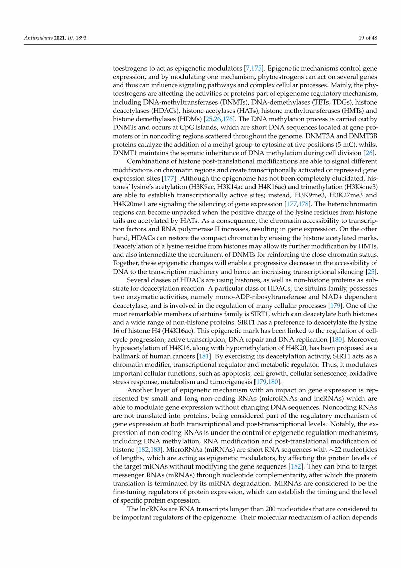
Antioxidants 2021, 10, 1893 19 of 48
toestrogens to act as epigenetic modulators [7,175]. Epigenetic mechanisms control geneexpression, and by modulating one mechanism, phytoestrogens can act on several genesand thus can influence signaling pathways and complex cellular processes. Mainly, the phy-toestrogens are affecting the activities of proteins part of epigenome regulatory mechanism,including DNA-methyltransferases (DNMTs), DNA-demethylases (TETs, TDGs), histonedeacetylases (HDACs), histone-acetylases (HATs), histone methyltransferases (HMTs) andhistone demethylases (HDMs) [25,26,176]. The DNA methylation process is carried out byDNMTs and occurs at CpG islands, which are short DNA sequences located at gene pro-moters or in noncoding regions scattered throughout the genome. DNMT3A and DNMT3Bproteins catalyze the addition of a methyl group to cytosine at five positions (5-mC), whilstDNMT1 maintains the somatic inheritance of DNA methylation during cell division [26].
Combinations of histone post-translational modifications are able to signal differentmodifications on chromatin regions and create transcriptionally activated or repressed geneexpression sites [177]. Although the epigenome has not been completely elucidated, his-tones’ lysine’s acetylation (H3K9ac, H3K14ac and H4K16ac) and trimethylation (H3K4me3)are able to establish transcriptionally active sites; instead, H3K9me3, H3K27me3 andH4K20me1 are signaling the silencing of gene expression [177,178]. The heterochromatinregions can become unpacked when the positive charge of the lysine residues from histonetails are acetylated by HATs. As a consequence, the chromatin accessibility to transcrip-tion factors and RNA polymerase II increases, resulting in gene expression. On the otherhand, HDACs can restore the compact chromatin by erasing the histone acetylated marks.Deacetylation of a lysine residue from histones may allow its further modification by HMTs,and also intermediate the recruitment of DNMTs for reinforcing the close chromatin status.Together, these epigenetic changes will enable a progressive decrease in the accessibility ofDNA to the transcription machinery and hence an increasing transcriptional silencing [25].
Several classes of HDACs are using histones, as well as non-histone proteins as sub-strate for deacetylation reaction. A particular class of HDACs, the sirtuins family, possessestwo enzymatic activities, namely mono-ADP-ribosyltransferase and NAD+ dependentdeacetylase, and is involved in the regulation of many cellular processes [179]. One of themost remarkable members of sirtuins family is SIRT1, which can deacetylate both histonesand a wide range of non-histone proteins. SIRT1 has a preference to deacetylate the lysine16 of histone H4 (H4K16ac). This epigenetic mark has been linked to the regulation of cell-cycle progression, active transcription, DNA repair and DNA replication [180]. Moreover,hypoacetylation of H4K16, along with hypomethylation of H4K20, has been proposed as ahallmark of human cancers [181]. By exercising its deacetylation activity, SIRT1 acts as achromatin modifier, transcriptional regulator and metabolic regulator. Thus, it modulatesimportant cellular functions, such as apoptosis, cell growth, cellular senescence, oxidativestress response, metabolism and tumorigenesis [179,180].
Another layer of epigenetic mechanism with an impact on gene expression is rep-resented by small and long non-coding RNAs (microRNAs and lncRNAs) which areable to modulate gene expression without changing DNA sequences. Noncoding RNAsare not translated into proteins, being considered part of the regulatory mechanism ofgene expression at both transcriptional and post-transcriptional levels. Notably, the ex-pression of non coding RNAs is under the control of epigenetic regulation mechanisms,including DNA methylation, RNA modification and post-translational modification ofhistone [182,183]. MicroRNAa (miRNAs) are short RNA sequences with ∼22 nucleotidesof lengths, which are acting as epigenetic modulators, by affecting the protein levels ofthe target mRNAs without modifying the gene sequences [182]. They can bind to targetmessenger RNAs (mRNAs) through nucleotide complementarity, after which the proteintranslation is terminated by its mRNA degradation. MiRNAs are considered to be thefine-tuning regulators of protein expression, which can establish the timing and the levelof specific protein expression.
The lncRNAs are RNA transcripts longer than 200 nucleotides that are considered tobe important regulators of the epigenome. Their molecular mechanism of action depends

Antioxidants 2021, 10, 1893 20 of 48
on cellular localization and the type of interacting molecules; thereby, lncRNAs can beclassified as signal, decoy, guides or scaffold [183]. Signal lncRNAs regulate transcriptionalactivity or signaling initiation, whilst decoy lncRNAs bind and titrate out gene regulatoryelements, such as proteins, mRNAs and miRNAs. Nuclear lncRNAs can bind transcriptionfactors or chromatin modifiers proteins (e.g., DNMTs, HATs, HDACs and PcGs), whereas,in the cytoplasm, they function as a sponge to attract proteins and miRNA/RISC com-plexes away from their targets [183,184]. Acting as scaffolds, lncRNAs can coordinatethe formation of distinct epigenetic regulatory complexes on specific chromatin regions,dictating their active or inactive transcriptional status. They have the ability to guideprotein complexes to both close and distant genomic loci which allow them to regulategene expression on a genome-wide scale [184]. In conclusion, as epigenetic modulators,lncRNAs can increase or repress transcriptional activity by controlling the deposition ofhistone marks and DNA methylation pattern on chromatin regions. The reciprocal inter-connection of non-coding RNAs with epigenetic pathways appears to form a feedback loopthat have an extensive influence on gene expression throughout the genome. Therefore,any dysregulation may affect physiological and pathological processes and will contributeto a variety of diseases.
Emerging scientific data suggest that a diet rich in vegetables and fruits can signif-icantly reduce the risk of chronic disease development, due to the action of nutritionbioactive constituents, which can regulate the gene expression [176,185]. Remarkably,many phytochemicals, including phytoestrogens, may act through epigenetic mechanisms,such as modulation of DNA methyltransferases (DNMTs), histone deacetylases (HDACs)activities and non-coding RNA expression [24,30,176]. In the next sections, the epigeneticmodulator capacity of phytoestrogens and of their metabolites is presented, underliningtheir mode of action with effect on different biological processes.
5.1. Isoflavones
Isoflavones have been intensely studied in connection with their epigenetic modulatorpotential in chronic diseases, especially in cancers. Genistein is acting as a chemotherapeuticagent in various cancer cells, modulating cell proliferation, apoptosis and metastasis [186].Indeed, anticancer properties of genistein have been associated with re-expression oftumor-suppressor genes that were methylation-silenced during carcinogenesis [175,187].For instance, genistein reversed hypermethylation of ATM, APC, BRCA1 and PTEN tumor-suppressor genes and restored the ER expression in ER positive and triple-negative breastcancer cells by inhibiting the DNMTs’ activities [188,189]. In general, the BRCA1 andBRCA2 mutations imply a high risk of breast and ovarian cancers, yet their epigeneticallysilenced promoters are found frequently in many types of cancers [189]. Moreover, inthe case of ER positive cells, genistein epigenetically had re-expressed BRCA1, but alsoacted as antagonist of AhR [190]. In addition, genistein and daidzein treatment of breastcancer cells decreased the expression levels of MeCP2 protein [191], which have a methyl-CpG-binding (MBD) domain that recognizes and binds to 5-mC regions of DNA. Thedaidzein gut metabolite, S-equol, at physiological concentration of 2 µM and after long-term treatment, was acting as demethylation agent of BRCA1 and 2 promoters in breastcancer cells but not in normal mammary gland cell line [192]. In general, isoflavones haveshown DNA demethylating capacity, with genistein as the most potent among testedisoflavones, by reducing the promoter methylation of genes involved in preventing cancerdevelopment [187,190,191,193,194].
The epigenetic modulation capacity of isoflavones has been observed in correlationwith histone posttranslational modifications. Thus, genistein treatment can affect differentepigenome modifier proteins, including histone deacetylase (HDACs) and histone H3 Lys 9methyltransferase (HMT) by reducing their enzymatic activities in cervical cancer cells [194].The two most studies isoflavones (genistein and daidzein) and the metabolite S-equol havebeen reported to demethylate transcriptional repression marks, such as H3K9me3 andH3K27me3, concomitant with modulating the EZH2 protein expression, which is control-

Antioxidants 2021, 10, 1893 21 of 48
ling and maintaining the repressed chromatin status in breast cancer cells [195]. Moreover,epigenetic marks, such as H3K4ac and H4K8ac, signaling the active chromatin, becamehyper-acetylated, accompanied by increased expression of transcriptional activators P300and SRC3 [195]. Another study reported that genistein alone or in combination with tri-chostatin A can reactivate ERα expression in the ER negative cells. An enhancement ofERα expression and increased global H3K9, H3 and H4 acetylation, along with decreasedDNMT1 expression and HDAC activity, were observed [196]. Furthermore, mice exposedto a long-term genistein diet resulted in having a high expression of ERα in spontaneousbreast tumors, preventing the occurrence of a more aggressive ER-negative type of breastcancer [196]. The protective effect of genistein can be assumed to be at least in part due toits epigenetic modulator capacity, since HDACs activity was significantly reduced in micebreast tumors [196]. Studies on genistein treatment of precancerous and fully developedbreast cancer cells have shown an increased expression of tumor-suppressor genes p16 andp21 through enrichment of transcriptional active markers, such as acetyl-H3, acetyl-H4and H3K4me3 [188]. Moreover, genistein induced a stronger anti-proliferative effect andpro-apoptotic response in precancerous cells than in breast cancer cells [188], which mightsuggest that its anticancer properties could be more effective at an early stage of tumorige-nesis. In addition, the genistein-enriched diet has prevented tumorigenesis and inhibitedcancer development in breast-cancer mice xenografts [188]. Isoflavones can modulate miR-NAs expression with impact on their anticancer activity. In vitro studies have demonstratedthat onco-miRNAs expression, such as miR-155, can be significantly reduced by treatingbreast cancer cells with physiological concentration of 1–10 µM genistein [197]. Conversely,the expression levels of miR-155 targets were upregulated in response to genistein treat-ment, including pro-apoptotic and anti-proliferative proteins: FOXO3, PTEN, casein kinaseand p27 [197]. Recently, Lynch et al. have demonstrated that the long-term treatmentof PC3 prostate cancer cell line with 40 µM genistein has induced hypomethylation ofmiR-200c gene/cluster resulting in increased expression of miR-200c [198]. Furthermore,genistein can modulate the expression of tumor-suppressor Smad4 in prostate cancer celllines via multiple epigenetic mechanisms. Thus, genistein can downregulate miR-1260bthat targets Smad4, or genistein can induce DNA methylation and histone modifications atSmad4 gene promoter in prostate and renal cancer cells [199,200].
There are a few studies exploring the possible connection of biochanin A and for-mononetin with epigenetic mechanisms. Notably, biochanin A effectively and reversibleinhibited the Lysine-specific histone demethylase 1A (LSD1) activity in vitro, in gastriccancer cells at physiological concentration [201]. However, emerging evidence is suggest-ing that red clover isoflavones might have the ability to modulate miRNAs expression indifferent types of cancer cell lines [202]. Whilst biochanin A seems to promote ERα-positivecells’ proliferation through miR-375 activation [203], the formononetin at its physiologicconcentration showed the same pro-proliferative effect and anti-apoptosis activity viaupregulation of miR-375 in aortic endothelial cells, but not in breast cancer cells [204].Moreover, in ovariectomized rats, formononetin intake have increased uterine weight andinduced the upregulation of miR-375, ERα and Bcl-2 and the downregulated RASD1, butno effect was observed on mice bearing MCF-7 xenografts [204]. The formononetin abilityto trigger selectively the miR-375/RASD1/ERα pathway may suggest that its long-termuse can lower the risk of postmenopausal breast cancer development. Formononetincan modulate in a time-and dose-dependent manner the miR-21 expression in bladdercancer in vitro by significantly inhibiting cellular proliferation and invasiveness and in-ducing apoptosis. In addition, reduced expression of miR-21 was followed by increase ofPTEN, and downregulation of p-Akt [205]. Moreover, formononetin-combined therapymay enhance the therapeutic efficacy of doxorubicin in glioma cells by preventing EMTthrough inhibition of HDAC5 [206]. In vivo experiments on rats with type 2 diabetes theformononetin long-term treatment has been able to control hyperglycemia and insulinresistance and also to reduce oxidative stress through SIRT1 re-expression in kidney tissuesof diabetic animals [207]. Moreover, a recent study has demonstrated that formononetin

Antioxidants 2021, 10, 1893 22 of 48
can attenuated triple negative breast cancer cells malignancy by suppressing the lncRNAAFAP1-AS1 expression with an influence on the miR-545-3p/CDK4 and miR-195/Raf-1axes, which are associated with triple-negative breast cancer chemoresistance [208]. Re-cently, it has been demonstrated that S-equol is involved in miRNAs’ expression regulationas exerting its antitumor activities [209]. S-equol has induced apoptosis in MCF-7 cells bymodulating the PI3K/AKT pathway through the upregulation of miR-10a-5p expressionin a time-and dose-dependent manner [209]. Interestingly, a new report has shown thatisoflavones, particularly genistein, can reduce the interaction of an oncogenic lincRNAHOTAIR with chromatin remodeling factors, resulting in renal tumor cell line malignancysuppression [210]. Thus, new approaches to investigate the epigenetic modulator capacityof isoflavones at lncRNAs level could be initiated. Other examples are presented in Table 3.
Table 3. Dietary phytoestrogens as epigenetic modulators—in vitro and in vivo studies.
Dietary Phy-toestrogen Concentration Cell Line/In
Vivo Models Epigenetic Changes Targeted Gene Biological Activity References
I. Isoflavones
Genistein
In vitro0.5–1 µM MCF-7 ↓DNMT1, ↓CpG methylation at
BRCA1, ESR1 promoter
↑BRCA1, p53,CYP1A1,↓cyclin D1
-Anti-proiferative andchemopreventive effect in breastcancer cells with activated AhR.
[190]
3.125 µM MDA-MB-468, ↓CpG methylation atGSTP1 promoter ↑GSTP1 -Preventive effect, activates phaseI
enzyme in TNBC cells. [211]
5 µM MDA-MB-435,Hs578T ↓miR-155
↑FOXO3, PTEN,casein kinase, p27,↓β-catenin
-Inhibits cells viability and inducesapoptosis in TNBC cells. [197]
18.5 µM MCF-7MDA-MB-231
↓Global DNA methylation,↓CpG methylation BRCA1,BRCA2, MeCP2 promoters;↓H3K9me3, H3K4me3,
H3K27me3;↑H4K8ac, H3K4ac at promoters
of EZH2,BRCA1, ERα, ERβ,SRC3, p300
↑BRCA1, ↑BRCA2;↓EZH2,
↑p300, ↑SRC3
-Inhibition of breast cancercells’ proliferation. [191,195]
10–20 µM
UACC-3199,KYSE 510,
SiHa;DU145, LNCaP,
PC-3,ARCaP-E,ARCaP-M
↓DNMT1, ↓CpG methylation ofBRCA1, ESR1 promoters;↓CpG methylation at RARβ2,
p16, MGMT, ↓DNMTs;↓DNA methylation at RARβ 2;↑H3K9ac at promoters of APC,
SOX7, SFRP1, SFRP2, DKK,WIF1, ↑HAT1
↑BRCA1, ER α, p53CYP1A1, ↓cyclin D1↑RARβ2, p16,
MGMT↑RARβ2;
↑SOX7, SFRP1,BRCA1, BARD1,RAD23B, XRCC2,↓BIRC7, SLUG,HES1, TGFBIII
-Anti-proiferative andchemopreventive effect in breastcancer cells with activated AhR;
-Inhibition of esophageal squamouscell carcinoma growth;
-Induces apoptosis in cervixsquamous cells carcinoma;
-Reduces proliferation and inducesapoptosis in prostate cancer cells
[190,193,212,213]
25 µM
MDA-MB-157,MDA-MB-231;A-498; 786-O;
Caki-2;PC3, DU145,
RWPE-1;786-O, ACHN;PC-3, DU145
↑Ac-H3, ↓DNMT1, HDAC1;↓miR-1260b;
↓CpG methylation at SFRP1↓H3K9me2, H2K9me3,H3K27me3 at SFRP1,
Smad4 genes;↓lnc HOTAIR, ↓EZH2,↑miR-574-3p, ↑mir-34a
↑Erα;↑SFRP1, Dkk2,
Smad4;↓ARID1A, EED
SMARCB1, SNAIL,↑ZO-1;
↓RAC1, ↓EGFR,↓EP300
↓MMP9, VEGF
-Inhibitits proliferation, invasion ofTNBC, renal carcinoma and prostate
cancer cells;-Enhances tamoxifen inducedanticancer effect in TNBC cells;-Promotes apoptosis in renal
carcinoma and prostate cancer cells;-Inhibits cell proliferation, migrationand invasion in vitro and in vivo in
prostate cancer;
[196,199,200,210,214,215]
5–40 µM SH, SHR
↑Global H3ac, ↑H3K4me3,↓H3K27me3, H3K9me3 at p16,
p21 promoters, ↑HMTsactivities
↑p21, p16,↓BMI1, c-MYC
-Inhibits growth of breast cancercells, but no effect on normal cells;
-Preventive effect on breasttumorigenesis in vivo.
[188]
20–40 µM U266 ↑miR-29b ↓NF-κB -Inhibits proliferation and inducesapoptosis in multiple myeloma. [216]
40 µMDU-145,
PC-3LNCaP
↓CpG methylation at BRCA1,GSTP1, EPHB2, RASSF1A
promoters; altered methylationpattern of MAD1L1, TRAF7,
KDM4B, hTERT genes;↑miR-200c
↑ BRCA1, GSTP1,↑EPHB2;
Potential targetgenes SOX2, ZEB1
-Inhibits prostate cancer cells’proliferation, clonogenic potential
and induces apoptosis.
[187,217,218]
50 µM HeLa
↓DNA methylation at TP53,PTEN, CDH1, DAPK1, FHIT,RUNX3, SOCS1 promoters,↓DNMTs, HDACs, HMTs
↑TP53, PTEN,CDH1, DAPK1,FHIT, RUNX3,
SOCS1
-Anti-proliferative effect on cervicaltumor cells. [194]

Antioxidants 2021, 10, 1893 23 of 48
Table 3. Cont.
Dietary Phy-toestrogen Concentration Cell Line/In
Vivo Models Epigenetic Changes Targeted Gene Biological Activity References
60–100 µM MCF-7,MDA-MB-231
↓Global DNA methylation;↓DNAmethylation at TSG
promoters; ↓DNMT1
↑ATM,↑APC,↑PTEN
-Reduced cellular viability andanti-proliferative effect on breast
cancer cells;[189]
In vivo2 mg/day50 mg /kg250 mg/kg270 mg/kg
Neuroblastomaxenografts,Eker rats,
Avy femalemice,
tumorxenograft mice,123/SvJ:C57BL/
6J mice
↓DNTM3B, ↓CHD5 promotermethylation;
↓EZH2, ↓H3K27me3;↑CpG methylation at Avy
IAP gene;↓DNMT1, HDAC1;↓HDACs activity;↑DNA methylation in
repetitive elements;↓HDAC6
↑CHD5;↑PI3K/AKT
pathways in uterus;↓ectopic Agouti
expression;↑ERα, ↑PCNA in
breast tissues;↓p21, cyclin D1,
PCNA, IGF2expression in
adult mice
-Decrease of tumor sizeand frequency.
-Increases hypersensivity ofER-responsive genes in neonatal
uteri and adult myometrium;-Reduces obesity offsprings,
phenotypes changes;-Re-sensitizing ERα-negative breast
cancer to therapy;-Prenatal exposure leads to
long-term epigenetics changes.
[196,219–222]
Soy baseddiet
Cynomolgusmonkeys
↓DNA methylation at promoterof HOXA5, HOXA11,
HOXB1, ABCG5
↑HOXA5, HOXA11,HOXB1, ABCG5
-Decrease in fasting insulin andHOMA index values. [223]
Daidzein
20–50 µM KYSE 510 ↓DNA methylation at RARβ2promoter; ↓DNMTs activity ↑RARβ2 -Dose-dependent inhibition of
cells growth. [193]
78.5 µM MCF-7,MDA-MB 231
↓Global DNA methylation,↓CpG methylation at BRCA1,BRCA2 promoters, ↓MeCP2;↓H3K9, H3K27, H3K4 (me3)↑H4K8ac, H3K4ac at promoters
of EZH2, BRCA1, ERα, ERβ,SRC3, p300
↑BRCA1, BRCA2;↓EZH2, ↑p300,
SRC3
-Inhibition of ER(-) and ER(+) breastcancer cells’ proliferation. [191,195]
110 µM DU-145, PC-3LNCaP
↓CpG methylation at BRCA1,GSTP1, EPHB2 promoters;
altered methylation pattern ofMAD1L1, TRAF7, KDM4B,
hTERT
↑BRCA1, GSTP1,EPHB2
-Inhibits prostate cancer cells’proliferation and induces apoptosis. [187,218]
Biochanin A
2–6 µM T47-D, MCF-7 ↑miR-375 ↑ERα, ↑Bcl -2 -Promotes proliferation of breastcancer cells. [203]
2.95 µM MGC-803 ↓LSD1,↑H3K4me1/2, H3K9 me1/2
↓MAO-A/B, Bcl-2,↑Bax
-Suppresses colony formation andmigration and induces apoptosis in
gastric cancer cells.[201]
20–50 µM KYSE 510 ↓CpG methylation at RARβpromoter, ↓DNMTs activity ↑RARβ -Inhibits the growth of eosphageal
squamous cells. [193]
Formononetin
In vitro2–6 µM HUVEC ↑miR-375 ↑ERα, Bcl-2,
↓RASD1-Promotes cell proliferation and
inhibits apoptosis. [204]
10–20 µM GMCs ↑SIRT1↑Nrf2/ARE↓Fibronectin,
ICAM-1
-Inhibits hyperglycemia-inducedROS overproduction in glomerular
mesangial cells.[224]
40 µM BT-549,MDA-MB-231
↓lncRNA AFAP1-AS1↑miR-545, miR-195 ↓CDK4, Raf-1 -Inhibits proliferation, migration
and invasion of TNBC cells. [208]
20–100 µM SW1116,HCT116 ↑miR-149
↓EphB3, cyclin D1,MMP2/9,↓PI3K/AKT↓STAT3
-Inhibits colon carcinoma cellproliferation and invasion. [202]
50–200 µM T24 ↓miR-21 ↑PTEN, ↓p-Akt-Inhibits proliferation, induces
apoptosis and decreasesinvasiveness of bladder cancer.
[205]
100 µM U87MG ↓HDAC5 ↓Vimentin↑E-cadherin
-Enhances the cytotoxicity ofdoxorubicin in glioma cells. [206]
In vivo4–8 mg/kg/day
Ovariectomizedrats ↑miR-375 ↑ERα, ↑Bcl-2,
↓RASD1-Lower risk of postmenopausal
breast cancer development. [204]
20–40 mg/kg Diabetic typeII rats
↑SIRT1 in pancreatic tissuesand sciatic nerve tissue
↓MDA, ↑GSH, SOD;↑NGF in sciatic
nerve tissue.
-Reduces oxidative stress, risk ofnephro-pathy and the level oftriglyceride and cholesterol;
-Protects from hyperglycemiainduced neuronal damage.
[225,226]
25–50 mg/kg Diabetesmice model ↑SIRT1 in kidney tissues ↑Nrf2, ↓Fibronectin,
ICAM 1-Reduces renal fibrosis, improves
renal function. [207,224]

Antioxidants 2021, 10, 1893 24 of 48
Table 3. Cont.
Dietary Phy-toestrogen Concentration Cell Line/In
Vivo Models Epigenetic Changes Targeted Gene Biological Activity References
S-equol
In vitro2 µM
MCF-7,MDA-MB-231
↓CpGmethylation at BRCA1,BRCA2 promoters ↑BRCA1, BRCA2 -Inhibits breast cancer
cells’ proliferation. [192]
12.8 µM MCF-7,MDA-MB-231
↓H3K9, H3K27, H3K4(me3)↑H3K4ac, H4K8ac
at promoter of EZH2,BRCA1,ERα, ERβ, SRC3, p300 genes
↓EZH2,↑p300, ↑SRC3 - [195]
50–150 µg/mL MCF-7 ↑miR-10a-5p ↓PI3K p110α, ↓p-Akt
-Anti-proliferative andpro-apoptotic effect. [209]
In vivo10–100 mg/day Neonatal rats ↑DNA methylation of H-ras in
pancreatic cells - - [227]
II. Prenylflavonoids
6-PN8-PN
1 µM50–100 µM
MCF-7SK-MEL-28
↓DNMT1HDAC2,4,7,8 inhibition↑H3 acetylation
↓ERα ↑P450 1A1↓pS6P,
↓pERK/pP90
-Activates AhR to attenuateinhibition of CYP1A1 and
degradation of ERα;-Antiproliferative effects on
melanoma cells.
[228,229]
Glabridin
In vitro10 µM
MDA-MB-231Hs-578T
↑miR-148a, ↓DNMT1↓DNMT3A, ↓TGFβ/SMAD2 -Inhibits the CSCs-like properties of
breast cancer cells. [167]
10–20 µMMDA-MB-231,
Hs-578T;HepG2, Huh-7
↑miR-148a↓Wnt/β-catenin
↓VEGF↓SMAD2
-Attenuates angiogenes in breastcancer cells;
-Inhibits the CSCs-like properties ofHCC cells.
[166,230]
In vivo20 mg/kg/d
mousexenograft
↑miR-148a,↓DNMT1 ↓DNMT3A ↓TGFβ/SMAD2 -Attenuated the tumor growth,
CSCs-like properties in vivo. [167]
Icariin
1 nM hMSCs ↓DNA methylation at ABCB1promoter
↑ABCB1, MMP↑P-gp protein
-Improves cellular viability,decreases oxidative stress and
promotes osteogenesis of MSCs;[231]
10 nM Mouse aorticECs ↑SIRT6, ↓H3K9ac ↓NF-κB, TNF-α,
ICAM-1, IL-2, IL-6-Reduces inflammation in vitro and
in vivo; [232]
5–10 µM MDA-MB-231,4T1
↑SIRT6↓H3K9ac
↓NF-κB p65,↓MMP2,↓N-cadherin↓TNFα,↑E-cadherin
-Suppresses migration, invasion,decreases ROS level in breast
cancer cells;[233]
2–16 µM IMR-90 ↑SIRT6, SIRT1 ↓NF-κB, ↓p-p53,p-p21, ↓Cav1
-Prevents D-gal-induced aging andcell-cycle arrest in lung
fibroblast cells;[234]
25–50 µM A2780 ↓miR21 ↑PTEN, RECK,↓Bcl-2
-Regulates proliferation andapoptosis of ovarian cancer cells; [235]
In vivo100 mg/kg Rats ↑SIRT1, H4AcK16
↓TNF-α, IL-1β, andIL-6
↓NF-κB(p65)phosphorylation
-Suppresses paclitaxel-inducedneuroinflammation andmechanical allodynia;
[236]
10−2 µM100 mg/kg/day
FA HSPCsisolated
from mice
↑SIRT6,↓H3K9ac ↓NF-κB
-In vitro progenitor capacity;-In vivo repopulating ability of
FA HSCs.[237]
Icaritin 40 µMCD4+T cells
from SLEpatients
↑H3K4me3 at Foxp3 gene↑H3K9me3 at IL17a gene
↑Foxp3↓IL17a
-Reduced autoreactivity ofCD4+Tcells. [238]
III. Coumestans
Coumestrol
In vitro1–10 µM Muscle cells ↑SIRT1
↑NDUFA9, SDHA,UQCRC2, COX1,
PGC1, Nrf1
-Increases mitochondria number,respiratory chain proteins and
mitochondrial function;[239]
10–50 µM ES2 ↓DNMT3A phospho
↓CK2, PCNA,ERBB2, p-AKT,
p70S6K, ERK1/2,JNK1/2, p90RSK
-Preventive effects on epithelialovarian cancer cells; [161]
20–40 µM HCT116 ↓H3Tr3phos ↓Haspin kinase -Suppresses colon cancercells’ proliferation; [162]
In vivo10–100 mg/day Neonatal rats ↑DNA methylation of H-ras - - [227]
Wedelolactone0.1–10 µM Mino ↓EZH2, PRC2, HTM
↓H3K27me3 ↓PRC2, EZH2 -Inhibition of B cell non-Hodgkin’slymphoma cells’ proliferation; [240]
50 µM HepG2, THP1,K562 ↓EZH2, PRC2
↑DAB2IP, ADRB2,CDKN2A,GADD45A
-Inhibits proliferation and migration,and induces apoptosis and cell-cyclearrest of PRC2-dependent cancers.
[241]

Antioxidants 2021, 10, 1893 25 of 48
Table 3. Cont.
Dietary Phy-toestrogen Concentration Cell Line/In
Vivo Models Epigenetic Changes Targeted Gene Biological Activity References
IV. Lignans
Arctigenin
In vitro0.268 µM0.5–1 µM
5 µM20–100 µM
Rats’ neurons;SH-SY5Y;
MDA-MB-231H9C2,Rats
cardiomyocyte
↑SIRT1↑miR-16 ↑miR-199a
↑H3K9 me3 at AP-1, Bcl-2promoters↑SIRT1
↓NLRP3 ↓IL-1β,↓IL-18, ↓ASC↓caspases-1 p20;↓IKKα ↓IKKβ,↓NF-κB ↓TNF-α↓IL-6, ↑IL-10; ↓Bcl-2,↑ phos ATF-2↑AMPK, ↑I-κB,↓NFkB
-Protection against ischemicstroke, neuroprotection;
-Induces anti-inflammatory,anti-apoptotic mechanisms toprevent secondary damage;-Supressed cardiomyocytesapoptosis, inflammation and
oxidative stress;
[169,242–244]
In vivo100 µM/kg;
4 mg/kg/day;20 mg/kg;
50–100 mg/kg
Rats—myocardialischemia;
xenograft mice;Rats—cerebral
ischemia
↑SIRT1;↑pho-p38;
↑SIRT1, ↑miR-96-5p↓miR-126-5p, miR-21-5p,↑miR-135a-5p, miR-205-5p,
miR-22-3p, miR-455-5p
↑AMPK, ↑I-κB,↓NFkB↓Bcl-2;
↓NLRP3 IL-1β,IL-18;
↓ASC, caspases-1p20
TIMP3, ZNF185↓VEGF, EGF,
FGF-β,↑Bax/Bcl-2 ratio
-Inhibition of oxidative Stress andinflammation after acute
myocardial ischemia;-Protects against ischemic stroke;
-Inhibited prostate tumor cellgrowth both in vitro and in vivo.
[169,242,244,245]
Arctiin 5 µM10 µM
HaCaTNHDF
↑miR-125a-5p, -205-3p, -21-3p,-29b-1-5p
↓miR-3652, -494, -1246;↑miR-602, -762, -150-3p, -4327,
-584-5p, -874, -3665↓miR-3679-5p, -1290, -575
-Possible regulationof members of
MAPK pathwaysand cell growth
signaling pathways.
-Enhances wound healing,DNA repair in UVB-
exposed keratinocytes;-Inhibits the UVB-mediated cell
growth defect, apoptosis,DNA damage.
[246,247]
SGD
In vitro50–100 µM 3T3L1 ↓DNMTs, HDACs, MBD2 - -Antioxidant effect, epigenetic
modification in murine adipocytes. [248]
In vivoFlaxseed diet
Female miceMice pneu-monopathy
↑miR-30b, -324-5p↓miR-382, -423;
↓miR-142-3p, -150 ↑miR-34a
Changes inmammary gland
miRNome;↓Bcl2, FGFR1
-Prevents breast cancerdevelopment during adulthood.
-Antioxidant andanti-inflammatory effects.
[249,250]
Sesamin 10 µM THP-1 ↓H3/H4 acetylation at MDCpromoter area, ↓CBP
↓MAPK-p38,NFkB-p65, MDC,
IP-10
-Supresses allergy andasthma-related
chemokines expression;-Anti-inflammatory effect.
[251]
ENL
20 µM
RWPE-1,WPE1-NA22,-NB14, -NB11,-NB26, LNCaP
↓miR-106b cluster (miR-106b,-93, -25)
↓GMNN, CDT1,MCM2, MCM 7↑PTEN
-Anti-proliferative effect on mid andlate prostate cancers; [252]
50–100 µM 3T3L1 ↓DNMTs, HDACs, MBD2; -
-Antioxidant activities,downregulates
epigenetic-modification-associatedgene expression inmurine adipocytes.
[248]
V. Stibenes
Resveratrol
In vitro1 µM HaCaT ↑ SIRT1, HDAC2,
↓H4K16Ac ↑eNOS-Accelerates wound healing repair
in vitro and in vivoskin-wound models;
[253]
5 µMCanine-bonetissue cellsMC3T3-E1
↑ SIRT1↓p300
↓NF-κB acetylation,IκBα
phosphorylation,IKK activity kinase
activity, ↑Cbfa-1
-Anti-osteoclastogenic, activates thebone-tissue cells to osteoblast
and osteogenesis;[254]
10 µM
MDA-MB-468A2058, M223;
HCT116,SW480;
HUVEC;ARPE-19
↓CpG methylation at ERαpromoter;↑SIRT1↑DNMT1,
↑LINE-1 methylation
↓STAT3acethylation
↑ERα expression;↓NF-Kb, CXCR4,
MMP9;↑eNOS acetylation
-Anti-proliferative, reduces viabilityand induces mesenchymal to
epithelial transition phenotype inbreast cancer and CRC cells;
-Regulates endothelial functionduring oxidative stress;
-Ameliorates viability and ROSproduction in retinal pigment
epithelia cells under oxidative andinflammatory conditions;
[255–258]

Antioxidants 2021, 10, 1893 26 of 48
Table 3. Cont.
Dietary Phy-toestrogen Concentration Cell Line/In
Vivo Models Epigenetic Changes Targeted Gene Biological Activity References
14–15 µM
MCF10A,MCF7,
HCC1806,MDA-MB-157
↑CpG methylation at KCNJ4,RNF169, BCHE, DAOA
↓CpG methylation of HOXA9,KRTAP2-1, TAGAP, RUNX3;↓CpG methylation at PTEN
promoter, ↓DNMT1;↓SIRT1, DNMT3B, ↓DNMTs
activity;↑HDACs, HATs, ↑H3Ac, H4Ac,
H3K9Ac at ERα promoter
↓KCNJ4, DAOA↑BCHE, KRTAP2-1,
TAGAP;↑PTEN, p21;
↓γ-H2AX, ↓hTERT;↑ERα
-No cytotoxic effect on normalmammary gland cells;
-Antiproliferative effect on breastcancer cells;
-Induces apoptosis and cell-cyclearrest on TNBC cells.
[259–262]
10–20 µM MCF-7;MDA-MB-231
↓DMNT1, MBD2, H3K9me3 atBRCA1 promoter
↑H4Ac, H3K9Ac at BRCA1promoter
↓PRMT5, EZH2, KDACs,↑KAT2A/3B
↑global H3K9ac, H3K27ac↓H4R3me2s, H3K27me3 atBRCA1, p53, p21 promoters
↑BRCA-1↓AhR and ERα atBRCA1 promoters;↑BRCA1, p53, p21
-Attenuates dioxin carcinogenicchemicals-dependent repression of
BRCA-1 and induction ofDNA damage;
-Inhibits breast cancer cells’proliferation.
[263,264]
25 µM C2C12;A549
↑SIRT1;↓lncAK001796
↑AMPK ↓LKB1ac,↓PGC-1α ac↑Nrf-1, Nrf-2,
NDUFS8, SDHb,Uqcrc1, COX5b,
ATP5a1↑BIRC5, TFDP2,
CDC6↓ATR, CCNB1,
CKS2
-Increase mitochondrial membranepotential, cellular ATP content in
mouse mioblasts;-Inhibits lung cancercells’ proliferation.
[265,266]
20–30 µMPANC-1, MIA
PaCa-2, AsPC1;THP1
↓SIRT1, SIRT2, SIRT3;↑miR-663, ↓miR-155
↑PTEN, p-JNK,FOXO
↓Ras, p-AKT,p-ERK, AKT kinase
activity↑caspase-3;↓AP-1, ↑cMaf
-Induces cell-cycle arrest andapoptosis in pancreatic cancer cells;-Inhibits pancreatic tumor growth
in vivo;
[267,268]
20–50 µM
A549,BGC-823,SGC-7901
U266,LP1
↓CpG methylation at ZFP36promoter; ↓DNMT1;
↑SIRT1;↓lnc NEAT1
↑ZFP36, ↓CCND1,MYC, VEGFA;↓cyclin D1, CDK4,
CDK6↑p21, p16;↑β-catenincytoplasm,
↓c-Myc, MMP7,Survivin
-Inhibits migration and cellproliferation in non-small-cell lung
cancer cells;-Inhibits gastric cancer cells’
proliferation and inducescell-cycle arrest;
-Inhibits the tumor growth ofxenografts;
-Inhibits the proliferation, migrationand invasion of multiple
myeloma cells;
[269–271]
50 µM
MCF-10A,MCF-7,
MDA-MB-231LNCaP,
DU145, 22Rv1SW480
↓CpG methylation at Nrf2,↓miR-93;
↓HDAC activity,↓HDAC2 at ATP2A3 promoter↑global H3Ac, H3K27ac↓DNMTs activity, ↓MeCP2,
MBD2;↓miRs-17-92, -106ab clusters
↓miRs-7, -17, -18b↑miRs-150, -296-5p
↓miRs-17, -20a, -106a, -106b;↓HDACs, NuRD complex,
↑p300;↑miR-663, ↓miR-17, -21, -25,
-92a-2
↑Nrf2;↑ATP2A3, SERCA3;
↑PTEN;↑PTENac, p53,↓MTA1, PI3K-Akt;↓TGFβ1, ↑PTEN,PDCD4, SMAD7
-Protective role against E2-inducedmammary carcinogenesis;
-Pro-apoptosis effect and changes inCa2+ homeostasis;
-Induces apoptosis and inhibits cellgrowth, angiogenesis and metastasis
in prostate cancer cells;-Induces tumor regression in
orthotopic prostatecancer xenografts;
-Induces apoptosis, inhibits coloncancer cells growth.
[272–277]
In vivo7 ppm mixed
AIN-76Adiet
Pregnantfemale
Sprague–Dawley
rats
↓CpG methylation atBRCA1 promoter
↓DNMT1 at BRCA-1 promoter↑BRCA1, AhR -Reduces the risk of breast
tumorigenesis in the offspring; [278]
diet with0.4%
resveratrol
Wild-typemouse ↑SIRT1 ↑p-AMPK, NAD+,
LKB1 acetylation
-Improves mitochondrial functionand increases cellular ATP in
skeletal muscle;[265]
5–25 mg/kg/dayFemale rats
bearing breastcancer
↑miRs -21, -129, -204, -489↓DNMT3B in tumor tissues;↓miRs -21, -129, -204, -489↑DNMT3B in normal tissues
-Inhibits breast tumor formationin vivo; [279]

Antioxidants 2021, 10, 1893 27 of 48
Table 3. Cont.
Dietary Phy-toestrogen Concentration Cell Line/In
Vivo Models Epigenetic Changes Targeted Gene Biological Activity References
25–50 mg/kgMice bearing
humanmelanoma
↓DNMT1↓CpG methylation at PTPN6,CDKN2A, SOCS3 promoters
↓STAT3 acetylation -Tumor-growth inhibition; [255]
50 mg subcu-taneouspellet/month
FemaleACI rats
↓CpG methylation at Nrf2promoter↓miR-93
↑Nrf2, NQO1,SOD3 OGG1,FMO1, AOX1↓MTA1, pAkt
-Decreases tumor incidence andchemoprevention; [272]
extractcontainingresveratrol/
1 year
Peripheralblood malewith type-2
diabetes
↑miR-21, -181b, -663, -30c2↓miR-155, -34a
↓IL-6, CCL3, IL-1β,TNF-α↑LRRFIP-1
-Beneficial immunomodulatoryeffect on hypertensive patients with
type 2 diabeties.[280]
Pterostilbene
2.5–10 µM
MCF-7MDA-MB-231in coculturewith TAM
↑miR488↓NF-κB, Twist1,
vimentin↑E-cadherin
-Suppresses breast EMT and/orgeneration of CSCs; [173]
5 µM HCC1806MDA-MB-157
↓SIRT1, ↓DNMTs activity↑HDACs, HATs
↑H3Ac, H4Ac, H3K9Ac at ERαpromoter
↓γ-H2AX, hTERT↑ERα
-Induces apoptosis and cell-cyclearrest in breast cancer cell lines; [261,262]
50 µM DU145, 22Rv1 ↓miRs-17, -20a, -106a, -106b ↑PTEN↓PI3K-Akt
-Promotes apoptosis, inhibits cellproliferation both in vitro and
in vivo, and downregulatescirculating tumor-derived oncomiRs
in vivo.
[275]
Piceatannol
In vitro1 µM U937 ↓miR-183
↓ADAM17, Sp1,Foxp3,
TNFα/NFkB↑ β-TrCP
-InhibitsTNF α-mediated signalingpathway in leukemia cell line; [281]
10 µM THP-1,Raw264.7
↑SIRT1↓miR-183 ↑HO-1 -Attenuates osteoclastogenesis in
bone-marrow-derived macrophages; [282]
30 µM
RAW264.7,A2058,
WM266-4,HCT116
↑miR-200a↑miR-181a↑miR-129
↑Nrf2↓NLRP3, IL-18,IL-1β, caspase1↑Bax, caspase 3,
↓Bcl-2
-Attenuates oxLDL-induced lipidstorage by inhibiting pyroptosis in
human macrophage cells;-Induces apoptosis of melanoma
cells and CRC cells;
[283,284]
In vivo50 mg/kg/day
Renal fibrosismice model ↓HDAC4, HDAC5 ↓p38-MAPK, ECM -Ameliorates renal fibrosis. [285]
Resveratrol +Pterostibene
In vitro15 µM + 5 µM
HCC1806MDA-MB-157
↓SIRT1, ↓DNMTs,↓Global DNA methylation
↑HDACs, HATs↑ H3Ac, H4Ac, H3K9Ac at
ERα promoter
↓γ-H2AX, hTERT,↑ERα
-Induces apoptosis andcell-cycle arrest;
-Retrieves responsiveness to E2 and4-hydroxytamoxifen treatments in
resensitized breast cancer cells.
[261,262]
15 µM + 7 µMMCF10A
MCF10CA1hMCF10CA1a
↑CpG methylation at MAML2,GLI2 promoters; ↑DNMT3B
↓MAML2↓NOTCH
-Inhibition of growth of cancer cellswith low and high
invasive properties;[286]
In vivo5–25 mg/kg/day
Rats bearingestrogen-
dependentbreast tumors
↓DNMT3B, ↑miR10a,−21,−129, −204, −489 -
-Delay in mammarytumor formation;
-Different pattern of epigeneticchanges tumor versus
normal tissues;
[279]
CSAA diet +REV 1.2 g or
with PTS,1.34 g/kg/day
Rats DNA methylation ↓RUNX3, ↑KCNJ4 ↑RUNX3, ↓KCNJ4
-Changes theDNA methylationpattern on long-term
dietary exposures.[259]
Note: 5-mC, 5 methyl cytosine; 6-PN, 6-prenylnaringenin; 8-PN, 8-prenylnaringenin; ABCB1, ATP-binding cassette sub-family B member1; ABCG5, ATP Binding Cassette Subfamily G Member 5; ADAM17, ADAM Metallopeptidase Domain 17; AhR, Aryl hydrocarbonreceptor; AIF, Apoptosis Inducing Factor; AMPK-5’, Adenosine Monophosphate-Activated Protein Kinase; AOX1, Aldehyde Oxidase1;APC, Adenomatous Polyposis Coli; ARE, Antioxidant Responsive Element; ARID1A, AT-Rich Interaction Domain 1A; ATM, AtaxiaTelangiectasia Mutated Protein; ATP5a1, ATP Synthase F1 Subunit Alpha; BARD1, BRCA1-Associated RING Domain 1; Bax, BCL2Associated X, Apoptosis Regulator; BCHE, Butyrylcholinesterase; Bcl-2, B-cell lymphoma/leukemia protein; BIRC7, Baculoviral IAP RepeatContaining 7; BMI1, Polycomb complex protein; BRCA1,2, breast cancer type 1,2 susceptibility protein; Cav1, Caveolin 1; CCND1, cyclinD1 protein; CDK4, cyclin-dependent kinase 4; CDH1, cadherin 1; CDT1, chromatin licensing and DNA replication Factor 1; CK2, proteincasein kinase 2; COX1, Cyclooxygenase-1; COX5b, Cytochrome C Oxidase Subunit 5B; CYP1A1, Cytochrome P450, family 1, subfamilyA, polypeptide 1; DAOA, D-amino acid oxidase activator; DAPK1, Death Associated Protein Kinase 1; Dkk2, Dickkopf WNT SignalingPathway Inhibitor 2; DNMT1, 3A, 3B DNA Methyltransferase 1, 3A, 3B; EED, Embryonic Ectoderm Development; EGFR, EpidermalGrowth Factor Receptor; ERK1/2 extracellular signal-regulated protein kinase 1
2 ; EP300/p300 E1A Binding Protein P300; EPHB2 Ephrintype-B receptor 2; ERα, ERβ Estrogen receptor alpha, beta; ERBB-4, Erb-B2 Receptor Tyrosine Kinase 4; ESR1, 2 Estrogen receptor 1, 2;EZH2 Enhancer of Zeste 2 Polycomb Repressive Complex 2 Subunit; FHIT (Fragile Histidine Triad Diadenosine Triphosphatase; FMO1

Antioxidants 2021, 10, 1893 28 of 48
Flavin Containing Dimethylaniline Monoxygenase 1; FOXO3 Forkhead Box O3; GSH Glutathione; GSTP1 Glutathione S-Transferase Pi 1;HES1 Hes Family BHLH Transcription Factor 1; H3K9me3, Histone 3 lysine 9 trimethylation; H3K27me3 Histone 3 lysine 9 trimethylation;H3K4ac Histone H3 acetylated at lysine 4; H4K8ac Histone H4 acetylated at lysine 8; HDAC1, 2, 5 Histone Deacetylase 1, 2, 5; HOXA5,A9, A11, B1 Homeobox proteins; HO-1 heme oxygenase-1; hTERT Telomerase reverse transcriptase; ICAM 1 Intercellular AdhesionMolecule 1; IL-1β2,6, Interleukin-1β,2, 6; IX, isoxanthohumol; JAK/STAT, Janus kinase/signal transducer and activator of transcription;JNK1/2, c-Jun N-terminal kinases 1
2 ; KAT2A/3B, (Lysine Acetyltransferase 2A/3B); KCNJ4, Potassium Inwardly Rectifying ChannelSubfamily J Member 4; KDACs, Lysine (K) deacetylases; KDM4B, Lysine Demethylase 4B; KRTAP2-1, keratin-associated protein 2-1; LSD1,Lysine-specific histone demethylase 1A; LKB1, Liver Kinase B1; MAD1L1 mitotic arrest deficient 1–like 1; MBD2, Methyl-CpG-bindingdomain protein 2; MCM7, Minichromosome Maintenance Complex Component 7; MeCP2, Methyl-CpG Binding Protein 2; MGMTO-6-Methylguanine-DNA Methyltransferase; MMP2,7,9 matrix metallopeptidase 2,7,9; NF-Kb Nuclear factor-kappa B; NGF Nerve growthfactor; Nrf1 Nuclear Respiratory Factor 1; Nrf2 Nuclear Factor Erythroid 2–like 2; NDUFA9 NADH:Ubiquinone Oxidoreductase Subunit A9;NDUFS8 NADH:Ubiquinone Oxidoreductase Core Subunit S8; NOTCH Notch Receptor; O-DMA O-demethylangolensin; OGG1 oxidativeDNA damage repair gene; p16 multiple tumor-suppressor 1 or cyclin-dependent kinase inhibitor 2A; p27 Cyclin-dependent kinase inhibitor1B;p53/ TP53 transformation-related protein 53; p65 nuclear factor NF-kappa-B; p70S6K phosphorylated ribosomal protein S6 kinase;p90RSK phosphorylated Serine/threonine-protein kinase; P450 1A1 Cytochrome P450, Family 1, Subfamily A, Polypeptide 1; p-AKTphosphorylated Protein kinase B; PCNA Proliferating Cell Nuclear Antigen; pS6P phosphorylated (Ser240/244) S6 ribosomal protein; pERKPKR-like ER kinase;PI3K p110α phosphatidylinositol 3-kinase P110 Subunit Alpha;PcG polycomb-group proteins;P-gp P-glycoprotein 1;PGC-1α peroxisome proliferator-activated receptor gamma coactivator 1-α; PRC2 Polycomb repressive complex 2; PRMT5 Protein ArginineMethyltransferase 5; PTEN Phosphatase and Tensin Homolog; RAC1 Rac Family Small GTPase 1; RAD23B UV Excision Repair ProteinRAD23 Homolog B; Raf1 Proto-Oncogene Serine/Threonine Protein Kinase; RASD1 Ras Related Dexamethasone Induced 1; RARβ2 retinoicacid receptor beta2; RECK Reversion Inducing Cysteine Rich Protein with Kazal Motifs; REPS2 RALBP1-associated Eps domain containing2; RNF169 ring finger protein 169; RUNX3 runt-related transcription factor 3; SDHb Succinate Dehydrogenase Complex Iron Sulfur SubunitB; SFRP1 Secreted Frizzled Related Protein 1; Smad 2,4 SMAD Family Member 2,4; SDHA Succinate Dehydrogenase Complex FlavoproteinSubunit A; SIRT1,6, silent mating type information regulation 2 homolog 1,6; SLUG/SNAIL, Snail Family Transcriptional Repressor2; SMARCB1, SWI/SNF-Related Matrix-Associated Protein; SOCS1, Suppressor of Cytokine Signaling 1; SOD, Superoxide Dismutase;SOX7, SRY-Box Transcription Factor 7; SRC3, Nuclear Receptor Coactivator 3; TAGAP, T-cell activation RhoGTPase-activating protein;TGFβ, Transforming Growth Factor-Beta; TGFBIII, Transforming Growth Factor Beta Induced; TNFα, Tumor Necrosis Factor; TRAF7, TNFReceptor Associated Factor 7; Uqcrc1,2, Ubiquinol-Cytochrome C Reductase Core Protein 1,2; VEGFA, Vascular Endothelial Growth FactorA; ZFP36, ZFP36 Ring Finger Protein; ZO-1, Tight junction protein-1; XN, Xanthohumol; XRCC2, X-Ray Repair Cross Complementing 2.Cell lines: 22Rv1 human prostate carcinoma epithelial; 3T3L1 mouse fibroblast-cell line capable of differentiating into adipocytes; 4T1 mousebreast cancer epithelial, resistant to 6-thioguanine; 786-O human renal adenocarcinoma; A2058 human melanoma; A2780 human ovariancarcinoma; A-498 human renal carcinoma; A549 human adenocarcinoma alveolar basal epithelial cells; ACHN human papillary renal cellcarcinoma; ARCaP-E, ARCaP-M human prostate cancer cells; ARPE-19, spontaneously arising retinal pigment epithelia (RPE) cells; AsPC-1human pancreatic adenocarcinoma; BGC-823 human-papillomavirus-related endocervical adenocarcinoma; BT-474 human invasive ductalcarcinoma; BT-549 human ductal carcinoma; Caki-2 human renal carcinoma; C2C12 immortalized mouse myoblast; DU145-human prostatecancer (AR+); ES2 human ovarian carcinoma; Ecs mouse aortic endothelial cells; FA HSPCs human Fanconi anemia hematopoietic stem cells;GMCs, human glomerular mesangial cells; H9C2 rat myoblast cells; HaCaT, human aneuploid immortal keratinocyte; HCC1806 humanbreast cancer (ER-, PR-); HCT116 human colorectal carcinoma; HeLa human cervical cancer; HepG2 human hepatocellular carcinoma;HMECs normal human mammary epithelial; hMSCs, Human Mesenchymal Stem Cells; Hs578T human breast carcinoma (ER-); HT29human recto-sigmoid adenocarcinoma; Huh-7 human hepatocellular carcinoma; HUVEC, Human Umbilical Vein Endothelial Cells; IMR-90human embryonic lung fibroblasts; K562 human erythroleukemia; KYSE-510 human eosphageal squamous cell carcinoma; LAPC-4human prostate cancer (AR+, PSA+); LNCaP, Lymph Node Carcinoma of the Prostate cell line (AR+, ER+); LP-1 human myeloma; M223human etastatic melanoma; MC3T3-E1 mouse pre-osteoblastic cells; MCF7 human breast adenocarcinoma (ER+); MCF10Ahuman breastnon-tumorigenic epithelial cells; MCF10CA1h human malignant breast cells; MCF10CA1a human pre-neoplastic mammary gland cells;MDA-MB-157, MDA-MB-231, MDA-MB-435 and MDA-MB-468 human metastatic breast carcinoma cells (ER-, PR-, Her2-); MGC-803human gastric cancer cells; Mino human mantle cell lymphoma; NHDF human dermal fibroblast cells; MIA PaCa-2 human pancreaticcarcinoma; PANC-1 human pancreatic epithelioid carcinoma; PC-3-human prostate adenocarcinoma; RAW264.7 murine macrophage cells;RWPE-1 human immortalized, normal prostate cells; SGC-7901 human gastric cancer cells; SH human precancerous breast cells expressinghTERT; SHR human completely transformed, breast cancer cells; SH-SY5Yhuman neuroblastoma; SiHa human cervix squamous carcinoma;SKBR3 human breast adenocarcinoma; SK-MEL-28 human melanoma; SW1116 human colorectal adenocarcinoma; SW480 human colonadenocarcinoma; SW620 human metastatic colon adenocarcinoma; T24 human bladder cancer; T-47D human breast carcinoma; TAMtumor-associated macrophages; THP-1 human monocytic leukemia; UACC-3199 human mammary gland ductal carcinoma c (ERα-);U-87MG human glioblastoma; U266 human B lymphocyte myeloma; U937 human monocyte histiocytic lymphoma; WM266-4 humanmetastatic melanoma; WPE1–human prostate cells with different grade of malignancy, increasing from NA22, NB11, NB14, to NB26.
In vivo studies have demonstrated the direct effect of isoflavones on the epigenome.The exposure of newborn rats to high doses of coumestrol and equol caused hypermethy-lation and subsequent inhibition of the proto-oncogene H-ras independent of newborngender [227]. Furthermore, supplementing the maternal diet with genistein was reportedto alter loci-specific DNA methylation status in the offspring of transgenerational Agouti

Antioxidants 2021, 10, 1893 29 of 48
mice [220]. Moreover, the genistein-induced DNA hypermethylation persisted into adult-hood, decreasing ectopic Agouti expression and protecting mice from developing obesity.Thus, this study provided direct evidence that in utero dietary genistein affects gene ex-pression by permanently altering the epigenome [220,287]. Genistein may have cancersuppressive effect through modulating early epigenetic events related to carcinogenesis.Early exposure to xenoestrogen chemical Bisphenol A may predispose humans to developprecancerous lesions by altering DNA and histone methylation pattern and dysregulatedmiRNAs expression [288]. Bisphenol A is used in plastic industry and has a negativeimpact on the epigenome leading to multiple dysregulations of normal development [289].The simultaneous exposure of pre-pubertal rats to Bisphenol A and genistein significantlyreduced the altered DNA modifications. Moreover, HPSE and RPS9 genes which have astrong predictive value for long-term survival of breast cancer patients have been identifiedto be hypomethylated and highly expressed in mammary glands of exposed rats [290].A recent report of Shanghai Breast Cancer Survival has been established that long-termsoy-based diets might lead to increase expression of tumor-suppressor miRNAs involvedin TP53 cancer-related network and decreased expression of oncogenes such as KRAS andFGFR4 [291].
The investigations of possible mechanism of isoflavones epigenome modulator activityhave revealed that genistein can fit and bind to catalytic domain of DNMT1, but notto DNMT3A and B [189]. Indeed, genistein showed a strong inhibitory activity againstDNMT1, but not to other DNMTs, in several tumor cell lines [189,194]. However, itappears that similarly to other bioactive compounds, isoflavones act differentially to impactDNA methylation states, depending on the gene-loci or gene function. An additionallevel of complexity for the epigenetic effects of genistein concerns the dose-dependenteffects [186]. Interestingly, the changes in miRNAs expression induced by isoflavonesare similar to those obtained by demethylating agent 5-azacytidine in prostate cancerexperiments [198,292]. This might suggest that the regulatory impact of isoflavones onmiRNAs expression could be related to their ability to inhibit DNMTs activities suchas 5-azacytidine, and accordingly the promoter methylation status of the epigeneticallysuppressed miRNAs is reversed. Indeed, the decrease of 5-mC marks at promoters ofmiR-29a and miR-1256 after genistein treatment of prostate cancer cells had been observed.The re-expression of two miRNAs significantly downregulated their direct targets, TRIM68and PGK-1, leading to the inhibition of prostate cancer cells growth and invasion [292].
5.2. Prenylflavonoids
Recently, the epigenetic modulator capacities of prenylflavonoids have started tobe studied based on assumption that many of their biological activities could be relatedwith epigenetic mechanism. Prenylflavonoids, especially those derived from hops areknown as estrogen-synthesis modulators, but can also affect the estrogen metabolismthrough the AhR pathway [293]. The 6-PN has demonstrated to be an AhR agonist bytargeting ER for proteasomal degradation, which causes CYP1A1 transcription but notCYP1B1 [164]. The ERα-mediated regulations of selective transcription of AhR-mediatedgenes can be controlled by epigenetic mechanisms such as DNA methylation. Indeed,6-PN and a standardized hop extract selectively induced upregulation of AhR-dependentestrogen detoxification pathway by reducing of DNMT1-mediated inhibition of CYP1A1transcription [228]. Moreover, Venturelli et al. [229] have shown by in silico studies thatboth 6-PN and 8-PN can fit into catalytic site of HDAC 2, 4, 7 and 8 enzymes and interactwith Zn ion for catalytic activities, so may be acting as pan-HDACs inhibitors. The in vitroexperiments revealed that 6-PN and 8-PN can induce a rapid hyperacetylation of H3histone within few hours after applying the PFs treatment on melanoma cells. Theirstrong HDACi activity have been correlated with an anti-proliferative effect and prolongtreatment with prenylflavonoids resulted into G2 cell-cycle arrest and necrosis. Theseresults have suggested that cells death may be induced by 6-PN and 8-PN via the interplay

Antioxidants 2021, 10, 1893 30 of 48
of inhibition of HDACs activities and Phospho-S6 Ribosomal Protein decreased expressionby downregulation of pERK/pP90 pathways [229].
Icariin might act as epigenetic modulator of SIRT6 in several cell and animal modeldiseases, such as breast cancer [233], leukaemia [237], inflammation and aging [232,234]. Inall studies icariin has shown inhibitory activity against NF-κB signaling pathway throughSIRT6-mediated deacetylation of H3K9. As a consequence, several NF-κB target genesinvolved in migration and invasion of breast cancer cells were inhibited. The N-cadherinand MMP-2 protein expression levels were significantly downregulated by icariin treat-ment [233]. The SIRT6 protein is a deacetylase of histone H3K9 and by its involvement inepigenetic mechanism is able to regulate several fundamental processes such as DNA re-pair, gene expression, telomeric maintenance and genomic stability [294]. Moreover, SIRT6,similar to its partner SIRT1, as a member of HDACs class III, is capable to facilitate thecellular stress response and could regulate glucose and fat metabolism, thereby inhibitingthe inflammatory response [232]. The activation of SIRT1, followed by deacetylation of H4histone has been observed in paclitaxel-induced neuropathic pain rats [236], proving thaticariin, as with other phytoestrogens, can modulate the sirtuins deacetylation activity.
Reports have suggested that icariin inhibited the PI3K/AKT and MEK/ERK signalingpathways through regulation of miR-625-3p, and consequently affected cell proliferation,apoptosis, migration and invasion of thyroid cancer cells [295]. By regulation of miR-21expression, icariin inhibited proliferation and induced apoptosis and increased expressionof tumor suppressors PTEN and RECK in ovarian cancer cells [235].
Furthermore, icariin can restore the ABCB1 expression through promoter demethyla-tion in human mesenchymal stem cells (hMSCs), extracted from patients with osteonecro-sis [231]. The ABCB1 gene encodes P-glycoprotein, a drug transporter protein that de-termines the uptake and efflux of drugs in cells [296]. The icariin treatment restored thedynamic balance between osteogenic and adipogenic differentiation of hMSCs, increasedP-glycoprotein expression and decreased oxidative stress [231].
Data from the recent literature have suggested that glabridin modulates expressionof miR-148a through DNA demethylation in breast cancer cell lines [[167,230]. Glabridindecreased the average methylation level, reducing DNMT1 and DNMT3 expression in thepromoter regions of miR-148a, enhancing its expression levels. Consequently, miR-148asuppresses the activation of TGF-β/SMAD2 signaling pathway, and attenuates cancer stem-cell-like functions in hepatocellular carcinoma and breast cancer cells in vitro and in vivoexperiments [167,230]. The same overexpression of miR-148a with glabridin treatment wasobserved to be correlated with suppression of Wnt/β-catenin signaling pathway, which inturn attenuates angiogenesis and vascular endothelial grow factor (VEGF) secretion [166].
5.3. Coumestans
Coumestrol is considered a chemoprevention agent in estrogen-responsive carcino-mas [70], and a potential therapeutic agent in other pathologies such as neurologicaland autoimmune disorders [56]. Antitumorigenic activities of coumestrol include anti-proliferative effect, inhibition of apoptosis and oncogenesis which had been demonstratedin vitro breast, cervix, lung and prostate cancer models [297,298]. The mechanism beyondits antitumor activities is not entirely elucidated. Recently, coumestrol had been discoveredto act as novel inhibitor of protein casein kinase 2 (CK2), which is a highly expressed inseveral human cancers, including breast, ovarian and cervical neoplasia [161]. CK2 is a ser-ine/threonine protein kinase that phosphorylates PTEN resulting in increased PI3K/AKTsignaling in ovarian cancer. By blocking the CK2 activity, coumestrol can potentially targetseveral keys signaling pathways such as the PI3K/AKT and ERK1/2 MAPK [161]. Interest-ingly, CK2 phosphorylates the DNA methyltransferase 3A, and thereby coumestrol candownregulate the activity of DNMT3A. Genome-wide DNA methylation analysis showedthat CK2 modulates CpG methylation of several repeats, and that CK2-mediated phos-phorylation is required for localization of DNMT3A to heterochromatin [299]. Recently, astudy reported that coumestrol had inhibitory effect on Haspin kinase activity and down-

Antioxidants 2021, 10, 1893 31 of 48
regulated phosphorylation level of histone H3Tr3, in colon cancer cells [162]. Haspin is aprotein kinase that phosphorylates histone H3 at Thr3 residue and this epigenetic markeris necessary for cellular mitosis progression [300]. Depletion of Haispin kinase and as aconsequence lack of phosphorylation at H3Thr3 induces chromosome alignment defectsand failure of mitosis [300,301]. Thereby, coumestrol can indirectly affect the epigenome byreshaping the CpG methylome and modulating the H3 phosphorylation pattern throughits inhibitory activities against protein kinases.
Interestingly, coumestrol had been able to induce mitochondrial biogenesis, by activat-ing the SIRT1 in cultured myocytes. A high increase of mitochondrial contents along withincreased expression of key proteins in the mitochondrial electron transfer chain and anelevation of ATP concentration was observed. By inhibiting the SIRT1 expression the effectsinduced by coumestrol on mitochondrial biogenesis were abolished [239]. Recent dataconfirm that the SIRT1 could promote mitochondrial biogenesis in conditions of energydeficiency associated with disease and injury [302].
Wedelolactone, a component of coumestans family has a high affinity for EED, andin vitro blocks the EZH2-EED interaction and inhibits PRC2-complex that is responsiblefor tri-methylation of H3 histone at lysine 27 position (H3K27me3) [241]. This histone epi-genetic mark and PRC2-complex are playing an important role on carcinogenesis, and alsoin tissue differentiation in normal development [303]. Therefore, some PRC2-dependentcancer cells undergone growth arrest upon treatment with wedelolactone and severaltumor suppressors, downstream targets of PRC2 were activated [241]. Moreover, in vitroexperiments using wedelolactone at physiological concentration, have demonstrated aninhibitory effect of wedelolactone on histone methytransferase activity of EZH2 and adecrease of EZH2 protein expression [240]. These activities were correlated with a stronganti-proliferative effect on non-Hodgkin’s lymphoma cells [240]. The anticancer capacity ofwedelolactone has been observed in vitro and in vivo in several cancer models and couldbe explained by its ability to bind and inhibit the PRC2 complex.
5.4. Lignans
Lignans are phytoestrogens with a significant potential on human health, and withdiverse biological activities. Still their potential capability to modulate the epigenomeand epigenetic mechanisms just started to be investigated. [171]. The flaxseed lignan SDGand its gut metabolite, ENL have been studied in murine adipocytes exposed to reactiveoxygen species and an interconnection between antioxidant potential and their capacityto modulate the epigenome was observed. The expression of epigenetic modifier genesDNMTs, HDAC1/2 and MBD2 was downregulated significantly in control adipocytesand hydroxyl-radical-treated adipocytes in comparison with superoxide-radical-treatedcells [248]. Hydroxyl radicals are highly reactive and cause DNA modifications whichmight result in the oxidative damage of DNA and epigenetic alterations that affect chro-matin organization [304]. The downregulation of several epigenetic modifier proteinswould prevent gene silencing and the occurrence of epigenetic alterations produced by hy-droxyl radicals. Moreover, the strong anti-proliferative effect of ENL on mid-and late-stagemodels of prostate cancers has been reported [252]. These effects were associated withsignificantly reduced expression of the miR-106b cluster (miR-106b, miR-93 and miR-25)and oncogene MCM7, and with the upregulation of the PTEN tumor-suppressor gene [252].The highly conservative miR-106b cluster is located within an intron of MCM7 gene and isoverexpressed in several human malignancies, including breast and prostate cancers [305].Emerging evidence have been demonstrated that miR-106b cluster promotes tumorigenesisby regulating multiple cellular processes associated with cancer development and pro-gression [305]. Moreover, a flaxseed diet which exerts antioxidant and anti-inflammatorycapacity in preclinical disease models can significantly modulated expression of multiplemiRNAs, including miR-142-3p, miR-150 and miR-34a with complementary role in tumorsuppression and cancer radiosensitization [250].

Antioxidants 2021, 10, 1893 32 of 48
Recently, a comprehensive study compared the effect of flaxseed as a whole food withits isolated components, flaxseed oil and SDG, on healthy female mice during mammarygland development. The study has identified a diet-specific miRNA signatures and adiet-dependent deregulated expression of miRNAs associated with breast cancer [249]. Asother studies have showed that a SDG-based diet during pregnancy and lactation helpedin reducing the susceptibility of small animals to mammary carcinogenesis and reducedtumorigenesis [249].
Other dietary lignan, sesamin, has exerted its anti-inflammatory effect by suppressingmacrophage-derived chemokine expression in human immune cells [251]. The intracel-lular mechanism has revealed that sesamin inhibited MAPK-p38 and NFkB-p65 path-ways through epigenetic modulation of H3 and H4 acetylation in the macrophage-derivedchemokine promoter area. The hypo-acetylated pattern at H3 and H4 induced by sesamincould be a result of decreasing recruitment of CBP histone acetyltransferase protein asso-ciated to NFkB-p65 subunit [251]. Interestingly, the suppressive effect of sesamin on bothMAPK-p38/NFkB-p65 pathways was also reported in several cancer cells and animal mod-els [306], indicating that sesamin might be a potent inhibitor of these two protein complexes.
Arctigenin induces apoptosis of ER-negative breast cancer in vitro and in vivo througha ROS mediated MAPK-p38 pathway and by epigenetic regulating the Bcl-2 expressionthrough increasing H3K9 trimethylation [169]. The H3K9 trimethylation at promoter regionis often an epigenetic mark for silencing transcription and maintaining the chromatin incondensed state [307].
Several studies have reported the antioxidant and anti-inflammatory activities ofarctigenin in different pathologies. In cerebral and myocardial ischemia, the same modeof action of arctigenin has been reported, through activation of SIRT1 signaling path-way [242,244]. From in vivo studies of myocardial ischemia, arctigenin seemed to activateAMPK/SIRT1 signaling pathway, and consequently an upregulation of I-κB and inhibitionof NF-κB have been observed. Thereby, it has been able to reduce oxidative stress, inflamma-tion and apoptosis in cardiomyocytes, through SIRT1 signaling pathways [244]. Moreover,in vivo and in vitro experiments have demonstrated that arctigenin treatment effectivelyinhibited cerebral ischemia, by inducing NLRP3 inflammasome activation and IL-1β, IL-18secretion and by activation SIRT1 [242]. Moreover, a recent study has demonstrated that itcan modulate the miRNAs expression against neuroblastoma cells by reducing expressionof inflammation genes TNF-α and IL-6, and increasing expression of anti-inflammatoryprotein IL-10 [243]. The miR-16 and miR-199a have been positively regulated by arctigeninto reduce expression of direct upstream activators, IKKα and IKKβ of NF-κB signalingpathway, thereby inhibiting their activation as well as pro-inflammatory cytokines (IL-6and TNF-α) activities [243]. Notably, bioinformatic analysis suggested that one of themiR-199a potential gene target is SIRT1 [144].
In vivo experiments have showed that arctigenin at physiological concentration couldmodulate multiple-responsive signaling molecules involved in proliferation, apoptosis,angiogenesis and invasion of prostate cancer through miRNAs expression [245]. Never-theless, the arctigenin glucoside derivative, arctiin is also capable to modulate miRNAsexpression in order to promote UVB protective effect on human dermal fibroblasts andkeratinocytes [246,247] Identifying miRNAs that have their expression modulated by thesephytoestrogens may lead to our understanding of the regulatory mechanisms mediated bytheir target mRNAs and will facilitate the efforts to maximize the therapeutic benefits ofphytochemicals. The molecular mechanism that connects these regulated miRNAs and theanticancer function of arctigenin remains to be elucidated.
5.5. Stilbenes
The effect of stilbenes on the epigenome has been intensely studied in last decade.Specifically, resveratrol has been in the center of scientific studies due to its therapeuticpotential in many diseases, including cancer, diabetes, cardiovascular and neurodegenera-tive diseases and metabolic disorders [267]. Resveratrol can act at every level of epigenetic

Antioxidants 2021, 10, 1893 33 of 48
mechanisms: DNA methylation, histone acetylation and methylation, chromatin remodel-ing and non-coding RNA expression, as Table 2 shows [176,185].
Genome-wide technologies have proved that stilbenoids can remodel patterns ofDNA methylation in human cancer and normal cells, leading to CpG-loci specific hyper-methylation or hypomethylation [259,262]. Notably, the results on normal mammary cellsstudies showed that genes with methylated sites or regions were enriched with pathwaysassociated with chronic disease prevention mechanisms [259]. DNA methylation changesin normal cells and blood of healthy animals have occurred upon prolonged exposure tostilbenoid compounds at physiological concentrations [259]. Thus, it can be assumed thatthese subtle epigenetic modulations in normal cells are necessary for maintaining a healthyphenotype. In vivo experiments with prolong administration of resveratrol (21 weeks) toanimal models bearing breast tumors showed a difference in DNMT3B expression levelsbetween normal and tumor mammary tissues [279]. This different pattern of DNMT3Bexpression highlighted once again that resveratrol and other stilbenes could differentiallymodulate normal versus malignant tissue gene expression. Moreover, in human studies,the glucuronide metabolite of resveratrol had a dose-related effect on DNA methylation ofRASSF-1α and prostaglandin expression [308].
In contrast to genistein, resveratrol can act on all three enzymes that control the epige-netic process of DNA methylation. It cans downregulate DNMT1 expression, by inhibitingits activity in chronic models in vitro and in vivo. Studies revealed that resveratrol candemethylate and elevate expression of PTEN, which subsequently blocks the AP-1 presenceat the DNMT1 gene promoter [309]. By downregulating DNMT1 and increasing PTENexpression, a cascade of events starts and as a result Ras/Raf/MAPK/AP-1 signalingpathway is inhibited and ER is activated [310]. In breast cancers, resveratrol inhibits byhypermethylation the expression of MAML2, which acts as a transcriptional coactivatorfor NOTCH proteins [286]. This effect was correlated with a higher presence of DNMT3Bprotein and a reduced binding of OCT1 transcription factor at enhanced region of MAML2gene [286]. Thus, the presence of DNMT3B hinders the binding of transcriptional factors tooncogenic genes enhancing the methylation and inducing onco-genes silencing. Epigeneticregulation of the JAK1/STAT3 pathway is common in many tumor cells. The phospho-rylation and acetylation of STAT3 are crucial events for STAT3-mediated upregulation ofoncogenic genes and interplay between DNMT1 with role in maintaining the methylationat promoter genes, and DNMT3B the novo methyltransferase has a key role. It has beensuggested that the inhibition of carcinogenesis can be mediated through STAT3 acetylationby p300 and DNMT3B-dependent silencing of a protein tyrosine phosphatase promoter,which is succeed by a long-term preservation mediated by a DNMT1-dependent loss ofJAK1 kinase activity [311]. Resveratrol had shown inhibitory activity on STAT3 acetylation,and it consequently reactivated several tumor suppressors’ genes, such as ESR1 in breastcancer and melanoma [255]. By inducing deacetylation of oncogenic transcription factors,resveratrol can concurrently promote demethylation of CpG island of tumor suppressorsto inhibit cancer development and progression.
Furthermore, combination of resveratrol and pterostilbene treatment on triple-negativebreast cancer cells resulted in enzymatic activities inhibition of DNMTs, HDACs, downreg-ulation of DNMTs and SIRT1 expression and global DNA hypomethylation [261,262]. Inaddition, the modulation of several epigenetic alterations was accompanied by a decrease inDNA damage response enzymes, resulting in cell-cycle arrest, along with apoptosis induc-tion [261]. In general, stilbenes can induce intense hypermethylation at promoter genes withoncogenic and pro-metastatic functions in several signaling pathways, such as NOTCH,WNT, MAPK and JAK/STAT, as is common in several types of cancers [175,269,286].
Resveratrol epigenetically regulates Nrf2 expression, exerting its chemoprotectiveproperties on estrogen-induced breast cancer by demethylation of Nrf2 promoter and thusreactivates the downstream antioxidant genes [272]. On the other hand, in vitro and in vivoexperiments have described resveratrol as an activator of SIRT1 deacetylase activity, andmany of its biological activities are closely related to this protein. Whilst SIRT1 expression is
Antioxidants 2021, 10, 1893 34 of 48
lost or its activity is inhibited, resveratrol’s biological activity is partially abolished [179]. Asversatile bioactive molecules, resveratrol has multiple molecular targets, and it is reasonableto assume that it can inhibit classical HDACs and also to sustain the activation of SIRT1.Interestingly, resveratrol is also capable to induce a nitric oxide–dependent crosstalk amongSIRT1 and HDAC2, by activating SIRT1 and stimulating NO synthesis and also reducingbinding capacity and activity of HDAC2 in skin repair model experiments [253]. Moreover,instead of directly activating SIRT1, resveratrol may modulate SIRT1 activity via anothertarget protein, AMPK with main role in cellular energy homeostasis [312], or enhancesthe SIRT1 activity by facilitating laminin A binding to SIRT1, and serving as an allostericeffector of SIRT1 [313]. In vivo experiments have shown that resveratrol is able to stimulatemitochondrial biogenesis and functions, by activating SIRT1 which in turn deacetylatesLKB1 and activates AMPK [265]. The concentration of resveratrol is a critical parameter andits capacity to modulate mitochondrial NAD+/NADH ratio along with SIRT1 activity canprovide health benefits by activating SIRT1 downstream pathways in metabolic syndromeand age-related diseases [265,314]. As resveratrol and pterostibenes, the piceatannol and itsO-methyl metabolite isorhapontigenin have demonstrated a strong capacity to upregulateSIRT1 mRNA and SIRT1 protein in monocytes cells [282], and have shown a strong effecton miRNAs expression in melanoma and colon cancer [283,284]. Given free piceatannol’sstability in plasma and the biological activity of its methylated metabolite, they may have amore impact on human health than less stable and poor bioavailable resveratrol.
All known stilbenes exert their antioxidative and anti-inflammatory activities via reg-ulating several signaling pathways involved in disease prevention mechanism. Recently, ithas been demonstrated that they can modulate inflammation through modulating miRNAsexpression. Resveratrol induced the upregulation of miR-663, a miRNA associated withimmune response modulation, which in turn decreased AP-1 activity and impaired theincrease of the pro-inflammatory miR-155, with oncogenic activity in human monocytescells [268,280]. Several studies have demonstrated that resveratrol can modulate the ex-pression of miR-663 and its target the transforming growth factor beta 1 (TGF1) transcript,in inflammation and inflammation-related cancers [277,315]. Moreover, miR-663 is con-sidered to be a mediator of resveratrol anti-inflammatory activity [316]. Moreover, thestilbenes anti-proliferative and pro-apoptotic effects on cancer cells are closely related totheir strong capacity to modulate tumor suppressors’ miRNAs that control pathways ofcell death and cell cycle [317]. For example, in breast cancer cell lines resveratrol modulatesmiR-122-5p, miR-125b-5p, miR-200c-3p, miR-409-3p and miR-542-3p expression, with im-pact on Bcl-2, X-linked of apoptosis protein (XIAP) and CDKs, that are proteins regulatingthe cell cycle [317]. Both resveratrol and pterostilbene are able to downregulate the PTEN-targeting members of the oncogenic miR-17 family of miRNAs, which are overexpressed inprostate cancer [275]. Moreover, the pterostilbene treatment had led to reduction of prostatetumor growth, and also had decreased the levels of circulating oncogenic miRs-17 and-106a, showing that stilbene’s chemopreventive effect could be related to their epigeneticmodulator capacity [275].
In conclusion many biological activities of phytoestrogens can rely on their epigeneticmodulator capacity, yet many aspects regarding their direct or indirect interaction with theepigenome and the molecular mechanism underlying their action should be investigated.
6. Future Perspective and Conclusions
Increasing scientific evidence is suggesting that dietary patterns have an impacton human health and life expectancy by their potential role in preventing of chronicdiseases [2,17,309]. A diet rich in raw fruits and vegetables supplies the human body withbiological active compounds including phytoestrogens that can act as agonist or antagonistestrogenic molecules, antioxidants and anti-inflammatory nutrients. Thus, they are beingable to activate protective mechanisms. Many phytoestrogens are consumed at every meal,several times per day, over long terms, thus increasing the possibility of accumulating orrepeatedly producing beneficial or adverse effects on human health. However, the optimal

Antioxidants 2021, 10, 1893 35 of 48
concentration, mode of administration and frequency of use of dietary phytoestrogens toproduce beneficial effects are far from being completely understood.
In general, their low absorption and bioavailability has been cited as a reason againsttheir possible beneficial effects on health. However, the low bioavailability of phytoestro-gens is consistent with the concept that these molecules may produce beneficial hormeticeffects at low doses. The term “hormesis” describes the phenomenon where a specificcompound is able to induce biologically opposite effects at different doses; it most com-monly has a stimulatory or beneficial effect at low doses and an inhibitory or toxic ef-fect at high doses [318]. Notably, in vitro studies have revealed that phytoestrogens arecapable to induce biphasic dose–response on several types of cell lines, especially tu-morigenic [188,202,203]. The fact that, at low concentrations, most of the phytoestrogensstimulated the proliferation rate of cancer cell lines can have an important impact on car-cinogenesis. However, at high concentrations of phytoestrogens, the apoptotic events wereobserved [189,193,270]. Importantly, the working concentrations of phytoestrogens with astimulatory effect on proliferation of cancer cells are, in many cases, much higher than thephysiological ones and could not be relevant for the whole organism or for various organstargeted by bioavailable phytoestrogens. Even so, there are examples of phytoestrogensthat have anti-proliferative and anticancer activities at low concentrations. Genistein exertsits preventative effects by epigenetically modulating BRCA1 gene’s CpG methylation anddownregulating its expression in ERα positive breast cancer cells with activated AhR [190].Recently, the treatment of breast cancer cell lines with 6-PN at 1 µM concentration has beenable to activate AhR in order to attenuate epigenetic inhibition of CYP1A1 through degra-dation of ERα [228]. Moreover, the daidzein metabolite S-equol, at a low concentrationand for long-term exposure (three weeks), has demonstrated a significant anti-proliferativeeffect on triple-negative-breast-cancer cell lines, along with epigenetic modulator capacityof DNA methylation at BRCAs promoters [192]. Remarkably, many dietary phytoestrogensand their metabolites at human plasma concentrations are able to reduce oxidative stressor inflammatory response in vitro and in vivo models [204,224,231,232,242,244].
As this review shows, most phytoestrogens have multitudinous molecular targets,including epigenome marks, which can influence the transcriptional status of several genes.The direct interactions of phytoestrogens with proteins that are controlling the epigeneticmechanisms have been demonstrated, such as the inhibitory effect as direct binding to DN-MTs or HDACs [189,229,260]. However, intense investigations are still needed to decipherthe molecular mechanisms beyond their epigenetic modulator capacity, especially theirimpact on non-coding RNAs expression. Given that there are many examples of dietaryphytoestrogens that are biologically active while provided at a low dose for a sustainedperiod of time [173,259,260,278,281], rather than at higher doses for a shorter period, it ispossible that the wide range of beneficial properties may rely on their capacity to simul-taneously reset or activate the expression of multiple miRNAs or lncRNAs. In addition,the synergistic or additive effects of different phytoestrogens and other biologically activephytochemicals present in the human diet may be of significant importance and should beexplored in future studies. The interconnection between the phytoestrogens’ action at epi-genetic levels and their capacity to activate multiple pathways related with inflammatoryand antioxidant response should be an approach to find out the health benefit that theycan induce.
Another important aspect of phytoestrogens impact on humans is regarding how inter-individual genetic, epigenetic and gut microbiota variability might influence their modeof action. At this moment, the dietary phytoestrogens have proved to promote intestinalhomeostasis by improving the gut barrier’s integrity and short-chain fatty acids’ productionand by modulating the composition of the gut microbiota [13,16,17]. As the gut microbiotaof individuals are highly variable and difficult to standardize, establishing a metabolomicsprofile of urine and fecal samples with regards to phytoestrogens consumption will helpto understand the variation between the microbial composition and therapeutic effects ofphytoestrogens. Future studies with particular attention to the action of phytoestrogen

Antioxidants 2021, 10, 1893 36 of 48
metabolites are required for better understanding of mechanisms behind their beneficialor adverse effects. Measuring phytoestrogens’ exposure by means of dietary assessmentsor by their intake in clinical studies does not reflect the actual presence of phytoestrogensin the organism, since it does not comprise the biotransformation of them by the gutmicrobiota and their absorption. In those studies, it is not possible to unveil the effects ofcompounds produced by microbiota, such as S-equol, O-DMA, enterolignans and lunularin.Within this scenario, the measurement of bioactive phytoestrogens and their metabolites asbiomarkers in blood or urine samples seems a better approach in order to have an objectiveand more accurate estimation of phytoestrogens exposure. Hence, the clinical study designshould include more randomized controlled trials, different doses of phytoestrogens testedand even genotyping and microbiota profile of enrolled individuals.
New developments in metabolomics field are now capable to identify biomarkers ofdietary patterns and combined with modern statistical methods; it might provide moreinsights into the relationships between diet and disease prevention. Future studies based onnovel approaches, such as genome-wide association studies, metabolomics and nutritionalepigenetics, will provide more scientific clinical evidence of dietary phytoestrogens’ impacton human health.
Supplementary Materials: The following is available online at https://www.mdpi.com/article/10.3390/antiox10121893/s1, Table S1: Chemical Abstracts Services numbers of Phytoestrogens.
Author Contributions: Conceptualization, S.P.; writing—original draft preparation, V.S.I., A.P., A.A.,E.M., M.N. and S.P.; writing—review and editing, S.P.; visualization, S.P.; supervision, S.P.; tables andfigures, V.S.I. and S.P. All authors have read and agreed to the published version of the manuscript.
Funding: This research was funded by Romanian Ministry of Research and Innovation, grant COP A1.2.3., ID: P_40_197/2016 and by Core Program PN 19.29.01.03.
Acknowledgments: Figure 2 was created with BioRender.com at 25 November 2021.
Conflicts of Interest: The authors declare no conflict of interest.
References1. Patisaul, H.B.; Jefferson, W. The pros and cons of phytoestrogens. Front. Neuroendocrinol. 2010, 31, 400–419. [CrossRef] [PubMed]2. Steck, S.E.; Murphy, E.A. Dietary patterns and cancer risk. Nat. Rev. Cancer 2020, 20, 125–138. [CrossRef]3. Paul, B.; Barnes, S.; Demark-Wahnefried, W.; Morrow, C.; Salvador, C.; Skibola, C.; Tollefsbol, T.O. Influences of diet and the gut
microbiome on epigenetic modulation in cancer and other diseases. Clin. Epigenetics 2015, 7, 112. [CrossRef]4. Forslund, L.C.; Andersson, H.C. Phytoestrogens in Foods on the Nordic Market: A Literature Review on Occurrence and Levels, 2017th ed.;
Nordisk Ministerråd: Copenhagen, Denmark, 2017; p. 179.5. Frankenfeld, C.L. Dairy consumption is a significant correlate of urinary equol concentration in a representative sample of US
adults. Am. J. Clin. Nutr. 2011, 93, 1109–1116. [CrossRef]6. Krizova, L.; Dadakova, K.; Kasparovska, J.; Kasparovsky, T. Isoflavones. Molecules 2019, 24, 1076. [CrossRef] [PubMed]7. Rietjens, I.; Louisse, J.; Beekmann, K. The potential health effects of dietary phytoestrogens. Br. J. Pharmacol. 2017, 174, 1263–1280.
[CrossRef]8. Koudoufio, M.; Desjardins, Y.; Feldman, F.; Spahis, S.; Delvin, E.; Levy, E. Insight into Polyphenol and Gut Microbiota Crosstalk:
Are Their Metabolites the Key to Understand Protective Effects against Metabolic Disorders? Antioxidants 2020, 9, 982. [CrossRef][PubMed]
9. Zamora-Ros, R.; Knaze, V.; Lujan-Barroso, L.; Kuhnle, G.G.; Mulligan, A.A.; Touillaud, M.; Slimani, N.; Romieu, I.; Powell, N.;Tumino, R.; et al. Dietary intakes and food sources of phytoestrogens in the European Prospective Investigation into Cancer andNutrition (EPIC) 24-hour dietary recall cohort. Eur. J. Clin. Nutr. 2012, 66, 932–941. [CrossRef]
10. Zamora-Ros, R.; Knaze, V.; Rothwell, J.A.; Hemon, B.; Moskal, A.; Overvad, K.; Tjonneland, A.; Kyro, C.; Fagherazzi, G.;Boutron-Ruault, M.C.; et al. Dietary polyphenol intake in Europe: The European Prospective Investigation into Cancer andNutrition (EPIC) study. Eur. J. Nutr. 2016, 55, 1359–1375. [CrossRef] [PubMed]
11. Tomova, A.; Bukovsky, I.; Rembert, E.; Yonas, W.; Alwarith, J.; Barnard, N.D.; Kahleova, H. The Effects of Vegetarian and VeganDiets on Gut Microbiota. Front. Nutr. 2019, 6, 47. [CrossRef]
12. Peiroten, A.; Bravo, D.; Landete, J.M. Bacterial metabolism as responsible of beneficial effects of phytoestrogens on human health.Crit. Rev. Food Sci. Nutr. 2020, 60, 1922–1937. [CrossRef] [PubMed]
13. Seyed Hameed, A.S.; Rawat, P.S.; Meng, X.; Liu, W. Biotransformation of dietary phytoestrogens by gut microbes: A review onbidirectional interaction between phytoestrogen metabolism and gut microbiota. Biotechnol. Adv. 2020, 43, 107576. [CrossRef]

Antioxidants 2021, 10, 1893 37 of 48
14. Bode, L.M.; Bunzel, D.; Huch, M.; Cho, G.S.; Ruhland, D.; Bunzel, M.; Bub, A.; Franz, C.M.; Kulling, S.E. In vivo and in vitrometabolism of trans-resveratrol by human gut microbiota. Am. J. Clin. Nutr. 2013, 97, 295–309. [CrossRef]
15. Rafii, F. The role of colonic bacteria in the metabolism of the natural isoflavone daidzin to equol. Metabolites 2015, 5, 56–73.[CrossRef]
16. Possemiers, S.; Bolca, S.; Eeckhaut, E.; Depypere, H.; Verstraete, W. Metabolism of isoflavones, lignans and prenylflavonoidsby intestinal bacteria: Producer phenotyping and relation with intestinal community. FEMS Microbiol. Ecol. 2007, 61, 372–383.[CrossRef]
17. Senizza, A.; Rocchetti, G.; Mosele, J.I.; Patrone, V.; Callegari, M.L.; Morelli, L.; Lucini, L. Lignans and Gut Microbiota: An InterplayRevealing Potential Health Implications. Molecules 2020, 25, 5709. [CrossRef] [PubMed]
18. Clifford, M.N. Diet-derived phenols in plasma and tissues and their implications for health. Planta Med. 2004, 70, 1103–1114.[CrossRef]
19. Huser, S.; Guth, S.; Joost, H.G.; Soukup, S.T.; Kohrle, J.; Kreienbrock, L.; Diel, P.; Lachenmeier, D.W.; Eisenbrand, G.; Vollmer, G.;et al. Effects of isoflavones on breast tissue and the thyroid hormone system in humans: A comprehensive safety evaluation.Arch. Toxicol. 2018, 92, 2703–2748. [CrossRef]
20. Cassidy, A.; Minihane, A.M. The role of metabolism (and the microbiome) in defining the clinical efficacy of dietary flavonoids.Am. J. Clin. Nutr. 2017, 105, 10–22. [CrossRef] [PubMed]
21. Luca, S.V.; Macovei, I.; Bujor, A.; Miron, A.; Skalicka-Wozniak, K.; Aprotosoaie, A.C.; Trifan, A. Bioactivity of dietary polyphenols:The role of metabolites. Crit. Rev. Food Sci. Nutr. 2020, 60, 626–659. [CrossRef]
22. Risk assessment for peri- and post-menopausal women taking food supplements containing isolated isoflavones. EFSA J. 2015,13, 4246. [CrossRef]
23. Koonrungsesomboon, N.; Khatsri, R.; Wongchompoo, P.; Teekachunhatean, S. The impact of genetic polymorphisms on CYP1A2activity in humans: A systematic review and meta-analysis. Pharm. J. 2018, 18, 760–768. [CrossRef]
24. Hardy, T.M.; Tollefsbol, T.O. Epigenetic diet: Impact on the epigenome and cancer. Epigenomics 2011, 3, 503–518. [CrossRef]25. Gibney, E.R.; Nolan, C.M. Epigenetics and gene expression. Heredity 2010, 105, 4–13. [CrossRef]26. Bernstein, B.E.; Meissner, A.; Lander, E.S. The mammalian epigenome. Cell 2007, 128, 669–681. [CrossRef]27. Sharma, S.; Kelly, T.K.; Jones, P.A. Epigenetics in cancer. Carcinogenesis 2010, 31, 27–36. [CrossRef]28. Memedula, S.; Belmont, A.S. Sequential recruitment of HAT and SWI/SNF components to condensed chromatin by VP16. Curr.
Biol. 2003, 13, 241–246. [CrossRef]29. Vanden Berghe, W. Epigenetic impact of dietary polyphenols in cancer chemoprevention: Lifelong remodeling of our epigenomes.
Pharmacol. Res. 2012, 65, 565–576. [CrossRef] [PubMed]30. Pop, S.; Enciu, A.M.; Tarcomnicu, I.; Gille, E.; Tanase, C. Phytochemicals in cancer prevention: Modulating epigenetic alterations
of DNA methylation. Phytochem. Rev. 2019, 18, 1005–1024. [CrossRef]31. Vuorelaa, P.; Leinonenb, M.; Saikkuc, P.; Tammelaa, P.; Rauhad, J.P.; Wennberge, T.; Vuorela, H. Natural products in the process of
finding new drug candidates. Curr. Med. Chem. 2004, 11, 1375–1389. [CrossRef]32. Bourgaud, F.; Gravot, A.; Milesi, S.; Gontier, E. Production of plant secondary metabolites: A historical perspective. Plant Sci.
2001, 161, 839–851. [CrossRef]33. Brandt, K.; Mølgaard, J.P. Organic agriculture: Does it enhance or reduce the nutritional value of plant foods? J. Sci. Food Agric.
2001, 81, 924–931. [CrossRef]34. Murkies, A.L.; Wilcox, G.; Davis, S.R. Clinical review 92: Phytoestrogens. J. Clin. Endocrinol. Metab. 1998, 83, 297–303. [CrossRef]35. Miksicek, R.J. Estrogenic flavonoids: Structural requirements for biological activity. Proc. Soc. Exp. Biol. Med. 1995, 208, 44–50.
[CrossRef]36. Crozier, A.; Jaganath, I.B.; Clifford, M.N. Dietary phenolics: Chemistry, bioavailability and effects on health. Nat. Prod. Rep. 2009,
26, 1001–1043. [CrossRef] [PubMed]37. Thompson, L.U.; Boucher, B.A.; Liu, Z.; Cotterchio, M.; Kreiger, N. Phytoestrogen content of foods consumed in Canada, including
isoflavones, lignans, and coumestan. Nutr. Cancer 2006, 54, 184–201. [CrossRef] [PubMed]38. Dixon, R.A. Phytoestrogens. Annu. Rev. Plant Biol. 2004, 55, 225–261. [CrossRef] [PubMed]39. Tolleson, W.H.; Doerge, D.R.; Churchwell, M.I.; Marques, M.M.; Roberts, D.W. Metabolism of biochanin A and formononetin by
human liver microsomes in vitro. J. Agric. Food Chem. 2002, 50, 4783–4790. [CrossRef]40. Nikolic, I.L.; Savic-Gajic, I.M.; Tacic, A.D.; Savic, I.M. Classification and biological Activity of phytoestrogens: A review. Adv.
Technol. 2017, 6, 96–106.41. Bustamante-Rangel, M.; Delgado-Zamarreno, M.M.; Perez-Martin, L.; Rodriguez-Gonzalo, E.; Dominguez-Alvarez, J. Analysis of
Isoflavones in Foods. Compr. Rev. Food Sci. Food Saf. 2018, 17, 391–411. [CrossRef]42. Bernatoniene, J.; Kazlauskaite, J.A.; Kopustinskiene, D.M. Pleiotropic Effects of Isoflavones in Inflammation and Chronic
Degenerative Diseases. Int. J. Mol. Sci. 2021, 22, 5656. [CrossRef] [PubMed]43. Stevens, J.F.; Page, J.E. Xanthohumol and related prenylflavonoids from hops and beer: To your good health! Phytochemistry 2004,
65, 1317–1330. [CrossRef] [PubMed]44. Yazaki, K.; Sasaki, K.; Tsurumaru, Y. Prenylation of aromatic compounds, a key diversification of plant secondary metabolites.
Phytochemistry 2009, 70, 1739–1745. [CrossRef] [PubMed]

Antioxidants 2021, 10, 1893 38 of 48
45. Allsopp, P.; Possemiers, S.; Campbell, D.; Gill, C.; Rowland, I. A comparison of the anticancer properties of isoxanthohumol and8-prenylnaringenin using in vitro models of colon cancer. Biofactors 2013, 39, 441–447. [CrossRef] [PubMed]
46. Xu, S.; Yu, J.; Zhan, J.; Yang, L.; Guo, L.; Xu, Y. Pharmacokinetics, Tissue Distribution, and Metabolism Study of Icariin in Rat.Biomed. Res. Int. 2017, 2017, 4684962. [CrossRef]
47. Tamir, S.; Eizenberg, M.; Somjen, D.; Stern, N.; Shelach, R.; Kaye, A.; Vaya, J. Estrogenic and antiproliferative properties ofglabridin from licorice in human breast cancer cells. Cancer Res. 2000, 60, 5704–5709.
48. Tronina, T.; Poplonski, J.; Bartmanska, A. Flavonoids as Phytoestrogenic Components of Hops and Beer. Molecules 2020, 25, 4201.[CrossRef] [PubMed]
49. Lin, M.; Xiang, D.; Chen, X.; Huo, H. Role of Characteristic Components of Humulus lupulus in Promoting Human Health. J.Agric. Food Chem. 2019, 67, 8291–8302. [CrossRef]
50. van de Schans, M.G.; Ritschel, T.; Bovee, T.F.; Sanders, M.G.; de Waard, P.; Gruppen, H.; Vincken, J.P. Involvement of aHydrophobic Pocket and Helix 11 in Determining the Modes of Action of Prenylated Flavonoids and Isoflavonoids in the HumanEstrogen Receptor. Chembiochem 2015, 16, 2668–2677. [CrossRef]
51. Mukai, R. Prenylation enhances the biological activity of dietary flavonoids by altering their bioavailability. Biosci. Biotechnol.Biochem. 2018, 82, 207–215. [CrossRef]
52. Nehybova, T.; Smarda, J.; Benes, P. Plant coumestans: Recent advances and future perspectives in cancer therapy. AnticancerAgents Med. Chem. 2014, 14, 1351–1362. [CrossRef]
53. Kuiper, G.G.; Lemmen, J.G.; Carlsson, B.; Corton, J.C.; Safe, S.H.; van der Saag, P.T.; van der Burg, B.; Gustafsson, J.A. Interactionof estrogenic chemicals and phytoestrogens with estrogen receptor beta. Endocrinology 1998, 139, 4252–4263. [CrossRef]
54. Simons, R.; Vincken, J.P.; Bohin, M.C.; Kuijpers, T.F.; Verbruggen, M.A.; Gruppen, H. Identification of prenylated pterocarpansand other isoflavonoids in Rhizopus spp. elicited soya bean seedlings by electrospray ionisation mass spectrometry. Rapid Commun.Mass Spectrom. 2011, 25, 55–65. [CrossRef] [PubMed]
55. Soares, A.M.; Januário, A.H.; Lourenço, M.V.; Pereira, A.M.S.; Pereira, P.S. Neutralizing effects of Brazilian plants against snakevenoms. Drugs Future 2004, 29, 1105. [CrossRef]
56. Chung, I.M.; Rajakumar, G.; Lee, J.H.; Kim, S.H.; Thiruvengadam, M. Ethnopharmacological uses, phytochemistry, biologicalactivities, and biotechnological applications of Eclipta prostrata. Appl. Microbiol. Biotechnol. 2017, 101, 5247–5257. [CrossRef][PubMed]
57. Jeon, H.Y.; Seo, D.B.; Shin, H.J.; Lee, S.J. Effect of Aspergillus oryzae-challenged germination on soybean isoflavone content andantioxidant activity. J. Agric. Food Chem. 2012, 60, 2807–2814. [CrossRef]
58. Pan, J.-Y.; Chen, S.-L.; Yang, M.-H.; Wu, J.; Sinkkonen, J.; Zou, K. An update on lignans: Natural products and synthesis. Nat.Prod. Rep. 2009, 26. [CrossRef] [PubMed]
59. Weng, J.K.; Chapple, C. The origin and evolution of lignin biosynthesis. New Phytol. 2010, 187, 273–285. [CrossRef]60. Satake, H.; Koyama, T.; Bahabadi, S.E.; Matsumoto, E.; Ono, E.; Murata, J. Essences in metabolic engineering of lignan biosynthesis.
Metabolites 2015, 5, 270–290. [CrossRef] [PubMed]61. Satake, H.; Ono, E.; Murata, J. Recent advances in the metabolic engineering of lignan biosynthesis pathways for the production
of transgenic plant-based foods and supplements. J. Agric. Food Chem. 2013, 61, 11721–11729. [CrossRef]62. Hayashi, K.; Narutaki, K.; Nagaoka, Y.; Hayashi, T.; Uesato, S. Therapeutic effect of arctiin and arctigenin in immunocompetent
and immunocompromised mice infected with influenza A virus. Biol. Pharm. Bull. 2010, 33, 1199–1205. [CrossRef]63. Rodriguez-Garcia, C.; Sanchez-Quesada, C.; Toledo, E.; Delgado-Rodriguez, M.; Gaforio, J.J. Naturally Lignan-Rich Foods:
A Dietary Tool for Health Promotion? Molecules 2019, 24, 917. [CrossRef]64. Riviere, C.; Pawlus, A.D.; Merillon, J.M. Natural stilbenoids: Distribution in the plant kingdom and chemotaxonomic interest in
Vitaceae. Nat. Prod. Rep. 2012, 29, 1317–1333. [CrossRef]65. Rothwell, J.A.; Perez-Jimenez, J.; Neveu, V.; Medina-Remon, A.; M’Hiri, N.; Garcia-Lobato, P.; Manach, C.; Knox, C.; Eisner, R.;
Wishart, D.S.; et al. Phenol-Explorer 3.0: A major update of the Phenol-Explorer database to incorporate data on the effects offood processing on polyphenol content. Database 2013, 2013, bat070. [CrossRef] [PubMed]
66. Romero-Perez, A.I.; Ibern-Gomez, M.; Lamuela-Raventos, R.M.; de La Torre-Boronat, M.C. Piceid, the major resveratrol derivativein grape juices. J. Agric. Food Chem. 1999, 47, 1533–1536. [CrossRef] [PubMed]
67. Vinas, P.; Martinez-Castillo, N.; Campillo, N.; Hernandez-Cordoba, M. Directly suspended droplet microextraction with ininjection-port derivatization coupled to gas chromatography-mass spectrometry for the analysis of polyphenols in herbalinfusions, fruits and functional foods. J. Chromatogr. A 2011, 1218, 639–646. [CrossRef]
68. Tang, Y.L.; Chan, S.W. A review of the pharmacological effects of piceatannol on cardiovascular diseases. Phytother Res. 2014, 28,1581–1588. [CrossRef] [PubMed]
69. Liu, Y.; You, Y.; Lu, J.; Chen, X.; Yang, Z. Recent Advances in Synthesis, Bioactivity, and Pharmacokinetics of Pterostilbene, anImportant Analog of Resveratrol. Molecules 2020, 25, 5166. [CrossRef]
70. Basu, P.; Maier, C. Phytoestrogens and breast cancer: In vitro anticancer activities of isoflavones, lignans, coumestans, stilbenesand their analogs and derivatives. Biomed. Pharmacother. 2018, 107, 1648–1666. [CrossRef] [PubMed]
71. Clavel, T.; Mapesa, J.O. Phenolics in Human Nutrition: Importance of the Intestinal Microbiome for Isoflavone and LignanBioavailability. In Natural Products; Springer: Berlin/Heidelberg, Germany, 2013; pp. 2433–2463.

Antioxidants 2021, 10, 1893 39 of 48
72. Barnes, S.; Prasain, J.; D’Alessandro, T.; Arabshahi, A.; Botting, N.; Lila, M.A.; Jackson, G.; Janle, E.M.; Weaver, C.M. Themetabolism and analysis of isoflavones and other dietary polyphenols in foods and biological systems. Food Funct. 2011, 2,235–244. [CrossRef]
73. Guengerich, F.P. Cytochrome P450s and other enzymes in drug metabolism and toxicity. AAPS J. 2006, 8, E101–E111. [CrossRef][PubMed]
74. Smit, S.; Szymanska, E.; Kunz, I.; Gomez Roldan, V.; van Tilborg, M.W.; Weber, P.; Prudence, K.; van der Kloet, F.M.; vanDuynhoven, J.P.; Smilde, A.K.; et al. Nutrikinetic modeling reveals order of genistein phase II metabolites appearance in humanplasma. Mol. Nutr. Food Res. 2014, 58, 2111–2121. [CrossRef]
75. Rowland, I.; Faughnan, M.; Hoey, L.; Wahala, K.; Williamson, G.; Cassidy, A. Bioavailability of phyto-oestrogens. Br. J. Nutr. 2003,89 (Suppl. 1), S45–S58. [CrossRef] [PubMed]
76. Izumi, T.; Piskula, M.K.; Osawa, S.; Obata, A.; Tobe, K.; Saito, M.; Kataoka, S.; Kubota, Y.; Kikuchi, M. Soy isoflavone aglyconesare absorbed faster and in higher amounts than their glucosides in humans. J. Nutr. 2000, 130, 1695–1699. [CrossRef] [PubMed]
77. Zubik, L.; Meydani, M. Bioavailability of soybean isoflavones from aglycone and glucoside forms in American women. Am. J.Clin. Nutr. 2003, 77, 1459–1465. [CrossRef]
78. Franke, A.A.; Lai, J.F.; Halm, B.M. Absorption, distribution, metabolism, and excretion of isoflavonoids after soy intake. Arch.Biochem. Biophys. 2014, 559, 24–28. [CrossRef]
79. Mayo, B.; Vazquez, L.; Florez, A.B. Equol: A Bacterial Metabolite from The Daidzein Isoflavone and Its Presumed BeneficialHealth Effects. Nutrients 2019, 11, 2231. [CrossRef] [PubMed]
80. Frankenfeld, C.L. O-desmethylangolensin: The importance of equol’s lesser known cousin to human health. Adv. Nutr. 2011, 2,317–324. [CrossRef]
81. Yang, Z.; Kulkarni, K.; Zhu, W.; Hu, M. Bioavailability and pharmacokinetics of genistein: Mechanistic studies on its ADME.Anticancer Agents Med. Chem. 2012, 12, 1264–1280. [CrossRef]
82. Matthies, A.; Blaut, M.; Braune, A. Isolation of a Human Intestinal Bacterium Capable of Daidzein and Genistein Conversion.Appl. Environ. Microbiol. 2009, 75, 1740–1744. [CrossRef]
83. Yang, Z.; Zhu, W.; Gao, S.; Yin, T.; Jiang, W.; Hu, M. Breast cancer resistance protein (ABCG2) determines distribution of genisteinphase II metabolites: Reevaluation of the roles of ABCG2 in the disposition of genistein. Drug Metab. Dispos. 2012, 40, 1883–1893.[CrossRef]
84. Liu, F.; Pei, S.; Li, W.; Wang, X.; Liang, C.; Yang, R.; Zhang, Z.; Yao, X.; Fang, D.; Xie, S.; et al. Characterization of FormononetinSulfonation in SULT1A3 Overexpressing HKE293 Cells: Involvement of Multidrug Resistance-Associated Protein 4 in Excretionof Sulfate. Front. Pharmacol. 2020, 11, 614756. [CrossRef]
85. Sarfraz, A.; Javeed, M.; Shah, M.A.; Hussain, G.; Shafiq, N.; Sarfraz, I.; Riaz, A.; Sadiqa, A.; Zara, R.; Zafar, S.; et al. Biochanin A:A novel bioactive multifunctional compound from nature. Sci. Total Environ. 2020, 722, 137907. [CrossRef]
86. Matthies, A.; Loh, G.; Blaut, M.; Braune, A. Daidzein and genistein are converted to equol and 5-hydroxy-equol by humanintestinal Slackia isoflavoniconvertens in gnotobiotic rats. J. Nutr. 2012, 142, 40–46. [CrossRef]
87. Setchell, K.D.; Clerici, C. Equol: History, chemistry, and formation. J. Nutr. 2010, 140, 1355S–1362S. [CrossRef]88. Rowland, I.; Gibson, G.; Heinken, A.; Scott, K.; Swann, J.; Thiele, I.; Tuohy, K. Gut microbiota functions: Metabolism of nutrients
and other food components. Eur. J. Nutr. 2018, 57, 1–24. [CrossRef]89. Iino, C.; Shimoyama, T.; Iino, K.; Yokoyama, Y.; Chinda, D.; Sakuraba, H.; Fukuda, S.; Nakaji, S. Daidzein Intake Is Associated
with Equol Producing Status through an Increase in the Intestinal Bacteria Responsible for Equol Production. Nutrients 2019,11, 433. [CrossRef] [PubMed]
90. Decroos, K.; Vanhemmens, S.; Cattoir, S.; Boon, N.; Verstraete, W. Isolation and characterisation of an equol-producing mixedmicrobial culture from a human faecal sample and its activity under gastrointestinal conditions. Arch. Microbiol. 2005, 183, 45–55.[CrossRef] [PubMed]
91. Tamura, M.; Hori, S.; Nakagawa, H. Lactobacillus rhamnosus JCM 2771: Impact on metabolism of isoflavonoids in the fecal florafrom a male equol producer. Curr. Microbiol. 2011, 62, 1632–1637. [CrossRef] [PubMed]
92. Wiseman, H.; Casey, K.; Bowey, E.A.; Duffy, R.; Davies, M.; Rowland, I.R.; Lloyd, A.S.; Murray, A.; Thompson, R.; Clarke, D.B.Influence of 10 wk of soy consumption on plasma concentrations and excretion of isoflavonoids and on gut microflora metabolismin healthy adults. Am. J. Clin. Nutr. 2004, 80, 692–699. [CrossRef] [PubMed]
93. Morton, M.S.; Arisaka, O.; Miyake, N.; Morgan, L.D.; Evans, B.A. Phytoestrogen concentrations in serum from Japanese men andwomen over forty years of age. J. Nutr. 2002, 132, 3168–3171. [CrossRef]
94. Nikolic, D.; Li, Y.; Chadwick, L.R.; van Breemen, R.B. In vitro studies of intestinal permeability and hepatic and intestinalmetabolism of 8-prenylnaringenin, a potent phytoestrogen from hops (Humulus lupulus L.). Pharm. Res. 2006, 23, 864–872.[CrossRef]
95. Possemiers, S.; Heyerick, A.; Robbens, V.; De Keukeleire, D.; Verstraete, W. Activation of proestrogens from hops (Humulus lupulusL.) by intestinal microbiota; conversion of isoxanthohumol into 8-prenylnaringenin. J. Agric. Food Chem. 2005, 53, 6281–6288.[CrossRef]
96. Possemiers, S.; Rabot, S.; Espin, J.C.; Bruneau, A.; Philippe, C.; Gonzalez-Sarrias, A.; Heyerick, A.; Tomas-Barberan, F.A.; DeKeukeleire, D.; Verstraete, W. Eubacterium limosum activates isoxanthohumol from hops (Humulus lupulus L.) into the potentphytoestrogen 8-prenylnaringenin in vitro and in rat intestine. J. Nutr. 2008, 138, 1310–1316. [CrossRef]

Antioxidants 2021, 10, 1893 40 of 48
97. Mukherjee, A.; Lordan, C.; Ross, R.P.; Cotter, P.D. Gut microbes from the phylogenetically diverse genus Eubacterium and theirvarious contributions to gut health. Gut Microbes 2020, 12, 1802866. [CrossRef]
98. Licciardi, P.V.; Wong, S.S.; Tang, M.L.; Karagiannis, T.C. Epigenome targeting by probiotic metabolites. Gut Pathog. 2010, 2, 24.[CrossRef]
99. Wu, H.; Kim, M.; Han, J. Icariin Metabolism by Human Intestinal Microflora. Molecules 2016, 21, 1158. [CrossRef] [PubMed]100. Shen, P.; Wong, S.P.; Yong, E.L. Sensitive and rapid method to quantify icaritin and desmethylicaritin in human serum using gas
chromatography-mass spectrometry. J. Chromatogr. B Anal. Technol. Biomed. Life Sci. 2007, 857, 47–52. [CrossRef]101. Angeloni, C.; Barbalace, M.C.; Hrelia, S. Icariin and Its Metabolites as Potential Protective Phytochemicals against Alzheimer’s
Disease. Front. Pharmacol. 2019, 10, 271. [CrossRef] [PubMed]102. Kent, U.M.; Aviram, M.; Rosenblat, M.; Hollenberg, P.F. The licorice root derived isoflavan glabridin inhibits the activities of
human cytochrome P450S 3A4, 2B6, and 2C9. Drug Metab. Dispos. 2002, 30, 709–715. [CrossRef] [PubMed]103. Cao, J.; Chen, X.; Liang, J.; Yu, X.Q.; Xu, A.L.; Chan, E.; Wei, D.; Huang, M.; Wen, J.Y.; Yu, X.Y.; et al. Role of P-glycoprotein in the
intestinal absorption of glabridin, an active flavonoid from the root of Glycyrrhiza glabra. Drug Metab. Dispos. 2007, 35, 539–553.[CrossRef] [PubMed]
104. Aoki, F.; Nakagawa, K.; Kitano, M.; Ikematsu, H.; Nakamura, K.; Yokota, S.; Tominaga, Y.; Arai, N.; Mae, T. Clinical safety oflicorice flavonoid oil (LFO) and pharmacokinetics of glabridin in healthy humans. J. Am. Coll. Nutr. 2007, 26, 209–218. [CrossRef][PubMed]
105. Mallis, L.M.; Sarkahian, A.B.; Harris, H.A.; Zhang, M.Y.; McConnell, O.J. Determination of rat oral bioavailability of soy-derivedphytoestrogens using an automated on-column extraction procedure and electrospray tandem mass spectrometry. J. Chromatogr.B Anal. Technol. Biomed. Life Sci. 2003, 796, 71–86. [CrossRef]
106. Landete, J.M.; Arques, J.; Medina, M.; Gaya, P.; de Las Rivas, B.; Munoz, R. Bioactivation of Phytoestrogens: Intestinal Bacteriaand Health. Crit. Rev. Food Sci. Nutr. 2016, 56, 1826–1843. [CrossRef]
107. Li, L.; Huang, X.J.; Peng, J.L.; Zheng, M.Y.; Zhong, D.F.; Zhang, C.F.; Chen, X.Y. Wedelolactone metabolism in rats throughregioselective glucuronidation catalyzed by uridine diphosphate-glucuronosyltransferases 1As (UGT1As). Phytomedicine 2016, 23,340–349. [CrossRef]
108. Jaskulski, S.; Jung, A.Y.; Behrens, S.; Johnson, T.; Kaaks, R.; Thone, K.; Flesch-Janys, D.; Sookthai, D.; Chang-Claude, J. Circulatingenterolactone concentrations and prognosis of postmenopausal breast cancer: Assessment of mediation by inflammatory markers.Int. J. Cancer 2018, 143, 2698–2708. [CrossRef]
109. Liu, Z.; Fei, Y.J.; Cao, X.H.; Xu, D.; Tang, W.J.; Yang, K.; Xu, W.X.; Tang, J.H. Lignans intake and enterolactone concentration andprognosis of breast cancer: A systematic review and meta-analysis. J. Cancer 2021, 12, 2787–2796. [CrossRef]
110. Fuentealba, C.; Figuerola, F.; Estevez, A.M.; Bastias, J.M.; Munoz, O. Bioaccessibility of lignans from flaxseed (Linum usitatissimumL.) determined by single-batch in vitro simulation of the digestive process. J. Sci. Food Agric. 2014, 94, 1729–1738. [CrossRef]
111. Clavel, T.; Henderson, G.; Engst, W.; Dore, J.; Blaut, M. Phylogeny of human intestinal bacteria that activate the dietary lignansecoisolariciresinol diglucoside. FEMS Microbiol. Ecol. 2006, 55, 471–478. [CrossRef] [PubMed]
112. Setchell, K.D.; Brown, N.M.; Zimmer-Nechemias, L.; Wolfe, B.; Jha, P.; Heubi, J.E. Metabolism of secoisolariciresinol-diglycosidethe dietary precursor to the intestinally derived lignan enterolactone in humans. Food Funct. 2014, 5, 491–501. [CrossRef][PubMed]
113. Woting, A.; Clavel, T.; Loh, G.; Blaut, M. Bacterial transformation of dietary lignans in gnotobiotic rats. FEMS Microbiol. Ecol.2010, 72, 507–514. [CrossRef]
114. Jin, J.S.; Hattori, M. Further studies on a human intestinal bacterium Ruminococcus sp. END-1 for transformation of plant lignansto mammalian lignans. J. Agric. Food Chem. 2009, 57, 7537–7542. [CrossRef] [PubMed]
115. Heinonen, S.; Nurmi, T.; Liukkonen, K.; Poutanen, K.; Wahala, K.; Deyama, T.; Nishibe, S.; Adlercreutz, H. In vitro metabolismof plant lignans: New precursors of mammalian lignans enterolactone and enterodiol. J Agric. Food Chem. 2001, 49, 3178–3186.[CrossRef] [PubMed]
116. Quartieri, A.; Garcia-Villalba, R.; Amaretti, A.; Raimondi, S.; Leonardi, A.; Rossi, M.; Tomas-Barberan, F. Detection of novelmetabolites of flaxseed lignans in vitro and in vivo. Mol. Nutr. Food Res. 2016, 60, 1590–1601. [CrossRef]
117. Mali, A.V.; Padhye, S.B.; Anant, S.; Hegde, M.V.; Kadam, S.S. Anticancer and antimetastatic potential of enterolactone: Clinical,preclinical and mechanistic perspectives. Eur. J. Pharmacol. 2019, 852, 107–124. [CrossRef]
118. De Silva, S.F.; Alcorn, J. Flaxseed Lignans as Important Dietary Polyphenols for Cancer Prevention and Treatment: Chemistry,Pharmacokinetics, and Molecular Targets. Pharmaceuticals 2019, 12, 68. [CrossRef]
119. Saarinen, N.M.; Thompson, L.U. Prolonged administration of secoisolariciresinol diglycoside increases lignan excretion and alterslignan tissue distribution in adult male and female rats. Br. J. Nutr. 2010, 104, 833–841. [CrossRef]
120. Bowers, L.W.; Lineberger, C.G.; Ford, N.A.; Rossi, E.L.; Punjala, A.; Camp, K.K.; Kimler, B.K.; Fabian, C.J.; Hursting, S.D. Theflaxseed lignan secoisolariciresinol diglucoside decreases local inflammation, suppresses NFkappaB signaling, and inhibitsmammary tumor growth. Breast Cancer Res. Treat. 2019, 173, 545–557. [CrossRef] [PubMed]
121. Gambini, J.; Ingles, M.; Olaso, G.; Lopez-Grueso, R.; Bonet-Costa, V.; Gimeno-Mallench, L.; Mas-Bargues, C.; Abdelaziz, K.M.;Gomez-Cabrera, M.C.; Vina, J.; et al. Properties of Resveratrol: In Vitro and In Vivo Studies about Metabolism, Bioavailability,and Biological Effects in Animal Models and Humans. Oxid. Med. Cell. Longev. 2015, 2015, 837042. [CrossRef]

Antioxidants 2021, 10, 1893 41 of 48
122. Walle, T.; Hsieh, F.; DeLegge, M.H.; Oatis, J.E., Jr.; Walle, U.K. High absorption but very low bioavailability of oral resveratrol inhumans. Drug Metab. Dispos. 2004, 32, 1377–1382. [CrossRef]
123. Delmas, D.; Aires, V.; Limagne, E.; Dutartre, P.; Mazue, F.; Ghiringhelli, F.; Latruffe, N. Transport, stability, and biological activityof resveratrol. Ann. N. Y. Acad. Sci. 2011, 1215, 48–59. [CrossRef]
124. Henry, C.; Vitrac, X.; Decendit, A.; Ennamany, R.; Krisa, S.; Merillon, J.M. Cellular uptake and efflux of trans-piceid and itsaglycone trans-resveratrol on the apical membrane of human intestinal Caco-2 cells. J. Agric. Food Chem. 2005, 53, 798–803.[CrossRef] [PubMed]
125. Tome-Carneiro, J.; Gonzalvez, M.; Larrosa, M.; Yanez-Gascon, M.J.; Garcia-Almagro, F.J.; Ruiz-Ros, J.A.; Tomas-Barberan, F.A.;Garcia-Conesa, M.T.; Espin, J.C. Resveratrol in primary and secondary prevention of cardiovascular disease: A dietary andclinical perspective. Ann. N. Y. Acad. Sci. 2013, 1290, 37–51. [CrossRef]
126. Zhu, Y.; Pan, W.H.; Ku, C.F.; Zhang, H.J.; Tsang, S.W. Design, synthesis and evaluation of novel dihydrostilbene derivatives aspotential anti-melanogenic skin-protecting agents. Eur. J. Med. Chem. 2018, 143, 1254–1260. [CrossRef]
127. Piotrowska, H.; Kucinska, M.; Murias, M. Biological activity of piceatannol: Leaving the shadow of resveratrol. Mutat. Res. 2012,750, 60–82. [CrossRef]
128. Niles, R.M.; Cook, C.P.; Meadows, G.G.; Fu, Y.M.; McLaughlin, J.L.; Rankin, G.O. Resveratrol is rapidly metabolized in athymic(nu/nu) mice and does not inhibit human melanoma xenograft tumor growth. J. Nutr. 2006, 136, 2542–2546. [CrossRef]
129. Li, F.; Sun, Y.; Song, M.; Wu, X.; Xiao, H. Gastrointestinal biotransformation of resveratrol in mice. FASEB J. 2016, 30. [CrossRef]130. Yeo, S.C.; Ho, P.C.; Lin, H.S. Pharmacokinetics of pterostilbene in Sprague-Dawley rats: The impacts of aqueous solubility, fasting,
dose escalation, and dosing route on bioavailability. Mol. Nutr. Food Res. 2013, 57, 1015–1025. [CrossRef] [PubMed]131. Dellinger, R.W.; Garcia, A.M.; Meyskens, F.L., Jr. Differences in the glucuronidation of resveratrol and pterostilbene: Altered
enzyme specificity and potential gender differences. Drug Metab. Pharmacokinet. 2014, 29, 112–119. [CrossRef] [PubMed]132. Bolca, S.; Wyns, C.; Possemiers, S.; Depypere, H.; De Keukeleire, D.; Bracke, M.; Verstraete, W.; Heyerick, A. Cosupplementation
of isoflavones, prenylflavonoids, and lignans alters human exposure to phytoestrogen-derived 17beta-estradiol equivalents. J.Nutr. 2009, 139, 2293–2300. [CrossRef]
133. van der Velpen, V.; Hollman, P.C.; van Nielen, M.; Schouten, E.G.; Mensink, M.; Van’t Veer, P.; Geelen, A. Large inter-individualvariation in isoflavone plasma concentration limits use of isoflavone intake data for risk assessment. Eur. J. Clin. Nutr. 2014, 68,1141–1147. [CrossRef]
134. Milder, I.E.; Arts, I.C.; van de Putte, B.; Venema, D.P.; Hollman, P.C. Lignan contents of Dutch plant foods: A database includinglariciresinol, pinoresinol, secoisolariciresinol and matairesinol. Br. J. Nutr. 2005, 93, 393–402. [CrossRef]
135. Kuhnle, G.G.; Dell’Aquila, C.; Aspinall, S.M.; Runswick, S.A.; Mulligan, A.A.; Bingham, S.A. Phytoestrogen content of beverages,nuts, seeds, and oils. J. Agric. Food Chem. 2008, 56, 7311–7315. [CrossRef]
136. Kuhnle, G.G.; Dell’aquila, C.; Aspinall, S.M.; Runswick, S.A.; Mulligan, A.A.; Bingham, S.A. Phytoestrogen content of cereals andcereal-based foods consumed in the UK. Nutr. Cancer 2009, 61, 302–309. [CrossRef]
137. Kuhnle, G.G.C.; Dell’Aquila, C.; Runswick, S.A.; Bingham, S.A. Variability of phytoestrogen content in foods from differentsources. Food Chem. 2009, 113, 1184–1187. [CrossRef]
138. Gerstenmeyer, E.; Reimer, S.; Berghofer, E.; Schwartz, H.; Sontag, G. Effect of thermal heating on some lignans in flax seeds,sesame seeds and rye. Food Chem. 2013, 138, 1847–1855. [CrossRef] [PubMed]
139. Cady, N.; Peterson, S.R.; Freedman, S.N.; Mangalam, A.K. Beyond Metabolism: The Complex Interplay Between DietaryPhytoestrogens, Gut Bacteria, and Cells of Nervous and Immune Systems. Front. Neurol. 2020, 11, 150. [CrossRef]
140. Gill, S.R.; Pop, M.; Deboy, R.T.; Eckburg, P.B.; Turnbaugh, P.J.; Samuel, B.S.; Gordon, J.I.; Relman, D.A.; Fraser-Liggett, C.M.;Nelson, K.E. Metagenomic analysis of the human distal gut microbiome. Science 2006, 312, 1355–1359. [CrossRef] [PubMed]
141. Olson, M.V. Human genetics: Dr Watson’s base pairs. Nature 2008, 452, 819–820. [CrossRef] [PubMed]142. Turnbaugh, P.J.; Hamady, M.; Yatsunenko, T.; Cantarel, B.L.; Duncan, A.; Ley, R.E.; Sogin, M.L.; Jones, W.J.; Roe, B.A.; Affourtit,
J.P.; et al. A core gut microbiome in obese and lean twins. Nature 2009, 457, 480–484. [CrossRef]143. David, L.A.; Maurice, C.F.; Carmody, R.N.; Gootenberg, D.B.; Button, J.E.; Wolfe, B.E.; Ling, A.V.; Devlin, A.S.; Varma, Y.;
Fischbach, M.A.; et al. Diet rapidly and reproducibly alters the human gut microbiome. Nature 2014, 505, 559–563. [CrossRef][PubMed]
144. Chen, J.; Gong, X.; Huang, L.; Chen, P.; Wang, T.; Zhou, W.; Luo, K.; Wang, J. MiR-199a-5p regulates sirtuin1 and PI3K in the rathippocampus with intrauterine growth restriction. Sci. Rep. 2018, 8, 13813. [CrossRef] [PubMed]
145. Arora, T.; Rudenko, O.; Egerod, K.L.; Husted, A.S.; Kovatcheva-Datchary, P.; Akrami, R.; Kristensen, M.; Schwartz, T.W.; Backhed,F. Microbial fermentation of flaxseed fibers modulates the transcriptome of GPR41-expressing enteroendocrine cells and protectsmice against diet-induced obesity. Am. J. Physiol. Endocrinol. Metab. 2019, 316, E453–E463. [CrossRef] [PubMed]
146. Qiao, Y.; Sun, J.; Xia, S.; Tang, X.; Shi, Y.; Le, G. Effects of resveratrol on gut microbiota and fat storage in a mouse model withhigh-fat-induced obesity. Food Funct. 2014, 5, 1241–1249. [CrossRef]
147. Chaplin, A.; Carpene, C.; Mercader, J. Resveratrol, Metabolic Syndrome, and Gut Microbiota. Nutrients 2018, 10, 1651. [CrossRef]148. Yang, C.; Deng, Q.; Xu, J.; Wang, X.; Hu, C.; Tang, H.; Huang, F. Sinapic acid and resveratrol alleviate oxidative stress with
modulation of gut microbiota in high-fat diet-fed rats. Food Res. Int. 2019, 116, 1202–1211. [CrossRef]149. Makarewicz, M.; Drozdz, I.; Tarko, T.; Duda-Chodak, A. The Interactions between Polyphenols and Microorganisms, Especially
Gut Microbiota. Antioxidants 2021, 10, 188. [CrossRef]

Antioxidants 2021, 10, 1893 42 of 48
150. Hinnebusch, B.F.; Meng, S.; Wu, J.T.; Archer, S.Y.; Hodin, R.A. The effects of short-chain fatty acids on human colon cancer cellphenotype are associated with histone hyperacetylation. J. Nutr. 2002, 132, 1012–1017. [CrossRef]
151. Wu, G.D.; Chen, J.; Hoffmann, C.; Bittinger, K.; Chen, Y.Y.; Keilbaugh, S.A.; Bewtra, M.; Knights, D.; Walters, W.A.; Knight, R.;et al. Linking long-term dietary patterns with gut microbial enterotypes. Science 2011, 334, 105–108. [CrossRef]
152. Zhernakova, A.; Kurilshikov, A.; Bonder, M.J.; Tigchelaar, E.F.; Schirmer, M.; Vatanen, T.; Mujagic, Z.; Vila, A.V.; Falony, G.;Vieira-Silva, S.; et al. Population-based metagenomics analysis reveals markers for gut microbiome composition and diversity.Science 2016, 352, 565–569. [CrossRef]
153. Parada Venegas, D.; De la Fuente, M.K.; Landskron, G.; González, M.J.; Quera, R.; Dijkstra, G.; Harmsen, H.J.M.; Faber, K.N.;Hermoso, M.A. Short Chain Fatty Acids (SCFAs)-Mediated Gut Epithelial and Immune Regulation and Its Relevance forInflammatory Bowel Diseases. Front. Immunol. 2019, 10, 277. [CrossRef]
154. Kimura, I.; Ichimura, A.; Ohue-Kitano, R.; Igarashi, M. Free Fatty Acid Receptors in Health and Disease. Physiol. Rev. 2020, 100,171–210. [CrossRef] [PubMed]
155. Rothhammer, V.; Quintana, F.J. The aryl hydrocarbon receptor: An environmental sensor integrating immune responses in healthand disease. Nat. Rev. Immunol. 2019, 19, 184–197. [CrossRef] [PubMed]
156. Ercolini, D.; Fogliano, V. Food Design to Feed the Human Gut Microbiota. J. Agric. Food Chem. 2018, 66, 3754–3758. [CrossRef][PubMed]
157. Bialesova, L.; Novotna, A.; Macejova, D.; Brtko, J.; Dvorak, Z. Agonistic effect of selected isoflavones on arylhydrocarbon receptorin a novel AZ-AhR transgenic gene reporter human cell line. Gen. Physiol. Biophys. 2015, 34, 331–334. [CrossRef]
158. Goya-Jorge, E.; Jorge Rodriguez, M.E.; Veitia, M.S.; Giner, R.M. Plant Occurring Flavonoids as Modulators of the Aryl HydrocarbonReceptor. Molecules 2021, 26, 2315. [CrossRef]
159. Mohammadi-Bardbori, A.; Bengtsson, J.; Rannug, U.; Rannug, A.; Wincent, E. Quercetin, resveratrol, and curcumin are indirectactivators of the aryl hydrocarbon receptor (AHR). Chem. Res. Toxicol. 2012, 25, 1878–1884. [CrossRef]
160. Muthyala, R.S.; Ju, Y.H.; Sheng, S.; Williams, L.D.; Doerge, D.R.; Katzenellenbogen, B.S.; Helferich, W.G.; Katzenellenbogen, J.A.Equol, a natural estrogenic metabolite from soy isoflavones: Convenient preparation and resolution of R- and S-equols and theirdiffering binding and biological activity through estrogen receptors alpha and beta. Bioorg. Med. Chem. 2004, 12, 1559–1567.[CrossRef]
161. Lim, W.; Jeong, W.; Song, G. Coumestrol suppresses proliferation of ES2 human epithelial ovarian cancer cells. J. Endocrinol. 2016,228, 149–160. [CrossRef]
162. Kim, J.E.; Lee, S.Y.; Jang, M.; Choi, H.K.; Kim, J.H.; Chen, H.; Lim, T.G.; Dong, Z.; Lee, K.W. Coumestrol Epigenetically SuppressesCancer Cell Proliferation: Coumestrol Is a Natural Haspin Kinase Inhibitor. Int. J. Mol. Sci. 2017, 18, 2228. [CrossRef]
163. Nehybova, T.; Smarda, J.; Daniel, L.; Brezovsky, J.; Benes, P. Wedelolactone induces growth of breast cancer cells by stimulation ofestrogen receptor signalling. J. Steroid Biochem. Mol. Biol. 2015, 152, 76–83. [CrossRef]
164. Wang, S.; Dunlap, T.L.; Howell, C.E.; Mbachu, O.C.; Rue, E.A.; Phansalkar, R.; Chen, S.N.; Pauli, G.F.; Dietz, B.M.; Bolton, J.L. Hop(Humulus lupulus L.) Extract and 6-Prenylnaringenin Induce P450 1A1 Catalyzed Estrogen 2-Hydroxylation. Chem. Res. Toxicol.2016, 29, 1142–1150. [CrossRef]
165. Bolton, J.L.; Dunlap, T.L.; Hajirahimkhan, A.; Mbachu, O.; Chen, S.-N.; Chadwick, L.; Nikolic, D.; van Breemen, R.B.; Pauli, G.F.;Dietz, B.M. The Multiple Biological Targets of Hops and Bioactive Compounds. Chem. Res. Toxicol. 2019, 32, 222–233. [CrossRef]
166. Mu, J.; Zhu, D.; Shen, Z.; Ning, S.; Liu, Y.; Chen, J.; Li, Y.; Li, Z. The repressive effect of miR-148a on Wnt/beta-catenin signalinginvolved in Glabridin-induced anti-angiogenesis in human breast cancer cells. BMC Cancer 2017, 17, 307. [CrossRef]
167. Jiang, F.; Li, Y.; Mu, J.; Hu, C.; Zhou, M.; Wang, X.; Si, L.; Ning, S.; Li, Z. Glabridin inhibits cancer stem cell-like properties ofhuman breast cancer cells: An epigenetic regulation of miR-148a/SMAd2 signaling. Mol. Carcinog. 2016, 55, 929–940. [CrossRef][PubMed]
168. Wu, X.; Tong, B.; Yang, Y.; Luo, J.; Yuan, X.; Wei, Z.; Yue, M.; Xia, Y.; Dai, Y. Arctigenin functions as a selective agonist of estrogenreceptor β to restrict mTORC1 activation and consequent Th17 differentiation. Oncotarget 2016, 7, 83893–83906. [CrossRef][PubMed]
169. Hsieh, C.J.; Kuo, P.L.; Hsu, Y.C.; Huang, Y.F.; Tsai, E.M.; Hsu, Y.L. Arctigenin, a dietary phytoestrogen, induces apoptosis ofestrogen receptor-negative breast cancer cells through the ROS/p38 MAPK pathway and epigenetic regulation. Free Radic Biol.Med. 2014, 67, 159–170. [CrossRef]
170. Ahmad, A.; Zhu, Y.; Kawaguchi, K.; Kiyama, R. Differential and directional estrogenic signaling pathways induced by enterolig-nans and their precursors. PLoS ONE 2017, 12, e0171390. [CrossRef]
171. Kiyama, R. Biological effects induced by estrogenic activity of lignans. Trends Food Sci. Technol. 2016, 54, 186–196. [CrossRef]172. Cheng, J.Q.; Pan, C.; Hu, Y.; Li, J.; Wang, Z.; Huang, J.; Zhang, S.; Ding, L. Estrogen Receptor-α36 Is Involved in Pterostilbene-
Induced Apoptosis and Anti-Proliferation in In Vitro and In Vivo Breast Cancer. PLoS ONE 2014, 9, e104459. [CrossRef]173. Mak, K.K.; Wu, A.T.; Lee, W.H.; Chang, T.C.; Chiou, J.F.; Wang, L.S.; Wu, C.H.; Huang, C.Y.; Shieh, Y.S.; Chao, T.Y.; et al. Pterostil-
bene, a bioactive component of blueberries, suppresses the generation of breast cancer stem cells within tumor microenvironmentand metastasis via modulating NF-kappaB/microRNA 448 circuit. Mol. Nutr. Food Res. 2013, 57, 1123–1134. [CrossRef] [PubMed]
174. Eichholzer, M.; Richard, A.; Nicastro, H.L.; Platz, E.A.; Linseisen, J.; Rohrmann, S. Urinary lignans and inflammatory markers inthe US National Health and Nutrition Examination Survey (NHANES) 1999–2004 and 2005–2008. Cancer Causes Control 2014, 25,395–403. [CrossRef]

Antioxidants 2021, 10, 1893 43 of 48
175. Beetch, M.; Harandi-Zadeh, S.; Shen, K.; Lubecka, K.; Kitts, D.D.; O’Hagan, H.M.; Stefanska, B. Dietary antioxidants remodelDNA methylation patterns in chronic disease. Br. J. Pharmacol. 2020, 177, 1382–1408. [CrossRef]
176. Carlos-Reyes, A.; Lopez-Gonzalez, J.S.; Meneses-Flores, M.; Gallardo-Rincon, D.; Ruiz-Garcia, E.; Marchat, L.A.; Astudillo-de laVega, H.; Hernandez de la Cruz, O.N.; Lopez-Camarillo, C. Dietary Compounds as Epigenetic Modulating Agents in Cancer.Front. Genet. 2019, 10, 79. [CrossRef]
177. Rando, O.J. Combinatorial complexity in chromatin structure and function: Revisiting the histone code. Curr. Opin. Genet. Dev.2012, 22, 148–155. [CrossRef]
178. Strahl, B.D.; Allis, C.D. The language of covalent histone modifications. Nature 2000, 403, 41–45. [CrossRef]179. Revollo, J.R.; Li, X. The ways and means that fine tune Sirt1 activity. Trends Biochem. Sci. 2013, 38, 160–167. [CrossRef] [PubMed]180. Martinez-Redondo, P.; Vaquero, A. The diversity of histone versus nonhistone sirtuin substrates. Genes Cancer 2013, 4, 148–163.
[CrossRef]181. Fraga, M.F.; Ballestar, E.; Villar-Garea, A.; Boix-Chornet, M.; Espada, J.; Schotta, G.; Bonaldi, T.; Haydon, C.; Ropero, S.; Petrie, K.;
et al. Loss of acetylation at Lys16 and trimethylation at Lys20 of histone H4 is a common hallmark of human cancer. Nat. Genet.2005, 37, 391–400. [CrossRef]
182. Yao, Q.; Chen, Y.; Zhou, X. The roles of microRNAs in epigenetic regulation. Curr. Opin. Chem. Biol. 2019, 51, 11–17. [CrossRef][PubMed]
183. Pop, S.; Enciu, A.M.; Necula, L.G.; Tanase, C. Long non-coding RNAs in brain tumours: Focus on recent epigenetic findings inglioma. J. Cell Mol. Med. 2018, 22, 4597–4610. [CrossRef]
184. Beckedorff, F.C.; Amaral, M.S.; Deocesano-Pereira, C.; Verjovski-Almeida, S. Long non-coding RNAs and their implications incancer epigenetics. Biosci. Rep. 2013, 33, e00061. [CrossRef]
185. Shankar, E.; Kanwal, R.; Candamo, M.; Gupta, S. Dietary phytochemicals as epigenetic modifiers in cancer: Promise andchallenges. Semin. Cancer Biol. 2016, 40–41, 82–99. [CrossRef]
186. Tuli, H.S.; Tuorkey, M.J.; Thakral, F.; Sak, K.; Kumar, M.; Sharma, A.K.; Sharma, U.; Jain, A.; Aggarwal, V.; Bishayee, A. MolecularMechanisms of Action of Genistein in Cancer: Recent Advances. Front. Pharmacol. 2019, 10, 1336. [CrossRef] [PubMed]
187. Adjakly, M.; Bosviel, R.; Rabiau, N.; Boiteux, J.P.; Bignon, Y.J.; Guy, L.; Bernard-Gallon, D. DNA methylation and soy phytoe-strogens: Quantitative study in DU-145 and PC-3 human prostate cancer cell lines. Epigenomics 2011, 3, 795–803. [CrossRef][PubMed]
188. Li, Y.; Chen, H.; Hardy, T.M.; Tollefsbol, T.O. Epigenetic regulation of multiple tumor-related genes leads to suppression of breasttumorigenesis by dietary genistein. PLoS ONE 2013, 8, e54369. [CrossRef]
189. Xie, Q.; Bai, Q.; Zou, L.Y.; Zhang, Q.Y.; Zhou, Y.; Chang, H.; Yi, L.; Zhu, J.D.; Mi, M.T. Genistein inhibits DNA methylationand increases expression of tumor suppressor genes in human breast cancer cells. Genes Chromosomes Cancer 2014, 53, 422–431.[CrossRef] [PubMed]
190. Romagnolo, D.F.; Donovan, M.G.; Papoutsis, A.J.; Doetschman, T.C.; Selmin, O.I. Genistein Prevents BRCA1 CpG Methylationand Proliferation in Human Breast Cancer Cells with Activated Aromatic Hydrocarbon Receptor. Curr. Dev. Nutr. 2017, 1, e000562.[CrossRef]
191. Bosviel, R.; Dumollard, E.; Dechelotte, P.; Bignon, Y.J.; Bernard-Gallon, D. Can soy phytoestrogens decrease DNA methylation inBRCA1 and BRCA2 oncosuppressor genes in breast cancer? OMICS 2012, 16, 235–244. [CrossRef]
192. Bosviel, R.; Durif, J.; Dechelotte, P.; Bignon, Y.J.; Bernard-Gallon, D. Epigenetic modulation of BRCA1 and BRCA2 gene expressionby equol in breast cancer cell lines. Br. J. Nutr. 2012, 108, 1187–1193. [CrossRef]
193. Fang, M.Z.; Chen, D.; Sun, Y.; Jin, Z.; Christman, J.K.; Yang, C.S. Reversal of hypermethylation and reactivation of p16INK4a,RARbeta, and MGMT genes by genistein and other isoflavones from soy. Clin. Cancer Res. 2005, 11, 7033–7041. [CrossRef]
194. Sundaram, M.K.; Unni, S.; Somvanshi, P.; Bhardwaj, T.; Mandal, R.K.; Hussain, A.; Haque, S. Genistein Modulates SignalingPathways and Targets Several Epigenetic Markers in HeLa Cells. Genes 2019, 10, 955. [CrossRef]
195. Dagdemir, A.; Durif, J.; Ngollo, M.; Bignon, Y.J.; Bernard-Gallon, D. Histone lysine trimethylation or acetylation can be modulatedby phytoestrogen, estrogen or anti-HDAC in breast cancer cell lines. Epigenomics 2013, 5, 51–63. [CrossRef]
196. Li, Y.; Meeran, S.M.; Patel, S.N.; Chen, H.; Hardy, T.M.; Tollefsbol, T.O. Epigenetic reactivation of estrogen receptor-alpha(ERalpha) by genistein enhances hormonal therapy sensitivity in ERalpha-negative breast cancer. Mol. Cancer 2013, 12, 9.[CrossRef] [PubMed]
197. de la Parra, C.; Castillo-Pichardo, L.; Cruz-Collazo, A.; Cubano, L.; Redis, R.; Calin, G.A.; Dharmawardhane, S. Soy IsoflavoneGenistein-Mediated Downregulation of miR-155 Contributes to the Anticancer Effects of Genistein. Nutr. Cancer 2016, 68, 154–164.[CrossRef] [PubMed]
198. Lynch, S.M.; O’Neill, K.M.; McKenna, M.M.; Walsh, C.P.; McKenna, D.J. Regulation of miR-200c and miR-141 by Methylation inProstate Cancer. Prostate 2016, 76, 1146–1159. [CrossRef] [PubMed]
199. Hirata, H.; Hinoda, Y.; Shahryari, V.; Deng, G.; Tanaka, Y.; Tabatabai, Z.L.; Dahiya, R. Genistein downregulates onco-miR-1260band upregulates sFRP1 and Smad4 via demethylation and histone modification in prostate cancer cells. Br. J. Cancer 2014, 110,1645–1654. [CrossRef] [PubMed]
200. Hirata, H.; Ueno, K.; Nakajima, K.; Tabatabai, Z.L.; Hinoda, Y.; Ishii, N.; Dahiya, R. Genistein downregulates onco-miR-1260b andinhibits Wnt-signalling in renal cancer cells. Br. J. Cancer 2013, 108, 2070–2078. [CrossRef]

Antioxidants 2021, 10, 1893 44 of 48
201. Wang, L.; Li, L.; Han, Q.; Wang, X.; Zhao, D.; Liu, J. Identification and biological evaluation of natural product Biochanin A.Bioorg. Chem. 2020, 97, 103674. [CrossRef]
202. Wang, A.L.; Li, Y.; Zhao, Q.; Fan, L.Q. Formononetin inhibits colon carcinoma cell growth and invasion by microRNA149mediatedEphB3 downregulation and inhibition of PI3K/AKT and STAT3 signaling pathways. Mol. Med. Rep. 2018, 17, 7721–7729.[CrossRef]
203. Chen, J.; Ge, B.; Wang, Y.; Ye, Y.; Zeng, S.; Huang, Z. Biochanin A promotes proliferation that involves a feedback loop ofmicroRNA-375 and estrogen receptor alpha in breast cancer cells. Cell Physiol. Biochem. 2015, 35, 639–646. [CrossRef] [PubMed]
204. Chen, J.; Zhang, X.; Wang, Y.; Ye, Y.; Huang, Z. Differential ability of formononetin to stimulate proliferation of endothelial cellsand breast cancer cells via a feedback loop involving MicroRNA-375, RASD1, and ERalpha. Mol. Carcinog. 2018, 57, 817–830.[CrossRef]
205. Wu, Y.; Zhang, X.; Li, Z.; Yan, H.; Qin, J.; Li, T. Formononetin inhibits human bladder cancer cell proliferation and invasivenessvia regulation of miR-21 and PTEN. Food Funct. 2017, 8, 1061–1066. [CrossRef] [PubMed]
206. Liu, Q.; Sun, Y.; Zheng, J.M.; Yan, X.L.; Chen, H.M.; Chen, J.K.; Huang, H.Q. Formononetin sensitizes glioma cells to doxorubicinthrough preventing EMT via inhibition of histone deacetylase 5. Int. J. Clin. Exp. Pathol. 2015, 8, 6434–6441.
207. Oza, M.J.; Kulkarni, Y.A. Formononetin attenuates kidney damage in type 2 diabetic rats. Life Sci. 2019, 219, 109–121. [CrossRef]208. Wu, J.; Xu, W.; Ma, L.; Sheng, J.; Ye, M.; Chen, H.; Zhang, Y.; Wang, B.; Liao, M.; Meng, T.; et al. Formononetin relieves the
facilitating effect of lncRNA AFAP1-AS1-miR-195/miR-545 axis on progression and chemo-resistance of triple-negative breastcancer. Aging 2021, 13, 18191–18222. [CrossRef]
209. Zhang, J.; Ren, L.; Yu, M.; Liu, X.; Ma, W.; Huang, L.; Li, X.; Ye, X. S-equol inhibits proliferation and promotes apoptosis ofhuman breast cancer MCF-7cells via regulating miR-10a-5p and PI3K/AKT pathway. Arch. Biochem. Biophys. 2019, 672, 108064.[CrossRef]
210. Imai-Sumida, M.; Dasgupta, P.; Kulkarni, P.; Shiina, M.; Hashimoto, Y.; Shahryari, V.; Majid, S.; Tanaka, Y.; Dahiya, R.; Yamamura,S. Genistein Represses HOTAIR/Chromatin Remodeling Pathways to Suppress Kidney Cancer. Cell Physiol. Biochem. 2020, 54,53–70. [CrossRef] [PubMed]
211. King-Batoon, A.; Leszczynska, J.M.; Klein, C.B. Modulation of gene methylation by genistein or lycopene in breast cancer cells.Environ. Mol. Mutagen. 2008, 49, 36–45. [CrossRef]
212. Jha, A.K.; Nikbakht, M.; Parashar, G.; Shrivastava, A.; Capalash, N.; Kaur, J. Reversal of hypermethylation and reactivation of theRARbeta2 gene by natural compounds in cervical cancer cell lines. Folia Biol. 2010, 56, 195–200.
213. Phillip, C.J.; Giardina, C.K.; Bilir, B.; Cutler, D.J.; Lai, Y.H.; Kucuk, O.; Moreno, C.S. Genistein cooperates with the histonedeacetylase inhibitor vorinostat to induce cell death in prostate cancer cells. BMC Cancer 2012, 12, 145. [CrossRef]
214. Chiyomaru, T.; Yamamura, S.; Fukuhara, S.; Hidaka, H.; Majid, S.; Saini, S.; Arora, S.; Deng, G.; Shahryari, V.; Chang, I.; et al.Genistein up-regulates tumor suppressor microRNA-574-3p in prostate cancer. PLoS ONE 2013, 8, e58929. [CrossRef]
215. Chiyomaru, T.; Yamamura, S.; Fukuhara, S.; Yoshino, H.; Kinoshita, T.; Majid, S.; Saini, S.; Chang, I.; Tanaka, Y.; Enokida, H.;et al. Genistein inhibits prostate cancer cell growth by targeting miR-34a and oncogenic HOTAIR. PLoS ONE 2013, 8, e70372.[CrossRef]
216. Xie, J.; Wang, J.; Zhu, B. Genistein inhibits the proliferation of human multiple myeloma cells through suppression of nuclearfactor-kappaB and upregulation of microRNA-29b. Mol. Med. Rep. 2016, 13, 1627–1632. [CrossRef] [PubMed]
217. Vardi, A.; Bosviel, R.; Rabiau, N.; Adjakly, M.; Satih, S.; Dechelotte, P.; Boiteux, J.P.; Fontana, L.; Bignon, Y.J.; Guy, L.; et al. Soyphytoestrogens modify DNA methylation of GSTP1, RASSF1A, EPH2 and BRCA1 promoter in prostate cancer cells. Vivo 2010, 24,393–400.
218. Karsli-Ceppioglu, S.; Ngollo, M.; Adjakly, M.; Dagdemir, A.; Judes, G.; Lebert, A.; Boiteux, J.P.; Penault, L.F.; Bignon, Y.J.; Guy,L.; et al. Genome-wide DNA methylation modified by soy phytoestrogens: Role for epigenetic therapeutics in prostate cancer?OMICS 2015, 19, 209–219. [CrossRef]
219. Greathouse, K.L.; Bredfeldt, T.; Everitt, J.I.; Lin, K.; Berry, T.; Kannan, K.; Mittelstadt, M.L.; Ho, S.M.; Walker, C.L. Environmentalestrogens differentially engage the histone methyltransferase EZH2 to increase risk of uterine tumorigenesis. Mol. Cancer Res.2012, 10, 546–557. [CrossRef]
220. Dolinoy, D.C.; Weidman, J.R.; Waterland, R.A.; Jirtle, R.L. Maternal genistein alters coat color and protects Avy mouse offspringfrom obesity by modifying the fetal epigenome. Environ. Health Perspect. 2006, 114, 567–572. [CrossRef]
221. Rosenfeld, C.S.; Sieli, P.T.; Warzak, D.A.; Ellersieck, M.R.; Pennington, K.A.; Roberts, R.M. Maternal exposure to bisphenol A andgenistein has minimal effect on A(vy)/a offspring coat color but favors birth of agouti over nonagouti mice. Proc. Nat. Acad. Sci.USA 2013, 110, 537–542. [CrossRef] [PubMed]
222. Vanhees, K.; Coort, S.; Ruijters, E.J.; Godschalk, R.W.; van Schooten, F.J.; Barjesteh van Waalwijk van Doorn-Khosrovani, S.Epigenetics: Prenatal exposure to genistein leaves a permanent signature on the hematopoietic lineage. FASEB J. 2011, 25, 797–807.[CrossRef]
223. Howard, T.D.; Ho, S.M.; Zhang, L.; Chen, J.; Cui, W.; Slager, R.; Gray, S.; Hawkins, G.A.; Medvedovic, M.; Wagner, J.D. Epigeneticchanges with dietary soy in cynomolgus monkeys. PLoS ONE 2011, 6, e26791. [CrossRef]
224. Zhuang, K.; Jiang, X.; Liu, R.; Ye, C.; Wang, Y.; Wang, Y.; Quan, S.; Huang, H. Formononetin Activates the Nrf2/ARE SignalingPathway Via Sirt1 to Improve Diabetic Renal Fibrosis. Front. Pharmacol. 2020, 11, 616378. [CrossRef] [PubMed]

Antioxidants 2021, 10, 1893 45 of 48
225. Oza, M.J.; Kulkarni, Y.A. Formononetin Treatment in Type 2 Diabetic Rats Reduces Insulin Resistance and Hyperglycemia. Front.Pharmacol. 2018, 9, 739. [CrossRef] [PubMed]
226. Oza, M.J.; Kulkarni, Y.A. Formononetin Ameliorates Diabetic Neuropathy by Increasing Expression of SIRT1 and NGF. Chem.Biodivers 2020, 17, e2000162. [CrossRef] [PubMed]
227. Lyn-Cook, B.D.; Blann, E.; Payne, P.W.; Bo, J.; Sheehan, D.; Medlock, K. Methylation profile and amplification of proto-oncogenesin rat pancreas induced with phytoestrogens. Proc. Soc. Exp. Biol. Med. 1995, 208, 116–119. [CrossRef] [PubMed]
228. Hitzman, R.T.; Dunlap, T.L.; Howell, C.E.; Chen, S.N.; Vollmer, G.; Pauli, G.F.; Bolton, J.L.; Dietz, B.M. 6-Prenylnaringenin fromHops Disrupts ERalpha-Mediated Downregulation of CYP1A1 to Facilitate Estrogen Detoxification. Chem. Res. Toxicol. 2020, 33,2793–2803. [CrossRef]
229. Venturelli, S.; Niessner, H.; Sinnberg, T.; Berger, A.; Burkard, M.; Urmann, C.; Donaubauer, K.; Bocker, A.; Leischner, C.; Riepl, H.;et al. 6- and 8-Prenylnaringenin, Novel Natural Histone Deacetylase Inhibitors Found in Hops, Exert Antitumor Activity onMelanoma Cells. Cell. Physiol. Biochem. 2018, 51, 543–556. [CrossRef]
230. Jiang, F.; Mu, J.; Wang, X.; Ye, X.; Si, L.; Ning, S.; Li, Z.; Li, Y. The repressive effect of miR-148a on TGF beta-SMADs signalpathway is involved in the glabridin-induced inhibition of the cancer stem cells-like properties in hepatocellular carcinoma cells.PLoS ONE 2014, 9, e96698. [CrossRef] [PubMed]
231. Sun, Z.B.; Wang, J.W.; Xiao, H.; Zhang, Q.S.; Kan, W.S.; Mo, F.B.; Hu, S.; Ye, S.N. Icariin may benefit the mesenchymal stem cellsof patients with steroid-associated osteonecrosis by ABCB1-promoter demethylation: A preliminary study. Osteoporos. Int. 2015,26, 187–197. [CrossRef]
232. Chen, Y.; Sun, T.; Wu, J.; Kalionis, B.; Zhang, C.; Yuan, D.; Huang, J.; Cai, W.; Fang, H.; Xia, S. Icariin intervenes in cardiacinflammaging through upregulation of SIRT6 enzyme activity and inhibition of the NF-kappa B pathway. Biomed. Res. Int. 2015,2015, 895976. [CrossRef] [PubMed]
233. Song, L.; Chen, X.; Mi, L.; Liu, C.; Zhu, S.; Yang, T.; Luo, X.; Zhang, Q.; Lu, H.; Liang, X. Icariin-induced inhibition of SIRT6/NF-kappaB triggers redox mediated apoptosis and enhances anti-tumor immunity in triple-negative breast cancer. Cancer Sci. 2020,111, 4242–4256. [CrossRef] [PubMed]
234. Xu, C.; Huang, X.; Tong, Y.; Feng, X.; Wang, Y.; Wang, C.; Jiang, Y. Icariin modulates the sirtuin/NFkappaB pathway and exertsantiaging effects in human lung fibroblasts. Mol. Med. Rep. 2020, 22, 3833–3839. [CrossRef]
235. Li, J.; Jiang, K.; Zhao, F. Icariin regulates the proliferation and apoptosis of human ovarian cancer cells through microRNA-21 bytargeting PTEN, RECK and Bcl-2. Oncol. Rep. 2015, 33, 2829–2836. [CrossRef] [PubMed]
236. Gui, Y.; Zhang, J.; Chen, L.; Duan, S.; Tang, J.; Xu, W.; Li, A. Icariin, a flavonoid with anti-cancer effects, alleviated paclitaxel-induced neuropathic pain in a SIRT1-dependent manner. Mol. Pain. 2018, 14, 1744806918768970. [CrossRef]
237. Li, Y.; Li, X.; Cole, A.; McLaughlin, S.; Du, W. Icariin improves Fanconi anemia hematopoietic stem cell function throughSIRT6-mediated NF-kappa B inhibition. Cell Cycle 2018, 17, 367–376. [CrossRef] [PubMed]
238. Liao, J.; Liu, Y.; Wu, H.; Zhao, M.; Tan, Y.; Li, D.; Long, H.; Dai, Y.; Yung, S.; Chan, T.M.; et al. The role of icaritin in regulatingFoxp3/IL17a balance in systemic lupus erythematosus and its effects on the treatment of MRL/lpr mice. Clin. Immunol. 2016, 162,74–83. [CrossRef]
239. Seo, D.B.; Jeong, H.W.; Lee, S.J.; Lee, S.J. Coumestrol induces mitochondrial biogenesis by activating Sirt1 in cultured skeletalmuscle cells. J. Agric. Food Chem. 2014, 62, 4298–4305. [CrossRef] [PubMed]
240. Romanchikova, N.; Trapencieris, P. Wedelolactone Targets EZH2-mediated Histone H3K27 Methylation in Mantle Cell Lymphoma.Anticancer Res. 2019, 39, 4179–4184. [CrossRef]
241. Chen, H.; Gao, S.; Li, J.; Liu, D.; Sheng, C.; Yao, C.; Jiang, W.; Wu, J.; Chen, S.; Huang, W. Wedelolactone disrupts the interaction ofEZH2-EED complex and inhibits PRC2-dependent cancer. Oncotarget 2015, 6, 13049–13059. [CrossRef]
242. Zhang, S.; Jiang, L.; Che, F.; Lu, Y.; Xie, Z.; Wang, H. Arctigenin attenuates ischemic stroke via SIRT1-dependent inhibition ofNLRP3 inflammasome. Biochem. Biophys. Res. Commun. 2017, 493, 821–826. [CrossRef]
243. Song, J.; Li, N.; Xia, Y.; Gao, Z.; Zou, S.F.; Yan, Y.H.; Li, S.H.; Wang, Y.; Meng, Y.K.; Yang, J.X.; et al. Arctigenin ConfersNeuroprotection Against Mechanical Trauma Injury in Human Neuroblastoma SH-SY5Y Cells by Regulating miRNA-16 andmiRNA-199a Expression to Alleviate Inflammation. J. Mol. Neurosci. 2016, 60, 115–129. [CrossRef] [PubMed]
244. Liu, C.Y.; Zhou, Y.; Chen, T.; Lei, J.C.; Jiang, X.J. AMPK/SIRT1 Pathway is Involved in Arctigenin-Mediated Protective Effectsagainst Myocardial Ischemia-Reperfusion Injury. Front. Pharmacol. 2020, 11, 616813. [CrossRef] [PubMed]
245. Wang, P.; Solorzano, W.; Diaz, T.; Magyar, C.E.; Henning, S.M.; Vadgama, J.V. Arctigenin inhibits prostate tumor cell growthin vitro and in vivo. Clin. Nutr. Exp. 2017, 13, 1–11. [CrossRef]
246. Cha, H.J.; Lee, G.T.; Lee, K.S.; Lee, K.K.; Hong, J.T.; Lee, N.K.; Kim, S.Y.; Lee, B.M.; An, I.S.; Hahn, H.J.; et al. Photoprotectiveeffect of arctiin against ultraviolet B-induced damage in HaCaT keratinocytes is mediated by microRNA expression changes. Mol.Med. Rep. 2014, 10, 1363–1370. [CrossRef]
247. Lee, G.T.; Cha, H.J.; Lee, K.S.; Lee, K.K.; Hong, J.T.; Ahn, K.J.; An, I.S.; An, S.; Bae, S. Arctiin induces an UVB protective effect inhuman dermal fibroblast cells through microRNA expression changes. Int. J. Mol. Med. 2014, 33, 640–648. [CrossRef] [PubMed]
248. Rhee, Y. Flaxseed secoisolariciresinol diglucoside and enterolactone down-regulated epigenetic modification associated geneexpression in murine adipocytes. J. Funct. Foods 2016, 23, 523–531. [CrossRef]
249. Taibi, A.; Lin, Z.; Tsao, R.; Thompson, L.U.; Comelli, E.M. Effects of Flaxseed and Its Components on Mammary Gland MiRNome:Identification of Potential Biomarkers to Prevent Breast Cancer Development. Nutrients 2019, 11, 2656. [CrossRef] [PubMed]

Antioxidants 2021, 10, 1893 46 of 48
250. Christofidou-Solomidou, M.; Pietrofesa, R.; Arguiri, E.; McAlexander, M.A.; Witwer, K.W. Dietary flaxseed modulates the miRNAprofile in irradiated and non-irradiated murine lungs: A novel mechanism of tissue radioprotection by flaxseed. Cancer Biol. Ther.2014, 15, 930–937. [CrossRef]
251. Hsieh, C.C.; Kuo, C.H.; Kuo, H.F.; Chen, Y.S.; Wang, S.L.; Chao, D.; Lee, M.S.; Hung, C.H. Sesamin suppresses macrophage-derivedchemokine expression in human monocytes via epigenetic regulation. Food Funct. 2014, 5, 2494–2500. [CrossRef]
252. McCann, M.J.; Rowland, I.R.; Roy, N.C. The anti-proliferative effects of enterolactone in prostate cancer cells: Evidence for therole of DNA licencing genes, mi-R106b cluster expression, and PTEN dosage. Nutrients 2014, 6, 4839–4855. [CrossRef]
253. Spallotta, F.; Cencioni, C.; Straino, S.; Nanni, S.; Rosati, J.; Artuso, S.; Manni, I.; Colussi, C.; Piaggio, G.; Martelli, F.; et al. Anitric oxide-dependent cross-talk between class I and III histone deacetylases accelerates skin repair. J. Biol. Chem. 2013, 288,11004–11012. [CrossRef]
254. Shakibaei, M.; Buhrmann, C.; Mobasheri, A. Resveratrol-mediated SIRT-1 interactions with p300 modulate receptor activator ofNF-kappaB ligand (RANKL) activation of NF-kappaB signaling and inhibit osteoclastogenesis in bone-derived cells. J. Biol. Chem.2011, 286, 11492–11505. [CrossRef] [PubMed]
255. Lee, H.; Zhang, P.; Herrmann, A.; Yang, C.; Xin, H.; Wang, Z.; Hoon, D.S.; Forman, S.J.; Jove, R.; Riggs, A.D.; et al. AcetylatedSTAT3 is crucial for methylation of tumor-suppressor gene promoters and inhibition by resveratrol results in demethylation. Proc.Nat. Acad. Sci. USA 2012, 109, 7765–7769. [CrossRef] [PubMed]
256. Buhrmann, C.; Shayan, P.; Popper, B.; Goel, A.; Shakibaei, M. Sirt1 Is Required for Resveratrol-Mediated Chemopreventive Effectsin Colorectal Cancer Cells. Nutrients 2016, 8, 145. [CrossRef] [PubMed]
257. Arunachalam, G.; Yao, H.; Sundar, I.K.; Caito, S.; Rahman, I. SIRT1 regulates oxidant- and cigarette smoke-induced eNOSacetylation in endothelial cells: Role of resveratrol. Biochem. Biophys. Res. Commun. 2010, 393, 66–72. [CrossRef] [PubMed]
258. Maugeri, A.; Barchitta, M.; Mazzone, M.G.; Giuliano, F.; Basile, G.; Agodi, A. Resveratrol Modulates SIRT1 and DNMT Functionsand Restores LINE-1 Methylation Levels in ARPE-19 Cells under Oxidative Stress and Inflammation. Int. J. Mol. Sci. 2018,19, 2118. [CrossRef]
259. Beetch, M.; Lubecka, K.; Kristofzski, H.; Suderman, M.; Stefanska, B. Subtle Alterations in DNA Methylation Patterns in NormalCells in Response to Dietary Stilbenoids. Mol. Nutr. Food Res. 2018, 62, e1800193. [CrossRef] [PubMed]
260. Stefanska, B.; Salame, P.; Bednarek, A.; Fabianowska-Majewska, K. Comparative effects of retinoic acid, vitamin D and resveratrolalone and in combination with adenosine analogues on methylation and expression of phosphatase and tensin homologuetumour suppressor gene in breast cancer cells. Br. J. Nutr. 2012, 107, 781–790. [CrossRef]
261. Kala, R.; Shah, H.N.; Martin, S.L.; Tollefsbol, T.O. Epigenetic-based combinatorial resveratrol and pterostilbene alters DNAdamage response by affecting SIRT1 and DNMT enzyme expression, including SIRT1-dependent gamma-H2AX and telomeraseregulation in triple-negative breast cancer. BMC Cancer 2015, 15, 672. [CrossRef]
262. Kala, R.; Tollefsbol, T.O. A Novel Combinatorial Epigenetic Therapy Using Resveratrol and Pterostilbene for Restoring EstrogenReceptor-alpha (ERalpha) Expression in ERalpha-Negative Breast Cancer Cells. PLoS ONE 2016, 11, e0155057. [CrossRef]
263. Papoutsis, A.J.; Lamore, S.D.; Wondrak, G.T.; Selmin, O.I.; Romagnolo, D.F. Resveratrol prevents epigenetic silencing of BRCA-1by the aromatic hydrocarbon receptor in human breast cancer cells. J. Nutr. 2010, 140, 1607–1614. [CrossRef] [PubMed]
264. Chatterjee, B.; Ghosh, K.; Kanade, S.R. Resveratrol modulates epigenetic regulators of promoter histone methylation andacetylation that restores BRCA1, p53, p21(CIP1) in human breast cancer cell lines. Biofactors 2019, 45, 818–829. [CrossRef][PubMed]
265. Price, N.L.; Gomes, A.P.; Ling, A.J.; Duarte, F.V.; Martin-Montalvo, A.; North, B.J.; Agarwal, B.; Ye, L.; Ramadori, G.; Teodoro, J.S.;et al. SIRT1 is required for AMPK activation and the beneficial effects of resveratrol on mitochondrial function. Cell Metab. 2012,15, 675–690. [CrossRef]
266. Yang, Q.; Xu, E.; Dai, J.; Liu, B.; Han, Z.; Wu, J.; Zhang, S.; Peng, B.; Zhang, Y.; Jiang, Y. A novel long noncoding RNA AK001796acts as an oncogene and is involved in cell growth inhibition by resveratrol in lung cancer. Toxicol. Appl. Pharmacol. 2015, 285,79–88. [CrossRef]
267. Roy, S.K.; Chen, Q.; Fu, J.; Shankar, S.; Srivastava, R.K. Resveratrol inhibits growth of orthotopic pancreatic tumors throughactivation of FOXO transcription factors. PLoS ONE 2011, 6, e25166. [CrossRef]
268. Tili, E.; Michaille, J.J.; Adair, B.; Alder, H.; Limagne, E.; Taccioli, C.; Ferracin, M.; Delmas, D.; Latruffe, N.; Croce, C.M. Resveratroldecreases the levels of miR-155 by upregulating miR-663, a microRNA targeting JunB and JunD. Carcinogenesis 2010, 31, 1561–1566.[CrossRef]
269. Fudhaili, A.; Yoon, N.A.; Kang, S.; Ryu, J.; Jeong, J.Y.; Lee, D.H.; Kang, S.S. Resveratrol epigenetically regulates the expression ofzinc finger protein 36 in nonsmall cell lung cancer cell lines. Oncol. Rep. 2019, 41, 1377–1386. [CrossRef]
270. Yang, Q.; Wang, B.; Zang, W.; Wang, X.; Liu, Z.; Li, W.; Jia, J. Resveratrol inhibits the growth of gastric cancer by inducing G1phase arrest and senescence in a Sirt1-dependent manner. PLoS ONE 2013, 8, e70627. [CrossRef]
271. Geng, W.; Guo, X.; Zhang, L.; Ma, Y.; Wang, L.; Liu, Z.; Ji, H.; Xiong, Y. Resveratrol inhibits proliferation, migration and invasionof multiple myeloma cells via NEAT1-mediated Wnt/beta-catenin signaling pathway. Biomed. Pharmacother. 2018, 107, 484–494.[CrossRef]
272. Singh, B.; Shoulson, R.; Chatterjee, A.; Ronghe, A.; Bhat, N.K.; Dim, D.C.; Bhat, H.K. Resveratrol inhibits estrogen-inducedbreast carcinogenesis through induction of NRF2-mediated protective pathways. Carcinogenesis 2014, 35, 1872–1880. [CrossRef][PubMed]

Antioxidants 2021, 10, 1893 47 of 48
273. Izquierdo-Torres, E.; Hernandez-Oliveras, A.; Meneses-Morales, I.; Rodriguez, G.; Fuentes-Garcia, G.; Zarain-Herzberg, A.Resveratrol up-regulates ATP2A3 gene expression in breast cancer cell lines through epigenetic mechanisms. Int. J. Biochem. CellBiol. 2019, 113, 37–47. [CrossRef] [PubMed]
274. Dhar, S.; Hicks, C.; Levenson, A.S. Resveratrol and prostate cancer: Promising role for microRNAs. Mol. Nutr. Food Res. 2011, 55,1219–1229. [CrossRef]
275. Dhar, S.; Kumar, A.; Rimando, A.M.; Zhang, X.; Levenson, A.S. Resveratrol and pterostilbene epigenetically restore PTENexpression by targeting oncomiRs of the miR-17 family in prostate cancer. Oncotarget 2015, 6, 27214–27226. [CrossRef]
276. Dhar, S.; Kumar, A.; Li, K.; Tzivion, G.; Levenson, A.S. Resveratrol regulates PTEN/Akt pathway through inhibition ofMTA1/HDAC unit of the NuRD complex in prostate cancer. Biochim. Biophys. Acta 2015, 1853, 265–275. [CrossRef] [PubMed]
277. Tili, E.; Michaille, J.J.; Alder, H.; Volinia, S.; Delmas, D.; Latruffe, N.; Croce, C.M. Resveratrol modulates the levels of microRNAstargeting genes encoding tumor-suppressors and effectors of TGFbeta signaling pathway in SW480 cells. Biochem. Pharmacol.2010, 80, 2057–2065. [CrossRef]
278. Papoutsis, A.J.; Selmin, O.I.; Borg, J.L.; Romagnolo, D.F. Gestational exposure to the AhR agonist 2,3,7,8-tetrachlorodibenzo-p-dioxin induces BRCA-1 promoter hypermethylation and reduces BRCA-1 expression in mammary tissue of rat offspring:Preventive effects of resveratrol. Mol. Carcinog. 2015, 54, 261–269. [CrossRef]
279. Qin, W.; Zhang, K.; Clarke, K.; Weiland, T.; Sauter, E.R. Methylation and miRNA effects of resveratrol on mammary tumors vs.normal tissue. Nutr. Cancer 2014, 66, 270–277. [CrossRef] [PubMed]
280. Tome-Carneiro, J.; Larrosa, M.; Yanez-Gascon, M.J.; Davalos, A.; Gil-Zamorano, J.; Gonzalvez, M.; Garcia-Almagro, F.J.; Ruiz Ros,J.A.; Tomas-Barberan, F.A.; Espin, J.C.; et al. One-year supplementation with a grape extract containing resveratrol modulatesinflammatory-related microRNAs and cytokines expression in peripheral blood mononuclear cells of type 2 diabetes andhypertensive patients with coronary artery disease. Pharmacol. Res. 2013, 72, 69–82. [CrossRef]
281. Liu, W.H.; Chang, L.S. Suppression of Akt/Foxp3-mediated miR-183 expression blocks Sp1-mediated ADAM17 expression andTNFalpha-mediated NFkappaB activation in piceatannol-treated human leukemia U937 cells. Biochem. Pharmacol. 2012, 84,670–680. [CrossRef]
282. Kawakami, S.; Kinoshita, Y.; Maruki-Uchida, H.; Yanae, K.; Sai, M.; Ito, T. Piceatannol and its metabolite, isorhapontigenin, induceSIRT1 expression in THP-1 human monocytic cell line. Nutrients 2014, 6, 4794–4804. [CrossRef]
283. Du, M.; Zhang, Z.; Gao, T. Piceatannol induced apoptosis through up-regulation of microRNA-181a in melanoma cells. Biol. Res.2017, 50, 36. [CrossRef] [PubMed]
284. Zhang, H.; Jia, R.; Wang, C.; Hu, T.; Wang, F. Piceatannol promotes apoptosis via up-regulation of microRNA-129 expression incolorectal cancer cell lines. Biochem. Biophys. Res. Commun. 2014, 452, 775–781. [CrossRef] [PubMed]
285. Choi, S.Y.; Piao, Z.H.; Jin, L.; Kim, J.H.; Kim, G.R.; Ryu, Y.; Lin, M.Q.; Kim, H.S.; Kee, H.J.; Jeong, M.H. Piceatannol AttenuatesRenal Fibrosis Induced by Unilateral Ureteral Obstruction via Downregulation of Histone Deacetylase 4/5 or p38-MAPKSignaling. PLoS ONE 2016, 11, e0167340. [CrossRef]
286. Lubecka, K.; Kurzava, L.; Flower, K.; Buvala, H.; Zhang, H.; Teegarden, D.; Camarillo, I.; Suderman, M.; Kuang, S.; Andrisani, O.;et al. Stilbenoids remodel the DNA methylation patterns in breast cancer cells and inhibit oncogenic NOTCH signaling throughepigenetic regulation of MAML2 transcriptional activity. Carcinogenesis 2016, 37, 656–668. [CrossRef]
287. Greco, E.A.; Lenzi, A.; Migliaccio, S.; Gessani, S. Epigenetic Modifications Induced by Nutrients in Early Life Phases: GenderDifferences in Metabolic Alteration in Adulthood. Front. Genet. 2019, 10, 795. [CrossRef] [PubMed]
288. Martin, E.M.; Fry, R.C. Environmental Influences on the Epigenome: Exposure- Associated DNA Methylation in HumanPopulations. Annu. Rev. Public Health 2018, 39, 309–333. [CrossRef]
289. Kundakovic, M.; Gudsnuk, K.; Franks, B.; Madrid, J.; Miller, R.L.; Perera, F.P.; Champagne, F.A. Sex-specific epigenetic disruptionand behavioral changes following low-dose in utero bisphenol A exposure. Proc. Nat. Acad. Sci. USA 2013, 110, 9956–9961.[CrossRef]
290. Jadhav, R.R.; Santucci-Pereira, J.; Wang, Y.V.; Liu, J.; Nguyen, T.D.; Wang, J.; Jenkins, S.; Russo, J.; Huang, T.H.; Jin, V.X.; et al.DNA Methylation Targets Influenced by Bisphenol A and/or Genistein Are Associated with Survival Outcomes in Breast CancerPatients. Genes 2017, 8, 144. [CrossRef]
291. Guo, X.; Cai, Q.; Bao, P.; Wu, J.; Wen, W.; Ye, F.; Zheng, W.; Zheng, Y.; Shu, X.O. Long-term soy consumption and tumor tissueMicroRNA and gene expression in triple-negative breast cancer. Cancer 2016, 122, 2544–2551. [CrossRef]
292. Li, H.; Xu, W.; Huang, Y.; Huang, X.; Xu, L.; Lv, Z. Genistein demethylates the promoter of CHD5 and inhibits neuroblastomagrowth in vivo. Int. J. Mol. Med. 2012, 30, 1081–1086. [CrossRef]
293. van Duursen, M.B.M. Modulation of estrogen synthesis and metabolism by phytoestrogens in vitro and the implications forwomen’s health. Toxicol. Res. 2017, 6, 772–794. [CrossRef] [PubMed]
294. Mao, Z.; Hine, C.; Tian, X.; Van Meter, M.; Au, M.; Vaidya, A.; Seluanov, A.; Gorbunova, V. SIRT6 promotes DNA repair understress by activating PARP1. Science 2011, 332, 1443–1446. [CrossRef] [PubMed]
295. Fang, L.; Xu, W.; Kong, D. Icariin inhibits cell proliferation, migration and invasion by down-regulation of microRNA-625-3p inthyroid cancer cells. Biomed. Pharmacother. 2019, 109, 2456–2463. [CrossRef]
296. Tanigawara, Y. Role of P-glycoprotein in drug disposition. Ther. Drug Monit. 2000, 22, 137–140. [CrossRef]297. Lim, W.; Jeong, M.; Bazer, F.W.; Song, G. Coumestrol Inhibits Proliferation and Migration of Prostate Cancer Cells by Regulating
AKT, ERK1/2, and JNK MAPK Cell Signaling Cascades. J. Cell. Physiol. 2017, 232, 862–871. [CrossRef]

Antioxidants 2021, 10, 1893 48 of 48
298. Zafar, A.; Singh, S.; Naseem, I. Cytotoxic activity of soy phytoestrogen coumestrol against human breast cancer MCF-7 cells:Insights into the molecular mechanism. Food Chem. Toxicol. 2017, 99, 149–161. [CrossRef] [PubMed]
299. Deplus, R.; Blanchon, L.; Rajavelu, A.; Boukaba, A.; Defrance, M.; Luciani, J.; Rothe, F.; Dedeurwaerder, S.; Denis, H.; Brinkman,A.B.; et al. Regulation of DNA methylation patterns by CK2-mediated phosphorylation of Dnmt3a. Cell Rep. 2014, 8, 743–753.[CrossRef]
300. Huertas, D.; Soler, M.; Moreto, J.; Villanueva, A.; Martinez, A.; Vidal, A.; Charlton, M.; Moffat, D.; Patel, S.; McDermott, J.; et al.Antitumor activity of a small-molecule inhibitor of the histone kinase Haspin. Oncogene 2012, 31, 1408–1418. [CrossRef] [PubMed]
301. Dominguez-Brauer, C.; Thu, K.L.; Mason, J.M.; Blaser, H.; Bray, M.R.; Mak, T.W. Targeting Mitosis in Cancer: Emerging Strategies.Mol. Cell 2015, 60, 524–536. [CrossRef]
302. Tang, B.L. Sirt1 and the Mitochondria. Mol. Cells 2016, 39, 87–95. [CrossRef]303. Gan, L.; Yang, Y.; Li, Q.; Feng, Y.; Liu, T.; Guo, W. Epigenetic regulation of cancer progression by EZH2: From biological insights
to therapeutic potential. Biomark. Res. 2018, 6, 10. [CrossRef]304. Kreuz, S.; Fischle, W. Oxidative stress signaling to chromatin in health and disease. Epigenomics 2016, 8, 843–862. [CrossRef]305. Mehlich, D.; Garbicz, F.; Włodarski, P.K. The emerging roles of the polycistronic miR-106b∼25 cluster in cancer—A comprehensive
review. Biomed. Pharmacother. 2018, 107, 1183–1195. [CrossRef] [PubMed]306. Dalibalta, S.; Majdalawieh, A.F.; Manjikian, H. Health benefits of sesamin on cardiovascular disease and its associated risk factors.
Saudi Pharm. J. 2020, 28, 1276–1289. [CrossRef]307. Hyun, K.; Jeon, J.; Park, K.; Kim, J. Writing, erasing and reading histone lysine methylations. Exp. Mol. Med. 2017, 49, e324.
[CrossRef] [PubMed]308. Zhu, W.; Qin, W.; Zhang, K.; Rottinghaus, G.E.; Chen, Y.C.; Kliethermes, B.; Sauter, E.R. Trans-resveratrol alters mammary
promoter hypermethylation in women at increased risk for breast cancer. Nutr. Cancer 2012, 64, 393–400. [CrossRef] [PubMed]309. Stefanska, B.; Karlic, H.; Varga, F.; Fabianowska-Majewska, K.; Haslberger, A. Epigenetic mechanisms in anti-cancer actions of
bioactive food components—the implications in cancer prevention. Br. J. Pharmacol. 2012, 167, 279–297. [CrossRef]310. Beetch, M.; Stefanska, B. DNA Methylation in Anti-cancer Effects of Dietary Catechols and Stilbenoids: An Overview of
Underlying Mechanisms. In Handbook of Nutrition, Diet, and Epigenetics; Springer: Cham, Switzerland, 2019; pp. 1819–1844.311. Wehde, B.L.; Radler, P.D.; Shrestha, H.; Johnson, S.J.; Triplett, A.A.; Wagner, K.U. Janus Kinase 1 Plays a Critical Role in Mammary
Cancer Progression. Cell Rep. 2018, 25, 2192–2207 e2195. [CrossRef]312. Ruderman, N.B.; Xu, X.J.; Nelson, L.; Cacicedo, J.M.; Saha, A.K.; Lan, F.; Ido, Y. AMPK and SIRT1: A long-standing partnership?
Am. J. Physiol. Endocrinol. Metab. 2010, 298, E751–E760. [CrossRef] [PubMed]313. Liu, B.; Zhou, Z. Activation of SIRT1 by resveratrol requires lamin A. Aging 2013, 5, 94–95. [CrossRef]314. Bhullar, K.S.; Hubbard, B.P. Lifespan and healthspan extension by resveratrol. Biochim. Biophys. Acta 2015, 1852, 1209–1218.
[CrossRef] [PubMed]315. Michaille, J.J.; Piurowski, V.; Rigot, B.; Kelani, H.; Fortman, E.C.; Tili, E. MiR-663, a MicroRNA Linked with Inflammation and
Cancer That Is under the Influence of Resveratrol. Medicines 2018, 5, 74. [CrossRef] [PubMed]316. Tili, E.; Michaille, J.J. Resveratrol, MicroRNAs, Inflammation, and Cancer. J. Nucleic Acids 2011, 2011, 102431. [CrossRef]317. Venkatadri, R.; Muni, T.; Iyer, A.K.; Yakisich, J.S.; Azad, N. Role of apoptosis-related miRNAs in resveratrol-induced breast cancer
cell death. Cell Death Dis. 2016, 7, e2104. [CrossRef]318. Martel, J.; Ojcius, D.M.; Ko, Y.F.; Ke, P.Y.; Wu, C.Y.; Peng, H.H.; Young, J.D. Hormetic Effects of Phytochemicals on Health and
Longevity. Trends Endocrinol. Metab. 2019, 30, 335–346. [CrossRef] [PubMed]