The Colorful Diversity of Thyroid Hormone Metabolites
42
Basic Thyroidology / Review Eur Thyroid J 2019;8:115–129 The Colorful Diversity of Thyroid Hormone Metabolites Josef Köhrle Institut für Experimentelle Endokrinologie, Charité Campus Virchow-Klinikum (CVK), Charité – Universitätsmedizin Berlin, corporate member of Freie Universität Berlin, Humboldt-Universität zu Berlin, and Berlin Institute of Health, Berlin, Germany Received: November 2, 2018 Accepted after revision: January 22, 2019 Published online: May 21, 2019 Josef Köhrle Institut für Experimentelle Endokrinologie Charité Campus Virchow-Klinikum (CVK), Charité – Universitätsmedizin Berlin Augustenburger Platz 1, DE–13353 Berlin (Germany) E-Mail josef.koehrle @charite.de © 2019 European Thyroid Association Published by S. Karger AG, Basel E-Mail [email protected] www.karger.com/etj DOI: 10.1159/000497141 Keywords 3-iodothyronamine · 3,5-diiodothyronine · Tetraiodothyroacetic acid · Triiodothyroacetic acid · Reverse T3 Abstract Since the discovery of L-thyroxine, the main secretory prod- uct of the thyroid gland, and its major metabolite T3, which exerts the majority of thyroid hormone action via ligand-de- pendent modulation of the function of T3 receptors in nu- clei, mitochondria, and other subcellular compartments, var- ious other T4-derived endogenous metabolites have been identified in blood and tissues of humans, animals, and early protochordates. This review addresses major historical mile- stones and experimental findings resulting in the discovery of the key enzymes of thyroid hormone metabolism, the three selenoprotein deiodinases, as well as the decarboxyl- ases and amine oxidases involved in formation and degrada- tion of recently identified endogenous thyroid hormone me- tabolites, i.e. 3-iodothyronamine and 3-thyroacetic acid. The concerted action of deiodinases 2 and 3 in regulation of local T3 availability is discussed. Special attention is given to the role of the thyromimetic “hot” metabolite 3,5-T2 and the “cool” 3-iodothyronamine, especially after administration of pharmacological doses of these endogenous thyroid hor- mone metabolites in various animal experimental models. In addition, available information on the biological roles of the two major acetic acid derivatives of thyroid hormones, i.e. Tetrac and Triac, as well as sulfated metabolites of thyroid hormones is reviewed. This review addresses the conse- quences of the existence of this broad spectrum of endog- enous thyroid hormone metabolites, the “thyronome,” be- yond the classical thyroid hormone profile comprising T4, T3, and rT3 for appropriate analytical coverage and clinical diagnostics using mass spectrometry versus immunoassays for determination of total and free concentrations of thyroid hormone metabolites in blood and tissues. © 2019 European Thyroid Association Published by S. Karger AG, Basel Dedicated to R. Dieter Hesch on occasion of his 80th Birthday in Feb- ruary 2019. As one of my scientific mentors he pertinently inspired me early during my career by addressing the scientific topic of this review. The challenge remains to decode the molecular events in- volved in local and cellular action of this exceptional class of hor- mone metabolites which leverage an essential trace element to me- diate their unique biological function for central processes of life from metazoan organisms to humans.
-
Upload
khangminh22 -
Category
Documents
-
view
0 -
download
0
Transcript of The Colorful Diversity of Thyroid Hormone Metabolites

Basic Thyroidology / Review
Eur Thyroid J 2019;8:115–129
The Colorful Diversity of Thyroid Hormone Metabolites
Josef Köhrle
Institut für Experimentelle Endokrinologie, Charité Campus Virchow-Klinikum (CVK), Charité – Universitätsmedizin Berlin, corporate member of Freie Universität Berlin, Humboldt-Universität zu Berlin, and Berlin Institute of Health, Berlin, Germany
Received: November 2, 2018Accepted after revision: January 22, 2019Published online: May 21, 2019
Josef KöhrleInstitut für Experimentelle EndokrinologieCharité Campus Virchow-Klinikum (CVK), Charité – Universitätsmedizin BerlinAugustenburger Platz 1, DE–13353 Berlin (Germany)E-Mail josef.koehrle @ charite.de
© 2019 European Thyroid AssociationPublished by S. Karger AG, Basel
E-Mail [email protected]/etj
DOI: 10.1159/000497141
Keywords3-iodothyronamine · 3,5-diiodothyronine · Tetraiodothyroacetic acid · Triiodothyroacetic acid · Reverse T3
AbstractSince the discovery of L-thyroxine, the main secretory prod-uct of the thyroid gland, and its major metabolite T3, which exerts the majority of thyroid hormone action via ligand-de-pendent modulation of the function of T3 receptors in nu-clei, mitochondria, and other subcellular compartments, var-ious other T4-derived endogenous metabolites have been identified in blood and tissues of humans, animals, and early protochordates. This review addresses major historical mile-stones and experimental findings resulting in the discovery of the key enzymes of thyroid hormone metabolism, the three selenoprotein deiodinases, as well as the decarboxyl-ases and amine oxidases involved in formation and degrada-tion of recently identified endogenous thyroid hormone me-tabolites, i.e. 3-iodothyronamine and 3-thyroacetic acid. The concerted action of deiodinases 2 and 3 in regulation of local T3 availability is discussed. Special attention is given to the role of the thyromimetic “hot” metabolite 3,5-T2 and the
“cool” 3-iodothyronamine, especially after administration of pharmacological doses of these endogenous thyroid hor-mone metabolites in various animal experimental models. In addition, available information on the biological roles of the two major acetic acid derivatives of thyroid hormones, i.e. Tetrac and Triac, as well as sulfated metabolites of thyroid hormones is reviewed. This review addresses the conse-quences of the existence of this broad spectrum of endog-enous thyroid hormone metabolites, the “thyronome,” be-yond the classical thyroid hormone profile comprising T4, T3, and rT3 for appropriate analytical coverage and clinical diagnostics using mass spectrometry versus immunoassays for determination of total and free concentrations of thyroid hormone metabolites in blood and tissues.
© 2019 European Thyroid Association Published by S. Karger AG, Basel
Dedicated to R. Dieter Hesch on occasion of his 80th Birthday in Feb-ruary 2019. As one of my scientific mentors he pertinently inspired me early during my career by addressing the scientific topic of this review. The challenge remains to decode the molecular events in-volved in local and cellular action of this exceptional class of hor-mone metabolites which leverage an essential trace element to me-diate their unique biological function for central processes of life from metazoan organisms to humans.

KöhrleEur Thyroid J 2019;8:115–129116DOI: 10.1159/000497141
Introduction
Thyroid extracts from sheep and larger animals have been successfully administered to relieve symptoms of hypothyroid myxedematous patients in the late 1890s. Observations made by mindful physicians experiencing with this novel therapeutic regimen clearly indicated in-creases in body temperature, pulse rate, and respiration as well as efficient reduction of myxedema and adipose tissue in treated patients [1]. Magnus-Levy, who fed thy-roid extracts to a hypothyroid myxedematous patient in Frankfurt, Germany, provided first quantitative evalua-tion of this treatment observing increased oxygen con-sumption and CO2 production and increasing respira-tory rate, a parameter, which over the following decades guided treating physicians to avoid thyroid hormone (TH) intoxication, tachycardia, and excessive body tem-perature [2].1
L-thyroxine (L-T4) is among the top 10 medically pre-scribed drugs worldwide, or ranking top in wealthy soci-eties with affordable health systems. L-T4 is one of the safest and best-studied drugs currently available for treat-ment of hypothyroidism. Its wide and continuous use is not related to the need of effective and life-long treatment of congenital hypothyroidism, which is detected by worldwide screening of newborns in approximately 1 case per 2,500–3,000 live births. The major cause of L-T4 prescription is due to the development and subsequent medical treatment of autoimmune thyroiditis slowly but irreversibly impairing thyroid function and destroying the thyroid gland for still unknown reasons and by un-clear pathogenic mechanisms. Thus, it comes as no sur-prise that among millions of mainly female patients treat-ed for autoimmune thyroiditis during their reproductive age and later on a significant fraction of patients treated for hypothyroid symptoms (5–15%) report to be discon-tent with their L-T4 treatment prescribed by their physi-cians and search for alternative preparations [5, 6]. Most benign but also malignant thyroid diseases have a strong
gender bias, affecting females more than men. While phy-sicians are convinced that thyroid function tests of these patients and effects of T4 treatment are as expected, some patients still are not satisfied and report various com-plaints such as increases in body weight, appetite and food intake, as well as various presumably TH-related side effects including mood or behavioral changes. Con-sidering the natural history of slowly developing autoim-mune thyroiditis initially leading to mild and with time to more pronounced hypothyroidism, sometimes span-ning over months and years without being diagnosed, both the physician and patient rarely have relevant infor-mation on thyroid status and thyroid function test before subclinical or manifest hypothyroidism develops. Thus, comparison and objective interpretation of successful L-T4 treatment remains quite difficult. Therefore, over the last years, the thyroid community discusses and ex-periments in more or less meaningful observational, in-terventional, prospective or retrospective, but rarely dou-ble-blind clinical studies whether such discontentment of patients (and sometimes physicians) with L-T4 mono-therapy might be avoided, at least in a susceptible sub-group of patients by administration of more than L-T4 only, i.e. at least a combination of the “prohormone” to-gether with a fraction of the truly thyromimetic L-T3 [5, 6]. T3 has been discovered in the 1950s [7, 8] and subse-quently identified as the most relevant in vivo ligand for the two types of T3 receptors, i.e. TRα and TRβ, which mediate the majority of TH actions at the molecular, cel-lular, tissue as well as whole body level in humans and animals, taking advantage of their activation by the pow-erful iodinated TH T3 [9].
Online supplement 1 (for all online suppl. material, see www.karger.com/doi/10.1159/000497141) gives a short summary of major achievements accomplished during the long history between discovery of the trace element iodine [10], its key role as major chemical constituent of TH T4 and 3,3′,5-L-triiodothyronine (T3) [11–13], the first characterization and clinical application of various animal-derived thyroid extracts, the establishment of re-liable bioassays quantifying their action on oxygen con-sumption [14], basal metabolic rate, thermogenesis in ex-perimental animals including tadpole metamorphosis [15], and culminating in the identification of receptors for T3, the major thyromimetic hormone [16–21].
This review discusses the biological function of endog-enous metabolites, i.e. the “thyronome,” which are enzy-matically generated from the parent TH T4. A focus is given to the thyromimetically active compounds 3,5-T2, Tetrac and Triac, as well as to the “cool” 3-T1-amine.
1 Amazingly, more than 125 years later, ill-defined animal thyroid extracts are still popular among patients [3, 4], and some careless physicians rec-ommending potentially contaminated prion-containing “natural animal ex-tracts” as “mother nature’s healthy gifts” to their hypothyroid patients in the 21st century, avoid or dissuade from the well-established, highly safe, validat-ed, quality-controlled, pure, reproducible, and active TH L-T4 as a classical effective pharmaceutical. Alternatively, iodine-deficient salts, plant-derived inefficient antithyroid extracts, and other obscure preparations with respect to their composition, quality, and safety, find their customers and profiteers via the internet and OTC distribution and propagation, frequently without any medical prescription by trained experts.

Thyroid Hormone and Metabolites 117Eur Thyroid J 2019;8:115–129DOI: 10.1159/000497141
From the beginning of the discovery of TH, also vari-ous metabolites of iodothyronines conjugated with sulfate or glucuronic acid at the 4′OH position as well as oxidized or metabolized at their alanine amino acid side chain were reported, such as acetic acid and amine derivatives of TH with various iodination grades ranging from 0 to 4 [22]. Inconsistent data were reported with respect to their po-tency, mechanism, and mode of action, their occurrence in vivo and their physiological or pathophysiological rel-evance, not to mention here potential pharmacological administration. Consistently however, from the 1990s, bi-ological actions and endogenous occurrence were also re-ported for 3,5-T2 [23], which, at lower concentrations ap-pears to target mitochondria and to exert rapid and direct actions distinct from those of the classical T3 receptor binding ligand [24]. However, at higher concentrations, 3,5-T2 has been reported to suppress TSH and the HPT axis, to cause adverse cardiac effects similar to hyperthy-roid conditions, and to regulate expression of T3-respon-sive target genes similar to T3 [25–27].
In 2004, a major discovery was made with the first identification of 3-iodothyronamine (3-T1AM) as a phar-macological agent with remarkable biological properties [28]. This iodine-poor biogenic amine synthesized, by or-nithine decarboxylase (ODC) [29] and possibly other amino acid-metabolizing enzymes, reversibly reduced body temperature by 8 ° C in various animal models, ex-hibited negative inotropic and chronotropic effects on the heart, and if administered in close time relation, also prevented experimentally induced myocardial and brain infarct lesions [30]. However, quite high pharmacological doses were needed to exert these effects, which are cur-rently under investigation. More than 100 years after the detection of the hormonal principle in the thyroid gland as iodinated amino acid derivatives [11, 31], successful attempts have been made to generate thyroids in a dish from embryonic stem cells and human-induced progeni-tor stem cells [32–35], which, as proof of principle, in a mouse model could restore TH in athyreotic hypothyroid mice. Thus, it appears realistic to expect treatment of hy-pothyroid diseases and congenital hypothyroidism by transplanting in vitro propagated thyroid follicles to hy-pothyroid patients during the next decade(s).
TH Deiodination and Related T4 Metabolites
The discovery of T3 as the main thyromimetically ac-tive hormone and its formation from its substituted pre-cursor T4 in thyroidectomized patients opened the race
for discovery and characterization of the enzymes cata-lyzing this and possibly related reactions generating a cascade of lower iodinated T4 metabolites with three, two, one, or none iodine left in the 5′,3′- and/or 5,3-posi-tion of the iodothyronine back bone (see Fig. 1a). Figure 1 provides an overview of further T4 metabolites han-dled by deiodination reactions, such as reverse-T3 (3,3′,5′-triiodo-L-thyronine, rT3), the two structural iso-mers 3,5-diiodo-L-thyronine (3,5-T2) and 3,3′-diiodo-L-thyronine (3,3′-T2), an 3-iodo-L-thyronine (3-T1). De-iodinases also accept the deaminated TH metabolites (THM) 3,3′,5,5′-tetraiodothyroacetic acid (Tetrac) and 3,3′,5-triiodothyroacetic acid (Triac) as well as the decar-boxylated 3-T1AM as substrates [22, 30] (Fig. 1b; see de-tails below). Apart from initial analyses using 131-iodine or 125-iodine radioactively labeled precursors and sub-strates, and paper- or liquid chromatography, the devel-opment of highly specific immunoassays for various THM created a wave of discoveries, not only of endoge-nous THM but also three different iodothyronine deio-dinases (DIO1, -2, -3), the unrelated dehalogenase (DE-HAL1) removing iodide from diiodotyrosine (DIT) and monoiodotyrosine (MIT), as well as several enzymes transferring activated sulfate residues to the 4′-hydroxy-group of iodothyronines in addition to UDP-glucurono-syltransferases transferring activated glucuronic acid to the 4′-hydroxy group [22]. Beyond the last two modifica-tion reactions, which are also related to and interfering with the metabolism of endogenous steroid hormones as well as exogenous drugs or xenobiotics, also TH metabo-lism at the side chain of iodothyronines was discovered, albeit enzymes involved in these reactions are still elu-sive, except of the recently identified ODC catalyzed for-mation of thyronamines from iodothyronine precursors [29]. Enzymes catalyzing oxidative decarboxylation/de-amination to generate acetic acid derivatives such as Tet-rac and Triac are not characterized in detail with respect to their cellular activity or specificity handling iodothy-ronine substrates. Whether propionic acid derivatives are endogenous THM generated by reductive deamina-tion is not clear at this point [36]. While DIT and MIT are essential precursors generated during the thyroper-oxidase-catalyzed TH biosynthesis on the synthesis and storage protein thyroglobulin produced and secreted into the colloid lumen by thyrocytes [37], at least DIT and possibly also MIT are also formed during oxidative degradation of TH in activated macrophages or leuko-cytes generating significant transient increases in DIT serum concentrations as analyzed by radioimmunoas-says, for example in sepsis, severe inflammation, and re-

KöhrleEur Thyroid J 2019;8:115–129118DOI: 10.1159/000497141
lated conditions [38] (see online suppl. Fig. S1). Forma-tion of DIT (and possibly MIT) contributes to a rapid decrease in circulating TH under conditions of nonthy-roidal illness [38, 39].
The biochemical, molecular, and functional character-ization of the enzymes catalyzing deiodination of TH and THM, initially based on immunoassay methods [40–43], posed a major still ongoing challenge. These membrane-integral enzymes cleaving the carbon-iodine bond are se-lenocysteine-containing proteins with a thioredoxin fold as structural motive, while their endogenous cofactor(s), in vivo regeneration, reaction mechanism, development- and cell-specific expression as well as individual regula-tion by (patho)physiological conditions and contribution to local and systemic T3 supply is still controversial (see
online Supplement 2) [44–53]. Albeit significant effort has been made to biochemically characterize various monodeiodinase sequential reactions, the formal proof of formation of the thyromimetically active THM 3,5-T2 from its putative precursor T3 (Fig. 1a) in vitro has not been successful, while in vivo animal experimental and human data suggest that T3 might be the endogenous 3,5-T2 precursor [54–57]. This is of high interest because T4, T3, and 3,5-T2, to different extent, have been dubbed as “hot” TH [22, 25] (see online suppl. Fig. S2a), able to stim-ulate oxygen consumption, thermogenesis, intermediary metabolism, and various steps of lipid, carbohydrate and structural metabolism. In contrast, the reported action of 3-T1AM and its deiodination product T0AM (which is also found in vivo), at least at high pharmacological acute
COO-
-O O
NH3+H
COO-
HO O
NH3+HH
HO O
NH3+H
H
T4
T3
3T1Ac (TA1)COO-
HO O
NH3H
3,5-T2
COO-
HO O
NH3H
3-T1
COO-
-O O
NH3+H
rT3
COO-
HO O
NH3+HH
3,3‘-T2
COO-
-O O
Tetrac
COO-
HO O
Triac
COO-
HO O3T1AM
3
3‘
55‘
4‘
ODC (M)AO ?
AADC ?
AADC ?
active HormoneT3 (3,3´,5-Triiodothyronine)
COO-
HO O
NH3+H
COO-
HO O
NH3+HDIO3 ?
Decarb-oxylation
HO O
NH3+H
H
T1AM3-Iodothyronamine
?
5´-DeiodinasesDIO1 & DIO2
HO O
NH3+H
H
DIO3 ?
T0AMThyronamine
HO O
COO-HO O
COO-
Amine-oxidase
Amine-oxidase
DIO3 ?
T1AC3-Iodothyroacetic acid
T0ACThyroacetic acid
TRIAC
DIAC
Amine-oxidase
Amine-oxidase
DIO3 ?
DIO1 ?
a
b
Fig. 1. a Thyroid hormone metabolites and main metabolic pathways of T4, T3, and 3-T1AM. Deiodinases (DIO), ornithine decarboxylase (ODC), and amino-oxidases (AO) contribute to development-, tissue- and cell-specific metabolism of THM. T4, L-thyroxine; T3, 3,3′,5-triiodo-L-thyro-nine; 3-T1AM, 3-Iodothyronamine; DIO1, 2, type 1 and type 2 deiodinases. b Sche-matic view of TAM metabolism. T4 and its metabolites undergo a cascade of metabol-ic reactions which may lead to formation of acetic acid (TAC) and amine derivatives (TAM). Not all details of these reactions have been characterized. Organs symbolize major tissues studied and contributing to THM formation.

Thyroid Hormone and Metabolites 119Eur Thyroid J 2019;8:115–129DOI: 10.1159/000497141
and chronic administration of these THM, decreases body temperature, oxygen consumption, and induces a shift from carbohydrate to lipid oxidation (the “cool” TH) (online suppl. Fig. S2b).
While biological functions have been assigned to 3,5-T2, the role of rT3, a major T4 metabolite generated by reductive deiodination at the tyrosyl ring of T4, either cat-alyzed by deiodinase 3 or deiodinase 1, is currently unclear [58]. Production, degradation, and serum concentration of the very short-lived rT3 are tightly controlled. Increas-es in rT3 serum concentrations have been found under various pathophysiological conditions [59, 60] (online Supplement 3). Tissue rT3 has recently been visualized in metamorphosing tadpoles (online suppl. Fig. S3), but its biological actions [61–67], if any at all, e.g. as T3-antago-nist, remain as controversial2 as observations made for low-molecular-weight metabolites of other hormones in-teracting with nuclear receptors (e.g., seco-steroids, reti-noids, or fatty acid-derived hormones) [22, 68–71].
rT3 represents one of the most enigmatic endogenous THM, already detected early after T3 as minor constitu-
ent in thyroglobulin [74, 75], but as an abundant T4 me-tabolite in human blood after development of chromato-graphic and immunoassay methods. rT3 concentrations in blood are equimolar or sometimes even higher than those of the active hormone T3 (Table 1) and typically changes are inverse to those of T3 [40, 41, 59, 72, 73, 85, 93]. rT3 is an avid substrate for both Dio1 and Dio2 [86] leading to formation of the inert 3,3′-diiodo-L-thyronine (3,3-T2) [22]. The in vitro observed competitive inhibi-tion of Dio1 activity by rT3 probably has no physiological relevance [87]. Neither has its postulated role as a source of placental iodide supply to the fetus been confirmed [88], nor do stoichiometric considerations support a po-tential function of rT3 as an iodine source in phagocyto-sis-associated iodination of foreign (bacterial) proteins facilitated by activated deiodinases in monocytes or leu-cocytes [39, 89, 90]. Placental membranes abundantly ex-press both sodium iodide symporter [91] and deiodinases [92–94].
The Concerted Actions of Dio2 and Dio3 Activities Regulate Local T3 Availability and Action
Type 3 deiodinase (Dio3) is the key enzyme in rT3 production, and current hypotheses support the fact that rT3 production via Dio3 prevents tissues and cells
Table 1. Key characteristics of endogenous TH metabolites
TH metabolite Abbreviation Function(s) Serum concentration, pmol/L
References
T4 T4 prohormone, ligand for integrin receptor ανβ3 110,000 [76]
T3 T3 thyromimetic hormone, TR ligand 2,100 [76]
rT3 rT3 “inactive” metabolite 620; 140–320 [76, 77]
3,5-T2 3,5-T2 active “hot” metabolite 55; 240 nM; 150–700 [76, 78, 79, 80]
3,3′-T2 3,3′-T2 inactive 58 [76]
3-iodothyronamine 3T1AM “cool” thyroid hormone 15,000 [81]
Thyronamine T0AM “cool” thyroid hormone
Tetraiodo-thyroacetic acid Tetrac antagonist for integrin receptor ανβ3 7,200; 115 [76, 82, 83]
Triiodo-thyroacetic acid Triac thyromimetic ligand for T3 receptors 2,800 [76, 84]
3-iodo-thyroacetic acid TA1
4′-O-glucuronides TH-G metabolites for fecal elimination, enterohepatic circulation
4′-O-sulfates TH-S metabolites for renal elimination, enterohepatic circulation
10–80 [76]
2 rT3 attracts high attention in the paramedic community of self-medica-tion and body building, who inappropriately administer and distribute rT3 as a “hormonally active compound.” Unfortunately, others interpret high or elevated concentrations of rT3 as sign of unwanted so-called “anti-thyroid” condition, and unsubstantiated advice of self-declared experts suggests in-tervention with active thyroid hormones or other measures.

KöhrleEur Thyroid J 2019;8:115–129120DOI: 10.1159/000497141
from inadequate exposure to the prohormone T4, which otherwise would possibly undergo 5′-deiodin-ation to yield thyromimetically active T3 [85, 95, 96]. Regulation, upregulation, and increased activity of Dio3 turned out to be a major theme of developmental regulation of TH action as prevention of T3 production from T4 apparently favors cell proliferation and pre-vents cell differentiation induced by T3 formation and action. Such processes of enhanced expression of Dio3 either during development or neo-expression of Dio3 in pathophysiology identified this gene as a putative on-cofetal gene, highly relevant in local regulation of pro-liferation of various cell types. High Dio3 concentra-tions were found in many tissues during early develop-ment including the brain, typically associated with high Dio3 activity supporting the concept of Dio3 function as an enzyme favoring proliferation. Compatible with that hypothesis is the observation that Dio3 is expressed in human-induced pluripotent stem cell-derived car-diomyocytes [97]. Currently, development of inhibitors of DIO3 is in progress, which might be of high value in the prevention of proliferation of tumor cells, which de novo express DIO3 and form rT3 [98]. Proof of prin-ciple of this concept has been provided by in vitro stud-ies as well as animal experimental models, demonstrat-ing that expression of Dio3 in various tumor models enhances proliferation while downregulation of Dio3 prevents proliferation and tumor growth [63, 64, 85, 96, 99–102]. Of interest in this context is the mirror-invert-ed regulation of Dio2 and Dio3, influencing cell cycle and survival of carcinoma cells as illustrated for basal cell carcinoma, colorectal cancer cells, and other tumor cells [64]. These observations raised the question on the regulation of Dio3 expression and thus production of rT3. One of the major factors involved might be hypox-ia and the hypoxia-induced transcription factor HIF1α known to induce Dio3 expression apart from various growth factors and other signalling molecules [103]. This is also of clinical interest in the context of the syn-drome of consumptive hypothyroidism, where overex-pression of DIO3 in juvenile hemangioma leads to high DIO3 activity, which exceeds the production of T4 by the thyroid gland even if T4 is substituted. The activity of hemangioma DIO3 is sufficient to remove and inac-tivate all T4, leading to clinical hypothyroidism. So far, only removal of the tumor and thus DIO3 is an efficient treatment choice [104]. Alternatively, tissue transplan-tation might be an option. Also, for this condition, po-tent and selective DIO3 inhibitors would be valuable drugs.
Deaminated Acetic Acid Derivatives (Tetrac and Triac) Are Endogenous Biologically Active TH Metabolites
Soon after the discovery of the classical TH T4 and T3 as iodinated amino acid derivatives, formation of de-aminated propionic, acetic acid, and formic acid deriva-tives has been demonstrated using chromatographic methods and radioiodine-labelled TH precursors as substrates [105–107]. This resulted in the detection of endogenous Tetrac and Triac as biologically active com-pounds (e.g., in goiter prevention assays), formation of these metabolites and their intermediates in various tis-sues (e.g., thyroid, liver, kidney, etc.) or their extracts and in subcellular fractions such as mitochondria and cytosols [7, 8, 36, 107–112] (for details see online Sup-plement 4).
Tetrac
Tetrac, the physiological T4 metabolite found in hu-man serum in low nanomolar concentrations [82, 112, 113, 114], has recently received major attention as a pow-erful ligand of the cell membrane THM receptor ανβ3 integrin [115] and as a precursor for deiodination to Tri-ac [116]. Both are short-lived and bind in serum to trans-thyretin [117, 118]. Triac bypasses MCT8, the main TH transmembrane transporter (THTT) in cellular uptake and passage through the blood-brain barrier, an observa-tion leading to ongoing multicentric clinical trials to res-cue neuronal developmental deficits in the AHDS syn-drome caused by X-chromosomal MCT8 mutations [84, 119–123] (see below) based on observations in various experimental animal models. Clinical experience with Triac has been accumulated during its previous admin-istration to ameliorate hyperthyroid symptoms and sup-pression of elevated TSH in TH resistance caused by mu-tated TRβ [84, 124–125, 135–137]. A recent report found elevated Tetrac concentrations in the sera of patients with Graves’ disease and release of Tetrac from stable isotope labeled T4 by various cell types including fibro-cytes [138]. At the ανβ3 integrin THM receptor, TH ini-tiate rapid signaling mediated by the MAPK/ERK cyto-solic kinase cascades [115] but Tetrac efficiently com-petes for T4 and T3 effects, thus e.g. blocking their angiogenic effects in various (cellular) models of tumor proliferation, migration, tissue invasion, or differentia-tion [126–128].
Thyroid Hormone and Metabolites 121Eur Thyroid J 2019;8:115–129DOI: 10.1159/000497141
Triac
Triac also was identified as primordial bioactive TH in the protochordate amphioxus, where Triac but not T3 is the bona fide deiodinase substrate [129] and active TR ligand in an early evolutionary context where the ancient glycoproteohormone “thyrostimulin,” a TSH precursor, regulates T4 synthesis [130, 131].
Triac has received major attention as a short-lived but potent T3-mimetic metabolite modulating expression of T3-responsive genes with some preference for TRβ bind-ing including some mutated TRβ variants [124, 125, 132, 133]. Among those tissue selective targets is suppression of TSH in the pituitary, induction of spot 14 and DIOs in liver and other selected target tissues. Triac (and Tetrac) might not affect hypothalamic TRH [119] and cardiac function, while bone, skin, kidney, and liver endpoints and body weight parameters respond similarly to Triac but not identical to classical TH treatment (for review see [84]) explaining its abuse3 [137]. Recently, tissue selectiv-ity has been linked to its ability to bypass MCT8 as THTT leading to its experimental use both in animal models of the AHDS syndrome and clinical trials [84, 119, 121, 122, 134] (for more details see online Supplement 4, Table 1, and review [84]). Whether endogenous Triac has rele-vance for cell-specific TH action in (patho)physiology, where altered serum concentrations have been reported [113], remains to be studied in more detail and with im-proved analytical tools such as mass spectrometry (MS), which can avoid limits of error-prone quantification of Triac crossreactivity in T3 immunoassays [22].
3,5-T2, a Neglected but Thyromimetically Active THM
3,5-T2 has been known and studied for a long time by researchers but has mainly been neglected by the com-munity of clinically oriented thyroidologists. 3,5-T2 cir-culates in human serum, and during the 1980s, several immunoassays have been developed and applied to deter-
mine 3,5-T2 concentration in serum of healthy individu-als and in various states of TH pathophysiology [76]. The concentration ranges reported then are surprisingly wide compared to that of the classical TH T4, T3, and rT3. De-pending on the method, low to high nanomolar 3,5-T2 serum concentrations have been reported (for summary, see [78]). Main methodological issues in this context are high cross-reactivities of 3,5-T2 with several T3 antibod-ies used and, vice versa, T3 cross-reactivities in the 3,5-T2 immunoassays, as well as the difficulty in generating suf-ficiently pure tyrosyl ring radioactively labelled tracers for the radio-immunoassays applied then. Here, the main technical challenge is the avoidance of additional un-wanted labelling of the phenolic-ring once two iodine at-oms have been introduced into the 3- and 5-position of the tyrosyl ring. Recently, an immunoassay based on monoclonal antibodies has been developed and applied [78], and concentrations reported are quite low in the range of 0.15–0.7 nmol/L, with a remarkably wider con-centration range than known for the classical TH. Fur-thermore, commercially available 3,5-T2 preparations are notoriously contaminated with traces of T3 (up to 2%), making it difficult to assess the biological activity of 3,5-T2 without interference by T3 contaminant activity.
While no reliable biological activities have been re-ported for the two other T2-isomers, 3,3′-T2 and 3′5′-T2, 3,5-T2 has been demonstrated to bind to T3 receptors [139] with quite high affinity and activity, and in vivo its administration led to suppression of TSH and altered ex-pression of classical T3-regulated genes in liver and other tissues of animal experimental models [25, 26, 139, 140]. 3,5-T2 action on thyrotrope cell function has been dem-onstrated in various pituitary experimental models ex vivo, in situ, and also in pituitary cell lines, and relevant thyromimetic activity was observed focusing mainly on TSH suppression but also GH stimulation [27]. Interest of the TH research community in 3,5-T2 increased after the demonstration that 3,5-T2 exerts immediate, rapid action, probably mediated by activation of mitochondrial function but not by classical T3-like modulation of nucle-ar T3 receptor function (for reviews see [24, 54, 55, 57]). 3,5-T2 effects were shown to be independent of transcrip-tion and translation and thus interpreted as bona fide di-rect activities on intracellular compartments so far not in the focus of classical TH research. Current status and pre-vious work have been extensively reviewed recently by researchers of the teams around Goglia, Lanni, and More-no (see recent review [141]). Remarkable effects of 3,5-T2 in animal models have been shown with respect to rapid stimulation of oxygen consumption, antisteatotic effects
3 Triac (Tiratricol) is widely used as weight-reducing, slimming drug with-out medical prescription, and easy to obtain OTC or via internet distribution [137]. In such cases, overdosing and chronic abuse might lead to severe thy-romimetic side effects beyond intended reduction of body weight and body fat mobilization. Typically, Triac highly interacts in most currently used T3 immunoassays. Therefore, obscure laboratory findings in clinical medicine of thyroid patients and other individuals concerned about their “well-being” need to be questioned and monitored appropriately. MS clearly distinguishes between T3 and Triac in serum. Whether Triac administration or abuse im-pacts on traditional hepatic readouts of thyroid hormone action is contro-versial.

KöhrleEur Thyroid J 2019;8:115–129122DOI: 10.1159/000497141
in the liver, decrease in serum lipids, increased glucose consumption by various tissues including muscle, modu-lation of beta cell function as well as several effects on skeletal muscle including sirtuin signaling (Fig. 2a) [141, 142]. In a pilot volunteer study, two of the researchers administered themselves dose-escalating 3,5-T2 and ob-served weight reduction without side effects [143]. These observations have, however, not been confirmed. In con-trast, a clinical study using a synthetic 3,5-T2 mimic (TRC150094) [144, 145] in a clinical study could not con-firm such beneficial effects for this 3,5-T2 analogue com-pound, which was effective in rodent models. Most of the studies on 3,5-T2 have been performed in rats, a few mouse experiments have also been published [25, 140, 146]. The majority of experimental models employed started out with hypothyroid rats to whom 3,5-T2 was given, in part compared to classical T3. Recently, several studies have been performed in rodent models of high-fat
diet-induced obesity, both in a preventive and a treat-ment approach either by coadministration of high-fat diet with 3,5-T2 or treatment of obese animals with 3,5-T2, but not all outcomes confirmed the antisteatotic ef-fects in rats or humans [144, 147]. Concentrations needed to exert wanted biological effects seem to be quite high. In vitro, typically concentrations in the low micromolar range were used, and observations were made that 3,5-T2 concentrations higher than 10 μM already lead to detri-mental effects on cells or tissues studied. In vivo, 3,5-T2 doses between 10 and 100 µg/100 g body weight were used, and administration forms were quite different (sub-cutaneously, i.p., etc), and mainly single doses were dis-pensed, while only few studies used chronic application up to 1 month [25, 26]. Apart from the beneficial, intend-ed lipid-lowering and metabolism-enhancing effects, sev-eral studies in rats and mice observed that 3,5-T2 concen-trations, which did not yet affect hepatic lipid status, al-
T4 T3
3,5-T2
?
TSH
3T1AM
NIS
heartweight
FA oxidation
heart ratesyst. aort. press. insulin glucagon
Lipogenesisgluconeogenesis
3T1AM3T1AM
T3T4
T4 T3 Secretion
Tshr
Tsh
Circulation
Tshb
Trh
TrhrTshbDio2
Trhr
Trh
T3 T43,5-T2
3,5-T2
HypothalamusHypophysis Thyroid
Liver
3,5-T2
Regulated Parameters
Trhde
Dio1
a
b
Fig. 2. a Effects of thyroid hormone me-tabolites 3,5-T2 and 3-T1AM. Overview of reported metabolic effects of the hot 3,5-T2 and the “cool” 3-T1AM on selected target tissues in experimental animals (see text for details). FA, fatty acids; NIS, sodium-io-dide symporter of the thyroid; syst. aort. press., systolic aortic pressure. The figure was designed by Julika Lietzow, IEÉ. b 3,5-T2 effects on the HPT axis. 3,5-T2 sup-presses the HPT axis in mice under HFD and standard diet. Already the low dose of 3,5-T2 (0.25 µg/g body weight) reduces hypothalamic Tshβ expression and T3 content in the liver and serum. Dio1, deio-dinase type 1; HFD, high-fat diet; Trh, thy-rotropin-releasing hormone; Tsh, thyro-tropin; Tshr, TSH receptor; SD, standard diet. The figure was designed by Julika Liet-zow, IEÉ [25].

Thyroid Hormone and Metabolites 123Eur Thyroid J 2019;8:115–129DOI: 10.1159/000497141
ready led to TSH suppression [25, 140] (Fig. 2b) and additionally to unwanted cardiac effects [26]. These ob-servations showed clear dose-dependency, and long-term administration successfully suppressed TSH, T4, and T3 serum concentrations as well as hepatic T4 and T3 con-tent while 3,5-T2 itself accumulated to unexpectedly high levels in mouse livers after repeated administration [25]. Mechanisms of cellular uptake, retention, and tissue ac-cumulation of 3,5-T2 are not clear at this point. It is spec-ulated that some tissues such as liver rapidly accumulate 3,5-T2 but metabolize it slowly or fail to eliminate it as conjugates or via deiodination. Apparently, 3,5-T2 is not released from the liver into the blood as typically observed for T3. Whether intrahepatic 3,5-T2 formation also oc-curs after administration of T3 doses is not yet known, and no increase in hepatic 3,5-T2 concentration was ob-served under comparable conditions in mice after T3 dos-ing [25]. The 3,5-T2 accumulation in the liver is somehow reminiscent of beneficial effects of T3 analogues such as eprotirome, which also was a beneficial antilipidemic agent accumulating in the liver by yet unknown mecha-nism [148, 149]. Effects of TH, their metabolites and ana-logues, on the liver with focus on hepatic lipid mechanism and induction of autophagy have recently been reviewed by Sinha et al. [150]. Cardiac mechanisms of action of 3,5-T2 have not been studied in detail. A recent publication by Sacripanti et al. [151] reported on application of rela-tively high concentrations of 3,5-T2 in the low micromo-lar range to rat cardiomyoblasts (H9C2 cells), and they also used isolated rat heart preparations perfused with 3,5-T2, T3, or T4. Authors observed 3,5-T2 uptake by car-diomyoblasts and slightly increased glucose consump-tion similar to previous observations in skeletal muscle by Moreno et al. [152]. However, in contrast to classical T3 action, no major 3,5-T2 effects were observed on chrono- or inotropic cardiac parameters. Cardiac tissue concen-trations of 3,5-T2 are not yet established. Accorroni et al. [153] did not find 3,5-T2 in heart tissue, but Moreno [56] observed low femtomolar 3,5-T2 concentrations in rat liver (1.5 fmol/100 g tissue). Jonas et al. [25] analyzed 3,5-T2 concentrations in mouse liver before and after 3,5-T2 exposure and also reported femtomolar concentrations (limit of detection: around 5 nmol/g after administration of 0.25 µg/g body weight). In this setting, concentrations increased to 10–20 nmol/g, and after administration of 2.5 µg/g body weight to a range between 20–60 nmol/g.
Strong evidence for direct action of 3,5-T2 after chron-ic treatment comes from observations that 3,5-T2 is effec-tive in presence of deiodinase inhibitors PTU and iopa-noic acid [55], while T3 actions are blocked by inhibition
of 3,5-T2 production. However, several doses were need-ed to exert 3,5-T2 effects under these conditions [154]. 3,5-T2 is not only active in peripheral rat tissues such as liver, heart, or the pituitary but may also downregulate TRH expression as demonstrated by Padron et al. [26]. Apart from effects on hepatic lipid status and energy me-tabolism, 3,5-T2 also alters muscle composition from slow-twitch to fast-twitch muscles, which are character-ized by glycolytic phenotype and increased phosphofruc-tokinase expression [152]. Various studies examined underlying mechanisms involved in these changes and concluded that AMP kinase activation together with mi-tochondrial effects and increased GLUT4 expression in skeletal muscle lead to these effects, some of which might, however, require altered gene expression beyond direct rapid effects [152–155]. A comprehensive summary of 3,5-T2 effects in comparison to T3 and 3-T1AM is pro-vided in Table 1 of the very recent review by Louzada and Carvalho [23]. While 3,5-T2 effects on endocrine pancre-atic function remain ambiguous with respect to changes in blood insulin levels and insulin secretion, 3,5-T2 was also applied in an experimental model of diabetic ne-phropathy (streptozotocin-induced diabetes); however, concentrations used in this model were too high [156] and probably cannot be interpreted with respect to pos-sible extrapolation on the clinically relevant conditions in humans.
Only limited data have been presented in the recent years on 3,5-T2 tissue concentrations. Pinna et al. [157] established and validated a method to determine T4 and T3 concentrations in rat brain using tissue homogeniza-tion in presence of Dio1 inhibitor PTU, solvent extrac-tion, HPLC separation of various THM, and subsequent analysis of individual THM by specific radioimmunoas-say in those extracts. They were the first to determine also concentrations of 3,5-T2, 3,3′-T2 and its sulfate 3,3′-T2-S in various brain regions and subfractions [157]. Several surprising results were observed. Apart from expected re-gional differences of THM in various rat brain regions including the hypothalamus and pituitary, the T4-to-T3 ratio was found as almost equimolar in contrast to circu-lating blood concentrations, where T4 exceeds T3 con-centration by at least one magnitude. This indicates al-ready that local control of T3 and THM production by DIO isoenzymes and cellular uptake/retention by THTT play a significant role in maintenance of steady-state lev-els and regulation of local (autonomic) T3 action. While T4 and T3 concentrations ranged between 1 and 6 pmol/g, those of rT3 and the T2 metabolites were 20- to 60-fold lower in the fmol/g concentration range, up to 50 fmol/g

KöhrleEur Thyroid J 2019;8:115–129124DOI: 10.1159/000497141
in the amygdala (half as high as liver), but pituitary con-tent was below the limit of detection. 3,3′-T2 concentra-tions tended to be 2- to 3-fold higher as also observed in serum. Highest 3,3′-T2 concentrations were found in cer-ebellum (almost 200 fmol/g), while concentrations in the hypothalamus, pituitary, and liver were undetectable probably due to rapid export by LAT transporter [158, 159]. rT3 concentrations ranged around 50 fmol/g like in the liver with no major regional difference except 4-fold higher concentrations in septum. Surprisingly high and first time reported were 3,3-T2-S concentrations in brain regions (50–100 fmol/g), while the septum (200 fmol/g) and hypothalamus (ca 100 fmol/g) revealed higher con-centrations, and both the pituitary and liver were nega-tive. T3 sulfate was only detected in the hypothalamus (ca 70 fmol/g) and liver (150 fmol/g). The biological rele-vance of the THM and THM-sulfate concentrations re-mains open, but apparently the highly active local deio-dinase activities (all three DIOs including DIO1 were measured) in the brain, hypothalamus, and pituitary gen-erate a region-specific metabolite profile strongly influ-enced by local expression of various THTT. While 3,3-T2 and its sulfate may represent end products of TH metab-olism undergoing elimination, the potential biological function of 3,5-T2 as “thyromimetic” agent and of T3-sulfate as a local reservoir of T3, which can be regener-ated to active T3 by locally active sulfatases, remains to be studied in more detail. Pinna et al. [157] also presented detailed analysis of expression of Dio enzyme activities in these brain regions and observed clear and distinct re-gion-specific circadian patterns of T4, T3 and 3,5-T2 con-centrations in the midbrain, cerebellum, and liver. In oth-er publications, these authors demonstrated that brain DIO activities and TH contents are influenced by various nutritional (patho)physiological conditions and pharma-ceutical drugs [160–162].
3,5-T2 Serum Concentrations
Application of the monoclonal antibody-based chemi-luminescence immunoassay for 3,5-T2, which shows no relevant cross-reactivity with known naturally occurring TH metabolites in human serum, revealed serum concen-trations in healthy individuals around 0.2 nM or higher. Surprisingly, no relevant, significant changes were ob-served in hyper- (n = 24) or hypothyroid (n = 31) patients with clearly altered T4 and T3 concentrations [78]. How-ever, thyroid cancer patients (n = 100) after thyroidecto-my and/or radioiodine treatment, substituted with T4 to
euthyroidism, showed higher serum concentrations around 0.45 nM compared to healthy controls (n = 99). No differences were observed between males and females or depending on age. Authors assumed that patients on oral L-T4, lacking endogenous functional thyroid tissue, might generate higher 3,5-T2 concentrations during the absorption process of L-T4 in the gastrointestinal mu-cosa, similar to elevated 3-T1AM concentration found in the same patient group on oral L-T4 [81]. A similar study on n = 143 patients with thyroid cancer on T4 monother-apy revealed no correlation of 3,5-T2 concentrations (around 0.65 nM) with TSH, T4, or T3. T3 did not corre-late with 3,5-T2 concentration, also no correlation to quality of life and 3,5-T2 concentration was observed [80]. Elevated 3,5-T2 concentrations were found in pa-tients with nonthyroidal illness in cardiac context after postoperative atrial fibrillation. Here, 3,5-T2 serum con-centrations directly correlated with reverse-T3 concen-tration in patients with nonthyroidal illness, and an in-verse correlation was observed for preoperative fT3 [163]. Considering that endogenous pathway(s) of formation of 3,5-T2 are still unclear, the association between nonthy-roidal illness and elevated 3,5-T2 might indicate en-hanced degradation of T3 to 3,5-T2 or decreased 3,5-T2 degradation. The studies do not yet allow to distinguish between these two hypotheses. 3,5-T2 serum concentra-tions were also elevated in critically ill patients on inten-sive care compared to healthy volunteers, and especially in non-survivors, higher 3,5-T2 concentrations up to 10 nmol/L were observed [164]. Apparently, elevated 3,5-T2 observed in few individuals of an otherwise healthy, epi-demiological anonymized study population may indicate underlying yet unknown disease(s) [79]. The reference range of this healthy population located in Pomerania (761 euthyroid participants) covered a median 3,5-T2 se-rum concentration of about 0.24 nM, and a significant portion (up to 1/3) of this population had 3,5-T2 concen-trations at or below the limit of detection of the assay [79]. In this study, associations between 3,5-T2 were found with TSH and leptin as well as fasting serum glucose but not TH status. More detailed analyses of 3,5-T2 concen-trations in serum together with urine metabolites of healthy individuals of the SHIP-TREND study [165] re-vealed surprising 3,5-T2 associations with trigonelline, pyroglutamate, acetone, and hippurate concentrations. These urine metabolites represent a “signature of coffee users”; however, a mechanistic link between elevated 3,5-T2 concentrations and these metabolites has not yet been established. Probably, omics technologies applied to hu-man blood and urine in combination with transcrip-
Thyroid Hormone and Metabolites 125Eur Thyroid J 2019;8:115–129DOI: 10.1159/000497141
tomics and proteomics from animal experiments might unravel links between 3,5-T2 metabolism and other path-ways relevant to THM homeostasis [165–167]. Further experimental and clinical studies are needed to shed more light on the biological activity of 3,5-T2 in energy homeo-stasis and metabolism.
Of major interest in this context are observations made in nonmammalian species, such as some fish species [168]. Two forms of TRβ T3 receptors have been identi-fied in their genome. A long TRβ isoform with a 9-amino-acid insert at the beginning of the ligand-binding domain, and a shorter version without this insert. Interestingly, 3,5-T2 binds and activates the long TRβ isoforms, while T3 is selective for the short TRβ isoform activation. Met-abolic and regulatory impact of these observations needs further studies, and so far, no such different TRβ isoforms have been identified in humans or mammals [168, 169].
Pinna et al. [170] reported elevated 3,5-T2 serum con-centration in sepsis patients (47 pmol/L), higher than un-der several other conditions of illness. They also analyzed tissue concentrations of 3,5-T2 and found in normal hu-man brain tissues 70–150 fmol/g, which were by a factor of 20 lower than those observed for T3. They also com-pared 3,5-T2 concentrations in the brain, pituitary, and liver of rats. Some tissues had detectable 3,5-T2 concen-trations in the range of 20–50 fmol/g, while the liver ranked at the highest concentrations with almost 100 fmol/g. Thus, in their rat study, 3,5-T2 concentrations were similar to those of reverse-T3 in most regions, while 3,3′-T2 concentrations were higher. All the analyses have been performed with immunoassays [157]. They even ob-served circadian variations with highest values around 60 fmol/g during the light phase around noon. Iannucci et al. [171] compared effects of 3,5-T2 and T3 in a rat high-fat diet model using metabolomics analysis. They report-ed an induction of autophagy and anti-steatotic effects in the liver, but also observed that 3,5-T2, but not T3, would counteract impaired signalling via the AKT and MAPK/ERK pathways disturbed by high-fat diet. They compared a 10-fold lower T3 dose to 25 µg/100 g body weight of 3,5-T2 injected i.p. Unfortunately, no control diet group was reported. Hormone application was performed for 1 week daily. In summary, several in vitro and in vivo observa-tions in rodent and human context indicate a relevant (patho)physiological role for the THM 3,5-T2, which is distinct from actions and changes in tissue and serum concentrations of classical TH T4, T3, rT3 as well as se-rum TSH. However, further studies will be needed to un-ravel the relevance of these observations for THM action and clinical practice.
Particular Features and Pharmacological Actions of the Endogenous THM 3-T1AM
The 2004 discovery of transient hypothermia effects of 3-T1AM after injection of rodents by the group of Scanlan [28] raised a wave of studies addressing biosynthesis of this aminergic metabolite of TH, its concentrations in an-imal models and humans under various pathophysiologi-cal conditions and opened the search for mechanism of action and other effects of this peculiar aminergic THM. So far, the majority of studies has been conducted with ap-plication of pharmacologically rather high 3-T1AM doses, and recent observations with repeated administration or lower doses did not confirm all hypothermic or metabolic effects initially observed [172–174]. Thyronamine metab-olites of TH were already observed early after discovery of thyroxine and T3 (for recent reviews see [22, 30, 175]). However, these studies in rodents and other animal ex-periments did not yet lead to a clinical application or rou-tine analysis of 3-T1AM serum concentrations. Analysis of autoradiographic images after application of radiola-beled T4, T3, and reverse-T3 raised the hypothesis that TH, similar to other amino acids, might be precursors of aminergic metabolites similar to catecholamines, sero-tonin, or dopamine [176]. Dratman [176] proposed aro-matic amino acid decarboxylase (AADC) as potential en-zyme generating thyronamines, based on her observations of accumulation of radioactivity in synaptic vesicles and nerve endings. Development of a monoclonal-based im-munoassay for 3-T1AM allowed first immunological measurements of thyronamines in blood of humans and rodents detecting rather unexpectedly high 3-T1AM con-centrations in the range of those of T3 or even higher [81]. The observation that 3-T1AM concentrations were higher in thyroidectomized thyroid cancer patients on T4 sup-plementation (similar to elevated 3,5-T2 concentrations, see above) compared to controls, raised the possibility that 3-T1AM might be formed during oral resorption of T4 or other THM. This hypothesis was tested using everted mouse intestinal sacs incubated with T4 and other TH, and indeed, 3,5-T2 and 3-T1AM formation could be de-tected after incubation with T4, using LC-MS analytics [29]. Furthermore, the hypothesis was tested whether aro-matic AADC can form thyronamines in vitro. This was not the case using human recombinant AADC and TH as potential substrates, and also analysis of 3-T1AM concen-trations in patients lacking AADC showed comparable 3-T1AM concentrations as in healthy individuals, exclud-ing AADC as source of thyronamine production [177]. In contrast, another decarboxylase, ODC, efficiently gener-

KöhrleEur Thyroid J 2019;8:115–129126DOI: 10.1159/000497141
ated thyronamines including 3-T1AM from TH precur-sors, making this enzyme a likely candidate of biosynthe-sis of thyronamines [29] not excluding potential other de-carboxylases involved in their production (Fig. 1b).
Human serum concentrations of 3-T1AM are in the range of 10–70 nM as determined by chemiluminescence immune assay [81]. No major differences were observed between males and females, and no age-dependent chang-es have yet been identified. In human serum, 3-T1AM has a remarkably long half-life. Even 6 days after T4 with-drawal, no decrease in concentrations was observed, while T4 and T3 concentrations decreased as expected [81]. Authors explained this remarkable stability for a biogenic amine with its high-affinity binding to serum apolipoprotein B100, which has been identified as highly specific and high-affinity binding protein for 3-T1AM by the Scanlan group [178]. Serum 3-T1AM concentration concentrations did not decrease in nonthyroidal illness or postoperative atrial fibrillation [163], while lower con-centrations were observed in critically ill ICU patients compared to healthy individuals [164]. However, ICU survivors and non-survivors did not differ in their serum 3-T1AM concentrations.
A series of metabolic and energy metabolism effects was observed for 3-T1AM. Apart from transitory hypo-thermia, cardiac function was affected by bradycardia and decreased output, heart rate and contractility were reduced [28, 179–184]. 3-T1AM application at these dos-es decreased metabolic rate, shifted metabolism from car-bohydrate to lipids and was accompanied by elevated H2O2 production [185–188]. Again, sirtuin expression has been reported to be affected by pharmacological dai-ly doses of 10 and 25 mg/kg 3-T1AM, and SIRT4- and SIRT6-dependent genes responded in the liver after 3-T1AM administration in subchronic protocols using female CD1 mice [188]. Analysis of 3-T1AM concentra-tion in various tissues revealed its accumulation in a dose-dependent manner in the liver, white adipose tissue, mus-cle, and heart [188]. Basal concentrations differed be-tween these tissues, with heart and muscle leading in the range of 18–20 pmol/g, liver at 7.7 pmol/g, and white ad-ipose tissue at a low 0.5 pmol/g. Remarkably, 3-T1AM concentrations affect insulin secretion and blood glucose in various models [181, 182, 189–191]. Reduced insulin secretion has been accompanied by hyperglycemia, but in another study no changes in blood glucose and glucose tolerance were observed [190]. These different outcomes might depend on different application forms and doses of 3-T1AM used as well as in vivo versus in vitro studies us-ing isolated pancreatic islets [181, 189, 191]. 3-T1AM af-
fects mitochondrial energy machinery already at nano-molar concentrations and via decreasing the cellular ATP/ADP ratio impairs glucose-stimulated insulin secre-tion in vitro using murine MIN6 pancreatic β-cells as model. The major 3-T1AM metabolite 3-T1 acetic acid (TA1) can mimic part of these effects albeit with lower potency [191]. These experimental observations support previous in vivo effects of 3-T1AM achieved with phar-macological 3-T1AM concentrations [181, 182, 190, 192], while lower doses did not result in the same outcome [172].
The mechanisms involved in various 3-T1AM ac-tions are still controversial and include several types of membrane receptors and intracellular targets (e.g., mi-tochondria) apart from the initially identified receptor trace amine-associated receptor TAAR1 (e.g., ADRA2A, TRPM8, TAAR5, TAAR8, etc.) (for recent reviews see [30, 175, 190, 193–196]). Besides activating TAAR1, also adrenergic receptor signaling has been documented as well as induction of biased signaling on serotonin 1b re-ceptor [197]. Whether 3-T1AM has potential therapeu-tic applications in protecting from cardiac- or neu-roischemia requires further studies [180, 184]. Recently, also central effects of 3-T1AM were observed, which led to vasodilation in tails of mice [173] as well as various behavioral and pharmacological effects in the brain, which require further analysis especially with respect to the question whether 3-T1AM or its rapidly formed me-tabolite TA1 is the mediator of those actions (for recent detailed reviews see [198, 199]). Glossmann and Lutz [200] recently even proposed that 3-T1AM might be formed by microbiota during gastrointestinal passage of TH. However, no experimental evidence or data were provided to support this assumption. Several reviews comprehensively and in detail summarized and dis-cussed observations made on 3-T1AM, synthesis func-tion, and pharmacological action [30, 153, 199]. Apart from its endocrine relevant actions, 3-T1AM is a power-ful ligand and modulator of Transient Receptor Poten-tial Melastatin 8 (TRPM8) [194, 201, 202] with higher potency exerted by this endogenous multitarget activat-ing ligand 3-T1AM than that of established TRPM8 li-gands such as icilin or menthol activating this “cold receptor.” TRPM8 activation blunts function of the Ca-channel Transient Receptor Potential Vanilloid (TRPV1), which is activated by heat, capsaicin and ex-pressed on many tumor cells and leading to activation and secretion of growth factors and cytokines [202, 203]. Thus, 3-T1AM might be a powerful agent to inhibit such adverse effects exerted by TRPV1.

Thyroid Hormone and Metabolites 127Eur Thyroid J 2019;8:115–129DOI: 10.1159/000497141
Open Issues in THM Research
Apart from the open questions on site(s) of production of TAM and enzymes involved in its biosynthesis apart from DIOs and ODC (see above) the exact reaction mech-anisms of the three DIO isoenzymes is still not clear [46, 47, 51, 204–206], even if recently major progress has been made to clarify the exact role of essential selenocysteines in this catalytic reductive cleavage of the aromatic car-bon-iodine bond as well as the preference for tyrosyl ver-sus phenolic ring deiodination. A first X-ray structural analysis for the sulfur-homologue of DIO3 has been pub-lished [47] and interesting small-molecule selenium-based mimics for DIO enzymes have been developed which allow insight into the mechanisms of reaction [45, 49, 204]. Currently, the exact nature of the required en-dogenous reducing cofactor(s) of DIO reactions is elu-sive, and it remains to be studied in detail which of the THM so far identified in vivo or ex vivo are substrates of which THM metabolizing enzymes and in which cell type or tissues such reactions are prevailing. Sulfated THM have been described by many groups, but their exact met-abolic fate, potential targets mediating their biological ac-tion, and the (patho)physiological relevance of altered THM sulfate concentrations in blood remains to be elu-cidated in more detail. 4′-OH sulfation of THM creates substrates selectively handled by DIO1 [207–213], and es-pecially T3 sulfate might represent a reservoir of active T3. TH sulfates undergo enterohepatic recycling, and a sulfated T2 metabolite (“compound W”) [43, 94, 214–216] might represent a valuable biomarker for fetal thy-roid function secreted across the placental membrane into the maternal circulation (for details see online Sup-plement 5).
Impact of Colorful TH Metabolite Diversity on Analytical Challenges in Clinical Practice and Research
Thyroid function tests currently focus on the determi-nation of TSH by immunoassay-based methods as first-line biomarker. Depending on elevation or suppression of TSH, more detailed analyses are indicated, and mea-surement of TSH is repeated and complemented with de-termination of free T4 (fT4) serum concentrations, occa-sionally also fT3 is determined. In pediatric clinical prax-is, preference is frequently given to determination of total T4 considering age- and development-dependent chang-es in reference ranges as well as distrust in readouts of
serum free TH concentration in pediatric patients. Also, during pregnancy special attention has to be given to tri-mester-specific reference ranges for TH function tests, which are related to increased TBG production and secre-tion during pregnancy or associated with oral contracep-tion [217–220].
Figure 3 illustrates quantitative relationships between the prohormone T4 and the major THM discussed in this article. This picture intends to visualize the analytical problems generated by the vast excess of total T4 concen-tration in blood (58–161 nM), compared to total T3 (< 2.8 nM) and concentrations of total 3,5-T2 (0.5 nM) as well as total 3-T1AM determined by immunoassay (< 30 nM) (see also Table 1). Typically, these hormone concentrations are determined by immunoassay-based methodology il-lustrating that cross-reaction by T4 might create prob-lems in determination of the concentrations of the other THM if antibodies are not highly specific. Apart from this major issue, the picture intends to illustrate the enormous challenge in reliability of exact determination of free TH concentrations. The magnifying glass already attempts to visualize fT4 concentrations 10-fold expanded compared to the pool of total T4 or 1,000-fold expanded for fT4 and fT3, depicting the reference values for fT4 (> 10 pM < 23 pM) in the serum of healthy individuals. Even more chal-lenging is this issue in the case of fT3. The 1,000-fold mag-nification represents the concentration of fT3 (< 6.5 pM) in healthy individuals. This presentation is augural in that
tT4< 161 nmol/L
fT4<23 pmol/L
×10
fT4
fT4
×1,000
×1,000
tT3 <2.8 nM fT3
fT3 <6.5 pM
fT4>10 pM<23 pMtT4
>58 nM<161 nM
t3,5-T2<0.5 nM
<30 nMtT1Am
Fig. 3. Schematic presentation of relative thyroid hormone con-centrations in human blood. Circles illustrate pool sizes of THM in blood. Symbols for magnifying glasses are used to illustrate the 10- or 1,000-fold magnified circles representing fT4 and fT3 con-centrations, respectively. T, total; f, free.

KöhrleEur Thyroid J 2019;8:115–129128DOI: 10.1159/000497141
any interference of (patho)physiology, drugs, or blood components which alter either the huge pool of total T4 of free fraction of fT4 or fT3 poses a tremendous chal-lenge on assay technology and precision in order to pro-vide clinically relevant precise analytic information. The technical and methodological task is even more difficult for the minor THM such as 3,5-T2, 3-T1AM, or Tetrac, Triac and the TH-sulfates discussed above; for the latter group no specific antibodies are yet available. It must be remembered that either changes in TH distribution pro-teins (TBG, transthyretin, albumin, ApoB100) in the blood (more than 99.7% of TH is protein bound) or vari-ations of concentrations of agents challenging THM binding to any of these blood proteins might markedly affect the readouts of assay systems.
Not only are the issues of total versus free THM of rel-evance but, as discussed above, blood concentrations of the THM do not in many cases reflect local tissue concen-trations and thus only demonstrate integral tissue contri-bution of THM to blood compartments. Sensitivity of hormone determination is a well-known problem not only for THM but also for steroid, protein, and peptide as well as fatty acid-derived hormones because matrix com-ponents interfere with both immunological and more ad-vanced MS-based methods of hormone analytics. The major advantage of immunoassay-based hormone ana-lytics is the possibility to directly measure hormone con-centration without any preanalytical sample workup, ex-traction of analyte of interest or dilution of plasma or serum typically used for hormone determination. Never-theless, technical provisions necessary to adequately and precisely determine free hormone concentrations (ultra-filtration, equilibrium dialysis, etc.) already introduce changes in hormone binding protein interaction and might perturb binding equilibria based on mass action relationships [221]. Determination of fT4 and fT3 con-centration (if required at all) has been met with criticism as the introduction of MS methods revealed in the case of fT4 a rather reasonable outcome, but in case of fT3 sys-tematic errors became obvious for most of the immuno-assay-based methods [218, 222, 223]. While fT4 determi-nation might provide adequate readout in the ambula-tory practice, in hospitalized stationary patients such de-terminations frequently yield problematic readouts due to interference of underlying disease, medical interven-tions, or medication. It has also been pointed out that free hormone analytics might provide reasonable precision in the reference range but not in those situations where ei-ther high or low free hormone concentrations are en-countered. It is a frequent assumption that MS might rep-
resent the “gold standard” in hormone analytics as it can precisely determine both quantity and quality of the li-gands by their molecular fragmentation patterns. How-ever, it is slightly underestimated that also MS needs care-ful method establishment, validation, and quality control during application. The sensitivity of contemporary tan-dem MS is so high that this technology requires preana-lytical sample workup either by liquid-liquid or solid-phase extraction procedures which need to be adequately established to assure precision of determination, recovery of analytes of interest, and elimination of matrix effects. Many applications currently using MS analysis either for total or free THM unfortunately do not provide adequate information on the precision of preanalytical sample workup and subsequent quantification of analytes of in-terest by the mass spectrometer. Frequently, only quick methods with an inadequate sample or analyte separation by liquid (or gas) chromatography are applied, and un-equivocal identification of analytes of interest does not take into account the existence of isobaric metabolites having the same mass but compound-specific molecular fragmentation pattern. The precise analysis, e.g. of T3 and rT3 or the three diiodothyronine isomers, requires pre-cise determination of qualifier and quantifier molecular ions and their molecular fragmentation, similar to estab-lished MS-based analyses of vitamin D metabolite or sev-eral steroid hormone analytical procedures. One great ad-vantage of current MS methodology is the application of the principle of isotope dilution and use of compound-specific stable isotope-labelled internal standards, at least if these are available, which is not the case for all THM of interest. It is not adequate to just use one single isotope-labelled internal standard, e.g. T4, for all THM, which have different physicochemical, extraction, and ioniza-tion properties. Implementation of MS in human serum THM analytics still requires intensive method develop-ment and validation of assay procedures especially for the minor THM such as 3-T1AM, T2-isomers, or other me-tabolites. Several reviews recently addressed this issue and discussed the methodology applied for THM analyt-ics in experimental animal models, cell culture research paradigms, and first experience in the application of the MS method for human serum or plasma4 [224, 225]. MS also is applied to the quality control of content and com-position of L-T4 medication [226, 227].
4 Decent analysis and quantification of THM patterns and composition would urgently be needed also for animal THM extract administered to hy-pothyroid patients.

Thyroid Hormone and Metabolites 129Eur Thyroid J 2019;8:115–129DOI: 10.1159/000497141
Conclusions
Synthesis and metabolism of the classical TH, i.e. T4 mainly considered as “prohormone” and thyromimeti-cally T3, has been studied in detail during the last decades. Development-, tissue-, and cell-specific expression of the three Dio enzymes, activating and inactivating T4 and T3, generates several other THM such as rT3, 3,5-T2, and, in combination with decarboxylases such as ODC, 3-T1AM and its amine oxidation product 3-TA1. These metabo-lites, identified in human serum and several tissues, exert various TH-like or in part antagonistic actions at least after application of pharmacological doses in animal ex-perimental models. The thyroacetic acid derivatives Tet-rac and Triac, also endogenous THM, are of clinical inter-est in the treatment of TH resistance and AHDS (in the case of Triac) or because they interfere with T4 and T3 rapid effects, mediated via the integrin ανβ5 plasma membrane receptor (in case of Tetrac). Triac binds and activates TR and has the ability to bypass MCT8 in cellu-lar uptake, which is deficient in AHDS. Whether the thy-romimetic “hot” actions of exogenous 3,5-T2 are also ex-erted by endogenous 3,5-T2 production and synthesis still has to be established. Similarly, the function of en-dogenously formed 3-T1AM and its oxidation product 3-TA1 requires more detailed studies to confirm the vari-ety of metabolic and neurological effects demonstrated after pharmacological administration of these com-pounds in animal experimental models. Analytics to de-termine the patterns and profiles of these THM and their
precursors need more detailed development, validation, and application of MS-based methods. Apart from the knowledge on classical TH T4 and T3, the role of THM in fine tuning of physiological and pathophysiological conditions related to TH metabolism and action demands more careful consideration of their distinct and specific cellular actions in vivo. The routine analytics of THM in clinical practice still has to be boosted by more intensive interdisciplinary organized research.
Acknowledgements
This project has been funded by the grants from the Deutsche Forschungsgemeinschaft (DFG) priority program 1629 Thy-roidTransAct (Ko 922/16-1/2 and 922/17-1/2). The expert secre-tarial support by Elke Abdel-Karim, IEÉ, during the preparation of this manuscript is greatly acknowledged.
Disclosure Statement
The author declares no conflict of interest related to the subject of this review and as clinically oriented basic scientist neither ad-vises patients nor companies producing thyroid hormone medica-tions. The author has no ethical conflicts to disclose.
References
For references, supplements and supplementary figures, see online supplementary material.
References
1. Mackenzie HW. A Case of Myxoedema Treated with Great Benefit by Feeding with Fresh Thyroid Glands. Br Med J. 1892 Oct 29;2(1661):940-1.
2. Magnus-Levy A. Ueber den respirato-rischen Gaswechsel unter dem Einfluss der Thyreoidea sowie unter verschiedenen pathologischen Zuständen. Berl Klin Wschr. 1895;29(Juli):650–3.
3. Hennessey JV. Historical and Current Per-spective in the Use of Thyroid Extracts for the Treatment of Hypothyroidism. Endocr Pract. 2015 Oct;21(10):1161–70.
4. de Carvalho GA, Paz-Filho G, Mesa Junior C, Graf H. Management of endocrine dis-ease: Pitfalls on the replacement therapy for primary and central hypothyroidism in adults. Eur J Endocrinol. 2018 Jun;178(6):R231-R244.
5. Michaelsson LF, Medici BB, la Cour JL, Selmer C, Røder M, Perrild H, et al. Treat-ing Hypothyroidism with Thyrox-ine/Triiodothyronine Combination Thera-py in Denmark: Following Guidelines or Following Trends? Eur Thyroid J. 2015 Sep;4(3):174–80.
6. Wiersinga WM. Therapy of endocrine dis-ease: T4 + T3 combination therapy: is there a true effect? Eur J Endocrinol. 2017 Dec;177(6):R287-R296.
7. Gross J, Pitt-Rivers R. 3:5:3′ -triiodothyronine. 1. Isolation from thyroid gland and synthesis. Biochem J. 1953 Mar;53(4):645–50.
8. Gross J, Pitt-Rivers R. 3:5:3′-triiodothyronine. 2. Physiological activity. Biochem J. 1953 Mar;53(4):652–7.
9. Vella KR, Hollenberg AN. The actions of thyroid hormone signaling in the nucleus. Mol Cell Endocrinol. 2017 Dec 15;458:127-135.
10. Zimmermann MB. Research on iodine de-ficiency and goiter in the 19th and early 20th centuries. J Nutr. 2008 Nov;138(11):2060–3.
11. Kendall EC. The Isolation in Crystalline Form of the Compound Containing Iodin, Which Occurs in the Thyroid. Its Chemical Nature and Physiologic Activity. JAMA. 1915;64(25):2042–3.
12. Harington CR, Barger G. Chemistry of Thyroxine: Constitution and Synthesis of Thyroxine. Biochem J. 1927;21(1):169–83.
13. Hird F Jr, Trikojus VM. Paper partition chromatography with thyroxine and ana-logues. Aust J Sci. 1948 Jun;10(6):185–7.
14. Barker SB, Klitgaard HM. Metabolism of tissues excised from thyroxine-injected rats. Am J Physiol. 1952 Jul;170(1):81–6.
15. Gudernatsch JF. Feeding experiments on tadpoles. Arch Entwicklungsmech Organ 1912;35(3):457–83.
16. Braverman LE, Ingbar SH, Sterling K. Con-version of thyroxine (T4) to triiodothyro-nine (T3) in athyreotic human subjects. J Clin Invest. 1970 May;49(5):855–64.
17. Koerner D, Schwartz HL, Surks MI, Op-penheimer JH. Binding of selected iodothy-ronine analogues to receptor sites of isolat-ed rat hepatic nuclei. High correlation be-tween structural requirements for nuclear binding and biological activity. J Biol Chem. 1975 Aug;250(16):6417-23.
18. Sterling K, Milch PO, Brenner MA, Lazarus JH. Thyroid hormone action: the mito-chondrial pathway. Science. 1977 Sep;197(4307):996–9.
19. Wrutniak-Cabello C, Casas F, Cabello G. Mitochondrial T3 receptor and targets. Mol Cell Endocrinol. 2017 Dec;458:112–20.
20. Sap J, Muñoz A, Damm K, Goldberg Y, Ghysdael J, Leutz A, et al. The c-erb-A pro-tein is a high-affinity receptor for thyroid hormone. Nature. 1986 Dec;324(6098):635–40.
21. Weinberger C, Thompson CC, Ong ES, Lebo R, Gruol DJ, Evans RM. The c-erb-A gene encodes a thyroid hormone receptor. Nature. 1986 Dec;324(6098):641–6.
22. Köhrle J. Thyroid Hormones and Deriva-tives: Endogenous Thyroid Hormones and Their Targets. Methods Mol Biol. 2018;1801:85–104.
23. Louzada RA, Carvalho DP. Similarities and Differences in the Peripheral Actions of Thyroid Hormones and Their Metabolites. Front Endocrinol (Lausanne). 2018 Jul 19;9:394.
24. Moreno M, Giacco A, Di Munno C, Goglia F. Direct and rapid effects of 3,5-diiodo-L-thyronine (T2). Mol Cell Endocrinol. 2017 Dec;458:121–6.
25. Jonas W, Lietzow J, Wohlgemuth F, Hoefig CS, Wiedmer P, Schweizer U, et al. 3,5-Diiodo-L-thyronine (3,5-t2) exerts thyro-mimetic effects on hypothalamus-pituitary-thyroid axis, body composition, and energy metabolism in male diet-induced obese mice. Endocrinology. 2015 Jan;156(1):389–99.
26. Padron AS, Neto RA, Pantaleão TU, de Souza dos Santos MC, Araujo RL, de An-drade BM, et al. Administration of 3,5-diiodothyronine (3,5-T2) causes central hypothyroidism and stimulates thyroid-sensitive tissues. J Endocrinol. 2014 Jun;221(3):415–27.
27. Baur A, Bauer K, Jarry H, Köhrle J. 3,5-diiodo-L-thyronine stimulates type 1 5'deiodinase activity in rat anterior pitui-
taries in vivo and in reaggregate cultures and GH3 cells in vitro. Endocrinology. 1997 Aug;138(8):3242–8.
28. Scanlan TS, Suchland KL, Hart ME, Chiel-lini G, Huang Y, Kruzich PJ, et al. 3-Iodothyronamine is an endogenous and rapid-acting derivative of thyroid hor-mone. Nat Med. 2004 Jun;10(6):638–42.
29. Hoefig CS, Wuensch T, Rijntjes E, Lehmphul I, Daniel H, Schweizer U, et al. Biosynthesis of 3-Iodothyronamine From T4 in Murine Intestinal Tissue. Endocri-nology. 2015 Nov;156(11):4356–64.
30. Hoefig CS, Zucchi R, Köhrle J. Thyrona-mines and Derivatives: Physiological Rele-vance, Pharmacological Actions, and Fu-ture Research Directions. Thyroid. 2016 Dec;26(12):1656-1673.
31. Baumann E. Ueber das normale Vork-ommen von Jod im Thierkörper. Z Physiol Chem. 1895;21:319–30.
32. Antonica F, Kasprzyk DF, Opitz R, Iacovino M, Liao XH, Dumitrescu AM, et al. Generation of functional thyroid from embryonic stem cells. Nature. 2012 Nov;491(7422):66–71.
33. Kurmann AA, Serra M, Hawkins F, Rankin SA, Mori M, Astapova I, et al. Regeneration of Thyroid Function by Transplantation of Differentiated Pluripotent Stem Cells. Cell Stem Cell. 2015 Nov;17(5):527–42.
34. Davies TF. Is thyroid transplantation on the distant horizon? Thyroid. 2013 Feb;23(2):139–41.
35. Nilsson M, Fagman H. Development of the thyroid gland. Development. 2017 Jun;144(12):2123-2140.
36. Ramsden DB, Lawson AM, Raw PJ, Hof-fenberg R. The identification of 3,3′, 5,5′,-tetraiodothyroformic acid within the rat liver. Biochem J. 1974 Oct;143(1):47–50.
37. Carvalho DP, Dupuy C. Thyroid hormone biosynthesis and release. Mol Cell Endo-crinol. 2017 Dec;458:6–15.
38. Meinhold H, Gramm HJ, Meissner W, Zimmermann J, Schwander J, Dennhardt R, et al. Elevated serum diiodotyrosine (DIT) in severe infections and sepsis: DIT, a possible new marker of leukocyte activity. J Clin Endocrinol Metab. 1991 Apr;72(4):945–53.
39. Klebanoff SJ, Green WL. Degradation of thyroid hormones by phagocytosing hu-man leukocytes. J Clin Invest. 1973 Jan;52(1):60–72.
40. Chopra IJ. A radioimmunoassay for meas-urement of 3,3′,5′-triiodothyronine (re-verse T3). J Clin Invest. 1974 Sep;54(3):583–92.

41. Ködding R, Hesch RD. L-3', 5'-diiodothyronine in human serum. Lancet. 1978 Nov;2(8098):1049.
42. Burman KD. Recent developments in thy-roid hormone metabolism: interpretation and significance of measurements of re-verse T3, 3,3'T2, and thyroglobulin. Me-tabolism. 1978 May;27(5):615–30.
43. Wu SY, Polk DH, Chen WL, Fisher DA, Huang WS, Yee B. A 3,3′-diiodothyronine sulfate cross-reactive compound in serum from pregnant women. J Clin Endocrinol Metab. 1994 Jun;78(6):1505–9.
44. Köhrle J. Thyroid hormone deiodinases – a selenoenzyme family acting as gate keepers to thyroid hormone action. Acta Med Aus-triaca. 1996;23(1-2):17-30.
45. Manna D, Mugesh G. Regioselective de-iodination of thyroxine by iodothyronine deiodinase mimics: an unusual mechanistic pathway involving cooperative chalcogen and halogen bonding. J Am Chem Soc. 2012 Mar;134(9):4269–79.
46. Manna D, Mondal S, Mugesh G. Halogen bonding controls the regioselectivity of the deiodination of thyroid hormones and their sulfate analogues. Chemistry. 2015 Feb;21(6):2409–16.
47. Schweizer U, Schlicker C, Braun D, Köhrle J, Steegborn C. Crystal structure of mam-malian selenocysteine-dependent iodothy-ronine deiodinase suggests a peroxiredox-in-like catalytic mechanism. Proc Natl Acad Sci USA. 2014 Jul;111(29):10526–31.
48. Köhrle J. Iodothyronine deiodinases. Methods Enzymol. 2002;347:125-67.
49. Goto K, Sonoda D, Shimada K, Sase S, Ka-washima T. Modeling of the 5′-deiodination of thyroxine by iodothyronine deiodinase: chemical corroboration of a se-lenenyl iodide intermediate. Angew Chem Int Ed Engl. 2010;49(3):545–7.
50. Köhrle J, Jakob F, Contempré B, Dumont JE. Selenium, the thyroid, and the endo-crine system. Endocr Rev. 2005 Dec;26(7):944–84.
51. Fortino M, Marino T, Russo N, Sicilia E. A DFT investigation of a bulky biomimetic model catalyzing the 5′-outer ring de-iodination of thyroxine. J Mol Model. 2016 Dec;22(12):287.
52. Doerge DR, Takazawa RS. Mechanism of thyroid peroxidase inhibition by eth-ylenethiourea. Chem Res Toxicol. 1990 Mar-Apr;3(2):98–101.
53. Valderrama B, Ayala M, Vazquez-Duhalt R. Suicide inactivation of peroxidases and the challenge of engineering more robust enzymes. Chem Biol. 2002 May;9(5):555-65.
54. Horst C, Rokos H, Seitz HJ. Rapid stimula-tion of hepatic oxygen consumption by 3,5-di-iodo-L-thyronine. Biochem J. 1989 Aug;261(3):945–50.
55. Moreno M, Lanni A, Lombardi A, Goglia F. How the thyroid controls metabolism in the rat: different roles for triiodothyronine
and diiodothyronines. J Physiol. 1997 Dec;505(Pt 2):529–38.
56. Moreno M, Lombardi A, Beneduce L, Sil-vestri E, Pinna G, Goglia F, et al. Are the ef-fects of T3 on resting metabolic rate in eu-thyroid rats entirely caused by T3 itself? Endocrinology. 2002 Feb;143(2):504–10.
57. Goglia F. Biological effects of 3,5-diiodothyronine (T2). Biochemistry (Mosc). 2005 Feb;70(2):164-72.
58. Burger AG. Is there a physiological role for reverse triiodothyronine? Acta Med Aus-triaca. 1988;15(Suppl 1):30-3.
59. Van den Berghe G. Non-thyroidal illness in the ICU: a syndrome with different faces. Thyroid. 2014 Oct;24(10):1456-65.
60. Leonard JL, Koehrle J. Chapter 8: Intracel-lular Pathways of Iodothyronine Metabo-lism. In: Braverman LE, Utiger RD, editors. Werner and Ingbar’s The Thyroid: a fun-damental and clinical text. 7th ed. Phila-delphia, London: Lippincott Williams & Wilkins; 1996. pp. 125–61.
61. Farwell AP, Dubord-Tomasetti SA, Pie-trzykowski AZ, Stachelek SJ, Leonard JL. Regulation of cerebellar neuronal migra-tion and neurite outgrowth by thyroxine and 3,3′,5′-triiodothyronine. Brain Res Dev Brain Res. 2005 Jan;154(1):121–35.
62. Deng H, Hu H, Fang Y. Multiple tyrosine metabolites are GPR35 agonists. Sci Rep. 2012;2(373):373.
63. Ambrosio R, De Stefano MA, Di Girolamo D, Salvatore D. Thyroid hormone signaling and deiodinase actions in muscle stem/progenitor cells. Mol Cell Endocrinol. 2017 Dec 25;459:79-83.
64. Miro C, Ambrosio R, De Stefano MA, Di Girolamo D, Di Cicco E, Cicatiello AG, et al. The Concerted Action of Type 2 and Type 3 Deiodinases Regulates the Cell Cy-cle and Survival of Basal Cell Carcinoma Cells. Thyroid. 2017 Apr;27(4):567–76.
65. Hüfner M, Grussendorf M, Lorenz U, Knöpfle M. 3,3′,5′-Triiodothyronine (Re-verse T3) in amniotic fluid and cord serum. Eur J Pediatr. 1977 Jul;125(3):213–7.
66. Cettour-Rose P, Visser TJ, Burger AG, Rohner-Jeanrenaud F. Inhibition of pitui-tary type 2 deiodinase by reverse triiodo-thyronine does not alter thyroxine-induced inhibition of thyrotropin secretion in hypo-thyroid rats. Eur J Endocrinol. 2005 Sep;153(3):429–34.
67. Goto-Inoue N, Sato T, Morisasa M, Kashiwagi A, Kashiwagi K, Sugiura Y, et al. Utilizing mass spectrometry imaging to map the thyroid hormones triiodothyro-nine and thyroxine in Xenopus tropicalis tadpoles. Anal Bioanal Chem. 2018 Feb;410(4):1333–40.
68. Darras VM, Houbrechts AM, Van Herck SL. Intracellular thyroid hormone metabo-lism as a local regulator of nuclear thyroid hormone receptor-mediated impact on vertebrate development. Biochim Biophys Acta. 2015 Feb;1849(2):130-41.
69. Schiffer L, Arlt W, Storbeck KH. Intracrine androgen biosynthesis, metabolism and ac-tion revisited. Mol Cell Endocrinol. 2018 Apr 15;465:4-26.
70. Woods C, Tomlinson JW. The Dehydro-genase Hypothesis. Adv Exp Med Biol. 2015;872:353–80.
71. Bikle DD. Vitamin D Metabolism, Mecha-nism of Action, and Clinical Applications. Chem Biol. 2014 Mar;21(3):319–329.
72. Domingues JT, Cattani D, Cesconetto PA, Nascimento de Almeida BA, Pierozan P, Dos Santos K, et al. Reverse T3 interacts with αvβ3 integrin receptor and restores enzyme activities in the hippocampus of hypothyroid developing rats: insight on signaling mechanisms. Mol Cell Endo-crinol. 2018 Jul;470:281–94.
73. Hercbergs A, Mousa SA, Davis PJ. Nonthy-roidal Illness Syndrome and Thyroid Hor-mone Actions at Integrin αvβ3. J Clin En-docrinol Metab. 2018 Apr;103(4):1291–5.
74. Roche J, Michel R, Wolf W. Probable pres-ence of 3,3',5'-triiodothyronine in thy-roglobulin [in French]. C R Hebd Seances Acad Sci. 1955 Jan;240(2):251-3.
75. Roche J, Michel R, Nunez U, Wolf W. Two new hormonal constituents of the thyroid gland: 3, 3'-diiodothyronine and 3, 3', 5'-triiodothyronine [in French]. Biochim Bio-phys Acta. 1955 Sep;18(1):149-50.
76. Chopra IJ. Chapter 7: Nature, source and relative significance of circulating thyroid hormones. In: Braverman LE, Utiger RD, editors. Werner and Ingbar’s The Thyroid: a fundamental and clinical text. 7th ed. Philadelphia, London: Lippincott Williams & Wilkins; 1996. pp. 111–24.
77. Schmidt RL, LoPresti JS, McDermott MT, Zick SM, Straseski JA. Does Reverse Triio-dothyronine Testing Have Clinical Utility? An Analysis of Practice Variation Based on Order Data from a National Reference La-boratory. Thyroid. 2018 Jul;28(7):842–8.
78. Lehmphul I, Brabant G, Wallaschofski H, Ruchala M, Strasburger CJ, Köhrle J, et al. Detection of 3,5-diiodothyronine in sera of patients with altered thyroid status using a new monoclonal antibody-based chemilu-minescence immunoassay. Thyroid. 2014 Sep;24(9):1350–60.
79. Pietzner M, Lehmphul I, Friedrich N, Schurmann C, Ittermann T, Dörr M, et al. Translating pharmacological findings from hypothyroid rodents to euthyroid humans: is there a functional role of endogenous 3,5-T2? Thyroid. 2015 Feb;25(2):188–97.
80. Massolt ET, van der Windt M, Korevaar TI, Kam BL, Burger JW, Franssen GJ, et al. Thyroid hormone and its metabolites in re-lation to quality of life in patients treated for differentiated thyroid cancer. Clin En-docrinol (Oxf). 2016 Nov;85(5):781–8.
81. Hoefig CS, Köhrle J, Brabant G, Dixit K, Yap B, Strasburger CJ, et al. Evidence for extrathyroidal formation of 3-iodothyronamine in humans as provided

by a novel monoclonal antibody-based chemiluminescent serum immunoassay. J Clin Endocrinol Metab. 2011 Jun;96(6):1864–72.
82. Engler D, Burger AG. The deiodination of the iodothyronines and of their derivatives in man. Endocr Rev. 1984 Spring;5(2):151-84.
83. Ramsden DB, Raw PJ, Carter PJ, Hoffen-berg R. Estimation of tetraiodothyroacetate in human serum. Proc R Soc Med. 1975 Feb;68(2):69–70.
84. Groeneweg S, Peeters RP, Visser TJ, Visser WE. Triiodothyroacetic acid in health and disease. J Endocrinol. 2017 Aug;234(2):R99-R121.
85. Dentice M, Salvatore D. Deiodinases: the balance of thyroid hormone: local impact of thyroid hormone inactivation. J Endo-crinol. 2011 Jun;209(3):273–82.
86. Heinen E, Basler M, Herrmann J, Hafner D, Krüskemper HL. Enzyme kinetic and substrate-binding studies of the thyroxine to 3,5,3′-triiodothyronine converting en-zyme in the rat liver microsomal fraction. Endocrinology. 1980 Oct;107(4):1198–204.
87. LoPresti JS, Anderson KP, Nicoloff JT. Does a hidden pool of reverse triiodothy-ronine (rT3) production contribute to total thyroxine (T4) disposal in high T4 states in man. J Clin Endocrinol Metab. 1990 May;70(5):1479–84.
88. Köhrle J. [Transfer and metabolism of thy-roid gland hormones in the placenta]. Acta Med Austriaca. 1997;24(4):138–43.
89. Segal AW, Garcia RC, Harper AM, Banga JP. Iodination by stimulated human neu-trophils. Studies on its stoichiometry, sub-cellular localization and relevance to mi-crobial killing. Biochem J. 1983 Jan;210(1):215–25.
90. van der Spek AH, Fliers E, Boelen A. Thy-roid hormone metabolism in innate im-mune cells. J Endocrinol. 2017 Feb;232(2):R67-R81.
91. Mitchell AM, Manley SW, Morris JC, Pow-ell KA, Bergert ER, Mortimer RH. Sodium iodide symporter (NIS) gene expression in human placenta. Placenta. 2001 Feb-Mar;22(2-3):256–8.
92. Peeters RP, Visser TJ. Metabolism of Thy-roid Hormone. In: De Groot LJ, Chrousos G, Dungan K, Feingold KR, Grossman A, Hershman JM, Koch C, Korbonits M, McLachlan R, New M, Purnell J, Rebar R, Singer F, Vinik A, editors. Endotext [Inter-net]. South Dartmouth: MDText.com 2000-2017 Jan 1.
93. Gomes-Lima C, Burman KD. Reverse T3 or perverse T3? Still puzzling after 40 years. Cleve Clin J Med. 2018 Jun;85(6):450-455.
94. Akturk M, Oruc AS, Danisman N, Erkek S, Buyukkagnici U, Unlu E, et al. Na+/I- symporter and type 3 iodothyronine de-iodinase gene expression in amniotic membrane and placenta and its relation-
ship to maternal thyroid hormones. Biol Trace Elem Res. 2013 Sep;154(3):338–44.
95. Huang SA. Physiology and pathophysiolo-gy of type 3 deiodinase in humans. Thy-roid. 2005 Aug;15(8):875-81.
96. Dentice M, Antonini D, Salvatore D. Type 3 deiodinase and solid tumors: an intri-guing pair. Expert Opin Ther Targets. 2013 Nov;17(11):1369–79.
97. Nishimura K, Takeda M, Yamashita JK, Shiojima I, Toyoda N. Type 3 iodothyro-nine deiodinase is expressed in human in-duced pluripotent stem cell derived cardi-omyocytes. Life Sci. 2018 Jun;203:276–81.
98. Renko K, Schäche S, Hoefig CS, Welsink T, Schwiebert C, Braun D, et al. An Improved Nonradioactive Screening Method Identi-fies Genistein and Xanthohumol as Potent Inhibitors of Iodothyronine Deiodinases. Thyroid. 2015 Aug;25(8):962–8.
99. Ciavardelli D, Bellomo M, Crescimanno C, Vella V. Type 3 deiodinase: role in cancer growth, stemness, and metabolism. Front Endocrinol (Lausanne). 2014 Dec;5:215.
100.Dentice M, Ambrosio R, Damiano V, Sibil-io A, Luongo C, Guardiola O, et al. Intra-cellular inactivation of thyroid hormone is a survival mechanism for muscle stem cell proliferation and lineage progression. Cell Metab. 2014 Dec;20(6):1038–48.
101.Cicatiello AG, Ambrosio R, Dentice M. Thyroid hormone promotes differentiation of colon cancer stem cells. Mol Cell Endo-crinol. 2017 Dec;459:84-89.
102.Popławski P, Wiśniewski JR, Rijntjes E, Richards K, Rybicka B, Köhrle J, et al. Res-toration of type 1 iodothyronine deiodinase expression in renal cancer cells downregu-lates oncoproteins and affects key metabol-ic pathways as well as anti-oxidative sys-tem. PLoS One. 2017 Dec 22;12(12):e0190179.
103.Simonides WS, Mulcahey MA, Redout EM, Muller A, Zuidwijk MJ, Visser TJ, et al. Hypoxia-inducible factor induces local thyroid hormone inactivation during hy-poxic-ischemic disease in rats. J Clin In-vest. 2008 Mar;118(3):975–83.
104.Huang SA, Tu HM, Harney JW, Venihaki M, Butte AJ, Kozakewich HP, et al. Severe hypothyroidism caused by type 3 iodothy-ronine deiodinase in infantile hemangio-mas. N Engl J Med. 2000 Jul;343(3):185-9.
105.Roche J, Michel R. Thyroid hormones and iodine metabolism. Annu Rev Biochem. 1954;23(1):481–500.
106.Roche J, Michel R. Nature, biosynthesis and metabolism of thyroid hormones. Physiol Rev. 1955 Jul;35(3):583–610.
107.Albright EC, Lardy HA, Larson FC, To-mita K. Enzymatic conversion of thyroxine and triiodothyronine to the corresponding acetic acid analogues. Endocrinology. 1956 Aug;59(2):252–4.
108.Larson FC, Tomita K, Albright EC. The deiodination of thyroxine to triiodothyro-nine by kidney slices of rats with varying
thyroid function. Endocrinology. 1955 Sep;57(3):338–44.
109.Albright EC, Lardy HA, Larson FC, To-mita K. Enzymatic conversion of thyroxine to tetraiodothyroacetic acid and of triiodo-thyronine to triiodothyroacetic acid. J Biol Chem. 1957 Jan;224(1):387–97.
110.Roche J, Michel R, Tata J. The nature of iodinated compounds excreted by liver and kidneys after administration of L-thyroxin and L-3,5,3'-triiodothyronine [in French]. Biochim Biophys Acta. 1954 Dec;15(4):500-7.
111.Roche J. Michel R, Jouan P, Wolf W. Pres-ence of triiodothyroacetic acid in the rat kidney following administration of triiodo-thyronine [in French]. C R Hebd Seances Acad Sci. 1955 Dec;241(24):1880–2.
112.Myant NB. Enterohepatic circulation of thyroxine in humans. Clin Sci. 1956 Nov;15(4):551–5.
113.Green WL, Ingbar SH. The peripheral me-tabolism of tri- and tetraiodothyroacetic acids in man. J Clin Endocrinol Metab. 1961 Dec;21(12):1548–65.
114.Pittman CS, Shimizu T, Burger A, Cham-bers JB Jr. The nondeiodinative pathways of thyroxine metabolism: 3,5,3′,5-tetraiodothyroacetic acid turnover in nor-mal and fasting human subjects. J Clin En-docrinol Metab. 1980 Apr;50(4):712–6.
115.Davis PJ, Davis FB, Mousa SA, Luidens MK, Lin HY. Membrane receptor for thy-roid hormone: physiologic and pharmaco-logic implications. Annu Rev Pharmacol Toxicol. 2011;51(1):99–115.
116.Sorimachi K, Yasumura Y. High affinity of triiodothyronine (T3) for nonphenolic ring deiodinase and high affinity of tetraiodo-thyroacetic acid (TETRAC) for phenolic ring deiodinase in cultured monkey hepa-tocarcinoma cells and in rat liver homoge-nates. Endocrinol Jpn. 1981 Dec;28(6):775–83.
117.Koehrle J, Auf’mkolk M, Rokos H, Hesch RD, Cody V. Rat liver iodothyronine monodeiodinase. Evaluation of the iodo-thyronine ligand-binding site. J Biol Chem. 1986 Sep;261(25):11613–22.
118.Neumann P, Cody V, Wojtczak A. Ligand binding at the transthyretin dimer-dimer interface: structure of the transthyretin-T4Ac complex at 2.2 Angstrom resolution. Acta Crystallogr D Biol Crystallogr. 2005 Oct;61(Pt 10):1313–9.
119.Groeneweg S, Peeters RP, Visser TJ, Visser WE. Therapeutic applications of thyroid hormone analogues in resistance to thyroid hormone (RTH) syndromes. Mol Cell En-docrinol. 2017 Dec;458:82–90.
120.Horn S, Kersseboom S, Mayerl S, Müller J, Groba C, Trajkovic-Arsic M, et al. Tetrac can replace thyroid hormone during brain development in mouse mutants deficient in the thyroid hormone transporter mct8. Endocrinology. 2013 Feb;154(2):968–79.

121.Kersseboom S, Horn S, Visser WE, Chen J, Friesema EC, Vaurs-Barriere C, et al. In vitro and mouse studies supporting thera-peutic utility of triiodothyroacetic acid in MCT8 deficiency. Mol Endocrinol. 2014;28(12):1961-70.
122.Bárez-López S, Obregon MJ, Martínez-de-Mena R, Bernal J, Guadaño-Ferraz A, Morte B. Effect of Triiodothyroacetic Acid Treatment in Mct8 Deficiency: A Word of Caution. Thyroid. 2016 May;26(5):618–26.
123.Delbaere J, Vancamp P, Van Herck SL, Bourgeois NM, Green MJ, Wingate RJ, et al. MCT8 deficiency in Purkinje cells dis-rupts embryonic chicken cerebellar devel-opment. J Endocrinol. 2017 Feb;232(2):259–72.
124.Lameloise N, Siegrist-Kaiser C, O’Connell M, Burger A. Differences between the ef-fects of thyroxine and tetraiodothyroacetic acid on TSH suppression and cardiac hy-pertrophy. Eur J Endocrinol. 2001 Feb;144(2):145–54.
125.Juge-Aubry CE, Morin O, Pernin AT, Liang H, Philippe J, Burger AG. Long-lasting effects of Triac and thyroxine on the control of thyrotropin and hepatic de-iodinase type I. Eur J Endocrinol. 1995 Jun;132(6):751–8.
126.Davis PJ, Sudha T, Lin HY, Mousa SA. Thyroid Hormone, Hormone Analogs, and Angiogenesis. Compr Physiol. 2015 Dec;6(1):353-62.
127.Rajabi M, Sudha T, Darwish NH, Davis PJ, Mousa SA. Synthesis of MR-49, a de-iodinated analog of tetraiodothyroacetic acid (tetrac), as a novel pro-angiogenesis modulator. Bioorg Med Chem Lett. 2016 Aug;26(16):4112–6.
128.Schmohl KA, Müller AM, Wechselberger A, Rühland S, Salb N, Schwenk N, et al. Thyroid hormones and tetrac: new regula-tors of tumour stroma formation via integ-rin αvβ3. Endocr Relat Cancer. 2015 Dec;22(6):941–52.
129.Klootwijk W, Friesema EC, Visser TJ. A nonselenoprotein from amphioxus de-iodinates triac but not T3: is triac the pri-mordial bioactive thyroid hormone? Endo-crinology. 2011 Aug;152(8):3259–67.
130.Wang P, Liu S, Yang Q, Liu Z, Zhang S. Functional Characterization of Thyrostim-ulin in Amphioxus Suggests an Ancestral Origin of the TH Signaling Pathway. Endo-crinology. 2018 Oct;159(10):3536–48.
131.Holzer G, Roux N, Laudet V. Evolution of ligands, receptors and metabolizing en-zymes of thyroid signaling. Mol Cell Endo-crinol. 2017 Dec;459:5-13.
132.Roche J, Michel R, Etling N, Jouan P. Sur le metabolism hepatique de l’acide 3:5:3′-triiodothyroacetique. C R Seances Soc Biol Fil. 1956;150:1320.
133.Martínez L, Nascimento AS, Nunes FM, Phillips K, Aparicio R, Dias SM, et al. Gain-ing ligand selectivity in thyroid hormone
receptors via entropy. Proc Natl Acad Sci USA. 2009 Dec;106(49):20717–22.
134.Zada D, Tovin A, Lerer-Goldshtein T, Ap-pelbaum L. Pharmacological and BBB-targeted genetic therapies for thyroid hor-mone-dependent hypomyelination. Dis Model Mech. 2016;9(11):1339e1348.
135.Beck-Peccoz P, Piscitelli G, Cattaneo MG, Faglia G. Successful treatment of hyperthy-roidism due to nonneoplastic pituitary TSH hypersecretion with 3,5,3’-triiodothyroacetic acid (TRIAC). J Endo-crinol Invest 1983 Jun;6(3):217e223.
136.Beck-Peccoz P, Sartorio A, De Medici C, Grugni G, Morabito F, Faglia G. Dissociat-ed thyromimetic effects of 3, 5, 3′-triiodothyroacetic acid (TRIAC) at the pi-tuitary and peripheral tissue levels. J Endo-crinol Invest. 1988 Feb;11(2):113–8.
137.Cohen-Lehman J, Charitou MM, Klein I. Tiratricol-induced periodic paralysis: a re-view of nutraceuticals affecting thyroid function. Endocr Pract. 2011 Jul-Aug;17(4):610–5.
138.Fernando R, Placzek E, Reese EA, Placzek AT, Schwartz S, Trierweiler A, et al. Elevat-ed Serum Tetrac in Graves’ Disease: Poten-tial Pathogenic Role in Thyroid-Associated Ophthalmopathy. J Clin Endocrinol Metab. 2017 Mar;102(3):776–85.
139.Ball SG, Sokolov J, Chin WW. 3,5-Diiodo-L-thyronine (T2) has selective thyromimet-ic effects in vivo and in vitro. J Mol Endo-crinol. 1997 Oct;19(2):137–47.
140.Lietzow J, Golchert J, Homuth G, Völker U, Jonas W, Köhrle J. 3,5-T2 alters murine genes relevant for xenobiotic, steroid, and thyroid hormone metabolism. J Mol Endo-crinol. 2016 May;56(4):311–23.
141.Senese R, de Lange P, Petito G, Moreno M, Goglia F, Lanni A. 3,5-Diiodothyronine: A Novel Thyroid Hormone Metabolite and Potent Modulator of Energy Metabolism. Front Endocrinol (Lausanne). 2018 Jul;9:427.
142.Silvestri E, Lombardi A, Coppola M, Gen-tile A, Cioffi F, Senese R, et al. Differential Effects of 3,5-Diiodo-L-Thyronine and 3,5,3′-Triiodo-L-Thyronine On Mitochon-drial Respiratory Pathways in Liver from Hypothyroid Rats. Cell Physiol Biochem. 2018;47(6):2471–83.
143.Antonelli A, Fallahi P, Ferrari SM, Di Do-menicantonio A, Moreno M, Lanni A, et al. 3,5-diiodo-L-thyronine increases resting metabolic rate and reduces body weight without undesirable side effects. J Biol Reg-ul Homeost Agents. 2011 Oct-Dec;25(4):655–60.
144.van der Valk F, Hassing C, Visser M, Thakkar P, Mohanan A, Pathak K, et al. The effect of a diiodothyronine mimetic on insulin sensitivity in male cardiometabolic patients: a double-blind randomized con-trolled trial. PLoS One. 2014 Feb;9(2):e86890.
145.Cioffi F, Zambad SP, Chhipa L, Senese R, Busiello RA, Tuli D, et al. TRC150094, a novel functional analog of iodothyronines, reduces adiposity by increasing energy ex-penditure and fatty acid oxidation in rats receiving a high-fat diet. FASEB J. 2010 Sep;24(9):3451–61.
146.Goldberg IJ, Huang LS, Huggins LA, Yu S, Nagareddy PR, Scanlan TS, et al. Thyroid hormone reduces cholesterol via a non-LDL receptor-mediated pathway. Endocri-nology. 2012 Nov;153(11):5143–9.
147.Vatner DF, Snikeris J, Popov V, Perry RJ, Rahimi Y, Samuel VT. 3,5 Diiodo-L-Thyronine (T2) Does Not Prevent Hepatic Steatosis or Insulin Resistance in Fat-Fed Sprague Dawley Rats. PLoS One. 2015 Oct;10(10):e0140837.
148.Angelin B, Kristensen JD, Eriksson M, Carlsson B, Klein I, Olsson AG, et al. Re-ductions in serum levels of LDL cholester-ol, apolipoprotein B, triglycerides and lipo-protein(a) in hypercholesterolaemic pa-tients treated with the liver-selective thy-roid hormone receptor agonist eprotirome. J Intern Med. 2015 Mar;277(3):331–42.
149.Kersseboom S, van Gucht AL, van Mullem A, Brigante G, Farina S, Carlsson B, et al. Role of the Bile Acid Transporter SLC10A1 in Liver Targeting of the Lipid-Lowering Thyroid Hormone Analog Eprotirome. Endocrinology. 2017 Oct;158(10):3307–18.
150.Sinha RA, Singh BK, Yen PM. Direct ef-fects of thyroid hormones on hepatic lipid metabolism. Nat Rev Endocrinol. 2018 May;14(5):259-269.
151.Sacripanti G, Nguyen NM, Lorenzini L, Frascarelli S, Saba A, Zucchi R, et al. 3,5-Diiodo-l-Thyronine Increases Glucose Consumption in Cardiomyoblasts Without Affecting the Contractile Performance in Rat Heart. Front Endocrinol (Lausanne). 2018 May;9:282.
152.Moreno M, Silvestri E, De Matteis R, de Lange P, Lombardi A, Glinni D, et al. 3,5-Diiodo-L-thyronine prevents high-fat-diet-induced insulin resistance in rat skeletal muscle through metabolic and structural adaptations. FASEB J. 2011 Oct;25(10):3312–24.
153.Accorroni A, Saponaro F, Zucchi R. Tissue thyroid hormones and thyronamines. Heart Fail Rev. 2016 Jul;21(4):373-90.
154.Lanni A, Moreno M, Lombardi A, de Lange P, Silvestri E, Ragni M, et al. 3,5-diiodo-L-thyronine powerfully reduces ad-iposity in rats by increasing the burning of fats. FASEB J. 2005 Sep;19(11):1552–4.
155.Lombardi A, de Lange P, Silvestri E, Busi-ello RA, Lanni A, Goglia F, et al. 3,5-Diiodo-L-thyronine rapidly enhances mi-tochondrial fatty acid oxidation rate and thermogenesis in rat skeletal muscle: AMP-activated protein kinase involvement. Am J Physiol Endocrinol Metab. 2009 Mar;296(3):E497–502.

156.Shang G, Gao P, Zhao Z, Chen Q, Jiang T, Zhang N, et al. 3,5-Diiodo-l-thyronine ameliorates diabetic nephropathy in strep-tozotocin-induced diabetic rats. Biochim Biophys Acta. 2013 May;1832(5):674–84.
157.Pinna G, Brödel O, Visser T, Jeitner A, Grau H, Eravci M, et al. Concentrations of seven iodothyronine metabolites in brain regions and the liver of the adult rat. Endo-crinology. 2002 May;143(5):1789–800.
158.Kinne A, Wittner M, Wirth EK, Hinz KM, Schülein R, Köhrle J, et al. Involvement of the L-Type Amino Acid Transporter Lat2 in the Transport of 3,3′-Diiodothyronine across the Plasma Membrane. Eur Thyroid J. 2015 Sep;4 Suppl 1:42–50.
159.Hinz KM, Neef D, Rutz C, Furkert J, Köhrle J, Schülein R, et al. Molecular fea-tures of the L-type amino acid transporter 2 determine different import and export profiles for thyroid hormones and amino acids. Mol Cell Endocrinol. 2017 Mar;443:163–74.
160.Eravci M, Pinna G, Meinhold H, Baum-gartner A. Effects of pharmacological and nonpharmacological treatments on thyroid hormone metabolism and concentrations in rat brain. Endocrinology. 2000 Mar;141(3):1027–40.
161.Pinna G, Broedel O, Eravci M, Stolten-burg-Didinger G, Plueckhan H, Fuxius S, et al. Thyroid hormones in the rat amygdala as common targets for antidepressant drugs, mood stabilizers, and sleep depriva-tion. Biol Psychiatry. 2003 Nov;54(10):1049–59.
162.Broedel O, Eravci M, Fuxius S, Smolarz T, Jeitner A, Grau H, et al. Effects of hyper- and hypothyroidism on thyroid hormone concentrations in regions of the rat brain. Am J Physiol Endocrinol Metab. 2003 Sep;285(3):E470–80.
163.Dietrich JW, Müller P, Schiedat F, Schlö-micher M, Strauch J, Chatzitomaris A, et al. Nonthyroidal Illness Syndrome in Cardiac Illness Involves Elevated Concentrations of 3,5-Diiodothyronine and Correlates with Atrial Remodeling. Eur Thyroid J. 2015 Jun;4(2):129–37.
164.Langouche L, Lehmphul I, Perre SV, Köhrle J, Van den Berghe G. Circulating 3-T1AM and 3,5-T2 in Critically Ill Patients: A Cross-Sectional Observational Study. Thyroid. 2016 Dec;26(12):1674–80.
165.Pietzner M, Homuth G, Budde K, Lehmphul I, Völker U, Völzke H, et al. Urine Metabolomics by (1)H-NMR Spec-troscopy Indicates Associations between Serum 3,5-T2 Concentrations and Inter-mediary Metabolism in Euthyroid Hu-mans. Eur Thyroid J. 2015 Sep;4 Suppl 1:92–100.
166.Friedrich N, Pietzner M, Cannet C, Thuesen BH, Hansen T, Wallaschofski H, et al. Urinary metabolomics reveals glyce-mic and coffee associated signatures of thy-
roid function in two population-based co-horts. PLoS One. 2017 Mar;12(3):e0173078.
167.Pietzner M, Kacprowski T, Friedrich N. Empowering thyroid hormone research in human subjects using OMICs technologies. J Endocrinol. 2018 Jul;238(1):R13-R29.
168.Orozco A, Lazcano I, Hernández-Puga G, Olvera A. Non-mammalian models reveal the role of alternative ligands for thyroid hormone receptors. Mol Cell Endocrinol. 2017 Dec;459:59–63.
169.Olvera A, Martyniuk CJ, Buisine N, Jimé-nez-Jacinto V, Sanchez-Flores A, Sachs LM, et al. Differential transcriptome regu-lation by 3,5-T2 and 3′,3,5-T3 in brain and liver uncovers novel roles for thyroid hor-mones in tilapia. Sci Rep. 2017 Nov;7(1):15043.
170.Pinna G, Meinhold H, Hiedra L, Thoma R, Hoell T, Gräf KJ, et al. Elevated 3,5-diiodothyronine concentrations in the sera of patients with nonthyroidal illnesses and brain tumors. J Clin Endocrinol Metab. 1997 May;82(5):1535–42.
171.Iannucci LF, Cioffi F, Senese R, Goglia F, Lanni A, Yen PM, et al. Metabolomic anal-ysis shows differential hepatic effects of T2 and T3 in rats after short-term feeding with high fat diet. Sci Rep. 2017 May;7(1):2023.
172.Harder L, Schanze N, Sarsenbayeva A, Ku-gel F, Köhrle J, Schomburg L, et al. In vivo Effects of Repeated Thyronamine Admin-istration in Male C57BL/6J Mice. Eur Thy-roid J. 2018 Jan;7(1):3–12.
173.Gachkar S, Oelkrug R, Martinez-Sanchez N, Rial-Pensado E, Warner A, Hoefig CS, et al. 3-Iodothyronamine Induces Tail Vas-odilation Through Central Action in Male Mice. Endocrinology. 2017 Jun;158(6):1977–84.
174.Hoefig CS, Jacobi SF, Warner A, Harder L, Schanze N, Vennström B, et al. 3-Iodothyroacetic acid lacks thermoregulato-ry and cardiovascular effects in vivo. Br J Pharmacol. 2015 Jul;172(13):3426–33.
175.Köhrle J, Biebermann H. 3-iodothyronamine - a thyroid hormone me-tabolite with distinct target profiles and mode of action. Endocr Rev. 2019 Jan 10. doi: 10.1210/er.2018-00182.
176.Dratman MB. On the mechanism of action of thyroxin, an amino acid analog of tyro-sine. J Theor Biol. 1974 Jul;46(1):255–70.
177.Hoefig CS, Renko K, Piehl S, Scanlan TS, Bertoldi M, Opladen T, et al. Does the ar-omatic L-amino acid decarboxylase con-tribute to thyronamine biosynthesis? Mol Cell Endocrinol. 2012 Feb;349(2):195–201.
178.Roy G, Placzek E, Scanlan TS. ApoB-100-containing lipoproteins are major carriers of 3-iodothyronamine in circulation. J Biol Chem. 2012 Jan;287(3):1790–800.
179.Braulke LJ, Klingenspor M, DeBarber A, Tobias SC, Grandy DK, Scanlan TS, et al. 3-Iodothyronamine: a novel hormone con-trolling the balance between glucose and li-
pid utilisation. J Comp Physiol B. 2008 Feb;178(2):167–77.
180.Doyle KP, Suchland KL, Ciesielski TM, Lessov NS, Grandy DK, Scanlan TS, et al. Novel thyroxine derivatives, thyronamine and 3-iodothyronamine, induce transient hypothermia and marked neuroprotection against stroke injury. Stroke. 2007 Sep;38(9):2569–76.
181.Regard JB, Kataoka H, Cano DA, Camerer E, Yin L, Zheng YW, et al. Probing cell type-specific functions of Gi in vivo identi-fies GPCR regulators of insulin secretion. J Clin Invest. 2007 Dec;117(12):4034–43.
182.Klieverik LP, Foppen E, Ackermans MT, Serlie MJ, Sauerwein HP, Scanlan TS, et al. Central effects of thyronamines on glucose metabolism in rats. J Endocrinol. 2009 Jun;201(3):377–86.
183.Dhillo WS, Bewick GA, White NE, Gardi-ner JV, Thompson EL, Bataveljic A, et al. The thyroid hormone derivative 3-iodothyronamine increases food intake in rodents. Diabetes Obes Metab. 2009 Mar;11(3):251–60.
184.Frascarelli S, Ghelardoni S, Chiellini G, Galli E, Ronca F, Scanlan TS, et al. Cardio-protective effect of 3-iodothyronamine in perfused rat heart subjected to ischemia and reperfusion. Cardiovasc Drugs Ther. 2011 Aug;25(4):307–13.
185.Venditti P, Napolitano G, Di Stefano L, Chiellini G, Zucchi R, Scanlan TS, et al. Ef-fects of the thyroid hormone derivatives 3-iodothyronamine and thyronamine on rat liver oxidative capacity. Mol Cell Endo-crinol. 2011 Jul;341(1-2):55–62.
186.Haviland JA, Reiland H, Butz DE, Tonelli M, Porter WP, Zucchi R, et al. NMR-based metabolomics and breath studies show li-pid and protein catabolism during low dose chronic T(1)AM treatment. Obesity (Silver Spring). 2013 Dec;21(12):2538–44.
187.Selen Alpergin ES, Bolandnazar Z, Sabatini M, Rogowski M, Chiellini G, Zucchi R, et al. Metabolic profiling reveals reprogram-ming of lipid metabolic pathways in treat-ment of polycystic ovary syndrome with 3-iodothyronamine. Physiol Rep. 2017 Jan;5(1):e13097.
188.Assadi-Porter FM, Reiland H, Sabatini M, Lorenzini L, Carnicelli V, Rogowski M, et al. Metabolic Reprogramming by 3-Iodothyronamine (T1AM): A New Per-spective to Reverse Obesity through Co-Regulation of Sirtuin 4 and 6 Expression. Int J Mol Sci. 2018 May;19(5):E1535.
189.Manni ME, De Siena G, Saba A, Marchini M, Dicembrini I, Bigagli E, et al. 3-Iodothyronamine: a modulator of the hy-pothalamus-pancreas-thyroid axes in mice. Br J Pharmacol. 2012 May;166(2):650–8.
190.Dinter J, Mühlhaus J, Jacobi SF, Wienchol CL, Cöster M, Meister J, et al. 3-iodothyronamine differentially modulates α-2A-adrenergic receptor-mediated signal-

ing. J Mol Endocrinol. 2015 Jun;54(3):205–16.
191.Lehmphul I, Hoefig CS, Köhrle J. 3-Iodothyronamine reduces insulin secretion in vitro via a mitochondrial mechanism. Mol Cell Endocrinol. 2018 Jan;460:219–28.
192.Laurino A, Raimondi L. Commentary: Torpor: The Rise and Fall of 3-Monoiodothyronamine from Brain to Gut-From Gut to Brain? Front Endocrinol (Lausanne). 2017 Aug;8:206.
193.Rutigliano G, Zucchi R. Cardiac actions of thyroid hormone metabolites. Mol Cell Endocrinol. 2017 Dec;458:76-81.
194.Khajavi N, Mergler S, Biebermann H. 3-Iodothyronamine, a Novel Endogenous Modulator of Transient Receptor Potential Melastatin 8? Front Endocrinol (Lau-sanne). 2017 Aug;8:198.
195.Dinter J, Khajavi N, Mühlhaus J, Wienchol CL, Cöster M, Hermsdorf T, et al. The Multitarget Ligand 3-Iodothyronamine Modulates β-Adrenergic Receptor 2 Signal-ing. Eur Thyroid J. 2015 Sep;4 Suppl 1:21–9.
196.Laurino A, Landucci E, Raimondi L. Cen-tral Effects of 3-Iodothyronamine Reveal a Novel Role for Mitochondrial Monoamine Oxidases. Front Endocrinol (Lausanne). 2018 Jun;9:290.
197.Bräunig J, Dinter J, Höfig CS, Paisdzior S, Szczepek M, Scheerer P, et al. The Trace Amine-Associated Receptor 1 Agonist 3-Iodothyronamine Induces Biased Signaling at the Serotonin 1b Receptor. Front Phar-macol. 2018 Mar;9:222.
198.Bellusci L, Laurino A, Sabatini M, Sestito S, Lenzi P, Raimondi L, et al. New Insights in-to the Potential Roles of 3-Iodothyronamine (T1AM) and Newly De-veloped Thyronamine-Like TAAR1 Ago-nists in Neuroprotection. Front Pharmacol. 2017 Dec;8:905.
199.Laurino A, Landucci E, Resta F, De Siena G, Matucci R, Masi A, et al. 3-Iodothyroacetic acid (TA1), a by-product of thyroid hormone metabolism, reduces the hypnotic effect of ethanol without in-teracting at GABA-A receptors. Neuro-chem Int. 2018 May;115:31–6.
200.Glossmann HH, Lutz OM. Torpor: The Rise and Fall of 3-Monoiodothyronamine from Brain to Gut-From Gut to Brain? Front Endocrinol (Lausanne). 2017 Ma-y;8:118.
201.Türker E, Garreis F, Khajavi N, Reinach PS, Joshi P, Brockmann T, et al. Vascular Endothelial Growth Factor (VEGF) In-duced Downstream Responses to Transient Receptor Potential Vanilloid 1 (TRPV1) and 3-Iodothyronamine (3-T1AM) in Hu-man Corneal Keratocytes. Front Endo-crinol (Lausanne). 2018 Nov;9:670.
202.Walcher L, Budde C, Böhm A, Reinach PS, Dhandapani P, Ljubojevic N, et al. TRPM8 Activation via 3-Iodothyronamine Blunts VEGF-Induced Transactivation of TRPV1
in Human Uveal Melanoma Cells. Front Pharmacol. 2018 Nov;9:1234.
203.Lucius A, Khajavi N, Reinach PS, Köhrle J, Dhandapani P, Huimann P, et al. 3-Iodothyronamine increases transient re-ceptor potential melastatin channel 8 (TRPM8) activity in immortalized human corneal epithelial cells. Cell Signal. 2016 Mar;28(3):136–47.
204.Marsan ES, Bayse CA. Halogen-Bonding Interactions of Polybrominated Diphenyl Ethers and Thyroid Hormone Derivatives: A Potential Mechanism for the Inhibition of Iodothyronine Deiodinase. Chemistry. 2017 May;23(27):6625–33.
205.Mondal S, Mugesh G. Novel thyroid hor-mone analogues, enzyme inhibitors and mimetics, and their action. Mol Cell Endo-crinol. 2017 Dec;458:91-104.
206.Mondal S, Raja K, Schweizer U, Mugesh G. Chemistry and Biology in the Biosynthesis and Action of Thyroid Hormones. Angew Chem Int Ed Engl. 2016 Jun;55(27):7606-30.
207.Otten MH, Mol JA, Visser TJ. Sulfation preceding deiodination of iodothyronines in rat hepatocytes. Science. 1983 Jul;221(4605):81–3.
208.Visser TJ. Role of sulfation in thyroid hormone metabolism. Chem Biol Interact. 1994 Jun;92(1-3):293-303.
209.Visser TJ. Pathways of thyroid hormone metabolism. Acta Med Austriaca. 1996;23(1-2):10-6.
210.Rooda SJ, Kaptein E, Rutgers M, Visser TJ. Increased plasma 3,5,3′-triiodothyronine sulfate in rats with inhibited type I iodothy-ronine deiodinase activity, as measured by radioimmunoassay. Endocrinology. 1989 Feb;124(2):740–5.
211.Chanoine JP, Safran M, Farwell AP, Dub-ord S, Alex S, Stone S, et al. Effects of sele-nium deficiency on thyroid hormone economy in rats. Endocrinology. 1992 Oct;131(4):1787–92.
212.Santini F, Giannetti M, Ricco I, Querci G, Saponati G, Bokor D, et al. Steady-State Se-rum T3 Concentrations for 48 Hours Fol-lowing the Oral Administration of a Single Dose of 3,5,3′-Triiodothyronine Sulfate (T3S). Endocr Pract. 2014 Jul;20(7):680–9.
213.Peeters RP, Kester MH, Wouters PJ, Kaptein E, van Toor H, Visser TJ, et al. In-creased thyroxine sulfate levels in critically ill patients as a result of a decreased hepatic type I deiodinase activity. J Clin Endocrinol Metab. 2005 Dec;90(12):6460–5.
214.Wu SY, Green WL, Huang WS, Hays MT, Chopra IJ. Alternate pathways of thyroid hormone metabolism. Thyroid. 2005 Aug;15(8):943-58.
215.Huang B, Yu H, Bao J, Zhang M, Green WL, Wu SY. A Homogeneous Time-Resolved Fluorescence Immunoassay Method for the Measurement of Com-pound W. Biomark Insights. 2018 Feb;13:1177271918757484.
216.Ekins RP, Sinha AK, Pickard MR, Evans IM, al Yatama F. Transport of thyroid hormones to target tissues. Acta Med Aus-triaca. 1994;21(2):26–34.
217.Stockigt J. Clinical Strategies in the Testing of Thyroid Function. In: De Groot LJ, Chrousos G, Dungan K, Feingold KR, Grossman A, Hershman JM, Koch C, Kor-bonits M, McLachlan R, New M, Purnell J, Rebar R, Singer F, Vinik A, editors. En-dotext [Internet]. South Dartmouth: MDText.com; 2000-2017 Jan 1.
218.Welsh KJ, Soldin SJ. DIAGNOSIS OF EN-DOCRINE DISEASE: how reliable are free thyroid and total T3 hormone assays? Eur J Endocrinol. 2016 Dec;175(6):R255–63.
219.Spencer CA. Assay of Thyroid Hormones and Related Substances. In: De Groot LJ, Chrousos G, Dungan K, Feingold KR, Grossman A, Hershman JM, Koch C, Kor-bonits M, McLachlan R, New M, Purnell J, Rebar R, Singer F, Vinik A, editors. En-dotext [Internet]. South Dartmouth: MDText.com; 2017.
220.Alexander EK, Pearce EN, Brent GA, Brown RS, Chen H, Dosiou C, et al. 2017 Guidelines of the American Thyroid Asso-ciation for the Diagnosis and Management of Thyroid Disease During Pregnancy and the Postpartum. Thyroid. 2017 Mar;27(3):315–89.
221.Thienpont LM, Van Uytfanghe K, Poppe K, Velkeniers B. Determination of free thy-roid hormones. Best Pract Res Clin Endo-crinol Metab. 2013 Oct;27(5):689-700.
222.Jonklaas J, Sathasivam A, Wang H, Gu J, Burman KD, Soldin SJ. Total and free thy-roxine and triiodothyronine: measurement discrepancies, particularly in inpatients. Clin Biochem. 2014 Sep;47(13-14):1272–8.
223.De Grande LA, Van Uytfanghe K, Reynders D, Das B, Faix JD, MacKenzie F, et al.; IFCC Committee for Standardization of Thyroid Function Tests (C-STFT). Standardization of Free Thyroxine Meas-urements Allows the Adoption of a More Uniform Reference Interval. Clin Chem. 2017 Oct;63(10):1642–52.
224.Richards KH, Schanze N, Monk R, Rijntjes E, Rathmann D, Köhrle J. A validated LC-MS/MS method for cellular thyroid hor-mone metabolism: Uptake and turnover of mono-iodinated thyroid hormone metabo-lites by PCCL3 thyrocytes. PLoS One. 2017 Aug;12(8):e0183482.
225.Richards K, Rijntjes E, Rathmann D, Köhrle J. Avoiding the pitfalls when quan-tifying thyroid hormones and their me-tabolites using mass spectrometric meth-ods: The role of quality assurance. Mol Cell Endocrinol. 2017 Dec;458:44-56.
226.Ruggenthaler M, Grass J, Schuh W, Huber CG, Reischl RJ. Levothyroxine sodium re-visited: A wholistic structural elucidation approach of new impurities via HPLC-HRMS/MS, on-line H/D exchange, NMR

spectroscopy and chemical synthesis. J Pharm Biomed Anal. 2017 Feb;135:140–52.
227.Neu V, Bielow C, Gostomski I, Wintringer R, Braun R, Reinert K, et al. Rapid and
comprehensive impurity profiling of syn-thetic thyroxine by ultrahigh-performance liquid chromatography-high-resolution
mass spectrometry. Anal Chem. 2013 Mar;85(6):3309–17
1
Supplement 1:
Some Historical Milestones
Iodine, the essential trace element main component of TH, has been identified as element in 1811 by
Courtois and Lusac, and Eugen Baumann identified what he called ‘Jodthyrin’ after boiling around
1000 porcine thyroids in sulfuric acid, providing first hints on the biochemical nature of TH; for
review see [10]. Unacceptable with life-work balance in our societies is the report that Kendall sees
the first thyroxine crystals in his microscope on Christmas day 1914 after isolating several grams of
pure T4 from approximately three tons of porcine thyroids over months and years [11]. Kendall also
proposes a structure for this molecule in 1919, which he called ‘oxindole’. Unfortunately, this
observation turns out to be quite wrong as indicated by Harington and Barger in 1927 [12], who
succeeded in total synthesis and elucidation of the true, correct structure of thyroxine, an
iodothyronine. Much later, in 1953, Gross and Pitt-Rivers [7,8] isolated the “real active thyroid
hormone” T3. They only needed five-kilogram batches of thyroid gland, and already used in vivo
radioactive labelling of thyroid hormones by injecting sodium 131-iodide into rats, which allowed to
identify T3 as a separate spot in autoradiograms of tryptic hydrolysates from those treated rats.
Claims have been made that such a compound already had been discovered before by a scientist
team in Australia [13], one whom (FH) visited Pitt-Rivers and discussed their findings, possibly guiding
the London team to success.
Not only adult hypothyroid myxedematous patients but also congenital hypothyroid cretin patients
were treated with various thyroid extracts from ovine, bovine, or porcine origin, and during the mid-
twenties of the last century, already extracted T4 preparations were given as i.v. solutions and not
any more as oral preparations. Synthetic, purified, and clean thyroxine was used as tablet by 1955,
and during the recent years, also liquid L-thyroxine preparations in various matrices (aqueous and
oily) entered the spectrum of preparations, especially administered to children, hospitalized and
intensive care patients, but also individuals with resorption problems. The eminent role of T3 as the
active TH was demonstrated after many in vivo experiments using several suitable animal models
rapidly indicating TH effects in various forms of “bioassays”. The key observation systematically
performed by Gudernatsch and reported in 1912 was the acceleration and induction of
metamorphosis of amphibian tadpoles after feeding thyroid extracts [15]. Remarkably, this could be
facilitated by additional administration of adrenal extracts. Various thyroxine-related compounds
were synthesized, tested, and applied with various, sometimes irreproducible success over the next
decades. A systematic analysis by Barker and Klitgaard published in 1952 showed a reproducible and
quantifiable stimulation of oxygen consumption in various rodent tissues except brain, spleen, and
testis [14]. Thus, authors made the premature conclusion that these three tissues are not responding

2
to and dependent on thyroid hormone, which, obviously, with our knowledge is not true anymore.
They just are not responding to TH with this endpoint of action. The remarkable discovery and
confirmation of in vivo T3 (and TRIAC) formation by the groups of Braverman, Sterling, and Ingbar
[16], demonstrating T3 formation in athyreotic patients after thyroxine administration opens this
exciting scenario to better understanding of target cell and tissue-specific effects of TH. Five years
later, the evidence for nuclear T3 receptors mediating at least part of TH action in tissue and cell-
specific manner was contributed by Oppenheimer’s team [17]. A heretic, controversial line of
research, demonstrating T3 receptors and action in mitochondria, was not accepted by the
mainstream crowd, and some of this data are still cock-eyed observed in the 21st millennium [18,19],
especially, after the successful cloning of the genes encoding two different but related T3 receptors
(TR) on two different chromosomes in 1986 [20,21]. These bona fide TR represent members of the
ligand-modulated family of transcription factors binding to hormone response elements in the
promoter, enhancer, or regulatory regions of T3-responsive genes [9]. Not surprisingly, there are
many such genes, approximately 8% of the expressed liver transcripts were reported to be T3-
responsive, either stimulated or suppressed in their expression.

3
Supplement 2:
Deiodinases are selenoenzymes enzymes catalyzing reductive THM deiodination
Initially, it was not clear whether reductive TH deiodination by intracellular integral membrane
protein deiodinases occurs at random or as mono-deiodination in sequential fashion. The latter
reaction has been demonstrated and documented in various model systems and in vitro and in vivo
in experimental animals and humans, in part based on monitoring metabolic steps, educts and
products using isotope-labeled injected or infused TH, or by analysis employing highly specific
immunoassays developed during the seventies and later on in the last century. Such immunoassays
were not only developed, validated, and applied for the major TH but also for di- and mono-
iodothyronines as well as for their sulfated conjugates [40-43]. Employing these tools, it became
obvious that, for example, sulfated TH are generated in the fetus and reach maternal circulation via
transplacental passage [43]. Identification, isolation, and characterization of deiodinases generating
the active hormone T3 and degrading the prohormone T4, the active hormone T3, and various
metabolites turned out to be a challenging issue as enzymes known to cleave aromatic carbon iodine
chemical bonds were only described in microorganisms but not in mammals [47-49]. It took the
effort of several teams to identify that this peculiar class of enzymes belongs to the family of
selenocysteine-containing enzymes, and that indeed three distinct but related enzymes have evolved
to catalyze these key reactions of TH action and metabolism in humans [47-51]. In the meanwhile,
the structure of one enzyme, DIO3, the key enzyme in degradation of thyroid hormones, has been
solved as X-ray structure, at least for the cysteine homologue, and more detailed information on the
complex reductive one or two step mechanism of cleavage of the C-I bond liberating iodide has been
accumulated [47]. All three deiodinases are of low abundance as hormone metabolizing enzymes.
Nevertheless, compared to their turnover rates and capacity to handle TH and their metabolites, at
least DIO1 in liver, kidney, thyroid, anterior, pituitary, and several other tissues is much more
abundant than theoretically required. Unfortunately, the exact nature of the physiological cofactor(s)
required for reductive iodothyronine deiodination, is not finally solved. It is assumed that glutathione
and/or thioredoxin-dependent co-factor systems assist during these reductive processes [47].
However, it might be possible that these peculiar enzymes are not regenerated after substrate
deiodination and release of product and iodide, thus resembling suicide catalysts of metabolic
reactions [48], as observed for some cytochrome P450 enzymes and peroxidases like TPO [52,53].
Characterization of tissue distribution, cellular location, and intracellular compartmentalization of
these three deiodinase enzymes is ongoing, and details still need to be worked out. This requires
better tools for the unequivocal identification of these very low abundant selenoenzymes in various
cells, e.g. highly specific antibodies (which are not yet available for all three enzymes), or

4
development of highly specific affinity tagging molecules in order to monitor biosynthesis,
membrane integration and degradation of deiodinases during development, in different life phases
and under specific physiological and pathophysiological conditions. Currently, tracking of their
deiodinated iodothyronine products is not yet technically feasible as these small molecules are highly
hydrophobic but charged and thus unspecifically interact with various subcellular (phospho-)lipid
membranes and/or other hydrophobic cellular structures as well as with surfaces of culture pates
and plastic tubes.

5
Supplement 3:
Is reverse-T3 a T4 metabolite with relevant function?
While biological functions have been assigned to 3,5-T2, the role of rT3, the T4 metabolite generated
by reductive deiodination at the tyrosyl ring of T4, either catalyzed by deiodinase 3 or deiodinase 1
(Fig. 1a), is currently unclear [58]. Peculiar to rT3 is its very short half-life probably due to its low
binding to serum distributor proteins for thyroid hormones (such as thyroxine-binding globulin,
transthyretin and albumin) as well as its high affinity for deiodinase 1 and 2, both removing the 5’-
iodine atom and generating 3,3’-T2, which is more stable [48]. Remarkably, the production,
degradation, and serum concentration of rT3 are apparently tightly controlled. Increases of rT3
serum concentrations have been found under conditions of non-thyroidal illness [59], carbohydrate
depletion, under influence of various drugs inhibiting deiodinase 1 (such as amiodarone, iodinated X-
ray contrast agents like iopanoic acid) [60]. During development as well as under conditions of
macrophage and leucocyte activation, production of rT3 from T4 is increased while its degradation by
deiodinase 1 and 2 are decreased, leading to accumulation, longer half-life, and higher serum, and/or
tissue concentrations. So far, only limited functions have been attributed to rT3 such as inhibition of
T3 formation by competing at the active site of DIO1 and DIO2. Furthermore, experimental evidence
indicates that rT3 might inhibit neuronal migration and neurite outgrowth during early brain
development [61]. A further not yet confirmed report indicates rT3 as an avid ligand for the GPR 35
kynurenine and/or chemokine receptor [62]. Generally, rT3 production and accumulation is
considered to favour cell and tissue protection from T3 action, T3-dependent cell differentiation, and
possibly facilitating proliferation of progenitor and stem cells or cells, which are not terminally
differentiated [63, 64]. Thus, rT3 production is high during brain development, in placenta, skin and
several other tissues. High rT3 concentrations have been determined in amniotic fluid [65]. All these
observations are currently interpreted as rT3 possibly serving as a biomarker that prevents
production of active T3 (and 3,5-T2 from the prohormone thyroxine) and counters the ‘thyromimetic
drive’ in those cells, tissues, or exposed organs surfaces. Attempts have been made to monitor rT3
function by various applications in animal experimental models but, due to its short half-life and
rapid metabolism, extremely high concentrations had to be used to exert effects, for example, on the
HPT axis in rodents [66]. Recently, distinct distribution and localization of the ‘inactive’ rT3 different
from that of T4 and T3 was demonstrated in intestine and muscles of developing tadpoles using
matrix-assisted laser desorption/ionization (MALDI)-mass spectrometry (MS) imaging technology
[67], supporting the concept of local production of this THM at sites protected from T4 and T3 action
(Supplement Figure S3). Potentially, accumulation and increased serum concentration of rT3 might
be a self-protective mechanism of tissues and organisms including humans to save energy and
resources, to decrease oxygen consumption, thermogenesis, and metabolic turnover, thus allowing

6
for preparation of tissue regeneration, recovery, or survival until adverse conditions have been
managed by the innate or cell-based immune system, adaptation of metabolism and compensatory
changes in regulation and metabolism.
Comparably regulated changes between concentrations of active vs. inactive hormone metabolites
are well-known in the field of secosteroids (1,25-dihydroxy vitamin D3 vs. 24,25-dihydroxy vitamin
D), or active vs. inactive forms of sex steroid, mineralocorticoid and glucocorticoids hormones,
retinoid acid metabolites, and various fatty acid derived ligands of de-orphanized nuclear receptors
[22, 68-71]. Details of such regulation and adaptation are not fully explained, but an obvious
utilization of such local activation and inactivation reactions of hormone precursors, active hormones
and their metabolites suggest successful evolutionary adaptation of this principle favouring survival.
A recent publication by Domingues et al. [72] reports on rT3 interaction via the ανβ3 integrin
receptor in hypothyroid developing rats. Daily injection of 50 nanogram rT3 per kilogram body
weight, between day 12 and 14, alters expression of genes and function of various hippocampal
proteins, if these were tested on day 15 in slices originating from these treated brain preparations.
The parallel in vitro treatment of slices used 1 nanomolar rT3 concentrations. This complex model is
difficult to interpret, and authors propose that rT3 might also inhibit calcium influx in addition to
several other actions. Farwell and colleagues [61] reported that rT3 similarly to T4 influences
neuronal migration and guidance processes in the developing rat brain by mechanisms involving Dio2
and F-actin polymerization. In contrast, no such effects were observed for T3. Granule cell migration,
neurite outgrowth and migration, according to their observations, involved laminin-S guidance
structures. These effects were not mediated by classical T3 receptors, but rapid membrane
associated processes initiated by T4 and rT3. Possibly, also integrin receptors might be involved.
Cettour-Rose et al. [66] tested whether rT3 administration might influence TSH secretion in
hypothyroid rats by inhibiting pituitary Dio2. They infused the high amount of 25 nmoles rT3 per 100
g body weight per day for 3 days into hypothyroid rats, which were co-treated with subcutaneous T4
injections. They demonstrated that rT3 infusions indeed inhibited Dio2 activity in pituitary, brain
cortex and brown adipose tissue, but TSH concentrations did not increase and both T4 and T3 serum
concentrations were not markedly affected. Apparently, systemic administration of rT3 does not
influence pituitary Dio2 mediated TSH production and secretion. A summary of putative and
reported rT3 effects on various cellular and experimental animal models has recently been reported
by Hercbergs et al. [73], but no detailed critical discussions on experimental conditions,
reproducibility, and mechanisms underlying those reported effects is provided. Typically, quite high
concentrations of rT3 are needed to exert such actions, and in most of these studies, stability of rT3
during incubation and exposure of targets is not reported.

7
rT3 represents one of the most enigmatic THM. rT3 has been detected early after T3 as constituent in
thyroglobulin [74, 75], and after development of chromatographic and immunoassay methods, rT3
concentrations were determined in blood as well as in tissues in concentrations equimolar or
sometimes even higher than those of the active hormone T3 (Table 1). rT3 concentrations are tightly
regulated under various pathophysiological conditions, typically in inverted manner compared to T3
[40, 41, 59, 76, 85]. This tight regulation and the discovery of the very short biological half-life (only a
few hours) [40] raised significant attention for the potential physiological role and putative
mechanism of action of rT3. rT3 has been identified as one of the most favorable substrates of
deiodinase 1, exhibiting a nanomolar KM value and high Vmax constant [86]. rT3 also is substrate for
type 2 deiodinase with almost equal affinity to that of T4, and beyond that, tyrosyl ring deiodination
of rT3 to 3’5’-T2 has also been described. However, 3’,5’-T2 concentrations are quite low [41,76], and
the major metabolic pathway of rT3 leads to the inert 3,3’-T2, which is the most abundant T2 isomer
found in blood and most tissues [22]. Based on the high affinity of rT3 for type 1 5’deiodinase, initial
hypotheses favoured a role as inhibitor of T3 production from T4 [88]. However, this has not been
substantiated and supported by physiologically relevant observations and data [66]. The high
abundance of T4 compared to rT3 and the short half-life of rT3, rapidly deiodinated to 3,3’-T2, makes
a relevant inhibitory role of rT3 quite improbable. An alternative hypothesis had been put forward,
that rT3 might be an inactive metabolite with respect to classical TH action, but a rich source of
iodide, which could be delivered to specific compartments such as the fetus (for review see [89]).
This hypothesis has not been refuted and receives some support, considering that placental
membranes are rich in deiodinase activity and thus could directionally deliver iodide to the fetal
compartment and its thyroid during early development, at the same time protecting the fetus from
inadequate supply with either the prohormone T4 or active T3 [44]. Whether such a role is also
important for brain development and early fetal development of other organs remains to be tested,
provided that organs require iodide for specific functions such as phagocytosis-associated iodination
of foreign proteins [39, 88, 90]. On the other hand, placental membranes and several other
membranes also express sodium iodide symporter and thus are equipped with mechanisms of iodide
accumulation independent of deiodinase activity [91]. Chopra developed the first radioimmunoassay
for rT3 in 1974, and other labs supported the presence of rT3 in blood and its altered concentrations
under various conditions [40]. Typically, starvation, non-thyroidal illness, several drugs like
amiodarone, propranolol, and others, lead to increased rT3 serum concentrations, and systematic in
vitro and in vivo studies revealed that the majority of rT3 does not originate from thyroid secretion,
where rT3 is a minor component of thyroglobulin, but that rT3 is generated from the prohormone T4
by tyrosyl ring deiodination in many tissues such as skin, muscle, intestinal tract, brain, and especially
activated leucocytes and macrophages (for recent review see [22]).

8
During the end of the last century, rT3 determination has frequently been included in thyroid
function tests, but recently, rT3 concentrations are only determined under specific conditions [92]
and in clinical studies to support specific changes in TH serum profiles in context of impaired THTT
due to MCT8 mutations in AHDS patients, or under conditions of consumptive hypothyroidism, or the
rare condition of altered TH metabolism due to mutations in selenocysteine binding protein 2 (SBP2).
The initial expectation that rT3 to T3 ratios or rT3 to T4 ratios might be indicators of prognosis and
outcome in non-thyroidal illness were not supported by several studies. Schmidt et al. [77] recently
analyzed clinical practice of rT3 testing and concluded that this test might only be relevant in highly
specialized centers but not for clinical routine. Their comprehensive study and analysis of available
data revealed a reference range for adult individuals between 90-207 nanograms/liter (tab.1) and a
remarkable reduction of numbers of rT3 determinations during the last 25 years [77]. Gomes-Lima
and Burman recently commented on the current status of rT3 diagnostics and function [93]. They
reminded of elevated rT3 concentrations under conditions of hyperthyroidism, and the typical daily
production of 30-40 micrograms of rT3 originating from T4 via deiodinase 3 activity. They discuss
various conditions interfering with rT3 concentration and interpretation, especially in clinical context
including caloric restriction, major surgery, cardiac failure, or HIV infections.

9
Supplement 4:
The endogenous acetic acid metabolites of TH – Tetrac & Triac – are biologically active
Soon after the discovery of the classical TH T4 and T3 as iodinated amino acid derivatives, formation
of deaminated propionic, acetic acid and formic acid derivatives has been postulated and documented
by various chromatographic methods using radioiodine-labelled TH precursors [105-7]. In the context
of studying deiodination and activation of the prohormone thyroxine, several other metabolites have
been detected, which are distinct from T3 but release (radio-)iodide during incubation with tissue
extracts or after administration of radiolabeled T4 or T3 in vivo. One of the first main products
identified was Tetrac, and in the same publication, also formation of Triac was documented as product
of T3 [107-9]. Follow-up studies suggested mitochondria and/or cytosol as sites of production of these
acetic acid derivatives in rat kidney, and evidence has been presented that these products do not form
from the postulated intermediates tetraiodothyronamine or triiodothyronamine but possibly the
pyruvic acid analogues might be precursors [110]. Enzymes catalyzing the generation of these acetic
acid derivatives were found as soluble preparations and FAD cofactors stimulated this activity.
Based on previous observations [7,8], both Tetrac and Triac were found biologically active in the rat
goiter prevention assay albeit at lower potency. In contrast to these acetic acid derivatives of TH, the
pyruvic acid metabolites were not studied in great detail. Also, formic acid derivatives were only of
interest as potential THM analogues. The group of Roche et al., 1955, identified radioactive Triac in
kidneys of thyroidectomized rats treated with radiolabeled T3 [111], also supporting in vivo
formation of Triac outside of the thyroid gland. While Tetrac and Triac have received attention over
the last decades, the propionic and formic acid derivatives were only occasionally analyzed in detail.
Ramsden et al., 1974, [36] identified tetraiodoformate by combined gas-liquid chromatography mass
spectrometry after incubating rat liver with thyroid hormone. Tetrac represented the main
metabolite, but also tetraiodoformate was detected in some of the livers. They speculated that it
originated from deaminated tetraiodopropionate via oxidative decarboxylation of the pyruvic acid
analogue previously observed by Roche and Michel, 1954 [105, 106] and Myant et al., 1956 [112].
Details of these postulated reactions have not been analyzed.
Tetrac
Tetrac, the physiological T4 metabolite found in human serum in low nanomolar concentrations [82,
113, 114], higher than those of the active hormone T3 (Table 1), represents a neglected THM with
dual function, i.e. either as a prohormone for the active T3 ligand Triac (see below) or as a powerful
ligand for the cell membrane THM receptor 3 integrin [115]. Tetrac and the other acetic acid side

10
chain metabolites are good substrates for deiodinases [116, 117] and thus exhibit short half-lives in
serum despite their avid binding to the serum distributor protein TTR [118]. Recently, cellular
transport and uptake of Tetrac and Triac received marked attention, because these acetic acid THM
are transported by OAPTs and thus bypass MCT8. This is of therapeutic importance in an attempt to
rescue T3 (T4) deficiency in the AHDS syndrome caused by MCT8 mutations (see below, [84,119]).
High doses of TRIAC (and Tetrac) can bypass (defective) MCT8 in the blood-brain-barrier and reach
the neuronal targets during brain development as recently shown in mice [120,121]. Efficiency in
restoration of ‘normal’ brain development, Purkinje cell morphology, parvalbumin-positive neurons
and myelination has been demonstrated in mouse, chicken and zebrafish animal models of AHDS
[120-123]. Multicentric clinical trials using Triac in AHDS patients are in progress [84,119]. The
‘prodrug’ Tetrac, after its deiodination to Triac, and even better Triac itself efficiently suppress TSH
[124-126] and Triac thus was initially used to ameliorate effects of thyroid hormone resistance
caused by mutated TR (for recent review see [84].
The second research development around Tetrac centers around its action at the 3 integrin THM
receptor for THM. This receptor exhibits two distinct binding sites for T4 and T3 and is involved in
rapid signaling mediated by the MAPK/ERK cytosolic kinase cascades [115]. Davis et al. initially
demonstrated its angiogenic action using the chicken chorioallantoic membrane model [126]. Using
nanoparticulate formulations of T4, T3 and eventually Tetrac, which cannot pass the cell membrane,
rapid and concentration-dependent effects have been documented (for review see [115, 126]. In
contrast to the pro-angiogenic effects of both T4 and T3, Tetrac acts as inhibitor to the classical THM
T4 and T3 at this receptor, and thus has been developed as promising anti-angiogenic drug in
nanoparticular formulations and iodine-free analogs, which cannot be deiodinated and thus have
longer half-life [126,127]. Part of this work recently has been confirmed and extended by Schmohl et
al. 2015 [128], who characterized the role of the 3 integrin receptor and its regulation by T4 and
T3 on differentiation, migration and tissue invasion of (primary human bone marrow-derived)
mesenchymal stem cells in the context of the fibrovascular microenvironment of various solid tumor
models. They successfully applied Tetrac as specific inhibitor of these TH-dependent membrane
signaling pathways. However, at this point not much information is yet available, whether Tetrac is
stable or rapidly metabolized to Triac by deiodinases under these conditions and whether classical
TR-mediated Triac effects also contribute to these processes. Considering this promising
experimental research developpment the acetic acid side chain metabolites of T4 and T3,
endogenously generated or pharmacologically applied Tetrac and Triac, might exert several non-
canonical actions which are unexpected from the available classical knowledge of T3-mediated TR
action.

11
On this note also the observation made in amphioxus [129] is of high relevance, that the deiodinase
of this early protochordate does not accept T3 as substrate while Triac is a good substrate,
suggesting that Triac might be a phylogenetic relict or ancestral ligand in vertebrate evolution. This
idea finds strong support by the recent identification of the ancient TSH-primordial
glycoproteohormone ‘Thyrostimulin’, which is functionally active at its recombinantly expressed TSH
receptor stimulating T4 synthesis [130,131].
Triac
Biological effects of Triac were studied in several models. Of interest for human application were basic
rodent experiments comparing Triac and T4 action on pituitary TSH secretion, and hepatic induction
of TH response genes such as Dio1. One of the most extensive studies was performed by the group of
Burger et al. [124], who administered T4 and Triac to hypothyroid rats over six days by i.p. injection.
Triac rapidly decreased TSH concentrations already after six hours, and this suppression persisted
beyond the time of Triac application. Similarly, hepatic Dio1 activity was stimulated, but the time
courses of effects exerted by 10 nmoles Triac vs. 2 nmoles T4/100 g body weight/day were remarkable.
Authors concluded that endpoints for thyromimetic activity respond differently and in a tissue-specific
context, as illustrated by the response of Dio1 and the expression of transcripts of spot 14 in the same
organ, i.e. liver. Differences between T4 and Triac effects were discussed in context of distinct
intracellular kinetics of uptake and metabolism, different binding to cytosolic proteins, and also
different half-lives of transcripts and proteins responding to thyromimetic activity. Obviously, Triac has
a much shorter half-life (ca. 6 h) than T4 [125,126,132], however, this cannot explain those differences
in action. Meanwhile it is known that cellular uptake of iodothyronines is distinct from those of acetic
acid derivatives, and these features may be clinically useful in treatment of AHDS, where MCT8
expression is missing or impaired. Clinical utility and option of these approaches have been recently
reviewed in detail by Groeneweg et al. [84,119]. Differences in thyromimetic activities of Triac
compared to T3 have also been interpreted as distinct interaction with and activation of TRα vs. TRβ
[133]. But at this point, no clear evidence is presented whether TR beta1 and TR beta2 forms show
distinct Triac activation, which might be involved in altered pituitary and brain sensitivity compared to
T3. Structure analysis (X-ray crystallography) supports higher Triac affinity for TRβ compared to the
ligand T3, an observation relevant for several TRβ mutants [133]. Cellular uptake of Triac is
independent of MCT8, and Triac administration in MCT8/OATP1C1 double-knockout mice rescued
several deficits in brain development of these mice [121]. Similar positive effects were also
demonstrated in chicken model [123] and zebrafish model [134], where MCT8 was genetically
inactivated. The disadvantage of Triac use in clinic practice might be due to its short half-life, which
requires multiple dosing, and the necessity to administer Triac already in utero if MCT8 deficiency is

12
suspected. Whether Triac might reach all relevant brain structures and neurons deficient in MCT8
function requires further detailed studies. Timing and dosage of such treatment is a matter of intensive
research unless other THM will be identified for intervention. Kinetic properties of T3 vs. Triac in
humans and rats have been compared by Groeneweg et al. [84] (Table 1). Similarly, data on
deiodination, sulfate and glucuronide conjugation of Triac have been recently summarized and
discussed [84].
Higher affinity of Triac for TRβ isoforms has been explained by the amino acid difference between TRα1
(SER277) and TRβ1 (ASN331) in the TR ligand binding site, the prominent difference in the ligand
binding domain of these two receptor isoforms [133]. Whether higher affinity for TRβ isoforms also
manifests as higher transcriptional activation is still questionable. Many of these studies addressing
these issues did not consider different half-lives and metabolic fates of T3 vs. Triac in those in vitro or
in vivo assays using distinct TH response elements such as DR4. Studies on physiology and
pathophysiology as well as endogenous activity of Triac in humans are not conclusive yet. Increases of
Triac (and Tetrac) were reported during fasting and non-thyroidal illness [114], data that need to be
interpreted with caution, as most of the analytics used radioimmunoassays with cross-reactivities with
conjugates of Triac and Tetrac or T4 and T3. Probably, mass spectrometry analysis distinguishing
between TH precursor, acetic acids and their conjugates needs to be employed to better understand
alterations in serum and tissue concentrations of acetic acid derivatives and various clinical conditions
[22].
Of interest might be a recent observation that, in contrast to pituitary TSH, hypothalamic TRH
expression and secretion might not be affected by administration of Tetrac or Triac [120]. While
studies on clinical use of Triac in AHDS are ongoing, Triac has been initially used in patients with TH
resistance [135,136]. Typical daily Triac treatment suppresses TSH, T4, T3 and rT3 concentrations.
Major side effects of these treatments in terms of excessive thyromimetic activity were not
observed, as summarized by Groeneweg et al., 2017 [84]. Cardiac function was not much affected
while some bone endpoints and liver parameters responded in a thyromimetic manner. Triac reaches
cardiomyocytes and leads to expected thyromimetic effects albeit at lower potency than typically
expected for T3, if properly dosed. Possibly, the weaker interaction and activation of TRα1, the
dominant T3 receptor in heart, might protect this organ from excessive thyromimetic activity of
Triac. Several other target tissues of Triac action such as adipose tissue, bone, skin, muscle, kidney,
and brain have been reviewed and comprehensively discussed by Groeneweg et al. 2017 [84].
Changes in sex hormone binding globulin and ferritin have been observed by some but not all
authors (for review see [84]). Interpretation of published data needs to carefully consider the quite
distinct responses to Triac, exerted in hypothyroid animal models vs. euthyroid conditions, and also

13
in thyroidectomized or athyreotic patients compared to patient with intact thyroid function. Here,
additive and/or competitive effects might be observed if endogenous T3 and TH are present, while
under conditions of thyroidectomy and hypothyroidism, classical thyromimetic effects might prevail.
Similar considerations need to be made for applications of other thyromimetic compounds such as
3,5-T2 or synthetic T3 analogues.
Clinicians need to take into account and be reminded that Triac (Tiratricol) is widely used as weight-
reducing, slimming drug without medical prescription and OTC or internet distribution [137]. In such
cases, over-dosing and chronic abuse might lead to severe thyromimetic side effects beyond wanted
control of body weight and composition. Typically, Triac highly interacts in most currently used T3
immunoassays, therefore, obscure laboratory findings in clinical medicine of thyroid patients and
other individuals need to be questioned and monitored appropriately. Mass spectrometry clearly
distinguishes between T3 and Triac in serum. Whether Triac administration or abuse impacts on
traditional hepatic readouts of TH action is controversial.
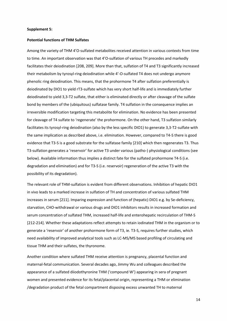
14
Supplement 5:
Potential functions of THM Sulfates
Among the variety of THM 4’O-sulfated metabolites received attention in various contexts from time
to time. An important observation was that 4’O-sulfation of various TH precedes and markedly
facilitates their deiodination [208, 209]. More than that, sulfation of T4 and T3 significantly increased
their metabolism by tyrosyl-ring deiodination while 4’-O-sulfated T4 does not undergo anymore
phenolic ring deiodination. This means, that the prohormone T4 after sulfation preferentially is
deiodinated by DIO1 to yield rT3-sulfate which has very short half-life and is immediately further
deiodinated to yield 3,3-T2 sulfate, that either is eliminated directly or after cleavage of the sulfate
bond by members of the (ubiquitous) sulfatase family. T4 sulfation in the consequence implies an
irreversible modification targeting this metabolite for elimination. No evidence has been presented
for cleavage of T4 sulfate to ‘regenerate’ the prohormone. On the other hand, T3 sulfation similarly
facilitates its tyrosyl-ring deiodination (also by the less specific DIO1) to generate 3,3-T2-sulfate with
the same implication as described above, i.e. elimination. However, compared to T4-S there is good
evidence that T3-S is a good substrate for the sulfatase family [210] which then regenerates T3. Thus
T3-sulfation generates a ‘reservoir’ for active T3 under various (patho-) physiological conditions (see
below). Available information thus implies a distinct fate for the sulfated prohormone T4-S (i.e.
degradation and elimination) and for T3-S (i.e. reservoir) regeneration of the active T3 with the
possibility of its degradation).
The relevant role of THM-sulfation is evident from different observations. Inhibition of hepatic DIO1
in vivo leads to a marked increase in sulfation of TH and concentration of various sulfated THM
increases in serum [211]. Imparing expression and function of (hepatic) DIO1 e.g. by Se-deficiency,
starvation, CHO-withdrawal or various drugs and DIO1 inhibitors results in increased formation and
serum concentration of sulfated THM, increased half-life and enterohepatic recirculation of THM-S
[212-214]. Whether these adaptations reflect attempts to retain iodinated THM in the organism or to
generate a ‘reservoir’ of another prohormone form of T3, ie. T3-S, requires further studies, which
need availability of improved analytical tools such as LC-MS/MS based profiling of circulating and
tissue THM and their sulfates, the thyronome.
Another condition where sulfated THM receive attention is pregnancy, placental function and
maternal-fetal communication. Several decades ago, Jimmy Wu and colleagues described the
appearance of a sulfated diiodothyronine THM (‘compound W’) appearing in sera of pregnant
women and presented evidence for its fetal/placental origin, representing a THM or elimination
/degradation product of the fetal compartment disposing excess unwanted TH to maternal

15
circulation and elimination [214]. The exact nature of compound W remained elusive except for its
relation to T2 and its sulfation state [43, 214]. Recently, a more precise assay was developped and
concentrations of compound W were described in more detail [215]. Still further work is needed to
better understand the biochemical nature and processes leading to its formation and placental
export into maternal serum and its fetal/placental function. Several years ago, R. Ekins postulated
the hypothesis [216], that a major part of maternal/fetal import of TH and THM to the fetal
compartment during pregnancy represents an efficient way to deliver the essential trace element
iodine to the fetus (apart from its direct import by NIS) [94] and that especially the high expression
of DIO3 in placental membranes during pregnancy is a clever way to prevent unwanted fetal
exposure to excess maternal TH and at the same time to use iodide liberated from T4 during
deiodination to rT3 for fetal supply. Whether stoichiometry and physiology fit with this hypothesis
has so far not been tested.
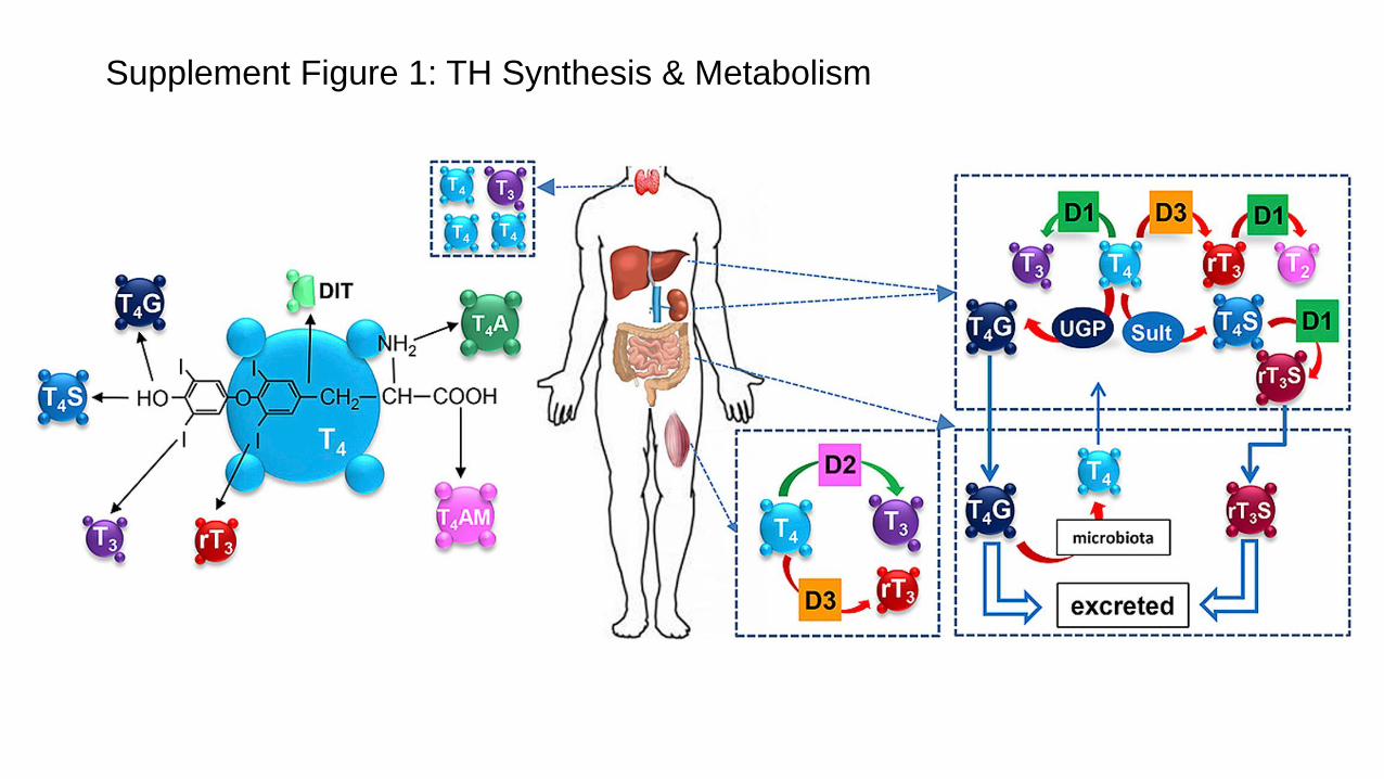
Supplement Figure 1: TH Synthesis & Metabolism

HO
I
O
I
I
OH
O
H2N
I
Supplement Figure 2A: The “hot” Thyroid hormones T4, T3, & 3,5-T2
T3 (3,3´,5-Triiodothyronine)
T4 (Thyroxine, 3,3′,5,5′-Tetraiodothyronine)
5´Deiodinases (DIO1, DIO2)
HO
I
O
I
I
OH
O
H2N
active hormone
HO
I
O
I
I
OH
O
H2N
5´Deiodinases (DIO1, DIO2)
3,5-T2 (3,5-Diiodothyronine)

Supplement Figure 2B: Thyroid hormones & Thyronamines
3-T1AM (3-Iodo-Thyronamine)
T4 (Thyroxine,3,3′,5,5′-Tetraiodothyronine)
Deiodinases & Decarboxilase
the “cool” thyroid hormone
HO O
NH3+H
H
COO-
-O O
NH3H

Visualization of rT3 in tadpoles. We were able to observe rT3-specific ions at m/z 458. By monitoring the value, we visualized rT3 localization in both stages 54 and 61. White arrows show the specific distribution of rT3; which were distributed in intestine, brain, and muscle. Scale bar: 2.5 mm
Visualization of T4 and T3 during metamorphosis.The molecular ions of T3 (m/z 605) and T4 (m/z 731) were demonstrated and the relative amount with the same threshold set at a maximum intensity count of 200 were compared. White arrows show the specific distribution of T3 and T4. T3 were distributed in gill and eyes, and T4 were detected in eyes and inside of the gills. Scale bar: 2.5 mm
Supplement Figure 3: Localization of T4, T3 and rT3 in tadpole tissues during metamorphosis
A B

1
Legends for Supplementary Figures
Supplement Figure 1: Thyroid Hormone Metabolic Pathways. The amino acid derived thyroid (pro-)
hormone T4 may undergo several metabolic transformations to active and inactive THM.
Abbreviations: T4A: Tetrac (Tetraiodothyroacetic acid); T4AM: Tetraiodothyronamine (not yet
identified ex vivo); rT3: 3,3‘,5‘-triiodothyronine (reverseT3); T4S: T4-O-sulfate; T4G: T4-
glucuronide; DIT: Diiodotyrosine; D2: Deiodinase Type 2; D3: Deiodinase Type 3 (Inactivating).
From van der Spek AH, Fliers E, Boelen AMol Cell Endocrinol. 2017 Jan 18. pii: S0303-
7207(17)30029-1. doi: 10.1016/j.mce.2017.01.025. (reproduced with permission).
Supplement Figure 2A. The “hot” Thyroid hormones T4, T3, & 3,5-T2. The human thyroid produces all
endogenous T4 and to some extent T3. The majority of T3, the bioactive hormone, is generated
in responsive tissues by the two 5‘-Deiodinases DIO1 and DIO2, the thyromimetic ‚hot‘ THM,
3,5-T2 has been found in blood and tissues, but its formation from the postulated precursor,
T3, has not yet been formally proven in vitro. Red shading illustrates thermogenic (‘hot’) action
of high concentrations of exogenous 3,5-T2 administered to experimental animals.
Both figures 2A and 2B were designed by Peter J. Hofmann, IEÉ.
Supplement Figure 2B. Thyroid hormones & Thyronamines. 3-T1AM, the ‘cool’ THM found in blood
and tissues, is generated from T4 and THM via DIO and Ornithine Decarboxylase (ODC)
activities. Details of its endogenous formation and mode(s) of action require further studies.
Blue shading illustrates anapyrexic action (‘cool’) of high concentrations of exogenous 3-T1AM,
administered to experimental animals.
Both figures 2A and 2B were designed by Peter J. Hofmann, IEÉ.
Supplement Figure 3: Localization of T4, T3 and rT3 in tadpole tissues during metamorphosis.
Visualization of T4 and T3 during metamorphosis by mass spectrometry imaging [67]. A) The
molecular ions of T3 (m/z 605) and T4 (m/z 731) were demonstrated and the relative amount
with the same threshold set at a maximum intensity count of 200 were compared. White
arrows show the specific distribution of T3 and T4. T3 was distributed in gill and eyes, and T4
was detected in eyes and inside of the gills. B) Visualization of rT3 in tadpoles. rT3-specific
ions were observed at m/z 458. By monitoring the value, rT3 localization was visualized in
both metamorphic stages 54 and 61. White arrows show the specific distribution of rT3,
which were distributed in intestine, brain, and muscle. Scale bars: 2.5 mm. (Reproduced with
permission [67]).