Secondary Metabolites in Basil, Bio-Insecticide, Inhibition ...
25
Citation: Darrag, H.M.; Almuhanna, H.T.; Hakami, E.H. Secondary Metabolites in Basil, Bio-Insecticide, Inhibition Effect, and In Silico Molecular Docking against Proteolytic Enzymes of the Red Palm Weevil (Rhynchophorus ferrugineus). Plants 2022, 11, 1087. https:// doi.org/10.3390/plants11081087 Academic Editors: Filippo Maggi and Giovanni Benelli Received: 28 March 2022 Accepted: 15 April 2022 Published: 16 April 2022 Publisher’s Note: MDPI stays neutral with regard to jurisdictional claims in published maps and institutional affil- iations. Copyright: © 2022 by the authors. Licensee MDPI, Basel, Switzerland. This article is an open access article distributed under the terms and conditions of the Creative Commons Attribution (CC BY) license (https:// creativecommons.org/licenses/by/ 4.0/). plants Article Secondary Metabolites in Basil, Bio-Insecticide, Inhibition Effect, and In Silico Molecular Docking against Proteolytic Enzymes of the Red Palm Weevil (Rhynchophorus ferrugineus) Hossam Moustafa Darrag 1,2, * , Hani Taher Almuhanna 3 and Emadaldeen Hamad Hakami 3 1 Department of Research and Training, Research and Training Station, King Faisal University, Al-Ahsa 31982, Saudi Arabia 2 Pesticide Chemistry and Technology Department, Faculty of Agriculture, Alexandria University, Alexandria 21545, Egypt 3 Research and Training Station, King Faisal University, Al-Ahsa 31982, Saudi Arabia; [email protected] (H.T.A.); [email protected] (E.H.H.) * Correspondence: [email protected]; Tel.: +966-508299027 Abstract: The purpose of this work was to determine the secondary metabolites generated by O. basilicum cell suspensions, as well as their insecticide and inhibitory activity against R. ferrugineus. The growth kinetics with inoculation Verticillium dahliae were determined and identified using LC-MS. Determination of total phenolic components (TFC), flavonoids (TF), and condensed tannins (TCT) were measured. Insecticidal activity of O. basilicum extract against R. ferrugineus (larva and adult) and proteolytic enzymes activity were assessed (in vitro and in vivo). The O. basilicum extract had an LC 50 of 1238 μg/mL and an LD 50 of 13.4 μg/larva. The LC 50 of chicoric acid, ursolic acid, salvigenin, quercetin-3-O-rutinoside, rosmarinyl glucoside, and nepetoidin B demonstrated activity at an LC 50 of 1132, 1167, 1189, 1214, 1275, and 1317 μg/mL, respectively. Chicoric acid, salvigenin, nepetoidin B, and rosmarinic acid demonstrated an LD 50 activity of 10.23, 11.4, 11.9, and 12.4 μg/larva, respectively. The active extract of O. basilicum inhibited total protease, trypsin-like serine proteinases, elastase, cysteine, and metalloprotease activity with an IC 50 (in vitro) of 119.4, 91, 102.4, 76.4, and 52.4 μg/mL, respectively. In silico studies of compounds were conducted, such as molecular docking and ADMET analysis. The study proposes using an efficient cell suspension technique to produce O. basilicum extract containing active secondary metabolites and accessible using as bio-insecticide. Keywords: Biopesticides; biotic elicitor; compounds activity; docking energy; embryogenic calli; liquid media; target enzyme; somatic embryos 1. Introduction Date palm (Phoenix dactylifera L.) is an important economic crop that is commonly attacked by a variety of pests during its growth season. Rhynchophorus ferrugineus (Oliver) is one of the most harmful pests to date. Additionally, it is a significant pest of several palm species; it has been found to affect over 21 palm species globally [1–3], resulting in agricultural output losses. Larvae are considered to be one of the most difficult and dangerous stages, since they grow swiftly and infiltrate palms, killing and deforming palm fronds by feeding on the apical meristem [4,5]. Once infected, palms become vulnerable to infection by a range of insects, fungus, and pests through the tunnels made by larvae [6,7]. Rhynchophorus ferrugineus is a tropical insect prevalent and widely distributed in the Middle East and Mediterranean region, which encompasses North Africa and Europe. It is the most destructive of the ten species of the genus Rhynchophorus, which are widely distributed across the pan-tropics [8–12]. Numerous active ingredients (including fixed oils, volatile oils, and other compounds) have been extracted from natural resources that are used for pest control [13]. The larvicidal Plants 2022, 11, 1087. https://doi.org/10.3390/plants11081087 https://www.mdpi.com/journal/plants
-
Upload
khangminh22 -
Category
Documents
-
view
5 -
download
0
Transcript of Secondary Metabolites in Basil, Bio-Insecticide, Inhibition ...

�����������������
Citation: Darrag, H.M.; Almuhanna,
H.T.; Hakami, E.H. Secondary
Metabolites in Basil, Bio-Insecticide,
Inhibition Effect, and In Silico
Molecular Docking against
Proteolytic Enzymes of the Red Palm
Weevil (Rhynchophorus ferrugineus).
Plants 2022, 11, 1087. https://
doi.org/10.3390/plants11081087
Academic Editors: Filippo Maggi and
Giovanni Benelli
Received: 28 March 2022
Accepted: 15 April 2022
Published: 16 April 2022
Publisher’s Note: MDPI stays neutral
with regard to jurisdictional claims in
published maps and institutional affil-
iations.
Copyright: © 2022 by the authors.
Licensee MDPI, Basel, Switzerland.
This article is an open access article
distributed under the terms and
conditions of the Creative Commons
Attribution (CC BY) license (https://
creativecommons.org/licenses/by/
4.0/).
plants
Article
Secondary Metabolites in Basil, Bio-Insecticide, InhibitionEffect, and In Silico Molecular Docking against ProteolyticEnzymes of the Red Palm Weevil (Rhynchophorus ferrugineus)Hossam Moustafa Darrag 1,2,* , Hani Taher Almuhanna 3 and Emadaldeen Hamad Hakami 3
1 Department of Research and Training, Research and Training Station, King Faisal University,Al-Ahsa 31982, Saudi Arabia
2 Pesticide Chemistry and Technology Department, Faculty of Agriculture, Alexandria University,Alexandria 21545, Egypt
3 Research and Training Station, King Faisal University, Al-Ahsa 31982, Saudi Arabia;[email protected] (H.T.A.); [email protected] (E.H.H.)
* Correspondence: [email protected]; Tel.: +966-508299027
Abstract: The purpose of this work was to determine the secondary metabolites generated byO. basilicum cell suspensions, as well as their insecticide and inhibitory activity against R. ferrugineus.The growth kinetics with inoculation Verticillium dahliae were determined and identified using LC-MS.Determination of total phenolic components (TFC), flavonoids (TF), and condensed tannins (TCT)were measured. Insecticidal activity of O. basilicum extract against R. ferrugineus (larva and adult)and proteolytic enzymes activity were assessed (in vitro and in vivo). The O. basilicum extract had anLC50 of 1238 µg/mL and an LD50 of 13.4 µg/larva. The LC50 of chicoric acid, ursolic acid, salvigenin,quercetin-3-O-rutinoside, rosmarinyl glucoside, and nepetoidin B demonstrated activity at an LC50
of 1132, 1167, 1189, 1214, 1275, and 1317 µg/mL, respectively. Chicoric acid, salvigenin, nepetoidin B,and rosmarinic acid demonstrated an LD50 activity of 10.23, 11.4, 11.9, and 12.4 µg/larva, respectively.The active extract of O. basilicum inhibited total protease, trypsin-like serine proteinases, elastase,cysteine, and metalloprotease activity with an IC50 (in vitro) of 119.4, 91, 102.4, 76.4, and 52.4 µg/mL,respectively. In silico studies of compounds were conducted, such as molecular docking and ADMETanalysis. The study proposes using an efficient cell suspension technique to produce O. basilicumextract containing active secondary metabolites and accessible using as bio-insecticide.
Keywords: Biopesticides; biotic elicitor; compounds activity; docking energy; embryogenic calli;liquid media; target enzyme; somatic embryos
1. Introduction
Date palm (Phoenix dactylifera L.) is an important economic crop that is commonlyattacked by a variety of pests during its growth season. Rhynchophorus ferrugineus (Oliver)is one of the most harmful pests to date. Additionally, it is a significant pest of severalpalm species; it has been found to affect over 21 palm species globally [1–3], resultingin agricultural output losses. Larvae are considered to be one of the most difficult anddangerous stages, since they grow swiftly and infiltrate palms, killing and deforming palmfronds by feeding on the apical meristem [4,5]. Once infected, palms become vulnerable toinfection by a range of insects, fungus, and pests through the tunnels made by larvae [6,7].Rhynchophorus ferrugineus is a tropical insect prevalent and widely distributed in the MiddleEast and Mediterranean region, which encompasses North Africa and Europe. It is the mostdestructive of the ten species of the genus Rhynchophorus, which are widely distributedacross the pan-tropics [8–12].
Numerous active ingredients (including fixed oils, volatile oils, and other compounds)have been extracted from natural resources that are used for pest control [13]. The larvicidal
Plants 2022, 11, 1087. https://doi.org/10.3390/plants11081087 https://www.mdpi.com/journal/plants

Plants 2022, 11, 1087 2 of 25
impact may be a result of a variety of separate components, including terpenoids, alkaloids,flavonoids, and sterols [14]. Certain naturally separated compounds, such as filiferol andextracts of Justicia brandegeana Cangelosi, et al., revealed larvicidal activity and have astrong biocontrol effect on the red palm weevil R. ferrugineus [15]. Tests in the lab haveshown that Invasive Alien has insecticidal efficacy at controlling Rice Weevils [16]. It wasshown to have a possible impact on enzymatic bioactivity and chitinase effectiveness,as well as a deleterious effect on protein synthesis, the enzymatic system, and DNAdamage [3]. Calotrapos gigantean latex has been found to be pesticide against R. ferrugineus,and an inhibitor of serine protease [17]. According to previous studies, protease inhibitorsare effective against a variety of biotic factors and have protective effect in plants, as,potentially, ecologically beneficial agrochemicals [18]. Monoterpene derivatives exhibitedpesticidal activity, making them interesting candidates for the development of safe andenvironmentally acceptable agents [19]. Secondary metabolites might be utilized to monitorthe red palm weevil by possible candidates for R. ferrugineus population control, such asgeraniol, 1-octen-3-ol, and α-pinene [20]. Indeed, α-pinene, with methyl salicylate, inparticular, demonstrated pheromone-disrupting properties [21]. Additionally, coumarininhibited the expression of genes in the R. ferrugineus detoxifying process, suggesting thatit might be employed as a control agent [22]. Additionally, picrotoxin may be used as abioinsecticide to suppress infestations of R. ferrugineus [23].
Ocimum basilicum L. is a plant from the Lamiaceae family that has been historicallygrown globally owing to its major characteristics [24]. Numerous genera, including Ocimum,generate a variety of secondary metabolites, including phenols, terpenoids, flavonoids, andalkaloids, which provide a wide range of activity and applications, including antioxidantand anti-inflammatory properties, as well as antibacterial properties [25]. Monoterpenes,sesquiterpenes, phenylpropanoids derivatives, and flavonoids have been found in severalspecies of the Lamiaceae family, including O. basilicum [26–28], and may be researched forpotential use as bio-insecticides. In this context, plant cells and tissue cultures producesecondary metabolites in a regulated manner. Current productivity and yield levels areinadequate to fulfill the bioprocess objectives for secondary metabolite generation byplant cells [29–31]. Opportunities for plant-cell-based processes, new paths, and recentadvancements are thoroughly evaluated.
Plant reproduction has been accomplished by the use of somatic embryogenesis andgenotypes employing a variety of explants using meristematic cells [1]. Somatic embryo-genesis is more effective and may be utilized to produce secondary metabolites. Previousresearch has been conducted to optimize plant somatic embryogenesis by manipulatingculture medium ingredients such as auxins, amino acids, cytokinins, N-phenyl N’-1,2,3-thidiazol-5-ylurea (TDZ), abscisic acid, biotin, sucrose, thiamine, organic additives, andbasal salt formulations [29–32]. There have been few investigations on the production ofbioactive chemical synthesis in vitro cultures by plants. We extend our prior work on char-acterization of O. basilicum and Thymus vulgaris as eco-insecticides against R. ferrugineus [1].As a result, such as thorough chemical composition of volatile compounds isolated fromO. basilicum and T. vulgaris cells suspension. Furthermore, the growth kinetics of cell sus-pension extracts were investigated, studying the effect of incubated with V. dahliae andidentifying chemical components using GC-MS [1,33–36].
There has recently been renewed interest in the secondary metabolites productionin vitro from employing cell suspension cultures [1]. A hypothesis for this study used thecell suspension technique for produced secondary metabolites from O. basilicum, estab-lishing a link between the polyphenolic and flavonoid (secondary metabolites) chemicalscontained in O. basilicum and their usage as bio-insecticides against R. ferrugineus. Thecurrent work seeks to investigate the growth kinetics of O. basilicum cell suspensions inorder to develop and produce secondary metabolites. The phenolic composition of thecomponents generated from O. basilicum culture was determined using LC-MS (total phe-nols, flavonoids, and (poly) phenolic acids). The insecticidal activity of R. ferrugineus larvaeand adults is investigated in vivo and in vitro for contact insecticide and antifeedant action,
Plants 2022, 11, 1087 3 of 25
as well as inhibition of the red palm weevil’s serine, cysteine, and metalloproteinases. Insilico ADMET property evaluation (absorption, distribution, metabolism, excretion, andtoxicity) as well as molecular docking occurred. The findings are likely to culminate in thedevelopment of an eco-friendly natural bio-insecticide to combat this pest.
2. Results2.1. Initiation of Callus and Cell Suspension
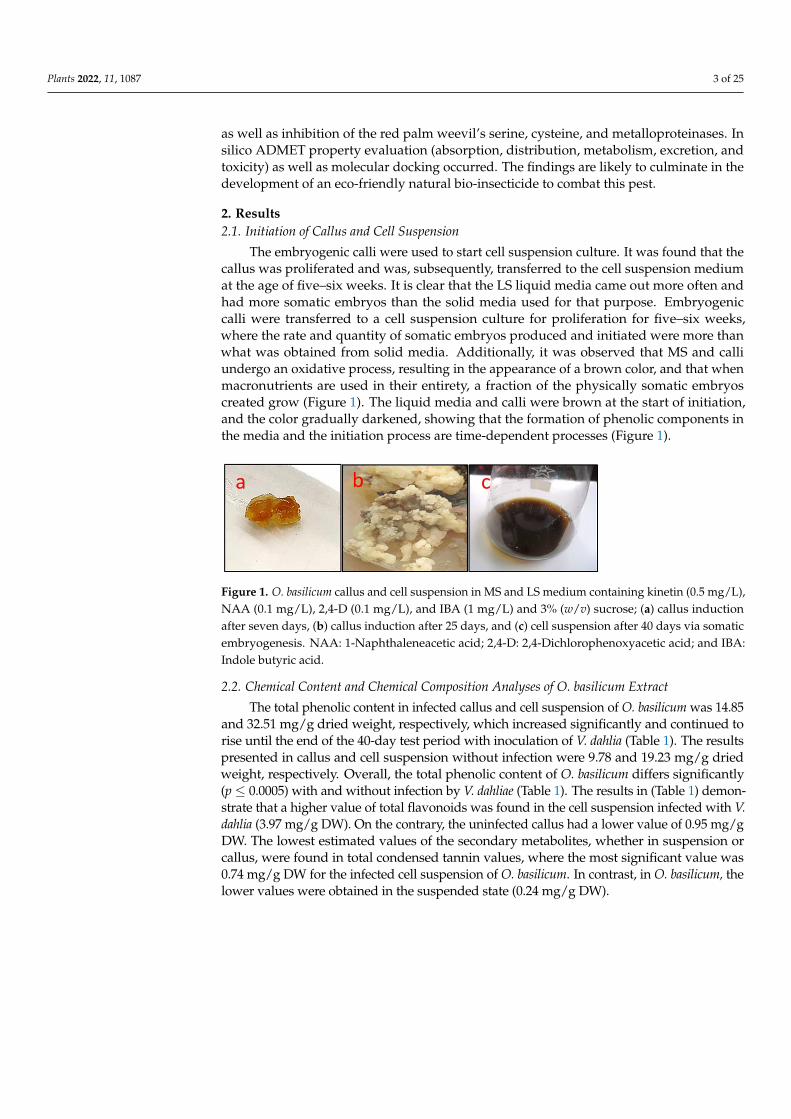
The embryogenic calli were used to start cell suspension culture. It was found that thecallus was proliferated and was, subsequently, transferred to the cell suspension mediumat the age of five–six weeks. It is clear that the LS liquid media came out more often andhad more somatic embryos than the solid media used for that purpose. Embryogeniccalli were transferred to a cell suspension culture for proliferation for five–six weeks,where the rate and quantity of somatic embryos produced and initiated were more thanwhat was obtained from solid media. Additionally, it was observed that MS and calliundergo an oxidative process, resulting in the appearance of a brown color, and that whenmacronutrients are used in their entirety, a fraction of the physically somatic embryoscreated grow (Figure 1). The liquid media and calli were brown at the start of initiation,and the color gradually darkened, showing that the formation of phenolic components inthe media and the initiation process are time-dependent processes (Figure 1).
Plants 2022, 11, 1087 3 of 25
components generated from O. basilicum culture was determined using LC-MS (total phe-nols, flavonoids, and (poly) phenolic acids). The insecticidal activity of R. ferrugineus lar-vae and adults is investigated in vivo and in vitro for contact insecticide and antifeedant action, as well as inhibition of the red palm weevil’s serine, cysteine, and metalloprotein-ases. In silico ADMET property evaluation (absorption, distribution, metabolism, excre-tion, and toxicity) as well as molecular docking occurred. The findings are likely to culmi-nate in the development of an eco-friendly natural bio-insecticide to combat this pest.
2. Results 2.1. Initiation of Callus and Cell Suspension
The embryogenic calli were used to start cell suspension culture. It was found that the callus was proliferated and was, subsequently, transferred to the cell suspension me-dium at the age of five–six weeks. It is clear that the LS liquid media came out more often and had more somatic embryos than the solid media used for that purpose. Embryogenic calli were transferred to a cell suspension culture for proliferation for five–six weeks, where the rate and quantity of somatic embryos produced and initiated were more than what was obtained from solid media. Additionally, it was observed that MS and calli un-dergo an oxidative process, resulting in the appearance of a brown color, and that when macronutrients are used in their entirety, a fraction of the physically somatic embryos created grow (Figure 1). The liquid media and calli were brown at the start of initiation, and the color gradually darkened, showing that the formation of phenolic components in the media and the initiation process are time-dependent processes (Figure 1).
Figure 1. O. basilicum callus and cell suspension in MS and LS medium containing kinetin (0.5 mg/L), NAA (0.1 mg/L), 2,4-D (0.1 mg/L), and IBA (1 mg/L) and 3% (w/v) sucrose; (a) callus induction after seven days, (b) callus induction after 25 days, and (c) cell suspension after 40 days via somatic em-bryogenesis. NAA: 1-Naphthaleneacetic acid; 2,4-D: 2,4-Dichlorophenoxyacetic acid; and IBA: In-dole butyric acid.
2.2. Chemical Content and Chemical Composition Analyses of O. basilicum Extract The total phenolic content in infected callus and cell suspension of O. basilicum was
14.85 and 32.51 mg/g dried weight, respectively, which increased significantly and con-tinued to rise until the end of the 40-day test period with inoculation of V. dahlia (Table 1). The results presented in callus and cell suspension without infection were 9.78 and 19.23 mg/g dried weight, respectively. Overall, the total phenolic content of O. basilicum differs significantly (p ≤ 0.0005) with and without infection by V. dahliae (Table 1). The results in (Table 1) demonstrate that a higher value of total flavonoids was found in the cell suspen-sion infected with V. dahlia (3.97 mg/g DW). On the contrary, the uninfected callus had a lower value of 0.95 mg/g DW. The lowest estimated values of the secondary metabolites, whether in suspension or callus, were found in total condensed tannin values, where the most significant value was 0.74 mg/g DW for the infected cell suspension of O. basilicum. In contrast, in O. basilicum, the lower values were obtained in the suspended state (0.24 mg/g DW).
Figure 1. O. basilicum callus and cell suspension in MS and LS medium containing kinetin (0.5 mg/L),NAA (0.1 mg/L), 2,4-D (0.1 mg/L), and IBA (1 mg/L) and 3% (w/v) sucrose; (a) callus inductionafter seven days, (b) callus induction after 25 days, and (c) cell suspension after 40 days via somaticembryogenesis. NAA: 1-Naphthaleneacetic acid; 2,4-D: 2,4-Dichlorophenoxyacetic acid; and IBA:Indole butyric acid.
2.2. Chemical Content and Chemical Composition Analyses of O. basilicum Extract
The total phenolic content in infected callus and cell suspension of O. basilicum was 14.85and 32.51 mg/g dried weight, respectively, which increased significantly and continued torise until the end of the 40-day test period with inoculation of V. dahlia (Table 1). The resultspresented in callus and cell suspension without infection were 9.78 and 19.23 mg/g driedweight, respectively. Overall, the total phenolic content of O. basilicum differs significantly(p ≤ 0.0005) with and without infection by V. dahliae (Table 1). The results in (Table 1) demon-strate that a higher value of total flavonoids was found in the cell suspension infected with V.dahlia (3.97 mg/g DW). On the contrary, the uninfected callus had a lower value of 0.95 mg/gDW. The lowest estimated values of the secondary metabolites, whether in suspension orcallus, were found in total condensed tannin values, where the most significant value was0.74 mg/g DW for the infected cell suspension of O. basilicum. In contrast, in O. basilicum, thelower values were obtained in the suspended state (0.24 mg/g DW).

Plants 2022, 11, 1087 4 of 25
Table 1. Chemical contents of total phenolic, total flavonoid (TF), and condensed tannins (TCT) fromcells from methanolic extracts of O. basilicum callus and cell suspensions infected by V. dahliae.
TF(mg of Quercetin/g DW)
TPC(mg of Gallic Acid/g DW) Callus and Cell Suspension TCT
(mg of Cyanidins/g DW)
0.95 d ± 0.1324 9.78 d ± 0.1109 Callus without infection 0.24 d ± 0.04541.36 c ± 0.1562 14.85 c ± 0.1674 Callus with infection 0.38 c ± 0.0742
2.21 b ± 0.0413 19.23 b ± 0.1457Cell suspension without
infection 0.41 b ± 0.0316
3.97 a ± 0.0478 32.51 a ± 0.1904 Cell suspension with infection 0.74 a ± 0.1245
Mean ± Standard deviation (SD), n = 3, total phenolic content (TPC), total flavonoid (TF), total condensed tannins(TCT). Values followed by the same letter within a column are not significantly different (p ≤ 0.0005 ≡ significantfor all values) according to Student–Newman–Keuls (SNK) test.
2.3. Polyphenolic Acids and Flavonoids Compounds in O. basilicum Cell Suspension ExtractsUsing UPLC–I Class Coupled with Xevo TQD MS
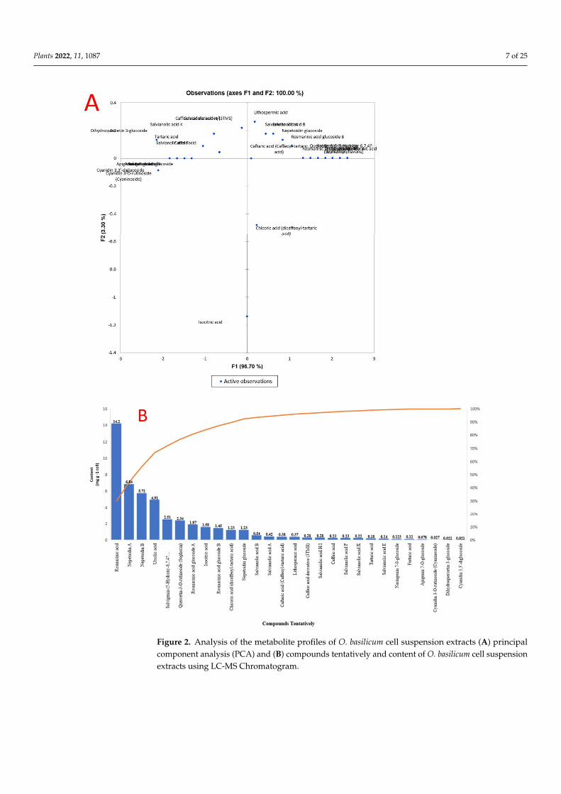
The concentrations of polyphenolic acids and flavonoids in O. basilicum cell suspensionextracts identify 27 compounds using molecular weight, retention time, and fragmentationprofile according to the literature and software’s data bank; this approach allowed for(Table 2). The detected compounds were identified using the negative scan mode of themass spectrometer; the scan took 45 min, and the results for the extract are summarized.More specifically, the most abundant peak in extracts was rosmarinic acid, a polyphenol incell suspension of O. basilicum (14.2 mg/g DW) with a retention time of 11.60 min at m/z359.08 (M-H), m/z 117, 135, 161, 179, and 197 were used to identify the items (rosmarinicacid). Nepetoidin (A and B) was presented in an extract with 6.84 and 5.72 mg/g DW.Nepetoidin A and B had a retention time of 25.53 and 25.67 min, respectively, at m/z314.29 (M-H). The molecular ions were identified at m/z 133, 161, 313, and 335 for nepetoidinA and 133, 161, 269, 313, and 335 for nepetoidin B. Basil cell suspension extract containedursolic acid (4.91 mg/g DW), a polyphenol. It retained 26.06 min at m/z 456.7 (M-H),and the resultant ions had m/z 455, 456, 523, 524, and 591. Salvigenin (5-Hydroxy-6,7,4′-trimethoxyflavone was observed extracted with 2.51 mg/g DW at m/z 327.215 (M-H), aretention time of 18.29 min, and resultant ions at m/z 116.9, 205, 215, 277, and 311. Quercetin-3-O-rutinoside (Rutin), a flavonoid, was observed extracted with 2.34 mg/g DW at m/z611.16 (M-H), a retention time of 11.53 min, and resultant ions at m/z 465, 449, and 303.Rosmarinic acid glucoside A and B were present in the cell suspension extract of basil with1.84 and 1.45 mg/g DW, respectively. It had a retention time of 21.37 and 25.07 min atm/z 521.12 (M-H), and the resultant ions were identified at m/z 135, 161, 179, 197, and 359for rosmarinic acid glucoside A as well as 135, 161, 179, 197, 323, and 359 for rosmarinicacid glucoside. Nepetoidin glucoside was present in the extract with 1.23 mg/g DWand a retention time of 27.99 min at m/z 475.12 (M-H). The presence of molecular ionswas discovered at m/z 151, 161, 313, 323, and 475. Acidic compounds such as chicoricacid (dicaffeoyl-tartaric acid) and isocitric acid were significantly presented in the cellsuspension extract at m/z 473, 311, 293, 179, and 149 at a retention time of 11.18 min ofchicoric acid. Isocitric acid was present with a retention time of 2.54 min, m/z 191.01 (M-H),with resultant ions at m/z 111, 129, and 173 (Figure S1).
Additionally, other components were discovered in the extracts as naringenin7-0-glucoside, apigenin 7-O-glucoside, cyanidin 3-O-rutinoside (cyaninoside), cyanidin3,3’-diglucoside, and apigenin 7-O-glucoside with m/z 434.4, 432.4, 595.17, 611.16, and432.4 (M-H), respectively. The salvianolic acid chemicals found in extracts included thefollowing: salvianolic acid F (m/z 313.07 (M-H)), salvianolic acid B (m/z 717.15 (M-H)),salvianolic acid A (m/z 493.11 (M-H)), salvianolic acid E (m/z 717.15 (M-H)), salvianolic acidK (m/z 555.11 (M-H)), and salvianolic acid H/I (m/z 537.10 (M-H)). Other acids found werelithospermic acid, fertaric acid, caffeic acid, caftaric acid (caffeoyl-tartaric acid), caffeic acidderivative (3TMS), and tartaric acid, with m/z of 537.10, 325.06, 179.03, 311.04, 359.70, and149.0076, respectively [24,37–51].

Plants 2022, 11, 1087 5 of 25
Table 2. Chemical composition of secondary metabolites, polyphenolic acids, and flavonoids detectedfrom the cell suspensions of O. basilicum by Waters Acquity UPLC –I class coupled with Xevo TQDMS negative mode.
No. Compounds Tentatively RT (min) RI(exp) Formula [M − H]−
(m/z)Fragmentation Ions
(m/z)Content (µmol
g−1 cell)
1 Tartaric acid 1.01 1249 C4H5O6 149.00 149, 141, 131, 113, 103, 87 0.182 Isocitric acid 2.54 1805.4 C6H7O7 191.0175 191, 173, 129, 111 1.58
3 Caffeic acid derivative(3TMS) 4.13 2155 C18H32O4Si3 359.70 396, 381, 359, 219, 191, 75 0.28
4 Caftaric acid(Caffeoyl-tartaric acid) 5.63 2701.3 C13H12O9 311.04 311, 179.03, 149.01, 135.04 0.38
5 Caffeic acid 6.28 1854.3 C9H8O4 179.03 179, 135 0.23
6 Fertaric acid 6.31 5191.1 C14H14O9 325.06 325, 193, 134 0.12
7 Salvianolic acid H/I 6.38 5237.8 C27H22O12 537.10 537, 493, 339, 313, 295,197, 179 0.28
8 Salvianolic acid K 9.57 4556.9 C27H24O13 555.11 555, 537, 493, 295 0.22
9 Chicoric acid(dicaffeoyl-tartaric acid) 11.18 4552.3 C22H18O12 473.07 473, 311, 293, 179, 149 1.23
10 Lithospermic acid 11.31 4920.2 C27H22O12 537.10 537, 493, 356, 295 0.37
11 Dihydroquercetin3-glucoside 11.46 4505.7 C21H22O12 456.10 467, 465, 313, 285, 259,
456, 175, 151 0.021
12 Quercetin-3-O-rutinoside(rutin) 11.53 4992.3 C27H30O16 611.16 611, 465, 449, 303 2.34
12 Rosmarinic acid 11.60 3504.5 C18H16O8 359.08 359, 197, 179, 161, 135, 117 14.2
13 Salvianolic acid E 12.69 4627.5 C36H30O16 717.15 717, 519, 475, 339 0.14
14 Salvianolic acid A 12.49 4585.8 C26H22O10 493.11 493, 313, 295, 185 0.42
15 Salvianolic acid B 12.61 5377.7 C36H30O16 717.15 717, 519, 321 0.54
16 Salvianolic acid F 17.94 4566.3 C17H14O6 313.07 313, 269 0.23
17 Cyanidin 3,3’-diglucoside 18.14 6158.2 C27H31O16 611.16 611, 287 0.021
18 Cyanidin 3-O-rutinoside(Cyaninoside) 18.27 5192.3 C27H31O15 595.17 595, 287 0.027
19 Salvigenin (5-Hydroxy-6,7,4′-trimethoxyflavone) 18.29 3121.7 C18H16O6 327.21 327, 311, 277, 215, 205,
116.9 2.51
20 Naringenin 7-0-glucoside 18.36 4081.3 C21H22Os10 434.4 435, 271, 151, 119 0.123
21 Apigenin 7-O-glucoside 18.45 4142.7 C21H20O10 432.4 432, 271, 171, 147, 119 0.078
22 Rosmarinic acidglucoside A 21.37 4023.4 C24H26O13 521.12 359, 197, 179, 161, 135 1.87
23 Rosmarinic acidglucoside B 25.07 4061.4 C24H26O13 521.12 359, 323, 197, 179, 161, 135 1.45
24 Nepetoidin A 25.53 4413.7 C17H14O6 314.29 335, 313, 161, 133 6.84
25 Nepetoidin B 25.67 4418.9 C17H14O6 314.29 335, 313, 269, 161, 133 5.72
26 Ursolic acid 26.06 3658.3 C30H48O3 456.7 591, 524, 523, 459, 455 4.91
27 Nepetoidin glucoside 27.99 4341 C23H24O11 475.12 475, 323, 313, 161, 151 1.23
28 Unknown 36.11 3697.2 ND ND ND ND
29 Unknown 36.80 3751 ND ND ND ND30 Unknown 42.34 3508 ND ND ND ND
The result was calculated as the mean of three replicates, n = 3, standard deviation (SD); the average relativeabundances of each fragment ion are given in brackets; RT: retention time; [M−H]−: negative ion observed in amolecular ion (m/z: mass/charge); RI (exp): relative retention index determined. RI (exp) relative retention indexfrom MS libraries (Wiley); National Institute of Standards and Technology (NIST).
Principal component analysis (PCA) was performed to validate the differences be-tween samples and the contribution of the metabolites to clustering. Figure 2 demonstratesno difference between O. basilicum extracts. The first component (PC1) accounts for 96.70%

Plants 2022, 11, 1087 6 of 25
of variance, whereas the second component (PC2) accounts for 3.30%. The secondarymetabolites that contributed the most to the cluster of O. basilicum extracts were acids, con-tain a high concentration of rosmarinic acid, nepetoidin (A and B), ursolic acid, salvigenin,quercetin-3-O-rutinoside, rosmarinic acid glucoside A and B, nepetoidin glucoside, andacidic compounds such as isocitric acid and chicoric acid (Figure 2).
2.4. O. basilicum Extract and Pure Compounds Activity against Adults and Larvae of R. ferrugineus
Table 3 shows the extract’s efficiency against adult (R. ferrugineus). The O. basilicum ex-tract was shown to be active against adults with an LC50 of 1238 µg/mL and 95% confidencelimits of 1038–1389. The LC50 values of chicoric acid, ursolic acid, salvigenin, quercetin-3-O-rutinoside, rosmarinyl glucoside, and nepetoidin B had the highest insecticidal activity withLC50 1132, 1167, 1189, 1214, 1275, and 1317 µg/mL, and 95% confidence limits of 1004–1198,1038–1204, 1049–1219, 1089–1234, 1147–1315, and 1268–1346, respectively. Adults’ mod-erate, low activity was presented in rosmarinic acid and isocitric acid with an LC50 of1495 and 1826 µg/mL, respectively. The topical application showed that the LD50 valueof O. basilicum extracts (µg/larva) was 13.7. Chicoric acid, salvigenin, nepetoidin B, androsmarinic acid showed the highest insecticidal activity with LD50 values of 10.23, 11.4, 11.9,and 12.4 µg/larva, respectively. Ursolic acid, quercetin-3-O-rutinoside, and rosmarinylglucoside all demonstrated moderate activity on the larva, with LD50 values of 15.2, 16.9,and 17.6 g/larva, respectively. Finally, isocitric acid showed low activity with an LD50 of23.9 µg/larva.
2.5. Evaluation Specific Activity of O. basilicum Extract and Pure Compounds on Serine, Cysteine,and Metalloproteinase (In Vitro)
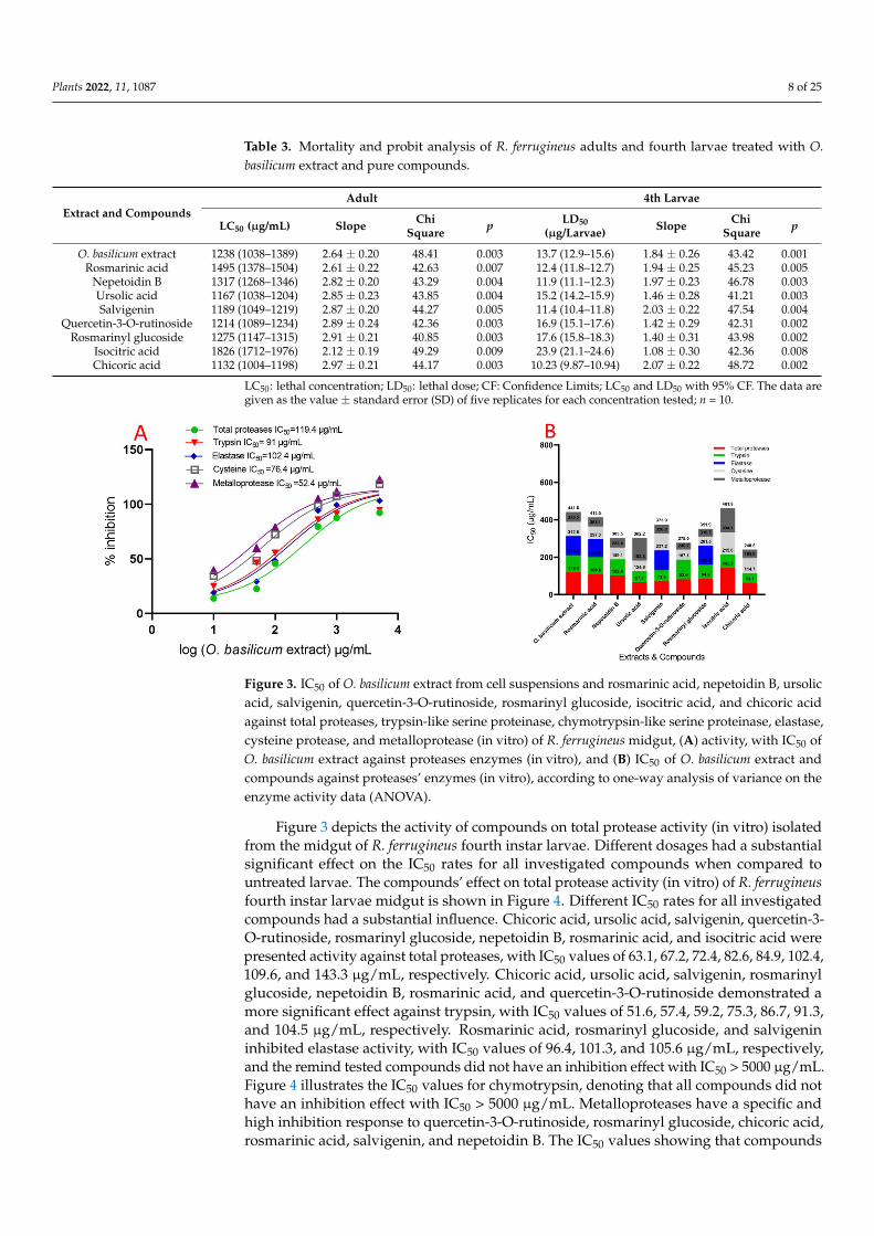
The data demonstrated that serial doses of O. basilicum extract had an effect on IC50values, as compared with untreated proteases from the midgut of larvae. Figure 3 demon-strated unequivocally that the IC50 of the midgut increased with concentrations. The IC50value for O. basilicum extract was 119.4 µg/mL. The IC50 value for O. basilicum extract was119.4 µg/mL. Figure 4 depicts the relative activity and inhibition of trypsin, chymotrypsin,and elastase-like proteinases in the fourth instar midgut homogenate. Serine proteinaseshave comparable specific activities in total homogenate preparations and are expressedas the number of OD/mg protein min. All IC50 values had a significant influence of O.basilicum extracts. The IC50 values in inhibition by O. basilicum extract is substantiallygreater for trypsin-like serine proteinases and elastase than for chymotrypsin-like serineproteinase in midgut homogenate preparations. The IC50 value for O. basilicum extractis shown in Figure 4, which demonstrates that the inhibition varies greatly amongst ho-mogenates of the midgut. In OD/mg protein min from fourth instar midgut preparation,trypsin-like serine and elastase proteinase activities were 4.10 and 1.24, respectively. More-over, O. basilicum was the active extract, with an IC50 of 91 and 102.4 µg/mL, respectively.These extracts have a potent inhibitory effect on trypsin-like serine proteinases isolatedfrom the fourth midgut. Both cysteine and metalloprotease are a strong inhibitory responseto O. basilicum extract; the IC50 values are shown in Figure 4, which demonstrates that theextract exhibits inhibitory activity at IC50 100 µg/mL with values of 76.4 and 52.4 µg/mL,respectively. Chymotrypsin-like serine proteinases have a distinct pattern of activity thanserine activity; the IC50 values for chymotrypsin are shown in Figure 4, which demonstratesthat the extract did not have an inhibition effect against chymotrypsin when the IC50 valueis more than 5000 µg/mL.
Plants 2022, 11, 1087 7 of 25
Plants 2022, 11, 1087 6 of 25
Figure 2. Analysis of the metabolite profiles of O. basilicum cell suspension extracts (A) principal component analysis (PCA) and (B) compounds tentatively and content of O. basilicum cell suspen-sion extracts using LC-MS Chromatogram.
2.4. O. basilicum Extract and Pure Compounds Activity against Adults and Larvae of R. ferrugineus
Table 3 shows the extract’s efficiency against adult (R. ferrugineus). The O. basilicum extract was shown to be active against adults with an LC50 of 1238 µg/mL and 95% confi-dence limits of 1038–1389. The LC50 values of chicoric acid, ursolic acid, salvigenin, quer-cetin-3-O-rutinoside, rosmarinyl glucoside, and nepetoidin B had the highest insecticidal activity with LC50 1132, 1167, 1189, 1214, 1275, and 1317 µg/mL, and 95% confidence limits of 1004–1198, 1038–1204, 1049–1219, 1089–1234, 1147–1315, and 1268–1346, respectively.
Figure 2. Analysis of the metabolite profiles of O. basilicum cell suspension extracts (A) principalcomponent analysis (PCA) and (B) compounds tentatively and content of O. basilicum cell suspensionextracts using LC-MS Chromatogram.
Plants 2022, 11, 1087 8 of 25
Table 3. Mortality and probit analysis of R. ferrugineus adults and fourth larvae treated with O.basilicum extract and pure compounds.
Extract and CompoundsAdult 4th Larvae
LC50 (µg/mL) Slope ChiSquare p LD50
(µg/Larvae) Slope ChiSquare p
O. basilicum extract 1238 (1038–1389) 2.64 ± 0.20 48.41 0.003 13.7 (12.9–15.6) 1.84 ± 0.26 43.42 0.001Rosmarinic acid 1495 (1378–1504) 2.61 ± 0.22 42.63 0.007 12.4 (11.8–12.7) 1.94 ± 0.25 45.23 0.005
Nepetoidin B 1317 (1268–1346) 2.82 ± 0.20 43.29 0.004 11.9 (11.1–12.3) 1.97 ± 0.23 46.78 0.003Ursolic acid 1167 (1038–1204) 2.85 ± 0.23 43.85 0.004 15.2 (14.2–15.9) 1.46 ± 0.28 41.21 0.003Salvigenin 1189 (1049–1219) 2.87 ± 0.20 44.27 0.005 11.4 (10.4–11.8) 2.03 ± 0.22 47.54 0.004
Quercetin-3-O-rutinoside 1214 (1089–1234) 2.89 ± 0.24 42.36 0.003 16.9 (15.1–17.6) 1.42 ± 0.29 42.31 0.002Rosmarinyl glucoside 1275 (1147–1315) 2.91 ± 0.21 40.85 0.003 17.6 (15.8–18.3) 1.40 ± 0.31 43.98 0.002
Isocitric acid 1826 (1712–1976) 2.12 ± 0.19 49.29 0.009 23.9 (21.1–24.6) 1.08 ± 0.30 42.36 0.008Chicoric acid 1132 (1004–1198) 2.97 ± 0.21 44.17 0.003 10.23 (9.87–10.94) 2.07 ± 0.22 48.72 0.002
LC50: lethal concentration; LD50: lethal dose; CF: Confidence Limits; LC50 and LD50 with 95% CF. The data aregiven as the value ± standard error (SD) of five replicates for each concentration tested; n = 10.
Plants 2022, 11, 1087 8 of 25
Figure 3 depicts the activity of compounds on total protease activity (in vitro) isolated from the midgut of R. ferrugineus fourth instar larvae. Different dosages had a substantial significant effect on the IC50 rates for all investigated compounds when compared to un-treated larvae. The compounds’ effect on total protease activity (in vitro) of R. ferrugineus fourth instar larvae midgut is shown in Figure 4. Different IC50 rates for all investigated compounds had a substantial influence. Chicoric acid, ursolic acid, salvigenin, quercetin-3-O-rutinoside, rosmarinyl glucoside, nepetoidin B, rosmarinic acid, and isocitric acid were presented activity against total proteases, with IC50 values of 63.1, 67.2, 72.4, 82.6, 84.9, 102.4, 109.6, and 143.3 µg/mL, respectively. Chicoric acid, ursolic acid, salvigenin, rosmarinyl glucoside, nepetoidin B, rosmarinic acid, and quercetin-3-O-rutinoside demonstrated a more significant effect against trypsin, with IC50 values of 51.6, 57.4, 59.2, 75.3, 86.7, 91.3, and 104.5 µg/mL, respectively. Rosmarinic acid, rosmarinyl glucoside, and salvigenin inhibited elastase activity, with IC50 values of 96.4, 101.3, and 105.6 µg/mL, re-spectively, and the remind tested compounds did not have an inhibition effect with IC50 > 5000 µg/mL. Figure 4 illustrates the IC50 values for chymotrypsin, denoting that all com-pounds did not have an inhibition effect with IC50 > 5000 µg/mL. Metalloproteases have a specific and high inhibition response to quercetin-3-O-rutinoside, rosmarinyl glucoside, chicoric acid, rosmarinic acid, salvigenin, and nepetoidin B. The IC50 values showing that compounds have the highest inhibition where IC50 ˂ 100 µg/mL, with values of 37.3, 41.2, 44.6, 48.4, 49.1, and 51.1 µg/mL, respectively (Figure 3). Furthermore, ursolic acid and iso-citric acid showed moderated activity against metalloproteases, with IC50 values of 119.4 and 127.8 µg/mL, respectively. Moreover, cysteine demonstrated an inhibition response to rosmarinyl glucoside, quercetin-3-O-rutinoside, ursolic acid, nepetoidin B, rosmarinic acid, chicoric acid, and salvigenin compounds, which presented the highest inhibition with values 49.2, 53.6, 58.2, 63.3, 67.8, 81.2, and 89.6 µg/mL, respectively (IC50 ˂ 100 µg/mL). Isocitric acid was lower in IC50 value with 119.1 µg/mL.
These were included in proteolysis activity evaluation, to determine the involvement of several intestinal proteases in the fourth larval instars when protease inhibitors were used. Purified proteases from the fourth larval instars of R. ferrugineus’s midgut were sig-nificantly reduced by inhibitors, as shown in Figure S2. Additionally, the inhibitors of trypsin-like serine proteinases TLCK and chymotrypsin-like serine proteinases TPCK, as well as the cysteine protease inhibitor iodoacetic acid, dramatically suppressed the mid-gut instar (Figure S2).
Figure 3. IC50 of O. basilicum extract from cell suspensions and rosmarinic acid, nepetoidin B, ursolic acid, salvigenin, quercetin-3-O-rutinoside, rosmarinyl glucoside, isocitric acid, and chicoric acid against total proteases, trypsin-like serine proteinase, chymotrypsin-like serine proteinase, elastase, cysteine protease, and metalloprotease (in vitro) of R. ferrugineus midgut, (A) activity, with IC50 of O. basilicum extract against proteases enzymes (in vitro), and (B) IC50 of O. basilicum extract and
Figure 3. IC50 of O. basilicum extract from cell suspensions and rosmarinic acid, nepetoidin B, ursolicacid, salvigenin, quercetin-3-O-rutinoside, rosmarinyl glucoside, isocitric acid, and chicoric acidagainst total proteases, trypsin-like serine proteinase, chymotrypsin-like serine proteinase, elastase,cysteine protease, and metalloprotease (in vitro) of R. ferrugineus midgut, (A) activity, with IC50 ofO. basilicum extract against proteases enzymes (in vitro), and (B) IC50 of O. basilicum extract andcompounds against proteases’ enzymes (in vitro), according to one-way analysis of variance on theenzyme activity data (ANOVA).
Figure 3 depicts the activity of compounds on total protease activity (in vitro) isolatedfrom the midgut of R. ferrugineus fourth instar larvae. Different dosages had a substantialsignificant effect on the IC50 rates for all investigated compounds when compared tountreated larvae. The compounds’ effect on total protease activity (in vitro) of R. ferrugineusfourth instar larvae midgut is shown in Figure 4. Different IC50 rates for all investigatedcompounds had a substantial influence. Chicoric acid, ursolic acid, salvigenin, quercetin-3-O-rutinoside, rosmarinyl glucoside, nepetoidin B, rosmarinic acid, and isocitric acid werepresented activity against total proteases, with IC50 values of 63.1, 67.2, 72.4, 82.6, 84.9, 102.4,109.6, and 143.3 µg/mL, respectively. Chicoric acid, ursolic acid, salvigenin, rosmarinylglucoside, nepetoidin B, rosmarinic acid, and quercetin-3-O-rutinoside demonstrated amore significant effect against trypsin, with IC50 values of 51.6, 57.4, 59.2, 75.3, 86.7, 91.3,and 104.5 µg/mL, respectively. Rosmarinic acid, rosmarinyl glucoside, and salvigenininhibited elastase activity, with IC50 values of 96.4, 101.3, and 105.6 µg/mL, respectively,and the remind tested compounds did not have an inhibition effect with IC50 > 5000 µg/mL.Figure 4 illustrates the IC50 values for chymotrypsin, denoting that all compounds did nothave an inhibition effect with IC50 > 5000 µg/mL. Metalloproteases have a specific andhigh inhibition response to quercetin-3-O-rutinoside, rosmarinyl glucoside, chicoric acid,rosmarinic acid, salvigenin, and nepetoidin B. The IC50 values showing that compounds
Plants 2022, 11, 1087 9 of 25
have the highest inhibition where IC50 < 100 µg/mL, with values of 37.3, 41.2, 44.6, 48.4,49.1, and 51.1 µg/mL, respectively (Figure 3). Furthermore, ursolic acid and isocitricacid showed moderated activity against metalloproteases, with IC50 values of 119.4 and127.8 µg/mL, respectively. Moreover, cysteine demonstrated an inhibition response torosmarinyl glucoside, quercetin-3-O-rutinoside, ursolic acid, nepetoidin B, rosmarinic acid,chicoric acid, and salvigenin compounds, which presented the highest inhibition withvalues 49.2, 53.6, 58.2, 63.3, 67.8, 81.2, and 89.6 µg/mL, respectively (IC50 < 100 µg/mL).Isocitric acid was lower in IC50 value with 119.1 µg/mL.
Plants 2022, 11, 1087 9 of 25
compounds against proteases’ enzymes (in vitro), according to one-way analysis of variance on the enzyme activity data (ANOVA).
Figure 4. IC50 of O. basilicum extract from cell suspensions and rosmarinic acid, nepetoidin B, ursolic acid, salvigenin, quercetin-3-O-rutinoside, rosmarinyl glucoside, isocitric acid, and chicoric acid against total proteases, trypsin-like serine proteinase, chymotrypsin-like serine proteinase, elastase, cysteine protease, and metalloprotease (in vivo) of R. ferrugineus midgut. (A) activity, with IC50 of O. basilicum extract against proteases enzymes (in vivo), and (B) IC50 of O. basilicum extract and com-pounds aganist proteases enzymes (in vivo), according to one-way analysis of variance on the en-zyme activity data (ANOVA).
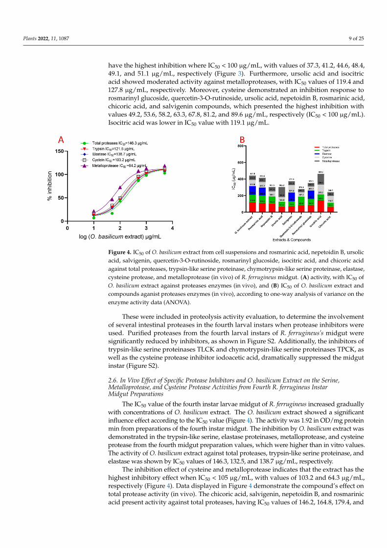
2.6. In Vivo Effect of Specific Protease Inhibitors and O. basilicum Extract on the Serine, Metalloprotease, and Cysteine Protease Activities from Fourth R. ferrugineus Instar Midgut Preparations
The IC50 value of the fourth instar larvae midgut of R. ferrugineus increased gradually with concentrations of O. basilicum extract. The O. basilicum extract showed a significant influence effect according to the IC50 value (Figure 4). The activity was 1.92 in OD/mg pro-tein min from preparations of the fourth instar midgut. The inhibition by O. basilicum ex-tract was demonstrated in the trypsin-like serine, elastase proteinases, metalloprotease, and cysteine protease from the fourth midgut preparation values, which were higher than in vitro values. The activity of O. basilicum extract against total proteases, trypsin-like ser-ine proteinase, and elastase was shown by IC50 values of 146.3, 132.5, and 138.7 µg/mL, respectively.
The inhibition effect of cysteine and metalloprotease indicates that the extract has the highest inhibitory effect when IC50 ˂ 105 µg/mL, with values of 103.2 and 64.3 µg/mL, respectively (Figure 4). Data displayed in Figure 4 demonstrate the compound’s effect on total protease activity (in vivo). The chicoric acid, salvigenin, nepetoidin B, and rosmarinic acid present activity against total proteases, having IC50 values of 146.2, 164.8, 179.4, and 186.2 µg/mL, respectively. Ursolic acid, quercetin-3-O-rutinoside, rosmarinyl glucoside, and isocitric acid activity against total protease shows that compounds have the lowest inhibition when IC50 is 279.5, 286.4, 292.1, and 486.3 µg/mL. Chicoric acid, Nepetoidin B, and rosmarinic acid have a more substantial effect against trypsin, having IC50 values of 75.1, 102.7, and 174.6 µg/mL, respectively. In addition, ursolic acid, salvigenin, rosmarinyl glucoside, isocitric acid, and quercetin-3-O-rutinoside have a non-significant effect against trypsin, having IC50 values of 235.1, 241.2, 274.3, 314.8, and 325.5 µg/mL, respectively. The elastase activity indicates inhibition effects with rosmarinic acid, salvigenin, and rosma-rinyl glucoside, having IC50 values of 119.7 and 138.2, 751.7 µg/mL, respectively, and re-mind tested compounds have the most non-significant inhibition where IC50 > 5000 µg/mL. Metalloproteases have an inhibition response with chicoric acid, rosmarinic acid, nepetoidin B, salvigenin, and isocitric acid; the IC50 values are presented in Figure 5, which
Figure 4. IC50 of O. basilicum extract from cell suspensions and rosmarinic acid, nepetoidin B, ursolicacid, salvigenin, quercetin-3-O-rutinoside, rosmarinyl glucoside, isocitric acid, and chicoric acidagainst total proteases, trypsin-like serine proteinase, chymotrypsin-like serine proteinase, elastase,cysteine protease, and metalloprotease (in vivo) of R. ferrugineus midgut. (A) activity, with IC50 ofO. basilicum extract against proteases enzymes (in vivo), and (B) IC50 of O. basilicum extract andcompounds aganist proteases enzymes (in vivo), according to one-way analysis of variance on theenzyme activity data (ANOVA).
These were included in proteolysis activity evaluation, to determine the involvementof several intestinal proteases in the fourth larval instars when protease inhibitors wereused. Purified proteases from the fourth larval instars of R. ferrugineus’s midgut weresignificantly reduced by inhibitors, as shown in Figure S2. Additionally, the inhibitors oftrypsin-like serine proteinases TLCK and chymotrypsin-like serine proteinases TPCK, aswell as the cysteine protease inhibitor iodoacetic acid, dramatically suppressed the midgutinstar (Figure S2).
2.6. In Vivo Effect of Specific Protease Inhibitors and O. basilicum Extract on the Serine,Metalloprotease, and Cysteine Protease Activities from Fourth R. ferrugineus InstarMidgut Preparations
The IC50 value of the fourth instar larvae midgut of R. ferrugineus increased graduallywith concentrations of O. basilicum extract. The O. basilicum extract showed a significantinfluence effect according to the IC50 value (Figure 4). The activity was 1.92 in OD/mg proteinmin from preparations of the fourth instar midgut. The inhibition by O. basilicum extract wasdemonstrated in the trypsin-like serine, elastase proteinases, metalloprotease, and cysteineprotease from the fourth midgut preparation values, which were higher than in vitro values.The activity of O. basilicum extract against total proteases, trypsin-like serine proteinase, andelastase was shown by IC50 values of 146.3, 132.5, and 138.7 µg/mL, respectively.
The inhibition effect of cysteine and metalloprotease indicates that the extract has thehighest inhibitory effect when IC50 < 105 µg/mL, with values of 103.2 and 64.3 µg/mL,respectively (Figure 4). Data displayed in Figure 4 demonstrate the compound’s effect ontotal protease activity (in vivo). The chicoric acid, salvigenin, nepetoidin B, and rosmarinicacid present activity against total proteases, having IC50 values of 146.2, 164.8, 179.4, and
Plants 2022, 11, 1087 10 of 25
186.2 µg/mL, respectively. Ursolic acid, quercetin-3-O-rutinoside, rosmarinyl glucoside,and isocitric acid activity against total protease shows that compounds have the lowestinhibition when IC50 is 279.5, 286.4, 292.1, and 486.3 µg/mL. Chicoric acid, Nepetoidin B,and rosmarinic acid have a more substantial effect against trypsin, having IC50 values of75.1, 102.7, and 174.6 µg/mL, respectively. In addition, ursolic acid, salvigenin, rosmarinylglucoside, isocitric acid, and quercetin-3-O-rutinoside have a non-significant effect againsttrypsin, having IC50 values of 235.1, 241.2, 274.3, 314.8, and 325.5 µg/mL, respectively. Theelastase activity indicates inhibition effects with rosmarinic acid, salvigenin, and rosmarinylglucoside, having IC50 values of 119.7 and 138.2, 751.7 µg/mL, respectively, and remindtested compounds have the most non-significant inhibition where IC50 > 5000 µg/mL. Met-alloproteases have an inhibition response with chicoric acid, rosmarinic acid, nepetoidin B,salvigenin, and isocitric acid; the IC50 values are presented in Figure 5, which indicates com-pounds have inhibition where IC50 is 74.2, 86.7, 95.8, 113.7, and 169.2 µg/mL, respectively.Furthermore, ursolic acid, quercetin-3-O-rutinoside, and rosmarinyl glucoside showedless activity against metalloproteases having IC50 548.3, 612.4, and 643.5 µg/mL, respec-tively. Moreover, cysteine has shown an inhibition response to nepetoidin B, salvigenin,rosmarinic acid, chicoric acid, and isocitric acid (Figure 5), with IC50 < 100 µg/mL andvalues of 109.5, 126.7, 131.5, 157.6, and 174.2 µg/mL, respectively. Moreover, ursolic acid,quercetin-3-O-rutinoside, and rosmarinyl glucoside demonstrated less activity against IC50of 685.2, 746.6, 876.8, and 1163.2 µg/mL, respectively. Chymotrypsin-like serine proteinasesdid not present the same trend, as shown in the specific activity of serine proteinase,metalloprotease, and cysteine protease; the IC50 values of chymotrypsin are presented inFigure 5, indicating that extract did not have an inhibition effect in chymotrypsin whenIC50 > 5000 µg/mL.
Plants 2022, 11, 1087 10 of 25
indicates compounds have inhibition where IC50 is 74.2, 86.7, 95.8, 113.7, and 169.2 µg/mL, respectively. Furthermore, ursolic acid, quercetin-3-O-rutinoside, and rosmarinyl gluco-side showed less activity against metalloproteases having IC50 548.3, 612.4, and 643.5 µg/mL, respectively. Moreover, cysteine has shown an inhibition response to nepetoidin B, salvigenin, rosmarinic acid, chicoric acid, and isocitric acid (Figure 5), with IC50 ˂ 100 µg/mL and values of 109.5, 126.7, 131.5, 157.6, and 174.2 µg/mL, respectively. Moreover, ursolic acid, quercetin-3-O-rutinoside, and rosmarinyl glucoside demonstrated less activ-ity against IC50 of 685.2, 746.6, 876.8, and 1163.2 µg/mL, respectively. Chymotrypsin-like serine proteinases did not present the same trend, as shown in the specific activity of ser-ine proteinase, metalloprotease, and cysteine protease; the IC50 values of chymotrypsin are presented in Figure 5, indicating that extract did not have an inhibition effect in chy-motrypsin when IC50 > 5000 µg/mL.
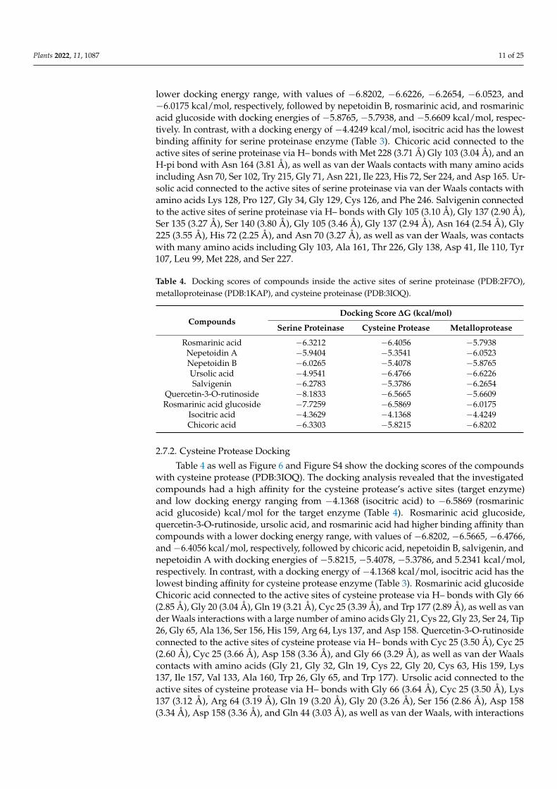
Figure 5. Docking view of (A1,A2) chicoric acid, (B1,B2) ursolic acid, and (C1,C2) salvigenin, in the binding sites of serine proteinase (PDB:2F7O); (A1,B1,C1) 3D complex structures (stereoview) and (A2,B2,C2) 2D interaction diagram structures.
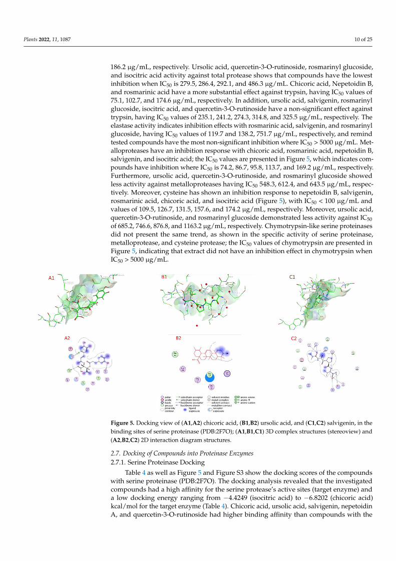
2.7. Docking of Compounds into Proteinase Enzymes 2.7.1. Serine Proteinase Docking
Table 4 as well as Figures 5 and S3 show the docking scores of the compounds with serine proteinase (PDB:2F7O). The docking analysis revealed that the investigated com-pounds had a high affinity for the serine protease’s active sites (target enzyme) and a low docking energy ranging from −4.4249 (isocitric acid) to −6.8202 (chicoric acid) kcal/mol for the target enzyme (Table 4). Chicoric acid, ursolic acid, salvigenin, nepetoidin A, and quercetin-3-O-rutinoside had higher binding affinity than compounds with the lower docking energy range, with values of −6.8202, −6.6226, −6.2654, −6.0523, and −6.0175 kcal/mol, respectively, followed by nepetoidin B, rosmarinic acid, and rosmarinic acid glucoside with docking energies of −5.8765, −5.7938, and −5.6609 kcal/mol, respectively. In contrast, with a docking energy of −4.4249 kcal/mol, isocitric acid has the lowest binding affinity for serine proteinase enzyme (Table 3). Chicoric acid connected to the active sites of serine proteinase via H– bonds with Met 228 (3.71 Å) Gly 103 (3.04 Å), and an H-pi bond with Asn 164 (3.81 Å), as well as van der Waals contacts with many amino acids including Asn 70, Ser 102, Try 215, Gly 71, Asn 221, Ile 223, His 72, Ser 224, and Asp 165. Ursolic acid connected to the active sites of serine proteinase via van der Waals contacts with amino acids Lys 128, Pro 127, Gly 34, Gly 129, Cys 126, and Phe 246. Salvigenin con-nected to the active sites of serine proteinase via H– bonds with Gly 105 (3.10 Å), Gly 137 (2.90 Å), Ser 135 (3.27 Å), Ser 140 (3.80 Å), Gly 105 (3.46 Å), Gly 137 (2.94 Å), Asn 164 (2.54
Figure 5. Docking view of (A1,A2) chicoric acid, (B1,B2) ursolic acid, and (C1,C2) salvigenin, in thebinding sites of serine proteinase (PDB:2F7O); (A1,B1,C1) 3D complex structures (stereoview) and(A2,B2,C2) 2D interaction diagram structures.
2.7. Docking of Compounds into Proteinase Enzymes2.7.1. Serine Proteinase Docking
Table 4 as well as Figure 5 and Figure S3 show the docking scores of the compoundswith serine proteinase (PDB:2F7O). The docking analysis revealed that the investigatedcompounds had a high affinity for the serine protease’s active sites (target enzyme) anda low docking energy ranging from −4.4249 (isocitric acid) to −6.8202 (chicoric acid)kcal/mol for the target enzyme (Table 4). Chicoric acid, ursolic acid, salvigenin, nepetoidinA, and quercetin-3-O-rutinoside had higher binding affinity than compounds with the
Plants 2022, 11, 1087 11 of 25
lower docking energy range, with values of −6.8202, −6.6226, −6.2654, −6.0523, and−6.0175 kcal/mol, respectively, followed by nepetoidin B, rosmarinic acid, and rosmarinicacid glucoside with docking energies of −5.8765, −5.7938, and −5.6609 kcal/mol, respec-tively. In contrast, with a docking energy of −4.4249 kcal/mol, isocitric acid has the lowestbinding affinity for serine proteinase enzyme (Table 3). Chicoric acid connected to theactive sites of serine proteinase via H– bonds with Met 228 (3.71 Å) Gly 103 (3.04 Å), and anH-pi bond with Asn 164 (3.81 Å), as well as van der Waals contacts with many amino acidsincluding Asn 70, Ser 102, Try 215, Gly 71, Asn 221, Ile 223, His 72, Ser 224, and Asp 165. Ur-solic acid connected to the active sites of serine proteinase via van der Waals contacts withamino acids Lys 128, Pro 127, Gly 34, Gly 129, Cys 126, and Phe 246. Salvigenin connectedto the active sites of serine proteinase via H– bonds with Gly 105 (3.10 Å), Gly 137 (2.90 Å),Ser 135 (3.27 Å), Ser 140 (3.80 Å), Gly 105 (3.46 Å), Gly 137 (2.94 Å), Asn 164 (2.54 Å), Gly225 (3.55 Å), His 72 (2.25 Å), and Asn 70 (3.27 Å), as well as van der Waals, was contactswith many amino acids including Gly 103, Ala 161, Thr 226, Gly 138, Asp 41, Ile 110, Tyr107, Leu 99, Met 228, and Ser 227.
Table 4. Docking scores of compounds inside the active sites of serine proteinase (PDB:2F7O),metalloproteinase (PDB:1KAP), and cysteine proteinase (PDB:3IOQ).
CompoundsDocking Score ∆G (kcal/mol)
Serine Proteinase Cysteine Protease Metalloprotease
Rosmarinic acid −6.3212 −6.4056 −5.7938Nepetoidin A −5.9404 −5.3541 −6.0523Nepetoidin B −6.0265 −5.4078 −5.8765Ursolic acid −4.9541 −6.4766 −6.6226Salvigenin −6.2783 −5.3786 −6.2654
Quercetin-3-O-rutinoside −8.1833 −6.5665 −5.6609Rosmarinic acid glucoside −7.7259 −6.5869 −6.0175
Isocitric acid −4.3629 −4.1368 −4.4249Chicoric acid −6.3303 −5.8215 −6.8202
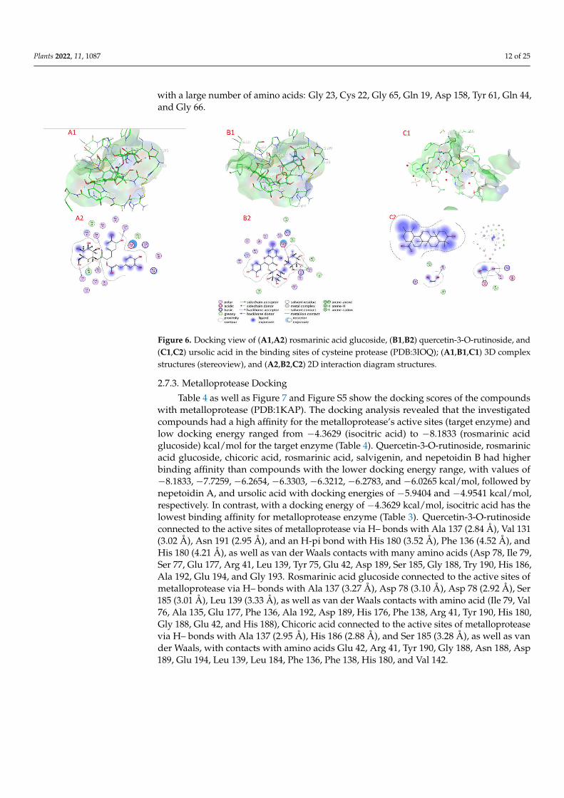
2.7.2. Cysteine Protease Docking
Table 4 as well as Figure 6 and Figure S4 show the docking scores of the compoundswith cysteine protease (PDB:3IOQ). The docking analysis revealed that the investigatedcompounds had a high affinity for the cysteine protease’s active sites (target enzyme)and low docking energy ranging from −4.1368 (isocitric acid) to −6.5869 (rosmarinicacid glucoside) kcal/mol for the target enzyme (Table 4). Rosmarinic acid glucoside,quercetin-3-O-rutinoside, ursolic acid, and rosmarinic acid had higher binding affinity thancompounds with a lower docking energy range, with values of −6.8202, −6.5665, −6.4766,and−6.4056 kcal/mol, respectively, followed by chicoric acid, nepetoidin B, salvigenin, andnepetoidin A with docking energies of −5.8215, −5.4078, −5.3786, and 5.2341 kcal/mol,respectively. In contrast, with a docking energy of −4.1368 kcal/mol, isocitric acid has thelowest binding affinity for cysteine protease enzyme (Table 3). Rosmarinic acid glucosideChicoric acid connected to the active sites of cysteine protease via H– bonds with Gly 66(2.85 Å), Gly 20 (3.04 Å), Gln 19 (3.21 Å), Cyc 25 (3.39 Å), and Trp 177 (2.89 Å), as well as vander Waals interactions with a large number of amino acids Gly 21, Cys 22, Gly 23, Ser 24, Tip26, Gly 65, Ala 136, Ser 156, His 159, Arg 64, Lys 137, and Asp 158. Quercetin-3-O-rutinosideconnected to the active sites of cysteine protease via H– bonds with Cyc 25 (3.50 Å), Cyc 25(2.60 Å), Cyc 25 (3.66 Å), Asp 158 (3.36 Å), and Gly 66 (3.29 Å), as well as van der Waalscontacts with amino acids (Gly 21, Gly 32, Gln 19, Cys 22, Gly 20, Cys 63, His 159, Lys137, Ile 157, Val 133, Ala 160, Trp 26, Gly 65, and Trp 177). Ursolic acid connected to theactive sites of cysteine protease via H– bonds with Gly 66 (3.64 Å), Cyc 25 (3.50 Å), Lys137 (3.12 Å), Arg 64 (3.19 Å), Gln 19 (3.20 Å), Gly 20 (3.26 Å), Ser 156 (2.86 Å), Asp 158(3.34 Å), Asp 158 (3.36 Å), and Gln 44 (3.03 Å), as well as van der Waals, with interactions
Plants 2022, 11, 1087 12 of 25
with a large number of amino acids: Gly 23, Cys 22, Gly 65, Gln 19, Asp 158, Tyr 61, Gln 44,and Gly 66.
Plants 2022, 11, 1087 12 of 25
Figure 6. Docking view of (A1,A2) rosmarinic acid glucoside, (B1,B2) quercetin-3-O-rutinoside, and (C1,C2) ursolic acid in the binding sites of cysteine protease (PDB:3IOQ); (A1,B1,C1) 3D complex structures (stereoview), and (A2,B2,C2) 2D interaction diagram structures.
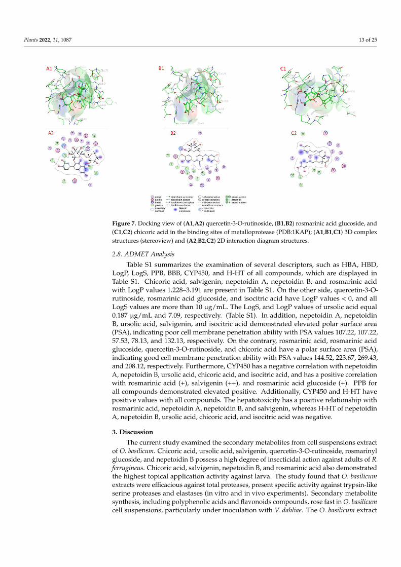
2.7.3. Metalloprotease Docking Table 4 as well as Figures 7 and S5 show the docking scores of the compounds with
metalloprotease (PDB:1KAP). The docking analysis revealed that the investigated com-pounds had a high affinity for the metalloprotease’s active sites (target enzyme) and low docking energy ranged from −4.3629 (isocitric acid) to −8.1833 (rosmarinic acid glucoside) kcal/mol for the target enzyme (Table 4). Quercetin-3-O-rutinoside, rosmarinic acid glu-coside, chicoric acid, rosmarinic acid, salvigenin, and nepetoidin B had higher binding affinity than compounds with the lower docking energy range, with values of −8.1833, −7.7259, −6.2654, −6.3303, −6.3212, −6.2783, and −6.0265 kcal/mol, followed by nepetoidin A, and ursolic acid with docking energies of −5.9404 and −4.9541 kcal/mol, respectively. In contrast, with a docking energy of −4.3629 kcal/mol, isocitric acid has the lowest bind-ing affinity for metalloprotease enzyme (Table 3). Quercetin-3-O-rutinoside connected to the active sites of metalloprotease via H– bonds with Ala 137 (2.84 Å), Val 131 (3.02 Å), Asn 191 (2.95 Å), and an H-pi bond with His 180 (3.52 Å), Phe 136 (4.52 Å), and His 180 (4.21 Å), as well as van der Waals contacts with many amino acids (Asp 78, Ile 79, Ser 77, Glu 177, Arg 41, Leu 139, Tyr 75, Glu 42, Asp 189, Ser 185, Gly 188, Try 190, His 186, Ala 192, Glu 194, and Gly 193. Rosmarinic acid glucoside connected to the active sites of met-alloprotease via H– bonds with Ala 137 (3.27 Å), Asp 78 (3.10 Å), Asp 78 (2.92 Å), Ser 185 (3.01 Å), Leu 139 (3.33 Å), as well as van der Waals contacts with amino acid (Ile 79, Val 76, Ala 135, Glu 177, Phe 136, Ala 192, Asp 189, His 176, Phe 138, Arg 41, Tyr 190, His 180, Gly 188, Glu 42, and His 188), Chicoric acid connected to the active sites of metalloprotease via H– bonds with Ala 137 (2.95 Å), His 186 (2.88 Å), and Ser 185 (3.28 Å), as well as van der Waals, with contacts with amino acids Glu 42, Arg 41, Tyr 190, Gly 188, Asn 188, Asp 189, Glu 194, Leu 139, Leu 184, Phe 136, Phe 138, His 180, and Val 142.
Figure 6. Docking view of (A1,A2) rosmarinic acid glucoside, (B1,B2) quercetin-3-O-rutinoside, and(C1,C2) ursolic acid in the binding sites of cysteine protease (PDB:3IOQ); (A1,B1,C1) 3D complexstructures (stereoview), and (A2,B2,C2) 2D interaction diagram structures.
2.7.3. Metalloprotease Docking
Table 4 as well as Figure 7 and Figure S5 show the docking scores of the compoundswith metalloprotease (PDB:1KAP). The docking analysis revealed that the investigatedcompounds had a high affinity for the metalloprotease’s active sites (target enzyme) andlow docking energy ranged from −4.3629 (isocitric acid) to −8.1833 (rosmarinic acidglucoside) kcal/mol for the target enzyme (Table 4). Quercetin-3-O-rutinoside, rosmarinicacid glucoside, chicoric acid, rosmarinic acid, salvigenin, and nepetoidin B had higherbinding affinity than compounds with the lower docking energy range, with values of−8.1833, −7.7259, −6.2654, −6.3303, −6.3212, −6.2783, and −6.0265 kcal/mol, followed bynepetoidin A, and ursolic acid with docking energies of −5.9404 and −4.9541 kcal/mol,respectively. In contrast, with a docking energy of −4.3629 kcal/mol, isocitric acid has thelowest binding affinity for metalloprotease enzyme (Table 3). Quercetin-3-O-rutinosideconnected to the active sites of metalloprotease via H– bonds with Ala 137 (2.84 Å), Val 131(3.02 Å), Asn 191 (2.95 Å), and an H-pi bond with His 180 (3.52 Å), Phe 136 (4.52 Å), andHis 180 (4.21 Å), as well as van der Waals contacts with many amino acids (Asp 78, Ile 79,Ser 77, Glu 177, Arg 41, Leu 139, Tyr 75, Glu 42, Asp 189, Ser 185, Gly 188, Try 190, His 186,Ala 192, Glu 194, and Gly 193. Rosmarinic acid glucoside connected to the active sites ofmetalloprotease via H– bonds with Ala 137 (3.27 Å), Asp 78 (3.10 Å), Asp 78 (2.92 Å), Ser185 (3.01 Å), Leu 139 (3.33 Å), as well as van der Waals contacts with amino acid (Ile 79, Val76, Ala 135, Glu 177, Phe 136, Ala 192, Asp 189, His 176, Phe 138, Arg 41, Tyr 190, His 180,Gly 188, Glu 42, and His 188), Chicoric acid connected to the active sites of metalloproteasevia H– bonds with Ala 137 (2.95 Å), His 186 (2.88 Å), and Ser 185 (3.28 Å), as well as vander Waals, with contacts with amino acids Glu 42, Arg 41, Tyr 190, Gly 188, Asn 188, Asp189, Glu 194, Leu 139, Leu 184, Phe 136, Phe 138, His 180, and Val 142.
Plants 2022, 11, 1087 13 of 25Plants 2022, 11, 1087 13 of 25
Figure 7. Docking view of (A1,A2) quercetin-3-O-rutinoside, (B1,B2) rosmarinic acid glucoside, and (C1,C2) chicoric acid in the binding sites of metalloprotease (PDB:1KAP); (A1,B1,C1) 3D complex structures (stereoview) and (A2,B2,C2) 2D interaction diagram structures.
2.8. ADMET Analysis Table S1 summarizes the examination of several descriptors, such as HBA, HBD,
LogP, LogS, PPB, BBB, CYP450, and H-HT of all compounds, which are displayed in Table S1. Chicoric acid, salvigenin, nepetoidin A, nepetoidin B, and rosmarinic acid with LogP values 1.228–3.191 are present in Table S1. On the other side, quercetin-3-O-rutinoside, rosmarinic acid glucoside, and isocitric acid have LogP values < 0, and all LogS values are more than 10 µg/mL. The LogS, and LogP values of ursolic acid equal 0.187 µg/mL and 7.09, respectively. (Table S1). In addition, nepetoidin A, nepetoidin B, ursolic acid, salvig-enin, and isocitric acid demonstrated elevated polar surface area (PSA), indicating poor cell membrane penetration ability with PSA values 107.22, 107.22, 57.53, 78.13, and 132.13, respectively. On the contrary, rosmarinic acid, rosmarinic acid glucoside, quercetin-3-O-rutinoside, and chicoric acid have a polar surface area (PSA), indicating good cell mem-brane penetration ability with PSA values 144.52, 223.67, 269.43, and 208.12, respectively. Furthermore, CYP450 has a negative correlation with nepetoidin A, nepetoidin B, ursolic acid, chicoric acid, and isocitric acid, and has a positive correlation with rosmarinic acid (+), salvigenin (++), and rosmarinic acid glucoside (+). PPB for all compounds demon-strated elevated positive. Additionally, CYP450 and H-HT have positive values with all compounds. The hepatotoxicity has a positive relationship with rosmarinic acid, nepe-toidin A, nepetoidin B, and salvigenin, whereas H-HT of nepetoidin A, nepetoidin B, ur-solic acid, chicoric acid, and isocitric acid was negative.
3. Discussion The current study examined the secondary metabolites from cell suspensions extract
of O. basilicum. Chicoric acid, ursolic acid, salvigenin, quercetin-3-O-rutinoside, rosma-rinyl glucoside, and nepetoidin B possess a high degree of insecticidal action against adults of R. ferrugineus. Chicoric acid, salvigenin, nepetoidin B, and rosmarinic acid also demonstrated the highest topical application activity against larva. The study found that O. basilicum extracts were efficacious against total proteases, present specific activity against trypsin-like serine proteases and elastases (in vitro and in vivo experiments). Sec-ondary metabolite synthesis, including polyphenolic acids and flavonoids compounds, rose fast in O. basilicum cell suspensions, particularly under inoculation with V. dahliae. The O. basilicum extract was shown to be efficacious and effective, not only enhancing
Figure 7. Docking view of (A1,A2) quercetin-3-O-rutinoside, (B1,B2) rosmarinic acid glucoside, and(C1,C2) chicoric acid in the binding sites of metalloprotease (PDB:1KAP); (A1,B1,C1) 3D complexstructures (stereoview) and (A2,B2,C2) 2D interaction diagram structures.
2.8. ADMET Analysis
Table S1 summarizes the examination of several descriptors, such as HBA, HBD,LogP, LogS, PPB, BBB, CYP450, and H-HT of all compounds, which are displayed inTable S1. Chicoric acid, salvigenin, nepetoidin A, nepetoidin B, and rosmarinic acidwith LogP values 1.228–3.191 are present in Table S1. On the other side, quercetin-3-O-rutinoside, rosmarinic acid glucoside, and isocitric acid have LogP values < 0, and allLogS values are more than 10 µg/mL. The LogS, and LogP values of ursolic acid equal0.187 µg/mL and 7.09, respectively. (Table S1). In addition, nepetoidin A, nepetoidinB, ursolic acid, salvigenin, and isocitric acid demonstrated elevated polar surface area(PSA), indicating poor cell membrane penetration ability with PSA values 107.22, 107.22,57.53, 78.13, and 132.13, respectively. On the contrary, rosmarinic acid, rosmarinic acidglucoside, quercetin-3-O-rutinoside, and chicoric acid have a polar surface area (PSA),indicating good cell membrane penetration ability with PSA values 144.52, 223.67, 269.43,and 208.12, respectively. Furthermore, CYP450 has a negative correlation with nepetoidinA, nepetoidin B, ursolic acid, chicoric acid, and isocitric acid, and has a positive correlationwith rosmarinic acid (+), salvigenin (++), and rosmarinic acid glucoside (+). PPB forall compounds demonstrated elevated positive. Additionally, CYP450 and H-HT havepositive values with all compounds. The hepatotoxicity has a positive relationship withrosmarinic acid, nepetoidin A, nepetoidin B, and salvigenin, whereas H-HT of nepetoidinA, nepetoidin B, ursolic acid, chicoric acid, and isocitric acid was negative.
3. Discussion
The current study examined the secondary metabolites from cell suspensions extractof O. basilicum. Chicoric acid, ursolic acid, salvigenin, quercetin-3-O-rutinoside, rosmarinylglucoside, and nepetoidin B possess a high degree of insecticidal action against adults of R.ferrugineus. Chicoric acid, salvigenin, nepetoidin B, and rosmarinic acid also demonstratedthe highest topical application activity against larva. The study found that O. basilicumextracts were efficacious against total proteases, present specific activity against trypsin-likeserine proteases and elastases (in vitro and in vivo experiments). Secondary metabolitesynthesis, including polyphenolic acids and flavonoids compounds, rose fast in O. basilicumcell suspensions, particularly under inoculation with V. dahliae. The O. basilicum extract

Plants 2022, 11, 1087 14 of 25
was shown to be efficacious and effective, not only enhancing deterrence but also reducingfeeding in R. ferrugineus larvae. However, its antifeedant action is most evident in adults.
Furthermore, our results indicated that under ideal settings, the strongest rates ofantifeedant activity were found. Significant insecticidal activity was observed in theextract, which might be related to the extract’s variety of bioactive metabolites. O. basilicumpolyphenolic acids and flavonoids from cell suspension grew gradually throughout thefirst 25 days, then rapidly increased and continued to rise fast over the last 15 days of thetesting session. Different dosages had a substantial influence for compounds examinedwith IC50 rate. The impact of compounds against protease activity isolated in vitro fromthe midgut of R. ferrugineus fourth instar larvae explains the action of O. basilicum.
The LC50 value was 1238 µg/mL for O. basilicum extract against adults. The LC50values of chicoric acid, ursolic acid, salvigenin, quercetin-3-O-rutinoside, rosmarinyl glu-coside, and nepetoidin B had the highest insecticidal activity, with LC50 1132, 1167, 1189,1214, 1275, and 1317 µg/mL, respectively. The topical application showed that the LD50value (µg/larva) was 13.7 for O. basilicum extracts. Chicoric acid, salvigenin, nepetoidinB, and rosmarinic acid demonstrated the highest insecticidal activity, with LD50 values of10.23, 11.4, 11.9, and 12.4 µg/larva, respectively. Isocitric acid was present with low activitywith LD50 23.9 µg/larva.
The active extract of Ocimum basilicum showed inhibitory activity against total protease,trypsin-like serine proteinases, elastase, cysteine, and metalloprotease activity with IC50values of 119.4, 91, 102.4, 76.4 and 52.4 µg/mL, respectively. However, the activity of O.basilicum extracts against proteases enzymes demonstrated a correlation with nepetoidin B(5.72%), quercetin-3-O-rutinoside (2.34%), chicoric acid (1.23%), rosmarinic acid (14.2%),rosmarinyl glucoside (3.32%), and salvigenin (2.51%) contents. Chicoric acid, ursolic acid,salvigenin, quercetin-3-O-rutinoside, rosmarinyl glucoside, nepetoidin B, rosmarinic acid,and isocitric acid presenting inhibition effects against total proteases, with IC50 valuesof 63.1, 67.2, 72.4, 82.6, 84.9, 102.4, 109.6, and 143.3 µg/mL, respectively. The Ocimumbasilicum extract activity against trypsin is related to chicoric acid, ursolic acid, salvigenin,rosmarinyl glucoside, nepetoidin B, rosmarinic acid, and quercetin-3-O-rutinoside, withIC50 values of 51.6, 57.4, 59.2, 75.3, 86.7, 91.3, and 104.5 µg/mL (1.23%, 4.91%, 2.51%, 3.32%,5.72%, 14.2%, and 2.31%), respectively. Quercetin-3-O-rutinoside, rosmarinyl glucoside,chicoric acid, rosmarinic acid, salvigenin, and nepetoidin B have the highest responsibilityof inhibition effect against metalloproteases with IC50 < 100 µg/mL, and values of 37.3,41.2, 44.6, 48.4, 49.1 and 51.1 µg/mL, respectively. On the contrary, rosmarinyl glucoside,quercetin-3-O-rutinoside, ursolic acid, nepetoidin B, rosmarinic acid, chicoric acid, andsalvigenin presented specific inhibitory effects against cysteine where IC50 < 100 µg/mL,with values of 49.2, 53.6, 58.2, 63.3, 67.8, 81.2, and 89.6 µg/mL, respectively. The inhibitoryactivities of rosmarinic acid, rosmarinyl glucoside, and salvigenin against elastase activityare demonstrated by IC50 values of 96.4, 101.3, and 105.6 µg/mL, respectively. Thesefindings are consistent with the fact that compounds have an effect on total proteaseactivity (in vitro, in vivo), such as nepetoidin B, quercetin-3-O-rutinoside, chicoric acid,rosmarinic acid, rosmarinyl glucoside, and salvigenin, which have activity against totalproteases. It can be concluded that nepetoidin B, quercetin-3-O-rutinoside, chicoric acid,rosmarinic acid, rosmarinyl glucoside, and salvigenin were presented to effect insecticidalactivity against proteases enzymes of R. ferrugineus.
Moreover, since these extracts are antifeedant chemicals, their targets and methodof action in insects remain unclear. The research suggests that processes may includedisturbance of feeding physiology, chronic toxicity, or repellency associated with theinsecticidal activity [52]. The most significant secondary metabolites in O. basilicum extractswere flavonoids and polyphenolic acids, which were the major steps in the productionpathway for flavonoid and polyphenolic precursors [53,54].
Notably, these secondary molecules are recognized as significant constituents after theirformation on cells as a vital protection from pathogens [55,56]. PGR is required for the regula-tion of plant development and differentiation, which could manage the anabolism destruction

Plants 2022, 11, 1087 15 of 25
of the phenolic contents [57,58]. As a result, novel techniques such as the green biosynthesisof the bioactive secondary metabolites are in great demand now [59–61]. Additionally, cellsuspension culture is a more effective and quick approach for enhancing bioactive chemicalsynthesis than callus culture, owing to its rapid responsiveness, cell division, and simplicityof application [1,60,61]. Total proteolytic activity was evaluated in the midgut juice of R.ferrugineus larval instars using azocasein as the substrate with a slightly alkaline mixture(pH 8.0), which is physiologically similar to the insect midgut pH [1], and DTT activatorwas used [1,62]. The variations in the secondary metabolites of O. basilicum extracts towardprotease inhibition in vitro and in vivo assays (Figures 3 and 4) established the critical roleproteinases in their mode of action [63]. The findings indicate that toxicity might exist as aconsequence of nepetoidin B (5.72%), quercetin-3-O-rutinoside (2.34%), chicoric acid (1.23%),rosmarinic acid (14.2%), rosmarinic acid glucoside (3.32%), and salvigenin (2.51%) in extractO. basilicum and may attest to the critical nature of proteinases.
The inhibition of trypsin-like serine, elastase proteinases, metalloprotease, and cysteineprotease by O. basilicum extract was clearly demonstrated in the fourth midgut preparationvalues, which were lower than the values in vitro. Many values in vivo have the sametrend and are lower than the values in vitro. The in vivo assay of ursolic acid, quercetin-3-O-rutinoside, rosmarinyl glucoside, and isocitric acid activity against total proteasedemonstrated that compounds inhibit total protease the least in vitro. According to ADMETscreening analysis, they are extremely hydrophobic, allowing them to permeate biologicalmembranes according to the chicoric acid, salvigenin, nepetoidin A, nepetoidin B, androsmarinic acid.
Further, Quercetin-3-O-rutinoside, rosmarinyl glucoside, and isocitric acid have poorlipid bilayer permeability according to LogP values (−1.687, −0.98 and 01.811, respec-tively). Compound ursolic acid has poor aqueous solubility (LogP 7.09), and low solubility(−6.387 log mol/L (0.187 µg/mL)). Based on these findings, the Lipinski rules indicatethat chicoric acid, salvigenin, nepetoidin, nepetoidin B, and rosmarinic acid possess a hightheoretical bioavailability in the oral. The effectiveness of compounds (chicoric acid, salvi-genin, nepetoidin A, nepetoidin B, and rosmarinic acid) as a bio-insecticide is measurednot only by its potential to attack the target insect but also by its absorption, distribu-tion, metabolism, excretion, and toxicity risk (ADMET) profile. In terms of metabolism,nepetoidin A, nepetoidin B, ursolic acid, chicoric acid, and isocitric acid have no inhibitionagainst CYP450 enzymes, indicating good metabolic stability against CYP450 enzymes,whereas rosmarinic acid, salvigenin, and rosmarinic acid glucoside have inhibition againstCYP450 enzymes. However, the hepatotoxicity prediction suggests that the compoundsrosmarinic acid, nepetoidin A, nepetoidin B, and salvigenin may be toxic to liver cells.
Docking analysis demonstrated that all of the examined ligands form hydrogenbonds between amino acids and target active pockets of serine proteinases, elastase,and cysteine protease enzymes [64]. Only metalloprotease exhibited ionic and metal-lic bonds with H-bonding with amino acids. The highest docking compound’s binding(chicoric acid, ∆G = −6.8202) to serine proteinase is exhibited. The binding confirmationof quercetin-3-O-rutinoside has the highest docking (∆G = −6.5869) with cysteine pro-tease. Rosmarinic acid glucoside (∆G = −8.1833) also showed the highest docking withmetalloprotease. ADMET assessment of the compounds under investigation revealed thatchicoric acid, salvigenin, nepetoidin A, nepetoidin B, and rosmarinic acid met ADMETdescriptors at their optimum level, as determined by in silico studies [45]. The MolecularDocking is explained as a technique for predicting binding affinities and interactions be-tween molecules by studying their location or orientation on possible targets. The dockingof the compounds on serine proteinases, elastase, metalloprotease, and cysteine proteaserevealed a variety of interactions, including H– bonds as well as H-pi hydrophobic and vander Waals interactions. These previous bond interactions assisted in the understanding of avariety of chemicals’ biological functions in a variety of domains, including medicationsand insecticides [45].

Plants 2022, 11, 1087 16 of 25
The extract of O. basilicum show activity according to the LC50 values against R.ferrugineus. The O. basilicum extract demonstrated obvious antifeedant and insecticidalaction, and, clearly, inhibited proteinases isolated from the fourth midgut preparation.These results give insight into components that might be utilized to create biochemicalmarkers that indicate the resistance of particular plants to insect infestation.
4. Materials and Methods4.1. Chemicals and Reagents
The chemicals were provided by Merck Chemical Co. (St. Louis, MO, USA); hypochlo-rite solution, Hydrochloric acid, gallic acid, cyanidin, quercetin, and solvents were obtainedfrom Sigma-Aldrich Chemical Co. (St. Louis, MO, USA), whereas Sigma-Aldrich Chem-ical Co. (St. Louis, MO, USA) provided reagents, media, and chemicals for biochemicalstudies. Rosmarinic acid, ursolic acid, quercetin-3-O-rutinoside (Rutin), isocitric acid, andchicoric acid were obtained from Merck Chemical Co. (St. Louis, MO, USA). Rosmarinylglucoside was obtained from Aobious Inc., 9 Blackburn Drive, Gloucester, MA 01930 (USA).Nepetoidin B, salvigenin, and rosmarinyl glucoside were obtained from BioCrick Biotech,88 Keyuan Road, Hi-Tech Zone, Chengdu, Sichuan 610042, PRC.
4.2. Media
Several types of media were used. Murashige and Skoog (MS) as solid mediumand Linsmaier and Skoog (LS) as liquid medium (including 0.1 mg thiamine HCl and100 mg myo-inositol) were obtained from Sigma-Aldrich Chemical Co. (St. Louis, MO,USA). Various media were made then autoclaved for 20 min (121 ◦C). The Microbiol-ogy Laboratory, College of Agricultural and Food Sciences, King Faisal University, Al-Ahsa, Saudi Arabia contributed the V. dahliae strain. Pronadisa, Madrid, Spain, pro-vided the potato dextrose agar (PDA), tryptone, and yeast extract, while Sigma ChemicalCo., St. Louis, MO, provided the agar (USA).
4.3. Plants
During the months of February and March, O. basilicum seeds were obtained fromcommercial nurseries in Al-Ahsa, Saudi Arabia. Sterilized seeds were put in MS medium,including agar (0.6%), with pH 5.7 with 3% sucrose (3%, w/w), and incubated in a climateroom (26 ± 4 ◦C, 16 h light) for 7–8 weeks (seedling 17–20 cm) at King Faisal University’sResearch and Training Station in Al-Ahsa, Saudi Arabia.
4.4. O. basilicum Calluses Initiation Employing Various PGRs in Conjunction and V. dahliae as aBiotic Elicitor
The seeds of O. basilicum were germinated in a Petri dish lined after being sterilized,delinted using paper for sterile blotting at a temperature of 28 ± 2 ◦C and a light intensityof 30 Einsteins/(m2·s). The explants of O. basilicum (epicotyls, hypocotyls, and cotyle-donary, 4–5 mm in length) were put in an MS medium including kinetin (0.5 mg/L), NAA(1-Naphthaleneacetic acid) (0.1 mg/L), 2,4-D (2,4-Dichlorophenoxyacetic acid) (0.1 mg/L),IBA (Indole butyric acid) (1 mg/L), and sucrose (3%, w/v) according to Darrag et al.,per Muhammed Akif Açıkgöz with modifications [1,32]. Every treatment (except controlwithout PGRs) was maintained for eight weeks in a climate chamber (26 ± 4 ◦C, 16 h light)with three weekly subcultures. According to Darrag et al. [1], V. dahliae was utilized as aninitiator (biotic elicitor) to explore callus growth enhancement. Vacuum filtration was usedto extract callus from individual cultures 72 h after infection, and for 40 days Calli wasvisually examined every five days. V. dahliae was cultured in PDA (22 ◦C) and subcultureevery 5–6 weeks. Conidia were cultivated for 10 days at 22 ◦C in a potato dextrose (PD)medium with 240 rpm by rotary shaker. Conidia were extracted using centrifugationand rinsed three times at pH 6.5 with 0.1 M K2HPO4-KH2PO4. Conidia concentrationwas determined using a hemocytometer under a microscope. Finally, 25 µL of conidial
Plants 2022, 11, 1087 17 of 25
suspension [(2–5) × 107 conidia/mL] or sterile distilled water (control) was injected into8 mL of fresh solid medium of MS.
4.5. O. basilicum Cell Suspension Initiation
According to Darrag et al. with modifications [1], callus was started to initiate andbe detected throughout a 5–6-week period utilizing LS medium. The medium was passedthrough a variety of mesh sizes of screens. Liquid LS culture (200 mL) was filtered andadded with either 25 mL conidial suspension [(2–5) × 107 conidia/mL], then transferredto 30 flasks (Erlenmeyer, 500 mL), and adjusted to a final volume of 250 mL using liquidmedium. After 72 h, cultures were collected and tested for protein content. Suspensionmedia was established in conical flasks (250 mL) using LS medium (100 mL) without agar,maintained for six weeks at the conditions 30 ± 4 ◦C, with 16 h of light with a quick shakerspeed 110 rpm, every two weeks, while the subculture was kept in a climate chamber.
4.6. Total Phenolic Content (TPC) Determination
The total phenolic components from samples (callus and cell suspension) were ex-tracted using methanol by shaking for 12 h and followed by drying using anhydrousNa2SO4. Next was the addition of 790 µL distilled water to 10 µL sample solution and the50 µL Folin–Ciocalteu reagent (50 L), according to the Folin–Ciocalteu technique [65]. Themixture was homogenized, and 150 µL of Na2CO3 solution (20%, w/v) was applied (1 min),vortexed again, and incubated at room temperature (120 min in dark). The total phenoliccontent was evaluated using spectroscopy at 750 nm and estimated using the standardcurve of 1–100 mg gallic acid per 100 g·Dw.
4.7. Total Flavonoids Contents (TF)
The content in methanol extract was evaluated by extracting (500 µL) in mixture ofextract and 5% (w/v) of sodium nitrite (500 µL), and, then, adding aluminum chloride(300 µL, 10% (w/v)) [40] from the cell suspension and callus samples [66]. The assay mixtureis incubated for 5 min at ambient temperature, then sodium hydroxide (1 mL, 1 M) wasadded to bring the process to a halt. Total flavonoid content was evaluated by spectroscopyat 510 nm and calculated using standard curve of quercetin with a serial concentration ofquercetin (1–100 mg quercetin/100 g·DW).
4.8. Determination of Total Condensed Tannins (TCT)
Total condensed tannins in methanol extract were determined according to Hager-man [67] from the cell suspension and callus samples. Ferric ammonium sulfate or ferricreagent (3000 µL, 2%, w/v in 2 M HCl) was added to extract sample (500 µL), butanol–HClreagent (100 µL, 95:5) and mixed. The sample was kept at 90 ◦C for 15 min, then the totalcondensed tannins content was determined by spectroscopy at 550 nm and calculated usinga serial concentration of cyanidin (0.1–10 mg of cyanidin/100 g·DW).
4.9. Liquid Chromatography-Mass Spectrometry Analysis (LC-MS)
Individual polyphenolic acids and flavonoids were quantified in g O. basilicum cellsuspension extracts using Waters Acquity UPLC I-class coupled with Xevo TQD MS(USA). Methanolic extract (5 mL) was shaken (at 4 ◦C for 30 min), followed by 5 minof gentle sonication (at 4 ◦C) and 10 min of centrifugation at 3000× g. The supernatantwas collected and filtered using PVDF, 0.45 m (polyvinylidene difluoride, Thermo FisherScientific, Waltham, MA, USA). Compounds were measured using MS spectra by extractingthe extracted ion current (EIC) to an accuracy of 40 parts per million (ppm), includingusing the appropriate standard curve regarding the molecular weight (MW) and chemicalsimilarities. Diluted samples were injected (5 µL) using an auto-sampler injector with 2 µL:1 mL n-hexane (LC grade) (Model Combi Pal, Varian). The gradient was as follows, usingsolvent A that contents of 1% formic acid in deionized water and acetonitrile as solventB: solvent B 10–20% in 20 min, solvent B 20–25% in 10 min, solvent B 25–30% in 10 min),

Plants 2022, 11, 1087 18 of 25
and gradient elution thereafter for 10 min with an adjusted flow of 8 mL/minute. ForWaters Acquity UPLC I-class coupled with Xevo TQD MS (U.S.A), Acquity UPLC BEHC18 1.7 um–2.1 um × 100 mm column flow rate 0.5 mL min−1, the injection volume was10 µL, Masslynix v4.1 software with Mass library (Milford, 017757 MA, USA), Argon ascollision cell gas inlet 7 psi, and Nitrogen pressure 60 psi. The MS was configured towork in both negative and positive ion mode using an electrospray ionization (ESI) asan atmospheric pressure source. With a nebulizing flowrate of approximately 12 L/hour,the voltage of electrospray capillary was set to 3000 V and 300 ◦C of drying gas. Scanmode mass spectrometry data were acquired (mass range m/z 100–900). Compounds weremeasured using MS spectra by extracting the extracted ion chromatogram (EIC) with30 ppm tolerance and comparing it to acceptable external calibration curves based onmolecular weight and chemical similitude. Quantitative analyses were performed on threereplicates for each condition, verifying assignments of molecules by Acquity UPLC I-classcoupled with Xevo TQD MS analysis.
4.10. Evaluation of the Extracted Secondary Metabolites’ Contact-Insecticide and AntifeedantEfficacy against R. ferrugineus
The methanolic extract from the cell suspension of O. basilicum after 40 days and com-pounds (rosmarinic acid, nepetoidin B, ursolic acid, salvigenin, quercetin-3-O-rutinoside,rosmarinyl glucoside, chicoric acid, and isocitric acid) were prepared in acetone in serialconcentrations 1, 10, 50, 100, 500, 1000, and 5000 µg/mL and, then, using 0.1% TritonX-100to the constructed volume according to Darrag et al. [1] and Shukla et al. [68] with modi-fications. Serial concentrations (1, 10, 50, 100, 500, 1000, and 5000 µg/mL) of methanolicextract of cell suspension after 40 days and compounds (rosmarinic acid, nepetoidin B,ursolic acid, salvigenin, querce-tin-3-O-rutinoside, rosmarinyl glucoside, chicoric acid, andisocitric acid) were dissolved in acetone and diluted with 0.1% TritonX-100 according toDarrag et al. [1] and Shukla et al. [40]. The R. ferrugineus adult and larvae were acquiredfrom an insect breeding lab at King Faisal University’s Date Palm Research Center ofExcellence and grown utilizing sugarcane stem long pieces. The extracts activity againstlarvae was determined by using topical application method with storing larvae at 4–5 ◦Cfor 5 min. Previous serial concentrations of extract (10 µL) were applied to the dorsumof each larvae using a hand-operated micro-applicator (Burkard Manufacturing Co., Ltd.,Hertfordshire, UK with a 50-ll micro-syringe (MS-N50; Ito Corp., Shizuoka, Japan). Eachlarva was feeding on 10 cm stem pieces of long sugarcane, with 5 larvae/box with threereplicates. After topical administration, larval mortality was determined after 24, 48, 72,and 96 h to determine the LD50. Adults were tested for antifeedant effect determined usingten-centimeter-long sections of sugarcane stem divided into equal longitudinal halves.Pieces of long sections of sugarcane stem with area ~32.2 cm2 were dipped for ten secondsin the previous serial concentration of extract (10 mL) and, then, dried in room temperatureair. Each item that had been treated was put in a plastic box. Each package received areplacement pair (male and female). Each treatment was replicated ten times. After 24, 48,72, and 96 h, feeding observations were assayed.
4.11. Assessment of an O. basilicum Cell Suspension Extract and Pure Components on theProteolytic Enzyme Activity (In Vitro) of R. ferrugineus Larvae
The Lowry technique was used to determine the protein concentrations [69]. Total pro-teolytic activity was determined in the fourth midgut instar larval (lab strain) homogenateof R. ferrugineus with azocasein in accordance with Darrag et al. [1] and Olga et al. [70].Midgut larvae homogenates (10 larvae) were carefully extracted, dissected, and washedrepeatedly with NaCl solution (0.9% (w/v), before being homogenized in 500 µL of anassay buffer (5 mM dithiothreitol (DTT), 50 mM HEPS (N-2-hydroxyethyl piperazin-N’-2-ehtanesulphonic acid), pH 8.0, and 0.1%, v/v Triton X-100). Using a Sigma 3k30 coolingcentrifuge, reserve homogenates prepared in a previous stage were spun at 5000 rpm for30 min. Estimates of protein content and total proteolytic enzyme activity were made usingthe supernatants. The supernatant (10 µL) was incubated at 37 ◦C for 20 min, and the
Plants 2022, 11, 1087 19 of 25
volume was adjusted to 60 µL using an assay buffer (pH 8), before adding azocasien (200 µL,2% (w/v)). In all instances, 10 µL enzyme samples, methanolic extract of O. basilicum cellsuspension extract after 40 days, and pure chemicals (rosmarinic acid, nepetoidin B, ursolicacid, salvigenin, quercetin-3-O-rutinoside, rosmarinyl glucoside, chicoric acid, and isocitricacid) were incubated for 10 min. Following that, to activate the reaction, the substratewas introduced 20 min for Leupeptin, so the reaction was halted after 180 min at 37 ◦Cwith 300 µL of cool tri-chloroacetic acid (TCA) (10% (v/v). The mixture was centrifugedfor 20 min at 5000 rpm (Sigma 3k30 cooling centrifuge). The supernatant was treatedwith 10 µL of NaOH (10 N), and the absorbance was determined at 450 nm using anELISA plate reader, the total specific activity of proteolytic enzymes was approximatedas OD450 mg−1·protein−1·h−1, and a blank solution sample devoid of enzyme solutionwas determined.
4.12. Assessment of an O. basilicum Cell Suspension Extract and Pure Components on SerineProteinase Specific Activity (In Vitro) of R. ferrugineus Larvae
The specific activities of serine proteinase were determined using a rapid quick mi-croplate assay with substrates in reaction mixtures (150 µL) including a serine proteaseassay buffer, according to Darrag et al. [1] and Olga et al. [70] with modifications.
The midguts were homogenized in the assay buffer (100 mM Tris-HCl, pH 8.1) andcentrifuged for 30 min at 8000 rpm (Sigma 3K 30, rotors No. 12158) [1]. Each plate wellreceived 10 µL of the enzyme for the trypsin-, chymotrypsin-, and elastase-like proteinasesin the experiment (containing 40 µL of buffers). SAAPFρNA (100 mg/mL in DMF), BAρNA(100 mg/mL in DMSO), and SAAPLρNA (100 mg/mL in DMF) stock substrates werediluted in the assay buffer to 1 mg/mL, and substrate volume was 50 µL. The mixture wasstirred at 37 ◦C for 15 min, then the reaction was stopped using 50 µL acetic acid (30%,v/v). The activity was quantified using an ELISA plate reader at a wavelength of 405 nm.As an empty well, an assay combination containing a denaturation enzyme was utilizedinstead of an active enzyme. Specific activities of proteinase were reported as OD/mg−1
protein−1 using substrates N-succinyl-ala-alapro-leucine ρ-nitroanilide (SAAPLρNA), Na-benzoyl-L-arginine ρ-nitroanilide (BAρNA), and N-succinyl-ala-ala-pro-phenylalanineρ-nitroanilide (SAAPFρNA)).
4.13. Assessment of an O. basilicum Cell Suspension Extract and Pure Components onMetalloproteinase Specific Activity (In Vitro) of R. ferrugineus Larvae
A substrate azocasein assessed the metalloproteinases activity in the fourth midgutinstar larval homogenate [1]. Ten larvae were carefully removed, dissected, and rinsed re-peatedly with NaCl solution (0.9% (w/v). The midgut was homogenized using protease testsolution (500 µL) containing 5 mM dithiothreitol (DTT), 50 mM HEPS (N-2-hydroxyelthylpiperazin-N’-2-ehtanesulphonic acid), an adjusted pH at 8.0, and Triton X-100 (0.1%, v/v).After homogenization, the homogenates were centrifuged at 5000 rpm (for 30 min) using aSigma centrifuge 3k30, and the supernatant was utilized to determine enzyme activity andprotein content. The supernatant per experiment (10 µL) was incubated for 20 min at 37 ◦C,and the volume was adjusted to 60 µL using assay buffer pH 8 before adding azocasein(200 µL, 2% (w/v)). In all instances, 10 µL enzyme samples were incubated for 10 min(except EDTA with 20 min), and EDTA was used as a blocker of metalloproteinase activitybefore being added to the specific substrate. The reaction was interrupted with 300 µL oftrichloroacetic acid (TCA cold, 10% (v/v) and continued at 37 ◦C for 180 min. The reactionmixture was then centrifuged at 5000 rpm for 20 min (Sigma centrifuge 3k30), sodiumhydroxide (10 µL of 10 N) was incorporated into the supernatant, then absorbance was de-termined using an ELISA plate reader at 450 nm. As a blank, an assay mixture devoid of theenzyme was devoted, the specific activity was determined using the OD450 mg−1 proteinmin−1 value, and a blank solution sample devoid of enzyme solution was determined.

Plants 2022, 11, 1087 20 of 25
4.14. Assessment of an O. basilicum Cell Suspension Extract and Pure Components on CysteineProteinase Specific Activity (In Vitro) of R. ferrugineus Larvae
The activity of cysteine proteinase was determined in the fourth midgut instar larvalhomogenate of R. ferrugineus using the substrate Z-Phe-Arg-MNA [1]. Ten larvae werecarefully removed, dissected, and rinsed repeatedly with NaCl solution (0.9% (w/v). Themidgut instar was homogenized in protease assay buffer (500 µL) [5 mM DTT, 50 mM HEPS(N-2-Hydroxyethyl piperazin-N’-2-ethanesulfonic acid), pH 8.0, and 0.1 percent TritonX-100]. The reaction mixture was centrifuged for 30 min at 5000 rpm (Sigma 3k30 coolingcentrifuge). The supernatant was utilized to determine the overall activity of proteolyticenzymes and the protein content. The supernatant (10 µL) was incubated for 30 min at37 ◦C, and the volume was adjusted to 60 µL using assay buffer (pH 8) before adding100 µL of substrate (0.5 mM). Prior to adding the substrate, 10 µL enzyme samples werepre-incubated with iodoacetic acid for 10 min. After 60 min at 37 ◦C of mixture incubation,the reaction was halted with the addition of 1.5 mL mersalyl (5 mM), 0.02 mg/mL fastgarnet reactive solution, and 2% Tween 20. The reaction was centrifuged (at 5000 rpm,6 min), then the absorbance was determined (at 520 nm). The activity of cysteine proteinasewas measured using an ELISA quick plate reader at the OD520/60 min/mg of midgutprotein. As a blank, a test mixture devoid of enzyme was used. PMSF as general inhibitorof serine proteinase, TLCK as specific inhibitor of trypsin-like serine proteinase, TPCK asspecific inhibitor of chymotrypsin-like serine proteinase, iodoacetic acid as specific inhibitorof cysteine proteinase, EDTA as specific inhibitor of metalloproteinase, and Leupeptin asgeneral inhibitor of proteinase were added to reactions for demonstrate the specificity. Thefourth homogenates of midgut larval were produced as reported before in the experimentfor determining enzyme activity. As previously disclosed, the inhibitory experiment wasperformed using a microplate assay. A series of inhibitor concentrations was created todetermine the inhibitor’s maximal inhibitory effect. Leupeptin concentrations of 0.01,0.05, 0.1, and 1.0 mM, PMSF concentrations of 0.1, 1.0, 10, and 50 mM, TLCK and TPCKconcentrations of 0.01, 0.05, 0.1, 1, 50, and 100 mM, EDTA concentrations of 0.1, 1, 10, 50,and 100 mM, and iodoacetic acid concentrations of 0.01, 0.05, 0.1, and 1 mM.
To learn more about the proteases found in homogenate preparations of R. ferrugineusfourth midgut larval instars, inhibitors from many different types of mechanisms were used.In all experiments for protease activity assay, 10 µL enzyme samples were pre-incubated for10 min with extract or pure compounds (rosmarinic acid, nepetoidin B, ursolic acid, salvigenin,quercetin-3-O-rutinoside, rosmarinyl glucoside, chicoric acid, and isocitric acid) or inhibitorsat concentrations of 1, 10, 50, 100, 500, 1000, and 5000 mg/L. After 24 h of treatment, the effectof treated larvae 1, 10, 50, 100, 500, 1000, and 5000 mg/L of extract or pure compounds oneach enzyme activity was also evaluated in vivo. As a control, an assay combination devoidof inhibitors was utilized. The absorbance of the enzymes was determined using an ELISAplate quick reader at various wavelengths. The percentage activity of the control enzyme foreach inhibitor was evaluated for each enzyme. Protease activity was assayed in vivo in the R.ferrugineus fourth midgut larval instars, which were treated with 1, 10, 50, 100, 500, 1000, and5000 mg/L of extracts or compounds, measured as described above. Larvae were exposed for24 h to leaf discs treated with extract and compounds.
4.15. Docking of Tested Compounds into Enzymes
Serine proteinase (PDB:2F7O) [71], metalloproteinase (PDB:1KAP) [72], and cysteineproteinase (PDB:3IOQ) [64] were obtained using protein data bank (PDB) and put intoMolecular Operating Environment (MOE). The protein’s chemistry was adjusted for themissing hydrogen, and heteroatoms and crystallographic water molecules were deleted [73].The Chem Draw professional 15 Builder module was used to generate compounds (ros-marinic acid, nepetoidin A, nepetoidin B, ursolic acid, salvigenin, quercetin-3-O-rutinoside,rosmarinic acid glucoside, chicoric acid, and isocitric acid). Prior to docking, the ligandswere reduced, three-dimensional structures were constructed, duplicates were eliminated,and bonds were added. After initializing all default settings and getting the lowest energy

Plants 2022, 11, 1087 21 of 25
structures after that, the ligands were permitted to just be elastic and positioned into en-zyme model’s catalytic site cavity of the enzyme model manually. The docking was carriedout with the MOE 2015.10, Chemical Computing Group Inc., Montreal, QC, Canada, usingan induced-fit technique that assumes the receptor to be fixed and the ligand to be flexi-ble [46]. The binding energy was determined using a field of full-force. A scoring systemwere used to determine the affinities between both the ligandcompounds and protein thatestimated energy of non-covalent (free binding) interactions, using terminology pertainingto the molecular force field. The scoring functions and RMSD were calculated, and theoptimal ligand-compound–protein interaction was reviewed and evaluated at conclusionof docking findings.
4.16. ADMET Screening
In silico ADMET screening was using ADMETLAB 2.0 to assess the toxicity risks of thetested pure compounds. Aqueous solubility, intestinal absorption, hydrogen bond acceptor(HBA), hydrogen bond donor (HBD), distribution coefficient P (LogP), solubility (LogS),plasma protein binding (PPB), blood-brain barrier (BBB), Cytochrome P450 (CYP450), andhuman hepatotoxicity (H-HT) were used to predict ADMET properties for each compound.The models utilized in this procedure to predict ADMET properties are derived from anumber of empirical sources of data and are described in detail in the documentation.
4.17. Statistical Design
Finney [74] states that the statistical analysis and probit analysis were conducted usingthe SPSS 25.0 program (Statistical Package for Social Sciences, Armonk, NY, USA). Chemicalcomponents were determined by PCA using SPSS 25.0 program and presented usingXLSTAT 2018.1. PCA is a clustering technique that is used to determine the relationshipsbetween samples. The score plot demonstrates reciprocal contrast, however, the loadingplot clarifies cluster separation. The toxicity parameters, and results were reported as mean± standard error. We did a mortality against dosage regression and converted the resultantmedian dose to an LC50 value (µg/mL). The range of LC50 was calculated with a 95 percentconfidence interval using least-squares. We conducted a one-way analysis of variance onthe enzyme activity data (ANOVA). The Student–Newman–Keuls (SNK) test was appliedwith separate means, and differences at p ≤ 0.05 were considered significant.
5. Conclusions
The results indicate that secondary metabolites isolated from O. basilicum extractsmay be employed as a bio-insecticide against R. ferrugineus. The findings establisheda link between secondary metabolites and their usage, which might be linked to thepolyphenolic and flavonoid chemicals contained in O. basilicum. The research is crucial fordetermining the efficacy and effects of such secondary metabolites. In silico ADMET andmolecular docking analyses were performed, and the potential for developing eco-friendlybio-insecticides. Furthermore, large-scale generation of these secondary metabolites ispossible utilizing cell suspension, a simple and clean process that opens the way to studyingthe produced and final formula for application.
Supplementary Materials: The following supporting information can be downloaded at:https://www.mdpi.com/article/10.3390/plants11081087/s1. Figure S1: LC-MS Chromatogramof O. basilicum cell suspension extracts. Figure S2: in vitro specific inhibitors against protease en-zymes in R. ferrugineus larval instars; Leupeptin: general proteinase inhibitor; PMSF: a generalinhibitor of serine proteinase (Elastase); TLCK: Trypsin inhibitor; TPCK: chymotrypsin inhibitor;EDTA: metalloprotease inhibitor; Iodoacetic acid: cysteine protease inhibitor. Figure S3: dockingand ligand interactions of rosmarinic acid (A1, A2), nepetoidin A (B1, B2), nepetoidin B (C1, C2),quercetin-3-O-rutinoside (D1, D2), rosmarinyl glucoside (E1, E2), and isocitric acid (F1, F2) within theactive sites of serine proteinase (PDB:2F7O). Figure S4: docking and ligand interactions of rosmarinicacid (A1, A2), nepetoidin A (B1, B2), nepetoidin B (C1, C2), salvigenin (D1, D2), chicoric acid (E1,E2), and isocitric acid (F1, F2) within the active sites of cysteine proteinase (PDB:3IOQ). Figure

Plants 2022, 11, 1087 22 of 25
S5: docking and ligand interactions of rosmarinic acid (A1, A2), nepetoidin A (B1, B2), nepetoidinB (C1, C2), ursolic acid (D1, D2), salvigenin (E1, E2), and isocitric acid (F1, F2) within the activesites of metalloproteinase (PDB:1KAP). Table S1: ADMET analysis of rosmarinic acid, nepetoidin A,nepetoidin B, ursolic acid, salvigenin, quercetin-3-O-rutinoside, rosmarinyl glucoside, isocitric acid,and chicoric acid.
Author Contributions: H.M.D.: conceptualization, funding acquisition, formal analysis, supervision,conceptualization, methodology, writing—original draft preparation, formal analysis, and writing—review and editing; H.T.A. and E.H.H.: validation, methodology, software, and writing—originaldraft preparation. All authors have read and agreed to the published version of the manuscript.
Funding: The authors extend their appreciation to the Deputyship for Research and Innovation, Ministryof Education, in Saudi Arabia, for funding this research work through the project number IFT20001.
Institutional Review Board Statement: Not applicable.
Informed Consent Statement: Not applicable.
Data Availability Statement: Not applicable.
Acknowledgments: The authors extend their appreciation to the Deputyship for Research andInnovation, Ministry of Education, Saudi Arabia, for funding this research through the projectnumber IFT20001.
Conflicts of Interest: The authors declare no conflict of interest.
References1. Darrag, H.M.; Alhajhoj, M.R.; Khalil, H.E. Bio-Insecticide of Thymus vulgaris and Ocimum basilicum Extract from Cell Suspensions
and Their Inhibitory Effect against Serine, Cysteine, and Metalloproteinases of the Red Palm Weevil (Rhynchophorus ferrugineus).Insects 2021, 12, 405. [CrossRef] [PubMed]
2. Milosavljevic, I.; El-Shafie, H.A.; Faleiro, J.R.; Hoddle, C.D.; Lewis, M.; Hoddle, M.S. Palmageddon: The wasting of ornamentalpalms by invasive palm weevils, Rhynchophorus spp. J. Pest Sci. 2019, 92, 143–156. [CrossRef]
3. Shehawy, A.A.; Ibrahim, M.T.; Aboutaleb, E.S.; Qari, S.H. Bioactivity and biochemical efficacy of chitinase and Justicia brandegeanaextract against Red Palm Weevil Rhynchophorus ferrugineus Olivier (Coleoptera: Curculionidae). Food Sci. Nutr. 2020, 8, 4625–4636.[CrossRef] [PubMed]
4. Idris, A.M.; Miller, T.A.; Durvasula, R.; Fedoroff, N. Bridging the Knowledge Gaps for Development of Basic Components of RedPalm Weevil IPM. In Sustainable Pest Management in Date Palm: Current Status and Emerging Challenges; Springer: New York, NY,USA, 2015; pp. 37–62.
5. Faleiro, J. A review of the issues and management of the red palm weevil Rhynchophorus ferrugineus (Coleoptera: Rhynchophoridae)in coconut and date palm during the last one hundred years. Int. J. Trop. Insect Sci. 2006, 26, 135–154.
6. Downer, A.J.; Uchida, J.Y.; Hodel, D.R.; Elliott, M.L. Lethal palm diseases common in the United States. HortTechnology 2009, 19,710–716. [CrossRef]
7. Kontodimas, D.; Soroker, V.; Pontikakos, C.; Suma, P.; Beaudoin-Ollivier, L.; Karamaouna, F.; Riolo, P. Visual Identification andCharacterization of Rhynchophorus ferrugineus and Paysandisia archon Infestation. In Handbook of Major Palm Pests: Biology andManagement; Wiley: Hoboken, NJ, USA, 2016; pp. 187–208.
8. Wattanapongsiri, A. A Revision of the Genera Rhynchophorus and Dynamis (Coleoptera: Curculionidae). Ph.D. Dissertation,Oregon State University Corvallis, Corvallis, OR, USA, 1966.
9. Žd’árek, J.; Howard, F.W.; Moore, D.; Giblin-Davis, R.M.; Abad, R.G. Insects on Palms.(Ecological Studies 142.). Biol. Plant. 2002,45, 196. [CrossRef]
10. Fiaboe, K.; Peterson, A.T.; Kairo, M.; Roda, A. Predicting the potential worldwide distribution of the red palm weevil Rhyn-chophorus ferrugineus (Olivier) (Coleoptera: Curculionidae) using ecological niche modeling. Fla. Entomol. 2012, 95, 659–673.[CrossRef]
11. Rugman-Jones, P.F.; Hoddle, C.D.; Hoddle, M.S.; Stouthamer, R. The lesser of two weevils: Molecular-genetics of pest palmweevil populations confirm Rhynchophorus vulneratus (Panzer 1798) as a valid species distinct from R. ferrugineus (Olivier 1790),and reveal the global extent of both. PLoS ONE 2013, 8, e78379. [CrossRef]
12. Hoddle, M.; Hoddle, C. Palmageddon: The invasion of California by the South American palm weevil is underway. CAPCAAdvis. 2017, 20, 40–44.
13. Ahmed, F.; Hussein, K.; Gad, M. Biological activity of four plant oils, against the red palm weevil, Rhynchophorus ferrugineus(Oliver), (Coleoptera: Curculionidae). J. Biosci. Appl. Res. 2015, 1, 213–222. [CrossRef]
14. Abdel-Raheem, M.; ALghamdi, H.A.; Reyad, N.F. Nano essential oils against the red palm weevil, Rhynchophorus ferrugineusOlivier (Coleoptera: Curculionidae). Entomol. Res. 2020, 50, 215–220. [CrossRef]

Plants 2022, 11, 1087 23 of 25
15. Cangelosi, B.; Clematis, F.; Monroy, F.; Roversi, P.F.; Troiano, R.; Curir, P.; Lanzotti, V. Filiferol, a chalconoid analogue fromWashingtonia filifera possibly involved in the defence against the Red Palm Weevil Rhynchophorus ferrugineus Olivier. Phytochemistry2015, 115, 216–221. [CrossRef] [PubMed]
16. Orfali, R.; Binsuwaileh, A.; Al-Ala’a, H.A.; Bane-Gamea, S.; Zaidan, N.; Abdelazim, M.; Ismael, M.A.; Perveen, S.; Majrashi, N.;Alluhayb, K. Production of a biopesticide on host and Non-Host serine protease inhibitors for red palm weevil in palm trees.Saudi J. Biol. Sci. 2020, 27, 2803–2808. [CrossRef] [PubMed]
17. Rodríguez-Sifuentes, L.; Marszalekfer, J.E.; Chuck-Hernández, C.; Serna-Saldívar, S.O. Legumes Protease Inhibitors as Biopesti-cides and Their Defense Mechanisms against Biotic Factors. Int. J. Mol. Sci. 2020, 21, 3322. [CrossRef]
18. Saad, M.M.; Gouda, N.A.; Abdelgaleil, S.A. Bioherbicidal activity of terpenes and phenylpropenes against Echinochloa crus-galli. J.Environ. Sci. Health Part B 2019, 54, 954–963. [CrossRef]
19. Guarino, S.; Colazza, S.; Peri, E.; Bue, P.L.; Germanà, M.P.; Kuznetsova, T.; Gindin, G.; Soroker, V. Behaviour-modifyingcompounds for management of the red palm weevil (Rhynchophorus ferrugineus Oliver). Pest Manag. Sci. 2015, 71, 1605–1610.[CrossRef]
20. Guarino, S.; Peri, E.; Bue, P.L.; Germanà, M.P.; Colazza, S.; Anshelevich, L.; Ravid, U.; Soroker, V. Assessment of syntheticchemicals for disruption of Rhynchophorus ferrugineus response to attractant-baited traps in an urban environment. Phytoparasitica2013, 41, 79–88. [CrossRef]
21. AlJabr, A.M.; Hussain, A.; Rizwan-ul-Haq, M.; Al-Ayedh, H. Toxicity of plant secondary metabolites modulating detoxificationgenes expression for natural red palm weevil pesticide development. Molecules 2017, 22, 169. [CrossRef]
22. Hussain, A.; Rizwan-Ul-Haq, M.; AlJabr, A.M.; Al-Ayedh, H. Lethality of sesquiterpenes reprogramming red palm weevildetoxification mechanism for natural novel biopesticide development. Molecules 2019, 24, 1648. [CrossRef]
23. Prinsi, B.; Morgutti, S.; Negrini, N.; Faoro, F.; Espen, L. Insight into Composition of Bioactive Phenolic Compounds in Leaves andFlowers of Green and Purple Basil. Plants 2020, 9, 22. [CrossRef]
24. Elansary, H.O.; Szopa, A.; Kubica, P.; Ekiert, H.; El-Ansary, D.O.; Al-Mana, F.A.; Mahmoud, E.A. Saudi Rosmarinus officinalis andOcimum basilicum L. Polyphenols and Biological Activities. Processes 2020, 8, 446. [CrossRef]
25. Wojdyło, A.; Oszmianski, J.; Czemerys, R. Antioxidant activity and phenolic compounds in 32 selected herbs. Food Chem. 2007,105, 940–949. [CrossRef]
26. Al-Asmari, A.K.; Athar, M.T.; Al-Faraidy, A.A.; Almuhaiza, M.S. Chemical composition of essential oil of Thymus vulgaris collectedfrom Saudi Arabian market. Asian Pac. J. Trop. Biomed. 2017, 7, 147–150. [CrossRef]
27. Chenni, M.; El Abed, D.; Rakotomanomana, N.; Fernandez, X.; Chemat, F. Comparative study of essential oils extracted fromEgyptian basil leaves (Ocimum basilicum L.) using hydrodistillation and solvent-free microwave extraction. Molecules 2016, 21, 113.[CrossRef]
28. Padalia, R.; Verma, R.; Chauhan, A.; Chanotiya, C. Changes in aroma profiles of 11 Indian Ocimum taxa during plant ontogeny.Acta Physiol. Plant. 2013, 35, 2567–2587. [CrossRef]
29. Murthy, H.N.; Lee, E.-J.; Paek, K.-Y. Production of secondary metabolites from cell and organ cultures: Strategies and approachesfor biomass improvement and metabolite accumulation. Plant Cell Tissue Organ Cult. (PCTOC) 2014, 118, 1–16. [CrossRef]
30. Monfort, L.E.F.; Bertolucci, S.K.V.; Lima, A.F.; de Carvalho, A.A.; Mohammed, A.; Blank, A.F.; Pinto, J.E.B.P. Effects of plantgrowth regulators, different culture media and strength MS on production of volatile fraction composition in shoot cultures ofOcimum basilicum. Ind. Crops Prod. 2018, 116, 231–239. [CrossRef]
31. Ramachandra Rao, S.; Ravishankar, G.A. Plant cell cultures: Chemical factories of secondary metabolites. Biotechnol. Adv. 2002,20, 101–153. [CrossRef]
32. Açıkgöz, M.A. Establishment of cell suspension cultures of Ocimum basilicum L. and enhanced production of pharmaceuticalactive ingredients. Ind. Crops Prod. 2020, 148, 112278. [CrossRef]
33. Khalil, H.E.; Ibrahim, H.I.; Darrag, H.M.; Matsunami, K. Insight into Analysis of Essential Oil from Anisosciadium lanatumBoiss.—Chemical Composition, Molecular Docking, and Mitigation of Hepg2 Cancer Cells through Apoptotic Markers. Plants2021, 11, 66. [CrossRef]
34. Khalil, H.E.; Alqahtani, N.K.; Darrag, H.M.; Ibrahim, H.-I.M.; Emeka, P.M.; Badger-Emeka, L.I.; Matsunami, K.; Shehata, T.M.;Elsewedy, H.S. Date Palm Extract (Phoenix dactylifera) PEGylated Nanoemulsion: Development, Optimization and CytotoxicityEvaluation. Plants 2021, 10, 735. [CrossRef]
35. Ibrahim, H.-I.M.; Darrag, H.M.; Alhajhoj, M.R.; Khalil, H.E. Biomolecule from Trigonella stellata from Saudi Flora to SuppressOsteoporosis via Osteostromal Regulations. Plants 2020, 9, 1610. [CrossRef] [PubMed]
36. Younis, H.M.; Badawy, M.E.; Darrag, H.M. Tissue culture of the Egyptian cotton cultivars: Production and morphologicalheterogeneity of primary callus tissues. Exp. Biol. 2013, 27, 1014.1. [CrossRef]
37. Lee, J.; Chan, B.L.S.; Mitchell, A.E. Identification/Quantification of Free and Bound Phenolic Acids in Peel and Pulp of Apples(Malus domestica) Using High Resolution Mass Spectrometry (HRMS). Food Chem. 2017, 215, 301–310. [CrossRef] [PubMed]
38. Feng, L.; Fu, S.; Du, W.; Wang, B.; Li, L.; Zhu, M.; Liu, C.; Zhang, J. Validation and Application of an Rapid HPLC–MS Method forthe Determination of Salvianic Acid A in Human Plasma. J. Chromatogr. Sci. 2015, 53, 771–777. [CrossRef] [PubMed]
39. Khoza, B.S.; Gbashi, S.; Steenkamp, P.A.; Njobeh, P.B.; Madala, N.E. Identification of Hydroxylcinnamoyl Tartaric Acid Esters inBidens Pilosa by UPLC-Tandem Mass Spectrometry. S. Afr. J. Bot. 2016, 103, 95–100. [CrossRef]

Plants 2022, 11, 1087 24 of 25
40. Ruan, M.; Li, Y.; Li, X.; Luo, J.; Kong, L. Qualitative and Quantitative Analysis of the Major Constituents in Chinese MedicinalPreparation Guan-Xin-Ning Injection by HPLC–DAD–ESI-MSn. J. Pharm. Biomed. Anal. 2012, 59, 184–189. [CrossRef]
41. Lu, Y.; Foo, Y. Rosmarinic Acid Derivatives from Salvia officinalis. Phytochemistry 1999, 51, 91–94. [CrossRef]42. Barros, L.; Dueñas, M.; Dias, M.I.; Sousa, M.J.; Santos-Buelga, C.; Ferreira, I.C.F.R. Phenolic Profiles of Cultivated, In Vitro
Cultured and Commercial Samples of Melissa officinalis L. Infusions. Food Chem. 2013, 136, 1–8. [CrossRef]43. Prinsi, B.; Negri, A.S.; Quattrocchio, F.M.; Koes, R.E.; Espen, L. Proteomics of Red and White Corolla Limbs in Petunia Reveals a
Novel Function of the Anthocyanin Regulator ANTHOCYANIN1 in Determining Flower Longevity. J. Proteom. 2016, 131, 38–47.[CrossRef]
44. Abad-García, B.; Garmón-Lobato, S.; Berrueta, L.A.; Gallo, B.; Vicente, F. A Fragmentation Study of Dihydroquercetin UsingTriple Quadrupole Mass Spectrometry and Its Application for Identification of Dihydroflavonols in Citrus Juices. Rapid Commun.Mass Spectrom. 2009, 23, 2785–2792. [CrossRef] [PubMed]
45. Nováková, L.; Vildová, A.; Mateus, J.P.; Gonçalves, T.; Solich, P. Development and Application of UHPLC–MS/MS Method forthe Determination of Phenolic Compounds in Chamomile Flowers and Chamomile Tea Extracts. Talanta 2010, 82, 1271–1280.[CrossRef] [PubMed]
46. Hossain, M.B.; Rai, D.K.; Brunton, N.P.; Martin-Diana, A.B.; Barry-Ryan, C. Characterization of phenolic composition in Lamiaceaespices by LC-ESI-MS/MS. J. Agric. Food Chem. 2010, 58, 10576–10581. [CrossRef] [PubMed]
47. Mena, P.; Calani, L.; Dall’Asta, C.; Galaverna, G.; García-Viguera, C.; Bruni, R.; Crozier, A.; Del Rio, D. Rapid and ComprehensiveEvaluation of (Poly)phenolic Compounds in Pomegranate (Punica granatum L.) Juice by UHPLC-MSn. Molecules 2012, 17,14821–14840. [CrossRef]
48. Chen, G.; Li, X.; Saleri, F.; Guo, M. Analysis of Flavonoids in Rhamnus davurica and Its Antiproliferative Activities. Molecules 2016,21, 1275. [CrossRef]
49. Stander, M.A.; Van Wyk, B.E.; Taylor, M.; Long, H.S. Analysis of Phenolic Compounds in Rooibos Tea (Aspalathus linearis) with aComparison of Flavonoid-Based Compounds in Natural Populations of Plants from Different Regions. J. Agric. Food Chem. 2017,65, 10270. [CrossRef]
50. Ben Said, R.; Hamed, A.I.; Mahalel, U.A.; Al-Ayed, A.S.; Kowalczyk, M.; Moldoch, J.; Oleszek, W.; Stochmal, A. TentativeCharacterization of Polyphenolic Compounds in the Male Flowers of Phoenix dactylifera by Liquid Chromatography Coupledwith Mass Spectrometry and DFT. Int. J. Mol. Sci. 2017, 18, 512. [CrossRef]
51. Ye, M.; Guo, D.; Ye, G.; Huang, C. Analysis of homoisoflavonoids in Ophiopogon japonicus by HPLC-DAD-ESI-MS. J. Am. Soc.Mass Spectrom. 2005, 16, 234–243. [CrossRef]
52. Ross, D.C.; Brown, T.M. Inhibition of larval growth in Spodoptera frugiperda by sublethal dietary concentrations of insecticides. J.Agric. Food Chem. 1982, 30, 193–196. [CrossRef]
53. Lewis, N.G.; Yamamoto, E. Lignin: Occurrence, biogenesis and biodegradation. Annu. Rev. Plant Biol. 1990, 41, 455–496.[CrossRef]
54. Bolwell, G.P.; Robbins, M.P.; Dixon, R.A. Metabolic changes in elicitor-treated bean cells: Enzymic responses associated withrapid changes in cell wall components. Eur. J. Biochem. 1985, 148, 571–578. [CrossRef] [PubMed]
55. Kefeli, V.I.; Kalevitch, M.V.; Borsari, B. Phenolic cycle in plants and environment. J. Cell Mol. Biol. 2003, 2, 13–18.56. López Arnaldos, T.; Muñoz, R.; Ferrer, M.A.; Calderón, A.A. Changes in phenol content during strawberry (Fragaria × ananassa,
cv. Chandl. ) callus culture. Physiol. Plant. 2001, 113, 315–322.57. Mato, M.; Rua, M.; Ferro, a. Changes in levels of peroxidases and phenolics during root formation in Vitis cultured in vitro.
Physiol. Plant. 1988, 72, 84–88. [CrossRef]58. Pickens, C.L.; Airavaara, M.; Theberge, F.; Fanous, S.; Hope, B.T.; Shaham, Y. Neurobiology of the incubation of drug craving.
Trends Neurosci. 2011, 34, 411–420. [CrossRef]59. Atanasov, A.G.; Waltenberger, B.; Pferschy-Wenzig, E.-M.; Linder, T.; Wawrosch, C.; Uhrin, P.; Temml, V.; Wang, L.; Schwaiger, S.;
Heiss, E.H. Discovery and resupply of pharmacologically active plant-derived natural products: A review. Biotechnol. Adv. 2015,33, 1582–1614. [CrossRef]
60. Ochoa-Villarreal, M.; Howat, S.; Hong, S.; Jang, M.O.; Jin, Y.-W.; Lee, E.-K.; Loake, G.J. Plant cell culture strategies for theproduction of natural products. BMB Rep. 2016, 49, 149. [CrossRef]
61. Mathew, R.; Sankar, P.D. Comparison of major secondary metabolites quantified in elicited cell cultures, non-elicited cell cultures,callus cultures and field grown plants of Ocimum. Int. J. Pharm. Pharm. Sci. 2014, 6, 102–106.
62. Michaud, N.R.; Fabian, J.R.; Mathes, K.D.; Morrison, D.K. 14-3-3 is not essential for Raf-1 function: Identification of Raf-1 proteinsthat are biologically activated in a 14-3-3-and Ras-independent manner. Mol. Cell. Biol. 1995, 15, 3390–3397. [CrossRef]
63. Li, H.; Child, M.A.; Bogyo, M. Proteases as regulators of pathogenesis: Examples from the Apicomplexa. Biochim. Et Biophys. Acta(BBA)-Proteins Proteom. 2012, 1824, 177–185. [CrossRef]
64. Gomes, M.T.; Teixeira, R.D.; Lopes, M.T.; Nagem, R.A.; Salas, C.E. X-ray crystal structure of CMS1MS2: A high proteolytic activitycysteine proteinase from Carica candamarcensis. Amino Acids 2012, 43, 2381–2391. [CrossRef] [PubMed]
65. Waterman, P.G.; Mole, S. Analysis of Phenolic Plant Metabolites; Blackwell Scientific Oxford: Oxfordshire, UK, 1994; Volume 83.66. Waterhouse, A.L. Determination of Total Phenolics. Curr. Protoc. Food Anal. Chem. 2002, 6, I1.1.1.

Plants 2022, 11, 1087 25 of 25
67. Hagerman, A. Quantification of Tannins in Tree Foliage: A Laboratory Manual for the FAO/IAEA Co-Ordinated Research Project on theUse of Nuclear and Related Techniques to Develop Simple Tannin Assays for Predicting and Improving the Safety and Efficiency of FeedingRuminants on Tanninferous Tree Foliage; IAEA Division of Nuclear Techniques in Food and Agriculture: Vienna, Austria, 2000;Volume 33, pp. 1–26. Available online: http://www.iaea.org/programmes/nafa/dx/ (accessed on 27 March 2022).
68. Shukla, P.; Vidyasagar, P.; Aldosari, S.A.; Abdel-Azim, M. Antifeedant activity of three essential oils against the red palm weevil,Rhynchophorus ferrugineus. Bull. Insectology 2012, 65, 71–76.
69. Lowry, O.; Rosebrough, N.J.; Farr, A.; Randall, R.J. Protein Measurement with the Folin Phenol Reagent. J. Biol. Chem. 1951, 193,265–275. [CrossRef]
70. Olga, L.; Ibrahim, M.; Candas, N.; Koller, N.; Bauer, L.; Bulla, L. Changes in proteases activity and cry 3Aa toxin binding inthe Colorado potato beetle: Implications for insect resistance to Bacillus thuringiensis toxins. Insect Biochem. Mol. Biol. 2002, 32,567–577.
71. Kawatkar, S.P.; Kuntz, D.A.; Woods, R.J.; Rose, D.R.; Boons, G.J. Structural basis of the inhibition of Golgi α-mannosidase II bymannostatin A and the role of the thiomethyl moiety in ligand−protein interactions. J. Am. Chem. Soc. 2006, 128, 8310–8319.[CrossRef] [PubMed]
72. Baumann, U.; Wu, S.; Flaherty, K.M.; McKay, D.B. Three-dimensional structure of the alkaline protease of Pseudomonas aeruginosa:A two-domain protein with a calcium binding parallel beta roll motif. EMBO J. 1993, 12, 3357–3364. [CrossRef]
73. Lipinski, C.A. Lead-and drug-like compounds: The rule of-five revolution. Drug Discov. Today Technol. 2004, 1, 337–341. [CrossRef]74. Finney, D.J. Probit Analysis; Cambridge University Press: Cambridge, UK, 1971.