Adult Onset Still's Disease With Multiple Lymphadenopathy
14
Page 1/14 Adult Onset Still's Disease With Multiple Lymphadenopathy: a Case Report and Literature Review Zhonghua Huang ( [email protected] ) Shenzhen Traditional Chinese Medicine Hospital https://orcid.org/0000-0001-9491-3377 Hua Xu Shenzhen Traditional Chinese Medicine Hospital Qinqin Min Shenzhen Traditional Chinese Medicine Hospital Zhenguo Li Shenzhen Traditional Chinese Medicine Hospital Jiaxin Bi Shenzhen Traditional Chinese Medicine Hospital Lingyun Liu Shenzhen Traditional Chinese Medicine Hospital Yingying Liang Shenzhen Traditional Chinese Medicine Hospital Case Report Keywords: Adult onset Still's disease, Lymphadenopathy, Paracortical areas, Autoinammatory diseases, Case report Posted Date: July 12th, 2021 DOI: https://doi.org/10.21203/rs.3.rs-698883/v1 License: This work is licensed under a Creative Commons Attribution 4.0 International License. Read Full License
-
Upload
khangminh22 -
Category
Documents
-
view
2 -
download
0
Transcript of Adult Onset Still's Disease With Multiple Lymphadenopathy

Page 1/14
Adult Onset Still's Disease With MultipleLymphadenopathy: a Case Report and LiteratureReviewZhonghua Huang ( [email protected] )
Shenzhen Traditional Chinese Medicine Hospital https://orcid.org/0000-0001-9491-3377Hua Xu
Shenzhen Traditional Chinese Medicine HospitalQinqin Min
Shenzhen Traditional Chinese Medicine HospitalZhenguo Li
Shenzhen Traditional Chinese Medicine HospitalJiaxin Bi
Shenzhen Traditional Chinese Medicine HospitalLingyun Liu
Shenzhen Traditional Chinese Medicine HospitalYingying Liang
Shenzhen Traditional Chinese Medicine Hospital
Case Report
Keywords: Adult onset Still's disease, Lymphadenopathy, Paracortical areas, Autoin�ammatory diseases,Case report
Posted Date: July 12th, 2021
DOI: https://doi.org/10.21203/rs.3.rs-698883/v1
License: This work is licensed under a Creative Commons Attribution 4.0 International License. Read Full License

Page 2/14
AbstractBackground: Adult onset Still's disease (AOSD) is a rare systemic autoin�ammatory disorder that oftenpresents with systemic multiple lymphadenopathy. In addition to the common paracortical and mixedpatterns in AOSD lymph nodes histopathological features, the other morphological patterns includediffuse, necrotic, and follicular patterns. However, there have been very few reports to date on thehistopathological description of AOSD lymph nodes.
Case presentation: An 18-year-old female presented two months earlier with pain in her large joints withpainless rash formation, bilateral posterior cervical lymph nodes, left supraclavicular lymph nodes andleft posterior axillary lymph nodes enlargement, and no tenderness. Left cervical lymph node resectionwas performed for pathological examination. Histologically, lymph node structure is basically preserved,subcapsular sinus, medullary sinus structure exists, there is a lot of histiocytes in the sinus, cortical areais reduced, a few lymphoid follicles of different sizes can be seen, some atrophy, some hyperplasia. Thelymphoid tissue in the paracortical region of the lymph node was diffusely proliferative and enlarged,mainly composed of histiocytes with abundant cytoplasm, immunoblasts, and numerous lymphocyteswith slightly irregular small to medium-sized nuclei. Nuclear karyorrhexis was easily observed, showing afew nuclear debris and the “starry sky” phenomenon, accompanied by abundant branching highendothelial small vessels with a background of scattered few plasma cells and eosinophil in�ltration.Immunohistochemically, lymphoid follicle immunophenotype with reactive proliferative changes. About40% of the cells in the paracortical region were positive for Ki-67, and the histiocytes expressed CD68,CD163, some expressed S-100, and no MPO. The immunoblasts expressed CD30, CD20, and not ALK orCD15. Background small to medium-sized T cells express CD2, CD3, CD5, CD7, CD4, and CD8, the numberof CD8 positive T cells is slightly predominant, and a small number of T cells express GRB and TIA-1. Thepatient received comprehensive medical treatment after operation, and the condition was stable withoutprogression at the 11-month follow-up evaluation.
Conclusion: The presentation of morphologic cases of AOSD lymphadenopathy will increase theawareness of AOSD among pathologists and clinicians, and aid in the diagnosis and differentialdiagnosis of AOSD lymphadenopathy from other reactive lymphadenopathies and lymphomas.
BackgroundAdult onset Still's disease (AOSD) is a rare group of systemic autoin�ammatory diseases with complex,incompletely de�ned etiology and pathogenesis, mainly characterized by intermittent hyperthermia,transient skin rash, elevated blood leukocytes (neutrophils > 80%), polyarthritic pain, multiplelymphadenopathy, and a predilection for young adults [1]. In 1971, Eric Bywaters �rst described 14 casesof Still's disease occurring from 17 to 35 years of age with clinical features very similar to those ofchildhood still's disease, mainly characterized by high fever, multiple skin rashes and polyarthritis, thusde�ning AOSD [2]. Adult still's disease is often accompanied by liver and spleen enlargement andlymphadenopathy, and the clinical manifestations are complex and unspeci�c, sometimes similar and

Page 3/14
overlapping with lymphoma in clinical manifestations and histopathology [3], which may easily lead tomisdiagnosis or missed diagnosis.In this review, we describe a case of adult onset Still's disease andreview the relevant literature to explore the clinical features, pathomorphologic features of enlargedlymph nodes, and immunophenotype, with the aim of improving the level of pathologic diagnosis of thedisease so as not to be misdiagnosed as lymphoma or other associated lymphadenopathy.
Case PresentationPatient, female, 18 years old. Two months earlier there was no obvious trigger for the development ofpain in large joints of all extremities with pruritic and painless rash formation on the skin of the dorsumof the shoulders(Fig.a), both wrists and both sides of the thighs, which worsened with increasingsymptoms with febrile and night sweats on admission at 6 days. Admitted to the hospital for bloodinvestigation: ferritin 466.4ng/ml, C-reactive protein 82.7mg/l, IL-6 (85.12pg/ml), white blood cells15.55×109 / L (89.3% neutrophils), antinuclear antibody positive, but RF ANCA, etc. were negative. Sinceonset, the patient lost 5kg of body weight. Examination revealed bilateral posterior cervical lymph nodes,left supraclavicular lymph nodes and left posterior axillary lymph nodes without tenderness. Clinicalsuspicion of lymphoma, left neck lymph node resection was performed for pathological examination.
Pathological �ndings
One lymph node, approximately 2.5×2×1.2cm, cut surface is grey white, grey red and medium in texture.
The lymph node structure was partially preserved, the subcapsular sinus and medullary sinus wereslightly dilated, and the sinus contained a larger amount of histiocytes (Fig.b).The cortical areas wereatrophic and smaller, and a few lymphoid follicles of various sizes were seen, some of the folliculargerminal centers were atrophic and smaller, some follicles were hyperplastic and enlarged, and there wasa “starry sky” phenomenon (Fig.c).The paracortical areas of the lymph nodes were diffusely hyperplasticand enlarged, and were mainly composed of abundant histiocytes, immunoblasts, and numeroushyperplastic medium-sized, slightly irregular nuclei containing T lymphocytes, against a backgroundscattered with a higher amount of plasma cells and a few eosinophils in�ltrating (Fig.d).In some areas,histiocyte hyperplasia was patchy, only a few small T lymphocytes were scattered in the primaryparacortical area, and a few proliferating histiocytes had distorted and elongated nuclei with irregularmorphology (Fig.e).In some areas, the immune blasts proliferate and become “mottled”, apoptotic nucleardebris, histiocyte phagocytosis of nuclear debris. T lymphocytes of medium size were activelyproliferative, karyorrhexis was easily seen, and the proliferation of high endothelial venules in theparacortical area showed a complex branching pattern (Fig.f).
Immunohistochemical �ndings
Proliferating histiocytes expressed CD68, CD163, some expressed S-100 (Fig.g), and some expressedMPO. The proliferating immunoblasts were strongly and weakly heterogeneously positive for CD30 andCD20 and negative for CD15.The proliferating medium-sized T lymphocytes in the paracortical areas

Page 4/14
expressed CD3, CD5, CD4, and CD8, with a slight predominance of CD8 positive cells (Fig.h), and some Tcells expressed GrB, TIA-1.About 40% of the hot spot areas of Ki-67, a proliferative index in theparacortical region were positive, and plasma cells expressed CD138 but not IgG4.Proliferating oratrophic lymphoid follicles express CD20, CD10, bcl-6, etc.,the Ki-67 proliferation index is about 80%, anddo not express bcl-2.CD123, CD56, TDT, ALK and CK antibodies were not expressed.
The patient received comprehensive medical treatment after operation, and the condition was stablewithout progression at the 11-month follow-up evaluation.
Discussion And ConclusionsClinically AOSD patients are relatively rare, with an incidence between approximately 1 and 34 per 1million population, equal incidence in both genders, and a "bimodal" age of onset, 15-25 and 36-46 years,respectively [4]. AOSD often has four major clinical features: transient rash in the proximal limbs or trunkat the peak of fever, high fever of 39°C or more, elevated peripheral white blood cell count and neutrophilproportion greater than 80%, generalized polyarticular pain or arthritis. Other clinical manifestationsinclude: pharyngeal pain, myalgia, myositis, lymphadenopathy, splenomegaly, pericarditis, myocarditis,pleuritis, lung disease, hepatitis, increased erythrocyte sedimentation rate and CRP levels, increasedferritin, decreased glycosylated ferritin and coagulopathy [5]. According to the course of the disease,AOSD patients can be divided into three different clinical patterns: a monocyclic pattern, a multicyclicpattern, and a chronic pattern [4]. AOSD can be easily misdiagnosed clinically as infectious lesions orother diseases, because clinical symptoms and laboratory tests are not speci�c, such as fever, arthralgia,elevated white blood cells. Clinicians are not aware of the possibility of AOSD when conservativetreatment fails. After futile antibiotic treatment like the case presented here, AOSD was �nally diagnosedby pathological examination of biopsied lymph nodes. Studies have shown that the general time from theappearance of symptoms or signs to the �nal diagnosis of AOSD ranges from 1.5 years to 4 years [6].
Adult onset Still's disease (AOSD) usually presents with high fever, arthralgia, rash, and is oftenaccompanied by multiple lymphadenopathy and hepatosplenomegaly [7].When malignant lymphoma iseasily suspected clinically after conservative medical treatment is ineffective, pathological biopsy oflymph nodes is the inevitable choice, combined with pathomorphological features,immunohistochemistry and molecular biological examination to con�rm the diagnosis.Y K Jeon et al [8]summarized the pathohistomorphological changes in 12 AOSD enlarged lymph nodes and classi�ed thelymphadenopathy into four morphological types. The �rst atypical paracortical hyperplasia pattern,characterized by hyperplasia in the paracortical areas of the lymph nodes with abundant high endothelialvessels, was composed mainly of reactive proliferating T lymphocytes, scattered large activated B / Timmunoblasts and few plasma cells and eosinophils, with a ratio of CD4 to CD8 positive T lymphocytesof approximately 3:2,And mildly hyperplastic histiocytes and focally hyperplastic monocytoid B cells wereseen. And the second burnt out histiocytic pattern, characterized by hyperplasia of the paracortical areas,high endothelial vascularity, and sinus histiocyte proliferation with no remnants of lymphoid follicles.Histiocytes expressed CD68 and S-100 and often clustered in a mottled pattern in the cortical areas. And

Page 5/14
the third exuberant immunoblastic reaction pattern, characterized by patchy or diffuse proliferation ofnumerous immunoblasts in the paracortical area, predominantly t immunoblasts with numerous mitotic�gures and a Ki-67 proliferation index of up to 90%, which is most easily confused with malignantlymphoma. The fourth follicular hyperplasia pattern, is characterized by numerous lymphoid follicles ofvarious sizes distributed throughout the lymph nodes, some with enlarged germinal centers, some withatrophy of germinal centers, and vascular hyalinization with widening of mantle or marginal zones. Thehistomorphological features of AOSD lymphadenopathy are complex and diverse, and changedynamically with the course of the disease.
Hyoun ah Kim et al [9] performed histological observation of lymphadenopathy in 48 AOSD patients andsummarized 6 morphological patterns of lymph nodes. These include: follicular pattern, dominated byextensive hyperplasia of lymphoid follicles; Paracortical areas pattern, with proliferation and expansionof the paracortical areas and only a few small remnants of lymphoid follicles; Diffuse pattern, diffusehyperplasia of the paracortical areas, no lymphoid follicular structures seen; Necrotic pattern, proliferativeexpansion of the paracortical areas, focal scattered necrosis and nuclear fragmentation; Mixed patternsof lymphoid follicles and paracortical areas; And a mixed pattern of diffuse and paracortical areas. It isalso mentioned in the text that in almost all morphologic patterns, moderate to severe hyperplasia ofhistiocytes is seen, and there are more CD8 positive T cells than CD4 positive T cells. The morphologicfeatures of lymph nodes in our case should belong to the mixed pattern of lymphoid follicles andparacortical areas, which were expanded with proliferative and atrophic lymphoid follicular structures,and a slight predominance of CD8 positive T lymphocytes.
AOSD patients often present with multiple enlarged lymph nodes, and when the diagnosis is still di�cultto be established by a combination of clinical manifestations, laboratory tests, and imaging studies,surgical resection with pathological biopsy of lymph nodes is the inevitable choice for the �nal de�nitediagnosis. Previous articles have shown that most lymph node lesions histomorphometrically exhibitreactive hyperplasia in the paracortical region, characterized by the proliferation of immunoblasts andhigh endothelial venules [9-10]. The histomorphological features of AOSD lymphadenopathy are complexand diverse, and change dynamically with the course of the disease, which still needs to be differentiatedfrom the following diseases.(1) Angioimmunoblastic T-cell lymphoma(AITL): AITL is a T-cell lymphomaformed by the proliferation of mature follicular helper T cells with prominent hyperplasia of highendothelial venules and follicular dendritic cells, often showing generalized lymphadenopathy,hepatosplenomegaly, systemic symptoms and polyclonal hypergammaglobulinemia, and often a rashwith pruritus. There are many similarities between AITL and AOSD in clinical presentation andhistomorphologic features, with the former neoplastic cells often expressing CXCL13, PD1, CD10, BCL6,and ICOS, most cases often showing EBV positive B cells, and an irregular proliferation of CD21 positivefollicular dendritic cells surrounding high endothelial venules. 2 Dermatopathic lymphadenopathy(DL)DL is a special type of proliferative lesion of the paracortical region of lymph nodes that usually presentsas lymphadenopathy in the drainage area with chronic skin irritation. Histomorphology often shows apale nodular appearance in the paracortical region, which is mainly composed of proliferatinginterdigitated dendritic cells, Langerhans cells, and pigment laden histiocytes. Some studies have shown

Page 6/14
that in dermatopathic lymphadenopathy lesions [11], the paracortical areas of lymph nodes contain atleast three subsets of dendritic cells with different immunophenotypes: interdigitated dendritic cells(S100 positive, CD1a sparsely positive, langerin negative), Langerhans cells (S100 positive, CD1apositive, langerin positive) and few dendritic cells (S100 positive, CD1a negative, langerin negative). 3Infectious mononucleosis(IM):IM is an EBV infection induced proliferative lesion of lymph nodes andtonsils, commonly seen in adolescents and young adults, has a short disease course, and histologicfeatures vary with disease duration. Lymphofollicular hyperplasia predominates early in the disease, withmonocytoid B-cell and histiocytic hyperplasia. The later stages of the disease show proliferativeexpansion in the paracortical areas, composed of proliferating immunoblasts, small to medium-sizedlymphocytes, and plasma cells with a mottled appearance, dominated by CD8 positive T cells, and theimmunoblasts often show EBER positivity.(4) Histiocytic necrotizing lymphadenitis (Kikuchi's disease):also known as Kikuchi Fujimoto lymphadenitis, usually has a self limited, predilection for young adults,especially young Asian women. It is classi�ed into three different subtypes: proliferative, necrotic, andxanthomatous. The early stage was dominated by the proliferation of immunoblasts, crescentichistiocytes, and plasmacytoid dendritic cells in the paracortical region. The necrotic phase showedpatchy necrosis without neutrophil in�ltration in the paracortical area, with a large number of nucleardebris. The xanthoma stage contains a large number of foamy histiocytes and few immunoblasts. WhenAOSD lymphadenopathy appears in a necrotic pattern, it needs to be differentiated from Kikuchi'sdisease, and the simultaneous appearance of both lesions has also been documented [12].
Depending on the course of the disease, AOSD patients have been clinically classi�ed into three differentclinical patterns (monocyclic, multicyclic, and slowly progressive) [13-14]. The chronic progressionpattern is most commonly characterized by the occurrence of at least one persistent symptom lastingmore than one year, mainly characterized by stable disease progression, persistent in�ammation andoften erosion of the affected joint. Followed by a multicyclic pattern, manifesting as periodic recurrenceswith unpredictable deterioration months or years later. A monocyclic pattern, manifesting as a singleepisode over 2 months but less than 1 year, persisted in remission with no recurrence throughout follow-up. A new approach has divided AOSD patients into two phenotypes: those with systemic features andthose with chronic arthritis as the predominant feature [14].
AOSD often presents as a chronic passage, and patients may develop different complications within thecourse of the disease, which affect their clinical condition, treatment, and prognosis. Secondaryhemophagocytic lymphohistiocytosis (HLH), aka macrophage activation syndrome (MAS), is the mostsevere complication and is associated with high mortality. Common complications are coagulopathy withmultiorgan involvement including heart, lung, liver, spleen and other sites [15-16], and these patients oftenrequire more intensive treatment and have a worse prognosis. It has been shown that more than 20%AOSD patients experience recurrence and that patients with severe disease at the initial stage of thedisease may be at an increased risk of recurrence, which requires intensive treatment and close follow-up[17].

Page 7/14
AbbreviationsAOSD: Adult onset Still's disease; CRP: C-reactive protein; AITL: angioimmunoblastic T-cell lymphoma; DL:Dermatopathic lymphadenopathy; IM: Infectious mononucleosis; HLH: hemophagocyticlymphohistiocytosis; MAS: macrophage activation syndrome;
DeclarationsAcknowledgements
We would like to thank director Mumin Shao from Pathology Department of Shenzhen Hospital oftraditional Chinese medicine for his diagnostic assistance.
Authors’ contributions
ZHH conceptualized and wrote the manuscript. HX collected clinical data. QQM performed pathologicaldiagnosis and immunohistochemical analyses. All authors have read and approved the �nal manuscriptprior to submission.
Funding
Not applicable.
Availability of data and materials
The datasets used and/or analyzed during the current study are available from the corresponding authorupon reasonable request
Ethics approval and consent to participate
This case study was approved by the Institutional Review Board for ethical committee of ShenzhenHospital of traditional Chinese medicine.
Consent for publication
Written informed consent for publication of the clinical details and images was obtained from the patient.
Competing interests
The authors declare that they have no competing interests.
References1. Gerfaud-Valentin M, Maucort-Boulch D, Hot A, Iwaz J, Ninet J, Durieu I, Broussolle C, Sève P. Adult-
onset still disease: manifestations, treatment, outcome, and prognostic factors in 57 patients.

Page 8/14
Medicine (Baltimore). 2014 Mar;93(2):91-99.
2. Bywaters EG. Still's disease in the adult. Ann Rheum Dis. 1971 Mar;30(2):121-133.
3. Dudziec E, Pawlak-Buś K, Leszczyński P. Adult-onset Still's disease as a mask of Hodgkin lymphoma.Reumatologia. 2015;53(2):106-110.
4. Giacomelli R, Ruscitti P, Shoenfeld Y. A comprehensive review on adult onset Still's disease. JAutoimmun. 2018 Sep;93:24-36.
5. Feist E, Mitrovic S, Fautrel B. Mechanisms, biomarkers and targets for adult-onset Still's disease. NatRev Rheumatol. 2018 Oct;14(10):603-618.
�. Sfriso P, Priori R, Valesini G, Rossi S, Montecucco CM, D'Ascanio A. Adult-onset Still's disease: anItalian multicentre retrospective observational study of manifestations and treatments in 245patients. Clin Rheumatol. 2016 Jul;35(7):1683-1689.
7. Kadavath S, Efthimiou P. Adult-onset Still's disease-pathogenesis, clinical manifestations, and newtreatment options. Ann Med. 2015 Feb;47(1):6-14.
�. Jeon YK, Paik JH, Park SS, Park SO, Kim YA, Kim JE, Song YW, Kim CW. Spectrum of lymph nodepathology in adult onset Still's disease; analysis of 12 patients with one follow up biopsy. J ClinPathol. 2004 Oct;57(10):1052-1056.
9. Kim HA, Kim YH, Jeon YK, Yang WI, Kwon JE, Han JH. Histopathology and expression of thechemokines CXCL10, CXCL13, and CXCR3 and the endogenous TLR-4 ligand S100A8/A9 in lymphnodes of patients with adult-onset Still's disease. Sci Rep. 2019 May 17;9(1):7517.
10. Kim HA, Kwon JE, Yim H, Suh CH, Jung JY, Han JH. The pathologic �ndings of skin, lymph node,liver, and bone marrow in patients with adult-onset still disease: a comprehensive analysis of 40cases. Medicine (Baltimore). 2015 May;94(17):e787.
11. Garces S, Yin CC, Miranda RN, Patel KP, Li S, Xu J, Thakral B, Poppiti RJ, Medina AM, Sriganeshan V,Castellano-Sánchez A, Khoury JD, Garces JC, Medeiros LJ. Clinical, histopathologic, andimmunoarchitectural features of dermatopathic lymphadenopathy: an update. Mod Pathol. 2020Jun;33(6):1104-1121.
12. Toribio KA, Kamino H, Hu S, Pomeranz M, Pillinger MH. Co-occurrence of Kikuchi-Fujimoto's diseaseand Still's disease: case report and review of previously reported cases. Clin Rheumatol. 2015Dec;34(12):2147-53.
13. Pouchot J, Sampalis JS, Beaudet F, Carette S, Décary F, Salusinsky-Sternbach M, Hill RO, GutkowskiA, Harth M, Myhal D, et al. Adult Still's disease: manifestations, disease course, and outcome in 62patients. Medicine (Baltimore). 1991 Mar;70(2):118-36.
14. Tomaras S, Goetzke CC, Kallinich T, Feist E. Adult-Onset Still's Disease: Clinical Aspects andTherapeutic Approach. J Clin Med. 2021 Feb 12;10(4):733.
15. Sakairi T, Hiromura K, Kaneko Y, Maeshima A, Hirato J, Nojima Y. Histological �ndings in the spleenaffected by adult-onset Still's disease: a report of three cases. Clin Exp Rheumatol. 2016 May-Jun;34(3):566-7.

Page 9/14
1�. Chi H, Wang Z, Meng J, Han P, Zhai L, Feng T, Teng J, Sun Y, Hu Q, Zhang H, Liu H, Cheng X, Ye J, ShiH, Wu X, Zhou Z, Jia J, Wan L, Liu T, Qiao X, Wang M, Wang F, Chen X, Yang C, Su Y. A Cohort Studyof Liver Involvement in Patients With Adult-Onset Still's Disease: Prevalence, Characteristics andImpact on Prognosis. Front Med (Lausanne). 2020 Dec 23;7:621005.
17. Meng J, Chi H, Wang Z, Zhang H, Sun Y, Teng J, Hu Q, Liu H, Cheng X, Ye J, Shi H, Wu X, Jia J, WangM, Ma Y, Zhou Z, Wang F, Liu T, Wan L, Qiao X, Chen X, Yang C, Su Y. Characteristics and risk factorsof relapses in patients with adult-onset still's disease: a long-term cohort study. Rheumatology(Oxford). 2021 Jan 25:keab023.
Figures
Figure 1
Pruritus and painless rash developed on the back and shoulder skin of the patient.

Page 10/14
Figure 2
lymph node structure is partially preserved, the subcapsular sinus and medullary sinus are slightlyexpanded, and the sinus contains a larger amount of histiocytes.

Page 11/14
Figure 3
The cortical areas are atrophic and smaller, a few lymphoid follicles of various sizes are seen, and someof the follicular germinal centers are atrophic and smaller.
Figure 4
lymph node paracortical areas are diffusely proliferative and enlarged, and consist mainly of histiocytes,immunoblasts, and medium-sized T lymphocytes.

Page 12/14
Figure 5
A small number of histocyte had distorted and elongated nuclei with irregular shape.
Page 13/14
Figure 6
High endothelial venule proliferation in the paracortical area.
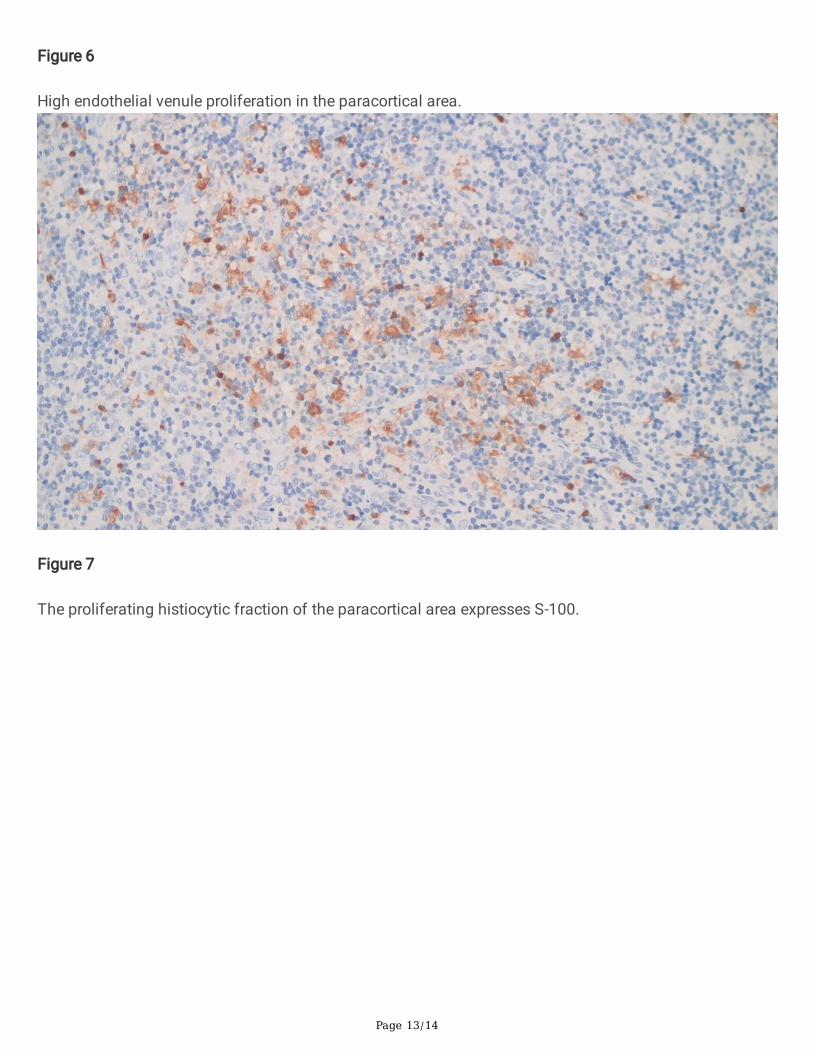
Figure 7
The proliferating histiocytic fraction of the paracortical area expresses S-100.

Page 14/14
Figure 8
Proliferating CD8 positive T cells in the paracortical zone.
Supplementary Files
This is a list of supplementary �les associated with this preprint. Click to download.
CAREchecklist.docx