Linkage and association studies in a Malaysian family with autosomal recessive non-syndromic hearing...
11
23 Malaysian J Pathol 2006; 28 : 23 – 33 Linkage and association studies in a Malaysian family with autosomal recessive non-syndromic hearing loss Farah Wahida I, Aminuddin BS* and Ruszymah BHI Department of Physiology, Faculty of Medicine, Universiti Kebangsaan Malaysia, Tissue Engineering and Human Genetics Laboratory, Hospital Universiti Kebangsaan Malaysia, Kuala Lumpur, Malaysia, and *Ear, Nose & Throat Consultant Clinic, Ampang Puteri Specialist Hospital, Selangor, Malaysia. Abstract Hearing loss is a common sensory deficit in humans. The hearing loss may be conductive, sensorineural, or mixed, syndromic or nonsyndromic, prelingual or postlingual. Due to the complexity of the hearing mechanism, it is not surprising that several hundred genes might be involved in causing hereditary hearing loss. There are at least 82 chromosomal loci that have been identified so far which are associated with the most common type of deafness - non-syndromic deafness. However, there are still many more which remained to be discovered. Here, we report the mapping of a locus for autosomal recessive, non-syndromic deafness in a family in Malaysia. The investigated family (AC) consists of three generations - parents who are deceased, nine affected and seven unaffected children and grandchildren. The deafness was deduced to be inherited in an autosomal recessive manner with 70% penetrance. Recombination frequencies were assumed to be equal for both males and females. Using two-point lod score analysis (MLINK), a maximum lod score of 2.48 at 0% recombinant (Z=2.48, θ=0% ) was obtained for the interval D14S63-D14S74. The haplotype analysis defined a 14.38 centiMorgan critical region around marker D14S258 on chromosome 14q23.2-q24.3. There are 16 candidate genes identified with positive expression in human cochlear and each has great potential of being the deaf gene responsible in causing non-syndromic hereditary hearing loss in this particular family. Hopefully, by understanding the role of genetics in deafness, early interventional strategies can be undertaken to improve the life of the deaf community. Keywords: Autosomal recessive deafness, gene locus, linkage analysis INTRODUCTION Hearing impairment (HI) in newborn infants is a common medical condition that may lead to delays in the development of spoken language skills. Due to the absence of abnormal physical features, it is often difficult to recognize non- syndromic hearing impairment early in a child’s life. Thus, hearing impairment is not detected many months or even years later and often long after essential hearing-dependant learning processes are underway. Improvement in neonatal care and universal immunization programs has the effect of reducing the incidence of non- genetic hearing loss, and consequently increases the relative contribution of hereditary hearing impairment. 1 The discovery of genes and the mutations that lead to hearing loss have been pursued for two major reasons: (i) to provide diagnostic and genetic counseling for patients, and (ii) to provide biological sources for therapeutic approaches in hearing loss. 2 In 1997, Kelsell et al was the first to discover the gap junction beta 2 (GJB2) as one of the gene responsible for non-syndromic deafness, a gene that encodes the gap junction protein of connexin 26 (Cx26). 3 Since then, the number of genes known to cause non-syndromic deafness has remarkably risen, encompassing a large variety of proteins namely molecular motors, gap junctions, ion channels, transcription factors and proteins that form extracellular matrix of the inner ear. 4 Information about these genes and their protein products is revolutionizing our knowledge on the molecular processes involved in hearing and enhancing our understanding of how the alteration of these processes can lead to hearing loss. 5 Address for correspondence and reprint requests: Associate Professor Dr Ruszymah Idrus, Department of Physiology, Medical Faculty, �ni�ersiti �ni�ersiti Kebangsaan Malaysia, Jalan Raja Muda Abdul Aziz, ����� Kuala �umpur, Malaysia ��mail ruszy�medicu�mmy Phone ������������� ����� Kuala �umpur, Malaysia ��mail ruszy�medicu�mmy Phone ������������� Fax � ������9�9��7
-
Upload
independent -
Category
Documents
-
view
0 -
download
0
Transcript of Linkage and association studies in a Malaysian family with autosomal recessive non-syndromic hearing...
23
LINKAGE ANALYSIS OF AUTOSOMAL RECESSIVE DEAFNESSMalaysian J Pathol 2006; 28 : 23 – 33
Linkage and association studies in a Malaysian family with autosomal recessive non-syndromic hearing lossFarah Wahida I, Aminuddin BS* and Ruszymah BHI
Department of Physiology, Faculty of Medicine, Universiti Kebangsaan Malaysia, Tissue Engineering and Human Genetics Laboratory, Hospital Universiti Kebangsaan Malaysia, Kuala Lumpur, Malaysia, and *Ear, Nose & Throat Consultant Clinic, Ampang Puteri Specialist Hospital, Selangor, Malaysia.
Abstract
Hearing loss is a common sensory deficit in humans. The hearing loss may be conductive, sensorineural, or mixed, syndromic or nonsyndromic, prelingual or postlingual. Due to the complexity of the hearing mechanism, it is not surprising that several hundred genes might be involved in causing hereditary hearing loss. There are at least 82 chromosomal loci that have been identified so far which are associated with the most common type of deafness - non-syndromic deafness. However, there are still many more which remained to be discovered. Here, we report the mapping of a locus for autosomal recessive, non-syndromic deafness in a family in Malaysia. The investigated family (AC) consists of three generations - parents who are deceased, nine affected and seven unaffected children and grandchildren. The deafness was deduced to be inherited in an autosomal recessive manner with 70% penetrance. Recombination frequencies were assumed to be equal for both males and females. Using two-point lod score analysis (MLINK), a maximum lod score of 2.48 at 0% recombinant (Z=2.48, θ=0% ) was obtained for the interval D14S63-D14S74. The haplotype analysis defined a 14.38 centiMorgan critical region around marker D14S258 on chromosome 14q23.2-q24.3. There are 16 candidate genes identified with positive expression in human cochlear and each has great potential of being the deaf gene responsible in causing non-syndromic hereditary hearing loss in this particular family. Hopefully, by understanding the role of genetics in deafness, early interventional strategies can be undertaken to improve the life of the deaf community.
Keywords: Autosomal recessive deafness, gene locus, linkage analysis
INTRODUCTION
Hearing impairment (HI) in newborn infants is a common medical condition that may lead to delays in the development of spoken language skills. Due to the absence of abnormal physical features, it is often difficult to recognize non-syndromic hearing impairment early in a child’s life. Thus, hearing impairment is not detected many months or even years later and often long after essential hearing-dependant learning processes are underway. Improvement in neonatal care and universal immunization programs has the effect of reducing the incidence of non-genetic hearing loss, and consequently increases the relative contribution of hereditary hearing impairment.1 The discovery of genes and the mutations that lead to hearing loss have been pursued for
two major reasons: (i) to provide diagnostic and genetic counseling for patients, and (ii) to provide biological sources for therapeutic approaches in hearing loss.2 In 1997, Kelsell et al was the first to discover the gap junction beta 2 (GJB2) as one of the gene responsible for non-syndromic deafness, a gene that encodes the gap junction protein of connexin 26 (Cx26).3 Since then, the number of genes known to cause non-syndromic deafness has remarkably risen, encompassing a large variety of proteins namely molecular motors, gap junctions, ion channels, transcription factors and proteins that form extracellular matrix of the inner ear.4 Information about these genes and their protein products is revolutionizing our knowledge on the molecular processes involved in hearing and enhancing our understanding of how the alteration of these processes can lead to hearing loss.5
Address for correspondence and reprint requests: Associate Professor Dr Ruszymah Idrus, Department of Physiology, Medical Faculty, �ni�ersiti�ni�ersiti Kebangsaan Malaysia, Jalan Raja Muda Abdul Aziz, ����� Kuala �umpur, Malaysia��� ��mail�� ruszy�medic���u�m���my�� Phone�� �������������������� Kuala �umpur, Malaysia��� ��mail�� ruszy�medic���u�m���my�� Phone�� ��������������� Fax �� � ������9�9��7
Malaysian J Pathol June 2006
24
Once the disease gene’s location is pinpointed, it is often possible to provide a more accurate prognosis for individuals at risk for a genetic disease. Genetic mapping via linkage analysis technique has been widely used to map disease genes that follow the Mendelian mode of inheritance. The term ‘linkage’ refers to the tendency of two or more markers (e.g. genes or in this study microsatellite markers) which are located on the same chromosome, to segregate together because of close proximity, hence the probability that these two markers at two different loci will be separated during DNA repair or replication processes is low. Basically, LOD (logarithm of the odds) score is a statistical estimate of linkage (logarithmic odds ratio) whether two loci are likely to lie near each other on the same chromosome and are therefore likely to be inherited together. It is calculated as the log of the ratio of the probability of the observed trait patterns (in this study, hearing loss) if linkage is present to the probability of the observed patterns if no linkage is present.6 Over 100 nuclear gene localizations for non-syndromic hereditary hearing impairment have been identified. These loci are named with the prefix “DFNA” for autosomal dominant, “DFNB” for autosomal recessive and “DFN” for X-linked loci. As of April 2004, 36 auditory genes for non-syndromic deafness have been identified and cloned through linkage analysis in which includes 17 genes for autosomal recessive, 16 genes for autosomal dominant, two genes for mitochondrial and 1 gene for X-linked inheritance.7 According to the statistics, the ratio of deaf persons in Malaysia in 2003 is one in 1046.8 Approximately 50% of all deaf cases are thought to be due to environmental factors leaving another 50% due to genetic aetiology with autosomal dominant, autosomal recessive, X-linked or mitochondrial patterns of inheritance.9 This significant number of incidence affecting Malaysians has prompted this research of hereditary hearing loss in Malaysian families. Here, we report our investigation on linkage and association studies that involved a Malaysian family with autosomal recessive non-syndromic hereditary hearing loss.
MATERIALS AND METHODS
Families, medical history and general examinationThe Research and Ethical Committee of the Medical Faculty, Universiti Kebangsaan
Malaysia has approved this study. We identified a three-generation Malay family (referred to as AC family) where the first generation is already deceased. The following two generations consists of nine affected (including one deceased and one not available for study), seven unaffected individuals (including four not available for study) and four of unknown status (Figure 1). Written informed consents were obtained from all living subjects involved in the study. Once consented, we started our study by investigating the family medical background so as to exclude syndromic deafness. Then, each individual was required to undergo physical and audiological evaluations.
Genomic DNA isolationPeripheral blood samples were taken from participating family members. Mononuclear cells were collected from peripheral blood by using Lymphoprep (AXIS-SHIELD PoC AS, Oslo, Norway) according to the manufacturer’s instructions. DNA samples were extracted from these cells via salting out procedure.8
Genotyping Genetic linkage analysis was performed on this family by using genome-wide, 382 microsatellite markers of ABI Prism Linkage Mapping Set-MD10 of short tandem repeats (STRs) that have an average spacing of 10 cM (Applied Biosystems, Foster City, CA). PCR primer pairs (markers) optimized to highly informative microsatellite loci selected from the Genethon human linkage map.10 The reaction components include 1.2 μl of 50 ng/μl genomic DNA, 1.5 μl of GeneAmp dNTP mix (2.5mM), 1.5 μl of 10X GeneAmp PCR Buffer II, 1.5 μl of 25mM magnesium chloride, 0.12 μl of AmpliTaq Gold DNA Polymerase (5 units/ μl ), 8.18 μl sterile, deionized water and 1 μl of non-syndromic autosomal dominant loci-specific microsatellite markers primer pair mix (5μM each primer) of Linkage Mapping Set MD10 (Applied Biosystem, Foster City, CA, USA) which consists of fluorescently-labeled (6FAM™, HEX® or NED™ which are displayed on ABI PRISM genetic analysis system as blue, green and yellow respectively. The PCR reactions were run according to the instructions provided by the supplier (Table 1). The PCR products were pooled post-PCR in ratios 1:1:2 (FAM:HEX:NED respectively). The pooled PCR products were mixed with 3 μl of loading cocktail containing deionized formamide, EDTA
25
LINKAGE ANALYSIS OF AUTOSOMAL RECESSIVE DEAFNESS
loading buffer and Genescan-400HD ROX size standard (Applied Biosystem, Foster City, CA, USA). After denaturation for two minutes at 95 0C, the samples were then resolved on a 4.25 % polyacrylamide gel and were detected by using ABI377 DNA Sequencer (Applied Biosystems, Foster City, CA, USA). Sizes of marker alleles were defined by the use of GENESCAN software (Applied Biosystems, Foster City, CA, USA). Analyzed data were imported to the GENOTYPER software (Applied Biosystems, Foster City, CA, USA) for allele-calling procedure to generate genotype data. These genotypes were used to determine the lod score and also to reconstruct a haplotype map for all participating subjects. A lod score of > 3.0 is considered significant for linkage whereas <-2.0 is considered against linkage. A lod score between 2.00 to 3.00 is considered suggestive for linkage.
Haplotype reconstruction Haplotypes for each individual were reconstructed based on the genotype data generated from the marker that gave significant lod score (D14S258) and its surrounding markers (D14S63, D14S1036 and D14S74). Haplotypes are a set of closely linked genetic markers present on one chromosome which tend to be inherited together and are not easily separable by recombination. Then, flanking markers or the markers located on the either side of the gene region of interest were determined (D14S63 and D14S74). Flanking markers are used in linkage analysis to track the coinheritance of the gene in question. So
as to reduce the risk of recombination which is proportional to the distance between the disease-causing mutation and the markers, intragenic markers of 5 centiMorgans interval within the candidate regions of interest were selected to generate more genotype data and also to confirm linkage (D14S1036).
Linkage analysisLinkage analysis was done using MLINK (version 5.1). For the analysis, a disease allele frequency of 0.01 was assumed. When more than 50% of the siblings were affected, the mode of inheritance was taken to be autosomal recessive with 70% penetrance. Male and female chromosomes are almost equal in length, thus, the recombination frequencies were assumed to be equal for both male and female.
RESULTS
Families, medical history and general examinationFamily pedigree and medical investigations revealed that hearing loss in this family is inherited recessively though the inheritance is similar to that of autosomal dominance as more than 50% of the siblings were affected (Figure 1). From the pedigree, nine individuals are deaf: II-AC1, II-AC2 (deceased), II-AC3, II-AC6, II-AC7, II-AC8, III-AC11, III-AC12 and III-AC13. The unaffected are II-AC5, III-AC9, III-AC10, III-AC17, III-AC18, III-AC19 and III-AC20, whereas II-AC4 and her children (III-AC14, III-AC15, III-AC16) are of unknown status as they were not available during the study. Only a total of 10 DNA samples were available for
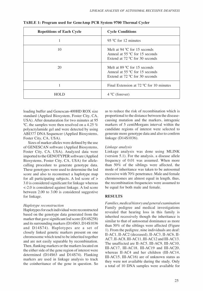
TABLE 1: Program used for GeneAmp PCR System 9700 Thermal Cycler
Repetitions of Each Cycle Cycle Conditions
1 95 0C for 12 minutes 10 Melt at 94 0C for 15 seconds Anneal at 55 0C for 15 seconds Extend at 72 0C for 30 seconds 20 Melt at 89 0C for 15 seconds Anneal at 55 0C for 15 seconds Extend at 72 0C for 30 seconds 1 Final Extension at 72 0C for 10 minutes HOLD 4 0C (forever)
Malaysian J Pathol June 2006
26
1 3 1 3
1 1 1 2
1 3 1 3
1 1 1 2
1 3 1 3
1 1 1 5
1 3 1 3
1 1 1 2
1000
D14
S63
D14
S258
D14
S103
6
D14
S74
II
*AC1
*AC9
*AC1
0*A
C11
*AC1
3A
C12
AC1
4
??
?A
C15
Sym
bols: A
ffect
ed m
ale
Affe
cted
fem
ale
Unk
now
n sta
tus
Pers
onal
ly ex
amin
ed
Hig
h ris
kha
plot
ype
Prob
and
?
I, II,
III
G
ener
atio
n
AC1
6A
C17
AC1
8A
C19
AC2
0
*AC3
*AC5
*AC6
*AC7
*AC8
AC2
AC4
91
1 1 1 1
1 2 1 2
1 3 1 3
1 1 1 1
1 3 1 3
1 1 1 2
I III
1 2 1 2
1 1 1 1
1 2 1 1
1 3 1 3
1001
1 3 1 3
1 1 1 1
1 2 1 3
1 2 1 1
1 3 1 3
1 1 1 1
1 3 1 1
1 1 1 1
??
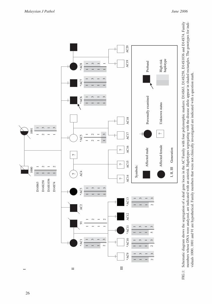
FIG
.1:
Sche
mat
ic d
iagr
am s
how
s th
e se
greg
atio
n of
a d
eaf g
ene
locu
s in
the
AC
Fam
ily w
ith fo
ur p
olym
orph
ic m
arke
rs: D
14S6
3, D
14S2
58, D
14S1
036
and
D14
S74.
Fam
ily
mem
bers
who
se D
NA
wer
e an
alyz
ed, a
re in
dica
ted
with
an
aste
risk.
Hap
loty
pes s
egre
gatin
g w
ith th
e di
seas
e al
lele
app
ear i
n sh
aded
rect
angl
es. T
he g
enot
ypes
for i
ndi-
vidu
als 1
000,
100
1 an
d 91
are
hyp
othe
tical
. Fam
ily m
embe
rs th
at w
ere
not c
linic
ally
inve
stig
ated
are
indi
cate
d w
ith a
que
stio
n m
ark.
27
LINKAGE ANALYSIS OF AUTOSOMAL RECESSIVE DEAFNESS
generating lod scores. The DNAs were from II-AC1, II-AC3, II-AC5, II-AC6, II-AC7, II-AC8, III-AC9, III-AC10, III-AC11 and III-AC13 (marked with asterisk in Figure 1).
Audiological assessmentsAudiological assessments or audiograms revealed 6 out of 8 available affected individuals have bilateral sensorineural profound hearing loss whereas III-AC6 (proband) has bilateral moderate sensorineural hearing loss and III-AC11 is suffering from bilateral high frequency hearing loss. The findings of the audiological assessments conducted are summarised in Table 2.
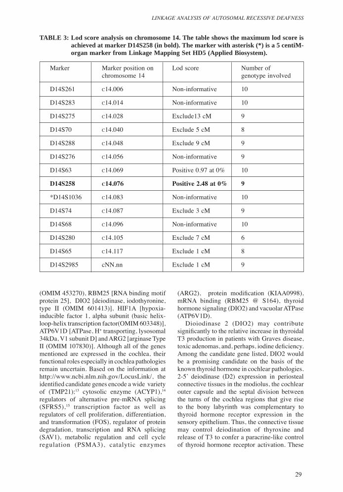
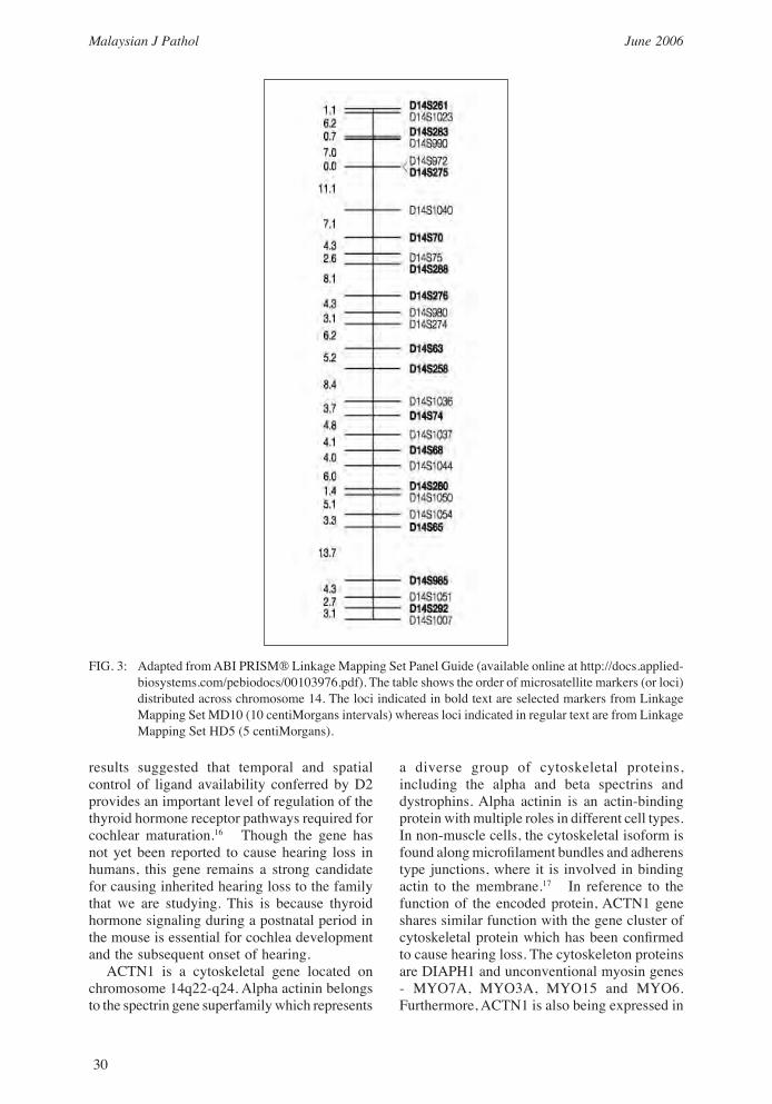
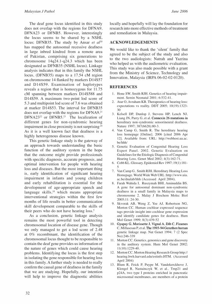
Linkage studiesOnly 10 genotype data were used to calculate lod scores. As the inheritance of hearing loss was autosomal recessive, all the markers listed in the Chromosome X were excluded from the lod scoring. With 382 polymorphic markers, only 326 markers gave meaningful results. After excluding approximately 58.5% of the genome (Figure 2), linkage to the deaf locus gene was detected with the microsatellite marker D14S258, which gave a maximum lod score of 2.48 at 0 % recombinant (Zmax = 2.48, θ = 0.00) (Table 3). The marker D14S63 also gave a positive lod score of 0.97 at 0% recombinant. Three markers centromeric to D14S258; D14S63, D14S276, D14S288 and two markers telomeric to D14S258; D14S74, D14S68 were selected for haplotyping to determine which allele in this set of genotypes belonged to the paternal alleles and which the maternal alleles. Analysis on the reconstructed haplotype showed recombination events on markers D14S276, D14S288 and D14S68 and thus, these markers were excluded. With this exclusion, the markers flanking the region thought to contain the disease gene were D14S63 and D14S74, defining the locus interval of 14.38 cM. The family was genotyped further in the identified locus using 5cM spacing markers. By referring to the Panel Guide for Linkage Mapping Set provided by the supplier, only one 5cM marker (D14S1036) was included in the region (Figure 3), however the genotype data generated from this marker showed homozygosity for all subjects and thus was not very informative. Haplotyping showed the critical region to be flanked by markers D14S63 and D14S74 (Figure 1) on the long arm of chromosome 14q23.2-q24.3. By comparing the haplotypes of family members whose genetic status is known (e.g. affected versus unaffected),
the haplotype associated with the disease-causing allele was identified. Once the disease-associated haplotype was established, it was possible to determine the genetic status of at-risk family members. It was very clear that those who were affected in generation II have both high risk haplotype blocks, thus confirming that the hearing loss portrayed in the family were of autosomal recessive inheritance. However, in II-AC1and II-AC8, a recombination has occurred at marker D14S74, whereas in II-AC5, none of the high risk haplotype blocks was seen except in the area around marker D14S74. This may be because II-AC5 is not affected. Interestingly, individuals III-AC9 and III-AC10 who were also not affected seem to have inherited both the high risk haplotypes.
DISCUSSION
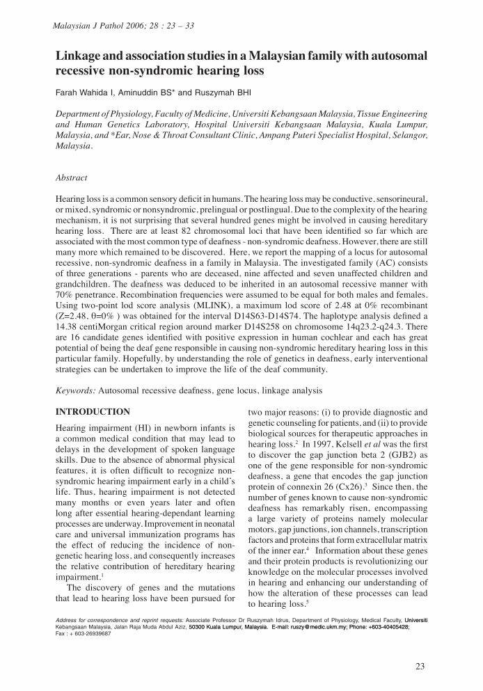
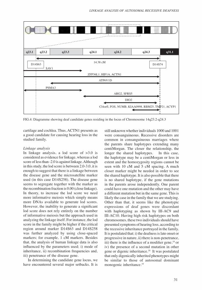
Candidate gene analysisThis study on autosomal recessive, non-syndromic hearing loss has been restricted largely to a small participating family. Unavaibility of large consanguineous populations means insufficient number of informative recombination events to narrow the chromosomal interval resulting in a candidate region9 consisting of 14.34 Megabases. However, within the interval identified, there are approximately 300 genes residing in the region with known or unknown protein functions. The candidate gene approach used in this study depends on the identification of genes specifically expressed in the inner ear. Therefore, to identify the candidate gene for hearing loss in this family and to understand better about human hearing at the molecular level, we referred to the human cochlear cDNA library.11,12 We have narrowed down the genes to 16 (Figure 4) with important roles in auditory function. The genes are ACTN1 [actinin, alpha 1(OMIM 102575), NUMB [numb homolog (Drosophila) (OMIM 603728)], TMP21 [transmembrane trafficking protein (OMIM 605406)], ACYP1 [acylphosphatase, erythrocytes (OMIM 600875)], C14orf1 [chromosome 14 open reading frame 1 (OMIM 604576), SFRS5 [ splicing factor, aginine/serine-rich 5 (OMIM 600914)], FOS [v-fos fbj murine osteosarcoma viral oncogene homolog (OMIM : 164810)], SAV1 [Salvador homolog 1 (Drosophila) (OMIM 607203)], ZFP36L1 [zinc finger protein 36, C3H type-like 1 (OMIM 601064)], PSMA3 (proteasome (prosome, macropain) subunit, alpha type, 3 (OMIM 176843)], KIAA0998

Malaysian J Pathol June 2006
28
FIG. 2: Exclusion mapping excludes 58.5% of the genome from having the disease gene locus. Vertical lines in the map represents areas of exclusion on every chromosome (with the exception of Chromosome X).
Pemetaan Eksklusi Analisa Pertalian Genetik Bagi Keluarga AC
1
2
3
4
56
7
8 9 10
11
12
13
14
1516
17 18
19 20
21 22
0
50
100
150
200
250
300
cMTABLE 2: Results of audiological assessment of affected individuals in the family studied
Audiogram Subject Age Sex Severity Shape Right Left Right Left
II-AC1 43 Female Profound Profound Flat Flat
II-AC3 40 Female Profound Profound – – (>120dB at (>120dB at all frequency) all frequency)
II-AC6 32 Male Profound Profound Steeply Steeply at high at high sloping sloping frequency frequency
II-AC7 26 Male Profound Profound Gently Gently sloping sloping
II-AC8 24 Female Profound Profound Mid Mid frequency frequency U-Shaped U-Shaped
III-AC11 20 Female Moderate- Moderate- Mid Mid Severe Severe frequency frequency steeply steeply sloping sloping
III-AC12 15 Male Profound Profound Gently Gently sloping sloping
III-AC13 11 Male Profound Profound Gently Gently sloping sloping
29
LINKAGE ANALYSIS OF AUTOSOMAL RECESSIVE DEAFNESS
(ARG2), protein modification (KIAA0998), mRNA binding (RBM25 @ S164), thyroid hormone signaling (DIO2) and vacuolar ATPase (ATP6V1D). Dioiodinase 2 (DIO2) may contribute significantly to the relative increase in thyroidal T3 production in patients with Graves disease, toxic adenomas, and, perhaps, iodine deficiency. Among the candidate gene listed, DIO2 would be a promising candidate on the basis of the known thyroid hormone in cochlear pathologies. 2-5’ deiodinase (D2) expression in periosteal connective tissues in the modiolus, the cochlear outer capsule and the septal division between the turns of the cochlea regions that give rise to the bony labyrinth was complementary to thyroid hormone receptor expression in the sensory epithelium. Thus, the connective tissue may control deiodination of thyroxine and release of T3 to confer a paracrine-like control of thyroid hormone receptor activation. These
(OMIM 453270), RBM25 [RNA binding motif protein 25], DIO2 [deiodinase, iodothyronine, type II (OMIM 601413)], HIF1A [hypoxia-inducible factor 1, alpha subunit (basic helix-loop-helix transcription factor(OMIM 603348)], ATP6V1D [ATPase, H+ transporting, lysosomal 34kDa, V1 subunit D] and ARG2 [arginase Type II (OMIM 107830)]. Although all of the genes mentioned are expressed in the cochlea, their functional roles especially in cochlea pathologies remain uncertain. Based on the information at http://www.ncbi.nlm.nih.gov/LocusLink/, the identified candidate genes encode a wide variety of (TMP21);13 cytosolic enzyme (ACYP1),14 regulators of alternative pre-mRNA splicing (SFRS5),15 transcription factor as well as regulators of cell proliferation, differentiation, and transformation (FOS), regulator of protein degradation, transcription and RNA splicing (SAV1), metabolic regulation and cell cycle regulation (PSMA3), catalytic enzymes
TABLE 3: Lod score analysis on chromosome 14. The table shows the maximum lod score is achieved at marker D14S258 (in bold). The marker with asterisk (*) is a 5 centiM-organ marker from Linkage Mapping Set HD5 (Applied Biosystem).
Marker Marker position on Lod score Number of chromosome 14 genotype involved
D14S261 c14.006 Non-informative 10
D14S283 c14.014 Non-informative 10
D14S275 c14.028 Exclude13 cM 9
D14S70 c14.040 Exclude 5 cM 8
D14S288 c14.048 Exclude 9 cM 9
D14S276 c14.056 Non-informative 9
D14S63 c14.069 Positive 0.97 at 0% 10
D14S258 c14.076 Positive 2.48 at 0% 9
*D14S1036 c14.083 Non-informative 10
D14S74 c14.087 Exclude 3 cM 9
D14S68 c14.096 Non-informative 10
D14S280 c14.105 Exclude 7 cM 6
D14S65 c14.117 Exclude 1 cM 8
D14S2985 cNN.nn Exclude 1 cM 9
Malaysian J Pathol June 2006
30
results suggested that temporal and spatial control of ligand availability conferred by D2 provides an important level of regulation of the thyroid hormone receptor pathways required for cochlear maturation.16 Though the gene has not yet been reported to cause hearing loss in humans, this gene remains a strong candidate for causing inherited hearing loss to the family that we are studying. This is because thyroid hormone signaling during a postnatal period in the mouse is essential for cochlea development and the subsequent onset of hearing. ACTN1 is a cytoskeletal gene located on chromosome 14q22-q24. Alpha actinin belongs to the spectrin gene superfamily which represents
a diverse group of cytoskeletal proteins, including the alpha and beta spectrins and dystrophins. Alpha actinin is an actin-binding protein with multiple roles in different cell types. In non-muscle cells, the cytoskeletal isoform is found along microfilament bundles and adherens type junctions, where it is involved in binding actin to the membrane.17 In reference to the function of the encoded protein, ACTN1 gene shares similar function with the gene cluster of cytoskeletal protein which has been confirmed to cause hearing loss. The cytoskeleton proteins are DIAPH1 and unconventional myosin genes - MYO7A, MYO3A, MYO15 and MYO6. Furthermore, ACTN1 is also being expressed in
FIG. 3: Adapted from ABI PRISM® Linkage Mapping Set Panel Guide (available online at http://docs.applied-biosystems.com/pebiodocs/00103976.pdf). The table shows the order of microsatellite markers (or loci) distributed across chromosome 14. The loci indicated in bold text are selected markers from Linkage Mapping Set MD10 (10 centiMorgans intervals) whereas loci indicated in regular text are from Linkage Mapping Set HD5 (5 centiMorgans).
31
LINKAGE ANALYSIS OF AUTOSOMAL RECESSIVE DEAFNESS
cartilage and cochlea. Thus, ACTN1 presents as a good candidate for causing hearing loss in the studied family.
Linkage analysisIn linkage analysis, a lod score of >3.0 is considered as evidence for linkage, whereas a lod score of less than -2.0 is against linkage. Although in this study, the lod score is between 2.0-3.0, it is enough to suggest that there is a linkage between the disease gene and the microsatellite marker used (in this case D14S258). The disease gene seems to segregate together with the marker as the recombination fraction is 0.00 (close linkage). In theory, to increase the lod score we need more informative meiosis which simply means more DNAs available to generate lod scores. However, the inability to generate a significant lod score does not rely entirely on the number of informative meiosis but the approach used in analyzing the linkage itself. For instance, the lod score in the family might be higher if the critical region around marker D14S63 and D14S258 was further analyzed by using close-spaced markers; for example, 1 cM markers. Besides that, the analysis of human linkage data is also influenced by the parameters used; i) mode of inheritance, ii) recombination frequencies and, iii) penetrance of the disease gene. In determining the candidate gene locus, we have encountered several major setbacks. It is
FIG.4: Diagramme showing deaf candidate genes residing in the locus of Chromosome 14q23.2-q24.3
still unknown whether individuals 1000 and 1001 were consanguineous. Recessive disorders are common in consanguineous marriages where the parents share haplotypes extending many centiMorgan. The closer the relationship, the longer the shared haplotypes. In this case, the haplotype may be a centiMorgan or less in extent and the homozygosity regions cannot be seen with 10 cM and 5 cM spacing. A much closer marker might be needed in order to see the shared haplotype. It is also possible that there is no shared haplotype, if the gene mutations in the parents arose independently. One parent could have one mutation and the other may have a different mutation but in the same gene. This is likely the case in the family that we are studying. Other than that, it seems like the phenotypic expressions of deaf genes were discordant with haplotyping as shown by III-AC9 and III-AC10. Having high risk haplotypes on both chromosomes, these two individuals should have presented symptoms of hearing loss, according to the recessive inheritance portrayed in the family. It is postulated that; i) the deafness is late onset or progressive in nature, ii) there is non-penetrance, iii) there is the influence of a modifier gene,18 or iv) the presence of a second mutation in other gene or digenic inheritance.18 It was postulated that only digenically inherited phenotypes might be similar to those of autosomal dominant monogenic inheritance.19
q31.1q24.3q24.2q24.1q23.3q23.2
SAV1
PSMA3
14.38 cM
ZFP36L1, HIF1A, ACTN1
ATP6V1D
ARG2, SFRS5
DIO2
Cl4orfl, FOS, NUMB, KIAA0998, RBM25, TMP21, ACYP1
q23.1
D14S74D14S63
Malaysian J Pathol June 2006
32
The deaf gene locus identified in this study does not overlap with the regions for DFNA9, DFNA23 or DFNB5. However, interestingly the locus seems to be shared by a NSHL locus; DFNB35. The study by Ansar et al20 has mapped the autosomal recessive deafness in large inbred kindred from a remote area of Pakistan, comprising six generations to chromosome 14q24.1-q24.3 which has been designated as DFNB35 (NSHL locus). Linkage analysis indicates that in this family the NSHL locus, (DFNB35) maps to a 17.54 cM region on chromosome 14 flanked by markers D14S57 and D14S59. Examination of haplotypes reveals a region that is homozygous for 11.75 cM spanning between markers D14S588 and D14S59. A maximum two-point lod score of 5.3 and multipoint lod score of 7.6 was obtained at marker D14S53. The interval for DFNB35 does not overlap with the regions for DFNA9,21 DFNA2322 or DFNB5.23 The localization of different genes for non-syndromic hearing impairment in close proximity is not surprising.24 As it is a well known fact that deafness is a highly heterogenous disease known. This genetic linkage study is the gateway of an approach towards understanding the basic function of the auditory system in the hope that the outcome might provide professionals with specific diagnosis, accurate prognosis, and optimal intervention for people with hearing loss and diseases. But the most important thing is, early identification of significant hearing impairment in infants and young children and early rehabilitation is essential for the development of age-appropriate speech and language skills,18 which means appropriate interventional strategies within the first few months of life results in better communication skill development comparable to the skills of their peers who do not have hearing loss.5
As a conclusion, genetic linkage analysis remains the most powerful tool in detecting chromosomal location of disease gene. Though we only managed to get a lod score of 2.48 at 0% recombinant, the identification of the chromosomal locus thought to be responsible to contain the deaf gene provides us information of the nature of genes which could cause hearing problems. Identifying the locus is the first step in isolating the gene responsible for hearing loss in this family. A further study is needed to really confirm the causal gene of deafness in the family that we are studying. Hopefully, our intention will help to improve the diagnostic abilities
locally and hopefully will lay the foundation for research into more effective methods of treatment and remediation in Malaysia.
ACKNOWLEDGEMENTS
We would like to thank the ‘silent’ family that agreed to be the subject of the study and also to the two audiologists; Natrah and Yazrina who helped us with the audiometric evaluation. This study was also made possible with a grant from the Ministry of Science, Technology and Innovation, Malaysia (IRPA 06-02-02-0120).
REFERENCES 1. Hone SW, Smith RJH. Genetics of hearing impair-
ment. Semin Neonatal 2001; 6:532-41. 2. Atar O, Avraham KB. Therapeutics of hearing loss:
expectations vs reality. DDT 2005; 10(19):1323-30
3. Kelsell DP, Dunlop J, Stevens HP, Lench NJ, Liang JN, Parry G, et al. Connexin 26 mutations inConnexin 26 mutations in hereditary non-syndromic sensorineural deafness. Nature 1997; 387(6628):80-3.
4. Van Camp G, Smith R. The hereditary hearing loss homepage [Online]. 2006 [cited 2006 Apr 12]. Available from: URL: http://webhost.ua.ac.be/hhh/
5. Genetic Evaluation of Congenital Hearing Loss Expert Panel. 2002. Genetic Evaluation on Guidelines for the Etiologic Diagnosis of Congenital Hearing Loss. Genet Med 2002; 4(3):162-71.
6. Cobb KL. Glossary. Epidemiol Rev 1997; 19(1):181-5
7. Van Camp G, Smith RJH. Hereditary Hearing Loss Homepage. World Wide Web URL :http://www.uia.ac.be/dnalab/hhh (Accessed April 2004).
8. Farah Wahida I, Ruszymah BHI, Aminuddin BS. A gene for autosomal dominant non-syndromic deafness in a small family in Malaysia maps to chromosome 2. Malay J Biochem Molec Biol 2005;11: 24-30.
9. Skvorak AB, Weng Z, Yee AJ, Robertson NG, Morton CC. Human cochlear expressed sequence tags provide insight into cochlear gene expression and identify candidate genes for deafness. Hum Mol Genet 1999; 8(3):439-52
10. Gyapay G, Morissette J, Vignal A, Dib C, FizamesGyapay G, Morissette J, Vignal A, Dib C, Fizames C, Millasesau P, et al. The 1993-94 Genethon humanThe 1993-94 Genethon human genetic linkage map. Nat Genet 1994; 7 (2 Spec No):246-339
11. Morton CC. Genetics, genomics and gene discovery in the auditory system. Hum Mol Genet 2002; 11(10):1229-40.
12. Morton CC. Morton Hearing Research Group http://hearing.bwh.harvard.edu/estinfo.HTM. (Accessed April 2004)
13. Blum R, Feick P, Puype M, Vandekerckhove J, Klengel R, Nastainczyk W, et al.. Tmp21 and p24A, two type I proteins enriched in pancreatic microsomal membranes, are members of a protein
33
LINKAGE ANALYSIS OF AUTOSOMAL RECESSIVE DEAFNESS
family involved in vesicular trafficking. J Biol Chem 1996; 271(29):17183-9.
14. Fiaschi T, Raugei G, Marzocchini R, Chiarugi P, Cirri P, Ramponi G. Cloning and expression of the cDNA coding for the erythrocyte isoenzyme of human acylphosphatase. FEBS Lett 1995; 367(2):145-8
15. Diamond RH, Du K, Lee VM, Mohn KL, Haber
family involved in vesicular trafficking. J Biol Chem 1996; 271(29):17183-9.
14. Fiaschi T, Raugei G, Marzocchini R, Chiarugi P, Cirri P, Ramponi G. Cloning and expression of the cDNA coding for the erythrocyte isoenzyme of human acylphosphatase. FEBS Lett 1995; 367(2):145-8
15. Diamond RH, Du K, Lee VM, Mohn KL, Haber BA, Tewari DS, et al. Novel delayed-early and highly insulin-induced growth response genes. Identification of HRS, a potential regulator of alternative pre-mRNA splicing. J Biol Chem 1993; 268(20):15185-92.
16. Campos-Barros A, Amma LL, Faris JS, Shailam R, Kelley MW, Forrest D. Type 2 iodothyronine deiodinase expression in the cochlea before the onset of hearing. Proc Natl Acad Sci USA 2000; 97(3):1287-92.
17. Youssoufian H, McAfee M, Kwiatkowski DJ. Cloning and chromosomal localization of the human cytoskeletal alpha-actinin gene reveals linkage to the beta-spectrin gene. Am J Hum Genet 1990; 47(1):62-71
18. Zlotogora J. Penetrance and expressivity in the molecular age. Genet Med 2003; 5(5):347-52.
19. Balciuniene J, Dahl N, Borg E, Samuelsson E, Koisti M J, Petterson U, et al. Evidence for digenicEvidence for digenic inheritance of non syndromic hereditary hearing loss in a Swedish family. Am J Hum Genet 1998; 63: 786-93.
20. Ansar M, Din MA, Arshad M, Sohail M, Faiyaz-Ul-Haque M, Haque S, et al. A novel autosomalA novel autosomal recessive non-syndromic deafness locus (DFNB35) maps to 14q24.1-14q24.3 in large consanguineous kindred from Pakistan. Eur J Hum Genet 2003; 11(1):77-80.
21. Manolis EN, Yandavi N, Nadol JB, Eavey RD, McKenna M, Rosenbaum S, et al. A gene for non-syndromic autosomal dominant progressive postlingual sensorineural hearing loss maps to chromosome 14q12-13. Hum Molec Genet 1996; 5:1047-50.
22. Salam AA, Hafner FM, Linder TE, Spillmann T, Schinzel AA, Leal SM. A novel locus (DFNA23)A novel locus (DFNA23) for prelingual autosomal dominant nonsyndromic hearing loss maps to 14q21-q22 in a Swiss German kindred. Am J Hum Genet 2000; 66: 1984-8.
23. Fukushima K, Ramesh A, Srikumari Srisailapathy CR, Ni L, Chen A, O’Neill M, et al. Consanguineous nuclear families used to identify a new locus for recessive non-syndromic hearing loss on 14q. Hum Molec Genet 1995; 4: 1643-8.
24. Snoeckx RL, Kremer H, Ensink RJH, Flothmann K, de Brower A, Smith RJ H, et al. A novel locus for autosomal dominant non-syndromic hearing loss, DFNA31, maps to chromosome 6p21.3. J Med Genet 2004; 41(1):11-3.