and Stretch-Induced Dissociation of P-Selectin/PSGL-1 Complex
9
Molecular Dynamics Simulation of Shear- and Stretch-Induced Dissociation of P-Selectin/PSGL-1 Complex Yingyong Kang, 6 Shouqin Lu ¨, 6 Peng Ren, Bo Huo,* and Mian Long* Key Laboratory of Microgravity (National Microgravity Laboratory) and Center for Biomechanics and Bioengineering, Institute of Mechanics, Chinese Academy of Sciences, Beijing, People’s Republic of China ABSTRACT By mediating the tethering and rolling of leukocytes on vascular surfaces, the interactions between P-selectin and the P-selectin glycoprotein ligand 1 (PSGL-1) play crucial roles during inflammation cascade. Tensile stretch produced by rolling leukocytes and shear stress exerted by blood flow constitute the two types of mechanical forces that act on the P-selectin/ PSGL-1 bond. These forces modulate not only dissociation kinetics of this bond, but also the leukocyte adhesion dynamics. However, the respective contribution of the two forces to bond dissociation and to the corresponding microstructural bases remains unclear. To mimic the mechanical microenvironment, we developed two molecular dynamics approaches; namely, an approach involving the shear flow field with a controlled velocity gradient, and the track dragging approach with a defined trajectory. With each approach or with both combined, we investigate the microstructural evolution and dissociation kinetics of the P-LE/SGP-3 construct, which is the smallest functional unit of the P-selectin/PSGL-1 complex. The results demonstrate that both shear flow and tensile stretch play important roles in the collapse of the construct and that, before bond dissociation, the former causes more destruction of domains within the construct than the latter. Dissociation of the P-LE/SGP-3 construct features intramolecular destruction of the epidermal-growth-factor (EGF) domain and the breaking of hydrogen-bond clusters at the P-selectin-lectin/EGF interface. Thus, to better understand how mechanics impacts the dissociation kinetics of the P-selectin/PSGL-1 complex, we propose herein two approaches to mimic its physiological mechanical environment. INTRODUCTION P-selectin is a member of the selectin family and is ex- pressed on activated endothelium and platelets (1). One of its ligands, P-selectin glycoprotein ligand-1 (PSGL-1), is constitutively expressed on leukocytes (2). Interactions between P-selectin and PSGL-1 play crucial roles in medi- ating the tethering and rolling of leukocytes on postcapillary venular surfaces during the initial stage of inflammation cascade, which is a necessary stage for leukocytes to further adhere to the surface before finally penetrating through the venule into the inflamed tissues (3,4). To support smooth rolling of leukocytes, the dissociation of a P-selectin/ PSGL-1 bond in the rear of the cell should be balanced with the formation of another bond at the front. During this process, two types of mechanical forces act on the complexes, which further regulate the dissociation kinetics for the dynamic balance of leukocytes (5,6). These two mechanical forces are tensile stretch, which is induced by the rolling of the leukocytes, and shear stress, which is ex- erted by blood flow near the venular surface. Here, tensile stretch is exerted via cell rolling at one end of the complex and extends along the principal axis of the complex, whereas shear flow acts on the entire molecule with a velocity gradient perpendicular to the venular surface (7). In previous work, the researchers experimentally investi- gated the effects of tensile stretch on P-selectin/PSGL-1 bond dissociation using techniques such as atomic force microscopy, biomembrane force probe, and optical tweezers (5,8–10). Furthermore, they used steered molecular dynamics simulation (SMD) to elucidate the microstructural bases of P-selectin unfolding (11) and P-selectin/PSGL-1 bond dissociation under mechanical stretch (12) and proposed a sliding-rebinding model to explain the catch bond at atomic level (13). However, along the preset direc- tion the uniaxial stretch with constant force or constant velocity in these measurements and simulations is distinct from the physiological case where the mechanical stretch is applied in a time-varying direction and magnitude deter- mined by the rolling leukocyte. Thus, a new approach is desirable to manipulate the movement of this molecular complex under a physiologically relevant stretch. The flow-chamber technique is ideal for measuring the kinetic features of the P-selectin/PSGL-1 complex at the cell level when both tensile stretch and shear stress are exerted (5,14,15). But it is difficult to isolate the impact of shear flow from that of tensile stretch. Molecular dynamics (MD) simu- lation might be a useful tool to study the effect of shear flow on the microstructural evolution of the complex. Previously, a flow field with constant velocity was established to study the flow-induced structural transition of glycoprotein Ib, in which a constant force was applied to each atom in a layer upstream of the interested molecule (16–18). The uniform flow field described in these studies may be applicable for the short 16-residue peptide, but a flow field with a nonzero velocity gradient should be more reasonable for the P-selec- tin/PSGL-1 complex, which is >1000-residues long. Submitted July 17, 2011, and accepted for publication November 21, 2011. 6 Yingyong Kang and Shouqin Lu ¨ contributed equally to this work. *Correspondence: [email protected] or [email protected] Editor: Nathan Baker. Ó 2012 by the Biophysical Society 0006-3495/12/01/0112/9 $2.00 doi: 10.1016/j.bpj.2011.11.4002 112 Biophysical Journal Volume 102 January 2012 112–120 brought to you by CORE View metadata, citation and similar papers at core.ac.uk provided by Elsevier - Publisher Connector
-
Upload
khangminh22 -
Category
Documents
-
view
0 -
download
0
Transcript of and Stretch-Induced Dissociation of P-Selectin/PSGL-1 Complex
112 Biophysical Journal Volume 102 January 2012 112–120
brought to you by COREView metadata, citation and similar papers at core.ac.uk
provided by Elsevier - Publisher Connector
Molecular Dynamics Simulation of Shear- and Stretch-InducedDissociation of P-Selectin/PSGL-1 Complex
Yingyong Kang,6 Shouqin Lu,6 Peng Ren, Bo Huo,* and Mian Long*Key Laboratory of Microgravity (National Microgravity Laboratory) and Center for Biomechanics and Bioengineering, Institute of Mechanics,Chinese Academy of Sciences, Beijing, People’s Republic of China
ABSTRACT By mediating the tethering and rolling of leukocytes on vascular surfaces, the interactions between P-selectin andthe P-selectin glycoprotein ligand 1 (PSGL-1) play crucial roles during inflammation cascade. Tensile stretch produced by rollingleukocytes and shear stress exerted by blood flow constitute the two types of mechanical forces that act on the P-selectin/PSGL-1 bond. These forces modulate not only dissociation kinetics of this bond, but also the leukocyte adhesion dynamics.However, the respective contribution of the two forces to bond dissociation and to the corresponding microstructural basesremains unclear. To mimic the mechanical microenvironment, we developed two molecular dynamics approaches; namely,an approach involving the shear flow field with a controlled velocity gradient, and the track dragging approach with a definedtrajectory. With each approach or with both combined, we investigate the microstructural evolution and dissociation kineticsof the P-LE/SGP-3 construct, which is the smallest functional unit of the P-selectin/PSGL-1 complex. The results demonstratethat both shear flow and tensile stretch play important roles in the collapse of the construct and that, before bond dissociation, theformer causes more destruction of domains within the construct than the latter. Dissociation of the P-LE/SGP-3 constructfeatures intramolecular destruction of the epidermal-growth-factor (EGF) domain and the breaking of hydrogen-bond clustersat the P-selectin-lectin/EGF interface. Thus, to better understand how mechanics impacts the dissociation kinetics of theP-selectin/PSGL-1 complex, we propose herein two approaches to mimic its physiological mechanical environment.
INTRODUCTION
P-selectin is a member of the selectin family and is ex-pressed on activated endothelium and platelets (1). One ofits ligands, P-selectin glycoprotein ligand-1 (PSGL-1), isconstitutively expressed on leukocytes (2). Interactionsbetween P-selectin and PSGL-1 play crucial roles in medi-ating the tethering and rolling of leukocytes on postcapillaryvenular surfaces during the initial stage of inflammationcascade, which is a necessary stage for leukocytes to furtheradhere to the surface before finally penetrating through thevenule into the inflamed tissues (3,4). To support smoothrolling of leukocytes, the dissociation of a P-selectin/PSGL-1 bond in the rear of the cell should be balancedwith the formation of another bond at the front. Duringthis process, two types of mechanical forces act on thecomplexes, which further regulate the dissociation kineticsfor the dynamic balance of leukocytes (5,6). These twomechanical forces are tensile stretch, which is induced bythe rolling of the leukocytes, and shear stress, which is ex-erted by blood flow near the venular surface. Here, tensilestretch is exerted via cell rolling at one end of the complexand extends along the principal axis of the complex,whereas shear flow acts on the entire molecule witha velocity gradient perpendicular to the venular surface (7).
In previous work, the researchers experimentally investi-gated the effects of tensile stretch on P-selectin/PSGL-1
Submitted July 17, 2011, and accepted for publication November 21, 2011.6Yingyong Kang and Shouqin Lu contributed equally to this work.
*Correspondence: [email protected] or [email protected]
Editor: Nathan Baker.
� 2012 by the Biophysical Society
0006-3495/12/01/0112/9 $2.00
bond dissociation using techniques such as atomic forcemicroscopy, biomembrane force probe, and optical tweezers(5,8–10). Furthermore, they used steered moleculardynamics simulation (SMD) to elucidate the microstructuralbases of P-selectin unfolding (11) and P-selectin/PSGL-1bond dissociation under mechanical stretch (12) andproposed a sliding-rebinding model to explain the catchbond at atomic level (13). However, along the preset direc-tion the uniaxial stretch with constant force or constantvelocity in these measurements and simulations is distinctfrom the physiological case where the mechanical stretchis applied in a time-varying direction and magnitude deter-mined by the rolling leukocyte. Thus, a new approach isdesirable to manipulate the movement of this molecularcomplex under a physiologically relevant stretch.
The flow-chamber technique is ideal for measuring thekinetic features of the P-selectin/PSGL-1 complex at thecell levelwhenboth tensile stretch and shear stress are exerted(5,14,15). But it is difficult to isolate the impact of shear flowfrom that of tensile stretch. Molecular dynamics (MD) simu-lation might be a useful tool to study the effect of shear flowon the microstructural evolution of the complex. Previously,a flow field with constant velocity was established to studythe flow-induced structural transition of glycoprotein Ib, inwhich a constant force was applied to each atom in a layerupstream of the interested molecule (16–18). The uniformflow field described in these studies may be applicable forthe short 16-residue peptide, but a flow field with a nonzerovelocity gradient should be more reasonable for the P-selec-tin/PSGL-1 complex, which is >1000-residues long.
doi: 10.1016/j.bpj.2011.11.4002

Shear MD Simulations of P-Selectin/Ligand Bond 113
Only a few works have reported the flow field construc-tion with a preset velocity gradient. For example,researchers created a Poiseuille flow by applying an externalforce on each solvent particle within the entire field (19),and a Couette flow was created by moving the upper planeto drag the below-solvent particles (20). However, theformer produces compulsive flow by enforcing each solventparticle to move, which should not be the case in reality,especially for those solvent particles very close to themacromolecule. The latter takes a very long time to generatea flow field with various velocities due to the low efficiencyof the momentum delivery from the mobile plane to theneighboring molecules, which would be time-consumingwhen performing macromolecule simulations under flow.Therefore, a fully developed shear flow with a preset linearvelocity distribution is required to mimic physiologicalblood flow.
In this study, we use MD simulation to develop two waysto address the problem: the shear flow field (SFF) withconstant velocity gradient and the track-dragging approach(TDA) with a defined trajectory. Using these methods, weelucidate herein the impact of shear flow and/or tensilestretch on the dissociation dynamics of the P-LE/SGP-3construct, which is an effective binding unit of the P-selec-tin/PSGL-1 complex (21). Microstructural bases of bonddissociation are then analyzed under mechanical conditionsthat mimic the physiological situation.
FIGURE 1 Schematic of shear flow field (A), CWS in NAMD (B),
virtual-leukocyte rolling model (C), and TDA (D). (A) Flow field is mainly
divided into four regions: bottom layer, top layer, left dragging box, and
work region. Margins on both sides are set to prevent water molecules on
the right side of the system from directly running into the left dragging
box because of the periodic boundary condition. (B) Water molecules in
the work region and margins at both sides are shown (lines), and those in
other regions are displayed (denoted VDW). (C) Virtual leukocyte (sphere)
with radius R rolls at velocity Vunder shear flow. The physical axis of P-LE/
SGP3 is denoted (line) with length L connecting leukocyte and vascular
surface. Angle between the line and the surface is shown (denoted as q).
(D) To mimic mechanical stretch on the complex (springs in series) from
the rolling leukocyte, a virtual spring is placed between the end of the
complex (solid circle) and its target point (open circle), which moves along
the predefined track (dashed line). The dragging force F (arrow) points to
the target point from the end of the complex.
MATERIALS AND METHODS
System construction and equilibration
Two systems were constructed: a cubic water system (CWS) consisting of
only water molecules to evaluate the shear-flow simulation method, and
a molecular complex system (MCS) to evaluate the impacts of shear flow
and/or tensile stretch. The MCS system was built by solvating P-LE/
SGP-3 (Protein DataBank code: 1G1S) into a water box and neutralizing
it with ~100 mM Naþ and Cl� ions. The water molecules and the ions
were created using SOLVATE1.2 and AUTOIONIZE1.3 plugins for VMD
(22). The force-field parameters of CHARMM22 were adopted to describe
water molecules (23), peptides, and ions (24,25) in P-LE/SGP-3. Those of
sLex-modified glycan and tyrosine sulfate TYS residues of SGP-3 are iden-
tical to our previous study (12). All simulations were performed using the
program NAMD2 (26).
The CWS system included 66,390 water molecules and had dimensions
of 120� 90� 60 A3 (x� y� z). Before equilibration, energy minimization
was done in sequence for 10,000 steps and heating to 330 K in 33-K incre-
ments every 10 ps. The system was then allowed to equilibrate freely for
200 ps in a 330-K heat bath (controlled by Langevin thermostat) and at
one atm of pressure (controlled by the Nose-Hoover Langevin piston
method with a coupling coefficient of 2.0 ps�1). Next, another 200-ps equil-
ibration was manipulated by further fixing water molecules in the bottom
5 A layer. We use the resulting equilibrated system to evaluate the proposed
approach for simulating shear flow with a constant velocity gradient.
Our MCS system contained 129,823 molecules and had dimensions of
105 � 175� 70 A3. The original strontium ion Sr2þ of the crystal structure
was replaced by the calcium ion Ca2þ because, physiologically, P-selectin/
PSGL-1 interactions are Ca2þ-dependent (27). The physical axis of the
P-LE/SGP-3 construct is defined by the vector connecting the terminal
atom of the SGP-3 peptide (P618-Ca) to the terminal atom of the P-selectin
epidermal-growth-factor (EGF) domain (D158-Ca). To represent the phys-
iological orientation (Figs. 1 and 2), this axis is rotated by 60� relative to thex axis (28). We first minimized the system using 5000 steps with the fixed
backbone of P-LE and of the SGP-3 peptide, the Ca2þ ion, and the nonhy-
drogen atoms of SGP-3 glycan. Further minimization was done for another
5000 steps with fixed D158-Ca and with P618-Ca constrained in the x and z
directions. The system was then heated to 310 K followed by equilibration
for 1 ns with the same elements of D158-Ca held fixed and P618-Ca con-
strained in the x and z directions, and another equilibration of 3 ns with
addition of bottom 5 A-water layer fixation. The equilibrations were per-
formed under the same pressure, and temperature as that of the CWS
system. During the second equilibration process, the root mean-square devi-
ation (RMSD) of P-LE/SGP-3 construct with respect to the crystal structure
was 1.39 5 0.23 A. We use this system for the subsequent dissociation
simulations.
For all simulations with periodic boundary conditions, we use an integra-
tion time step of 1 fs, a uniform dielectric constant of 1.0, a cutoff of
nonbonded interactions with a switching function starting at 11 A and
reaching zero at 14 A, and a scaling factor of 1.0 for 1–4 interactions.
Shear flow field
We use the TclBCmethod based on the Tcl interface of the NAMD program
(29) to achieve a shear flow field with constant velocity gradient. This
method allows us to apply forces at each time step to water molecules in
specific regions to accelerate them to the target velocity. As indicated in
Fig. 1, A and B, water molecules within the bottom 5 A layer are fixed,
and those within the top 5 A layer and the leftmost layer with thickness
of 5, 10, or 15 A are forced to move at the target velocity.
Biophysical Journal 102(1) 112–120

FIGURE 2 Definitions of physical parameters for structural analysis of
the dissociation of the P-LE/SGP-3 construct. Extension (E1þE2) of the
construct is the sum of the distance E1 between D158-Ca and the mass
center of the binding sites (S46-N, S46-OG, S47-OG, N82-ND2, R85-
NH1, E88-OE2, K112-N, and H114-NE2) and distance E2 between
P618-Ca and mass center of the binding sites. Separation distance
(S1þS2) is the sum of the distance S1 between the mass center of the
Lec domain and the mass center of the binding sites and the distance S2
between the mass center of SGP-3 and the mass center of the binding sites.
D1 represents the distance between the mass center of the backbone atoms
of two key antiparallel b-sheets (G131-E135 and Y140-C144), D2 is the
distance between T2-Ca and I137-Ca, and D3 is the distance between
Ca2þ and the mass center of FUC623. The physical axis (dotted line linking
D158-Ca and P618-Ca) of the construct makes an angle q with the hori-
zontal. (Cartoon) P-LE domains (SGP-3 ligand was shown in ribbons for
peptide and licorice for glycan, respectively).
114 Kang et al.
By Newton’s second law, the dragging force is
F ¼ MUtðyÞ � UðyÞ
Dt; (1)
where Ut(y) is the target velocity along the x axis, U(y) is the current
velocity, M is the mass of the water molecule, and Dt ¼ 1 fs is the time
interval. The dragging force is applied to the oxygen atoms of the water
molecules along the x axis and is recalculated at each time step. The target
velocity of water molecules upon which a force is exerted is defined as
UtðyÞ ¼ U0
y� ybyu � yb
; (2)
in which U0 is the target velocity of the whole top layer, yb and yu are,
respectively, the minimal and maximal y coordinates of the leftmost region,
and y is the instantaneous coordinate of the oxygen atom upon which the
force is exerted.
Biophysical Journal 102(1) 112–120
We assessed the feasibility of our approach for simulating the shear flow
field under three different control conditions: neither temperature- nor pres-
sure-controlled (NTNP), temperature- but not pressure-controlled (CTNP),
and both temperature- and pressure-controlled (CTCP). Because pressure
control is coupled with system temperature, the case of pressure-controlled
but not temperature-controlled (NTCP) is impracticable. Three velocity
gradients, 1.25, 3.75, and 6.25� 10�3 ps�1, were selected with correspond-
ing target velocities U0 of 0.1, 0.3, and 0.5 A/ps, respectively. Each simu-
lation, with its specific control conditions, velocity gradient, and
leftmost-box thickness, was repeated three times.
Track dragging approach
We developed a virtual model of a rolling leukocyte to mimic mechanical
stretch and applied it to one end of a P-selectin/PSGL-1 complex (21)
(Fig. 1 C): a virtual leukocyte of radius R rolls with velocity V, and a
P-selectin/PSGL-1 complex with physical length L links the virtual cell
and the vascular surface and makes an angle q with the vascular surface.
Because the length of P-LE/SGP-3 is much shorter than that of the P-selec-
tin/PSGL-1-complex (30–32), the virtual cell shrinks by reducing the radius
Ri according to
Ri ¼ RrL1
L0
; (3)
where Rr is the actual radius of the leukocyte, L1 is the linear length of the
P-LE/SGP-3 construct along the physical axis, and L0 is the extracellular
length of the P-selectin/PSGL-1 complex. We find Ri ¼ 0.4 mm for Rr ¼5.0 mm, L1 ¼ 76.20 A, and L0 ¼ 90.0 nm. After equilibration, the angle
q of P-LE/SGP-3 varies from 60� to 65� (Fig. 1C).We also modeled the tensile stretching of P-LE/SGP-3, which is caused
by the rolling leukocyte, with the TclBC method (29). This stretching pulls
atom P618-Ca of P-LE/SGP-3 along the predefined trajectory via a virtual
spring to mimic drag on the rolling cell (Fig. 1D). The drag force is given as
F ¼ k
"X3
i¼ 1
ðxit � xiÞ2#1=4
; (4)
where the stiffness k of the virtual spring was set to 25.0 kcal/(mol$A3/2) or
1737 pN$A-1/2, in which xi (i ¼ 1, 2, 3) represents the instantaneous coor-
dinates x, y, and z of the end of P-LE/SGP-3 that experiences the force, and
xit is the target coordinate, which is updated along the trajectory at each
time step Dt (¼ 0.1 ps). We use the fourth root in Eq. 4 to get a moderate
drag force to prevent excessively large or small forces. Based on the virtual
model of a rolling leukocyte (Fig. 1 C), the coordinates of the target trajec-
tory at time t are
xt ¼ x0 þ Vt � Risin
�V
t
Ri
þ 0:186
�þ 773:107;
yt ¼ y0 þ Ri � Ricos
�V
t
Ri
þ 0:186
�;
zt ¼ z0;
(5)
where x0, y0, and z0 are the coordinates of the fixed atom D158-Ca of P-LE/
SGP-3 and t is the simulation time. The rolling velocity V of the virtual
leukocyte is coupled with shear flow because leukocyte rolling results
from shear flow.
Both in vitro and in vivo measurements suggest that the ratio between
leukocyte rolling velocity and wall shear stress (ranging from 0.085
to 0.5 Pa) is ~1.1–7.0 � 10�7 A/(ps$Pa) (7,9,33). Given the ratio of
5.86 � 10�7 A/(ps$Pa), we ran the TDA simulations alone or the combined
TDA-SFF simulations for the MCS at a virtual-cell rolling velocity of
0.08 A/ps, which resulted from a velocity gradient of 1.875 � 10�3 ps�1

Shear MD Simulations of P-Selectin/Ligand Bond 115
with a dynamic water viscosity of 8550 � 10�7 N$s/m2, a system temper-
ature of 300 K, and a virtual-cell radius of 0.4 mm.
Data analysis
To evaluate the quality of the constructed SFF, we calculate the velocity
distribution of water molecules within different regions and their corre-
sponding temporal evolution. A cuboid 10 � 80 � 20 A3 in size in the
central work region, 40 A away from the left-dragging box, is divided
into a series of 10 � 10 � 20 A3 small cuboids along the y axis, and
each small cuboid included ~64 water molecules. The overall velocity of
each small cuboid is defined as the average velocity of the water molecules
passing through it in a 20-ps time interval. To obtain sufficient data and to
decrease the local computational errors, neighboring cuboids are overlap-
ped by 5 A along the y axis. The SFF uniformity along the x axis is similarly
evaluated by calculating the overall velocities of a series of cuboids along
the x axis whose geometric centers are located 45 A above the bottom layer.
We estimate the deviation of the constructed flow field from the target flow
field by using the root mean-square deviation of velocities (RMSV),
RMSV ¼
ffiffiffiffiffiffiffiffiffiffiffiffiffiffiffiffiffiffiffiffiffiffiffiffiffiffiffiPNi¼ 1
ðUi � UtÞ2s
ffiffiffiffiN
p ; (6)
where Ui is the average velocity for each cuboid, Ut is the target velocity at
the center of the corresponding cuboid, and N is the total number of
cuboids.
The extension and separation of the molecular complex are defined to
globally quantify the conformational evolution for SFF- and/or TDA-
induced dissociation of the P-LE/SGP-3 construct (Fig. 2). Molecular
extension includes both intermolecular dissociation and intramolecular
disruption. Considering that the construct may bend under the SFF, the
extension is determined by summing the two distances E1 and E2. Simi-
larly, we determine the complex separation accompanied by intermolecular
dissociation by summing the distances S1 and S2.
To further refine the intramolecular conformational changes of the P-LE
domains and the intermolecular dissociation of the P-LE/SGP-3 construct,
we analyze the RMSDs of backbone atoms for respective Lec (W1-A120)
and EGF (S121-D158) domains relative to their own initial structures to
evaluate conformational stability. We monitor the orientation change of
the EGF domain relative to the Lec domain by observing the angle between
two vectors: the first connects the mass center of the nonhydrogen atoms of
the P-selectin-lectin/EGF interface hinge (residues A120 and S121) to that
of the Lec domain (residues W1 to T119) and the second connects the same
origin to the nonhydrogen atoms of the main EGF domain (residues C122
to T141).
We monitor destruction of the two main antiparallel b-sheets in the EGF
domain via the distance D1 as well as via the corresponding hydrogen-bond
number H1 between the two peptides. Hinge disruption between Lec and
EGF domains is monitored via the distance D2 and the corresponding
hydrogen-bond number H2. To characterize the dissociation of the
construct, we calculate the distance D3 as well as the hydrogen-bond H3.
These parameter definitions were shown in Fig. 2.
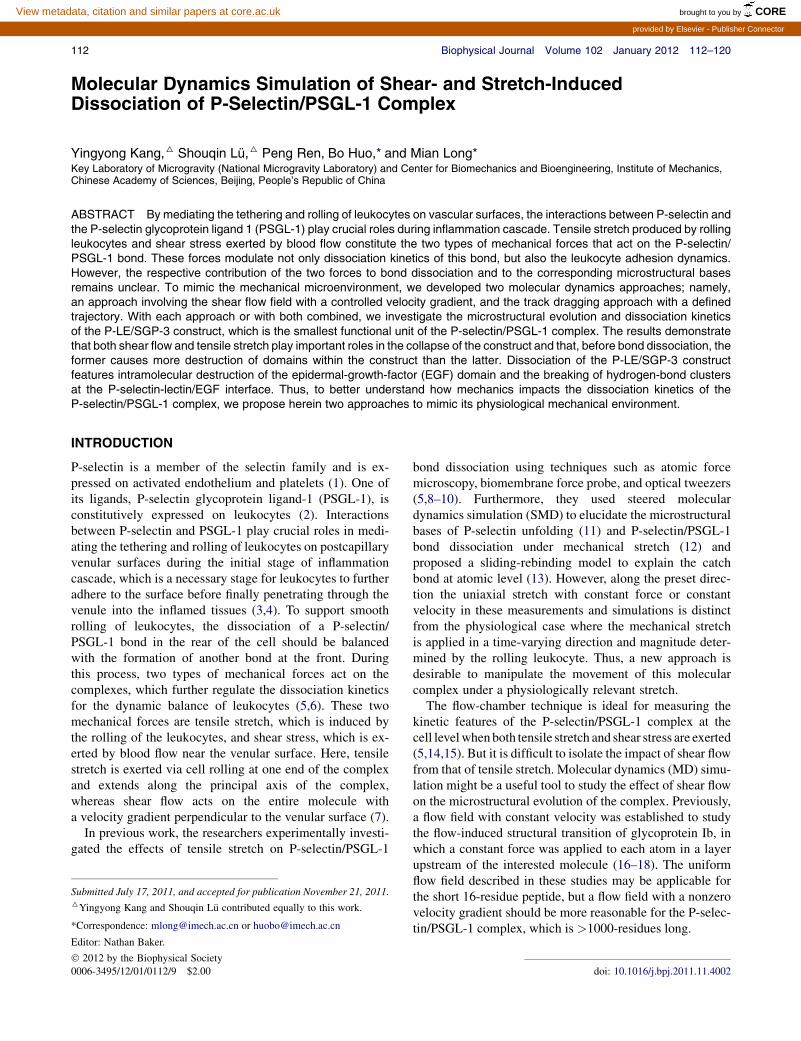
FIGURE 3 Flow velocity distribution in different time periods under
various control conditions: CTCP (A), CTNP (B), and NTNP (C), and their
RMSVs (D). The thickness of the left dragging box is 5 A, and the target
velocity gradient is 3.75 � 10�3 ps�1. Computations were repeated in
triplet and data are presented as mean 5 SD in panel D.
RESULTS
Feasibility of constructing shear flow field
To test the feasibility of SFF constructing, we used the CWSwith a left-dragging layer thickness of 5 A to elucidate theeffect of various control parameters on the quality andstability of the shear flow. We employed three cases oftemperature and pressure control and conducted corre-
sponding MD simulations. Under CTCP conditions, thedistribution of flow velocity coincided well with the targetdistribution after a simulation time of 40 ps with a constantgradient of 3.75 � 10�3 ps�1 (Fig. 3 A). The correspondingRMSV decreases initially and then reaches a plateau after50 ps (Fig. 3 D), which suggests that a steady shear flowis obtained. This flow field remains stable during anextended 1.5-ns simulation (see Fig. S1 and Movie S1 inthe Supporting Material) and is uniform along the flowdirection (see Fig. S2). Moreover, under CTNP conditions,the shear flow field is also effectively constructed (Fig. 3B) with a similar evolution of the RMSV (Fig. 3 D) and,under NTNP, the shear flow field actually fits the targetflow field better (Fig. 3 C) with a smaller RMSV (Fig. 3 D).
We noticed that the shear flow under NTNP conditions isaccompanied by a sharp drop in system temperature to 50 K(within 100 ps), which is far lower than the physiologicaltemperature (see Fig. S3). In contrast, the system tempera-ture under both CTCP and CTNP conditions remains stablenear 316 K (after an initial slight reduction; see Fig. S3).This contradiction is not surprising because the temperaturecontrol is able to maintain Brownian motion of water mole-cules so as to reduce the motion due to the applied force,which decreases the system temperature. Taken together,CTCP conditions were eclectically adopted in the followingsimulations of the MCS system by considering both thephysiological relevance and the numerical efficiency ofthe constructed shear flow field.
We also test the impact of the velocity gradient on shearflow field construction under three target gradients of 1.25,3.75, and 6.25 � 10�3 ps�1 with target velocities U0 of 0.1,
Biophysical Journal 102(1) 112–120
116 Kang et al.
0.3, and 0.5 A/ps. The results indicate that shear flow fieldsare well developed under the low gradients of 1.25 and3.75 � 10�3 ps�1 (Figs. 4 A and 3 A) and have similarRMSVs beyond 50 ps (Fig. 4 C) and system temperatures(see Fig. S4). At the high gradient of 6.25 � 10�3 ps�1,however, the upper-half work region cannot reach the targetvelocity (Fig. 4 B), as indicated by the continuous increasein the RMSVafter 30 ps (Fig. 4 C). We attribute this result toaccelerated water molecules in this region being driven outso quickly that the water molecules continuously recruitedinto the region are too few to replace them. Thus, weuse a velocity gradient of 3.75�10�3 ps�1 by setting thetarget velocity U0 to 0.3 A/ps in the following simulationsof the MCS.
The purpose of fixing the bottom layer and uniformlydriving the top layer in the SFF approach is to preventa direct connection between the two layers due to periodicboundary conditions and to assure the quality and stabilityof the constructed field. With these simplifications, watermolecules in the left dragging box are pushed so that theirinitial velocity is the target velocity and are thus effectivelytransmitted downstream along the x axis. We also test theeffect of the thickness of the left dragging layer on the con-structed shear flow. The results indicate that increasing thethickness from 5 to 10 to 15 A does not impact theRMSVof the constructed flow field (Fig. 4 D) but decreasesthe system temperature by ~5 K (see Fig. S5). Thus, we setthe thickness of the left dragging box to 5 A in the followingSFF simulations.
FIGURE 4 Under CTCP, flow velocity distribution at the target velocity
gradient of 1.25 � 10�3 ps�1 (A) and 6.25 � 10�3 ps�1 (B), and their
RMSVs over time (C), in which the thickness of the left dragging box is
5 A. The dependence of the RMSVs on left-dragging-box thickness
(ranging from 5 to 15 A) is exemplified in panel D at the target gradient
of 3.75 � 10�3 ps�1. All the simulations were repeated in triplet and
data are presented as mean 5 SD in panels C and D.
Biophysical Journal 102(1) 112–120
We test the shear flow using the SFF approach underCTCP conditions with a 5 A-thick top layer, a left draggingbox, and a fixed bottom layer as well as a velocity gradientof 1.875 � 10�3 ps�1 with the system of MCS (see Fig. S6).Similarly, a stable shear flow field with a constant velocitygradient under CTCP conditions is effectively constructedby using an MD simulation based on the SFF approach,which provides a powerful tool to investigate the micro-structural bases of dissociation of P-LE/SGP-3 constructunder conditions that mimic physiological conditions.
Application of tensile stretch model
SMD, a widely used module in the NAMD program, appliestwo types of mechanical stretch along a fixed direction:stretch with constant velocity and stretch with constantforce. However, the physiological stretch force that actson the P-selectin/PSGL-1 complex is distinguished bymomentary variations in the force direction that are causedby leukocyte rolling. We developed the TDA approach inthis study to overcome the limitation of the currently avail-able SMD module to the exertion of a force with time-varying direction and, by using Eqs. 4 and 5, to mimic themechanical stretch that stems from cell rolling.
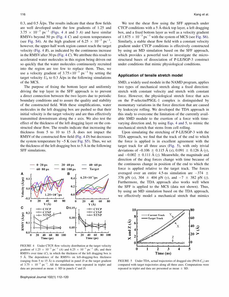
Upon simulating the stretching of P-LE/SGP-3 with theTDA approach, we find that the track of the end to whichthe force is applied is in excellent agreement with thetarget track for all three axes (Fig. 5), with only trivialdeviations of –0.106 5 0.115 A (x), 0.091 5 0.126 A (y),and �0.002 5 0.111 A (z). Meanwhile, the magnitude anddirection of the drag forces change with time because ofthe continuous change in position of the end to which theforce is applied relative to the target track. The forcesaveraged over an entire 4.5-ns simulation are �374 5376 pN (x), 304 5 404 pN (y), and �7 5 382 pN (z).Furthermore, the TDA approach also works well whenthe SFF is applied to the MCS (data not shown). Thus,by using an MD simulation based on the TDA approach,we effectively model a mechanical stretch that mimics
FIGURE 5 Under TDA, actual trajectories of dragged site (P618-Ca) are
compared with target trajectories along all three axes. Computations were
repeated in triplet and data are presented as mean 5 SD.

Shear MD Simulations of P-Selectin/Ligand Bond 117
the physiological force that results from leukocyte rollingand that acts on the P-selectin/PSGL-1 complex.
Two types of mechanical forces lead to differentcollapse scenarios for P-LE domains
To mimic leukocyte rolling under physiological flow, weapplied a combination of the SFF and TDA approaches tothe ratio of leukocyte rolling velocity to wall shear stress.We therefore further investigate the influence of the SFFand TDA approaches alone or combined on the dissociationof the P-LE/SGP-3 construct and find that the two types ofmechanical forces induce the dissociation of P-LE/SGP-3 indifferent ways.
First, we observe a distinct structural collapse when theP-LE/SGP-3 construct is subjected to an SFF and/or trackdragging (see Movie S2, Movie S3, and Movie S4). Forexample, as seen in Fig. 6 A, the construct extension in-creases linearly at the rate of 0.086, 0.072, and 0.016 A/psfor TDAþSFF, SFF, and TDA, respectively—all of whichresults in remarkable conformational changes. Complexseparation exhibits a steady phase initially (when the sepa-ration distance remains unchanged), followed by a transitionphase during which the separation distance increases line-arly with extension distance. Thus, construct dissociationdoes not occur when the extension is less than the thresholdextension (arrows in Fig. 6 B), which suggests that intramo-lecular collapse happens before construct dissociation.Combined with the transition threshold values (estimatedfrom the separation-extension curve shown in Fig. 6 B) of30, 50, and 23 A for TDAþSFF, SFF, and TDA, respec-tively, these results indicate that SFF and TDA havedifferent impacts on the structural disruption of the P-LE/SGP-3 construct; that is, before construct dissociation,
FIGURE 6 Under three mechanical stimuli, evolutions of construct
extension (A), dependence on construct extension of the separation distance
(B), RMSD of the EGF and Lec domains against their initial structures (C),
and hinge angle between the Lec and EGF domains (D).
SFF induces a more remarkable intramolecular collapsethan TDA.
Second, we analyze the structural bases of different intra-molecular collapse scenarios of the P-LE receptor. Theevolution of the RMSDs of the Lec and EGF domains indi-cates that the EGF domain is destroyed at an early stage(a transition threshold of ~5 A) under TDAþSFF, SFF,and TDA, whereas a more severe disruption is found withlarger RMSDs (~11 A) under TDAþSFF and SFF than thedisruption with an RMSD of ~8 A under TDA at the equilib-rium stage (Fig. 6 C). By contrast, the Lec domain tends toretain its conformation with small and stable RMSDs (Fig. 6C) even though its conformation is enhanced after constructdissociation under TDAþSFF and SFF (Fig. 6 C). Thiseffect is further confirmed by hinge breakage between theLec and EGF domains. Under three types of mechanicalstimuli, we find similar increases in the angle between theLec and EGF domains (from an initial value of ~150� toa higher value of 165� to 175�), with the evolution in anglespanning the duration of construct dissociation (Fig. 6 D).We tentatively attribute the descending phases before andafter dissociation under TDAþSFF to the rotation and addi-tional destruction of the Lec domain (Fig. 6 D). Together,these results indicate that the P-LE collapse mainlyconcerns the EGF domain and the hinge region betweenthe Lec and EGF domains.
Third, we further test the conformational change withinthe EGF domain, especially in regard to its main compo-nents of two antiparallel b-sheets (G131-E135 and Y140-C144). Consistent with the evolution in the RMSD of theEGF domain, the distance between the two b-sheets (D1)initially remains constant then increases starting from anextension of ~20 A (TDAþSFF and SFF) and of ~30 A(TDA), before finally reaching a high plateau of ~21 Aand low plateau of ~9 A, respectively (Fig. 7 A). Accord-ingly, the number of hydrogen bonds between the twob-sheets (H1) first decreases then becomes stable, concur-rent with the transition in D1 (Fig. 7 B). The high RMSDin the EGF domain of ~10 A (Fig. 6 C) and the largedistance D1 of ~20 A between the two b-sheets (Fig. 7 A)under TDAþSFF and SFF show a similar trend; namely,that SFF induces a more thorough intramolecular collapsethan TDA alone.
We see the hinge opening given by the distance D2between T2-Ca and I137-Ca increases slowly at first andthen reaches a plateau at ~30 A under TDAþSFF and SFFand at ~20 A under TDA (Fig. 7 C). Again, the number ofhydrogen bonds between the Lec and EGF domains (H2)decreases rapidly to zero under TDAþSFF and SFF,whereas it remains occasionally nonzero under TDA(Fig. 7 D). Together, the forced dissociation of the P-LE/SGP-3 construct occurs after the intramolecular disruptionof P-LE, which focuses on the EGF domain and the inter-face hinge between the EGF and Lec domains. The differ-ences in the P-LE collapse scenario between TDAþSFF
Biophysical Journal 102(1) 112–120

FIGURE 8 Under three mechanical stimuli, dependence on construct
extension of the distance between Ca2þ and the mass center of FUC623
(A), of the number of hydrogen bonds in P-LE-SGP3 interface (B), of the
number of hydrogen bonds between P-LE and SUG (C), and of the number
of hydrogen bonds between P-LE and LIG (D).
FIGURE 7 Under three mechanical stimuli, dependence on construct
extension of the distance between the mass center of backbone atoms of
two antiparallel b-sheets (G131-E135 and Y140-C144) (A), of the number
of hydrogen bonds between the two b-sheets (B), of the distance between
T2-Ca and I137-Ca (C), and of the number of hydrogen bonds within the
hinge region between the Lec and EGF domains (D).
118 Kang et al.
or SFF and TDA highlights the distinct roles of SFF andTDA in P-LE conformational changes.
Two types of mechanical forces induce similardissociation scenario of P-LE/SGP-3 construct
In contrast to the intramolecular collapse, the intermoleculardissociation of the P-LE/SGP-3 construct under TDAþSFF,SFF, and TDA presents similar features. For example, thedistance D3 between the Ca2þ ion and the mass center ofthe sugar residue FUC623 increases linearly after an initialsteady phase (Fig. 8 A), and the transition thresholdsprecisely correspond to those from the separation-extensionprofile (Fig. 6 B). This result implies that the interactionbetween Ca2þ and FUC623 is the key interaction of thebinding interface and that their separation is the primaryindicator of dissociation of the P-LE/SGP-3 construct.
This conclusion is also supported by the sharp reductionin the corresponding number H3 of hydrogen bonds thatstarts at different time delays for the different simulationscenarios (Fig. 8, A and B). Moreover, the separation ofall sugar residues of the SGP-3 ligand from the P-LEreceptor is represented by the number H3_SUG of hydrogenbonds, which is consistent with that of the Ca2þ-FUC623interaction (Fig. 8 C). In addition, the peptide residues ofthe SGP-3 ligand dissociate from P-LE with a slight delay,as seen in the evolution of the H3_LIG hydrogen bonds(Fig. 8 D).
In summary, our MD simulations of dissociation of theP-LE/SGP-3 construct under TDAþSFF, SFF, or TDA indi-cate that intramolecular collapse of P-LE happens beforeconstruct dissociation, which mainly focuses on the twomajor antiparallel b-sheets of the EGF domain and the hingeinterface between the EGF and Lec domains. We attribute
Biophysical Journal 102(1) 112–120
the intermolecular disruption of the P-LE-SGP-3 constructmainly to the separation of Ca2þ from FUC623. Thesefeatures are similar to those found in our SMD simulationsin previous work (12). Our results also indicate that the SFFand TDA approaches have different impacts on P-LE/SGP-3interactions in intramolecular collapse but not on interfacedisruption. Conformational analysis of the P-LE extension,the complex separation, as well as the collapse of the EGFdomain and the P-selectin-lectin/EGF hinge imply that theimpact of shear flow cannot be neglected in MD simulationsof complex dissociation. When using the physiological ratioof leukocyte rolling velocity/wall shear stress, our simula-tions indicate that, before complex dissociation, shear flowinduces more intermolecular destruction than does tensilestretch.
DISCUSSION
The P-selectin/PSGL-1 complex is subjected to mechanicalstretching from the rolling leukocyte and to shear stressfrom blood flow, and the modulation of these two types ofmechanical forces on the dissociation kinetics of the P-se-lectin/PSGL-1 complex is crucial to the correspondingphysiological functions. The currently existing limitationsof experimental assays and simulation approaches havethus far prevented researchers from isolating the impact ofshear stress and from clarifying the influence of an externalforce with time-varying direction and magnitude. In thisstudy, we developed two approaches, SFF and TDA, tomimic the mechanical loading of the physiological microen-vironment using MD simulations (Fig. 1). The microscopicstructural bases of the P-LE/SGP-3 construct under SFFand/or TDA are analyzed using a virtual rolling-leukocytemodel.

Shear MD Simulations of P-Selectin/Ligand Bond 119
In our estimation, the novelty of this study lies in thefollowing two aspects:
The first is that it extends the current MD simulationapproaches. In a sufficiently long simulation, the SFFapproach developed in this work mimics shear flow witha linear velocity distribution. Compared with the previouslydescribed ‘‘uniform’’ flow fields with constant velocity(16–18), our approach successfully mimics the shearstress of blood flow in the boundary layer near the venularsurface, at least in a velocity-gradient range of 1.25 to3.75 � 10�3 ps�1 (Figs. 3 and 4). The TDA approach devel-oped herein successfully implements a tensile stretch thathas a time-varying direction and magnitude and followsa preset trajectory. This approach mimics the mechanicalstretch exerted on the P-selectin/PSGL complex by the roll-ing leukocyte. Numerically, our approach overcomes thelimitation of loading constant velocity or constant forcealong a fixed direction in SMD (Fig. 5). Note that the virtualrolling-leukocyte model is based on the hypothesis that theleukocyte is an ideal sphere and rolls purely at constantvelocity. Further studies are required to optimize the roll-ing-leukocyte model by integrating the jump modelobserved in vitro (6) and to investigate the impacts of leuko-cyte inhomogeneity and deformability. Lees-Edwardsboundary condition approach is widely used to generatelinear shear flow (34,35) with a underlying assumptionthat the flow distribution will not be disturbed by the exis-tence of target particle. But it will produce an unreal shearflow field locally around the studied macromolecule. TheSFF approach established here is applicable for studyingthe microscopic behaviors of protein molecule in a scaleof Angstroms and evaluating the interactions of each watermolecule and target protein.
The second novel (to our knowledge) aspect of our studyis that it illustrates the remarkable impact of shear flow onP-LE/SGP-3 dissociation. Although the conformationalevolution of the P-LE/SGP-3 construct under each type ofmechanical stimulus (Figs. 6–8) is similar to that obtainedwith the previously described SMD simulations (12), thesimulations presented here also indicate that shear stressmore strongly impacts P-LE/SGP-3 dissociation than doespoint-directed mechanical stretch. The relative contributionof these two forces is revealed mainly by the degree of intra-molecular collapse of P-LE before construct dissociation,and SFF or TDAþSFF induces more significant P-LEcollapse than does TDA. These simulations explicate themodulation of physiology-mimicking shear flow on P-selec-tin/PSGL-1 interactions and offer clues to the role of iso-lated shear stress on receptor-ligand interactions. Theyindicate not only that the shear stress is functional for thetransportation of molecular carriers along the flow, butalso that it works effectively as a mechanical modulator ofthe dissociation of the molecular complex.
It is undeniable that some simplifications exist in ourmodels. For example, we considered the leukocyte to be
a rigid sphere, took the P-selectin/PSGL-1 complex to bea linear spring, and transformed the virtual leukocyte inproportion to the length ratio between the P-LE/SGP-3construct and the P-selectin/PSGL-1 complex (the latterwas to ensure that the extension of P-LE/SGP-3 constructcoincided with the velocity of the rolling leukocyte). Inaddition, some parameters used in this study are beyondthe physiological ranges. For instance, the velocity gradientand virtual-leukocyte rolling velocity were approximatelysix orders-of-magnitude higher than measured, althoughthe track dragging force (average ~800 pN in this study)was the same order of magnitude as is found experimentally(~104 pN (36) or ~155 pN (37)). However, it is the rollingvelocity of virtual cell (i.e., elongation rate and extent ofprotein complex) that determines its conformational disrup-tion, and not the track dragging force itself; the latter is justto constrain the forced complex-end to the predefined roll-ing trajectory and keep the trajectory consistent betweendragging point and rolling cell. Therefore the controllingparameters in SFF and TDA, i.e., the velocity gradient andelongation rate, were comparable.
Regardless of the inevitable limitations on computationalcapacity, our simulations offer valuable information forexploring the structure-function relationship of single mole-cules or molecular complexes of interest (13,16–18). Thus,this study provides two useful concepts in that it mimics thephysiological shear stress and the mechanical stretch withvarying direction and magnitude, and it offers clues forbetter understanding the physiological role of shear stresson the interactions involved in the dissociation of P-selec-tin/PSGL-1 as well as other receptor-ligand interactions.
SUPPORTING MATERIAL
Six figures and four movies are available at http://www.biophysj.org/
biophysj/supplemental/S0006-3495(11)05353-7.
MD simulations were performed at the Supercomputing Center, Chinese
Academy of Sciences and on the Computing Facility for Computational
Mechanics, Institute of Mechanics, Chinese Academy of Sciences.
This work was supported by the National Natural Science Foundation of
China (30970707, 10702075, and 30730032), the National High-Tech-
nology Research and Development Program of China 2009AA02Z407),
the National Key Basic Research Foundation of China (2011CB710904),
and the Knowledge Innovation Project of the Chinese Academy of Sciences
(KJCX2-YW-L08).
REFERENCES
1. McEver, R. P., J. H. Beckstead, ., D. F. Bainton. 1989. GMP-140,a platelet a-granule membrane protein, is also synthesized by vascularendothelial cells and is localized in Weibel-Palade bodies. J. Clin.Invest. 84:92–99.
2. Moore, K. L., N. L. Stults, ., R. P. McEver. 1992. Identification ofa specific glycoprotein ligand for P-selectin (CD62) on myeloid cells.J. Cell Biol. 118:445–456.
Biophysical Journal 102(1) 112–120

120 Kang et al.
3. McEver, R. P., K. L. Moore, and R. D. Cummings. 1995. Leukocytetrafficking mediated by selectin-carbohydrate interactions. J. Biol.Chem. 270:11025–11028.
4. Moore, K. L., K. D. Patel,., R. P. McEver. 1995. P-selectin glycopro-tein ligand-1 mediates rolling of human neutrophils on P-selectin.J. Cell Biol. 128:661–671.
5. Alon, R., D. A. Hammer, and T. A. Springer. 1995. Lifetime of the P-selectin-carbohydrate bond and its response to tensile force in hydrody-namic flow. Nature. 374:539–542.
6. Zhu, C., T. Yago, ., R. P. McEver. 2008. Mechanisms for flow-enhanced cell adhesion. Ann. Biomed. Eng. 36:604–621.
7. Atherton, A., and G. V. R. Born. 1973. Relationship between thevelocity of rolling granulocytes and that of the blood flow in venules.J. Physiol. 233:157–165.
8. Marshall, B. T., M. Long,., C. Zhu. 2003. Direct observation of catchbonds involving cell-adhesion molecules. Nature. 423:190–193.
9. Chen, S. Q., and T. A. Springer. 1999. An automatic braking systemthat stabilizes leukocyte rolling by an increase in selectin bond numberwith shear. J. Cell Biol. 144:185–200.
10. Evans, E., A. Leung, ., C. Zhu. 2004. Mechanical switching andcoupling between two dissociation pathways in a P-selectin adhesionbond. Proc. Natl. Acad. Sci. USA. 101:11281–11286.
11. Lu, S. Q., and M. Long. 2004. Forced extension of P-selectin constructusing steered molecular dynamics. Chin. Sci. Bull. 49:10–17.
12. Lu, S. Q., and M. Long. 2005. Forced dissociation of selectin-ligandcomplexes using steered molecular dynamics simulation. Mol. Cell.Biomech. 2:161–177.
13. Lou, J. Z., and C. Zhu. 2007. A structure-based sliding-rebindingmechanism for catch bonds. Biophys. J. 92:1471–1485.
14. Yago, T., A. Leppanen,., R. P. McEver. 2002. Distinct molecular andcellular contributions to stabilizing selectin-mediated rolling underflow. J. Cell Biol. 158:787–799.
15. Schmidtke, D. W., and S. L. Diamond. 2000. Direct observation ofmembrane tethers formed during neutrophil attachment to plateletsor P-selectin under physiological flow. J. Cell Biol. 149:719–730.
16. Chen, Z. Z., J. H. Lou, ., K. Schulten. 2008. Flow-induced structuraltransition in the b-switch region of glycoprotein Ib. Biophys. J.95:1303–1313.
17. Lou, J. Z., and C. Zhu. 2008. Flow induces loop-to-b-hairpin transitionon the b-switch of platelet glycoprotein Iba. Proc. Natl. Acad. Sci.USA. 105:13847–13852.
18. Zou, X. Q., Y. X. Liu, ., K. Schulten. 2010. Flow-induced b-hairpinfolding of the glycoprotein Iba b-switch. Biophys. J. 99:1182–1191.
19. Gratton, Y., and G. W. Slater. 2005. Molecular dynamics study of teth-ered polymers in shear flow. Eur Phys J E Soft Matter. 17:455–465.
20. Delgado-Buscalioni, R., and P. V. Coveney. 2006. Structure of a teth-ered polymer under flow using molecular dynamics and hybrid molec-ular-continuum simulations. Physica A. 362:30–35.
21. Somers, W. S., J. Tang, ., R. T. Camphausen. 2000. Insights into themolecular basis of leukocyte tethering and rolling revealed by struc-
Biophysical Journal 102(1) 112–120
tures of P- and E-selectin bound to SLe(X) and PSGL-1. Cell.103:467–479.
22. Humphrey, W., A. Dalke, and K. Schulten. 1996. VMD: visual molec-ular dynamics. J. Mol. Graph. 14:33–38, 27–28.
23. Jorgensen, W. L., J. Chandrasekhar,., M. L. Klein. 1983. Comparisonof simple potential functions for simulating liquid water. J. Chem.Phys. 79:926–935.
24. MacKerell, A. D., D. Bashford,., M. Karplus. 1998. All-atom empir-ical potential for molecular modeling and dynamics studies of proteins.J. Phys. Chem. B. 102:3586–3616.
25. Foloppe, N., and A. D. MacKerell. 2000. All-atom empirical force fieldfor nucleic acids: I. Parameter optimization based on small moleculeand condensed phase macromolecular target data. J. Comput. Chem.21:86–104.
26. Kale, L., R. Skeel, ., K. Schulten. 1999. NAMD2: greater scalabilityfor parallel molecular dynamics. J. Comput. Phys. 151:283–312.
27. Geng, J. G., G. A. Heavner, and R. P. McEver. 1992. Lectin domainpeptides from selectins interact with both cell surface ligands andCa2þ ions. J. Biol. Chem. 267:19846–19853.
28. Shao, J. Y., H. P. Ting-Beall, and R. M. Hochmuth. 1998. Static anddynamic lengths of neutrophil microvilli. Proc. Natl. Acad. Sci. USA.95:6797–6802.
29. Phillips, J. C., R. Braun, ., K. Schulten. 2005. Scalable moleculardynamics with NAMD. J. Comput. Chem. 26:1781–1802.
30. Ushiyama, S., T. M. Laue,., R. P. McEver. 1993. Structural and func-tional characterization of monomeric soluble P-selectin and compar-ison with membrane P-selectin. J. Biol. Chem. 268:15229–15237.
31. Johnston, G. I., R. G. Cook, and R. P. McEver. 1989. Cloning of GMP-140, a granule membrane protein of platelets and endothelium:sequence similarity to proteins involved in cell adhesion and inflamma-tion. Cell. 56:1033–1044.
32. Li, F. G., H. P. Erickson, ., R. P. McEver. 1996. Visualization ofP-selectin glycoprotein ligand-1 as a highly extended molecule andmapping of protein epitopes for monoclonal antibodies. J. Biol.Chem. 271:6342–6348.
33. Jones, D. A., O. Abbassi, ., C. W. Smith. 1993. P-selectin mediatesneutrophil rolling on histamine-stimulated endothelial cells. Biophys. J.65:1560–1569.
34. Lees, A. W., and S. F. Edwards. 1972. Computer study of transportprocesses under extreme conditions. J. Phys. C Solid State Phys.5:1921–1928.
35. Kobayashi, H., and R. Yamamoto. 2011. Implementation of Lees-Edwards periodic boundary conditions for direct numerical simulationsof particle dispersions under shear flow. J. Chem. Phys. 134:064110.
36. Evans, E. A., and D. A. Calderwood. 2007. Forces and bond dynamicsin cell adhesion. Science. 316:1148–1153.
37. McEver, R. P., and C. Zhu. 2010. Rolling cell adhesion. Annu. Rev. CellDev. Biol. 26:363–396.