Human leukocyte antigen class II-based immune risk model ...
Upload
algeria-univCategory
view
1download
0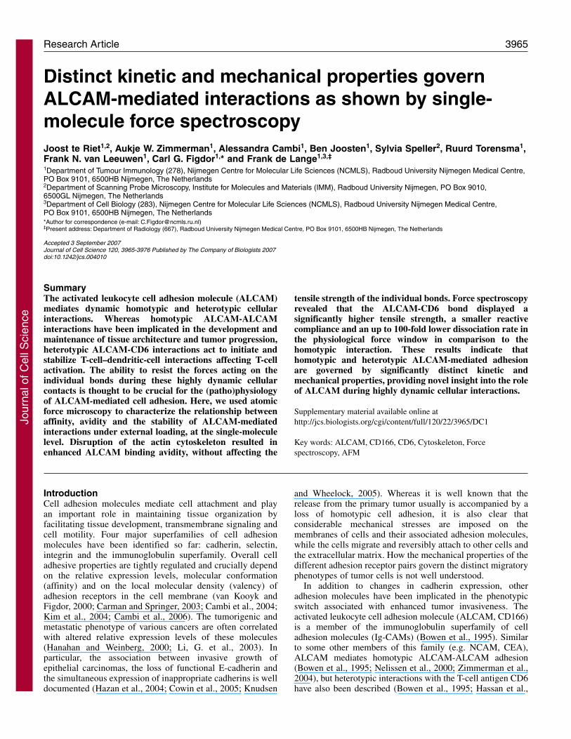
3965Research Article
IntroductionCell adhesion molecules mediate cell attachment and playan important role in maintaining tissue organization byfacilitating tissue development, transmembrane signaling andcell motility. Four major superfamilies of cell adhesionmolecules have been identified so far: cadherin, selectin,integrin and the immunoglobulin superfamily. Overall celladhesive properties are tightly regulated and crucially dependon the relative expression levels, molecular conformation(affinity) and on the local molecular density (valency) ofadhesion receptors in the cell membrane (van Kooyk andFigdor, 2000; Carman and Springer, 2003; Cambi et al., 2004;Kim et al., 2004; Cambi et al., 2006). The tumorigenic andmetastatic phenotype of various cancers are often correlatedwith altered relative expression levels of these molecules(Hanahan and Weinberg, 2000; Li, G. et al., 2003). Inparticular, the association between invasive growth ofepithelial carcinomas, the loss of functional E-cadherin andthe simultaneous expression of inappropriate cadherins is welldocumented (Hazan et al., 2004; Cowin et al., 2005; Knudsen
and Wheelock, 2005). Whereas it is well known that therelease from the primary tumor usually is accompanied by aloss of homotypic cell adhesion, it is also clear thatconsiderable mechanical stresses are imposed on themembranes of cells and their associated adhesion molecules,while the cells migrate and reversibly attach to other cells andthe extracellular matrix. How the mechanical properties of thedifferent adhesion receptor pairs govern the distinct migratoryphenotypes of tumor cells is not well understood.
In addition to changes in cadherin expression, otheradhesion molecules have been implicated in the phenotypicswitch associated with enhanced tumor invasiveness. Theactivated leukocyte cell adhesion molecule (ALCAM, CD166)is a member of the immunoglobulin superfamily of celladhesion molecules (Ig-CAMs) (Bowen et al., 1995). Similarto some other members of this family (e.g. NCAM, CEA),ALCAM mediates homotypic ALCAM-ALCAM adhesion(Bowen et al., 1995; Nelissen et al., 2000; Zimmerman et al.,2004), but heterotypic interactions with the T-cell antigen CD6have also been described (Bowen et al., 1995; Hassan et al.,
The activated leukocyte cell adhesion molecule (ALCAM)mediates dynamic homotypic and heterotypic cellularinteractions. Whereas homotypic ALCAM-ALCAMinteractions have been implicated in the development andmaintenance of tissue architecture and tumor progression,heterotypic ALCAM-CD6 interactions act to initiate andstabilize T-cell–dendritic-cell interactions affecting T-cellactivation. The ability to resist the forces acting on theindividual bonds during these highly dynamic cellularcontacts is thought to be crucial for the (patho)physiologyof ALCAM-mediated cell adhesion. Here, we used atomicforce microscopy to characterize the relationship betweenaffinity, avidity and the stability of ALCAM-mediatedinteractions under external loading, at the single-moleculelevel. Disruption of the actin cytoskeleton resulted inenhanced ALCAM binding avidity, without affecting the
tensile strength of the individual bonds. Force spectroscopyrevealed that the ALCAM-CD6 bond displayed asignificantly higher tensile strength, a smaller reactivecompliance and an up to 100-fold lower dissociation rate inthe physiological force window in comparison to thehomotypic interaction. These results indicate thathomotypic and heterotypic ALCAM-mediated adhesionare governed by significantly distinct kinetic andmechanical properties, providing novel insight into the roleof ALCAM during highly dynamic cellular interactions.
Supplementary material available online athttp://jcs.biologists.org/cgi/content/full/120/22/3965/DC1
Key words: ALCAM, CD166, CD6, Cytoskeleton, Forcespectroscopy, AFM
Summary
Distinct kinetic and mechanical properties governALCAM-mediated interactions as shown by single-molecule force spectroscopyJoost te Riet1,2, Aukje W. Zimmerman1, Alessandra Cambi1, Ben Joosten1, Sylvia Speller2, Ruurd Torensma1,Frank N. van Leeuwen1, Carl G. Figdor1,* and Frank de Lange1,3,‡
1Department of Tumour Immunology (278), Nijmegen Centre for Molecular Life Sciences (NCMLS), Radboud University Nijmegen Medical Centre,PO Box 9101, 6500HB Nijmegen, The Netherlands2Department of Scanning Probe Microscopy, Institute for Molecules and Materials (IMM), Radboud University Nijmegen, PO Box 9010,6500GL Nijmegen, The Netherlands3Department of Cell Biology (283), Nijmegen Centre for Molecular Life Sciences (NCMLS), Radboud University Nijmegen Medical Centre,PO Box 9101, 6500HB Nijmegen, The Netherlands*Author for correspondence (e-mail: [email protected])‡Present address: Department of Radiology (667), Radboud University Nijmegen Medical Centre, PO Box 9101, 6500HB Nijmegen, The Netherlands
Accepted 3 September 2007Journal of Cell Science 120, 3965-3976 Published by The Company of Biologists 2007doi:10.1242/jcs.004010
Jour
nal o
f Cel
l Sci
ence
3966
2004) (Fig. 1A). Bone marrow stromal cells and hematopoieticprogenitor cells, neuronal cells, and a large number ofepithelial and endothelial cell types express significant levelsof ALCAM, and contributions for homotypic ALCAM-mediated adhesion have been described for neuraldevelopment, hematopoietic stem cell maturation andtransendothelial monocyte migration (Tanaka et al., 1991; Patelet al., 1995; Degen et al., 1998; Swart, 2002; Masedunskas etal., 2006). ALCAM has been implicated in the onset andprogression of melanoma (van Kempen et al., 2000; vanKempen et al., 2001; Lunter et al., 2005), bladder cancer(Tomita et al., 2003), prostate carcinoma (Kristiansen et al.,2003), breast cancer (King et al., 2004) and colorectalcarcinoma (Weichert et al., 2004). The invasiveness ofmalignant melanoma correlates with enhanced ALCAMexpression and this molecule is considered to be a prognostic
marker in this disease (Degen et al., 1998; van Kempen et al.,2000; van Kempen et al., 2001; Swart et al., 2005).
Besides mediating homotypic interactions, ALCAM is theonly known ligand for CD6 identified on immune cells. Recentwork indicates that ALCAM localizes to the immunologicalsynapse (Grakoui et al., 1999) in an antigen-dependent manner(Gimferrer et al., 2004) and that ALCAM-CD6 engagementplays a pivotal role both during early T-cell–dendritic-cell (DC)contact formation and in later stages of T-cell activation(Zimmerman et al., 2006). In fact, several studies now pointtowards a role for CD6 as a co-stimulatory molecule in T-cellactivation (Gangemi et al., 1989; Wee et al., 1993; Rasmussenet al., 1994; Osorio et al., 1998; Hassan et al., 2004;Zimmerman et al., 2006). Recent intravital microscopy studiesindicate that, within lymphoid tissue, naïve T cells scan thesurface of DCs at relative cell speeds of up to 30 �m/minute
Journal of Cell Science 120 (22)
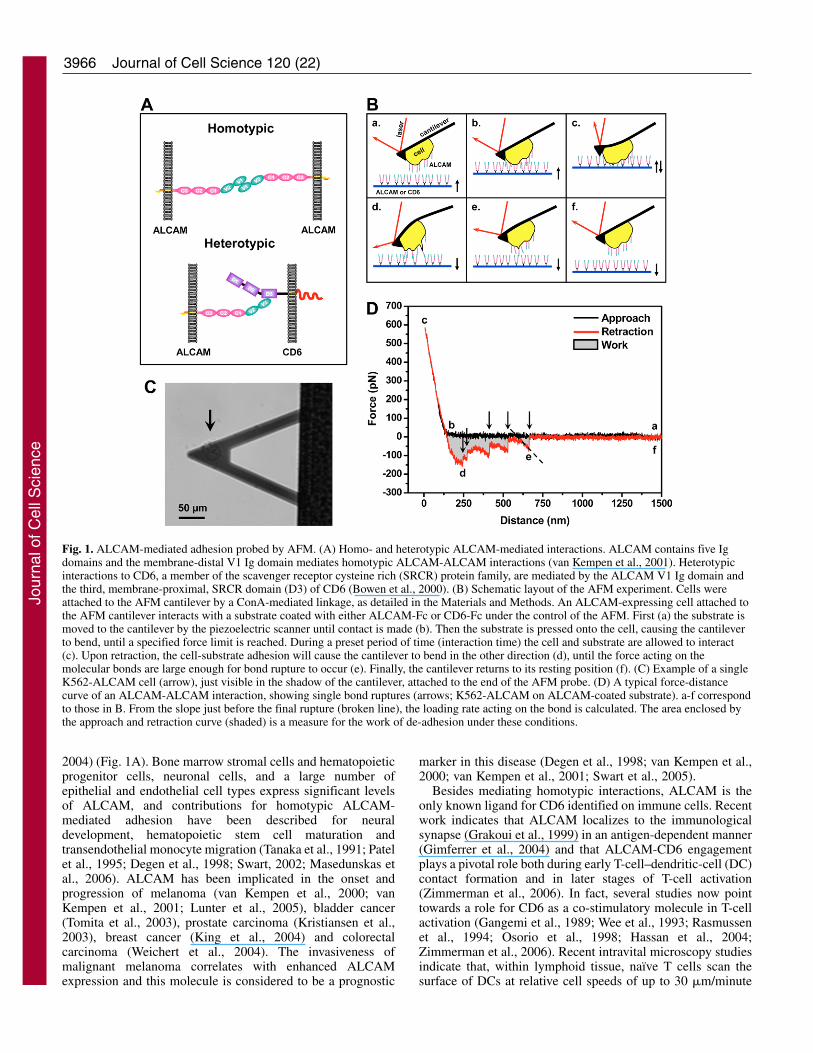
Fig. 1. ALCAM-mediated adhesion probed by AFM. (A) Homo- and heterotypic ALCAM-mediated interactions. ALCAM contains five Igdomains and the membrane-distal V1 Ig domain mediates homotypic ALCAM-ALCAM interactions (van Kempen et al., 2001). Heterotypicinteractions to CD6, a member of the scavenger receptor cysteine rich (SRCR) protein family, are mediated by the ALCAM V1 Ig domain andthe third, membrane-proximal, SRCR domain (D3) of CD6 (Bowen et al., 2000). (B) Schematic layout of the AFM experiment. Cells wereattached to the AFM cantilever by a ConA-mediated linkage, as detailed in the Materials and Methods. An ALCAM-expressing cell attached tothe AFM cantilever interacts with a substrate coated with either ALCAM-Fc or CD6-Fc under the control of the AFM. First (a) the substrate ismoved to the cantilever by the piezoelectric scanner until contact is made (b). Then the substrate is pressed onto the cell, causing the cantileverto bend, until a specified force limit is reached. During a preset period of time (interaction time) the cell and substrate are allowed to interact(c). Upon retraction, the cell-substrate adhesion will cause the cantilever to bend in the other direction (d), until the force acting on themolecular bonds are large enough for bond rupture to occur (e). Finally, the cantilever returns to its resting position (f). (C) Example of a singleK562-ALCAM cell (arrow), just visible in the shadow of the cantilever, attached to the end of the AFM probe. (D) A typical force-distancecurve of an ALCAM-ALCAM interaction, showing single bond ruptures (arrows; K562-ALCAM on ALCAM-coated substrate). a-f correspondto those in B. From the slope just before the final rupture (broken line), the loading rate acting on the bond is calculated. The area enclosed bythe approach and retraction curve (shaded) is a measure for the work of de-adhesion under these conditions.
Jour
nal o
f Cel
l Sci
ence
3967ALCAM-mediated adhesion under loading
(Mempel et al., 2004). Clearly, the ability to withstand shearat the molecular level is essential for establishing andmaintaining productive DC–T-cell contacts for prolongedperiods of time.
These findings indicate a key role for ALCAM-mediatedadhesion during highly dynamic cellular interactions.Mechanistically, this implies that ALCAM must be equippedto facilitate adhesion under different conditions of externalloading. Previous work has focused on the affinity and avidityof ALCAM-mediated interactions (Nelissen et al., 2000;Hassan et al., 2004; Zimmerman et al., 2004). Yet, how theseproperties relate to the stability of ALCAM-mediated bondsunder mechanical stress is still poorly understood. Here, weaddress this issue using the atomic force microscope (AFM)(Binnig et al., 1986) to measure ALCAM-mediated adhesionof living cells under varying loading conditions and withsingle-bond sensitivity. The AFM has been used successfullyto study single-molecule adhesion of isolated proteins(Willemsen et al., 2000; Hinterdorfer, 2002), and later instudies towards cell adhesion phenomena – an approach thatwas first explored by Gaub and co-workers (Benoit et al.,2000). The use of force spectroscopy has meanwhile providedinsight into the compliance of individual cell adhesion bondsto physiological-range external forces (Zhang et al., 2002;Wojcikiewicz et al., 2003; Hanley et al., 2004; Hinterdorfer andDufrêne, 2006; Panorchan et al., 2006b).
We have adapted the AFM technology to study thestability of homo- and heterotypic ALCAM-mediatedadhesion under loading. In the low-force regime theALCAM-mediated interactions displayed similardissociation kinetics. However, by applyingphysiologically relevant external forces we found thathomo- and heterotypic ALCAM-mediated adhesion aregoverned by distinct kinetic and mechanical propertiesindicating that the ALCAM-CD6 bond is significantlymore stable under mechanical stress. The reactivecompliance and dissociation kinetics found for theALCAM-CD6 interaction were similar in magnitude tothose reported for selectin mediated bonds (Zhang et al.,2004b). By contrast, the ALCAM-ALCAM bond
displayed a significantly greater lability under force, also incomparison to homotypic E-cadherin-mediated interactions(Panorchan et al., 2006b).
ResultsUsing AFM to measure single ALCAM-mediatedinteractions on living cellsA schematic layout of the AFM adhesion measurements isdepicted in Fig. 1B. Cell adhesion forces were measured bymoving ALCAM- or CD6-coated substrates alternatelytowards and away from an AFM cantilever to which a singleALCAM-expressing cell was attached by means ofconcanavalin A (ConA)-mediated linkages (Fig. 1B,C; seeMaterials and Methods). The detachment of the cell wasrecorded during retraction of the substrate. The rupture ofcell-substrate bonds caused subtle changes in cantileverdeflection that provided a measure for the cell adhesionforces that were acting on the molecular level (Fig. 1D,arrows). The work needed to detach the cell from thesubstrate – derived from the area enclosed by the retractioncurve and the zero-force axis – was taken as a measure foroverall cell adhesion (Zhang et al., 2002). Homo- andheterotypic ALCAM-mediated adhesions were comparedusing the same cell and cantilever probing the distinct ligand-coated substrates.
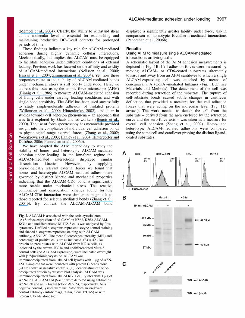
Fig. 2. ALCAM is associated with the actin cytoskeleton.(A) Surface expression of ALCAM on K562, K562-ALCAM,KG1a and undifferentiated MUTZ-3 cells was analyzed by flowcytometry. Unfilled histograms represent isotype control stainingand shaded histograms represent staining with ALCAMantibody, AZN-L50. The mean fluorescence intensity (MFI) andpercentage of positive cells are as indicated. (B) A 42 kDaprotein co-precipitates with ALCAM from KG1a cells, asindicated by the arrows. KG1a and undifferentiated Mutz-3control cells (no ALCAM expression) were incubated overnightwith [35S]methionine/cysteine. ALCAM wasimmunoprecipitated from labeled cell lysates with 1 �g of AZN-L51. Samples that were incubated with protein G beads alone(–) are shown as negative controls. (C) Identification of the co-precipitated protein by western blot analysis. ALCAM wasimmunoprecipitated from labeled KG1a cell lysates with 1 �g ofAZN-L51. ALCAM and �-actin were detected using antibodiesAZN-L50 and anti-�-actin (clone AC-15), respectively. As anegative control, lysates were incubated with an irrelevantcontrol antibody (anti-hemagglutinin, clone 12CA5) or withprotein G beads alone (–).
Jour
nal o
f Cel
l Sci
ence
3968
ALCAM is linked to the actin cytoskeletonKG1a and ALCAM-transfected K562 cells (K562-ALCAM)both express similar levels of ALCAM at the cell surface (Fig.2A). Parental ALCAM-negative K562 and MUTZ-3 cells wereused as negative controls in further experiments. We found thata 42 kDa protein co-precipitates with ALCAM frommetabolically labeled KG1a cells, but not from ALCAM-negative MUTZ-3 cells (Fig. 2B). This 42 kDa protein wasidentified as �-actin by immunoprecipitation (IP) of ALCAMfollowed by western blot analysis (Fig. 2C). Total cell lysateserved as a control for total amounts of ALCAM and �-actin(not shown). Control IPs with either protein G beads alone orwith a control antibody were negative for both ALCAM and�-actin, confirming the specificity of the interaction. Theseresults confirm and extend our previously reported finding thatALCAM-mediated adhesion is directly regulated by the actincytoskeleton (Nelissen et al., 2000; Zimmerman et al., 2004).
Avidity of ALCAM-mediated adhesion is controlled bythe actin cytoskeletonWe have previously shown by means of an optical-trap-basedmotility assay and fluorescence microscopy that modestdisruption of the cortical actin cytoskeleton – usingcytochalasin D (CytD) – results in enhanced lateral mobility ofALCAM and the formation of ALCAM clusters on the cellsurface. Interestingly, CytD pretreatment further enhanced celladhesiveness to ALCAM–Fc-coated plates, suggesting that theobserved clustering effectively enhanced ALCAM bindingavidity (Nelissen et al., 2000; Zimmerman et al., 2004). Here,we exploit the sensitivity of the AFM to determine to whatextent the affinity of individual ALCAM-mediated interactionscontributes to this effect.
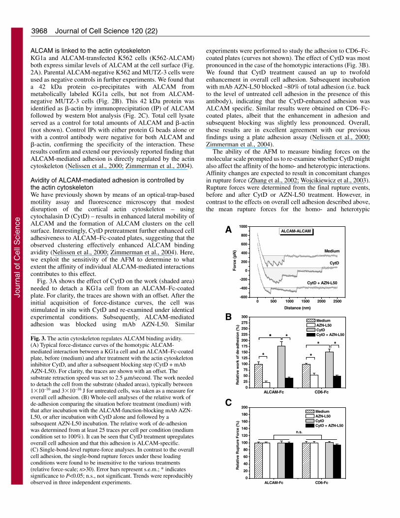
Fig. 3A shows the effect of CytD on the work (shaded area)needed to detach a KG1a cell from an ALCAM–Fc-coatedplate. For clarity, the traces are shown with an offset. After theinitial acquisition of force-distance curves, the cell wasstimulated in situ with CytD and re-examined under identicalexperimental conditions. Subsequently, ALCAM-mediatedadhesion was blocked using mAb AZN-L50. Similar
experiments were performed to study the adhesion to CD6–Fc-coated plates (curves not shown). The effect of CytD was mostpronounced in the case of the homotypic interactions (Fig. 3B).We found that CytD treatment caused an up to twofoldenhancement in overall cell adhesion. Subsequent incubationwith mAb AZN-L50 blocked ~80% of total adhesion (i.e. backto the level of untreated cell adhesion in the presence of thisantibody), indicating that the CytD-enhanced adhesion wasALCAM specific. Similar results were obtained on CD6–Fc-coated plates, albeit that the enhancement in adhesion andsubsequent blocking was slightly less pronounced. Overall,these results are in excellent agreement with our previousfindings using a plate adhesion assay (Nelissen et al., 2000;Zimmerman et al., 2004).
The ability of the AFM to measure binding forces on themolecular scale prompted us to re-examine whether CytD mightalso affect the affinity of the homo- and heterotypic interactions.Affinity changes are expected to result in concomitant changesin rupture force (Zhang et al., 2002; Wojcikiewicz et al., 2003).Rupture forces were determined from the final rupture events,before and after CytD or AZN-L50 treatment. However, incontrast to the effects on overall cell adhesion described above,the mean rupture forces for the homo- and heterotypic
Journal of Cell Science 120 (22)
A
B
CALCAM-Fc CD6-Fc
0
25
50
75
100
125
150
175
200
225
250
275
300
Rel
ativ
e w
ork
of
de-
adh
esio
n (
%)
Medium AZN-L50 CytD CytD + AZN-L50**
**
**
*
ALCAM-Fc CD6-Fc0
20
40
60
80
100
120
140
160
180
200
Rel
ativ
e R
up
ture
Fo
rce
(%)
Medium AZN-L50 CytD CytD + AZN-L50
n.s.
0 500 1000 1500 2000 2500-600
-400
-200
0
200
400
600
800
1000
Medium
CytD + AZN-L50
CytD
ALCAM-ALCAMF
orc
e (p
N)
Distance (nm)
Fig. 3. The actin cytoskeleton regulates ALCAM binding avidity.(A) Typical force-distance curves of the homotypic ALCAM-mediated interaction between a KG1a cell and an ALCAM–Fc-coatedplate, before (medium) and after treatment with the actin cytoskeletoninhibitor CytD, and after a subsequent blocking step (CytD + mAbAZN-L50). For clarity, the traces are shown with an offset. Thesubstrate retraction speed was set to 2.5 �m/second. The work neededto detach the cell from the substrate (shaded areas), typically between1�10–16 and 3�10–16 J for untreated cells, was taken as a measure foroverall cell adhesion. (B) Whole-cell analyses of the relative work ofde-adhesion comparing the situation before treatment (medium) withthat after incubation with the ALCAM-function-blocking mAb AZN-L50, or after incubation with CytD alone and followed by asubsequent AZN-L50 incubation. The relative work of de-adhesionwas determined from at least 25 traces per cell per condition (mediumcondition set to 100%). It can be seen that CytD treatment upregulatesoverall cell adhesion and that this adhesion is ALCAM-specific.(C) Single-bond-level rupture-force analyses. In contrast to the overallcell adhesion, the single-bond rupture forces under these loadingconditions were found to be insensitive to the various treatments(relative force-scale; n>30). Error bars represent s.e.m.; * indicatessignificance to P<0.05; n.s., not significant. Trends were reproduciblyobserved in three independent experiments.
Jour
nal o
f Cel
l Sci
ence
3969ALCAM-mediated adhesion under loading
interactions were not affected by either treatment (Fig.3C). These results clearly demonstrate that disruptionof the actin cytoskeleton by CytD enhances ALCAMbinding avidity without affecting the affinity of theindividual ALCAM-mediated interactions.
The ALCAM-CD6 bond is more stable underexternal loadingPrevious work focused on the affinity of ALCAM-mediated interactions using soluble ligand bindingassays (Hassan et al., 2004; Zimmerman et al., 2004).Here, we address the relative stability of single homo-versus heterotypic ALCAM-mediated bonds underconditions of external loading (i.e. under conditionsthat mimic the forces acting on the cell adhesionmolecules during dynamic cell-cell contacts).
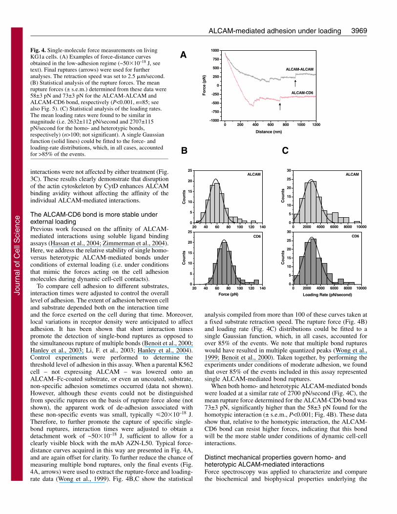
To compare cell adhesion to different substrates,interaction times were adjusted to control the overalllevel of adhesion. The extent of adhesion between celland substrate depended both on the interaction timeand the force exerted on the cell during that time. Moreover,local variations in receptor density were anticipated to affectadhesion. It has been shown that short interaction timespromote the detection of single-bond ruptures as opposed tothe simultaneous rupture of multiple bonds (Benoit et al., 2000;Hanley et al., 2003; Li, F. et al., 2003; Hanley et al., 2004).Control experiments were performed to determine thethreshold level of adhesion in this assay. When a parental K562cell – not expressing ALCAM – was lowered onto anALCAM–Fc-coated substrate, or even an uncoated, substrate,non-specific adhesion sometimes occurred (data not shown).However, although these events could not be distinguishedfrom specific ruptures on the basis of rupture force alone (notshown), the apparent work of de-adhesion associated withthese non-specific events was small, typically �20�10–18 J.Therefore, to further promote the capture of specific single-bond ruptures, interaction times were adjusted to obtain adetachment work of ~50�10–18 J, sufficient to allow for aclearly visible block with the mAb AZN-L50. Typical force-distance curves acquired in this way are presented in Fig. 4A,and are again offset for clarity. To further reduce the chance ofmeasuring multiple bond ruptures, only the final events (Fig.4A, arrows) were used to extract the rupture-force and loading-rate data (Wong et al., 1999). Fig. 4B,C show the statistical
analysis compiled from more than 100 of these curves taken ata fixed substrate retraction speed. The rupture force (Fig. 4B)and loading rate (Fig. 4C) distributions could be fitted to asingle Gaussian function, which, in all cases, accounted forover 85% of the events. We note that multiple bond ruptureswould have resulted in multiple quantized peaks (Wong et al.,1999; Benoit et al., 2000). Taken together, by performing theexperiments under conditions of moderate adhesion, we foundthat over 85% of the events included in this assay representedsingle ALCAM-mediated bond ruptures.
When both homo- and heterotypic ALCAM-mediated bondswere loaded at a similar rate of 2700 pN/second (Fig. 4C), themean rupture force determined for the ALCAM-CD6 bond was73±3 pN, significantly higher than the 58±3 pN found for thehomotypic interaction (± s.e.m., P<0.001; Fig. 4B). These datashow that, relative to the homotypic interaction, the ALCAM-CD6 bond can resist higher forces, indicating that this bondwill be the more stable under conditions of dynamic cell-cellinteractions.
Distinct mechanical properties govern homo- andheterotypic ALCAM-mediated interactionsForce spectroscopy was applied to characterize and comparethe biochemical and biophysical properties underlying the
A
B
0 200 400 600 800 1000 1200-1000
-750
-500
-250
0
250
500
750
1000
ALCAM-CD6
ALCAM-ALCAM
Fo
rce
(pN
)
Distance (nm)
C
20 40 60 80 100 120 1400
5
10
15
20
25CD6
Co
un
ts
Force (pN)
20 40 60 80 100 120 1400
5
10
15
20
25ALCAM
Co
un
ts
0 2000 4000 6000 8000 100000
5
10
15
20
25
30
Co
un
ts
Loading Rate (pN/second)
CD6
0 2000 4000 6000 8000 100000
5
10
15
20
25
30
Co
un
ts
ALCAM
Fig. 4. Single-molecule force measurements on livingKG1a cells. (A) Examples of force-distance curvesobtained in the low-adhesion regime (~50�10–18 J, seetext). Final ruptures (arrows) were used for furtheranalyses. The retraction speed was set to 2.5 �m/second.(B) Statistical analysis of the rupture forces. The meanrupture forces (± s.e.m.) determined from these data were58±3 pN and 73±3 pN for the ALCAM-ALCAM andALCAM-CD6 bond, respectively (P<0.001, n=85; seealso Fig. 5). (C) Statistical analysis of the loading rates.The mean loading rates were found to be similar inmagnitude (i.e. 2632±112 pN/second and 2707±115pN/second for the homo- and heterotypic bonds,respectively) (n>100; not significant). A single Gaussianfunction (solid lines) could be fitted to the force- andloading-rate distributions, which, in all cases, accountedfor >85% of the events.
Jour
nal o
f Cel
l Sci
ence
3970
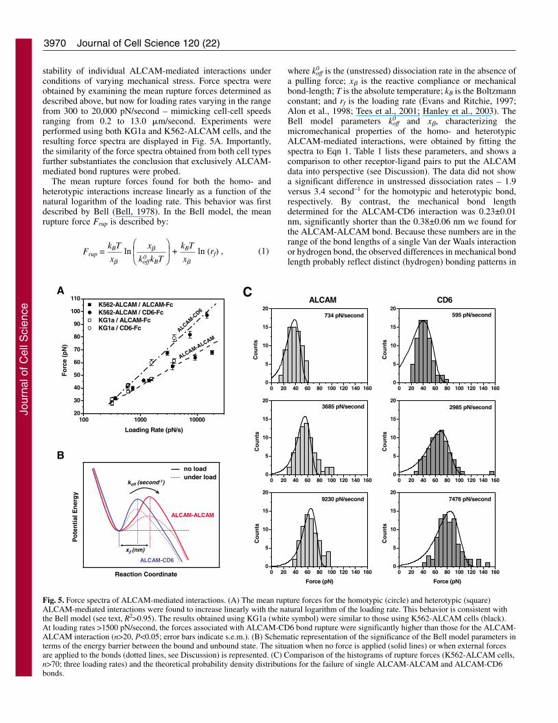
stability of individual ALCAM-mediated interactions underconditions of varying mechanical stress. Force spectra wereobtained by examining the mean rupture forces determined asdescribed above, but now for loading rates varying in the rangefrom 300 to 20,000 pN/second – mimicking cell-cell speedsranging from 0.2 to 13.0 �m/second. Experiments wereperformed using both KG1a and K562-ALCAM cells, and theresulting force spectra are displayed in Fig. 5A. Importantly,the similarity of the force spectra obtained from both cell typesfurther substantiates the conclusion that exclusively ALCAM-mediated bond ruptures were probed.
The mean rupture forces found for both the homo- andheterotypic interactions increase linearly as a function of thenatural logarithm of the loading rate. This behavior was firstdescribed by Bell (Bell, 1978). In the Bell model, the meanrupture force Frup is described by:
kBT Frup = ln (rf) ,ln +
x
x
koffkBT(1)
⎛⎜⎝
⎞⎟⎠�
kBT
x�
�
0
where k0off is the (unstressed) dissociation rate in the absence of
a pulling force; x� is the reactive compliance or mechanicalbond-length; T is the absolute temperature; kB is the Boltzmannconstant; and rf is the loading rate (Evans and Ritchie, 1997;Alon et al., 1998; Tees et al., 2001; Hanley et al., 2003). TheBell model parameters k0
off and x�, characterizing themicromechanical properties of the homo- and heterotypicALCAM-mediated interactions, were obtained by fitting thespectra to Eqn 1. Table 1 lists these parameters, and shows acomparison to other receptor-ligand pairs to put the ALCAMdata into perspective (see Discussion). The data did not showa significant difference in unstressed dissociation rates – 1.9versus 3.4 second–1 for the homotypic and heterotypic bond,respectively. By contrast, the mechanical bond lengthdetermined for the ALCAM-CD6 interaction was 0.23±0.01nm, significantly shorter than the 0.38±0.06 nm we found forthe ALCAM-ALCAM bond. Because these numbers are in therange of the bond lengths of a single Van der Waals interactionor hydrogen bond, the observed differences in mechanical bondlength probably reflect distinct (hydrogen) bonding patterns in
Journal of Cell Science 120 (22)
A
B
koff (second-1)
Po
ten
tial
En
erg
y
Reaction Coordinate
ALCAM-CD6
ALCAM-ALCAM
no loadunder load
xβ (nm)
100 1000 1000020
30
40
50
60
70
80
90
100
110
ALCAM-ALCAM
ALCAM-CD6
Fo
rce
(pN
)
Loading Rate (pN/s)
K562-ALCAM / ALCAM-Fc K562-ALCAM / CD6-Fc KG1a / ALCAM-Fc KG1a / CD6-Fc
C
0 20 40 60 80 100 120 140 1600
5
10
15
20ALCAM CD6
Co
un
ts
734 pN/second
0 20 40 60 80 100 120 140 1600
5
10
15
20
Co
un
ts
595 pN/second
0 20 40 60 80 100 120 140 1600
5
10
15
20
Co
un
ts
3685 pN/second
0 20 40 60 80 100 120 140 1600
5
10
15
20
Co
un
ts
2985 pN/second
0 20 40 60 80 100 120 140 1600
5
10
15
20
Co
un
ts
Force (pN)
9230 pN/second
0 20 40 60 80 100 120 140 1600
5
10
15
20
Co
un
ts
Force (pN)
7476 pN/second
Fig. 5. Force spectra of ALCAM-mediated interactions. (A) The mean rupture forces for the homotypic (circle) and heterotypic (square)ALCAM-mediated interactions were found to increase linearly with the natural logarithm of the loading rate. This behavior is consistent withthe Bell model (see text, R2>0.95). The results obtained using KG1a (white symbol) were similar to those using K562-ALCAM cells (black).At loading rates >1500 pN/second, the forces associated with ALCAM-CD6 bond rupture were significantly higher than those for the ALCAM-ALCAM interaction (n>20, P<0.05; error bars indicate s.e.m.). (B) Schematic representation of the significance of the Bell model parameters interms of the energy barrier between the bound and unbound state. The situation when no force is applied (solid lines) or when external forcesare applied to the bonds (dotted lines, see Discussion) is represented. (C) Comparison of the histograms of rupture forces (K562-ALCAM cells,n>70; three loading rates) and the theoretical probability density distributions for the failure of single ALCAM-ALCAM and ALCAM-CD6bonds.
Jour
nal o
f Cel
l Sci
ence
3971ALCAM-mediated adhesion under loading
both types of ALCAM-mediated interactions (Fig. 1A). Bondswith shorter x� are more resistant to applied force. Thesefindings, therefore, corroborate the previous conclusion that,with respect to the homotypic interaction, the ALCAM-CD6bond is more resistant to applied force, and hence more stableunder loading than the ALCAM-ALCAM bond.
In terms of interaction potentials, the unstressed dissociationrate k0
off represents the rate-limiting step in the dissociation (i.e.the transition over the activation barrier) of the unstressedcomplex. The reactive compliance, x�, then represents thereaction coordinate, and describes how far the bond can bestretched before it breaks (Hummer and Szabo, 2001; Rief andGrubmüller, 2002). A schematic graphical representation of thedata is shown in Fig. 5B (solid curves). The linearity of theforce spectra (Fig. 5A) indicates that, in this loading-rateregime, the dissociation of ALCAM-mediated interactions isbest described by a single activation energy barrier. Bycontrast, bi-phasic force spectra reflecting a double-barrierinteraction potential have been reported [e.g. for E-cadherin–E-cadherin, LFA-1–ICAM-1, and E-, P- or L-selectin–sLex(sialyl Lewis X) (Evans et al., 2001; Zhang et al., 2002; Zhanget al., 2004a; Panorchan et al., 2006b)]. The Bell modelpredicts that, with increasing loading forces, the activationenergy barrier of the complex is suppressed and thedissociation rate constant increases. This is shownschematically in Fig. 5B and is discussed below.
Fig. 5C shows a comparison of the histograms of ruptureforces at three loading rates and the corresponding probabilitydensity distributions for the failure of single ALCAM-ALCAMand ALCAM-CD6 bonds that were calculated using the Bellmodel parameters derived from Fig. 5A using (Evans andRitchie, 1997; Li, F. et al., 2003):
As can be seen in Fig. 5C, the theoretical distributions closelymatch the histograms of rupture forces at all three loadingrates. The width of the distributions does not reflectexperimental error, but is a manifestation of the underlyingstochastic distribution of breakup times (Evans and Ritchie,
⎧⎨⎩
⎫⎬⎭
.⎡⎢⎣
⎤⎥⎦x
koffkBT
�
P(Frup) = koff exp
exp 1 – exp
x
kBT
(2)
⎛⎜⎝
⎞⎟⎠
�Frup
x
kBT
⎛⎜⎝
⎞⎟⎠
�Frup
rf
0
0
1997; Tees et al., 2001; Evans et al., 2005). The small numberof events beyond the predicted distributions, <16% of eventsin all cases in Fig. 5C, can be accounted for by thesimultaneous (i.e. unresolved) rupture of multiple bondlinkages (Evans et al., 2005). Importantly, this analysis impliesthat indeed ~85% of the events represent the kinetically limitedfailure of single bonds.
Control experiments were performed to verify whether theattachment of the cells to the cantilevers by means of ConA-mediated linkages possibly activated the cells and affected theoutcome of the single-molecule adhesion measurements.K562-ALCAM cells were seeded onto poly-L-lysine-coatedglass coverslips and probed with a 10 �m ALCAM–Fc- orCD6–Fc-coated bead glued to an AFM cantilever (seesupplementary material Fig. S1A). The interactions were foundto be ALCAM-specific and showed a clear single-moleculesignature (see supplementary material Fig. S1B,C). The meanrupture forces for both the homo- and heterotypic ALCAM-mediated interactions that were found under varying pullingconditions agree well with the data obtained using the cell-functionalized cantilevers (supplementary material Fig. S1D).These data show that the presented single-molecule-leveladhesion measurements are independent of the probingmethod.
Finally, the dissociation energies of single ALCAM-mediated interactions were derived from the net amount ofwork required to break the bonds, by calculating the productof rupture force and rupture length. The work to break a singleALCAM-ALCAM interaction – at 1000 pN/second,corresponding to a pulling speed of 700 nm/second – amountedto 40 pN �0.38 nm � 4 kBT (Fig. 5A; T=300 K). Interestingly,under the same conditions, due to the higher unbinding forceand the shorter reactive compliance, the net work to rupture anALCAM-CD6 bond was 50 pN �0.23 nm � 3 kBT. Hence,despite the shorter mechanical bond length, underphysiologically relevant loading we found that the net work tobreak a single ALCAM-CD6 interaction was similar inmagnitude to that found for the dissociation of the ALCAM-ALCAM complex. We note that, in order to extract atransmembrane protein from the cell membrane, around 70kcal/mol is required (Chen and Moy, 2000) (i.e. about ~120kBT for a single protein). Previous reports suggested that Ig-CAMs might cushion shear stresses at cell-cell contacts in theimmune system by forced (reversible) unfolding of their Igdomains (Carl et al., 2001; Bhasin et al., 2004). However,
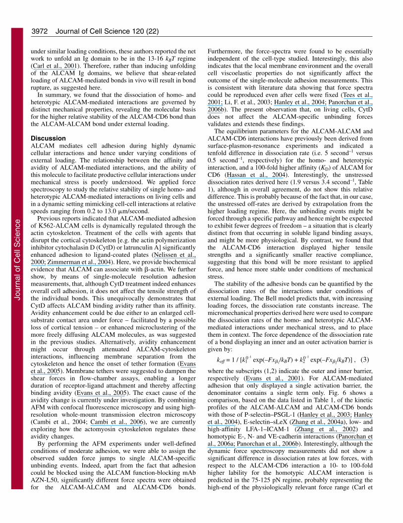
Table 1. The kinetics and reactive compliance of ALCAM-mediated interactions placed in contextSystem x�1 (nm) k1
0 (second–1) x�2 (nm) k20 (second–1) Reference
ALCAM-ALCAMa 0.38±0.06* 1.9±0.8* This workALCAM -CD6a 0.23±0.01* 3.4±0.3* This workN-cadherin–N-cadherinb 0.77±0.09 0.98±0.46 (Panorchan et al., 2006b)E-cadherin–E-cadherinc 0.32±0.07 1.09±0.35 0.10±0.02 4.00±0.68 (Panorchan et al., 2006b)VE-cadherin–VE-cadherinb 0.42±0.03 0.45±0.12 (Panorchan et al., 2006a)P-selectin–PSGL-1d 0.15±0.01 0.22±0.06 (Hanley et al., 2004)LFA-1–ICAM-1 (low affinity)e 0.15 4.0 0.018 57 (Zhang et al., 2002)LFA-1–ICAM-1 (high affinity)e 0.21 0.17 0.024 40 (Zhang et al., 2002)E-selectin–sLeXf 0.5 0.3 0.09 65 (Zhang et al., 2004a)
aLoading rate 300-20,000 pN/second; bloading rate 50-5000 pN/second; cloading rate 100-500 pN/second (lower regime), 500-10,000 pN/second (higherregime); dloading rate 100-10,000 pN/second; eloading rate 20-10,000 pN/second (lower regime), 10,000-50,000 pN/second (higher regime); floading rate 100-10,000 pN/second (lower regime), 10,000-100,000 pN/second (higher regime).
*Error estimation on the basis of a comparison between the data derived from both cell types.
Jour
nal o
f Cel
l Sci
ence
3972
under similar loading conditions, these authors reported the network to unfold an Ig domain to be in the 13-16 kBT regime(Carl et al., 2001). Therefore, rather than inducing unfoldingof the ALCAM Ig domains, we believe that shear-relatedloading of ALCAM-mediated bonds in vivo will result in bondrupture, as suggested here.
In summary, we found that the dissociation of homo- andheterotypic ALCAM-mediated interactions are governed bydistinct mechanical properties, revealing the molecular basisfor the higher relative stability of the ALCAM-CD6 bond thanthe ALCAM-ALCAM bond under external loading.
DiscussionALCAM mediates cell adhesion during highly dynamiccellular interactions and hence under varying conditions ofexternal loading. The relationship between the affinity andavidity of ALCAM-mediated interactions, and the ability ofthis molecule to facilitate productive cellular interactions undermechanical stress is poorly understood. We applied forcespectroscopy to study the relative stability of single homo- andheterotypic ALCAM-mediated interactions on living cells andin a dynamic setting mimicking cell-cell interactions at relativespeeds ranging from 0.2 to 13.0 �m/second.
Previous reports indicated that ALCAM-mediated adhesionof K562-ALCAM cells is dynamically regulated through theactin cytoskeleton. Treatment of the cells with agents thatdisrupt the cortical cytoskeleton [e.g. the actin polymerizationinhibitor cytochalasin D (CytD) or latrunculin A] significantlyenhanced adhesion to ligand-coated plates (Nelissen et al.,2000; Zimmerman et al., 2004). Here, we provide biochemicalevidence that ALCAM can associate with �-actin. We furthershow, by means of single-molecule resolution adhesionmeasurements, that, although CytD treatment indeed enhancesoverall cell adhesion, it does not affect the tensile strength ofthe individual bonds. This unequivocally demonstrates thatCytD affects ALCAM binding avidity rather than its affinity.Avidity enhancement could be due either to an enlarged cell-substrate contact area under force – facilitated by a possibleloss of cortical tension – or enhanced microclustering of themore freely diffusing ALCAM molecules, as was suggestedin the previous studies. Alternatively, avidity enhancementmight occur through attenuated ALCAM-cytoskeletoninteractions, influencing membrane separation from thecytoskeleton and hence the onset of tether formation (Evanset al., 2005). Membrane tethers were suggested to dampen theshear forces in flow-chamber assays, enabling a longerduration of receptor-ligand attachment and thereby affectingbinding avidity (Evans et al., 2005). The exact cause of theavidity change is currently under investigation. By combiningAFM with confocal fluorescence microscopy and using high-resolution whole-mount transmission electron microscopy(Cambi et al., 2004; Cambi et al., 2006), we are currentlyexploring how the actomyosin cytoskeleton regulates theseavidity changes.
By performing the AFM experiments under well-definedconditions of moderate adhesion, we were able to assign theobserved sudden force jumps to single ALCAM-specificunbinding events. Indeed, apart from the fact that adhesioncould be blocked using the ALCAM function-blocking mAbAZN-L50, significantly different force spectra were obtainedfor the ALCAM-ALCAM and ALCAM-CD6 bonds.
Furthermore, the force-spectra were found to be essentiallyindependent of the cell-type studied. Interestingly, this alsoindicates that the local membrane environment and the overallcell viscoelastic properties do not significantly affect theoutcome of the single-molecule adhesion measurements. Thisis consistent with literature data showing that force spectracould be reproduced even after cells were fixed (Tees et al.,2001; Li, F. et al., 2003; Hanley et al., 2004; Panorchan et al.,2006b). The present observation that, on living cells, CytDdoes not affect the ALCAM-specific unbinding forcesvalidates and extends these findings.
The equilibrium parameters for the ALCAM-ALCAM andALCAM-CD6 interactions have previously been derived fromsurface-plasmon-resonance experiments and indicated atenfold difference in dissociation rate (i.e. 5 second–1 versus0.5 second–1, respectively) for the homo- and heterotypicinteraction, and a 100-fold higher affinity (KD) of ALCAM forCD6 (Hassan et al., 2004). Interestingly, the unstresseddissociation rates derived here (1.9 versus 3.4 second–1, Table1), although in overall agreement, do not show this relativedifference. This is probably because of the fact that, in our case,the unstressed off-rates are derived by extrapolation from thehigher loading regime. Here, the unbinding events might beforced through a specific pathway and hence might be expectedto exhibit fewer degrees of freedom – a situation that is clearlydistinct from that occurring in soluble ligand binding assays,and might be more physiological. By contrast, we found thatthe ALCAM-CD6 interaction displayed higher tensilestrengths and a significantly smaller reactive compliance,suggesting that this bond will be more resistant to appliedforce, and hence more stable under conditions of mechanicalstress.
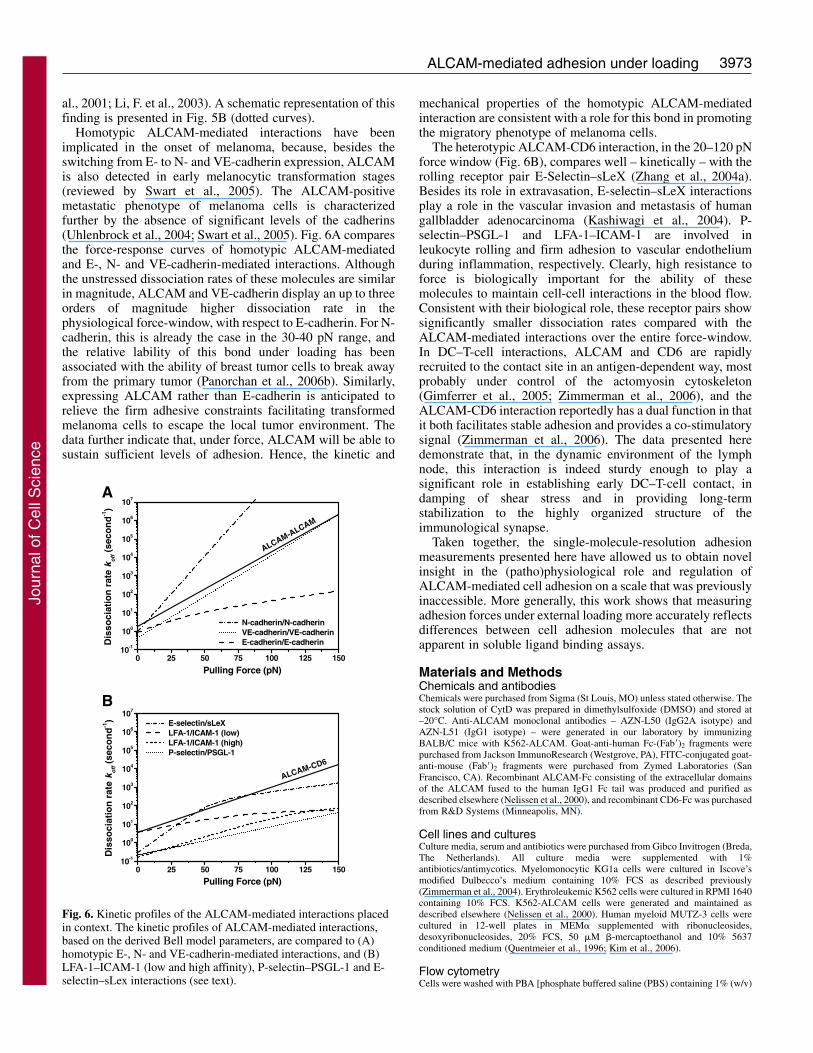
The stability of the adhesive bonds can be quantified by thedissociation rates of the interactions under conditions ofexternal loading. The Bell model predicts that, with increasingloading forces, the dissociation rate constants increase. Themicromechanical properties derived here were used to comparethe dissociation rates of the homo- and heterotypic ALCAM-mediated interactions under mechanical stress, and to placethem in context. The force dependence of the dissociation rateof a bond displaying an inner and an outer activation barrier isgiven by:
koff = 1 / [k10–1
exp(–Fx�1/kBT) + k2
0–1exp(–Fx�2
/kBT)] , (3)
where the subscripts (1,2) indicate the outer and inner barrier,respectively (Evans et al., 2001). For ALCAM-mediatedadhesion that only displayed a single activation barrier, thedenominator contains a single term only. Fig. 6 shows acomparison, based on the data listed in Table 1, of the kineticprofiles of the ALCAM-ALCAM and ALCAM-CD6 bondswith those of P-selectin–PSGL-1 (Hanley et al., 2003; Hanleyet al., 2004), E-selectin–sLeX (Zhang et al., 2004a), low- andhigh-affinity LFA-1–ICAM-1 (Zhang et al., 2002) andhomotypic E-, N- and VE-cadherin interactions (Panorchan etal., 2006a; Panorchan et al., 2006b). Interestingly, although thedynamic force spectroscopy measurements did not show asignificant difference in dissociation rates at low forces, withrespect to the ALCAM-CD6 interaction a 10- to 100-foldhigher lability for the homotypic ALCAM interaction ispredicted in the 75-125 pN regime, probably representing thehigh-end of the physiologically relevant force range (Carl et
Journal of Cell Science 120 (22)
Jour
nal o
f Cel
l Sci
ence
3973ALCAM-mediated adhesion under loading
al., 2001; Li, F. et al., 2003). A schematic representation of thisfinding is presented in Fig. 5B (dotted curves).
Homotypic ALCAM-mediated interactions have beenimplicated in the onset of melanoma, because, besides theswitching from E- to N- and VE-cadherin expression, ALCAMis also detected in early melanocytic transformation stages(reviewed by Swart et al., 2005). The ALCAM-positivemetastatic phenotype of melanoma cells is characterizedfurther by the absence of significant levels of the cadherins(Uhlenbrock et al., 2004; Swart et al., 2005). Fig. 6A comparesthe force-response curves of homotypic ALCAM-mediatedand E-, N- and VE-cadherin-mediated interactions. Althoughthe unstressed dissociation rates of these molecules are similarin magnitude, ALCAM and VE-cadherin display an up to threeorders of magnitude higher dissociation rate in thephysiological force-window, with respect to E-cadherin. For N-cadherin, this is already the case in the 30-40 pN range, andthe relative lability of this bond under loading has beenassociated with the ability of breast tumor cells to break awayfrom the primary tumor (Panorchan et al., 2006b). Similarly,expressing ALCAM rather than E-cadherin is anticipated torelieve the firm adhesive constraints facilitating transformedmelanoma cells to escape the local tumor environment. Thedata further indicate that, under force, ALCAM will be able tosustain sufficient levels of adhesion. Hence, the kinetic and
mechanical properties of the homotypic ALCAM-mediatedinteraction are consistent with a role for this bond in promotingthe migratory phenotype of melanoma cells.
The heterotypic ALCAM-CD6 interaction, in the 20–120 pNforce window (Fig. 6B), compares well – kinetically – with therolling receptor pair E-Selectin–sLeX (Zhang et al., 2004a).Besides its role in extravasation, E-selectin–sLeX interactionsplay a role in the vascular invasion and metastasis of humangallbladder adenocarcinoma (Kashiwagi et al., 2004). P-selectin–PSGL-1 and LFA-1–ICAM-1 are involved inleukocyte rolling and firm adhesion to vascular endotheliumduring inflammation, respectively. Clearly, high resistance toforce is biologically important for the ability of thesemolecules to maintain cell-cell interactions in the blood flow.Consistent with their biological role, these receptor pairs showsignificantly smaller dissociation rates compared with theALCAM-mediated interactions over the entire force-window.In DC–T-cell interactions, ALCAM and CD6 are rapidlyrecruited to the contact site in an antigen-dependent way, mostprobably under control of the actomyosin cytoskeleton(Gimferrer et al., 2005; Zimmerman et al., 2006), and theALCAM-CD6 interaction reportedly has a dual function in thatit both facilitates stable adhesion and provides a co-stimulatorysignal (Zimmerman et al., 2006). The data presented heredemonstrate that, in the dynamic environment of the lymphnode, this interaction is indeed sturdy enough to play asignificant role in establishing early DC–T-cell contact, indamping of shear stress and in providing long-termstabilization to the highly organized structure of theimmunological synapse.
Taken together, the single-molecule-resolution adhesionmeasurements presented here have allowed us to obtain novelinsight in the (patho)physiological role and regulation ofALCAM-mediated cell adhesion on a scale that was previouslyinaccessible. More generally, this work shows that measuringadhesion forces under external loading more accurately reflectsdifferences between cell adhesion molecules that are notapparent in soluble ligand binding assays.
Materials and MethodsChemicals and antibodiesChemicals were purchased from Sigma (St Louis, MO) unless stated otherwise. Thestock solution of CytD was prepared in dimethylsulfoxide (DMSO) and stored at–20°C. Anti-ALCAM monoclonal antibodies – AZN-L50 (IgG2A isotype) andAZN-L51 (IgG1 isotype) – were generated in our laboratory by immunizingBALB/C mice with K562-ALCAM. Goat-anti-human Fc-(Fab�)2 fragments werepurchased from Jackson ImmunoResearch (Westgrove, PA), FITC-conjugated goat-anti-mouse (Fab�)2 fragments were purchased from Zymed Laboratories (SanFrancisco, CA). Recombinant ALCAM-Fc consisting of the extracellular domainsof the ALCAM fused to the human IgG1 Fc tail was produced and purified asdescribed elsewhere (Nelissen et al., 2000), and recombinant CD6-Fc was purchasedfrom R&D Systems (Minneapolis, MN).
Cell lines and culturesCulture media, serum and antibiotics were purchased from Gibco Invitrogen (Breda,The Netherlands). All culture media were supplemented with 1%antibiotics/antimycotics. Myelomonocytic KG1a cells were cultured in Iscove’smodified Dulbecco’s medium containing 10% FCS as described previously(Zimmerman et al., 2004). Erythroleukemic K562 cells were cultured in RPMI 1640containing 10% FCS. K562-ALCAM cells were generated and maintained asdescribed elsewhere (Nelissen et al., 2000). Human myeloid MUTZ-3 cells werecultured in 12-well plates in MEM� supplemented with ribonucleosides,desoxyribonucleosides, 20% FCS, 50 �M �-mercaptoethanol and 10% 5637conditioned medium (Quentmeier et al., 1996; Kim et al., 2006).
Flow cytometryCells were washed with PBA [phosphate buffered saline (PBS) containing 1% (w/v)
Fig. 6. Kinetic profiles of the ALCAM-mediated interactions placedin context. The kinetic profiles of ALCAM-mediated interactions,based on the derived Bell model parameters, are compared to (A)homotypic E-, N- and VE-cadherin-mediated interactions, and (B)LFA-1–ICAM-1 (low and high affinity), P-selectin–PSGL-1 and E-selectin–sLex interactions (see text).
A
B
0 25 50 75 100 125 15010-1
100
101
102
103
104
105
106
107
ALCAM-CD6
E-selectin/sLeX LFA-1/ICAM-1 (low) LFA-1/ICAM-1 (high) P-selectin/PSGL-1
Dis
soci
atio
n r
ate
k off (
seco
nd
-1)
Pulling Force (pN)
0 25 50 75 100 125 15010-1
100
101
102
103
104
105
106
107
Dis
soci
atio
n r
ate
k off (
seco
nd
-1)
N-cadherin/N-cadherin VE-cadherin/VE-cadherin E-cadherin/E-cadherin
ALCAM-ALCAM
Pulling Force (pN)
Jour
nal o
f Cel
l Sci
ence
3974
bovine serum albumin (BSA) and 0.05% (w/v) NaN3] and stained for 30 minutesat 4°C with AZN-L50 primary antibody (2-5 �g/ml in PBA). Cells were washedwith PBA and incubated with FITC-conjugated goat-anti-mouse (Fab�)2 secondaryantibodies. After washing, cells were analyzed on a FACScan analyzer (BectonDickinson, Oxnard, CA). The gates were set to exclude dead cells and 5000 gatedcells were analyzed. Data are displayed as histograms of fluorescence intensityversus cell count.
Radioactive cell labeling and immunoprecipitationKG1a or control MUTZ-3 cells were pre-incubated for 1 hour in serum- andmethionine/cysteine-free RPMI 1640 medium prior to labeling with Tran[35S]-label(MP Biomedicals, Irvine, CA), 250 �Ci per 10�106 cells for 16 hours at 37°C.Cells were washed once in PBS and subsequently lysed in lysis buffer A (50 mMTris pH 7.5, 0.5% Triton X-100, 300 mM NaCl, 1.5 mM MgCl2, 0.2 mM EDTA,0.5 mM DTT, 1 mM PMSF, 1 �g/ml leupeptin and aprotinin). Lysates weresubjected to immunoprecipitation with 1 �g of AZN-L51 antibody coupled toProtein G Sepharose 4 Fast Flow beads (Amersham Biosciences). Beads werewashed three times in lysis buffer A and bound proteins were eluted by boiling inLaemmli sample buffer and subjected to 9% SDS-PAGE under reducing conditions.Radioactive proteins were detected by exposure to X-ray film (BioMax XAR;Kodak, USA).
Immunoprecipitation and western blot analysis5�106 KG1a cells were lysed in lysis buffer B (50 mM HEPES pH 7.5, 150 mMNaCl, 1.5 mM MgCl2, 1 mM EDTA, 10% glycerol, 1% Triton X-100, 1 mM PMSF,1 �g/ml leupeptin and aprotinin). Immunoprecipitations were carried out with 1�g of AZN-L51 antibody coupled to Protein G Sepharose 4 Fast Flow beads.Bound proteins were eluted by boiling in Laemmli sample buffer, subjected to 12%SDS-PAGE under reducing conditions and transferred onto a nitrocellulosemembrane. To detect ALCAM and actin, membranes were incubated for 1 hourwith AZN-L50 or mouse monoclonal anti-�-actin (Sigma), followed by 1-hourincubation with a peroxidase-conjugated rabbit anti-mouse IgG (DAKO, Denmark)and proteins were visualized using an enhanced chemiluminescence system(Amersham Biosciences).
AFM force measurementsForce measurements were made on living cells in force-distance mode (Fig. 1) usinga MultiMode AFM (Nanoscope IIIa) equipped with a ‘J’-type piezoelectrictranslator (Veeco Instruments, Santa Barbara, CA). Triangular gold-coated silicon-nitride cantilevers were used with a nominal spring constant of 10 pN/nm as givenby the manufacturer (MLCT-AUHW, Veeco Instruments). Cantilever deflection wasdetermined from the difference in signal generated by a two-segment photodiodemonitoring the reflection of a laser beam focused onto the endpoint of the cantilever(Fig. 1B). Each cantilever was calibrated before use by a nondestructive thermaloscillation method (Hutter and Bechhoefer, 1993); by using this method theuncertainty in the determination of the spring constant amounted to 3-4% percantilever. The experimentally determined spring constants of the used cantileverswere 15±2 pN/nm, and these values were used to obtain interaction forces usingHooke’s law, F=k��x. Here, F is the force (expressed in piconewtons, pN), k isthe experimentally obtained spring constant (pN/nm), and �x is the measuredcantilever deflection (nm).
Protein immobilizationALCAM-Fc and CD6-Fc were immobilized on 13 mm plastic coverslips (NalgeNunc, Rochester, NY). First, in an overnight (4°C) incubation, 10 �g/ml goat anti-human Fc-(Fab�)2 fragments were absorbed to the coverslip surface in TSM (20 mMTris, 150 mM NaCl, 1 mM CaCl2, 2 mM MgCl2, pH 8.0). Then the substrates wererinsed and subsequently incubated for 30 minutes in TSM/1% (w/v) BSA at 37°Cto block the remaining exposed non-coated surface. After an additional washingstep, the plates were incubated with 5 �g/ml ALCAM-Fc or 5 �g/ml CD6-Fc inTSM for 1 hour at 37°C. Finally, the coated substrates were washed and transferredinto the AFM measuring chamber (MTFML, Veeco Instruments).
Functionalization of AFM cantilevers with cellsCells were attached to the AFM cantilever by concanavalin A (ConA)-mediatedlinkages essentially as described previously (Wojcikiewicz et al., 2003). ConA-coated cantilevers were prepared as follows. Cantilevers were first cleaned byimmersion in acetone for 5 minutes, then rinsed with ethanol and subsequently driedin a microwave oven. Following an overnight incubation at 37°C in biotinylatedBSA (biotin-BSA, 0.5 mg/ml in 100 mM NaHCO3, pH 8.6) the cantilevers wererinsed using PBS and exposed to 0.5 mg/ml (PBS, 30 minutes, 37°C) streptavidin(Pierce, Rockford, IL). Finally, the cantilevers were incubated in biotinylated ConA(biotin-ConA, 0.2 mg/ml in PBS) for 30 minutes at 37°C and washed with PBS.
Cells kept in medium A (RPMI 1640, 10% FCS, 25 mM HEPES; pH 7.0) wereseeded onto a clean uncoated glass coverslip and were picked up under the guidanceof an optical microscope mounted on top of the AFM, using the AFM as amicromanipulator. For this, the ConA-functionalized cantilever was positioned overa target cell on the substrate and was approached to establish contact lasting at least
one minute. During this time the applied indentation force was kept constant at about2.5 nN. Upon retraction, the successful pick-up was readily scored by visualinspection, and, in these events, the cell was positioned right behind the AFM tip(Fig. 1C).
Rupture-force measurements and force spectroscopyThe cell-bearing cantilever was brought into contact with the ligand-coated substrate(Fig. 1Ba-c) for a preset period of time (interaction time; 25°C, medium A). Duringthis time, a force was exerted on the cell of no more than ~1 nN. Interaction timeswere such that a minimal, yet significant, ALCAM-specific adhesion wasestablished (typically between 0.5-3.0 seconds, see Results). Upon retraction, theforces acting on the cantilever were recorded as a function of displacement of theALCAM–CD6-coated substrate (Fig. 1Bd-f, Fig. 1D). ALCAM-ALCAM andALCAM-CD6 rupture forces were determined directly from the height of thesudden variations in binding force that are associated with bond rupture. The finalruptures in the force-distance curves were used for further analysis (Fig. 1D; seebelow). The area enclosed by the zero-force axis and the force-distance curve (Fig.1D) was taken as a measure for the work (W=F�d) performed during thedetachment phase (Zhang et al., 2002; Puech et al., 2005). CytD treatments wereperformed in situ (2.5 �g/ml in medium A, 25 minutes). Specificity was verifiedby an in situ incubation with the function-blocking ALCAM-specific monoclonalantibody AZN-L50 (10 �g/ml, 25 minutes). Force curves were analyzed usingOrigin Pro 6.1 (OriginLab Corporation, Northampton, MA). The same package wasused for performing Student’s t-test.
Force spectroscopy was applied to study how the rupture forces depend on theloading rate (i.e. the rate at which force builds up on the respective bonds). Loadingrates (pN/second) were computed as the product of the slope of the force-distancecurve (pN/nm) just before a rupture event – the effective force constant that takesthe viscoelastic properties of the system into account (Evans and Ritchie, 1999;Yuan et al., 2000) – and the pulling velocity (nm/second). Pulling velocities werevaried from 250-12,500 nm/second. The final ruptures in the force-distance curveswere used for further analysis and for each of these events both the loading rate andthe rupture force was determined (Fig. 1D). For each cell, complete force spectrawere recorded under identical conditions on both substrates by switching the ligand-coated plate for one containing the other. Pulling rates were varied randomly andreproducibility over time was verified by repeating measurements using priorpulling-rate settings. After each series, ALCAM specificity of the adhesion waschecked using the blocking mAb AZN-L50.
At retraction speeds >1 �m/second, the hydrodynamic drag on the cantileverresulted in damping and, as a result, smaller forces were recorded than were actuallyapplied to rupture the bonds (Evans et al., 2001; Tees et al., 2001; Zhang et al.,2002). To compensate for this effect, the data were corrected using a dampingcoefficient of 2 pN second/�m.
J.t.R. is supported by NanoNed, the Dutch nanotechnologyprogramme of the Ministry of Economic Affairs. The NetherlandsOrganization of Scientific Research supports C.G.F. through TOPGrant 9120.6030 and A.C. through Grant Veni 916.66.028 and SLW33.302P. F.d.L. received support through grant KUN OZ-2002-5 fromthe Radboud University Nijmegen Medical Centre. The authors thankBen Ohler and Patrick Markus (Veeco Instruments) for their valuabletechnical support, and Peter Schön for critical reading of themanuscript. The Microscopic Imaging Centre (MIC) of the NCMLSis kindly acknowledged for providing facilities.
ReferencesAlon, R., Chen, S., Fuhlbrigge, R., Puri, K. D. and Springer, T. A. (1998). The kinetics
and shear threshold of transient and rolling interactions of L-selectin with its ligand onleukocytes. Proc. Natl. Acad. Sci. USA 95, 11631-11636.
Bell, G. I. (1978). Models for the specific adhesion of cells to cells. Science 200, 618-627.
Benoit, M., Gabriel, D., Gerisch, G. and Gaub, H. E. (2000). Discrete interactions incell adhesion measured by single-molecule force spectroscopy. Nat. Cell Biol. 2, 313-317.
Bhasin, N., Carl, P., Harper, S., Feng, G., Lu, H., Speicher, D. W. and Discher, D. E.(2004). Chemistry on a single protein, vascular cell adhesion molecule-1, during forcedunfolding. J. Biol. Chem. 279, 45865-45874.
Binnig, G., Quate, C. F. and Gerber, C. (1986). Atomic force microscope. Phys. Rev.Lett. 56, 930-933.
Bowen, M. A., Patel, D. D., Li, X., Modrell, B., Malacko, A. R., Wang, W. C.,Marquardt, H., Neubauer, M., Pesando, J. M. and Francke, U. (1995). Cloning,mapping, and characterization of activated leukocyte-cell adhesion molecule(ALCAM), a CD6 ligand. J. Exp. Med. 181, 2213-2220.
Bowen, M. A., Aruffo, A. A. and Bajorath, J. (2000). Cell surface receptors and theirligands: in vitro analysis of CD6-CD166 interactions. Proteins 40, 420-428.
Cambi, A., de Lange, F., van Maarseveen, N. M., Nijhuis, M., Joosten, B., van Dijk,E. M., de Bakker, B. I., Fransen, J. A., Bovee-Geurts, P. H., van Leeuwen, F. N.
Journal of Cell Science 120 (22)
Jour
nal o
f Cel
l Sci
ence
3975ALCAM-mediated adhesion under loading
et al. (2004). Microdomains of the C-type lectin DC-SIGN are portals for virus entryinto dendritic cells. J. Cell Biol. 164, 145-155.
Cambi, A., Joosten, B., Koopman, M., de Lange, F., Beeren, I., Torensma, R.,Fransen, J. A., Garcia-Parajo, M., van Leeuwen, F. N. and Figdor, C. G. (2006).Organization of the integrin LFA-1 in nanoclusters regulates its activity. Mol. Biol. Cell17, 4270-4281.
Carl, P., Kwok, C. H., Manderson, G., Speicher, D. W. and Discher, D. E. (2001).Forced unfolding modulated by disulfide bonds in the Ig domains of a cell adhesionmolecule. Proc. Natl. Acad. Sci. USA 98, 1565-1570.
Carman, C. V. and Springer, T. A. (2003). Integrin avidity regulation: are changes inaffinity and conformation underemphasized? Curr. Opin. Cell Biol. 15, 547-556.
Chen, A. and Moy, V. T. (2000). Cross-linking of cell surface receptors enhancescooperativity of molecular adhesion. Biophys. J. 78, 2814-2820.
Cowin, P., Rowlands, T. M. and Hatsell, S. J. (2005). Cadherins and catenins in breastcancer. Curr. Opin. Cell Biol. 17, 499-508.
Degen, W. G., van Kempen, L. C., Gijzen, E. G., van Groningen, J. J., van Kooyk,Y., Bloemers, H. P. and Swart, G. W. (1998). MEMD, a new cell adhesion moleculein metastasizing human melanoma cell lines, is identical to ALCAM (activatedleukocyte cell adhesion molecule). Am. J. Pathol. 152, 805-813.
Evans, E. and Ritchie, K. (1997). Dynamic strength of molecular adhesion bonds.Biophys. J. 72, 1541-1555.
Evans, E. and Ritchie, K. (1999). Strength of a weak bond connecting flexible polymerchains. Biophys. J. 76, 2439-2447.
Evans, E., Leung, A., Hammer, D. and Simon, S. (2001). Chemically distinct transitionstates govern rapid dissociation of single L-selectin bonds under force. Proc. Natl.Acad. Sci. USA 98, 3784-3789.
Evans, E., Heinrich, V., Leung, A. and Kinoshita, K. (2005). Nano- to microscaledynamics of P-selectin detachment from leukocyte interfaces. I. Membrane separationfrom the cytoskeleton. Biophys. J. 88, 2288-2298.
Gangemi, R. M., Swack, J. A., Gaviria, D. M. and Romain, P. L. (1989). Anti-T12,an anti-CD6 monoclonal antibody, can activate human T lymphocytes. J. Immunol.143, 2439-2447.
Gimferrer, I., Calvo, M., Mittelbrunn, M., Farnos, M., Sarrias, M. R., Enrich, C.,Vives, J., Sanchez-Madrid, F. and Lozano, F. (2004). Relevance of CD6-mediatedinteractions in T cell activation and proliferation. J. Immunol. 173, 2262-2270.
Gimferrer, I., Ibanez, A., Farnos, M., Sarrias, M. R., Fenutria, R., Rosello, S.,Zimmermann, P., David, G., Vives, J., Serra-Pages, C. et al. (2005). The lymphocytereceptor CD6 interacts with syntenin-1, a scaffolding protein containing PDZ domains.J. Immunol. 175, 1406-1414.
Grakoui, A., Bromley, S. K., Sumen, C., Davis, M. M., Shaw, A. S., Allen, P. M. andDustin, M. L. (1999). The immunological synapse: a molecular machine controllingT cell activation. Science 285, 221-227.
Hanahan, D. and Weinberg, R. A. (2000). The hallmarks of cancer. Cell 100, 57-70.Hanley, W., McCarty, O., Jadhav, S., Tseng, Y., Wirtz, D. and Konstantopoulos, K.
(2003). Single molecule characterization of P-selectin/ligand binding. J. Biol. Chem.278, 10556-10561.
Hanley, W. D., Wirtz, D. and Konstantopoulos, K. (2004). Distinct kinetic andmechanical properties govern selectin-leukocyte interactions. J. Cell Sci. 117, 2503-2511.
Hassan, N. J., Barclay, A. N. and Brown, M. H. (2004). Frontline: optimal T cellactivation requires the engagement of CD6 and CD166. Eur. J. Immunol. 34, 930-940.
Hazan, R. B., Qiao, R., Keren, R., Badano, I. and Suyama, K. (2004). Cadherin switchin tumor progression. Ann. N. Y. Acad. Sci. 1014, 155-163.
Hinterdorfer, P. (2002). Molecular recognition studies using the atomic forcemicroscope. Methods Cell Biol. 68, 115-139.
Hinterdorfer, P. and Dufrêne, Y. F. (2006). Detection and localization of singlemolecular recognition events using atomic force microscopy. Nat. Methods 3, 347-355.
Hummer, G. and Szabo, A. (2001). Free energy reconstruction from nonequilibriumsingle-molecule pulling experiments. Proc. Natl. Acad. Sci. USA 98, 3658-3661.
Hutter, J. L. and Bechhoefer, J. (1993). Calibration of atomic-force microscope tips.Rev. Sci. Instrum. 64, 1868-1873.
Kashiwagi, H., Kijima, H., Dowaki, S., Ohtani, Y., Tobita, K., Yamazaki, H.,Nakamura, M., Ueyama, Y., Tanaka, M., Inokuchi, S. et al. (2004).Clinicopathological significance of sialyl Lex expression in human gallbladdercarcinoma. Oncol. Rep. 11, 1139-1143.
Kim, K. D., Choi, S. C., Noh, Y. W., Kim, J. W., Paik, S. G., Yang, Y., Kim, K., 2ndand Lim, J. S. (2006). Impaired responses of leukemic dendritic cells derived from ahuman myeloid cell line to LPS stimulation. Exp. Mol. Med. 38, 72-84.
Kim, M., Carman, C. V., Yang, W., Salas, A. and Springer, T. A. (2004). The primacyof affinity over clustering in regulation of adhesiveness of the integrin �L�2. J. CellBiol. 167, 1241-1253.
King, J. A., Ofori-Acquah, S. F., Stevens, T., Al-Mehdi, A. B., Fodstad, O. and Jiang,W. G. (2004). Activated leukocyte cell adhesion molecule in breast cancer: prognosticindicator. Breast Cancer Res. 6, R478-R487.
Knudsen, K. A. and Wheelock, M. J. (2005). Cadherins and the mammary gland. J.Cell. Biochem. 95, 488-496.
Kristiansen, G., Pilarsky, C., Wissmann, C., Stephan, C., Weissbach, L., Loy, V.,Loening, S., Dietel, M. and Rosenthal, A. (2003). ALCAM/CD166 is up-regulatedin low-grade prostate cancer and progressively lost in high-grade lesions. Prostate 54,34-43.
Li, F., Redick, S. D., Erickson, H. P. and Moy, V. T. (2003). Force measurements of the�5�1 integrin-fibronectin interaction. Biophys. J. 84, 1252-1262.
Li, G., Satyamoorthy, K., Meier, F., Berking, C., Bogenrieder, T. and Herlyn, M.
(2003). Function and regulation of melanoma-stromal fibroblast interactions: whenseeds meet soil. Oncogene 22, 3162-3171.
Lunter, P. C., van Kilsdonk, J. W., van Beek, H., Cornelissen, I. M., Bergers, M.,Willems, P. H., van Muijen, G. N. and Swart, G. W. (2005). Activated leukocyte celladhesion molecule (ALCAM/CD166/MEMD), a novel actor in invasive growth,controls matrix metalloproteinase activity. Cancer Res. 65, 8801-8808.
Masedunskas, A., King, J. A., Tan, F., Cochran, R., Stevens, T., Sviridov, D. andOfori-Acquah, S. F. (2006). Activated leukocyte cell adhesion molecule is acomponent of the endothelial junction involved in transendothelial monocytemigration. FEBS Lett. 580, 2637-2645.
Mempel, T. R., Henrickson, S. E. and Von Andrian, U. H. (2004). T-cell priming bydendritic cells in lymph nodes occurs in three distinct phases. Nature 427, 154-159.
Nelissen, J. M., Peters, I. M., de Grooth, B. G., van Kooyk, Y. and Figdor, C. G.(2000). Dynamic regulation of activated leukocyte cell adhesion molecule-mediatedhomotypic cell adhesion through the actin cytoskeleton. Mol. Biol. Cell 11, 2057-2068.
Osorio, L. M., Rottenberg, M., Jondal, M. and Chow, S. C. (1998). Simultaneous cross-linking of CD6 and CD28 induces cell proliferation in resting T cells. Immunology 93,358-365.
Panorchan, P., George, J. P. and Wirtz, D. (2006a). Probing intercellular interactionsbetween vascular endothelial cadherin pairs at single-molecule resolution and in livingcells. J. Mol. Biol. 358, 665-674.
Panorchan, P., Thompson, M. S., Davis, K. J., Tseng, Y., Konstantopoulos, K. andWirtz, D. (2006b). Single-molecule analysis of cadherin-mediated cell-cell adhesion.J. Cell Sci. 119, 66-74.
Patel, D. D., Wee, S. F., Whichard, L. P., Bowen, M. A., Pesando, J. M., Aruffo, A.and Haynes, B. F. (1995). Identification and characterization of a 100-kD ligand forCD6 on human thymic epithelial cells. J. Exp. Med. 181, 1563-1568.
Puech, P. H., Taubenberger, A., Ulrich, F., Krieg, M., Muller, D. J. and Heisenberg,C. P. (2005). Measuring cell adhesion forces of primary gastrulating cells fromzebrafish using atomic force microscopy. J. Cell Sci. 118, 4199-4206.
Quentmeier, H., Duschl, A., Hu, Z. B., Schnarr, B., Zaborski, M. and Drexler, H. G.(1996). MUTZ-3, a monocytic model cell line for interleukin-4 and lipopolysaccharidestudies. Immunology 89, 606-612.
Rasmussen, R. A., Counts, S. L., Daley, J. F. and Schlossman, S. F. (1994). Isolationand characterization of CD6- T cells from peripheral blood. J. Immunol. 152, 527-536.
Rief, M. and Grubmüller, H. (2002). Force spectroscopy of single biomolecules.Chemphyschem 3, 255-261.
Swart, G. W. (2002). Activated leukocyte cell adhesion molecule (CD166/ALCAM):developmental and mechanistic aspects of cell clustering and cell migration. Eur. J.Cell Biol. 81, 313-321.
Swart, G. W., Lunter, P. C., Kilsdonk, J. W. and Kempen, L. C. (2005). Activatedleukocyte cell adhesion molecule (ALCAM/CD166): signaling at the divide ofmelanoma cell clustering and cell migration? Cancer Metastasis Rev. 24, 223-236.
Tanaka, H., Matsui, T., Agata, A., Tomura, M., Kubota, I., McFarland, K. C., Kohr,B., Lee, A., Phillips, H. S. and Shelton, D. L. (1991). Molecular cloning andexpression of a novel adhesion molecule, SC1. Neuron 7, 535-545.
Tees, D. F., Waugh, R. E. and Hammer, D. A. (2001). A microcantilever device to assessthe effect of force on the lifetime of selectin-carbohydrate bonds. Biophys. J. 80, 668-682.
Tomita, K., van Bokhoven, A., Jansen, C. F. J., Kiemeney, L. A., Karthaus, H. F. M.,Vriesema, J., Bussemakers, M. J. G., Witjes, J. A. and Schalken, J. A. (2003).Activated Leukocyte Cell Adhesion Molecule (ALCAM) expression is associated witha poor prognosis for bladder cancer patients. UroOncology 3, 121-129.
Uhlenbrock, K., Eberth, A., Herbrand, U., Daryab, N., Stege, P., Meier, F., Friedl, P.,Collard, J. G. and Ahmadian, M. R. (2004). The RacGEF Tiam1 inhibits migrationand invasion of metastatic melanoma via a novel adhesive mechanism. J. Cell Sci. 117,4863-4871.
van Kempen, L. C., van den Oord, J. J., van Muijen, G. N., Weidle, U. H., Bloemers,H. P. and Swart, G. W. (2000). Activated leukocyte cell adhesion molecule/CD166,a marker of tumor progression in primary malignant melanoma of the skin. Am. J.Pathol. 156, 769-774.
van Kempen, L. C., Nelissen, J. M., Degen, W. G., Torensma, R., Weidle, U. H.,Bloemers, H. P., Figdor, C. G. and Swart, G. W. (2001). Molecular basis for thehomophilic activated leukocyte cell adhesion molecule (ALCAM)-ALCAMinteraction. J. Biol. Chem. 276, 25783-25790.
van Kooyk, Y. and Figdor, C. G. (2000). Avidity regulation of integrins: the drivingforce in leukocyte adhesion. Curr. Opin. Cell Biol. 12, 542-547.
Wee, S., Schieven, G. L., Kirihara, J. M., Tsu, T. T., Ledbetter, J. A. and Aruffo, A.(1993). Tyrosine phosphorylation of CD6 by stimulation of CD3: augmentation by theCD4 and CD2 coreceptors. J. Exp. Med. 177, 219-223.
Weichert, W., Knosel, T., Bellach, J., Dietel, M. and Kristiansen, G. (2004).ALCAM/CD166 is overexpressed in colorectal carcinoma and correlates withshortened patient survival. J. Clin. Pathol. 57, 1160-1164.
Willemsen, O. H., Snel, M. M., Cambi, A., Greve, J., De Grooth, B. G. and Figdor,C. G. (2000). Biomolecular interactions measured by atomic force microscopy.Biophys. J. 79, 3267-3281.
Wojcikiewicz, E. P., Zhang, X., Chen, A. and Moy, V. T. (2003). Contributions ofmolecular binding events and cellular compliance to the modulation of leukocyteadhesion. J. Cell Sci. 116, 2531-2539.
Wong, J., Chilkoti, A. and Moy, V. T. (1999). Direct force measurements of thestreptavidin-biotin interaction. Biomol. Eng. 16, 45-55.
Yuan, C., Chen, A., Kolb, P. and Moy, V. T. (2000). Energy landscape of streptavidin-biotin complexes measured by atomic force microscopy. Biochemistry 39, 10219-10223.
Zhang, X., Wojcikiewicz, E. P. and Moy, V. T. (2002). Force spectroscopy of the
Jour
nal o
f Cel
l Sci
ence

3976
leukocyte function-associated antigen-1/intercellular adhesion molecule-1 interaction.Biophys. J. 83, 2270-2279.
Zhang, X., Bogorin, D. F. and Moy, V. T. (2004a). Molecular basis of the dynamicstrength of the sialyl Lewis X-selectin interaction. Chemphyschem 5, 175-182.
Zhang, X., Craig, S. E., Kirby, H., Humphries, M. J. and Moy, V. T. (2004b).Molecular basis for the dynamic strength of the integrin �4ß1/VCAM-1 interaction.Biophys. J. 87, 3470-3478.
Zimmerman, A. W., Nelissen, J. M., Van Emst-De Vries, S. E., Willems, P. H., DeLange, F., Collard, J. G., Van Leeuwen, F. N. and Figdor, C. G. (2004). Cytoskeletalrestraints regulate homotypic ALCAM-mediated adhesion through PKC�independently of Rho-like GTPases. J. Cell Sci. 117, 2841-2852.
Zimmerman, A. W., Joosten, B., Torensma, R., Parnes, J. R., van Leeuwen, F. N. andFigdor, C. G. (2006). Long-term engagement of CD6 and ALCAM is essential for T-cell proliferation induced by dendritic cells. Blood 107, 3212-3220.
Journal of Cell Science 120 (22)
Jour
nal o
f Cel
l Sci
ence
Copyright © 2022 FDOKUMEN