The role of activated leukocyte cell adhesion molecule ...
199
ADVERTIMENT. Lʼaccés als continguts dʼaquesta tesi queda condicionat a lʼacceptació de les condicions dʼús establertes per la següent llicència Creative Commons: http://cat.creativecommons.org/?page_id=184 ADVERTENCIA. El acceso a los contenidos de esta tesis queda condicionado a la aceptación de las condiciones de uso establecidas por la siguiente licencia Creative Commons: http://es.creativecommons.org/blog/licencias/ WARNING. The access to the contents of this doctoral thesis it is limited to the acceptance of the use conditions set by the following Creative Commons license: https://creativecommons.org/licenses/?lang=en
-
Upload
khangminh22 -
Category
Documents
-
view
9 -
download
0
Transcript of The role of activated leukocyte cell adhesion molecule ...

ADVERTIMENT. Lʼaccés als continguts dʼaquesta tesi queda condicionat a lʼacceptació de les condicions dʼúsestablertes per la següent llicència Creative Commons: http://cat.creativecommons.org/?page_id=184
ADVERTENCIA. El acceso a los contenidos de esta tesis queda condicionado a la aceptación de las condiciones de usoestablecidas por la siguiente licencia Creative Commons: http://es.creativecommons.org/blog/licencias/
WARNING. The access to the contents of this doctoral thesis it is limited to the acceptance of the use conditions setby the following Creative Commons license: https://creativecommons.org/licenses/?lang=en

The role of activated leukocyte cell adhesion molecule (ALCAM) in endometrial cancer progression and dissemination
Laura Devis Jauregui


The role of activated leukocyte cell adhesion molecule (ALCAM) in endometrial cancer
progression and dissemination
Memoria presentada por
Laura Devis Jauregui para optar al grado de
Doctora por la Universitat Autònoma de Barcelona (UAB)
Tesis doctoral realizada en el Grup de Recerca Biomèdica en Ginecologia del
Institut de Recerca de l’Hospital Universitari de la Vall d’Hebron, bajo la
dirección del Dr. Jaume Reventós, Dra. Eva Colás y Dr. Antonio Gil.
Tesis adscrita al departamento de Biología Celular, Fisiología e Inmunología de
la Facultad de Medicina de la UAB, en el programa de doctorado de Biología
Celular, bajo la coordinación y tutoría de la Dra. Joaquima Navarro.
Universitat Autònoma de Barcelona, 1 de Marzo de 2017
Dr. Jaume Reventós (director) Dra. Eva Colás (directora) Dr. Antonio Gil (director)
Dra. Joaquima Navarro (tutora) Laura Devis Jauregui (estudiante)


A mi ama
A mi padre
A Angel


Agradecimientos/Acknowledges
Parece que se cierra una etapa, una etapa de aprendizaje y crecimiento
personal, una etapa de frustraciones pero también de éxitos y buenos
momentos. Con el cambio de aire, llega también la ilusión por lo que está a
punto de empezar pero no sin antes echar la vista atrás y agradecer a todas
las personas que me han ayudado durante el proceso, en muchas facetas
distintas. Estas líneas pretenden reflejar la inmensa gratitud que siento hacia
todas ellas, gratitud que sin duda no puede expresarse con palabras.
A mis directores de tesis, el Dr. Jaume Reventós, la Dra. Eva Colás y el Dr.
Antonio Gil. A Jaume por sus consejos, por ser la primera persona que
confió en mí, por brindarme la oportunidad de dar mis primeros pasos en el
mundo de la investigación y hacerme un sitio en su laboratorio. A Eva, por
confiar también en mí, querer que hiciese la tesis en el laboratorio desde el
primer momento, por su apoyo constante a lo largo de todo el proceso y
hasta el final, por el camino recorrido juntas. A Antonio, por su buena
voluntad y disposición constante, su cercanía y su compromiso.
A todos los coautores de los artículos que forman la base de esta tesis.
Quisiera hacer una mención especial a la Dra. Sylvie Dufour y a la Dra.
Françoise Brochard, así como a todo su equipo, por los buenos momentos
que he vivido con ellos y por participar activamente en el desarrollo de los
artículos, por su ayuda durante mi estancia de investigación en sus
laboratorios (URM 144 y 168, Institut Curie, Paris), por la confianza
depositada en mí y su cálida acogida en su equipo. “Merci Sylvie et
Françoise pour votre accueil chaleureux au sein de vos laboratoires, pour
votre participation active pendant le séjour et par la suite dans la réalisation
des articles de cette thèse. Merci Vasilica et Bill, pour votre aide et les bons
moments ensemble”.
A mis compañeros de “Endometrio”, Irene, Elena, Cristian y Tati. A Irene y
Elena por compartir estos años, por los ánimos y por la terapia de grupo.
Porque parecía que no, pero al final parece que si, porque será verdad que
todo llega y que ya estamos las tres en la línea de meta. Y porque he
tenido la suerte de disfrutaros como compañeras y confidentes estos años.

A Cristian por hacer más fácil la recta final, por estar siempre dispuesto a
ayudar y a colaborar, por todos los ánimos y por el “asado”. A Tati por su
fuerza y la energía que nos transmite y contagia.
Al resto del equipo del Grup de Recerca Biomèdica en Ginecologia del
VHIR. A la Dra. Anna Santamaría por su activa participación e implicación
en una parte de los artículos que conforman esta tesis. Anna, gracias por
darme confianza, por ser tan justa y por tu cariño. A Blanca, por ser
compañera de travesía, por entendernos muy bien y porque haya más
“Manchesters” juntas en el futuro. A Lucía, por hacer el día a día más fácil,
siempre con una sonrisa cariñosa. ¡Vosotras también estáis ya en la línea
de meta campeonas! A Mireia, por sus ánimos y sus consejos, por
aguantar que le quitase la tranquilidad del despacho en muchas ocasiones,
por su cariño. A Eli, porque ella ya sabe que en el fondo está
pluriempleada, y que nos ayuda a mantener la cordura, ¡aunque a veces te
la hagamos perder a ti! A Júlia, por su alegría contagiosa y por hacerlo
todo siempre más divertido. A Nuria, por tener otro trocito de la “terreta”
cerca, por los ensayos a mil manos entre las dos y por su cariño. A Iolanda
y a Marta, el otro trocito de “terreta”, y a Leire, por su cariño y ánimos
durante los meses en que pude coincidir con vosotras. A Gabriel, por
hacerme reír en muchas ocasiones, por tener palabras bonitas muchas
veces de esas que sin querer ayudan a pasar los días mejor. A Alfonso,
por su cariño, su empatía y por traerme chocolate a cultivos cuando mis
ánimos lo necesitaban. A los que han empezado esta aventura, Berta,
Manuel ¡Suerte¡ Y a todos vosotros ¡nos vemos fuera!
A mi amiga Laura Lobato, porque nuestras vidas siempre se entrecruzan.
Porque tenía que vivir mi estancia en Paris contigo y porque
increíblemente también tenía que vivir la de Buenos Aires. Porque tu eres
la parte más esencial de “mí”/“nuestro” Buenos Aires y porque estemos
preparadas para el resto de sorpresas que nos depara el futuro.
A uno de los mejores regalos que directa o indirectamente me trajo Paris,
poder disfrutar de la amistad de personas tan especiales, buenas y bonitas
como Chema, Fer, Olalla, Marta y Aleix. Gracias por haberos convertido en
parte fundamental de mi día a día y por hacerlo todo mejor.

A Adri y Marina, por todos estos años juntos y por lo afortunada que soy
por teneros a los dos, siempre. Por todos los buenos momentos con dos de
las mejores personas que conozco y por todo lo que nos queda por venir.
A mis grandes amigas, Bea, Ziggie, Amparo y Amanda por ser mi gran
apoyo fuera del laboratorio, porque ya no se cuentan los años juntas, los
momentos, las ciudades, las vivencias compartidas y las que nos quedan
por vivir, por ser mis grandes confidentes y por hacerlo todo más bonito.
¡Os quiero chicas!
A mi familia catalana-aragonesa: Roser, Angel, Miriam, Jose e Ingrid por su
inmenso cariño y su apoyo desde el primer momento. En especial, a Ingrid
que con su llegada nos llenó a todos de alegría e ilusión.
A mi familia Portugaluja, a mis queridos y añorados tíos Imanol e Iñaki, a
mis tías Mertxe, Lucía y Marina, por su enorme cariño y en especial a mis
maravillosos primos Ane, Leire, Eneko y Jon con quienes tengo la suerte
de mantener un vínculo tan especial. A mi tía y madrina Loli, por estar
siempre ahí, por su cariño y por confiar continuamente en mi con más
fuerza que yo misma. A todos vosotros, eskerrik asko.
A mis abuelitos Lola y Manolo, a mi amama Vicen y mi aitite Iñaki porque
sé que también hubiesen disfrutado de este momento y se hubiesen
sentido orgullosos.
A mi padre por recordarme siempre que crea en mi misma, por los
descansos en el oasis de Oropesa, por el “arròs al forn” cuando lo he
necesitado, por sus consejos, por intentar calmar mis ansias y neuras
(sobre todo en la recta final!), por su cariño incondicional, por sus ánimos y
su optimismo. Moltes gràcies papa, t’estime.
A mi ama por ser la responsable de que hoy haya llegado hasta aquí. Por
ser cariño, inspiración y admiración. Por enseñarme siempre los valores de
la constancia, del tesón y la voluntad. Por recogerme y levantarme siempre
que lo he necesitado, por recordarme siempre lo que es realmente
importante, por hacerme reír a carcajadas, por darme siempre todo y
quererme tan incondicionalmente. Por ser tú. Maite zaitut.

A Angel, por ser mi compañero de viaje todos estos años. Por aguantar las
frustraciones y también ser cómplice y responsable de los mejores
momentos de mi vida, por hacerme sentir siempre tan querida. Per tot,
t’estimo Angel.

Table of contents


Index of figures Figure 1. Estimated incidence ASR(W) of corpus uteri cancer per 100,000 person-
year .................................................................................................................... 16
Figure 2. Incidence ASR(W) in Spain, Europe and USA by age .............................. 17
Figure 3. Incidence/Mortality ASR(W) per 100,000 .................................................. 18
Figure 4. Evolution of mortality in Spain by age cohort and its trends ...................... 18
Figure 5. Endometrial cancer: stage at diagnosis and 5-year survival rate by race.. 20
Figure 6. Proposed model for endometrial cancer type I .......................................... 21
Figure 7. Biopsy by aspiration and by hysteroscopy ................................................ 28
Figure 8. Endometrial hyperplasia ............................................................................ 30
Figure 9. Endometrioid adenocarcinoma of the endometrium .................................. 34
Figure 10. Mucinous adenocarcinoma containing mucin in the cytoplasm ............... 35
Figure 11. Serous adenocarcinoma .......................................................................... 36
Figure 12. Clear cell adenocarcinoma ...................................................................... 37
Figure 13. Mixed carcinoma ..................................................................................... 37
Figure 14. Squamous cell carcinoma ....................................................................... 38
Figure 15. Transitional disposition of neoplastic cells ............................................... 39
Figure 16. Small cell carcinoma ................................................................................ 40
Figure 17. FIGO staging from Cancer Research UK ................................................ 41
Figure 18. Molecular events associated with endometrioid endometrial carcinoma . 45
Figure 19. Molecular events associated with non-endometrioid endometrial
carcinoma .......................................................................................................... 47
Figure 20. TCGA molecular classification of endometrial cancer ............................. 48
Figure 21. Drivers and mediators of EMT ................................................................. 59
Figure 22. Schematic representation of the principal cell adhesion molecules ........ 66
Figure 23. NCAM and N-cadherin signalling in neurons ........................................... 68
Figure 24. Schematic representation of the cell adhesion molecules VVC2C2C2
subgroup in the immunoglobulin superfamily .................................................... 70
Figure 25. ALCAM-ALCAM interactions between cells ............................................ 71
Figure 26. ALCAM-positivity is a marker of recurrence .......................................... 109
Figure 27. Univariate survival analyses according to ALCAM expression in 174 EEC
patients ............................................................................................................ 111
Figure 28. Inhibition of ALCAM in Hec1A cell line .................................................. 112
Figure 29. ALCAM inhibition decreased migration and invasion in Hec1A cell line 114
Figure 30. ALCAM inhibition decreased cell-cell adhesion in Hec1A cell line ........ 115

Figure 31. ALCAM inhibition had no effect on cell proliferation or progression
throuhgh the cell cycle ..................................................................................... 115
Figure 32. Inhibition of ALCAM in Ishikawa cell line ............................................... 116
Figure 33. ALCAM inhibition in Ishikawa cell line decreased migration and invasion
......................................................................................................................... 116
Figure 34. ALCAM-depletion decreased primary tumour size in an orthotopic mice
model of EEC ................................................................................................... 118
Figure 35. ALCAM-depletion reduced metastasis in an orthotopic mice model of EEC
......................................................................................................................... 119
Figure 36. Gene expression analysis of ALCAM-depleted cell lines ...................... 121
Figure 37. Gene ontology analyses of deregulated ALCAM-depleted cell lines ..... 122
Figure 38. LAMC2, TXNRD1 and FLNB were decreased at mRNA level in ALCAM-
depleted cells ................................................................................................... 125
Figure 39. LAMC2, TXNRD1 and FLNB were decreased at protein level in ALCAM-
depleted cells ................................................................................................... 125
Figure 40. RT-qPCR performed on deregulated genes from the microarray study. 127
Figure 41. ALCAM patterns at the tumour .............................................................. 130
Figure 42. ALCAM expression was decreased at the invasive front of patients with
myometrial invasion ......................................................................................... 134
Figure 43. Soluble ALCAM detected in uterine aspirates is a marker of myometrial
invasion. ........................................................................................................... 137
Figure 44. Soluble ALCAM and MMP-9 expressions were correlated in uterine
aspirates .......................................................................................................... 139
Figure 45. ALCAM was decreased at the invasive front of the primary tumours in a
controlled model of EEC dissemination ........................................................... 141
Figure 46. ALCAM staining was significantly decreased at the invasive front of the
primary tumours in a controlled model of EEC dissemination ......................... 142
Figure 47. Hec1A and Hec1A-ETV5 orthotopic murine models followed by IVIS ... 142
Figure 48. The expression of full ALCAM protein was decreased at the invasive front
of Hec1A-ETV5 cells ........................................................................................ 143
Figure 49. ALCAM recovery in mesenchymal Hec1A-ETV5 cells .......................... 144
Figure 50. ALCAM overexpression in mesenchymal Hec1A-ETV5 cells decreased
cell migration .................................................................................................... 146
Figure 51. ALCAM recovery in mesenchymal Hec1A-ETV5 cells increased cell-cell
adhesion .......................................................................................................... 147
Figure 52. ALCAM recovery in mesenchymal Hec1A-ETV5 cells and p-ERK ........ 147

Index of tables Table 1. Ten leading cancers in women ................................................................... 16
Table 2. Cumulative survival rate (CSR) ................................................................... 19
Table 3. Features of the methods used for obtaining endometrial biopsies .............. 29
Table 4. Clinicopathological classification of endometrial cancer ............................. 32
Table 5. Histological types of epithelial endometrial carcinoma ................................ 33
Table 6. Revised FIGO staging ................................................................................. 41
Table 7. Genetic alterations in endometrial cancer by type ...................................... 42
Table 8. Risk group classification to guide adjuvant treatment ................................. 50
Table 9. Surgical procedures depending on staging ................................................. 52
Table 10. Recommended adjuvant treatment based on the assessed risk .............. 54
Table 11. The role of the IgSF members in the steps of metastasis ......................... 67
Table 12. ALCAM levels in various malignancies ..................................................... 75
Table 13. TMA description (i) .................................................................................... 82
Table 14. TMA description (ii) ................................................................................... 83
Table 15. Clinicopathological parameters of selected uterine aspirates ................... 83
Table 16. Summary of generated cells ..................................................................... 91
Table 17. Primers designed for SYBR Green RT-qPCR ........................................... 95
Table 18. Clinicopathologic parameters according to ALCAM expression (N=174) 108
Table 19. ALCAM expression signature of EEC recurrence (N=174) ..................... 110
Table 20. Multivariate Cox regression model for patients with early stage tumours
(N=134) ............................................................................................................ 112
Table 21. List of the selected genes deregulated in the gene expression analysis of
Hec1A shALCAM cells relative to Hec1A shControl ........................................ 124
Table 22. Patterns of ALCAM in the tumour: univariate linear regression analysis. 130
Table 23. ALCAM patterns at the superficial tumour: multivariate linear regression
analysis ............................................................................................................ 131
Table 24. ALCAM patterns at the invasive front of the tumour: multivariate linear
regression analysis .......................................................................................... 132
Table 25. ALCAM expression and clinical parameters ........................................... 133
Table 26. Multivariate logistic regression model, at the invasive front, related to the
myometrial invasion >50% (N=89) ................................................................... 134
Table 27. ALCAM and MMP-9 at the invasive front of the tumour .......................... 136
Table 28. ALCAM correlation with MMP-9 in uterine aspirates ............................... 138
Table 29. Matrix correlation MMP-9 forms .............................................................. 138


Abbreviations & acronyms
ADAM17 ADAM metallopeptidase domain 17 AKT V-Akt Murine Thymoma Viral Oncogene Homolog 1 ALCAM Activated leukocyte cell adhesion molecule ALCAMcytoless ALCAM without its cytoplasmic tail ANOVA Analysis of variance ANXA2 Annexin A2 ASR(W) Age-world-standardized rate AUC Area under the curve BDNF Brain-derived neurotrophic factor BM Basal membrane BMI-1 Polycomb group Ring finger protein 4 BRCA1 Breast cancer gene 1 BRCA2 Breast cancer gene 2 BSA Bovine serum albumin c-Met Tyrosine-protein kinase Met, hepatocyte growth factor receptor CAF Cancer associated fibroblast CAM Cell adhesion molecule CD6 Cluster of differentiation 6 CD9 Cluster of differentiation 9 CDH1 Cadherin 1, type 1, E-cadherin CDH2 Neural cadherin, Cadherin 2, N-cadherin CDK4/6 Cyclin-dependent kinase 4/6 CDKN2A Cyclin-dependent kinase inhibitor 2A cDNA Complementary DNA CEEA Comité de Ética para la Experimentación Animal Cherry (Ch) mCherry fluorescent protein CI Confidence interval CI5X Cancer Incidence in Five Continents Volume X cm Centimetres CMV Cytomegalovirus COX-2 Cyclooxygenase 2 CpG Methyl-CpG-binding domain CSR Cumulative survival rate Ct Threshold cycle CTNNB1 Catenin (cadherin-associated protein), beta 1 (β-catenin) DAPI 4', 6-diamidino-2-phenylindole ddCt Delta-Delta-Ct Dlg Disc large DMEM:F-12 Dulbecco's modified eagle medium: nutrient mixture F-12 DNA Deoxyribonucleic acid dNTP Deoxynucleotide triphosphate DTT Dithiothreitol

dTTP Deoxythymidine triphosphate EBRT External beam radiation EC Endometrial carcinoma ECM Extracellular matrix EDTA Ethylenediaminetetraacetic acid EEC Endometrioid endometrial cancer EGF Epidermal growth factor EGFR Epidermal growth factor receptor EIN Endometrial intraepithelial neoplasia ELISA Enzyme-linked immunosorbent assay EMT Epithelial-to-mesenchymal transition ER Oestrogen receptor ERM Ezrin, Radixin, Moesin ESGO European Society of Gynaecologic Oncology ESMO European Society for Medical Oncology ESTRO European Society for Radiotherapy and Oncology EtOH Ethanol Ets Ets transcription factor family ETV5 Ets variant 5
EUROCARE EUROan CAncer REgistry based study on survival and care of cancer patients
FAK Focal adhesion kinase FBS Fetal bovine serum FBXW7 F-box/WD repeat-containing protein 7 FC Fold change FDR False discovery rate FGF Fibroblast growth factor FGFR Fibroblast growth factor receptor FIGO International Federation of Gynaecology and Obstetrics FLNB Filamin B FYN Tyrosine-protein kinase Fyn GAPDH Glyceraldehyde-3-phosphate dehydrogenase GEO Gene Expression Omnibus GFP Green fluorescent protein
GLOBOCAN GLOBOCAN 2012: Estimated Cancer Incidence, Mortality and Prevalence Worldwide
GSK3 Glycogen synthase kinase 3 beta h Hours H&E Haematoxylin and eosin HBS Hepes buffered saline HEK 293T Human embryonic kidney cells 293 Hep27 Dehydrogenase/reductase member 2 HEPES 4-(2-hydroxyethyl)-1-piperazineethanesulfonic acid HER2 Human epidermal growth factor receptor 2 HGF Hepatocyte growth factor HNPCC Hereditary non-polyposis colorectal cancer HRP Horseradish peroxidase

Abbreviations and acronyms
IARC International Agency for Research on Cancer ICAM-1 Intercellular adhesion molecule 1 ICAM-2 Intercellular adhesion molecule 2 IF Immunofluorescence Ig Immunoglobulin IGF-1 Insulin-like growth factor 1 IgSF Immunoglobulin superfamily IHC Immunohistochemistry IL-6 Interleukine 6 ILK Integrin-linked kinase IPA Ingenuity pathway analysis IRS Immunoreactive scores JNK c-Jun N-terminal kinase Ki67 Marker of proliferation ki67 KLF17 Kruppel like factor 17
KRAS Kirsten rat sarcoma viral oncogene homolog LOXL2 Lysyl oxidase-like 2
L1CAM L1 cell adhesion molecule LAMC2 Laminin subunit gamma 2 LD Low DNA LH Luteinizing hormone
LIV-1 Oestrogen-regulated protein LIV-1, Solute carrier family 39 member 6
LOH Loss of heterozygosity LPP Lipoma-preferred partner LVSI Lymphovascular space invasion mA Milliamps MAPK/ERK Mitogen-activated protein kinase (ERK1/2) MCAM Melanoma cell adhesion molecule MELF Microcystic elongated and fragmented µg Micrograms µl Microliters μm Micrometres mg Milligrams min Minutes ml Millilitres MLH1 MutL homolog 1 mM Millimolar mm Millimetres MMP-2 Matrix metalloproteinase 2 MMP-3 Matrix metalloproteinase 3 MMP-9 Matrix metalloproteinase 9 MMR Mismatch repair system MPA Medroxyprogesterone acetate MRI Magnetic resonance imaging mRNA Messenger RNA MSH2 MutS homolog 2

MSH6 MutS homolog 6 MSI Microsatellite instability MTA 3 Metastasis associated 1 family member 3 mTOR Mammalian target of rapamycin N.S No statistically significant NCAM Neural cell adhesion molecule NF-Kβ Nuclear factor-kappa B ng Nanograms NK4 Natural killer cells protein 4 nm Nanometres Notch Notch receptor family OR Odds ratio p-ERK Phospho-p44/42 MAPK (Erk1/2) p38 P38 mitogen-activated protein kinase PBS Phosphate‐buffered saline PCA Principal component analysis PCOS Polycystic ovary syndrome PCR Polymerase chain reaction PCRD Positive-charge-rich domain PDGF Platelet-derived growth factor PEA3 Polyoma enhancer activator 3 PECAM-1 Platelet and endothelial cell adhesion molecule 1 PEG-PLL Poly(ethylene glycol)-b-poly-L-lysine PEI Polyethylenimine PFA Paraformaldehyde PI Propidium iodide PI3K Phosphoinositide-3-kinase PIK3CA Phosphatidylinositol-4,5-bisphosphate 3-kinase, catalytic PIK3R1 Phosphatidylinositol 3-kinase regulatory subunit alpha PKC Protein kinase C POLE Polimerase ɛ PPP2R1A Serine/threonine-protein phosphatase 2A PR Progesterone receptor PTEN Phosphatase and tensin homolog PVDF Polyvinylidene difluoride rcf Relative centrifugal force RIN RNA integrity number RIPA Radioimmunoprecipitation assay buffer RLP22 Receptor like protein 22 RNA Ribonucleic acid ROC Receiver operating characteristic rpm Revolution per minute RT Reverse transcription RT-qPCR Real-time quantitative PCR sALCAM Soluble ALCAM SD Standard deviation

Abbreviations and acronyms
SDF-1 Stromal derived factor 1 SDS-PAGE Sodium dodecyl sulphate polyacrylamide gel electrophoresis
SEER Surveillance, Epidemiology, and End Results. Program of the National Cancer Institute
SEGO Spanish Society of Gynaecology and Obstetrics SEM Standard error of the mean SF Separation force SHARP1 Basic helix-loop-helix family member e41 SLN Sentinel lymph node Smad2 Mothers against decantaplegic homolog 2 Smad3 Mothers against decantaplegic homolog 3 SNAIL Snail family zinc finger SPSS Statistical Package for Social Science SRCR Scavenger receptor cysteine-rich domains STAT-3 Signal transducer and activator transcription 3 TBS Tris-buffered saline TCGA The Cancer Genome Atlas TGF-β Transforming growth factor beta TMA Tissue microarray TMB 3,3',5,5' Tetramethylbenzidine TNF-α Tumour necrosis factor alpha TP53 Tumour protein p53 TWIST Twist basic helix‐loop‐helix transcription factor TXNRD1 Thioredoxin reductase 1 UA Uterine aspirate UV Ultraviolet VCAM-1 Vascular cell adhesion molecule 1 VE-cadherin Vascular endothelial cadherin VEGF Vascular endothelial growth factor WB Western blot WHO World Health Organization WNT Wingless-type MMTV integration site family ZEB1 Zinc finger E-box binding homeobox 1 ZEB2 Zinc finger E-box binding homeobox 2


Chapter 1. Introduction
1.1 Endometrial cancer
1.1.1 Epidemiology Incidence and mortality
Endometrial cancer is the sixth most common cancer in women worldwide, the
fourth in developed countries, and the third in Spain 1 (Table 1). Moreover, it is
the most common gynaecologic malignancy of the female genital tract in
western countries.
Based on the last Globocan data, the number of estimated cases in 2012 is
319,605 women in the world 2. The estimated incidence ASR(W) (age-world-
standardized rate) is 8.2/100,000 and the accumulated risk (0-74 years) is
0.97%. In Spain, the estimation is of 5,121 cases diagnosed in 2012 and the
accumulated risk is 1.4%.
The geographic distribution of endometrial cancer in the world is considerably
unequal (Figure 1). The highest incidence ASR(W) is registered in the more
developed regions: 14.7 vs. 5.5 for the less developed regions. The
accumulated risk in the more and less developed regions are 1.79% and 0.63%,
respectively. The estimated incidence ASR(W) for the US is 19.5 and 13.9 for
Europe.

World Developed countries Spain Breast 25.1 Breast 28.1 Breast 29.0 Colon 9.2 Colon 11.8 Colon 14.9 Lung 8.8 Lung 10.0 Corpus uteri 5.9
Cervix uteri 7.9 Corpus uteri 5.3 Lung 5.7 Stomach 4.8 Thyroid 4.3 Ovary 3.7 Corpus
uteri 4.8 Stomach 3.3 Pancreas 3.5
Ovary 3.6 Non-
Hodgkin lymphoma
3.3 Stomach 3.4
Thyroid 3.5 Pancreas 3.2 Non-Hodgkin lymphoma 3.2
Liver 3.4 Ovary 3.2 Melanoma 3.1 Others 28.9 Others 27.5 Others 27.7
Table 1. Ten leading cancers in women (type and percentage of diagnosed women) from Globocan 2012 (http://globocan.iarc.fr).
Figure 1. Estimated incidence ASR(W) of corpus uteri cancer per 100,000 person-year (from http://globocan.iarc.fr).
The latest available data (Cancer Incidence in Five Continents Volume X, CI5X,
based on diagnoses made in the period 2003-2007), which allows comparing
the structure of these ASR(W) incidence rates by age (Figure 2), shows very
similar profiles between Spain, Europe and the US with a maximum incidence

Chapter 1. Introduction
reached at 65 years. However, we observed that the incidence for this age is
slightly higher for Europe and the US compared to Spain.
Figure 2. Incidence ASR(W) in Spain, Europe and USA by age from IARC.
In the last few years, the incidence of endometrial cancer has increased,
presumably related to different reasons such as increased life expectancy of the
population, increased percentage of obese women and associated pathologies
like diabetes and hypertension 3. Although endometrial carcinoma presents a
high incidence, this does not translate into high mortality rates (Figure 3). In
fact, although endometrial cancer is the third most prevalent cancer, it is the
ninth cause of death related to cancer in Spain, with a significant increase in
hospital morbidity. The mortality ASR(W) for Spain is 1.9 and its 5-year
prevalence is 95.5 per 100,000.

Figure 3. Incidence/Mortality ASR(W) per 100,000 (from http://globocan.iarc.fr).
In addition, as represented in the trend graph from 1980-2012, we observe a
generalized increase in mortality in the cohorts over 65 years (Figure 4). This
could be explained by the increased life expectancy of the Spanish population.
Figure 4. Evolution of mortality in Spain by age cohort and its trends from the IARC.

Chapter 1. Introduction
Survival
The survival of patients with cancer is the main indicator of the effectiveness of
the healthcare system. It is presented as the proportion of cases surviving 1-, 3-
and 5-years from the time of diagnosis. The 1- and 5-year survival rates for
endometrial cancer in Spain are 89.00% and 74.43%, respectively (data
obtained from the European Cancer Registry Based Study on Survival and
Care-EUROCARE 5 for the period 2000-2007) 4. These survival rates are
slightly below the European average (90.45% and 76.19%, respectively) (Table
2). Compared to the previous EUROCARE 4, for the period 1995-1999, the rate
has increased by 1.30%.
Population 1-year 3-year 5-year
Northern Europe 93.08 % 85.97 % 83.16 %
Spain 89.00% 78.43 % 74.43 %
Southern Europe 89.98 % 79.56 % 75.34 %
European average 90.45 % 80.46 % 76.19 %
USA - - 81.8%*
*USA (SEER: 2006-2012)
Table 2. Cumulative survival rate (CSR) from EUROCARE 5.
Survival is strongly influenced by the stage of the cancer at the time of
diagnosis and the effectiveness of therapeutic procedures. Specific studies from
data of the US shows that around 67% of cases are diagnosed when the
tumour is still localized, 21% at regional stage and 8% at distant stage 1.
Depending on whether the cancer is diagnosed at local, regional, or distant
stages, the 5-year survival rates are 95%, 68%, or 17%, respectively (Figure 5).
As seen in the graphs, endometrial cancer may be associated with race. The
relative survival for Caucasians exceeds that of African Americans at every
stage of diagnosis.

Figure 5. Endometrial cancer: stage at diagnosis and 5-year survival rate by race. On the left, distribution of endometrial cancer by race and stage at diagnosis (USA 2005-2011). On the right, 5-year survival rates among patients diagnosed with endometrial cancer by race and stage at diagnosis (USA 2005-2011), from Siegel et al. 1.
1.1.2 Risk factors Although the aetiology of endometrial cancer is not clear, several risk factors
have been identified: the association with long-term exposure to endogenous or
exogenous oestrogen, obesity, hypertension, diabetes mellitus, some granulosa
cell oestrogen-secreting tumours and genetic factors 5–7.
1.1.2.1 Long-term unopposed endogenous and exogenous oestrogen exposure Endometrial cancer, type I (section 1.1.6.1), has been associated with an
excess of oestrogen exposure. In fact, prolonged exposure to oestrogen
(specially unopposed by progesterone) promotes uncontrolled cell proliferation
of the endometrium and an increase in its thickness. In addition, it also inhibits
apoptosis through a downstream cascade of transcriptional changes that
include the modulation of tumour suppressor functions. Moreover, uncontrolled
processes of cell determination and differentiation increase the risk of random
mutations, DNA replication errors and as consequence an increase in the
possibility of cancer development. Then, all these changes might lead to
endometrial hyperplasia, which is known to be the precursor lesion of
endometrial cancer (see section 1.1.5.1) (Figure 6) 6.

Chapter 1. Introduction
Figure 6. Proposed model for endometrial cancer type I, adapted from Ali AT. et al. 6.
A) Endogenous oestrogen exposure The most well known conditions related to hyperoestrogenism and hence, to an
elevated risk of endometrial carcinoma, are age, early menarche, late
menopause, infertility, nulliparity and chronic anovulation, since all of these
situations increase the lifetime exposure to oestrogen. These risk factors are
briefly described below.
Age
Endometrial cancer affects especially women older than 50 years, in more than
90% of cases. The mean age of detection is comprised between 62.6 and 68.7
years 8,9 presenting a maximum at 65 years. Only around 3-5% of the cases are
presented in women <40 years. In fact, oestrogens have a larger effect after the
menopause, since the compensatory levels of progesterone produced by the
ovaries before the menopause have disappeared leading to an unopposed
hyperoestrogenism.
High oestrogen level unopposed by progesterone
Endometrial Hyperplasia

Menarche and menopause
Early menarche and late menopause are risk factors for the development of
endometrial carcinoma 10–17. Early menarche is associated with an earlier onset
of ovulatory cycles and exposure to oestrogens. If this event is also
accompanied by late age at menopause, the time of oestrogen exposure will be
even longer because of the increased number of menstrual cycles 12.
Infertility and nulliparity
Infertility is one of the main causes of endometrial cancer in women <40 years 16. This is due to irregular menstrual periods or infrequent ovulation and chronic
anovulation 17, both processes associated with increased oestrogen production
and progesterone deficiency 6.
Also, nulliparity is associated with 2- to 3-fold increase in the risk of endometrial
cancer due to a higher number of ovulatory menstrual cycles with absence of
pregnancy and lactation 11.
Polycystic ovary syndrome (PCOS)
Polycystic ovary syndrome (PCOS) is the most common ovulatory disorder that
may cause chronic infertility when not treated. PCOS is characterized by a
deficiency in progesterone levels that leads to the appearance of irregular
menstrual cycles or even anovulation. Women diagnosed with PCOS have 3
times more risk of developing endometrial cancer 18–20. This is due to a
prolonged anovulation and consequential release of oestrogens. Unopposed
oestrogen may enhance the development and growth of endometrial cancer,
especially in young women.
B) Exogenous oestrogen exposure
Oestrogen therapy
Oestrogen therapy is the use of oestrogen to balance the symptoms of the
menopause 21. The use of oestrogen alone increases the risk of endometrial
cancer by 5-fold as it prolongs the exposure to oestrogen by delaying the age of
the menopause. It has also been associated to an increase in the incidence of
hyperplasia from 20-50% after one year of therapy without progesterone 22–24.

Chapter 1. Introduction
The risk is related to the dose and the duration of the exposure to oestrogen
and even continues to be higher when women no longer use oestrogen.
Hormone replacement therapy
Once the effects of using oestrogen alone were evidenced by an increase in the
incidence of endometrial cancer, hormone replacement therapy was used as a
substitute. This therapy consists of a combination of oestrogen and progestin,
the last one used to attenuate the risk of unopposed oestrogen exposure 21,25.
The results concerning the study of the associated risk of hormone replacement
therapy are still controversial. Although most studies have shown an increase in
the risk of endometrial cancer, some have found a decrease in the risk, or even
no reported association 11,26–28.
Tamoxifen
Tamoxifen is the hormonal anti-oestrogen therapy used in pre-menopausal
women with oestrogen-receptor-positive breast cancer, and a standard
treatment in post-menopausal women with breast cancer. It is a selective
oestrogen receptor modulator that, while presenting antagonistic effects in
specific tissues, like for the breast, it presents agonistic effects in others tissues,
including the uterus. The endometrial activity of tamoxifen appears to depend
upon menopausal status 29. The increased risk of developing endometrial
cancer with the use of tamoxifen in postmenopausal women is well established.
1.1.2.2 Obesity The association between obesity and the incidence of endometrial cancer has
been demonstrated 30,31. Obesity is one of the major contributors to the
increasing occurrence of endometrial carcinoma in western countries 32. Obese
postmenopausal women have the propensity for a chronic oestrogenic
stimulation that is not counterbalanced and this could lead to endometrial
hyperplasia and endometrial carcinoma. Large-scale conversion of adrenal
precursors into oestrone and oestradiol by the adipose tissues in women with
obesity are the main reasons for excessive endogenous oestrogen levels 7.
Androgens produced by the adrenal cortex and postmenopausal ovaries are
converted into oestrogens by aromatase enzymes that are also found in

adipose tissue 33. In addition, increased fat accumulation has been associated
with high levels of cytokines TNF-α, leading to stimulation of de novo synthesis
of oestrogen 21. Obesity is also associated with increased levels of insulin and
insulin-like growth factor-1 (IGF-1). Both of them are ligands of the PI3K
signalling pathway and could lead to the activation of the pathway, and as
consequence, stimulating processes like cell proliferation and survival 31,34.
1.1.2.3 Diabetes and hypertension Though the exact causes are not still well understood, the association of the
incidence of endometrial cancer with hypertension and diabetes has been
described 31,35.
1.1.2.4 Genetic factors The hereditary component only represents approximately 5% to 10% of all
reported endometrial cancer cases. Patients with inherited diseases such as
Lynch II syndrome, Cowden syndrome or Peutz-Jeghers syndrome and BRCA
mutation present an increased risk of developing endometrial cancer.
Lynch II syndrome
Lynch II syndrome or hereditary non-polyposis colorectal cancer (HNPCC), is
an autosomal dominant disease caused by pathogenic germ line mutations in
DNA mismatch repair (MMR) genes 36. Endometrial cancer represents the
second most common cancer in families diagnosed with HNPCC. In addition to
an increased risk of suffering endometrial cancer, these patients also present
an increased risk of developing ovarian, colorectal and gastric cancers 37.
Cowden syndrome and Peutz-Jeghers syndrome
Cowden syndrome is an autosomal dominant inherited disease, and part of the
PTEN hamartoma tumour syndrome. Cowden syndrome patients have
increased risk of both benign and cancerous tumours of breast, thyroid,
colorectal, kidney, skin, and endometrium 38. The cancer lifetime risk for
endometrial cancer is 5-10% 39.

Chapter 1. Introduction
BRCA
Women with mutated breast cancer genes, BRCA1 or BRCA2, have up to an
87% risk of developing breast cancer by age 70 and also a high risk of
developing ovarian cancer. However, the link between having a BRCA1 or
BRCA2 mutation and a higher risk of developing uterine cancer is not clear. A
recent study suggests that women presenting a BRCA1 mutation have a slightly
higher risk of developing serous or serous-like endometrial cancer 40.
1.1.3 Protective factors In contrast to the risk factors explained before, lower levels of oestrogen
exposure are related to a decreased incidence of endometrial cancer 41. Among
the factors related to the lower levels of oestrogen exposure are, women with
delayed menarche, with a high number of children and/or longer period of
lactation. In addition, the use of oral contraceptives also decreases the risk of
developing endometrial cancer 21,42. In all aforementioned conditions, the levels
of progesterone are increased resulting in the thinning and atrophy of the
uterine glands.
Although smoking is clearly related to many adverse effects, it has been
associated to a decrease in the risk of developing endometrial cancer 43,44.
Smoking presents an anti-oestrogenic effect, maybe involved in the absorption
and metabolism of hormones, and it is also related with the loss of weight and
earlier menopause.
Lastly, some practices such as physical activity also provide protection against
endometrial cancer 45.
1.1.4 Diagnosis
1.1.4.1 Screening recommendations based on the assessed risk for endometrial cancer
Women at average and increased risk for endometrial cancer
Women at average and increased risk for endometrial cancer, due to the use of
unopposed oestrogen therapy, late menopause, undergoing tamoxifen

treatment, nulliparity, infertility, obesity, diabetes or hypertension, should be
informed of the risks and symptoms of endometrial cancer and strongly advised
to inform gynaecologists of any associated symptoms in order to diagnose the
malignancy in an early stage and to receive the appropriate treatment 46. There
is no indication for a general population screening, as it does not present
advantages in the early detection of endometrial cancer or reduction in
mortality. The histological study will only be performed after presenting any
symptomatology 3,47. In fact, some studies lead to the conclusion that a general
screening in asymptomatic women will increase the number of unnecessary
biopsies because of false-positive test results, due to anxiety or complications
from the biopsies 47,48.
Women at high risk for endometrial cancer
Women diagnosed with HNPCC, women with a family history of the mutation,
and women without genetic testing results but from families with a suspected
autosomal dominant predisposition to colon cancer, are considered at high risk
for endometrial cancer. For these women, an annual screening from 35 years of
age is recommended. Due to the limited efficacy of screening, when women
have no desire of having more children, the option of prophylactic hysterectomy
and bilateral salpingo-oophorectomy should be considered 8,49.
1.1.4.2 Suspected diagnosis: clinical examination Signs and symptomatology
The most common early symptom of endometrial cancer is abnormal vaginal
bleeding. The blood originates in the uterine cavity, where the tumour is located,
and drains out of the vagina 8. In fact, abnormal vaginal bleeding is present in
around 90% of endometrial cancer patients. Women in the pre- and
perimenopausal periods could experience irregular bleeding 50 due to hormonal
changes 51 but when the bleeding occurs in postmenopausal women it should
always be treated as a sign that deserves an evaluation by the clinician 52. The
probability of cancer in postmenopausal women which present irregular vaginal
bleeding is comprised between 5-10%, the overall risk increases when women
get older and with risk factors 53.

Chapter 1. Introduction
Some frequently reported symptoms of endometrial cancer are: lower
abdominal pain or pelvic cramping, thin white or clear vaginal discharge in
postmenopausal women, changes in bowel or bladder functions, anaemia and
shortness of breath. However, most of them have been related to a more
advanced stage of the disease 54 .
Pelvic examination
During a pelvic examination, the gynaecologist inspects the vulva for irritations,
lesions or abnormal vaginal discharge. Palpation of the vulva is performed and
an examination of the internal organs is undertaken in order to evaluate if they
are enlarged or tender. The use of a speculum into the vagina allows an
examination of the cervix and the vaginal walls. In general, the results of a
pelvic examination are normal in the size, shape and consistency of the uterus,
until the disease is in an advanced stage.
Transvaginal ultrasound
Transvaginal ultrasound is the diagnostic imaging technique of choice for the
evaluation of the endometrium in patients presenting abnormal vaginal bleeding 3. This technique leads the clinicians to discard others pathologies like myomas,
polyps and also to evaluate the thickness of the endometrium. The use of
ultrasound in premenopausal women presents increased difficulty because of
the changes in the thickness of the endometrial wall due to cyclic hormonal
variations. Transvaginal ultrasound normality is set to a cut-off of <4-5 mm,
including both endometrial layers 55,56. In a recent meta-analysis, the authors
found a diagnostic accuracy characterized by a sensitivity of 95% and 98% with
a specificity of 47% and 35%, respectively, at a cut-off of ≤4 mm and ≤3 mm.
They reported that the use of transvaginal ultrasound is justified, and
recommend decreasing the cut-off to ≤3 mm 57. Although it presents a high
sensitivity, a final definitive diagnosis will usually require endometrial sampling
(Section 1.1.4.3).
Moreover, transvaginal ultrasound has been described as a potential tool to
determine pre-operatively, myometrial infiltration (sensitivity 62-78%, specificity
81-94%) and cervical stroma infiltration (sensitivity 77-86%, specificity 85-99%) 58–61.

1.1.4.3 Confirmatory diagnosis: pathological examination When there is a suspicion of endometrial cancer, the gold standard diagnosis is
pathological examination of an endometrial biopsy (Figure 7), i.e. a sample of
the endometrium is collected and analysed microscopically by the pathologist.
Figure 7. Biopsy by aspiration and by hysteroscopy.
Endometrial biopsies can be performed by aspiration, with a straw-like device
(pipelle) that suctions in the uterine cavity, or by the use of a hysteroscope and
a catheter. The hysteroscope is placed in the vagina to enable the visualization
of the uterine cavity, and the catheter is introduced through the cervical opening
to collect small pieces of selected endometrial tissue.
When biopsy by aspiration is not suitable for the patient, due to cervical
stenosis or discomfort, or when the result from its analysis is not conclusive, a
biopsy by hysteroscopy should be done 3,62. Although both techniques are
excellent diagnostic tools, the biopsy by aspiration presents some advantages
(Table 3). The Spanish Society of Gynaecology and Obstetrics (SEGO)
consider the endometrial biopsy by aspiration as the first method of choice for
diagnosis 3.
Endometrial Biopsy
By aspiration By hysteroscopy

Chapter 1. Introduction
Biopsy by aspiration Biopsy by hysteroscopy Less expensive More expensive Less painful More invasive Performed as an office procedure Performed at the hospital No anaesthesia Previous anaesthesia Faster Previous blood testing required
No dissemination of endometrial cancer cells
Increased risk of dissemination of endometrial cancer cells in the peritoneal cavity 63
Table 3. Features of the methods used for obtaining endometrial biopsies.
1.1.5 Endometrial preneoplastic lesions
1.1.5.1 Endometrial hyperplasia Endometrial hyperplasia is defined as an increase in the gland to stroma ratio,
greater than 1:1. Although the exact pathogenesis of the hyperplasia is not
clear, this lesion is thought to result from excessive or unopposed oestrogen
stimulation. However, it has also been described as an abnormal response by
the endometrial glands to normal levels of oestrogen in some women 64.
The most commonly used classification system for endometrial hyperplasia is
the World Health Organization (WHO) system, which is based on the
architectural pattern of the endometrial glands and the presence or absence of
cytologic atypia 65. This classification leads to four possible categories: simple
hyperplasia without atypia, complex hyperplasia without atypia, simple
hyperplasia with atypia and complex hyperplasia with atypia 66 (Figure 8):
• Endometrial hyperplasia (simple or complex): irregularity and cystic
dilatation of glands (simple) or crowding and budding of glands (complex)
without atypia.
• Endometrial hyperplasia with atypia (simple or complex): simple or
complex architectural pattern of endometrial glands, with atypical
changes including cell stratification, tufting, loss of nuclear polarity,
enlarged nuclei, and an increase in mitotic activity.
Kurman et al. found that, without treatment for a mean of 13 years, lesions with
different degrees of complexity and atypia progressed to adenocarcinoma.
Simple hyperplasia was associated with a 1% rate of progression to endometrial

cancer, complex hyperplasia was associated with a 3% rate, simple hyperplasia
with atypia was associated with an 8% rate, and complex hyperplasia with
atypia was the most significantly associated with cancer progression reaching a
rate of 29% 67.
Figure 8. Endometrial hyperplasia (A) simple without atypia, (B) simple with atypia, (C) complex without atypia and (D) complex with atypia.
Despite the WHO classification having a good correlation in general with the
risk of progression to cancer, this system presents limitations in the variability
and the reproducibility of the diagnosis amongst specialized pathologists 68,69.
This is why an alternative classification, proposed in origin by the International
Endometrial Collaborative Group, divides the lesion into benign hyperplasia and
endometrial intraepithelial neoplasia (EIN) 64.
EIN is a premalignant clonal glandular proliferation with strict histologically
defined criteria and improved prognostic value. By using computerized
morphometric analysis, the stromal volume can be measured: epithelial
crowding in precancers displaces stroma to a point at which the stromal volume
is less than approximately half of the total tissue volume (stroma epithelium

Chapter 1. Introduction
gland lumen) 70,71. Women with EIN who remain cancer free for the first year
after diagnosis, have a 45-fold increased risk of eventual progression to
endometrial cancer 72.
Although 85% of EIN lesions would be diagnosed as atypical hyperplasia in the
WHO classification system, this means that the other 15% of EIN without atypia
would not be properly classified at high risk of progression according to this
classification 64. This fact highlights the evident limitation of the WHO
classification system.
1.1.6 Classification Endometrial cancer is composed of biologically and histologically by diverse
neoplasms with different pathogeneses. In order to include this heterogeneity
on current classification systems, the clinicians estimate the histological type
and grade of the tumours, as well as the anatomical site and the clinical and
pathological extent of the disease.
1.1.6.1 Dualistic classification In 1983, Bokhman et al. described a dualistic model of endometrial cancer 73.
This classification has been used to categorize this malignancy up to present.
Based on clinical, pathological and molecular features, two main categories of
endometrial carcinoma have been described: type I (endometrioid) and type II
(non-endometrioid). This section will only address the clinical and pathological
aspects. The molecular features of each type will be explained in section
1.1.7.1.
Type I or endometrioid adenocarcinomas represent around 80-90% of
endometrial carcinomas 46. They normally express oestrogen and progesterone
receptors and are associated with excessive exposure to oestrogen. They occur
in pre- or perimenopausal women and are usually preceded by endometrial
hyperplasia with or without atypia 65. They are usually well-differentiated
tumours (low-grade) and are composed of glands that resemble in major
measure the normal endometrium. Rare mucinous adenocarcinomas are also

considered type I carcinomas, since they usually express oestrogen and
progesterone receptors and they are also basically low-grading tumours 74.
Type II or non-endometrioid carcinomas only represent around 10-20%. The
most common non-endometrioid cancer is the serous carcinoma, followed by
the clear cell carcinoma. They are high-grade tumours and present a
significantly worse prognosis. By contrast with type I tumours, type II are
hormone-independent tumours and related to EIN precursor lesions. When
diagnosed, around 20% of patients present myometrial invasion and/or lymph
node involvement, the main indicators associated with a poor prognosis and a
decrease in the survival rate.
A summary of the principal characteristics of endometrioid and non-
endometrioid tumours are listed in Table 4.
Features Type I Type II
Age Pre-perimenopausal Postmenopausal
Specific subtypes Endometrioid-Mucinous Serous-Clear cell
Prevalence 80-90% cases 10-20% cases
Diagnosis Diagnosis in early stage Diagnosis in advanced stage
Grade Low grade High grade
Oestrogen exposure Associated with unopposed oestrogen exposure
Not associated with oestrogen exposure
Precursor Hyperplasia Present Absent
Hormone dependence Hormone dependent Hormone independent
Evolution Slow evolution Aggressive evolution
Prognosis Better prognosis Worse prognosis
Table 4. Clinicopathological classification of endometrial cancer.
1.1.6.2 Histological classification The current classification of endometrial adenocarcinomas by the International
Society of Gynaecological Pathologists and the WHO 65 divides endometrial
cancers based on their histology and the features of the individual cancer cells
(Table 5).

Chapter 1. Introduction
Histologic types
Endometrioid adenocarcinoma Special variants:
- Variant with squamous differentiation
- Villoglandular variant
- Secretory variant
- Ciliated cell variant
Mucinous adenocarcinoma
Serous adenocarcinoma
Clear cell adenocarcinoma
Mixed adenocarcinoma
Squamous carcinoma
Transitional cell carcinoma
Small cell carcinoma
Undifferentiated carcinoma
Table 5. Histological types of epithelial endometrial carcinoma.
Endometrioid adenocarcinoma
Endometrioid adenocarcinoma is the most common endometrial cancer (80-
85%). It has been defined as a primary endometrial adenocarcinoma containing
glands with resemblance to the normal endometrium glands (Figure 9).
Endometrioid adenocarcinoma is characterized by a diverse spectrum of
histological differentiation going from a very well-differentiated carcinoma that
resembles a complex hyperplasia with atypia to a poorly-differentiated
carcinoma that can even be compared to an undifferentiated carcinoma 65. In
fact, they are graded based on the amount of solid growth of the glandular
component, adjusted by nuclear features 75.
• Histologic Grade 1: adenocarcinoma with easily recognizable glandular
pattern (< 5% solid growth).
• Histologic Grade 2: well-formed glands with interspersed solid sheets of
neoplastic cells (<50% solid growth).
• Histologic Grade 3: solid sheets of cells with hardly recognizable glands,
presenting nuclear atypia and higher mitotic activity (> 50% solid growth).
Severe nuclear atypia raises the grade by one.

A common feature of the endometrioid adenocarcinoma is the presence of
glandular or villoglandular structures, lined by simple to pseudostratified
columnar cells with their axes perpendicular to the basement membrane and
slightly elongated nuclei polarized in the same direction.
Figure 9. Endometrioid adenocarcinoma of the endometrium, grade 1.
This histology presents some variants comprising: adenocarcinomas with
squamous, secretory or ciliated differentiation.
Mucinous adenocarcinoma
Mucinous adenocarcinoma represents the 0.6-5% of endometrial cancer cases.
It is characterized by the presence of intracytoplasmic mucin (Figure 10). It has
been observed that both endometrioid and clear cell adenocarcinomas may
have large amounts of intraluminal mucin, but the mucinous adenocarcinoma is
the only one that contains mucin within the cytoplasm 65.

Chapter 1. Introduction
Figure 10. Mucinous adenocarcinoma containing mucin in the cytoplasm.
Mucinous adenocarcinoma presents some variants, presenting a
microglandular pattern that could be confused with a microglandular hyperplasia 76,77. These neoplasms have been reported as microglandular carcinomas. Rare
intestinal differentiation can be observed in mucinous adenocarcinomas,
containing goblet cells 65.
Mucinous adenocarcinomas are graded like endometrioid adenocarcinomas,
but they are usually grade 1.
Serous adenocarcinoma
Serous carcinoma is the major type II or non-endometrioid carcinoma (5-10%).
It has been described as a primary adenocarcinoma of the endometrium, which
comprises a complex pattern of papillae with cellular budding and could contain
psammoma bodies.
The papillae usually have fibrovascular cores, secondary and tertiary papillary
processes and sloughing of the cells 78–80 (Figure 11). The nuclei are generally
rounded and non-perpendicular to the basement membrane. The nuclei present
poor differentiation and are more often apically localized. They usually present
eosinophilic macronucleoli, solid cell nests and foci of necrosis. Psammoma
bodies are found in about 30% of cases and can be found in high quantity.
When the tumour grows in a glandular pattern, the glands are generally
complex and "labyrinthine".

Figure 11. Serous adenocarcinoma. Papillae covered by cuboid and cylindrical cells with
pleomorphic nucleus.
Serous carcinoma could come from an endometrial intraepithelial neoplasia 81,82. In contrast with endometrioid adenocarcinomas, serous carcinoma is a
high-grade carcinoma and is not graded 83. Moreover, it has been observed in
older patients, frequently diagnosed at advanced stages and highly related to
recurrence and a poor outcome.
Clear cell adenocarcinoma
Clear cell adenocarcinoma is the second most common type II or non-
endometrioid carcinoma (1-4%).
Clear cell adenocarcinoma is composed of clear or hobnail cells arranged in
solid, tubulocystic or papillary patterns or a combination of both. Histologically, it
is composed of clear, glycogen-filled cells and hobnail cells that project
individually into lumens and papillary spaces (Figure 12). It contains large,
highly pleomorphic nuclei and multinucleated forms. The architectural growth
pattern could be tubular, papillary, tubulocystic or solid and more frequently a
mixture of them. Occasionally the neoplastic cells present granular eosinophilic
cytoplasm 65,84.

Chapter 1. Introduction
Figure 12. Clear cell adenocarcinoma.
Like serous carcinoma, it is present in older patient populations, diagnosed at
advanced stages and not graded.
Mixed cell adenocarcinoma
Mixed adenocarcinoma (Figure 13) is composed of both type I and type II
carcinomas in which the less represented type comprises at least 10% of the
tumour volume. The pathologist will report the percentage of the minor
component and it has been found that ≥25% of a type II tumour suggests a
worse prognosis 83.
Figure 13. Mixed carcinoma of endometrioid (left) and serous (right) carcinomas. Only the serous component presents positive p53 nuclear staining.

Squamous cell carcinoma
Squamous cell carcinoma is composed of squamous cells of varying degrees of
differentiation 65 (Figure 14). It occurs in postmenopausal women and it is
associated with cervical stenosis and pyometra, however it is really uncommon
(0.1-0.5%).
Figure 14. Squamous cell carcinoma. Atypical squamous cells involving the myometrium.
Its appearance is essentially identical to squamous cell carcinoma of the cervix,
including a rare verrucous variant 85,86. The prognosis of this endometrial cancer
is really poor, whereas the verrucous variant is more favourable.
Transitional cell carcinoma
Transitional cell carcinoma is an extremely rare endometrial carcinoma. It is
composed of at least 90% of cells resembling urothelial transitional cells (Figure
15). If the percentage of transitional cells is smaller, the tumour would be
diagnosed as mixed carcinoma with transitional cell differentiation 65.

Chapter 1. Introduction
Figure 15. Transitional disposition of neoplastic cells.
The tumours are often polypoid or papillary and the transitional component is
graded 2-3. All endometrial transitional cell carcinomas are cytokeratin-20
negative and cytokeratin-7 positive 87,88. Human papillomavirus type 16 has
been described in 22% of studied cases, suggesting an etiologic role 87.
Small cell carcinoma
Small cell carcinoma is a rare tumour <1% of all endometrial carcinomas. It
resembles the small lung cell carcinoma (Figure 16). Small cell carcinomas are
positive for cytokeratin and frequently positive for neuroendocrine markers,
while one half of all cases are positive for vimentin.
The prognosis is better than all small cell carcinoma in other organs with a 5-
year survival of around 60% 89.

Figure 16. Small cell carcinoma. Sheet distribution of small atypical cells.
Undifferentiated carcinoma
Undifferentiated carcinomas are characterized by a lack of differentiation.
1.1.6.3 FIGO staging The International Federation of Gynaecology and Obstetrics (FIGO) developed
its classification and staging system for endometrial cancer in 1958. The staging
of endometrial cancer was changed from clinical to surgico-pathologic in 1988.
The surgical staging was updated in 2009 (Table 6), to solve the problems
observed in reproducibility, accuracy, and predictive value noticed during
previous years 90,91. The FIGO staging provides relevant information to assess
the spread of the tumour in the body (Figure 17). It uses surgical and
pathological staging. For the pathological assessment all these parameters are
evaluated:
• Myometrial invasion
• Cervical involvement
• Tumour size and location
• Extension of tumour to Fallopian tubes and ovaries
• Tumour grade and histology
• Lymphovascular space invasion (LVSI)
• Lymph node status

Chapter 1. Introduction
Stage
I Tumour confined to the corpus uteri
IA No or <50% invasion of the myometrium
IB Invasion ≥50% of the myometrium
II Tumour invades cervical stroma but does not extend beyond the uterus
III Local and/or regional spread of the tumour
IIIA Tumour invades serosa of the corpus uteri and/or adnexae
IIIB Vaginal and/or parametrial involvement
IIIC1 Positive pelvic lymph nodes
IIIC2 Positive para-aortic lymph nodes with or without pelvic nodes
IV Tumour invades bladder/bowel mucosa, and/or distant metastases
IVA Tumour invasion of bladder and/or bowel mucosa
IVB Distant metastases including intra-abdominal and/or inguinal lymph
nodes
Table 6. Revised FIGO staging, adapted from Plataniotis et al. 92.
Figure 17. FIGO staging from Cancer Research UK (CRUK) CC BY-SA 4.0, via Wikimedia
commons.

1.1.7 Molecular bases Tumour development comprises genetic, epigenetic and functional changes at
the cell metabolism, regulation of gene expression and cell division. Oncogenes
and tumour suppressor genes are two categories of genes that play a key role
in cancer development. Proto-oncogenes are involved in processes of growth
and maintenance of tissues and organs. They stimulate cell division, stop
apoptosis and control cell differentiation. Gain-of-function mutations change
proto-oncogenes in oncogenes. At the cellular level, the oncogenes act as
dominant, under activation or aberrant increased expression, one mutated copy
(allele) is sufficient to alter the phenotype of the cell to malignant 93. By contrast,
tumour suppressor genes promote tumorigenicity through a loss-of-function in
both alleles. Damage in tumour suppressor genes allows tumour growth and
cell death escape.
1.1.7.1 Dualistic model In addition to the clinical and pathological aspects (section 1.1.6.1) the dualistic
classification of endometrial cancer 73 divides type I and type II cancers based
on genetic alterations 94,95. The predominant molecular alterations for each type
are given in Table 7 and in the following sections.
Genetic Alteration Type I (%) Type II (%)
PTEN inactivation 50-80 10 KRAS mutation 15-30 0-5
β-catenin mutation 20-40 0-3
Microsatellite instability 20-40 0-5
p53 mutation 10-20 80-90
HER2/neu 10-30 40-80
p16 inactivation 10 40
E-cadherin reduced 10-20 60-90
ER and PR expression 70-73 19-24
Table 7. Genetic alterations in endometrial cancer by type (%), adapted from N. Bansal et al. 95.
ER and PR: oestrogen and progesterone receptors.

Chapter 1. Introduction
A) Type I endometrioid endometrial cancer
PTEN silencing
Phospatase and TENsin homolog (PTEN) gene codes a 47 KDa protein with
tyrosine kinase activity that acts as a tumour suppressor gene. PTEN is altered
in around 80% of endometrioid endometrial cancers 96. Inactivation of PTEN is
due to mutations that lead to a loss of expression 71, and to a lesser extent due
to a loss of heterozygosity (LOH) 97 or promoter hypermethylation. Loss of
PTEN has been found in precancerous lesions and as a consequence linked to
an early event in endometrial cancer, which probably originated in response to
hormonal associated risks 98.
PTEN has been observed to have both lipid and phosphatase activity, leading
to different functions 95. The lipid phosphatase activity negatively regulates the
level of phosphatidylinositol (3,4,5)-triphosphate and partially, in co-operation
with increased p27, causes cell cycle arrest at the G1/S stage 99. Mutation of
PTEN increases the activation of phosphatidylinositol 3-kinase (PI3KCA)
leading to AKT phosphorylation 100. The PTEN phosphatase activity is involved
in inhibition of focal adhesion formation, cell spread and migration, as well as
MAPK signalling inhibition. Consequently, altered PTEN expression leads to
tumour cell growth, escape to apoptosis, and atypical cell spreading and
migration.
Microsatellite instability
The human genome is divided into DNA coding sequences (1.5%) and
noncoding sequences (98.5%). Around half of noncoding DNA consists of
different types of repetitive sequences, whose function is to maintain the
chromosomal structure and is involved in the evolution of genes and genomes 101. Microsatellite DNA are sections of 2-5 nucleotides in various places of the
genome, which due to their repetitive structure are susceptible to replication
errors. Accumulated mutations in these sequences during DNA replication and
defects in the mismatch repair system (MMR) lead to microsatellite instability
(MSI) 102. The most well known members of the MMR are: MLH1, MSH2 or
MSH6. MSI has been described in around 20% of sporadic endometrioid
endometrial carcinomas 103. In endometrial cancer, MLH1 inactivation due to

hypermethylation of CpG islands is the most common mechanism leading to
MSI 104.
MSI has been reported to be more common in endometrioid than non-
endometrioid cancers 105. In fact, an association between MSI and PTEN
mutation has been reported for endometrioid endometrial carcinomas. In
patients presenting MSI the ratio of mutation in PTEN increases up to 60-80%
compared to a 24-35% in tumours without MSI 106. This suggests that PTEN
could be a target gene for mutations in a deficient DNA repair scenario.
KRAS
KRAS oncogene codes a 21 KDa protein involved in the signal transduction
pathway of cell proliferation and differentiation. Mutation in KRAS results in an
activation of the pathway leading to unregulated proliferation and reduced cell
differentiation 99.
KRAS mutations have been found in 10–30% of endometrial carcinomas,
predominantly in endometrioid tumours 107,108. The KRAS mutation has also
been found in 16% of endometrial hyperplasia cases 109. Consequently, this
mutation is related to an early event in endometrial cancer.
CTNNB1
The CTNNB1 gene codes the β-catenin protein, a component of the adherens
junctions and of the E-cadherin complex. As a consequence it has a relevant
role in tissue architecture maintenance, cell differentiation, and signal
transduction. Moreover, β-catenin is a downstream member of the WNT
signalling pathway, which is related to embryogenesis and tumorigenesis 110.
Mutation in this gene leads to protein stabilization and as a consequence,
protein accumulation in the cytoplasm, the nucleus and constitutive target gene
activity 111.
β-catenin nuclear accumulation has been widely reported in endometrioid
compared to non-endometrioid endometrial carcinomas, and also in hyperplasia
with atypia. Thus, the CTNNB1 mutation is linked to an early event in
endometrial carcinogenesis 112.

Chapter 1. Introduction
Oestrogen and progesterone receptors
Oestrogen (ER) and progesterone (PR) receptors belong to a group of nuclear
receptors. They act as transcription factors by binding to specific locations on
the DNA. ER belongs to the group of receptors under 17β-oestradiol activation.
It presents two different subtypes, ERα and ERβ, encoded by different genes.
While ERα is the main receptor in the endometrium and leads to increased
proliferation, ERβ has an antiproliferative effect and modulates the ERα
mediated functions 99. The balance of the two different isoforms is crucial in type
I endometrioid carcinoma. In fact, significant differences in the ERα/β mRNA
ratios and protein expression between normal endometrium and endometrial
carcinoma were evidenced. Decreasing levels of ERα have been described in
both well-differentiated and poorly-differentiated tumours compared to control
postmenopausal women 113.
The PR is an intracellular receptor that binds to progesterone. Two isoforms of
PR have been observed, PR-A and PR-B, each one with a different molecular
weight. In the endometrium, PR-A downregulates the effects of ERα activity
while PR-B acts as an oestrogen agonist. As in the case of the ERs, the
imbalance of the PR isoforms ratio is critical in endometrial tumorigenesis 114.
The absence of ER and PR has been related to tumour aggressiveness and
bad prognosis 113,115.
A summary of the molecular events associated to the development of
endometrioid endometrial carcinoma is presented in Figure 18.
Figure 18. Molecular events associated with endometrioid endometrial carcinoma.

B) Type II non-edometrioid endometrial cancer
Aneuploidy p53 mutation
The p53 gene encodes a tumour suppressor protein containing transcriptional
activation, DNA binding, and oligomerization domains. It has a crucial function
by stopping the propagation of cells with DNA damage. Mutation in the p53
tumour suppressor gene is the most common mutation in type II (non-
endometrioid) endometrial cancer, and it is present in around 80-90% of these
tumours. After DNA damage, p53 accumulates in the nucleus and causes cell
cycle arrest and apoptosis 111. Mutated p53 leads to a non-functional protein
that accumulates within the cell and acts as a double negative inhibitor of the
wild-type p53 95.
Her2/neu amplification
Her2/neu (c-erbB2) is an oncogene, that codes for a transmembrane
glycoprotein receptor, tyrosine kinase, which is involved in cell growth, survival,
adhesion, migration and differentiation 99. Amplification of Her2/neu has been
described in 10-30% of all endometrial carcinomas and in 40-80% of serous
endometrial cancer 116.
CDH1
Cadherin 1 or epithelial cadherin (E-cadherin) is a protein that in humans is
encoded by the CDH1 gene. It is a transmembrane cell adhesion molecule,
composed of 5 extracellular domains and one cytoplasmic tail, which lead to
linkage to the actin cytoskeleton. Reduced E-cadherin has been related to a
decrease in cell-cell adhesion and as a previous step in cell migration. Negative
or low E-cadherin staining has been found in 62% and 87% of serous and clear
cell carcinoma, respectively 117.
p16
The CDKN2A tumour suppressor gene codes for two proteins, one of them is
the p16 protein, whose function is to regulate cell cycle. By binding to cyclin
dependent kinases 4 and 6 (CDK4 and CDK6), p16 blocks their abilities to

Chapter 1. Introduction
stimulate cell cycle progression. Inactivation of this gene occurs in around 40%
of type II endometrial cancers and leads to uncontrolled cell growth 95,102.
The molecular features associated with the development of a non-endometrioid
carcinoma are represented in Figure 19.
Figure 19. Molecular events associated with non-endometrioid endometrial carcinoma.
1.1.7.2 TCGA model Although the dualistic classification has been broadly used, it is not entirely
accurate since some endometrial cancers present shared characteristics of both
type I and type II groups. Thus, a molecular classification was pursued in order
to develop a more accurate subtype classification of endometrial cancer.
The Cancer Genome Atlas (TCGA) Research Network proposed a novel
classification based on an integrated genomic characterization of endometrial
carcinoma 118.The authors performed a genomic, transcriptomic and proteomic
characterization of 373 endometrial carcinomas using array and sequencing
based technologies. Thanks to this extensive characterization and data
analysis, the authors described a novel classification that divides endometrial
carcinomas into 4 subtypes and also identifies similarities between endometrial
cancer and others types of cancers.
The four groups can be observed in Figure 20, and are listed as follows:
• Pole (ultramutated): this group classifies around 10% of endometrioid
endometrial cancers. It is characterized by an ultrahigh somatic mutation
frequency and a common hotspot mutation in the exonuclease domain of
POLE. It comprises few copy-number aberrations, increased frequency
of C→A transversions, and mutations in PTEN, PIK3R1, PIK3CA,
FBXW7 and KRAS. It presents an excellent progression free-survival.

• Microsatellite instability (MSI): this group is characterized by MLH1
promoter methylation. It also presents frameshift deletions in RLP22,
frequent non-synonymous KRAS mutations, and few mutations in
FBXW7, CTNNB1, PPP2R1A and p53.
• Low copy-number: this group is composed basically of endometrioid
tumours, with high mutation frequency in CTNNB1.
• High copy-number: this group is composed essentially of serous-like
tumours. Most of these tumours have p53 mutations and a high
frequency of FBXW7 and PPP2R1A mutations. It presents a worse
progression free-survival.
Figure 20. TCGA molecular classification of endometrial cancer. Recurrently mutated genes in the four groups. Figure from G. Getz et al. 118.
The molecular data demonstrated that around 25% of tumours classified as
high-grade endometrioid have similarities with serous carcinomas, including p53
mutations and a high copy-number. The genomic-based classification might
lead to a better understanding and facilitate the development of treatments
tailored to specific disease groups and thus, improve the management of these
patients.
1.1.8 Prognostic factors It is generally well accepted that endometrial cancer presents a favourable
prognosis, due to its early detection. However, there are still relevant subgroups

Chapter 1. Introduction
of patients presenting poor prognosis. Thus, it is important to identify accurate
predictive and prognostic factors that may envisage the development of
recurrent disease, in order to improve the choice of primary and adjuvant
therapy.
Several studies demonstrated that the prognostic factors for endometrial cancer
could be divided into uterine and extra-uterine factors 119–123. The uterine factors
are: the histological type and grade of the tumour, the depth of myometrial
infiltration, vascular invasion, the presence of endometrial hyperplasia with
atypia, cervical involvement, DNA ploidy and S-phase fraction, and expression
of ER and PR. By contrast, the extra-uterine factors include: a positive
peritoneal cytology, adnexal involvement, presence of pelvic and para-aortic
lymph node metastasis, and/or peritoneal metastasis. Among those, the most
significant factors are the histological type and grade, the depth of myometrial
invasion, and lymphovascular invasion 124. The FIGO stage is the single most
important prognostic factor and the most used as a reference, with a significant
reduction in survival in advanced stages 1. In fact, the 5-year survival rate
decreases from 81% in stage I to 69% for stage II, 51% for stage III, and 16%
for stage IV 123,125,126. Although the spread to regional lymph nodes is an
important prognostic factor, the role of lymphadenectomy in women with early-
stage tumours still remains controversial 127.
Prognostic factors are assessed before and after surgical treatment to define
the staging of the tumour and the risk of recurrence of each patient. Apart from
medical history, clinical examination, and endometrial biopsy, the initial
preoperative evaluation includes blood count, liver and renal functions tests and
chest X-ray 92. Magnetic resonance imaging (MRI) should be requested to
determine the cervical and nodal involvement as well as the myometrial
invasion. Before surgery, the preoperative staging relies on the type and grade
of the tumour, the depth of myometrial infiltration and the extent of cervical
involvement, and the medical condition of the patient. The pre-operative staging
will guide the extent of the surgery, which is always the primary treatment of
endometrial cancer. Accurate staging of the tumour is always achieved after
surgery, and it is called clinical staging.

Clinicians are able to divide patients diagnosed with endometrial cancer based
on their risk of recurrence into the following categories (Table 8).
Risk group Description
Low Stage I endometrioid, grade 1-2, <50% myometrial invasion, LVSI negative
Intermediate Stage I endometrioid, grade 1-2, ≥50% myometrial invasion, LVSI negative
High-intermediate
Stage I endometrioid, grade 3, <50% myometrial invasion, regardless of LVSI
Stage I endometrioid, grade 1-2, LVSI unequivocally positive, regardless of invasion depth
High
Stage I endometrioid, grade 3, ≥50% myometrial invasion, regardless of LVSI status
Stage II Stage III endometrioid, no residual disease
Non-endometrioid
Advanced Stage III residual disease and stage IVA
Metastatic Stage IVB
FIGO 2009 used; molecular factors were considered but not included; tumour size considered but not included; nodal status may be considered for treatment recommendations
LVSI lymphovascular space invasion
Table 8. Risk group classification to guide adjuvant treatment, from Colombo et al. 46.
The stage I intermediate and high-intermediate risk groups include patients with
grade 1 or 2 adenocarcinoma with ≥50% myometrial infiltration or grade 3
tumours with superficial infiltration and the presence of extensive lymph
vascular space invasion in the uterine specimen 128. The 5-year risk of
recurrence for patients in this group has been described as between 20% and
25% 129–133.
The high-risk and advanced groups include stage I patients G3 with ≥50%
myometrial infiltration and all patients at stages II-III, IV endometrioid
adenocarcinomas and all non-endometrioid endometrial cancers. The 5-year
risk of recurrence for patients in this group has been described between 30%
and 65% 134–137. Although non-endometrioid endometrial cancers only represent

Chapter 1. Introduction
around 10% of diagnosed endometrial cancers, they account for more that 50%
of total recurrences and deaths 78,79,138,139.
1.1.9 Treatment
1.1.9.1 Surgery Following the recommendations adopted by the European Society for Medical
Oncology (ESMO), the European Society of Gynaecology and Obstetrics
(ESGO) and the European Society for Radiotherapy and Oncology (ESTRO)
consensus conference 46, a work-up is mandatory before surgery and must
include: family history, assessment and list of comorbidities, geriatric
assessment, clinical examination including pelvic examination, transvaginal or
transrectal ultrasound, pathology assessment including histologic subtype and
histological grade of the tumour of an endometrial biopsy or curettage sample.
The primary treatment of endometrial cancer is surgery, traditionally by
laparotomy. The final extent of the surgery should be adjusted to the patient’s
medical conditions. Within the last few years, the use of minimally invasive
techniques has been widely accepted. In fact, the same results of both disease-
free and overall survival have been found for laparoscopy, with further benefits:
shorter hospital stay, less use of pain killers, lower rate of complications, and
improved quality of life 5,140. Moreover, the use of the robotic approach has
incremented the potential of laparoscopy, especially in obese women 141.
The recommended surgery depending on the stage of the disease is listed in
Table 9 5.

Stage I IA G1-G2 Hysterectomy with bilateral salpingo-oophorectomy
IA G3 Hysterectomy with bilateral salpingo-oophorectomy +/- bilateral
pelvic-para-aortic lymphadenectomy
IB G1-G3 Hysterectomy with bilateral salpingo-oophorectomy +/- bilateral
pelvic-para-aortic lymphadenectomy
Stage II Hysterectomy with bilateral salpingo-oophorectomy and bilateral
pelvic-para-aortic lymphadenectomy
Stage III Maximal surgical cytoreduction with a good performance status
Stage IV IVA Anterior and posterior pelvic exenteration
IVB Systemic therapeutical approach with palliative surgery
Serous and Clear cell Hysterectomy with bilateral salpingo-oophorectomy, pelvic and para-
aortic lymphadenectomy, omentectomy, and peritoneal biopsies
Table 9. Surgical procedures depending on staging, adapted from Colombo et al. 5.
Stage I
When a patient is diagnosed in FIGO stage I, the standard surgical approach
consists of total hysterectomy and bilateral salpingo-oophorectomy with or
without lymphadenectomy. Although some authors state that lymphadenectomy
could help in determining the prognosis of the patient and in adapting the
adjuvant therapies, the results about its benefits are still controversial. Many
studies suggest a complete surgical staging for intermediate- high-risk
endometrioid endometrial cancers (stage IA G3 and IB).
In Benedetti et al., the authors studied the effect of lymphadenectomy
(excluding stage IA-B G1 and non-endometrioid tumours) in a randomized trial
in 514 patients. The results did not evidence any improvement either in disease-
free survival or in overall survival 142. Moreover in the ASTEC trial 143 the results
in a randomized study, in 1,408 patients with tumours confined to the uterus,
support the idea that lymphadenectomy in these patients did not show
compelling evidence on overall or recurrence-free survival. However, these
studies were not focused on the possible benefits of lymphadenectomy in high-
risk patients. In 2010, the SEPAL retrospective study compared systemic pelvic
lymphadenectomy and systemic pelvic and para-aortic lymphadenectomy. The

Chapter 1. Introduction
overall survival in patients undergoing pelvic and para-aortic lymphadenectomy
increased significantly 144.
In addition, sentinel lymph node (SLN) mapping has also been studied in early
stage endometrial cancer 145 with positive results. SLN is defined as the first
chain node in the lymphatic basin that receives primary lymphatic flow. When
the SLN is found negative for metastatic disease, then other nodes are
expected to be disease-free. More studies are needed for both the use of
lymphadenectomy for high-risk patients and its impact on tailoring the
subsequent treatment and the use of SLN biopsy.
Stage II
The standard surgical approach for patients diagnosed with stage II consists in
hysterectomy with bilateral salpingo-oophorectomy and systemic pelvic
lymphadenectomy with or without para-aortic lymphadenectomy.
Lymphadenectomy is recommended in order to guide surgical staging and
posterior adjuvant treatment.
Stage III-IV
In patients diagnosed with stage III-IV of the disease, the recommended surgery
is maximal surgical debulking in patients with a good performance and
resectable tumour. In the cases when optimal cytoreduction can be performed,
a significant increase in both disease-free and overall survival has been
observed 146. In cases presenting distant metastatic disease, palliative surgery
could be considered in patients with a good performance status.
Serous and clear cell carcinomas
Serous and clear cell carcinomas are more aggressive and present a higher
rate of recurrence and metastatic disease. Thus, for their treatment complete
staging with total hysterectomy, bilateral salpingo-oophorectomy, pelvic and
para-aortic lymphadenectomy, omentectomy, and peritoneal biopsies are
required.

1.1.9.2 Adjuvant treatment The majority of patients diagnosed with endometrial cancer present a low-risk
and thus, are treated by surgery alone 8. Prognostic factors are used to classify
patients according to their risk of recurrence (see section 1.1.8) in order to
determine the indication for adjuvant therapy (Table 10). These
clinicopathological prognostic factors include: age, FIGO stage, depth of
myometrial invasion, tumour grade and type and LVSI 122.
Risk group Recommended adjuvant treatment
Low and Intermediate No required adjuvant radiation therapy
High-intermediate Adjuvant brachytherapy for negative nodes Adjuvant EBRT for LVSI unequivocally positive
High
EBRT Platinum-based chemotherapy can be considered in stage I G3 with adverse risk factors and in patients with stage II–III
Advanced Radiotherapy and/or chemotherapy
Metastatic Chemotherapy or hormonal therapy depending on the histological type and the clinical features of the patient EBRT in locoregional recurrence
Serous and clear cell Platinum-based adjuvant chemotherapy
Table 10. Recommended adjuvant treatment based on the assessed risk.
Low and intermediate-risk
Low-risk disease (Stage I G1-2 with no or superficial myometrial invasion) does
not required adjuvant radiation therapy 90,147. In fact, a randomized trial in
patients diagnosed at stage IA G1-2, comparing vaginal brachytherapy and
observation, showed no differences in survival, recurrence rate and late toxic
effects 5. Three large randomized trials (PORTEC-1, GOG-99 and ASTEC
MRC-NCIC CTG EN.5) 133,148,149 did not demonstrate the benefits of external
beam radiation (EBRT) in overall and disease-free survival. Moreover, EBRT
was associated with risk of long-term morbidity.
The PORTEC-2 trial studied the effects of EBRT versus vaginal brachytherapy,
in a randomized trial including 427 patients with intermediate-risk, and found

Chapter 1. Introduction
that although both radiations were effective, the patients treated with vaginal
brachytherapy have a better quality of life 20.
High-intermediate-risk endometrial cancer
Following the consensus between ESMO-ESGO-ESTRO, when nodes are
negative, adjuvant brachytherapy is recommended to decrease vaginal
recurrence. Adjuvant EBRT is recommended for LVSI unequivocally positive.
No benefits on systemic therapy have been reported. In fact, on-going or recent
trials are focused on the study of EBRT alone or combined with chemotherapy
for patients with high-intermediate- and high-risk (GOG-249). However, the first
results seem to evidence no differences in relapse-free survival or overall
survival 90.
High-risk endometrial cancer
EBRT is the standard therapy used for high-risk patients to maximize pelvic
control. Platinum-based chemotherapy can be considered in stage I G3 with
adverse risk factors and in patients with stage II–III.
In the RTOG 9708 phase II study (N=46), patients received concurrent pelvic
RT and two cycles of cisplatin (50 mg/m2 days 1 and 28) followed by four
additional courses at 28-day intervals of cisplatin (50 mg/m2) and paclitaxel
(175mg/m2). The results evidenced that the 4-year overall survival rates
increased for the whole group (85%) and for stage III patients (77%). The
recently completed trials PORTEC-3 and GOG-258 investigated the role of
combined cisplatin-based chemoradiation plus adjuvant chemotherapy
compared with either RT alone or chemotherapy alone, respectively, in those
patients 46.
Advanced disease
There is no agreement on the standard treatment of patients diagnosed with
advanced endometrial cancer. The treatment consists in a combination of
surgery, radiotherapy and/or chemotherapy. In fact, the GOG-122 trial tested
the effect of abdominal radiation versus doxorubicin-cisplatin chemotherapy in
stage III-IV. The results evidenced a significant improvement in the patients
treated with chemotherapy, in both progression-free and overall survival 137.

Locoregional recurrence
For vaginal relapse the standard treatment consists in EBRT, which presents
high rates of local control and a 5-year survival of 50%. When there is pelvic
relapse, the treatment is surgery or radiation for central pelvic relapse and
radiation associated if possible with chemotherapy for regional pelvic relapse 46.
Metastatic disease
The majority of patients diagnosed with advanced or recurrent disease will be
candidates for systemic palliative therapy. The choice between chemotherapy
and hormonal therapy depends on the histological type and the clinical features
of the individual patient. Hormonal therapy is recommended for endometrioid
histologies only and involves mainly the use of progestational agents; tamoxifen
and aromatase inhibitors are also used. It is the preferred front-line systemic
therapy for grade 1-2 tumours without rapidly progressive disease. The status of
the hormone receptor should be determined before hormone therapy, as it may
be more successful in patients presenting positive PR and ER. The overall
response to progestins is ∼25%. Other endocrine therapies have demonstrated
efficacy in phase II trials among patients with advanced or recurrent disease,
like tamoxifen, anastrozole and fulvestrant. They have been associated with a
∼10% rate of response 150–153. Up to a 40% response has been reported by
using single cytotoxic agents in chemotherapy-naïve metastatic patients. No
specific regimen can be recommended for second-line chemotherapy.
Regarding the progestagen therapy, in the Cochrane review 154 seven trials
assessing 4,556 women were identified and studied in order to evaluate the
effects of progestagens in endometrial cancer. Three trials included women with
stage I disease, whereas four included women with more advanced disease.
Meta-analysis of four trials showed that there was no significant difference in
the risk of death at 5-years between adjuvant progestagen therapy and no
further treatment. Only relapse of disease appeared to be reduced by
progestagen therapy in one trial. All together suggesting that the role of
progestagens is not supported. However, the tumour progesterone receptor
status was not assessed in the studies.

Chapter 1. Introduction
Serous and clear cell carcinomas
Platinum-based adjuvant chemotherapy has been shown to improve the
progression-free and overall survival rate in stage I and II 155. The same
adjuvant chemotherapy is recommended for more advanced stages of the
disease (stage III-IV).
1.1.9.3 Targeted therapy Understanding the molecular mechanisms altered in cancer have led to the
development of drugs, targeting specific processes like angiogenesis, DNA
repair and apoptosis.
The most deregulated pathway in endometrial cancer corresponds to PI3K-
AKT-mTOR. Decreased transcription in PTEN, due to mutations, leads to
decreased PI3K inhibition and increased activity of AKT and mTOR 156. Some
mTOR inhibitors (temsirolimus, deforolimus and everolimus) have been tested
in phase II trials and they have been found to stabilize the disease in 44% of
metastatic or recurrent patients 157,158. Everolimus and letrozole have
demonstrated a clinically beneficial rate in women with recurrent disease 159.
Angiogenesis is one of the key processes of cancer. Angiogenic factors are
produced by the cancer cells; they promote new vessel formation and recruit
supporting cells 160. Vascular endothelial growth factor (VEGF) overexpression
has been associated with poor outcomes in most gynaecologic malignancies,
one of them being endometrial cancer 160. The function of drugs targeting
angiogenesis, like bevacizumab or tyrosine kinase inhibitors, is being studied in
endometrial cancer. Bevacizumab has been tested in patients with recurrent or
persistent endometrial cancers 161 in a phase II trial and presented a response
and stable disease in 13.5% and 40.4% of patients, respectively. Bevacizumab
combined with paclitaxel and cisplatin, or temsirolimus has also been described 162,163. The bevacizumab, paclitaxel and carboplatin regimen is active and
tolerable in advanced and recurrent disease but its impact awaits the results of
the recently completed randomized phase II trial. For the association of
bevacizumab and temsirolimus, first result indicate a high rate of toxicity and
further research needs to be done.

A phase II trial concerning the use of tyrosine kinase inhibitors, evaluated
gefitinib in 26 patients with persistent or recurrent disease, however the
treatment did not demonstrate satisfactory efficacy 164.
One of the important steps in cancer growth comes from the deregulation of the
cell cycle. In that sense, palbociclib is a reversible oral inhibitor of CDK4/6. A
phase II trial of letrozole associated or not with palbociclib is ongoing in Europe 90.
Some groups have studied the use of targeted therapies to HER2/neu in
aggressive endometrial cancer but without promising results 165,166.
1.1.10 Myometrial invasion Endometrial cancer spreads by (1) direct extension through the myometrium,
(2) exfoliation of cells that are shed through the fallopian tubes, (3) lymphatic
dissemination, and (4) haematogenous dissemination 62.
Direct extension of the tumour through the myometrium is the most common
form of spread. In fact, myometrial invasion is considered one of the main
prognostic factors of endometrial cancer and has been described to directly
influence the risk of recurrence with an increased risk of lymphatic, pelvic and
aortic metastases 167. Thus, improving our knowledge of the molecular
mechanisms responsible for promoting or controlling the invasive process of
endometrial carcinoma remains of crucial importance. Further research is
expected to lead to the development of targeted therapies against this process,
consequently improving the overall survival of patients 168.
The initial steps associated with the myometrial invasion include tumour cell
dissociation from the epithelial layer of the endometrial glands, and penetration
of those cells through the basement membrane, into the adjacent tissue, the
myometrium 62. To succeed in this invasive process, one of the main
mechanisms describes that epithelial tumour cells undergo an epithelial to
mesenchymal transition (EMT) 169,170.

Chapter 1. Introduction
1.1.10.1 EMT phenotype EMT is a complex process that is crucial in physiological processes, such as
embryonic development, but also in pathogenic processes. In cancer, EMT is
responsible for inducing a more invasive and aggressive phenotype in tumour
cells. During the EMT process, epithelial cells lose their basal apical polarity
and cell-cell contacts, undergo a dramatic remodelling of the cytoskeleton,
change to a spindle shape morphology, and acquire a migratory phenotype.
The EMT process could be initiated by several extracellular signals and
secreted soluble factors that lead to the activation of specific signalling
pathways and transcription factors 171 (Figure 21, explained in detail in section
1.1.10.2). In addition, some authors have suggested the initiation of EMT
caused by dynamic interactions between the tumour microenvironment and the
cancer cells 172.
Figure 21. Drivers and mediators of EMT, from Kang and Massague 173.
A hallmark of EMT is the loss of E-cadherin expression coupled with an
overexpression of mesenchymal markers like N-cadherin and cadherin 11. This

process is known as “cadherin switch” and leads to the disassembly of the cell-
cell junction 173. In addition, cells acquire markers like smooth muscle actin,
vimentin, fibronectin and increased metalloproteinase activity (MMP-2, MMP-3
and MMP-9) 174.
Loss of E-cadherin has been demonstrated to be crucial for endometrial cancer
progression. In fact, a decrease in E-cadherin expression has been associated
with tumour dedifferentiation, myometrial invasion and patients with advanced
stage 175,176. Moreover, negative E-cadherin expression has been found in
tumours with poor prognostic factors, such as grade 3 and non-endometrioid
tumours 168,177.
Several developmentally important genes that induced EMT have been
described as E-cadherin repressors. One of them is the zinc finger protein
SNAIL, a DNA binding factor that recognizes E-box motifs in target promoters,
including the E-cadherin promoter 178. SNAIL has been observed to participate
in endometrial progression and dedifferentiation 179. In fact, SNAIL and E-
cadherin protein expression presented an inverse correlation in both primary
tumours and metastasis. Moreover, an association between reduced expression
of E-cadherin, nuclear expression of SNAIL and lymph node metastasis and
death risk has also been established 180.
Another known E-cadherin repressor is TWIST 181. In fact, TWIST has been
shown to promote EMT in endometrioid endometrial cancer by direct repression
of E-cadherin or by upregulation of the EMT inducer BMI-1 182. TWIST helps
EMT and is responsible for a more infiltrating cell phenotype, promoting
myometrial invasion and worse patient survival.
Kruppel like factor 17 (KLF17) has been observed to promote EMT through the
regulation of TWIST, leading to endometrioid endometrial cancer progression 183.
The zinc finger protein ZEB1 has also been shown to control endometrial
cancer motility in the Ishikawa cell line. ZEB1 expression in this cell line results
in decreased E-cadherin and increased migration 184. Moreover, a correlation
between the ZEB1 expression and the progression of gynaecological
carcinomas has been found 185.

Chapter 1. Introduction
1.1.10.2 Molecular factors that induce EMT in endometrial cancer There are multiple factors responsible for producing EMT in endometrial cancer,
some of which are described below.
ETV5
In recent years, in our laboratory, a crucial role for the Ets translocation variant
5 (ETV5) in endometrial cancer progression has been proposed. ETV5 is a
transcription factor, that belongs to the PEA3 subfamily of the Ets family,
characterized by a sequence of 85 amino acids in an evolutionarily conserved
DNA-binding domain that regulates the expression of diverse genes by binding
to its promoter in a central A/GGAA/T core motif 186,187. ETV5 is overexpressed
in endometrioid endometrial cancer with a significant increase in patients
diagnosed in stage IB (FIGO 2009), associated with myometrial infiltration 188.
ETV5 was found to correlate with metalloproteinases MMP-2 and -9 in
endometrioid endometrial cancer tissues 189. ETV5 transcriptionally activates
the zinc finger E-box-binding transcription factor ZEB1, resulting in decreased
E-Cadherin. Overexpression of ETV5 in Hec1A led to an EMT. These included
a modulation of cell-cell adhesion, cell-cell contact, cellular junctions and actin
cytoskeleton reorganization. In addition, cellular functions like cell-to-cell
signalling, cell movement, and adhesion to substrates were also altered 190.
In an orthotopic murine model, ETV5 overexpression induces a more
aggressive and infiltrative pattern of myometrial invasion. ETV5 function is
mediated both in vitro and in vivo through the increased activity of MMP-2 191.
We have also evidenced the role of ETV5 as a protective factor against
oxidative stress induced by the Hep27 as well as its ETV5-dependent
mitochondrial location in Hec1A cells 192.
Moreover, we identified the role of the lipoma-preferred partner (LPP), which
acts as a novel co-regulatory partner of ETV5 in the transcriptional regulation of
the EMT process 190. When ETV5 promoted EMT, LPP is reorganized from
cell-cell contacts to focal adhesions and after an external stimuli, it translocated
to the nucleus establishing a crosstalk between the tumour cells and their
microenvironment. All of this demonstrates its validity as an in vitro and in vivo

model that mimics invasive endometrial cancer cells in the invasive area of the
tumour.
Oestrogen and progesterone receptors
The role of ER and PR in endometrial cancer is commonly accepted. Oestrogen
has been proposed to stimulate endometrial cancer invasion by interactions
between the cancer and stromal cells 171. It stimulates tumour necrosis alpha
(TNF-α), which induces stromal hepatocyte growth factor (HGF) expression and
as a consequence an increase in NK4, resulting in invasion of endometrial
cancer cells 193. ERα loss has been associated with increased EMT, vascular
invasion and myometrial invasion 194 while ERβ has been described as an
important factor in progression of myometrial invasion 195. Van der Horst et al.
proposed that a loss of progesterone throughout the disease might promote
EMT 196. In this study, PR modulated cell lines were treated with
medroxyprogesterone acetate (MPA), resulting in decreased migration and
downregulation of EMT markers. A downregulation of EGF, IGF-1, IL-6,
integrin/ILK, PDGF, TGF-β, VEGF and Wnt/β-catenin signalling were found in
the PR modulated cell lines.
TGF- :
TGF-β members have been described as promoters of cancer, and involved in
cell proliferation, survival, migration, and invasion and as important inducers of
EMT in both development and cancer 197. TGF-β1 promoting EMT was
associated with the initial steps of endometrial cancer 198. TGF-β binds to a
heteromeric complex of transmembrane serine/threonine kinase, the type I (RI)
and type II receptors (RII). RI phosphorylates Smad2 and Smad3, which then
form a heteromeric complex with Smad4, translocate into the nucleus and
regulate TGF-β-responsive gene transcription 199. The annulment of TGF-β
receptor signalling produced apoptosis and reduced the aggressive phenotype
of endometrial cancer cells by reversal of autocrine TGF-β induced EMT 200.
Other factors
Other growth factors such as, EGF, IGF-1, VEGF, PDGF and FGF, and
signalling pathways like Notch and Wnt/β-catenin play a major role in E-

Chapter 1. Introduction
cadherin repression 170,201. The Notch pathway induces EMT by activating
nuclear factor-Kβ (NF-Kβ) pathway or by modulating the activity of TGF. In
endometrial cancer, overexpression of SHARP1 in Ishikawa cell line
suppresses EMT and metastasis by attenuating NOTCH1 signalling 202. The
Wnt pathway leads to EMT through the inhibition of phosphorylation of β-
catenin mediated by glycogen synthase kinase 3 beta (GSK3) 203. In high-grade
endometrial cancer, the nuclear accumulation of β-catenin and its association
with E-cadherin loss was related to the acquisition of an aggressive phenotype 204. EGFR was found overexpressed in endometrial cancer 205 and shown to
stimulate EMT through SNAIL upregulation and consequent E-cadherin
downregulation 206.
1.1.10.3 MELF type invasion Recently, a new pattern of myometrial invasion named MELF has been
observed which appears in low-grade endometrioid endometrial cancers and is
related to a poorer prognosis. MELF changes were more frequently found in
tumours with local mucinous differentiation and often associated with vascular
invasion and fibromyxoid stromal alteration. They may represent a specific
tumour-stromal reaction within the myometrium and are localized specifically at
the deep invasive front of tumours.
The MELF pattern is characterized by the loss of standard glandular
architecture, attenuation of neoplastic epithelium and infiltration of stroma by
small nests of cells and individual tumour cells 207,208. It is characterized by the
presence of microcystic, elongated and fragmented invasive glands (MELF) 209,210 that have been shown to be accompanied by a decrease in E-cadherin
levels 211. In addition to the reduced E-cadherin, they are also described by a
strong cytokeratin-7 staining, increased cyclin-D1 and p16, loss of membranous
β-catenin, frequent negative staining for hormone receptors, and they are more
often present in patients presenting KRAS mutation 212. Another finding that
supports the hypothesis that MELFs represent active areas of the tumour is the
increase in fascin expression described by Stewart et al. 213.

1.1.10.4 Tumour microenvironment Malignant tumours are complex structures that consist of cancer cells and the
surrounding tumour stroma 214. For tumour growth and systemic dissemination
dynamics, interactions between the cancer cells and the local/distant host
environment are required 215.
The extracellular matrix (ECM) and the stromal cells compose the endometrial
tumour microenvironment 216. The ECM suffers deep reorganization, to provide
structural support to the cells, with increased collagens, proteoglycans, and
glycosaminoglycans 217, whereas the stromal cells, comprising stem and
progenitor cells of the stroma, cancer-associated fibroblasts (CAFs), pericytes,
endothelial cells, and inflammatory cells, supply the epithelium with abnormal
paracrine factors, which enable EMT and metastatic progression through
interactions with the cancer cells 218.
The role of the tumour microenvironment has been evidenced in endometrial
cancer. Some authors described that secretions from normal endometrial
fibroblasts could inhibit proliferation, through inhibition of PI3K signalling, in the
Ishikawa cell line 219,220. Endometrial cancer cells evidenced an increase in cell
motility and invasiveness in response to CAFs secretion, suggesting a pro-
tumorigenic role in cancer progression. In fact, the use of inhibitors for PI3K/Akt
and MAPK/ERK demonstrated a suppression of CAF mediated endometrial
cancer proliferation 221.
Although the mechanisms by which CAFs regulate invasion are not widely
known, they have been found to promote cancer progression and metastasis,
through the release of stromal derived factor 1 (SDF1) 222,223. Hepatocyte
growth factor (HGF) and c-Met compose another important ligand/receptor pair,
which is known to produce mitogenic and motogenic effects on various cell
types. Moreover, c-Met positive staining has been correlated to advanced
stages 224. In addition to these findings, some authors have evidenced the
interactions between tumour cells and the microenvironment, demonstrating
how factors released by the surrounding stroma, play an important role in
activating the expression of transcription factors in those cells undergoing an
EMT 225–227. In this way, cancer cells at the invasive front of certain carcinomas

Chapter 1. Introduction
can be seen to have undergone EMT, suggesting that these cancer cells were
under different microenvironmental stimuli from those received by cancer cells
at the cores of these lesions. More research to deepen the understanding of the
dialogue between CAFs and tumour cells is necessary.
1.2 Adhesion molecules and tumour dissemination
Metastasis is a major clinical problem and one of the main causes of cancer
death. The metastatic pathway is a multistep process by which cancer cells give
rise to metastatic lesions in a new tissue or organ. Five sequential steps
describe the metastatic pathway, including (1) cell proliferation in the primary
tumour and angiogenesis, (2) local cell invasion, (3) intravasation and
dissemination, (4) extravasation and (5) colonization and proliferation in new
tissue or organ 228. Moreover, cancer cells have to escape from the
immunological attack to successfully metastasize. As explained in the previous
section, the detachment of tumour cells from the primary site is accompanied by
the acquisition of an invasive phenotype through EMT and a switch in the cell-
cell adhesion repertoire. Cell migration of tumour cells is prone to a dynamic
regulation of adhesion and de-adhesion processes, evidencing the necessity of
cellular plasticity of adhesion molecules to enable metastatic spreading.
The main types of cell adhesion molecules (CAMs) are: cadherins, integrins,
immunoglobulin superfamily members and selectins (Figure 22). These proteins
are typically transmembrane receptors and are composed of three domains: an
intracellular domain that allows interactions with the cytoskeleton, a
transmembrane domain, and an extracellular domain that interacts with other
CAMs. They mediate cell-cell adhesion, cell-extracellular matrix adhesions, and
translate external stimuli within the cell by interacting with a complex network of
cytoskeletal signalling molecules 229. They are regulated by several
mechanisms, including transcriptional regulation, post-translational changes,
and cleavage from the cell surface 230, allowing a rapid response to external
signals like growth factors or cytokines.

Much has been written about cadherins, integrins and their role in cancer
metastasis, however the immunoglobulin superfamily (IgSF) has received less
attention. Nevertheless, the IgSF plays a key role in cell-cell adhesion and
several members among this family have been linked to cancer progression.
Figure 22. Schematic representation of the principal cell adhesion molecules, from K. Reiss et al. 229.
1.2.1 The immunoglobulin superfamily The immunoglobulin superfamily (IgSF) is composed of over 765 members,
comprising a different range of proteins, including cell surface glycoproteins,
proteins of the T cell receptors complex, virus receptors and major
histocompatibility complex class I and II molecules 101,231,232. IgSF members are
characterized by the presence of variable immunoglobulin Ig-like domains. Each
one composed of a sandwich structure with two opposed antiparallel β-pleated
sheets stabilized by a disulphide bridge and fibronectin type III repetitions 233.
The majority of the IgSF members present the same structure: an extracellular

Chapter 1. Introduction
domain, a single transmembrane domain and a cytoplasmic tail. However,
some are linked to the cell surface through a glycosylphosphatidylinositol
anchor 234. Through their N-terminal domains, they mediate both homotypic and
heterotypic calcium-independent adhesion 235. The extracellular interactions of
the IgSF can lead to signalling within the cell, due to their C-terminal
cytoplasmic tail, which is linked to cytoskeletal or adaptor proteins.
Some of the most important members of the IgSF are the intercellular adhesion
molecule 1 (ICAM-1), vascular cell adhesion molecule 1 (VCAM-1), platelet
endothelial cell adhesion molecule 1 (PECAM-1), neural cell adhesion molecule
(NCAM), activated leukocyte cell adhesion molecule (ALCAM), L1 cell adhesion
molecule (L1CAM) and melanoma cell adhesion molecule (MCAM). Several of
the IgSF members have been described as biomarkers for cancer progression
in different cancer types: glioma, melanoma, breast, endometrial, ovarian,
prostate and colon cancer 236. The specific functions associated with each IgSF
member in the metastatic process are listed in Table 11.
Stage in metastasis Known role Potential role (1) Cell proliferation in primary
tumour
(i) Apoptotic evasion NCAM, ALCAM MCAM
(ii) Angiogenesis PECAM-1, ICAM-1 VCAM
(2) Local cell invasion
(i) Cell-cell interactions MCAM, ALCAM, L1CAM
(ii) Directional cell migration
and cell polarity MCAM
ICAM-1, VCAM-1, PECAM-1,
NCAM, MCAM, L1CAM
(iii) Matrix degradation MCAM, NCAM ALCAM
(3) Intravasation and
dissemination MCAM, ALCAM
(4) Extravasation MCAM, ALCAM, NCAM,
L1CAM, PECAM-1
(5) Colonization and proliferation As for (1) and (2) As for (1) and (2)
(6) Immunological scape MCAM, ALCAM, NCAM
Table 11. The role of the IgSF members in the steps of metastasis, adapted from Wong et al. 236.
Thanks to its role in neurite outgrowth, axon guidance, and long-term
potentiation, NCAM is one of the most studied members of the IgSF. NCAM

binds to FGFR, through its fibronectin type III domains, prevents FGFR
endocytosis and leads to its activation. NCAM also associates with other signal-
transduction molecules, like FYN and FAK 237, resulting in sustained MAPK
activation together with increased cell motility, increased cell-substrate
adhesion, cell migration, and invasion 238. The N-cadherin binding to FGFR is
dependent on NCAM and the union of the complex, leading ultimately to the
activation of β1-integrin mediated cell matrix adhesion (Figure 23).
Figure 23. NCAM and N-cadherin signalling in neurons, from Cavallaro et al. 234.
MCAM has also been found to recruit the protein tyrosine kinase FYN to its
cytoplasmic tail, leading to FAK activation 239. The same was found for L1CAM,
whose expression in ovarian cancer has been linked to sustained FAK
phosphorylation and resistance to apoptosis 240.
Several members of the IgSF have been associated with an increase in
apoptosis evasion and in angiogenesis. In fact, the expression of NCAM seems
to be associated with the activation of NF-Kβ 241. In addition, MCAM has been
described as an activator of the NF-Kβ pathway via the upstream p38 mitogen-

Chapter 1. Introduction
activated protein kinase (MAPK) 242. After acquiring apoptotic resistance,
tumour progression depends on new blood vessel formation. As regards to the
role of the IgSF members in angiogenesis, the vascular endothelial growth
factor (VEGF), which is secreted by tumour cells and stroma, has been shown
to stimulate the expression of ICAM-1, VCAM-1 and PECAM-1 in endothelial
cells. The IgSF has also been associated with the promotion of local invasion,
by increasing MCAM, ALCAM and L1CAM homophilic cell-cell interactions. In
fact, they have been shown to control directional cell migration and regulate the
expression of metalloproteinases responsible of the degradation of ECM and
tumour cells dissemination 236.
Although much remains to be investigated regarding the exact mechanisms, the
relation of the IgSF members to the metastatic cascade seems evident.
Specifically, ALCAM has been implicated in the progression of several cancer
types, but its role in tumorigenesis or its specific signalling pathways still remain
unclear.
1.2.2 Activated leukocyte cell adhesion molecule (ALCAM)
1.2.2.1 Introduction ALCAM is a member of the IgSF and was identified by expression cloning
based on its ability to bind to CD6 243,244. The human gene for ALCAM is located
on chromosome 3 (3q13.1q13.2), is composed of 16 exons and has a size of
over 200 kb. ALCAM orthologs are expressed in many species with alternative
names such as ALCAM, CD166, MEMD, SB10 antigen and HCA in humans;
HB2, KG-CAM and F84.1 antigen in rat; MuSC in mouse; DM GRASP, SC1 and
BEN in chicken; and Neurolin and E21 antigen in zebrafish 245,246.
ALCAM is a type I transmembrane glycoprotein of 105 kDa (69 kDa after
deglycosylation) composed of an extracellular domain of 500 amino acids, a
transmembrane domain of 22 amino acids, and a short cytoplasmic domain of
34 amino acids. The ALCAM extracellular domain consists of five Ig-like
domains (2 NH2-terminal, membrane-distal variable-V-type and 3 membrane-
proximal-constant –C2-type Ig folds) 243,245 (Figure 24).

Figure 24. Schematic representation of the cell adhesion molecules VVC2C2C2 subgroup in the immunoglobulin superfamily. Members are type I transmembrane proteins with five extracellular NH2-terminal immunoglobulin domains (D1, D2, D3, D4 and D5), a transmembrane domain, and a C-terminal cytoplasmic tail, from Swart et al. 245.
The fact that the cytoplasmic tail of ALCAM is highly conserved suggests that
ALCAM-related functions could be partially explained by conveying extracellular
signals into the cytoplasm. Despite the participation of ALCAM in many different
biological processes, ALCAM knockout mice are fertile and have no visible
defects 247. However, an axon fasciculation and a neuromuscular synapse
defects have been identified 248.
1.2.2.2 Molecular basis of the cell adhesion mediated by ALCAM: structure-function analysis Heterophilic and homophilic interactions of ALCAM with its partners involve
different parts of its structure and present significant differences regarding
strength and function. In fact, the affinity of the homophilic interactions (Kd=29-
48 μM, koff ≥5.3 s-1) is two orders of magnitude lower than the ALCAM-CD6
interaction (Kd=0.4-1 μM, koff ≥0.4-0.63 s-1) 249.
ALCAM homophilic adhesion
The extracellular region of ALCAM is composed of two functional and structural
modules, a ligand binding module composed of the membrane distal V-type Ig
loops (D1-D2) that mediates receptor trans-trans-interactions between opposing
cells, and an avidity module composed of the three membrane proximal C2-type
Ig-loops (D3-D5). The strength of the avidity is controlled by recruitment of
ALCAM molecules to the site of cell-cell contact and by receptor cis-
oligomerization via the C-type immunoglobulin domains 250. Coordination of
both modules is necessary for stable ALCAM-ALCAM interaction, leading to the

Chapter 1. Introduction
formation of a tight bilayered ALCAM network (Figure 25) 251.
Figure 25. ALCAM-ALCAM interactions between cells 245,250, from Van Kempen et al. and Swart et al. The extracellular structures of ALCAM consist of two functional modules. Immunoglobulin domain D1 is an essential part of the ligand-binding module and in trans-interactions between ALCAM molecules on the neighbouring cells. The D4-D5 domains are involved in cis-oligomerization at the cell surface.
Zimmerman et al. described the interaction between ALCAM and the actin
cytoskeleton 252. This interaction could be allowed by the presence in its short
cytoplasmic tail of a positive-charge-rich domain (PCRD) as well as a PDZ-
binding motif KTEA placed at the C-terminus 253,254. The inhibition of actin
polymerization by low levels of cytochalasin D toughly stimulates homotypic
ALCAM–ALCAM interactions, increases protein lateral mobility, and the
formation of ALCAM clusters in the cell surface, demonstrating that
cytoskeleton regulates ALCAM-mediated adhesion 255. Although little is known
about its cytoplasmic partners and how it interacts with the actin, ALCAM has
been observed to form a supra-molecular complex with Syntenin-1 and Ezrin 256. PKCα also plays a role in the cytoskeleton-dependent avidity modulation of
ALCAM 252 and regulates the supra-molecular complex formed by ALCAM-Ezrin 254.
In summary, activation of ALCAM-mediated adhesion is dynamically regulated
through actin cytoskeleton-dependent clustering. Furthermore, it has been
associated with cell motility and metastasis in different tumours 257.

ALCAM heterophilic adhesion
Among the heterophilic interactions mediated by ALCAM, the most studied is
the one between ALCAM and CD6. However, additional interactions with other
proteins have been suggested.
CD6 is a surface receptor present on T-lymphocytes, thymocytes and a subset
of B cells. It belongs to the SRCR family, characterized by the presence of
scavenger receptor cysteine-rich domains. ALCAM has been identified as the
only known ligand for CD6 243,250. Cell clustering promoted by ALCAM-CD6
heterophilic interaction, comprises the NH2-terminal Ig domain (D1) of ALCAM
and the membrane-proximal SRCR domain of CD6 258,259 with a stoichiometry of
1:1. These interactions are significantly stronger than ALCAM homophilic
interactions 249,260. In addition, these interactions are needed for the binding of
thymocytes to thymic epithelial cells and of T-cells to activated leukocytes. They
are thought to be relevant for T-cell proliferation and maturation 261.
ALCAM has been shown to directly associate with another tetraspanin, CD9, in
a protein complex on the leukocyte surface that also includes the
metalloproteinase ADAM 17. Through these interactions, CD9 upregulates both
homophilic and heterophilic ALCAM interactions 262.
ALCAM has also been seen to interact with L1CAM and this interaction seems
to target retinal axons during development 248,263,264.
Several reports indicate a functional link between ALCAM and cadherins. In
fact, ALCAM expression has been localized in cell-cell contact areas, where it
may interact with other cell–cell adhesion molecules. In a panel of prostate
cancer cells that lacked the gene encoding α-catenin, upon reconstitution of the
α-catenin/E-cadherin complex by α-N-catenin transfection, ALCAM relocalized
from the cytoplasm to the cell membrane where it co-localized with E-cadherin 265. Ofori-Acquah et al. 266 reports the existence of a complex formed by ALCAM
with vascular endothelial (VE)-cadherin and N-Cadherin in pulmonary
microvascular endothelial cells. In the same cells, discs-large (Dlg), a
component of adherens junctions in a number of epithelial tissue types 267 was
immunoprecipitated together with ALCAM.

Chapter 1. Introduction
1.2.2.3 Regulation of ALCAM concentration in the cell Transcriptional regulation, endocytosis, and/or metalloproteinase shedding can
control the concentration of ALCAM in the cell.
ALCAM clustering in the plasma membrane induces its rapid internalization via
dynamin- and clathrin-dependent endocytosis, which is controlled by PI3K and
mitogen-activated protein kinase ERK. The ALCAM cytoplasmic tail has been
observed to interact directly with ubiquitin and this ubiquitination seems to be
responsible for ALCAM endocytosis, affecting its function in axon navigation 268,269. ALCAM is continuously recycled through endocytic pathways and is
detectable in early endosomes.
In addition to ubiquitination, ALCAM concentration in the cell surface can be
controlled by metalloproteinase cleavage. In a panel of ovarian cancer cell lines,
ALCAM was shed by ADAM 17, leading to the generation of a soluble ALCAM
composed of most of its extracellular domain 270. Soluble ALCAM has also been
found in ascites and sera of patients, suggesting that a similar process might
occur in vivo.
1.2.2.4 General functions Although ALCAM was initially identified and primarily expressed in activated
leukocytes, it is widely expressed in human tissues and cells, including
neuronal, immune, epithelial, and stem cells of hematopoietic and
mesenchymal origin 251. ALCAM contribution has been described in T-cell
activation and proliferation, axon fasciculation, angiogenesis and
hematopoiesis. However, the pathologic state of ALCAM expression has been
associated with different cancers, such as melanoma, colon, prostate, breast,
ovarian, bladder and oesophageal squamous cell cancers 271.
ALCAM in hematopoietic cells
ALCAM and CD6 are actively recruited at the T-cell-APC (antigen-presenting
cell) interface and enable immunological synapse stabilization 272. ALCAM-CD6
interaction contributes to the early and later stages of dendritic cell induced T-

cell activation and proliferation 261. In addition, ALCAM plays a role in mediating
the transmigration of T-cells and monocytes across the blood-barrier 273,274.
ALCAM-CD6 interaction results in activation of three mitogen-activated protein
kinase cascades, ERK1/2, p38 and JNK 275.
ALCAM in vertebrate development
ALCAM is expressed on endometrial luminal and glandular epithelial cell
surfaces and on the blastocyst cell surface 127 but not in embryos, at the 8-cell
or morula stages. However, ALCAM expression presents a distinctive, temporal
and spatial distribution in development of several tissues 276,277. In a Xenopus
model, loss of ALCAM results in lack of cardiac morphogenesis 278. Moreover,
ALCAM is strongly associated with haematopoiesis and vasculogenesis 279.
The knockdown of Alcama (the zebrafish ortholog of mouse and human
ALCAM) in zebrafish presented imperfections in neural crest differentiation,
resulting in cartilage defects 280. Alcama played a crucial, non-autonomous role
in pharyngeal endoderm during zebrafish cartilage morphogenesis. Moreover,
double knockdown of Alcama and Nadl1.1 (the zebrafish ortholog of mouse and
human L1CAM) evidenced that the two proteins interact during cartilage
morphogenesis.
ALCAM has also been described as a guidance molecule in migration and
neuronal outgrowth during development 276,281.
ALCAM in multipotent and stem cells
ALCAM has been used, with other markers, to define multipotent cells from a
wide spectrum of tissues like umbilical cord blood 282, testes 283, foetal lung 284,
bone marrow 285, dental pulp 286 and intervertebral disc 287. However, the exact
function of ALCAM in the multipotent capacity of these cells still remains not
fully understood.
ALCAM in neuronal network
ALCAM has been involved in the extension of axons 263,264 as well as in axonal
guidance and mapping 248. Moreover, as explained before, ALCAM knockout
mice presented irregularities in axon fasciculation and a delay in maturation of
neuromuscular junctions 247,248.

Chapter 1. Introduction
ALCAM in cancer
ALCAM was first identified as MEMD in melanoma cell lines 288. Since then,
ALCAM has been associated with tumorigenesis of many cancers comprising
melanoma 289, prostate 290, colorectal 291, breast 292,293,294, ovarian 295, oral 296,
pancreatic 297 and thyroid cancer 298.
In those cancers, the potential use of ALCAM as a biomarker of recurrence was
evidenced, but it has been linked to contradictory results, greatly limiting its
value to predict a clinical outcome. In fact, even for the same type of
malignancy ALCAM has been described as a marker of good prognosis, of poor
prognosis, or even completely unrelated to survival (Table 12).
Tumour type Level of ALCAM in studied malignancies Melanoma Increased expression in vertical phase growth
Prostate cancer Up-regulated in low-grade tumours compared to high-grade tumours
Breast cancer Reduced expression is associated with poor prognosis (nodal involvement, higher grade, higher TNM stage, and worse NPI) and clinical outcome (local recurrence and death)
Breast cancer Intraductal and invasive carcinomas have high ALCAM expression; high cytoplasmic ALCAM expression is associated with shortened patient disease-free survival
Colorectal cancer Up-regulation is an early event in malignant transformation; membranous ALCAM expression correlates with shortened patient survival
Colorectal cancer Loss of membranous ALCAM indicated worse patient prognosis
Colorectal cancer No relation with patient survival
Bladder cancer Increased expression in bladder cancer with correlation between ALCAM expression and stage/grade
Oesophageal
squamous cell
carcinoma
Overexpression of ALCAM is associated with poor prognosis (late clinical stage, enhanced tumour invasiveness, and nodal metastasis)
Table 12. ALCAM levels in various malignancies, adapted from Ofori et al. 300.
Also, ALCAM localization within the tumour (membranous or cytoplasmic
staining) has been linked to different outcomes. The role of ALCAM in cancer
seems related to the specific context and its function appears induced by the

tumour tissue of origin. Some authors have associated these contradictory
results to the heterogeneous shedding of the protein within the same tumour 299.
Serum levels of ALCAM have recently been explored as a diagnostic tool in
ovarian, oesophageal, pancreatic, breast, thyroid and colon malignancies 299,301–
305.
Although the exact function of ALCAM still remains not fully understood, its
participation in cancer progression has been extensively studied. Interestingly,
in vivo mouse model studies 257,306 based on the upregulation of a truncated
ALCAM, lacking the D1 domain, reflected reduced subcutaneous tumour growth
but were accompanied by an accelerated spontaneous lung metastasis in a
melanoma transplant tumour model. On the contrary, a secreted variant of
ALCAM containing only the V-type domain 1 (D1) has been observed to confer
ligand binding and attenuated invasion in the highly metastatic BLM melanoma
cells 307.
ALCAM-mediated cell-cell adhesion has also been shown to contribute to MMP-
2 activation. BLM melanoma cells transfected with a dominant-negative ALCAM
mutant (ΔN-ALCAM) presented inhibition of MMP-2 activation, but increased
invasive behaviour, suggesting less dependence on MMP-2 activity. The
authors evidenced the role of ALCAM as a cell density sensor and initiator of a
signal toward MMP-2 activation 308.
Further research is needed to evaluate the contribution of ALCAM in each
tumour type, as well as to study its interactions with other cellular intermediates,
and to determine whether ALCAM could be a potential therapeutic target to
treat primary or metastatic disease.

Chapter 2. Objectives
Around 80% of endometrial cancer patients are diagnosed when the tumour is
still localized and confined to the uterus, which is usually associated with a
better prognosis. The recurrence risk assessment is defined based on the
evaluation of clinical prognostic factors like FIGO staging, grade, and tumour
histology, to guide the management of patients. Early-diagnosed patients are
categorized following this classification and receive the established treatment
according to their assessed risk. However, a small but consistent number of
these patients diagnosed continue to relapse or even die of the disease.
Therefore, it is of vital importance to find markers to predict recurrence for all
those patients for whom a sufficiently robust risk factor is still not known.
Regarding the other 20% of patients, diagnosed at a more advanced stage of
the disease, they present myometrial invasion and/or lymph node affectation,
which are both related to a poor prognosis and patient survival. In particular,
myometrial invasion is one of the most relevant risk factors, being strongly
correlated with high-risk cancer and being also associated with an increase in
the rate of recurrence and a decrease in the 5-year survival rate. Despite
myometrial invasion supposing a critical step in cancer dissemination, the
molecular mechanisms involved in the acquisition of migratory and invasive

capabilities of tumour cells, which allow the infiltration of the myometrium, still
remain unclear.
Cancer progression combines multiple processes, which involve many cell
adhesion molecules. Aberrant expression of these molecules has been
related to loss of cell-cell adhesion, disaggregation of tumour cells from the
lamina propria, and acquisition of an invasive and migratory phenotype.
ALCAM is a member of the immunoglobulin superfamily, which participates in
both homotypic and heterotypic interactions between adjacent cells. In the last
few years, ALCAM expression has been related to the tumorigenesis of many
cancers, but its exact function and mechanisms are still not fully understood.
Under this scenario, this thesis has focused on two specific objectives:
(i) The study of ALCAM as a marker of recurrence in endometrioid endometrial cancer and its function in tumour progression. (ii) The characterization of the molecular mechanisms associated with ALCAM in the superficial and the invasive front of the tumour and its relation to myometrial infiltration.
Both objectives aimed to improve survival of endometrial cancer patients. The
former will enhance the accuracy of current methods for risk assessment; and
the latter will permit the identification of key molecules in the myometrial
invasion process to advance personalized treatment.
The tasks of these two main research objectives, are presented as follows:
(i) The study of ALCAM as a marker of recurrence in endometrioid endometrial cancer and its function in tumour progression.
a. Retrospective multicentre study to evaluate the
immunohistochemical expression of ALCAM in endometrioid
endometrial tumours, and specifically in the early stage of the
disease, as a reliable marker of recurrence.
b. The lentiviral silencing, by using two specific hairpins, of ALCAM in
Hec1A and Ishikawa endometrial cancer cell lines and its validation
at mRNA and protein level.
c. A behavioural study of the Hec1A and Ishikawa endometrial cancer

Chapter 2. Objectives
cell lines and their stable populations with ALCAM downregulation,
examining cell migration on a 2D and 3D model, invasion, cell-cell
adhesion by measuring the strength with a dual micropipette
technique and cell proliferation.
d. An orthotopic murine model, by inoculating Hec1A shControl and
shALCAM cells directly into the uterus of mice, to evaluate the
effects of ALCAM depletion in primary tumour and metastasis
formation.
e. A microarray study profiling to elucidate ALCAM-dependent gene
expression in Hec1A cell line and the identification of the principal
altered functions and pathways by using IPA and PANTHER
software.
f. Validation of the ALCAM-dependent genes by using RT-qPCR in
Hec1A ALCAM-depleted and ALCAM-overexpressing cells. This is
conducted with the aim of finally validating, by western blot, the
selected target genes presenting a coherent behaviour between
the ALCAM loss- and gain-of-function.
(ii) The characterization of the molecular mechanisms associated with ALCAM in the superficial and the invasive front of the tumour and its relation to myometrial infiltration.
a. The immunohistochemical study of ALCAM and its relation with a
set of epithelial and mesenchymal markers, which are of relevant
importance in endometrial cancer dissemination, at the superficial
and the invasive front of endometrioid endometrial tumours.
b. The detection of soluble ALCAM in uterine aspirates by ELISA and
its evaluation to discriminate patients presenting myometrial
infiltration.
c. A study of ALCAM cleavage by MMP-9. The MMP-9 expression
and activity in uterine aspirates were measured by using the
zymography and correlated with soluble ALCAM in the same
biological fluid.

d. An orthotopic murine model, by inoculating Hec1A luciferase and
Hec1A-ETV5 luciferase overexpressing cells directly into the uterus
of the mice, to evaluate the immunohistochemical expression and
localization of ALCAM in the inner tumour and at the invasive front.
In addition, this in vivo model, which mimics the first steps of
myometrial infiltration, leads to the study of the relationship
between ALCAM, ETV5 and MMP-9.
e. A behavioural study of the Hec1A-ETV5 mesenchymal endometrial
cell and the effects of full-length and cytoplasmic-truncated ALCAM
recovery, by examining cell migration using an aggregate
spreading on a fibronectin coated surface and cell-cell adhesion
using the dual micropipette technique.
f. Characterization of the ERK1/2 pathway involved in ALCAM rescue
in Hec1A-ETV5 overexpressing cells by western blot.

Chapter 3. Materials and methods
The materials and methods used in this thesis are described in this section
including the individual specifications for each objective (i) and (ii).
3.1 Human endometrial cancer samples
Ethical approval was obtained by each participating institution. Samples were
obtained after informed consent was signed.
3.1.1 Tissues Objective (i):
For the 10-year retrospective multicentre study, a total of 174 EEC formalin
fixed paraffin embedded tissue samples were recruited from: Vall d’Hebron
Hospital (Barcelona, Spain), Arnau de Vilanova Hospital (Lleida, Spain),
Hospital del Mar (Barcelona, Spain), University Hospital of Santiago de
Compostela (Santiago de Compostela, Spain), MD Anderson Cancer Center
Madrid (Madrid, Spain) and Virgen del Rocio Hospital (Sevilla, Spain). The
clinicopathological parameters of these patients are described in Table 13.

Variable Σ (%) Age 172 (100.0)
<64 years 76 (44.2)
≥64 years 96 (55.8)
FIGO Stage 172 (100.0)
Stage IA 85 (49.4)
Stage IB 49 (28.5)
Stage II 25 (14.5)
Stage III 13 (7.6)
Grading 172 (100.0)
Grade 1 99 (57.6)
Grade 2 46 (26.7)
Grade 3 27 (15.7)
Recurrence 174 (100.0)
No 122 (70.1)
Yes 52 (29.9)
Table 13. TMA description (i).
Objective (ii):
A total of 116 endometrioid endometrial carcinomas (EEC) formalin-fixed,
paraffin-embedded tissue samples were recruited from the Pathology
Department of the Hospital del Mar (Barcelona, Spain). The Clinicopathologic
characteristics of these patients are detailed in Table 14.

Chapter 3. Materials and methods
Variable Σ (%)
FIGO Stage 116 (100.0)
Stage IA 56 (48.3)
Stage IB 41 (35.3)
Stage II 15 (12.9)
Stage III 4 (3.4)
Grading 116 (100.0)
Grade 1 79 (68.1)
Grade 2 24 (20.7)
Grade 3 13 (11.2)
Myometrial Infiltration
116 (100.0)
No 58 (50.0)
Yes 58 (50.0)
Table 14. TMA description (ii).
3.1.2 Uterine aspirates A total of 40 uterine aspirates from patients diagnosed with endometrial
cancer were collected in the Vall d’Hebron Hospital (Barcelona, Spain).
Uterine aspirates were collected by aspiration with a Cornier Pipelle
(Eurogine Ref. 03040200) in the office of the gynaecologist or prior to surgery
in the operating room and transferred to 1.5 ml microtubes. The clinical and
pathological characteristics of the patients are described in Table 15.
Variable Σ (%)
FIGO Stage 40 (100.0)
Stage IA 15 (37.5)
Stage IB 12 (30.0)
Stage II 7 (17.5)
Stage III 3 (7.5)
Stage IV 3 (7.5)
Myometrial Infiltration
40 (100.0)
No 17 (42.5)
Yes 23 (57.5)
Table 15. Clinicopathological parameters of selected uterine aspirates.

3.1.2.1 Uterine aspirates processing Phosphate buffer saline 1x (PBS) was added in a 1:1 (v/v) ratio to uterine
aspirates, and centrifuged at 2,500 rcf for 20 min in order to separate the
soluble fraction (supernatant) from the solid fraction (pellet). The separated
fractions were kept at −80°C until use.
3.2 Protein detection
3.2.1 Immunohistochemistry Immunohistochemistry (IHC) is based on the utilization of monoclonal and/or
polyclonal antibodies for the detection of specific antigens in tissue sections.
IHC is an important application to determine the tissue distribution of an
antigen of interest in health and disease. It is widely used for diagnosis of
cancers because specific tumour antigens are expressed de novo or
upregulated in certain cancers. For visualizing the antibody-antigen
interaction, the secondary antibody is conjugated to an enzyme such as
peroxidase that will catalyse a colour-producing reaction.
Objective (i):
Two different tumour areas of each patient tissue block were selected for
tissue microarray (TMA) construction. Sections were incubated with the
primary antibody for 1 h at room temperature using 1:100 mAb ALCAM
(MOG/07; Abcam, Cambridge, USA). Epitope retrieval was performed in
citrate buffer pH 9. After incubation, the reaction was visualized with the
EnVision FLEX Detection Kit (DAKO, Glostrup, Denmark) using
diaminobenzidine chromogen as a substrate. Sections were counterstained
with haematoxylin & eosin. A pathologist evaluated ALCAM expression using
two criteria: an intensity score (ranging from 1-3) and a % of positive staining
cells [0-100%]. The product of the two assessments yielded final values on a
scale ranging from 0-300. The cut-off for the dichotomization corresponds to
the first quartile of the distribution of the ALCAM expression value in the
cohort: ALCAM positive ≥ 30.

Chapter 3. Materials and methods
Objective (ii):
For each patient, 2 spots from the superficial area and 2 spots from the
invasive front were selected for TMA construction. ALCAM, E-cadherin, β-
catenin, ETV5, COX-2, MMP-2 and MMP-9 immunohistochemical
expressions were analysed in both areas of the tumour. To detect
extracellular ALCAM ectodomain, antigen retrieval and primary antibody
incubation were performed as in objective (i). To detect MMP-2, MMP-9, and
ETV5, citrate buffer pH 9 was used for antigen retrieval. Then the sections
were incubated with 1:100 rAb ETV5 (H-100; Santa Cruz Biotechnology, CA,
USA), 1:50 mAb MMP-2 (CA-4001; Abcam, Cambridge, MA, USA) and 1:50
rAb MMP-9 (3852; Cell Signalling Technology, Beverly, MA, USA) for 1h
(MMPs) or 2h (ETV5) at room temperature. Detection of COX-2 was
performed with antigen retrieval citrate pH 6 buffer and incubation was
performed overnight using 1:100 gAb COX-2 (M-19; Santa Cruz
Biotechnology, CA, USA). Primary antibodies mAb E-cadherin and mAb β-
Catenin (36 and 14; Roche, Basel, Switzerland) were used with Ventana
Benchmark automated slide stainer, 24 min at 36ºC.
A pathologist evaluated the expression of each protein using immunoreactive
scores (IRS). ALCAM, E-Cadherin and COX-2 expressions were evaluated
from the product of the intensity (ranging from 1-3) and the percentage [0-
100%] of neoplastic cells with positive staining. The product of the two
assessments yielded final values on a scale ranging from 0-300. ETV5, β-
catenin, MMP-2 and MMP-9 were only evaluated based on the “percentage”,
and so, in scale IRS [0-100%]. For the selected candidates, the IRS was
dichotomized (positive/negative), by using the average of their staining
(calculated with both the superficial and the invasive staining) and the cut-off
value was established with the first quartile of the distributions of the ALCAM,
ETV5 and MMP-9 expressions values (Tukey’s first hinge). The cut-off for
each protein was: ALCAM positive ≥ 65, ETV5 positive ≥ 25, MMP-9 positive
>5.
3.2.2 Western blot and protein cell extraction Western blot (WB) is a technique used to detect specific proteins in

homogenate tissue samples or cellular extracts. Gel electrophoresis is used
in order to separate denatured proteins by length and electric charge. Then,
the proteins are transferred to a nitrocellulose or PVDF membrane. Finally,
specific antibodies are used to target the desired protein.
Before protein extraction, cultured cells were washed with PBS 1x and then
scraped with 1 ml of PBS 1x. Pellets obtained after centrifugation during 5
minutes at 500 rcf were used to extract total protein. Cell lysates were
obtained using RIPA buffer (Tris 20 mM pH 8.8, NaCl 150 mM, EDTA 5 mM,
Triton X-100 1%, 1:100 protease inhibitors) and PhosphoStop 1x (Roche,
Basel, Switzerland), incubated at 4ºC for 1h and passed through a syringe for
cell disruption. After 15 minutes of centrifugation at 15,000 rcf, supernatants
containing proteins were quantified by BioRad DCTM Protein Assay (Reagent
A, Reagent B and Reagent S, Bio-Rad Laboratories, Hercules, CA, USA) and
then boiled with Laemmli Buffer (100 mM Tris-HCl pH 6.8, 4% SDS and 20%
glycerol) during 5 minutes at 95ºC. After centrifugation, the protein fractions
were stored at -20ºC until use.
For western blot, samples were run on a 10% SDS-PAGE and transferred to a
PVDF membrane (Bio-Rad Laboratories, Hercules, CA, USA). Membranes
were blocked in 5% non-fat milk solution (TBS-0.1% Tween) for 1 hour at
room temperature and incubated with indicated primary antibody diluted in 5%
non-fat milk solution overnight at 4°C. The membranes were washed 3 times
for 10 minutes in TBS-0.1% Tween at room temperature and incubated for 1
hour with the corresponding horseradish peroxidase (HRP)-conjugated
secondary antibody. Proteins were detected by the Enhanced
Chemiluminescence System (Amersham Pharmacia Biotech, Little Chalfont,
UK) as described by the manufacturer's instructions.
Incubation with primary antibodies was performed using 1:500 mAb ALCAM
(MOG/07, Abcam, Cambridge, UK); 1:500 mAb LAMC2 (CL2980, Abcam,
Cambridge, USA); 1:2000 rAb α-Tubulin, (11H10, Cell Signalling, Beverly,
MA, USA); 1:2000 rAb TXNRD1, (HPA001395, Atlas, Bromma, Sweden);
1:1000 rAb FLNB, (HPA004747, Atlas); 1:2000 rAb ERK1/2 (9102, Cell
Signalling, Beverly); 1:1000 rAb P-ERK1/2 (9101, Cell Signalling).

Chapter 3. Materials and methods
3.2.3 Enzyme-linked immunosorbent assay (ELISA) The ALCAM Duoset ELISA kit (R&D Systems, Minneapolis, MN, USA) was
used to detect ALCAM levels in the supernatant of the uterine aspirates. All
samples were diluted 1:100 in BSA 1%. All steps were performed at room
temperature.
For the plate preparation, the 96-well microplates were coated with 100 μl/well
of capture antibody at 2 μg/ml and incubated overnight. Each well was
washed three times with 400 μl of 0.05% Tween 20 in PBS 1x. Then, plates
were blocked by adding 300 μl of BSA 1% to each well, incubated for 1 h and
afterwards washed.
For the assay procedure, 100 μl of each sample and standards were added
per well and incubated 2 h. After washing, 100 μl/well of the detection
antibody at 100 ng/ml were added and incubated 2 h. Subsequently to
another washing step, 100 μl/well of Streptavidin-HRP (1:200) were added
and incubated for 20 min. After washing, 100 μl/well of substrate solution
(3,3’,5,5’-tetramethylbenzidine (TMB), hydrogen peroxide (H2O2), and
proprietary catalysing and stabilising agents) were added and incubated for
20 min in the dark. The reaction was stopped by the addition of 50 μl of 2N
H2SO4 to each well. The optical density of each well was immediately
determined, by using a microplate reader set to 450 nm and 570 nm for
correction.
3.2.4 Immunofluorescence Immunofluorescence (IF) uses the specificity of antibodies to their antigen to
target fluorescent dyes to specific biomolecules, allowing the visualization of
their expression and distribution in the cell.
IF was used to localize ALCAM-cherry in the transiently transfected Hec1A-
ETV5 cells. Cells seeded onto glass coverslips were fixed with 4%
paraformaldehyde (PFA) for 15 min, treated with 50mM NH4Cl for 30 min to
prevent autofluorescence, blocked in PBS BSA 4% and incubated with DAPI
for 15 min at room temperature in the dark. Coverslips were mounted using

the Aqua/Poly Mount medium (Polysciences Europe GmbH, Eppelheim,
Germany). Fluorescence images were captured with Spectral Confocal
Microscope FV1000 (Olympus, Hamburg, Germany).
3.3 Zymography
Zymography is an electrophoretic method for measuring proteolytic activity. It
is based on a sodium dodecyl sulphate gel impregnated with a protein
substrate, which is degraded by the proteases resolved during the incubation
period. Coomassie blue staining of the gel reveals sites of proteolysis as white
bands on a dark blue background.
Proteins with metalloprotease activity (MMP-2 and -9) were identified by
zymography. Equal quantities of protein, from the uterine aspirates, were size
fractionated under non-reducing conditions on SDS-PAGE gels impregnated
with gelatin (7.5%; 1.5 mg/ml), at 20 mA/gel, 4ºC. Gels were washed with
2.5% TritonX-100 (2x15 min); 50 mM Tris-HCl pH 7.5/2.5 Triton X-100 (2x15
min); 50mM Tris-HCl pH 7.5 (2x15 min) to remove SDS, before incubation in
assay buffer (50 mM Tris-HCl 7.5/150 mM NaCl/10 mM CaCl2; 1% Triton X-
100; NaN3 0.02%; 24h, 37ºC, gentle shaking). Enzyme activity was visualized
by staining the gels with 0.25% Coomassie Blue, and appeared as clear
bands within the stained gel. Activity was semiquantitatively determined by
densitometry. One control sample was loaded on each gel to normalize band
intensities between gels.
3.4 Cell lines, constructs and cell lines generation
3.4.1 Human cell lines Hec1A (ATCC®HTB-122TM) and Ishikawa (#99040201, Sigma-Aldrich, St
Louis, MO, USA) endometrial cancer cell lines were grown in McCoy’s and
DMEM/F-12 culture media (BioWest, Nuaillé, France), respectively,
supplemented with 10% FBS and 1% penicillin-streptomycin in a 5% CO2

Chapter 3. Materials and methods
humidified atmosphere at 37ºC. Both cell lines were established from an
endometrial adenocarcinoma. The Hek 293T human embryonic kidney cell
line was maintained in DMEM (Gibco, Grand Island, NY, US) supplemented
with 10% FBS and 1% penicillin-streptomycin in a 5% CO2 humidified
atmosphere at 37ºC.
3.4.2 Lentiviral stable generation cell lines for ALCAM knockdown Hec1A (ATCC®HTB-122TM) and Ishikawa (#99040201, Sigma-Aldrich, St
Louis, MO, USA) endometrial cancer cells were used for infection. To
generate downregulated stably transfected cells, we used the lentiviral pGIPZ
vectors: shALCAM1 (V3LHS-3600072), shALCAM2 (V3LHS-3600074) and
the empty control vector, shControl (GE Healthcare Bio-Sciences, Pittsburgh,
PA, USA).
To generate downregulated stably transfected cells, we used lentiviral vectors
carrying a CMV-driven Lac Z gene packaged with attenuated HIV-derived
constructs and pseudotyped with VSV-G envelope, prepared by transient
transfection of Hek 293T cells, together with pGIPZ against ALCAM. The
293T cells were split into 100 mm culture dishes at a density of 4x10⁶ cells
per plate using 10 ml of DMEM media supplemented with 10% FBS and 1%
penicillin-streptomycin. One hour prior to transfection, the medium was
changed for free DMEM without FBS or antibiotics. The viral, packaging and
envelope vectors (ratio 3:2:1, respectively) were put in 0.5 ml of H2O and 62.5
μL of CaCl2 2M. The mixture was added dropwise in 0.5 ml of buffer HBS 2x
at pH 7.1 (0.28 M NaCl, 0.05 M HEPES and 1.5 mM Na2HPO4; optimal pH
range, 7.00–7.28) under continuous bubbling. The day after transfection, the
medium was replaced by new completed DMEM. Lentiviral supernatants were
collected 48 h after transfection, centrifuged at 2,500 rpm to precipitate any
293T cells and filtered through 0.45-μm-pore-size filters. Cleaned lentiviral
medium was used to infect cells in the presence of polybrene (8 μg/ml;
Millipore, Billerica, MA, USA). Fresh medium was added to the 293T cells and
the collection and infection was repeated the following day. Puromycin (1

µg/ml, Sigma, St. Louis, MO, USA) was used to select and maintain a mixed
population of the shRNA expressing cells.
3.4.3 ETV5 and ALCAM overexpression in endometrial cancer cell lines The modified Hec1A cell line for the overexpression of ETV5 was previously
generated and characterized by Monge M. et al. 191 and maintained in
selection with Geneticin (500 μg/ml; Invitrogen, Carlsbad, CA, USA).
For ALCAM overexpression, we amplified ALCAM full-length and ALCAM
without the cytoplasmic tail (ALCAMcytoless) from an EC tissue, using the
forward primer 5’-GCAACTCGAGATGGAATCCAAGGGGGCCA-3’, and 2
reverse primers 5’-GCTTGAATTCCGGCTTCAGTTTTGTGATTGTT-3’ and 5’-
GCTTGAATTCGCAGCCAGTAGACGACACCAG-3’, respectively. Both
amplified regions were inserted in pmCherry-N1 vector (Clontech
Laboratories, Mountain View, CA, USA) with XhoI and EcorI. Transfection
was performed using Lipofectamine 2000 (Invitrogen, Life technologies,
Carlsbad, CA, USA) following manufacturers’ instructions.
Full-length ALCAM was stably overexpressed in Hec1A. To generate stable
cultures, cells were selected and maintained with Geneticine (500 μg/ml;
Invitrogen, Carlsbad, CA, USA) and the Cherry positive mixed population was
selected by flow cytometry. The two constructs of ALCAM overexpression
were transiently transfected in Hec1A-ETV5.
3.4.4 Luciferase expression in Hec1A and Hec1A-ETV5 cell lines To monitor non-invasively tumour grafts of Hec1A and Hec1A-ETV5 cells,
they were infected with lentivirus bearing pLenti CMV V5-LUC Blast (w567-1)
(Addgene, Cambridge, MA, USA) to constitutively express the luciferase
reporter gene. Lentivirus transfection and stable cells expressing luciferase
generation were performed by L. Alonso-Alconada and M. Abal in the
University Hospital of Santiago de Compostela (CHUS). To produce lentiviral
particles, 293T cells were co-transfected with polyethyleneimine (PEI method)

Chapter 3. Materials and methods
with the virion packaging elements (VSV-G and D8.9) and the FSV on 293T
human embryonic kidney. Supernatants were collected after 3 days,
concentrated by centrifugation through a filter column of 100 kDa (VWR
International LLC, West Chester, PE, USA) for 1 h at 4,000 rpm. Cells were
incubated overnight in the presence of medium containing lentiviral particles.
After this period, medium was replaced for fresh medium and cells were
incubated for at least 72 h to allow endogenous protein knockdown. Stable
infected cells expressing luciferase were selected with Blasticidine S HCl (3
μg/ml; Invitrogen, Carlsbad, CA, USA).
A summary of the generated cells with each specific modification is illustrated
in Table 16.
Cell line Modification
Hec1A shControl Hec1A + empty shControl vector
Hec1A shALCAM1 Hec1A + shALCAM1 vector
Hec1A shALCAM2 Hec1A + shALCAM2 vector
Hec1A NCherry Hec1A + empty pmCherry-N1 vector
Hec1A ALCAM Cherry Hec1A + ALCAM full-length pmCherry-N1 vector
Hec1A-ETV5 NCherry Hec1A + GFP-ETV5 vector + empty pmCherry-N1
vector
Hec1A-ETV5 ALCAM Cherry Hec1A + GFP-ETV5 vector + ALCAM full-length
pmCherry-N1 vector
Hec1A-ETV5 ALCAMcytoless Cherry Hec1A + GFP-ETV5 vector + ALCAM without the
cytoplasmic tail pmCherry-N1 vector
Hec 1A Luciferase Hec1A + pLenti CMV V5-LUC
Hec1A-ETV5 Luciferase Hec1A + GFP-ETV5 vector + pLenti CMV V5-LUC
Table 16. Summary of generated cells.

3.5 RNA extraction and reverse transcription
3.5.1 Total RNA extraction and quantification Total RNA was collected and purified using the RNeasy kit (Qiagen, Hilden,
Germany) following the manufacturer’s instructions. Before extraction,
cultured cells were washed with PBS 1x and then scraped. Cell suspensions
were centrifuged at 1,500 rpm and cleaned again with PBS 1x. Cell pellets
obtained after centrifugation were used to extract RNA. To quantify the
isolated RNA two different techniques were used:
1) Nanodrop (Thermo Fisher Scientific, Waltham, MA, USA). This
microvolume spectrophotomer allows the determination of the
absorbance at 260 nm (RNA) and 280 nm (protein) in small volume
samples. Sample purity value is given as a ratio of 260/280 nm.
2) Bioanalyzer Assay (Agilent, Santa Clara, CA, USA). This microfluidic-
based platform is used for sizing, quantification and quality control of
DNA, RNA, proteins and cells. In addition to measuring total RNA, the
Bioanalyzer allows the determination of RNA integrity (RNA integrity
number, RIN).
3.5.2 RNA reverse transcription Reverse transcription (RT) is the procedure of converting RNA into DNA. A
reverse transcriptase enzyme (DNA polymerase) with other reagents (dNTPs
and primers) converts mRNA into complementary DNA (cDNA). By definition,
cDNA is double-stranded DNA that originates from the mRNA. RT is needed
prior to the determination of gene expression levels inside the cells using RT-
qPCR.
One microgram of total purified RNA was used for reverse transcriptase
reaction using 1 μl of Random Primers (50 μM, Invitrogen, Carlsbad, CA,
USA), 1 μl of dNTPs mixture (10 mM, Promega Madison, WI, USA) and sterile
distilled water to reach 13 μl of total volume for each reaction. The

Chapter 3. Materials and methods
thermocycler program used for the RT is described as follows: 5 min at 65ºC,
then an incubation of 1 minute on ice; addition of 4 μl of 5X First-Strand
Buffer, 1 μl of DTT (0.1 M, and 1 μl of SuperScript III (Invitrogen, Carlsbad,
CA, USA); and incubation for 5 min at 25ºC, 60 min at 50ºC, 10 min at 70ºC,
and a final step at 4ºC.
3.6 Gene expression analysis
3.6.1 Quantitative real-time PCR (RT-qPCR) The polymerase chain reaction (PCR) is a technique used to amplify DNA
across several orders of magnitude, generating thousands to millions of
copies of a specific DNA sequence. The method relies on thermal cycling,
consisting of cycles of repeated heating and cooling of the reaction for DNA
melting and enzymatic replication of the DNA. Almost all PCR applications
employ a heat-stable DNA polymerase, which assembles a new DNA strand
by using single-stranded DNA as a template and DNA oligonucleotides, under
specific thermal cycling conditions.
Real-time quantitative PCR (RT-qPCR) is a PCR technique used to measure
the quantity of a PCR product in real-time. It quantitatively measures starting
amounts of DNA, cDNA, or RNA. This technique is commonly used to
determine whether a DNA sequence is present and the number of copies in
the sample.
RT-qPCR was performed following the manufacturer’s protocol for SYBR®
Green (Roche, Basel, Switzerland). SYBR® Green is a commonly used
fluorescent dye that binds double-stranded DNA molecules by intercalating
between the DNA bases. It is used in quantitative PCR because the
fluorescence can be measured at the end of each amplification cycle to
determine, relatively or absolutely, how much DNA has been amplified. The
master mix formulation contains a blend of dTTP and dUTP, which ensures
optimal PCR results and compatibility with AmpErase® UNG treatment. In
addition, the master mix includes AmpliTaq Gold® DNA Polymerase, LD (Low
DNA). The enzyme is provided in an inactive state to automate the Hot Start
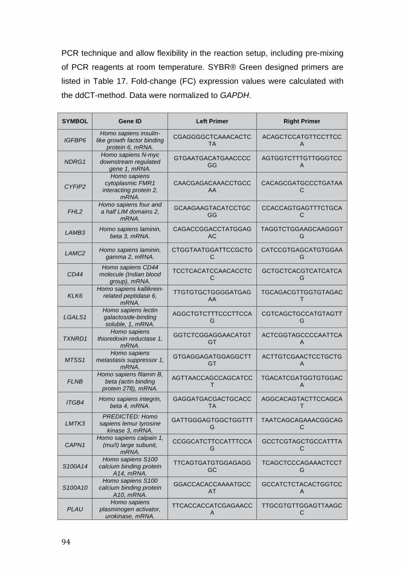
PCR technique and allow flexibility in the reaction setup, including pre-mixing
of PCR reagents at room temperature. SYBR® Green designed primers are
listed in Table 17. Fold-change (FC) expression values were calculated with
the ddCT-method. Data were normalized to GAPDH.
SYMBOL Gene ID Left Primer Right Primer
IGFBP6 Homo sapiens insulin-
like growth factor binding protein 6, mRNA.
CGAGGGGCTCAAACACTCTA
ACAGCTCCATGTTCCTTCCA
NDRG1 Homo sapiens N-myc downstream regulated
gene 1, mRNA.
GTGAATGACATGAACCCCGG
AGTGGTCTTTGTTGGGTCCA
CYFIP2
Homo sapiens cytoplasmic FMR1
interacting protein 2, mRNA.
CAACGAGACAAACCTGCCAA
CACAGCGATGCCCTGATAAC
FHL2 Homo sapiens four and a half LIM domains 2,
mRNA.
GCAAGAAGTACATCCTGCGG
CCACCAGTGAGTTTCTGCAC
LAMB3 Homo sapiens laminin, beta 3, mRNA.
CAGACCGGACCTATGGAGAC
TAGGTCTGGAAGCAAGGGTG
LAMC2 Homo sapiens laminin, gamma 2, mRNA.
CTGGTAATGGATTCCGCTGC
CATCCGTGAGCATGTGGAAG
CD44 Homo sapiens CD44
molecule (Indian blood group), mRNA.
TCCTCACATCCAACACCTCC
GCTGCTCACGTCATCATCAG
KLK6 Homo sapiens kallikrein-
related peptidase 6, mRNA.
TTGTGTGCTGGGGATGAGAA
TGCAGACGTTGGTGTAGACT
LGALS1 Homo sapiens lectin galactoside-binding soluble, 1, mRNA.
AGGCTGTCTTTCCCTTCCAG
CGTCAGCTGCCATGTAGTTG
TXNRD1 Homo sapiens
thioredoxin reductase 1, mRNA.
GGTCTCGGAGGAACATGTGT
ACTCGGTAGCCCCAATTCAA
MTSS1 Homo sapiens
metastasis suppressor 1, mRNA.
GTGAGGAGATGGAGGCTTGT
ACTTGTCGAACTCCTGCTGA
FLNB Homo sapiens filamin B,
beta (actin binding protein 278), mRNA.
AGTTAACCAGCCAGCATCCT
TGACATCGATGGTGTGGACA
ITGB4 Homo sapiens integrin, beta 4, mRNA.
GAGGATGACGACTGCACCTA
AGGCACAGTACTTCCAGCAT
LMTK3 PREDICTED: Homo
sapiens lemur tyrosine kinase 3, mRNA.
GATTGGGAGTGGCTGGTTTG
TAATCAGCAGAAACGGCAGC
CAPN1 Homo sapiens calpain 1,
(mu/I) large subunit, mRNA.
CCGGCATCTTCCATTTCCAG
GCCTCGTAGCTGCCATTTAC
S100A14 Homo sapiens S100
calcium binding protein A14, mRNA.
TTCAGTGATGTGGAGAGGGC
TCAGCTCCCAGAAACTCCTG
S100A10 Homo sapiens S100
calcium binding protein A10, mRNA.
GGACCACACCAAAATGCCAT
GCCATCTCTACACTGGTCCA
PLAU Homo sapiens
plasminogen activator, urokinase, mRNA.
TTCACCACCATCGAGAACCA
TTGCGTGTTGGAGTTAAGCC

Chapter 3. Materials and methods
PLAUR
Homo sapiens plasminogen activator,
urokinase receptor, mRNA.
ATGCAGTGTAAGACCAACGG
TGATCTTCAAGCCAGTCCGA
ITGB1
Homo sapiens integrin, beta 1 (fibronectin
receptor, beta polypeptide, antigen
CD29 includes MDF2, MSK12), mRNA.
TCTGGGCTTTACGGAGGAAG
TCTACCAACACGCCCTTCAT
EPHA2 Homo sapiens EPH receptor A2, mRNA.
GGCTTCTTTATCCACCGCAG
CGAGGATGTCTTCAGCATGC
AHNAK Homo sapiens AHNAK nucleoprotein, mRNA.
GTCTTTGTGCAGGAGGTGAC
GAACCACTTCAGAGCTGCAG
ALCAM
Homo sapiens activated leukocyte cell adhesion
molecule (ALCAM), mRNA.
TCCTGCCGTCTGCTCTTCT TTCTGAGGTACGTCAAGTCGG
Table 17. Primers designed for SYBR® Green RT-qPCR.
3.6.2 Microarray analysis A DNA microarray (also DNA chip or biochip) consists of a collection of
microscopic DNA spots attached to a solid surface. Each spot contains a
specific DNA sequence, known as a probe or reporter. Gene expression
arrays provide a comprehensive view of gene activity in biological samples.
Common uses of gene expression microarrays include genome-wide
differential expression studies, disease classification, pathway analysis,
expression-based quantitative trait loci mapping, among others.
3.6.2.1 Microarray analysis Illumina HumanHT-12 v4 Triplicates of Hec1A shControl and Hec1A shALCAM1 were used to carry out
this study. The concentration and the quality of isolated RNA were first
measured by Bioanalyzer Assay (Agilent, Santa Clara, CA, USA). Microarray
analysis was performed by the Genome Analysis Platform in the CIC Biogune
(Derio, Bizkaia, Spain). The gene expression profile was studied with the
HumanHT-12 v4 Expression BeadChip (Illumina Inc, San Diego, CA, USA),
which targets 31,325 annotated genes with >47,000 probes. RNA was
subjected to reverse transcription to produce first and second strand cDNA
and biotin-labelled cRNA fragments were generated following the
manufacturer's instructions. The labelled cRNA was hybridized overnight to

the arrays. The beadchips were washed, stained with dye-labelled
streptavidin, and scanned with an Illumina IScan to measure the intensity. The
raw data images were analysed with Illumina Genome Studio software, which
generated the average probe intensity for each sample. Raw data for the
analysis were extracted with Illumina’s GenomeStudio data analysis software,
in the form of GenomeStudio’s Final Report (sample probe profile). Gene
expression data was analysed using the R/Bioconductor statistical computing
environment (www.r-project.org, www.bioconductor.org). Using the lumi
Bioconductor package, raw expression data were background corrected, log2
transformed and normalized. Probes not detected in at least one sample were
excluded for subsequent analyses as they are considered to represent
transcripts that are not expressed.
For the detection of differentially expressed genes, a linear model was fitted to
the corrected, transformed and normalized data and empirical Bayes
moderated t-statistics were calculated using the limma package from
Bioconductor. Adjustment of p-values was done by the determination of false
discovery rates (FDR) using the Benjami-Hochberg procedure. Microarray
data is available on the GEO database (GSE86543).
3.7 Gene ontology analysis
For the gene ontology analysis, deregulated genes from the microarray with
a fold-change >1.2 and an adjusted p-value <0.05 were introduced in the
PANTHER database 309 and in the QIAGEN’S Ingenuity® Pathway Analysis
(IPA® QIAGEN Redwood City, CA, USA) to identify the most significant
canonical routes, biological processes and gene interaction networks in
which they are involved.
3.8 Migration 2D and 3D assays
Cell migration is a key process in the development and maintenance of
multicellular organisms. Tissue formation during embryonic development,
wound healing and immune responses, require the orchestrated movement

Chapter 3. Materials and methods
of cells. Errors during this process have serious consequences, including
tumour formation and metastasis.
3.8.1 Wound healing assay The wound healing assay is a method that allows the study of directional cell
migration in vitro. It mimics cell migration during wound healing in vivo. The
assay consists in making a “wound” in a confluent cell monolayer and then
capturing images at time 0h and at different time points until the wound
closes. The images are used to quantify the migration rate of the different cell
lines.
To perform the wound healing migration assay, we used Hec1A and Ishikawa
shControl and shALCAM cell lines. A total of 5-8×10⁵ cells were seeded into
24-well plates and cultured until confluence. A wound was made by scraping
the cell monolayer with a 10μl pipette tip. Images were captured at 0, 24 and
48 h using an inverted microscope FSX100 (Olympus, Hamburg, Germany) to
measure the wound closure rate.
3.8.2 Aggregate model We used a 3D spheroid model to quantify the spreading of the aggregates
with ALCAM-depletion on a fibronectin-coated pattern (Hec1A shControl and
Hec1A shALCAM). The aggregate model was also used to test the effect on
migration in Hec1A-ETV5 cells after ALCAM-recovery. Moreover, this specific
model allows the study of competition between cell–cell and cell–substratum
adhesion on tissue spreading.
3.8.2.1 Spheroid formation and coated surface preparation Aggregates were obtained from 5 ml of cell suspension in CO2-equilibrated
culture medium at a concentration of 4x10⁵ cells/ml in 25 ml Erlenmeyer
flasks and placed in a gyratory orbital shaker at 75 rpm at 37°C for 22 h. The
flasks were pretreated with 2% dimethylchlorosilane in chloroform to prevent
adhesion of cells to the glass surface.

For the preparation of coated glass substrates: twenty-five mm circular glass
coverslips were sonicated in ethanol for 5 min, dried at ambient temperature,
and exposed to deep UV for 5 min. Fibronectin (Sigma-Aldrich, St Louis, MO,
USA) coating was performed using a 0.1 mg/ml solution of fibronectin in PBS
(pH 7.4) for 1 h. A Mixed coating of fibronectin and PEG-PLL (PLL(20)-g[3.5]-
PEG(2), Surface Solution) was made by mixing at various rates 0.1 mg/ml
fibronectin in PBS and 0.1 mg/ml PEG-PLL in Hepes solution (pH 7.3) for 1 h.
Coverslips were then rinsed with PBS (pH 7.4).
3.8.2.2 Aggregate spreading Cell aggregates were deposited randomly on fibronectin-coated coverslips,
which were placed in a magnetic imaging chamber (Chamlide CMB, CM-
B25-1) filled with CO2-equilibrated culture medium. Spreading was observed
using a NIKON confocal microscope equipped with an x10 air objective and a
37°C heating cube system. Bright field images were recorded with a CCD
camera (Luca-R, Andor) using NIS-Elements software every 15 min for 36-48
h. The area of each aggregate was measured with Image J software at time
0h and at the end of the assay (National Institutes of Health, Bethesda, MA,
USA).
For Hec1A-ETV5 ALCAM-recovery, cell aggregates were deposited on 200
μm striped fibronectin-coated surface. The increased spreading was
measured at different time points with Image J software (National Institutes of
Health, Bethesda), which allowed calculating the speed of migration for each
cell line.
3.9 Invasion
The movement of cancer cells into surrounding tissue and the vasculature is
the first step in the spread of metastatic cancers. Cell invasion is related to,
and encompasses, cell migration, but in addition invasive cells move through
the extracellular matrix into neighbouring tissues in a process that involves
ECM degradation and proteolysis.

Chapter 3. Materials and methods
3.9.1 Transwell invasion assay A total of 5x10⁵ cells were seeded on 8 μm pore size transwell filters of the
CytoSelect 24-well cell invasion assay kit according to manufacturers’
instruction (Cell Biolabs, San Diego, CA, USA), and allowed to invade for 36-
48 h. The upper faces of the inserts were coated with a basement membrane
matrix solution. Only the cells able to degrade the matrix proteins in the layer
and travel through the pores of the polycarbonate membrane are considered
invasive cells. Invading cells were stained with DAPI and Phalloidin and
counted with Image J (National Institutes of Health, Bethesda, MA, USA).
3.10 Cell-cell adhesion
Tumour cells often show aberrant cell-cell and/or cell-matrix adhesion. This
cell-cell adhesion change has been correlated with tumour invasion and
metastasis. Cell adhesion molecules are responsible for cell adhesion and
they can also function as ligand-activated cell surface receptors, activating
signals involved in the regulation of cell shape, migration, proliferation,
differentiation, and survival. These two functions show considerable
interdependence with the regulatory processes exercising feedback control
over cell adhesion, often through inside-out signalling.
3.10.1 Dual-pipette adhesion force assay The dual pipette assay used here, provides an overall quantification in terms
of mechanical force of the adhesive properties of the cells conferred by
ALCAM modulation during the development of adhesion.
To preserve intact the cell surface proteins, cells were dissociated with Cell
dissociation enzyme-free buffer (Gibco, NY, USA), rinsed and then transferred
in working medium (CO2-independent medium, Invitrogen, CA, USA) and
used immediately. Two isolated cells were brought gently into contact and
held for a predetermined period of time (1, 4, 10 and 20 min). The cells were
manipulated in suspension, to eliminate matrix-mediated signalling, by
diminishing the contribution of generalized membrane events, and avoiding

the initiation of intercellular adhesion through lamellipodial and filopodial
activities. Isolated cells were manipulated at 37ºC with two micropipettes,
each held by one micromanipulator connected to a combined
hydraulic/pneumatic system and a pressure sensor making it possible to
control and measure the aspiration applied to the cells. The cell doublet was
cyclically brought back into contact with the left pipette and then withdrawn to
the right, each time a stepwise increase in the strength of aspiration by the left
pipette was applied, until the cell doublet was disrupted. A pressure sensor
measured the aspiration applied to the left pipette. Aspiration was monitored
continuously during the separation process, and the values recorded for each
of the last two cycles in the series (Pn-1 and Pn) were used to calculate the
separation force (SF) for each doublet using the equation: SF = π (d/2)2 (Pn-
1+Pn)/2 where d is the inside diameter of left pipette. SF was considered to
be zero for pairs of cells that did not form adherent doublets in this assay.
Results for 15–40 measurements were used to obtain the mean force of
separation for a specific contact time.
3.11 Cell proliferation
Cell proliferation and differentiation is crucial from embryogenesis to
development of whole organism and also in maintenance of adult tissue
homoeostasis. Abnormal cell proliferation has been also associated with
various human diseases like cancer. Consequently, cell proliferation assays
become critical to examine the rate of cell proliferation in vitro and in vivo.
There exist several assays to determine cell proliferation rate like amount of
deoxyribonucleic acid synthesis, metabolic activity of cells, different antigens
associated with cell proliferation and adenosine triphosphate concentration.
One of the simplest methods uses crystal violet staining.
During cell death, adherent cells detach from cell culture plates. This
characteristic is used for the indirect quantification of cell death and finally to
determine the difference in the proliferation rate between cells. Crystal violet
dye binds to proteins and DNA. Cells that undergo cell death lose their

Chapter 3. Materials and methods
adherence and are subsequently lost from the cell population, decreasing the
amount of crystal violet staining.
3.11.1 Crystal violet A total of 4,000 cells per well were seeded into 96-well plates. Cell viability
was measured after 0, 24, 48, 72, 96 and 120 h. Cells were fixed with 50
μl/well of Formaldehyde 4% and incubated for 20 min. After washing, cells
were stained with 100 μl/well crystal violet 0.5% in H2O (Sigma, St Louis, MO,
USA) and incubated at room temperature for 20 min. Crystal violet was
washed by immersing and shaking plates in water and leaving to dry for 5
min. Then, crystal violet was dissolved in 100 μl/well of 15% acetic acid for 10
min with shaking, and finally the supernatant was read at 595 nm.
3.12 Cell cycle assay for flow cytometry
Cancer is a disease accompanied by uncontrolled cell division. Its
development and progression are usually linked to a series of changes in the
activity of cell cycle regulators. The DNA of mammalian, yeast, plant or
bacterial cells can be stained by a variety of DNA binding dyes. The principle
of these dyes is that they are stoichiometric and as a consequence bind in
proportion to the amount of DNA present in the cell. Cells that are in S phase
will have more DNA than cells in G1. They will take up proportionally higher
quantity of dye and will fluoresce more brightly until they have doubled their
DNA content. The cells in G2 will be approximately twice as bright as cells in
G1.
3.12.1 Propidium iodide DNA staining Propidium iodide (PI) is an intercalating agent and a fluorescent molecule that
can be used to stain cells. After 48 h, a total of 1x10⁶ cells were trypsinized,
resuspended in 300 µl of PBS, and fixed with 700 µl of pre-cooled absolute
EtOH for 2 h on ice. EtOH was slowly added while mixing to avoid
aggregates. Cells were pelleted (5,000 rpm), resuspended in working solution

(940 µl of PBS, 30 µl of solution A and 30 µl of solution B) and incubated for
30 min at room temperature. Finally, cells were analysed by flow cytometry.
Solution A: Sodium Citrate (38 mM) and propidium Iodide (500 µg/ml).
Solution B: RNAse A (10 mg/ml in PBS).
3.13 Mouse model
The use of clinically relevant mouse models, which mimic the tumour growth,
progression, invasion and metastases steps, is an essential requirement to
better understand the endometrial cancer molecular mechanisms. Previously
in the laboratory, we developed an orthotopic murine model that represents a
realistic approach towards the process of dissemination of endometrial
cancer. Tumour cells are directly injected into the uterus of the mice and so
they are localized in the same microenvironment as the original tumour,
replicating the processes of tumour growth and myometrial infiltration under
endometrial stimuli.
3.13.1 Orthotopic murine model of endometrial cancer All the procedures were performed according to the guidelines of the Spanish
Council for Animal Care and the institutional guidelines for animal welfare
(CEEA 23/16).
For objective (i), we tested the ability of Hec1A ALCAM-depleted cells in
tumour growth and dissemination. For this, a total of 24 six-week old female
athymic nude mice (Charles River Laboratories, Inc, Wilmington, MA, USA)
were inoculated by transmyometrial injection of stably transfected Hec1A
shControl and Hec1A shALCAM2 cells (n=12 per group). Mice were
anesthetized with 2% isofluorane (ABBOT Laboratories, Madrid, Spain), and
the lower abdomen was swabbed with Betadine®. A longitudinal incision
(medial laparotomy) was performed and the murine uterus was exposed. EC
cells were inoculated into the uterine body and the uterine horns. A 27G
insulin syringe (Myjector® 1 ml, Terumo, Somerset, NJ, USA) was used for
the injection. The strain on the endometrial cavity and the expulsion of a small

Chapter 3. Materials and methods
quantity of fluid through the vagina ensured the correct localization of the
injection. Mice were sacrificed seven weeks after the injection. Tumour growth
and dissemination were evaluated macroscopically and histologically ex vivo.
For histological analysis, all extracted tissues were formalin-fixed, stained with
haematoxylin and eosin (H&E), and evaluated by a pathologist. Ki67
expression was scored by using the ACIS® III Instrument automated imaging
system (DAKO, Glostrup, Denmark).
For objective (ii), Hec1A control and Hec1A-ETV5 luciferase cells were
injected into the uterus of 5 and 4 mice, respectively and following the
aforementioned protocol. The in vivo model was performed by L. Alonso-
Alconada and M. Abal in the CHUS (Santiago, Spain). Mice were followed
weekly after cell injection and before sacrifice by using IVIS system (Xenogen
Corporation) coupled to Living Imaging software 4.2 (Xenogen Corporation) to
detect tumour growth by bioluminescent imaging. For non-invasive
bioluminescence tumour imaging, luciferin (Firefly Luciferin, Caliper
Lifescience Corp, Hopkinton, MA, USA) was used as the substrate for the
luciferase expressing tumour cells and injected intraperitoneally at a
concentration of 150 mg/kg in PBS. Mice were sacrificed 3 weeks after
injection and before metastases could be observed.
3.14 Statistical analysis
The statistical approaches used for objective (i) are explained as follows:
For categorical variables
The relationship between ALCAM expression and clinicopathological
parameters was tested in univariate analysis by chi-square and Fisher's exact
test, two-sided p-values. Stratified analysis was performed to study the
relation between recurrence and ALCAM status. Follow-up studies were
analysed by Kaplan-Meier and Cox’s proportional hazards test. Recurrence-
free survival was evaluated in the present study, but not overall survival as the
number of deaths from disease did not allow a reliable statistical analysis.

For continuous variables
The normal distribution of data was verified (Shapiro-Wilk or Kolmogorov-
Smirnov) and accordingly, parametric (t-test, ANOVA, and multiple post-hoc
comparisons (Scheffe, Dunnet) and non-parametric (Mann-Whitney) tests
were performed for each comparison.
The statistical approaches used for objective (ii) are explained as follows:
For continuous variables
Univariate regression analyses were performed to evaluate the association
between ALCAM and different epithelial and mesenchymal markers at the
superficial and the invasive front. Variables that were statistically significant in
univariate linear regression analysis were included in the multiple linear
regression models. For each cohort, a stepwise method was used to select
the explanatory variables based on analysis of variance, checking the
normality (Shapiro-Wilk or Kolmogorov-Smirnov) and the independence of
residuals (Durbin-Watson).
Receiver operating characteristic (ROC) curve analysis was conducted on
individual marker soluble ALCAM (sALCAM), detected in uterine aspirates at
diagnosis, to discriminate patients presenting myometrial infiltration. To
explore its relationship with the levels of the different forms of MMP-9 present
in the uterine aspirates, we performed a factor analysis with extraction of
principal component (PCA) for MMP-9 expression and then a regression
analysis with sALCAM levels. The normal distribution of data was verified
(Shapiro-Wilk) and levels were Ln-transformed for linear analysis.
Non-parametric tests (Mann Whitney) were used for comparisons of data sets
whose distributions deviate from normal.
For categorical variables
The relationships between ALCAM expression, at the superficial and the
invasive front, and clinicopathological parameters were tested in univariate
analysis by Chisquare and Fisher’s exact test. Stratified analysis was
performed to study the molecular features ALCAM/MMP-9/ETV5 and the
correlation between ALCAM/MMP-9 and clinicopathological parameters. The

Chapter 3. Materials and methods
odds ratios (OR) were calculated across the strata, with their 95 % confidence
interval (IC 95 %) obtained through bootstrapping methods. In addition,
logistic regression multivariate analysis was done for the clinicopathological
parameters and status biomarkers to determine independent predictors for
myometrial infiltration. Categorical variables are presented as an absolute
number and percentage.
For both objectives, measurements were made in triplicate in three
independent experiments and presented with the Mean±SD (except for the
separation force experiments, which were presented by Mean±SEM). All
statistical analyses were performed using the IBM SPSS Statistics 21. All
two-sided pvalues<0.05 were considered statistically significant.


Chapter 4. Results
The results section is organized into two blocks, where we provide the results
obtained when addressing the two main objectives of this thesis, previously
described in Chapter 2.
4.1 ALCAM as a marker of recurrence and as a promoter of tumour progression in EEC
4.1.1 Framework As stated in Chapter 1, 1.1.4.2 section, abnormal vaginal bleeding is an early
symptom of endometrial cancer and as consequence allows the diagnosis of
around 75% of patients at an early stage of the tumour. Recurrence risk groups
are basically defined based on the assessment of clinical prognostic factors like
age, FIGO staging, grade, and histology, to guide the management of patients.
Despite those early-diagnosed patients are classified and treated according to
their established risk of recurrence, a number of them relapse or even die of the
disease.
For this reason, in order to improve the proper classification of these patients,
the main objective of this section was to evaluate the immunohistochemical

determination of ALCAM in 174 EEC primary tumours as a reliable marker of
recurrence. In addition, we used in vitro and in vivo orthotopic murine models as
well as microarray technology, to unveil the functions regulated by ALCAM to
promote EEC dissemination and metastasis.
4.1.2 ALCAM-positive expression is a marker of recurrence in early stage moderately-poorly differentiated tumours Among the 174 EEC patients analysed for ALCAM expression, we found 131
(76.20%) to be ALCAM-positive (Table 18).
Variable, No. (%) Σ ALCAM Positive* ALCAM Negative† P-value Age 172 131 (76.2) 41 (23.8) 0.589 ≤64 years 76 56 (73.7) 20 (26.3) >64 years 96 75 (78.1) 21 (21.9) FIGO 172 132 (76.7) 40 (23.3) 0.125 Stage IA 85 70 (82.4) 15 (17.6) Stage IB 49 38 (77.6) 11 (22.4) Stage II 25 16 (64.0) 9 (36.0) Stage III 13 8 (61.5) 5 (38.5) Grade 172 130 (75.6) 42 (24.4) 0.049 Grade 1 99 81 (81.8) 18 (18.2) Grade 2 46 29 (63.0) 17 (37.0) Grade 3 27 20 (74.1) 7 (25.9) *ALCAM positive ≥ 30 †ALCAM negative < 30
Table 18. Clinicopathologic parameters according to ALCAM expression (N=174).
Expression of ALCAM was homogeneous across the section, and
predominantly localized at the membrane and the cytoplasm (Figure 26-D). Of
the included cancers, 77.9% were early stage EEC (stage IA and IB according
to the FIGO classification), containing 88 well-differentiated (G1) and 45
moderately-poorly differentiated tumours (G2-G3); whereas 22.1% of remaining
cases were diagnosed at advanced stages.

Chapter 4. Results
Figure 26. ALCAM-positivity is a marker of recurrence. (A) Patients diagnosed with tumours in early stage (rate of recurrence in ALCAM positive tumours is 27.8% vs. ALCAM negative tumours is 7.7%, p=0.039); (B) patients diagnosed with moderately-poorly differentiated tumours (59.2% versus 25%, p=0.007); and (C) patients diagnosed with early stage moderately-poorly differentiated tumours (55.9% versus 9.1%, p=0.012). (D) Representative images of ALCAM expression in a recurrent (left) and non-recurrent (right) early stage moderately-poorly differentiated tumour.
The univariate analysis between ALCAM expression and the most common
prognostic factors only showed a minor correlation with tumour grade when
studied in the whole population (Table 18). Interestingly, in a stratified analysis,
ALCAM expression showed a significant correlation with the recurrence for
patients diagnosed in early stage (N=134) (p=0.039), with moderately-poorly
differentiated tumours (N=73) (p=0.007), and with tumours combining these
two clinical parameters (N=45) (p=0.012) (Table 19). Specifically, in the subset
of patients diagnosed at early stage, the recurrence rate was significantly
higher for ALCAM-positive patients compared to ALCAM-negative patients
(27.8% vs 7.7%). The results were similar for patients with moderately-poorly
differentiated tumours (59.2% vs. 25.0%); and noteworthy, for the early stage
moderately-poorly differentiated cohort in which the percentage of ALCAM-
positivity in recurrent patients increased up to a 55.9% compared to a 9.1% for
ALCAM-negative patients (Figure 26A-C).

Variable, No. (%) Σ No recurrence Recurrence P-value
Staging Early Stage I 134 102 (76.1) 32 (23.9) 0.039
ALCAM Positive* 108 78 (72.2) 30 (27.8)
ALCAM Negative† 26 24 (92.3) 2 (7.7)
Advanced Stages II-III 38 19 (50.0) 19 (50.0) 0.737
ALCAM Positive* 24 11 (45.8) 13 (54.2)
ALCAM Negative† 14 8 (57.1) 6 (42.9)
Grade
Differentiated tumors (G1) 99 84 (84.8) 15 (15.2) 1.00
ALCAM Positive* 81 69 (85.2) 12 (14.8)
ALCAM Negative† 18 15 (83.3) 3 (16.7)
Moderately- Poorly differentiated tumors (G2-3) 73 38 (52.1) 35 (47.9) 0.007
ALCAM Positive* 49 20 (40.8) 29 (59.2)
ALCAM Negative† 24 18 (75.0) 6 (25.0) Early Stage I & Moderately-Poorly differentiated tumors 45 25 (55.6) 20 (44.4) 0.012
ALCAM Positive* 34 15 (44.1) 19 (55.9) ALCAM Negative† 11 10 (90.9) 1 (9.1)
* ALCAM positive ≥ 30 † ALCAM negative < 30
Table 19. ALCAM expression signature of EEC recurrence (N=174).
These results were supported by univariate Cox regression and Kaplan Meier
analyses. Recurrence-free survival was lower in patients with ALCAM-positive
tumours than in patients with ALCAM-negative expression. For early stage
EEC patients, median recurrence-free survival in ALCAM-negative patients
was 111.44 months vs. 95.47 months for ALCAM-positive cancers (p=0.031)
with a hazard ration (HR) of 4.237 (p=0.048) (Figure 27-A). Similarly, for
patients with moderately-poorly differentiated tumours, recurrence-free survival
was 93.243 vs. 64.762 months (p=0.011) with a HR 2.966 (p=0.016) (Figure
27-B). Again, the highest difference was observed in patients with early stage
moderately-poorly differentiated tumours, in which ALCAM reached a HR 9.259
(p=0.034). In this subcohort, recurrence-free survival was significantly longer in

Chapter 4. Results
Figure 27. Univariate survival analyses according to ALCAM expression in 174 EEC patients, (A) early stage, (B) moderately-poorly differentiated, and (C) early stage moderately-poorly differentiated tumours. Differences in survival between ALCAM positive (ALCAM pos) and ALCAM negative (ALCAM neg) groups were assessed by the two-sided log-rank test. The HR (95% CI) for each comparison is given below the graphs.
ALCAM-negative cancers than in patients with ALCAM-positive expression
(110.636 vs. 68.279 months, respectively; p=0.008) (Figure 27-C).
Furthermore, in the early stage patients, ALCAM-positivity was identified as an
independent prognostic factor of recurrence with a HR 6.027 (p=0.015)
together with the tumour grade with a HR 5.634 (p<0.001) in a multivariate Cox
regression analysis (Table 20).

Recurrence-free survival Variable HR 95% Interval confidence P-value ALCAM
Negative 1
Positive 6.027 (1.411-25.746) 0.015
Age ≤64 years 1 >64 years 0.935 (0.450-1.943) 0.857 Grade Differentiated tumours (G1) 1
Moderateley-poorly differentiated tumours (G2-3)
5.634 (2.623-12.101) <0.001
Table 20. Multivariate Cox regression model for patients with early stage tumours (N=134).
4.1.3 Inhibition of ALCAM in EEC cell lines decreased cell migration, invasion and cell-cell adhesion To understand the role of ALCAM in endometrial tumour progression and
dissemination we studied the effect of ALCAM inhibition in the Hec1A EEC
cells. We generated two ALCAM-depleted Hec1A cell lines by using two specific
shRNAs obtaining >70% of inhibition (Figure 28A-B).
Figure 28. Inhibition of ALCAM in Hec1A cell line. (A) Stable downregulation of ALCAM in Hec1A cell line was analysed by western blot and (B) by RT-qPCR, normalized with GAPDH as housekeeping (**p<0.01).

Chapter 4. Results
We used in vitro 2D and 3D models to evaluate the role of ALCAM in EEC
motility and a dual-micropipette assay to determine quantitatively ALCAM
function in cell-cell adhesion. ALCAM-depletion decreased the wound closure
rate of Hec1A cells in a 2D wound healing assay (Figure 29-A). To mimic the
tumour progression in patients, we used a 3D model to quantitatively study the
spreading of cell aggregates with ALCAM-depletion on a fibronectin-coated
surface. Three-dimensional models fill the gap between 2D cell cultures and
animal systems as they mimic characteristics of the in vivo environment 310.
Specifically, this model allows the study of the competition between cell–cell
and cell–substratum adhesion on tissue spreading 311. We found that, in
agreement with 2D studies, ALCAM-depleted cells presented a decreased
motility in the 3D model (Figure 29-B). In addition, ALCAM-depleted cells
showed a significant reduced invasive capability on a matrigel transwell assay
(Figure 29-C).
Finally, we evaluated how ALCAM affects the formation and strength of cell-cell
adhesion. Two isolated cells maintained in suspension, to avoid cell-matrix
interactions, were put into contact to initiate adhesion by using two
micropipettes. After a defined period of contact, we measured the forces
required to separate them. The mean separation force (SF) is used as readout
of the strength of adhesion for a specific contact time. In both control and
ALCAM-depleted cells the SF required to disrupt the cell doublet increased
with the time of contact. However, at all time points, the intercellular cell
adhesion was weaker in the ALCAM-depleted cells (Figure 30). Cell-cell
adhesion and aggregate spheroid assays pointed to ALCAM as an important
player in the overall balance of adhesion, which is crucial in cancer processes.
Those effects were independent of cell proliferation, as no significant
differences were shown in cell viability or progression through the cell cycle
(Figure 31).

Figure 29. ALCAM inhibition decreased migration and invasion in Hec1A cell line. (A) ALCAM inhibition decreased wound closure rate at 24 and 48 h. Up, wound is delimited with a dotted line at 0 h and 48 h for each cell line. Bottom, the gap closure is quantified at 24 and 48 h. (B) The effect of ALCAM silencing in cell motility was assessed by a 3D spheroid-spreading model on fibronectin-coated surface. The spreading area is outlined in the upper image and measured at 48 h in the bar graph. (C) ALCAM depletion decreased the invasive abilities of Hec1A cells in a matrigel transwell assay after 36 h. (For all, **p<0.01, *p<0.05).

Chapter 4. Results
Figure 30. ALCAM inhibition decreased cell-cell adhesion in Hec1A cell line. (A) Two cells were put into contact to form adhesion. (B) After 4, 10 and 20 min ALCAM inhibition decreased the cell-cell adhesion in Hec1A cells as shown by the quantification of the separation force for each time point. (*p<0.05, Mean ± SEM).
Figure 31. ALCAM inhibition had no effect on cell proliferation or progression throughout the cell cycle. (A) A crystal violet assay was performed at 0, 24, 48, 72, 96 and 120 h. ALCAM inhibition had no effect on cell proliferation in Hec1A cell line. (B) The percentages of cells in G1, S and G2 were quantified by using propidium iodide DNA staining and flow citometry. ALCAM inhibition in Hec1A had no effect on cell cycle progression.
The same results were observed with the ALCAM-depleted Ishikawa EEC cell
line (Figure 32). Specifically, Ishikawa shALCAM cells presented lower rate of
wound closure, decreased invasive ability, and no differences in cell
proliferation were observed (Figure 33). Taken together, these results
supported the role of ALCAM in key processes for tumour dissemination,
leading to less aggressive phenotypes when supressed.

Figure 32. Inhibition of ALCAM in Ishikawa cell line. Stable downregulation of ALCAM by two shRNAs in Ishikawa cell line was analysed by (A) western blot and (B) by RT-qPCR and normalized to GAPDH as a reference transcript (**p<0.01).
Figure 33. ALCAM inhibition in Ishikawa cell line decreased migration and invasion. (A) ALCAM inhibition decreased the wound closure rate in a 2D wound healing assay at 24 h and 48 h (24 h *p<0.05; 48 h **p<0.01). (B) ALCAM depletion decreased the invasive abilities of Ishikawa cells in a Matrigel transwell assay after 36 h (**p<0.01). (C) A crystal violet assay was performed at 0, 24, 48, 72, 96 and 120 h. ALCAM inhibition had no effect on cell proliferation in the Ishikawa cell line.

Chapter 4. Results
4.1.4 Depletion of ALCAM reduced primary tumour size and inhibited metastasis in an orthotopic murine model of EEC To assess the effect of ALCAM on tumour development and metastasis we
used a clinically relevant EEC orthotopic murine model. We inoculated stably
transfected control or ALCAM-depleted cells into the uterus of mice and after 7
weeks both conditions developed solid tumours (Figure 34-A). Hec1A shControl
tumours were larger than those generated by shALCAM cells (Figure 34B-C).
However, no differences were observed in the H&E or Ki67 staining (Figure 34-
A; p=0.3), indicating that the dissimilarity in tumour size was not due to cell
proliferation. These results might indicate that ALCAM interferes with the ability
of tumour cells to communicate with the surrounding microenvironment.
Reinforcing this hypothesis, mice with ALCAM-depleted cells developed fewer
metastases and more significantly, fewer local metastases than the control
(Figure 35A-B). Local metastases were differentially found in the peritoneum,
pelvic fat, kidney, spleen, pancreas, colon, liver and diaphragm (Figure 35-C).
Accordingly, we observed that a larger proportion of control mice presented
macro metastases (>5 mm) (Figure 35D-E). Taken together, we evidenced that
ALCAM-depletion profoundly affected the ability of tumour cells in developing
metastasis.

Figure 34. ALCAM-depletion decreased primary tumour size in an orthotopic mice model of EEC. (A) Up, representative H&E sections of the mice uterus, both cell lines produced solid primary tumours (T arrow) within the normal mice uterus (N arrow). Below, Ki67 representative images of the primary tumours showed no differences in cell proliferation. (B) Image of 6 uteri containing representative primary tumours in Hec1A shControl and shALCAM (the 3 largest and smallest tumours for each cell line). (C) ALCAM-depletion inhibited significantly the size of the primary tumour (p<0.05).

Chapter 4. Results
Figure 35. ALCAM-depletion reduced metastasis in an orthotopic mice model of EEC. (A) ALCAM-depletion reduced the total number of metastases per mouse and (B) reduced significantly the number of metastases produced by local invasion (p<0.05). (C) Graph representing the percentage of mice affected by metastases in the shControl and the shALCAM groups per organ. (D) The number of metastases >5 mm was significantly higher in the control group (p<0.05). (E) Representative images of colon and kidney metastases in Hec1A shControl and shALCAM mice (tumours are indicated by arrows).

4.1.5 Microarray analysis of ALCAM-dependent gene expression in Hec1A cell line To elucidate the molecular mechanisms governed by ALCAM to promote EC
dissemination and metastasis, we performed a cDNA microarray to compare
the gene expression profile of Hec1A shControl and shALCAM cells. A total of
315 genes were found to be deregulated with FC >1.2 and adjusted p-value
<0.05. Among those, a heatmap of the 50 most deregulated genes is
represented in Figure 36.
All the differentially expressed genes were introduced in IPA and PANTHER
databases for gene ontology analysis. We found that the deregulated genes
were related to commonly altered pathways in cancer (Figure 37-A) and among
those, the integrin signalling pathway was the most significantly altered (Figure
37-B). The IPA analysis revealed that the most enriched functions
corresponded to cell movement, cell migration and cell invasion. We also
observed that ALCAM inhibition had an effect in cellular assembly and
organization through the regulation of genes involved in formation of cellular
protrusions, microtubule dynamics, organization of cytoplasm and cytoskeleton
among others; as well as functions related to cell-to-cell adhesion (Figure 37-
C). All of them presented a negative z-score and thus are predicted to be
inhibited in ALCAM-depleted cells, in agreement with our previous
observations.

Chapter 4. Results
Figure 36. Gene expression analysis of ALCAM-depleted cell lines. Heatmap of the most significant deregulated genes of microarray data for three replicates of Hec1A shALCAM and Hec1A shControl cell lines.

Figure 37. Gene ontology analyses of deregulated ALCAM-depleted cell lines. (A) Signalling pathways of deregulated genes in ALCAM-depleted cells by Panther analysis. The legend describes the most important affected pathways. (B) The integrin signalling pathway was the most significant represented pathway (p<0.05) and the specific genes are represented in the graph and described in the legend. (C) Functional categories of deregulated genes in ALCAM inhibited cells by Ingenuity pathway analysis. The activation z-score was generated by the software using the information about the direction of gene regulation. The representation of the number of genes deregulated for each functional category evidenced enrichment in cell motility and invasion.

Chapter 4. Results
4.1.6 ALCAM regulates LAMC2, FLNB and TXNRD1 during EEC dissemination To characterize the downstream effectors of ALCAM-mediated functions, we
selected from the microarray data the most deregulated genes (FC >1.4,
adjusted p-value <0.05) related to cell motility, cell movement and cell invasion
functions. Additionally, genes from the plasminogen activating cascade, which
participated in the same functions, were included (Table 21). The levels of 21
genes were validated by RT-qPCR, using both knockdown and Hec1A-ALCAM
overexpressing cells. Among those, six genes -LAMC2, TXNRD1, FLNB,
EPHA2, PLAU and KLK6-, presented a coherent behaviour in ALCAM-depleted
and ALCAM-overexpressing cells (Figure 38A-B). LAMC2, TXNRD1 and FLNB
also showed decreased protein levels in ALCAM-depleted cells (Figure 39A-B).
This finding supported their role as downstream effectors of the ALCAM-related
functions. The genes that were not fully consistent by RT-qPCR are presented
in Figure 40A-B.
SYMBOL Gene ID Acc_No Fold-Change
Adj. P-Value
CD24 Homo sapiens CD24 molecule, mRNA. NM_013230.2 2.191 8.37E-08
IGFBP6 Homo sapiens insulin-like growth factor binding protein 6, mRNA.
NM_002178.2 -1.7949 1.70E-05
NDRG1 Homo sapiens N-myc downstream regulated gene 1, mRNA.
NM_006096.2 -1.6342 5.35E-05
CYFIP2 Homo sapiens cytoplasmic FMR1 interacting protein 2, mRNA.
NM_014376.2 1.4927 1.54E-04
FHL2 Homo sapiens four and a half LIM domains 2, mRNA. NM_201555.1 1.4673 2.76E-04
LAMB3 Homo sapiens laminin, beta 3, mRNA. NM_000228.2 -1.4721 3.25E-04
LAMC2 Homo sapiens laminin, gamma 2, mRNA. NM_005562.1 -1.4504 4.42E-04
CD44 Homo sapiens CD44 molecule (Indian blood group), mRNA. NM_001001391.1 1.506 4.42E-04
KLK6 Homo sapiens kallikrein-related peptidase 6, mRNA. NM_001012964.1 1.4234 4.53E-04
LGALS1 Homo sapiens lectin, galactoside-binding, soluble, 1, mRNA.
NM_002305.3 -1.4032 5.26E-04

TXNRD1 Homo sapiens thioredoxin reductase 1, mRNA. NM_001093771.1 -1.4451 6.48E-04
MTSS1 Homo sapiens metastasis suppressor 1, mRNA. NM_014751.2 1.397 6.83E-04
FLNB Homo sapiens filamin B, beta (actin binding protein 278), mRNA.
NM_001457.1 -1.4373 7.14E-04
ITGB4 Homo sapiens integrin, beta 4, mRNA. NM_001005619.1 -1.4425 1.92E-03
LMTK3 PREDICTED: Homo sapiens lemur tyrosine kinase 3, mRNA.
XM_936372.2 -1.4319 1.92E-03
CAPN1 Homo sapiens calpain 1, (mu/I) large subunit, mRNA. NM_005186.2 -1.4049 1.92E-03
S100A14 Homo sapiens S100 calcium binding protein A14, mRNA. NM_020672.1 -1.4442 2.39E-03
S100A10 Homo sapiens S100 calcium binding protein A10, mRNA. NM_002966.2 -1.4119 5.39E-03
PLAU Homo sapiens plasminogen activator, urokinase, mRNA. NM_002658.2 -1.3676 7.47E-04
PLAUR Homo sapiens plasminogen activator, urokinase receptor, mRNA.
NM_001005376.1 -1.245 1.80E-02
ITGB1
Homo sapiens integrin, beta 1 (fibronectin receptor, beta polypeptide, antigen CD29 includes MDF2, MSK12), mRNA.
NM_033668.1 -1.2938 2.35E-03
EPHA2 Homo sapiens EPH receptor A2, mRNA. NM_004431.2 -1.2124 3.89E-02
AHNAK Homo sapiens AHNAK nucleoprotein, mRNA. NM_001620.1 -1.3103 1.92E-03
Table 21. List of the selected genes deregulated in the gene expression analysis of Hec1A shALCAM cells relative to Hec1A shControl.

Chapter 4. Results
Figure 38. LAMC2, TXNRD1 and FLNB were decreased at mRNA level in ALCAM-depleted cells. The expressions of PLAU, FLNB, EPHA2, LAMC2, TXNRD1 and KLK6 were analysed by RT-qPCR in Hec1A cells with (A) ALCAM-depletion (B) and overexpression versus control (*p<0.01).
Figure 39. LAMC2, TXNRD1 and FLNB were decreased at protein level in ALCAM-depleted cells. (A) Western blot analysis confirmed the down-regulation at protein level of LAMC2, TXNRD1 and FLNB in ALCAM inhibited cells. (B) Western blot was quantified using Image J software.


Chapter 4. Results
Figure 40. RT-qPCR performed on deregulated genes from the microarray study. Genes with FC >1.4 (except for PLAUR, FC >1.2) and adjusted p-value<0.05, all associated with the functional category of cell migration and invasion. (A, B) Genes included in this figure are not fully consistent to ALCAM expression, as they either present contradictory results or are not validated in both ALCAM-overexpressing and depleted cells (*p<0.01).

4.2 Characterization of the molecular mechanisms associated with ALCAM in EEC and its relation to myometrial invasion
4.2.1 Framework The first step of myometrial invasion is characterized by the dissociation of
tumour cells from the epithelial layer of the endometrial glands, and their
infiltration through the basement membrane into the adjacent layer, the
myometrium 312. For that to happen, several authors suggested that epithelial
tumour cells undergo an EMT, either transiently or stably. Specifically, it is
known that in invading tumours this process could take place at the invasive
front of the tumour 169,313,314. As explained in Chapter 1 section 1.1.10.2,
multiple factors have been described as being responsible for producing EMT in
EC, such as ETV5, oestrogen and progesterone receptors, and TGF-β, among
others 312.
ALCAM expression in the cell surface can be regulated by endocytosis or
protein cleavage by ADAM 17 metalloproteinase 268,315,270, generating a soluble
96kDa and a truncated membrane-bound. Interestingly, in addition to its clear
association with the tumorigenesis of many cancers in tissues 289,290,296,299,302,303,316,317, ALCAM shedding has also been reported in the serum
of breast, ovarian, thyroid and pancreatic cancer patients and in all cases has
been related to poor clinical outcome 303,305,318,319.
We have previously demonstrated that ALCAM is an important player in EEC
dissemination 320. For this reason, in this results section, we go further in our
understanding of ALCAM in EEC tumorigenesis by evaluating its full-form
expression (non-cleaved protein) in different areas of the tumour as well as its
relation with key molecules associated to the epithelial (E-cadherin, β-catenin)
and mesenchymal (ETV5, COX-2, MMP-2 and -9) context in EC. Moreover, we
evaluated the levels of shed ALCAM (sALCAM) in uterine aspirates as a
potential prognostic tool to predict myometrial invasion at the time of diagnosis.

Chapter 4. Results
4.2.2 ALCAM partners differed in the superficial compared to the invasive area of the tumour We studied the immunohistochemical expression of ALCAM extracellular
domain (non-cleaved protein) and a representative set of epithelial (E-
cadherin/β-catenin adhesion complex) and mesenchymal molecules involved in
EEC dissemination (ETV5, COX-2, MMP-2 and -9), in both the superficial and
the invasive areas of the tumour. As our previous results indicated the
differential behaviour of ALCAM regarding tumour differentiation, analyses were
performed in the whole population and in the well-differentiated (G1) and poorly-
differentiated (G2-3) subcohorts 320 .
Univariate regression analyses were performed as a first approach to evaluate
the association between ALCAM and epithelial or mesenchymal markers.
Variables significantly correlated with ALCAM (Table 22) were included in
multivariate linear regression models.
Multivariate linear regression analyses evidenced a significantly positive
correlation between ALCAM and the adhesion complex only at the superficial
area of the tumour. Whilst β-catenin was retained in the whole population and
the well-differentiated cohort (R=0.416 and 0.391, respectively), E-cadherin was
retained in the moderately-poorly differentiated tumours (R=0.445) (Table 23,
Figure 41). By contrast, at the invasive front of the tumour, the model performed
in all patients retained COX-2 and MMP-9. Specifically, COX-2 is presented as
a predictor that correlated positively with ALCAM, whilst MMP-9 was negatively
correlated. However, the best regression adjustment was reached in the
moderately-poorly differentiated subcohort of patients, in which the model
retained ETV5 and MMP-9 with a R=0.559. Both molecules showed a negative
correlation with ALCAM (Table 24, Figure 41). Altogether, the results suggest a
different functional profile of the molecule that correlated strongly with the
epithelial markers at the superficial tumour; and by contrast, evidenced a
correlation with mesenchymal markers at the invasive front.

All cases Grade 1 Grade 2-3
β p-value β p-value β p-value
Superficial area of the tumour
β-catenin 0.419 < 0.001*** 0.394 < 0.001*** 0.410 0.013*
E-cadherin 0.346 < 0.001*** 0.275 0.014* 0.462 0.004**
ETV5 0.026 0.791 0.033 0.780 0.027 0.878
COX-2 0.109 0.248 0.153 0.180 0.013 0.938
MMP-9 -0.176 0.060 -0.014 0.906 -0.395 0.015*
MMP-2 0.247 0.008** 0.251 0.026* 0.246 0.143
Invasive front of the tumour
β-catenin 0.144 0.174 0.181 0.157 0.017 0.933
E-cadherin 0.160 0.125 0.173 0.182 0.111 0.546
ETV5 -0.034 0.748 0.118 0.365 -0.408 0.025*
COX-2 0.210 0.038* 0.221 0.073 0.188 0.310
MMP-9 -0.242 0.018* -0.137 0.280 -0.438 0.014*
MMP-2 0.035 0.723 0.029 0.808 - -
Table 22. Patterns of ALCAM in the tumour: univariate linear regression analysis.
Figure 41. ALCAM patterns at the tumour. Upper: full-length ALCAM was positively correlated positively with the E-cadherin/β-catenin complex at the superficial area of the tumour and its mainly localized at the cell membrane. Down: At the invasive front, full-length ALCAM was significantly inversely correlated with MMP-9 and ETV5, in the moderately-poorly differentiated EEC.

Chapter 4. Results
Superficial area of the tumour
Summary model Coefficients
R R2 adj. p-value B CI95% β p-value
All cases 0.416 0.166 <0.001***
Predictors
β-catenin 1.690 (0.990; 2.391) 0.416 < 0.001***
E-cadherin 0.172 0.081
MMP-9 -0.148 0.090
MMP-2 0.120 0.194
Grade Well-differentiated 0.391 0.141 0.001**
Predictors
β-catenin 1.585 (0.715; 2.455) 0.391 0.001**
E-cadherin 0.092 0.460
MMP-9 -0.079 0.470
MMP-2 0.129 0.261 Moderately-poorly differentiated
0.445 0.174 0.007**
Predictors
E-cadherin 0.673 (0.201; 1.146) 0.445 0.007**
β-catenin 0.272 0.109
MMP-9 -0.264 0.108 Table 23. ALCAM patterns at the superficial area of the tumour: multivariate linear regression analysis.

Invasive front of the tumour
Summary model Coefficients
R R2 adj. p-value B CI95% β p-value
All cases 0.382 0.126 0.001**
Predictors
COX-2 0.630 (0.202; 1.058) 0.296 0.004**
MMP-9 -1.493 (-2.607; -0.379) -0.270 0.009**
ETV5 -0.101 0.363
Grade
Well-differentiated 0.344 0.102 0.008**
Predictors
COX-2 0.754 (0.203; 1.305) 0.344 0.008**
ETV5 0.008 0.952
MMP-9 -0.215 0.091 Moderately-poorly differentiated
0.559 0.259 0.008**
Predictors
ETV5 -1.175 (-2.283; -0.067) -0.363 0.039*
MMP-9 -1.987 (-3.917; -0.057) -0.353 0.044*
COX-2 0.279 0.120
Table 24. ALCAM patterns at the invasive front of the tumour: multivariate linear regression analysis.
4.2.3 ALCAM-negativity at the invasive front of the tumour is a marker of myometrial invasion In order to further investigate in the role of ALCAM at the invasive front, as well
as in its relationship with ETV5 and MMP-9, categorized analyses were
performed. In the superficial area, uncleaved ALCAM expression was unrelated
to the clinical parameters of myometrial invasion, tumour grade, and tumour
progression. Interestingly, it acts as a marker of tumour progression and
invasion when analysed at the invasive front. In that case, ALCAM was shown
to significantly decrease throughout the tumour stages and in tumours with
myometrial invasion >50% (Table 25, Figure 42A-B). In fact, the potential of
ALCAM-negativity as an independent prognostic factor of myometrial invasion

Chapter 4. Results
was confirmed by multivariate logistic regression analysis with an OR 3.273,
together with the tumour grade (OR 3.484) (Table 26).
Variable, No. (%) Σ ALCAM Positive*
ALCAM Negative† P-value
Superficial area of the tumour FIGO 116 82 (70.7) 34 (29.3) 0.208
IA 56 42 (75.0) 14 (25.0)
IB 41 28 (68.3) 13 (31.7)
II 15 11 (73.3) 4 (26.7)
III 4 1 (25.0) 3 (75.0)
Grade 116 82 (70.7) 34 (29.3) 0.131
Grade 1 79 60 (75.9) 19 (24.1)
Grade 2 24 13 (54.2) 11 (45.8)
Grade 3 13 9 (69.2) 4 (30.8)
Myometrial infiltration 116 82 (70.7) 34 (29.3) 0.541 < 50% 58 43 (74.1) 15 (25.9)
≥ 50% 58 39 (67.2) 19 (32.8)
Invasive front of the tumour
FIGO 106 71 (67.0) 35 (33.0) 0.004**
IA 53 42 (79.2) 11 (20.8)
IB 36 21 (58.3) 15 (41.7)
II 13 8 (61.5) 5 (38.5)
III 4 0 (0.0) 4 (100.0)
Grade 106 71 (67.0) 35 (33.0) 0.213
Grade 1 72 52 (72.2) 20 (27.8)
Grade 2 22 13 (59.1) 9 (40.9)
Grade 3 12 6 (50.0) 6 (50.0)
Myometrial infiltration 106 71 (67.0) 35 (33.0) 0.014* < 50% 55 43 (78.2) 12 (21.8)
≥ 50% 51 28 (54.9) 23 (45.1)
* Positive ≥ 65
† Negative < 65
Table 25. ALCAM expression and clinical parameters.

Figure 42. ALCAM expression was decreased at the invasive front of patients with myometrial invasion. (A) ALCAM-negativity, at the invasive front, is a marker of myometrial invasion. (B) At the invasive front, ALCAM-negative tumours increased from a 21.8% in patients without myometrial invasion to a 45.1% in patients presenting myometrial infiltration (p= 0.014).
Variable OR 95% Interval confidence p-value
Grade
Differentiated tumours (Grade 1)
1
Moderateley-poorly differentiated tumours (Grade 2-3)
3.484 (1.284-9.524) 0.014*
ALCAM
Positive 1
Negative 3.273 (1.200-8.932) 0.021*
MMP-9 Positive 1 Negative 1.629 (0.602-4.409) 0.337 ETV5 Positive 1 Negative 0.556 (0.194-1.592) 0.274
Table 26. Multivariate logistic regression model, at the invasive front, related to myometrial invasion >50% (N=89).

Chapter 4. Results
ALCAM maintained a close and inverse relation with MMP-9 in the invasive
front of the tumours (p=0.004; OR 0.26) (Table 27).
This relation was only maintained in poor prognosis tumours when the cohort
was divided according to clinical parameters, i.e. either in tumours with
moderately-poorly differentiated EEC histology (p=0.02; OR 0.08) or in patients
presenting >50% of myometrial infiltration (p=0.016; OR 0.19). Interestingly,
when we studied this relationship in terms of ETV5-expression, we observed
that ALCAM and MMP-9 correlation was only significant in the ETV5-positive
tumours (p=0.009). Altogether, our results demonstrated that ALCAM-negativity
is a marker for myometrial invasion. Moreover, its close and inverse relation
with MMP-9 led us speculate that ALCAM-negativity might be due to an
increased shedding of the protein by the metalloproteinase at the invasive front
of poor prognostic tumours. This dialog could be orchestrated by the
mesenchymal transcription factor ETV5, whose up-regulation has been strongly
associated with the myometrial infiltration event.
4.2.4 ALCAM shedding in uterine aspirates is a marker of myometrial invasion and is closely related to MMP-9 expression In order to evaluate the possible association between ALCAM shedding and
myometrial invasion, we analysed the expression of soluble ALCAM (sALCAM)
in 40 uterine aspirates (UA) from moderately-poorly differentiated EEC patients
presenting different grades of myometrial infiltration. The UA is a body fluid that
is collected from inside of the uterine cavity, and is formed mainly by the
secretion of the tumour cells and other cells from the endometrium, enabling the
detection of cleaved ALCAM by ELISA. We confirmed that sALCAM was
significantly increased in patients presenting myometrial invasion >50% (Figure
43-A). Moreover, ROC analysis showed that sALCAM in UAs is a significant
predictor of myometrial infiltration (AUC 0.80; p=0.001) (Figure 43-B). In fact,
when ALCAM is set at a cut-off of 9.375 ng/mg, the model presents a sensitivity
of 87% and a specificity of 70.6%.

Variable, No. (%) Σ ALCAM Positive* ALCAM Negative† P-value OR (Positive/ Negative)
All 95 64 (67.4) 31 (32.6) 0.004** 0.26 (0.10-0.66)
MMP-9 Positive 47 25 (53.2) 22 (46.8) MMP-9 Negative 48 39 (81.3) 9 (18.8)
Clinical parameters Grade Differentiated tumours 64 46 (71.9) 18 (28.1) 0.162 N.S
MMP-9 Positive 26 16 (61.5) 10 (38.5) MMP-9 Negative 38 30 (78.9) 8 (21.1)
Moderately- Poorly differentiated tumours
31 18 (58.1) 13 (41.9) 0.020* 0.08 (0.03-0.63)
MMP-9 Positive 21 9 (42.9) 12 (57.1)
MMP-9 Negative 10 9 (90.0) 1 (10.00)
Myometrial infiltration <50% 53 41 (77.4) 12 (22.6) 0.190 N.S
MMP-9 Positive 25 17 (68.0) 8 (32.0)
MMP-9 Negative 28 24 (85.7) 4 (14.3) Myometrial infiltration ≥50% 42 23 (54.8) 19 (45.2) 0.016* 0.19 (0.03-0.65)
MMP-9 Positive 22 8 (36.4) 14 (63.6)
MMP-9 Negative 20 15 (75.0) 5 (25.0)
Molecular features
ETV5 Positive 64 40 (62.5) 24 (37.5) 0.009** 0.22 (0.07-0.69)
MMP-9 Positive 37 18 (48.6) 19 (51.4) MMP-9 Negative 27 22 (81.5) 5 (18.5) ETV5 Negative 25 19 (76.0) 6 (24.0) 1.000 N.S MMP-9 Positive 7 5 (71.4) 2 (28.6) MMP-9 Negative 18 14 (77.8) 4 (22.2)
* Positive ≥ 65
† Negative < 65
Table 27. ALCAM and MMP-9 at the invasive front of the tumour.

Chapter 4. Results
Figure 43. Soluble ALCAM detected in uterine aspirates is a marker of myometrial invasion. (A) Soluble ALCAM detected by ELISA in uterine aspirates was significantly increased in patients presenting myometrial invasion (***p<0.001). ALCAM values were normalized to total protein amount from the uterine aspirates. (B) ROC curve of sALCAM individual marker demonstrated its prognostic value to discriminate patients with myometrial invasion (AUC=0.800; p=0.001).
Then, the relation between sALCAM and MMP-9 was assessed in UA by
performing a gelatine zymography of the same patients (Figure 44-A).
Univariate linear regression analyses (Table 28) evidenced a significant positive
correlation between sALCAM with all MMP-9 detected forms, with the exception
of the more labile active form. No correlation was found with the MMP-2 forms
(data not shown). In order to avoid the problems derived from the simultaneous
use of several measurements that reflect expressions of molecular forms of the
same enzyme (Table 29), PCA was addressed to explore the close relationship

among the four MMP-9 forms and reduce their number before performing
regression analysis. Two main components C1, which explains 75.18% of the
variability of the original dataset, and C2 which only explains 13.86%, were
extracted (Figure 44-B). Regression analyses showed a strong and positive
correlation between sALCAM and C1 at the global level and specifically in
patients presenting myometrial invasion >50% (R=0.575; p=0.004) (Table 28,
Figure 44C-D). These results suggested that at the invasive front of the tumour
ALCAM extracellular shedding by MMP-9 could be an important player in
myometrial invasion.
Univariate linear regression
All cases Myometrial infiltration <50%
Myometrial infiltration >50%
R p-value R p-value R p-value MMP-9 Dimeric 0.355 0.025* 0.298 0.246 0.484 0.019* MMP-9 NGAL 0.407 0.009** 0.313 0.221 0.446 0.033* MMP-9 Latent 0.385 0.014* 0.282 0.272 0.326 0.129 MMP-9 Active 0.100 0.541 0.197 0.449 0.095 0.666
C1 Regression Factor Score 0.447 0.004** 0.322 0.207 0.575 0.004**
Table 28. ALCAM correlation with MMP-9 in uterine aspirates.
MMP-9 forms (*) MMP-9 Dimeric MMP-9 NGAL MMP-9 Latent MMP-9 Active
MMP-9 Dimeric 0.815*** 0.826*** 0.552*** MMP-9 NGAL 0,815*** 0,699*** 0,545*** MMP-9 Latent 0.826*** 0.699*** 0.543*** MMP-9 Active 0.552*** 0.545*** 0,543***
(*ln-transformed)
Table 29. Matrix correlation MMP-9 forms.

Chapter 4. Results
Figure 44. Soluble ALCAM and MMP-9 expressions were correlated in uterine aspirates. (A) Two different examples of gelatine zymography from uterine aspirates and detection of the different forms of MMP-9 and MMP-2. (B) Principal component analysis from the MMP-9 detected molecular forms evidenced two principal components: C1 (75.18% of the variability) and C2 (13.86% of the variability). The factor loadings, correlation between the original variables and the main components of each form can be observed in the graph. The KMO obtained was 0.80 and the t Barlett <0.001. (C) The principal component C1 and the sALCAM were significantly correlated in all patients (R=0.447; p=0.004). (D) The correlation between the C1 component and sALCAM was significant in patients with myometrial invasion >50% (R=0.575; p=0.004).

4.2.5 ALCAM and MMP-9 are important actors at the invasive front of an in vivo model of EEC dissemination The ETV5 overexpression in Hec1A cell line has been extensively used as a
model that mimics the step of tumour invasion in EEC 190,191,321, as ETV5
overexpression is known to induce EMT in EEC. Furthermore, the parental
Hec1A cell line is representative of moderately-poorly differentiated EEC
superficial tumours. In order to study ALCAM in a controlled environment, which
represents a realistic approach towards the process of dissemination of EC, we
generated orthotopic murine models by injecting Hec1A (n=5) or its ETV5 stable
overexpressing cells (n=4) directly in the mice uterus. In these models, tumour
cells are localized in the same microenvironment of the original tumour,
resembling the processes of tumour growth and myometrial infiltration under
endometrial stimuli.
As expected, the tumours generated by the ETV5-overexpressing cells
presented larger myometrial invasion and tumour burden as seen in the H&E
and in the quantified luminescence from the IVIS images (Figure 45, Figure 47).
While in the ETV5-overexpressing tumours, we observed disseminated cells in
finger-like strands or single-cells projecting into the stroma at the invasive front;
in the Hec1A tumours, cells invade forming clusters from the primary tumour
(see arrows, Figure 45). The immunohistochemical staining of uncleaved
ALCAM, MMP-9 and ETV5 in the mice’s primary tumours unveiled that ALCAM
expression is reduced in the invasive front of ETV5-overexpressing compared
to Hec1A tumours (p<0.05; Figure 46), and moreover, the pattern of expression
is modified from a very membranous staining in Hec1A tumours to a diffuse
cytoplasmic staining in ETV5 overexpressing cells (Figure 45, 48). This could
be promoted by the shedding of ALCAM in ETV5 overexpressing tumours.
In relation to this, we observed that the intensity of ALCAM and MMP-9 markers
showed no variation between the superficial or the invasive area in the Hec1A
tumours. But interestingly, their intensity was graded in the ETV5
overexpressing tumours. In those, MMP-9 increased was concomitant with a

Chapter 4. Results
decrease in ALCAM, when comparing the superficial to the invasive front of the
tumour (Figure 46-B, Figure 47).
Figure 45. ALCAM was decreased at the invasive front of the primary tumours in a controlled model of EEC dissemination. Upper: Immunohistochemistry of ETV5, ALCAM and MMP-9 in Hec1A control mice. Both ALCAM and MMP-9 presented a homogeneous staining between the superficial and invasive front or disseminated cells. Black arrow signals a cluster of disseminated cells released from the primary tumour. Down: Immunohistochemistry of ETV5, ALCAM and MMP-9 in Hec1A-ETV5 mice. ALCAM expression was decreased at the invasive front of the tumour, concomitant with an increase of MMP-9 expression. Black arrows evidenced disseminated cells in finger-strand or individual cells, released from the ETV5-overexpressed primary tumour.

Figure 46. ALCAM staining was significantly decreased at the invasive front of the primary tumours in a controlled model of EEC dissemination. (A) Representation of the intensity of staining of the two molecules. ALCAM expression was only significantly decreased at the invasive front of the ETV5-overexpressing mice, compared to control (*p<0.05). (B) Relative intensity of the two markers at the invasive front compared to the superficial tumour. While in the control mice, ALCAM and MMP-9 where homogeneous across the section, in the ETV5-overexpressing mice we observed a decrease in ALCAM expression concomitant with an increase in MMP-9 expression in the invasive front of the primary tumours.
Figure 47. Hec1A and Hec1A-ETV5 orthotopic murine models followed by IVIS. (A) IVIS images of the injected mice evidenced a larger tumour growth in the ETV5-overexpressing cells. (B) Quantification of the luminescence of the primary tumours ex vivo evidenced a higher intensity in the ETV5-overexpressing mice (p=0.032, one-sided test).

Chapter 4. Results
Figure 48. The expression of the full ALCAM protein was decreased at the invasive front of Hec1A-ETV5 cells. Upper: Images of ALCAM expression in Hec1A control mice, presenting less invasion and scattered clusters of cells. Down: Images of ALCAM in Hec1A-ETV5 mice, with higher rate of invasion and disseminated cells, with a finger-strand pattern. ALCAM staining was more homogeneous in the control model with lower invasion and more collective invasion.
As a result of the in vivo model, we evidenced that the cell-cell contacts of the
Hec1A invading cells seem to be preserved, as shown by the highly collective
migration and a homogeneous ALCAM expression. However, the ETV5-
overexpressing invading cells were more prone to present switching between
thin cords and single-cells, both presented decreased or more transient
contacts and higher rate of cleaved ALCAM expression. Moreover, we finally
confirmed that in an invasive scenario, ALCAM and MMP-9 are important actors
at the invasive front of the tumour. Whilst ALCAM diminishes its expression as
an uncleaved protein, MMP-9 increases its expression, probably promoting
ALCAM shedding.
4.2.6 Full ALCAM recovery impaired migration and decreased cell-cell adhesion of invasive EEC cells In order to understand the role of ALCAM in the invasive Hec1A-ETV5 model,
we constructed two ALCAM overexpression vectors: a full-length ALCAM and
an ALCAM containing only the extracellular and transmembrane region

(ALCAMcytoless) in pmCherry-N1 vectors (Figure 49-A). We transiently
transfected the Hec1A-ETV5 cells with the Cherry empty-vector as a control,
and both ALCAM constructs. As expected, ALCAM overexpression was
localized primarily in the plasma membrane and in the cytoplasm.
(Figure 49-B).
Figure 49. ALCAM recovery in mesenchymal Hec1A-ETV5 cells. (A) Representation of the two vectors constructed to overexpress ALCAM: full-length and ALCAMcytoless inserted in the pmCherry-N1 construct. (B) Images of the Hec1A-ETV5 cell line transfected with the pmCherry vector as a control, and both ALCAM-overexpression constructs.
To evaluate the effects of ALCAM-recovery in ETV5-overexpressing cells, we
used 3D in vitro approaches that closely mimic the in vivo settings. We used a
spheroid model to quantitatively study the spreading of cell aggregates on a
striped fibronectin-coated surface. In addition to reproducing characteristics of
the in vivo environment, as stated in objective (i), this model also allows us to
analyse the effects of the competition between cell–cell and cell–substratum
adhesion on tissue spreading 311. In both conditions (ALCAM full-length and
ALCAMcytoless) the speed was significantly decreased compared to the control
cells (Figure 50-A). However, the larger difference was found in the transfected
cells expressing full-length ALCAM. In fact, we observed that the mean time of

Chapter 4. Results
disaggregation of the Cherry-control spheroids was around 5 h, while ALCAM
full-length and ALCAMcytoless expressing aggregates needed approximately
21 h and 18 h, respectively (Figure 50-B). Moreover, the pattern of spreading in
control cells presented a more spindle-shaped phenotype, with larger presence
of individual cells and protrusions, and highly dynamic transient cell-cell
contacts, migration in a sheet fashion and presentation of chemotactic abilities.
In contrast, full-ALCAM and ALCAMcytoless cells presented an increased
intercellular adhesion phenotype and a more collective pattern of spreading
(Figure 50-B). Consequently, ALCAM recovery clearly impaired the migratory
abilities of ETV5 overexpressing cell lines.
To verify these effects we evaluated how ALCAM-rescue affects the formation
and strength of cell-cell adhesion. We used two micropipettes to put into contact
two isolated cells maintained in suspension, to avoid cell-matrix interactions,
and to initiate adhesion. Single cells were chosen under a fluorescence
microscope to verify the Cherry-transfection. The mean separation force (SF) is
used as a read-out of the strength of adhesion for a precise contact time. In all
the cases, the SF required to disrupt the cell doublet increased with the contact
time. We observed that at 4 min both ALCAM transfected cells presented
significantly larger adhesion than the control line (Figure 51A-B). Cells
expressing full-length ALCAM displayed stronger intercellular adhesion, as
shown by the two-fold increase in their SF compared to control cells, and were
more adhesive than cells expressing ALCAMcytoless. In contrast, at 10 min
only the full-length ALCAM-transfected cells presented significantly stronger
adhesion than the control. Thus, we showed that ALCAM shedding was
important to also reduce ETV5-adhesive properties. In this particular context,
the cytoplasmic tail was an important player in cell-cell adhesion formation or
maturation.

Figure 50. ALCAM overexpression in mesenchymal Hec1A-ETV5 cells decreased cell migration. (A) Effect of ALCAM overexpression on migration was assessed by a 3D spheroid-spreading model on 200 μm fibronectin-coated stripes. On the left, box plot of the speed of migration for each cell line. On the right, representative images at time 0h and at 24h are illustrated and the spreading reached at 24h is outlined in the images. Migration decreased in cells transfected with full-length ALCAM (**p<0.01) and ALCAMcytoless (*p<0.05) compared to control. (B) Images of the 3D spheroid-spreading model are illustrated at different time points. In the control cells, the spheroids were almost disaggregated after 5 h, whilst the mean time for disaggregation for the ALCAMcytoless cells was around 18 h, and more than 21 h for the full-length ALCAM cells.
The molecular pathway by which ETV5 promotes its invasive functions in EEC
has been recently postulated by Alonso-Alconada et al. 322. The authors
described how ETV5 works through the BDNF–TrkB–ERK1/2 axis by inducing
ERK1/2 phosphorylation, leading to the promotion of migration and invasion. In
this work, we observed that ALCAM-overexpression was concomitant with a
decrease in p-ERK levels, without reverting to an epithelial phenotype (Figure
52), but impairing the migratory and adhesive properties promoted by ETV5.
Although overexpression of the extracellular-ALCAM is sufficient to reduce the

Chapter 4. Results
levels of ERK phosphorylation and to decrease spheroid spreading, full-length
ALCAM is necessary to increase cell-cell adhesion with time.
Figure 51. ALCAM recovery in mesenchymal Hec1A-ETV5 cells increased cell-cell adhesion. (A) Two Cherry transiently transfected cells were put into contact and allowed to form adhesion. After 4 and 10 min, we applied an increasing measurable force (nN) up to the disruption of the formed cell doublet. (B) Full-length ALCAM-overexpression increased cell-cell adhesion at all points, as shown by quantification of the separation force (***p<0.001, Mean±SEM). However, ALCAMcytoless only increased cell-cell adhesion at 4 min and showed no differences with control line at 10 min.
Figure 52. ALCAM recovery in mesenchymal Hec1A-ETV5 cells and p-ERK. Western blot analysis of ALCAM-overexpression in Hec1A-ETV5 cells demonstrated a significant decrease in the levels of ERK-phosphorylation.


Chapter 5. Discussion
Endometrial cancer (EC) is the most common gynaecological cancer in the
western countries and the second leading cause of gynaecological
malignancy worldwide 1. Endometrial cancers are classified into two broad
categories, endometrioid and non-endometrioid carcinoma, which are
associated with different pathogeneses and prognoses 323. Endometrioid
endometrial cancers (EEC) comprise ≈80% of the tumours, present a better
outcome, and are manifest basically of low-grade endometrioid histology
whereas non-endometrioid endometrial carcinomas (NEEC) are usually
represented by serous carcinomas. While EEC are associated with a
premalignant hyperplastic lesion and are related to hyperoestrogenism, NEEC
are associated with atrophic endometrium and hormone-independent
carcinomas 324. The symptomatology allows an early stage diagnosis, when
the disease is confined in the uterus. These patients are classified in FIGO
stage I, and present a favourable prognosis. The FIGO stage, assessed after
pathological examination of the hysterectomy sample, is the most compelling
independent prognostic factor.
The main goal of risk stratification systems in EC is to identify those patients
who will benefit from a specific adjuvant treatment. A general consensus was
recently reached between the European Society for Medical Oncology
(ESMO), the European Society for Radiotherapy & Oncology (ESTRO) and

the European Society of Gynaecological Oncology (ESGO) to define
recurrence risk groups based on the assessment of prognostic factors: age,
FIGO stage, grade and histology 46. Following this classification, EEC patients
at low- and intermediate-risk of recurrence achieve an overall 5-year survival
rate of ≈90%. Nonetheless, a significant number of patients stratified
according to these clinicopathological parameters experience recurrence and
die from the disease. This statement underlines an important unmet clinical
need in the definition of more accurate risk stratification systems
In the first part of this thesis, we demonstrated that ALCAM-positivity
expression was an independent prognostic factor of recurrence in the early
stage EEC patients in a multicentre retrospective study, reaching a HR 9.293
(95% IC 2.119-53.470) in the subset of early stage moderately-poorly
differentiated tumours. Moreover, in these cohorts ALCAM-positive
expression was correlated with a decrease in the recurrence-free survival.
Thus, ALCAM-positivity demonstrated to be a reliable biomarker to
discriminate patients currently identified at an intermediate-risk of recurrence.
Recent studies have also identified other markers, such as L1CAM 325 and
ANXA2 326, which improved risk prediction in early stage EEC. Actually,
L1CAM also belongs to the IgSF, adding weight to the role of CAMs in EC
dissemination and metastasis. The authors evidenced that L1CAM is a
reliable prognostic factor of recurrence in FIGO stage I, type I EC. They also
proved that L1CAM positive expression indicated the need for adjuvant
treatment. Further validations are needed in order to foster the
implementation of ALCAM and other markers in new risk prediction models
that may favourably impact on the survival of EC patients.
Cell adhesion molecules (CAMs) are essential for cell attachment, tissue
organization and development, and they also play a major role in
transmembrane signalling and cell motility. Interaction of cadherins, integrins,
selectins and IgSF members result in signal transduction into the cell 327. In
fact, they translate external mechanical forces through the cytoskeleton to
activate biochemical signals within the cell 328. ALCAM homotypic adhesion is
a tightly regulated process, and it has been associated with cell motility and
metastasis in cancer 329. ALCAM has been described to interact with the actin

Chapter 5. Discussion
cytoskeleton, since low concentrations of cytochalasin D allowed a partial
disruption of the cytoskeleton, which is necessary for the transient release of
ALCAM from the actin cortex and for the stable homotypic ALCAM-mediated
adhesion 252. Actually, ALCAM is composed of 5 extracellular domains, a
transmembrane region and a short cytoplasmic tail that can interact, via
adaptor proteins, with the actin cytoskeleton 253. The short cytoplasmic tail
includes a positive-charge-rich domain (PCRD) as well as a PDZ-binding
motif KTEA placed at the C-terminus 254. FRET-FLIM and single-cell force
spectroscopy measurements demonstrated the existence of a supramolecular
complex, where ALCAM binds to syntenin-1 and ezrin, these binds allow
ALCAM to interact with the actin 256. In addition, PKCα also plays a role in the
cytoskeleton-dependent avidity modulation of ALCAM 252. However, the
cytoplasmic domain of ALCAM lacks both of PKC phosphorylation motif and
the ability to be phosphorylated upon PKC activation. PKCα regulates the
supramolecular complex formed by ALCAM and Ezrin 254. Tetraspanin CD9
was also described to work as a scaffold interacting with the ALCAM
extracellular Ig-like domain 330.
In our study, less force was required to separate ALCAM-depleted cells,
demonstrating its role in cell-cell adhesion. As such, several studies have
evidenced crosstalk between cell-cell and cell-matrix adhesion in regulating
cell migration 331,332. ALCAM modulation may have impacted the balance
between cell-cell and cell-matrix with an effect on cell polarization and
migration. We also demonstrated that ALCAM acted as a positive regulator of
migration and invasion in Hec1A and Ishikawa cells. Moreover, we tested the
tumorigenic and metastatic abilities of ALCAM-depleted cells in vivo in an EC
orthotopic murine model. We found that ALCAM inhibition decreased both the
size of primary tumour and the number of metastases. Specifically, ALCAM
inhibition impaired the ability of tumour cells to disseminate at the locoregional
level. These results were independent of an increase in cell proliferation and
lead to speculation that ALCAM-depletion interferes with the ability of tumour
cells to communicate with the surrounding microenvironment. Similarly,
ALCAM knockdown in malignant mesothelioma cells was related to supressed

migration and invasion 333. Moreover, Wilger et al. used a fully human scFv
antibody recognising ALCAM on cancer cells that reduced cell invasion 334.
Likewise, microarray analysis evidenced enrichment in deregulated genes
involved in cell migration and invasion. Microarray validation by RT-qPCR and
western blot demonstrated that FLNB, TXNRD1 and LAMC2 are dependent
upon ALCAM regulation, so we hypothesize that those proteins may be
downstream effectors of ALCAM-mediated functions. Filamins are a type of
acting-binding proteins that play an important role in cross-linking cortical
actin filament in 3D structures. They have been described to interact directly
with many functional proteins 335,336, including channels, IgSF members
integrins, kinases and transcription factors. Specifically, FLNB interacts with
ICAM-1, this allowing its recruitment and ICAM-1 induced transmigration 336.
FLNB has been described to promote cancer cells invasive capabilities and a
spindle-morphology 337. Iguchi et al. proposed a model whereby FLNB
enhances contractile forces generated by actomyosin, via regulation of
diphosphorylation of the myosin regulatory light chain. They also state that
FLNB may be responsible for promoting invasion through FAK-mediated
regulation of focal adhesion turnover. Under this scenario, it is tempting to
hypothesize that FLNB could bind to the cytoplasmic tail of ALCAM and serve
as a scaffold for ALCAM-mediated functions into the cell.
Additionally, TXNRD1 possesses glutaredoxin and thioredoxin-reductase
activity. Moreover, specific functions of TXNRD1 splice variants have been
described to regulate the cell cytoskeleton by induction of actin and tubulin
polymerization, this leading to the formation of cell membrane protrusions
identified as filopodia 338,339. LAMC2 is a subunit of the heterotrimeric
glycoprotein laminin-5, and it has been described as a fundamental
constituent of the epithelial basement membrane 340. LAMC2 expression has
been correlated with poor clinical outcomes and metastatic potential in lung
adenocarcinoma, oesophageal squamous cell cancer, bladder cancer and its
expression in the serum of pancreatic patients was associated with higher
aggressiveness 341–343. The exact mechanisms leading to ALCAM regulation
of TXNRD1 and LAMC2 remain unclear and require further study.

Chapter 5. Discussion
In summary, in our first objective we demonstrated the clinical utility of
ALCAM expression as a biomarker of recurrence in early stage moderately-
poorly differentiated EEC tumours. We have established the role of ALCAM
as a positive modulator of cell adhesion, motility and invasiveness, in vitro and
in vivo; and we have identified some of the downstream effector genes that
are involved in ALCAM-mediated cell functions, specifically FLNB, TXNRD1
and LAMC2. Altogether our results point to ALCAM as an important molecule
in EEC carcinogenesis and metastasis development and suggest that
identifying the intrinsic mechanisms in which it is involved may lead to the
development of powerful clinical and therapeutic approaches.
At this stage, the important role of ALCAM in EEC carcinogenesis has been
demonstrated. Next, in the second part of this thesis, we went further in the
characterization of the involved mechanisms, and specifically, in its link to the
myometrial invasion event as well as to some key epithelial and mesenchymal
molecules in EC dissemination.
The principal components of adherens junctions in epithelial cells are E-
cadherin and β-catenin. E-cadherin is a calcium-dependent transmembrane
receptor that interacts homotypically to the E-cadherin of adjacent cells and
heterotypically to cytoplasmic-binding proteins, including β-catenin, to
transduce external signals within the cell and provide a physical link to the
cytoskeleton 344. Loss of epithelial polarity and decreased E-cadherin lead to
EMT and have been described to promote invasion and are related to poor
prognosis and high-grade EC tumours 345. In addition, the upregulation of the
ETV5 Ets transcription factor has been linked to EMT and associated with
myometrial infiltration in EC 346. Our previous in vitro and in vivo studies
evidenced that ETV5 promotes EMT by modulation of ZEB1 expression and
consequent E-cadherin repression, leading to a complete reorganization of
cell-cell and cell-substrate contacts and in the acquisition of migratory and
invasive capabilities 190,321. Moreover, ETV5 has been shown to co-localize
with MMP-2 and -9 at the invasive front of the tumour 346. The high expression
of both metalloproteinases and COX-2, another direct target of ETV5, has
been correlated to poor prognosis and relapse in EC 347,348.

In this study, we demonstrated the role of ALCAM in the process of
myometrial invasion. To do this, we used an extracellular ALCAM antibody to
assess the uncleaved ALCAM protein expression by immunohistochemistry in
two different tumour areas in 116 patients, at the superficial and the invasive
front, and correlated to a set of epithelial (E-cadherin, β-catenin) and
mesenchymal markers (ETV5, COX-2, MMP-2, and MMP-9).
While in the superficial tumour, uncleaved ALCAM was correlated with the
major adhesion complex E-Cadherin/β-catenin; in the invasive tumour, where
a more mesenchymal microenvironment takes place, this correlation is lost
and replaced by the correlation between ALCAM and the mesenchymal
markers COX-2, MMP-9 and ETV5. Our previous studies on ALCAM in EEC
evidenced the important role of this molecule in recurrence and cancer
progression in patients with moderately-poorly differentiated tumours 320.
Interestingly, the best regression models were obtained for the moderately-
poorly differentiated tumours in this study. In this subcohort, only the E-
cadherin was retained at the superficial level (R=0.445) versus ETV5 and
MMP-9 (R=0.559) at the invasive front. While the correlation with E-cadherin
was positive, both ETV5 and MMP-9 presented a significant negative
correlation with extracellular ALCAM.
Moreover, multivariate logistic regression analysis evidenced that ALCAM-
negativity is an independent prognostic marker of myometrial invasion
together with the tumour grade (OR 3.273 and 3.484, respectively) when
analysed at the invasive front of the tumour. Categorized analyses also
demonstrated that extracellular ALCAM and MMP-9 present a negative
significant correlation (p=0.004). However, this relationship was only
maintained in the moderately-poorly differentiated tumours and in patients
presenting >50% of myometrial invasion. Interestingly, the relationship was
dependent on the ETV5-positive phenotype, suggesting that the
mesenchymal transcription factor might promote ALCAM cleavage and
release from the cell surface through MMP-9.
In the last few years, the interest in ALCAM ectodomain shedding and its use
as prognostic biomarker for patient outcome has significantly increased. To
demonstrate its cleavage in colorectal cancer, some authors used a dual-stain

Chapter 5. Discussion
system that detects both the extracellular and the intracellular domains in
formalin-fixed tissue. ALCAM shedding was significantly elevated in patients
with colon cancer and correlated with reduced survival. Moreover, they
suggested that shedding could identify patients with early stage disease at
risk of rapid progression for colon cancer 299. In fact, soluble ALCAM has been
detected in some biofluids and described as a prognostic marker of tumour
progression in ovarian, oesophageal, pancreatic, breast, thyroid, colon and
bladder cancers 298,301,302,304,349,350. In this study, we found that soluble
ALCAM, in uterine aspirates (UA) from patients with moderately-poorly
differentiated EEC, is significantly increased when myometrial invasion is
>50%. Moreover, ROC analysis corroborated sALCAM as a significant
predictor of myometrial invasion in UA (AUC=0.80; p=0.001). These findings
are particularly interesting as the uterine aspirates are the preferable biopsy
used in the diagnostic process of EC and the assessment of sALCAM might
open the possibility of developing a potential tool to determine pre-operatively
myometrial infiltration (sensitivity 87.0%, specificity 70.6%) at the time of
diagnosis even in earlier EC stages 351.
In addition, the use of UA allowed us to evidence that sALCAM and MMP-9
were significantly strongly correlated (R=0.575; p=0.004) only in patients
presenting myometrial invasion >50%. Given that the disruption of ALCAM–
ALCAM interactions between cells promotes tumour cell motility and
metastasis, ALCAM shedding may predict malignant progression at a
molecular level. In fact, ectopic expression of a mutant ΔN-ALCAM in
melanoma cells has been described to induce loss of cellular aggregation,
increased cell motility in skin reconstructs and larger spontaneous lung
metastasis formation at the expense of primary tumour growth in mouse
xenografts 257.
In our orthotopic in vivo model of EEC dissemination, Hec1A and the Hec1A-
ETV5 luciferase overexpressing cells were injected directly into the mice
uterus. ETV5-overexpressing mice presented larger tumour burden, invasion
and disseminated cells with a more mesenchymal phenotype and a switch
from finger-like strands to single-cells compared to the control mice. In the
ETV5-overexpressing tumours, we observed a decrease of uncleaved

ALCAM expression at the invasive front concomitant with an increase in
MMP-9 expression in comparison to a homogeneous staining in the less
invasive control tumours. MMP-9 expression has been associated with
increased metastatic potential in many cancer types including prostate,
breast, melanoma, and brain cancer 352. MMP-9 activity directly modulates the
cell-cell and cell-matrix attachment and has been described to regulate the
process of cell invasion and migration. Here, we could hypothesize that MMP-
9 is responsible for ALCAM shedding, allowing cell invasion.
Moreover, we proved that ALCAM recovery in ETV5 overexpressing cells
impaired the ETV5-related functions in the invasive process. Firstly, ALCAM-
recovery in Hec1A-ETV5 cells decreased cell migration in a 3D spheroid
model. In addition, ALCAM-transfected cells showed a pattern of spreading
more prone to collective migration compared to the single-cell phenotype of
ETV5-control, which are characterised by higher dynamic transient cell-cell
contacts and the presence of protrusions. In fact, we evidenced that a larger
adhesion force was necessary to separate the ALCAM-transfected cell
doublet and especially, that after longer periods of adhesion, the only
significant difference was found with the full-length transfected ALCAM
indicating its capacity to promote intercellular adhesion strengthening. This
fact, evidences the crucial role of its cytoplasmic tail in the maturations of cell-
cell adhesion. In fact, as aforementioned, the cytoplasmic tail of ALCAM has
been described to interact with the actin cytoskeleton, and could be essential
in the translation of external mechanical forces into the cells to activate
biochemical signals 257,328.
These differences in cell-cell adhesion, in the cell speed and the pattern of
migration could be almost partially explained by the decrease of the active
levels of p-ERK. The link between the role of BDNF in the modulation of EMT
induced by ETV5 and the acquisition of cell migratory/invasive capabilities
through p-ERK phosphorylation has been established 322. The known role of
the BDNF–TrkB–ERK1/2 axis on neuronal plasticity and organogenesis might
be mediating pro-metastatic events in endometrial cancer and as
consequence the decrease in the p-ERK levels would lead to a less
aggressive phenotype 322. Moreover, some authors evidenced that sustained

Chapter 5. Discussion
MAPK activation could lead to enhanced induction of proteolytic enzymes in
the surrounding environment in tumours. Specifically, the inhibition of ERK
activation by MEK inhibitor PD098059 has been described to partially block
MMP-9 production and attenuated the in vivo invasiveness of head and neck
squamous cancer cells 353.
Although further research is needed to define the exact mechanisms, our in
vitro and in vivo models evidenced that ALCAM participates in cell-cell
adhesion and collective cell migration, and suggest that to allow invasion in
the frontier between tissue-restricted carcinoma and disseminated tumour
cells, a dynamic and adaptive switch between ALCAM expression at the cell
surface and cleaved ALCAM shed by MMP-9 might take place. In fact, cancer
cell invasion is currently viewed as a heterogeneous and adaptive process 354
in which, the plasticity in cell adhesion, the dynamics of the cytoskeleton and
the mechanotransduction of external stimuli are key processes. Taking into
account that myometrial invasion is highly associated with poor prognosis and
to a limited therapeutic response, our research opens an avenue for the use
of ALCAM recovery as a therapeutic approach to control tumour
dissemination.


Chapter 6. Conclusions
This manuscript gathers the main scientific contributions achieved through the
current PhD thesis and framed in the field of endometrial cancer. These
research achievements have led to the publication of a manuscript in The
Journal of Pathology (paper A) and to the submission of a second one in
another peer-review journal (paper B).
This thesis aimed to improve survival of endometrial cancer patients by
enhancing the accuracy of current risk assessment methods and improving
our knowledge in the specific mechanisms leading to invasion. As stated in
Chapter II of this manuscript, this thesis has focused on two specific
objectives: (i) The study of ALCAM as a marker of recurrence in endometrioid
endometrial cancer and its function in tumour progression; and (ii) The
characterization of the molecular mechanisms associated with ALCAM in the
superficial and the invasive front of the tumour and its relation to myometrial
infiltration.
In the first part of this thesis, we demonstrated the value of ALCAM as a
marker of recurrence in endometrioid endometrial cancer by conducting a
retrospective multicentre study in 174 primary tumours. ALCAM positive

expression was an independent prognostic factor of recurrence in the early
stage EEC patients (HR 6.027, 95% CI 1.41-25.74). Moreover, in the subset
of early stage moderately-poorly differentiated tumours, ALCAM-positivity was
correlated with a significant decrease in recurrence-free survival and a high
probability of relapse (HR 9.293, 95% IC 2.119-53.470; p=0.008). Then, we
demonstrated in vitro a role for ALCAM in cell migration and invasion by using
a loss-of-function model in Hec1A and Ishikawa endometrial cancer cell lines.
Moreover, ALCAM depletion resulted in reduced primary tumour and
metastases size, as well as in a reduction of the number of metastases
formed by local spread in an orthotopic murine model. Microarray gene
expression analysis of ALCAM-depleted cell lines supported that motility,
invasiveness, cellular assembly and organization were the most deregulated
functions associated with ALCAM. Finally, we evidenced some of the
downstream effector genes that are involved in ALCAM mediated cell
migration; specifically FLNB, TXNRD1 and LAMC2 were decreased at mRNA
and at protein level when ALCAM expression was knockdown. In conclusion,
our results highlighted the potential of ALCAM as a biomarker of recurrence in
early stage endometrioid endometrial cancer and point to ALCAM as an
important molecule in endometrial cancer dissemination by regulating cell
migration, invasion and metastasis.
Once we demonstrated the role of ALCAM in EEC invasion, we went further in
our understanding of its mechanisms to promote invasion and specifically
regarding the myometrial infiltration event. Myometrial invasion in EC is an
event of poor prognosis; so research to control this process is important in
order to hamper tumour dissemination. It has already been described that
ALCAM can be proteolytically cleaved at the cell surface causing the
ectodomain to be shed, generating a soluble 96 kDa and a truncated
membrane-bound. Although currently ALCAM shedding in endometrial cancer
has not been studied, it has been reported in the serum of breast, ovarian,
thyroid and pancreatic cancer patients and related to poor clinical outcome.
In the second part of this thesis, we used an antibody that specifically
recognises the N-terminal domain of ALCAM extracellular domains. Thus, we
were able to measure the non-cleaved protein at the cell surface. ALCAM

Chapter 6. Conclusions
expression was assessed by immunohistochemistry in two tumour areas in
116 EC patients, at the superficial and the invasive front and correlated to a
set of epithelial (E-cadherin, β-catenin) and mesenchymal markers (ETV5,
COX-2, MMP-2 and MMP-9). Moreover, ALCAM shedding was analysed by
ELISA in uterine aspirates from endometrial cancer patients diagnosed with
moderately-poorly differentiated tumours, and related to clinical-parameters
and to MMP-9 expression by using gelatin zymography. Finally, in vivo and in
vitro assays were assessed to study ALCAM modulation effects in a
controlled model of invasion. We described the heterogeneous functionality of
ALCAM depending on its localization, i.e. superficial vs. invasive areas of the
tumour. While in the superficial area of the tumour ALCAM correlated with the
major adhesion complex, at the invasive front ALCAM correlated with the
mesenchymal markers. Specifically, at the invasive front of moderately-poorly
differentiated tumours, ALCAM correlated inversely with ETV5 and MMP-9.
Only at the invasive front, was the extracellular ALCAM-negativity an
independent marker of myometrial invasion (OR 3.273). The lack of ALCAM
staining at the invasive front was due to an increase of ALCAM shedding, as
proved by the increased soluble ALCAM seen in uterine aspirates from
patients with moderately-poorly differentiated EC presenting myometrial
infiltration (AUC=0.80). In vivo and in vitro ETV5-overexpressing models of
EC were selected as models of EC invasion. We demonstrated that ALCAM
shedding at the invasion front promotes cancer cell dissemination and that the
recovery of the full-form of ALCAM reverts most of the ETV5-overepressing
cells mesenchymal abilities, at least partially in a p-ERK dependent-manner.
In conclusion, we demonstrated that ALCAM participates in cell-cell adhesion
and collective cell migration, and our results suggest that a dynamic and
adaptive switch between cell surface and cleaved ALCAM, in the frontier
between tissue-restricted carcinoma and disseminated tumour cells, allows
invasion to take place.
For the sake of clarity, the main conclusions achieved in this thesis are
summarized in the following list:

1) A retrospective multicentre study in 174 EEC primary tumours evidenced
that ALCAM-positivity was an independent prognostic factor of relapse in
early stages of the disease (HR 6.027).
2) In early stage patients, recurrence-free survival was significantly lower in
patients with ALCAM-positive compared to ALCAM-negative tumours. This
difference was more relevant in patients with early stage moderately-poorly
differentiated tumours, for whom the risk of relapse reached a HR 9.259.
3) ALCAM knockdown decreased cell migration and invasion in Hec1A and
Ishikawa endometrial cancer cell lines and demonstrated its participation in
cell-cell adhesion strengthening.
4) ALCAM knockdown resulted in reduced primary tumour and metastases
size, as well as in a reduction of the number of metastases formed by local
spread.
5) Gene expression analysis of Hec1A ALCAM-depleted cells confirmed that
motility, invasiveness, and cellular assembly and organization were the most
associated deregulated functions.
6) FLNB, TXNRD1 and LAMC2 were validated as downstream effector genes
involved in ALCAM mediated functions. All these genes were decreased at
mRNA and at protein level in ALCAM-depleted cells.
7) ALCAM presented a heterogeneous functionality depending on its
localization within the tumour. At the superficial area, uncleaved ALCAM was
correlated with the E-cadherin/β-catenin complex; whereas at the invasive
front, this correlation was lost and COX-2, MMP-9 and ETV5 mesenchymal
markers replaced the epithelial partners.
8) In the moderately-poorly differentiated tumours, uncleaved ALCAM
correlated positively with E-cadherin at the superficial area, and negatively
with ETV5 and MMP-9 at the invasive front.

Chapter 6. Conclusions
9) Categorized analyses demonstrated that correlation between uncleaved
ALCAM and MMP-9 was specifically significant in moderately-poorly
differentiated tumours, tumours presenting more than 50% of myometrial
invasion, and tumours with an ETV5-positive phenotype.
10) Lack of uncleaved ALCAM staining (or ALCAM-negativity) associated with
protein shedding, at the invasive front of the tumour, was an independent
marker of myometrial invasion.
11) In uterine aspirates from patients diagnosed with moderately-poorly
differentiated tumours, soluble ALCAM was significantly increased when
myometrial invasion was higher than 50%. ROC analysis corroborated
sALCAM as a significant predictor of myometrial invasion in this body fluid
(AUC=0.80) at time of diagnosis even in earlier EEC stages, and presented a
sensitivity of 87.0% and a specificity of 70.6%.
12) MMP-9 and sALCAM levels were significantly correlated in uterine
aspirates from patients diagnosed with moderately-poorly differentiated
tumours presenting myometrial invasion higher than 50%.
13) The differential pattern of ALCAM expression seen in human tumours was
corroborated in an in vivo model of EC dissemination. Whilst in the orthotopic
murine model generated with the epithelial Hec1A cells, ALCAM and MMP-9
expressions were homogeneously distributed throughout the tumour; in the
model generated with the invasive ETV5-overexpressing cells, they showed a
differential staining in the superficial and the invasive front of the tumour. In
this latter model, ALCAM expression decreased, whilst MMP-9 increased at
the invasive front.
14) The recovery of the full-form of ALCAM in Hec1A-ETV5 cells impaired the
ETV5-related functions associated with the invasive process. It decreased
migration and showed a more collective migration pattern compared to the
ETV5-control single-cell phenotype, which is characterised by higher dynamic

transient cell-cell contacts and presence of protrusions. Moreover, ALCAM
recovery increased cell-cell adhesion.
15) ALCAM recovery in Hec1A-ETV5 cells was concomitant with a decrease
in ERK phosphorylation level, which could explain the impairment of ETV5-
related functions.
16) In vitro and in vivo models evidenced that ALCAM participates in cell-cell
adhesion and collective cell migration, and suggest that a dynamic and
adaptive switch between ALCAM expression at the cell surface and ALCAM
cleavage by MMP-9 might take place in order to invade the frontier between
tissue-restricted carcinoma and disseminated tumour cells.

Journal publications
This thesis has led to the publication (paper A) and submission (paper B) of
the two following manuscripts:
[Paper A] L. Devis, C. P. Moiola, N. Masia, E. Martinez-Garcia, M. Santacana, T. V. Stirbat, F. Brochard-Wyart, Á. García, F. Alameda, S. Cabrera, J. Palacios, G. Moreno-Bueno, M. Abal, W. Thomas, S. Dufour, X. Matias-Guiu, A. Santamaria, J. Reventos, A. Gil-Moreno, and E. Colas, “Activated leukocyte cell adhesion molecule (ALCAM) is a marker of recurrence and promotes cell migration, invasion and metastasis in early stage endometrioid endometrial cancer,” J. Pathol., vol. 241, no. 4, pp. 475-487, 2017.
[Paper B] L. Devis, E. Martinez-Garcia, C. P. Moiola, M. T. Quiles, M. A. Arbos, T. V. Stirbat, F. Brochard-Wyart, Á. García, L. Alonso-Alconada, M. Abal, B. Diaz-Feijoo, W. Thomas, S. Dufour, G. Mancebo, F. Alameda, J. Reventos, A. Gil-Moreno, E. Colas. “ALCAM shedding at the invasive front of endometrioid endometrial cancer is a marker of cancer progression, promotes myometrial invasion and tumour dissemination,” (submitted to Gynecol. Oncol.).
In the course of this thesis, collaborations with other projects focus on
endometrial and ovarian cancer research have also be done. Although these
works are not part of the thesis, they are listed in the following articles:

[Paper C] B. Majem, M. Rosso, L. Lapyckyj, L. Devis, M. F. Abascal, M. Llaurado, M. L. Matos, M. J. Besso, L. Lanau, J. CastellvI, J. L. Sanchez, A. Perez-Benavente, A. Gil-Moreno, J. Reventos, A. Santamaria-Margalef, M. Rigau, M. Vazquez-Levin. “Changes in E-cadherin expression levels are associated with ovarian cancer progression, dissemination and aggressiveness,” (manuscript in preparation for PLOS ONE). [Paper D] E. Martinez-Garcia, A. Lesur, L. Devis, S. Cabrera, X. Matias-Guiu, M. Hirshfeld, J. Asberger, J. van Oostrum, , M. A. Casares, A. Gómez-Tato, , J. Reventos, B. Domon, E. Colas, and A. Gil-Moreno, “Targeted proteomics identifies proteomic signatures in liquid-biopsies of the endometrium to diagnose endometrial cancer and assist in the prediction of the optimal surgical treatment,” (submitted to Clin. Cancer Res.). [Paper E] E. Martinez-Garcia, A. Lesur, L. Devis, A. R. Campos, S. Cabrera, J. van Oostrum, X. Matias-Guiu, A. Gil-Moreno, J. Reventos, E. Colas, and B. Domon, “Development of a sequential workflow based on LC-PRM for the verification of endometrial cancer protein biomarkers in uterine aspirate samples,” Oncotarget, vol. 7, no. 33, pp. 53102–53115, 2016.
[Paper F] N. Pedrola, L. Devis, M. Llauradó, I. Campoy, E. Martinez-Garcia, M. Garcia, L. Muinelo-Romay, L. Alonso-Alconada, M. Abal, F. Alameda, G. Mancebo, R. Carreras, J. Castellví, S. Cabrera, A. Gil-Moreno, X. Matias-Guiu, J. L. Iovanna, E. Colas, J. Reventós, and A. Ruiz, “Nidogen 1 and Nuclear Protein 1: novel targets of ETV5 transcription factor involved in endometrial cancer invasion,” Clin. Exp. Metastasis, vol. 32, no. 5, pp. 467–478, 2015.
[Paper G] E. Colas, L. Muinelo-Romay, L. Alonso-Alconada, M. Llaurado, M. Monge, J. Barbazan, M. Gonzalez, M. Schoumacher, N. Pedrola, T. Ertekin, L. Devis, A. Ruiz, J. Castellvi, A. Doll, A. Gil-Moreno, M. Vazquez-Levin, L. Lapyckyj, R. Lopez-Lopez, S. Robine, E. Friederich, M. Castro, J. Reventos, D. Vignjevic, and M. Abal, “ETV5 cooperates with LPP as a sensor of extracellular signals and promotes EMT in endometrial carcinomas,” Oncogene, vol. 31, pp. 4778–4788, 2012.

Bibliography
1 R. L. Siegel, K. D. Miller, and A. Jemal, “Cancer statistics, 2016,” CA. Cancer J. Clin., vol. 66, no. 1, pp. 7–30, 2016.
2 J. Ferlay, I. Soerjomataram, R. Dikshit, et al., “Cancer incidence and mortality worldwide: Sources, methods and major patterns in GLOBOCAN 2012,” Int. J. Cancer, vol. 136, no. 5, pp. E359–E386, 2015.
3 J. Ponce, R. Torrejón, and M. Barahoma, Oncoguia SEGO: Cáncer de endometrio. Guías de práctica clínica en cáncer ginecológico y mamario, in Obstetricia SedGy, Publicaciones SEGO (ed.), 2010.
4 S. Rossi, P. Baili, R. Capocaccia, M. Caldora, et al., “The EUROCARE-5 study on cancer survival in Europe 1999-2007: Database, quality checks and statistical analysis methods,” Eur. J. Cancer, vol. 51, no. 15, pp. 2104–2119, 2015.
5 N. Colombo, E. Preti, F. Landoni, et al., “Endometrial cancer: ESMO clinical practice guidelines for diagnosis, treatment and follow-up,” Ann. Oncol., vol. 24, no. SUPPL.6, 2013.
6 A. T. Ali, “Reproductive factors and the risk of endometrial cancer,” Int. J. Gynecol. Cancer, vol. 24, no. 3, pp. 384–93, 2014.
7 P. K. Siiteri, “Adipose tissue as a source of hormones,” Am. J. Clin. Nutr., vol. 45, no. 1 Suppl. pp. 277–282, 1987.
8 F. Amant, P. Moerman, P. Neven, et al., “Endometrial cancer,” Lancet, vol. 366, no. 9484, pp. 491–505, 2005.
9 J. González-Merlo, J. Bosquet Gónzalez, and E. Bosquet Gónzalez, Ginecología, Elsevier-Masson, 2003.
10 M. Fujita, T. Tase, Y. Kakugawa, et al., “Smoking, earlier menarche and low parity as independent risk factors for gynecologic cancers in Japanese: a case-control study,” Tohoku J. Exp. Med., vol. 216, no. 4, pp. 297–307, 2008.
11 N. Reis and N. K. Beji, “Risk factors for endometrial cancer in Turkish women: results from a hospital-based case-control study,” Eur. J. Oncol. Nurs., vol. 13, no. 2, pp. 122–127, 2009.
12 L. A. Brinton, M. L. Berman, R. Mortel, et al., “Reproductive, menstrual, and medical risk factors for endometrial cancer: results from a case-control study,” Am J Obs. Gynecol, vol. 167, no. 5, pp. 1317–1325, 1992.
13 V. W. Setiawan, M. C. Pike, L. N. Kolonel, et al., “Racial/ethnic differences in endometrial cancer risk: The multiethnic cohort study,” Am. J. Epidemiol., vol. 165, no.

3, pp. 262–270, 2007. 14 K. J. Wernli, R. M. Ray, D. L. Gao, et al., “Menstrual and reproductive factors in
relation to risk of endometrial cancer in Chinese women,” Cancer Causes Control, vol. 17, no. 7, pp. 949–955, 2006.
15 A. Zucchetto, D. Serraino, J. Polesel, et al., “Hormone-related factors and gynecological conditions in relation to endometrial cancer risk,” Eur. J. Cancer Prev., vol. 18, no. 4, pp. 316–321, 2009.
16 L. A. Brinton, C. L. Westhoff, B. Scoccia, et al., “Causes of infertility as predictors of subsequent cancer risk,” Epidemiology, vol. 16, no. 4, pp. 500–7, 2005.
17 P. Uharcek, M. Mlyncek, J. Ravinger, and M. Matejka, “Prognostic factors in women 45 years of age or younger with endometrial cancer,” Int. J. Gynecol. Cancer, vol. 18, no. 2, pp. 324–328, 2008.
18 L. C. Giudice, “Endometrium in PCOS: Implantation and predisposition to endocrine CA,” Best Pract. Res. Clin. Endocrinol. Metab., vol. 20, no. 2. pp. 235–244, 2006.
19 M. Gottschau, S. K. Kjaer, A. Jensen, et al., “Risk of cancer among women with polycystic ovary syndrome: A Danish cohort study,” Gynecol. Oncol., vol. 136, no. 1, pp. 99–103, 2015.
20 J. A. Barry, M. M. Azizia, and P. J. Hardiman, “Risk of endometrial, ovarian and breast cancer in women with polycystic ovary syndrome: A systematic review and meta-analysis,” Hum. Reprod. Update, vol. 20, no. 5. pp. 748–758, 2014.
21 E. Weiderpass, H. O. Adami, J. a Baron, et al., “Risk of endometrial cancer following estrogen replacement with and without progestins,” J. Natl. Cancer Inst., vol. 91, no. 13, pp. 1131–1137, 1999.
22 J. D. Woodruff and J. H. Pickar, “Incidence of endometrial hyperplasia in postmenopausal women taking conjugated estrogens (Premarin) with medroxyprogesterone acetate or conjugated estrogens alone. The Menopause Study Group,” Am. J. Obstet. Gynecol., vol. 170, no. 5, pp. 1213-23, 1994.
23 I. Schiff, H. K. Sela, D. Cramer, et al., “Endometrial hyperplasia in women on cyclic or continuous estrogen regimens,” Fertil. Steril., vol. 37, no. 1, pp. 79–82, 1982.
24 S. Furness, H. Roberts, J. Marjoribanks, and A. Lethaby, “Hormone therapy in postmenopausal women and risk of endometrial hyperplasia,” Cochrane Database Syst. Rev., CD000402, 2009.
25 D. Grady, T. Gebretsadik, K. Kerlikowske, et al., “Hormone replacement therapy and endometrial cancer risk: a meta-analysis,” Obstet. Gynecol., vol. 85, no. 2, pp. 304–13, 1995.
26 J. V. Lacey, L. A. Brinton, J. H. Lubin, et al., “Endometrial carcinoma risks among menopausal estrogen plus progestin and unopposed estrogen users in a cohort of postmenopausal women,” Cancer Epidemiol. Biomarkers Prev., vol. 14, no. 7, pp. 1724–1731, 2005.
27 J. A. Doherty, K. L. Cushing-Haugen, B. S. Saltzman, et al., “Long-term use of postmenopausal estrogen and progestin hormone therapies and the risk of endometrial cancer,” Am. J. Obstet. Gynecol., vol. 197, no. 2, pp. 139 (1-7), 2007.
28 P. Razavi, M. C. Pike, P. L. Horn-Ross, et al., “Long-term postmenopausal hormone therapy and endometrial cancer,” Cancer Epidemiol. Biomarkers Prev., vol. 19, no. 2, pp. 475–83, 2010.
29 M. J. Mourits, E. G. De Vries, P. H. Willemse, et al., “Tamoxifen treatment and gynecologic side effects: a review,” Obstet. Gynecol., vol. 97, no. 5 Pt 2, pp. 855–66, 2001.
30 R. Kaaks, A. Lukanova, and M. S. Kurzer, “Obesity, endogenous hormones, and endometrial cancer risk: A synthetic review,” Cancer Epidemiol. Biomarkers Prev., vol. 11, no. 12. pp. 1531–1543, 2002.
31 R. E. Schmandt, D. A. Iglesias, N. N. Co, and K. H. Lu, “Understanding obesity and

Bibliography
endometrial cancer risk: Opportunities for prevention,” Am. J. Obstet. Gynecol., vol. 205, no. 6. pp. 518–525, 2011.
32 A. N. Fader, L. N. Arriba, H. E. Frasure, and V. E. von Gruenigen, “Endometrial cancer and obesity: Epidemiology, biomarkers, prevention and survivorship,” Gynecol. Oncol., vol. 114, no. 1. pp. 121–127, 2009.
33 L. R. Nelson and S. E. Bulun, “Estrogen production and action,” J. Am. Acad. Dermatol., vol. 45, no. 3 SUPPL., 2001.
34 M. Pollak, “Insulin and insulin-like growth factor signalling in neoplasia,” Nat. Rev. Cancer, vol. 8, no. 12, pp. 915–928, 2008.
35 F. Parazzini, C. La Vecchia, L. Bocciolone, and S. Franceschi, “The epidemiology of endometrial cancer,” Gynecol. Oncol., vol. 41, no. 1. pp. 1–16, 1991.
36 L. L. Holman and K. H. Lu, “Genetic risk and gynecologic cancers,” Hematol. Oncol. Clin. North Am., vol. 26, no. 1. pp. 13–29, 2012.
37 H. T. Lynch, M. W. Shaw, C. W. Magnuson, et al., “Hereditary Factors in Cancer Study of Two Large Midwestern Kindreds,” Arch. Intern. Med., vol. 117, no. 2, pp. 206–2012, 1966.
38 K. Banno, I. Kisu, M. Yanokura, et al., “Epigenetics and genetics in endometrial cancer: new carcinogenic mechanisms and relationship with clinical practice,” Epigenomics, vol. 4, no. 2, pp. 147–162, 2012.
39 O. S. Salem and W. D. Steck, “Cowden’s disease (multiple hamartoma and neoplasia syndrome). A case report and review of the English literature,” J. Am. Acad. Dermatol., vol. 8, no. 5, pp. 686–696, 1983.
40 C. A. Shu, M. C. Pike, A. R. Jotwani, et al., “Uterine Cancer After Risk-Reducing Salpingo-oophorectomy Without Hysterectomy in Women With BRCA Mutations,” JAMA Oncol., vol. 2, no. 11, pp. 1434–1440, 2016.
41 D. W. Cramer, “The Epidemiology of Endometrial and Ovarian Cancer,” Hematol. Oncol. Clin. North Am., vol. 26, no. 1. pp. 1–12, 2012.
42 D. W. Kaufman, S. Shapiro, D. Slone, et al., “Decreased risk of endometrial cancer among oral-contraceptive users,” N. Engl. J. Med., vol. 303, no. 18, pp. 1045–7, 1980.
43 H. G. Stockwell and G. H. Lyman, “Cigarette smoking and the risk of female reproductive cancer,” Am. J. Obstet. Gynecol., vol. 157, no. 1, pp. 35–40, 1987.
44 C. Lawrence, I. Tessaro, S. Durgerian, et al., “Smoking, body weight, and early-stage endometrial cancer,” Cancer, vol. 59, no. 9, pp. 1665–1669, 1987.
45 E. M. John, J. Koo, and P. L. Horn-Ross, “Lifetime Physical Activity and Risk of Endometrial Cancer,” Cancer Epidemiol. Biomarkers Prev., vol. 19, no. 5, pp. 1276–1283, 2010.
46 N. Colombo, C. Creutzberg, F. Amant, et al., “ESMO-ESGO-ESTRO Consensus Conference on Endometrial Cancer: Diagnosis, Treatment and Follow-up,” Int. J. Gynecol. Cancer, vol. 26, no. 1, pp. 2–30, 2016.
47 Y. Sonoda and R. R. Barakat, “Screening and the prevention of gynecologic cancer: endometrial cancer,” Best Pract. Res. Clin. Obstet. Gynaecol., vol. 20, no. 2, pp. 363–77, 2006.
48 I. Jacobs, A. Gentry-Maharaj, M. Burnell, et al., “Sensitivity of transvaginal ultrasound screening for endometrial cancer in postmenopausal women: A case-control study within the UKCTOCS cohort,” Lancet Oncol., vol. 12, no. 1, pp. 38–48, 2011.
49 H. F. A. Vasen, G. Möslein, A. Alonso, et al., “Guidelines for the clinical management of Lynch syndrome (hereditary non-polyposis cancer),” J. Med. Genet., vol. 44, no. 6, pp. 353–62, 2007.
50 A. Goodman, “Abnormal genital tract bleeding,” Clin. Cornerstone, vol. 3, no. 1, pp. 25–33, 2000.
51 J. R. Albers, S. K. Hull, and R. M. Wesley, “Abnormal Uterine Bleeding,” Am. Fam. Physician, vol. 69, no. 8, pp. 1915–1926, 2004.

52 J. N. Bakkum-Gamez, J. Gonzalez-Bosquet, N. N. Laack, et al., “Current issues in the management of endometrial cancer,” Mayo Clin. Proc., vol. 83, no. 1, pp. 97–112, 2008.
53 T. Gredmark, S. Kvint, G. Havel, and L. A. Mattsson, “Histopathological findings in women with postmenopausal bleeding,” Br. J. Obstet. Gynaecol., vol. 102, no. 2, pp. 133–6, 1995.
54 M. S. Baggish, R. F. Valle, and H. Guedj, “Hysteroscopy : visual perspectives of uterine anatomy, physiology and pathology,” Wolters Kluwer Health/Lippincott Williams & Wilkins, 2007.
55 R. Smith-Bindman, K. Kerlikowske, V. a Feldstein, et al., “Endovaginal ultrasound to exclude endometrial cancer and other endometrial abnormalities,” JAMA, vol. 280, no. 17, pp. 1510–1517, 1998.
56 J. K. Gupta, P. F. W. Chien, D. Voit, et al., “Ultrasonographic endometrial thickness for diagnosing endometrial pathology in women with postmenopausal bleeding: a meta-analysis,” Acta Obstet. Gynecol. Scand., vol. 81, no. 9, pp. 799–816, 2002.
57 A. Timmermans, B. C. Opmeer, K. S. Khan, et al., “Endometrial thickness measurement for detecting endometrial cancer in women with postmenopausal bleeding: a systematic review and meta-analysis,” Obstet. Gynecol., vol. 116, no. 1, pp. 160–167, 2010.
58 O. Akbayir, A. Corbacioglu, C. Numanoglu, et al., “Combined use of preoperative transvaginal ultrasonography and intraoperative gross examination in the assessment of myometrial invasion in endometrial carcinoma,” Eur. J. Obstet. Gynecol. Reprod. Biol., vol. 165, no. 2, pp. 284–288, 2012.
59 L. Savelli, A. C. Testa, M. Mabrouk, et al., “A prospective blinded comparison of the accuracy of transvaginal sonography and frozen section in the assessment of myometrial invasion in endometrial cancer,” Gynecol. Oncol., vol. 124, no. 3, pp. 549–552, 2012.
60 O. Akbayir, A. Corbacioglu, C. Numanoglu, et al., “Preoperative assessment of myometrial and cervical invasion in endometrial carcinoma by transvaginal ultrasound,” Gynecol. Oncol., vol. 122, no. 3, pp. 600–603, 2011.
61 J. L. Alcázar, R. Galván, S. Albela, et al., “Assessing myometrial infiltration by endometrial cancer: uterine virtual navigation with three-dimensional US,” Radiology, vol. 250, no. 3, pp. 776–783, 2009.
62 M. D. Pisarska, C. Alexander, R. Azziz, and R. P. Buyalos, Hacker & Moore’s Essentials of Obstetrics and Gynecology, Elsevier Saunders, 2010.
63 N. P. Polyzos, D. Mauri, S. Tsioras, et al., “Intraperitoneal dissemination of endometrial cancer cells after hysteroscopy: A systematic review and meta-analysis,” Int. J. Gynecol. Cancer, vol. 20, no. 2, pp. 261–267, 2010.
64 M. M. Hannemann, H. M. Alexander, N. J. Cope, et al., “Endometrial hyperplasia: a clinician’s review,” Obstet. Gynaecol. Reprod. Med., vol. 20, no. 4, pp. 116–120, 2010.
65 F. A. Tavassoli and P. Devilee, Tumours of the Breast and Female Genital Organs, in World Health Organization Classification Tumours series, IARC press, 2003.
66 B. G. Skov, H. Broholm, U. Engel, et al., “Comparison of the reproducibility of the WHO classifications of 1975 and 1994 of endometrial hyperplasia,” Int. J. Gynecol. Pathol., vol. 16, no. 1, pp. 33–7, 1997.
67 R. J. Kurman, P. F. Kaminski, and H. J. Norris, “The behavior of endometrial hyperplasia. A long-term study of ‘untreated’ hyperplasia in 170 patients,” Cancer, vol. 56, no. 2, pp. 403–412, 1985.
68 B. S. Kendall, B. M. Ronnett, C. Isacson, et al., “Reproducibility of the diagnosis of endometrial hyperplasia, atypical hyperplasia, and well-differentiated carcinoma,” Am. J. Surg. Pathol., vol. 22, no. 8, pp. 1012–9, 1998.
69 R. J. Zaino, J. Kauderer, C. L. Trimble, et al., “Reproducibility of the diagnosis of

Bibliography
atypical endometrial hyperplasia: A gynecologic oncology group study,” Cancer, vol. 106, no. 4, pp. 804–811, 2006.
70 J. P. A. Baak, A. Ørbo, P. J. van Diest, et al., “Prospective multicenter evaluation of the morphometric D-score for prediction of the outcome of endometrial hyperplasias,” Am. J. Surg. Pathol., vol. 25, no. 7, pp. 930–935, 2001.
71 G. L. Mutter, J. P. A. Baak, C. P. Crum, et al., “Endometrial precancer diagnosis by histopathology, clonal analysis, and computerized morphometry,” J. Pathol., vol. 190, no. 4, pp. 462–469, 2000.
72 E. A. Jarboe and G. L. Mutter, “Endometrial intraepithelial neoplasia,” Semin. Diagn. Pathol., vol. 27, no. 4, pp. 215-25, 2010.
73 J. V. Bokhman, “Two pathogenetic types of endometrial carcinoma,” Gynecol. Oncol., vol. 15, no. 1, pp. 10–17, 1983.
74 F. S. Liu, “Molecular carcinogenesis of endometrial cancer,” Taiwanese J. Obstet. Gynecol., vol. 46, no. 1, pp. 26–32, 2007.
75 W. Böcker, “WHO classification of breast tumors and tumors of the female genital organs: pathology and genetics,” Verh. Dtsch. Ges. Pathol., vol. 86, pp. 116–9, 2002.
76 M. R. Nucci, C. J. Prasad, C. P. Crum, and G. L. Mutter, “Mucinous endometrial epithelial proliferations: a morphologic spectrum of changes with diverse clinical significance,” Mod. Pathol. An Off. J. United States Can. Acad. Pathol. Inc, vol. 12, no. 12, pp. 1137–42, 1999.
77 R. J. Jalloul, M. a Elshaikh, R. Ali-Fehmi, et al., “Mucinous adenocarcinoma of the endometrium: case series and review of the literature,” Int. J. Gynecol. Cancer, vol. 22, no. 5, pp. 812–8, 2012.
78 M. Hendrickson, J. Ross, P. Eifel, et al., “Uterine papillary serous carcinoma: a highly malignant form of endometrial adenocarcinoma,” Am. J. Surg. Pathol., vol. 6, no. 2, pp. 93–108, 1982.
79 B. M. Slomovitz, T. W. Burke, P. J. Eifel, et al., “Uterine papillary serous carcinoma (UPSC): A single institution review of 129 cases,” Gynecol. Oncol., vol. 91, no. 3. pp. 463–469, 2003.
80 M. L. Carcangiu and J. T. Chambers, “Uterine papillary serous carcinoma: A study on 108 cases with emphasis on the prognostic significance of associated endometrioid carcinoma, absence of invasion, and concomitant ovarian carcinoma,” Gynecol. Oncol., vol. 47, no. 3, pp. 298–305, 1992.
81 R. A. Ambros, M. E. Sherman, C. M. Zahn, et al., “Endometrial intraepithelial carcinoma: A distinctive lesion specifically associated with tumors displaying serous differentiation,” Hum. Pathol., vol. 26, no. 11, pp. 1260–1267, 1995.
82 P. A. Gehrig, P. A. Groben, W. C. Fowler, et al., “Noninvasive papillary serous carcinoma of the endometrium,” Obstet. Gynecol., vol. 97, no. 1, pp. 153–157, 2001.
83 M. E. Sherman, P. Bitterman, N. B. Rosenshein, et al., “Uterine serous carcinoma. A morphologically diverse neoplasm with unifying clinicopathologic features,” Am. J. Surg. Pathol., vol. 16, no. 6, pp. 600–10, 1992.
84 M. L. Carcangiu and J. T. Chambers, “Early pathologic stage clear cell carcinoma and uterine papillary serous carcinoma of the endometrium: comparison of clinicopathologic features and survival,” Int. J. Gynecol. Pathol., vol. 14, no. 1, pp. 30–8, 1995.
85 A. Rodolakis, I. Papaspyrou, M. Sotiropoulou, et al., “Primary squamous cell carcinoma of the endometrium. A report of 3 cases,” Eur. J. Gynaecol. Oncol., vol. 22, no. 2, pp. 143–146, 2001.
86 Y. Shidara, A. Karube, M. Watanabe, et al., “A case report: verrucous carcinoma of the endometrium--the difficulty of diagnosis, and a review of the literature,” J. Obstet. Gynaecol. Res., vol. 26, no. 3, pp. 189–92, 2000.
87 S. Labonté, B. Têtu, D. Boucher, and H. Larue, “Transitional cell carcinoma of the

endometrium associated with a benign ovarian brenner tumor: A case report,” Hum. Pathol., vol. 32, no. 2, pp. 230–232, 2001.
88 R. A. Lininger, R. Ashfaq, J. Albores-Saavedra, and F. A. Tavassoli, “Transitional cell carcinoma of the endometrium and endometrial carcinoma with transitional cell differentiation,” Cancer, vol. 79, no. 10, pp. 1933–43, 1997.
89 D. G. Huntsman, P. B. Clement, C. B. Gilks, and R. E. Scully, “Small-cell carcinoma of the endometrium. A clinicopathological study of sixteen cases,” Am. J. Surg. Pathol., vol. 18, no. 4, pp. 364–375, 1994.
90 F. Amant, M. R. Mirza, M. Koskas, and C. L. Creutzberg, “Cancer of the corpus uteri,” Int. J. Gynecol. Obstet., vol. 131, pp. S96–S104, 2015.
91 W.T. Creasman, F. Odicino, P. Maisonneuve, et al., “Carcinoma of the corpus uteri. FIGO 26th,” Int. J. Gynaecol. Obstet., vol. 95 Suppl 1, pp. S105-43, 2006.
92 G. Plataniotis and M. Castiglione, “Endometrial cancer: ESMO Clinical Practice Guidelines for diagnosis, treatment and follow-up,” Ann. Oncol., vol. 21, no. Supplement 5, pp. v41–v45, 2010.
93 R. W. Ruddon, Cancer biology,Oxford University Press, 2007. 94 J. L. Hecht and G. L. Mutter, “Molecular and Pathologic Aspects of Endometrial
Carcinogenesis,” J. Clin. Oncol., vol. 24, pp. 4783–4791. 95 N. Bansal, V. Yendluri, and R. M. Wenham, “The molecular biology of endometrial
cancers and the implications for pathogenesis, classification, and targeted therapies,” Cancer Control, vol. 16, no. 1, pp. 8–13, 2009.
96 G. L. Mutter, M. C. Lin, J. T. Fitzgerald, et al., “Altered PTEN expression as a diagnostic marker for the earliest endometrial precancers,” J. Natl. Cancer Inst., vol. 92, no. 11, pp. 924–30, 2000.
97 S. B. Simpkins, S. Peiffer-Schneider, D. G. Mutch, et al., “PTEN mutations in endometrial cancers with 10q LOH: additional evidence for the involvement of multiple tumor suppressors,” Gynecol. Oncol., vol. 71, no. 3, pp. 391–5, 1998.
98 G. L. Maxwell, J. I. Risinger, C. Gumbs, et al., “Mutation of the PTEN tumor suppressor gene in endometrial hyperplasias,” Cancer Res., vol. 58, no. 12, pp. 2500–3, 1998.
99 I. Markova and M. Prochazk, Molecular Biology of Endometrial Carcinoma, in Cancer of the Uterine Endometrium - Advances and Controversies, InTech, J. Salvador Saldivar (Ed.), 2012.
100 M. C. Boruban, K. Altundag, G. S. Kilic, and J. Blankstein, “From endometrial hyperplasia to endometrial cancer: insight into the biology and possible medical preventive measures,” Eur. J. Cancer Prev., vol. 17, no. 2, pp. 133–8, 2008.
101 J. C. Venter, M. D. Adams, E. W. Myers, et al., “The sequence of the human genome,” Science, vol. 291, no. 5507, pp. 1304–51, 2001.
102 I. B. Engelsen, L. A. Akslen, and H. B. Salvesen, “Biologic markers in endometrial cancer treatment,” APMIS, vol. 117, no. 10, pp. 693–707, 2009.
103 J. B. Basil, P. J. Goodfellow, J. S. Rader, et al., “Clinical significance of microsatellite instability in endometrial carcinoma,” Cancer, vol. 89, no. 8, pp. 1758–64, 2000.
104 M. Esteller, R. Levine, S. B. Baylin, et al., “MLH1 promoter hypermethylation is associated with the microsatellite instability phenotype in sporadic endometrial carcinomas,” Oncogene, vol. 17, no. 18, pp. 2413–2417, 1998.
105 L. Catasus, P. Machin, X. Matias-Guiu, and J. Prat, “Microsatellite instability in endometrial carcinomas: Clinicopathologic correlations in a series of 42 cases,” Hum. Pathol., vol. 29, no. 10, pp. 1160–1164, 1998.
106 C. Bilbao, G. Rodríguez, R. Ramírez, et al., “The relationship between microsatellite instability and PTEN gene mutations in endometrial cancer,” Int. J. cancer, vol. 119, no. 3, pp. 563–70, 2006.
107 T. Enomoto, M. Inoue, A. O. Perantoni, et al., “K-ras Activation in Neoplasms of the

Bibliography
Human Female Reproductive Tract,” Cancer Res., vol. 50, no. 19, 1990. 108 R. F. Caduff, C. M. Johnston, and T. S. Frank, “Mutations of the Ki-ras oncogene in
carcinoma of the endometrium,” Am. J. Pathol., vol. 146, no. 1, pp. 182–8, 1995. 109 A. Di Cristofano and L. H. Ellenson, “Endometrial carcinoma,” Annu. Rev. Pathol., vol.
2, pp. 57–85, 2007. 110 L. C. Bullions and A. J. Levine, “The role of beta-catenin in cell adhesion, signal
transduction, and cancer,” Curr. Opin. Oncol., vol. 10, no. 1, pp. 81–7, 1998. 111 A. Doll, M. Abal, M. Rigau, et al., “Novel molecular profiles of endometrial cancer-new
light through old windows,” J. Steroid Biochem. Mol. Biol., vol. 108, no. 3–5, pp. 221–9, 2008.
112 G. Moreno-Bueno, D. Hardisson, C. Sánchez, et al., “Abnormalities of the APC/beta-catenin pathway in endometrial cancer,” Oncogene, vol. 21, no. 52, pp. 7981–90, 2002.
113 A. A. Jazaeri, K. J. Nunes, M. S. Dalton, et al., “Well-differentiated endometrial adenocarcinomas and poorly differentiated mixed mullerian tumors have altered ER and PR isoform expression,” Oncogene, vol. 20, no. 47, pp. 6965–9, 2001.
114 R. L. Arnett-Mansfield, A. deFazio, G. V Wain, et al., “Relative expression of progesterone receptors A and B in endometrioid cancers of the endometrium,” Cancer Res., vol. 61, no. 11, pp. 4576–82, 2001.
115 G. Ferrandina, F. O. Ranelletti, V. Gallotta, et al., “Expression of cyclooxygenase-2 (COX-2), receptors for estrogen (ER), and progesterone (PR), p53, ki67, and neu protein in endometrial cancer,” Gynecol. Oncol., vol. 98, no. 3, pp. 383–9, 2005.
116 M. Abal, J. Planaguma, A. Gil-Moreno, et al., “Molecular pathology of endometrial carcinoma: transcriptional signature in endometrioid tumors,” Histol. Histopathol., vol. 21, no. 2, pp. 197–204, 2006.
117 K. Holcomb, R. Delatorre, B. Pedemonte, et al., “E-cadherin expression in endometrioid, papillary serous, and clear cell carcinoma of the endometrium,” Obstet. Gynecol., vol. 100, no. 6, pp. 1290–5, 2002.
118 C. G. A. R. Network, G. S. C. B. Institute, G. Getz, et al., “Integrated genomic characterization of endometrial carcinoma,” Nature, vol. 497, no. 7447, pp. 67–73, 2013.
119 J. Prat, “Prognostic parameters of endometrial carcinoma,” Hum. Pathol., vol. 35, no. 6, pp. 649–62, 2004.
120 R. C. Boronow, C. P. Morrow, W. T. Creasman, et al., “Surgical staging in endometrial cancer: clinical-pathologic findings of a prospective study,” Obstet. Gynecol., vol. 63, no. 6, pp. 825–32, 1984.
121 P. J. DiSaia, W. T. Creasman, R. C. Boronow, and J. A. Blessing, “Risk factors and recurrent patterns in Stage I endometrial cancer,” Am. J. Obstet. Gynecol., vol. 151, no. 8, pp. 1009–15, 1985.
122 W. T. Creasman, C. P. Morrow, B. N. Bundy, et al., “Surgical pathologic spread patterns of endometrial cancer. A Gynecologic Oncology Group Study,” Cancer, vol. 60, no. 8 Suppl, pp. 2035–41, 1987.
123 C. P. Morrow, B. N. Bundy, R. J. Kurman, et al., “Relationship between surgical-pathological risk factors and outcome in clinical stage I and II carcinoma of the endometrium: a Gynecologic Oncology Group study,” Gynecol. Oncol., vol. 40, no. 1, pp. 55–65, 1991.
124 F. Vidal and A. Rafii, “Lymph node assessment in endometrial cancer: towards personalized medicine,” Obstet. Gynecol. Int., vol. 2013, pp. 1–8, 2013.
125 V. M. Abeler and K. E. Kjørstad, “Endometrial adenocarcinoma in Norway. A study of a total population,” Cancer, vol. 67, no. 12, pp. 3093–103, 1991.
126 P. W. Grigsby, C. A. Perez, A. Kuten, et al., “Clinical stage I endometrial cancer: prognostic factors for local control and distant metastasis and implications of the new

FIGO surgical staging system,” Int. J. Radiat. Oncol. Biol. Phys., vol. 22, no. 5, pp. 905–11, 1992.
127 J. D. Wright, N. I. Barrena Medel, J. Sehouli, et al., “Contemporary management of endometrial cancer,” Lancet, vol. 379, no. 9823, pp. 1352–60, 2012.
128 R. Kupets, T. Le, J. Bentley, et al., “The role of adjuvant therapy in endometrial cancer,” J. Obstet. Gynaecol. Can., vol. 35, no. 4, pp. 375-9, 2013.
129 A. J. Fakiris and M. E. Randall, “Endometrial carcinoma: the current role of adjuvant radiation,” J. Obstet. Gynaecol., vol. 29, no. 2, pp. 81–9, 2009.
130 R. A. Nout, V. T. H. B. M. Smit, H. Putter, et al., "Vaginal brachytherapy versus pelvic external beam radiotherapy for patients with endometrial cancer of high-intermediate risk (PORTEC-2): an open-label, non-inferiority, randomised trial,” Lancet, vol. 375, no. 9717, pp. 816–23, 2010.
131 C. L. Creutzberg, W. L. van Putten, P. C. Koper, et al., “Surgery and postoperative radiotherapy versus surgery alone for patients with stage-1 endometrial carcinoma: multicentre randomised trial. PORTEC Study Group. Post Operative Radiation Therapy in Endometrial Carcinoma,” Lancet, vol. 355, no. 9213, pp. 1404–11, 2000.
132 J. Aalders, V. Abeler, P. Kolstad, and M. Onsrud, “Postoperative external irradiation and prognostic parameters in stage I endometrial carcinoma: clinical and histopathologic study of 540 patients,” Obstet. Gynecol., vol. 56, no. 4, pp. 419–27, 1980.
133 H. M. Keys, J. A. Roberts, V. L. Brunetto, et al., “A phase III trial of surgery with or without adjunctive external pelvic radiation therapy in intermediate risk endometrial adenocarcinoma: A Gynecologic Oncology Group study,” Gynecol. Oncol., vol. 92, no. 3, pp. 744–751, 2004.
134 H. D. Homesley, V. Filiaci, S. K. Gibbons, et al., “A randomized phase III trial in advanced endometrial carcinoma of surgery and volume directed radiation followed by cisplatin and doxorubicin with or without paclitaxel: A Gynecologic Oncology Group study,” Gynecol. Oncol., vol. 112, no. 3, pp. 543–52, 2009.
135 R. Maggi, A. Lissoni, F. Spina, et al., “Adjuvant chemotherapy vs radiotherapy in high-risk endometrial carcinoma: results of a randomised trial,” Br. J. Cancer, vol. 95, no. 3, pp. 266–71, 2006.
136 J. T. Thigpen, M. F. Brady, H. D. Homesley, et al., “Phase III trial of doxorubicin with or without cisplatin in advanced endometrial carcinoma: a gynecologic oncology group study,” J. Clin. Oncol., vol. 22, no. 19, pp. 3902–8, 2004.
137 M. E. Randall, V. L. Filiaci, H. Muss, et al., “Randomized phase III trial of whole-abdominal irradiation versus doxorubicin and cisplatin chemotherapy in advanced endometrial carcinoma: a Gynecologic Oncology Group Study,” J. Clin. Oncol., vol. 24, no. 1, pp. 36–44, 2006.
138 K. M. Alektiar, A. McKee, O. Lin, et al., “Is there a difference in outcome between stage I-II endometrial cancer of papillary serous/clear cell and endometrioid FIGO Grade 3 cancer?,” Int. J. Radiat. Oncol. Biol. Phys., vol. 54, no. 1, pp. 79–85, 2002.
139 W. T. Creasman, M. F. Kohler, F. Odicino, et al., “Prognosis of papillary serous, clear cell, and grade 3 stage I carcinoma of the endometrium,” Gynecol. Oncol., vol. 95, no. 3, pp. 593–6, 2004.
140 N. Colombo, E. Preti, F. Landoni, et al., “Endometrial cancer: ESMO Clinical Practice Guidelines for diagnosis, treatment and follow-up,” Ann. Oncol., vol. 22 Suppl 6, no. suppl 6, p. vi35-9, 2011.
141 A. Subramaniam, K. H. Kim, S. A. Bryant, et al., “A cohort study evaluating robotic versus laparotomy surgical outcomes of obese women with endometrial carcinoma,” Gynecol. Oncol., vol. 122, no. 3, pp. 604–7, 2011.
142 P. Benedetti-Panici, S. Basile, F. Maneschi, et al., “Systematic pelvic lymphadenectomy vs. no lymphadenectomy in early-stage endometrial carcinoma:

Bibliography
randomized clinical trial,” J. Natl. Cancer Inst., vol. 100, no. 23, pp. 1707–16, 2008. 143 ASTEC study group, H. Kitchener, A. M. C. Swart, Q. Qian, et al., “Efficacy of
systematic pelvic lymphadenectomy in endometrial cancer (MRC ASTEC trial): a randomised study,” Lancet, vol. 373, no. 9658, pp. 125–36, 2009.
144 Y. Todo, H. Kato, M. Kaneuchi, et al., “Survival effect of para-aortic lymphadenectomy in endometrial cancer (SEPAL study): a retrospective cohort analysis,” Lancet, vol. 375, no. 9721, pp. 1165–72, 2010.
145 T. Allameh, V. Hashemi, F. Mohammadizadeh, and F. Behnamfar, “Sentinel lymph node mapping in early stage of endometrial and cervical cancers,” J. Res. Med. Sci., vol. 20, no. 2, pp. 169–73, 2015.
146 K. K. Shih, E. Yun, G. J. Gardner, et al., “Surgical cytoreduction in stage IV endometrioid endometrial carcinoma,” Gynecol. Oncol., vol. 122, no. 3, pp. 608–11, 2011.
147 H. K. Poulsen, M. Jacobsen, K. Bertelsen, et al., “Adjuvant radiation therapy is not necessary in the management of endometrial carcinoma stage I, low-risk cases,” Int. J. Gynecol. Cancer, vol. 6, no. 1, pp. 38–43, 1996.
148 M. C. Deleon, N. R. Ammakkanavar, and D. Matei, “Adjuvant therapy for endometrial cancer,” J. Gynecol. Oncol., vol. 25, no. 2, pp. 136–47, 2014.
149 ASTEC/EN.5 Study Group, P. Blake, A. M. Swart, J. Orton, et al., “Adjuvant external beam radiotherapy in the treatment of endometrial cancer (MRC ASTEC and NCIC CTG EN.5 randomised trials): pooled trial results, systematic review, and meta-analysis,” Lancet, vol. 373, no. 9658, pp. 137–46, 2009.
150 T. Thigpen, M. F. Brady, H. D. Homesley, et al., “Tamoxifen in the treatment of advanced or recurrent endometrial carcinoma: a Gynecologic Oncology Group study,” J. Clin. Oncol., vol. 19, no. 2, pp. 364–7, 2001.
151 B. B. Y. Ma, A. Oza, E. Eisenhauer, et al., “The activity of letrozole in patients with advanced or recurrent endometrial cancer and correlation with biological markers--a study of the National Cancer Institute of Canada Clinical Trials Group,” Int. J. Gynecol. Cancer, vol. 14, no. 4, pp. 650–8, 2004.
152 P. G. Rose, V. L. Brunetto, L. VanLe, et al., “A phase II trial of anastrozole in advanced recurrent or persistent endometrial carcinoma: a Gynecologic Oncology Group study,” Gynecol. Oncol., vol. 78, no. 2, pp. 212–6, 2000.
153 C. W. Whitney, V. L. Brunetto, R. J. Zaino, et al., “Phase II study of medroxyprogesterone acetate plus tamoxifen in advanced endometrial carcinoma: a Gynecologic Oncology Group study,” Gynecol. Oncol., vol. 92, no. 1, pp. 4–9, 2004.
154 P. P. Martin-Hirsch, A. Bryant, S. L. Keep, et al., “Adjuvant progestagens for endometrial cancer,” Cochrane database Syst. Rev., no. 6, pp. CD001040, 2011.
155 A. N. Fader, R. D. Drake, D. M. O’Malley, et al., “Platinum/taxane-based chemotherapy with or without radiation therapy favorably impacts survival outcomes in stage I uterine papillary serous carcinoma,” Cancer, vol. 115, no. 10, pp. 2119–27, 2009.
156 C. R. Calabrese, R. Almassy, S. Barton, et al., “Anticancer chemosensitization and radiosensitization by the novel poly(ADP-ribose) polymerase-1 inhibitor AG14361,” J. Natl. Cancer Inst., vol. 96, no. 1, pp. 56–67, 2004.
157 A. M. Oza, L. Elit, M. S. Tsao, et al., “Phase II study of temsirolimus in women with recurrent or metastatic endometrial cancer: a trial of the NCIC Clinical Trials Group,” J. Clin. Oncol., vol. 29, no. 24, pp. 3278–85, 2011.
158 B. M. Slomovitz, K. H. Lu, T. Johnston, et al., “A phase 2 study of the oral mammalian target of rapamycin inhibitor, everolimus, in patients with recurrent endometrial carcinoma,” Cancer, vol. 116, no. 23, pp. 5415–9, 2010.
159 B. M. Slomovitz, Y. Jiang, M. S. Yates, et al., “Phase II study of everolimus and letrozole in patients with recurrent endometrial carcinoma,” J. Clin. Oncol., vol. 33, no.

8, pp. 930–6, 2015. 160 H. Zeng, H. F. Dvorak, and D. Mukhopadhyay, “Vascular permeability factor
(VPF)/vascular endothelial growth factor (VEGF) peceptor-1 down-modulates VPF/VEGF receptor-2-mediated endothelial cell proliferation, but not migration, through phosphatidylinositol 3-kinase-dependent pathways,” J. Biol. Chem., vol. 276, no. 29, pp. 26969–79, 2001.
161 C. Aghajanian, M. W. Sill, K. M. Darcy, et al., “Phase II trial of bevacizumab in recurrent or persistent endometrial cancer: a Gynecologic Oncology Group study,” J. Clin. Oncol., vol. 29, no. 16, pp. 2259–65, 2011.
162 F. Simpkins, R. Drake, P. F. Escobar, et al., “A phase II trial of paclitaxel, carboplatin, and bevacizumab in advanced and recurrent endometrial carcinoma (EMCA),” Gynecol. Oncol., vol. 136, no. 2, pp. 240–5, 2015.
163 E. A. Alvarez, W. E. Brady, J. L. Walker, et al., “Phase II trial of combination bevacizumab and temsirolimus in the treatment of recurrent or persistent endometrial carcinoma: a Gynecologic Oncology Group study,” Gynecol. Oncol., vol. 129, no. 1, pp. 22–7, 2013.
164 K. K. Leslie, M. W. Sill, E. Fischer, et al., “A phase II evaluation of gefitinib in the treatment of persistent or recurrent endometrial cancer: a Gynecologic Oncology Group study,” Gynecol. Oncol., vol. 129, no. 3, pp. 486–94, 2013.
165 C. Morrison, V. Zanagnolo, N. Ramirez, et al., “HER-2 is an independent prognostic factor in endometrial cancer: association with outcome in a large cohort of surgically staged patients,” J. Clin. Oncol., vol. 24, no. 15, pp. 2376–85, 2006.
166 G. F. Fleming, M. W. Sill, K. M. Darcy, et al., “Phase II trial of trastuzumab in women with advanced or recurrent, HER2-positive endometrial carcinoma: a Gynecologic Oncology Group study,” Gynecol. Oncol., vol. 116, no. 1, pp. 15–20, 2010.
167 K. R. Lee, P. M. Vacek, and J. L. Belinson, “Traditional and nontraditional histopathologic predictors of recurrence in uterine endometrioid adenocarcinoma,” Gynecol. Oncol., vol. 54, no. 1, pp. 10–8, 1994.
168 M. Abal, M. Llauradó, A. Doll, et al., “Molecular determinants of invasion in endometrial cancer,” Clin. Transl. Oncol., vol. 9, no. 5, pp. 272–7, 2007.
169 D. Hanahan and R. A. Weinberg, “Hallmarks of cancer: The next generation,” Cell, vol. 144, no. 5. pp. 646–674, 2011.
170 G. Moreno-Bueno, F. Portillo, and A. Cano, “Transcriptional regulation of cell polarity in EMT and cancer,” Oncogene, vol. 27, no. 55, pp. 6958–69, 2008.
171 A. Makker and M. M. Goel, “Tumor progression, metastasis and modulators of EMT in endometrioid endometrial carcinoma: an update,” Endocr. Relat. Cancer, vol. 23, no. 2, pp. ERC-15-0218, 2015.
172 J. P. Thiery, “Epithelial-mesenchymal transitions in tumour progression,” Nat. Rev. Cancer, vol. 2, no. 6, pp. 442–54, 2002.
173 Y. Kang and J. Massagué, “Epithelial-mesenchymal transitions: twist in development and metastasis,” Cell, vol. 118, no. 3, pp. 277–9, 2004.
174 J. P. Thiery and J. P. Sleeman, “Complex networks orchestrate epithelial-mesenchymal transitions,” Nat. Rev. Mol. Cell Biol., vol. 7, no. 2, pp. 131–42, 2006.
175 N. Sakuragi, M. Nishiya, K. Ikeda, et al., “Decreased E-cadherin expression in endometrial carcinoma is associated with tumor dedifferentiation and deep myometrial invasion,” Gynecol. Oncol., vol. 53, no. 2, pp. 183–9, 1994.
176 W. N. Yang, Z. H. Ai, J. Wang, et al., “Correlation between the overexpression of epidermal growth factor receptor and mesenchymal makers in endometrial carcinoma,” J. Gynecol. Oncol., vol. 25, no. 1, pp. 36–42, 2014.
177 A. N. Scholten, R. Aliredjo, C. L. Creutzberg, and V. T. H. B. M. Smit, “Combined E-cadherin, alpha-catenin, and beta-catenin expression is a favorable prognostic factor in endometrial carcinoma,” Int. J. Gynecol. Cancer, vol. 16, no. 3, pp. 1379–85, 2006.

Bibliography
178 M. A. Nieto, “The snail superfamily of zinc-finger transcription factors,” Nat. Rev. Mol. Cell Biol., vol. 3, no. 3, pp. 155–166, 2002.
179 K. Blechschmidt, S. Sassen, B. Schmalfeldt, et al., “The E-cadherin repressor Snail is associated with lower overall survival of ovarian cancer patients,” Br. J. Cancer, vol. 98, no. 2, pp. 489–95, 2008.
180 Y. Tanaka, Y. Terai, H. Kawaguchi, et al., “Prognostic impact of EMT (epithelial-mesenchymal-transition)-related protein expression in endometrial cancer,” Cancer Biol. Ther., vol. 14, no. 1, pp. 13–19, 2013.
181 J. Yang, S. A. Mani, J. L. Donaher, et al., “Twist, a master regulator of morphogenesis, plays an essential role in tumor metastasis,” Cell, vol. 117, no. 7, pp. 927–39, 2004.
182 P. Dong, M. Kaneuchi, H. Watari, et al., “MicroRNA-194 inhibits epithelial to mesenchymal transition of endometrial cancer cells by targeting oncogene BMI-1,” Mol. Cancer, vol. 10, pp. 99, 2011.
183 P. Dong, M. Kaneuchi, Y. Xiong, et al., “Identification of KLF17 as a novel epithelial to mesenchymal transition inducer via direct activation of TWIST1 in endometrioid endometrial cancer,” Carcinogenesis, vol. 35, no. 4, pp. 760–8, 2014.
184 M. Singh, N. S. Spoelstra, A. Jean, et al., “ZEB1 expression in type I vs type II endometrial cancers: a marker of aggressive disease,” Mod. Pathol., vol. 21, no. 7, pp. 912–23, 2008.
185 E. M. Hurt, J. N. Saykally, B. M. Anose, et al., “Expression of the ZEB1 (δEF1) transcription factor in human: additional insights,” Mol. Cell. Biochem., vol. 318, no. 1–2, pp. 89–99, 2008.
186 Y. de Launoit, J. L. Baert, A. Chotteau-Lelievre, et al., “The Ets transcription factors of the PEA3 group: transcriptional regulators in metastasis,” Biochim. Biophys. Acta, vol. 1766, no. 1, pp. 79–87, 2006.
187 B. J. Graves and J. M. Petersen, “Specificity within the ets family of transcription factors,” Adv. Cancer Res., vol. 75, pp. 1–55, 1998.
188 J. Planagumà, M. Abal, A. Gil-Moreno, et al., “Up-regulation of ERM/ETV5 correlates with the degree of myometrial infiltration in endometrioid endometrial carcinoma,” J. Pathol., vol. 207, no. 4, pp. 422–9, 2005.
189 J. Planagumà, M. Liljeström, F. Alameda, et al., “Matrix metalloproteinase-2 and matrix metalloproteinase-9 codistribute with transcription factors RUNX1/AML1 and ETV5/ERM at the invasive front of endometrial and ovarian carcinoma,” Hum. Pathol., vol. 42, no. 1, pp. 57–67, 2011.
190 E. Colas, L. Muinelo-Romay, L. Alonso-Alconada, et al., “ETV5 cooperates with LPP as a sensor of extracellular signals and promotes EMT in endometrial carcinomas,” Oncogene, vol. 31, pp. 4778–4788, 2012.
191 M. Monge, E. Colas, A. Doll, et al., “ERM/ETV5 up-regulation plays a role during myometrial infiltration through matrix metalloproteinase-2 activation in endometrial cancer,” Cancer Res., vol. 67, no. 14, pp. 6753–9, 2007.
192 M. Monge, E. Colas, A. Doll, et al., “Proteomic approach to ETV5 during endometrial carcinoma invasion reveals a link to oxidative stress,” Carcinogenesis, vol. 30, no. 8, pp. 1288–97, 2009.
193 D. S. Choi, H. J. Kim, J. H. Yoon, et al., “Endometrial cancer invasion depends on cancer-derived tumor necrosis factor-alpha and stromal derived hepatocyte growth factor,” Int. J. cancer, vol. 124, no. 11, pp. 2528–38, 2009.
194 E. Wik, M. B. Ræder, C. Krakstad, et al., “Lack of estrogen receptor-α is associated with epithelial-mesenchymal transition and PI3K alterations in endometrial carcinoma,” Clin. Cancer Res., vol. 19, no. 5, pp. 1094–105, 2013.
195 I. Mylonas, “Prognostic significance and clinical importance of estrogen receptor alpha and beta in human endometrioid adenocarcinomas,” Oncol. Rep., vol. 24, no. 2, pp. 385–93, 2010.

196 P. H. van der Horst, Y. Wang, I. Vandenput, et al., “Progesterone Inhibits Epithelial-to-Mesenchymal Transition in Endometrial Cancer,” PLoS One, vol. 7, no. 1, pp. e30840, 2012.
197 J. Zavadil and E. P. Böttinger, “TGF-beta and epithelial-to-mesenchymal transitions,” Oncogene, vol. 24, no. 37, pp. 5764–74, 2005.
198 L. Muinelo-Romay, E. Colas, J. Barbazan, et al., “High-risk endometrial carcinoma profiling identifies TGF-β1 as a key factor in the initiation of tumor invasion,” Mol. Cancer Ther., vol. 10, no. 8, pp. 1357–66, 2011.
199 R. Derynck and Y. E. Zhang, “Smad-dependent and Smad-independent pathways in TGF-beta family signalling,” Nature, vol. 425, no. 6958, pp. 577–84, 2003.
200 X. Lei, L. Wang, J. Yang, and L.-Z. Sun, “TGFbeta signaling supports survival and metastasis of endometrial cancer cells,” Cancer Manag. Res., vol. 2009, no. 1, pp. 15–24, 2009.
201 J. P. Thiery, H. Acloque, R. Y. J. Huang, and M. A. Nieto, “Epithelial-mesenchymal transitions in development and disease,” Cell, vol. 139, no. 5, pp. 871–90, 2009.
202 Y. Liao, X. He, H. Qiu, et al., “Suppression of the epithelial-mesenchymal transition by SHARP1 is linked to the NOTCH1 signaling pathway in metastasis of endometrial cancer,” BMC Cancer, vol. 14, pp. 487, 2014.
203 K. Polyak and R. A. Weinberg, “Transitions between epithelial and mesenchymal states: acquisition of malignant and stem cell traits,” Nat. Rev. Cancer, vol. 9, no. 4, pp. 265–73, 2009.
204 H.-C. Shih, T. Shiozawa, T. Miyamoto, et al., “Immunohistochemical expression of E-cadherin and beta-catenin in the normal and malignant human endometrium: an inverse correlation between E-cadherin and nuclear beta-catenin expression,” Anticancer Res., vol. 24, no. 6, pp. 3843–50, 2004.
205 R. J. Lelle, F. Talavera, H. Gretz, et al., “Epidermal growth factor receptor expression in three different human endometrial cancer cell lines,” Cancer, vol. 72, no. 2, pp. 519–525, 1993.
206 S. Hipp, A. Walch, T. Schuster, et al., “Activation of epidermal growth factor receptor results in snail protein but not mRNA overexpression in endometrial cancer,” J. Cell. Mol. Med., vol. 13, no. 9B, pp. 3858–67, 2009.
207 R. J. Zaino, “Unusual patterns of endometrial carcinoma including MELF and its relation to epithelial mesenchymal transition,” Int. J. Gynecol. Pathol., vol. 33, no. 4, pp. 357–64, 2014.
208 K. Pavlakis, I. Messini, T. Vrekoussis, et al., “MELF invasion in endometrial cancer as a risk factor for lymph node metastasis,” Histopathology, vol. 58, no. 6, pp. 966–73, 2011.
209 C. J. R. Stewart, B. A. Brennan, Y. C. Leung, and L. Little, “MELF pattern invasion in endometrial carcinoma: association with low grade, myoinvasive endometrioid tumours, focal mucinous differentiation and vascular invasion,” Pathology, vol. 41, no. 5 , pp. 454–459, 2009.
210 S. K. Murray, R. H. Young, and R. E. Scully, “Unusual epithelial and stromal changes in myoinvasive endometrioid adenocarcinoma: a study of their frequency, associated diagnostic problems, and prognostic significance,” Int. J. Gynecol. Pathol., vol. 22, no. 4, pp. 324–33, Oct. 2003.
211 C. J. R. Stewart and L. Little, “Immunophenotypic features of MELF pattern invasion in endometrial adenocarcinoma: evidence for epithelial-mesenchymal transition,” Histopathology, vol. 55, no. 1, pp. 91–101, 2009.
212 C. J. R. Stewart, B. Amanuel, F. Grieu, et al., “KRAS mutation and microsatellite instability in endometrial adenocarcinomas showing MELF-type myometrial invasion,” J. Clin. Pathol., vol. 63, no. 7, pp. 604–8, 2010.
213 C. J. R. Stewart, M. L. Crook, and L. Manso, “Fascin expression in low-grade uterine

Bibliography
endometrioid adenocarcinoma: correlation with microcystic, elongated and fragmented (MELF)-type alteration at the deep invasive margin,” Histopathology, vol. 59, no. 1, pp. 73–80, 2011.
214 M. Sund and R. Kalluri, “Tumor stroma derived biomarkers in cancer,” Cancer Metastasis Rev., vol. 28, no. 1–2, pp. 177–83, 2009.
215 R. Kalluri and M. Zeisberg, “Fibroblasts in cancer,” Nat. Rev. Cancer, vol. 6, no. 5, pp. 392–401, 2006.
216 N. Itano, L. Zhuo, and K. Kimata, “Impact of the hyaluronan-rich tumor microenvironment on cancer initiation and progression,” Cancer Sci., vol. 99, no. 9, pp. 1720–5, 2008.
217 N. A. Bhowmick and H. L. Moses, “Tumor-stroma interactions,” Curr. Opin. Genet. Dev., vol. 15, no. 1, pp. 97–101, 2005.
218 S. Vong and R. Kalluri, “The role of stromal myofibroblast and extracellular matrix in tumor angiogenesis,” Genes Cancer, vol. 2, no. 12, pp. 1139–45, 2011.
219 J. T. Arnold, B. A. Lessey, M. Seppälä, and D. G. Kaufman, “Effect of normal endometrial stroma on growth and differentiation in Ishikawa endometrial adenocarcinoma cells,” Cancer Res., vol. 62, no. 1, pp. 79–88, 2002.
220 M. Shi, H. Zhang, M. Li, et al., “Normal endometrial stromal cells regulate survival and apoptosis signaling through PI3K/AKt/Survivin pathway in endometrial adenocarcinoma cells in vitro,” Gynecol. Oncol., vol. 123, no. 2, pp. 387–92, 2011.
221 K. S. Subramaniam, I. S. Omar, S. C. Kwong, et al., “Cancer-associated fibroblasts promote endometrial cancer growth via activation of interleukin-6/STAT-3/c-Myc pathway,” Am. J. Cancer Res., vol. 6, no. 2, pp. 200–13, 2016.
222 E. Schmidt, M. Haase, E. Ziegler, et al., “Kisspeptin-10 inhibits stromal-derived factor 1-induced invasion of human endometrial cancer cells,” Int. J. Gynecol. Cancer, vol. 24, no. 2, pp. 210–7, 2014.
223 M. Walentowicz-Sadlecka, P. Sadlecki, M. Bodnar, et al., “Stromal Derived Factor-1 (SDF-1) and Its Receptors CXCR4 and CXCR7 in Endometrial Cancer Patients,” PLoS One, vol. 9, no. 1, pp. e84629, 2014.
224 S. Wagatsuma, R. Konno, S. Sato, and A. Yajima, “Tumor angiogenesis, hepatocyte growth factor, and c-Met expression in endometrial carcinoma,” Cancer, vol. 82, no. 3, pp. 520–30, 1998.
225 F. Hlubek, T. Brabletz, J. Budczies, et al., “Heterogeneous expression of Wnt/beta-catenin target genes within colorectal cancer,” Int. J. cancer, vol. 121, no. 9, pp. 1941–8, 2007.
226 L. Kopfstein and G. Christofori, “Metastasis: cell-autonomous mechanisms versus contributions by the tumor microenvironment,” Cell. Mol. Life Sci., vol. 63, no. 4, pp. 449–68, 2006.
227 J. C. Tse and R. Kalluri, “Mechanisms of metastasis: epithelial-to-mesenchymal transition and contribution of tumor microenvironment,” J. Cell. Biochem., vol. 101, no. 4, pp. 816–29, 2007.
228 E. Sahai, “Illuminating the metastatic process,” Nat. Rev. Cancer, vol. 7, no. 10, pp. 737–49, 2007.
229 K. Reiss, A. Ludwig, and P. Saftig, “Breaking up the tie: disintegrin-like metalloproteinases as regulators of cell migration in inflammation and invasion,” Pharmacol. Ther., vol. 111, no. 3, pp. 985–1006, 2006.
230 N. M. Hooper, E. H. Karran, and A. J. Turner, “Membrane protein secretases,” Biochem. J., pp. 265–79, 1997.
231 V. Soroka, C. Kasper, and F. M. Poulsen, Structural Biology of NCAM, in Advances in Experiemtal Medicine and Biology 633, V. Berezin (ed.), Springer New York, pp. 3–22, 2010.
232 T. S. Dermody, E. Kirchner, K. M. Guglielmi, and T. Stehle, “Immunoglobulin

Superfamily Virus Receptors and the Evolution of Adaptive Immunity,” PLoS Pathog., vol. 5, no. 11, pp. e1000481, 2009.
233 R. L. Juliano, “Signal transduction by cell adhesion receptors and the cytoskeleton: Functions of Integrins, Cadherins, Selectins,” Annu. Rev. Pharmacol. Toxicol., vol. 42, pp. 283–323, 2002.
234 U. Cavallaro and G. Christofori, “Cell adhesion and signalling by cadherins and Ig-CAMs in cancer,” Nat. Rev. Cancer, vol. 4, no. 2, pp. 118–32, 2004.
235 A. N. Barclay, “Membrane proteins with immunoglobulin-like domains—a master superfamily of interaction molecules,” Semin. Immunol., vol. 15, no. 4, pp. 215–223, 2003.
236 C. Wai Wong, D. E. Dye, and D. R. Coombe, “The role of immunoglobulin superfamily cell adhesion molecules in cancer metastasis,” Int. J. Cell Biol., vol. 2012, pp. 1-9, 2012.
237 F. Lehembre, M. Yilmaz, A. Wicki, et al., “NCAM-induced focal adhesion assembly: a functional switch upon loss of E-cadherin,” EMBO J., vol. 27, no. 19, pp. 2603–2615, 2008.
238 M. Yilmaz and G. Christofori, “Mechanisms of motility in metastasizing cells,” Mol. Cancer Res., vol. 8, no. 5, pp. 629–42, 2010.
239 F. Anfosso, N. Bardin, E. Vivier, et al., “Outside-in signaling pathway linked to CD146 engagement in human endothelial cells,” J. Biol. Chem., vol. 276, no. 2, pp. 1564–9, 2001.
240 A. Stoeck, D. Gast, M. P. Sanderson, et al., “L1-CAM in a membrane-bound or soluble form augments protection from apoptosis in ovarian carcinoma cells,” Gynecol. Oncol., vol. 104, no. 2, pp. 461–9, 2007.
241 P. B. Campodónico, E. D. B. de Kier-Joffé, A. J. Urtreger, et al., “The neural cell adhesion molecule is involved in the metastatic capacity in a murine model of lung cancer,” Mol. Carcinog., vol. 49, no. 4, pp. 386–97, 2010.
242 P. Bu, L. Gao, J. Zhuang, et al., “Anti-CD146 monoclonal antibody AA98 inhibits angiogenesis via suppression of nuclear factor-kappaB activation,” Mol. Cancer Ther., vol. 5, no. 11, pp. 2872–8, 2006.
243 M. A. Bowen, D. D. Patel, X. Li, et al., “Cloning, mapping, and characterization of activated leukocyte-cell adhesion molecule (ALCAM), a CD6 ligand,” J. Exp. Med., vol. 181, no. 6, pp. 2213–2220, 1995.
244 D. D. Patel, S. F. Wee, L. P. Whichard, et al., “Identification and characterization of a 100-kD ligand for CD6 on human thymic epithelial cells,” J. Exp. Med., vol. 181, no. 4, pp. 1563–8, 1995.
245 G. W. M. Swart, “Activated leukocyte cell adhesion molecule (CD166/ALCAM): developmental and mechanistic aspects of cell clustering and cell migration,” Eur. J. Cell Biol., vol. 81, pp. 313–321, 2002.
246 M. A. Bowen and A. Aruffo, “Adhesion molecules, their receptors, and their regulation: analysis of CD6-activated leukocyte cell adhesion molecule (ALCAM/CD166) interactions,” Transplant. Proc., vol. 31, no. 1–2, pp. 795–6, 1999.
247 J. A. Weiner, S. J. Koo, S. Nicolas, et al., “Axon fasciculation defects and retinal dysplasias in mice lacking the immunoglobulin superfamily adhesion molecule BEN/ALCAM/SC1,” Mol. Cell. Neurosci., vol. 27, no. 1, pp. 59–69, 2004.
248 M. Buhusi, G. P. Demyanenko, K. M. Jannie, et al., “ALCAM regulates mediolateral retinotopic mapping in the superior colliculus,” J. Neurosci., vol. 29, no. 50, pp. 15630–41, 2009.
249 N. J. Hassan, A. N. Barclay, and M. H. Brown, “Frontline: Optimal T cell activation requires the engagement of CD6 and CD166,” Eur. J. Immunol., vol. 34, no. 4, pp. 930–40, 2004.
250 L. C. L. T. Van Kempen, J. M. D. T. Nelissen, W. G. J. Degen, et al., “Molecular Basis

Bibliography
for the Homophilic Activated Leukocyte Cell Adhesion Molecule (ALCAM)-ALCAM Interaction,” J. Biol. Chem., vol. 276, pp. 25783–25790, 2001.
251 U. H. Weidle, D. Eggle, S. Klostermann, and G. W. M. Swart, “ALCAM/CD166: Cancer-related issues,” Cancer Genomics and Proteomics, vol. 7, pp. 231–244, 2010.
252 A. W. Zimmerman, J. M. D. T. Nelissen, S. E. van Emst-de Vries, et al., “Cytoskeletal restraints regulate homotypic ALCAM-mediated adhesion through PKCalpha independently of Rho-like GTPases,” J. Cell Sci., vol. 117, pp. 2841–2852, 2004.
253 R. G. Fehon, A. I. McClatchey, and A. Bretscher, “Organizing the cell cortex: the role of ERM proteins,” Nat. Rev. Mol. Cell Biol., vol. 11, no. 4, pp. 276–87, 2010.
254 J. Te Riet, J. Helenius, N. Strohmeyer, et al., “Dynamic coupling of ALCAM to the actin cortex strengthens cell adhesion to CD6,” J. Cell Sci., vol. 127, no. Pt 7, pp. 1595–606, 2014.
255 J. M. Nelissen, I. M. Peters, B. G. de Grooth, et al., “Dynamic regulation of activated leukocyte cell adhesion molecule-mediated homotypic cell adhesion through the actin cytoskeleton,” Mol. Biol. Cell, vol. 11, pp. 2057–2068, 2000.
256 C. Tudor, J. Te Riet, C. Eich, et al., “Syntenin-1 and ezrin proteins link activated leukocyte cell adhesion molecule to the actin cytoskeleton,” J. Biol. Chem., vol. 289, no. 19, pp. 13445–13460, 2014.
257 L. C. L. T. Van Kempen, F. Meier, M. Egeblad, et al., “Truncation of activated leukocyte cell adhesion molecule: A gateway to melanoma metastasis,” J. Invest. Dermatol., vol. 122, no. 5, pp. 1293–1301, 2004.
258 J. E. Skonier, M. A. Bowen, J. Emswiler, et al., “Mutational analysis of the CD6 binding site in activated leukocyte cell adhesion molecule,” Biochemistry, vol. 35, no. 47, pp. 14743–8, 1996.
259 M. A. Bowen, J. Bajorath, A. W. Siadak, et al., “The amino-terminal immunoglobulin-like domain of activated leukocyte cell adhesion molecule binds specifically to the membrane-proximal scavenger receptor cysteine-rich domain of CD6 with a 1:1 stoichiometry,” J. Biol. Chem., vol. 271, no. 29, pp. 17390–6, 1996.
260 J. Te Riet, A. W. Zimmerman, A. Cambi, et al., “Distinct kinetic and mechanical properties govern ALCAM-mediated interactions as shown by single-molecule force spectroscopy,” J. Cell Sci., vol. 120, pp. 3965–3976, 2007.
261 A. W. Zimmerman, B. Joosten, R. Torensma, et al., “Long-term engagement of CD6 and ALCAM is essential for T-cell proliferation induced by dendritic cells,” Blood, vol. 107, no. 8, pp. 3212-20, 2006.
262 A. Gilsanz, L. Sánchez-Martín, M. D. Gutiérrez-López, et al., “ALCAM/CD166 adhesive function is regulated by the tetraspanin CD9,” Cell. Mol. Life Sci., vol. 70, pp. 475–493, 2013.
263 H. X. Avci, P. Zelina, K. Thelen, and G. E. Pollerberg, “Role of cell adhesion molecule DM-GRASP in growth and orientation of retinal ganglion cell axons,” Dev. Biol., vol. 271, no. 2, pp. 291–305, 2004.
264 A. P. DeBernardo and S. Chang, “Native and recombinant DM-GRASP selectively support neurite extension from neurons that express GRASP,” Dev. Biol., vol. 169, no. 1, pp. 65–75, 1995.
265 K. Tomita, A. van Bokhoven, C. F. Jansen, et al., “Coordinate recruitment of E-cadherin and ALCAM to cell-cell contacts by alpha-catenin.,” Biochem. Biophys. Res. Commun., vol. 267, no. 3, pp. 870–874, 2000.
266 S. F. Ofori-Acquah, J. King, N. Voelkel, et al., “Heterogeneity of barrier function in the lung reflects diversity in endothelial cell junctions,” Microvasc. Res., vol. 75, no. 3, pp. 391–402, 2008.
267 P. Laprise, A. Viel, and N. Rivard, “Human homolog of disc-large is required for adherens junction assembly and differentiation of human intestinal epithelial cells,” J. Biol. Chem., vol. 279, no. 11, pp. 10157–66, 2004.

268 K. Thelen, T. Georg, S. Bertuch, et al., “Ubiquitination and Endocytosis of Cell Adhesion Molecule DM-GRASP Regulate Its Cell Surface Presence and Affect Its Role for Axon Navigation,” J. Biol. Chem., vol. 283, no. 47, pp. 32792–32801, 2008.
269 K. Thelen, B. Maier, M. Faber, et al., “Translation of the cell adhesion molecule ALCAM in axonal growth cones – regulation and functional importance,” J. Cell Sci., vol. 125, no. 4, pp. 1003-14, 2012.
270 O. Rosso, T. Piazza, I. Bongarzone, et al., “The ALCAM shedding by the metalloprotease ADAM17/TACE is involved in motility of ovarian carcinoma cells,” Mol. Cancer Res., vol. 5, no. 12, pp. 1246–1253, 2007.
271 S. F. Ofori-Acquah and J. a King, “Activated leukocyte cell adhesion molecule: a new paradox in cancer,” Transl. Res., vol. 151, no. 3, pp. 122–8, 2008.
272 M. A. A. Castro, M. I. Oliveira, R. J. Nunes, et al., “Extracellular isoforms of CD6 generated by alternative splicing regulate targeting of CD6 to the immunological synapse,” J. Immunol., vol. 178, no. 7, pp. 4351–61, 2007.
273 R. Cayrol, K. Wosik, J. L. Berard, et al., “Activated leukocyte cell adhesion molecule promotes leukocyte trafficking into the central nervous system,” Nat. Immunol., vol. 9, no. 2, pp. 137–45, 2008.
274 B. P.-L. Lee and B. A. Imhof, “Lymphocyte transmigration in the brain: a new way of thinking,” Nat. Immunol., vol. 9, no. 2, pp. 117–8, 2008.
275 A. Ibáñez, M.-R. Sarrias, M. Farnós, et al., “Mitogen-activated protein kinase pathway activation by the CD6 lymphocyte surface receptor,” J. Immunol., vol. 177, no. 2, pp. 1152–9, 2006.
276 H. Diekmann and C. A. O. Stuermer, “Zebrafish neurolin-a and -b, orthologs of ALCAM, are involved in retinal ganglion cell differentiation and retinal axon pathfinding,” J. Comp. Neurol., vol. 513, no. 1, pp. 38–50, 2009.
277 O. Pourquié, C. Corbel, J. P. Le Caer, et al., “BEN, a surface glycoprotein of the immunoglobulin superfamily, is expressed in a variety of developing systems,” Proc. Natl. Acad. Sci. U. S. A., vol. 89, no. 12, pp. 5261–5, 1992.
278 S. Gessert, D. Maurus, T. Brade, et al., “DM-GRASP/ALCAM/CD166 is required for cardiac morphogenesis and maintenance of cardiac identity in first heart field derived cells,” Dev. Biol., vol. 321, no. 1, pp. 150–61, 2008.
279 O. Ohneda, K. Ohneda, F. Arai, et al., “ALCAM (CD166): Its role in hematopoietic and endothelial development,” Blood, vol. 98, pp. 2134–2142, 2001.
280 P. Choudhry, D. Joshi, B. Funke, and N. Trede, “Alcama mediates Edn1 signaling during zebrafish cartilage morphogenesis,” Dev. Biol., vol. 349, no. 2, pp. 483–493, 2011.
281 D. S. Heffron and J. A. Golden, “DM-GRASP is necessary for nonradial cell migration during chick diencephalic development,” J. Neurosci., vol. 20, pp. 2287–2294, 2000.
282 C. Prat-Vidal, S. Roura, J. Farré, et al., “Umbilical cord blood-derived stem cells spontaneously express cardiomyogenic traits,” Transplant. Proc., vol. 39, no. 7, pp. 2434–7, 2007.
283 R. Gonzalez, L. Griparic, V. Vargas, et al., “A putative mesenchymal stem cells population isolated from adult human testes,” Biochem. Biophys. Res. Commun., vol. 385, no. 4, pp. 570–5, 2009.
284 J. Hua, H. Yu, W. Dong, et al., “Characterization of mesenchymal stem cells (MSCs) from human fetal lung: potential differentiation of germ cells,” Tissue Cell, vol. 41, no. 6, pp. 448–55, 2009.
285 F. Liu, Y. Akiyama, S. Tai, et al., “Changes in the expression of CD106, osteogenic genes, and transcription factors involved in the osteogenic differentiation of human bone marrow mesenchymal stem cells,” J. Bone Miner. Metab., vol. 26, no. 4, pp. 312–20, 2008.
286 E. Karaöz, B. N. Doğan, A. Aksoy, et al., “Isolation and in vitro characterisation of

Bibliography
dental pulp stem cells from natal teeth,” Histochem. Cell Biol., vol. 133, no. 1, pp. 95–112, 2010.
287 M. V Risbud, A. Guttapalli, T. T. Tsai, et al., J “Evidence for skeletal progenitor cells in the degenerate human intervertebral disc,” Spine (Phila. Pa. 1976)., vol. 32, no. 23, pp. 2537–44, 2007.
288 W. G. Degen, L. C. van Kempen, E. G. Gijzen, et al., “MEMD, a new cell adhesion molecule in metastasizing human melanoma cell lines, is identical to ALCAM (activated leukocyte cell adhesion molecule),” Am. J. Pathol., vol. 152, no. 3, pp. 805–13, 1998.
289 L. C. van Kempen, J. J. van den Oord, G. N. van Muijen, et al., “Activated leukocyte cell adhesion molecule/CD166, a marker of tumor progression in primary malignant melanoma of the skin,” Am. J. Pathol., vol. 156, no. 3, pp. 769–774, 2000.
290 G. Kristiansen, C. Pilarsky, C. Wissmann, et al., “ALCAM/CD166 is up-regulated in low-grade prostate cancer and progressively lost in high-grade lesions,” Prostate, vol. 54, no. 1, pp. 34–43, 2003.
291 D. Horst, L. Kriegl, J. Engel, et al., “Prognostic significance of the cancer stem cell markers CD133, CD44, and CD166 in colorectal cancer,” Cancer Invest., vol. 27, no. 8, pp. 844–850, 2009.
292 M. Burkhardt, E. Mayordomo, K.-J. Winzer, et al., “Cytoplasmic overexpression of ALCAM is prognostic of disease progression in breast cancer,” J. Clin. Pathol., vol. 59, pp. 403–409, 2006.
293 J. W. King JA, Ofori-Acquah SF, Stevens T, et al., “Activated leukocyte cell adhesion molecule in breast cancer: prognostic indicator,” Breast Cancer Res., vol. 6, no. 5, pp. 478–87, 2004.
294 A. Jezierska, W. Matysiak, and T. Motyl, “ALCAM/CD166 protects breast cancer cells against apoptosis and autophagy,” Med. Sci. Monit., vol. 12, no. 8, pp. BR263-73, 2006.
295 D. Mezzanzanica, M. Fabbi, M. Bagnoli, et al., “Subcellular localization of activated leukocyte cell adhesion molecule is a molecular predictor of survival in ovarian carcinoma patients,” Clin. Cancer Res., vol. 14, no. 6, pp. 1726–1733, 2008.
296 M. van den Brand, R. P. Takes, M. Blokpoel-deRuyter, et al., “Activated leukocyte cell adhesion molecule expression predicts lymph node metastasis in oral squamous cell carcinoma,” Oral Oncol., vol. 46, pp. 393–398, 2010.
297 M. Tachezy, H. Zander, A. H. Marx, et al., “ALCAM (CD166) expression as novel prognostic biomarker for pancreatic neuroendocrine tumor patients,” J. Surg. Res., vol. 170, no. 2, pp. 226–32, 2011.
298 F. Miccichè, L. da Riva, M. Fabbi, et al., “Activated leukocyte cell adhesion molecule expression and shedding in thyroid tumors,” PLoS One, vol. 6, no. 2, 2011.
299 A. G. Hansen, T. J. Freeman, S. A. Arnold, et al., “Elevated ALCAM shedding in colorectal cancer correlates with poor patient outcome,” Cancer Res., vol. 73, pp. 2955–2964, 2013.
300 H. Vaisocherová, V. M. Faca, A. D. Taylor, et al., “Comparative study of SPR and ELISA methods based on analysis of CD166/ALCAM levels in cancer and control human sera,” Biosens. Bioelectron., vol. 24, no. 7, pp. 2143–8, 2009.
301 M. Tachezy, K. Effenberger, H. Zander, et al., “ALCAM (CD166) expression and serum levels are markers for poor survival of esophageal cancer patients,” Int. J. Cancer, vol. 131, no. 2, pp. 396–405, 2012.
302 G. Carbotti, A. M. Orengo, D. Mezzanzanica, et al., “Activated leukocyte cell adhesion molecule soluble form: a potential biomarker of epithelial ovarian cancer is increased in type II tumors,” Int. J. Cancer, vol. 132, no. 11, pp. 2597–605, 2013.
303 I. Witzel, C. Schröder, V. Müller, et al., “Detection of activated leukocyte cell adhesion molecule in the serum of breast cancer patients and implications for prognosis,”

Oncology, vol. 82, no. 6, pp. 305–12, 2012. 304 S. Chaker, L. Kashat, S. Voisin, et al., “Secretome proteins as candidate biomarkers
for aggressive thyroid carcinomas,” Proteomics, vol. 13, no. 5, pp. 771–87, 2013. 305 S. F. Ofori-Acquah and J. A. King, “Activated leukocyte cell adhesion molecule: a new
paradox in cancer,” Transl. Res., vol. 151, pp. 122–128, 2008. 306 S. Choi, M. Kobayashi, J. Wang, et al., “Activated leukocyte cell adhesion molecule
(ALCAM) and annexin II are involved in the metastatic progression of tumor cells after chemotherapy with Adriamycin,” Clin. Exp. Metastasis, vol. 18, no. 1, pp. 45–50, 2000.
307 J. W. J. Van Kilsdonk, R. H. Wilting, M. Bergers, et al., “Attenuation of melanoma invasion by a secreted variant of activated leukocyte cell adhesion molecule,” Cancer Res., vol. 68, pp. 3671–3679, 2008.
308 P. C. Lunter, J. W. J. Van Kilsdonk, H. Van Beek, et al., “Activated leukocyte cell adhesion molecule (ALCAM/CD166/MEMD), a novel actor in invasive growth, controls matrix metalloproteinase activity,” Cancer Res., vol. 65, pp. 8801–8808, 2005.
309 P. D. Thomas, M. J. Campbell, A. Kejariwal, et al., “PANTHER: A library of protein families and subfamilies indexed by function,” Genome Res., vol. 13, no. 9, pp. 2129–2141, 2003.
310 K. M. Yamada and E. Cukierman, “Modeling Tissue Morphogenesis and Cancer in 3D,” Cell, vol. 130, no. 4. pp. 601–610, 2007.
311 P. L. Ryan, R. A. Foty, J. Kohn, and M. S. Steinberg, “Tissue spreading on implantable substrates is a competitive outcome of cell-cell vs. cell-substratum adhesivity,” Proc. Natl. Acad. Sci., vol. 98, no. 8, pp. 4323–4327, 2001.
312 E. Colas, N. Pedrola, L. Devis, et al., “The EMT signaling pathways in endometrial carcinoma,” Clin. Transl. Oncol., vol. 14, no. 10, pp. 715–20, 2012.
313 C. Francí, M. Takkunen, N. Dave, et al., “Expression of Snail protein in tumor–stroma interface,” Oncogene, vol. 25, no. 37, pp. 5134–44, 2006.
314 G. Christofori, “New signals from the invasive front,” Nature, vol. 441, no. 7092, pp. 444–450, 2006.
315 T. Piazza, E. Cha, I. Bongarzone, et al., “Internalization and recycling of ALCAM/CD166 detected by a fully human single-chain recombinant antibody,” J. Cell Sci., vol. 118, pp. 1515–1525, 2005.
316 J. A. King, S. F. Ofori-Acquah, T. Stevens, et al., “Activated leukocyte cell adhesion molecule in breast cancer: prognostic indicator,” Breast Cancer Res., vol. 6, no. 5. pp. R478-87, 2004.
317 C. Kahlert, H. Weber, C. Mogler, et al., “Increased expression of ALCAM/CD166 in pancreatic cancer is an independent prognostic marker for poor survival and early tumour relapse,” Br. J. Cancer, vol. 101, no. 3, pp. 457–464, 2009.
318 V. Kulasingam, Y. Zheng, A. Soosaipillai, et al., “Activated leukocyte cell adhesion molecule: A novel biomarker for breast cancer,” Int. J. Cancer, vol. 125, pp. 9–14, 2009.
319 M. Tachezy, H. Zander, A. H. Marx, et al., “ALCAM (CD166) expression and serum levels in pancreatic cancer,” PLoS One, vol. 7, no. 6, 2012.
320 L. Devis, C. P. Moiola, N. Masia, et al., “Activated leukocyte cell adhesion molecule (ALCAM) is a marker of recurrence and promotes cell migration, invasion and metastasis in early stage endometrioid endometrial cancer,” J. Pathol., vol. 241, no. 4, pp. 475-487, 2017.
321 N. Pedrola, L. Devis, M. Llauradó, et al., “Nidogen 1 and Nuclear Protein 1: novel targets of ETV5 transcription factor involved in endometrial cancer invasion,” Clin. Exp. Metastasis, vol. 32, no. 5, pp. 467–478, 2015.
322 L. Alonso-Alconada, N. Eritja, L. Muinelo-Romay, et al., “ETV5 transcription program links BDNF and promotion of EMT at invasive front of endometrial carcinomas,” Carcinogenesis, vol. 35, no. 12, pp. 2679–2686, 2014.

Bibliography
323 R. Murali, R. A. Soslow, and B. Weigelt, “Classification of endometrial carcinoma: More than two types,” Lancet Oncol., vol. 15, no. 7, pp. e268-78 , 2014.
324 H. C. Kitchener and E. L. Trimble, “Endometrial cancer state of the science meeting,” Int. J. Gynecol. Cancer, vol. 19, pp. 134–140, 2009.
325 A. G. Zeimet, D. Reimer, M. Huszar, et al., “L1CAM in early-stage type i endometrial cancer: Results of a large multicenter evaluation,” J. Natl. Cancer Inst., vol. 105, no. 15, pp. 1142–1150, 2013.
326 L. Alonso-Alconada, M. Santacana, P. Garcia-Sanz,et al., “Annexin-A2 as predictor biomarker of recurrent disease in endometrial cancer,” Int. J. Cancer, vol. 136, no. 8, pp. 1863–1873, 2015.
327 A. E. Aplin, A. Howe, S. K. Alahari, and R. L. Juliano, “Signal transduction and signal modulation by cell adhesion receptors: the role of integrins, cadherins, immunoglobulin-cell adhesion molecules, and selectins,” Pharmacol. Rev., vol. 50, no. 2, pp. 197–263, 1998.
328 M. L. Gardel, I. C. Schneider, Y. Aratyn-Schaus, and C. M. Waterman, “Mechanical integration of actin and adhesion dynamics in cell migration,” Annu. Rev. Cell Dev. Biol., vol. 26, pp. 315–333, 2010.
329 L. C. L. T. van Kempen, F. Meier, M. Egeblad, et al., “Truncation of Activated Leukocyte Cell Adhesion Molecule: A Gateway to Melanoma Metastasis,” J. Invest. Dermatol., vol. 122, no. 5, pp. 1293–1301, 2004.
330 A. Gilsanz, L. Sánchez-Martín, M. D. Gutiérrez-López, et al., “ALCAM/CD166 adhesive function is regulated by the tetraspanin CD9,” Cell. Mol. Life Sci., vol. 70, no. 3, pp. 475–93, 2013.
331 M. Burute and M. Thery, “Spatial segregation between cell-cell and cell-matrix adhesions,” Curr. Opin. Cell Biol., vol. 24, no. 5. pp. 628–636, 2012.
332 A. Jasaitis, M. Estevez, J. Heysch, et al., “E-cadherin-dependent stimulation of traction force at focal adhesions via the Src and PI3K signaling pathways,” Biophys. J., vol. 103, no. 2, pp. 175–184, 2012.
333 F. Ishiguro, H. Murakami, T. Mizuno, et al., “Activated leukocyte cell-adhesion molecule (ALCAM) promotes malignant phenotypes of malignant mesothelioma,” J. Thorac. Oncol., vol. 7, no. 5, pp. 890–9, 2012.
334 M. T. Wiiger, H. B. Gehrken, Ø. Fodstad, et al., “A novel human recombinant single-chain antibody targeting CD166/ALCAM inhibits cancer cell invasion in vitro and in vivo tumour growth,” Cancer Immunol. Immunother., vol. 59, no. 11, pp. 1665–74, 2010.
335 Y. Feng and C. A. Walsh, “The many faces of filamin: a versatile molecular scaffold for cell motility and signalling,” Nat. Cell Biol., vol. 6, no. 11, pp. 1034–1038, 2004.
336 E. Kanters, J. Van Rijssel, P. J. Hensbergen, et al., “Filamin B mediates ICAM-1-driven leukocyte transendothelial migration,” J. Biol. Chem., vol. 283, no. 46, pp. 31830–31839, 2008.
337 Y. Iguchi, S. Ishihara, Y. Uchida, et al., “Filamin B Enhances the Invasiveness of Cancer Cells into 3D Collagen Matrices,” Cell Struct. Funct., vol. 40, no. 2, pp. 61–67, 2015.
338 P. Dammeyer, A. E. Damdimopoulos, T. Nordman, et al., “Induction of cell membrane protrusions by the N-terminal glutaredoxin domain of a rare splice variant of human thioredoxin reductase 1,” J. Biol. Chem., vol. 283, no. 5, pp. 2814–2821, 2008.
339 P. E. Damdimopoulou, A. Miranda-Vizuete, E. S. J. Arnér, et al., “The human thioredoxin reductase-1 splice variant TXNRD1_v3 is an atypical inducer of cytoplasmic filaments and cell membrane filopodia,” Biochim. Biophys. Acta - Mol. Cell Res., vol. 1793, no. 10, pp. 1588–1596, 2009.
340 H. Colognato and P. D. Yurchenco, “Form and function: The laminin family of heterotrimers,” Dev. Dynam., vol. 218, no. 2. pp. 213–234, 2000.

341 Y. W. Moon, G. Rao, J. J. Kim, et al., “LAMC2 enhances the metastatic potential of lung adenocarcinoma,” Cell Death Differ., pp. 1–12, 2015.
342 J. Z. Shou, N. Hu, M. Takikita, et al., “Overexpression of CDC25B and LAMC2 mRNA and protein in esophageal squamous cell carcinomas and premalignant lesions in subjects from a high-risk population in China,” Cancer Epidemiol. Biomarkers Prev., vol. 17, no. 6, pp. 1424–1435, 2008.
343 S. C. Smith, B. Nicholson, M. Nitz, et al., “Profiling bladder cancer organ site-specific metastasis identifies LAMC2 as a novel biomarker of hematogenous dissemination,” Am. J. Pathol., vol. 174, no. 2, pp. 371–9, 2009.
344 D. Théard, M. A. Raspe, D. Kalicharan, et al., “Formation of E-cadherin/beta-catenin-based adherens junctions in hepatocytes requires serine-10 in p27(Kip1),” Mol. Biol. Cell, vol. 19, no. 4, pp. 1605–13, 2008.
345 T. Sugihara, “Loss of Adherens Junction Protein E-Cadherin is a Biomarker of High-Grade Histology and Poor Prognosis in Endometrial Cancer,” Endometrial Cancer. Ann. Clin. Lab. Res., vol. 4, no. 1, 2016.
346 J. Planagumà, M. Liljeström, F. Alameda, et al., “Matrix metalloproteinase-2 and matrix metalloproteinase-9 codistribute with transcription factors RUNX1/AML1 and ETV5/ERM at the invasive front of endometrial and ovarian carcinoma,” Hum. Pathol., vol. 42, no. 1, pp. 57–67, 2011.
347 K. Aglund, M. Rauvala, U. Puistola, et al., “Gelatinases A and B (MMP-2 and MMP-9) in endometrial cancer—MMP-9 correlates to the grade and the stage,” Gynecol. Oncol., vol. 94, no. 3, pp. 699–704, 2004.
348 N. Karahan, M. Güney, S. Baspinar, et al., “Expression of gelatinase (MMP-2 and MMP-9) and cyclooxygenase-2 (COX-2) in endometrial carcinoma,” Eur. J. Gynaecol. Oncol., vol. 28, no. 3, pp. 184–8, 2007.
349 S. Chaker, I. Kak, C. MacMillan, et al., “Activated leukocyte cell adhesion molecule is a marker for thyroid carcinoma aggressiveness and disease-free survival,” Thyroid, vol. 23, no. 2, pp. 201–8, 2013.
350 S. A. Arnold Egloff, L. Du, H. A. Loomans, et al., “Shed urinary ALCAM is an independent prognostic biomarker of three-year overall survival after cystectomy in patients with bladder cancer,” Oncotarget, vol. 8, no. 1, pp. 722–741, 2016.
351 E. Martinez-Garcia, A. Lesur, L. Devis, et al., “Development of a sequential workflow based on LC-PRM for the verification of endometrial cancer protein biomarkers in uterine aspirate samples,” Oncotarget, vol. 7, no. 33, pp. 53102–53115, 2016.
352 S. Moulik, S. Pal, J. Biswas, and A. Chatterjee, “Role of ERK in Modulating MMP 2 and MMP 9 with Respect to Tumour Invasiveness in Human Cancer Cell Line MCF-7 and MDA-MB-231,” J. Tumor, vol. 2, no. 2, pp. 87-98, 2014.
353 J. Yao, S. Xiong, K. Klos, et al., “Multiple signaling pathways involved in activation of matrix metalloproteinase-9 (MMP-9) by heregulin-β1 in human breast cancer cells,” Oncogene, vol. 20, no. 56, pp. 8066–8074, 2001.
354 P. Friedl and S. Alexander, “Cancer Invasion and the Microenvironment: Plasticity and Reciprocity,” Cell, vol. 147, no. 5, pp. 992–1009, 2011.