Junction Adhesion Molecule Is a Receptor for Reovirus
11
Cell, Vol. 104, 441–451, February 9, 2001, Copyright 2001 by Cell Press Junction Adhesion Molecule Is a Receptor for Reovirus segregate with the viral S1 gene, which encodes the viral attachment protein, s1. Attachment protein s1 forms an elongated fiber Erik S. Barton,* ‡ # J. Craig Forrest,* ‡ # Jodi L. Connolly,* ‡ James D. Chappell, ‡ Yuan Liu, § Frederick J. Schnell, § Asma Nusrat, § topped with a globular head (Fraser et al., 1990). The Charles A. Parkos, § and Terence S. Dermody* ‡k s1 protein binds two types of cellular receptors using * Departments of Microbiology and Immunology independent receptor-binding domains (RBDs). A do- Department of Pediatrics main in the fibrous tail of T3 s1 binds a-linked sialic acid ‡ Elizabeth B. Lamb Center for Pediatric Research (SA) (Chappell et al., 1997, 2000). T1 s1 also binds a Vanderbilt University School of Medicine cell-surface carbohydrate, but this molecule has not Nashville, Tennessee 37232 been defined (Chappell et al., 2000). A second RBD § Division of Gastrointestinal Pathology is located in the globular head of both T1 and T3 s1. Department of Pathology and Laboratory Medicine Recognition of the s1 head receptor is important for Emory University School of Medicine virus binding and infection in vitro (Nagata et al., 1987; Atlanta, Georgia 30322 Turner et al., 1992; Nibert et al., 1995) and modulates CNS tropism in mice (Spriggs et al., 1983; Kaye et al., 1986). These data have led to a model in which the Summary interaction of the s1 head with distinct T1 and T3 recep- tors determines route of spread, tissue tropism, and Virus attachment to cells plays an essential role in viral resultant disease. tropism and disease. Reovirus serotypes 1 and 3 differ Despite the foundational nature of these observations in the capacity to target distinct cell types in the murine for our understanding of mammalian reovirus tropism nervous system and in the efficiency to induce apopto- and pathogenesis, cellular receptors for the s1 head sis. The binding of viral attachment protein s1 to un- domain have not been identified. Previous attempts to identified receptors controls these phenotypes. We identify reovirus receptors have yielded promising can- used expression cloning to identify junction adhesion didates (Noseworthy et al., 1983; Co et al., 1985); how- molecule (JAM), an integral tight junction protein, as ever, the molecular cloning of cellular molecules confer- a reovirus receptor. JAM binds directly to s1 and per- ring reovirus infection has not been reported. These mits reovirus infection of nonpermissive cells. Ligation earlier studies were perhaps confounded by the use of of JAM is required for reovirus-induced activation of SA-binding T3 reovirus as an affinity ligand, an approach NF-kB and apoptosis. Thus, reovirus interaction with that might isolate highly sialyllated molecules regardless cell-surface receptors is a critical determinant of both of their affinity for the s1 head (Choi et al., 1990). cell-type specific tropism and virus-induced intracel- In addition to targeting viral replication to neurons, T3 lular signaling events that culminate in cell death. s1 triggers signaling events that lead to apoptosis of infected cells. Reovirus infection induces apoptosis in cultured cells (Tyler et al., 1995) and in vivo (Oberhaus Introduction et al., 1997). T3 reovirus strains induce apoptosis more efficiently than T1 strains, which is a phenotype regu- Mammalian reoviruses were one of the first animal virus lated by the s1-encoding S1 gene (Tyler et al., 1995). systems to permit a molecular genetic approach to anal- Reovirus infection also activates nuclear factor kappa ysis of viral replication and pathogenesis. Reoviruses B (NF-kB) (Connolly et al., 2000), an evolutionarily con- infect most children and can cause mild gastrointestinal served transcription factor that plays critical roles in or respiratory illnesses (Tyler and Fields, 1996). Studies determining cell fate and regulating immune responses of reovirus disease in mice established the importance (Neurath et al., 1998; Barkett and Gilmore, 1999). Activa- of the viral attachment step as a critical determinant of tion of NF-kB by reovirus is required for apoptosis induc- disease outcome in the host (Sharpe and Fields, 1985). tion, since apoptosis elicited by reovirus is significantly Following oral inoculation into newborn mice, serotype reduced in cells expressing a transdominant inhibitor of 1 (T1) reoviruses spread hematogenously to the CNS NF-kB and in cells deficient in the expression of NF-kB and replicate in ependymal cells, resulting in hydroceph- subunits p50 and p65 (Connolly et al., 2000). Together, alus. In contrast, serotype 3 (T3) reoviruses spread neu- these findings suggest that the binding of T3 s1 to its rally to the CNS, where they replicate in neurons causing receptors results in nuclear translocation of NF-kB and lethal encephalitis (Weiner et al., 1980). Since reovirus expression of cellular genes required for activation of contains a segmented genome, pathogenic phenotypes the apoptotic machinery. can be ascribed to specific viral genes by screening To permit dissection of the mechanism by which s1– reassortant viruses. Using this approach, it was deter- receptor interactions control reovirus tropism, apopto- mined that the mode of spread in the host (Tyler et al., sis, and disease, we employed an expression-cloning 1986) and cell tropism in the CNS (Weiner et al., 1980) approach to identify the cellular receptor for the T3 s1 head domain. Screening of a human neuronal-precursor cell cDNA library identified junction adhesion molecule k To whom correspondence should be addressed (e-mail: terry. (JAM) as a reovirus receptor. JAM is a member of the [email protected]). # These authors contributed equally to this work. immunoglobulin superfamily (IgSF) involved in regula-
-
Upload
independent -
Category
Documents
-
view
0 -
download
0
Transcript of Junction Adhesion Molecule Is a Receptor for Reovirus
Cell, Vol. 104, 441–451, February 9, 2001, Copyright 2001 by Cell Press
Junction Adhesion MoleculeIs a Receptor for Reovirus
segregate with the viral S1 gene, which encodes theviral attachment protein, s1.
Attachment protein s1 forms an elongated fiber
Erik S. Barton,*‡# J. Craig Forrest,*‡#Jodi L. Connolly,*‡ James D. Chappell,†‡
Yuan Liu,§ Frederick J. Schnell,§ Asma Nusrat,§topped with a globular head (Fraser et al., 1990). TheCharles A. Parkos,§ and Terence S. Dermody*†‡‖s1 protein binds two types of cellular receptors using*Departments of Microbiology and Immunologyindependent receptor-binding domains (RBDs). A do-†Department of Pediatricsmain in the fibrous tail of T3 s1 binds a-linked sialic acid‡Elizabeth B. Lamb Center for Pediatric Research(SA) (Chappell et al., 1997, 2000). T1 s1 also binds aVanderbilt University School of Medicinecell-surface carbohydrate, but this molecule has notNashville, Tennessee 37232been defined (Chappell et al., 2000). A second RBD§Division of Gastrointestinal Pathologyis located in the globular head of both T1 and T3 s1.Department of Pathology and Laboratory MedicineRecognition of the s1 head receptor is important forEmory University School of Medicinevirus binding and infection in vitro (Nagata et al., 1987;Atlanta, Georgia 30322Turner et al., 1992; Nibert et al., 1995) and modulatesCNS tropism in mice (Spriggs et al., 1983; Kaye et al.,1986). These data have led to a model in which the
Summary interaction of the s1 head with distinct T1 and T3 recep-tors determines route of spread, tissue tropism, and
Virus attachment to cells plays an essential role in viral resultant disease.tropism and disease. Reovirus serotypes 1 and 3 differ Despite the foundational nature of these observationsin the capacity to target distinct cell types in the murine for our understanding of mammalian reovirus tropismnervous system and in the efficiency to induce apopto- and pathogenesis, cellular receptors for the s1 headsis. The binding of viral attachment protein s1 to un- domain have not been identified. Previous attempts toidentified receptors controls these phenotypes. We identify reovirus receptors have yielded promising can-used expression cloning to identify junction adhesion didates (Noseworthy et al., 1983; Co et al., 1985); how-molecule (JAM), an integral tight junction protein, as ever, the molecular cloning of cellular molecules confer-a reovirus receptor. JAM binds directly to s1 and per- ring reovirus infection has not been reported. Thesemits reovirus infection of nonpermissive cells. Ligation earlier studies were perhaps confounded by the use ofof JAM is required for reovirus-induced activation of SA-binding T3 reovirus as an affinity ligand, an approachNF-kB and apoptosis. Thus, reovirus interaction with that might isolate highly sialyllated molecules regardlesscell-surface receptors is a critical determinant of both of their affinity for the s1 head (Choi et al., 1990).cell-type specific tropism and virus-induced intracel- In addition to targeting viral replication to neurons, T3lular signaling events that culminate in cell death. s1 triggers signaling events that lead to apoptosis of
infected cells. Reovirus infection induces apoptosis incultured cells (Tyler et al., 1995) and in vivo (Oberhaus
Introduction et al., 1997). T3 reovirus strains induce apoptosis moreefficiently than T1 strains, which is a phenotype regu-
Mammalian reoviruses were one of the first animal virus lated by the s1-encoding S1 gene (Tyler et al., 1995).systems to permit a molecular genetic approach to anal- Reovirus infection also activates nuclear factor kappaysis of viral replication and pathogenesis. Reoviruses B (NF-kB) (Connolly et al., 2000), an evolutionarily con-infect most children and can cause mild gastrointestinal served transcription factor that plays critical roles inor respiratory illnesses (Tyler and Fields, 1996). Studies determining cell fate and regulating immune responsesof reovirus disease in mice established the importance (Neurath et al., 1998; Barkett and Gilmore, 1999). Activa-of the viral attachment step as a critical determinant of tion of NF-kB by reovirus is required for apoptosis induc-disease outcome in the host (Sharpe and Fields, 1985). tion, since apoptosis elicited by reovirus is significantlyFollowing oral inoculation into newborn mice, serotype reduced in cells expressing a transdominant inhibitor of1 (T1) reoviruses spread hematogenously to the CNS NF-kB and in cells deficient in the expression of NF-kBand replicate in ependymal cells, resulting in hydroceph- subunits p50 and p65 (Connolly et al., 2000). Together,alus. In contrast, serotype 3 (T3) reoviruses spread neu- these findings suggest that the binding of T3 s1 to itsrally to the CNS, where they replicate in neurons causing receptors results in nuclear translocation of NF-kB andlethal encephalitis (Weiner et al., 1980). Since reovirus expression of cellular genes required for activation ofcontains a segmented genome, pathogenic phenotypes the apoptotic machinery.can be ascribed to specific viral genes by screening To permit dissection of the mechanism by which s1–reassortant viruses. Using this approach, it was deter- receptor interactions control reovirus tropism, apopto-mined that the mode of spread in the host (Tyler et al., sis, and disease, we employed an expression-cloning1986) and cell tropism in the CNS (Weiner et al., 1980) approach to identify the cellular receptor for the T3 s1
head domain. Screening of a human neuronal-precursorcell cDNA library identified junction adhesion molecule‖ To whom correspondence should be addressed (e-mail: terry.(JAM) as a reovirus receptor. JAM is a member of [email protected]).
# These authors contributed equally to this work. immunoglobulin superfamily (IgSF) involved in regula-
Cell442
tion of tight junction (TJ) formation (Liu et al., 2000) andleukocyte transmigration across endothelium (Martin-Padura et al., 1998; Del Maschio et al., 1999). We demon-strate that non-SA-binding T1 and T3 reovirus strainsare completely dependent on s1–JAM interactions toestablish infection. However, SA-binding strains are alsocapable of infection by a JAM-independent, SA-medi-ated pathway. NF-kB activation and apoptosis inducedby SA-binding T3 reovirus requires interaction betweenT3 s1 and JAM, indicating that reovirus infection is notsufficient to trigger apoptosis in the absence of JAM-induced signaling. These data indicate that JAM servesas a serotype-independent reovirus receptor capable ofmediating virus attachment, infection, and intracellularsignaling. Furthermore, by linking an integral TJ proteinto the NF-kB signaling pathway, our results highlighta potential role for the TJ in regulating tissue-specificinflammatory responses to viral infection.
Results
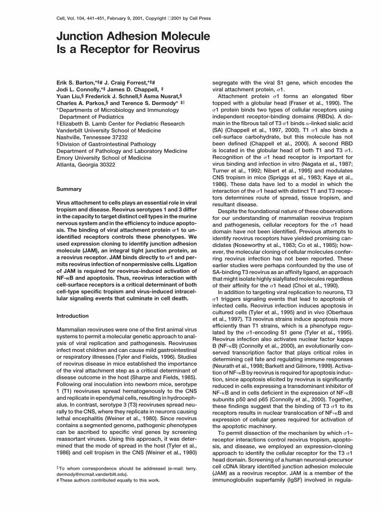
Figure 1. Identification of JAM as a Reovirus Receptor by Expres-Expression Cloning of hJAM, a Reovirus Receptorsion CloningTo facilitate the identification of reovirus receptors based(A) COS-7 cells were transfected with an NT2-cell cDNA libraryon the capacity to bind directly to the s1 head, non-SA-and incubated with FITC-labeled T3SA2 virions. The 0.5% mostbinding T3 reovirus strain T3SA2 was used as an affinityfluorescent cells were collected by FACS for plasmid rescue. Per-ligand (Barton et al., 2001) in a FACS-based expression-cent cells binding virus in (B), (C), and (D) is expressed relative to
cloning approach (Aruffo and Seed, 1987). NT2 cells are cells transfected with NT2-cell cDNA library.human neuronal-precursor cells that support T3 reovirus (B) Virus binding to cells transfected with plasmid obtained from ainfection (data not shown). An NT2 cDNA library was positive pool of 50 bacterial transformants (pool 8) following four
rounds of FACS enrichment.selectively enriched for cDNAs that confer binding of(C) Virus binding to cells transfected with plasmid from positivefluoresceinated T3SA2 (FITC-T3SA2) virions to trans-subpool 8-10, containing 5 bacterial transformants.fected COS-7 cells (Figures 1A–1D). After four rounds(D) Virus binding to cells transfected with plasmid from hJAM-
of FACS enrichment and sib selection, four clones were encoding clone 8-10-2.identified that conferred FITC-T3SA2 binding to greaterthan 20% of transfected cells, which was equivalent tothe transfection efficiency in these experiments (Figure Fab fragments (Fabs) of s1-specific mAb 9BG5 (Burstin1D). All four clones encoded hJAM (Liu et al., 2000), et al., 1982), which binds the T3 s1 head domainwhich suggests that hJAM is a reovirus receptor. (Chappell et al., 2000), and each of four anti-hJAM mAbs
tested (Figure 2E). Control antibodies directed againstCD47 (mAb C5/D5) (Parkos et al., 1996) and the humanAntibodies Directed against hJAM Inhibit Reovirus
Infection by Abrogation of Virus Binding coxsackievirus and adenovirus receptor (hCAR) (J. Ber-gelson, personal communication), had no effect on reo-To test the hypothesis that hJAM is a reovirus receptor,
we determined whether anti-hJAM monoclonal antibod- virus binding. Anti-hJAM mAb J10.4 inhibited T3SA2
binding to NT2 cells in a dose-dependent manner, withies (mAbs) could inhibit reovirus infection (Figure 2). Forthese experiments, we used NT2 cells (Figure 2A), HeLa a minimal inhibitory concentration between 0.2 and 0.02
mg/ml (Figure 2E). Anti-hJAM mAb J10.4 also signifi-cells (Figure 2B), and Caco-2 cells (Figures 2C and 2D),an intestinal epithelial cell line. Cells were treated with cantly inhibited binding of strain T3SA1 to NT2 cells
(Figure 2E); however, residual virus binding above back-anti-hJAM mAbs 7G2C9 or J10.4 (Liu et al., 2000) priorto infection with T3SA2 . In the absence of anti-hJAM ground remained. Preincubation of T3SA1 with sialyllac-
tose (SLL) to inhibit binding to cell-surface SA (Bartonantibodies, T3SA2 grew efficiently in all three cell types.In contrast, anti-hJAM mAbs dramatically inhibited et al., 2001) abolished T3SA1 binding to NT2 cells
treated with mAb J10.4 (Figure 2E). These results indi-T3SA2 growth, resulting in 10- to 100-fold reductionin viral yield. In each case, decreased viral yield was cate that T3SA2 binds to hJAM, while T3SA1 binds to
both hJAM and SA on NT2 cells. Together, these resultsmediated by reduction in the number of infected cells(Figure 2D and data not shown). These results indicate strongly suggest that reovirus binding and infection are
dependent on the availability of hJAM to serve as athat hJAM binding is critical for reovirus infection ofmultiple cell types, including cells of intestinal and neural reovirus receptor.lineages.
To determine whether anti-hJAM mAbs inhibit reovi- Transient Transfection of hJAM Renders MurineErythroleukemia (MEL) Cells Permissive forrus infection at the viral attachment step, we assessed
the capacity of these mAbs to block binding of radiola- Infection by T1 and T3 Reovirus StrainsIf hJAM functions as a bona fide reovirus receptor, thenbeled T3SA2 and an SA-binding T3 strain T3SA1 (Barton
et al., 2001) to NT2 cells (Figure 2E). Binding of T3SA2 transfection of reovirus-resistant cells with hJAM shouldpermit reovirus infection. To test this prediction, we usedto NT2 cells was inhibited by unlabeled competitor virus,

Junction Adhesion Molecule Is a Reovirus Receptor443
Figure 2. JAM Is Required for Reovirus Bind-ing and Infection of NT2 Cells, HeLa Cells,and Caco-2 Cells
(A–C) Effect of anti-hJAM mAbs on T3SA2
growth in cultured cells. NT2 cells (A), HeLaCells (B), or Caco-2 cells (C) were incubatedin the presence or absence of 20 mg/ml CD47-specific mAb C5/D5 or 1 mg/ml of hJAM-spe-cific mAbs 7G2C9 or J10.4 prior to adsorptionwith T3SA2 at an MOI of 1 PFU/cell. Antibod-ies and inocula were removed, cells were in-cubated for the times shown, and progenyvirions were quantitated by plaque assay.Shown are mean viral titers for three experi-ments. Error bars indicate standard devia-tions.(D) Effect of anti-hJAM mAbs on T3SA2 infec-tion of Caco-2 cells. Caco-2 cells weretreated with CD47-specific mAb C5/D5 (20mg/ml) or hJAM-specific mAbs (5 mg/ml) priorto infection with T3SA2 at an MOI of 100 PFU/cell. After incubation for 24 hr, infected cellswere visualized by indirect immunofluores-cence.(E) Effect of anti-hJAM mAbs on binding ofT3SA2 and T3SA1 to NT2 cells. Cells wereuntreated as a control, incubated with unla-beled virus as competitor, or pretreated withthe mAbs shown prior to adsorption with ra-dioiodinated virions of T3SA2 or T3SA1 . Viri-ons also were untreated or pretreated with 5mM SLL or 9BG5 Fabs (50 mg/ml). Antibodieswere used at either 20 mg/ml or the concen-trations shown. Cell-associated virus wascaptured by vacuum filtration and quantitatedby liquid scintillation. Error bars indicate therange of values from duplicate experiments.
MEL cells, which are resistant to infection by T1 strains To test the hypothesis that JAM is a receptor for bothT1 and T3 reovirus, we assessed the capacity of anti-and non-SA-binding T3 strains, but support infection by
SA-binding T3 strains (Rubin et al., 1992; Barton et al., hJAM mAbs to block the binding of T1L and T3D to NT2cells (Figures 3C and 3D). T1L binding to NT2 cells was2001). MEL cells were transiently transfected with empty
vector or hJAM-encoding plasmid and infected with abolished by either Fabs of mAb 5C6, which is specificfor the T1 s1 head domain (Chappell et al., 2000), or anti-T3SA2 or T3SA1 (Figure 3A). As expected, T3SA1 but
not T3SA2 grew efficiently in vector-transfected cells. hJAM mAb J10.4 (Figure 3C). T3D bound much more effi-ciently than T1L to NT2 cells (Figure 3D), but .60% ofIn contrast, yields of T3SA2 were 10-fold greater in cells
transfected with hJAM than in vector-transfected cells. T3D binding was inhibited by either 9BG5 Fabs or mAbJ10.4 (Figure 3D). Significant binding of T3D remainedThis result indicates that expression of hJAM in MEL
cells rescues infection of non-SA-binding reovirus. even in the presence of anti-hJAM mAbs, consistentwith the fact that T3D s1 also can bind SA (Chappell etWe also tested the effect of transient hJAM expression
on growth of prototype reovirus strains T1 Lang (T1L) al., 2000). These results indicate that prototype T1 and T3reovirus strains recognize hJAM as a receptor on a humanand T3 Dearing (T3D) in MEL cells (Figure 3B). As antici-
pated, SA-binding strain T3D grew efficiently in vector- neuronal-precursor cell line. However, in addition to recog-nition of hJAM, strain T3D binds an additional NT2 mole-and hJAM-transfected MEL cells. Surprisingly, although
T1L did not infect vector-transfected MEL cells, it grew cule that is likely to be SA (Figure 2E).efficiently in hJAM-transfected MEL cells. Given thatserotype-dependent differences in reovirus tropism and Transient Transfection of hJAM or Murine JAM
(mJAM) Renders Chicken Embryo Fibroblastpathogenesis are thought to be determined by differ-ences in receptor utilization (Sharpe and Fields, 1985), (CEF) Cells Permissive for Infection by T1
and T3 Reovirus Strainsthis result was unexpected and suggests that T1 and T3reovirus strains utilize JAM as a serotype-independent Since MEL cells support growth of some reovirus
strains, we thought it possible that transfection of thisreceptor.

Cell444
particles bind cell-surface receptors but do not requireendocytic proteolysis for infection (Baer and Dermody,1997). ISVPs were used in this experiment since wereasoned that CEF cells might not express endosomalproteases required for mammalian reovirus disassem-bly. This was found to be the case, since neither controlnor JAM-expressing CEF cells supported infection withvirions of any reovirus strain (data not shown). However,ISVPs of T1L, T3SA2 , and T3SA1 were capable of in-fecting CEF cells transfected with hJAM or mJAM butnot hCAR (Figure 4). These results indicate that the blockto reovirus infection in avian cells is rescued by expres-sion of hJAM or mJAM, providing strong evidence thatJAM functions as a serotype-independent reovirus re-ceptor in both human and murine hosts. Furthermore,these findings suggest that the attachment and disas-sembly steps of the reovirus life cycle serve as keydeterminants of the host-range restriction exhibited bydiverse classes of vertebrates.
Figure 3. JAM Rescues Infectivity of T1 and T3 Reovirus Strains
(A and B) Effect of JAM expression on growth of T1 and T3 reovirusstrains in MEL cells. MEL cells were transiently transfected with hJAM Binds Directly to the Reovirus s1 Headcontrol vector (pBK) or hJAM clones 1-4-3 and 1-4-5. Transfected Domain with High Affinitycells were adsorbed with T3SA2 , T3SA1 , T1L, or T3D at an MOI of
To exclude the possibility that hJAM indirectly enhances1 PFU/cell and incubated for either 24 (A) or 48 (B) hr. Viral titersreovirus infection at a post-attachment step, we usedwere determined by plaque assay. Shown are mean viral yields (titersurface plasmon resonance (SPR) to determine whetherat 24 or 48 hr divided by titer at 0 hr) for three experiments. Error
bars indicate standard deviations. reovirus particles and s1 protein bind directly to hJAM(C and D) Effect of anti-hJAM mAbs on binding of T1 and T3 reovirus (Karlsson and Falt, 1997) (Figure 5). For these experi-strains to NT2 cells. NT2 cells were incubated with CD47-specific ments, we generated a fusion protein consisting of themAb C5/D5 (20 mg/ml) or hJAM-specific mAb J10.4 (2 mg/ml) prior
rabbit Ig Fc domain linked to the extracellular domainto adsorption with radioiodinated T1L (C) or T3D (D). Virions wereof hJAM (Fc-hJAM). A fusion of the rabbit Fc with hCARpretreated with the inhibitors shown.(Fc-hCAR) was used as a control for nonspecific interac-tions (Bergelson et al., 1997). Fc-hJAM and Fc-hCARwere conjugated to SPR sensor chips, and purified viri-cell type with hJAM might upregulate an endogenous
receptor. Therefore, we tested the capacity of cDNAs ons of T3SA2 , T3SA1 , T1L, or T3D were injected acrossthe Fc-hJAM and Fc-hCAR flow cells (Figure 5A). Virionsencoding either hJAM or mJAM to confer reovirus infec-
tion to CEF cells, which do not support reovirus infection of all four strains displayed a time-dependent increase inbinding to Fc-hJAM, and this binding was stable during(Figure 4). CEF cells were transiently transfected with
hCAR-, hJAM-, or mJAM-encoding plasmids and in- buffer wash. Virions did not bind to Fc-hCAR, and prein-jection of anti-hJAM mAbs blocked virus binding tofected with virions or infectious subvirion particles
(ISVPs) of T1L, T3SA2 , or T3SA1 . ISVPs are reovirus hJAM, indicating that binding was specific for the hJAMextracellular domain (Figure 5A and data not shown).disassembly intermediates generated in vivo in the in-
testinal lumen or in endocytic vesicles and in vitro by To determine whether reovirus binding to hJAM ismediated by s1, T3SA1 virions were incubated withprotease treatment (Baer and Dermody, 1997). These
Figure 4. Transfection of CEF Cells withhJAM or mJAM Enables Infection by T1 andT3 Reovirus
CEF cells were transiently transfected withplasmid encoding hCAR, hJAM, or mJAM.Transfected CEF cells were adsorbed withISVPs of T1L, T3SA2 , or T3SA1 at an MOI of10 PFU/cell. Reovirus antigen was detectedby indirect immunofluorescence 20 hr post-infection. Parallel transfections were per-formed using pEGFP-N1 as an indicator oftransfection efficiency.

Junction Adhesion Molecule Is a Reovirus Receptor445
specific inhibitors of either the s1 SA binding domain(SLL) or the s1 head RBD (9BG5 Fabs) (Figure 5B). Bind-ing of T3SA1 to Fc-hJAM was not inhibited by SLL,indicating that binding to SA on hJAM glycosylationchains is not required for this interaction. In contrast,preincubation of T3SA1 virions with 9BG5 Fabs, but notcontrol 5C6 Fabs, substantially reduced T3SA1 bindingto Fc-hJAM, suggesting that this binding is mediatedby the s1 head.
To investigate whether hJAM interacts directly withs1, we tested the capacity of recombinant s1 to bindFc-hJAM (Figure 5C). Purified T3D s1 (Chappell et al.,2000) bound specifically, saturably, and reversibly to Fc-hJAM. Kinetic analysis of the s1-Fc-hJAM interactionassuming 1:1 stoichiometry indicated a KD of approxi-mately 9 3 1028 M (Karlsson and Falt, 1997). SLL hadno effect on the binding of s1 to Fc-hJAM, confirmingthat this interaction does not require SA (data notshown).
To confirm that the s1 head mediates binding tohJAM, a proteolytically derived fragment of s1 con-taining only the s1 head was injected over the Fc-hJAMsurface (Figure 5C). The s1 head domain bound specifi-cally to Fc-hJAM with a KD of 6 3 1028 M, which approxi-mates that of full-length s1. These results indicate thatthe head domain of reovirus attachment protein s1 bindswith high affinity directly to the extracellular domain ofhJAM.
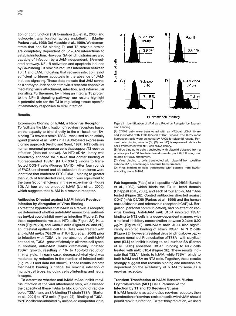
Figure 5. Quantitation of s1 Binding Affinity to hJAM Using SurfaceReovirus–JAM Interactions Are Required Plasmon Resonancefor Activation of NF-kB and Induction (A) T1 and T3 reovirus strains interact directly with hJAM. Virionsof Apoptosis during Reovirus Infection of T3SA2 , T3SA1 , T1L, or T3D were injected over biosensor surfacesReovirus infection of cultured cells leads to NF-kB acti- coated with either Fc-hJAM or Fc-hCAR fusion proteins. Binding in
response units was measured over time.vation, which is required for reovirus-induced apoptosis(B) T3SA1 binding to hJAM is not dependent on SA. Binding of(Connolly et al., 2000). To determine whether engage-T3SA1 to Fc-hJAM and Fc-hCAR on a biosensor was assessed inment of JAM by s1 triggers these cellular responses,the presence of SLL, lactose (control), or Fabs of 9BG5 or 5C6
we tested the capacity of T3SA1 to replicate, activate (control).NF-kB, and induce apoptosis in the presence of anti- (C) The s1 head domain binds to hJAM. Purified T3D s1 or s1 headhJAM mAbs. The presence of residual binding of T3SA1 domain were injected over a biosensor coated with Fc-hJAM or Fc-
hCAR. Calculated affinities for binding to Fc-hJAM, expressed asand T3D to NT2 cells preincubated with anti-hJAM mAbsapparent KD, are shown.(Figures 2E and 3D) suggested that reovirus can bind
SA in the absence of s1–JAM interactions. To assesswhether binding to SA alone could mediate reovirus HeLa cells (Connolly et al., 2000), we predicted that
blockade of NF-kB activation by mAb J10.4 would corre-entry, HeLa cells were incubated in the presence orabsence of anti-hJAM mAb J10.4 prior to adsorption late with decreased apoptosis. In untreated cells, T3SA1
elicited high levels of apoptosis (Figure 6C). However,with 100 PFU/cell of T3SA1 . Although infection of T3SA2
is substantially inhibited in HeLa cells incubated with incubation of cells with mAb J10.4 abolished the capac-ity of T3SA1 to induce this response. Importantly, treat-mAb J10.4 (Figure 2B), growth of T3SA1 was only mini-
mally reduced (less than 2-fold at 48 hr) by hJAM block- ment of HeLa cells with mAb J10.4 had no effect onthe capacity of TNF-a to activate NF-kB and induceade (Figure 6A). Consistent with this finding, we also
observed z50% fewer reovirus-antigen-positive cells apoptosis, indicating that neither the NF-kB signalingmachinery nor the apoptotic response is inhibited byfollowing hJAM blockade and adsorption with T3SA1 at
an MOI of 1 PFU/cell (data not shown). These results anti-hJAM mAb J10.4 (Figures 6B and 6C). These resultsdemonstrate that while SA can mediate entry of SA-suggest that cell-surface SA can serve as a functional
receptor when hJAM is absent or inaccessible. binding reovirus strains, reovirus-induced NF-kB acti-vation and resultant cell death require virus-inducedSince T3SA1 can infect HeLa cells via an hJAM-inde-
pendent pathway, it was possible to determine whether signaling events mediated by the interaction of s1with JAM.s1–hJAM interactions modulate the efficiency of reovi-
rus-induced NF-kB activation. We found that T3SA1 in-duced strong activation of NF-kB in untreated cells, but Discussionthis activation was abolished in cells treated with anti-hJAM mAb J10.4 (Figure 6B). Since NF-kB activation is Results presented in this report demonstrate that JAM
satisfies all requisite criteria of a functional reovirus re-required for induction of apoptosis in reovirus-infected
Cell446
Figure 6. Anti-hJAM mAbs Inhibit NF-kB Ac-tivation and Apoptosis Induction by ReovirusT3SA1
(A) Effect of anti-hJAM mAb J10.4 on growthof T3SA1 in HeLa cells. HeLa cells were incu-bated with PBS or 5 mg/ml mAb J10.4 priorto adsorption with T3SA1 at an MOI of 100PFU/cell. Shown are mean viral titers for threeexperiments. Error bars indicate standard de-viations.(B) Effect of anti-hJAM mAb J10.4 on NF-kB activation induced by T3SA1 and TNF-a.HeLa cells were incubated with PBS or 5 mg/ml mAb J10.4 prior to adsorption with T3SA1
(100 PFU/cell) or treatment with TNF-a (20ng/ml). Mock-infected cells are shown as acontrol. After incubation for either 1 (TNF-a)or 10 (T3SA1 ) hr, NF-kB complexes in nuclearextracts were detected by EMSA.(C) Effect of anti-hJAM mAb J10.4 on apopto-sis induced by T3SA1 and TNF-a. HeLa cellswere incubated with PBS or 5 mg/ml mAbJ10.4 prior to adsorption with T3SA1 (100 PFU/
cell) or treatment with TNF-a (20 ng/ml). Mock-infected cells are shown as a control. After incubation for either 24 (TNF-a) or 48 (T3SA1 ) hr,cells were stained with acridine orange. Shown is the mean percentage of cells undergoing apoptosis for three experiments. Error bars indicatestandard deviations.
ceptor. First, transfection of COS-7 cells with hJAM con- 1998; Liu et al., 2000). Reovirus gains access to thebasolateral surface of intestinal cells by transport throughfers binding of reovirus T3SA2. Second, blockade of
hJAM on the surface of NT2 cells, HeLa cells, or Caco-2 microfold cells (Wolf et al., 1981), which would allowvirus exposure to the area of highest JAM expression.cells abolishes T3SA2 binding and growth. Third, bind-
ing of prototype reovirus strains T1L and T3D to NT2 It is also possible that transient disruptions of the TJbarrier, such as those that occur during migration ofcells is blocked by hJAM-specific mAbs. Fourth, trans-
fection of murine and avian cells with hJAM rescues immune and inflammatory cells, permit reovirus accessto JAM from the intestinal lumen. Such micro-disrup-reovirus infection in a serotype-independent manner.
Finally, and most conclusively, the biological effects of tions of TJ integrity facilitate infection by other patho-gens, including Yersinia (McCormick et al., 1997) andhJAM on reovirus infection correlate with a direct, SA-
independent, high-affinity interaction between hJAM Salmonella (Jensen et al., 1998).Since the discovery that differences in the tropism ofand the head domain of reovirus attachment protein s1.
JAM is a type I transmembrane protein with two extra- T1 and T3 reovirus for specific cells in the CNS segregatewith the s1-encoding S1 gene, it has been hypothesizedcellular Ig domains and a short cytoplasmic tail (Martin-
Padura et al., 1998; Liu et al., 2000). JAM is an important that T1 and T3 strains bind to distinct receptors ex-pressed on ependymal cells and neurons, respectivelycomponent of TJs between endothelial and epithelial
cells, and it may function to organize TJ formation by (Weiner et al., 1980; Sharpe and Fields, 1985). However,JAM confers infection by both T1 and T3 reovirus strains,interaction with other TJ proteins and the cytoskeleton
(Bazzoni et al., 2000; Ebnet et al., 2000). JAM also may suggesting that the interaction between JAM and the s1head domain is not the critical determinant of serotype-influence the migration of leukocytes across endothelial
and epithelial barriers during the course of an inflamma- dependent differences in reovirus CNS tropism. Instead,we think it possible that differences in the cell-surfacetory response (Del Maschio et al., 1999; Lechner et al.,
2000). JAM is highly conserved among mammals, with carbohydrates bound by T1 and T3 s1 proteins influenceviral tropism in the murine nervous system. Accordingly,human, murine, bovine, and rat JAM displaying z70%
amino acid identity (Martin-Padura et al., 1998; Liu et neural polysialic acid (Rutishauser and Landmesser,1996) may permit infection by T3 strains, as was ob-al., 2000). Given the broad host range of mammalian
reovirus (Tyler and Fields, 1996), it is not surprising that served for T3SA1 infection of HeLa cells. Since T1strains do not bind SA (Chappell et al., 2000), thesea reovirus receptor would display a high degree of se-
quence conservation. strains would be incapable of utilizing this pathway andmight even be repelled by the negatively-charged SAJAM is expressed on many cell types that are known
targets for reovirus infection in vivo, including intestinal moieties on the neural surface. Alternatively, it is possi-ble that proteins with homology to JAM display sero-epithelium, bile duct epithelium, lung epithelium, leuko-
cytes, and CNS endothelial cells (Martin-Padura et al., type-specific interactions with s1.T3 reovirus infects neurons and causes encephalitis1998; Williams et al., 1999; Liu et al., 2000). These obser-
vations are consistent with the extensive and overlap- in neonatal mice, but mice rapidly become resistant toreovirus disease during the first few weeks of life (Tar-ping tissue distribution of T1 and T3 reovirus infections
in mice, particularly within the intestine (Tyler and Fields, dieu et al., 1983). As embryonic neuronal differentiationprogresses, the neuroepithelium down-regulates inte-1996). Cell-surface JAM is localized to the subapical
surface of polarized epithelial TJs (Martin-Padura et al., gral TJ proteins and loses the capacity to form TJs
Junction Adhesion Molecule Is a Reovirus Receptor447
(Aaku-Saraste et al., 1996). JAM is not highly expressedon adult mouse neurons (Martin-Padura et al., 1998);however, its expression in the developing murine ner-vous system has not been rigorously examined. It ispossible that JAM is expressed at birth and then de-clines as neonatal CNS remodeling is completed,thereby providing a mechanistic explanation for the age-restriction of reovirus encephalitis.
The capacity of reovirus to interact with the TJ via JAMmay have important consequences for the pathology ofreovirus infection. Regulation of the TJ is critical formaintenance of epithelial and endothelial barriers (Baz-zoni et al., 1999). Anti-hJAM mAbs prevent the reorgani-zation of disrupted TJs in cultured intestinal epithelial
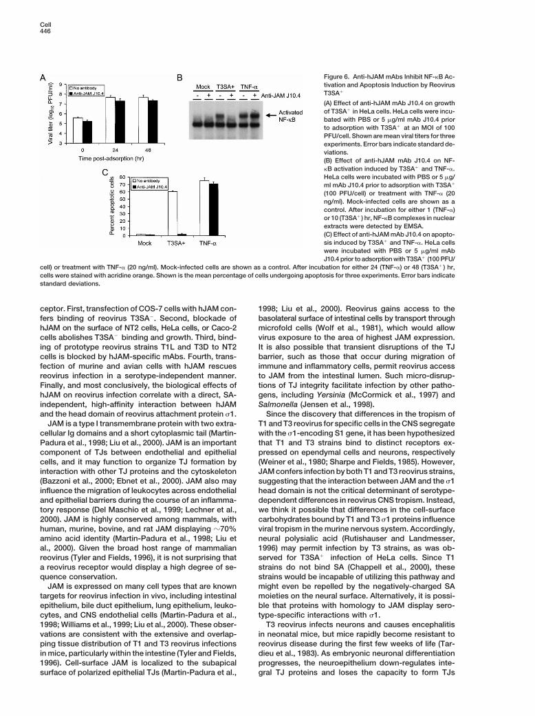
Figure 7. Models of Reovirus–JAM Interactions at the Cell Surfacecells (Liu et al., 2000) and promote TJ breakdown of(A) Non-SA-binding reovirus strains like T3SA2 bind JAM as a recep-the endothelium lining CNS blood vessels, resulting intor. Binding of these strains to JAM mediates virus internalizationenhanced disease in response to bacterial or viral infec-but does not induce signals leading to activation of NF-kB.
tions (Lechner et al., 2000). If reovirus–JAM interactions (B) SA-binding reovirus strains like T3SA1 bind both JAM and SA.lead to a similar destabilization of TJs in CNS endothe- The membrane-proximal Ig domain of hJAM contains two N-linked
glycosylation sites (Liu et al., 2000), one of which is schematicallylium, this might promote breakdown of the blood-brainindicated. The dual ligation of JAM and JAM-bound SA is proposedbarrier, permitting cerebral edema and neural inflamma-to induce JAM cross-linking or conformational alterations leadingtion, conditions evident in reovirus encephalitis (Tylerto initiation of signals required for NF-kB activation and apoptosis.and Fields, 1996). In addition, reovirus-induced TJ dys-(C) SA-binding reovirus strains also may activate intracellular signal-
regulation within the murine intestinal epithelium might ing pathways by the simultaneous crosslinking of JAM and otherpromote diarrhea, thereby enhancing viral shedding and sialyllated cell-surface proteins.transmission. A similar mechanism has been proposedfor the pathogenesis of diarrhea induced by rotavirus(Obert et al., 2000). of TJ proteins that includes the Ras-interacting protein
Our finding that s1–JAM interactions are required for AF-6 (Bazzoni et al., 2000; Ebnet et al., 2000). Impor-NF-kB activation and apoptosis underscores the impor- tantly, Ras-mediated pathways have been implicated intance of the attachment step in modulating the cellular activation of NF-kB (Anrather et al., 1999; Norris andconsequences of reovirus infection. Although T3SA1 is Baldwin, 1999; Romashkova and Makarov, 1999). Sincecapable of efficient infection via SA-mediated attach- infection of newborn mice with T3 reovirus at a dosement, this route of infection seems to bypass the cellular that leads to lethal encephalitis also induces apoptosissignaling events culminating in apoptosis, suggesting in the CNS (Oberhaus et al., 1997), our findings raise thethat viral replication is not sufficient to trigger this re-
possibility that reovirus–JAM interactions dictate strain-sponse. Our findings demonstrate that reovirus binding
specific pathogenic phenotypes as a consequence ofto JAM is required for induction of the apoptotic re-
intracellular signaling in addition to determining viralsponse; however, s1–JAM interactions are not the sole
tropism.determinant of virus-induced apoptosis. T3SA1 is muchIt is interesting to speculate why reoviruses wouldmore efficient than T3SA2 at inducing NF-kB-dependent
utilize a receptor that triggers apoptotic death of theapoptosis in HeLa cells (J. L. C. et al., submitted), despiteinfected cell. Although many viruses induce apoptosisthe fact that both strains bind JAM on this cell type. Weof host cells, only a select group encodes proteins thatpropose a model in which a virus-cell synapse formedactively inhibit this process. For these viruses, it is likelyby multivalent interactions of s1 with both JAM and SAthat apoptosis must be blocked for a period sufficientsurpasses a critical cellular activation threshold requiredto assemble viable progeny (Roulston et al., 1999). Reo-for NF-kB activation and apoptosis (Figure 7). Thisvirus replication requires z16 hr; however, the majoritymodel has similarities to mechanisms of lymphocyteof reovirus-infected cells remain viable for up to 48 hractivation in which lymphocyte receptors must be en-post-infection (Connolly et al., 2000). This interval wouldgaged by both antigen and accessory molecules to initi-allow for multiple rounds of viral replication prior to cellate effector functions (Grakoui et al., 1999). JAM appearsdeath. Thus, there may be no selective advantage forto be the only receptor on some cell types for non-SA-reovirus to inhibit the host cell apoptotic response. Inbinding reovirus strains. However, the binding of thesefact, there is increasing evidence to suggest that somestrains to JAM is not sufficient to trigger NF-kB activa-viruses utilize apoptosis as a means to evade the hosttion (Figure 7A). In contrast, the simultaneous ligationimmune response by minimizing tissue inflammation andof JAM and SA moieties on JAM by SA-binding strainspermitting viral spread within apoptotic bodies (Teodoromay alter the conformation or oligomeric nature of JAM,and Branton, 1997; O’Brien, 1998). Identification of JAMthereby triggering signaling events subsequent to virusas a signaling molecule required for reovirus-inducedbinding (Figure 7B). Alternatively, SA-binding strainsapoptosis will permit dissection of the underlying mech-might bind JAM and SA residues on other cellular pro-anisms and pathologic significance of apoptosis in re-teins, and the juxtaposition of JAM with these proteinssponse to viral infection.may activate signaling cascades that culminate in apo-
Although a number of signaling molecules concen-ptotic cell death (Figure 7C). In support of the observedcoupling of JAM and NF-kB, JAM exists in a complex trate at the cytoplasmic face of the TJ, their roles in
Cell448
Assessment of Virus Growthtransducing signals from the environment to the nucleusCells (2–5 3 105) were incubated in PBS or PBS containing variousare unclear (Clarke et al., 2000; Hopkins et al., 2000).concentrations of mAbs C5/D5, 7G2C9, or J10.4 at room tempera-We provide the first demonstration that JAM and the TJture for 1 hr. Virus was adsorbed to antibody-treated cells at an
are involved in a discrete outside-in nuclear signaling MOI of 1 or 100 PFU/cell and incubated at room temperature forpathway. NF-kB regulates the transcription of many in- 1 hr. Inocula were removed, cells were washed, and complete me-
dium was added. Cells were incubated at 378C for various intervals,flammatory cytokines involved in the activation and at-and viral titers in lysates were determined by plaque assay.traction of immune effector cells (Pahl, 1999). The link-
age of JAM to NF-kB has important implications for theFluorescent-Focus Assay of Viral Infectionrole of the TJ in responding to pathogens and suggestsVirus was adsorbed to confluent monolayers as for growth experi-
that the TJ can serve as an environmental sensor capa- ments. Following incubation at 378C for 20 hr, cells were fixed withble of responding to viral infection by triggering apopto- 1 ml of methanol at 2208C for 30 min. Infected cells were identifiedsis. The discovery of JAM as a reovirus receptor ex- by indirect immunofluorescence using rabbit anti-reovirus sera as
described (Barton et al., 2001).pands our knowledge of virus-receptor biology andhighlights a potential role for the TJ in regulating both
Virus Radioligand Binding Assaysinflammatory responses and cell death.Purified virions (2–4 3 1013/ml in Dulbecco’s PBS [Gibco-BRL, GrandIsland, NY]) were iodinated and used for binding assays as described(Barton et al., 2001). Iodinated virus was added to cells (1 3 106)Experimental Proceduresand incubated at room temperature for 3 hr. Cell-associated viruswas captured by vacuum filtration and quantitated by liquid scintilla-Cells, Viruses, and Antibodiestion. For experiments assessing the effect of SLL or Fabs on virusL cells, MEL cells, and HeLa cells were maintained as describedattachment, iodinated virions were preincubated with each reagent(Barton et al., 2001). NTERA-2 (NT2) and Caco-2 cells were obtainedat 378C for 30 min. Antibodies against cell-surface proteins werefrom the American Type Culture Collection and maintained in mono-preincubated with cells at 378C for 30 min prior to adsorption oflayer culture as for HeLa cells. CEF cells were derived from day 10radiolabeled virus.fertilized chicken eggs and maintained as described (Brown et al.,
1999).Cloning of mJAMReovirus strains T1L and T3D are laboratory stocks. IsogenicmJAM was cloned by PCR amplification from Quickclone cDNAs1-point mutants T3/C44-SA2 and T3/C44-SA1, abbreviated in thisderived from poly-A mRNA of 7-day murine embryos (Clontech, Palomanuscript as T3SA2 and T3SA1 , respectively, were generated asAlto, CA). cDNA (4 ng) was subjected to PCR with primers (0.2 mM)described (Barton et al., 2001). Viral titer was determined by plaquespecific for the reported mJAM sequence appended at the 59 andassay on L cells (Virgin et al., 1988). Purified virions were prepared39 termini with XbaI and SpeI restriction sites, respectively (59-CCTand quantitated as described (Furlong et al., 1988). ISVPs wereACTAGTGGATTGTAACTGTAATGGGCA-39 and 59-CCTTCTAGAGCgenerated as described (Baer and Dermody, 1997). Purified T3SA2
CGCAGCAGGTCACACCAGG-39) (Martin-Padura et al., 1998). PCR(1 3 1013/ml in carbonate-bicarbonate buffer [pH 9] [Sigma-Aldrich,product was purified using DNAzol (Molecular Research Center,St. Louis, MO]) was fluoresceinated by incubation in 50 mg/ml FITCCincinnati, OH), treated with XbaI and SpeI, and ligated into the(Pierce, Rockford, IL) for 1 hr at room temperature.XbaI and SpeI sites of alkaline phosphatase-treated pBK-CMV (Stra-Murine mAbs 9BG5 (Burstin et al., 1982) and 5C6 (Virgin et al.,tagene). Fidelity of PCR amplification and cloning was confirmed1991) were purified from hybridoma supernatants (Cell Culture Cen-by automated sequencing.ter, Minneapolis, MN), and Fabs of each were prepared using the
Immunopure Fab system (Pierce). Anti-hJAM murine mAbs J10.4,Transient Transfection and Reovirus Infection of MEL Cells7G2C9, J3F.4, and 1H2A9 (all IgG1) (Williams et al., 1999; Liu et al.,MEL cells (1 3 107) were transiently transfected with 40 mg each2000) and CD47-specific mAb C5/D5 (IgG1) (Parkos et al., 1996)of pEGFP-N1 (Clontech) (transfection control), pBK-CMV (negativewere purified from ascites by protein A affinity. Rabbit hCAR-specificcontrol), hJAM 1-4-3, or hJAM 1-4-5 by electroporation. Transfectionantiserum was provided by Dr. Jeffrey Bergelson (University ofefficiency was z10%. After 48 hr incubation, 2 3 105 cells werePennsylvania).adsorbed with T3SA2 , T3SA1 , T1L, or T3D at an MOI of 1 PFU/cell in a total volume of 150 ml. Adsorptions were terminated afterincubation at room temperature for 1 hr by washing in PBS. CellsExpression Cloning of hJAMwere incubated at 378C in 1 ml of culture medium for various inter-COS-7 cells were transfected with an NT2 cDNA library (Stratagene,vals, and viral titers in lysates were determined by plaque assay.La Jolla, CA) as described (Aruffo and Seed, 1987). After 48 hr
incubation, cells were detached from plates by incubation with 2 mMEDTA/PBS at 378C. Detached cells (1 3 106) were resuspended in Transient Transfection and Reovirus Infection of CEF Cells150 ml PBS containing 1 3 1011 FITC-conjugated T3SA2 particles Passage-five CEF cells (50%–75% confluence) were transfectedand incubated on ice for 1 hr. Cells were washed twice with PBS, with 0.4 mg of plasmid encoding hCAR (in pCDNA 3.1), hJAM, orviable cells were analyzed by FACS, and the 0.5% most fluorescent mJAM using Lipofectamine Plus (Gibco-BRL). Transfected CEF cellscells were collected using a FACStar Plus (Becton, Dickinson and were infected with virions or ISVPs of T3SA2 , T3SA1 , or T1L at anCo., Franklin Lakes, NJ). Plasmid was rescued from sorted cells MOI of 10 PFU per cell and processed for fluorescent-focus assay.(Hirt, 1967), amplified in bacteria, and used in three subsequentrounds of FACS enrichment. Individual bacterial colonies obtained Generation and Purification of Fc-hJAM Fusion Proteinfrom the quaternary sort were grouped into pools of 50 colonies. The hJAM extracellular domain was amplified by PCR from 1 mg ofPlasmid prepared from pools was used to transfect COS-7 cells. hJAM clone 1-4-5 using hJAM-specific primers (0.2 mg) appendedPositive pools were defined as those conferring a greater than 2% with HindIII and BamHI restriction sites at the 59 and 39 termini,increase in the number of maximally-fluorescent cells as compared respectively (59-TAGCAAGCTTCCTGATCGCGATG-39 and 59-TACGto cells transfected with cDNA library. Positive pools were subdi- GGATCCATTCCGCTCCAC-39). PCR product and Fc-hCAR-pCDNAvided into sub-pools of 5 colonies, and plasmid from sub-pools was 3.1 (Bergelson et al., 1997) were subjected to restriction digest withtested for the capacity to confer enhanced FITC-T3SA2 binding to HindIII and BamHI, resulting in excision of hCAR-encoding se-transfected COS-7 cells. Individual clones from positive sub-pools quences from the plasmid vector. Digestion products were purifiedwere similarly screened. This process yielded four clones that con- by agarose gel electrophoresis, and the hJAM PCR product wasferred T3SA2 binding to all transfected COS-7 cells (1-4-3, 1-4-5, ligated into the digested Fc-pCDNA plasmid. Fidelity of PCR amplifi-8-10-2, and 8-10-3). Automated sequencing revealed that each cation and cloning was confirmed by automated sequencing.
The Fc-hJAM and Fc-hCAR plasmid constructs were amplified inclone encoded hJAM (Liu et al., 2000).

Junction Adhesion Molecule Is a Reovirus Receptor449
bacteria and used in DEAE/dextran transfections of COS-7 cells Acknowledgments(Aruffo and Seed, 1987). After 72 hr incubation, supernatants wereharvested, and the soluble Fc-fusion proteins were purified by pro- We thank Ray Mernaugh and Ergang Shi of the Molecular Recogni-
tion Unit, Vanderbilt Cancer Center Core Facility, for assistance withtein A affinity.biosensor experiments and Kathy Allen for help with FACS analysis.We are grateful to Jeffrey Bergelson for providing Fc-hCAR and
Purification of T3D s1 hCAR-specific antiserum and to the laboratory of Joey Barnett forT3D-derived s1 protein was generated, expressed, and purified as preparing CEF cells. We thank Jacek Hawiger, Earl Ruley, and Lucdescribed (Chappell et al., 2000). The s1 head domain was purified Van Kaer for review of the manuscript. This research was supportedfollowing trypsin digestion of s1 deletion mutant 3-D-3-3-3 (Chappell by the National Science Foundation (E. S. B.), Public Health Serviceet al., 2000). The 3-D-3-3-3 mutant was concentrated to 0.3 mg/ml awards T32 CA09385 (J. C. F.), DK10013 (Y. L.), HL54229 andin PBS using a centrifugal concentrator (Millipore, Bedford, MA) and HL60540 (C. A. P.), and AI38296 (T. S. D.), the Crohns and Colitisincubated at 48C for 1 hr with 10% (v/v) PBS-washed trypsin-agarose Foundation (A. N.), the Vanderbilt University Research Councilbeads (Sigma-Aldrich). Trypsin beads were pelleted and the super- (E. S. B. and J. C. F.), and the Elizabeth B. Lamb Center for Pediatricnatant was removed. Stable fragments corresponding to the s1 Research. Additional support was provided by Public Health Servicehead (25 kDa) and tail (14 kDa) are produced by this treatment awards CA68485 for the Vanderbilt Cancer Center and DK20593 for(Chappell et al., 2000). Trypsin-treated 3-D-3-3-3 s1 was incubated the Vanderbilt Diabetes Research and Training Center.in an equal volume of Sepharose Q (Amersham Pharmacia) in 20mM Tris-HCl (pH 8.1) at room temperature for 1 hr. Sepharose Q Received October 10, 2000; revised December 22, 2000.beads were pelleted, and the s1 head fragment was eluted frombeads using 0.1 M sodium acetate and 0.5 M NaCl (pH 4.0). Eluted
Referencesfragments were visualized by SDS-PAGE and found to include the25 kDa s1 head fragment free of contaminating 3-D-3-3-3 s1 or s1
Aaku-Saraste, E., Hellwig, A., and Huttner, W.B. (1996). Loss oftail fragment.occludin and functional tight junctions, but not ZO-1, during neuraltube closure—remodeling of the neuroepithelium prior to neurogen-esis. Dev. Biol. 180, 664–679.Assessment of Virus-hJAM and s1–hJAM Interactions
Using SPR Anrather, J., Csizmadia, V., Soares, M.P., and Winkler, H. (1999).Fc-hJAM and Fc-hCAR at a concentration of 0.2–0.4 mg/ml in PBS Regulation of NF-kappaB RelA phosphorylation and transcriptionalwere biotinylated by incubation with a 10-fold molar excess of sulfo- activity by p21(ras) and protein kinase Czeta in primary endothelialNHS-LC-biotin (Pierce) at room temperature for 1 hr. A BIAcore CM5 cells. J. Biol. Chem. 274, 13594–13603.chip (Pharmacia Biosensor AB, Uppsala, Sweden) was coated with Aruffo, A., and Seed, B. (1987). Molecular cloning of a CD28 cDNA400 mg/ml streptavidin (Sigma-Aldrich) by amine coupling. Biotiny- by a high-efficiency COS cell expression system. Proc. Natl. Acad.lated fusion proteins were injected at a concentration of 5 to 50 mg/ Sci. USA 84, 8573–8577.ml in HEPES-buffered saline (HBS) (pH 7.2) across duplicate flow
Baer, G.S., and Dermody, T.S. (1997). Mutations in reovirus outer-cells of a streptavidin-coated chip at 10 ml/min using a BIAcorecapsid protein s3 selected during persistent infections of L cells2000 (Pharmacia Biosensor AB). Sensor-chip flow cells were coatedconfer resistance to protease inhibitor E64. J. Virol. 71, 4921–4928.with 2000 RU of each protein. Purified reovirus virions (5 3 1012
Barkett, M., and Gilmore, T.D. (1999). Control of apoptosis by Rel/particles/ml) were injected across the conjugated chip surfaces atNF-kappaB transcription factors. Oncogene 18, 6910–6924.10 ml/min. Following virus binding, chip surfaces were regenerated
with a 1 min pulse of a 1:1 mixture of HBS and Immunopure Gentle Barton, E.S., Connolly, J.L., Forrest, J.C., Chappell, J.D., and Der-Ag/Ab Elution Buffer (Pierce). Virions also were preincubated with mody, T.S. (2001). Utilization of sialic acid as a coreceptor enhances10 mM SLL, 10 mM lactose (Sigma-Aldrich), or 50 mg/ml Fabs of reovirus attachment by multi-step adhesion-strengthening. J. Biol.9BG5 or 5C6 at room temperature for a minimum of 10 min prior to Chem. 276, 2200–2211.injection. Bazzoni, G., Martinez Estrada, O., and Dejana, E. (1999). Molecular
Purified s1 and s1 head fragments (80 mg/ml) were injected across structure and functional role of vascular tight junctions. Trendsflow cells as for virions. Affinity constants for s1 binding to Fc-hJAM Cardiovasc. Med. 9, 147–152.were determined using separate kon and koff nonlinear regression with
Bazzoni, G., Martinez-Estrada, O.M., Orsenigo, F., Cordenonsi, M.,BIAevaluation 3.0 software (Pharmacia Biosensor AB), assuming aCiti, S., and Dejana, E. (2000). Interaction of junctional adhesion1:1 Langmuir binding model (Karlsson and Falt, 1997). Molar concen-molecule with the tight junction components ZO-1, cingulin, andtrations of s1 constructs were determined assuming that s1 existsoccludin. J. Biol. Chem. 275, 20520–20526.as a homotrimer (Leone et al., 1992).Bergelson, J.M., Cunningham, J.A., Droguett, G., Kurt-Jones, E.A.,Krithivas, A., Hong, J.S., Horwitz, M.S., Crowell, R.L., and Finberg,
Electrophoretic Mobility Shift Assay (EMSA) R.W. (1997). Isolation of a common receptor for Coxsackie B virusesCells (5 3 106) were either untreated or treated with 5 mg/ml mAb and adenoviruses 2 and 5. Science 275, 1320–1323.J10.4 in PBS. After incubation at 378C for 30 min, cells were ad- Brown, C.B., Boyer, A.S., Runyan, R.B., and Barnett, J.V. (1999).sorbed with T3SA1 at an MOI of 100 PFU/cell or treated with 20 ng/ Requirement of type III TGF-beta receptor for endocardial cell trans-ml human recombinant TNF-a (Sigma-Aldrich). After incubation at formation in the heart. Science 283, 2080–2082.378C for either 10 (T3SA1) or 1 (TNF-a) hr, nuclear extracts were
Burstin, S.J., Spriggs, D.R., and Fields, B.N. (1982). Evidence forprepared and assayed for NF-kB activation by EMSA using afunctional domains on the reovirus type 3 hemagglutinin. Virology32P-labeled oligonucleotide consisting of the NF-kB consensus bind-117, 146–155.ing sequence (Santa Cruz Biotechnology, Santa Cruz, CA) (ConnollyChappell, J.D., Gunn, V.L., Wetzel, J.D., Baer, G.S., and Dermody,et al., 2000).T.S. (1997). Mutations in type 3 reovirus that determine binding tosialic acid are contained in the fibrous tail domain of viral attachment
Quantitation of Apoptosis by Acridine Orange Staining protein s1. J. Virol. 71, 1834–1841.Cells (5 3 104) were untreated or treated with 5 mg/ml anti-hJAM
Chappell, J.D., Duong, J., Wright, B.W., and Dermody, T.S. (2000).mAb J10.4 in PBS. After incubation at 378C for 30 min, cells were
Identification of carbohydrate-binding domains in the attachmentadsorbed with T3SA1 at an MOI of 100 PFU/cell or treated with 20
proteins of type 1 and type 3 reoviruses. J. Virol. 74, 8472–8479.ng/ml TNF-a and 10 mg/ml cycloheximide (ICN, Aurora, OH). After
Choi, A.H.C., Paul, R.W., and Lee, P.W.K. (1990). Reovirus binds toincubation at 378C for 48 (T3SA1 ) or 24 (TNF-a) hr, the percentagemultiple plasma membrane proteins of mouse L fibroblasts. Virologyof apoptotic cells was determined using acridine orange staining178, 316–320.(Tyler et al., 1995). For each experiment, 200 to 300 cells were
counted. Clarke, H., Marano, C.W., Peralta Soler, A., and Mullin, J.M. (2000).

Cell450
Modification of tight junction function by protein kinase C isoforms. invasion by Yersinia pseudotuberculosis. Infect. Immunol. 65, 1414–1421.Adv. Drug Deliv. Rev. 41, 283–301.
Nagata, L., Masri, S.A., Pon, R.T., and Lee, P.W.K. (1987). AnalysisCo, M.S., Gaulton, G.N., Fields, B.N., and Greene, M.I. (1985). Isola-of functional domains on reovirus cell attachment protein sigma 1tion and biochemical characterization of the mammalian reovirususing cloned S1 gene deletion mutants. Virology 160, 162–168.type 3 cell-surface receptor. Proc. Natl. Acad. Sci. USA 82, 1494–
1498. Neurath, M.F., Becker, C., and Barbulescu, K. (1998). Role of NF-kappaB in immune and inflammatory responses in the gut. Gut 43,Connolly, J.L., Rodgers, S.E., Clarke, P., Ballard, D.W., Kerr, L.D.,856–860.Tyler, K.L., and Dermody, T.S. (2000). Reovirus-induced apoptosis
requires activation of transcription factor NF-kB. J. Virol. 74, 2981– Nibert, M.L., Chappell, J.D., and Dermody, T.S. (1995). Infectious2989. subvirion particles of reovirus type 3 Dearing exhibit a loss in infec-
tivity and contain a cleaved s1 protein. J. Virol. 69, 5057–5067.Del Maschio, A., De Luigi, A., Martin-Padura, I., Brockhaus, M.,Bartfai, T., Fruscella, P., Adorini, L., Martino, G., Furlan, R., De Si- Norris, J.L., and Baldwin, A.S., Jr. (1999). Oncogenic Ras enhancesmoni, M.G., and Dejana, E. (1999). Leukocyte recruitment in the NF-kappaB transcriptional activity through Raf-dependent and Raf-cerebrospinal fluid of mice with experimental meningitis is inhibited independent mitogen-activated protein kinase signaling pathways.by an antibody to junctional adhesion molecule (JAM). J. Exp. Med. J. Biol. Chem. 274, 13841–13846.190, 1351–1356. Noseworthy, J.H., Fields, B.N., Dichter, M.S., Sobotka, C., Pizer, E.,
Perry, L.L., Nepom, J.T., and Greene, M.I. (1983). Cell receptorsEbnet, K., Schulz, C.U., Meyer Zu Brickwedde, M.K., Pendl, G.G.,for the mammalian reovirus. I. Syngeneic monoclonal anti-idiotypicand Vestweber, D. (2000). Junctional Adhesion Molecule Interactsantibody identifies a cell surface receptor for reovirus. J. Immunol.with the PDZ Domain-containing Proteins AF-6 and ZO-1. J. Biol.131, 2533–2538.Chem. 275, 27979–27988.
Oberhaus, S.M., Smith, R.L., Clayton, G.H., Dermody, T.S., and Tyler,Fraser, R.D., Furlong, D.B., Trus, B.L., Nibert, M.L., Fields, B.N.,K.L. (1997). Reovirus infection and tissue injury in the mouse centraland Steven, A.C. (1990). Molecular structure of the cell-attachmentnervous system are associated with apoptosis. J. Virol. 71, 2100–protein of reovirus: correlation of computer-processed electron mi-2106.crographs with sequence-based predictions. J. Virol. 64, 2990–3000.Obert, G., Peiffer, I., and Servin, A.L. (2000). Rotavirus-induced struc-Furlong, D.B., Nibert, M.L., and Fields, B.N. (1988). Sigma 1 proteintural and functional alterations in tight junctions of polarized intesti-of mammalian reoviruses extends from the surfaces of viral particles.nal Caco-2 cell monolayers. J. Virol. 74, 4645–4651.J. Virol. 62, 246–256.O’Brien, V. (1998). Viruses and apoptosis. J. Gen. Virol. 79, 1833–Grakoui, A., Bromley, S.K., Sumen, C., Davis, M.M., Shaw, A.S.,1845.Allen, P.M., and Dustin, M.L. (1999). The immunological synapse: aPahl, H.L. (1999). Activators and target genes of Rel/NF-kappaBmolecular machine controlling T cell activation. Science 285,transcription factors. Oncogene 18, 6853–6866.221–227.
Parkos, C.A., Colgan, S.P., Liang, T.W., Nusrat, A., Bacarra, A.E.,Hirt, B. (1967). Selective extraction of polyoma DNA from infectedCarnes, D.K., and Madara, J.L. (1996). CD47 mediates post-adhesivemouse cell cultures. J. Mol. Biol. 26, 365–369.events required for neutrophil migration across polarized intestinalHopkins, A.M., Li, D., Mrsny, R.J., Walsh, S.V., and Nusrat, A. (2000).epithelia. J. Cell Biol. 132, 437–450.Modulation of tight junction function by G protein-coupled events.Romashkova, J.A., and Makarov, S.S. (1999). NF-kappaB is a targetAdv. Drug Deliv. Rev. 41, 329–340.of AKT in anti-apoptotic PDGF signalling. Nature 401, 86–90.
Jensen, V.B., Harty, J.T., and Jones, B.D. (1998). Interactions of theRoulston, A., Marcellus, R.C., and Branton, P.E. (1999). Viruses andinvasive pathogens Salmonella typhimurium, Listeria monocyto-apoptosis. Ann. Rev. Microbiol. 53, 577–628.genes, and Shigella flexneri with M cells and murine Peyer’s patches.Rubin, D.H., Wetzel, J.D., Williams, W.V., Cohen, J.A., Dworkin, C.,Infect. Immun. 66, 3758–3766.and Dermody, T.S. (1992). Binding of type 3 reovirus by a domainKarlsson, R., and Falt, A. (1997). Experimental design for kineticof the s1 protein important for hemagglutination leads to infectionanalysis of protein-protein interactions with surface plasmon reso-of murine erythroleukemia cells. J. Clin. Invest. 90, 2536–2542.nance biosensors. J. Immunol. Meth. 200, 121–133.Rutishauser, U., and Landmesser, L. (1996). Polysialic acid in theKaye, K.M., Spriggs, D.R., Bassel-Duby, R., Fields, B.N., and Tyler,vertebrate nervous system: a promoter of plasticity in cell-cell inter-K.L. (1986). Genetic basis for altered pathogenesis of an immune-actions. Trends Neurosci. 19, 422–427.selected antigenic variant of reovirus type 3 Dearing. J. Virol. 59,Sharpe, A.H., and Fields, B.N. (1985). Pathogenesis of viral infection:90–97.basic concepts derived from the reovirus model. N. Engl. J. Med.
Lechner, F., Sahrbacher, U., Suter, T., Frei, K., Brockhaus, M.,312, 486–497.
Koedel, U., and Fontana, A. (2000). Antibodies to the junctionalSpriggs, D.R., Bronson, R.T., and Fields, B.N. (1983). Hemagglutininadhesion molecule cause disruption of endothelial cells and do notvariants of reovirus type 3 have altered central nervous systemprevent leukocyte influx into the meninges after viral or bacterialtropism. Science 220, 505–507.infection. J. Infect. Dis. 182, 978–982.Tardieu, M., Powers, M.L., and Weiner, H.L. (1983). Age-dependentLeone, G., Maybaum, L., and Lee, P.W.K. (1992). The reovirus cellsusceptibility to reovirus type 3 encephalitis: role of viral and hostattachment protein possesses two independently active trimeriza-factors. Ann. Neurol. 13, 602–607.tion domains: basis of dominant negative effects. Cell 71, 479–488.Teodoro, J.G., and Branton, P.E. (1997). Regulation of apoptosis byLiu, Y., Nusrat, A., Schnell, F.J., Reaves, T.A., Walsh, S., Ponchet,viral gene products. J. Virol. 71, 1739–1746.M., and Parkos, C.A. (2000). Human junction adhesion moleculeTurner, D.L., Duncan, R., and Lee, P.W. (1992). Site-directed muta-regulates tight junction resealing in epithelia. J. Cell Sci. 113, 1–11.genesis of the C-terminal portion of reovirus protein s1: evidence
Martin-Padura, I., Lostaglio, S., Schneemann, M., Williams, L., Ro-for a conformation-dependent receptor binding domain. Virology
mano, M., Fruscella, P., Panzeri, C., Stoppacciaro, A., Ruco, L., Villa,186, 219–227.
A., et al. (1998). Junctional adhesion molecule, a novel memberTyler, K.L., and Fields, B.N. (1996). Reoviruses. In Fields Virology,of the immunoglobulin superfamily that distributes at intercellularB.N. Fields, D.M. Knipe, and P.M. Howley, eds. (Philadelphia: Lippin-junctions and modulates monocyte transmigration. J. Cell Biol. 142,cott-Raven), pp. 1597–1623.117–127.Tyler, K.L., McPhee, D.A., and Fields, B.N. (1986). Distinct pathwaysMcCormick, B.A., Nusrat, A., Parkos, C.A., D’Andrea, L., Hofman,of viral spread in the host determined by reovirus S1 gene segment.P.M., Carnes, D., Liang, T.W., and Madara, J.L. (1997). UnmaskingScience 233, 770–774.of intestinal epithelial lateral membrane beta1 integrin consequent to
transepithelial neutrophil migration in vitro facilitates inv-mediated Tyler, K.L., Squier, M.K.T., Rodgers, S.E., Schneider, B.E., Oberhaus,
Junction Adhesion Molecule Is a Reovirus Receptor451
S.M., Grdina, T.A., Cohen, J.J., and Dermody, T.S. (1995). Differ-ences in the capacity of reovirus strains to induce apoptosis aredetermined by viral attachment protein s1. J. Virol. 69, 6972–6979.
Virgin, H.W., IV, Bassel-Duby, R., Fields, B.N., and Tyler, K.L. (1988).Antibody protects against lethal infection with the neurally spreadingreovirus type 3 (Dearing). J. Virol. 62, 4594–4604.
Virgin, H.W., IV, Mann, M.A., Fields, B.N., and Tyler, K.L. (1991).Monoclonal antibodies to reovirus reveal structure/function relation-ships between capsid proteins and genetics of susceptibility toantibody action. J. Virol. 65, 6772–6781.
Weiner, H.L., Powers, M.L., and Fields, B.N. (1980). Absolute linkageof virulence and central nervous system tropism of reoviruses toviral hemagglutinin. J. Infect. Dis. 141, 609–616.
Williams, L.A., Martin-Padura, I., Dejana, E., Hogg, N., and Simmons,D.L. (1999). Identification and characterisation of human junctionaladhesion molecule (JAM). Mol. Immunol. 36, 1175–1188.
Wolf, J.L., Rubin, D.H., Finberg, R., Kaufman, R.S., Sharpe, A.H.,Trier, J.S., and Fields, B.N. (1981). Intestinal M cells: A pathway ofentry of reovirus into the host. Science 212, 471–472.