Dental Fast Track: Prenatal Enamel Growth, Incisor Eruption, and Weaning in Human Infants
Upload
univ-montp1Category
view
7download
0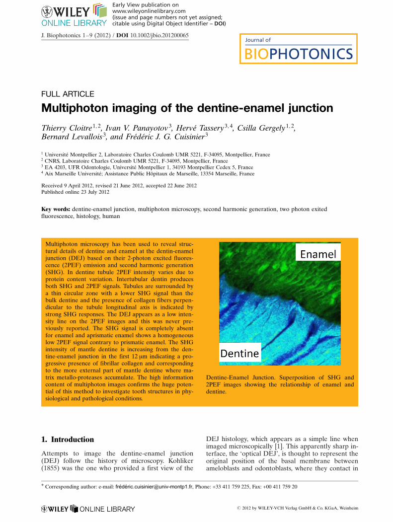
FULL ARTICLE
Multiphoton imaging of the dentine-enamel junction
Thierry Cloitre1; 2, Ivan V. Panayotov3, Herve Tassery3; 4, Csilla Gergely1; 2,Bernard Levallois3, and Frederic J. G. Cuisinier3
1 Universite Montpellier 2, Laboratoire Charles Coulomb UMR 5221, F-34095, Montpellier, France2 CNRS, Laboratoire Charles Coulomb UMR 5221, F-34095, Montpellier, France3 EA 4203, UFR Odontologie, Universite Montpellier 1, 34193 Montpellier Cedex 5, France4 Aix Marseille Universite; Assistance Public Hopitaux de Marseille, 13354 Marseille, France
Received 9 April 2012, revised 21 June 2012, accepted 22 June 2012Published online 23 July 2012
Key words: dentine-enamel junction, multiphoton microscopy, second harmonic generation, two photon exitedfluorescence, histology, human
1. Introduction
Attempts to image the dentine-enamel junction(DEJ) follow the history of microscopy. Kohliker(1855) was the one who provided a first view of the
DEJ histology, which appears as a simple line whenimaged microscopically [1]. This apparently sharp in-terface, the ‘optical DEJ’, is thought to represent theoriginal position of the basal membrane betweenameloblasts and odontoblasts, where they contact in
# 2012 by WILEY-VCH Verlag GmbH & Co. KGaA, Weinheim
Journal of
BIOPHOTONICS
Early View publication onwww.wileyonlinelibrary.com(issue and page numbers not yet assigned;citable using Digital Object Identifier – DOI)
Multiphoton microscopy has been used to reveal struc-tural details of dentine and enamel at the dentin-enameljunction (DEJ) based on their 2-photon excited fluores-cence (2PEF) emission and second harmonic generation(SHG). In dentine tubule 2PEF intensity varies due toprotein content variation. Intertubular dentin producesboth SHG and 2PEF signals. Tubules are surrounded bya thin circular zone with a lower SHG signal than thebulk dentine and the presence of collagen fibers perpen-dicular to the tubule longitudinal axis is indicated bystrong SHG responses. The DEJ appears as a low inten-sity line on the 2PEF images and this was never pre-viously reported. The SHG signal is completely absentfor enamel and aprismatic enamel shows a homogeneouslow 2PEF signal contrary to prismatic enamel. The SHGintensity of mantle dentine is increasing from the den-tine-enamel junction in the first 12 mm indicating a pro-gressive presence of fibrillar collagen and correspondingto the more external part of mantle dentine where ma-trix metallo-proteases accumulate. The high informationcontent of multiphoton images confirms the huge poten-tial of this method to investigate tooth structures in phy-siological and pathological conditions.
Dentine-Enamel Junction. Superposition of SHG and2PEF images showing the relationship of enamel anddentine.
* Corresponding author: e-mail: [email protected], Phone: +33 411 759 225, Fax: +00 411 759 20
J. Biophotonics 1–9 (2012) / DOI 10.1002/jbio.201200065
the embryological tooth bud. With the appearanceof transmission electron microscopy a step towarda precise morphological description of DEJ wasachieved [2]. The DEJ itself has a hierarchical micro-structure with a three dimensional scalloped appear-ance along the interface [3]. Developmental studiesindicate that DEJ is not restricted to a thin interfaceand is a transitional zone with spatiotemporal gradi-ent in both tissues [4]. Specifically collagen fibrils inthe transitional zone were either perpendicular tothe plane of the junction, or converged towards en-amel [3]. These coarse fibrils forming bundles of 80–120 nm in diameter appeared to be similar to the“von Korff fibers” [5]. The matrix of bulk dentinconsists of an interwoven fibrillar array, in which themajority of fibrils appeared to be either parallel orat angles correspondingly less than 90� to the planeof junction. High resolution electron microscopy(HREM) provides a description of the dentine en-amel relationship at the scale of the crystals [6].Atomic Force Microscopy (AFM) was used to imagethe dentine-enamel junction but mainly to study mi-crohardness of dentin and enamel on each side ofthe junction [7]. Synchrotron radiation-based microcomputed tomography (SR-CT) measurements werealso tested to better understand the inner structureof the collagen fibers [8].
Recently multiphoton microscopy has become animportant research tool for noninvasive imaging ofthick specimens with cellular resolution using 2-photon exited intrinsic fluorescence (2PEF) [9] andsecond harmonic generation (SHG) [10]. A numberof advantages render multiphoton microscopy awidely used imaging modality in many biomedicalapplications. The use of near infrared excitation al-lows deep sample penetration with benign irradia-tion. The 2PEF and SHG signals in multiphoton mi-croscopy are produced only at the focal point causingless sample photo-damage and enabling sectionalimaging. 2PEF image contrast is generated by theexcitation of tissue fluorophores. The characteristic2-photon exited fluorescence (2PEF) provides bothmorphological and functional information. In addi-tion collagen, microtubules and myosin, are tissueintrinsic and highly non-centrosymmetric molecularassemblies that possess first hyperpolarizabilitieslarge enough to produce SHG signals [10, 11]. Thephotons interact with the non-centrosymmetric sys-tems producing emission of a coherent light beam attwice the energy of the incident light [12]. As den-tine and cement are formed by a calcified collage-nous matrix, biphoton microscopy was used to imagesound teeth and carious tissue [13–16].
The aim of this work is to provide a multipho-tonic description of dentine and enamel structures atthe sound dentin-enamel junction. Although multi-photon imaging has been previously applied to in-vestigate dental structures here we present a detailed
study of dentine and enamel structures separated byDEJ, taking advantages of the 2PEF signal of enam-el and the strong SHG of dentinal collagen.
2. Materials and methods
2.1 Specimen preparation
Teeth extracted for orthodontic reasons were col-lected from University Hospital of Montpellier (writ-ten consent was signed before extraction). The teethwere stored in 0.9% phosphate buffer saline (PBS)buffer solution (pH ¼ 7.4) containing 0.002% sodiumazide at 4 �C. Eight freshly extracted teeth were sec-tioned in the longitudinal axis by means of an Iso-met diamond saw (Isomet 1000, Buehler, Lake Bluff,USA) with a thickness up to 0.5 mm. The sampleswere grinded to 0.25 mm, polished on carbure disksand using diamond pastes (6, 1 and 0.25 mm) with anEscil polishing machine (Escil, Lyon, France). Speci-mens were finally thoroughly cleaned up in waterwith an ultrasound bath for 5 min.
2.2 Multiphoton microscopy (MPM)
MPM images were recorded using a custom-builtmultiphoton microscope based on a Zeiss Imager Z1upright microscope. Excitation is provided by80 MHz femtosecond Ti-sapphire laser system (Tsu-nami Ti-Sapphire laser, Spectra-Physics, SpectraPhysics, Mountain View, USA) operated in pulsedmode (wavelength range 820–890, typically 880 nm,repetition rate 80 MHz, pulse duration 100 fs). Thelaser beam was fed into the back port of the micro-scope, deflected by a short pass 670 nm dichroicbeamsplitter (Chroma Technology Corp) and fo-cused onto the sample with a 40x, Fluar, N.A. 1.3oil-immersion microscope objective (Zeiss). Imageswere created by scanning the sample using an x/yscanning piezostage (P-542-2CD, Physik Instru-mente). Two-photon fluorescence was epi-collectedthrough the microscope objective. Fluorescence sig-nal was discriminated from backscattered laser exci-tation and SHG signal using a short pass dichroicmirror (670 dcspxruv-3p, Chroma Technology Corp)and a combination of long pass, short pass and colorglass filters (NT64-625, NT49-822 Edmund Opticsand CG BG39 1.0 CVI Melles Griot respectively).This filters combination result in 475–700 nm band-pass for the fluorescence detection channel. Secondharmonic signal was dia-collected through a largeaperture oil immersion condenser and was discrimi-nated from transmitted laser excitation an fluores-
T. Cloitre et al.: Multiphoton imaging of the dentine-enamel junction2
Journal of
BIOPHOTONICS
# 2012 by WILEY-VCH Verlag GmbH & Co. KGaA, Weinheim www.biophotonics-journal.org

cence signal using band pass (417–477 nm) and co-lored glass filters (NT48-074 Edmund Optics andCG BG39 2.0 CVI Melles Griot respectively). Fluo-rescence and SH signals were synchronously meas-ured using respectively a photomultiplier tube (R928PHamamatsu) and a photon counting unit (H7422PHamamatsu). The method enables the acquisition ofZ-stacks of 2D images of the sample with a typical Zresolution of 600–800 nm.
2.3 Image analysis
SHG and 2PEF images were viewed using Image J(NIH, Bethesda, MD, USA, http://rsb.info.nih.gov/ij/).Lookup tables (LUT) were applied on 2PEF andSHG images, respectively the Green LUT and the16-colors LUT. Intensity profile plots perpendicularto the dentine-enamel junction were calculated withthe same software. SHG image were overlaid on2PEF images using the co-localization finder pluginof Image J.
3. Results
Images of 8 polished DEJ sections were obtained bymeasuring second harmonic generation (SHG) andtwo photon exited fluorescence (2PEF) signals be-neath the surface to depths of up to about 70 mm.The 2PEF displays protein intrinsic fluorescence ofthe sample, whereas the SHG reveals the collagenfibrils due to their non-centrosymmetric structure.
When dentine is imaged in 2PEF and SHG (Fig-ure 1A and B), one can observe a network of lowintensity spots corresponding to perpendicular sec-tions of tubules. In the 2PEF image the whole dentinis green fluorescent except some tubules having a
very low 2PEF signal. Other tubules reveal an in-tense signal (for example the tubule indicated by ablack arrow on Figure 1A) indicating the presenceof fluorescent material. On the corresponding SHGimage (Figure 1B) regardless of the intensity of the2PEF emission, the tubules (for example the whitearrow indicating a tubule with strong 2PEF signal)reveal a very weak SHG indicating the absence ofhighly noncentrosymmetric molecular assemblies ascollagen and microtubules.. The mean diameter ofthe tubules on the 2PEF image is 1.3 � 0.1 mmwhereas on SHG images it is 4.0 � 0.2 mm on SHGimages; this difference will be discussed later. Addi-tionally we have observed variations in the intertu-bular dentine SHG signal. On the area showing astrong SHG signal (indicated by an orange red colorin Figure 1B) tubules are surrounded by a thin circu-lar zone with a lower SHG signal than the bulk den-tine.
A series of multiphoton images corresponding toan area including the dentine enamel junction ispresented in Figure 2. The upper row of images (Fig-ure 2A–C) depicts 2PEF signals and the bottom rowcorresponds to the SHG signal. The enamel is situ-ated at the upper right corner of images and dentinetubules end in the close vicinity of the DEJ. Imageswere recorded at various depths expressed in z va-lues: Figure 2A and D correspond to the samedepth; Figure 2B and F are recorded 2 mm under thesection presented in Figure 2A and D whereas Fig-ure 2C and G are recorded 2 mm under those de-picted in Figure 2B and F. We focus our attention onone tubule indicated by white arrows in the 2PEFimages and by black arrows in the SHG images ofthis z-scan series of images. In Figure 2A the tubuleis showing an internal intense 2PEF signal with smallisolated non fluorescent areas. On the next image(Figure 2B), 2 mm deeper in Z direction, the 2PEFintensity of the tubule is low and even weaker thanthe fluorescence of the intertubular dentine. Thisweak signal is probably due to peritubular dentinewith low organic content. In Figure 2C, 2 mm underthis area the signal diffuse without clear border andthe tubule looks wider than in Figure 2A and B. Thecorresponding SHG images (Figure 2D–F) show aweak SHG signal for all depth indicating an absenceof fibrous collagen in the tubule and in the peritubu-lar area as observed in Figure 1B.
The contact zone between enamel and dentinecan be well observed in the 2PEF images as an irre-gular line of low signal (Figure 3A, black arrow).The average line thickness measured at five spotsalong the junction is 2.4 � 0.4 mm. The SHG signal istotally absent in the enamel (Figure 3B). The den-tine tubules are parallel to the image plan and stopat the vicinity of the DEJ. They appear either withlow or with high fluorescence intensity. The superpo-sition of the 2PEF and SHG images enable to clearly
Figure 1 (online color at: www.biophotonics-journal.org)Dentine observed almost perpendicularly to canalicules.(A) 2PEF image. Black arrow: tubule with high fluores-cence; (B) SHG image: white arrow: same tubule than inA. The red–orange signal showing the SHG intensity ofthe collagen framework.
J. Biophotonics (2012) 3
FULLFULLARTICLEARTICLE
# 2012 by WILEY-VCH Verlag GmbH & Co. KGaA, Weinheimwww.biophotonics-journal.org

visualize the DEJ (Figure 3C). Figure 3D and E areenlargements of Figure 3A and B, respectively. Thediameters of tubules in 2PEF images are smallerthan in the corresponding SHG images as observedon Figure 3D, E that are enlargement of Figure 3Aand B. Strong SHG signals, as the one indicated by awhite arrow, are aligned parallel at a constant dis-tance of the tubule (Figure 3E). These strong signalsare produced by collagen fibers parallel to the exci-tation light [12] and they indicate the presence ofcollagen fibers perpendicular to the tubule longitudi-nal axis.
A high magnification of the 2PEF image of DEJ(Figure 4) reveals the transitional zone of the enam-el. A non-fluorescent line corresponding to the inter-face between dentin and enamel is observed (darkarrow in Figure 3) indicating the zone of DEJ. En-
amel prism sheaths exhibit a high fluorescence(white arrow in Figure 4). The aprismatic enamel isindicated by a dark bracket. This layer has a homo-geneous and lower fluorescence intensity comparedto the prismatic enamel. Inside the prisms the fluo-rescence is weak due to low protein content is noted.The interprismatic space also named enamel prismsheaths stops in the aprismatic enamel layer. The2PEF intensity of enamel and dentine are similar(Figure 4) which is somehow surprising since dentincontains 30 times more protein than enamel. 2PEFintensity depends of the number of fluorophores,cross sections and quantum yield of the tissue fluo-rophores. The main fluorescent amino acids are tryp-tophan, tyrosine and phenylalanine. They fluorescein the range of 350–280 nm but the objective and thedichroic filter of the microscope cut above 450 nm
Figure 2 (online color at:www.biophotonics-journal.org)2PEF and SHG images of DEJwith 2 mm thickness steps. Whitearrows show the terminal branch-ing of a tubule at different depthsin 2PEF images: (A): z ¼ 0 mm,(B): z ¼ �2 mm, (C): z ¼ �4 mm.Dark arrows show the same tu-bule in the corresponding SHGimages at different depths: (D):z ¼ 0 mm, (F): z ¼ �2 mm, (G):z ¼ �4 mm. There is no intenseSHG response corresponding tothe high fluorescence intensity inthe tubule.
Figure 3 (online color at:www.biophotonics-journal.org)Dentine-enamel junction. (A)2PEF image. Black arrow: DEJshowing a dark line due to a de-crease of 2PEF signal; (B) SHGimage: Enamel has no SHG sig-nal. (C) 2PEF/SHG images super-position; (D) Enlargement of A;(E) Enlargement of B: white ar-row indicating high intensity SHGsignal around the tubule.
T. Cloitre et al.: Multiphoton imaging of the dentine-enamel junction4
Journal of
BIOPHOTONICS
# 2012 by WILEY-VCH Verlag GmbH & Co. KGaA, Weinheim www.biophotonics-journal.org

and eliminate their contributions. Other intrinsicfluorophores fluorescing at 450 nm are NADH, fla-vins and porphyrins however these fluorophores arenot present in sound enamel. The fluorescence ofenamel is really not understood [17]. It was only de-monstrated that the one-photon fluorescence of en-amel decrease with demineralization. The origins ofauto fluorescence generated by dental tissue may be
quite diverse, and include contributions of both or-ganic and inorganic components [18, 19].
Furthermore we monitored the variation of theSHG signal at the dentine transitional zone of DEJ.As depicted in Figure 5 SHG signal at the dentinetransitional zone of DEJ. SHG intensity increases ina two regime rate starting from the DEJ clearly ob-served as a border line between the dentine and theenamel. Enamel produces no SHG signal. In thedentine we observe a transition zone of 14 mm wherethe SHG signal varies continuously: in the closestpart to the enamel (zone 1) the increase of SHG sig-nal is faster than in zone 2, deeper into the dentin.After 14 mm from the DEJ, SHG intensity reaches aplateau indicating a constant collagen fibrillar struc-ture within the dentine.
4. Discussion
4.1 Dentine tubules
The origin of the observed variations in the 2PEF in-tensities of dentine tubules is not straightforward.An opposite observation was made by Chen et al.on longitudinal dentin slices [13]. After superposi-tion of 2PEF and SHG images, they found that thecollagen composition of the dentinal tubules pro-duces a strong SHG signal.
Our first hypothesis could be that the strong 2PEFemission in the dentine tubules is due to the odonto-blast process containing fluorescent proteins. A tu-bule without fluorescence could point an empty tu-bule due to the retraction of the odontoblast processas no fixation of dental tissue was performed [20].The absence of SHG signal in the highly fluorescenttubule is challenging this hypothesis as previous elec-tron transmission studies indicated that odontoblastprocesses involve a high density of microtubules andmicrofilaments [21]. Microtubules reveal SHG signalas they are non centrosymmetric [9]. These elementsare parallel to the tubules and aim at keeping theodontoblast process elongated. The stability of themicrotubules is very weak with an average life span-ning from 10 min to 2 hours. Therefore the chance toretrieve SHG signal of microtubules is very low inour experimental conditions. The presence of micro-tubules would have also been observed by SHGwhen changing orientation between molecule’s axisand incident light. If the elongated molecular ele-ments are parallel to the incident light the SHG sig-nal is weaker [22] and we would expect for SHGimages recorded orthogonally to tubules (Figure 2B)that microtubules involved in the odontoblast pro-cess have a high signal. However we never detectedsuch a high SHG signal in tubules. Dentine tubule
Figure 4 2PEF image of dentine-enamel junction. Blackarrow: DEJ; Black left square bracket: inner aprismaticenamel; white arrow: enamel prism sheath with high 2PEFintensity.
Figure 5 (A) SHG image of DEJ. Enamel gives no SHGsignal. The frame is corresponding to the area used to plotSHG intensity. (B) Profile plot of SHG intensity perpendi-cular to DEJ as indicated by the black frame on A, zone 1is corresponding to a fast increase of SHG intensity, inzone 2 intensity growth decreases and then reaches a pla-teau.
J. Biophotonics (2012) 5
FULLFULLARTICLEARTICLE
# 2012 by WILEY-VCH Verlag GmbH & Co. KGaA, Weinheimwww.biophotonics-journal.org
might also behave as an optical waveguide. In po-rous media such as porous silicon light confinementwas observed [23] when light path was parallel toporous cavities. Similarly to those studies, we neverobserved a high SHG signal in dentine whatever thelight path.
Therefore it seems that the inconstant high 2PEFsignal observed in dentine tubule is not related eitherto the presence of odontoblast processes, neither toan intrinsic property to guide light but is simply dueto the presence of intrinsic fluorophores. Irregularelectron-dense material within the tubules in ab-sence of cellular processes was reported in dentinestudied by transmission electron microscopy (TEM)[24]. The organic periodontoblastic space ie thespace between the odontoblast process membraneand the mineralized wall of the tubule is filled by un-calcified collagen fibrils and an amorphous or finelygranular ground substance when observed by TEM[25]. Glycosaminoglycan’s were localized by stainingin the periodontoblastic space [26]. The observed ir-regular fluorescent materials are certainly this peri-odontoblastic organic matrix aggregating when theodontoblast process retracts.
4.2 Diameter of dentine tubules
The mean diameter of the tubules in the 2PEF im-age is 1.3 � 0.1 mm. This value is corresponding tothe lumen diameter of tubules measured by scan-ning electron microscopy [27]. On SHG images tu-bules have a larger diameter of 4.0 � 0.2 mm. Theabsence of SHG signals is due to the absence of fi-brillar collagen in tubule lumen and in peritubulardentine [28, 29]. The measured dimension corre-sponds to the outer diameter of peritubular dentineobserved by AFM [30]. Peritubular outer diameterdetermined by SEM is smaller (2.5 mm) [31]. Thedifference between tubule diameters measured inthe 2PEF and SHG images corresponds to the thick-ness of peritubular dentine. The peritubular dentineis characterized by an absence of SHG signal due toits high mineralization and its low content of pro-tein.
We observe a difference also between the 2PEFsignals of dentin tubules imaged longitudinally. High2PEF intensity is corresponding to the central partof the tubule due to an accumulation of intrinsicfluorescent proteins in the tubule. The same tubule,in a different plane, has an aspect of ghost due todiffuse low fluorescence extending in intertubulardentine. This low fluorescence originates in the pre-sence of the peritubular dentine with low organiccontent. It should be noted that the peritubular den-tine is present until the DEJ even in case of Y termi-nated tubule.
4.3 Variation of intensityof intertubular dentine
It was already described that intertubular dentinproduce both SHG and 2PEF signals [13]. Addition-ally we have observed variations in the SHG signalof the intertubular dentine. In the area exhibiting astrong SHG signal (indicated by an orange red colorin Figure 1B) tubules are surrounded by a thin circu-lar zone with a lower SHG signal than the bulk den-tine.
Around dentin tubules strong SHG signals areobserved when tubules are imaged longitudinally.The observed variation of the SHG signal points to-wards different orientations of collagen fibrils. Whenthe laser polarization is oriented parallel with theaxis of the collagen fibril a maximal SHG signal isproduced [12, 32]. Therefore our SHG images indi-cate the presence of collagen fibers perpendicular tothe tubule’s longitudinal axis. Our results are in goodagreement with previous observations, where groupsof fibrils have appeared to follow a circular or obli-que course [33]. Such collagen network was also ob-served by AFM [34].
4.4 Enamel
SHG signal is totally absent in enamel (Figure 2D–Fand Figure 3E). Human enamel crystals are poorlycrystalline carbonated apatite with a P63/m spacegroup [35]. This space group belongs to the 6/mpoint group of the hexagonal crystal system. Thesecond-order susceptibility tensor (c(2)), expressingnon-linear phenomena like SHG, vanishes for the 6/msymmetry group [36]. Deviation from the hydroxy-apatite hexagonal symmetry of a human tooth enam-el crystal observed by high-resolution electron mi-croscopy was reported [37]. Absence of SHG ofenamel indicates either that such deviation affects alimited number of crystals either that they are toosmall to modify the non-linear response of enamel.Mature enamel contains 0.36 per cent of protein[38]. A recent paper has postulated the presence offiber i.e. non centrosymmetric protein in enamel[39]. The absence of SHG in enamel is also suggest-ing an absence of non centrosymmetric protein.
4.5 Dentine Enamel Junction
DEJ is indicated as a low intensity line in the 2PEFimages. The absence of 2PEF signal at the DEJ wasnever reported previously. This indicates an absenceof endogenous fluorophores at DEJ, contrary to the
T. Cloitre et al.: Multiphoton imaging of the dentine-enamel junction6
Journal of
BIOPHOTONICS
# 2012 by WILEY-VCH Verlag GmbH & Co. KGaA, Weinheim www.biophotonics-journal.org

dentine where major fluorophores are tryptophan,collagen and elastin [40]. The dark line is certainlyindicating a very low concentration of protein at theDEJ. The superposition of the 2PEF and SHGimages allow to visualize clearly the DEJ and provesthat SHG signals quenches exactly at the level ofDEJ.
The invaginations of enamel into dentine providea scalloped structure with convexities directed to-wards the dentine and concavities towards the enam-el [16, 41, 42]. Aprismatic enamel shows a homoge-neous low 2PEF signal contrary to enamel prism. Inprismatic enamel the dominant 2PEF contrast comesfrom the organic-matrix-filled sheaths. The prismsreveal less fluorescence than the sheath due to theirlower protein content. Similar enamel images weretaken by Chen et al. (2008) using a Third HarmonicGeneration (THG) microscopy [43]. Because of theabsence of SHG signal of enamel they used THGgenerated from the interprismatic spaces to revealthe prism structure. In our case we never observedSHG signal at the DEJ. As it is known that strain incrystals can cause the change in symmetry and thusinduce SHG [44, 45], the absence of SHG signal inenamel indicates an absence of stress in enamel crys-tals at the DEJ.
The SHG intensity of mantle dentine is increas-ing from the level of DEJ towards the dentine. Theincrease rate is higher in the first 4 mm than in thefollowing 8 mm. This 12 mm zone is corresponding tothe more external part of mantle dentine were VonKorf collagen fibrils are present. We pointed threehypotheses to explain this increase. MMP-2 (Gelati-nase A) in both pro (�72 kDa) and active (�68 kDa)forms has been identified in human dentin [46].MMP-2 is the predominant matrix metalloproteinasein mineralized dentin and may be associated withthe collagen matrix but not with hydroxyapatite[46]. Analysis revealed immunoreactivity for MMP-2throughout human coronal dentin with intense label-ing in a 9–10 mm wide zone adjacent to the DEJ[47]. Recently in enamelysin (MMP-20) knockoutmice a 10 mm wide hypomineralized area was de-scribed [48]. MMP-2 in forming rat incisors may beconcentrated in an area adjacent to the dentine-en-amel junction [49]. Collagen denaturation decreasesSHG intensity and it was demonstrated that collagendegradation with MMP collagenase decrease SHGintensity [50]. Taking into account the accumulationof different MMP’s in this zone and the effect ofMMP on collagen SHG signal we can hypothesizean influence of MMP’s on dentine SHG.
Also the chemical composition profiles (matrix/mineral ratios) determined by Raman spectroscopychange in the first layer of dentine. They increasedgradually from nearly zero across the dentin-enameljunction to a constant value in dentine. The width ofthis transition zone was determined to be in the
range of 5–16 mm [51]. This variation of the Ramanpeak intensity ratio indicates a progressive increasein protein concentration. Dentine carbonated apatitecrystal, as enamel crystal, do not generate SHG.More than ninety percent of the dentine organic ma-trix is fibrillar collagen type I and collagen is astrong SHG emitter. Our second hypothesis is thatSHG intensity is following the matrix/mineral ratioobserved by Raman spectroscopy.
The increase of SHG observed in our study canalso be explained with reorientation of the collagenfibrils before their interaction with the zone of non-prismatic enamel. Collagen fibrils in the junctionarea are oriented parallel to each other and perpen-dicular to the plane of the junction [52]. This is co-herent with the increase of SHG observed in ourstudy as it is well known that SHG intensity dependsof the respective orientation of the laser light andthe fibril [22]. If they are perpendicular, the intensityof the forward SHG emission is low and is high incase of parallel orientation. In contrast to these ori-ented collagen fibrils entering the enamel, the ma-trix of bulk dentin was seen to consist of an inter-woven fibrillar array, in which the majority of fibrilsappeared to be either parallel or at angles corre-spondingly less than 90� to the plane of the junction[52].
Using multiphoton microscopy it is impossible toselect one of these three hypotheses and it is possi-ble that the three mechanisms cooperate to createthe SHG increase in the area. However the associa-tion of 2PEF and SHG images in our measurementsprovides a full description of enamel and dentinestructures in the DEJ vicinity.
5. Conclusions
In this study we report 2PEF and SHG images re-corded by multiphoton microscopy enabling to de-scribe two transitional zones between enamel anddentine reflecting the early developmental stages infirst formed enamel and dentin. This technique there-by allows identifying the DEJ and structural charac-teristic of immediately surrounding tissue character-istics. We found different degrees of organic materialin the dentin structure that provokes related changesin the 2PEF and SHG signals. The high quality ofimages and the simplicity of sample’s preparationwithout any histology treatment render the multi-photon microscopy a powerful methodology to in-vestigate the tooth structures on a sub-micrometricscale. The presented results demonstrate the opticalsectioning power of this imaging technique, hencefuture investigations will aim to describe the differ-ences between normal tooth structures and carioustooth tissues.
J. Biophotonics (2012) 7
FULLFULLARTICLEARTICLE
# 2012 by WILEY-VCH Verlag GmbH & Co. KGaA, Weinheimwww.biophotonics-journal.org

Thierry Cloitre received hisPh.D. in physics of con-densed matter from theSciences University of Mont-pellier, France in 1992. Hehas turned its research activ-ity from semiconductor phy-sics to biophysics in 2006.He is an assistant professor inthe Bionanophotonics teamof the Charles Coulomb La-
boratory, Montpellier 2 University. His main contribu-tions rely on his expertise in the field of optical physicsfor the development of non-linear optical microscopyexperimental set-ups and the study of innovative solu-tions for optical and electrical devices for biosensingapplications.
Ivan Vladislavov Panayotov,doctor of dentistry, receivedhis Master of Sciences, Tech-nologies and Health in 2009at Montpellier 1 University(France). Currently, he isPh.D. student in EA4203Laboratory of Nano-science
and assistant professor in endodontic. His research in-terests are in the functionalization of biomaterials withpolyelectrolyte multilayers and specific peptides formedical applications and in the development of newtechnologies to detected structural changes in minera-lized tooth tissues.
Herve Tassery has receivedhis Ph.D. in biomaterialsfrom Aix Marseille Univer-sity 2001. Currently, he isprofessor and head of theDepartment of RestorativeDentistry of Marseille Den-tal School at Aix-MarseilleUniversity. His major fieldsof interest are in cariology,fluorescence devices, mini-mally invasive dentistry andclinical researches. Workingin the Laboratory of biona-
notechnology of Montpellier 1 University, (EA 4203)his actual research interest lies in improving the linksbetween fundamental researches, clinical researchesand clinical applications.
Csilla Gergely has receivedher Ph.D. in biophysics fromSzeged University, Hungaryin 1997. Currently, she isprofessor in the CharlesCoulomb Laboratory of theMontpellier 2 University inFrance and head of the Bio-nanophotonics team. Heractual research interest liesin (i) elaborating adhesionpeptides for selective bio-functionalization of photoniccrystals for elaboration of
semiconductors based biosensing and (ii) developingvarious microscopic techniques (near-probe and multi-photon microscopy) for functional imaging and follow-up-therapy of cancerous cells.
Bernard Levallois receivedhis doctorate in dentistry in1986 and his Ph.D. in 1999.He is head of the OperativeDentistry and endodonticdepartment of Montpellierhospital. His research inter-ests are in endodontic, bio-material and caries diagnos-tic. He is deputy director ofLaboratoire Biologie Santeet nanoscience (EA4203) ofMontpellier university.
Frederic J. G. Cuisinier, pro-fessor in Dentistry, receivedis Ph.D. in 1989 at Stras-bourg University under thedirection of Pr. RobertFranck. He authored morethan 90 papers in High Re-solution Electron micro-scopy of mineralized tissues,in biomineralization, in bio-sensor and in multilayer filmbuild-up. His current re-
searches concern confocal Raman microscopy of livingcells, multiphotonic microscopy of dental tissue andbiomaterial interactions with dental pulp stem cells.
T. Cloitre et al.: Multiphoton imaging of the dentine-enamel junction8
Journal of
BIOPHOTONICS
# 2012 by WILEY-VCH Verlag GmbH & Co. KGaA, Weinheim www.biophotonics-journal.org

References
[1] A. Kolliker, Elements d’histologie humaine: Parties 1a 5. Paris: Masson; 1955.
[2] M. B. Quigley, J. Dent. Res. 38, 558–568 (1959).[3] C. P. Lin, W. H. Douglas, and S. L. Erlandsen, J. Histo-
chem. Cytochem. 41, 381–388 (1993).[4] J. Meyer, P. Bodier-Houlle, F. Cuisinier, H. Lesot, and
J. V. Ruch, In Vitro Cell. Dev. A 35, 159–168 (1999).[5] D. B. Scott and M. U. Nylen, Ann. N.Y. Acad. Sci. 85,
133–144 (1960).[6] P. Bodier-Houlle, P. Steuer, J. Meyer, L. Bigeard, and
F. Cuisinier, Cell Tissue Res. 301, 389–395 (2000).[7] H. Fong, M. Sarikaya, S. White, and M. Snead, Mat.
Sci. Eng. C.-Bio. S 7, 119–128 (2000).[8] H. Deyhle, O. Bunk, and B. Muller, Nanomed-Nano-
technol. 7, 694–701 ( 2011).[9] W. Denk, J. Strickler, and W. Webb, Science 248, 73–
76 (1990).[10] W. Zipfel, R. Williams, R. Christie, A. Nikitin, B. Hy-
man, and W. Webb, Proc. Nat. Acad. Sci. USA 100,7075–7080 (2003).
[11] I. Freund, M. Deutsch, and A. Sprecher, Biophys. J.50, 693–712 (1986).
[12] R. Williams, W. Zipfel, and W. Webb, Biophys. J. 88,1377–1386 (2005).
[13] M.-H. Chen, W.-L. Chen, Y. Sun, P. T. Fwu, and C.-Y.Dong, J. Biomed. Opt. 12, 40181–40186 (2007).
[14] J. M. Girkin, A. F. Hall, and S. L. Creanor. Multi-photon imaging of intact dental tissue, in: Proceedingsof the 4th Annual Indiana Conference, G. K. Stookey,ed. (Indiana University School of Dentistry, India-napolis, Indiana 1999), pp. 155–168.
[15] A. Hall and M. Girkin, J. Dent. Res. 83, 89–94 (2004).[16] P.-Y. Lin, H.-C. Lyu, C.-Y. S. Hsu, C.-S. Chang, and
F.-J. Kao, Biomed. Opt. Exp. 2, 149–158 (2010).[17] L. Bachmann, D. M. Zezell, A. da Costa Ribeiro,
L. Gomes, and A. S. Ito, Appl. Spectrosc. Rev. 41,575–590 (2006).
[18] U. Hafstrom-Bjorkman, F. Sundstrom, and J. J. TenBosch, Acta. Odontol. Scand. 49, 133–138 (1991).
[19] D. Spitzer and J. J. Ten Bosh, Calcif. Tiss. Res. 24,249–251 (1977).
[20] R. Frank and P. Steuer, Arch. Oral. Biol. 33, 91–98(1988).
[21] R. Frank, Arch. Oral. Biol. 1, 29–32 (1959).[22] C. Raub, J. Unruh, V. Suresh, T. Krasieva, T. Lindmo,
E. Gratton, B. Tromberg, and S. George, Biophys. J.94, 2361–2373 (2008).
[23] M. Martin, G. Palestino, T. Cloitre, V. Agarwal, L. Zi-manyi, and C. Gergely, Appl. Phys. Lett. 94, (2009).
[24] G. R. Holland, J. Anat. 120, 169–177 (1975).[25] R. M. Frank, Arch. Oral. Biol. 11, 179–192 (1966).[26] M. Goldberg and M. Takagi, Histochem. J. 25, 781–806
(1993).
[27] J. Arends, I. Stikroos, W. Jongebloed, and J. Ruben,Caries. Res. 29, 118–121 (1995).
[28] L. Bertassoni, K. Stankoska, and M. Swain, Micron.43, 229–236 (2012).
[29] L. Schroeder and R. Frank, Cell Tissue Res. 2, 449–451 (1985).
[30] G. W. Marshall, S. Marshall, J. Kinney, and M. Balooch,J. Dent. 25, 441–458 (1997).
[31] C.-Y. Chu, T.-C. Kuo, S.-F. Chang, Y.-C. Shyu, and C.-P.Lin, J. Dent. Sci. 5, 14–20 (2010).
[32] C. Pfeffer, B. Olsen, F. Ganikhanov, and F. Legare, J.Struct. Biol. 164, 140–145 (2008).
[33] E. Johansen, Ann. Ny Acad. Sci. 131, 776–785 (1965).[34] S. Habelitz, M. Balooch, S. Marshall, G. Balooch, and
G. Marshall, J. Struct. Biol. 138, 227–236 (2002).[35] E. F. Bres, D. Cherns, R. Vincent, and J.-P. Morniroli,
Acta. Cryst. B49, 56–62 (1993).[36] R. Boyd (ed.), Non linear Optics: Academic press;
1992.[37] E. F. Bres, P. Steuer, J.-C. Voegel, R. M. Frank, and
F. J. G. Cuisinier, J. Microsc. 170, 147–154 (1993).[38] J. E. Eastoe, Nature 187, 411–412 (1960).[39] S. V. Kalinin, B. J. Rodriguez, J. Shin, S. Jesse, V. Grich-
ko, T. Thundat, A. Baddorf, and P. Gruverman, Ultra-microscopy 106, 334–340 (2006).
[40] M. L. Sinyaeva, A. A. Mamedo, S. Y. Vasilchenko, A. I.Volkova, and V. B. Loschenov, Laser Phys. 14, 1132–1140 (2004).
[41] D. Whittaker, J. Anat. 125, 323–335 (1978).[42] D. Brauer, G. Marshall, and S. Marshall, J. Dent. 38,
597–601 (2010).[43] S. Chen, C. S. Hsu, and C. Sun, Opt. Express 16, 11670–
11679 (2008).[44] I. L. Lyubchanskii, N. N. Dadoenkova, M. I. Lyub-
chanskii, T. Rasing, J. W. Jeong, and S. C. Shin, Appl.Phys. Lett. 76, 1848–1850 (2000).
[45] T. Zhao, H. Lu, F. Chen, G. Yang, and Z. Chen, J.Appl. Phys. 87, 7448–7451 (2000).
[46] S. M. D. L. Heras, A. Valenzuela, and C. Overall,Arch. Oral. Biol. 45, 757–765 (2000).
[47] L. Boushell, M. Kaku, Y. Mochida, R. Bagnell, andM. Yamauchi, Arch. Oral. Biol. 53, 109–116 (2008).
[48] E. Beniash, Z. Skobe, and J. Bartlett, Eur. J. Oral. Sci.114, 24–29 (2006).
[49] M. Goldberg, D. Septier, K. Bourd, R. Hall, A. George,H. Goldberg, and S. Menashi, Connect. Tissue Res.44, 143–153 (2003).
[50] P. C. Stoller, K. M. Reiser, P. M. Celliers, and A. M.Rubenchik, Proc. SPIE 4963 41–51 (2003).
[51] C. Xu, X. Yao, M. Walker, and Y. Wang, Calcif. TissueInt. 84, 221–228 (2009).
[52] C. Lin, W. Douglas, and S. Erlandsen, J. Hist. Cyto-chem. 41, 381–388 (1993).
J. Biophotonics (2012) 9
FULLFULLARTICLEARTICLE
# 2012 by WILEY-VCH Verlag GmbH & Co. KGaA, Weinheimwww.biophotonics-journal.org
Copyright © 2022 FDOKUMEN