High-Resolution Multiphoton Imaging of Tumors In Vivo
19
Topic Introduction High-Resolution Multiphoton Imaging of Tumors In Vivo Jeffrey Wyckoff, Bojana Gligorijevic, David Entenberg, Jeffrey Segall, and John Condeelis Analysis of the individual steps in metastasis is crucial if insights at the molecular level are to be linked to the cell biology of cancer. A technical hurdle to achieving the analysis of the individual steps of metastasis is the fact that, at the gross level, tumors are heterogeneous in both animal models and patients. Human primary tumors show extensive variation in all properties ranging from growth and morphology of the tumor through tumor-cell density in the blood and formation and growth of metastases. Methods capable of the direct visualization and analysis of tumor-cell behavior at single-cell resolution in vivo have become crucial in advancing the understanding of mechanisms of metastasis, the definition of microenvironment, and the markers related to both. This article discusses the use of high-resolution multiphoton imaging of tumors (specifically breast tumors in mice) in vivo. RELATED INFORMATION See also The In Vivo Invasion Assay: Preparation and Handling of Collection Needles (Wyckoff et al. 2011). BACKGROUND Our primary interest has been in breast tumors because metastasis is dependent on cell motility invol- ving macrophage-mediated chemotaxis (Condeelis and Pollard 2006). Invasive migration and entry of tumor cells into the circulation, common features of breast tumors, are crucial early steps in the meta- static cascade and have been assayed in various ways but only indirectly using conventional methods (Liotta et al. 1974; Butler and Gullino 1975; Glaves 1986). Human breast carcinomas, in particular, the vast majority of which are ductal carcinomas, are extremely heterogeneous both clinically and pathologically. Current methods of histologic grading, such as the modified Bloom–Richardson method, allow pathologists to separate tumors into three strata. Distinct differences in survival rates, however, effectively apply only to those patients with very well-differentiated or low-grade tumors. Recurrence-free survival for grade 1 carcinomas at 5 and 10 yr, for example, are 75% and 64%, respectively, while the same figures for grade 2 carcinomas are 50% and 40%, and for grade 3 carcinomas are 40% and 38%. This suggests considerable overlap in features of grades 2 and 3 tumors, with classification by grade being a very imperfect means for effec- tive tailoring of management and prediction of outcome (Elston and Ellis 1991; Carter 2001). Gene-expression profiling with microarrays has shown promise in refining these groups further (Ramaswamy et al. 2003) and supports the notion that the invasive and metastatic potentials of the primary tumor are encoded early in the bulk of the tumor, including the stroma (Bernards and Adapted from Live Cell Imaging, 2nd edition, by Goldman et al. CSHL Press, Cold Spring Harbor, NY, USA, 2010. © 2011 Cold Spring Harbor Laboratory Press Cite this article as Cold Spring Harbor Protoc; 2011; doi:10.1101/pdb.top065904 1167 Cold Spring Harbor Laboratory Press on May 3, 2016 - Published by http://cshprotocols.cshlp.org/ Downloaded from
Transcript of High-Resolution Multiphoton Imaging of Tumors In Vivo
Topic Introduction
High-Resolution Multiphoton Imaging of Tumors In Vivo
Jeffrey Wyckoff Bojana Gligorijevic David Entenberg Jeffrey Segall and John Condeelis
Analysis of the individual steps in metastasis is crucial if insights at the molecular level are to be linkedto the cell biology of cancer A technical hurdle to achieving the analysis of the individual steps ofmetastasis is the fact that at the gross level tumors are heterogeneous in both animal models andpatients Human primary tumors show extensive variation in all properties ranging from growthand morphology of the tumor through tumor-cell density in the blood and formation and growthof metastases Methods capable of the direct visualization and analysis of tumor-cell behavior atsingle-cell resolution in vivo have become crucial in advancing the understanding of mechanisms ofmetastasis the definition of microenvironment and the markers related to both This article discussesthe use of high-resolution multiphoton imaging of tumors (specifically breast tumors in mice) in vivo
RELATED INFORMATION
See also The In Vivo Invasion Assay Preparation and Handling of Collection Needles (Wyckoffet al 2011)
BACKGROUND
Our primary interest has been in breast tumors because metastasis is dependent on cell motility invol-vingmacrophage-mediated chemotaxis (Condeelis and Pollard 2006) Invasivemigration and entry oftumor cells into the circulation common features of breast tumors are crucial early steps in the meta-static cascade and have been assayed in various ways but only indirectly using conventional methods(Liotta et al 1974 Butler and Gullino 1975 Glaves 1986)
Human breast carcinomas in particular the vast majority of which are ductal carcinomas areextremely heterogeneous both clinically and pathologically Current methods of histologic gradingsuch as the modified BloomndashRichardson method allow pathologists to separate tumors into threestrata Distinct differences in survival rates however effectively apply only to those patients withvery well-differentiated or low-grade tumors Recurrence-free survival for grade 1 carcinomas at 5and 10 yr for example are 75 and 64 respectively while the same figures for grade 2 carcinomasare 50 and 40 and for grade 3 carcinomas are 40 and 38 This suggests considerable overlap infeatures of grades 2 and 3 tumors with classification by grade being a very imperfect means for effec-tive tailoring of management and prediction of outcome (Elston and Ellis 1991 Carter 2001)Gene-expression profiling with microarrays has shown promise in refining these groups further(Ramaswamy et al 2003) and supports the notion that the invasive and metastatic potentials of theprimary tumor are encoded early in the bulk of the tumor including the stroma (Bernards and
Adapted from Live Cell Imaging 2nd edition by Goldman et al CSHL Press Cold Spring Harbor NY USA 2010
copy 2011 Cold Spring Harbor Laboratory PressCite this article as Cold Spring Harbor Protoc 2011 doi101101pdbtop065904
1167
Cold Spring Harbor Laboratory Press on May 3 2016 - Published by httpcshprotocolscshlporgDownloaded from
Weinberg 2002) However none of these approaches reveals the mechanistic basis for the differencesin the outcome of human breast tumors Additional approaches that interrogate tumors as to themicroenvironments that support invasion and metastasis using common markers in otherwise het-erogeneous tumors may reveal mechanisms behind the outcome in sufficient detail to generatenew strategies for marker development that can be used in routine histopathological diagnosis prog-nosis and treatment
MULTIPHOTON INTRAVITAL IMAGING AT SINGLE-CELL RESOLUTION
Within the past several years imaging methods for the detailed characterization of the behavior ofcarcinoma cells and macrophages within intact primary tumors using multiphoton imaging com-bined with fluorescent animal models has become routine (Condeelis and Segall 2003 Wyckoff etal 2004 2006 2007 Sahai et al 2005 Sidani et al 2006 Soon et al 2007 Wang et al 2007)These methods give information directly about cell behavior in vivo at single-cell resolution and indifferent microenvironments of the tumor an essential capability given the heterogeneity oftumors Imaging is inherently quantitative allowing the quantification of behavior parametersincluding directional migration toward histological landmarks such as blood vessels frequency vel-ocity and persistence of cell motility in vivo as well as interactions between tumor cells and stromalcells leading to invasion intravasation and extravasation These imaging methods are valuable indefining (1) cell behaviors that are necessary for invasion intravasation and extravasation (2) cellbehavior phenotypes of cells with specific mutations (3) polarized motility and chemotaxis of cellsin vivo and (4) the definition size and regulation of microenvironments in vivo For more infor-mation about multiphoton microscopy and its applications to the study of cell behavior in vivosee our websites wwweinsteinyueduaifpageaspx and wwweinsteinyueduaifintravital_imagingintroductionhtm
ADVANTAGES OF MULTIPHOTON MICROSCOPY OVER CONFOCAL MICROSCOPY
We have shown that multiphoton excitation fluorescence microscopy has important advantages overother imaging techniques such as confocal microscopy particularly for the study of live cells andorfor thick tissues (Farina et al 1998 Wang et al 2002) The advantages of the multiphoton instrumentsinclude the following
1 The lack of fluorophore excitation in regions away from the focal plane minimizes fluorophorebleaching and the generation of toxic by-products during imaging
2 Multiphoton images are less prone to degradation by light scattering This is because the longerwavelengths used for excitation suffer less scattering from microscopic refractive-index differ-ences within the sample allowing much greater penetration of the tissues (Centonze andWhite 1998) In addition as all the resolution is defined by the geometry of the excitationbeam the fluorescence emission is unaffected by light scattering The reduced sensitivity of multi-photon imaging to light scattering is particularly advantageous for the study of living specimensbecause of the presence of many refractive-index interfaces in living tissues
3 The longer wavelengths used in multiphoton microscopy have the added benefit of second-harmonic generation to image collagen fibers (Campagnola et al 2001 Williams et al 2001)This phenomenon is not found in conventional microscopy and allows for imaging ofcell-to-matrix interactions such as adhesion and degradation
A valuable benefit resulting from the second-harmonic-generated image is the ability to directly esti-mate the amount and condition of the extracellular matrix (ECM) adjacent to carcinoma cells intumors The amount integrity and size of ECM fibers can be determined by measuring the signal
1168 Cite this article as Cold Spring Harbor Protoc 2011 doi101101pdbtop065904
J Wyckoff et al
Cold Spring Harbor Laboratory Press on May 3 2016 - Published by httpcshprotocolscshlporgDownloaded from
intensity and morphology of fibers in images obtained from the second-harmonic-generated lightemitted from the ECM (Wang et al 2002) An important insight resulting from this analysis is thedemonstration that adjacent to carcinoma cells in the primary tumor metastatic MTLn3-derivedtumors contain two- to threefold less ECM fibers than nonmetastatic MTC-derived tumors (Wanget al 2002) This is determined by calculating the pixel intensity of a volume using the National Insti-tutes of Health (NIH) Image software Furthermore the morphology of the fibers shows discontinu-ities in second-harmonic emissions along the fibers suggesting that proteolysis has occurred atintervals along the ECM fibers (Wang et al 2002 Sidani 2006) Finally these fibers are observed tosupport the rapid and directional linear migration of carcinoma cells in metastatic tumors (Wanget al 2002) This is an exciting observation because chemotaxis of carcinoma cells in response to epi-dermal growth factor-like (EGF-like) ligands resulting from proteolysis of the ECM could be animportant property of invasive tumors (Liotta and Kohn 2001)
The second-harmonic signal is emitted as polarized light when the electrons in the π orbitals ofα-helix chains such as those found in collagens are excited by a long wavelength of light (Campagnolaet al 2001 Williams et al 2001) We have found that we are able to excite the second-harmonic signalwith wavelengths of 760ndash960 nm and image them through a filter with a 450ndash480-nm cutoff
Direct comparisons of confocal imaging with multiphoton imaging have demonstrated thatalthough the deepest useful image possible with the conventional confocal was lt60 microm usefulimages were obtained with the multiphoton microscope to depths gt300 microm (Wang et al 2002Kedrin et al 2008) In addition the bleach rate is much less in the multiphoton microscope andsignal to noise does not fall off significantly throughout the z series obtained with the multiphoton(Fig 1) In both imaging techniques (ie in the live animal under anesthesia on either a laser-scanningconfocal microscope or a multiphotonmicroscope) we found that green fluorescent protein (GFP) inthe tumor cells is an excellent cytoplasmic volume marker that allows the entire cell outline to bedefined in vivo in the living intact tissue without rejection of the tumor due to GFP expression(Farina et al 1998 Condeelis et al 2000)
ANIMAL MODELS FOR MULTIPHOTON IMAGING TRANSGENIC MICE
To relate the expression of oncogenes to the progression and metastasis of primary breast tumors thetransgenic mouse has served as an important research tool to assess the tissue-specific action of onco-genes in vivo Of particular importance are transgenic mouse models of breast cancer that resemblethe human disease in both etiology and histology These mouse models are derived from theexpression of either the polyomavirus middle T antigen (PyMT) or neuerbB-2 under the controlof the mammary epithelium-specific mouse mammary tumor virus MMTV promoter The MMTVpromoter has been used in a number of studies to generate expression of proteins in the mammarygland (Gunzburg et al 1991 Webster and Muller 1994 Boumlttinger et al 1997) Expression studiesusing this promoter to drive β-galactosidase expression indicate that in female mice the major siteof expression is in the epithelial cells of the mammary gland
Transgenic mice expressing either normal or activated forms of neu have been generated byMullerand colleagues (Muller et al 1998 Siegel et al 1999) Mice harboring the activated transgene (NDL1)developmammary adenocarcinomas with an average onset of 135 d (plusmn37) and ~57of those animalsdevelop lung metastases within an average of 45 d after the appearance of the primary tumor ErbB-2family members directly interact with a diverse set of signaling proteins such as Src PTKs Shc PLC-γand PI3K The PyMT is a related oncogene that exerts its oncogenic effects through its association withSrc PTKs The signaling path from Src onward in PyMT mice is believed to be very similar to that inactivated-neu mice (Muller et al 1998) These models are particularly relevant to the study of humanbreast cancer because erbB-2 is overexpressed in a large percentage of primary breast cancers and thedegree of its overexpression predicts a poor clinical prognosis in both lymph-node-positive andlymph-node-negative patients (Price et al 1997)
Cite this article as Cold Spring Harbor Protoc 2011 doi101101pdbtop065904 1169
Multiphoton Imaging of Tumors In Vivo
Cold Spring Harbor Laboratory Press on May 3 2016 - Published by httpcshprotocolscshlporgDownloaded from
During the past 6 yr we have developed transgenic mice that express fluorescent protein fromtissue-specific promoters crossed with mice that express oncogenes from theMMTV promoter gen-erating mammary tumors with different fluorescent cell types and thereby making these cells in theprimary tumors of the mammary gland visible for imaging at cellular resolution (Ahmed et al2002 Condeelis and Segall 2003 Wang et al 2003 Goswami et al 2004 Wyckoff et al 2004ab 2007)
The transgenic mouse models created are
1 MMTV-PyMT timesWAP-CreCAG-CAT-EGFP or MMTV-CreCAG-CAT-EGFP mice
2 MMTV-HER2neu timesWAP-CreCAG-CAT-EGFP or MMTV-CreCAG-CAT-EGFP mice
These produced mice with GFP-fluorescent carcinoma cells in PyMT- and HER2neu-oncogene-generated tumors These mammary tumors were used to image migratory cells and their accompany-ing stromal cells Stromal cells were imaged as shadows because they scattered light from thefluorescent cells in the tumor (Condeelis et al 2000 Wyckoff et al 2000 Condeelis and Segall 2003)
3 MMTV-PyMT times lys-GFPKi
4 MMTV-HER2neu times lys-GFPKi
FIGURE 1 Multiphoton microscopy allows for much deeper penetration into tissue as compared with conventionalconfocal microscopy (A) A mouse mammary tumor from an MMTV-PyMT times Cfms-GFP transgenic mouse wasimaged using a multiphoton microscope Macrophages both stromal (arrowheads) and perivascular (arrows) areseen in green Collagen (double arrowheads) seen as second-harmonic-generated polarized light is seen in blueBlood vessels labeled with Texas red dextran are seen in red Scale bar 75 microm (B) Multiphoton microscopy canimage to 300 microm with very little falloff in signal intensity while almost all signal is gone by 70 microm in conventionallaser-scanning confocal microscopy
1170 Cite this article as Cold Spring Harbor Protoc 2011 doi101101pdbtop065904
J Wyckoff et al
Cold Spring Harbor Laboratory Press on May 3 2016 - Published by httpcshprotocolscshlporgDownloaded from
These produced mice with GFP-fluorescent macrophages in the same types of tumors These micewere used to confirm that shadow cells are potentially macrophages Because the lys-GFP mice alsoexpress GFP in neutrophils we also used mice expressing GFP from the CSF-1-receptor promoterwhich is more macrophage specific (Sasmono et al 2003) Similar results were obtained with theCSF-1-receptor promoter-GFP mice
5 MMTV-PyMT timesMMTV-iCreCAG-CAC-ECFP times c-fms-GFP
6 MMTV-HER2neu timesMMTV-iCreCAG-CAC-ECFP times c-fms-GFP
To visualize tumor cells and macrophages simultaneously in the same animal we prepared the twomouse models described above (5 and 6) by breeding mice with genetic susceptibility to mammarytumors expressing cyan fluorescent protein (CFP) (models 1 and 2) with mice expressing GFP intheir myelomonocytic cells (from the c-fms promoter)
7 Tie2-GFP timesMMTV-PyMT
We obtained Tie2GFP transgenic mice from Dr Richard Lang (Skirball Institute New York Univer-sity) Tie2 is expressed specifically in endothelial cells and consequently GFP can mark the vascula-ture at sites of Tie2 expression (Yuan et al 2000) Because the GFP expression in these mice can bemosaic we also mark blood vessels using intravenous Texas red dextran (10 kDa to 2 mDa)Similar conclusions were reached in our studies using both methods (Wyckoff et al 2007)
All of these transgenic mouse models with mammary tumors are suitable for high-resolution mul-tiphoton intravital imaging We have used several together with multiphoton imaging of undissectedliving primary mammary tumors (reviewed in Condeelis and Segall 2003 Condeelis et al 2005 Wanget al 2005 Sidani et al 2006 Soon et al 2007) This effort revealed the microenvironments in whichtumor cells undergo migration and intravasation in mammary tumors and the importance of macro-phages in these events (reviewed in Condeelis and Segall 2003 Condeelis and Pollard 2006 Yamagu-chi et al 2006 Kedrin et al 2007) In particular it was found that chemotaxis of tumor cells towardmacrophages and their relay chemotaxis to generate single files were essential for invasion in primarymammary tumors of mice (Fig 2) (Goswami et al 2005) In addition it was found that chemotaxis oftumor cells toward perivascular macrophages was essential for intravasation (Fig 3) (Wyckoff et al2007) The point at which tumor cells migrate through the endothelium of blood vessels was identified
FIGURE 2 CFP tumor cells and GFP macrophages are imaged as they interact in vivo using multiphoton microscopy(A) In three panels taken from a movie of a tumor from an MMTV-PyMT timesMMTV-iCreCAG-CAC-ECFP times c-fms-GFPtransgenic mouse a perivascular macrophage (Φ) is attracting two tumor cells (arrows) that in turn attract anothermacrophage ( in the third frame) all converging on the perivascular macrophage (Φ) The line shows the extent ofmacrophage movement between frames Time between frames is 20 min with each panel being a consecutiveframe (B) In another field tumor cells (blue outline) and a macrophage (green outline) are seen moving togetherusing signaling from a paracrine loop to form a single file of cells Scale bars 25 microm
Cite this article as Cold Spring Harbor Protoc 2011 doi101101pdbtop065904 1171
Multiphoton Imaging of Tumors In Vivo
Cold Spring Harbor Laboratory Press on May 3 2016 - Published by httpcshprotocolscshlporgDownloaded from
to be the site of docking on the blood vessel of at least one perivascular macrophage (Fig 3) (Wyckoffet al 2007)
Methods for collecting the invasive subpopulations of tumor cells and macrophages during inva-sion were invented (which we discuss below in the section Visualization and Capture of the InvasivePopulation of Cells in Primary Tumors) based on these observations and coupled to expression pro-filing of small numbers of invasive cells to reveal the identities of the genes correlated with the survivaladjuvant resistance and chemotaxis of invasive cancer cells inside living tumors (Wang et al 20032004 2006 2007 Goswami et al 2004 Xue et al 2006 Goswami et al 2009) These genes fall intowell-defined pathways and are coordinately regulated in metastatic tumor cells (Condeelis et al2005 Wang et al 2005 2007) These pathways are collectively called the Invasion Signature Amajor insight to emerge from these studies was that the motility pathways of the Invasion Signaturedefine the mechanisms for tumor-cell migration in vivo (Condeelis et al 2005)
Molecular markers derived from the mouse Invasion Signature have been extended to humanbreast tumors so as to determine if any of the master genes of the pathways of the Invasion Signaturehave a prognostic value A gene that is strongly up-regulated in invasive tumor cells collected from ratmouse and human mammary tumors is the gene for Mena (Wang et al 2007 Goswami et al 2009)an EnaVASP protein EnaVASP proteins regulate cell motility by controlling the geometry of assem-bling actin networks (Krause et al 2003) Mena appears in the Invasion Signature as regulating signalsfrom the EGF receptor through PI3K and the Rho family to capping protein to increase the lifetime ofbarbed ends produced by the cofilin and N-WASP pathways
As summarized above multiphoton-based intravital imaging had demonstrated that invasivecarcinoma cells in mouse and rat mammary tumors intravasate when associated with perivascularmacrophages thereby identifying a tumormicroenvironment of metastasis as an anatomical structurein tumors (Fig 3) (Wyckoff et al 2007) Therefore we defined the tripartite arrangement by tripleimmunohistochemistry of an invasive carcinoma cell (marked by Mena overexpression) a macro-phage and an endothelial cell as TMEM (tumor microenvironment of metastasis) TMEM hasbeen identified in human breast tumors using this triple immunohistochemistry technique (Fig 3)(Robinson et al 2009) In a retrospective study we determined that TMEM density in humanbreast carcinoma samples predicts the development of systemic hematogenous metastases A casendashcontrol study of 30 patients who developed metastatic breast cancer and 30 patients without
FIGURE 3 The invasion and intravasation microenvironments discovered usingmultiphoton microscopy The cartoonsummarizes results from multiphoton imaging defining the invasion and intravasation microenvironments The toptwo insets show the interactions of tumor cells (green) and macrophages (red after Texas-red-dextran phagocytosis)in the intravasation microenvironment in mammary tumors in vivo in mice The same intravasation microenvironmentwas found using conventional immunohistology (lowest inset) by staining for Mena (with anti-Mena) in tumor cells(red) anti-CD68 as a macrophage-specific marker and anti-CD31 as an endothelial cell marker A perivascularmacrophage is shown in brown touching a Mena-expressing tumor cell that defines this structure as TMEM (Robinsonet al 2009)
1172 Cite this article as Cold Spring Harbor Protoc 2011 doi101101pdbtop065904
J Wyckoff et al
Cold Spring Harbor Laboratory Press on May 3 2016 - Published by httpcshprotocolscshlporgDownloaded from
metastatic disease was performed Cases were matched to controls based on currently used prognosticcriteria Paraffin-embedded primary breast-cancer samples were stained using the triple immunohis-tochemical method allowing simultaneous identification of carcinoma cells macrophages and endo-thelial cells (TMEM) Two pathologists blinded to outcome evaluated the number of TMEMs per 20high-power fields No association was seen between TMEM density and tumor size lymph-nodemetastasis lymphovascular invasion or hormone-receptor status TMEM density was greater inthe group of patients who developed systemic metastases compared with the patients with only loca-lized breast cancer (mean = 112 vs 55 respectively p = 000006) For every increase in TMEM of 10the risk of systemic metastasis increased by 90 (OR 19 95 confidence interval [CI] = 11ndash34)This study indicates that TMEM is a useful and novel prognostic marker for hematogenous metastasisof human breast tumors (Robinson et al 2009) This work also illustrates the power of combiningmultiphoton imaging with mouse models of breast cancer in the development of new insights intometastasis and themicroenvironments essential to dissemination of tumor cells in vivo and the identi-fication of markers for both
ANIMAL MODELS FOR MULTIPHOTON IMAGING CELL LINES
Transgenic models are ideal for the study of tumors with histology similar to that present in humanpatients However the preparation and maintenance of transgenic models are extremely expensiveand time consuming A relatively quick and inexpensive method for assessing tumor-cell phenotypein vivo is the use of cell lines that express fluorescent proteins We have used the MTLn3 and MTCcells isolated from a rat mammary tumor as a matched pair of metastatic and nonmetastatic cell typesrespectively It should be noted that the methods for use of cell lines in multiphoton imaging oftumor-cell behavior in vivo described here apply to all cell lines tested to date
MTLn3-GFP cells were created by Lipofectamine transfection of parental MTLn3 cells withpEGFP-N1 (Clontech) (Farina et al 1998) MTC-GFP cells were created using retroviral transfectionof parentalMTC cells with a retroviral GFP-expression construct The GFP sequence was excised frompEGFPN1 using NsiI and EcoRI and subcloned into the BamHIEcoRI site of pLXSN This constructwas then transfected into Phoenix cells (created by Gary Nolan of Stanford University) using standardmethods (Miller and Rosman 1989 Kinsella and Nolan 1996) and allowed to grow for 24ndash48 h Thesupernatant was collected filtered and spun for 5 min at 1000 rpm and 1 mL was overlaid on a con-fluent plate of MTC cells Positive clones were selected by neomycin selection and GFP fluorescenceStable cells were cultured as parental cells (Segall et al 1996) Cell growth rate and morphology oftransfected cells were determined to be the same as parental cells and fluorescence was shown toremain constant for 30 passages Extensive histopathology studies for the MTLn3-GFP cells (Farinaet al 1998) and the MTC-GFP cells were carried out to confirm that their metastatic potentialswere similar to the parental cell lines and this held true for all cell lines expressing members of theGFP family To create mammary tumors 1 times 106 cells were injected under the second nipple anteriorfrom the tail of a Fischer 344 rat and allowed to grow for 35 wk
IMAGING ANIMAL MODELS WITH MAMMARY TUMORS
The methods used for handling animals during multiphoton imaging are identical for both transgenicmouse tumors and tumors derived from the injection of cell lines Rats and mice with mammarytumors are placed under anesthesia with 5 isofluorane and maintained for the course of theimaging session between 05 and 25 isofluorane to control the rate of breathing The tumor isexposed by creating a skin flap This is done by inserting a scalpel between the epidermis anddermis to peel back the epidermis to expose and optically couple the dermis to the objective Themammary tumor lies below the dermis and is readily imaged as are cells within the ECM of thedermis If done properly the extent of tissue damage is equivalent to that caused by a tattoo and
Cite this article as Cold Spring Harbor Protoc 2011 doi101101pdbtop065904 1173
Multiphoton Imaging of Tumors In Vivo
Cold Spring Harbor Laboratory Press on May 3 2016 - Published by httpcshprotocolscshlporgDownloaded from
produces minimal inflammation and no disruption of the blood supply and microvasculature of thetumor During imaging we have found that fat and dense ECM can be detriments to a quality imageIn these cases the surgery performed on a tumor to remove overlaying fat must be carefully carriedout so as not to cut through the vasculature across the top of the tumor and so as not to disrupt thecells in the tumor to avoid destroying the microenvironment This precaution is particularly impor-tant in highly necrotic tumors which can burst during surgery
Once prepared for imaging the animal is placed onto the stage of a multiphoton microscope andusing 20times 095-numerical-aperture (NA) 40times 080-NA or 60times 110-NA water-immersion objectives(Fig 4) is imaged in time lapse with a single image being captured by a line speed of 50 lps being takenevery 2 min with a stack of three to five frame averages in each slice being taken every minute (differ-ences in multiphoton systems are discussed below in the section on Multiphoton Microscopy) Onaverage each field is imaged for 20ndash30 min and each animal can be maintained on the microscopefor 3ndash4 h without special heating or ventilation equipment Maintaining breathing without drift ormovement of the sample is the key to successful intravital imaging This is achieved by adjustingthe level of isofluorane that is administered to keep breathing even and steady while still keepingthe animal alive Imaging areas near the chest cavity is more difficult and means the animal mustbe kept more deeply sedated This can affect the length of an imaging session Most injectible anes-thetics such as ketamine do not suppress breathing and cannot be monitored or controlledduring a long imaging session and are therefore unsuitable for intravital imaging of mammary tumors
Transgenic PyMT tumor-bearing mice are allowed to develop tumors for 10ndash16 wk prior toimaging In general other oncogene-driven tumor models progress more slowly and the optimumtime for imaging must be empirically determined and guided by the goals of each experiment
Multiphoton Microscopy
For imaging samples with fluorophores that can be excited with a standard femtosecond-pulsed laser(eg GFP CFP or Texas red dextran) we use a turnkey multiphoton system in our case the BioRadRadiance 2000 (Fig 4) In this microscope the output of a femtosecond-pulsed laser system(Millennia-Tsunami NewportSpectra-Physics) passes through a beam-conditioning unit composedof an attenuator a shutter and a pair of collimation lenses which sizes the beam to fill the back-aperture of the objective From there the beam is sent on to a galvo pair within the scan head thatraster scans the beam within the microscope stand (IX70 Olympus) The microscope has an environ-mental chamber that can be heated to keep the mouse warm The temperature in the chamber should
FIGURE 4 Standard method for imaging a mouse on a multi-photonmicroscope Amousewith aGFP-labeled orthotopicallyinjected tumor is shown on the inverted microscope stand (pre-ferred) under isofluorane anesthesia (inset) on a multiphotonmicroscope The anesthesia hose and temperature-control boxare illustrated
1174 Cite this article as Cold Spring Harbor Protoc 2011 doi101101pdbtop065904
J Wyckoff et al
Cold Spring Harbor Laboratory Press on May 3 2016 - Published by httpcshprotocolscshlporgDownloaded from
not be kept above 30˚C as this will make it harder to keep the mouse under anesthesia (Fig 4 inset)The fluorescence and second-harmonic signals generated are collected via a dichroic mirror and sentto two photomultiplier-tube (PMT) detectors The emission filters before the PMTs can be switchedto allow detection of CFP (and second harmonic) GFP and yellow fluorescent protein (YFP) Aportion of the emission of rhodamine dextran and Texas red dextran can be detected in the YFPchannel Although the system is limited to only two detectors we have developed a technique thatalso allows three channel acquisitions with only two detectors when imaging samples labeled withboth CFP and GFP Because there is a significant bleed of the CFP signal into the GFP channel adjust-ing the laser powers and the detector gains to equalize the overlapped signal allows for the separationof the fluors and second-harmonic signals bymerging the images into an RGB (redndashgreenndashblue) file inthe image-processing program ImageJ (httprsbinfonihgovij) If the CFPsecond-harmonicimage is put in both the red and the blue channels and the GFP image is placed in the greenchannel the CFP will appear white the GFP will appear green and the second-harmonic collagenwill appear purple (Sahai et al 2005)
To both extend the range of usable fluorophores and expand the number of available detectorswe have constructed a multiphoton microscope that utilizes two laser-light sources simultaneously(Fig 5) The first source consists of a standard femtosecond-pulsed laser system (TsunamindashMillenniaNewportSpectra-Physics) and is used for excitation of fluors in the range of 740ndash950 nm (eg CFPGFP YFP) The second source consists of a rapidly tunable femtosecond laser (Mai Tai NewportSpectra-Physics) and an optical parametric oscillator (Opal NewportSpectra-Physics)
Depending on the combination of fluorophores needed (see Fig 6) the output from the Mai Taican be used directly giving one source with the wavelength range of 960ndash1050 nm or it can be tunedto 750 nm and used to pump the optical parametric oscillator where its energy is converted into fem-tosecond pulses in the range of 1100ndash1500 nm A flip mirror and a combining dichroic(1095DRLPXRP Omega Optical) allow selection between these two possible paths With eitherchoice of path the beam is next passed through a shutter and a Pockels-cell attenuator (360-40 Con-Optics) The two beams are combined using a second dichroic (LP02-1064RU-25 Semrock) and senton to a galvo pair (VM500+ GSI Group) The timing and control signals generating the raster-scansignals for the galvos are created with custom software and a digital-to-analog inputoutput board (NIPXI-6713 National Instruments) Three nondescanned (2X H7422P-40 module and 1X HC 125-02
FIGURE 5 Schematic of the custom multiphoton microscope By adding an additional light source and additionalPMTs we have increased our ability to do intravital imaging using animals containing multiple fluorescent labelsand image endogenous fluorescence by second-harmonic-generated light and by FLIM
Cite this article as Cold Spring Harbor Protoc 2011 doi101101pdbtop065904 1175
Multiphoton Imaging of Tumors In Vivo
Cold Spring Harbor Laboratory Press on May 3 2016 - Published by httpcshprotocolscshlporgDownloaded from
Hamamatsu) detectors collect the fluorescence light and their signals are captured and digitized withtwo data acquisition boards (PXI-6115 National Instruments) Custom software written in theLabVIEW (National Instruments) programming language controls the entire system from one inter-face and allows several types of imaging modalities including time series z stacks time lapses andcombinations thereof Future expansion of the system is simplified as all electronics and softwarehave been designed to accommodate eight simultaneous acquisition channels These extra imagingchannels will allow simultaneous detection of up to four different cell types two second-harmonicsignals and two reflectance confocal channels in the same tumor with the goal of expanding ourability to observe multiple tumor cellndashstromal cell interactions and parental cells with multiplemutant tumor cells andor mutant macrophages in the same tumor (see Fig 6 for fluorophoresthat can be used in this system) Tomato is particularly promising as a bright probe as it has ahighly efficient two-photon cross section that increases its fluorescent output (Fig 7) In additionimaging of tumor cells in live mammary tumors that express Tomato indicates improved penetrationfor excitation and emission allowing imaging to much greater depths (1000 microm) (Fig 7B) Inaddition two of these new detectors are gated allowing fluorescence lifetime-imaging microscopy(FLIM) of intrinsic fluors which are present in human tissues and do not contain expression ofGFP-like molecules (eg NADH [nicotinamide adenine dinucleotide plus hydrogen]) (Bird et al2004) This capability will support the multiphoton imaging of human tumor cells and stromalcells in human tissue heterotransplanted into mice because differences in FLIM between differentcell types can be pseudocolored to create images similar to those generated using GFP-expressingcells (Bird et al 2005)
FIGURE 6 The multiple fluorescent proteins and injectable agents that can be imaged using the combination of lasersshown in Figure 5 Previously we were only able to image fluors excitable from incident 760ndash980-nm light With theaddition of an optical parametric oscillator we have increased our ability to image fluors out to 1300 nm The chro-mophores that can be used are illustrated by comparing the power spectrum of the lasers listed in Figure 5 (axis = laserpower) with the multiphoton efficiency of excitation (axis = relative fluorescence) This analysis indicates that TomatogtGFP gt CFP and that all can be efficiently imaged
1176 Cite this article as Cold Spring Harbor Protoc 2011 doi101101pdbtop065904
J Wyckoff et al
Cold Spring Harbor Laboratory Press on May 3 2016 - Published by httpcshprotocolscshlporgDownloaded from
Vasculature Visualization
For visualizing blood vessels a key landmark for metastasis in mammary tumors 200 microL of rhoda-mine dextran (molecular mass 2 MDa Sigma-Aldrich) at 20 mgmL in Dulbeccorsquos PBS (phosphate-buffered saline) is injected into the tail vein of the rat or mouse after anesthesia but before surgery Thevasculature in the tumor is then visualized using the red filter in the multiphoton microscope Toimage macrophages with Texas red dextran the animals are injected with 70 kDa of Texas reddextran (Molecular Probes Invitrogen) as described above and allowed to sit for 2 h postinjectionto allow the macrophages to take up the dextran by phagocytosis (see top two insets in Fig 3)(Farina et al 1998 Wang et al 2002 Wyckoff et al 2007) Other injectable dyes can be used formarking blood vessels including quantum dots fluorescently tagged BSA (bovine serum albumin)and fluorescently labeled lectins Quantum dots can be excited at a single wavelength but differentones emit at different wavelengths We have found though that quantum dots tend to aggregate inthe vessels and are more variable from lot to lot and so are not as effective as the labeled proteinsin delineating vessels
Imaging Window and Photoswitching
Tracking of individual cells for several hours via the skin-flap method has revealed the existence oftumor-cell invasion and intravasation microenvironments However to quantify these behaviorsand to determine the physical size of these tumor microenvironments tracking for longer timeperiods is required Skin-flap surgery is usually terminal and limited to a single-imaging sessionOn the other hand another widely used animal-imaging approach which allows several imaging ses-sions is the dorsal skinfold chamber (Papenfuss et al 1979 Lehr et al 1993) However this approachintroduces limitations of a nonorthotopic environment (breast tumors grown in the skin of the back)and monitoring only very small or two-dimensional (2D) tumors (Dolmans et al 2002) This is a realproblem if one desires to study cell behavior that is specific to themicroenvironment of the orthotopicsite (eg breast tumor in the breast)
To assess orthotopic breast tumors intravitally at high resolution over multiple-imaging sessionswe developed a mammary-imaging window (MIW) that can be placed on top of the palpable tumor
FIGURE 7 Tomato is a bright far-red chromophore foruse in multiphoton imaging (A) Cells both in cultureand in living tumors were imaged using the Opal from1100 to 1300 nm Over the power spectrum of theOpal the maximum intensity of emitted fluorescence(multiphoton efficiency) occurs at ~1150 nm of exci-tation for cells in vitro and in vivo L and R refer to leftand right axes respectively (B) Tomato-labeled MTLn3cells (red) orthotopically injected into a Scid mousemammary fat pad are seen imaged using our combinedmultiphotonoptical parametric oscillator microscopeMacrophages (green) are seen after phagocytosis ofOregon Green dextran Collagen (blue) is visualized assecond-harmonic-generated polarized light
Cite this article as Cold Spring Harbor Protoc 2011 doi101101pdbtop065904 1177
Multiphoton Imaging of Tumors In Vivo
Cold Spring Harbor Laboratory Press on May 3 2016 - Published by httpcshprotocolscshlporgDownloaded from
(4ndash10 mm in diameter) or alternatively on top of a mammary gland of a mouse in which we can laterintroduce tumor cells (Fig 8) This allows us to use orthotopic mouse xenografts transgenic mice ortransiently transfected cells injected into the mammary fat pad The MIW consists of a plastic basewith eight suturing holes and a glass coverslip attached on top of the base (Fig 8A) Use of theglass coverslip assures the optimal working distance and refraction index for high-resolutionimaging Based on the size and the shape of the tumor the MIW can be assembled to be flat(Fig 8A) or anatomically curved (Fig 8B) Implantation of the MIW extends the imaging timefrom several hours to multiple days (depending on the tumor growth rate up to 21 d) and islimited solely by the size of the tumor underneath Tumors with MIW implants do not show a sig-nificant difference in rate of tumor growth macrophage density angiogenesis or necrosis comparedwith tumors without the implant at 1ndash9 d after the implantation procedure (Kedrin et al 2008)
To properly position the MIW on top of the microscope objective and immobilize the animal sothat the same field can be repeatedly imaged in all imaging sessions we place the animal with theMIWimplant inside the stereotactic-imaging box (Fig 9) which also keeps anesthesia levels stable via theinlet (isofluoraneoxygen) and the outlet (vacuum) A flow of anesthesia enters the stereotactic boxthrough the inlet and exits toward the vacuum because of the negative pressure passing through acarbon filter that removes isofluorane from the mix
As noted earlier in the article we are interested in monitoring specific populations of cells overprolonged times and in comparing different microenvironments inside the same animal Howevertissue topology of the tumor changes because of angiogenesis and cell proliferation and migrationlimiting the use of fixed reference points such as fluorescent beads attached to the tissue or to theMIW (Entenberg et al 2006 Bins et al 2007) To circumvent this problem we used Dendra2 a photo-switchable fluorescent protein (Lukyanov et al 2005 Gurskaya et al 2006) which we stably expressedin breast-cancer lines MTLn3 and MDA-MB-231 to directly photomark cells of interest Dendra2resembles GFP in its spectrum prior to photoswitching but exposure to blue light (eg 405-nmsingle photon and 810-nm two photon) can induce an irreversible redshift gt150 nm in the excitationand emission spectra of the chromophore creating an RFP (red fluorescent protein)-like protein(Chudakov et al 2007) Following the photoswitch the red fluorescence stably increases up to250-fold both in vitro (data not shown) and in vivo (Fig 10A) resulting in redgreen contrast ofup to 850times and allowing us to track cells marked in this way Five days after photoswitching thered fluorescence of the cells photoswitched inside the tumor is still 31-fold higher than the red fluo-rescence of nonswitched cells which enables us to recognize the highlighted cells in vivo for extendedtimes after the photoswitch (Fig 10B)
FIGURE 8MIWcomponents and implantation TheMIWconsists of two plastic rings that form a base for a glass cover-slip The mount has holes that facilitate suturing into the skin whereas the glass coverslip assures the optimal workingdistance and refraction index for high-resolution imaging MIW is surgically implanted on top of the mammary glandor themammary tumor (A) The view of the window as it would appear sutured into the skin (beige) on top of a growingtumor (B) The plastic base of the MIW is anatomically shaped to fit on top of the tumor 4ndash10 mm in diameter (roundgreen mass under the window) (C ) Photograph of the MIW from the side and the bottom
1178 Cite this article as Cold Spring Harbor Protoc 2011 doi101101pdbtop065904
J Wyckoff et al
Cold Spring Harbor Laboratory Press on May 3 2016 - Published by httpcshprotocolscshlporgDownloaded from
FIGURE 9 The stereotactic box (A) View of the stereotactic-imaging box from the side (left) and the bottom (right) Theanimal is placed inside the box by opening the lid on the top the imaging window is further securely immobilizedbetween two sliding doors on the bottom of the box (B) The view of the mouse inside the imaging box Theimaging box is connected to the anesthesia machine (right side) and to the vacuum (front left) and placed insidethe environmental chamber built around the microscope stage The condenser and the slide holder are removedprior to the imaging-box placement
FIGURE 10 Speed stability and precision of photoswitching in vivo (A) Average increase in red and decrease in greensignal in Dendra2-MTLn3 cells upon photoswitching as measured in the region of interest from panel D The valuesare normalized to the highest fluorescent level in red and the initial fluorescent level in greenWe determined the ideallength of the photoswitching (reaching the plateau in fluorescence) for this region to be 126 sec (ie 70 cycles) (B)Cells within Dendra2-MTLn3 tumors were photoswitched through the MIW and the red fluorescence was quantifiedbefore photoswitching immediately after (0 d) photoswitching and over the 5 subsequent days The values were nor-malized to the red fluorescence level before photoswitching (CD) Photoswitching of Dendra2 in vivo can be easilydone in regions of interest ranging from one cell (C scale bar 10 μm) to hundreds of cells (D scale bar 75 μm) throughthe MIW
Cite this article as Cold Spring Harbor Protoc 2011 doi101101pdbtop065904 1179
Multiphoton Imaging of Tumors In Vivo
Cold Spring Harbor Laboratory Press on May 3 2016 - Published by httpcshprotocolscshlporgDownloaded from
Regions of interest containing one to hundreds of cells can be selectively photoswitched and visu-alized through the MIW (Fig 10CD) As cells in the tumor migrate and invade the distribution ofthese (red) cells relative to blood vessels and other (green) tumor cells changes over time To quantifyinvasion and intravasation within distinct mammary-gland microenvironments we photoswitchsquare regions (~300 cells) inside the same orthotopically grown tumor focusing on regionslacking and containing detectable blood vessels The location of the photoswitched red cells is deter-mined via acquiring z stacks of the same regions at 0 6 and 24 h after photoswitching (Fig 10CD)(Kedrin et al 2008)
The combination of photoswitchable proteins with the MIW allows for a quantitative long-termanalysis of a distinct group of cells photomarked in the primary tumor and tracked over time withoutlong-term anesthesia Depending on the experimental goals we are able to acquire images using theMIW with either single- or multiphoton microscopes High stability of Dendra2 also enables us tofreeze fix the tissues and analyze them without additional labeling One limitation of the photoswitch-ing approach is that both green and red channels are used to label tumor cells which therefore lowersthe number of excitation wavelengths to be used for other components of the microenvironment Thecombined use of MIW and photomarking cells to revisit chosen subpopulations of cells is an impor-tant capability that allows us to further tackle the link between tumor-cell motility and metastasis in aheterogeneous tumor background
Visualization and Capture of the Invasive Population of Cells in Primary Tumors
One of the most useful technologies to come from intravital imaging of tumors is the in vivo invasionassay The in vivo invasion assay was inspired by the multiphoton-imaging observation that tumorcells are spontaneously and chemotactically attracted to blood vessels in the primary mammarytumors of rats and mice (Fig 11) The in vivo invasion assay is an artificial blood vessel consistingof a 33-gauge needle filled with Matrigel and various growth factors to act as surrogates for bloodvessels when inserted into a primary tumor (Wang et al 2003 2004 2006 2007 Goswami et al2004 Wyckoff et al 2004ab Hernandez et al 2009) The design of the device and itsmicromanipulator-controlled positioning system allow one to test for growth factors that are effectiveat attracting invasive tumor cells inside the primary tumor and assessing the stromal cells that arecomigratory with invasive tumor cells in vivo The in vivo invasion assay has been used successfullyto collect the invasive tumor cells and accompanying stromal cells from rat and mouse mammarytumors and human heterotransplant tumors of the breast lung and head and neck In mammarytumors the migration of tumor cells andmacrophages into the needles has been imaged bymultipho-ton microscopy in vivo demonstrating that tumor and stromal cells actively migrate into the needlesconsistent with their mimicry of blood vessels (see Fig 11D)
By using the in vivo invasion assay we can collect the subpopulation of invasive tumor cellsobserved in vivo by imaging as a live cell population This collection of cells can be subjected toexpression profiling to correlate cell behavior in vivo with gene expression (Wang et al 20062007) A detailed protocol for the preparation and insertion of collection needles is described inThe In Vivo Invasion Assay Preparation and Handling of Collection Needles (Wyckoff et al 2011)
GENE DISCOVERY BASED ON BEHAVIORAL ANALYSIS
Correlated multiphoton imaging and collection of live cells into needles in the in vivo invasion assaydemonstrate that the collection of migratory cells from the primary tumor is efficient enough toprovide a window on the behavior and properties of the invasive cells within the primary tumor(Wang et al 2004) Multiphoton imaging is performed in combination with the in vivo invasionassay to confirm that the tumor cells and their accompanying stromal cells are actively invadingthe needles of the in vivo invasion assay using cell motility and that cell collection using this assayis not a passive result from insertion of the collection needles or some other artifact resulting fromthe methodology The use of imaging to confirm the presence of invasive tumor cells moving into
1180 Cite this article as Cold Spring Harbor Protoc 2011 doi101101pdbtop065904
J Wyckoff et al
Cold Spring Harbor Laboratory Press on May 3 2016 - Published by httpcshprotocolscshlporgDownloaded from
the collection needles has been described in detail previously (Wang et al 2004) The combination ofgenetically derived animal models of metastasis with multiphoton imaging and cell collection rep-resents a truly unique and innovative approach to the study of invasion and metastasis in vivoThis combined technology has allowed the characterization of gene-expression patterns of invasivetumor cells isolated at the moment of invasion from the primary tumor (Wang et al 2003 20042005 2006 2007 Condeelis et al 2005 Goswami et al 2005 2009) By further comparing thegene-expression patterns of invasive tumor cells collected by invasion into the needles of the invivo invasion assay with those of tumor cells obtained from the whole primary tumor the bloodand whole metastatic tumors genes that contribute uniquely to the invasive process in tumor cellshave been identified This set or pathway of genes is called the Invasion Signature (described in thesection Animal Models for Multiphoton Imaging Transgenic Mice) and it has supplied a numberof biomarkers useful in prognosis (Robinson et al 2009) Using these approaches it has been possibleto demonstrate that the changes in gene expression of the Invasion Signature of rats and mice fall intotwo categories transient and stable (Goswami et al 2009) Some differential expression is lost whenthe invasive tumor cells are cultured as primary cells and also spontaneously in vivo during later stagesof metastasis whereas other genes of the Invasion Signature retain their differential expression duringculture and during progression to later stages of metastasis The nature of this mechanism is currentlybeing evaluated in mice and has been useful in identifying Mena as a biomarker for metastasis inhumans (Robinson et al 2009)
MEASUREMENT OF CELL BEHAVIOR IN VIVO
Cell motility polarity intravasation and adhesion are visualized by time-lapse multiphotonmicroscopy by taking an image at 2-min intervals over a period of at least 30 min Currently each
FIGURE 11 The in vivo invasion assay can be used to collect invasive tumor cells and accompanying stromal cellsfrom living tumors during intravital imaging (A) A model for the in vivo invasion assay shows themethod for collectingcells from living tumors using needles (ID 102 μm) filled with Matrigel and the ligand of interest when placed in themammary tumor of an anesthetized animal (B) Holding apparatus for 25-gauge guide needles and 33-gauge exper-imental needles is shown attached to micromanipulator used to precisely place needles into the mammary tumor (C )Living tumor in anesthetized mouse is shown with needles inserted The needles are left in the tumor for up to 4 h (D)Movement of tumor cells (top WAP-CreCAG-CAT-EGFPMMTV-PyMT tumor) and macrophages (bottomMMTV-PyMTlys-GFPKi) toward EGF-containing needles The approximate opening of the collection needle isshown in the field as Each image is a 50-μm z projection from a time-lapse series Images on the rightwere recorded90 min after images on the left Scale bar 25 μm
Cite this article as Cold Spring Harbor Protoc 2011 doi101101pdbtop065904 1181
Multiphoton Imaging of Tumors In Vivo
Cold Spring Harbor Laboratory Press on May 3 2016 - Published by httpcshprotocolscshlporgDownloaded from
image requires 10 sec for collection to provide good spatial resolution for cells moving 10 micrommin orless The images are then assembled into four-dimensional (4D) movies in ImageJ Animation of themovies allows for the detection of cell motility intravasation and cell protrusion From these movieswe have used manual tracing to determine the number of cells translocating the velocity and distancetraveled by cells polarization and orientation of cells toward blood vessels protrusive movement ofpseudopods and invadopods and the orientation and movement of cells around landmarks such asECM and macrophages (Farina et al 1998 Wang et al 2002 2007 Sidani et al 2006 Wyckoff et al2007)
Cell polarization especially orientation toward blood vessels is determined by observing the shapeof the cells within the field A polarized cell is characterized as a cell with a distinct leading edge and alengthwidth ratio gt15 (Sidani et al 2007) The polarization of cells around blood vessels is used todetermine if cells are oriented toward the vessels compared with those displaying random orientationPercent orientation was determined as the percentage of blood vessels per 200-microm imaging field withfour or more directly adjacent cells polarized toward the vessel Percent orientation was corrected forrandomly polarized fields of cells subtracting from the above value the percentage of blood vesselswith four or fewer cells polarized toward the vessel (Sidani et al 2006 Wang et al 2007)
Host cells can be imaged in three ways First they can be imaged as shadows crawling on top offluorescent carcinoma cells (Farina et al 1998) Second macrophages can be imaged by loadingthrough intravenous injection with rhodamine-labeled dextran which they then phagocytoseFinally specific cell types can be imaged in transgenic mice by expression of GFP by cell-type-specificpromotersmdashfor example using the lys or CSF-1 promoter to drive GFP expression in macrophages asdescribed in the section Animal Models for Multiphoton Imaging Transgenic Mice (Faust et al 2000Wyckoff et al 2004ab)
Because of the ability to image ECM fibers by second-harmonic light scattering cellndashmatrix inter-actions can also be inferred These interactions include cell motility along matrix fibers and adhesion(Wang et al 2002 Condeelis and Segall 2003) To image adhesion one compares amovie showing justthe matrix channel together with a movie of the combined cell and matrix channels Sites of cell colo-calization with the matrix that correlate with shadows that appear on the matrix are counted asadhesion sites Adhesion cannot be directly quantitated however differences in cell adhesion canbe determined by differences in intensity of second-harmonic signals from collagen fibers betweenlocations where cells are in contact compared with those observed in cell-free regions Cell locomotionthat tracks exactly along collagen fibers can be scored as a measure of cell adhesion (Wang et al 2002)Comparative matrix density between differing tumor volumes is determined by calculating the pixelintensity from a reconstructed z series of just the matrix channel (Wang et al 2002)
More recently an ImageJ plug-in (MTrackJ Biomedical Imaging Group Rotterdam) has beenused for the determination of such variables as velocity total path length and net path lengthFrom this information such variables as persistence and directionality can be calculated Furtherthis plug-in allows for the delineation of the cell tracks that can then be used to illustrate thecell-movement paths (Egeblad et al 2008) Other software such as Volocity (Improvision) is usedfor centroid plots Perimeter plots of shape changes of individual cells are performed using AdobeImageReady CS2 (Cvejic et al 2008) or an equivalent (eg Adobe Photoshop C3) High-resolution4D reconstructions are accomplished using rendering software (Imaris Bitplane)
ACKNOWLEDGMENTS
We thankMazen Sidani Erik Sahai Jacco van Rheenen and Dimitri Kedrin for their contributions tothe technology of intravital imaging discussed in this article We also thank the staff of the AnalyticalImaging Facility at Albert Einstein College of Medicine for their help with this article This work wassupported by grants from the NIH including CA100324 CA113395 and CA126511 and from the USDepartment of Defense W81XWH0501405
1182 Cite this article as Cold Spring Harbor Protoc 2011 doi101101pdbtop065904
J Wyckoff et al
Cold Spring Harbor Laboratory Press on May 3 2016 - Published by httpcshprotocolscshlporgDownloaded from
WWW RESOURCE
httpwwweinsteinyuedubiophotonics Advanced Optical Microscopeused in studies described in this article are part of the Gruss Lipper Biopho-tonics Center
REFERENCES
Ahmed F Wyckoff J Lin EY Wang W Wang Y Hennighausen L MiyazakiJ Jones J Pollard JW Condeelis JS et al 2002 GFP expression in themammary gland for imaging of mammary tumor cells in transgenicmice Cancer Res 62 7166ndash7169
Bernards R Weinberg RA 2002 A progression puzzle Nature 418 823Bins AD van Rheenen J Jalink K Halstead JR Divecha N Spencer DM
Haanen JB Schumacher TN 2007 Intravital imaging of fluorescentmarkers and FRET probes by DNA tattooing BMC Biotechnol 7 2
Bird DK Eliceiri KW Fan CH White JG 2004 Simultaneous two-photon spectral and lifetime fluorescence microscopy Appl Opt 435173ndash5182
Bird D Yan L Vrotsos K Eliceiri K Keely PJ White JG Ramanujam N2005 Metabolic mapping of MCF10A human breast cells via multipho-ton fluorescence lifetime imaging of the coenzyme NADH Cancer Res65 8766ndash8773
Boumlttinger EP Jakubczak JL Haines DC Bagnall K Wakefield LM 1997Transgenic mice overexpressing a dominant-negative mutant type IItransforming growth factor ltgbgt receptor show enhanced tumorigen-esis in the mammary gland and lung in response to the carcinogen712-dimethylbenz-[a]-anthracene Cancer Res 57 5564ndash5570
Butler TP Gullino PM 1975 Quantitation of cell shedding into efferentblood of mammary adenocarcinoma Cancer Res 35 512ndash516
Campagnola PJ Clark HA Mohler WA Lewis A Loew LM 2001 Second-harmonic imaging microscopy of living cells J Biomed Opt 6 277ndash286
Carter D 2001 Interpretation of breast biopsies Lippincott Williams ampWilkins New York
Centonze VE White JG 1998 Multiphoton excitation provides optical sec-tions from deeper within scattering specimens than confocal imagingBiophys J 75 2015ndash2024
Chudakov DM Lukyanov S Lukyanov KA 2007 Tracking intracellularprotein movements using photoswitchable fluorescent proteinsPS-CFP2 and Dendra2 Nat Protoc 2 2024ndash2032
Condeelis J Pollard J 2006 Macrophages Obligate partners for tumor cellmigration invasion and metastasis Cell 124 263ndash266
Condeelis J Segall JE 2003 Intravital imaging of cell movement in tumoursNat Rev Cancer 3 921ndash930
Condeelis JS Wyckoff J Segall JE 2000 Imaging of cancer invasion andmetastasis using green fluorescent protein Eur J Cancer 36 1671ndash1680
Condeelis J Singer RH Segall JE 2005 THEGREAT ESCAPEWhen cancercells hijack the genes for chemotaxis and motility Annu Rev Cell DevBiol 21 695ndash718
Cvejic A Hall C Bak-Maier M Flores MV Crosier P Redd MJ Martin P2008 Analysis of WASp function during the wound inflammatoryresponsemdashLive-imaging studies in zebrafish larvae J Cell Sci 1213196ndash3206
Dolmans DE Kadambi A Hill JS Waters CA Robinson BC Walker JPFukumura D Jain RK 2002 Vascular accumulation of a novel photo-sensitizer MV6401 causes selective thrombosis in tumor vessels afterphotodynamic therapy Cancer Res 62 2151ndash2156
Egeblad M Ewald AJ Askautrud HA Truitt ML Welm BE BainbridgeE Peeters G Krummel MF Werb Z 2008 Visualizing stromal celldynamics in different tumor microenvironments by spinning disk con-focal microscopy Dis Models Mech 1 155ndash167
Elston CW Ellis IO 1991 Pathological prognostic factors in breastcancer I The value of histological grade in breast cancer Experiencefrom a large study with long-term follow-up Histopathology 19403ndash410
Entenberg D Aranda I Li Y Toledo-Crow R Schaer D Li Y 2006 Multi-modal microscopy of immune cells and melanoma for longitudinalstudies Proc SPIE 6081 62ndash73
Farina KL Wyckoff JB Rivera J Lee H Segall JE Condeelis JS Jones JG1998 Cell motility of tumor cells visualized in living intact
primary tumors using green fluorescent protein Cancer Res 582528ndash2532
Faust N Varas F Kelly LM Heck S Graf T 2000 Insertion of enhancedgreen fluorescent protein into the lysozyme gene creates micewith green fluorescent granulocytes and macrophages Blood 96719ndash726
Glaves D 1986 Detection of circulating metastatic cells Prog Clin Biol Res212 151ndash167
Goswami S Wang W Wyckoff JB Condeelis JS 2004 Breast cancer cellsisolated by chemotaxis from primary tumors show increased survivaland resistance to chemotherapy Cancer Res 64 7664ndash7667
Goswami S Sahai E Wyckoff JB Cammer M Cox D Pixley FJ Stanley ERSegall JE Condeelis JS 2005 Macrophages promote the invasion ofbreast carcinoma cells via a colony-stimulating factor-1epidermalgrowth factor paracrine loop Cancer Res 65 5278ndash5283
Goswami S Philippar U Sun D Patsialou A Avraham J Wang W DiModugno F Nistico P Bertier FB Condeelis JS 2009 Identificationof invasion specific splice variants of the cytoskeletal protein Menapresent in mammary tumor cells during invasion in vivo Clin ExpMetastasis 26 153ndash159
Gunzburg WH Salmons B Zimmermann B Muller M Erfle V Brem G1991 A mammary-specific promoter directs expression of growthhormone not only to the mammary gland but also to Bergman gliacells in transgenic mice Mol Endocrinol 5 123ndash133
Gurskaya NG Verkhusha VV Shcheglov AS Staroverov DB ChepurnykhTV Fradkov AF Lukyanov S Lukyanov KA 2006 Engineering of amonomeric green-to-red photoactivatable fluorescent proteininduced by blue light Nat Biotechnol 24 461ndash465
Hernandez L Smirnova T Wyckoff J Condeelis J Segall JE 2009 In vivoassay for tumor cell invasion Methods Mol Biol 571 227ndash238
Kedrin D van Rheenen J Hernandez L Condeelis J Segall JE 2007 Cellmotility and cytoskeletal regulation in invasion and metastasis JMammary Gland Biol Neoplasia 12 143ndash152
Kedrin D Gligorijevic B Wyckoff J Verkhusha VV Condeelis J Segall JEvan Rheenen J 2008 Intravital imaging of metastatic behaviorthrough a mammary imaging window Nat Methods 5 1019ndash1021
Kinsella TM Nolan GP 1996 Episomal vectors rapidly and stablyproduce high-titer recombinant retrovirus Hum Gene Ther 71405ndash1413
Krause M Dent EW Bear JE Loureiro JJ Gertler FB 2003 EnaVASP pro-teins Regulators of the actin cytoskeleton and cell migration Annu RevCell Dev Biol 19 541ndash564
Lehr HA Leunig M Menger MD Nolte D Messmer K 1993 Dorsal skin-fold chamber technique for intravital microscopy in nude mice Am JPathol 143 1055ndash1062
Liotta LA Kohn EC 2001 The microenvironment of the tumourndashhostinterface Nature 411 375ndash379
Liotta LA Kleinerman J Saidel GM 1974 Quantitative relationships ofintravascular tumor cells tumor vessels and pulmonary metastasesfollowing tumor implantation Cancer Res 34 997ndash1004
Lukyanov KA Chudakov DM Lukyanov S Verkhusha VV 2005 Inno-vation Photoactivatable fluorescent proteins Nat Rev Mol Cell Biol 6885ndash891
Miller AD Rosman GJ 1989 Improved retroviral vectors for gene transferand expression BioTechniques 7 980ndash982 984ndash986 989ndash990
Muller WJ Ho J Siegel PM 1998 Oncogenic activation of NeuErbB-2 in atransgenic mouse model for breast cancer Biochem Soc Symp 63149ndash157
Papenfuss HD Gross JF IntagliettaM Treese FA 1979 A transparent accesschamber for the rat dorsal skin fold Microvasc Res 18 311ndash318
Price JT Bonovich MT Kohn EC 1997 The biochemistry of cancer disse-mination Crit Rev Biochem Mol Biol 32 175ndash253
Cite this article as Cold Spring Harbor Protoc 2011 doi101101pdbtop065904 1183
Multiphoton Imaging of Tumors In Vivo
Cold Spring Harbor Laboratory Press on May 3 2016 - Published by httpcshprotocolscshlporgDownloaded from
Ramaswamy S Ross KN Lander ES Golub TR 2003 A molecular signatureof metastasis in primary solid tumors Nat Genet 33 49ndash54
Robinson B Sica G Liu Y-F Rohan T Gertler F Condeelis J Jones J 2009Tumor microenvironment of metastasis (TMEM) in human breast car-cinoma A potential prognostic marker linked to hematogenous disse-mination Clin Cancer Res 15 2433ndash2441
Sahai E Wyckoff J Philippar U Segall JE Gertler F Condeelis J 2005 Sim-ultaneous imaging of GFP CFP and collagen in tumors in vivo usingmultiphoton microscopy BMC Biotechnol 5 14
Sasmono R Oceandy D Pollard J Tong W Pavli P Wainwright BOstrowski M Himes S Hume D 2003 A macrophage colony-stimulating factor receptor-green fluorescent protein transgene isexpressed throughout the mononuclear phagocyte system of themouse Blood 101 1155ndash1163
Segall JE Tyerech S Boselli L Masseling S Helft J Chan A Jones J Condee-lis J 1996 EGF stimulates lamellipod extension inmetastatic mammaryadenocarcinoma cells by an actin-dependent mechanism Clin ExpMetastasis 14 61ndash72
SidaniMWyckoff K Xue C Segall JE Condeelis J 2006 Probing themicro-environment of mammary tumors using multiphoton microscopy JMammary Gland Biol Neosplasia 11 151ndash163
Sidani M Wessels D Mouneimne G Ghosh M Goswami S Sarmiento CWangW Kuhl S El-SibaiM Backer EM et al 2007 Cofilin determinesthemigration behavior and turning frequency of metastatic cancer cellsJ Cell Biol 179 777ndash791
Siegel PM Ryan ED Cardiff RD Muller WJ 1999 Elevated expression ofactivated forms of NeuErbB-2 and ErbB-3 are involved in the induc-tion of mammary tumors in transgenic mice Implications for humanbreast cancer EMBO J 18 2149ndash2164
Soon L Braet F Condeelis J 2007 Moving in the right direction-nanoimaging in cancer cell motility and metastasis Microsc Res Tech70 252ndash257
Wang W Wyckoff JB Frohlich VC Oleynikov Y Huttelmaier S Zavadil JCermak L Bottinger EP Singer RH White JG et al 2002 Single cellbehavior in metastatic primary mammary tumors correlated withgene expression patterns revealed by molecular profiling Cancer Res62 6278ndash6288
Wang W Wyckoff JB Wang Y Bottinger EP Segall JE Condeelis JS 2003Gene expression analysis on small numbers of invasive cells collected bychemotaxis from primary mammary tumors of the mouse BMC Bio-technol 3 13ndash25
Wang W Goswami S Lapidus K Wells A Wyckoff J Sahai E Singer RSegall J Condeelis J 2004 Identification and testing of a geneexpression signature of invasive carcinoma cells within primarymammary tumors Cancer Res 64 8585ndash8594
Wang W Goswami S Sahai E Wyckoff JB Segall JE Condeelis JS 2005Tumor cells caught in the act of invading Their strategy for enhancedcell motility Trends Cell Biol 15 138ndash145
WangW Mouneimne G Sidani M Wyckoff J Chen X Makris A GoswamiS Bresnick AR Condeelis JS 2006 The activity status of cofilin isdirectly related to invasion intravasation and metastasis ofmammary tumors J Cell Biol 173 395ndash404
Wang W Wyckoff J Wang Y Goswami S Sidani M Condeelis J 2007Coordinated regulation of pathways for enhanced cell motility and che-motaxis is conserved in rat and mouse mammary tumors Cancer Res67 3505ndash3511
Webster MA Muller WJ 1994 Mammary tumorigenesis and metastasis intransgenic mice Semin Cancer Biol 5 69ndash76
Williams RM Zipfel WR Webb WW 2001 Multiphoton microscopy inbiological research Curr Opin Chem Biol 5 603ndash608
Wyckoff J Jones JG Condeelis JS Segall JE 2000 A critical step in metas-tasis In vivo analysis of intravasation at the primary tumor CancerRes 60 2504ndash2511
Wyckoff J Wang W Lin EY Wang Y Pixley F Stanley ER Graf T PollardJW Segall J Condeelis J 2004a A paracrine loop between tumor cellsand macrophages is required for tumor cell migration in mammarytumors Cancer Res 64 7022ndash7029
Wyckoff J Segall J Condeelis J 2004b Single-cell imaging in animal tumorsin vivo In Live cell imaging A laboratory manual (ed Goldman RDSpector DL) pp 409ndash422 Cold Spring Harbor Laboratory PressCold Spring Harbor NY
Wyckoff JB Pinner SE Gschmeissner S Condeelis JS Sahai E 2006 ROCK-and myosin-dependent matrix deformation enables protease-indepen-dent tumor-cell invasion in vivo Curr Biol 16 1515ndash 1523
Wyckoff JB Wang Y Lin EY Li JF Goswami S Stanley ER Segall JE PollardJW Condeelis J 2007 Direct visualization of macrophage-assistedtumor cell intravasation in mammary tumors Cancer Res 672649ndash2656
Wyckoff J Gligorijevic B Entenberg D Segall J Condeelis J 2011 The invivo invasion assay Preparation and handling of collection needlesCold Spring Harb Protoc doi 101101pdbprot065912
Xue C Wyckoff J Liang F Sidani M Violini S Tsai KL Zhang ZY Sahai ECondeelis J Segall JE 2006 Epidermal growth factor receptor over-expression results in increased tumor cell motility in vivo coordinatelywith enhanced intravasation and metastasis Cancer Res 66 192ndash197
Yamaguchi H Pixley F Condeelis J 2006 Invadopodia and podosomes intumor invasion Eur J Cell Biol 85 213ndash218
Yuan HT Suri C Landon DN Yancopoulos GD Woolf AS 2000Angiopoietin-2 is a site-specific factor in differentiation of mouserenal vasculature J Am Soc Nephrol 11 1055ndash1066
1184 Cite this article as Cold Spring Harbor Protoc 2011 doi101101pdbtop065904
J Wyckoff et al
Cold Spring Harbor Laboratory Press on May 3 2016 - Published by httpcshprotocolscshlporgDownloaded from
doi 101101pdbtop065904Cold Spring Harb Protoc Jeffrey Wyckoff Bojana Gligorijevic David Entenberg Jeffrey Segall and John Condeelis High-Resolution Multiphoton Imaging of Tumors In Vivo
ServiceEmail Alerting click hereReceive free email alerts when new articles cite this article -
CategoriesSubject Cold Spring Harbor ProtocolsBrowse articles on similar topics from
(128 articles)Transgenic Mice (100 articles)Multi-Photon Microscopy
(358 articles)Mouse (264 articles)Live Cell Imaging
(312 articles)Fluorescence general (484 articles)Cell Imaging
httpcshprotocolscshlporgsubscriptions go to Cold Spring Harbor Protocols To subscribe to
copy 2011 Cold Spring Harbor Laboratory Press
Cold Spring Harbor Laboratory Press on May 3 2016 - Published by httpcshprotocolscshlporgDownloaded from
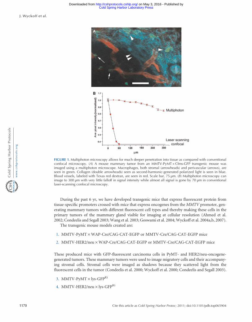
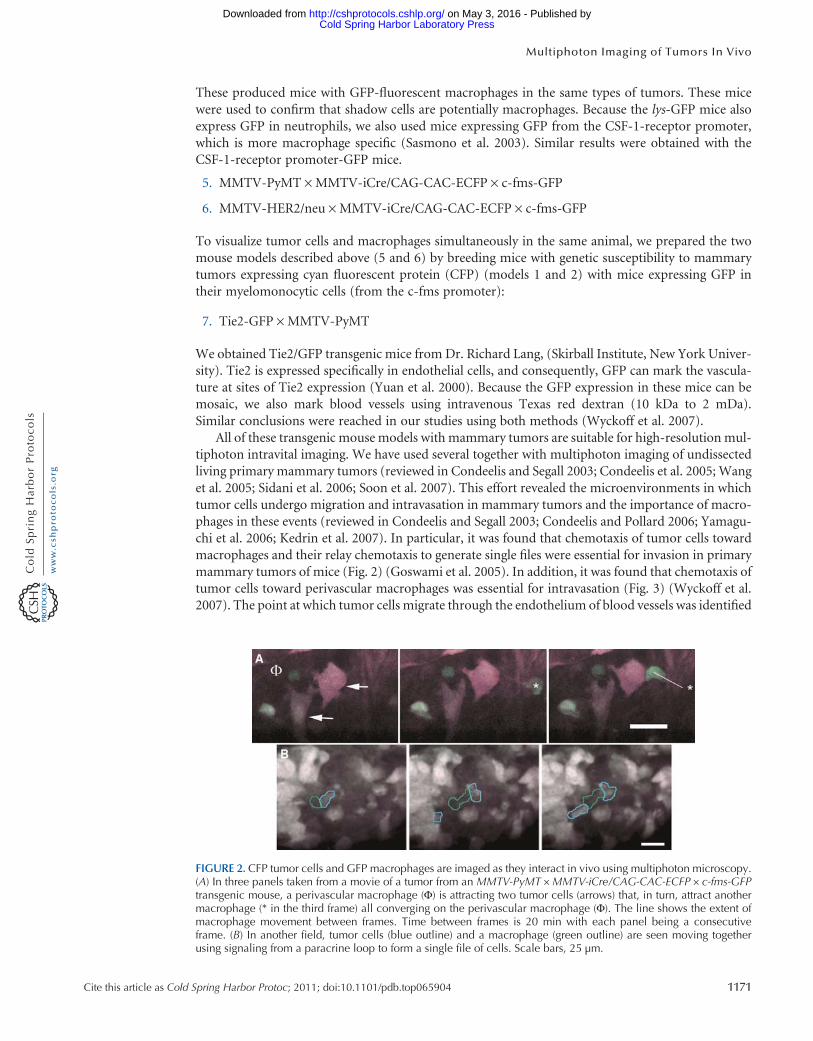
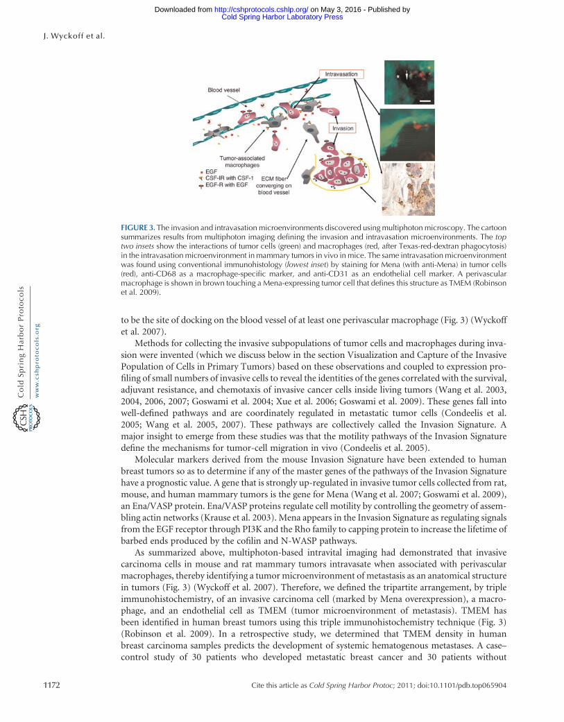
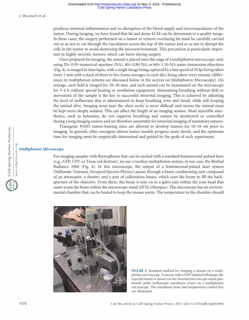
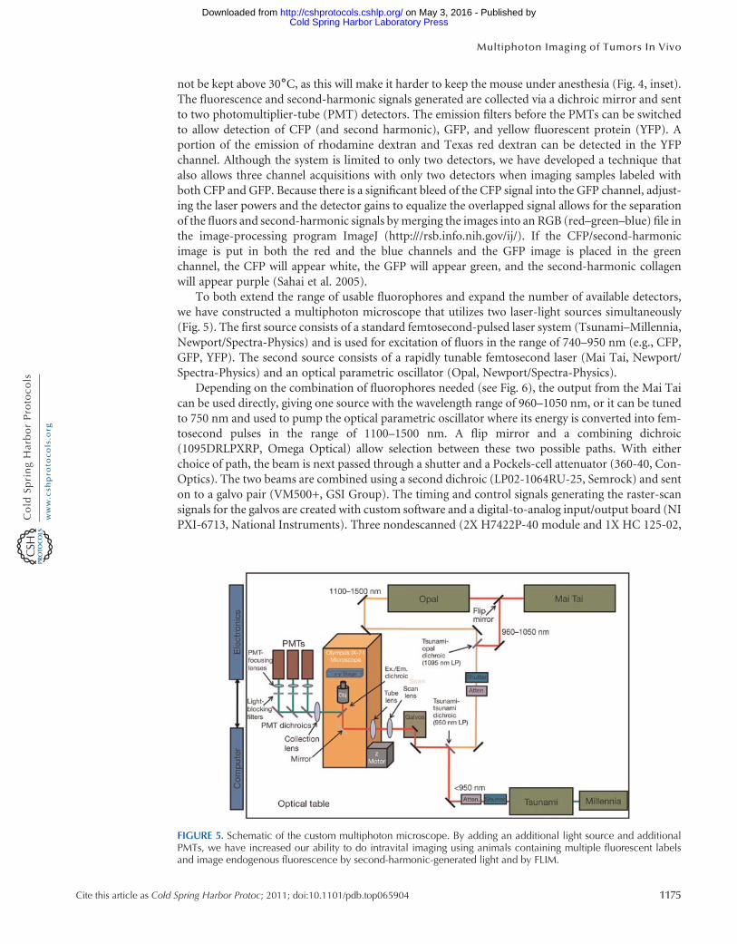
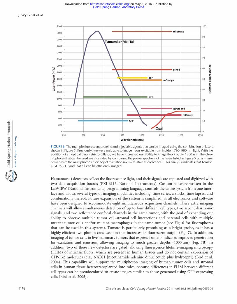
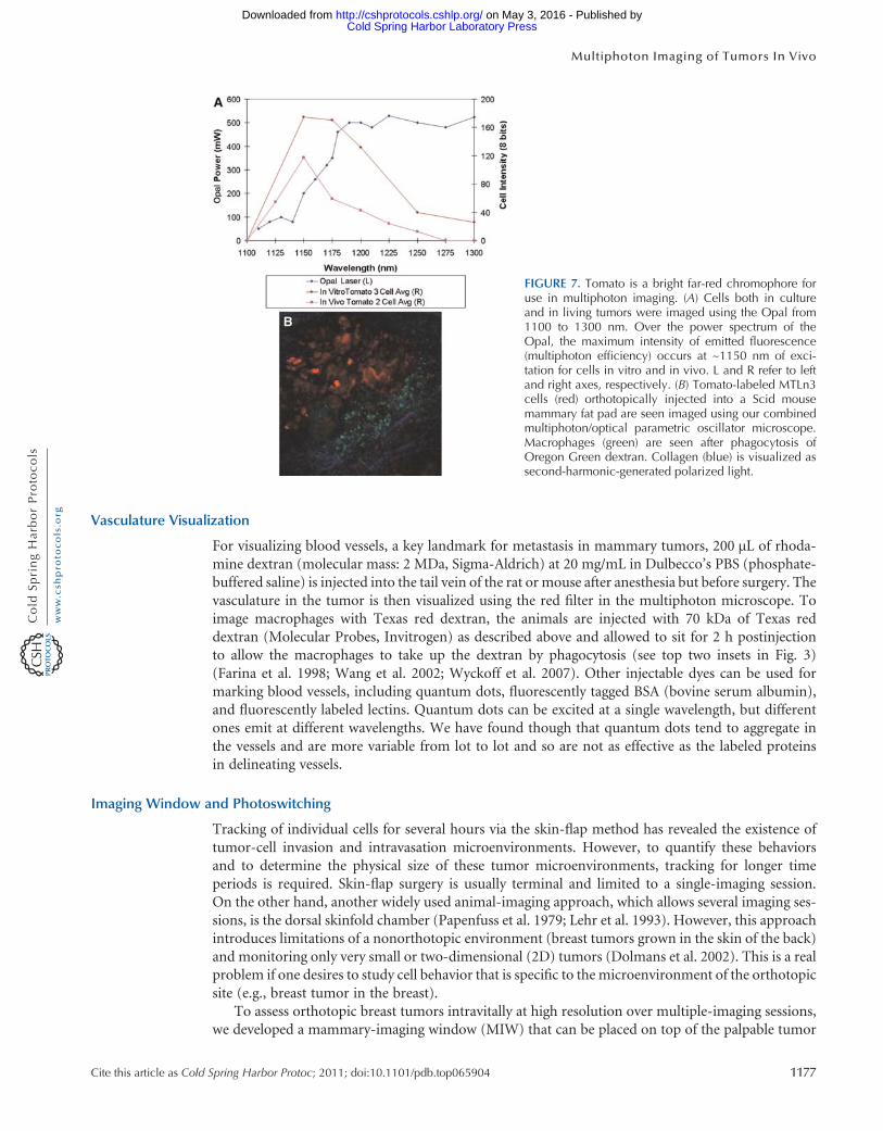
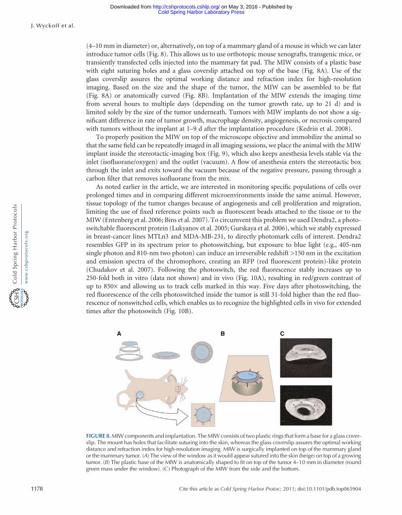
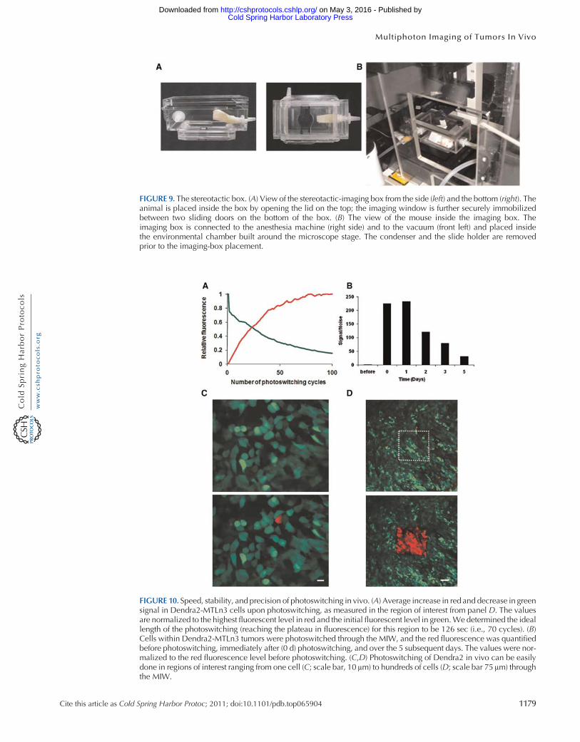
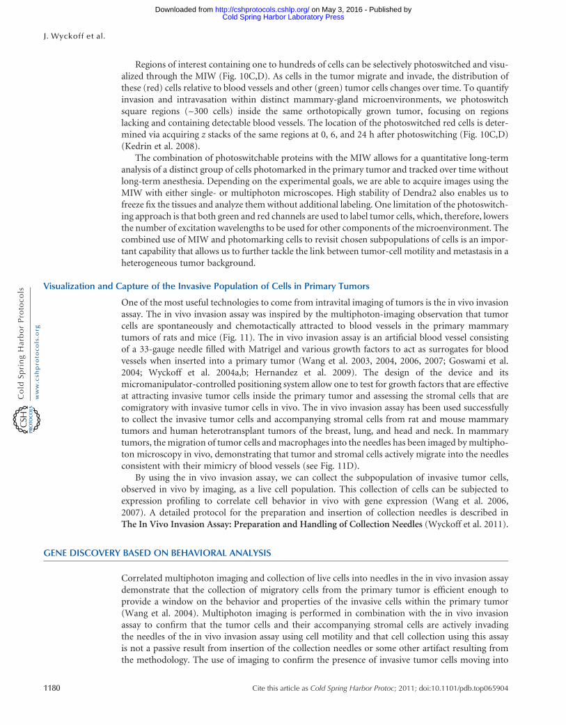
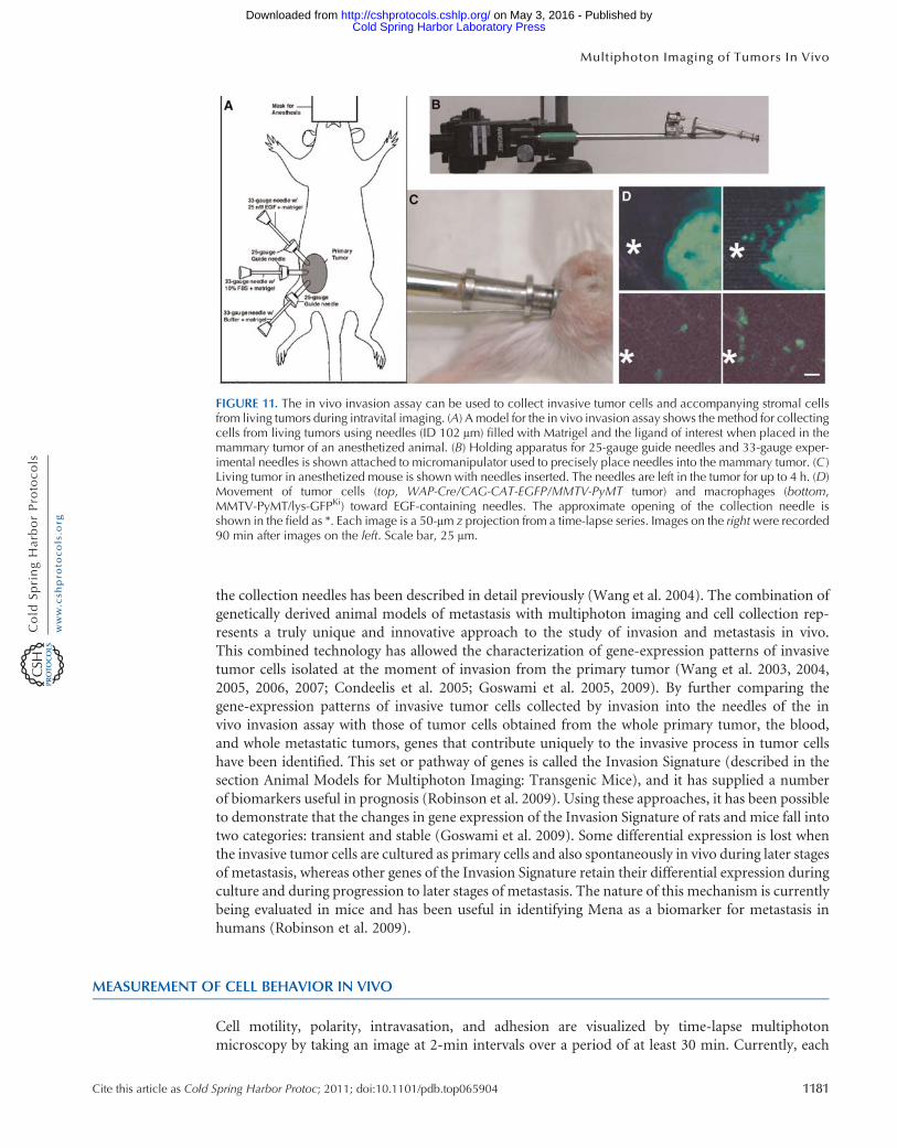