Molar-incisor hypomineralisation: narrative review on etiology ...
Dental Fast Track: Prenatal Enamel Growth, IncisorEruption, and Weaning in Human Infants
Patrick Mahoney*
Human Osteology Research Lab., School of Anthropology and Conservation, University of Kent, Canterbury, CT27NR, UK
KEY WORDS life history; incremental markings; allometry; circadian rhythm
ABSTRACT Correlation between the timing of per-manent first molar eruption and weaning age in extantprimates has provided a way to infer a life history eventin fossil species, but recent debate has questionedwhether the same link is present in human infants.Deciduous incisors erupt at an age when breast milkcan be supplemented with additional foods (mixed feed-ing), and weaning is typically complete before perma-nent first molars erupt. Here, I use histological methodsto calculate the prenatal rate by which enamel increasesin thickness and height on human deciduous incisors,canines, and molars (n 5 125). Growth trajectories foreach tooth type are related to the trimesters andassessed against the eruption sequence and final crownheight. Analyses show that central incisors initiate earlyin the second trimester with significantly faster secre-
tion rates relative to canines and second molars, whichinitiate closer to birth. Even though initial extensionrates were correlated with crown height and scaled withpositive allometry within each tooth class, the relativelyshort incisors still increased in height at a significantlyfaster rate than the taller canines and molars. The inci-sor prenatal “fast track” produces a greater proportion ofthe crown before birth than all other tooth types. Thisgrowth mechanism likely facilitates early incisor erup-tion at a time when the mixed feeding of infants can beinitiated as part of the weaning process. Findings pro-vide a basis from which to explore new links betweendevelopmental trends along the tooth row and mixedfeeding age in other primates. Am J Phys Anthropol000:000–000, 2014. VC 2014 Wiley Periodicals, Inc.
Studies of permanent teeth have demonstrated thatthe timing of first molar (M1) eruption is correlated witha number of life history traits across the primate order(Smith, 1989, 1992), and this correlation extends tohumans and extant apes for some traits (e.g., Kelley andSchwartz, 2009). Age at M1 eruption has therefore beenused to infer some aspects of life history in fossil homi-noids (e.g., Kelley 1997, 2002; Kelley and Smith, 2003).However, the relationship between M1 eruption, and onetrait, weaning age, has recently been re-examined inhumans (Humphrey, 2010). The weaning process forhuman infants typically involves a period of mixed-feeding that includes breast milk and additional foods,followed by the cessation of breastfeeding when aninfant is “weaned.” The link between weaning age andM1 eruption age does not hold for humans (Humphrey,2010), or for some of the great apes (Robson and Wood,2008; Smith et al., 2013; Macho and Lee-Thorpe, 2014).For example, M1 emerges through the gum line inhumans at around six years of age (Hillson, 2014: hisTable 5), and soon after three years of age in wild chim-panzees (Pan troglodytes) (Smith et al., 2013). Humansare typically weaned before the eruption of M1, whilechimpanzees continue to suckle (Robson and Wood,2008; Smith et al., 2013). Thus, humans are weanedearly relative to their permanent dental developmentwhen compared to great apes (Robson and Wood, 2008;Humphrey, 2010), though the correlation between M1eruption age and weaning age still holds across the pri-mate order (Smith, 1989). When compared with thegreat apes, the combination of early weaning, as well aslong gestation, short interbirth interval, extended child-hood, later age at first reproduction, and a long adultlife span is unique to modern humans (Schultz, 1960;
Harvey and Clutton-Brock, 1985; Bogin, 1990). Thesearch for when these life history traits evolved inhumans continues to be an important debate (Smith andTompkins, 1995), and key insights have been gainedfrom analyses of enamel incremental markings and for-mation times (Bromage and Dean, 1985; Beynon andWood, 1987; Dean et al., 2001; Macho, 2001; Guatelli-Steinberg et al., 2005; Macchiarelli et al., 2006; Dean,2006; Smith et al., 2007, 2010).
Human deciduous crowns start to increase in thick-ness (enamel secretion) and height (enamel extension) atdifferent times in utero commencing with the centralincisor (Kraus and Jordan, 1965). Deciduous maxillarycentral incisors (di1) erupt through the gum line aroundthe middle of the first postnatal year, followed by lateralincisors and first molars, then canines, and lastly secondmolars (Hillson, 2014: his Table 4). The timing of di1
eruption in humans can overlap with the age thatmixed-feeding commences (Humphrey, 2010). For exam-ple, Sellen (2001) found that mixed-feeding began on
Grant sponsor: The Royal Society; Grant number: RG-110435.
*Correspondence to: Patrick Mahoney, Human Osteology ResearchLab., School of Anthropology and Conservation, University of Kent,Canterbury CT2 7NR, UK.E-mail: [email protected]
Received 11 May 2014; revised 28 October 2014; accepted 28October 2014
DOI: 10.1002/ajpa.22666Published online 00 Month 2014 in Wiley Online Library
(wileyonlinelibrary.com).
� 2014 WILEY PERIODICALS, INC.
AMERICAN JOURNAL OF PHYSICAL ANTHROPOLOGY 00:00–00 (2014)
average at five months of age among 45 nonindustrialpopulations, while 6 months of age is recommended forpresent-day populations (WHO, 2002). Early di1 erup-tion, which is preceded slightly by fusion of the mandi-ble, may contribute to a masticatory system that can aidwith processing food (Humphrey, 1998, 2010). Clearly,the age at which mixed-feeding commences has a socialdeterminant that can, and does, vary from one humanpopulation to another (e.g., from the third to ninthmonth in the British historical era: Fildes, 1986), butthe “natural” age for when it can begin is thought to bearound the middle of the first year after birth (Hum-phrey, 2010). For instance, in addition to functional mor-phological change (mandibular fusion and incisoreruption), an infant’s nutritional requirements haveincreased at this age (Humphrey, 2010), and theirimmune system has developed to approximately half thelevel seen among adults (Abrams and Miller, 2011).Developed immunity is important because the provisionof maternal antibodies has already declined (Abramsand Miller, 2011), and consumption of new foods has thepotential to introduce an array of new antigens (Cum-mins and Thompson, 1997) and potentially, pathogens(Katzenburg et al., 1996).
This study builds upon the previous work of the author(Mahoney 2011, 2012) and calculates the prenatal forma-tion rate of crown enamel thickness and height in humandeciduous maxillary incisors, canines, and molars toexplore one research question: is the pace of prenatalenamel growth accelerated for di1 relative to the othertooth types to accommodate the start of mixed-feeding?There are several reasons why this acceleration might beexpected. First, the age at which mixed-feeding can com-mence in humans is advanced relative to their deciduousdental development, when compared to Pan. A study ofwild juvenile chimpanzees reported mixed-feeding com-menced from 6 months of age onwards (Smith et al.,2013). Deciduous central and lateral incisors, as well asfirst molars, have usually erupted in captive chimpanzeesby 6 months of age, and second molars erupt soon after-wards (Hillson, 2014: his Table 6). In contrast, the startof mixed-feeding and the timing of di1 eruption can over-lap in humans (Humphreys, 2010). Advancing the start ofmixed-feeding relative to deciduous dental developmentin human infants might have constrained the time avail-able to form di1 leading to rapid enamel growth. Forexample, enamel secretion cannot initiate before the sec-ond trimester (Kraus and Jordan, 1965), after which, di1
has to form and erupt by the middle of the first year afterbirth. This is unlike the other tooth types where the timeavailable for crown formation is not constrained in thesame way. The central incisor is also a larger and tallertooth (Mahoney, 2013) compared with the lateral incisor,which erupts after di1. Second, rapid deciduous incisormineralization compared with molars has been reportedpreviously (FitzGerald and Hillson, 2009; Mahoney, 2012)and this was related to early eruption in mandibular lat-eral incisors (Mahoney, 2012), although the mix of pre-and postnatal enamel in the previous histological studieslikely contributed variation to the secretion rates (dis-cussed below).
On the other hand, the rate of deciduous enamelextension (Shellis, 1984) has not been compared alongthe human tooth row. Based upon human permanentteeth (Guatelli-Steinberg et al., 2012), early eruptingshort incisors might be expected to grow in heightslowly, not quickly, compared with the taller canines and
second molars which erupt closer to birth. Yet, if it couldbe shown that developmental trends along the humanprenatal tooth row correspond with the timing of mixed-feeding then this could potentially introduce a newmethodological approach for exploring links betweendental development and the start of mixed-feeding inextant apes and fossil species with erupted deciduousteeth. Deciduous enamel retains a record of prenatalgrowth that can be recovered from teeth that havealready formed and erupted. Thus, potentially, suchstudies would not have to rely upon fossil skulls withteeth that were in the process of erupting at the time ofdeath, which are extremely rare finds.
In addition to the main research question, this is thefirst histological study to relate human enamel secretionand extension rates to developmental trimesters. Assuch, I will establish if the trajectory of enamel growthchanges in utero, which will contribute a fundamentalunderstanding of the prenatal enamel growth curve forhuman biologists. Data presented here are critical forclearly evaluating differences between extant and fossilspecies (e.g., Macchiarelli et al., 2006), especially fordeciduous extension rates, which are poorly understood.
ENAMEL DEVELOPMENT
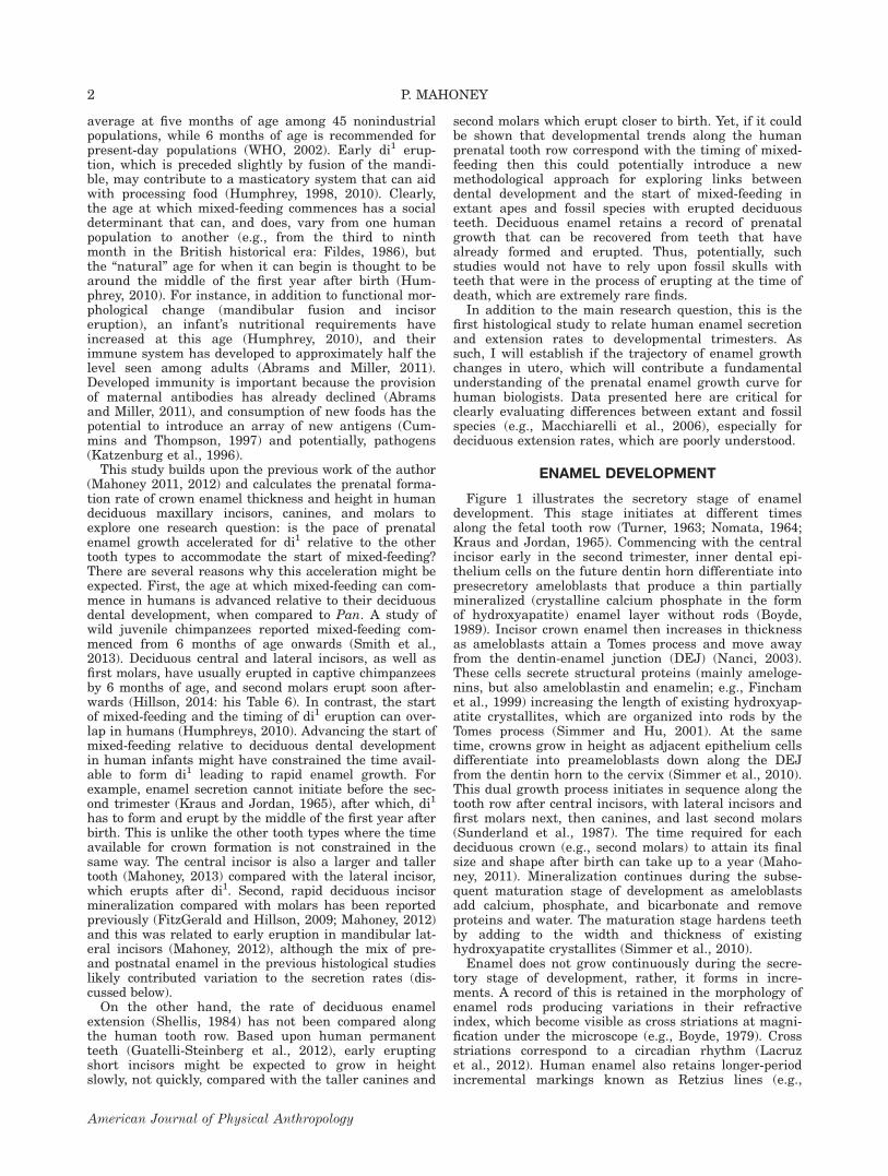
Figure 1 illustrates the secretory stage of enameldevelopment. This stage initiates at different timesalong the fetal tooth row (Turner, 1963; Nomata, 1964;Kraus and Jordan, 1965). Commencing with the centralincisor early in the second trimester, inner dental epi-thelium cells on the future dentin horn differentiate intopresecretory ameloblasts that produce a thin partiallymineralized (crystalline calcium phosphate in the formof hydroxyapatite) enamel layer without rods (Boyde,1989). Incisor crown enamel then increases in thicknessas ameloblasts attain a Tomes process and move awayfrom the dentin-enamel junction (DEJ) (Nanci, 2003).These cells secrete structural proteins (mainly ameloge-nins, but also ameloblastin and enamelin; e.g., Finchamet al., 1999) increasing the length of existing hydroxyap-atite crystallites, which are organized into rods by theTomes process (Simmer and Hu, 2001). At the sametime, crowns grow in height as adjacent epithelium cellsdifferentiate into preameloblasts down along the DEJfrom the dentin horn to the cervix (Simmer et al., 2010).This dual growth process initiates in sequence along thetooth row after central incisors, with lateral incisors andfirst molars next, then canines, and last second molars(Sunderland et al., 1987). The time required for eachdeciduous crown (e.g., second molars) to attain its finalsize and shape after birth can take up to a year (Maho-ney, 2011). Mineralization continues during the subse-quent maturation stage of development as ameloblastsadd calcium, phosphate, and bicarbonate and removeproteins and water. The maturation stage hardens teethby adding to the width and thickness of existinghydroxyapatite crystallites (Simmer et al., 2010).
Enamel does not grow continuously during the secre-tory stage of development, rather, it forms in incre-ments. A record of this is retained in the morphology ofenamel rods producing variations in their refractiveindex, which become visible as cross striations at magni-fication under the microscope (e.g., Boyde, 1979). Crossstriations correspond to a circadian rhythm (Lacruzet al., 2012). Human enamel also retains longer-periodincremental markings known as Retzius lines (e.g.,
2 P. MAHONEY
American Journal of Physical Anthropology

Shellis, 1998). An example of daily cross striations andRetzius lines are shown in Figure 1. These markings arenot remodelled and are retained in teeth after birth. Theconsistent and temporal nature of cross striations hasbeen confirmed in humans and other primates (Schourand Poncher, 1937; Bromage, 1991).
Reconstructing deciduous enamel growth rates
The rate at which a tooth crown achieves its size andshape can be reconstructed from histological analyses of
enamel incremental markings (e.g., Boyde, 1964). Tworates have been calculated previously and incorporatedinto studies of human deciduous enamel development.The first is the rate that ameloblasts secrete enamel(calculated as a daily enamel secretion rate: DSR) asthey move away from the first formed dentin, the DEJ,as a tooth crown increases in thickness. Previous studieshave measured the distance between adjacent crossstriations along enamel rods to calculate DSRs (Hudaand Bowman, 1994; Macchiarelli et al., 2006; Birch and
Fig. 1. Upper incisor thin section and incremental enamel markings. A thin section through the long axis of a tooth crown (a)illustrating the direction of growth (b) during the secretory stage of enamel development. Retzius lines (c) are revealed at a magni-fication of 34–10. Cross striations (d) are revealed at a magnification of 320–40 and run between Retzius lines. The distancebetween two adjacent cross striations along an enamel rod is the amount of enamel secreted by ameloblasts over 24 h. Ten days ofenamel secretion are highlighted. [Color figure can be viewed in the online issue, which is available at wileyonlinelibrary.com.]
RAPID PRENATAL INCISOR ENAMEL GROWTH 3
American Journal of Physical Anthropology
Dean, 2009; FitzGerald and Hillson, 2009; Mahoney,2011, 2012). They have shown that the amount ofenamel secreted by ameloblasts in any one 24-h periodcan vary through a deciduous crown, from a lowestmean DSR of 2.40 lm to a maximum of 5.70 lm per day(Birch and Dean, 2009; Mahoney, 2012). These ratescommence slowly, then accelerate, are temporarily inter-rupted at birth, and slow slightly towards the end ofeach crown’s developmental period.
The second rate is the extension rate, which deter-mines how fast new enamel formation spreads downalong the DEJ away from the dentin horn to the cervixas a deciduous crown grows in height (e.g., Shellis,1984). Previous histological analyses of deciduous exten-sion rates are limited. Shellis (1984) reported that, likehuman permanent teeth (e.g., Dean, 2009), deciduousextension rates slow within a crown from cuspal to cervi-cal enamel. Previous assessments of differences in exten-sion rates between deciduous incisors and molars haveproven inconclusive.
Potential sources of variation in deciduousenamel growth rates
In addition to the proposition that growth may beaccelerated in di1 relative to the other tooth types tofacilitate early weaning (above), there are several otherreasons why it might be expected that prenatal enamelgrowth trajectories will vary from tooth to tooth, evenwhen equivalent crown regions are compared, differen-ces in initiation are accounted for, and rates arerecorded from before the neonatal line. First, the trajec-tory of enamel growth may correspond to the sequencein which teeth initiate in utero. Normal human fetalgrowth is determined by an array of intrinsic variables,such as genetic potential and hormones, as well asextrinsic factors such as maternal and placentalnutrients (e.g., Singer et al., 1991; Cetin et al., 2005;Pardi and Cetin, 2006). Under these conditions, tissuepatterns and organ systems are established in theembryo during the first trimester of pregnancy. In thesecond and third trimester, fetal body size increases, themajor organs mature, and body fat increases during theweeks before birth. Research has shown that differenttissue types can have different growth trajectoriesthrough the second and third trimester (Tanner, 1989).For example, linear growth in long bones reaches itsmaximum velocity in the second trimester (e.g., Bertinoet al., 1996) only to slow during the third trimesterwhen fetal growth in weight accelerates (Yudkin et al.,1987; Hernandez 1998). Enamel on deciduous teeth ini-tiates in utero on one tooth after the other throughoutthe second and third trimester (Kraus and Jordan,1965). Teeth develop alongside other fetal hard tissues,but whether enamel secretion and extension rates varyfrom one tooth type to the next through the trimestersremains poorly understood. One older study in the liter-ature (Kraus, 1959) reported that deciduous central inci-sors calcified at a faster rate between weeks 13 and 18in utero than either lateral incisors or maxillary firstmolars. However, this comparison did not account fordifferences in initiation, which would make it appearthat these teeth have different growth trajectories.
Second, prenatal extension rates may be influenced byfinal crown height. Analyses of human permanent teethhave shown that variation in initial extension rates canbe explained by DEJ length as a proxy for crown height,
when compared between permanent incisors and molars(Guatelli-Steinberg et al., 2012). If deciduous teeth fol-low the correlation reported for permanent teeth (Shel-lis, 1984; Guatelli-Steinberg et al., 2012) then tallerdeciduous crowns should initially extend in height fasterthan shorter crowns.
Third, Beynon et al. (1991b) proposed that aspects ofincremental development in permanent teeth from ajuvenile gorilla reflected mean tooth emergence times.Mahoney (2012) tested this proposition and hypothesizedthat rapid mean DSRs in human deciduous manidbularlateral incisors facilitated early postnatal eruption, eventhough the neonatal line would have contributed some ofthe variation in DSRs. The neonatal line is an accentu-ated marking created at birth and retained withinenamel (Rushton, 1933). The marking can temporarilyreduce secretion rates (e.g., Macchiarelli et al., 2006).
HYPOTHESES
The background information provides a foundationfrom which to formulate four hypotheses that will betested by calculating and comparing prenatal enamelsecretion and extension rates between deciduous inci-sors, canines, and molars. These hypotheses are asfollows.
Initiation sequence
If secretion and extension rates correspond to thesequence in which crown enamel initiates through thetrimesters in a way that is broadly similar to the varia-tion seen in the linear growth of long bones (Tanner,1989), then the length of time required to form 250 lmof enamel, and the speed by which a crown increases inheight, ought to be expected to change from tooth totooth such that rates are faster in di1, which initiatesearly in the second trimester. Rates should then slow,from one tooth type to the next, following the sequenceof initiation.
Crown height
If variation in human deciduous extension rates, liketheir permanent counterparts (Guatelli-Steinberg et al.,2012), can be explained by DEJ length as a proxy forcrown height, then initial growth in height ought to beslower among deciduous incisors, which have short DEJlengths, relative to canines and second molars, whichhave taller crowns and hence longer DEJs (contra toHypothesis 1).
Eruption sequence
If incremental enamel growth reflects the eruptionsequence (Beynon et al., 1991b), and rapid secretionrates facilitate early eruption in mandibular lateral inci-sors (Mahoney, 2012), then faster secretion rates arealso expected to occur in maxillary incisor prenatalenamel. Rates should then slow, from one tooth type tothe next, following the eruption sequence.
Mixed feeding
If early mixed-feeding in human infants relative todeciduous dental development favored rapid enamelgrowth in di1, because the time available to form thistooth type was constrained, then enamel secretion andextension rates in central incisors ought to be acceler-ated relative to other tooth types (contra to Hypothesis
4 P. MAHONEY
American Journal of Physical Anthropology

2), but rates will not vary between canines and molars(contra to Hypotheses 1 and 3).
MATERIALS AND METHODS
Samples
Histological thin sections will be prepared from 125deciduous teeth (maxillary n 5 92; mandibular n 5 33)obtained from England and Sweden. The English sam-ples (central and lateral incisors, canines, first and sec-ond molars) are from the recent medieval period inCanterbury, UK, and will be used to calculate initiationtimes, DSRs, and extension rates. The Swedish samples(second molars) are routine anonymous clinical extrac-tions provided by the Department of Paediatric Den-tistry, University of Gothenburg, and will be used tosupplement the initiation times and extension rates.Sample sizes are given with each table in the Resultssection as they change depending on the histology vari-able. For each tooth type, each thin section represents adifferent individual and one data count for each histol-ogy variable. There are two exceptions. First, in additionto the main analyses, secretion rates will be calculatedalong the entire maxillary row (except di1) for two indi-viduals (reported separately from the others in tablefootnotes). Second, pre- and postnatal extension ratescalculated either side of the neonatal line are from thesame slides.
Thin section preparation
Standard histological methods were used to preparethin ground sections of each tooth (e.g., Reid et al.,1998a, b). Each tooth was embedded in polyester resinto reduce the risk of splintering while sectioning. Usinga diamond-wafering blade (Buehler IsoMet 4000 preci-sion saw) labial-lingual sections were taken through theoutermost incisor and canine enamel cusp tip, the dentinhorn at the tip of the DEJ, and the most cervical exten-sion of the enamel. Buccal-lingual sections were takenthrough the molars capturing the protoconid and meta-conid (mandibular) and the paracone and protocone(maxillary) dentinal horns. Establishing the optimumplane of section is easier in incisors and canines thanmolars because of their relatively flat surface. Formolars, marking the tip of the enamel cusp and themost cervical extension of the enamel before the tooth isembedded in resin helps orientate the tooth when cut-ting the section.
Each section was mounted on a microscope slide,which was lapped using a graded series of grinding pads(Buehler Eco-Met 300) to reveal accentuated and otherincremental lines, polished with a 0.3 mm aluminumoxide powder, placed in an ultrasonic bath to removesurface debris, dehydrated through a series of alcoholbaths, cleared (Histoclear), and mounted with a coverslip using a xylene-based mounting medium (DPX). Allsections were examined at magnification under a highpowered microscope (Olympus BX51) using transmittedand polarized light. Images were captured (OlympusDP25) and analyzed (Olympus Cell D).
Enamel initiation times, crown height, and DEJlength
The sequence and timing of deciduous crown enamelinitiation (e.g., Nomata, 1964; Kraus and Jordan, 1965)and crown height (e.g., Liversidge et al., 1993) have
been reported previously. These measures are presentedbriefly in the Results section, but the raw data for initia-tion times are available in the tables. Initiation timesare needed to relate enamel growth rates to the trimes-ters. Final crown height and DEJ length are assessedagainst initial extension rates.
Initiation times were calculated for each tooth type bylocating the position of the neonatal line. The thicknessof the enamel between this line and the tip of the dentinhorn was measured, sub-divided into three regions ofequal thickness, and then divided by a mean DSR calcu-lated for each region. This value provides an estimate ofthe time required to form the enamel between the tip ofthe dentin horn and the neonatal line. Crown heightmeasurements followed Liversidge et al. (1993). TheDEJ length of each tooth type was recorded from thethin sections. The length was measured in millimeters(mm) from the tip of the dentin horn to the most cervicalenamel along the labial surface of the incisors and can-ines, and the buccal surface of the paracone.
Prenatal secretion rates and the time taken toform 250 lm of enamel
Enamel secretion rates were recorded in the labialsurface of incisors and canines, and the buccal surface ofthe molar paracone. A distance of 250 lm was measuredfrom the dentin horn or just adjacent to it, along enamelrods towards the outer enamel surface. This distancefrom the DEJ towards the outer cuspal enamel is beforethe neonatal line in all maxillary tooth types, whereenamel secretion rates can temporarily slow (Macchiar-elli et al., 2006). It is also before the outermost enamelin di1 and the lateral incisor (di2) where it can be diffi-cult to see or record cross striations.
The distance of 250 lm was subdivided into tensequential regions each measuring 25 lm and theamount of time taken to form each region was calcu-lated. Cross striations could be counted along enamelprisms in each 25 lm region for slides of 11 teeth (dc1,dm1, dm2), while the time taken to form each 25 lmregion was approximated for all remaining slides. A dis-tance corresponding to five days of enamel secretion wasmeasured in each 25 lm region and then divided by fiveto yield a mean daily rate. Rates were measured alongthe long axis of an enamel rod around the center of eachregion. The procedure was repeated a minimum of sixtimes in each region, which allows a grand mean valueand standard deviation (SD) to be calculated. The grandmean value was used to calculate the time taken to formthe 25 lm region (distance/mean DSR). The procedurewas repeated for successive regions. The number of daystaken to form each region was then summed. This givesthe total number of days taken to form a 250 lm thickinitial layer of prenatal enamel.
The methodological approach used here is differentfrom that employed in the intra-specific study of meanDSRs along the mandibular tooth row by Mahoney(2011), but it permits a much more detailed examinationof the amount of enamel that ameloblasts secrete eachday as they move away from the DEJ. The presentmethodology does not include secretion rates immedi-ately adjacent to the DEJ, which can be difficult torecord. The time taken to form each increment will beplotted against measures of enamel thickness (e.g., Deanet al., 2001) to examine a near continuous growth trajec-tory for the first formed prenatal enamel.
RAPID PRENATAL INCISOR ENAMEL GROWTH 5
American Journal of Physical Anthropology

Prenatal extension rates
Several methods have been utilized previously toreconstruct extension rates. The one used here is basedupon Dean (2009) and Guatelli-Steinberg et al. (2012).An equivalent method has also been applied to rootextension rates (Macchiarelli et al., 2006).
Enamel extension rates were recorded for the labialsurface of incisors and canines, and the buccal surface ofthe molar paracone. Starting at the tip of the dentinhorn (see the blue circle in Fig. 1c) the number of crossstriations was counted along an enamel rod (followingthe blue dashed line) for 200lm towards the outermostsurface. A distance of 200 lm was chosen to allow futuredirect comparisons with data for permanent teeth (e.g.,Dean, 2009). On average, this distance took 45–46 daysto form in incisors, and 50–51 days in canines andmolars. An accentuated marking or Retzius line was fol-lowed back (at an angle to the blue dashed line) towardsthe DEJ. The distance down along the DEJ between theblue circle and the finishing point formed over the samenumber of days required to form 200 lm along theenamel rod. The initial extension rate is calculated bydividing the distance down along the DEJ by 45 or 46
days (incisors) or 50 to 51 days (canines and molars).The calculation gives the average rate that the cusprequires to amass 200 lm of prenatal enamel heighteach day during the 45–46 day period (incisors), or 50–51 day period (canines, molars).
Several other extension rates were calculated in addi-tion to the initial extension rate. The procedure wasrepeated in two consecutive and adjacent 200 lm unitslocated immediately after the initial rate and continuingin a cervical direction down along the DEJ. These addi-tional rates are presented in the tables along with theinitial rate to provide an assessment of the growth tra-jectory through the cuspal and lateral enamel and tofacilitate future comparisons. A previous study foundthat enamel secretion rates vary only slightly betweencuspal and lateral regions of deciduous teeth (Mahoney,2012). So, for the third extension rate, the number ofdays taken to form 200 lm was increased by two daysfor each tooth type. In addition, the extension rateimmediately before the neonatal line was compared withthe rate immediately afterwards for incisors and molars.
Only the initial rate reported in the Results sectionbelow occurs prenatally in all tooth types. Subsequentrates are either a mix of pre- and postnatal, or postnataldepending on the tooth type (i.e., the neonatal lineemerged at different locations in each tooth type). Thesedifferences are identified in the corresponding table.
Data analysis
Three inferential statistical tests were used to analyzethe data. First, mean DSRs for each 25 lm region andinitial prenatal mean extension rates were comparedbetween tooth types using a one- way ANOVA combinedwith multiple comparisons of group means (tooth types)with a Bonferroni adjustment to reduce the possibility ofType 1 errors. Second, deciduous extension rates fromeither side of the neonatal line were compared using apaired-samples t-test. ANOVA assumes that the valuesfor each group have a normal distribution and that thevariation within the groups is approximately similar,while a paired t-test assumes that the differences calcu-lated for each pair have a normal distribution. Homoge-neity of variance was assessed with Levene’s test;normality was determined with a Kolmogorov Smirnovgoodness-of-fit-test. Results of these tests indicated thatgroup variances did not differ significantly from eachother and that the distribution of data for each histologyvariable did not differ significantly from a normal curve.
Third, allometric scaling relationships between naturallog-transformed initial extension rates (ie., just pre-natal)and DEJ length were examined through reduced majoraxis regression (RMA) slopes and 95% confidence inter-vals (CIs). The regression slope captures the growth ratiobetween variables such that isometry is reflected by aslope equal to one. Significantly positive (greater than 1)and negative (less than 1) allometry were identified usingthe 95% CIs. Statistical analyses were conducted in SPSS(version 20), except the RMA regression analysis whichwas undertaken in PAST (Hammer et al., 2001).
RESULTS
Enamel initiation sequence, crown height, andDEJ length
The di1 initiated early in the second trimester, followed bydi2 and dm1, then dc1, and finally dm2 in the third trimester
TABLE 1. Initiation of enamel secretion in days before birthcounted from sections of isolated deciduous teeth
di1 di2 dc1
dm1 dm2
prob parc pro par
201a 104a 77a 101 100 70 58188a 90a 92a 109 146 43 66169a 170a 103a 95 96 89 66199a 180a 89a 98 147 54 65168 196a 127a 132 120 53 76163 216a 76a 117 158 82 66175 186a 90a 92 165 47 120161 157 97a 106 146 53 102160 142 89a 187 166 91 57
134 113a 117 104 93154 132a 137 79 86170 88a 146 85 111135 127 119 108
117 121 79110 140 6298
xd,e 176 156 102 121 138 71 81sd 15 34 17 24 25 19 20
a Taken from Mahoney 2012. All other times are this study.b Pro is protocone. Par is paracone.c Earlier initiation in the paracone of maxillary molars relativeto the protocone has been reported previously (e.g., Turner,1963; Nomata, 1964).d Initiation sequence is the same as that reported by Sunder-land et al. (1987) for the initiation of dentin in maxillary teeth,except for the lateral incisor, which is slightly in advance of thefirst molar in this study. The timing of enamel initiation for thelateral incisor in this study was more varied than any othertooth type, which was also reported in the study of dentin ini-tiation (Sunderland et al., 1987). Mean enamel initiation timesin Table 1 occurred slightly earlier when compared to their iso-mers in the lower jaw (in Mahoney, 2011, 2012). Earlier enamelinitiation in maxillary compared to mandibular teeth has beenreported previously (Butler, 1992), though the timing of enamelinitiation can vary between isomers.e The range of dm2 initiation times (pro5 47–70 days; par5 57–86 days) for the Swedish sample overlaps with the range of ini-tiations times for the English dm2 samples (pro5 43–104 days;par5 65–120 days). Also see footnote b in Table 4.
6 P. MAHONEY
American Journal of Physical Anthropology

(Table 1). At completion, the tallest crown was dc1 (7.04 mm;0.27 sd) then dm2 (6.46 mm; 0.42 sd) followed by di1 (6.28mm; 0.27 sd), the di2 (6.09 mm; 0.18 sd), while the shortestcrown was dm1 (5.62 mm; 0.33 sd). The sequence in height isthe same as that reported by Liversidge et al. (1993), and italso distinguishes mean DEJ lengths along the tooth row(dc1 5 6.98 mm, dm2 5 6.43 mm, di1 5 6.14 mm, di2 5 5.96mm, dm1 5 5.39 mm).
Pre-natal secretion rates and the time taken toform 250 lm of enamel
Figure 2 confirms that growth trajectories are not thesame for all teeth. The 250 lm unit of enamel formed in
54–56 days in maxillary incisors (Table 2). In contrast,the same depth of enamel required 61–62 days to formin canines and molars. The shorter formation time in di1
occurred because ameloblasts secreted significantly (P <0.05) more enamel after they had moved between 50 and100 lm away from the DEJ (Table 3), and then contin-ued to do so for the remainder of the time it took to form250 lm of enamel. Ameloblast secretion rates were alsosignificantly (P < 0.05) faster in maxillary lateral inci-sors compared to second molars after these cells hadtravelled 75 lm away from the DEJ. When rates werecompared along the entire maxillary tooth row in twoindividuals, mean DSRs were faster in lateral incisors
Fig. 2. Incisor prenatal secretion rates are fastest. The plot shows the trajectory of enamel growth for each tooth type (a) asameloblasts move away from the newly formed dentin. Ameloblasts secrete 250 lm of enamel faster in incisors compared to caninesand molars. The faster forming incisor crowns initiate early in the second trimester (b) compared to teeth located more distal alongthe jaw. Mean DSRs are in Table 2 and footnotes. Mean initiation times have been taken from Table 1. [Color figure can be viewedin the online issue, which is available at wileyonlinelibrary.com.]
RAPID PRENATAL INCISOR ENAMEL GROWTH 7
American Journal of Physical Anthropology

(Table 2 footnotes). A 250 lm unit of prenatal enamelalso formed over a shorter period of time in mandibularlateral incisors compared to mandibular second molars(Table A1). Mean DSRs did not vary significantly betweenfirst and second molars, or between molars and canines.
The mean DSRs reported here ranged between a low-ermost value of 2.69 to an uppermost value of 5.34 lm/day. The range of values is similar to and overlaps withthe range of mean DSRs (2.5–4.5 lm/day) reported byBirch and Dean (2009). When the mean DSRs in Table 2are recalculated as an overall mean rate for the innerenamel region (see Mahoney, 2011 for method) the rateof 4.25 lm/day for di1 is greater than the rate of 3.90lm/day for the second molar. Other studies have alsoreported higher mean DSRs in incisors relative to eithermolars or canines, depending on which enamel regionsare compared (FitzGerald and Hillson, 2009; Mahoney,2012). This suggests that, on average, enamel secretionrates might not ascend as high in the other tooth typescompared with incisors.
Prenatal extension rates
All crowns initially extended in height rapidly (Table4). Growth subsequently slowed, with the lowest ratesoccurring further down the DEJ away from the dentinhorn in a fashion similar to the trajectory reported for
permanent molars (Shellis, 1984; Dean, 2009). Thechange in the deciduous growth trajectory indicates thatameloblast differentiation along the DEJ occurs morerapidly early in crown formation and proceeds moreslowly during later stages of development.
Initial extension rates were significantly correlatedwith DEJ length and scaled with positive allometry forincisors and molars (di1 and di2 combined: r 5 0.714,slope 5 2.744, 95% CI 5 2.18–3.30, intercept 5 28.707,P 5 0.000; dm1 and dm2 combined: r 5 0.676, slope 51.542, 95% CI 5 1.193–1.848, intercept 5 24.217, P 50.000). Thus, taller incisors and taller molars with lon-ger DEJs grow at proportionally faster prenatal ratesthan their shorter counterparts (Fig. 3), and as such, thecorrelation between crown height and initial extensionrate is similar to results reported for permanent teeth(Guatelli-Steinberg et al., 2012).
Extension rates varied between tooth types (Table 4).Central incisors initially extended over the greatest dis-tance with a higher mean rate of 52.03 lm/day that dif-fered significantly (P < 0.05) from the mean rates of35.75–40.05 lm/day found for canines and molars (Fig.4). Lateral incisors also had a significantly (P < 0.05)higher initial rate of 47.01 lm/day compared with themolars. Growth in height subsequently slowed moregradually for both incisor types relative to molars. Bycontrast, the rate of deceleration in canines was similar
TABLE 2. Mean daily enamel secretion rates and the time taken (days) to form 250lm of enamel
di1 (n 5 10–12) di2 (n 5 11–14) dc1 (n 5 12–14) dm1 (n 5 13–15) dm2 (n 5 12–16)
lma DSR (sd)b daysc DSR (sd) days DSR (sd) days DSR (sd) days DSR (sd) days
25 3.59 (0.39) 6.93 3.69 (0.36) 6.77 3.31 (0.32) 7.55 3.54 (0.33) 7.06 3.33 (0.38) 7.5050 4.02 (0.33) 13.18 3.94 (0.23) 13.12 3.77 (0.31) 14.18 3.79 (0.24) 13.65 3.66 (0.22) 14.3375 4.35 (0.38) 18.92 4.30 (0.29) 18.93 3.95 (0.44) 20.51 3.97 (0.27) 19.95 3.88 (0.21) 20.78100d 4.51 (0.30) 24.47 4.49 (0.31) 24.50 3.94 (0.31) 26.85 4.09 (0.20) 26.06 4.05 (0.25) 26.95125 4.80 (0.26) 29.68 4.64 (0.27) 29.89 4.02 (0.27) 33.07 4.09 (0.28) 32.18 4.05 (0.24) 33.12150 4.99 (0.25) 34.69 4.76 (0.23) 35.14 4.09 (0.29) 39.19 4.12 (0.29) 38.24 4.09 (0.27) 39.23175 4.96 (0.29) 39.73 4.74 (0.23) 40.41 4.44 (0.41) 44.82 4.21 (0.28) 44.18 4.15 (0.23) 45.26200 5.10 (0.26) 44.63 4.76 (0.27) 45.66 4.47 (0.44) 50.41 4.35 (0.24) 49.93 4.16 (0.21) 51.27225 5.12 (0.24) 49.51 4.83 (0.32) 50.84 4.64 (0.32) 55.80 4.52 (0.21) 55.46 4.42 (0.21) 56.92250 5.15 (0.19) 54.37 4.88 (0.28) 55.96 4.66 (0.32) 61.16 4.71 (0.23) 60.77 4.55 (0.30) 62.42Total days 54 56 61 61 62
a Distance in microns from dentin-enamel junction towards the outer enamel surface.b Mean daily enamel secretion rates for each 25 lm region.c Cumulative number of days (25 lm/mean DSR).d Mean daily enamel secretion rates were calculated for di2, dc1, dm1, and dm2 from the same jaw, for two individuals (not includedin Table 2 or 3, but included in Figure 2), at a depth of 100, 125, and 150 lm away from the dentin-enamel junction. Mean ratesfor the two individuals from lateral incisors to second molars at a depth of 100 lm were 4.58 lm, 4.02 lm, 4.10 lm, 4.00 lm,respectively. At a depth of 125 lm: 4.85 lm, 4.24 lm, 4.00 lm, 4.00 lm, respectively. At a depth of 150 lm: 4.80 lm, 4.21 lm,3.96 lm, 4.00 lm.
TABLE 3. Comparing mean daily enamel secretion rates along the maxillary tooth row
Distance fromDEJ (in lm) di1 v di2 di1 v dc1 di1 v dm1 di1 v dm2 di2 v dc1 di2 v dm1 di2 v dm2 dc1 v dm1 dc1 v dm2 dm1 v dm2
25 0.899 0.309 1.000 1.000 0.055 1.000 0.381 0.724 0.892 0.29450 1.000 0.103 0.107 0.003a 0.463 0.775 0.057 0.378 0.520 0.89575 1.000 0.107 0.196 0.030a 0.201 0.368 0.043a 1.000 1.000 1.000100 1.000 0.004a 0.047a 0.017a 0.004a 0.057 0.026a 1.000 1.000 1.000125 1.000 0.000a 0.000a 0.000a 0.004a 0.001a 0.006a 0.851 0.954 0.858150 0.561 0.000a 0.000a 0.000a 0.011a 0.027a 0.000a 1.000 1.000 1.000175 1.000 0.057 0.000a 0.000a 0.239 0.008a 0.002a 1.000 0.495 1.000200 0.364 0.001a 0.007a 0.000a 0.359 0.073 0.021a 1.000 1.000 1.000225 0.197 0.002a 0.001a 0.000a 1.000 0.237 0.012a 1.000 0.597 1.000250 0.314 0.001a 0.017a 0.000a 0.508 1.000 0.046a 1.000 1.000 1.000
a Significant difference.
8 P. MAHONEY
American Journal of Physical Anthropology

TABLE 4. Mean extension rates and standard deviation (sd) in lm per day
di1 (n513) di2 (n514) dc1 (n519) dm1 (n519) dm2 (n515)
Initiala,b 52.03 (6.96) 47.01 (5.70) 40.05 (6.05) 35.75 (6.05) 39.97 (6.29)32.97 (4.70) 27.24 (4.97) 25.43 (3.18) 15.93 (3.12) 19.02 (4.17)22.12 (3.05) 19.99 (2.96) 21.05 (2.67) 12.73 (2.58) 15.32 (2.89)
Beforec 17.40 (3.63) 17.58 (3.30) 15.59 (3.26) 18.44 (3.88)After 14.16 (2.72) 14.84 (2.73) 12.91 (2.79) 14.44 (1.99)
Dark gray is prenatal, light gray is pre-, and postnatal, white is postnatal. Statistics are in footnotes.a Comparisons between initial extension rates: di1 v di2, P 5 0.386; di1 v dc1, P 5 0.000*; di1 v dm1 P 5 0.000*. di1 v dm2, P 5
0.000*; di2 v dc1, P 5 0.303; di2 v dm1, P 5 0.000*; di2 v dm2, P 5 0.036*. dc1 v dm1, P 5 0.027*; dc1 v dm2, P 5 1.000; dm1 v dm2,P 5 0.516.b Initial extension rates for the Swedish dm2 sample (n 5 5) ranged between 33.30–42.92 lm per day, which is encompassed by therange of values of 32.73–46.98 lm per day from the English dm2 sample (n 5 10). Given this, and the overlap in the timing of ini-tiation between the Swedish and English samples (see Table 1), these northern European samples appear homogenous for purposesof this study. This proposal is consistent with studies of permanent teeth, where the greatest difference in molar enamel growthoccurred when northern European samples (recent archaeological and modern) were compared to African samples (Reid and Dean,2006).c Comparison of mean extension rate immediately before the neonatal line with rate immediately afterwards: di1, P 5 0.001*; di2, P5 0.001*; dm1, P 5 0.000*; dm2, P 5 0.000*.
Fig. 3. Bivariate plots of log-transformed initial extension rate and dentin-enamel junction length. Reduced major axis regres-sion lines are fitted to the data. Initial extension rates are correlated with DEJ length and scale with positive allometry for incisors(a), and this scaling relationship is the same for molars (b). See Results section for regression statistics. [Color figure can beviewed in the online issue, which is available at wileyonlinelibrary.com.]
RAPID PRENATAL INCISOR ENAMEL GROWTH 9
American Journal of Physical Anthropology

to the lateral incisors, and the range of values for thethird extension rate in canines overlapped with both lat-eral and central incisors (Table 4).
The extension rate immediately before the neonatalline was significantly faster (P < 0.05) than the rateimmediately afterwards for all tooth types (Table 4). Atfirst glance, the deceleration in rates after birth appearssimilar to the situation reported previously for meanDSRs (Macchiarelli et al., 2006). However, extensionrates also slow from cuspal to cervical enamel, whichcould explain the pattern reported here.
When secretion and extension rates are consideredtogether it is apparent that over the first two months ofenamel growth many ameloblast cells are employedquickly and these secrete enamel faster in incisors, withthe highest growth rates in di1. By itself, rapid extensionis a mechanism to grow a crown quickly, but this is com-bined with rapid ameloblast secretion rates in incisors.In all tooth types, the rate at which new ameloblasts arerecruited down along the DEJ is faster than the rate atwhich ameloblasts secrete enamel as they move awayfrom the DEJ (Tables 2 and 4).
Fig. 4. Incisor prenatal extension rates are fastest. The plot shows the mean distance that incisor and molar crowns increase inheight in lm each day (a). The initial rate (first enamel unit to the left) is prenatal for all teeth. The units of enamel growth startat the dentin horn and continue down along the DEJ into lateral enamel. Incisor crowns initiate early in the second trimester (b)and initially extend in height at a faster rate than canines and molars. Mean extension rates are in Table 4. [Color figure can beviewed in the online issue, which is available at wileyonlinelibrary.com.]
10 P. MAHONEY
American Journal of Physical Anthropology

DISCUSSION
Initiation sequence
The prenatal trajectory of enamel growth correspondswith the initiation sequence to some extent. Mean DSRs arefastest in di1, and then lateral incisors, which, on average,initiate enamel growth one after the other early in the sec-ond trimester (Fig. 2b). Canines and second molars initiatelater and closer to birth and are marked by slower secretionrates. Thus, a broad distinction can be made between thesetooth types based on prenatal secretion rates and initiationsequence, although the correspondence is not present in alltooth types. For example, lateral incisors and first molarsinitiate at about the same time, but enamel forms moreslowly on molars. Thus, the trajectory of enamel growthchanges through the trimesters, when incisors are comparedto canines and second molars. Generally, enamel growth isfaster early in the second trimester and slower closer tobirth, which is a growth trajectory broadly similar to the lin-ear growth of long bone. Studies have reported variation ingrowth trajectories though the trimesters for other tissuestypes as well (e.g., Tanner, 1989; Bogin, 1990).
Crown height
When compared along the tooth row, prenatal extensionrates are partly influenced by crown height. When com-parisons are limited to canines and molars, the taller can-ines and taller second molars initiate closer to birth withfaster initial extension rates relative to the shorter firstmolars (Fig. 4b). So for these tooth types, crown heightrather than the initiation sequence seems to determinethe initial speed at which they grow in height.
The relationship between initial extension rate andfinal crown height does not hold when incisors are com-pared to canines and molars, because incisors are shorterthan canines and second molars but their growth inheight is faster. This is the opposite to expectations basedon permanent teeth (Guatelli-Steinberg et al., 2012).There is no change in the way that initial extension ratesand DEJ length correlate or scale in deciduous incisorscompared to molars (Fig. 3). So, for deciduous incisors,when compared to other tooth types, something in addi-tion to crown height influences extension rates. Differen-ces in cusp shape are an unlikely explanation. Incisorshave a less complicated cusp morphology than molars,but canines also have an uncomplicated and thin profilelike central incisors when sectioned. Never-the-less, thetaller canines initially extend more slowly than theshorter front teeth. Furthermore, initial extension rateswere from the cuspal region where DEJ shape differencesare relatively reduced.
Eruption sequence
A distinction can be made between incisors with fastersecretion rates which erupt first, and canines and molarswith slower rates, which erupt latter. However, similar-ity in secretion rates between canines and molars, andbetween first and second molars, does not support thehypothesis that variation in enamel secretion rates cor-responds closely to the eruption sequence for teethlocated more distal along the row.
Dental “fast track” for weaning
Neither the initiation nor the eruption sequence, orcrown height, explains all of the variation in enamel
growth rates when compared along the deciduous toothrow. Rapid growth in di1, and to a lesser extent in lat-eral incisors, corresponds with the initiation and erup-tion sequence relative to canines and second molars.When canines and molars are compared, variation inenamel secretion and initial extension rates do not relateclearly to the initiation and eruption sequence. Instead,initial extension appears to be determined by final crownheight. Therefore, incisors, and di1 in particular, have adifferent prenatal growth trajectory than teeth locatedmore distal along the jaw.
When the di1 enamel growth trajectory is placedalongside the developmental changes that subsequentlyoccur around the middle of the first post-natal year(Humphreys, 1998, 2010) it seems consistent with thisprocess. Incisor enamel growth, and especially di1, is“fast tracked” in utero so that the crown is formed andready to erupt at an age when the weaning process cancommence. Rapid di1 growth supports the hypothesisthat early mixed-feeding, relative to deciduous dentaldevelopment, may have constrained the time availableto form the central incisor crown. Perhaps precociousincisor mineralization and early eruption were selectedfor by weaning practices that can occur near the mid-point of the first year of life due to the increasing nutri-tional demands of the infant, which eventually exceedsthe nutritional supply provided by nursing.
Further support for the hypothesis that incisor growthis accelerated to accommodate the age at which mixed-feeding can commence is provided by the proportion ofenamel formed in utero. Additional analyses show thatat birth, both maxillary incisors have completed 45% oftheir total crown formation time, while first molars haveformed 33% of their crown, canines 22%, and secondmolars 15% (Tables 1 and 5). Therefore, relatively moreincisor enamel is formed by commencing rapid growthearly on in the second trimester, which results in a greatproportion of their crown being formed prior to birththan any other tooth type. The remaining postnatalinterval prior to eruption, during which the rest of themaxillary crown forms and root growth is initiated (seeAlQahtani et al., 2010), is also shorter for the incisors,and shortest of all for di1.
The growth trajectories for each tooth type are notindependent of final enamel thickness. Incisor crownsform along a faster trajectory because epithelium cellsinitially differentiate rapidly into preameloblasts. Fol-lowing this, incisor ameloblast cells secrete more enamelsoon after they move away from the DEJ, comparedwith the other tooth types. This developmental processeventually produces a small thinly enameled crown(Mahoney, 2013) that has formed quickly. By contrast,canines and molar crowns are produced along a slowertrajectory, where less enamel is secreted in daily incre-ments following a slower initial rate of cell differentia-tion. For example, after 19 days, di1 secretes on average4.35 lm of enamel every 24 h, whereas the molarsrequire 50–54 days to secrete a similar amount (Table2). The slower trajectory eventually produces a thickerenameled tooth (Mahoney, 2013). It would make sensethat these different enamel growth trajectories are coor-dinated with the space available within a growing jaw(e.g., Dean, 2000; Smith 2000), whereby a greater pro-portion of thin fast forming enamel develops before birthin the small fetal jaw. Less prenatal enamel is formedfor canines and molars because more time and space isavailable in the growing jaw after birth to produce
RAPID PRENATAL INCISOR ENAMEL GROWTH 11
American Journal of Physical Anthropology

larger thicker enameled crowns, which then erupt lateron. Such coordinated timing suggests that deciduousenamel growth is part of a highly integrated biologicalsystem.
Applications
Future studies might explore links between life his-tory events and variation in enamel secretion and exten-sion rates along the tooth row among our nearestrelatives within the primate order. It would seemunlikely that the link reported here for human infants,between incisor enamel growth rates, eruption, andmixed feeding, will be exactly the same in juvenile chim-panzees. The age at which mixed feeding commences inPan does not seem to coincide with the median age fordeciduous incisor eruption (above). Given that the corre-lation between the timing of permanent first molar erup-tion and the age that humans are finally weaned is notclear, such analyses may potentially provide an addi-tional way of inferring aspects of weaning from dentaldevelopment in fossil hominins with erupted deciduousteeth.
The present methodology can be applied more broadlyacross the primate order to determine whether the paceof enamel growth along the tooth row provides a mecha-nism to facilitate precociality. Many species are bornwith fully erupted deciduous teeth and the degree ofdevelopment at birth can differ markedly even amongstclosely related primates (Schwartz et al., 2005, 2007).Advanced dental development in primates soon afterbirth can correlate with life history related variables,such as adult female cranial capacity (Smith, 1989; God-frey et al., 2003). Future studies of enamel growth tra-jectories along the row might also examine patterns ofpermanent tooth eruption, such as the eruption ofmolars relative to incisors, canines, and premolars,which can be advanced in “faster living” anthropoids, ordelayed in “slower growing” primate species (Schultz,1935; Smith, 2000; Henderson, 2007).
Methodological considerations
Variation in enamel growth rates along the deciduoustooth row has methodological implications for studies ofenamel defects in deciduous teeth. At any one time
TABLE 5. First and second maxillary molar crown formation times in days (years)a,b
dm1 (n 5 23) dm2 (n 5 21)
protocone paracone protocone paracone
cuspal lateral cuspal lateral cuspal lateral cuspal lateral
115 395 104 306 219 371 145 31477 401 88 332 – – 141 43598 245 111 220 69 601 – –– – 122 221 – – 210 254231 234 124 285 188 301 – –142 253 127 217 173 298 117 249103 247 131 193 214 255 126 25389 292 – – 115 328 97 320113 287 – – – – 98 248111 276 – – – – 130 42398 400 – – – – 193 272120 276 – – 155 324 91 344108 228 – – 119 384 – –– – 103 284 158 458 93 446107 262 119 238 145 489 – –99 296 87 242 184 341 148 –91 283 86 263 221 275 164 184– – 91 190 – – 262 284– – 111 163 – – 147 292– – 172 271 193 385 170 –141 203 – – 158 279 – –125 225 – –205 – 60 351
CFTc 398 (1.09) 361 (0.99) 529 (1.45) 452 (1.24)min 336 (0.92) 274 (0.75) 437 (1.20) 346 (0.95)max 510 (1.40) 443 (1.21) 670 (1.84) 576 (1.58)sd 56 51 75 78
dTotal mean CFT 5 415 days (1.14yrs) eTotal mean CFT 5 539 (1.47)
a English sample.b Proportion of molar prenatal growth was calculated from Tables 1 and 5. Proportions for incisors and canines were calculatedfrom Mahoney (2012).c Mean cusp formation time and sd calculated from individuals with both cuspal and lateral values (for method: Mahoney, 2011). d
Total mean crown formation time (includes an additional mean 17 days of prenatal growth that is recorded in the paracone but notthe protocone). e Total mean CFT (plus an additional mean 10 days of prenatal growth for the paracone). Total CFT for dm1 anddm2 is slightly greater compared with mandibular dm1 and dm2 values reported previously (mean 5 1.06 years, 1.29 years respec-tively; Mahoney, 2011). Given that secretion rates from comparable regions are generally similar in upper and lower molars (com-pare Appendix 1 and Table 2), then slightly longer formation times, together with slightly earlier mean initiation times (Table 1) inmaxillary molar crowns might be expected, as they are larger with thicker enamel (Mahoney, 2013).
12 P. MAHONEY
American Journal of Physical Anthropology

during enamel formation, each deciduous tooth type willhave a different secretion and extension rate. Part ofthis is due to differences in initiation, but not for allteeth, since lateral incisors and first molars initiate closetogether in utero but develop along different growth tra-jectories. Thus, a systemic disturbance to secretory ame-loblasts leading to a hypoplastic defect or anaccentuated marking, which registers in all growingteeth at the same time, is unlikely to be recorded in lat-eral incisors and first molars in the same way. For exam-ple, over any one period of time the disturbance willregister in a greater proportion of incisor enamel thanfirst molars because of differences in the speed of enamelsecretion and extension. Thus, studies of enamel defectsin deciduous teeth should standardise their sampleselection by comparing the same tooth types, and con-struct their analyses accordingly.
Previously, Mahoney (2012) reported that both theenamel initiation sequence and proportion of enamelformed at birth in mandibular teeth mirrored the erup-tion sequence. Results presented here suggest the pro-portion of enamel formed in each tooth type at birthmore closely correlates with the eruption sequence, aslateral incisors and first molars can initiate closetogether but erupt at different times. The eruptionsequence can be reconstructed in deciduous teeth recov-ered outside of the jaw by calculating prenatal relativeto total crown formation time. The proportion of prenatalenamel formed in each tooth type will match the erup-tion sequence.
CONCLUSIONS
This study employed histological methods to calculatethe prenatal rate by which enamel increases in thick-ness and height on human deciduous maxillary incisors,canines, and molars. Rates were assessed against fourhypotheses, formulated from the sequence in which max-illary teeth initiate enamel growth in utero and eruptafter birth, crown height at completion, and the possibleadaptation of tooth formation to early mixed-feeding. Itwas found that enamel growth rates corresponded withthe initiation sequence, when incisors were compared tocanines and second molars, and this produced a trajec-tory of enamel growth that slowed from the second tothird trimester. The eruption sequence also appeared tobe linked to the pace of enamel formation, whereby earlyerupting incisors grew rapidly relative to teeth locatedmore distal along the jaw. However, growth rates did notcorrespond with either initiation or eruption sequencesfor canines and molars. Instead, crown height deter-mined their initial extension rate. Thus, variation inenamel growth rates along the tooth row was only partlyexplained by developmental sequences and models ofenamel formation related to crown height.
The prenatal enamel growth trajectory for deciduousincisors differed relative to the other tooth types. Inci-sors combined rapid growth with initiation early on inthe second trimester to produce a greater proportion oftheir crown before birth than any other tooth type.Accelerated incisor growth, and for di1 in particular,made sense when interpreted through an evolutionaryhypothesis, as a possible adaptation of tooth formationand eruption to a major life history event. As such, pre-cocious incisor enamel formation can be seen as agrowth mechanism that facilitates early eruption at anage when the transition from breast-feeding to mixed-
feeding commences, which is advanced in humans rela-tive to their deciduous dental development, comparedwith chimpanzees. This hypothesis, as well as the meth-odology employed here, can provide a basis from whichto explore new links between developmental trendsalong the tooth row and life history variables in prima-tes. Such links may potentially prove useful for inferringan aspect of weaning in fossil species with erupteddeciduous teeth.
ACKNOWLEDGMENTS
The author thanks the Editor-in-Chief Peter T. Elli-son, an Associate Editor, and two anonymous reviewersfor comments that improved this manuscript. Theresearch benefited from discussions with Chris Deter,Debbie Guatelli-Steinberg, Gabriele Macho, J€orgenNor�en, Nina Sabel, and Christopher Schmidt. The Uni-versity Kent and University of Gothenburg provideddental samples.
APPENDIX
LITERATURE CITED
Abrams ET, Miller EM. 2011. The roles of the immune systemin women’s reproduction: evolutionary constraints and lifehistory trade-offs. Yrbk Phys Anth 54:134–154.
AlQahtani SJ, Hector MP, Liversidge HM. 2010. Brief communi-cation: The London atlas of human tooth development anderuption. Am J Phys Anth 142:481–490.
Bertino E, Di Battista E, Bossi A, Pagliano M, Fabris C, AicardiG, Milani S. 1996. Fetal growth velocity: kinetic, clinical, andbiological aspects. Arch Dis Child 74:10–F15.
Beynon AD, Dean MC, Reid DJ. 1991a. On thick and thinenamel in hominoids. Am J Phys Athropol 86:295–309.
Beynon AD, Dean MC, Reid DJ. 1991b. Histological study onthe chronology of the developing dentition in gorilla andorangutan. Am J Phys Anthropol 86:189–203.
Beynon, AD, Wood BA. 1987. Patterns and rates of enamel growthin the molar teeth of early hominids. Nature 326:493–496.
Birch W, Dean MC. 2009. Rates of enamel formation in humandeciduous teeth. In: Koppe T, Meyer G, Alt K, editors. Com-parative dental morphology. Front Oral Biol. Basel: Karger. p116–120.
Bogin B. 1990. The evolution of human childhood. Bioscience40:1–25.
Boyde A. 1964. Estimation of age at death of young humanskeletal remains from incremental lines in dental enamel.Third International Meeting in Forensic Immunology, Medi-cine, Pathology and Toxicology, Plenary Session 11A. London,April 16–24, 1963. Excerpta Med IntCongrSer, Vol.80.
TABLE A1. Mean daily enamel secretion rates in di2 and dm2
di2 (n 5 11–15) dm2 (n 5 14–18)
lm DSR (sd) days DSR (sd) days
25 3.36 (0.46) 7.44 3.08 (0.39) 8.1250 4.00 (0.53) 13.69 3.66 (0.20) 14.9575 4.23 (0.20) 19.60 3.88 (0.28) 21.39100 4.28 (0.39) 25.44 3.61 (0.35) 28.31125 4.53 (0.55) 30.96 3.98 (0.35) 34.59150 4.60 (0.51) 36.39 4.01 (0.32) 40.82175 4.68 (0.43) 41.73 4.19 (0.22) 46.79200 4.74 (0.22) 47.02 4.00 (0.27) 53.04225 5.06 (0.28) 51.96 4.41 (0.33) 58.71250 5.06 (0.24) 56.90 4.53 (0.41) 64.23Total days 57 64
RAPID PRENATAL INCISOR ENAMEL GROWTH 13
American Journal of Physical Anthropology

Boyde A. 1979. Carbonate concentration, crystal centres, coredissolution, caries, cross striation, circadian rhythms andcompositional contrast in the SEM. J Dent Res 58:981–983.
Boyde A. 1989. Enamel. In: Berkovitz BKB, Boyde A, FrankRM, H €Ohling HJ, Moxham BJ, Nalbandian J, Tonge CH, edi-tors. Teeth. Handbook of microscopic anatomy. New York:Springer Verlag. p 309–473.
Bromage TG. 1991. Enamel incremental periodicity in the pig-tailed macaque: a polychrome fluorescent labelling study ofdental hard tissues. Am J Phys Anthropol 86:205–214.
Bromage TG, Dean MC. 1985. Re-evaluation of the age at deathof immature fossil hominids. Nature 317:525–527.
Butler PM. 1992. Correlative growth on upper and lower toothgerms in human foetus. Ann Zool Fennici 28:261–271.
Cetin I, Alvino G, Radaelli T, Pardi G. 2005. Fetal nutrition: areview. Acta Paed 94:7–13.
Cummins AG, Thompson FA. 1997. Review Article. Postnatalchanges in mucosal immune response: a physiological per-spective of breast feeding and weaning. Immun Cell Biol 75:419–429.
Dean MC. 2000. Progress in understanding hominoid dentaldevelopment. J Anat 197:77–101.
Dean MC. 2006. Tooth microstructure tracks the pace of humanlife history evolution. Proc R Soc Series B 273:2799–2802.
Dean MC. 2007. A radiographic and histological study of mod-ern human lower first permanent molar root growth duringthe supraosseous eruptive phase. J Hum Evol 53:635–646.
Dean MC. 2009. Extension rates and growth in tooth height ofmodern human and fossil hominin canines and molars. In:Koppe T, Meyer G, Alt K, editors. Comparative dental mor-phology. Front Oral Biol. Basel: Karger. p 68–73.
Dean MC, Leakey MG, Reid DJ, Schrenk F, Schwartz GT,Stringer C, Walker AC. 2001. Growth processes in teeth dis-tinguish modern humans from Homo erectus and earlierhominins. Nature 414:628–631.
Fildes VA. 1986. Breasts, bottles and babies: a history of infantfeeding. Edinburgh: Edinburgh University Press.
Fincham AG, Moradian-Oldak J, Simmer JP. 1999. The struc-tural biology of the developing dental enamel matrix. J StructBiol 126:270–299.
FitzGerald CM, Hillson S. 2009. Deciduous tooth growth in anancient Greek infant cemetery. In: Koppe T, Meyer G, Alt K,editors. Comparative dental morphology. Front Oral Biol.Basel: Karger. p 178–183.
Godfrey LR, Samonds KE, Jungers WL, Sutherland MR. 2003.Dental development and primate life histories. In: KappelerPM, Pereira ME. Primate life histories and socioecology. Uni-versity of Chicago Press: Chicago. p 177–203.
Guatelli-Steinberg D, Floyd BA, Dean MC, Reid DJ. 2012.Enamel extension rate patterns in modern human teeth: twoapproaches designed to establish an integrated comparativecontext for fossil primates. J Hum Evol 63:475–486.
Guatelli-Steinberg D, Reid DJ, Bishop TA, Larsen CS. 2005.Anterior tooth growth periods in Neandertals were compara-ble to those of modern humans. Proc Natl Acad Sci USA 102:14197–14202.
Hammer Ø, Harper DAT, Ryan PD. 2001. PAST: PaleontologicalStatistics Software Package for Education and Data Analysis.Palaeontol Electron 4:9.
Harvey PH, Clutton-Brock TH. 1985. Life history variation inprimates. Evolution 39:559–581.
Henderson H. 2007. Platyrrhine dental eruption sequences. AmJ Phys Anthropol 134:226–239.
Hernandez M. 1998. Hormonal regulation of fetal growth. In:Ulijaskek SJ, Johnston FE, Preece MA, editors. The Cam-bridge Encyclopedia of Human Growth and Development.Cambridge University Press: Cambridge. p 151–153.
Hillson S. 2014. Tooth development in human evolution and bio-archaeology. Cambridge University Press: Cambridge, UK.
Huda T, Bowman JE. 1994. Variation in cross-striation numberbetween striae in an archaeological population. Int J Osteoar-chaeol 4:49–52.
Humphrey LT. 1998. Patterns of growth in the modern humanskeleton. Am J Phys Anthropol 105:57–72.
Humphrey LT. 2010. Weaning behaviour in human evolution.Sem Cell Develop Biol 21:453–461.
Katzenberg MA, Herring DA, Saunders SR. 1996. Weaning andinfant mortality: evaluating the skeletal evidence. Yrbk PhysAnthropol 101:177–199.
Kelley J. 1997. Paleobiological and phylogenetic significance oflife history in Miocene hominoids.In: Begun DR, Ward CV,Rose MD, editors. Function, phylogeny, and fossils: miocenehominoid evolution and adaptations. Plenum Press: NewYork. p 173–208.
Kelley J. 2002. Life-history evolution in Miocene and extantapes.In: Minugh-Purvis N, McNamara KJ, editors. Humanevolution through developmental change. Johns Hopkins Uni-versity Press: Baltimore. p 223–248.
Kelley J, Smith TM. 2003. Age at first molar emergence in earlyMiocene Afropithecus turkanensis and life-history evolutionin the Hominoidea. J Hum Evol 44:307–329.
Kelley J, Schwartz GT. 2009. Dental development and life his-tory in living African and Asian apes. Proc Nat Acad Sci USA107:1035–1040.
Kraus BS. 1959. Differential calcification rates in the humanprimary dentition. Arch Oral Biol 1:133–144.
Kraus B, Jordan R. 1965. The human dentition before birth.Philadelphia: Lea and Fibiger.
Lacruz RS, Hacia JG, Bromage TG, Boyde A, Lei Y, et al. 2012.The circadian clock modulates enamel development. J BiolRhyth 27:237–245.
Liversidge HM, Dean MC, Molleson TI. 1993. Increasing humantooth length between birth and 5.4 years. Am J Phys Anthro-pol 90:307–313.
Macchiarelli R, Bondioli L, Debenath A, Mazurier A,Tournepiche JF, Birch W, Dean C. 2006. How Neanderthalmolar teeth grew. Nature 444:748–751.
Macho G. 2001. Primate molar crown formation times and lifehistory evolution revisited. Am J Primatol 55:189–201.
Macho GA, Lee-Thorpe JA. 2014. Niche partitioning in sympa-tric Gorilla and Pan from Cameroon: Implications for life his-tory strategies and for reconstructing the evolution ofhominin life history. PLoS One 9:e102794.
Mahoney P. 2011. Human deciduous mandibular molar incre-mental enamel development. Am J Phys Anthropol 144:204–214.
Mahoney P. 2012 Incremental enamel development in modernhuman deciduous anterior teeth. Am J Phys Anthropol 147:637–651.
Mahoney P. 2013. Testing functional and morphological inter-pretations of enamel thickness along the deciduous tooth rowin human children. Am J Phys Anthropol 151:518–525.
Nanci A. 2003. Enamel: composition, formation, and structur-e.In: Ten Cate AR, editor. Oral histology, development struc-ture and function, 6th ed. St. Louis, Missouri: Mosby YearBook. p 145–191.
Nomata N. 1964. A chronological study on the crown formationof the human deciduous dentition. Bull Tokyo Med Dent Univ11:55–76.
Pardi G, Cetin I. 2006. Human fetal growth and organ develop-ment: 50 years of discoveries. Am J Obster Gyn 194:1088–1099.
Reid DJ, Beynon AD, Ramirez Rozzi FV. 1998a. Histologicalreconstruction of dental development in four individualsfrom a medieval site in Picardie, France. J Hum Evol 35:463–477.
Reid DJ, Dean MC. 2006. Variation in modern human enamelformation times. J Hum Evol 50:329–346.
Reid DJ, Schwartz GT, Dean MC, Chandrasekera MS. 1998b. Ahistological reconstruction of dental development in the com-mon chimpanzee, Pan troglodytes. J Hum Evol 35:427–448.
Robson SL, Wood B. 2008. Hominin life history: reconstructionand evolution. J Anat 212:394–425.
Rushton MA. 1933. Fine contour lines of enamel milk teeth.Dent Res 53:170.
Schour I, Poncher HG. 1937. Rate of apposition of enamel anddentin, measured by the effect of acute fluorosis. Am J DisChild 54:757–776.
14 P. MAHONEY
American Journal of Physical Anthropology

Schultz AH. 1935. Eruption and decay of the permanent teethin primates. Am J Phys Anthropol 19:489–581.
Schultz AH. 1960. Age changes in primates and their modifica-tion in man.In: Tanner JM, editor. Human Growth. Perga-mon: Oxford. p 1–20.
Schwartz GT, Mahoney P, Godfrey LR, Cuozzo FP, Jungers WL,Randria GFN. 2005. Dental development in Megaladapisedwardsi (Primates, Lemuriformes): implications for under-standing life history variation in sub-fossil lemurs. J HumEvol 49:702–721.
Schwartz GT, Godfrey LR, Mahoney P. 2007. Inferring primategrowth, development, and life history from dental microstruc-ture: The case of the extinct giant Malagasy lemur.In: BaileyS, Hublin JJ, editors. Dental perspectives on human evolu-tion. Dordrecht: Kluwer Academic Publishers. p 69–81.
Sellen DW. 2001. Comparison of infant feeding patternsreported for nonindustrial populations with current recom-mendations. J Nutr 131:2715–2720.
Shellis RP. 1984. Variations in growth of the enamel crown inhuman teeth and a possible relationship between growth andenamel structure. Arch Oral Biol 29:671–682.
Shellis RP. 1998. Utilization of periodic markings in enamel toobtain information on tooth growth. J Hum Evol 35:387–400.
Simmer JP, Hu JC-C. 2001. Dental enamel formation and itsimpact on clinical dentistry. J Dent Ed 65:896–905.
Simmer JP, Papagerakis P, Smith CE, Fisher DC, Rountrey AN,Zheng L, Hu JC-C. 2010. Regulation of dental enamel shapeand hardness. J Dent Res 89:1024–1038.
Singer DB, Sung CJ, Wigglesworth JS. 1991. Fetal growth andmaturation.In: Wigglesworth JS, Singer DB, editors. Textbookof fetal and perinatal pathology. Boston: Blackwell ScientificPublications. p 11–47.
Smith BH. 1989. Dental development as a measure of life his-tory in primates. Evolution 43:683–688.
Smith BH. 1992. Life history and the evolution of human matu-ration. Evol Anthropol 1:134–142.
Smith BH. 2000. “Schultz’s Rule” and the evolution of toothreplacement patterns in primates and ungulates.In: TeafordM, Smith M, Ferguson MWJ, editors. Development, functionand evolution of teeth. Cambridge University Press: Cam-bridge. p 212–227.
Smith TM, Machanda Z, Bernard AB, Donovan RM,Papakyrikos AM, Muller MN, Wrangham R. 2013. Firstmolar eruption, weaning, and life history in living wild chim-panzees. Proc Natl Acad Sci USA 110:2787–2791.
Smith TM, Tafforeau P, Reid DJ, Gr€un R, Eggins S, BoutakioutM, Hublin JJ. 2007. Earliest evidence of modern human lifehistory in North African early Homo sapiens. Proc Natl AcadSci USA 104:6128–6133.
Smith TM, Tafforeau P, Reid DJ, Pouech J, Lazzari V, et al.2010. Dental evidence for ontogenetic differences betweenmodern humans and Neanderthals. Proc Natl Acad Sci USA107:20923–20928.
Smith BH, Tompkins RL. 1995. Toward a life history of thehominidae. Annu Rev Anthropol 24:257–279.
Sunderland EP, Smith CJ, Sunderland R. 1987. A histologicalstudy of the chronology of initial mineralization in the humandeciduous dentition. Arch Oral Biol 32:167–174.
Tanner JM. 1989. Foetus into Man. Physical growth from con-ception into maturity. Castlemead Publications.
Turner EP. 1963. Crown development in human deciduousmolarteeth. Arch Oral Biol 8:523–540.
World Health Organization. 2002. Fifty-fifth world healthassembly. Infant and young child nutrition. Global strategyon infant and young child feeding. A55/15. p1–18.
Yudkin PL, Aboualfa M, Wilkinson AR. 1987. Influence of elec-tive preterm delivery on birth weight and head circumferencestandards. Arch Dis Child 62:24–29.
RAPID PRENATAL INCISOR ENAMEL GROWTH 15
American Journal of Physical Anthropology
Copyright © 2022 FDOKUMEN