The excavation of buried articulated Neanderthal skeletons at Sima de las Palomas (Murcia, SE Spain

1
Trigonid crests at the enamel-dentine surface of Atapuerca-Sima de los Huesos lower molars by means of microtomographic (mCT) techniques. Morphological expression and evolutionary inferences.
Martínez de Pinillos González, Marina
Martinón-Torres, María. Dental Anthropology Group, CENIEH, Burgos.
Keywords: permanent lower molars, Homo heidelbergensis, Middle Pleistocene, Sima de los Huesos,
microtomographic techniques, trigonid crest patterns.
Abstract
Hitherto, the analysis of lower molars dentine surface, within de genus Homo, has been basically
limited to Homo neanderthalensis and Homo sapiens. In this study, we present the analysis of the
Middle Pleistocene permanent lower molar sample (n=62) from the Sima de los Huesos (SH) site,
assigned to the Homo heidelbergensis species. The aim of our analysis is to characterize the pattern of
trigonid crests expression at the outer enamel and the enamel-dentine junction surfaces (OES and EDJ)
of the SH sample, to evaluate the concordance of expression between both surfaces and to explore
possible evolutionary meanings of the expression of this feature as it was previously done based on the
external morphology of the teeth. The sample has been scanned with medium and high resolution of
the nondestructively technique of Micro-Computed Tomography and the final 3D models were
reconstructed and analyzed using the AMIRA 5.3.3 software. Our results reveal a greater variability of
the trigonid crest pattern at the dentine compared to the enamel, and in M3s compared to M1s and M2s.
Our analysis also confirms a good correspondence in the expression of trigonid crest patterns between
the outer and inner surface of a molar that suggests the EDJ is mostly responsible for the morphology of
the OES. This aspect is particularly useful as it may imply the possibility of predicting the type of trigonid
crest pattern at the OES and thus, it will allow us to enlarge our study sample by including teeth that
were too worn to be analyzed based on the outer surface. Moreover, our study reveals that the SH
sample coincides with H. neanderthalensis in the almost constant expression of a continuous middle
trigonid crest. Other aspects of the variability of the trigonid crest expression at the dentine are
presented and discussed.
Introduction
The variation in dental form, as well as the frequency and degree of expression of many dental traits
are highly heritable, and that makes teeth more useful than other skeletal remains to explore
2
resemblances between extant and extinct human populations. Thus, paleoanthropologists consider
teeth the “safe box” of the genetic code (Turner, 1963; Martinon-Torres et al., 2007), which allow to
investigate phylogenetic relationships and the scenario in which these species evolved (Martinon-Torres
et al., 2007; Martinón-Torres et al., 2012). The expression of those morphological traits have been
classically recorded in the outer enamel surface (OES), but other researchers, particularly during the last
decade, have attempted to characterize the expression of these features at the enamel dentine junction
(EDJ) (Macchiarelli et al., 2006; Skinner et al., 2008; Bailey et al., 2011). It is assumed that the
morphology of the EDJ is largely responsible of the external morphology of a tooth (e.g., Nager, 1960;
Schwartz et al., 1998). According to Korenhof (1982) the EDJ morphology is more evolutionarily
conservative than the OES morphology because “the enamel-dentine partition is much more a genetic
blueprint of the occlusal anatomy of the teeth”. However, the precise level of concordance between
both surfaces is still under study (e.g., Skinner et al., 2008, 2009; Bailey et al., 2011).
In the past, in order to analyze the EDJ surface it was necessary the application of destructive
techniques or that the teeth were broken or incomplete (e.g., Nager, 1960; Suzuki and Sakai, 1973;
Korenhof, 1982). Nowadays, with the aids of new methods like computed microtomography (mCT), it is
possible to carry out the virtual separation of the different tissues that compose a tooth, a process that
is also called segmentation. From this segmentation, it is possible to obtain the tridimensional (3D)
reconstruction of each dental surface with very high-resolutions in a non-destructive manner by means
of different software packages, like Amira (Visage Imaging, Inc.). In this sense, dental microCT studies
are providing a new source of variables, such as discrete morphological traits at the dentine surface that
could not be explored with standard classic analyses.
In this context, the expression of trigonid crests on human molars has revealed certain patterns of
variation that seem to be taxonomically and phylogenetically informative (e.g., (Turner et al., 1991;
Scott and Turner, 1997; Irish, 1998; Bailey, 2002a). From an evolutionary point of view, the primitive
mammalian cusp pattern in molars was a triangle (Figure 1). From this pattern evolution has allowed the
development of different forms. In humans, as in most primates, the mesial or anterior part of the lower
molars is called the trigonid (trigon in upper molars), and the distal or posterior part of the lower molars
is the talonid (talon in upper molars) (White and Folkens, 2005). Trigonid crest refers to the expression
of a crest that connect the first (protoconid) with the second (metaconid) main cusps in lower molars

3
(Turner et al., 1991; Scott and Turner, 1997) and it can start from any lobe segment or from the marginal
ridge complex as well (Bailey et al., 2011).
Recently, and following the pioneer work of Korenhof in the 80’s, trigonid crests have been also
studied at the EDJ by means of mCT (Skinner et al., 2008; Bailey et al., 2011). These studies have
revealed that, as it happens at the OES, Neanderthals and modern humans present characteristic and
different patterns of trigonid crests expressions at the EDJ, both in terms of frequency and morphology
(Bailey et al., 2011). In particular, the expression of conspicuous and continuous middle trigonid crests in
lower molars has been interpreted as typical and distinctive of H. neanderthalensis (Bailey, 2002a;
Martinón-Torres, 2006). We now know that this species is not only characterized by the high frequencies
of expression of middle trigonid crests, but also that the structures underlining this crest in the dentine
may be different to the ones that contribute to the middle trigonid crest expression in other human
species (Bailey et al., 2011).
In this study, we aim to investigate the expression of trigonid crests at EDJ of the Atapuerca-Sima de
los Huesos lower molar sample by means of mCT. Sima de los Huesos site is a small cavity of
approximately 8m2 x 4 m2 that belongs to the Cueva Mayor-Cueva del Silo karst system (Atapuerca,
Spain). This site has provided the largest Middle Pleistocene Homo fossil record coming from the same
place and, to date, the human fossils recovered sum up more than 6.500 remains, about the 80% of the
worldwide human fossil record for the Middle Pleistocene (Bermúdez de Castro et al., 2004). This
extraordinary accumulation gives us the opportunity to study intrapopulation variability in a fossil
biological population (Arsuaga et al., 1991, 1993, 1997; Bermúdez de Castro et al., 2004; Martinón-
Torres et al., 2012). Sima de los Huesos hominins have been assigned to Homo heidelbergensis, a species
that has been interpreted as ancestral to H. neanderthalensis, although the exact relationship between
both taxa is still matter of debate (Arsuaga et al., 1997; Hublin, 2009; Martinón-Torres et al., 2012). Our
objectives are: a) to characterize the pattern of expression of trigonid crests at the EDJ of the Sima de
los Huesos lower molar sample and its correspondence with the expression at the OES and b) to make
possible evolutionary inferences about its pattern of expression particularly in relation to H.
neanderthalensis.

4
Figure 1. Tribosphenic molar and its counterpart in humans; modified designs from Teeth by Simon Hillson (2005).
Materials
Although the SH dental sample consists of 213 permanent molars, of which 123 are lower ones, we
have only included in this study those that were isolated or included in a mandibular fragment small
enough to fit in the micro-CT scan. In addition, and following Bailey et al., 2011, we excluded those with
wear degree higher than category 5 (Molnar, 1971), i.e., teeth that exhibit moderate to extensive
secondary dentine but the entire tooth is still completely surrounded by enamel. Thus, the sample
analyzed here consisted of 62 permanent lower molars from the Sima de los Huesos site (Table 1).
PERMANENT RIGTH MOLARS (N) PERMANENT LEFT MOLARS (N)
Tooth N Individual Sigla Sex Age Tooth N Individual Sigla Sex Age
M1 right 10 II AT-2 M 12.5-14.5 M1 left 12 III AT-22 F 15-17
III AT-101 F 15-17 IV AT-14 F 26-32
X AT-141 F 15-17 VI AT-1759 F 16-18
XI AT-272 F 13-15 VII AT-21 M 24-30
XIV AT-2276 - 12.5-14.5 X AT-556 F 15-17
XVIII AT-943 M 9.5-11.5 XI AT-286 F 13-15
XX AT-3175 M 12.5-14.5 XIV AT-1459 - 12.5-14.5
XXIV AT-2438 - 12.5-14.5 XVIII AT-829 M 9.5-11.5
XXV AT-3933 F 11-13 XIX AT-576 F 16-18
XXVI AT-561 F 16-18 XX AT-4318 M 12.5-14.5
M2 right 7 II AT-142 - 12,5-14,5 XXIV AT-1458 - 12.5-14.5
III AT-271 F 15-17 XXV AT-3934 F 11-13
XI AT-1761 F 13-15 M2 left 11 II AT-3179 - 12,5-14,5
XIV AT-284 - 12,5-14,5 III AT-273 F 15-17
XVIII AT-1752 M 9,5-11,5 X AT-169 F 15-17
XX AT-3890 M 12,5-14,5 XI AT-557 F 13-15
XXVI AT-1756 M 16-18 XIV AT-2272 - 12,5-14,5
5
M3 right 12 IV AT-811 F 26-32 XVIII AT-941 M 9,5-11,5
XVIII AT-2277 M 9,5-11,5 XX AT-946 M 12,5-14,5
XXIV AT-2438 - 12,5-14,5 XXIV AT-2396 - 12,5-14,5
XXV AT-3943 F 11-13 XXV AT-6579 F 11-13
XXVI AT-30 F 16-18 XXVI AT-2270 F 16-18
- AT-143 - - XXVII AT-3176 M 20-26
- AT-599 - - M3 left 10 IV AT-100 F 26-32
- AT-942 - - VII AT-13 M 24-30
- AT-1468 - - XVIII AT-2271 M 9,5-11,5
- AT-1959 - - XXIV AT-2385 - 12,5-14,5
- AT-2777 - - XXV AT-6580 F 11-13
- AT-3182 - - - AT-598 - -
- AT-1473 - -
- AT-1945 - -
- AT-2273 - -
- AT-2760 - -
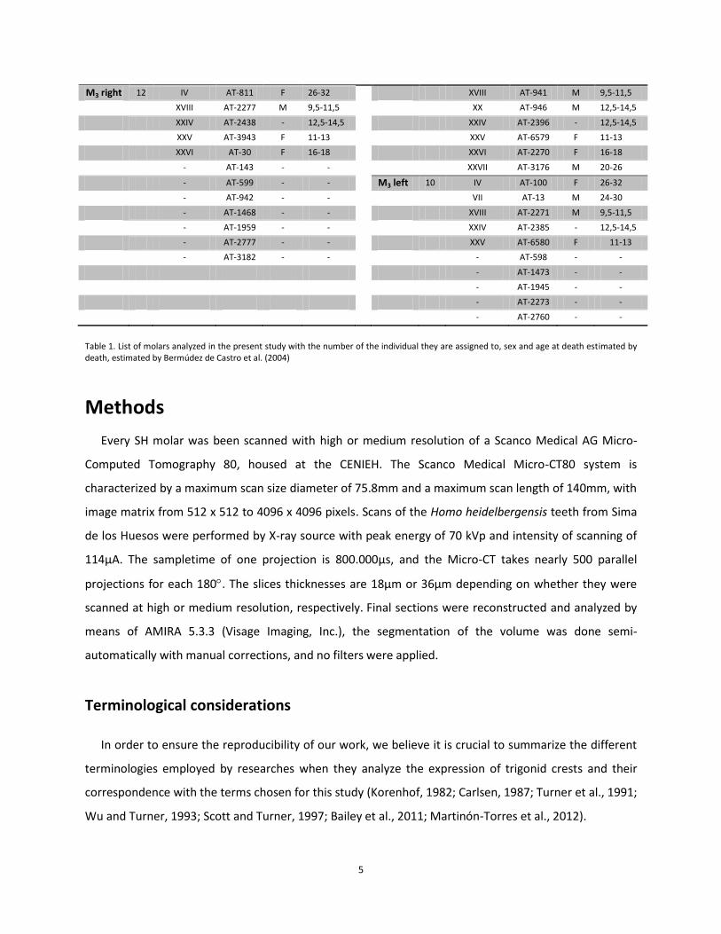
Table 1. List of molars analyzed in the present study with the number of the individual they are assigned to, sex and age at death estimated by death, estimated by Bermúdez de Castro et al. (2004)
Methods
Every SH molar was been scanned with high or medium resolution of a Scanco Medical AG Micro-
Computed Tomography 80, housed at the CENIEH. The Scanco Medical Micro-CT80 system is
characterized by a maximum scan size diameter of 75.8mm and a maximum scan length of 140mm, with
image matrix from 512 x 512 to 4096 x 4096 pixels. Scans of the Homo heidelbergensis teeth from Sima
de los Huesos were performed by X-ray source with peak energy of 70 kVp and intensity of scanning of
114µA. The sampletime of one projection is 800.000µs, and the Micro-CT takes nearly 500 parallel
projections for each 180. The slices thicknesses are 18µm or 36µm depending on whether they were
scanned at high or medium resolution, respectively. Final sections were reconstructed and analyzed by
means of AMIRA 5.3.3 (Visage Imaging, Inc.), the segmentation of the volume was done semi-
automatically with manual corrections, and no filters were applied.
Terminological considerations
In order to ensure the reproducibility of our work, we believe it is crucial to summarize the different
terminologies employed by researches when they analyze the expression of trigonid crests and their
correspondence with the terms chosen for this study (Korenhof, 1982; Carlsen, 1987; Turner et al., 1991;
Wu and Turner, 1993; Scott and Turner, 1997; Bailey et al., 2011; Martinón-Torres et al., 2012).

6
Following Carlsen’s terminology, the crown of a tooth is divided in different structures such as lobes,
lobe segments, marginal ridges complexes, cingulum derivatives and supernumerary coronal structures
(Carlsen, 1987). According to him, in posterior teeth, what usually researchers refer as a cusp (Cope,
1888; Osborn, 1888) it is indeed a lobe. A lobe is made up of different lobe segments: two accessory
lobes segments (referred by Scott and Turner as accessory ridges (Scott and Turner, 1997)) and one
essential lobe segment (referred by Scott and Turner as an essential ridge (Scott and Turner, 1997)) with
an occlusal cusp with a free apex, known as an essential cusp (Carlsen, 1987). For him, a ridge is the
elevation of enamel that does not extend across the central sulcus, and a crest is the bridge of enamel
resulting for the join of two ridges from opposing lobes (Carlsen, 1987). All the crown traits that usually
take the form of accessory occlusal tubercles are called supernumerary coronal structures (Carlsen,
1987).
In 1997, Scott and Turner claimed that a tooth crown is made up of four secondary macroscopic
units: lobes, marginal ridges, cingulum derivatives and supernumerary coronal structures (Scott and
Turner, 1997). According to them, and following Carlsen´s terminology (Carlsen, 1987), one lobe is
divided in two sections -buccal and lingual- and three segments –mesial accessory lobe, essential lobe
and distal accessory lobe -, and a cusp is the part of a lobe segment with a free apex at the occlusal edge
(Scott and Turner, 1997). The marginal ridge complex occurs on the mesial and distal surface of the
crown but it may not be always clear. When a free apex occurs on the occlusal surface of a marginal
ridge complex, it is called marginal tubercle (Scott and Turner, 1997). Finally, a cingulum derivative is a
lingually or buccally projection at the cervical section of a tooth crown (Scott and Turner, 1997).
In this study, we will basically follow the terminology employed in Martinón-Torres et al., 2012, that
combines terms and concepts from both Carlsen and Scott and Turner (Carlsen, 1987; Scott and Turner,
1997) (Figure 2). Thus, we consider that the main structures of a molar crown are cusps -with their
corresponding lobes or ridges-, grooves and crests, and that these may be expressed or not at the
dentine surface with the same score as at the enamel one (Skinner et al., 2008). In addition, we can also
find secondary structures such as marginal ridges, cingula or tuberculum projections, and
supernumerary structures, for which we will use the same terminology as Scott and Turner (Scott and
Turner, 1997). Thereby, in accordance with the traditional literature (Cope, 1888; Osborn, 1888) and in
order to avoid possible confusions, we will refer as a cusp what Carlsen (Carlsen, 1987) and Scott and
Turner (Scott and Turner, 1997) referred as an entire lobe, and thus, we consider it like the occlusal
projection of the crown (White and Folkens, 2005). A cusp can be divided in three different segments or

7
lobes or ridges (hereof we will call it lobe) which can be mesial, middle –or essential- and distal (Scott
and Turner, 1997) depending on their anatomical position. The free apex on the top of the essential lobe
will be called cusp tip (note that Carlsen, 1987, called it essential cusp, but Scott and Turner, 1997,
simply call it cusp). The shallow and longitudinal depressions in the surface of enamel will be called
grooves (Carbó Ayala, 2005).
CARLSEN (1987) SCOTT & TURNER (1997) MARTINÓN ET AL. (2012)
Lobe Lobe Cusp
Accessory Lobe Segment (ALS) Mesial Accessory Lobe (MAL) Mesial Lobe (ML)
Essential Lobe Segment (ELS) Essential Accessory Lobe (EAL) Essential Lobe (EL)
Accessory Lobe Segment (ALS) Distal Accessory Lobe (DAL) Distal Lobe (DL)
Essential Cusp (EC) Cusp (C) Cusp Tip (CT)
Figure 2. Definitions and schemes (below) of some of the molar crown structures.
Arising from these concepts about the basic components of a molar we can now discuss the different
terminologies published so far to refer to the trigonid crests.
In 1982, Korenhof defined a mesial (sic) trigonid crest -although Wu and Turner (Wu and Turner,
1993) and Bailey et al. (Bailey et al., 2011) assumed that Korenhof also called it middle trigonid crest-
and a distal trigonid crest. These crests were joining the mesial and the distal parts, respectively, of the
two main mesial cusps of a lower molar. In 1993, Wu and Turner defined a middle and a distal trigonid
crest, but they did not found individuals expressing both crests at the same time. According to them,
when a complete crest connects the middle portions of the mesial cusps and it lies mesialward, but not
on the marginal border, it is a middle trigonid crest (Wu and Turner, 1993); otherwise it is a distal
trigonid crest. In 1997, Scott and Turner only described a distal trigonid crest, and they defined it as a
crest or ridge that courses buccolingually along the distal aspect of the primitive trigonid, now
represented by the protoconid and metaconid (Scott and Turner, 1997).

8
Finally, in recent studies, Skinner et al., 2008 and Bailey et al., 2011, scored crests at the EDJ based on
their origin and thus, stated that a mesial trigonid crest could occur between the mesial marginal ridge
and the middle trigonid crest. However, these authors scored trigonid crests mostly based on the
relative position of one to another, so a mesial trigonid crest might occur between the cusp tips
(although according to Carlsen’s terminology (Carlsen, 1987) that would be a middle-middle crest) as far
as its position is substantially mesialward within the tooth contour. Although we acknowledge the
atypical mesial position of some middle crests (Figure 9b from Bailey et al., 2011), in order to
standardize terms to discuss anatomy in such a detail, we have preferred to subscribe to the notion that
individual cusps are divided into three elements (mesial, middle and distal lobes) and that the middle or
essential one is the one bearing the cusp tip. In this frame, the three types of crests should be scored
independently of the presence/absence of the other, in a similar way as they are scored at the OES (e.g.,
(Guatelli-Steinberg and Irish, 2005; Martinón-Torres et al., 2012). Anyhow, although we subscribe to the
notion that there are mesial, middle and distal trigonid crests we believe that, at the OES, it is not
possible to reliably differentiate a mesial (the one where at least one of the extremes goes clearly to the
marginal ridge) from a middle trigonid. Thus, at the OES we will simply score a middle trigonid crest and
a distal trigonid crest, following traditional literature and classic scoring systems (Korenhof, 1982; Scott
and Turner, 1997).
Scoring procedures
For this study, the crest that courses along the distal aspect of the protoconid and metaconid will be
scored as a distal trigonid crest (DTC). In addition and on the enamel, the one that courses along the
mesial aspect of the protoconid and metaconid will be registered as a middle trigonid crest (MdTC)1
(Figure 3). However, on the dentine we sometimes find another type of crest that we recognize as a
mesial trigonid crest (MeTC), which arises from the middle or mesial lobe to the mesial marginal ridge
(Figure 4). In order to score these crests, we draw an imaginary straight line from the protoconid to the
metaconid tip; any crest lying distal to this line would be a distal trigonid crest, and over or mesial to this
line it can be a middle or mesial.
1 What Korenhof calls mesial trigonid crest, in our study is considered as middle trigonid crest.

9
Figure 3. Enamel from the third lower molar of the Sima de los Huesos site where we can see a Middle Trigonid Crest (MdTC) and a Distal Trigonid Crest (DTC) in AT-2271. Green line is the imaginary line from protoconid tip to metaconid tip employed to assess the position of the crests. Virtual reconstruction of the enamel computer model based on microCT.
Figure 4. Dentine from the third lower molars of the Sima de los Huesos site where we can see a Middle Trigonid Crest (MdTC) and a Distal Trigonid Crest (DTC) in AT-2271, and Mesial Trigonid Crest (MeTC) in AT-143. Green line is the imaginary line from protoconid tip to metaconid tip employed to assess the position of the crests. Virtual reconstruction of the dentine computer models based on microCT.
Summarizing, and taking into account all the anatomical considerations above, at the OES we
recognize four basic types of trigonid crest expression (Figure 5 and Figure 6):
Type A: a continuous MdTC and an absent or discontinuous DTC
Type B: continuous DTC and absent or discontinuous MdTC
Type C: continuous MdTC and DTC
Type D: absent or discontinuous MdTC and DTC

10
Figure 5. Scheme (based on Korenhof, 1982) of the four basic types of trigonid crests on enamel identified in our study. Explanation in the text. Scheme represents the occlusal surface at the OES including the tips of the cusps (represented by the circles) of the protoconid and metaconid.
Figure 6. Examples of different types of Trigonid Crests on enamel: type A (AT-946); type B (AT-2385); type C (AT-2271); type D (AT-943). Virtual reconstruction of the enamel computer models based on microCT.
Although both antimeres were analyzed, we employed the unilateral count method (Turner, 1987)
and in case of asymmetry we have chosen the tooth with the highest degree of expression for the trait
because we consider that is the one that has better expressed the genetic signal.
11
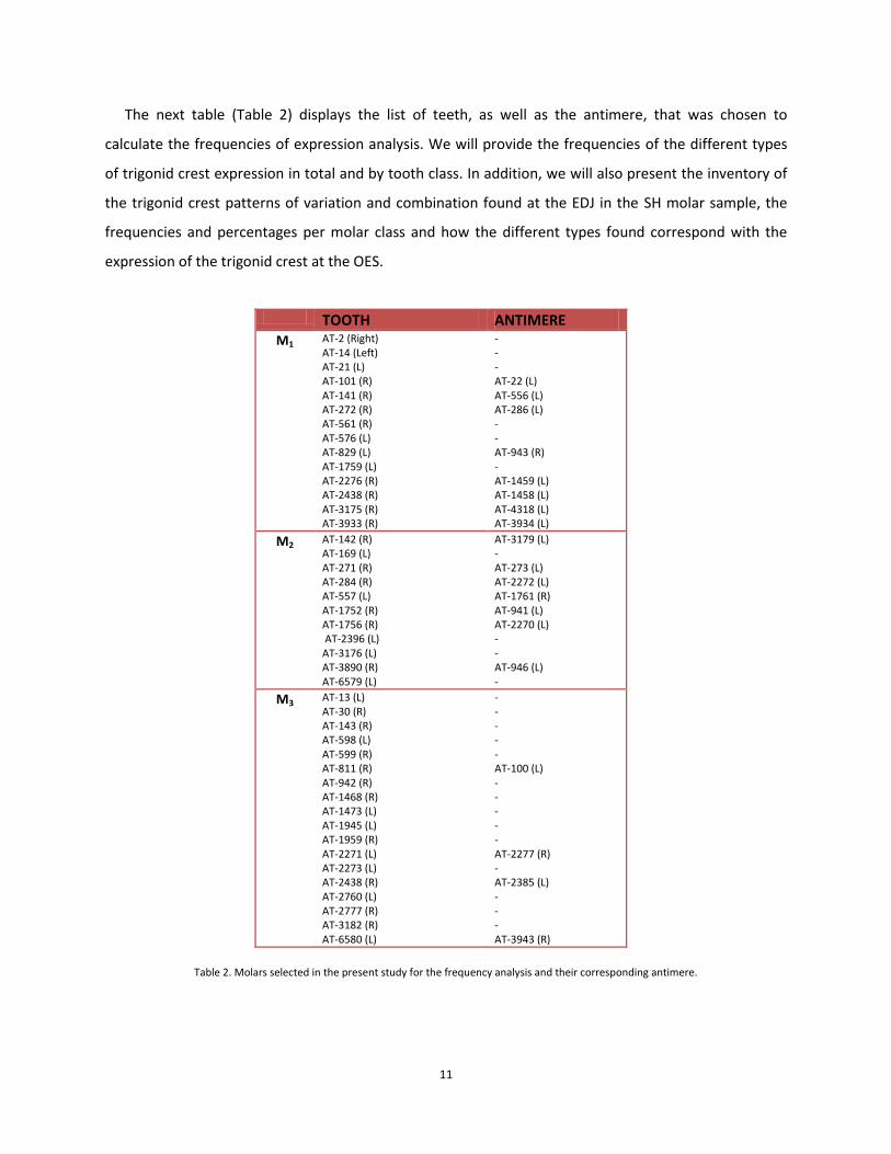
The next table (Table 2) displays the list of teeth, as well as the antimere, that was chosen to
calculate the frequencies of expression analysis. We will provide the frequencies of the different types
of trigonid crest expression in total and by tooth class. In addition, we will also present the inventory of
the trigonid crest patterns of variation and combination found at the EDJ in the SH molar sample, the
frequencies and percentages per molar class and how the different types found correspond with the
expression of the trigonid crest at the OES.
Table 2. Molars selected in the present study for the frequency analysis and their corresponding antimere.
TOOTH ANTIMERE M1 AT-2 (Right)
AT-14 (Left) AT-21 (L) AT-101 (R) AT-141 (R) AT-272 (R) AT-561 (R) AT-576 (L) AT-829 (L) AT-1759 (L) AT-2276 (R) AT-2438 (R) AT-3175 (R) AT-3933 (R)
- - - AT-22 (L) AT-556 (L) AT-286 (L) - - AT-943 (R) - AT-1459 (L) AT-1458 (L) AT-4318 (L) AT-3934 (L)
M2 AT-142 (R) AT-169 (L) AT-271 (R) AT-284 (R) AT-557 (L) AT-1752 (R) AT-1756 (R) AT-2396 (L) AT-3176 (L) AT-3890 (R) AT-6579 (L)
AT-3179 (L) - AT-273 (L) AT-2272 (L) AT-1761 (R) AT-941 (L) AT-2270 (L) - - AT-946 (L) -
M3 AT-13 (L) AT-30 (R) AT-143 (R) AT-598 (L) AT-599 (R) AT-811 (R) AT-942 (R) AT-1468 (R) AT-1473 (L) AT-1945 (L) AT-1959 (R) AT-2271 (L) AT-2273 (L) AT-2438 (R) AT-2760 (L) AT-2777 (R) AT-3182 (R) AT-6580 (L)
- - - - - AT-100 (L) - - - - - AT-2277 (R) - AT-2385 (L) - - - AT-3943 (R)

12
Results
Trigonid crest types at the OES
Table 3 displays the results for the trigonid crest types found at the OES for all the Sima de los Huesos
molar specimens (n=62) by tooth class.
ENAMEL (OES) Specimen Tooth Side Wear Type A Type B Type C Type D
AT-2 M1 R 3 X
AT-14 M1 L 5 X
AT-21 M1 L 5 X
AT-22 M1 L 3 X
AT-101 M1 R 3 X
AT-141 M1 R 3 X
AT-272 M1 R 3 X
AT-286 M1 L 3 X
AT-556 M1 L 3 X
AT-561 M1 R 3 X
AT-576 M1 L 4 X
AT-829 M1 L 2 X
AT-943 M1 R 2 X
AT-1458 M1 L 3 X
AT-1459 M1 L 3 X
AT-1759 M1 L 4 X
AT-2276 M1 R 3 X
AT-2438 M1 R 3 X
AT-3175 M1 R 3 X
AT-3933 M1 R 3 X
AT-3934 M1 L 3 X
AT-4318 M1 L 3 X
AT-142 M2 R 3 X
AT-169 M2 L 2 X
AT-271 M2 R 2 X
AT-273 M2 L 2 X
AT-284 M2 R 2 X
AT-557 M2 L 2 X
AT-941 M2 L 1 X
AT-946 M2 L 2 X
AT-1752 M2 R 1 X
AT-1756 M2 R 2 X
AT-1761 M2 R 2 X
AT-2270 M2 L 2 X
AT-2272 M2 L 2 X
AT-2396 M2 L 2 X
AT-3176 M2 L 3 X
AT-3179 M2 L 2 X
AT-3890 M2 R 2 X
AT-6579 M2 L 2 X
AT-13 M3 L 3 X
AT-30 M3 R 2 X
AT-100 M3 L 3 X
AT-143 M3 R 1 X
AT-598 M3 L 2 X

13
AT-599 M3 R 1 X
AT-811 M3 R 2 X
AT-942 M3 R 1 X
AT-1468 M3 R 1 X
AT-1473 M3 L 2 X
AT-1945 M3 L 1 X
AT-1959 M3 R 1 X
AT-2271 M3 L 1 X
AT-2273 M3 L 1 X
AT-2277 M3 R 1 X
AT-2385 M3 L 1 X
AT-2438 M3 R 1 X
AT-2760 M3 L 1 X
AT-2777 M3 R 1 X
AT-3182 M3 R 1 X
AT-3943 M3 R 1 X
AT-6580 M3 L 1 X
Table 3. Different types of trigonid crest developed on enamel of all the molar sample from the Sima de los Huesos. Bold
type samples refer to the molars selected for the frequency analysis.
As we can see in Figure 8, the most frequent type in the SH molar sample is the trigonid crest type A,
which represents the 77% of the total data that means a frequency of 48 out of 62 lower molars (21 out
of 22 for the M1; the totality of the M2; 9 out of 22 for the M3). The next percentage corresponds to TC
type C whose frequency falls to a 15% with 9 specimens (9 out of 22 for the M3). Regarding TC type B,
the percentage is a 5% of the overall sample, with only 3 molars with that type of crest (3 out of 22 for
the M3). Finally, TC type D is the one with the lowest percentage, only 2 molars of the entire sample
which corresponds to the 3% of the total (1 out of 22 for the M1; 1 out of 22 for the M3). Graphs below
(Figure 7) show the rates of the different types of trigonid crests on enamel for the overall of the molars
from the Sima de los Huesos site.
Figure 7. Graphic with the TC types at OES by tooth class for the overall molar sample from the SH site.

14
ENAMEL (OES) N / %
Type A 48 (77%)
Type B 3 (5%)
Type C 9 (15%)
Type D 2 (3%)
Table 4.Table with the frequencies and percentages of all molar sample from the SH for the different types of TC on enamel.
Figure 8. Graphic with the frequencies of all the molar sample from the SH for the different types of TC on enamel.
If we calculate the frequency of expression of the trigonid crest types at the OES counting only one
antimere per individual (n=43), we observe that the totality of the M1 and M2 specimens analyzed
present a MdTC, and for the M3 the percentage reaches to 83.33%, which means a frequency of 15 out
of 18 lower molars (Table 5). All the MdTC present in the sample show continuous crests at the OES.
ENAMEL (OES)
Middle Trigonid Crest Total molar sample M1 sample M2 sample M3 sample
Present N 40 14 11 15
% 93.02% 100% 100% 83.33%
Absent N 3 0 0 3
% 6.97% 0% 0% 16.66%
Total 43 14 11 18
Table 5. Frequencies and percentages of the middle trigonid crest (MdTC) at the OES for the SH lower permanent molars analyzed (only one antimere included).

15
Table 6 shows us that the overall M3 molars sample analyzed exhibit distal trigonid crest (DTC), half of
which present continuous crests and the other half discontinuous ones. For the M2 the percentage
reaches 72.72% that means a frequency of 8 out of 11 lower molars, all of them with a discontinuous
crest; and for the M1 just only one molar (7.14%) present this type of trigonid crest that is also
discontinuous.
ENAMEL (OES)
Distal Trigonid Crest Total molar sample M1 sample M2 sample M3 sample
Present N 27 1 8 18
% 62.79% 7.14% 72.72% 100%
Absent N 16 13 3 0
% 37.20% 92.85% 27.27% 0%
Total 43 14 11 18
Table 6. Frequencies and percentages of the distal trigonid crest (DTC) at the OES for the SH lower permanent molars analyzed (only one antimere included).
Trigonid crest types at the EDJ
In the dentine of the SH molar sample, the variety of different trigonid crest types and combinations
is clearly higher and more complex that the ones established on enamel. This has led us to develop an
inventory of all the types of trigonid crests combinations that may be useful for other researchers in
forthcoming studies about these traits. Contrary to what occurred with the enamel, at the EDJ we were
able of being more precise in discerning the origin of the crests and in differentiating a mesial trigonid
crest (MeTC) from a middle trigonid crest (MdTC). As made clear on page 8, a MeTC is present when at
least one of the extremes of the crest ends at the mesial marginal ridge, and a MdTC is the one that runs
in the mesial aspect of the trigonid and originates from the middle or mesial lobe. In addition, and as we
explained above, we could also found a DTC that arise from the essential or middle lobes (from the cusp
tip or very close to it), or DTC that clearly arise from the distal lobes. These crests can be continuous or
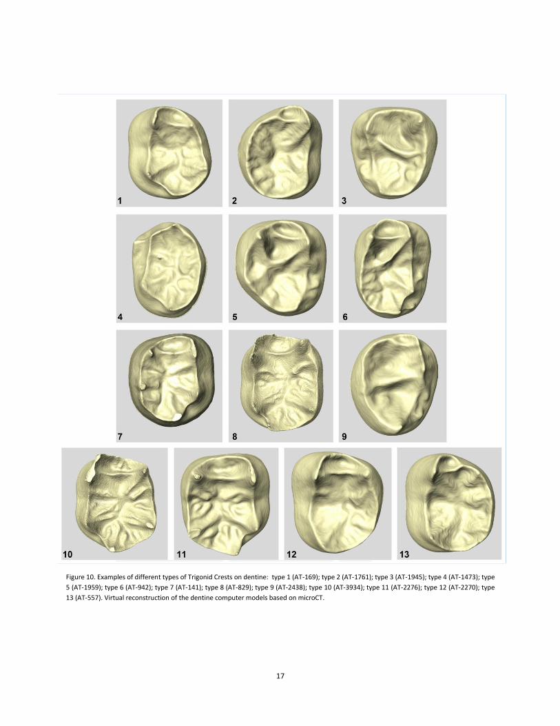
discontinuous and, in the SH sample (Figure 9 and Figure 10), were combined in 13 different types such
as:
Type 1: continuous MdTC with an absent or discontinuous DTC.
Type 2: continuous MeTC with an absent or discontinuous DTC.

16
Type 3: continuous DTC when at least one of the origins is the distal lobe, with an absent or
discontinuous MeTC or MdTC.
Type 4: discontinuous DTC starting from the distal lobes, with an absent or discontinuous MeTC or
MdTC.
Type 5: continuous MdTC and a continuous DTC arising from the middle lobes.
Type 6: continuous MdTC and a continuous DTC when at least one of the origins is the distal lobe.
Type 7: continuous MdTC with a discontinuous DTC when at least one of the origins is the distal
lobe.
Type 8: continuous MdTC with a discontinuous DTC arising from the distal lobes.
Type 9: continuous MeTC and continuous DTC when at least one of the origins is the distal lobe.
Type 10: continuous MdTC, discontinuous DTC when at least one of the origins is the distal lobe,
and a pronounced distal ridge on one side.
Type 11: continuous MdTC, a discontinuous DTC when at least one of the origins is the distal lobe,
and a discontinuous DTC originated from the distal lobes.
Type 12: continuous MdTC, a discontinuous DTC arising from the middle lobes and discontinuous
DTC arising from the distal lobes.
Type 13: continuous MeTC, a discontinuous DTC arising from the middle lobes and discontinuous
DTC arising from the distal lobes.
Figure 9. Scheme of all the different types of trigonid crests on dentine identified in our study from the SH molar sample. Explanation in the text. View is towards the occlusal surface of the EDJ including the tips of the dentine horn (represented by circles) of the protoconid and metaconid and the mesial marginal ridge (represented by the continuous black line).
17
Figure 10. Examples of different types of Trigonid Crests on dentine: type 1 (AT-169); type 2 (AT-1761); type 3 (AT-1945); type 4 (AT-1473); type
5 (AT-1959); type 6 (AT-942); type 7 (AT-141); type 8 (AT-829); type 9 (AT-2438); type 10 (AT-3934); type 11 (AT-2276); type 12 (AT-2270); type
13 (AT-557). Virtual reconstruction of the dentine computer models based on microCT.

18
Table 7 displays the results for the trigonid crest types found at the dentine (EDJ) for the entire Sima
de los Huesos molar sample by tooth class. We also present the side and degree of wear of each one.
DENTINE (EDJ)
Specimen Tooth Side Wear Type
1 Type
2 Type
3 Type
4 Type
5 Type
6 Type
7 Type
8 Type
9 Type
10 Type
11 Type
12 Type
13
AT-2 M1 R 3 X
AT-14 M1 L 5
X
AT-21 M1 L 5
X
AT-22 M1 L 3 X
AT-101 M1 R 3
X
AT-141 M1 R 3
X
AT-272 M1 R 3
X
AT-286 M1 L 3
X
AT-556 M1 L 3
X
AT-561 M1 R 3
X
AT-576 M1 L 4
X
AT-829 M1 L 2
X
AT-943 M1 R 2
X
AT-1458 M1 L 3
X
AT-1459 M1 L 3
X
AT-1759 M1 L 4
X
AT-2276 M1 R 3
X
AT-2438 M1 R 3
X
AT-3175 M1 R 3
X
AT-3933 M1 R 3
X
AT-3934 M1 L 3
X
AT-4318 M1 L 3
X
AT-142 M2 R 3 X
AT-169 M2 L 2 X
AT-271 M2 R 2
X
AT-273 M2 L 2
X
AT-284 M2 R 2
X
AT-557 M2 L 2
X
AT-941 M2 L 1
X
AT-946 M2 L 2
X
AT-1752 M2 R 1
X
AT-1756 M2 R 2
X
AT-1761 M2 R 2
X
AT-2270 M2 L 2
X
AT-2272 M2 L 2
X
AT-2396 M2 L 2
X
AT-3176 M2 L 3
X
AT-3179 M2 L 2
X
AT-3890 M2 R 2
X
AT-6579 M2 L 2
X
AT-13 M3 L 3
X
AT-30 M3 R 2
X
AT-100 M3 L 3
X
AT-143 M3 R 1
X
AT-598 M3 L 2
X
AT-599 M3 R 1
X
AT-811 M3 R 2
X
AT-942 M3 R 1
X
AT-1468 M3 R 1
X
AT-1473 M3 L 2
X

19
AT-1945 M3 L 1
X
AT-1959 M3 R 1
X
AT-2271 M3 L 1
X
AT-2273 M3 L 1
X
AT-2277 M3 R 1
X
AT-2385 M3 L 1
X
AT-2438 M3 R 1
X
AT-2760 M3 L 1
X
AT-2777 M3 R 1
X
AT-3182 M3 R 1
X
AT-3943 M3 R 1
X
AT-6580 M3 L 1
X
Table 7. Different types of trigonid crest developed in dentine of the entire molar sample from the Sima de los Huesos.
Bold type samples refer to the molars selected for the frequency analysis.
As we can see in Figure 12, the most frequent pattern in the SH molar sample at the EDJ is TC type
11, which represents the 18% of the total sample with 11 out of 62 lower molars (4 out of 22 for the M1;
5 out of 18 for the M2; 2 out of 22 for the M3). The next percentage corresponds to the TC type 10 with
the 13% or a frequency of 8 molars out of 62 (5 out of 22 for the M1; 1 out of 18 for the M2; 2 out of 22
for the M3). In decreasing order, the TC type 12 (3 out of 22 for the M1; 4 out of 18 for the M2) and TC
type 13 (5 out of 18 for the M2; 2 out of 22 for the M3) present an equal value of 11% which corresponds
to 7 molars out of the total sample. Regarding the TC type 8, the percentage falls down to a 10% with a
frequency of 6 molars (5 out of 22 for the M1; 1 out of 22 for the M3). The TC type 1 (2 out of 22 for the
M1; 2 out of 18 for the M2), TC type 3 (4 out of 22 for the M3) and TC type 6 (4 out of 22 for the M3) are
represented by 6% each one, followed by the and the TC type 5 with a 5% (3 out of 22 for the M3).
Finally, for the TC type 2 (1 out of 18 for the M2; 1 out of 22 for the M3), TC type 4 (1 out of 22 for the
M1; 1 out of 22 for the M3), TC type 7 (2 out of 22 for the M1) and TC type 9 (2 out of 22 for the M3) the
percentage reduce to 3% of the overall sample, this means just only a frequency of 2 molars with those
types of crests (Figure 11).
Figure 11.Graphic with the frequencies of TC types at EDJ by tooth class for the overall molar sample from the SH site.

20
Table 8. Table with frequencies and percentages of all the molar
sample from SH for the different types of TC on dentine.
Figure 12. Graphic with the frequencies of the entire molar sample from SH for the different types of TC on dentine.
The next tables present the frequencies of expression of the MeTC (Table 9), MdTC (Table 10) and
DTC (Table 11) continuous and discontinuous for each dental class if we choose only one antimere per
individual. The largest number of MeTCs is displayed by the M3s (5 molars out of 18), followed by M2s (3
molars out of 11) and ending with no MeTCs for the M1s. The totality of the molars with MeTCs (19%)
presents a continuous trigonid crest type.
DENTINE (EDJ) N / % Type 1 4 (7%)
Type 2 2 (3%)
Type 3 5 (8%)
Type 4 2 (3%)
Type 5 3 (5%)
Type 6 4 (6%)
Type 7 2 (3%)
Type 8 6 (10%)
Type 9 1 (2%)
Type 10 8 (13%)
Type 11 11 (18%)
Type 12 7 (11%)
Type 13 7 (11%)

21
DENTINE (EDJ)
Mesial Trigonid Crest Total molar sample M1 sample M2 sample M3 sample
Present N 8 0 3 5
% 19% 0% 27.27% 27.77%
Absent N 35 14 8 13
% 81% 100% 72.72% 72.22%
Total 43 14 11 18
Table 9.Frequencies and percentages of the mesial trigonid crest (MeTC) at the EDJ for the SH lower permanent molars analyzed.
In Table 10, we can observe that on dentine the middle trigonid crest (MdTC) is present in 74% of the
overall sample analyzed and represented by a continuous trigonid type. The highest percentage
correspond to the M1s with the totality of the sample, followed by a 72.72% for the M2s with a
frequency of 8 molars out of 11, and 55.55% for the M3s with 10 molars out of 18.
DENTINE (EDJ)
Middle Trigonid Crest Total molar sample M1 sample M2 sample M3 sample
Present N 32 14 8 10
% 74% 100% 72.72% 55.55%
Absent N 11 0 3 8
% 26% 0% 27.27% 44.44%
Total 43 14 11 18
Table 10. Frequencies and percentages of the middle trigonid crest (MdTC) at the EDJ for the SH lower permanent molars analyzed.
In relation to the distal trigonid crest (DTC) on dentine, the Table 11 shows us how this pattern
presents the highest value of the overall sample analyzed with a percentage of 91% that means a
frequency of 39 out of 43 lower molars. The values are a 94.44% for the M3s, 92.85% for the M1s and
90.90% for the M2s. All molars present discontinuous crests except 10 M3s that exhibit continuous ones.
DENTINE (EDJ)
Distal Trigonid Crest Total molar sample M1 sample M2 sample M3 sample
Present N 39 13 10 17
% 91% 92.85% 90.90% 94.44%
Absent N 4 1 1 1
% 9% 7.14% 9.09% 5.55%
Total 43 14 11 18
Table 11. Frequencies and percentages of the distal trigonid crest (DTC) at the EDJ for the SH lower permanent molars analyzed.
22
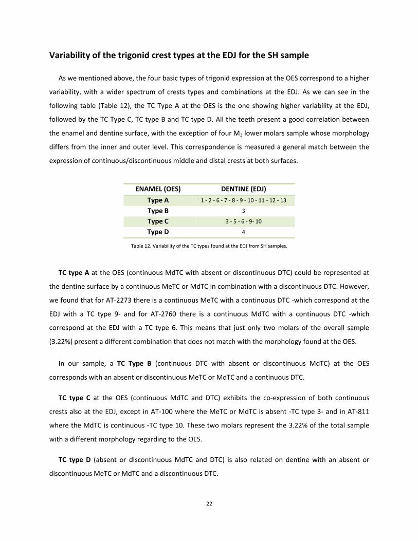
Variability of the trigonid crest types at the EDJ for the SH sample
As we mentioned above, the four basic types of trigonid expression at the OES correspond to a higher
variability, with a wider spectrum of crests types and combinations at the EDJ. As we can see in the
following table (Table 12), the TC Type A at the OES is the one showing higher variability at the EDJ,
followed by the TC Type C, TC type B and TC type D. All the teeth present a good correlation between
the enamel and dentine surface, with the exception of four M3 lower molars sample whose morphology
differs from the inner and outer level. This correspondence is measured a general match between the
expression of continuous/discontinuous middle and distal crests at both surfaces.
ENAMEL (OES) DENTINE (EDJ)
Type A 1 - 2 - 6 - 7 - 8 - 9 - 10 - 11 - 12 - 13
Type B 3
Type C 3 - 5 - 6 - 9- 10
Type D 4
Table 12. Variability of the TC types found at the EDJ from SH samples.
TC type A at the OES (continuous MdTC with absent or discontinuous DTC) could be represented at
the dentine surface by a continuous MeTC or MdTC in combination with a discontinuous DTC. However,
we found that for AT-2273 there is a continuous MeTC with a continuous DTC -which correspond at the
EDJ with a TC type 9- and for AT-2760 there is a continuous MdTC with a continuous DTC -which
correspond at the EDJ with a TC type 6. This means that just only two molars of the overall sample
(3.22%) present a different combination that does not match with the morphology found at the OES.
In our sample, a TC Type B (continuous DTC with absent or discontinuous MdTC) at the OES
corresponds with an absent or discontinuous MeTC or MdTC and a continuous DTC.
TC type C at the OES (continuous MdTC and DTC) exhibits the co-expression of both continuous
crests also at the EDJ, except in AT-100 where the MeTC or MdTC is absent -TC type 3- and in AT-811
where the MdTC is continuous -TC type 10. These two molars represent the 3.22% of the total sample
with a different morphology regarding to the OES.
TC type D (absent or discontinuous MdTC and DTC) is also related on dentine with an absent or
discontinuous MeTC or MdTC and a discontinuous DTC.

23
Frequencies of expression for the TC types at the OES and EDJ by tooth class
The frequencies of expression for trigonid crests are given by tooth class (M1, M2 and M3). Table 13,
Table 14, Table 15 and Table 16 provide the frequencies and percentages of each different type of
trigonid crest on both enamel and dentine surfaces, but using the individual count method. We have
detailed the frequency of the different types of trigonid crest expression from the Sima de los Huesos
sample in the 43 lower permanent molars analyzed (Table 13), in the M1 permanent ones (Table 14), in
the M2 (Table 15) and in the M3 (Table 16).
In Table 13, we present the frequencies and percentages for the different trigonid crest types, both
on enamel and dentine, of the total molar sample. At the enamel, the TC type A represents the 77% of
the total data, which means a frequency of 33 molars with this type of trigonid crest, the highest value.
Regarding to the TC type C the percentage decrease to 16% with a total frequency of 7 molars samples.
For TC type B, the percentage reduces to the number of 5% for the overall sample, this means just only
a frequency of 2 molars with that type of crest. The lowest value corresponds to the TC type D with a 2%
that represent only 1 molar of the entire analyzed sample. Talking about the dentine, the highest value
stands for the TC type 11 (16%) with a frequency of 7 molars, following by the TC type 10 (14%) with a
frequency of 6 molars, and the TC type 8 and 13, both with 12% or 5 lower molars of frequency. Next
value is represented by the TC type 1, 5, 6 and 12, with a frequency of 3 molars that means the 7% of
the overall sample for each type. For the TC type 3, 7 and 9 (5%) there are just only 2 molars; one more
than the frequency represented by the TC type 2 and 4 whose percentage fall down to 2% of the total
analyzed sample.
ENAMEL TYPES N / % DENTINE TYPES N / % A 33 (76.74%) 1 3 (6.97%) B 2 (4.65%) 2 1 (2.32%) C 7 (16.27%) 3 2 (4.65%) D 1 (2.32%) 4 1 (2.32%)
TOTAL 43 5 3 (6.97%) 6 3 (6.97%) 7 2 (4.65%) 8 5 (11.62%)
9 2 (4.65% 10 6 (13.95%) 11 7 (16.27%) 12 3 (6.97%) 13 5 (11.62%) TOTAL 43
Table 13. Frequencies of expression and percentages for the different types of trigonid crests on both enamel and dentine from the Sima de los Huesos lower permanent molars sample analyzed.

24
For the M1 (Table 14) M2 (Table 15) and M3 (Table 16) permanent molars, we can observe different
frequencies and percentages for each trigonid crest type depending on the enamel or dentine surface.
The next table (Table 14) shows us that for the M1s there is a complete correlation between the enamel
trigonid crest types and the dentine ones. Treating the data separately, we can see that for the enamel
the TC type A represents the overall percentage (100%), meanwhile at the dentine the values are
distributed between the TC type 8 with the highest percentage of the sample (29%), following by the TC
type 10 and TC type 11 both with a frequency of 3 molars (21%), the TC type 7 (14%) with 2 lower
molars, and lastly the TC type 1 and TC type 12 with only one sample (7%).
ENAMEL TYPES N / % DENTINE TYPES N / % A 14 (100%) 1 1 (7.14%) B 0 2 0 C 0 3 0 D 0 4 0
TOTAL 14 5 0 6 0 7 2 (14.28%) 8 4 (28.57%) 9 0 10 3 (21.42%) 11 3 (21.42%) 12 1 (7.14%) 13 0 TOTAL 14
Table 14. Frequency of expression of trigonid crests and their percentages on both enamel and dentine for the M1 permanent molars analyzed.
Once more, the TC type A represents the whole percentage (100%) at enamel (Table 15), whereas at
dentine the values are distributed between the TC type 11 and 13, both with a frequency of 3 molars
(27%), following by the TC type 1 and 12 with 18% that means 2 lower molars for each one, and the TC
type 10 with the lowest percentage of the M2 sample represented by only one molar (9%).
ENAMEL TYPES N / % DENTINE TYPES N / % A 11 (100%) 1 2 (18.18%) B 0 2 0 C 0 3 0 D 0 4 0
TOTAL 11 5 0 6 0 7 0 8 0 9 0 10 1 (9.09%) 11 3 (27.27%) 12 2 (18.18%) 13 3 (27.27%) TOTAL 11
Table 15. Frequency of expression of trigonid crests and their percentages on both enamel and dentine for the M2 permanent molars analyzed.

25
With regard to the M3 molar samples analyzed there is a greater variability between the trigonid
crest types on enamel and dentine than the previous molars samples (Table 16). In this way, the overall
percentage at enamel are divided into TC type A (44%), TC type C (39%), TC type B (11%) and TC type D
(6%). The values at dentine correspond with the highest percentage to TC type 5 and 6 (17%) following
by the TC type 3, 9, 10 and 13 (11%), and finally with the TC type 2, 4, 8 and 11 (6%).
ENAMEL TYPES N / % DENTINE TYPES N / %
A 8 (44.44%) 1 0 B 2 (11.11%) 2 1 (5.55%) C 7 (38.88%) 3 2 (11.11%) D 1 (5.55%) 4 1 (5.55%)
TOTAL 18 5 3 (16.66%) 6 3 (16.66%) 7 0 8 1 (5.55%) 9 2 (11.11%) 10 2 (11.11%) 11 1 (5.55%) 12 0 13 2 (11.11%) TOTAL 18
Table 16. Frequency of expression of trigonid crests and their percentages on both enamel and dentine for the M3 permanent molars analyzed.
Discussion
Due to the many discrepancies found in the traditional classification systems, one of the most
important inconveniences we have found when analyzing the lower molar sample from the Sima de los
Huesos site was to establish single and uniform criteria to classify them (see Terminological
considerations on page 5). This problem prevents the direct comparison of our results with those
obtained by Bailey et al., 2011 for the Neanderthal and the H. sapiens groups.
Among all the molar samples from SH, the M3s ones are those with more variability and complexity.
They are rather atypical (in some of them cusps are rotated within the tooth contour, some other have
reduced main cusps in combination with accessory cusps…), preventing an easy identification of the
morphological features. As an example, in AT-100 the marginal ridge is atrophied and ends at the mesial
part of the molar, making difficult to distinguish whether there is a continuous MeTC or not (Figure 13).
Most of the cases showing a “mismatch” between surfaces correspond to atypical teeth. Despite of this
inconvenience, there is a huge correlation between the OES and EDJ, but sometimes happens that what

26
seems to be a continuous crest on the enamel surface actually is a discontinuous one at dentine level. As
stated above, it happens rarely, and when this occurs it is usually in quite atypical teeth and with C7s.
Figure 13.Dentine surface of a M3 (AT-100) with an atrophied MMR.
Variability and frequencies of expression at the EDJ
Our results confirm a greater variability in the dentine than in the enamel surface, but with a good
general correspondence between them. Thus, at the OES a MdTC could be a MeTC or MdTC at the EDJ.
Similarly, a DTC at the EDJ could have different origins such as middle-middle, distal-middle, distal-distal,
or even exhibit double distal ridges. But in all cases there is generally great concordance, in whatever
form, between the presence of a continuous crest at the OES and the presence of a continuous crest at
the EDJ.
Regarding the distal trigonid crest at the EDJ, and unlike what other researchers have stated -Wu and
Turner did not found individuals with MdTC and DTC at the same time (Wu and Turner, 1993); and
Bailey and colleagues affirmed that they have not seen molars connecting distal segments of the C1 and
C2 (Bailey et al., 2011)- we can affirm that in the 43 lower molar sample analyzed there are large
number of molars with distal origins (36 specimens that means a 83.72%) and, that we have also found
cases with “true” distal trigonid crest although they are not very common (3 molars or 6.97%) and only
M3s display it (Figure 14) which connects the distal portions of the protoconid and metaconid (Bailey et
al., 2011). Concerning the crests that connect middle and distal lobe segments, other researchers have
found a small number of molars but only in Homo sapiens (Bailey et al., 2011), while in our sample there
are 4 molars, which mean a 9.30% of the sample, that present this type of trigonid crest pattern.

27
Figure 14. Illustration of a “true” distal trigonid crest at EDJ and OES in an M3 molar specimen (AT-2385). Virtual reconstruction of dentine and enamel computer models based on microCT.
Talking about the “true” middle trigonid crest (Figure 15) which connects the essential lobes of the
protoconid and metaconid (Bailey et al., 2011), there are 13 out of 32 molars sample that present this
type of trigonid crest (40.62%), which means that the remaining 19 samples present a middle trigonid
crest connecting the mesial and essential lobes (59.37%). According to Bailey et al. (2011) the mesial
trigonid crest originates most often from the middle lobe segment; however, our results reveals that of
the 8 lower molars which present a mesial trigonid crest, 5 of them have their origin at the mesial lobe
(62.5%) and the other 3 specimens have an origin at the middle segment origin (37.5%).
Figure 15. Illustration of a “true” middle trigonid crest at EDJ and OES in an M2 molar specimen (AT-946). Virtual reconstruction of dentine and enamel computer models based on microCT.
If we pay attention to the trigonid crests types by tooth class, we can observe two aspects that could
be relevant for the correct classification of isolated molars. One of them relates to the TC types at the
OES where the TC type B and C are only found in M3s samples (see pag. 13) The other relevant feature is
related to the TC types at the EDJ, since there are not M1s with MeTC in the overall SH sample (see pag.
28
20). If these notions are confirmed in this and other fossil samples, it may become an advantage for the
correct classification of some isolated remains in a particular sample.
Taxonomical implications
At this point we are obliged to limit our debate to the Sima de los Huesos Pleistocene samples, for
which we have analyzed the trigonid crests patterns on both the enamel (OES) and dentine (EDJ).
Although the results of our analyses corroborate a high correspondence in the percentages and
frequency of this trait between the enamel and dentine surface, (Bailey et al., 2011) the expression and
variability of some trigonid crests at the EDJ can be masked by the enamel deposition at the OES (Bailey
et al., 2011; this study). We have used some traditional criteria -like Wu and Turner used in 19932 (Wu
and Turner, 1993)- to score the trigonid crest at the OES, but we also recorded the full variability
expressed at the EDJ to have a more precise classification of our fossil dental sample. According to
Bailey (Bailey, 2002a, 2002b) and Martinón-Torres (Martinón-Torres et al., 2012) the expression at the
OES of a continuous MdTC is a typical Neanderthal feature, but this feature is also present in the Middle
Pleistocene hominins fossils from Europe like Sima de los Huesos and Arago –but not in Mauer despite
being the holotype of the species Homo heidelbergensis that group them- and in variable percentages in
earlier species like H. erectus, H. georgicus and H. antecessor (Martinón-Torres et al., 2007, 2008, 2012).
Previous studies about the outer surface on Homo heidelbergensis teeth from Sima de los Huesos site
have shown that those fossils present the classic Neanderthal traits (Bermúdez de Castro, 1986;
Bermúdez de Castro et al., 1999; Martinón-Torres et al., 2007, 2012). Now, with the new techniques of
microCT and virtual segmentations, we are allowed to compare these characteristics present on the
enamel (OES) with those offered at dentine (EDJ) in order to be more accurate when considering the
resemblances found between them, and thus attempt to answer the currently assumptions whether the
SH hominids were the starting point at the origin of the classic Neanderthals, or if on the contrary were
a different lineage (Tattersall and Schwartz, 2009; Dennell et al., 2010; Martinón-Torres et al., 2012).
In absolute terms, we can affirm that at the EDJ there is a greater number of SH molars with middle
or mesial trigonid crest than without it, that the middle crest is more frequent than the mesial one and
that there is a high proportion of lower molars with a continuous mesial (MeTC) or middle trigonid crest
2 “If only one trigonid crest occurs, and if the crest lies slightly mesialward, but not on the marginal border, it is said
to be a middle trigonid crest.”

29
(MdTC) and a discontinuous distal trigonid crest (DTC), which means a clear predominance of the TC
type A. We have also identified a wide spectrum of trigonid crests types at the EDJ, but future studies in
larger samples are necessary in order to know whether all the types are present and/or typical of other
hominin groups or some of them are particular to the SH group. In this sense, the comparison with other
hominins samples is necessary to ensure if the manifestation of these types of trigonid crest pattern in
the Sima de los Huesos sample are derived or primitive in Homo, particularly with regard to H. sapiens
and H. neanderthalensis. However, and although a direct comparison between both studies is not
possible because of the different scoring systems employed (this study and Bailey et al., 2011),it was
possible to make some general assessments between both analysis. It seems that the SH sample
coincides with H. neanderthalensis in the almost constant expression of a continuous mesial or middle
trigonid crest and a discontinuous DTC, and therefore, in the high percentages3 of this pattern (although
Bailey et al., 2011 use a different terminology).
ENAMEL (OES) SH sample H. neanderthalensis H. sapiens N % N % N %
MdTC continuous (Grade 2-3, Bailey et al.) 40 93.02 61 100 8 11.76 MdTC discontinuous (Grade 1, Bailey et al.) 3 6.97 0 0 13 19.11
DENTINE (EDJ) SH sample H. neanderthalensis H. sapiens N % N % N %
MdTC continuous (Grade 2-3, Bailey et al.) 40 93.02 72 98.63 24 35,29 MdTC discontinuous (Grade 1, Bailey et al.) 3 6.97 1 1.36 8 11.76
Figure 16. Illustrations of differences among specimens for grades of presence of middle trigonid crest (MdTC) at the OES and EDJ. Note that H. neanderthalensis and H. sapiens dates have been taken from Bailey and colleagues (Bailey et al., 2011).
3 Bailey et al. (Bailey et al., 2011) found a 100% of a trigonid crest at the OES and 98.6% at the EDJ for the H.
neanderthalensis and we found a 100% at both OES and EDJ for the SH sample.

30
Conclusions
In accordance with Bailey and colleagues (Bailey et al., 2011) and because the dentine exhibits
greater variability and more different trigonid crest types than the enamel, it was necessary to use
different scoring system than the traditional ones (Korenhof, 1982; Wu and Turner, 1993) in order to
characterize in a more precise manner the morphology of this sample. Also in accordance with that
study, our analysis has revealed a higher variability of trigonid crest expressions at the EDJ compared to
the OES.
However, and in general terms, our results broadly ratify the strong harmony of trait expression
between the enamel (OES) and the dentine (EDJ) (Skinner et al., 2008; Bailey et al., 2011). Because of
that, we may also point out that both the expression and the grade of development of the trigonid crest
patterns at the enamel is mostly determined by the manifestation at the dentine. This fact is particularly
relevant as it implies the potential possibility of predicting the type of trigonid crest pattern at the OES
in the case of excessively worn teeth. This also maximizes the number of teeth that can be analyzed,
enlarging the sample size particularly in the case of the scarce fossil human remains.
Based on the results of the 43 lower molars sample analyzed from the Sima de los Huesos site, the
complete lack of trigonid crest type B, type C and type D in SH M1s and M2s , could be an interesting
feature to help classify isolated dental specimens, but we must contrast it with other fossils hominins.
Additionally, we observe a greater variability of trigonid crest patterns in M3s than in M1s and M2s for
the overall molars sample. Future studies may clarify if these are general patterns of certain species or
lineages or is a particularity of the SH group.
We also think is desirable to analyze other Pleistocene hominins in order to have greater fossil record
that allows us to get more taxonomic and phylogenetic information about these traits expression as well
as to make possible evolutionary inferences about the expression of this trait in relation with other
ancestral hominins.

31
Bibliography
Arsuaga, J.L., Carretero, J.M., Lorenzo, C., Gracia, A., Martínez, I., Bermúdez de Castro, J.M., Carbonell, E., 1997. Size variation in middle Pleistocene humans. Science 277, 1086–1088.
Arsuaga, J.L., Carretero, J.M., Martínez, I., Gracia, A., 1991. Cranial remains and long bones from Atapuerca/Ibeas (Spain). Journal of Human Evolution 20, 191–230.
Arsuaga, J.-L., Martínez, I., Gracia, A., Carretero, J.-M., Carbonell, E., 1993. Three new human skulls from the Sima de los Huesos Middle Pleistocene site in Sierra de Atapuerca, Spain. Nature 362, 534–537.
Bailey, S.E., 2002a. Neandertal dental morphology: implications for modern human origins. Arizona State University.
Bailey, S.E., 2002b. A closer look at Neanderthal postcanine dental morphology: I. The mandibular dentition. New Anatomist 269, 148–156.
Bailey, S.E., Skinner, M.M., Hublin, J.-J., 2011. What lies beneath? An evaluation of lower molar trigonid crest patterns based on both dentine and enamel expression. American Journal of Physical Anthropology 145, 505–518.
Bermúdez de Castro, J.M., 1986. Dental remains from Atapuerca (Spain) I. Metrics. Journal of Human Evolution 15, 265–287.
Bermúdez de Castro, J.M., Martinón-Torres, M., Carbonell, E., Sarmiento, S., Rosas, A., Van der Made, J., Lozano, M., 2004. The Atapuerca sites and their contribution to the knowledge of human evolution in Europe. Evolutionary Anthropology: Issues, News, and Reviews 13, 25–41.
Bermúdez de Castro, J.M., Martinón-Torres, M., Lozano, M., Sarmiento, S., Muela, A., 2004. Paleodemography of the Atapuerca-Sima de los Huesos hominin sample: a revision and new approaches to the paleodemography of the European Middle Pleistocene population. Journal of Anthropological Research 60, 5–26.
Bermudez de Castro, J.M., Rosas, A., Carbonell, E., Nicolas, M.E., Rodríguez, J., Arsuaga, J.L., 1999. A modern human pattern of dental development in Lower Pleistocene hominids from Atapuerca-TD6 (Spain). Proceedings of the National Academy of Sciences 96, 4210.
Carbó Ayala, J., 2005. Anatomía dental y de la oclusión. Ciudad de La Habana: Editorial de Ciencias Médicas.
Carlsen, O., 1987. Dental morphology. Munksgaard. Cope, E.D., 1888. The mechanical causes of the origin of the dentition of the Rodentia. The American
Naturalist 22, 3–13. Dennell, R.W., Martinón-Torres, M., Bermúdez de Castro, J.M., 2010. Out of Asia: The initial colonization
of Europe in the Early and Middle Pleistocene. Quaternary international 223. Guatelli-Steinberg, D., Irish, J.D., 2005. Brief Communication: Early Hominin Variability in First Molar
Dental Trait Frequencies. American Journal of Physical Anthropology 128, 477–484. Hublin, J.J., 2009. The origin of Neandertals. Proceedings of the National Academy of Sciences 106,
16022 –16027. Irish, J.D., 1998. Diachronic and synchronic dental trait affinities of Late and Post-Pleistocene peoples
from North Africa. Homo 49, 138–155. Korenhof, C.A.W., 1982. Evolutionary trends of the inner enamel anatomy of deciduous molars from
Sangiran (Java, Indonesia). Teeth: form, function and evolution. New York: Columbia University Press. p 350–365.
Macchiarelli, R., Bondioli, L., Debénath, A., Mazurier, A., Tournepiche, J.-F., Birch, W., Dean, M.C., 2006. How Neanderthal molar teeth grew. Nature 444, 748–51.
Martinón-Torres, M., 2006. Evolución del aparato dental en homínidos: estudio de los dientes humanos
32
del Pleistoceno de la Sierra de Atapuerca (Burgos). Universidad de Santiago de Compostela. Martinon-Torres, M., Bermudez de Castro, J.M., Gomez-Robles, A., Arsuaga, J.L., Carbonell, E.,
Lordkipanidze, D., Manzi, G., Margvelashvili, A., 2007. Dental evidence on the hominin dispersals during the Pleistocene. Proc Natl Acad Sci U S A 104, 13279–13282.
Martinón-Torres, M., Bermúdez de Castro, J.M., Gómez-Robles, A., Arsuaga, J.L., Carbonell, E., Lordkipanidze, D., Manzi, G., Margvelashvili, A., 2007. Dental evidence on the hominin dispersals during the Pleistocene. Proceedings of the National Academy of Sciences 104, 13279 –13282.
Martinón-Torres, M., Bermúdez de Castro, J.M., Gómez-Robles, A., Margvelashvili, A., Prado, L., Lordkipanidze, D., Vekua, A., 2008. Dental remains from Dmanisi (Republic of Georgia): morphological analysis and comparative study. Journal of human evolution 55, 249–273.
Martinón-Torres, M., Bermúdez de Castro, J.M., Gómez-Robles, A., Prado-Simón, L., Arsuaga, J.L., 2012. Morphological description and comparison of the dental remains from Atapuerca-Sima de los Huesos site (Spain). Journal of Human Evolution 62, 7–58.
Molnar, S., 1971. Human tooth wear, tooth function and cultural variability. American Journal of Physical Anthropology 34, 175–190.
Nager, G., 1960. [Comparison between the spatial relations of the dentin crown relief and the enamel relief of dental crowns.]. Acta Anatomica 42, 226.
Osborn, H.F., 1888. The evolution of mammalian molars to and from the tritubercular type. The American Naturalist 22, 1067–1079.
Schwartz, G.T., Thackeray, J.F., Reid, C., van Reenan, J.F., 1998. Enamel thickness and the topography of the enamel–dentine junction in South African Plio-Pleistocene hominids with special reference to the Carabelli trait. Journal of Human Evolution 35, 523–542.
Scott, G.R. and Turner, C.G., 1997. The anthropology of modern human teeth: dental morphology and its variation in recent human populations. Cambridge University Press, Cambridge.
Skinner, M.M., Gunz, P., Wood, B.A., Boesch, C., Hublin, J.J., 2009. Discrimination of extant Pan species and subspecies using the enamel-dentine junction morphology of lower molars. American Journal of Physical Anthropology 140, 234–243.
Skinner, M.M., Gunz, P., Wood, B.A., Hublin, J.-J., 2008. Enamel-dentine junction (EDJ) morphology distinguishes the lower molars of Australopithecus africanus and Paranthropus robustus. Journal of Human Evolution 55, 979–988.
Skinner, M.M., Wood, B.A., Boesch, C., Olejniczak, A.J., Rosas, A., Smith, T.M., Hublin, J.J., 2008. Dental trait expression at the enamel-dentine junction of lower molars in extant and fossil hominoids. Journal of human evolution 54, 173–186.
Suzuki, M. and Sakai, T., 1973. Occlusal surface pattern of the lower molars and second deciduous molar among the living polynesians. American Journal of Physical Anthropology 39, 305–316.
Tattersall, I., Schwartz, J.H., 2009. Evolution of the Genus Homo. Annual Review of Earth and Planetary Sciences 37, 67–92.
Turner, C.G., 1987. Late Pleistocene and Holocene population history of East Asia based on dental variation. American Journal of Physical Anthropology 73, 305–321.
Turner, C.G., Nichol, C.R., Scott, G.R., 1991. Scoring procedures for key morphological traits of the permanent dentition: the Arizona State University Dental Anthropology System., in: Kelley, M., Larsen, C. (Eds.), Advances in Dental Anthropology. Wiley Liss, New York, pp. 13–31.
Turner, E.P., 1963. Crown development in human deciduous molar teeth. Archives of Oral Biology 8, 523–IN8.
White, T.D. and Folkens, P.A., 2005. The human bone manual. Academic Press. Wu, L. and Turner, C.G., 1993. Brief communication: Variation in the frequency and form of the lower
permanent molar middle trigonid crest. American Journal of Physical Anthropology 91, 245–248.

33
Illustration list
Tables Table 1. List of molars analyzed in the present study with the number of the individual they are assigned
to, sex and age at death estimated by death, estimated by Bermúdez de Castro et al. (2004) .................. 5
Table 2. Molars selected in the present study for the frequency analysis and their corresponding
antimere. ..................................................................................................................................................... 11
Table 3. Different types of trigonid crest developed on enamel of all the molar sample from the Sima de
los Huesos. Bold type samples refer to the molars selected for the frequency analysis. .......................... 13
Table 4.Table with the frequencies and percentages of all molar sample from the SH for the different
types of TC on enamel. ............................................................................................................................... 14
Table 5. Frequencies and percentages of the middle trigonid crest (MdTC) at the OES for the SH lower
permanent molars analyzed (only one antimere included). ...................................................................... 14
Table 6. Frequencies and percentages of the distal trigonid crest (DTC) at the OES for the SH lower
permanent molars analyzed (only one antimere included). ...................................................................... 15
Table 7. Different types of trigonid crest developed in dentine of the entire molar sample from the Sima
de los Huesos. ............................................................................................................................................. 19
Table 8. Table with frequencies and percentages of all the molar sample from SH for the different types
of TC on dentine. ......................................................................................................................................... 20
Table 9.Frequencies and percentages of the mesial trigonid crest (MeTC) at the EDJ for the SH lower
permanent molars analyzed. ...................................................................................................................... 21
Table 10. Frequencies and percentages of the middle trigonid crest (MdTC) at the EDJ for the SH lower
permanent molars analyzed. ...................................................................................................................... 21
Table 11. Frequencies and percentages of the distal trigonid crest (DTC) at the EDJ for the SH lower
permanent molars analyzed. ...................................................................................................................... 21
Table 12. Variability of the TC types found at the EDJ from SH samples. ................................................... 22
Table 13. Frequencies of expression and percentages for the different types of trigonid crests on both
enamel and dentine from the Sima de los Huesos lower permanent molars sample analyzed. ............... 23
Table 14. Frequency of expression of trigonid crests and their percentages on both enamel and dentine
for the M1 permanent molars analyzed. ..................................................................................................... 24
Table 15. Frequency of expression of trigonid crests and their percentages on both enamel and dentine
for the M2 permanent molars analyzed. ..................................................................................................... 24
Table 16. Frequency of expression of trigonid crests and their percentages on both enamel and dentine
for the M3 permanent molars analyzed. ..................................................................................................... 25
Figures Figure 1. Tribosphenic molar and its counterpart in humans; modified designs from Teeth by Simon
Hillson (2005). ............................................................................................................................................... 4
Figure 2. Definitions and schemes (below) of some of the molar crown structures. .................................. 7

34
Figure 3. Enamel from the third lower molar of the Sima de los Huesos site where we can see a Middle
Trigonid Crest (MdTC) and a Distal Trigonid Crest (DTC) in AT-2271. Green line is the imaginary line from
protoconid tip to metaconid tip employed to assess the position of the crests. Virtual reconstruction of
the enamel computer model based on microCT. ......................................................................................... 9
Figure 4. Dentine from the third lower molars of the Sima de los Huesos site where we can see a Middle
Trigonid Crest (MdTC) and a Distal Trigonid Crest (DTC) in AT-2271, and Mesial Trigonid Crest (MeTC) in
AT-143. Green line is the imaginary line from protoconid tip to metaconid tip employed to assess the
position of the crests. Virtual reconstruction of the dentine computer models based on microCT. ........... 9
Figure 5. Scheme (based on Korenhof, 1982) of the four basic types of trigonid crests on enamel
identified in our study. Explanation in the text. Scheme represents the occlusal surface at the OES
including the tips of the cusps (represented by the circles) of the protoconid and metaconid. ............... 10
Figure 6. Examples of different types of Trigonid Crests on enamel: type A (AT-946); type B (AT-2385);
type C (AT-2271); type D (AT-943). Virtual reconstruction of the enamel computer models based on
microCT. ...................................................................................................................................................... 10
Figure 7. Graphic with the TC types at EDJ by tooth class for the overall molar sample from the SH site. 13
Figure 8. Graphic with the frequencies of all the molar sample from the SH for the different types of TC
on enamel. .................................................................................................................................................. 14
Figure 9. Scheme of all the different types of trigonid crests on dentine identified in our study from the
SH molar sample. Explanation in the text. View is towards the occlusal surface of the EDJ including the
tips of the dentine horn (represented by circles) of the protoconid and metaconid and the mesial
marginal ridge (represented by the continuous black line). ....................................................................... 16
Figure 10. Examples of different types of Trigonid Crests on dentine: type 1 (AT-169); type 2 (AT-1761);
type 3 (AT-1945); type 4 (AT-1473); type 5 (AT-1959); type 6 (AT-942); type 7 (AT-141); type 8 (AT-829);
type 9 (AT-2438); type 10 (AT-3934); type 11 (AT-2276); type 12 (AT-2270); type 13 (AT-557). Virtual
reconstruction of the dentine computer models based on microCT. ........................................................ 17
Figure 11.Graphic with the frequencies of TC types at EDJ by tooth class for the overall molar sample
from the SH site. ......................................................................................................................................... 19
Figure 12. Graphic with the frequencies of the entire molar sample from SH for the different types of TC
on dentine. .................................................................................................................................................. 20
Figure 13.Dentine surface of a M3 (AT-100) with an atrophied MMR. ....................................................... 26
Figure 14. Illustration of a “true” distal trigonid crest at EDJ and OES in an M3 molar sample (AT-2385).
Virtual reconstruction of dentine and enamel computer models based on microCT. ............................... 27
Figure 15. Illustration of a “true” middle trigonid crest at EDJ and OES in an M2 molar sample (AT-946).
Virtual reconstruction of dentine and enamel computer models based on microCT. ............................... 27
Figure 16. Illustrations of differences among specimens for grades of presence of middle trigonid crest
(MdTC) at the OES and EDJ. Note that H. neanderthalensis and H. sapiens dates have been taken from
Bailey and colleagues (Bailey et al., 2011). ................................................................................................. 29
Copyright © 2022 FDOKUMEN


![8 Sima review RDS Spec Iss 4 reprint[1] de2](https://static.fdokumen.com/doc/165x107/631b167a2784ca2fc00526da/8-sima-review-rds-spec-iss-4-reprint1-de2.jpg)