Pediatric Gastroesophageal Reflux Clinical Practice Guidelines
Upload
khangminh22Category
view
0download
0
LUND UNIVERSITY
PO Box 117221 00 Lund+46 46-222 00 00
Reconstruction in the gastroesophageal junction- from routine to advanced.
Djerf, Pauline
2015
Link to publication
Citation for published version (APA):Djerf, P. (2015). Reconstruction in the gastroesophageal junction- from routine to advanced. Lund University.
Total number of authors:1
General rightsUnless other specific re-use rights are stated the following general rights apply:Copyright and moral rights for the publications made accessible in the public portal are retained by the authorsand/or other copyright owners and it is a condition of accessing publications that users recognise and abide by thelegal requirements associated with these rights. • Users may download and print one copy of any publication from the public portal for the purpose of private studyor research. • You may not further distribute the material or use it for any profit-making activity or commercial gain • You may freely distribute the URL identifying the publication in the public portal
Read more about Creative commons licenses: https://creativecommons.org/licenses/Take down policyIf you believe that this document breaches copyright please contact us providing details, and we will removeaccess to the work immediately and investigate your claim.

1
Reconstruction in the gastroesophageal junction
- from routine to advanced
Pauline Djerf
DOCTORAL DISSERTATION
by due permission of the Faculty of Medicine, Lund University, Sweden.
To be defended in the F2 lecture room, Main Building, Skåne University Hospital. Lund November 13th 2015, at 13:00
Faculty opponent
Professor Magnus Sundbom
Department of Surgical Sciences,
Uppsala University, Sweden.

2
Organization LUND UNIVERSITY DOCTORAL DISSERTATION
Department of Surgery Clinical Sciences Lund University and Skåne University Hospital SE-22185 LUND
Date of issue November 13th , 2015
Author: Pauline Djerf, MD Sponsoring organization
Title and subtitle: Reconstruction in the gastroesophageal junction- from routine to advanced.
Abstract
The general aim of the thesis was to contribute to a more evidenced based framework in the surgical treatment of diseases in the gastroesophageal junction (GEJ), by comparing outcome, measured by complication and survival rates, and evaluate different reconstructions regarding long-term symptoms and quality of life (QoL). The GEJ is a poorly defined anatomic area that represents the lower part of the esophagus and its’ junction to the proximal stomach. Diseases in this area cause symptoms that affect negatively the QoL for the patients and often interfere with the ability to eat and drink. The surgical treatments not only aim at eliminating the patient’s symptoms but also to cure her/his sometimes life-threatening condition.
In Paper I we compared two different antireflux procedures in the treatment of gastroesophageal reflux disease (GERD). Many patients do not respond to acid suppressor medication making the need of an antireflux procedure with good long-term functional results important. Epiphrenic diverticula are rare but can cause life-threatening conditions. In Paper II we studied the treatment of symptomatic epiphrenic diverticula, and evaluated the outcome after an antireflux procedure had been added to the myotomy, and studied the long-term effect after surgery both regarding symptoms relief and QoL. The incidence of adenocarcinoma (AC) in the GEJ increases rapidly in the Western world. In Paper III we compared the extensive and less used extended gastrectomy with long Roux-en-Y loop with the more commonly used esophagectomy with gastric tube in the treatment of AC in the GEJ. In Paper IV, we validated the paracetamol absorption test for measuring emptying from the gastric tube and compared it to gold standard, scintigraphy. In Paper V, we evaluated if there is a place for redoing reconstruction of the esophagus when the primary reconstruction fails.
In conclusion, reconstruction for diseases in the GEJ can, both for benign and malignant diseases, be performed with a low postoperative morbidity and mortality, and with good long-term results. In the treatment of GERD, both a total and an anterior 120° fundoplication result in good long-term QoL. Treatment of epiphrenic diverticula should include a myotomy extended through the LES and an antireflux procedure, although the long-term QoL will remain impaired despite good postoperative regression of the primary symptoms. For AC in the GEJ, the extended gastrectomy can be used safely as a complement to esophagectomy and with good long term functional results and QoL. The paracetamol absorption test may be used as an alternative to scintigraphy for identifying delayed emptying from the gastric tube. In case of failure of the primary reconstruction, the patient should be offered another attempt, since it is possible to achieve an equally good outcome after a redo-reconstruction as after a primary reconstruction.
Key words Gastroesophageal junction, Antireflux surgery, Esophagectomy, Gastric tube, Epiphrenic diverticula
Classification system and/or index terms (if any)
Supplementary bibliographical information Language English
ISSN and key title 1652-8220 ISBN 978-91-7619-196-5
Recipient’s notes Number of pages Price
Security classification
I, the undersigned, being the copyright owner of the abstract of the above-mentioned dissertation, hereby grant to all
reference sources permission to publish and disseminate the abstract of the above-mentioned dissertation.
Signature Date

3
Reconstruction in the gastroesophageal junction
- from routine to advanced
Pauline Djerf

4
Copyright Pauline Djerf
Department of Surgery, Clinical Sciences Lund ISBN 978-91-7619-196-5 ISSN 1652-8220
Lund University, Faculty of Medicine Doctoral Dissertation Series 2015:117
Printed in Sweden by Media-Tryck, Lund University Lund 2015

5
Surgery is a study in anatomy and an experiment in pathology
Walther/Belsey

6
Contents
Original papers 7 Thesis at a glance 8 Abbreviations 9 Introduction 11 Anatomy, pathophysiology and some implication on the surgical treatment 13
The esophagus, the gastroesophageal junction and the stomach 13 Innervation 15 Vascular supplies 15 Lymphatic drainage 16 Antireflux mechanism 17
Previous studies 19 Surgical therapy of GERD 19 Epiphrenic diverticula 26 Adenocarcinoma in the GEJ 30 Measuring of gastric tube emptying in esophagectomy patients 38 Redoing reconstruction of the esophagus 39
Objectives 41 Material and Methods 43
Patients 43 Ethical aspects 44 Methods 45
Results 53 Paper I 53 Paper II 57 Paper III 60 Paper IV 66 Paper V 68
Discussion 73 Conclusions 81 Future perspectives 83 Populärvetenskaplig sammanfattning (in Swedish) 85 Acknowledgment 91 References 93

7
Original papers
This thesis is based on the following original papers, which will be referred to in the text by their Roman numerals. The papers are appended at the end of the thesis.
I. Djerf P, Montgomery A, Hallerbäck B, Håkansson HO, Johnsson F. One- and ten-year outcome of laparoscopic anterior 120° versus total fundoplication: a double-blind, randomized multicenter study. Surg Endosc. 2015 Apr 1. [Epub ahead of print]
II Djerf P, Kjellin A, Walther B. Open surgical treatment of Epiphrenic diverticula. 18 years of experience. Submitted.
III Johansson J, Djerf P, Oberg S, Zilling T, von Holstein CS, Johnsson F, Walther B. Two different surgical approaches in the treatment of adenocarcinoma at the gastroesophageal junction. World J Surg. 2008; 32(6):1013-20.
IV Djerf P, Brundin M, Bajc M, Smedh U. Validation of the paracetamol absorption test for measuring gastric tube emptying in esophagectomized patients versus gold standard scintigraphy. Scand J Gastroenterol. 2015; 50(11):1339-47.
V Djerf P, Johansson J, Zilling T, Öberg S, Johnsson F, von Holstein CS, Walther B. Redoing reconstruction of the esophagus. A case-control study comparing primary to repeated esophageal resected patients. Submitted.
Papers are reproduced with permissions ordered through Copyright Clearance Center's RightsLink service.

8
Thesis at a glance
Question Method Result Conclusion
I Gives partial fundoplication less adverse effects, with equally good control of reflux, as total fundoplication in the treatment of GERD?
A double-blind, randomized study, comparing one and ten year outcome after partial versus total fundoplication
Partial fundoplication resulted in less adverse effects but more heartburn than total fundoplication. In both group, reflux symptoms were less severe than preoperatively and QOL improved..
Both procedures offer good long-term control of reflux symptom, with modest post-fundoplication-symptoms.
II Should an antireflux procedure be added to the myotomy in the surgical treatment of symptomatic epiphrenic diverticula:?
Retrospective study of prospectively collected data of 21 patients operated on for symptomatic epiphrenic diverticula.
PPI use and reflux symptoms were more frequent in the group without an antireflux procedure. Despite good symptoms relief the QOL was impaired at long-term follow up.
To lessen the risk of postoperative reflux symptoms, the surgical procedure should inlude a myotomy extended throught the LES and an antireflux procedure
III Is extended gastrectomy with long Roux-en-Y loop an alternative to esophagectomy and gastric pull-up in the treatment of AC in the GEJ?
Retrospective study of prospectively collected data of 133 patients operated on for AC in the GEJ.
Extended gastrectomy with long Roux-en-Y loop results in a high rate of tumor-free resection margin and can be performed with modest morbidity and mortality, and with a satisfactory long-term outcome.
Extended gastrectomy with long Roux-en-Y loop can be used, without risking the QOL for the patient, as an alternative to esophagectomy in the treatment of AC in the GEJ.
IV To validate, by reference to scintigraphy, the paracetamol absorption test for measuring gastric tube emptying.
The two tests were simultaneous conducted, and emptying was calculated for both methods and compared.
There was a reasonably close correlation between the paracetamol absorption test and scintigraphy for time to 25% and 50% emptying, except for one outlier
The paracetamol absorption test may be able to recognize cases of delayed emptying from the gastric tube.
V Is there a place for redoing reconstruction when the primary reconstruction fails?
24 patients with repeated esophageal reconstructions were compared to 48 primary esophagectomized patients.
No increase in postoperative morbidity or mortality was found. The redo patients have the same life expectancy, and similar quality of life, as the primarily operated
Patients with a failed primary reconstruction should be offered another attempt to reconstruct the upper alimentary tract.

9
Abbreviations
AC Adenocarcinoma
APF Anterior partial (120°) fundoplication
BE Barrett’s esophagus
GERD Gastroesophageal reflux disease
GER Gastric emptying rate
HGD High-grade dysplasia
LES Lower esophageal sphincter
LGD Low-grade dysplasia
PPI Proton pump inhibitor
RCT Randomized Controlled Trial
QoL Quality of life

10

11
Introduction
The gastroesophageal junction (GEJ) is a poorly defined anatomic area that represents the lower part of the esophagus and its’ junction to the proximal stomach (Odze et al. 2005, American cancer society, 2015). From an oncological and surgical point of view, and as has been defined in the latest definition of TNM 7 (AJCC 2010, 7th edition), tumors with an epicenter proximal or distal to the GEJ, with extension into the GEJ, are classified as esophageal tumors and stage accordingly. Diseases in the GEJ affect both the esophagus and the GEJ, and their surgical treatment can involve the esophagus, the stomach and/or part of the intestine.
The first description of a hiatal hernia was provided by Bright in 1836 (Bright 1836), but the consequences following it was not clear, and not until 1912 came the first report of a repair of a sliding hernia, presented by Scudder (Scudder et al. 1912). The pathology of esophagitis was described by Quincke in 1879 (Quincke 1879), although the etiology was unknown until 1935 when Winkelstein could demonstrate that esophagitis had a peptic origin secondary to gastroesophageal reflux (Winkelstein 1935). The Antireflux surgery were developed during the 1950th and 60th with Nissen, Hill, Belsey Mark IV, Toupet and Dor procedures (Nissen 1956, Toupet 1963, Skinner & Belsey 1967, Hill 1967, Dor 1967). These procedures intend to augment the pressure of the lower esophageal sphincter and enhance the antireflux barrier by increasing the intraabdominal length of the esophagus and create different kind and size of fundoplication. The most used fundoplication today is the Nissen fundoplication, which is a total fundoplication (360°), but unwanted side effects, as inability to belch and vomit, abdominal bloating and dysphagia, have promoted modifications, both within the Nissen procedure itself but also with other degree of fundoplication. The frequency of antireflux surgery has declined after the introduction of acid suppressor medication (PPI), but even though there are many patients responding to PPI, there are also many patients who do not, or only partial, making the need for antireflux surgery with good long-term functional results still vivid. The best way of achieving such an antireflux procedure is still a matter of discussion.
Esophageal resection with restoring of the gastrointestinal continuity was first described by Billroth in 1871 (Billroth 1871) and Mikulitz in 1886 (Mikulitz 1886). Neither of their patients survived the operation. In 1913, Torek was the first to present a patient who survived an esophageal resection with restoring of the gastrointestinal tract (Torek 1913), and as a substitute for the esophagus he used an

12
external rubber tube. The first transthoracic esophagectomy with an esophagogastrostomy was performed in 1933 by Ohsawa (Ohsawa 1933), but it was in the 1940th that Ivor-Lewis popularized the thoracoabdominal approach, described already in 1922 by Hedblom, where the stomach was prepared through an abdominal incision, and the resection and reconstruction were conducted through a thoracotomy (Hedblom 1922, Lewis 1946). This method is still used worldwide today, with some modifications, mainly for malignancies in the esophagus and the GEJ. It is, though, a matter of debate if malignancy in the GEJ needs a combined approach (abdominal and chest incisions) to be adequately resected, or if a transhiatal esophagectomy or a gastrectomy with transhiatal resection of the lower esophagus would be sufficient. Despite technical and medical development, esophageal surgery still goes with high perioperative morbidity and a considerable risk of postoperative mortality. Substantial weight loss and impoverished physical condition are often seen in patients in need of esophageal surgery. The surgery is technically demanding, requiring skilled and experienced surgeons, and the high risk of complications calls for careful surveillance postoperatively for any deviation from a non-complicated postoperative course to be identified early.
When postoperative mortality decrease and long-term survival increase, more focused has been laid upon the functional result and its impact on the quality of life (QoL). One of the most important factors for a good long-term QoL is a well functional reconstruction. The best way of objectively evaluate the function of a substitute is to perform an emptying measurement. Ideally, if delayed emptying from the substitute is suspected, an objective measurement ought to be performed routinely in order to apply treatment on the right indications, but in reality such a measurement is not always conducted, maybe due to lack of simple accessible measuring tools. To objectively evaluate QoL in patients suffering from diseases in the esophagogastric area, validated questionnaires have been developed (Aaronson et al. 1993, Blazeby et al. 1995, Michelson et al. 2000). QoL measures have become a therapeutic outcome measurement, making it possible to compare the results between surgical procedures, which might help in treatment decisions. In addition, results from QoL measures can promote further development of surgical procedure and postoperative care in order to optimize the outcome for the patients.

13
Anatomy, pathophysiology and some implication on the surgical treatment
The esophagus, the gastroesophageal junction and the stomach
Esophagus connects the throat to the stomach, and is made for transport of ingested material to the stomach and prevent regurgitation of ingested content. The function of the stomach is to digest food, with the help of strong acids and protein-enzymes (proteases) in combination with physically break down. To prevent backward flow of gastric acid and ingested food into the esophagus (i.e. gastroesophageal reflux), there is a sphincter mechanism in the transition area between the esophagus and the stomach called the lower esophageal sphincter (LES) (Sabiston 2004, Skinner&Belsey 1988).
The esophagus and the stomach derive from the embryologic foregut. Esophagus lies behind the trachea, in front of the spine in the posterior mediastinum, continuing behind the heart before entering the abdominal cavity through the hiatus in the diaphragm. The intraabdominal part of the esophagus transforms into the gastroesophageal junction (GEJ) at the gastric cardia. In adults, esophagus is normally 25-33 cm long, the cervical part is usually 4-5 cm and the intraabdominal part about 3-4 cm, the rest of the esophagus lays intrathoracic. The phrenoesophageal ligament, made primary from fascia extended from the diaphragm, closes the opening between the thoracic and abdominal cavity and anchors the esophagus within the hiatus.
The esophageal wall consists of mucosa, submucosa and muscularis propria. The muscularis propria creates the propulsive movement of the esophagus and has two layers of muscles, an inner circular and an outer longitudinal. As opposed to the rest of the gastrointestinal tract, the esophagus lacks an outer serosal layer but is surrounded by a loose connective tissue that allows movement during swallowing and respiration. The upper third part of the esophageal wall consists of striated muscle. In the mid portion there is a mixture of striated and smooth muscle, with the smooth muscle successively dominating distally. As the rest of the gastrointestinal tract, the muscle in the distal third part of esophagus consists only of smooth muscle. The

14
stomach has an extra inner oblique muscle layer, in addition to the circular and longitudinal layers, used for its’ grinding activity.
The lumen of the esophagus is lined by a squamous epithelium except for its’ distal 2-3 cm where a columnar epithelium of the gastric cardia type occurs. The transition between the different epithelium is easily seen endoscopically, and the junction is referred to as the Z-line since it has a jagged line (ora serrata). The Z-line is normally situated on the top of the gastric folds.
Consequences of a defect barrier between the esophagus and the stomach
Reflux of gastric content into the esophagus is normal, especially after a meal or when performing physical activity. The normal reflux is short, harmless and most often asymptomatic since there are defense mechanisms, as protective factors in the mucosa and saliva neutralizing the refluxate material, and esophageal peristalsis clearing the esophagus from the reflux material. In fact, the transient relaxation of the LES that can be associated with small, short reflux episodes helps to relieve gas from the stomach and subsequently diminishing the distention of the stomach. If the frequency or length of the reflux episodes increases, gastroesophageal reflux diseases (GERD) might develop.
In GERD, mucosal inflammation occurs that causes damage to the squamous epithelium. In the case of long-standing GERD, the squamous epithelium may transform into a metaplastic columnar epithelium, moving the z-line cranially. If this process continues, intestinal metaplasia may develop in the esophageal columnar epithelium (so called Barrett’s esophagus, BE), which is associated with an increased risk of adenocarcinoma (Hvid-Jensen et al. 2011). It is unknown what components in the refluxate that causes the transformation from squamous into metaplastic columnar epithelium and further into BE, dysplasia and finally adenocarcinoma (AC). Medications that prevent gastric acid production have been proven to heal esophagitis but not to stop the development of BE (Hvid-Jensen et al. 2014). Antireflux surgery may promote regression of short-segment dysplastic BE, but the level of evidence are low and endoscopic surveillance is recommended even after surgery (Fuchs et al. 2014, Allaix et al. 2015).
As a result of the increase prevalence of GERD, the incidence of AC in the GEJ and distal esophagus is rising in the western world. The cornerstone in the treatment of AC in the GEJ is surgery with resection of the primary tumor and its’ lymphatic drainage, although it is debated how extensive this surgery has to be to achieve an optimal oncologic result.

15
Innervation
Esophagus has both parasympathetic and sympathetic innervation. Branches of the recurrent laryngeal nerve supply the upper part of the esophagus. The rest of the esophagus and the stomach are supplied by the vagus nerve, which stimulate muscle contraction and glandular secretion. The vagus nerve has connection with the Auerbach's plexus, which is situated in between the muscle layers. The Auerbach’s plexus is an autonomic innervation that regulates muscle contractions. The stomach is also stimulated by circulated digestive hormones, which help in regulating peristalsis and secretion of digestive enzymes and acid production.
Consequences of a malfunctioning innervation
Malfunction of the autonomic innervation causes motor disorders in the esophagus and the LES, resulting in different forms of uncoordinated contractions of the esophagus, like diffuse esophageal spasm, or absence of propulsive activity in combination with inability to relax the LES as seen in achalasia. These motor disorders leads to eating difficulties and symptoms mimicking GERD or angina pectoris, and may be associated with formation of diverticula in the lower part of the esophagus (epiphrenic diverticula). Surgery may be the only way to relieve symptoms if an epiphrenic diverticulum is present, but controversy regarding the etiology of epiphrenic diverticula have led to a debate regarding the optimal surgical procedure, for example if a myotomy is required and if a myotomy is performed, if it should include the LES. Malfunctioning of the innervation of the stomach may lead to delayed gastric emptying, which may worsen symptoms of gastroesophageal reflux.
Vascular supplies
The arterial supply in the cervical part of the esophagus comes from the inferior thyroid artery. The esophageal body receives blood supply from vessels that arise directly from the aorta, but also from branches from the bronchial arteries and from intercostal arteries. Distally, there are ascending branches from the left gastric artery and the inferior phrenic artery. The stomach is supplied by the left and right gastric arteries, left and right gastroepiploic (gastro-omental) arteries, and the short gastric arteries. There are extensive dense collaterals, both along the esophagus and in the stomach.
Regarding venous drainage, there is an extensive network of longitudinal veins in the submucosa of the esophagus. From the cervical esophagus, veins drain into the inferior thyroid vein. In the intrathoracic portion of esophagus, the veins empty into the azygos, hemiazygos, bronchial or intercostal veins, and further to the superior

16
vena cava. At the very distal portion of the esophagus, the veins drain into the left gastric vein and portal circulation.
Some implications of the vascular network on surgical procedures
The extensive vascular network makes it possible to dissect the entire intrathoracic esophagus and still be able to leave it in place without risking necrosis (given that the distal and proximal arterial supplies are still in place). This is of great value in operations were unexpected events or findings impedes further resection. The extensive network in the stomach is crucial for the possibility to create a gastric tube, the most common reconstruction in esophagectomy, where the right gastroepiploic artery alone supplies the gastric tube.
Lymphatic drainage
The lymphatic drainage of the esophagus consists of an extensive, longitudinally, continuous submucosal system with lymphatic vessel that extends more superficial than in the rest of the gastrointestinal tract, reaching into the mucosa. The cervical part drains into the deep lymph node of the neck. The thoracic part drains into regional lymph nodes and further into the thoracic duct, directly or through the posterior mediastinum, and anteriorly to the subcarinal and paratracheal lymph nodes. The distal part of the esophagus has lymphatic drainage into the left gastric nodes and celiac lymph nodes, which continues into the cisterna chyli in the retroperitoneum. There are considerable interconnections among these three drainage regions.
Implication of the lymphatic system on the surgical treatment of carcinoma
The superficial lymphatic network facilitates for an early spread of carcinoma in the esophagus and GEJ, and the densely interconnected network enables spread along the length of esophagus and into the abdomen. Nodal involvement can be seen quite far from the primary lesion with concomitant involvement in both the thorax and the abdomen, sometimes skipping closer lymph nodes (Kakeji et al. 2012). Lymph node involvement often exist when the diagnosis is made, since symptoms of dysphagia usually evolve late when the tumor engage a considerable part of the circumference of the esophageal lumen. Therefore, an extensive lymph node dissection is mandatory to achieve accurate staging and curative treatment. The size of this extensive lymph node dissection is still a matter of debate, especially for AC in the GEJ, were both esophagectomy, extended gastrectomy and esophagogastrectomy are used in the surgical treatment.

17
Antireflux mechanism
There are several components in the antireflux mechanism that prevent backward flow of gastric content into the esophagus. One such component is the LES, which is a functionally sphincter in the area of the GEJ that creates a high-pressure zone between the esophagus and the stomach. It is about 3-4 cm in length, is composed of smooth muscle and has to be identified using manometry since there are no anatomical landmarks. LES is tonically contracted and relax as a response to swallowing. In addition, there are transient relaxations of the LES (a vagally mediated reflex) called transient lower esophageal sphincter relaxation (TLESR), which allow ventilation of gas from the stomach (Wyman 1990, Holloway et al. 1995). Therefore, gastroesophageal reflux is a normal physiological event (to a limited extent) that, in the combination with normal peristalsis in the esophagus, do not cause damage to the esophageal mucosa or elicit symptoms (Sifrim et al. 2001, Mittal et al. 1995). The intra-abdominal location of the LES results in a positive abdominal pressure on the LES, which is believed to contribute to the antireflux mechanism, although in morbid obesitas, the pathophysiology differs. Obese patients develop GERD despite an intra-abdominal LES with normal or hypertensive pressure, and outcomes after a fundoplication may not be as good as in non-obese patients (Herbella et al. 2007, Nadaleto et al. 2015). Consequently, the American College of Gastroenterology recommends a bariatric operation with a Roux-en-Y gastric bypass in this patient group since it decreases both acid and bile reflux (Katz et al. 2013).
Another important part of the antireflux mechanism is the crus of the diaphragm, which encircle the esophagus at its’ entry to the abdominal cavity, forming a loop of skeletal muscle that function as an extrinsic sphincter mechanism and has synergistic action with the LES (Mittal et al. 1997). The contribution of the crus in the antireflux mechanism is especially important in case of sudden elevated abdominal pressure caused by, for example, coughing, bending or physical activities. In the case of a hiatal hernia with displacement of the GEJ above the diaphragm, the synergism between the LES and the crus is lost, leading to an increase in the frequency and the duration of reflux episodes (Patti et al. 1996, Jones et al. 2001). In addition, reflux in combination with a distorted anatomy might impair esophageal peristalsis, further aggravating the reflux episodes (Diener et al. 2001), and there are studies showing improvement or even normalization of the altered esophageal motility after antireflux surgery (Wetscher et al. 1998, Pastore et al. 2006).
Other components to the antireflux barrier are a “flap-valve” mechanism created by the sling-like oblique gastric muscle of the proximal stomach and the angle that is created between the fundus and the abdominal part of the esophagus, called the angel of Hiz (Hill et al. 1996).

18
Figure 1. Anatomy of the gastro-oesophageal junction N Engl J Med 1997;336(13):924-32 (with permission)

19
Previous studies
Diseases of the esophagus and in the GEJ, benign as well as malignant, cause symptoms that severely deteriorate the quality of life (QoL) for patients, such as impaired ability to eat and drink, sometimes in such a prominent way that it results in considerable weight loss and fatigue. In addition the diseases may cause pain, heartburn and/or acid regurgitation that often continues during all night, and further aggravates the negative impact on the patient’s daily life. The surgical treatments not only aim at eliminating the patient’s symptoms but also to cure her/his sometimes life-threatening condition.
Surgical therapy of GERD
Definition and prevalence
Gastroesophageal reflux disease (GERD) is a chronic, relapsing disease. A generally accepted definition of GERD is from the Montreal consensus conference where GERD was defined as a condition that develops when the reflux of gastric contents causes troublesome symptoms and/or complications (Vakil et al. 2006). Important to notice is that the definition points out that GERD can exist even without symptoms. It is estimated that about 20% of the Western population suffer from the two most common reflux symptoms, heartburn and acid regurgitation, at least once a week (Locke et al. 1997). The disease takes up a lot of resources in the health care system with a huge annual drug cost, only in the UK the cost of PPI for treatment of reflux symptoms and/or esophagitis is about 90 million pound/year (Wileman et al. 2010).
Pathophysiology
The pathophysiology of GERD is multifactorial and mainly connected to a defective antireflux mechanism. A malfunctioning reflux barrier (mechanically defective LES, inappropriate transient LES relaxations, hiatal hernia), with or without an abnormal esophageal peristalsis with delayed esophageal clearance, and/or a delayed gastric emptying can all be a part in the pathophysiology of GERD (Herregods et al. 2015). Not all patients with GERD have a hiatal hernia, and not all patients with a hiatal

20
hernia experience reflux symptoms. Obesity is independently associated to the severity of GERD (Herbella et al. 2007).
The refluxed material may have both duodenal and gastric components, with varying composition of acid, pepsin, bile acid and pancreatic enzymes (Woodland et al. 2010, Reder et al. 2014).
Symptoms and Complications associated with GERD
The most common symptoms of GERD are heartburn, regurgitation and dysphagia. Dysphagia, gastrointestinal bleeding, unexplained anemia, weight loss, chest and/or epigastric pain can all occur with GERD and are considered alarm symptoms, and they should be investigated without delay (Fuchs et al. 2014). Extra esophageal symptoms that may go with GERD include “gastric symptoms” (bloating, early satiety, belching and nausea), pulmonary symptoms (chronic cough, asthma), and a variety of laryngeal-pharyngeal symptoms (globus sensation, chronic sore throat, pharyngitis, laryngitis and hoarseness) (Fuchs et al. 2014). Dental erosion might develop, and aspiration can cause lung fibrosis and dyspnea. Chronic reflux of gastro-duodenal content is able to trigger the esophageal squamous epithelium to transform into a specialized metaplastic columnar epithelium, Barrett’s esophagus, which is associated with an increased risk of adenocarcinoma development. (Spechler et al. 2013).
Impact of Quality of Life
GERD impair QoL and has a negative impact on work productivity (Dimenäs et al. 1993, Wahlqvist et al. 2006, Gisberg et al. 2009). Patients with GERD who has partial or no response to acid suppressor medication (PPI) have a greater loss of productivity than responders (Bruley et al. 2013, Suzuki et al. 2014).
Diagnosis
Patients without alarm symptoms but with typical symptoms of heartburn and regurgitation, and who respond well to empiric medical therapy with PPI are assumed to have GERD (SBU 2007, Katz et al. 2013).
pH-monitoring, the gold standard for objective diagnosis of GERD, can be combined with impedance measuring. The former is performed by a 24-hour catheter-based system, where the pH probe is placed trans-nasally and located 5 cm proximal to the upper border of the LES (identified by manometry), or by a 48-hour wireless system (Bravo capsule®) (Roman et al 2012).
Manometry is mostly used for preoperative evaluation to rule out motility disorders, especially esophageal spasm or achalasia (Fuchs et al. 2104). If present, the symptoms can be due to the motor disorder rather than to the gastroesophageal reflux and

21
antireflux surgery with a fundoplication may worsen the patient’s symptoms by creating an outflow obstruction.
When extra esophageal symptoms are predominant, non-GERD causes should be evaluated carefully, and symptom correlation with proven reflux episodes is advocated for accurate diagnosis (Fuchs et al. 2014).
Treatment of GERD
The treatment of GERD aims to control symptoms, treat complications if existent, prevent recurrence, and improve QoL. This is rarely achieved by life-style changes alone. The vast majority of patients need medical or surgical treatment.
Surgical therapy aims to restore the anatomy in the GEJ and to reinforce the LES, thereby strengthen the antireflux barrier. If a hiatal hernia is present with intrathoracic herniation of the stomach, with or without other organs, the basic techniques for repair are the same for the different types of hernias. The hernia sac must be taken down and extirpated, esophagus mobilized in order to achieve 3-4 cm intra-abdominal length, the hiatal opening repaired and an antireflux-procedure added.
Non-surgical Therapy
Life-style changes
The only lifestyle interventions that have resulted in improvement of both pH profile and symptoms are weight loss, head of bed elevation (15-25 cm), and a left lateral decubitus position (sleep on left side) (Kaltenbach et al. 2006).
Medical therapy
The goal of medical therapy in GERD is to control heartburn, heal gastroesophageal mucosal injuries, and improve QoL. PPI is the most potent inhibitor of gastric acid production, with long-lasting effects, and is normally well tolerated by the patients, even in long-term use. Some patients (1-10%) experience side effects like nausea, headache, diarrhea, obstipation, abdominal bloating and pain (von Holstein et al. 2007). PPI should be initiated once a day, 20 mg 30-60 min. before breakfast, except in the case of erosive esophagitis where a higher dose is indicated initially. In case of partial but not sufficient response to once a day therapy, a twice a day regimen should be tried (Katz et al. 2013).
Antireflux surgery or PPI
The frequency of antireflux surgery declined in the West after the introduction of PPI in the 1980s. PPI are now more frequently used in the treatment of GERD than antireflux surgery. The decline in the number of antireflux operations is probably a result of fear for perioperative complications and risk of annoying side effects

22
following surgery, in combination with high expectation of cure with medical therapy (Hunter et al. 1999). Many patients experience good symptom relief with PPI. However, PPI only change the pH of the refluxed material, and the patients continue to have gastroesophageal reflux still on PPI’s, which may explain the unsatisfactory results in some patients, as was shown in a multicenter study by Maine and colleagues (Maine et al. 2006). In their study, patients with persistent GERD symptoms despite PPI therapy underwent ambulatory impedance-pH monitoring while taking PPI’s twice daily. By measuring both acid and non-acid reflux, Maine and co-workers could show that almost half of the patients still experienced GERD symptoms despite PPI twice a day. Eleven percent had symptom correlation with acid reflux and 37% had symptom correlation with non-acid reflux.
The reported wide variability in outcomes following antireflux repairs has been a drawback for surgical procedures in GERD, a variability that might be explained by the diverse experience among reporting centers (Catarci et al. 2004, Richter et al. 2008). It is stated in the guidelines from the Society of American Gastrointestinal Endoscopic Surgeons (SAGES 2010), that it is highly desirable with standardization of antireflux surgery since it results in good postoperative outcomes. In recent years, it has been shown that antireflux surgery is safer than previously reported, with a 30 day mortality rate of 0.05% and a serious complication rate of 0.8-1.8% in patients less than 70 years (Niebish et al. 2009).
In the LOTUS trial, a randomized multicenter trial comparing PPI with laparoscopic Nissen fundoplication for the treatment of GERD, an equally good symptom relief was shown with most patients remaining in remission after five years (Galmiche et al. 2011). From a surgical point of view, it could be seen as selection bias that only patients responding to PPI treatment were eligible for randomization. On the other hand it is known that a good response to PPI indicates a good response to antireflux surgery (Katz et al. 2013).
For the treatment of GERD, a Cochrane analysis found that laparoscopic fundoplication surgery is more effective compared to medical therapy both in short and medium-term follow up, with better improvement of symptoms of heartburn, reflux and bloating, and with better health-related and GERD-specific QoL. The Cochrane analysis, however, stated that surgery does carry some risk and that there is lack of data regarding postoperative long-term follow up (beyond 3 years) (Wileman et al. 2010). Further, the cost effectiveness between life-long PPI and surgery is debated (Funk et al. 2015).
Fundoplication
An antireflux procedure was first described in 1956 by Dr. Rudolph Nissen (1896–1981) (Nissen 1956), who had created a fundoplication by wrapping the gastric fundus 360° around the esophagus, thereby reinforcing the closing function of the LES. This fundoplication was shown to give a god antireflux protection, but reports

23
of unwanted functional side effects like dysphagia, inability to belch or vomit, and increased abdominal bloating followed. The adverse effects were believed to be a consequence of a too effective closing mechanism of the total fundoplication and different types of partial fundoplication have been constructed and studied, both anterior and posterior (Toupet.1963, Dor 1967, Watson et al. 1999, Wenck et al. 2010), of which Toupet and Dor fundoplication are among the more well known used today. A Toupet fundoplication is a partial (270°) posterior fundoplication (the gastric fundus is wrapped posteriorly around the esophagus) described by Toupet in 1963, and a Dor fundoplication consists of a partial (180°) anterior fundoplication (gastric fundus wrapped anteriorly around the esophagus), described by Dor in 1967. The Nissen fundoplication, also referred to as a total or a complete fundoplication, have had some modifications since the original description and when creating the fundoplication today, both the anterior and the posterior wall of the stomach are used and the fundoplication is fixed with sutures to the right anterior wall of GEJ. Laparoscopic Nissen fundoplication was first reported in 1991 (Dallemagne et al. 1991, Geagea et al. 1991). The effectiveness has been equivalent to open surgery and the laparoscopic approach is now standard of care, both for total and partial fundoplication (Wileman et al. 2010, Fuchs et al. 2014).
A transthoracic antireflux procedure was developed by Belsey and, after several years of modifications, presented as a Belsey Mark IV procedure (Hiebert et al. 1961, Skinner et al. 1967). This antireflux repair is a collar-shaped valve formation of the fundic region of the stomach that forms a 270° wrap around the distal esophagus, and performed through a left thoracotomy. The procedure is still used today in the surgical treatment of epiphrenic diverticula.
Technical aspects and areas of controversy in antireflux surgery
Several modifications of the original Nissen fundoplication have been made over the years in order to decrease the risk of recurrence and mitigate postoperative functional problems. It is now regarded as fundamental to restore the original anatomy before creating a fundoplication, and mobilization of the esophagus for achieving 3-4 cm intra-abdominal esophageal length is settled, as is posterior closure of the hiatus when widened. If an adequate length of intra-abdominal esophagus cannot be achieved, despite thoroughly mobilization, a lengthening procedure, a Collis gastroplasty, should be considered (Swanström et al. 1996, Youssef et al. 2006). In case of a very large hiatal opening, both posterior and anterior closure of the hiatus might be required. Reinforcement with a mesh could be involved for hiatal closure (SAGES 2010, Fuchs et al. 2014), although mesh dislocation and penetration may develop over time, sometimes mandatory reoperations with major resections (Targarona et al. 2004, Stadlhuber et al. 2009, Parker et al. 2010).
To reduce the risk of postoperative dysphagia, supporters for the Nissen fundoplication strain the importance of a tension-free fundoplication. To accomplish this, and to avoid a too tight hiatal repair, the use of an intra esophageal bougie has

24
been recommended but also questioned. There are several studies showing that a Nissen fundoplication can be performed safely without including the use of an esophageal bougie (SAGES 2010, Somasekar et al. 2010, Jarral et al. 2012, Velidedeoglu et al. 2013).
Whether the short gastric vessels need to be divided, as was originally described by Nissen, in order to create a loose, tension free fundoplication is also a matter of concern. Rosetti proposed omitting this step and he used the anterior wall of the gastric fundus to perform a 360° Nissen-Rossetti fundoplication (Rossetti et al. 1997). Other studies have suggested that dividing the short gastric vessels would lead to an increase in postoperative abdominal bloating, possibly due to fewer transient lower oesophageal relaxations (Engström et al. 2004, Engström et al. 2011). Randomized controlled trials have been carried out, and recently, two meta-analyses, comparing laparoscopic Nissen fundoplication with or without division of the short gastric vessels, showed no difference in functional outcomes, neither at one year postoperatively, nor at ten years (Markar et al. 2011, Kathri et al. 2012). In the guideline from Society of American Gastrointestinal Endoscopic Surgeons (SAGES 2010), division of the short gastric vessels is regarded as necessary only if a tension-free fundoplication cannot be achieved otherwise.
The optimal fundoplication
Controversy still exists regarding the optimal degree of fundoplication to maintain sustainable reflux control without risking troublesome dysphagia and other functional side effects. Since GERD is a benign disease and most patient are young or middle-aged, the demand on long-lasting and well functional treatment is high.
Nissen fundoplication is still the most commonly used fundoplication, except in cases with weak esophageal contractions verified with manometry where a partial fundoplication is encouraged (Wykypiel et al. 2007, Strate et al. 2008). However, there are some authors advocating partial fundoplication as a routine in favor of a total fundoplication (Strate et al. 2008, Broeders et al. 2012, Engström et al. 2012), and studies are at hand showing similar reflux control but less postoperative problems with dysphagia and gas-related symptoms for the Toupet procedure compared to a Nissen fundoplication (Broeders et al. 2010). On the other hand, the follow up time are often not more than 12 months and rarely longer than three years. Whether a partial fundoplication should be anterior or posterior is another area of discussion, and it is hard to conclude from meta-analyses which one to prefer since anterior partial fundoplication of different sizes are considered one group and compared to the posterior 270° Toupet fundoplication (Hagedorn 2003, Broeders et al. 2011, Memon et al 2015). There are only six randomized trials comparing the symptomatic outcome of an anterior partial fundoplication to the Nissen procedure. Four out of these six trials compared an anterior 180° (Watson et al. 1999, Baigrie et al. 2005, Raue et al. 2011 and Cao et al. 2012) and two compared an anterior 90° fundoplication [20, 21] with a Nissen antireflux repair. Only one of these trials, a trial

25
from Adelaide comparing a 180° procedure to a Nissen fundoplication (Broeders et al. 2013), has a long-term follow up time far above five years. The trials comparing an anterior 90° to Nissen reported in the early postoperative months less adverse effects but a higher risk of recurrent reflux (Watson et al. 2004, Spence et al. 2006). The lower effectiveness of reflux control for the anterior 90° fundoplication compared to both the anterior 180° and the Nissen fundoplication, have been confirmed at a five-year follow up (Broeders et al. 2012). For anterior 180° fundoplication, a recently published meta-analysis suggested fewer adverse effects (dysphagia and gas-related symptoms) and equally good reflux control as the Nissen fundoplication at both one and five years of follow up (Broeders et al. 2013). On the contrary a 14-year follow up of a RCT comparing anterior 180° fundoplication with Nissen fundoplication could not find any differences in gas-related symptoms or patients satisfaction (Broeders et al. 2013). In order to diminish the postoperative functional side effects, Watson presented 1991 an anterior 120° fundoplication where the emphasis were to strengthen several of the factors normally involved in the antireflux barrier such as reestablishment of the ‘external sphincter’ by performing a crura plasty, elongate and secure the intra-abdominal length of the esophagus by anchoring the dissected esophagus to the left crus, and to reinforce the angle of His by suturing the fundus to the crura sling and the esophagus. The intention with this antireflux repair was to create a more physiological reflux barrier (Watson et al. 1991). The five-year results were good with relief of primary reflux symptoms and few unwanted side effects (Watson et al. 1995). However, very little is known about the long-term results after an anterior 120° fundoplication. Further studies are needed to evaluate whether the low frequency of gas-related symptoms is true also after five years and whether the competency of the antireflux repair are at hand after that time. Being the most commonly used fundoplication with well-documented efficiency in the treatment of GERD, the Nissen fundoplication can be regarded as the gold standard to which other forms of antireflux treatments should be compared. There are no previous studies comparing the anterior 120° fundoplication with the Nissen procedure, nor is there any report of long-term outcomes after an anterior 120° fundoplication.
Future treatments options
Endoscopic treatment of GERD has been tried, but most attempts to create an antireflux barrier endoscopically have failed to provide long-term symptom relief (Fry et al. 2007). There is some evidence for the use of radiofrequency treatment of the LES, the Stratta system, in selected patients (Perry et al. 2012), and a Transoral Incisionless Fundoplication (TIF) (the EsophyX system) have been studied in a small number of patients (Cadière et al. 2008, Pleskow et al. 2008, Testoni et al. 2012).
In the USA, magnetic sphincter augmentation (MSA) of the GEJ with the LINX® was approved by the U.S. Food and Drug Administration (FDA) in March 2012. The LINX® system is composed of magnetic beads linked together in an expandable ring that is implanted laparoscopically around the esophagus at the GEJ, creating an

26
artificial sphincter augmentation. Its use is restricted to patients who still have the GE junction intraabdominal (Bonavina et al. 2013). The number of studies is few, as is the long-term follow ups. Even though positive results have been reported with decrease in reflux symptoms and esophageal acid exposure (Bonavina et al. 2013, Reynolds et al. 2014, Sheu et al. 2015, Smith et al. 2014), there have been reports of migration of the device through the esophageal wall (Bauer et al. 2015).
The use of endoscopic procedures and MSA has been restricted to patients with mild-to-moderate typical GERD symptoms with small (maximum 2-3 cm) hiatal hernias (Velanovich et al. 2010, Bonavina et al. 2013). These are the same patients that often gain most from PPI’s.
Epiphrenic diverticula
Definition and prevalence
Epiphrenic diverticula are rarely encountered in the clinical praxis since the prevalence is low and the majority being asymptomatic. The true prevalence is unknown, but radiological studies (contrast esophagogram) have estimated a prevalence of around 0.015% in the United States, 0.77% in Japan and 2.0% in Europe (Wheeler et al. 1947, Dobashi et al. 1996, Schima et al. 1997). The incidence of epiphrenic diverticula have been estimated to be approximately 1:500,000 annually (Trastek and Payne 1989). Of those, less than one-third produces symptoms severe enough for the patients to seek medical attention or to warrant surgery (Zaninotti et al, 2013). The disorder is more common among elderly (>65 years), suggesting that that the etiology is connected both to morphologic changes and genetic mechanisms (Onwugbufor et al. 2013).
Pathophysiology
Epiphrenic diverticulum is a diverticulum in the lower third of the esophagus, composed of mucosa and submucosa, but without a muscular layer. It is considered to be a pulsion diverticulum caused by a high intraluminal pressure in the esophagus. Although questioned, many investigators have suggested that an epiphrenic diverticulum is secondary to an underlying esophageal motility disorder or abnormalities in the lower esophageal sphincter (LES) (Belsey et al. 1966, Zaninotti et al. 2011, Herbella et al. 2012, do Nascimento et al. 2006, Nehra et al. 2002, D'Journo et al. 2009). The high intraluminal pressure created by these pathophysiological changes, results in herniation of mucosa and submucosa through a weak spot in the esophageal wall proximal to the spasm (Nehra et al. 2002, Varghese et al. 2007, D’Journo et al 2009, Melman et al. 2009). This explanation of the pathophysiology has been questioned, mainly due to lack of identifying an esophageal

27
motor disorder or LES dysfunction in all patients with epiphrenic diverticula. In addition, it has been proposed that the high-pressure zone can be due to mechanical obstruction from, for instance, tumors, peptic strictures or hiatal hernias (Reznik et al. 2007).
Symptoms and complications
The most common symptoms are dysphagia and regurgitation, but chest pain, heartburn, halitosis, and aspiration are also common complaints (Rosati et al. 2011, Tedesco et al. 2009, Melman et al. 2009). These problems cause much suffering for the patient, and, in addition, it interferes with the patient’s ability to eat. Further, aspiration can lead to feared complications as pneumonia, acute respiratory distress symptom (ARDS) and even death.
Perforation of the diverticulum in the mediastinum is rare but exists (Zaninotti et al. 2011). Malignant transformation in the diverticulum is very rare. Herbella and colleagues performed a literature review and found the incidence of cancer to be 0.6 % for epiphrenic diverticula. Risk factors for malignancy were old age, mail gender, long-standing history, and larger diverticula (Herbella et al. 2012).
Diagnosis
Diagnosis can be made by barium swallow and endoscopy. Barium swallow may also reveal disorders in the esophageal propulsive activity or in the LES relaxation. Endoscopy is a must to rule out malignancy and associated diseases. Esophageal manometry is the gold standard test for the diagnosis of associated esophageal motor disorders, although, it is not uncommon that the catheter fails to reach the stomach due to a large diverticulum or a spastic LES. pH monitoring may not be reliable since retention in a symptomatic epiphrenic diverticulum might have impact on the esophageal pH.
Surgical treatment and areas of controversy
Since epiphrenic diverticulum is a rare entity, the evidence of how surgical treatment should be carried out is based on series from different departments. Traditionally the operation is performed through a left thoracotomy, but minimal invasive techniques gain popularity. The surgical procedure is sometimes modified when using the minimal invasive technique, which might affect perioperative morbidity. Surgery does not restore the normal function of the esophagus, but is addressing the symptoms. Most studies have only evaluated symptoms relief after the operation, but not the actual impact of the QoL for patient, before and after surgery.

28
To operate or not to operate?
Controversies exist if, and how, epiphrenic diverticula should be treated. Altorki and colleagues argued in a study published 1993 that all patients with epiphrenic diverticula should undergo surgical treatment because of a high prevalence (45%) of aspiration and the potential for life-threatening pulmonary complications (Altorki et al. 1993). This view has been questioned considering the risk associated with operation of an epiphrenic diverticulum, with a reported leakage rate of 27% and a mortality rate up to 13% (Benacci et al. 1993, Fekete et al. 1992, Zaninotti et al. 2011). Because of the high risk with surgical treatment, a conservative approach has been strongly advocated in asymptomatic patients (Benacci et al. 1993, Nehra et al. 2002, Vargese et al. 2007, Zaninotti et al. 2012). In addition, the Mayo clinic showed that patients with epiphrenic diverticula and minimal symptoms do well without any treatment (Benacci et al. 1993). This was confirmed by Zaninotti and colleagues who found that small, mildly symptomatic diverticula safely could be left in place (Zaninotti et al. 2008). If the diverticulum is left a careful surveillance has been encouraged, for example every 1- to 2-year (Orringer 1993).
Diverticulectomy
Small diverticula can be left in place if a myotomy has been performed. Larger diverticula should be treated with resection (diverticulectomy). It is crucial when performing a diverticulectomy, to carefully dissect the diverticulum from surrounding tissue and free it to its base, clearly visualizing its origin through the muscularis propria in the esophageal wall. The defect in the muscularis propria, over the staple line, is closed by sutures when open surgery is performed, but with minimal invasive procedure this step is sometimes omitted. After the diverticulectomy, a myotomy is added, preferably on the opposite side. There are data showing that if a myotomy is not performed, a prompt recurrence of the diverticulum can be expected (Fernando et al 2005, Valentini et al. 2005). In addition, without a myotomy, the risk for leakage from the staple-line after diverticulectomy is increased (Benacci et al 1993). Higher risk for leakage is also seen when two or more staples are used for laparoscopic resection of the diverticulum presumably due to crossing staple lines (Zaninotti et al. 2008, Rosati et al. 2011, Klaus et al 2013). Leakage is a feared complication, which adds to the morbidity and augments the risk for postoperative deaths.
Myotomy
Belsey suggested 1966 that surgical treatment of epiphrenic diverticula must include a myotomy to treat the underlying cause; the high-pressure zone (Belsey, 1966). When performing a myotomy, both the circular and longitudinal layer of the esophageal wall is divided while keeping the mucosa intact. This has been, and still is, debated, mostly because of absence of an identified esophageal motor disorder in some patients. Nehra and co-workers showed that this can be due to diagnostic failures

29
rather than a non-existing motor disorder. When they added a 24-hour ambulatory manometry, a motor disorder could be recognized in all patients (Nehra et al. 2002). During recent years, the acceptance for a concomitant myotomy is gaining acceptance.
The myotomy is performed when the diverticulectomy is finished and on the opposite side to avoid interfering with the sutured resection line. There are different opinions regarding the extent of the myotomy, if the myotomy should include the lower esophageal sphincter, and if an antireflux procedure should be added. Some surgeons promote to start the myotomy at the level of the neck of the diverticulum and continue distally, other surgeons advocate that the myotomy should be guided by manometry, which often ends up in a long myotomy up to the aortic arch (Zaninotti et al. 2011). In line is the debate whether the LES should be routinely included in the myotomy or only if the preoperative manometry has shown an increased LES pressure (Stretz et al 1992). D’Journo and colleagues found that the most constant functional abnormality in patients with epiphrenic diverticula is a LES incoordination, including isolated hypertensive LES and atypical disorders of LES relaxation. The usual technique in recent studies is subsequently to include the LES in the myotomy and extend the myotomy 2-3 cm on the gastric side. When including the LES in the myotomy, there is no antireflux barrier left, and the part of the esophagus where the myotomy has been performed lack peristalsis. Therefore, it has been emphasized that not only an antireflux procedure should be carried out, but rather also a partial one (Dor, Toupet or Belsey-Mark IV) to avoid excessive outflow resistance (Rebecchi et al. 2008). Up till now, however, there has not been any trial comparing myotomy with or without adding an antireflux repair to the myotomi. If a normal LES is left without a myotomy, one could speculate that a fundoplication would not be necessary to carry out, but no such comparison have been conducted.
Open versus minimal-invasive technique
The advantage with an open left transthoracic approach is the direct exposure of the diverticulum whether bulging to the right (more common) or to the left. This approach permits a careful dissection of the diverticulum, irrespective of the size and level (high or low), and the resection can be performed with only one staple row. In addition, the transthoracic approach gives a good access to reinforce the staple line if needed, and to close the muscular defect in the esophageal wall over the staple line. There is also good access to perform a long myotomy if needed and adding a Belsey Mark IV antireflux repair without changing the right lateral position of the patient,
Those advocating a minimal-invasive procedure, most commonly a laparoscopic approach, promote the good visualization, the good access to perform a fundoplication, as well as a shorter postoperative hospital stay (Tedesco et al. 2005). Although they do point out that a large diverticulum localized high up in the esophagus makes the dissection of the upper part of the diverticular neck more

30
complex, risking pleural tear, and it is more difficult to approximate the muscle layers laparoscopically when the diverticular neck is high in the mediastinum (Soares et al. 2010).
Since the case series often are small, the impact of the experience of the surgeons presumably has greater impact on the outcome than the actual surgical technique. Two recent reviews came to different conclusions regarding the approach, one favoring minimal invasive technics, while the other, and in case of large diverticula, favor thoracotomy in combination with a laparoscopic fundoplication (Zaninotti et al. 2011, Herbella et al. 2010).
Other treatment options
In elderly patients, unfit for or unwilling to undergo surgery, who have achalasia or hypertensive lower esophageal sphincter, endoscopic pneumatic dilation of the LES can improve the symptoms (Zaninotti et al. 2008).
In some rare cases of epiphrenic diverticula, for example multiple diverticula or mega-esophagus, the only treatment is an esophagectomy.
Adenocarcinoma in the GEJ
Definition and incidence
Adenocarcinoma (AC) in GEJ includes AC in the distal esophagus as well as in or just below the gastric cardia. It is an area of contention and the tumors localized here have been variably classified as esophageal and gastric malignancy. In the 7th edition, that is, the latest AJCC Cancer Staging Manual from 2010, tumors in the esophagogastric junction (C16.0 - C16.2) is classified as esophageal cancer in contrast to the previous classification in the 6th edition where junction tumors with less than 2 cm involvement of the esophagus were considered primary gastric cancers (AJCC Cancer Staging Manual, Seventh Edition 2010 and Sixth Edition 2002). The incidence of AC in the GEJ is dramatically increasing in the Western countries (Blot et al. 1991, Hansen et al. 1997, Devesa et al. 1998), in some of them more rapidly than that of any other type of neoplasm (Jemal et al. 2005). In Sweden, 2009, there were 238 newly diagnosed patients with AC, and 187 with SCC in the esophagus including cancers in the GE-junction (Cancer statistics Sweden 2011).

31
Epidemiology and risk factors
Esophageal AC predominantly occur in patients with GERD, arising in areas with specialized intestinal metaplasia, Siewert type I (Barrett’s esophagus), and subsequently esophageal AC has been associated with hiatal hernia in combination with other reflux conditions and symptoms (Lagergren et al. 1999, Wu et al. 2003, Rubenstein et al. 2010). These associations are more modest or lacking for gastric cardia AC, Siewert type III, even though obesity has been linked to an increased risk for AC both in the lower esophagus and gastric cardia (McInnis et al. 2006, Ryan et al. 2006).
Prognosis
The prognosis for carcinoma in the GEJ is poor. The tumor disseminates early and the symptoms often come late, making many patients beyond cure when the diagnosis is made. Survival rates are often reported as survival for esophageal or gastric malignancies, but there are reports of 5-year survival after treatment with curative intent for AC in the GEJ of 48% without lymph node metastases (N0), and 23% with lymph node metastases (Reynolds et al. 2010). A R0 resection (no residual tumor cells left) is as strong a predictor for survival as are T, N and M-status (Siewert et al. 2000, Peyre et al. 2008, Markar et al. 2015). Five-year survival for esophageal carcinoma according to stadium are 71 %, 32% and 11% for stadium 0-I, II and III, respectively (Rouvelas et al. 2005).
Classification
In the 1980s, Siewert and Stein suggested a topographic classification of AC in the GEJ into three types, based on anatomical landmarks known as Siewert’s classification (Siewert et al. 1998). According to Siewert’s original classification, Type I tumors are situated in the distal esophagus, usually arising in an area with intestinal metaplasia (Barrett’s esophagus) and the carcinoma may infiltrate the esophagogastric junction from above. Type II tumors, also known as true cardia tumors, are considered to evolve from the cardiac epithelium or short segments with intestinal metaplasia, and often referred to as ‘junctional carcinoma’. Type III tumors are gastric carcinomas that infiltrate the GEJ and distal esophagus from below. With time, the Siewert classification has been specified as shown in Figure 2.

32
Figure 2. Siewert classification of GEJ tumors, with the epicenter of the tumor as follow; Type I = distal esophagus (green): 1–5 cm proximal from esophagogastric junction. Type II = true cardia (blue): 1 cm proximal to 2 cm distal from esophagogastric junction. Type III = sub cardia (purple): 2–5 cm distal from esophagogastric junction. Surg Oncol. 2014; 23(4):222-8 (with permission)
Siewert’s classification, although widely used, has been questioned as guideline for treatment since it is a topographic classification and not a pathological one. In the 6th edition of tumor node metastasis classification (TNM 6), adenocarcinomas of the distal esophagus (Type I) were staged according to esophageal carcinoma especially in the presence of Barrett epithelium and Type II and III staged according to gastric carcinomas with the prerequisite that less than 2 cm of tumor involvement into the esophagus was at hand. (Sobin et al. 2002, AJCC 2002). In the revised TNM 7 from 2010, the area of interest is a 10 cm long segment encompassing 5 cm of the distal esophagus and 5 cm of proximal stomach with the GEJ in the middle. All tumors with epicenter within this 10 cm segment, in combination with extension into the esophagus or GEJ are classified as esophageal tumors and staging are according to esophageal carcinoma scheme, while tumors in the stomach with epicenter within 5 cm distal of the GEJ without extension into the esophagus or GEJ are classified and staged using the gastric carcinoma scheme (AJCC 2010, Sobin et al. 2010). This classification has been questioned as well and the debate is still flourishing (von Rahden et al. 2005, Huang et al. 2011).
Impact of classification
Since classification of AC in the GEJ has changed during recent years, the tumors have variably been included in trials studying outcomes after treatment of esophageal and gastric cancer, respectively. The number of AC in the GEJ in the trials are often outnumbered by the number of either esophageal or gastric cancers, and in addition, AC is normally not separated from squamous cell carcinoma, making it difficult to draw conclusions regarding the optimal treatment for AC in the GEJ. Controversy exists about the etiology and pathogenesis, especially regarding the ‘true’ cardia

33
tumors (Type II) (Huang et al. 2011). Type II tumors have been associated with more advanced staging and less favorable clinicopathologic features at diagnosis compared to more distal located gastric cancers (An et al. 2010). Also, it has been suggested that Type II tumors are more aggressive than Type I tumors, with a high rate of lymphatic dissemination and lymph node metastases, and with a lower survival rate (Rüdiger 2000, Hulscher et al. 2005, Huang et al. 2011.)
Preoperative staging
To accurately stage the tumor preoperatively, both regarding depth of tumor infiltration and lymph node involvement, is difficult. The primary tumor can dislodge the anatomy making it impossible to distinguish the exact location of the GEJ. Computer tomography is a blunt instrument regarding local lymph node involvement, as are the PET-scan (Stahl et al. 2013). Endosonography (EUS) are used routinely as a complement to CT-scan for detecting nodal involvement and to evaluate depth of tumor infiltration, even though studies have found that only 66 % of patients with esophageal carcinoma were accurately staged preoperatively concerning primary tumor (T) and 64 % regarding nodal involvement (N). For gastric carcinoma the corresponding figures of EUS accuracy were 45 % for T and 71 % for N category, irrespective of tumor location (Kutup et al. 2007, Kutup et al. 2012).
Extensive surgery or additional oncological treatment?
To reduce the high local recurrence rate and improve the poor survival associated with GEJ carcinoma, a multimodal approach has been developed combining surgery with oncological treatment. The concern that is valid for classification, whether an AC in the GEJ is a gastric or an esophageal tumor, is relevant also for evaluating treatment effects. As mentioned, the tumors have varying been included in trials studying esophageal or gastric cancer, and in the trials AC is normally not separated from SCC, making it difficult to draw conclusions regarding the optimal treatment for AC in the GEJ. There are results from randomized large phase III trials supporting perioperative chemotherapy or preoperative radiochemotherapy for GEJ carcinomas, with decreased tumor stage, improved disease-free survival and improved overall survival (Cunningham et al. 2006, Ychou et al. 2011, von Hagen et al. 2012). In the study of Cunningham and colleagues, the junction tumors were those with the best effect of the combined therapy compared to gastric and esophageal tumors, and could be the reason for the significant outcome in favor of combination therapy in this study that also included distal esophageal and gastric carcinomas (Cunningham et al. 2006). None of the randomized trials comparing the effects of oncological treatments in addition to surgery have, however, standardized the surgical procedure, except for a RCT by Mariette and colleagues, where the patients with esophageal cancer stage I and II were operated with a transthoracic esophagectomy with two-field

34
lymphadenectomy. In their study they showed that neoadjuvant radiochemotherapy did not improve R0 resection rate or survival but enhanced postoperative mortality in patients with stage I or II esophageal cancer (Mariette et al. 2014). It may be possible that oncological treatment to some extent compensate for less extensive surgery. In support of this view is a study showing comparable results with surgery alone versus neoadjuvant therapy plus surgery for advanced esophageal cancer (cT3) when the surgery is performed with the more extensive transthoracic esophagectomy (Reeh et al. 2015). There is no RCT that have studied the effect of oncological treatment in combination with surgery for exclusively junction carcinomas when the surgical arm is standardized to two-field lymphadenectomy.
Postoperative morbidity and mortality
Esophagectomy is an extensive procedure with a high rate of postoperative complications and with mortality rates of 2-10% (Briez et al. 2012, Hii et al. 2013, Gronnier et al. 2014). Common postoperative complications are of pulmonary and cardiac origin with reported rates of pneumonia and acute respiratory distress syndrome (ARDS) in 10-40 % of cases and atrial arrhythmias up to 33 % (Dolan et al. 2013, Markar et al. 2015). Surgical infection is seen in 10-15 % of the cases, anastomotic leak in about 10 % and cardiovascular complications in 8-11 % (Hii et al. 2013, Gronnier et al. 2014, Mariette et al. 2015). Major pulmonary complications, anastomotic leaks, cardiovascular and neurological complications are associated with postoperative deaths and poor prognosis (Hulscher et al. 2002, Kinugasa et al. 2004, Mariette et al. 2007, D’Journo et al. 2008, Markar et al. 2015). Recurrent nerve damage exists, with vocal cord paralysis and increased risk of aspiration with recurrent pulmonary infection as a result but also acute respiratory insufficiency.
In an attempt to decrease postoperative complications, especially major pulmonary complications, transhiatal, that is esophagectomy through an abdominal and a neck incision, has been performed. A randomized series compared the transhiatal to the transthoracic approach and found significantly less pulmonary complications, significantly shorter ventilation time, shorter ICU and shorter hospital stay in the transhiatal group (Hulscher et al. 2002). Significantly more lymph nodes were on the other hand harvested with the transthoracic esophagectomy in comparison to the transhiatal one, 31±14 and 16±9 (Mean ±SD, p<0.001) respectively, and QoL did not differ between the groups at long-term follow up (de Boer et al. 2004).
Minimal invasive esophagectomy procedures, laparoscopic and/or thoracoscopic were introduced by Cuschieri and DePaula 1992 and 1995, respectively, to achieve a significant decrease in postoperative morbidity, especially acute respiratory insufficiency, and mortality rates, without compromising oncological outcome compared to open approaches (Cuschieri, et al 1992, DePaula et al. 1995). The adoption of minimal invasive esophagectomy has been slow mainly because of

35
inability to show superiority in oncological outcome and diminished frequency of adverse events and to reduce postoperative mortality (Luketich et al 2015, Wright et al 2009) Shorter postoperative stay might be at hand after minimal invasive esophagectomy, however, but whether this is due to ERAS (enhanced recovery after surgery) programs is hard to evaluate. In the largest multicenter trial investigating minimal invasive esophagectomy published 2015 including 104 patients (narrow inclusion criteria) since the start 2004 the mortality rate (30-day mortality = 2.9 %) is not superior to a larger multicenter series from The Society of Thoracic Surgeons incorporating 2315 patients from73 centers with a hospital mortality rate of 2.7 %. In the seventh edition of AJCC Cancer Staging Handbook it is recommended that at least 20 and 30 nodes are removed for stage II and stage III disease respectively. In the mentioned minimal invasive series 19 nodes in median (range: 2-55) were dissected. In the first paragraph in the discussion of this publication it is concluded: ‘The optimal approach to esophagectomy remains controversial’. This statement is easily taken. It has been found, though, that it can be sufficient with a hybrid minimally invasive approach, laparoscopic gastric mobilization and open right thoracotomy, to achieve a significant decrease in postoperative respiratory distress syndrome, without compromising oncological outcome compared to open approach (Briez et al. 2012).
Surgical treatment and Controversy about the optimal resection
In the case of very early tumors, confined to the mucosa or superficial submucosa, endoscopic mucosal resection may be sufficient, since the risk for lymphatic spread is low (Stein et al. 2005, Feith et al. 2006, Leers et al. 2011). In all other cases, surgical resection is still the cornerstone in the treatment of AC in the GEJ, given there are no signs of metastases outside the surgical dissection area. The most important factor for cure is a complete removal of the primary tumor (R0 resection) in combination with removal of the lymph nodes draining the tumor (Siewert et al. 2000, Markar et al. 2015). Neoadjuvant treatment is now standard in most centers, but even if a complete response is seen after neoadjuvant radiochemotherapy, recurrence rate are lower and overall survival better if the neoadjuvant treatment is followed by surgery than with surveillance only (Piessen et al. 2014).
To achieve a R0 resection, a gross proximal esophageal margin greater than 5 cm is considered to be necessary for AC in the GEJ, even though it has been suggested that 2 cm would be sufficient for Type II and III tumors if a free proximal resection line is verified by intraoperative frozen-section analysis (Mine et al. 2013). The latter has been questioned since 15 % of the patients had their proximal margin extended beyond 2 cm after the results from the frozen section and even then there was almost 5 % who ended up with a R1 resection (microscopic cancer cells in the resection margin) (Polkowski et al. 2014). How extensive the dissection needs to be in order to achieve an adequate dissection of the lymphatic drainage is debated. To stick to the latest AJCC Cancer Staging Handbook, edition 7, from 2010 it is advised that at least 20 and 30 nodes are removed for stage II and stage III disease, respectively.

36
For esophageal cancer, studies have shown a survival benefit for the transthoracic procedure compared to the transhiatal approach, but only if there are 8 or less tumor positive lymph nodes (Johansson et al. 2004, Omloo et al. 2007, Rizetto et al. 2008). With more than 8 positive lymph nodes the prognosis is poor regardless of the surgical approach, probably due to the high risk of metastatic involvement that follows (Oomlo et al. 2007). Peyre and colleagues showed an association with number of lymph nodes removed to overall survival, with a survival benefit if more than 23 lymph nodes were resected (Peyre et al. 2008).
For patients with gastric carcinoma a radical D2 lymphadenectomy has been found to improve outcome (Fotia et al. 2004, Siewert et al. 2006).
For true cardia tumors (Type II) the evidence is scarce regarding how extensive the dissection of the lymphatic drainage needs to be and what surgical procedure is required to optimize the outcome. It has been found that lymph node metastasis occur in the proximal field of the chest in 22 % of patient with Type II tumors (Lagarde et al. 2005). In addition, it has been shown that Type II tumors are more aggressive tumors with higher recurrence rates than Type I (Reeh et al. 2012).
The general approach for type I tumors include esophagectomy and gastric tube reconstruction, and for type III tumor gastrectomy with limited resection of the distal esophagus and a Roux-en-Y reconstruction (Siewert et al 2000). The controversy is regarding Type II tumors, were esophagectomy with proximal gastric resection and gastric pull-up in the right chest, similar to the approach for type I tumor is performed by some, while other have used an extended total gastrectomy and abdominal transhiatal esophagectomy or an esophagogastrectomy through a left thoracoabdominal approach and reconstruction with a Roux-en-Y loop or a colon interposition. (Feith et al. 2006, Chandrasoma et al. 2006, von Rahden et al. 2006, Chandrasoma et al. 2007, Gronnier et al. 2012).
The different opinion regarding surgical resection is mainly about how extensive the dissection of lymph nodes has to be to improve long-term survival, without risking high postoperative morbidity and mortality rates. In an attempt to predict the lymph node dissection needed for an optimal oncological result, trials with sentinel node detection have been conducted, but only with very small numbers, and it is not used in clinical routine (Burian et al. 2004, Thompson et al. 2011, Matsuda et al. 2014).
To base the need of resection on the preoperative classification, according to Siewert, is delicate. The difficulties in correctly classify AC in the GEJ preoperatively has been demonstrated in a study by Reeh and colleagues (Reeh et al. 2012). In their study, they found that more than half of the patients classified as having a Siewert Type I tumor, based on endoscopy, endoscopic ultrasound and intraoperative assessment, were reclassified as Siewert Type II after operation. This misclassification had a direct impact on the choice of surgical resection, since the routine resection for Type I tumors were esophagectomy while Type II tumors had an extended total gastrectomy reconstructed with a Roux loop. This was found to affect the outcome, which was

37
poorer for patients with Type II tumors resected with an esophagectomy compared with type II tumors resected with an extended gastrectomy. In addition, for tumors staged as Type I preoperatively, the study revealed a significantly higher rate of tumor relapse, both concerning local tumor recurrence and distant metastases, in the transhiatal group compared to transthoracic esophagectomy, supporting the concept that there could be a survival benefit for the transthoracic approach for Type I tumors, as has been shown for esophageal cancer. Further, for Type II tumors, the authors recommended resection with extended gastrectomy or esophagogastrectomy. The survival benefit with a transthoracic approach compared to a transhiatal resection in the surgical treatment of Type I and II tumors have been further supported in other studies (Hagen et al. 1993, Omloo et al. 2007). The low accuracy in preoperative classification of tumors in the GEJ makes randomized trials comparing different surgical strategies for Type I, II and III tumors, hard to perform, and results based on outcome after surgical resection for the different type of GEJ tumors, are often retrospective studies.
For AC in the GEJ, the possible growth of the tumor along the lesser curvature of the stomach is an important issue and hard to evaluate preoperatively but of crucial importance for the choice of surgical procedure, esophagectomy and gastric pull-up or extended total gastrectomy with a long Roux-loop (up to the azygos vein). When performing an esophagectomy with gastric tube reconstruction a fear is also that seeding within the gastric wall may result in residual tumor cells in the substitute and eventually subsequent local recurrence. Extended gastrectomy offer an oncologically good alternative with adequate resection margins, both on the esophageal part and distally when performed through a laparotomy and a right thoracotomy procedure where the whole stomach and most of the intrathoracic esophagus (up to the azygos vein) are resected in combination with a two-field lymph node dissection (lymph node dissection in the abdomen and the chest). The reconstruction necessary with such a long Roux-en-Y reconstruction is, however, scarcely documented. There have been concerns about the risk of morbidity and mortality following this reconstruction with the long Roux-en-Y loop since the reconstruction is a more demanding procedure with an increased risk of ischemia. There are on the other hand good conditions for adequate resection margins and a precise anastomosis. The long-term QoL after this type of extended gastrectomy is important but not studied.
Postoperative symptoms and Quality of Life
Since the overall survival in esophageal resection surgery has improved significantly, greater focus has been laid on the impact that surgery has on the patient, both in the short- and long-term perspective. Operation with esophagectomy and a gastric tube reconstruction has a substantial physical impact on the patient. During the first half year postoperatively common problems are fatigue, diarrhea, appetite loss, nausea and vomiting, as well as eating problems, cough, reflux, and esophageal pain (Viklund et al. 2006, Djärv et al. 2008). Some functional symptoms often remain and the QoL

38
for the patient can partly depend on the ability to adapt to their new reconstruction. As an example, it has been found that younger patients score worse than older, both in functional and QoL scales (Viklund et al. 2006).
Measuring of gastric tube emptying in esophagectomy patients
The most important method to evaluate the function of a substitute is to evaluate it’s emptying. Esophagectomy includes total vagotomy, which results in dysmotility of the substitute (most commonly a gastric tube), and may also results in spasm in the pylorus leading to an outlet obstruction of the gastric tube. Most often, this dysmotility is temporary but can be chronic. Dysmotility induces a wide spectrum of symptoms like early satiety, postprandial fullness, nausea, heartburn, regurgitation, dysphagia, odynophagia and vomiting, and poses risk for aspiration and pneumonia (Shibuya et al. 2003, Lerut et al. 2004). As a result, nutritional difficulties follow and most patients loose weight after the operation, at least 10-15 % during the first 3-6 month, before the function improve and the weight stabilize. These symptoms have a negative effect on the patients QOL. This negative impact has been found to be sustainable during the first postoperative year, even though it seems to be at the worst two month after the operation (Malmström et al. 2015).
Esophagectomy patients often describe symptoms that can be associated with delayed emptying from the gastric tube, even though emptying proves normal when tested (Sellke et al. 2009). To accurately help the patient, and to avoid exposing the patient for unnecessary treatments that can be both expensive and intrusive, it is important to ensure a correct diagnosis before treatment. To obtain a correct diagnosis it is important to conduct an objective measurement of the emptying from the gastric tube.
Measurement of gastric tube emptying
Scintigraphy is the gold standard for measuring emptying from an intact stomach, as well as from the gastric tube, but the method is relatively expensive and for some caretakers hard to get access to. Although speculative, this might contribute to a less frequent use of objective measurement. In patients with an intact stomach and in patients operated on with pancreaticoduodenectomy, a simple and inexpensive method, the paracetamol absorption test, have been used for measuring GER (Sanaka et al. 1998, Harmuth et al. 2014, Tamandl et al. 2014). Since paracetamol is rapidly and completely absorbed from the small intestine with no absorption from the stomach, the rate of absorption of paracetamol depends on the gastric emptying rate (GER). Consequently, in the paracetamol absorption test, the plasma concentration

39
of paracetamol over time reflects GER (Prescott et al. 1980, Forrest et al. 1982) and the test has been validated by comparison with scintigraphy for measuring emptying from an intact stomach (Medhus et al. 1999, Näslund et al. 2000, Medhus et al. 2001, Glerup et al. 2007). This paracetamol absorption test can be carried out in an outpatient setting, and if this method could be applied for measuring emptying from a gastric tube, it would permit an accessible way for objective measurement of gastric tube emptying. However, it has never been tested if the paracetamol absorption test can be used to evaluate gastric tube emptying in esophagectomy patients.
Redoing reconstruction of the esophagus
Redoing reconstruction of the esophagus, preceded by failure of the primary substitute or by discontinuity resulting from esophagogastric disruptive catastrophes, is a challenging reconstructive problem. The surgical outcome is seldom discussed in the literature, and controlled comparison to primary reconstructions is nonexistent. Good functional results are at hand in Orringer’s and Braghetto’s series (Orringer 2007, Braghetto et al. 2013), but no evaluation of symptoms and quality of life in the groups are performed according to the QLQ questionnaires (QLQ-OG25 and QLQ-C30) (Blazeby JM, et al 1995)

40

41
Objectives
The general aim of the thesis was to contribute to a more evidenced based framework in the surgical treatment of diseases in the GEJ, by comparing outcome, measured by complication and survival rates, and evaluate different reconstructions regarding long-term symptoms and QoL.
The study specific objectives were:
I. To study if patients with gastroesophageal reflux disease treated with a laparoscopic anterior partial (120°) fundoplication have less unwanted side effects, like flatulence and dysphagia, at one an ten-year follow up than patient treated with a laparoscopic total fundoplication, while maintaining the same good control of reflux.
II. To evaluate outcome from myotomy with or without an antireflux procedure in the treatment of epiphrenic diverticula, and quantify the frequency of esophageal motor disorder associated with the disease. Further aim was to describe the outcome of 18 years’ experience from open surgery of epiphrenic diverticula, for that the results can represent as reference values for the outcomes of minimal invasive treatment of the disease.
III. To compare extended gastrectomy with long Roux-en-Y loop with esophagectomy with gastric tube reconstruction in the treatment of adenocarcinoma in the GEJ, regarding postoperative morbidity, mortality, recurrence pattern, long-term survival, and long-term symptoms and QoL.
IV. To validate, by reference to scintigraphy, the paracetamol absorption test for measuring gastric tube emptying in esophagectomy patients, in order to see if the paracetamol absorption test could be used as a screening tool for identifying delayed gastric tube.
V. To evaluate if there is a place for redoing reconstruction of the esophagus when the primary reconstruction fails by comparing postoperative complication and mortality rates, and long-term symptoms and QoL, following surgery with another attempt to restore the alimentary tract with primary operated patients.

42

43
Material and Methods
Patients
Paper I
Adult patients diagnosed with gastroesophageal reflux disease, who had responded with decreased heartburn and acid regurgitation when treated with acid suppressor medication, were asked to participate in the study. A double-blind randomization was conducted to either laparoscopic anterior partial (120°) fundoplication or laparoscopic total fundoplication. 72 patients, 36 in either group were included. Gastroscopy, 24 hour pH monitoring and evaluation for symptoms and quality of life using questionnaires (GSRS, PGWB and 7-graded Likert scales) were performed preoperatively, at one and ten years postoperatively.
Paper II
All patients operated on for symptomatic epiphrenic diverticula between 1993 and 2011 at the Department of Surgery, Lund University Hospital, Sweden, were included, in total 21 patients. Postoperative evaluation was performed with endoscopy, manometry, and 24 h pH monitoring and yearly interviews. A retrospective review of their medical record and postoperative symptoms was performed. All patients still alive in December 2013 were asked to answer EORTC questionnaires for evaluation of long-term symptoms and QoL.
Paper III
All patients operated on for adenocarcinomas in the GEJ at the Department of Surgery, Lund University Hospital, Sweden between 1990 and 2001 were included, in total 133 patients. Classification of the tumors into Siewert Type I, II and III were based on both clinical and histolopathological grounds. According to TNM 6, which was the latest version of the TNM classification at the time of the study, Type I tumors were staged as esophageal neoplasms, and Type II and III were staged as gastric tumors. Follow-up included clinical visits and blood analyses at 1, 3, 6, 9, and 12 months postoperatively and thereafter twice per year. Endoscopy was offered patients at least twice during the first 9 months after surgery and later once per year

44
during the next 2 years. A retrospective review was performed of their medical record and postoperative symptoms. Patients still alive at least 5 years after the operation were asked to answer EORTC questionnaires for evaluation of long-term symptoms and QoL.
Paper IV
Adult patients who had undergone an esophagectomy with gastric tube reconstruction because of carcinoma, and who had no signs of recurrence at least two years after the operation were eligible for inclusion. The paracetamol absorption test and scintigraphy were simultaneously conducted in thirteen patients, and emptying from the gastric tube was calculated for both methods and compared. Post-esophagectomy symptoms and QoL were assessed by EORTC questionnaires in order to investigate if the study group could be considered a representative sample of esophagectomy patients.
Paper IV
All patients operated on with repeated esophageal reconstructions between 1992 and 2014 at the Department of Surgery, Lund University Hospital, Sweden were included, in total 24 patients. Patients still alive in October 2014 were asked to answer EORTC questionnaires. The surgical outcome and survival were compared to primary esophageal resection with gastric tube reconstruction. For every redo patient there were two primary operated patients, operated on at time points as close as possible before and after the date for the redo patient. Symptoms and QoL for the redo-patients were compared to the general population.
Ethical aspects
The original studies were all conducted following the Declaration of Helsinki, which is a statement of the ethical principle for medical research involving human subjects (including identifiable human material and data), developed by The world Medical Association (WMA). The original studies were all approved by the Local Ethical committee at Lund University and the participants received oral and written information of the treatment, examinations and/or questionnaires included in the studies, and gave their informed consent.

45
Methods
Surgical techniques
Anterior 120° partial fundoplication (APF)
Blunt dissection was performed of the esophageal hiatus, full mobilization of intraabdominal esophagus resulting in a 5-6 cm long segment while preserving the hepatic branch of the vagus nerve. Posterior hiatal repair was performed, by adopting the crural muscles with non-absorbable sutures, leaving only a small hiatal opening alongside the esophagus. The esophagus was anchored to the left hiatal pillar with three or four sutures. The anterior 120 ° fundoplication was performed by suturing the superio-medial aspect of the gastric fundus to the superior arch of the crural sling with two sutures and with three sutures to the right side of the anterior wall of the esophagus, taking care to avoid branches of the anterior vagus nerve. Short gastric vessels were left intact. (Figure 3)
Figure 3. Anterior 120 ° fundoplication according to the principles described by Watson and co-workers.

46
Total fundoplication (Nissen)
Similar to the APF fundoplication, the initial steps for laparoscopic Nissen commenced with esophageal mobilization and posterior hiatal repair, letting a 10 mm instrument passing easily along the esophagus through the hiatus after the repair. The frontal wall of fundus was wrapped around the esophagus creating a loose 360° fundoplication which was anchored with three or four 2/0 non-absorbable braided sutures, one secured to the esophagus. The fundoplication had to be loose enough to let instrument pas through the fundoplication. Division of short gastric vessels was optional. (Figure 4)
Figure 4. Nissen fundoplication, Ann Surg. 2000 Oct; 232(4): 608–618. (with permision)
Long myotomy, with or without antireflux procedure
Access to the epiphrenic diverticula was achieved through a left thoracotomy in the seventh intercostal space, except in one case where a right thoracotomy was used. The inferior pulmonary ligament was divided, the lung retracted superiorly, and the parietal pleura covering the esophagus divided. Careful dissection of the diverticula was performed, clearly visualizing its origin through the muscularis propria in the esophageal wall. A 36 F (12 mm) bougie was inserted transorally into the esophagus. The diverticulum was stapled at its base and the defect in the muscularis propria was closed over the staple line with interrupted 4-0 absorbable sutures. A long myotomy was carried out at the opposite side of the resected diverticulum. In the first 7 cases, pressure in the LES was normal and the myotomy began at the upper border of the

47
GEJ, keeping the phrenoesophageal membrane intact. The myotomy was extended up to the aortic arch. No antireflux procedure was made since the LES was left intact. Postoperative problem with reflux promoted modification. The consecutive patients had the myotomy extended distally across the GEJ, 1.5-2 centimeter down the gastric wall, and a non-obstructing 240-degree modified Belsey Mark IV antireflux procedure was added (Skinner and Belsey 1967).
Esophagectomy with gastric tube reconstruction
An Ivor Lewis esophagectomy was performed, starting with an upper midline incision for lymph nodes dissection in the abdomen and tubulization of the stomach creating a 5-cm wide gastric tube. The creation of the tube started approximately 7 cm cranial to the pylorus and was continued along the greater curvature of the stomach. A separate right posterolateral thoracotomy allowed esophageal dissection and the formation of a circular stapled anastomosis at the level of the apex in the right chest. The esophagus and proximal part of the stomach were resected. Dissection in the chest included paratracheal, aortopulmonary window, subcarinal and mediastinal lymph nodes. Lymph node dissection in the abdomen included the paracardial, left gastric artery, the coeliac trunk and the common hepatic artery nodes, lymph nodes in the hepatoduodenal ligament, at the splenic artery, and along the lesser curvature of the stomach. (Figure 5)
Figure 5. Esophagectomy and gastric tube reconstruction
Extended gastrectomy with long Roux-en-Y reconstruction
The intrathoracic esophagus, from the level of the azygos vein and distally, was removed as well as the complete stomach. Lymph node removal was similar to the esophagectomy described above with the additional dissection of lymph nodes along

48
the greater curvature and prepyloric nodes. The procedure started with an upper midline incision in the abdomen. In the upper jejunum, a vessel with a strong pulsatile flow was identified. When such a vessel had been found, one to three other supporting vessels proximal to the selected vessel were clamped temporarily and the small bowel evaluated for signs of ischemia. If signs of ischemia did not appear, the reconstruction proceeded and the temporarily closed vessels were ligated one by one until an approximately 50-cm long Roux-en-Y loop was prepared. For best vascular supply of the graft, special attention was paid to include the longitudinally running deep vascular arcade built by the branches of the major arteries and veins that usually run at a level corresponding to half the distance between the root of the mesentery and the bowel, since it is not safe to trust the vascular supply to the graft only on the second and more delicate arcade of vessels that run close to the wall of the small bowel. After a meticulous dissection of this vascular network, the preparation continued in a distal direction following the principles above. Thereafter, the abdomen was temporarily closed and a right posterolateral thoracotomy was performed in the sixth intercostal space in order to perform a circular stapled anastomosis to the esophagus at the level of the azygos vein. After the esophagojejunal anastomosis has been performed, the temporarily closed abdominal incision was re-opened. The long Roux loop was stretched and was secured to the crus of the diaphragm to avoid redundancy. If necessary the hiatal opening was reduced. A stapled entero-entero anastomosis was performed 50 to 60 cm below the hiatal level, re-establishing the gastrointestinal continuity. (Figure 6)
Figure 6. Extended gastrectomy with long Roux-en-Y reconstruction.

49
Questionnaires
Likert scales
A 7- graded Likert scale was used to score postoperative symptoms regarding flatulence and dysphagia. Flatulence score were graded from 0 to 6, depending on the grade of discomfort or disturbance of normal activities that the symptoms evoked, with higher grade corresponding to more troublesome symptoms. For dysphagia the score were graded from 0 to 6, with 0 corresponding to an ability to eat all kinds of food, and 6 corresponding to severe difficulties in swallowing liquid food or completely stop/not able to get anything down.
GSRS, Gastrointestinal Symptom Rating Scale
The GSRS is used for assessment of gastrointestinal symptoms. The patient answered 15 questions combined into five syndrome domains: diarrhea (3 items), indigestion (4 items), constipation (3 items), abdominal pain (3 items) and reflux (2 items). The questionnaire uses a 7-graded Likert scale: 1= No discomfort, 2=Minimal discomfort, 3= Mild discomfort, 4= Moderate discomfort, 5=Moderate severe discomfort, 6= Severe discomfort, 7= Very severe discomfort. For each domain, a mean value was calculated. The sum of the mean values from the 5 domaines is presented as the total score of GSRS.
PGWB, Psychological General Well-being Index
PGWB measure subjective well-being or distress. The patients answered 22 questions combined into six dimensions: anxiety (5 items), depressed mood (3 items), positive well-being (4 items), self-control (3 items), general health (3 items), and vitality (4 items). The questionnaire uses a 6-graded Likert scale. The sum of the 22 questions is presented as the PGWB score. A low score corresponds to a poor level and a higher score to a better level of well-being.
EORTC questionnaires
The EORTC QLQ-C30 is developed to assess the QoL of cancer patients and it is design to be combined with a disease-specific module. The disease specific modules can be used for patient still having the disease as well as for patients who have been treated for the disease. We used the QLQ OES18 designed for esophageal neoplasm and QLQ OG25 designed for neoplasm of the esophagus, esophagogastric junction and the stomach. The questionnaires consist of both multi-item and single-item scales. The results from each scale were converted, according to the EORTC scoring manual, into a scale range between 0 and 100. High score for a functional scale represents a good functioning performance, high score for a symptom scale represents severe symptoms, and high score for the global health status represents a high QoL. For comparison, there are normative values from the general population for the questionnaires. The disease specific questionnaires used focus on symptoms regarding

50
eating related problems as, dysphagia, eating restrictions, reflux, odynophagia and weight loss.
Examinations
Barium esophagogram
A barium swallow can be performed to diagnose structural or functional abnormalities of the pharynx, esophagus and GEJ. The patients swallow a liquid suspension (barium sulfate mixed with water), which coat the inside wall of the esophagus and the stomach, enhancing the visibility of the area. By a series of radiographs, anatomical or functional abnormalities can be visualized.
Upper gastrointestinal endoscopy
With a long, flexible video-endoscope, the esophagus, stomach and duodenum can visually be examined and diagnostic or therapeutic catheters be introduced within.
Los Angeles classification of esophagitis was used to describe and grade the endoscopic appearance of reflux esophagitis (Lundell et al. 1999), in which grade A correspond to one or more mucosal break less than 5 mm that does not extend between the tops of two mucosal folds. Grade B correspond to one or more mucosal break more than 5 mm long that does not extend between the tops of two mucosal folds. Grad C consist of mucosal break that is continuous between the tops of two or more mucosal folds but involve less than 75% of the circumference, and Grade D is equivalent to mucosal break which involves at least 75% of the esophageal circumference.
Siewert’s classification was used to classify adenocarcinoma in the GEJ, and accordingly tumors that had their epicenter situated in the distal esophagus down to 2 cm above the true GEJ were classified as Type I, tumors with their epicentrum from 2 cm above to 1 cm below the GEJ as Type II, and subcardial gastric tumors, infiltrating the GEJ from below, with the epicenter between 2 and 5 cm below the GEJ as Type III.
Esophageal manometry
An esophageal manometry is a test where the motor function of the esophageal body and LES can be assessed. It is performed using a catheter assembly, consisting of several water-perfused tubes bonded together with lateral openings. These laterals openings are placed with the distal four holes at the same level, in a radial orientation, and the remaining four holes placed at 5.0 cm intervals cranially to this level. The catheter is passed through the nose and is positioned with all pressure sensors in the stomach for measurement of the gastric baseline pressure. Measurements of the resting pressure, the overall high-pressure zone length, and the abdominal length of the esophagus are obtained by the stationary pull-through technique, in which the

51
catheter is slowly withdrawn from the stomach through the high-pressure zone, allowing it to detect pressure changes. With this technique, the lower esophageal high-pressure zone (LES), the respiratory inversion point (RIP) and the resting pressure of the LES can be defined. The mean of the five measurements is used as a measure of the structural integrity of the LES. By asking the patients to take some swallows of water, the LES relaxation and the esophageal peristalsis can be evaluated.
24-hour esophageal pH monitory
Esophageal pH monitoring is a test that measures the pH in the esophagus, and it detects acid regurgitation from the stomach to the distal esophagus. A pH electrode is introduced through the nose, into the distal esophagus with the sensor placed 5 cm above the upper border of the LES, previously determined by esophageal manometry. The catheter is fixed and left in pace for 24 hours. A digital memory box is worn on a waist belt, registering the pH monitoring.
A reflux episode is defined as esophageal pH drop below four, and the results are presented as percent of total investigated time with pH<4, and percent of time with pH<4 in upright position, supine position, and postprandial. In addition, length and number of reflux episode are reported.
Scintigraphy
Scintigraphy can be used to evaluate emptying from the gastric tube. The patient ingests a meal labeled with radioisotopes. The emitted radiation is captured by a dual head gamma camera (external detectors), and the activity in the gastric tube is measured to form two-dimensional images. By drawing region of interest around the tracer activity in the gastric tube, in anterior and posterior views, the activity in the gastric tube can be measured. The activity is measured at specific time intervals and, at each measurement, the percentage of the total radioactivity content ingested is measured and reported as percentage of the total meal left in the gastric tube. In this way, emptying from the gastric tube can be assessed.
Paracetamol absorption test
Since paracetamol is rapidly and completely absorbed from the small intestine, with no absorption from the stomach, the rate of absorption of paracetamol depends on the gastric emptying rate (GER). Consequently, the plasma concentration of paracetamol over time reflects GER. A dose of paracetamol, adjusted to the patient’s body weight, is mixed into a standard meal. After the patient has ingested the meal, measurement of the plasma concentration of paracetamol is performed at specific time points. By using an algorithm described in detail by Medhus and co-workers (Medhus et al. 1999), plasma concentrations of paracetamol is used to calculate gastric emptying. The algorithm adjusts for first-pass metabolism, unequal distribution of paracetamol and individual elimination of paracetamol, and it

52
transforms serum paracetamol levels into percentage of meal emptied as a function of time.
Statistics
• Values from ordinal scales or non-normally distributed data were expressed as median and Inter Quartile Range (IQR) or 5th and 95th percentiles.
• Values from interval scales were expressed as mean and 95% confidence intervals or, in the case of a low number, as median and range.
• Unpaired Student's t test was employed on interval scale for operation time, hospital stay, and differences in GSRS and PGWB, and used to evaluate differences between groups in which the data was normally distributed.
• A two-tailed Mann Whitney U-test was used to compare ordinal scales and for comparison between groups with non-normally distributed data.
• Chi-square test or Fisher’s exact test were used to compare categorical data.
• Wilcoxon matched-pairs signed rank test was used to compare results over time.
• Survival curves were computed by the Kaplan-Meier method and compared using the log-rank test.
• A p-value of < 0.05 was regarded as statistical significant, and all reported p values are two-sided.
• Comparison of the paracetamol absorption test with scintigraphy was performed at the three-quartile degrees of emptying (25%, 50% and 75%). Systematic error was calculated as mean difference between the two methods and random error as the standard deviation of the mean difference. A two-way mixed Intra-Class Correlation (ICC) was calculated.
• Time data for each of the three-quartile degrees of emptying comparing paracetamol absorption test with scintigraphy were presented as Bland-Altman plots.

53
Results
Paper I
CONSORT diagram of patients included in the study with one and ten-year follow up is shown in Figure 1.
One-year outcome
At one-year follow up 65 patients out of the 71 patients still alive (91.5%) answered the questionnaires, 58 patients (82%) had a gastroscopy and 54 patients (76%) had a 24-hour pH monitoring. No patient had undergone revisional surgery.
Both the APF and the Nissen group had improved their QoL from preoperatively to one year (APF p= 0.038, Nissen p=0.044). No differences were detected in reported levels of flatulence, dysphagia, general gastrointestinal symptom scale (GSRS) or quality of life (PGWB) between the groups (Table 3, Figure 2). More patients in the APF group could belch at one year (Table 4). No difference was found regarding ability to vomit at one year but almost one third of the patients in both groups did not know if they were able to vomit or not.
The 24-hour pH monitoring was within the normal range in both groups, but in number there were more patients in the APF group that had erosive esophagitis compared to the Nissen group, although all patient in the APF had grade A esophagitis and the difference between the groups was not significant (p=0.067). In addition, no differences in reflux symptoms were detected between the groups (Table 3, Figure 2).
Ten-year outcome
None of the included patients had undergone revisional surgery. A total of 61 out of the 68 patients still alive at 10-year follow up and without further esophagogastric surgery (90 %) answered the questionnaires.
The APF group reported less symptomatic flatulence and less dysphagia than the Nissen group, and more patients in the APF group could belch and vomit compared to the Nissen group (Table 3 and 4, Figure 2). There were no differences in the daily or weakly use of PPI between the groups (p=0.575), although the APF group had a higher incidence of heartburn compared to the Nissen group but the symptom levels were modest (Table 3, Figure 2). For both groups the symptoms of heartburn and
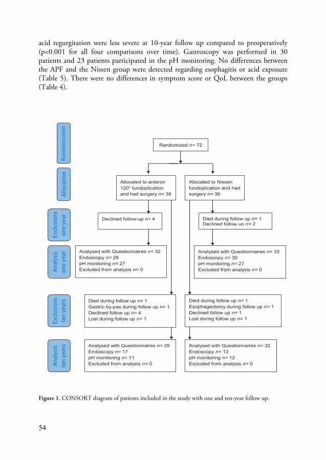
54
acid regurgitation were less severe at 10-year follow up compared to preoperatively (p<0.001 for all four comparisons over time). Gastroscopy was performed in 30 patients and 23 patients participated in the pH monitoring. No differences between the APF and the Nissen group were detected regarding esophagitis or acid exposure (Table 5). There were no differences in symptom score or QoL between the groups (Table 4).
Figure 1. CONSORT diagram of patients included in the study with one and ten-year follow up.
Analysed with Questionnaires n= 32 Endoscopy n= 28 pH monitoring n= 27 Excluded from analysis n= 0
Declined follow-up n= 4
Allocated to anterior 120° fundoplication and had surgery n= 36
Died during follow up n= 1 Declined follow up n= 2
Allocated to Nissen fundoplication and had surgery n= 36
Analysed with Questionnaires n= 33 Endoscopy n= 30 pH monitoring n= 27 Excluded from analysis n= 0
Allo
catio
n An
alys
is
one
year
Ex
clus
ions
on
e ye
ar
Randomized n= 72
Rand
omiza
tion
Analysed with Questionnaires n= 29 Endoscopy n= 17 pH monitoring n= 11 Excluded from analysis n= 0
Analysed with Questionnaires n= 32 Endoscopy n= 13 pH monitoring n= 12 Excluded from analysis n= 0
Died during follow up n= 1 Esophagectomy during follow up n= 1 Declined follow up n= 1 Lost during follow up n= 1
Died during follow up n= 1 Gastric by-pas during follow up n= 1 Declined follow up n= 4 Lost during follow up n= 1 Ex
clus
ions
te
n ye
ars
Anal
ysis
te
n ye
ars

55
Figure 2. Symptom score at one and ten years according to a seven graded Likert scale. APF= anterior partial (120°) fundoplication. y=year.
Table 3. Symptom score at one and ten years
1 year APF 1 year Nissen 10 years APF 10 years Nissen
Flatulence†a 2.0 (3.0) n=31 2.0 (2.0) n=32 p=0.384 2.0 (2.8) n=24 2.5 (2.0) n=30 p=0.051
Symptomatic flatulence‡
17/31(55 %)
19/32 (59 %)
p=0.801 16/24 (67 %)
27/30 (90 %)
p=0.046
Dysphagia†a 0.0 (1.0) n=32 1.0 (1.0) n=33 p=0.102 0.0 (1.0) n=24 1.0 (2.0) n=30 p<0.001
Heartburn†b 1.0 (1.0) n=32 1.0 (1.0) n=33 p=0.675 2.0 (2.0) n=29 1.0 (1.0) n=32 p=0.019
Acid regurgitation†b
1.0 (0.0) n=32 1.0 (0.0) n=33 p=0.512 1.0 (1.5) n=29 1.0 (0.0) n=31 p=0.053
† Median (IQR); Mann-Whitney U-test, ‡ Scored Likert 2 or more, Fisher’s exact test. a Scored on a 7-graded Likert scale (0-6), b Scored on a 7-graded Likert scale (1-7). APF= anterior partial (120°) fundoplication

56
Table 4. Outcome at one and ten years
1 year APF 1 year Nissen 10 years APF 10 years Nissen
n=32 n=32 n=29 n=32
Can belch† 30 /32 (94 %) 20 /32 (63 %) p=0.005 27 /29 (93 %) 21 /32 (66 %) p=0.012
Unable to vomit†
15 /23(47 %) 17 /19 (53 %) p=0.083 10 /26 (34 %) 29 /31 (90 %) p<0.001
GSRS‡ 2.0 (1.7-2.3) 2.2 (1.9-2.5) p=0.298
2.3 (1.9-2.5) 2.2 (1.9-2.5) p=0.752
PGWB‡ 110 (104-115) 102 (94-109) p=0.064 103 (95-111) 106 (100-112) p=0.552
Taking PPI† 10 /29 (34 %) 8 /32 (25%) p=0.575
† Fisher´s exact test, ‡ Mean (95 % CI), t-test, APF= anterior partial (120°) fundoplication
Table 5. Esophagitis and pH at one and ten years
1 year APF 1 year Nissen 10 years APF 10 years Nissen
n=28 n=30 n=17 n=13
Esophagitis†
-no 24 (86%)
29 (97 %)
p=0.067 16 (94%)
13 (100 %)
p=1.000
-grade A 4 (14%)
0
1 (6%)
0
-grade B 0
1 (3 %)
0
0
-grade C 0
0
0
0
-grade D 0
0
0
0
pH<4 (% of time)‡
1.6 (4.9) n=27 0.3 (1.4)
n=27 p=0.006 0.1 (0.4)
n=11 0.15 (0.62) n=12
p=0.749
Abnormal refluxa 10/27 (37%) 3/27 (11 %) p=0.054 0/11(0%)
0/11 (0 %)
Normal reflux
17/27 (63%) 24/27 (89 %) 11/11(100%) 12/12 (100 %)
† Fisher´s exact test; ‡ Median (IQR), Mann-Whitney U-test, a More than 4 % of time with pH <4 APF= anterior partial (120°) fundoplication

57
Paper II
There were 21 patients operated on for symptomatic epiphrenic diverticula. Patients’ characteristics are shown in Table 1. Esophageal motor disorder was identified in 17 (81 %) patients by manometry and/or barium swallow. Table 1. Patients characteristics and clinical characteristics
Age (years) Median 71
Range 37-90
Sex, no (%) Male 11
Female 10
ASA class I 3 (14 %)
II 9 (43 %)
III 8 (38 %)
IV 1 (5 %)
Symptoms Dysphagia 18 (86 %)
Regurgitation 13 (62 %)
Chest pain 8 (38 %)
Chronic cough 2 (10 %)
Aspiration pneumonia 2 (10 %)
Weight loss 6 (29 %)
Duration of symptom (year) Median 3
Range 1-15
Diverticulum (cm) Median size 6
Range 2-10
Side of presentation Right 19 (90%)
Left 2 (10%)
Postoperative complications and mortality
Surgical treatment was carried out with a long myotomy in 17 patients, of which 10 patients had an antireflux procedure added. Esophagectomy and gastric tube reconstruction was performed in 4 patients.
Median length of hospital stay was 15 days (range 12-46). There was no perioperative death, neither any 90 days mortality. Reoperation was performed in three patients, caused by one bleeding, one pleuraempyem and one late perforation. Other complications included pneumonia (n=2), minor myocardial infarct (n=1), urinary tract infection (n=1), and wound infection (n=1). One patient was readmitted and treated conservatively for pericardial fluid and bilateral pleural effusion.
Functional results and QoL
Myotomy without an antireflux procedure, but with the cardia left intact, were performed in 7 patients. Of these, 3 had objective measurement of pathologic acid exposure in the esophagus (24 hour pH monitoring and/or endoscopy) and a fourth

58
used acid suppressor medication (PPI) because of acid regurgitation. In 10 patients, the myotomy was extended across the cardia and an antireflux procedure was added to the myotomy. Of these, only one patient used PPI because of reflux symptoms, and this patient was the only patient among those 10 patients with objective measurement of pathologic acid exposure in the esophagus.
When comparing long-term symptoms between patients who had the cardia left intact and no antireflux procedure, with those who had the cardia myotomized and an antireflux procedure added, the most obvious difference was regarding reflux symptom. The group without an antireflux procedure reported more reflux symptoms than the group with an antireflux procedure, 67 (IQR 0-92, n=5) and 0 (IQR 0-33, n=7) respectively, and the use of PPI was more frequent among the group without an antireflux procedure (80 % and 14 % respectively).
For the entire study group, functional results were evaluated yearly by interviews at the outpatient clinic and based on improvement of symptoms preceding the operation were found to be excellent for 13 patients (62 %), good for 3 patients (14%), fair for 2 patients (9%) and poor for 2 patients (9%). Despite these results, which showed relief of the primary symptoms for the majority of the patients, evaluation of long-term symptoms and QoL demonstrated remaining eating-related symptoms and an impaired QoL compared to the general population (Figure 1a+b).

59
Figure 1 a+b. Mean and 95% CI for the scores of the EORTC QLQ OG25 (A) and QLQ C30 (B) scales. The arrow symbols ( ) show the mean scores from 3427 (A), respectively 1081 (B), persons in the age of 60-79 years, randomly picked from the Swedish population, who completed the EORTC questionnaires (van der Schaaf et al 2012, Michelson et al. 2000).

60
Paper III
There were 133 patients included, consecutively operated for adenocarcinoma in the GEJ. Demographics and clinical characteristics are shown in Table 1. Preoperative neoadjuvant treatment was not routinely performed. Esophagectomy with gastric tube reconstruction were performed in 96 patients, of which the majority had Type I tumors. Extended gastrectomy with long Roux-en-Y loop were performed in 37 patients, of which the majority had Type II tumors. The distribution of tumor stage was equal between the two treatment groups (p=0.355). The vast majority (100/133) of patients had R0 resections, with no differences between the groups (p=0.348). The R0 resection rate was 72% in the esophagectomy group and 84% in the extended gastrectomy group. Tumor free upper and lower resection margins were achieved in all patients except one. Extended gastrectomy required longer operation time than esophagectomy (p=0.002).

61
Table 1. Demographics and Clinical characteristics
Esophagectomy with Gastric tube
reconstruction (n=96)
Extended Gastrectomy with long Roux-en-Y
loop (n=37)
p-value
Gender Male 83 31 0.783 (A)
Female 13 6
Mean age (std dev) 65 (9.7) 64 (13.6) 0.543 (B)
ASA score 1 27 15 0.379(C)
2 61 18
3 7 4
4 1 0
Preoperative chemotherapy 3 1 1.0 (A)
Preoperative radiotherapy 1 1 0.481 (A)
Tumor site Distal esophagus (type I) 67 5 <0.001(C)
Cardia (type II) 26 26
Subcardia (type III) 3 6
Mean (std dev) Duration of surgery (h) 10.1 (1.4) 11.0 (1.8) 0.002 (B)
Median (5th, 95th percentile) Postoperative ICU stay in hours 18.5 (13, 208) 19.0 (13, 118) 0.797(D)
Tumor type Adenocarcinoma with Barrett 56 6 <0.001 (C)
Adenocarcinoma without Barrett 37 31
Severe Barrett dysplasia 3 0
Extent of dissection R0 69 31 0.348 (C)
R1 16 4
R2 11 2
Tumor free upper margin yes 95 37 1,0 (A)
no 1 0
Tumor free lower margin yes 96 37
no 0 0
Mean (std dev) of the maximum tumor size in cm 12.8 (5.8) 15.1 (4.7) 0.041 (B)
Mean (std dev) resection margin, measured from the upper end of the tumor (cm)
9.5 (3.9) 7.8 (3.2) 0.017 (B)
Tumor stage 0 10 0 0.355 (C)
I 12 5
II 16 8
III 38 15
IV 20 9
Hospital mortality 1 1 0.481 (A)
Median (5th, 95th percentile) Hospital stay (days) 15 (13, 21) 14(12, 25) 0.874 (D)
A Fisher’s exact test, B Student’s T-test, C Chi-square test, D Mann-Whitney U-test

62
Postoperative complications and mortality
Postoperative mortality within the first 30 days included one patient in each group. Both died of treatment resistant heart failure. A total of six patients in each group underwent reoperations within the first 30 days after the initial operation (p = 0.093). Ischemic necrosis of the conduit occurred in 3 cases in the esophagectomy group, all of them were managed conservatively, and in 2 patients in the extended gastrectomy group, both of them required a reoperation (Table 2). Cardio-pulmonary complications were the most common postoperative complication in both group (Table 2). Table 2. Hospital mortality, reoperations and complications
Esophagectomy with Gastric tube reconstruction (n=96)
Extended Gastrectomy with long Roux-en-Y loop (n=37)
Hospital mortality 1 1
Reoperations
Focal ischemic necroses in the conduit but outside the anastomoses or anastomotic leakage
2
Intestinal obstruction 2
Abscess with intact anastomoses 2 1
Postoperative pancreatitis 1
Negative chest exploration 1
Pneumothorax 1
Chylothorax 1
Tracheostomy 1
Complications managed without open surgery
Focal ischemic necroses in the conduit but outside the anastomoses or anastomotic leakage
3
New onset of atrial fibrillation or cardiac failure 9 5
Respiratory insufficiency 6 3
Aspiration 1
Pulmonary embolus 1
Pneumonia 2 1
Persistant pneumothorax 1
Pleural effusions 1
Abdominal abscess 4
Cerebrovascular lesion 1
Severe psychotic reaction 1
Unilateral vocal cord palsy 2

63
Recurrence and long-term survival
In both groups, most recurrence occurred within the field of dissection (Table 4).
All patient were followed to death or for at least 5 years. In September 2006, 28 of the operated 133 patients were alive. The major cause of death was recurrent disease, which was seen in 63 patients after esophagectomy and in 27 patients after extended gastrectomy. Fourteen patients died of non tumor-related causes, nine of whom had been treated with esophagectomy and five with extended gastrectomies. One patient in the esophagectomy group died of a malignant melanoma.
Long-term survival rate was best and independently determined by tumor stage (0, I, II, III, IV; p<0.001), the extent of resection described as R0, R1 or R2 resections (p<0.001), and by the distance between the tumor and the resection margin (p = 0.009) (Table 3). After adjustment for these factors, as well as for age, ASA score, tumor site according to the Siewert classification (I, II, III), and tumor size, the two used surgical procedures had no significant impact on the long-term survival rate. Overall true 5-year survival rate was 80% for stage 0 tumors, 65% for stage I, 42% for tumors at stage II, 13% for stage III, and 0% for stage IV disease. Table 3. Impact factors for survival (multivariable Cox regression study)
95% Confidence Interval for HR
p-value Hazard ratio (HR)
Lower Upper
Tumor stage <0.001
0 Reference
I 2.73 0.56 13.37
II 3.46 0.74 16.28
III 9.23 1.93 44.08
IV 14.2 2.95 68.43
Extent of dissection 0.006
R0 Reference
R1 2.16 1.18 3.96
R2 2.82 1.4 5.79
Distance between tumor edge and resection edge in cm 0.009 0.91 0.85 0.98
Age 0.204 n/a
ASA scores (1-4) 0.753 n/a
Esophagectomy with gastric tube reconstruction vs. Extended gastrectomy with long Roux-en-Y reconstruction
0.252 n/a
Tumor site: distal esophagus (I), cardia (II), subcardia (III) 0.618 n/a
Maximum tumor size in cm 0.510 n/a

64
Table 4. Pattern of tumor recurrence in patients who later died of their tumors
Esophagectomy with Gastric tube reconstruction (n=96, 63 deaths)
Extended Gastrectomy with long Roux-en-Y loop (n=37, 27 deaths)
Anastomotic recurrences and systemic disease 2 0
Anastomotic recurrences, recurrences within the field of dissection and systemic disease
2 1
Recurrences within the field of dissection outside the anastomoses and systemic disease
29 11
Systemic disease only * 30 15
Functional result and Quality of Life
The QoL forms were completed by 32 of 33 patients still alive in September 2005, with a median follow-up time after surgery of 8.5 years (range 4-15) (Figure 2 a+b). Of these 32 patients, 26 had been operated with esophagectomy and gastric tube reconstruction and six with extended gastrectomy with long Roux-en-Y reconstruction. Using the EORTC QLQ OES18 questionnaires, the patients reported low scores for “Dysphagia”, “Problems with eating” and “Trouble swallowing” indicating an overall satisfying long-term restitution of the swallowing function, both for patient having a gastric tube as for the patient having a long Roux-en-Y reconstruction. The results of the EORTC QLQ C30 questionnaires showed a mean “Global Health Status” in the study group that was not significantly different from the results of the Swedish reference population (p=0.295). In addition, with the exception of the scales “Physical function,” “Dyspnea,” “Appetite loss,” and “Diarrhea”, which showed less favorable results in the operated patients compared with the reference population, we found that the 95% confidence interval of the remaining scales included the mean scores of the reference population. This indicates good overall recovery with minor side effects.

65
Fig. 2. Mean and 95% confidence intervals for the scores of the EORTC QLQ C30 (a) and QLQ OES18 (b) scales. a The arrow symbols (;) show the mean scores of a randomly picked Swedish reference population 6 (see text) who completed the QLQ C30 form.

66
Paper IV
Thirteen out of 15 patients accepted participation in the study. Time to 25%, 50% and 75% emptying, measurement errors, and intra-class correlation coefficient are shown in Table 1. Two patients did not reach steady state elimination phase for paracetamol during the observation time of 120 minutes and had to be excluded from the reliability analysis. In addition, four patients did not reach 75% emptying as measured with scintigraphy during the observation time, explaining the smaller number at time to 75% emptying. Table I. Time to 25 %, 50% and 75% emptying from the gastric tube
Emptying Scintigraphy mean (median) min
Paracetamol absorption test mean (median) min
Systematic error (min)
Random error (min)
ICC†
25 % (n=11) 9.1 (8.0) 10.7 (9.3) 1.6 4.1 0.68
50 % (n=11) 22.8 (20.4) 16.5 (14.0) -6.3 15.4 0.25
75 % (n=6) 39.0 (39.0) 27.4 (25.0) -11.6 13.2 0.53 †ICC= Intraclass correlation coefficient
Agreement between the two methods presented as a Bland-Altman plot
Bland-Altman plots of the time data at 25%, 50% and 75% emptying (T25, T50, T75 respectively) are shown in figure 1a-c.

67
Figure 1 a-c. Bland-Altman plots of gastric tube emptying time at; (a) 25% emptying, (b) 50% emptying, and (c) 75% emptying. Plotted is the mean time difference (paracetamol absorption test minus scintigraphy), against the mean value from the two methods. Broken lines represent the mean difference and 95% limits of agreement. Standard deviation of the differences; (a) 4.0 min, (b) 15.4 min and (c) 13.8 min.

68
Examples of representative normal time-emptying curves for two of the patients are shown in Figure 2.
Figure 2. Emptying curves for two patients measured with scintigraphy and the paracetamol absorption test
Paper V
Between 1992 and 2014, a consecutive series of 24 patients (14 men, 10 women), median age 61 years (range 8-78), underwent repeated esophageal reconstruction because of esophageal discontinuity (n=9), stenosis of the substitute (n=3), tortuous substitute (n=5), recurrent malignancy (n=2), 2-session operation (n=2), airway fistula (n=1), severe bile reflux (n=1), and ulceration in an interposed jejunum penetrating the left ventricle of the heart (n=1) (Figure 1). Esophageal discontinuity in the 9 patients mentioned above were the results of substitute necrosis after esophagectomy in 5 patients, esophagogastric necrosis due to incarcerated paraesophageal hernia in 3 patients, and complication due to thyroidectomy in one patient.

69
Figure 1. The indications for the redoe-operation (headings) and subsequent reconstruction (illustrations) of the upper intestinal tract

70
The patients in the control group, operated on before and after the date for the corresponding redo patients, were operated on in median 21 days before (range: 2-404) and in median 28 days after (range: 3-850) the corresponding redo patient.
Attempts to recreate the upper alimentary tract were performed 1 week to 35 years (median 59 weeks) after the primary reconstruction and following 0 to 2 earlier tries. The indications (headings) and subsequent reconstruction (illustrations) of the upper intestinal tract are visible in Figure 1.
Postoperative complications and mortality
There was a longer operation time in the redo-group but no differences in postoperative complication rate (Table1). There was no postoperative mortality in neither of the group. Table 1. In-hospital data and postoperative complications and mortality rate
Redo reconstruction (n=24)
Primary reconstruction (=48)
p
Operating time, min.† 726 (378-2094)* 573 (390-780) p=0.0008
Blood loss, ml† 1125 (50-12100) 700 (100-3000) p=0.11045
Anastomotic leak 5 (21 %) 3 (6 %) p=0.107
Airway complication# 6 (27 %) 6 (12.5%) p=0.197
Cardiac complication# 1 (4.5 %) 4 (8 %) p=0.659
Vocal cord palsy# 0 (0 %) 0 (0%) p=1.000
Chylothorax# 0 (0%) 1 (2%) p=1.000
Reoperation# 3 (12.5 %) 4 (8 %) p=0.679
Clavien-Dindo I+II ‡ 6 (25 %) 12 (25 %) p=1.000
Clavien-Dindo III a+b‡ 4 (17 %) 6 (12.5 %) p=0.722
Clavien-Dindo IV a+b‡ 1 (4.5 %) 0 (0 %) p=0.333
Hospital stay, days† 23 (14-100) 15 (10-89) p<0.0001
Postoperative 90-days mortality# 0 (0 %) 0 (0%) p=1.000
† Median (Range), Mann Whitney test * Operation time for the 2-session operations is registered as the total time of session one and two together. # Fisher’s exact test. ‡ 30-days complications rate according to the Clavien-Dindo classification.
Survival
Survival curves are shown in Figure 2. There was no difference in 5-year survival between patients with malignant diagnosis in the redo-group compared to the primary operated (40 % and 39.5 % respectively, p=0.706), nor were there any deaths from sequel of their operations among patients with benign diagnosis in either group (Figure 2). Median survival time for the malignant redo patients was 27 months (95% confidence interval, 0-72 months), and 42 months (95% confidence interval, 20-63 months) for the primary operated cancer patients.

71
Figure 2. Cumulative survival rates (Kaplan-Meier) for patients operated with repeated esophageal reconstructions (redo, n =24) and patients operated with primary esophageal resection and gastric pull-up (primary, n = 48), p = 0.0001. Two patients are censored (+) in the redo group within the 5 year period after surgery, and none in the primary operated group
Long-term symptoms and Quality of Life
Symptoms and QoL in the redo group was evaluated using the EORTC questionnaires (QLQ-OG25 and QLQ-C30) and were completed by all 12 patients alive in November 2013. The results are shown in Figure 3A+B. QoL measures were similar to previous reports from patients with primary esophagectomy and gastric tube reconstruction with higher score of “Dysphagia”, “Eating problems”, “Trouble swallowing” and “Trouble coughing” compared to the general population, although QoL measurement showed a Global health and overall function level which were as good as in the normal Swedish population (Figure 3a+b).

72
Figure 3 a+b. Mean and 95% CI for the scores of the EORTC QLQ OG25 (A) and QLQ C30 (B) scales. The arrow symbols (↓) show the mean scores from 4910 (A), respectively 3069 (B), adult persons randomly picked from the Swedish population, who completed the EORTC questionnaires.

73
Discussion
Diseases of the GEJ make up a heterogeneous group concerning treatment, but a homogenous group concerning symptoms. All patients have some form of eating disturbances. Surgical treatments try to eliminate annoying symptoms but also to cure from life-threatening conditions. The surgery itself may, however, cause morbidity that negatively affect the long-term QoL, and may results in mortality. Since patients treated for benign diseases often have a long life expectancy, and since the long term survival after treatment of malignant diseases have improved significantly during later years, the long term results regarding gastrointestinal functions and QoL are important.
Benign disorders of the GEJ
In Paper I and II, were treatment of benign diseases in the GEJ is studied, a common denominator for a good outcome was the restoration of a defective LES. In the case of GERD this was already known, but how the LES could be restored in order to lessen adverse outcome were studied further. In the treatment of epiphrenic diverticula a fundoplication in addition to a myotomy of the LES was found to give the best result.
Paper I
In paper I we compared two antireflux repairs. The optimal size of fundoplication to treat GERD is a matter of discussion, although Nissen (total) fundoplication is the most commonly used worldwide. The Nissen fundoplication has a well-documented effect in treating reflux symptoms (Salminen et al. 2007, SAGES 2010). In some patients, though, the Nissen fundoplication cause dysphagia and abdominal bloating, probably due to the effective closure of the LES which may prohibit the normal relief of gas from the gastrointestinal tract (Salminen et al. 2007, Sandbu et al. 2010, Makris et al. 2012). In Paper I our hypothesis was that an anterior partial fundoplication of 120° (APF) would mitigate unwanted side effects, such as flatulence and dysphagia (Watson et al. 1991). Since Nissen fundoplication must be regarded as the gold standard of antireflux repairs, the effectiveness of APF and its functional side effects was compared with that operation.
The number of patients included in Paper I are somewhat smaller than initially planned due to a prematurely closure of the study caused by a dramatic drop in the number of antireflux procedures performed in Sweden. Although, when closing the

74
study, the number of included patients were considered sufficient for making comparisons. In addition, the strength of the study is the high follow-up rate in combination with the double-blinded design, which kept both the patients and the researchers unaware of the type of repair. There are only six other randomized trials comparing anterior fundoplication to a Nissen wrap, four of them comparing a 180° and two comparing a 90°, and only one of those six trials have a follow up beyond five years (Watson et al. 1999, Watson et al. 2004, Baigrie et al. 2005, Spence et al. 2006, Raue et al. 2011, Cao et al. 2012, Broeders et al. 2014). It should be noted that the results from the preoperative assessments of our study population indicate that the patients had mild to moderate reflux disease, and the results may not be applicable in patients with severe reflux disease.
We found that the QoL had improved at one year compared to preoperatively for both the APF group and the Nissen group, and the QoL level was kept sustainable at ten years for both groups. Regarding adverse effects and control of reflux symptoms, the outcome changed over time. At one year, there was no differences in adverse effects or reflux symptoms, but at ten years follow up, the APF group reported less symptomatic flatulence and less dysphagia than the Nissen group, but scored higher for heartburn, indicating a more effective antireflux repair to the cost of inability to get rid of intestinal gas. More patients could belch and vomit in the APF group at then years compared to the Nissen group. Even though these differences could be found, the symptoms were modest. This was reflected in the overall gastrointestinal symptoms scale and QoL measurement that did not show any differences between the groups, and the results were almost equally to the average population at long-term follow up. Taken together, the results show that both procedures offer sustainable reflux control with modest unwanted side effects, although with a little differences in pro and cons. Those pro and cons should be taken into consideration when discussing surgical treatment of GERD with the patient so that the patient is aware of what can be anticipated from the surgery.
Paper II
Epiphrenic diverticula are rare, and symptomatic diverticula even more uncommon. Randomized controlled trials are not eligible to perform when studying its treatment and is missing. Retrospective reviews of prospectively collected data, similar to what we have done, are the most common way to study the surgical treatment of epiphrenic diverticula.
Epiphrenic diverticula can grow to considerable size, causing dysphagia, retention, chest pain and regurgitation with risk of aspiration. Having those complications in mind, it might appear obvious that the diverticula should be resected, but the risk following an operation with reported leakage rate of 21% and mortality rate of 11% (Fékéte et al. 1992, Benacci et al. 1993) has warranted a cautious approach. In case of a successful operation, the primary symptoms can be relieved, although the long-term QoL is barely studied. If surgery is carried out, there are different views on how the

75
surgery should be performed, and in recent year minimal invasive procedure has become common, which have had implication on the operative strategy. The ones advocating a minimal-invasive procedure often claim the minimal invasive technique to cause less morbidity, and sometimes even less mortality, than the traditional open approach, referring to studies reporting high morbidity and mortality rates. The same high complication rates have, however, been reported from minimal invasive techniques. Since patients’ series often are small, the impact of the surgeons’ experiences may have a greater impact on the outcome than the surgical approach. In our series, only two surgeons have performed the procedures, which guarantee a standardized operation, making it possible to correlate outcome from our techniques to long-term functional results and QoL.
In Paper II, we confirmed the theory suggested by Belsey in the 1960s that an esophageal motor disorder, including the LES, is behind the formation of the diverticulum (Belsey et al 1966). Thus, the treatment of epiphrenic diverticulum should include a myotomy in order to lessen the intraluminal pressure. In order to avoid postoperative reflux symptoms, we found that the myotomy should include the LES and an antireflux procedure should be added even in a case when a preoperative normal LES was found (measured with a stationary manometry). This might seem a bit contradictory, but is probably due to difficulties in identifying a functional disorder in the LES preoperatively since stationary manometry does not capture intermittent spasm in the LES, nor all kind of motor abnormalities in the esophagus, as have been shown by Nehra and colleagues (Nehra et al. 2002). The view that the LES should be included in the myotomy is further supported by a study conducted by D’Journo and colleagues who found that the most constant functional abnormality in patients with epiphrenic diverticula was a LES incoordination, including isolated hypertensive LES and atypical disorders of LES relaxation (D’Journo et al. 2009). Avoiding postoperative reflux symptoms is of outmost importance since GERD have a negative impact on the QoL for the patient, comparable with the low level seen in patients suffering from chronic condition such as diabetes, congestive heart failure and arthritis (Wiklund et al. 2004, Tack et al. 2012). In some cases of epiphrenic diverticula, for example multiple diverticula or mega-esophagus, the only treatment is an esophagectomy. In case of young patients with long life expectancies a more reflux resistant reconstruction than a gastric pull-up should be considered.
Surgical treatment of symptomatic epiphrenic diverticula with an open transthoracic approach can, despite the risks connected with the procedure, be performed with few postoperative complications and no mortality, although it has to be emphasized that the surgery is so seldom performed that it should be performed in centralized and specialized centers in order to achieve these results. The treatment should address the underlying motor disorder by performing a long myotomy including the LES, and adding an antireflux procedure, preferably a partial one. Although we found that this surgical procedure resulted in good or excellent relief of the primary symptoms in the majority of patients, eating-related problem remained and the long-term QoL were

76
impaired compared to the general population. The underlying motor disorder and the consequences of the myotomy can explain some of the symptoms that remain or even evoke after the operation. It is important to inform the patients preoperatively that the esophagus won’t regain a normal motoric pattern after the surgery, and consequently the surgery won’t make them asymptomatic, although surgery has a good chance to relieve the main symptoms they suffer from.
Malignant disorders of the GEJ
Paper III
In Paper III we compared two operations for treatment of adenocarcinoma (AC) in the GEJ, the more commonly used esophagectomy with gastric tube reconstruction and the less documented extended gastrectomy with a long Roux-en-Y loop, performed via a laparotomy and a right thoracotomy (see Methods). The strongest predictors for survival are the tumor stage and R0 resection (Mariette et al 2008, Cabau et al. 2013, Mariette et al 2014). We can do little about but the tumor stage, but more about the resection. Even though neo-and adjuvant oncological treatment has been introduced to improve the poor long-term survival, the cornerstone in the treatment of AC in the GEJ is still surgery. Therefore, it is of outmost importance that the surgical procedure is enough radical without risking too much morbidity.
Since AC in the GEJ can spread to mediastinal and intraabdominal lymph nodes, lymph node dissection has to be performed both in the chest and in the abdomen, but the extent of this dissection needed is debated. If only a transhiatal approach is used, the lymph node dissection in the chest is restricted, with the upper middle and proximal field of the mediastinum being out of reach. In a RCT with a five-year follow up, comparing extended transthoracic versus transhiatal esophagectomy for Type I and II tumors, they found an ongoing trend towards better 5-year survival for the transthoracic group, especially for patient with a limited number of positive lymph nodes (Omloo et al. 2007). Since the sensitivity for identifying positive lymph nodes preoperatively is low, as is the classification into Type I or II tumors, the decision regarding surgical approach cannot be made from the preoperative assessment only. In addition the risk of seeding in the esophageal and gastric wall warrant a macroscopic free resection margin of at least 5 cm, and in the case of a Laurén diffuse histological type the length of clear margin should ideally be extended to 8-10 cm (Barbour et al. 2007, Polkowski et al. 2014). The need for these resection margins in combination with the need for a thoroughly lymph node dissection both in the chest and in the abdomen have been the reason for us to choose the extended gastrectomy with a long Roux-en-Y reconstruction for GEJ tumors within the true cardia and/or with subcardial involvement, even though the extended gastrectomy with a transhiatal procedure is usually associated with less morbidity and mortality (Hulcher et al. 2002). The extended gastrectomy we prefer, through separate incisions in the abdomen and the right chest and with an anastomosis at the level of the azygos

77
vein, is scarcely studied. There have been concerns about the morbidity and risk for mortality that may follow the procedure, especially with the more demanding reconstruction with a long Roux-en-Y loop considering the increased risk of ischemia in the reconstruction. In addition, the long-term QoL after an extended gastrectomy with a long Roux-en-Y reconstruction is scarcely studied.
The decision of which procedure, esophagectomy or extended gastrectomy, that we considered most suitable for the GEJ tumor at hand was definitively made in the operating theatre. The patients who underwent esophagectomy and gastric tube reconstructions had comparable tumor stages as those who underwent extended gastrectomy with long Roux-en-Y loops. The majority of GEJ tumors in the esophagectomy group were Type I tumors and in the extended gastrectomy group Type II tumors. R0 resection was achieved in the majority of cases with no difference between the two groups, showing that the chosen procedure for the tumor at hand were adequate in both groups. For those patients who died of recurrent disease, approximately half had recurrence within the field of dissection in addition to systemic disease, and none of the patients operated with extended gastrectomy and Roux-en-Y loop, and only two in the esophagectomy group, had anastomotic recurrence. Even though the extended gastrectomy required a longer operating time, the fear of increased postoperative complications and mortality were not confirmed. The type of tumor (Type I, II or III) had neither any impact of survival. Influencing survival were tumor stage, extent of dissection (R0-R2) and the length of the upper resection margin. Long-term evaluation of functional results revealed good swallowing function and QoL measurement showed a ‘‘Global Health Status’’ not significantly different from a large randomly selected Swedish population group.
Our results support the use of extensive surgical resections margins. Adequate length of esophageal resection margins can only be performed through a right thoracotomy, where all of the esophagus can be exposed. This is not possible through an abdominal transhiatal procedure. The extended gastrectomy procedure with a long Roux-en-Y reconstruction results in a high rate of tumor-free resection margin and can be performed with modest morbidity and mortality, and with a satisfactory long-term outcome. Consequently, this procedure can be used, without risking the quality of life for the patient, as an alternative to esophagectomy with gastric tube reconstruction in the treatment of AC in the GEJ.
Function and dysfunction of esophageal reconstruction
Paper IV
Whether a more simple and accessible procedure, the paracetamol absorption test, could be used for measuring gastric tube emptying instead of the more resource demanding scintigraphy, was studied in Paper IV. The gastric tube is the most commonly used reconstruction when performing an esophagectomy, and, as can be seen in Paper III, we have in our Department used esophagectomy with gastric tube

78
reconstruction in the majority of patients with AC in the GEJ. Common symptoms after esophagectomy are appetite loss, nausea, early satiety postprandial fullness, regurgitation, dysphagia, odynophagia, reflux and cough (Lerut et al. 2004, Viklund et al. 2006, Djärv et al. 2008). These symptoms are sometimes, but not always, caused by retention in the gastric tube. The best way of identify the function of the substitute is to objectively measure its emptying, although this seems to be underused in relation to the frequencies of above mention symptoms. If a simple measuring tool would be available, it may increase objective measurement of emptying from the gastric tube and treatment could be more individualized for patients suffering from retention related symptoms, enhancing the chance of increasing the QoL for the patients.
Gold standard for measuring gastric tube emptying is scintigraphy. To be able to validate the paracetamol absorption test, with scintigraphy, we started with patients who were considered cured from their malignancies, and who had had enough time to adapt to their new reconstructions. We considered a postoperative time span of at least 2 years to be sufficient to achieve this, and the included patient had in median 3 years between operation and time for participation in the study. To further ensure that the study population was representative for the esophagectomy patient category, quality of life assessments was conducted and confirmed that the examined group could be regarded as a representative sample, and not the ones with sever symptoms. The paracetamol absorption test and scintigraphy were conducted simultaneously.
To interpret the plasma concentration of paracetamol over time into emptying from the gastric tube we used the Medhus algorithm (Medhus et al. 1999). The algorithm adjusts for first-pass metabolism, unequal distribution and individual elimination of paracetamol, and calculates percentage of meal emptied as a function of time, which can be displayed in a graph, although, the Medhus algorithm, requires that paracetamol concentration reach steady state elimination phase for emptying to be calculated. In our study two patients did not reach steady state and they were excluded from the reliability analyses. A reason for this could be fast emptying from the gastric tube, both of them having a time to 50% emptying (T50) of less than 20 minutes measured with scintigraphy, in combination with a reduced individual capacity to rapidly eliminate the paracetamol. The same phenomenon occurred in another study by Strömmer and colleagues, in which the gastric emptying after a Whipple procedure was measured using a lower dose of paracetamol (Strömmer et al. 2005). This may be a concern with the paracetamol absorption test but is not necessarily a reason to avoid applying the test clinically.
Although not being the aim for our study, an interesting finding in Paper IV was that the emptying rate from the gastric tube appeared to be more rapid compared to a previous trial conducted in our department. In this previous trial, the gastric tube emptying was measured during the first year after esophagectomy, using an identical test meal and identical scintigraphic methodology, and the results showed a mean T50 after 3 months of 60 minutes and after 12 months of 44 minutes (Johansson et

79
al. 1999). In the current study, performed in median 3 years postoperatively, the mean T50 was 22 minutes suggesting that there may be a tendency for the emptying rate from the gastric tube to increase over time. Since there are no normative values for emptying at different time points after esophagectomy, these results add valuable information of the gastric tube’s long-term function.
Our results show a reasonable close correlation between the paracetamol absorption test and scintigraphy for time to 25% and 50% emptying, except for one outlier, indicating that the paracetamol absorption test may be able to recognize patients with delayed emptying from the gastric tube, and consequently may be used to identify patients who would benefit from interventions, although, further studies are needed before the test can be used as a clinical screening test in esophagectomy patients.
Paper V
In paper V we studied the problem with redoing reconstruction of the esophagus. When the primary reconstruction fail, another attempt to restore the alimentary tract is a much more demanding procedure. In some patients, failure of the primary reconstruction can drive the decision to disconnect the reconstruction. Several different advents can precede esophageal discontinuity like complications following esophagectomy (anastomotic leak, necrosis) or antireflux operation (necrosis). In addition, an esophagogastric disruptive catastrophe can occur as a result of trauma, iatrogenic perforation, spontaneous rupture or ingestion of corrosive agents. Delayed reconstruction can be used when a mediastinal contamination and/or a severely ill patient preclude a safe reconstruction. When the discontinuity is a fact, a reoperation is the only alternative for restoring the alimentary tract, although such an operation poses the risk of worsen the situation for the patients and adding further morbidity and/or mortality. The experiences in many centers are short. Operative strategy most suitable varies a lot due to the different preceding circumstances, and individualized assessment and treatment are required.
To study if there is a place for redoing reconstruction of the esophagus we studied the patients operated on for repeated esophageal reconstruction in our department. Our Department is a tertiary center to which patients are referred to from all of Sweden, making the number of redo-patients substantial. Since it is not eligible to randomize these patients in order to compare different surgical strategies, the outcome have been measured by survival rate and quality of life levels for the patients and compared to primary reconstructed patients. The outcome accomplished from our redo-operations is the result from a number of different considerations, like timing of the secondary operation or the final reconstruction, choice of reconstruction, and choice of route for the new reconstruction.
Timing of the redo-operation is essential. As stated by Orringer, ‘Reversal of esophageal discontinuity is never an urgent operation’ (Orringer 2007). The nutritional status of the patient should be optimized as much as possible before another attempt is tried. This often involves nutritional support, through parenteral

80
infusion and/or enteral supplemental nutrition. After a mediastinal infection, the re-establishment of the digestive tract is recommended to be delayed for 6 months (Barkley et al. 2003), a recommendation we have followed. The risk of recurrence for patients primary treated for malignancy is an important factor to take into consideration. In patients where the primary cancer has been removed, but the reestablishment of the upper digestive tract are yet to be performed, Orringer recommended 6 to 12 months before reversing discontinuity (Orringer et al. 2007, Barkley et al. 2003). In our series we had one patient with the reconstruction performed 6 months after the discontinuity operation but the patient had a recurrence just one month after the reconstruction-operation. What time perspective to choose, closer to 6 month or to 12 month, must be up to the surgeon to decide but the stage of the primary tumor ought to be taken into consideration.
Which route and what method of reconstruction to use when reestablishing the alimentary tract are other important issues to discuss. The native bed for the esophagus, often used in the primary reconstruction, can be obliterated after earlier surgery and after complications following the primary reconstruction, such as necrosis or anastomotic leakage. In these cases, the substernal route can be used. Alternative substitutes when the stomach is not available are jejunum or colon for interposition, or reconstruction with a Roux-en-Y loop. Free jejunal graft is an alternative that seldom is necessary.
The 5-year survival for patients with malignant diagnosis was as good in the redo-group (40 %) as in the control group (39.5 %). For the patients with benign diagnosis the 5-year survival was excellent (95% in the redo-group and 100% in the control group). An important factor when achieving a good long-term survival is the functional result and the QoL. We found that the redo patient had similar good long-term functional result and QoL as primary operated patients.
The surgical challenge that redoing esophageal reconstruction constitute is gratifying both in short and long term despite longer operation time and added hospital stay since the redo patients have the same life expectancy, as well as similar quality of life, as the primarily operated. This is especially evident in benign patients and in those with a cured malignancy

81
Conclusions
I. Laparoscopic anterior 120° fundoplication resulted in less dysphagia, less symptomatic flatulence and better ability to belch, while maintaining an adequate reflux control compared to a Nissen 360° fundoplication in patients with a mild - moderate gastroesophageal reflux disease, and can be used as an alternative to the ‘gold standard’ Nissen 360° fundoplication.
II Surgical treatment of symptomatic epiphrenic diverticula with an
open left transthoracic approach, addressing the underlying motility disorder and compensating the reflux at hand after the myotomy with a partial transthoracic antireflux repair, can be safely performed irrespective of the size and location of the diverticula.
III For patients with adenocarcinoma at the gastroesophageal junction the
extended gastrectomy with esophagojejunostomy at the level of the azygos vein can be performed with tumor-free resection margins, low mortality and with similar outcome as for patients operated with esophagectomy and gastric tube reconstruction and can be an alternative to this reconstruction.
IV A reasonably close correlation was found between paracetamol
absorption test and scintigraphy for gastric emptying, indicating that the paracetamol absorption test may be able to recognize delayed gastric emptying. Further studies are needed before the test can be used routinely.
V The surgical challenge that redoing esophageal reconstruction
constitute is gratifying both in short and long term despite longer operation time and added hospital stay since the redo patients have the same life expectancy, as well as similar quality of life, as the primarily operated, and an attempt to restore the upper gastrointestinal tract could be recommended.

82

83
Future perspectives
The good long-term outcome after antireflux surgery in reducing reflux symptoms makes surgery a good, and in some cases better, alternative to PPI in the treatment of GERD. The impact of antireflux surgery on the pathogenesis of Barrett’s esophagus and it’s further development into dysplasia and adenocarcinoma is, though, less known. Barrett’s esophagus is considered to develop from a long standing GERD, but it is not known what components in the refluxed gastric contents that trigger the development. The refluxed material includes a mixture of gastric acid, digestive enzymes and bile. PPI only reduce the acid, but the other components are still left. Barrett’s esophagus goes with a risk of progression to low grade dysplasia (LGD), high-grade dysplasia (HGD) and finally adenocarcinoma. There are few randomized trials that compare the effect of antireflux surgery to PPI on the development of dysplasia in Barrett’s esophagus, and in the few studies performed, the follow up time have been limited. Therefore, no final conclusions can be drawn. Retrospective studies and non-randomized studies have been carried out. Altogether, the results are conflicting, with some trials reporting a more pronounced regression of Barrett’s esophagus after an antireflux procedure compared to PPI treatment, while others studies report no differences between the two treatments, neither in the regression of Barrett’s esophagus nor in the progression to dysplasia. Consequently, surveillance is still recommended for patients with Barrett’s esophagus regardless the treatment of GERD. A problem when studying dysplasia formation in Barrett’s esophagus is that the entire surface cannot be checked. Samples from the mucosa are taken as systematic four-quadrant biopsies, with additional biopsy if an abnormality is seen. Consequently, much of the mucosal surface is not checked at every time of follow up. In addition, the interpretation of what can be considered a LGD can be hard to make for the pathologist. Inflammation results in cellular changes mimicking LGD. In order to study the effect of antireflux surgery on Barrett’s esophagus and it’s progression to dysplasia and adenocarcinoma, with comparison to PPI treatment, it would be optimal to perform four-quadrant biopsies every centimeter and to have two or three pathologists examine each biopsies, although this requires a lot of resources. In addition, the follow up time ought to be at least ten years since it is not known how fast, or slowly, the development of dysplasia and adenocarcinoma occur.
In the case when adenocarcinoma has developed in the GEJ, the conclusion of the optimal treatment is yet to be done. Neoadjuvant treatment is now standard of care in most centers, sometimes in combination with adjuvant oncological treatment,

84
although there is no consensus whether the patients should be treated with chemotherapy alone or radio-chemotherapy. In addition, the surgical procedure is not standardized. Studies comparing outcome after combined treatment (surgery and oncological treatment) with surgery alone have only standardized the different oncological treatments but not the surgical procedure. The trials are often multicenter studies in order to include sufficient patients for comparison to be made. Consequently, the surgical procedures differ, affecting length of resection margins and lymph node dissection, both in the group receiving additional oncological treatment as in the group treated solely with surgery. The survival benefit found after additional oncological treatment may be a result of inadequate resection margins and dissection. It is not known what impact additional oncological treatment would have if the surgery were carried out as extensively as possible. In this thesis, we have shown that extensive surgery can be performed without risking too much morbidity and with no additional mortality compared to less extensive procedures. It would be most interesting to perform a randomized controlled trial in order to study the effect of additional oncological therapy when surgery is standardized and carried out as extensively as possible.
Regarding evaluation of emptying from the gastric tube, several more aspects are to be studied. It would be valuable to examine patients with known delayed emptying (previously measured with scintigraphy) for further development of the paracetamol absorption test for measuring emptying from the gastric tube. In addition, to evaluate emptying from the same patients at different time span postoperatively, both with scintigraphy and with the paracetamol absorption test, would enhance the validation and give valuable information of the natural course of emptying function of the gastric tube over time. The Medhus algorithm used in our study was easy to interpret and would be the one method we would use in further studies. It would be interesting, though, to study if the problem with steady state elimination could be eliminated. To use a fluid meal containing more fat for a slower emptying, and subsequently a slower absorption of paracetamol, in combination with a prolonged time of observation could be one way to further evaluate the method.

85
Populärvetenskaplig sammanfattning (in Swedish)
Gastroesofageala övergången (GE) är ett område motsvarande övergången mellan matstrupen (esofagus) och magsäck (på latin: ventriculus gaster). Olika benämningar av området används, såsom övre magmunnen, nedre matstrupsfinktern och cardia. Ingen av dessa benämningar täcker dock in hela området. American cancer society har ur ett onkologiskt perspektiv definierat området såsom innefattande nedre delen av matstrupen och övergången till magsäcken. American Joint Committee on Cancer inkluderar dessutom de 5 översta centimetrarna av magsäcken som ligger i direkt anslutning till själva övergången.
Sjukdomar i GE övergången påverkar förmågan att äta och dricka. Symtom såsom sura uppstötningar, halsbränna, svårigheter att svälja, smärta vid måltidsintag, illamående och kräkningar är vanligt förekommande, samt svårigheter att få sig tillräckligt med energi med viktnedgång som följd. Besvären kan störa nattsömnen och omöjliggöra arbete dagtid med mycket lidande för patienten och en stor kostnad för samhället som följd. Vid kirurgisk behandling av benigna (icke-cancer) sjukdomar i GE övergången är det primära målet att lindra symtomen, förbättra förmågan att äta och dricka samt öka livskvaliteten. Vid cancer i området är målet i första hand att bota patienten, men för de patienter som överlever är funktionen av den nya rekonstruktion (ersättningen av den del av matstrupen och magsäcken som tagits bort) viktig då det har stor inverkan på deras fortsatta livskvalitet. Långtidsöverlevnaden efter operation av cancer i GE övergången är låg, även om den ökat under de senaste decennierna. Dessutom är enbart cirka hälften av patienterna som diagnostiseras med cancer i GE övergången botbara. Trots botande behandling ligger femårsöverlevnaden kring 40 %. Botande behandling innefattar kirurgisk borttagning av tumören och de lymfkörtlar som dränerar den, men det råder delade meningar om hur omfattande kirurgi som krävs för att öka möjligheterna till bot.
I detta doktorandprojekt studeras hur olika sjukdomstillstånd i GE övergången kirurgiskt kan behandlas och vilka effekter det får på patienterna. Det övergripande syftet med de planerade delarbetena är att jämföra och utvärdera olika kirurgiska rekonstruktioner gällande komplikationsfrekvens, morbiditet, mortalitet, symtom och livskvalitet. Detta för att kunna bidra till en evidensbaserad grund för kirurgisk behandling och rekonstruktion vid sjukdomstillstånd i GE övergången.

86
I delarbete 1 studeras kirurgisk behandling av gastroesofageal refluxsjukdom, även benämnd GERD. GERD är en mycket vanlig sjukdom, vilken orsakas av att maginnehåll backar upp (reflux) i matstrupen, med besvärande symtom och/eller skada i matstrupens slemhinna som följd. Uppskattningsvis lider cirka 20 % av västvärldens befolkning av halsbränna och/eller sura uppstötningar, de två vanligaste symtomen vid reflux. Maginnehållet innehåller syra, galla och olika matspjälkningsenzymer. Om slemhinnan i matstrupen blir utsatt för långvarig kontakt med dessa ämnen uppstår inflammation i slemhinnan, som gradvis förändras, och cellförändringar kan uppstå som ger en ökad risk för cancerutveckling. I normala fall skyddas matstrupen mot reflux av maginnehåll genom flera olika mekanismer, varav den viktigaste utgörs av en funktionell sfinkter i den nedersta delen av matstrupen. Sfinktern kan inte identifieras utifrån anatomiska landmärken utan endast ’ses’ med tryckmätning och utgörs av en cirka 4 centimeter lång högtryckszon. För att kunna fungera optimalt behöver sfinktern vara belägen i buken. Denna sfinkter, även kallad den inre sfinktern, samverkar med muskulaturen i mellangärdet som omger matstrupen (även kallas den yttre sfinktern). Utöver denna barriär skyddas matstrupen av ämnen i saliven, vilka neutraliserar det sura innehållet som backat upp från magsäcken, samt av kontraktioner i matstrupsväggen som rensar matstrupen från innehåll genom att föra det vidare ner i magsäcken. Om dessa skyddande mekanismer sviktar uppstår förutsättningar för GERD. GERD är en kronisk sjukdom vilken kan ge lika stor negativ påverkan på livskvaliteten som diabetes, hjärtsvikt och ledinflammationer. I okomplicerade fall av GERD ska behandling med magsyrahämmande medicin prövas. I de fall där magsyrahämmande medicin är otillräcklig eller vid mer uttalade besvär bör patienten, efter att diagnosen GERD bekräftats med objektiva undersökningar, erbjudas antirefluxkirurgi. Antirefluxkirurgi syftar till att behandla symtomframkallande reflux samt förhindra ytterligare skador på matstrupen, och det kirurgiska ingreppet går ut på att återskapa barriären mellan matstrupe och magsäck. Den vanligast förekommande antireflux operationen är en total fundoplikation enligt Nissen, i vilken magsäckens översta del dras ett helt varv (360°) runtom nedersta delen av matstrupen. En Nissen fundoplikation förhindrar effektivt reflux men har visats ge oönskade bieffekter i form av sväljningssvårigheter, oförmåga att rapa och kräkas samt ökad gasansamling i buken. Anledningen till dessa bieffekter tros vara en alltför effektiv stängning av den nedre matstrupsfinktern/övre magmunnen, och modifieringar av antirefluxplastiken har utförts, både av själva utförandet av en Nissen plastik men även av utvecklande av partiella fundoplikationer (övre delen av magsäcken läggs enbart delvis runt matstrupen). Partiella fundoplikationer av storlek 90° till 270° har provats, varav en del används flitigt. Långtidsresultat saknas dock i många fall. Flera studier har enbart en uppföljningstid på 1-3 år, och resultat bortom 5 år är ovanliga.
I vår studie lottades patienter med GERD till att opereras med en partiell (120°) fundoplikation eller en Nissen fundoplikation. Uppföljning avseende objektiva tecken till reflux samt utvärdering av symtom gjordes efter 1 och 10 år. Långtidsresultaten

87
visade att gruppen med partiell fundoplikation i större utsträckning hade kvar förmågan att rapa och kräkas, samt hade mindre besvär med gasrelaterade symtom och mindre obehag vid sväljning, än gruppen som opererats med en Nissen fundoplikation. Däremot hade den partiella gruppen mer symptom av halsbränna än Nissen gruppen. Symtomen i de båda grupperna var dock måttliga, och i båda grupperna angavs färre reflux-relaterade besvär vid 10 årsuppföljningen jämfört med innan operation. Detta återspeglades i livskvalitetmätningen som påvisade förbättrad livskvalitet efter operation i båda grupperna.
I delarbete 2 studeras kirurgisk behandling av epifreniska divertiklar, vilka är fickbildningar (divertiklar) i matstrupsväggen belägna i nedre delen av matstrupen. Divertiklarna kan bli stora och orsaka symtom som idissling (redan nedsvald mat backar tillbaka upp i munnen), sväljningssvårigheter, smärtor i bröstkorgen/bakom bröstbenet, hosta och lunginflammation (orsakat av aspiration, d.v.s. nedsvald föda backar tillbaka och spiller över i luftvägarna). Förekomsten av epifreniska divertiklar är sällsynt. Det har under många år debatterats om divertiklarna alltid är en följd av motorikstörningar i matstrupen och nedre matstrupssfinktern eller inte, vilket har resulterat i olika åsikter angående om, och i så fall hur, dessa divertiklar ska behandlas kirurgiskt. Då sjukdomen är så pass ovanlig blir erfarenheterna på olika centra sparsam. På senare år har det blivit alltmer accepterat att divertiklarna uppkommer på grund av ett ökat tryck i matstrupen till följd av en motorikstörning i matstrupen och/eller övre magmunnen, och följaktligen att en myotomi (klyvning av muskulaturen i matstrupsväggen) bör utföras i de fall symtomen leder till operation. Hur lång den myotomin bör vara, samt om den ska innefatta den nedre matstrupssfinktern/övre magmunnen, har debatterats, likaså om en antireflux plastik ska läggas till. Klassisk har dessa operationer utförts via en vänstersidig torakotomi (öppning av vänster sida av bröstkorgen) men på senare år har alltfler kirurgiska enheter börjat använda minimalinvasiv teknik, framför allt laparoskopi (titthållskirurgi via buken). Kirurgin, öppen som minimalinvasiv, är riskfull och i flera av publicerade serier finns läckagefrekvenser på drygt 20 % och postoperativ dödlighet på upp till 11 %. På grund av den låga förekomsten av epifreniska divertiklar innehåller studier om behandlingsutfall ofta få patienter. Då ny teknik nu införs i form av laparoskopi är det viktigt att kunna relatera dessa ingrepps morbiditets- och mortalitetssiffror till vad som föreligger vid traditionell öppen kirurgi.
I vår studie har vi gått igenom 18 års erfarenhet av öppen kirurgisk behandling av epifrenska divertiklar. Sammanlagt 21 patienter opererades mellan åren 1993 och 2011 på Kirurgiska kliniken vid Lunds Universitetssjukhus. De första 7 hade enbart opererats med en myotomi som inte innefattade den nedre matstrupsfinktern, och sålunda hade inte heller någon antirefluxplastik utförts. Proceduren modifierades därefter och i resterande fall inkluderades den nedre matstrupsfinktern i myotomin och en antirefluxplastik utfördes. Hos fyra patienter var matstrupen så vidgad eller innehöll alltför många divertiklar för att kunna sparas och de patienterna opererades

88
istället med en esofagektomi (bortopererande av matstrupen). Då utfallen efter operation studerades påvisades en högre förekomst av reflux symptom bland de patienter som inte fått någon antirefluxplastik, trots att deras nedre matstrupsfinktern påvisade normal funktion vid undersökningarna som utfördes innan operation. Symptomlindring avseende symtomen som föregick operation hade lindrats bra hos de allra flesta patienter, men trots detta påvisade långtidsuppföljningen kvarvarande ätrelaterade besvär och en försämrad livskvalitet jämfört med normalbefolkningen. Detta beror sannolikt på den underliggande motorikstörningen dessa patienter lider av vilken inte kan botas, varken med kirurgi eller medicinering.
I delarbete 3 studeras kirurgisk behandling av adenocarcinom (AC) i GE-övergången. AC är en form av cancer vars förekomst i GE-övergången ökar i västvärlden som en följd av en ökad förekomst av reflux. När tumören sitter i nedre delen av matstrupen innebär botande behandling kirurgi med esofagektomi (bortopererande av stor del av matstrupen) och rekonstruktion med ventrikeltub (magsäcken omgjord till en tub, vilken dras upp i bröstkorgen och kopplas ihop med kvarvarande del av matstrupen). När tumören sitter på magsäcksidan av GE övergången är den vanligaste operationen borttagande av magsäcken samt den nedersta delen av matstrupen varefter en rekonstruktion med en tunntarmsslynga (så kallad Roux-slynga) utförs. När tumören sitter mitt i GE-övergången går dock åsikterna isär hur radikal kirurgi, inkluderande lymfkörteldissektionen, som krävs för att prognosen ska förbättras. Det krävs ordentlig marginal till tumören för att magsäcken, som ska användas som ersättning för den bortopererade matstrupen, inte ska riskera att innehålla kvarvarande cancerceller. Dessutom kan tumören spridas till lymfkörtlar både i bröstkorgen och i buken, och för att kunna genomföra en utförlig lymfkörtelutrymning i övre delen av bröstkorgen krävs en torakotomi (öppning av bröstkorgen). Behovet av resektion och utrymning av lymfkörtlar måste vägas mot risken för komplikationer och negativ påverkan på livskvaliteten vid omfattande kirurgi. En mindre studerad operationsmetod är utvidgad gastrektomi (bortopererande av hela magsäcken samt en stor del av matstrupen) och rekonstruktion med lång Roux-en-Y loop (tunntarmslynga uppdragen i bröstkorgen och fäst till kvarvarande matstrupe). Ingreppet är mycket omfattande och behäftad med avsevärd risk för komplikationer. Långtidsutfallet vad gäller symtom och livskvalitet är föga studerat.
I vår studie har vi gått igenom alla operationer av AC i GE övergången mellan åren 1990-2001, utförda på Kirurgisk kliniken vid Universitetssjukhuset i Lund. Komplikations- och mortalitetssiffror jämfördes mellan de två operationsmetoderna esofagektomi med ventrikeltub och utvidgad gastrektomi med lång Roux-slynga. Långtids symtom och livskvalitets utvärderades med validerade frågeformulär. Våra resultat visade att båda metoderna uppnådde god resektion av tumören och att inga överlevnadsvinster fanns med endera metoden. Komplikationsfrekvensen skiljde sig inte åt mellan grupperna. Livskvalitetsformulären påvisade goda resultat med avseende på funktion och symptom, vilka var likvärdiga mellan grupperna.

89
I delarbete 4 studeras två olika metoder att mäta tömningshastighet från ventrikeltuben. Esofagektomi med ventrikeltubsrekonstruktion är ett omfattande ingrepp för patienten vilket påverkar deras liv livslångt. En viktig faktor till att patienterna erhåller ett så normalt liv som möjligt är att den ”nya” matstrupen, det vill säga ventrikeltuben, kan tömma sig ordentligt. Vanliga symtom efter operationen är tidig mättnadskänsla, frånvaro av hunger, sväljningssvårigheter, refluxsymtom och illamående. Ibland orsakas dessa symtom av en förlångsammad tömning från ventrikeltuben. Det kan därför, i fall när symtom på förlångsammad tömning föreligger, finnas själ att objektivt mäta tömningen från ventrikeltuben så att de patienter som kan vara fall för någon form av behandling kan identifieras. Att ha tillgång till en säker, lätt genomförbar och billig mätmetod kan öka sådana objektiva mätningar, och en sådan metod skulle kunna vara paracetamol absorption test. I vår studie jämförs paracetamol absorption test med gold standard, vilket är scintigrafi. Vid paracetamol absorption test får patienten inta en måltid vari en specifik mängd paracetamol blandats. Efter att patienten intagit måltiden mäts koncentrationen av paracetamol i blodet med jämna intervall, och eftersom paracetamol tas upp (absorberas) snabbt och fullständigt i tunntarmen blir paracetamolkoncentrationen i blodet ett indirekt mått på tömningshastigheten från ventrikeltuben. Scintigrafi är en undersökning där patienten får inta en måltid vari ett svagt radioaktiv ämne ha blandats. Med hjälp av en specifik kamerautrustning mäts sedan radioaktiviteten i ventrikeltuben med jämna mellanrum, och sjunkande radioaktivitet blir då ett mått på tömningen.
Våra resultat visar att det finns en god överenstämmelse mellan de båda metoderna vad gäller tid till tömning av 25 % respektive 50 % av innehållet i ventrikeltuben.
I delarbete 5 studeras kirurgiskt återställande av kontinuiteten i övre magtarmkanalen. När en rekonstruktion efter resektion av matstrupen och övre magmunnen misslyckas eller slutar fungera ändamålsmässigt återstår reoperation vilket är ett mycket mer komplicerat ingrepp än själva primäroperationen (förstagångsoperationen). Utvärderingar av operationer och rekonstruktioner är dock minimalt förekommande eftersom dessa operationer är så ovanliga. För att kunna selektera vilka patienter som bedöms ha nytta av en reoperation är studier av utfallen efter reoperationer viktiga. Eftersom Kirurgiska kliniken i Lund är ett tertiärt center, det vill säga får patienter skickade till sig från olika sjukhus i Sverige, så har vi kunnat identifiera 21 patienter som genomgått reoperation för återställande av kontinuiteten i över magtarmkanalen. För att kunna värdera komplikations- och mortalitetsfrekvens, jämfördes de 21 patienterna med 42 patienter som primäropererats med esofagektomi och ventrikeltubsrekonstruktion, och utvärdering av långtidssymtom samt livskvalitet gjordes med hjälp av frågeformulär och jämfördes med resultat från den svenska normalbefolkningen.
Våra resultat visade att denna typ av kirurgi kan utföras utan förhöjd komplikationsfrekvens eller mortalitet jämfört med primäroperationer, förutsatt att hänsyn tas till timing av operation, goda förberedelser samt att kunskap finns om

90
alternativa rekonstruktioner, vilket dock kräver ett högspecialiserat center. Långtidsuppföljningen visade på god livskvalitet hos de reopererade patienterna.
Sammanfattningsvis dras slutsatserna
• Vid behandling av refluxsjukdom ger antirefluxkirurgi utfört både med partiell respektive total fundoplikation goda långtidsresultat avseende symtomlindring och förbättring av livskvaliteten. De olika metoderna har sina fördelar och nackdelar vilket bör diskuteras med patienten innan val av fundoplikation tas.
• Vid kirurgisk behandling av symtomatiska epifreniska divertiklar bör myotomin innefatta den nedre matstrupssfinktern, även i de fall då sfinktern har påvisats vara normal innan operationen, och en fundoplikation utföras. Patienten bör förberedas på att det finns goda chanser att symtomen som föranleder operationen lindras, men att ätrelaterade symtom kommer kvarstå och en normal funktion av matstrupen inte kan återskapas.
• Utvidgad gastrektomi med long Roux-en-Y rekonstruktion kan utföras utan att riskera en försämrad livskvalitet för patienten och bör vara ett alternativ till esofagektomi med ventrikeltubsuppdragning vid behandlingen av cancer i GE övergången.
• Paracetamol absorption test skulle kunna användas för att identifiera förlångsammad tömning från ventrikeltuben. Eftersom detta är den första studie som gjorts för att validera paracetamol absorption test mot scintigrafi för bestämning av tömning från ventrikeltuben behövs dock fler studier innan metoden kan användas i klinisk praxis.
• Då en rekonstruktion efter resektion av matstrupen och övre magmunnen misslyckas av olika själ eller slutar fungera ändamålsmässigt bör patienten erbjudas ett förnyat rekonstruktionsförsök. Eftersom denna typ av kirurgi är mycket sällsynt bör dock reoperationerna samlas på få centra för att uppnå optimalt resultat.

91
Acknowledgment
“The road of life twists and turns and no two directions are ever the same. Yet, our lessons come from the journey, not the destination.” wrote the American poet Don Williams Jr. The road to this thesis have included research, clinical work and every day life, deeply interwoven, and it would not have been possible without the support from colleagues, friends, and family, to whom I express my sincere gratitude, as well as to all patients participating in the studies.
A special thanks to:
Bruno Walther, my supervisor and surgical mentor. Your experience in surgical treatment of esophagogastric diseases are outstanding and the reason behind this thesis.
Ulrika Smedh, my co-supervisor and former colleague, for structuring a plan for this thesis and having it implemented. Your knowledge in the emptying process is impressing.
Agneta Montgomery, co-author and research mentor. You are never more than an email away, and within no time at all, you get back with thoroughly elaborated answers. Your positive attitude was what carried me through Paper I.
Martin Brundin, co-author. Your mathematical skills in combination with your medical knowledge make your future colleagues at the Department of Clinical Physiology to be congratulated!
Jan Åke Nilsson for leading me into the right path! Lost in the jungle of statistics you showed me the way and explained how to get on it. Thank You!
Marika Bajc, co-author. Paper IV was made possible thanks to your profound experience in clinical research.
Marie Larsson, research nurse, for your never-ending energy in finding the lost patients and completing lost data, and for guiding me into all practical issues when performing a clinical research involving real, physically present patients.
Monica Keiser, for all your support and encouraging words, and for invaluable help in the administration of my thesis.
Co-authors Bengt Hallerbäck and Hans-Olof Håkansson for standing with the study, Paper I, through the whole ten-year follow up.

92
To my team members, old and new, especially Folke Johnsson co-author and now Head of the Department of Surgery, thank you for giving me the full responsibility to run the follow up of Paper I, and thank you Jan Johansson, co-author and team-boss, who engaged me into Paper III and introduced me into this area of research. To Christer Staël von Holstein, Dan Falkenback, Stefan Öberg, Thomas Zilling, Oskar Åkesson and Martin Jeremiasen, thank you for stimulating collaboration and discussions, most valuable for me in this clinical thesis.
To my “x-roomies” Ellen Andersson and Kristin Huld Haraldsdottir: finally I got the three letters too. Thank you for all your support and friendship during my somewhat bumpy ride. For laughs and deep discussions. It is a luxury to have you as friends!
Christian Sturesson for encouraging me when I have needed it as most, and to my other Musketeer friend, Per-Jonas Blind, you are missed when Christian and I have our chats- we used to be three.
Richard Finley, Professor at the Department of Surgery, UBC, and former Head of Thoracic surgery, VGH, Vancouver, Canada. You welcomed me into your department of Thoracic surgery, engaged me in your weekly clinical meetings and invited me into your home. I am most grateful!
Lars Lundell, you introduced me into the international world of esophageal and gastric surgeons, and for that I am grateful. A special thank to Jakob Hedberg, Gustav Linder, Mats Lindblad, Magnus Nilsson, and Peter Elbe for interesting collaboration in the research of esophagogastric cancer still going on.
My gratitude to the Anna and Edwin Berger foundation, the Erik and Angelica Sparre foundation, the Skåne County Council Research and Development Foundation, and the Department of Surgery at Skånes University Hospital in Lund for financial support of the research presented in this thesis.
A special thanks to my dear friends, left in Stockholm, and here in Skåne. You have given me distance to my projects, bringing enjoyable moments into my life!
To my dear family, thank you for encouragement and support. My parents, Birgitta and Lars, and my parents-in-law, Birgitta and Bengt, you have all come to help when needed, sometimes with a very short notice. Thank you! Thanks to my brother Henrik for showing me that there are options to my sometimes to optimistic schedule.
To my beloved husband Peter and children Smilla and Vidar. Peter, you have supported me all the way, and also helped me navigate in the jungle of academic. Your support has been invaluable! Smilla and Vidar, the sunshine of my life! Your patience with having me glued to the computer has been outstanding, and your cheerfully encouraging words and hugs are treasures more valuable than anything. You show me what life is really about!

93
References
Aaronson NK, Ahmedzai S, Bergman B, Bullinger M, Cull A, Duez NJ, Filiberti A, Flechtner H, Fleishman SB, de Haes JC, et al. The European Organization for Research and Treatment of Cancer QLQ-C30: a quality-of-life instrument for use in international clinical trials in oncology. J Natl Cancer Inst. 1993; 85(5):365-76.
Allaix ME, Patti MG. Antireflux surgery for dysplastic Barrett. World J Surg. 201; 39(3):588-94.
Altorki NK, Sunagawa M, Skinner DB. Thoracic esophageal diverticula. Why is operation necessary? J Thorac Cardiovasc Surg 1993; 105:260–264.
American Joint Committee on Cancer. AJCC Cancer Staging Manual (6th edn). Springer: New York, 2002.
American Joint Committee on Cancer. AJCC Cancer Staging Manual (7th edn). Springer: New York, 2010.
American Cancer Society. Cancer Facts & Figures 2015. Atlanta, Ga: American Cancer Society; 2015.
An JY, Baik YH, Choi MG, Noh JH, Sohn TS, Bae JM, Kim S. The prognosis of gastric cardia cancer after R0 resection. Am J Surg. 2010; 199(6):725-9.
Baigrie RJ, Cullis SN, Ndhluni AJ, Cariem A. Randomized double-blind trial of laparoscopic Nissen fundoplication versus anterior partial fundoplication. Br J Surg 2005; 92:819–823
Barbour AP, Rizk NP, Gonen M, Tang L, Bains MS, Rusch VW, Coit DG, Brennan MF. Adenocarcinoma of the gastroesophageal junction: influence of esophageal resection margin and operative approach on outcome. Ann Surg 2007; 246(1):1–8.
Barbour AP, Rizk NP, Gonen M, Tang L, Bains MS, Rusch VW, Coit DG, Brennan MF. Lymphadenectomy for adenocarcinoma of the gastroesophageal junction (GEJ): impact of adequate staging on outcome. Ann Surg Oncol. 2007; 14(2):306-16.
Barkley C, Orringer M B, Iannettoni, M D, Yee J. Challenges in Reversing Esophageal Discontinuity Operations. Ann Thorac Surg 2003; 76(4):989-95.
Bauer M, Meining A, Kranzfelder M, Jell A, Schirren R, Wilhelm D, Friess H, Feussner H. Endoluminal perforation of a magnetic antireflux device. Surg Endosc. 2015 Apr 16. [Epub ahead of print]
Belsey R. Functional disease of the esophagus. J Thorac Cardiovasc Surg 1966; 52:164-88. Benacci JC, Deschamps C, Trastek VF, Allen MS, Daly RC, Pairolero PC. Epiphrenic
diverticulum: results of surgical treatment. Ann Thorac Surg. 1993; 55(5):1109-14.

94
Billroth T. Ueber die Resection des Oesophagus. Arch f klin Chir 1871; 13:65-69. Blazeby JM, Williams MH, Brookes ST, Alderson D, Farndon JR. Quality of life
measurement in patients with oesophageal cancer. Gut. 1995; 37(4): 505-8. Blot WJ, Devesa SS, Kneller RW, Fraumeni JF Jr. Rising incidence of adenocarcinoma of the
esophagus and gastric cardia. JAMA1991;265:1287–9. Bonavina L, Saino G, Lipham JC, Demeester TR. LINX(®) Reflux Management System in
chronic gastroesophageal reflux: a novel effective technology for restoring the natural barrier to reflux. Therap Adv Gastroenterol. 2013; 6(4):261-8.
Braghetto I, Cardemil G, Csendes A, Venturelli A, Herrera M, Korn O, Sepúlveda S, Rojas J. Digestive tract reconstitution after failed esophago-gastro or esophago-coloanastomosis. Arq Bras Cir Dig. 2013; 26(1):7-12.
Briez N, Piessen G, Torres F, Lebuffe G, Triboulet JP, Mariette C. Effects of hybrid minimally invasive oesophagectomy on major postoperative pulmonary complications. Br J Surg. 2012; 99(11):1547-53.
Bright K. Account of a remarkable misplacement of the stomach. Guys Hosp Rep 1836; 1:598.
Broeders JA, Mauritz FA, Ahmed Ali U, Draaisma WA, Ruurda JP, Gooszen HG, Smout AJ, Broeders IA, Hazebroek EJ. Systematic review and meta-analysis of laparoscopic Nissen (posterior total) versus Toupet (posterior partial) fundoplication for gastro-oesophageal reflux disease. Br J Surg. 2010; 97(9):1318-30.
Broeders JA, Roks DJ, Ahmed Ali U, Draaisma WA, Smout AJ, Hazebroek EJ. Laparoscopic anterior versus posterior fundoplication for gastroesophageal reflux disease: systematic review and meta-analysis of randomized clinical trials. Ann Surg. 2011; 254(1):39-47.
Broeders JA, Roks DJ, Jamieson GG, Devitt PG, Baigrie RJ, Watson DI. Five-year outcome after laparoscopic anterior partial versus Nissen fundoplication: four randomized trials. Ann Surg 2012; 255:637-42.
Broeders JA, Roks DJ, Ahmed Ali U, Watson DI, Baigrie RJ, Cao Z, Hartmann J, Maddern GJ. Laparoscopic anterior 180-degree versus Nissen fundoplication for gastroesophageal reflux disease: systematic review and meta-analysis of randomized clinical trials. Ann Surg 2013; 257:850–859.
Broeders JA, Broeders EA, Watson DI, Devitt PG, Holloway RH, Jamieson GG. Objective outcomes 14 years after laparoscopic anterior 180-degree partial versus nissen fundoplication: results from a randomized trial. Ann Surg 2013; 258:233–239.
Bruley des Varannes S, Löfman HG, Karlsson M, Wahlqvist P, Ruth M, Furstnau ML, Despiégel N, Stålhammar NO. Cost and burden of gastroesophageal reflux disease among patients with persistent symptoms despite proton pump inhibitor therapy: an observational study in France. BMC Gastroenterol. 2013 Feb 28;13:39.
Burian M, Stein HJ, Sendler A, Piert M, Nährig J, Feith M, Siewert JR. Sentinel node detection in Barrett's and cardia cancer. Ann Surg Oncol. 2004; 11(3 Suppl):255S-8S.

95
Cabau M, Luc G, Terrebonne E, Belleanne G, Vendrely V, Sa Cunha A, Collet D. Lymph node invasion might have more prognostic impact than R status in advanced esophageal adenocarcinoma. Am J Surg. 2013; 205(6):711-7.
Cadière GB, Buset M, Muls V, Rajan A, Rösch T, Eckardt AJ, Weerts J, Bastens B, Costamagna G, Marchese M, Louis H, Mana F, Sermon F, Gawlicka AK, Daniel MA, Devière J. Antireflux transoral incisionless fundoplication using EsophyX: 12-month results of a prospective multicenter study. World J Surg. 2008; 32(8):1676-88.
Cai W, Watson DI, Lally CJ, Devitt PG, Game PA, Jamieson GG. Ten-year clinical outcome of a prospective randomized clinical trial of laparoscopic Nissen versus anterior 180 (degrees) partial fundoplication. Br J Surg. 2008; 95(12):1501-5.
Cao Z, Cai W, Qin M, Zhao H, Yue P, Li Y. Randomized clinical trial of laparoscopic anterior 180 partial versus 360 Nissen fundoplication: 5-year results. Dis Esophagus 2012; 25:114–120
Catarci M, Gentileschi P, Papi C, Carrara A, Marrese R, Gaspari AL, Grassi GB. Evidence-based appraisal of antireflux fundoplication. Ann Surg. 2004; 239(3):325-37
Chandrasoma P, Makarewicz K, Wickramasinghe K, Ma Y, Demeester T. A proposal for a new validated histological definition of the gastroesophageal junction. Hum Pathol 2006; 37: 40 -47.
Chandrasoma P, Wickramasinghe K, Ma Y, DeMeester T. Adenocarcinomas of the distal esophagus and ‘gastric cardia’ are predominantly esophageal carcinomas. Am J Surg Pathol 2007; 31: 569 -575.
Cunningham D, Allum WH, Stenning SP, Thompson JN, Van de Velde CJ, Nicolson M, Scarffe JH, Lofts FJ, Falk SJ, Iveson TJ, Smith DB, Langley RE, Verma M, Weeden S, Chua YJ, MAGIC Trial Participants. Perioperative chemotherapy versus surgery alone for resectable gastroesophageal cancer. N Engl J Med. 2006; 355(1):11-20.
Cuschieri A, Shimi S, Banting S. Endoscopic oesophagectomy through a right thoracoscopic approach. J R Coll Surg Edinb. 1992; 37(1):7-11.
Dallemagne B, Weerts JM, Jehaes C, Markiewicz S, Lombard R. Laparoscopic Nissenfundoplication: preliminary report. Surg Laparosc Endosc 1991; 1(3):138-143.
de Boer AG, van Lanschot JJ, van Sandick JW, Hulscher JB, Stalmeier PF, de Haes JC, Tilanus HW, Obertop H, Sprangers MA. Quality of life after transhiatal compared with extended transthoracic resection for adenocarcinoma of the esophagus. J Clin Oncol. 2004; 15;22(20):4202-8.
DePaula AL, Hashiba K, Ferreira EA, de Paula RA, Grecco E. Laparoscopic transhiatal esophagectomy with esophagogastroplasty. Surg Laparosc Endosc. 1995 Feb; 5:1-5.
Devesa SS, Blot WJ, Fraumeni JF Jr.Changing patterns in the incidence of esophageal and gastric carcinoma in the United States. Cancer. 1998; 83(10):2049-53.
Diener U, Patti MG, Molena D, Fisichella PM, Way LW. Esophageal dysmotility and gastroesophageal reflux disease. J Gastrointest Surg. 2001; 5(3):260-5.

96
Dimenäs E, Glise H, Hallerbäck B, Hernqvist H, Svedlund J, Wiklund I. Quality of life in patients with upper gastrointestinal symptoms. An improved evaluation of treatment regimens? Scand J Gastroenterol. 1993; 28(8):681-7.
D'Journo XB, Ferraro P, Martin J, Chen LQ, Duranceau A. Lower oesophageal sphincter dysfunction is part of the functional abnormality in epiphrenic diverticulum. Br J Surg. 2009; 96(8):892-900.
Djärv T, Lagergren J, Blazeby JM, Lagergren P. Long-term health-related quality of life following surgery for oesophageal cancer. Br J Surg. 2008 ; 95(9):1121-6.
Dobashi Y, Goseki N, Inutake Y, Kawano T, Endou M, Nemoto T. Giant epiphrenic diverticulum with achalasia occurring 20 years after Heller’s operation. J Gastroenterol 1996; 31:844–847.
Dolan JP, Kaur T, Diggs BS, Luna RA, Schipper PH, Tieu BH, Sheppard BC, Hunter JG. Impact of comorbidity on outcomes and overall survival after open and minimally invasive esophagectomy for locally advanced esophageal cancer. Surg Endosc. 2013; 27(11):4094-103.
do Nascimento FA, Lemme EM, Costa MM. Esophageal diverticula: pathogenesis, clinical aspects, and natural history. Dysphagia. 2006; 21(3):198-205.
Dor J, Humbert P, Paoli JM, Miorclerc M, Aubert J. [Treatment of reflux by the so-called modified Heller-Nissen technic]. Presse Med 1967; 75(50):2563-65.
Engström C, Blomqvist A, Dalenbäck J, Lönroth H, Ruth M, Lundell L. Mechanical consequences of short gastric vessel division at the time of laparoscopic total fundoplication. J Gastroenterol 2004; 8: 442 – 447.
Engström C, Jamieson GG, Devitt PG, Watson DI. Meta-analysis of two randomized controlled trials to identify long-term symptoms after division of the short gastric vessels during Nissen fundoplication. Br J Surg. 2011; 98(8):1063-7.
Engström C, Cai W, Irvine T, Devitt PG, Thompson SK, Game PA, Bessell JR, Jamieson GG, Watson DI. Twenty years of experience with laparoscopic antireflux surgery. Br J Surg. 2012; 99(10):1415-21.
Feith M, Stein HJ, Siewert JR. Adenocarcinoma of the esophagogastric junction: Surgical therapy based on 1602 consecutive resected patients. Surg Oncol Clin N Am. 2006; 15(4):751-64.
Fékéte F, Vonns C. Surgical management of esophageal thoracic diverticula. Hepatogastroenterology. 1992; 39(2):97-9.
Fernando HC, Luketich JD, Samphire J, Alvelo-Rivera M, Christie NA, Buenaventura PO, Landreneau RJ. Minimally invasive operation for esophageal diverticula. Ann Thorac Surg. 2005; 80(6):2076-80.
Fotia G, Marrelli D, De Stefano A, Pinto E, Roviello F. Factors influencing outcome in gastric cancer involving muscularis and subserosal layer. Eur J Surg Oncol 2004; 30: 930–934.
Forrest JA, Clements JA, Prescott LF. Clinical pharmacokinetics of paracetamol. Clin Pharmacokinet 1982; 7(2): 93-107.

97
Fry LC, Mönkemüller K, Malfertheiner P. Systematic review: endoluminal therapy for gastro-oesophageal reflux disease: evidence from clinical trials. Eur J Gastroenterol Hepatol. 2007; 19(12):1125-39. Review.
Fuchs KH, Babic B, Breithaupt W, Dallemagne B, Fingerhut A, Furnee E, Granderath F, Horvath P, Kardos P, Pointner R, Savarino E, Van Herwaarden-Lindeboom M, Zaninotto G; European Association of Endoscopic Surgery (EAES). EAES recommendations for the management of gastroesophageal reflux disease. Surg Endosc. 2014; 28(6):1753-73.
Funk LM, Zhang JY2, Drosdeck JM2, Melvin WS2, Walker JP2, Perry KA2. Long-term cost-effectiveness of medical, endoscopic and surgical management of gastroesophageal reflux disease. Surgery. 2015;157(1):126-36.
Galmiche JP, Hatlebakk J, Attwood S, Ell C, Fiocca R, Eklund S, Långström G, Lind T, Lundell L; LOTUS Trial Collaborators. Laparoscopic antireflux surgery vs esomeprazole treatment for chronic GERD: the LOTUS randomized clinical trial. JAMA. 2011; 305(19):1969-77.
Geagea T. Laparoscopic Nissen’s fundoplication: preliminary report on ten cases. Surg Endosc 1991; 5: 170–173.
Gisbert JP, Cooper A, Karagiannis D, Hatlebakk J, Agréus L, Jablonowski H, Nuevo J. Impact of gastroesophageal reflux disease on work absenteeism, presenteeism and productivity in daily life: a European observational study. Health Qual Life Outcomes. 2009; 7:90.
Glerup H, Bluhme H, Villadsen GE, Rasmussen K, Ejskjaer N, Dahlerup JF. Gastric emptying: a comparison of three methods. Scand J Gastroenterol 2007; 42:1182-6.
Gronnier C, Piessen G, Mariette C. Diagnosis and treatment of non-metastatic esophagogastric junction adenocarcinoma: what are the current options? J Visc Surg. 2012; 149(1):e23-33.
Gronnier C, Tréchot B, Duhamel A, Mabrut JY, Bail JP, Carrere N, Lefevre JH et al. Impact of neoadjuvant chemoradiotherapy on postoperative outcomes after esophageal cancer resection: results of a European multicenter study. Ann Surg. 2014; 260(5):764-71.
Hagedorn C, Jönson C, Lönroth H, Ruth M, Thune A, Lundell L. Efficacy of an anterior as compared with a posterior laparoscopic partial fundoplication: results of a randomized, controlled clinical trial. Ann Surg. 2003; 238(2):189-96.
Hagen JA, Peters JH, DeMeester TR. Superiority of extended en bloc esophagogastrectomy for carcinoma of the lower esophagus and cardia. J Thorac Cardiovasc Surg 1993; 106: 850–858.
Hansen S, Wiig JN, Giercksky KE, Tretli S. Esophageal and gastric carcinoma in Norway 1958–1992: incidence time trend variability according to morphological subtypes and organ subtypes. Int J Cancer 1997;71:340–4.
Harmuth S, Wewalka M, Holst JJ, Nemecek R, Thalhammer S, Schmid R, Sahora K, Gnant M, Miholić J. Distal gastrectomy in pancreaticoduodenectomy is associated with

98
accelerated gastric emptying, enhanced postprandial release of GLP-1, and improved insulin sensitivity. J Gastrointest Surg. 2014; 18(1):52-9.
Hedblom C. Combined transpleural and transperitoneal resection of the thoracic esophagus and cardia for carcinoma. Surg Gynecol Obstet 1922; 35:284-287.
Herbella FA, Sweet MP, Tedesco P, Nipomnick I, Patti MG. Gastroesophageal reflux disease and obesity. Pathophysiology and implications for treatment. J Gastrointest Surg. 2007; 11(3):286-90.
Herbella FA, Patti MG. Modern pathophysiology and treatment of esophageal diverticula. Langenbecks Arch Surg. 2012; 397(1):29-35.
Herbella FA, Dubecz A, Patti MG. Esophageal diverticula and cancer. Dis Esophagus. 2012; 25(2):153-8.
Herregods TV, Bredenoord AJ, Smout AJ. Pathophysiology of gastroesophageal reflux disease: new understanding in a new era. Neurogastroenterol Motil. 2015 Jun 5. [Epub ahead of print].
Hiebert CA. Surgical management of esophageal reflux and hiatal hernia. Ann Thorac Surg. 1961;52:159–160.
Hii MW, Smithers BM, Gotley DC, Thomas JM, Thomson I, Martin I, Barbour AP. Impact of postoperative morbidity on long-term survival after oesophagectomy. Br J Surg. 2013;100(1):95-104.
Hill LD. An effective operation for hiatal hernia: An eight-year appraisal. Ann Surg 1967; 166:681-692.
Hill LD, Kozarek RA, Kraemer SJ, Aye RW, Mercer CD, Low DE, Pope CE 2nd. The gastroesophageal flap valve: in vitro and in vivo observations. Gastrointest Endosc. 1996; 44(5):541-7.
Holloway RH, Penagini R, Ireland AC. Criteria for objective definition of transient lower esophageal sphincter relaxation. Am J Physiol. 1995; 268(1 Pt 1):G128-33.
Huang Q, Shi J, Feng A, Fan X, Zhang L, Mashimo H, Cohen D, Lauwers G. Gastric cardiac carcinomas involving the esophagus are more adequately staged as gastric cancers by the 7th edition of the American Joint Commission on Cancer Staging System. Mod Pathol 2011; 24: 138 -146
Hulscher JB, van Sandick JW, de Boer AG, Wijnhoven BP, Tijssen JG, Fockens P, Stalmeier PF, ten Kate FJ, van Dekken H, Obertop H, Tilanus HW, van Lanschot JJ. Extended transthoracic resection compared with limited transhiatal resection for adenocarcinoma of the esophagus. N Engl J Med. 2002; 347(21):1662-9.
Hulscher JB, van Lanschot JJ. Individualised surgical treatment of patients with an adenocarcinoma of the distal oesophagus or gastro-oesophageal junction. Dig Surg. 2005; 22(3):130-4.
Hunter JG, Smith CD, Branum GD, Waring JP, Trus TL, Cornwell M, Galloway K. Laparoscopic fundoplication failures: patterns of failure and response to fundoplication revision. Ann Surg 1999; 230: 595-06.

99
Hvid-Jensen F, Pedersen L, Drewes AM, Sørensen HT, Funch-Jensen P. Incidence of adenocarcinoma among patients with Barrett's esophagus. N Engl J Med. 2011; 365(15):1375-83.
Hvid-Jensen F, Pedersen L, Funch-Jensen P, Drewes AM. Proton pump inhibitor use may not prevent high-grade dysplasia and oesophageal adenocarcinoma in Barrett's oesophagus: a nationwide study of 9883 patients. Aliment Pharmacol Ther. 2014; 39(9):984-91.
Jarral OA, Athanasiou T, Hanna GB, Zacharakis E. Is an intra-oesophageal bougie of use during Nissen fundoplication? Interact Cardiovasc Thorac Surg. 2012; 14(6):828-33.
Jemal A, Murray T, Ward E. Cancer statistics, 2005. CA Cancer J Clin 2005; 55:10–30. Johansson J, Sloth M, Bajc M, Walther B. Radioisotope evaluation of the esophageal remnant
and the gastric conduit after gastric pull-up esophagectomy. Surgery 1999; 125(3): 297-303.
Johansson J, DeMeester TR, Hagen JA, DeMeester SR, Peters JH, Oberg S, Bremner CG. En bloc vs transhiatal esophagectomy for stage T3 N1 adenocarcinoma of the distal esophagus. Archives of surgery (Chicago, Ill : 1960). 2004; 139(6):627-33.
Jones MP, Sloan SS, Rabine JC, Ebert CC, Huang CF, Kahrilas PJ. Hiatal hernia size is the dominant determinant of esophagitis presence and severity in gastroesophageal reflux disease. Am J Gastroenterol. 2001; 96(6):1711-7.
Kakeji Y, Yamamoto M, Ito S, Sugiyama M, Egashira A, Saeki H, Morita M, Sakaguchi Y, Toh Y, Maehara Y. Lymph node metastasis from cancer of the esophagogastric junction, and determination of the appropriate nodal dissection. Surg Today. 2012; 42(4):351-8.
Kaltenbach T, Crockett S, Gerson LB. Are lifestyle measures effective in patients with gastroesophageal reflux disease? An evidence-based approach. Arch Intern Med. 2006; 166(9):965-71.
Katz PO, Gerson LB, Vela MF. Guidelines for the diagnosis and management of gastroesophageal reflux disease. Am J Gastroenterol. 2013; 108(3): 308-28.
Khatri K, Sajid MS, Brodrick R, Baig MK, Sayegh M, Singh KK. Laparoscopic Nissen fundoplication with or without short gastric vessel division: a meta-analysis. Surg Endosc. 2012; 26(4):970-8.
Klaus A, Hinder RA, Swain J, Achem SR.Management of epiphrenic diverticula. J Gastrointest Surg. 2003; 7(7):906-11.
Kutup A, Link BC, Schurr PG, Strate T, Kaifi JT, Bubenheim M, Seewald S, Yekebas EF, Soehendra N, Izbicki JR. Quality control of endoscopic ultrasound in preoperative staging of esophageal cancer. Endoscopy 2007; 39: 715–719.
Kutup A, Vashist YK, Groth S, Vettorazzi E, Yekebas EF, Soehendra N, Izbicki JR. Endoscopic ultrasound staging in gastric cancer: does it help management decisions in the era of neoadjuvant treatment? Endoscopy 2012; 44:572–576.
Lagarde SM, Cense HA, Hulscher JB, Tilanus HW, Ten Kate FJ, Obertop H, van Lanschot JJ. Prospective analysis of patients with adenocarcinoma of the gastric cardia and lymph node metastasis in the proximal field of the chest. Br J Surg. 2005; 92(11):1404-8.

100
Lagergren J, Bergström R, Lindgren A, Nyrén O. Symptomatic gastroesophageal reflux as a risk factor for esophageal adenocarcinoma. N Engl J Med. 1999; 340(11):825-31.
Leers JM, DeMeester SR, Oezcelik A, Klipfel N, Ayazi S, Abate E, Zehetner J, Lipham JC, Chan L, Hagen JA, DeMeester TR. The prevalence of lymph node metastases in patients with T1 esophageal adenocarcinoma a retrospective review of esophagectomy specimens. Ann Surg. 2011; 253(2): 271-278.
Lerut TE, van Lanschot JJ.Chronic symptoms after subtotal or partial oesophagectomy: diagnosis and treatment. Best Pract Res Clin Gastroenterol. 2004; 18(5):901-15.
Lewis I. The surgical treatment of carcinoma of the oesophagus with special reference to a new operation for growths in the middle third. Br J Surg 1946; 34:18-31.
Locke GR 3rd, Talley NJ, Fett SL, Zinsmeister AR, Melton LJ 3rd. Prevalence and clinical spectrum of gastroesophageal reflux: a population-based study in Olmsted County, Minnesota. Gastroenterology. 1997; 112(5):1448-56.
Luketich JD, Pennathur A, Franchetti Y, Catalano PJ, Swanson S, Sugarbaker DJ, De Hoyos A, Maddaus MA, Nguyen NT, Benson AB, Fernando HC. Minimally invasive esophagectomy: results of a prospective phase II multicenter trial-the eastern cooperative oncology group (E2202) study. Ann Surg. 2015;261(4):702-7.
Lundell LR, Dent J, Bennett JR, Blum AL, Armstrong D, Galmiche JP, Johnson F, Hongo M, Richter JE, Spechler SJ, Tytgat GN, Wallin L. Gut. 1999; 45(2):172-80. Endoscopic assessment of oesophagitis: clinical and functional correlates and further validation of the Los Angeles classification.
Lundell L, Attwood S, Ell C, Fiocca R, Galmiche JP, Hatlebakk J, Lind T, Junghard O; LOTUS trial collaborators. Comparing laparoscopic antireflux surgery with esomeprazole in the management of patients with chronic gastro-oesophageal reflux disease: a 3-year interim analysis of the LOTUS trial. Gut. 2008; 57(9):1207-13.
MacInnis RJ, English DR, Hopper JL, Giles GG. Body size and composition and the risk of gastric and oesophageal adenocarcinoma. Int J Cancer. 2006; 118(10):2628-31.
Mainie I, Tutuian R, Shay S, Vela M, Zhang X, Sifrim D, Castell DO. Acid and non-acid reflux in patients with persistent symptoms despite acid suppressive therapy: a multicentre study using combined ambulatory impedance-pH monitoring. Gut. 2006; 55(10): 1398-402.
Makris KI, Cassera MA, Kastenmeier AS, Dunst CM, Swanström LL. Postoperative dysphagia is not predictive of long-term failure after laparoscopic antireflux surgery. Surg Endosc. 2012; 26(2):451-7.
Malmström M, Klefsgard R, Ivarsson B, Roman M, Johansson J. Quality of life measurements as an indicator for timing of support after oesophagectomy for cancer: a prospective study. MC Health Serv Res. 2015 Mar 12;15:96.
Mariette C, Piessen G, Briez N, Triboulet JP. The number of metastatic lymph nodes and the ratio between metastatic and examined lymph nodes are independent prognostic factors in esophageal cancer regardless of neoadjuvant chemoradiation or lymphadenectomy extent. Ann Surg. 2008; 247(2):365-71.

101
Mariette C, Dahan L, Mornex F, Maillard E, Thomas PA, Meunier B, Boige V, Pezet D, Robb WB, Le Brun-Ly V, Bosset JF, Mabrut JY, Triboulet JP, Bedenne L, Seitz JF. Surgery alone versus chemoradiotherapy followed by surgery for stage I and II esophageal cancer: final analysis of randomized controlled phase III trial FFCD 9901. J Clin Oncol. 2014; 32(23):2416-22.
Markar SR, Karthikesalingam AP, Wagner OJ, Jackson D, Hewes JC, Vyas S, Hashemi M. Systematic review and meta-analysis of laparoscopic Nissen fundoplication with or without division of the short gastric vessels. Br J Surg. 2011; 98(8):1056-62.
Markar S, Gronnier C, Duhamel A, Bigourdan JM, Badic B, du Rieu MC, Lefevre JH, Turner K, Luc G, Mariette C. Pattern of Postoperative Mortality After Esophageal Cancer Resection According to Center Volume: Results from a Large European Multicenter Study. Ann Surg Oncol. 2015 Jan 21. [Epub ahead of print]
Matsuda T, Takeuchi H, Tsuwano S, Nakahara T, Mukai M, Kitagawa Y. Sentinel node mapping in adenocarcinoma of the esophagogastric junction. World J Surg. 2014; 38(9):2337-44.
Medhus AW, Sandstad O, Bredesen J, Husebye E. Delay of gastric emptying by duodenal intubation: sensitive measurement of gastric emptying by the paracetamol absorption test. Aliment Pharmacol Ther 1999; 13(5): 609-20.
Medhus AW, Lofthus CM, Bredesen J, Husebye E. Gastric emptying: the validity of the paracetamol absorption test adjusted for individual pharmacokinetics. Neurogastroenterol Mot 2001; 13(3): 179-85.
Melman L, Quinlan J, Robertson B, Brunt LM, Halpin VJ, Eagon JC, Frisella MM, Matthews BD. Esophageal manometric characteristics and outcomes for laparoscopic esophageal diverticulectomy, myotomy, and partial fundoplication for epiphrenic diverticula. Surg Endosc. 2009; 23(6):1337-41.
Memon MA, Subramanya MS, Hossain MB, Yunus RM, Khan S, Memon B. Laparoscopic anterior versus posterior fundoplication for gastro-esophageal reflux disease: a meta-analysis and systematic review. World J Surg. 2015 Apr;39(4):981-96.
Michelson H, Bolund C, Nilsson B, Brandberg Y. Health-related quality of life measured by the EORTC QLQ-C30--reference values from a large sample of Swedish population. Acta Oncol 2000; 39(4): 477-84.
Mikulicz J. Ein Fall von Resection des Carcinomatosen Oesophagus mit Plastischen Ersatz des excirdirten Stuckes. Prager Med Wchnschr 1886; 11:93-94.
Mine S, Sano T, Hiki N, Yamada K, Kosuga T, Nunobe S, Yamaguchi T. Proximal margin length with transhiatal gastrectomy for Siewert type II and III adenocarcinomas of the oesophagogastric junction. Br J Surg. 2013; 100(8):1050-4.
Mittal RK, Holloway RH, Penagini R, Blackshaw LA, Dent J. Transient lower esophageal sphincter relaxation. Gastroenterology. 1995; 109(2):601-10.
Mittal RK, Balaban DH. The esophagogastric junction. N Engl J Med. 1997 Mar 27;336(13):924-32.

102
Nadaleto BF, Herbella FA, Patti MG. Gastroesophageal reflux disease in the obese: Pathophysiology and treatment. Surgery. 2015 Jun 5 [Epub ahead of print].
Nehra D, Lord RV, DeMeester TR, Theisen J, Peters JH, Crookes PF, Bremner CG. Physiologic basis for the treatment of epiphrenic diverticulum. Ann Surg. 2002; 235(3):346-54.
Niebisch S, Fleming FJ, Galey KM, Wilshire CL, Jones CE, Litle VR, Watson TJ, Peters JH. Perioperative risk of laparoscopic fundoplication: safer than previously reported-analysis of the American College of Surgeons National Surgical Quality Improvement Program 2005 to 2009. J Am Coll Surg. 2012; 215(1):61-9.
Nissen R. Eine Enfache Operation zur Beeinflussung der Reflux Oesophagitis. Schweize Med Wochenschr 1956; 86(Suppl): 590–592.
Näslund E, Bogefors J, Grybäck P, Jacobsson H, Hellström PM. Gastric emptying: Comparison of scintigraphic, polyethylene glycol dilution and paracetamol tracer assessment techniques. Scand J gastrenterol 2000; 35(4): 275-9.
Odze RD. Pathology of the gastroesophageal junction. Semin Diagn Pathol. 2005; 22(4):256-65.
Ohsawa T. Surgery of the oesophagus. Arch Jap Surg 1933; 10: 605-695. Omloo JM, Lagarde SM, Hulscher JB, Reitsma JB, Fockens P, van Dekken H, Ten Kate FJ,
Obertop H, Tilanus HW, van Lanschot JJ. Extended transthoracic resection compared with limited transhiatal resection for adenocarcinoma of the mid/distal esophagus: five-year survival of a randomized clinical trial. Annals of surgery. 2007; 246(6):992-1001.
Onwugbufor MT, Obirieze AC, Ortega G, Allen D, Cornwell EE 3rd, Fullum TM. Surgical management of esophageal diverticulum: a review of the Nationwide Inpatient Sample database. J Surg Res. 2013; 184(1):120-5.
Orringer MB. Reversing esophageal discontinuity. Semin Thorac Cardiovasc Surg. 2007 Spring;19 (1): 47-55
Palmes D, Weilinghoff M, Colombo-Benkmann M, Senninger N, Bruewer M. Effect of pyloric drainage procedures on gastric passage and bile reflux after esophagectomy with gastric conduit reconstruction. Langenbecks Arch Surg. 2007; 392(2):135-41.
Parker M, Bowers SP, Bray JM, Harris AS, Belli EV, Pfluke JM, Preissler S, Asbun HJ, Smith CD. Hiatal mesh is associated with major resection at revisional operation. Surg Endosc. 2010; 24(12):3095-101
Pastore R, Crema E, Silveira Mde C, Presoto AF, Herbella FA, Del Grande JC. [Electromanometry and acid pHmetry of 24 hours in postoperative evaluation of the hiatoplasty and total antireflux wrap laparoscopic]. [Article in Portuguese] Arq Gastroenterol. 2006; 43(2):112-6.
Patti MG, Goldberg HI, Arcerito M, Bortolasi L, Tong J, Way LW. Hiatal hernia size affects lower esophageal sphincter function, esophageal acid exposure, and the degree of mucosal injury. Am J Surg. 1996; 171(1):182-6.
Piessen G, Messager M, Mirabel X, Briez N, Robb WB, Adenis A, Mariette C. Is there a role for surgery for patients with a complete clinical response after chemoradiation for

103
esophageal cancer? An intention-to-treat case-control study. Ann Surg. 2013; 258(5):793-800.
Perry KA, Banerjee A, Melvin WS. Radiofrequency energy delivery to the lower esophageal sphincter reduces esophageal acid exposure and improves GERD symptoms: a systematic review and meta-analysis. Surg Laparosc Endosc Percutan Tech. 2012; 22(4):283-8.
Peyre CG, Hagen JA, DeMeester SR, Altorki NK, Ancona E, Griffin SM, Hölscher A, Lerut T, Law S, Rice TW, Ruol A, van Lanschot JJ, Wong J, DeMeester TR. The number of lymph nodes removed predicts survival in esophageal cancer: an international study on the impact of extent of surgical resection. Annals of surgery. 2008; 248(4):549-56.
Pleskow D, Rothstein R, Kozarek R, Haber G, Gostout C, Lo S, Hawes R, Lembo A. Endoscopic full-thickness plication for the treatment of GERD: Five-year long-term multicenter results. Surg Endosc. 2008; 22(2):326-32.
Polkowski WP, van Lanschot JJ. Proximal margin length with transhiatal gastrectomy for Siewert type II and III adenocarcinomas of the oesophagogastric junction (Br J Surg 2013; 100: 1050-1054). Br J Surg. 2014; 101(6):735.
Prescott LF. Kinetics and metabolism of paracetamol and phenacetin. Br J Clin Pharmacol 1980; 10(suppl 2): 291S–298S.
Quinke H. Ulcus oesophagi ex digestione. Deutch Arch Klin Med 1879; 24:72-79. Raue W, Ordemann J, Jacobi CA, Menenakos C, Buchholz A, Hartmann J. Nissen versus
Dor fundoplication for treatment of gastroesophageal reflux disease: a blinded randomized clinical trial. Dig Surg. 2011; 28(1):80-6.
Rebecchi F, Giaccone C, Farinella E, Campaci R, Morino M. Randomized controlled trial of laparoscopic Heller myotomy plus Dor fundoplication versus Nissen fundoplication for achalasia: long-term results. Ann Surg 2008; 248(6):1023-30.
Reder NP, Davis CS, Kovacs EJ, Fisichella PM. The diagnostic value of gastroesophageal reflux disease (GERD) symptoms and detection of pepsin and bile acids in bronchoalveolar lavage fluid and exhaled breath condensate for identifying lung transplantation patients with GERD-induced aspiration. Surg Endosc. 2014; 28(6):1794-800.
Reeh M, Mina S, Bockhorn M, Kutup A, Nentwich MF, Marx A, Sauter G, Rösch T, Izbicki JR, Bogoevski D. Staging and outcome depending on surgical treatment in adenocarcinomas of the oesophagogastric junction. Br J Surg. 2012; 99(10):1406-14.
Reeh M, Nentwich MF, Asani S, Uzunoglu FG, Bockhorn M, Sauter G, Rösch T, Izbicki JR, Bogoevski D. Locally advanced esophageal carcinoma: is there still a role of surgery alone without neoadjuvant treatment? J Gastrointest Surg. 2015; 19(4):587-93.
Reynolds JL, Zehetner J, Bildzukewicz N, Katkhouda N, Dandekar G, Lipham JC. Magnetic sphincter augmentation with the LINX device for gastroesophageal reflux disease after U.S. Food and Drug Administration approval. Am Surg. 2014; 80(10):1034-8.
Reznik SI, Rice TW, Murthy SC, Mason DP, Apperson-Hansen C, Blackstone EH. Assessment of a pathophysiology-directed treatment for symptomatic epiphrenic diverticulum. Dis Esophagus. 2007; 20(4):320-7.

104
Richter, J. and Dempsey, D. (2008) Laparoscopic antireflux surgery: key to success in the community setting. Am J Gastroenterol 103: 289–291.
Rizzetto C, DeMeester SR, Hagen JA, Peyre CG, Lipham JC, DeMeester TR. En bloc esophagectomy reduces local recurrence and improves survival compared with transhiatal resection after neoadjuvant therapy for esophageal adenocarcinoma. The Journal of thoracic and cardiovascular surgery. 2008;135(6):1228-36.
Roman S, Mion F, Zerbib F, Benamouzig R, Letard JC, Bruley des Varannes S. Wireless pH capsule--yield in clinical practice. Endoscopy 2012; 44(3):270-6.
Rosati R, Fumagalli U, Elmore U, de Pascale S, Massaron S, Peracchia A. Long-term results of minimally invasive surgery for symptomatic epiphrenic diverticulum. Am J Surg. 2011; 201(1):132-5.
Rossetti ME, Liebermann-Meffert D. Nissen-Rossetti antireflux fundoplication (open procedure). In Nyhus LM, Baker RJ, Fischer JE, eds. Mastery of surgery. Boston: Little, Brown; 1997: 743–762.
Rouvelas I, Zeng W, Lindblad M, Viklund P, Ye W, Lagergren J. Survival after surgery for oesophageal cancer: a population-based study. Lancet Oncol. 2005; 6(11):864-70.
Rubenstein JH, Taylor JB. Meta-analysis: the association of oesophageal adenocarcinoma with symptoms of gastro-oesophageal reflux. Aliment Pharmacol Ther. 2010; 32(10):1222-7.
Rüdiger Siewert J, Feith M, Werner M, Stein HJ. Adenocarcinoma of the esophagogastric junction: results of surgical therapy based on anatomical/topographic classification in 1,002 consecutive patients Ann Surg. 2000; 232(3):353-61.
Ryan AM, Rowley SP, Fitzgerald AP, Ravi N, Reynolds JV. Adenocarcinoma of the oesophagus and gastric cardia: male preponderance in association with obesity. Eur J Cancer. 2006; 42(8):1151-8.
Sabiston, Textbook of Surgery, 17th edition, Elsevier Saunders, 2004. SAGES, Society of American Gastrointestinal and Endoscopic Surgeons, Guidelines for
Surgical Treatment of Gastroesophageal Reflux Disease (GERD), 2010), http://www.sages.org.
Salminen PT, Hiekkanen HI, Rantala AP, Ovaska JT. Comparison of long-term outcome of laparoscopic and conventional nissen fundoplication: a prospective randomized study with an 11-year follow-up. Ann Surg. 2007; 246(2):201-6.
Sanaka M, Kuyama Y, Yamanaka M. Guide for judicious use of the paracetamol absorption technique in a study of gastric emptying rate of liquids. J Gastroenterol 1998; 33(6): 785-91.
Sandbu R, Sundbom M. Nationwide survey of long-term results of laparoscopic antireflux surgery in Sweden. Scand J Gastroenterol. 2010;45(1):15-20.
SBU, The Swedish Council on Technology Assessment in Health Care, Dyspepsi och reflux (in Swedish) 2007.

105
Schima W, Schober E, Stacher G, Franz P, Uranitsch K, Pokieser P, Wenzl E, Resch A, Herold CJ. Association of midoesophageal diverticula with oesophageal motor disorders: videofluoroscopy and manometry. Acta Radiol 1997; 38:108-14.
Scudder CL. A case of nontraumafic diaphragmatic hernia, operation and recovery. Trans Am Surg Assoc 1912; 428-439.
Sellke FW, del Nido PJ, and Swanson SJ (eds). Sabiston and Spencer’s Surgery of the Chest, 8th edition, Philadelphia: Elsevier Science 2009.
Sheu EG, Nau P, Nath B, Kuo B, Rattner DW. A comparative trial of laparoscopic magnetic sphincter augmentation and Nissen fundoplication. Surg Endosc. 2015; 29(3):505-9.
Shibuya S, Fukudo S, Shineha R, Miyazaki S, Miyata G, Sugawara K, Mori T, Tanabe S, Tonotsuka N, Satomi S. High incidence of reflux esophagitis observed by routine endoscopic examination after gastric pull-up esophagectomy. World J Surg. 2003; 27(5):580-3.
Siewert JR, Stein HJ. Classification of adenocarcinoma of the oesophagogastric junction. Br J Surg. 1998; 85(11):1457-9.
Siewert JR, Feith M, Werner M, Stein HJ. Adenocarcinoma of the esophagogastric junction. Results of surgical therapy based on anatomical/topographic classification in 1,002 consecutive patients. Ann Surg 2000; 232: 353-361.
Siewert JR, Stein HJ, Feith M. Adenocarcinoma of the esophago-gastric junction. Scand J Surg 2006; 95:260–269.
Sifrim D, Holloway R. Transient lower esophageal sphincter relaxations: how many or how harmful? Am J Gastroenterol. 2001; 96(9):2529-32.
Skinner DB, Belsey RH. Surgical management of esophageal reflux and hiatus hernia. Long-term results with 1,030 patients. J Thorac Cardiovasc Surg. 1967; 53(1):33-54.
Skinner, Belsey. Management of Esophageal disease, Saunders company, 1988. Smith CD, DeVault KR, Buchanan M. Introduction of mechanical sphincter augmentation
for gastroesophageal reflux disease into practice: early clinical outcomes and keys to successful adoption. J Am Coll Surg. 2014; 218(4):776-81
Soares R, Herbella FA, Prachand VN, Ferguson MK, Patti MG. Epiphrenic diverticulum of the esophagus. From pathophysiology to treatment. J Gastrointest Surg 2010; 14(12):2009-15.
Sobin LH, Wittekind C, International Union Against Cancer. TNM classification of malignant tumours 6th edition. New York; Wiley-Liss. 2002
Sobin LH, Gospodarowicz MK, Wittekind C. The TNM classification of malignant tumours 7th edition. Wiley-Blackwell. 2009
Sobin LH, Compton CC. TNM seventh edition: what's new, what's changed: communication from the International Union Against Cancer and the American Joint Committee on Cancer. Cancer. 2010; 116(22):5336-9.

106
Somasekar K, Morris-Stiff G, Al-Madfai H, Barton K, Hassn A.Is a bougie required for the performance of the fundal wrap during laparoscopic Nissen fundoplication? Surg Endosc. 2010; 24(2):390-4.
Spechler SJ. Barrett esophagus and risk of esophageal cancer: a clinical review. JAMA. 2013; 310(6):627-36.
Spence GM, Watson DI, Jamiesion GG, Lally CJ, Devitt PG. Single center prospective randomized trial of laparoscopic Nissen versus anterior 90 degrees fundoplication. J Gastrointest Surg 2006; 10:698–705.
Stadlhuber RJ, Sherif AE, Mittal SK, Fitzgibbons RJ Jr, Michael Brunt L, Hunter JG, Demeester TR, Swanstrom LL, Daniel Smith C, Filipi CJ. Mesh complications after prosthetic reinforcement of hiatal closure: a 28-case series. Surg Endosc. 2009; 23(6):1219-26.
Stahl M, Mariette C, Haustermans K, Cervantes A, Arnold D; ESMO Guidelines Working Group. Oesophageal cancer: ESMO Clinical Practice Guidelines for diagnosis, treatment and follow-up. Ann Oncol. 2013; 24 Suppl 6:vi51-6.
Stein HJ, Feith M, Bruecher BL, Naehrig J, Sarbia M, Siewert JR. Early esophageal cancer. Pattern of lymphatic spread and prognostic factors for long-term survival after surgical resection. Ann Surg 2005; 242(4):566-575.
Strate U, Emmermann A, Fibbe C, Layer P, Zornig C.Laparoscopic fundoplication: Nissen versus Toupet two-year outcome of a prospective randomized study of 200 patients regarding preoperative esophageal motility. Surg Endosc. 2008; 22(1):21-30
Streitz JM Jr, Glick ME, Ellis FH Jr. Selective use of myotomy for treatment of epiphrenic diverticula. Manometric and clinical analysis. Arch Surg. 1992; 127(5):585-8.
Strömmer L, Räty S, Hennig R, Adrian TE, Friess H, Böttiger Y, Stanaitis J, Nordback I, Sand J, Arnelo U. Delayed gastric emptying and intestinal hormones following pancreatoduodenectomy. Pancreatology 2005; 5(6): 537-44.
Suzuki H, Matsuzaki J, Masaoka T, Inadomi JM. Greater loss of productivity among Japanese workers with gastro-esophageal reflux disease (GERD) symptoms that persist vs resolve on medical therapy. Neurogastroenterol Motil. 2014; 26(6):764-71.
Swanstrom LL, Marcus DR, Galloway GQ (1996) Laparoscopic Collis gastroplasty is the treatment of choice for the shortened esophagus. Am J Surg 171(5):477–481
Tack J, Becher A, Mulligan C, Johnson DA Systematic review: the burden of disruptive gastro-oesophageal reflux disease on health-related quality of life. Aliment Pharmacol Ther. 2012; 35(11):1257-66.
Tamandl D, Sahora K, Prucker J, Schmid R, Holst JJ, Miholic J, Goetzinger P, Gnant M. Impact of the reconstruction method on delayed gastric emptying after pylorus-preserving pancreaticoduodenectomy: a prospective randomized study. World J Surg. 2014; 38(2):465-75.
Targarona EM, Bendahan G, Balague C, Garriga J, Trias M. Mesh in the hiatus: a controversial issue. Arch Surg. 2004; 139(12):1286-96.

107
Tedesco P, Fisichella PM, Way LW, Patti MG. Cause and treatment of epiphrenic diverticula. Am J Surg. 2005; 190(6):891-4.
Testoni PA, Vailati C, Testoni S, Corsetti M. Transoral incisionless fundoplication (TIF 2.0) with EsophyX for gastroesophageal reflux disease: long-term results and findings affecting outcome. Surg Endosc. 2012; 26(5):1425-35.
Thompson SK, Bartholomeusz D, Devitt PG, Lamb PJ, Ruszkiewicz AR, Jamieson GG. Feasibility study of sentinel lymph node biopsy in esophageal cancer with conservative lymphadenectomy. Surg Endosc. 2011; 25(3):817-25.
Torek F. The first successful case of resection of the thoracic portion of the oesophagus for carcinoma. Surg Gynec Obstet 1913; 16:614-617.
Toupet A. [Technic of esophago-gastroplasty with phrenogastropexy used in radical treatment of hiatal hernias as a supplement to Heller's operation in cardiospasms.]. Mem Acad Chir (Paris) 1963; 89:384-389.
Vakil N, van Zanten SV, Kahrilas P, Dent J, Jones R; Global Consensus Group. The Montreal definition and classification of gastroesophageal reflux disease: a global evidence-based consensus. Am J Gastroenterol. 2006; 101(8):1900-20.
Valentini M, Pera M, Vidal O, Lacima G, Belda J, de Lacy AM. Incomplete esophageal myotomy and early recurrence of an epiphrenic diverticulum. Dis Esophagus. 2005;18(1):64-6.
van der Schaaf M, Derogar M, Lagergren P. Reference values of oesophago-gastric symptoms (EORTC QLQ-OG25) in a population-based setting. Eur J Cancer. 2012; 48(11):1602-7.
van Hagen P, Hulshof MC, van Lanschot JJ, Steyerberg EW, van Berge Henegouwen MI, Wijnhoven BP, Richel DJ, Nieuwenhuijzen GA, Hospers GA, Bonenkamp JJ, Cuesta MA, Blaisse RJ, Busch OR, ten Kate FJ, Creemers GJ, Punt CJ, Plukker JT, Verheul HM, Spillenaar Bilgen EJ, van Dekken H, van der Sangen MJ, Rozema T, Biermann K, Beukema JC, Piet AH, van Rij CM, Reinders JG, Tilanus HW, van der Gaast A; CROSS Group. Preoperative chemoradiotherapy for esophageal or junctional cancer. N Engl J Med. 2012; 366(22):2074-84
Varghese TK Jr, Marshall B, Chang AC, Pickens A, Lau CL, Orringer MB. Surgical treatment of epiphrenic diverticula: a 30-year experience. Ann Thorac Surg. 2007; 84(6):1801-9.
Velanovich V. Endoscopic, endoluminal fundoplication for gastroesophageal reflux disease: initial experience and lessons learned. Surgery. 2010; 148(4):646-53.
Velidedeoglu M, Arikan AE, Zengin K.Omitting of bougie appears to be safe for the performance of the fundal wrap at laparoscopic Nissen fundoplication. Minerva Chir. 2013; 68(5):523-7.
Viklund P, Wengström Y, Rouvelas I, Lindblad M, Lagergren J. Quality of life and persisting symptoms after oesophageal cancer surgery Eur J Cancer. 2006; 42(10):1407-14.
von Holstein CS, Norlund A [Dyspepsia and reflux. Systematic literature review from SBU].[Article in Swedish]. Lakartidningen. 2007; 104(18):1414-7.

108
von Rahden BH, Feith M, Stein HJ. Carcinoma of the cardia: classification as esophageal or gastric cancer? Int J Colorectal Dis 2005; 20:89 -93.
von Rahden BH, Stein HJ, Siewert JR. Surgical management of esophagogastric junction tumors. World J Gastroenterol. 2006; 12(41):6608-13.
Wahlqvist P, Reilly MC, Barkun A. Systematic review: the impact of gastro-oesophageal reflux disease on work productivity. Aliment Pharmacol Ther. 2006; 24(2):259-72.
Wang H, Tan L, Feng M, Zhang Y, Wang Q. Comparison of the short-term health-related quality of life in patients with esophageal cancer with different routes of gastric tube reconstruction after minimally invasive esophagectomy. Qual Life Res. 2011; 20(2):179-89.
Watson A, Jenkinson LR, Ball SC, Barlow AP, Norris TL. A more physiological alternative to total fundoplication for the surgical correction of resistant gastro-esophageal reflux. Br J Surg 1991; 78(9):1088-94.
Watson A, Spychal RT, Brown MG, Peck N, Callander N. Laparoscopic ‘physiological’ antireflux procedure: preliminary results of a prospective symptomatic and objective study. Br J Surg 1995; 82:651–656
Watson D, Jamieson GG, Pike GK, Davies N, Richardson M, Devitt PG. Prospective randomized double-blind trial between laparoscopic Nissen fundoplication and anterior partial fundoplication. Br J Surg 1999; 86(1):123-130.
Watson DI, Jamieson GG, Lally C, Archer S, Bessell JR, Booth M, Cade R, Cullingford G, Devitt PG, Fletcher DR, Hurley J, Kiroff G, Martin CJ, Martin IJ, Nathanson LK, Windsor JA. Multicenter, prospective, double-blind, randomized trial of laparoscopic nissen vs anterior 90 degrees partial fundoplication. Arch Surg 2004; 139:1160-1167.
Wenck C, Zornig C. Laparoscopic Toupet fundoplication. Langenbecks Arch Surg. 2010; 395(4):459-61.
Wetscher GJ, Glaser K, Gadenstätter M, Wieschemeyer T, Profanter C, Klinger P. Laparoscopic partial posterior fundoplication improves poor oesophageal contractility in patients with gastrooesophageal reflux disease. Eur J Surg. 1998; 164(9):679-84.
Wheeler D. Diverticula of the foregut. Radiology 1947; 49:476–481. Wileman SM, McCann S, Grant AM, Krukowski ZH, Bruce J. Medical versus surgical
management for gastro-oesophageal reflux disease (GORD) in adults. Cochrane Database Syst Rev. 2010 Mar 17;(3):CD003243.
Wiklund I. Review of the quality of life and burden of illness in gastroesophageal reflux disease. Dig Dis. 2004; 22(2):108-14
Winkelstein A. Peptic esophagitis. A new clinical entity. JAMA 1935; 104:906-909. Woodland P, Sifrim D. The refluxate: The impact of its magnitude, composition and
distribution. Best Pract Res Clin Gastroenterol. 2010; 24(6):861-71. Wright CD, Kucharczuk JC, O'Brien SM, Grab JD, Allen MS; Society of Thoracic Surgeons
General Thoracic Surgery Database. Predictors of major morbidity and mortality after esophagectomy for esophageal cancer: a Society of Thoracic Surgeons General Thoracic

109
Surgery Database risk adjustment model. J Thorac Cardiovasc Surg. 2009; 137(3):587-96.
Wu AH, Tseng CC, Bernstein L. Hiatal hernia, reflux symptoms, body size, and risk of esophageal and gastric adenocarcinoma. Cancer. 2003; 98(5):940-8.
Wykypiel H, Hugl B, Gadenstaetter M, Bonatti H, Bodner J, Wetscher GJ. Laparoscopic partial posterior (Toupet) fundoplication improves esophageal bolus propagation on scintigraphy. Surg Endosc. 2008; 22(8):1845-51.
Wyman JB, Dent J, Heddle R, Dodds WJ, Toouli J, Downton J. Control of belching by the lower oesophageal sphincter. Gut 1990; 31(6):639-646.
Ychou M, Boige V, Pignon JP, Conroy T, Bouché O, Lebreton G, Ducourtieux M, Bedenne L, Fabre JM, Saint-Aubert B, Genève J, Lasser P, Rougier P. Perioperative chemotherapy compared with surgery alone for resectable gastroesophageal adenocarcinoma: An FNCLCC and FFCD multicenter phase III trial. J Clin Oncol 2011;29:1715–1721.
Youssef YK, Shekar N, Lutfi R, Richards WO, Torquati A. Long-term evaluation of patient satisfaction and reflux symptoms after laparoscopic fundoplication with Collis gastroplasty. Surg Endosc. 2006; 20(11):1702-5.
Zaninotto G, Portale G, Costantini M, Merigliano S, Guirroli E, Rizzetto C, Rampado S, Ancona E. Long-term outcome of operated and unoperated epiphrenic diverticula. J Gastrointest Surg. 2008; 12(9):1485-90.
Zaninotto G, Portale G, Costantini M, Zanatta L, Salvador R, Ruol A. Therapeutic strategies for epiphrenic diverticula: systematic review. World J Surg 2011; 35:1447.
Copyright © 2022 FDOKUMEN