Practical Manual of Gastroesophageal Reflux Disease
354
Practical Manual of Gastroesophageal Reflux Disease
-
Upload
independent -
Category
Documents
-
view
3 -
download
0
Transcript of Practical Manual of Gastroesophageal Reflux Disease

Practical Manual of Gastroesophageal Reflux Disease
ffirs.indd i 11/15/2012 2:47:40 AM

Practical Manual of Gastroesophageal Reflux Disease EDITED BY
Marcelo F. VelaMD, MSCR Director of GI Motility
Gastroenterology Section
Baylor College of Medicine & Michael E. DeBakey VA Medical Center
Houston, TX, USA
Joel E. RichterMD, MACG, FACP Hugh Culverhouse Professor of Medicine
Director, Division of Digestive Diseases and Nutrition
Director, Joy M. Culverhouse Center for Esophageal Diseases
University of South Florida
Tampa, FL, USA
John E. PandolfinoMD, MSCI Professor of Medicine
Feinberg School of Medicine
Northwestern University
Chicago, IL, USA
A John Wiley & Sons, Ltd., Publication
ffirs.indd iii 11/15/2012 2:47:40 AM
This edition first published 2013, © 2013 by John Wiley & Sons, Ltd
Wiley-Blackwell is an imprint of John Wiley & Sons, formed by the merger of Wiley ’ s global
Scientific, Technical and Medical business with Blackwell Publishing.
Registered Office
John Wiley & Sons, Ltd, The Atrium, Southern Gate, Chichester, West Sussex, PO19 8SQ, UK
Editorial Offices
9600 Garsington Road, Oxford, OX4 2DQ, UK
The Atrium, Southern Gate, Chichester, West Sussex, PO19 8SQ, UK)
111 River Street, Hoboken, NJ 07030-5774, USA
For details of our global editorial offices, for customer services and for information about
how to apply for permission to reuse the copyright material in this book please see our website
at www.wiley.com/wiley-blackwell .
The right of the author to be identified as the author of this work has been asserted in accordance
with the UK Copyright, Designs and Patents Act 1988.
All rights reserved. No part of this publication may be reproduced, stored in a retrieval system,
or transmitted, in any form or by any means, electronic, mechanical, photocopying, recording
or otherwise, except as permitted by the UK Copyright, Designs and Patents Act 1988, without the
prior permission of the publisher.
Designations used by companies to distinguish their products are often claimed as trademarks.
All brand names and product names used in this book are trade names, service marks, trademarks or
registered trademarks of their respective owners. The publisher is not associated with any product
or vendor mentioned in this book. This publication is designed to provide accurate and authoritative
information in regard to the subject matter covered. It is sold on the understanding that the
publisher is not engaged in rendering professional services. If professional advice or other expert
assistance is required, the services of a competent professional should be sought.
The contents of this work are intended to further general scientific research, understanding, and
discussion only and are not intended and should not be relied upon as recommending or promoting
a specific method, diagnosis, or treatment by physicians for any particular patient. The publisher and
the author make no representations or warranties with respect to the accuracy or completeness of the
contents of this work and specifically disclaim all warranties, including without limitation any implied
warranties of fitness for a particular purpose. In view of ongoing research, equipment modifications,
changes in governmental regulations, and the constant flow of information relating to the use of
medicines, equipment, and devices, the reader is urged to review and evaluate the information
provided in the package insert or instructions for each medicine, equipment, or device for, among
other things, any changes in the instructions or indication of usage and for added warnings and
precautions. Readers should consult with a specialist where appropriate. The fact that an organization
or website is referred to in this work as a citation and/or a potential source of further information
does not mean that the author or the publisher endorses the information the organization or website
may provide or recommendations it may make. Further, readers should be aware that internet
websites listed in this work may have changed or disappeared between when this work was written
and when it is read. No warranty may be created or extended by any promotional statements for this
work. Neither the publisher nor the author shall be liable for any damages arising herefrom.
Library of Congress Cataloging-in-Publication Data
Manual of gastroesophageal reflux disease / edited by Marcelo F. Vela, Joel E. Richter,
John A. Pandolfino.
p. cm.
Includes bibliographical references and index.
ISBN 978-0-470-65626-6 (pbk. : alk. paper) – ISBN 978-1-118-44478-8 (obook) –
ISBN 978-1-118-44479-5 (mobi) – ISBN 978-1-118-44480-1 (epub) – ISBN 978-1-118-44482-5
(ebook/epdf) 1. Gastroesophageal reflux–Handbooks, manuals, etc. I. Vela, Marcelo F.
II. Richter, Joel E. III. Pandolfino, John E.
RC815.7.M368 2013
616.3′24–dc23
2012030222
A catalogue record for this book is available from the British Library.
Wiley also publishes its books in a variety of electronic formats. Some content that appears in print
may not be available in electronic books.
Cover design by Andrew Magee Design Ltd
Set in 9.5/13pt Meridien by SPi Publisher Services, Pondicherry, India
1 2013
ffirs.indd iv 11/15/2012 2:47:40 AM
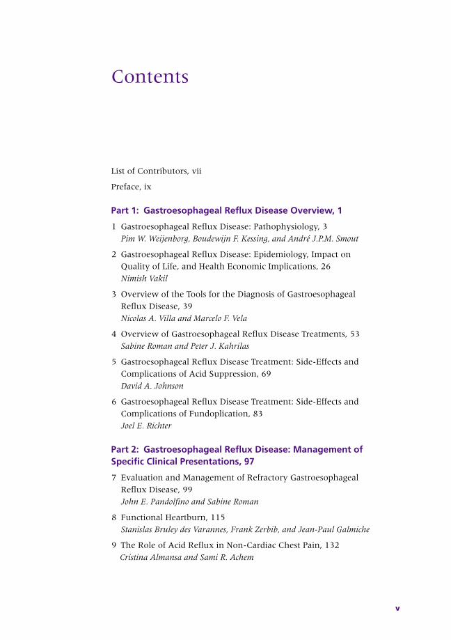
v
Contents
List of Contributors, vii
Preface, ix
Part 1: Gastroesophageal Reflux Disease Overview, 1
1 Gastroesophageal Reflux Disease: Pathophysiology, 3
Pim W. Weijenborg, Boudewijn F. Kessing, and André J.P.M. Smout
2 Gastroesophageal Reflux Disease: Epidemiology, Impact on
Quality of Life, and Health Economic Implications, 26
Nimish Vakil
3 Overview of the Tools for the Diagnosis of Gastroesophageal
Reflux Disease, 39
Nicolas A. Villa and Marcelo F. Vela
4 Overview of Gastroesophageal Reflux Disease Treatments, 53
Sabine Roman and Peter J. Kahrilas
5 Gastroesophageal Reflux Disease Treatment: Side-Effects and
Complications of Acid Suppression, 69
David A. Johnson
6 Gastroesophageal Reflux Disease Treatment: Side-Effects and
Complications of Fundoplication, 83
Joel E. Richter
Part 2: Gastroesophageal Reflux Disease: Management of Specific Clinical Presentations, 97
7 Evaluation and Management of Refractory Gastroesophageal
Reflux Disease, 99
John E. Pandolfino and Sabine Roman
8 Functional Heartburn, 115
Stanislas Bruley des Varannes, Frank Zerbib, and Jean-Paul Galmiche
9 The Role of Acid Reflux in Non-Cardiac Chest Pain, 132
Cristina Almansa and Sami R. Achem
ftoc.indd v 11/15/2012 2:47:15 AM
vi Contents
10 Laryngopharyngeal Reflux, 154
Robert T. Kavitt and Michael F. Vaezi
11 Reflux-Related Cough, 179
Etsuro Yazaki, Ryuichi Shimono, and Daniel Sifrim
12 Relationship between Gastroesophageal Reflux
Disease and Sleep, 195
Tiberiu Hershcovici and Ronnie Fass
13 Aerophagia and Belching, 221
Albert J. Bredenoord
14 Dysphagia and Gastroesophageal Reflux Disease, 239
Donald O. Castell and Erick R. Singh
15 Eosinophilic Esophagitis: Interactions with Gastroesophageal
Reflux Disease, 253
Kumar Krishnan and Ikuo Hirano
16 Helicobacter pylori and Gastroesophageal Reflux Disease, 267
Maria Pina Dore and David Y. Graham
Part 3: Barrett ’ s Esophagus, 287
17 Barrett ’ s Esophagus: Diagnosis and Surveillance, 289
Gary W. Falk
18 Barrett ’ s Esophagus: Treatment Options, 310
Jianmin Tian and Kenneth K. Wang
Index, 335
Color plate section can be found facing page 118
ftoc.indd vi 11/15/2012 2:47:16 AM

vii
List of Contributors
Sami R. Achem, MD, FACP, FACG, AGAF, FASGE Professor of Medicine
Division of Gastroenterology
Mayo Clinic Florida
Jacksonville, FL, USA
Cristina Almansa, MD, PhD Assistant Professor of Medicine
Division of Gastroenterology
Mayo Clinic Florida
Jacksonville, FL, USA
Albert J. Bredenoord, MD Department of Gastroenterology
Academic Medical Centre
Amsterdam, The Netherlands
Stanislas Bruley des Varannes Institut des Maladies de l ’ Appareil Digestif
Centre Hospitalier Universitaire de Nantes
Nantes, France
Donald O. Castell, MD Director, Esophageal Disease Program
Medical University of South Carolina
Charleston, SC, USA
Gary W. Falk, MD, MS Professor of Medicine
Division of Gastroenterology
Perelman School of Medicine at the University
of Pennsylvania
Philadelphia, PA, USA
Ronnie Fass, MD, FACP, FACG Division of Gastroenterology and Hepatology
MetroHealth Medical Center
Case Western Reserve University
Cleveland, Ohio, USA
Jean-Paul Galmiche Institut des Maladies de l ’ Appareil Digestif
Centre Hospitalier Universitaire de Nantes
Nantes, France
David Y. Graham, MD Professor of Medicine
Gastroenterology Section
Baylor College of Medicine
Michael E. DeBakey VA Medical Center
Houston, TX, USA
Tiberiu Hershcovici, MD Neuroenteric Clinical Research Group
Section of Gastroenterology
Department of Medicine
Southern Arizona VA Health Care System and
University of Arizona School of Medicine
Tucson, AZ, USA
Ikuo Hirano, MD Professor of Medicine
Northwestern University
Feinberg School of Medicine
Department of Medicine
Division of Gastroenterology
Chicago, IL, USA
David A. Johnson, MD, FACG, FASGE Professor of Medicine and Chief of
Gastroenterology
Eastern Virginia School of Medicine
Norfolk, VA, USA
Peter J. Kahrilas, MD Professor, Division of Gastroenterology
Northwestern University
Chicago, IL, USA
Robert T. Kavitt, MD Division of Gastroenterology, Hepatology,
and Nutrition
Vanderbilt University Medical Center
Nashville, TN, USA
Boudewijn F. Kessing, MD Department of Gastroenterology and
Hepatology
Academic Medical Center
Amsterdam, The Netherlands
fbetw.indd vii 11/15/2012 2:47:51 AM

viii List of Contributors
Kumar Krishnan, MD Northwestern University
Feinberg School of Medicine
Department of Medicine
Division of Gastroenterology
Chicago, IL, USA
Maria Pina Dore, MD, PhD Professor of Gastroenterology
Clinica Medica – Dipartimento di Medicina
Clinica e Sperimentale
University of Sassari
Sassari, Italy
Baylor College of Medicine
Houston, TX, USA
Sabine Roman Digestive Physiology
Claude Bernard Lyon I University and Hospices
Civils de Lyon
Lyon, France
Ryuichi Shimono, MD Wingate Institute for Neurogastroenterology
Barts and the London School of Medicine and
Dentistry
Research Fellow at GI Physiology Unit,
Royal London Hospital
London, UK
Daniel Sifrim, MD, PhD Professor of Gastrointestinal Physiology
Wingate Institute for Neurogastroenterology
Barts and the London School of Medicine and
Dentistry
Director GI Physiology Unit,
Royal London Hospital
London, UK
Erick R. Singh, MD Department of Medicine
Section of Gastroenterology and Hepatology
Georgia Health Sciences University
Augusta, GA, USA
André J.P.M. Smout, MD, PhD Department of Gastroenterology and
Hepatology
Academic Medical Center
Amsterdam, The Netherlands
Jianmin Tian, MD Barrett ’ s Esophagus Unit
Mayo Clinic
Rochester, MN, USA
Michael F. Vaezi, MD, PhD, MSc(Epi) Division of Gastroenterology, Hepatology, and
Nutrition
Vanderbilt University Medical Center
Nashville, TN, USA
Nimish Vakil, MD, FACP, FACG, AGAF, FASGE University of Wisconsin School of Medicine
and Public Health
Madison, WI, USA
Nicolas A. Villa Gastroenterology Section
Baylor College of Medicine
& Michael E. DeBakey VA Medical Center
Houston, TX, USA
Kenneth K. Wang, MD VanCleve Professor of Gastroenterology
Research
Director, Advanced Endoscopy
Director, Barretts Esophagus Unit
Mayo Clinic
Rochester, MN, USA
Pim W. Weijenborg, MD Department of Gastroenterology and
Hepatology
Academic Medical Center
Amsterdam, The Netherlands
Etsuro Yazaki, PhD, MAGIP Wingate Institute for Neurogastroenterology
Barts and the London School of Medicine and
Dentistry
Manager GI Physiology Unit,
Royal London Hospital
London, UK
Frank Zerbib Département de Gastroentérologie
CHU de Bordeaux
Centre Hospitalier Saint André de Bordeaux
Bordeaux, France
fbetw.indd viii 11/15/2012 2:47:51 AM

ix
Preface
Gastroesophageal reflux disease (GERD) is a very common clinical problem
and a frequent reason for consultation. Many patients have a typical pre-
sentation of heartburn and regurgitation, and a good response to treatment
with acid suppressive medication, such as a proton pump inhibitor (PPI).
However, the evaluation and management of GERD has become more
challenging for several reasons. The spectrum of clinical presentations
attributed to GERD has moved beyond the typical esophageal symptoms
of heartburn and regurgitation, and now incorporates various extraesoph-
ageal manifestations including laryngeal symptoms, cough, and even
disordered sleep. Furthermore, we are facing an increasing number of
patients in whom symptoms, either typical or atypical, persist despite acid
suppression with a PPI. Some of these patients with refractory symptoms
have persistent reflux due to treatment failure and require alternative
therapeutic approaches, while in others the reported symptoms may be
due to causes other than GERD, including functional disorders; in the
latter, a negative evaluation for GERD can direct the diagnostic and
treatment efforts toward other causes. Finally, how concomitant condi-
tions such as eosinophilic esophagitis and Helicobacter pylori gastritis affect
GERD management is not always clear, and a lucid perspective about these
issues is needed in daily practice.
Practical Manual of Gastroesophageal Reflux Disease , as it name indicates, is
meant to serve as a practical manual to aid the clinician in managing
GERD. The first section of the book presents an overview of pathophysi-
ology, epidemiology, diagnostic tools and treatment options of GERD.
Whole chapters are devoted to the potential side effects of medical and
surgical therapy, a highly relevant topic in routine practice. In the second
section, the evaluation and management of specific clinical presentations
in GERD (refractory heartburn, functional heartburn, chest pain, laryn-
gitis, cough, sleep disorders, belching, and dysphagia) are discussed and a
management algorithm is suggested for each clinical entity. In addition,
further chapters focus on the role of eosinophilic esophagitis and Helicobacter
pylori in GERD patients. A third section is devoted to Barrett’s esophagus,
to help the clinician deal with the challenges of screening for, diagnosing,
and treating this complication of GERD.
fpref.indd ix 11/15/2012 2:47:28 AM

x Preface
We are fortunate and thankful for the participation of the many
recognized experts from around the world who agreed to write the chapters
that make up this book. Our hope is that this book will provide a first-line
reference for clinicians who deal with this common and often challenging
problem of GERD.
Marcelo F. Vela
Houston, TX
fpref.indd x 11/15/2012 2:47:28 AM

PART 1
Gastroesophageal Reflux Disease Overview
p01.indd 1 11/15/2012 2:47:03 AM

3
Practical Manual of Gastroesophageal Reflux Disease, First Edition.
Edited by Marcelo F. Vela, Joel E. Richter and John E. Pandolfino.
© 2013 John Wiley & Sons, Ltd. Published 2013 by John Wiley & Sons, Ltd.
CHAPTER 1
Gastroesophageal Reflux Disease: Pathophysiology Pim W. Weijenborg, Boudewijn F. Kessing, and André J.P.M. Smout Department of Gastroenterology and Hepatology , Academic Medical Center ,
Amsterdam , The Netherlands
Key points • The anti-reflux barrier does not solely consist of the intrinsic pressure generated by
the lower esophageal sphincter, but is complemented by the extrinsic pressure
exerted by the crural diaphragm and the presence of the flap valve.
• Transient lower esophageal sphincter relaxations constitute the main mechanism of
reflux in gastroesophageal reflux disease patients and healthy subjects.
• The presence of a hiatal hernia increases the severity of esophageal acid exposure,
and changes the position of the acid pocket.
• The severity of gastroesophageal reflux disease-related symptoms is not predicted by
the severity of esophageal acid exposure and is dependent on factors influencing the
perception of reflux.
• Dilated intercellular spaces are more frequently present in non-erosive reflux disease
patients and possibly contribute to symptom generation.
Introduction
Over the past decades, considerable changes in our understanding of
gastroesophageal reflux disease (GERD) have taken place. In the era
before widespread application of endoscopy, when radiography was
the only diagnostic tool available, the diagnosis of GERD was more or
less synonymous with hiatal hernia. After the introduction of flexible
esophagogastroduodenoscopy, mucosal lesions in the distal esophagus
became the most important characteristic of the disease. Nowadays, we
know that reflux symptoms can be present in the absence of reflux
esophagitis. This subset of the disease is labeled non-erosive reflux
disease (NERD). In addition, extraesophageal symptoms and signs, such
c01.indd 3 11/15/2012 2:34:07 AM
4 Part 1: Gastroesophageal Reflux Disease Overview
as laryngitis, gastric asthma and chronic cough, were recognized. The
Montreal definition encompasses all of these elements of the disease by
stating that it is characterized by either bothersome symptoms and/or
lesions caused by reflux of gastric contents. This gradual broadening
of our understanding of what GERD is has led to an expansion of our
concepts of the pathophysiology of the disease [1] . Whilst the factors that
determine the exposure of the esophageal mucosa to gastric contents
are still relevant to the pathophysiology of GERD, factors that affect the
sensitivity of the esophagus have become recognized as equally important.
This chapter aims to summarize the many factors that are presently seen
as important in the pathophysiology of GERD.
Mechanisms leading to gastroesophageal reflux
Anti-reflux barrier In the early days after the advent of esophageal manometry, the lower
esophageal sphincter (LES) was conceptually prominent in the patho-
physiology of GERD. A LES able to maintain a sufficiently high pressure at
the esophagogastric junction (EGJ) was considered to be the most important
factor preventing gastroesophageal reflux. Nowadays, the anti-reflux
barrier is thought to consist of intrinsic LES pressure, extrinsic compres-
sion of the LES by the crural diaphragm, and the “flap valve” constituted
by an acute angle of His.
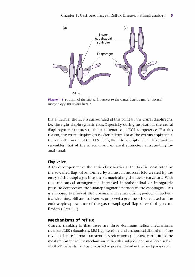
Lower esophageal sphincter The LES is a 3–4 cm segment of tonically contracted smooth muscle at the
EGJ. Normally, the LES is surrounded by the crural diaphragm. When a
sliding hiatus hernia is present, the LES is proximal to the crural diaphragm
(Figure 1.1 ). Resting LES tone, best measured during end-expiration, var-
ies among normal individuals from 10 to 30 mmHg relative to intragastric
pressure. Within a subject, LES pressure varies considerably during the
day. The highest pressure occurs during phase III of the migrating motor
complex, during which it may exceed 80 mmHg. Immediately after a meal,
LES pressure typically decreases. The genesis of LES tone is a property of
both the smooth muscle itself and of its extrinsic innervation.
Lower esophageal sphincter pressure is affected by myogenic factors,
intraabdominal pressure, gastric distension, peptides, hormones, various
foods, and many medications.
Crural diaphragm The opening in the diaphragm through which the esophagus reaches the
abdomen (hiatus esophagei) is shaped like a teardrop. In the absence of a
c01.indd 4 11/15/2012 2:34:07 AM
Chapter 1: Gastroesophageal Reflux Disease: Pathophysiology 5
hiatal hernia, the LES is surrounded at this point by the crural diaphragm,
i.e. the right diaphragmatic crus. Especially during inspiration, the crural
diaphragm contributes to the maintenance of EGJ competence. For this
reason, the crural diaphragm is often referred to as the extrinsic sphincter,
the smooth muscle of the LES being the intrinsic sphincter. This situation
resembles that of the internal and external sphincters surrounding the
anal canal.
Flap valve A third component of the anti-reflux barrier at the EGJ is constituted by
the so-called flap valve, formed by a musculomucosal fold created by the
entry of the esophagus into the stomach along the lesser curvature. With
this anatomical arrangement, increased intraabdominal or intragastric
pressure compresses the subdiaphragmatic portion of the esophagus. This
is supposed to prevent EGJ opening and reflux during periods of abdom-
inal straining. Hill and colleagues proposed a grading scheme based on the
endoscopic appearance of the gastroesophageal flap valve during retro-
flexion (Plate 1.1).
Mechanisms of reflux Current thinking is that there are three dominant reflux mechanisms:
transient LES relaxations, LES hypotension, and anatomical distortion of the
EGJ, e.g. hiatus hernia. Transient LES relaxations (TLESRs), constituting the
most important reflux mechanism in healthy subjects and in a large subset
of GERD patients, will be discussed in greater detail in the next paragraph.
Diaphragm
Loweresophagealsphincter
(a) (b)
Z-line
Figure 1.1 Position of the LES with respect to the crural diaphragm. (a) Normal
morphology. (b) Hiatus hernia.
c01.indd 5 11/15/2012 2:34:07 AM
6 Part 1: Gastroesophageal Reflux Disease Overview
When diminished LES pressure is present (either with or without
anatomical abnormality), short-lived increases in intraabdominal pressure
caused by straining are often the precipitating factor of the reflux.
Manometric data suggest that this rarely occurs when the LES pressure is
greater than 10 mmHg [2] . It is also a rare occurrence in patients without
hiatus hernia [3] . Free reflux is characterized by a fall in intraesophageal
pH without an identifiable change in either intragastric pressure or LES
pressure. Episodes of free reflux are observed only when the LES pressure
is lower than 5 mmHg.
It is important to realize that EGJ relaxation as measured manometri-
cally does not equate to EGJ opening or EGJ compliance, which are likely
to be more relevant to the occurrence of reflux. EGJ compliance can be
assessed with a water-filled balloon straddling the EGJ and measurement
of the diameter of the balloon at various levels of filling. In patients with
hiatus hernia, the compliance of the EGJ is increased but even patients
without hiatus hernia may have increased EGJ compliance. In the latter,
defects not readily detectable with imaging techniques, such as an abnormal
gastroesophageal flap valve, defects in the LES musculature or a wide dia-
phragmatic hiatus, are thought to be present. Subtle differences in EGJ
opening and compliance are likely to explain the discriminatory function
of the EGJ: large volumes of gas can be vented from the stomach while at
the same time fluid is largely contained within the stomach.
Transient lower esophageal sphincter relaxations Function and definition Lower esophageal sphincter relaxations are common and occur mainly
during swallows to allow passage of a bolus into the stomach [4] . In
addition, the LES can relax during the so-called TLESR which occurs less
frequently, about 3–6 times per hour [5,6] . TLESRs are considered the
physiological mechanism which enables venting of gas from the stomach,
also known as belching [7] . This belching reflex acts as a protective
mechanism which prevents excess amounts of gas accumulating in
the stomach. Since the discovery of TLESRs in the early 1980s, it has
become increasingly clear that most reflux episodes occur during TLESRs
[8] . Other mechanisms which can induce reflux episodes include straining,
coughing, and free reflux. However, these mechanisms only become
important – relatively and absolutely – in patients with severe reflux disease
associated with hiatal hernia.
A TLESR is currently defined as an abrupt decline in pressure at the
position of the LES which is not induced by swallowing [9] . Additional
criteria which could be helpful but are not needed for the identification
of TLESRs are crural diaphragm inhibition and a prominent after-
contraction [10] . Since the definition of TLESR is based solely on the
c01.indd 6 11/15/2012 2:34:08 AM

Chapter 1: Gastroesophageal Reflux Disease: Pathophysiology 7
esophageal pressure profile, the gold standard by which to measure
TLESRs is esophageal manometry (Plate 1.2).
Pharyngeal stimulation can also result in an LES relaxation which
resembles a TLESR [4] . However, LES relaxations induced by pharyngeal
stimulation are rarely associated with inhibition of the crural diaphragm
and acid reflux [6] . Furthermore, esophageal reflux was found only when
an LES relaxation was associated with diaphragm inhibition [6] .
Mechanisms of transient lower esophageal sphincter relaxation The primary stimulus which triggers a TLESR is gastric distension, often
resulting from accumulation of gastric air or consumption of a meal.
Distension in any part of the stomach can trigger a TLESR. However, the
subcardiac region of the stomach showed the lowest threshold for trigger-
ing TLESRs [11] . While still under debate, several studies suggest that
tension receptors in the stomach appear to be more relevant than pressure
receptors as the stimulus for transient LES relaxation [12,13] .
Transient LES relaxations are characterized by four different events. The
concerted action of these events results in complete relaxation of the EGJ.
The first and most prominent event during a TLESR is relaxation of the
inner part of the LES [14] . The second event is relaxation of the crural
diaphragm [15] . The third event is suppression of esophageal peristalsis
[14] and the fourth is a contraction of the distal esophageal longitudinal
muscle leading to esophageal shortening [16] . It has been hypothesized
that the longitudinal muscle contraction of the distal esophagus may be
the primary motor event leading to LES relaxation [17] but this hypothesis
remains to be proven.
Relaxation of the EGJ during a TLESR is terminated by primary
peristalsis or, more commonly, by secondary peristalsis [18] . Swallow-
induced primary peristalsis is characterized by upper esophageal
sphincter (UES) relaxation with pharyngeal contraction and esophageal
peristalsis progressing along the entire esophagus. Secondary peristalsis
is defined as a wave in the esophagus which is not associated with UES
relaxation and is a result of esophageal distension, often arising from
gastroesophageal reflux.
The rate of TLESRs can vary greatly during the day. The postprandial
period is characterized by a four- to fivefold increase in the rate of TLESRs
and an increase in the proportion of TLESRs accompanied by reflux [19] .
Body position can also influence the rate of TLESRs since the incidence of
TLESRs, as well as the incidence of reflux-associated TLESRs, is higher in
the right recumbent position compared to the left recumbent position [20] .
Furthermore, the rate of TLESRs is greatly decreased during the night [18] .
This is in accordance with the observation that reflux episodes occur less
often during the night than during the day [8] . Despite this nocturnal
c01.indd 7 11/15/2012 2:34:08 AM

8 Part 1: Gastroesophageal Reflux Disease Overview
decrease in the rate of TLESRs, a subset of GERD patients still shows
substantial acid exposure during the night. Therefore, in patients with
pathological nocturnal reflux, additional mechanisms are involved, such as
free reflux through a mechanically incompetent sphincter [21] .
The reflex pathway of the TLESR is a vagovagal reflex which commences
with activation of gastric receptors primarily in the subcardiac region [11] .
Sensory signals from the stomach are projected to the brain through
afferent sensory fibers of the vagus [22] and its terminating synapses are
located in the nucleus tractus solitarius (NTS) [23] . Signals from the NTS
do not provide signals to the EGJ directly but are relayed to the caudal part
of the dorsal motor nucleus of the vagus [24,25] . This central pathway
which modulates TLESRs is shared by both the LES and crural diaphragm
[26] . Furthermore, the crural diaphragm is innervated not only through
efferent vagal endings but also by the phrenic nerve [27] . The brainstem
sites responsible for this dual innervation are yet to be defined. Efferent
motor function signals from the brain to the LES and crural diaphragm are
conducted through the motor tract of the vagus [28] . Finally, motor signals
are relayed through the myenteric plexus from where they are further
distributed to the esophageal body and LES [28] .
Many excitory and inhibitory neurotransmitters and receptors, including
nitric oxide, opioids, anticholinergic agents and the neuropeptide CCK,
have been found to play a role in the neuromodulation of TLESRs [29] .
Among these neurotransmitters, the gamma-aminobutyric acid (GABA)
and metabotropic glutamate receptors (mGluR), and the cannabinoid
receptor 1 (CBR1) are of particular interest as potential targets for
therapeutic interventions. The most extensively investigated neurotrans-
mitter in the TLESR pathway is GABA-B. GABA-B acts as an inhibitory
neurotransmitter, and its receptors are located at both central and peripheral
sites in the TLESR reflex arch [30,31] . Metabotropic glutamate receptors
are also present throughout the central and peripheral nervous system. The
most extensively investigated metabotropic glutamate receptor is mGluR5
which has an excitatory function, mainly with a periperal site of action [32,
33] . The CBR1 has only recently been investigated with regard to TLESRs.
Its site of action is believed to be the central nervous system [34] .
Despite the importance of the TLESR in the pathophysiology of GERD,
most of our knowledge regarding the neural pathways involved in the
reflex arc of the TLESRs is derived from animal studies. However, it is
assumed that TLESRs in humans follow similar pathways.
Association between gastroesophageal reflux and transient lower esophageal sphincter relaxations Transient LES relaxations are considered to be the main mechanism
leading to gastroesophageal reflux in GERD patients. However, the majority
c01.indd 8 11/15/2012 2:34:08 AM
Chapter 1: Gastroesophageal Reflux Disease: Pathophysiology 9
of the studies show a similar rate of TLESRs in healthy subjects and GERD
patients [35,36] . This means that in GERD patients, there is a higher
percentage of TLESRs which not only vent air but are also associated with
gastroesophageal reflux. Therefore, a different underlying mechanism is
necessary which results in this loss of discrimination between air and liquid
by the LES.
In GERD patients, a slightly higher transsphincteric pressure gradient is
present before and during a TLESR when compared to healthy subjects
[37] . More importantly, the pressure gradient is greater during TLESRs
accompanied by acid reflux compared to TLESRs without acid reflux.
Another proposed contributing factor is EGJ compliance, also known as
EGJ distensibility. GERD patients are characterized by an increase in EGJ
compliance which could explain the loss of discrimination between air and
liquids [38] . Furthermore, EGJ compliance in GERD patients with hiatal
hernia is increased compared to GERD patients without hiatal hernia [39] .
Obesity is associated with an increased rate of TLESRs as well as with an
increased association of TLESRs with gastroesophageal reflux [40] . In
addition, a higher pressure gradient has been measured during TLESRs in
obese subjects compared to normal-weight subjects. The influence of dif-
ferent nutritional factors on the association of TLESRs and reflux as well as
the rate of TLESRs has been extensively studied. However, no correlation
between reflux-associated TLESRs or an influence on the rate of TLESRs
has been demonstrated.
Hiatal hernia In 1971, Cohen and Harris published a paper in which they reported that
reflux symptoms correlated with low LES pressure, rather than with
presence of a hiatus hernia [41] . From then on, the emphasis in studies on
GERD pathophysiology was on basal LES pressure. Another change took
place when the phenomenon of TLESR was found to play a pivotal role
[42] . The sleeve sensor that was required to record TLESRs did not allow
recognition of the two distinct components of the high-pressure zone, i.e.
LES and crural diaphragm. Awareness of the importance of hiatal hernia
for the pathophysiology of GERD emerged again around the turn of the
century. It is clear that esophageal acid exposure is greater in patients with
hiatus hernia [3,43–45] . In addition, the severity of esophageal acid
exposure increases with increasing size of the hernia [45,46] and esopha-
gitis is more severe with more severe acid exposure [47] . Patients with
Barrett ’ s esophagus have the highest prevalence of hiatus hernia [48] .
Hiatus hernia is not an all-or-nothing phenomenon. The so-called
physiological hernia (also known as phrenic ampulla) is only present
during swallowing when the esophageal shortening leads to displace-
ment of the Z-line to a site proximal to the diaphragm. This displacement
c01.indd 9 11/15/2012 2:34:08 AM
10 Part 1: Gastroesophageal Reflux Disease Overview
is < 2 cm. A reducing hiatal hernia is a hernia which is greater than 2 cm
but which is only seen during a swallow; between swallows, the Z-line is
at the level of the diaphragm. A non-reducing hiatal hernia is defined as
a hernia greater than 2 cm in which the Z-line does not return to its
normal position between swallows. At moments at which a hiatus hernia
is present, the anti-reflux effect of the crural diaphragm is exerted at the
wrong spot, i.e. distal to the LES, and the effect is weakened because the
hiatus is usually wider than normal. Using pull-through manometry and
three-dimensional representation of the pressure profiles, Kahrilas and
co-workers demonstrated in hiatus hernia patients that there are distinct
intrinsic sphincter and hiatal canal pressure components, with each one
exerting pressure of lower magnitude than normal. Simulating reduction
of the hernia by repositioning the intrinsic sphincter back within the
hiatal canal and arithmetically summing superimposed pressures resulted
in calculated EGJ pressures which were practically indistinguishable
from those of the control subjects [49] . Prolonged manometric studies
have also made clear that mechanisms other than TLESR play a more
prominent role when a hiatus hernia is present. These other mechanisms
include low LES pressure, straining-induced reflux and swallow- associated
reflux [3] .
Even within the same patient, the mechanisms leading to reflux vary
from time to time, depending on the reduced or non-reduced status of the
hiatus hernia [50] . Another mechanism by which the presence of a hiatus
hernia is associated with excessive esophageal acid exposure is character-
ized by superimposed reflux from the hiatal sac during swallowing-induced
LES relaxation. This can be seen in non-reducing hiatus hernias [51, 52] .
Gastric factors Total gastric emptying It is tempting to speculate that delayed gastric emptying is an important
factor in the pathogenesis of GERD. However, the evidence for this hypo-
thesis appears to be controversial.
Numerous studies have observed delayed gastric emptying in a
proportion of GERD patients compared to healthy controls [53] and only a
few studies reported no difference. However, no correlation between
esophageal acid exposure time and delayed gastric emptying could be
proven [54] . Furthermore, acceleration of gastric emptying by cisapride
was not associated with a decrease in esophageal acid exposure or with the
number of reflux events [55] . Studies investigating the association bet-
ween gastroesophageal reflux and gastric emptying are limited by
measuring acidic reflux episodes only. To our knowledge, no study has
been published which assesses the influence of gastric emptying on weakly
acidic reflux episodes.
c01.indd 10 11/15/2012 2:34:08 AM

Chapter 1: Gastroesophageal Reflux Disease: Pathophysiology 11
Emptying of the proximal stomach Over the last few decades, the role of the proximal stomach in the pathogenesis
of GERD has gained much attention since TLESRs are triggered by distension
of the proximal stomach and the refluxate is located in the proximal stomach
as well. The motor response of the proximal stomach to a meal is characterized
by a relaxation followed by a gradual recovery of gastric tone. It has been found
that GERD patients are characterized by a delayed recovery of proximal gastric
tone after a meal compared to healthy controls [56] . Furthermore, emptying
from the proximal stomach, but not the distal stomach, was significantly
delayed in GERD patients compared to healthy controls.
Slow proximal emptying shows a correlation with increased esophageal
acid exposure time [57] . Furthermore, the number of acidic reflux epi-
sodes correlates with proximal gastric retention [58] . Thus, in contrast to
gastric emptying of the whole stomach, delayed emptying of the proximal
stomach appears to be a factor in the pathogenesis of GERD. In theory,
delayed emptying of the proximal stomach could cause an altered position
of the postprandial acid pocket (see below) and influence the association of
TLESRs with reflux. However, this hypothesis remains to be proven.
Acid pocket Until recently it was assumed that gastric acid secreted after a meal is
instantly mixed with the ingested food into one homogeneous mixture.
The buffering effect of many food constituents leads to a postprandial
increase in gastric pH. However, Fletcher et al . observed that the pH in the
body of the stomach was markedly higher (pH 4.7) than the pH of the
esophageal refluxate (pH 1.6) [59] . In subsequent pull-through pH studies,
they identified a pocket of unbuffered gastric acid which lies on top of a
homogenized fatty meal. This so-called acid pocket extends from the cardia
to the distal esophagus [59] .
The position of the acid pocket in GERD patients differs from healthy
controls, i.e. a supradiaphragmatic localization of the pocket was more fre-
quent in patients with GERD, especially those with a large HH (Plate 1.3)
[60] . Localization of the acid pocket strongly correlates with the occur-
rence of acid reflux. When the acid pocket is located above the diaphragm,
70–85% of all TLESRs are accompanied by acid reflux [60] . In contrast,
when the acid pocket is located below the diaphragm, only 7–20% of
TLESRs are accompanied by an acidic reflux episode. Even during reflux
episodes which are caused by mechanisms other than TLESRs, the position
of the acid pocket is still of major importance.
Effect of posture on reflux Body position does not affect the acidity in the gastric cardia and corpus.
However, the right recumbent position is associated with an increase in
c01.indd 11 11/15/2012 2:34:08 AM
12 Part 1: Gastroesophageal Reflux Disease Overview
acid exposure time in the distal esophagus compared to the left recumbent
position [61] . This is due to an increase in reflux episodes, TLESRs and
TLESRs associated with reflux [20] . The duration of reflux episodes is not
affected by body position.
Obesity Overall, the weight of the evidence suggests that obesity and GERD are
related. When dissected to individual aspects of the disease, there are areas
of controversy. For instance, the results of studies on esophageal acid
exposure – as measured with 24-h pH monitoring – in obesity are not
entirely unequivocal [40,62–72] . Recent data indicate that the proximal
esophageal extent of the refluxate is higher in obese subjects [73] . It is
likely that, in the obese, waist circumference is a more important determi-
nant of excessive reflux [65,66] .
There are relatively few studies on LES function in the obese. The limited
data available suggest that basal LES pressures in the morbidly obese are
similar to those of ideal body weight [74] . However, obesity is associated
with an increased incidence of TLESRs, the association being present for
increased Body Mass Index (BMI) as well as waist circumference [40] .
Hiatal hernia is found more often in patients with obesity than in subjects
with a normal BMI [75,76] . Increased intragastric pressure may promote
the development of hiatus hernia by applying an axial pressure strain
through the diaphragm [77] .
Apart from promoting the development of hiatus hernia, the increased
intragastric pressure found in the obese tends to promote reflux. Especially
during inspiration, increased intragastric pressure and the gastroesopha-
geal pressure gradient are correlated with increased BMI. The changes
noted above are more strongly correlated with waist circumference.
In summary, obese subjects are more likely to have a high incidence of
TLESRs, a hiatal hernia, increased intragastric pressure, and an increased
gastroesophageal pressure gradient. These factors all facilitate reflux.
A positive association between reflux symptoms and BMI was found in
more than a dozen studies. Two metaanalyses incorporating these studies
confirmed the existence of such an association and found the risk of having
reflux symptoms in the overweight and obese to be 43–94% higher than
in normal-weight subjects [66,78] . In a study in women, a BMI > 30 kg/m 2
was associated with a threefold increase in the odds of having frequent
reflux symptoms [79] .
Despite the equivocal nature of the evidence for increased gastroesoph-
ageal reflux in the obese, a metaanalysis showed a statistically significant
increase in the risk for esophageal lesions with increasing weight. A BMI
greater than 25 kg/m 2 had an odds ratio of 1.76 for erosive esophagitis and
2.02 for esophageal adenocarcinoma, compared with patients with normal
c01.indd 12 11/15/2012 2:34:08 AM
Chapter 1: Gastroesophageal Reflux Disease: Pathophysiology 13
weight [78] . Four prospective multicenter, randomized, double-blind trials
comparing esomeprazole and other proton pump inhibitors found a weak
but statistically significant increased risk for Los Angeles grades C and D
esophagitis, but not grades A and B, in the obese [80] . In a case–control
study that evaluated cases with Barrett ’ s esophagus and two control groups
(normal-weight patients and patients with GERD but without Barrett ’ s
esophagus), abdominal diameter was found to be an independent risk
factor for Barrett ’ s esophagus. There was no association between Barrett ’ s
esophagus and BMI [66] .
Studies on the effect of weight loss obtained by non-surgical methods on
reflux symptoms, endoscopic findings or pH monitoring have yielded
somewhat disappointing results [81,82] . However, when studies describing
surgically achieved weight loss are also taken into account, a positive
conclusion can be drawn [83] .
Mechanisms involved in perception of reflux
With the development of new techniques it has become clear that esopha-
geal acid exposure is not the only factor involved in the generation of
reflux symptoms, and that mechanisms altering the perception of gastro-
esophageal reflux must have an effect.
The addition of ambulatory pH measurement to the diagnostic arma-
mentarium made it possible to not only quantify the severity of esophageal
acid exposure, but also to assess the temporal relation between symptoms
and acid reflux episodes. In order to describe this relationship between gas-
troesophageal reflux and symptoms, several tools have been developed.
The one considered to have the fewest shortcomings is the Symptom
Association Probability (SAP), proposed by Weusten et al . [84] . To calculate
the SAP, the 24-h pH measurement is divided into 2-min time frames and
the occurrence of reflux in these periods and in the 2-min time frame pre-
ceding the moments of symptom onset is noted. Thereafter the probability
that symptoms are associated with reflux is calculated. The SAP is consid-
ered to be positive once it is > 95%.
Using the SAP, it has become apparent that esophageal acid exposure is
not closely related with the number of reflux symptoms experienced by
the patient and that acid exposure and positive symptom-reflux associa-
tions are largely independent phenomena [85] . This is in contrast to the
finding that as the severity of esophageal acid exposure increases, this is
accompanied by an increasing severity of erosions [47] . When a patient ’ s
esophagus is exposed to physiological acid reflux and there is no correla-
tion between symptoms and the reflux episodes (negative SAP), he or she
is classified as having “functional heartburn.” When physiological reflux is
c01.indd 13 11/15/2012 2:34:08 AM

14 Part 1: Gastroesophageal Reflux Disease Overview
present and bothersome reflux symptoms appear to be correlated with that
reflux, the patient is considered to have a “hypersensitive esophagus.” In
patients with pathological esophageal acid reflux, the distribution between
those with a positive and a negative SAP is not different from the distribu-
tion in patients with physiological esophageal acid exposure, suggesting
that symptom generation is mostly independent of the severity of the
reflux [85] .
Intraluminal factors influencing perception and thereby symptom gen-
eration include several reflux characteristics. First, reflux episodes pre-
ceded by a higher cumulative acid exposure time are more likely to be
perceived. The difference in cumulative acid exposure time between symp-
tomatic and asymptomatic reflux episodes is apparent for up to 75 min
[86] . Furthermore, symptomatic reflux episodes have a higher median
proximal extent and a longer median duration [87] . However, it must be
considered that there is an overlap in proximal extent between symptom-
atic and asymptomatic reflux episodes and therefore an individual
threshold above which a reflux episode will always be symptomatic cannot
be established.
Non-acid reflux The introduction of combined pH and impedance monitoring broadened
the spectrum of gastroesophageal reflux since the technique allows further
characterization of reflux episodes according to acidity and composition
(liquid or mixed liquid-gas). By the addition of impedance, reflux episodes
without a pH drop that would have been missed with a conventional
ambulatory pH measurement can be detected. Thereby the new
phenomenon of non-acid reflux emerged. Whereas it was long felt to be
unlikely that non-acid reflux can provoke symptoms, results of a perfusion
study carried out two decades ago had indicated that non-acid solutions
with pH up to 6 exacerbate symptoms in around 50% of subjects [88] .
We now know that esophageal exposure to non-acid gastric content is
a possible explanation for the persistence of symptoms after adequate
acid- suppressive therapy.
Using impedance measurement, it has been shown that acid suppression
with a proton pump inhibitor (PPI) reduces neither the total number of
reflux events nor their proximal extent. Rather, PPI treatment decreases
the number of acid reflux in favor of weakly acidic (nadir pH between
4 and 7) and alkaline (nadir pH > 7) reflux [89] .
Non-acid reflux proved to be responsible for 15% of symptomatic reflux
episodes in patients off PPI [86] . In patients on PPI therapy presenting with
persistent reflux symptoms, 37% of subjects showed a positive Symptom
Index (SI) for non-acid reflux. This emphasizes the possible role of imped-
ance measurement in identifying this subgroup of patients who could
c01.indd 14 11/15/2012 2:34:08 AM

Chapter 1: Gastroesophageal Reflux Disease: Pathophysiology 15
benefit from additional therapy aimed at reducing the absolute number of
reflux events (TLESR inhibitors, fundoplication) [90] . The most interesting
finding made with impedance monitoring is that the majority of patients
with persisting symptoms under PPI therapy show a negative symptom
index for acid and non-acid reflux, suggesting an erroneous initial diag-
nosis and supporting the possibility of stopping PPI therapy.
As mentioned, the composition of the refluxate differs, with about half
of total reflux episodes being completely liquid and half having a gaseous
component, which is similar in GERD patients and healthy volunteers.
However, the reflux episodes causing symptoms in NERD patients more
often contain a gaseous component [91] .
Dilated intercellular spaces The mechanical barrier that lies between luminal acid gastric content
and esophageal nociceptors is the esophageal epithelium. The human
esophageal epithelium is a stratified squamous epithelium consisting of
three layers: the upper layer is the stratum corneum or so-called functional
layer, below which lies the stratum spinosum or prickle cell layer. Finally,
on the serosal side of the epithelium, the stratum basale is located.
A functional epithelial barrier function is maintained by desmosomes and
tight junctions. Desmosomes enable strong cell-to-cell adhesion by linking
cell surface adhesion proteins to intracellular keratin cytoskeletons. They
are present throughout the three layers of esophageal epithelium but are
most frequently located in the prickle cell layer [92] . In addition, tight
junctions seal the intercellular space and prevent the paracellular diffusion
of fluid and small molecules.
Several histopathological changes in the esophageal epithelium of GERD
patients have been described, such as thickening of the basal cell layer,
elongation of mucosal papillae [93] and dilated intercellular spaces (DIS)
[94] . Since Tobey et al . first described DIS in NERD patients [95] , the
phenomenon has been extensively studied and proposed as a possible key
mediator of symptom generation in GERD patients. DIS can be seen as a
dysfunction of the epithelial barrier function, enabling the diffusion of fluid
and acid molecules into the intercellular space and allowing them to reach
and activate chemosensitive nociceptors in the underlying layers [96] .
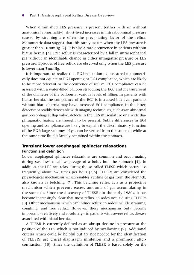
Several studies have assessed DIS in human esophageal biopsy samples,
some of which used transmission electron microscopy (TEM), allowing
accurate measurement of the intercellular space (Figure 1.2 ) [95,97,98] .
These studies found that the mean diameter of intercellular spaces in
NERD patients (1.0–2.2 μm) is at least twice that in healthy controls (0.45–
0.56 μm) [99] . This suggests that DIS measurement by TEM in biopsies is a
useful tool to confirm the otherwise difficult diagnosis of NERD. However,
TEM is expensive and time-consuming and therefore it does not seem
c01.indd 15 11/15/2012 2:34:09 AM

16 Part 1: Gastroesophageal Reflux Disease Overview
easily applicable in clinical practice. Multiple studies have tried to measure
intercellular space diameters using the more accessible technique of light
microscopy (LM) [100,101] . However, the results regarding the variability
between TEM and LM are conflicting and the correlation between mea-
surements performed by the two techniques does not seem to be very
promising [102,103] .
The exact mechanism responsible for the generation of DIS has not been
elucidated. Since exposure of esophageal mucosa to gastric contents was
the first logical explanation, in vitro and in vivo studies have primarily
focused on their relation with DIS.
Exposure of rabbit esophagus to an acidic solution with pH 1.1 causes no
macroscopic erosions but shows clear DIS under TEM, which is accompa-
nied by a drop in epithelial resistance and an increase of esophageal per-
meability to small molecules [104] . The addition of pepsin to an acidic
solution further increased the rate of DIS, but the effect was only present
with pH < 3 [105] . Besides acid and pepsin, bile acids are other potentially
harmful erosive components of gastric content. Exposure of rabbit esoph-
ageal mucosa to bile acids can cause the generation of DIS in both acidic
and weakly acidic conditions [106] . This is in contrast to the earlier finding
that biopsies of GERD patients with and without duodenal reflux exposure
show a similar amount of DIS [97] .
The concept of DIS generation in response to acid and acid-pepsin
proved to hold in vivo , in a model where infusion of acid and acid-pepsin
solutions in the distal esophagus was followed by the direct assessment of
DIS in biopsy samples by TEM [107] . The concept of acid exposure gener-
ating DIS is corroborated by the fact that DIS recovered after 3 months of
Figure 1.2 Transmission electron microscopy image of the basal layer of rat esophageal
mucosa. (a) Normal morphology. (b) Dilated intercellular spaces in a rat treated with a
moderate stressor.
(a) (b)
c01.indd 16 11/15/2012 2:34:09 AM
Chapter 1: Gastroesophageal Reflux Disease: Pathophysiology 17
acid suppressive therapy [108] . Subsequently, the effect of weakly acidic
solutions and bile salts on DIS was studied and proved to be present in a
similar in vivo model [106] . An interesting finding in this study is that
although these solutions provoked DIS, the majority of subjects did not
experience heartburn. This supports the hypothesis that symptom genera-
tion is multifactorial and DIS is not the only determinant of symptoms.
Next to luminal effects, there are indications that systemic factors play a
role in the generation of DIS. The predominant location of DIS in the basal
layer of the epithelium, and the less pronounced presence in the more
directly exposed prickle cell and functional layers, suggests that circulating
agents such as cytokines exert a systemic effect, possibly in response to the
aggressive luminal contents. Furthermore, it has been shown that acute
stress increases the perception of heartburn in GERD patients [109] and
acute stress enhances the effect of acid-pepsin on DIS and the permeability
to small molecules in a rat model [110] .
Visceral hypersensitivity Visceral hypersensitivity is an established concept in inflammatory and
functional gastrointestinal disorders, where patients have a heightened
perception of various stimuli in the gastrointestinal tract [111] . This
reduced pain threshold to mechanical, chemical, thermal or electrical
stimuli is considered to be caused by a combination of peripheral sensi-
tization, central sensitization and interactions between the neural and
immune systems [112] . The previously mentioned finding that stress
influences patients’ heartburn perception suggests a similar role for
visceral hypersensitivity in the pathophysiology of GERD. Peripheral
nociceptors in the esophagus express several cation channels, of which
the most relevant for GERD are cation channels sensitive to a low pH,
like acid-sensitive ion channels (ASICs) 1–3, ionotropic purinergic (P2X)
receptors and the transient receptor potential (TRP) channels. TRPV1, a
member of the TRP family, has been shown to be upregulated in the
esophageal mucosa of patients with esophagitis and NERD [113,114] .
Sensitization of peripheral neurons occurs once the signaling threshold
of these channels reduces in response to continuous noxious stimula-
tion. A possible mechanism of sensitization in GERD is through direct
contact of these channels with H + by the presence of DIS and subsequent
acidification of the intercellular space or via indirect signaling by cyto-
kines released in response to the exposure of epithelium to aggressive
gastric contents.
Central sensitization occurs once repetitive firing from the peripheral
neurons leads to triggering of intercellular changes in the spinal dorsal
horn neurons responsible for central signal transduction of nociceptors.
This in turn leads to amplified responses to peripheral stimuli and also
c01.indd 17 11/15/2012 2:34:11 AM

18 Part 1: Gastroesophageal Reflux Disease Overview
to triggering of adjacent spinal neurons, giving rise to hypersensitivity of
more remote areas such as the chest wall [115] .
Sustained esophageal contractions Another mechanism proposed as a mediator in the perception of reflux
episodes is the phenomenon of sustained esophageal contractions (SEC).
Using high-frequency endoscopic ultrasonography, intermittent thick-
ening of the esophageal wall can be observed, representing a sustained
contraction of the longitudinal muscle. SECs preceded 70% of heartburn
symptoms during ambulatory ultrasonography combined with a pH
measurement and accompanied 75% of provoked heartburn symptoms
during a Bernstein test [116] . SECs were also found to correlate with
symptoms in patients with unexplained chest pain [117] . The findings
suggest a role of SECs in the pathophysiology of esophageal pain percep-
tion, although it should be noted that all findings were obtained in a
small number of patients. Furthermore, the concept cannot explain the
entire spectrum of symptom generation since the majority of SECs do not
cause symptoms and 30% of heartburn symptoms are not accompanied
by a SEC [116] .
Genetic factors The observation that reflux symptoms are often clustered in families
prompted a search for genetic factors that might play a role in GERD. An
association was found between GERD and the heterozygous genotype of
the C825T allele of the G-protein B3 subunit, coding for a receptor fre-
quently present in the neural brain-gut axis which is associated with intra-
cellular cell transduction [118] . The polymorphism had previously been
associated with visceral hypersensitivity in functional dyspepsia. The
association was specifically present in patients with a “hypersensitive
esophagus,” suggesting a genetic predisposition to visceral hypersensitivity
in GERD.
Summary
Gastroesophageal reflux disease is a multifactorial disorder and although
many aspects of the pathophysiology have been described, parts remain to
be elucidated. The pathophysiology comprises factors that determine the
exposure of the esophageal mucosa to gastric contents, and factors that
influence the esophageal sensitivity and thereby alter the perception of
reflux. The esophageal exposure to gastric contents is dependent on reflux
mechanisms as TLESRs, LES hypotension and the presence of an anatomical
disruption of the normal anti-reflux barrier, i.e. a hiatal hernia. Additionally,
c01.indd 18 11/15/2012 2:34:11 AM

Chapter 1: Gastroesophageal Reflux Disease: Pathophysiology 19
reflux is facilitated by gastric factors such as delayed emptying of the
proximal stomach and an altered position of the acid pocket. Obesity leads
to an increased severity of gastroesophageal reflux by influencing several
of these mechanisms.
The fact that esophageal acid exposure and symptom generation are
mainly independent phenomena has led to the understanding that sensi-
tivity of the esophagus and perception of reflux are equally important in
the pathophysiology of GERD. Characteristics of the reflux episode itself,
such as proximal extent, duration and the composition of the refluxate,
can lead to increased perception. Suggested changes at the esophageal
level contributing to an increased perception of reflux are the presence of
dilated intercellular spaces and visceral hypersensitivity. Lastly, genetic
mutations could predispose to visceral hypersensitivity and thereby to
reflux perception in GERD.
References
1 GI Motility online . Available from: www.nature.com/gimo/index.html .
2 Sloan S , Rademaker AW , Kahrilas PJ . Determinants of gastroesophageal junction
incompetence: hiatal hernia, lower esophageal sphincter, or both? Ann Intern Med
1992 ; 117 ( 12 ): 977 – 82 .
3 Van Herwaarden MA , Samsom M , Smout AJ . Excess gastroesophageal reflux in
patients with hiatus hernia is caused by mechanisms other than transient LES relax-
ations . Gastroenterology 2000 ; 119 ( 6 ): 1439 – 46 .
4 Pouderoux P , Verdier E , Kahrilas PJ . Patterns of esophageal inhibition during swal-
lowing, pharyngeal stimulation, and transient LES relaxation. Lower esophageal
sphincter . Am J Physiol Gastrointest Liver Physiol 2003 ; 284 ( 2 ): G242 – 7 .
5 Schoeman MN , Holloway RH . Integrity and characteristics of secondary oesophageal
peristalsis in patients with gastro-oesophageal reflux disease . Gut 1995 ; 36 ( 4 ): 499 – 504 .
6 Mittal RK , Chiareli C , Liu J , Shaker R . Characteristics of lower esophageal sphincter
relaxation induced by pharyngeal stimulation with minute amounts of water .
Gastroenterology 1996 ; 111 ( 2 ): 378 – 84 .
7 Wyman JB , Dent J , Heddle R , Dodds WJ , Toouli J , Downton J . Control of belching by
the lower oesophageal sphincter . Gut 1990 ; 31 ( 6 ): 639 – 46 .
8 Dent J , Dodds WJ , Friedman RH , et al . Mechanism of gastroesophageal reflux in
recumbent asymptomatic human subjects . J Clin Invest 1980 ; 65 ( 2 ): 256 – 67 .
9 Holloway RH , Penagini R , Ireland AC . Criteria for objective definition of transient
lower esophageal sphincter relaxation . Am J Physiol 1995 ; 268 ( 1 Pt 1 ): G128 – 33 .
10 Holloway RH , Boeckxstaens GE , Penagini R , Sifrim D , Smout A , Ruth M . T1229
objective definition and detection of transient lower esophageal relaxation revisited:
is there room for improvement? Gastroenterology 2009 ; 136 (5, Suppl 1): A - 527 .
11 Franzi SJ , Martin CJ , Cox MR , Dent J . Response of canine lower esophageal sphincter
to gastric distension . Am J Physiol 1990 ; 259 (3 Pt 1): G380 – 5 .
12 Straathof JWA , van Veen MM , Masclee AAM . Provocation of transient lower esoph-
ageal sphincter relaxations during continuous gastric distension . Scand J Gastroenterol
2002 ; 37 ( 10 ): 1140 – 3 .
c01.indd 19 11/15/2012 2:34:11 AM
20 Part 1: Gastroesophageal Reflux Disease Overview
13 Scheffer RCH , Akkermans LMA , Bais JE , Roelofs JMM , Smout AJPM , Gooszen HG .
Elicitation of transient lower oesophageal sphincter relaxations in response to gastric
distension and meal ingestion . Neurogastroenterol Motil 2002 ; 14 ( 6 ): 647 – 55 .
14 Mittal RK , Holloway RH , Penagini R , Blackshaw LA , Dent J . Transient lower esoph-
ageal sphincter relaxation . Gastroenterology 1995 ; 109 ( 2 ): 601 – 10 .
15 Mittal RK , Fisher MJ . Electrical and mechanical inhibition of the crural diaphragm
during transient relaxation of the lower esophageal sphincter . Gastroenterology
1990 ; 99 ( 5 ): 1265 – 8 .
16 Shi G , Pandolfino JE , Joehl RJ , Brasseur JG , Kahrilas PJ . Distinct patterns of oesoph-
ageal shortening during primary peristalsis, secondary peristalsis and transient lower
oesophageal sphincter relaxation . Neurogastroenterol Motil 2002 ; 14 ( 5 ): 505 – 12 .
17 Babaei A , Bhargava V , Korsapati H , Zheng WH , Mittal RK . A unique longitudinal
muscle contraction pattern associated with transient lower esophageal sphincter
relaxation . Gastroenterology 2008 ; 134 ( 5 ): 1322 – 31 .
18 Kuribayashi S , Massey BT , Hafeezullah M , et al . Terminating motor events for TLESR
are influenced by the presence and distribution of refluxate . Am J Physiol Gastrointest
Liver Physiol 2009 ; 297 ( 1 ): G71 – 5 .
19 Holloway RH , Kocyan P , Dent J . Provocation of transient lower esophageal sphincter
relaxations by meals in patients with symptomatic gastroesophageal reflux . Dig Dis
Sci 1991 ; 36 ( 8 ): 1034 – 9 .
20 Van Herwaarden MA , Katzka DA , Smout AJ , Samsom M , Gideon M , Castell DO .
Effect of different recumbent positions on postprandial gastroesophageal reflux in
normal subjects . Am J Gastroenterol 2000 ; 95 ( 10 ): 2731 – 6 .
21 Freidin N , Fisher MJ , Taylor W , et al . Sleep and nocturnal acid reflux in normal sub-
jects and patients with reflux oesophagitis . Gut 1991 ; 32 ( 11 ): 1275 – 9 .
22 Martin CJ , Patrikios J , Dent J . Abolition of gas reflux and transient lower esophageal
sphincter relaxation by vagal blockade in the dog . Gastroenterology 1986 ; 91 ( 4 ): 890 – 6 .
23 Kalia M , Mesulam MM . Brain stem projections of sensory and motor components of
the vagus complex in the cat: II. Laryngeal, tracheobronchial, pulmonary, cardiac,
and gastrointestinal branches . J Comp Neurol 1980 ; 193 ( 2 ): 467 – 508 .
24 Rinaman L , Card JP , Schwaber JS , Miselis RR . Ultrastructural demonstration of a
gastric monosynaptic vagal circuit in the nucleus of the solitary tract in rat . J Neurosci
1989 ; 9 ( 6 ): 1985 – 96 .
25 Rossiter CD , Norman WP , Jain M , Hornby PJ , Benjamin S , Gillis RA . Control of lower
esophageal sphincter pressure by two sites in dorsal motor nucleus of the vagus . Am
J Physiol 1990 ; 259 ( 6 Pt 1 ): G899 – 906 .
26 Niedringhaus M , Jackson PG , Evans SRT , Verbalis JG , Gillis RA , Sahibzada N . Dorsal
motor nucleus of the vagus: a site for evoking simultaneous changes in crural
diaphragm activity, lower esophageal sphincter pressure, and fundus tone . Am
J Physiol Regul Integr Comp Physiol 2008 ; 294 ( 1 ): R121 – 31 .
27 Young RL , Page AJ , Cooper NJ , Frisby CL , Blackshaw LA . Sensory and motor
innervation of the crural diaphragm by the vagus nerves . Gastroenterology
2010 ; 138 ( 3 ): 1091 – 101 .
28 Yuan S , Costa M , Brookes SJ . Neuronal pathways and transmission to the lower
esophageal sphincter of the guinea pig . Gastroenterology 1998 ; 115 ( 3 ): 661 – 71 .
29 Kessing BF , Conchillo JM , Bredenoord AJ , Smout AJPM , Masclee AAM . Review
article: the clinical relevance of transient lower oesophageal sphincter relaxations in
gastro-oesophageal reflux disease . Aliment Pharmacol Ther 2011 ; 33 ( 6 ): 650 – 61 .
30 Yuan CS , Liu D , Attele AS . GABAergic effects on nucleus tractus solitarius neurons
receiving gastric vagal inputs . J Pharmacol Exp Ther 1998 ; 286 ( 2 ): 736 – 41 .
c01.indd 20 11/15/2012 2:34:11 AM

Chapter 1: Gastroesophageal Reflux Disease: Pathophysiology 21
31 Blackshaw LA , Smid SD , O ’ Donnell TA , Dent J . GABA(B) receptor-mediated effects
on vagal pathways to the lower oesophageal sphincter and heart . Br J Pharmacol
2000 ; 130 ( 2 ): 279 – 88 .
32 Cartmell J , Schoepp DD . Regulation of neurotransmitter release by metabotropic glu-
tamate receptors . J Neurochem 2000 ; 75 ( 3 ): 889 – 907 .
33 Young RL , Page AJ , O ’ Donnell TA , Cooper NJ , Blackshaw LA . Peripheral versus
central modulation of gastric vagal pathways by metabotropic glutamate receptor 5 .
Am J Physiol Gastrointest Liver Physiol 2007 ; 292 ( 2 ): G501 – 11 .
34 Van Sickle MD , Oland LD , Ho W , et al . Cannabinoids inhibit emesis through CB1
receptors in the brainstem of the ferret . Gastroenterology 2001 ; 121 ( 4 ): 767 – 74 .
35 Bredenoord AJ , Weusten BLAM , Timmer R , Smout AJPM . Gastro-oesophageal
reflux of liquids and gas during transient lower oesophageal sphincter relaxations .
Neurogastroenterol Motil 2006 ; 18 ( 10 ): 888 – 93 .
36 Sifrim D , Holloway R . Transient lower esophageal sphincter relaxations: how many
or how harmful? Am J Gastroenterol 2001 ; 96 ( 9 ): 2529 – 32 .
37 Frankhuisen R , van Herwaarden MA , Scheffer RC , Hebbard GS , Gooszen HG , Samsom
M . Increased intragastric pressure gradients are involved in the occurrence of acid
reflux in gastroesophageal reflux disease . Scand J Gastroenterol 2009 ; 44 ( 5 ): 545 – 50 .
38 Pandolfino JE , Shi G , Curry J , Joehl RJ , Brasseur JG , Kahrilas PJ . Esophagogastric
junction distensibility: a factor contributing to sphincter incompetence . Am J Physiol
Gastrointest Liver Physiol 2002 ; 282 ( 6 ): G1052 – 8 .
39 Pandolfino JE , Shi G , Trueworthy B , Kahrilas PJ . Esophagogastric junction opening
during relaxation distinguishes nonhernia reflux patients, hernia patients, and
normal subjects . Gastroenterology 2003 ; 125 ( 4 ): 1018 – 24 .
40 Wu JCY , Mui LM , Cheung CMY , Chan Y , Sung JJY . Obesity is associated with
increased transient lower esophageal sphincter relaxation . Gastroenterology
2007 ; 132 ( 3 ): 883 – 9 .
41 Cohen S , Harris LD . Does hiatus hernia affect competence of the gastroesophageal
sphincter? N Engl J Med 1971 13; 284 ( 19 ): 1053 – 6 .
42 Dodds WJ , Dent J , Hogan WJ , et al . Mechanisms of gastroesophageal reflux in
patients with reflux esophagitis . N Engl J Med 1982 ; 307 ( 25 ): 1547 – 52 .
43 Kasapidis P , Vassilakis JS , Tzovaras G , Chrysos E , Xynos E . Effect of hiatal hernia on
esophageal manometry and pH-metry in gastroesophageal reflux disease . Dig Dis Sci
1995 ; 40 ( 12 ): 2724 – 30 .
44 Mattioli S , d ’ Ovidio F , di Simone MP , et al . Clinical and surgical relevance of the pro-
gressive phases of intrathoracic migration of the gastroesophageal junction in gastro-
esophageal reflux disease . J Thorac Cardiovasc Surg 1998 ; 116 ( 2 ): 267 – 75 .
45 Jones MP , Sloan SS , Jovanovic B , Kahrilas PJ . Impaired egress rather than increased
access: an important independent predictor of erosive oesophagitis . Neurogastroenterol
Motil 2002 ; 14 ( 6 ): 625 – 31 .
46 Patti MG , Goldberg HI , Arcerito M , Bortolasi L , Tong J , Way LW . Hiatal hernia size
affects lower esophageal sphincter function, esophageal acid exposure, and the
degree of mucosal injury . Am J Surg 1996 ; 171 ( 1 ): 182 – 6 .
47 Berstad A , Weberg R , Frøyshov Larsen I , Hoel B , Hauer-Jensen M . Relationship of
hiatus hernia to reflux oesophagitis. A prospective study of coincidence, using endos-
copy . Scand J Gastroenterol 1986 ; 21 ( 1 ): 55 – 8 .
48 Cameron AJ . Barrett ’ s esophagus: prevalence and size of hiatal hernia . Am
J Gastroenterol 1999 ; 94 ( 8 ): 2054 – 9 .
49 Kahrilas PJ , Lin S , Chen J , Manka M . The effect of hiatus hernia on gastro- oesophageal
junction pressure . Gut 1999 ; 44 ( 4 ): 476 – 82 .
c01.indd 21 11/15/2012 2:34:11 AM

22 Part 1: Gastroesophageal Reflux Disease Overview
50 Bredenoord AJ , Weusten BLAM , Timmer R , Smout AJPM . Characteristics of gastro-
esophageal reflux in symptomatic patients with and without excessive esophageal
acid exposure . Am J Gastroenterol 2006 ; 101 ( 11 ): 2470 – 5 .
51 Mittal RK , Lange RC , McCallum RW . Identification and mechanism of delayed
esophageal acid clearance in subjects with hiatus hernia . Gastroenterology
1987 ; 92 ( 1 ): 130 – 5 .
52 Sloan S , Kahrilas PJ . Impairment of esophageal emptying with hiatal hernia .
Gastroenterology 1991 ; 100 ( 3 ): 596 – 605 .
53 McCallum RW , Berkowitz DM , Lerner E . Gastric emptying in patients with gastro-
esophageal reflux . Gastroenterology 1981 ; 80 ( 2 ): 285 – 91 .
54 Shay SS , Eggli D , McDonald C , Johnson LF . Gastric emptying of solid food in patients
with gastroesophageal reflux . Gastroenterology 1987 ; 92 ( 2 ): 459 – 65 .
55 Carmagnola S , Fraquelli M , Cantù P , Conte D , Penagini R . Relationship between
acceleration of gastric emptying and oesophageal acid exposure in patients with
endoscopy-negative gastro-oesophageal reflux disease . Scand J Gastroenterol
2006 ; 41 ( 7 ): 767 – 72 .
56 Penagini R , Mangano M , Bianchi PA . Effect of increasing the fat content but not the
energy load of a meal on gastro-oesophageal reflux and lower oesophageal sphincter
motor function . Gut 1998 ; 42 ( 3 ): 330 – 3 .
57 Stacher G , Lenglinger J , Bergmann H , et al . Gastric emptying: a contributory factor in
gastro-oesophageal reflux activity? Gut 2000 ; 47 ( 5 ): 661 – 6 .
58 Herculano JRL Jr , Troncon LE , Aprile LR , et al . Diminished retention of food in the
proximal stomach correlates with increased acidic reflux in patients with gastro-
esophageal reflux disease and dyspeptic symptoms . Dig Dis Sci 2004 ; 49 ( 5 ): 750 – 6 .
59 Fletcher J , Wirz A , Young J , Vallance R , McColl KE . Unbuffered highly acidic gastric
juice exists at the gastroesophageal junction after a meal . Gastroenterology
2001 ; 121 ( 4 ): 775 – 83 .
60 Beaumont H , Bennink RJ , de Jong J , Boeckxstaens GE . The position of the acid
pocket as a major risk factor for acidic reflux in healthy subjects and patients with
GORD . Gut 2010 ; 59 ( 4 ): 441 – 51 .
61 Katz LC , Just R , Castell DO . Body position affects recumbent postprandial reflux .
J Clin Gastroenterol 1994 ; 18 ( 4 ): 280 – 3 .
62 Wajed SA , Streets CG , Bremner CG , DeMeester TR . Elevated body mass disrupts the
barrier to gastroesophageal reflux; discussion 1018-9 . Arch Surg 2001 ; 136 ( 9 ): 1014 – 18 .
63 Hong D , Khajanchee YS , Pereira N , Lockhart B , Patterson EJ , Swanstrom LL .
Manometric abnormalities and gastroesophageal reflux disease in the morbidly
obese . Obes Surg 2004 ; 14 ( 6 ): 744 – 9 .
64 Suter M , Dorta G , Giusti V , Calmes JM . Gastro-esophageal reflux and esophageal
motility disorders in morbidly obese patients . Obes Surg 2004 ; 14 ( 7 ): 959 – 66 .
65 El-Serag HB , Ergun GA , Pandolfino J , Fitzgerald S , Tran T , Kramer JR . Obesity
increases oesophageal acid exposure . Gut 2007 ; 56 ( 6 ): 749 – 55 .
66 Corley DA . Obesity and the rising incidence of oesophageal and gastric adenocar-
cinoma: what is the link? Gut 2007 ; 56 ( 11 ): 1493 – 4 .
67 Crowell MD , Bradley A , Hansel S , et al . Obesity is associated with increased 48-h
esophageal acid exposure in patients with symptomatic gastroesophageal reflux . Am
J Gastroenterol 2009 ; 104 ( 3 ): 553 – 9 .
68 Räihä I , Impivaara O , Seppälä M , Knuts LR , Sourander L . Determinants of symptoms
suggestive of gastroesophageal reflux disease in the elderly . Scand J Gastroenterol
1993 ; 28 ( 11 ): 1011 – 14 .
69 Chang CS , Poon SK , Lien HC , Chen GH . The incidence of reflux esophagitis among
the Chinese . Am J Gastroenterol 1997 ; 92 ( 4 ): 668 – 71 .
c01.indd 22 11/15/2012 2:34:11 AM
Chapter 1: Gastroesophageal Reflux Disease: Pathophysiology 23
70 Jaffin BW , Knoepflmacher P , Greenstein R . High prevalence of asymptomatic esophageal
motility disorders among morbidly obese patients . Obes Surg 1999 ; 9 ( 4 ): 390 – 5 .
71 Lagergren J , Bergström R , Nyrén O . No relation between body mass and
gastro- oesophageal reflux symptoms in a Swedish population based study . Gut
2000 ; 47 ( 1 ): 26 – 9 .
72 Talley NJ , Howell S , Poulton R . Obesity and chronic gastrointestinal tract symptoms
in young adults: a birth cohort study . Am J Gastroenterol 2004 ; 99 ( 9 ): 1807 – 14 .
73 Blondeau K , van Oudenhove L , Farré R , et al . T1682 increasing body weight is asso-
ciated with a higher incidence and proximal extent of reflux in patients with GERD
Both ‘on’ and ‘off’ PPI therapy . Gastroenterology 2010 ; 138 (5, Suppl 1): S - 556 .
74 O ’ Brien TF Jr. Lower esophageal sphincter pressure (LESP) and esophageal function
in obese humans . J Clin Gastroenterol 1980 ; 2 ( 2 ): 145 – 8 .
75 Stene-Larsen G , Weberg R , Frøyshov Larsen I , Bjørtuft O , Hoel B , Berstad A .
Relationship of overweight to hiatus hernia and reflux oesophagitis . Scand J
Gastroenterol 1988 ; 23 ( 4 ): 427 – 32 .
76 Wilson L J , Ma W , Hirschowitz BI . Association of obesity with hiatal hernia and
esophagitis . Am J Gastroenterol 1999 ; 94 ( 10 ): 2840 – 4 .
77 Pandolfino JE , El-Serag HB , Zhang Q , Shah N , Ghosh SK , Kahrilas PJ . Obesity: a
challenge to esophagogastric junction integrity . Gastroenterology 2006 ; 130 ( 3 ): 639 – 49 .
78 Hampel H , Abraham NS , El-Serag HB . Meta-analysis: obesity and the risk for gastro-
esophageal reflux disease and its complications . Ann Intern Med 2005 2;
143 ( 3 ): 199 – 211 .
79 Jacobson BC , Somers SC , Fuchs CS , Kelly CP , Camargo CA Jr. Body-mass index and
symptoms of gastroesophageal reflux in women . N Engl J Med 2006 ; 354 ( 22 ): 2340 – 8 .
80 El-Serag HB , Johanson JF . Risk factors for the severity of erosive esophagitis in
Helicobacter pylori-negative patients with gastroesophageal reflux disease . Scand J
Gastroenterol 2002 ; 37 ( 8 ): 899 – 904 .
81 Kjellin A , Ramel S , Rössner S , Thor K . Gastroesophageal reflux in obese patients is
not reduced by weight reduction . Scand J Gastroenterol 1996 ; 31 ( 11 ): 1047 – 51 .
82 Mathus-Vliegen EMH , van Weeren M , van Eerten PV . Los function and obesity: the
impact of untreated obesity, weight loss, and chronic gastric balloon distension .
Digestion 2003 ; 68 ( 2–3 ): 161 – 8 .
83 De Groot NL , Burgerhart JS , van de Meeberg PC , de Vries DR , Smout AJPM , Siersema
PD . Systematic review: the effects of conservative and surgical treatment for obesity on
gastro-oesophageal reflux disease . Aliment Pharmacol Ther 2009 ; 30 ( 11–12 ): 1091 – 102 .
84 Weusten BL , Roelofs JM , Akkermans LM , van Berge-Henegouwen GP , Smout AJ .
The symptom-association probability: an improved method for symptom analysis of
24-hour esophageal pH data . Gastroenterology 1994 ; 107 ( 6 ): 1741 – 5 .
85 Colas-Atger E , Bonaz B , Papillon E , et al . Relationship between acid reflux episodes and
gastroesophageal reflux symptoms is very inconstant . Dig Dis Sci 2002 ; 47 ( 3 ): 645 – 51 .
86 Bredenoord AJ , Weusten BLAM , Curvers WL , Timmer R , Smout AJPM . Determinants
of perception of heartburn and regurgitation . Gut 2006 ; 55 ( 3 ): 313 – 18 .
87 Weusten BL , Akkermans LM , van Berge-Henegouwen GP , Smout AJ . Symptom per-
ception in gastroesophageal reflux disease is dependent on spatiotemporal reflux
characteristics . Gastroenterology 1995 ; 108 ( 6 ): 1739 – 44 .
88 Smith JL , Opekun AR , Larkai E , Graham DY . Sensitivity of the esophageal mucosa to
pH in gastroesophageal reflux disease . Gastroenterology 1989 ; 96 ( 3 ): 683 – 9 .
89 Hemmink GJM , Bredenoord AJ , Weusten BLAM , Monkelbaan JF , Timmer R , Smout
AJPM . Esophageal pH-impedance monitoring in patients with therapy-resistant
reflux symptoms: “on” or “off” proton pump inhibitor? Am J Gastroenterol
2008 ; 103 ( 10 ): 2446 – 53 .
c01.indd 23 11/15/2012 2:34:11 AM
24 Part 1: Gastroesophageal Reflux Disease Overview
90 Mainie I , Tutuian R , Shay S , et al . Acid and non-acid reflux in patients with persis-
tent symptoms despite acid suppressive therapy: a multicentre study using
combined ambulatory impedance-pH monitoring . Gut 2006 ; 55 ( 10 ): 1398 – 402 .
91 Emerenziani S , Sifrim D , Habib FI , et al . Presence of gas in the refluxate enhances
reflux perception in non-erosive patients with physiological acid exposure of the
oesophagus . Gut 2008 ; 57 ( 4 ): 443 – 7 .
92 Logan KR , Hopwood D , Milne G . Cellular junctions in human oesophageal epi-
thelium . J Pathol 1978 ; 126 ( 3 ): 157 – 63 .
93 Ismail-Beigi F , Horton PF , Pope CE . Histological consequences of gastroesophageal
reflux in man . Gastroenterology 1970 ; 58 ( 2 ): 163 – 74 .
94 Hopwood D , Milne G , Logan KR . Electron microscopic changes in human oeso-
phageal epithelium in oesophagitis . J Pathol 1979 ; 129 ( 4 ): 161 – 7 .
95 Tobey NA , Carson JL , Alkiek RA , Orlando RC . Dilated intercellular spaces: a mor-
phological feature of acid reflux-damaged human esophageal epithelium .
Gastroenterology 1996 ; 111 ( 5 ): 1200 – 5 .
96 Barlow WJ , Orlando RC . The pathogenesis of heartburn in nonerosive reflux dis-
ease: a unifying hypothesis . Gastroenterology 2005 ; 128 ( 3 ): 771 – 8 .
97 Calabrese C , Fabbri A , Bortolotti M , et al . Dilated intercellular spaces as a marker of
oesophageal damage: comparative results in gastro-oesophageal reflux disease with
or without bile reflux . Aliment Pharmacol Ther 2003 ; 18 ( 5 ): 525 – 32 .
98 Caviglia R , Ribolsi M , Gentile M , et al . Dilated intercellular spaces and acid reflux at
the distal and proximal oesophagus in patients with non-erosive gastro- oesophageal
reflux disease . Aliment Pharmacol Ther 2007 ; 25 ( 5 ): 629 – 36 .
99 Van Malenstein H , Farré R , Sifrim D . Esophageal dilated intercellular spaces (DIS)
and nonerosive reflux disease . Am J Gastroenterol 2008 ; 103 ( 4 ): 1021 – 8 .
100 Villanacci V , Grigolato PG , Cestari R , et al . Dilated intercellular spaces as markers of
reflux disease: histology, semiquantitative score and morphometry upon light
microscopy . Digestion 2001 ; 64 ( 1 ): 1 – 8 .
101 Zentilin P , Savarino V , Mastracci L , et al . Reassessment of the diagnostic value of
histology in patients with GERD, using multiple biopsy sites and an appropriate
control group . Am J Gastroenterol 2005 ; 100 ( 10 ): 2299 – 306 .
102 Solcia E , Villani L , Luinetti O , et al . Altered intercellular glycoconjugates and dilated
intercellular spaces of esophageal epithelium in reflux disease . Virchows Arch
2000 ; 436 ( 3 ): 207 – 16 .
103 Cui R , Zhou L , Lin S , et al . The feasibility of light microscopic measurements of
intercellular spaces in squamous epithelium in the lower-esophagus of GERD
patients . Dis Esophagus 2011 ; 24 ( 1 ): 1 – 5 .
104 Tobey NA , Hosseini SS , Argote CM , Dobrucali AM , Awayda MS , Orlando RC .
Dilated intercellular spaces and shunt permeability in nonerosive acid-damaged
esophageal epithelium . Am J Gastroenterol 2004 ; 99 ( 1 ): 13 – 22 .
105 Tobey NA , Hosseini SS , Caymaz-Bor C , Wyatt HR , Orlando GS , Orlando RC . The
role of pepsin in acid injury to esophageal epithelium . Am J Gastroenterol
2001 ; 96 ( 11 ): 3062 – 70 .
106 Farré R , Fornari F , Blondeau K , et al . Acid and weakly acidic solutions impair
mucosal integrity of distal exposed and proximal non-exposed human oesophagus .
Gut 2010 ; 59 ( 2 ): 164 – 9 .
107 Bove M , Vieth M , Dombrowski F , Ny L , Ruth M , Lundell L . Acid challenge to the
human esophageal mucosa: effects on epithelial architecture in health and disease .
Dig Dis Sci 2005 ; 50 ( 8 ): 1488 – 96 .
c01.indd 24 11/15/2012 2:34:11 AM

Chapter 1: Gastroesophageal Reflux Disease: Pathophysiology 25
108 Calabrese C , Bortolotti M , Fabbri A , et al . Reversibility of GERD ultrastructural
alterations and relief of symptoms after omeprazole treatment . Am J Gastroenterol
2005 ; 100 ( 3 ): 537 – 42 .
109 Fass R , Naliboff BD , Fass SS , et al . The effect of auditory stress on perception of
intraesophageal acid in patients with gastroesophageal reflux disease .
Gastroenterology 2008 ; 134 ( 3 ): 696 – 705 .
110 Farré R , de Vos R , Geboes K , et al . Critical role of stress in increased oesophageal
mucosa permeability and dilated intercellular spaces . Gut 2007 ; 56 ( 9 ): 1191 – 7 .
111 Anand P , Aziz Q , Willert R , van Oudenhove L . Peripheral and central mechanisms
of visceral sensitization in man . Neurogastroenterol Motil 2007 ; 19 (1 Suppl): 29 – 46 .
112 Knowles CH , Aziz Q . Visceral hypersensitivity in non-erosive reflux disease . Gut
2008 ; 57 ( 5 ): 674 – 83 .
113 Bhat YM , Bielefeldt K . Capsaicin receptor (TRPV1) and non-erosive reflux disease .
Eur J Gastroenterol Hepatol 2006 ; 18 ( 3 ): 263 – 70 .
114 Guarino MPL , Cheng L , Ma J , et al . Increased TRPV1 gene expression in esophageal
mucosa of patients with non-erosive and erosive reflux disease . Neurogastroenterol
Motil 2010 ; 22 ( 7 ): 746 – 51 .
115 Sarkar S , Aziz Q , Woolf CJ , Hobson AR , Thompson DG . Contribution of
central sensitisation to the development of non-cardiac chest pain . Lancet
2000 ; 356 ( 9236 ): 1154 – 9 .
116 Pehlivanov N , Liu J , Mittal RK . Sustained esophageal contraction: a motor correlate of
heartburn symptom . Am J Physiol Gastrointest Liver Physiol 2001 ; 281 ( 3 ): G743 – 51 .
117 Balaban DH , Yamamoto Y , Liu J , et al . Sustained esophageal contraction: a marker
of esophageal chest pain identified by intraluminal ultrasonography .
Gastroenterology 1999 ; 116 ( 1 ): 29 – 37 .
118 De Vries DR , ter Linde JJM , van Herwaarden MA , Smout AJPM , Samsom M .
Gastroesophageal reflux disease is associated with the C825T polymorphism in the
G-protein beta3 subunit gene (GNB3) . Am J Gastroenterol 2009 ; 104 ( 2 ): 281 – 5 .
c01.indd 25 11/15/2012 2:34:11 AM

26
Practical Manual of Gastroesophageal Reflux Disease, First Edition.
Edited by Marcelo F. Vela, Joel E. Richter and John E. Pandolfino.
© 2013 John Wiley & Sons, Ltd. Published 2013 by John Wiley & Sons, Ltd.
CHAPTER 2
Gastroesophageal Reflux Disease: Epidemiology, Impact on Quality of Life, and Health Economic Implications Nimish Vakil University of Wisconsin School of Medicine and Public Health , Madison , WI , USA
Key points • Gastroesophageal reflux disease is defined as a condition which develops when the
reflux of stomach contents causes troublesome symptoms and/or complications.
• Gastroesophageal reflux disease may present with many discrete syndromes defined
by unique attributes or symptom complexes.
• Quality of life decreases in gastroesophageal reflux disease when heartburn occurs
two or more times a week and is moderate in severity.
• Gastroesophageal reflux disease is associated with significant costs related to
treatment and delivery of healthcare.
• Gastroesophageal reflux disease reduces work productivity and is associated with
significant indirect costs to society.
• Treatment of gastroesophageal reflux disease is cost-effective and restores quality of
life and decreases the cost of the disease.
Potential pitfalls • Epidemiology studies are generally based on identifying patients who have heartburn
once a week. These may not be patients who present in clinical practice.
• Cost-effectiveness studies on medical therapy for gastroesophageal reflux disease predate
the availability of generic proton pump inhibitors.
• Cost-effectiveness studies comparing surgery and medical therapy have several
limitations (costs of generic drugs not considered, long-term failure of surgery not
considered, ill effects of surgery underestimated).
• Many work productivity studies are based on self-report of absenteeism and
presenteeism.
c02.indd 26 11/15/2012 2:33:56 AM

Chapter 2: Epidemiology, Impact on Quality of Life, and Health 27
Introduction
A global consensus group has developed a definition of gastroesophageal
reflux disease (GERD) called the Montreal definition of reflux disease [1] .
The Montreal definition is the basis of guidelines and regulatory guidance
for the management of GERD and is simple enough for use in clinical prac-
tice [2] . GERD is defined as a condition which develops when the reflux of
stomach contents causes troublesome symptoms and/or complications [1] .
Patients with typical symptoms can be diagnosed based on symptoms alone
[1,2] . To aid in making a clinical diagnosis in primary care settings, simple
questionnaires have been developed that can identify patients with GERD
with an accuracy that is similar to that achieved by consultation with a
gastroenterologist [3] .
Individual reflux syndromes
A disease may have many symptoms. Symptom clusters can provide
clinical syndromes with which patients may be identified. These
syndromes can overlap with each other. The Montreal classification recog-
nizes two groups of syndromes: esophageal and extraesophageal syn-
dromes.
Esophageal and extraesophageal syndromes (Figure 2.1 ) The spectrum of GERD has expanded from a primarily esophageal disorder
into a group of syndromes that mirror the different manifestations of reflux
disease. These are conveniently divided into esophageal and extraesopha-
geal syndromes [1] .
Esophageal syndromes: symptomatic There are two symptomatic reflux syndromes.
1 Typical reflux syndrome . The typical reflux syndrome is defined by the
presence of troublesome heartburn and/or regurgitation. Heartburn is
defined as a burning sensation in the retrosternal area (behind the
breastbone). Regurgitation is defined as the perception of flow of
refluxed gastric content into the mouth or hypopharynx. The typical
reflux syndrome can be diagnosed on the basis of the characteristic
symptoms, without diagnostic testing.
2 Reflux chest pain syndrome . Gastroesophageal reflux can cause episodes of
chest pain that resemble coronary ischemia. The chest pain can be indis-
tinguishable from ischemic cardiac pain and may not be accompanied by
heartburn or regurgitation.
c02.indd 27 11/15/2012 2:33:56 AM
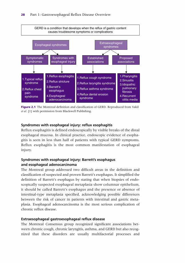
28 Part 1: Gastroesophageal Reflux Disease Overview
Syndromes with esophageal injury: reflux esophagitis Reflux esophagitis is defined endoscopically by visible breaks of the distal
esophageal mucosa. In clinical practice, endoscopic evidence of esopha-
gitis is seen in less than half of patients with typical GERD symptoms.
Reflux esophagitis is the most common manifestation of esophageal
injury.
Syndromes with esophageal injury: Barrett ’ s esophagus and esophageal adenocarcinoma The Montreal group addressed two difficult areas in the definition and
classification of suspected and proven Barrett ’ s esophagus. It simplified the
definition of Barrett ’ s esophagus by stating that when biopsies of endo-
scopically suspected esophageal metaplasia show columnar epithelium,
it should be called Barrett ’ s esophagus and the presence or absence of
intestinal-type metaplasia specified, acknowledging possible differences
between the risk of cancer in patients with intestinal and gastric meta-
plasia. Esophageal adenocarcinoma is the most serious complication of
chronic reflux disease.
Extraesophageal gastroesophageal reflux disease The Montreal Consensus group recognized significant associations bet-
ween chronic cough, chronic laryngitis, asthma, and GERD but also recog-
nized that these disorders are usually multifactorial processes and
Esophageal syndromes
Syndromes withesophageal injury
1.Pharyngitis2.Sinusitis3.Idiopathic pulmonary fibrosis4.Recurrent otitis media
1.Reflux cough syndrome
2.Reflux laryngitis syndrome
3.Reflux asthma syndrome
4.Reflux dental erosion syndrome
1.Reflux esophagitis
2.Reflux stricture
3.Barrett’s esophagus
4.Esophageal adenocarcinoma
Symptomaticsyndromes
1.Typical reflux syndrome
2.Reflux chest pain syndrome
Extraesophagealsyndromes
Establishedassociations
Proposedassociations
GERD is a condition that develops when the reflux of gastric contentcauses troublesome symptoms or complications
Figure 2.1 The Montreal definition and classification of GERD. Reproduced from Vakil
et al . [1] with permission from Blackwell Publishing.
c02.indd 28 11/15/2012 2:33:56 AM
Chapter 2: Epidemiology, Impact on Quality of Life, and Health 29
gastroesophageal reflux may be a co-factor rather than a cause. An
understanding of the various syndromes of GERD is essential for
epidemiological studies in GERD.
Epidemiology of gastroesophageal reflux disease
Studies of the epidemiology of GERD have been limited by the lack of con-
sensus as to when symptoms of heartburn or regurgitation become trou-
blesome enough to constitute a disease. Population-based studies suggest
that when symptoms of moderate intensity occur twice a week or moderate
symptoms occur once a week, quality of life drops, suggesting that the
symptoms become troublesome at these thresholds [1,4] . Unfortunately,
epidemiological studies have used other thresholds for measurement, usu-
ally weekly symptoms of heartburn, making it difficult to extrapolate these
studies to patients presenting with symptoms in clinical practice. There are
geographic differences in the prevalence of reflux disease.
Two recent systematic reviews have evaluated the prevalence of reflux
disease in different regions of the world [5,6] . The prevalence of GERD (as
defined by heartburn and/or acid regurgitation at least weekly) in North
America (19.8–20%) is similar to that in Europe (9.8–18%) [6] . The prev-
alence of GERD is lower in Asia (2.5–4.8%) [6] . In a UK database, the
incidence of GERD is estimated to be 4–5 per 1000 patient-years [7] .
Obesity, increasing age and smoking were significant risk factors. Patients
who had a diagnosis of GERD had a higher incidence of a subsequent diag-
nosis of esophageal adenocarcinoma, esophageal stricture, chronic cough,
sinusitis, and sleep problems. In this study, the mortality of subjects with a
diagnosis was higher in the first year after diagnosis but not in subsequent
years. An association with chronic obstructive airway disease has also been
reported in other studies of the same database [7] .
Children with reflux and reflux-related problems are a growing problem
in many countries. A UK database found that the incidence of GERD in
children was 0.84 per 1000 patient-years [8] . The incidence decreases from
the age of 1 year to the age of 12 years, after which it increases again,
reaching a maximum prevalence at age 16–17 years of age [8] . Children
with reflux disease often continue to have symptoms as an adult [9] .
A pediatric definition and classification of GERD has recently been
published that should help simplify epidemiological studies [10] .
Population-based studies have suggested that differences may exist bet-
ween different ethnic groups with regard to the prevalence of GERD. There
is a higher prevalence of reflux symptoms in Hispanic subjects compared to
Caucasian subjects [11] . In a multiracial population in Malaysia, Indian
ethnicity was consistently associated with reflux disease [12] . Other risk
c02.indd 29 11/15/2012 2:33:56 AM

30 Part 1: Gastroesophageal Reflux Disease Overview
factors that have been identified in a number of studies include the
presence of a hiatus hernia, a family history of reflux disease, smoking,
obesity, pregnancy, and increasing age [6] .
Obesity is a major risk factor for the development of reflux disease and
its complications. Truncal obesity raises intragastric pressure and compro-
mises the esophagogastric junction, increasing the likelihood of reflux in
patients predisposed to this disorder [13] . In a large epidemiological study
in Norway, increasing Body Mass Index (BMI) was associated with
increasing GERD symptoms [14] . There was a dose–response relationship
between increasing BMI and reflux symptoms in both men and women,
with a significantly stronger association in women. Compared with those
with a BMI less than 25, the risk of reflux was increased significantly
among those with a BMI > 35: men (odds ratio (OR) 3.3, 95% confidence
interval (CI) 2.4–4.7) and women (OR 6.3, 95% CI 4.9–8.0).
A US case–control study showed that waist circumference but not BMI
was associated with Barrett ’ s esophagus [15] . Measures of visceral adi-
posity correlate best with the risk of cancer in Barrett ’ s esophagus. Patients
with a high waist-hip ratio have approximately twice the rate of devel-
oping adenocarcinoma [16] . In a recent study of the risk of cancer in
Barrett ’ s esophagus, the adjusted odds ratios for the development of cancer
was 2.4 (95% CI 1.4–3.9) for all cases, 2.8 (95% CI 1.5–5.1) for visible
Barrett ’ s esophagus, and 4.3 (95% CI 1.9–9.9) for long segment Barrett ’ s
esophagus [16] .
The epidemiology of extraesophageal syndromes and their relationship
with reflux disease are more difficult to assess. Studies are based on associ-
ations between asthma, laryngitis, chronic obstructive airway disease and
symptoms of GERD but a causal relationship between reflux disease and
the extraesophageal syndrome cannot be inferred from such studies.
Patients with a diagnosis of chronic obstructive pulmonary disease (COPD)
are more likely to have a diagnosis of GERD compared with individuals
with no COPD diagnosis [17] . In this study, 1628 patients in the UK gen-
eral practice database were identified with a first diagnosis of chronic
obstructive airway disease and compared to 4391 patients with a first diag-
nosis of GERD. Over a 5-year follow-up, the relative risk of having GERD
diagnosed among patients with a diagnosis of COPD was 1.46 (95% CI
1.19–1.78) [17] .
A systematic review found a strong association between dental erosions
and reflux disease, particularly in children [18] . The median prevalence of
dental erosions in patients with GERD was 24% (range 5–47.5%), and
17% (range 14–87%) in children. A study of 1980 children with GERD
between ages 2 and 18 showed that compared to healthy controls, children
with GERD had a significant risk for extraesophageal complications of
GERD. GERD was a significant risk factor for sinusitis (adjusted OR 2.3,
c02.indd 30 11/15/2012 2:33:56 AM

Chapter 2: Epidemiology, Impact on Quality of Life, and Health 31
95% CI 1.7–3.2, P < 0.0001), laryngitis (OR 2.6, 95% CI 1.2–5.6, P = 0.0228),
asthma (OR 1.9, 95% CI 1.6–2.3, P < 0.0001), pneumonia (OR 2.3, 95%
CI 1.8–2.9, P < 0.0001), and bronchiectasis (OR 2.3, 95% CI 1.1–4.6,
P = 0.0193) [19] .
A study that compared 8228 hospitalized patients with laryngeal cancers
and 1912 with pharyngeal cancers to controls reported that GERD was a
significant risk factor for the development of these cancers [20] . For outpa-
tients, GERD was associated with an adjusted OR of 2.31 (95% CI 2.10–
2.53) for laryngeal cancer and adjusted OR of 1.92 for pharyngeal cancer
(95% CI 1.72–2.15).
Health-related quality of life in gastroesophageal reflux disease
Health-related quality of life (HRQOL) is defined as the patient ’ s subjective
perception of the impact of their disease and its treatment on daily life,
physical, psychological and social functioning and well-being. Health-
related quality of life can be measured by general scales such as the Short
Form (SF)-36 and the Psychological General Well-Being Scale (PGWS).
Quality of life measured by generic quality of life instruments such as the
SF-36 and the PGWS is significantly reduced in patients with GERD [4,21] .
There is a relationship between symptom severity and general quality of life
scales [4,22] . Figure 2.2 shows the relationship between symptom severity
and general quality of life as measured by the PGWS. Symptom frequency
is also related to decreases in quality of life. A recent population-based
study of patients with reflux symptoms found that 6% of subjects reported
reflux symptoms (heartburn and/or regurgitation) daily, 14% weekly and
20% less than weekly during the previous 3 months [23] . Compared to
patients with no reflux symptoms, a clinically relevant impairment of
health-related quality of life (≥ 5 points) was seen in all eight SF-36 dimen-
sions for patients with daily symptoms and in five dimensions for patients
with weekly symptoms [23] . In a study of 1011 patients with GERD in
Germany and Sweden, health-related quality of life was measured using
the EUROQol5. Patients with GERD had a significant impairment in quality
of life and the impairment was related to the severity of symptoms [24] .
Disease-specific quality of life instruments have been developed for
GERD and are helpful in assessing the response to treatment. Quality of life
has been assessed using the QOLRAD, a disease-specific instrument devel-
oped for reflux disease. In adolescents with GERD, quality of life is impaired
and treatment with a proton pump inhibitor improves all domains of
quality of life [22] . Improvements in symptoms are associated with improve-
ments in quality of life and overall satisfaction with treatment [22] .
c02.indd 31 11/15/2012 2:33:57 AM

Figure 2.2 The relationship between the severity of heartburn (a) and acid regurgita-
tion (b) measured by the score on the Gastrointestinal Symptoms Rating Scale (GSRS)
and quality of life measured by the Psychological General Well-Being (PGWB) Scale.
As symptom severity increases, well-being decreases. Normal values are 103 and a
clinically relevant change is a decrease in 5 points to 98. Reproduced from Wiklund
et al . [4] with permission from Blackwell Publishing.
120
100
80
60
40
221
(n = 1039)(n = 194)(n = 136) (n = 32) (n = 10) (n = 7) (n = 2)2 3
Heartburn severity (GSRS score)
Individual score Linear regression
4 5 6 7Worst possible
score
Wel
l-bei
ng(P
GW
B s
core
)
Best possiblescore
98
132
(a)
105.6 97.7 90.6 83.9
Mean
71.2 64.0 77.8
120
100
80
60
40
221
(n = 1113)(n = 173)(n = 102) (n = 19) (n = 7) (n = 4) (n = 2)2 3
Acid regurgitation severity (GSRS score)
Individual score Linear regression
4 5 6 7Worst possible
score
Wel
l-bei
ng(P
GW
B s
core
)
Best possiblescore
98
132
(b)
105.5 95.0 87.1 69.8
Mean
74.5 78.0 72.5
c02.indd 32 11/15/2012 2:33:57 AM

Chapter 2: Epidemiology, Impact on Quality of Life, and Health 33
Health economic implications
The Agency for Health Care Policy and Research has conducted a survey of
the inpatient costs of treating GERD in the USA from 1998 to 2005 [25] .
Hospitalizations with either a primary or secondary GERD diagnosis
increased by 216% from 995,402 in 1998 to 3,141,965 in 2005. Adult hos-
pitalizations with a primary GERD diagnosis decreased by 2.4% from 77,783
in 1998 to 75,888 in 2005. However, for pediatric GERD, stays with a pri-
mary GERD diagnosis increased by 42% for infants and by 84% for children
age 2–17. Eight out of every 1000 hospitalizations with a GERD diagnosis
had Barrett ’ s esophagus. The average cost per hospital stay with a primary
GERD diagnosis was $5616 in 1998 and $6545 in 2005. The total national
hospital costs for all hospitalizations with a primary GERD diagnosis
increased by 22% from $509 million in 1998 to $622 million in 2005 [22] .
In Canada, in 2004–5, the Canadian healthcare system spent a mean of
$6915 per patient for the 7554 patients who had a primary diagnosis
of diseases of the esophagus and associated complications, for a total of
$52,235,910 [26] .
The outpatient costs of treating GERD are significant. Managed care orga-
nizations spend large amounts of money for the treatment of acid-related
disorders. The IMS tracks the costs and trends of drug use in the USA [27] .
In 2009, proton pump inhibitors were the third largest therapeutic class in
sales. Proton pump inhibitors sales totaled $13.6 billion, and dispensed
prescription volume for this therapeutic class rose 5% [27] . Treatment of
GERD can reduce healthcare costs. A managed care study of 41,895 patients
with GERD, conducted between 2000 and 2005, found that patients who
were started on a proton pump inhibitor had a reduction in costs and
patients who were compliant with proton pump inhibitor therapy had
lower overall downstream costs than those who were non-compliant due
to lower outpatient visits and hospital admissions [27,28] .
Impact of gastroesophageal reflux disease on work productivity A systematic review of the impact of GERD on work productivity was reported
in 2006 [29] . The studies were conducted in seven countries and involved
eight different study populations. The results of these studies show that
absences from work related to GERD were infrequent and ranged from < 1%
to 7%. Presenteeism, which is defined as reduced work productivity while pre-
sent at work, was much more common, ranging from 6% to 42%. Assuming
a 40-h work week and average US wages for 2005, the mean productivity loss
per employee with GERD is estimated to range from $62–430/week [29] .
Treatment of GERD restores quality of life and work productivity (29).
c02.indd 33 11/15/2012 2:33:57 AM
34 Part 1: Gastroesophageal Reflux Disease Overview
Cost-effectiveness of medical therapy Many patients with reflux disease stop and start treatment rather than
taking it continuously. This form of therapy (on-demand therapy) may
make sense in patients without esophagitis. In one study, 424 patients with
endoscopy-negative reflux disease were randomized to placebo or proton
pump inhibitor (omeprazole 20 mg or omeprazole 10 mg) on demand [29] .
Of those patients randomized to on-demand therapy with omeprazole
20 mg a day, 83% were satisfactorily maintained over the 6-month time
frame. The mean number of omeprazole capsules used per day was 0.43,
suggesting that the total medication use was reduced by approximately
50%. In a study of esomeprazole therapy, 320 patients with endoscopy-
negative reflux disease who had complete symptom resolution after
4 weeks of therapy with either esomeprazole 20 mg or omeprazole 20 mg
were randomized to receive esomeprazole 20 mg on-demand or placebo
on-demand for 6 months [30] . Medication intake was measured using
electronic chips embedded in the caps of the medication containers. On
average, esomeprazole was taken once every 3 days and 86% of patients
were managed with on-demand therapy compared to 49% in the placebo
group. These data suggest that on-demand therapy is effective and can
substantially reduce costs of medical therapy [31] .
Economics of medical versus surgical therapy Several cost models have compared medical therapy to fundoplication.
Bojke et al . evaluated the costs of laparoscopic fundoplication using a
Markov model and UK cost estimates [32] . The incremental cost per
additional quality-adjusted life-year (QALY) for surgery compared to
medical therapy medical management was £180. The cost-effectiveness
of surgery could not be demonstrated and the probability that surgery
was cost-effective using a generally accepted threshold of £30,000 per
QALY was only 60%. A study by Arguedas et al . [33] is interesting
because the authors revised a previously published analysis that had
suggested that surgery was cost-effective. When they reexamined the
model which covered a 10-year time frame, using newly available data
from randomized controlled trials and adding quality of life as an out-
come measure, they found that medical therapy had a total per-patient
cost of $8798 and 4.59 quality-adjusted life-years, while the surgical
approach was more expensive at $10,475 and less effective 4.55 quality-
adjusted life-years. Cost models are sensitive to the cost of the proton
pump inhibitor, which has dropped significantly in many countries with
the availability of low-cost generic proton pump inhibitors. They are
also sensitive to the success rates assumed for surgery and medical
therapy (effectiveness).
c02.indd 34 11/15/2012 2:33:57 AM

Chapter 2: Epidemiology, Impact on Quality of Life, and Health 35
A recent randomized controlled trial suggests that newer proton pump
inhibitors may be more effective than surgery at 5 years. Estimated
remission rates at 5 years were 92% (95% CI 89–96%) in the esomepra-
zole group and 85% (95% CI 81–90%) in the fundoplication group
( P = 0.048) [34] .
Evolution and natural progression of gastroesophageal reflux disease
The evolution and natural progression of GERD are unclear because
longitudinal studies were not performed before acid inhibitory therapy
became generally available. The natural history of GERD is influenced by
changes in the populations being studied. For example, obesity has been
increasing in Western populations and is increasing in some countries in
Asia. A recent study evaluated healthy women in a longitudinal study in
the USA [35] . Compared with women who had a BMI of 20.0–22.4, the
multivariate odds ratio for frequent symptoms was 2.92 (95% CI 2.35–
3.62) for a BMI of 30.0–34.9, and 2.93 (95% CI 2.24–3.85) for a BMI of
35.0 or more. An interesting aspect of this study was that symptoms of
GERD regress if a substantial weight loss occurs. A recent systematic review
examined the effects of age on the severity of reflux disease [36] . Age is
associated with a decrease in GERD symptom prevalence but more severe
patterns of acid reflux and more severe esophagitis.
An aspect of the natural history that remains controversial is the rela-
tionship between non-erosive reflux disease and reflux esophagitis. The
ProGERD study, a naturalistic study that follows patients on treatment in
Germany, suggests that patients may transition between non-erosive
reflux disease and erosive esophagitis [37] . Twenty-five percent of patients
who had non-erosive reflux disease at baseline progressed to Los Angeles
Grade A or B esophagitis and 0.6% to Los Angeles Grade C or D; 1.6% of
patients who had Los Angeles Grade A or B disease progressed to Los
Angeles Grade C or D and 61% regressed to non-erosive reflux disease;
42% of patients who had Los Angeles Grade C or D disease regressed to
Grade A or B disease and 50% regressed to non-erosive reflux disease.
Patients with Grade C or D esophagitis were at greatest risk of developing
Barrett ’ s esophagus (5.8%) compared with patients with grade A and B
disease (1.4%) and patients with non-erosive reflux disease (0.5%). Some
complications such as esophageal strictures have declined in frequency in
the last decade and this change corresponds with the availability of proton
pump inhibitors [38] .
Over the last 25 years there has been a remarkable change in the epide-
miology of esophageal cancer in Western countries. The incidence of
c02.indd 35 11/15/2012 2:33:57 AM

36 Part 1: Gastroesophageal Reflux Disease Overview
esophageal adenocarcinoma has been rising rapidly in some countries
although the absolute annual risk of developing adenocarcinoma remains
low at 0.4% [39] . Esophageal adenocarcinoma has been causally linked to
GERD and is the final stage in the evolution of reflux disease. In a large
epidemiological study in Sweden, the risk of esophageal adenocarcinoma
was increased (OR 7.7) in patients suffering from long-standing reflux
symptoms [40] . A higher frequency of symptoms (greater than three times
per week) and a long duration (greater than 10–20 years) of symptoms
increased the risk (OR 16–20).
Summary
Gastroesophageal reflux disease is widely prevalent throughout the world
and its prevalence is increasing in Asia. GERD has a number of symptom
associations, including esophageal adenocarcinoma, laryngeal and pha-
ryngeal cancer, chronic obstructive airway disease and laryngitis. The cost
of treating GERD poses an important economic burden to most healthcare
organizations. GERD is associated with a marked impairment in quality of
life. It is also associated with loss of work productivity and this is primarily
due to decreased effectiveness while at work rather than absences from
work. Treatment with proton pump inhibitors restores quality of life and
improves work productivity.
References
1 Vakil N , van Zanten SV , Kahrilas P , Dent J , Jones R , Global Consensus Group. The
Montreal definition and classification of gastroesophageal reflux disease:a global
evidence-based consensus . Am J Gastroenterol 2006 ; 101 ( 8 ): 1900 – 20 .
2 Kahrilas PJ , Shaheen NJ , Vaezi MF , American Gastroenterological Association
Institute. Technical review on the management of gastroesophageal reflux disease .
Gastroenterology 2008 ; 135 ( 4 ): 1392 – 413 .
3 Dent J , Vakil N , Jones R , et al . Accuracy of the diagnosis of GORD by questionnaire,
physicians ad a trial of proton pump inhibitor treatment:the Diamond study . Gut
2010 ; 59 ( 6 ): 714 – 21 .
4 Wiklund I , Carlsson J , Vakil N . Gastro-esophageal reflux symptoms and well-being in
a random sample of the general population of a Swedish community . Am J
Gastroenterol 2006 ; 101 ( 1 ): 18 – 28 .
5 Wong BC , Kinoshita Y . Systematic review on epidemiology of gastroesophageal reflux
disease in Asia . Clin Gastroenterol Hepatol 2006 ; 4 ( 4 ): 398 – 407 .
6 Dent J , El-Serag H , Wallander M , Johansson S . Epidemiology of gastroesophageal
reflux disease:a systematic review . Gut 2005 ; 54 : 710 – 17 .
7 Ruigómez A , García Rodríguez LA , Wallander MA , Johansson S , Graffner H , Dent J .
Natural history of gastro-oesophageal reflux disease diagnosed in general practice .
Aliment Pharmacol Ther 2004 ; 20 ( 7 ): 751 – 60 .
c02.indd 36 11/15/2012 2:33:57 AM

Chapter 2: Epidemiology, Impact on Quality of Life, and Health 37
8 Ruigomez A , Wallander AM , Lundborg P , Johansson S , Garcia Rodriguez L .
Gastroesophageal reflux disease in children and adults in primary care . Scand
J Gastroenterol 2010 ; 45 : 139 – 46 .
9 El-Serag HB , Gilger M , Carter J , Genta RM , Rabeneck L . Childhood GERD is a risk factor
for GERD in adolescents and young adults . Am J Gastroenterol 2004 ; 99 ( 5 ): 806 – 12 .
10 Sherman PM , Hassall E , Fagundes-Neto U , et al . A global, evidence-based consensus
on the definition of gastroesophageal reflux disease in the pediatric population . Am
J Gastroenterol 2009 ; 104 ( 5 ): 1278 – 95 .
11 Yuen E , Romney M , Toner RW , et al . Prevalence, knowledge and care patterns for
gastro-esophageal reflux disease in United States minority populations Aliment
Pharmacol Ther 2010 ; 32 ( 5 ): 645 – 54 .
12 Rosaida MS , Goh KL . Gastro-esophageal reflux disease, reflux esophagitis and non-
erosive reflux disease in a multiracial Asian population: a prospective endoscopy
based study . Eur J Gastroenterol Hepatol 2004 ; 16 ( 5 ): 495 – 501 .
13 Pandolfino JE , El-Serag HB , Zhang Q , Shah N , Ghosh SK , Kahrilas PJ . Obesity: a
challenge to esophagogastric junction integrity . Gastroenterology 2006 ; 130 ( 3 ): 639 – 49 .
14 Nilsson M , Johnsen R , Ye W , Hveem K , Lagergren J . Obesity and estrogen as risk
factors for gastroesophageal reflux symptoms. JAMA 2003 2; 290 ( 1 ): 66 – 72 .
15 Corley DA , Kubo A , Levin TR , et al . Abdominal obesity and body mass index as risk
factors for Barrett ’ s esophagus . Gastroenterology 2007 ; 133 ( 1 ): 34 – 41 .
16 Edelstein ZR , Farrow DC , Bronner MP , Rosen SN , Vhan TL . Central adiposity and risk
of Barrett ’ s esophagus . Gastroenterology 2007 ; 133 ( 2 ): 403 – 11 .
17 García Rodríguez LA , Ruigómez A , Martín–Merino E, Johansson S , Wallander MA .
Relationship between gastroesophageal reflux disease and COPD in UK primary care .
Chest 2008 ; 134 ( 6 ): 1223 – 30 .
18 Pace F , Pallotta S , Tonini M , Vakil N , Bianchi Porro G. Systematic review: gastro-
esophageal reflux disease and dental lesions . Aliment Pharmacol Ther 2008 ;
27 ( 12 ): 1179 – 86 .
19 El-Serag HB , Gilger M , Kuebeler M , Rabeneck L . Extraesophageal associations of
gastroesophageal reflux disease in children without neurologic defects .
Gastroenterology 2001 ; 121 ( 6 ): 1294 – 9 .
20 El-Serag HB , Hepworth EJ , Lee P , Sonnenberg A . Gastroesophageal reflux disease is a risk
factor for laryngeal and pharyngeal cancer . Am J Gastroenterol 2001 ; 96 ( 7 ): 2013 – 18 .
21 Revicki DA , Wood M , Maton PN , Sorensen S . The impact of gastroesophageal reflux
disease on health-related quality of life . Am J Med 1998 ; 104 ( 3 ): 252 – 8 .
22 Revicki DA , Zodet MW , Joshua-Gotlib S , Levine D , Crawley JA . Health-related
quality of life improves with treatment-related GERD symptom resolution after
adjusting for baseline severity . Health Qual Life Outcomes 2003 ; 1 : 73 .
23 Ronkainen J , Aro P , Storskrubb T , et al . Gastro-oesophageal reflux symptoms and
health-related quality of life in the adult general population – the Kalixanda study .
Aliment Pharmacol Ther 2006 ; 23 ( 12 ): 1725 – 33 .
24 Jones R , Horbach S , Sander P , Rydén-Bergsten T . Heartburn in patients with gastro-
oesophageal reflux disease in Germany and Sweden: a study on patients’ burden of
disease . Pharmacoeconomics 2003 ; 21 ( 15 ): 1091 – 102 .
25 Zhao Y , Encinosa W . Gastroesophageal reflux disease ( GERD ) hospitalizations in 1998 and 2005:
Statistical Brief #44 . Washington, DC : Agency for Health Care Policy and Research , 2006 .
26 Fedorak RN , Veldhuyzen van Zanten S, Bridges R . Canadian Digestive Health
Foundation Public Impact Series: gastro-esophageal reflux disease in Canada: inci-
dence, prevalence, and direct and indirect economic impact . Can J Gastroenterol
2010 ; 24 ( 7 ): 431 – 4 .
c02.indd 37 11/15/2012 2:33:57 AM

38 Part 1: Gastroesophageal Reflux Disease Overview
27 Anonymous . Cost of drugs . Available at: www.imshealth.com/portal/site/ims/menu-
item.d248e29c86589c9c30e81c033208c22a/?vgnextoid=d690a27e9d5b7210VgnVC
M100000ed152ca2RCRD&vgnextfmt=default .
28 Gosselin A , Luo R , Lohoues H , et al . The impact of proton pump inhibitor compliance
on health-care resource utilization and costs in patients with gastroesophageal reflux
disease . Value Health 2009 ; 12 ( 1 ): 34 – 9 .
29 Wahlquist P , Reilly M , Barkun A . Systematic review: the impact of gastro-esophageal
reflux on work productivity . Aliment Pharmacol Ther 2006 ; 24 : 259 – 72 .
30 Lind T , Havelund T , Lundell L , et al . On demand therapy with omeprazole for the
long-term management of patients with heartburn without esophagitis – a placebo-
controlled randomized trial . Aliment Pharmacol Ther 1999 ; 13 : 907 – 14 .
31 Talley N , Lauritsen K , Tunturi-Hihnala H , et al . Esomeprazole 20 mg maintains
symptom control in endoscopy-negative GERD. A randomized placebo-controlled
trial of on-demand therapy for 6 months . Aliment Pharmacol Ther 2001 ; 15 : 347 – 54 .
32 Bojke L , Hornby E , Sculpher M . A comparison of the cost effectiveness of pharmaco-
therapy or surgery (laparoscopic fundoplication) in the treatment of GORD .
Pharmacoeconomics 2007 ; 25 : 829 – 41 .
33 Arguedas MR , Heudebert GR , Klapow JC , et al . Re-examination of the cost- effectiveness
of surgical versus medical therapy in patients with gastroesophageal reflux disease: the
value of long-term data collection . Am J Gastroenterol 2004 ; 99 : 1023 – 8 .
34 Galmiche JP , Hatlebakk J , Attwood S , et al ., LOTUS Trial Collaborators. Laparoscopic
antireflux surgery vs esomeprazole treatment for chronic GERD: the LOTUS
randomized clinical trial . JAMA 2011 ; 305 ( 19 ): 1969 – 77 .
35 Jacobson BC , Somers SC , Fuchs CS , Kelly CP , Camargo CA Jr. Body mass index and
symptoms of gastroesophageal reflux in women . N Engl J Med 2006 ; 354 ( 22 ): 2340 – 8 .
36 Becher A , Dent J . Systematic review: ageing and gastro-esophageal reflux disease
symptoms, oesophageal function and reflux esophagitis . Aliment Pharmacol Ther
2011 ; 33 : 442 – 54 .
37 Labenz J , Nocon M , Lind T , et al . Prospective follow-up data from the ProGERD study
suggest that GERD is not a categorial disease . Am J Gastroenterol 2006 ; 101 ( 11 ): 2457 – 62 .
38 Guda NM , Vakil N . Proton pump inhibitors and the time trends for esophageal
dilation . Am J Gastroenterol 2004 ; 99 ( 5 ): 797 – 800 .
39 De Jonge PJ , van Blankenstein M , Looman CW , Casparie MK , Meijer GA , Kuipers EJ .
Risk of malignant progression in patients with Barrett ’ s oesophagus: a Dutch nation-
wide cohort study . Gut 2010 ; 59 ( 8 ): 1030 – 6 .
40 Lagergren J , Bergström R , Lindgren A , Nyrén O . Symptomatic gastroesophageal reflux
as a risk factor for esophageal adenocarcinoma . N Engl J Med 1999 ; 340 ( 11 ): 825 – 31 .
c02.indd 38 11/15/2012 2:33:57 AM

39
Practical Manual of Gastroesophageal Reflux Disease, First Edition.
Edited by Marcelo F. Vela, Joel E. Richter and John E. Pandolfino.
© 2013 John Wiley & Sons, Ltd. Published 2013 by John Wiley & Sons, Ltd.
CHAPTER 3
Overview of the Tools for the Diagnosis of Gastroesophageal Reflux Disease Nicolas A. Villa and Marcelo F. Vela Section of Gastroenterology , Baylor College of Medicine, and Michael E. DeBakey
VA Medical Center , Houston , TX , USA
Key points • Endoscopy is indicated when there are alarm features such as dysphagia, weight loss,
and anemia.
• The presence of erosive esophagitis on endoscopy provides robust evidence of
gastroesophageal reflux disease, but endoscopy is normal in the majority of patients.
• Ambulatory reflux monitoring is the gold standard for diagnosing gastroesophageal
reflux disease.
• In some patients, the reported symptoms are due to non-gastroesophageal reflux
disease causes; in this context, a negative evaluation for gastroesophageal reflux
disease can direct the diagnostic and treatment efforts toward other causes.
Potential pitfalls • Gastroesophageal reflux disease can be diagnosed by symptoms and a “positive
proton pump inhibitor test” in some settings, but the limited sensitivity and
specificity of this approach as a diagnostic intervention need to be kept in mind.
• Gastroesophageal reflux disease cannot be diagnosed by barium esophagram or
esophageal manometry.
• A normal endoscopy does not exclude gastroesophageal reflux disease.
Introduction
Gastroesophageal reflux disease (GERD) is a very common clinical problem.
Heartburn or acid regurgitation is experienced on a weekly basis by nearly
20% of the US population, with an annual prevalence of up to 59% [1] .
c03.indd 39 11/15/2012 2:33:43 AM

40 Part 1: Gastroesophageal Reflux Disease Overview
Diagnostic testing options for GERD include the assessment of symptoms
(e.g. patient history or GERD questionnaires), the response to a trial of acid
suppression (generally with a proton pump inhibitor – PPI), evaluation for
acid-related damage to the esophageal mucosa (endoscopy), or determina-
tion of pathological reflux on prolonged ambulatory monitoring with pH
or impedance pH. In clinical practice, a diagnosis of GERD is often made
based upon symptom presentation along with a good response to a trial
of PPI therapy. While the PPI trial is a reasonable and simple option in
the appropriate setting (e.g. primary care), it is important to remember its
limitations in terms of sensitivity and specificity.
Beyond taking a careful history for typical symptoms of GERD, a number
of validated questionnaires are available including specific symptom scales,
GERD-related quality of life scales, and combined instruments to assess
both symptoms and quality of life. Questionnaires can be helpful but they
generally have similar specificity and sensitivity compared to clinical exam-
ination by a gastroenterologist and therefore, to date, their role has often
been limited to clinical trials.
Upper endoscopy is indicated when the clinical presentation includes
alarm features such as dysphagia, bleeding or weight loss. Endoscopy pro-
vides a robust diagnosis of GERD when erosive esophagitis is present, but
this is only found in approximately 30% of untreated patients [2] and
even a smaller proportion of patients after treatment with a PPI. Random
biopsies to look for evidence of GERD in those with normal endoscopy are
not recommended because conventional histology has poor performance
for a diagnosis of GERD.
Direct measurement of gastroesophageal reflux through pH monitoring
(catheter based or wireless) can establish whether there is a pathological
amount of acid reflux and if there is an association between symptoms and
reflux episodes. Impedance-pH monitoring enables measurement of both
acid and non-acid reflux (with a pH > 4); the latter may be clinically impor-
tant in patients with persistent symptoms despite acid-suppressive therapy.
Evaluation should always begin with a careful history. A PPI trial is a
reasonable option for diagnosing GERD in patients with typical symptoms.
GERD cannot be diagnosed by barium esophagram or esophageal mano-
metry. Endoscopy should be performed when alarm features are present.
Objective documentation of GERD by endoscopy or reflux monitoring is
mandatory before antireflux surgery. Evaluation by endoscopy and reflux
monitoring is useful in patients with extraesophageal symptoms, and those
in whom symptoms persist despite acid suppression. It is important to note
that in some patients the reported symptoms may be due to causes other
than GERD, including functional disorders; in these patients, endoscopy
and reflux monitoring can be valuable tools to exclude GERD. The advantages,
disadvantages, and clinical use of tests to diagnose GERD are discussed in
c03.indd 40 11/15/2012 2:33:43 AM
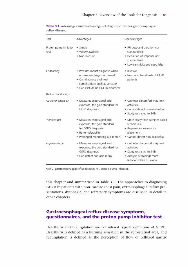
Chapter 3: Overview of the Tools for Diagnosis 41
this chapter and summarized in Table 3.1 . The approaches to diagnosing
GERD in patients with non-cardiac chest pain, extraesophageal reflux pre-
sentations, dysphagia, and refractory symptoms are discussed in detail in
other chapters.
Gastroesophageal reflux disease symptoms, questionnaires, and the proton pump inhibitor test
Heartburn and regurgitation are considered typical symptoms of GERD.
Heartburn is defined as a burning sensation in the retrosternal area, and
regurgitation is defined as the perception of flow of refluxed gastric
Table 3.1 Advantages and disadvantages of diagnostic tests for gastroesophageal
reflux disease.
Test Advantages Disadvantages
Proton pump inhibitor
test
• Simple
• Widely available
• Non-invasive
• PPI dose and duration not
standardized
• Definition of response not
standardized
• Low sensitivity and specificity
Endoscopy • Provides robust diagnosis when
erosive esophagitis is present
• Can diagnose and treat
complications such as stricture
• Can exclude non-GERD disorders
• Invasive
• Normal in two-thirds of GERD
patients
Reflux monitoring
Catheter-based pH • Measures esophageal acid
exposure, the gold standard for
GERD diagnosis
• Catheter discomfort may limit
activities
• Cannot detect non-acid reflux
• Study restricted to 24 h
Wireless pH • Measures esophageal acid
exposure, the gold standard
for GERD diagnosis
• Better tolerability
• Prolonged monitoring (up to 96 h)
• More costly than catheter-based
techniques
• Requires endoscopy for
placement
• Cannot detect non-acid reflux
Impedance pH • Measures esophageal acid
exposure, the gold standard for
GERD diagnosis
• Can detect non-acid reflux
• Catheter discomfort may limit
activities
• Study restricted to 24 h
• Analysis of tracings more
laborious than pH alone
GERD, gastroesophageal reflux disease; PPI, proton pump inhibitor.
c03.indd 41 11/15/2012 2:33:43 AM
42 Part 1: Gastroesophageal Reflux Disease Overview
contents into the pharynx or mouth [3] . Heartburn and regurgitation are
the most reliable symptoms for making a history-based diagnosis of GERD
but are far from perfect in this respect. When heartburn and regurgitation
are the dominant symptoms, they have high sensitivity but poor specificity
[4] . While heartburn is the most typical symptom of GERD, it may also be
present in patients with other esophageal disorders such as eosinophilic
esophagitis or achalasia. Furthermore, some patients may have no organic
cause for heartburn, a condition that is termed “functional heartburn” that
is discussed in detail in a later chapter. Dysphagia may be part of the clinical
presentation in a patient with GERD and is considered to be an alarm
symptom that warrants endoscopic evaluation to exclude a complication,
including malignancy. Chest pain may also be experienced by GERD
patients, but this symptom requires thorough evaluation for a cardiac
cause before GERD is considered.
More recently, the spectrum of clinical presentations attributed to GERD
has moved beyond the typical esophageal symptoms of heartburn and
regurgitation, and now includes various extraesophageal manifestations
that focus on respiratory and laryngeal symptoms. Several epidemiological
studies have identified an association between GERD and asthma [5] ,
chronic cough [6] , and laryngitis [7] . However, causality cannot be inferred
from these studies. Therefore, the Montreal Consensus recognized established
associations between GERD and asthma, chronic cough, and laryngitis,
while acknowledging that these disorders frequently have a multifactorial
etiology and gastroesophageal reflux may be a co-factor rather than a cause.
The Montreal Consensus also recognized the rarity of extraesophageal
syndromes occurring in isolation without concomitant typical symptoms of
GERD [3] . The evaluation of GERD in patients with non- cardiac chest pain,
cough, and laryngitis is discussed in detail in later chapters.
A number of questionnaires have been developed for GERD. Available
validated instruments include specific symptom scales, quality of life scales,
and those which combine the two. A comprehensive review of these
instruments is beyond the scope of this chapter, especially because they are
not widely used in routine clinical practice. Questionnaires can be helpful,
but have similar specificity and sensitivity as clinical examination by a gas-
troenterologist and therefore, to date, their role has been in screening large
numbers of patients by non-specialists or as part of clinical trials [8] .
Although, as noted above, the sensitivity and specificity of heartburn
and regurgitation are not perfect for making a diagnosis of GERD, it is not
necessary to conduct a diagnostic evaluation in all patients with typical
symptoms and no alarm features. In this context, a 2–4-week trial of acid
suppression with a PPI is a non-invasive, simple, and reasonable option for
supporting a diagnosis of GERD. If the patient has a clear response to
therapy, it can be assumed that GERD is present. That said, the potential
c03.indd 42 11/15/2012 2:33:44 AM

Chapter 3: Overview of the Tools for Diagnosis 43
shortcomings of this approach have to be kept in mind. The manner in
which this “PPI test” is administered is not standardized. Studies evalu-
ating this approach have used differing PPI doses (once versus twice daily),
variable duration of treatment (from 1 to 4 weeks or even longer), and
different definitions of what constitutes a positive test (for instance, 50%
improvement as opposed to 100% improvement). In addition, a meta-
analysis of several studies that evaluated the diagnostic capability of a short
course (1–4 weeks) of PPI compared to other tests found a sensitivity of
78% and specificity of 54% for the PPI test [9] .
Esophagram and esophageal manometry
Barium esophagram and esophageal manometry deserve a brief discussion
because they are often used in the work-up of patients with symptoms
suggestive of GERD. A diagnosis of GERD cannot be made based upon the
results of an esophagram or esophageal manometry.
While a high-quality barium esophagram with double contrast can reveal
signs of esophagitis, the overall sensitivity of this test for esophagitis is very
low [10] . The finding of barium reflux from stomach to esophagus, with or
without provocative maneuvers, may be absent in patients with GERD; fur-
thermore, reflux of barium may be found with provocative maneuvers in
some healthy subjects [11,12] . An esophagram can be helpful when a patient
has dysphagia, as it may reveal a structural abnormality such as a stricture or
ring. However, GERD cannot be diagnosed based upon esophagram.
Esophageal manometry will often reveal impaired peristalsis in GERD
patients, and some but certainly not all GERD patients may have a hypo-
tensive lower esophageal sphincter [13] . However, these findings are
not specific and a diagnosis of GERD cannot be made based upon man-
ometric findings. Manometry is useful to guide placement of transnasal
pH or impedance pH catheters for ambulatory reflux monitoring. In
addition, esophageal manometry should always be performed prior to
antireflux surgery. While the presence of decreased peristalsis has not
been found to reliably predict postfundoplication dysphagia [14] ,
manometry should be performed to exclude aperistalsis due to a
scleroderma-like esophagus or achalasia, as both of these conditions
represent contraindications to fundoplication.
Endoscopy and esophageal biopsies
Upper endoscopy enables direct visualization of the esophageal mucosa
in patients with symptoms suggestive of GERD. Endoscopy may reveal
c03.indd 43 11/15/2012 2:33:44 AM
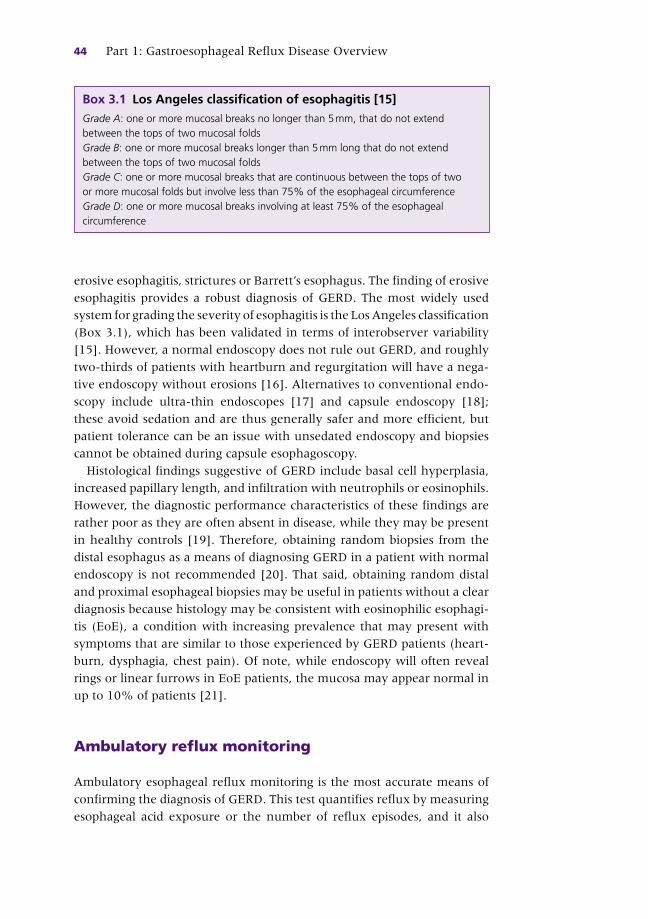
44 Part 1: Gastroesophageal Reflux Disease Overview
erosive esophagitis, strictures or Barrett ’ s esophagus. The finding of erosive
esophagitis provides a robust diagnosis of GERD. The most widely used
system for grading the severity of esophagitis is the Los Angeles classification
(Box 3.1 ), which has been validated in terms of interobserver variability
[15] . However, a normal endoscopy does not rule out GERD, and roughly
two-thirds of patients with heartburn and regurgitation will have a nega-
tive endoscopy without erosions [16] . Alternatives to conventional endo-
scopy include ultra-thin endoscopes [17] and capsule endoscopy [18] ;
these avoid sedation and are thus generally safer and more efficient, but
patient tolerance can be an issue with unsedated endoscopy and biopsies
cannot be obtained during capsule esophagoscopy.
Histological findings suggestive of GERD include basal cell hyperplasia,
increased papillary length, and infiltration with neutrophils or eosinophils.
However, the diagnostic performance characteristics of these findings are
rather poor as they are often absent in disease, while they may be present
in healthy controls [19] . Therefore, obtaining random biopsies from the
distal esophagus as a means of diagnosing GERD in a patient with normal
endoscopy is not recommended [20] . That said, obtaining random distal
and proximal esophageal biopsies may be useful in patients without a clear
diagnosis because histology may be consistent with eosinophilic esophagi-
tis (EoE), a condition with increasing prevalence that may present with
symptoms that are similar to those experienced by GERD patients (heart-
burn, dysphagia, chest pain). Of note, while endoscopy will often reveal
rings or linear furrows in EoE patients, the mucosa may appear normal in
up to 10% of patients [21] .
Ambulatory reflux monitoring
Ambulatory esophageal reflux monitoring is the most accurate means of
confirming the diagnosis of GERD. This test quantifies reflux by measuring
esophageal acid exposure or the number of reflux episodes, and it also
Box 3.1 Los Angeles classification of esophagitis [15] Grade A : one or more mucosal breaks no longer than 5 mm, that do not extend between the tops of two mucosal folds Grade B : one or more mucosal breaks longer than 5 mm long that do not extend between the tops of two mucosal folds Grade C : one or more mucosal breaks that are continuous between the tops of two or more mucosal folds but involve less than 75% of the esophageal circumference Grade D : one or more mucosal breaks involving at least 75% of the esophageal circumference
c03.indd 44 11/15/2012 2:33:44 AM

Chapter 3: Overview of the Tools for Diagnosis 45
enables an examination of the temporal relationship between reflux epi-
sodes and reported symptoms. Reflux monitoring was performed through
catheter-based pH studies for many years. More recently, there have been
two major developments in this field: the wireless pH capsule which allows
catheter-free monitoring, and impedance pH measurement, a catheter-
based technique that enables detection of acid and non-acid reflux (i.e.
with pH > 4). Additional techniques that will not be discussed in this
chapter include esophageal bilirubin monitoring [22] , which is not widely
used in clinical practice, and a new transnasal catheter for pharyngeal
monitoring that can measure pH in either liquid or aerosolized droplets
[23] . Data to support the use of the latter are very limited and the test is
not used routinely.
Catheter-based pH monitoring Conventional ambulatory pH monitoring is performed by a transnasal cath-
eter that records esophageal pH over a 24-h period, with a pH electrode posi-
tioned 5 cm above the proximal border of the lower esophageal sphincter.
A reflux episode is defined by a drop in pH to below 4.0. Various measures
can be derived from a pH monitoring study, including percent time with pH
< 4 (upright, recumbent, and total time), number of reflux episodes, number
of reflux episodes longer than 5 min, and longest reflux episode [24] . Of
these, the total percentage of time with pH < 4 is felt to be the most useful
indicator of pathological acid reflux [25] . A positive pH test can establish a
diagnosis of GERD in a patient with normal endoscopy; it may also help to
confirm or exclude GERD in patients who do not respond to PPI therapy, as
explained in greater detail in Chapter 7. While the sensitivity and specificity
of this test are above 90% in some studies [26,27] , other data point to lower
sensitivity [28] . Additional shortcomings of pH monitoring include the possi-
bility of changes in patient behavior related to having a transnasal catheter,
such as altered eating habits and decreased activity [29] . Finally, pH moni-
toring does not allow assessment of non-acid reflux (i.e. with pH > 4), that
may occur when the gastric contents are buffered (during pharmacological
acid suppression, in the postprandial period or in patients with atrophic
gastritis).
Bravo pH monitoring A new technology has been developed recently to enable wireless moni-
toring of esophageal pH, obviating the need for a transnasal catheter. The
system uses a small recording capsule that is endoscopically attached to the
distal esophagus, and transfers pH data via radiofrequency signals to an
external recording device. Wireless pH monitoring has similar and possibly
improved accuracy compared to catheter-based pH monitoring [30] . In
addition, it is better tolerated by patients [31] , thus allowing for testing
c03.indd 45 11/15/2012 2:33:44 AM

46 Part 1: Gastroesophageal Reflux Disease Overview
under more physiological conditions with fewer limitations on diet and
activity [32] . An additional advantage of this approach is the capability for
prolonged monitoring, with a standard study of 48 h that can be extended
to up to 96 h [33] . In a study of 48-h wireless pH monitoring in 44 healthy
subjects and 41 GERD patients, improved sensitivity in distinguishing con-
trols from GERD patients was achieved by using the data from the worst of
the 2 days [34] .
The wireless pH monitoring system has some limitations, including early
capsule detachment (an infrequent problem), chest pain or discomfort,
increased cost and, like catheter-based pH monitoring, the inability to
detect non-acid reflux, which may be clinically relevant in some patients.
Impedance- pH monitoring Intraesophageal impedance, determined by measuring electrical conduc-
tivity across a pair of closely spaced electrodes within the esophageal
lumen, is dependent on the conductivity of the material through which
the current travels. By placing a series of conducting electrodes in a cath-
eter that spans the length of the esophagus, changes in impedance can be
recorded in response to movement of intraesophageal material in either
antegrade or retrograde direction [35] . Because the esophageal mucosa,
air, and any given bolus material (i.e. swallowed food, saliva, refluxed
gastric contents) each produce a different change in impedance, the tech-
nique enables very detailed characterization of gastroesophageal reflux
episodes, including composition (air, liquid or mixed), proximal extent
(height), velocity, and clearance time.
Impedance-pH is currently the most accurate and detailed method for
measuring gastroesophageal reflux [36] . During combined impedance and
pH monitoring, impedance is used to detect retrograde bolus movement
(i.e. reflux), while pH measurement establishes the acidity of the reflux
episode (acid if pH < 4.0, non-acid otherwise). Non-acid reflux may be
further classified as either weakly acidic (pH ≥ 4 but < 7) or weakly alkaline
(pH ≥ 7). In this chapter, non-acid reflux refers to any reflux with pH ≥ 4.
The main advantage of this method is the capability to detect non-acid
reflux, which may be relevant in some patients, especially those with
symptoms that persist despite PPI therapy. Examples of acid and non-acid
reflux are shown in Figure 3.1 .
Ambulatory-impedance-pH monitoring can be performed with different
catheters that incorporate a varying number of impedance measuring
segments and pH electrodes in different configurations. A typical catheter
has a pH electrode positioned 5 cm above the manometrically determined
lower esophageal sphincter (similar to conventional pH testing), along
with six or more impedance-measuring segments (each composed of two
metal ring electrodes usually spaced 2 cm apart) to detect impedance
c03.indd 46 11/15/2012 2:33:44 AM

Figure 3.1 Acid and non-acid reflux detected by impedance-pH monitoring.
Impedance changes in six measuring segments spanning the esophagus, and pH
changes from a single sensor in the distal esophagus are shown. (a) Acid reflux:
a typical impedance reflux pattern with sequential decreases in impedance starting in
the distal esophagus and progressing upward in retrograde direction; this reflux episode
is associated with a pH fall to below 4.0 ( arrow ). (b) Non-acid reflux: typical impedance
reflux pattern; during this episode esophageal pH remains above 4. The patient
reported heartburn during this reflux episode.
Impe
danc
e
pH 4
(a)
Impe
danc
e
pH 4
(b)
Hea
rt b
urn
c03.indd 47 11/15/2012 2:33:44 AM
48 Part 1: Gastroesophageal Reflux Disease Overview
changes along variable lengths of the esophagus. Catheter placement is
transnasal and thus similar to that of conventional pH monitoring.
Ambulatory impedance-pH monitoring is conventionally performed over a
24-h period.
Assessment of reflux with impedance-pH is reproducible [37] and normal
values for ambulatory 24-h impedance-pH monitoring are now well
established [38,39] . While impedance pH is considered the most accurate
method for reflux detection, the clinical indications for its use and its role
in managing GERD patients are still evolving because the clinical relevance
of non-acid reflux is awaiting confirmation by high-quality trials. Studies
have shown that non-acid reflux can cause symptoms indistinguishable
from those caused by acid reflux [40] . Furthermore, non-acid reflux can be
treated by pharmacological inhibition of transient lower esophageal
sphincter relaxations [41] or through fundoplication [42] . However, it is
very important to note that there is a paucity of high-quality controlled
studies examining the benefit of treating non-acid reflux. In addition, it
must be remembered that non-acid reflux occurs predominantly in the
postprandial period (when food buffers the stomach contents) or during
pharmacological acid suppression. The potential relevance of non-acid
reflux is discussed in greater detail in Chapter 7.
Impedance-pH monitoring has some limitations. It is catheter based
which, like conventional pH testing, can result in patient discomfort and
change in behavior on the day of testing. In addition to these catheter-
based difficulties, low baseline impedance (seen in some patients with
severe reflux) can make the tracing difficult to read. Finally, interpretation
of impedance-pH tracings may be more time consuming compared to
pH-metry.
Symptom association studies during reflux monitoring As mentioned earlier, the total percentage of time with pH < 4 is felt to be
the most useful indicator of pathological acid reflux [25] . The number of
reflux episodes detected by impedance can also serve as a measure of
abnormal reflux, but the clinical utility of this approach is unclear and out-
come studies proving that treating patients based upon this endpoint is
beneficial are lacking.
Beyond establishing the presence of pathological reflux, ambulatory
reflux monitoring with any of the available techniques (catheter or wireless
pH, impedance-pH) may be used to determine whether the patient ’ s
symptoms are due to reflux. The two methods most commonly used to
evaluate the temporal association between reflux episodes and symptoms
are the Symptom Index (SI) [43] and the Symptom Association Probability
(SAP) [44] . The SI is defined as the percentage of symptom events that are
temporally related to a reflux episode (number of reflux-related symptom
c03.indd 48 11/15/2012 2:33:44 AM
Chapter 3: Overview of the Tools for Diagnosis 49
events/total number of symptom events × 100%). An SI of 50% is consid-
ered positive, meaning that the symptom is related to reflux. Although this
value was derived from receiver operating characteristic (ROC) curves that
found this threshold to be sensitive and specific for heartburn [45] , the SI
is used to analyze any symptom that may be attributed to GERD. The SAP
is calculated by dividing the pH or impedance-pH tracing in 2-min seg-
ments and determining whether a reflux episode and/or a symptom
occurred in each 2-min segment. A 2 × 2 contingency table with the
number of segments with and without symptoms and with and without
reflux is built; the probability that a positive association between reflux
and symptoms occurred by more than chance is evaluated through a mod-
ified chi-square test, with an SAP greater than 95% considered positive.
A detailed discussion of the advantages and disadvantages of the SI and
the SAP is beyond the scope of this chapter, but both have methodological
shortcomings that have been reviewed elsewhere [46] . Of note, both
methods rely on precise and timely symptom recording by the patient,
along with accurate reflux detection by the testing device. Furthermore,
prospective data to validate the ability of these symptom association mea-
sures to predict response to treatment are scarce. Nevertheless, they are
useful if their limitations are kept in mind, and they are commonly
employed in clinical practice. In terms of patient management, a strongly
positive SI or SAP may suggest the need for a therapeutic intervention and
a negative result supports the notion that the patient ’ s symptoms are
unlikely to be due to reflux. However, these indices should not be used in
isolation and other reflux monitoring parameters as well as the patient ’ s
presentation have to be incorporated into the decision-making process.
References
1 Locke GR III , Talley NJ , Fett SL et al . Prevalence and clinical spectrum of gastroesoph-
ageal reflux: a population-based study in Olmsted County, Minnesota . Gastroenterology
1997 ; 112 : 1448 – 56 .
2 Lind T , Havelund T , Carlsson R , et al . Heartburn without esophagitis: efficacy of
omeprazole therapy and features determining therapeutic response . Scand J
Gastroenterol 1997 ; 32 : 974 – 9 .
3 Vakil N , van Zanten SV , Kahrilas P , et al . The Montreal definition and classification of
gastroesophageal reflux disease: a global evidence-based consensus . Am J Gastroenterol
2006 ; 101 : 1900 .
4 Klauser AG , Shcindelbeck NE , Muller-Lissner SA . Symptoms in gastro-oesophageal
reflux disease . Lancet 1990 ; 335 : 205 .
5 Havemann BD , Henderson CA , El-Serag HB . The association between gastro-oesoph-
ageal reflux disease and asthma: a systematic review . Gut 2007 ; 56 : 1654 – 64 .
6 Irwin RS , Curley FJ , French CL . Chronic cough. The spectrum and frequency of
causes, key components of the diagnostic evaluation, and outcome of specific therapy .
Am Rev Respir Dis 1990 ; 141 : 640 – 7 .
c03.indd 49 11/15/2012 2:33:44 AM

50 Part 1: Gastroesophageal Reflux Disease Overview
7 El-Serag HB , Sonnenberg A . Comorbid occurrence of laryngeal or pulmonary disease
with esophagitis in United States military veterans . Gastroenterology 1997 ; 113 : 755 – 60 .
8 Stanghellini V , Arsmtrong D , Monnikes H , Bardhan KD . Systematic review: do we need
a new gastroesophageal diseases questionnaire? Digestion 2007 ; 75 ( suppl 1 ): 3 – 16 .
9 Numans ME , Lau J , de Wit NK , Bonis PA . Short-term treatment with proton-pump
inhibitors as a test for gastroesophageal reflux disease . Ann Intern Med 2004 ;
140 : 518 – 52 .
10 Johnston BT , Troshinsky MB , Castell JA , et al . Comparison of barium radiology with
esophageal pH monitoring in the diagnosis of gastroesophageal reflux disease . Am
J Gastroenterol 1996 ; 91 : 1181 – 5 .
11 Thompson JK , Koehler RE , Richter JE . Detection of gastroesophageal reflux: value of
barium studies compared with 24-hr pH monitoring . Am J Roentgenol 1994 ; 162 : 621 – 6 .
12 Richter JE , Castell DO . Gastroesophageal reflux. Pathogenesis, diagnosis, and
therapy . Ann Intern Med 1982 ; 97 : 93 – 103 .
13 Savarino E , Gemignani L , Pohl D , et al . Oesophageal motility and bolus transit abnor-
malities increase in parallel with the severity of gastro-oesophageal reflux disease .
Aliment Pharmacol Ther 2011 ; 34 : 476 – 86 .
14 Shaw J , Bornman PC , Callanan MD , Beckingham IJ , Metz DC . Long-term outcome
of laparoscopic Nissen and laparoscopic Toupet fundoplication for gsatroesophageal
reflux disease: a prospective, randomized trial . Surg Endosc 2010 ; 24 : 924 – 32 .
15 Lundell LR , Dent J , Bennett JR , et al . Endoscopic assessment of oesophagitis: clinical
and functional correlates and further validation of the Los Angeles classification . Gut
1999 ; 45 : 172 – 80 .
16 Johnsson F , Joelsson B , Gudmundsson K , et al . Symptoms and endoscopic findings
in the diagnosis of gastroesophageal reflux disease . Scand J Gastroenterol 1987 ;
22 : 714 – 18 .
17 Mokhashi MS , Wildi SM , Glenn TF , et al . A prospective, blinded study of diagnostic
esophagoscopy with a superthin, stand-alone, battery-powered esophagoscope . Am
J Gastroenterol 2003 ; 98 : 2383 .
18 Eliakim R , Sharma VK , Yassin K , et al . A prospective study of the diagnostic accuracy
of PillCam ESO esophageal capsule endoscopy versus conventional upper endoscopy
in patients with chronic gastroesophageal reflux diseases . J Clin Gastroenterol
2005 ; 39 : 572 – 8 .
19 Nandurkar S , Talley NJ , Martin CJ , Ng T , Adams S . Esophageal histology does not
provide additional useful information over clinical assessment in identifying reflux
patients presenting for esophagogastroduodenoscopy . Dig Dis Sci 2000 ; 45 : 217 – 24 .
20 Tytgat G . The value of esophageal histology in the diagnosis of gastroesophageal
reflus diseas in patients with heartburn and normal endoscopy . Curr Gastroenterol
Rep 2008 ; 10 : 231 – 4 .
21 Prasad GA , Talley NJ , Romero Y , et al . Prevalence and predictive factors of eosino-
philic esophagitis in patients presenting with dysphagia: a prospective study . Am
J Gastroenterol 2007 ; 102 : 2627 .
22 Vaezi MF , Richter JE . Role of acid and duodenogastroesophageal reflux in
gastroesophageal reflux disease . Gastroenterology 1996 ; 111 : 1192 – 9 .
23 Sun G , Muddana S , Slaughter JC , et al . A new pH catheter for laryngopharyngeal
reflux: normal values . Laryngoscope 2009 ; 119 : 1639 – 43 .
24 Johnson LF , DeMeester TR . Twenty-four-hour pH monitoring of the distal esoph-
agus. A quantitative measure of gastroesophageal reflux . Am J Gastroenterol
1974 ; 62 : 325 – 32 .
25 Pandolfino JE , Vela MF . Esophageal reflux monitoring . Gastrointest Endosc
2009 ; 69 : 917 – 30 .
c03.indd 50 11/15/2012 2:33:44 AM

Chapter 3: Overview of the Tools for Diagnosis 51
26 Jamieson JR , Stein HJ , DeMeester TR , et al . Ambulatory 24-h esophageal pH moni-
toring: normal values, optimal thresholds, specificity, sensitivity, and reproducibility .
Am J Gastroenterol 1992 ; 87 : 1102 – 11 .
27 Richter JE , Bradley LA , DeMeester TR , Wu WC . Normal 24-hr ambulatory esopha-
geal pH values. Influence of study center, pH electrode, age, and gender . Dig Dis Sci
1992 ; 37 : 849 – 56 .
28 Schlesinger PK , Donahue PE , Schmid B , et al . Limitations of 24-hour intraesophageal
pH monitoring in the hospital setting . Gastroenterology 1985 ; 894 : 797 – 804 .
29 Fass R , Hell R , Sampliner RE , et al . Effect of ambulatory 24-h esophageal pH moni-
toring on reflux-provoking activities . Dig Dis Sci 1999 ; 44 : 2263 – 9 .
30 Pandolfino J , Zhang Q , Schreiner M , et al . Acid reflux event detection using the
Bravo™ wireless vs the Slimline™ catheter pH systems: why are the numbers so
different? Gut 2005 ; 54 : 1687 – 92 .
31 Wenner J , Jonsson F , Johansson J , Oberg S . Wireless esophageal pH monitoring is
better tolerated than the catheter-based technique: results from a randomized cross-
over trial . Am J Gastroenterol 2007 ; 102 : 239 – 45 .
32 Ward EM , Devault KR , Bouras EP , et al . Successful oesophageal pH monitoring with
a catheter-free system . Aliment Pharmacol Ther 2004 ; 19 : 449 – 54 .
33 Hirano I , Zhang Q , Pandolfino JE , et al . Four-day Bravo pH capsule monitoring with and
without proton pump inhibitor therapy . Clin Gastroenterol Hepatol 2005 ; 3 : 1083 – 8 .
34 Pandolfino JE , Richter JE , Ours T , et al . Ambulatory esophageal pH monitoring using
a wireless system . Am J Gastroenterol 2003 ; 984 : 740 – 9 .
35 Vela MF . Non-acid reflux: detection by multichannel intraluminal impedance and
pH, clinical significance and management . Am J Gastroenterol 2009 ; 104 : 277 – 80 .
36 Sifrim D , Castell D , Dent J , et al . Gastro-oesophageal reflux monitoring: reflux and
consensus report on detection and definitions of acid, non-acid, and gas reflux . Gut
2004 ; 53 : 1024 – 31 .
37 Bredenoord AJ , Weusten BL , Timmer R , Smout AJ . Reproducibility of multichannel
intraluminal electrical impedance monitoring of gastroesophageal reflux . Am J
Gastroenterol 2005 ; 100 : 265 – 9 .
38 Shay S , Tutuian R , Sifrim D , et al . Twenty-four hour ambulatory simultaneous
impedance and pH monitoring: a multicenter report of normal values from 60 healthy
volunteers . Am J Gastroenterol 2004 ; 99 : 1037 – 43 .
39 Zerbib F , des Varannes SB , Roman S , et al . Normal values and day-to-day variability
of 24-h ambulatory oesophageal impedance-pH monitoring in a Belgian-French
cohort of healthy subjects . Aliment Pharmacol Ther 2005 ; 22 : 1011 – 21 .
40 Vela MF , Camacho-Lobato L , Srinivasan R , et al . Intraesophageal impedance and pH
measurement of acid and non-acid reflux: effect of omeprazole . Gastroenterology
2001 ; 96 : 647 – 55 .
41 Vela MF , Tutuian R , Katz PO , et al . Baclofen decreases acid and non-acid post- prandial
gastro-oesophageal reflux measured by combined multichannel intraluminal impedance
and pH . Aliment Pharmacol Ther 2003 ; 17 : 243 – 51 .
42 Frazzoni M , Conigliaro R , Melotti G . Reflux parameters as modified by laparoscopic
fundoplication in 40 patients with heartburn/regurgitation persisting despite PPI
therapy: a study using impedance-pH moniotoring . Dig Dis Sci 2011 ; 56 : 1099 – 106 .
43 Wiener GJ , Richter JE , Copper JB , et al . The symptom index: a clinically important
parameter of ambulatory 24-hour esophageal pH monitoring . Am J Gastroenterol
1988 ; 83 : 358 – 61 .
44 Weusten BL , Roelofs JM , Akkermans LM , et al . The symptom-association probability:
an improved method for symptom analysis of 24-hour esophageal pH data .
Gastroenterology 1994 ; 107 : 1741 – 5 .
c03.indd 51 11/15/2012 2:33:44 AM

52 Part 1: Gastroesophageal Reflux Disease Overview
45 Singh S , Richter JE , Bradley LA , Haile JM . The symptom index. Differential useful-
ness in suspected acid-related copmlaints of heartburn and chest pain . Dig Dis Sci
1993 ; 34 : 309 – 16 .
46 Connor J , Richter J . Increasing yield also increases false positives and best serves to
exclude GERD . Am J Gastroenterol 2006 ; 101 : 460 – 3 .
c03.indd 52 11/15/2012 2:33:45 AM

53
Practical Manual of Gastroesophageal Reflux Disease, First Edition.
Edited by Marcelo F. Vela, Joel E. Richter and John E. Pandolfino.
© 2013 John Wiley & Sons, Ltd. Published 2013 by John Wiley & Sons, Ltd.
CHAPTER 4
Overview of Gastroesophageal Reflux Disease Treatments Sabine Roman and Peter J. Kahrilas Division of Gastroenterology , Northwestern University , Chicago , IL , USA
Key points • Proton pump inhibitors are the dominant treatment for gastroesophageal reflux
disease.
• Proton pump inhibitors are progressively less effective in achieving the endpoints of
healing esophagitis, relieving heartburn, treating regurgitation, and treating
extraesophageal syndromes.
• The therapeutic gain with reflux inhibitors (gamma-amino butyric acid B agonists) is
modest for typical gastroesophageal reflux disease symptoms.
• Reducing visceral hypersensitivity should be considered in patients with persistent
heartburn or chest pain despite proton pump inhibitor treatment.
Potential pitfalls • Indications for laparoscopic fundoplication must be balanced against its risks.
• In the absence of proton pump inhibitor response, an alternative diagnosis should be
considered for suspected gastroesophageal reflux disease symptoms.
Introduction
Gastroesophageal reflux disease (GERD) is defined as “a condition which
develops when the reflux of stomach contents causes troublesome
symptoms and/or complications” [1] . Typical esophageal GERD symptoms
include heartburn and regurgitation. Additional esophageal symptoms are
dysphagia and chest pain. Extraesophageal or “atypical” symptoms with
an established association with GERD on the basis of population-based
studies are chronic cough, asthma, and laryngitis. However, these have
potential etiologies other than GERD and in the absence of a concomitant
c04.indd 53 11/15/2012 2:33:30 AM
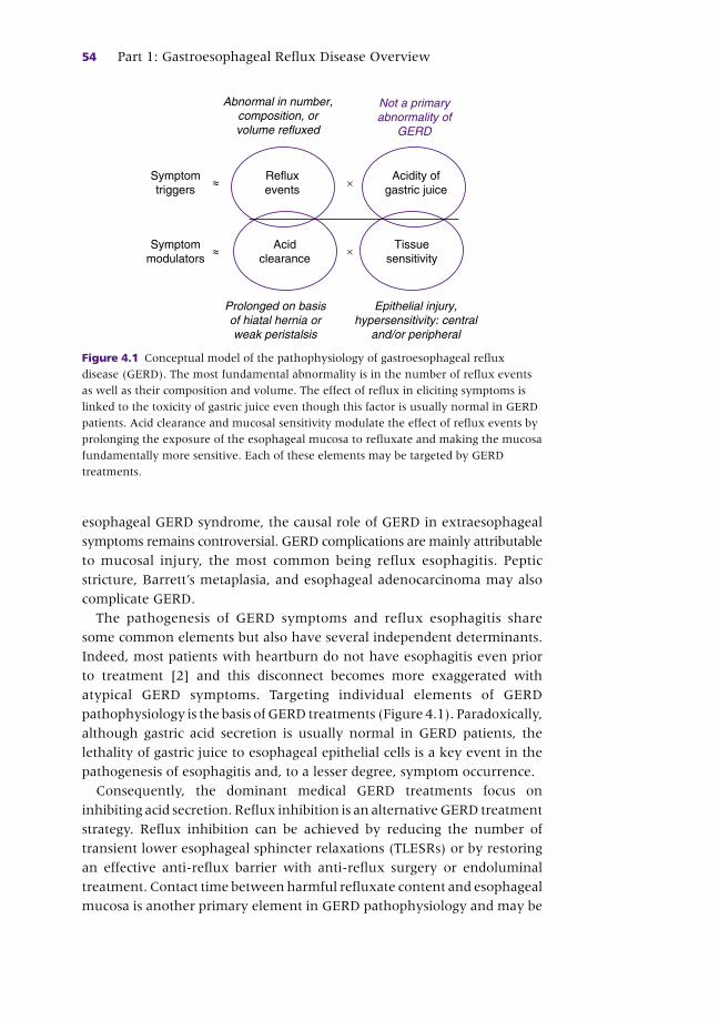
54 Part 1: Gastroesophageal Reflux Disease Overview
esophageal GERD syndrome, the causal role of GERD in extraesophageal
symptoms remains controversial. GERD complications are mainly attributable
to mucosal injury, the most common being reflux esophagitis. Peptic
stricture, Barrett ’ s metaplasia, and esophageal adenocarcinoma may also
complicate GERD.
The pathogenesis of GERD symptoms and reflux esophagitis share
some common elements but also have several independent determinants.
Indeed, most patients with heartburn do not have esophagitis even prior
to treatment [2] and this disconnect becomes more exaggerated with
atypical GERD symptoms. Targeting individual elements of GERD
pathophysiology is the basis of GERD treatments (Figure 4.1 ). Paradoxically,
although gastric acid secretion is usually normal in GERD patients, the
lethality of gastric juice to esophageal epithelial cells is a key event in the
pathogenesis of esophagitis and, to a lesser degree, symptom occurrence.
Consequently, the dominant medical GERD treatments focus on
inhibiting acid secretion. Reflux inhibition is an alternative GERD treatment
strategy. Reflux inhibition can be achieved by reducing the number of
transient lower esophageal sphincter relaxations (TLESRs) or by restoring
an effective anti-reflux barrier with anti-reflux surgery or endoluminal
treatment. Contact time between harmful refluxate content and esophageal
mucosa is another primary element in GERD pathophysiology and may be
Abnormal in number,composition, orvolume refluxed
Refluxevents
Acidity ofgastric juice
Acidclearance
×
×
Epithelial injury,hypersensitivity: central
and/or peripheral
Symptomtriggers
≈
≈Symptommodulators
Prolonged on basisof hiatal hernia orweak peristalsis
Tissuesensitivity
Not a primaryabnormality of
GERD
Figure 4.1 Conceptual model of the pathophysiology of gastroesophageal reflux
disease (GERD). The most fundamental abnormality is in the number of reflux events
as well as their composition and volume. The effect of reflux in eliciting symptoms is
linked to the toxicity of gastric juice even though this factor is usually normal in GERD
patients. Acid clearance and mucosal sensitivity modulate the effect of reflux events by
prolonging the exposure of the esophageal mucosa to refluxate and making the mucosa
fundamentally more sensitive. Each of these elements may be targeted by GERD
treatments.
c04.indd 54 11/15/2012 2:33:31 AM

Chapter 4: Overview of GERD Treatments 55
attributable to the effects of a hiatal hernia or weak peristalsis [3] . Although
pharmaceutical therapies have minimal effect in this domain, lifestyle
modifications such as avoiding postprandial recumbency and anti-reflux
surgery to eliminate hiatus hernia target this mechanism. Finally, visceral
sensitivity is increasingly recognized as an important modulator of
symptom severity and, when abnormal, should be considered as an
alternative target for GERD treatment. The aim of this overview is to
broadly consider alternative GERD treatments and their differential
effectiveness in the context of varied GERD presentation and severity.
Lifestyle modifications
There are many recommendations regarding lifestyle modifications as
GERD therapy. Generally, these fall into three categories:
• avoid food that may lead to reflux presumably by relaxing the lower
esophageal sphincter (coffee, alcohol, chocolate, fatty foods)
• avoid acidic foods that may precipitate heartburn by a direct irritative
effect on the esophageal mucosa (citrus, carbonated drinks, spicy foods)
• adopt behaviors that may reduce esophageal acid exposure by reducing
the occurrence of reflux and/or enhancing the process of acid clearance
(weight loss, smoking cessation, raising the head of bed, and avoiding
recumbent position for 2–3 h after meals).
Although evidence supporting these recommendations is generally weak
[4] , they should be encouraged to the extent to which they seem relevant to
the individual patient. For instance, patients with regurgitation and heartburn
in the recumbent position should be advised to elevate the head of the bed
and to avoid eating for the 2–3-h period before going to bed. Similarly,
someone who consistently experiences heartburn after ingestion of alcohol,
coffee or any specific food will benefit from avoidance of these items.
Obesity and weight control merit special attention because
accumulating evidence suggests this to be one of the root causes of the
GERD epidemic of the past two decades. Epidemiological data suggest a
dose-dependent relationship between increasing Body Mass Index (BMI)
and frequent reflux symptoms [5] . However, the benefit of weight loss
on GERD symptoms has not been demonstrated in rigorous clinical trials
[6–8]. Nonetheless, if the development of troublesome heartburn
paralleled weight gain in an individual patient, it is very reasonable to
propose weight loss as an intervention that may prevent the need for
continuous acid suppressive therapy. This is particularly true in view of
the added health benefits of weight loss beyond reflux disease and
because obesity has been shown to be an independent risk factor for
Barrett ’ s esophagus and esophageal adenocarcinoma.
c04.indd 55 11/15/2012 2:33:31 AM

56 Part 1: Gastroesophageal Reflux Disease Overview
Acid suppression
Acid neutralization with antacid and pharmacologically inhibiting gastric
acid secretion are cornerstones of GERD therapy. The most potent drugs
are the proton pump inhibitors (PPIs), which covalently bind with gastric
H+/K + ATPase to block the final common pathway for acid secretion.
Histamine-2 receptor antagonists (H 2 RA) competitively block histamine-
stimulated acid secretion, making them less potent than PPIs and giving
them a duration of action limited by their serum half-life. Antacids neu-
tralize acid or acidic food without having any effect on subsequent acid
secretion. It is well established that PPIs are more effective than H 2 RAs in
healing esophagitis and in relieving heartburn [9,10] . Similarly, H 2 RAs are
more effective than placebo in the same applications.
Returning to the principles of Figure 4.1 , it follows that acid suppression
in general and PPI therapy specifically do not “cure” reflux disease. Rather,
PPIs treat GERD in an indirect fashion. It follows that PPI efficacy will vary
widely across disease manifestations, dependent on the degree to which
those manifestations are attributable to acid. The most responsive disease
manifestation is esophagitis, in which case esophageal epithelial injury is
directly attributable to gastric acid and eliminating acid facilitates mucosal
healing with close to 100% effectiveness. Similarly, dysphagia, which is
reported in about one-third of patients with esophagitis without stricture
or malignancy, resolves in 83% with PPI therapy [11] . However, esophagi-
tis is the best-case scenario for PPI efficacy. Heartburn is the next-best case,
but already the therapeutic gain associated with PPI therapy is sharply
reduced from that observed with esophagitis (Figure 4.2 ). Furthermore,
there is a 10–20% difference in therapeutic gain for heartburn dependent
on whether or not it occurs in the context of erosive esophagitis or non-
erosive reflux disease. Conceptually, this is because the specificity of heart-
burn as an acid-induced symptom is less in the absence of esophagitis. This
limitation is even more evident in the case of regurgitation wherein the
therapeutic gain from PPI therapy is less than 20% [12] . Consequently,
persistent regurgitation despite PPI therapy is a major contributor to
treatment failure.
The linkage between PPI response and acid-mediated symptoms is par-
ticularly well illustrated in the case of the treatment of unexplained chest
pain. A recent meta-analysis examined the responsiveness of chest pain to
PPI therapy in randomized controlled trials that utilized pH monitoring as
an objective means of distinguishing patients with or without abnormal
acid reflux [13] . Based on the data from six randomized controlled trials,
the therapeutic gain of > 50% improvement with PPIs relative to placebo
was achieved in 56–85% in GERD-positive patients and only 0–17% in
c04.indd 56 11/15/2012 2:33:31 AM
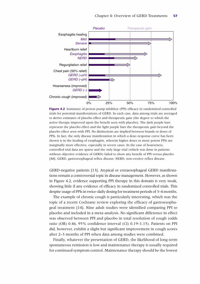
Chapter 4: Overview of GERD Treatments 57
GERD-negative patients [13] . Atypical or extraesophageal GERD manifesta-
tions remain a controversial topic in disease management. However, as shown
in Figure 4.2 , evidence supporting PPI therapy in this domain is very weak,
showing little if any evidence of efficacy in randomized controlled trials. This
despite usage of PPIs in twice-daily dosing for treatment periods of 3–4 months.
The example of chronic cough is particularly interesting, which was the
topic of a recent Cochrane review exploring the efficacy of gastroesopha-
geal treatment [14] . Nine adult studies were identified comparing PPI to
placebo and included in a meta-analysis. No significant difference in effect
was observed between PPI and placebo in total resolution of cough (odds
ratio (OR) 0.46, 95% confidence interval (CI) 0.19–1.15). Patients on PPI
did, however, exhibit a slight but significant improvement in cough scores
after 2–3 months of PPI when data among studies were combined.
Finally, whatever the presentation of GERD, the likelihood of long-term
spontaneous remission is low and maintenance therapy is usually required
for continued symptom control. Maintenance therapy should be the lowest
0%
Chronic cough (improved)
Hoarseness (improved)
Chest pain (50% relief)
Regurgitation relief
Heartburn relief
Esophagitis healing
GERD (–)
GERD (–pH)GERD (+pH)
NERDEsophagitis
Mild
Placebo Therapeutic gain
Servere
25% 50% 75% 100%
Figure 4.2 Summary of proton pump inhibitor (PPI) efficacy in randomized controlled
trials for potential manifestations of GERD. In each case, data among trials are averaged
to derive estimates of placebo effect and therapeutic gain (the degree to which the
active therapy improved upon the benefit seen with placebo). The dark purple bars
represent the placebo effect and the light purple bars the therapeutic gain beyond the
placebo effect seen with PPI. No distinctions are implied between brands or doses of
PPIs. In fact, the only disease manifestation in which a dose–response curve has been
shown is in the healing of esophagitis, wherein higher doses or more potent PPIs are
marginally more effective, especially in severe cases. In the case of hoarseness,
controlled trial data are sparse and the only large trial (which was done in patients
without objective evidence of GERD) failed to show any benefit of PPI versus placebo
[48] . GERD, gastroesophageal reflux disease; NERD, non-erosive reflux disease.
c04.indd 57 11/15/2012 2:33:31 AM
58 Part 1: Gastroesophageal Reflux Disease Overview
PPI dose necessary for symptom relief. Regardless of physicians’ instruc-
tions, most patients do this independently, adopting on-demand or inter-
mittent dosing of PPIs as required for symptom control [4] .
Reflux inhibition
Substantial experimental evidence suggests that TLESRs are the dominant
mechanism of reflux in most GERD patients [15] . Increased compliance of
the esophagagastric junction (EGJ), as occurs in GERD, is associated with
increased opening and flow across the relaxed sphincter, making proximal
reflux and regurgitation more likely. Consequently, pharmacological
inhibition of TLESRs is an attractive therapeutic target for GERD and, con-
ceptually, might be more effective than PPIs in treating regurgitation.
Baclofen, a GABA B agonist, inhibits the vagal pathway for TLESRs
both centrally and peripherally [16] . However, since the drug crosses
the blood–brain barrier, neurological side-effects (somnolence, dizziness,
drowsiness) limit its use for GERD in clinical practice. Therefore, novel
GABA B agonists have been developed in an attempt to limit these side-
effects. Experimentally, arbaclofen placarbil, a pro-drug of the pharmaco-
logically active R -isomer of balclofen, decreases the number of postprandial
reflux events [17] . Similarly, lesogaberan, a novel GABA B agonist, has
been shown to decrease postprandial TLESRs [18] . Preliminary studies
have also shown GABA B agonists to be superior to placebo in controlling
typical symptoms of GERD [17,19] . Nevertheless, larger scale studies
have thus far failed to show significant improvement in heartburn or
regurgitation with these drugs. In symptomatic GERD patients who failed
to respond to PPI therapy, the proportion of patients who respond to reflux
inhibition appeared modest and not specific for heartburn or regurgitation.
In a recent randomized trial including 244 patients with persistent
heartburn and/or regurgitation despite PPI therapy, lesogaberan, used as
add-on therapy, increased the number of heartburn-free days by 14% and
the number of regurgitation-free days by 13% compared to placebo [20] .
As for the effect of GABA B agonists on extraesophageal symptoms of
GERD, this may be difficult to evaluate. Baclofen inhibits cough via a
central mechanism [21] , making the role of reflux inhibition difficult to
differentiate in chronic cough patients with GERD.
Enhancing acid clearance
Patients with symptomatic GERD exhibit longer volume and acid esophageal
clearance times [22] . Mechanistically, this is attributable to both diminished
c04.indd 58 11/15/2012 2:33:31 AM
Chapter 4: Overview of GERD Treatments 59
peristaltic function and the effects of hiatus hernia. Of the two, the hiatus
hernia effect is probably dominant, especially when in a supine posture
[23,24] . Thus, limiting the contact time between the refluxate and the
esophageal mucosa may be proposed to reduce the occurrence of symptoms.
Lifestyle measures such as head of bed elevation and avoidance of
recumbency after meals target this mechanism but it is also a key principal
behind attempts at treating GERD with prokinetics and mucosal protectants.
Prokinetics Ideally, a prokinetic would enhance esophageal clearance by enhancing
peristalsis. Unfortunately, no such drugs are currently available. An alternative
approach is to promote gastric emptying which may have the secondary
consequence of reducing the occurrence of TLESR. Metoclopramide may
improve gastric emptying [25] , leading to its proposed use in GERD, especially
if accompanied by measurably delayed gastric emptying. However, there
are no high-quality data supporting the use of metoclopramide as either
monotherapy or adjunctive therapy in esophageal or suspected extraesopha-
geal GERD syndromes. Additionally, considering the toxicity profile of the
drug, the current recommendation is against the use of metoclopramide in
GERD because the potential risks exceed the potential benefits [4] .
Mucosal protection Decreasing esophageal mucosa permeability to luminal contents may
reduce the toxic effect of the gastric refluxate on esophageal mucosa.
Rebapimide has been shown to increase the gastric epithelial barrier [26] ; it
may also exert its effect on esophageal mucosa. Consistent with this
hypothesis, the combination of rebamipide and lansoprazole 15 mg was
more effective than lansoprazole 15 mg alone in maintaining long-term
symptom relief in patients with Los Angeles Grade A and B esophagitis [27] .
Stimulating secretion of mucosal protective factors may be another new
therapy in GERD. Bicarbonates, mucin, epidermal growth factor (EGF),
transforming growth factor alpha (TGF-alpha), and prostaglandin E2 are
present in saliva and esophageal secretions; they may promote mucosal
healing. Tegaserod, a serotonin 5-hydroxytryptamine 4 receptor agonist,
has been shown to increase the volume of salivary and esophageal secre-
tions in patients with GERD [28] . It particularly increased bicarbonate and
EGF secretion. In a randomized trial including 88 patients with chronic
constipation and GERD or dyspepsia, the combination of tegaserod plus
esomeprazole induced complete relief of heartburn in 85% of patients
versus 40% for esomeprazole alone and 47% for tegaserod alone ( P = 0.012)
[29] . However, although these results seemed promising, tegaserod is no
longer commercially available. Nonetheless, the serotonin pathway and
stimulation of protective factor secretion may be a target for future therapy.
c04.indd 59 11/15/2012 2:33:31 AM

60 Part 1: Gastroesophageal Reflux Disease Overview
Visceral hypersensitivity
Some patients with erosive esophagitis and, especially, non-erosive reflux
disease exhibit hypersensitivity to esophageal stimuli. This can be demon-
strated by balloon distension in the esophagus or acid perfusion (Bernstein
test). Hypersensitive patients have a diminished threshold for perceiving
these stimuli and a reduced threshold for experiencing pain compared to
healthy volunteers [30] . The same observations have been made in patients
with functional chest pain [31] . In extreme cases, patients with hypersen-
sitivity perceive the normal passage of food or fluid through the esophagus
as uncomfortable.
Low-dose antidepressants Antidepressants may modulate esophageal sensitivity at the central ner-
vous system and/or sensory afferent level, potentially benefitting symp-
tomatic patients. Low-dose tricyclic antidepressants have been shown to
be effective in patients with chest pain after incomplete response to PPI
[32] . Trazodone, a serotonin reuptake inhibitor, was more effective than
placebo in patients with esophageal symptoms (chest pain, dysphagia,
heartburn, and/or regurgitation) associated with esophageal contraction
abnormalities [33] . Citalopram, another selective serotonin reuptake
inhibitor, significantly increased the threshold for perception and discom-
fort after balloon distension in healthy volunteers [34] . It also prolonged
the duration of esophageal acid perfusion required to induce heartburn.
Consequently, these medications may be useful to alleviate esophageal
discomfort and heartburn in the subset of GERD patients with hypersensi-
tivity. A recent placebo-controlled trial of citalopram in patients with pH-
impedance findings suggestive of hypersensitivity supports this concept
[35] . However, thus far there are no large studies that evaluate antidepres-
sants in GERD patients.
Acupuncture In a series of 30 patients who failed PPI once daily, adding acupuncture
was significantly better in controlling acid regurgitation and heartburn
than doubling the PPI dose [36] . These results are promising and acupunc-
ture may represent an alternative therapy in PPI non-responders.
Hypnotherapy Response to PPI treatment can be modulated by the level of psychological
distress [37] . Consequently, a therapy which reduces psychological distress
may be beneficial in some patients who have an inadequate response
to PPI. Hypnotherapy has been proposed as such an alternative therapy,
c04.indd 60 11/15/2012 2:33:31 AM

Chapter 4: Overview of GERD Treatments 61
especially for patients with atypical GERD symptoms. In a randomized trial
including 28 patients with non-cardiac chest pain, patients treated with
hypnotherapy experienced a global improvement in pain more frequently
than did controls (80% versus 23%, P = 0.008). Similarly, in a case series of
patients with globus sensation, hypnotherapy appeared to be a beneficial
intervention [38] . It remains to be determined if this alternative is effective
in larger series of patients with GERD-associated functional symptoms.
Surgery and endoscopic treatment
Surgical fundoplication High-quality evidence on the efficacy of anti-reflux surgery exists only for
esophagitis and/or excessive distal acid exposure determined without
ongoing PPI therapy [4] . Anti-reflux surgery is at least as effective as PPI
therapy in controlling heartburn and acid regurgitation in controlled
trials. The best illustration of this is the recently published LOTUS trial, a
large randomized European trial comparing laparoscopic anti-reflux sur-
gery with esomeprazole treatment for patients with chronic GERD. The
diagnosis of GERD was established on the basis of typical symptoms and
presence of esophageal mucosal breaks at endoscopy and/or a patholog-
ical pH monitoring study. Only patients with clinical response to esome-
prazole during a 3-month run-in period were randomized. Over the first
3 years of follow-up, both laparoscopic fundoplication and PPI therapy
were similarly effective in achieving complete symptom remission [39] .
The estimated remission rates at 5 years were greater in the esomeprazole
group than in the laparoscopic fundoplication group (92% versus 85%,
P = 0.048) [40] . However, differences were observed between treatments
when analyzed by specific symptoms. Specifically, regurgitation was sig-
nificantly worse in the medical group than in the surgical group (13%
versus 2% respectively, P < 0.001) while there was no significant difference
between the groups in heartburn severity. Dysphagia, bloating, and flatu-
lence were all significantly more common in the fundoplication group
than in the PPI group.
Consequently the potential benefits of anti-reflux surgery should be
weighed against the deleterious effect of new symptoms consequent upon
surgery, particularly dysphagia, flatulence, an inability to belch, and post-
surgery bowel symptoms (bloating, gas, diarrhea, abdominal pain).
Another important requirement for anti-reflux surgery is the presence of
some peristaltic activity in the esophagus. Although the precise cut-off
remains uncertain, severe peristaltic dysfunction is a relative contraindica-
tion and complete absence of peristalsis an absolute contraindication for
anti-reflux surgery [4] . Given this perspective, esophageal manometry
c04.indd 61 11/15/2012 2:33:31 AM
62 Part 1: Gastroesophageal Reflux Disease Overview
should be done preoperatively to evaluate peristaltic function [41] . It also
allows diagnosis of major esophageal motility disorders that may mas-
querade as GERD: achalasia and distal esophageal spasm.
Another practical limitation of anti-reflux surgery is that it is known to
be highly operator dependent. Efficacy data from community practice
reports [42] are widely divergent from those of the LOTUS trial, with as
many as 30% of patients resuming PPI therapy within 5 years of anti-
reflux surgery. Revision fundoplication surgery is also common, accounting
for up to 50% of operations performed at some referral centers [43] .
Hence, anti-reflux surgery should be recommended with restraint. Patients
with esophagitis who are intolerant of PPIs will likely benefit from anti-
reflux surgery. In contrast, patients with esophagitis who are well main-
tained on medical therapy have nothing to gain from anti-reflux surgery
and incur added risk. Patients with esophageal GERD symptoms poorly
controlled by PPIs may benefit from surgery, especially in the setting of
persistent regurgitation. Even so, the indication must be balanced with the
risk of surgery and patients need to be advised of potential dysphagia,
inability to belch, flatulence, and the development of new bowel symp-
toms. There is currently no high-level evidence supporting the use of anti-
reflux surgery in patients demonstrating only non-acid reflux on
pH-impedance monitoring.
Novel procedural anti-reflux therapies Recent years have seen many putative endoscopic reflux treatments come
and go. As a group, they demonstrated minimal efficacy and an unaccept-
able incidence of adverse events, leading to poor acceptance and/or rapid
withdrawal from the market. Currently, there are two procedural ther-
apies, both designed to restore competency to the EGJ, that are still under-
going evaluation in clinical trials or the approval process: transoral
incisionless fundoplication (TIF) with the EsophyX® device and the LINX®
sphincter augmentation device.
Transoral incisionless fundoplication is done with the EsophyX® device
(Endogastric Solutions, Inc., Redmond, WA), an instrument designed to be
used in conjuction with an endoscope to create transmural plications in
the region of the EGJ. With the TIF procedure, an omega-shaped, full-
thickness gastroesophageal valve is created from inside the stomach [44] .
In an early open label study, TIF was compared to laparoscopic fundoplica-
tion in patients with persistent heartburn or regurgitation despite PPI
therapy [45] . TIF was significantly less effective than laparoscopic fundo-
plication in improving reflux parameters and symptoms even though more
than half of the patients in the group who underwent endoscopic procedure
were improved. Other reports also suggest that symptoms may be improved
by the TIF procedure [46] and evaluation is continuing using a modified
c04.indd 62 11/15/2012 2:33:32 AM

Chapter 4: Overview of GERD Treatments 63
technique (TIF2) that more closely emulates the intragastric valve achieved
by fundoplication [44] .
LINX® is a recently developed sphincter augmentation device (LINX®
Reflux Management System, Torax Medical, Shoreview, NM) [47] . The
device consists of a miniature string of interlinked titanium beads with
magnetic cores that is laparoscopically placed around the EGJ with or
without surgical repair of the hiatus. The magnetic bond between adjacent
beads augments sphincter competence by resisting opening and limiting
distension. However, the beads do temporarily separate to allow swallow-
ing, belching or vomiting. A recent open label report of 2 years follow-up
after LINX® placement found significant symptomatic improvement in a
series of 44 patients with typical GERD symptoms that had been at least
partially responsive to PPI therapy. Furthermore, although all the patients
had pathological esophageal acid exposure at baseline, 77% and 90% had
normal esophageal acid exposure at 1 and 2 years. Early dysphagia was
observed in 43% of patients, resolving spontaneously within 3 months in
all but one who had the device removed at 1 month.
The eventual place of these novel procedures in GERD management
remains to be determined, certainly awaiting a more comprehensive
understanding of their effectiveness, limitations, and safety. In their favor,
they are designed to be reversible and it is hoped that they will cause fewer
adverse events than laparoscopic fundoplication, potentially representing
a therapy intermediate between existing medical and surgical GERD
approaches.
CASE STUDY
A 53-year-old woman was referred for heartburn and belching. Her previous medical history was remarkable for asthma and obesity (BMI 33 kg/m 2 ). Her symptoms began several years before and progressively worsened. Rabeprazole 20 mg daily was sufficient to achieve complete heartburn relief but belching persisted. Because of the persistence of belching, an esophagogastroduodenoscopy was performed and was normal. A pH impedance study done while taking the rabeprazole revealed pathological esophageal acid exposure (esophageal pH < 4 during 8.9% of the total time) with 65 reflux events in 24 h. During the recording, the patient reported 52 symptom-events (mainly belching) and the symptom association probability was 100%. Treatment with baclofen 10 mg twice daily was then added to the PPI therapy. This was effective in relieving the belching but the patient discontinued the treatment because the baclofen made her somnolent. Laparoscopic fundoplication was discussed as a possible alternative but after balancing of the potential benefit of treating belching against the risk of postoperative gas bloat syndrome, it was decided against. Instead, the patient attempted to modify her diet and lose weight; this was unsuccessful. Finally she was referred for behavioral therapy. After three sessions of diaphragmatic breathing and habit reversal training, the patient reported a significant improvement of her belching.
c04.indd 63 11/15/2012 2:33:32 AM

64 Part 1: Gastroesophageal Reflux Disease Overview
Management algorithm
A management algorithm for GERD treatment is presented in Figure 4.3 .
PPIs are the first-line therapy. The other alternative should be proposed
when PPIs are not well tolerated or in case of incomplete response.
Conclusion
Reflux disease is caused by physiological dysfunction of the EGJ leading
to excessive reflux of gastric secretions into the esophagus. Esophagitis
is a direct consequence of this. Consequently, reducing gastric acid
secretion with PPIs is very effective in esophagitis healing. However,
PPIs do not eliminate reflux and the response of specific GERD symptoms
to PPI therapy is dependent on the degree to which those symptoms are
related to acid. PPIs are most effective for the symptom of heartburn but
progressively less so for regurgitation, chest pain, and atypical symptoms.
However, even in the case of heartburn, PPI efficacy is substantially less
Adjust PPI therapy to the lowestdose that maintains symptom relief
Consider surgery if intolerant of PPIs
Consider on-demand therapy ifsymptoms are mild or moderate
Consideralternativediagnosis
Absence ofresponse
Incompleteresponse
Completeresponse
PPItherapy
Persistent regurgitation:Consider fundoplication
Add on therapy:Medical reflux inhibition?Reducing visceral hypersensitivity?
Visceral hypersensitivity?low-dose antidepressant,acupuncture, hypnotherapy
Proven GERDChange PPI?Medical reflux inhibition?
Figure 4.3 Management algorithm for GERD treatment. GERD, gastroesophageal
reflux disease; PPI, proton pump inhibitor.
c04.indd 64 11/15/2012 2:33:32 AM
Chapter 4: Overview of GERD Treatments 65
than for healing esophagitis. When the PPIs are not well tolerated or
ineffective, alternative treatment should be tested. H 2 RAs and antacids
are alternatives with mild-to-moderate GERD symptoms. Reducing the
occurrence of reflux is another important therapeutic target, especially
in patients with persistent regurgitation on PPI therapy. Achieving this
pharmacologically with TLESR inhibition raised expectations in this
domain but thus far the therapeutic gain seems to be modest. Anti-reflux
surgery is the main alternative in these patients if a clear relationship is
established between persistent regurgitation and reflux. Finally, treating
visceral hypersensitivity may be beneficial in the subset of GERD
patients whose symptoms are driven by this mechanism.
References
1 Vakil N , van Zanten SV , Kahrilas P , Dent J , Jones R . The Montreal definition and
classification of gastroesophageal reflux disease: a global evidence-based consensus .
Am J Gastroenterol 2006 ; 101 ( 8 ): 1900 – 20 .
2 Sontag SJ , Sonnenberg A , Schnell TG , Leya J , Metz A . The long-term natural history
of gastroesophageal reflux disease . J Clin Gastroenterol 2006 ; 40 ( 5 ): 398 – 404 .
3 Pandolfino JE , Kwiatek MA , Kahrilas PJ . The pathophysiologic basis for epidemio-
logic trends in gastroesophageal reflux disease . Gastroenterol Clin North Am
2008 ; 37 ( 4 ): 827 – 43 , viii.
4 Kahrilas PJ , Shaheen NJ , Vaezi MF , et al . American Gastroenterological Association
Medical Position Statement on the management of gastroesophageal reflux disease .
Gastroenterology 2008 ; 135 ( 4 ): 1383 – 91 .
5 Hampel H , Abraham NS , El-Serag HB . Meta-analysis: obesity and the risk for
gastroesophageal reflux disease and its complications . Ann Intern Med 2005
2; 143 ( 3 ): 199 – 211 .
6 Kjellin A , Ramel S , Rossner S , Thor K . Gastroesophageal reflux in obese patients is
not reduced by weight reduction . Scand J Gastroenterol 1996 ; 31 ( 11 ): 1047 – 51 .
7 Fraser-Moodie CA , Norton B , Gornall C , Magnago S , Weale AR , Holmes GK . Weight
loss has an independent beneficial effect on symptoms of gastro-oesophageal reflux
in patients who are overweight . Scand J Gastroenterol 1999 ; 34 ( 4 ): 337 – 40 .
8 Jacobson BC , Somers SC , Fuchs CS , Kelly CP , Camargo CA Jr. Body-mass index
and symptoms of gastroesophageal reflux in women . N Engl J Med 2006 ;
354 ( 22 ): 2340 – 8 .
9 Khan M , Santana J , Donnellan C , Preston C , Moayyedi P . Medical treatments in
the short term management of reflux oesophagitis . Cochrane Database Syst Rev
2007 ; 2 : CD003244 .
10 Van Pinxteren B , Sigterman KE , Bonis P , Lau J , Numans ME . Short-term treatment
with proton pump inhibitors, H2-receptor antagonists and prokinetics for gastro-
oesophageal reflux disease-like symptoms and endoscopy negative reflux disease .
Cochrane Database Syst Rev 2010 ; 11 : CD002095 .
11 Vakil NB , Traxler B , Levine D . Dysphagia in patients with erosive esophagitis: preva-
lence, severity, and response to proton pump inhibitor treatment . Clin Gastroenterol
Hepatol 2004 ; 2 ( 8 ): 665 – 8 .
c04.indd 65 11/15/2012 2:33:32 AM

66 Part 1: Gastroesophageal Reflux Disease Overview
12 Kahrilas PJ , Howden CW , Hughes N . Response of regurgitation to proton pump
inhibitor therapy in clinical trials of gastroesophageal reflux disease . Am J
Gastroenterol 2011 ; 106 ( 8 ): 1419 – 25 .
13 Kahrilas PJ , Hughes N , Howden CW . Response of unexplained chest pain to proton
pump inhibitor treatment in patients with and without objective evidence of gastro-
oesophageal reflux disease . Gut 2011 ; 60 ( 11 ): 1473 – 8 .
14 Chang AB , Lasserson TJ , Gaffney J , Connor FL , Garske LA . Gastro-oesophageal
reflux treatment for prolonged non-specific cough in children and adults . Cochrane
Database Syst Rev 2011 ; 1 : CD004823 .
15 Sifrim D , Holloway R , Silny J , Tack J , Lerut A , Janssens J . Composition of the
postprandial refluxate in patients with gastroesophageal reflux disease . Am J
Gastroenterol 2001 ; 96 ( 3 ): 647 – 55 .
16 Zhang Q , Lehmann A , Rigda R , Dent J , Holloway RH . Control of transient lower
oesophageal sphincter relaxations and reflux by the GABA(B) agonist baclofen in
patients with gastro-oesophageal reflux disease . Gut 2002 ; 50 ( 1 ): 19 – 24 .
17 Gerson LB , Huff FJ , Hila A , et al . Arbaclofen placarbil decreases postprandial reflux
in patients with gastroesophageal reflux disease . Am J Gastroenterol 2010 ;
105 ( 6 ): 1266 – 75 .
18 Boeckxstaens GE , Beaumont H , Mertens V , et al . Effects of lesogaberan on reflux and
lower esophageal sphincter function in patients with gastroesophageal reflux disease .
Gastroenterology 2010 ; 139 ( 2 ): 409 – 17 .
19 Ciccaglione AF , Marzio L . Effect of acute and chronic administration of the GABA B
agonist baclofen on 24 hour pH metry and symptoms in control subjects and in
patients with gastro-oesophageal reflux disease . Gut 2003 ; 52 ( 4 ): 464 – 70 .
20 Boeckxstaens GE , Beaumont H , Hatlebakk JG , et al . A novel reflux inhibitor
lesogaberan (AZD3355) as add-on treatment in patients with GORD with persistent
reflux symptoms despite proton pump inhibitor therapy: a randomised placebo-
controlled trial . Gut 2011 ; 60 ( 9 ): 1182 – 8 .
21 Dicpinigaitis PV , Dobkin JB . Antitussive effect of the GABA-agonist baclofen . Chest
1997 ; 111 ( 4 ): 996 – 9 .
22 Bredenoord AJ , Weusten BL , Timmer R , Smout AJ . Characteristics of gastroesopha-
geal reflux in symptomatic patients with and without excessive esophageal acid
exposure . Am J Gastroenterol 2006 ; 101 ( 11 ): 2470 – 5 .
23 Sloan S , Kahrilas PJ . Impairment of esophageal emptying with hiatal hernia .
Gastroenterology 1991 ; 100 ( 3 ): 596 – 605 .
24 Lin S , Ke M , Xu J , Kahrilas PJ . Impaired esophageal emptying in reflux disease . Am
J Gastroenterol 1994 ; 89 ( 7 ): 1003 – 6 .
25 Durazo FA , Valenzuela JE . Effect of single and repeated doses of metoclopramide on
the mechanisms of gastroesophageal reflux . Am J Gastroenterol 1993 ; 88 ( 10 ):
1657 – 62 .
26 Matysiak-Budnik T , Heyman M , Megraud F . Review article: rebamipide and
the digestive epithelial barrier . Aliment Pharmacol Ther 2003 ; 18 ( Suppl 1 ):
55 – 62 .
27 Yoshida N , Kamada K , Tomatsuri N , et al . Management of recurrence of symptoms of
gastroesophageal reflux disease: synergistic effect of rebamipide with 15 mg lansopra-
zole . Dig Dis Sci 2010 ; 55 ( 12 ): 3393 – 8 .
28 Majewski M , Jaworski T , Sarosiek I , et al . Significant enhancement of esophageal
pre-epithelial defense by tegaserod: implications for an esophagoprotective effect .
Clin Gastroenterol Hepatol 2007 ; 5 ( 4 ): 430 – 8 .
c04.indd 66 11/15/2012 2:33:32 AM
Chapter 4: Overview of GERD Treatments 67
29 Zeng J , Zuo XL , Li YQ , Wei W , Lv GP . Tegaserod for dyspepsia and reflux symptoms
in patients with chronic constipation: an exploratory open-label study . Eur J Clin
Pharmacol 2007 ; 63 ( 6 ): 529 – 36 .
30 Yang M , Li ZS , Chen DF , et al . Quantitative assessment and characterization of vis-
ceral hyperalgesia evoked by esophageal balloon distention and acid perfusion in
patients with functional heartburn, nonerosive reflux disease, and erosive esopha-
gitis . Clin J Pain 2010 ; 26 ( 4 ): 326 – 31 .
31 Nasr I , Attaluri A , Hashmi S , Gregersen H , Rao SS . Investigation of esophageal sensa-
tion and biomechanical properties in functional chest pain . Neurogastroenterol Motil
2010 ; 22 ( 5 ): 520 – 6 .
32 Prakash C , Clouse RE . Long-term outcome from tricyclic antidepressant treatment of
functional chest pain . Dig Dis Sci 1999 ; 44 ( 12 ): 2373 – 9 .
33 Clouse RE , Lustman PJ , Eckert TC , Ferney DM , Griffith LS . Low-dose trazodone for
symptomatic patients with esophageal contraction abnormalities. A double-blind,
placebo-controlled trial . Gastroenterology 1987 ; 92 ( 4 ): 1027 – 36 .
34 Broekaert D , Fischler B , Sifrim D , Janssens J , Tack J . Influence of citalopram, a
selective serotonin reuptake inhibitor, on oesophageal hypersensitivity: a
double-blind, placebo-controlled study . Aliment Pharmacol Ther 2006 ; 23 ( 3 ):
365 – 70 .
35 Viazis N , Keyoglou A , Kanellopoulos AK , et al . Selective serotonin reuptake
inhibitors for the treatment of hypersensitive esophagus: a randomized,
double-blind, placebo-controlled study . Am J Gastroenterol 2011 ; May 31 (epub
ahead of print).
36 Dickman R , Schiff E , Holland A , et al . Clinical trial: acupuncture vs. doubling the
proton pump inhibitor dose in refractory heartburn . Aliment Pharmacol Ther 2007 ;
26 ( 10 ): 1333 – 44 .
37 Nojkov B , Rubenstein JH , Adlis SA , et al . The influence of co-morbid IBS and
psychological distress on outcomes and quality of life following PPI therapy in
patients with gastro-oesophageal reflux disease . Aliment Pharmacol Ther 2008 ;
27 ( 6 ): 473 – 82 .
38 Kiebles JL , Kwiatek MA , Pandolfino JE , Kahrilas PJ , Keefer L . Do patients with glo-
bus sensation respond to hypnotically assisted relaxation therapy? A case series
report . Dis Esophagus 2010 ; 23 ( 7 ): 545 – 53 .
39 Lundell L , Attwood S , Ell C , et al . Comparing laparoscopic antireflux surgery with
esomeprazole in the management of patients with chronic gastro-oesophageal
reflux disease: a 3-year interim analysis of the LOTUS trial . Gut 2008 ; 57 ( 9 )
: 1207 – 13 .
40 Galmiche JP , Hatlebakk J , Attwood S , et al . Laparoscopic antireflux surgery vs esome-
prazole treatment for chronic GERD. The LOTUS randomized clinical trial . JAMA
2011 ; 305 ( 19 ): 1969 – 77 .
41 Pandolfino JE , Kahrilas PJ . AGA technical review on the clinical use of esophageal
manometry . Gastroenterology 2005 ; 128 ( 1 ): 209 – 24 .
42 Vakil N , Shaw M , Kirby R . Clinical effectiveness of laparoscopic fundoplication in a
U.S. community . Am J Med 2003 ; 114 ( 1 ): 1 – 5 .
43 Hunter JG , Smith CD , Branum GD , et al . Laparoscopic fundoplication failures:
patterns of failure and response to fundoplication revision . Ann Surg 1999 ; 230 ( 4 ):
595 – 604 ; discussion 606.
44 Bell RC , Cadiere GB . Transoral rotational esophagogastric fundoplication: technical,
anatomical, and safety considerations . Surg Endosc 2011 ; 25 ( 7 ): 2387 – 99 .
c04.indd 67 11/15/2012 2:33:32 AM

68 Part 1: Gastroesophageal Reflux Disease Overview
45 Frazzoni M , Conigliaro R , Manta R , Melotti G . Reflux parameters as modified by
EsophyX or laparoscopic fundoplication in refractory GERD . Aliment Pharmacol
Ther 2011 ; 34 ( 1 ): 67 – 75 .
46 Testoni PA , Corsetti M , di Pietro S , et al . Effect of transoral incisionless fundoplication
on symptoms, PPI use, and ph-impedance refluxes of GERD patients . World J Surg
2010 ; 34 ( 4 ): 750 – 7 .
47 Bonavina L , DeMeester T , Fockens P , et al . Laparoscopic sphincter augmentation
device eliminates reflux symptoms and normalizes esophageal acid exposure: one-
and 2-year results of a feasibility trial . Ann Surg 2010 ; 252 ( 5 ): 857 – 62 .
48 Vaezi MF , Richter JE , Stasney CR , et al . Treatment of chronic posterior laryngitis with
esomeprazole . Laryngoscope 2006 ; 116 ( 2 ): 254 – 60 .
c04.indd 68 11/15/2012 2:33:32 AM
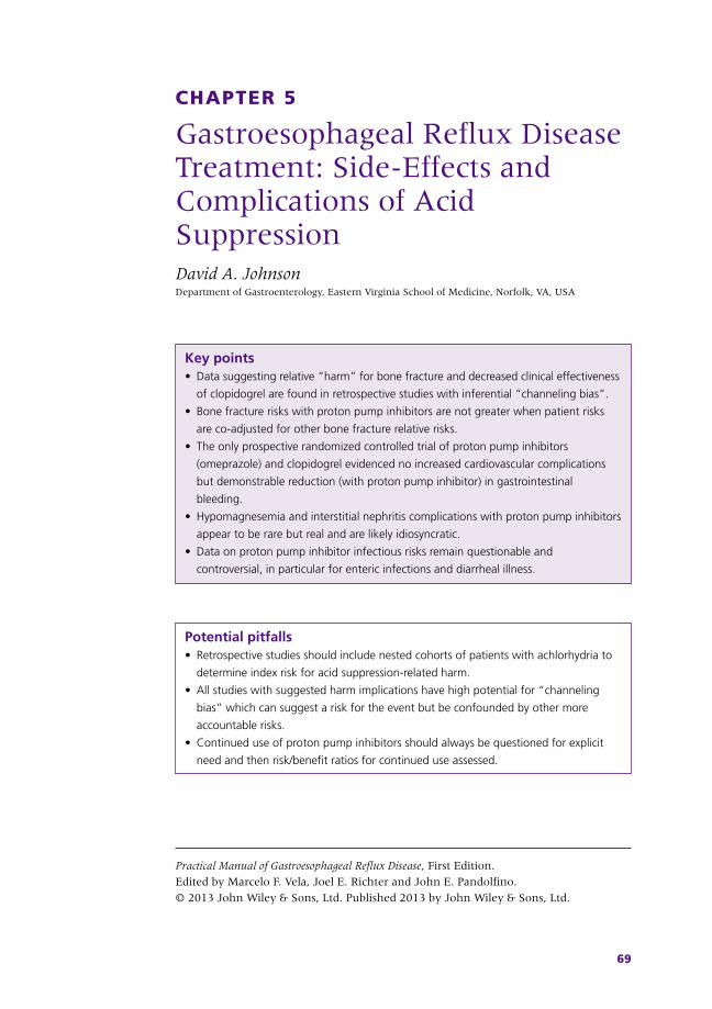
69
Practical Manual of Gastroesophageal Reflux Disease, First Edition.
Edited by Marcelo F. Vela, Joel E. Richter and John E. Pandolfino.
© 2013 John Wiley & Sons, Ltd. Published 2013 by John Wiley & Sons, Ltd.
CHAPTER 5
Gastroesophageal Reflux Disease Treatment: Side-Effects and Complications of Acid Suppression David A. Johnson Department of Gastroenterology , Eastern Virginia School of Medicine , Norfolk , VA , USA
Key points • Data suggesting relative “harm” for bone fracture and decreased clinical effectiveness
of clopidogrel are found in retrospective studies with inferential “channeling bias”.
• Bone fracture risks with proton pump inhibitors are not greater when patient risks
are co-adjusted for other bone fracture relative risks.
• The only prospective randomized controlled trial of proton pump inhibitors
(omeprazole) and clopidogrel evidenced no increased cardiovascular complications
but demonstrable reduction (with proton pump inhibitor) in gastrointestinal
bleeding.
• Hypomagnesemia and interstitial nephritis complications with proton pump inhibitors
appear to be rare but real and are likely idiosyncratic.
• Data on proton pump inhibitor infectious risks remain questionable and
controversial, in particular for enteric infections and diarrheal illness.
Potential pitfalls • Retrospective studies should include nested cohorts of patients with achlorhydria to
determine index risk for acid suppression-related harm.
• All studies with suggested harm implications have high potential for “channeling
bias” which can suggest a risk for the event but be confounded by other more
accountable risks.
• Continued use of proton pump inhibitors should always be questioned for explicit
need and then risk/benefit ratios for continued use assessed.
c05.indd 69 11/15/2012 2:33:20 AM

70 Part 1: Gastroesophageal Reflux Disease Overview
Introduction
Proton pump inhibitors (PPIs) are medications that are essentially ubiqui-
tous in a gastroenterologist ’ s practice. This class of medication has been
available for commercial use for nearly 25 years and has supplanted the
use of histamine-2 receptor antagonists for patients with moderate to
severe gastric acid-related diseases as well as for prophylaxis of upper gas-
trointestinal (GI) injury, e.g. with non-steroidal antiinflammatory drugs
(NSAIDs). The success of these drugs, with sales totaling approximately
$13.6 billion worldwide in 2009 [1] , is not just a result of their potency
and effectiveness in improving symptoms and complications of acid-peptic
diseases. Their safety among pharmacological agents has been unparalleled
but although they are one of the safest classes of medication that gastro-
enterologists deal with, there have been emerging concerns with reports of
potential adverse effects associated with use of PPIs. In the US, such reports
have led the Food and Drug Administration to issue a number of broad-
based product warnings, including all the available PPI drugs available
either for prescription or over-the-counter purchase. The pathogenesis of
these proposed associations is not clear in most cases and the evidence base
to support a clear association for harm is extremely variable. These poten-
tial interactions have included altered absorption of vitamins and minerals,
alteration of pharmacokinetics/pharmacodynamics of concomitant medi-
cations, infection risks, metabolic effects on bone density, and hypersensi-
tivity responses with consequent organ damage.
This chapter will examine the proposed scientific basis for the adverse
events and the evidence base surrounding these controversies.
Vitamin and mineral absorption effects
Long-term PPI therapy has been thought to be associated with micro-
nutrient deficiencies, especially of iron, calcium, magnesium, and vitamin B 12
.
Iron Hydrochloric acid in the stomach assists in the dissociation of iron salts
from food and the reduction of ferric iron to the more soluble ferrous iron.
As non-heme (ferric) iron constitutes the majority of dietary iron require-
ments, there has been concern about impairment of iron absorption if the
conversion to the more soluble ferrous iron is impaired. In vivo data have
demonstrated that iron absorption was directly related to gastric juice
release of ferric iron contained in food [2] . There is some evidence suggest-
ing this may be more specifically related to the vitamin C which is released
c05.indd 70 11/15/2012 2:33:20 AM

Chapter 5: Side-Effects and Complications of Acid Suppression 71
in gastric secretion [3] . There is also some evidence that PPIs may reduce
the bioavailability of ingested vitamin C [4] . The long-term follow-up of
patients taking chronic daily PPIs for up to 7 years has not shown iron
absorption to be a clinically apparent problem [5] .
Clinical summary Despite the above theoretical considerations, there are relatively few data
to indicate that proton pump inhibitor therapy causes iron deficiency. In
fact, there is no report suggesting that proton pump inhibitor therapy
under normal clinical circumstances results in the occurrence of iron
deficiency. In patients with iron deficiency demanding increased iron
absorption, it is conceivable that proton pump inhibitor therapy may
reduce absorption of the non-heme iron and thereby retard replenishment
of the iron pool. This, however, has not been well studied nor is it evident
from widespread use in clinical practice.
Calcium The solubilization of dietary calcium salts is thought to be essential for
absorption of calcium. This is mediated through the release of ionized
calcium from the insoluble calcium salts. It is believed that gastric acid
mediates this solubilization of the dietary calcium and hence there is con-
cern that reduction in gastric acid secretion may impair calcium absorption.
However, the data linking hypochlorhydric states, including pharmacologi-
cal hypocholorhyria induced by PPI therapy, have not shown consistent
evidence of potential impaired calcium absorption. In fact, two high- quality
studies have shown no adverse effect [6,7] .
The clinical reports inferring potential harm from PPIs as an influence
for bone fractures have been controversial but convincing enough to the
FDA that a product label warning was issued for all PPIs. Most recently,
this has been revised to release this warning from labels for over-the-counter
products given that they are intended for short-term use (2 weeks) and for
only up to three cycles/year [8] . The earlier published reports linking PPI
use to development of hip fractures have been observational studies and
case–control studies and thus have greater potential for bias and therefore
less accurate estimates. Additionally, the strength of association in the PPI
studies has been of low magnitude. The adjusted summary odds ratios (OR)
were 1.18 (95% confidence interval (CI) 1.12–1.43), 1.44 (1.30–1.59), and
0.99 (0.90–1.11) but 1.92 (1.16–3.18) if 7 years of continuous PPI use
[9–13] . Given that the estimates and even the upper bounds of most of the
95% CIs of the odds ratio were well below 2, there is a strong possibility
that these differences could have been due to the channeling bias inherent
in observational studies [14] . More recent cross-sectional, longitudinal
and prospective observational reports do not support the reported
c05.indd 71 11/15/2012 2:33:20 AM

72 Part 1: Gastroesophageal Reflux Disease Overview
association [15,16] . It is highly unlikely that randomized controlled clinical
trials can be accurately done to further address the question of bone density
loss and PPI therapy. The relative rarity of fracture across the population
and the extent of potential confounding variables would make this an
extremely difficult study to conduct.
It has also been demonstrated that there are proton pumps in osteo-
clasts, the cells responsible for bone resorption. These vacuolar proton
pumps are responsible for acidification at the ruffled border and facilitate
the dissolution of bone matrix and subsequent resorption [17] . Accordingly,
a PPI would be expected to impair this osteoclastic function of bone resorp-
tion, which should actually lead to an increase in bone density. Patients on
PPIs have lower levels of urinary calcium and hydroxproline, suggesting a
diminution of osteoclast activity. Furthermore, these patients have
increased osteocalcin and tissue resistant alkaline phosphatase, suggestive
of increased new bone formation [18] .
Clinical summary Overall, the studies suggest that calcium absorption is potentially nega-
tively affected only in the setting of reduced acid secretion when ingested
calcium carbonate is provided in the fasting state. As most dietary
calcium is ingested either as a component of food or in supplements
taken with meals, this calcium absorption issue is not likely to be of
great clinical relevance. Furthermore, given that the biological plausi-
bility is also not consistent for causality, it seems reasonable to conclude
that, overall, it is unlikely that PPI use has a significant risk for bone
density loss and related complications of osteoporotic fractures.
Accordingly, the data on bone density loss/osteoporotic fractures would
not suggest that PPI therapy be discontinued in patients taking PPIs for
appropriate indications at appropriate doses. Clearly, adherence to osteo-
porosis screening guidelines is recommended for all patients at risk,
irrespective of the use of PPIs.
Magnesium There have been several (total <50) cases of hypomagnesemia associated
with long-term PPI use [19–21] . The patients generally presented with
profound hypomagnesemia and typically required hospitalization. In
approximately 25% of these cases, the patients had persistent hypomagne-
semia despite supplements. Prompt resolution of magnesium levels was
evident after discontinuance of PPIs, and in a few cases where the patients
were rechallenged with a PPI, the hypomagesemia recurred, suggesting a
PPI-related effect. None of the patients had identifiable GI wasting or renal
loss etiologies. This prompted a recent alert by the FDA about PPI use
and hypomagnesemia [22] . This alert suggested that healthcare providers
c05.indd 72 11/15/2012 2:33:20 AM

Chapter 5: Side-Effects and Complications of Acid Suppression 73
should consider checking magnesium levels in patients who are antici-
pated to be on long-term PPIs.
The mechanism for the magnesium depletion is not known. The primary
absorption of magnesium is through a passive pathway in the small
intestine. There is some identifiable active transport, however, via trans-
port channels (TRPM6 and 7) [23] . It is not known if PPIs may have some
effect on this pathway but there are familial cases with mutations at this
pathway who develop hypomagnesemia.
Clinical summary Although omeprazole and esomeprazole were initially cited, other PPIs
have now been included so this is likely a class effect. The FDA recom-
mendation to consider checking magnesium levels before starting is not
practical, in particular for the over-the-counter market. In patients who
may be predisposed to present/ongoing magnesium loss, e.g. intestinal
malabsorption or renal excretion/wasting, it may be reasonable to follow
magnesium levels more closely and consider this association, particu-
larly if a profound hypomagnesemia condition develops. Given the
extreme rarity of the report and no controlled studies to delineate the
mechanisms, it is important for healthcare providers to be aware of this
but maintain PPI use where clinically justified, in their appropriate scope
of practice.
Vitamin B 12 Gastric acid also facilitates the release of vitamin B
12 bound to proteins
within ingested foodstuffs to permit binding to R-proteins for eventual
absorption in the terminal ileum. This vitamin needs to be released from
these proteins and subsequently bound to R-proteins and intrinsic factor in
order to be absorbed in the terminal ileum. Gastric acid facilitates the pro-
teolytic process involved in releasing the vitamin from the proteins in
ingested food. Accordingly, there are theoretical reasons why the inhibi-
tion of gastric acid secretion by PPI therapy would reduce the bioavail-
ability of of dietary vitamin B 12
[2] .
Studies which have examined the potential association between
long-term PPI use and vitamin B 12
have shown conflicting results [23] .
Additionally, to date no studies have provided a longitudinal evaluation
demonstrating alterations of specific metabolic intermediates (e.g. methyl-
malonate and homocysteine) which can accumulate with this deficiency.
Clinical summary Despite the biological plausibility of this deficiency, there is currently little
evidence to support a clinically relevant association to recommend a
change in current practice.
c05.indd 73 11/15/2012 2:33:20 AM

74 Part 1: Gastroesophageal Reflux Disease Overview
Alteration of pharmacokinetics/pharmacodynamics
Proton pump inhibitors and related impairment of antiplatelet therapy (clopidogrel) It is well known that PPIs are metabolized by the cytochrome p450 enzyme
pathway, specifically CYP2C19 and CYP3A4. As clopidogrel is a pro-drug
which requires active biotransformation via the cytochrome p450 pathway,
it has been hypothesized that competition at CYP2C19 sites may result in
reduced biological effects of clopidogrel when co-administered with PPIs.
The concept of PPIs interfering with clopidogrel biotransformation
stemmed from in vitro studies that demonstrated a pharmacodynamic
interaction which was an attenuated antiplatelet effect as measured by
adenosine diphosphate (ADP)-induced platelet aggregation and elevated
platelet activity [24] .
The inference in regard to PPIs and clopidogrel first arose from the
combined use of clopidogrel and omeprazole, although several other PPIs
have subsequently been found to be associated with a smaller or insignifi-
cant attenuation of the clopidogrel antiplatelet effect; these PPIs included
pantoprazole, esomeprazole, and to a lesser degree lansoprazole [14,25] .
However, these in vitro data were quickly extrapolated in several high-pro-
file retrospective database evaluations that found higher cardiac event rates
(stent thrombosis, myocardial infarct, and death) in patients who were tak-
ing clopidogrel with any PPI versus those on clopidogrel alone [26,27] . This
physiological intermediary endpoint of attenuated effect led to a number of
retrospective post hoc analyses and these suggested a potential clinical harm
with adverse cardiovascular outcomes for patients taking clopidogrel and
PPIs. Based on these data, the FDA issued a recommendation against the
combined use of omeprazole and esomeprazole and clopidogrel [28] . In
that statement the FDA also advised against the combined use of clopido-
grel coupled with other potent inhibitors of the cytochrome p450 pathway
such as cimetidine, as well as cautioning against use of other PPIs.
In fact, although consensus recommendations from the leading gastro-
enterology and cardiology national societies suggested that this combined
use was appropriate for patients at significant increased risk for gastro-
intestinal bleeding [29] , these recommendations were developed at the
same time as the emerging controversy and the experts involved in the
recommendations were in favor of combined use of PPI (as the preferred
strategy over H2 receptor antagonists) plus clopidogrel for patients defined
as being at risk of GI bleeding. Additionally, there were prospective
randomized controlled studies emerging which showed no evidence of
increased cardiovascular adverse events when patients had combined use
of PPIs [30] .
c05.indd 74 11/15/2012 2:33:21 AM
Chapter 5: Side-Effects and Complications of Acid Suppression 75
The most substantive data to date, supporting the lack of cardiovascular
harm, come from a randomized prospective placebo-controlled trial com-
paring clopidogrel with or without omeprazole in patients who had coro-
nary stents following an acute coronary syndrome [31] . In this study of
3761 patients, the a priori primary objectives were assessment of both
cardiovascular and GI harm. There was no difference for cardiovascular
adverse events between the placebo and omeprazole group. In all, 51
patients had a gastrointestinal event; the event rate was 1.1% with
omeprazole and 2.9% with placebo at 180 days (hazard ratio (HR) with
omeprazole, 0.34, 95% CI 0.18–0.63, P < 0.001). The rate of overt upper
gastrointestinal bleeding was also reduced with omeprazole compared
with placebo (HR 0.13, 95% CI 0.03–0.56, P = 0.001). Despite the pub-
lished prospective randomized controlled data demonstrating no significant
cardiovascular harm, but actually demonstrating increased GI harm when
omeprazole was not used with clopidogrel, the FDA updated the warning
in 2010, removing the other PPIs from the caution notice but reiterating
the citation of omeprazole [32] . The safety of combined use of PPIs with
clopidogrel has also been evident in patients following a stroke or transient
ischemic event [33] .
In light of all the controversy over the combined use of clopidogrel and
PPIs, an expert consensus document was prepared and endorsed by the
American College of Cardiology, the American College of Gastroenterology,
and the American Heart Association [34] . The consensus recommendation
was that if patients were deemed at significant risk of GI bleeding, then use
of clopidogrel combined with PPIs was appropriate. Based on the extensive
review of all available data, there was no evidence to suggest increased
cardiovascular harm with this combined use.
What has become clearer in review of the retrospective analyses suggest-
ing harm with PPI and clopidogrel combined usage is the high likelihood for
channeling bias. In fact, the most recent post hoc database assessment (using
the VA database) did suggest an apparent cardiovascular harm for combined
usage, but when the authors used propensity-matched evaluations to
correct for co-variate cardiovascular risks and medication compliance, they
found no significant association between major cardiovascular events and
use of clopidogrel with continuous, switched or discontinued PPIs [35] .
It is not well understood that although clopidogrel is not an NSAID, there
is significant risk for upper GI ulceration and related bleeding. Conventional
wisdom suggests that clopidogrel should be a safer, non-ulcerogenic
alternative for patients at high risk for aspirin-induced ulcers. However,
Chan et al . reported on patients with NSAID-related ulcer disease who were
randomized (after ulcer healing) to receive clopidogrel alone or esomepra-
zole plus aspirin [36] . After a 1-year follow-up period, the patients in the
clopidogrel group had a significant increase in the rate of recurrent upper
c05.indd 75 11/15/2012 2:33:21 AM
76 Part 1: Gastroesophageal Reflux Disease Overview
gastrointestinal bleeding from ulcers, compared with those in the group
taking aspirin plus esomeprazole (8.6% versus 0.7%, P = 0.001).
The impairment of ulcer healing by clopidogrel has not been widely
appreciated. Platelet aggregation plays a critical role in healing, through
the release of various platelet-derived growth factors that promote angio-
genesis, which is essential for ulcer healing. ADP-receptor antagonists
impair the healing of gastric ulcers by inhibiting the release by platelets of
proangiogenic growth factors such as vascular endothelial growth factor,
which promotes endothelial proliferation and accelerates the healing of
ulcers [37] . Accordingly, although clopidogrel might not be the primary
cause of gastrointestinal ulcers, the anti-angiogenic effects may impair the
healing of background ulcers; when combined with the propensity to
increase bleeding, these agents may convert small, silent erosions or ulcers
into large ulcers that bleed. A recent prospective randomized controlled
study involving patients with histories of peptic ulcer disease showed at
6 months that the combination of esomeprazole and clopidogrel had a
1.2% ulcer recurrence rate compared to 11.0% recurrence in patients
receiving clopidogrel alone ( P = 0.009) [38] .
Clinical summary These results question the exact relationship between ex vivo platelet assays
and clinical outcomes, especially with regard to the assessment of drug
interactions. Platelet assays and observational data may be factual but are
not always appropriate for extrapolation to clinical care, as evidenced by
what we have seen during the last several months with the prescribing
recommendations for PPI and clopidogrel usage. Platelet assays and obser-
vational data are not substitutes for randomized controlled trial data.
Appropriate use is the key consideration for any medication. Healthcare
providers should be attuned to the need for PPI therapy in patients who
exhibit signs or symptoms of acid-related disease, as well as in asymptom-
atic patients treated with NSAIDs or antiplatelet agents who meet risk
stratification criteria to justify GI prophylaxis (co-therapy with a PPI).
Proton pump inhibitors and infection As gastric acid creates a potential barrier to acid-sensitive spores and
bacteria which may colonize the upper GI tract, there has been a concern
about potential alteration of native GI flora and clinical consequences of
bacterial overgrowth. Additionally, there are some data which suggest
that PPIs may have a direct effect on white blood cell function with alter-
ations of neutrophil chemotaxis and degranulation [39] . Specific infec-
tion risks have been cited for community- and hospital-acquired
pneumonia, Clostridium difficile colitis, enteric infections, and spontaneous
bacterial peritonitis.
c05.indd 76 11/15/2012 2:33:21 AM

Chapter 5: Side-Effects and Complications of Acid Suppression 77
Pneumonia Several studies have suggested that PPI use may increase the risk for both
community- and hospital-acquired pneumonia. In theory, when gastro-
esophageal reflux occurs, gastric bacteria could be carried up to the hypo-
pharynx where microaspiration into the lower airways could lead to
pneumonia, especially in patients with compromised oropharyngeal
protective reflexes, e.g. those on mechanical ventilation. In general, most
of the studies assessing the relationship between PPIs and community-
acquired pneumonia have revealed a modestly higher risk of community-
acquired pneumonia in patients exposed to PPIs [40–42] . This risk
was confirmed in a recent metaanalysis, which found a higher risk of
community-acquired pneumonia with PPI use (OR 1.36, 95% CI 1.12–1.65).
The authors refrained from drawing definitive conclusions from these data
because of the significant heterogeneity between the studies [43] . Other
studies from large database analyses, however, have not shown a significant
increase in community-acquired pneumonia [44,45] .
Clinical summary Residual confounding may have complicated interpretation of these
studies, suggesting an association of harm (increased pneumonia). These
studies surprisingly showed that the association was weakest in current
recipients who had been taking PPIs for the longest duration. If an associ-
ated risk of PPIs and pneumonia risk is in fact present, the relative risk is
small and may be most likely accounted for by channeling bias.
Enteric infections Alterations of the gastric pH and possible related changes in susceptibility
to enteric infections have been a topic of long-standing debate. Although
gastric hypochlorhydria is commonly listed as a risk factor for traveler ’ s
diarrhea [46] , PPI exposure as a risk factor for enteric infections in trav-
elers has not been formally studied. In fact, there is only one study evalua-
ting acid reduction medication use and this study reported no significant
association (OR 6.9, range 0.7–67.4) of travelers ’ diarrhea with antacids
and H2 receptor antagonist use [47] . A metaanalysis of the diagnosis of
enteric infections did identify an increased risk of acute bacterial infection
associated with the usage of PPIs (OR 3.33, 95% CI 1.84–6.02) [48] . A
recent comprehensive analysis of the data on PPI use and enteric infections
concluded that there was no association of PPI use and viral or parasitic
enteric infections [49] . The data on specific bacterial infections were gen-
erally supportive of no associated risk although there were a few specific
case reports suggesting a remote causal association.
Previously, gastric acid was not believed to be important in protecting
against C. difficile infection because acid-resistant spores were presumed to be
c05.indd 77 11/15/2012 2:33:21 AM

78 Part 1: Gastroesophageal Reflux Disease Overview
the principal vector of transmission. Recently, this has been challenged, as
several studies have found a higher risk of C. difficile infection in PPI users. In
theory, PPIs may increase the risk of C. difficile infection by increasing the
ability of the spore to convert to the vegetative form and to survive in the
lumen of the GI tract. The data for community-acquired versus hospital-
acquired infection have been variable and inconclusive for an associated risk
of harm [49] . A recent metaanalysis of 11 papers, including nearly 127,000
patients, found a significant relationship between PPI use and C. difficile
infection, with an odds ratio of 2.05 (95% CI 1.47–2.85) [48] . Further sup-
porting the hypothesis of a direct causative association, a recent study found
a significant dose–response relationship, with more aggressive acid suppres-
sion paired with higher odds association [50] .
Other enteric infections have been found to be associated with PPIs
[48,49] . A recent metaanalysis did suggest an increased risk of acute bac-
terial enteric infection with the use of PPIs, with a random effects model
pooled risk OR of 3.33 (95% CI 1.84–6.02) [48] . Small intestinal bacte-
rial overgrowth (SIBO), a condition associated with bloating, diarrhea,
and malabsorption, has recently been associated with PPI use, although
the significance of the association is uncertain [51] . In this report, SIBO
was detected in 50% of patients using PPIs, 24.5% of patients with
irritable bowel syndrome (IBS), and 6% of healthy control subjects.
There was a statistically significant difference between patients using
PPIs and those with IBS or healthy control subjects ( P < 0.001). The prev-
alence of SIBO increased after 1 year of treatment with PPI. The reported
eradication rate of SIBO (using rifaximin) was 87% in the PPI group and
91% in the IBS group.
Clinical summary For community-acquired enteric infections including C. difficile , the
reported odds ratios are low and difficult to evaluate fully beyond the
potential for channeling bias of sicker patients. The situation is complex in
hospitalized settings where drugs, such as PPIs, may be highly correlated
with other variables, such as severity of illness and length of stay. The data
for possible association with SIBO are extremely limited and will need
further corroboration and validation before they can be considered appli-
cable to current clinical use.
Spontaneous bacterial peritonitis Recent reports have suggested that there is a relationship between PPI use
and the development of spontaneous bacterial peritonitis in hospitalized
cirrhotic patients with ascites [52,53] . One study found a strong association
(OR 4.3, 95% CI 1.3–11.7) between PPIs and SBP [52] whereas another
study found no significant association (OR 1.0, 95% CI 0.4–2.6) [53] .
c05.indd 78 11/15/2012 2:33:21 AM

Chapter 5: Side-Effects and Complications of Acid Suppression 79
Clinical summary The two studies on this association were small case–control studies of hos-
pitalized patients and the data are conflicting as to a reported associated
risk. At present, accordingly, no firm conclusion can be drawn about the
relevance of this association.
Proton pump inhibitors and interstitial nephritis Several case reports have implicated PPIs as a cause of acute interstitial
nephritis [23] . This disorder is a humoral and cell-mediated hypersensi-
tivity inflammatory reaction of the renal interstitium and tubules. A
systematic review from 2007 found 64 cases documented in the literature,
12 of which were considered definitely associated and nine of which were
probably associated [54] . Initial symptoms were non-specific and included
nausea, malaise, and fever. With such extensive use worldwide as the
denominator, the authors concluded that acute interstitial nephritis was a
rare, idiosyncratic occurrence related to PPI use, but did not find enough
evidence to support a causative relationship.
Clinical summary Despite the extreme rarity of the syndrome, the association cannot be dis-
missed and a high level of clinical suspicion is necessary to detect acute
interstitial nephritis early in its course, especially soon after the initiation
of PPI therapy.
Conclusion
Although concerns have been raised about the long-term safety of PPIs,
the majority of the evidence does not strongly support the deluge of reports
citing a potential for significant adverse harm associated with PPI usage.
When translating these studies into the routine management of patients, it
is important to recall some very basic tenets of good patient care.
Obviously, no therapy is completely without risk, whether pharmaco-
logical, surgical or psychological. Consequently, no drug, procedure or
treatment plan should be prescribed without a valid indication. Even with
an appropriate indication for use, the risk/benefit ratio of every therapy
prescribed should always be evaluated. If the indication for the PPI is weak
or uncertain, then even a slight risk tips the balance away from the drug
and the drug should be discontinued. Clearly, there are too many patients
who receive continued PPI therapy without a valid need. When seeing
patients in long-term care, the indication and necessity for continued
usage for all drugs, including PPIs, should be reviewed and continually
reevaluated.
c05.indd 79 11/15/2012 2:33:21 AM

80 Part 1: Gastroesophageal Reflux Disease Overview
References
1 Gatyas G . IMS Health Reports 2009 . Available at: www.imshealth.com .
2 Bezwoda W , Charlton R , Bothwell T , et al . The importance of gastric hydrochloric acid
in the absorption of nonheme food iron . J Lab Clin Med 1978 ; 92 : 108 – 16 .
3 Conrad ME , Schade SG . Ascorbic acid chelates in iron absorption: a role for hydro-
chloric acid and bile . Gastroenterology 1968 ; 55 : 35 – 43 .
4 McColl KE . Effect of proton pump inhibitors on vitamins and iron . Am J Gastroenterol
2009 ; 104 (Suppl 2): S5 – S9 .
5 Koop H . Review article: metabolic consequences of long-term inhibition of acid
secretion by omeprazole . Aliment Pharmacol Ther 1992 ; 6 : 399 – 406 .
6 Wright MJ , Sullivan RR , Gaffney-Stomberg E , et al . Inhibiting gastric acid production
does not affect intestinal calcium absorption in young, healthy individuals: a
randomized, crossover, controlled clinical trial . J Bone Miner Res 2010 ; 25 : 2205 – 11 .
7 Hansen KE , Jones AN , Lindstrom JM , et al . Do proton pump inhibitors decrease
calcium absorption? J Bone Miner Res 2010 ; 25 : 2510 – 19 .
8 www.fda.gov/drugs/drugsafety/postmarketdrugsafetyinformationforpatientsand
providers/ucm213206.htm
9 Vestergaard P , Rejnmark L , Mosekilde L . Proton pump inhibitors, histamine H2
receptor antagonists, and other antacid medications and the risk of fracture . Calcif
Tissue Int 2006 ; 79 : 76 – 83 .
10 Yang YX , Lewis JD , Epstein S . et al . Long-term proton pump inhibitor therapy and
risk of hip fracture . JAMA 2006 ; 296 : 2947 – 53 .
11 Targownik LE , Lix LM , Metge CJ , et al . Use of proton pump inhibitors and risk of
osteoporosis-related fractures . CMAJ 2008 ; 179 : 319 – 26 .
12 Corley DA , Kubo A , Zhao W , Quesenberry C . Proton pump inhibitors and histamine-2
receptor antagonists are associated with hip fractures among at-risk patients .
Gastroenterology 2010 ; 139 : 93 – 101 .
13 Laine L . Proton pump inhibitors and bone fractures? Am J Gastroenterol 2009 ;
104 : S21 – S26 .
14 Johnson DA . Safety of proton pump inhibitors: current evidence for osteoporosis and
interaction with antiplatelet agents . Curr Gastroenterol Rep 2010 ; 12 : 167 – 74 .
CASE STUDY
A 75-year-old woman is seen in ongoing follow-up of her chronic gastroesophageal reflux disease (GERD). She has had prior relapsing symptoms of dysphagia with an endoscopy demonstrating Los Angeles Grade C erosive esophagitis and a distal esophageal stricture at the esophagogastric junction, which required endoscopic dilation. She had been well controlled over the last year, taking a daily proton pump inhibitor in the morning 30–60 min before breakfast. She recently saw a television broadcast which suggested that the use of PPIs was associated with an increased risk for bone fracture, in particular hip, neck, and wrist. She became frightened about her risks for fracture and therefore stopped her PPI. Since then, she has had recurrent heartburn on a daily basis and is now having increasing recurrent problems with solid food dysphagia. Despite her recurrent symptoms, she is very concerned about restarting the PPI medication which alleviated her GERD symptoms.
c05.indd 80 11/15/2012 2:33:21 AM

Chapter 5: Side-Effects and Complications of Acid Suppression 81
15 Targownik LE , Lix LM , Leung S , Leslies WD . Proton-pump inhibitor use is not asso-
ciated with osteoporosis or accelerated bone mineral density loss . Gastroenterology
2010 ; 138 : 896 – 904 .
16 Gray SL , LaCroix AZ , Larson J , et al . Proton pump inhibitor use, hip fracture, and
change in bone mineral density in postmenopausal women: results from the
Women ’ s Health Initiative . Arch Intern Med 2010 ; 170 : 765 – 71 .
17 Jefferies KC , Cipriano DJ , Forgac M . Function structure and regulation of the
vacuolar (H+) ATPases . Arch Biochem Biophys 2008 ; 476 : 33 – 42 .
18 Mizunashi K , Furukawa Y , Katano K , Abe K . Effect of omeprazole, an inhibitor of
H+, K+, ATPase on bone resorption in humans . Calcif Tissue Int 1993 ; 53 : 21 – 5 .
19 Epstein M , McGrath S , Law F . Proton pump inhibitors and hypomagesemic hyop-
parathyroidism . N Engl J Med 2006 ; 355 : 1834 – 6 .
20 Broeren MAC , Geerdink EAM , Vader HL , et al . Hyopmagnesmia induced by several
proton pump inhibitors . Ann Intern Med 2009 ; 151 : 755 – 6 .
21 MacKay JD , Bladon PT . Hypomagesemia due ot proton pump inhibitor therapy: a
clinical case series . Q J Med 2010 ; 103 ( 6 ): 387 – 95 .
22 www.fda.gov/Safety/MedWatch/SafetyInformation/SafetyAlertsforHumanMedical
Products/ucm245275.htm
23 Yang YX , Metz DC . Safety of proton pump inhibitor exposure . Gastroenterology
2010 ; 139 : 1115 – 27 .
24 Gilard M , Arnaud B , Cornily JC , et al . Influence of omeprazole on the antiplatelet
action of clopidogrel associated with aspirin: the randomized, double-blind OCLA
(Omeprazole CLopidogrel Aspirin) Study . J Am Coll Cardiol 2008 ; 51 : 256 – 60 .
25 Laine L , Hennekens C . Proton pump inhibitor and clopidogrel interaction: fact or
fiction? Am J Gastroenterol 2010 ; 105 : 34 – 41 .
26 Ho PM , Maddox TM , Wang L , et al . Risk of adverse outcomes associated with con-
comitant use of clopidogrel and proton pump inhibitors following acute coronary
syndrome . JAMA 2009 ; 301 : 937 – 44 .
27 Ray WA , Murray KT , Griffin MR , et al . Outcomes with concurrent use of clopidogrel
and proton-pump inhibitors: a cohort study . Ann Intern Med 2010 ; 152 : 337 – 45 .
28 www.fda.gov/Drugs/DrugSafety/PostmarketDrugSafetyInformationforPatientsand
Providers/DrugSafetyInformationforHeathcareProfessionals/ucm190784.htm
29 Bhatt DL , Scheiman J , Abraham NS , et al . American College of Cardiology
Foundation, American College of Gastroenterology, American Heart Association.
ACCF/ACG/AHA 2008 expert consensus document on reducing the gastrointes-
tinal risks of antiplatelet therapy and NSAID use . Am J Gastroenterol 2008 ;
103 : 2890 – 907 .
30 O ’ Donoghue ML , Braunwald E , Antman EM , et al . Pharmacodynamic effect and
clinical efficacy efficacy of clopidogrel and prasugrel with or without a proton-pump
inhibitor: an analysis of two randomized trials . Lancet 2009 ; 374 : 989 – 97 .
31 Bhatt DL , Cryer BL , Contant CF , et al ., COGENT Investigators. Clopidogrel with or
without omeprazole in coronary artery disease . N Engl J Med 2010 ; 363 : 1909 – 17 .
32 www.fda.gov/Drugs/DrugSafety/ucm231161.htm.
33 Juurlink DN , Gomes T , Marndarni MM , et al . The safety of proton pump inhibitors
and clopidogrel in patients after stroke . Stroke 2011 ; 42 : 128 – 32 .
34 Abraham NS , Hlatky MA , Antman EM , et al . ACCF/ACG/AHA 2010 expert con-
sensus document on the concomitant use of proton pump inhibitors and thienopyri-
dines: a focused update of the ACCF/ACG/AHA 2008 expert consensus document on
reducing the gastrointestinal risks of antiplatelet therapy and NSAID use . Am J
Gastroenterol 2010 ; 105 : 2533 – 49 .
c05.indd 81 11/15/2012 2:33:21 AM

82 Part 1: Gastroesophageal Reflux Disease Overview
35 Banerjee S , Weideman RA , Weideman MW , et al . Effect of concomitant use of clopi-
dogrel and proton pump inhibitors after percutaneous coronary intervention . Am J
Cardiol 2011 ; 107 ( 6 ): 871 – 8 .
36 Chan FKL , Ching JYL , Hung LCT , et al . Clopidogrel versus aspirin and esomeprazole
to prevent recurrent ulcer bleeding . N Engl J Med 2005 ; 352 : 238 – 44 .
37 Ma L, Elliott SN , Cirino G , Buret A , Ignarro LJ , Wallace JL . Platelets modulate gastric
ulcer healing: role of endostatin and vascular endothelial growth factor release . Proc
Natl Acad Sci USA 2001 ; 98 : 6470 – 5 .
38 Hus PI , Lai KH , Liu CP . Esomeprazole with clopidogrel reduces peptic ulcer recurrence
compared with clopidogrel alone, in patients with atherosclerosis . Gastroenterology
2011 ; 140 : 791 – 8 .
39 Wandall JH . Effects of omeprazole on neutrophil chemotaxis, super oxide produc-
tion, degranualation and translocation of cytochrome b-245 . Gut 1992 ; 33 : 617 – 21 .
40 Laheij RJ , Sturkenboom MD , Hassing RJ , et al . Risk of community-acquired pneu-
monia and use of gastric acid-suppressive drugs . JAMA 2004 ; 292 : 1955 – 60 .
41 Gulmez SE , Holm A , Frederiksen H , et al . Use of proton pump inhibitors and the risk
of community-acquired pneumonia: a population-based case-control study . Arch
Intern Med 2007 ; 167 : 950 – 5 .
42 Herzig SJ , Howell MD , Ngo LH , Marcantonio ER . Acid-suppressive medication use
and the risk for hospital-acquired pneumonia . JAMA 2009 ; 301 : 2120 – 8 .
43 Johnstone J , Nerenberg K Loeb M . Meta-analysis: proton pump inhibitor use and the
risk of community-acquired pneumonia . Aliment Pharmacol Ther 2010 ; 31 : 1165 – 77 .
44 Sarkar M , Hennessy S , Yang YX . Proton-pump inhibitor use and the risk for
community-acquired pneumonia . Ann Intern Med 2008 ; 149 : 391 – 8 .
45 Dublin A , Walker RL , Kackson ML , et al . Use of proton pump inhibitors and H2
blockers and risk of pneumonia in older adults: a population-based case-control
study . Pharmacoepidemiol Drug Saf 2010 ; 19 : 792 – 802 .
46 DuPont HL . Travellers ’ diarrhoea: contemporary approaches to therapy and preven-
tion . Drugs 2006 ; 66 : 303 – 14 .
47 Cobelens FG , Leentvaar-Kuijpers A , Kleijnen J , et al . Incidence and risk factors of
diarrhoea in Dutch travellers: consequences for priorities in pre-travel health advice .
Trop Med Int Health 1998 ; 3 : 896 – 903 .
48 Leonard J , Marshall JK , Moayyedi P . Systematic review of the risk of enteric infection
in patients taking acid suppression . Am J Gastroenterol 2007 ; 102 : 2047 – 56 .
49 Dial MS . Proton pump inhibitor use and enteric infections . Am J Gastroenterol
2009 ; 104 ( Suppl 2 ): S10 – S16 .
50 Howell MD , Novack V , Grgurich P , et al . Iatrogenic gastric acid suppression and the risk
of nosocomial Clostridium difficile infection . Arch Intern Med 2010 ; 170 : 784 – 90 .
51 Lombardo L , Foti M , Ruggia O , Chiecchio A . Increased incidence of small intestinal
bacterial overgrowth during proton pump inhibitor therapy . Clin Gastroenterol
Hepatol 2010 ; 8 ( 6 ): 504 – 8 .
52 Bajaj JS , Zadvornova Y , Heuman DM , et al . Association of proton pump inhibitor
therapy with spontaneous bacterial peritonitis in cirrhotic patients with ascites . Am J
Gastroenterol 2009 ; 104 : 1130 – 4 .
53 Campbell MS , Obstein K , Reddy KR , Yang YX . Association between proton pump
inhibitor use and spontaneous bacterial peritonitis . Dig Dis Sci 2008 ; 53 : 394 – 8 .
54 Sierra F , Suarez M , Rey M , et al . Systematic review: proton pump inhibitor associated
acute interstitial nephritis . Aliment Pharmacol Ther 2007 ; 26 : 545 – 53 .
c05.indd 82 11/15/2012 2:33:21 AM
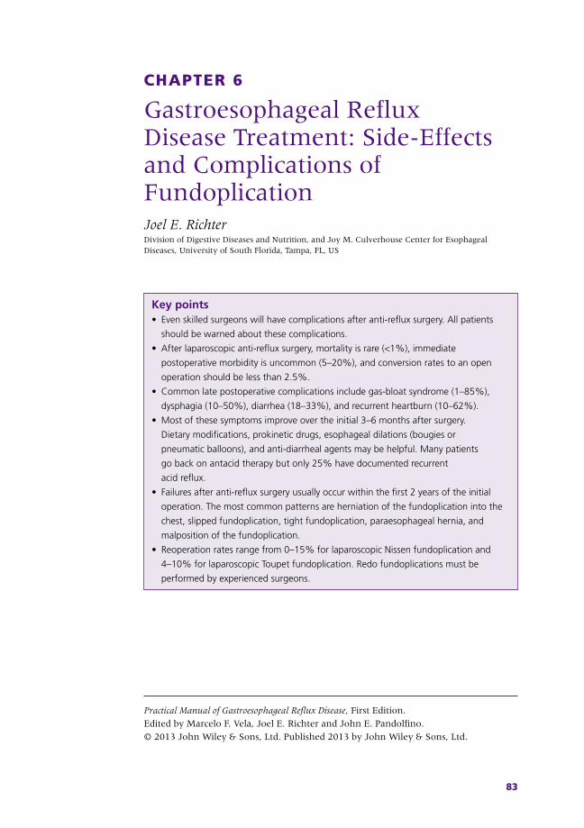
83
Practical Manual of Gastroesophageal Reflux Disease, First Edition.
Edited by Marcelo F. Vela, Joel E. Richter and John E. Pandolfino.
© 2013 John Wiley & Sons, Ltd. Published 2013 by John Wiley & Sons, Ltd.
CHAPTER 6
Gastroesophageal Reflux Disease Treatment: Side-Effects and Complications of Fundoplication Joel E. Richter Division of Digestive Diseases and Nutrition, and Joy M. Culverhouse Center for Esophageal
Diseases , University of South Florida , Tampa , FL , US
Key points • Even skilled surgeons will have complications after anti-reflux surgery. All patients
should be warned about these complications.
• After laparoscopic anti-reflux surgery, mortality is rare (<1%), immediate
postoperative morbidity is uncommon (5–20%), and conversion rates to an open
operation should be less than 2.5%.
• Common late postoperative complications include gas-bloat syndrome (1–85%),
dysphagia (10–50%), diarrhea (18–33%), and recurrent heartburn (10–62%).
• Most of these symptoms improve over the initial 3–6 months after surgery.
Dietary modifications, prokinetic drugs, esophageal dilations (bougies or
pneumatic balloons), and anti-diarrheal agents may be helpful. Many patients
go back on antacid therapy but only 25% have documented recurrent
acid reflux.
• Failures after anti-reflux surgery usually occur within the first 2 years of the initial
operation. The most common patterns are herniation of the fundoplication into the
chest, slipped fundoplication, tight fundoplication, paraesophageal hernia, and
malposition of the fundoplication.
• Reoperation rates range from 0–15% for laparoscopic Nissen fundoplication and
4–10% for laparoscopic Toupet fundoplication. Redo fundoplications must be
performed by experienced surgeons.
c06.indd 83 11/15/2012 2:33:07 AM
84 Part 1: Gastroesophageal Reflux Disease Overview
Introduction
In the past 15 years, there has been an increase in the number of anti-
reflux operations being performed. The reasons for this increase include
the development and proliferation of laparoscopic techniques, the increase
in the fraction of the population that is overweight, and possibly the
increased willingness of the population to undergo an operation to avoid a
lifetime of medications or lifestyle changes. The operation is now widely
available in community hospitals, the length of stay ranges between 1 and
4 days, some operations are even done as day surgery, and most patients
return to normal activity within 2 weeks [1,2] . Patients over 65 years of
age can expect an excellent outcome after laparoscopic surgery in at least
90% of cases, similar to younger patients [3] . Based on the US Nationwide
Inpatient Sample, there were 9173 adult anti-reflux operations in 1993,
which increased nearly 3.5-fold, reaching a peak at 32,980 in 2000 [4] . For
poorly understood reasons, the most recent available data for 2006 show a
40% decline to 19,688 operations [5] .
This review will focus on the surgical and medical complications pri-
marily reported after laparoscopic anti-reflux surgery (Box 6.1 ). The avail-
able reports on this subject are numerous so I have relied on summary data
available from the Society of American Gastrointestinal and Endoscopic
Surgeons (SAGES) [1] and the Agency for Healthcare Research and Quality
Effective Health Care Program [6] . The chapter will summarize mortality
and morbidity data for laparoscopic anti-reflux operations, review the
common perioperative and postoperative complications, and discuss the
common reasons for fundoplication failure.
Potential pitfalls • Anti-reflux surgery is never an emergency procedure. All patients should be carefully
evaluated prior to surgery.
• At a minimum, preoperative testing should include upper endoscopy, esophageal
manometry and pH testing (the latter primarily patients with non-erosive gastro-
esophageal reflux disease). Don ’ t take short cuts.
• Even skilled surgeons will have complications after anti-reflux surgery. All patients
should have a frank discussion with their gastroenterologist and surgeon about
postoperative dysphagia, gas-bloat, diarrhea, and the durability of the operation with
return of heartburn.
• Treat these common complications with conservative therapy and never rush into
redo anti-reflux surgery. Most problems will improve over 3–6 months.
• A redo operation requires a very experienced surgeon. The keys to success must not
be violated. These include careful review of the patient ’ s prior work-up and repeat
studies as necessary, recognition of esophageal shortening, and complete takedown
of the original fundoplication.
c06.indd 84 11/15/2012 2:33:08 AM
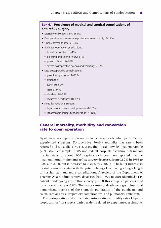
Chapter 6: Side-Effects and Complications of Fundoplication 85
General mortality, morbidity and conversion rate to open operation
By all measures, laparoscopic anti-reflux surgery is safe when performed by
experienced surgeons. Postoperative 30-day mortality has rarely been
reported and is usually < 1% [1] . Using the US Nationwide Inpatient Sample
(20% stratified sample of US non-federal hospitals recording 5–8 million
hospital stays for about 1000 hospitals each year), we reported that the
inpatient mortality after anti-reflux surgery decreased from 0.82% in 1993 to
0.26% in 2000, but it increased to 0.54% by 2006 [5] . The latter increase in
mortality was associated with the patients being older, having a longer length
of hospital stay and more complications. A review of the Department of
Veterans Affairs administrative databases from 1990 to 2001 identified 3145
patients undergoing anti-reflux surgery [7] . Of this group, 28 patients died
for a mortality rate of 0.8%. The major causes of death were gastrointestinal
hemorrhage, necrosis of the stomach, perforation of the esophagus and
colon, cardiac arrest, respiratory complications, and pulmonary embolism.
The perioperative and immediate postoperative morbidity rate of laparo-
scopic anti-reflux surgery varies widely related to experience, technique,
Box 6.1 Prevalence of medical and surgical complications of anti-reflux surgery • Mortality (< 30 days): 1% or less
• Perioperative and immediate postoperative morbidity: 8–17%
• Open conversion rate: 0–24%
• Early postoperative complications:
° bowel perforation: 0–4%
° bleeding and splenic injury: < 1%
° pneumothorax: 0–10%
° severe postoperative nausea and vomiting: 2–5%
• Late postoperative complications:
° gas-bloat syndrome: 1–85%
° dysphagia:
early: 10–50%
late: 3–24%
° diarrhea: 18–33%
° recurrent heartburn: 10–62%
• Need for revisional surgery:
° laparoscopic Nissen fundoplication: 0–15%
° laparoscopic Toupet fundoplication: 4–10%
c06.indd 85 11/15/2012 2:33:08 AM

86 Part 1: Gastroesophageal Reflux Disease Overview
and degree of follow-up. One review suggested a rate up to 17% [8] . Our
National Inpatient Sample database found that 8.3% of adults hospitalized
for anti-reflux surgery had at least one complication in 1993. This rate
decreased to 4.7% in 2000 but increased to 6.1% in 2006 [5] . In these
reviews and reports, the most important complications include perfora-
tion, hemorrhage, splenic injury, pneumothorax, and wrap herniation
from intractable nausea and vomiting.
The rate of open conversion during laparoscopic anti-reflux surgery
ranges from 0% to 24%; however, most series from high-volume centers
report conversion rates less than 2.4% [1] . The intraoperative conversion
rate seems to parallel the surgeons’ experience and the operative volume
in the hospital [9] . The reasons for conversion may be loosely divided into
three categories: complications, surgeon comfort, and equipment failure.
Surgeon comfort is a broad category that encompasses such problems as
adhesions from previous operations, difficult exposure secondary to a large
liver, or failure to progress. In addition, the category boundaries are indis-
tinct because surgeon comfort plays a variable role in the decision to con-
vert after most complications or equipment failures. The distribution
among categories in one review representing 135 open conversions was
34.1% complications, 59.3% surgeon comfort, and 6.7% equipment
failure [10] .
Acute perioperative and immediate postoperative complications
Poor functional outcome after anti-reflux surgery often can be traced to
inadequate patient selection or technical problems encountered during the
operation. In other cases, a different set of complications become manifest
clearly during the operation or immediately postoperatively and may lead
to significant morbidity if not immediately recognized and treated [11] .
Bowel perforation Bowel perforation, especially of the esophagus and stomach, may be
life-threatening and lead to longer hospital stay. The perforation rate
varies according to technique and exposure, ranging from 0% to 4% [1] ,
with the highest incidence being reported with redo fundoplications [12] .
The injury may occur during placement of the camera port with a trocar,
from excessive retraction on the stomach, passage of the esophageal
bougie or during lysis of adhesions [11] . Because it is not possible to
palpate a bougie or nasogastric tube during a laparoscopic procedure,
correction of the esophagogastric angulation by appropriate traction of
the stomach is critical to avoid damage. The importance of experience
c06.indd 86 11/15/2012 2:33:08 AM

Chapter 6: Side-Effects and Complications of Fundoplication 87
in passing the tube or dilator is also central [8] ; this should be done by
an experienced anesthesiologist or surgeon. The frequency of perfora-
tion during laparoscopic operation is no higher than in the conventional
open approach of laparotomy [8] . The greatest threat to the patient is
unrecognized damage to the esophagus or stomach, which can be at
least partly prevented by frequent leakage testing during the operation.
If the perforation is recognized and repaired during the index operation,
the patient ’ s subsequent course is usually uneventful and the functional
results excellent [13] .
Bleeding and splenic injury Usually the bleeding encountered during anti-reflux surgery is minor
and easily controlled. Most commonly, bleeding occurs during division of
the short gastric vessels which is necessary to mobilize the fundus of the
stomach [11] . This technique generally includes dissecting and cutting
the short gastric vessels arising from the spleen. Bleeding and tears of the
splenic capsule were common after open laparotomy and fundoplication,
requiring splenectomy in 5–11% of cases; however, the rate has decreased
to less than 1% after laparoscopic procedures [14] . This decrease in
morbidity is due to better exposure induced by the pneumoperitoneum
and laparoscopic technological developments that facilitate division of the
short gastric vessels with less trauma to the spleen. Not unexpectedly,
patients in whom accidental splenectomy has to be carried out have an
increased rate of infection complications as well as a slight but definite
increased postoperative mortality rate [15] .
Pneumothorax During mediastinal dissection, it is not uncommon to create a tear of one
or both pleura. Rates of pneumothorax during laparoscopic anti-reflux
surgery in most series range from 0% to 1.5%, but may be as high as 10%,
especially in repairing paraesophageal hernias [1] .
Postoperative nausea and vomiting This can be a major problem after laparoscopic anti-reflux surgery, causing
both discomfort and harm to the newly created fundoplication. Up to
60% of patients have problems with severe postoperative nausea, with
as many as 5% experiencing vomiting in the recovery unit or hospital
room after laparoscopic fundoplication [16] . Aggressive prophylactic
treatment with intravenous antiemetics such as ondansetron has been
recommended [17] . Patients who retch or vomit in the early postoperative
period are at risk for disruption of the crural closure and/or intrathoracic
herniation of the fundoplication. Patients with early postoperative
vomiting should undergo immediate barium esophagram to access the
c06.indd 87 11/15/2012 2:33:08 AM

88 Part 1: Gastroesophageal Reflux Disease Overview
integrity of the fundoplication. If a disruption is identified, the patient
should be taken back to surgery as early as possible. If reoperation is
performed within 4–10 days, the procedure is usually relatively simple
but if delayed until adhesions develop, the anatomy may be difficult to
discern and manage [17] .
Late postoperative complications
Gas-bloat syndrome The gas-bloat syndrome comprises an ill-defined and variable group of
complaints assumed to result from the inability to vent gas from the
stomach into the esophagus after fundoplication. The predominant com-
plaint is bloating but other symptoms include abdominal distension, early
satiety, nausea, upper abdominal pain, flatulence, inability to belch, and
inability to vomit. The cause of the syndrome is unclear but proposed
mechanisms include:
• inability of the surgically altered gastroesophageal junction to relax in
response to gastric distension
• aerophagia, a frequent habit among patients with severe gastroesopha-
geal reflux disease (GERD), which becomes problematic after fundopli-
cation when the air cannot be vented
• impairment of meal-induced receptive relaxation and accommodation
of the stomach with rapid gastric emptying
• surgical injury to the vagus nerve, which delays gastric emptying and inter-
feres with transient relaxation that is part of the normal belch reflux [18] .
The reported frequency of gas-bloat syndrome has ranged widely from
1% to 85%, depending on the definition of the disorder as well as under-
lying population and type of fundoplication [6] . For example, an early
VAH trial of medication and surgical therapies for GERD found by ques-
tionnaire that 81% of the surgical patients had at least one symptom of
the gas-bloat syndrome, but the comparable medically treated patients
also had a 60% rate of gas-bloat symptoms [19] . These symptoms seem to
be worse with a total compared to a partial fundoplication [8] . Symptoms
tend to be worse immediately after surgery, with most improving or
resolving over the first year.
Recommended therapies, albeit without convincing evidence of effec-
tiveness, include dietary modifications to avoid gas-producing foods,
eating slower to avoid aerophagia, cessation of smoking, gas-reducing
agents such as simethicone, and prokinetic drugs. Debilitating cases
need further evaluation for small bowel obstruction secondary to
adhesions from the original surgery and delayed gastric emptying. Up to
40% of patients with GERD may have some element of delayed gastric
c06.indd 88 11/15/2012 2:33:08 AM

Chapter 6: Side-Effects and Complications of Fundoplication 89
emptying [20] but anti-reflux surgery usually accelerates the emptying
of both solids and liquids [21] .
Inadvertent vagotomy, especially common with redo fundoplications,
can delay gastric emptying of solids by interfering with antral motility and
pyloric relaxation. Severe cases may require surgical revision which could
include conversion to a partial fundoplication, allowing easier gas venting,
and pyloromyotomy when delayed gastric emptying is documented.
Dysphagia Approximately 50% or more of patients experience solid foods passing
slowly through the esophagus immediately after fundoplication, presum-
ably as a consequence of postsurgical edema and inflammation [6] . Marked
dysphagia for liquids is rare and should suggest an important anatomical
dysfunction. These patients are initially treated with dietary modification
(soft diets, plenty of fluids) and reassurance, with the dysphagia usually
resolving spontaneously within 2–3 months. However, 3–24% of patients
experience dysphagia that persists beyond 3 months requiring more than
dietary management [22] . This group of patients usually have a fundopli-
cation which is too tight for their functional esophageal pump, but other
problems include previously unrecognized achalasia, healed peptic stric-
ture, paraesophageal hernia, slipped fundoplication into the chest with a
recurrent hernia or distal migration of the wrap onto the stomach creating
a two-compartment stomach. Preoperative manometry is mandatory to
exclude achalasia but esophageal function testing otherwise is poor in
defining those patients likely to be troubled with postoperative dysphagia.
Therefore, “tailoring” the type of fundoplication to the esophageal pump
has lost favor with the exception of a partial fundoplication in patients
with aperistalsis [23] . Patients with dysphagia prior to surgery are more
likely to have dysphagia after surgery, regardless of the type of fundoplica-
tion [24] .
Patients with persistent dysphagia will need further investigation to
determine whether the fundoplication is too tight or long versus an ana-
tomical disruption. These tests include barium esophagram with a 13 mm
tablet, esophageal manometry, and/or endoscopy. If the fundoplication is
intact, bougie and/or through-the-scope balloon dilation will relieve symp-
toms in one-half to two-thirds of cases, usually with one series of dilations
up to 18 mm (54 Fr) [22,25] . This can be done within a month of the fun-
doplication and does not produce new reflux symptoms [22] . More
recently, pneumatic dilation (30–40 mm balloons) has been advocated, if
the patients fail to respond to bougie dilation and the nadir lower esopha-
geal sphincter (LES) pressure on manometry is ≥10 mmHg [26] . About
two-thirds of patients not responding to bougie dilation with tight fun-
doplications will respond to pneumatic dilation. The remainder will need
c06.indd 89 11/15/2012 2:33:08 AM
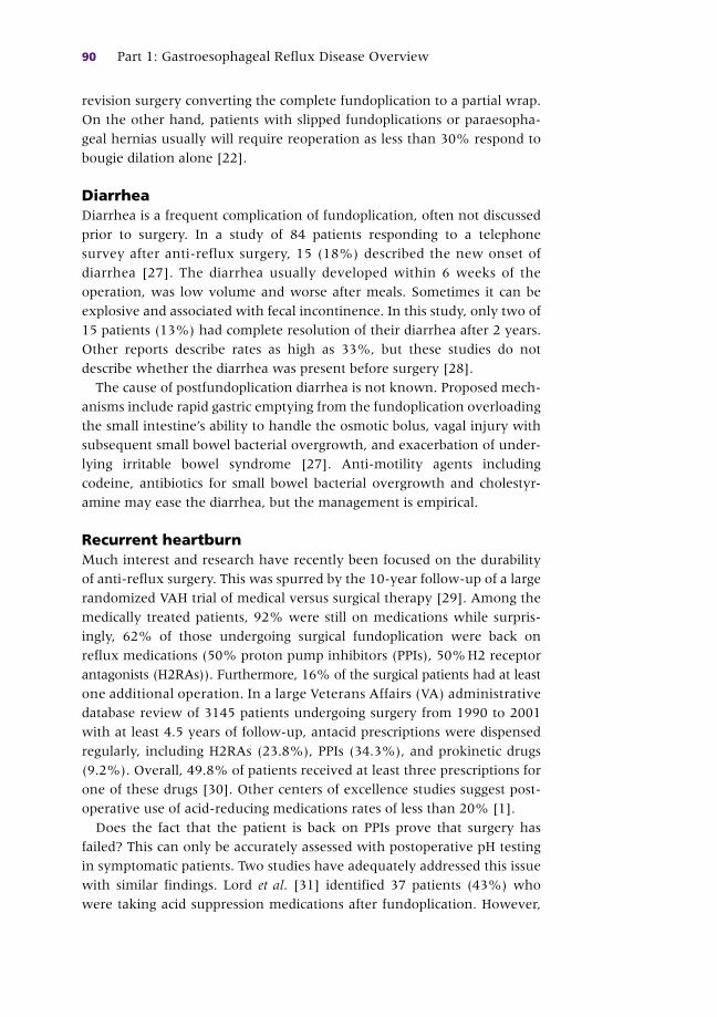
90 Part 1: Gastroesophageal Reflux Disease Overview
revision surgery converting the complete fundoplication to a partial wrap.
On the other hand, patients with slipped fundoplications or paraesopha-
geal hernias usually will require reoperation as less than 30% respond to
bougie dilation alone [22] .
Diarrhea Diarrhea is a frequent complication of fundoplication, often not discussed
prior to surgery. In a study of 84 patients responding to a telephone
survey after anti-reflux surgery, 15 (18%) described the new onset of
diarrhea [27] . The diarrhea usually developed within 6 weeks of the
operation, was low volume and worse after meals. Sometimes it can be
explosive and associated with fecal incontinence. In this study, only two of
15 patients (13%) had complete resolution of their diarrhea after 2 years.
Other reports describe rates as high as 33%, but these studies do not
describe whether the diarrhea was present before surgery [28] .
The cause of postfundoplication diarrhea is not known. Proposed mech-
anisms include rapid gastric emptying from the fundoplication overloading
the small intestine ’ s ability to handle the osmotic bolus, vagal injury with
subsequent small bowel bacterial overgrowth, and exacerbation of under-
lying irritable bowel syndrome [27] . Anti-motility agents including
codeine, antibiotics for small bowel bacterial overgrowth and cholestyr-
amine may ease the diarrhea, but the management is empirical.
Recurrent heartburn Much interest and research have recently been focused on the durability
of anti-reflux surgery. This was spurred by the 10-year follow-up of a large
randomized VAH trial of medical versus surgical therapy [29] . Among the
medically treated patients, 92% were still on medications while surpris-
ingly, 62% of those undergoing surgical fundoplication were back on
reflux medications (50% proton pump inhibitors (PPIs), 50% H2 receptor
antagonists (H2RAs)). Furthermore, 16% of the surgical patients had at least
one additional operation. In a large Veterans Affairs (VA) administrative
database review of 3145 patients undergoing surgery from 1990 to 2001
with at least 4.5 years of follow-up, antacid prescriptions were dispensed
regularly, including H2RAs (23.8%), PPIs (34.3%), and prokinetic drugs
(9.2%). Overall, 49.8% of patients received at least three prescriptions for
one of these drugs [30] . Other centers of excellence studies suggest post-
operative use of acid-reducing medications rates of less than 20% [1] .
Does the fact that the patient is back on PPIs prove that surgery has
failed? This can only be accurately assessed with postoperative pH testing
in symptomatic patients. Two studies have adequately addressed this issue
with similar findings. Lord et al . [31] identified 37 patients (43%) who
were taking acid suppression medications after fundoplication. However,
c06.indd 90 11/15/2012 2:33:08 AM
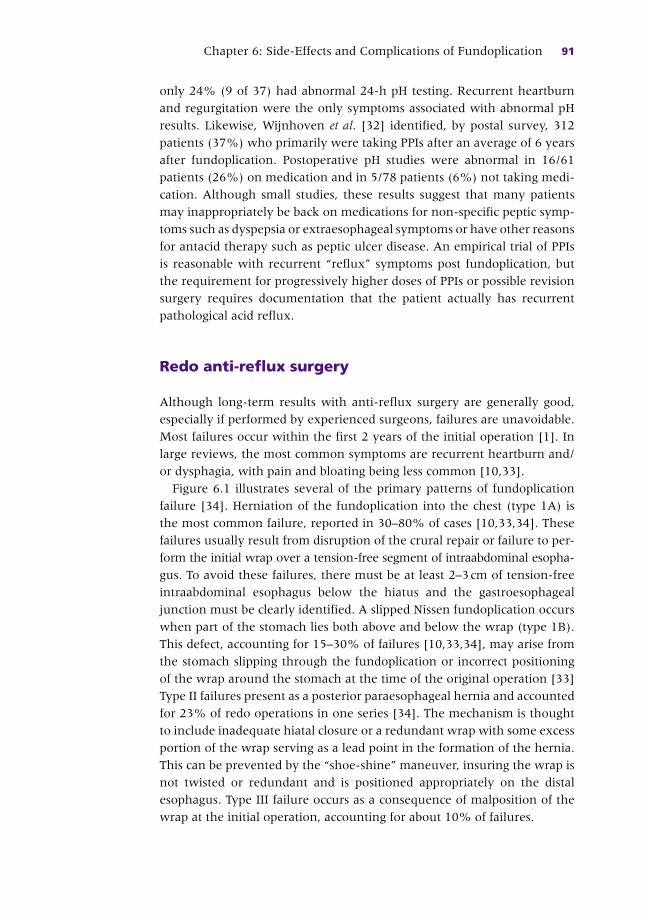
Chapter 6: Side-Effects and Complications of Fundoplication 91
only 24% (9 of 37) had abnormal 24-h pH testing. Recurrent heartburn
and regurgitation were the only symptoms associated with abnormal pH
results. Likewise, Wijnhoven et al . [32] identified, by postal survey, 312
patients (37%) who primarily were taking PPIs after an average of 6 years
after fundoplication. Postoperative pH studies were abnormal in 16/61
patients (26%) on medication and in 5/78 patients (6%) not taking medi-
cation. Although small studies, these results suggest that many patients
may inappropriately be back on medications for non-specific peptic symp-
toms such as dyspepsia or extraesophageal symptoms or have other reasons
for antacid therapy such as peptic ulcer disease. An empirical trial of PPIs
is reasonable with recurrent “reflux” symptoms post fundoplication, but
the requirement for progressively higher doses of PPIs or possible revision
surgery requires documentation that the patient actually has recurrent
pathological acid reflux.
Redo anti-reflux surgery
Although long-term results with anti-reflux surgery are generally good,
especially if performed by experienced surgeons, failures are unavoidable.
Most failures occur within the first 2 years of the initial operation [1] . In
large reviews, the most common symptoms are recurrent heartburn and/
or dysphagia, with pain and bloating being less common [10,33] .
Figure 6.1 illustrates several of the primary patterns of fundoplication
failure [34] . Herniation of the fundoplication into the chest (type 1A) is
the most common failure, reported in 30–80% of cases [10,33,34] . These
failures usually result from disruption of the crural repair or failure to per-
form the initial wrap over a tension-free segment of intraabdominal esopha-
gus. To avoid these failures, there must be at least 2–3 cm of tension-free
intraabdominal esophagus below the hiatus and the gastroesophageal
junction must be clearly identified. A slipped Nissen fundoplication occurs
when part of the stomach lies both above and below the wrap (type 1B).
This defect, accounting for 15–30% of failures [10,33,34] , may arise from
the stomach slipping through the fundoplication or incorrect positioning
of the wrap around the stomach at the time of the original operation [33]
Type II failures present as a posterior paraesophageal hernia and accounted
for 23% of redo operations in one series [34] . The mechanism is thought
to include inadequate hiatal closure or a redundant wrap with some excess
portion of the wrap serving as a lead point in the formation of the hernia.
This can be prevented by the “shoe-shine” maneuver, insuring the wrap is
not twisted or redundant and is positioned appropriately on the distal
esophagus. Type III failure occurs as a consequence of malposition of the
wrap at the initial operation, accounting for about 10% of failures.
c06.indd 91 11/15/2012 2:33:09 AM

92 Part 1: Gastroesophageal Reflux Disease Overview
A tight fundoplication represents an anatomically appropriately placed
wrap which generates too much resistance for the esophageal pump. The
primary complaint is dysphagia rather than heartburn. Studies suggest this
may account for 8–16% of redo operations [10,33] . Careful preoperative
manometry to recognize a weak esophageal pump, performance of the
Type IA Type IB
Type II Type III
Figure 6.1 Common patterns of primary fundoplication failures. Type I failures occur
with displacement of the gastroesophageal junction into the chest through the
esophageal hiatus. Type 1A has herniation of the wrap and GE junction both into the
chest. Type 1B presents with recurrence of the hiatal hernia, but the wrap remains
below the diaphragm. Type II failures are defined as failures secondary to paraesopha-
geal hernia. Type III failures occur as a consequence of malposition of the wrap at the
time of initial surgery, usually on the cardia of the stomach. Reproduced from Hatch
et al . [34] , with permission from Elsevier.
c06.indd 92 11/15/2012 2:33:09 AM

Chapter 6: Side-Effects and Complications of Fundoplication 93
fundoplication over a large bougie (52–56 Fr) or a floppy Nissen
fundoplication or partial wrap may minimize this problem [35] . Other
factors associated with recurrent symptoms include too loose a fundoplica-
tion, vagal injury and pseudoachalasia [35] . Interestingly, complete fundo-
plication disruptions are much less common (3–14%) in the laparoscopic
era than with open operations (greater than 30%) [33] .
The keys to success of a redo fundoplication are careful review of the
patient ’ s prior work-up and repeat studies as necessary, recognition of
esophageal shortening, and complete takedown of the original fundoplica-
tion [35] . Although controversial, it is hypothesized that long-standing
reflux leads to circumferential esophageal scarring and, in more severe
cases, varying degrees of longitudinal scarring and esophageal shortening.
A short esophagus should be suspected in the presence of a moderate or
large non-reducible hiatal hernia, difficult-to-manage peptic stricture or
long-segment Barrett ’ s esophagus [36] . Adequate laparoscopic mobiliza-
tion of the mediastinal esophagus is critical in constructing a tension-free
intraabdominal fundoplication. If esophageal shortening is identified and
adequate intraabdominal esophageal length cannot be obtained, a Collis
gastroplasty will be required.
The most important principle during reoperation is to restore normal
anatomy before recreating the fundoplication [35] . This requires the
wrap be completely taken down, the fundus restored to its normal
location, and the degree of esophageal shortening determined. The
restoration can be tedious and it is very tempting to convert the Nissen
fundoplication to what appears to the surgeon to be a posterior wrap.
Taking this shortcut only increases the likelihood that the patient will not
benefit from the reoperation. Dysphagia is often the result of an improperly
constructed wrap, and to relieve this symptom the fundoplication must
be completely dismantled.
Redo fundoplications must be done by experienced surgeons [1] . In
these settings, laparoscopic approaches to reoperative anti-reflux surgery
offer similar results to open surgery, but conversion rates are higher than
with the initial operation [1] . Reoperation rates range from 0% to 15% for
laparoscopic Nissen fundoplication and 4–10% for laparoscopic Toupet
fundoplication [6] . Compared to primary repair, redo surgery requires
longer operation times, has higher complication rates (20–45%) and the
mortality rates are higher, from 0% to 17% [1,6] . Finally, the likelihood of
success for controlling GERD decreases with subsequent reoperations,
approaching at least 10% per each revisional surgery and being no better
than 50% or less in patients undergoing three or more reoperations [37] .
For these latter unusual cases, serious consideration should be given to
an esophagectomy.
c06.indd 93 11/15/2012 2:33:10 AM

94 Part 1: Gastroesophageal Reflux Disease Overview
References
1 Stefanidis D , Hope WW , Kohn GP , et al . Guidelines for surgical treatment of gastro-
esophageal reflux disease . Surg Endosc 2010 ; 24 : 2647 – 69 .
2 Jensen CD , Gilliam AD , Horgan LF , et al . Day-care laparoscopic Nissen fundoplication .
Surg Endosc 2009 ; 23 : 1745 – 9 .
3 Pizza F , Rossetti G , Limongelli P , et al . Influence of age on outcome of total laparoscopic
fundoplication for gastroesophageal reflux disease . World J Gastroenterol 2007 ; 13 : 740 – 7 .
4 Finks JF , Wei Y , Birkmeyer JD . The rise and fall of antireflux surgery in the United
States . Surg Endosc 2006 ; 20 : 1698 – 701 .
5 Wang YR , Dempsey DT , Richter JE . Trends and perioperative outcomes of inpatient
antireflux surgery in the United States, 1993–2006 . Dis Esophagus 2011 ; 24 ( 4 ): 215 – 23 .
6 Agency for Healthcare Research and Quality – the Effective Healthcare Program .
Comparative effectiveness of management strategies for gastroesophageal reflux disease – an update
to the 2005 report . Washington, DC : Agency for Healthcare Research and Quality , 2011 .
CASE STUDY
A 42-year-old man was referred to his local gastroenterologists for surgical evaluation due to long-standing heartburn and regurgitation. His heartburn was improved by a b.i.d. proton pump inhibitor, but the regurgitation was still a daily problem after meals and interfered with sleep. Endoscopy showed a 3 cm hiatal hernia with Los Angeles Grade B esophagitis despite his PPI regimen. Esophageal manometry found a low LES pressure (5 mmHg) with peristalsis present but in the low normal range (25–40 mmHg). He underwent a “loose” Nissen fundoplication performed laparoscopically by an experienced surgeon over a 54 Fr bougie.
For the first 6 weeks postoperatively, he was on a soft diet. Initially, postprandial bloating and increased flatus were an issue. These resolved, but the patient began to experience substernal chest pain and dysphagia if he ate too rapidly or the food was of firm texture (i.e. meats, hard breads and sandwiches). He was pleased his heartburn and regurgitation were relieved, but these new symptoms interfered with his general activities, especially his many evening business dinners. His gastroenterologist performed an endoscopy, finding the fundoplication to be intact, and there was no resistance to passage of the endoscope. Repeated bougie dilations to 20 mm (60 Fr) gave transient relief for about 2 weeks but then the dysphagia recurred, requiring modification of his diet.
A discussion began considering the possibility of surgically revising the fundoplication to a partial wrap. Preoperative testing included a barium swallow showing the wrap was intact below the diaphragm, intermittent peristalsis was observed and a 13 mm tablet passed over 3–5 min after drinking more barium. Esophageal manometry found an increase in LES pressure to 35 mmHg with incomplete relaxation. Peristalsis was present after 70% of wet swallows with the amplitude increased on average to 45 mmHg. A diagnosis of a “tight” Nissen fundoplication was made. Prior to revising the fundoplication, the gastroenterologist and surgeon suggested one last attempt at aggressive dilation with a 30 mm pneumatic balloon. This was performed under fluoroscopic guidance. The patient tolerated the procedure and has been symptom free for the last 6 months.
c06.indd 94 11/15/2012 2:33:10 AM
Chapter 6: Side-Effects and Complications of Fundoplication 95
7 Dominitz JA , Dire CA , Billingsley KG , Todd-Steinberg JA . Complications of antireflux
medication use after antireflux surgery . Clin Gastroenterol Hepatol 2006 ; 4 : 299 – 305 .
8 Lundell L. Complications of anti-reflux surgery . Best Pract Res Clin Gastroenterol
2004 ; 18 ( 5 ): 935 – 45 .
9 Zacharoulis D , O’Boyle CJ , Sedman PC , et al . Laparoscopic fundoplication: a 10-year
learning curve . Surg Endosc 2006 ; 20 : 1662 – 70 .
10 Carlson MA , Frantzides CT . Complications and results of primary minimally invasive
antireflux procedures: a review of 10,735 reported cases . J Am Coll Surg
2001 ; 193 : 428 – 39 .
11 Bizekis C , Kent M , Luketich J . Complications after surgery for gastroesophageal
reflux disease . Thorac Surg Clin 2006 ; 16 : 99 – 108 .
12 Neuhauser B , Hinder RA . Laparoscopic reoperation after failed antireflux surgery .
Semin Laparosc Surg 2001 ; 8 : 281 – 6 .
13 Rantanen TK , Salo JA , Sipponer JT . Fatal and life-threatening complications in
antireflux surgery: analysis of 5,502 operations . Br J Surg 1999 ; 86 : 1573 – 7 .
14 Flum DR , Koepsell T , Heagerty P , Pellegrini C . The nationwide frequency of major
adverse outcome in antireflux surgery and the role of surgeon experience, 1992–1997 .
J Am Coll Surg 2002 ; 195 : 611 – 18 .
15 Rogers DM , Herrington JL , Morton C . Incidental splenectomy associated Nissen
fundoplication . Ann Surg 1980 ; 191 : 153 – 6 .
16 Bradshaw WA , Gregory BC , Finley C , et al . Frequency of postoperative nausea and
vomiting in patients undergoing laparoscopic foregut surgery . Surg Endosc
2002 ; 16 : 777 – 80 .
17 Meyers BF , Soper NJ . Complications of surgery for gastroesophageal reflux . In:
Patterson GA , Cooper JD , Deslauriers J , Lerut AEMR , Luketic JD , Rice TW (eds)
Person ’ s thoracic and esophageal surgery , 3rd edn. Philadelphia : Churchill Livingstone ,
2008 : pp. 376 – 86 .
18 Spechler SJ . The management of patients who have “failed” antireflux surgery . Am J
Gastroenterol 2004 ; 99 : 552 – 61 .
19 Spechler SJ . Comparison of medical and surgical therapy for complicated GERD in
veterans. Department of Veterans Affairs Gastroesophageal Reflux Study Group .
N Engl J Med 1992 ; 326 : 786 – 92 .
20 McCallum RW , Berkowitz DM , Lerner E . Gastric emptying in patients with gastro-
esophageal reflux disease . Gastroenterology 1981 ; 80 : 285 – 91 .
21 Bias JE , Samson M , Boudesteijn CG , et al . Impact of delayed gastric emptying on the
outcome of antireflux surgery . Ann Surg 2001 ; 234 : 139 – 46 .
22 Wo JM , Trus TL , Richardson WS , et al . Evaluation and management of post
fundoplication dysphagia . Am J Gastroenterol 1996 ; 91 : 2318 – 22 .
23 Fibbe C , Layer P , Keller J , et al . Esophageal motility in reflux disease before and
after fundoplication: a prospective randomized, clinical and manometric study .
Gastroenterology 2001 ; 121 : 5 – 14 .
24 Watson DL . Laparoscopic treatment of gastroesophageal reflux disease . Best Pract Res
Clin Gastroenterol 2004 ; 18 : 19 – 35 .
25 Malki-Chowla N , Gorecki P , Bammer T , et al . Dilation after fundoplication: timing,
frequency, indications and outcome . Gastrointest Endosc 2002 ; 55 : 219 – 23 .
26 Hui JM , Hunt DR , Carle DJ , et al . Esophageal pneumatic dilation for post fundoplication
dysphagia: safety, efficacy and predictors of outcome . Am J Med 2002 ; 97 : 2986 – 91 .
27 Klaus A , Hinder RA , deVault KR , Achem SR . Bowel dysfunction after laparoscopic
antireflux surgery: incidence, severity and clinical course . Am J Med 2003 ;
114 : 6 – 9 .
c06.indd 95 11/15/2012 2:33:10 AM

96 Part 1: Gastroesophageal Reflux Disease Overview
28 Swanstrom L , Wayne R . Spectrum of gastrointestinal symptoms after laparoscopic
fundoplication . Am J Surg 1994 ; 167 : 538 – 41 .
29 Spechler SJ , Lee E , Ahnen D , et al . Long-term outcome of medical and surgical
therapies for gastroesophageal reflux disease: follow-up of a randomized controlled
trial . JAMA 2001 ; 285 : 2331 – 8 .
30 Domintz JA , Dire C , Billingsley KG , Todd-Steinberg JA . Complications and antireflux
medication use after antireflux surgery . Clin Gastroenterol Hepatol 2006 ; 4 : 299 – 305 .
31 Lord RVN , Kaminski A , Oberg S , et al . Absence of gastroesophageal reflux disease in
a majority of patients taking acid suppression medications after Nissen fundoplica-
tion . J Gastrointest Surg 2002 ; 6 : 3 – 10 .
32 Wijnhoven BPL , Lally CJ , Kelly JJ , et al . Use of antireflux medication after antireflux
surgery . J Gastrointest Surg 2008 ; 12 : 510 – 17 .
33 Hunter JG , Smith CS , Branum GD , et al . Laparoscopic fundoplication failure: patterns
of failure and response to fundoplication revision . Ann Surg 1999 ; 23 : 595 – 606 .
34 Hatch KF , Daily MF , Christiansen BJ , Glasgow RE . Failed fundoplications . Am J Surg
2004 ; 188 : 786 – 91 .
35 Bizekis C , Kent M , Luketich J . Complications after surgery for gastroesophageal
reflux disease . Thorac Surg Clin 2006 ; 16 : 99 – 108 .
36 Gastal OL , Hagen JA , Peters JH , et al . Short esophagus: analysis of predictors and
clinical implications . Arch Surg 1999 ; 134 : 633 – 6 .
37 Smith CD , McClusky DA , Rajad MA , et al . When fundoplications fails: redo? Ann
Surg 2005 ; 241 : 861 – 71 .
c06.indd 96 11/15/2012 2:33:10 AM

PART 2
Gastroesophageal Reflux Disease: Management of Specific Clinical Presentations
p02.indd 97 11/15/2012 2:46:47 AM

99
Practical Manual of Gastroesophageal Reflux Disease, First Edition.
Edited by Marcelo F. Vela, Joel E. Richter and John E. Pandolfino.
© 2013 John Wiley & Sons, Ltd. Published 2013 by John Wiley & Sons, Ltd.
CHAPTER 7
Evaluation and Management of Refractory Gastroesophageal Reflux Disease John E. Pandolfino and Sabine Roman Division of Gastroenterology , Northwestern University , Chicago , IL , USA
Key points • Four phenotypes of proton pump inhibitor non-responders are described:
– phenotype 1: persistent acid reflux
– phenotype 2: non-acid reflux
– phenotype 3: functional overlap with gastroesophageal reflux disease
– phenotype 4: functional heartburn.
• Choice of physiological testing is based on pretest probability for baseline
gastroesophageal reflux disease.
• Treatment strategy should be based on proton pump inhibitor non-responder
phenotype.
Potential pitfalls • Proton pump inhibitor non-responder phenotypes are defined in patients compliant
to optimized proton pump inhibitor therapy.
• Physiological testing should be performed off therapy when the pretest probability
for baseline gastroesophageal reflux disease is low.
Introduction
Proton pump inhibitor (PPI) therapy for gastroesophageal reflux disease
( GERD) is highly effective; however, there are a substantial number of
patients who do not respond to this therapy and seek further medical care.
While success rates in healing esophagitis may reach 80–90%, a large
number of patients (up to 30%) remain symptomatic or unsatisfied despite
continued PPI therapy [1,2] . Additionally, patients with non-erosive reflux
c07.indd 99 11/15/2012 2:29:35 AM

100 Part 2: Gastroesophageal Reflux Disease
disease (NERD) tend to have even lower response rates to PPI therapy,
with symptom resolution rates ranging from 30% to 70% [3] . Given the
large baseline prevalence of GERD, with approximately 7,000,000
ambulatory care visits per year constituting 17.5% of all digestive system
diagnoses, PPI non-responders now represent a substantial utilization of
healthcare resources in gastroenterology clinics [4] .
Proton pump inhibitor non-responders are an extremely challenging
population to manage for many reasons. First, there is a lack of consensus
regarding the definition of a PPI non-responder or refractory GERD; this is
largely due to the heterogeneity of the patient group, including individual
response to medication, type of symptoms, and the mechanism behind
continued symptom generation. For example, some patients have a partial
response to medication that may be associated with a reduction of heart-
burn or resolution of the primary symptom with continued secondary
symptoms. Others will have no change in their symptoms despite aggres-
sive acid suppressive therapy. Furthermore, patients are heterogeneous in
terms of the mechanism behind the generation of symptoms in this patient
population. Some patients will exhibit no evidence of abnormal reflux or
symptom reflux correlation, while others will have refractory symptoms
with continued acid or non-acid reflux-related symptoms. Thus, all PPI
non-responders are not the same and many do not have refractory GERD.
The following review will focus on evaluating patients with reflux
symptoms that are not responding to PPI therapy (PPI non-responders).
The first section will focus on defining the phenotypes of PPI non-
responders in the context of clinical presentation and objective
physiological evidence of abnormal reflux. The second section will focus
on a management algorithm to assess and evaluate mechanisms behind
the lack of response to PPI therapy. Finally, the third section will focus on
alternative strategies to manage patients not responding to PPI therapy
beyond acid suppression.
Definition of refractory gastroesophageal reflux disease and proton pump inhibitor non-responders
How to define proton pump inhibitor non-responders? As PPIs are extremely effective at healing virtually all esophagitis grades
and virtually all patients referred for refractory GERD symptoms are taking
or have taken PPI therapy, most patients will have a negative endoscopy.
Patients who continue to have erosive esophagitis despite PPI therapy are
a true refractory GERD subtype that typically does not require a sophisti-
cated work-up and therapy can be focused on escalating anti-reflux
c07.indd 100 11/15/2012 2:29:36 AM
Chapter 7: Evaluation and Management of Refractory 101
therapy based on endoscopic findings. Although there can be alternative
explanations apart from acid reflux that may cause esophagitis, such as pill
esophagitis and various infections, endoscopy is very specific for defining
peptic injury and further physiological evaluation may only be required to
determine why the PPI is not working. However, the more important
group of patients that we are currently evaluating in gastroenterology
clinics are the endoscopy-negative patients with continued symptoms.
Therefore, the real management issue in current practice is focused on
dealing with refractory symptoms in patients who are on optimized PPI
therapy. A more accurate definition of this clinical dilemma should focus
on the lack of response to therapy and thus, PPI non-responder is a much
more appropriate definition. The mechanism behind PPI non-response
may be related to non-reflux pathophysiology or to refractory gastro-
esophageal reflux and therefore, the latter is actually a subcategory of PPI
non-response causes.
Two additional important issues must be addressed before defining
patients as PPI non-responders: the response to PPI therapy in terms of
partial or complete non-response, and what dose constitutes an appro-
priate trial before someone is considered a failure. In terms of response to
therapy, some patients may have a partial response with their primary
symptom being partially reduced or completely reduced with continued
secondary symptoms. In contrast, some patients may have absolutely no
response to therapy and their symptoms will show no improvement with
escalation of therapy or no worsening symptoms with discontinuation of
therapy. These clinical issues are extremely important in defining the
pretest probability of whether or not these patients actually have
abnormal reflux at baseline. Given the success of PPI therapy in reducing
acid reflux-related symptoms, it would be unusual for patients with
GERD to have absolutely no response to PPI therapy. Patients who do not
respond at all and can undergo discontinuation of PPI therapy without
an escalation of symptoms likely have an alternative diagnosis that is not
acid or reflux mediated. Therefore, this first question focused on response
to therapy does provide an extremely useful starting point to assess pre-
test probability that gastroesophageal reflux is causing the current
subjective complication.
Even before one assesses the level of response, one would also have to
document compliance to medical therapy at an appropriate dose sufficient
to treat most grades of reflux severity. In evaluating the dose of proton
pump inhibitor therapy that would reasonably be seen as a failure, one
would likely utilize a dose that is higher than the current FDA-approved
doses for the various available PPIs. Based on current treatment guidelines
[5] and physiological testing data available on patients on single-dose
therapy and double-dose therapy [6] , a reasonable approach would be
c07.indd 101 11/15/2012 2:29:36 AM

102 Part 2: Gastroesophageal Reflux Disease
to consider patients who fail twice the FDA-approved dose in either a
single-dose or split (b.i.d.)-dose regimen as a failure. Most guidelines
advocate for physiological reflux testing after patients have attempted an
escalation of PPI therapy to double dose [5,7] . In addition, studies assessing
abnormal acid exposure on PPI therapy in patients with continued symp-
toms suggest that up to 30% of symptomatic patients on single-dose PPI
therapy will have abnormal acid exposure. In contrast, less than 10% of
symptomatic patients on double-dose PPI therapy will have abnormal acid
exposure. Given that the primary mechanism of PPI therapy is to reduce
overall acid reflux and reflux burden, it would appear that the yield of
reducing acid burden by increasing the PPI dose to double the FDA-
approved dose is significant and would warrant this degree of escalation as
the threshold for someone to be considered a PPI failure.
Thus, in addition to defining phenotypes of PPI non-responders, defining
level of response to therapy on adequate treatment is an important com-
ponent of the evaluation and management of these refractory patients.
Conceptual phenotypes of proton pump inhibitor non-responders Once patients are documented to have a poor or inadequate response to
optimized PPI therapy (double-dose PPI), the next most important steps
are to document whether or not the patients actually have abnormal gas-
troesophageal reflux and to document whether or not their symptoms
experienced on medication are associated with reflux. By focusing on
these two specific issues, one can define four specific phenotypes of PPI
non-responders. This will require further physiological testing that will
focus on ambulatory reflux monitoring and will be discussed in the section
describing the appropriate utilization of ambulatory reflux monitoring.
The four specific phenotypes of PPI non-responders are described below
and illustrated in Table 7.1 .
• Phenotype 1: persistent acid reflux
• Phenotype 2: non-acid reflux
• Phenotype 3: functional overlap with GERD
• Phenotype 4: functional heartburn
As mentioned above, the first distinction in the phenotypes is focused on
determining whether or not the patient has baseline abnormal gastro-
esophageal reflux. Phenotypes 1–3 are patients who have abnormal
gastroesophageal reflux off PPI therapy, but continue to have symptoms
that are either partially treated or secondary complaints that may (pheno-
types 1 and 2) or may not (phenotype 3) be related to reflux. Phenotypes
1 and 2 have continued symptoms that are related to reflux and these
subtypes are truly refractory GERD. Phenotype 1 will have evidence of
abnormal acid exposure on ambulatory pH reflux testing and/or a positive
c07.indd 102 11/15/2012 2:29:36 AM

Chapter 7: Evaluation and Management of Refractory 103
symptom-reflux correlation in the context of overt abnormal acid exposure
or normal acid exposure associated with an acid hypersensitivity. Similarly,
phenotype 2 will also have a positive symptom reflux correlation; how-
ever, the correlation is predominantly with weakly acidic reflux events.
Phenotype 2 will have no overt abnormality in distal esophageal acid
exposure and is likely hypersensitive to volume, other components of the
gastric refluxate or refluxate with a pH above 4. These particular pheno-
types will likely respond to an escalation of anti-reflux therapy focused on
reducing acid burden and the overall number of reflux events.
It is important to distinguish phenotypes 3 and 4 from phenotypes
1 and 2 because they should exhibit a lack of response to more aggressive
anti-reflux therapy. However, phenotype 3 patients do have baseline reflux
disease and many require PPI therapy to maintain control of other symp-
toms that are related to abnormal reflux. This particular group of patients
will exhibit pathological acid reflux off PPI therapy and normalization on
PPI therapy with a negative symptom correlation with all types of reflux
events. Ambulatory reflux testing on PPI therapy incorporating impedance
may reveal an increased number of overall reflux events suggesting under-
lying baseline GERD. Thus, these patients will be unable to discontinue PPI
therapy and will require an evaluation for alternative causes and therapy
beyond reflux suppression. In contrast, phenotype 4 patients will have no
evidence of abnormal reflux or a symptom-reflux correlation at baseline or
Table 7.1 Phenotypes of PPI non-responders based on physiological testing.
Phenotype 1 Phenotype 2 Phenotype 3 Phenotype 4
Persistent acid
reflux
Non-acid reflux Functional overlap
with GERD
Functional
heartburn
Acid esophageal
exposure off PPI * + + + −
Acid esophageal
exposure on PPI *
+ − − −
Excessive number of
reflux events with
impedance on PPI
+ /− + /− + −
Positive reflux-
symptom association
with impedance on PPI
+ /− + − −
* Prolonged wireless pH monitoring both off and on PPI may be used to evaluate esophageal acid
exposure in a single examination [19] .
GERD, gastroesophageal reflux disease; PPI, proton pump inhibitor.
c07.indd 103 11/15/2012 2:29:36 AM

104 Part 2: Gastroesophageal Reflux Disease
CASE STUDY
A 28-year-old woman complained of heartburn, chest pain and gurgling in the throat for the past 10 years. She tried different PPIs (dexlansoprazole, omeprazole, esomeprazole) without any improvement of her symptoms. The symptoms were worse after a meal and with stress. She had no significant previous health history. Her Body Mass Index (BMI) was 21.4 kg/m 2 . No hiatal hernia was seen on esophagogastroduodenscopy. Two small tongues of pink mucosa extending 1 cm maximally from the squamocolumnar junction were observed. The corresponding biopsies showed esophageal squamous and gastric mucosa with chronic carditis. A 24-h pH impedance monitoring was performed on PPI therapy (esomeprazole 40 mg b.i.d.). The esophageal acid exposure was normal at 0.6%. The number of reflux episodes was also normal (23 episodes, three acid and 20 non-acid). During the recording, the patient reported 17 episodes of heartburn. The symptom index and the symptom association probability were both equal to 0%.
What do you propose for the management of this patient? As the pH impedance on PPI was normal without any positive correlation between symptom and reflux event, the current symptoms presented by the patient were not linked to persistent pathological GERD on PPI. An alternative therapy triggering visceral hypersensitivity should be proposed.
Should PPI therapy be discontinued in this patient? In the absence of GERD documentation off medicine, it is not possible to differentiate a phenotype 4 (functional heartburn) from a phenotype 3 (functional overlap with GERD). An evaluation off PPI (pH or pH impedance monitoring) would be helpful to diagnose a “baseline” GERD. If the patient presented a pathological acid esophageal exposure, PPIs should be maintained. Otherwise they should be stopped.
on PPI therapy. This group of patients can be labeled as functional heartburn
once an endoscopy has ruled out alternative causes and manometry has
not revealed an underlying esophageal motor disorder. These patients
should have their PPI therapy discontinued and will likely require therapy
focused beyond acid suppression and reflux inhibition.
Evidence to support this phenotypic classification can be found in recent
studies assessing large series of referral patients for combined pH imped-
ance testing both off and on PPI therapy. Savarino et al . noted in a series of
200 patients with non-erosive reflux disease that 27% had normal esoph-
ageal acid exposure and negative symptom association probability on 24-h
pH impedance monitoring performed off PPI (phenotype 4) [8] . Eleven
percent of the patients presented with a positive association between
symptoms and non-acid reflux events only in the absence of PPI therapy.
Mainie et al . also observed different phenotypes of PPI non-responders in a
series of 168 patients who underwent 24-h pH impedance monitoring on
PPI for refractory GERD symptoms [9] . Eleven percent of subjects had a
persistent pathological esophageal acid exposure despite PPI twice daily
(phenotype 1), 31% had a positive association between symptoms and
c07.indd 104 11/15/2012 2:29:36 AM

Chapter 7: Evaluation and Management of Refractory 105
non-acid reflux (phenotype 2), and 58% had no evidence of pathological
reflux and/or positive association on PPI (phenotypes 3 and 4). As data on
esophageal pH monitoring off PPI were not available for these patients, it
is not possible to differentiate phenotypes 3 and 4.
Diagnostic algorithm for managing proton pump inhibitor non-responders
The evaluation of patients who are not responding to PPI therapy begins
by first documenting that the patient is compliant with medical
management. This is an important component of the work-up of the
refractory patient as a large population-based study suggests that over
50% of patients on PPI therapy are not compliant with medication [10,11] .
In addition to daily compliance, less than 50% of patients take their PPI
optimally (timing, frequency, and dose) [12] . Most of the current guide-
lines support empiric treatment with single FDA-approved dose PPI
therapy for a 4–8-week period for a patient presenting with typical GERD
symptoms [13] . If the patient fails single-dose therapy, it is reasonable to
escalate therapy to double dose as there is little risk to this practice and a
small group of patients may respond.
One caveat to empiric treatment focuses on the presence or absence of
warning signs. Although there is controversy regarding the predictive
value of warning symptoms [14] , an upfront endoscopy is reasonable if
there is evidence of dysphagia, odynophagia, gastrointestinal (GI) bleeding,
unintentional weight loss, early satiety or age at presentation greater than
55 to rule out significant complications and malignancy. In the absence of
warning signs, patients are typically not referred for endoscopy unless they
have failed a course of optimized PPI therapy. The timing of endoscopy in
the algorithm and the dose of PPI that is considered a failure which war-
rants endoscopic evaluation are unclear. However, we would recommend
a trial of double-dose therapy.
A diagnostic algorithm is presented in Figure 7.1 . The test choice is firstly
determined by the pretest probability for baseline GERD. This algorithm
allows the differentiation of the different phenotypes of PPI non- responders.
As mentioned above, patients who fail initial single-dose FDA-approved
PPI therapy will have their acid suppression therapy increased to at least
double the FDA-approved dose [5] . The data to support the yield of an
escalation of PPI therapy to double dose in patients not responding to
single dose are marginal and likely in the range of 10–20% [15] . Thus,
the majority of patients who are escalated to double dose still have
continued symptoms and do warrant further evaluation with ambulatory
reflux testing.
c07.indd 105 11/15/2012 2:29:36 AM

No
resp
onse
Trea
tmen
t opt
imiz
atio
n
Pat
holo
gica
l eso
phag
eal a
cid
expo
sure
Pos
itive
sym
ptom
–ref
lux
asso
ciat
ion
Nor
mal
eso
phag
eal
acid
exp
osur
eN
egat
ive
sym
ptom
-re
flux
asso
ciat
ion
Phe
noty
pe 3
:F
unct
iona
lov
erla
p w
ithG
ER
D
?P
heno
type
4:
Fun
ctio
nal h
eart
burn
Phe
noty
pe 2
:N
on-a
cid
reflu
xP
heno
type
1:
Pre
sist
ent a
cid
reflu
x
Nor
mal
num
ber
of r
eflu
xev
ents
AN
DN
o po
sitiv
e te
st o
ff m
edic
ine
Pat
holo
gica
les
opha
geal
aci
dex
posu
reA
ND
/ O
RS
tron
g po
sitiv
esy
mpt
om-a
cid
reflu
x as
soci
atio
n
Nor
mal
eso
phag
eal
acid
exp
osur
eA
ND
Pos
itive
sym
ptom
-re
flux
asso
ciat
ion
Nor
mal
eso
phag
eal
acid
exp
osur
eA
ND
Neg
ativ
e sy
mpt
om-
reflu
x as
soci
atio
n
Abn
orm
ality
on
prev
ious
end
osco
py: B
arre
tt’s
esop
hagu
s, e
soph
agiti
s, la
rge
hiat
us h
erni
apo
sitiv
e pH
stu
dy o
ff m
edic
atio
ncl
inic
al p
rese
ntat
ion
(reg
urgi
tatio
n)
Hig
h pr
etes
t pro
babi
lity
for
base
line
GE
RD
:
Sta
ndar
d pH
–cat
hete
r m
onito
ring
com
bine
d pH
–im
peda
nce
Pro
long
ed w
irele
ss p
H m
onito
ring
Am
bula
tory
ref
lux
test
ing
OF
F P
PI
Com
bine
d pH
-impe
danc
e
Am
bula
tory
ref
lux
test
ing
ON
PP
I
Lack
of a
bnor
mal
ity o
n pr
evio
us e
ndos
copy
Clin
ical
pre
sent
atio
n (h
eart
burn
, ext
ra-
esop
hage
al s
ympt
oms)
Low
pre
test
pro
babi
lity
for
base
line
GE
RD
:
PP
I non
-res
pond
ers
com
plai
nt o
n op
timiz
ed P
PI t
hera
py
Fig
ure
7.1
D
iagn
osi
s alg
ori
thm
to iden
tify
th
e f
ou
r ph
en
oty
pes
of
PPI
non
-resp
on
ders
. M
odif
ied f
rom
Pan
dolf
ino a
nd V
ela
[16],
wit
h p
erm
issi
on
from
Els
evie
r.
c07.indd 106 11/15/2012 2:29:36 AM

Chapter 7: Evaluation and Management of Refractory 107
The approach to ambulatory reflux monitoring is outlined in Figure 7.1
and mirrors the American Society for Gastrointestinal Endoscopy (ASGE)
guidelines published in 2009 [16] . Decisions regarding the type of device
utilized for the evaluation of PPI non-responders are beyond the scope of this
particular chapter and are covered in Chapter 3. However, a few important
concepts should be reviewed regarding whether or not to study patients off
or on medical therapy.
The decision to study patients off and on medication should focus on
the pretest probability that the patient has baseline pathological acid
reflux and also on the specific question that needs to be answered. If the
pretest probability is low based on the clinical presentation and lack of
overt abnormalities on endoscopy (esophagitis, Barrett ’ s esophagus, large
hiatus hernia), the first and most important question in the evaluation of
refractory symptoms should be whether or not the patient actually has
baseline abnormal reflux/pathological acid exposure. Thus, these patients
should have ambulatory pH monitoring off medication to document
whether or not abnormal reflux/pathological acid exposure is present at
baseline (Plate 7.1). This can be accomplished with a standard pH cath-
eter, combined pH impedance catheter or wireless pH capsule. Data sug-
gesting that the wireless capsule pH monitoring system is superior in
diagnosing abnormal reflux are limited; however, there are studies that
suggest that extending the duration of pH monitoring from 24 h to 48–96 h
may improve the yield of documenting abnormal reflux. For example,
among 38 patients with negative 24-h pH catheter-based results,
prolonged (48–96 h) wireless pH monitoring revealed a pathological acid
esophageal exposure in 37% and 47% using average and worst day anal-
ysis respectively [17] . Overall, GERD was diagnosed in 61% (average
analysis) and 76% (worst day) based on either pathological acid exposure
or positive symptom association.
If the pretest probability for baseline GERD is high secondary to a docu-
mented abnormality found previously on endoscopy (large hernia, esopha-
gitis, Barrett ’ s esophagus), a positive pH study off medication or strong
clinical scenario where the patient is likely to have baseline GERD, the
patient should undergo ambulatory reflux testing on PPI therapy using
combined pH impedance (Plate 7.2). In this instance, the question of
whether or not baseline GERD exists is less important and the focus of the
evaluation is on defining the mechanism behind the refractory symptoms.
Moreover, it has been shown that 93% of patients with symptoms refractory
to twice-daily dosage of PPI and abnormal pH impedance monitoring on
PPI have abnormal acid esophageal exposure off PPI [18] . Thus, this tech-
nology can define the remaining phenotypes (2–4) and direct appropriate
management. In the absence of evaluation off PPI, the distinction between
phenotypes 3 and 4 may be difficult. If the total number of reflux events is
c07.indd 107 11/15/2012 2:29:36 AM
108 Part 2: Gastroesophageal Reflux Disease
normal, the patient likely fits a phenotype consistent with functional
heartburn. However, the threshold number of reflux events to distinguish
phenotype 3 from 4 is not clear when patients are studied on PPI therapy.
An alternative to answer the question of whether to test on or off medica-
tion is prolonged wireless pH monitoring both off and on PPI (BOOP
testing) in a single examination (2 days off PPI followed by 2 days on
PPI) [19] . This test is sufficient to identify patients with phenotypes 1 and
4 but does not allow one to differentiate phenotype 2 from phenotype 3.
Treatment of proton pump inhibitor non-responders
Unfortunately, the treatment strategies and management options for PPI
non-responders are poorly defined and draw from open label studies or
small clinical trials. In addition, most trials assessing refractory GERD have
not clearly defined the relevant phenotypes and thus, it has been difficult
to assess the overall benefit of specific therapies. Given this fact, this sec-
tion will focus on the available therapies and also discuss which phenotype
would be most likely to respond to the various therapies.
Acid suppression Although the majority of this chapter has focused on the evaluation and
management of refractory symptoms in endoscopy-negative patients, one
important aspect of true refractory GERD is patients with refractory eso-
phagitis, peptic strictures, and continued symptoms in Barrett ’ s esophagus.
These patients may require higher doses of PPI therapy or other adjunct
treatment focused on reducing the overall reflux burden. These patients
tend to have the most severe abnormalities in the anti-reflux barrier
(hiatus hernia, hypotensive lower esophageal sphincter (LES)) and eso-
phageal clearance (weak peristalsis, hiatus hernia). Improving acid suppres-
sion can be accomplished by either increasing the dose of PPI therapy or
adding an H2 blocker at night [20] . The data supporting the efficacy of
these particular treatments are limited, but this strategy represents a rea-
sonable approach with limited risk and a valid endpoint to follow on
endoscopy. Occasionally, ambulatory pH monitoring without impedance
can be performed to assess the effectiveness of PPI therapy by studying
patients on medication. However, this is an exception and once again, it is
probably better to study these patients with combined pH impedance on
PPI therapy.
The data supporting the efficacy of increasing PPI therapy to double dose
or adding an H2 blocker at night in endoscopy-negative patients with
refractory symptoms are limited. The therapeutic benefit of increasing PPI
c07.indd 108 11/15/2012 2:29:36 AM
Chapter 7: Evaluation and Management of Refractory 109
dose was reported in a study by Fass [15] . In patients with persistent
heartburn on PPI once-daily treatment, two therapeutic strategies were
compared: increasing PPI dosage to twice daily (lansoprazole 30 mg b.i.d)
versus switching to another PPI (esomeprazole 40 mg). These two strat-
egies were equivalent in terms of heartburn symptom control. However,
these results would not necessarily be similar with other PPIs as esomepra-
zole may have a better bioavailability than the other PPIs. Finally, Becker
et al . showed that increasing PPI dosage was more effective in patients with
pathological findings in pH impedance monitoring on standard PPI therapy
(persistent abnormal acid exposure and/or increased number of reflux
events) than in patients without any pathological findings on pH imped-
ance (91% symptom relief versus 43%, P < 0.001) [21] . This study empha-
sizes the benefit of acid suppression intensification in patients with
phenotypes 1 and 2.
Another strategy to augment acid suppression has been focused on add-
ing an H2 blocker at night to help control the histamine-generated noc-
turnal acid breakthrough common with PPI therapy [22] . This strategy
was shown to obliterate nocturnal acid breakthrough on pH studies but a
clinical correlation with symptom improvement is still unproven.
Furthermore, there have been potential issues with tachyphylaxis as
evidenced by a study by Fackler et al . that revealed a blunting of the H2
blocker effect after 4 weeks [23] . However, the addition of an H2 blocker
at night is reasonable as the medication is safe and could potentially be
effective in a small proportion of patients with significant nocturnal symp-
toms, especially if the medication is used intermittently when symptoms
are exacerbated.
Treatment beyond acid suppression A number of therapies have been proposed to treat refractory symptoms in
PPI non-responders that go beyond acid suppression and focus on reflux
inhibition and reducing visceral hypersensitivity.
Reflux inhibition Targeting transient lower esophageal sphincter relaxations (TLESRs) pro-
vides an opportunity to develop reflux inhibitors. Baclofen, a gamma-
aminobutyric acid B (GABA B ) receptor agonist, is a potential add-on
treatment for patients with persistent symptomatic reflux events despite
PPI. It decreases the number of postprandial acid and non-acid reflux
events [24] . The dose of 20 mg three times daily has been proposed in
refractory GERD [25] . However, no controlled trial of baclofen in PPI
non-responders is available. Moreover, because the drug crosses the blood–
brain barrier, neurological side-effects including somnolence, dizziness,
and drowsiness are important limiting factors in the use of baclofen in
c07.indd 109 11/15/2012 2:29:36 AM

110 Part 2: Gastroesophageal Reflux Disease
clinical practice. Novel GABA B agonists are currently in clinical development
for the treatment of refractory GERD. Arbaclofen placarbil, a pro-drug of
the pharmacologically active R -isomer of baclofen, decreased the number
of postprandial reflux events and associated symptoms [26] . The safety
profile could be better compared to baclofen. Lesogabaran, a new GABA B
agonist, was developed to overcome the side-effects of baclofen. It
decreased the number of postprandial TLESRs, but its effect is modest [27] .
As metoclopramide may improve gastric emptying and basal lower
esophageal sphincter pressure [28] , it represents a reasonable treatment
option in GERD. However, side-effects and lack of efficacy limit recom-
mendation of this drug in GERD management [13] . Current strategies
would only advocate using this medication in patients with poor gastric
emptying and for patients who have shown a dramatic benefit from the
medicine. Short trials to determine if the medication is associated with
improvement in symptoms should be performed with close follow-up and
a careful discussion of potential adverse events. If no improvement on
metoclopramide occurs, it should be discontinued given the significant
neurological side-effects.
Visceral hypersensitivity There is growing evidence to suggest that altered hypersensitivity and
central sensitization may be important components of the visceral pain
perception pathway. This concept was first acknowledged when balloon
distension studies were noted to reproduce heartburn and chest pain in a
higher proportion of patients with non-cardiac chest pain compared to
healthy volunteers. Subsequently, studies in non-cardiac chest pain and
functional heartburn have shown a reduced perception threshold to
balloon distension and acid perfusion [29–31] . Most models of hyperal gesia
suggest that both hypersensitivity of peripheral nociceptors and aberrant
central modification of pain may be responsible for this phenomenon.
Previous injury in the esophagus related to reflux of gastric contents may
elicit inflammation and release cytokines that could potentially induce
greater afferent firing and central delivery of pain mediators. This response
could be implicated in the hyperalgesia that may be associated with weakly
acidic events in a subgroup of PPI non-responders. Additionally, chronic
neuropathic injury may also cause spontaneous neuropathic pain which
may elicit symptoms independent of the stimulus and thus could poten-
tially explain why functional heartburn patients have no evidence of
symptom reflux correlation.
Given the importance of sensitivity in visceral pain perception,
PPI non-responders may be better suited for interventions focused
on reducing esophageal hypersensitivity or central perception.
Furthermore, even those patients with a clear reflux symptom
c07.indd 110 11/15/2012 2:29:36 AM
Chapter 7: Evaluation and Management of Refractory 111
relationship may require both acid suppression and an adjunct therapy
focused on esophageal hypersensitivity [32,33] .
Low-dose antidepressants Low-dose antidepressants have been shown to reduce symptoms in
patients with non-cardiac chest pain. Currently, this class of medication is
commonly used as adjunct therapy for patients with refractory GERD
symptoms despite lack of efficacy data and a significant side-effect
profile [7] . Although experimental physiological data suggest that these
agents may alter symptom perception thresholds to artificial stimuli such
as balloon distension and catheter-delivered acid infusion [30] , it is unclear
how effective these agents are in reducing symptoms.
Acupuncture Alternative approaches to treating visceral pain may be useful in PPI non-
responders. In a series of 30 patients who presented refractory heartburn
on standard dose PPI, acupuncture in combination with single-dose PPI
therapy has been shown to be more effective than double-dose PPI [34] .
This promising result has to be confirmed in larger series.
Hypnotherapy Response to PPI may be dependent on the level of psychological
distress [35] . Moreover, patients with poor correlation between symptoms
and reflux events display a higher level of anxiety than patients with a
good correlation between symptoms and reflux [36] . Hypnotherapy is
effective in pathology associated with high levels of anxiety. In patients
with non-cardiac chest pain, hypnotherapy improved pain relief and was
associated with a decrease in medication use [37] . Therefore, this
alternative approach may be useful to improve PPI response not only in
patients with refractory symptoms associated with reflux events but also
in patients with functional heartburn.
The role of surgery in refractory gastroesophageal reflux disease Few data are available on the efficacy of fundoplication in refractory
GERD. Broeders et al . recently showed that patients with PPI-refractory
non-erosive reflux disease and erosive reflux disease benefit equally from
Nissen fundoplication [38] . It is important to note that all the patients
included in this study presented with pathological esophageal acid exposure
on ambulatory 24-h pH testing performed off PPI and thus, phenotype 4
was ruled out. In a series of 19 patients who underwent fundoplication for
refractory GERD with a positive symptom index on pH impedance moni-
toring, Mainie et al . noted a significant improvement of GERD symptoms
c07.indd 111 11/15/2012 2:29:37 AM

112 Part 2: Gastroesophageal Reflux Disease
in 17 patients 14 months after fundoplication [39] . However, response to
surgery has not been tested in large controlled trials and careful selection
before treatment is a requirement. Before referring the patient to the sur-
geon for refractory GERD, an objective evaluation is always required to
rule in pathological acid exposure and rule out alternative causes, such as
achalasia and eosinophilic esophagitis [13] . Only patients with proven
pathological GERD and persistent positive association between reflux
events and symptoms despite PPI therapy should be eventually considered
for surgery.
Conclusion
Proton pump inhibitor non-responders represent a large group of patients
being seen in both primary care and gastroenterology clinics. Among
patients compliant on optimized PPI therapy, four different phenotypes
can be identified using ambulatory reflux testing: persistent acid reflux
(phenotype 1), non-acid reflux (phenotype 2), functional overlap with
GERD (phenotype 3), and functional heartburn (phenotype 4). The choice
of which ambulatory reflux test (catheter, wireless or combined with
impedance) and whether to study the patient off or on medication should
be based on the pretest probability for baseline GERD. When the proba-
bility is low, pH monitoring off medication allows one to identify patients
with functional heartburn. When the pretest probability is high, combined
pH impedance on PPI should be the preferred method as the main question
now focuses on why the medicine is not working. The treatment of
refractory GERD also remains challenging and identification of PPI non-
responder phenotypes may provide the pathophysiological basis to help
guide therapy. Improving reflux control may be proposed in patients with
phenotypes 1 and 2, whereas treatment strategies targeting visceral hyper-
sensitivity may be more relevant in phenotypes 3 and 4. Further studies
are required to evaluate this treatment strategy.
References
1 El-Serag H , Becher A , Jones R . Systematic review: persistent reflux symptoms on
proton pump inhibitor therapy in primary care and community studies . Aliment
Pharmacol Ther 2010 ; 32 ( 6 ): 720 – 37 .
2 Castell DO , Kahrilas PJ , Richter JE , et al . Esomeprazole (40 mg) compared with lanso-
prazole (30 mg) in the treatment of erosive esophagitis . Am J Gastroenterol
2002 ; 97 ( 3 ): 575 – 83 .
3 Lind T , Havelund T , Carlsson R , et al . Heartburn without oesophagitis: efficacy of
omeprazole therapy and features determining therapeutic response . Scand J
Gastroenterol 1997 ; 32 ( 10 ): 974 – 9 .
c07.indd 112 11/15/2012 2:29:37 AM

Chapter 7: Evaluation and Management of Refractory 113
4 Everhart JE , Ruhl CE . Burden of digestive diseases in the United States part I: overall
and upper gastrointestinal diseases . Gastroenterology 2009 ; 136 ( 2 ): 376 – 86 .
5 Tytgat GN , McColl K , Tack J , et al . New algorithm for the treatment of gastro-
oesophageal reflux disease . Aliment Pharmacol Ther 2008 ; 27 ( 3 ): 249 – 56 .
6 Charbel S , Khandwala F , Vaezi MF . The role of esophageal pH monitoring in
symptomatic patients on PPI therapy . Am J Gastroenterol 2005 ; 100 ( 2 ): 283 – 9 .
7 Fass R , Shapiro M , Dekel R , Sewell J . Systematic review: proton-pump inhibitor
failure in gastro-oesophageal reflux disease – where next? Aliment Pharmacol Ther
2005 ; 22 ( 2 ): 79 – 94 .
8 Savarino E , Pohl D , Zentilin P , et al . Functional heartburn has more in common with
functional dyspepsia than with non-erosive reflux disease . Gut 2009 ; 58 ( 9 ): 1185 – 91 .
9 Mainie I , Tutuian R , Shay S , et al . Acid and non-acid reflux in patients with persistent
symptoms despite acid suppressive therapy: a multicentre study using combined
ambulatory impedance-pH monitoring . Gut 2006 ; 55 ( 10 ): 1398 – 402 .
10 El-Serag HB , Fitzgerald S , Richardson P . The extent and determinants of prescribing
and adherence with acid-reducing medications: a national claims database study . Am
J Gastroenterol 2009 ; 104 ( 9 ): 2161 – 7 .
11 Hungin AP , Rubin G , O’Flanagan H . Factors influencing compliance in long-term proton
pump inhibitor therapy in general practice . Br J Gen Pract 1999 ; 49 ( 443 ): 463 – 4 .
12 Gunaratnam NT , Jessup TP , Inadomi J , Lascewski DP . Sub-optimal proton pump
inhibitor dosing is prevalent in patients with poorly controlled gastro-oesophageal
reflux disease . Aliment Pharmacol Ther 2006 ; 23 ( 10 ): 1473 – 7 .
13 Kahrilas PJ , Shaheen NJ , Vaezi MF , et al . American Gastroenterological Association
Medical Position Statement on the management of gastroesophageal reflux disease .
Gastroenterology 2008 ; 135 ( 4 ): 1383 – 91 .
14 Vakil N. Review article: test and treat or treat and test in reflux disease? Aliment
Pharmacol Ther 2003 ; 17 ( Suppl 2 ): 57 – 9 .
15 Fass R , Sontag SJ , Traxler B , Sostek M . Treatment of patients with persistent
heartburn symptoms: a double-blind, randomized trial . Clin Gastroenterol Hepatol
2006 ; 4 ( 1 ): 50 – 6 .
16 Pandolfino JE , Vela MF . Esophageal-reflux monitoring . Gastrointest Endosc
2009 ; 69 ( 4 ): 917 – 30 .
17 Sweis R , Fox M , Anggiansah A , Wong T . Prolonged, wireless pH-studies have a high
diagnostic yield in patients with reflux symptoms and negative 24-h catheter-based
pH-studies . Neurogastroenterol Motil 2011 ; 23 ( 5 ): 419 – 26 .
18 Pritchett JM , Aslam M , Slaughter JC , Ness RM , Garrett CG , Vaezi MF . Efficacy of
esophageal impedance/pH monitoring in patients with refractory gastroesophageal
reflux disease, on and off therapy . Clin Gastroenterol Hepatol 2009 ; 7 ( 7 ): 743 – 8 .
19 Garrean CP , Zhang Q , Gonsalves N , Hirano I . Acid reflux detection and symptom-
reflux association using 4-day wireless pH recording combining 48-hour periods off
and on PPI therapy . Am J Gastroenterol 2008 ; 103 ( 7 ): 1631 – 7 .
20 Rackoff A , Agrawal A , Hila A , Mainie I , Tutuian R , Castell DO . Histamine-2 receptor
antagonists at night improve gastroesophageal reflux disease symptoms for patients
on proton pump inhibitor therapy . Dis Esophagus 2005 ; 18 ( 6 ): 370 – 3 .
21 Becker V , Bajbouj M , Waller K , Schmid RM , Meining A . Clinical trial: persistent
gastro-oesophageal reflux symptoms despite standard therapy with proton pump
inhibitors – a follow-up study of intraluminal-impedance guided therapy . Aliment
Pharmacol Ther 2007 ; 26 ( 10 ): 1355 – 60 .
22 Hatlebakk JG , Katz PO , Kuo B , Castell DO . Nocturnal gastric acidity and acid
breakthrough on different regimens of omeprazole 40 mg daily . Aliment Pharmacol
Ther 1998 ; 12 ( 12 ): 1235 – 40 .
c07.indd 113 11/15/2012 2:29:37 AM
114 Part 2: Gastroesophageal Reflux Disease
23 Fackler WK , Ours TM , Vaezi MF , Richter JE . Long-term effect of H2RA therapy on
nocturnal gastric acid breakthrough . Gastroenterology 2002 ; 122 ( 3 ): 625 – 32 .
24 Vela MF , Tutuian R , Katz PO , Castell DO . Baclofen decreases acid and non-acid post-
prandial gastro-oesophageal reflux measured by combined multichannel intralumi-
nal impedance and pH . Aliment Pharmacol Ther 2003 ; 17 ( 2 ): 243 – 51 .
25 Koek GH , Sifrim D , Lerut T , Janssens J , Tack J . Effect of the GABA(B) agonist baclofen
in patients with symptoms and duodeno-gastro-oesophageal reflux refractory to
proton pump inhibitors . Gut 2003 ; 52 ( 10 ): 1397 – 402 .
26 Gerson LB , Huff FJ , Hila A , et al . Arbaclofen placarbil decreases postprandial reflux in
patients with gastroesophageal reflux disease . Am J Gastroenterol 2010 ; 105 ( 6 ):
1266 – 75 .
27 Boeckxstaens GE , Beaumont H , Mertens V , et al . Effects of lesogaberan on reflux and
lower esophageal sphincter function in patients with gastroesophageal reflux disease .
Gastroenterology 2010 ; 139 ( 2 ): 409 – 17 .
28 Durazo FA , Valenzuela JE . Effect of single and repeated doses of metoclopramide on the
mechanisms of gastroesophageal reflux . Am J Gastroenterol 1993 ; 88 ( 10 ): 1657 – 62 .
29 Yang M , Li ZS , Chen DF , et al . Quantitative assessment and characterization of vis-
ceral hyperalgesia evoked by esophageal balloon distention and acid perfusion in
patients with functional heartburn, nonerosive reflux disease, and erosive esopha-
gitis . Clin J Pain 2010 ; 26 ( 4 ): 326 – 31 .
30 Broekaert D , Fischler B , Sifrim D , Janssens J , Tack J . Influence of citalopram, a
selective serotonin reuptake inhibitor, on oesophageal hypersensitivity: a double-
blind, placebo-controlled study . Aliment Pharmacol Ther 2006 ; 23 ( 3 ): 365 – 70 .
31 Nasr I , Attaluri A , Hashmi S , Gregersen H , Rao SS . Investigation of esophageal
sensation and biomechanical properties in functional chest pain . Neurogastroenterol
Motil 2010 ; 22 ( 5 ): 520 – 6 .
32 Shi G , Bruley des Varannes S , Scarpignato C , Le Rhun M , Galmiche JP . Reflux related
symptoms in patients with normal oesophageal exposure to acid . Gut 1995 ; 37
( 4 ): 457 – 64 .
33 Fass R , Tougas G . Functional heartburn: the stimulus, the pain, and the brain . Gut
2002 ; 51 ( 6 ): 885 – 92 .
34 Dickman R , Schiff E , Holland A , et al . Clinical trial: acupuncture vs. doubling the
proton pump inhibitor dose in refractory heartburn . Aliment Pharmacol Ther
2007 ; 26 ( 10 ): 1333 – 44 .
35 Nojkov B , Rubenstein JH , Adlis SA , et al . The influence of co-morbid IBS and
psychological distress on outcomes and quality of life following PPI therapy in
patients with gastro-oesophageal reflux disease . Aliment Pharmacol Ther
2008 ; 27 ( 6 ): 473 – 82 .
36 Rubenstein JH , Nojkov B , Korsnes S , et al . Oesophageal hypersensitivity is associated
with features of psychiatric disorders and the irritable bowel syndrome . Aliment
Pharmacol Ther 2007 ; 26 ( 3 ): 443 – 52 .
37 Jones H , Cooper P , Miller V , Brooks N , Whorwell PJ . Treatment of non-cardiac chest
pain: a controlled trial of hypnotherapy . Gut 2006 ; 55 ( 10 ): 1403 – 8 .
38 Broeders JA , Draaisma WA , Bredenoord AJ , Smout AJ , Broeders IA , Gooszen HG .
Long-term outcome of Nissen fundoplication in non-erosive and erosive gastro-
oesophageal reflux disease . Br J Surg 2010 ; 97 ( 6 ): 845 – 52 .
39 Mainie I , Tutuian R , Agrawal A , Adams D , Castell DO . Combined multichannel intra-
luminal impedance-pH monitoring to select patients with persistent gastro-oesopha-
geal reflux for laparoscopic Nissen fundoplication . Br J Surg 2006 ; 93 ( 12 ):
1483 – 7 .
c07.indd 114 11/15/2012 2:29:37 AM
115
Practical Manual of Gastroesophageal Reflux Disease, First Edition.
Edited by Marcelo F. Vela, Joel E. Richter and John E. Pandolfino.
© 2013 John Wiley & Sons, Ltd. Published 2013 by John Wiley & Sons, Ltd.
CHAPTER 8
Functional Heartburn Stanislas Bruley des Varannes, 1 Frank Zerbib, 2 and Jean-Paul Galmiche 1 1 Institut des Maladies de l ’ Appareil Digestif , Centre Hospitalier Universitaire de Nantes ,
Nantes , France
2 Département de Gastroentérologie , CHU de Bordeaux and Centre Hospitalier Saint André
de Bordeaux , Bordeaux , France
Key points • The diagnostic criteria of functional heartburn according to the ROME III definition
(presence for at least 3 months, with onset at least 6 months before diagnosis, of
burning retrosternal discomfort or pain; and absence of evidence that gastroesopha-
geal acid reflux is the cause of the symptom; and absence of histopathology-based
esophageal motility disorders).
• Functional heartburn is frequently associated with other functional disorders and
psychological co-morbidities.
• In endoscopy-negative patients with heartburn unresponsive to a proton pump
inhibitor trial, the diagnosis of functional heartburn should be considered and
gastroesophageal reflux disease excluded by the appropriate investigations,
esophageal pH monitoring or, better still, pH impedance monitoring performed
off medication.
• Although no treatment has been proven to be effective by well-conducted
randomized controlled trials, pain modulators and behavioral therapies may be
of benefit to some patients.
Potential pitfalls • Continue proton pump inhibitor despite treatment failure in a patient with refractory
functional heartburn.
• Miss or underestimate the role of other functional gastrointestinal disorders
(e.g. dyspepsia) in the pathogenesis of symptom complaints.
• Perform anti-reflux surgery in a patient with functional heartburn.
c08.indd 115 11/15/2012 2:28:39 AM

116 Part 2: Gastroesophageal Reflux Disease
Introduction
Heartburn occurring at least once a month is reported by 10–20% of the
general population in the US [1] , but very few sufferers actually seek med-
ical help. Heartburn seems to be more prevalent in the US and Europe than
in Asia, although the incidence is probably rising in this part of the world
[2] . Typical heartburn has been considered traditionally to be a specific
symptom for gastroesopheal reflux disease (GERD), thus allowing diagnosis
without the need for any further, invasive investigation [3] . However, with
the development of more accurate diagnostic tools such as pH monitoring
and, more recently, pH impedance monitoring [4] , it has become increas-
ingly evident that the perception of heartburn is not always associated with
a reflux event, either acid or non-acid (i.e. weakly acidic or weakly alkaline
[5] ). Of note, the majority of acid reflux episodes occurring either physio-
logically or in GERD are not perceived and remain asymptomatic.
Conversely, slight decreases in esophageal pH which do not reach the (arbi-
trary) threshold of pH 4 may be perceived as painful sensations (heartburn
or chest pain) in some individuals. Finally, physiological studies have clearly
established that mechanical stimuli such as esophageal balloon distension
can also elicit the perception of heartburn [6] , rendering the saying “no
acid, no heartburn” in fact wrong, even if acid plays a major role in the
pathogenesis of symptoms for the large majority of GERD patients.
Considering the complexity and diversity of esophageal functional dis-
orders and GERD phenotypes, several attempts have been made to better
characterize GERD with and without esophageal injury (non-erosive
reflux disease or NERD) and to reclassify the group(s) of patients with typ-
ical heartburn but no evidence of the presence of GERD, either at endo-
scopy or after pH monitoring.
Functional heartburn (FH) is a term that has been introduced by the
ROME group of experts to fully recognize this entity and to encourage
further research [7–9] . As a result of the progress accomplished in the
characterization of functional esophageal disorders, the definition of FH
itself has evolved in the time between the implementation of the ROME II
and ROME III systems [7,9] . Notably, the so-called acid-sensitive esoph-
agus (characterized by a statistically significant relationship between acid
reflux and symptom events, despite a normal acid exposure of the esoph-
agus [10,11] ), initially included in the FH group by the ROME II criteria,
has been requalified in ROME III as part of the GERD spectrum. In Rome
III, only patients with normal acid exposure and no correlation between
reflux episodes qualify as having functional heartburn [12] .
Irrespective of the academic aspects of this debate, it is clinically relevant
to point out that the simple presence of heartburn, even if typical, does not
correlate exactly with a homogeneous group of patients suffering from the
c08.indd 116 11/15/2012 2:28:40 AM
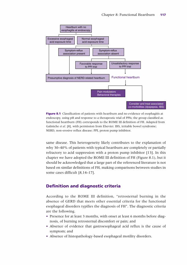
Chapter 8: Functional Heartburn 117
same disease. This heterogeneity likely contributes to the explanation of
why 30–40% of patients with typical heartburn are completely or partially
refractory to acid suppression with a proton pump inhibitor [13] . In this
chapter we have adopted the ROME III definition of FH (Figure 8.1 ), but it
should be acknowledged that a large part of the referenced literature is not
based on similar definitions of FH, making comparisons between studies in
some cases difficult [8,14–17] .
Definition and diagnostic criteria
According to the ROME III definition, “retrosternal burning in the
absence of GERD that meets other essential criteria for the functional
esophageal disorders typifies the diagnosis of FH”. The diagnostic criteria
are the following.
• Presence for at least 3 months, with onset at least 6 months before diag-
nosis, of burning retrosternal discomfort or pain; and
• Absence of evidence that gastroesophageal acid reflux is the cause of
symptom; and
• Absence of histopathology-based esophageal motility disorders.
Pain modulatorsBehavioral therapies
Heartburn with noesophagitis at endoscopy
Excessive esophagealacid exposure time
Normal esophagealacid exposure time
Symptom-reflux association present
Symptom-reflux association absent
Favorable responseto PPI trial
Unsatisfactory responseto PPI trial
Presumptive diagnosis of NERD-related heartburn Functional heartburn
Consider and treat associatedco-morbidities (dyspepsia, IBS)
Figure 8.1 Classification of patients with heartburn and no evidence of esophagitis at
endoscopy, using pH and response to a therapeutic trial of PPIs; the group classified as
functional heartburn (FH) corresponds to the ROME III definition of FH. Adapted from
Galmiche et al . [8] , with permission from Elsevier. IBS, irritable bowel syndrome;
NERD, non-erosive reflux disease; PPI, proton pump inhibitor.
c08.indd 117 11/15/2012 2:28:40 AM
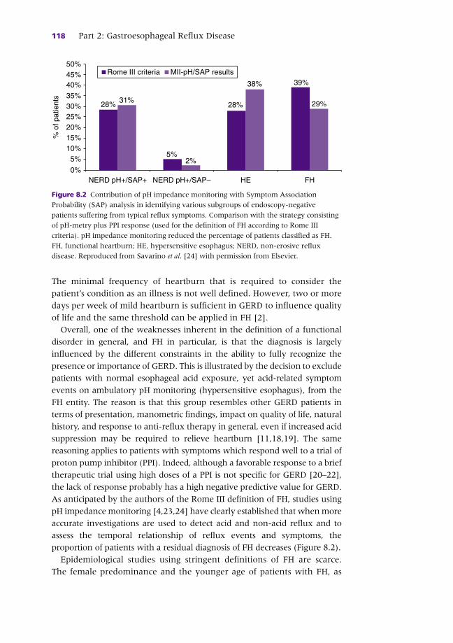
118 Part 2: Gastroesophageal Reflux Disease
The minimal frequency of heartburn that is required to consider the
patient ’ s condition as an illness is not well defined. However, two or more
days per week of mild heartburn is sufficient in GERD to influence quality
of life and the same threshold can be applied in FH [2] .
Overall, one of the weaknesses inherent in the definition of a functional
disorder in general, and FH in particular, is that the diagnosis is largely
influenced by the different constraints in the ability to fully recognize the
presence or importance of GERD. This is illustrated by the decision to exclude
patients with normal esophageal acid exposure, yet acid-related symptom
events on ambulatory pH monitoring (hypersensitive esophagus), from the
FH entity. The reason is that this group resembles other GERD patients in
terms of presentation, manometric findings, impact on quality of life, natural
history, and response to anti-reflux therapy in general, even if increased acid
suppression may be required to relieve heartburn [11,18,19] . The same
reasoning applies to patients with symptoms which respond well to a trial of
proton pump inhibitor (PPI). Indeed, although a favorable response to a brief
therapeutic trial using high doses of a PPI is not specific for GERD [20–22] ,
the lack of response probably has a high negative predictive value for GERD.
As anticipated by the authors of the Rome III definition of FH, studies using
pH impedance monitoring [4,23,24] have clearly established that when more
accurate investigations are used to detect acid and non-acid reflux and to
assess the temporal relationship of reflux events and symptoms, the
proportion of patients with a residual diagnosis of FH decreases (Figure 8.2 ).
Epidemiological studies using stringent definitions of FH are scarce.
The female predominance and the younger age of patients with FH, as
28% 28%
38% 39%
29%31%
2%5%
5%
0%
10%15%%
of p
atie
nts
20%25%30%35%
40%45%50%
NERD pH+/SAP–NERD pH+/SAP+ FHHE
MII-pH/SAP resultsRome III criteria
Figure 8.2 Contribution of pH impedance monitoring with Symptom Association
Probability (SAP) analysis in identifying various subgroups of endoscopy-negative
patients suffering from typical reflux symptoms. Comparison with the strategy consisting
of pH-metry plus PPI response (used for the definition of FH according to Rome III
criteria). pH impedance monitoring reduced the percentage of patients classified as FH.
FH, functional heartburn; HE, hypersensitive esophagus; NERD, non-erosive reflux
disease. Reproduced from Savarino et al . [24] with permission from Elsevier.
c08.indd 118 11/15/2012 2:28:40 AM

Chapter 8: Functional Heartburn 119
opposed to NERD, are not really evident once patients with acid-sensitive
esophagus have been excluded from the definition of FH [17] . Interestingly,
and irrespective of the clinical setting (i.e. primary care or specialty
practice), FH frequently occurs in association with symptoms usually
considered to be components of dyspeptic syndrome, such as postprandial
fullness, bloating, nausea, and early satiety [25–27] . Irritable bowel
syndrome (IBS) symptoms also seem to be more prevalent in FH than in
GERD patients [28,29] .
In summary, the prevalence of FH depends on the defining and diag-
nostic criteria. Studies using both endoscopy and ambulatory pH moni-
toring to objectively establish evidence of GERD indicate that FH is likely
to represent less than 10% of heartburn patients presenting to gastro-
enterologists [30] . The proportion is probably higher in primary care set-
tings, when all patients with GERD symptoms which are non-responsive
to PPI given empirically are considered. Conversely, in tertiary centers,
CASE STUDY
A 40-year-old female patient is referred for refractory heartburn. She has a history of appendectomy and irritable bowel syndrome. She has been complaining of heartburn for the previous 2 years. She describes a burning sensation behind the breastbone, occurring almost daily, mainly during the daytime. The symptoms appear to be more severe in the postprandial period, during which she also complains of epigastric bloating, nausea, and fullness. She has a long history of constipation and lower abdominal pain related to IBS. She has not lost weight over time (Body Mass Index (BMI) 23 kg/m 2 ). There is no evidence of a psychiatric disorder such as depression or anxiety. She has been prescribed several PPIs at single and double dose without any significant improvement of heartburn. Compliance with PPI therapy, and also dosing times, appear to be adequate. An endoscopic work-up has been performed. There were no mucosal breaks at endoscopy and no eosinophilic infiltration of esophageal mucosa on the esophageal biopsies. Colonoscopy findings were also normal.
The patient is referred for refractory heartburn and advice regarding anti-reflux surgery. High-resolution esophageal manometry is performed, demonstrating no significant abnormality of esophageal motility. Lower esophageal sphincter pressure is within the normal range (23 mmHg). Wireless, 48-h pH monitoring is performed off therapy Both the number of acid reflux episodes and esophageal acid exposure are within normal limits during this 48-h period. Symptom association analysis does not demonstrate any correlation between heartburn episodes reported by the patient and the reflux events.
The final diagnosis is functional heartburn associated with functional dyspepsia and irritable bowel syndrome. Treatment with pain modulators is proposed to the patient who initially refuses it. Two months later, she finally accepts and is prescribed amitriptyline. The initial dose is 10 mg at bedtime, increased by 10 mg increments weekly. As the patient experiences side-effects (dry mouth, somnolence), the dose cannot be increased above 40 mg daily. After a period of 3 months, there is a significant improvement in both the frequency and severity of heartburn symptoms. There is no significant effect on functional dyspepsia and IBS symptoms.
c08.indd 119 11/15/2012 2:28:40 AM
120 Part 2: Gastroesophageal Reflux Disease
when pH impedance monitoring is performed with statistical symptom
analysis for acid and non-acid reflux episodes (including Symptom
Associated Probability (SAP) [31] or Symptom Index (SI) determination),
the proportion of remaining patients with a final diagnosis of FH is cer-
tainly smaller.
Pathogenesis of heartburn in functional heartburn
One difficulty in adequately analyzing the available data on the pathogen-
esis of FH is that the literature, at least with regard to the earliest reports,
is clouded by the inclusion of subjects with undetected GERD in patient
groups with presumed FH. This is actually the case for the excellent review
published in 2002 by Fass and Tougas [32] on the role of peripheral and
central factors in the pathogenesis of esophageal symptoms in NERD.
Nevertheless, the conceptual model proposed by these authors remains
valid as a representative overview of the mechanisms involved in esoph-
ageal perception in general (Figure 8.3 ). From a more mechanistic point
of view, noxious esophageal stimuli are known to activate nociceptive
receptors such as the TRPV1 (vanilloid) receptor, the transient receptor
potential acid-sensing ion channel (ASIC), and the PX 2 family of ligand-
gated ion channels responsive to adenosine triphosphate (ATP) (for a
review see [33] ). Activation of these receptors generates signals that are
transmitted to the central nervous system (CNS) via either vagal or spinal
nerves (Figure 8.4 ). Whether the same pathways are involved in NERD
and FH remains unknown; if differences are present, they may contribute
to explain the differences encountered in therapeutic response to acid
suppression. It is also worth considering that, apart from pathways
activated by topical chemical stimuli such as bile or acid, even in minute
amounts, vagal afferents may also play a role in the perception of esoph-
ageal distension.
Regarding FH in particular, the prevailing view is to consider disturbed
visceral perception as a major factor involved in its pathogenesis [32] .
Hypersensitivity includes allodynia (defined by the perception of stimuli
which are not normally perceived, for example slight changes in intralu-
minal esophageal pH) and hyperalgesia (pain greater than normally
expected for a given stimulus). Both phenomena may be involved in FH.
Moreover, “esophageal hypersensitivity” can include peripheral, central
and possibly psychological factors which may act independently or, more
likely, in concert.
Among other factors which are potentially involved in the pathogenesis
of esophageal symptoms, hormonal pathways may also play a role. Indeed,
the most frequent trigger of heartburn is eating a meal, suggesting that
c08.indd 120 11/15/2012 2:28:40 AM

Chapter 8: Functional Heartburn 121
some food components, especially fats, may induce or exacerbate symptoms
through a hormonal pathway. For example, Meyer et al . [34] showed that
infusion of fat into the duodenum of reflux patients reduced the latency of
occurrence of heartburn in response to esophageal acid infusion and
increased symptom severity. Although the interpretation of this
phenomenon remains largely speculative, a role for cholecystokinin has
been proposed but other neurotransmitters also, e.g. calcitonin gene-
related peptide or substance P, may influence esophageal perception by
either peripheral (on vagal afferences) or central action.
The role of corticocerebral processing of esophageal signals has been
investigated recently in a few studies using the recording of cortical
Central
Psychiatric comorbidity
StressSleep?
Peripheral
Intraduodenal fat
Heartburn+
+
Acid exposure
Pathological Physiological
Figure 8.3 Proposed conceptual model for symptom generation in patients with
non-erosive reflux disease. This model suggests that central (through brain–gut
interactions) and peripheral mechanisms are essential for intraoesophageal stimuli
(either physiological or pathological) to reach the conscious level and thus be
perceived. Reproduced from Fass and Tougas [32] with permission from BMJ
Publishing Group.
c08.indd 121 11/15/2012 2:28:40 AM
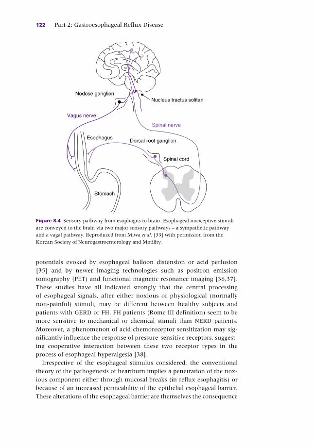
122 Part 2: Gastroesophageal Reflux Disease
potentials evoked by esophageal balloon distension or acid perfusion
[35] and by newer imaging technologies such as positron emission
tomography (PET) and functional magnetic resonance imaging [36,37] .
These studies have all indicated strongly that the central processing
of esophageal signals, after either noxious or physiological (normally
non-painful) stimuli, may be different between healthy subjects and
patients with GERD or FH. FH patients (Rome III definition) seem to be
more sensitive to mechanical or chemical stimuli than NERD patients.
Moreover, a phenomenon of acid chemoreceptor sensitization may sig-
nificantly influence the response of pressure-sensitive receptors, suggest-
ing cooperative interaction between these two receptor types in the
process of esophageal hyperalgesia [38] .
Irrespective of the esophageal stimulus considered, the conventional
theory of the pathogenesis of heartburn implies a penetration of the nox-
ious component either through mucosal breaks (in reflux esophagitis) or
because of an increased permeability of the epithelial esophageal barrier.
These alterations of the esophageal barrier are themselves the consequence
Nucleus tractus solitari
Spinal nerve
Dorsal root ganglion
Nodose ganglion
Vagus nerve
Esophagus
Stomach
Spinal cord
Figure 8.4 Sensory pathway from esophagus to brain. Esophageal nociceptive stimuli
are conveyed to the brain via two major sensory pathways – a sympathetic pathway
and a vagal pathway. Reproduced from Miwa et al . [33] with permission from the
Korean Society of Neurogastroenterology and Motility.
c08.indd 122 11/15/2012 2:28:41 AM
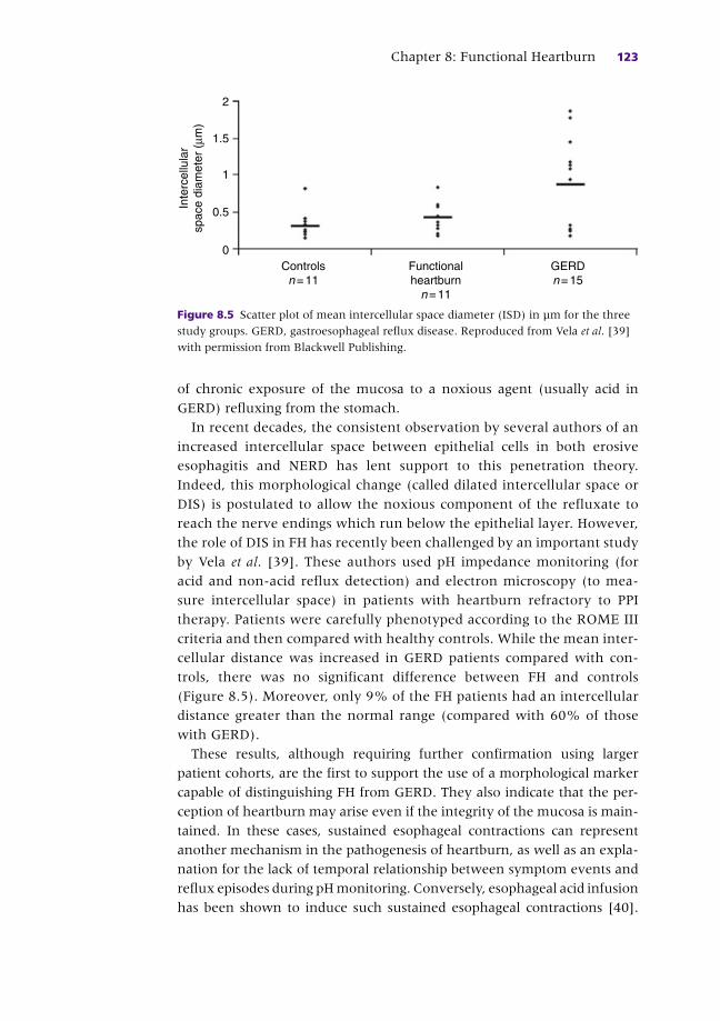
Chapter 8: Functional Heartburn 123
of chronic exposure of the mucosa to a noxious agent (usually acid in
GERD) refluxing from the stomach.
In recent decades, the consistent observation by several authors of an
increased intercellular space between epithelial cells in both erosive
esophagitis and NERD has lent support to this penetration theory.
Indeed, this morphological change (called dilated intercellular space or
DIS) is postulated to allow the noxious component of the refluxate to
reach the nerve endings which run below the epithelial layer. However,
the role of DIS in FH has recently been challenged by an important study
by Vela et al . [39] . These authors used pH impedance monitoring (for
acid and non-acid reflux detection) and electron microscopy (to mea-
sure intercellular space) in patients with heartburn refractory to PPI
therapy. Patients were carefully phenotyped according to the ROME III
criteria and then compared with healthy controls. While the mean inter-
cellular distance was increased in GERD patients compared with con-
trols, there was no significant difference between FH and controls
(Figure 8.5 ). Moreover, only 9% of the FH patients had an intercellular
distance greater than the normal range (compared with 60% of those
with GERD).
These results, although requiring further confirmation using larger
patient cohorts, are the first to support the use of a morphological marker
capable of distinguishing FH from GERD. They also indicate that the per-
ception of heartburn may arise even if the integrity of the mucosa is main-
tained. In these cases, sustained esophageal contractions can represent
another mechanism in the pathogenesis of heartburn, as well as an expla-
nation for the lack of temporal relationship between symptom events and
reflux episodes during pH monitoring. Conversely, esophageal acid infusion
has been shown to induce such sustained esophageal contractions [40] .
Controlsn= 11
0
Inte
rcel
lula
rsp
ace
diam
eter
(μm
)0.5
1
1.5
2
Functionalheartburnn= 11
GERDn= 15
Figure 8.5 Scatter plot of mean intercellular space diameter (ISD) in μm for the three
study groups. GERD, gastroesophageal reflux disease. Reproduced from Vela et al . [39]
with permission from Blackwell Publishing.
c08.indd 123 11/15/2012 2:28:41 AM
124 Part 2: Gastroesophageal Reflux Disease
Therefore different mechanisms are probably associated which may
interact between each other and facilitate pain perception [41] .
Role of psychological factors
It is now well established that acute experimental stress enhances the
perception of esophageal acid in GERD patients. Indeed, Bradley et al . [42]
showed in 1993 that reflux patients who are chronically anxious and
exposed to prolonged stress may perceive low-intensity acid stimuli as
painful symptoms. Of interest, in this controlled study the stress tasks did
not significantly influence acid reflux parameters. More recently, the effect
of life stress on symptoms of heartburn was studied by Naliboff et al . [43]
in a cohort of patients followed prospectively for 4 months. The presence
of severe, sustained life stress during the previous 6 months significantly
predicted increased heartburn symptoms during the subsequent 4 months.
Anxiety was strongly associated with impaired quality of life, and depres-
sion with heartburn medication use.
In another study, whereas psychological profiles did not differentiate
subjects with normal esophageal acid exposure and no esophagitis from
those having elevated acid exposure times, patients with FH had greater
anxiety and somatization scores and poorer social support levels than
those with reflux-provoked symptoms [44] . Shapiro et al . [45] conducted
a study comparing the physiological and clinical characteristics of a group
of 22 NERD patients (with abnormal pH test) with those of 30 FH patients
(ROME II definition). There were no statistical differences in demographic
parameters, frequency of hiatal hernia and H. pylori infection rates bet-
ween the two groups. In contrast, FH patients had increased reports of
chest pain, somatization and altered autonomic function (assessed by heart
rate variability and skin conductance). In addition to stress and anxiety,
sleep disorders may also enhance the perception of low-intensity esopha-
geal stimuli [46] . Finally, some studies have suggested that a lower social
status may be associated with FH [44] .
Clinical evaluation
Clarification of the nature of the symptom is an essential first step [7] .
Heartburn is characterized by pain or discomfort of burning quality that orig-
inates high in the epigastrium with intermittent cephalad retrosternal radia-
tion. There are no evidence-based data to determine the specific symptom
features of FH, including diurnal characteristics, exacerbating factors and ame-
liorating maneuvers. The benefit of structured questionnaires to better identify
c08.indd 124 11/15/2012 2:28:41 AM
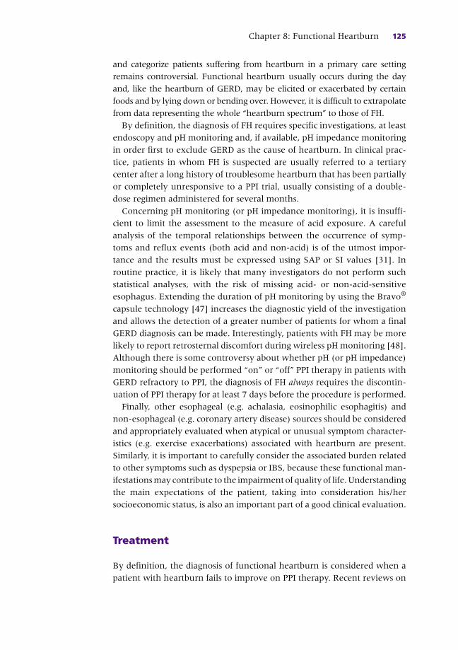
Chapter 8: Functional Heartburn 125
and categorize patients suffering from heartburn in a primary care setting
remains controversial. Functional heartburn usually occurs during the day
and, like the heartburn of GERD, may be elicited or exacerbated by certain
foods and by lying down or bending over. However, it is difficult to extrapolate
from data representing the whole “heartburn spectrum” to those of FH.
By definition, the diagnosis of FH requires specific investigations, at least
endoscopy and pH monitoring and, if available, pH impedance monitoring
in order first to exclude GERD as the cause of heartburn. In clinical prac-
tice, patients in whom FH is suspected are usually referred to a tertiary
center after a long history of troublesome heartburn that has been partially
or completely unresponsive to a PPI trial, usually consisting of a double-
dose regimen administered for several months.
Concerning pH monitoring (or pH impedance monitoring), it is insuffi-
cient to limit the assessment to the measure of acid exposure. A careful
analysis of the temporal relationships between the occurrence of symp-
toms and reflux events (both acid and non-acid) is of the utmost impor-
tance and the results must be expressed using SAP or SI values [31] . In
routine practice, it is likely that many investigators do not perform such
statistical analyses, with the risk of missing acid- or non-acid-sensitive
esophagus. Extending the duration of pH monitoring by using the Bravo®
capsule technology [47] increases the diagnostic yield of the investigation
and allows the detection of a greater number of patients for whom a final
GERD diagnosis can be made. Interestingly, patients with FH may be more
likely to report retrosternal discomfort during wireless pH monitoring [48] .
Although there is some controversy about whether pH (or pH impedance)
monitoring should be performed “on” or “off” PPI therapy in patients with
GERD refractory to PPI, the diagnosis of FH always requires the discontin-
uation of PPI therapy for at least 7 days before the procedure is performed.
Finally, other esophageal (e.g. achalasia, eosinophilic esophagitis) and
non-esophageal (e.g. coronary artery disease) sources should be considered
and appropriately evaluated when atypical or unusual symptom character-
istics (e.g. exercise exacerbations) associated with heartburn are present.
Similarly, it is important to carefully consider the associated burden related
to other symptoms such as dyspepsia or IBS, because these functional man-
ifestations may contribute to the impairment of quality of life. Understanding
the main expectations of the patient, taking into consideration his/her
socioeconomic status, is also an important part of a good clinical evaluation.
Treatment
By definition, the diagnosis of functional heartburn is considered when a
patient with heartburn fails to improve on PPI therapy. Recent reviews on
c08.indd 125 11/15/2012 2:28:41 AM

126 Part 2: Gastroesophageal Reflux Disease
the management of refractory GERD have been published [49,50] and
different algorithms have been proposed. As some patients with FH may be
sensitive to small amounts of acid or acidic fluid with a pH > 4, a trial of
more vigorous acid suppression therapy is usually considered first. The
addition of a prokinetic to acid suppression is not supported by evidence
and a recent, randomized, placebo-controlled trial conducted in NERD
patients failed to show any significant benefit of mosapride citrate
(5 mg t.i.d.) plus omeprazole (10 mg o.d.) versus omeprazole alone [51] .
The role of diet is frequently advocated by patients with functional gas-
trointestinal disorders but there is no clear evidence for a benefit of
excluding specific foods (e.g. acidic or spicy foods). On the contrary, there
are some arguments suggesting that the chronic ingestion of chili, which
contains capsaicin, may improve functional dyspepsia and reflux symp-
toms by desensitization of TRPV 1 receptors [52] . Similarly, as fat and cho-
lesterol could increase esophageal sensitivity to acid [53] , heavy meals are
usually avoided spontaneously by the patients themselves. Changing the
composition of the refluxate may, at least in theory, represent an alternative
approach; for example, in patients who are sensitive to bile salts, it might
be possible to change the bile composition by the administration of urso-
deoxycholic acid [54] . In fact, in clinical practice it is important to reassure
the patient concerning the harmless effects of many foods and to convince
him/her to avoid overly restrictive dietary regimens. Although the role of
obesity in FH has not really been investigated, it is common sense to
attempt to reduce excess weight.
Because the pathophysiology of FH may be quite similar to that of non-
cardiac chest pain, and involve heightened visceral sensation, the use of
low-dose tricyclic antidepressants, and possibly selective serotonin reuptake
inhibitors, is reasonable. Similarly, psychological approaches such as
behavioral modification, acupuncture [50] , hypnotherapy or relaxation
therapy [55] may be beneficial. However, to date no published controlled
trials have demonstrated efficacy of any of these interventions in FH patients.
Several molecules may, potentially, influence visceral perception and
this may be exploited in functional esophageal disorders in general, and in
FH in particular. For example, the effect of tegaserod, a 5-HT4 agonist, was
tested in a placebo-controlled, cross-over trial conducted in patients with
overlapping symptoms of FH and functional dyspepsia (ROME II defini-
tion). The severities of heartburn, regurgitation, early fullness, and bloat-
ing were significantly lower after tegaserod compared with a placebo [56] .
In the same study, mechanical sensitivity was assessed using the barostat
technique and tegaserod was shown to increase the pressure threshold for
gastric pain. However, the study conclusions are limited by the small
sample of patients included and the cross-over design of the trial. Moreover,
tegaserod is not available in many countries, especially in Europe and
c08.indd 126 11/15/2012 2:28:41 AM

Chapter 8: Functional Heartburn 127
North America, making applications of these findings very limited. Among
other molecules, antagonists of the TRPV 1 receptor (AZD1386) have
recently been developed and proof-of-concept studies have been published
[57] . In healthy human subjects, AZD1386 increased esophageal and skin
heat pain thresholds and was well tolerated. Thus this new class of drug
may have a potential in NERD and also FH but it is too early to extrapolate
from pharmacodynamic effects to the clinic.
Anti-reflux surgery in patients with FH has not been fully evaluated, but
surgical management is unlikely to provide relief for these patients with
frequently overlapping symptoms of dyspepsia and IBS, and in whom test-
ing for pathological reflux is negative. Although the predictive value of
preoperative esophageal acid exposure on postsurgical outcome remains
controversial, some studies have indicated worse results in patients with a
normal pH test [58] . Moreover, it is now well established that a proportion
of patients with recurrent heartburn after anti-reflux surgery have normal
esophageal pH values. Thompson et al . [59] reported that in such patients
heartburn is frequently associated with psychiatric co-morbidities such as
depression, suggesting that FH rather than GERD recurrence may be
responsible for the persisting heartburn. The same prudent limitation
should be applied to endoscopic anti-reflux procedures, although some of
these procedures, such as a radiofrequency energy delivery (Stretta
procedure), may affect esophageal sensitivity independently of their effect
on cardial continence [60,61] . That said, endoscopic therapies cannot be
recommended for FH at the present time.
In summary, once PPI failure has been confirmed and GERD excluded
by appropriate tests, the treatment of FH remains largely empirical and an
individual approach is therefore recommended. The clinician should pro-
vide reassurance and refrain from performing too many invasive tests or
therapeutic procedures. Although the long-term natural history of FH is
poorly known, some studies, as well as clinical experience, suggest a con-
siderable “turnover” of functional gastrointestinal disorders with the
appearance and disappearance of different categories of functional disor-
ders in the same patient. In this context, Boyd et al . [62] reported a decrease
in the prevalence of FH over time in a cohort of patients with associated
eating disorders. Such findings may have important implications in favor
of a very conservative and non-invasive approach in FH patients.
References
1 Locke GR , Talley NJ , Fett SL , et al . Prevalence and clinical spectrum of gastroesopha-
geal reflux: a population-based study in Olmsted Country, Minnesota . Gastroenterology
1997 ; 112 : 1448 – 56 .
c08.indd 127 11/15/2012 2:28:41 AM
128 Part 2: Gastroesophageal Reflux Disease
2 Dent J , Armstrong D , Delanye B , et al . Symptom evaluation in reflux disease:
workshop, background, process, terminology, recommendations and discussion
outputs . Gut 2004 ; 53 : iv1 – iiv24 .
3 Carlsson R , Dent J , Bolling–Sternevalde E , et al . The usefulness of a structured
questionnaire in the assessment of symptomatic gastroesophageal reflux disease .
Scand J Gastroenterol 1998 ; 33 : 1023 – 9 .
4 Zerbib F , Roman S , Ropert A , et al . Esophageal pH-impedance monitoring and
symptom analysis in GERD. A study in patients off and on therapy . Am J Gastroenterol
2006 ; 101 : 1956 – 63 .
5 Sifrim D , Castell D , Dent J , et al . Gastro-oesophageal reflux monitoring: review and
consensus report on detection and definitions of acid, non-acid, and gas reflux . Gut
2004 ; 53 : 1024 – 31 .
6 Karamanolis G , Stevens W , Vos R , et al . Oesophageal tone and sensation in the
transition zone between proximal striated and distal smooth muscle oesophagus .
Neurogastroenterol Motil 2008 ; 20 : 291 – 7 .
7 Clouse RE , Richter JE , Heading RC , et al . Functional esophageal disorders . In:
Drossman DA , Corazziari E , Talley NJ , Thompson WG , Whitehead WE (eds) Rome II .
The functional gastrointestinal disorders , 2nd edn. McLean, VA : Degnon Associates ,
2000 : pp. 247 – 98 .
8 Galmiche JP , Clouse RA , Balint A , et al . Functional esophageal disorders .
Gastroenterology 2006 ; 130 : 1459 – 65 .
9 Galmiche JP , Clouse RE , Balint A , et al . Functional esophageal disorders . In: Drossman
DA , Corazziari E , Spiller R , et al . (eds) Rome III .The functional gastrointestinal disorders ,
3rd edn. McLean, VA : Degnon Associates , 2006 : pp. 369 – 418 .
10 Shi G , Bruley des Varannes S , Scarpignato C , et al . Reflux related symptoms in
patients with normal oesophageal exposure to acid . Gut 1995 ; 37 : 457 – 64 .
11 Trimble KC , Douglas S , Pryde A , et al . Clinical characteristics and natural history of
symptomatic but not excess gastroesophageal reflux . Dig Dis 1995 ; 40 : 1098 – 104 .
12 Fass R , Ofman JJ . Gastroesophageal reflux disease – should we adopt a new
conceptual framework? Am J Gastroenterol 2002 ; 97 : 1901 – 9 .
13 Fass R , Shapiro M , Dekel R , et al . Systematic review: proton-pump inhibitor failure in
gastro-oesophageal reflux disease – where next? Aliment Pharmacol Ther
2005 ; 22 : 79 – 94 .
14 Frazzoni M , de Michell E , Zentilin P , Pathophysiological characteristics of patients
with non-erosive reflux disease differ from those of patients with functional heart-
burn . Aliment Pharmacol Ther 2004 ; 20 : 81 – 8 .
15 Fry LC , Monkemuller K , Malfertheiner P . Functional heartburn, non-erosive reflux
disease, and reflux esophagitis are all distinct conditions – a debate: con . Curr Treat
Options Gastroenterol 2007 ; 10 : 305 – 11 .
16 Hershcovici T , Zimmerman J . Functional heartburn vs non-erosive reflux disease:
similarities and differences . Aliment Pharmacol Ther 2008 ; 27 : 1103 – 9 .
17 Fass R . Functional heartburn: what it is and how to treat it . Gastrointest Endosc Clin
North Am 2009 ; 19 : 23 – 33 .
18 Sacher-Huvelin S , Gournay J , Amouretti M , et al . Acid sensitive esophagus: natural
history and quality of life of patients with acid hypersensitive esophagus syndrome.
Comparison with classical gastro-esophageal reflux disease . Gastroenterol Clin Biol
2000 ; 24 : 911 – 16 .
19 Watson RGP , Tham TCK , Johnston BT , et al . Double blind cross-over placebo con-
trolled study of omeprazole in the treatment of patients with reflux symptoms and
physiological levels of acid reflux – the “sensitive” esophagus . Gut 1997 ; 40 : 587 – 90 .
c08.indd 128 11/15/2012 2:28:41 AM

Chapter 8: Functional Heartburn 129
20 Numans ME , Lau J , de Wit NJ , et al . Short-term treatment with proton-pump inhib-
itors as a test for gastroesophageal reflux disease: a meta-analysis of diagnostic test
characteristics . Ann Intern Med 2004 ; 140 : 518 – 27 .
21 Bruley des Varannes S , Sacher-Huvelin S , Vavasseur F , et al . Rabeprazole test for the
diagnosis of gastro-esophageal reflux disease: results of a study in a primary care
setting . World J Gastroenterol 2006 ; 28 : 2569 – 73 .
22 Meineche-Schmidt V , Christensen E , Bytzer P . Randomised clinical trial: identification of
responders to short-term treatment with esomeprazole for dyspepsia in primary care – a
randomised, placebo-controlled study . Aliment Pharmacol Ther 2011 ; 33 : 41 – 9 .
23 Savarino E , Zentilin P , Tutuian R , et al . The role of nonacid reflux in NERD: lessons
learned from impedance pH monitoring in 150 patients off therapy . Am J
Gastroenterol 2008 ; 103 : 2685 – 93 .
24 Savarino E , Marabotto E , Zentilinn P , et al . The added value of impedance-pH moni-
toring to Rome III criteria in distinguishing functional heartburn from non-erosive
reflux disease . Dig Liver Dis 2011 ; 43 ( 7 ): 542 – 7 .
25 Savarino E , Pohl D , Zentilin P , et al . Functional heartburn has more in common with
functional dyspepsia that with non-erosive reflux disease . Gut 2009 ; 58 : 1185 – 91 .
26 Hershcovici T , Fass R . GERD: are functional heartburn and functional dyspepsia one
disorder? Nat Rev Gastroenterol Hepatol 2010 ; 7 : 71 – 2 .
27 Xiao YL , Peng S , Tao J . Prevalence and symptom pattern of pathologic esophageal
acid reflux in patients with functional dyspepsia based on the Rome III criteria . Am J
Gastroenterol 2010 ; 105 : 2626 – 31 .
28 Lee KJ , Kwon HC , Cheong JY , et al . Demographic, clinical and psychological charac-
teristics of the heartburn groups classified using the Rome III criteria and factors
associated with the responsiveness to proton pump inhibitors in the gastroesopha-
geal reflux disease group . Digestion 2009 ; 79 : 131 – 6 .
29 Kaji M , Fujiwara Y , Shiba M . Prevalence of overlaps between GERD, FD and IBS and
impact on health-related quality of life . J Gastroenterol Hepatol 2010 ; 25 : 1151 – 6 .
30 Martinez SD , Malagon IB , Garewal HS , et al . Non-erosive reflux disease (NERD) –
acid reflux and symptom patterns . Aliment Pharmacol Ther 2003 ; 17 : 537 – 45 .
31 Weusten BLAM , Roelofs JMM , Akkermans LM , et al . The symptom-association prob-
ability: an improved method for symptom analysis of 24-hour esophageal pH data .
Gastroenterology 1994 ; 107 : 1741 – 5 .
32 Fass R , Tougas G . Functional heartburn: the stimulus, the pain, and the brain . Gut
2002 ; 51 : 885 – 92 .
33 Miwa H , Kondo T , Oshima T , et al . Esophageal sensation and esophageal hypersensitivity –
overview from bench to bedside . J Neurogastroenterol Motil 2010 ; 16 : 353 – 62 .
34 Meyer JH , Lembo A , Elashoff JD , et al . Duodenal fat intensifies the perception of
heartburn . Gut 2001 ; 40 : 624 – 8 .
35 Yang M , Li ZS , Xu XR . Characterization of cortical potentials evoked by oesophageal
balloon distention and acid perfusion in patients with functional heartburn .
Neurogastroenterol Motil 2006 ; 18 : 292 – 9 .
36 Kern MK , Birn RM , Jaradeh S , et al . Identification and characterization of cerebral
cortical response to esophageal mucosal acid exposure and distention .
Gastroenterology 1998 ; 115 : 1353 – 62 .
37 Kern M , Hofmann C , Hyde J , et al . Characterization of the cerebral cortical represen-
tation of heartburn in GERD patients . Am J Physiol Gastrointest Liver Physiol
2004 ; 286 : G174 – 81 .
38 Yang M , Li ZS , Chen DF , et al . Quantitative assessment and characterization of vis-
ceral hyperalgesia evoked by esophageal balloon distention and acid perfusion in
c08.indd 129 11/15/2012 2:28:42 AM

130 Part 2: Gastroesophageal Reflux Disease
patients with functional heartburn, nonerosive reflux disease, and erosive esophagitis .
Clin J Pain 2010 ; 26 : 326 – 31 .
39 Vela MF , Craft BMM , Sharma N , et al . Refractory heartburn: comparison of intercel-
lular space diameter in documented GERD vs functional heartburn . Am J
Gastroenterol 2011 ; 106 : 844 – 50 .
40 Pehlivanov N , Liu J , Mittal RK . Sustained esophageal contraction: a motor correlate
of heartburn symptom . Am J Physiol Gastrointest Liver Physiol 2001 ; 281 : G743 – G751 .
41 Barlow WJ , Orlando RC . The pathogenesis of heartburn in nonerosive reflux disease:
a unifying hypothesis . Gastroenterology 2005 ; 128 : 771 – 8 .
42 Bradley LA , Richter JE , Pulliam TJ , et al . The relationship between stress and symp-
toms of gastroesophageal reflux: the influence of psychological factors . Am J
Gastroenterol 1993 ; 88 : 11 – 19 .
43 Naliboff BD , Mayer M , Fass R , et al . The effect of life stress on symptoms of heartburn .
Psychosom Med 2004 ; 66 : 426 – 34 .
44 Johnston BT , Lewis SA , Collins JS , et al . Acid perception in gastro-oesophageal reflux
disease is dependent on psychosocial factors . Scand J Gastroenterol 1995 ; 30 : 1 – 5 .
45 Shapiro M , Green C , Bautista JM . Functional heartburn patients demonstrate traits
of functional bowel disorder but lack a uniform increase of chemoreceptor sensitivity
to acid . Am J Gastroenterol 2006 ; 10 : 1084 – 91 .
46 Schey R , Dickman R , Parthasarathy S , et al . Sleep deprivation is hyperalgesis in
patients with gastroesophageal reflux disease . Gastroenterology 2007 ; 133 : 1787 – 95 .
47 Prakash C , Clouse RE . Value of extended recording time with a wireless esophageal
pH system in patients with gastroesophageal reflux disease . Clin Gastroenterol
Hepatol 2005 ; 3 : 329 – 34 .
48 Lee YC , Wang HP , Chiu HM , et al . Patients with functional heartburn are more likely
to report retrosternal discomfort during wireless pH monitoring . Gastrointest Endosc
2005 ; 62 : 834 – 41 .
49 Fass R , Sifrim D . Management of heartburn not responding to proton pump inhibi-
tors . Gut 2009 ; 58 : 295 – 309 .
50 Hershcovici T , Fass R . An algorithm for diagnosis and treatment of refractory GERD .
Best Pract Res Clin Gastroenterol 2010 ; 24 : 923 – 36 .
51 Miwa H , Inoue K , Ashida K . Randomised clinical trial: efficacy of the addition of a
prokinetic, mosapride citrate, to omeprazole in the treatment of patients with non-
erosive reflux disease – a double-blind, placebo-controlled study . Aliment Pharmacol
Ther 2011 ; 33 : 323 – 32 .
52 Gonlachanvit S . Are rice and spicy diet good for functional gastrointestinal disorders?
J Neurogastroenterol Motil 2010 ; 16 : 131 – 8 .
53 Shapiro M , Green C , Bautista JM , et al . Assessment of dietary nutrients that influence
perception of intra-oesophageal acid reflux events in patients with gastro- oesophageal
reflux disease . Aliment Pharmacol Ther 2007 ; 25 : 93 – 101 .
54 Siddiqui A , Rodriguez-Stanley S , Zubaidi S . Esophageal visceral sensitivity to bile
salts in patients with functional heartburn and in healthy control subjects . Dig Dis Sci
2005 ; 50 : 81 – 5 .
55 McDonald-Haile J , Bradley LA , Bailey MA , et al . Relaxation training reduces symptom
reports and acid exposure in patients with gastroesophageal reflux disease .
Gastroenterology 1994 ; 107 : 61 – 9 .
56 Miner PB Jr , Rodriguez-Stanley S , Proskin HM , et al . Tegaserod in patients with
mechanical sensitivity and overlapping symptoms of functional heartburn and
functional dyspepsia . Curr Med Res Opin 2008 ; 24 : 2159 – 72 .
c08.indd 130 11/15/2012 2:28:42 AM
Chapter 8: Functional Heartburn 131
57 Krarup AL , Ny L , Astrand M . Randomised clinical trial: the efficacy of a transient
receptor potential vanilloid 1 antagonist AZD1386 in human oesophageal pain .
Aliment Pharmacol Ther 2011 ; 33 : 1113 – 22 .
58 Khajanchee YS , Hong D , Hansen PD , et al . Outcomes of antireflux surgery in patients
with normal preoperative 24-hour pH test results . Am J Surg 2004 ; 187 : 599 – 603 .
59 Thompson SK , Cai W , Jamieson GG . Recurrent symptoms after fundoplication with
a negative pH study – recurrent reflux or functional heartburn? J Gastrointest Surg
2009 ; 13 : 54 – 60 .
60 Galmiche JP , Bruley des Varannes S . Endoluminal therapies for gastro-oesophageal
reflux disease . Lancet 2003 ; 361 : 1119 – 21 .
61 Arts J , Tack J , Galmiche JP . Endoscopic antireflux procedures . Gut 2004 ; 53 : 1207 – 14 .
62 Boyd C , Abraham S , Kellow J . Appearance and disappearance of functional gastroin-
testinal disorders in patients with eating disorders . Neurogastroenterol Motil
2010 ; 22 : 1279 – 83 .
c08.indd 131 11/15/2012 2:28:42 AM
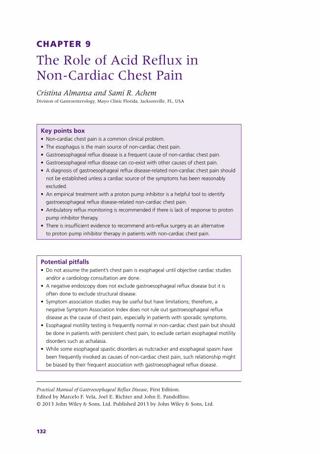
132
Practical Manual of Gastroesophageal Reflux Disease, First Edition.
Edited by Marcelo F. Vela, Joel E. Richter and John E. Pandolfino.
© 2013 John Wiley & Sons, Ltd. Published 2013 by John Wiley & Sons, Ltd.
CHAPTER 9
The Role of Acid Reflux in Non-Cardiac Chest Pain Cristina Almansa and Sami R. Achem Division of Gastroenterology , Mayo Clinic Florida , Jacksonville , FL , USA
Key points box • Non-cardiac chest pain is a common clinical problem.
• The esophagus is the main source of non-cardiac chest pain.
• Gastroesophageal reflux disease is a frequent cause of non-cardiac chest pain.
• Gastroesophageal reflux disease can co-exist with other causes of chest pain.
• A diagnosis of gastroesophageal reflux disease-related non-cardiac chest pain should
not be established unless a cardiac source of the symptoms has been reasonably
excluded.
• An empirical treatment with a proton pump inhibitor is a helpful tool to identify
gastroesophageal reflux disease-related non-cardiac chest pain.
• Ambulatory reflux monitoring is recommended if there is lack of response to proton
pump inhibitor therapy.
• There is insufficient evidence to recommend anti-reflux surgery as an alternative
to proton pump inhibitor therapy in patients with non-cardiac chest pain.
Potential pitfalls • Do not assume the patient ’ s chest pain is esophageal until objective cardiac studies
and/or a cardiology consultation are done.
• A negative endoscopy does not exclude gastroesophageal reflux disease but it is
often done to exclude structural disease.
• Symptom association studies may be useful but have limitations; therefore, a
negative Symptom Association Index does not rule out gastroesophageal reflux
disease as the cause of chest pain, especially in patients with sporadic symptoms.
• Esophageal motility testing is frequently normal in non-cardiac chest pain but should
be done in patients with persistent chest pain, to exclude certain esophageal motility
disorders such as achalasia.
• While some esophageal spastic disorders as nutcracker and esophageal spasm have
been frequently invoked as causes of non-cardiac chest pain, such relationship might
be biased by their frequent association with gastroesophageal reflux disease.
c09.indd 132 11/15/2012 2:28:24 AM

Chapter 9: The Role of Acid Reflux in Non-Cardiac Chest Pain 133
Introduction
Non-cardiac chest pain (NCCP) is defined as recurrent episodes of chest pain
resembling angina after cardiac origin has been excluded. NCCP is a common
clinical problem. Population studies estimated a prevalence ranging from
12% in European countries like Sweden and Spain to 33% in Australia
[1–3] . In the United States, approximately 23% of the population have
NCCP [4] ; thus, more than 70 million Americans suffer from this condition.
A prospective multicenter study of patients presenting to the emergency
department at 10 US hospitals found that as many as 55% of those with
chest pain had no evidence of cardiac disease [5] . For patients undergoing
cardiac catheterization for chest pain, as many as 14–30% have normal or
insignificant coronary artery disease [6] . Despite the widespread prevalence
of the problem, an Australian study suggests that only a small proportion of
patients with episodes of recurrent chest pain consult a physician [7] .
However, the economic impact of this condition is still high. In 2007, in the
US almost 6 million patients visited the emergency department for chest
pain at an estimated cost of at least $19 billion (average cost per visit of
$3205 according to Medicare) [8,9] . NCCP is also an important cause of
work absenteeism and impaired productivity. A survey performed in patients
attending an emergency department in Australia found that 29% of subjects
with NCCP ( n = 126) missed work or school in the previous year with an
average number of 23 missed days (range 1–240). In addition, up to two-
thirds of those presenting with NCCP mentioned some kind of interruption
to their daily activities (including work) relating to the condition [10] .
The purpose of this chapter is to focus on the role of gastroesophageal
reflux disease (GERD) as a cause of NCCP.
Reflux in non-cardiac chest pain
The esophagus is the most common source of NCCP [11] . Several factors
have been associated with NCCP including esophageal motility disorders,
visceral hyperalgesia, autonomic dysregulation, and GERD. GERD is by far
the most common cause of NCCP [12] . Evidence supporting the role of
GERD comes from different sources: symptom-derived studies, endoscopy
and ambulatory pH testing data, and therapeutic trials.
In a population-based study that included 672 subjects in Australia,
Eslick et al . demonstrated that the frequency of heartburn was the only
independent risk factor for NCCP (odds ratio (OR) 1.74%, 95% confidence
interval (CI) 1.08–2.79) [7] .
A population-based study in Olmsted County, Minnesota ( n = 1511),
found that NCCP was significantly more common among subjects with
c09.indd 133 11/15/2012 2:28:24 AM
134 Part 2: Gastroesophageal Reflux Disease
weekly typical GERD symptoms than in those without typical symptoms
(37% versus 7.9%, P < 0.001) [4] . In another study, the same researchers
at the Mayo Clinic explored the prevalence and risk factors for NCCP in the
same geographical area ( n = 1524) [13] . In this later study, 65 (52%) of 124
subjects with frequent NCCP also complained of GERD symptoms.
Independent risk factors for NCCP were similar to those found in GERD,
including obesity and family history of GERD [13] .
Symptom-oriented studies suggest a high prevalence of GERD in NCCP
but ambulatory pH testing and endoscopy-based studies have also noted a
high association of GERD in this setting. Hewson et al . observed that 48 of
100 patients with NCCP exhibited abnormal acid exposure during 24-h pH
monitoring [14] . Beedassy et al . assessed 104 patients with NCCP and doc-
umented a similar proportion (48%) with abnormal acid exposure [15] .
A review of the utility of 24-h pH monitoring in patients with NCCP esti-
mated that 22–75% (average 43%) have abnormal acid reflux [16] . In a
cohort of 94 patients with NCCP, 50% also had GERD on pH testing and/
or endoscopy [17] . Though patients with NCCP have a lower prevalence of
GERD endoscopic findings than those with typical GERD symptoms, an
endoscopic chart review of 161 patients who underwent upper endoscopy
for NCCP found that as many as 18.6% had esophagitis and 21% had
Barrett ’ s esophagus (20% short and 1% long segment) [18] . Dickman et al .
reported a prevalence of 19.4% for esophagitis and 4.4% for Barrett ’ s
esophagus in a cohort of 3688 consecutive patients with NCCP undergoing
endoscopy [19] .
In addition to the aforementioned studies confirming a strong association
between GERD and NCCP, a causal relationship between both entities has
also been implied on the basis of a temporal correlation between episodes
of reflux and chest pain during ambulatory 24-h pH monitoring [14,16]
and the reproduction of chest pain following esophageal acid perfusion
[20,21] . Hewson et al . evaluated 100 consecutive patients with recurrent
NCCP and found that 50 (60%) had a temporal symptom correlation or
positive Symptom Index (SI) during 24-h pH monitoring [14] . Lam et al .
reported that 17 of 48 patients (35%) with recurrent NCCP who were
symptomatic during ambulatory pH monitoring presented a positive SI
correlation [22] .
Attempts at reproducing acid-related chest pain have used the acid per-
fusion test or Bernstein test (a measure of esophageal acid sensitivity). In
patients with NCCP, the results of the Bernstein test have been variable,
ranging from 6.7% in a study by Katz et al . that included 910 patients with
NCCP [23] to 100% in a small series of 11 patients with NCCP reported by
Behar et al . [24] . Richter et al . compared the accuracy of the acid perfusion
test with 24-h pH monitoring and use of the SI at different cut-off levels
(25%, 50% and 75%) in 75 consecutive patients with NCCP. They
c09.indd 134 11/15/2012 2:28:24 AM

Chapter 9: The Role of Acid Reflux in Non-Cardiac Chest Pain 135
concluded that the acid perfusion test was highly specific (83% for a SI
≥ 75%, 94% for a SI ≥ 25%) but very insensitive (32% for a SI ≥ 25%, 46%
for a SI ≥ 75%) [21] . In view of those findings and for practical reasons,
most investigators have now replaced the acid perfusion test with
ambulatory pH testing. In addition, impedance pH monitoring enables
measurement of non-acid reflux; the clinical importance of this
phenomenon in patients with NCCP awaits further study.
In summary, several lines of evidence such as symptom-driven surveys
and pH-based studies and, to a lesser extent, endoscopy-related investiga-
tions report a strong prevalence of GERD in NCCP. Given the ubiquitous
nature of GERD, it is unclear whether GERD is a cause of chest pain or an
associated phenomenon. Whether cause and effect exists may be best
determined by direct therapeutic trials aimed at acid suppression. The
introduction of proton pump inhibitor (PPI) agents during the late 1980s
provided an additional tool to explore the relationship between GERD
and NCCP. PPIs are effective acid inhibitor compounds. Today, the most
widely used approach in clinical practice to demonstrate a causal relation-
ship between GERD and NCCP is the clinical response to anti-reflux
therapy [25,26] . This approach and its success will be reviewed in a
subsequent section.
Mechanisms of pain
Nociceptors Esophageal pain can be the consequence of stimulation of esophageal
nociceptors by acid [27] . Experimental studies have shown that esopha-
geal mucosa exposure to acid and pepsin damages the intercellular junction
complex, increasing paracellular permeability and the development of
dilated intercellular spaces. This results in enhanced permeability of the
esophageal mucosa to noxious stimuli such as acid. The enhanced perme-
ability allows contact with the chemical-sensitive nociceptors, leading to
irritation of these cells [28] . It has also been suggested that the volume of
the acid refluxate may cause esophageal wall distension, triggering local
intramural reflexes that generate abnormal contractility [27,29] . Following
stimulation of chemo- and mechanoreceptors, sensory information is
passed on to nociceptors which transmit their signals through C-fibers
(unmyelinated) or A-delta fibers (myelinated). Functional differences bet-
ween both types of fibers might explain different painful perceptions
among individuals exposed to the same stimulus; myelinated fibers con-
duct nervous impulses fast and their stimulation causes a well-localized,
sudden, sharp pain, while unmyelinated fibers are slower conductors and
produce a poorly localized, dull, burning pain [27] .
c09.indd 135 11/15/2012 2:28:24 AM
136 Part 2: Gastroesophageal Reflux Disease
Visceral hypersensitivity Richter et al . demonstrated that patients with NCCP have lower painful
thresholds (allodynia) to mechanical stimulation. In their experiment,
progressive balloon distension of the distal esophagus up to a maximum of
10 mL induced pain more often in patients with NCCP than in controls
(60% versus 20%, P < 0.005). In addition, patients with NCCP developed
chest pain at lower volumes (≤ 8 mL) compared to controls (≥ 9 mL) [30] .
The increased esophageal sensitivity in patients with NCCP may be due to
either hypersensitive afferent pathways (peripheral sensitization) or
abnormal central processing of visceral sensation [31,32] .
Gastroesophgeal reflux disease may also be involved in the pathogenesis
of visceral hypersensitivity. Hu et al . performed an experimental study
using an esophageal barostat. They evaluated the sensory esophageal
threshold at baseline and after perfusion of normal saline or hydrochloric
acid in a group of healthy male volunteers ( n = 12). They found that, com-
pared to baseline, acid perfusion significantly reduced the first perception
(median value 15 mmHg versus 8 mmHg, P = 0.05) and pain threshold
(32.5 versus 26.5 mmHg, P = 0.05) while saline perfusion was not associ-
ated with significant changes in the esophageal sensory thresholds [33] .
This study suggests that, at least in a subset of patients, acute acid exposure
could sensitize the esophageal mechanoreceptors (peripheral sensitization)
contributing to the development of visceral hypersensitivity.
In a seminal study, Sarkar et al . demonstrated the presence of secondary
allodynia (lower pain thresholds distant to the site of the stimuli) as well as
the concurrence of visceral and somatic hypersensitivity, characteristic of
central sensitization, in patients with NCCP [34] . Acid infusion (but not
saline) in the distal esophagus decreased pain thresholds in non-acid-
exposed areas (proximal esophagus and the cutaneous area of pain referral)
of both patients with NCCP ( n = 7) and controls ( n = 19), though the
responses were longer and more pronounced in those with NCCP [34] . The
same investigators compared the presence of secondary allodynia in patients
with chest pain and co-existing GERD and controls; while healthy controls
showed a significant decrease in pain thresholds following acid infusion
(but not saline), none of the patients with chest pain and GERD developed
secondary allodynia in response to saline or acid. The authors explained
this finding by suggesting that once esophageal afferent pathways are sen-
sitized by GERD (resting pain thresholds were lower in patients than con-
trols), exposure to additional amounts of acid might not reduce the pain
thresholds further. Following PPI therapy (20 mg b.i.d., 6 weeks), resting
pain thresholds increased in patients with NCCP (though were still lower
than in healthy controls) and a significant decrease in esophageal pain
thresholds (secondary allodynia) was seen after acid infusion, suggesting
c09.indd 136 11/15/2012 2:28:24 AM

Chapter 9: The Role of Acid Reflux in Non-Cardiac Chest Pain 137
that in the presence of GERD, esophageal pain hypersensitivity could
decrease, at least partially, following acid suppression therapy [35] .
Sustained esophageal contractions Balaban et al ., using high-frequency intraluminal endosonography in
patients with unexplained NCCP, identified prolonged (mean duration
68 sec), sustained esophageal longitudinal smooth muscle contractions
(SECs) preceding 75% of chest pain episodes and a temporal correlation
between a drop in pH levels and SECs in 78% of GERD-related chest pain
events. SECs of shorter duration (mean duration 29 sec) were also identi-
fied in 45% of asymptomatic reflux episodes. The authors concluded that
in this subset of patients, the cause of chest pain was most likely the dura-
tion of SECs rather than the presence of GERD [36] . The mechanism by
which SECs produce chest pain and/or why in some patients the same
stimuli produce longer or shorter duration SECs are still unknown.
Gastroesophageal reflux in patients with coronary artery disease Patients with coronary artery disease may suffer from co-existing GERD.
A review of the literature estimated that 51% of patients with coronary
artery disease (CAD) and chest pain have GERD, and of those, 54% showed
a direct correlation between chest pain episodes and acid events on pH
testing [37] . The increased association between GERD and CAD may be
explained by the fact that both entities share common risk factors [38,39] .
In addition, a 2-year-follow-up study of patients with chest pain and CAD
( n = 415) found that those treated with PPI ( n = 94) developed a significant
reduction in the number of chest pain episodes (70%), the use of emergency
facilities (55%) and the number of hospitalizations for chest pain (53%)
compared to a cohort not treated with PPI ( n = 321) [40] . Budzynski et al .
performed a double-blind, cross-over, placebo-controlled trial to evaluate
the effect of acid reduction therapy (omeprazole 20 mg b.i.d. for 2 weeks)
in a cohort of patients with chest pain and CAD ( n = 48). They found that
treatment with omeprazole was significantly associated with a decrease
in the number of chest pain episodes and their severity, and a reduced use
of medication (nitrates). Interestingly, omeprazole also reduced the
percentage of subjects showing significant decrease of the ST interval
during a treadmill stress test [41] .
These observations raise the possibility of a close interaction between
heart and esophageal disease. This potential interaction was first described
by Froment [42] though the term “linked angina” was coined in 1962 by
Smith and Papp [43] to describe a condition in which gastrointestinal
factors trigger the development of angina in patients with CAD by
increasing cardiac workload. Indeed, Mellow et al . showed that esophageal
c09.indd 137 11/15/2012 2:28:24 AM
138 Part 2: Gastroesophageal Reflux Disease
acid perfusion induced myocardial ischemia and chest pain in almost
two-thirds of patients with CAD, while producing a significant increase in
the rate-pressure product (an index of myocardial workload) and electro-
cardiogram signs of myocardial ischemia (17% of those with pain) [44] .
The presence of a cardioesophageal reflex was also suggested by Chauhan
et al . who demonstrated that esophageal acid instillation in subjects with
syndrome X (typical angina pain, positive exercise test, negative coronary
angiography) reproduced the pain in more than half of them (11 out of
20). Interestingly, in this subset of subjects, acid instillation was also fol-
lowed by a significant decrease in coronary blood flow velocity [45] .
A later study comparing the results of esophageal acid infusion in a cohort
of patients with syndrome X and another cohort of heart transplant recip-
ients showed that reproduction of chest pain and coronary blood flow
reduction were exclusively seen in those with syndrome X (57% versus
0%). The lack of symptoms and vascular changes in those with denervated
hearts suggests that this cardioesophageal reflex may be mediated by a
neural mechanism, most likely vagal [46] .
Diagnosis
The diagnosis of NCCP is challenging. This is frequently due to the lack of
specificity of symptoms, the insufficient sensitivity of the current diag-
nostic tests and the potential co-existence of different sources of pain in
the same individual [38,39,47] .
Data from a multicenter, prospective study suggested that 2.1% of the
patients presenting to the emergency department with acute myocardial
infarction and 2.3% of those with unstable angina were mistakenly dis-
charged from the emergency department as suffering from NCCP [5] .
A review of malpractice claims against emergency doctors in Massachusetts
from 1975 to 1993 indicates that a wrong diagnosis of chest pain was one
of the leading cause of claims (10.4%) and accounted for the highest
percentage of indemnity and expense ($9,974,847 or 25.47%) [48] . When
approaching a patient complaining of chest pain, the physician must
ensure that a cardiac cause(s) of pain has been considered and properly
excluded by cardiology testing and/or consultation. Other sources of chest
pain such as pleural, pulmonary, musculoskeletal and gastrointestinal (i.e.
peptic ulcer, gallbladder, pancreatic disease) should also be considered.
Review of these other causes of NCCP is beyond the scope of this chapter.
Once cardiac and other potential non-cardiac causes of chest pain have
been excluded, esophageal sources of chest pain are frequently considered.
As previously described, GERD is one of the most common esophageal
causes of NCCP. However, GERD-related NCCP is frequently indistinguishable
c09.indd 138 11/15/2012 2:28:25 AM

Chapter 9: The Role of Acid Reflux in Non-Cardiac Chest Pain 139
from other types of chest pain, including angina [47] . The identification
of heartburn and/or regurgitation in a patient with NCCP is very specific of
GERD [49] but the fact that a patient complaining of chest pain also has a
clinical history suggestive of GERD does not imply a causal relationship
[47] ; therefore additional tests are required to reach a diagnosis of GERD-
related NCCP.
Barium tests and endoscopy Barium studies are rarely indicated in the evaluation of NCCP due to their
low diagnostic sensitivity. Yet barium radiology can be useful to detect
motility disorders causing chest pain such as achalasia [50] , define the
anatomy of the upper gastrointestinal (GI) tract and exclude abnormalities
such as peptic strictures or distal rings [51] .
Endoscopy is frequently performed in the evaluation of patients with
NCCP, mostly to exclude structural diseases [51] . The prevalence of endo-
scopic findings in NCCP has been reported to be lower when compared to
those with typical GERD symptoms [18] . A review of a large ( n = 3688) US
multicenter endoscopic database revealed that at least a third of patients
with NCCP had esophageal findings on upper endoscopy, mostly acid related
[19] . Other studies have also shown a low prevalence of endoscopic findings
in patients with NCCP and for those with abnormal results, most were acid
related too [52,53] . Esophageal cancer in patients complaining only of chest
pain has rarely been observed but when patients complained of dysphagia in
addition to chest pain, this rate increases to 7% [54] . In certain patients,
mainly young-middle aged Caucasian males with an allergic background,
upper endoscopy with multiple esophageal biopsies could be justified to rule
out eosinophilic esophagitis as a cause of chest pain [55] .
Esophageal motility Esophageal manometry remains the best tool to detect esophageal motility
disorders. However, 70% of NCCP patients have normal esophageal
motility during manometry testing [23,56] . Although esophageal motility
disorders are noted in up to 30% of patients with NCCP, the relationship
between these motor abnormalities and chest pain remains unclear. The
single largest series of patients undergoing esophageal motility due to
NCCP published to date ( n = 910) identified nutcracker esophagus as the
most common motility disorder related to NCCP (48%), followed by non-
specific esophageal motility disorder (36%) and diffuse esophageal spasm
(10%) [23] . However, a more recent review of a national motility database
including data from 140 patients with NCCP found that nutcracker esoph-
agus and non-specific esophageal motility disorder were less common than
expected in patients with NCCP, each of them diagnosed in 10% of those
with an abnormal motility, while esophageal spasm was definitively a rare
c09.indd 139 11/15/2012 2:28:25 AM

140 Part 2: Gastroesophageal Reflux Disease
diagnosis, only 2% of those with NCCP and a motility disorder [56] .
Furthermore, therapeutic trials aimed at improving abnormal motility in
NCCP patients have not consistently resulted in symptomatic improve-
ment [57–59] . Thus, the motility abnormalities represent either a marker
of sensorimotor dysfunction or an epiphenomenon due to the frequent
co-existence of GERD and spastic motility disorders [60,61] . Esophageal
motility therefore is reserved for evaluating patients with recurrent chest
pain not responding to a therapeutic trial of PPI and/or in whom a motility
diagnosis is suspected as the source of pain.
Proton pump inhibitor trials as a diagnostic test A PPI trial is defined as an empirical treatment for NCCP with any PPI at
double dose during a short period of time, such as 1 week [25,26] . Fass
et al . reported the diagnostic value of a PPI trial for diagnosing GERD in
patients with NCCP [62] (the “omeprazole test” since this was the only PPI
available at the time). They found that treatment with omeprazole at doses
of 40 mg morning and 20 mg evening during 1 week saved an average of
$573 per patient and significantly reduced the number of diagnostic proce-
dures performed due to its high positive predictive value (90%) [62] .
Previously, there were some attempts to rule out GERD using a single large
dose of omeprazole (80 mg) but these experiences were only reported in
abstract form [63,64] .
Recently, the results of two metaanalyses have confirmed that in patients
with NCCP, a brief empirical PPI trial (any PPI at double dose, during
1 week) is an adequate tool to identify GERD-related NCCP, with an overall
sensitivity of 80% and a specificity of 74% [25,26] . It is noteworthy, how-
ever, that the studies included in these two metaanalyses were small sized,
methodologically heterogeneous, evaluated different types and doses of
PPIs, used different outcome measures and did not even agree on the def-
inition of NCCP. A recent uncontrolled study performed in Korea has sug-
gested that, at least in the Chinese population, the optimal minimal
duration should be 2 weeks. In this study, Kim et al . compared the efficacy
of 1-week versus 2-week PPI trial (rabeprazole 20 mg b.i.d.) in 42 patients
with frequent chest pain. Participants were classified according the results
of pH and endoscopy as GERD-related NCCP ( n = 16) and non-GERD-
related NCCP ( n = 26). The comparison of outcomes at 1 week of treatment
did not differ significantly between groups (50% of response in GERD-
related NCCP versus 23% in non-GERD). At the end of the second week
there was a significant improvement in patients with GERD-related NCCP
compared to those without GERD (81% versus 27%, P = 0.001) [65] . Of
interest, in patients with typical forms of GERD the benefits of a therapeutic
trial have also been demonstrated. A prospective study in 612 patients
with typical GERD symptoms showed that empirical treatment with PPI
c09.indd 140 11/15/2012 2:28:25 AM
Chapter 9: The Role of Acid Reflux in Non-Cardiac Chest Pain 141
(esomeprazole) is cost-effective and does not affect the patients’ quality of
life when compared with an endoscopy-oriented strategy (perform upper
endoscopy and treat according the endoscopic findings) [66] . Table 9.1
shows a summary of the studies assessing the diagnostic efficacy of a short-
course PPI trial in NCCP.
Ambulatory reflux monitoring Ambulatory pH monitoring is the most useful test for diagnosing GERD
and may be particularly helpful in ruling out GERD in those patients who
fail to respond to a PPI trial [67] . The study can be done on or off PPI
therapy. In patients studied off therapy, it can confirm whether reflux is
associated with NCCP. In patients on therapy, it can help verify whether
sufficient acid inhibition is accomplished. For studying patients on PPI,
impedance pH monitoring is felt to be superior to pH-metry because the
yield of pH testing on medication is very low and impedance pH moni-
toring enables measurement of non-acid reflux as a possible cause of chest
pain [68] . If the study confirms acid reflux, this raises the possibility that
the patient ’ s adherence to treatment may be suspect or that timing of PPI
intake may not be adequate (30 min prior to breakfast and dinner) [69] .
In addition, ambulatory pH monitoring may also help to determine
whether acid reflux episodes correlate to chest pain. Several scores have
been developed to attempt to identify whether symptoms occurring during
ambulatory pH testing are related to GERD: the Symptom Index (SI), the
Symptom Sensitivity Index (SSI), the Symptom Association Probability
(SAP) and the Ghillebert Probability Estimate (GPE) [70–73] . The SI has
traditionally been the parameter most widely used in NCCP [74] . Despite
efforts to secure a useful SI that can correlate symptoms to GERD, this
measure is an insensitive parameter whose accuracy depends largely on
the presence of symptoms at the time of the patient ’ s evaluation [17,75] .
More importantly, there are scarce data suggesting that SI correlates with
treatment (acid suppression) outcomes.
Dekel et al . evaluated a group of 94 patients with NCCP that were classi-
fied as GERD-positive if presenting either esophageal mucosal lesions on
upper endoscopy or abnormal acid exposure on the pH monitoring (n = 47)
and GERD negative if both upper endoscopy and pH testing were normal
(n = 47) [17] . Sixteen patients (34%) in the GERD group and 20 (42%) in
the GERD-negative group reported pain during the 24-h pH monitoring.
The SI was positive in nine (19%) of the GERD-positive patients and five
(10.6%) of the GERD-negative group. Eight out of nine (89%) in the
GERD-positive group and two of five (40%) in the GERD-negative group
responded to a therapeutic PPI trial [17] .
Most recently, Kushnir et al . assessed the value of different GERD indices,
including the acid exposure time, SI and GPE, alone or in combination, to
c09.indd 141 11/15/2012 2:28:25 AM
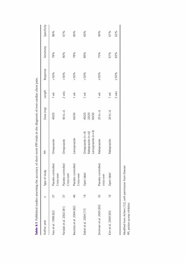
Tab
le 9
.1 P
ubli
shed s
tudie
s ass
ess
ing t
he a
ccu
racy
of
short
-term
PPI
tria
ls in
th
e d
iagn
osi
s of
non
-card
iac
chest
pain
.
Aut
hor,
year
n Ty
pe o
f st
udy
PPI
Dos
e (m
g)Le
ngth
Resp
onse
Sens
itivi
tySp
ecifi
city
Fass
et
al . 1
998
[62]
37
Plac
ebo
cont
rolle
d
Cro
ss-o
ver
Om
epra
zole
40/2
01
wk
> 50
%78
%86
%
Pand
ak e
t al
. 200
2 [8
1]
37Pl
aceb
o co
ntro
lled
Cro
ss-o
ver
Om
epra
zole
40 b
.i.d.
2 w
ks>
50%
90%
67%
Baut
ista
et
al . 2
004
[82]
40
Plac
ebo
cont
rolle
d
Cro
ss-o
ver
Lans
opra
zole
60/3
01
wk
> 50
%78
%80
%
Dek
el e
t al
. 200
4 [1
7]
14O
pen
labe
lO
mep
razo
le ( n
= 4
)
Rabe
praz
ole
( n =
6)
Lans
opra
zole
( n =
4)
40/2
0
20/2
0
60/3
0
1 w
k>
50%
89%
60%
Dic
kman
et
al . 2
005
[83]
35
Plac
ebo
cont
rolle
d,
cros
s-ov
er
Rabe
praz
ole
20 b
.i.d.
1 w
k≥
50%
75%
90%
Kim
et
al . 2
009
[65]
16
Ope
n la
bel
Rabe
praz
ole
20 b
.i.d.
1 w
k67
%67
%
2 w
ks≥
50%
83%
62%
Mod
ified
fro
m A
chem
[12]
, with
per
mis
sion
fro
m E
lsev
ier.
PPI,
prot
on p
ump
inhi
bito
r.
c09.indd 142 11/15/2012 2:28:25 AM
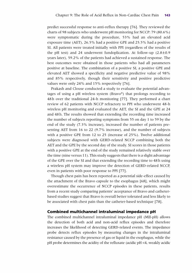
Chapter 9: The Role of Acid Reflux in Non-Cardiac Chest Pain 143
predict successful response to anti-reflux therapy [76] . They reviewed the
charts of 98 subjects who underwent pH monitoring for NCCP: 79 (80.6%)
were symptomatic during the procedure, 53% had an elevated acid
exposure time (AET), 26.5% had a positive GPE and 25.5% had a positive
SI. All patients were treated initially with PPI (regardless of the results of
the pH test) and 24 underwent fundoplication. At follow-up (2.8 ± 0.9
years later), 59.2% of the patients had achieved a sustained response. The
best outcomes were obtained in those patients who had all parameters
positive at baseline. The combination of a positive SI, a positive GPE and
elevated AET showed a specificity and negative predictive value of 98%
and 85% respectively, though their sensitivity and positive predictive
values were only 24% and 15% respectively [76] .
Prakash and Clouse conducted a study to evaluate the potential advan-
tages of using a pH wireless system (Bravo®) that prolongs recording to
48 h over the traditional 24-h monitoring [77] . They performed a chart
review of 62 patients with NCCP refractory to PPI who underwent 48-h
wireless pH monitoring and evaluated the AET, the SI and the GPE at 24
and 48 h. The results showed that extending the recording time increased
the number of subjects reporting symptoms from 55 on day 1 to 59 by the
end of the study (7.3% increase), increased the number of patients pre-
senting AET from 16 to 22 (9.7% increase), and the number of subjects
with a positive GPE from 12 to 25 (increase of 25%). Twelve additional
subjects were diagnosed with GERD-related NCCP combining both the
AET and the GPE by the second day of the study. SI scores in those patients
with a positive GPE at the end of the study remained relatively stable over
the time (nine versus 11). This study suggests that there is a slight advantage
of the GPE over the SI and that extending the recording time to 48 h using
a wireless pH system may improve the detection of GERD-related NCCP,
even in patients with poor response to PPI [77] .
Though chest pain has been reported as a potential side-effect caused by
the attachment of the Bravo capsule to the esophagus [68] , which might
overestimate the occurrence of NCCP episodes in these patients, results
from a recent study comparing patients’ acceptance of Bravo and catheter-
based studies suggest that Bravo is overall better tolerated and less likely to
be associated with chest pain than the catheter-based technique [78] .
Combined multichannel intraluminal impedance pH The combined multichannel intraluminal impedance pH (MII-pH) allows
the detection of both acid and non-acid reflux episodes and therefore
increases the likelihood of detecting GERD-related events. The impedance
probe detects reflux episodes by measuring changes in the intraluminal
resistance caused by the presence of gas or liquid in the esophagus, while the
pH probe determines the acidity of the refluxate (acidic pH <4, weakly acidic
c09.indd 143 11/15/2012 2:28:25 AM

144 Part 2: Gastroesophageal Reflux Disease
pH 4–7, non-acidic pH > 7) [74] . There is scarce information about the
usefulness of MII-pH for detecting GERD-related NCCP. The only series
reported to date includes 75 consecutive patients with NCCP [79] ; 16 (23%)
patients had abnormal acid exposure and 40 (53.3%) “pathological bolus
exposure.” Pathological bolus exposure was defined as “cases in which
reflux time was above 1.4% of the total reflux number on impedance tests”.
Fifty of 54 patients (92.6%) reported symptomatic improvement following
PPI treatment regardless of the presence of GERD [79] ; whether this
improvement was due to placebo effect or acid hypersensitivity was not
clear. In summary, despite its limitations (lack of a control group), this study
suggests that the use of MII-pH might increase the diagnostic yield of
ambulatory wire pH testing, given its ability to detect patients with weakly
acidic and non-acidic GERD. Other reent studies using impedance pH mon-
itoring on medication have shown that in some patients, symptoms that
persist despite PPI, including non-cardiac chest pain, may be due to non-acid
reflux [80] . Therefore, impedance pH monitoring may be especially useful in
patients who are refractory to PPI. However, prospective, controlled studies
to confirm these observations are not available at the present time.
In summary, several tests are available for the diagnostic evaluation of
patients with NCCP including endoscopy, ambulatory reflux monitoring,
and esophageal motility. The available data suggest that the use of an initial
short course of a PPI therapeutic trial is a sensitive and specific approach to
diagnose GERD-related NCCP. Figure 9.1 summarizes our current sug-
gested approach to NCCP.
Treatment
Medical therapy Several studies, including the previously mentioned metaanalysis, have
confirmed the empiric PPI strategy as a useful tool in the diagnosis and
treatment of patients with GERD-related NCCP [17,25,26,57,62,81–85] .
For patients responding to this initial approach, it is reasonable to maintain
them on a PPI for a longer period of time. The length of therapy in GERD-
related NCCP has not been critically evaluated. In 1993, Achem et al .
showed that high doses of ranitidine (300 mg t.i.d.) or omeprazole 20 mg
b.i.d. for 8 weeks induced a significant chest pain improvement in patients
with NCCP, GERD and nutcracker esophagus following an open label study
[86] . The same authors published the first placebo-controlled trial assess-
ing the efficacy of PPI (omeprazole 20 mg twice daily) for 8 weeks in NCCP.
They showed a significant clinical improvement in the omeprazole group
versus the placebo [57] . These data suggest that patients with NCCP may
be treated effectively for up to 2 months. Whether patients need to be
c09.indd 144 11/15/2012 2:28:25 AM

Chapter 9: The Role of Acid Reflux in Non-Cardiac Chest Pain 145
maintained on a PPI for longer periods or can be gradually tapered is
unknown. There is insufficient information to provide guidelines beyond
this time. Some experts recommended tapering the dose from twice a day to
once daily and subsequently reaching the minimal dose effective to control
the symptoms [87] .
There are different PPIs available but to date, only omeprazole, lansopra-
zole, and rabeprazole have been tested in NCCP. Most studies have been
done with omeprazole [57, 62, 81, 85] , given that this was the first PPI
developed, and for some time the only drug of that category that was avail-
able. However, more recent trials using other PPIs suggest that these are
also effective in relieving GERD-related chest pain [65, 83, 85] . The studies
that have evaluated the impact of chronic (> 4 weeks) PPI in GERD-related
NCCP are summarized in Table 9.2 .
Surgery The outcomes of surgery in patients with GERD-related NCCP have not
been systematically studied. The available data suggest that a variable
Chest pain
Cardiac work-up
Long term PPI
Taper dose based on response
PPI trial (double dose)
Check compliance/schedule(30 min before meals)
Consider alternative PPI(*)
or consider an alternative treatment for reflux (+)
Evaluation/treatment of other causes of
NCCP
Cardiac evaluation
Consider adding a PPI if associated GERD symptoms or persistent chest pain
Cardiac chest pain
+ –
+ –
Ambulatory reflux monitoring
Endoscopy
Non-cardiacchest pain
PPI trial
(double dose1week)
+–
Figure 9.1 Suggested management algorithm. (*) There is no published evidence
suggesting that changing the PPI type can improve clinical outcomes. (+) There are no
trials supporting the use of alternative treatment modalities for reflux (such as agents
acting on transient lower esophageal sphincter relaxation). GERD, gastroesophageal
reflux disease; NCCP, non-cardiac chest pain; PPI, proton pump inhibitor.
c09.indd 145 11/15/2012 2:28:25 AM
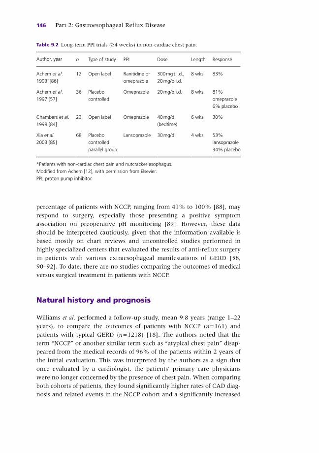
146 Part 2: Gastroesophageal Reflux Disease
percentage of patients with NCCP, ranging from 41% to 100% [88] , may
respond to surgery, especially those presenting a positive symptom
association on preoperative pH monitoring [89] . However, these data
should be interpreted cautiously, given that the information available is
based mostly on chart reviews and uncontrolled studies performed in
highly specialized centers that evaluated the results of anti-reflux surgery
in patients with various extraesophageal manifestations of GERD [58,
90–92] . To date, there are no studies comparing the outcomes of medical
versus surgical treatment in patients with NCCP.
Natural history and prognosis
Williams et al . performed a follow-up study, mean 9.8 years (range 1–22
years), to compare the outcomes of patients with NCCP ( n = 161) and
patients with typical GERD ( n = 1218) [18] . The authors noted that the
term “NCCP” or another similar term such as “atypical chest pain” disap-
peared from the medical records of 96% of the patients within 2 years of
the initial evaluation. This was interpreted by the authors as a sign that
once evaluated by a cardiologist, the patients’ primary care physicians
were no longer concerned by the presence of chest pain. When comparing
both cohorts of patients, they found significantly higher rates of CAD diag-
nosis and related events in the NCCP cohort and a significantly increased
Table 9.2 Long-term PPI trials (≥ 4 weeks) in non-cardiac chest pain.
Author, year n Type of study PPI Dose Length Response
Achem et al .
1993 * [86]
12 Open label Ranitidine or
omeprazole
300 mg t.i.d.,
20 mg/b.i.d.
8 wks 83%
Achem et al .
1997 [57]
36 Placebo
controlled
Omeprazole 20 mg/b.i.d. 8 wks 81%
omeprazole
6% placebo
Chambers et al .
1998 [84]
23 Open label Omeprazole 40 mg/d
(bedtime)
6 wks 30%
Xia et al .
2003 [85]
68 Placebo
controlled
parallel group
Lansoprazole 30 mg/d 4 wks 53%
lansoprazole
34% placebo
* Patients with non-cardiac chest pain and nutcracker esophagus.
Modified from Achem [12], with permission from Elsevier.
PPI, proton pump inhibitor.
c09.indd 146 11/15/2012 2:28:25 AM

Chapter 9: The Role of Acid Reflux in Non-Cardiac Chest Pain 147
use of acid suppression drugs in the GERD group. A Kaplan–Meier survival
curve did not show any differences in survival between cohorts. This study
suggests that patients presenting with NCCP (they did not specify the rate
of patients with GERD-related NCCP in the group) and patients with typ-
ical GERD symptoms were two separate groups with different characteris-
tics but a similar survival [18] .
Eslick and Talley assessed a cohort of patients who presented to the
emergency department with chest pain ( n = 197) and surveyed them at
baseline, 2 ( n = 129) and 4 years ( n = 91) after the initial consultation.
The cohort was divided according to the results of the initial evaluation:
those diagnosed with cardiac pain ( n = 71) and those with NCCP ( n = 126).
At the 4-year follow-up, 23 of 27 patients with cardiac chest pain and 45
of 64 with NCCP confirmed persistence of the pain. At the end of fol-
low-up, nine of 71 patients with cardiac pain had died, eight of them of
myocardial infarction. Eight of the 126 patients in the NCCP cohort
were deceased, seven of them of myocardial infarction. A multiple
regression logistic model identified only advanced age as an independent
predictor of mortality in both groups [93] . Wilhemsen et al . also reported
increased mortality rate (46%) and cardiovascular mortality (24.5%) in
a cohort of 441 patients presenting with NCCP that were followed up
longitudinally during a 16-year study in Göteborg, Sweden [94] .
It is unclear from these studies whether cardiac disease was missed in
the original cohorts or developed subsequently, but the findings under-
score the need for periodic reassessment of cardiac disease in patients with
persistent unexplained pain during long-term follow-up studies.
Summary and recommendations
Non-cardiac chest pain is a common, expensive and challenging clinical
problem. GERD is frequently associated with NCCP and occurs in approx-
imately 43% of the patients. In addition, GERD can co-exist and precipi-
tate chest pain in patients with CAD. After cardiac causes of NCCP have
been objectively ruled out, a short therapeutic trial (such as 1–2 weeks) of
double-dose PPI is helpful in identifying GERD-related chest pain. Patients
with GERD-related NCCP have a high response to PPI therapy. Once
symptom improvement has been obtained, treatment is frequently
extended up to 8 weeks. Then, patients may be gradually tapered off PPI
therapy to achieve the lowest required dose (or no therapy) to sustain
symptom control. For patients failing such a trial, further testing such as
ambulatory reflux monitoring, endoscopy and motility testing (mostly to
exclude achalasia) may be needed.
c09.indd 147 11/15/2012 2:28:25 AM
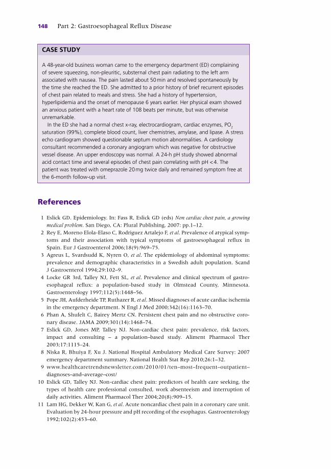
148 Part 2: Gastroesophageal Reflux Disease
References
1 Eslick GD . Epidemiology . In: Fass R , Eslick GD (eds) Non cardiac chest pain, a growing
medical problem . San Diego, CA : Plural Publishing , 2007 : pp. 1 – 12 .
2 Rey E , Moreno Elola-Elaso C , Rodriguez Artalejo F , et al . Prevalence of atypical symp-
toms and their association with typical symptoms of gastroesophageal reflux in
Spain . Eur J Gastroenterol 2006 ; 18 ( 9 ): 969 – 75 .
3 Agreus L , Svardsudd K , Nyren O , et al . The epidemiology of abdominal symptoms:
prevalence and demographic characteristics in a Swedish adult population . Scand
J Gastroenterol 1994 ; 29 : 102 – 9 .
4 Locke GR 3rd , Talley NJ , Fett SL , et al . Prevalence and clinical spectrum of gastro-
esophageal reflux: a population-based study in Olmstead County, Minnesota .
Gastroenterology 1997 ; 112 ( 5 ): 1448 – 56 .
5 Pope JH , Aufderheide TP , Ruthazer R , et al . Missed diagnoses of acute cardiac ischemia
in the emergency department . N Engl J Med 2000 ; 342 ( 16 ): 1163 – 70 .
6 Phan A , Shufelt C , Bairey Mertz CN . Persistent chest pain and no obstructive coro-
nary disease . JAMA 2009 ; 301 ( 14 ): 1468 – 74 .
7 Eslick GD , Jones MP , Talley NJ . Non-cardiac chest pain: prevalence, risk factors,
impact and consulting – a population–based study . Aliment Pharmacol Ther
2003 ; 17 : 1115 – 24 .
8 Niska R , Bhuiya F , Xu J . National Hospital Ambulatory Medical Care Survey: 2007
emergency department summary . National Health Stat Rep 2010 ; 26 : 1 – 32 .
9 www.healthcaretrendsnewsletter.com/2010/01/ten–most–frequent–outpatient–
diagnoses–and–average–cost/
10 Eslick GD , Talley NJ . Non-cardiac chest pain: predictors of health care seeking, the
types of health care professional consulted, work absenteeism and interruption of
daily activities . Aliment Pharmacol Ther 2004 ; 20 ( 8 ): 909 – 15 .
11 Lam HG , Dekker W , Kan G , et al . Acute noncardiac chest pain in a coronary care unit.
Evaluation by 24-hour pressure and pH recording of the esophagus . Gastroenterology
1992 ; 102 ( 2 ): 453 – 60 .
CASE STUDY
A 48-year-old business woman came to the emergency department (ED) complaining of severe squeezing, non-pleuritic, substernal chest pain radiating to the left arm associated with nausea. The pain lasted about 50 min and resolved spontaneously by the time she reached the ED. She admitted to a prior history of brief recurrent episodes of chest pain related to meals and stress. She had a history of hypertension, hyperlipidemia and the onset of menopause 6 years earlier. Her physical exam showed an anxious patient with a heart rate of 108 beats per minute, but was otherwise unremarkable.
In the ED she had a normal chest x-ray, electrocardiogram, cardiac enzymes, PO 2 saturation (99%), complete blood count, liver chemistries, amylase, and lipase. A stress echo cardiogram showed questionable septum motion abnormalities. A cardiology consultant recommended a coronary angiogram which was negative for obstructive vessel disease. An upper endoscopy was normal. A 24-h pH study showed abnormal acid contact time and several episodes of chest pain correlating with pH < 4. The patient was treated with omeprazole 20 mg twice daily and remained symptom free at the 6-month follow-up visit.
c09.indd 148 11/15/2012 2:28:25 AM

Chapter 9: The Role of Acid Reflux in Non-Cardiac Chest Pain 149
12 Achem SR . Noncardiac chest pain – treatment approaches . Gastroenterol Clin North
Am 2008 ; 37 : 859 – 78 .
13 Wise JL , Locke GR , Zinsmeister AR , et al . Risk factors for non-cardiac chest pain in
the community . Aliment Pharmacol Ther 2005 ; 22 : 1023 – 31 .
14 Hewson EG , Sinclair JW , Dalton CB , et al . Twenty-four-hour esophageal pH moni-
toring: the most useful test for evaluating noncardiac chest pain . Am J Med
1991 ; 90 ( 5 ): 576 – 83 .
15 Beedassy A , Katz PO , Gruber A , et al . Prior sensitization of esophageal mucosa by
acid reflux predisposes to a reflux-induced chest pain . J Clin Gastroenterol 2000 ;
31 ( 2 ): 121 – 4 .
16 Lacima G , Grande L , Pera M , et al . Utility of ambulatory 24-hour esophageal pH and
motility monitoring in noncardiac chest pain: report of 90 patients and review of the
literature . Dig Dis Sci 2003 ; 48 : 952 – 61 .
17 Dekel R , Martinez-Hawthorne SD , Guillen J , et al . Evaluation of symptom index in
identifying gastroesophageal reflux disease-related noncardiac chest pain . J Clin
Gastroenterol 2004 ; 38 ( 1 ): 24 – 9 .
18 Williams JF , Sontag SJ , Schnell T , et al . Non-cardiac chest pain: the long term natural
history and comparison with gastroesophageal reflux disease . Am J Gastroenterol
2009 ; 104 : 2145 – 52 .
19 Dickman R , Mattek N , Holub J , et al . Prevalence of upper gastrointestinal tract find-
ings in patients with noncardiac chest pain versus those with gastroesophageal reflux
disease (GERD)-related symptoms: results from a national endoscopic database . Am
J Gastroenterol 2007 ; 102 : 1173 – 9 .
20 Smith JL , Opekun AR , Larkai E , et al . Sensitivity of the esophageal mucosa to pH in
gastroesophageal reflux disease . Gastroenterology. 1989 ; 96 ( 3 ): 683 – 9 .
21 Richter JE , Hewson EG , Sinclair JW , et al . Acid perfusion test and 24-hour esophageal
pH monitoring with symptom index. Comparison of tests for esophageal acid sensi-
tivity . Dig Dis Sci 1991 ; 36 ( 5 ): 565 – 71 .
22 Lam HG , Breumelhof R , van Berge Henegouwen GP , et al . Temporal relationships
between episodes of non-cardiac chest pain and abnormal oesophageal function . Gut
1994 ; 35 ( 6 ): 733 – 6 .
23 Katz PO , Dalton CB , Richter JE , et al . Esophageal testing of patients with noncardiac
chest pain or dysphagia. Results of three years’ experience with 1161 patients . Ann
Intern Med 1987 ; 106 ( 4 ): 593 – 7 .
24 Behar J , Biancani P , Sheahan DG . Evaluation of esophageal tests in the diagnosis of
reflux esophagitis . Gastroenterology 1976 ; 71 ( 1 ): 9 – 15 .
25 Cremonini F , Wise J , Moayyedi P , et al . Diagnostic and therapeutic use of proton
pump inhibitors in non-cardiac chest pain: a metaanalysis . Am J Gastroenterol
2005 ; 100 ( 6 ): 1226 – 32 .
26 Wang WH , Huang JQ , Zheng GF , et al . Is proton pump inhibitor testing an effective
approach to diagnose gastroesophageal reflux disease in patients with noncardiac
chest pain? A meta-analysis . Arch Intern Med 2005 ; 165 ( 11 ): 1222 – 8 .
27 Orlando RC . Esophageal perception and noncardiac chest pain . Gastroenterol Clin
North Am 2004 ; 33 : 25 – 33 .
28 Barlow WJ , Orlando RC . The pathogenesis of heartburn in nonerosive reflux disease:
a unifying hypothesis . Gastroenterology 2005 ; 128 : 771 – 8 .
29 Barlow JD , Gregersen H , Thompson G . Identification of the biomechanical factors
associated with the perception of distension in the human esophagus . Am J Physiol
Gastrointest Liver Physiol 2002 ; 282 : G683 – 9 .
30 Richter JE , Barish CF , Castell DO . Abnormal sensory perception in patients with
esophageal chest pain . Gastroenterology 1986 ; 91 ( 4 ): 845 – 52 .
c09.indd 149 11/15/2012 2:28:26 AM

150 Part 2: Gastroesophageal Reflux Disease
31 Hobson AR , Furlong PL , Sarkar S , et al . Neurophysiologic assessment of esophageal
sensory processing in noncardiac chest pain . Gastroenterology 2006 ; 130 : 80 – 8 .
32 Hollerbach S , Bulat R , May A , et al . Abnormal cerebral processing of oesophageal
stimuli in patients with noncardiac chest pain (NCCP) . Neurogastroenterol Motil
2000 ; 12 ( 6 ): 555 – 65 .
33 Hu WH , Martin CJ , Talley NJ . Intraesophageal acid perfusion sensitizes the esoph-
agus to mechanical distension: a Barostat study . Am J Gastroenterol 2000 ; 95 ( 9 ):
2189 – 94 .
34 Sarkar S , Aziz Q , Woolf CJ , et al . Contribution of central sensitisation to the
development of non-cardiac chest pain . Lancet 2000 ; 356 : 1154 – 9 .
35 Sarkar S , Thompson DG , Woolf CJ , et al . Patients with chest pain and occult gastro-
esophageal reflux demonstrate visceral pain hypersensitivity which may be partially
responsive to acid suppression . Am J Gastroenterol 2004 ; 99 ( 10 ): 1998 – 2006 .
36 Balaban DH , Yamamoto Y , Liu J , et al . Sustained esophageal contraction: a marker of
esophageal chest pain identified by intraluminal ultrasonography . Gastroenterology
1999 ; 116 ( 1 ): 29 – 37 .
37 Liuzzo JP , Ambrose JA . Chest pain from gastroesophageal reflux disease in patients
with coronary artery disease . Cardiol Rev 2005 ; 13 : 167 – 73 .
38 Mehta AJ , Caestecker JS , Camm AJ , et al . Gastro-esophageal reflux in patients with
coronary artery disease: how common is it and does it matter? Eur J Gastroenterol
Hepatol 1996 ; 8 : 973 – 8 .
39 Schultz T , Mannheimer C , Dellborg M , et al . High prevalence of gastroesophageal
reflux in patients with clinical unstable angina and known coronary artery disease .
Acute Cardiac Care 2008 ; 10 : 37 – 42 .
40 Liuzzo JP , Ambrose JA , Diggs P . Proton pump inhibitor use by coronary artery disease
patients is associated with fewer chest pain episodes, emergency department visits
and hospitalizations . Aliment Pharmacol Ther 2005 ; 22 : 95 – 100 .
41 Budzynski J , Kłopocka M , Pulkowski G , et al . The effect of double dose of omeprazole
on the course of angina pectoris and treadmill stress test in patients with coronary
artery disease – a randomised, double-blind, placebo controlled, crossover trial . Int J
Cardiol 2008 ; 127 ( 2 ): 233 – 9 .
42 Froment R . [Intricated coronary angina; clinical and experimental data]. Brux Med
1955 ; 35 ( 49 ): 2429 – 42 .
43 Smith KS , Papp C . Episodic, postural, and linked angina . BMJ 1962 ; 2 ( 5317 ): 1425 – 30 .
44 Mellow M , Simpson AG , Watt L , et al . Esophageal acid perfusion in coronary artery
disease: induction of myocardial ischemia . Gastroenterology 1983 ; 85 : 306 – 12 .
45 Chauhan A , Petch MC , Schofield PM . Effect of esophageal acid instillation on coro-
nary blood flow . Lancet 1993 ; 341 : 1309 – 10 .
46 Chauhan A , Petch MC , Schofield PM . Cardio-esophageal reflex in humans as a
mechanism for ‘linked angina’ . Eur Heart J 1996 ; 17 : 407 – 13 .
47 Voskuil JH , Cramer MJ , Breumelhof R , et al . Prevalence of esophageal disorders in
patients with chest pain newly referred to the cardiologist . Chest 1996 ; 109 :
1210 – 14 .
48 Karcz A , Korn R , Burke MC , et al . Malpractice claims against emergency physicians
in Massachusetts: 1975–1993 . Am J Emerg Med 1996 ; 14 ( 4 ): 341 – 5 .
49 Mousavi S , Tosi J , Eskandarian R , et al . Role of clinical presentation in diagnosing
reflux-related non-cardiac chest pain . J Gastroenterol Hepatol 2007 ; 22 : 218 – 21 .
50 Richter JE , Boeckxstaens GE . Management of achalasia: surgery or pneumatic
dilation . Gut 2011 ; 60 ( 6 ): 869 – 76 .
51 Lacy BE , Weiser K , Chertoff J , et al . The diagnosis of gastroesophageal reflux disease .
Am J Med 2010 ; 123 ( 7 ): 583 – 92 .
c09.indd 150 11/15/2012 2:28:26 AM

Chapter 9: The Role of Acid Reflux in Non-Cardiac Chest Pain 151
52 García-Compeán D , González MV , Galindo G , et al . Prevalence of gastroesophageal reflux
disease in patients with extraesophageal symptoms referred from otolaryngology, allergy,
and cardiology practices: a prospective study . Dig Dis 2000 ; 18 ( 3 ): 178 – 82 .
53 Battaglia E , Bassotti G , Buonafede G , et al . Noncardiac chest pain of esophageal origin
in patients with and without coronary artery disease . Hepatogastroenterology
2005 ; 52 ( 63 ): 792 – 5 .
54 Gibbs JF , Rajput A , Chadha KS , et al . The changing profile of esophageal cancer pre-
sentation and its implication for diagnosis . JAMA 2007 ; 99 ( 6 ): 620 – 6 .
55 Achem SR , Almansa C , Murli K , et al . Esophageal eosinophilic infiltration in patients
with non cardiac chest pain . Aliment Pharm Ther 2011 ; 33 ( 11 ): 1194 – 201 .
56 Dekel R , Pearson T , Wendel C , et al . Assesment of esophageal motor function in
patients with dyspepsia or chest pain – the Clinical Outcomes Research Initiative
experience . Aliment Pharmacol Ther 2003 ; 18 ( 11–12 ): 1083 – 9 .
57 Achem SR , Kolts BE , MacMath T , et al . Effects of omeprazole versus placebo in
treatment of noncardiac chest pain and gastroesophageal reflux . Dig Dis Sci
1997 ; 42 ( 10 ): 2138 – 45 .
58 Achem SR . Noncardiac chest pain – treatment approaches . Gastroenterol Clin North
Am 2008 ; 37 : 859 – 78 .
59 Achem SR . Treatment of non-cardiac chest pain . Dis Mon 2008 ; 54 ( 9 ): 642 – 70 .
60 Gerson LB , Achem SR . Prevalence of pathologic reflux disease in patients with spastic
motility disorders . Gastroenterology 2011 ; 140 ( 5 )(Suppl 1): S – 232 .
61 Almansa C , Heckman MG , Devault KR , et al . Esophageal spasm: demographic,
clinical, radiographic, and manometric features in 108 patients . Dis Esophagus
2012 ; 25 ( 3 ): 214 – 21 .
62 Fass R , Fennerty MB , Ofman JJ , et al . The clinical and economic value of a short
course of omeprazole in patients with noncardiac chest pain . Gastroenterology
1998 ; 115 ( 1 ): 42 – 9 .
63 Young MA , Sanowski R , Talbert G , et al . Omeprazole administration as a test for gas-
troesophageal reflux . Gastroenterology 1992 ; 102 : 192 .
64 Squillace SJ , Young MF , Sanowski RA . Single dose omeprazole as a test for non
cardiac chest pain . Gastroenterology 1993 ; 104 : A197 .
65 Kim JH , Sinn DH , Son HJ , et al . Comparison of one-week and two-week empirical
trial with a high-dose rabeprazole in non-cardiac chest pain patients . J Gastroenterol
Hepatol 2009 ; 24 ( 9 ): 1504 – 9 .
66 Giannini EG , Zentilin P , Dulbecco P , et al . Management strategy for patients with gas-
troesophageal reflux disease: a comparison between empirical treatment with esome-
prazole and endoscopy-oriented treatment . Am J Gastroenterol 2008 ; 103 ( 2 ): 267 – 75 .
67 Faybush EM , Fass R . Gastroesophageal reflux disease in noncardiac chest pain .
Gastroenterol Clin North Am 2004 ; 33 : 41 – 54 .
68 Pandolfino JE , Vela MF . Esophageal-reflux monitoring . Gastrointest Endosc
2009 ; 69 ( 4 ): 917 – 30 .
69 Fass R. Proton pump inhibitor failure – what are the therapeutic options? Am J
Gastroenterol 2009 ; 104 (Suppl 2): S33 – 8 .
70 Wiener GJ , Richter JE , Copper JB , et al . The symptom index: a clinically important
parameter of ambulatory 24-hour esophageal pH monitoring . Am J Gastroenterol
1988 ; 83 : 358 – 61 .
71 Breumelhof R , Smout APJM . The symptom sensitivity index: a valuable additional
parameter in 24-h esophageal pH recording . Am J Gastroenterol 1991 ; 86 : 160 – 4 .
72 Weusten BL , Rolfs JMM , Akkermans LMA , et al . The symptom association proba-
bility: an improved method for symptom analysis of 24-h esophageal pH data .
Gastroenterology 1994 ; 107 : 1741 – 5 .
c09.indd 151 11/15/2012 2:28:26 AM
152 Part 2: Gastroesophageal Reflux Disease
73 Ghillebert G , Janssens J , Vantrappen G , et al . Ambulatory 24-h intraesophageal pH
and pressure recordings vs provocation test in the diagnosis of chest pain of esopha-
geal origin . Gut 1990 ; 31 : 738 – 44 .
74 Hirano I , Richter J E . ACG practice guidelines; esophageal reflux testing . Am J
Gastroenterol 2007 ; 102 : 668 – 85 .
75 Taghavi SA , Ghasedi M , Saberi-Firoozi M , et al . Symptom association probability and
symptom sensitivity index: preferable but still suboptimal predictors of response to
high dose omeprazole . Gut 2005 ; 54 ( 8 ): 1067 – 71 .
76 Kushnir VM , Sayuk GS , Gyawali CP . Abnormal GERD parameters on ambulatory pH
monitoring predicts therapeutic success in noncardiac chest pain . Am J Gastroenterol
2010 ; 105 : 1032 – 8 .
77 Prakash C , Clouse RE . Wireless pH monitoring in patients with non-cardiac chest
pain . Am J Gastroenterol 2006 ; 101 : 446 – 52 .
78 Sweis R , Fox M , Anggiansah R , et al . Patient acceptance and clinical impact of Bravo
monitoring in patients with previous failed catheter-based studies . Aliment Pharmacol
Ther 2009 ; 29 ( 6 ): 669 – 76 .
79 Kim BJ , Choi SC , Kim JJ , et al . Pathological bolus exposure plays a significant role in
eliciting non-cardiac chest pain . J Gastroenterol Hepatol 2010 ; 25 ( 12 ): 1855 – 60 .
80 Mainie I , Tutuian R , Shay S , et al . Acid and non-acid reflux in patients with persistent
symptoms despite acid suppressive therapy: a multicentre study suing combined
ambulatory impedance-pH monitoring . Gut 2006 ; 55 : 1398 – 402 .
81 Pandak WM , Arezo S , Everett S , et al . Short course of omeprazole: a better first diag-
nostic approach to noncardiac chest pain than endoscopy, manometry, or 24-hour
esophageal pH monitoring . J Clin Gastroenterol 2002 ; 35 ( 4 ): 307 – 14 .
82 Bautista J , Fullerton H , Briseno M , et al . The effect of an empirical trial of high-dose
lansoprazole on symptom response of patients with non-cardiac chest pain – a
randomized, double-blind, placebo-controlled, crossover trial . Aliment Pharmacol
Ther 2004 ; 19 ( 10 ): 1123 – 30 .
83 Dickman R , Emmons S , Cui H , et al . The effect of a therapeutic trial of high-dose
rabeprazole on symptom response of patients with non-cardiac chest pain: a
randomized, double-blind, placebo-controlled, crossover trial . Aliment Pharmacol
Ther 2005 ; 22 ( 6 ): 547 – 55 .
84 Chambers J , Cooke R , Anggiansah A , et al . Effect of omeprazole in patients with chest
pain and normal coronary anatomy: initial experience . Int J Cardiol 1998 ; 65 ( 1 ): 51 – 5 .
85 Xia HH , Lai KC , Lam SK , et al . Symptomatic response to lansoprazole predicts
abnormal acid reflux in endoscopy-negative patients with non-cardiac chest pain .
Aliment Pharmacol Ther 2003 ; 17 ( 3 ): 369 – 77 .
86 Achem SR , Kolts BE , Wears R , et al . Chest pain associated with nutcracker esophagus:
a preliminary study of the role of gastroesophageal reflux . Am J Gastroenterol
1993 ; 88 ( 2 ): 187 – 92 .
87 Oranu AC , Vaezi MF . Noncardiac chest pain: gastroesophageal reflux disease . Med
Clin North Am 2010 ; 94 ( 2 ): 233 – 42 .
88 Patti MG , Molena D , Fisichella PM , et al . Gastroesophageal reflux disease (GERD) and
chest pain. Results of laparoscopic antireflux surgery . Surg Endosc 2002 ; 16 ( 4 ): 563 – 6 .
89 So JB , Zeitels SM , Rattner DW . Outcomes of atypical symptoms attributed to gastro-
esophageal reflux treated by laparoscopic fundoplication . Surgery 1998 ; 124 ( 1 ): 28 – 32 .
90 Chen RYM , Thomas RJS . Results of laparoscopic fundoplication where atypical
symptoms coexist with oesophageal reflux . Aust NZ J Surg 2000 ; 70 : 840 – 2 .
91 Farrell TM , Richardson WS , Trus TL , et al . Response of atypical symptoms of gastro-
esophageal reflux to antireflux surgery . Br J Surg 2001 ; 88 ( 12 ): 1649 – 52 .
c09.indd 152 11/15/2012 2:28:26 AM

Chapter 9: The Role of Acid Reflux in Non-Cardiac Chest Pain 153
92 Rakita S , Valladolid D , Thomas A , et al . Laparoscopic nissen fundoplication offers high
patient satisfaction with relief of extraesophageal symptoms of gastroesophageal
reflux disease . Am Surg 2006 ; 72 ( 3 ): 207 – 12 .
93 Eslick GD , Talley NJ . Natural history and predictors of outcome for non-cardiac chest
pain: a prospective 4-year cohort study . Neurogastroenterol Motil 2008 ; 20 ( 9 ): 989 – 97 .
94 Wilhelmsen L , Rosengren A , Hagman M , Lappas G . "Nonspecific" chest pain associ-
ated with high long-term mortality: results from the primary prevention study in
Göteborg, Sweden . Clin Cardiol 1998 ; 21 ( 7 ): 477 – 82 .
c09.indd 153 11/15/2012 2:28:26 AM

154
Practical Manual of Gastroesophageal Reflux Disease, First Edition.
Edited by Marcelo F. Vela, Joel E. Richter and John E. Pandolfino.
© 2013 John Wiley & Sons, Ltd. Published 2013 by John Wiley & Sons, Ltd.
CHAPTER 10
Laryngopharyngeal Reflux Robert T. Kavitt and Michael F. Vaezi Division of Gastroenterology, Hepatology, and Nutrition, Vanderbilt University Medical Center ,
Nashville, TN, USA
Key points • Gastroesophageal reflux disease is increasingly associated with ear, nose, and throat
symptoms, including laryngitis.
• Laryngopharyngeal reflux can be assessed with laryngoscopy, esophagogastroduo-
denoscopy, and the use of ambulatory pH and impedance monitoring.
• Gastroenterology and otolaryngology specialty societies have released various
guidelines in recent years addressing diagnostic and therapeutic approaches to this
prevalent and often difficult-to-treat condition.
• Many uncertainties remain, including which patient subgroups might benefit from
acid suppressive therapy.
Potential pitfalls • Laryngopharyngeal reflux symptoms may be associated with, but not necessarily always
caused by, acid reflux. It is important to distinguish between potential signs and
symptoms of laryngopharyngeal reflux and a true causal relationship with acid reflux.
• One must take a cautious approach in the management of laryngopharyngeal reflux
patients with an absence of any symptomatic response to aggressive acid suppression.
Introduction
Reflux of gastroduodenal contents into the laryngopharyngeal region may
cause inflammation and symptoms resulting in chronic laryngitis, often
referred to as laryngopharyngeal reflux (LPR) [1] . Reports suggest that
4–10% of patients presenting to otolaryngologists demonstrate symptoms
attributed in part to gastroesophageal reflux disease (GERD) [2] . Such
symptoms include hoarseness, sore or burning throat, chronic cough, glo-
bus, dysphagia, postnasal drip, apnea, laryngospasm, and even laryngeal
neoplasm, among other complaints (Box 10.1 ). Chronic laryngitis and
throat symptoms are reportedly associated with GERD in up to 60% of
c10.indd 154 11/15/2012 2:28:04 AM

Chapter 10: Laryngopharyngeal Reflux 155
patients [2–8] . Additionally, some studies suggest an association of laryn-
geal cancer with chronic laryngeal exposure to reflux of gastroduodenal
contents [9–13] . Thus, LPR is a significant clinical issue.
The first association between GERD and laryngeal disease was suggested
by L.A. Coffin in 1903 [14] with subsequent studies suggesting a role of
gastroduodenal contents in the development of “contact ulcer” [15,16]
with patient response to antacids, dietary changes, and head of bed eleva-
tion. However, since these initial studies, the direct association between
reflux of gastroduodenal contents and laryngeal signs and symptoms has
been difficult to establish. The 2006 Montreal consensus group distin-
guished GERD symptoms between esophageal and extraesophageal syn-
dromes [17] . Extraesophageal manifestations with established associations
include chronic cough, laryngitis, and asthma, based on population-based
studies, with odds ratios (OR) of 1.3–3.0 [18–21] . However, the causal
relationship of GERD to these non-specific symptoms is not proven. More
recently, the American Gastroenterological Association (AGA) guidelines
for GERD recommended against treating for acid reflux for patients with
laryngitis or asthma who do not have concomitant typical reflux symp-
toms [18,22] . Thus, the role of “silent” reflux, extraesophageal symptoms
without concomitant heartburn or regurgitation, is controversial and divi-
sive between the gastroenterology and otolaryngology communities.
Prevalence
Gastroesophageal reflux disease is a widely prevalent condition with
significant impact on quality of life. A Gallup poll of 1000 adults with
Box 10.1 Symptoms associated with gastroesophageal reflux laryngitis • Hoarseness
• Dysphonia
• Sore or burning throat
• Excessive throat clearing
• Chronic cough
• Globus
• Apnea
• Laryngospasm
• Dysphagia
• Postnasal drip
• Neoplasm
c10.indd 155 11/15/2012 2:28:05 AM
156 Part 2: Gastroesophageal Reflux Disease
heartburn at least weekly conducted for the American Gastroenterology
Association found that 79% of respondents noted heartburn symptoms at
night [23] . Twenty percent reported supraesophageal symptoms 3–6 times
each week, and 43% reported these symptoms once or twice per week. In
a population survey study, Locke et al . [19] showed that heartburn and
acid regurgitation are significantly associated with chest pain, dysphagia,
dyspepsia, and globus sensation. A subsequent Veterans Affairs (VA)-based
case–control study by El-Serag and Sonnenberg suggested that erosive
esophagitis and esophageal stricture were associated with various extra-
esophageal symptoms such as sinusitis (OR 1.60, 95% confidence interval
(CI) 1.51–1.70), pharyngitis (OR 1.48, 95% CI 1.15–1.89), aphonia (OR 1.81,
95% CI 1.18–2.80), and chronic laryngitis (OR 2.01, 95% CI 1.53–2.63),
among others [20] . The authors concluded that patients with reflux esoph-
agitis are at an increased risk of harboring a large variety of sinus, pharyn-
geal, laryngeal, and pulmonary diseases.
Laryngeal disorders have been shown to be twice as likely in patients
with esophagitis compared to those without [20] . Twenty-five percent of
patients with LPR are found to have histologic evidence of esophagitis
[24] . Thus, epidemiological studies suggest an association between reflux
disease and extraesophageal symptoms, including LPR.
Mechanisms of gastroesophageal reflux laryngitis
The two predominant pathophysiological mechanisms for LPR are direct
and indirect exposure of the larynx to injurious gastric contents. The direct
exposure is due to acid, pepsin, and bile acid exposure to the laryngopha-
ryngeal mucosa. The indirect mechanism is thought to be a result of reflux-
ate interactions with structures distal to the larynx, evoking a vagally
mediated response of bronchoconstriction [25] .
The potential agents causing laryngitis may include gastric contents (acid
and pepsin) and duodenal contents (bile acids and the pancreatic enzyme
trypsin). Animal studies in the past have shown the potential of both acid
and pepsin to cause laryngeal injury [26] . The role of conjugated and
unconjugated bile and trypsin at pH values of 1–7 was investigated by
Adhami et al . who did not find histological injury to the canine larynx by
the above agents alone. However, they showed that the combination of the
bile constituents with acid and pepsin in an acidic pH caused the greatest
injury [27] . This is difficult to assess in humans, because refluxate into the
esophagus is often a mix of gastric and duodenal contents [28] . However,
these data indirectly suggest that reflux of duodenal contents into the
larynx, although it may be associated with symptoms such as regurgitation,
is less likely to cause mucosal damage unless it occurs in an acidic milieu.
c10.indd 156 11/15/2012 2:28:05 AM

Chapter 10: Laryngopharyngeal Reflux 157
Clinical symptoms
Many patients with gastroesophageal reflux-induced chronic laryngitis pre-
sent with symptoms such as sore throat, globus, chronic cough, hoarseness,
dysphagia, apnea or postnasal drip, among other symptoms (see Box 10.1 ).
However, they may not present with classic GERD symptoms of heartburn
and regurgitation. A 1991 study found that of 225 patients with otolaryngo-
logical disorders having suspected GERD, only 43% reported symptoms of
heartburn or acid regurgitation [6] . Additional symptoms which may be
seen in reflux-related laryngitis include frequent throat clearing, dry mouth,
prolonged voice warm-up time (greater than 20–30 min), halitosis, excess
phlegm, coated tongue, throat tickle, regurgitation of food, nocturnal
cough, difficulty breathing especially at night, aspiration, laryngospasm,
poorly controlled asthma or pneumonia [29,30] .
The Reflux Symptom Index (RSI) is a validated nine-item self-adminis-
tered instrument published in 2002 to help assess the severity of LPR
symptoms at the time of diagnosis and after therapy [31] . Symptoms
assessed include throat clearing, difficulty swallowing, hoarseness, excess
throat mucus or postnasal drip, coughing after eating or lying down,
breathing difficulties or choking episodes, troublesome or annoying cough,
sensation of something sticking in the throat or a lump in the throat, and
symptoms of heartburn, chest pain or indigestion, on a scale from 0 to 5.
An RSI score greater than 12 is defined as abnormal. The RSI was once
believed to be significantly higher in untreated patients with LPR than
controls; however, more recent studies suggest that it may be of lower
clinical utility than previously believed [32] . Reliability of this instrument
may vary based on patient population and clinical setting. Additionally,
given the complexity of the symptoms and the scoring system, it is not
widely used in clinical practice.
Evaluation of the larynx
Physical examination Physical examination of patients with suspected gastroesophageal reflux
laryngitis must be thorough, and include an examination of the head and
neck, with assessment of ears and hearing, patency of the nares, oral
cavity, temporomandibular joints, and larynx. One should also assess for
signs of systemic disease (such as hypothyroidism) or neurological impair-
ment that may manifest with symptoms affecting the throat or voice,
including Parkinson ’ s disease, multiple sclerosis or other conditions. When
a patient reports symptoms of vocal difficulties, laryngeal examination by
c10.indd 157 11/15/2012 2:28:05 AM

158 Part 2: Gastroesophageal Reflux Disease
an otolaryngologist may include an assessment of the speaking and singing
voice and strobovideolaryngoscopy [29] . Objective voice analysis can
quantify quality of voice, pulmonary function, harmonic spectral charac-
teristics, valvular efficiency of vocal folds, and neuromuscular function on
electromyography [33] . Use of flexible transnasal laryngoscopy plays a
vital role in excluding more ominous causes for patients’ laryngeal symp-
toms. Hoarseness is a symptom that can be present in patients with LPR
but also may be a symptom in vocal fold paresis, polyps, postviral
inflammatory reactions, allergies, vocal abuse, dysplasia, and cancer [34] .
If hoarseness persists longer than 2 weeks, laryngoscopy is indicated [35] .
Once more ominous diagnoses such as laryngeal cancer are ruled out, LPR
is often entertained as a potential contributing etiology for patients’ throat
symptoms due to laryngeal irritation often found at laryngosocopy.
Laryngeal signs Normal laryngeal tissue has sharply demarcated landmarks with glistening
mucosa with minimal or no laryngeal edema (Plate 10.1), unlike abnormal
laryngeal findings (Plate 10.2). The epithelium of the larynx is thin and is
not adapted to accommodating injury from acid and pepsin [36] . Several
laryngeal signs are attributed to GERD, including edema, erythema, pseudo-
sulcus, ventricular obliteration, and postcricoid hyperplasia (Box 10.2 ) [1] .
In a study reporting the results of a survey of otolaryngologists regarding
the signs used to diagnose LPR, subjective signs of laryngeal erythema and
edema were the findings most commonly employed to diagnose GERD
[37,38] . However, these signs are criticized for their lack of specificity
for GERD. Several signs of posterior laryngitis thought to be markers for
LPR are actually present in a high percentage of asymptomatic healthy
Box 10.2 Potential Laryngopharyngeal Signs Associated with Gastroesophageal Reflux Laryngitis • Edema and hyperemia of larynx
• Hyperemia and lymphoid hyperplasia of posterior pharynx (cobblestoning)
• Contact ulcers
• Laryngeal polyps
• Granuloma
• Interarytenoid changes
• Subglottic stenosis
• Posterior glottic stenosis
• Reinke ’ s edema
• Tumors
c10.indd 158 11/15/2012 2:28:05 AM

Chapter 10: Laryngopharyngeal Reflux 159
volunteers, raising questions about the specificity of such findings [39] .
Forty-three of 50 healthy subjects (86%) may exhibit one or more findings
considered pathognomonic of laryngeal complaints due to GERD. This
finding suggests that GERD may be overdiagnosed, as the laryngeal signs
used in clinical practice are non-specific [40] . The advantages and dis-
advantages of laryngoscopy and other diagnostic tests in detecting reflux
are shown in Table 10.1 .
First described in 2001, the Reflux Finding Score (RFS) received consid-
erable attention initially as a validated sign for reflux-induced laryngeal
pathology. This instrument is an eight-item clinical severity scale based on
laryngoscopic findings including subglottic edema, vocal fold edema, dif-
fuse laryngeal edema, ventricular obliteration, erythema/hyperemia, pos-
terior commissure hypertrophy, granuloma/granulation tissue, and thick
endolaryngeal mucus, ranging from 0 to 26 [41,42] . However, similar to
the RSI, the RFS is not commonly employed in clinical practice. A recent
study found that both the RSI and RFS have poor specificity, with no
significant difference between patients and control groups [32] .
Table 10.1 Advantages and disadvantages of methods for detecting esophageal reflux.
Method Advantages Disadvantages
Endoscopy Easy visualization of mucosal
damage/erosions
Poor sensitivity/specificity/PPV
Requires sedation
High cost
Laryngoscopy No sedation required
Direct visualization of the larynx
and laryngeal pathology
No specific laryngeal signs for reflux
Overdiagnoses GERD
pH monitoring Easy to perform
Relatively non-invasive
Prolonged monitoring possible
Ambulatory
Catheter based
May have up to 30% false-negative rate
No pH predictors of treatment response
in LPR
Impedance
monitoring
Easy to perform
Relatively non-invasive
Prolonged monitoring possible
Ambulatory
Measures acidic and non-acidic gas
and liquid reflux (combined with pH)
Catheter based
False-negative rate unknown but most
likely similar to catheter-based pH
monitoring
Unknown clinical relevance when
abnormal on PPI therapy
Unknown importance in LPR
ResTech Dx-pH Faster detection rate and faster time
to equilibrium pH than traditional
pH catheters
Unknown if clinically useful in patients
with LPR
GERD, gastroesophageal reflux disease; LPR, laryngopharyngeal reflux; PPI, proton pump inhibitor;
PPV, positive predictive value.
c10.indd 159 11/15/2012 2:28:05 AM
160 Part 2: Gastroesophageal Reflux Disease
The variability of detecting laryngeal signs may also be affected by the
quality and sensitivity of the detection instrument. Abnormal laryngeal
signs are more likely to be suspected with flexible laryngoscopy as opposed
to rigid laryngoscopy in the same individual, indicating that flexible laryngo-
scopy may be more sensitive and less specific for detecting laryngeal irrita-
tion [43] . Laryngeal signs appear to be poorly specific for identifying
gastroesophageal reflux. One study showed that lesions of the vocal fold
may represent more specific signs for LPR, exhibiting 91% specificity and
88% response to treatment with proton pump inhibitors (PPIs) [44] . The
non-specificity of laryngoscopy may also be due to poor inter- and intra-
observer variability. A randomized prospective analysis by five otolaryngo-
logists blinded to patient information of 120 video segments of rigid
fiberoptic laryngoscopy found poor interrater reliability of the laryngo-
scopic findings associated with LPR (intraclass correlation coefficient of
0.265), and intrarater reliability was extremely variable for the various
physical findings (Kendall correlation coefficients ranging from −0.121 to
0.837). Taken together, these studies suggest that accurate assessment of
laryngeal involvement with LPR is quite difficult as interpretation of
physical findings is subjective and varies among physicians.
Ambulatory pH studies
Ambulatory pH monitoring allows for detection of esophageal or hypopha-
ryngeal acid exposure. Since even healthy individuals have some reflux,
the normal values have a range with an accepted upper limit, based on
studies in healthy subjects. Up to 50 acid reflux events into the esophagus
each day may occur normally [24] . When compared to physical exam
findings, dual pH probe monitoring is reported to have superior sensitivity
and specificity [6] . A metaanalysis of 16 studies involving a total of 793
subjects who underwent 24-h pH monitoring (529 patients with LPR, 264
controls) showed that the number of pharyngeal reflux events for the con-
trol group and for LPR patients differed significantly ( P < 0.0001). The
authors concluded that the “upper probe gives accurate and consistent
information in normal subjects and patients with LPR” and that the acid
exposure time and number of reflux events are most important in distin-
guishing normal subjects from patients with LPR [45] . However, there is a
great degree of variability in the reported prevalence of pH abnormalities
in the literature for patients with LPR (Table 10.2 ). This heterogeneity may
be due to different patient populations and non-standard pH probe
placement [46] . Some investigators utilized direct laryngoscopy for probe
placement while others utilized esophageal manometry to identify the
upper and lower esophageal sphincters [1] . Current recommendations
c10.indd 160 11/15/2012 2:28:05 AM

Chapter 10: Laryngopharyngeal Reflux 161
Table 10.2 Prevalence of abnormal pH monitoring in distal and proximal esophagus
and the hypopharynx.
Study, year
(reference)Proportion
of patients
with LPR
Number of
patients with
reflux identified
during proximal
pH monitoring
Number of
patients with
reflux
identified
during distal
pH monitoring
Number of
patients with
reflux identified
during
hypopharyngeal
pH monitoring
Prevalence
(%)
Ossakow,
1987 [94]
43/63 NR 43 NR 68
Koufman,
1988 [2]
24/32 NR 24 7 75
Wiener,
1989 [95]
12/15 NR 12 3 80
Wilson,
1989 [96]
17/97 17 NR NR 18
Katz, 1990
[97]
7/10 NR 7 7 70
Woo, 1996
[98]
20/31 20 20 NR 65
Metz, 1997
[99]
6/10 ? 6 NR 60
Vaezi, 1997
[100]
21/21 11 21 NR 100
Chen, 1998
[101]
365/735 NR 229 255 50
Havas,
1999 [71]
10/15 NR 6 4 67
Ulualp,
1999 [102]
15/20 15 15 15 75
Smit, 2001
[103]
7/15 7 3 NR 47
Ulualp,
2001 [52]
28/39 28 28 28 72
Noordzij,
2002 [51]
29/42 NR 29 29 69
Park, 2005
[44]
33/78 20 28 NR 42
Cumulative 637/1223 46 % 42 % 38 % 52
LPR, laryngopharyngeal reflux; NR, not reported; ?, unclear how many patients tested.
c10.indd 161 11/15/2012 2:28:05 AM

162 Part 2: Gastroesophageal Reflux Disease
suggest that the hypopharyngeal probe be placed 1–2 cm above the upper
esophageal sphincter as determined by manometry, while the distal and
proximal pH probes be placed 5 and 15 cm above the manometric lower
esophageal sphincter [1] .
Initial studies of patients with suspected gastroesophageal reflux laryn-
gitis investigated the role of proximal esophageal pH probes. A 1991 study
assessed the prevalence of abnormal acid exposure on the proximal eso-
phagus in 15 patients with typical GERD (group 1), 15 patients with laryngeal
symptoms without abnormal findings on laryngoscopy (group 2), and 10
patients with both laryngeal symptoms and findings on laryngoscopy
(group 3) [47] . Increased proximal esophageal acid exposure was observed
in patients in groups 1 and 2, indicating that proximal esophageal acid
exposure may differentiate patients with laryngitis from those with typical
GERD. Measurement of hypopharyngeal pH exposure was initially used to
objectively measure laryngeal extension of reflux.
An earlier study suggested that hypopharyngeal pH assessment may be
useful when used in conjunction with findings on laryngoscopy to identify
patients whose symptoms may be related to GERD [48] . In this study, 76
patients with respiratory complaints thought to be related to GERD were
divided into three groups based on RFS and pharyngeal reflux events. The
patients were classified as RFS + if the RFS was greater than 7, and pharyn-
geal reflux positive if they had greater than one episode of reflux noted
during pH assessment. Controls were found to have a significantly lower
RFS and fewer episodes of pharyngeal reflux. None of the controls had
more than one episode of pharyngeal reflux during a 24-h period. Twenty-
one patients had both an abnormal RFS and pharyngeal reflux, and these
patients also had significantly higher heartburn scores and acid exposure
in the distal esophagus. The authors concluded that agreement between
detection of pharyngeal reflux by pH monitoring and an increased RFS
greater than 7 helps establish or refute the diagnosis of GERD as an
etiology of laryngeal symptoms. When both are normal, GERD is most
likely not playing a role in a patient ’ s extraesophageal symptoms.
However, initial enthusiasm about the diagnostic ability of hypopharyn-
geal reflux monitoring has now been replaced by skepticism. The posi-
tioning of the hypopharyngeal pH probe is operator dependent and varies
with regard to placement via direct visualization with laryngoscopy com-
pared to measurement by manometry [49] . Artifacts commonly occur and
computer-driven interpretations must be manually reviewed [50] . Several
studies have found that positive results of pharyngeal testing do not pre-
dict a favorable response to anti-reflux therapy [51,52] . One study showed
that the degree of improvement in symptoms among 19 of 27 patients
with pharyngeal reflux was similar to the eight patients not exhibiting
pharyngeal reflux [52] . Additionally, there are no universally accepted
c10.indd 162 11/15/2012 2:28:05 AM

Chapter 10: Laryngopharyngeal Reflux 163
diagnostic criteria for pH monitoring of the hypopharynx. The range of
normal pH values is not uniformly defined, and can vary from none to
4 pH drops less than 4 [51,53,54] . Less restrictive pH values, including a
drop in pH of 1.0 or 1.5 units instead of 2.0 units, do not differentiate
healthy volunteers from patients with suspected ENT complaints [55] .
The 2008 American Gastroenterological Association (AGA) technical
review on the management of GERD suggested that the role of pH or
impedance pH monitoring in diagnosing extraesophageal reflux is contro-
versial and unproven (Table 10.3 ) [18] . This evidence-based technical
review concludes that the value of a negative pH or impedance pH study is
of greater clinical utility, and states “In the absence of troublesome eso-
phageal symptoms or endoscopic findings, with a failed 8-week therapeutic
trial of twice-daily PPI therapy, and with normal esophageal acid exposure
(PPI therapy withheld) on 24-hour monitoring, one has gone as far as cur-
rently possible to rule out GERD as a significant contributor to these non-
specific syndromes. Such patients should have etiologies other than GERD
explored” [18] .
This conclusion is in direct contrast to guidelines published by The
American Academy of Otolaryngology-Head and Neck Surgery (AAOHNS)
Committee on Speech, Voice, and Swallowing Disorders. The authors of
the AAOHNS guidelines state that LPR can be diagnosed based on symp-
toms or laryngeal findings, but ambulatory 24-h double-probe (simulta-
neous esophageal and pharyngeal) pH assessment is considered the gold
standard diagnostic tool (see Table 10.3 ) [24] . They also suggested that
barium esophagraphy or esophagoscopy provide far less sensitive assess-
ments of LPR, but may be advisable for screening of the esophagus for
related pathology [6,24,56] . However, in line with the AGA guidelines,
the American College of Gastroenterology (ACG) practice guidelines sug-
gested that pH testing may not be the gold standard diagnostic test in this
group of patients (see Table 10.3 ) [49] . The authors refer to data indicating
that the overall pretherapy prevalence of an abnormal pH test in a
population with chronic laryngeal symptoms is 53%, with the prevalence
of excessive distal, proximal, and hypopharyngeal acid exposure being
42%, 44%, and 38%, respectively [46] , suggesting that this population
may have abnormal acid reflux exposure but not proving causality. In
support of the ACG and AGA guidelines, a placebo-controlled study of 145
patients with suspected GERD-related ENT symptoms treated with high-
dose esomeprazole or placebo for 16 weeks found that degree of symptom-
atic or laryngeal involvement was independent of pretherapy pH findings
and that neither esophageal nor hypopharyngeal acid reflux predicted a
response to PPI use [57] .
In patients who remain symptomatic despite aggressive acid suppressive
therapy, recent studies suggest that non-acid reflux may play a role in their
c10.indd 163 11/15/2012 2:28:05 AM

164 Part 2: Gastroesophageal Reflux Disease
Table 10.3 Summary of AGA, AAOHNS, and ACG guidelines regarding pH testing and
treatment modalities for patients with suspected LPR.
American
Gastroenterological
Association Institute
Technical Review on the
Management of
Gastroesophageal Reflux
Disease, 2008 [18]
Laryngopharyngeal
Reflux: Position
statement of the
Committee on Speech,
Voice, and Swallowing
Disorders of the
American Academy of
Otolaryngology-Head
and Neck Surgery,
2002 [24]
American College of
Gastroenterology
Practice Guidelines:
Esophageal Reflux
Testing, 2007
[49]
pH testing Role of pH or impedance
pH monitoring in diagnosing
extraesophageal reflux is
controversial and unproven
“In the absence
of troublesome esophageal
symptoms or endoscopic
findings, with a failed
8-week therapeutic trial of
twice-daily PPI therapy, and
with normal esophageal
acid exposure (PPI therapy
withheld) on 24-hour
monitoring, one has gone as
far as currently possible to
rule out GERD as a
significant contributor to
these nonspecific
syndromes. Such patients
should have etiologies other
than GERD explore”
Diagnosis of LPR can be
made based on
symptoms and laryngeal
findings, but ambulatory
24-h double-probe pH
assessment is considered
the gold standard
diagnostic too
l Barium esophagraphy or
esophagoscopy provide
far less sensitive
assessments of LPR, but
may be advisable for
screening of the
esophagus for related
pathology
“The accumulating data
seriously question the
clinical usefulness of
esophageal or
hypopharyngeal pH
monitoring in the initial
evaluation of patients
with suspected
acid-related ENT
complaints”
“Studies using
impedance pH
monitoring in patients
with extraesophageal
symptoms unresponsive
to PPI therapy show little
evidence of nonacid
reflux, except in the
chronic cough patient”
Treatment Empiric therapy with
twice-daily PPI for 2 months
for patients with
concomitant esophageal
GERD syndrome and
laryngitis remains a
pragmatic clinical strategy
(USPSTF grade B, quality fair)
Do not support use of
once- or twice-daily PPIs (or
H2RAs) for acute treatment
of potential extraesophageal
GERD syndromes, including
laryngitis and asthma, in
absence of esophageal GERD
(USPSTF grade D, quality fair)
Treatment for LPR needs
to be more aggressive
and prolonged than that
for GERD, and depends
on symptoms and
severity of LPR and on
response to therapy
Mild or intermittent LPR
symptoms can be treated
with dietary and lifestyle
changes and H2
antagonists, while the
majority of patients
require at least
twice-daily PPI (minimum
of 6 months)
“The practical and
popular approach is an
empiric trial with a BID
PPI regimen for several
months, reserving pH
testing for patients with
persistent symptoms.
However, here again, the
results of acid pH testing
have limited clinical
utility”
c10.indd 164 11/15/2012 2:28:05 AM
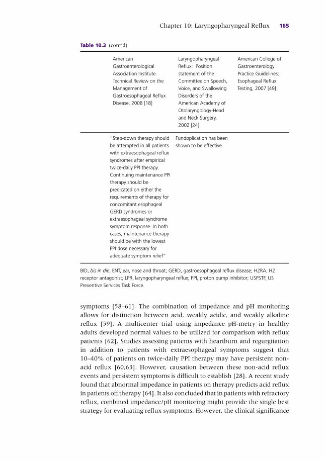
Chapter 10: Laryngopharyngeal Reflux 165
symptoms [58–61] . The combination of impedance and pH monitoring
allows for distinction between acid, weakly acidic, and weakly alkaline
reflux [59] . A multicenter trial using impedance pH-metry in healthy
adults developed normal values to be utilized for comparison with reflux
patients [62] . Studies assessing patients with heartburn and regurgitation
in addition to patients with extraesophageal symptoms suggest that
10–40% of patients on twice-daily PPI therapy may have persistent non-
acid reflux [60,63] . However, causation between these non-acid reflux
events and persistent symptoms is difficult to establish [28] . A recent study
found that abnormal impedance in patients on therapy predicts acid reflux
in patients off therapy [64] . It also concluded that in patients with refractory
reflux, combined impedance/pH monitoring might provide the single best
strategy for evaluating reflux symptoms. However, the clinical significance
“Step-down therapy should
be attempted in all patients
with extraesophageal reflux
syndromes after empirical
twice-daily PPI therapy.
Continuing maintenance PPI
therapy should be
predicated on either the
requirements of therapy for
concomitant esophageal
GERD syndromes or
extraesophageal syndrome
symptom response. In both
cases, maintenance therapy
should be with the lowest
PPI dose necessary for
adequate symptom relief”
Fundoplication has been
shown to be effective
BID, bis in die ; ENT, ear, nose and throat; GERD, gastroesophageal reflux disease; H2RA, H2
receptor antagonist; LPR, laryngopharyngeal reflux; PPI, proton pump inhibitor; USPSTF, US
Preventive Services Task Force.
Table 10.3 (cont’d)
American
Gastroenterological
Association Institute
Technical Review on the
Management of
Gastroesophageal Reflux
Disease, 2008 [18]
Laryngopharyngeal
Reflux: Position
statement of the
Committee on Speech,
Voice, and Swallowing
Disorders of the
American Academy of
Otolaryngology-Head
and Neck Surgery,
2002 [24]
American College of
Gastroenterology
Practice Guidelines:
Esophageal Reflux
Testing, 2007 [49]
c10.indd 165 11/15/2012 2:28:05 AM

166 Part 2: Gastroesophageal Reflux Disease
of abnormal impedance findings in this group of patients awaits further
study. The most recent uncontrolled surgical study in patients suspected of
having LPR found that on or off therapy impedance monitoring does not
predict LPR symptom response to fundoplication but the presence of hiatal
hernia, significant acid reflux at baseline and presence of regurgitation
concomitantly with the LPR symptom were important predictors of
symptom response [65] .
The Restech Dx-pH Measurement System™ (Respiratory Technology
Corp., San Diego, CA) is a new device developed to detect acid reflux in the
posterior oropharynx [66] . A nasopharyngeal catheter is utilized to assess
pH in liquid or aerosolized droplets. A comparison of this device to the tradi-
tional pH catheters has shown faster detection rate and faster time to
equilibrium pH. A recent prospective observational study in healthy volun-
teers developed normative data for this device at pH cut-offs of 4, 5 and 6 for
the distal esophagus and oropharynx [66] . Although the initial studies with
this device in patients with LPR are encouraging [67] , controlled studies are
needed to assess the future role of this new device in patients with LPR.
Managing laryngeal complications of reflux disease
Medical management Given the poor sensitivity and specificity of diagnostic tests, empiric
treatment of suspected gastroesophageal reflux laryngitis using PPIs is
common [1] . A recent study assessed response to PPIs based on change in
24-h pH studies in 27 patients with LPR with abnormal pH studies at base-
line. Of five patients who did not have a measurable pH response to PPI,
four reported improvements in their symptoms, highlighting poor predic-
tion of treatment response based on pH results [68] . Most trials have uti-
lized twice-daily PPIs for 3–4 months [1,69] . The primary reason for this
unapproved high-dose acid suppression is based on pH monitoring data
indicating that the chance of normalizing exposure of the esophagus to
acid in patients with chronic cough, laryngeal symptoms or asthma is 99%
with a twice-daily PPI [70] . A prospective cohort study (uncontrolled and
open label) assessed optimal PPI dose in patients with LPR, and indicated
that twice-daily PPI is more effective than daily PPI in achieving clinical
symptom response in patients with suspected LPR [44] .
Although PPIs are widely used in patients with suspected LPR, high- quality
supporting evidence remains minimal at this time as most trials have
utilized small sample sizes and are uncontrolled [1,17,18,69] . Placebo-
controlled studies assessing lansoprazole, esomeprazole, pantoprazole, and
rabeprazole for 2–4 months’ duration have observed no significant difference
in symptoms experienced by LPR patients on placebo as compared to PPI
c10.indd 166 11/15/2012 2:28:05 AM

Chapter 10: Laryngopharyngeal Reflux 167
(Table 10.4 ) [57, 71–73] . Similarly, a metaanalysis of randomized controlled
trials assessing PPI use for suspected GERD-related chronic laryngitis noted
no benefit of PPIs over placebo (Figure 10.1 ) [74] . On the other hand, in
support of the role of acid suppression in LPR, a more recent double-blind,
placebo-controlled trial noted a significant improvement in LPR symptoms
with 3 months of esomeprazole therapy [75] . One reason for the positive
nature of this study is that the RSI was used as one of the outcome measures
assessed. Of all symptoms queried, patients experienced the greatest
improvement in heartburn symptoms after 3 months (and less marked
improvements in hoarseness, throat clearing, coughing after meals, breathing
difficulties, and other symptoms addressed). The improvement in heartburn
symptoms had the most significant effect on the overall RSI score.
Similarly, an earlier study comparing lansoprazole with placebo in 22
patients with idiopathic chronic laryngitis noted that after 3 months, 50%
of patients in the lansoprazole group had noted resolution of symptoms,
compared to 10% of patients in the placebo group [76] . Furthermore,
another study reported that LPR patients who tested positive for H. pylori
antigen were more likely to respond to PPI than those seronegative for
H. pylori [77] . Finally, in a randomized controlled study of patients with
chief complaint of postnasal drainage, Vaezi et al . found a benefit for
treatment with PPIs, suggesting that PPIs may do more than just suppress
reflux in this group of individuals [78] . Thus, the search for the subgroup
of patients with suspected LPR or extraesophageal reflux symptoms more
likely to respond to PPI therapy continues.
The AGA guidelines advise empiric therapy with twice-daily PPI for 2
months for patients with a concomitant esophageal GERD syndrome (i.e.
typical symptoms such as heartburn) and laryngitis (US Preventive Services
Task Force (USPSTF) grade B recommendation) [18] . Their recommenda-
tions do not support the use of PPIs for acute treatment of laryngitis in the
absence of esophageal GERD (USPSTF grade D) [18] . However, the AAOHNS
position statement indicates that treatment for LPR needs to be more aggres-
sive and prolonged than that for GERD, and depends on the symptoms and
severity of LPR and on the response to therapy [24] . The AAOHNS advises
that patients with mild or intermittent symptoms of LPR can be treated with
dietary and lifestyle changes and with H2 antagonists, while the majority of
patients require at least twice-daily PPI therapy [5,6,25,41,79–84] . Some
patients require therapy with both a PPI and an H2 antagonist, and the
AAOHNS recommends the use of twice-daily PPI for a minimum of 6 months
[24,81] . The authors of the AAOHNS statement suggest that fundoplication
has been shown to be an effective treatment for LPR [85,86] . However, this
point is controversial, as discussed in the next section.
The need for chronic therapy in patients suspected of GERD-related laryn-
gitis comes from uncontrolled observational studies with small sample
c10.indd 167 11/15/2012 2:28:05 AM
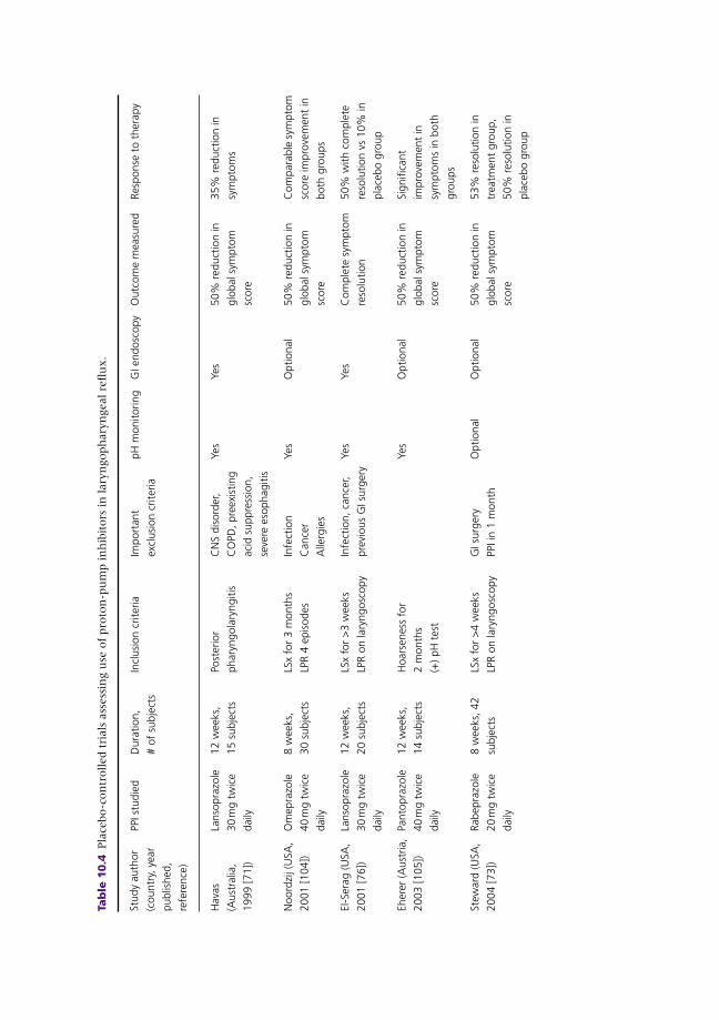
Tab
le 1
0.4
Pla
cebo-c
on
trolled t
rials
ass
ess
ing u
se o
f pro
ton
-pu
mp in
hib
itors
in
lary
ngoph
ary
ngeal re
flu
x.
Stud
y au
thor
(cou
ntry
, yea
r
publ
ishe
d,
refe
renc
e)
PPI s
tudi
edD
urat
ion,
# of
sub
ject
s
Incl
usio
n cr
iteria
Impo
rtan
t
excl
usio
n cr
iteria
pH m
onito
ring
GI e
ndos
copy
Out
com
e m
easu
red
Resp
onse
to
ther
apy
Hav
as
(Aus
tral
ia,
1999
[71]
)
Lans
opra
zole
30 m
g tw
ice
daily
12 w
eeks
,
15 s
ubje
cts
Post
erio
r
phar
yngo
lary
ngiti
s
CN
S di
sord
er,
CO
PD, p
reex
istin
g
acid
sup
pres
sion
,
seve
re e
soph
agiti
s
Yes
Yes
50%
red
uctio
n in
glob
al s
ympt
om
scor
e
35%
red
uctio
n in
sym
ptom
s
Noo
rdzi
j (U
SA,
2001
[104
] )
Om
epra
zole
40 m
g tw
ice
daily
8 w
eeks
,
30 s
ubje
cts
LSx
for
3 m
onth
s
LPR
4 ep
isod
es
Infe
ctio
n
Can
cer
Alle
rgie
s
Yes
Opt
iona
l50
% r
educ
tion
in
glob
al s
ympt
om
scor
e
Com
para
ble
sym
ptom
scor
e im
prov
emen
t in
both
gro
ups
El-S
erag
(USA
,
2001
[76]
)
Lans
opra
zole
30 m
g tw
ice
daily
12 w
eeks
,
20 s
ubje
cts
LSx
for
>3
wee
ks
LPR
on la
ryng
osco
py
Infe
ctio
n, c
ance
r,
prev
ious
GI s
urge
ry
Yes
Yes
Com
plet
e sy
mpt
om
reso
lutio
n
50%
with
com
plet
e
reso
lutio
n vs
10%
in
plac
ebo
grou
p
Eher
er (A
ustr
ia,
2003
[105
] )
Pant
opra
zole
40 m
g tw
ice
daily
12 w
eeks
,
14 s
ubje
cts
Hoa
rsen
ess
for
2 m
onth
s
(+) p
H t
est
Yes
Opt
iona
l50
% r
educ
tion
in
glob
al s
ympt
om
scor
e
Sign
ifica
nt
impr
ovem
ent
in
sym
ptom
s in
bot
h
grou
ps
Stew
ard
(USA
,
2004
[73]
)
Rabe
praz
ole
20 m
g tw
ice
daily
8 w
eeks
, 42
subj
ects
LSx
for
>4
wee
ks
LPR
on la
ryng
osco
py
GI s
urge
ry
PPI i
n 1
mon
th
Opt
iona
lO
ptio
nal
50%
red
uctio
n in
glob
al s
ympt
om
scor
e
53%
res
olut
ion
in
trea
tmen
t gr
oup,
50%
res
olut
ion
in
plac
ebo
grou
p
c10.indd 168 11/15/2012 2:28:06 AM

Wo
(USA
,
2006
[72]
)
Pant
opra
zole
40 m
g da
ily
12 w
eeks
, 39
subj
ects
LSx
for
3 da
ys a
wee
k
(+) p
H t
est
Yes
Opt
iona
l50
% r
educ
tion
in
glob
al s
ympt
om
scor
e
40%
with
res
olut
ion
in b
oth
grou
ps
Vaez
i (U
SA,
2006
[57]
)
Esom
epra
zole
40 m
g tw
ice
daily
16 w
eeks
, 145
subj
ects
LSx
for
3 m
onth
s
LPR
on la
ryng
osco
py
Hea
rtbu
rn >
3/w
k
PPI i
n 2
wk
Opt
iona
lO
ptio
nal
50%
red
uctio
n in
sym
ptom
s
~15
% r
esol
utio
n of
sym
ptom
s in
bot
h
grou
ps, w
ith 4
5% o
f
both
gro
ups
expe
rienc
ing
impr
ovem
ent
in
sym
ptom
s
Reic
hel
(Ger
man
y,
2008
[75]
)
Esom
epra
zole
20 m
g tw
ice
daily
12 w
eeks
, 62
subj
ects
RFS
>7
and
RSI >
13PP
I in
3 m
onth
s,
GI s
urge
ry, l
aryn
geal
mal
igna
ncy
No
No
Redu
ctio
n of
tot
al
RSI a
nd R
FS
Sign
ifica
ntly
gre
ater
impr
ovem
ent
in
lary
ngea
l app
eara
nce
and
LPR
sym
ptom
s
CN
S, c
entr
al n
ervo
us s
yste
m; C
OPD
, chr
onic
obs
truc
tive
pulm
onar
y di
seas
e; G
I, ga
stro
inte
stin
al; L
PR, l
aryn
goph
aryn
geal
ref
lux;
LSx
, lar
ynge
al s
ympt
oms;
PPI
, pro
ton
pum
p
inhi
bito
r; R
FS, R
eflu
x Fi
ndin
g Sc
ore;
RSI
, Ref
lux
Sym
ptom
Inde
x.
c10.indd 169 11/15/2012 2:28:06 AM

170 Part 2: Gastroesophageal Reflux Disease
sizes. For example, patients with LPR with concomitant GERD symptoms
may have esophagitis (12%) and Barrett ’ s esophagus (7%) [87,88] . Studies
suggest a possible association between chronic reflux-induced inflamma-
tion of the larynx and laryngeal cancer [13,18,34] . However, the main
purpose of maintenance use of PPIs in patients with LPR is for control of
symptoms and step-down therapy should always follow the initial empiric
trial [18] . Long-term PPI is the current practice in many patients, as
evidenced by a double-blind placebo-controlled trial finding only 21–48%
likelihood of remaining PPI free at 1 year [89] . However, evidence sup-
porting the use of long-term PPI therapy for patients with LPR is primarily
anecdotal and future studies are needed to assess appropriate duration and
use of PPI in patients with suspected LPR.
Surgical therapy A number of uncontrolled observational studies have suggested efficacy of
anti-reflux surgery in patients with gastroesophageal reflux laryngitis. An
earlier study assessed the effect of laparoscopic Hill repair on 145 patients
Favors PPIs
Combined
10 1001.1
Risk ratio
Langevin
El–Serag
Eherer
Noordzij
Havas
Steward
Vaezi
Wo
Favors placebo
Symbol
Combined
Individual
Figure 10.1 Forest plot depicting the risk ratios for studies assessing efficacy of proton
pump inhibitor (PPI) in reflux laryngitis and pooled risk ratio by random effects
method. Adapted from Qadeer et al . [74] with permission from Blackwell Publishing.
c10.indd 170 11/15/2012 2:28:06 AM
Chapter 10: Laryngopharyngeal Reflux 171
and found that after a mean follow-up of 20 months, reports of sore throat
decreased from 43% to 8% of patients. Symptoms of cough decreased
from 41% to 8%, and voice loss decreased from 25% to 11% [90] .
Similarly, another study evaluated 40 patients who underwent laparo-
scopic Nissen fundoplication for complaints of reflux laryngitis. After 3
months, 79.3% of patients had decreased inflammation noted on otorhino-
laryngeal exam, and 41.4% described improvement in voice quality. After
12 months, these figures were 92.3% and 38.5%, respectively. After a
median follow-up of 42 months, 62.5% of patients reported either no
cough or mild cough or hoarseness [91] .
A more recent prospective concurrent controlled study in patients with
LPR symptoms refractory to PPI therapy did not find Nissen fundoplication
to be of benefit. One year after surgery, only 10% of patients noted
improvement in laryngeal symptoms, while signs of LPR on laryngoscopy
improved in 80% of patients [92] . Recent controversy surrounds the role
of surgical fundoplication in patients with PPI-refractory symptoms who
have abnormal non-acid reflux by impedance monitoring. A retrospective
review assessed patients with chronic cough referred for fundoplication
after documentation of an association between their symptoms and reflux
disease using multichannel intraluminal impedance and pH testing. In all
six patients who underwent surgery, fundoplication was found to elimi-
nate chronic cough due to non-acid reflux [93] . In this subgroup of
patients, an uncontrolled telephone survey study suggested symptom
improvement in most patients with laparoscopic Nissen fundoplica-
tion [61] . However, controlled studies are needed before this practice can
be advocated, especially since the most recent surgical study suggests that
impedance monitoring is not a predictor of LPR symptom response to fun-
doplication [65] . Based on published data, the role of fundoplication is best
delineated in those who have a positive symptom response to PPI therapy
and caution should be exercised in referring patients who do not respond
to aggressive acid suppression, especially those with extraesophageal com-
plaints.
Summary
Gastroesophageal reflux disease is associated with laryngeal signs and
symptoms but the frequency of the association between these two entities
is not firmly established. Improvement in the specificity of laryngeal
examination would be an important goal in improving the accuracy of
diagnosis of gastroesophageal reflux laryngitis. pH or impedance pH
studies can serve as diagnostic tools in patients whose symptoms are
refractory to an empiric trial of PPIs. If these tests are normal on PPI
c10.indd 171 11/15/2012 2:28:06 AM

172 Part 2: Gastroesophageal Reflux Disease
therapy despite persistence of symptoms, other etiologies for abnormal
laryngeal signs and symptoms should be investigated [1] . It is prudent to
remember that patients with suspected extraesophageal GERD syn-
dromes, including those with LPR, may have GERD as a contributing
etiology but rarely as the sole cause of their complaints [18] .
Based on our experiences in treating patients with reflux-suspected
chronic laryngitis, we suggest a treatment algorithm as illustrated in
Figure 10.2 . Initial empiric therapy with twice-daily PPI for 2 months is a
reasonable starting point for patients with suspected GERD-related laryn-
gitis and no ominous symptoms or signs. If symptoms improve then acid
suppression should be tapered to the minimum dose for symptom control.
If symptoms persist despite twice-daily PPI therapy, diagnostic testing with
pH monitoring “off or on” PPIs or impedance pH monitoring on therapy is
recommended. In patients with normal test results, a search for an
alternative explanation for symptoms should be pursued. In the infrequent
Suspected gastroesophageal reflux laryngitis
Empiric trial of twice-daily PPI ×2 months
Symptoms persist Symptoms improve
24-h Impedance/pH testing Titrate PPI dose down over time
Normal Abnormal
Entertainalternativediagnoses
• Change PPI
• Ensure PPI compliance
• Increase PPI dose
• Consider surgical options
Figure 10.2 Suggested algorithm for evaluation and treatment of suspected
laryngopharyngeal reflux. PPI, proton pump inhibitor.
c10.indd 172 11/15/2012 2:28:07 AM

Chapter 10: Laryngopharyngeal Reflux 173
cases of abnormal test results on therapy, clinical judgment should be exer-
cised regarding the role of surgical fundoplication given the lack of con-
trolled studies in this area.
References
1 Vaezi MF , Hicks DM , Abelson TI , Richter JE . Laryngeal signs and symptoms and
gastroesophageal reflux disease (GERD): a critical assessment of cause and effect
association . Clin Gastroenterol Hepatol 2003 ; 1 ( 5 ): 333 – 44 .
2 Koufman JA Wiener GJ , Wallace CW , et al . Reflux laryngitis and its sequelae: the
diagnostic role of ambulatory 24-h pH monitoring . J Voice 1988 ; 2 : 78 – 9 .
3 Gaynor EB . Otolaryngologic manifestations of gastroesophageal reflux . Am J
Gastroenterol 1991 ; 86 ( 7 ): 801 – 8 .
4 Graser A . Gastroesophageal reflux and laryngeal symptoms . Aliment Pharmacol Ther
1994 ; 8 : 265 – 72 .
5 Koufman J , Sataloff RT , Toohill R . Laryngopharyngeal reflux: consensus conference
report . J Voice 1996 ; 10 ( 3 ): 215 – 16 .
6 Koufman JA . The otolaryngologic manifestations of gastroesophageal reflux disease
(GERD): a clinical investigation of 225 patients using ambulatory 24-hour pH moni-
toring and an experimental investigation of the role of acid and pepsin in the
development of laryngeal injury . Laryngoscope 1991 ; 101 (4 Pt 2 Suppl 53): 1 – 78 .
7 Richter JE . Typical and atypical presentations of gastroesophageal reflux disease. The
role of esophageal testing in diagnosis and management . Gastroenterol Clin North
Am 1996 ; 25 ( 1 ): 75 – 102 .
8 Toohill RJ , Kuhn JC . Role of refluxed acid in pathogenesis of laryngeal disorders . Am
J Med 1997 ; 103 ( 5A ): 100S – 6S .
9 Ward PH Hanson DG . Reflux as an etiological factor of carcinoma of the laryngophar-
ynx . Laryngoscope 1998 ; 98 : 1195 – 9 .
10 Freije JE , Beatty TW , Campbell BH , Woodson BT , Schultz CJ , Toohill RJ . Carcinoma
of the larynx in patients with gastroesophageal reflux . Am J Otolaryngol
1996 ; 17 ( 6 ): 386 – 90 .
11 Morrison MD . Is chronic gastroesophageal reflux a causative factor in glottic carci-
noma? Otolaryngol Head Neck Surg 1988 ; 99 ( 4 ): 370 – 3 .
12 Vaezi MF , Qadeer MA , Lopez R , Colabianchi N . Laryngeal cancer and gastroesopha-
geal reflux disease: a case–control study . Am J Med 2006 ; 119 ( 9 ): 768 – 76 .
CASE STUDY
A 46-year-old woman presents on a referral from her ENT physician for evaluation and treatment of her throat clearing and chronic cough. She has a history of asthma and postnasal drip from seasonal allergies and has been treated aggressively by her pulmonary and allergy physician for the past 3 years. She had already undergone laryngoscopy by her ENT physician, which showed “laryngeal irritation that suggested GERD.” The patient is on once-daily proton pump inhibitor intermittently which has only minimally helped her symptoms. She does have heartburn and occasional regurgitation, especially at night when in the supine position. The patient is inquiring about undergoing fundoplication.
c10.indd 173 11/15/2012 2:28:07 AM

174 Part 2: Gastroesophageal Reflux Disease
13 Qadeer MA , Colabianchi N , Strome M , Vaezi MF . Gastroesophageal reflux and laryn-
geal cancer: causation or association? A critical review . Am J Otolaryngol
2006 ; 27 ( 2 ): 119 – 28 .
14 Coffin L . The relationship of upper airway passages to diseases of gastrointestinal
tract . Ann Otol Rhinol Laryngol 1903 ; 12 : 521 – 6 .
15 Jackson C MS . Contact ulcer of larynx . Ann Otol Rhinol Laryngol 1928 ; 37 : 227 – 30 .
16 Cherry J , Margulies SI . Contact ulcer of the larynx . Laryngoscope 1968 ; 78 ( 11 ): 1937 – 40 .
17 Vakil N , van Zanten SV , Kahrilas P , Dent J , Jones R . The Montreal definition and
classification of gastroesophageal reflux disease: a global evidence-based consensus .
Am J Gastroenterol 2006 ; 101 ( 8 ): 1900 – 20 ; quiz 1943.
18 Kahrilas PJ , Shaheen NJ , Vaezi MF . American Gastroenterological Association
Institute technical review on the management of gastroesophageal reflux disease .
Gastroenterology 2008 ; 135 ( 4 ): 1392 – 413 .
19 Locke GR 3rd , Talley NJ , Fett SL , Zinsmeister AR , Melton LJ 3rd. Prevalence and
clinical spectrum of gastroesophageal reflux: a population-based study in Olmsted
County, Minnesota . Gastroenterology 1997 ; 112 ( 5 ): 1448 – 56 .
20 El-Serag HB , Sonnenberg A . Comorbid occurrence of laryngeal or pulmonary disease with
esophagitis in United States military veterans . Gastroenterology 1997 ; 113 ( 3 ): 755 – 60 .
21 Sontag SJ , O’Connell S , Khandelwal S , et al . Asthmatics with gastroesophageal
reflux: long term results of a randomized trial of medical and surgical antireflux ther-
apies . Am J Gastroenterol 2003 ; 98 ( 5 ): 987 – 99 .
22 Kahrilas PJ , Shaheen NJ , Vaezi MF , et al . American Gastroenterological Association
Medical Position Statement on the management of gastroesophageal reflux disease .
Gastroenterology 2008 ; 135 ( 4 ): 1383 – 91 .
23 Shaker R , Castell DO , Schoenfeld PS , Spechler SJ . Nighttime heartburn is an under-
appreciated clinical problem that impacts sleep and daytime function: the results of a
Gallup survey conducted on behalf of the American Gastroenterological Association .
Am J Gastroenterol 2003 ; 98 ( 7 ): 1487 – 93 .
24 Koufman JA , Aviv JE , Casiano RR , Shaw GY . Laryngopharyngeal reflux: position
statement of the committee on speech, voice, and swallowing disorders of the
American Academy of Otolaryngology-Head and Neck Surgery . Otolaryngol Head
Neck Surg 2002 ; 127 ( 1 ): 32 – 5 .
25 Hanson DG , Jiang JJ . Diagnosis and management of chronic laryngitis associated
with reflux . Am J Med 2000 ; 108 ( Suppl 4a ): 112S – 9S .
26 Loughlin CJ , Koufman JA , Averill DB , et al . Acid-induced laryngospasm in a canine
model . Laryngoscope 1996 ; 106 ( 12 Pt 1 ): 1506 – 9 .
27 Adhami T , Goldblum JR , Richter JE , Vaezi MF . The role of gastric and duodenal
agents in laryngeal injury: an experimental canine model . Am J Gastroenterol
2004 ; 99 ( 11 ): 2098 – 106 .
28 Vaezi MF . Laryngitis: from the gastroenterologist ’ s point of view . In: Vaezi MF (ed)
Extraesophageal reflux . San Diego, CA : Plural Publishing , 2009 : pp. 37 – 47 .
29 Sidhu H , Shaker R , Hogan WJ . Gastroesophageal reflux laryngitis . In: Castell DO ,
Richter JE (eds) The esophagus , 4th edn. Philadelphia : Lippincott, Williams and
Wilkins , 2003 : pp. 518 – 29 .
30 Hogan WJ , Shaker R . Supraesophageal complications of gastroesophageal reflux .
Dis Mon 2000 ; 46 ( 3 ): 193 – 232 .
31 Belafsky PC , Postma GN , Koufman JA . Validity and reliability of the reflux symptom
index (RSI) . J Voice 2002 ; 16 ( 2 ): 274 – 7 .
32 Park KH , Choi SM , Kwon SU , Yoon SW , Kim SU . Diagnosis of laryngopharyngeal
reflux among globus patients . Otolaryngol Head Neck Surg 2006 ; 134 ( 1 ): 81 – 5 .
c10.indd 174 11/15/2012 2:28:07 AM

Chapter 10: Laryngopharyngeal Reflux 175
33 Sataloff RT . Professional voice: the science and art of clinical care , 2nd edn. San Diego, CA :
Singular Publishing Group , 1997 .
34 Ford CN . Evaluation and management of laryngopharyngeal reflux . JAMA 2005 ;
294 ( 12 ): 1534 – 40 .
35 Weinberger PM , Postma GN . Laryngopharyngeal reflux from the otolaryngologist ’ s
perspective . In: Vaezi MF (ed) Extraesophageal reflux . San Diego, CA : Plural Publishing ,
2009 : pp. 49 – 66 .
36 Axford SE , Sharp N , Ross PE , et al . Cell biology of laryngeal epithelial defenses in health
and disease: preliminary studies . Ann Otol Rhinol Laryngol 2001 ; 110 ( 12 ): 1099 – 108 .
37 Ahmed TF , Khandwala F , Abelson TI , et al . Chronic laryngitis associated with gastro-
esophageal reflux: prospective assessment of differences in practice patterns between
gastroenterologists and ENT physicians . Am J Gastroenterol 2006 ; 101 ( 3 ): 470 – 8 .
38 Book DT , Rhee JS , Toohill RJ , Smith TL . Perspectives in laryngopharyngeal reflux: an
international survey . Laryngoscope 2002 ; 112 ( 8 Pt 1 ): 1399 – 406 .
39 Hicks DM , Ours TM , Abelson TI , Vaezi MF , Richter JE . The prevalence of hypo-
pharynx findings associated with gastroesophageal reflux in normal volunteers .
J Voice 2002 ; 16 ( 4 ): 564 – 79 .
40 Vaezi MF , Ours TM , Hicks DM , et al . Laryngoscopic signs of gastroesophageal reflux
disease: science or fiction? Am J Gastroenterol 1999 ; 94 : 2601 .
41 Belafsky PC , Postma GN , Koufman JA . The validity and reliability of the reflux find-
ing score (RFS) . Laryngoscope 2001 ; 111 ( 8 ): 1313 – 17 .
42 Mesallam TA , Stemple JC , Sobeih TM , Elluru RG . Reflux symptom index versus
reflux finding score . Ann Otol Rhinol Laryngol 2007 ; 116 ( 6 ): 436 – 40 .
43 Milstein CF , Charbel S , Hicks DM , Abelson TI , Richter JE , Vaezi MF . Prevalence of
laryngeal irritation signs associated with reflux in asymptomatic volunteers: impact
of endoscopic technique (rigid vs. flexible laryngoscope) . Laryngoscope 2005 ;
115 ( 12 ): 2256 – 61 .
44 Park W , Hicks DM , Khandwala F , et al . Laryngopharyngeal reflux: prospective cohort
study evaluating optimal dose of proton-pump inhibitor therapy and pretherapy pre-
dictors of response . Laryngoscope 2005 ; 115 ( 7 ): 1230 – 8 .
45 Merati AL , Lim HJ , Ulualp SO , Toohill RJ . Meta-analysis of upper probe measure-
ments in normal subjects and patients with laryngopharyngeal reflux . Ann Otol
Rhinol Laryngol 2005 ; 114 ( 3 ): 177 – 82 .
46 Ahmed T , Vaezi MF . The role of pH monitoring in extraesophageal gastroesophageal
reflux disease . Gastrointest Endosc Clin North Am 2005 ; 15 ( 2 ): 319 – 31 .
47 Jacob P , Kahrilas PJ , Herzon G . Proximal esophageal pH-metry in patients with
‘reflux laryngitis’ . Gastroenterology 1991 ; 100 ( 2 ): 305 – 10 .
48 Oelschlager BK , Eubanks TR , Maronian N , et al . Laryngoscopy and pharyngeal
pH are complementary in the diagnosis of gastroesophageal-laryngeal reflux .
J Gastrointest Surg 2002 ; 6 ( 2 ): 189 – 94 .
49 Hirano I , Richter JE . ACG practice guidelines: esophageal reflux testing . Am J
Gastroenterol 2007 ; 102 ( 3 ): 668 – 85 .
50 Wo JM , Jabbar A , Winstead W , Goudy S , Cacchione R , Allen JW . Hypopharyngeal pH
monitoring artifact in detection of laryngopharyngeal reflux . Dig Dis Sci
2002 ; 47 ( 11 ): 2579 – 85 .
51 Noordzij JP , Khidr A , Desper E , Meek RB , Reibel JF , Levine PA . Correlation of pH
probe-measured laryngopharyngeal reflux with symptoms and signs of reflux laryn-
gitis . Laryngoscope 2002 ; 112 ( 12 ): 2192 – 5 .
52 Ulualp SO , Toohill RJ , Shaker R . Outcomes of acid suppressive therapy in patients
with posterior laryngitis . Otolaryngol Head Neck Surg 2001 ; 124 ( 1 ): 16 – 22 .
c10.indd 175 11/15/2012 2:28:07 AM

176 Part 2: Gastroesophageal Reflux Disease
53 Eubanks TR , Omelanczuk PE , Maronian N , Hillel A , Pope CE 2nd, Pellegrini CA .
Pharyngeal pH monitoring in 222 patients with suspected laryngeal reflux .
J Gastrointest Surg 2001 ; 5 ( 2 ): 183 – 90 ; discussion 190–1.
54 Maldonado A , Diederich L , Castell DO , Gideon RM , Katz PO . Laryngopharyngeal
reflux identified using a new catheter design: defining normal values and excluding
artifacts . Laryngoscope 2003 ; 113 ( 2 ): 349 – 55 .
55 Shaker R , Bardan E , Gu C , Kern M , Torrico L , Toohill R . Intrapharyngeal distribution
of gastric acid refluxate . Laryngoscope 2003 ; 113 ( 7 ): 1182 – 91 .
56 Belafsky PC , Postma GN , Daniel E , Koufman JA . Transnasal esophagoscopy .
Otolaryngol Head Neck Surg 2001 ; 125 ( 6 ): 588 – 9 .
57 Vaezi MF , Richter JE , Stasney CR , et al . Treatment of chronic posterior laryngitis with
esomeprazole . Laryngoscope 2006 ; 116 ( 2 ): 254 – 60 .
58 Vaezi MF . Reflux-induced laryngitis (laryngopharyngeal reflux) . Curr Treat Options
Gastroenterol 2006 ; 9 ( 1 ): 69 – 74 .
59 Sifrim D , Blondeau K . Technology insight: the role of impedance testing for esopha-
geal disorders . Nat Clin Pract Gastroenterol Hepatol 2006 ; 3 ( 4 ): 210 – 19 .
60 Mainie I , Tutuian R , Shay S , et al . Acid and non-acid reflux in patients with persistent
symptoms despite acid suppressive therapy: a multicentre study using combined
ambulatory impedance-pH monitoring . Gut 2006 ; 55 ( 10 ): 1398 – 402 .
61 Mainie I , Tutuian R , Agrawal A , Adams D , Castell DO . Combined multichannel intra-
luminal impedance-pH monitoring to select patients with persistent gastro-esophageal
reflux for laparoscopic Nissen fundoplication . Br J Surg 2006 ; 93 ( 12 ): 1483 – 7 .
62 Shay S , Tutuian R , Sifrim D , et al . Twenty-four hour ambulatory simultaneous imped-
ance and pH monitoring: a multicenter report of normal values from 60 healthy
volunteers . Am J Gastroenterol 2004 ; 99 ( 6 ): 1037 – 43 .
63 Vaezi MF , Hicks DM , Ours TM , Richter JE . ENT manifestation of GERD: a large
prospective study assessing treatment outcome and predictors of response .
Gastroenterology 2001 ; 120 : A636 .
64 Pritchett JM , Aslam M , Slaughter JC , Ness RM , Garrett CG , Vaezi MF . Efficacy of
esophageal impedance/pH monitoring in patients with refractory gastroesophageal
reflux disease, on and off therapy . Clin Gastroenterol Hepatol 2009 ; 7 ( 7 ): 743 – 8 .
65 Fletcher KC GM , Slaughter JC , Garrett CG , Vaezi MF . Significance and degree of
reflux in patients with primary extraesophageal symptoms . Laryngoscope 2011 ;
121 ( 12 ): 2561 – 5 .
66 Sun G , Muddana S , Slaughter JC , et al . A new pH catheter for laryngopharyngeal
reflux: normal values . Laryngoscope 2009 ; 119 ( 8 ): 1639 – 43 .
67 Wiener GJ , Tsukashima R , Kelly C , et al . Oropharyngeal pH monitoring for the detec-
tion of liquid and aerosolized supraesophageal gastric reflux . J Voice 2009 ; 23 ( 4 ):
498 – 504 .
68 Reichel O , Keller J , Rasp G , Hagedorn H , Berghaus A . Efficacy of once-daily esome-
prazole treatment in patients with laryngopharyngeal reflux evaluated by 24-hour
pH monitoring . Otolaryngol Head Neck Surg 2007 ; 136 ( 2 ): 205 – 10 .
69 Field SK , Sutherland LR . Does medical antireflux therapy improve asthma in asth-
matics with gastroesophageal reflux? A critical review of the literature . Chest
1998 ; 114 ( 1 ): 275 – 83 .
70 Charbel S , Khandwala F , Vaezi MF . The role of esophageal pH monitoring in symp-
tomatic patients on PPI therapy . Am J Gastroenterol 2005 ; 100 ( 2 ): 283 – 9 .
71 Havas T , Huang S , Levy M , et al . Posterior pharyngolaryngitis: double-blind
randomised placebo-controlled trial of proton pump inhibitor therapy . Aust J
Otolaryngol 1999 ; 3 : 243 .
c10.indd 176 11/15/2012 2:28:07 AM

Chapter 10: Laryngopharyngeal Reflux 177
72 Wo JM, Koopman J , Harrell SP , Parker K , Winstead W , Lentsch E . Double-blind,
placebo-controlled trial with single-dose pantoprazole for laryngopharyngeal reflux .
Am J Gastroenterol 2006 ; 101 ( 9 ): 1972 – 8 ; quiz 2169.
73 Steward DL , Wilson KM , Kelly DH , et al . Proton pump inhibitor therapy for chronic
laryngo-pharyngitis: a randomized placebo-control trial . Otolaryngol Head Neck
Surg 2004 ; 131 ( 4 ): 342 – 50 .
74 Qadeer MA , Phillips CO , Lopez AR , et al . Proton pump inhibitor therapy for sus-
pected GERD-related chronic laryngitis: a meta-analysis of randomized controlled
trials . Am J Gastroenterol 2006 ; 101 ( 11 ): 2646 – 54 .
75 Reichel O , Dressel H , Wiederanders K , Issing WJ . Double-blind, placebo-controlled
trial with esomeprazole for symptoms and signs associated with laryngopharyngeal
reflux . Otolaryngol Head Neck Surg 2008 ; 139 ( 3 ): 414 – 20 .
76 El-Serag HB , Lee P , Buchner A , Inadomi JM , Gavin M , McCarthy DM . Lansoprazole
treatment of patients with chronic idiopathic laryngitis: a placebo-controlled trial .
Am J Gastroenterol 2001 ; 96 ( 4 ): 979 – 83 .
77 Oridate N , Takeda H , Yamamoto J , et al . Helicobacter pylori seropositivity predicts
outcomes of acid suppression therapy for laryngopharyngeal reflux symptoms .
Laryngoscope 2006 ; 116 ( 4 ): 547 – 53 .
78 Vaezi MF , Hagaman DD , Slaughter JC , et al . Proton pump inhibitor therapy improves
symptoms in postnasal drainage . Gastroenterology 2010 ; 139 ( 6 ): 1887 – 93 .
79 Ulualp SO , Toohill RJ . Laryngopharyngeal reflux: state of the art diagnosis and
treatment . Otolaryngol Clin North Am 2000 ; 33 ( 4 ): 785 – 802 .
80 Belafsky PC , Postma GN , Koufman JA . Laryngopharyngeal reflux symptoms improve
before changes in physical findings . Laryngoscope 2001 ; 111 ( 6 ): 979 – 81 .
81 Bough ID Jr , Sataloff RT , Castell DO , Hills JR , Gideon RM , Spiegel JR . Gastroesopha-
geal reflux laryngitis resistant to omeprazole therapy . J Voice 1995 ; 9 ( 2 ): 205 – 11 .
82 Chiverton SG , Howden CW , Burget DW , Hunt RH . Omeprazole (20 mg) daily
given in the morning or evening: a comparison of effects on gastric acidity, and
plasma gastrin and omeprazole concentration . Aliment Pharmacol Ther 1992 ;
6 ( 1 ): 103 – 11 .
83 Kahrilas PJ , Falk GW , Johnson DA , et al . Esomeprazole improves healing and
symptom resolution as compared with omeprazole in reflux oesophagitis patients: a
randomized controlled trial. The Esomeprazole Study Investigators . Aliment
Pharmacol Ther 2000 ; 14 ( 10 ): 1249 – 58 .
84 Shaw GY , Searl JP . Laryngeal manifestations of gastroesophageal reflux before and
after treatment with omeprazole . South Med J 1997 ; 90 ( 11 ): 1115 – 22 .
85 Hunter JG , Trus TL , Branum GD , Waring JP , Wood WC . A physiologic approach to
laparoscopic fundoplication for gastroesophageal reflux disease . Ann Surg
1996 ; 223 ( 6 ): 673 – 85 ; discussion 685–7.
86 Dallemagne B , Weerts JM , Jeahes C , Markiewicz S . Results of laparoscopic Nissen
fundoplication . Hepatogastroenterology 1998 ; 45 ( 23 ): 1338 – 43 .
87 Koufman JA , Belafsky PC , Bach KK , Daniel E , Postma GN . Prevalence of esophagitis
in patients with pH-documented laryngopharyngeal reflux . Laryngoscope 2002 ;
112 ( 9 ): 1606 – 9 .
88 Halum SL , Postma GN , Bates DD , Koufman JA . Incongruence between histologic and
endoscopic diagnoses of Barrett ’ s esophagus using transnasal esophagoscopy .
Laryngoscope 2006 ; 116 ( 2 ): 303 – 6 .
89 Bjornsson E , Abrahamsson H , Simren M , et al . Discontinuation of proton pump
inhibitors in patients on long-term therapy: a double-blind, placebo-controlled trial .
Aliment Pharmacol Ther 2006 ; 24 ( 6 ): 945 – 54 .
c10.indd 177 11/15/2012 2:28:07 AM

178 Part 2: Gastroesophageal Reflux Disease
90 Wright RC , Rhodes KP . Improvement of laryngopharyngeal reflux symptoms after
laparoscopic Hill repair . Am J Surg 2003 ; 185 ( 5 ): 455 – 61 .
91 Salminen P , Sala E , Koskenvuo J , Karvonen J , Ovaska J . Reflux laryngitis: a feasible
indication for laparoscopic antireflux surgery? Surg Laparosc Endosc Percutan Tech
2007 ; 17 ( 2 ): 73 – 8 .
92 Swoger J , Ponsky J , Hicks DM , et al . Surgical fundoplication in laryngopharyngeal
reflux unresponsive to aggressive acid suppression: a controlled study . Clin
Gastroenterol Hepatol 2006 ; 4 ( 4 ): 433 – 41 .
93 Tutuian R , Mainie I , Agrawal A , Adams D , Castell DO . Nonacid reflux in patients
with chronic cough on acid-suppressive therapy . Chest 2006 ; 130 ( 2 ): 386 – 91 .
94 Ossakow SJ , Elta G , Colturi T , Bogdasarian R , Nostrant TT . Esophageal reflux and
dysmotility as the basis for persistent cervical symptoms . Ann Otol Rhinol Laryngol
1987 ; 96 ( 4 ): 387 – 92 .
95 Wiener GJ , Koufman JA , Wu WC , Cooper JB , Richter JE , Castell DO . Chronic
hoarseness secondary to gastroesophageal reflux disease: documentation with 24-h
ambulatory pH monitoring . Am J Gastroenterol 1989 ; 84 ( 12 ): 1503 – 8 .
96 Wilson JA , White A , von Haacke NP , et al . Gastroesophageal reflux and posterior
laryngitis . Ann Otol Rhinol Laryngol 1989 ; 98 ( 6 ): 405 – 10 .
97 Katz PO . Ambulatory esophageal and hypopharyngeal pH monitoring in patients
with hoarseness . Am J Gastroenterol 1990 ; 85 ( 1 ): 38 – 40 .
98 Woo P , Noordzij P , Ross JA . Association of esophageal reflux and globus symptom:
comparison of laryngoscopy and 24-hour pH manometry . Otolaryngol Head Neck
Surg 1996 ; 115 ( 6 ): 502 – 7 .
99 Metz DC , Childs ML , Ruiz C , Weinstein GS . Pilot study of the oral omeprazole test
for reflux laryngitis . Otolaryngol Head Neck Surg 1997 ; 116 ( 1 ): 41 – 6 .
100 Vaezi MF , Schroeder PL , Richter JE . Reproducibility of proximal probe pH parame-
ters in 24-hour ambulatory esophageal pH monitoring . Am J Gastroenterol
1997 ; 92 ( 5 ): 825 – 9 .
101 Chen MY , Ott DJ , Casolo BJ , Moghazy KM , Koufman JA . Correlation of laryngeal
and pharyngeal carcinomas and 24-hour pH monitoring of the esophagus and
pharynx . Otolaryngol Head Neck Surg 1998 ; 119 ( 5 ): 460 – 2 .
102 Ulualp SO , Toohill RJ , Hoffmann R , Shaker R . Pharyngeal pH monitoring in patients
with posterior laryngitis . Otolaryngol Head Neck Surg 1999 ; 120 ( 5 ): 672 – 7 .
103 Smit CF , Copper MP , van Leeuwen JA , Schoots IG , Stanojcic LD . Effect of cigarette
smoking on gastropharyngeal and gastroesophageal reflux . Ann Otol Rhinol
Laryngol 2001 ; 110 ( 2 ): 190 – 3 .
104 Noordzij JP , Khidr A , Evans BA , et al . Evaluation of omeprazole in the treatment of
reflux laryngitis: a prospective, placebo-controlled, randomized, double-blind
study . Laryngoscope 2001 ; 111 ( 12 ): 2147 – 51 .
105 Eherer AJ , Habermann W , Hammer HF , Kiesler K , Friedrich G , Krejs GJ . Effect of
pantoprazole on the course of reflux-associated laryngitis: a placebo-controlled
double-blind crossover study . Scand J Gastroenterol 2003 ; 38 ( 5 ): 462 – 7 .
c10.indd 178 11/15/2012 2:28:07 AM

179
Practical Manual of Gastroesophageal Reflux Disease, First Edition.
Edited by Marcelo F. Vela, Joel E. Richter and John E. Pandolfino.
© 2013 John Wiley & Sons, Ltd. Published 2013 by John Wiley & Sons, Ltd.
CHAPTER 11
Reflux-Related Cough Etsuro Yazaki, Ryuichi Shimono, and Daniel Sifrim Wingate Institute for Neurogastroenterology, Barts and the London School of Medicine
and Dentistry, and GI Physiology Unit , Royal London Hospital , London , UK
Key points • Gastroesophageal reflux disease is now considered to be one of the three most frequent
causes of chronic cough along with asthma and upper airways cough syndrome.
• The accurate, objective detection of both gastroesophageal reflux and cough and
suitable statistical analysis are required to assess the association between the two
phenomena. The development of impedance pH monitoring with simultaneous
cough detection allows objective assessment of all types of reflux events and cough
and the use of a statistic algorithm to relate both phenomena.
• Careful clinical selection of patients with suspected gastroesophageal reflux
disease-related cough is important before starting treatment. These patients might
receive either an empirical trial of double-dose proton pump inhibitor or diagnostic
reflux-cough monitoring.
Potential pitfalls • Cough and gastroesophageal reflux disease are very common presentations in a
general population, and these two phenomena can co-exist without any causal
association.
• The acidity of the refluxate might be unimportant if the esophagobronchial reflex is
already sensitized. This would be one of the reasons why acid-suppressing therapy is
less effective than anti-reflux surgery.
• Empirical treatment with proton pump inhibitor double dose for at least 3 months is
widely used, but it should be noted that this strategy has not been supported by
strong scientific evidence.
Introduction
Pathological gastroesophageal reflux (GER) usually results in typical
symptoms, i.e. regurgitation and/or heartburn, but can also be associated
with extraesophageal symptoms, such as asthma, laryngitis or chronic
c11.indd 179 11/15/2012 2:50:03 AM
180 Part 2: Gastroesophageal Reflux Disease
cough [1] . According to the Montreal classification, gastroesophageal
reflux disease (GERD)-related cough is regarded as one of the established
extraesophageal syndromes of GERD [2] .
Epidemiology
Gastroesophageal reflux disease is now considered to be one of the three
most frequent etiologies of chronic cough together with asthma and upper
airways cough syndrome (previously known as postnasal drip syndrome
[3–5] . An epidemiological association between GERD and chronic cough
has been recognized for many years and a prevalence of “reflux-related
cough” has been reported with a wide range from 5% to 41% [6–10] . This
large variation in prevalence can be due to differences in patient popula-
tions but is mainly due to the methodology used to establish the reflux-
cough association, i.e questionnaires, cough during pH-metry, symptom
responses to acid reflux therapies. Clinical awareness of GERD-related
chronic cough, both from the pneumology and gastroenterology sides, has
influenced the prevalence estimation. For example, studies published by
Irwin ’ s group have shown that the prevalence of GERD as the cause of
chronic cough increased from 10% in 1981 [8] , to 21% in 1990 [9] to 36%
in 1998 [10] .
Pathophysiology
Three main pathophysiological mechanisms have been proposed to explain
the relationship between reflux and cough: microaspiration, activation
of esophagobronchial vagal reflexes, and reflux-induced airway hyper-
sensitivity.
Microaspiration Microaspiration can be responsible for chronic cough by stimulating cough
receptors directly. Several studies have suggested the possibility of arrival
of gastric contents into the proximal airway. Laryngeal acidification has
been demonstrated with transcutaneous pH measurements [11] and more
recently, a technique developed to study aerosolized acid in the pharynx
[12] . Microaspiration of refluxate can be demonstrated by the presence of
gastric contents in bronchoalveolar lavage fluid (BALF) and sputum and
suggested by their presence in saliva. Detection of lipid-laden macrophages
in BALF or sputum has been used as a marker for microaspiration in chil-
dren. Studies showed that lipid-laden alveolar macrophages were present
in 85% of children with chronic respiratory tract disorders and GER
c11.indd 180 11/15/2012 2:50:05 AM

Chapter 11: Reflux-Related Cough 181
[13,14] . However, recent reports have shown that this method had low
specificity and its prevalence in adult patients with chronic unexplained
cough was unknown [15,16] . Pepsin and bile acids (BA) are currently
assessed in saliva, sputum and BALF in patients with respiratory disorders.
While pepsin and BA are clearly increased in patients with cystic fibrosis
and lung transplant [17] , there is no difference in pepsin concentrations of
BALF between chronic cough patients and healthy controls [5] . Recent
studies using impedance pH monitoring failed to demonstrate increased
numbers of reflux episodes with high proximal extent in patients with
chronic cough [18,19] . The current data suggest, therefore, that micro-
aspiration is unlikely to be the most important mechanism for cough in
these patients.
Esophagobronchial reflex Esophagus, larynx, and bronchi are all innervated by the vagal nerve. The
divergence of vagal afferent neurons in the brainstem may allow vagally
mediated reflexes from the distal esophagus by chemical or mechanical
stimuli [20] . Ing et al . reported that acid perfusion into the distal esophagus
of patients with cough and GERD significantly increased cough frequency,
when compared with saline infusion [21] . Topical esophageal anesthesia
with lidocaine blocked acid-induced cough [21] . In contrast, when the anti-
cholinergic agent ipratropium was instilled into the esophagus, there was no
effect, whilst inhaled ipratropium inhibited cough, suggesting a vagally
mediated esophagobronchial reflex. Such a reflex can be sensitized in
patients with GERD and chronic cough [22,23] . These patients have a
hypersensitive cough reflex to both capsaicin and citric acid inhalation.
Reduced cough threshold due to prolonged acid exposure in the distal
esophagus could be an important mechanism for chronic cough [24] . A low-
ered airway cough threshold becomes stimulus non-specific and any other
stimulus, not necessarily reflux, would trigger cough, i.e. cold air, stress, etc.
Interestingly, Benini et al . demonstrated that treatment with proton pump
inhibitors (PPIs) could reverse the bronchial hypersensitivity [22] .
Recent studies using multichannel intraluminal impedance pH (MII-pH)
and objective cough recordings have shown that both acid and non-acid
reflux events can be time associated with cough [18,25] . Furthermore,
equal numbers of patients may have a positive reflux-cough association
(Symptom Association Probability – SAP) with acid and non-acid reflux
events [26] . These findings suggest that the acidity of reflux might be
unimportant if the esophagobronchial reflex is already sensitized. Apart
from the reflux-cough association, recent studies have shown evidence of
cough-reflux sequences [25,26] which might result in a self-perpetuating
cycle of reflux-cough-reflux in some patients. For further complexity,
Smith et al . [26] suggested cough-induced reflux might occur by triggering
c11.indd 181 11/15/2012 2:50:05 AM
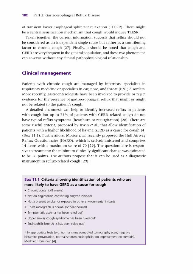
182 Part 2: Gastroesophageal Reflux Disease
of transient lower esophageal sphincter relaxation (TLESR). There might
be a central sensitization mechanism that cough would induce TLESR.
Taken together, the current information suggests that reflux should not
be considered as an independent single cause but rather as a contributing
factor to chronic cough [27] . Finally, it should be noted that cough and
GERD are very frequent in the general population, and these two phenomena
can co-exist without any clinical pathophysiological relationship.
Clinical management
Patients with chronic cough are managed by internists, specialists in
respiratory medicine or specialists in ear, nose, and throat (ENT) disorders.
More recently, gastroenterologists have been involved to provide or reject
evidence for the presence of gastroesophageal reflux that might or might
not be related to the patient ’ s cough.
A detailed anamnesis can help to identify increased reflux in patients
with cough but up to 75% of patients with GERD-related cough do not
have typical reflux symptoms (heartburn or regurgitation) [28] . There are
some useful criteria, proposed by Irwin et al ., that allow identification of
patients with a higher likelihood of having GERD as a cause for cough [4]
(Box 11.1 ). Furthermore, Morice et al . recently proposed the Hull Airway
Reflux Questionnaire (HARQ), which is self-administered and comprises
14 items with a maximum score of 70 [29] . The questionnaire is respon-
sive to treatment; the minimum clinically significant change was estimated
to be 16 points. The authors propose that it can be used as a diagnostic
instrument in reflux-related cough [29] .
Box 11.1 Criteria allowing identification of patients who are more likely to have GERD as a cause for cough • Chronic cough (> 8 weeks)
• Not on angiotensin-converting enzyme inhibitor
• Not a present smoker or exposed to other environmental irritants
• Chest radiograph is normal (or near normal)
• Symptomatic asthma has been ruled out *
• Upper airway cough syndrome has been ruled out *
• Eosinophilic bronchitis has been ruled out *
* By appropriate tests (e.g. normal sinus computed tomography scan, negative histamine provocation, normal sputum eosinophilia, no improvement on steroids). Modified from Irwin [4] .
c11.indd 182 11/15/2012 2:50:05 AM
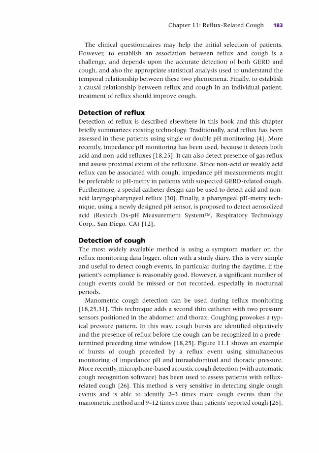
Chapter 11: Reflux-Related Cough 183
The clinical questionnaires may help the initial selection of patients.
However, to establish an association between reflux and cough is a
challenge, and depends upon the accurate detection of both GERD and
cough, and also the appropriate statistical analysis used to understand the
temporal relationship between these two phenomena. Finally, to establish
a causal relationship between reflux and cough in an individual patient,
treatment of reflux should improve cough.
Detection of reflux Detection of reflux is described elsewhere in this book and this chapter
briefly summarizes existing technology. Traditionally, acid reflux has been
assessed in these patients using single or double pH monitoring [4] . More
recently, impedance pH monitoring has been used, because it detects both
acid and non-acid refluxes [18,25] . It can also detect presence of gas reflux
and assess proximal extent of the refluxate. Since non-acid or weakly acid
reflux can be associated with cough, impedance pH measurements might
be preferable to pH-metry in patients with suspected GERD-related cough.
Furthermore, a special catheter design can be used to detect acid and non-
acid laryngopharyngeal reflux [30] . Finally, a pharyngeal pH-metry tech-
nique, using a newly designed pH sensor, is proposed to detect aerosolized
acid (Restech Dx-pH Measurement System™, Respiratory Technology
Corp., San Diego, CA) [12] .
Detection of cough The most widely available method is using a symptom marker on the
reflux monitoring data logger, often with a study diary. This is very simple
and useful to detect cough events, in particular during the daytime, if the
patient ’ s compliance is reasonably good. However, a significant number of
cough events could be missed or not recorded, especially in nocturnal
periods.
Manometric cough detection can be used during reflux monitoring
[18,25,31] . This technique adds a second thin catheter with two pressure
sensors positioned in the abdomen and thorax. Coughing provokes a typ-
ical pressure pattern. In this way, cough bursts are identified objectively
and the presence of reflux before the cough can be recognized in a prede-
termined preceding time window [18,25] . Figure 11.1 shows an example
of bursts of cough preceded by a reflux event using simultaneous
monitoring of impedance pH and intraabdominal and thoracic pressure.
More recently, microphone-based acoustic cough detection (with automatic
cough recognition software) has been used to assess patients with reflux-
related cough [26] . This method is very sensitive in detecting single cough
events and is able to identify 2–3 times more cough events than the
manometric method and 9–12 times more than patients’ reported cough [26] .
c11.indd 183 11/15/2012 2:50:05 AM

Impe
danc
e
Eso
phag
eal p
H
Gas
tric
pH
Man
omet
ricco
ugh
dete
ctio
n
Aci
d re
flux
Cou
gh
Fig
ure
11.1
Sim
ult
an
eou
s m
on
itori
ng o
f im
pedan
ce p
H a
nd in
traabdom
inal an
d t
hora
cic
pre
ssu
re. A
n a
cid r
efl
ux e
pis
ode d
ete
cted b
y im
pedan
ce p
H
mon
itori
ng is
follow
ed b
y c
ou
gh
dete
cted b
y p
ress
ure
ch
an
ges.
c11.indd 184 11/15/2012 2:50:05 AM

Chapter 11: Reflux-Related Cough 185
Finally, using even more advanced technology, Chang et al . [32] reported
the use of an ambulatory device to measure pH, electromyograph and
audio, designed to study reflux-cough in children.
Association between reflux and cough Several statistical algorithms have been designed in order to analyze the
time association between reflux and symptoms [33] . The Symptom Index
(SI) has been defined as the percentage of reflux-associated symptom epi-
sodes in the total symptoms and is considered positive if > 50%. The disad-
vantage of the SI is that it does not take into account the total number of
reflux episodes and symptoms. The Symptom Association Probability
(SAP) calculates the statistical relationship between symptoms and reflux
episodes using Fischer ’ s exact test, taking into account the number of asso-
ciated reflux-symptom episodes as well as the total number of reflux and
symptom events [34] . The SAP is considered positive when higher than
95% [33] . We and others [26] use a time window of 2 min to assess the
association reflux-cough and we identified SAP-positive patients in whom
the time association might not occur by chance. It is important to stress,
however, that the SI and SAP were designed to study the relationship bet-
ween reflux and heartburn or chest pain, and not for the reflux-cough
association. The optimal time window for GERD-related cough needs
further investigation.
Finally, Hersh et al . recently reported hierarchical use of parameters from
ambulatory pH testing in predicting response to anti-reflux medical therapy
in patients with suspected GERD-related cough [35] . The study showed
that the highest likelihood of a sustained, durable response (high degree
response) to anti-reflux therapy was achieved when acid exposure time,
SAP, and SI were all positive.
Treatment
Medical treatment If increased GER does provoke chronic cough, treatment of GERD should
improve cough. Empiric trials with PPI (without investigation of GERD)
and lifestyle modifications have been proposed by the American College of
Chest Physicians [4] and the British Thoracic Society [36] . This empiric
strategy was reported to be effective in a community-based patient
population in the US [37] . However, published studies of anti-reflux
medical treatments in patients with cough are inconsistent. Kiljander et al .,
in a prospective, double-blind, placebo-controlled trial, reported significant
improvement of cough after 8 weeks of PPI treatment (omeprazole
40 mg o.d.) [38] . Ours et al . reported improvement or resolution of cough
c11.indd 185 11/15/2012 2:50:07 AM

186 Part 2: Gastroesophageal Reflux Disease
in 35% of patients with GERD-related cough after 12 weeks of PPI
treatment (omeprazole 40 mg b.i.d.) [39] . In contrast, more recent studies
have shown no significant improvement of cough after PPI therapy
[40,41] . The most recent Cochrane review concluded that: “PPI is not effi-
cacious for cough associated with GORD symptoms in very young children
(including infants) and should not be used for cough outcomes. In adults,
there is insufficient evidence to conclude definitely that GORD treatment
with PPI is universally beneficial for cough associated with GORD. Future
paediatric and adult studies should be double-blind, randomized, con-
trolled and parallel design, using treatments for at least 2 months, with
validated subjective and objective cough outcomes” [42] .
In a subgroup of patients, a possible reason for ineffective PPI therapy
could be an association between non-acid reflux and cough. Such
association can be assessed using impedance pH monitoring [18,25] . In
general, patients presenting with a positive SAP between weakly acidic
reflux and cough do not have increased numbers of weakly acidic reflux
events, suggesting the possibility of hypersensitivity to such refluxate.
A key mechanism of GER is known to be TLESR. Baclofen is a gamma-
aminobutyric acid (GABAB) agonist that reduces the number of reflux epi-
sodes by reducing the number of TLESRs. Baclofen was reported to reduce
numbers of acid and weakly acidic reflux events and also has an antitus-
sive effect by altering cough reflex [43,44] . However, baclofen is known to
have significant side-effects and patient tolerability is relatively low [45] .
New GABAB agonists and other medications to reduce TLESRs and reflux
with fewer neurological side-effects are currently under development and
might be of potential benefit for GERD-related cough.
Prokinetics drugs are frequently used in GERD treatment to accelerate
gastric emptying and improve esophageal motility. Their efficacy in GERD-
related cough has not been formally tested. Azithromycin (AZI) belongs to
the group of macrolide antibiotics, which are known to have prokinetic
effects, and often used in lung transplant patients to prevent bronchiolitis
obliterans syndrome [46] . In lung transplant patients, standard esophageal
pH monitoring revealed an increased acid exposure in 70% [47,48] .
Mertens et al . studied the effect of AZI on GER in lung transplant patients
and found that AZI reduced esophageal acid and volume exposure as well
as the number of proximal reflux events [49] . AZI is currently under inves-
tigation in patients with reflux-associated cough.
Surgical treatment Fundoplication is an alternative to medical treatment for GERD. The
procedure is known to be highly effective in reducing esophageal acid
exposure time and reflux symptoms [50] . Various mechanisms are
responsible for a decrease in reflux frequency after fundoplication,
c11.indd 186 11/15/2012 2:50:07 AM
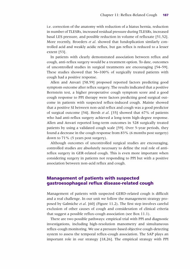
Chapter 11: Reflux-Related Cough 187
i.e. correction of the anatomy with reduction of a hiatus hernia, reduction
in number of TLESRs, increased residual pressure during TLESRs, increased
basal LES pressure, and possible reduction in volume of refluxate [51,52] .
More recently, Broeders et al . showed that fundoplication similarly con-
trolled acid and weakly acidic reflux, but gas reflux is reduced to a lesser
extent [53] .
In patients with clearly demonstrated association between reflux and
cough, anti-reflux surgery would be a treatment option. To date, outcomes
of uncontrolled studies in surgical treatments are encouraging [54–59] .
These studies showed that 56–100% of surgically treated patients with
cough had a positive response.
Allen and Anvari [58,59] proposed reported factors predicting good
symptom outcome after reflux surgery. The results indicated that a positive
Bernstein test, a higher preoperative cough symptom score and a good
cough response to PPI therapy were factors predicting good surgical out-
come in patients with suspected reflux-induced cough. Mainie showed
that a positive SI between non-acid reflux and cough was a good predictor
of surgical outcome [54] . Hersh et al . [35] showed that 67% of patients
who had anti-reflux surgery achieved a long-term high-degree response.
Allen and Anvari reported long-term outcomes in 528 surgically treated
patients by using a validated cough scale [59] . Over 5-year periods, they
found a decrease in the cough response from 83% (6 months post surgery)
down to 71% (5 years post surgery).
Although outcomes of uncontrolled surgical studies are encouraging,
controlled studies are absolutely necessary to define the real role of anti-
reflux surgery in GER-related cough. This is even more important when
considering surgery in patients not responding to PPI but with a positive
association between non-acid reflux and cough.
Management of patients with suspected gastroesophageal reflux disease-related cough
Management of patients with suspected GERD-related cough is difficult
and a real challenge. In our unit we follow the management strategy pro-
posed by Galmiche et al . [60] (Figure 11.2 ). The first step involves careful
exclusion of other causes of cough and consideration of clinical criteria
that suggest a possible reflux-cough association (see Box 11.1 ).
There are two possible pathways: empirical trial with PPI and diagnostic
investigations, including high-resolution manometry and simultaneous
reflux-cough monitoring. We use a pressure-based objective cough-detecting
system to assess the temporal reflux-cough association. The SAP plays an
important role in our strategy [18,26] . The empirical strategy with PPI
c11.indd 187 11/15/2012 2:50:07 AM

Em
piric
al s
trat
egy
with
PP
I tria
lIn
vest
igat
ion
stra
tegy
: HR
M, i
mpe
danc
e-pH
+ c
ough
det
ecto
r ‘O
FF
’ PP
I
Sym
ptom
sgo
neS
ympt
oms
pers
ist
GE
RN
o G
ER
Or
Oth
er c
ause
s?C
ough
sen
sitiv
ity te
st?
Rep
eat i
nves
tigat
ion?
Sym
ptom
sgo
neS
ympt
oms
pers
ist
Impe
danc
e-pH
+ c
ough
det
ecto
r‘O
N’ P
PI
No
reflu
x co
ugh
asso
ciat
ion
Pat
ient
with
sus
pect
ed G
ER
D-r
elat
ed c
ough
Incr
ease
d ac
id e
xpos
ure
SA
P +
for
acid
ref
lux
PP
I tria
lP
PI t
rial
SA
P+
with
non
-aci
d re
flux
OR
aci
d re
flux
(unc
ontr
olle
d ac
id)S
ympt
oms
gone
Sto
p P
PI
No
rela
pse
Rel
apse
Mai
nten
ace
PP
IA
ntire
flux
surg
ery
Ada
pt th
erap
y
Fig
ure
11.2
A
lgori
thm
for
the e
valu
ati
on
an
d m
an
agem
en
t of
pati
en
ts w
ith
su
spect
ed G
ER
D-r
ela
ted c
ou
gh
. G
ER
, gast
roeso
-
ph
ageal re
flu
x; G
ER
D: gast
roeso
ph
ageal re
flu
x d
isease
; H
RM
, h
igh
reso
luti
on
man
om
etr
y; PPI,
pro
ton
pu
mp in
hib
itor;
SA
P,
sym
pto
m a
ssoci
ati
on
pro
babilit
y.
c11.indd 188 11/15/2012 2:50:07 AM

Impe
danc
e
Eso
phag
eal p
H
Gas
tric
pH
Wea
kly
acid
ref
lux
Cou
gh
Man
omet
ricco
ugh
dete
ctio
n
Fig
ure
11.3
Sim
ult
an
eou
s m
on
itori
ng o
f im
pedan
ce p
H a
nd in
tra a
bdom
inal an
d t
hora
cic
pre
ssu
re in
a p
ati
en
t w
ho is
aci
d s
uppre
ssed w
ith
a p
roto
n p
um
p
inh
ibit
or
(PPI)
. A
weakly
aci
dic
refl
ux e
pis
ode (
refl
ux d
ete
cted b
y im
pedan
ce, pH
rem
ain
s above 4
) dete
cted b
y im
pedan
ce p
H m
on
itori
ng is
follow
ed b
y a
cou
gh
even
t.
c11.indd 189 11/15/2012 2:50:07 AM
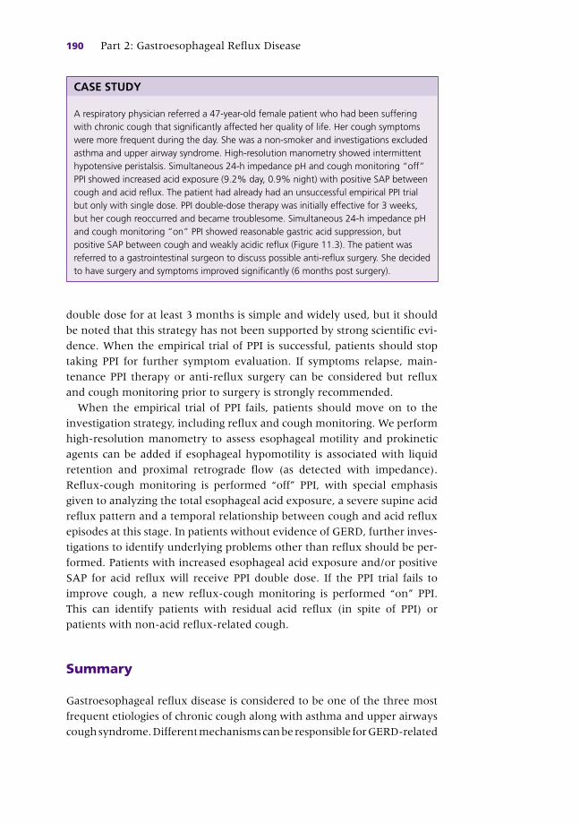
190 Part 2: Gastroesophageal Reflux Disease
double dose for at least 3 months is simple and widely used, but it should
be noted that this strategy has not been supported by strong scientific evi-
dence. When the empirical trial of PPI is successful, patients should stop
taking PPI for further symptom evaluation. If symptoms relapse, main-
tenance PPI therapy or anti-reflux surgery can be considered but reflux
and cough monitoring prior to surgery is strongly recommended.
When the empirical trial of PPI fails, patients should move on to the
investigation strategy, including reflux and cough monitoring. We perform
high-resolution manometry to assess esophageal motility and prokinetic
agents can be added if esophageal hypomotility is associated with liquid
retention and proximal retrograde flow (as detected with impedance).
Reflux-cough monitoring is performed “off” PPI, with special emphasis
given to analyzing the total esophageal acid exposure, a severe supine acid
reflux pattern and a temporal relationship between cough and acid reflux
episodes at this stage. In patients without evidence of GERD, further inves-
tigations to identify underlying problems other than reflux should be per-
formed. Patients with increased esophageal acid exposure and/or positive
SAP for acid reflux will receive PPI double dose. If the PPI trial fails to
improve cough, a new reflux-cough monitoring is performed “on” PPI.
This can identify patients with residual acid reflux (in spite of PPI) or
patients with non-acid reflux-related cough.
Summary
Gastroesophageal reflux disease is considered to be one of the three most
frequent etiologies of chronic cough along with asthma and upper airways
cough syndrome. Different mechanisms can be responsible for GERD-related
CASE STUDY
A respiratory physician referred a 47-year-old female patient who had been suffering with chronic cough that significantly affected her quality of life. Her cough symptoms were more frequent during the day. She was a non-smoker and investigations excluded asthma and upper airway syndrome. High-resolution manometry showed intermittent hypotensive peristalsis. Simultaneous 24-h impedance pH and cough monitoring “off” PPI showed increased acid exposure (9.2% day, 0.9% night) with positive SAP between cough and acid reflux. The patient had already had an unsuccessful empirical PPI trial but only with single dose. PPI double-dose therapy was initially effective for 3 weeks, but her cough reoccurred and became troublesome. Simultaneous 24-h impedance pH and cough monitoring “on” PPI showed reasonable gastric acid suppression, but positive SAP between cough and weakly acidic reflux (Figure 11.3 ). The patient was referred to a gastrointestinal surgeon to discuss possible anti-reflux surgery. She decided to have surgery and symptoms improved significantly (6 months post surgery).
c11.indd 190 11/15/2012 2:50:11 AM

Chapter 11: Reflux-Related Cough 191
cough, i.e. microaspiration, esophagobronchial reflex and central sensitization
of the cough reflex [26] . The diagnosis and management of reflux-related
cough are a difficult challenge. The development of impedance pH moni-
toring with simultaneous cough detection allows a more objective
assessment of all types of reflux events and cough [18,26] . Empirical PPI
treatment is widely used. The response rate to PPI treatment appeared to
be rather poor. Studies have suggested that acidity of the refluxate is not
critical when the esophagobronchial reflex has already been sensitized,
which is one of the reasons for failed acid-suppressing treatments. Anti-
reflux surgery has been performed successfully on a group of patients with
GERD-related cough in uncontrolled trials. However, controlled, prospec-
tive studies are necessary to confirm the role of anti-reflux surgery in the
management of GERD-related cough.
Management strategies for patients with suspected GERD-related cough
include empirical PPI trial and reflux-cough investigation. In our unit,
simultaneous monitoring of reflux and cough “off” PPI is performed first.
Evidence of GERD and SAP between reflux and cough are important
parameters for managing patients further. For patients who do not respond
to double-dose PPI trial, simultaneous reflux and cough monitoring is
repeated “on” PPI. This identifies patients with a positive SAP between
weakly acidic reflux and cough, or patients with PPI resistance.
References
1 Richter JE . Review article: extraoesophageal manifestations of gastro-oesophageal
reflux disease . Aliment Pharmacol Ther 2005 ; 22 (Suppl 1): 70 – 80 .
2 Vakil N , van Zanten SV , Kahrilas P , Dent J , Jones R , Group GC . The Montreal defini-
tion and classification of gastroesophageal reflux disease: a global evidence-based
consensus . Am J Gastroenterol 2006 ; 101 ( 8 ): 1900 – 20 ; quiz 43.
3 Pratter MR . Chronic upper airway cough syndrome secondary to rhinosinus diseases
(previously referred to as postnasal drip syndrome): ACCP evidence-based clinical
practice guidelines . Chest 2006 ; 129 ( 1 Suppl ): 63S – 71S .
4 Irwin RS . Chronic cough due to gastroesophageal reflux disease: ACCP evidence-
based clinical practice guidelines . Chest 2006 ; 129 (1 Suppl): 80S – 94S .
5 Smith J , Woodcock A , Houghton L . New developments in reflux-associated cough .
Lung 2010 ; 188 (Suppl 1): S81 – 6 .
6 Marchesani F , Cecarini L , Pela R , Sanguinetti CM . Causes of chronic persistent cough
in adult patients: the results of a systematic management protocol . Monaldi Arch
Chest Dis 1998 ; 53 ( 5 ): 510 – 14 .
7 Palombini BC , Villanova CA , Araújo E , et al . A pathogenic triad in chronic cough:
asthma, postnasal drip syndrome, and gastroesophageal reflux disease . Chest
1999 ; 116 ( 2 ): 279 – 84 .
8 Irwin RS , Corrao WM , Pratter MR . Chronic persistent cough in the adult: the spec-
trum and frequency of causes and successful outcome of specific therapy . Am Rev
Respir Dis 1981 ; 123 ( 4 Pt 1 ): 413 – 17 .
c11.indd 191 11/15/2012 2:50:11 AM

192 Part 2: Gastroesophageal Reflux Disease
9 Irwin RS , Curley FJ , French CL . Chronic cough. The spectrum and frequency of
causes, key components of the diagnostic evaluation, and outcome of specific therapy .
Am Rev Respir Dis 1990 ; 141 ( 3 ): 640 – 7 .
10 French CL , Irwin RS , Curley FJ , Krikorian CJ . Impact of chronic cough on quality of
life . Arch Intern Med 1998 ; 158 ( 15 ): 1657 – 61 .
11 Ledson MJ , Wilson GE , Tran J , Walshaw MJ . Tracheal microaspiration in adult cystic
fibrosis . J Roy Soc Med 1998 ; 91 ( 1 ): 10 – 12 .
12 Sun G , Muddana S , Slaughter JC , et al . A new pH catheter for laryngopharyngeal
reflux: normal values . Laryngoscope 2009 ; 119 ( 8 ): 1639 – 43 .
13 Ahrens P , Noll C , Kitz R , Willigens P , Zielen S , Hofmann D . Lipid-laden alveolar mac-
rophages (LLAM): a useful marker of silent aspiration in children . Pediatr Pulmonol
1999 ; 28 ( 2 ): 83 – 8 .
14 Parameswaran K , Anvari M , Efthimiadis A , Kamada D , Hargreave FE , Allen CJ .
Lipid-laden macrophages in induced sputum are a marker of oropharyngeal reflux
and possible gastric aspiration . Eur Respir J 2000 ; 16 ( 6 ): 1119 – 22 .
15 Köksal D , Ozkan B , Simşek C , Köksal AS , Aǧaçkýran Y , Saşmaz N . Lipid-laden alve-
olar macrophage index in sputum is not useful in the differential diagnosis of
pulmonary symptoms secondary to gastroesophageal reflux . Arch Med Res
2005 ; 36 ( 5 ): 485 – 9 .
16 Krishnan U , Mitchell JD , Tobias V , Day AS , Bohane TD . Fat laden macrophages in
tracheal aspirates as a marker of reflux aspiration: a negative report . J Pediatr
Gastroenterol Nutr 2002 ; 35 ( 3 ): 309 – 13 .
17 Blondeau K , Mertens V , Vanaudenaerde BA , et al . Gastro-oesophageal reflux and
gastric aspiration in lung transplant patients with or without chronic rejection . Eur
Respir J 2008 ; 31 ( 4 ): 707 – 13 .
18 Blondeau K , Dupont LJ , Mertens V , Tack J , Sifrim D . Improved diagnosis of gastro-
oesophageal reflux in patients with unexplained chronic cough . Aliment Pharmacol
Ther 2007 ; 25 ( 6 ): 723 – 32 .
19 Smith JA , Abdulqawi R , Houghton LA . GERD-related cough: pathophysiology and
diagnostic approach . Curr Gastroenterol Rep 2011 ; 13 ( 3 ): 247 – 56 .
20 Irwin RS , Zawacki JK , Curley FJ , French CL , Hoffman PJ . Chronic cough as the sole
presenting manifestation of gastroesophageal reflux . Am Rev Respir Dis
1989 ; 140 ( 5 ): 1294 – 300 .
21 Ing AJ , Ngu MC , Breslin AB . Pathogenesis of chronic persistent cough associated
with gastroesophageal reflux . Am J Respir Crit Care Med 1994 ; 149 ( 1 ): 160 – 7 .
22 Benini L , Ferrari M , Sembenini C , et al . Cough threshold in reflux oesophagitis:
influence of acid and of laryngeal and oesophageal damage . Gut 2000 ; 46 ( 6 ):
762 – 7 .
23 Javorkova N , Varechova S , Pecova R , et al . Acidification of the oesophagus acutely
increases the cough sensitivity in patients with gastro-oesophageal reflux and chronic
cough . Neurogastroenterol Motil 2008 ; 20 ( 2 ): 119 – 24 .
24 Benini L , Ferrari M , Talamini G , Vantini I . Reflux associated cough is usually not
associated with reflux: role of reduced cough threshold . Gut 2006 ; 55 ( 4 ): 583 ; author
reply 584.
25 Sifrim D , Dupont L , Blondeau K , Zhang X , Tack J , Janssens J . Weakly acidic reflux in
patients with chronic unexplained cough during 24 hour pressure, pH, and imped-
ance monitoring . Gut 2005 ; 54 ( 4 ): 449 – 54 .
26 Smith JA , Decalmer S , Kelsall A , et al . Acoustic cough-reflux associations in chronic
cough: potential triggers and mechanisms . Gastroenterology 2010 ; 139 ( 3 ): 754 – 62 .
27 Kahrilas PJ . Chronic cough and gastroesophageal reflux disease: new twists to the
riddle . Gastroenterology 2010 ; 139 ( 3 ): 716 – 18 .
c11.indd 192 11/15/2012 2:50:11 AM

Chapter 11: Reflux-Related Cough 193
28 Irwin RS , French CL , Curley FJ , Zawacki JK , Bennett FM . Chronic cough due to gas-
troesophageal reflux. Clinical, diagnostic, and pathogenetic aspects . Chest 2009 ; 136 (5
Suppl): e30 .
29 Morice AH , Faruqi S , Wright CE , Thompson R , Bland JM . Cough hypersensitivity
syndrome: a distinct clinical entity . Lung 2011 ; 189 ( 1 ): 73 – 9 .
30 Maldonado A , Diederich L , Castell DO , Gideon RM , Katz PO . Laryngopharyngeal
reflux identified using a new catheter design: defining normal values and excluding
artifacts . Laryngoscope 2003 ; 113 ( 2 ): 349 – 55 .
31 Paterson WG , Murat BW . Combined ambulatory esophageal manometry and dual-
probe pH-metry in evaluation of patients with chronic unexplained cough . Dig Dis
Sci 1994 ; 39 ( 5 ): 1117 – 25 .
32 Chang AB , Connor FL , Petsky HL , et al . An objective study of acid reflux and cough
in children using an ambulatory pHmetry-cough logger . Arch Dis Child 2011 ;
96 ( 5 ): 468 – 72 .
33 Bredenoord AJ , Weusten BL , Smout AJ . Symptom association analysis in ambulatory
gastro-oesophageal reflux monitoring . Gut 2005 ; 54 ( 12 ): 1810 – 17 .
34 Weusten BL , Roelofs JM , Akkermans LM , Van Berge-Henegouwen GP , Smout AJ .
The symptom-association probability: an improved method for symptom analysis of
24-hour esophageal pH data . Gastroenterology 1994 ; 107 ( 6 ): 1741 – 5 .
35 Hersh MJ , Sayuk GS , Gyawali CP . Long-term therapeutic outcome of patients under-
going ambulatory pH monitoring for chronic unexplained cough . J Clin Gastroenterol
2010 ; 44 ( 4 ): 254 – 60 .
36 Morice AH , McGarvey L , Pavord I , British Thoracic Society Cough Guideline Group.
Recommendations for the management of cough in adults . Thorax 2006 ; 61 (Suppl
1): i1 – 24 .
37 Poe RH , Kallay MC . Chronic cough and gastroesophageal reflux disease: experience
with specific therapy for diagnosis and treatment . Chest 2003 ; 123 ( 3 ): 679 – 84 .
38 Kiljander TO , Salomaa ER , Hietanen EK , Terho EO . Chronic cough and gastro-
oesophageal reflux: a double-blind placebo-controlled study with omeprazole . Eur
Respir J 2000 ; 16 ( 4 ): 633 – 8 .
39 Ours TM , Kavuru MS , Schilz RJ , Richter JE . A prospective evaluation of esophageal
testing and a double-blind, randomized study of omeprazole in a diagnostic and
therapeutic algorithm for chronic cough . Am J Gastroenterol 1999 ; 94 ( 11 ): 3131 – 8 .
40 Poe RH , Harder RV , Israel RH , Kallay MC . Chronic persistent cough. Experience in diag-
nosis and outcome using an anatomic diagnostic protocol . Chest 1989 ; 95 ( 4 ): 723 – 8 .
41 Shaheen NJ , Crockett SD , Bright SD , et al . Randomised clinical trial: high-dose acid
suppression for chronic cough – a double-blind, placebo-controlled study . Aliment
Pharmacol Ther 2011 ; 33 ( 2 ): 225 – 34 .
42 Chang AB , Lasserson TJ , Gaffney J , Connor FL , Garske LA . Gastro-oesophageal
reflux treatment for prolonged non-specific cough in children and adults . Cochrane
Database Syst Rev 2011 ; 1 : CD004823 .
43 Vela MF , Tutuian R , Katz PO , Castell DO . Baclofen decreases acid and non-acid post-
prandial gastro-oesophageal reflux measured by combined multichannel intralumi-
nal impedance and pH . Aliment Pharmacol Ther 2003 ; 17 ( 2 ): 243 – 51 .
44 Dicpinigaitis PV , Grimm DR , Lesser M . Baclofen-induced cough suppression in
cervical spinal cord injury . Arch Phys Med Rehabil 2000 ; 81 ( 7 ): 921 – 3 .
45 Lehmann A. GABAB receptors as drug targets to treat gastroesophageal reflux dis-
ease . Pharmacol Ther 2009 ; 122 ( 3 ): 239 – 45 .
46 Trulock EP , Edwards LB , Taylor DO , et al . Registry of the International Society for
Heart and Lung Transplantation: twenty-third official adult lung and heart–lung
transplantation report –2006 . J Heart Lung Transplant 2006 ; 25 ( 8 ): 880 – 92 .
c11.indd 193 11/15/2012 2:50:11 AM

194 Part 2: Gastroesophageal Reflux Disease
47 Benden C , Aurora P , Curry J , Whitmore P , Priestley L , Elliott MJ . High prevalence of
gastroesophageal reflux in children after lung transplantation . Pediatr Pulmonol
2005 ; 40 ( 1 ): 68 – 71 .
48 D’Ovidio F , Mura M , Ridsdale R , et al . The effect of reflux and bile acid aspiration on
the lung allograft and its surfactant and innate immunity molecules SP-A and SP-D .
Am J Transplant 2006 ; 6 ( 8 ): 1930 – 8 .
49 Mertens V , Blondeau K , Pauwels A , et al . Azithromycin reduces gastroesophageal
reflux and aspiration in lung transplant recipients . Dig Dis Sci 2009 ; 54 ( 5 ): 972 – 9 .
50 Draaisma WA , Rijnhart-de Jong HG , Broeders IA , Smout AJ , Furnee EJ , Gooszen HG .
Five-year subjective and objective results of laparoscopic and conventional Nissen
fundoplication: a randomized trial . Ann Surg 2006 ; 244 ( 1 ): 34 – 41 .
51 Lindeboom MA , Ringers J , Straathof JW , van Rijn PJ , Neijenhuis P , Masclee AA .
Effect of laparoscopic partial fundoplication on reflux mechanisms . Am J Gastroenterol
2003 ; 98 ( 1 ): 29 – 34 .
52 Bredenoord AJ , Draaisma WA , Weusten BL , Gooszen HG , Smout AJ . Mechanisms of
acid, weakly acidic and gas reflux after anti-reflux surgery . Gut 2008 ; 57 ( 2 ): 161 – 6 .
53 Broeders JA , Bredenoord AJ , Hazebroek EJ , Broeders IA , Gooszen HG , Smout AJ .
Effects of anti-reflux surgery on weakly acidic reflux and belching . Gut 2011 ;
60 ( 4 ): 435 – 41 .
54 Mainie I , Tutuian R , Agrawal A , et al . Fundoplication eliminates chronic cough due to
non-acid reflux identified by impedance pH monitoring . Thorax 2005 ; 60 ( 6 ): 521 – 3 .
55 Mainie I , Tutuian R , Agrawal A , Adams D , Castell DO . Combined multichannel intra-
luminal impedance-pH monitoring to select patients with persistent gastro-oesopha-
geal reflux for laparoscopic Nissen fundoplication . Br J Surg 2006 ;
93 ( 12 ): 1483 – 7 .
56 So JB , Zeitels SM , Rattner DW . Outcomes of atypical symptoms attributed to gastro-
esophageal reflux treated by laparoscopic fundoplication . Surgery 1998 ; 124 ( 1 ):
28 – 32 .
57 Irwin RS , Zawacki JK , Wilson MM , French CT , Callery MP . Chronic cough due to
gastroesophageal reflux disease: failure to resolve despite total/near-total elimination
of esophageal acid . Chest 2002 ; 121 ( 4 ): 1132 – 40 .
58 Allen CJ , Anvari M . Preoperative symptom evaluation and esophageal acid infusion
predict response to laparoscopic Nissen fundoplication in gastroesophageal reflux
patients who present with cough . Surg Endosc 2002 ; 16 ( 7 ): 1037 – 41 .
59 Allen CJ , Anvari M . Does laparoscopic fundoplication provide long-term control of
gastroesophageal reflux related cough? Surg Endosc 2004 ; 18 ( 4 ): 633 – 7 .
60 Galmiche JP , Zerbib F , Bruley des Varannes S . Review article: respiratory manifesta-
tions of gastro-oesophageal reflux disease . Aliment Pharmacol Ther 2008 ; 27 ( 6 ):
449 – 64 .
c11.indd 194 11/15/2012 2:50:11 AM
195
Practical Manual of Gastroesophageal Reflux Disease, First Edition.
Edited by Marcelo F. Vela, Joel E. Richter and John E. Pandolfino.
© 2013 John Wiley & Sons, Ltd. Published 2013 by John Wiley & Sons, Ltd.
CHAPTER 12
Relationship between Gastroesophageal Reflux Disease and Sleep Tiberiu Hershcovici and Ronnie Fass Neuroenteric Clinical Research Group, Section of Gastroenterology, Department of Medicine ,
Southern Arizona VA Health Care System and University of Arizona School of Medicine ,
Tucson , AZ , USA and Division of Gastroenterology and Hepatology, MetroHealth Medical Center,
Case Western Reserve University, Cleveland, OH, USA
Key points • Approximately half the patients with gastroesophageal reflux disease report
heartburn that awakens them from sleep during the night.
• Nighttime reflux has been more commonly associated with gastroesophageal reflux
disease complications.
• Gastroesophageal reflux disease and sleep demonstrate a bidirectional relationship.
• Nighttime gastroesophageal reflux occurs primarily after patients’ arousal from sleep.
• Sleep deprivation due to gastroesophageal reflux disease is markedly improved after
treatment with a proton pump inhibitor.
Potential pitfalls • Nighttime reflux has not been clearly defined in the literature, and may not necessarily
denote reflux during sleep (it may reflect reflux while recumbent but awake).
• It is very unusual for gastroesophageal reflux disease patients to have nighttime
reflux alone.
• Gastroesophageal reflux may still be associated with awakening from sleep, even if
patients report no gastroesophageal reflux disease-related symptoms.
• Proton pump inhibitor therapy has been shown to improve sleep quality using
subjective but not objective tools.
Introduction
Gastroesophageal reflux disease (GERD) is a chronic disorder and the most
common disease that affects the esophagus. A population-based study
estimated that 20% of the US adult population experience GERD-related
c12.indd 195 11/15/2012 2:49:43 AM

196 Part 2: Gastroesophageal Reflux Disease
symptoms at least once a week [1] . GERD can lead to esophageal mucosal
injury in a subset of patients as well as bothersome symptoms, such as
heartburn and acid regurgitation, that may affect patients’ reported quality
of life.
Gastroesophageal reflux (GER) may occur during both daytime and
nighttime periods [2] . Studies have demonstrated that up to 79% of GERD
patients experience nighttime symptoms. Of all GERD patients, 65% expe-
rience both nighttime and daytime symptoms [3–6] . It has been estimated
that 13% of GERD patients experience nighttime symptoms only. Of the
patients with nocturnal heartburn, 75% reported that the symptoms
affected their sleep, and 40% stated that symptoms affected their ability to
function the following day.
Recent studies have suggested a bidirectional relationship between
GERD and sleep. GERD has been shown to adversely affect sleep by awak-
ening patients from sleep during the night or more commonly by leading
to multiple short amnestic arousals, resulting in sleep fragmentation. At
the same time, sleep deprivation per se can adversely affect GERD by
enhancing perception of intraesophageal acid (esophageal hypersensi-
tivity) and potentially by increasing esophageal acid exposure [7] . In fact,
there is a potential “vicious cycle” in which GERD leads to poor quality of
sleep, which then in turn enhances perception of intraesophageal stimuli
that further exacerbates GERD (Figure 12.1 ) [8] .
Nighttime reflux has been demonstrated to be associated with a more
aggressive presentation of GERD (erosive esophagitis, complications of
GERD, Barrett ’ s esophagus, and adenocarcinoma of the esophagus) [9–15] .
Alter
Sleep
DecreaseDecrease
Increase
Increase
Increase
Esophagealphysiology
GERD
Figure 12.1 The bidirectional relationship between gastroesophageal reflux disease
(GERD) and sleep.
c12.indd 196 11/15/2012 2:49:44 AM
Chapter 12: Gastroesophageal Reflux Disease and Sleep 197
In addition, these patients have a higher prevalence of oropharyngeal,
laryngeal, and pulmonary manifestations [12, 16] . Poor quality of sleep
and a variety of sleep disturbances have been recently added to the growing
list of extraesophageal manifestations of GERD [17] . Most importantly, the
overall quality of life of patients with nighttime heartburn appears to be
significantly worse than the quality of life of patients with daytime heart-
burn only [5] .
Sleep
There is growing evidence that sleep has an important role in maintaining
good health. However, at the same time there is evidence that average
nightly sleep duration has declined by 2 h in the last century and that
many people sleep no more than 5–6 h instead of the needed 7–8 h per
night [18] . Approximately 20% of the population regularly experiences fits
of irresistible daytime sleepiness, 10–15% have severe or chronic insomnia,
and 50–60% of older people report sleep abnormalities.
Sleep deprivation has a profound impact on people ’ s quality of life and
has far-reaching implications on subjects’ mental and physical health.
Sleep deprivation makes people prone to accidents, more irritable, and
often listless. It leads to decreased creativity and enthusiasm, memory
impairment, and limited comprehension and attention span. In addition,
it has been associated with psychiatric disorders, mood impairment,
dementia, heart disease, metabolic disorder, and reduced immune
function. Recently, studies have shown that sleep deprivation can lead to
glucose intolerance, diabetes mellitus, and obesity. Sleep should be con-
sidered as one of the pillars of good health, equivalent to diet and
exercise.
Polysomnography (PSG) has traditionally been considered the gold stan-
dard for objectively assessing sleep. Sleep is scored as non-rapid eye
movement (NREM) and rapid eye movement on the basis of the electro-
encephalogram (EEG) component of PSG. NREM accounts for 75–80% of
total sleep time in normal human adults and consists of three stages. Stage
N1 involves transition of EEG from alpha-waves (frequently seen in
drowsy state or during quiet wakefulness with eyes closed and having a
frequency of 8–13 Hz) to theta-waves (frequency 4–7 Hz) along with
significant changes in respiration, heart rate, and cerebral blood flow. Stage
N2 is a deeper sleep stage characterized by low-amplitude background
superimposed with two morphologically distinct waveforms: sleep spindles
(11–16 Hz) and K-complexes. Stage N3 or slow-wave sleep is characterized
by delta-waves (0.5–2 Hz) and is considered to be the deepest stage of
sleep. Beta-waves are seen during awake and alert stages.
c12.indd 197 11/15/2012 2:49:44 AM

198 Part 2: Gastroesophageal Reflux Disease
Epidemiology
Nighttime heartburn or regurgitation has long been used as a clinical
marker for nocturnal reflux. Unfortunately, the literature is devoid of a
widely accepted definition for nighttime heartburn. More disconcerting is
the fact that almost all studies assessing the prevalence or therapeutic
response of patients with nighttime heartburn lacked a clear definition of
nocturnal GERD [1,3,19,20] . Farup et al . offered the following definition
of nighttime GERD: nocturnal awakening by GERD symptoms; nocturnal
awakening caused by coughing or choking, regurgitation of fluid or food,
and acidic/bitter taste; GERD symptoms while in the supine position; and
morning awakening secondary to GERD symptoms [21] . This is an
inclusive definition that may include patients who experience GERD-
related symptoms in the supine position while still awake. In contrast, Fass
et al . suggested that nighttime heartburn should be defined as heartburn
that awakens patients from sleep during the night [22] . While this is a
much more restrictive definition, it underscores the importance of having
GERD-related symptoms during sleep physiology.
Overall, the epidemiology of nocturnal gastroesophageal reflux is not
well studied. According to a Gallup poll in which 1000 subjects with GERD
completed the survey, 75% of the participants reported that GERD symp-
toms affected their sleep, and 63% believed that heartburn negatively
affected their ability to sleep well [19] . Additionally, 42% stated that they
were unable to sleep through a full night, 39% had to take naps during the
day, and 34% were sleeping in a seated position. Interestingly, 27% reported
that their heartburn-induced sleep disturbances kept their spouse from
having a good night ’ s sleep. The prevalence of sleep disturbances among
respondents increased with increase in frequency of the nighttime heart-
burn episodes during the week. In a study by Farup et al ., 74% of subjects
with frequent GERD symptoms reported nocturnal GERD symptoms [21] .
In contrast, Locke et al . found in a community-based survey that 47% and
34% of GERD sufferers reported nocturnal heartburn and nocturnal acid
regurgitation, respectively [1] . However, in the first two studies, only 57%
and 54% of the patients, respectively, reported heartburn that awakened
them from sleep during the night. Fass et al ., in a large prospective, cohort
study of subjects evaluated for sleep disturbances, demonstrated that
24.9% reported heartburn during sleep [22] . Recently, it was demonstrated
that heartburn that awakens patients from sleep during the night is highly
predictive for GERD [23] . This effect was further accentuated in morbidly
obese subjects.
In general, sleep disturbances in patients with GERD are poorly recog-
nized and rarely elicited during clinic visits despite the significant impact of
c12.indd 198 11/15/2012 2:49:44 AM
Chapter 12: Gastroesophageal Reflux Disease and Sleep 199
these disturbances on patients’ quality of life and perception of the severity
of their disease. When 759 patients with non-erosive reflux disease
(NERD), who were enrolled in the esomeprazole (Nexium) clinical trial
program, were assessed by a quality-of-life tool for “sleep disturbances
for at least some of the time” (score ≤4), 50% reported that symptoms of
GERD were responsible for difficulties in getting a good night ’ s sleep [24] .
Other indicators of sleep disturbances were “feeling tired/worn out due
to lack of sleep” (42%), “failure to wake up feeling fresh/rested” (41%),
“having trouble falling asleep” (40%), and “heartburn/acid regurgitation
waking the patient and preventing him/her from falling asleep” (35%)
[24] . Additionally, nocturnal reflux may present with nighttime cough,
wheezing, sore throat, choking, and other symptoms [25] . In one series, it
was demonstrated that patients with nighttime heartburn were more likely
to report wheezing (odds ratio (OR) 2.5), breathlessness at rest (OR 2.8),
and nocturnal breathlessness (OR 2.9). These subjects also had increased
peak flow variability compared with subjects without gastroesophageal
reflux [26] . Furthermore, insomnia, repeated awakening during the night,
snoring, tossing and turning, and even nightmares have all been related to
nocturnal GER.
A recent national patient-reported survey quantified the effects of GERD
symptoms on sleep difficulties and their effects on outcomes [27] . Of
11,685 survey respondents with GERD, 88.9% experienced nighttime
symptoms, 68.3% sleep difficulties, 49.1% difficulty initiating asleep
(induction symptoms), and 58.3% difficulty maintaining sleep (mainte-
nance symptoms). Respondents with nighttime GERD symptoms were
more likely to experience sleep difficulties (OR 1.53) and difficulties with
induction (OR 1.43) and maintenance (OR 1.56) of sleep ( P < 0.001 for
all). Sleep difficulties were associated with a 5.5% increase in overall work
impairment, and reductions of 3.1 and 3.6 points in Short Form (SF)-8
physical and mental summary scores, respectively.
Gastroesophageal physiology during sleep
The accentuated noxious effects of nocturnal reflux are driven primarily
by decrease in saliva production, swallowing rate, primary and secondary
esophageal peristalsis, gastric emptying, and conscious perception of reflux
events (Box 12.1 ) [2] . The normal esophageal defense mechanisms are
pivotal for preventing mucosal injury during acid reflux events.
Sleep impairs esophageal acid clearance in both GERD patients and
healthy controls [28] . It has been shown that the clearance time is signifi-
cantly prolonged when sleep was maintained compared with the awak-
ening period. Overall, acid clearance occurs predominantly in association
c12.indd 199 11/15/2012 2:49:44 AM
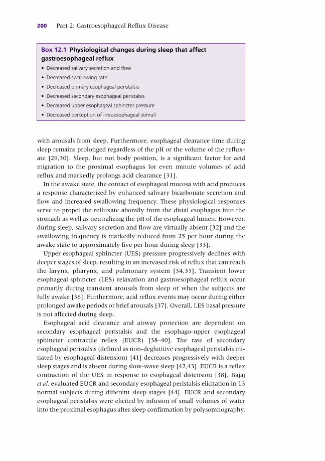
200 Part 2: Gastroesophageal Reflux Disease
with arousals from sleep. Furthermore, esophageal clearance time during
sleep remains prolonged regardless of the pH or the volume of the reflux-
ate [29,30] . Sleep, but not body position, is a significant factor for acid
migration to the proximal esophagus for even minute volumes of acid
reflux and markedly prolongs acid clearance [31] .
In the awake state, the contact of esophageal mucosa with acid produces
a response characterized by enhanced salivary bicarbonate secretion and
flow and increased swallowing frequency. These physiological responses
serve to propel the refluxate aborally from the distal esophagus into the
stomach as well as neutralizing the pH of the esophageal lumen. However,
during sleep, salivary secretion and flow are virtually absent [32] and the
swallowing frequency is markedly reduced from 25 per hour during the
awake state to approximately five per hour during sleep [33] .
Upper esophageal sphincter (UES) pressure progressively declines with
deeper stages of sleep, resulting in an increased risk of reflux that can reach
the larynx, pharynx, and pulmonary system [34,35] . Transient lower
esophageal sphincter (LES) relaxation and gastroesophageal reflux occur
primarily during transient arousals from sleep or when the subjects are
fully awake [36] . Furthermore, acid reflux events may occur during either
prolonged awake periods or brief arousals [37] . Overall, LES basal pressure
is not affected during sleep.
Esophageal acid clearance and airway protection are dependent on
secondary esophageal peristalsis and the esophago-upper esophageal
sphincter contractile reflex (EUCR) [38–40] . The rate of secondary
esophageal peristalsis (defined as non-deglutitive esophageal peristalsis ini-
tiated by esophageal distension) [41] decreases progressively with deeper
sleep stages and is absent during slow-wave sleep [42,43] . EUCR is a reflex
contraction of the UES in response to esophageal distension [38] . Bajaj
et al . evaluated EUCR and secondary esophageal peristalsis elicitation in 13
normal subjects during different sleep stages [44] . EUCR and secondary
esophageal peristalsis were elicited by infusion of small volumes of water
into the proximal esophagus after sleep confirmation by polysomnography.
Box 12.1 Physiological changes during sleep that affect gastroesophageal reflux • Decreased salivary secretion and flow
• Decreased swallowing rate
• Decreased primary esophageal peristalsis
• Decreased secondary esophageal peristalsis
• Decreased upper esophageal sphincter pressure
• Decreased perception of intraesophageal stimuli
c12.indd 200 11/15/2012 2:49:44 AM

Chapter 12: Gastroesophageal Reflux Disease and Sleep 201
It was possible to elicit EUCR and secondary esophageal peristalsis during
stage 2 and REM sleep. Their activation occurred before arousal and helped
clear the esophagus. However, during slow-wave sleep (stages 3 and 4),
EUCR and secondary peristalsis could not be triggered. Instead, the infusion
induced arousal and coughing. During these sleep stages, esophageal
clearance occurred only during arousals by swallow-induced primary
peristalsis.
There is also evidence that gastric emptying is significantly slower for
solid food during late evening hours [45] . A decrease in the frequency of
gastric slow wave was noted during non-REM sleep, which returned to
normal during REM sleep [46] . These sleep changes in gastric emptying
may promote nighttime reflux, although their effect was not specifically
studied in GERD patients.
Sleep is an altered state of consciousness, resulting in reduced percep-
tion of visceral events. Consequently, conscious-dependent defensive
behavior against gastroesophageal reflux (antacid consumption, assuming
the upright position, initiating a swallow, etc.) is markedly affected during
this period [47] .
Pathophysiology of nocturnal gastroesophageal reflux disease
Acid reflux episodes are traditionally divided into upright and recumbent
reflux. It has been noted that acid reflux during the upright period tends to be
more frequent but of shorter duration. In contrast, acid reflux during recum-
bency is commonly less frequent but of longer duration [48] . Early studies
suggested that acid reflux was significantly more frequent during the first half
of the recumbent period compared with the second half [49] . However, there
was no attempt in these studies to distinguish between the recumbent-awake
and the recumbent-asleep periods. Thus, it was unclear if sleep induction or
the early sleep period is associated with increase in acid reflux.
In a study by Dickman et al ., the authors compared the principal charac-
teristics of acid reflux events during upright, recumbent-awake, and
recumbent-asleep periods [50] . Recumbent-awake and recumbent-asleep
periods were estimated by using patients as well as their spouses or other
family members in documenting the time they went to bed at night, time
they fell asleep and time they woke up. The authors demonstrated that the
mean percentage of total time pH < 4, frequency of acid reflux events, and
number of sensed reflux events were similar in the upright and recum-
bent-awake periods but were significantly higher than those in the recum-
bent-asleep period. The authors concluded that, due to similar reflux
patterns in the upright and recumbent-awake periods, pH data analysis
c12.indd 201 11/15/2012 2:49:44 AM

202 Part 2: Gastroesophageal Reflux Disease
should be divided into awake and asleep periods rather than upright and
recumbent periods. The results of this study have been further supported
by a recently published study demonstrating that the recumbent period is
heterogeneous and clearly divided into recumbent-awake and recumbent-
asleep periods [51] . The percent total time pH < 4, the mean number of
acid reflux events, and the number of symptoms associated with reflux
events were significantly greater in the recumbent-awake period com-
pared with the recumbent-asleep period.
The nighttime period is heterogeneous regarding the frequency of reflux
events. Dickman et al . demonstrated that esophageal acid exposure was the
highest during the first 2 h of sleep [52] . This was further accentuated in
patients with Barrett ’ s esophagus compared to those with erosive esopha-
gitis or NERD with an abnormal pH test. Patients with Barrett ’ s esophagus
had the highest esophageal acid exposure parameters throughout the sleep
period. The increase in esophageal acid exposure during the first hours
of sleep is likely to be driven, amongst others, by short dinner-to-bed time.
It has been shown that dinner-to-bed time less than 3 h significantly
increased the risk of subjects experiencing gastroesophageal reflux regard-
less of their phenotypic presentation of GERD (erosive esophagitis or
NERD) [53] . A study by Piesman et al . also demonstrated that a meal con-
sumed 2 h before going to bed was significantly more associated with
recumbent reflux compared to a meal consumed 6 h prior to bed time [54] .
The presence of hiatal hernia, higher Body Mass Index (BMI), and having
erosive esophagitis increased the likelihood of developing recumbent
reflux. Other factors like alcohol and/or carbonated beverage consump-
tion, and use of benzodiazepines at bed time have all been shown to
increase the risk for reported heartburn during sleep time [22,55] .
Underlying mechanisms for sleep disturbances
The two pivotal underlying mechanisms for reduced quality of sleep and
sleep disturbances in patients with GERD are heartburn that awakens
patients from sleep during the night and short, amnestic arousals that lead
to sleep deprivation. Whilst nighttime heartburn has been perceived by
many investigators as the most important underlying mechanism for sleep
disturbances in GERD patients, recent studies have shown that acid reflux
events are more commonly encountered and often associated with short,
amnestic arousals. These arousals usually last 30 sec and tend to occur dur-
ing an acid reflux event. Most of the arousals occurred during stage 2 of
sleep and rarely during the REM period. When assessing the risks for
injury to the esophagus in patients who awake with heartburn versus
those with short arousals in response to an acid reflux event, the former
c12.indd 202 11/15/2012 2:49:45 AM

Chapter 12: Gastroesophageal Reflux Disease and Sleep 203
appears to have an important defensive effect. Patients who awake with
heartburn can initiate swallows and thus primary peristalsis, deliver saliva
to the distal portion of the esophagus, and consume anti-reflux treatment
with an acute ameliorating effect (e.g. antacids, Gaviscon, over-the-counter
H2 receptor antagonists). In contrast, patients who respond to gastro-
esophageal reflux with only short arousal will be unable to activate these
vital esophageal defense mechanisms. leading to prolonged esophageal
acid contact time and possibly esophageal mucosal injury (Table 12.1 ).
Surprisingly, there is no difference in relation to the effect on sleep quality
between patients with NERD and those with erosive esophagitis [56] .
As previously mentioned, the importance of the recumbent position for
gastroesophageal reflux was previously suggested. However, none of the
aforementioned studies utilized any technique to determine if the reflux
occurred primarily during the recumbent-awake or recumbent-asleep
periods. Actigraphy is a validated technique that has been shown to be
highly comparable to a polysomnographic study in determining sleep
duration and awakening in many conditions [57] . The actigraph, a watch-
like device worn on the patient ’ s non-dominant wrist, records motion with
an accelerometer (Figure 12.2 ). Subsequently, stored digital information is
downloaded and analyzed by proprietary software to yield periods of qui-
escence that can be inferred as sleep time. Thus, actigraphs can be used to
estimate the timing and duration of sleep as well as identifying periods of
wakefulness. A novel analysis software (FRIM© analysis) superimposes
simultaneously recorded actigraphy raw data over pH-collected data
matched by time. For the first time, the new, color-coordinated integrated
analysis allows determination of all pH parameters during the awake and
asleep periods in addition to the traditional recumbent and upright periods.
The integrative analysis can objectively determine recumbent-awake and
Table 12.1 Different esophageal physiological responses to gastroesophageal reflux
during sleep (waking up with heartburn versus short, amnestic arousals).
Waking up with heartburn Short, amnestic arousals
Initiating swallows ↑ ↓
Saliva production ↑ ↓
Primary peristalsis ↑ ↓
Gravitation ↑ ↓
Esophageal acid contact time ↓ ↑
Acute antireflux therapy ↑ ↓
Reproduced from Fass R. J Clin Gastroenterol 2007 ; 41 ( Suppl 2 ): S154 – S159 , with permission
from Lippincott Williams & Wilkins.
c12.indd 203 11/15/2012 2:49:45 AM

204 Part 2: Gastroesophageal Reflux Disease
recumbent-asleep time, frequency and duration of conscious awakenings
during sleep, the relationship between conscious awakenings during sleep
and reflux events, the relationship between GERD-related symptoms and
conscious awakenings, and the presence of patients’ recall bias (inaccurate
reporting of recumbent and upright information) [58] . Using this analysis
software, the principal characteristics of reflux during the recumbent-
awake and recumbent-asleep periods were recently reassessed [59–62] .
Most acid reflux events observed during the early part of the recumbent
period occurred during the recumbent-awake period [62] . There was a
significant reduction in the number of acid reflux events during the
corresponding recumbent-asleep period. This study suggests that the
recumbent-awake period is more commonly associated with increased
acid reflux events and symptom reports compared with the recumbent-
asleep period, which further supports the need to separate the two periods
during esophageal function analysis.
Overall, the recumbent-awake period was noted to be of substantial
duration. The mean duration of acid reflux events was similar among the
various periods (upright, recumbent-awake, and recumbent-asleep) [59] .
However, within the recumbent-asleep period, some of the acid reflux
events occurred during conscious awakenings and others during sleep. The
mean duration of an acid reflux event that occurred during sleep was sig-
nificantly longer than the mean duration of an acid reflux event that
occurred during a conscious awakening (Figure 12.3 ). The results of this
Figure 12.2 The actigraph is a watch-like device that is worn on the non-dominant
wrist and records motion with an accelerometer that is stored digitally in the device
(Basic Motionlogger, manufactured by Ambulatory Monitoring Inc., Ardsley, NY, USA).
Reproduced from Hershcovici et al . [58] , with permission from Nature Publishing
Group.
c12.indd 204 11/15/2012 2:49:45 AM

Chapter 12: Gastroesophageal Reflux Disease and Sleep 205
study demonstrated that reflux events during the recumbent-asleep period
were heterogeneous and that their principal characteristics were dependent
upon whether the patient was asleep or consciously awake during the
reflux event.
The relationship between acid reflux events and conscious awakenings
in GERD patients was further assessed using the integrated actigraphy and
pH program [61] . Interestingly, 76% of the patients reported at least one
conscious awakening, and 47% of the conscious awakenings were associ-
ated with acid reflux events. However, only 18% of the conscious awaken-
ings associated with acid reflux events were also associated with
GERD-related symptoms (Figure 12.4 ). Furthermore, in 83% of the reflux-
associated awakenings, the awakening preceded the reflux event. Acid
Proximal pHpH
6
4
2
10:00 15:00 20:00 01:00 06:00
A
C
B
t0
pH
8.0
515 sec
Proximal pH
43 sec
H
8.0
7.0
6.0
5.0
4.0
3.0
2.0
0.0
02:15 02:20 02:25 02:30 02:35 t
1.0
pH
7.0
6.0
5.0
4.0
3.0
2.0
1.0
0.0
01:00 01:10 01:12 01:14 01:16 01:18 01:20 01:22
8
AWAKESLEEP
HeartburnMealSupine
(a) (b)
C
Figure 12.3 Duration of acid reflux events during sleep and during conscious
awakenings. In this example, the “A” reflux event during sleep is typically long.
However, the “B” reflux event, occurring during conscious awakenings, appears to
be of very short duration, comparable to reflux events during the upright ( white
background ) or recumbent-awake “C” periods. Reproduced from Poh et al . [59] with
permission from Elsevier.
c12.indd 205 11/15/2012 2:49:45 AM

206 Part 2: Gastroesophageal Reflux Disease
reflux events occurring after awakening were significantly shorter than
those that preceded an awakening (occurred during sleep) [63] .
The integrated actigraphy and pH testing analysis also allowed the eval-
uation of acid reflux events that occur during the transition between sleep
and awakening [60] . In this study, almost half of the patients experienced
an acid reflux event within 20 minutes after waking up in the morning.
This is in contrast to only 18% of the patients who experienced a reflux
event during the hour of sleep prior to awakening in the morning
(Figure 12.5 ). While changes in body position may potentially explain
these reflux events, the actigraphy-based analysis demonstrated that the
reflux events were still documented immediately after waking up in the
morning by 42% of the patients who remained recumbent after waking up.
Several recent studies were unable to demonstrate differences in sleep
architecture (i.e. the patterning of sleep stages and quantification of sleep
stages throughout the night), including conventional sleep stage summaries
and sleep efficiency (i.e. the percentage of time during the night spent
asleep) when comparing the different phenotypes of GERD or when anti-
reflux treatment has been instituted [64, 65] . However, it has been demon-
strated that spectral analysis of sleep in patients with erosive esophagitis is
characterized by a shift in the electroencephalogram (EEG) power spectrum
toward higher frequencies compared to patients with functional heartburn
(having classic GERD-related symptoms but no reflux events) [65] . The
presence of delta activity in the sleep EEG indicates the density of low-
frequency slow waves and is considered an established indicator of sleep
homeostasis. Consequently, the lower delta-power and higher alpha-power
in subjects with erosive esophagitis suggests a specific sleep physiological
difference in this group compared with those with functional heartburn.
(a) (b)n= 54
16.3%
83.7%
31.5%
68.5%51.9%48.1%
n= 104n= 104
Figure 12.4 The relationship between conscious awakenings and acid reflux events
as well as symptoms in the GERD group ( n = 39). (a) The left pie demonstrates the
percentage of conscious awakenings with ( purple ) and without ( gray ) acid reflux.
The right pie depicts the percentage of symptomatic ( purple ) and asymptomatic ( gray )
conscious awakenings associated with acid reflux events. (b) The overall percentage
of conscious awakenings associated with ( purple ) or without ( gray ) acid reflux events
and GERD-related symptoms. Reproduced from Poh et al . [61] with permission from
Elsevier.
c12.indd 206 11/15/2012 2:49:46 AM

Chapter 12: Gastroesophageal Reflux Disease and Sleep 207
Sleep deprivation and gastroesophageal reflux disease
In order to explore the mechanism of the association between sleep depri-
vation and GERD, Schey et al . exposed 10 healthy subjects and 10 GERD
patients to sleep deprivation (< 3 h) and normal sleep (≥ 7 h) [7] . The
authors were able to demonstrate that after sleep deprivation, subjects
were significantly more sensitive to esophageal acid perfusion than after a
good night ’ s sleep. This study clearly showed that sleep deprivation is likely
an important central factor that can exacerbate GERD symptoms by
enhancing perception of intraesophageal stimuli.
In another study it was demonstrated that sleep deprivation per se can
precipitate acid reflux and even result in an abnormal pH test in normal
subjects [66] . In this study, a total of 11 normal subjects without evidence
of GERD were randomized to either sleep deprivation protocol (4 h of sleep
on two consecutive nights) or good sleep protocol (at least 7 h of sleep on
two consecutive nights). The mean percentage total time pH < 4 was signif-
icantly higher after bad sleep compared to good sleep (5.6 versus 2.3,
P < 0.05). Mean percentage upright and recumbent time pH < 4 were also
higher after bad sleep compared to good sleep (4.6 versus 1.9 and 5.5
versus 2.6, respectively, P < 0.05). Five (45.5%) of the normal subjects
developed an abnormal pH test after sleep deprivation (> 4.2%). All pH
tests after good sleep were within the normal range.
20 min postawakening
10 min postawakening
1 h beforeawakening
0
20
40
60
80
100
n= 39
% o
f pat
ient
s48.7%
33.3%
17.9%
Figure 12.5 Percentage of gastro-oesophageal reflux disease (GERD) patients with acid
reflux events in the three evaluative time periods (1 h prior to and 10 min and 20 min
after waking up in the morning). Reproduced from Poh et al . [60] with permission from
Blackwell Publishing.
c12.indd 207 11/15/2012 2:49:47 AM

208 Part 2: Gastroesophageal Reflux Disease
The aforementioned information suggests that sleep deprivation per se
may adversely affect GERD through two mechanisms. The first is increased
esophageal sensitivity and the second is increased esophageal acid
exposure. The latter is likely due to increased food consumption observed
in sleep-deprived subjects.
Gastroesophageal reflux disease and obstructive sleep apnea
Obstructive sleep apnea (OSA) is a breathing disorder that occurs during
sleep in which the patient experiences respiratory pauses lasting at least
10 sec and occurring at least five times per hour of sleep [67] . OSA is char-
acterized by excessive daytime sleepiness, snoring, repeated episodes of
upper airway obstruction during sleep, and nocturnal hypoxemia leading
to memory problems, irritability, and depression.
The exact association between OSA and GERD remains controversial.
Kerr et al . have demonstrated that precipitous drops in pH were frequently
preceded by arousal (98.4%), movement of the patient (71.9%), and swal-
lowing (80.4%) [68] . In this case, arousal is theorized to be caused by
increased ventilatory effort [69] . Arousal and movement may trigger gas-
troesophageal reflux by causing transient alteration in the pressure gra-
dient across the LES. Additionally, the lowered intrathoracic pressure that
accompanies OSA may by itself predispose the patient to gastroesophageal
reflux by exacerbating the LES pressure gradient. A recent physiological
investigation using concurrent high-resolution manometry, intraluminal
impedance + pH sensor, and polysomnography was performed in order to
evaluate physiological mechanisms for reflux in patients with OSA [70] .
The study demonstrated that despite a decrease in esophageal body
pressure during OSA events, compensatory changes in UES and gastro-
esophageal junction pressure prevented reflux. Specifically, crural
diaphragm contractions became increasingly vigorous, augmenting the
anti-reflux barrier and preventing reflux during OSA events.
Investigators have suggested that GERD is associated with OSA and that
there might be a potential causal link between the two disorders.
Continuous positive airway pressure (CPAP) that has been shown to
improve breathing mechanics also improved GERD parameters in patients
with OSA. In one study, Tawk et al . investigated 16 patients with both OSA
and GERD [71] . Nasal CPAP treatment (titrated to reduce the Apnea-
Hypopnea Index (AHI) to < 10/h) was found to normalize the esophageal
acid exposure in 81% and reduce the mean percentage esophageal acid
exposure time from 12.4% before CPAP to 6.8% on CPAP. In another study,
treatment with nasal CPAP showed dramatic reduction in the esophageal
c12.indd 208 11/15/2012 2:49:47 AM
Chapter 12: Gastroesophageal Reflux Disease and Sleep 209
acid exposure (the mean percentage time pH < 4 dropped significantly
from 6.3 ± 2.1 to 0.1 ± 0.1%) by elevating intrathoracic pressure [68] .
Thirty-seven of the 52 reflux events which occurred during sleep, either an
apnea or a hypopnea, were found prior to the event.
Recent studies have failed to demonstrate a causal relationship between
OSA and GERD. In a study of 15 patients with OSA, Penzel et al . found that
in 37 of 52 reflux events that occurred during sleep, either apnea or hypop-
nea was documented prior to the reflux event [72] . The sequence in time
did not prove a causal relationship between the respiratory and reflux
events. In another study, 24-h esophageal pH monitoring was performed
in 16 patients with OSA [73] ; 80% of the patients had abnormally high
esophageal acid exposure time. However, there was no relationship bet-
ween the number of reflux episodes and the severity of OSA nor a time
association between reflux and OSA episodes. Patients subjectively
reported that the quality of sleep was affected by the severity of GERD;
however, objective correlation between OSA and GERD was lacking.
Another study concluded that both conditions are common entities sharing
similar risk factors but may not be causally linked [74] . OSA is not influ-
enced by severity of GERD. Additionally, objective measures of disordered
sleep had stronger association with age, smoking, and alcohol use than
with GERD in men and stronger association with age and BMI than with
GERD in women [74] . Similarly, a study by Kim et al . could not find a rela-
tionship between OSA and GERD symptoms among 123 patients referred
to a sleep disorders center [75] . Furthermore, there was no relationship
between the severity of OSA and the likelihood of GERD symptoms.
Silent reflux and sleep
It is highly plausible that patients who experience silent reflux might be
recognized by the presence of sleep abnormalities, despite lack of reports of
typical or atypical manifestations of GERD. Recent studies have shown
that even in patients without nighttime heartburn, GER may result in
sleep disturbances and reports of reduced quality of sleep [76] . Short,
amnestic, reflux-related awakenings are not uncommon in patients with
nocturnal GER, resulting in sleep fragmentation and thus poor quality of
sleep [77] . In addition, conscious awakenings from sleep during the night
associated with acid reflux events are commonly asymptomatic [61] .
Hence, sleep abnormalities and reports of poor quality of sleep could
potentially be the sole presentation of silent GERD. In a study, 81 subjects
with documented sleep abnormalities and without heartburn were evalu-
ated by two simultaneous polysomnographic sleep studies and pH testing,
separated by an interval of 10–21 days [78] . They were compared with
c12.indd 209 11/15/2012 2:49:47 AM

210 Part 2: Gastroesophageal Reflux Disease
39 normal subjects. The mean acid exposure time was significantly higher
(3.76 versus 0.56%) among the disturbed-sleep group than the normal
sleepers. The disturbed-sleep group required significantly longer time to
fall asleep, had less total sleep time, increased wake time after sleep onset,
and less deep sleep. There was no significant difference between the study
groups with regard to the frequency of arousal responses. The authors
speculated that silent reflux may be the cause of sleep disturbances in indi-
viduals with unexplained sleep disorders. This study illuminates an area
that has been rarely evaluated in the past: sleep disturbances as the sole
presentation of GERD. The presence of sleep disturbances and poor quality
of sleep could be the necessary clinical clues for diagnosing patients with
silent GERD. Further studies are needed to assess the predictive value of
sleep abnormalities or poor quality of sleep for silent GERD.
Therapeutic approach
Overall, therapeutic studies using proton pump inhibitors (PPI) have
shown good control of nighttime heartburn. The timing of PPI dosing may
be important for the control of nighttime GERD-related symptoms.
Recently, the effect of different dosing regimens of esomeprazole on day-
time and nighttime intragastric pH was evaluated [79] . Esomeprazole
40 mg twice daily (morning and evening) provided the best nighttime
intragastric pH control (81.0%) followed by esomeprazole 40 mg prior to
dinner (70.6%) (Table 12.2 ).
A number of studies have examined the effects of anti-reflux therapy on
sleep quality as assessed by subjective or objective parameters. In an open
label trial, Chand et al . treated 18 erosive esophagitis patients with esome-
prazole 40 mg once daily for 8 weeks [80] . The authors were only able to
document improvement in subjective reports of sleep quality using the
Pittsburg Sleep Quality Questionnaire. In a study by Johnson et al ., 262
patients with moderate-to-severe nighttime heartburn and GERD-related
sleep disturbances received esomeprazole 20 mg or placebo each morning
for 4 weeks [81] . Patients receiving esomeprazole achieved significantly
greater nighttime heartburn relief than those receiving placebo (34.3%
versus 10.4%, P < 0.001). Sleep quality (assessed by the Pittsburg Sleep
Quality Questionnaire), work productivity, and regular daily activities also
significantly improved with esomeprazole.
Other studies examined improvement in sleep quality by both objective
and subjective parameters. In one study, 42 subjects were randomized to
receive either placebo or rabeprazole 20 mg twice daily for 1 week [64] .
Subsequently, the patients were crossed over to the other arm. Whilst
rabeprazole significantly reduced reflux-related parameters, there was no
c12.indd 210 11/15/2012 2:49:47 AM

Tab
le 1
2.2
Mean
ph
arm
aco
dyn
am
ic f
indin
gs
of
dif
fere
nt
eso
mepra
zole
regim
en
s (p
er-
pro
toco
l popu
lati
on
, n =
33).
24 h
our
Day
time
Nig
httim
e
Perc
enta
ge o
f tim
e w
ith
intr
agas
tric
pH
> 4
Med
ian
pHPe
rcen
tage
of
time
with
intr
agas
tric
pH
> 4
Med
ian
pHPe
rcen
tage
of
time
with
intr
agas
tric
pH
> 4
Med
ian
pH
20 m
g be
fore
bre
akfa
st56
.54.
368
.54.
639
.83.
4
20 m
g be
fore
din
ner
46.3
3.8
46.6
3.7
46.8
3.8
20 m
g tw
ice
daily
76.5
4.9
86.4
5.0
62.8
4.6
40 m
g be
fore
bre
akfa
st68
.84.
880
.94.
951
.44.
3
40 m
g be
fore
din
ner
59.5
4.4
52.3
4.1
70.6
4.8
40 m
g at
bed
time
55.2
4.2
50.6
3.9
60.5
5.2
40 m
g tw
ice
daily
87.1
5.2
91.4
5.2
81.0
5.2
Repr
oduc
ed f
rom
Bra
ndt
et a
l . Sy
mpt
oms,
aci
d ex
posu
re a
nd m
otili
ty in
pat
ient
s w
ith B
arre
tt's
eso
phag
us. C
an J
Sur
g 20
04;4
7:47
–51,
with
per
mis
sion
fro
m C
anad
ian
Med
ical
Ass
ocia
tion.
c12.indd 211 11/15/2012 2:49:47 AM

212 Part 2: Gastroesophageal Reflux Disease
difference between the drug and placebo in objective polysomnographic
measurements (percentage sleep efficiency, percentage slow-wave sleep,
percentage REM sleep, and arousals per hour). However, during rabepra-
zole treatment patients reported a significantly better quality of sleep and
reduced mean number of remembered awakenings. The authors con-
cluded that in GERD patients, anti-reflux treatment improves subjective
and not objective sleep parameters. In contrast, Dimarino et al . demon-
strated that in subjects with documented abnormal pH testing and reports
of sleep disorders, standard-dose omeprazole reduced acid reflux-related
arousals and awakenings, improved sleep efficiency, increased REM sleep,
and increased total sleep time [82] . In a large study that included 635
patients with GERD and reduced quality of sleep, treatment with esome-
prazole 40 mg or 20 mg daily markedly improved sleep by reducing
(83.2–84.1%) the number of days with GERD-associated sleep disturbances
[81] . Additionally, both pantoprazole 40 mg daily and esomeprazole 40 mg
daily improved sleep in GERD patients with documented sleep distur-
bances on the ReQuest™ questionnaire [83] .
The efficacy and safety of dexlansoprazole medium release (MR) in
controlling daytime and nighttime GERD-related symptoms in patients
with NERD were evaluated in a 4-week, double-blind, placebo-controlled
trial [27] . A total of 947 NERD patients were randomized to dexlansopra-
zole MR 30 mg, 60 mg, or placebo once daily. The percentage of nights
without heartburn was significantly higher in patients receiving dexlanso-
prazole MR 60 and 30 mg versus placebo (80.8% and 76.9% versus 51.7%,
respectively, P < 0.00001). A subsequent study specifically evaluated the
efficacy of dexlansoprazole MR 30 mg in relieving nocturnal heartburn
and GERD-related sleep disturbances [84] . A total of 305 patients with
frequent, moderate-to-severe nocturnal heartburn and associated sleep
disturbances were randomized in a double-blind fashion to receive dexlan-
soprazole MR 30 mg or placebo once daily for 4 weeks. Dexlansoprazole
MR 30 mg ( n = 152) was superior to placebo ( n = 153) in median percentage
of nights without heartburn (73.1 versus 35.7%, respectively, P < 0.001)
and in the percentage of patients with relief of nocturnal heartburn and
GERD-related sleep disturbances (47.5 versus 19.6%, 69.7 versus 47.9%,
respectively, P < 0.001) (Figure 12.6 ). Treatment with dexlansoprazole MR
led to significantly greater improvement in sleep quality and work produc-
tivity and decreased nocturnal symptom severity.
Several studies specifically evaluated the effect of anti-reflux medical
treatment on intragastric pH but without correlation with clinical end-
points, like healing or symptom improvement. Immediate-release (IR)
omeprazole, a non-enteric-coated omeprazole mixed with sodium bicar-
bonate, has been shown in several studies to rapidly control nighttime
gastric pH and significantly decrease nocturnal acid breakthrough compared
c12.indd 212 11/15/2012 2:49:47 AM
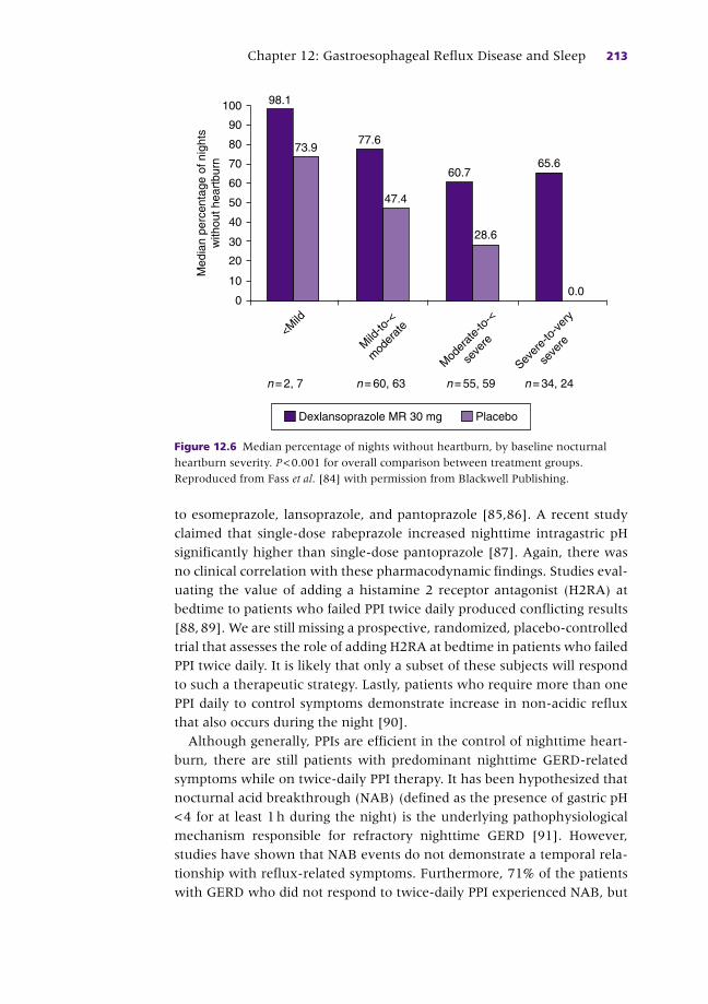
Chapter 12: Gastroesophageal Reflux Disease and Sleep 213
to esomeprazole, lansoprazole, and pantoprazole [85,86] . A recent study
claimed that single-dose rabeprazole increased nighttime intragastric pH
significantly higher than single-dose pantoprazole [87] . Again, there was
no clinical correlation with these pharmacodynamic findings. Studies eval-
uating the value of adding a histamine 2 receptor antagonist (H2RA) at
bedtime to patients who failed PPI twice daily produced conflicting results
[88, 89] . We are still missing a prospective, randomized, placebo-controlled
trial that assesses the role of adding H2RA at bedtime in patients who failed
PPI twice daily. It is likely that only a subset of these subjects will respond
to such a therapeutic strategy. Lastly, patients who require more than one
PPI daily to control symptoms demonstrate increase in non-acidic reflux
that also occurs during the night [90] .
Although generally, PPIs are efficient in the control of nighttime heart-
burn, there are still patients with predominant nighttime GERD-related
symptoms while on twice-daily PPI therapy. It has been hypothesized that
nocturnal acid breakthrough (NAB) (defined as the presence of gastric pH
< 4 for at least 1 h during the night) is the underlying pathophysiological
mechanism responsible for refractory nighttime GERD [91] . However,
studies have shown that NAB events do not demonstrate a temporal rela-
tionship with reflux-related symptoms. Furthermore, 71% of the patients
with GERD who did not respond to twice-daily PPI experienced NAB, but
0.0
65.6
28.6
60.7
47.4
<Mild
Sever
e-to
-ver
y
seve
re
Mod
erat
e-to
-<
seve
reMild
-to-<
mod
erat
e
0
10
20
30
40
50
60
70
80
90
100
Med
ian
perc
enta
ge o
f nig
hts
with
out h
eart
burn
n= 34, 24n= 55, 59n= 60, 63n= 2, 7
77.673.9
98.1
Dexlansoprazole MR 30 mg Placebo
Figure 12.6 Median percentage of nights without heartburn, by baseline nocturnal
heartburn severity. P < 0.001 for overall comparison between treatment groups.
Reproduced from Fass et al . [84] with permission from Blackwell Publishing.
c12.indd 213 11/15/2012 2:49:47 AM

214 Part 2: Gastroesophageal Reflux Disease
only 36% showed a correlation between symptoms and NAB [92] .
Furthermore, no relationship between NAB and nocturnal heartburn has
ever been established.
The effect of anti-reflux surgery on sleep was evaluated in a small
number of GERD patients [93] . The authors primarily demonstrated
improvement in subjective reports of quality of sleep but with very little
difference in objective sleep parameters between baseline and post fundo-
plication. There was a significant increase in the fraction of the night spent
in deeper sleep (49.61 versus 58.3%, P = 0.022).
Another way to treat nighttime heartburn is by addressing sleep. The
effect of insomnia treatment on nocturnal GERD was recently examined.
Gagliardi et al . administered zolpidem 10 mg or placebo to 16 reflux patients
and eight control subjects in a cross-over design [94] . Polysomnography
combined with esophageal pH testing was performed during each treatment
arm to assess nocturnal acid exposure and sleep arousals. Zolpidem was
not associated with a significant change in the number of acid reflux events
in each group. However, reflux events were associated with arousal or
awakening for 40% of the time when zolpidem was administered com-
pared with 89% when subjects received placebo ( P < 0.01). This lack of an
arousal response with zolpidem waned after the first 3 h post drug
administration. Zolpidem significantly increased the esophageal acid
clearance times for individual acid reflux events ( P < 0.05). The results of
this study indicate that hypnotic therapy with zolpidem prolongs nocturnal
esophageal acid clearance time, probably secondary to the inhibition of the
CASE STUDY
A 48-year-old woman, mother of three and busy social worker, is seen by her primary care physician for heartburn that has affected her for the last 4 years. The patient reports almost daily symptoms, primarily after meals and about 3–4 times a week heartburn that awakens her from sleep during the night. Symptoms have worsened in the last 6 months. Besides high blood pressure, which is treated with a calcium channel blocker, the patient ’ s medical history is otherwise unremarkable. The patient ’ s BMI is 30. She is usually very busy during working hours and tends to eat outside her working place. A recent upper endoscopy was unremarkable. The patient is initiated on one PPI per day, taken 30 min before breakfast. Whilst her daytime symptoms completely resolve, breakthrough GERD symptoms during sleep continue to affect her, up to three times a week. A weight loss program is recommended, in addition to other lifestyle modifications. However, the patient is unable to lose weight and continues to report nighttime breakthrough GERD-related symptoms. The patient is instructed to avoid going to bed less than 3 h after the last meal of the day. In addition, she is recommended to eliminate her recumbent-awake period and avoid falling asleep in the right decubitus or supine positions. However, only the introduction of a second PPI 30 min before dinner results in complete resolution of her breakthrough symptoms during sleep.
c12.indd 214 11/15/2012 2:49:48 AM
Chapter 12: Gastroesophageal Reflux Disease and Sleep 215
centrally mediated sleep arousal acid clearance mechanisms. However, the
conclusions of this study should be addressed with caution due to the small
number of patients evaluated. Moreover, these conclusions may be irrele-
vant to other classes of hypnotics. Further studies are necessary to evaluate
the effect of hypnotic medication on nighttime GERD-related symptoms.
Summary
Nocturnal heartburn is very common, affecting most of the patients with
GERD. However, patients may not report nocturnal symptoms, unless spe-
cifically asked. In a subset of GERD patients nocturnal symptoms may not
be present, but patients may display other manifestations of nocturnal gas-
troesophageal reflux such as nighttime choking, cough, and wheezing as
well as sleep disturbances. The latter may be the sole manifestation of
GERD, even in patients who do not report nighttime awakenings due to
heartburn.
The recent introduction of the integrated pH testing and actigraphy data
analysis program offers better separation of the recumbent-awake and
recumbent-asleep periods. Studies using this technique have shown that
sleep, and not recumbency, has a greater impact on gastroesophageal reflux
during the night. The physiological studies are further supported by clinical
trials demonstrating that gastroesophageal reflux characteristics during the
recumbent-awake period are similar to those in the upright rather than the
recumbent-asleep period. Overall, proton pump inhibitors appear to be an
effective therapeutic modality in controlling nocturnal heartburn symp-
toms and reports of sleep disturbances in most heartburn sufferers.
References
1 Locke G III , Talley NJ , Fett S , Zinsmeister A , Melton L III . Prevalence and clinical spec-
trum of gastroesophageal reflux: a population-based study in Olmsted County,
Minnesota . Gastroenterology 1997 ; 112 : 1448 – 56 .
2 Orr WC . Review article: sleep-related gastro-oesophageal reflux as a distinct clinical
entity . Aliment Pharmacol Ther 2010 ; 31 ( 1 ): 47 – 56 .
3 Gerson LB , Fass R . A systematic review of the definitions, prevalence, and response to
treatment of nocturnal gastroesophageal reflux disease . Clin Gastroenterol Hepatol
2009 ; 7 ( 4 ): 372 – 8 ; quiz 367.
4 Shaker R , Brunton S , Elfant A , Golopol L , Ruoff G , Stanghellini V . Review article:
impact of night-time reflux on lifestyle – unrecognized issues in reflux disease . Aliment
Pharmacol Ther 2004 ; 20 (Suppl 9): 3 – 13 .
5 Farup C , Kleinman L , Sloan S , et al . The impact of nocturnal symptoms associated with
gastroesophageal reflux disease on health-related quality of life . Arch Intern Med
2001 ; 161 ( 1 ): 45 – 52 .
c12.indd 215 11/15/2012 2:49:48 AM
216 Part 2: Gastroesophageal Reflux Disease
6 Shaker R , Castell DO , Schoenfeld PS , Spechler SJ . Nighttime heartburn is an under-
appreciated clinical problem that impacts sleep and daytime function: the results
of a Gallup survey conducted on behalf of the American Gastroenterological
Association . Am J Gastroenterol 2003 ; 98 ( 7 ): 1487 – 93 .
7 Schey R , Dickman R , Parthasarathy S , et al . Sleep deprivation is hyperalgesic in
pati ents with gastroesophageal reflux disease . Gastroenterology 2007 ; 133 ( 6 ):
1787 – 95 .
8 Maneerattanaporn M , Chey WD . Sleep disorders and gastrointestinal symptoms:
chicken, egg or vicious cycle? Neurogastroenterol Motil 2009 ; 21 ( 2 ): 97 – 99 .
9 Adachi K , Fujishiro H , Katsube T , et al . Predominant nocturnal acid reflux in pati-
ents with Los Angeles grade C and D reflux esophagitis . J Gastroenterol Hepatol
2001 ; 16 ( 11 ): 1191 – 6 .
10 Frazzoni M , de Micheli E , Savarino V . Different patterns of oesophageal acid exposure
distinguish complicated reflux disease from either erosive reflux oesophagitis or non-
erosive reflux disease . Aliment Pharmacol Ther 2003 ; 18 ( 11–12 ): 1091 – 8 .
11 Lagergren J , Bergstrom R , Lindgren A , Nyren O . Symptomatic gastroesophageal reflux
as a risk factor for esophageal adenocarcinoma . N Engl J Med 1999 ; 340 ( 11 ): 825 – 31 .
12 Jacob P , Kahrilas PJ , Herzon G . Proximal esophageal pH-metry in patients with
‘reflux laryngitis’ . Gastroenterology 1991 ; 100 ( 2 ): 305 – 10 .
13 Pellegrini CA , DeMeester TR , Johnson LF , Skinner DB . Gastroesophageal reflux and
pulmonary aspiration: incidence, functional abnormality, and results of surgical
therapy . Surgery 1979 ; 86 ( 1 ): 110 – 19 .
14 Harding SM , Guzzo MR , Richter JE . The prevalence of gastroesophageal reflux
in asthma patients without reflux symptoms . Am J Respir Crit Care Med 2000 ;
162 ( 1 ): 34 – 9 .
15 Dean BB , Aguilar D , Johnson LF , et al . Night-time and daytime atypical manifesta-
tions of gastro-oesophageal reflux disease: frequency, severity and impact on health-
related quality of life . Aliment Pharmacol Ther 2008 ; 27 ( 4 ): 327 – 37 .
16 Cuttitta G , Cibella F , Visconti A , Scichilone N , Bellia V , Bonsignore G . Spontaneous
gastroesophageal reflux and airway patency during the night in adult asthmatics . Am
J Respir Crit Care Med 2000 ; 161 ( 1 ): 177 – 81 .
17 Fass R. The relationship between gastroesophageal reflux disease and sleep . Curr
Gastroenterol Rep 2009 ; 11 ( 3 ): 202 – 8 .
18 Wilson JF . Is sleep the new vital sign? Ann Intern Med 2005 ; 142 ( 10 ): 877 – 80 .
19 Shaker R , Castell DO , Schoenfeld PS , Spechler SJ . Nighttime heartburn is an under-
appreciated clinical problem that impacts sleep and daytime function: The results
of a Gallup Survey conducted on behalf of the American Gastroenterological
Association . Am J Gastroenterol 2003 ; 98 ( 7 ): 1487 – 93 .
20 Castell DO , Richter JE , Robinson M , Sontag SJ , Haber MM . Efficacy and safety of
lansoprazole in the treatment of erosive reflux esophagitis . The Lansoprazole Group.
Am J Gastroenterol 1996 ; 1996 ( 91 ): 9 .
21 Farup C , Kleinman L , Sloan S , et al . The impact of nocturnal symptoms associated
with gastroesophageal reflux disease on health-related quality of life . Arch Intern
Med 2001 ; 161 ( 1 ): 1448 – 56 .
22 Fass R , Quan SF , O’Connor GT , Ervin A , Iber C . Predictors of heartburn during sleep
in a large prospective cohort study . Chest 2005 ; 127 ( 5 ): 1658 – 66 .
23 Fornari F , Madalosso CAS , Callegari-Jacques SM , Gurski RR . Heartburn during sleep:
a clinical marker of gastro-oesophageal reflux disease in morbidly obese patients .
Neurogastroenterol Motil 2009 ; 21 ( 2 ): 136 – 42 .
24 Fass R. Poorly recognized reflux-induced symptoms . Eur J Gastroenterol Hepatol
2001 ; 13 ( Suppl 3 ): S32 – S34 .
c12.indd 216 11/15/2012 2:49:48 AM

Chapter 12: Gastroesophageal Reflux Disease and Sleep 217
25 Dean BB , Aguilar D , Johnson LF , et al . Night-time and daytime atypical manifestations
of gastro-oesophageal reflux disease: frequency, severity and impact on health-related
quality of life . Aliment Pharmacol Ther 2008 ; 27 : 327 – 37 .
26 Gislason T , Janson C , Vermeire P , et al . Respiratory symptoms and nocturnal gastro-
esophageal reflux: a population-based study of young adults in three European
countries . Chest 2002 ; 121 ( 1 ): 158 – 63 .
27 Mody R , Bolge SC , Kannan H , Fass R . Effects of gastroesophageal reflux disease on
sleep and outcomes . Clin Gastroenterol Hepatol 2009 ; 7 ( 9 ): 953 – 9 .
28 Orr WC , Robinson MG , Johnson LF . Acid clearance during sleep in the pathogenesis
of reflux esophagitis . Dig Dis Sci 1981 ; 26 ( 5 ): 423 – 7 .
29 Orr WC , Robinson MG , Johnson LF . The effect of esophageal acid volume on arousals
from sleep and acid clearance . Chest 1991 ; 99 ( 2 ): 351 – 4 .
30 Orr WC , Johnson LF . Responses to different levels of esophageal acidification during
waking and sleeping . Dig Dis Sci 1998 ; 43 ( 2 ): 241 – 5 .
31 Orr WC , Elsenbruch S , Harnish MJ , Johnson LF . Proximal migration of esophageal
acid perfusions during waking and sleep . Am J Gastroenterol 2000 ; 95 ( 1 ): 37 – 42 .
32 Schneyer LH , Pigman W , Hanahan L , Gilmore RW . Rate of flow of human parotid,
sublingual, and submaxillary secretions during sleep . J Dent Res 1956 ; 35 ( 1 ): 109 – 14 .
33 Lear CS , Flanagan JB Jr , Moorrees CF . The frequency of deglutition in man . Arch
Oral Biol 1965 ; 10 : 83 – 100 .
34 Eastwood PR , Katagiri S , Shepherd KL , Hillman DR . Modulation of upper and lower
esophageal sphincter tone during sleep . Sleep Med 2007 ; 8 ( 2 ): 135 – 43 .
35 Avots-Avotins AE , Ashworth WD , Stafford BD , Moore JG . Day and night esophageal
motor function . Am J Gastroenterol 1990 ; 85 ( 6 ): 683 – 5 .
36 Dent J , Dodds WJ , Friedman RH , et al . Mechanism of gastroesophageal reflux in
recumbent asymptomatic human subjects . J Clin Invest 1980 ; 65 ( 2 ): 256 – 67 .
37 Freidin N , Fisher MJ , Taylor W , et al . Sleep and nocturnal acid reflux in normal sub-
jects and patients with reflux oesophagitis . Gut 1991 ; 32 ( 11 ): 1275 – 9 .
38 Shaker R , Hogan WJ . Reflex-mediated enhancement of airway protective mecha-
nisms . Am J Med 2000 ; 108 ( Suppl 4a ): 8S – 14S .
39 Lang IM , Medda BK , Shaker R . Mechanisms of reflexes induced by esophageal dis-
tension . Am J Physiol 2001 ; 281 ( 5 ): G1246 – 63 .
40 Enzmann DR , Harell GS , Zboralske FF . Upper esophageal responses to intraluminal
distention in man . Gastroenterology 1977 ; 72 ( 6 ): 1292 – 8 .
41 Schoeman MN , Holloway RH . Stimulation and characteristics of secondary oesopha-
geal peristalsis in normal subjects . Gut 1994 ; 35 ( 2 ): 152 – 8 .
42 Castiglione F , Emde C , Armstrong D , et al . Nocturnal oesophageal motor activity is
dependent on sleep stage . Gut 1993 ; 34 ( 12 ): 1653 – 9 .
43 Dent J , Dodds WJ , Friedman RH , et al . Mechanism of gastroesophageal reflux in
recumbent asymptomatic human subjects . J Clin Invest 1980 ; 65 ( 2 ): 256 – 67 .
44 Bajaj JS , Bajaj S , Dua KS , et al . Influence of sleep stages on esophago-upper eso-
phageal sphincter contractile reflex and secondary esophageal peristalsis . Gastro-
enterology 2006 ; 130 ( 1 ): 17 – 25 .
45 Goo RH , Moore JG , Greenberg E , Alazraki NP . Circadian variation in gastric emptying
of meals in humans . Gastroenterology 1987 ; 93 ( 3 ): 515 – 18 .
46 Elsenbruch S , Orr WC , Harnish MJ , Chen JD . Disruption of normal gastric myoelec-
tric functioning by sleep . Sleep 1999 ; 22 ( 4 ): 453 – 8 .
47 Pasricha PJ . Effect of sleep on gastroesophageal physiology and airway protective
mechanisms . Am J Med 2003 ; 115 (Suppl 3A): 114S – 118S .
48 Orr WC , Lackey C , Robinson MG , Johnson LF , Welsh JD . Esophageal acid clearance
during sleep in patients with Barrett ’ s esophagus . Dig Dis Sci 1988 ; 33 ( 6 ): 654 – 9 .
c12.indd 217 11/15/2012 2:49:48 AM

218 Part 2: Gastroesophageal Reflux Disease
49 Hila A , Castell DO . Nighttime reflux is primarily an early event . J Clin Gastroenterol
2005 ; 39 ( 7 ): 579 – 83 .
50 Dickman R , Shapiro M , Malagon IB , Powers J , Fass R . Assessment of 24-h oesopha-
geal pH monitoring should be divided to awake and asleep rather than upright and
supine time periods . Neurogastroenterol Motil 2007 ; 19 ( 9 ): 709 – 15 .
51 Mizyed I , Allen LM , Navarro-Rodriguez T , et al . Actigraphy is a simple and accurate
technique to determine esophageal acid exposure during sleep in patients under-
going pH testing . Gastroenterology 2008 ; 134 (4 Suppl 1): T2004 , P- 284 .
52 Dickman R , Parthasarathy S , Malagon IB , et al . Comparisons of the distribution of
oesophageal acid exposure throughout the sleep period among the different gastro-
oesophageal reflux disease groups . Aliment Pharmacol Ther 2007 ; 26 ( 1 ): 41 – 8 .
53 Fujiwara Y , Machida A , Watanabe Y , et al . Assocation between dinner-to-bed time
and gastro-esophageal reflux disease . Am J Gastroenterol 2005 ; 100 : 2633 – 6 .
54 Piesman M , Hwang I , Maydonovitch C , Wong RKH . Nocturnal reflux episodes
following administration of a standardized meal. Does timing matter? Am J
Gastroenterol 2007 ; 102 : 2128 – 34 .
55 Happe MR , Maydonovitch CL , Belle L , Gorske AC , Wong RKH . Not eating close to
bedtime – is this good advice? The relationship between dinner and supine reflux .
Gastroenterology 2004 ; 126 (4 (Suppl 2)):#T1732, A- 501 .
56 Yi CH , Hu CT , Chen CL . Sleep dysfunction in patients with GERD: erosive versus
nonerosive reflux disease . Am J Med Sci 2007 ; 334 ( 3 ): 168 – 70 .
57 Morgenthaler T , Alessi C , Friedman L , et al . Practice parameters for the use of acti-
graphy in the assessment of sleep and sleep disorders: an update for 2007 . Sleep
2007 ; 30 ( 4 ): 519 – 29 .
58 Hershcovici T , Gasiorowska A , Fass R . Advancements in the analysis of esophageal
pH monitoring in GERD . Nat Rev Gastroenterol Hepatol 2011 ; 8 ( 2 ): 101 – 7 .
59 Poh CH , Gasiorowska A , Allen L , et al . Reassessment of the principal characteristics of
gastroesophageal reflux during the recumbent period using integrated actigraphy-
acquired information . Am J Gastroenterol 2010 ; 105 ( 5 ): 1024 – 31 .
60 Poh CH , Allen L , Malagon I , et al . Riser ’ s reflux – an eye-opening experience .
Neurogastroenterol Motil 2010 ; 22 ( 4 ): 387 – 94 .
61 Poh CH , Allen L , Gasiorowska A , et al . Conscious awakenings are commonly associ-
ated with acid reflux events in patients with gastroesophageal reflux disease . Clin
Gastroenterol Hepatol 2010 ; 8 ( 10 ): 851 – 7 .
62 Allen L , Poh CH , Gasiorowska A , et al . Increased oesophageal acid exposure at the
beginning of the recumbent period is primarily a recumbent-awake phenomenon .
Aliment Pharmacol Ther 2010 ; 32 ( 6 ): 787 – 94 .
63 Poh CH , Allen LM , Gasiorowska A , et al . Acid reflux and arousals: which is the
chicken, and which is the egg? Gastroenterology 2009 ; 136 (5:Suppl 1):#S1891,
A- 286 .
64 Orr WC , Goodrich S , Robert J . The effect of acid suppression on sleep patterns and
sleep-related gastro-oesophageal reflux . Aliment Pharmacol Ther 2005 ; 21 ( 2 ): 103 – 8 .
65 Budhiraja R , Quan SF , Punjabi NM , Drake CL , Dickman R , Fass R . Power spectral
analysis of the sleep electroencephalogram in heartburn patients with or without
gastroesophageal reflux disease: a feasibility study . J Clin Gastroenterol 2010 ;
44 ( 2 ): 91 – 6 .
66 Hershcovici T , Gasiorowska A , Poh CH , et al . The differential effect of sleep depri-
vation versus good sleep on esophageal acid exposure in normal subjects .
Gastreoenterology 2010 ; 138 (Suppl 5):S- 603 .
67 Farmer W , Yaffe J , Santiago T . Managing sleep disorders in military personnel .
Federal Pract 2002 ; 19 : 21 – 39 .
c12.indd 218 11/15/2012 2:49:48 AM

Chapter 12: Gastroesophageal Reflux Disease and Sleep 219
68 Kerr P , Shoenut JP , Millar T , Buckle P , Kryger MH . Nasal CPAP reduces gastroesoph-
ageal reflux in obstructive sleep apnea syndrome . Chest 1992 ; 101 ( 6 ): 1539 – 44 .
69 Gleeson K , Zwillch CW , White DP . The influence of increasing ventilatory effort on
arousal from sleep . Am Rev Respir Dis 1990 ; 142 : 295 – 300 .
70 Kuribayashi S , Massey BT , Hafeezullah M , et al . Upper esophageal sphincter and gas-
troesophageal junction pressure changes act to prevent gastroesophageal and esoph-
agopharyngeal reflux during apneic episodes in patients with obstructive sleep
apnea . Chest 2010 ; 137 ( 4 ): 769 – 76 .
71 Tawk M , Goodrich S , Kinasewitz G , Orr W . The effect of 1 week of continuous
positive airway pressure treatment in obstructive sleep apnea patients with concom-
itant gastroesophageal reflux . Chest 2006 ; 130 ( 4 ): 1003 – 8 .
72 Penzel T , Becker HF , Brandenburg U , et al . Arousal in patients with gastro-oesophageal
reflux and sleep apnoea . Eur Respir J 1999 ; 14 : 1266 – 70 .
73 Graf KI , Karaus M , Heinemann S , Korber S , Dorow P , Hampel KE . Gastroesophageal
reflux in patients with sleep apnea syndrome . Z Gastroenterol 1995 ; 33 ( 12 ): 689 – 93 .
74 Morse CA , Quan SF , Mays MZ , Green C , Stephen G , Fass R . Is there a relationship
between obstructive sleep apnea and gastroesophageal reflux disease? Clin
Gastroenterol Hepatol 2004 ; 2 ( 9 ): 761 – 8 .
75 Kim H , Vorona R , Winn M , Doviak M , Johnson D , Ware J . Symptoms of gastro-
oesophageal reflux disease and the severity of obstructive sleep apnoea syndrome
are not related in sleep disorders center patients . Aliment Pharmacol Ther 2005 ;
21 ( 9 ): 1127 – 33 .
76 Orr WC . Heartburn: another danger in the night? Chest 2005 ; 127 ( 5 ): 1486 – 8 .
77 Dekel R , Green C , Malagon I , et al . Short, spontaneous, anamnestic arousals are the
most common sleep abnormality associated with nocturnal esophageal acid exposure .
Gastroenterology 2003 ; 124 (4 Supplement 1):A- 412 (M 2087).
78 Orr WC , Goodrich S , Fernstrom P , Hasselgren G . Occurrence of nighttime gastro-
esophageal reflux in disturbed and normal sleepers . Clin Gastroenterol Hepatol
2008 ; 6 ( 10 ): 1099 – 104 .
79 Wilder-Smith C , Rohss K , Bokelund Singh S , Sagar M , Nagy P . The effects of dose and
timing of esomeprazole administration on 24-h, daytime and night-time acid inhibi-
tion in healthy volunteers . Aliment Pharmacol Ther 2010 ; 32 ( 10 ): 1249 – 56 .
80 Chand N , Johnson DA , Tabangin M , Ware JC . Sleep dysfunction in patients with
gastro-oesophageal reflux disease: prevalence and response to GERD therapy, a pilot
study . Aliment Pharmacol Ther 2004 ; 20 ( 9 ): 969 – 74 .
81 Johnson DA , Orr WC , Crawley JA , et al . Effect of esomeprazole on nighttime heart-
burn and sleep quality in patients with GERD: a randomized, placebo-controlled
trial . Am J Gastroenterol 2005 ; 100 ( 9 ): 1914 – 22 .
82 Dimarino A Jr , Banwait K , Eschinger E , et al . The effect of gastro-oesophageal reflux
and omeprazole on key sleep parameters . Aliment Pharmacol Ther 2005 ; 22 ( 4 ): 325 – 9 .
83 DeVault K , Holtmann J , Malagelada J , Schmitt H , Chassany O . Pantoprazole 40 mg
(PANTO) is superior to esomeprazole 40 mg (ESO) in resolving sleep disturbances in
GERD patients (abstract) . Am J Gastroenterol 2006 ; 101 ( 9 ): S399 – S400 .
84 Fass R , Johnson DA , Orr WC , et al . The effect of dexlansoprazole MR on nocturnal
heartburn and GERD-related sleep disturbances in patients with symptomatic GERD .
Am J Gastroenterol 2011 ; 106 ( 3 ): 421 – 31 .
85 Castell D , Bagin R , Goldlust B , Major J , Hepburn B . Comparison of the effects of
immediate-release omeprazole powder for oral suspension and pantoprazole
delayed-release tablets on nocturnal acid breakthrough in patients with symptomatic
gastro-oesophageal reflux disease . Aliment Pharmacol Ther 2005 ; 21 ( 12 ):
1467 – 74 .
c12.indd 219 11/15/2012 2:49:48 AM

220 Part 2: Gastroesophageal Reflux Disease
86 Katz PO , Koch FK , Ballard ED , et al . Comparison of the effects of immediate-release
omeprazole oral suspension, delayed-release lansoprazole capsules and delayed-
release esomeprazole capsules on nocturnal gastric acidity after bedtime dosing in
patients with night-time GERD symptoms . Aliment Pharmacol Ther 2007 ; 25 ( 2 ):
197 – 205 .
87 Warrington S , Baisley K , Kee D , et al . Pharmacodynamic effects of single doses of
rabeprazole 20 mg and pantoprazole 40 mg in patients with GERD and nocturnal
heartburn . Aliment Pharmacol Ther 2007 ; 25 ( 4 ): 511 – 17 .
88 Rackoff A , Agrawal A , Hila I , Mainie I , Tutuian R , Castell DO . Histamine-2 receptor
antagonists at night improve gastroesophageal reflux disease symptoms for patietns
on proton pump inhibitor therapy . Dis Esophagus 2005 ; 18 : 370 – 3 .
89 Janiak P , Thumshirn M , Menne D , et al . Clinical trial: the effects of adding ranitidine
at night to twice daily omeprazole therapy on nocturnal acid breakthrough and acid
reflux in patients with systemic sclerosis – a randomized controlled, cross-over trial .
Aliment Pharmacol Ther 2007 ; 26 ( 9 ): 1259 – 65 .
90 Orr WC , Craddock A , Goodrich S . Acidic and non-acidic reflux during sleep under
conditions of powerful acid suppression . Chest 2007 ; 131 ( 2 ): 460 – 5 .
91 Peghini PL , Katz PO , Bracy NA , Castell DO . Nocturnal recovery of gastric acid secre-
tion with twice-daily dosing of proton pump inhibitors . Am J Gastroenterol 1998 ;
93 ( 5 ): 763 – 7 .
92 Nzeako UC , Murray JA . An evaluation of the clinical implications of acid break-
through in patients on proton pump inhibitor therapy . Aliment Pharmacol Ther
2002 ; 16 ( 7 ): 1309 – 16 .
93 Cohen JA , Harris PA , Byrne DW , Holzman MD , Sharp KW , Richards WO . Surgical
trial investigating nocturnal gastroesophageal reflux and sleep (STINGERS) . Surg
Endosc 2003 ; 17 : 394 – 400 .
94 Gagliardi GS , Shah AP , Goldstein M , et al . Effect of zolpidem on the sleep arousal
response to nocturnal esophageal acid exposure . Clin Gastroenterol Hepatol 2009 ;
7 ( 9 ): 948 – 52 .
c12.indd 220 11/15/2012 2:49:48 AM

221
Practical Manual of Gastroesophageal Reflux Disease, First Edition.
Edited by Marcelo F. Vela, Joel E. Richter and John E. Pandolfino.
© 2013 John Wiley & Sons, Ltd. Published 2013 by John Wiley & Sons, Ltd.
CHAPTER 13
Aerophagia and Belching Albert J. Bredenoord Department of Gastroenterology , Academic Medical Centre , Amsterdam , The Netherlands
Key points • Two types of belching can be distinguished: gastric and supragastric belching. Both
can be detected with impedance monitoring.
• Excessive belching is a behavior disorder in which patients have episodic high-
frequency supragastric belching.
• Excessive belching can be treated with speech therapy or behavior therapy.
• Aerophagia is a rare disorder in which too much air is ingested, and the accumulation
of air in the stomach and intestines causes abdominal distension and bloating.
• In aerophagia, impedance monitoring shows excessive air swallowing and on a plain
abdominal radiograph, distended bowel loops but no air–fluid levels are seen.
• Aerophagia can be treated with lifestyle measures such as avoidance of carbonated
drinks and smoking cessation, and with speech therapy.
Potential pitfalls Excessive belching should be differentiated from other disorders accompanied by belching:
• gastroesophageal reflux disease
• aerophagia
• functional dyspepsia
• irritable bowel syndrome
• acute pancreatitis
• cholecystolithiasis
• peptic ulcer disease.
Aerophagia should be differentiated from other disorders accompanied by gas-related
symptoms:
• excessive belching
• functional dyspepsia
• irritable bowel syndrome
• bacterial overgrowth
• paralytic or mechanical ileus
• constipation
• lactose intolerance.
c13.indd 221 11/15/2012 2:49:21 AM
222 Part 2: Gastroesophageal Reflux Disease
Introduction
In normal conditions, a certain volume of air or gas is present in the various
compartments of the gastrointestinal tract such as the stomach, intestines
and colon, as can be seen in abdominal radiographs. With every swallow,
air enters the esophagus and is transported towards the stomach along
with the peristaltic wave in the esophagus. Depending on position, gastric
and antroduodenal motility and presence of other intragastric factors, this
intragastric air is eventually transported towards the small bowel or leaves
the stomach through belching. Gas can also be released intragastrically
from ingested foods and drinks, such as beverages containing carbon
dioxide. In the small intestine and colon, gas is usually the result of bacte-
rial fermentation of luminal contents. Most intragastric gas escapes the
gastrointestinal tract proximally in the form of belches and most intestinal
and colonic gas leaves the body in the form of flatus.
Although the presence of some gastrointestinal air is thus normal, very
large volumes of gastrointestinal gas may lead to symptoms such as bloat-
ing and abdominal distension. When this is thought to be related to exces-
sive air swallowing, this disorder is referred to as aerophagia, which in
Greek means “air eating.” Aerophagia can be accompanied by excessive
belching, but excessive belching can also occur as an isolated symptom or
in combination with gastroesophageal reflux disease (GERD) and functional
dyspepsia.
In this chapter we review the pathophysiology, diagnosis, and treatment
of aerophagia and excessive belching and will describe how these syn-
dromes differ from GERD.
Air swallowing and belching
Belching is usually defined as audible escape of air or gas from the esoph-
agus into the throat. Mostly, this air has reached the esophagus from the
stomach (gastric belch), but not necessarily so. The air can also come from
the throat, reach the esophagus and be expelled from the esophagus in a
retrograde direction again. This is referred to as supragastric belching, as
the air does not originate from the stomach and does not reach the stomach
at all.
Thus, with each swallow, a certain volume of air is ingested [1] . The
swallowed air mixed with swallowed saliva and food is pushed towards the
stomach by the peristaltic contraction wave and the swallowed mixture
moves through the relaxed lower esophageal sphincter (LES) and falls into
the stomach [2] . Relaxation of the proximal stomach upon arrival of the
bolus ensures accommodation of the swallowed volume without an
c13.indd 222 11/15/2012 2:49:21 AM
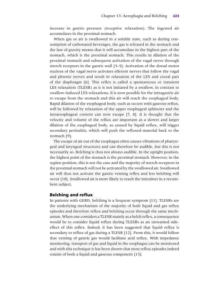
Chapter 13: Aerophagia and Belching 223
increase in gastric pressure (receptive relaxation). The ingested air
accumulates in the proximal stomach.
When gas or air is swallowed in a soluble state, such as during con-
sumption of carbonated beverages, the gas is released in the stomach and
the law of gravity means that it will accumulate in the highest part of the
stomach, which is the proximal stomach. This results in dilation of the
proximal stomach and subsequent activation of the vagal nerve through
stretch receptors in the gastric wall [3–5] . Activation of the dorsal motor
nucleus of the vagal nerve activates efferent nerves that follow the vagal
and phrenic nerves and result in relaxation of the LES and crural part
of the diaphragm [6] . This reflex is called a spontaneous or transient
LES relaxation (TLESR) as it is not initiated by a swallow, in contrast to
swallow-induced LES relaxations. It is now possible for the intragastric air
to escape from the stomach and this air will reach the esophageal body.
Rapid dilation of the esophageal body, such as occurs with gaseous reflux,
will be followed by relaxation of the upper esophageal sphincter and the
intraesophageal content can now escape [7, 8] . It is thought that the
velocity and volume of the reflux are important as a slower and larger
dilation of the esophageal body, as caused by liquid reflux, will trigger
secondary peristalsis, which will push the refluxed material back to the
stomach [9] .
The escape of air out of the esophagus often causes vibrations of pharyn-
geal and laryngeal structures and can therefore be audible, but this is not
necessarily so. Belching is thus not always audible. In the upright position,
the highest point of the stomach is the proximal stomach. However, in the
supine position, this is not the case and the majority of stretch receptors in
the proximal stomach will not be activated by the swallowed air. Swallowed
air will thus not activate the gastric venting reflex and less belching will
occur [10] . Swallowed air is more likely to reach the intestines in a recum-
bent subject.
Belching and reflux In patients with GERD, belching is a frequent symptom [11] . TLESRs are
the underlying mechanism of the majority of both liquid and gas reflux
episodes and therefore reflux and belching occur through the same mech-
anism. When one considers a TLESR mainly as a belch reflex, a consequence
would be to consider liquid reflux during TLESRs as an unwanted side-
effect of this reflex. Indeed, it has been suggested that liquid reflux is
secondary to reflux of gas during a TLESR [12] . From this, it would follow
that venting of gastric gas would facilitate acid reflux. With impedance
monitoring, transport of gas and liquid in the esophagus can be monitored
and with this technique it has been shown that most reflux episodes indeed
consist of both a liquid and gaseous component [13] .
c13.indd 223 11/15/2012 2:49:21 AM

224 Part 2: Gastroesophageal Reflux Disease
However, when studying the onset of reflux episodes in patients with
GERD, it was observed that liquid followed gas reflux just as frequently as
gas followed liquid reflux. This does not support the hypothesis that liquid
reflux simply follows gastric venting of air. Furthermore, with impedance
monitoring, a accurate estimation can be made of whether a swallow con-
tains a significant volume of air (air swallow) or whether little air is present
(Figure 13.1 ). With this technique, we have studied the relationships bet-
ween the frequency of air swallowing, the size of the intragastric air bubble,
the occurrence of belching, and acid reflux in patients with GERD and
healthy volunteers [14,15] . Both the size of the intragastric air bubble and
the number of belches were found to be related to the frequency of air
swallowing. This implies that more air swallowing leads to more intragas-
tric air and more belching. However, we did not find a relationship bet-
ween the occurrence of acid reflux and air swallowing, indicating that the
intake of air is not related to reflux of acidic liquids. Also, no relationship
was found between the occurrence of acid reflux and the size of the intra-
gastric air bubble or the number of belches.
From this it follows that belching and reflux of liquids are not related, at
least not directly. A consistent finding, however, is that GERD patients
swallow air more often and belch more frequently than asymptomatic
controls [14,16,17] . Treatment with a proton pump inhibitor (PPI) reduces
the number of swallows in patients with reflux-related symptoms but not
in other subjects. Perhaps the unpleasant sensation of heartburn stimu-
lates patients to swallow more and take larger gulps with swallowing [16] .
Other explanations for excessive belching in patients with GERD are avail-
able. Some patients with GERD report heartburn during reflux episodes of
pure gas reflux [18] . It is possible that gastroesophageal reflux of gas causes
significant distension of the esophageal body which subsequently can
trigger heartburn and chest pain [19,20] .
Evaluation and management of belching in gastroesophageal reflux disease patients There is no guideline or evidence-based approach for the evaluation and
management of symptoms of belching in patients with GERD. Ambulatory
pH impedance monitoring can confirm the diagnosis of GERD and show
increased air swallowing and gaseous reflux. It can also help to distinguish
between excessive supragastric belching and regular gastric belching.
However, if belching is not excessive and the diagnosis of GERD has been
established, no additional testing is required.
Regarding treatment, it seems sensible to advise patients to eat more
slowly, reduce the intake of beverages containing carbon dioxide, avoid
chewing gum, and stop smoking. Since air swallowing and belching can be
secondary to heartburn, a logical first step in medical treatment is to initiate
c13.indd 224 11/15/2012 2:49:21 AM

Fig
ure
13.1
(a
) R
egu
lar
swallow
dete
cted d
uri
ng im
pedan
ce p
H m
on
itori
ng. A
decr
ease
in
im
pedan
ce m
oves
from
th
e p
rox
imal to
th
e d
ista
l eso
ph
agu
s an
d
is c
leare
d b
y t
he p
eri
stalt
ic w
ave a
few
seco
nds
late
r.
(a)
c13.indd 225 11/15/2012 2:49:21 AM
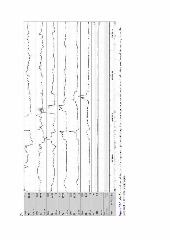
(b)
Fig
ure
13.1
(b
) A
ir s
wallow
s dete
cted w
ith
im
pedan
ce p
H m
on
itori
ng. Th
ere
is
a larg
e in
crease
in
im
pedan
ce in
dic
ati
ng s
wallow
ed a
ir, m
ovin
g f
rom
th
e
pro
xim
al to
th
e d
ista
l eso
ph
agu
s.
c13.indd 226 11/15/2012 2:49:23 AM

Chapter 13: Aerophagia and Belching 227
test treatment with a PPI. Anti-reflux surgery will reduce the frequency of
belching but will induce symptoms of bloating and abdominal distension
because of the inability to belch and is therefore relatively contraindicated.
A similar effect is likely to result from pharmacological inhibition of TLESRs
such as can be induced with the gamma-amino butyric acid (GABA) B
receptor agonist baclofen [21] .
Belching and functional dyspepsia Patients with functional dyspepsia often complain of frequent belching.
Indeed, these patients swallow air more frequently and have more belches
compared to controls, as measured on 24-h impedance monitoring
[22–24] . It is suggested that the increased belching frequency is secondary
to the observed increase in air swallowing, and in line with GERD, this is a
reaction to unpleasant gastrointestinal sensations. Increased belching in
organic painful disorders such as acute pancreatitis, peptic ulcer disease,
and cholecystolithiasis can also be the result of a reaction to abdominal
pain, but this has not been studied so far. In these disorders, one should
therefore focus on pain relief and it is likely that the increased prevalence
of belching will respond as well.
Isolated excessive belching Occasionally, patients complain of isolated excessive belching. These
patients can belch loudly and repetitively, up to several times a minute, and
often demonstrate this in the clinic during consultation. Reflux symptoms
and dyspepsia can be present as well but these symptoms are generally not
predominant and not spontaneously mentioned by the patient. Isolated
excessive belching is accompanied by a pronounced reduction in health-
related quality of life, particularly in the domains of social functioning and
mental health, which can be explained by the social isolation related to
excessive belching. Although it has been suggested that personality and
psychiatric disorders are common in these patients, there is little evidence
for the presence of depression and anxiety in the majority [25,26] .
The belching pattern in patients with isolated excessive belching is dis-
tinct from belches in healthy subjects and belches in patients with GERD
and other disorders. As mentioned above, normally a belch is the result of
the escape of gas from the stomach through the esophagus to the throat,
the so-called “gastric belch” (Figure 13.2 a). The frequency of these gastric
belches is not increased in patients with isolated excessive belching.
However, they demonstrate an increase in so-called “supragastric belches”
which are belches that result from a rapid inflow of air from the pharynx
into the esophagus where it is expelled again in the oral direction within a
second [27] . The air thus does not originate from the stomach and does
not reach the stomach and therefore these belches are referred to as
c13.indd 227 11/15/2012 2:49:24 AM
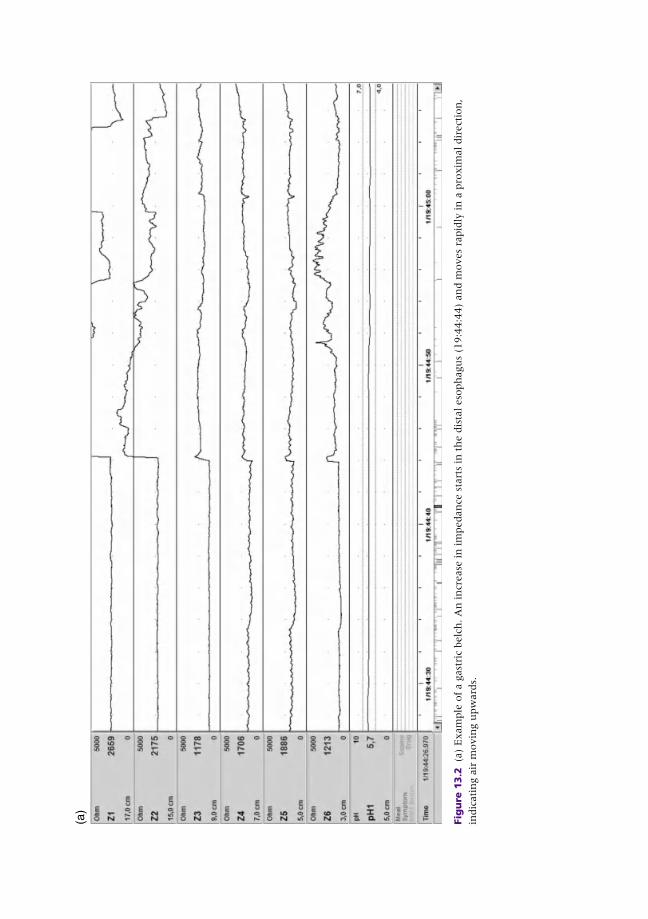
Fig
ure
13.2
(a
) E
xam
ple
of
a g
ast
ric
belc
h. A
n in
crease
in
im
pedan
ce s
tart
s in
th
e d
ista
l eso
ph
agu
s (1
9:4
4:4
4)
an
d m
oves
rapid
ly in
a p
roxim
al dir
ect
ion
,
indic
ati
ng a
ir m
ovin
g u
pw
ard
s.
(a)
c13.indd 228 11/15/2012 2:49:24 AM
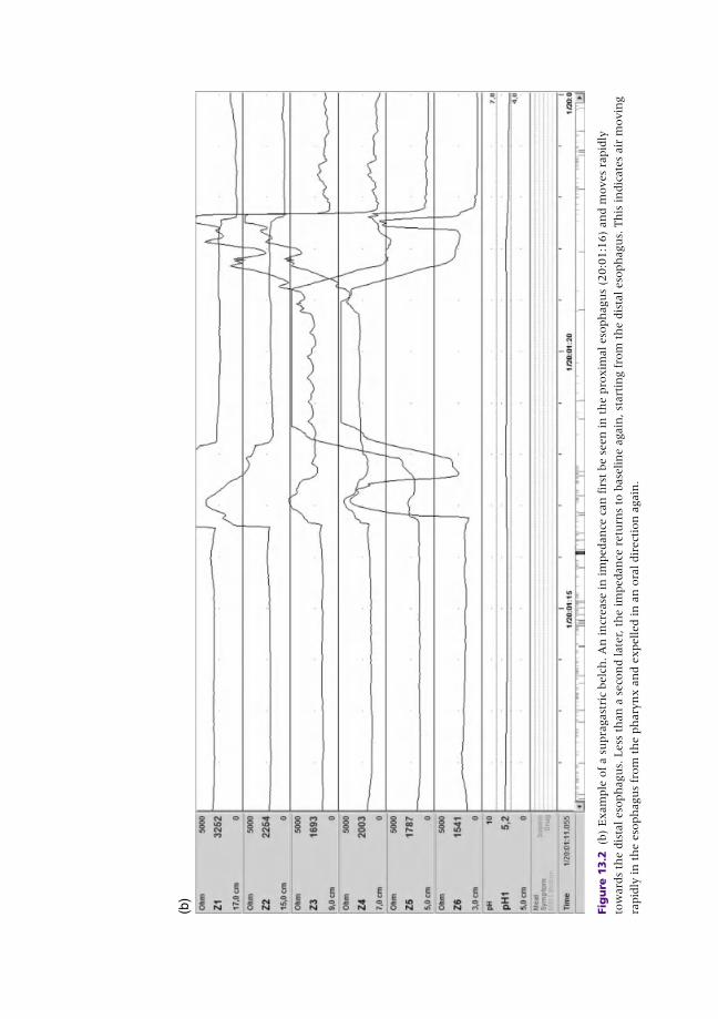
(b)
Fig
ure
13.2
(b
) E
xam
ple
of
a s
upra
gast
ric
belc
h. A
n in
crease
in
im
pedan
ce c
an
fir
st b
e s
een
in
th
e p
roxim
al eso
ph
agu
s (2
0:0
1:1
6)
an
d m
oves
rapid
ly
tow
ard
s th
e d
ista
l eso
ph
agu
s. L
ess
th
an
a s
eco
nd late
r, t
he im
pedan
ce r
etu
rns
to b
ase
lin
e a
gain
, st
art
ing f
rom
th
e d
ista
l eso
ph
agu
s. T
his
in
dic
ate
s air
movin
g
rapid
ly in
th
e e
soph
agu
s fr
om
th
e p
hary
nx a
nd e
xpelled in
an
ora
l dir
ect
ion
again
.
c13.indd 229 11/15/2012 2:49:26 AM

230 Part 2: Gastroesophageal Reflux Disease
“ supragastric” or “esophageal” belches [28] . These belches are not the
result of air swallowing; instead, the mechanism of belching is different
and does not initiate the peristaltic contraction that usually follows
swallowing. First, air is injected into the esophagus through contraction of
pharyngeal muscles or suction into the esophagus through contraction of
the diaphragm with opening of the upper esophageal sphincter. After the
air has entered the esophageal body, straining is initiated to expel the
trapped air in a retrograde direction (Figure 13.2 b). While this behavior
can be performed intentionally, it is currently believed that patients with
isolated excessive belching have somehow lost control of their belching
behavior. It thus seems that belching behavior, often initiated purposely to
relieve sensations of bloating, becomes involuntary and excessive.
Impedance monitoring can be used to demonstrate supragastric belches
and confirm the diagnosis of isolated excessive belching. When this is
combined with esophageal manometry, the mechanism of air introduction
into the esophagus (injection or suction) can be visualized as well but this
is not required to make a diagnosis.
Sometimes this disorder of excessive belching is referred to as aerophagia,
which in Greek means “air eating.” However, since the air is not “eaten”
or swallowed, this is not a correct term for this disorder. In the Rome III
criteria, a distinction has been made between excessive belching and true
aerophagia, in which air is really swallowed and transported to the stomach
and more distally to the intestines [29] .
As mentioned above, it is thought that excessive belching is a behavior
disorder, and that patients have somehow lost control over an initially
voluntary action. One hypothesis is that patients initially used supragastric
belching as a futile effort to vent gastric air, for example to relieve symp-
toms of bloating. The patients who consult for excessive belching have lost
control over this behavior. This is supported by a study in which the effect
of distraction and stimulation on the frequency of belching was investi-
gated [30] . When the patients were unaware of being monitored, the
frequency of belching was significantly lower than during the period
after which they were informed of being measured. During distraction,
the frequency of belching decreased again. This study thus supports the
hypothesis that suction and injection of air into the esophagus during
supragastric belching is a behavior disorder.
Evaluation and management of isolated excessive belching. Excessive belching disorder can be a clinical diagnosis, based on the history
and observation of excessive belching in the clinic. In the presence of other
symptoms such as dysphagia, gastroscopy can be useful to exclude other
diseases. In the case of diagnostic uncertainty, impedance monitoring can
be helpful to distinguish between gastric and supragastric belching.
c13.indd 230 11/15/2012 2:49:28 AM

Chapter 13: Aerophagia and Belching 231
Therapy for excessive (supragastric) belching is difficult and little evidence
exists to support the various treatment strategies reported in the literature.
We suggest the following approach (Figure 13.3 ), in which reassurance
by explaining the cause of belching is the first step. In addition to this,
some physicians demonstrate to patients that they can belch intentionally
themselves, in order to show the patients that supragastric belching can be
controlled [31] . A second step would be behavior therapy, given that
excessive belching is suggested to be a behavior disorder. During behavior
therapy, it is explained to patients that excessive belching is a self-induced
learned behavior and that it is therefore possible to gain control of this
behavior again [32, 33] .
Speech therapy may be an alternative to behavior therapy. After a
laryngectomy, it becomes impossible to speak as the vocal cords are resected.
These patients are taught to speak by means of belching which is called
esophageal speech. In order to learn to belch intentionally, speech therapists
teach these patients the above described suction and injection methods
[34] . As it is possible to use speech therapy to teach laryngectomized
patients to perform supragastric belches, it would perhaps also be possible
for patients with excessive belching to “unlearn” this behavior. We referred
11 patients to a speech therapist who is familiar with the concept of supra-
gastric belching [35] . A visual analog scale completed before and after 10
sessions of therapy showed that patients significantly benefitted from this
therapy. Six patients reported a large decrease in symptoms and four
reported a modest decrease.
Prescription of gas-reducing drugs such as simethicone and dimethicone
seems not useful for the treatment of excessive belching given that the
Symptoms of excessive belching
Alarm symptoms Upper endoscopyYes
No
24-h pH-impedance test
Frequent supragastric belching
Pathological refluxTreat reflux
Speech therapyand/ or behavior
therapy
Figure 13.3 Algorithm for excessive belching.
c13.indd 231 11/15/2012 2:49:28 AM

232 Part 2: Gastroesophageal Reflux Disease
volume of gastrointestinal gas is not thought to play a role in excessive
supragastric belching. Therefore, avoiding beverages containing gas is not
very helpful either. Anecdotal reports describe promising results with
hypnosis and biofeedback therapy [36,37] . In cases where excessive
belching is secondary to a psychiatric disorder, it seems advisable to treat this
first [38] .
Inability to belch The importance of the belch reflex is illustrated by the consequences that
follow the inability to belch that can occur in certain situations. During
anti-reflux surgery such as a Nissen fundoplication, the gastric fundus is
wrapped around the distal esophagus. This makes dilation by air of the
proximal stomach impossible and results in a very large reduction of the
TLESR frequency. Sometimes, belching will become completely impossible
[39–41] . This results in the desired reduction of reflux episodes but also in
an unwanted total reduction of the capacity of the stomach to vent exces-
sive intragastric air, which results in bloating, abdominal distension and
increased flatulence (see Chapter 6).
It has been shown that a self-reported ability to belch does not guarantee
that a patient can truly vent gastric air [42] . Often, symptomatic patients
develop supragastric belching in a futile effort to evacuate gastric air and to
find relief of their symptoms, and gastric belches do not occur. It also hap-
pens that patients report that belching has become impossible after surgery
but impedance monitoring shows a reduced rate but not total absence of
gastric belching. Only the presence or absence of gaseous gastroesophageal
reflux found with impedance monitoring is reliable in making a diagnosis
here. Treatment of these symptoms is difficult and pneumatic dilation is
not effective. In very severe cases dismantling of the fundoplication wrap
can be required.
Aerophagia
Aerophagia is the syndrome in which patients swallow air in too frequent
and/or in too large quantities, resulting in an excessive volume of gas in
the stomach, small bowel, and colon. Extreme aerophagia, even with fatal
consequences, has been reported in mentally retarded children but aero-
phagia also occurs in otherwise healthy adults [43,44] . Usually, symptoms
of abdominal distension, bloating, abdominal pain, flatulence, and belch-
ing are reported. A high percentage of patients suffer from constipation,
although it is unclear how this is related to the aerophagia. On plain
abdominal radiographs, a large volume of gas is seen in the gastrointestinal
tract in the absence of airfluid levels or other signs of ileus [29] (Figure 13.4 ).
c13.indd 232 11/15/2012 2:49:28 AM
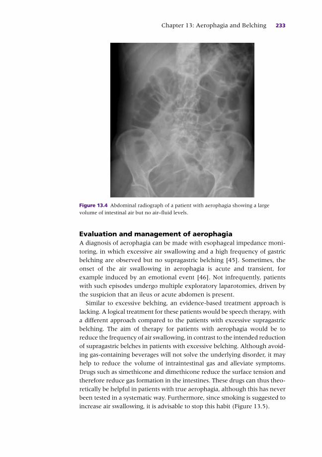
Chapter 13: Aerophagia and Belching 233
Evaluation and management of aerophagia A diagnosis of aerophagia can be made with esophageal impedance moni-
toring, in which excessive air swallowing and a high frequency of gastric
belching are observed but no supragastric belching [45] . Sometimes, the
onset of the air swallowing in aerophagia is acute and transient, for
example induced by an emotional event [46] . Not infrequently, patients
with such episodes undergo multiple exploratory laparotomies, driven by
the suspicion that an ileus or acute abdomen is present.
Similar to excessive belching, an evidence-based treatment approach is
lacking. A logical treatment for these patients would be speech therapy, with
a different approach compared to the patients with excessive supragastric
belching. The aim of therapy for patients with aerophagia would be to
reduce the frequency of air swallowing, in contrast to the intended reduction
of supragastric belches in patients with excessive belching. Although avoid-
ing gas-containing beverages will not solve the underlying disorder, it may
help to reduce the volume of intraintestinal gas and alleviate symptoms.
Drugs such as simethicone and dimethicone reduce the surface tension and
therefore reduce gas formation in the intestines. These drugs can thus theo-
retically be helpful in patients with true aerophagia, although this has never
been tested in a systematic way. Furthermore, since smoking is suggested to
increase air swallowing, it is advisable to stop this habit (Figure 13.5 ).
Figure 13.4 Abdominal radiograph of a patient with aerophagia showing a large
volume of intestinal air but no air–fluid levels.
c13.indd 233 11/15/2012 2:49:28 AM

234 Part 2: Gastroesophageal Reflux Disease
Besides patients with true aerophagia, intestinal gas can cause symptoms
of distension and bloating in other subjects. In 90% of patients with
irritable bowel syndrome, bloating and abdominal distension are reported.
It has been suggested that at least part of this problem is caused by intestinal
air [47,48] . An increased volume of air, hypersensitivity to intestinal
distension and a different distribution of intestinal air are found. The
underlying mechanisms are heterogeneous and differences in intestinal
transit, sensitivity, food intolerance, bacterial overgrowth, and sugar mal-
absorption are suggested to play a role [47,49] . Air swallowing is not
suspected to play an important role in irritable bowel syndrome and speech
therapy is thus not useful for these patients.
Summary
Belching is a physiological phenomenon and is defined as sometimes audible
gastroesophagopharyngeal reflux of gas. Belching is only pathological when
Symptoms suggestive of aerophagia
Abdominal radiograph Dyspepsia, IBSNormal
Air–fluid levels
Large volume of air in intestines
Absent Present
24hr pH-impedance testIleus?
CT scan, etc Aerophagia
Excessive air swallowing
Normal
Treatment: lifestyle measures,speech therapy
Figure 13.5 Algorithm for aerophagia. CT, computed tomography; IBS, irritable bowel
syndrome.
c13.indd 234 11/15/2012 2:49:29 AM

Chapter 13: Aerophagia and Belching 235
it becomes bothersome or excessive. Excessive belching is a common
symptom, which is often seen in patients with functional dyspepsia,
choledocholithiasis, and GERD. In these disorders, other symptoms are
usually predominant. A small group of patients complain of isolated excessive
and repetitive belching. These patients suffer from a behavior disorder, as
they involuntary apply suction or inject air into the esophagus from the
pharynx and expel this air immediately afterwards in a retrograde direction.
This behavior is called supragastric belching and can be treated by a
well-informed speech pathologist.
Although the presence of intestinal gas plays a role in various disor-
ders, including irritable bowel disorder, the term “aerophagia” should
only be used for patients with objectively demonstrable excessive air
swallowing and excessive amounts of intestinal gas visualized on a plain
abdominal radiograph. The primary symptoms of patients with aero-
phagia are bloating and abdominal distension, while belching is less pre-
dominant. It is important to differentiate aerophagia from a mechanical
or paralytic ileus, and avoid unnecessary surgery in these patients.
Treatment of aerophagia is with speech therapy and the prognosis is
generally good.
CASE STUDY
A 43-year-old office worker presented at our outpatient clinic with symptoms of episodic belching. He mentioned that these belch attacks never occurred during work time but mostly started when he was driving home after work. He admitted that he considered his job very stressful, although he denied a direct relationship between stress and his symptoms. There was some epigastric fullness as well but no heartburn, regurgitation or dysphagia. A few years ago he was diagnosed with fibromyalgia but further history was negative.
The patient ’ s general practitioner prescribed omeprazole 20 mg and domperidone but both were not considered very useful by the patient and he took these medicines only occasionally. A recently performed upper endoscopy was negative and an ultrasound scan of the abdomen revealed no abnormalities.
The patient underwent an ambulatory 24-h impedance pH monitoring test which revealed a physiological esophageal acid exposure time (time pH <4 = 3.1%) and no statistical relationship between his symptoms and acid or weakly acidic reflux. However, manual analysis of the tracings showed several periods with repetitive supragastric belching. In total, more than 100 supragastric belches were detected in a 2-h period.
The patient was diagnosed with excessive supragastric belching and referred to a dedicated speech therapist. Although he gained more control over his belching behavior in the following months, there was no complete resolution of the belching episodes.
c13.indd 235 11/15/2012 2:49:29 AM

236 Part 2: Gastroesophageal Reflux Disease
References
1 Pouderoux P , Ergun GA , Lin S , Kahrilas PJ . Esophageal bolus transit imaged by ultra-
fast computerized tomography . Gastroenterology 1996 ; 110 ( 5 ): 1422 – 8 .
2 Johnson HD , Laws JW . The cardia in swallowing, eructation, and vomiting . Lancet
1966 ; 2 ( 7476 ): 1268 – 73 .
3 Zerbib F , des Varannes SB , Ropert A , Lamouliatte H , Quinton A , Galmiche JP .
Proximal gastric tone in gastro-oesophageal reflux disease . Eur J Gastroenterol
Hepatol 1999 ; 11 ( 5 ): 511 – 15 .
4 Penagini R , Carmagnola S , Cantu P , Allocca M , Bianchi PA . Mechanoreceptors of the
proximal stomach: role in triggering transient lower esophageal sphincter relaxation .
Gastroenterology 2004 ; 126 ( 1 ): 49 – 56 .
5 McNally EF , Kelly E , Ingelfinger F . Mechanism of belching: effects of gastric disten-
tion with air . Gastroenterology 1964 ; 46 : 254 – 9 .
6 Mittal RK , Fisher MJ . Electrical and mechanical inhibition of the crural diaphragm
during transient relaxation of the lower esophageal sphincter . Gastroenterology
1990 ; 99 ( 5 ): 1265 – 8 .
7 Kahrilas PJ , Dodds WJ , Dent J , Wyman JB , Hogan WJ , Arndorfer RC . Upper esoph-
ageal sphincter function during belching . Gastroenterology 1986 ; 91 ( 1 ): 133 – 40 .
8 Enzmann DR , Harell GS , Zboralske FF . Upper esophageal responses to intraluminal
distention in man . Gastroenterology 1977 ; 72 ( 6 ): 1292 – 8 .
9 Shaker R , Ren J , Kern M , Dodds WJ , Hogan WJ , Li Q . Mechanisms of airway protec-
tion and upper esophageal sphincter opening during belching . Am J Physiol
1992 ; 262 (4 Pt 1): G621 – G628 .
10 Wyman JB , Dent J , Heddle R , Dodds WJ , Toouli J , Downton J . Control of belching by
the lower oesophageal sphincter . Gut 1990 ; 31 ( 6 ): 639 – 46 .
11 Klauser AG , Schindlbeck NE , Muller-Lissner SA . Symptoms in gastro-oesophageal
reflux disease . Lancet 1990 ; 335 ( 8683 ): 205 – 8 .
12 Straathof JW , Ringers J , Lamers CB , Masclee AA . Provocation of transient lower
esophageal sphincter relaxations by gastric distension with air . Am J Gastroenterol
2001 ; 96 ( 8 ): 2317 – 23 .
13 Sifrim D , Silny J , Holloway RH , Janssens JJ . Patterns of gas and liquid reflux during
transient lower oesophageal sphincter relaxation: a study using intraluminal electrical
impedance . Gut 1999 ; 44 ( 1 ): 47 – 54 .
14 Bredenoord AJ , Weusten BL , Timmer R , Akkermans LM , Smout AJ . Relationships
between air swallowing, intragastric air, belching and gastro-oesophageal reflux .
Neurogastroenterol Motil 2005 ; 17 ( 3 ): 341 – 7 .
15 Bredenoord AJ , Weusten BL , Timmer R , Smout AJ . Air swallowing, belching, and
reflux in patients with gastroesophageal reflux disease . Am J Gastroenterol
2006 ; 101 ( 8 ): 1721 – 6 .
16 Hemmink GJ , Weusten BL , Bredenoord AJ , Timmer R , Smout AJ . Increased swal-
lowing frequency in GORD is likely to be caused by perception of reflux episodes .
Neurogastroenterol Motil 2009 ; 21 ( 2 ): 143 – 8 .
17 Kamolz T , Bammer T , Granderath FA , Pointner R . Comorbidity of aerophagia in
GERD patients: outcome of laparoscopic antireflux surgery . Scand J Gastroenterol
2002 ; 37 ( 2 ): 138 – 43 .
18 Bredenoord AJ , Weusten BL , Curvers WL , Timmer R , Smout AJ . Determinants of
perception of heartburn and regurgitation . Gut 2005 ; 55 ( 3 ): 313 – 18 .
19 Barish CF , Castell DO , Richter JE . Graded esophageal balloon distention. A new pro-
vocative test for noncardiac chest pain . Dig Dis Sci 1986 ; 31 ( 12 ): 1292 – 8 .
c13.indd 236 11/15/2012 2:49:29 AM
Chapter 13: Aerophagia and Belching 237
20 Kahrilas PJ , Dodds WJ , Hogan WJ . Dysfunction of the belch reflex. A cause of inca-
pacitating chest pain . Gastroenterology 1987 ; 93 ( 4 ): 818 – 22 .
21 Zhang Q , Lehmann A , Rigda R , Dent J , Holloway RH . Control of transient lower
oesophageal sphincter relaxations and reflux by the GABA(B) agonist baclofen in
patients with gastro-oesophageal reflux disease . Gut 2002 ; 50 ( 1 ): 19 – 24 .
22 Lin M , Triadafilopoulos G . Belching: dyspepsia or gastroesophageal reflux disease?
Am J Gastroenterol 2003 ; 98 ( 10 ): 2139 – 45 .
23 Camilleri M , Dubois D , Coulie B , et al . Prevalence and socioeconomic impact of upper
gastrointestinal disorders in the United States: results of the US Upper Gastrointestinal
Study . Clin Gastroenterol Hepatol 2005 ; 3 ( 6 ): 543 – 52 .
24 Conchillo JM , Selimah M , Bredenoord AJ , Samsom M , Smout AJ . Air swallowing,
belching, acid and non-acid reflux in patients with functional dyspepsia . Aliment
Pharmacol Ther 2007 ; 25 ( 8 ): 965 – 71 .
25 Bredenoord AJ , Smout AJ . Impaired health-related quality of life in patients with
excessive supragastric belching . Eur J Gastroenterol Hepatol 2010 ; 22 ( 12 ): 1420 – 3 .
26 Chitkara DK , Bredenoord AJ , Tucker MJ , Talley NJ . Aerophagia in adults: a
comparison of presenting symptoms with functional dyspepsia . Aliment Pharmacol
Ther 2005 ; 22 ( 9 ): 855 – 8 .
27 Bredenoord AJ , Weusten BL , Sifrim D , Timmer R , Smout AJ . Aerophagia, gastric,
and supragastric belching: a study using intraluminal electrical impedance moni-
toring . Gut 2004 ; 53 ( 11 ): 1561 – 5 .
28 [No authors listed] Physiology of belch . Lancet 1991 ; 337 ( 8732 ): 23 – 4 .
29 Tack J , Talley NJ , Camilleri M , et al . Functional gastroduodenal disorders .
Gastroenterology 2006 ; 130 ( 5 ): 1466 – 79 .
30 Bredenoord AJ , Weusten BL , Timmer R , Smout AJ . Psychological factors affect the
frequency of belching in patients with aerophagia . Am J Gastroenterol 2006 ;
101 ( 12 ): 2777 – 81 .
31 Roth JLA . Aerophagia . In: Linder AE (ed) Emotional factors in gastrointestinal illness .
Amsterdam : Excerpta Medica , 1973 : pp. 16 – 36 .
32 Cigrang JA , Hunter CM , Peterson AL . Behavioral treatment of chronic belching due
to aerophagia in a normal adult . Behav Modif 2006 ; 30 ( 3 ): 341 – 51 .
33 Chitkara DK , Bredenoord AJ , Talley NJ , Whitehead WE . Aerophagia and rumina-
tion: recognition and therapy . Curr Treat Options Gastroenterol 2006 ; 9 ( 4 ):
305 – 13 .
34 Damste PH . Methods of restoring the voice after laryngectomy . Laryngoscope 1975 ;
85 ( 4 ): 649 – 55 .
35 Hemmink GJ , Ten Cate L , Bredenoord AJ , Timmer R , Weusten BL , Smout AJ . Speech
therapy in patients with excessive supragastric belching – a pilot study . Neuro-
gastroenterol Motil 2010 ; 22 ( 1 ): 24 – 8 .
36 Spiegel SB . Uses of hypnosis in the treatment of uncontrollable belching: a case
report . Am J Clin Hypn 1996 ; 38 ( 4 ): 263 – 70 .
37 Bassotti G , Whitehead WE . Biofeedback as a treatment approach to gastrointestinal
tract disorders . Am J Gastroenterol 1994 ; 89 ( 2 ): 158 – 64 .
38 Zella SJ , Geenens DL , Horst JN . Repetitive eructation as a manifestation of
obsessive-compulsive disorder . Psychosomatics 1998 ; 39 ( 3 ): 299 – 301 .
39 Smith D , King NA , Waldron B , et al . Study of belching ability in antireflux surgery
patients and normal volunteers . Br J Surg 1991 ; 78 ( 1 ): 32 – 5 .
40 Scheffer RC , Tatum RP , Shi G , Akkermans LM , Joehl RJ , Kahrilas PJ . Reduced tLESR
elicitation in response to gastric distension in fundoplication patients . Am J Physiol
Gastrointest Liver Physiol 2003 ; 284 ( 5 ): G815 – G820 .
c13.indd 237 11/15/2012 2:49:29 AM

238 Part 2: Gastroesophageal Reflux Disease
41 Bredenoord AJ , Draaisma WA , Weusten BL , Gooszen HG , Smout AJ . Mechanisms of
acid, weakly acidic and gas reflux after anti-reflux surgery . Gut 2008 ; 57 ( 2 ): 161 – 6 .
42 Broeders JA , Bredenoord AJ , Hazebroek EJ , Broeders IA , Gooszen HG , Smout AJ .
Effects of antireflux surgery on weakly acidic reflux and belching . Gut 2011 : 60 ( 4 ):
435 – 41 .
43 Gauderer MW , Halpin TC Jr , Izant RJ Jr. Pathologic childhood aerophagia: a recog-
nizable clinical entity . J Pediatr Surg 1981 ; 16 ( 3 ): 301 – 5 .
44 Hackl H. Pathologische arophagie als todesursache . Med Klin 1973 ; 68 ( 20 ): 667 – 9 .
45 Hemmink GJ , Weusten BL , Bredenoord AJ , Timmer R , Smout AJ . Aerophagia:
excessive air swallowing shown by esophageal impedance monitoring . Clin
Gastroenterol Hepatol 2009 ; 7 ( 10 ): 1127 – 9 .
46 Fonagy P , Calloway SP . The effect of emotional arousal on spontaneous swallowing
rates . J Psychosom Res 1986 ; 30 ( 2 ): 183 – 8 .
47 Serra J , Salvioli B , Azpiroz F , Malagelada JR . Lipid-induced intestinal gas retention in
irritable bowel syndrome . Gastroenterology 2002 ; 123 ( 3 ): 700 – 6 .
48 Caldarella MP , Serra J , Azpiroz F , Malagelada JR . Prokinetic effects in patients with
intestinal gas retention . Gastroenterology 2002 ; 122 ( 7 ): 1748 – 55 .
49 Houghton LA , Whorwell PJ . Towards a better understanding of abdominal bloating
and distension in functional gastrointestinal disorders . Neurogastroenterol Motil
2005 ; 17 ( 4 ): 500 – 11 .
c13.indd 238 11/15/2012 2:49:30 AM
239
Practical Manual of Gastroesophageal Reflux Disease, First Edition.
Edited by Marcelo F. Vela, Joel E. Richter and John E. Pandolfino.
© 2013 John Wiley & Sons, Ltd. Published 2013 by John Wiley & Sons, Ltd.
CHAPTER 14
Dysphagia and Gastroesophageal Reflux Disease Donald O. Castell 1 and Erick R. Singh 2 1 Esophageal Disease Program , Medical University of South Carolina , Charleston , SC , USA
2 Section of Gastroenterology and Hepatology , Georgia Health Sciences University ,
Augusta , GA , USA
Key points • Gastroesophageal reflux disease may induce structural and physiological changes
in the esophagus which can cause dysphagia.
• Initial evaluation requires thorough history and physical examination in order to
identify the etiology of dysphagia.
• Esophagogastroduodenosopy allows direct visual inspection of the esophageal lumen
and mucosa, localization of mechanical obstruction, and therapeutic and diagnostic
interventions.
• Barium esophagram and esophageal manometry are often necessary for diagnosis
of motility and structural causes of dysphagia not identified by
esophagogastroduodenosopy.
• Once the underlying cause of dysphagia has been treated, it is important for the
patient to remain on treatment to control reflux and prevent recurrence.
Potential pitfalls • Failing to distinguish between oropharyngeal and esophageal dysphagia, as
gastroesophageal reflux disease is a common cause of the latter but not the former.
• Failing to pursue further testing by esophagram or manometry when esophagogas-
troduodenosopy is normal, as endocopy may miss proximal structural or motility
causes of dysphagia.
• Assuming that therapeutic failure of any single proton pump inhibitor or H2 blocker
medication for treatment of gastroesophageal reflux disease means that other similar
drugs will not work. Many patients will respond favorably to other drugs in these
medication classes.
c14.indd 239 11/15/2012 2:49:06 AM

240 Part 2: Gastroesophageal Reflux Disease
Introduction
Gastroesophageal reflux disease (GERD) is caused by the retrograde flow
of stomach contents into the body of the esophagus caused by chronic
hypotension or by transient relaxation or effacement of the lower esopha-
geal sphincter. GERD may cause inflammation of the esophagus leading to
the typical symptoms of heartburn and acid regurgitation. Some patients
with GERD may also develop difficulty with swallowing. Dysphagia refers
to the sensation of difficult passage of organic materialfrom the mouth
to the stomach. The word “dysphagia” can be traced from the root origins
of the Greek words “ dys ” meaning “difficulty” and “ phagia ” meaning “to
eat.” The sensations described by patients encompass a wide variety of com-
plaints ranging from inability to transfer material from the mouth into the
pharynx or esophagus to feeling that food is “getting stuck” in the esoph-
agus. There is also a wide range of co-existing symptoms which may occur
with dysphagia such as immediate regurgitation, pain with swallowing
(odynophagia), heartburn, or weight loss.
It is estimated that 5–8% of the general population over the age of
50 years may experience dysphagia but the incidence continues to increase
with age, as up to 50–60% of patients in nursing homes and other chronic
care facilities report this symptom [1] . Dysphagia should be considered as
an alarm symptom warranting immediate evaluation, primarily to exclude
a malignant etiology.
Dysphagia can be classified into two main types: oropharyngeal and
esophageal. Oropharyngeal dysphagia, also known as “transfer dysphagia,”
refers to an abnormality in the delivery of oral contents to the proximal
esophagus. For adequate bolus transfer from mouth to esophagus, the bolus
must pass across a relaxed upper esophageal sphincter via coordinated
contractile movements of the pharynx. Symptoms of oropharyngeal dys-
phagia occur almost immediately after swallowing. Patients may describe
sensations due to abnormalities of the oral phase (food spillage, drooling)
or the pharyngeal phase which may include failure to protect the airway
(choking, coughing, gagging). They are also often able to localize the area
of the neck or pharynx as the region of their difficulties. The presence of
oropharyngeal dysphagia warrants careful structural and neurological
evaluation. Residual neurological deficits following a cerebrovascular
accident are often the cause of oropharyngeal dysphagia although cranial
nerve palsies, myositis, amyotrophic lateral sclerosis (ALS), Parkinson ’ s
disease, and muscular dystrophies also remain in the differential diagnosis.
With symptoms of regurgitation of undigested food, halitosis, and aspira-
tion, structural abnormalities such as Zenker ’ s diverticulum must also be
considered [2] .
c14.indd 240 11/15/2012 2:49:07 AM

Chapter 14: Dysphagia and Gastroesophageal Reflux Disease 241
Esophageal dysphagia refers to difficulty swallowing that is attributable
to dysfunction or obstruction within the body of the esophagus. Mechanical
causes or motility abnormalities most often lead to this type of dysphagia.
This chapter focuses on evaluation and management of GERD-related
causes of esophageal dysphagia.
Evaluation
The first and most important aspect in the evaluation of dysphagia is the
patient history. It is imperative to enquire about whether the dysphagia
is (a) for liquids, solids or both; (b) intermittent or progressive; (c) oropha-
ryngeal as opposed to esophageal. As previously discussed, oropharyngeal
dysphagia is usually described as an immediate difficulty of initiating a
swallow localized to the pharyngeal region and characterized by choking,
coughing or gagging. On the other hand, esophageal dysphagia is more
commonly described as the sensation of the bolus “getting stuck behind
the breastbone” (in the esophagus) moments after initiating a swallow.
Once esophageal dysphagia is suspected, the type of food producing
symptoms should be ascertained. Dysphagia caused by solids is largely
attributable to mechanical obstructions whereas dysphagia to solids and/
or liquids is more commonly associated with motility disorders. Next, the
temporal relationship or progression of symptoms must be determined.
Intermittent dysphagia for solids and liquids may be indicative of a motility
disorder such as ineffective esophageal motility (IEM), diffuse esophageal
spasm (DES) or nutcracker esophagus. Progressive dysphagia with weight
loss, regurgitation, and aspiration is a constellation of symptoms seen with
motility disorders such as achalasia, which is not associated with GERD.
Determining the location of a mechanical obstruction by the patient ’ s
description of symptoms is not very accurate. In general, patient perception
of obstruction located down the sternum has good localization with a
disorder involving the distal esophagus whereas perceived obstruction
at or above the suprasternal notch may represent referred sensation from
an obstruction anywhere from the pharynx to the distal esophagus [3] .
Esophageal rings and webs produce intermittent dysphagia to solids.
Progressive solid food dysphagia is encountered with strictures, esophageal
and gastric cardia cancers, and occasionally with esophagitis. Esophageal
and gastric cardia cancers may be differentiated from strictures in that the
solid food dysphagia which occurs with malignancy is often quite rapid in
onset, progressive, and associated with marked weight loss. The presence
of heartburn may also be helpful in identifying the etiology of dysphagia.
Progressive solid dysphagia in a patient with long history of heartburn may
be found with both peptic stricture and erosive esophagitis. Eosinophilic
c14.indd 241 11/15/2012 2:49:07 AM

242 Part 2: Gastroesophageal Reflux Disease
esophagitis may be the cause of dysphagia in a young adult which may
also present with an initial symptom of food impaction [4] .
After the history and physical examination have identified possible
causes of dysphagia, the next diagnostic tool must be selected.
Esophagogastroduodenoscopy (EGD) is the initial test of choice for esophageal
dysphagia in that it allows thorough inspection of the esophagus and mucosa,
localization of possible mechanical obstruction, and diagnostic and therapeutic
interventions. If EGD is negative, further evaluation includes barium esopha-
gram to rule out subtle causes of obstruction such as rings or webs which may
be missed during endoscopic evaluation, along with esophageal manometry
to investigate dysmotility. Barium esophagram may also be the preferred
initial test in patients with a clinical presentation suggestive of a proximal
esophageal lesion (such as previous laryngeal surgery/cancer, Zenker ’ s
diverticulum, radiation therapy) or a complex stricture (radiation exposure
or caustic injury), as in these cases EGD may be safer once the expected
anatomy is delineated by the esophagram. In addition, esophagram findings
consistent with achalasia in a patient with history suggestive of this disorder
would also warrant an esophageal manometric evaluation for diagnostic
confirmation prior to proceeding to EGD. Computed tomographic (CT) and
endoscopic ultrasound (EUS) studies may also be of diagnostic benefit in the
evaluation of esophageal and gastric cancers (Figure 14.1 ).
GERD-related esophageal dysphagia
Solids &liquids
Solids only
IEMScleroderma StrictureRings & webs CancerEsophagitis
Structural problemMotility problem
Progressive Intermittent
Acute Intermittent ProgressiveHeartburn ®urgitation
GERD sx’s
> 50 years
Bariumesophagram &
manometry
EGD
Figure 14.1 Evaluation of esophageal dysphagia. EGD, esophagogastroduodenoscopy;
GERD, gastroesophageal reflux disease; IEM, ineffective esophageal motility.
c14.indd 242 11/15/2012 2:49:07 AM
Chapter 14: Dysphagia and Gastroesophageal Reflux Disease 243
Causes of dysphagia associated with gastroesophageal reflux disease
All GERD-associated causes of dysphagia fall into the category of esopha-
geal dysphagia and the differential diagnosis is as follows.
• Peptic strictures
• Rings and webs
• Esophageal adenocarcinoma
• Esophagitis (erosive and eosinophilic)
• Ineffective esophageal motility (IEM)
• Scleroderma
Peptic stricture An esophageal peptic stricture is usually a short, focal, and straight
narrowing of the esophageal lumen located near the gastroesophageal
(GE) junction (Figure 14.2 ). Although strictures may be produced as a
result of many different injuries such as caustic, pill or radiation-induced
damage, the vast majority occur as a consequence of long-standing GERD.
Approximately 4–20% of patients with GERD undergoing endoscopy are
found to have peptic stricture. With the current widespread use of proton
pump inhibitors (PPIs), the prevalence of peptic strictures has decreased
[5] . The mechanism of stricture formation centers on collagen deposition
which occurs during the healing phase of esophageal injury. As acid gastric
contents move past the GE junction, they damage the columnar esopha-
geal mucosa, producing an inflammatory state. After this initial injury,
collagen is deposited during the healing phase. As healing continues, the
esophageal mucosa narrows as the collagen fibers contract to form a peptic
stricture. Conditions which may increase the incidence of GERD such as
scleroderma, Zollinger–Ellison syndrome, and Heller myotomy in achalasia
patients as well as increased age and long duration of GERD symptoms
increase the overall incidence of peptic strictures [6] .
The most common symptom of peptic stricture is dysphagia. Dysphagia
occurs as the esophageal lumen narrows to <13 mm [7] . Patients fre-
quently first experience solid food dysphagia which may progress to liquid
dysphagia and possible food impaction if acid control is not obtained. As
stricture formation is most commonly associated with long- standing
GERD, patients will usually have a history of chronic heartburn, indiges-
tion or acid regurgitation. The initial diagnostic test should be EGD as it
enables visual inspection and localization of the injury. This test may
also provide the opportunity for treatment through balloon or bougie
dilation, which may be repeated if an optimal diameter and symptomatic
relief are not achieved after a single dilation. Barium esophagram or
c14.indd 243 11/15/2012 2:49:07 AM

244 Part 2: Gastroesophageal Reflux Disease
CT studies may also be of diagnostic utility but they do not provide the
benefit of dilation therapy.
Treatment of simple peptic strictures is a two-fold approach which
includes endoscopic dilation along with pharmacological acid suppression
to maintain healing and prevent recurrence. The pharmacological and
surgical treatments for GERD are discussed elsewhere in this book.
Dilation may be performed by bougie or balloon-type dilators. Subtypes of
mechanical bougie dilators include the Maloney and Savary–Gilliard dila-
tors. The Maloney dilator is tapered and comes in various sizes, and does
not require passage over a guidewire. The Savary–Gilliard dilator is the
most widely used dilator which is also tapered and available in multiple
sizes but does require passage over a guidewire. Both types of mechanical
dilators allow for both longitudinal and radial force to be applied to the
stricture for stretching [8] .
Balloon dilators are passed through the endoscope or over the guidewire
but only provide for radial force to be applied to the stricture. The so-called
Figure 14.2 Midesophageal peptic
stricture.
c14.indd 244 11/15/2012 2:49:07 AM
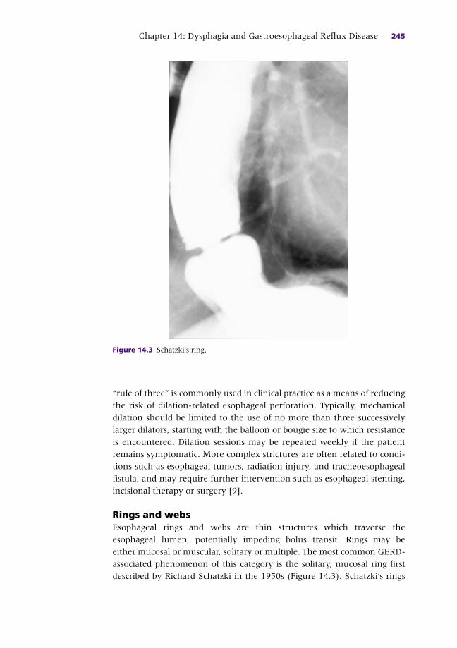
Chapter 14: Dysphagia and Gastroesophageal Reflux Disease 245
“rule of three” is commonly used in clinical practice as a means of reducing
the risk of dilation-related esophageal perforation. Typically, mechanical
dilation should be limited to the use of no more than three successively
larger dilators, starting with the balloon or bougie size to which resistance
is encountered. Dilation sessions may be repeated weekly if the patient
remains symptomatic. More complex strictures are often related to condi-
tions such as esophageal tumors, radiation injury, and tracheoesophageal
fistula, and may require further intervention such as esophageal stenting,
incisional therapy or surgery [9] .
Rings and webs Esophageal rings and webs are thin structures which traverse the
esophageal lumen, potentially impeding bolus transit. Rings may be
either mucosal or muscular, solitary or multiple. The most common GERD-
associated phenomenon of this category is the solitary, mucosal ring first
described by Richard Schatzki in the 1950s (Figure 14.3 ). Schatzki ’ s rings
Figure 14.3 Schatzki ’ s ring.
c14.indd 245 11/15/2012 2:49:08 AM
246 Part 2: Gastroesophageal Reflux Disease
are thin, webbed structures found in the lower esophageal region at the
squamocolumnar junction [10] . They are evident in 6–14% of barium
esophagrams but may account for >15% of cases of esophageal dysphagia.
Rings do not commonly cause dysphagia until the ring diameter constricts
to <13 mm. The pathogenesis leading to the formation of these rings is
not entirely clear, but GERD may play a role as reduction in esophageal
acid exposure in conjunction with dilation procedures has been shown to
decrease the risk of recurrence in several studies. Some investigators
propose a congenital origin as opposed to that of reactive formation in the
setting of chronic GERD. In either case, treatment entails either bougie
or balloon dilation along with acid suppressive therapy [11] . Eosinophilic
esophagitis may present clinically in a somewhat similar fashion to a
Schatzki ’ s ring, but usually multiple rings are present throughout the
esophagus [12] .
Esophageal webs are web-like structures more commonly found in
the proximal esophagus or the hypopharyngeal region. Their origin is less
clear than that of esophageal rings. Plummer Vinson syndrome is an entity
found in middle-aged women with a triad of symptoms including iron
deficency anemia, glossitis, and dysphagia due to an esophageal web.
Possible correlation of esophageal webs with GERD is unclear. Similar to
esophageal rings, the mainstay of treatment is esophageal dilation [13] .
Esophageal adenocarcinoma Esophageal and gastric cardia adenocarcinomas are GERD-associated
malignancies which cause esophageal dysphagia. Although squamous cell
carcinoma is still the most common type of esophageal cancer worldwide,
the increasing incidence of adenocarcinoma has led to it becoming the
most common esophageal cancer in the Western world. The incidence
among white men of 5.69 per 1000,000 person-years in 2000–2004
represents a greater than four-fold increase since the mid-1970s [14] .
The pathogenesis of adenocarcinoma involves many steps of transfor-
mation which may be attributable to a number of risk factors. These
include GERD, obesity, and smoking, as well as race and genetic background
as esophageal adenocarcinoma is most common in white males. However,
a study of greater than 300 patients with esophageal adenocarcinoma and
a similar number of age- and gender-matched controls revealed GERD to
be the strongest single risk factor for adenocarcinoma [15] . Although the
precise mechanism of carcinogenesis is yet to be fully determined, the
initial step involves the metaplastic change known as Barrett ’ s esophagus.
This term refers to the transformation of the normal stratified squamous
epithelium of the esophagus into intestinal columnar cells. The squamous
cells at or near the gastroesophageal junction are damaged by chronic
exposure to gastric acid refluxed into the esophagus, and the damaged
c14.indd 246 11/15/2012 2:49:09 AM
Chapter 14: Dysphagia and Gastroesophageal Reflux Disease 247
cells are replaced by aberrantly differentiating squamous cells [16] .
Adenocarcinoma manifests once these metaplastic cells neoplastically
proliferate after genetic changes are made to their DNA. Such changes
are thought to occur due to upregulation and activation of protooncogenes
or downregulation of tumor suppressor genes [17] .
Unfortunately, many such tumors do not present clinically until they
have grown substantially. Rapidly progressive solid dysphagia with
accompanying weight loss and tumor-induced cachexia may be an all too
common initial presentation. Despite the fact that GERD has been found to
be the biggest independent risk factor for the development of esophageal
adenocarcinoma, more than 50% of patients diagnosed with this tumor
type exhibit no symptoms of GERD [18] . Diagnosis can be suggested with
radiographic studies such as barium esophagram or CT imaging, but con-
firmatory tissue diagnosis is necessary via endoscopy with biopsies. Once
tissue diagnosis has been confirmed, staging of the cancer is undertaken
with CT imaging and EUS which provide further detail regarding the depth
of tumor invasion. EUS may also allow for fine needle aspiration of
suspicious-appearing lymph nodes of metastatic potential. Treatment options
vary greatly dependent upon the tumor ’ s stage at the time of diagnosis.
Endoscopic therapeutic approaches are reasonable in cases of esopha-
geal cancer which does not reach the submucosa. In such cases, modalities
used to treat Barrett ’ s esophagus such as endoscopic mucosal resection
(EMR), radiofrequency ablation or cryoablation may be appropriate. Once
the depth of the lesion involves the submucosa (T1), esophagectomy is
likely necessary although EMR may be used in selected cases. Once tumor
depth reaches the muscularis propria (T2) or adventitia (T3), esophagec-
tomy is required [19] . Any patient with T1–T3 tumors being treated
with resection should also be considered for chemoradiation therapy
with medications most commonly including cisplatin and 5-fluorouracil.
The role of curative chemoradiation therapy is controversial and currently
reserved for patients thought to be poor surgical candidates due to
co-morbidities or inoperable type lesions. Patients with locoregional lym-
phatic involvement are often treated with neoadjuvant chemoradiation
therapy before being restaged and considered for surgery. Chemoradiation
and palliation therapy are most common in patients with metastatic
disease given their poor overall prognosis, with 5-year survival rate being
less than 3% [20] .
Esophagitis Esophagitis refers to the inflammatory changes of the esophageal mucosa
in response to a noxious stimulus. In erosive esophagitis, squamous esoph-
ageal cells lose their ability to regulate their intracellular pH as they are
exposed to acidic gastric secretions. As they acidify, they lose the ability to
c14.indd 247 11/15/2012 2:49:09 AM

248 Part 2: Gastroesophageal Reflux Disease
regulate volume, leading to cellular edema and eventual necrosis [21] .
However, GERD-induced esophagitis seems less dependent upon the
acidity of the reflux than the persistent exposure of the esophageal
mucosa to the acidified medium [22] . Initial symptoms of erosive esopha-
gitis include heartburn, acid regurgitation or bitter taste. Interestingly,
the presence of dysphagia is not an accurate clinical predictor of disease
severity. However, persistence of dysphagia despite PPI therapy does likely
indicate therapeutic failure [23] . The treatment of erosive esophagitis
through pharmacological acid suppression is covered elsewhere in this
book (see Chapter XX).
Another form of esophagitis linked with dysphagia, especially in the
young, is eosinophilic esophagitis. This was first described in the late 1970s
with presenting symptoms such as heartburn, regurgitation, and vomiting
in children, but more commonly presenting in adults with symptoms of
dysphagia and food impaction. Diagnosis requires biopsy evidence of >15
eosinophils per high-power field. Endoscopic findings may include mul-
tiple esophageal rings, longitudinal furrows, white spots, and mucosal
friability.
Although the exact pathophysiological cause of this disease is unknown,
eosinophilic esophagitis is thought to be an antigenic response to food or
air-borne allergens [24] . The exclusion of GERD as a cause of esophagitis is
a common preliminary stage in the diagnosis of eosinophilic esophagitis.
However, many patients are placed on acid-suppressive regimens with
high-dose PPI initially in their treatment course. Favorable responses to
these trials suggest there may be some relationship between GERD and
eosinophilic esophagitis [25] . Eosinophilic esophagitis is discussed else-
where in this book (see Chapter XX).
Motility disorders In addition to the structural causes of esophageal dysphagia, several
motility disorders related to GERD are also known to contribute to dys-
phagia (Plate 14.1). Ineffective esophageal motility (IEM) is an entity in
which the lower esophageal smooth muscle contracts in a hypotensive
manner. More specifically, it is manometrically defined as an esophagus in
which 50% or more liquid swallows are found to have amplitude < 30
mmHg at positions located 5 or 10 cm above the lower esophageal sphincter
(LES) [26] .
Many studies have shown an association between GERD and IEM. Ho
et al . reported the overall prevalence of IEM in a group of 89 GERD patients
as 49%, with these patients also exhibiting abnormal esophageal acid
exposure compared to GERD patients without motility disorder [27] . The
causal relationship as to whether long-standing GERD predisposes the
lower esophageal body to continuous injury, prompting the hypotensive
c14.indd 248 11/15/2012 2:49:10 AM
Chapter 14: Dysphagia and Gastroesophageal Reflux Disease 249
contractions found in IEM, or whether the hypotensive contractions of
IEM contribute to poor acid clearance and worsened symptoms of GERD is
unclear. In either case, patients with IEM often complain of dysphagia,
regurgitation, and chronic heartburn.
The mainstays of therapy consist of two options. First, maximal acid
suppression is attempted in the hope that the lower esophageal smooth
muscle may produce higher amplitude contractions as GERD-induced
injury decreases. Second, several drugs have been utilized to preferentially
augment the hypotensive contractions. Richter et al . first showed that
intravenous injection of the short-acting acetylcholinesterase inhibitor
edrophonium increased LES resting pressure and distal esophageal
amplitudes [28] . Humphries and Castell showed a similar effect in healthy
volunteers when given the direct muscarinic agonist bethanechol orally
[29] . Given these observations, Blonski et al . evaluated the use of oral
bethanechol, buspirone, and the longer acting oral acetylcholinesterase
inhibitor pyridostigmine amongst 10 healthy volunteers [30] . All three
agents had a pro-motility effect, with pyridostigmine seeming to show the
most potential to preferentially improve contractility and LES tone.
Although further studies regarding the use of the cholinergic agents are
necessary, potential therapy with cholinergic agents such as bethanechol
and pyridostigmine in IEM patients may be considered [30] .
Another motility disorder in which hypotensive or absent contractions
of the esophagus may contribute to dysphagia is scleroderma (Plate 14.2).
Up to 75% of patients with systemic scleroderma are known to have
esophageal dysfunction, and approximately 50% of these patients are symp-
tomatic [31] . Manometric findings commonly found in a scleroderma esoph-
agus include absent or greatly diminished contractile waves in the lower
esophageal smooth muscle and decreased tone of the lower esophageal
sphincter. The mechanism of dysfunction is likely due to replacement of
smooth muscle fibers with collagen and fibrosis, or neuronal abnormalities
contributing to an absent smooth muscular contractile and resting tonic
response. The diminished resting tone of the LES contributes to the presence
of GERD in patients with scleroderma. This hypotensive LES allows acid
gastric contents to reflux into the esophageal body, predisposing these
patients to esophagitis, stricture formation, ulcers, and possible worsening of
already prevalent motility abnormalities [32] . Patients may complain of
heartburn, dysphagia, acid regurgitation or respiratory symptoms.
Unlike patients with IEM, scleroderma patients with abnormal
peristalsis have been found to have no response to methacholine (a
cholinergic agonist) or edrophonium (a cholinesterase inhibitor) which
increased contraction amplitudes in scleroderma patients with intact
peristalsis [33] . This absence of response strengthens the belief that a
dysfunctional neuronal axis may be the basis of dysphagia.
c14.indd 249 11/15/2012 2:49:10 AM

250 Part 2: Gastroesophageal Reflux Disease
The treatment of dysphagia in scleroderma patients may be particularly
frustrating due to the lack of therapeutic options. At the current time,
treatment consists of maximal pharmacological acid suppression.
Fundoplication aimed at augmenting the weakened LES may be counter-
productive and generally not recommended, as successful anti-reflux
surgery may prevent GERD but may contribute to worsened dysphagia
since the hypotensive esophageal smooth muscle may not be able to pro-
duce propulsive forces great enough to overcome the resistance produced
by fundoplication.
References
1 Lindgren MD , Janzon L . Prevalence of swallowing complaints and clinical findings
among 50–70 year old men and women in an urban population . Dysphagia
1991 ; 6 : 187 – 92 .
2 Katz KO , Anand G . Dysphagia and esophageal obstruction . In: Bope ET , Kellerman R ,
Rakel RE (eds) Conn ’ s current therapy . Philadelphia : Elsevier , 2011 : pp. 523 – 6 .
3 Wilcox CM , Alexander LN , Clark WS . Localization of an obstructing esophageal lesion.
Is the patient accurate? Dig Dis Sci 1995 ; 40 : 2192 – 6 .
4 Spechler SG . American Gastroenterological Association technical review on treatment
of patients with dysphagia caused by benign disorders of the distal esophagus .
Gastroenterology 1999 ; 177 : 233 – 54 .
5 Ruigómez A , García Rodríguez LA , Wallander MA , Johansson S , Eklund S . Esophageal
stricture: incidence, treatment patterns, and recurrence rate . Am J Gastroenterol
2006 ; 101 : 2685 – 92 .
6 Siersema , PD . Strictures, rings, and webs . In: Talley NJ , DeVault KR , Fleischer DE (eds)
Practical gastroenterology and hepatology: esophagus and stomach . Oxford : Wiley-Blackwell ,
2010 : pp. 276 – 8 .
7 Schatski R. The lower esophageal ring. Long term follow-up of symptomatic and
asymptomatic rings . Am J Roentgenol Radium Ther Nucl Med 1963 ; 90 : 805 – 10 .
CASE STUDY
A 47-year-old woman with a long-standing history of GERD confirmed by ambulatory reflux monitoring presents with worsening intermittent dysphagia and postprandial regurgitation. She has been treated for GERD characterized by heartburn and acid regurgitation for over 20 years, but symptoms are currently frequent despite acid suppression with a PPI. Her more recent symptoms of dysphagia and postprandial regurgitation have been present for the last 2 years but are now occurring more frequently. She describes dysphagia for solids without oropharyngeal transfer difficulties that was initially intermittent but is now frequent and is often followed by regurgitation. She also reveals occasional difficulty with swallowing liquids, weight loss of 5 pounds over 2 months, and increased time needed to complete meals. Her recent EGD revealed erosive esophagitis (LA Grade A) but was otherwise normal with no signs of obstruction.
c14.indd 250 11/15/2012 2:49:10 AM

Chapter 14: Dysphagia and Gastroesophageal Reflux Disease 251
8 Piotet E , Escher A , Monnier P . Esophageal and pharyngeal strictures: report of 1,862
endoscopic dilations using the Savary–Gilliard technique . Eur Arch Otorhinolaryngol
2008 ; 265 : 357 – 64 .
9 Lew RJ , Kochman ML . A review of endoscopic methods of esophageal dilation . J Clin
Gastroenterol 2002 ; 35 : 117 – 26 .
10 Schatzki R , Gary J . Dysphagia due to a diaphragm-like localized narrowing in the
lower esophagus (“lower esophageal ring”) . Am J Roentgenol 1953 ; 70 : 911 – 22 .
11 Sgouros SN , Vlachogiannakos J , Karamanolis G , et al . Long-term acid suppressive
therapy may prevent the relapse of lower esophageal (Schatzki ’ s) rings: a prospective,
randomized, placebo-controlled study . Am J Gastroenterol 2005 ; 100 ( 9 ): 1929 – 34 .
12 Nurko S , Teitelbaum JE , Husain K , et al . Association of Schatzki ring with eosinophilic
esophagitis in children . J Pediatr Gastroenterol Nutr 2004 ; 38 : 436 – 41 .
13 Novacek G . Plummer–Vinson syndrome . Orphanet J Rare Dis 2006 ; 1 : 36 .
14 Thun MJ , Peto R , Lopez AD , et al . Alcohol consumption and mortality among
middle-aged and elderly U.S. adults . N Engl J Med 1997 ; 337 : 1705 – 14 .
15 Zhai R , Chen F , Liu G , et al . Interactions among genetic variants in apoptosis pathway
genes, reflux symptoms, body mass index, and smoking indicate two distinct etio-
logic patterns of esophageal adenocarcinoma . J Clin Oncol 2010 ; 28 : 2445 – 51 .
16 Morales CP , Souza RF , Spechler SJ . Hallmarks of cancer progression in Barrett ’ s
oesophagus . Lancet 2002 ; 360 : 1587 – 9 .
17 Mendes de Almeida JC , Chaves P , Pereira AD , Altorki NK . Is Barrett ’ s esophagus the
precursor of most adenocarcinomas of the esophagus and cardia? A biochemical
study . Ann Surg 1997 ; 226 : 725 – 33 ;discussion 733–5.
18 Bytzer P , Christensen PB , Damkier P , Vinding K , Seersholm N . Adenocarcinoma of
the esophagus and Barrett ’ s esophagus: a population-based study . Am J Gastroenterol
1999 ; 94 : 86 – 91 .
19 Smith MS , Lightdale CJ . Esophageal cancer . In: Talley NJ , DeVault KR , Fleischer DE
(eds) Practical gastroenterology and hepatology: esophagus and stomach . Oxford : Wiley-
Blackwell , 2010 : pp. 320 – 3 .
20 Cooper JS , Guo MD , Herskovic A , et al . Chemoradiotherapy of locally advanced
esophageal cancer: long-term follow-up of a prospective randomized trial (RTOG
85-01) . JAMA 1999 ; 281 : 1623 – 7 .
21 Hirschowitz BI . A critical analysis, with appropriate controls, of gastric acid and
pepsin secretion in clinical esophagitis . Gastroenterology 1991 ; 101 : 1149 – 58 .
22 Little AG , DeMeester TR , Kirchner PT , O ’ Sullivan GC , Skinner DB . Pathogenesis
of esophagitis in patients with gastroesophageal reflux . Surgery 1980 ; 88 : 101 – 7 .
23 Vakil NB , Traxler B , Levine D . Dysphagia in patients with erosive esophagitis: preva-
lence, severity, and response to proton pump inhibitor treatment . Clin Gastroenterol
Hepatol 2004 ; 2 : 665 – 8 .
24 Prasad GA , Talley NJ , Romero Y , et al . Prevalence and predictive factors of eosino-
philic esophagitis in patients presenting with dysphagia: a prospective study . Am J
Gastroenterol 2007 102 : 2627 – 32 .
25 Spechler SJ , Genta RM , Souza RF . Thoughts on the complex relationship between
gastroesophageal reflux disease and eosinophilic esophagitis . Am J Gastroenterol
2007 ; 102 : 1301 – 6 .
26 Tutuian R , Castell DO . Clarification of the esophageal function defect in patients with
manometric ineffective esophageal motility: studies using combined impedance-
manometry . Clin Gastroenterol Hepatol 2004 ; 2 ( 3 ): 230 – 6 .
27 Ho SC , Chang CS , Wu CY , Chen GH . Ineffective esophageal motility is a primary
motility disorder in gastroesophageal reflux disease . Dig Dis Sci 2002 ; 47 : 652 – 6 .
c14.indd 251 11/15/2012 2:49:10 AM

252 Part 2: Gastroesophageal Reflux Disease
28 Richter JE , Hackshaw BT , Wu WC , Castell DO . Edrophonium: a useful provocative
test for esophageal chest pain . Ann Intern Med 1985 ; 103 : 14 – 21 .
29 Humphries TJ , Castell DO . Effect of oral bethanechol on parameters of esophageal
peristalsis . Dig Dis Sci 1981 ; 26 : 129 – 32 .
30 Blonski W , Vela MF , Freeman J , Sharma N , Castell DO . The effect of oral buspirone,
pyridostigmine, and bethanechol on esophageal function evaluated with combined
multichannel esophageal impedance-manometry in healthy volunteers . J Clin
Gastroenterol 2009 ; 43 : 253 – 60 .
31 Akesson A , Wollheim FA . Organ manifestations in 100 patients with progressive
systemic sclerosis: a comparison between the CREST syndrome and diffuse sclero-
derma . Br J Rheumatol 1989 ; 28 : 281 – 6 .
32 D’Angelo WA , Fries JF , Masi AT , Shulman LE . Pathologic observations in systemic
sclerosis (scleroderma). A study of fifty-eight autopsy cases and fifty-eight matched
controls . Am J Med 1969 ; 46 : 428 – 40 .
33 Cohen S , Fisher R , Lipshutz W , Turner R , Myers A , Schumacher R . The pathogenesis
of esophageal dysfunction in scleroderma and Raynaud ’ s disease . J Clin Invest
1972 ; 51 : 2663 – 8 .
c14.indd 252 11/15/2012 2:49:10 AM
253
Practical Manual of Gastroesophageal Reflux Disease, First Edition.
Edited by Marcelo F. Vela, Joel E. Richter and John E. Pandolfino.
© 2013 John Wiley & Sons, Ltd. Published 2013 by John Wiley & Sons, Ltd.
CHAPTER 15
Eosinophilic Esophagitis: Interactions with Gastroesophageal Reflux Disease Kumar Krishnan and Ikuo Hirano Division of Gastroenterology, Feinberg School of Medicine, and Northwestern University ,
Chicago , IL , USA
Key points • Suspect eosinophilic esophagitis in younger males with a history of atopy who
present with recurrent dysphagia. Heartburn and atypical chest pain are additional
symptoms.
• A significant proportion of patients with esophageal eosinophilia may respond
symptomatically and histologically to a trial of proton pump inhibitor therapy.
• Given the prevalence of gastroesophageal reflux disease in the general population,
invariably a significant proportion of patients with eosinophilic esophagitis will have
gastroesophageal reflux disease defined by symptoms or abnormal pH testing.
• Eotaxin-3 expression, eosinophil degranulation proteins and gene expression patterns
may be useful biomarkers in the diagnosis of eosinophilic esophagitis.
Potential pitfalls • Heartburn in the absence of dysphagia is a less common complaint of adult patients
with eosinophilic esophagitis.
• In a patient with suspected eosinophilic esophagitis who responds to proton pump
inhibitor therapy, it remains to be determined whether the response is indicative of a
gastroesophageal reflux disease pathogenesis or proton pump inhibitor-responsive
form of eosinophilic esophagitis.
• pH testing cannot reliably distinguish eosinophilic esophagitis from gastroesophageal
reflux disease-related esophageal eosinophilia given the complex nature of these two
conditions. The predictive value of negative or positive pH testing in eosinophilic
esophagitis has not been established.
• Symptoms, reflux testing and even histology can sometimes be misleading in
distinguishing eosinophilic esophagitis from gastroesophageal reflux disease as there
is no gold standard diagnostic test.
c15.indd 253 11/15/2012 2:48:54 AM
254 Part 2: Gastroesophageal Reflux Disease
Introduction
Eosinophilic esophagitis (EoE) is an increasingly recognized entity amongst
both children and adults that is conceptually defined as a chronic, immune/
antigen-mediated disease characterized clinically by symptoms related
to esophageal dysfunction and histologically by eosinophil-predominant
inflammation [1] . The association between gastroesophageal reflux
(GERD) and EoE has been the source of growing interest. This interest
stems from observations that EoE patients have symptom profiles that can
mimic GERD [2–4] , eosphageal eosinophilia is seen in both EoE and GERD
[5–7] , and treatment for GERD may lead to improvement in patients with
suspected EoE [8,9] . This chapter will focus on the clinical characteristics
of EoE, interactions between GERD and EoE, and evidence for treating
GERD in patients with suspected EoE.
Clinical, endoscopic, and manometric manifestations
Clinical presentation The typical clinical symptoms of EoE in adults are dysphagia and food
impaction [10] . It has been reported that 30–50% of food impaction cases
in adults are the result of EoE [11] . In larger adult series, 80–90% of EoE
patients presented with dysphagia while 20–30% reported heartburn.
When present, heartburn is usually not a dominant or frequent primary
complaint in EoE. In addition, failure to thrive, abdominal pain, nausea
and vomiting are frequently reported in children with EoE, but are
uncommon in adults. In pediatrics, a reflux-type phenotype is common,
with many children presenting with refractory reflux. This is not the case
in adults in whom prospective studies have demonstrated esophageal
eosinophilia in only 1–4% of patients with reflux symptoms unresponsive
to proton pump inhibition (PPI).
Eosinophilic esophagitis is a disease with a strong male predominance,
with most series reporting 75% of afflicted children and adults being
male [12] . Between 28% and 86% of adults with EoE also carry a history
of allergic disease, including atopic dermatitis, allergic rhinitis, and asthma.
Abnormal positive skinprick testing or antigen-specific serum immuno-
globulin (Ig)E can be demonstrated in the majority of EoE patients but
the clinical significance of such testing in the pathogenesis of EoE remains
uncertain [13] . While esophageal eosinophilia is a hallmark of this dis-
ease, peripheral eosinophilia is seen in only 10–50% of patients, with a
majority of cases revealing only modest increase (<2 times the upper limit
of normal) in blood eosinophilia [10] . Interestingly, marked peripheral
c15.indd 254 11/15/2012 2:48:55 AM
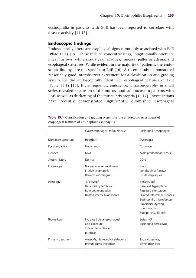
Chapter 15: Eosinophilic Esophagitis 255
eosinophilia in patients with EoE has been reported to correlate with
disease activity [14,15] .
Endoscopic findings Endoscopically, there are esophageal signs commonly associated with EoE
(Plate 15.1) [15] . These include concentric rings, longitudinally oriented,
linear furrows, white exudates or plaques, mucosal pallor or edema, and
esophageal strictures. While evident in the majority of patients, the endo-
scopic findings are not specific to EoE [10] . A recent study demonstrated
reasonably good interobserver agreement for a classification and grading
system for the endoscopically identified, esophageal features of EoE
(Table 15.1 ) [15] . High-frequency, endoscopic ultrasonography in small
series revealed expansion of the mucosa and submucosa in patients with
EoE, as well as thickening of the muscularis propria [16,17] . Investigations
have recently demonstrated significantly diminished esophageal
Table 15.1 Classification and grading system for the endoscopic assessment of
esophageal features of eosinophilic esophagitis.
Gastroesophageal reflux disease Eosinophilic esophagitis
Dominant symptom Heartburn Dysphagia
Food impaction Uncommon Common
Gender M = F Male predominant (75%)
Atopic history Normal 70%
Endoscopy Non-erosive reflux disease
Erosive esophagitis
Barrett ’ s esophagus
Rings
Longitudinal furrows
Exudates/plaques
Histology < 7 eos/hpf
Basal cell hyperplasia
Rete peg elongation
Dilated intercellular spaces
≥15 eos/hpf
Basal cell hyperplasia
Rete peg elongation
Dilated intercellular spaces
Eosinophilic microabscess
Superficial layering
of eosinophils
Subepithelial fibrosis
Biomarkers Increased distal esophageal
acid exposure
? E-cadherin cleaved
products
Eotaxin-3
Eosinophil peroxidase
Primary treatment Antacids, H2 receptor antagonist,
proton pump inhibition
Topical steroids,
elimination diet
c15.indd 255 11/15/2012 2:48:55 AM

256 Part 2: Gastroesophageal Reflux Disease
distensibility in patients with EoE [18] . Reduced distensibility may be an
important determinant of impaired bolus transit and dysphagia in EoE.
Manometric findings There has been considerable investigation into the effect of EoE on
esophageal motility. Esophageal dysmotility has been suggested to account
for the apparent dissociation between symptoms of dysphagia and the
presence of endoscopically evident esophageal strictures. Eosinophil-
derived proteins may affect enteric neurons and esophageal smooth muscle
function. Investigations using high-resolution manometry with pressure
topography demonstrated that the majority of manometric abnormalities
identified in EoE are non-specific and overlap significantly with features
identified in GERD [18] . Weak and failed peristalsis are frequent findings
on high-resolution manometry in both GERD and EoE [19] . Increased
esophageal pressurization patterns were identified in 36% of EoE patients
and only 12% of GERD patients and may reflect reduced wall compliance.
Pediatric investigation using prolonged esophageal manometry also reveals
increased frequency of ineffective peristalsis compared to GERD controls
[20] . In addition to impaired circular muscle function, as measured by
esophageal manometry, Korsapati et al . reported dysfunction in longitudinal
muscle function in patients with EoE [21] . Specifically, EoE patients
demonstrated decreased amplitude and duration of longitudinal muscle
contraction during swallows with dissociation between the timing of
circular muscle and longitudinal muscle contractions [21] . Longitudinal
muscle dysfunction is a potential mechanism for dysphagia in some
patients with EoE. However, it remains unclear as to whether the reduced
longitudinal muscle function is the result of a motility defect or increased
tissue stiffness related to esophageal mural fibrosis.
Diagnosis of eosinophilic esophagitis
The presence of eosinophils within the esophagus is pathological but not
specific for any particular disease state. There are many causes of esopha-
geal eosinophilia, including EoE, GERD, parasitic infection, inflammatory
bowel disease, autoimmune disorders, neoplasia, drug hypersensitivity, and
caustic injury [22] . Currently, there is no single test for the diagnosis of
EoE. Consensus recommendations suggest that EoE is a clinicopathological
diagnosis that incorporates symptoms of esophageal dysfunction together
with markers of eosinophil-predominant inflammation, with 15 eosino-
phils per high-power field (hpf) considered the minimum threshold for
the diagnosis [1] . Characteristic endoscopic signs raise the suspicion of
EoE in the appropriate clinical context, but are not a requirement (see
c15.indd 256 11/15/2012 2:48:55 AM

Chapter 15: Eosinophilic Esophagitis 257
Table 15.1 ) [15] . With the exception of GERD, most other secondary causes
of esophageal eosinophilia can be excluded through a careful clinical
history and endoscopic examination.
Histologically, there are no pathognomonic features of EoE, but several
findings have been strongly associated with the diagnosis. The number of
eosinophils required to consider the diagnosis has been an area of debate.
It is important to note that most studies rely on highest number of eosino-
phils per hpf, and not average number per hpf [23–25] . There is no study
defining the optimal threshold number of eosinophils per hpf for EoE,
although the consensus recommendations suggest a minimum threshold
of 15/hpf. Higher concentrations of eosinophils may increase the diagnostic
specificity for EoE at the expense of sensitivity. Eosinophil micro abscesses
defined by a cluster of more than four eosinophils, superficial layering of
eosinophils, eosinophil degranulation, and subepithelial fibrosis have been
suggested to distinguish EoE from GERD in a few small studies [11,26] .
Basal zone hyperplasia, rete peg elongation, and dilated intercellular
spaces have been associated with EoE, but are also common histological
findings in GERD [10,11,24,26] .
Distinguishing gastroesophageal reflux disease from eosinophilic esophagitis
Perhaps the most challenging aspect of the diagnosis of EoE is the ability
to distinguish it from GERD (Box 15.1 ). Interestingly, esophageal eosino-
philia was characteristically associated with GERD in the 1980s [6,27] .
In these early reports, the presence of esophageal eosinophilia correlated
with increased esophageal acid exposure and was primarily noted to be
in the distal esophagus. In the early 1990s, reports from adult and pedi-
atric centers described a cohort of patients with esophageal eosinophilia
in the absence of GERD [24,28,29] . Interestingly, the adult studies
revealed a phenotype distinct from that typically seen in GERD. Male
predominance and history of atopy were pronounced in comparison to
patients with GERD. Symptom presentations were dominated by dys-
phagia and food impaction, rather than heartburn and regurgitation.
Endoscopic findings were not those of erosive esophagitis, and instead
revealed concentric rings, exudates, and furrows [15] . In addition, the
degree of esophageal eosinophilia was markedly higher compared to that
typically associated with GERD. Most importantly, the patients studied
had either normal pH testing or had symptoms and histopathology
despite acid suppressive therapy.
As clinical recognition and the prevalence of EoE in adults began to
increase, further investigation supported the notion that EoE is distinct
c15.indd 257 11/15/2012 2:48:55 AM
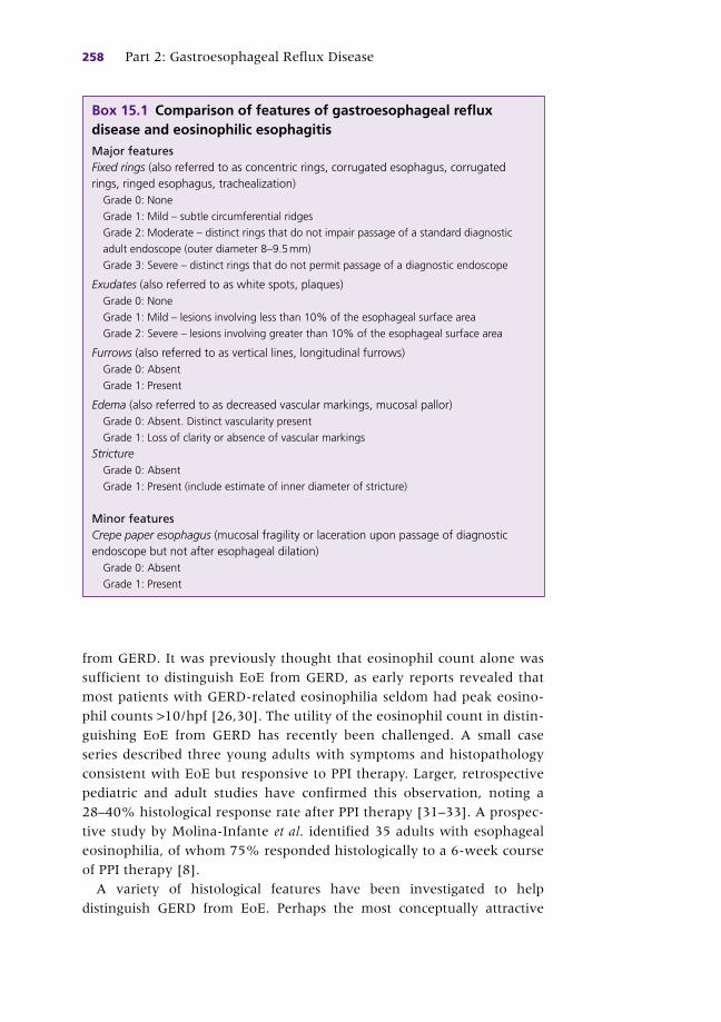
258 Part 2: Gastroesophageal Reflux Disease
from GERD. It was previously thought that eosinophil count alone was
sufficient to distinguish EoE from GERD, as early reports revealed that
most patients with GERD-related eosinophilia seldom had peak eosino-
phil counts >10/hpf [26,30] . The utility of the eosinophil count in distin-
guishing EoE from GERD has recently been challenged. A small case
series described three young adults with symptoms and histopathology
consistent with EoE but responsive to PPI therapy. Larger, retrospective
pediatric and adult studies have confirmed this observation, noting a
28–40% histological response rate after PPI therapy [31–33] . A prospec-
tive study by Molina-Infante et al . identified 35 adults with esophageal
eosinophilia, of whom 75% responded histologically to a 6-week course
of PPI therapy [8] .
A variety of histological features have been investigated to help
distinguish GERD from EoE. Perhaps the most conceptually attractive
Box 15.1 Comparison of features of gastroesophageal reflux disease and eosinophilic esophagitis Major features Fixed rings (also referred to as concentric rings, corrugated esophagus, corrugated rings, ringed esophagus, trachealization)
Grade 0: None
Grade 1: Mild – subtle circumferential ridges
Grade 2: Moderate – distinct rings that do not impair passage of a standard diagnostic
adult endoscope (outer diameter 8–9.5 mm)
Grade 3: Severe – distinct rings that do not permit passage of a diagnostic endoscope
Exudates (also referred to as white spots, plaques) Grade 0: None
Grade 1: Mild – lesions involving less than 10% of the esophageal surface area
Grade 2: Severe – lesions involving greater than 10% of the esophageal surface area
Furrows (also referred to as vertical lines, longitudinal furrows) Grade 0: Absent
Grade 1: Present
Edema (also referred to as decreased vascular markings, mucosal pallor) Grade 0: Absent. Distinct vascularity present
Grade 1: Loss of clarity or absence of vascular markings
Stricture Grade 0: Absent
Grade 1: Present (include estimate of inner diameter of stricture)
Minor features Crepe paper esophagus (mucosal fragility or laceration upon passage of diagnostic endoscope but not after esophageal dilation)
Grade 0: Absent
Grade 1: Present
c15.indd 258 11/15/2012 2:48:55 AM

Chapter 15: Eosinophilic Esophagitis 259
histological feature distinguishing EoE from GERD is the presence of
proximal eosinophilia, the reason being that reflux and reflux-associated
injury typically predominantly affect the distal esophagus. Recent investi-
gations have compared the frequency of proximal esophageal eosinophila
in patients with EoE compared to those with GERD. In one study, 70% of
patients with EoE were noted to have a greater degree of eosinophilia in
the proximal esophagus compared to the distal, whereas this was not seen
in any patients with GERD. Furthermore, 83% of the EoE patients in this
cohort had both proximal and distal eosinophilia compared to 0% in GERD
patients [34] . Other histological features that have shown modest utility in
favoring a diagnosis of EoE over GERD include superficial localization of
eosinophils, eosinophilic microabscesses, and eosinophil degranulation.
Unfortunately, none appears to be sensitive or specific enough to aid in
diagnosis. The prospective trial by Molina-Infante was unable to demonstrate
significant differences in these histological features in distinguishing GERD
from EoE as defined by a response to PPI therapy. As a result, the consensus
guidelines suggest that the diagnosis of EoE should not be established until
acid reflux is excluded with a trial of PPI therapy [1,10] .
In addition to its overlap with GERD, EoE can co-exist with conditions
that are complications of GERD, specifically Barrett ’ s esophagus (BE). The
prevalence of esophageal eosinophilia in patients with BE is not entirely
known, but retrospective data from the Mayo Clinic suggest that the prev-
alence is at least 7%. There was no clear association between dysplastic
BE and the presence of esophageal eosinophilia [35] . The relevance of
esophageal eosinophilia in patients with BE is not known, but it may be a
marker of persistent reflux. There is no evidence to causally link EoE with
the development of BE or esophageal cancer. Given the background
prevalence of GERD and BE in the general population, BE may be an
incidental finding unrelated to EoE.
Complicating our understanding of the interactions between GERD and
EoE is the possibility that GERD may predispose to or even cause EoE [36] .
This concept emerged not only from the observation that there is
considerable overlap between these two conditions, but also from early
studies revealing that treatment for acid reflux may improve the clinical
and histological manifestations of EoE [9] . From a mechanistic standpoint,
it is feasible that acid reflux may predispose to EoE. Though the normal
squamous epithelium is rather impermeable to allergenic peptides, erosive
or non-erosive acid injury has been shown to cause dilated intercellular
spaces. This dilation can allow larger peptides to permeate the esophageal
epithelium, thus exposing the stroma and antigen-presenting cells to
food-borne allergens [37] . Acid exposure induces release of mast cell medi-
ators and may prolong eosinophil viability. Furthermore, acid exposure to
the esophageal epithelium can result in recruitment of eosinophils by
c15.indd 259 11/15/2012 2:48:55 AM
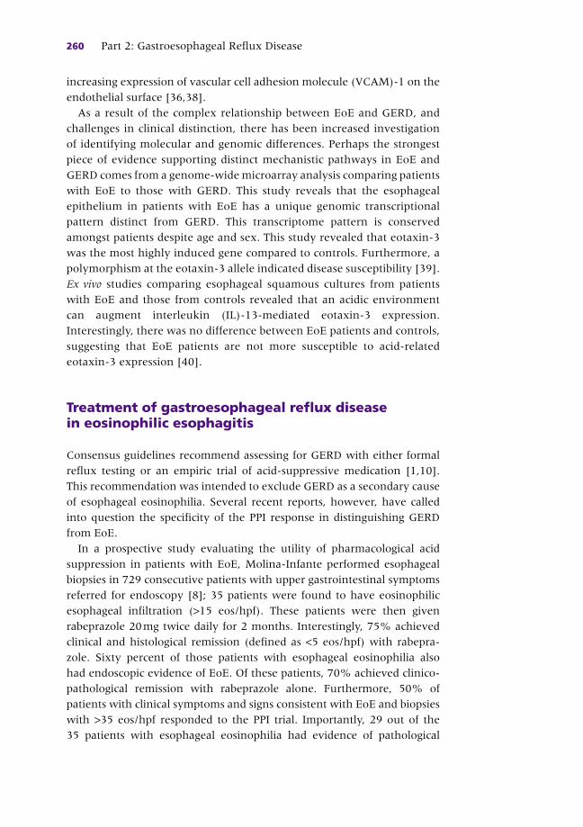
260 Part 2: Gastroesophageal Reflux Disease
increasing expression of vascular cell adhesion molecule (VCAM)-1 on the
endothelial surface [36,38] .
As a result of the complex relationship between EoE and GERD, and
challenges in clinical distinction, there has been increased investigation
of identifying molecular and genomic differences. Perhaps the strongest
piece of evidence supporting distinct mechanistic pathways in EoE and
GERD comes from a genome-wide microarray analysis comparing patients
with EoE to those with GERD. This study reveals that the esophageal
epithelium in patients with EoE has a unique genomic transcriptional
pattern distinct from GERD. This transcriptome pattern is conserved
amongst patients despite age and sex. This study revealed that eotaxin-3
was the most highly induced gene compared to controls. Furthermore, a
polymorphism at the eotaxin-3 allele indicated disease susceptibility [39] .
Ex vivo studies comparing esophageal squamous cultures from patients
with EoE and those from controls revealed that an acidic environment
can augment interleukin (IL)-13-mediated eotaxin-3 expression.
Interestingly, there was no difference between EoE patients and controls,
suggesting that EoE patients are not more susceptible to acid-related
eotaxin-3 expression [40] .
Treatment of gastroesophageal reflux disease in eosinophilic esophagitis
Consensus guidelines recommend assessing for GERD with either formal
reflux testing or an empiric trial of acid-suppressive medication [1,10] .
This recommendation was intended to exclude GERD as a secondary cause
of esophageal eosinophilia. Several recent reports, however, have called
into question the specificity of the PPI response in distinguishing GERD
from EoE.
In a prospective study evaluating the utility of pharmacological acid
suppression in patients with EoE, Molina-Infante performed esophageal
biopsies in 729 consecutive patients with upper gastrointestinal symptoms
referred for endoscopy [8] ; 35 patients were found to have eosinophilic
esophageal infiltration (>15 eos/hpf). These patients were then given
rabeprazole 20 mg twice daily for 2 months. Interestingly, 75% achieved
clinical and histological remission (defined as <5 eos/hpf) with rabepra-
zole. Sixty percent of those patients with esophageal eosinophilia also
had endoscopic evidence of EoE. Of these patients, 70% achieved clinico-
pathological remission with rabeprazole alone. Furthermore, 50% of
patients with clinical symptoms and signs consistent with EoE and biopsies
with >35 eos/hpf responded to the PPI trial. Importantly, 29 out of the
35 patients with esophageal eosinophilia had evidence of pathological
c15.indd 260 11/15/2012 2:48:55 AM
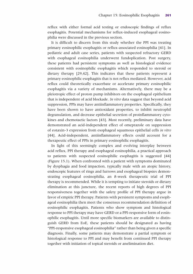
Chapter 15: Eosinophilic Esophagitis 261
reflux with either formal acid testing or endoscopic findings of reflux
esophagitis. Potential mechanisms for reflux-induced esophageal eosino-
philia were discussed in the previous section.
It is difficult to discern from this study whether the PPI was treating
primary eosinophilic esophagitis or reflux-associated eosinophilia [41] . In
pediatric and adult case series, patients with suspected refractory GERD
with esophageal eosinophilia underwent fundoplication. Post surgery,
these patients had persistent symptoms as well as histological evidence
consistent with eosinophilic esophagitis which responded to steroid or
dietary therapy [29,42] . This indicates that these patients represent a
primary eosinophilic esophagitis that is not reflux mediated. However, acid
reflux could theoretically exacerbate or accelerate primary eosinophilic
esophagitis via a variety of mechanisms. Alternatively, there may be a
pleiotropic effect of proton pump inhibitors on the esophageal epithelium
that is independent of acid blockade. In vitro data suggest that beyond acid
suppression, PPIs may have antiinflammatory properties. Specifically, they
have been shown to have antioxidant properties, to inhibit neutrophil
degranulation, and decrease epithelial secretion of proinflammatory cyto-
kines and chemotactic factors [43] . Most recently, preliminary data have
demonstrated an acid-independent effect of omeprazole on suppression
of eotaxin-3 expression from esophageal squamous epithelial cells in vitro
[44] . Acid-independent, antiinflammatory effects could account for a
therapeutic effect of PPIs in primary eosinophilic esophagitis.
In light of this seemingly complex and evolving interplay between
acid reflux, PPI therapy and esophageal eosinophilia, a practical approach
to patients with suspected eosinophilic esophagitis is suggested [44]
(Figure 15.1 ). When confronted with a patient with symptoms dominated
by dysphagia and food impaction, typically male with an atopic history,
endoscopic features of rings and furrows and esophageal biopsies demon-
strating esophageal eosinophilia, an 8-week therapeutic trial of PPI
therapy is recommended. While it is tempting to initiate steroids or dietary
elimination at this juncture, the recent reports of high degrees of PPI
responsiveness together with the safety profile of PPI therapy argue in
favor of empiric PPI therapy. Patients with persistent symptoms and esoph-
ageal eosinophilia then meet the consensus recommendation definition of
eosinophilic esophagitis. Patients who show symptom and histological
response to PPI therapy may have GERD or a PPI-responsive form of eosin-
ophilic esophagitis. Until more specific biomarkers are available to distin-
guish GERD from EoE, these patients should be designated as having
“PPI-responsive esophageal eosinophilia” rather than being given a specific
diagnosis. Finally, some patients may demonstrate a partial symptom or
histological response to PPI and may benefit from continued PPI therapy
together with initiation of topical steroids or anelimination diet.
c15.indd 261 11/15/2012 2:48:55 AM
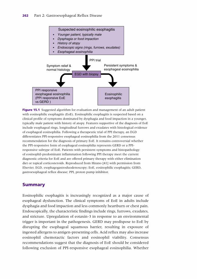
262 Part 2: Gastroesophageal Reflux Disease
Summary
Eosinophilic esophagitis is increasingly recognized as a major cause of
esophageal dysfunction. The clinical symptoms of EoE in adults include
dysphagia and food impaction and less commonly heartburn or chest pain.
Endoscopically, the characteristic findings include rings, furrows, exudates,
and stricture. Upregulation of eotaxin-3 in response to an environmental
trigger is important in the pathogenesis. GERD may predispose to EoE by
disrupting the esophageal squamous barrier, resulting in exposure of
ingested allergens to antigen-presenting cells. Acid reflux may also increase
eosinophil chemotactic factors and eosinophil viability. Consensus
recommendations suggest that the diagnosis of EoE should be considered
following exclusion of PPI-responsive esophageal eosinophilia. Whether
PPI trial
Suspected eosinophilic esophagitis
Dysphagia or food impactionHistory of atopyEndoscopic signs (rings, furrows, exudates)Esophageal eosinophilia
Younger patient, typically male
Symptom relief &normal histology
Persistent symptoms &esophageal eosinophilia
EGD with biopsy
Eosinophilicesophagitis
PPI responsiveesophageal eosinophilia(PPI responsive EoEvs GERD )
Figure 15.1 Suggested algorithm for evaluation and management of an adult patient
with eosinophilic esophagitis (EoE). Eosinophilic esophagitis is suspected based on a
clinical profile of symptoms dominated by dysphagia and food impaction in a younger,
typically male patient with history of atopy. Features supportive of the diagnosis of EoE
include esophageal rings, longitudinal furrows and exudates with histological evidence
of esophageal eosinophilia. Following a therapeutic trial of PPI therapy, an EGD
differentiates PPI-responsive esophageal eosinophilia from the 2011 consensus
recommendation for the diagnosis of primary EoE. It remains controversial whether
the PPI-responsive form of esophageal eosinophilia represents GERD or a PPI-
responsive subtype of EoE. Patients with persistent symptoms and histopathology
of eosinophil-predominant inflammation following PPI therapy meet the current
diagnostic criteria for EoE and are offered primary therapy with either elimination
diet or topical corticosteroids. Reproduced from Hirano [41] with permission from
Elsevier. EGD, esophagogastroduodenoscopy; EoE, eosinophilic esophagitis; GERD,
gastroesophageal reflux disease; PPI, proton pump inhibitor.
c15.indd 262 11/15/2012 2:48:55 AM
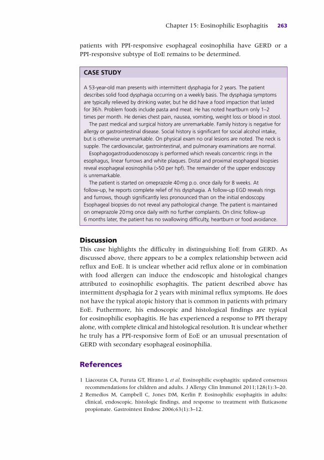
Chapter 15: Eosinophilic Esophagitis 263
patients with PPI-responsive esophageal eosinophilia have GERD or a
PPI-responsive subtype of EoE remains to be determined.
Discussion This case highlights the difficulty in distinguishing EoE from GERD. As
discussed above, there appears to be a complex relationship between acid
reflux and EoE. It is unclear whether acid reflux alone or in combination
with food allergen can induce the endoscopic and histological changes
attributed to eosinophilic esophagitis. The patient described above has
intermittent dysphagia for 2 years with minimal reflux symptoms. He does
not have the typical atopic history that is common in patients with primary
EoE. Futhermore, his endoscopic and histological findings are typical
for eosinophilic esophagitis. He has experienced a response to PPI therapy
alone, with complete clinical and histological resolution. It is unclear whether
he truly has a PPI-responsive form of EoE or an unusual presentation of
GERD with secondary esophageal eosinophilia.
References
1 Liacouras CA , Furuta GT , Hirano I , et al . Eosinophilic esophagitis: updated consensus
recommendations for children and adults . J Allergy Clin Immunol 2011 ; 128 ( 1 ): 3 – 20 .
2 Remedios M , Campbell C , Jones DM , Kerlin P . Eosinophilic esophagitis in adults:
clinical, endoscopic, histologic findings, and response to treatment with fluticasone
propionate . Gastrointest Endosc 2006 ; 63 ( 1 ): 3 – 12 .
CASE STUDY
A 53-year-old man presents with intermittent dysphagia for 2 years. The patient describes solid food dysphagia occurring on a weekly basis. The dysphagia symptoms are typically relieved by drinking water, but he did have a food impaction that lasted for 36 h. Problem foods include pasta and meat. He has noted heartburn only 1–2 times per month. He denies chest pain, nausea, vomiting, weight loss or blood in stool.
The past medical and surgical history are unremarkable. Family history is negative for allergy or gastrointestinal disease. Social history is significant for social alcohol intake, but is otherwise unremarkable. On physical exam no oral lesions are noted. The neck is supple. The cardiovascular, gastrointestinal, and pulmonary examinations are normal.
Esophagogastroduodenoscopy is performed which reveals concentric rings in the esophagus, linear furrows and white plaques. Distal and proximal esophageal biopsies reveal esophageal eosinophilia (>50 per hpf). The remainder of the upper endoscopy is unremarkable.
The patient is started on omeprazole 40 mg p.o. once daily for 8 weeks. At follow-up, he reports complete relief of his dysphagia. A follow-up EGD reveals rings and furrows, though significantly less pronounced than on the initial endoscopy. Esophageal biopsies do not reveal any pathological change. The patient is maintained on omeprazole 20 mg once daily with no further complaints. On clinic follow-up 6 months later, the patient has no swallowing difficulty, heartburn or food avoidance.
c15.indd 263 11/15/2012 2:48:55 AM
264 Part 2: Gastroesophageal Reflux Disease
3 Shah A , Kagalwalla AF , Gonsalves N , Melin-Aldana H , Li BU , Hirano I . Histopatho-
logic variability in children with eosinophilic esophagitis . Am J Gastroenterol
2009 ; 104 ( 3 ): 716 – 21 .
4 Teitelbaum JE , Fox VL , Twarog FJ , et al . Eosinophilic esophagitis in children: immu-
nopathological analysis and response to fluticasone propionate . Gastroenterology
2002 ; 122 ( 5 ): 1216 – 25 .
5 Rodrigo S , Abboud G , Oh D , et al . High intraepithelial eosinophil counts in esopha-
geal squamous epithelium are not specific for eosinophilic esophagitis in adults . Am
J Gastroenterol 2008 ; 103 ( 2 ): 435 – 42 .
6 Winter HS , Madara JL , Stafford RJ , Grand RJ , Quinlan JE , Goldman H . Intraepithelial
eosinophils: a new diagnostic criterion for reflux esophagitis . Gastroenterology
1982 ; 83 ( 4 ): 818 – 23 .
7 Molina-Infante J , Ferrando-Lamana L , Mateos-Rodriguez JM , Perez-Gallardo B ,
Prieto-Bermejo AB . Overlap of reflux and eosinophilic esophagitis in two patients
requiring different therapies: a review of the literature . World J Gastroenterol
7 2008 ; 14 ( 9 ): 1463 – 6 .
8 Molina-Infante J , Ferrando-Lamana L , Ripoll C , et al . Esophageal eosinophilic infil-
tration responds to proton pump inhibition in most adults . Clin Gastroenterol
Hepatol; 9(2):110–17.
9 Ngo P , Furuta GT , Antonioli DA , Fox VL . Eosinophils in the esophagus – peptic or
allergic eosinophilic esophagitis? Case series of three patients with esophageal
eosinophilia . Am J Gastroenterol 2006 ; 101 ( 7 ): 1666 – 70 .
10 Furuta GT , Liacouras CA , Collins MH , et al . Eosinophilic esophagitis in children
and adults: a systematic review and consensus recommendations for diagnosis and
treatment . Gastroenterology 2007 ; 133 ( 4 ): 1342 – 63 .
11 Desai TK , Stecevic V , Chang CH , Goldstein NS , Badizadegan K , Furuta GT . Association
of eosinophilic inflammation with esophageal food impaction in adults . Gastrointest
Endosc 2005 ; 61 ( 7 ): 795 – 801 .
12 Noel RJ , Putnam PE , Rothenberg ME . Eosinophilic esophagitis . N Engl J Med
2004 ; 351 ( 9 ): 940 – 1 .
13 Liacouras CA , Spergel JM , Ruchelli E , et al . Eosinophilic esophagitis: a 10-year
experience in 381 children . Clin Gastroenterol Hepatol 2005 ; 3 ( 12 ): 1198 – 206 .
14 Konikoff MR , Blanchard C , Kirby C , et al . Potential of blood eosinophils, eosinophil-
derived neurotoxin, and eotaxin-3 as biomarkers of eosinophilic esophagitis . Clin
Gastroenterol Hepatol 2006 ; 4 ( 11 ): 1328 – 36 .
15 Hirano I , Moy N , Heckman MG , Thomas CS , Gonsalves N , Achem SR . Endoscopic
Assessment of the Esophageal Features of Eosinophilic Esophagitis: Validation of a
Novel Classification and Grading System . Gut 2012 In press PMID 22619364.
16 Fox VL , Nurko S , Teitelbaum JE , Badizadegan K , Furuta GT . High-resolution EUS
in children with eosinophilic “allergic” esophagitis . Gastrointest Endosc 2003 ;
57 ( 1 ): 30 – 6 .
17 Stevoff C , Rao S , Parsons W , Kahrilas PJ , Hirano I . EUS and histopathologic correlates
in eosinophilic esophagitis . Gastrointest Endosc 2001 ; 54 ( 3 ): 373 – 7 .
18 Kwiatek MA , Hirano I , Kahrilas PJ , Rothe J , Luger D , Pandolfino JE . Mechanical
properties of the esophagus in eosinophilic esophagitis . Gastroenterology
2011 ; 140 ( 1 ): 82 – 90 .
19 Roman S , Hirano I , Kwiatek MA , et al . Manometric features of eosinophilic esophagitis
in esophageal pressure topography . Neurogastroenterol Motil 2011 ; 23 ( 3 ): 208 .
20 Nurko S , Rosen R , Furuta GT . Esophageal dysmotility in children with eosinophilic
esophagitis: a study using prolonged esophageal manometry . Am J Gastroenterol
2009 ; 104 ( 12 ): 3050 – 7 .
c15.indd 264 11/15/2012 2:48:55 AM
Chapter 15: Eosinophilic Esophagitis 265
21 Korsapati H , Babaei A , Bhargava V , Dohil R , Quin A , Mittal RK . Dysfunction of
the longitudinal muscles of the oesophagus in eosinophilic oesophagitis . Gut
2009 ; 58 ( 8 ): 1056 – 62 .
22 Rothenberg ME . Biology and treatment of eosinophilic esophagitis . Gastroenterology
2009 ; 137 ( 4 ): 1238 – 49 .
23 Croese J , Fairley SK , Masson JW , et al . Clinical and endoscopic features of eosino-
philic esophagitis in adults . Gastrointest Endosc 2003 ; 58 ( 4 ): 516 – 22 .
24 Attwood SE , Smyrk TC , Demeester TR , Jones JB . Esophageal eosinophilia with
dysphagia. A distinct clinicopathologic syndrome . Dig Dis Sci 1993 ; 38 ( 1 ): 109 – 16 .
25 Zimmerman SL , Levine MS , Rubesin SE , et al . Idiopathic eosinophilic esophagitis in
adults: the ringed esophagus . Radiology 2005 ; 236 ( 1 ): 159 – 65 .
26 Parfitt JR , Gregor JC , Suskin NG , Jawa HA , Driman DK . Eosinophilic esophagitis
in adults: distinguishing features from gastroesophageal reflux disease: a study of
41 patients . Mod Pathol 2006 ; 19 ( 1 ): 90 – 6 .
27 Brown LF , Goldman H , Antonioli DA . Intraepithelial eosinophils in endoscopic
biopsies of adults with reflux esophagitis . Am J Surg Pathol 1984 ; 8 ( 12 ): 899 – 905 .
28 Straumann A , Spichtin HP , Bernoulli R , Loosli J , Vogtlin J . [ Idiopathic eosinophilic
esophagitis: a frequently overlooked disease with typical clinical aspects and discrete
endoscopic findings ]. Schweiz Med Wochenschr 1994 ; 124 ( 33 ): 1419 – 29 .
29 Kelly KJ , Lazenby AJ , Rowe PC , Yardley JH , Perman JA , Sampson HA . Eosinophilic
esophagitis attributed to gastroesophageal reflux: improvement with an amino acid-
based formula . Gastroenterology 1995 ; 109 ( 5 ): 1503 – 12 .
30 Attwood SE , Fiocca R , Mastracci L , et al . High density eosinophilic infiltration in
gastro-esophageal reflux disease is rare and probably not related to primary eosino-
philic esophagitis: a cohort study from the Lotus Trial . Paper presented at Digestive
Disease Week 2011; Chicago, IL.
31 Dranove JE , Horn DS , Davis MA , Kernek KM , Gupta SK . Predictors of response
to proton pump inhibitor therapy among children with significant esophageal eosin-
ophilia . J Pediatr 2009 ; 154 ( 1 ): 96 – 100 .
32 Sayej WN , Patel R , Baker RD , Tron E , Baker SS . Treatment with high-dose proton
pump inhibitors helps distinguish eosinophilic esophagitis from noneosinophilic
esophagitis . J Pediatr Gastroenterol Nutr 2009 ; 49 ( 4 ): 393 – 9 .
33 Toto E , Garrean C , Hayman A , et al . Differentiation of GERD from EoE (eosinophilic
esophagitis): predictive factors and response to PPI trial . Paper presented at Digestive
Disease Week 2011; Chicago, IL.
34 Lee S , de Boer WB , Naran A , et al . More than just counting eosinophils: proximal
oesophageal involvement and subepithelial sclerosis are major diagnostic criteria
for eosinophilic oesophagitis . J Clin Pathol 2010 ; 63 ( 7 ): 644 – 7 .
35 Ravi K , Katzka DA , Smyrk TC , et al . Prevalence of esophageal eosinophils in patients
with Barrett ’ s esophagus . Am J Gastroenterol 2011 ; 106 ( 5 ): 851 – 7 .
36 Spechler SJ , Genta RM , Souza RF . Thoughts on the complex relationship between
gastroesophageal reflux disease and eosinophilic esophagitis . Am J Gastroenterol
2007 ; 102 ( 6 ): 1301 – 6 .
37 Tobey NA , Hosseini SS , Argote CM , Dobrucali AM , Awayda MS , Orlando RC . Dilated
intercellular spaces and shunt permeability in nonerosive acid-damaged esophageal
epithelium . Am J Gastroenterol 2004 ; 99 ( 1 ): 13 – 22 .
38 Barthel SR , Annis DS , Mosher DF , Johansson MW . Differential engagement of modules
1 and 4 of vascular cell adhesion molecule-1 (CD106) by integrins alpha4beta1 (CD49d/29)
and alphaMbeta2 (CD11b/18) of eosinophils . J Biol Chem 2006 ; 281 ( 43 ): 32175 – 87 .
39 Blanchard C , Wang N , Stringer KF , et al . Eotaxin-3 and a uniquely conserved gene-
expression profile in eosinophilic esophagitis . J Clin Invest 2006 ; 116 ( 2 ): 536 – 47 .
c15.indd 265 11/15/2012 2:48:55 AM

266 Part 2: Gastroesophageal Reflux Disease
40 Blanchard C , Stucke EM , Burwinkel K , et al . Coordinate interaction between IL-13
and epithelial differentiation cluster genes in eosinophilic esophagitis . J Immunol
2010 ; 184 ( 7 ): 4033 – 41 .
41 Hirano I. Eosinophilic esophagitis and gastroesophageal reflux disease: there and
back again . Clin Gastroenterol Hepatol 2011 ; 9 ( 2 ): 99 – 101 .
42 Dellon ES , Farrell TM , Bozymski EM , Shaheen NJ . Diagnosis of eosinophilic
esophagitis after fundoplication for ‘refractory reflux’: implications for preoperative
evaluation . Dis Esophagus 2010 ; 23 ( 3 ): 191 – 5 .
43 Kedika RR , Souza RF , Spechler SJ . Potential anti-inflammatory effects of proton
pump inhibitors: a review and discussion of the clinical implications . Dig Dis Sci
2009 ; 54 ( 11 ): 2312 – 17 .
44 Garrean C , Hirano I . Eosinophilic esophagitis: pathophysiology and optimal
management . Curr Gastroenterol Rep 2009 ; 11 ( 3 ): 175 – 81 .
c15.indd 266 11/15/2012 2:48:56 AM
267
Practical Manual of Gastroesophageal Reflux Disease, First Edition.
Edited by Marcelo F. Vela, Joel E. Richter and John E. Pandolfino.
© 2013 John Wiley & Sons, Ltd. Published 2013 by John Wiley & Sons, Ltd.
CHAPTER 16
Helicobacter pylori and Gastroesophageal Reflux Disease Maria Pina Dore 1 and David Y. Graham 2 1 Instituto di Clinica Medica , University of Sassari, Italy, and Michael E. DeBakey VA
Medical Center and Baylor College of Medicine , Houston, TX, USA
2 Michael E. DeBakey VA Medical Center and Baylor College of Medicine , Houston, TX, USA
Key points • Helicobacter pylori infections and gastroesophageal reflux disease are both common
and thus frequently occur together.
• Helicobacter pylori does not cause gastroesophageal reflux disease directly or
indirectly (i.e. it has no effect on the anti-reflux barrier).
• Helicobacter pylori infection does not protect against gastroesophageal reflux
disease, Barrett ’ s esophagus or adenocarcinoma of the esophagus.
• Hypochlorhydria or achlorhydria associated with atrophic gastritis prevents symptom-
atic reflux and gastroesophageal reflux disease sequelae such as erosive esophagitis,
Barrett ’ s esophagus, and adenocarcinoma of the esophagus. It is also strongly
associated with the risk of gastric cancer.
• Proton pump inhibitor therapy in patients with Helicobacter pylori gastritis can result in
acceleration of corpus gastritis and thus can theoretically increase the risk of gastric cancer.
• Helicobacter pylori eradication does not significantly affect anti-secretory therapy for
gastroesophageal reflux disease.
• Patients considered for long-term proton pump inhibitor therapy should be tested for
Helicobacter pylori and if present, the infection should be eradicated.
Potential pitfalls • Failure to treat an Helicobacter pylori infection because of fear that doing so would
increase the patient ’ s risk for gastroesophageal reflux disease and adenocarcinoma
of the esophagus.
• Failure to test for Helicobacter pylori in a patient in whom long-term proton pump
inhibitor therapy is planned.
• Failure to consider transient proton pump inhibitor-induced acid rebound for
development of gastroesophageal reflux disease-like symptoms after discontinuing
proton pump inhibitors given for a non-gastroesophageal reflux disease indication
such as treatment to eradicate Helicobacter pylori .
c16.indd 267 11/15/2012 2:48:36 AM

268 Part 2: Gastroesophageal Reflux Disease
Introduction
Prior to the identification of Helicobacter pylori , peptic ulcer disease and
gastric cancer were both known to be closely associated with the presence
of gastritis; the discovery of H. pylori provided the link between them.
Because H. pylori is a bacterial infection, it was thought likely that cure of
the infection might result in prevention of gastric cancer and cure of the
heretofore incurable peptic ulcer disease.
Helicobacter pylori is now accepted as etiologically related to gastritis and
the gastritis-associated diseases: duodenal ulcer, gastric ulcer and gastric
cancer, both adenocarcinoma and primary gastric B-cell lymphoma.
H. pylori infections are typically acquired in childhood with clinical disease
occurring typically after a long latent period during which gastric damage
occurs silently. The risk of a clinical outcome of the infection is approxi-
mately 20% [1] . Different outcomes are associated with different patterns
of gastritis: gastric ulcer and gastric cancer with atrophic pangastritis and
low acid secretion (hypochlorhydria or achlorhydria), and duodenal ulcer
with corpus-sparing gastritis (antral predominant gastritis) and high acid
secretion. Because different outcomes are linked to markedly different pat-
terns of gastritis, duodenal ulcer disease and gastric ulcer occur at entirely
different ends of the spectrum of gastritis and it can be said that duodenal
ulcer “protects” against gastric cancer and vice versa. Protection in this
context means “inversely related to” and not any actual involvement (i.e.
no one would fear that curing a duodenal ulcer would increase the risk of
gastric cancer). This possible misuse of the concept of H. pylori infections
“protecting” against other diseases will be discussed below.
Within a decade of the culture of H. pylori , it was discovered how to reli-
ably eradicate H. pylori infections which was also shown to result in the
healing of the underlying gastritis. The hypothesis that cure of H. pylori
infections would also cure peptic ulcer disease was also tested and proven to
be correct. However, following H. pylori eradication, some patients were
reported to have developed gastroesophageal reflux disease (GERD) [2,3] .
At the time, the appearance of GERD following the cure of H. pylori -related
duodenal ulcers was not expected although possibly it should have been.
For example, by the mid-20th century it was well known that there was an
association between the presence of duodenal ulcer and GERD and this find-
ing was repeatedly confirmed in the West [4–13] and in Japan [14] . During
this same time period, it was noted that the incidence of esophageal adeno-
carcinoma appeared to be increasing. The fact that H. pylori caused inflam-
mation, ulcers and cancer in the stomach led to questions about what, if any,
was the role of H. pylori in GERD and in esophageal adenocarcinoma.
This chapter will discuss whether there is a relationship between H. pylori
and the frequency and severity of GERD as well as the sequelae of GERD
c16.indd 268 11/15/2012 2:48:37 AM

Chapter 16: Helicobacter pylori and Gastroesophageal Reflux Disease 269
in different populations, attempt to provide an understanding of the path-
ophysiology involved, and explain the role of H. pylori , if any, in “protection”
against GERD and its sequelae such as Barrett ’ s esophagus and esophageal
adenocarcinoma.
Gastroesophageal reflux disease and Helicobacter pylori in the pre- Helicobacter pylori era
Interest in GERD became prominent in the late 20th century. However,
GERD was not a new problem but rather it became a disease du jour
[15–17] , as dyspepsia had long been recognized as particularly common in
America [18] and heartburn, water-brash and pyrosis were all prevalent
problems [19] . Antacids were widely and commonly used throughout the
19th and most of the 20th centuries. For example, in the 1970s over-
the-counter antacids were used by at least half of US adults, with approx-
imately a quarter of adults taking at least two doses per month and at least
5% of adults using antacids daily [20] . The introduction of H2 receptor
antagonists (H2RAs) in the mid-1970s produced the first simple and effec-
tive relief of ulcer pain and reliable ulcer healing and it was widely believed
that ulcers were finally cured. Clinicians and third-party payers were,
however, surprised to find that many patients with duodenal ulcer disease
continued to use H2RAs despite healing of their ulcers. Despite the use of
fiberoptic endoscopy to identify ulcers and confirm healing, GERD
continued to be largely ignored. Evidence that GERD was given short shrift
in the major gastroenterology textbooks can be easily found; for example,
the major textbooks of gastrointestinal disease in the 1970s in the US did
not recognize that GERD was a common manifestation of Zollinger–Ellison
syndrome.
The initial focus on GERD and how to study it was in part related to an
interest manifested by pharmaceutical companies who saw GERD as a pos-
sible way to expand their markets for proton pump inhibitors (PPIs) and to
differentiate PPIs from H2RAs which dominated the market at that time.
As noted above, over-the-counter antacid use among apparently other-
wise healthy individuals was extremely common throughout this period
but it was unclear why. In the early 1980s we studied why apparently
healthy individuals were frequent users of antacids. We found that the pri-
mary reason was GERD [20] . As interest in GERD increased, the gastroen-
terology community found that the lessons learned in the study of peptic
ulcer disease were not directly transferable to GERD. For example, as
noted by our forefathers, peptic ulcer was an episodic condition whereas
GERD was a chronic disease that required continuous therapy and that
therapy was in large part symptomatic and not curative.
c16.indd 269 11/15/2012 2:48:37 AM
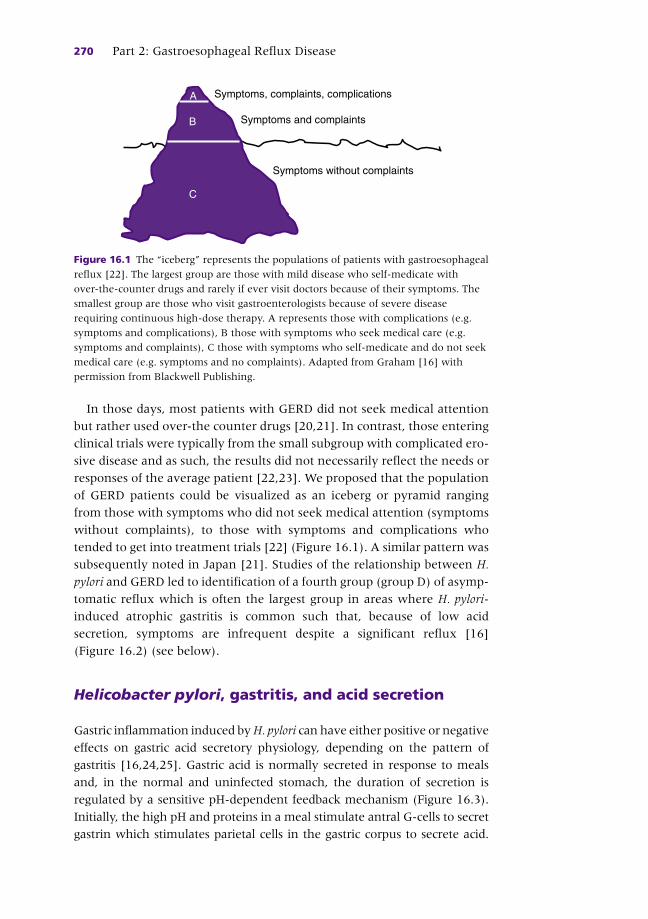
270 Part 2: Gastroesophageal Reflux Disease
In those days, most patients with GERD did not seek medical attention
but rather used over-the counter drugs [20,21] . In contrast, those entering
clinical trials were typically from the small subgroup with complicated ero-
sive disease and as such, the results did not necessarily reflect the needs or
responses of the average patient [22,23] . We proposed that the population
of GERD patients could be visualized as an iceberg or pyramid ranging
from those with symptoms who did not seek medical attention (symptoms
without complaints), to those with symptoms and complications who
tended to get into treatment trials [22] (Figure 16.1 ). A similar pattern was
subsequently noted in Japan [21] . Studies of the relationship between H.
pylori and GERD led to identification of a fourth group (group D) of asymp-
tomatic reflux which is often the largest group in areas where H. pylori -
induced atrophic gastritis is common such that, because of low acid
secretion, symptoms are infrequent despite a significant reflux [16]
(Figure 16.2 ) (see below).
Helicobacter pylori , gastritis, and acid secretion
Gastric inflammation induced by H. pylori can have either positive or negative
effects on gastric acid secretory physiology, depending on the pattern of
gastritis [16,24,25] . Gastric acid is normally secreted in response to meals
and, in the normal and uninfected stomach, the duration of secretion is
regulated by a sensitive pH-dependent feedback mechanism (Figure 16.3 ).
Initially, the high pH and proteins in a meal stimulate antral G-cells to secret
gastrin which stimulates parietal cells in the gastric corpus to secrete acid.
A
B
C
Symptoms, complaints, complications
Symptoms and complaints
Symptoms without complaints
Figure 16.1 The “iceberg” represents the populations of patients with gastroesophageal
reflux [22] . The largest group are those with mild disease who self-medicate with
over-the-counter drugs and rarely if ever visit doctors because of their symptoms. The
smallest group are those who visit gastroenterologists because of severe disease
requiring continuous high-dose therapy. A represents those with complications (e.g.
symptoms and complications), B those with symptoms who seek medical care (e.g.
symptoms and complaints), C those with symptoms who self-medicate and do not seek
medical care (e.g. symptoms and no complaints). Adapted from Graham [16] with
permission from Blackwell Publishing.
c16.indd 270 11/15/2012 2:48:37 AM

Chapter 16: Helicobacter pylori and Gastroesophageal Reflux Disease 271
When the buffering capacity of the meal is exhausted and the intragastric pH
falls below pH 3, antral D-cells secrete somatostatin which downregulates
gastrin secretion and turns off the brisk acid secretion. H. pylori infection
disrupts this regulatory mechanism, resulting in both an exaggerated gastrin
response to meals and dysregulation of the normal pH-sensitive
downregulatory mechanism. Together, this results in an abnormal
prolongation of meal-stimulated acid secretion [26] (see Figure 16.3 ).
Although everyone with an H. pylori infection has these abnormalities in the
regulation of acid secretion, the end result differs depending on the pattern
and severity of gastritis (i.e. antral, corpus or both).
At the beginning of the 20th century, it was recognized that gastric
cancer was strongly associated with reduced acid secretion [27] . By 1950,
it was recognized that corpus gastritis affected acid secretion and, depending
on the degree of destruction of the parietal cell mass, the results could vary
from a modest decrease in acid secretion to achlorhydria [27] . Because the
degree of reduction in acid secretion was greater than the reduction in
parietal cell mass, it was hypothesized that there was an inflammation-
related mediator that actually caused the reduction in acid secretion [28] .
The discovery of H. pylori and studies of acid secretion before and
immediately after treatment confirmed that in patients with corpus-sparing
gastritis (antral predominant) and duodenal ulcer, H. pylori eradication led
to a return to the normal mechanisms that controlled acid secretion
(elimination of the high prolonged meal-stimulated acid response), but
little or no change in maximum secretion (i.e. control of acid secretion was
normalized but the parietal cell mass was unchanged).
Gastric inflammation induced by H. pylori can have either positive or
negative effects on gastric acid secretory physiology, depending on the
Symptoms, complaints, complications
Symptoms and complaints
Symptoms without complaints
Reflux with no disease
AB
C
D
Figure 16.2 This “iceberg” represents the populations of patients with gastroesopha-
geal disease in countries where chronic atrophic gastritis and gastric cancer are
common and GERD is rare. A new group (D) is added to depict those with largely
non-acid reflux but without symptoms or complaints. In such areas the H. pylori
gastritis-associated reductions in acid secretion are such that patients have neither
symptoms nor endoscopic changes despite the presence of gastroesophageal reflux.
Reproduced from Graham [16] with permission from Blackwell Publishing.
c16.indd 271 11/15/2012 2:48:37 AM

272 Part 2: Gastroesophageal Reflux Disease
pattern of gastritis [16,24–26] (i.e. the reduction in mucosal acid inhibitory
mediators such as interleukin (IL)-1 β can result in a marked increase in
maximal acid secretion among those with corpus gastritis [20,29,30]
whereas in antral-predominant gastritis, the overall effect is a reduction in
total acid secretion due to return of the normal acid-related downregula-
tion of acid secretion. Thus, fundamentally, the overall result of H. pylori
infections correlates with the pattern and severity of gastritis (i.e. antral,
corpus or both) [27] .
The predominant pattern of H. pylori gastritis in any population or
patient predicts potential outcomes in terms of H. pylori infection (i.e.
antral-predominant gastritis leading to duodenal ulcer, and atrophic
pangastritis leading to gastric ulcers and/or gastric cancer) [16] . Thus, in
countries where gastric cancer is common, the most common pattern of
gastritis in adults is pangastritis, and hypochlorhydria and achlorhydria are
Gastrin
Histamine
Acetylcholine
VAGUS
G
ECL
D D
Somatostatin
Parietal cell
(– Feedback)
(–Feedback)
Antralgastritis
HCl
Proton pump
Figure 16.3 Gastric acid secretion is activated throughout the binding of gastrin
(secreted by the G-cells), acetylcholine (by vagus), and histamine (by enterochromaffin-
like (ECL) cells), and prostaglandins to the specific receptors on the basolateral surface
of parietal cells. The ultimate factor in acid secretion is the stimulation of the proton
pump (H+, K + −ATPase) to secrete hydrogen ions into the gastric lumen in exchange
for potassium ions. After sufficient acid secretion has occurred, a feedback system
terminates gastric acid secretion. A decrease of intragastric pH below 3 stimulates
somatostatin release from antral D-cells which downregulates gastrin secretion,
turning off acid secretion. The presence of an H. pylori infection disrupts
this mechanism, leading to an exaggerated gastrin response to meals and a delay
in the inhibitory effects of somatostatin.
c16.indd 272 11/15/2012 2:48:38 AM

Chapter 16: Helicobacter pylori and Gastroesophageal Reflux Disease 273
common. As such, the impaired ability to secrete acid means that irrespec-
tive of the status of the individual ’ s gastroesophageal reflux barrier, the
average individual will have a low esophageal acid load and erosive reflux
will be uncommon [16] (i.e. in countries where atrophic gastritis and
gastric cancer are common, such as Japan or Korea, GERD has not been a
significant clinical problem) [31] . However, as the pattern of gastritis
changes and becomes more like the current pattern in Western countries,
the incidence of gastric cancer and gastric ulcer also declines, acid secretion
rises and those with abnormal anti-reflux barriers are more likely to expe-
rience symptomatic GERD.
Helicobacter pylori infection and its relationship to gastroesophageal reflux
Gastroesophageal reflux refers to the reflux of fluid from the stomach into
the esophagus. Occasional reflux is a normal occurrence and is typically
asymptomatic and without clinical consequences. Reflux can also cause dis-
ease (i.e. symptoms and/or mucosal damage, as in gastroesophageal reflux
disease). The lumen of the stomach is separated from that of the esophagus
by an anti-reflux barrier that consists of both smooth muscle sphincter (the
physiological barrier of the lower esophageal sphincter or LES) and a group
of anatomical structures that together constitute a flap valve [32–34] . In
addition to the flap valve, the muscular LES normally maintains a pressure
gradient between the esophagus and stomach. The three dominant patho-
physiological mechanisms causing gastroesophageal junction incompetence
are transient lower esophageal sphincter relaxations (TLESRs), a hypoten-
sive LES, and anatomical disruption of the gastroesophageal junction
typically associated with the presence of hiatal hernia.
Helicobacter pylori infection per se is not thought to have a deleterious
effect on the anti-reflux barrier as it has no effect on the LES, the fre-
quency of TLESRs [35–38] , esophageal peristaltic function or acid clearance
[35,38] . Although H. pylori can occasionally be found on the esophageal
mucosa, H. pylori is only trophic for gastric mucosa and its presence in the
esophagus is likely to be transient and related to reflux from the stomach
(i.e. an epiphenomenon) [39,40] .
As noted above, indigestion and heartburn have been common symp-
toms throughout the recorded history of mankind. However, the role of
the esophagus as a cause of these symptoms was overlooked until recently
[15] . While it is commonly believed that there has been a great increase in
incidence of GERD in the last 30 years, the data to support this hypothesis
are slim at best and we believe that the data better support increased
recognition as the most important factor. One only has to think back a few
c16.indd 273 11/15/2012 2:48:38 AM
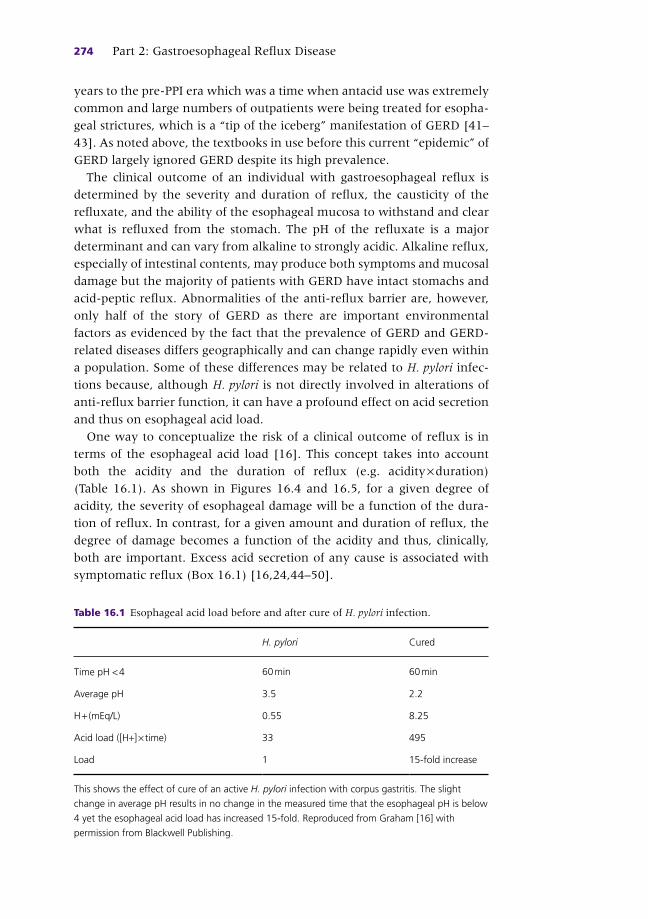
274 Part 2: Gastroesophageal Reflux Disease
years to the pre-PPI era which was a time when antacid use was extremely
common and large numbers of outpatients were being treated for esopha-
geal strictures, which is a “tip of the iceberg” manifestation of GERD [41–
43] . As noted above, the textbooks in use before this current “epidemic” of
GERD largely ignored GERD despite its high prevalence.
The clinical outcome of an individual with gastroesophageal reflux is
determined by the severity and duration of reflux, the causticity of the
refluxate, and the ability of the esophageal mucosa to withstand and clear
what is refluxed from the stomach. The pH of the refluxate is a major
determinant and can vary from alkaline to strongly acidic. Alkaline reflux,
especially of intestinal contents, may produce both symptoms and mucosal
damage but the majority of patients with GERD have intact stomachs and
acid-peptic reflux. Abnormalities of the anti-reflux barrier are, however,
only half of the story of GERD as there are important environmental
factors as evidenced by the fact that the prevalence of GERD and GERD-
related diseases differs geographically and can change rapidly even within
a population. Some of these differences may be related to H. pylori infec-
tions because, although H. pylori is not directly involved in alterations of
anti-reflux barrier function, it can have a profound effect on acid secretion
and thus on esophageal acid load.
One way to conceptualize the risk of a clinical outcome of reflux is in
terms of the esophageal acid load [16] . This concept takes into account
both the acidity and the duration of reflux (e.g. acidity × duration)
(Table 16.1 ). As shown in Figures 16.4 and 16.5 , for a given degree of
acidity, the severity of esophageal damage will be a function of the dura-
tion of reflux. In contrast, for a given amount and duration of reflux, the
degree of damage becomes a function of the acidity and thus, clinically,
both are important. Excess acid secretion of any cause is associated with
symptomatic reflux (Box 16.1 ) [16,24,44–50] .
Table 16.1 Esophageal acid load before and after cure of H. pylori infection.
H. pylori Cured
Time pH < 4 60 min 60 min
Average pH 3.5 2.2
H + (mEq/L) 0.55 8.25
Acid load ([H+] × time) 33 495
Load 1 15-fold increase
This shows the effect of cure of an active H. pylori infection with corpus gastritis. The slight
change in average pH results in no change in the measured time that the esophageal pH is below
4 yet the esophageal acid load has increased 15-fold. Reproduced from Graham [16] with
permission from Blackwell Publishing.
c16.indd 274 11/15/2012 2:48:38 AM
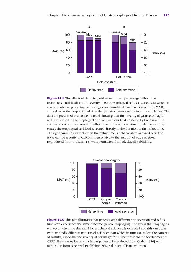
Chapter 16: Helicobacter pylori and Gastroesophageal Reflux Disease 275
MAO (%)
Acid
Reflux (%)
0
20
40
60
80
100
100
80
60
40
20
0MildModSevere
Reflux time
Mild
ModSevere
Hold constant
BA
Reflux time Acid secretion
Figure 16.4 The effects of changing acid secretion and percentage reflux time
(esophageal acid load) on the severity of gastroesophageal reflux disease. Acid secretion
is represented as percentage of pentagastrin-stimulated maximal acid output (MAO)
and reflux as the proportion of time that gastric contents reflux into the esophagus. The
data are presented as a concept model showing that the severity of gastroesophageal
reflux is related to the esophageal acid load and can be dominated by the amount of
acid secretion on the amount of reflux time. If the acid secretion is held constant ( left
panel ), the esophageal acid load is related directly to the duration of the reflux time.
The right panel shows that when the reflux time is held constant and acid secretion
is varied, the severity of GERD is then related to the amount of acid secretion.
Reproduced from Graham [16] with permission from Blackwell Publishing.
Reflux time Acid secretion
Severe esophagitis
ZES0
20
40
60
80
100
100
80
60
40
20
0
Corpusnormal
Corpusinflamed
Reflux (%)MAO (%)
Figure 16.5 This plot illustrates that patients with different acid secretion and reflux
times can experience the same outcome (severe esophagus). The key is that esophagitis
will occur when the threshold for esophageal acid load is exceeded and this can occur
with markedly different patterns of acid secretion which in turn can reflect the patterns
of gastritis, especially the severity of corpus gastritis. The threshold for development of
GERD likely varies for any particular patients. Reproduced from Graham [16] with
permission from Blackwell Publishing. ZES, Zollinger–Ellison syndrome.
c16.indd 275 11/15/2012 2:48:39 AM

276 Part 2: Gastroesophageal Reflux Disease
Helicobacter pylori eradication and gastroesophageal reflux disease
As noted above, it has long been recognized that there is a relationship
between duodenal ulcer disease and GERD and in retrospect, this was
likely responsible for the desire of many patients with duodenal ulcer to
continue H2RA therapy even after their ulcer was healed. The fact that
some patients complained of GERD after H. pylori eradication initially led
to confusion about whether H. pylori eradication might actually cause
GERD [2,3] . The effect of H. pylori eradication on GERD can vary from new
onset of symptoms, no change in existing symptoms, to amelioration of
existing symptoms [16,17,51] . The different outcomes are related to
posteradication changes in acid secretion. For example, it is possible for
H. pylori eradication to make preexisting asymptomatic reflux more acidic
and thus turn asymptomatic reflux into clinical GERD or to increase the
severity of mild GERD [16] . On the other hand, it could be associated with
a reduction in the severity of GERD by reducing the duration of the meal-
stimulated acid response.
Most often, eradication has no consistent effect on GERD symptoms or
management. The actual outcome depends in part on the patient ’ s ability
to secrete acid (e.g. pattern of gastritis) and the status of the anti-reflux
barrier mechanism (Figure 16.6 ) [16] . One also must consider the effects
of acid rebound associated with stopping PPI therapy, which is commonly
used as a part of the eradication therapy [48,49] . Considering that anti- H.
pylori therapy is generally of short duration (i.e. 1–2 weeks), rebound is
generally self-limited but in other patients who have been on longer term
anti-secretory therapy for maintenance of ulcer remission, it may last
considerably longer.
A number of double-blind controlled trials have looked into whether
H. pylori eradication results in new-onset GERD, worsening or improved
GERD, as well as whether H. pylori eradication makes the management of
Box 16.1 Causes of acid hypersecretion often associated with symptomatic GERD • Zollinger–Ellison syndrome
• Antral gastritis H. pylori positive
• Mastocytosis
• Retained antrum following partial gastrectomy
• Duodenal ulcer disease
• Idiopathic gastric acid hypersecretion
• Rebound hypersecretion related to discontinuation of PPIs
c16.indd 276 11/15/2012 2:48:39 AM
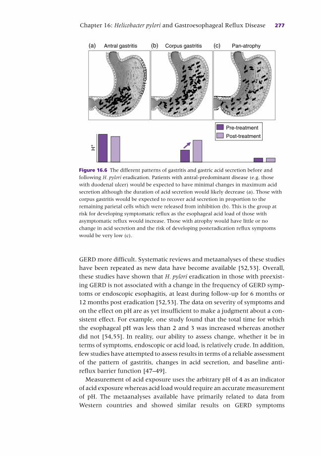
Chapter 16: Helicobacter pylori and Gastroesophageal Reflux Disease 277
GERD more difficult. Systematic reviews and metaanalyses of these studies
have been repeated as new data have become available [52,53] . Overall,
these studies have shown that H. pylori eradication in those with preexist-
ing GERD is not associated with a change in the frequency of GERD symp-
toms or endoscopic esophagitis, at least during follow-up for 6 months or
12 months post eradication [52,53] . The data on severity of symptoms and
on the effect on pH are as yet insufficient to make a judgment about a con-
sistent effect. For example, one study found that the total time for which
the esophageal pH was less than 2 and 3 was increased whereas another
did not [54,55] . In reality, our ability to assess change, whether it be in
terms of symptoms, endoscopic or acid load, is relatively crude. In addition,
few studies have attempted to assess results in terms of a reliable assessment
of the pattern of gastritis, changes in acid secretion, and baseline anti-
reflux barrier function [47–49] .
Measurement of acid exposure uses the arbitrary pH of 4 as an indicator
of acid exposure whereas acid load would require an accurate measurement
of pH. The metaanalyses available have primarily related to data from
Western countries and showed similar results on GERD symptoms
Antral gastritis Corpus gastritis Pan-atrophy
Pre-treatment
Post-treatment
H+
(a) (b) (c)
Figure 16.6 The different patterns of gastritis and gastric acid secretion before and
following H. pylori eradication. Patients with antral-predominant disease (e.g. those
with duodenal ulcer) would be expected to have minimal changes in maximum acid
secretion although the duration of acid secretion would likely decrease (a). Those with
corpus gastritis would be expected to recover acid secretion in proportion to the
remaining parietal cells which were released from inhibition (b). This is the group at
risk for developing symptomatic reflux as the esophageal acid load of those with
asymptomatic reflux would increase. Those with atrophy would have little or no
change in acid secretion and the risk of developing posteradication reflux symptoms
would be very low (c).
c16.indd 277 11/15/2012 2:48:39 AM

278 Part 2: Gastroesophageal Reflux Disease
following H. pylori eradication in the US and Europe. For example, a post
hoc analysis of eight double-blind prospective trials of H. pylori eradication
for duodenal ulcer disease failed to confirm the hypothesis that eradication
of H. pylori resulted in the development of erosive esophagitis or new
symptomatic GERD [56] . Similarly, an analysis of 27 observational studies
found no evidence that H. pylori eradication in duodenal ulcer disease
either provoked reflux esophagitis or worsened heartburn [52] . In con-
trast, in Japan, on average those with preexisting GERD most typically
experienced improvement of GERD symptoms following H. pylori eradica-
tion [14] . In Japan, when reflux developed post eradication, it was likely
to be mild and transient and associated with higher Body Mass Index
(BMI) and younger age [57] . One study reported that in duodenal ulcer
patients, absence of hiatal hernia and lower BMI were associated with
improvement of GERD after H. pylori eradication [57] .
In areas of Asia (e.g. Japan and Korea) where atrophic gastritis is
common and erosive GERD is rare, H. pylori eradication in a patient with
corpus gastritis may result in an increase in gastric acidity, making those
with abnormal anti-reflux barriers and asymptomatic reflux susceptible to
development of GERD symptoms [14,16,17,51] (see Figure 16.6 ). Such a
finding would be the result of the increase in esophageal acid load and
turning asymptomatic reflux into symptomatic GERD [14,16,17,51] .
Clinically, there is no group in which the trade-offs that occur following
H. pylori eradication do not strongly favor eradication. The management of
those with preexisting GERD is typically unchanged. A few with corpus
gastritis will develop reflux symptoms but these are generally mild, easily
controlled and often transient and the natural progression of their cor-
pus gastritis to more severe atrophic gastritis is halted, thus reducing,
if not eliminating, their risk of developing gastric cancer [27,58–60] .
Parenthetically, those with atrophic gastritis and low acid secretion are
at high risk for gastric cancer with annual risks of more than 500–1000/
100,000 per year. In countries where atrophic gastritis and gastric cancer
are infrequent and duodenal ulcer is common among those with H. pylori
infection, the overall effect of H. pylori eradication would be expected to be
little change or a decrease in acid secretion and either no change or a
reduction in symptoms (see Figure 16.6 ). New GERD would not be
expected except in those with corpus gastritis [38] or as a con sequence of
post-PPI acid rebound.
Barrett ’ s esophagus and adenocarcinoma
Although the incidence of adenocarcinoma of the esophagus has increased, it
remains a rare problem (i.e. it changed from a very rare to a rare disease) [61] .
c16.indd 278 11/15/2012 2:48:41 AM

Chapter 16: Helicobacter pylori and Gastroesophageal Reflux Disease 279
However, it is important because it is associated with Barrett ’ s esophagus,
which is a high-risk, easily recognizable precursor lesion. Barrett ’ s esoph-
agus allows for screening and surveillance programs for detection of early
cancer and for cancer prevention programs [62] .
Barrett ’ s esophagus is not a new disease; even in 1961, authors were
able to bring together series of 200 patients with Barrett ’ s esophagus [63] .
It was recognized that:
• Barrett ’ s esophagus and esophageal adenocarcinoma were most common
among white men (a group that in the US first experienced a decline in
H. pylori prevalence) [61]
• Barrett ’ s esophagus was associated with GERD and higher rates of acid
secretion [64]
• absence of H. pylori infection allowed the stomach to express its full
potential for acid secretion (i.e. higher acid loads among those that
refluxed) [65] .
Together, these data suggested that the rise in Barrett ’ s might be, in part,
related to the decline in the prevalence of H. pylori infection. This inverse
association was, as noted earlier, a form of “protection” but this protection
was often construed as not simply an association but rather a risk that
might be controlled. This hypothesis resulted in a series of dire consequence
hypotheses related to H. pylori eradication (discussed in references 66 and
67). The inverse relationship between H. pylori and Barrett ’ s esophagus
and adenocarcinoma of the esophagus was extended to be most important
among those with infecting H. pylori that expressed the cytotoxin-
associated antigen CagA, a putative virulence factor of H. pylori associated
with more severe inflammation, ulcer disease, and gastric cancer [65] .
Authors describing the importance of CagA in protection have ignored the
fact that CagA-positive H. pylori infections are strongly associated with
duodenal ulcer which, as noted above, is strongly associated with an
increased risk of GERD. The low frequency of CagA-positive H. pylori
among patients with Barrett ’ s esophagus is relative, and not absolute,
and as previously discussed, the “protective” feature is the presence of
atrophic gastritis. This low prevalence of CagA positivity is also present
in individuals from countries in which Barrett ’ s is rare, most patients
have CagA-positive infections, and atrophic gastritis and gastric cancer are
common [68] .
Overall, these data are consistent with the hypothesis that the inverse
relationship (inappropriately called protection) is related to the fact that
H. pylori -induced corpus gastritis can act as a biological anti-secretory agent
and reduce or prevent GERD and it sequelae [69] . CagA-containing
H. pylori does not protect against anything; rather, it is atrophic gastritis
that “protects” against GERD and Barrett ’ s esophagus [67,70] . Unfortu-
nately, atrophic gastritis is recognized as the precursor lesion for gastric
c16.indd 279 11/15/2012 2:48:41 AM

280 Part 2: Gastroesophageal Reflux Disease
cancer, the fourth most common cancer worldwide. As noted previously,
adenocarcinoma of the esophagus remains a rare disease with a prevalence
similar to that of small bowel cancer. Any “protection” against Barrett ’ s
esophagus afforded by atrophic gastritis comes with a many hundred-fold
increase in risk for gastric cancer and is thus a rather poor trade-off.
The change in the pattern of gastritis in the 20th century (i.e. a fall in the
prevalence of atrophic gastritis) in the US resulted in a marked decline in
the incidence of gastric cancer and a concomitant increase in the incidence
of duodenal ulcer, each of which “protects” against the other. The pro-
grams to eradicate H. pylori are based on the fact that H. pylori is an impor-
tant human pathogen responsible for much morbidity and mortality.
Among chronic infectious diseases, H. pylori infection has a higher risk of a
serious clinical outcome than either latent syphilis or tuberculosis which,
if one looked closely, would both probably “protect” against some other
diseases [71] . As noted previously, at least 20% of those with an H. pylori
infection will experience a symptomatic clinical illness (e.g. typically peptic
ulcer or gastric cancer) and 25% of those with peptic ulcers will experience
a life-threatening complication such as a major gastrointestinal bleed, per-
foration or obstruction [1] . Unfortunately, the dire consequences that have
been described in relation to H. pylori eradication have led some to forego
H. pylori eradication and to suffer even more the dire consequences of that
decision.
In epidemiology, when one variable is associated with a reduction of
another (e.g. a disease), it is said to be “protective”. For example, popula-
tions with limited sanitation, high rates of malnutrition, and early mortality
have a low incidence of age-related diseases such as diabetes, stroke, and
cancer. One could conclude that from an epidemiological standpoint, poor
sanitation “protects” against those diseases. H. pylori can no more be cred-
ited with a clinically important protection against esophageal cancer than
poor sanitation can be credited with protecting against diseases associated
with aging. H. pylori is a proven cause of gastric cancer; one does not keep
a King cobra in one ’ s house to protect against mice.
Helicobacter pylori , proton pump inhibitors and atrophic gastritis
Many, if not most, patients with symptomatic GERD who visit physicians
will receive anti-secretory drug therapy, typically a PPI. Anti-secretory
drug therapy changes the intragastric milieu and, as a consequence,
H. pylori , which is normally restricted to the surface of the gastric corpus of
a highly acidic stomach, can infect deeper within the mucosa [72,73] . This
change in localization is associated with an increase in the severity and
c16.indd 280 11/15/2012 2:48:41 AM

Chapter 16: Helicobacter pylori and Gastroesophageal Reflux Disease 281
depth of corpus gastritis which includes the proliferative zone [72] . In
clinical trials it was noticed that PPI use could result in the aggravation of
H. pylori -induced corpus gastritis [72,74,75] . The natural history of H. pylori
gastritis is for the inflamed area to advance from the antrum into the
corpus, resulting in a reduction in acid secretion and eventually loss of
parietal cells and development of atrophy [76] and, as noted above, atro-
phic pangastritis is the precursor lesion for development of gastric cancer.
The fact that PPI therapy was associated with the acceleration of corpus
gastritis development led to the suggestion that it may also increase the
risk of gastric cancer. Follow-up data of up to 10 years subsequently
showed that the annual incidence of gastric corpus mucosal atrophy was
higher in H. pylori -positive patients receiving PPI therapy than those not
receiving it (4.7% versus 0.7%) [75] . Recent long-term follow-up studies
from Europe also showed that the annual incidence of gastric corpus
mucosal atrophy among H. pylori -infected PPI users increased compared
with H. pylori -uninfected patients [77] .
Yang et al . studied an Asian population in which atrophic gastritis and
gastric cancer were both increased compared with European populations
[78] . They compared cohorts with typical reflux symptoms after H. pylori
eradication therapy ( n = 105) to H. pylori -positive patients without eradica-
tion ( n = 105) and H. pylori -negative GERD controls ( n = 115). Participants
were randomized to receive esomeprazole 40 mg daily until sustained
symptomatic response and were then switched to on-demand therapy.
Endoscopy with antral and corpus biopsies was done initially and at the
end of the first and second years. Important changes in gastric inflamma-
tion features among the study groups were observed in the first year of
follow-up with a persistently high score in the H. pylori -positive non-
eradicated group compared to the H. pylori -positive eradicated group
( P <0.00 l). Extension of atrophy upward to the corpus was only seen in
the two H. pylori -positive groups and more importantly in the second year
of follow-up, the prevalence rates of atrophy and intestinal metaplasia
were lower in the H. pylori -positive eradicated group than in the non-
eradicated controls (i.e. atrophy 72.3% versus 56.6% and intestinal meta-
plasia 36.1% versus 21.7 %; P <0.05 for both).
Controlled trials have demonstrated that H. pylori eradication in patients
with reflux esophagitis decreases inflammation and reverses corpus
gastritis despite long-term PPI therapy [79] . As noted above, it has clearly
been shown that H. pylori eradication does not make GERD therapy more
complicated or difficult, leading European consensus guidelines to recom-
mend an H. pylori test and treat strategy for patients requiring long-term
acid suppression therapy [80] . We agree that a test and treat strategy
should be employed for all patients for whom long-term anti-secretory,
especially PPI, therapy is indicated.
c16.indd 281 11/15/2012 2:48:41 AM
282 Part 2: Gastroesophageal Reflux Disease
References
1 Axon A , Forman D . Helicobacter gastroduodenitis: a serious infectious disease . BMJ
1997 ; 314 : 1430 – 1 .
2 Schutze K , Hentschel E , Dragosics B , Hirschl AM . Helicobacter pylori reinfection with
identical organisms: transmission by the patients’ spouses . Gut 1995 ; 36 : 831 – 3 .
3 Labenz J , Blum AL , Bayerdorffer E , Meining A , Stolte M , Borsch G . Curing Helicobacter
pylori infection in patients with duodenal ulcer may provoke reflux esophagitis .
Gastroenterology 1997 ; 112 : 1442 – 7 .
4 Winkelstein A , Wolf BS , Som ML , Marshak RH . Peptic esophagitis with duodenal or
gastric ulcer . JAMA 1954 ; 154 : 885 – 9 .
5 Winkelstein A . Peptic esophagitis with duodenal ulcer . Am J Surg 1957 ; 93 : 234 – 7 .
6 Cruze K , Byron FX , Hill JT . The association of peptic ulcers and asymptomatic hiatal
hernia . Surgery 1959 ; 46 : 664 – 8 .
7 Moraes-Filho JP , Zaterka S , Pinotti HW , Bettarello A . Esophagitis and duodenal ulcer .
Digestion 1974 ; 11 : 338 – 46 .
8 Moraes-Filho JP . Lack of specificity of the acid perfusion test in duodenal ulcer
patients . Am J Dig Dis 1974 ; 19 : 785 – 90 .
9 Flook D , Stoddard CJ . Gastro-oesophageal reflux and oesophagitis before and after
vagotomy for duodenal ulcer . Br J Surg 1985 ; 72 : 804 – 7 .
10 Behar J , Biancani P , Sheahan DG . Evaluation of esophageal tests in the diagnosis of
reflux esophagitis . Gastroenterology 1976 ; 71 : 9 – 15 .
11 Goldman MS Jr , Rasch JR , Wiltsie DS , Finkel M . The incidence of esophagitis in
peptic ulcer disease . Am J Dig Dis 1967 ; 12 : 994 – 9 .
12 Earlam RJ , Amerigo J , Kakavoulis T , Pollock DJ . Histological appearances of oesoph-
agus, antrum and duodenum and their correlation with symptoms in patients with a
duodenal ulcer . Gut 1985 ; 26 : 95 – 100 .
13 Boyd EJ . The prevalence of esophagitis in patients with duodenal ulcer or ulcer-like
dyspepsia . Am J Gastroenterol 1996 ; 91 : 1539 – 43 .
14 Fujiwara Y , Arakawa T . Epidemiology and clinical characteristics of GERD in the
Japanese population . J Gastroenterol 2009 ; 44 : 518 – 34 .
15 Dent J . Review article: from 1906 to 2006 – a century of major evolution of under-
standing of gastro-oesophageal reflux disease . Aliment Pharmacol Ther 2006 ;
24 : 1269 – 81 .
16 Graham DY . The changing epidemiology of GERD: geography and Helicobacter pylori .
Am J Gastroenterol 2003 ; 98 : 1462 – 70 .
17 Liu Y , Akiyama J , Graham DY . Current understandings of Helicobacter pylori , peptic
ulcer and gastroesophageal reflux disease . Minerva Gastroenterol Dietol 2006
; 52 : 235 – 48 .
18 Latrobe CJ. Travels in North America. The American Quarterly Review . Philadelphia : Lydia
R. Bailey , 1835 : pp. 390 – 422 .
CASE STUDY
Gastric biopsy from a 57-year-old man with erosive GERD was reported to show acute and chronic inflammation of the antral mucosa and Giemsa stain showed organisms consistent with H. pylori . The primary care physician asks whether the H. pylori infection should be treated, and expresses concern that eradication may result in worsening of GERD symptoms or an increase in the risk of esophageal adenocarcinoma.
c16.indd 282 11/15/2012 2:48:41 AM
Chapter 16: Helicobacter pylori and Gastroesophageal Reflux Disease 283
19 West T. treatise on pyrosis idiopathica or water-brash as contrasted with certain forms of
indigestion and of organic lesions of the abdominal organs, together with the remedies dietetic
and medicinal . London : Longman, Orme, Brown, Green and Longmans , 1841 .
20 Graham DY , Smith JL , Patterson DJ . Why do apparently healthy people use antacid
tablets? Am J Gastroenterol 1983 ; 78 : 257 – 60 .
21 Watanabe T , Urita Y , Sugimoto M , Miki K . Gastroesophageal reflux disease symp-
toms are more common in general practice in Japan . World J Gastroenterol
2007 ; 13 : 4219 – 23 .
22 Graham DY . Categories of patients with gastroesophageal reflux . Arch Intern Med
1991 ; 151 : 2476 .
23 Graham DY , Patterson DJ . Double-blind comparison of liquid antacid and placebo in
the treatment of symptomatic reflux esophagitis . Dig Dis Sci 1983 ; 28 : 559 – 63 .
24 Graham DY . Helicobacter pylori and perturbations in acid secretion: the end of the
beginning . Gastroenterology 1996 ; 110 : 1647 – 50 .
25 Graham DY , Dixon MF . Acid secretion, Helicobacter pylori , and peptic ulcer disease . In:
Graham DY , Genta RM , Dixon MF (eds) Gastritis . Philadelphia : Lippincott Williams
and Wilkins , 1999 : pp. 177 – 88 .
26 Liu Y , Vosmaer GD , Tytgat GN , Xiao SD , ten Kate FJ . Gastrin G. cells and somatostatin
D . cells in patients with dyspeptic symptoms: Helicobacter pylori associated and non-
associated gastritis . J Clin Pathol 2005 ; 58 : 927 – 31 .
27 Graham DY , Asaka M . Eradication of gastric cancer and more efficient gastric cancer
surveillance in Japan: two peas in a pod . J Gastroenterol 2010 ; 45 : 1 – 8 .
28 Comfort MW . Gastric acidity before and after development of gastric cancer: its etio-
logic, diagnostic and prognostic significance . Ann Intern Med 1951 ; 36 : 1331 – 48 .
29 Gutierrez O , Melo M , Segura AM , Angel A , Genta RM , Graham DY . Cure of
Helicobacter pylori infection improves gastric acid secretion in patients with corpus
gastritis . Scand J Gastroenterol 1997 ; 32 : 664 – 8 .
30 Graham DY , Shiotani A , El-Zimaity HM . Chromoendoscopy points the way to under-
standing recovery of gastric function after Helicobacter pylori eradication . Gastrointest
Endosc 2006 ; 64 : 686 – 90 .
31 Malaty HM , Kim JG , El-Zimaity HM , Graham DY . High prevalence of duodenal ulcer
and gastric cancer in dyspeptic patients in Korea . Scand J Gastroenterol 1997 ; 32 : 751 – 4 .
32 Ellis FH Jr , Lyons WS , Olsen AM . The gastroesophageal sphincter mechanism:
a review . Mayo Clin Proc 1956 ; 31 : 605 – 14 .
33 Hill LD , Kozarek RA . The gastroesophageal flap valve . J Clin Gastroenterol
1999 ; 28 : 194 – 7 .
34 Hill LD , Kozarek RA , Kraemer SJ , et al . The gastroesophageal flap valve: in vitro and
in vivo observations . Gastrointest Endosc 1996 ; 44 : 541 – 7 .
35 Shirota T , Kusano M , Kawamura O , Horikoshi T , Mori M , Sekiguchi T . Helicobacter
pylori infection correlates with severity of reflux esophagitis: with manometry find-
ings . J Gastroenterol 1999 ; 34 : 553 – 9 .
36 Wong WM , Lai KC , Hui WM , et al . Pathophysiology of gastroesophageal reflux dis-
eases in Chinese – role of transient lower esophageal sphincter relaxation and esoph-
ageal motor dysfunction . Am J Gastroenterol 2004 ; 99 : 2088 – 93 .
37 Zerbib F , Bicheler V , Leray V , et al . H. pylori and transient lower esophageal sphincter
relaxations induced by gastric distension in healthy humans . Am J Physiol
Gastrointest Liver Physiol 2001 ; 281 : G350 – G356 .
38 Tanaka I , Tatsumi Y , Kodama T , et al . Effect of Helicobacter pylori eradication on gastro-
esophageal function . J Gastroenterol Hepatol 2004 ; 19 : 251 – 7 .
39 Graham DY . Campylobacter pylori and Barrett ’ s esophagus . Mayo Clin Proc
1988 ; 63 : 1258 – 60 .
c16.indd 283 11/15/2012 2:48:41 AM

284 Part 2: Gastroesophageal Reflux Disease
40 Rugge M , Russo V , Busatto G , et al . The phenotype of gastric mucosa coexisting with
Barrett ’ s oesophagus . J Clin Pathol 2001 ; 54 : 456 – 60 .
41 Lanza FL , Graham DY . Bougienage is effective therapy for most benign esophageal
strictures . JAMA 1978 ; 240 : 844 – 7 .
42 Patterson DJ , Graham DY , Smith JL , et al . Natural history of benign esophageal
stricture treated by dilatation . Gastroenterology 1983 ; 85 : 346 – 50 .
43 Benedict EB , Sweet RH . Benign stricture of the esophagus . Gastroenterology
1948 ; 11 : 618 – 28 .
44 Collen MJ , Lewis JH , Benjamin SB . Gastric acid hypersecretion in refractory gastro-
esophageal reflux disease . Gastroenterology 1990 ; 98 : 654 – 61 .
45 Collen MJ , Johnson DA , Sheridan MJ. Basal acid output and gastric acid hypersecre-
tion in gastroesophageal reflux disease. Correlation with ranitidine therapy . Dig Dis
Sci 1994 ; 39 : 410 – 17 .
46 Collen MJ , Jensen RT. Idiopathic gastric acid hypersecretion. Comparison with
Zollinger–Ellison syndrome . Dig Dis Sci 1994 ; 39 : 1434 – 40 .
47 Gillen D , Wirz AA , Ardill JE , McColl KE . Rebound hypersecretion after omeprazole
and its relation to on-treatment acid suppression and Helicobacter pylori status .
Gastroenterology 1999 ; 116 : 239 – 47 .
48 Reimer C , Sondergaard B , Hilsted L , Bytzer P . Proton-pump inhibitor therapy induces
acid-related symptoms in healthy volunteers after withdrawal of therapy .
Gastroenterology 2009 ; 137 : 80 – 7 .
49 McColl KE , Gillen D . Evidence that proton-pump inhibitor therapy induces the
symptoms it is used to treat . Gastroenterology 2009 ; 137 : 20 – 2 .
50 Graham DY , Dore MP . Perturbations in gastric physiology in Helicobacter pylori
duodenal ulcer: are they all epiphenomena? Helicobacter 1997 ; 2 ( Suppl 1 ): S44 – 9 .
51 Nakajima S , Hattori T . Active and inactive gastroesophageal reflux diseases related to
Helicobacter pylori therapy . Helicobacter 2003 ; 8 : 279 – 93 .
52 Raghunath A , Hungin AP , Wooff D , Childs S . Prevalence of Helicobacter pylori
in patients with gastro-oesophageal reflux disease: systematic review . BMJ
2003 ; 326 : 737 – 9 .
53 Qia B , Ma S . Effects of Helicobacter pylori eradication on gastroesophageal reflux dis-
ease: evidence from eleven studies . Helicobacter 2011 ; 16 ( 4 ): 255 – 65 .
54 Moayyedi P , Bardhan C , Young L , Dixon MF , Brown L , Axon AT . Helicobacter pylori
eradication does not exacerbate reflux symptoms in gastroesophageal reflux disease .
Gastroenterology 2001 ; 121 : 1120 – 6 .
55 Schwizer W , Thumshirn M , Dent J , et al . Helicobacter pylori and symptomatic relapse
of gastro-oesophageal reflux disease: a randomised controlled trial . Lancet
2001 ; 357 : 1738 – 42 .
56 Laine L , Sugg J . Effect of Helicobacter pylori eradication on development of erosive
esophagitis and gastroesophageal reflux disease symptoms: a post hoc analysis of
eight double blind prospective studies . Am J Gastroenterol 2002 ; 97 : 2992 – 7 .
57 Ishiki K , Mizuno M , Take S , et al . Helicobacter pylori eradication improves pre-existing
reflux esophagitis in patients with duodenal ulcer disease . Clin Gastroenterol Hepatol
2004 ; 2 : 474 – 9 .
58 Asaka M , Kato M , Graham DY . Strategy for eliminating gastric cancer in Japan .
Helicobacter 2010 ; 15 : 486 – 90 .
59 Asaka M , Kato M , Graham DY . Prevention of gastric cancer by Helicobacter pylori erad-
ication . Intern Med 2010 ; 49 : 633 – 6 .
60 Graham DY . Gastric cancer surveillance or prevention plus targeted surveillance . Jap
J Helicobacter Res 2009 ; 10 : 9 – 14 .
c16.indd 284 11/15/2012 2:48:41 AM

Chapter 16: Helicobacter pylori and Gastroesophageal Reflux Disease 285
61 El-Serag HB . The epidemic of esophageal adenocarcinoma . Gastroenterol Clin North
Am 2002 ; 31 : 421 – 40 .
62 El-Serag HB , Graham DY . Routine polypectomy for colorectal polyps and ablation for
Barrett ’ s esophagus are intellectually the same . Gastroenterology 2011 ; 140 : 386 – 8 .
63 Hayward J . The lower end of the oesophagus . Thorax 1961 ; 16 : 36 – 41 .
64 Lagergren J , Bergstrom R , Nyren O . Association between body mass and adenocarci-
noma of the esophagus and gastric cardia . Ann Intern Med 1999 ; 130 : 883 – 90 .
65 Rokkas T , Pistiolas D , Sechopoulos P , Robotis I , Margantinis G . Relationship
between Helicobacter pylori infection and esophageal neoplasia: a meta-analysis . Clin
Gastroenterol Hepatol 2007 ; 5 : 1413 – 17 .
66 Graham DY . Helicobacter pylori is not and never was “protective” against anything,
including GERD . Dig Dis Sci 2003 ; 48 : 629 – 30 .
67 Kudo M , Gutierrez O , El-Zimaity HM , et al . CagA in Barrett ’ s oesophagus in Colombia,
a country with a high prevalence of gastric cancer . J Clin Pathol 2005 ; 58 : 259 – 62 .
68 Graham DY , Yamaoka Y. H. pylori and cagA: relationships with gastric cancer,
duodenal ulcer, and reflux esophagitis and its complications . Helicobacter 1998 ;
3 : 145 – 51 .
69 Graham DY . The only good Helicobacter pylori is a dead Helicobacter pylori . Lancet
1997 ; 350 : 70 – 1 .
70 Graham DY. Can therapy ever be denied for Helicobacter pylori infection?
Gastroenterology 1997 ; 113 6 (Suppl):S113– 17 .
71 Graham DY , Opekun AR , Yamaoka Y , Osato MS , El-Zimaity HM . Early events in
proton pump inhibitor-associated exacerbation of corpus gastritis . Aliment Pharmacol
Ther 2003 ; 17 : 193 – 200 .
72 Graham DY , Genta RM . Long-term proton pump inhibitor use and gastrointestinal
cancer . Curr Gastroenterol Rep 2008 ; 10 : 543 – 7 .
73 Kuipers EJ , Lundell L , Klinkenberg-Knol EC , et al . Atrophic gastritis and Helicobacter
pylori infection in patients with reflux esophagitis treated with omeprazole or fundo-
plication . N Engl J Med 1996 ; 334 : 1018 – 22 .
74 Kuipers EJ , Klinkenberg-Knol EC , Vandenbroucke-Grauls CM , Appelmelk BJ ,
Schenk BE , Meuwissen SG . Role of Helicobacter pylori in the pathogenesis of atrophic
gastritis . Scand J Gastroenterol 1997 ; 223 ( Suppl ): 28 – 34 .
75 Graham DY . Campylobacter pylori and peptic ulcer disease . Gastroenterology
1989 ; 96 : 615 – 25 .
76 Lundell L , Havu N , Miettinen P , et al . Changes of gastric mucosal architecture during
long-term omeprazole therapy: results of a randomized clinical trial . Aliment
Pharmacol Ther 2006 ; 23 : 639 – 47 .
77 Lundell L , Havu N , Miettinen P , Myrvold HE , Wallin L , Julkunen R , et al . Changes of
gastric mucosal architecture during long-term omeprazole therapy: results of a
randomized clinical trial . Aliment Pharmacol Ther 2006 ; 23 : 639 – 47 .
78 Yang HB , Sheu BS , Wang ST , Cheng HC , Chang WL , Chen WY. H. pylori eradication
prevents the progression of gastric intestinal metaplasia in reflux esophagitis patients
using long-term esomeprazole . Am J Gastroenterol 2009 ; 104 : 1642 – 9 .
79 Kuipers EJ , Nelis GF , Klinkenberg-Knol EC , et al . Cure of Helicobacter pylori infection
in patients with reflux oesophagitis treated with long term omeprazole reverses gas-
tritis without exacerbation of reflux disease: results of a randomised controlled trial .
Gut 2004 ; 53 : 12 – 20 .
80 Malfertheiner P , Megraud F , O’Morain C , et al . Current concepts in the management
of Helicobacter pylori infection: the Maastricht III Consensus Report . Gut 2007 ; 56 :
772 – 81 .
c16.indd 285 11/15/2012 2:48:41 AM

PART 3
Barrett ’ s Esophagus
p03.indd 287 11/15/2012 2:46:34 AM

289
Practical Manual of Gastroesophageal Reflux Disease, First Edition.
Edited by Marcelo F. Vela, Joel E. Richter and John E. Pandolfino.
© 2013 John Wiley & Sons, Ltd. Published 2013 by John Wiley & Sons, Ltd.
CHAPTER 17
Barrett ’ s Esophagus: Diagnosis and Surveillance Gary W. Falk Division of Gastroenterology , Perelman School of Medicine at the University of Pennsylvania ,
Philadelphia , PA , USA
Key points • Barrett ’ s esophagus is defined as a metaplastic change in the lining of the esophagus
accompanied by intestinal metaplasia on biopsy.
• The risk for progression to cancer in patients without dysplasia is low and may in fact
be lower than previously estimated.
• Most patients with Barrett ’ s esophagus succumb to a disease other than
adenocarcinoma.
• Optimal endoscopic surveillance involves careful inspection of the columnar-lined
esophagus with high-quality white light endoscopy accompanied by a systematic
four-quadrant biopsy protocol.
• Surveillance intervals in the absence of dysplasia are lengthening.
Potential pitfalls • A normal-appearing gastroesophageal junction should not be biopsied.
• Screening for Barrett ’ s esophagus remains controversial and repeated screening in
the absence of erosive esophagitis should be avoided.
• A diagnosis of dysplasia warrants confirmation by an expert gastrointestinal
pathologist.
Introduction
Barrett ’ s esophagus is an acquired condition resulting from severe
esophageal mucosal injury. It remains unclear why some patients with gas-
tro esophageal reflux disease (GERD) develop Barrett ’ s esophagus whereas
others do not. The diagnosis of Barrett ’ s esophagus is established if the
squamocolumnar junction is displaced proximal to the gastroesophageal
c17.indd 289 11/15/2012 2:48:24 AM

290 Part 3: Barrett’s Esophagus
junction and intestinal metaplasia is detected by biopsy, although
controversy now exists regarding the need for intestinal metaplasia for the
diagnosis. Diagnostic inconsistencies remain a problem in Barrett ’ s esoph-
agus, especially in differentiating short segment Barrett ’ s esophagus from
intestinal metaplasia of the gastric cardia. Barrett ’ s esophagus would be of
little importance if not for its well-recognized association with adenocarci-
noma of the esophagus. Fortunately, the overall disease burden of esoph-
ageal cancer remains low and cancer risk for a given patient with Barrett ’ s
esophagus may now be lower than previously estimated [1,2] . The
diagnosis of Barrett ’ s esophagus affects a patient ’ s quality of life and as
such, it is essential to be certain that the diagnosis is made by currently
acceptable criteria.
Definition
Barrett ’ s esophagus is defined as a metaplastic change in the lining of the
distal tubular esophagus that replaces the normal squamous epithelium.
Endoscopically, this is characterized by displacement of the squamocolum-
nar junction proximal to the gastroesophageal junction defined by the
proximal margin of gastric folds. If the squamocolumnar junction is above
the level of the esophagogastric junction, biopsies should be obtained for
confirmation of columnar metaplasia. There is an ongoing debate regarding
the requirement that intestinal metaplasia is present in the metaplastic
lining of the esophagus in order to diagnose an individual with Barrett ’ s
esophagus [3] . Consensus professional society guidelines from North
America require intestinal metaplasia for the diagnosis of Barrett ’ s esoph-
agus whereas the British Society of Gastroenterology and a global con-
sensus group require only columnar metaplasia with or without intestinal
metaplasia for the diagnosis [4–8] .
Diagnosis
Clinical presentation Patients with Barrett ’ s esophagus are difficult to distinguish clinically from
patients with GERD uncomplicated by a columnar-lined esophagus.
Observational studies suggest that features such as the development of
reflux symptoms at an earlier age, increased duration of reflux symptoms,
increased severity of nocturnal reflux symptoms, and increased complica-
tions of GERD such as esophagitis, ulceration, stricture, and bleeding may
distinguish Barrett ’ s esophagus patients from GERD patients without
Barrett ’ s esophagus [9] . Identification of Barrett ’ s esophagus patients may
c17.indd 290 11/15/2012 2:48:25 AM

Chapter 17: Barrett’s Esophagus: Diagnosis and Surveillance 291
be hampered by the paradox that despite Barrett ’ s esophagus being the most
severe complication of GERD, these patients have an impaired sensitivity to
esophageal acid perfusion compared to patients with uncomplicated GERD,
which may be related to the fact that many Barrett ’ s esophagus patients are
elderly, and there may be an age-related decrease in acid sensitivity [10,11] .
A subset of Barrett ’ s esophagus patients may have an inherited predisposi-
tion, as studies have reported on families with multiple affected relatives
over successive generations and recent work has found several germline
mutations associated with Barrett ’ s esophagus and esophageal adenocar-
cinoma [12,13] . These reports suggest an autosomal dominant pattern of
inheritance in selected individuals with Barrett ’ s esophagus.
Endoscopy Endoscopically, Barrett ’ s esophagus is characterized by displacement of
the squamocolumnar junction proximal to the gastroesophageal junction
defined by the proximal margin of gastric folds. At the time of endoscopy,
landmarks should be carefully identified, including the diaphragmatic
pinch, the gastroesophageal junction as best defined by the proximal
margin of the gastric folds seen on partial insufflation of the esophagus
and level of the squamocolumnar junction. The proximal margin of the
gastric folds remains the most useful landmark for defining the junction of
the stomach and the esophagus [14] . However, the precise junction of the
stomach and the esophagus may be challenging to determine endoscopi-
cally due to the presence of a hiatal hernia, inflammation, and the dynamic
nature of the gastroesophageal junction, all of which may make targeting
of biopsies problematic. Unfortunately, endoscopists identify landmarks
necessary for the diagnosis of the columnar lined esophagus inconsis-
tently [15] . The new Prague classification scheme provides the most reli-
able way to describe a Barrett ’ s segment greater than 1 cm in length and
guidelines now encourage the use of this system [6,16] . The Prague
classification describes the circumferential extent (C value) and maximum
extent (M value) of columnar mucosa above the proximal margin of the
gastric folds (Figure 17.1 ). However, even the Prague classification is sub-
optimal for reliably recognizing segments of columnar metaplasia less
than 1 cm in length and it does not account for columnar islands.
If the squamocolumnar junction is above the level of the esophagogas-
tric junction, biopsies should be obtained for confirmation of columnar
metaplasia. Biopsies of the squamocolumnar junction should not be
routinely obtained in clinical practice if it is at the level of the gastroesoph-
ageal junction. There is ongoing debate regarding the requirement of
intestinal metaplasia for the diagnosis of Barrett ’ s esophagus. Recent evi-
dence suggests that the non-goblet columnar metaplasia demonstrates
DNA content abnormalities indicative of neoplastic risk similar to those
c17.indd 291 11/15/2012 2:48:25 AM

292 Part 3: Barrett’s Esophagus
encountered in intestinal metaplasia [17] . Furthermore, the risk of
developing esophageal adenocarcinoma has been described as similar
among patients with and without intestinal metaplasia and more than
70% of early adenocarcinomas detected by endoscopic mucosal resection
have cardia and fundic-type mucosa adjacent to the cancer instead of
intestinal metaplasia [18,19] . Currently, the issue of intestinal metaplasia
versus columnar metaplasia as a diagnostic criterion remains unsettled.
However, the magnitude of cancer risk associated with non- goblet
columnar metaplasia remains unclear and the 2011 American Gastro-
enterological Association (AGA) Medical Position Statement notes that
there are currently insufficient data to make meaningful recommenda-
tions regarding non-goblet columnar metaplasia [6] .
The ability to detect intestinal metaplasia in the columnar-lined esoph-
agus is related to a number of factors, including location of biopsies, length
of columnar-lined segment, number of biopsies obtained, male gender and
increasing age [20] . Intestinal metaplasia is more commonly found in
biopsies obtained in the proximal portion of the columnar-lined esophagus
where goblet cell density is also greater [21] . The optimal yield for intestinal
metaplasia comes with taking eight biopsies, whereas taking more than
eight biopsies does not seem to enhance the yield of intestinal metaplasia
further [22] .
Pathology The columnar-lined esophagus is characterized by a mosaic of three differ-
ent types of columnar epithelium above the gastroesophageal junction:
fundic-type epithelium characterized by parietal and chief cells similar to
Distance(cm) fromGEJ
8
6
4
2
0
Maximal extent of metaplasia:M = 5.0 cm
Circumferential extent of metaplasia:C = 2.0 cm
True position of GEJ:Origin = 0.0 cm
Figure 17.1 Schematic representation of the Prague classification for Barrett ’ s
esophagus classified as a C2M5. Note that C is the circumferential extent of metaplasia
which extends up 2 cm whereas M is the maximal extent of metaplasia which extends
up 5 cm from the gastroesophageal junction (GEJ). Reproduced from Sharma et al . [16]
with permission from Elsevier.
c17.indd 292 11/15/2012 2:48:25 AM
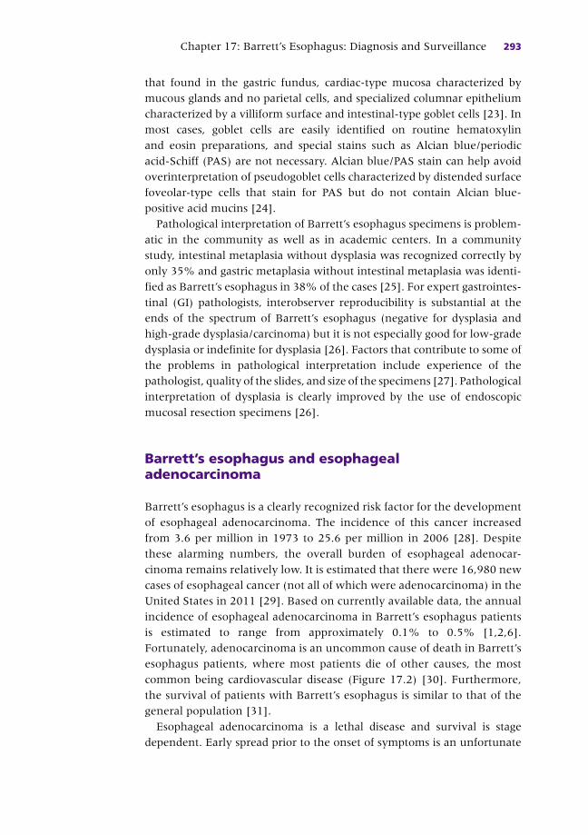
Chapter 17: Barrett’s Esophagus: Diagnosis and Surveillance 293
that found in the gastric fundus, cardiac-type mucosa characterized by
mucous glands and no parietal cells, and specialized columnar epithelium
characterized by a villiform surface and intestinal-type goblet cells [23] . In
most cases, goblet cells are easily identified on routine hematoxylin
and eosin preparations, and special stains such as Alcian blue/periodic
acid-Schiff (PAS) are not necessary. Alcian blue/PAS stain can help avoid
overinterpretation of pseudogoblet cells characterized by distended surface
foveolar-type cells that stain for PAS but do not contain Alcian blue-
positive acid mucins [24] .
Pathological interpretation of Barrett ’ s esophagus specimens is problem-
atic in the community as well as in academic centers. In a community
study, intestinal metaplasia without dysplasia was recognized correctly by
only 35% and gastric metaplasia without intestinal metaplasia was identi-
fied as Barrett ’ s esophagus in 38% of the cases [25] . For expert gastrointes-
tinal (GI) pathologists, interobserver reproducibility is substantial at the
ends of the spectrum of Barrett ’ s esophagus (negative for dysplasia and
high-grade dysplasia/carcinoma) but it is not especially good for low-grade
dysplasia or indefinite for dysplasia [26] . Factors that contribute to some of
the problems in pathological interpretation include experience of the
pathologist, quality of the slides, and size of the specimens [27] . Pathological
interpretation of dysplasia is clearly improved by the use of endoscopic
mucosal resection specimens [26] .
Barrett ’ s esophagus and esophageal adenocarcinoma
Barrett ’ s esophagus is a clearly recognized risk factor for the development
of esophageal adenocarcinoma. The incidence of this cancer increased
from 3.6 per million in 1973 to 25.6 per million in 2006 [28] . Despite
these alarming numbers, the overall burden of esophageal adenocar-
cinoma remains relatively low. It is estimated that there were 16,980 new
cases of esophageal cancer (not all of which were adenocarcinoma) in the
United States in 2011 [29] . Based on currently available data, the annual
incidence of esophageal adenocarcinoma in Barrett ’ s esophagus patients
is estimated to range from approximately 0.1% to 0.5% [1,2,6] .
Fortunately, adenocarcinoma is an uncommon cause of death in Barrett ’ s
esophagus patients, where most patients die of other causes, the most
common being cardiovascular disease (Figure 17.2 ) [30] . Furthermore,
the survival of patients with Barrett ’ s esophagus is similar to that of the
general population [31] .
Esophageal adenocarcinoma is a lethal disease and survival is stage
dependent. Early spread prior to the onset of symptoms is an unfortunate
c17.indd 293 11/15/2012 2:48:25 AM

294 Part 3: Barrett’s Esophagus
characteristic of this tumor. Lymph node metastases are a clear prognostic
factor for decreased survival and increase with depth of tumor involve-
ment [32] . Thus, the best hope for improved survival of patients with
esophageal adenocarcinoma is detection of cancer at an early and
potentially curable stage. Two potential strategies to accomplish this are
screening and surveillance of Barrett ’ s esophagus patients.
Screening
One potential strategy to decrease the mortality rate of esophageal adeno-
carcinoma is to identify more patients at risk, namely those with Barrett ’ s
esophagus. Population-based studies suggest that in patients with newly
diagnosed esophageal adenocarcinoma, a prior endoscopy and diagnosis of
Barrett ’ s esophagus is associated with both early-stage cancer and improved
survival [33] . Unfortunately, only the minority of patients with esophageal
adenocarcinoma have undergone prior endoscopy or have a prior diagnosis
of Barrett ’ s esophagus [34] .
Current professional society practice guidelines equivocate on screening
patients with chronic GERD symptoms for Barrett ’ s esophagus and the
2009 American Cancer Society cancer screening guideline does not include
any recommendation for screening of either esophageal cancer or Barrett ’ s
esophagus [4–7,35] . The 2011 AGA guidelines provide the most recent
clinical recommendations. Screening is recommended as a reasonable
approach for individuals with multiple risk factors for esophageal adeno-
carcinoma, including age greater than or equal to 50 years, male gender,
Caucasian race, chronic GERD symptoms, elevated body mass index (BMI)
and male-pattern abdominal obesity [6] (Figure 17.3 ). On the other hand,
the current AGA guidelines recommend against screening of the general
population of GERD patients without risk factors.
20% 18%
16%
4%35%
7%
EAC
Cardiovasular disease
Pulmonary disease
Other malignancies
Unrelated GI diseases
Other conditions
Figure 17.2 Causes of death in patients with Barrett ’ s esophagus. Reproduced from
Sikkema et al . [30] with permission from Elsevier. EAC, esophageal adenocarcinoma;
GI, gastrointestinal.
c17.indd 294 11/15/2012 2:48:25 AM
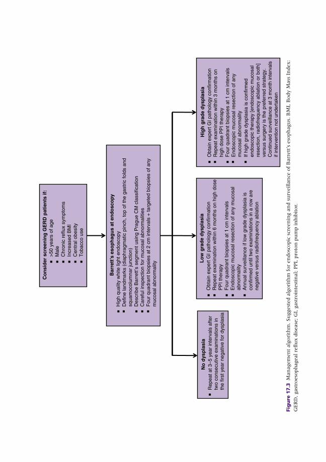
Lo
w g
rad
e d
ysp
lasi
aO
btai
n ex
pert
GI p
atho
logy
con
firm
atio
nR
epea
t exa
min
atio
n w
ithin
6 m
onth
s on
hig
h do
seP
PI t
hera
pyF
our
quad
rant
bio
psie
s at
1 c
m in
terv
als
End
osco
pic
muc
osal
res
ectio
n of
any
muc
osal
abno
rmal
ityA
nnua
l sur
veill
ance
if lo
w g
rade
dys
plas
ia is
conf
irmed
unt
il tw
o ex
amin
atio
ns in
a r
ow a
re
nega
tive
vers
us r
adio
freq
uenc
y ab
latio
n
No
dys
pla
sia
Rep
eat a
t 3–5
yea
r in
terv
als
afte
rtw
o co
nsec
utiv
e ex
amin
atio
ns in
th
e fir
st y
ear
nega
tive
for
dysp
lasi
a
Bar
rett
’s e
sop
hag
us
at e
nd
osc
op
yH
igh
qual
ity w
hite
ligh
t end
osco
pyD
efin
e la
ndm
arks
(di
aphr
agm
atic
pin
ch, t
op o
f the
gas
tric
fold
s an
d sq
uam
ocol
umna
r ju
nctio
n)D
escr
ibe
Bar
rett’
s se
gmen
t us
ing
Pra
gue
CM
cla
ssifi
catio
nC
aref
ul in
spec
tion
for
muc
osal
abn
orm
aliti
esF
our
quad
rant
bio
psie
s at
2 c
m in
terv
als
+ ta
rget
ed b
iops
ies
of a
nym
ucos
al a
bnor
mal
ity
Hig
h g
rad
e d
ysp
lasi
a
Co
nsi
der
scr
een
ing
GE
RD
pat
ien
ts if
:>
50 y
ears
of a
geM
ale
Chr
onic
ref
lux
sym
ptom
sIn
crea
sed
BM
IC
entr
al o
besi
tyT
obac
co u
se
Obt
ain
expe
rt G
I pat
holo
gy c
onfir
mat
ion
Rep
eat e
xam
inat
ion
with
in 3
mon
ths
on
high
dos
e P
PI t
hera
py
Fou
r qu
adra
nt b
iops
ies
at 1
cm
inte
rval
sE
ndos
copi
c m
ucos
al r
esec
tion
of a
nym
ucos
al a
bnor
mal
ityIf
high
gra
de d
yspl
asia
is c
onfir
med
endo
scop
ic th
erap
y [e
ndos
copi
c m
ucos
alre
sect
ion,
rad
iofr
eque
ncy
abla
tion
or b
oth]
vers
us s
urge
ry is
the
pref
erre
d st
rate
gy.
Con
tinue
d su
rvei
llanc
e at
3 m
onth
inte
rval
sif
inte
rven
tion
not u
nder
take
n
Fig
ure
17.3
M
an
agem
en
t alg
ori
thm
. Su
ggest
ed a
lgori
thm
for
en
dosc
opic
scr
een
ing a
nd s
urv
eilla
nce
of
Barr
ett
’ s e
soph
agu
s. B
MI,
Body M
ass
In
dex:
GE
RD
, gast
roeso
ph
ageal re
flu
x d
isease
; G
I, g
ast
roin
test
inal; P
PI,
pro
ton
pu
mp in
hib
itor.
c17.indd 295 11/15/2012 2:48:25 AM

296 Part 3: Barrett’s Esophagus
Endoscopy with biopsy remains the only widely available clinical
technique to diagnose Barrett ’ s esophagus. However, it has clear limita-
tions as a screening tool, including cost, risk, and complexity. If applied to
the estimated 20% of the population with regular GERD symptoms, the
cost implications would be staggering [36] . A recent study examined the
yield of endoscopy in both symptomatic and asymptomatic individuals uti-
lizing the Clinical Outcomes Research Initative (CORI) database. Among
white men with GERD symptoms, the yield of Barrett ’ s esophagus
increased from 3.3% in the fourth decade of life to 9.3% in the sixth
decade prior to reaching a plateau [37] . Interestingly, the yield in symp-
tomatic middle-aged women was comparable to that in asymptomatic
males. These findings suggest that screening should not be considered
prior to age 50.
Several alternatives to standard upper endoscopy have been explored
for screening. Unsedated upper endoscopy using small-caliber instruments
still has the potential to change the economics of endoscopic screening, as
this technique may decrease sedation-related complications and costs.
Unsedated small-caliber endoscopy detects Barrett ’ s esophagus and dys-
plasia with a sensitivity comparable to conventional endoscopy [38] . While
both procedures are well tolerated by patients, a major hurdle for unsedated
endoscopy is patient resistance to undergoing a test without sedation.
Esophageal capsule endoscopy will likely not become a screening
alternative to conventional upper endoscopy due to suboptimal cost and
performance characteristics [39,40] . The most recent alternative to endo-
scopic screening is a non-endoscopic Cytosponge [41] . This technique
involves swallowing a gelatin capsule containing a compressed mesh
device attached to a string. After the gelatin dissolves, the mesh is with-
drawn and retrieved for immunostaining with trefoil factor 3, which is a
diagnostic marker for Barrett ’ s esophagus. The sensitivity of this technique
in a primary care GERD population is 73% with a specificity of 94%. Given
the minimal cost involved, this is an attractive alternative to conventional
endoscopic screening.
While endoscopic screening remains a somewhat controversial issue,
there is little controversy on the role of repeating endoscopy to assess for
the new development of Barrett ’ s esophagus. Studies show consistent
results in patients who have already had a normal initial upper endoscopy.
In patients with non-erosive reflux disease at the index endoscopy, Barrett ’ s
esophagus is rarely found if the repeat endoscopy is performed within
5 years [42,43] . On the other hand, if erosive esophagitis is found at the
time of index endoscopy, Barrett ’ s esophagus may be present in 9–12% of
these patients on repeat endoscopy, with higher grades of esophagitis
associated with a higher case finding rate of Barrett ’ s esophagus [44,45] .
As such, screening for Barrett ’ s esophagus in GERD patients should only
c17.indd 296 11/15/2012 2:48:26 AM
Chapter 17: Barrett’s Esophagus: Diagnosis and Surveillance 297
take place after initial proton pump inhibitor (PPI) therapy. A negative
endoscopy at baseline makes it highly unlikely that Barrett ’ s esophagus
will be found if endoscopy is repeated.
There are still no data from randomized controlled trials or observational
studies to evaluate the strategy of screening. A decision analysis model
examined screening of 50-year-old white men with chronic GERD
symptoms for Barrett ’ s esophagus, and found that one-time screening is
probably cost-effective if subsequent surveillance is limited to patients
with dysplasia on initial examination [46] . Thus there is clearly a need to
develop either a better profile of patients at high risk for Barrett ’ s esoph-
agus or a far less expensive tool to provide mass population screening.
Problems inherent in demonstrating the utility of a screening program
such as healthy volunteer bias, lead time bias, and length time bias will all
need to be addressed as well.
Surveillance
Candidates for surveillance Current practice guidelines recommend endoscopic surveillance of patients
with a diagnosis of Barrett ’ s esophagus [4–7] . Prior to entering into a
surveillance program, patients should be advised about risks and benefits,
including the limitations of surveillance endoscopy as well as the
importance of adhering to appropriate surveillance intervals. Other con-
siderations include age, co-morbid conditions, likelihood of survival over
the next 5 years and ability to tolerate either endoscopic or surgical inter-
ventions for early esophageal adenocarcinoma. Patients in poor overall
health are not likely to benefit from endoscopic surveillance or tolerate
interventions for dysplasia or adenocarcinoma.
Technique The aim of surveillance is the detection of dysplasia or early carcinoma.
Active inflammation makes it more difficult to distinguish dysplasia from
reparative changes. As such, surveillance endoscopy should only be per-
formed after any active inflammation related to GERD is controlled with
anti-secretory therapy. The presence of ongoing erosive esophagitis is a
contraindication to performing surveillance biopsies.
At the time of endoscopy, the esophagus should be carefully examined
with high-resolution white light endoscopy, in particular looking for any
mucosal abnormality [47] . Current guidelines do not recommend the
routine use of advanced imaging techniques as adjuncts to high-quality
white light endoscopy, although many show promise for future applica-
tion. After careful visual inspection, systematic surveillance biopsies
c17.indd 297 11/15/2012 2:48:26 AM

298 Part 3: Barrett’s Esophagus
should be obtained in four quadrants at 2 cm intervals along the entire
length of the Barrett ’ s segment in the absence of known dysplasia and at
1 cm intervals in the presence of known dysplasia [6] . The rationale for
this rigorous approach comes from studies that demonstrate that a
systematic biopsy protocol detects more dysplasia and early cancer com-
pared to ad hoc random biopsies [48,49] . The safety of systematic endo-
scopic biopsy protocols has been demonstrated as well [50] . Subtle
mucosal abnormalities, no matter how trivial, such as ulceration, erosion,
plaque, nodule, stricture or other luminal irregularity in the Barrett ’ s
segment, should also be extensively biopsied, as there is an association of
such lesions with underlying cancer [51] . Mucosal abnormalities, espe-
cially in the setting of known high-grade dysplasia, should undergo
endoscopic mucosal resection [4] . Endoscopic mucosal resection will
change the diagnosis in approximately 50% of patients when compared
to endoscopic biopsies, given the larger tissue sample available for review
by the pathologist [52] . Interobserver agreement among pathologists is
improved as well [52] .
There has been considerable debate over the years regarding the need
for large particle forceps to obtain biopsies, but current evidence does not
support the routine use of the jumbo biopsy forceps [6] . A new large-
capacity forceps that can be passed through standard-diameter endoscopes
provides larger samples than standard large-capacity forceps and may
increase the yield of dysplasia [53] .
Intervals Surveillance intervals, determined by the presence and grade of dysplasia,
are based on our limited understanding of the biology of esophageal ade-
nocarcinoma. The most recent recommendations from the AGA are every
3–5 years in patients without dysplasia, every 6–12 months for low-grade
dysplasia and every 3 months for high-grade dysplasia if intervention is
not performed [6] (see Figure 17.3 ). Unfortunately, guidelines from the
various professional societies are not in agreement on surveillance inter-
vals, leading to considerable confusion in this area.
If low-grade dysplasia is found, the diagnosis should first be confirmed
by an expert gastrointestinal pathologist due to the marked interobserver
variability in interpretation of these biopsies. These patients should receive
aggressive anti-secretory therapy for reflux disease with a PPI to decrease
the changes of regeneration that make pathological interpretation of this
category so difficult. A repeat endoscopy should then be performed within
6–12 months of the initial diagnosis. Endoscopic mucosal resection should
be performed if any mucosal abnormality is present in these patients. The
American College of Gastroenterology recommends annual surveillance
when low-grade dysplasia is present until two examinations in a row are
c17.indd 298 11/15/2012 2:48:26 AM
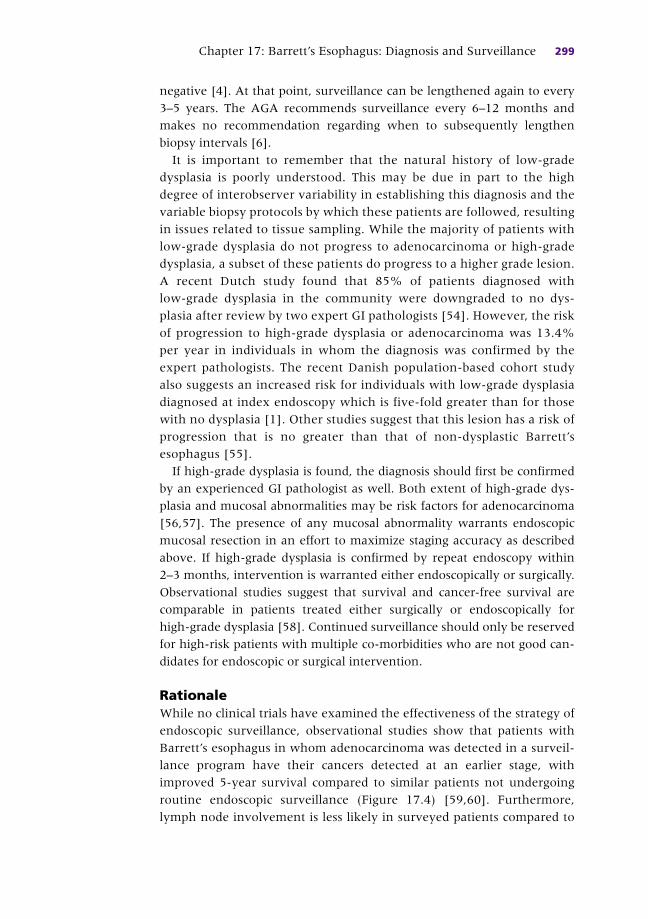
Chapter 17: Barrett’s Esophagus: Diagnosis and Surveillance 299
negative [4] . At that point, surveillance can be lengthened again to every
3–5 years. The AGA recommends surveillance every 6–12 months and
makes no recommendation regarding when to subsequently lengthen
biopsy intervals [6] .
It is important to remember that the natural history of low-grade
dysplasia is poorly understood. This may be due in part to the high
degree of interobserver variability in establishing this diagnosis and the
variable biopsy protocols by which these patients are followed, resulting
in issues related to tissue sampling. While the majority of patients with
low-grade dysplasia do not progress to adenocarcinoma or high-grade
dysplasia, a subset of these patients do progress to a higher grade lesion.
A recent Dutch study found that 85% of patients diagnosed with
low-grade dysplasia in the community were downgraded to no dys-
plasia after review by two expert GI pathologists [54] . However, the risk
of progression to high-grade dysplasia or adenocarcinoma was 13.4%
per year in individuals in whom the diagnosis was confirmed by the
expert pathologists. The recent Danish population-based cohort study
also suggests an increased risk for individuals with low-grade dysplasia
diagnosed at index endoscopy which is five-fold greater than for those
with no dysplasia [1] . Other studies suggest that this lesion has a risk of
progression that is no greater than that of non-dysplastic Barrett ’ s
esophagus [55] .
If high-grade dysplasia is found, the diagnosis should first be confirmed
by an experienced GI pathologist as well. Both extent of high-grade dys-
plasia and mucosal abnormalities may be risk factors for adenocarcinoma
[56,57] . The presence of any mucosal abnormality warrants endoscopic
mucosal resection in an effort to maximize staging accuracy as described
above. If high-grade dysplasia is confirmed by repeat endoscopy within
2–3 months, intervention is warranted either endoscopically or surgically.
Observational studies suggest that survival and cancer-free survival are
comparable in patients treated either surgically or endoscopically for
high-grade dysplasia [58] . Continued surveillance should only be reserved
for high-risk patients with multiple co-morbidities who are not good can-
didates for endoscopic or surgical intervention.
Rationale While no clinical trials have examined the effectiveness of the strategy of
endoscopic surveillance, observational studies show that patients with
Barrett ’ s esophagus in whom adenocarcinoma was detected in a surveil-
lance program have their cancers detected at an earlier stage, with
improved 5-year survival compared to similar patients not undergoing
routine endoscopic surveillance (Figure 17.4 ) [59,60] . Furthermore,
lymph node involvement is less likely in surveyed patients compared to
c17.indd 299 11/15/2012 2:48:26 AM
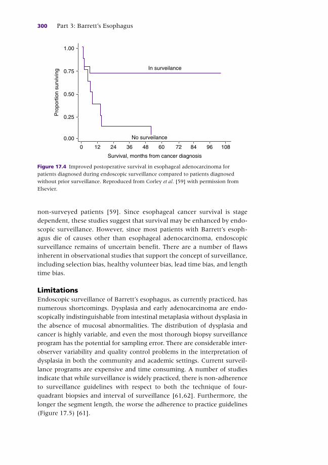
300 Part 3: Barrett’s Esophagus
non-surveyed patients [59] . Since esophageal cancer survival is stage
dependent, these studies suggest that survival may be enhanced by endo-
scopic surveillance. However, since most patients with Barrett ’ s esoph-
agus die of causes other than esophageal adenocarcinoma, endoscopic
surveillance remains of uncertain benefit. There are a number of flaws
inherent in observational studies that support the concept of surveillance,
including selection bias, healthy volunteer bias, lead time bias, and length
time bias.
Limitations Endoscopic surveillance of Barrett ’ s esophagus, as currently practiced, has
numerous shortcomings. Dysplasia and early adenocarcinoma are endo-
scopically indistinguishable from intestinal metaplasia without dysplasia in
the absence of mucosal abnormalities. The distribution of dysplasia and
cancer is highly variable, and even the most thorough biopsy surveillance
program has the potential for sampling error. There are considerable inter-
observer variability and quality control problems in the interpretation of
dysplasia in both the community and academic settings. Current surveil-
lance programs are expensive and time consuming. A number of studies
indicate that while surveillance is widely practiced, there is non-adherence
to surveillance guidelines with respect to both the technique of four-
quadrant biopsies and interval of surveillance [61,62] . Furthermore, the
longer the segment length, the worse the adherence to practice guidelines
(Figure 17.5 ) [61] .
Survival, months from cancer diagnosis
Pro
port
ion
surv
ivin
g1.00
0.75
0.50
0.25
0.00
0 12 24 36 48 60 72 84 96 108
No surveilance
In surveilance
Figure 17.4 Improved postoperative survival in esophageal adenocarcinoma for
patients diagnosed during endoscopic surveillance compared to patients diagnosed
without prior surveillance. Reproduced from Corley et al . [59] with permission from
Elsevier.
c17.indd 300 11/15/2012 2:48:26 AM

Chapter 17: Barrett’s Esophagus: Diagnosis and Surveillance 301
Potential strategies to enhance surveillance Most Barrett ’ s esophagus patients do not have dysplasia and will never
develop cancer. Thus, it would be highly desirable to make surveillance
techniques more effective and efficient by sampling larger areas of Barrett ’ s
mucosa, targeting biopsies to areas with a higher probability of having dys-
plasia or developing risk stratification tools to allow us to concentrate our
efforts on individuals at greatest risk while decreasing the frequency and
intensity of surveillance in individuals at lower risk. Unfortunately, none
of these conceptual paradigms has come to fruition to date despite the
obvious intuitive appeal of these approaches.
Advanced imaging technology A wide variety of endoscopic enhancements to surveillance has been
described (Box 17.1 ), but unfortunately none has really changed clinical
practice. Traditional chromoendoscopy with methylene blue is no better
than random biopsies for the detection of intestinal metaplasia,
high-grade dysplasia or early cancer [63] . Similarly, acetic acid does not
appear to increase the detection rate of dysplasia or adenocarcinoma
despite initial enthusiasm [64] . Optical contrast endoscopy allows for
detailed imaging of the mucosal and vascular surface patterns in Barrett ’ s
esophagus without the need for chromoendoscopy. This may be accom-
plished by either the placement of optical filters that narrow the band-
width of white light to blue light (narrow band imaging) or by
postprocessing software-driven systems to accomplish similar visualiza-
tion [65] . A recent systematic review found that narrow band imaging
Length of BE
Per
cent
age
90.0%
80.0%
70.0%
60.0%
50.0%
40.0%
30.0%
20.0%
10.0%
0.0%
78.7%
34.3%
17.9%10.7%
P< 0.001
< 3 cmn= 1.049
3–5 cmn= 756
6–8 cmn= 235
≥ 9 cmn= 205
Figure 17.5 Adherence to the Seattle biopsy protocol in the community setting by
length of Barrett ’ s esophagus. Reproduced from Abrams et al . [62] with permission
from Elsevier. BE, Barrett ’ s esophagus.
c17.indd 301 11/15/2012 2:48:26 AM

302 Part 3: Barrett’s Esophagus
has a sensitivity of 77–100% and a specificity of 58–100% for the
detection of high-grade dysplasia or cancer [66] .
Confocal laser endomicroscopy is a targeted imaging technique that
allows for subsurface imaging and in vivo histological assessment of the
mucosal layer during white-light endoscopy [67] . The goal of endomicros-
copy is to distinguish neoplastic from non-neoplastic tissue in “real time” by
performing virtual “optical biopsies.” Optical biopsies could accomplish two
important goals: decrease the number of conventional biopsies and allow
immediate treatment decisions at the time of endoscopy, thereby decreasing
the costs and increasing the efficiency of patient care. Two different endomi-
croscopy platforms are available: an endoscope-based device that is
integrated into the distal tip of the endoscope and a probe-based device that
can be inserted through a standard endoscope. Both devices require
administration of an intravenous fluorescence agent, fluorescein. Studies
suggest that confocal endomicroscopy has the potential to improve the diag-
nostic yield of endoscopically inapparent neoplasia compared with standard
white light endoscopy and surveillance biopsies, while also decreasing the
number of biopsies [68,69] . A number of other new techniques to target
biopsies, including molecular imaging, angle-resolved low-coherence inter-
ferometry, circumferential optical coherence tomography, and nanotech-
nology, are all under active development, each with enormous promise.
Perhaps the single greatest advance for the detection of dysplasia and
early cancer has been the advent of high-definition/high-resolution white
Box 17.1 Potential enhancements to white light endoscopic imaging • Chromoendoscopy
• Magnification endoscopy
• Narrow band imaging
• Photodynamic diagnosis
• Spectroscopy
• Partial wave spectroscopy
• Polarized scanning spectroscopy
• Optical coherence tomography
• Low coherence interferometry
• Autofluorescence endoscopy
• Confocal endomicroscopy
• Molecular imaging
Source: Falk GW. Probe-based confocal endomicroscopy in Barrett ’ s esophagus: the real deal or another tease? Gastrointest Endosc 2011;74:473–6.
c17.indd 302 11/15/2012 2:48:26 AM

Chapter 17: Barrett’s Esophagus: Diagnosis and Surveillance 303
light endoscopy. Studies have consistently shown that this technique is as
good as or better than more sophisticated methods such as narrow band
imaging, chromoendoscopy, autofluorescence endoscopy, and multimodal
imaging [64] . The recent AGA position statement does not recommend
using advanced imaging techniques for surveillance at this time [6] .
Risk stratification A number of clinical and biological markers may define patients at
increased risk for the development of adenocarcinoma. Clinical risk factors
for the development of high-grade dysplasia or adenocarcinoma include
gender, ethnicity, age, dysplasia, hiatal hernia size, length of the Barrett ’ s
segment, BMI, and smoking [70] .
Dysplasia is still the best available marker of cancer risk in clinical
practice. Dysplasia is recognized adjacent to and distant from Barrett ’ s
esophagus-associated adenocarcinoma in resection specimens from
patients with Barrett ’ s esophagus. Barrett ’ s esophagus patients progress
through a phenotypic sequence of no dysplasia, low-grade dysplasia,
high-grade dysplasia and then on to adenocarcinoma, although the time
course is highly variable and this step-wise sequence is not preordained
[59,71] . Furthermore, some patients may progress directly to cancer
without prior detection of dysplasia of any grade [72] . Unfortunately, dys-
plasia is an imperfect marker of increased cancer risk. It is typically not
distinguishable endoscopically and often focal in nature, thereby making
targeting of biopsies problematic. Furthermore, there is considerable inter-
observer variability in the grading of dysplasia. Therefore, a less subjective
marker for cancer risk that could supplement or replace the current
dysplasia grading system is still needed.
Biomarkers of increased risk A number of molecular markers may define patients at increased risk for
the development of esophageal adenocarcinoma. Among the most fre-
quently described molecular changes that precede the development of
adenocarcinoma in Barrett ’ s esophagus are alterations in p53 (mutation,
deletion or loss of heterozygosity (LOH)) p16 (mutation, deletion, promoter
hypermethylation, or LOH) and aneuploidy by flow cytometry [73–78] .
Neoplastic progression in Barrett ’ s esophagus is accompanied by flow cyto-
metric abnormalities such as aneuploidy or increased G2/tetraploid DNA
contents, and these abnormalities may precede the development of
high-grade dysplasia or adenocarcinoma. An alternative method for the
detection of DNA ploidy abnormalities is image cytometry, which can ana-
lyze formalin-fixed tissue with automated analysis algorithms. Image
cytometry may be comparable to flow cytometry for the detection of
ploidy [79] . Another technique with potential clinical applicability for the
c17.indd 303 11/15/2012 2:48:26 AM

304 Part 3: Barrett’s Esophagus
detection of abnormal DNA content is fluorescent in situ hybridization
(FISH) which may also be simpler to apply in clinical practice [80] .
Mutations of p53 and 17p LOH have been reported in up to 92% and
100%, respectively, of esophageal adencocarcinomas [73] . Furthermore,
both abnormalities have been detected in Barrett ’ s epithelium prior to the
development of carcinoma [81] . However, techniques to detect p53 muta-
tions and 17p LOH are also labor intensive and have not achieved wide-
spread acceptance in clinical practice to date. Similarly, p16 LOH and
inactivation of the p16 gene by promoter region hypermethylation have
been reported frequently in esophageal adenocarcinoma [82] . Furthermore,
9p LOH is commonly encountered in premalignant Barrett ’ s epithelium
and can be detected over large regions of the Barrett ’ s mucosa [80] . It is
hypothesized that clonal expansion occurs in conjunction with p16 abnor-
malities, creating a field in which other genetic lesions leading to esopha-
geal adenocarcinoma can arise.
Epigenetic changes, in the form of hypo- and hypermethylation and
alteration to histone complexes, have also been implicated in the progres-
sion of Barrett ’ s esophagus to adenocarcinoma. Hypermethylation of p16,
APC, RUNX3 and HPP1 are all independently associated with an increased
risk of progression of Barrett ’ s esophagus to high-grade dysplasia or esoph-
ageal adenocarcinoma [83,84] .
Given the complexity and diversity of alterations observed to date in the
metaplasia, dysplasia, carcinoma sequence, it appears that a panel of bio-
markers may be required for risk stratification. The combination of 17p
LOH, 9p LOH and DNA content abnormality has been shown to predict the
10-year adenocarcinoma risk better than any single biomarker alone [85] .
Patients with a combination of these abnormalities had a markedly increased
risk of developing cancer compared to those with no baseline abnormalities
(relative risk 38.7, 95% confidence interval (CI) 10.8–138.5). In those with
no abnormalities of any of these biomarkers at baseline, 12% developed
adenocarcinoma at 10 years. In contrast, those with the combination of 17p
LOH, 9p LOH and DNA content abnormality had a cumulative incidence of
adenocarcinoma of 79% over the same time period. A risk stratification
model utilizing a methylation index constructed from the methylation
values for p16, HPP1, and RUNX3 also showed potential for prediction of
progression to high-grade dysplasia or adenocarcinoma [86] .
Thus, while all of these studies demonstrate the potential for biomarkers
to predict risk of esophageal adenocarcinoma, none of these biomarkers
has been validated in large-scale clinical trials to date and as such they are
not yet useful for clinical decision making. It is likely that in the future, the
best predictor for the development of high-grade dysplasia or adenocarci-
noma will be a combination of clinical, demographic, histological, genetic,
and epigenetic data.
c17.indd 304 11/15/2012 2:48:26 AM

Chapter 17: Barrett’s Esophagus: Diagnosis and Surveillance 305
References
1 Hvid-Jensen F , Pedersen L , Drewes AM , et al . Incidence of adenocarcinoma among
patients with Barrett ’ s esophagus . N Engl J Med 2011 ; 365 : 1375 – 83 .
2 Wani S , Falk G , Hall M , et al . Patients with nondysplastic Barrett ’ s esophagus have
low risks for developing dysplasia or esophageal adenocarcinoma . Clin Gastroenterol
Hepatol 2011 ; 9 : 220 – 7 .
3 Riddell RH , Odze RD . Definition of Barrett ’ s esophagus: time for a rethink – is
intestinal metaplasia dead? Am J Gastroenterol 2009 ; 104 : 2588 – 94 .
4 Wang KK , Sampliner RE , Practice Parameters Committee of the American College of
Gastroenterology. Updated guidelines 2008 for the diagnosis, surveillance and
therapy of Barrett ’ s esophagus . Am J Gastroenterol 2008 ; 103 : 788 – 97 .
5 Hirota WK , Zuckerman MJ , Adler DG , et al . ASGE guideline: the role of endoscopy in
the surveillance of premalignant conditions of the upper GI tract . Gastrointest Endosc
2006 ; 63 : 570 – 80 .
6 Spechler SJ , Sharma P , Souza RF , et al . American Gastroenterological Association
medical position statement on the management of Barrett ’ s esophagus .
Gastroenterology 2011 ; 140 : 1084 – 91 .
7 British Society of Gastroenterology . Guidelines for the diagnosis and management of
Barrett ’ s columnar – lined oesophagus . Available at: www.BSG.org.uk .
8 Vakil N , van Zanten SV , Kahrilas P , et al . Global Consensus Group. The Montreal def-
inition and classification of gastroesophageal reflux disease: a global evidence-based
consensus . Am J Gastroenterol 2006 ; 101 : 1900 – 20 .
9 Eisen GM , Sandler RS , Murray S , et al . The relationship between gastroesophageal
reflux disease and its complications with Barrett ’ s esophagus . Am J Gastroenterol
1997 ; 92 : 27 – 31 .
10 Johnson DA , Winters C , Spurling TJ , et al . Esophageal acid sensitivity in Barrett ’ s
esophagus . J Clin Gastroenterol 1987 ; 9 : 23 – 7 .
11 Grade A , Pulliam G , Johnson C , et al . Reduced chemoreceptor sensitivity in patients
with Barrett ’ s esophagus may be related to age and not to the presence of Barrett ’ s
epithelium . Am J Gastroenterol 1997 ; 92 : 2040 – 3 .
12 Chak A , Ochs-Balcom H , Falk G , et al . Familiality in Barrett ’ s esophagus, adenocarci-
noma of the esophagus, and adenocarcinoma of the gastroesophageal junction .
Cancer Epidemiol Biomarkers Prev 2006 ; 15 : 1668 – 73 .
CASE STUDY
A 56-year-old white man with no prior history of dysplasia underwent surveillance endoscopy for known Barrett ’ s esophagus. LA Grade B esophagitis was noted in conjunction with a 5 cm segment of columnar-lined esophagus. No biopsies were performed because of ongoing inflammation and the endoscopy was repeated 8 weeks after commencing twice-daily PPI therapy. At that time endoscopy revealed a 3 cm hiatal hernia and a C3M5 segment of Barrett ’ s esophagus with no mucosal abnormalities seen with high-definition white light endoscopy. Four quadrant biopsies were done every 2 cm commencing at the GE junction and proceeding up to the squamocolumnar junction. All biopsies were read as no dysplasia. The patient was counseled that recent data suggest a very low risk of esophageal adenocarcinoma of the order of 0.1–0.5% per year and to return for his next surveillance examination in 3–5 years.
c17.indd 305 11/15/2012 2:48:26 AM
306 Part 3: Barrett’s Esophagus
13 Orloff M , Peterson C , He X , et al . Germline mutations in MSR1, ASCC1, and CTHRC1
in patients with Barrett esophagus and esophageal adenocarcinoma . JAMA 2011 ;
306 : 410 – 19 .
14 McClave SA , Boyce HW , Gottfried MR . Early diagnosis of the columnar-lined
esophagus: a new endoscopic criterion . Gastrointest Endosc 1987 ; 33 : 413 – 16 .
15 Ofman JJ , Shaheen NJ , Desai AA , et al . The quality of care in Barrett ’ s
esophagus: endoscopist and pathologist practices . Am J Gastroenterol 2001 ; 96 :
876 – 81 .
16 Sharma P , Dent J , Armstrong D , et al . The development and validation of an
endoscopic grading system for Barrett ’ s esophagus: the Prague C & M criteria .
Gastroenterology 2006 ; 131 : 1392 – 9 .
17 Liu W , Hahn H , Odze RD , et al . Metaplastic esophageal columnar epithelium without
goblet cells shows DNA content abnormalities similar to goblet cell-containing
epithelium . Am J Gastroenterol 2009 ; 104 : 816 – 24 .
18 Kelty CJ , Gough MD , van Wyk Q , et al . Barrett ’ s oesophagus: intestinal metaplasia is
not essential for cancer risk . Scand J Gastroenterol 2007 ; 42 : 1271 – 4 .
19 Takubo K , Aida J , Naomoto Y , et al . Cardiac rather than intestinal-type background in
endoscopic resection specimens of minute Barrett adenocarcinoma . Hum Pathol
2009 ; 4 : 65 – 74 .
20 Wang A , Mattek NC , Corless CL , et al . The value of traditional upper endoscopy as a
diagnostic test for Barrett ’ s esophagus . Gastrointest Endosc 2008 ; 68 : 859 – 6 .
21 Chandrasoma PT , Der R , Dalton P , et al . Distribution and significance of epithelial
types in columnar-lined esophagus . Am J Surg Pathol 2001 ; 25 : 1188 – 93 .
22 Harrison R , Perry I , Haddadin W , et al . Detection of intestinal metaplasia in Barrett ’ s
esophagus: an observational comparator study suggests the need for a minimum of
eight biopsies . Am J Gastroenterol 2007 ; 102 : 1154 – 61 .
23 Paull A , Trier JS , Dalton MD , et al . The histologic spectrum of Barrett ’ s esophagus .
N Engl J Med 1976 ; 295 : 476 – 80 .
24 Weinstein WM , Ippoliti AF . The diagnosis of Barrett ’ s esophagus: goblets, goblets,
goblets . Gastrointest Endosc 1996 ; 44 : 91 – 4 .
25 Alikhan M , Rex D , Khan A , et al . Variable pathologic interpretation of columnar lined
esophagus by general pathologists in community practice . Gastrointest Endosc
1999 ; 50 : 23 – 6 .
26 Montgomery E , Bronner MP , Goldblum JR , et al . Reproducibility of the diagnosis of
dysplasia Barrett esophagus: a reaffirmation . Hum Pathol 2001 ; 32 : 368 – 78 .
27 Mino-Kenudson M , Hull MJ , Brown I , et al . EMR for Barrett ’ s esophagus-related
superficial neoplasms offers better diagnostic reproducibility than mucosal biopsy .
Gastrointest Endosc 2007 ; 66 : 660 – 6 .
28 Pohl H , Sirovich B , Welch HG . Esophageal adenocarcinoma incidence: are we reach-
ing the peak? Cancer Epidemiol Biomarkers Prev 2010 ; 19 : 1468 – 70 .
29 Siegel R , Ward E , Brawley O , et al . Cancer statistics, 2011: the impact of eliminating
socioeconomic and racial disparities on premature cancer deaths . CA Cancer J Clin
2011 ; 61 : 212 – 36 .
30 Sikkema M , de Jonge PJ , Steyerberg EW , et al . Risk of esophageal adenocarcinoma
and mortality in patients with Barrett ’ s esophagus: a systematic review and meta-
analysis . Clin Gastroenterol Hepatol 2010 ; 8 : 235 – 44 .
31 Anderson LA , Murray LJ , Murphy SJ , et al . Mortality in Barrett ’ s oesophagus: results
from a population based study . Gut 2003 ; 52 : 1081 – 4 .
32 Rice TW , Blackstone EH , Goldblum JR , et al . Superficial adenocarcinoma of the
esophagus . J Thorac Cardiovasc Surg 2001 ; 122 : 1077 – 90 .
c17.indd 306 11/15/2012 2:48:27 AM
Chapter 17: Barrett’s Esophagus: Diagnosis and Surveillance 307
33 Cooper GS , Kou TD , Chak A . Receipt of previous diagnoses and endoscopy and
outcome from esophageal adenocarcinoma: a population-based study with temporal
trends . Am J Gastroenterol 2009 ; 104 : 1356 – 62 .
34 Dulai GS , Guha S , Kahn KL , et al . Preoperative prevalence of Barrett ’ s esophagus in
esophageal adenocarcinoma: a systematic review . Gastroenterology 2002 ; 122 :
26 – 33 .
35 Smith RA , Cokkinides V , Brawley OW . Cancer screening in the United States, 2009:
a review of current American Cancer Society guidelines and issues in cancer screen-
ing . CA Cancer J Clin 2009 ; 59 : 27 – 41 .
36 Shaheen NJ , Provenzale D , Sandler RS . Upper endoscopy as a screening and
sur veillance tool in esophageal adenocarcinoma: a review of the evidence . Am J
Gastroenterol 2002 ; 97 : 1319 – 27 .
37 Rubenstein JH , Mattek N , Eisen G . Age- and sex-specific yield of Barrett ’ s esophagus
by endoscopy indication . Gastrointest Endosc 2010 ; 71 : 21 – 7 .
38 Jobe BA , Hunter JG , Chang EY , et al . Office-based unsedated small-caliber endoscopy
is equivalent to conventional sedated endoscopy in screening and surveillance for
Barrett ’ s esophagus: a randomized and blinded comparison . Am J Gastroenterol
2006 ; 101 : 2693 – 703 .
39 Gerson L , Lin OS . Cost–benefit analysis of capsule endoscopy compared with stan-
dard upper endoscopy for the detection of Barrett ’ s esophagus . Clin Gastroenterol
Hepatol 2007 ; 5 : 319 – 25 .
40 Sharma P , Wani S , Rastogi A , et al . The diagnostic accuracy of esophageal capsule
endoscopy in patients with gastroesophageal reflux disease and Barrett ’ s esophagus:
a blinded, prospective study . Am J Gastroenterol 2008 ; 3 : 525 – 32 .
41 Kadri SR , Lao-Sirieix P , O ’ Donovan M , et al . Acceptability and accuracy of a non-
endoscopic screening test for Barrett ’ s oesophagus in primary care: cohort study . BMJ
2010 ; 341 : c4372 .
42 Stoltey J , Reeba H , Ullah N , et al . Does Barrett ’ s oesophagus develop over time in
patients with chronic gastro-oesophageal reflux disease? Aliment Pharmacol Ther
2007 ; 25 : 83 – 91 .
43 Rodriguez S , Mattek N , Lieberman D , et al . Barrett ’ s esophagus on repeat endoscopy:
should we look more than once? Am J Gastroenterol 2008 ; 103 : 1892 – 7 .
44 Hanna S , Rastogi A , Weston AP , et al . Detection of Barrett ’ s esophagus after endo-
scopic healing of erosive esophagitis . Am J Gastroenterol 2006 ; 101 : 1416 – 20 .
45 Modiano N , Gerson LB . Risk factors for the detection of Barrett ’ s esophagus in
patients with erosive esophagitis . Gastrointest Endosc 2009 ; 69 : 1014 – 20 .
46 Inadomi JM , Sampliner R , Lagergren J , et al . Screening and surveillance for Barrett
esophagus in high risk groups: a cost–utility analysis . Ann Intern Med 2003 ; 138 :
176 – 86 .
47 Spechler SJ , Sharma P , Souza RF , et al . American Gastroenterological Association
technical review on the management of Barrett ’ s esophagus . Gastroenterology
2011 ; 140 : e18 – 52 .
48 Fitzgerald RC , Saeed I , Khoo D , et al . Rigorous surveillance protocol increases
detection of curable cancers associated with Barrett ’ s esophagus . Dig Dis Sci
2001 ; 46 : 1892 – 8 .
49 Abela JE , Going JJ , Mackenzie JF , et al . Systematic four-quadrant biopsy detects
Barrett ’ s dysplasia in more patients than nonsystematic biopsy . Am J Gastroenterol
2008 ; 103 : 850 – 5 .
50 Levine DS , Blount PL , Rudolph RE , et al . Safety of a systematic endoscopic biopsy
protocol in patients with Barrett ’ s esophagus . Am J Gastroenterol 2000 ; 95 : 1152 – 7 .
c17.indd 307 11/15/2012 2:48:27 AM
308 Part 3: Barrett’s Esophagus
51 Reid BJ , Blount PL , Feng Z , et al . Optimizing endoscopic biopsy detection of early
cancers in Barrett ’ s high-grade dysplasia . Am J Gastroenterol 2000 ; 95 : 3089 – 6 .
52 Peters FP , Brakenhoff KP , Curvers WL , et al . Histologic evaluation of resection speci-
mens obtained at 293 endoscopic resections in Barrett ’ s esophagus . Gastrointest
Endosc 2008 ; 67 : 604 – 9 .
53 Komanduri S , Swanson G , Keefer L , et al . Use of a new jumbo forceps improves tissue
acquisition of Barrett ’ s esophagus surveillance biopsies . Gastrointest Endosc
2009 ; 70 : 1072 – 8 .
54 Curvers WL , ten Kate FJ , Krishnadath KK , et al . Low-grade dysplasia in Barrett ’ s
esophagus: overdiagnosed and underestimated . Am J Gastroenterol 2010 ; 105 :
1523 – 30 .
55 Wani S , Falk GW , Post J , et al . Risk factors for progression of low-grade dysplasia in
patients with Barrett ’ s esophagus . Gastroenterology 2011 ; 141 : 1179 – 86 .
56 Buttar NS , Wang KK , Sebo TJ , et al . Extent of high-grade dysplasia in Barrett ’ s esoph-
agus correlates with risk of adenocarcinoma . Gastroenterology 2001 ; 120 : 1630 – 9 .
57 Tharavej C , Hagen JA , Peters JH , et al . Predictive factors of coexisting cancer in
Barrett ’ s high-grade dysplasia . Surg Endosc 2006 ; 20 : 439 – 43 .
58 Prasad GA , Wang KK , Buttar NS , et al . Long-term survival following endoscopic and
surgical treatment of high-grade dysplasia in Barrett ’ s esophagus . Gastroenterology
2007 ; 132 : 1226 – 33 .
59 Corley DA , Levin TR , Habel LA , et al . Surveillance and survival in Barrett ’ s adenocar-
cinomas: a population-based study . Gastroenterology 2002 ; 122 : 633 – 40 .
60 Van Sandick JW , Lanschot JJ , Kuiken BW , et al . Impact of endoscopic biopsy surveil-
lance of Barrett ’ s esophagus on pathological stage and clinical outcome of Barrett ’ s
carcinoma . Gut 1998 ; 43 : 216 – 22 .
61 Das D , Ishaq S , Harrison R , et al . Management of Barrett ’ s esophagus in the UK: over-
treated and underbiopsied but improved by the introduction of a national randomized
trial . Am J Gastroenterol 2008 ; 103 : 1079 – 89 .
62 Abrams JA , Kapel RC , Lindberg GM , et al . Adherence to biopsy guidelines for Barrett ’ s
esophagus surveillance in the community setting in the United States . Clin
Gastroenterol Hepatol 2009 ; 7 : 736 – 42 .
63 Ngamruengphong S , Sharma VK , Das A . Diagnostic yield of methylene blue chromo-
endoscopy for detecting specialized intestinal metaplasia and dysplasia in Barrett ’ s
esophagus: a meta-analysis . Gastrointest Endosc 2009 ; 69 : 1021 – 8 .
64 Curvers W , Baak L , Kiesslich R , et al . Chromoendoscopy and narrow-band imaging
compared with high-resolution magnification endoscopy in Barrett ’ s esophagus .
Gastroenterology 2008 ; 134 : 670 – 9 .
65 Song LM , Adler DG , Conway JD , et al . Narrow band imaging and multiband imaging .
Gastrointest Endosc 2008 ; 67 : 581 – 9 .
66 Curvers WL , van den Broek FJ , Reitsma JB , et al . Systematic review of narrow-band
imaging for the detection and differentiation of abnormalities in the esophagus and
stomach . Gastrointest Endosc 2009 ; 69 : 307 – 17 .
67 Kantsevoy SV , Adler DG , Conway JD , et al . Confocal laser endomicroscopy .
Gastrointest Endosc 2009 ; 70 : 197 – 200 .
68 Dunbar KB , Okolo P , Montgomery E , et al . Confocal laser endomicroscopy in
Barrett ’ s esophagus and endoscopically inapparent Barrett ’ s neoplasia: a prospective,
randomized, double-blind, controlled, crossover trial . Gastrointest Endosc 2009 ;
70 : 645 – 54 .
69 Sharma P , Meining A , Coron E , et al . Real-time increased detection of neoplastic
tissue in Barrett ’ s esophagus with probe-based confocal laser endomicroscopy: final
c17.indd 308 11/15/2012 2:48:27 AM

Chapter 17: Barrett’s Esophagus: Diagnosis and Surveillance 309
results of an international multicenter, prospective, randomized, controlled trial .
Gastrointest Endosc 2011 ; 74 : 465 – 72 .
70 Falk GW . Risk factors for esophageal cancer development . Surg Oncol Clin North Am
2009 ; 18 : 469 – 85 .
71 Hameeteman W , Tytgat GN , Houthoff HJ , et al . Barrett ’ s esophagus: development of
dysplasia and adenocarcinoma . Gastroenterology 1989 ; 96 : 1249 – 56 .
72 Sharma P , Falk GW , Weston AP , et al . Dysplasia and cancer in a large multicenter
cohort of patients with Barrett ’ s esophagus . Clin Gastroenterol Hepatol 2006 ; 4 :
566 – 72 .
73 Reid BJ , Haggitt RC , Rubin CE , et al . Barrett ’ s esophagus. Correlation between flow
cytometry and histology in detection of patients at risk for adenocarcinoma .
Gastroenterology 1987 ; 93 : 1 – 11 .
74 Reid BJ , Blount PL , Rubin CE , et al . Flow-cytometric and histological progression to
malignancy in Barrett ’ s esophagus: prospective endoscopic surveillance of a cohort .
Gastroenterology 1992 ; 102 : 1212 – 19 .
75 Reid BJ . P53 and neoplastic progression in Barrett ’ s esophagus . Am J Gastroenterol
2001 ; 96 : 1321 – 3 .
76 Prevo LJ , Sanchez CA , Galipeau PC , et al . P53-mutant clones and field effects in
Barrett ’ s esophagus . Cancer Res 1999 ; 59 : 4784 – 7 .
77 Wong DJ , Barrett MT , Stoger R , et al . p16INK4a promoter is hypermethylated at a
high frequency in esophageal adenocarcinomas . Cancer Res 1997; 57 : 2619 – 22 .
78 Reid BJ , Levine DS , Longton G , et al . Predictors of progression to cancer in Barrett ’ s
esophagus: baseline histology and flow cytometry identify low- and high-risk patient
subsets . Am J Gastroenterol 2000 ; 95 : 1669 – 76 .
79 Dunn JM , Mackenzie GD , Oukrif D , et al . Image cytometry accurately detects DNA
ploidy abnormalities and predicts late relapse to high-grade dysplasia and adenocar-
cinoma in Barrett ’ s oesophagus following photodynamic therapy . Br J Cancer
2010 ; 102 : 1608 – 17 .
80 Fritcher EG , Brankley SM , Kipp BR , et al . A comparison of conventional cytology,
DNA ploidy analysis, and fluorescence in situ hybridization for the detection of dys-
plasia and adenocarcinoma in patients with Barrett ’ s esophagus . Hum Pathol
2008 ; 39 : 1128 – 35 .
81 Reid BJ , Prevo LJ , Galipeau PC , et al . Predictors of progression in Barrett ’ s esophagus
II: baseline 17p (p53) loss of heterozygosity identifies a patient subset at increased
risk for neoplastic progression . Am J Gastroenterol 2001 ; 96 : 2839 – 48 .
82 Wong DJ , Paulson TG , Prevo LJ , et al . p16INK4a lesions are common, early abnor-
malities that undergo clonal expansion in Barrett ’ s metaplastic epithelium . Cancer
Res 2001 ; 61 : 8284 – 9 .
83 Schulmann K , Sterian A , Berki A , et al . Inactivation of p16, RUNX3, and HPP1 occurs
early in Barrett ’ s-associated neoplastic progression and predicts progression risk .
Oncogene 2005 ; 24 : 4138 – 48 .
84 Wang JS , Guo M , Montgomery EA , et al . DNA promoter hypermethylation of p16
and APC predicts neoplastic progression in Barrett ’ s esophagus . Am J Gastroenterol
2009 ; 104 : 2153 – 60 .
85 Galipeau PC , Li X , Blount PL , et al . NSAIDs modulate CDKN2A, TP53, and DNAcontent
risk for progression to esophageal adenocarcinoma . PLoS Med 2007 ; 4 : e67 .
86 Sato F , Jin Z , Schulmann K , et al . Three-tiered risk stratification model to predict pro-
gression in Barrett ’ s esophagus using epigenetic and clinical features . PLoS ONE
2008 ; 3 : e1890 .
c17.indd 309 11/15/2012 2:48:27 AM

310
Practical Manual of Gastroesophageal Reflux Disease, First Edition.
Edited by Marcelo F. Vela, Joel E. Richter and John E. Pandolfino.
© 2013 John Wiley & Sons, Ltd. Published 2013 by John Wiley & Sons, Ltd.
CHAPTER 18
Barrett ’ s Esophagus: Treatment Options Jianmin Tian and Kenneth K. Wang Mayo Clinic , Rochester , MN , USA
Key point • The key to successful ablation is patient selection. This begins with selecting patients
with significant neoplastic lesions that might benefit from treatment. It is very
important to emphasize patient compliance, with an esophagogastroduo denoscopy
at scheduled intervals in the postablation period. The important step is to identify
and eradicate cancer at an early stage (no more than T1a) while endoscopic
treatment is still a good option. Recurrence usually does not affect overall outcomes.
Potential pitfall • For a patient with visible lesions such as nodules, thickened folds, mass, ulcers,
strictures, and/or high-grade dysplasia at multiple biopsy levels, it is essential to
perform mucosal resection to obtain sufficient tissues for accurate diagnosis before
any ablation.
Introduction
The prevalence of Barrett ’ s esophagus (BE) in patients with gastroesopha-
geal reflux disease (GERD) has been about 5.7% [1] . However, only about
half of BE patients have symptoms. This has led to a situation where most
patients are diagnosed either incidentally or with more advanced disease.
Our current understanding is that non-dysplastic Barrett ’ s esophagus
(NDBE) progresses to low-grade dysplasia (LGD), then to high-grade dys-
plasia ( HGD) and esophageal adenocarcinoma (EAC), although patients
do not always progress in a step-wise fashion. It is uncertain whether this
is due to sampling error and these mucosal changes are not discovered or,
alternatively, there is rapid evolution to more advanced neoplasia. The
c18.indd 310 11/15/2012 2:48:05 AM
Chapter 18: Barrett’s Esophagus: Treatment Options 311
currently held belief is that the primary target for treatment of BE is
dysplastic mucosa. The estimated 10-year survival is similar in patients
with non-dysplastic BE and in the general population [2] . Thus treatment
of all patients with BE without dysplasia at this time may not ultimately
lead to any survival benefit. If selected patients at higher risk of cancer can
be identified in this group, for instance through use of biomarkers,
treatment would likely prove to be useful.
There is consensus in current clinical practice on treating patients with
high-grade dysplasia in Barrett ’ s mucosa after this diagnosis is confirmed
by two expert pathologists and concomitant invasive adenocarcinoma has
been excluded [3] . As demonstrated in a recent metaanalysis and systematic
review, endoscopic treatment reduces the incidence of esophageal adeno-
carcinoma with most of the benefit of ablation being observed in patients
with HGD [4] . It has been reported that the survival of HGD patients with
endoscopic therapies is comparable to patients with esophagectomy.
Postablation surveillance is very important regardless of types of endo-
scopic treatment, because the rates of detecting EAC after ablation were
6.0% for HGD, 0.8% for LGD, and 0.03% for NDBE [5] .
For more advanced neoplasia such as adenocarcinoma that is confined
to the mucosa, mucosal resection techniques are recommended. Endoscopic
mucosal resection (EMR) is suitable for T1a lesions (intramucosal EAC)
because the percentage of lymph node metastasis (LNM) is low in this
condition. However, any lesion that is more advanced than T1a should be
managed by non-endoscopic approaches.
In this chapter, we will focus on the clinical indications, procedure
details, efficacy, outcomes, and complications of each endoscopic modality.
Endoscopic mucosal resection
Since it was first described in 1984, EMR has been adopted into practice in
diagnosis, staging and treatment for BE with dysplasia and early cancers
in addition to its applications in the stomach and colon. Overall, it is a
safe and effective method for managing most lesions less than 1.5 cm in
dia meter in the luminal gastrointestinal (GI) tract. For larger lesions,
endoscopic submucosal dissection (ESD) is preferred over piecemeal EMR
since this allows en bloc resection of the lesion. Circumferential EMR to
eradicate the entire BE was reported in 2003 in 12 patients who had HGD
or intra mucosal EAC. Although no recurrence was observed, complica-
tions occurred in six cases with four bleedings and two strictures [6] . Two
subsequent studies in 2005 and 2006 [7,8] showed a high rate of stricture
(up to one-third of the patients). Despite this radical treatment, the
recurrence rate of Barrett ’ s mucosa was up to 11%. Currently this
c18.indd 311 11/15/2012 2:48:05 AM

312 Part 3: Barrett’s Esophagus
circumferential EMR approach is being replaced by sequential targeted
EMRs along with ablation for residual flat mucosa in the United States.
Patient selection Patient selection is an important step to assure success and minimize com-
plications such as bleeding, stricture and perforation.
• Thorough work-up to rule out distal metastatic lesions using positron
emisson tomography (PET)/computed tomography (CT) scans should be
performed for cancer patients, because those with advanced disease
would not benefit from this procedure. Then endoscopic ultrasound
(EUS) should be done primarily for nodal staging. EUS has been consid-
ered as the most accurate non-invasive method for staging but a recent
metaanalysis of 12 EUS studies revealed that EUS found only 65% con-
cordance for T-staging when compared to EMR or esophagectomy find-
ings. High-frequency miniprobe EUS does provide more accurate
T-staging though even this technique is still not accurate enough to
exclude the use of EMR [9] . Nevertheless, locoregional lymph node
positivity by EUS with fine needle aspiration (FNA) still adds significant
value in patient management.
• The percentage of lymph node metastasis (LNM) has been estimated as
1–6% [10–13] for intramucosal cancer (T1a). But up to one-third of
patients with submucosal esophageal cancer (T1b) lesions could have
LNM at the time of esophagectomy. Thus any lesion that is more
advanced than T1a should not be treated with EMR for curative intent.
Lymphovascular invasion or high-grade malignancies are also felt to be
contraindications for endoscopic treatment.
• Generally speaking, previous biopsies do not affect subsequent EMRs
unless they are performed within a few days of attempted resection.
However, previous EMRs can lead to tissue scarring, and subsequent
EMRs at the same location are associated with higher risk of failure and
complications. If a lesion is not lifted (i.e. non-lifting sign) after sub-
mucosal fluid injection, then EMR should be reconsidered to avoid
perforation. Poor wound healing could also lead to difficulty after EMR.
Procedure The main technique for EMR involves submucosal injection and lifting,
followed by electrocautery and mechanical resection [14] .
Preparation American Society of Gastrointestinal Endoscopy guidelines [15] should be
followed for the management of anti-platelet or anti-coagulation (APAC)
medications. In a retrospective report, patients who underwent EMR while
c18.indd 312 11/15/2012 2:48:05 AM
Chapter 18: Barrett’s Esophagus: Treatment Options 313
on or off APAC medications were studied. This study showed the rate of
major GI bleeding was 0.23% for those on APAC versus 0.59% for those
off APAC. However, ischemic events (myocardial or cerebral ischemia)
happened in 6.25% of those who were off clopidogrel versus 0.22% of
those on clopidogrel ( P = 0.051) [16] .
Identify the lesions for endoscopic mucosal resection If there is an obvious mass in the esophagus, then it is probably too
advanced for curative EMR. If there are nodules and thickened folds,
which are often associated with cancers [17] , they should be targeted for
EMR. For less apparent lesions, narrow band imaging (NBI) or chromo-
endoscopy may aid the endoscopist in defining the margins. This step of
lesion identification could be difficult in some cases. Even trimodal methods
(high-resolution endoscopy followed by autofluorescence imaging (AFI)
and NBI) did not improve the overall detection of dysplasia compared with
standard video endoscopy and biopsies [18] . Dysplasia or even early EAC
is still found in a significant number of patients by random biopsies in areas
where no obvious target was identified for EMR.
Technique Submucosal injection of saline is an important initial step to separate the
mucosal layer from the muscular layer so that (a) the muscular layer will
not be suctioned into the cap and (b) the deep margin will have fewer
thermal effects when electrocautery is applied. This is important in avoiding
perforation. Other solutions such as 1:200,000 epinephrine could also be
used to achieve better homeostasis. For submucosal dissection, hypertonic
glucose, glycerol, and hydroxypropylmethyl cellulose have been used to
maintain a submucosal cushion. These agents are not useful with EMR since
a persistent cushion makes suction of the lesion into the cap more difficult.
Endoscopic mucosal resection can be performed in several ways, with
the common feature being the use of a snare to achieve tissue removal.
Original methods include strip biopsy using a two-channel therapeutic
endoscope but this is mostly obsolete since the technique is difficult to per-
form. The most commonly used current methods are cap- or band ligation-
assisted EMR. For the cap method, a soft or hard, flat or oblique transparent
cap, which comes in multiple diameters, is friction fitted firmly at the end
of a regular esophagogastroduodenoscopy (EGD) scope. Although the
manufacturer recommends using tape to secure the cap, this is usually not
necessary as the friction fit is sufficient. The esophagus is intubated and
submucosal injection is performed. A crescent snare is positioned on the
internal circumference of the cap; the lesion is suctioned into the cap and
then the snare is closed around the tissue. Prior to resection, it is beneficial
to “tug” on the snare to observe the esophageal wall motion relative to the
c18.indd 313 11/15/2012 2:48:06 AM
314 Part 3: Barrett’s Esophagus
repeated traction by the snare. If the entire wall moves with traction, this
suggests that the snare has captured the outer muscular wall of the esopha-
geal wall and one should open the snare and recapture the mucosa to
avoid perforation. Once the mucosa is appropriately captured, the snare is
applied with a pure cautery current to resect the lesion.
Ligation-assisted EMR is very much like band ligation for esophageal
varices. Once a band is placed over the lesion, a hexagonal snare is used to
resect it either above or below the band, and the specimen can be collected
using a Roth net or snare. These two methods (cap or ligation EMR) yield
similar results [19,20] . Piecemeal EMRs can be performed for larger lesions,
although ESD may be a better choice in such situations with a lower risk
of recurrence [21] .
Figure 18.1 depicts the EMR procedure.
Postprocedure care After EMR patients usually do not require hospitalization unless there is a
complication or their overall medical status is tenuous. Postprocedural
instructions usually include clear liquid diet for 24 h, avoid crunchy food,
aspirin or other non-steroidal anti-inflammatory drugs (NSAIDs) for an
additional 2 weeks and temporarily holding clopidogrel (Plavix) or other
platelet inhibitors for 2 days unless the patient has a high risk of cardiovas-
cular events.
Patients should return for further EGD exam every 3 months in the first
year after HGD or intramucosal esophageal adenocarcinoma (IM-EAC) has
been resected with tumor-free margins (i.e. R0 resection), or return in 6–8
weeks if the margin is positive for tumor cells (i.e. R1 resection). If sub-
mucosal adenocarcinoma is found, then esophagectomy should be
considered because of the higher risk of lymph node metastasis (LNM).
Complications Overall, EMR for carefully selected patients is safe in experienced hands.
Minor complications such as chest discomfort and nausea are usually
short-lived. Serious complications including major bleeding, perforation,
and stricture are fortunately rare.
The average complication rate of EMR is approximately 14% with a
major complication rate of 10% (bleeding requiring blood transfusion
(2%), perforation (0.5%) or stricture (7%)).
Bleeding Clinically significant bleeding can be seen in up to 3.8% of patients after
EMR [22] . The rate is influenced by the patient ’ s medical conditions and
the procedure itself. If suspicion of late bleeding is high, prophylactic hemo-
stasis methods such as Hemoclips® should be used. Bleeding risk increases
c18.indd 314 11/15/2012 2:48:06 AM

Mucosa Lesion
Lesion
Submucosalfluid injection
Excisionof lesion
Submucosa
Muscularispropria
Adventitia
(a)
(b)
(c)
Figure 18.1 (a) EMR is performed in superficial lesions with a low risk of nodal
metastases. (b) The submucosal injection of saline acts as a cushion separating the
mucosa from the muscularis propria. The presence of a lift sign suggests the absence of
deeper invasion. (c) The submucosal saline cushion mitigates the risk of deeper injury
and perforation during resection. Reproduced from Namasivayam et al . [14] , with
permission from Elsevier.
c18.indd 315 11/15/2012 2:48:06 AM
316 Part 3: Barrett’s Esophagus
with increased number of mucosal resections performed. Generally bleeding
is treated with clipping to avoid further thermal injury to the mucosa.
Perforation The rate of perforation has been described to be up to 2.5% [22–24] . The
recommendation is to exam the EMR site carefully to identify perforation
early. A “target sign” in which the pink muscularis propria layer is broken
by a dark spot is a potential site of perforation. In such cases, stents and/or
clips can be applied to seal the defects, and then followed by gastrografin
contrast study to ensure no leakage. The over-the-scope clip has also been
used in this situation. A nasogastric tube should be placed, and the patient
should be hospitalized with fluid and nutrition support and treatment with
antibiotics. If found and treated early, the majority of patients will have
satisfactory recovery from perforation after a few days. Pneumomediastinum
is a mild form of perforation and is usually managed by nil by mouth,
supportive measures, and observation.
Stricture This has been reported to occur in up to 23% of patients receiving EMR [25] .
The risk of stricture appears to be related to multiple EMRs, circumferential
EMRs, and EMRs with other modalities such as photodynamic therapy
(PDT) or radiofrequency ablation (RFA). The risk of strictures is increased
if EMR has removed 75% or more of the circumference of the esophagus.
Most strictures can be managed by repeated dilation or stents.
Outcomes Biopsies Compared to regular biopsies, EMR obtains more tissue for diagnosis and
staging with improved accuracy. Approximately 24% and 40% of patients
who were initially diagnosed with HGD and IM-EAC) had their diagnoses
changed to IM-EAC and submucosal EAC (SM-EAC) respectively based on
EMR [26] . This was also supported by a metaanalysis based on 15 original
articles and four abstracts which showed that EMR upstaged 16% of cases
and downstaged 12%. Conversely, random biopsies also detected lesions
in locations other than EMR sites. Given the patchy distribution of
dysplastic and neoplastic tissues, EMRs and biopsies are actually comple-
mentary to each other.
Eradication and recurrence All seven published studies regarding EMR have been case series. The
largest one, from Pech et al ., included 349 patients with HGD and T1 stage
EAC [27] . Among these 349 patients, 279 were treated with EMR and 70
received PDT, EMR plus PDT, or argon plasma coagulation (APC). About
c18.indd 316 11/15/2012 2:48:09 AM

Chapter 18: Barrett’s Esophagus: Treatment Options 317
96% achieved complete response (margin-free resection plus one normal
endoscopic follow-up examination) after an average of 2.1 EMRs per
patient, and recurrence was found in 21.5% of patients during the median
follow-up of 63 months. However, it should be noted that this case series
included 30 submucosal EACs who were unfit for surgery, and 35.8% of
the whole cohort had piecemeal resection. These two types of patients are
usually at higher risk of recurrence. Independent risk factors for recur-
rence included long segment of BE, multifocal carcinoma, piecemeal EMRs
and a longer period to achieve complete remission (>10 months). Among
13 (3.7%) patients who failed EMR and subsequently had esophagectomy,
11 had non-healing ulcers and/or scarring from previous endoscopic
treatment.
Another study of 100 carefully selected low-risk IM-EAC patients
showed that EMR was very successful in achieving complete remission in
99% of cases, and 5-year survival was 98%. However, metachronous can-
cer was observed in 11% of cases after 36.7 months of follow-up [28] . This
underlined the importance of long-term surveillance and also suggested
the need for combined therapies of EMR and other modalities such as
RFA, PDT, and cryoablation. Generally, the mucosal resection techniques
are used to eliminate areas of visible cancer while ablative therapies are
used to eliminate the intestinalized mucosa that remains. In six studies in
2003–2007 (prior to Pech ’ s study in 2008), recurrence of malignancy after
achieving complete responses was observed in up to 26% of patients [27] .
Usually these recurrences can be managed by further endoscopy to achieve
another complete response.
Mortality In a study of 178 patients with IM-EAC, the endoscopy (EMR with or
without other modalities) group had a similar cumulative mortality rate
as the esophagectomy group (17% versus 20%, P = 0.75) after a mean
follow-up of 43 and 64 months, respectively [22] .
Radiofrequency ablation
Radiofrequency ablation (RFA) is a thermal ablation method that uses
radiofrequency energy to destroy tissues with penetration depth of 0.5 mm
when energy density is 12 J/cm 2 [29, 30] . The RFA system was first cleared
by the Food and Drug Administration (FDA) in 2001 for BE, among other
cautery applications. The HALO 360 (BÂRRX Medical) has a balloon with 60
tightly spaced electrodes encircling it over 3 cm in length (Figure 18.2 ).
The diameters of available balloons range from 18, 22, 25, 28, 31 to 34 mm.
The HALO 90 is shown in Figure 18.3 . The upper surface containing electrode
c18.indd 317 11/15/2012 2:48:09 AM

318 Part 3: Barrett’s Esophagus
array is measured at 20 mm by 13 mm. It has the same electrode spacing
and can deliver energy density of 15 J/cm 2 and power of 40 Watt/cm 2 . The
device is mounted on a pivot to allow opposition against the esophageal
wall. This device is attached to the outside of the endoscope and positioned
to contact the lesion by manipulation of the endoscope tip. The average
patient needs approximately three sessions to achieve remission.
Patient selection The efficacy and side-effect profile make RFA the intervention of choice for
HGD. RFA for LGD may be of value in young patients and/or those with
long segment or multifocal disease. However, the diagnosis of LGD itself
has significant interobserver variability among pathologists. Additionally,
since the risk of progression to esophageal cancer is lower than HGD, the
cost-effectiveness of ablation versus surveillance EGD is yet to be
established. The treatment of non-dysplastic BE is of uncertain value
unless additional risk factors such as strong family history of adeno-
carcinoma are present [31] .
At the time of writing (2012), there have been no human data on the
use of RFA alone for IM-EAC. Since the device is designed to be superficial,
treatment of cancers that may penetrate deeper into the mucosa is not
recommended.
Figure 18.2 HALO 360 is a circum-
ferential balloon with electrode
array at the outside surface.
Figure 18.3 HALO 90 is not balloon
based. It is used for segmental
ablation of BE.
c18.indd 318 11/15/2012 2:48:09 AM

Chapter 18: Barrett’s Esophagus: Treatment Options 319
Procedure First, the size of esophagus is determined to allow selection of the right
balloon size if the HALO 360 is to be used. To do this, a sizing balloon can be
inserted over a 0.035 inch diameter guidewire to the esophageal lumen,
and the generator attached to the end of the catheter which will automati-
cally insufflate and deflate the sizing balloon to gauge the esophageal
diameter. This measurement can be started from the proximal end of BE to
the distal, with measurements made every centimeter. This can be done
without direct endoscopic visualization but if there are strictures present,
endoscopy is recommended to ensure that the measurements are accurate.
The equipment will average the diameters over a 4 cm segment to provide
the estimated balloon size to use.
A 1% N-acetylcysteine spray is usually used before ablation to remove
excessive mucus for optimal contact between the device and the esopha-
geal mucosa.
The HALO 360 catheter can be inserted into the esophageal lumen over the
guidewire, and an endoscope is placed alongside the HALO 360 catheter for
better visualization. Once the target BE area is selected, the treatment coils
are aligned visually with the proximal most extent of the columnar mucosa
balloon. First, the balloon is inflated and the air is suctioned from the
lumen to ensure good contact. A preset amount of energy, 12 J per square
centimeter, is then delivered by pressing a foot pedal. Multiple applications
are needed for segments that are >3 cm in length. After the entire segment
has been treated, the catheter is removed outside the patient and cleaned
with water. The ablated esophageal surface is also cleaned by scraping off
the coagulative debris with the small flexible cap at the end of the scope,
followed by careful inspection of this area. Then the treatment balloon is
reinserted to start the second pass by repeating the steps in the first pass.
There is usually some residual BE after initial RFA with the HALO 360 .
The HALO 90 can be used to ablate this residual BE on a second EGD exam
about 3 months later. Careful examination with white light and NBI is
done first. Then the HALO 90 device is friction fitted to the exterior of the
endoscope tip at the 12 o ’ clock position. Close contact with the esophageal
lining can be achieved by manipulation of the endoscope tip. During the
first pass, two applications can be done at the same location. After all the
residual BE is treated, the ablated areas need to be carefully inspected, fol-
lowed by gentle scraping of debris off the esophageal wall with the HALO 90
device and plain water flushes. Next, the device and scope are removed
outside the patient to be washed and cleaned before the second pass.
Postprocedure management Radiofrequency ablation is an outpatient procedure, and patients are
observed in the recovery area until they meet discharge criteria. Patients
c18.indd 319 11/15/2012 2:48:09 AM

320 Part 3: Barrett’s Esophagus
should be instructed to expect some chest pain/discomfort. They should
take a full liquid diet for 24 h, then slowly advance the diet over the next
couple of days but avoid crunchy food for 2 weeks. Meanwhile, they should
not take NSAIDs for 2 weeks and clopidogrel (Plavix) for 2 days unless a
high risk of cardiovascular events requires earlier reinstitution of these
medications. Proton pump inhibitor therapy twice a day for a month and
then daily afterwards with or without sucralfate should be recommended to
control reflux. Sucralfate is added to provide additional mucosal protection.
The follow-up interval will be every 3 months for HGD patients in their
first year after diagnosis. If no dysplastic tissue is found at the end of the
first year, patients can be followed every 6 months and then yearly, and
then at the intervals specified by available guidelines [3] .
Complications The most frequent side-effect is chest pain, which is seen more often when
a long segment is ablated in one session and can be managed with oral
analgesics. Fentanyl patches can be used to avoid the oral route with more
severe pain. Stricture occurs in 0–6% and is amenable to endoscopic
dilation. Bleeding (including minor ones without transfusion or hospitali-
zation) is seen in up to 10%. Infrequent side-effects include major bleeding
and perforation (<1%). In a study of 298 treatments from 84 patients [31] ,
no perforation or procedure-related death occurred. One patient who
received anti-platelet therapy for heart disease had GI bleeding, which was
managed with endoscopy. A second patient had overnight hospitalization
for new-onset chest pain 8 days after RFA. A third patient was hospitalized
due to chest discomfort and nausea immediately after RFA.
Outcomes There is only one reported randomized controlled trial (RCT) evaluating
RFA for the treatment of dysplastic Barrett ’ s mucosa. This study included
127 patients with LGD or HGD [5] , and complete eradication of dysplasia
(CE-D) occurred in 90.5% and 81.0% of patients in LGD and HGD groups
respectively at 2-year follow-up. The rate of complete eradication of
intestinal metaplasia (CE-IM) was 77% versus 2% for the RFA and sham
groups respectively, based on an intention-to-treat analysis. Patients in the
RFA group had less disease progression (3.6% versus 16.3%, P = 0.03) and
fewer cancers (1.2% versus 9.3%, P = 0.045).
There are five case series of RFA experience on dysplastic Barrett ’ s
mucosa and three studies on NDBE. Overall, the complete eradication
rates ranged from 70% to 100% for dysplastic or non-dysplastic tissues.
During the follow-up (all studies had >12 months of follow-up), only one
study showed that 2% of patients (1 of 44) had recurrence of a 1 mm
Barrett ’ s island.
c18.indd 320 11/15/2012 2:48:10 AM
Chapter 18: Barrett’s Esophagus: Treatment Options 321
Photodynamic therapy
Photodynamic therapy (PDT) has been used since the 1980s to treat skin
cancer, wet macular degeneration, bladder cancer, cholangiocarcinoma,
esophageal dysplasia, and cancer. PDT has three key elements: the drug
(photosensitizer), the light, and singlet oxygen that mediates cell death [25] .
• Drug . The only one approved in the US for GI applications is sodium
porfimer (Photofrin®), which is an intravenous (IV) injection given 48 h
before photoradiation. Some centers in Europe use 5-aminolevulinic
acid (5-ALA), which is an oral agent taken 4 h before the procedure.
Currently, 2-[1-hexyloxyethyl]-2-devinyl pyropheophorbide (HPPH), a
chlorine-type molecule derived from porphyrin, is being studied as
an alternative drug in research trials to replace sodium porfimer. HPPH
is likely to be a better agent for PDT because of its photophysical
and pharmacological properties such as high plasma clearances
(84.5 mL/h), selective and durable anti-tumor photodynamic activity,
which means it might have similar efficacy and a more desirable side-
effect profile than sodium porfimer.
• Light . Laser light at 630 nm wavelength is applied during the procedure
from a very small cylindrical diffusing fiber that is placed through the
regular endoscope channel.
• Oxygen . Once the light interacts with the drug in the presence of oxygen,
a singlet oxygen state is created which causes cell death.
Patient selection Most PDT experience comes from patients with HGD. However, PDT could
also serve as palliative treatment for advanced esophageal cancer. One
retrospective study revealed that symptomatic palliation rates from che-
motherapy alone, PDT alone, and PDT plus chemotherapy combined were
60.0%, 85.2%, and 93.9% respective [32] . However, more complications
after PDT were reported among those patients with prior chemoradiation
therapy.
Procedure Sodium porfimer IV at 2 mg/kg body weight is given over 3–5 min 48 h
before the procedure. If 5-ALA (used in Europe) is chosen as the photo-
sensitizer, it is given by mouth 4 h before the procedure.
Through the working channel of the endoscope, a cylindrical diffusing
fiber is passed that can be 1, 2.5 or 5 cm in length and is positioned in the
center of the lumen encompassing the length of the Barrett ’ s esophagus
segment. There can be overlap with the normal squamous tissue since
c18.indd 321 11/15/2012 2:48:10 AM
322 Part 3: Barrett’s Esophagus
squamous tissue is highly reflective and generally does not absorb much
light. A centering balloon to more effectively deliver light has been used in
Europe; this balloon system was available in the States but is no longer
being manufactured.
Laser light at 630 nm is applied for a total energy dose of 200 J/cm.
Usually no more than 7 cm of BE is treated in one session. This is applied
at a power of 400 milliwatts per centimeter fiber to avoid heating of the
tissue that occurs at higher light output powers. Some centers perform a
second PDT 2 days after initial photoradiation to inspect the ablated
field and perform a “touch up” for the skipped lesions using less energy
(50 J/cm). Of note, it can be difficult to visualize the mucosa during photo-
radiation; the use of NBI may be helpful to improve visualization as the
filters applied for imaging attenuate light from the laser fiber.
It is essential to instruct the patient about avoiding bright lights (surgical
lights, sunlight) for 30 days, continuing proton pump inhibitors (PPI), and
taking medications if needed for nausea, vomiting, and chest pain.
Complications Complications such as chest pain, nausea, and vomiting are transient in
most cases. The perforation rate of approximately 1% is lower than that of
EMR. Fistula or pleural effusion is also rare. However, strictures and pho-
tosensitivity have been the main drawbacks for PDT using Photofrin.
Photosensitivity Up to 69% of patients may have skin reactions to light which may last for
a month [33] . Other than light avoidance, there are currently available
medical interventions that can mitigate this. Sodium porfimer is currently
FDA approved for use in Barrett ’ s esophagus. A newer agent, HPPH, might
have less photosensitivity which is reported to be only a week in clinical
trials [34] .
Stricture Stricture occurs in as many as 35% of PDT patients after an average of 2.2
applications [25] . Risk factors for stricture development after PDT include
history of prior esophageal stricture, prior EMR, and more than one PDT
application in a single treatment session. The use of centering balloons was
not associated with significant reduction in stricture formation [35] . Only
1–2% of patients had strictures after PDT using 5-ALA in Europe, but
hypotension and sudden death were reported. 5-ALA may produce hypo-
tension in patients and it has been standard practice during its
administration in Europe to provide the patient with preadministration IV
hydration.
c18.indd 322 11/15/2012 2:48:10 AM

Chapter 18: Barrett’s Esophagus: Treatment Options 323
Outcomes Mortality Among patients with HGD treated with PDT and EMR, the mortality was
similar to those who had esophagectomy (9% versus 8.5%) at 60-month
follow-up. No patients in either group had esophageal cancer-related
death [33] .
Complete eradication For dysplastic BE, there have been 19 studies and three of them were
RCTs. One small RCT in 2005 by Ragunath compared PDT and APC mainly
in LGD patients (13 in each arm). The results revealed that PDT was
numerically but not statistically better in achieving complete eradication
(77% versus 62%) [36] . Overholt et al . studied one cohort of 218 HGD
patients and compared PDT (138 patients) with omeprazole (70 patients).
During the initial 18 months of treatment and follow-up, complete abla-
tion was achieved in 77% versus 39%, and progression to cancer occurred
in 13% versus 28% for the PDT and omeprazole groups respectively. The
benefit of PDT over omeprazole in 5 years was demonstrated by the
follow-up data from this cohort: PDT was effective in eradication of
HGD and slowing down the progression to cancer [37] .
Recurrence Photodynamic therapy was shown to be effective in eliminating HGD com-
pared to PPI therapy alone (78% versus 39%), and it also decreased the
risk of EAC by 50% after at least 48 months of follow-up [38] . Biomarkers
using fluorescence in situ hybridization (FISH) can detect loss of 9p21 (p16
gene) and 17p3.1 (p 53 gene), gains of the 8q24(c-myc), 17q (HER2-neu),
and 20q13 loci and multiple gains, which were found more often in
patients with recurrence [39] . These markers reflect gene copy number
alterations within the mucosa which is suggestive of chromosomal insta-
bility that is often found in cancer progression.
Endoscopic submucosal dissection
Endoscopic submucosal dissection was first introduced in 1998 for early
gastric cancer. It was shown to reduce recurrence of larger (>1.5 cm)
esophageal/gastric lesions because of the ability to perform en bloc resection
[40–42] . Another advantage of ESD over EMR is that the specimen mar-
gins are less affected by electrocautery and the pathology assessment of
margins is more accurate. ESD is a more lengthy procedure than EMR (2–3
times), has higher complication rates, is technically more challenging, and
c18.indd 323 11/15/2012 2:48:10 AM

Tab
le 1
8.1
Stu
die
s on
ESD
use
for
eso
ph
ageal le
sion
s.
Aut
hor,
year
Type
s of
lesi
ons
No.
of
patie
nts
(lesi
ons)
En b
loc
rese
ctio
n ra
te
Maj
or b
leed
ing
* St
rictu
rePe
rfor
atio
n †
Recu
rren
ceFo
llow
-up
**
Oya
ma,
200
5ES
CC
(T1a
)10
295
%N
A7%
00
NA
Kak
ushi
ma,
200
6EA
C a
t G
E ju
nctio
n30
97%
03.
3%3.
3%0
14.6
Fujis
hiro
, 200
6ES
CC
(Tis
or
T1a)
4310
0%0
16%
6.9%
2.3%
17
Hira
saw
a, 2
007
GE
junc
tion
EAC
5810
0%0
1.7%
00
36.6
Ishi
hara
, 200
816
8 ES
CC
and
3 E
AC
EMR
(140
)78
.6%
0.8%
2.5%
01.
7%N
A
ESD
(31)
100%
00
3.4%
0–
Ono
, 200
9ES
CC
84 (1
4 SM
lesi
ons)
100%
018
%4.
7%1.
2%21
Miz
uta,
200
9Su
perf
icia
l ESC
C33
(42)
NA
016
.7%
11.9
%N
AN
A
Cha
ves,
201
0Ea
rly E
AC
583
.3%
0N
A0
NA
9.5
Teoh
, 201
0ES
CC
and
dys
plas
iaEM
R 10
69.2
%10
%0
010
%22
.2
ESD
18
95.4
%0
11.1
%5.
6%0
–
Repi
ci, 2
010
Early
ESC
C20
90%
05%
00
18
Non
aka,
201
0Su
perf
icia
l ESC
C25
(27)
89.9
%0
12%
4%0
NA
Taka
hash
i, 20
10ES
CC
(T1N
0)EM
R 18
453
.3%
0.5%
9.2%
1.6%
9.8%
65
ESD
116
100%
017
.2%
2.6%
0.9%
–
Ishi
i, 20
10Su
perf
icia
l ESC
C48
(53)
100%
022
.9%
00
23
Yam
ashi
ta, 2
011
Supe
rfic
ial E
SCC
EMR
5644
.6%
014
.3%
7.1%
7.1%
39
ESD
71
97.2
%0
8.5%
1.4%
0%–
* Ble
edin
g th
at r
equi
red
tran
sfus
ion
or h
ospi
taliz
atio
n.
† Did
not
incl
ude
med
iast
inal
em
phys
ema.
** M
ean
or m
edia
n fo
llow
-up
in m
onth
s.
EAC
, eso
phag
eal a
deno
carc
inom
a; E
MR,
end
osco
pic
muc
osal
res
ectio
n; E
SCC
, eso
phag
eal s
quam
ous
cell
carc
inom
a; E
SD, e
ndos
copi
c su
bmuc
osal
dis
sect
ion;
GE,
gas
tro-
esop
hage
al; N
A: n
ot a
vaila
ble;
SM
, sub
muc
osal
.
A f
ew c
ase
stud
ies
with
ver
y sm
all s
ampl
e si
zes
(<5
case
s) f
rom
Sai
to (
2008
), Pr
obst
(200
9) a
nd C
oda
(201
0) w
ere
not
incl
uded
in t
he t
able
abo
ve.
c18.indd 324 11/15/2012 2:48:10 AM
Chapter 18: Barrett’s Esophagus: Treatment Options 325
reimbursement may be an issue. Therefore, ESD has not yet gained wide
popularity in the US or Europe.
Patient selection Endoscopic submucosal dissection is usually reserved for large (>1.5 cm),
early esophageal cancer (no more advanced than T1aN0M0), flat (type
0–III) or ulcerated lesions that are unsuitable for conventional methods
including EMR. However, ESD on a previous EMR site may present addi-
tional risks because tissue scarring at the submucosal layers makes it very
difficult to separate them from the mucosal layers on top. Dissection of
such esophageal lesions carries a high risk of perforation.
Procedure With the aid of regular EGD, NBI or chromoendoscopy, the margins of the
lesion are marked using electrocautery.
A mixed solution of saline, hyaluronic acid or hydroxypropylmethyl
cellulose and indigo carmine is injected into the submucosal space to
separate mucosal from muscular layers. Indigo carmine provides better
visualization of submucosal layers than plain saline.
Then a special device, such as an insulated-tip knife, triangle-tip knife,
flex knife, fork knife, flush knife or hook knife, is used to slowly and care-
fully make a circumferential incision, and then the lesion is dissected along
the submucosal layer. None of these knives has been found to be superior.
Visible vessels that are encountered during this process should be coagu-
lated with pulse electrocautery.
Careful inspection of the area for bleeding or perforation is performed
after dissection.
Complications and outcomes Table 18.1 summarizes results of ESD studies. ESD devices are approved
by the FDA for use in the United States.
Cryoablation
Cryoablation is a non-contact ablation procedure that uses medical grade
−196 °C liquid nitrogen or refrigerated gas (usually carbon dioxide, CO 2 )
administered by catheter through the endoscope ’ s working channel. Its
use for BE was first reported in 2005 by Johnston [43] . In 2007, it was offi-
cially approved by the FDA. Two devices are available commercially:
truFreeze® cryospray ablation (CSA Medical Inc., Baltimore, MD) and the
Polar Wand® (GI Supply, Camp Hill, PA). They use liquid nitrogen and
CO 2 respectively.
c18.indd 325 11/15/2012 2:48:10 AM
326 Part 3: Barrett’s Esophagus
Patient selection Cryoablation is mainly for BE with HGD (or subgroups of LGD when
indicated). Some studies have shown success in early cancers that either
failed conventional ablation or when a patient is unfit for esophagectomy.
Procedure A 16 Fr modified nasogastric tube is placed over a guidewire in the stomach
and connected to continuous wall suction to decompress during the
cryoablation procedure. It is essential to monitor for abdominal distension
during the entire procedure to avoid perforation from excessive gas in
the stomach.
Then a polyamide catheter is placed through the regular EGD channel,
and positioned at 3–4 mm beyond the tip of the endoscope.
Gas flow can be initiated by pressing on the foot pedal. Treatment is
usually twi cycles of 20 sec or four cycles of 10 sec. Usually an interval of
60 sec is allowed between cycles to allow tissue thawing.
Complications A study of 77 patients showed that the most common complaint was chest
pain (17.6%), followed by dysphagia (13.3%), odynophagia (12.1%), and
sore throat (9.6%) [44] . Another study of 333 treatments among 98 patients
showed no perforation, 3% stricture, and 2% severe chest pain [45] .
Outcomes In the largest study of cryoablation for dysplastic BE, 98 patients were
retrospectively included. Only 60 completed the planned cryoablation
therapy. Fifty-eight subjects (97%) had complete eradication of HGD; 52
(87%) had complete eradication of all dysplasia; and 34 (57%) had
complete eradication of all intestinal metaplasia [45] .
Endoscopic cryoablation could be an alternative for T1a cancer patients
who have failed other conventional therapies such as EMR or chemoradia-
tion, or those who are unfit for surgery. A recent study demonstrated that
cryoablation yielded about 72% of complete endoscopic response for 36
patients with T1 cancer [46] . However, this study only had 11.5 months of
follow-up and long-term information is lacking.
Multipolar electrocoagulation
Multipolar electrocoagulation (MPEC) is well known to most gastroentero-
logists as it has been widely used in coagulation for bleeding control. MPEC is
performed with a 7 Fr or 10 Fr catheter that has two electrodes, which enables
completion of an electrical circuit at the working end while in contact with
the target tissue [47] . The electrical current passes through the target tissue
and then causes desiccation and destruction, which leads to tissue injury.
c18.indd 326 11/15/2012 2:48:10 AM

Chapter 18: Barrett’s Esophagus: Treatment Options 327
Studies of MPEC for BE eradication are limited to NDBE and include
three case series for MPEC and two RCT studies comparing APC with
MPEC [48,49] . The overall complete eradication rate was approximately
60–75%. Currently the role of MPEC is limited to performing “touch-up”
therapy for NDBE.
Argon plasma coagulation
Argon plasma coagulation (APC) is a non-contact technique performed by
using a through-the-endoscope catheter that provides ionized argon via
monopolar current to produce mucosal coagulation. The depth of injury is
dependent on the flow of gas, power setting, distance of the catheter tip to
epithelial surface, duration of application, etc. APC for esophageal therapy
is usually performed at a lower power setting than for stomach therapy.
Some studies have demonstrated that injury may reach the muscularis
propria at usual settings of 20–40 W. APC has been used for many other GI
indications such as treatment for gastric antral vascular ectasia and radia-
tion proctopathy, etc. Overall, it is a safe, readily available, and easy-to-
perform procedure.
However, the experience of APC for BE has mostly come from NDBE
studies. There were 28 studies published between 1998 and 2008, including
seven RCTs comparing APC to PDT, MPEC or surveillance [8] . Currently,
as there is considerable debate about the need to treat NDBE, we will not
review these studies here.
There are very few reports regarding the use of APC to treat LGD and
HGD/EAC, most of which are case series with very small sample sizes. The
largest case series included 29 HGD patients treated with APC; 25 (86%)
patients responded (22 had complete regression to neosquamous esophageal
mucosa) [47] . Four patients developed cancer in the mean follow-up of 37
months. Ragunath et al . compared the effectiveness of APC with PDT among
dysplastic BE patients (mainly LGD) and concluded that PDT was slightly
more effective in eradicating dysplastic tissue. The photosensitizer used in
PDT preferentially accumulates in the dysplastic areas, resulting in selective
destruction of dysplastic tissue [50] . The effect of PDT may be more than just
physical tissue ablation and could be more advantageous than thermal abla-
tion methods. Stricture rate was up to 3.2% of patients after APC.
Laser
When tissue absorbs laser light, thermal injury occurs in the superficial
layers. This is the mechanism of laser treatment for BE. The depth of injury
depends on the wavelength of laser light, the properties of target tissue,
c18.indd 327 11/15/2012 2:48:10 AM

328 Part 3: Barrett’s Esophagus
power setting, and duration of application. The types of lasers include neo-
dymium (Nd):YAG, potassium titanium phosphate (KTP):YAG, carbon
dioxide (CO 2 ), neodymium-holmium, and diode. It has been reported that
the Nd:YAG laser could cause deeper tissue injury, up to 4–6 mm. The
studies reporting the use of laser therapy for BE eradication are all case
series from1997 to 2003. The complete eradication rate varied from 67%
to 100%, and some studies showed about 25% of recurrence of BE [51] .
Stricture rate was up to 4.5%. The use of laser in the GI tract has largely
been supplanted by other safer and more effective techniques.
Combination of endoscopic therapies
Studies on combined therapies are few and heterogeneous. One study in
2008 reported on 31 patients (16 EAC, 12 HGD, and three LGD) who were
treated by EMR followed by RFA. All except one subject (98%) achieved
complete eradication of dysplasia and intestinal metaplasia. In the study
from Pech et al . (2008), after EMR resection of early EAC, ablative thera pies
using APC (136 patients) or PDT (64 patients) of remaining non- neoplastic
Barrett ’ s mucosa yielded lower recurrence of EAC than those patients
without ablations (16.5% versus 29.9%, P = 0.001) [24] .
Surgery with or without chemoradiotherapy
Esophagectomy is no longer the standard treatment for HGD as outcomes
of endoscopic treatments have been shown to be comparable to esopha-
gectomy [3] . The vast majority of IM-EAC can be managed by endoscopic
therapies. However, esophagectomy with or without chemoradiotherapy
is usually recommended in the presence of LNM, which may be found in
one-third of SM-EAC [52] .
For patients with HGD or IM-EAC who have non-healing ulcers or exten-
sive scars from previous endoscopic therapy, esophagectomy is also an
option. Based on local expertise, a transhiatal or transthoracic, either stan-
dard open surgery or minimally invasive approach can be performed. Some
studies showed similar outcomes between open versus minimally invasive
approaches, but patients with the latter approach had shorter length of
hospital stay and faster recovery. A population-based study of early non-
squamous cell esophageal cancers (Tis, T1a and T1b) using Surveillance
Epidemiology and End Results data showed that endoscopic therapy may
have equivalent long-term survival compared to esophagectomy. However,
the endoscopy group had fewer submucosal cancers and information about
co-morbidities and chemoradiotherapy was not available in this analysis.
c18.indd 328 11/15/2012 2:48:10 AM

Chapter 18: Barrett’s Esophagus: Treatment Options 329
Barrett’s esophagus patients withHGD∗ and/or IM-EAC
Esophagectomy withor without CRT
Complete removal with EMR/ESDand ablation with either RFA PDT,
Cryo or APC
If non-responsive or intolerant to endoscopic
ablation
Continue endoscopyevery 6 months for the second year
Continue endoscopy per Barrett’sesophagus guideline [3]
If complete eradication of dysplasia is achieved
EMR/ESD and extensive biopsies; EUS, PET/CT if
EAC
Continue endoscopy q3m for 1 year for HGD∗∗
Patients with SM-EAC
If fit for surgery
If unfit for surgery, consider CRT and
EGD
Figure 18.4 Proposed treatment algorithm for dysplastic Barrett ’ s esophagus or early
esophageal cancer.
* The benefit of LGD ablation is not established. Some authors advocate ablation for
young patients or those with multiple levels of LGD.
** EGD every 2 months till all IM-EACs are resected if margins of EMR specimens are
positive for tumor, or EGD every 3 months in the first year if margins are free of
tumors, then every 6 months for the second year, and yearly from the third year or at
intervals set by the guideline [3] .
APC, argon plasma coagulation; CRT, chemoradiotherapy; cryo, cryoablation; CT,
computed tomography; EGD, esophagogastroduodenoscopy; EMR, endoscopic
mucosal resection; ESD, endoscopic submucosal dissection; EUS, endoscopic
ultrasound; HGD, high-grade dysplasia; IM-EAC, intramucosal esophageal
adenocarcinoma; PDT, photodynamic therapy; PET, positron emission
tomography; RFA, radiofrequency ablation; SM-EAC, submucosal esophageal
adenocarcinoma.
c18.indd 329 11/15/2012 2:48:10 AM

330 Part 3: Barrett’s Esophagus
Management algorithm
The management schema for patients with Barrett ’ s esophagus with
high-grade dysplasia or cancer is shown in Figure 18.4 .
References
1 Rex DK , Cummings OW , Shaw M . Screening for Barrett ’ s esophagus in colonoscopy
patients with and without heartburn . Gastroenterology 2003 ; 125 ( 6 ): 1670 – 7 .
2 Eckardt VF , Kanzler G , Bernhard G . Life expectancy and cancer risk in patients with
Barrett ’ s esophagus: a prospective controlled investigation . Am J Med 2001 ; 111 ( 1 ): 33 – 7 .
CASE STUDY
A 75-year-old Caucasian man had a 20-year history of frequent heartburn currently controlled with a daily PPI. EGD 1 year ago for work-up of iron deficiency anemia revealed a 4 cm hiatus hernia and a long segment of Barrett ’ s mucosa (C5M7 by Prague criteria). White light endoscope and NBI exam showed a small (3–4 mm) nodular lesion in the distal esophagus. Random biopsies from every 2 cm of BE showed LGD and focal HGD; biopsies from the nodule showed HGD. These findings were confirmed by two GI pathologists.
What is the most reasonable next step in management? Studies showed that visible lesions such as nodules, mass, ulcers, and strictures are high-risk findings indicative of cancers. The recommended management before any ablative therapy is to perform EMR in addition to EUS exam. According to the pooled analysis by Konda et al . [30] , about 40% of patients who underwent esophagectomy for HGD were found to have occult cancer. They also pointed out that 11% of patients with visible lesions were found to have invasive adenocarcinoma versus 3% of patients with no visible lesion. Certainly, the roles of EMR and random biopsies are complementary in diagnosing occult cancers.
How do you counsel patients regarding the endoscopic mucosal resection procedure? It should be explained that EMR is a low-risk outpatient procedure that is useful to search for hidden cancers that may be missed by biopsies. After EMR, the patient could experience some chest discomfort, nausea, and minor bleeding.
What is the long-term plan? Sequential EMRs should be performed until all visible lesions have been removed. After a review of the pathology, an ablation method is chosen, which may include RFA, PDT or cryoablation. For patients with difficult-to-heal ulcers or excessive scarring that leads to non-lifting after submucosal injection, further endoscopic therapy may be difficult or ineffective, and esophagectomy should be discussed as an alternative.
What is the overall follow-up plan? The patient should have endoscopic evaluation every 3 months for the first year after the diagnosis of HGD, and follow-up every 6 months during the second year, then annually if complete eradication is achieved. Patients should return every 3 months if IM-EAC is removed via EMR and the margins are free of tumor. If margins are positive, then the patient can take 6–8 weeks to heal, and return for further resection until all visible lesions are gone.
c18.indd 330 11/15/2012 2:48:11 AM
Chapter 18: Barrett’s Esophagus: Treatment Options 331
3 Wang KK , Sampliner RE . Updated guidelines 2008 for the diagnosis, surveillance and
therapy of barrett ’ s esophagus . Am J Gastroenterol 2008 ; 103 : 788 – 97 .
4 Menon D , Stafinski T , Wu H , Lau D , Wong C . Endoscopic treatments for Barrett ’ s
esophagus: a systematic review of safety and effectiveness compared to esophagec-
tomy . BMC Gastroenterology 2010 ; 10 : 111 .
5 Shaheen NJ , Sharma P , Overholt BF , et al . Radiofrequency ablation in Barrett ’ s
esophagus with dysplasia . N Engl J Med 2009 ; 360 ( 22 ): 2277 – 88 .
6 Seewald S , Akaraviputh T , Seitz U , et al . Circumferential EMR and complete removal
of Barrett ’ s epithelium: a new approach to management of Barrett ’ s esophagus con-
taining high-grade intraepithelial neoplasia and intramucosal carcinoma . Gastrointest
Endosc 2003 ; 57 : 854 – 9 .
7 Peters FP , Kara MA , Rosmolen WD , et al . Stepwise radical endoscopic resection is
effective for complete removal of Barrett ’ s esophagus with early neoplasia: a pros-
pective study . Am J Gastroenterol 2006 ; 101 : 1449 – 57 .
8 Wani S , Sayana H , Sharma P . Endoscopic eradication of Barrett ’ s esophagus .
Gastrointest Endosc 2010 ; 71 ( 1 ): 147 – 66 .
9 Attil T , Faigel DO . Role of endoscopic ultrasound in superficial esophageal cancer . Dis
Esophagus 2009 ; 22 ( 2 ): 104 – 12 .
10 Stein HJ , Feith M , Bruecher BL , et al . Early esophageal cancer: pattern of lymphatic
spread and prognostic factors for long-term survival after surgical resection . Ann
Surg 2005 ; 242 : 566 – 75 .
11 Altorki NK , Lee PC , Liss Y , et al . Multifocal neoplasia and nodal metastases in T1
esophageal carcinoma: implications for endoscopic treatment . Ann Surg
2008 ; 247 : 434 – 9 .
12 Pennathur A , Farkas A , Krasinskas AM , et al . Esophagectomy for T1 esophageal can-
cer: outcomes in 100 patients and implications for endoscopic therapy . Ann Thorac
Surg 2009 ; 87 : 1048 – 55 .
13 Buskens CJ , Westerterp M , Lagarde SM , et al . Prediction of appropriateness of local
endoscopic treatment for high-grade dysplasia and early adenocarcinoma by EUS
and histopathologic features . Gastrointest Endosc 2004 ; 60 : 703 – 10 .
14 Namasivayam V , Wang KK , Prasad GA . Endoscopic mucosal resection in the
management of esophageal neoplasia: current status and future directions . Clin
Gastroenterol Hepatol 2010 ; 8 ( 9 ): 743 – 54 .
15 Zuckerman MJ , Hirota WK , Adler DG , et al . ASGE guideline: the management of
low-molecular-weight heparin and nonaspirin antiplatelet agents for endoscopic
procedures . Gastrointest Endosc 2005 ; 61 : 189 – 94 .
16 Vikneswaran N , Lutzke LS , Prasad GA , et al . Bleeding and thrombotic risk in endo-
scopic mucosal resection (EMR) with clopidogrel . Gastrointest Endosc 2010 ; 71 : AB126 .
17 Buttar NS , Wang KK , Sebo TJ , et al . Extent of high-grade dysplasia in Barrett ’ s esoph-
agus correlates with risk of adenocarcinoma . Gastroenterology 2001 ; 120 ( 7 ): 1630 – 9 .
18 Curvers WL , Herrero LA , Wallace MB , et al . Endoscopic tri-modal imaging is more
effective than standard endoscopy in identifying early-stage neoplasia in Barrett ’ s
esophagus . Gastroenterology 2010 ; 139 ( 4 ): 1106 – 14 .
19 May A , Gossner L , Behrens A , et al . A prospective randomized trial of two different
endoscopic resection techniques for early stage cancer of the esophagus . Gastrointest
Endosc 2003 ; 58 : 167 – 75 .
20 Abrams JA , Fedi P , Vakiani E , et al . Depth of resection using two different endoscopic
mucosal resection techniques . Endoscopy 2008 ; 40 : 395 – 9 .
21 Esaki M , Matsumoto T , Hirakawa K , et al . Risk factors for local recurrence of superficial
esophageal cancer after treatment by endoscopic mucosal resection . Endoscopy
2007 ; 39 : 41 – 5 .
c18.indd 331 11/15/2012 2:48:11 AM

332 Part 3: Barrett’s Esophagus
22 Prasad GA , Wu TT , Wigle DA , et al . Endoscopic and surgical treatment of mucosal (T1a)
esophageal adenocarcinoma in Barrett ’ s esophagus . Gastroenterology 2009 ; 137 : 815 – 23 .
23 Kodama M , Kakegawa T . Treatment of superficial cancer of the esophagus: a sum-
mary of responses to a questionnaire on superficial cancer of the esophagus in Japan .
Surgery 1998 ; 123 : 432 – 9 .
24 Pech O , Behrens A , May A , et al . Long-term results and risk factor analysis for recur-
rence after curative endoscopic therapy in 349 patients with high-grade intraepithe-
lial neoplasia and mucosal adenocarcinoma in Barrett ’ s oesophagus . Gut 2008 ; 57 :
1200 – 6 .
25 Wang KK , Lutzke L , Borkenhagen L , et al . Photodynamic therapy for Barrett ’ s
esopha gus: does light still have a role? Endoscopy 2008 ; 40 ( 12 ): 1021 – 5 .
26 Nijhawan PK , Wang KK . Endoscopic mucosal resection for lesions with endoscopic
features suggestive of malignancy and high-grade dysplasia within Barrett ’ s esopha-
gus . Gastrointest Endosc 2000 ; 52 ( 3 ): 328 – 32 .
27 Pech O , May A , Rabenstein T , Ell C . Endoscopic resection of early oesophageal can-
cer . Gut 2007 ; 56 ( 11 ): 1625 – 34 .
28 Ell C , May A , Pech O , et al . Curative endoscopic resection of early esophageal adeno-
carcinomas (Barrett ’ s cancer) . Gastrointest Endosc 2007 ; 65 ( 1 ): 3 – 10 .
29 Van Vilsteren FG , Bergman JJ . Endoscopic therapy using radiofrequency ablation for
esophageal dysplasia and carcinoma in Barrett ’ s esophagus . Gastrointest Endosc Clin
North Am 2010 ; 20 ( 1 ): 55 – 74 .
30 Konda VJ , Ross AS , Ferguson MK , et al . Is the risk of concomitant invasive eso-
phageal cancer in high-grade dysplasia in Barrett ’ s esophagus overestimated? Clin
Gastroenterol Hepatol 2008 ; 6 ( 2 ): 159 – 64 .
31 Shaheen NJ , Frantz DJ . When to consider endoscopic ablation therapy for Barrett ’ s
esophagus . Curr Opin Gastroenterol 2010 ; 26 ( 4 ): 361 – 6 .
32 Li LB , Xie JM , Zhang XN , et al . Retrospective study of photodynamic therapy vs
photodynamic therapy combined with chemotherapy and chemotherapy alone on
advanced esophageal cancer . Photodiagn Photodynam Ther 2010 ; 7 ( 3 ): 139 – 43 .
33 Prasad GA , Wang KK , Buttar NS , et al . Long term survival following endoscopic and
surgical treatment of high grade dysplasia in Barrett ’ s esophagus . Gastroenterology
2007 ; 132 : 1226 – 33 .
34 Bellnier DA , Greco WR , Loewen GM , et al . Population pharmacokinetics of the
photo dynamic therapy agent 2-1-hexyloxyethyl -2-devinyl pyropheophorbide-a in
cancer patients . Cancer Res 2003 ; 63 ( 8 ): 1806 – 13 .
35 Prasad GA , Wang KK , Buttar NS , Wongkeesong LM , Lutzke LS , Borkenhagen LS .
Predictors of stricture formation after photodynamic therapy for high-grade dysplasia
in Barrett ’ s esophagus . Gastrointest Endosc 2007 ; 65 ( 1 ): 60 – 6 .
36 Ragunath K , Krasner N , Raman VS , Haqqani MT , Phillips CJ , Cheung I . Endoscopic
ablation of dysplastic Barrett ’ s oesophagus comparing argon plasma coagulation and
photodynamic therapy: a randomized prospective trial assessing efficacy and cost-
effectiveness . Scand J Gastroenterol 2005 ; 40 ( 7 ): 750 – 8 .
37 Overholt BF , Wang KK , Burdick JS , et al . Five-year efficacy and safety of photo-
dynamic therapy with Photofrin in Barrett ’ s high-grade dysplasia . [see comment].
Gastrointest Endosc 2007 ; 66 ( 3 ): 460 – 8 .
38 Overholt B , Lightdale C , Wang K , et al . Photodynamic therapy (PDT) with porfimer
sodium for the ablation of highgrade dysplasia in Barrett ’ s esophagus (BE): inter-
national, partially blinded randomized phase III trial . Gastrointest Endosc 2005 ; 62 :
488 – 98 .
39 Prasad GA , Wang KK , Halling KC , et al . Correlation of histology with biomarker
status after photodynamic therapy in Barrett esophagus . Cancer 2008 ; 113 ( 3 ): 470 – 6 .
c18.indd 332 11/15/2012 2:48:11 AM

Chapter 18: Barrett’s Esophagus: Treatment Options 333
40 Ishihara R , Iishi H , Uedo N , et al . Comparison of EMR and endoscopic submucosal
dissection for en bloc resection of early esophageal cancers in Japan . Gastrointest
Endosc 2008 ; 68 ( 6 ): 1066 – 72 .
41 Muto M , Miyamoto S , Hosokawa A . Endoscopic mucosal resection in the stomach
using the insulated-tip needle-knife . Endoscopy 2005 ; 37 ( 2 ): 178 – 82 .
42 Oka S , Tanaka S , Kaneko I , et al . Advantage of endoscopic submucosal dissection
compared with EMR for early gastric cancer . Gastrointest Endosc 2006 ; 64 ( 6 ):
877 – 83 .
43 Johnston MH , Eastone JA , Horwhat JD . Cryoablation of Barrett ’ s esophagus: a pilot
study . Gastrointest Endosc 2005 ; 62 ( 6 ): 842 – 8 .
44 Greenwald BD , Dumot JA , Abrams JA , et al . Endoscopic spray cryoablation for
esophageal cancer: safety and efficacy . Gastrointest Endosc 2010 ; 71 ( 4 ): 686 – 93 .
45 Shaheen NJ , Greenwald BD , Peery AF , et al . Safety and efficacy of endoscopic spray
cryoablation for Barrett ’ s esophagus with high-grade dysplasia . Gastrointest Endosc
2010 ; 71 ( 4 ): 680 – 5 .
46 Greenwald BD , Dumot JA , Horwhat JD . Safety, tolerability, and efficacy of endo-
scopic low-pressure liquid nitrogen spray cryoablation in the esophagus . Dis
Esophagus 2010 ; 23 ( 1 ): 13 – 19 .
47 Attwood SE , Lewis CJ , Caplin S , Hemming K , Armstrong G . Argon beam plasma
coagulation as therapy for high-grade dysplasia in Barrett ’ s esophagus . Clin
Gastroenterol Hepatol 2003 ; 1 : 258 – 63 .
48 Dulai GS , Jensen DM , Cortina G , Fontana L , Ippoliti A. Randomized trial of argon
plasma coagulation vs. multipolar electrocoagulation for ablation of Barrett ’ s esopha-
gus . Gastrointest Endosc 2005 ; 61 ( 2 ): 232 – 40 .
49 Sharma P , Wani S , Weston AP , et al . A randomised controlled trial of ablation of
Barrett ’ s oesophagus with multipolar electrocoagulation versus argon plasma coagu-
lation in combination with acid suppression: long term results . Gut 2006 ; 55 ( 9 ):
1233 – 9 .
50 Stewart F , Baas P , Star W . What does photodynamic therapy have to offer radiation
oncologists (or their cancer patients)? Radiat Oncol 1998 ; 48 : 233 – 48 .
51 Pech O , Gossner L , May A , Ell C . Management of Barrett ’ s oesophagus, dysplasia and
early adenocarcinoma . Best Pract Res Clin Gastroenterol 2001 ; 15 ( 2 ): 267 – 84 .
52 Wang KK , Prasad G , Tian J . Endoscopic mucosal resection and endoscopic sub-
mucosal dissection in esophageal and gastric cancers . Curr Opin Gastroenterol
2010 ; 26 ( 5 ): 453 – 8 .
c18.indd 333 11/15/2012 2:48:11 AM
335
Practical Manual of Gastroesophageal Reflux Disease, First Edition.
Edited by Marcelo F. Vela, Joel E. Richter and John E. Pandolfino.
© 2013 John Wiley & Sons, Ltd. Published 2013 by John Wiley & Sons, Ltd.
Index
Page references in italics; those in bold refer to Tables
achalasia 42, 43, 62, 112, 139, 241
achlorhydria 272
acid clearance, enhancing 58–9
mucosal protection 59
prokinetics 59
acid pocket 11
acid suppression 56–8
acid-sensitive esophagus 116
acid-sensitive ion channels (ASICs) 17, 120
actigraphy 203, 204, 206
acupuncture
functional heartburn and 126
in refractory GERD 111
aerophagia 88, 230, 232–4, 233evaluation and management 233–4, 234
age
as risk factor for GERD 29
and severity of disease 35
alcohol 55
heartburn during sleep and 202
allergic rhinitis 254
allodynia 120
ambulatory pH measurement 13
in laryngopharyngeal reflux 160–6, 161ambulatory reflux monitoring 40, 44–5, 48
ambulatory ultrasonography,
detection of sustained esophageal
contractions and 18
American Academy of Otolaryngology-Head
and Neck Surgery (AAOHNS)
Committee on Speech, Voice, and Swallowing
Disorders guidelines 163,
164–5, 167
American College of Gastroenterology (ACG)
practice guidelines 163, 164–5American Gastroenterological Association
(AGA) guidelines 155, 163, 164–5, 167
American Society for Gastrointestinal
Endoscopy (ASGE) guidelines
(2009) 107
amyotrophic lateral sclerosis (ALS),
oropharyngeal dysphagia in 240
angina 133, 138
antacids
heartburn in sleep and 203
OTC 269
see also under namesanticholinergic agents in
neuromodulation of TLESRs 8
antidepressants 60
anxiety, heartburn and 124
aphonia 156
Apnea-Hypopnea Index (AHI) 208
arbaclofen placarbil 58, 110
argon plasma coagulation (APC) 327
aspirin, GI bleeding and 75, 76
asthma 31, 42, 53, 254
gastric 4
atopic dermatitis 254
atrophic pangastritis 272
autofluorescence endoscopy 303
AZD 1386 127
azithromycin (AZI) 186
baclofen 58, 63, 109
in reflux-related cough 186
balloon dilators 244, 245
barium esophagram 40
Barrett’ s metaplasia 54
Barrett’s esophagus 28, 44
American Gastroenterological
Association (AGA) Medical
Position Statement (2011) 292
argon plasma coagulation (APC) 327
cancer risk in 30
case study 305, 330
causes of death 293, 294clinical presentation 290–1
combination of endoscopic therapies 328
cryoablation 325–6
bindex.indd 335 11/15/2012 2:38:59 AM

336 Index
Barrett’s esophagus (cont’d)
definition 290
diagnosis 290–3
endoscopic mucosal resection (EMR)
311–17
endoscopic submucosal dissection (ESD)
311, 323–5
endoscopy 107, 291–2, 296
esophageal adenocarcinoma and 293–4
Helicobacter pylori and 278–80
laser 327–8
management algorithm 295, 329, 330
multipolar electrocoagulation (MPEC) 326–7
nocturnal GERD and 202
obesity and 13, 55
pathology 292–3
photodynamic therapy (PDT) 321–3
Prague classification scheme 291, 292prevalence 310
progression 310
radiofrequency ablation (RFA) 311, 317–20
screening 294–7
surgery with or without chemotherapy 328
surveillance endoscopy 297–301
survival 311
treatment 310–30
waist circumference and 30
basal cell hyperplasia 44
behavior therapy, excessive belching and 231
belching 6, 88, 221–35
air swallowing and 222–32
case study 235
evaluation and management 224–7,
230–2, 231functional dyspepsia and 227
inability to 232
isolated excessive 227–30
mechanism 222
reflux and 223–4, 225–6benzodiazepines, sleep heartburn and 202
Bernstein test 18
bethanechol 249
bile acids 16
bleeding 40
as complication of fundoplication 87
clopidogrel and 75
aspirin and 75, 76
Body Mass Index (BMI) 12, 13, 30, 55
bowel perforation as complication of
fundoplication 86–7
Bravo capsule technology 125
pH monitoring 45–6, 143
see also wireless pH monitoring
bronchiectasis 31
bronchiolitis obliterans syndrome 186
buspirone 249
calcitonin gene-related peptide 121
cannabinoid receptor 1 (CBR1) in
neuromodulation of TLESRs 8
capsaicin 181
capsule endoscopy 44
carbon dioxide (CO2) laser 328
catheter-based pH monitoring 45–6
CCK neuropeptide in neuromodulation of
TLESRs 8
cerebrovascular accident, oropharyngeal
dysphagia in 240
chest pain 27, 42
PPI therapy and 56–7
see also non-cardiac chest pain (NCCP)
children
dental erosion 30
incidence of GERD in 29
chocolate 55
cholecystokinin 121
cholecystolithiasis, belching in 227
choledocholithiasis, excessive belching in 235
cholestyramine 90
chromoendoscopy 301–2, 303, 313
chronic obstructive pulmonary disease
(COPD) 30
citalopram 60
citric acid inhalation 181
Clinical Outcomes Research Initative (CORI)
database 296
Clostridium difficile colitis, PPI and 76, 77–8
coffee 55
Collis gastroplasty 93
computed tomography (CT)
in Barrett’s esophagus 312
in dysphagia 242
confocal laser endomicroscopy 302
continuous positive airway pressure
(CPAP) 208
corticocerebral processing of esophageal
signals 121–2
costs of treating GERD 33
medical and surgical therapy 34–5
cough, chronic 4, 6, 42, 53
PPI and 57
see also cough, reflux-related
cough, reflux-related 179–91
association between reflux and cough 185
case study 190
clinical management 182–5
detection of cough 183–5
detection of reflux 183
epidemiology 180
esophagobronchial reflex 181–2
manometry 183, 183microaspiration 180–1
pathophysiology 180–2
treatment 185–7
medical 185–6
surgical 186–7
suspected GERD-related cough 187–90,
188, 189cranial nerve palsies 240
cryoablation 325–6
complications 326
outcomes 326
patient selection 326
procedure 326
bindex.indd 336 11/15/2012 2:38:59 AM

Index 337
cumulative acid exposure time 14
cytochrome p450 enzyme pathway 74
Cytosponge 296
definition of GERD 27, 53
dental erosion 30
desmosomes 15
dexlansoprazole medium release (MR) 212
diabetes mellitus, sleep deprivation and 197
diagnostic tests for GERD, advantages and
disadvantages 41diarrhea as complication of
fundoplication 90
diffuse esophageal spasm (DES) (nutcracker
esophagus) 241
dilated intercellular spaces (DIS) 15–17
dimethicone 231, 233
diode laser 328
distal esophageal spasm 62
duodenal cancer 271
duodenal ulcer 268, 272
dyspeptic syndrome 119
dysphagia 40,42, 239–50
case study 250
causes 243–50
eosinophilic esophagitis 248
erosive esophagitis247–8
esophageal adenocarcinoma 246–7
esophagitis 247–8
ineffective esophageal motility
(IEM) 248–9
motility disorders 248–9
peptic stricture 243–5
rings and webs 245–6
scleroderma 249–50
classification 240
esophageal 240, 241
oropharyngeal (transfer) 240
as complication of fundoplication 89–90
evaluation 241–2, 242incidence 240
economic implications 33–5
edrophonium 249
endoscopic mucosal resection (EMR) in
Barrett’s esophagus 311–17
complications 314–16
bleeding 315–16
perforation 316
stricture 316
outcomes 316–17
biopsies 316
eradication and recurrence 316–17
mortality 317
patient selection 312
lymph node metastasis (LNM) 312
by PET/CT/ endoscopic ultrasound
(EUS) 312
procedure 312–17
identify lesions for endoscopic mucosal
resection 313
postprocedure care 314
preparation 312–13
technique 313–14
endoscopic submucosal dissection (ESD) in
Barrett’s esophagus 311, 323–5
complications and outcomes 324, 325
patient selection 325
procedure 325
endoscopic ultrasound (EUS)
in Barrett’s esophagus 312
in dysphagia 242
endoscopy 40, 43–4, 101
in Barrett’s esophagus 297–301
in eosinophilic esophagitis (EoE) 255–6
in GERD diagnosis 43
in hiatal hernia 107
in non-cardiac chest pain (NCCP) 139
eosinophilic esophagitis (EoE) 42, 112,
241–2, 253–63
algorithm for evaluation and
management 262Barrett’s esophagus and 259
case study 263
classification and grading system 255clinical presentation 254–5
diagnosis 256–7
dysphagia and 248
endoscopic findings 255–6
eotaxin-3 expression 260, 261, 262
GERD vs 257–60
manometric findings 256
PPI-responsive 261
treatment of GERD in 260–2
epidemiology of GERD 29–31
erosive esophagitis 40, 44, 156
dysphagia and 247–8
esomeprazole (Nexium) 13, 33, 59, 73,
74, 75, 281
in laryngopharyngeal reflux 166
nocturnal reflux and 213
sleep disturbance and 199, 210, 211, 212
esophageal adenocarcinoma 28, 29, 54, 268
Barrett’s esophagus and 293–4
dysphagia and 246–7
Helicobacter pylori and 278–80
incidence 36
obesity and 12, 55
postoperative survival in 300waist-hip ratio 30
esophageal balloon distension 122
esophageal belch see supragastric belch
esophageal bilirubin monitoring 45
esophageal biopsies 43–4
esophageal cancer 35–6, 139
esophageal hypersensitivity 120
esophageal manometry 40, 43, 61–2
esophageal rings and webs 241, 245–6
esophageal stricture 44, 156
chronic cough and 29
esophageal syndromes, Montreal
Classification 155
with esophageal injury 28
symptomatic 27
bindex.indd 337 11/15/2012 2:39:00 AM
338 Index
esophagitis, reflux 3, 28, 54
dysphagia and 247–8
endoscopy in 107
obesity and 12, 13
PPI effectiveness in 99
see also eosinophilic esophagitis; erosive
esophagitis
esophagogastric junction (EGJ) 4
distortion 5
relaxation 6
esophagogastroduodenoscopy (EGD)
3, 242, 313
see also endoscopy
esophagram, barium 43
EsophyX® device 62–3
see also transoral incisionless
fundoplication (TIF)
ethnic orgin 29–30
evolution and natural progression of
GERD 35–6
extraesophageal syndromes 28–9, 155
free reflux 6
FRIM© analysis, in combined actigraphy
and pH monitoring 203
functional dyspepsia 18
excessive belching in 235
functional heartburn (FH) 13, 115–27
case study 119
classification 117
clinical evaluation 124–5
definition and diagnostic criteria 117–20, 118diet 126
dilated intercellular space (DIS) in 123
pathogenesis of heartburn in 120–4
prevalence 116, 119
psychological factors, role of 124
ROME III criteria 116, 117, 117,
122, 123, 124
surgery 127
treatment 125–7
functional magnetic resonance imaging 122
fundoplication, laparoscopic, side-effects and
complications of 83–94
acute perioperative and immediate
postoperative complications 86–8
case study 94
causes of death 85
complications 86
fundoplication failures 91–3, 92late postoperative complications 88–9
mortality rate 85
open conversion 86
redo anti-reflux surgery 91–3
fundoplication, in GERD treatment 61
gamma-aminobutyric acid (GABA) in
neuromodulation of TLESRs 8
GABAB receptor agonist 58, 109–10
gas-bloat syndrome 232
as complication of fundoplication 88–9
gastric B-cell lymphoma, primary 268
gastric belch 227, 228gastric cancer 268, 271, 272, 273
gastric emptying, total 10
gastric ulcer 268, 272, 273
gastroesophageal reflux, mechanisms
leading to 4–5
anti-reflux barrier 4–5
crural diaphragm 4–5
flap valve 5
lower oesophageal sphincter (LES) 4, 5transient lower oesophageal sphincter
relaxations (TLESRs) 6
Gastrointestinal Symptoms Rating Scale
(GSRS) 32Gaviscon, heartburn in sleep and 203
genetic factors, pathophysiology of
GERD and 18
Ghillebert Probability Estimate (GPE) 141
glucose intolerance, sleep deprivation and 197
H2 blocker 56, 70, 109
heartburn in sleep and 203
HALO90 317, 318, 319
see also radiofrequency ablation (RFA)
HALO360 317, 318, 319
see also radiofrequency ablation (RFA)
health-related quality of life (HRQOL) 31
heartburn 17, 18
definition 41
see functional heartburn
Helicobacter pylori 124, 167, 267–82
Barrett’s esophagus and
adenocarcinoma 278–80
CagA antigen 279
case study 282
causes of acid hypersecretion and 276
eradication and GERD 276–8, 277gastritis, and acid secretion 270–3
PPI and atrophic gastritis 280–1
relationship to gastroesophageal reflux
273–5, 274, 275Heller myotomy for achalasia, GERD as a
complication in 243
Hemoclips® 314
hiatal hernia 3, 6, 9–10, 55
endoscopy in 107
non-reducing 10
obesity and 12
reducing 10
sliding 5
high-definition/high-resolution white
light endoscopy 302–3
history taking 40
hoarseness 158
Hull Airway Reflux Questionnaire
(HARQ) 182
hyperalgesia 120
hypersensitive oesophagus 14, 18
hypnotherapy 111
hypochlorhydria 272
bindex.indd 338 11/15/2012 2:39:00 AM

Index 339
hypomagnesemia, PPIs and 72–3
hypothyroidism 157
impedance-pH monitoring 40, 46–8, 47ineffective esophageal motility (IEM) 241,
248–9
intercellular space diameter (ISD) 123see also dilated intercellular spaces (DIS)
ionotropic purinergic (P2X) receptors 17
ipratropium inhibited cough,
esophagobronchial reflex in 181
irritable bowel syndrome (IBS) 78
aerophagia in 234, 235
symptoms 119
lansoprazole 59, 74, 145
in LPR 166, 167
nocturnal reflux and 213
laparoscopic fundoplication 60–1
in laryngopharygeal reflux 171
laryngeal cancers 31, 155
laryngectomy 231
laryngitis 4, 30, 31, 42, 53, 154, 156
laryngopharyngeal reflux (LPR) 154–73
ambulatory pH studies 160–6, 161case study 173
clinical symptoms 157
epithelium 158
evaluation of the larynx 157–60
laryngeal signs 158–60
methods, advantages and
disadvantages of 159physical examination 157–8
management of complications 166–71
mechanisms of gastroesophageal reflux
laryngitis 156
medical management 166–70
placebo-controlled trials 168–9prevalence 155–6
surgical therapy 170–1
Hill repair 170–1
laparoscopic Nissen fundoplication 171
treatment evaluation algorithm 172laser 327–8
neodymium (Nd):YAG 328
potassium titanium phosphate
(KTP):YAG 328
carbon dioxide (CO2), neodymium-holmium,
and diode 328
lesogaberan 58, 109
lidocaine 181
lifestyle modifications 55
light microscopy (LM) 16
linked angina 137
LINX® sphincter augmentation device 62, 63
Los Angeles classification of esophagitis 44
lower oesophageal sphincter hypotension 5
Maloneydilators 244
management algorithm for GERD 64, 64mechanical bougie dilators 244
mechanisms of reflux 5–6
metabotropic glutamate receptors (mGluR) in
neuromodulation of TLESRs 8
methacholine 249
metoclopramide 59, 109
Montreal Consensus 42
Montreal definition of reflux disease 27, 28mosapride citrate 126
multiple sclerosis 157
multipolar electrocoagulation (MPEC) 326–7
muscular dystrophies, oropharyngeal
dysphagia in 240
myositis 240
narrow band imaging (NBI) 303, 313
nausea as complication of
fundoplication 87–8
neodymium (Nd):YAG laser 328
NERD, PPI effectiveness in 99–100
nighttime heartburn 198, 212
nighttime reflux 8, 196, 198
Nissen fundoplication 232
nitric oxide in neuromodulation of TLESRs 8
nocturnal acid breakthrough (NAB) 213–14
non-acid reflux 14–15, 48
non-cardiac chest pain (NCCP), acid reflux
in 132–48
acid perfusion test 134
Bernstein test 134
case study 148
diagnosis 138–44
ambulatory reflux monitoring 141–3
barium tests and endoscopy 139
combined multichannel intraluminal
impedance and pH (MII-pH) 143–4
esophageal manometry 139
esophageal motility 139–40
PPI as diagnostic test 140–1, 142economic impact 133
GERD as risk factor for 133–4, 135
mechanisms of pain 135–8
gastroesophageal reflux, coronary artery
disease and 137–8
nociceptors 135
sustained esophageal contractions
(SECs) 137
visceral hypersensitivity 136–7
natural history and prognosis 146–7
prevalence 133
treatment 144–6
long-term PPI trials 146management algorithm 145medical therapy 144–5
surgery 145–6
non-dysplastic Barrett’ s esophagus
(NDBE) 310, 311
non-erosive esophagitis, relationship between
reflux oesophagitis and 35
non-erosive reflux disease (NERD) 3
non-PPI responders see refractory GERD
non-rapid eye movement (NREM) 197
bindex.indd 339 11/15/2012 2:39:00 AM
340 Index
non-steroidal antiinflammatory
drugs (NSAIDs) 70
nutcracker esophagus 139
obesity 55
hiatal hernia and 12
as risk factor for Barrett’s and esophageal
adenocarcinoma 55
as risk factor for GERD 12, 29, 30, 35
sleep deprivation and 197
TLESRs and 9
obstructive sleep apnea (OSA) 208–9
odynophagia 240
omeprazole 34, 73, 74, 75, 126, 140, 145, 261
immediate-release (IR), nighttime
heartburn and 212
ondansetron 87
opioids in neuromodulation of TLESRs 8
optical biopsies 302
osteoporotic fractures, PPI use and risk of 72
pancreatitis, acute, belching in 227
pangastritis 272
pantoprazole 74
in laryngopharyngeal reflux 166
nocturnal reflux and 213
sleep quality and 212
Parkinson’s disease 157, 240
pathological bolus exposure 144
pathophysiology of GERD, model of 54pepsin 16
peptic stricture 54
dysphagia and 243–5
peptic ulcers 91, 269
belching and 227
peristalsis
primary 7
secondary 7
pharyngeal cancers 31
photodynamic therapy (PDT) in Barrett’s
esophagus 321–3
complications 322
photosensitivity 322
stricture 322
components 321
outcomes 323
complete eradication 323
mortality 323
recurrence 323
patient selection 321
procedures 321–2
physiological hernia (phrenic ampulla) 9
physiological reflux 13–14
pill esophagitis 101
Pittsburg Sleep Quality Questionnaire 210
Plummer Vinson syndrome 246
pneumonia 31
pneumothorax as complication of
fundoplication 87
Polar Wand® 325
polysomnography (PSG) 197, 214
positron emission tomography (PET) 122
in Barrettt’s esophagus 312
postnasal drip syndrome 180
posture, effect on reflux 11–12
potassium titanium phosphate (KTP):YAG
laser 328
progressive solid food dysphagia 241
proton pump inhibitors (PPIs) 14–15, 31, 33,
34, 40, 56–7
case study 80
efficacy 57, 99
enteric infections 76, 77–8
impairment of antiplatelet therapy
(clopidogrel) 74–6
infection and 76–9
interstitial nephritis and 79
maintenance therapy 57–8
pharmacokinetics/pharmacodynamic
alteration 74–6
pneumonia and 77
small intestinal bacterial overgrowth (SIBO)
as complication of 78
spontaneous bacterial peritonitis and
76, 78–9
test 43
vitamin and mineral absorption effects 70–3
calcium 71–2
iron 70–1
magnesium 72–3
vitamin B12 73
vitamin C 70–1
Psychological General Well-Being (PGWB)
Scale 31, 32PX 2 family of ligand-gated ion channels 120
pyloromyotomy 89
pyridostigmine 249
pyrosis 269
QOLRAD 31
quality-adjusted life-year (QALY), cost of, for
surgery vs medical therapy 34
questionnaires for diagnosis of GERD 40, 42
rabeprazole 63, 145, 260
in laryngopharyngeal reflux 166
sleep quality and 210
radiofrequency ablation (RFA) 311, 317–20
complications 320
outcomes 320
patient selection 318
postprocedure management 31920
procedure 319
rebapimide 59
recurrent heartburn as complication of
fundoplication 90–1
Reflux Finding Score (RFS) 159
Reflux Symptom Index (RSI) 157, 159
refractory GERD (non-PPI responders) 100
ambulatory reflux monitoring 105, 107
case study 104
conceptual phenotypes 102–5, 103
bindex.indd 340 11/15/2012 2:39:00 AM

Index 341
definition 100–2
diagnostic algorithm 105–8, 106double-dose 101–2, 108–9
mechanism 101
PPI dosages 101–2, 108–9
surgery 111–12
treatment 108–11
acid suppression 108–9
acupuncture 111
hypnotherapy 111
low-dose antidepressants 111
reflux inhibition 109–10
visceral hypersensitivity 110–11
regurgitation, definition 4–2
ReQuest™ questionnaire 212
Restech Dx-pH Measurement System™
166, 183
rifaximin 78
Savary–Gilliard dilators 244
Schatzki’s ring 245, 246
scleroderma, dysphagia and 249–50
selective serotonin reuptake inhibitors
(SSRIs) 126
shoe-shine maneuver 91
Short Form (SF)-36 31
silent reflux 155
sleep and 209–10
simethicone 231, 233
sinusitis 30–1, 145
and sleep problems 29
sleep deprivation 197, 207–8
sleep, GERD and 195–215, 196case study 214
duration of acid reflux events 204–6, 205–7epidemiology 198–9
gastroesophageal physiology during
sleep 199–201
impact of deprivation 197
obstructive sleep apnea (OSA) and 208–9
pathophysiology of nocturnal GERD 201–2
silent reflux and 209–10
stages of 197
therapeutic approach 210–15
underlying mechanisms 202–4, 203slow-wave sleep 197
small intestinal bacterial overgrowth (SIBO) as
complication of proton pump inhibitor
therapy 78
smoking
aerophagia and 233
as risk factor for GERD 29
speech therapy, excessive belching and 231
splenic injury as complication of
fundoplication 87
spontaneous LES relaxation see transient LES
relation (TLESR)
squamous cell carcinoma 246
stomach, proximal, emptying of 11
stratum basale 15
stratum corneum (functional layer) 15
stratum spinosum (prickle cell layer) 15, 17
stress, heartburn and 124
strobovideolaryngoscopy 158
substance P 121
supragastric belch 222, 224, 227–32, 229surgical fundoplication 61–2
surveillance endoscopy in Barrett’s
esophagus 297–301
advanced imaging technology 301–3
biomarkers of increased risk 303–4
9p loss of heterozygosity 304
17p loss of heterozygosity (LOH) 304
aneuploidy by flow cytometry 303
APC, RUNX3 and HPP1
hypermethylation 304
p16 alterations 303, 304
p53 alterations 303, 404
candidates for 297
dysplasia as marker of cancer risk 303
fluorescent in situ hybridization (FISH) 304
image cytometry 303
intervals 298–9
limitations 300
postoperative survival in esophageal
adenocarcinoma and 300rationale 299–300
risk stratification 303
Seattle biopsy protocol 301strategies to enhance 301–5
technique 297–8
sustained esophageal contractions (SEC) 18
Symptom Association Probability (SAP) 13,
48–9, 118, 120, 125, 141, 181, 185
Symptom Index (SI) 14, 48–9, 134, 141, 185
Symptom Sensitivity Index (SSI) 141
syndrome X 138
tegaserod 59
transient lower oesophageal sphincter
relaxations (TLESRs) 5, 6–9,
109, 223, 273
cough-induced reflux and 181–2, 186
function and definition 6–7
gastroeosophageal reflux and 8–9
mechanisms 7–8
reducing 54
transient receptor potential (TRP) channels 17
TRPV1 17, 120
transmission electron microscopy (TEM) 15,
16, 16transoral incisionless fundoplication (TIF) 62–3
see also EsophyX® device
traveler’s diarrhea 77
trazodone 60
tricyclic antidepressants 60, 126
truFreeze® cryospray ablation 325
typical reflux syndrome 27
ulceration
belching and 227
clopidogrel and 75
bindex.indd 341 11/15/2012 2:39:00 AM

342 Index
ulceration (cont’d)
duodenal 268, 272
gastric 268, 272, 273
peptic ulcers 91, 269
ultra-thin endoscopes 44
upper airways cough syndrome (postnasal drip
syndrome) 180
ursodeoxycholic acid 126
vagotomy, inadvertent 89
visceral hypersensitivity 17–18, 60–1
low-dose antidepressants 60
acupuncture 60
hypnotherapy 60–1
voice analysis 158
vomiting as complication of
fundoplication 87–8
water-brash 269
weight control 55
weight loss 40
wireless pH monitoring 45–6
see also Bravo capsule
work productivity 33
Zenker’s diverticulum 240, 242
Zollinger–Ellison syndrome 243, 269
zolpidem, nighttime heartburnand 214
bindex.indd 342 11/15/2012 2:39:00 AM

I III
II IV
Plate 1.1 Endoscopic grading of the gastroesophageal flap valve. Grade I: a prominent
ridge of tissue is present along the lesser curvature that is closely approximated to the
endoscope. Grade II: the ridge is present but less well defined than in grade I, it opens
rarely with respiration and closes promptly. Grade III: the ridge is barely present and
there is often failure to close around the endoscope. It is nearly always accompanied by
a hiatal hernia. Grade IV: there is no muscular ridge at all. The lumen of the esophagus
gapes open, allowing the squamous epithelium to be viewed from below. A hiatal
hernia is always present. Classification described by Hill LD et al . Gastrointest Endosc
1996;44:541–7. Reproduced from Kim GH et al . J Gastroenterol 2006;41:654–61 with
permission from Springer.
200
180
160
140
120
100
80
60
40
20
10 s
TLESRLES
UES
mmHg
Plate 1.2 High-resolution manometry plot of a TLESR characterized by an abrupt fall
in pressure at the position of the LES, a prominent after-contraction and esophageal
shortening.
bins.indd 1 11/15/2012 2:34:34 AM

Plate 1.3 Scintigraphic image of the postprandial acid pocket with respect to the crural
diaphragm. (a) Normal subdiaphragmatic position of the acid pocket in a patient
without hiatal hernia. (b) Supradiaphragmatic position of the pocket in a patient with a
large hiatal hernia.
(a) (b)
Plate 7.1 48-h wireless pH monitoring off medication is depicted for two patients with
typical GERD symptoms (heartburn and chest pain). Esophageal pH was recorded 6 cm
above the squamocolumnar junction. The horizontal red line corresponds to pH 4. Acid
reflux is defined as a pH drop < 4. Meal periods are represented in yellow and supine
position in green. Symptom occurrence (S) is indicated by black arrows. (a) The patient
presented with an esophageal pH < 4 during 11.2% of total time on day 1 and during
7.9% on day 2. He had pathological acid reflux off medication and could present as
phenotype 1, 2 or 3. (b) The patient presented with an esophageal pH < 4 during 0.8%
of total time on both day 1 and day 2. The symptoms were not correlated with acid
reflux (symptom index 0%) and thus, he would fulfil criteria for phenotype 4.
48-h wireless pH-monitoring off medication(a)S S
Acid reflux off medicationPhenotypes 1/2/3
S(b)
SSS S S S S
Phenotype 4No acid reflux off medication
No significant correlation between symptomand reflux
bins.indd 2 11/15/2012 2:34:39 AM
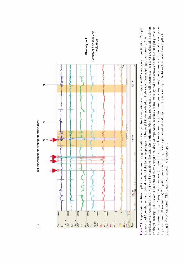
pH-im
peda
nce
mon
itorin
g on
med
icat
ion
S
(a)
SS
Ph
eno
typ
e 1
Per
sist
ent a
cid
reflu
x on
med
icat
ion
1/20
:45
RR
RR
Tim
e
04100
5000
5.0
cm
pH3.0
cm
5.0
cm
7.0
cm
Ohm
Ohm
Ohm
pH1
Z6
Z5
Z4
9.0
cm
Ohm
Z3
15.0
cm
Ohm
Z2
17.0
cm
Ohm
Z1
0
5000
0
5000
000
5000
5000
5000
1/21
:00
1/21
:15
7.0
4.0
Pla
te 7
.2
Repre
sen
tati
ve 4
0-m
in p
H im
pedan
ce m
on
itori
ng o
n m
edic
ati
on
giv
en
for
thre
e p
ati
en
ts w
ith
typic
al G
ER
D s
ym
pto
ms
on
medic
ati
on
. Th
e p
H
was
reco
rded 5
cm
above t
he p
roxim
al bord
er
of
the low
er
eso
ph
ageal sp
hin
cter
(LE
S)
dete
rmin
ed b
y h
igh
-reso
luti
on
eso
ph
ageal m
an
om
etr
y. T
he
impedan
ce w
as
reco
rded 3
, 5, 7, 9, 15 a
nd 1
7 c
m a
bove t
he L
ES. Th
e h
ori
zon
tal bla
ck lin
e r
epre
sen
ts p
H 4
. A
ll o
ccu
rren
ces
of
pH
<4 a
re s
haded in
salm
on
on
th
e p
H t
raci
ng. R
efl
ux e
ven
ts (
R)
defi
ned b
y a
n a
bru
pt
50%
dro
p o
f im
pedan
ce b
ase
lin
e a
re in
dic
ate
d b
y r
ed h
ead a
rrow
an
d s
haded in
lig
ht
pu
rple
on
the im
pedan
ce t
raci
ngs.
Sym
pto
m o
ccu
rren
ce (
S)
is in
dic
ate
d b
y b
lack
arr
ow
an
d t
he 2
-min
peri
od p
rece
din
g s
ym
pto
m o
ccu
rren
ce is
shaded in
ora
nge o
n
impedan
ce a
nd p
H t
raci
ngs.
(a)
Th
e p
ati
en
t pre
sen
ted w
ith
pers
iste
nt
path
olo
gic
al aci
d e
xposu
re d
esp
ite e
som
epra
zole
40 m
g b
.i.d
(eso
ph
ageal pH
<4
du
rin
g 8
.9%
of
tota
l ti
me).
Th
is p
att
ern
corr
esp
on
ds
to p
hen
oty
pe 1
.
bins.indd 3 11/15/2012 2:34:43 AM
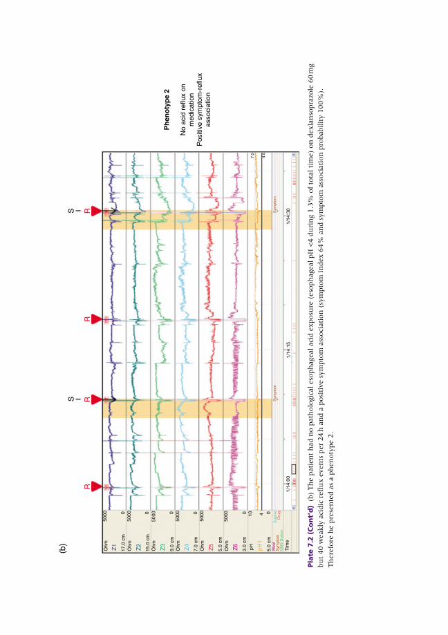
Ph
eno
typ
e 2
No
acid
ref
lux
onm
edic
atio
nP
ositi
ve s
ympt
om-r
eflu
xas
soci
atio
n
1/14
:00
1/14
:15
1/14
:30
Tim
e
000000
5000
5000
5000
5000
5000
5000
R
S(b
)
RR
RS
410
5.0
cm
3.0
cm
Ohm
Ohm
Ohm
Ohm
Ohm
Ohm
5.0
cm
7.0
cm
9.0
cm
15.0
cm
17.0
cm
pH pH1
Z6
Z5
Z4
Z3
Z2
Z1
Pla
te 7
.2 (
Co
nt’
d)
(b)
Th
e p
ati
en
t h
ad n
o p
ath
olo
gic
al eso
ph
ageal aci
d e
xposu
re (
eso
ph
ageal pH
<4 d
uri
ng 1
.3%
of
tota
l ti
me)
on
dexla
nso
pra
zole
60 m
g
bu
t 40 w
eakly
aci
dic
refl
ux e
ven
ts p
er
24 h
an
d a
posi
tive s
ym
pto
m a
ssoci
ati
on
(sy
mpto
m in
dex 6
4%
an
d s
ym
pto
m a
ssoci
ati
on
pro
babilit
y 1
00%
).
Th
ere
fore
he p
rese
nte
d a
s a p
hen
oty
pe 2
.
bins.indd 4 11/15/2012 2:34:46 AM

S(c
)
1/13
:00
04100
50000
50000
50000
50000
50000
5000
1/13
:15
1/13
:30
SS
S
Ph
eno
typ
es 3
/4
No
acid
ref
lux
onm
edic
atio
nN
o si
gnifi
cant
ass
ocia
tion
betw
een
sym
ptom
and
reflu
x
Tim
e
Tim
e
5.0
cm
3.0
cm
Ohm
Ohm
Ohm
Ohm
Ohm
Ohm
5.0
cm
7.0
cm
9.0
cm
15.0
cm
17.0
cm
pH pH1
Z6
Z5
Z4
Z3
Z2
Z1
Pla
te 7
.2 (
Co
nt’
d)
(c)
Th
e p
ati
en
t pre
sen
ted w
ith
GE
RD
sym
pto
ms
desp
ite e
som
epra
zole
40 m
g b
.i.d
. Th
e p
H im
pedan
ce o
n m
edic
ati
on
reveale
d a
norm
al
eso
ph
ageal aci
d e
xposu
re (
0.6
%),
25 r
efl
ux e
ven
ts p
er
24 h
an
d a
negati
ve s
ym
pto
m a
ssoci
ati
on
(sy
mpto
m in
dex 0
%).
Th
is p
ati
en
t co
uld
pote
nti
ally b
e a
ph
en
oty
pe 3
or
4. Stu
die
s to
dete
rmin
e a
n a
ppro
pri
ate
nu
mber
of
refl
ux e
ven
ts o
n P
PI
thera
py t
hat
clearl
y d
isti
ngu
ish
ph
en
oty
pes
3 a
nd 4
are
sti
ll lack
ing.
bins.indd 5 11/15/2012 2:34:48 AM

Plate 10.1 Normal laryngeal tissue. AC, arytenoid complex; AMW, arytenoid medial
wall; FVF, false vocal fold; PCW, posterior cricoid wall; PPW, phosterior pharyngeal
wall; TVF, true vocal fold. Adapted from Vaezi et al . [1] with permission from Elsevier.
(a) (b) (c)
(d) (e) (f)
(g) (h) (i)
Plate 10.2 Abnormal larynx. (a) Leukoplakia. (b) Reinke’s edema. (c) Bilateral true vocal cord nodules.
(d) True vocal fold hemorrhagic polyp. (e) True vocal fold erythema. (f) Vocal fold granuloma.
(g) Interarytenoid bar. (h) Arytenoid medial wall erythema. (i) Posterior pharyngeal wall cobblestoning.
Adapted from Vaezi et al . [1] with permission from Elsevier.
bins.indd 6 11/15/2012 2:34:50 AM
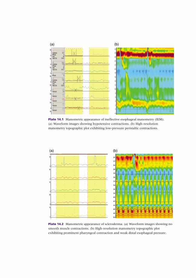
Plate 14.1 Manometric appearance of ineffective esophageal manometry (IEM).
(a) Waveform images showing hypotensive contractions. (b) High-resolution
manometry topographic plot exhibiting low-pressure peristaltic contractions.
(a) (b)
(a) (b)
Plate 14.2 Manometric appearance of scleroderma. (a) Waveform images showing no
smooth muscle contractions. (b) High-resolution manometry topographic plot
exhibiting prominent pharyngeal contraction and weak distal esophageal pressure.
bins.indd 7 11/15/2012 2:34:58 AM
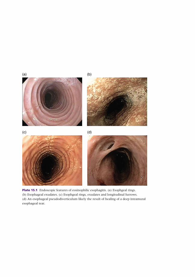
(a) (b)
(c) (d)
Plate 15.1 Endoscopic features of eosinophilic esophagitis. (a) Esophgeal rings.
(b) Esophageal exudates. (c) Esophgeal rings, exudates and longitudinal furrows.
(d) An esophageal pseudodiverticulum likely the result of healing of a deep intramural
esophageal tear.
bins.indd 8 11/15/2012 2:35:07 AM