Pancreaticobiliary reflux as a high-risk factor for biliary ...
8
Reiji Sugita Reiji Sugita, Department of Radiology, Sendai City Medical Center, Sendai, Miyagi 983-8520, Japan Author contributions: Sugita R solely contributed to this paper. Conflict-of-interest statement: None. Open-Access: This article is an open-access article which was selected by an in-house editor and fully peer-reviewed by external reviewers. It is distributed in accordance with the Creative Commons Attribution Non Commercial (CC BY-NC 4.0) license, which permits others to distribute, remix, adapt, build upon this work non-commercially, and license their derivative works on different terms, provided the original work is properly cited and the use is non-commercial. See: http://creativecommons.org/ licenses/by-nc/4.0/ Correspondence to: Reiji Sugita, MD, Department of Radiology, Sendai City Medical Center, 5-22-1, Tsurugaya, Miyagino-ku, Sendai, Miyagi 983-8520, Japan. [email protected] Telephone: +81-22-2521111 Fax: +81-22-2529431 Received: January 27, 2015 Peer-review started: January 28, 2015 First decision: March 6, 2015 Revised: March 18, 2015 Accepted: April 16, 2015 Article in press: April 20, 2015 Published online: July 8, 2015 Abstract Pancreaticobiliary junction is composed of complex structure with which biliary duct and pancreatic duct assemble and go out into the ampulla of Vater during duodenum wall surrounding the sphincter of Oddi. Although the sphincter of Oddi functionally prevents the reflux of pancreatic juice, pancreaticobiliary reflux (PBR) occurs when function of the sphincter of Oddi halt. The anatomically abnormal junction is termed pancreaticobiliary maljunction (PBM) and is characterized by pancreatic and bile ducts joining outside of the duodenal wall. PBM is an important anatomical finding because many studies have revealed that biliary malignancies are related due to the carcinogenetic effect of the pancreatic back flow on the biliary mucosa. On the other hand, several studies have been published on the reflux of pancreatic juice into the bile duct without morphological PBM, and the correlation of such cases with biliary diseases, especially biliary malignancies, is drawing considerable attention. Although it has long been possible to diagnose PBM by various imaging modalities, PBR without PBM has remained difficult to assess. Therefore, the pathological features of PBR without PBM have not been yet fully elucidated. Lately, a new method of diagnosing PBR without PBM has appeared, and the features of PBR without PBM should soon be better understood. Key words: Pancreaticobiliary maljunction; Pancreas juice; Reflux; Flow; Magnetic resonance imaging © The Author(s) 2015. Published by Baishideng Publishing Group Inc. All rights reserved. Core tip: Pancreaticobiliary reflux (PBR) is an important pathologic state that can cause biliary malignancy. PBR can occur regardless of whether the patient has pancreaticobiliary maljunction (PBM) or not. Although it has long been possible to diagnose PBM by various imaging modalities, PBR without PBM has remained difficult to assess. Therefore, the pathological features of PBR without PBM have not been yet fully elucidated. Lately, a new method of diagnosing PBR without PBM has appeared, and the features of PBR without PBM should soon be better understood. Sugita R. Pancreaticobiliary reflux as a high-risk factor for biliary malignancy: Clinical features and diagnostic advancements. EDITORIAL Submit a Manuscript: http://www.wjgnet.com/esps/ Help Desk: http://www.wjgnet.com/esps/helpdesk.aspx DOI: 10.4254/wjh.v7.i13.1735 1735 July 8, 2015|Volume 7|Issue 13| WJH|www.wjgnet.com World J Hepatol 2015 July 8; 7(13): 1735-1741 ISSN 1948-5182 (online) © 2015 Baishideng Publishing Group Inc. All rights reserved. Pancreaticobiliary reflux as a high-risk factor for biliary malignancy: Clinical features and diagnostic advancements
-
Upload
khangminh22 -
Category
Documents
-
view
0 -
download
0
Transcript of Pancreaticobiliary reflux as a high-risk factor for biliary ...
Reiji Sugita
Reiji Sugita, Department of Radiology, Sendai City Medical Center, Sendai, Miyagi 983-8520, Japan
Author contributions: Sugita R solely contributed to this paper.
Conflict-of-interest statement: None. Open-Access: This article is an open-access article which was selected by an in-house editor and fully peer-reviewed by external reviewers. It is distributed in accordance with the Creative Commons Attribution Non Commercial (CC BY-NC 4.0) license, which permits others to distribute, remix, adapt, build upon this work non-commercially, and license their derivative works on different terms, provided the original work is properly cited and the use is non-commercial. See: http://creativecommons.org/licenses/by-nc/4.0/
Correspondence to: Reiji Sugita, MD, Department of Radiology, Sendai City Medical Center, 5-22-1, Tsurugaya, Miyagino-ku, Sendai, Miyagi 983-8520, Japan. [email protected] Telephone: +81-22-2521111 Fax: +81-22-2529431
Received: January 27, 2015Peer-review started: January 28, 2015First decision: March 6, 2015Revised: March 18, 2015 Accepted: April 16, 2015 Article in press: April 20, 2015Published online: July 8, 2015
AbstractPancreaticobiliary junction is composed of complex structure with which biliary duct and pancreatic duct assemble and go out into the ampulla of Vater during duodenum wall surrounding the sphincter of Oddi. Although the sphincter of Oddi functionally prevents the reflux of pancreatic juice, pancreaticobiliary reflux (PBR) occurs when function of the sphincter of Oddi halt. The anatomically abnormal junction is termed
pancreaticobiliary maljunction (PBM) and is characterized by pancreatic and bile ducts joining outside of the duodenal wall. PBM is an important anatomical finding because many studies have revealed that biliary malignancies are related due to the carcinogenetic effect of the pancreatic back flow on the biliary mucosa. On the other hand, several studies have been published on the reflux of pancreatic juice into the bile duct without morphological PBM, and the correlation of such cases with biliary diseases, especially biliary malignancies, is drawing considerable attention. Although it has long been possible to diagnose PBM by various imaging modalities, PBR without PBM has remained difficult to assess. Therefore, the pathological features of PBR without PBM have not been yet fully elucidated. Lately, a new method of diagnosing PBR without PBM has appeared, and the features of PBR without PBM should soon be better understood.
Key words: Pancreaticobiliary maljunction; Pancreas juice; Reflux; Flow; Magnetic resonance imaging
© The Author(s) 2015. Published by Baishideng Publishing Group Inc. All rights reserved.
Core tip: Pancreaticobiliary reflux (PBR) is an important pathologic state that can cause biliary malignancy. PBR can occur regardless of whether the patient has pancreaticobiliary maljunction (PBM) or not. Although it has long been possible to diagnose PBM by various imaging modalities, PBR without PBM has remained difficult to assess. Therefore, the pathological features of PBR without PBM have not been yet fully elucidated. Lately, a new method of diagnosing PBR without PBM has appeared, and the features of PBR without PBM should soon be better understood.
Sugita R. Pancreaticobiliary reflux as a high-risk factor for biliary malignancy: Clinical features and diagnostic advancements.
EDITORIAL
Submit a Manuscript: http://www.wjgnet.com/esps/Help Desk: http://www.wjgnet.com/esps/helpdesk.aspxDOI: 10.4254/wjh.v7.i13.1735
1735 July 8, 2015|Volume 7|Issue 13|WJH|www.wjgnet.com
World J Hepatol 2015 July 8; 7(13): 1735-1741ISSN 1948-5182 (online)
© 2015 Baishideng Publishing Group Inc. All rights reserved.
Pancreaticobiliary reflux as a high-risk factor for biliary malignancy: Clinical features and diagnostic advancements
World J Hepatol 2015; 7(13): 1735-1741 Available from: URL: http://www.wjgnet.com/1948-5182/full/v7/i13/1735.htm DOI: http://dx.doi.org/10.4254/wjh.v7.i13.1735
INTRODUCTIONThe pancreaticobiliary junction is a complex structure composed of the biliary and pancreatic ducts, and is surrounded by the sphincter of Oddi. Although the sphincter of Oddi precludes the reflux of pancreatic juice into the bile duct, pancreaticobiliary reflux (PBR) may occur in case where the functioning of the sphincter of Oddi is impaired[1,2].
An anatomically abnormal pancreaticobiliary junction is termed as a “pancreaticobiliary maljunction” (PBM), and is characterized by the joining of the pancreatic and bile ducts outside the duodenal wall[3,4]. This deformity was first reported in 1916 in case with a choledochal cyst[5]. PBM is an important pathological condition as many studies have indicated that it may be associated with biliary malignancies[6].
Futhermore, several studies have been assessed the reflux of pancreatic juice into the bile duct in the absence of morphological PBM, and the relationship of such cases with biliary diseases, including biliary malignancies, is gaining prominence[2,7-12].
In this editorial, the current knowledge and diagnostic advancements concerning PBR are described.
ETIOLOGYAnatomy and physiologyThe ampulla of Vater is composed of the confluence of the biliary and pancreatic ducts, and the duodenum, and marks the transition from the embryologic foregut to the midgut. The ampulla includes the junction of the common bile duct and the pancreatic duct, the surrounding sphincter of Oddi, the fenestra choledochae, and the duodenal papilla. Although the arrangement of these intersections has some variability, the reported structural relation are common and predictable[13].
The sphincter of Oddi, the regulator of biliary and pancreatic drainage that enable intermittent biliary drainage, originates separately from the duodenal muscu-lature and the periductal mesenchyme, and subseqently becomes incorporated into the duodenal wall. Anatomical studies suggest that the papilla and the duodenal wall contained components specific to the common channel, as well as components specific to the pancreatic and common bile duct that extend outside the duodenal wall.
The common channel is present in 55%-90% of cases[14-16]. The common channel ranges in length from 1 to 12 mm, with an a mean length of approximately 4 mm[14,17,18]. In addition to controlling secretion into the duodenum, the sphincter of Oddi also preclude the mixing of bile and pancreatic juice within the duct system. The long common channels with PBM may cause to become
the sphincter mechanism impair and induce the reflux of pancreatic juice, as the pressure of the pancreatic duct is higher than that of the bile duct[2].
However, PBR in patients without PBM is being increasingly recognized[2,7-12], and may develop due to a number of reasons: dysfunction of the sphincter of Oddi, periamupullary diverticula, endoscopic sphincterotomy, and endoscopic papillary balloon dilatation. Of these, most cases of PBR without PBM seem to be caused by dysfunction of the sphincter of Oddi[2].
Etiology of cancerAlthough the etiology of biliary cancer in patients with PBR is not yet fully understood, PBR is believed to play an important role in bile duct carcinoma. Once PBR occurs in the pancreaticobiliary junction, the pancreatic and bile juices become mixed and regurgitated mutually, and stagnate in the gallbladder and the bile duct. As a result, activated pancreatic enzymes induce chronic inflammation of the biliary tree and lead to proliferation of the biliary epithelium; moreover this can even cause the expression of carcinogenic substances in the biliary system[19-22]. Therefore, the carcinogenesis process in cases of biliary cancer with PBR is believed to involve the hyperplasia-dysplasia-carcinoma sequence. Several factors that are reportedly related to carcinogenesis in case of biliary cancer have been identified including p53 mutations, microsatellite instability, Bcl-2, and k-Ras point mutations[6,23-28].
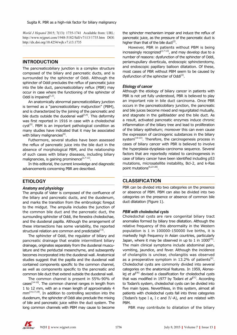
CLASSIFICATION PBR can be divided into two categories on the presence or absence of PBM. PBM can also be divided into two categories on the presence or absence of common bile duct dilatation (Figure 1).
PBM with choledochal cystsCholedochal cysts are rare congenital biliary tract anomalies formed by biliary tree dilatation. Although the relative frequency of this abnormality in the Western population is 1 in 100000-150000 live births, it is markedly high frequency in Asian countries, particularly Japan, where it may be observed in up to 1 in 1000[29]. The main clinical symptoms include abdominal pain, vomiting, jaundice, and fever. Although the incidence of cholangitis is unclear, cholangitis was observed as a preoperative symptom in 13.2% of patients[6]. Choledochal cysts are commonly divided into several categories on the anatomical features. In 1959, Alonso-lej et al[30] devised a classification for choledochal cysts that was modified in 1977 by Todani et al[31]. According to Todani’s system, choledochal cysts can be divided into five main types. Neverthless, in this system, almost all patients with choledochal cysts fall into three categories (Todani’s type Ⅰa, Ⅰc and Ⅳ-A), and are related with PBM.
PBR may contribute to dilatation of the biliary
1736 July 8, 2015|Volume 7|Issue 13|WJH|www.wjgnet.com
Sugita R. PBR as a high-risk factor for biliary malignancy
tree[32,33]. Babbit[34] suggested an etiological mechanism for choledochal cyst shaping; it was proposed that enzymes activated in refluxed pancreatic juice induce inflammation, and finaly lead to damage and cystic dilatation of the biliary wall. This theory, however, does not explain the development of all choledochal cysts, as showed by choledochal cysts of newbone infant, which are known to be congenital rather than degenerative.
Biliary tract malignancies are noted in 21.6% of patients with choledochal cysts who are aged > 15 years, whereas biliary malignancies are much rarer in children who present aged < 15 years (0.1%)[6]. Immediate and exact diagnosis of a choledochal cyst, followed by surgical treatment, is crucial consequently.
PBM without choledochal cystsPBM without a choledochal cyst may result in the reflux of pancreatic juice into the bile duct and may present clinical symptoms similar to those observed in case with PBM with a choledochal cyst. Cholangitis was observed as a preoperative symptom in 8.9% of patients[6]. However, only few patients with PBM without a choledochal cyst have symptoms in childhood; thus, in these patients, PBM tends to be diagnosed at a later stage than in those with a choledochal cyst[6,35]. Pancreatic juice is often refluxed into the biliary duct in PBM patients, and this may be associated with a high frequency of biliary cancer among such patients. While both PBM patients with and those without choledochal cysts are at a risk for biliary malignancies, they differ in the site of malignancy in the biliary tract[6].
In a previous study, biliary cancer was noted in 21.6% of adult patients with choledochal cysts, and in 42.2% of patients with PBM without biliary dilatation. Of all cases of biliary cancer associated with PBM without biliary dilatation, 88.1% were cancers of the gallbladder[6].
PBR without PBMLately, several researches on the reflux of pancreatic juice into the bile duct in case without PBM have been published, and the relationship of such a condition with biliary diseases, especially biliary malignancies, is attracting considerable attention[1,2,7-12,36-38]. However, these cases do not present specific clinical symptoms
and also could not be acculately detected by using current available imaging modalities based on the morphological changes. Thus, it was difficult to predict and detect PBR without PBM. Generally, PBR in these patients is diagnosed on the basis of elevated pancreatic enzyme levels in bile juice samples, or by using magnetic resonance cholangiopancreatography (MRCP) after secretin injection.
Anderson et al[9] demonstrated that elevated amylase levels in bile juice taken from indwelling T-tubes, suggestive of PBR were found in 81% (21 of 26) of patients with biliary disease without PBM. Horaguchi et al[8] presented that elevated amylase levels in bile juice after endoscopic retrograde cholangiopancreatography (ERCP) were found in 26% (46 of 178) of patients with a normal junction. Similarly, Sakamoto et al[38] detected elevated amylase levels in bile juice juice in 20% (39 of 196) of patients undergoing cholecystectomy without PBM[33]. Kamisawa et al[2] focused on the hypothesis that a relatively long common channel without PBM may be related to PBR. They defined a high confluence of the pancreaticobiliary ducts as a common channel length of more than 6 mm, in which communication may be sustained even when the sphincter of Oddi is contracted. They reported that a high confluence of the pancreaticobiliary ducts was found in 1.9% (65 of 3459) of patients who underwent ERCP in their single institute, and that the incidences of gallbladder cancer in patients with a high confluence of the pancreaticobiliary ducts was very high compared to that in controls.
Recently, a multi-center trial in Japan revealed that elevated amylase levels in bile juice after ERCP were found in 5.5% (23 of 420) of patients with a normal junction[36]. This trial showed that the presence of a relative long common channel (not shorter than 5 mm) was the only significant factor for PBR in multivariate analysis, and that the incidence of high amylase levels was significantly higher in patients with gallbladder cancer than in those without gallblader cancer.
DIAGNOSIS PBM PBM can be detected with ERCP, ultrasonography (US), endoscopic US (EUS), computed tomography and MRCP. ERCP is the gold standard method for diagnosis of PBM, and pancreatography through the minor duodenal papilla can directly demonstrate pancreatobiliary reflux in PBM patients. When the contrast medium is injected endoscopically through the minor duodenal papilla, it is possible to monitor the reflux of contrast medium into the bile duct through the common channel without outflow into the duodenum[2]. Although ERCP is the diagnostic standard method for PBM, it is somewhat invasive and has a non-negligible risk of morbidity. US can detect choledochal cysts as well as abnormalities of the gallbladder that are possibly associated with PBM. However, because US cannot directly detect PBM, it may
1737 July 8, 2015|Volume 7|Issue 13|WJH|www.wjgnet.com
PBR
PBR without PBMPBR with PBM
Common bile duct Dilatation
(choledochal cyst)No common bile duct dilatation
Figure 1 Classification of pancreaticobiliary reflux. PBR: Pancreaticobiliary reflux; PBM: Panceraticobiliary maljunction.
Sugita R. PBR as a high-risk factor for biliary malignancy
1738 July 8, 2015|Volume 7|Issue 13|WJH|www.wjgnet.com
(time-SLIP) has been developed for direct visualization of pancreatic juice flow (Figure 3). Initially, this technique allowed the examination of blood flow in vessels to a region of interest[42,43]. This technique could enable the visualization of the flow of bile and pancreatic juices and may allow the evaluation of various pancreatico-biliary diseases based on this information[44,45]. This method revealed new knowledge regarding physiology of bile and pancreatic juices, and was applied for the diagnosis of chronic pancreatitis.
Moreover, this new technique enables the visualization of the reflux of pancreatic juice flow into the bile duct without PBM, although no clinical results have yet been reported (Figure 4). This technique, involving the use of the pancreatic juice as an intrinsic imaging agent, facilitates the examination of pancreatic juice movement similar to more physiolosical situation. In addition, this technique is not time consuming. Accordingly, the method can be easily adopted as a screening tool for PBR. Therefore, this new technique may reveal what proportion of patients without PBM have PBR, and whether reflux is related to biliary carcinogenesis. Further clinical studies are required.
THERAPY Once PBM is diagnosed, preventive flow-diversion surgery (biloenteric anastomosis and bile duct resection) is excuted for patients with choledochal cysts[4,6]. These operations are often associated with complications such as anastomotic stricture, cholangitis, and secondary liver cirrhosis; therefore, careful follow-up is needed[46].
Nevertheless, any treatment for patients with PBM without biliary dilatation and biliary malignancy is controversial. Preventive cholecystectomy is first choise in many institutions because the majority of biliary malignancies that develop in PBM patients without biliary dilatation are cancers of the gallbladder[4,6,47,48]. On the other hand, excision of the extrahepatic bile duct along with the gallbladder is selected by some surgeons[4,24,49,50]. The treatment for patients with PBR without PBM is not defined because the pathology of PBR without PBM is less well understood. Moreover, strategies for the screening and prevention of PBR without PBM are not yet established.
CONCLUSIONPBR is an important pathologic state that can cause biliary malignancy. PBR can occur regardless of the presence of PBM. Although it has been possible to diagnose PBM by using various imaging modalities, PBR without PBM has remained difficult to assess. Thus, the pathological features of PBR without PBM have not yet been fully elucidated. Recently, a new method for diagnosing PBR without PBM has been introduced, and this would enable a better understanding the feature of
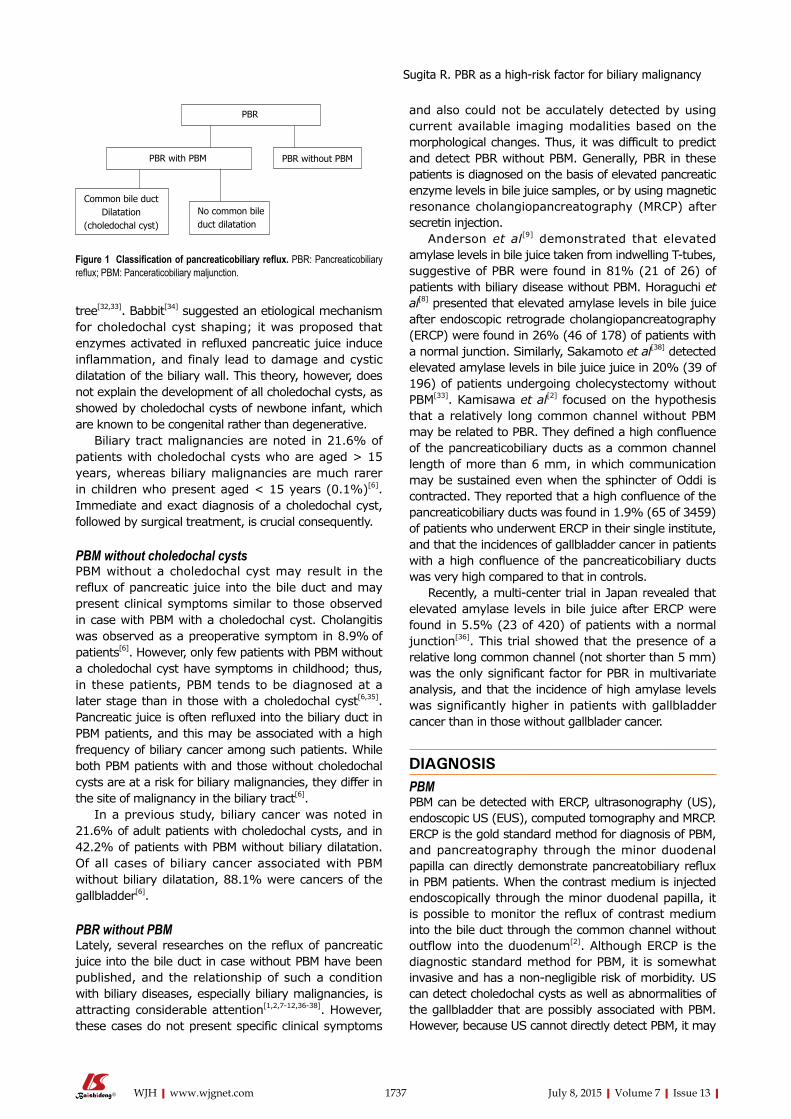
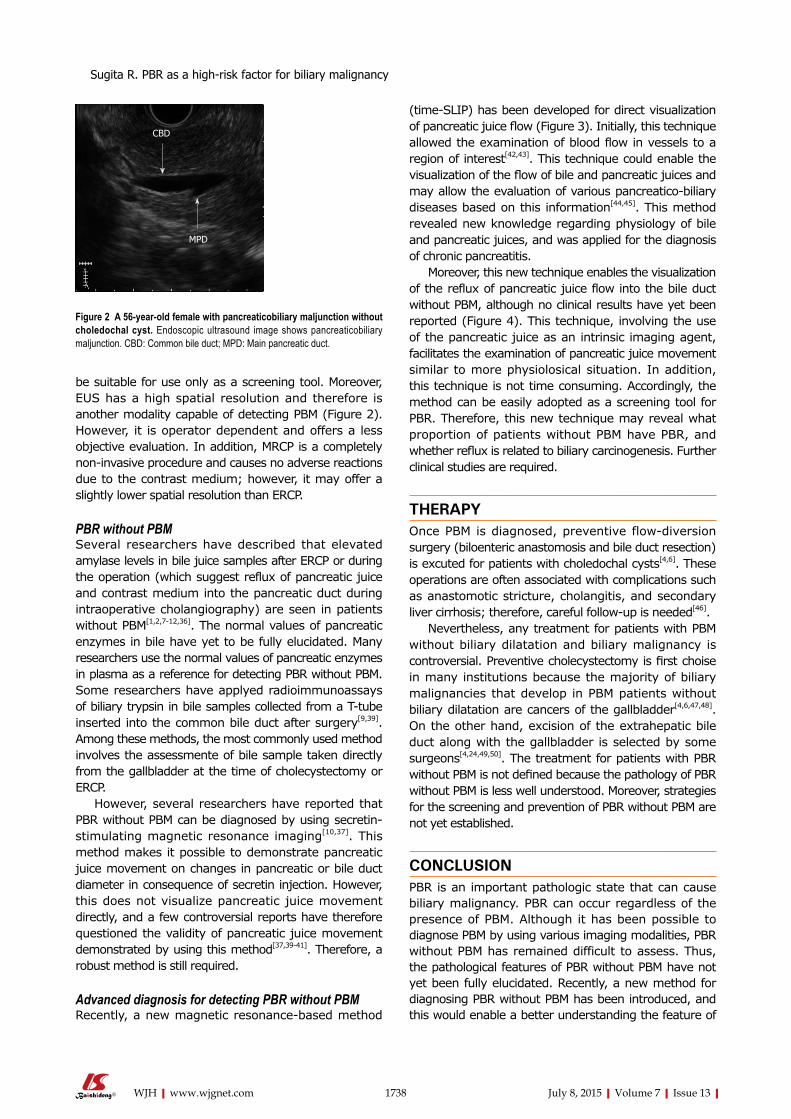
be suitable for use only as a screening tool. Moreover, EUS has a high spatial resolution and therefore is another modality capable of detecting PBM (Figure 2). However, it is operator dependent and offers a less objective evaluation. In addition, MRCP is a completely non-invasive procedure and causes no adverse reactions due to the contrast medium; however, it may offer a slightly lower spatial resolution than ERCP.
PBR without PBMSeveral researchers have described that elevated amylase levels in bile juice samples after ERCP or during the operation (which suggest reflux of pancreatic juice and contrast medium into the pancreatic duct during intraoperative cholangiography) are seen in patients without PBM[1,2,7-12,36]. The normal values of pancreatic enzymes in bile have yet to be fully elucidated. Many researchers use the normal values of pancreatic enzymes in plasma as a reference for detecting PBR without PBM. Some researchers have applyed radioimmunoassays of biliary trypsin in bile samples collected from a T-tube inserted into the common bile duct after surgery[9,39]. Among these methods, the most commonly used method involves the assessmente of bile sample taken directly from the gallbladder at the time of cholecystectomy or ERCP.
However, several researchers have reported that PBR without PBM can be diagnosed by using secretin-stimulating magnetic resonance imaging[10,37]. This method makes it possible to demonstrate pancreatic juice movement on changes in pancreatic or bile duct diameter in consequence of secretin injection. However, this does not visualize pancreatic juice movement directly, and a few controversial reports have therefore questioned the validity of pancreatic juice movement demonstrated by using this method[37,39-41]. Therefore, a robust method is still required.
Advanced diagnosis for detecting PBR without PBMRecently, a new magnetic resonance-based method
Figure 2 A 56-year-old female with pancreaticobiliary maljunction without choledochal cyst. Endoscopic ultrasound image shows pancreaticobiliary maljunction. CBD: Common bile duct; MPD: Main pancreatic duct.
MPD
CBD
Sugita R. PBR as a high-risk factor for biliary malignancy

1739 July 8, 2015|Volume 7|Issue 13|WJH|www.wjgnet.com
PBR without PBM.
REFERENCES1 Williams NE, Gundara JS, Hugh TJ, Samra JS. Many faces of
pancreaticobiliary reflux. ANZ J Surg 2012; 82: 403-407 [PMID: 22548700 DOI: 10.1111/j.1445-2197.2012.06076.x]
2 Kamisawa T, Anjiki H, Egawa N, Kurata M, Honda G, Tsuruta K. Diagnosis and clinical implications of pancreatobiliary reflux. World J Gastroenterol 2008; 14: 6622-6626 [PMID: 19034962 DOI: 10.3748/wjg.14.6622]
3 Kamisawa T, Ando H, Hamada Y, Fujii H, Koshinaga T, Urushihara N, Itoi T, Shimada H. Diagnostic criteria for pancreaticobiliary maljunction 2013. J Hepatobiliary Pancreat Sci 2014; 21: 159-161 [PMID: 24307541 DOI: 10.1002/jhbp.57]
4 Kamisawa T, Ando H, Shimada M, Hamada Y, Itoi T, Takayashiki T, Miyazaki M. Recent advances and problems in the management of pancreaticobiliary maljunction: feedback from the guidelines committee. J Hepatobiliary Pancreat Sci 2014; 21: 87-92 [PMID: 23798483 DOI: 10.1002/jhbp.8]
5 Kozumi K. A case of cystic dilatation of the common bile duct and etiology of the disease (in Japanese). Tokyo Med J 1916; 30: 1413-1423
6 Morine Y, Shimada M, Takamatsu H, Araida T, Endo I, Kubota M, Toki A, Noda T, Matsumura T, Miyakawa S, Ishibashi H, Kamisawa T, Shimada H. Clinical features of pancreaticobiliary maljunction: update analysis of 2nd Japan-nationwide survey. J
Hepatobiliary Pancreat Sci 2013; 20: 472-480 [PMID: 23579999 DOI: 10.1007/s00534-013-0606-2]
7 Beltrán MA. Current knowledge on pancreaticobiliary reflux in normal pancreaticobiliary junction. Int J Surg 2012; 10: 190-193 [PMID: 22361306 DOI: 10.1016/j.ijsu.2012.02.009]
8 Horaguchi J, Fujita N, Noda Y, Kobayashi G, Ito K, Takasawa O, Obana T, Endo T, Nakahara K, Ishida K, Yonechi M, Hirasawa D, Suzuki T, Sugawara T, Ohhira T, Onochi K, Harada Y. Amylase levels in bile in patients with a morphologically normal pancreaticobiliary ductal arrangement. J Gastroenterol 2008; 43: 305-311 [PMID: 18458847 DOI: 10.1007/s00535-008-2158-9]
9 Anderson MC, Hauman RL, Suriyapa C, Schiller WR. Pancreatic enzyme levels in bile of patients with extrahepatic biliary tract disease. Am J Surg 1979; 137: 301-306 [PMID: 434320 DOI: 10.1016/0002-9610(79)90055-2]
10 Sai JK, Ariyama J, Suyama M, Kubokawa Y, Sato N. Occult regurgitation of pancreatic juice into the biliary tract: diagnosis with secretin injection magnetic resonance cholangiopancreatography. Gastrointest Endosc 2002; 56: 929-932 [PMID: 12447317 DOI: 10.1016/S0016-5107(02)70379-9]
11 Sai JK, Suyama M, Kubokawa Y, Tadokoro H, Sato N, Maehara T, Iida Y, Kojima K. Occult pancreatobiliary reflux in patients with a normal pancreaticobiliary junction. Gastrointest Endosc 2003; 57: 364-368 [PMID: 12612517 DOI: 10.1067/mge.2003.53]
12 Itokawa F, Itoi T, Nakamura K, Sofuni A, Kakimi K, Moriyasu F, Tsuchida A, Aoki T. Assessment of occult pancreatobiliary reflux in patients with pancreaticobiliary disease by ERCP. J Gastroenterol 2004; 39: 988-994 [PMID: 15549453 DOI: 10.1007/s00535-004-1
Figure 3 A 47-year-old male with normal volunteer. A: Magnetic resonance cholangiopancreatography image. Labelling pulse of pancreas juice is applying to box surrounded by dotted lines on pancreas juice in body and caudal portion of the 3) main pancreatic duct; B: Time-SLIP image is not showing movement of pancreatic juice; C: Flow of pancreatic juice from body of the pancreas into head of pancreas is noted by high signal intensity (arrows). Reprinted from Sugita et al[43] (by permission of Wiley Periodicals, Inc). CBD: Common bile duct; MPD: Main pancreatic duct.
A B C
MPD
CBD
Figure 4 A 75-year-old male with common bile duct carcinoma without pancreaticobiliary maljunction. A: Magnetic resonance cholangiopancreatography image shows the irregular wall throughout the entire common bile duct, with bile duct carcinoma revealed by biopsy. During endoscopic retrograde cholangiopancreatography, amylase level was measured in the bile. The amylase level in the collected bile was 1415 IU/L, higher than the upper limit of serum amylase of 130 IU/L; B and C: Labeled pancreatic juice in the main pancreatic duct refluxing into the common bile duct. Reprinted from Sugita et al[43] (by permission of Wiley Periodicals, Inc).
B CA
Sugita R. PBR as a high-risk factor for biliary malignancy
1740 July 8, 2015|Volume 7|Issue 13|WJH|www.wjgnet.com
428-4]13 Cao W, Forssell KL, Nieman DR, Marino D, Yu J. Ampullary
tumors and tumor like lesions: histopathological classification, diagnosis and surgical treatment. In: Mercado MA. Bile duct: functional anatomy, disease and injury classification and surgical management. New York: Nova, 2014: 223-252
14 Sterling JA. The common channel for bile and pancreatic ducts. Surg Gynecol Obstet 1954; 98: 420-424 [PMID: 13146495]
15 Suda K, Miyano T, Konuma I, Matsumoto M. An abnormal pancreatico-choledocho-ductal junction in cases of biliary tract carcinoma. Cancer 1983; 52: 2086-2088 [PMID: 6627217]
16 Hand BH. An anatomical study of the choledochoduodenal area. Br J Surg 1963; 50: 486-494 [PMID: 13952484 DOI: 10.1002/bjs.18005022303]
17 Dowdy GS, Waldron GW, Brown WG. Surgical anatomy of the pancreatobiliary ductal system. Observations. Arch Surg 1962; 84: 229-246 [PMID: 13887616 DOI: 10.1001/archsung.1962.0130020007706]
18 Rienhoff WF, Pickrell KL. Pancreatitis; an anatomic study of the pancreatic and extrahepatic biliary systems. Arch Surg 1945; 51: 205-219 [PMID: 21013164 DOI: 10.1001/archsung.1945.01230040214001]
19 Shimada K, Yanagisawa J, Nakayama F. Increased lysopho-sphatidylcholine and pancreatic enzyme content in bile of patients with anomalous pancreaticobiliary ductal junction. Hepatology 1991; 13: 438-444 [PMID: 1999314 DOI: 10.1002/hep.1840130310]
20 Funabiki T, Matsubara T, Miyakawa S, Ishihara S. Pancrea-ticobiliary maljunction and carcinogenesis to biliary and pancreatic malignancy. Langenbecks Arch Surg 2009; 394: 159-169 [PMID: 18500533 DOI: 10.1007/s00423-008-0336-0]
21 Wistuba II, Albores-Saavedra J. Genetic abnormalities involved in the pathogenesis of gallbladder carcinoma. J Hepatobiliary Pancreat Surg 1999; 6: 237-244 [PMID: 10526058 DOI: 10.1007/s005340050113]
22 Hanada K, Tsuchida A, Kajiyama G. Cellular kinetics and gene mutations in gallbladder mucosa with an anomalous junction of pancreaticobiliary duct. J Hepatobiliary Pancreat Surg 1999; 6: 223-228 [PMID: 10526056 DOI: 10.1007/s005340050111]
23 Kamisawa T, Ando H, Suyama M, Shimada M, Morine Y, Shimada H. Japanese clinical practice guidelines for pancreaticobiliary maljunction. J Gastroenterol 2012; 47: 731-759 [PMID: 22722902 DOI: 10.1007/s00535-012-0611-2]
24 Funabiki T. Pancreaticobiliary maljunction-focused on biliary carcinogenesis (in Japanese). Jpn J Gastroenterol Surg 2000; 33: 261-270 [DOI: 10.5833/jjgs.33.261]
25 Matsubara T, Sakurai Y, Sasayama Y, Hori H, Ochiai M, Funabiki T, Matsumoto K, Hirono I. K-ras point mutations in cancerous and noncancerous biliary epithelium in patients with pancreaticobiliary maljunction. Cancer 1996; 77: 1752-1757 [PMID: 8608574 DOI: 10.1002/(SICI)1097-0142(19960415)77:8]
26 Hanada K, Itoh M, Fujii K, Tsuchida A, Hirata M, Ishimaru S, Iwao T, Eguchi N, Kajiyama G. Pathology and cellular kinetics of gallbladder with an anomalous junction of the pancreaticobiliary duct. Am J Gastroenterol 1996; 91: 1007-1011 [PMID: 8633539]
27 Nagai M, Watanabe M, Iwase T, Yamao K, Isaji S. Clinical and genetic analysis of noncancerous and cancerous biliary epithelium in patients with pancreaticobiliary maljunction. World J Surg 2002; 26: 91-98 [PMID: 11898040 DOI: 10.1007/s00268-001-0187-0]
28 Ichikawa Y, Kamiyama M, Sekido H, Ishikawa T, Miura Y, Kamiya N, Morita T, Shimada H. Telomerase activity and Bcl-2 expression in gallbladders of pancreaticobiliary maljunction patients: a preliminary study. J Hepatobiliary Pancreat Surg 2004; 11: 34-39 [PMID: 15754044 DOI: 10.1007/s00534-003-0860-9]
29 Miyano T, Yamataka A. Choledochal cysts. Curr Opin Pediatr 1997; 9: 283-288 [PMID: 9229170 DOI: 10.1097/00008480-199706000-00018]
30 Alonso-lej F, Rever WB, Pessagno DJ. Congenital choledochal cyst, with a report of 2, and an analysis of 94, cases. Int Abstr Surg 1959; 108: 1-30 [PMID: 13625059]
31 Todani T, Watanabe Y, Narusue M, Tabuchi K, Okajima K.
Congenital bile duct cysts: Classification, operative procedures, and review of thirty-seven cases including cancer arising from choledochal cyst. Am J Surg 1977; 134: 263-269 [PMID: 889044 DOI: 10.1016/0002-9610(77)90359-2]
32 Oguchi Y, Okada A, Nakamura T, Okumura K, Miyata M, Nakao K, Kawashima Y. Histopathologic studies of congenital dilatation of the bile duct as related to an anomalous junction of the pancreaticobiliary ductal system: clinical and experimental studies. Surgery 1988; 103: 168-173 [PMID: 3340986]
33 Komi N, Udaka H, Ikeda N, Kashiwagi Y. Congenital dilatation of the biliary tract; new classification and study with particular reference to anomalous arrangement of the pancreaticobiliary ducts. Gastroenterol Jpn 1977; 12: 293-304 [PMID: 590702]
34 Babbitt DP. [Congenital choledochal cysts: new etiological concept based on anomalous relationships of the common bile duct and pancreatic bulb]. Ann Radiol (Paris) 1969; 12: 231-240 [PMID: 5401505]
35 Tsuchida S, Aoki T, Ozawa T, Inoue K, Mimuro A, Ikeda T, Nakamura R, Kitamura K, Koyanagi Y. Surgical treatment of pancreaticobiliary maljunction without bile duct dilatation in adult cases. In: Koyanagi Y, Aoki T, editors. Pancreaticobiliary maljunction. Tokyo: Igaku Tosho, 2002: 331-337
36 Horaguchi J, Fujita N, Kamisawa T, Honda G, Chijiiwa K, Maguchi H, Tanaka M, Shimada M, Igarashi Y, Inui K, Hanada K, Itoi T, Hamada Y, Koshinaga T, Fujii H, Urushihara N, Ando H. Pancreatobiliary reflux in individuals with a normal pancreaticobiliary junction: a prospective multicenter study. J Gastroenterol 2014; 49: 875-881 [PMID: 23736796 DOI: 10.1007/s00535-013-0837-7]
37 Motosugi U, Ichikawa T, Araki T, Kitahara F, Sato T, Itakura J, Fujii H. Secretin-stimulating MRCP in patients with pancreatobiliary maljunction and occult pancreatobiliary reflux: direct demonstration of pancreatobiliary reflux. Eur Radiol 2007; 17: 2262-2267 [PMID: 17447071 DOI: 10.1007/s00330-007-0640-z]
38 Sakamoto H, Mutoh H, Ido K, Satoh S, Kumagai M, Hayakawa H, Tamada K, Sugano K. Intestinal metaplasia in gallbladder correlates with high amylase levels in bile in patients with a morphologically normal pancreaticobiliary duct. Hum Pathol 2009; 40: 1762-1767 [PMID: 19716161 DOI: 10.1016/j.humpath.2009.06.008]
39 Vracko J, Markovic S, Wiechel KL. Conservative treatment versus endoscopic sphincterotomy in the initial management of acute cholecystitis in elderly patients at high surgical risk. Endoscopy 2006; 38: 773-778 [PMID: 17001566 DOI: 10.1055/s-2006-925448]
40 Song HK, Kim MH, Lee SK, Lee SS. Progressive bile duct and gallbladder dilation on MRCP after secretin stimulation: physiologic or pathologic finding? Gastrointest Endosc 2003; 58: 165; author reply 165-166 [PMID: 12838255 DOI: 10.1053/ge.2003.v58.03303]
41 Kanno N, LeSage G, Glaser S, Alpini G. Regulation of cho-langiocyte bicarbonate secretion. Am J Physiol Gastrointest Liver Physiol 2001; 281: G612-G625 [PMID: 11518673]
42 Sugita R. Magnetic resonance evaluations of biliary malignancy and condition at high-risk for biliary malignancy: Current status. World J Hepatol 2013; 5: 654-665 [PMID: 24432183 DOI: 10.4254/wjh.v5.i12.654]
43 Sugita R, Furuta A, Horaguchi J, Itoh K, Kobayashi G, Noda Y, Fujita N, Shimizu S, Miyazaki M, Takahashi S. Visualization of pancreatic juice movement using unenhanced MR imaging with spin labeling: preliminary results in normal and pathophysiologic conditions. J Magn Reson Imaging 2012; 35: 1119-1124 [PMID: 22180260 DOI: 10.1002/jmri.23533]
44 Sugita R, Furuta A, Yamazaki T, Itoh K, Fujita N, Takahashi S. Direct visualization of pancreatic juice flow using unenhanced MRI with spin labeling can be aid in diagnosing chronic pancreatitis. AJR Am J Roentgenol 2014; 202: 1027-1034 [PMID: 24758655 DOI: 10.2214/AJR.13.10886]
45 Torigoe T, Ito K, Yamamoto A, Kanki A, Yasokawa K, Tamada T, Yoshida K. Age-related change of the secretory flow of pancreatic juice in the main pancreatic duct: evaluation with cine-dynamic MRCP using spatially selective inversion recovery pulse. AJR Am J Roentgenol 2014; 202: 1022-1026 [PMID: 24758654 DOI:
Sugita R. PBR as a high-risk factor for biliary malignancy
1741 July 8, 2015|Volume 7|Issue 13|WJH|www.wjgnet.com
10.2214/AJR.13.10852]46 Takeshita N, Ota T, Yamamoto M. Forty-year experience with
flow-diversion surgery for patients with congenital choledochal cysts with pancreaticobiliary maljunction at a single institution. Ann Surg 2011; 254: 1050-1053 [PMID: 21659852 DOI: 10.1097/SLA.0b013e3182243550]
47 Ohuchida J, Chijiiwa K, Hiyoshi M, Kobayashi K, Konomi H, Tanaka M. Long-term results of treatment for pancreaticobiliary maljunction without bile duct dilatation. Arch Surg 2006; 141: 1066-1070 [PMID: 17116798 DOI: 10.1001/archsurg.141.11.1066]
48 Kusano T, Takao T, Tachibana K, Tanaka Y, Kamachi M, Ikematsu Y, Nishiwaki Y, Kida H, Waki S, Uchimura M, Furukawa M. Whether or not prophylactic excision of the extrahepatic bile duct
is appropriate for patients with pancreaticobiliary maljunction without bile duct dilatation. Hepatogastroenterology 2005; 52: 1649-1653 [PMID: 16334749]
49 Ishida M, Niguma T, Yukawa T, Mimura T, Tsutsui M. A case of lower bile duct cancer associated with pancreaticobiliary maljunction without bile duct dilatation after operation of a gallbladder cancer. Jpn J Gastroenterol Surg 2007; 40: 1623-1629 [DOI: 10.5833/jjgs.40.1623]
50 Matsubara T, Sakurai Y, Zhi LZ, Miura H, Ochiai M, Funabiki T. K-ras and p53 gene mutations in noncancerous biliary lesions of patients with pancreaticobiliary maljunction. J Hepatobiliary Pancreat Surg 2002; 9: 312-321 [PMID: 12353142 DOI: 10.1007/s0053402000035]
P- Reviewer: Baran B, Bubnov RV, Solinas A S- Editor: Tian YL L- Editor: A E- Editor: Liu SQ
Sugita R. PBR as a high-risk factor for biliary malignancy

© 2015 Baishideng Publishing Group Inc. All rights reserved.
Published by Baishideng Publishing Group Inc8226 Regency Drive, Pleasanton, CA 94588, USA
Telephone: +1-925-223-8242Fax: +1-925-223-8243
E-mail: [email protected] Desk: http://www.wjgnet.com/esps/helpdesk.aspx
http://www.wjgnet.com