Gastroesophageal reflux and Helicobacter pylori: a review
154
Gastroesophageal reflux and Helicobacter pylori: a review F Pace and G Bianchi Porro Reviews Subject headings gastroesophageal reflux/therapy; Helicobacter pylori; epidemiology; peptic ulcer/therapy; stomach neoplasms/therapy; Helicobacter infections Pace F, Porro GB. Gastroesophageal reflux and Helicobacter pylori: a review. World J Gastroentero, 2000;6(3):311-314 INTRODUCTION Since the observation by Labenz et al that eradication of Helicobacter pyl ori ( Hp) infection may be followed by development of reflux esophagitis in arelevant proportion of duodenal ulcer patients previously not affected by gastro esophageal reflux disease (GERD) [1] , a growing attention has been given to the potential interactions between Hp and GERD. Epidemiological studies have now demonstrated that the prevalence of GERD is steadily increasing in the developed countries [2] , as is the incidence of adenocarcinoma of the esophagus [3] , its most dangerous complication, while the prevalence of peptic ulc er and gastric cancer is falling [4] , in parallel with a falling prevale nce of Hp infection in the western countries [5] . It is therefore t empting to causally relate these phenomena. Despite the number of original paper s and of reviews dealing with this topic, at least 3 issues are still debated: Does Hp infection interfere with the pathogenesis of GERD? Is the anti secretory effect of Hp infection of any clinical relevance in the management of GERD patients Does long- term proton pump inhibitors (PPI) therapy accel erate development of atrophic changes in Hp +ve GERD patients? Finally, the relationship(s) between Hp and Barrett’s esophagus may deserve some importance. The present review will focus on these 4 issues. The interested reader may also refer to some recent papers, dealing with the same subject [6-9] . Hp AND PATHOGENESIS OF GERD Several studies have now convincingly shown that the prevalence of Hp infect ion in patients with reflux esophagitis is somewhat lower than in normal subjects; in a careful review of 26 papers on this topic, O’Connor summarizes the data existing as follows: the overall prevalence of Hp infection in 2182 adult GERD patients is 40.3%, as compared with 50.2% in the 2010 controls [9] . He concludes that this “difference in prevalence (is) int imating that the pathogenesis of GERD might be related in some way to the absenc e of Hp”. In our view, more simplistically, the only link between Hp infection and GERD lies on the degree of gastric acid secretion, and through this, on esophageal acid exposure. In patients with a predisposition to GERD but without a clinical manifestation of GERD (symptoms and/or esophageal lesi ons), eradication of Hp may trigger it, disclosing the clinical picture. On the contrary, patients harboring the infection, may be protected if the infec tion involves the corpus (i.e. the acid-producing part of the gastric muc osa), because the amount of acid secretion and hence the esophageal acid exposur e is reduced. Infact, no single paper has ever been published so far focusing on Hp infection as a pathogenetic (aggressive or defensive) factor of GERD-perse. On the contrary, El-Serag et al have now clearly demonstrated th at, for the above reasons, corpus gastritis is protective against reflux esophag itis [10] . They have investigated 302 subjects, 154 of whom with endoscopic signs of esophagitis; there was no differe nce between patients with and controls without esophagitis in the overall infect ion rates with Hp infection. Compared with controls, corpus gastritis was less frequent and less severe in patients with esophagitis. Finally, in a multi variate logistic analysis, age, sex smoking status, and the presence of chronic corpus gastritis exerted a significant influence on the presence of reflux esoph agitis. This latter variable, however, showed an odds ratio of 0.46% only (95% confidence interval of 0.27-0.79), a value which is, albeit statistically sig nificant, of doubtful clinical relevance. In summary, the pathogenetic relationship Department of Gastroenterology, L. Sacco University Hospital, Milan, Italy Dr. Fabio Pace graduated from the Palermo Medical University in 1979. He is at present senior consultant at the Department and Chair of Gastroenterology of Milan, “L. Sacco” Hospital, Milan, Italy. His main interests are in the field of GI Motility, GERD disease and functional gastrointestinal disorders. He is author of more than 150 original papers. Correspondence to: G. Bianchi Porro, Divisione di Gastroenterolo gia, Ospedale Polo Universitario “L. Sacco”, Via G. B. Grassi, 74, I- 20157 Milano Tel. +39-02-35799438, Fax. +39-02-35799232 Email. [email protected] Received 2000-03-20 Accepted 2000-04-28 PO Box 2345, Beijing 100023, China World J Gastroentero, 2000; 6(3):311-314 Fax: +86-10-85381893 World Journal of Gastroenterology E-mail: [email protected] www.wjgnet.com Copyright2000 by the WJG Press ISSN 1007-9327
-
Upload
khangminh22 -
Category
Documents
-
view
3 -
download
0
Transcript of Gastroesophageal reflux and Helicobacter pylori: a review

Gastroesophageal reflux and Helicobacter pylori:a review
F Pace and G Bianchi Porro
Reviews
Subject headings gastroesophageal reflux/therapy;Helicobacter pylori; epidemiology; peptic ulcer/therapy;stomach neoplasms/therapy; Helicobacter infections
Pace F, Porro GB. Gastroesophageal reflux and Helicobacterpylori: a review. World J Gastroentero, 2000;6(3):311-314
INTRODUCTIONSince the observation by Labenz et al thateradication of Helicobacter pyl ori (Hp) infectionmay be followed by development of refluxesophagitis in arelevant proportion of duodenal ulcerpatients previously not affected by gastroesophageal reflux disease (GERD)[1], a growingattention has been given to the potential interactionsbetween Hp and GERD. Epidemiological studieshave now demonstrated that the prevalence of GERDis steadily increasing in the developed countries[2],as is the incidence of adenocarcinoma of theesophagus[3], its most dangerous complication, whilethe prevalence of peptic ulc er and gastric cancer isfalling[4], in parallel with a falling prevale nce of Hpinfection in the western countries[5] . It is thereforet empting to causally relate these phenomena.Despite the number of original paper s and ofreviews dealing with this topic, at least 3 issues arestill debated: Does Hp infection interfere withthe pathogenesis of GERD? Is the anti secretoryeffect of Hp infection of any clinical relevance inthe management of GERD patients Does long-term proton pump inhibitors (PPI) therapy accelerate development of atrophic changes in Hp +veGERD patients? Finally, the relationship(s) betweenHp and Barrett’s esophagus may deserve someimportance.
The present review will focus on these 4 issues.The interested reader may also refer to some recentpapers, dealing with the same subject[6-9].
Hp AND PATHOGENESIS OF GERDSeveral studies have now convincingly shown that theprevalence of Hp infect ion in patients with refluxesophagitis is somewhat lower than in normal subjects;in a careful review of 26 papers on this topic, O’Connorsummarizes the data existing as follows: the overallprevalence of Hp infection in 2182 adult GERD patientsis 40.3%, as compared with 50.2% in the 2010 controls[9]. He concludes that this “difference in prevalence(is) int imating that the pathogenesis of GERD mightbe related in some way to the absenc e of Hp”. In ourview, more simplistically, the only link between Hpinfection and GERD lies on the degree of gastric acidsecretion, and through this, on esophageal acidexposure. In patients with a predisposition to GERDbut without a clinical manifestation of GERD (symptomsand/or esophageal lesi ons), eradication of Hp maytrigger it, disclosing the clinical picture. On the contrary,patients harboring the infection, may be protected ifthe infec tion involves the corpus (i.e. the acid-producingpart of the gastric muc osa), because the amount ofacid secretion and hence the esophageal acid exposure is reduced. Infact, no single paper has ever beenpublished so far focusing on Hp infection as apathogenetic (aggressive or defensive) factor ofGERD-perse. On the contrary, El-Serag et al havenow clearly demonstrated th at, for the above reasons,corpus gastritis is protective against reflux esophag itis[10]. They have investigated 302 subjects, 154 of whomwith endoscopic signs of esophagitis; there was nodiffere nce between patients with and controls withoutesophagitis in the overall infect ion rates with Hpinfection. Compared with controls, corpus gastritis wasless frequent and less severe in patients withesophagitis. Finally, in a multi variate logistic analysis,age, sex smoking status, and the presence of chroniccorpus gastritis exerted a significant influence on thepresence of reflux esoph agitis. This latter variable,however, showed an odds ratio of 0.46% only (95%confidence interval of 0.27-0.79), a value which is,albeit statistically sig nificant, of doubtful clinicalrelevance. In summary, the pathogenetic relationship
Department of Gastroenterology, L. Sacco University Hospital, Milan,ItalyDr. Fabio Pace graduated from the Palermo Medical University in1979. He is at present senior consultant at the Department and Chairof Gastroenterology of Milan, “L. Sacco” Hospital, Milan, Italy. Hismain interests are in the field of GI Motility, GERD disease and functionalgastrointestinal disorders. He is author of more than 150 original papers.Correspondence to: G. Bianchi Porro, Divisione di Gastroenterologia, Ospedale Polo Universitario “L. Sacco”, Via G. B. Grassi, 74, I-20157 MilanoTel. +39-02-35799438, Fax. +39-02-35799232Email. [email protected] 2000-03-20 Accepted 2000-04-28
PO Box 2345, Beijing 100023, China World J Gastroentero, 2000; 6(3):311-314Fax: +86-10-85381893 World Journal of GastroenterologyE-mail: [email protected] www.wjgnet.com Copyright 2000 by the WJG Press ISSN 1007-9327

312 ISSN 1007-9327 CN 14-1018/ R World J Gastroentero June 2000 Volume 6 Number 3
between Hp infection and GERD are probablyweak and of indirect nature, being related to theamount of gastric acid secretion, a factor which isnecessary but not indispensable for inducing GERD.The most relevant GERD pathogenetic factor is,as universally kn own, the occurrence of transientrelaxation of the lower esophageal sphincter[11], afactor which has not, to the best of our knowledge,been observed to be influenced by Hp gastricinfection.
Is the antisecretory effect of Hp infection of anyclinical relevance in the management of GERDpatientsA profound inhibition of acid secretion is the mainstayof treatment for reflux esophagitis, in particular incases of moderate to severe RE[12]. There fore, theinfluence of Hp on the efficacy of acid-loweringtreatment may be important for patients with RE.Verdu et al[13]showed that o meprazole produces agreater decrease in gastric acidity in subjects withHp infection than in those who are Hp negative, andthat omeprazole produces a smaller decrease ingastric acidity after Hp infection has been cured[ 14].Similar findings have been obtained by Labenz et al[15], who showed that in 17 FU patients, Hperadication resulted in a marked decrease of the pH-increasing effect of omeprazole (24h median gastricpH: 5.5 vs 3.0, P<0.002) that was most pronouncedduring night time. Base line intragastric pH remainedunchanged after eradication (median gastric pH: 1.0 vs 1.1, P = 0.05). The same authors have alsoshown that this effect persisted for at least 1 yearafter Hp eradication[16], whereas others have beenshown that it is shared by other PPIs, suchas lansoprazole[17]. Despite this Hp mediated exaggeration of theeffect of acid-suppressive drugs on intragastric pHis clearly proven, there is little evidence that thiseffect has any clinical relevance for the treatmentof GERD patients with PPI. One reason is that theeffect, due to the logaritmich scale of pH, a variationof one pH unit from 5 to 6 is 10 000 times lessimportant than a variation from 1 to 2. The smallvariation in acid secretory capacity due to Hpcolonisation is only “visible” when the acid secretionis already potently red uced by PPI, but is otherwiseunimportant. Direct evidence shows in fact that, during acidsuppressive therapy with ranitid ine or omeprazole,Hp +ve or -ve GERD patients show a similarreduction of esophageal acid exposure, the entityof which is only influenced by the type of drugreceived[18]. Furthermore, both groups of GERDpatients require the same dose of omeprazoleduring long-term maintenance treatment to preventsym ptomatic and endoscopic relapse[19], and Hpstatus seems not to be an important prognostic
factor during long-term maintenance therapy with PPI;in a study conducted on 103 patients with RE grade 1 or2, randomized to mainten ance therapy with lansoprazole15 or 30 mg daily for 12 months, it was obse rved thatHp infected patients relapsed as early as patients whowere not infected[20]. The only discordant piece of evidence comes fromthe very large study of Holtman n et al[21], who claimsof a significantly better acute response of Hp +ve GERDpatients treated with the PPI pantoprazole incomparison to Hp -ve; however, the difference ofhealing rates between the two groups after 8wk of40mg daily was quite small (96.4% vs 91.8%, P<0.05)and no difference at all was observed in GERDsymptoms between infected and noninfected patients.There is therefore enough evidence to say, at least,that PPI maintenance therapy does not need to betitrated upon Hp status [19]. It is therefore to be fullyagreed upon the recommendation that “tes ting for Hpinfection is not indicated in patients on long termtreatment or in those considered for treatment with aproton pump i nhibitor for GERD”, as stated by therecent guidelines of the American College ofGastroenterology[22].
Does long-term proton pump inhibitors (PPI)therapy accelerate development of atrophic inHp +ve GERD patientsSeveral studies have shown that treatment with PPI isassociated with the worsen ing of gastritis (increase inseverity score, spreading from the antrum to corpu sand fundus)[23-25]. Because superficial corpus gastritismay lead to atrophic gastritis, the increased bodyinflammation in Hp positive patients observed duringshort term PPI therapy may lead to atrophic gastritisduring lo ng term PPI treatment. This has beenobserved so far after omeprazole administra tion[26],but the study was criticized in particular for theincorporati on of an inappropriate control group[27].Moreover, the findings hav e not been confirmed by arandomized Swedish study comparing the efficay ofomep razole maintenance treatment and antirefluxsurgery over a 3-years follow-up[28]. Thus, on thebasis of available evidence, long-term treatment withPPI up to 10 years appears to be a perfectly safetherapy[29].
Hp INFECTION AND BARRETT’ S ESOPHAGUSThe interest in BE is still growing since the earlydescription of this entity in 1950[30] for two mainreasons: BE is associated with GERD, an dalso with an increased risk of adenocarcinoma[31],thus representing a link between a common benigncondition and a rare very malignant disease; The incidence of adenocarcinoma of the esophagusand cardia is increasing at the fastest rate amonggas tro intes t ina l (and a lso n on GI) hu man
Pace F, et al. Gastroesophageal reflux and Helicobacter pylori: a review 313
cancers [3]. Since Hp exhibits a special affinity forgastric-type epithelium, and since Barrett’s metaplasiacontains columnar-lined epithelium, it is to be expectedthat Hp will also be able to attach the Barrett’s epithelium, at least of the gastric type, independently from anyinvolvment of Hp infection in the pathogenesis ofesophageal mucosal inflammation. It seems that the prevalence of Hp infection ofthe stomach in BE patients is not different from thatexbibited by controls, ro ughly one third of the subjects[9]. The colonization of metaplastic epit helium by t hebacterium has been tested only in a minority of studies,but appears to be ma rginally lower[9]. It seemstherefore that the stomach represents the primary siteof infection, with secundary colonization of columnarmucosa in the esophagus. Furthermore, most Hppositive patients show a very low bacterial load in theirmetaplastic epithelium, and no significant difference hasbeen fo und in the severity of inflammatory changesbetween Hp +ve and Hp -ve Barrett’ s esophaguspatients[32]. Finally, recent work has confirme d thatwithin the esophagus, Hp adheres only to gastric typemetaplasia , which is not considered premalignant foradenocarcinoma[33]. In conclusion, it is most probablethat Hp has no ethiologic role on the development ofBarr ett’s esophagus, nor in the esophagitis associatedwith this metaplastic cha nge; the colonization ofBarrett’s epithelium probably reflects only a shift fromgastric antrum. Another intriguing point is the prevalence of Hpinfection and the intestina l metaplasia of thegastric cardia. It is in fact at present not knownwhether i nflammation of the cardia indicatesGERD and/or is a manifestation of pang astritiscaused by Hp. Recently two studies have shedsome light on this iss ue[34,35]: in the first, biopsieswere obtained from the antrum, corpus and cardiafrom 135 Hp-infected patients with gastritis, ulcerdisease, o r RE. One hundred and thirty-two (97.7%) of them showed active card itis, resemblingantral g astritis in most patients, but with lessmarked bacterial density and inflammato ryprocess[34]. The authors conclude that Hp gastritiscommonly involves the cardia, that intestinalmetaplasia in the cardia is a common findi ng inHp gastritis, but that the cardia lower histologicdensi ty of the bact er ia and inf lammatoryresponses in comparison to the antrum are notclear. In th e second work[35], 22 GERD patientsand 11 controls were compared in relationship toendoscopic and bioptic evaluation of inflammation,Hp infection and intestinal metaplasia in distalesophagus, cardia, fundus and antrum. It turnedout that neither the prevalence of Hp infection( c o n t r o l s 4 8 % ; G E R D 4 1 % ) n o r c a r d i ainflammation (controls 41%; GERD 40%) differedbetween the two groups. All 11 controls and 22 of
23 (96%) patient s with GERD and cardiainflammation had HP infection. Cardia intestinalmetapl asia was more common among controls(22%) than among GERD patients (3%, P £¼0.01);all patients with cardia intestinal metaplasia hadcardia inflammati on, 7 had Hp infection, and 6 hadmetaplasia elsewhere in the stomach. The authorsconclude that the prevalence of cardia inflammationis similar in patie nts with and without GERD, and isassociated with Hp infection. Also, in thi s study,cardia intestinal metaplasia is associated with Hprelated cardia inflammation (P = 0.01) and intestinalmetaplasia elsewhere in the stoma ch, indicating thati t i s d i s t i nc t f rom Bar r e t t ’ s e sophagus . The final point is the association, if any, betweenH p i n f e c t i o n a n d B a r re t t ’s a s s o c i a t e dadenocarcinoma. Again, two recent works havecontributed to t he improvement of our knowledgeon this previously uninvestigated issue[36,37]. Qudduset al report on 19 cases of adenocarcinoma arisingin BE, wh o were examined for the presence of Hpafter staining with three different techniques: allsections of BE, with or without dysplasia,adenocarcinoma and stomach (when available) wereuniformly negative for the presence of Hp.Theauthors conclude that neither gastric nor esophagealinfection with Hp is a requisite for the developmentof adenocarcinoma in BE[36]. The second study aimed at comparing theprevalence of Hp and increasing grad es of dysplasia.Biopsies from 19 malignant and 94 benign cases of BEwere analy sed histologically for Hp; 34% of non-dysplastic Barrett’ s epithelium was colonized with Hpcompared with only 17% of dysplastic/malignant-cases (P = 0.04). No relationship was found betweenHp status and length of BE; the presence ofstrictures or ulcers; previous anti-reflux surgery.The authors therefore confirmed that Hp colonizationof BE is not p articularly common, and that a negativecorrelation exists with increasing sever ity ofdysplasia[37]. To summarize, from both studies it appears that itis unlikely that a a causal relationship exists betweenHp infection and Barrett’ s associated adeno carcinoma.
REFERENCES1 Labenz J, Blum AL, Bayerdorffer E, Meining A, Stolte M, Borsh
G. Curing Helicobacter pylori infection in patients with duodenalulcer may provoke reflux esophagitis. Gastroenterology, 1997;112:1442-1447
2 Howard PJ, Heading RC. Epidemiology of gastro-oesophageal re-flux disease. World J Surg, 1992;16:288-293
3 Pera M, Cameron AJ, Trastek VF, Carpenter HA, Zinsmeister AR.Increasing incidence of adenocarcinoma of the esophagus andesophagogastric junction. Gastroenterology, 1993;104:910-913
4 El-Serag HB, Sonnenberg A. Opposite time trends of peptic ulcerand reflux disease. Gut, 1998;43:327-333
5 Marshall BJ. Epidemiology of H. pylori in western countries. in:Hunt RH, Tytgat GN.Helicobacter pylori. Basic mechanism toclinical cure. Dordrecht: Kluwer Acad Pub, 1994:75-84
6 Xia HH, Talley NJ. Helicobacter pylori infection, reflux esophagitisand atrophic gastritis: an unexplored triangle. Am J Gastroenterol,
314 ISSN 1007-9327 CN 14-1018/ R World J Gastroentero June 2000 Volume 6 Number 3
1998;93:394-4007 Richter JE,Falk GW,Vaezi MF. Helicobacter pylori and gastroe-
sophageal reflux disease: the bug may not be all bad. Am JGastroenterol, 1998;93:1800-1802
8 Pace F, Bianchi Porro G. Gastro-oesophageal reflux andHelicobacter pylori. Ital J Gastroenterol Hepatol, 1998;30(Suppl3):289-293
9 O’ Connor HJ. Helicobacter pylori and gastro-oesophageal refluxdisease clinical implications and management. Aliment PharmacolTher, 1999;13:117-127
10 El-Serag BH, Sonnenberg A, Jamal MM, Inadomi JM, Crooks LA,Feddersen RM. Corpus gastritis is protective against refluxoesophagitis. Gut, 1999;45:181-185
11 Dodds WJ, Dent J, Hogan WJ, Helm JF, Hauser R, Patel GK, EgideMS. Mechanism of gastroesophageal reflux in patients with refluxesophagitis. N Engl J Med, 1982;307:1547-1552
12 Chiba N, De Gara CJ, Wilkinson JM, Hunt RH. Speed of healingand symptom relief in grade II to IV gastroesophageal reflux disease:a meta-analysis. Gastroenterology, 1997;112:1798-1810
13 Verdu EF, Armstrong D, Fraser R, Viani F, Idstrom J, Cederberg C,Blum AL. Effect of Helicobacter pylori status on intragastric pHduring treatment with omeprazole. Gut, 1995;36:539-543
14 Verdu EF, Armstrong D, Idstrom JP, Labenz J, Stolte M, Dorta G,Borsch G, Blum AL. Effect of curing Helicobacter pylori infectionon intragastric pH during treatment with omeprazole. Gut, 1995;37:743-748
15 Labenz J, Tillenburg B, Peitz U, Idstrom JP, Verdù E, Stolte M,Blum AL.Helicobacter pylori augments the pHincreasing effect ofomeprazole in patients with duodenal ulcer. Gastroenterology,1996;110:725-732
16 Labenz J, Tillenburg B, Peitz U, Idstrom JP, Verdù E, Stolte M,Bosch G, Blum AL. Efficacy of omeprazole one year after cure ofHelicobacter pylori infection in duodenal ulcer patients. Am JGastroenterol, 1997;92:576-578
17 Van Herwaarden MA, Samson M, Van Nispen CHM, Mulder PGH,Smout AJP. The effect of Helicobacter pylori eradication onintragastric pH during dosing with lansoprazole or ranitidine. Ali-ment Pharmacol Ther, 1999;13:731-740
18 Peters FT, Kuipers EJ, Ganesh S, Sluiter WJ, Klinkenberg-KnolEC, Lamers CB, Kleibeuker JH. The influence of Helicobacterpylori on esophageal acid exposure in GERD during acid suppres-sive therapy. Aliment Pharmacol Ther, 1999;13:921-926
19 Schenk BE, Kuipers EJ, Klinkenberg-Knol EC, Eskes SA,Meuwissen SGM.Helicobacter pylori and the efficacy of omeprazoletherapy for gastroesophageal reflux disease. Am J Gastroenterol,1999;94:884-887
20 Hatlebakk JG, Berstad A. Prognostic factors for relapse of refluxoesophagitis and symptoms during 12 months of therapy withomeprazole. Aliment Pharmacol Ther, 1997;11:1093-1099
21 Holtmann G, Cain C, Malfertheiner P. Gastric Helicobacter pyloriinfection accelerates healing of reflux esophagitis during treat-m e n t w i t h t h e p r o t o n p u m p i n h i b i t o r p a n t o p r a z o l e .Gastroenterology, 1999;117:11-16
22 Howden CW, Hunt RH. Guidelines for the management of
Helicobacter pylori infection. Am J Gastroenterol, 1998;93:2330-2338
23 Kuipers EJ, Uyterlinde AM, Pena AS, Hazenberg HJA, BloemenaE, Lindeman J, Klinkberg Knol E, Meuwissen SGNM.Increase ofHelicobacter pyloriassociated corpus gastritis during acid-suppres-sive therapy: implications for longterm safety. Am J Gastroenterol,1995;90:1401-1406
24 Logan RPH, Walker MM, Misiewicz JJ, Gummett PA, Karim QN,Baron JH. Changes in intragastric distribution of Helicobacterpylori during treatment with omeprazole. Gut, 1995;36:12-16
25 Eissele R, Brunner G, Simon B, Solcia E, Arnold R. Gastric mucosaduring treatment with lansoprazole: Helicobacter pylori is a riskfactor for argyrophil cell hyperplasia. Gastroenterology, 1997;112:707-715
26 Kuipers EJ, Lundell L, Klinkenberg-Knol EC, Havu N, FestenHPM, Liedman B, Lamers CBHW, Jansen JBMJ, Dalenback J,Snel P, Nelis GF, Meuwissen SGNM.Atrophic gastritis andHelicobacter pylori infection in patients with reflux esophagitistreated with omeprazole or fundoplication. N Eng J Med, 1996;334:1018-1022
27 Genta RM. Acid suppression and gastric atrophy: sifting fact fromfiction. Gut, 1998;43(Suppl 1):35-38
28 Lundell L, Miettinen P, Myrvold HE, Pedersen SA, Thor K,Andersson A, Hattlebakk J, Janatuinen E, Lecander C, Liedman B,Nystrom P, and the NORDIC GERD STUDY GROUP. Lack ofe f fec t o f ac id suppress ion the rapy on gas t r i ca t rophy .Gastroenterology, 1999;117:319-326
29 Labenz J. Does Helicobacter pylori affect the management ofgastroesophageal reflux disease? (Editorial). Am J Gastroenterol,1999;94:867-869
30 Barrett NR. Chronic peptic ulcer of the oesophagus and“oesophagitis”. Br J Surg, 1950;38:175-182
31 Hameeteman W. Barrett’ s esophagus and adenocarcinoma. In:Bianchi Porro G, Pace F, eds. Argomenti di patologia esofagea,Vol. 3. Milano: Springer Verlag Italia, 1998:5-27
32 Loffeld RJLF, Ten Tije BJ, Arends JW. Prevalence and signifi-cance of Helicobacter pylori in patients with Barrett’s esophagus.Am J Gastroenterol, 1992;87:1598-1600
33 Sharma VK, Demian SE, Taillon D, Vasuveda R, Howden CW.Examination of tissue distribution of Helicobacter pylori withincolumnar-lined esophagus. Dig Dis Sci, 1999;44:1165-1168
33 Hackelsberger A, Guenther T, Schultze V, Labenz J, Roessner A,Malfertheiner P. Prevalence and pattern of Helicobacter pylori gas-tritis in the gastric cardia. Am J Gastroenterol, 1997;92:2220-2224
35 Goldblum JR, Vicari JJ, Falk GW, Rice TW, Peek RM, Easley K,Richter JE. Inflammation and intestinal metaplasia of the gastriccardia: the role of gastr oesophageal reflux and H. pylori infection.Gastroenterology, 1998;114:633-639
36 Quddus MR, Henley JD, Sulaiman RA, Palumbo TC, Gnepp DR.Helicobacter infection and adenocarcinoma arising in Barrett’sesophagus.Hum Pathol,1997;28:1007-1009
37 Wright TA, Myskow M, Kingsnorth AN. Helicobacter pylori colo-nization of Barrett’s esophagus and its progression to cancer. DisEsophagus, 1997;10:196-200
Edited by Pan BRproofread by Ma JY

Management of difficult inflammatory boweldisease: where are we now?
D.S. Rampton
Subject headings inflammatory bowel diseases/therapy;colitis/therapy; Crohn disease/therapy; endoscopy,gastrointestinal; social support; azathioprine
Rampton DS.Management of difficult inflammatory bowel disease:where are we now? World J Gastroentero, 2000;6(3):315-323
INTRODUCTIONMedical care of patients with inflammatory boweldisease (IBD) comprises general measures and specificpharmacological, nutritional, endoscopic and surgicaltherapies (Table 1)[1-3]. In this paper, current managementoptions for patients with two commonly difficultpresentations of IBD, acute severe ulcerative colitis (UC)and steroid-refractory or dependent ileocaecal Crohn’sdisease (CD), are discussed. Practical considerationsand newer developments are emphasized.
MANAGEMENT OF ACUTE SEVERE ULCERATIVE
COLITISThese patients should be admitted immediately to agastroenterology ward for close joint medical, surgicaland nursing care. The nutrition team and a stomatherapist in patients likely to need surgery should beinvolved promptly. Patients undergoing an acute attackof UC need to be made aware from the outset thatthey have a one in four chance of failing to respondto the primary treatment (intravenous steroids), andthus need either cyclosporin or colectomy during theiradmission (Table 1).
Establishing the diagnosis, extent and severity ofdiseaseA carefully targeted history and appropriate investigationscan help establish the diagnosis (Table 2) in patientspresenting for the first time and, in those with establishedUC, to exclude infection and to assess disease extent (ifnot already known) and severity.
Blood and stool tests Stool should be sent to look for
pathogens, and serology checked for amoebiasis,strongyloidiasis and schistosomiasis. Blood tests arebetter for establishing the activity of UC than makingthe diagnosis or identifying its extent. However, a raisedplatelet count is more common in UC than in infectivecolitis. The best measures of disease activity arehaemoglobin, platelet count, ESR, C-reactive protein[4]
and serum albumin.
Sigmoidoscopy and rectal biopsy Cautious rigid orflexible sigmoidoscopy in the unprepared patient, andwithout excessive air insufflation, provides immediateconfirmation of active colitis. Sigmoidoscopy also allowsbiopsy for histology: to minimise the risks of bleedingand perforation a small superficial biopsy should be takenfrom the posterior rectal wall less than 10 cm from theanal margin using small-cupped forceps. Anecdotally,colonoscopy may cause c olonic perforation and dilatationin acute severe UC, and although some authorities havereported that it is both safe and useful for decision-making[5], mo st patients can be managed satisfactorily withoutit. In patients with establish ed UC, rectal biopsy is notroutinely necessary. However, in those presenting forthe first time, infective colitis may be suggested by anacute, focal and superficial inflammatory infiltrate withminimal goblet cell depletion and preservation of cryptarchitecture[6]. Although colitis due to Clostridiumdifficile, cytomegalovirus, amoebiasis and Crohn’sdisease often has characteristic macroscopicappearances, histology may confirm these diagnoses.
Plain abdominal X-ray A plain film at presentationcan be used to assess disease extent, since faecalresidual visible on X-ray usually indicates sites ofuninflamed colonic mucosa. Plain abdominal X-rayis also used to assess disease severity and inparticular to exclude colonic dilatation (diameter >5.5 cm) in sick patients, however, the gas pattern ona plain film may be misleading if there has beenexcessive air insufflation during a sigmo idoscopy orcolonoscopy done shortly beforehand. In patients withsuspected colo nic perforation, the diagnosis can beconfirmed by erect chest X-ray or a later al decubitusabdominal film.
Radiolabelled leucocyte scans The intensity andextent of colonic uptake one hour after injection ofautologous 99Tc-HMPAO or 111Indium-labelledleukocytes provides information about disease
Reader & Consultant Gastroenterologist, Gastrointestinal?ScienceResearch Unit and Digestive Diseases Research Centre,St Bartholomew’s& Royal London School of Medicine and Dentistry, London E1 2AD,UKCorrespondence to: Dr. DS Rampton, Royal London Hospital, London?E1 1BB, UKTel +44-171-7442, Fax. +44-171-7441Email. [email protected] 2000-02-22 Accepted 2000-04-22
PO Box 2345, Beijing 100023, China World J Gastroentero, 2000; 6(3):315-323Fax: +86-10-85381893 World Journal of GastroenterologyE-mail: [email protected] www.wjgnet.com Copyright 2000 by the WJG Press ISSN 1007-9327

activity and particularly extent, respectively, where doubtexists in patients with UC. Colonic uptake of leucocytesis not of course specific for UC and positive results areobtained in other inflammatory colonic diseases.
Table 1 Principles of management of acute severe ulcerativecolitis
GENERAL MEASURES Explanation, psychosocial support
- patient support groups Specialist multidisciplinary care
- physicians, surgeons, nutrition team, nurses,stoma therapist,counsellor
ESTABLISHING THE DIAGNOSIS, EXTENT/SITE AND SEVERITY- clinical evaluation- FBC, ESR, C-reactive protein, albumin, LFTs, amoebic serology- stool microscopy, culture, C. difficile toxin- limited sigmoidoscopy and biopsy- plain abdominal X-ray- consider radiolabelled leucocyte scan
MONITORING PROGRESS- daily clinical assessment- stool chart- 4-hrly temperature, pulse- daily FBC, ESR, C-reactive protein, urea andelectrolytes, albumin- daily plain abdominal X-ray
SUPPORTIVE TREATMENT- i.v. fluids, electrolytes (Na, K), blood transfusion- nutritional supplementation- heparin s.c.- haematinics (folate)-avoid antidiarrhoeals (codeine, loperamide,diphenoxylate),opiates, NSAIDs- rolling manoeuvre (if colon dilating)
SPECIFIC TREATMENTMedical -corticosteroids i.v. (hydrocortisone or
methylprednisolone) then p.o. (prednisolone)-continue 5-ASA p.o. in patients already taking it;otherwise start when improvement begins-antibiotics for very sick febrile patients, or wheninfection suspected-consider cyclosporin i.v. then p.o.) for steroid non-responders at 4-7 days
Surgical (for non-responders at 5-7 days, toxic megacolon,perforation, massive haemorrhage)- panproctocolectomy with ileoanal pouch or permanent ileostomy- subtotal colectomy with ileorectal anastomosis (rarely)
Table 2 Management of active ileocaecal Crohn’ s disease.General measures, monitoring progress and supportivetreatment are essentially as for ulcerative colitis
ESTABLISHING THE DIAGNOSIS, EXTENT/SITE AND SEVERITY- clinical evaluation- FBC, ESR, C-reactive protein, ferritin, folate, B12,albumin, LFTs, Ca, Mg, Zn- stool microscopy, culture, C difficile toxin- plain abdominal X-ray- consider colonoscopy and biopsy, small bowelbarium radiology,ultrasound, CT, MRI, leucocyte scan
SPECIFIC TREATMENT(separately or in combination)Medical - corticosteroids i.v. (hydrocortisone or methyl
prednisolone) then p.o. (prednisolone or budesonide CR)- continue high dose mesalazine (Pentasa or Asacol)in patients already taking it; otherwise start whenimprovement begins- consider metronidazole, ciprofloxacin; also broadspectrum antibiotics for very sick febrile patients,or when infection/collection suspected-consider azathioprine/6-mercaptopurine (slowresponse) or anti TNF antibodies (infliximab) forsteroid non-responders
Nutritional - liquid formula dietEndoscopic - balloon dilatationSurgical - resection or stricturoplasty
Monitoring progressProgress is monitored by twice daily clinicalassessment, stool chart and 4-hourly measurementof temperature and pulse. Blood count, ESR, C-reactive protein, routine biochemistry and plainabdominal X-ray should be done daily in sick patients.The two most useful variables in predicting theoutcome of the acute attack are stool frequency andC-reactive protein at three days: patients with valuesabove 8 stools/day or 45 mg/L, respectively, havean 85% chance of failing to respond to intravenoussteroids and needing cyclosporin or surger y duringtheir admission[4] (Table 1).
Supportive treatmentIntravenous fluids and blood Most patients requireintravenous fluids and electrolytes, particularly potassium,to replace diarrhoeal losses. Serum potassium concentrationshould be maintained at or above 4 mmol/L, sincehypokalaemia may predispose to colonic dilatation. Bloodtransfusion is recommended if the haemoglobin fallsbelow 100g/L.
Nutritional support Patients can usually eat normally,with liquid protein and calorie supplements if necessary.Very sick patients may need total parenteral nutrition.
Anticoagulation Because active UC is associated witha high risk of venous and arterial thrombo-embolism[7],patients should be given prophylactic subcutaneousheparin (e.g. low molecular mass heparin 3000-5000 Udaily). Heparin does not appear to increase rectal bloodloss even when given intravenously[8].
Drugs to avoid Antidiarrhoeal drugs (loperamide,codeine phosphate, diphenoxylate), opioid analgesics,antispasmodics and anticholinergic drugs should notbe prescribed in active UC since they may provokeacute colonic dilatation[9]. Patients should also avoidNSAIDs in view of their adverse effects on the clinicalcourse of IBD[10]. If relief of mild pain is needed, oralparacetamol appears to be safe, while severe painsuggests colonic dilatation or perforation needingurgent intervention.
Rolling manoeuvre In very sick patients, particularlythose with clinical and/or radiological evidence of incipientcolonic dilatation, rolling into the prone or knee-elbowposition for 15 minutes every two hours may aid in theevacuation of gas per rectum, particularly from thetransverse colon[11].
Specific medical treatmentThe cornerstone of specific medical treatment of acutes e v e r e U C r e m a i n s c o r t i c o s t e r o i d s [ 1 , 1 2 ] .Aminosalicylates and antibiotics have minor roles .Cyclosporin has become a useful option, but oral
316 ISSN 1007-9327 CN 14-1018/ R World J Gastroentero June 2000 Volume 6 Number 3

Rampton DS. Management of difficult inflammatory bowel disease 317
azathioprine and 6-mercapto purine are too slow towork in patients with acute steroid-refractory attacks(Table 1).
Corticosteroids Hydrocortisone (300 mg/d-400 mg/d) or methyl prednisolone (40 mg/d-60 mg/d) aregiven intravenously. There is no advantage in givinghigher doses, although continuous infusion may bemore effective than once or twice daily boluses[12].On this treatment, about 70% patients improvesubstantially in 5 d-7 d. They are then switched tooral prednisolone (40 mg/d-60 mg/d), the dose beingtapered to zero over 2-3 months. Conventionally,failure to respond to intravenous steroids after 7 dindicates urgent colectomy, but introduction ofintravenous cyclosporin can now be considered asan alternative.
Aminosalicylates Aminosalicylates in full dose arecontinued in patients already taking them at the time ofadmission, and well enough to take oral medication, butdo not have a primary therapeutic in acute severe UC.In case patients given aminosalicylates for the first timeprove to be allergic to, or intolerant of them, initiation ofthese drugs is best delayed until the patient showssufficient improvement on intravenous steroids to switchto oral treatment.
Antibiotics Although one study has suggested a rolefor adjunctive oral tobramycin[13], the use of antibioticsis usually restricted now to very sick febrile patients, orto those in whom an infective component to their colitisis strongly suspected. Under such circumstances, acombination of antibiotics, for example ciprofloxacin ora cephalosporin with metronidazole, is often given.
Cyclosporin The only current evidence-basedindication for cyclosporin in IBD is steroid-refractory acute severe UC. In a single small controlled trial[14], the results of which have beenlargely confirmed by subsequent experience[15,16],intravenous (4 mg·kg-1·d-1) for about 5 d) followedby oral (5 mg·kg-1·d-1-8 mg·kg-1·d-1) cyclosporin,given with continued corticosteroids, avertedcolectomy in the acute phase in 80% of patientsfailing to respond to 5 d-7 d of intravenous steroidsalone. Enthusiasm for this approach has to betempered by the frequency of relapse necessitatingcolectomy (up to 50%) that follows withdrawal ofcyclosporin, and by its serious adverse effectswhich in turn demand frequent monitoring ofcyclosporin blood levels and serum biochemistry intreated patients. The therapeutic range for monoclonalradioimmunoassay is 250 µg/-1 -400 µg/L-1 duringintravenous treatment, and 150 µg/L-1-300 µg/L-1 asthe trough level on oral treatment. Biochemicaldisturbances induced by cyclosporin include
hyperkalaemia, hypomagnesaemia and hyperur-icaemia, as well as renal dysfunction. The mostserious side effects of cyclosporin are opportunisticinfections (20% patients) including pneumocystiscar inii pneumonia, on account of which co-administration of prophylactic trimethop rim/sulphamethoxazole may be advisable; renalimpairment, including a small reduction in glomerularfiltration ratein most patients and, sometimes, aninterstitial nephritis which is not always reversibleon stopping cyclosporin; hypertension (30%patients); hepatotoxicity (up to 20%); and epilepticfits (3%), due to penetration of the blood-brain barrier by a vehicle, cremophor, in cyclosporin andessentially confined to patients with low serumcholesterol and/or magnesium concentration. Lessserious side-effects include nausea, headache,paraesthesiae and hypertrichosis. Further studies are needed to determine optimalusage of cyclosporin in UC. For example, preciselywhen should patients be given the drug, will a lowerdose (2 mg·kg-1·d-1 iv) be as effective but safer,should trimethoprim/sulphamethoxazole becoprescribed as prophylaxis against pneumocystiscarinii infection, and should oral cyclosporin orazathioprine be prescribed after the intravenoustreatment? It is clear, however, that intravenouscyclosporin can be invaluable in patients with steroid-refractory acute severe UC, not least for buying timefor improving their nutrition prior to, and/or preparingthem psychologically for surgery.
Azathioprine and 6-mercaptopurine Oralazathioprine and 6-mercaptopurine are very effective ininducing and maintaining remission in pat ients withsteroid-refractory or dependent IBD. Unfortunately,however, they take up to 4 months to exert their effectand are thus inappropriate for acute s evere UC.
Possible new treatments The possible roles of anti-TNF-alpha antibody[17], antibodies and antisenseoligonucleotides to leucocyte/endothelial cellular adhesionmolecules[18], and intravenou s heparin[8] require furtherevaluation in controlled clinical trials.
SurgeryA colorectal surgeon should be involved in the careof patients with acute severe UC throughout theiradmission. Indications for urgent colectomy, whichis required in about 25% of patients with acutesevere colitis, include toxic colonic dilatation whichdoes not respond within 24 h to intensification ofmedical treatment with rolling[11,12], antibiotics andnasogastric suction, and deterioration or failure toimprove on medical therapy in 5 d-7 d. Emergencysurgery , a f ter immedia te resusci ta t ion , i srequired in the rare patients, who develop colonicperforation or massive colonic haemorrhage.

Details of the surgical options available (pan-proctocolectomy with ileoanal pouch or permanentileostomy, or, rarely, sub-total colectomy with ileorectalanastomosis) and their elective indications, are beyondthe scope of this review.
MANAGEMENT OF ACTIVE CROHN’S DISEASETreatment of CD depends not only on disease activityand site, as in UC, but also needs to be tailored accordingto the patient’s clinical presentation[2,3]. Inflammation(Table 2), obstruction, abscess and fistula require differenttherapeutic approaches, and need to be distinguished byappropriate investigation before specific treatment isbegun.
Assessment of disease activityIts heterogeneous presentation makes assessment ofdisease activity in CD more complicated than in UC.For clinical trials, a large number of multifactorial clinicaland/or laboratory-based scoring systems, such as theCrohn’s Disease Activity Index (CDAI), has beendevised, but none is suitable for ordinary clinical use[2].The working definitions of the American College ofGastroenterology[2]are more practicable. Many patientswith active CD can be looked after as outpatients, butthose with moderate-severe and severe-fulminantdisease need prompt, and in the latter instance immediate,hospital admissi on.
General measuresAs for UC, patients with active CD should be lookedafter by a multi-disciplinary team with special expertisein IBD in a gastroenterology clinic or ward. Options fortreatment (medical, nutritional, surgical) are wider thanin UC, and it is essential that the patient is kept fullyinformed about his/her illness, and takes a place at thecentre of the therapeutic decision-making process[2,3].
Establishing the diagnosis and clinicopath-ological problemIn many patients, the diagnosis of CD and identificationof its principal site will have been made before the currentrelapse. Investigations, therefore, are directed primarilyto clarifying the dominant clinicopathological process soas to optimise subsequent treatment. In those individualspresenting acutely for the first time, the diagnosis needsto be established (Table 2).
Clinical evaluation Terminal ileal and ileocaecalCD usually present with pain, diarrhoea and/or atender mass in the right iliac fossa. Inflammation andabscess tend to cause constant pain, often withfever; in patients with small bowel obstruction, thepain is more generalised, intermittent, colicky andassociated with borborygmi, abdominal distensionand vomiting. Where the diagnosis of CD has notyet been made, an appendix mass , caeca l
carcinoma, lymphoma and, in some ethnic groups,ileocaecal tuberculosis require careful consideration.
Blood tests As in UC, the main value of blood testsis in assessing and monitoring disease activity, whichis related directly to the platelet count, ESR and C-reactive protein and inversely to serum haemoglobinand albumin. However, in very sick patients,particularly with extensive small bowel disease andsteatorrhoea, there may be laboratory evidence ofmalnutrition and malabsorption (anaemia, low serumiron, folate, Vit.B12, albumin, calcium, magnesium,zinc, essential fatty acids).
Endoscopy and biopsy In patients with right iliacfossa pain where the diagnosis of CD is in doubt,colonoscopy to the terminal ileum, with biopsies, ishelpful. It can also be used to balloon-dilate shortstrictures. In established Crohn’s colitis, colonoscopyduring acute relapse is not routinely necessary andmay be unsafe. In previously undiagnosed patients,digital rectal examination and sigmoidoscopy mayshow rectal induration or ulceration, or the presenceof perianal disease. Furthermore, biopsy ofmacroscopically normal rectal mucosa may revealepithelioid granulomata in a minority of patients withovert CD more proximally.
Plain abdominal X-ray A plain film is essential ifintestinal obstruction is suspected. It may also hint at amass in the right iliac fossa, and is often helpful, as inUC, in estimating extent or severity of Crohn’ s colitis.
Barium radiology Because it may exacerbateobstructive symptoms and pre-existing perforation,conventional barium follow through and sm all bowelenema should be avoided in severely ill patients withsmall bowel disease. In many centres, colonoscopy,because it allows biopsy and when necessary balloondilatation of strictures, is used in preference to bariumenema in patients with suspected large bowel andterminal ileal disease. Contrast fistulography is usefulfor the clarification of anatomical connections in patientswith abdominal sinuses or fistulae.
Radiolabelled leucocyte scans 99Tc-HMPAO or111Indium- leucocyte scanning can be helpful to identify,non-inv asively, not only sites of large bowelinflammation, as in UC, but also in the small intestine.Delayed scanning can also be helpful in identifying intra-abdominal abscesses.
Ultrasound, CT scan and magnetic resonanceimaging (MRI) Abdominal ultrasound and CT scancan be very useful in active CD, allowing not onlythe evaluation but also the percutaneous drainage oflocalised collections. CT also plays a central role in
318 ISSN 1007-9327 CN 14-1018/ R World J Gastroentero June 2000 Volume 6 Number 3

defining abdominal fistulous tracks and sinuses, whileendoluminal ultra sound and MRI are particularlyuseful for the anatomical delineation of perianalabscesses and fistulae.
Supportive treatmentPatients with active CD, like those with acute severeUC, need meticulous supportive treatment, including,as necessary, intravenous fluids and electrolytes,blood transfusion and prophylactic subcutaneousheparin[7](Table 2).
Dietary advice and nutritional support All patientsshould be carefully assessed in relation to their nutritionalintake and status, the latter clinically by measurement ofbody mass index [mass (kg)/height (m)2; (normal >20)].Patients with stricturing small bowel CD should avoidhigh residue foods (e.g. citrus fruit segments, nuts,sweetcorn, uncooked vegetables) which might causebolus obstruction. Special dietary and nutritionalmodifications are needed for patients with extensivesmall bowel CD or short bowel syndrome. Sick inpatientsmay need enteral or parenteral nutrition to restorenutritional deficits, while liquid formula diets offereffective primary therapy for some patients with activesmall bowel CD.
Smoking Patients with CD who smoke should bestrongly advised to stop, since this habit has a majoradverse effect on the long-term natural history of thedisease, particularly in women[19].
Drugs Codeine phosphate and loperamide are useful forthe control of diarrhoea in patients with small bowel CDor resection; they should, as in UC, be avoided in activeCrohn’s colitis in case they provoke colonic dilat ation.Cholestyramine sachets (4 g one to three times daily)reduce watery diarrhoea due to bile salt malabsorptioninduced by extensive terminal ileal disease or resection.Haematinics (Fe, folate, Vit.B12), calcium, magnesium,zinc and fat soluble Vit. (A,D,E,K) may be needed for thereplacement of particular deficiencies, as may appropriatedrugs for incipient or established osteoporosis.
Drugs to avoid NSAIDs may precipitate relapse ofCD, as of UC[10], and should be avoided. Likewise, inpatients with small bowel st ricturing due to CD, delayedrelease drugs should not be prescribed in case they causebolus obstruction.
SPECIFIC TREATMENT OF ACTIVE ILEOCAECAL CROHN’
S DISEASETherapeutic options include drugs, liquid formuladiet and surgery, as separate alternatives or incombinat ion, depending on the individualpatient’s age, presentation and personal preference(Table 2)[2,3].
Drug therapyCorticosteroids In active disease, oral steroidsprovide the quickest and most reliable response,60%-80% patients improving in 3 wk-4 wk.Conventionally, prednisolone (40 mg·d-1-60 mg·d-1)is used, the dose being tapered by 5 mg every 7 d-10 d once improvement has begun. Very sickpatients, or those needing to be fasted because ofin t e s t i na l obs t ruc t ion , need in t r avenouscorticosteroids at least initially (e.g. hydrocortisone300 mg·d-1-400 mg·d-1, methyl prednisolone 40 mg·d-
1-60 mg·d-1). In patients able to take oral treatmentin whom systemic steroid side effects are a majorproblem, a useful recent advance is the introductionof an oral con trolled ileal release formulation ofbudesonide (Entocort CR, Budenofalk) (9 mg·d-1).This steroid approaches prednisolone in efficacy,but because oirst-pass metabolism, has fewersystemic side-effects and causes much less adrenocortical suppression, albeit at greater financialcost[20]. Up to 20% of patients with CD may bedifficult to wean off steroids after relapse. Of these,many will be able partially or totally to discontinuesteroid therapy on introduction of an aminosalicylateor immunomod-ulatory agent.
Aminosalicylates Patients with only moderately activeileocaecal disease, most of whom can be treated as out-patients, can be tried on h igh dose oral mesalazine (e.g.Pentasa 2 g b.d., Asacol 1.2 g t.d.s.)[21,22]: about 40%will go into remission in 2-3 months on such tre atment,which may be preferred by individuals reluctant to useprednisolone.
M e t r o n i d a z o l e a n d o t h e r a n t i b i o t i c sMetronidazole alone[23]or in combination withciprofloxacin[24]is moderately effective in mildmoderately active CD, but is insufficiently potentfor use as sole therapy in patients ill enough to needhospital admission. Treatment needs to be given forup to 3 months, but may be confounded by nausea,vomiting, an unpleasant taste and/or patients’unwillingness to abstain from alcohol during thistime. More seriously, metronidazole taken long-termmay cause a peripheral neuropathy not alwaysreversible on its discon tinuation. The place of otherantibiotics such as clarithromycin, clofazimine andrifabutin has not yet been adequately establishedin controlled trials. Conventional antituberculoustherapy was not beneficial in a controlled trial inCD[25]. Antibiotics such as amoxycillin, trimethoprim,ciprofloxacin and metronidazole are sometimesuse fu l fo r the t r ea tment o f d ia r rhoea o rsteatorrhoea due to bacterial overgrowth in patientswith small bowel CD.
Azathioprine and 6-mercaptopurine Patients not
Rampton DS. Management of difficult inflammatory bowel disease 319

responding to or dependent on corticosteroids who,because of extensive disease or previous resection,need to avoid operative treatment, can be treatedwith adjunctive oral azathioprine (2-2.5 mg·kg-1·d-1)or 6-mercaptopurine (1-1.5 mg·kg-1·d-1); the dose ofsteroids is reduced as improvement occurs[26-28].Such patients must be well enough to wait for up tofour months for this to become apparent. Hopes thatintravenous azathioprine could be used to accelerateresponse in active Crohn’s have not been confirmedin a controlled trial[29]. Up to 20% of patients cannottolerate azathioprine because of nausea, rash, fever,arthralgia, upper abdominal pain and headache; in aminority of these patients, a switch to 6MP may avertthese problems. More seriously, both drugs maycause acute pancreatitis in about 3% of patients,particularly in the first few weeks of treatment. Theirother potentially serious side effects, bone marrowdepression (which occurs in 2% patients) andcholestatic hepatitis, necessitate blood tests everytwo weeks for the first two months of therapy:thereafter, white cell count, platelet count and liverfunction tests should be monitored every 2 months[30]. Opportunistic infections and a serious form ofglandular fever have been reported in patients onazathioprine or 6MP. Although existing data in IBDis reassuring[31], very long-term use, as in transplantpatients, may yet prove to increase the risk ofmalignancy. Indeed, the risk of skin cancer makes itadvisable to recommend to white patients onazathioprine or 6MP that they avoid excessiveexposure to sunlight. Homozygous deficiency of 6-thiopurine methyl transferase (6TPMT), the enzymeresponsible for the safe metabolic breakdown ofazathioprine and 6-MP, occurs in about 0.2% of thepopulation and may contribute to the occasionallyserious side-effects of both drugs and its routineassay is not yet available. Allopur inol, by inhibitingxanthine oxidase , reduces metabol ism ofazathioprine. Patients on this drug should not be giveneither thiopurine. Usage of azathioprine and 6-mercaptopurine in CD is long term. However, inpatients maintained in remission on azathioprine or6MP, the risk of relapse after four years of treatmentappears to be similar whether the drug is continuedor stopped[32]. In view of the potential toxicity of thelong-term use of these- drugs, their withdrawal shouldbe considered in patients still in re mission after fouryears treatment.
Methotrexate Methotrexate, given weekly as a25 mg intramu scular injection, improves symptomsand reduces steroid requirements in chronicallyactive steroid-dependent CD[33], but its potentialside effects (bone marrow depression, hepaticfibrosis, pneumonitis, opportunistic infections)restrict its use to the very small number of patients
with difficult CD refractory to safer treatments.Although a lower dose (12.5 mg weekly), givenorally, may also prove beneficial in CD[34], allpatients given methotrexate need careful bloodmonitoring.
Mycophenolate mofetil In an unblinded trial incomplicated CD, this newer immunomodulatory drugappeared to act quicker and produce fewer side-effectsthan azathioprine[35] and double-blind controlled trials are needed to confirm these results.
Cyclosporin Has not been confirmed as useful in activeileocaecal CD[38-40].
Anti-TNF-alpha antibody The first specificcytokine-related therapy to reach the bedside in CDis infliximab, a mouse-human chimeric (cA2)antibody to-TNF-alpha[36-38]; this drug was launchedin the USA in 1998 and in Europe in 1999. In patientswith CD refractory to steroids and/or conve ntionalimmunosuppressive drugs, a single infusion ofinfliximab produced, at 4 weeks, some improvementin 64% patients, compared with 17% after placebo;remission occurred in 33% patients treated withinfliximab but only 4% of those given placebo[39].Relapse tends to recur in the ensuing months:repeated infusions every 4 wk-8 wk may producemore lasting remissions[40]. Infliximab is administeredas a single[40] or, to obtain a more prolongedresponse, multiple intravenous infusions[40,41] at 4 wk-8 wk intervals, each given over 2 hours. The dose is5 mg·kg-1 per infusion, and the cost about 1000(US $1600) per infusion. Common minor side-effectsinc lude headache, nausea and upper respiratory tractinfections. Serious, but not opportunistic, infectionsincluding salmonella enterocolitis, pneumonia andcellulitis have been reported. Infusion reactions occurin up to 20% patients, are usually mild and respondto antihistamines: however, adrenaline andcorticosteroids should also be available wheninfusions are given. The development of humanantichimeric antibodies (HACA) in up to 15%patients may cause a serum sickness reaction anddiminished clinical response to repeated infusions. Alupus syndrome has been associated with anti-double-stranded DNA antibodies and cardiolipin antibodiesin rheumatoid patients g iven infliximab. Rapidhealing and fibrosis may precipitate bowelobstruction i n patients with small intestinal strictures.Lastly, there are several reports of lymphoma inrheumatoid and Crohn’s patients given infliximab,although wheth er these are due to the drug or theunderlying disease is not yet clear.The benefits, orotherwise, of coprescription of azathioprine or 6-mercaptopurine in patients given anti-TNF antibodyare not yet established. By analogy wit h the effects
320 ISSN 1007-9327 CN 14-1018/ R World J Gastroentero June 2000 Volume 6 Number 3

of methotrexate in infliximab-treated patients withrheumatoid arthritis, conventional immunosupp-ressivedrugs may have a synergistic eff ect and reduce theincidence of the development of autoantibodies and otheradverse effe cts. It is conceivable, however, thatimmunosuppressive agents could increase the risk oflymphoma in patients on infliximab[42]. In the future, selection of patients to be treated with anti-TNF antibody maydepend not only on the ir disease phenotype (eg fistulatingdisease), but also their genotype. Preliminary evidencesuggests that CD patients who are pANCA positive, andhave particular TNF microsatellite haplotypes, forinstance, show a poor response to infliximab.
Dietary therapyIn patients with a poor response to, or preferencefor avoiding corticosteroids, in those with extensivesmall bowel disease, and in children, an alternativeprimary therapy is a liquid formula diet. This can beeither elemental (aminoacid-based), proteinhydrolysate (peptide-containing) or polymeric(containing whole protein and not thereforehypoallergenic), and is given for 4-6 weeks as thesole nutritional source[43]. This approach is probablyas effective as corticosteroid therapy in the shortterm, about 60% patients achieving remission.Unfortunately, after the resumption of a normal diet,many patients relapse (50% at six months). Whetherthis can be prevented by selective and gradualreintroduction of particular foods to which individualpatients are not intolerant[44], or by the intermittentuse of further enteral feeding for short periods,remains to be proven. The success of enteral nutritionas a primary therapy for CD is also limited by itscost, the unpleasant taste of some of the availablepreparations and the need often to give the feed bynasogastric tube or percutaneous gastrostomy. Suchtherapy does, nevertheless, offer a valuablealternative in the compliant minority of adults forwhom it is appropriate.
SurgeryIn patients whose ileocaecal disease fails to respond todrug or dietary therapy, particularly if they have shortsegment (less than 20 cm) rather than extensivedisease, surgery is indicated. Indeed, some patientsprefer surgery at presentation to the prospect ofpharmacological or nutritional treatment of unce rtainduration; there is no controlled data to confirm whichapproach is best. After surgery, there is a 50% chanceof recurrent symptoms at 5 years and of f urthersurgery at 10 years.
SPECIFIC TREATMENT OF OTHER PRESENTATIONS OF
ACTIVE CROHN’S DISEASEObstructive small bowel Crohn’s disease Inpatients presenting with obstructive symptoms and signs,
with appropriate abnormalities on plain ab dominalX-ray, the principal difficulty lies in deciding whetherstricturing is due to active inflammation, fibrosis withscarring or even adhesions. Sometime s laboratorymarkers (e.g. raised platelet count, ESR, C-reactiveprotein) and/or radiolabelled leucocyte scan can helpto identify individuals with active inflammatoryCrohn’s, but in most instances a short trial ofintravenous corticoste roids is given in addition tointravenous fluids and, if necessary, nasogastricsuction. Parenteral nutrition is required if resumptionof an oral diet is not likely in 5 d-7 d. If the strictureis in the upper jejunum, terminal ile um or colon,enteroscopic or colonoscopic balloon dilatation canbe undertaken[45]; the value of concomitant localinjection of triamcinolone around the stricture is asyet unclear. In patients not settling after 48 h-72 hof conservative treatment, surgery is needed, optionsbeing local resection or, for short and/or multiplestrictures, stricturoplasty. Patients responding toconservative therapy should be advised to take a lowresidue diet to reduce the chance of recurrentsymptoms.
Intra-abdominal abscess Ultra sound, CT scan and/or radiolabelled leucocyte scan are usually used toconfirm suspected intra- abdominal abscess in patientswith Crohn’s. Broad spectrum antibiotics are given andthe abscess drained percutaneously under radiologicalcontrol, and/or surgica lly. Subsequent treatment is usuallyof the underlying pathological process, fo r example,ileocaecal inflammation.
Intestinal fistula The relevant anatomicalconnections are clarified using contrast radiology, CT,endoluminal ultra sound and/or MRI. Restitution ofnutritional well being is required using enteral orparenteral nutrition. Where there is no obstructiondistal to the site of intestinal fistulae, medical therapywith oral, rectal or intravenous metronidazole and/ororal azathioprine or 6-mercaptopurine[26] cause somefistulae to heal. Uncontrol led reports suggest thatintravenous cyclosporin may heal fistulous Crohn’s,wh ile a controlled trial shows anti-TNF-alpha antibody(infliximab) infusions to be a promising option[41]. Mostpatients with enterocutane ous, vesical or vaginalfistulae, however, require surgical resection of thefistula and local resection of involved intestine and/orother viscera.
Perianal disease Non-suppurative perianal CDmay respond to oral metronidazole[46] and/orciprofloxacin given for up to three m onths, and toazathioprine or 6-mercaptopurine in the long term[26]. Successful healing of >50% perianal (and other)fistulae was reported in 62% patients treatedwith three intravenous infusions of anti-TNF-alpha
Rampton DS. Management of difficult inflammatory bowel disease 321

antibody ( infliximab) compared with 26% of those givenplacebo[41]. Although in t his study it is not clear whetherthe fistulous tracks, rather than simply their openings onto the skin, healed, and reopening of fistulae wascommon in the 6 months after treatment was stopped,infliximab may prove a useful advance in therapy.Patients with suppurating perianal CD need surgery,minimised as far as possible and abscesses should bedrained and loose (seton) s utures inserted to facilitatethe continued drainage of chronic fistulae. Defunc tioningileostomy or colostomy is of uncertain benefit.
Crohn’s colitis The treatment of active Crohn’s colitisclosely resembles that of active UC (Table 1). Incontrast to UC, oral metronidazole (400 mg b.d. for upto three months), if tolerated, can be used in patientswith only moderately active disease who wish to avoidcorticosteroids or aminosalicylates: the response rateis up to 50%[23]. There is no data to support the use ofcyclosporin.. Meta-analysis data suggest that Crohn’scolitis, like ileocaecal disease, responds to a liquidformula diet[43]. In patients who require total colectomy,permanent ileostomy is usually preferred to an ileoanalpouch because of the high incidence of pouchbreakdown and sepsis in CD. Ileorectal anastomosis isan option in patients with rectal sparing, thoughrecurrence requiring further surgery is far more commonthan after ileostomy. In rare individuals with refractorysegmental colitis, local resections of short diseasedsegments can be performed. Toxic megacolon is evenmore rare in acute severe Crohn’s than it has becomein UC.
Oral and upper gastrointestinal Crohn’s diseaseTreatment of oral and upper gastrointestinal CD followsthe usual principles outlined above. Patients with oralCrohn’ s are best managed in close conjunction withspecialists in oral medicine: controlled trial data arelacking, but options include topical, intral esional andoral steroids as well as oral thiopurines and liquidformula diet. Duodenal Crohn’s may respond toomeprazole[47]; endoscopic balloon dilatation ofstrictures can be helpful, but surgery other thanstricturoplasty may be technically demanding andcomplicated by fistulation.
MEDICAL TREATMENT OF IBD—THE FUTUREImprovements in future medical treatments arelikely to take several directions. First, conventionaltherapies, such as steroids and aminosalicylates, arelikely to be made available in formulations whichfocus delivery more accurately on the site ofdisease and thereby further reduce systemic sideeffects. More excitingly, the increase in ourknowledge of the aetiology and pathogenesis of IBDwill inevitably lead to the development of moreselectively targeted pharmacological agents, of
which the first to reach clinical application has beenanti-TNF-alpha antibody. Gene therapy, forexample applied topically to involved gut mucosa,may prove an important step forward in UC andCrohn’s as in other chronic inflammatory diseasesoutside the gut. The choice of treatment in individualpatients with IBD will depend not only on thephenotypic expression of their disease, but also ontheir genotype. Whatever therapeutic advances are made in thecoming years, the management of patients with IBD,whether apparently straightforward or difficult, willcontinue to depend on close collaboration betweenphysicians, surgeons, specialist nurses, dieticians,radiologists, pathologists and counsellor, and a clinicalgeneticist may need to join this team. Mostimportantly, the patient with IBD m ust be looked uponas a person rather than a case. As treatment becomesmore com plex, and the options more varied, it isessential that the patient remains at t he centre of thedecision-making process, and the individual with IBDmust be t he final arbiter of the type of treatment heor she is to be given.
REFERENCES1 Kornbluth A, Sachar DB.Ulcerative colitis practice guidelines in
adults. Am J Gastroenterol, 1997;92:204-2112 Hanauer SB, Meyers S. Management of Crohn’ s disease in adults.
Am J Gastroenterol, 1997;92:559-5663 Rampton DS. Management of Crohn’s disease. Br Med J, 1999;
319:1480-14854 Travis SPL, Farrant JM, Ricketts C. Predicting outcome in severe
ulcerative colitis. Gut, 1996;38:905-9105 Alemayehu G, Järnerot G. Colonoscopy during an attack of severe
ulcerative colitis is a safe procedure and of great value in clinicaldecision making. Am J Gastroenterol, 1991;86:187-190
6 Surawicz CM, Haggit RC, Husseman M, McFarland LV. Mucosalbiopsy diagnosis of colitis: acute self-limited colitis and idio-pathic inflammatory bowel disease. Gastroenterology, 1994;107:755-763
7 Thromboembolism Risk Factors (THRIFT) Consensus Group. Riskof and prophylaxis for venous thromboembolism in hospitalpatients. Br Med J, 1992;305:567-57
8 Evans RC, Shim Wong V, Morris AI, Rhodes JM. Treatment ofcorticosteroid-resistant ulcerative colitis with heparin a report of16 cases. Aliment Pharmacol Ther, 1997;11:1037-1040
9 McInerney GT,Sauer WG,Baggenstossa H,Hodgson JR.Fulminat-ing ulcerative colitis with marked colonic dilatation: a clinico-pathologic study. Gastroenterology, 1962;42:244-257
10 Bjarnason I, Hayllar J, MacPherson AJ, Russell AS. Side effects ofnon steroidal antiinflammatory drugs in small and large intestinein humans. Gastroenterology, 1993;104:1832-1847
11 Present DH, Wolfson D, Gelernt IM, Rubin PH, Bauer J, ChapmanML. Medical decompression of toxic megacolon by “rolling”. JClin Gastroenterol, 1988;10:485-490
12 Marion JF, Present DH. The modern medical management ofacute severe ulcerative colitis. Eur J Gastroenterol Hepatol, 1997;9:831-835
13 Burke DA, Axon ATR, Clayden SA. The efficacy of tobramycin inthe treatment of ulcerative colitis. Aliment Pharmacol Ther, 1990;4:123-129
14 Lichtiger S, Present DG, Kornbluth A, Gelernt I, Bauer J, GallerG, Michelassi F, Hanauer SB. Cyclosporin in severe ulcerativecolitis refractory to steroid therapy. New Eng J Med, 1994;330:1841-1845
15 Sandborn WJ. A critical review of cyclosporin therapy in inflam-matory bowel disease. Inflamm Bowel Dis, 1995;1:48-63
16 Kornbluth A, Present DG, Lichtiger S, Hanner SB. Cyclosporinfor severe ulcerative colitis: a user’ s guide. Am J Gastroenterol,1997;92:1424-1428
17 Evans RC, Clarke L, Heath P, Stephens S, Morris AI, Rhodes JM.Treatment of ulcerative colitis with an engineered human anti-TNF-
322 ISSN 1007-9327 CN 14-1018/ R World J Gastroentero June 2000 Volume 6 Number 3

alpha antibody CDP571. Aliment Pharmacol Ther, 1997;11:1031-1035
18 Yacyshyn BR, Bowen-Yacyshyn MB, Jewell L. A placebo-con-trolled trial of ICAM-1 antisense oligonucleotide in the treatment of Crohn’s disease. Gastroenterology, 1998;114:1133-1142
19 Sutherland LR, Ramcharan S, Bryant H, Fick G. Effect of ciga-rette smoking on recurrence of Crohn’s disease. Gastroenterology,1990;98:1123-1128
20 Rutgeerts P, Lofberg R, Malchow H, Lamers C, Olaison G, JewellD, Danielsson A, Goebell H, Thomsen OO, Lorenz-Meyer H,Hodgson H, Persson T, Seidegaard C. A comparison of budesonidewith prednisolone for active Crohn’s disease. New Eng J Med,1994;331:842-845
21 Singleton JW, Hanauer SB, Gitnick GL, Peppercorn MA, RobinsonMG, WrubleLD, Krawitt EL. Mesalamine capsules for the treat-ment of active Crohn’s disease: results of a 16 week trial.Gastroenterology, 1993;104:1293-1301
22 Tremaine WJ, Schroeder KW, Harrison JM, Zinsmeister AR. Arandomised, double blind, placebo controlled trial of the oralmesalamine (5-ASA) preparation, Asacol, in the treatment ofsymptomatic Crohn’ s colitis and ileocolitis. J Clin Gastroenterol,1994;19:278-282
23 Ursing B, Alm T, Barany F. A comparative study of metronidazoleand sulphasalazine for active Crohn’s disease: the cooperativeCrohn’s disease study in Sweden. II Result. Gastroenterology, 1982;83:550-562
24 Prantera C, Zannoni F, Scribano ML, Berto E, Andreoli A, KohnA, Luzi C.An antibiotic regimen for the treatment of activeCrohn’s disease: a randomized controlled clinical trial of metron-idazole plus ciprofloxacin. Am J Gastroenterol, 1996;91:328-332
25 Swift GL, Srivastava ED, Stone R, Pullan RD, Newcombe RG,Rhodes J, Wilkinson S, Rhodes P, Roberts G, Lawrie BW, EvansKT, Jenkins PA, Williams GT, Strohmeyer G, Kreuzpaintner G,Thomas GAO, Calcraft B, Davies PS, Morris TJ, Morris J. Con-trolled trial of anti tuberculous chemotherapy for two years inCrohn’s disease. Gut, 1994;35:363-368
26 Pearson DC, May GR, Fick GH, Sutherland LR. Azathioprine and6mercaptopurine in Crohn’s disease: a metaanalysis. Ann InternMed, 1995;122:132-142
27 Ewe K,Press AG,Sing CC. Azathioprine combined with prednisoneor monotherapy with prednisone in active Crohn’s disease.Gastroenterology, 1993;105:367-372
28 D’Haens G, Geboes K, Ponette E, Penninckx F, Rutgeerts P. Heal-ing of severe recurrent ileitis with azathioprine therapy in patientswith Crohn’s disease. Gastroenterology, 1997;112:1475-1481
29 Sandborn WJ, Tremaine WJ, Wolf DC. Lack of effect of intrave-nous administration on time to respond to azathioprine for ste-roid-treated Crohn’s disease. Gastroenterology, 1999;117:527-535
30 Connell WR, Kamm MA, Ritchie JK, Lennard-Jones JE. Bonemarrow toxicity caused by azathioprine in inflammatory boweldisease: 27 years of experience. Gut, 1993;34:1081-1085
31 Connell WR, Kamm MA, Dickson M, Balkwill AM, Ritchie JK,Lennard-Jones JE. Long term neoplasia risk after azathioprinetreatment in inflammatory bowel disease. Lancet, 1994;343:1249-1252
32 Bouhnik Y, Lémann NM, Mary JY, Scemama G, Tai R,Matuchansky C, Modigliani R, Rambaud JC. Long term follow upof patients with Crohn’s disease treated with azathioprine or 6-
mercaptopurine. Lancet, 1996;347:215-21933 Feagan BG, Rochon JR, Fedorak RN, Irvine EJ, Wild G, Sutherland
L, Steinhart AH, Greenberg GR, Gillies R, Hopkins M, Hanauer SB,McDonald JWD. Methotrexate for the treatment of Crohn’sdisease. New Eng J Med, 1995;332:292-297
34 Oren R, Moshkowitz M, Odes S, Becker S, Keter D, Pomeranz I,Shirin C, Reisfeld I,Broider E, Lavy A, Fich A, Eliakim R, Patz J,Villa Y, Arber N, Gilat T. Methotrexate in chronic active Crohn’sdisease. Am J Gastroenterol, 1997;92:2203-2209
35 Neurath MF, Wanitschke R, Peters M, Krummenauer F, Meyerzum Buschenfelde K-H, Schlaak JF. Randomised trial formycophenolate mofetil versus azathioprine for treatment ofchronic active Crohn’s disease. Gut, 1999;44:625-628
36 Brynskov J, Freund L, Rasmussen SN, Lauritsen K, Schaffalitzkyde Muckadell O, Williams N, MacDonald AS, Tanton R, Molina F,Campanini MC, Bianchi P, Ranzi T,di Palo FQ, Malchow MollerA, Thomsen OO, Tage-Jensen U, Binder V, Riis P. A placebocontrolled, double-blind randomised trial of cyclosporin therapyin active Crohn’s disease. New Eng J Med, 1989;321:845-850
37 Feagan BJ, MacDonald JWD, Rochon JR. Low dose cyclosporinfor the treatment of Crohn’s disease. New Eng J Med, 1994;330:1846-1851
38 Stange EF, Modigliani R, Pena AS, Wood AJ, Feutren G, Smith PR.European trial of cyclosporin in chronic active Crohn’s disease.Gastroenterology, 1995;109:774-782
39 Targan SR, Hanauer SB, van Deventer SJH, Mayer L, Present DH,Braakman T, DeWoody KL, Schaible TF, Rutgeerts PJ. A shortterm study of chimeric monoclonal antibody cA2 to tumour ne-crosis factor alpha for Crohn’s disease. New Eng J Med, 1997;337:1029-1035
40 Rutgeerts P, D’Haens G, Targan S, Vasilauskas E, Hanauer SB,Present DH, Mayer L, van Hogezand RA, Braakman T, DeWoodyKL, Schaible TF, van Deventer SJH. Efficacy and safety ofretreatment with anti tumour necrosis factor antibody (infliximab)to maintain remission in Crohn’s disease. Gastroenterology, 1999;117:761-769
41 Present DH, Rutgeerts P, Targan S, Hanauer SB, Mayer L, vanHogesand RA, Podolsky DK, Sands BE, Braakman T, DeWoodyKL, Schaible TF, van Deventer SJH. Infliximab for the treatmentof fistulas in Crohn’s disease. New Eng J Med, 1999;340:1398-1405
42 Bickston SJ, Lichstenstein GR, Arseneau KO, Cohen RB, CominelliF. The relationship between infliximab treatment and lymphomain Crohn’s disease. Gastroenterology, 1999;117:1433-1437
43 Griffiths AM, Ohlsson A, Sherman PM, Sutherland LR. Meta-analysis of enteral nutrition as a primary therapy of active Crohn’sdisease. Gastroenterology, 1995;108:1056-1067
44 Riordan AM, Hunter JO, Cowan RE. Treatment of active Crohn’sdisease by exclusion diet:East Anglian Multicentre Controlled Trial.Lancet, 1993;342:1131-1134
45 Couckuyt H, Gevers AM, Coremans G, Hiele M, Rutgeerts P.Efficacy and safety of hydrostatic balloon dilatation of ileoco-lonic Crohn’s strictures: a prospective longterm analysis. Gut,1995;36:577-580
46 Bernstein LH, Frank MS, Brandt LJ, Boley SJ. Healing of perinealCrohn’s disease with metronidazole. Gastroenterology,1980;79:357-365
47 Dickinson JB. Is omeprazole helpful in inflammatory bowel disease.J Clin Gastroenterol, 1994;8:317-319
Edited by Pan BRproofread by Ma JY
Rampton DS. Management of difficult inflammatory bowel disease 323

Treatment of Helicobactor pylori infection: analysisof Chinese clinical trials
Yu Yuan Li and Wei Hong Sha
Subject headings Helicobacter infection/therapy; clinical trials; evaluating studies, Helicobacter pylori
Li YY, Sha WH. Treatment of Helicobactor pylori infection: analysisof Chinese clinical trials. World J Gastroentero, 2000;6(3):324-325
INTRODUCTIONEradication of Helicobacter pylori (Hp) infection isgenerally not easy. Various clinical regimens have beenrecommended in the literature. With the expe rience fromthe other countries and the practice in China, Chinesedoctors have tried many regimens. In this study, wecollected and pooled the data from Chines e literature toevaluate the effect of different regimens in Chinesepatients in fected with Hp.
MATERIALS AND METHODSPapers published from 1990 to 1997 were reviewed. Thepapers were cited from the index “Chinese Literature ofScience and Technology, (Medicine)”, Published by theMedical Information Institute of China, Beijing, and fromthe Chinese bio medical disks (CBMDISC). Papers wereselected according to the following criteri a: the papersmust be published in full text; data must be from originalstudies from author’s own unit; Hp status must bedetermined using histology, microbiology and urea breathtest; and the studies should be appro priately designedand reported. If several papers were published from thesame data source, the one with the best data was included.
RESULTSMonotherapy Monotherapy has been fully proved tobe not effective in Hp eradication, with a eradicationrate between 10%-45%.
Dual therapy Proton pump inhibitor (PPI) dual therapywas introduced from western countries to China,whereas furazolidone was developed in Chi na. The dataare shown in Tables 1,2.
Triple therapy PPI and bismuth triplies weremain regimens recommended. Furazolidone wasfully practiced in China. Their results are shownin Tables 3-5.
Quadruple therapy Only two studies were availableusing 1 week course of bismuth, PPI and two antibiotics.The eradication rates were 91% and 93%, and theoccurrence rate of side effect being 33%[19,20].
Table 1 PPI dual therapy
Authors Ome Amo Eradication(%) Healing(%) Side effect
Zhou YH[1] 20 bid×14 500 qid×14 30/33 (91) 31/33 (94) 12%Zhou YH[1] 20 qd×14 500 qid×14 31/35 (89) 32/35 (91)Nie YQ[2] 20 bid×14 500 qid×14 10/13 (77) 12/13 (92) 15%Li YY[3] 20 bid×14 500 qid×14 8/11 (73) 9/11 (82)Hu FL[4] 20 bid×14 750 bid×14 13/22 (59) 18/22 (82)Zhou Y[5] 20 bid×14 750 qid×14 30/36 (83) 36/36 (100) 2.7%
Ome: omeprazole Amo: amoxycilline
Table 2 Furazolidone dual therapy
Authors Furazolidone Antibiotics Eradication(%) Side effect
Xiao SD[6] 100 qid×14 CBS 120 qid×14 66/90 (73)Mao PJ[7] 100 tid×28 Ran 150 bid×28 10/17 (59) 8.9%Mao PJ[7] 100 tid×28 Ome 20 qd×28 15/18 (83)Li YN[8] 200 tid×7 CBS 110 qid×28 34/34 (100) 100 qid×7Li YN[8] 100 tid×14 CBS 110 qid×28 21/23 (91)Li YN[8] 50 tid×14 CBS 110 qid×28 13/21 (62)Xi BG[9] 200 tid×14 CBS 110 qid×28 24/24 (100)
CBS: bismuth Ran: ranitidine Ome: omeprazole
Table 3 Triple therapy with Furazolidone
Authors Fu Antibiotics Antibiotics Eradication(%) Side effect
Liu WZ[10] 200 bid×7 CBS 240 bid×7 Cla 500 bid×7 12/12(100) 57%Liu WZ[10] 100 bid×7 CBS 240 bid×7 Cla 250 bid×7 25/27(93) 7.4%Huang YS[11] 100 tid×5 Met 400 tid×5 Genta 40 tid×5 25/26(96)Xiao SD[6] 100 qid×10 CBS 120 qid×10 Met 200 qid×10 74/75(78)Chen JP[12] 100 qid×28 CBS 120 qid×28 Tetra 250 qid×28 35/54(65)
Cla: clarithromycin Genta: gentamycin Tetra: tetracyclin
Table 4 Hp eradication with CBS triple therapy
Authors CBS Amo Met Eradication(%) Side effect
Jia BQ[13] 120 qid×14 250 qid×14 200 qid×14 328/440(87) 7.8%Jia BQ[13] 240 bid×14 500 bid×14 400 bid×14 139/156(89) 7.8%Chen SP[14] 240 bid×14 1000 bid×14 400 bid×14 33/46(71)Li QN[15] 240 bid×14 500 bid×14 400 bid×14 13/16(81) 37.5%Geng Z[16] 110 qid×14 500 qid×14 200 qid×14 64/76(84)Zhou LY[17] 120 qid×14 250 qid×14 200 qid×14 56/73(77)Li YY[18] 120 qid×14 250 qid×14 200 qid×14 20/25(80) 28%
Table 5 PPI triple therapy
Authors PPI Antibiotics Cla Eradication(%) Side effect
Chen SP[14] Ome 20 bid×7 Amo 1000 bid×7 500 bid×7 43/48(90) 21.1%Chen SP[14] Ome 20 bid×14 Amo 1000 bid×14 500 bid×14 45/47(96)Liu WZ[10] Lan 30 bid×7 Fu 200 bid×7 500 bid×7 11/12(92) 10%Liu WZ[10] Lan 30 qd×7 Fu 100 bid×7 250 bid×7 27/30(90)Li QN[15] Lan 30 bid×7 Met 400 bid×7 250 bid×7 14/16(86) 18.8%Li QN[15] Lan 30 bid×7 Met 400 bid×7 500 bid×7 15/16(94) 25%
Department of Gastroenterology, First Municipal People’s Hospital ofGuangzhou 510180, Guangdong Province, ChinaCorrespondence to: Prof. Yu Yuan Li, Head of Department ofGastroenterology, First Municipal People’s Hospital of Guangzhou,602 Ren Min Bei Road, Guangzhou 510180, Guangdong Province, ChinaReceived 2000-01-23 Accepted 2000-03-19
PO Box 2345, Beijing 100023, China World J Gastroentero, 2000; 6(3):324-325Fax: +86-10-85381893 World Journal of GastroenterologyE-mail: [email protected] www.wjgnet.com Copyright 2000 by the WJG Press ISSN 1007-9327

DISCUSSIONEradication of Hp is considered to be confirmed whenthe tests of Hp continue to be negative for at least 4weeks after the discontinuation of treatment[21]. Greatefforts have been made to establish regimens withgood efficacy and safety. It is recognized in westerncountries that a good regimen should reach theeradication rate of intention to treat (ITT) > 80% andper protocol (PP) > 90%[22].In most Chinese papersonly the rate of PP was available, therefore used inthis review. In consideration of high resistant ratesto antibiotic and the high pre valence of Hp in thecountry, a regimen with a PP eradication rate > 85%should be accepted in our practice. With this standardwe found that both PPI triple therapy and bismuthtriple therapy with a two-week course were good forChinese patients. The former had high adverse eventsand the latter was more expensive. In most treatment,the dosage of antibiotics was cut down in or der toreduce the side effects. It was shown that the low-dose triple therapies could yield high eradication ratesin Chinese patients because they had lower bodyweight. The limited data with one-week quadrupletherapy showed that it could be a good alternativeespec ially for the patients who failed to otherregimens. Monotherapy and dual therap y were notsuitable in practice because of their poor efficacy.These findings a gree with the data from westernliteratures[23]. The presence of resistant Hp strains is a severeproblem in China and influe nces the efficacy oftreatments. The resistant rates to metronidazole werer e p o r t e d t o b e b e t w e e n 2 8 % - 8 0 % , a n dclarithromycin, <5%. However, there has been noreported resistance to bismuth, amoxycillin,furazolidone and tetracycline. Thes e antibioticsshould be used to replace metronidazole if theresistance exists. Some studies recommendedfurazolidone, which was less expensive, with lowresistance but more adv erse events. Recently the consensus reports from European,American and Asia Pacific areas recommended thefollowing regimens[22,24,25]: PPI (ome 20 mg or lan 30mg) + Cla 500 mg + Amo 1000mg; PPI + Cla 250/500 mg + Met 400 mg; RBC/Bis + Cla 500 mg + Met400 mg/Amo; and PPI + Bis + Met + Tetra. All were used twice daily for the one-week course.These regimens had a good ef ficacy in westerncountries, but have not been extensively examinedin China. RB C is still unavailable in China. Thepreliminary results from our group showed t hatLan30 + Cla500 + Met400 regimen reached a93% eradication rate, but Lan30 + Cla250 +Met400 only 77%[26]. More studies are neededbefore their establishment in this country.
REFERENCES1 Zhou YH, Wu XL. Control study of different doses of omeprazole
and a moxycilline on eradication of Helicobacter pylori. XiandaiXiaohuabing Zhengdu an Yu Zhiliao, 1995; 6:202-204
2 Nie YQ, Li YY, Wu HS. Eradication of Helicobacter pylori andduodenal ulc er recurrence. Zhonghua Xiaohua Neijing Zazhi,1997; 14:351-353
3 Li YY, Wu HS, Nie YQ, Zhou SF. Effect of losec and amoxycillineon treatment of duodenal ulcer and eradication of Helicobacterpylori. Guangzhou Yiyao,1995;26:24-26
4 Hu FL, Huang ZL, Wang JM, Jia BQ, Liu XG, Xie PY. Eradicationof Hp and its effect on the cure and recurrence of duodenal ulcer.Zhonghua Xiaohua Zazhi, 1996;16:106-108
5 Zhou Y, Yu JP. Study of relationship between Helicobacter pyloriand rebl eeding in peptic ulcer diseases. Weichangbingxue HeGanbingxue Zazhi, 1997; 6: 162-165
6 Xiao SD, Liu WZ, Lin GJ. Multi-center study of Hp eradicationusing co lloidal bismuth subcitrate combinative therapy. ZhonghaXiaohua Zazhi, 1995; 15(Suppl):16-18
7 Mao PJ, Guo YM. Effect of omeprazole on peptic ulcer bleedingand Helicobac ter pylori eradication. Shanghai Yixue, 1996; 19:359-360
8 Li YN, Xia ZW, Xi BG, Ye SM, Yang XS. Effect of colloidalbismuth subcitrate combined with furazolidone on Helicobacterpylori associated gastritis. Zhonghua Xiaohua Zazhi, 1995; 15:203-205
9 Xi BG, Li YN, Ye SM. Effect of furazolidone and colloid bismuthon Hp inf ection. Zhonghua Neike Zazhi, 1995; 34: 118-119
10 Liu WZ, Lu BL, Xiao SD, Xu WW, Shi Y, Zhang DZ. Clarithromycin combined short-term triple therapy for eradication ofHelicobacter pylori infection. Zhonghua Neike Zazhi, 1996; 35:803-805
11 Huang CS, Yang YS. Clinical observation of five-day antibiotictherap y to reduce the recurrence of duodenal ulcer. ZhonghuaXiaohua Zazhi, 1995; 15: 117-119
12 Chen JP, Xu CP. Effect of Helicobacter pylori infection and post-eradication of the organism on serum gastrin. LinchuangXiaohuabing Zazhi, 1996; 8: 153-155
13 Liu XG, Jia BQ. Helicobacter pylori eradication with low doseamox ycillin-metronidazole-colloidal bismuth subcitrate tripletherapy: a nation-wide cooperative clinical study in China.Zhonghua Xiaohua Zazhi, 1996; 16: 192-194
14 Chen SP, Chen ZQ, Bei L, Wen SH. Omeprazole, clarithromycinand amoxycilline therapy for Helicobacter pylori infection.Zhonghua Neike Zazhi, 1996; 35: 799-800
15 Li QL, Li YY, Wu HS, Sha WH. Lansoprazole in the treatment ofHp positive duodenal ulcer. Xin Xiaohuabingxue Zazhi, 1997;5:808-809
16 Gen Z, Zhang ZA, Zhen XZ. Significance of Helicobacter pylorierad ication to the cure of functional dyspepsia. ZhongyuanYikan, 1997; 24: 17-18
17 Zhou LY, Lin SR, Yu Zl. Multi-center clinical study of peptincolloid bismuth triple therapy on Helicobacter pylori eradication.Linchuang Xiaohuabing Zazhi, 1997; 3: 6-7
18 Li YY, Wu HS, Nie YQ, Li QL. Effect of low dose triple therapyon peptic ulcer and Helicobacter pylori eradication. Xin Yixue,1996; 27: 245-246
19 Fan BH. Effect of Helicobacter pylori eradication on peptic ulcerrecurrence. Nantong Yixueyuan Xuebao, 1996; 16: 568-569
20 Li YY. Control of Helicobacter pylori positive duodenal pepticcol loid bismuth triple and quadruple therapy. Xinyixue, 1998; 29:293-295
21 Price A. The Sydney system: histologic division. Am JGastroenterol, 1991; 86: 209-222
22 Chiba N. Analysis of antibiotic therapy on Hp eradication. Am JGastroenterol, 1992; 87: 1716-1719
23 The European Helicobacter pylori study group: current Europeancon cepts in the management of Helicobacter pylori infection.The Maastricht Consensus Report. Gut, 1997; 41: 8-13
24 Howden CW. For what condition is there evidence-based justifica-tion for treatment of H. pylori infection. Gastroenterology, 1997;113(Suppl): 5107-5112
25 Lam SK, Talley NJ. Helicobacter pylori consensus: report of the1997 Asia Pacific Consensus Conference on the management ofHelicobacter pylor i infection. J Gastroenterol Hepatol, 1998;13: 1-12
26 Hu PJ, Li YY, Chen WH, Cui Y, Nie YQ. Effect of clarithromycin,metronidazole and lansolazole on the treatment of Hp infection.Zhonghua Xiaohua Zazhi, 1997; 17: 204-206
Edited by Ma JY
Li YY, et al. Treatment of Helicobactor pylori infection: analysis of Chinese clinical trials 325

Protective effects of prostaglandin E1 onhepatocytes
Xian Ling Liu and Dai Ming Fan
Subject headings prostaglandin E1; hepatitis; liver injury;hepatic; cytoprotective effects; lipid microspheres
Liu XL, Fan DM. Protective effects of prostaglandin E1 onhepatocytes.World J Gastroentero, 2000;6(3):326-329
INTRODUCTIONNumerous studies have demonstrated the protectiveaction of prostaglandin E1 (PGE1) on experimental animalmodels of liver injury and on patients with fulminant viralhepatitis. It could act on PGE1 receptor of the diseasedvessels to dilate them and increase portal venous flow,improve the microcirculation of the liver, clear themetabolites of the liver cells, and increase oxygen supplyto the liver tissues. PGE1 could also accumulate at theinflammentory por tion, inhibit the release of lethal factors,stablize the membrane of liver cell s and lysosome, inhibitthe active oxygen, and promote the proliferation of theliver cells. It is now used to treat fulminant hepatic failure.
PROTECTIVE ACTION OF PGE1 ON EXPERIMENTAL ANIMAL
MODELS OF LIVER INJURYThe protective action of PGE1 has been shown onboth experimental animal models of liver injury andpatients with fulminant viral hepatitis. Beck et al[1]
examined the effects of long-term PGE treatment onliver and stomach in cirrhotic rats. Cirrhosis wasinduced by bile duct ligation. Sham-operation wasperformed an controls. Half of the rats received aPGE1 anal ogue, misoprostol (PGE1) (10 µg orally,daily) on d 1 -d 29 postoperation, and the others receivedplacebo only. Liver chemistry, portal pressures, andlevels of prostaglandin E2, leukotriene B4,myeloperoxida se, and collagen in hepatic and gastrictissue of all rats on d 31 were determined. PGE1-treatedcirrhotic rats had less hepatosplenomegaly, lower serumalanine aminotransferase levels, and portal pressures andhigher arterial pressure than placebo-treated
cirrhotic rats. Hepatic and gastric leukotriene B4,myeloperoxidase and collagen levels were significantlylower in the PGE1-treated compared with placebo-treated cirrhotic rats. Placebo-treated cirrhotic rats hadgreater spontaneous and ethanol-induced gastric damageand failed to show gastric hyperemic response to ethanol,whereas PGE1-pretreated rats did. PGE1 did notsignificantly affect sham-operated rats. Beck suggestedthat long-term PGE1 administration was cytoprotectivefor both the liver and ga stric mucosa in cirrhotic rats. An animal model of hepatocytic necrosis wasestablished by Gu[2] with injection of D-galactosamineinto peritoneal cavity. Examination at regular intervalsafter injection of PGE showed that the level of increasedserum TB, ALT and GST and the degree of histologicalchanges in the liver were less marked in PGE-treatedanimals (n = 34) than those in PGE-untreated animals(n = 29), suggesting that PGE has definite protectiveeffect on experimental hepatocytic necrosis. The effects of PGE1.CD on dimethylnitrosamine(DMN)- induced acu te l iver damage wi thintravascular coagulation in rats were biochemicallyand histopathologicall y investigated by Suzuki[3].PGE1.CD was administered i.v. 30 min before and24 h after DMN-intoxication (pretreatment) and 30min or 4 h to 24 h after DMN-intoxication (post-treatment). Pretreatment with P GE1.CD (0.2-2µg·kg-1·min-1) dose-dependently suppressed thedecrease of platelet counts and the elevation of bloodbiochemica l parameters (PT, HPT, GOT, GPT, LDH,LAP, T-Bil) caused by DMN-intoxication. PGE1.CD(0.5 µg·kg-1·min-1 or over) significant ly suppressedthe DMN-induced histopathological changes(occurrence of hemorrhage and necrosis). Post-treatment with PGE1.CD (2 µg·kg-1·min-1) alsosuppressed the liver damage. Furthermore, pretreatment with PGE1.CD (2 µg·kg-1·min-1) not onlysuppressed the disruption of hepatocytes, but alsoprevented the damages of sinu soidal endothelial cellsand lysosomal membrane, and it reduced the increaseof lipid peroxidation. PGE1.CD (1 µg·kg-1·min-1 orover) significantly suppressed the decrease of hepatictissue blood flow caused by DMN-intoxication.These results demonstrate that PGE1.CD hastherapeutical efficacy against DMN-induced acuteliver damage in rats, therefore, it will be clinicallyuseful for the treatment of severe hepatitis such asfulminant hepati tis with intravascular coagulation in thesinusoid.
Institute of Digestive Disease, Xijing Hospital, Fourth Military MedicalUniversity, Xi’an 710032, Shaanxi Province, ChinaDr. Xian Ling Liu, received Ph.D degree in 1998 from the FourthMilitary Medical University. Attending physician in the department ofgastroenterology, major in liver cirrhosis and gastric cancer, having 17papers published.Correspondence to: Dai Ming Fan, MD. Institute of Digestive Disease,Xijing Hospital, Fourth Military Medical University, 15 WesternChangle Road, Xi’an 710032, Shaanxi Province, ChinaTel. 0086-29-3375226, Fax. 0086-29-2539041Email. [email protected] 2000-02-07 Accepted 2000-03-29
PO Box 2345, Beijing 100023, China World J Gastroentero, 2000; 6(3):326-329Fax: +86-10-85381893 World Journal of GastroenterologyE-mail: [email protected] www.wjgnet.com Copyright 2000 by the WJG Press ISSN 1007-9327
PGE1 AND SEVERE HEPATITISThe effect of PG on patients with fulminant andsubfulminant viral hepatitis (FH F) was studied bySinclair[4]. Seventeen patients presented with FHF secondary to hepatitis A (n = 3), hepatitis B (n = 6), andnon-A, non-B (NANB ) hepatitis (n = 8). Fourteen ofthe 17 patients had stage III or IV hepaticencephalopathy (HE). The mean aspartatetransaminase (AST) was 1844U/L ±1246U/L, bilirubin232 µmol/L ±135 µmol/L, prothrombin time (PT)34s ±18s, partial th romboplastin time (PTT) 73s ±26s,and coagulation Factors V and VII 8% ± 4% and 9% ±5%,respectively. Intravenous PGE1 was initiated 24 h-48 hlater a fter a rise in AST (2195U/L ±1810U/L),bilirubin (341 µmol/L ±148 µmol/L), PT (36s ±15s),and PTT (75s ±18s). Twelve of 17 responded rapidlywith a decrease in AST from 1540U/L ± 833U/L to188U/L ± 324U/L.Improvement in hepatic syntheticfunction was indicated by a decrease in PT from27s ±7s to 12s ±1s and PTT from 61s ±10s to 31s ±2s,and an increase in Factor V from 9% ± 4% to 69% ± 18%and Factor VII from 11% ± 5% to 71% ± 20%. Fiveresponders with NANB hepatitis relapsed upondiscontinuation of therapy, with recurrence of HE andincreases in AST and PT, and improvement wasobserved upon retreatment. After 4 wk of intravenoustherapy oral PGE2 was substituted. Two patients withNANB hepatitis recovered completely and remainedin remission 6 mos and 12 mos after cessation oftherapy. Two additional patients maintained inremission after 2mos and 6mos of PGE2. No relapseswere seen in the patients with hepatitis A virus andhepatitis B virus infection. Liver biopsies in all 12surviving patients restored to normal. In the five non-responders an improvement in hepatic function wasindicated by a fall in AST (3767U/L ± 2611U/L to2142U/L ± 2040U/L), PT (52s ± 25s to 33s ± 18s),and PTT (103s ± 29s to 77s ± 44s), but all deterioratedand died of cerebral edema (n = 3) or livertransplantation (n = 2). These results suggest that PGEhas beneficial effect on FHF. According to the severity, hepatic failure wasdivided into early stage, typical symptom stage and latestage. A treatment group of 55 cases received PGE1therapy and a control group received basic supporttherapy only. T he results showed that difference ofthe total effective rate was not significan t betweenthese two groups, but in the early stage of hepaticfailure, the effec tive rate in the treatment group wasmarkedly higher than that in the control group. Inaddition, incidence of hepato-renal syndrome was lowerin the treatment group[2]. This study indicates thatdivision of severe viral hepatitis into three stages forevaluation of therapeutic effect is rational and usefuland early use of PGE1 may certainly show someefficacy. While orthotopic liver transplantation (OLT)has become the treatment of choice for most
irreversible end-stage liver diseases, its role in patientswith hepat itis B (HBV) infection is controversial. Ahigh risk of reinfection of the trans planted graft,associated with significant morbidity and mortality,has been rep orted. Although passive and activeimmunization can delay the reappearance of virus inthe allograft, there is not yet an effective therapy forrecurrent HBV infection in liver transplant recipients.Twenty-eight OLT in 25 patients with acute andchronic HBV infections were performed[5]. Twelveof the patie nts were HBV DNA-negative, six wereHBV DNA-positive, and seven were not teste d priorto transplantation. Only 19 patients surviving morethan 100 days after transplantation were consideredto have sufficient duration of follow-up (mean 734days) to include in the analysis of recurrence. Five(26%) were fr ee of recurrent disease at the time oflast follow-up (mean 1031 days). Rec urrent HBV inthe allograft, as defined by positive immunoperoxidasestains of biopsy sections for viral antigens, was detected in 74% at a mean of 134 d posttransplantation.Histological chang es of viral hepatitis were evidentin 13 of 14 with positive immunostaining. Twelve o fthe 14 patients were treated, on an open trial basis,with intravenous and ora l prostaglandin E (PGE)because of deteriorating clinical condition. Elevenof t he twelve responded to PGE with an initial dropin serum transaminases , improvem ent incoagulopathy and resolution of encephalopathy. Onepatient failed to resp ond and died of myocardialinfarction within 9 d of institution of thera py. Threeof the eleven patients with an initial responserelapsed and died of liver failure as a direct result ofrecurrent HBV after 13 d, 16 d and 37 d of treatment in association with generalized sepsis. Eight ofthe 12 patients (67%) had a sustained favorableresponse to PGE therapy (mean follow-up 737 days).All patients with a sustained response hadaccompanying improvement in histology andreduction in viral antigen staining in hepatocytes.Treatment with PGE appeared to be of benefit inrecurrent HBV infection of the transplanted liver withan initial response rate of 92% and a sustainedresponse rate of 67%. The efficacy of PGE1 was demonstrated in thetreatment of another 4 patients with subfulminanthepatitis of viral hepatitis B[6]. Three patients suffered from hepatic enchepalopathy of the firstdegree, and the remaining one of t he seconddegree . In three pat ients the c l in ical andbiochemical improvement cam e relatively quickly,followed by recovery. In one patient, due to drugintolera nce, the treatment was discontinued on thethird day. The recurrence of illness was noted withthe moderate increase of serum aminotransferaseact iv i t ies wi tho ut c l in ica l de ter iora t ion ,necessitating no further use of prostaglandin E1.
Liu XL, et al. Hepatoprotective effects of PGE1 327

Bojic suggested that prostaglandin E1, applied in thetreatment of patients with su bfulminant form of hepatitis,has favorable effect on the course of illness. In a rare case of severe acute hepatitis Acomplicated by pure red cell aplasia (PRCA), plasmaexchange transfusion and glucagon-insulin (GI)therapy improved the consciousness, but bilirubin,transaminase levels, and IgM anti-HAV titerremained high. Intravenous administration oflipophilized PGE1 (lipo-PGE1) was added to the GItherapy. Bilirubin and transaminase levels were normalized in the wk 8 after the initiation of thiscombination therapy (17 wk after admission). Thecombined use of lipo-PGE1 with plasma exchang eand GI therapy appeared to be useful for the severehepatitis in this patient [7]. Two patients with HIV infection developed acutehepatitis B with liver insufficiency and hepaticencephalopathy. After alprostadil infusion was begun,they improved quickly and got a full recovery[8].
MECHANISMS OF HEPATIC CYTOPOTECTION OF PGE1Indocyanine green disappearance enhanced byPGE1Indocyanine green (ICG) is a reliable indicator reflectinghepatocyte function a nd hepatic blood flow. PGE1 hasbeen indicated to increase hepatic blood flow and protectthe hepatocyte. Tsukada[9] found that PGE1administration increased ICG-K in the liver cirrhosis (LC)and chronic hepatitis (CH) groups with normal liverfunction, and the ICG-K response was dose depen dentwhen the dosage of PGE1 ranged from 0.01 µg·kg-1·min-
1 to 0.05 µg·kg-1·min-1.
Inhibitory effects of PGE1 on T-cell mediatedcytotoxicityTh e e f f e c t s o f P G E 1 o n c e l l - m e d i a t e dc y t o t o x i c i t y a g a i n s t h e p a t o c y t e s w e r einvestigated using in vitro cytotoxic assay systemby Ogawa[10] . Isolated liver cells from normalC57BL/6 mice were used as the target ce lls, andeffector cells were obtained from spleens ofC57BL/6 mice in whic h experimental hepatitishad been induced by immunization with syngeneicliver a ntigens. In this assay system, spleen Tcells adhering to nylon wool demonstrate d a highcytotoxic activity against target liver cells. Thecytotoxicity was mar kedly reduced by PGE1 atconcentrations greater than 10-7mol/L. Maximumsuppressive activity was obtained when PGE1was continuously present during the assay period.By contrast, indomethacin, a specific inhibitor ofprostaglandin synthes is, enhanced the cytotoxicactivity of effector cells. These data seem toindica te that exogenously added PGE1 has aninhibitory effect on cell-medi ated cytotoxicity ofeffector spleen cells against target hepatocytes.
PGE1 enhance DNA synthesis of injured liverafter partial hepatectomy by stimulating cyclicAMP production and increasing ATP level inhepatic tissueD-galactosamine (D-gal) damaged rats were infusedwith PGE1 through peripheral vein for 40 min. beforeand after partial hepatectomy. DNA s ynthesis following68% partial hepatectomy was severely inhibited by thepretrea tment of D-galactosamine. PGE1 infusion (0.5µg·kg-1·min-1, 1.0 µg·kg-1·min-1) enhanced the DNAsynthesis inhibited by D-gal 600 mg/kg significantly (P<0.01 ). After 20 min of PGE1 infusion cyclic AMPlevels of liver tissue was incre ased as compared withsaline infusion in D-gal (600 mg/kg) damaged rat (P<0.05). Twenty min and 3 h after partial hepatectomy,ATP levels of liver tissue was enhanced in PGE1 treatedgroup (P<0.05). These results suggest that PGE1enhances DNA synthesis of injured liver after partialhepatectomy by the mechanism of stimulating cyclicAMP production and increasing ATP level in hepatictissue[11].
PGE1 could accelerate the recovery ofmitochondrial respiratory functio n afterreperfusion, stabi l izat ion of membranemicroviscosityPGE1 has been indicated to increase hepatic bloodflow and protect the hepatocyte. Kurokawa et al[12]
found that PGE1 could accelerate the recovery ofmitochondrial respiratory function after reperfusion.When PGE1 was continuously administered to ratsfrom 24 h before giving a do se of carbon tetrachloride,deranged serum glutamic pyruvic transaminase levelsand prothrombin time were significantly reduced 12 hafter intoxication compared with controls. A similareffect of PGE1 was seen at 24 h in D-galactosamine-intoxicated rats. Liver histology showed a comparable attenuation of injury in these rats. These resultswere consistent with repo rted effects of PGE2,suggesting that both prostaglandins may share acommon pat hway in protection against liver injury.When PGE1 or 16,16’-dimethyl PGE2 was added tothe medium of primary cultured rat hepatocytes, lipidperoxidation- dependent killing of the cells by tert-butyl hydroperoxide was significantly at tenuatedwithout affecting the extent of malondialdehydeaccumulation compared w ith controls. Bothprostaglandins significantly reduced the extent ofincreased plasma membrane microviscosity of thesecells. Masaki et al[13] con cluded that PGE1 and PGE2may possess cytoprotective effects on liverparenchymal cells through stabilization of membranemicroviscosity, which may contribute to protectionagainst liver injury. While the mechanisms of prostaglandin onprotecting liver injury are not well understood, ithas been demonstrated that dimethyl PGE2
328 ISSN 1007-9327 CN 14-1018/ R World J Gastroentero June 2000 Volume 6 Number 3

abrogates the induction of tumour necrosis factor,leukotriene B4 (LTB4) and procoagulant activity by macrophages as well as attenuating the expression of majorhistocompatibility clas s antigens on the surface ofhepatocytes, and may inhibit viral replication. Fro m thepresent research, we came to a conclusion thatincreasing hepatic blood flow, accelerating the recoveryof mitochondrial respiratory function after reperfusion,stabilizing the membrane microviscosity, decreasing thecell-medi ated cytotoxicity against hepatocytes,enhancing DNA synthesis by stimulating cyclic AMPproduction and increasing ATP level in hepatic tissue madePGE1 as a hepatic cytoprotective agent.
TOXIC EFFECTS OF INTRAVENOUS AND ORAL PGE
THERAPY ON PATIENTS WITH LIVER DISEASEProstaglandins are cytoprotective agents that have beenshown to benefit patients with a variety of acute andchronic liver diseases. Few data exist on the freq uencyof adverse effects of prostaglandins in these patients.Cattral et al[14] retrospectively studied 105 patients withliver disease who were treated with either i.v. or oralPGE. Forty-four patients with primary nonfun ction afterliver transplantation and 36 patients with fulminanthepatic failure received i.v. PGE1 for 4.5 d ±2.6 d and12.6 d ±10.9 d, respectively. Twenty-five patients withrecurrent hepatitis B viral infection after livertransplantation received oral PGE1 for 105 d ± 94 d orPGE2 for 464 d ± 399 d. Twenty-six of 80 patients (33%)receiving i.v. PGE1 developed gastrointest inal and/orcardiovascular side effects and 8% developed arthritis.Twenty-thre e (92%) of 25 patients who received high-dose oral PGE1 or PGE2 incurred arthritis and/orgastrointestinal adverse effects. Twenty-five patientsreceive d prolonged PGE therapy (oral >60d; i.v. >28d).Of this group , 23 (92%) developed clubbing and corticalhyperostosis resembling hypertrophic osteoarthropathy.All adverse effects were dose related and resolved withreduction or cessation of therapy. PGE therapy resultedin a wide spectrum of multisys tem adverse effects whichwere reversible with reduction or cessation of therapy .Although the administration of PGE was safe andgenerally well tolerated, clos e medical supervision isnecessary to avoid serious side effects.
RECENT ADVANCES IN LIPID MICROSPHERE TECHNOLOGY
FOR TARGETING PROSTAGLANDIN DELIVERYAlthough PGE1 exhibits pharmacological activitiesin free for m, it has been hypothesized andexperimentally verified that carrier can targe t themmore effectively at lower doses, thus causing fewer
side effects. Lipid microspheres (LM) with a diameterof 0.2 µm are drug carriers prepared from soybeanoil and lecithin, and the drug is incorp orated withinthe LM. Lipo-PGE1 is LM preparations of PGE1 thatare designed to accumulate at the vascular lesions.The newly developed lipo-PGE1 (lipo-AS013 ) couldovercome the disadvantages of the preparationcurrently available[15]. Lipo-AS013, a precursor ofPGE1, is considered superior to free PGE1 in termsof its chemical stability and the retention ratio in LMin the body.
REFERENCE1 Beck PL,McKnight GW,Kelly JK, Wallace JL, Lee SS. Hepatic
and gastric cytoprotective effects of long term prostaglandin E1administration in cirrhotic rats. Gastroenterology, 1993;105:1483-1489
2 Gu CH, Cao R, Wang GX, Li MT, Liu SY. Protective effect ofprostaglandin E on hepatocytes and its value of early treatmentof severe viral hepatitis. Zhonghua Neike Zazhi, 1991;30:17-20
3 Suzuki A, Hagino M, Yasuda N, Sagawa K, Terawaki T, Ogawa M,Kondo K, Hamanaka N, Tanaka M, Aze Y. Inhibitory effects ofprostaglandin E1.alpha-cyclodextrin (PGE1.CD) on dimethylnit-rosamine-induced acute liver damage in rats. Nippon YakurigakuZasshi, 1995;105:221-229
4 Sinclair SB,Greig PD,Blendis LM, Abecassis M, Roberts EA, PhillipsMJ, Cameron R, Levy GA . Biochemical and clinical response offulminant viral hepatitis to administration of prostaglandin E. Apreliminary report. J Clin Invest, 1989;84:1063-1069
5 Flowers M, Sherker A, Sinclair SB, Greig PD, Cameron R, PhillipsMJ, Blendis L,Chung SW, Levy GA. Prostaglandin E in the treat-ment of recurrent hepatitis B infection after orthotopic livertransplantation. Transplantation, 1994;58:183-192
6 Bojic I, Begovic V, Mijuskovic P.Treatment of patients withsubfulminant forms of viral hepatitis B using prostaglandin E1.Vojnosanit Pregl, 1995;52:471-475
7 Tomida S, Matsuzaki Y, Nishi M, Ikegami T, Chiba T, Abei M,Tanaka N, Osuga T, Sato Y, Abe T, Severe acute hepatitis Aassociated with acute pure red cell aplasia. J Gastroenterol, 1996;31:612-617
8 Reus S, Priego M,Boix V, Torrus D, Portilla J.Can alprostadilimprove liver failure in HIV infected patients with severe acuteviral hepatitis. J Infect, 1998;37:84-86
9 Tsukada K, Sakaguchi T, Aono T, Ishizuka D, Fujita N, HatakeyamaK, Indocyanine green disappearance enhanced by prostaglandinE1 in patients with hepatic resection. J Surg Res, 1996;66:64-68
10 Ogawa M, Mori T, Mori Y, Ueda S, Yoshida H, Kato I, Iesato K,Wakashin Y, Wakashin M, Okuda K. Inhibitory effects of pros-taglandin E1 on T-cell mediated cytotoxicity against isolated mouseliver cells. Gastroenterology, 88;94:1024-1030
11 Shimura T, Miyazaki M, Koshikawa H, Takahashi O, Sugasawa H,Fujimoto S, Okui K. Cyclic AMP and ATP-mediated stimulationof DNA synthesis following partial hepatectomy by prostaglan-din-E1 in D-galactosamine injured liver. Nippon Geka GakkaiZasshi, 1985;86:1618-1624
12 Kurokawa T, Nonami T, Harada A, Nakao A, Sugiyama S, OzawaT, Takagi H. Effects of prostaglandin E1 on the recovery ofischemia-induced liver mitochondrial dysfunction in rats withcirrhosis. Scand J Gastroenterol, 1991;26:269-274
13 Masaki N, Ohta Y, Shirataki H, Ogata I, Hayashi S, Yamada S,Hirata K, Nagoshi S, Mochida S, Tomiya T, Ohno A, Ohta Y,Fujiwara K. Hepatocyte membrane stabilization by prostaglandinsE1 and E2: favorable effects on rat liver injury. Gastroenterology,1992;102:572-576
14 Cattral MS, Altraif I, Greig PD, Blendis L, Levy GA. Toxic effectsof intravenous and oral prostaglandin E therapy in patients withliver disease. Am J Med, 1994;97:369-373
15 Mizushima Y, Hoshi K . Review: recent advances in lipidmicrosphere technology for targeting prostaglandin delivery. JDrug Target, 1993;1:93-100
Edited by You DYproofread by Sun SM
Liu XL, et al. Hepatoprotective effects of PGE1 329

Zonula occludin toxin, a microtubule bindingprotein
Wen Le Wang, Rui Liang Lu, MariaRosaria DiPierro and Alessio Fasano
Original Articles
Subject headings microtubules; microtubule proteins;microtubu le-associated proteins; intercellular junctions;vibrio cholerae; cholera toxin; zonula occluding toxin
Wang WL, Lu RL, DiPierro M, Fasano A. Zonula occludin toxin, amicrotubule binding protein. World J Gastroentero, 2000;6(3):330-334
Abstract
AIM To investigate the interaction of Zot withmicrotubule.METHODS Zot affinity column was applied to purifyZot-binding protein(s) from crude intestinal celllysates. After incubation at room tempera ture,the column was washed and the proteins boundto the Zot affinity column we re eluted by stepgradient with NaCl (0.3 mol·L-1-0.5 mol·L-1) . Thefractions were subjected to 6.0%-15.0% (w/v)gradient SDS-PAGE and then transferred to PVDFmembrane for N-terminal sequencing. Purified Zotand tau protein were blotted by using anti-Zot oranti-tau antibodies. Finally, purified Zot was testedin an in vitro tubulin binding assay.RESULTS Fractions from Zot affinity column yielded twoprotein bands with a Mr of 60kU and 45kU respectively.The N-termina l sequence of the 60kU band resultedidentical to β-tubulin. Zot also cross-reacts with anti-tau antibodies. In the in vitro tubulin binding assay, Zotco-precipitate with Mt, further suggesting that Zotpossesses tubulin-binding properties.CONCLUSION Taken together, these resultssuggest that Zot regul ates the permeability ofintestinal tight junctions by binding to intracellularMt, with the subsequent activation of theint race l lu lar s igna l ing lead ing to thepermeabilization of intercellular tight junctions.
INTRODUCTIONVibrio cholerae, the human intestinal pathogenresponsible for the diarrho eal disease cholera,elaborates a large number of extracellular proteins,includ ing several virulence factors. The severedehydrating diarrhoea characteristic of cholera isinduced by cholera toxin (CT). A number ofepidemiological studies[1-4] have shown a concurrentoccurrence of the CT genes (ctx-A and ctx- B) andthe genes for two other virulence factors elaboratedby V. chol erae, zonula occludens toxin (Zot)[5] andaccessory cholera toxin (Ace)[6] . Zot increases theintestinal permeability by rearranging the intestinal cellcytoskeleton strategically located to modulateintercellular tight junctions[7] . However, the first stepof Zot signaling after the prote in internalizationremains to be established. Microtubules (MT) are intracellular structuresfunctionally and anatomically rel ated to the cellcytoskeleton. MTs are composed of á-tubulin and α-tubulin. Factors known to regulate microtubuledynamic include microtubule-associated proteins(MAPs). Neuronal MAPs, the most abundant of whichare MAP2 and tau, stimu late MT assembly[8,9] . Thebest characterized function of MT network polymers is the bi-directional movement of membranevesicles driven by the MT-bas ed motor proteins,kinesin and cytoplasmic dynein. Different cargoescan be tran sported via MT-dependent vesicles,including various types of endocytic and exo cyticvesicles[10,11] . Connection of actin filament networkhas been found[12] . In eukaryotic organisms, variouscell functions, including cell shape and mobilityrequire coordinatial interaction between actin andMT cytoskeleton[ 13] . In our study, we found that when cell lysates frommammalian tissues passed through a Zot affinitycolumn, two proteins bound to Zot, the N-terminalsequence of one of these two proteins revealedthat it corresponded to tubulin; Zot cross-reactedwith antibody against Tau, another well describedMAP isolated from mammalian brain; by using amicrotubule binding assay, Zot was found co-precipitated with MT. Taken together, theseresults suggest that Zot is a new member of MAPsfamily. This Zot property may be involved in theZot s ignal ing leading to the regulat ion ofintercellular TJ.
Center for Vaccine Development Division of Gastroenterology,University of Baltimore at Maryland, Baltimore, MD 21201, USAWen Le Wang obtained his medical degree from Norman BethuneUniversity of Medical Sciences in 1987 and M.S. in Biochemistry atWest China University of Medical Sciences in 1993. He had worked inmedical university and institute for three years in China. He is currentlya Ph.D. candidate in the Program of Biochemistry and Molecular Biologyat University of Maryland, Baltimore. He has 8 papers published.Correspondence to: Alessio Fasano, Division of Pediatric GI &Nutrition, Center for Vaccine Development University of MarylandSchool of Medicine, 685 W. Baltimore Street, Rm 480, Baltimore, MD21201, USATel. 001-410-706-7376, Fax. 001-410-706-6205Email. [email protected] 2000-03-04 Accepted 2000-04-25
PO Box 2345, Beijing 100023, China World J Gastroentero, 2000; 6(3):330-334Fax: +86-10-85381893 World Journal of GastroenterologyE-mail: [email protected] www.wjgnet.com Copyright 2000 by the WJG Press ISSN 1007-9327

MATERIAL AND METHODSPurification of 6-his ZotPlasmid pSU111, containing the clone Zot gene ina pQE-30 vector with a 6-hist idine tag at its N-terminal, was grown in LB medium with 20 g/Lglucose, 25 µg/L kanamycin and 200 µg/Lampicillin at 37 with vigorous mixing until theA600 reached 0.7-0.9. Cultures were then inducedwith 2 mmol·L-1 isopropylthio-β-D-galactoside(IPTG, Fisher), followed by an additional 2 hculture period at 37 with vigorous shaking. Thecells were harvested by centrifugation at 4 000×gfor 20 min and resuspen ded in buffer A (6 mol·L-1
Guanidine-HCl, 0 .1 mol ·L -1 Na-phosphate ,0 .01 mol·L-1 Tris-HCl, pH 8.0; 5 mL/g wet cellweight). After stirring for 1 h at room temperature,the mixture was cent rifuged at 10 000×g for 30 minat 4 . A 50% slurry of Superflow (QIAGE N,1 mL/g wet cell) was added to the supernatantand stirred for 1h at room temperature. Themixture was loaded onto a 5 cm×1.5 cm columnand washed sequentially with buffer A, buffer B(8 mol·L-1 urea, 0.1mol·L-1 Na-phosphate, 0.01mol·L-1 Tris-HCl, pH 8.0) and buffer C (8 mol·L-1
urea, 0.1 mol·L-1 Na- phosphate, 0.01 mol·L-1 Tris-HCl, pH 6.3). Each wash step was con tinued untilthe A280 of the flow-through was less than 0.01.His-Zot was eluted by addition of 250 mmol·L-1
imidazole (1,3-diaza-2,4-cyclopentadiene) tobuffer C. After dialysis against urea, the eluatewas diluted 200-500 times in PBS, stirred with 50%slurry Superflow (1 mL/g wet cel l weight) for 2 hat room temperature, loaded onto another 5 cm×1. 5 cmcolumn, washed with phosphate-buffered saline(PBS) and eluted with 250 mmol·L-1 imidazole inP B S . P u r i t y o f t h e H i s - Z o t p r o t e i n w a se s t a b l i s h e d b y s o d i u m d o d e c y l s u l f a t e -polyacrylamide gel electrophoresis (SDS-PAGE)analysis and Western blot using polyclonal anti-Zot antibodies.
Analytical proceduresSDS-PAGE It was carried out on a 50 g·L-1-150 g ·L-1
gradient gel, stained with Coomassie brilliant blue dye,destained by 75 mL·L-1 acetic acid with 100 mL·L-1
methanol and dri ed with Gel Drying Film (Promega).
Western blot analysis Following SDS-PAGE,proteins were transf erred onto polyvinylidenedifluoride (PVDF) membrane (MILLIPORE).Non-specific binding sites were blocked by PBSwith 50 mL·L-1 milk plus 1 g·L-1 Tween-20. Primaryand secondary antibodies were rabbit poly clonalanti-Zot antibody and anti-rabbit IgG (peroxidaseconjugate, Sigma), respectively. Films wereexposed with ECL detection reagent (Amersham)for 1min, and developed by Konica SRX-101developer.
Immobilization of his-Zot to AminoLink pluscolumnOne mg of his-Zot in 4 mL coupling buffer (0.1mol·L-1 sodium phosphate, 0.15 mol·L-1 NaCl, pH7 . 2 ) a n d 4 0 µ L o f 5 m o l · L - 1 s o d i u mcyanoborohydride, were added to an equilibratedAminoLink plus column (Pierce) and gently mixedovernight at 4 . After washing with couplingbuffer, 4 mL of 1mol·L-1 Tris-HCl (pH 7.4) and40 µL of 5mol·L-1 sodium cyanoborohydride wereadded to the column followed by gently mixing for30 minutes at room temperature to block theremaining active sites. The column was washedwith 1 mol·L-1 NaCl and stored in PBS containing0.5 g·L-1 sodium azide.
Pre p a r a t i o n o f h u m a n t i s s u e p l a s m amembranesAdult human brain tissues was obtained from theBrain and Tissue Banks for DevelopmentalDisorders at the University of Maryland and usedunde r t he app rova l o f t he Un ive r s i t y ’ sInstitutional Review Board. Adult human hearta n d i n t e s t i n a l t i s s u e s w e r e u t i l i z e d f o rcomparative analysis. Tissues were washed withbuf fer D (20 mmol·L-1 Tris-HCl, 20 mmol·L-1
E D TA , 2 5 0 m m o l · L - 1 s u c r o s e , p H 7 . 5 ) ,homogenized in buffer E (buffer D containing5 mg·L-1 leupeptin, 2 mg·L-1 aprotine, 1mg·L-1
pepstain, 10 mg·L-1 phenylme thyisulfonyl fluoride (PMSF), and centrifuged at 5 000×g, 4 for10 min . Superna tan t s were cen t r i fuged a t1 2 000×g, 4 for 45 min. Precipitates werediscarded a nd supernatants were centrifuged at30 000×g , 4 fo r an add i t iona l 90 min .Precipitates were dissolved in buffer E with5 g·L-1-3[(3-cholamidopropyl) dimetylammonio]-1-propanesulfonate (CHAPS), sitting on ice for60 min with gentle mixing every five minutes.
Affinity purification of his-Zot binding proteinsMembrane preparations obtained from human brainwas loaded on an AminoLink plus -Zot affinitycolumn, washed, and equilibrated with PBS at roomtemperature containing 1g·L-1 Triton X-100. Thecolumns were incubated for 90 min at roomtemperature, washed with 8 volumes of PBScontaining 1 g·L-1 Triton X-100, and eluted with PBScontaining 1 g·L-1 Triton X-100 with 0.1 mol·L-1
NaCl, 0.3 mol·L-1 NaCl, 0.5 mol·L-1 NaCl, 0.8 mol·L-1
NaCl and 1.0 mol·L-1 NaCl, respectively. Fractionswere collected and subjected to SDS-PAGE. N-terminal amino acid sequence analysis.
N-terminal amino acid sequence analysisThe fractions of human tissue lysates containingZot binding proteins were resol ved by 50 g·L-1-
Wang WL, et al. Zot binds to microtubules 331

150 g·L-1 gradient SDS-PAGE and transfe rred ontoPVDF membranes using CAPS buffer [10mmol·L-
1- 3-(cycloh exylamino)-1 propanesulfonic acid and100 mL·L-1 methanol]. The protein bands wereexcised and subjected to N-terminal sequencingusing a Perkin-Elmer Applied Biosystems ApparatusModel 494.
Microtubule binding assaySpin-Down assay kit (Cytoskeleton) was used inthe experiments according to the manufacture’sr ecommenda t ions . MT(20 nmol ·L - 1 f i na lconcentra tion) were obtained by mixing analiquate of tubulin (20 µL, 5 g/L) and 2 µL, 200mmol·L-1 taxol in G-PEM buffer (80mmol·L-1
Pipes pH 6.8, 1mmol·L-1 MgC l2, 1mmol·L-1 EGTAand 1mmol ·L -1 GTP) . One ug of Zot wasincubated with 20 nmol·L-1 microtubule at roomtemperature for 20 min, while MT-associated protein MAP2 and bovine serum albumin (BSA)were used as positive and negative control,respect ively. Proteins at tached to MT andu n b o u n d p r o t e i n s w e r e s e p a r a t e d b ycentr i fugat ion. Each react ion product wascarefully placed on the top of a cushion (G-PEMbuffer plus 400 g·L-1 glycerol) in Ultraclear TMultracentrifuge tube. Following centrifugation,supernants and pellets were carefully removedand supernants were mixed with 1/20th volum of500 g·L-1 TCA solution for precipatation proteinusing centrifuge. Both supernants and pellets ofthe preparations containing the tested proteinswere analized by either SDS-PAGE (MAP2 andBSA) or western blotting (Zot).
RESULTSIsolation of Zot binding proteins from human brainHis-Zot was successfully immobilized to AminoLinkPlus gel with immobilization yields of 89%-95%, asestablished by the protein assay (Bio-Raddetergent-compatible protein assay). Plasmamembrane preparations from human brain loadedon the Zot affinity column contained two major Zot-binding proteins with appare nt molecular massesof approximately 45kU and 55kU, respectively(Figure 1, lane 2).
N-terminal sequencing of the Zot binding proteinsfrom human brainThe N-terminal sequences of the two Zot/zonulinbinding proteins are sh own in Table 1. The twoproteins were also compared to other proteinsequences by Blast search analysis. The N-terminalsequence of the 55kU protein was 100% i denticalto the N- terminal sequence of tubulin (Table 1)whereas the -45kU pro tein band resulted 72%identical to the N-terminus of calprotectin, a
calcium binding protein associated to chronicinflammatory processes[14] and the cystic fibrosis antigen(CFA)[15] . This second protein resulted to be th e Zot/zonulin brain receptor[16] .
Western immunoblotting experimentsTo investigate wheather Zot and tau (a wellcharacterized MAP) are immunological ly related,cross immunoscreening exper iments wereperformed. As shown in Figure 2, both proteinswere recognized by either anti-Zot antibodies (leftpanel) or anti-tau antibodies (right panel). Theseresults suggest that Zot and tau are immunologicallyrelated.
Microtubule binding assayTo confirm that Zot possesses MT binding properties,an in vitro binding ass ay was performed. As shownin Figure 3, we observed that Zot co-precipitated withMT as shown by Western immunoblotting analysis,while no Zot was found unbound. BSA lacking MT-binding properties was present only in the supernant.MAP2, a well defined MT associated protein, co-precipitated with MT and was not presen t in thesupernant. These results confirmed the Zot ability tobind to MT.
Table 1 N-terminal amino acid sequences of Zot binding protein(55kU), β-tubulin, Zot binding protein (45kU), calprotectin(MRP-8), and cystic fibrosis antigen
Sample N-terminus Identity(%)
Zot binding protein-55kU MREIVHIQAGQAGNQIGAKFβ-tubulin MREIVHIQAGQAGNQIGAKF 100Zot binding protein-45kU LTELEKALNXGGGVGHKYCalprotectin (MRP-8) LTELEKALNSIIDVYHKY 77Cystic fibrosis antigen LTELEKALNSIIDVYHKY 77
Figure 1 SDS-PAGE of Zot binding proteins isolated by affinitycolumn chromatography from human brain cortex plasmamembrane preparations. Lane 1, molecular mass standards; Lane2, whole-plasma membrane lysate; lane 3, eluate with 0.5 mol·L-1
NaCl in PBS containing 1 g·L-1 Triton X-100.
332 ISSN 1007-9327 CN 14-1018/ R World J Gastroentero June 2000 Volume 6 Number 3

Figure 2 Western immunoblotting of Zot and tau using eitheranti-Zot antibodies (left panel) or anti-tau antibodies (right panel).Zot and tau were recognized by both antibodies.
Figure 3 Microtubule binding assay. BSA (negative control) andMAP2 (positive control) were visualized by Coomassie staning(left panel) while Zot was visualized by immunoblotting (rightpanel). The bottom prot ein bands in the left panel are tubulins. Asanticipated, BSA remained in the su pernatant (S), while MAP2was entirely precipitated in the pellet (P). Zot also appeared confinedin the pellet, confirming that it bound to MT.
DISCUSSIONTj is the hallmark of absorptive and secretoryepithel ia . As a barr ier between apical andbasolateral compartments, the tj selectivelycontrols the paracellular passage of water, solutesand immune ce l l s be tween epi the l ia l andendothelial cells. Variations in transepithelialconductance can usually be attributed to changesin the permeability of the paracellular pathway,since the resistances of eukaryotic cells plasmamembrane are relatively high[17] . Tj represent themajor barrier in this paracellular pathway and theelectrical resistance of epithelial and endothelialt issues seems to depend on the number oft ransmembrane p ro te in s t rands and the i rcomplexity as observed by freeze-fractureelectron microscopy[18] . It has become abundantlyclear that, in the presence of Ca2+, assembly ofthe tj is the result of cellular interactions th attrigger a complex cascade of biochemical eventsthat ul t imately lead to the f ormation andmodulat ion of an organized network of t jelements, the composition of which has been onlypartially characterized[19] . Identification and characterization of Zot, atoxin produced by Vibrio cholerae, has provided
new information on the regulation of intercellular tj[5,7,20-22] . After binding to its surface receptor, Zotis internalized[23], and subsequently triggers a seriesof intracellular events including phosp holipase Cand PKCα-dependent actin polymerization whichleads to the opening of tj[7] . However, the completecascade of the intracellular events acti vated byZot, particularly concerning the early steps, remainsundefined. There is now a large body of evidiencethat protein phosphorylation plays a major role in tjdevelopment[24] and cytoskeleton rearrangement[25].In eukaryotic cells, junctional complex proteins,actin filaments, microtubules, and i ntermediatefilaments interact to form the cytoskeleton networkinvolved in dete rmination of cell architecture,intracellular transport, modulation of surfacereceptors, paracellular permeability, mitosis, cellmotility, and differentiation [26]. We have previouslydemonstrated that there are two Zot binding proteins in the cell lysates of Zot-sensitive tissues[16] .One has been characterized as the Zot/zonulinreceptor. With this paper we showed that tubu linis the second Zot-binding protein. Based on theseresults, it is possible t o hypothesize that Zot affectsthe actin filament network by binding to MT. Theassociation of Zot to MT could be responsible ofthe effects of Zot on cell upta ke and intracellulartrafficking of molecules[10,11] as well as the changes of tj structure and permeability. Alterations ofintestinal tj occur in a variety of clinical conditionsaffecting the gastrointestinal system, including foodal lergies , malabsorpt ion sysndromes, andinflammatory bowel diseases. The kno wledge thatcan eventually be acquired by studying theregulation of tj may have a tremendous impact onour understanding of the pathogenesis of thesedisease. I t would not be surpr is ing i f themodification of tj structure and function by thesepathological conditions would be an extention ofnormal physiologic regul ation of tj. However, several questions remain unanswered:what is the role of Zot-MT inter action on rearrangementof actin filament? Does this interaction affect the peameabilty of tj? Are MT-dependent cell functions, suchasredistribution of organ elles and the polarized distributionof membrane proteins, influenced by the MT-Zotbinding? Experiments aimed at addressing these questionsare presently in progress in our laboratory.
REFERENCES1 Colombo MM, Mastrandrea S, Santona A, de Andrade AP, Uzzau
S, Rubino S, Cappuccinelli P. Distribution of the ace, zot, and ctxAFoxin genes in clinical and environmental Vibrio cholerae [letter].J Infect Dis, 1994;170:750-751
2 Johnson JA, Morris Jr JG, Kaper JB. Gene encoding zonulaoccludens toxin (zot) does not occur independently from choleraenterotoxin genes (ctx) in Vibrio cholerae. J Clin Microbiol, 1993;31:732-733
3 Karasawa T, Mihara T, Kurazono H, Nair GB, Garg S, RamamurthyT, Takeda Y. Distribution of the zot (zonula occludens toxin) geneamong strains of Vibrio cholerae 01 and non-01. FEMS Microbiol
Wang WL, et al. Zot binds to microtubules 333

Lett, 1993;106:143-1454 Kurazono H, Pal A, Bag PK, Nair GB, Karasawa T, Mihara T,
Takeda Y. Distribution of genes encoding cholera toxin, zonulaoccludens toxin, accessory cholera toxin, and El Tor hemolysin inVibrio cholerae of diverse origins. Microb Pathog, 1995;18:231-235
5 Fasano A, Baudry B, Pumplin DW, Wasserman SS, Tall BD, KetleyJM, Kaper JB. Vibrio cholerae produces a second enterotoxin,which affects intestinal tight junctions. Proc Natl Acad Sci USA,1991;88:5242-5246
6 Trucksis M, Galen JE, Michalski J, Fasano A, Kaper JB. Accessorycholera enterotoxin (Ace), the third toxin of a Vibrio choleraevirulence cassette. Proc Natl Acad Sci USA, 1993;90:5267-5271
7 Fasano A, Fiorentini C, Donell i G, Uzzau S, Kaper JB,Margaretten K, Ding X, Guandalini S, Comstock L, Goldblum S.Zonula occludens toxin modulates tight junctions through pro-tein kinase C-dependent actin reorganization, in vitro. J ClinInvest, 1995;96:710-720
8 Murphy DB, Johnson KA, Borisy GG. Role of tubulin-associatedproteins in microtubule nucleation and elongation. J Mol Biol,1977;117:33-52
9 Sloboda RD, Dentler WL, Rosenbaum JL. Microtubule-associatedproteins and the stimulation of tubulin assembly in vitro .Biochemistry, 1976;15:4497-4505
10 Goodson HV, Valetti C, Kreis TE. Motors and membrane traffic.Curr Opin Cell Biol, 1997;9:18-28
11 Cole NB, Lippincott-Schwartz J. Organization of organelles and mem-brane traffic by microtubules. Curr Opin Cell Biol, 1995;7:55-64
12 Fuchs E, Yang Y. Crossroads on cytoskeletal highways. Cell, 1999;98:547-550
13 Danowski BA. Fibroblast contractility and actin organization arestimulated by microtubule inhibitors. J Cell Sci, 1989;93(Pt 2):255-266
14 Odink K, Cerletti N, Bruggen J, Clerc RG, Tarcsay L, Zwadlo G,Gerhards G, Schlegel R, Sorg C. Two calcium-binding proteins in infil-trate macrophages of rheumatoid arthritis. Nature, 1987;330:80-82
15 Dorin JR, Novak M, Hill RE, Brock DJ, Secher DS, van Heyningen
V, Dorin JR. A clue to the basic defect in cystic fibrosis fromcloning the CF antigen gene. Nature, 1987;326:614-617
16 Lu R, Wang W, Uzzau S, Vigorito R, Zielke HR, Fasano A. Affinitypurification and partial characterization of the zonulin/ zonulaoccludens toxin (Zot) receptor from human brain [in ProcessCitation]. J Neurochem, 2000;74:320-326
17 Diamond JM. Twenty-first Bowditch lecture. The epithelialjunction: bridge gate, and fence. Physiologist, 1977;20:10-18
18 Madara JL. Loosening tight junctions. Lessons from the intestine.J Clin Invest, 1989;83:1089-1094
19 Denker BM, Nigam SK. Molecular structure and assembly of thetight junction. Am J Physiol, 1998;274(1 Pt 2):F1-9
20 Baudry B, Fasano A, Ketley J, Kaper JB. Cloning of a gene (zot)encoding a new toxin produced by Vibrio cholerae. Infect Immun,1992;60:428-434
21 Fasano A, Uzzau S. Modulation of intestinal tight junctions byZonula occludens toxin permits enteral administration of insulinand other macromolecules in an animal model. J Clin Invest, 1997;99:1158-1164
22 Fasano A, Uzzau S, Fiore C, Margaretten K. The enterotoxiceffect of zonula occludens toxin on rabbit small intestine involvesthe paracellular pathway. Gastroenterology, 1997;112:839-846
23 Fasano A. Modulation of intestinal permeability: an innovativemethod of oral drug delivery for the treatment of inherited andacquired human diseases. Mol Genet Metab, 1998;64:12-18
24 Madara JL. Maintenance of the macromolecular barrier at cellextrusion sites in intestinal epithelium: physiological rearrange-ment of tight junctions. J Membr Biol, 1990;116:177-184
25 Tang DG, Timar J, Grossi IM, Renaud C, Kimler VA, Diglio CA,Taylor JD, Honn KV. The lipoxygenase metabolite, 12(S)- HETE,induces a protein kinase C-dependent cytoskeletal rearrangementand retraction of microvascular endothelial cells. Exp Cell Res,1993;207:361-375
26 MacRae TH. Towards an understanding of microtubule functionand cell organization: an overview. Biochem Cell Biol, 1992;70:835-841
Edited by Pan BRproofread by Ma JY
334 ISSN 1007-9327 CN 14-1018/ R World J Gastroentero June 2000 Volume 6 Number 3

Anticipation phenomenon in familial adenomatouspolyposis: an analysis of its origin
Takeo Iwama1 and Joji Utsunomiya2
Subject headings familial adenomatous apolyposis (FAP);anticipation phenomenon; intergenerational bias; childgenerations; hereditary disorder; mortality
Iwama T, Utsunomiya J. Anticipation phenomenon in familialadenomatous polyposis: an analysis of its origin. World J Gastroentero,2000;6(3):335-338
Abstract
AIM To analyze the origin of the anticipationphenomenon, which means earlier death insuccessive generation in familial adenomatouspolyposis.METHODS The study subjects were 2161patients with familial adenomatous polyposisand their 7465 first-degree relatives who weremembers of 750 families registered at ourPolyposis Registry. The ages at death andcumulative mortality rates in the parent, theproband, and the child generations werecompared for both all subjects and the patientsalone.RESULTS In the patients over 5 years of age,the mean age at death was 50.9 years for theparent, 42.3 years for the proband, and 33.3years for the child generations, respectively(P<0.001). The deceased rates in the threegenerations were 90.7%, 51.3% and 23.1% ofthe patients, respectively, and this differencewas the main cause of the anticipation measuredby parent-child paring method. The cumulativemortality rates for all subjects failed to showanticipation, however the cumulative mortalityrates for the patients showed the anticipation.The anticipation phenomenon was shown byany parent-child pairing methods for thedeceased patients. Other important causes ofthe anticipation were different proportion of causesof death between generations (P<0.001), and alow proportion of detected or deceased patients
(P<0.001) in the child generation.CONCLUSION Anticipation in familialadenomatous polyposis may be caused byparent-child paring methods as well as severalintergenerational biases.
INTRODUCTIONThe earlier onset of a hereditary disorder insuccessive generations, often with increasedseverity or early death, is known as anticipation[1,2] . Despite the development of medical careover generations, this phenomenon is commonlyencountered in human dominant type hereditarydisorders including famil ia l adenomatouspolyposis (FAP) clinically. The anticipation wasevaluated by Penrose[ 1] in myotonic dystrophyand by Veale[2] in FAP using parent-child pairs.They attributed the apparent anticipation toascertainment biases and the general variabilityin age of onset in the parent-child pairs. Theyassumed that the modifying allelic gene might bethe cause of the lack of the paren t -ch i ldcorrelation. Of these two conditions, the causeof the anticipation in myotonic dystrophy wasproved to expand trinucleotide repeats in thecausative gene[3-9] . However, a mutation of the APC gene is stable,and the site of mutation determines the severityor associated features of FAP with strong parent-child correlation[10-13] . If the anticipation is thebiological phenomenon in FAP, not only we needto investigate the cause of the anticipation in theAPC gene, but also it has a considerable clinicalmeaning because colorectal cancer must beprevented by an early detection and treatment.Consequently, it has aroused our interest to studywhether the anticipation phenomenon in FAP is abiological fact.
MATERIALS AND METHODSBetween January 1975 and December 1995, datawere collected from 750 families from 1198 FAPpatients registered at our Polyposis Registry[14] .These FAP cases fulfilled the diagnostic criteria byBussey[15] . Turcot syndrome was excluded.Histories of their family members were obtainedfrom their doctors who registered patients, death
1Department of Surgery, Kyoundo Hospital, Sasaki Institute Kanda-Surugadai 1-8, Chiyoda-Ku, Tokyo 101-0062, Japan2Institute for Advanced Medical Sciences, Hyogo Medical CollegeMukogawa 1-1, Nisinomiya, Hyogo 663-8131, JapanCorrespondence to: Takeo Iwama, Department of Surgery, KyoundoHospital Kanda-Surugadai 1-8, Chiyoda-Ku, Tokyo 101-0062, JapanTel. 0081-3-3292-2051, Fax. 0081-3-3292-3376Email. [email protected] 2000-04-03 Accepted 2000-04-28
PO Box 2345, Beijing 100023, China World J Gastroentero, 2000; 6(3):335-338Fax: +86-10-85381893 World Journal of GastroenterologyE-mail: [email protected] www.wjgnet.com Copyright 2000 by the WJG Press ISSN 1007-9327

certificates and the National Family Registry[16] .All first-degree relatives of proved FAP patientswere recruited and no selection was made in thesecollections. Among the families of 9626 members(4991 men, and 4635 women), 2161 were FAPpatients with colorectal cancer, and 7465 weretheir first-degree family members. Their births anddeaths were certified by the National FamilyRegistry or from the documents of the registry,and causes of death were ascertained by deathcertificates or by inquiring the doctors. The 9626members were divided into three generations.They were 2958 individuals in the generation ofthe index patients (the proband generation), 4273in the generation of their parents (the parentgeneration), and 2395 in the child generation ofthe indicated patients (the child generation). Theages at death, causes of death, and cumulativemortality rates of the three generations werecompared to evaluate the features of anticipation.The cumulative mortality rates were calculated forboth the entire group and the FAP patients by theKaplan-Meier method. Chi-square test was usedfor comparison of occurrence rate in pairs ofgroups, and t test was used for the comparison ofage.
RESULTSThere were a total of 3891 deceased individuals inthe entire group. The proportion of deceasedindividuals in each generation was 66. 0% in the parentgeneration, 39.5% in the proband generation, and only10.5% in the child generation (P<0.001, Table 1). Themean age at death was 41.6 ± 26.2 years in the parentgeneration, 26.3 ± 22.3 years in the probandgeneration, and 15.8 ± 16.8 years in the childgeneration. These low age and large standarddeviation in the age at death were the result of earlychildhood death occurring before five years of age,accounting for 18.4% of all deaths in the parentgeneration, 33.4% in the proband generation, and 47.2% in the child generation (Table 1). We examinedthe causes of death in each generation and their ageat death (Table 2). The death from unknown causesin the parent generation (0.84 to FAP deaths) wasalmost five times higher than that in the other twogenerations (P<0.001). In this generation, the meanage at death (36.7 ± 19.8 years) from unknown causeswas significa ntly lower than that at death from othercauses (P<0.05). The number of FAP patients was 508 (17.2%) inthe parent generation, 1316 (30.8%) in the probandgeneration, and 337 (14.1%) in the child generation(Table 3). Among them, the number of deceasedFAP patients was 460 (90.6% of 508) in the parentgeneration, 675 (51.3% of 1316) in the probandgenera t ion , and 78 (23 .1% of 337) in the
chi ld generation. There were significant differencesamong the groups in the FAP death rate (P<0.001). Theage at death of FAP patients in each generation was 50.9 ± 12.4 years, 42.3 ± 12.3 years, and 33.3 ± 8.8 years,respectively, and the differences among the generationswere significant (P<0.001).These results showed thatany random selection of deceased intergenerationalpairing in FAP patients would produce anticipation causedby these differences between the generations. The cumulative mortality rates of the FAP patientsgave usa few clues to the anticipation (Figure 1). Thegeneration included less young FAP patients, and thestart of death was delayed as compared to othergenerations. It may be said pseudo-anticipation. Betweenthe child and proband generations, the a cceleration ofdeath was observed in the age between 22 and 40 years.Both a low detection rate of FAP patients (14.1%, Table3) in the child generation, and a low portion of deceasedFAP patients (23.1%) among them may cause thissituation because many FAP patients were remainedundiagnosed. This suggests a comparatively shortobservation period in the child generation. The incidenceof colorectal cancer increased rapidly between 20 to 40years of age, and this period might be overestimated inthis generation. In order to minimize these intergenerational biasesin FAP patients, we calculated the cumulative mortalityrates for the entire members as well as for the FAPpatients to evaluate the anticipation. The cumulativemortality rates for the all subjects were plotted onWeible’s probability paper according to each generation(Figure 2). If the mortality curve of a descendant shiftsto the left, it means earlier death or anticipation, and if amortality curve shifts downside, it means theimprovement of the general health condition of the group.The cumulative mortalities for both the parent and theproband generations were almost the same and parallel,while there was a slight vertical shift in these two curves,no horizontal shift was observed. This calculation methodrevealed no anticipation between the parent and theproband generations. The mortality rate of the childgeneration differed from those of the other generations;the overall cumulative mortality rate was low becaus eof the lower infantile mortality in this generation.Although the mortality curve for the child generationshowed a steeper slope line than those for the other twogenerations, the starting point of the steep slope, justafter 20 years of age, was common to all the generations.
Table 1 Deceased subjects in each generation, n (%)
Generation Total Deceased Age at death Early childhood death*
(x ± s, years) (% of the deceased cases)
Parent 2958 1952(66.0)b 41.6 ± 26.2 359(18.4)Proband 4273 1687(39.5)b 26.3 ± 22.3 563(33.4)Child 2395 252(10.5)b 15.8 ± 16.8 119(47.2)
bP<0.001, among the generations; *Death before the age of fiveyears.
336 ISSN 1007-9327 CN 14-1018/ R World J Gastroentero June 2000 Volume 6 Number 3

Iwama T, et al. Anticipation phenomenon in FAP 337
Table 2 The proportion of causes of death in each generationand their mean age at death (each number of deaths is shownas a proportion to the number of FAP deaths)
Generation FAP Extra colonic Other Unknown Death in malignancies diseases cause childhood
Parent 1.00* 0.37 1.24 0.84b 0.78n = 1952 (50.9 ± 12.4) (57.6 ± 14.4) (58.2 ± 19.6) (36.7 ± 19.8)a
Proband 1.00 0.11 0.37 0.19 0.84n = 1687 (42.3 ± 12.3) (46.2 ± 15.8) (36.7 ± 19.0) (22.7 ± 16.7)Child 1.00 0.13 0.44 0.14 1.53n = 252 (33.3 ± 8.8) (26.2 ± 16.3) (22.9 ± 14.2) (21.9 ± 14.2)
FAP: Death from familial adenomatous polyposis and colorectalcancer.n: Total number of deaths.*: Deaths from FAP were calculated as 1.00. (x ± s, years): Age atdeath.aP<0.05, vs other causes of death; bP<0.001, vs other twogenerations.
Table 3 Age at death in the FAP patients, n(%)
Total Patients Deceased FAP Age at deathGeneration number of with FAP* patients (x ± s, subjects years)
Parent 2958 508(17.2) 460(90.6) 50.9 ± 12.4b
Proband 4273 1316(30.8) 675(51.3) 42.3 ± 12.3b
Child 2395 337(14.1) 78(23.1) 33.3 ± 8.8b
bP<0.001, among the generationsFAP: familial adenomatous polyposis.*FAP patients and patients with colorectal cancer.
Table 4 Deceased parent-child pairs of FAP patients classifiedby the parental status of the child, and the anticipation
Age at death Group A Group Bb
Parent>child 184 93Parent child 57 5
Group A: Parent-child pairs in which the child had become a parent.Group B: Parent-child pairs in which the child had not had anychild.bP<0.01, vs Group A.
Figure 1 Age specific cumulative mortalities of FAP patients.Circles: the proband generation; Triangles: the parent generation;Black dots: the child generation
Figure 2 Age specific cumulative mortalities of all subjects,including FAP patients and their first-degree relatives.
DISCUSSIONThe anticipation phenomenon is so powerful thati t surmounts the improvement of medicalstandards over years, and it is easily detected inhereditary diseases[1,2,17-19] . Comparison of theage at death or onset in affected parent-childpairs is a conventional method in the study of theanticipation phenomenon[1,2,17,18] . McInnis et al[18] and Imamura et al [19] applied the life-tableanalysis or random pairs method only to diagnosedpatients and excluded other family members. Ifthis method was applied to our FAP patients, themean age at death in the proband generation was8.5 years lower than that in the parent generation,and that in the child generation was 9 years lowerthan that in the proband generation. These figuresexceed a standard period of 5 years to calculatepostoperative survival of patients with colorectalcancer. This apparent anticipation was we llcorrelated to the proportion of deceased casesin these three generations. Th is indicates thatany fa i r pa i r ing method wi l l produce theanticipation. Veale[2] estimated that a lack ofparent-child correlation in onset of FAP was amajor cause of sampling bias in his study over alimited period of time. He also concluded that thelack of correlation was due to the allele of theAPC gene. Although his suggestions have notbeen denied as a contributor to the anticipation,some recent reports [10-12] have suggested anapparent relationship between the site of the APCgene mutation and colorectal polyp density andretinal pigments in FAP patients. In addition, thecomparison of the age at onset or death in parent-child pairs inevitably has two selection biasesbesides the intergenerational biases. One is thatthe parent has already been selected because heor she has already had at least one child, andth i s ch i ld mus t necessa r i ly have a l readydeveloped a symptom or died in order to make aparent-child pair. The other one is that a patientwith a severe condition is difficult to gain a child,and this patient cannot be treated as a parent. We

confirmed the effect of selection bias in analysis of our339 deceased parent-child pairs with FAP (Table 4).When the pairs were classified according to whetherthe child of the pair had become a parent or not, thechildless group showed a significantly higher incidenceof the anticipation phenomenon. Recent reports have revealed that the elongationof trinucleotide repeat sizes is correlated with theincreased severity of several hereditary neurologicaldiseases such as myotonic dystrophy[4] , fragile Xsyndrome[5] , Huntington’s disease[6,7] , and spinal andbulbar muscular atrophy[8] . Although it is proved thatthe meiotic elongation of trinucleotide repeatsaccelerates these diseases, it does not necessarily meanthat the elongation is inevitable in the meiosis. If wetake parent-child pairs, it may seem as if the meiosis inthese diseases constantly increases the length oftrinucleotide repeats as the cause of the anticipation.The anticipation is such a powerful phenomenon asAshizawa et al[20] noted in 48% of 56 parent-child pairsthat showed contractions of CTG repeat. Becauseanticipation is commonly encountered in clinicalpractice, its mechanism must be rather general. It isnecessary to approach the elucidation of thisphenomenon using some methods other than parent-child pair comparisons[21] .We studied not only the FAPpatients but also their first-degree relatives in ourcalculations of life tables. In this kind of studies, thechange of medical environment among generations mayinfluence the pattern of mortality. Besides its difficultyin taking the recent improvement of medical care incount, we h ave several reasons for not calculating thisinfluence. parent-child pairing method in this seriesshowed an apparent anticipation phenomenon; overall improvement of mortality in colorectal cancerwas not so substantial as the anticipation phenomenon wasdiminished; and proportion of early detection andpreventive treatment in FAP patients was comparably small[14] . Bias was minimized by our method because the effectof deaths of undiagnosed FAP patients in the parentgeneration was taken into account. In the parent generation,inherent bias was present in the form of deceased butundiagnosed young FAP patients. In the child generation,the bias was present mainly in the form of prematureobservation period and unidentified FAP patients, pickingup only young deaths. As the anticipation phenomenon is observed onlyin the conditions that threat their life and reproductiveability, one of the methods to avoid these biases is toexamine the anticipation phenomenon on the FAPspecific phenotypes that do not affect the life of FAPpatients. For example, congenital hypertrophy of theretinal pigment epithelium is specific to FAP, and ithas no influence on optical function. Our experiencedid not show apparent anticipation phenomenon innumber of the pigment areas[22] . Despite these
results it will be wise to watch FAP patients for colorectalcancer before the age at which their parents had cancer.
ACKNOWLEDGEMENT We thank doctorswho registered FAP cases that were the clue of thisresearch. This work was supported by Foundation forPromotion of Cancer Research in Japan, Grant in Aid fromthe Ministry of Health and Welfare, and Japanese Societyfor Cancer of the Colon and Rectum. We are also gratefulto Dr. Yoshiyuki Hashimoto for his important advice.
REFERENCES1 Penrose LS. The problem of anticipation in pedigrees of dystrophia myotonia.
Ann Eug, 1948;14:125-1322 Veale AMO. Intestinal polyposis. Eug Lab Memoir, 1965;60:1-1013 Howeler CJ, Busch HFM, Geaedts JPM, Niermeijer MF, Stall A. Anticipation in
myotonic dystrophy: fact or fiction. Brain, 1989;112:779-7974 Brook JD, Mc Currach, Harley HG, Buckler AJ, Church D, Aburatani H,
Hunter K, Stanton VP, Thirion J-P, Hudson T, Sohn R, Zemelman B,Snell RG, Rundle SA, Crow S, Davies J, Shelbourne P, Buxton J, JohnsonK, Harper PS, Shaw DJ, Housman DE. Molecular bases of myotonicdystrophy: expansion of a trinucleotide (CGT) repeats at the 3’ end of atranscript encoding a protein kinase family member. Cell, 1992;68:799-808
5 Taylor AK, Safanda JF, Fall MZ, Quince C, Lang KA, Hull CE, CarpenterI, Staley LW, Hagelman RJ. Molecular predictors of cognitive involvementin female carriers of fragile X syndrome. JAMA, 1994;271:507-514
6 Ashizawa T, Wong LJC, Richards CS, Caskey CT, Jankovic J. CAG repeat sizeand clinical presentation in Huntington’s disease. Neurology, 1994;44:1137-1143
7 Tottier Y, Biancalana V, Mandel JL. Instability of CAG repeats inHuntington£§s disease: relation to parental transmission and age of onset. JMed Genet, 1994;31:377-382
8 La Spada AR, Roling DB, ZHarding AE, Warner CL, Spiegel R,Hausmanowa Petrusewicz I, Yee WC, Fishbeck KH. Meiotic stability andgenotype phenotype correlation of the trinucleotide repeats in X-linkedspinal and bulbar muscular atrophy. Nature Genetics, 1992;2:301-304
9 Teisberg P.The genetic background of anticipation. J Roy Soc Med,1995;88:185-187
10 Nagase H, Miyoshi Y, Horii A, Aoki T, Ogawa M, Utsunomiya J, Baba S,Sasazuki T, Nakamura Y. Correlation between the location of germ linemutation in the APC gene and the number of colorectal polyps in familialadenomatous polyposis patients. Cancer Res, 1992;52:4055-4057
11 Spirio R, Olschwang S, Groden J, Robertson M, Samowitz W, Joslyn G,Gelbert L, Thliveris A, Carlson M, Otterud B, Lynch H, Watson P, LynchP, Laurent Puig P, Burt R, Hughes JP, Thomas G, Leppert M, White R.Alleles of the APC gene: an attenuated form of familial polyposis. Cell,1993;75:951-957
12 Olschwang S, Tiret A, Laurent-Puig P, Mureris M, Parc R, Thomas G.Restriction of ocular fundus lesions to a specific subgroup of FAP mutations inadenomatous polyposis coli.Cell,1993;75:959-968
13 Young J, Simms LA, Tarish J, Buttenshaw R, Knight N, Anderson GJ, Bell A,Leggett B. A family with attenuated familial adenomatous polyposis due to a mutationin the alternatively spliced region of APC exon 9. Human Mutation, 1998;11:450-455
14 Iwama T, Mishima Y, Utsunomiya J. Current status of the registration of familialadenomatous polyposis at the Polyposis Center in Japan. In: Utsunomiya J,Lynch HT, eds. Hereditary colorectal cancer. Tokyo: Springer Verlag, 1990:63-69
15 Bussey HJR. Familial polyposis coli. Baltimore: Johns Hopkins Univer-sity Press, 1975:2-8
16 Utsunomiya J. Pathological and genetic aspects of adenomatosis coli inJapan. In: Takebe H, Utsunomiya J, eds. Genetics of human tumors in Japan.Tokyo: Japan Scientific Societies Press, 1988:45-62
17 Ashizawa T, Dunne CR, Dubel JR, Perryman MB, Epstein HF, Boerwinkel E,Hejtmancik JF. Anticipation in myotonic dystrophy. I. Statistical verificationbased on clinical and haplotype findings. Neurology, 1992;42:1871-1877
18 McInnis MG, McMahon FJ, Chase GA, Simpson SG, Ross CA, DePaulo JR.Anticipation in bipolar affective disorder. Am J Hum Genet, 1993;53:385-390
19 Imamura A, Honda S, Nakane Y, Okazaki Y. Anticipation in Japanese familieswith schizophrenia. J Hum Genet, 1998;43:217-223
20 Ashizawa T, Anvert M, Baignet M, Barcelo JM, Brunner H, Cobo AM,Dallapiccola B, Fenwick RG Jr, Grandell U, Harley H, Junien C, KochMC, Korneluk RG, Lavedan C, Miki T, Mulley JC, Lopez de Munai A,Novelli G, Roses AD, Seltzer WK, Shaw DJ, Smeets H, Sutherland GR,Yamagata H, Harper PS. Characteristics of intergenerational contractions ofthe CTG repeat in myotonic dystrophy. Am J Hum Genet, 1994;54:414-423
21 Borrmann Hassenbach MB, Albus M, Scherer J, Dreikorn B. Age at onsetanticipation infamilial schizophrenia. Does the phenomenon even exist.Schizophr Res,1999;40:55-65
22 Iwama T, Mishima Y, Okamoto N, Inoue J. Association of congenitalhypertrophy of the retinal pigment epithelium with familial adenomatouspolyposis. Br J Surg, 1990;77:273-276
Edited by Pan BR and Ma JY
338 ISSN 1007-9327 CN 14-1018/ R World J Gastroentero June 2000 Volume 6 Number 3

Review of 336 patients with hepatocellularcarcinoma at Songklanagarind Hospital
Pasiri Sithinamsuwan1, Teerha Piratvisuth1, Wiwatana Tanomkiat2, Nualta Apakupakul3 and Surat Tongyoo1
Subject headings hepatoma/therapy; biopsy; neoplasmstaging; survival analysis; neoplasms metastasis;prognosis; drug therapy
Sithinamsuwan P, Piratvisuth T, Tanomkiat W, Apakupakul N, TongyooS. Review of 336 patients with hepatocellular carcinoma atSongklanagarind Hospital. World J Gastroentero, 2000;6(3):339-343
Abstract
AIM To determine the clinical presentations,survival and prognostic factors of hepatocellularcarcinoma (HCC) in Southern Thailand.METHODS Retrospective analysis wasperformed on the 336 hepatoc ellular carcinomapatients treated at Songklanagarind hospitalbetween 1 January 1991 and 31 January 1999.RESULTS Of these 336 patients, 276 were malesand 60 were females. The mean age was 54.4 years.The common symptoms and signs were abdominalpain and hepatomegaly. The most commonpresentation of tumor was a dominant mass withdaughter nodules. Portal vein involvement wasfound in 50% of total. Extra hepatic metastasiswas found in 13%, and the lung was the mostcommon site. There were 65.4% with evidence ofcirrhosis and half of them were in Child’s class B.HBsAg was positive in 72.6%. Regarding Okuda’stumor staging, 15%, 61% and 24% were stage I,II and III, respectively. Overall median survivalwas 2.1 months (11.5, 2.6 and 0.7 months forstage I, II and III respect ively). Treatments ofHCC improved patient survival (5.5 months vs1.6 months for untreated patients). Mostcommon causes of death were hepatic failure.Using multivariate analysis, the prognostic
factors identified were tumor staging, alpha-fetoprotein level above 10 000 µµµµµg·L -1,extrahepatic metastasis, portal vein thrombosisand treatment.CONCLUSION HCC in Thailand is a fatal diseasewith poor outcome due to late presentationand high prevalence of liver cirrhosis. Earlydetection and proper management mayimprove outcome.
INTRODUCTIONHepatocellular carcinoma (HCC) is the mostcommon form of primary liver cancer and is theleading cause of cancer death especially amongmales in South-East A sia including Thailand[1,2].This may be related to high prevalence of c hronichepatitis B infection (8%-15% in Asia and Africa,and 8%-12% in Thailand)[3-7].Though many typesof treatment have been tried, HCC is still a fataldisease possibly associated with the advancedstage at which the disease is usu ally diagnosed[2,8-11]. Thus, it remains a serious medical problemin this part of the world. There were manyreports of the natural history of HCC in Japan,Mainland China, Southern Africa, AlaskanEskimos, Taiwan, Italy, Spain and North America,but little information has been published fromSouth -East Asia[9-24]. Therefore, we reviewed336 HCC patients at Songklanagarind Hospitalto describe the clinical presentations and historyof known risk factors and determine the survivalrate , prognost ic factors and the benefi t oftreatments.
MATERIALS AND METHODSPatientsThe medical records of 336 HCC patients admittedat Songklanagarind hospital between January 1,1991 and January 31, 1999 were reviewedretrospectively. The diagnosis of HCC was madeby liver biopsy or elevated serum alpha-fetoproteinlevel above 500 µg·L-1 with radiologic findingssuggestive of HCC in patients whose liver biopsywas not available[25]. Data from medical records,including patient demographic, known risk factors,clinical manifestation, abnormal physical findings,l a b o r a t o r y d a t a ( c o m p l e t e b l o o d c o u n t ,coagulogram, renal function test, liver function
1Department of Medicine, Prince of Songkla University, Hat Yai,Songkhla, Thailand2Department of Radiology, Prince of Songkla University, Hat Yai,Songkhla, Thailand3Department of Epidemiology, Prince of Songkla University, Hat Yai,Songkhla, ThailandPasiri Sithinamsuwan M.D., graduated from Siriraj Hospital (First ClassHonor in raining in Internal Medicine at Songklanagarind UniversityHospital, Thailand. Presented at The 5th Asia-Pacific Conference,American Gastroenterological Association and The 40th AnnualConference, The Gastroenterological Association of Thailand [DigestiveDisease Week (DDW) Thailand 1999], Lotus Pang Suang Kaew Hotel,Chiang Mai Thailand, December 14th 1999.Correspondence to: Pasiri Sithinamsuwan M.D., Department ofMedicine, Songklanakarind Hospital, Hat Yai, Songkhla 90110, ThailandFax. 0066-74-429385Email. Pasiri@Kichimail. ComReceived 2000-03-20 Accepted 2000-05-15
PO Box 2345, Beijing 100023, China World J Gastroentero, 2000; 6(3):339-343Fax: +86-10-85381893 World Journal of GastroenterologyE-mail: [email protected] www.wjgnet.com Copyright 2000 by the WJG Press ISSN 1007-9327

tests, viral hepatitis serology, serum alpha-fetoprotein level, chest X-ray, ultrasonography, CTscan, liver biopsy and other tissue biopsy ifsuggested metastasis), survival and treatmentsmodality, were used for analysis. The known riskfactors include d alcohol drinking, history of bloodtransfusion and history of jaundice or viralhepatitis infection or known cases of cirrhosis.Patients were classified int o 6 groups based ontheir clinical presentation: group 1, mass-relatedsymptom s (abdominal pa in or fullness, dyspepsia,palpable mass); group 2, cirrhosis-relatedsymptoms ( jaundice, GI bleeding, edema,abdominal enlargement, hepatic encephalopathy);group 3, liver abscess-like symptoms (high feverwith abdominal pain and tende rness); group 4,non-specif ic symptoms (anorexia, nausea,vomiting, malaise, weight loss and anemia); group5, metastasis symptoms (dyspnea, cough, bonepain and palpable lymph node); and group 6,asymptomatic cases (accidental finding by routinecheck-up or complain of other unrelated disease).The abnormal physic al findings included anemia,jaundice, fever , hepatomegaly, splenomegaly,ascites and sign of chronic liver stigmata such aspalmar erythema, spider nevi, gynecomastia andsuperficial dilated vein. A test of viral marker forhepatitis B (HBsAg) was done in most of patientsbut that for hepatitis C (anti-HCV) was notavailable until 1996. Tumor volume was calculatedfrom ultrasonography or CT scan of the liver bya radiologist. Tumor volume or sum of tumors ininstances multiple nodules were expressed asfraction of total liver and subsequence classifiedinto two groups (tumor size 50%, >50% of thewhole liver). Staging of HCC was made accordingto Okuda’s[9]. Cirrhosis was confirmed by liverbiopsy or ultrasono graphy or CT scan andclassified by Child- Pugh’s (Class A, B or C)[26 ].The extrahepatic metastasis was confirmed byhistology (incisional biopsy, excisional biopsy,necropsy or autopsy).
TherapyThe treatment of HCC ranged from no treatment,transhepatic artery oily chemoemb olization(TOCE), percutaneous ethanol intralesionali n j e c t i o n ( P E I ) , h e p a t e c t o m y , s y s t e m i cchemotherapy and multimodality combinationchemotherapy. For systemic chemotherapy before 1995, we usedintravenous adriamycin and/or 5-FU injection.Later this was changed to PIAF regimen (cisplatin80 mg· m -2 body sur face a rea (BSA) andadriamycin 40 mg·m-2 BSA at d1, followed by 5-FU 500 mg·m-2 infusion over 24 h for the following3 d, with alpha-Interferon 5 mU·m-2, iv, 3 h aftercisplatin and 5-FU every day).
TOCE was performed monthly by super-selectiveinsertion of catheter to the right or left hepatic arterybranch feeding the tumor then injection adriamycin 50mg, lipiodol 8 mL and gelfoam. PEI was performed byusing 10 mL of absolute ethanol injected percutaneouslyunder CT-scan guide. Multimodality combination therapy comprised acombination of several treatments depending onthe tumor staging and complication, such as thepatients who presented with advanced HCC andhad portal vein involvement with lung metastasis,t he t r ea tmen t was s t a r t ed wi th sys t emicchemotherapy (PIAF regimen) until no e videnceof extrahepatic metastasis remained and was thenfollowed by TOCE and/or intralesional ethanolinjection (PEI). Because of the limitation of retrospective study, we were unable to determine theexact outcome or improvement of generalcondition after treatments, so we determined theoutcome by survival analysis. In patients who were lost to follow-up, wedetermined the date and cause of death from thepopulation register and personal contact with thefamily.Patient status was unable to determine in 20%of the patients. For statistical analysis these patientswere considered as censored at the date of lastcontact. Survival profiles were constructed using theKaplan-Meier method. Prognostic factors wereidentified using Cox proportional hazards regression.P value <0.05 was considered statist icallysignificant.
RESULTSOf the 336 patients, 276 (82%) were male with a maleto female ratio of 4.6:1. The mean age was 54.4 (a rangeof 20-89) years (54.3 years in mal e and 55 years infemale) (Table 1). Diagnosis was confirmed histologically in 273 (72.3%) cases and 63 (18.7%) cases werediagnosed by a combination of elevated serum alpha-fetoprotein level above 500 µg·L-1 and imaging such asultrasound or CT scan showing a lesion compatible withHCC. The most common symptom was mass-relatedsuch as abdominal pain, abdominal discomfort, dyspepsiaand palpable mass (Table 2). Mean duration of symptomswas 49 d (range <1 day -1 year). Among the abnormalphysical findings hepatomegaly was the most common,followed by fever and jaundice (Table 2). Elevation of alkaline phosphatase and serumaspartate aminotransferase (AST) was the mostcommon abnormal finding in the liver function test.Serum bilirubin above 51.3 µmol·L-1 was found in30%. The most common radiologic finding was asolitary mass with or without daughter nodules(73%, Table 1). The tumor was located mostfrequently in the right lobe (53%) followed by both
340 ISSN 1007-9327 CN 14-1018/ R World J Gastroentero June 2000 Volume 6 Number 3

Sithinamsuwan P, et al. Hepatocellular carcinoma 341
lobes (37%) and left lobe (10%). Tumor volume largerthan half of the total liver was found in 71% and portalvein thrombosis or portal vein involvement in 50%. Portalvein thrombosis or involvement was significantlyassociated with increas ed tumor volume (P = 0.0005).The mean serum alpha-fetoprotein level was 145 110(range 2-7 990 000 µg·L-1) and values above 500 µg·L-
1 (the cut point for diagnosing HC C) occurred in 64%(Table 1).
Table 1 HCC patient characteristics
Patient characters n % Mean Range
Age(years) All 54.4 20-89
Male 54.3 20-89
Female 55.0 20-81
Sex Male 276 82.0
Female 60 18.0
Risk factors Alcohol drinking 126 38.0
HBsAg positive (299 sample) 217 72.6
anti-HCV positive (135 sample) 10 7.4
Cirrhosis 219 65.2
Liver function test Total bilirubin (µmol·L-1) 63.8 3.4-752.4
Direct bilirubin (µmol·L-1) 38.8 0.5-581.4
Aspatate aminotransferase (U·L-1) 225 17-3890
Alanine aminotransferase (U·L-1) 97 4-3370
Alkaline phosphatase (U·L-1) 304 8-2080
Albumin (g·L-1) 35.6 20-52
Globulin (g·L-1) 38.2 18-78
Radiologic finding Solitary type (total) 240 73.0
(n = 329) with daughter nodules 144 43.8
without daughter nodules 96 29.2
Multinodular type 47 14.3
Diffuse or infiltrative type 42 12.7
Alpha fetoprotein 145 110 2-7 990 000
(µg·L-1) (n = 295) <10 38 13.0
10-99 36 12.0
100-499 33 11.0
500 188 64.0
Okuda’s staging Stage I 51 15.0
Stage II 205 61.0
Stage III 80 24.0
The most common risk factor was chronichepatitis B infection (72.6%), and 65. 2% showedevidences of liver cirrhosis (Table 1). Patients withcirrhosis were classified into Child A, B and C in 20%,55.3% and 24.7% respectively. Cirrhosis was found in76.2% of alcoholic patients, 66.4% of HBV infectedpatients an d 90% of HCV infected patients. Okuda’sstaging distribution was 15%, 61% and 24% for stage I,II and III respectively (Table 1). Spontaneous ruptureof HCC was found 11% (4%, 9.3% and 20% in stage I,II and III). At th e time of diagnosis, extrahepaticmetastasis occurred in 43 cases (13.1%), 13.7%, 10.7% and 18.8% of stage I, II and III respectively. Themetastatic sites were lung (76%), lymph node (16%)and bone (7%). Overall median survival was 2.1 months (Stage I,11.5 months; Stage II, 2.6 months; and Stage III, 0.7 months) (Table 3, Figure 1). The 1 and 2-yearsurv ival rates were 15% and 8% respectively.Tr e a t m e n t o f H C C w a s a s s o c i a t e d w i t himprovement of patient survival (5.5 vs 1.6 months
in non-treated group; P = 0.011) (Table 3, Figure 2). Inthe non-treated group (n = 245) median survival was 1.6 months (7.7, 1.8 and 0.6 months for stages I, II and IIIres pectively, Table 3, Figure 3). Regarding treatment,patients treated with TOCE, intravenous chemotherapy,multimodality combination therapy and Tamoxifen administration had median survival times of 6.3, 5.33, 17.1 and3 months, respectively (Table 3). Compared to the non-treated group, patients treated with TOCE, intravenouschemotherapy or combination therapy had significantlybetter survival (P = 0.0005, 0.011 and 0.007 respectively)whereas survival of the patients treated with Tamoxifenwas not significantly different from non-trea ted (P = 0.86) patients.
Table 2 Presenting symptoms and abnormal physical findingsof patien ts
Presenting symptoms n %
Mass-related symptoms 188 56(Abdominal pain or fullness, dyspepsia, palpable mass)Cirrhosis-related symptoms 59 17.6(Jaundice, GI bleeding, edema, abdominal enlargement,encephalopathy)Liver abscess-like symptoms 45 13.4(High fever with acute abdominal pain and tenderness)Non-specific symptoms 26 7.7(Anorexia, nausea, vomiting, malaise, weight loss, chronic anemia)Metastasis symptoms 11 3.2(Dyspnea, cough, bone pain, palpable lymph node)Asymptomatic 7 2.1(Routine checked up or other unrelated disease)Abnormal physical findingsHepatomegaly 282 83.9Fever 185 50.5Jaundice 143 42.6Anemia 138 41.1Ascites 123 36.6Cachexia 86 25.6Chronic liver stigmata 86 25.6Edema 59 17.6Splenomegaly 42 12.5
Table 3 Median survival
Group of patients n Median survival (months)
All 336 2.1Stage I 51 11.5Stage II 205 2.6Stage III 80 0.73Untreated 245 1.6Stage I 26 7.7Stage II 146 1.8Stage III 73 0.63Treated 91 5.5Stage I 25 13.7Stage II 59 4.2Stage III 7 1.7TOCE 44 6.3Stage I 11 24.3Stage II 29 5.5Stage OOO 4 1.4Chemotherapy (adriamycin and/or 5-FU) 16 5.2Multimodality therapy 12 17.1(PIAF/iv, chemotherapy±TOCE±PEI)Tamoxifen 9 3.0PIAF regimen chemotherapy 5 *
Hepatectomy 4 $PEI 1 $
*Data of PIAF regimen chemotherapy is not completely finished.$It were too small number of patients to evaluated.
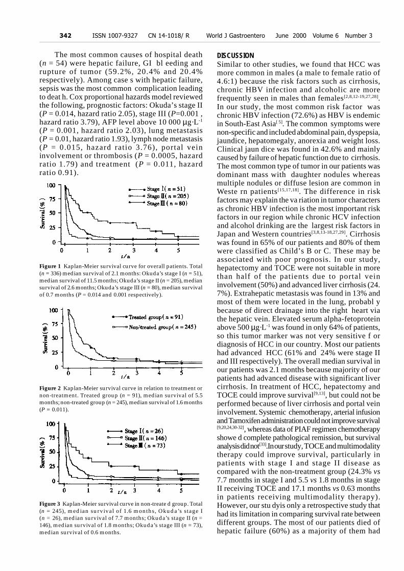
The most common causes of hospital death(n = 54) were hepatic failure, GI bl eeding andrupture of tumor (59.2%, 20.4% and 20.4%respectively). Among case s with hepatic failure,sepsis was the most common complication leadingto deat h. Cox proportional hazards model reviewedthe following, prognostic factors: Okuda’s stage II(P = 0.014, hazard ratio 2.05), stage III (P=0.001 ,hazard ratio 3.79), AFP level above 10 000 µg·L-1
(P = 0.001, hazard ratio 2.03), lung metastasis(P = 0.01, hazard ratio 1.93), lymph node metastasis(P = 0.015, hazard ratio 3.76), portal veininvolvement or thrombosis (P = 0.0005, hazardratio 1.79) and treatment (P = 0.011, hazardratio 0.91).
Figure 1 Kaplan-Meier survival curve for overall patients. Total(n = 336) median survival of 2.1 months: Okuda’s stage I (n = 51),median survival of 11.5 months; Okuda’s stage II (n = 205), mediansurvival of 2.6 months; Okuda’s stage III (n = 80), median survivalof 0.7 months (P = 0.014 and 0.001 respectively).
Figure 2 Kaplan-Meier survival curve in relation to treatment ornon-treatment. Treated group (n = 91), median survival of 5.5months; non-treated group (n = 245), median survival of 1.6 months(P = 0.011).
Figure 3 Kaplan-Meier survival curve in non-treate d group. Total(n = 245), median survival of 1.6 months, Okuda’s stage I(n = 26), median survival of 7.7 months; Okuda’s stage II (n =146), median survival of 1.8 months; Okuda’s stage III (n = 73),median survival of 0.6 months.
DISCUSSIONSimilar to other studies, we found that HCC wasmore common in males (a male to female ratio of4.6:1) because the risk factors such as cirrhosis,chronic HBV infection and alcoholic are morefrequently seen in males than females[2,8,12-19,27,28].In our study, the most common risk factor waschronic HBV infection (72.6%) as HBV is endemicin South-East Asia[ 5]. The common symptoms werenon-specific and included abdominal pain, dyspepsia,jaundice, hepatomegaly, anorexia and weight loss.Clinical jaun dice was found in 42.6% and mainlycaused by failure of hepatic function due to cirrhosis.The most common type of tumor in our patients wasdominant mass with daughter nodules whereasmultiple nodules or diffuse lesion are common inWeste rn patients[15,17,18]. The difference in riskfactors may explain the va riation in tumor charactersas chronic HBV infection is the most important riskfactors in our region while chronic HCV infectionand alcohol drinking are the largest risk factors inJapan and Western countries[3,8,13-18,27,29]. Cirrhosiswas found in 65% of our patients and 80% of themwere classified as Child’s B or C. These may beassociated with poor prognosis. In our study,hepatectomy and TOCE were not suitable in morethan half of the patients due to portal veininvolvement (50%) and advanced liver cirrhosis (24.7%). Extrahepatic metastasis was found in 13% andmost of them were located in the lung, probabl ybecause of direct drainage into the right heart viathe hepatic vein. Elevated serum alpha-fetoproteinabove 500 µg·L-1 was found in only 64% of patients,so this tumor marker was not very sensitive f ordiagnosis of HCC in our country. Most our patientshad advanced HCC (61% and 24% were stage IIand III respectively). The overall median survival inour patients was 2.1 months because majority of ourpatients had advanced disease with significant livercirrhosis. In treatment of HCC, hepatectomy andTOCE could improve survival[9,13], but could not beperformed because of liver cirrhosis and portal veininvolvement. Systemic chemotherapy, arterial infusionand Tamoxifen administration could not improve survival[9,20,24,30-32], whereas data of PIAF regimen chemotherapyshowe d complete pathological remission, but survivalanalysis did not[33].In our study, TOCE and multimodalitytherapy could improve survival, particularly inpatients with stage I and stage II disease ascompared with the non-treatment group (24.3% vs7.7 months in stage I and 5.5 vs 1.8 months in stageII receiving TOCE and 17.1 months vs 0.63 monthsin patients receiving multimodality therapy).However, our stu dyis only a retrospective study thathad its limitation in comparing survival rate betweendifferent groups. The most of our patients died ofhepatic failure (60%) as a majority of them had
342 ISSN 1007-9327 CN 14-1018/ R World J Gastroentero June 2000 Volume 6 Number 3

advanced HCC and liver cirrhosis. By multivariateanalysis, poor prognosis was associated with advancedstage of the tumor, serum alpha-fetoprotein level >10000 µg·L-1, extrahepatic metastasis and portal veininvolvement.
ACKNOWLEDGEMENTS The au tho r sacknowledge the cooperation of the staff of theRegistration Unit of Songklanagarind Hospital and of theOffici al Register of Population for providing patient dataand Dr. Allan Geater for English advice.
REFERENCES1 Srivatanakul P, Sontipong S. Incidence of liver cancer in Thailand
1979. Thai Cancer J, 1979;8:127-1342 Kassianides C, Kew MC. The clinical manifestations and natural
history of hepatocellular carcinoma. Gastroenterol Clin N Am,1987;16:553-562
3 Srivatanakul P, Burke DS, Thanasombutt S, Tan ngarmtrong D. Se-rum markers of hepatitis A and B virus infection in Thai patients withprimary hepatocellular carcinoma. Thai Cancer J, 1983;9:113-118
4 Kane MA. Progress on the control of hepatitis B infection throughimmunisation. Gut, 1993;34(Suppl):S10-12
5 Perrillo RP. Hepatitis B: transmission and natural history. Gut,1993;34(Suppl):S48-49
6 Beasley RP. Hepatitis B virus as the etiologic agent in hepatocel-lular carcinoma epidemiologic considerations. Hepatology, 1982;2(Suppl):S21
7 Beasley RP, Hwang LY, Lin CC, Chien CS. Hepatocellular carci-noma and hepatitis B virus. A prospective study of 22, 707 men inTaiwan. Lancet, 1981;2:1129-1132
8 Bisceglie AM, Rustgi VK, Hoofnagle JH, Dusheiko GM, LotzeMT. Hepatocellular carcinoma: NIH conference. Ann Intern Med,1988;108:390-401
9 Okuda K, Ohtsuki T, Obata H, Tomimatsu M, Okazaki N, HasegawaH, Nakajima Y, Ohnishi K. Natural history of hepatocellular car-cinoma and prognosis in relation to treatment: study of 850patients. Cancer, 1985;56:918-928
10 Kew MC, Geddes EW. Hepatocellular carcinoma in rural southernAfrican blacks. Medicine, 1982;61:98-108
11 Okuda K, Nakashima T. Primary carcinomas of the liver. 4th ed.In: Berk Bockus Gastroenterology. Philadelphia: WB Saunders,1985:335-3376
12 Cong WM, Wu MC. Primary hepatocellular carcinoma in womenof Mainland China: a clinicopathologic analysis of 104 patients.Cancer, 1993;71:2941-2945
13 Stuart KE, Anand AJ, Jenkins RL.Hepatocellular carcinoma in theUnited States: prognostic features, treatment outcome, andsurvival. Cancer, 1996;77:2217-2222
14 Chlebowski RT, Tong M, Weissman J, Block JB, Ramming KP,Weiner JM, Bateman JR,Chlebowski JS. Hepatocellular carcinoma:diagnostic and prognostic features in North American patients.Cancer, 1984;53:2701-2706
15 Calvet X, Bruix J, Gines P, Bru C, Sole M, Vilana R, Rodes J.Prognostic factors of hepatocellular carcinoma in the West: amultivariated analysis in 206 patients. Hepatology, 1990;12:753-760
16 Ebara M, Ohto M, Shinagawa T, Sugiura N, Kimura K, MatsutaniS, Morita M, Saisho H, Tsuchiya Y, Okuda K. Natural history ofminute hepatocellular carcinoma smaller than three centimeterscomplicating cirrhosis: a study in 22 patients. Gastroenterology,1986;90:289-298
17 Llovet JM, Bustamante J, Castells A, Vilana R, Ayuso MDC, SalaM, Bru C, Rodes J, Bruix J. Natural history of untreated nonsurgi-cal hepatocellular carcinoma: rationale for the design and evalua-tion of therapeutic trials. Hepatology, 1999;29:62-67
18 Colombo M. The natural history of hepatocellular carcinoma inWestern countries. Hepatogastroenterol, 1998;45:1221-122519 Nagasue N, Yukaya H, Hamada T, Hirose S, Kanashima R,Inokuchi K. The natural history of hepatocellular carcinoma: astudy of 100 untreated cases. Cancer, 1984;54:1461-1465
20 Okuda K. Primary liver cancer: Quadrennial review lecture. DigDis Sci, 1986;31(Suppl):S133-146
21 Heyward WL, Lanier AP, McMahon BJ, Bender TR, Francis DP,Maynard JE. Serological markers of hepatitis B virus and AlphaFetoprotein levels preceding primary hepatocellular carcinoma inAlaskan Eskimos. Lancet, 1982;23:889-891
22 Ebara M, Hatano R, Fukuda H, Yoshikawa M, Sugiura N, Saisho H.Natural course of small hepatocellular carcinoma with underlyingcirrhosis. A study of 30 patients. Hepatogastroenterol, 1998;45:1214-1220
23 Nomura F, Ohnishi K, Tanabe Y. Clinical features and prognosisof hepatocellular carcinoma with reference to serum Alpha-Feto-protein levels: analysis of 606 patients. Cancer, 1989;64:1700-1707
24 Group d’Etude et de traitement du carcinoma hepatocellulaire. Acomparison of Lipiodol chemoembolization and conservative treat-ment for unresectable hepatocellular carcinoma. N Engl J Med,1995;332:1256-1261
25 Jones DB, Koorey DJ. Screening studies and markers: hepaticcarcinoma. Gastroenterol Clin N Am, 1987;16:563-573
26 Pugh RMH, Murray-Lyon IM, Dawson JL, Pierroni MC, WilliamsR. Transection of the esophagus for bleeding esophageal varices.Br J Surg, 1973;60:646-664
27 Dusheiko G. Hepatocellular carcinoma: molecular biology, etiol-ogy and animal models. Gastroenterol Clin N Am, 1987;16:575-590
28 Sutton FM, Russell NC, Guinee VF, Alpert E. Factors affecting theprognosis of primary liver carcinoma. J Clin Oncol, 1988;6:321-328
29 Okada S, Okazaki N, Nose H, Yoshimori M, Aoki K. Prognosticfactors in patients with hepatocellular carcinoma receiving sys-temic chemotherapy. Hepatology, 1992;16:112-117
30 Yamashita Y, Takahashi M, Koga Y, Saito R, Nanakawa S,Hatanaka Y, Nobuyuki S, Nakashima K, Urata O, Yoshizumi K,Ito K, Sumi S, Kan M. Prognostic factors in the treatment ofhepatocellular carcinoma with transcatheter arterial emboliza-tion and arterial infusion. Cancer, 1991;67:385-391
31 Bruix J, Llovet JM, Castells A, Montana X, Bru C, Ayuso MDC,Vilana R, Rodes J. Transarterial embolization versus symptom-atic treatment in patients with advanced hepatocellular carcinoma:results of a randomized controlled trial in a single institution.Hepatology, 1998;27:1578-1583
32 Castells A, Bruix J, Bru C, Ayuso C, Roca M, Boix L, Vilana R,Rodes J. Treatment of hepatocellular carcinoma with Tamoxifen:a double bl ind placebo-control led tr ial in 120 patients .Gastroenterology, 1995;109:917-922
33 Leung TWT, Patt YZ, Lau WY, Ho SKW, Yu SCH, Chan ATC,Mok TSK, Yeo W, Liew C, Leung NWY, Tang AMY, Johnson PJ.Complete pathological remission is possible with systemic combi-nation chemotherapy for inoperable hepatocellular carcinoma.Clin Cancer Res, 1999;5:1676-1681
Edited by Ma JY and Pan BR
Sithinamsuwan P, et al. Hepatocellular carcinoma 343
Three-dimensional image of hepatocellularcarcinoma under confocal laser scanningmicroscope
Wang Hai Zhang1, Shi Neng Zhu2, Shi Lun Lu2, Ya Lin Huang3 and Peng Zhao3
Subject headings HCC; nucleus, three-dimensionalreconstruction; microscopy, confocal laser scanning
Zhang WH, Zhu SN, Lu SL, Huang YL, Zhao P. Three-dimensionalimage of hepatocellular carcinoma under confocal laser scanningmicroscope. World J Gastroentero, 2000;6(3):344-347
Abstract
AIM To investigate the application ofconfocal laser scanning microscopy (CLSM)in tumor pathology and three-dimensional(3-D) reconstructi on by CLSM in pathologicspecimens of hepatocellular carcinoma(HCC).METHODS The 30 µµµµµm thick sections were cutfrom the paraffin-embedded tissues of HCC,hyperplasia and normal liver, stained withDNA fluorescent probe YOYO-1 iodide andexamined by CLSM to collect optical sectionsof nuclei and 3-D images reconstructed.R E S U LT S H C C d i s p l a y e d c h a o t i carrangement of carcinoma cell nuclei,marked pleomorphism, indented andirregular nuclear surface, and irregular andcoarse chromatin texture.CONCLUSION The serial optical tomograms ofCLSM can be used to create 3-D reconstructionof cancer cell nuclei. Such 3-D impressions might be helpful or even essential in making anaccurate diagnosis.
INTRODUCTIONUnder conventional light microscope, histopath-ologists often use plane image to evaluate the three-dimensional (3-D) cellular characteristics. Three-Dconfiguration may be reconstructed by using serialmechanical sectioning, but it s axis definition is notgood, image is blur and tiny structure can’t be sh ownclearly, and the specimen might be damaged. Usingthe serial optical tomograms and 3-D reconstructionfunction, confocal laser scanning microscopy (CLSM)can provide a much better quality 3-D image thanconventional light microscope, and lead the observerinto a brand-new 3-D world. Although CLSM hasbeen used extensively in cell biology[1], fewapplications were reported in routine clinical pathologysuch as three-dimensional DNA image cytometry byCLSM in thi ck tissue blocks of prostatic lesions and3-Dreconstruction by CLSM in routine pathologicspecimens of benign and malignant lesions of humanbreast[2-4]. In this study, 3-D reconstruction wasperformed on routine formalin-fix ed, paraffin-embedded tissues of normal, and hyperplastic tissuesof liver and hepatocellular carcinoma by usingcomputer-assisted CLSM together with 3-Dreconstruction. The goals of our study were to present3-D morphologic characte ristics of benign andmalignant specimens of the liver and to attempt todemonst rate the usefulness of CLSM in routinelyobtained surgical pathologic tissues.
MATERIALS AND METHODSTwo cases of normal liver tissue were selected fromautopsy specimens collected in the Department ofPathology of Shanghai Medical University, sixcases of hepatocellular carcinoma including adjacentliver tissue were routine clinical specimenscollected in 1996 from the Liver Cancer Instituteof Zhongshan Hospital affiliated to ShanghaiMedical University. All tissues were fixed in 10% formalin, embeddedin paraffin, serial sect ions were cut at 5 µm and30 µm. The 5 µm slices were stained withhematoxylin and eosin for conventional lightmicroscopic observation. The 30 µm slices werestained with DNA fluorescent probe, YOYO-1iodide (Molecular Pro bes, Eugene, Ore., USA).
1Division of Pathology, Zhongshan Hospital, Shanghai MedicalUniversity, Shanghai 200032, China2Department of Pathology, Shanghai Medical University, Shanghai200032, China3National Laboratory of Medical Neurobiology, Shanghai MedicalUniversity, Shanghai 200032, ChinaDr. Wang Hai Zhang, graduated from Shanghai Medical University as aPh.D. in 1999, majoring in surgical pathology and molecular pathology,having 10 papers published.Correspondence to: Dr. Wang Hai Zhang, Division of Pathology,Zhongshan Hospital, Shanghai Medical University, Shanghai 200032,ChinaTel. 0086-21-64041990 Ext.2732Email. [email protected] 2000-03-19 Accepted 2000-04-28
PO Box 2345, Beijing 100023, China World J Gastroentero, 2000; 6(3):344-347Fax: +86-10-85381893 World Journal of GastroenterologyE-mail: [email protected] www.wjgnet.com Copyright 2000 by the WJG Press ISSN 1007-9327

The sections were deparaffinized with xylene (10min × 2) and dehydrated with 100%, 95%, and 70%ethanol (5 min × 2) and rinsed in distilled water for2 min × 5. The specimens were then fixed with10% neutral buffered fo rmalin for 30 min andwashed with tap water. After rinsing with distilledwater and 0.01M phosphate buffer 5 min × 2,nuclear RNA was removed by incubating thesections for 30min at 37 in 200 µL of ribonucleaseA (RNAase; Sigma, USA) at a concentration of160 g/L in PBS. DNA was next hydrolyzed with2N HCl for 25 min at 27.5 . After rinsing withdistilled water for 2 min × 5, the sections werecovered with 200 µL of YOYO-1 iodide diluted into1:2000 with PBS. The PBS was dil uted 1:5 withdistilled water to reduce the salt concentration. Tothis 200 µL working solution of YOYO-1iodide, 20µL of 0.1N HCl was added and the final solutionwas stored in the dark at 4 for use. Homogeneousfluorescence intensity of nuclei at different depthsof the confocal slices was obtained by agitating theYOYO-1 iodide for 1 h in the dark. Afterwards,the sections were rinsed with distilled wat er,covered with buffered glycerol, and the glass coverslip were scaled with finger nail polish. Sectionswere stored at 4 in the dark until CLSMexaminati on. A Leica TCS-NT confocal laser scanningmicroscope equipped with epifluoresce nce opticsand an appropriate combination of filters to visualizeand digitize t he images of the different specimens.An argon laser with an excitation waveleng th of488 nm was used to activate the green fluorescenceof the YOYO-1 iod ide-stained nuclear DNA(maximal absorption 491 nm and emission 509 nm).A 16 × objective (numerical aperture of 1.30) wasused to observe the specificity of t he staining, anda 100 × water objective (numerical aperture of 1.30) was used to study the details of chromatinpattern. Optical sections were collected thr oughoutthe entire stained thickness of the paraffin sectionswith a Z-step interval of 0.3 µm or 0.6 µm.The 3-D image processing was performed on a Leicacomputer with the original 3 -D interactivevisualization software. The collected confocaloptical sections and the 3-D reconstructed imageswere printed with a Panasonic color vide o dye-sublimation copy processor.
RESULTSThree-D morphologic features of normal livercellsA microscopic field of view was selected from thenormal liver specimen and comp arison of the imagestaken under transmission conventional lightmicroscope and CLSM is illustrated in Figure 1. Thetransmission conventional light microscopic image
was shown on the right side and the out-of-focussignals were visible. The out-of-focus blurreduced the contrast and sharpness of the fina limage. In the confocal image (left) the out-of-focus signals were cut off and only signals infocus were clearly visible. Optical sections(planes 1-60) were taken from the surface tothe bottom of normal liver specimen with a Z-step interval of 0.5 µm. Figure 2 shows the 10th,20th, 30th, 40th, 50th and 60th plane digitalimages of Z-series. The confocal images w eretaken at 5, 10, 15, 20, 25 and 30 µm depths,respecti vely. In the confocal images somenuclei appeared or disappeared depending on their orientation in space. The 60 2-D opticalsect ions were computer focused on aplane(deep-focusing) to analyze the fine structureof chromatin patterns inside the nucleus andreconstructed 3-D images to display the 3-Ddetailed a rrangement of nucleus. Figure 3 showsnormal liver cells with similar round or ovoidnuclei , homogeneous intensity of YOYO-1iodide fluorescence as well. Three-dimensionalview was shown in Figure 4 . The nuclearsurface appeared smooth with homogeneousfluorescence intensity.
Three-D morphologic features of atypicalhyperplasia of liver cellsOptical sections (planes 1-50) were taken from thesurface to the bottom of atypical hyperplasia of liverspecimen with a Z-step interval of 0.6 µm. Thestructure of chromatin patterns inside the nucleuswith homogeneous fluorescence intensity is shownwith deep-focusing in Figure 5. Three-dimensionalview is shown in Figure 6. The nuclear surfaceappeared smooth with homogeneous fluorescenceintensity. But the volume of nucleus of atypi calhyperplasia liver cell was bigger than that of normalliver cell.
Three-D morphologic features of hepatocellularcarcinoma cellsOptical sections (planes 1-50) were taken fromthe surface to the bottom of hepa tocellularcarcinoma specimen with a Z-step interval of 0.6 µm. The deep-focusing images were shownin Figure 7 and Figure 8. The structure ofchromatin patterns inside the nucleus withheterogeneous karyotheca thickness, irregularand coarse chromat in t ex tu re , ch romat inunders ide the kary o theca mainly . Three-dimensional view is shown in Figure 9 and Figure10. HCC displayed remarkably different featuresin 3-D morphology, including: indented, molding,a n d i r r e g u l a r n u c l e a r s u r f a c e ; m a r k e dpleomorphism; chaotic arrangeme nt of tumor cellnuclei.
Zhang WH, et al. Confocal laser scanning microscopic image of HCC 345
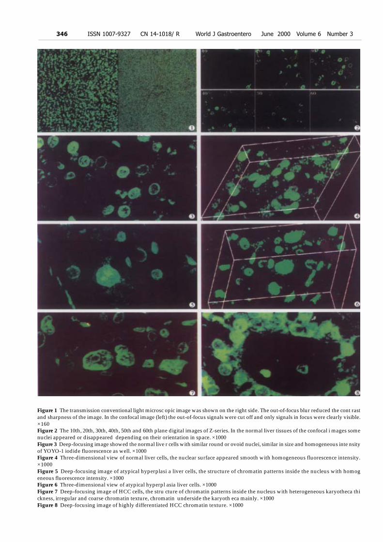
Figure 1 The transmission conventional light microsc opic image was shown on the right side. The out-of-focus blur reduced the cont rastand sharpness of the image. In the confocal image (left) the out-of-focus signals were cut off and only signals in focus were clearly visible.×160Figure 2 The 10th, 20th, 30th, 40th, 50th and 60th plane digital images of Z-series. In the normal liver tissues of the confocal i mages somenuclei appeared or disappeared depending on their orientation in space. ×1000Figure 3 Deep-focusing image showed the normal live r cells with similar round or ovoid nuclei, similar in size and homogeneous inte nsityof YOYO-1 iodide fluorescence as well. ×1000Figure 4 Three-dimensional view of normal liver cells, the nuclear surface appeared smooth with homogeneous fluorescence intensity.×1000Figure 5 Deep-focusing image of atypical hyperplasi a liver cells, the structure of chromatin patterns inside the nucleus with homogeneous fluorescence intensity. ×1000Figure 6 Three-dimensional view of atypical hyperpl asia liver cells. ×1000Figure 7 Deep-focusing image of HCC cells, the stru cture of chromatin patterns inside the nucleus with heterogeneous karyotheca thickness, irregular and coarse chromatin texture, chromatin underside the karyoth eca mainly. ×1000Figure 8 Deep-focusing image of highly differentiated HCC chromatin texture. ×1000
346 ISSN 1007-9327 CN 14-1018/ R World J Gastroentero June 2000 Volume 6 Number 3

DISCUSSIONThe area, volume, shape, DNA content, and chromatinpattern of nuclei may be imp ortant for the diagnosis andprognosis of cancer. Histopath ologists often use 4 µmto 6 µm thick paraffin se ctions to obtain representativeand diagnostically relevant images. Due to the verylimited section thickness in comparison with the size ofthe tissue, and the images are nearly two-dimensions,focusingup and down at high magnifica tion provides arough idea of the 3-D cellular characteristics, and such3-D impression may be helpful or even essential inarriving at a certain diagnosis, especially for borderlinelesions or tumors. However, in spite of the usefulnessof such 3-D information about nuclei, conventional lightmicroscopy is not alw ays the ideal tool for 3D evaluationdue to the interference of out-of-focus structures withthe images of the focus plane studied. Much of the lightemitte d from the regions of specimen above and belowthe focal plane contributes to the out-of-fo cus blur, whichseriously reduces the contrast and sharpness of the finalimage. Confocal laser scanning microscope allows theacquisition of optical sections from a thick specimen andout-of-focus blur can be reduced considerably and th usmuch sharper and clearer images will be obtained. CLSMhas become an exciting new instrument in biomedicalresearch because of its increased resolution overconventional light microscope and its utility for subsequent3-D-reconstructi on analysis[5,6]. In this paper, the 3-D reconstruction havedemonstrated 3-D contour of represe ntativecharacteristics of normal liver cells, atypicalhyperplasia liver cells, and HCC cells and thespatial relationship of nuclei, as well as the subtlestr ucture of chromatin texture inside nuclei.This
paper emphasized the practical fe asibility of CLSM and3-D reconstruction from routine surgical histopathologicmaterials. To obtain desirable quality 3-D image fromformalin-fixed, paraffin-embedded specimens, YOYO-1 iodide, a highly specific and sensitive (picogramsensitivity)[7] DNA probe was utilized. Intense andhomogeneous fluorescence was obtained by incubatingthe YOYO-1 iodide for 1h with agitation in the dark. Toreveal subtle details of nuclear structure, RNA wasremoved by RNas epredigestion, since nuclear RNA wasstained by YOYO-1 iodide as well. CLSM combines the three most advanced andimportant elements of our era, the microscope, the laserand the computer in one, moreover it is non-invasiveand can be used on archival paraffin blocks. Weanticipate that, in the future, pathol ogists may utilizethese new techniques to make more precise diagnosis.
REFERENCES1 Minsky M. Memoir on inventing the confocal scanning
microscope. Scanning, 1988;10:128-1382 Boon ME, Schut JJ, Suurmeijer AJ, Benita EM, Hut PK, Kok LP.
Confocal microscopy of false-negative breast aspirates. DiagnCytopathol, 1995;12:42-48
3 Liu S, Weaver DL, Taatjes DJ. Three-dimensional reconstructionby confocal laser scanning microscopy in routine pathologic speci-mens of benign and malignant lesions of the human breast.Histochem Cell Biol, 1997;107:267-278
4 Beil M, Irinopoulou T, Vassy J, Rigaut JP. Chromatin textureanalysis in three-dimensional images from confocal scanning la-ser microscopy.Anal Quant Cytol Histol, 1995;17:323-331
5 White JG, Amos WB, Fordham M. An evaluation of confocalversus conventional imaging of biological structures by fluores-cence light microscopy. J Cell Biol, 1987;105:41-48
6 Shotton DM. Electronic light microscopy: present capabilitiesand future prospects. Histochem Cell Biol, 1995;104:97-137
7 Tekola P, Baak JP, Belien JA, Brugghe J. Highly sensitive, specific,and stable new fluorescent DNA stains for confocal laser microscopyand image processing of normal paraffin sections.Cytometry, 1994;17:191-195
Figure 9 Three-dimensional view of spindle cell HCC. ×1000Figure 10 Three-dimensional view of tubular adenoca rcinoma of HCC. ×1000
Edited by WANG Xian-Linproofread by Sun SM
Zhang WH, et al. Confocal laser scanning microscopic image of HCC 347

Direct technetium-99m labeling of anti-hepatomam o n o c l o n a l a n t i b o d y f r a g m e n t : aradioimmunoconjugate for hepatocellularcarcinoma imaging
Hui Jie Bian1, Zhi Nan Chen1 and Jing Lan Deng2
Subject headings antibody, monoclonal; antibodyfragments; technetium-99m; hepatocellular carcinoma;liver neoplasms; radioimmunoimaging
Bian HJ, Chen ZN, Deng JL. Direct technetium-99m labeling of anti-hepatoma monoclonal antibody fragment: a radioimmunoconjugatefor hepatocellular carcinoma imaging. World J Gastroentero, 2000;6(3):348-352
AbstractAIM To directly radiolabel an anti-hepatoma mAbfragment HAb18 F(ab’)2 with 99mTc by stannous-reduced method, and assess the stability,biodistribution and radioimmun oimaging (R II).METHODS Immunoreactive fraction wasdetermined according to Lin dmo’s method.Ellman’s reagent was used to determine thenumber of thiols in the reduced F(ab’)2. Labelingefficiency and homogeneity were measured byp a p e r c h r o m a t o g r a p h y , s o d i u mdodecy lsu lphate po lyacry lamide ge lelectrophoresis (SDS-PAGE) and autoradiography. Challenge assay involved theincubation of aliquots of labeled antibo dy inethylenediaminetetraacetate (EDTA) and L-cysteine (L-cys) solutions with different molarratio at 37 for 1 h, respectively. Investigationsin vivo utilized nude mice bearing humanhepatocellular carcinoma (HHCC) xenograftswith gamma camera imaging and tissuebiodistribution studies at regular intervals.RESULTS The labeling procedure was finishedwithin 1.5 h compared with the “pretinning”method which would take at least 21 h. In vitro
studies demonstrated that the radiolabeledmAb fragment was homogen eous and retainedits immunoreactivity. Challenge studies indicatedthat 99mTc-labeled HAb18 F(ab’)2 in EDTA is morestable than in L-cys. Imaging and biodistribution
showed a significant tumor uptake at 24 h post-injection of 99mTc-labeled HAb18 F(ab’)2. Theblood, kidney, liver and tumor uptakes at 24 hwere 0.56 ± 0.09, 56.45 ± 11.36, 1.43 ± 0.27 and6.57 ± 3.01 (%ID/g), respectively.CONCLUSION 99mTc-HAb18 F(ab’)2 conjugateprepare d by this direct method appears to bean effective way to detect hepatoma in nudemice model.
INTRODUCTIONThe introduction of mAbs as targeting devices innuclear medicine is well develo ped and manydifferent antibodies which labeled with a varietyof isotopes have been reported in cancerdiagnosis. I t seemed that 99mTc is the mostpopular radionuclide for nuclear medicine imagingbecause of its favorable physi cal characteristics,low cost, and ready availability. 99mTc labeledmAb fragments should be superior to other bigmolecule radioimmunoconjugates for u se intumor R II. A number of methods have beenproposed for 99mTc labe ling proteins, and mAbsin particular. In general, these methodologiescan be d ivided into two categories: indirect anddirect methods[1]. In indirect method the proteinwas modified with a technetium binding ligandand then reacte d with a technetium complex.Several bifunctional chelating agents have been synthesized and used, such as diethylenetriaminepentaacetic acid (DTPA)[2] , diamide dimercaptide N2S2
ligands, and hydrazino nicotinamide analog [3].Although it is said that the indirect method canlead to loss of immuno reactivity. Joiris et al.have tested that the derivatization of antibodyor fragment by iminothiolane does not splitthe protein and keeps the immunoreact ivity[4]. By direct method, 99mTc metal ion bindsdirectly to endogenous donor groups on theantibody. The method is simple to perform andc o m p a t i b l e w i t h p rac t i ca l c l in i ca l u se .However, direct labeling of mAbs with 99mTc wasreported to be unstable due to non-specific binding(low and high-affinity)[5,6], but some reportssuggest an improved labeling of proteins with 99mTc.
1Cell Engineering Research Center, Basic Medical Department, FourthMilitary Medical University, Xi’an 710032, Shaanxi Province, China2Department of Clinical Nuclear Medicine, Xijing Hospital, Xi’an710033, Shaanxi Province, ChinaSupported by National Natural Science Foundation of China,No.39700175Correspondence to: Prof. Zhi Nan Chen, Cell Engineering ResearchCenter, Basic Medical Department, Fourth Military Medical University,Xi’an 710032, Shaanxi Province, ChinaTel. 0086-29-3374545, Fax. 0086-29-3293906Email. Chcerc [email protected] 2000-02-03 Accepted 2000-04-28
PO Box 2345, Beijing 100023, China World J Gastroentero, 2000; 6(3):348-352Fax: +86-10-85381893 World Journal of GastroenterologyE-mail: [email protected] www.wjgnet.com Copyright 2000 by the WJG Press ISSN 1007-9327

In the Schwarz and Steinstrasser procedure, asmodified by Mather and Ellison[7], disulide bridgesin the mAb are reduc ed with 2-mercaptoethanol(2-ME). After purification, the resulting reducedan tibody can be stored frozen until required foruse. Labeling is accomplished by addition ofstannous ion from a bone- scanning kit andpertechnetate. In addit ion to using regularreducing agents, such as 2-ME, stannous ions[8], borohydride[9], ascorbic acid[10], dithionite[11], or glutathione[12] to generate sulphydryl groups, otherpeculiar approaches also appeared recently.Direct 99mTc labeling of mAbs were finis hed byreduction of antibodies using photoactivation andinsoluble macromolecula r Sn (II) complex[13,14].With the development of direct method, there have been a few reports of successful use of thistechnique in colorectal, breast, and ovarian cancerimaging[15-17]. In this report, we describe a direct method forradiolabeling anti-hepatoma mon oclonal antibodyfragment HAb 18F (ab’)2 with 99mTc. The stabilityand homogeneity of 99mTc-HAb18 F(ab’)2 wereevaluated. The biodis tribution and tumor localizationin nude mice bearing a HHCC xenograft werestudied.
MATERIALS AND METHODSMonoclonal antibodyThe mAb HAb18 is of murine IgG1 isotype and wasdeveloped by our laboratory[18]. F(ab’)2 fragment ofHAb18 was generated by papain digestion with amolecular weight of -96 000 dalton[19].
TumorsHepatocellular carcinoma grown in Balb/c mice was usedas a prototype tumo r model. Approximately 107 HHCCcells obtained from Shanghai Cell Institute of ChineseAcademy of Sciences were implanted in the left thighof the animals and the tumors were allowed to grow for8-10 days to approximately 1cm in diameter.
Antibody reductionThe antibody concentrated to 8 g/L in neutral PBSwas reduced by reaction with a molar excess ofstannous/glucoheptonate (Sn/GH) ranging from 10:1 to 50:1 (Sn/GH: MAb) at 37 for 15 min-30 min.The Sn/GH with a mass ratio of 1:100 was dissolvedin 50mM acetate-buffered saline (ABS), pH 5.3purged with nitrogen. The reduced antibody wasisolated from reductant t hro ugh a PD-10 column(Pharmacia) equilibrated with 0.05 mol/L ABS. Thenumber of resulting free sulphydryl groups wasassayed with Ellman’s reagent 5, 5’dithio-bis (2-nitrobenzoic acid), (DTNB, Sigma Chemical Co.,USA) [20]. One hundred µL of sample was mixedwith 20 µL of 0.01 mol/L-DTNB and diluted to
3 mL with 0.05 mol/L Tris-HCl buffer pH 8.4. Themixture was incubated at room te mperature for 15min and coloration measured with an UV/VISspectrophotome ter at 412 nm. The number of thiolswas obtained by comparison with a seri es of L-cysteine (L-cys) standards ranging from 0.312 mg/Lto 10 mg/L. The integrity of the reduced F(ab’)2 was determinedby non-reduced SDS-PAGE with 100 g/L gel usingVertical Gel Electrophoresis System (Bio-Rad). Thegel was stained with Coomassie brilliant blue R250.Control experiments were run using unreduced mAbF(ab’)2.
RadiolabelingFor labeling, 160 µg of reduced HAb18 F(ab’)2
was mixed with a 10 µL-20 µL of diluted Sn/GHsolution (0.2 g/L), and pertechnetium solution (0.2 mL, 74MBq), (Chinese Academy of AtomicEnergy) was injected into the mixture. The Sn/GH solution was freshly prepared each time bydissolving 100 mg GH and 1mg SnCl2·2H2O in 5mL of saline purged with nitrogen. The reactionmixture was incubated for 0.5 h-1 h at 37 beforei t was analyzed by Whatman 3MM paperchromatography which was then developed inacetone or 100 g/L trichloroacetic acid (TCA). R-f values for acetone are: mAb 0.0, 99mTc-GH 0.0,and 99mTcO4-0.9-1.0. R-f values for 100 g/L TCAare: mAb 0.0, 99mTcGH 0, and 99mTcO4- 0.7.Labeled mAb was differentiated from 99mTc colloidby the method of Thrall et al[21]. The same stripsimpregnated with 10g/L-20g/L human serumalbumin before development with 5:2:1, water:ethanol: 5N NH4OH. Colloid remained on thebottom of the strip while mAb-bound isotopemigrated with the solvent front. The integrity of the labeled F(ab’)2 was assayedusing the same non-reduced SDS-PAGE as describedabove. The gel was autoradiograghied on x-ray filmbefore stained with Coomassie brilliant blue R250.
mmunoreactivity assessmentT h e i n v i t r o i m m u n o r e a c t i v i t y o f t h eradiolabeled HAb18 F(ab’)2 was evalu ated bya live cell assay[22]. Briefly, HHCC cells 5 × 10⁹/L were centrifuged (1 000r/min) for 5 min andwashed twice with 1% bovine serum albumin(BSA) in PBS, then 5 serial 1:2 dilutions weremade in 10 g /L BSA in Eppendor f tubesprecoated with BSA. Radiolabeled HAb18 F(ab’)2 at a concentration of 40 ng/mL in 10 g/LBSA was added using a volume equal to halfthe volume of cell suspension. The total volumeof cell-binding assay solution was 0.3 mL. Afterincubation for 2 h at 37 , the total as well asthe cell-bound radioactivity were counted in a gammacounter.
Bian HJ, et al. 99mTc-labeled HAb18(ab’)2 for hepatoma radioimmunoimaging 349

In Vitro stability studiesThe stability was analyzed by using two differentchallenging agents, EDTA and L-cys. An aliquot of 50µL 99mTc-HAb 18 F(ab’)2 solution was incubated withEDTA or L-cys at 37 for 1 h. The molar ratio of mAbto challenging agent was at a maximum of 10 000:1.Dissociat ion rat io was analyzed on paperchromatography.
Biodistribution and imagingBalb/c mice bearing HHCC were divided into threegroups. Each group consisted of three animals andeach animal received approximately 15 µg antibodywith about 7.4MBq through a lateral tail vein. At timeintervals of 4, 10 and 24 h postinjection, three groupsof mice were killed , and imaged on a SPECT(Starcam 3 000, UK). Data were collected 100 000co unts per image and peak energy settings at the140 ke V (20%) window for 99mTc. The blood andother organs of interest were collected. Tissues werewashed, blotted, weighed and counted in a gammacounter. For each mouse, data are expressed aspercent of injected dose per gram of tissue (%ID/g)after physical decay corrected.
RESULTSFigure 1 represents the calibration curve for thedetermination of sulphydryl groups using L-cysstandards over a range of 0.312 to 10 mg/L, by plottingoptical density at 412 nm versus L-cys standardconcentrations after subtraction of the background dueto Ellman’s reagent. Linear regression was used andcorrelation coefficient 0.999 was obtained. Table 1shows the influence of the reduction conditions onthe number of free sulphydryl groups detected by thisthiol assay. As expected, increasing the molar ratioof Sn/GH to antibody in the reaction mixture doesincrease the number of apparent -SH groups perantibody, and increase the labeling efficiencycorrespondingly, which results in the labelingefficiency at a maximum of 84.2%. The free 99mTcO4-and colloid amounts determined by Whatman 3MMpaper using different developing systems were alsoshowed in Table 1. In control expe ri ments, labelingefficiency was 2% when unreduced HAb18 F(ab’)2
was used. SD S-PAGE by both staining andautoradiography showed that the radioactivity co-migrated with the proteins and that there were almostno protein fragments prese nt within the 60:1 of molarratio of Sn/GH to mAb (Figure 2). However , anotherSDS-PAGE in Figure 3 illustrates that fragmentationoccurred during t he reduction procedure when themolar ratio of Sn/GH to mAb was at 500:1. As shown in Figure 4, the immunoreactivefraction, 0.84 was determined by plott ing theinverse of the bound fraction compared with theinverse of the cell conc entration, which is based on
the assumption that the total antigen concentration (cellconcentration) is a good enough approximation for thefree antig en concentration. Challenging with EDTA did not remove 99mTcfrom the labeling conjugat e remarkably, while L-cys ata molar ratio of 625:1 remove approximately one-tenthof the label (Figure 5). Biodistribution of radioactivity in blood andexcised tissues are displayed in Table 2. Thepreparation localized at the tumors was more thanat any organ examined at both 10 h and 24 h afterin ject ion, except the kidneys . Th e lowerradioactivity in blood at 24 h suggested fast bloodclearance. The imaging results in Figure 6showed significant tumor uptake at 24 h post-injection.
Table 1 Effect of molar ratio (Sn/GH: mAb) on quantity of-SH,fre e 99mTcO4- and colloid and labeling efficiency (%, n = 3)
Molar ratio SH groups 99mTcO4 colloid labeling(Sn/GH: mAb) /mAb efficiency
Control 0 ± 0 62.1 ± 4.5 1.1 ± 1.2 2.0 ± 0.910:1 0.43 ± 0.04 3.0 ± 1.4 1.4 ± 1.14 4.6 ± 3.820:1 1.25 ± 0.10 2.9 ± 1.1 1.4 ± 0.77 2.8 ± 5.130:1 2.46 ± 0.08 2.0 ± 0.9 3.2 ± 1.47 8.6 ± 3.240:1 3.34 ± 0.09 1.8 ± 1.2 2.8 ± 1.38 4.2 ± 2.850:1 3.61 ± 0.12 2.1 ± 0.8 3.6 ± 1.58 4.4 ± 3.4
Table 2 Biodistribution of 99mTc-HAb 18F(ab’)2 in nude micebearing hepatoma (x ± s, %ID/g)
Time after injection (h)Organ 4 10 24
Blood 2.21 ± 0.24 1.45 ± 0.15 0.56 ± 0.09Kidney 72.38 ± 4.37 70.47 ± 15.23 56.45 ± 11.36Liver 1.82 ± 0.48 1.59 ± 0.31 1.43 ± 0.27Lung 1.62 ± 0.34 1.40 ± 0.17 0.75 ± 0.21Stomach 1.37 ± 0.39 1.05 ± 0.28 0.50 ± 0.29Spleen 2.35 ± 0.81 2.11 ± 0.75 1.82 ± 0.85Large intestine 1.16 ± 0.34 1.42 ± 0.39 0.94 ± 0.32Small intestine 0.97 ± 0.31 0.95 ± 0.18 0.62 ± 0.24Heart 2.04 ± 0.55 1.83 ± 0.48 1.17 ± 0.42Muscle 1.15 ± 0.20 0.77 ± 0.28 0.51 ± 0.25Brain 0.18 ± 0.02 0.07 ± 0.04 0.02 ± 0.01Tumor 5.14 ± 2.26 5.84 ± 2.98 6.57 ± 3.01
Figure 1 L-cysteine standard curve for sulphydryl d eterminationusing Ellman reaction.
350 ISSN 1007-9327 CN 14-1018/ R World J Gastroentero June 2000 Volume 6 Number 3

Figure 2 Effect of reduction on integrity of 99mTc-labeled HAb18F(ab’)2 as monitored by SDS-PAGE. Vertical lanes re present molarratios of Sn/?GH to HAb18 F(ab’)2: 1, 10:1; 2, 20:1; 3, 30:1; 4, 40:1; 5, 50:1; 6, 60:1. (A)Coomassie brilliant blue R250 staining.Molecular weight (kD) is indicated at the left. (B) autoradiography.
Figure 3 Effect of reduction on integrity of HAb18 F(ab’)2 asmonitored by SDS-PAGE. Molecular weights (kD) are indicated atthe left. Vertical lanes represent molar ratios of Sn/GH to HAb18F(ab’)2: 1, 1000:1; 2, 500:1; 3, 50:1; 4, 10:1; 5, unreduced F(ab’)2.
Figure 4 Binding assay for the determination of the immunoreactivefraction of 99mTc-labeled HAb18 F(ab’)2.
Figure 5 Dissociation of 99mTc-labeled HAb18 F(ab’)2 withincreasing molar ratio of EDTA to mAb ( ) and L-cys to mAb( ).
Figure 6 Images of nude mice bearing human hepatocel lularcarcinoma with 99mTc-HAb18 F(ab’)2 at 24 h.
DISCUSSIONGreat efforts have been made to develop a methodthat can be used for the direct labeling of mAbswith 99mTc[16]. Earl ier s tudies involved thei n c u b a t i o n o f m A b s w i t h s t a n n o u sphthalate tartrate solution for up to 21 h at roomtemperature, which was named “pretinning”method. Clinical success with this method hasbeen claimed by the author[23]. One aim of our study was to further evaluate therole of stannous as a reducing agent in the directlabeling of mAb F(ab’)ª-2 with 99mTc. The differencebetween the “pretinning” method and this method isthat we use GH instead of phthalate-tartrate astransfer ligand and stabilizer to avoid Sn or Tc-c olloidformation. To do this, we investigated the effect ofthe quantity of Sn/ GH on the labeling time andefficiency. When the molar ratio of Sn/GH to mAb F(ab’)2 was constant, we found that there was noobvious difference on the number of-SH between thereduction time of 20 min and 30 min or even longer[
24]. The whole labeling process can be accomplishedwithin 1.5h. Hnatowich et al. reported that labelingefficiency in the case of the stannous ion-reduced antibodies was generally in excess of 70%[12], however,in our method mol ar ratio of Sn/GH to mAb was an importantparameter to obtain good labeling re sults, and molarratio of 40:1 or higher were needed to get labeling
Bian HJ, et al. 99mTc-labeled HAb18(ab’)2 for hepatoma radioimmunoimaging 351

effici ency of more than 80% (Table 1). The lowpercentage of free 99mTcO4 and radiocolloid in eachsample implied that pH 5.3 and GH are the opti mal pHvalue and transfer ligand. Under this condition, the labeledmAb HAb18 F(ab’)2 keeps its immunoreactivity.Autoradiography of SDS-PAGE had only one m igrationof component identical to that of native HAb18 F(ab’)2
determination by staini ng with Coomassie brilliant blueR250 (Figure 2), which demonstrated that S n/GHreduction is mild and does not destroy interchain bridgesin mAbs. Labeli ng efficiency of 2% in controlexperiments using unreduced HAb18 F(ab’)2 indicatedthat there was no exchange with the low affinity sitesand also demonstrated tha t reduction of disulfides is anecessary initial step in 99m£Ôc direct labeling ofantibodies. The bond betweenª²SH and Tc is strongerthan that of N -Tc or O-Tc which was verified by thechallenge assay of 99mTc-HAb18 F(ab’)2 in the presenceof EDTA. We found that EDTA even at a molar rat ioof 10 000:1 failed to remove a significant amount of 99mTc,this is in agreement with the results of Rhodes et al[8].But L-cys at 625¡Ã1 remove one-tenth of the label(Figure 5). Despite such insta bility of the label, therewas no in vivo evidence of release of pertechneta tedue to no thyroid imaging observed in the whole imagingprocess (Figure 7) . Tumor localization of 99mTc-HAb18F(ab’)2 was successfully dem onstrated in a humantumor/nude mouse xenograft model. Biodistribution andima ging results showed the highest tumor uptake at 24h post-injection. Where as kidney levels were foun d tobe higher in the whole process. Accumulation ofradioactivity in the kidney may be the result of retentionof this metallic radionuclide by the kidney pro ximaltubule[25], the possible release of 99mTc-labeled cysteinea n d g l u t a t h i o n e [ 2 6 ]s t e m m i n g f r o m t h eadioimmunoconjugate catabolism, and the relativeamount of 99mTc-GH. A technique has been used inpatients to block renal tubule uptake of 99mTc-anti-CEAFab’ fragments by amin o acid infusion[27]. In conclusion, a radioimmunoimaging conjugatefor hepatoma detection was prepare d by direct labelingmAb HAb18 F(ab’)2-with 99mTc using stannous/glucoheptonate as reducing agent. Although the labelingefficiency is not satisf actory to some degree, it hasseveral advantapes: simple, easy and quick, besides, thelabeled mAb fragment retains its immunoreactivity.Biodistribution and imaging studies reveal that thisconjugate is useful for the detection of hepato ma.
ACKNOWLEGDGMENTS The authors are grateful toDr. Wang Jing for mice imaging and the Department of NuclearMedicine of Shaanxi People’s Hospital for their support of thisproject.
REFERENCES1 Rhodes BA.Direct labeling of proteins with 99mTc. Nucl Med Biol, 1991;
18:667-6762 Childs RL, Hnatowich DJ. Optimum condition for labeling of DTPA coupled
antibodies with Technetium-99m. J Nucl Med, 1985;26:293-2993 Ultee ME, Bridger GJ, Abrams MJ, Longley CB, Burton CA, Larsen SK,
Henson GW, Padmanabhan S, Gaul FE, Schwartz DA. Tumor imagingwith technetium-99m-labeled hydrazinonicotinamide-Fab’ conjugates. JNucl Med, 1997;38:133-138
4 Joiris E, Bastin B, Thornback JR. A new method for labeling of monoclonal antibodiesand their fragments with technetium-99m.Nucl Med Biol, 1991;18:353-356
5 Eckelman WC, Meinken G, Richards P. 99mTc-human serum albumin. JNucl Med, 1971;12:707-710
6 John E, Thakur ML, Wilder S, Alauddin MM, Epstein AL. Technetium-99m-labeled monoclonal antibodies: influence of technetium-99m bindingsites. J Nucl Med, 1994;35:876-881
7 Mather SJ, Ellison D. Reduction-mediated technetium-99m labeling ofmonoclonal antibodies. J Nucl Med, 1990;31:692-697
8 Rhodes BA, Zamora PO, Newell KD, Valdez EF. Technetium-99m labelingof murine monoclonal antibody fragments. J Nucl Med, 1986;27:685-693
9 Pauwels EKJ, Welling MM, Feitsma RIJ, Atsma DE, Nieuwenhuizen W.The labeling of proteins and LDL with 99mTc: a new direct method em-ploying KBH4 and stannous chloride. Nucl Med Biol, 1993;20:825-833
10 Schwarz SW, Connett JM, Anderson CJ, Rocque PA, Philpott GW, GuoLW, Welch MJ. Evaluation of a direct method for technetium labeling intactand F(ab’)2 lA3, an anticolorectal monoclonal antibody. Nucl Med Biol,1994;21:619-626
11 Qi P, Muddukrishna SN, Both RT, Rahn J, Chen A. Direct 99mTc-labelingof antibodies by sodium dithionite reduction, and role of ascorbate as astabilizer in cysteine challenge. Nucl Med Biol, 1996;23:827-835
12 Hnatowich DJ, Virzi F, Fogarasi M, Rusckowski M, Winnard-JR P. Can acysteine challenge assay predict the in vivo behavior of 99mTc-labeledantibodies. Nucl Med Biol, 1994;21:1035-1044
13 Sykes TR, Woo TK, Baum RP, Qi P, Noujaim AA. Direct labeling ofmonoclonal antibodies with technetium 99m by photoactivation.J NuclMed, 1995;36:1913-1922
14 Nakayama M, Wada M, Araki N, Ginoza Y, Terahara T, Harada K, Sugh A,Tomiguchi S,Kojima A, Hara M, Takahashi M. Direct 99mTc labeling ofhuman immunoglobulin with an insoluble macromolecular Sn (II) complex.Nucl Med Biol, 1995;22:795-802
15 Rosenzweig HS, Ranadive GN, Seskey T, Epperly MW, Bloomer WD. Anovel method for the non-chromatographic purification of technetium-99m-labeled monoclonal antibodies:a study with B72.3 monoclonal antibody.Nucl Med Biol, 1994;21:171-178
16 Alauddin MM, Khawli LA, Epstein AL. An improved method of directlabeling monoclonal antibodies with 99mTc. Nucl Med Biol, 1992;19:445-454
17 Gooden CSR, Snook DE, Maraveyas A, Rowlinson BG, Peters AM, EpenetosAA. Direct technetium 99m labeling of three anticancer monoclonalantibodies: stability, pharmacokinetics and imaging. J Nucl Med, 1995;36:842-849
18 Chen ZN, Liu YF, Yang JZ. Anti human hepatocellular carcinoma mono-clonal antibody and immunohistochemical location of associated antigenP60. Dankelong Kangti Tongxun, 1989;2:33-36
19 Qiu K, Chen ZN, Liu ZG, Wang Q, He FC, Qu P, Mi L, Sui YF, Liu YF.Preparation of anti hepatoma McAb HAb18 F(ab’)2 and Fab fragments bypapainic digestions in different conditions. Disi Junyi Daxue Xuebao, 1995;16:414-417
20 Ellman GL. Tissue sulphydryl groups. Arch Bioch Biophys, 1959;82:70-7721 Thrall JH, Freitas JE, Swanson D, Rogers WL, Clare JM, Brown ML, Pitt B.
Clinical comparison of cardiac blood pool visualization with technetium-99mred blood cells labeled in vivo and with technetium-99m human serum albumin.J Nucl Med, 1978;19:796-803
22 Lindmo T, Boven E, Cuttitta F, Fedorko J, Bunn-Jr PA. Determinationof the immunoreactive fraction of radiolabeled monoclonal antibodies bylinear extrapolation to binding at infinite antigen excess. J Immun Med,1984;72:77-89
23 Morrison RT, Lyster DM, Alcorn L, Rhodes BA, Breslow K, Burchiel SW.Radioimmunoimaging with 99mTc monoclonal antibodies: clinical studies.Int J Nucl Med Biol, 1984;11:184-188
24 Bian HJ, Chen ZN, Deng JL, Duan XD, Mi L, Yu XL, Xu LQ. Direct labelingof anti hepatoma monoclonal antibody fragment with 99mTc by an improvedpretinning method. Tongweisu, 1999;12:90-94
25 Granowska M, Mather SJ, Britton KE, Bentley S, Richman P, PhillipsRKS, Northover JMA. 99mTc radioimmunoscintigraphy of colorectal cancer.Br J Cancer, 1990;62(Suppl ):30-33
26 Hnatowich DJ, Mardirossian G, Rusckowski M, Fogarasi M, Virzi F, Winnard-Jr P. Directly and indirectly technetium-99m- labeled antibodies-a compari-son of in vitro and animal in vivo properties. J Nucl Med, 1993;34:109-119
27 Behr TM, Becker WS, Sharkey RM, Juweid ME, Dunn RM, Bair HJ, Wolf FG,Goldenberg DM. Reduction of renal uptake of monoclonal antibody fragments byamino acid infusion. J Nucl Med, 1996;37:829-833
Edited by Zhu LHproofread by Sun SM
352 ISSN 1007-9327 CN 14-1018/ R World J Gastroentero June 2000 Volume 6 Number 3

Effect of intestinal ischemia-reperfusion onexpressions of endogenous basic fibroblast growthfactor and transforming growth factor βββββ in lungand its relation with lung repair
Xiao Bing Fu, Yin Hui Yang, Tong Zhu Sun, Xiao Man Gu, Li Xian Jiang, Xiao Qing Sun and Zhi Yong Sheng
Subject headings lung; intestinal ischemia-reperfusioninjur y; basic fibroblast growth factor; transforming growthfactor β
Fu XB, Yang YH, Sun TZ, Gu XM, Jiang LX, Sun XQ, Sheng ZY.Effect of intestinal ischemia-reperfusion on expressions ofendogenous basic fibroblast growth factor and transforming growthfactor β in lung and its relation with lung repair. World J Gastroentero,2000;6(3):353-355
Abstract
AIM To study the changes of endogenoustransforming growth factor βββββ(TGFβββββ) and basicfibroblast growth factor (bFGF) in lung followingintestinal ischemia and reperfusion injury andtheir effects on lung injury and repair.METHODS Sixty Wistar rats were divided intofive groups, which underwent sham-operation,ischemia (45 minutes), and reperfusion (6, 24and 48 hours, respectively) after ischemia (45minutes). Immunohistochemical method wasused to observe the localization and amountsof both growth factors.RESULTS Positive signals of both growth factorscould be found in normal lung, mainly in alveolarcells and endothelial cells of vein. After ischemiaand reperfusion insult, expressions of bothgrowth factors were increased and theiramounts at 6 hours were larger than those ofnormal control or of 24 and 48 hours after insult.CONCLUSION The endogenous bFGF and TGFβ expression appears to be up-regulated in thelung following intestinal ischemia andreperfusion, suggesting that both growth factorsmay be involved in the process of lung injury andrepair.
INTRODUCTIONOur previous investigations have shown that basicfibroblast growth factor (bFGF) and transforminggrowth factor β (TGF β) play important roles inorgan injury and repair after ischemia andreperfusion insult, and that there was a significantrelationship between gene expression of bFGF orTGF β and lung repair[1,2] . Because many growthfactors are involved in wound repair by theirmitogenic and non-mitogenic effects, we havefurther investigated the alteration of endogenousbFGF and TGF β in the lung tissue followingintestinal ischemia-reperfusion injury and exploredtheir effects on lung repair as well.
MATERIALS AND METHODSAnimal model and tissue preparationSixty male, pathogen-free Wistar rats, weighing 250 g± 10 g were used in this study. They were divided into5 groups, which underwent sham-operation, ischemiafor 45 minutes, and reperfusion for 6 hours, 24 hoursand 48 hours after ischemia for 45 minutes, respectively.Anesthesia was induce d by administration of 30 mg/kg of pentobarbital sodium. Following midlin elaparotomy, intestinal ischemia was achieved bycomplete occlusion of the superior mesenteric artery(SMA) with a non-crushing microvascular clip.Reperfusion was peformed by removal of the microvascular clip after 45 minutes SMA occlusion. Allanimals were killed by exsangu ination at designatedtimes. The samples from right base of lung wereobtained immediately and fixed in 10% formalin foranalysis. All procedures but SMA clip were done inanimals of sham-operated control group.
Immunohistochemical detection for bFGF andTGF βββββImmunohistochemical detection was made usingpolyclonal anti-bFGF or TGF β antibody (Santa CruzCo. and Zymed Co., respectively) by an indir ects t r e p t a v i d i n / p e r o x i d a s e ( S P ) t e c h n i q u e .Experiments were performed following themanufacturer’s recommendation. Paraffin-embedded sections were incubated with polyclonalanti-rat bFGF or TGF β antibody for 12 hours at 4
Research Laboratory, 304th Hospital of PLA, Beijing 100037, ChinaDr. Xiao Bing Fu, graduated from the Third Military Medical Universityas a Master of Medicine in 1988 and University of Madrid, Spain as aDoctor of Medicine, professor and head of research laboratory, majoringin wound healing, multiple organ injury and biology of growth factors,having 160 papers and 3 books published.Project supported by the National Grant for Outstanding YoungResearche rs of China, No.39525024Correspondence to: Dr. Xiao Bing Fu, 304th Hospital, 51 Fu ChengRoad, Beijing 100037, ChinaTel. 0086-10-66867396, Fax. 0086-10-88416390Email. [email protected] 2000-01-15 Accepted 2000-03-01
PO Box 2345, Beijing 100023, China World J Gastroentero, 2000; 6(3):353-355Fax: +86-10-85381893 World Journal of GastroenterologyE-mail: [email protected] www.wjgnet.com Copyright 2000 by the WJG Press ISSN 1007-9327

after antigen repair. Biotinylated IgG was added assecond antibody. Horseradish peroxidase labeledstreptomycin-avidin complex was used to detect secondantibody. Slides were stained with diaminnobenzidine,and examined under light microscope. The brown or darkbrow n stained cytoplasm and/or cell membrane wasconsidered as positive. The phosphate-buffered saline(PBS) solution was used as negative control.
Statistical analysisThe slides from 5 animals in each group were used forobservation and statistical analysis. One visual field ineach slide was randomly selected and observed underlight microscope with 400-fold magnification. Thepercentage of positive immunohistoc hemical stainingcells was expressed as mean ± SD. Statistical analyses were performed using paired Student’s t test. P<0.05 was considered significant.
RESULTSPathological alternations of lung tissueThe histological structure of alveolar and mesenchymalcells was normal in healt hy lungs, while the lung tissuesfrom ischemia and reperfusion rats were significantlydamaged, with pulmonary edema and inflammatory cellinfiltration.
Expression of bFGF and TGF βBoth bFGF and TGF â were expressed in alveolarepithelial cells and microvascular endothelial cells ofnormal lung tissues. The positive signals were ofimmunohistochemical staining in brown or dark browncolor and localize d in cytoplasm and/or membrane whenobserved under light microscopy (Figure 1A and B). Afterischemia, the expressions of both bFGF and TGF β wereincreased, expecially in the area of alveolar epithelialcells and capillar y endothelial cells (Figure 2A and B).At 6 hours postinjury, the expression of bFGF was thesame as that in the early injury, while that of TGF β wasincreased significantly. Many positive cells were type Ialveolar cells (Figure 3A and B). Up to 24 hours and 48hours postinjury, the expression of both growth factorsreturned to basal levels. By quantitative analysis, theexpressions of both bFGF and TGF β were quite different in the early injury when compared with those ofcontrol group (P<0.01, Table 1).
Table 1 Expression of bFGF and TGF β in lung followingischemia-re perfusion injury (x ± s)
Groups Animals bFGF TGF β
Sham-operated 5 15.4 ± 3.4 20.0 ± 5.1Ischemia 45min 5 61.8 ± 7.5b 63.4 ± 7.0b
Reperfusion 6h 5 42.4 ± 10.1b 50.6 ± 7.1b
Reperfusion 24h 5 29.0 ± 5.5b 32.8 ± 8.7a
Reperfusion 48h 5 15.6 ± 3.3 19.4 ± 7.1
aP<0.05, bP<0.01, vs sham-operated.
Figure 1 The expressions of bFGF and TGF β in normal lung. Theweakly positive signal could be found in alveolar epithelial cellsand microvascular endothelial cells. SP stain ×400
Figure 2 The expressions of bFGF and TGF β in damagedlung following ischemia (45 minutes) and reperfusion (6hours). Deep staining of both growth factors could be foundin alveolar epithelial cells and microvascular endothelial cells.SP stain ×400
354 ISSN 1007-9327 CN 14-1018/ R World J Gastroentero June 2000 Volume 6 Number 3

Fu XB, et al. Expressions of growth factors in lung 355
Figure 3 The expressions of bFGF and TGF β in damaged lungfollowing ischemia (45 minutes) and reperfusion (24 hours). Thestaining of both growth factors could be found in alveolar epithelialcells and microvascular endothelial cells and the expressionreturned to normal levels. SP stain×400
DISCUSSIONThe lung is one of the very important target organs inmultiple organ dysfunction syndrome (MODS) ormultiple system organ failure (MOSF) caused bysevere injury[3]. It has been found that in addition tothe direct trauma, the lung could also be damaged byindirect injury such as shock, gut ischemia, reperfusioninsult, etc. Under the condition of an inadequatemucosal blood flow, the gut barrier function can beprogressively impaired and invasded by bacteria orendogenous endotoxin. This process is associated withactivation of systemic inflammatory mediatorsincluding bacteriotoxin, inflammatory mediators,suchas tumor necrosis factor (TNF) and interleukin(IL) and immunocytokines. The tissue damage wasmanifested as increased inflammatory reaction, lowcontent of ATP in tissue, alveolar endothelial celldamage, enhanced permeability of microcirculation,etc[ 4] . In severe cases, the animal would die ofpulmonary failure. But most commonly, these changesare maintained brief because the lung has the abilityof self-repair. Recent studies demonstrated that oneof the important mechanisms of self-protection andself-repair was the effect of endogenous growth f actorand/or nitric oxide synthetase[ 2,5] . Therefore, the
localiza tion and quantitation of endogenous growthfactors play important roles in lung repair. Both bFGF and TGF β are important growthfactors involved in ti ssue repair. They are involved indermal and epidermal wound healing via theirchemotactic effects for inflammatory cells and mitogeniceffects for tissue cells , such as epidermal cells, fibroblastsand endothelial cells. Normally, TGF β is stored andreleased from platelets and macrophages, while bFGFcombined with heparin is stored in endothelial cells in aninactive form. Both the growth factors are involved inthe process of capillary reconstruction and tissueregeneration by their mitogenic and non-mitogeniceffects. At the same time, they can also be relieved frominjured tissues[6,7] . Our previous researches haveindicated that severe trauma results in histologicaldamage, further decreasing the endogenous growthfactors. Thus, it is necessary to supply exogenous growthfactors to promote internal organ repair. We have alsofound that slight ischemia can induce the expression ofendogenous factors, and these growth factors participatein the process of wound healing[ 8,9] . We also investigatedthe gene expression of both growth factors in the sameanimal mode l, and found that the changes of these geneexpressions were consistent with the changes of theirproteins. On the basis of these studies, we came to aconclusion that there is a positive relationship betweengrowth factors and tiss ue repair, and induction ofendogenous bFGF and TGF β by ischemia is necessaryfor tissue repair. By the end of tissue repair, they arerestored in tissue again. This result demonstrates thatgrowth factors are invol ved in organ repair by theirincreased synthesis or released from damage cells a fterischemia-reperfusion insult.
REFERENCES1 Fu XB. Treatment of internal organ damage by growth factors.
Xin Xiaohuabingxue Zazhi, 1997;5:663-6642 Yang YH, Fu XB, Sun TZ, Wang YP, Sheng ZY. bFGF and TGF â
gene expression inrat lung after ischemia reperfusion. JiefangjunYixue Zazhi, 1998;23:405-409
3 Guan J, Jin DD, Jin LJ. [JP2]Apoptosis in rat lung after shockcaused by multiple injuries. Zhongguo Weizhongbing Jijiu Yixue,1998;10:479-483
4 Turnage RH, Guice KS, Oldham KT. Endotoxemia and remoteorgan injury following intestinal reperfusion. J Surg Res, 1994;56:571-578
5 Turnage RH, Kadesky KM, Bartula L. Intestinal reperfusion in-jury up regulates inducible nitric oxide synthase activity withinthe lung. Surgery, 95;118:288-293
6 Muthukrishnan L, Warder E, McNeil PL. Basic fibroblast growthfactor is efficiently released from a cytosolic storage site throughplasma membrane disruptions of endothelial cells. J Cell Physiol,1991;148:1-6
7 Sporn MB, Rorberts AB. Transforming growth factor-beta: recentprogress and new challenges. J Cell Biol, 1992;199:1017-1021
8 Fu XB, Cuevas P, Gimenez-Gallego G, Tian HM, Sheng ZY. Is-chemia and reperfusion reduce the endogenous basic fibroblastgrowth factor in rat skeletal muscles: an immunohistochemicalstudy. Wound Rep Reg, 1996;4:381-385
9 Fu XB, Yang YH, Li XK, Sun TZ, Wang YP, Sheng ZY. Ischemiaand reperfusion impair the gene expression of endogenous basicfibroblast growth factor (bFGF) in rat skeletal muscles. J Surg Res,1998;80:88-93
Edited by You DY and Ma JY

Analysis of in vivo patterns of caspase 3 geneexpression in primary hepatocellular carcinomaand its relationship to p21WAF1 expression andhepatic apoptosis
Bao Hua Sun, Jun Zhang, Bao J¨¹ Wang, Xi Ping Zhao, You Kun Wang,Zhi Qun Yu,Dong Liang Yang and
Lian Jie Hao
Subject headings carcinoma, hepatocellular; caspase 3;apopto sis; liver neoplasms; gene expression
Sun BH, Zhang J, Wang BJ, Zhao XP, Wang YK, Yu ZQ, Yang DL,Hao LJ.Analysis of in vivo patterns of caspase 3 gene expression inprimary hepatocellular carcinoma and its relationship to p21WAF1expression and hepatic apoptosis. World J Gastroentero, 2000;6(3):356-360
Abstract
AIM To detect the expression of caspase 3 genein primary huma n hepatocellular carcinoma(HCC) and investigate its relationship to p21WAF1
gene expression and HCC apoptosis.METHODS In situ hybridization was employedto determine caspase 3 and p21WAF1 expressionin HCC. In situ end-labeling was used to detecthepatocytic apoptosis in HCC.RESULTS Twenty-one of 39 (53.8%) cases ofHCC were found to express caspase 3transcripts, while 46.2% of HCC failed toexpress caspase 3. Non-cancerous adjacentliver tissues showed more positive caspase3 (87.5%, 7/8) as compared with HCC (P<0.05 ) . The express ion o f caspase 3 i scorrelated with HCC differentiation, 72.2%(13/18) of moderately to highly differentiatedHCC showed caspase 3 transcripts positive,while only 38.1% of poorly differentiated HCCharbored caspase 3 transcripts (P<0.05). Norelationship was found between caspase 3expression and tumor size or grade ormetastasis, although 62.5% (5/8) of HCCwith metastasis were caspase 3 positiveand a l i t t le h igher than that with nometastasis (51.6%, P>0.05). Expression of
caspase 3 alone did not affect the apoptosisindex (AI) of HCC. The AI was 7.12‰ incaspase 3-positive tumors (n = 21), while incaspase 3-negative cases (n = 18) 6.59‰ (P>0.05). Expression of caspase 3 c learlysegregated with p21WAF1 positive tumors ascompared with p21WAF1 negative cases (16 of23 , 69.6% versus 5 of 16 , 31.3%) withstatistical significance(P = 0.017). In the caseswith positive caspase 3 and negative p21WAF1,the AI was found slightly higher, but with nostati stical significance, than that withexpression of p21WAF1 and caspase 3 (7.21‰vs 6.98‰, P>0.05).CONCLUSION Loss of caspase 3 expressionmay contribute to HCC carcinogenesis,although the expression of caspase 3 does notcorrelate well with cell apoptosis in HCC.p21WAF1 may be merely one of the inhibitorswhich can reduce caspase 3 mediated cellapoptosis in HCCs.
INTRODUCTIONHepatocellular carcinoma(HCC), represent80%-90% of the primary liver cancer, is one ofthe leading causes of cancer morbidity andmortality on a global scale. More than 80% ofliver cancer cases occurr in the developingworld, especially in China, where HCC is thesecond cause of cancer death and responsib lefor 130 000 dea ths every year [1]. Desp i tedramatic advances in basic and clinical researchin the pas t decades , the exac t molecu la rmechanism for hepatocarcinogenesis is unclear.Gene expression changes have been demonstratedin accordance with cell growth, differentiationand carcinogenesis[2]. Tumor formation can resultfrom a decrease in cell death, as well as anincrease in cell proliferation.In addition toaltered expression of cell cycle-related gene,dysregulation of apoptosis (programmed celldeath) is thought to contribute to cancer bya b e r r a n t l y e x t e n d i n g c e l l v i a b i l i t y a n dfavoring the accumulation of transforming mutations[3].
Department of Clinical Immunology, Tongji Hospital, Tongji MedicalUniversity, Wuhan 430030, Hubei Province, ChinaDr. Bao Hua Sun, graduated from the Department of Pathology ofTongji Medical University in 1997, Postdoctor, currently engaged inapoptosis research of hepatocellular carcinoma, having 17 paperspublished.Correspondence to: Dr. Dong Liang Yang, Department of ClinicalImmunology, Tongji Hospital, Tongji Medical University, Wuhan430030, Hubei Province, ChinaTel. 0086-27-83662570Email. [email protected] 2000-01-01 Accepted 2000-01-15
PO Box 2345, Beijing 100023, China World J Gastroentero, 2000; 6(3):356-360Fax: +86-10-85381893 World Journal of GastroenterologyE-mail: [email protected] www.wjgnet.com Copyright 2000 by the WJG Press ISSN 1007-9327

Caspase is a large family which contains at least14 members. It has been shown that caspase playan important role in regulating cancer cell death bothinduced by activated lymphocytes through Fas/FasLpathway and by chemotherapy agents[4]. Caspase 3or cpp32 is the key member of effector caspases.Caspase 3 had overexpression in B-cell chroniclymphocy t i c l eukemia (B-CLL) [5] , a cu t emyelogenous leukemia (AML)[6], follicular smallcleaved cell non-Hodgkin£§s B-cell lymphomas[7,8] ,human breast cancer cell lines and primary breasttumors[9]and neurob lastomas[10]. Caspase 3 is apotent protease which can cleave a large scale ofsubstrate, including cell-cycle related genes such asp21WAF1[4]. p21WAF1 is a cyclin-dependent kinaseinhibitor and plays an important role in DNA damage-induced growth arrest. p21WAF1 overexpression cancause G1 cell cycle arrest and further interrupt theapopto tic process at a point upstream from caspase3 activation[11]. In this study, we investigated theexpression of caspase 3 in primary human HCC andits potential impact on tumor cell apoptosis and itsrelationship to p21WAF1 expression.
MATERIALS AND METHODSPatients and samplesThe surgically resected specimens employed in thisstudy were obtained from cons ecutive patients withprimary HCC who had undergone potentiallycurative tumor resection at the Department ofGeneral and Hepato-Biliary Surgery, Tongji Hospital during 1996-1997. A cohort of 39 cases wasinvolved in this study. All cases were selected onthe basis of availability of frozen material for studyand on the absence of extensive chemotherapy-induced tumor necrosis. Materials were co mposedof 3 cases of grade I, 18 cases of grade II, 11 casesof grade III, the remaining 7 cases were grade IVaccording to TNM system (1987). The tumor lesionsanalyzed here included 21 poor, 9 moderate and 9well differentiations. There were 34 males and 5females, and the age ranged from 24 to 71 yearswith an average of 46.1 (SD, 12.5). Eight cases ofnon -cancerous adjacent liver tissues were alsoincluded in the study. Routinely pr ocessed 40 g/Lparaformaldehyde-fixed, paraffin-embedded blockscont aining principal tumor were selected. Serialsections of 5 µm were prepared from the cut surfaceof blocks at the maximum cross-section of thetumor.
In situ hybridization staining for caspase 3 andp21WAF1and scoring methods for its expressionThe plasmid pET21b-cpp32 containing caspase 3(cpp32) cDNA probe was kindly provided by Dr.JC. Reed(La Jolla, USA). After digestion with XhoI and Nde I, the fragment was separated by
electrophoresis through an agar ose gel andrecovered by QIA quick gel extraction kit(QIAGEN) using a micro-centrifuge according tothe manufacturer’s protocol. The p21WAF1 cDNAprobe was kindly provided by Dr. SJ Elledge(Houston, USA). Preparation of p21WAF1 probe wasdescribed previously[12]. The probes were labeledand detected using a Dig DNA labeling anddetection kit (Boehringer Mannheim Biochemica,Germany). Briefly, 40g/L paraformaldehyde-fixedparaffin embedded samples were cut at 5 µm anda d h e r e d t o A P E S - t r e a t e d s l i d e s . A f t e rdeparaffinized and rehydrated through a graded series of ethanol, the sections were immersed in a0.01 mol/L DEPC-treated PBS (pH 7.4) two timeseach for 5min, and then, in PBS containing 100mmol/L glycine and PBS containing 3 mL/L TritonX-100 for 5 min in turns . Sec t ions werepermeabilized for 30 min at 37 with TE buffer(100 mmol/L Tris-HCl, 50 mmol/L EDTA, pH 8.0)containing 10 mg/L RNase-free proteinase K andwashed with DEPC-treated PBS, then incubatedat 42 for 2 h with pre-hybridization buffer.Hybridization solution (400 mL/L deionizedformamide, 500 g/L dextra sulfate, 1 × Dehardt£§sreagent, 4 × SSC,10 mmol/L DTT, 1 g/L yeasttRNA, 1 g/L denatured salmon sperm DNA)containing 2 mg/L probe overlay each section afterdeprive prehybr idization buffer from slides andhybridize at 42 for 36 h in a humid chamb er.The sections were washed in a shaking water bathat 37 in 2 × SSC, 1 × SSC, 0.1 × SSC for 15 mineach, then washed with buffer I (100mmol/L Tris-HCl, pH 7.5, 150 mmol/L NaCl) for 20 min and withblocking solution (buffer I containing 20 mL/Lnormal sheep serum) for 30 min, and added sheepanti-Dig-alkaline phosphates (diluted at 1:800 inbuffer I) and incubated for another 1h beforedevelopment by NBT at 37 for 3 h in the dark.Hybridization buffer containing no probe was usedfor negativ e control for each staining. Scoringmethod for caspase 3 and p21WAF1 expression wasdescribed by Kawasaki[13]. Positive tumor cellswere quantified by two independent observers, andthe average percentage of positive tumor cells wasdetermined in at least 5 areas at × 400 and assignedto one of five categories: (a) 0, <1%; (b)1,1%-25%;(c)2, 25%-50%; (d) 3,50%-75% and (e)4, >75%.The ISH staining intensity was scored as (a) weak1+; (b) moderate, 2+; and intense, 3+. For tumorsshowing heterogeneous staining, the predomin antpattern was taken into account for scoring. Thepercentage of positive tumor cells and stainingintensity were multiplied to produce a weightedscore for each case. Cases with weighted scores<1 were defined as negative, otherwise weredefined as positive.Histochemical detection of apoptosis and
Sun BH, et al. Expression of caspase 3 gene in liver cancer 357
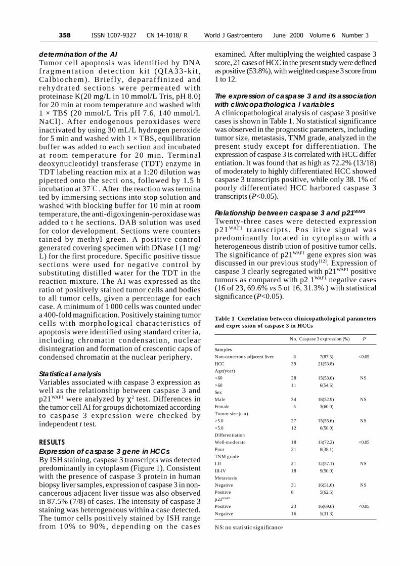
determination of the AITumor cell apoptosis was identified by DNAfragmen ta t i on de t ec t i on k i t (QIA33-k i t ,Calbiochem). Br ief ly , deparaff in ized andrehydra ted sec t ions were permeated wi thproteinase K(20 mg/L in 10 mmol/L Tris, pH 8.0)for 20 min at room temperature and washed with1 × TBS (20 mmol/L Tris pH 7.6, 140 mmol/LNaCl). After endogenous peroxidases wereinactivated by using 30 mL/L hydrogen peroxidefor 5 min and washed with 1 × TBS, equilibrationbuffer was added to each section and incubatedat room temperature for 20 min. Terminaldeoxynucleotidyl transferase (TDT) enzyme inTDT labeling reaction mix at a 1:20 dilution waspipetted onto the secti ons, followed by 1.5 hincubation at 37 . After the reaction was terminated by immersing sections into stop solution andwashed with blocking buffer for 10 min at roomtemperature, the anti-digoxingenin-peroxidase wasadded to t he sections. DAB solution was usedfor color development. Sections were counterstained by methyl green. A positive controlgenerated covering specimen with DNase I (1 mg/L) for the first procedure. Specific positive tissuesections were used for negative control bysubstituting distilled water for the TDT in thereaction mixture. The AI was expressed as theratio of positively stained tumor cells and bodiesto all tumor cells, given a percentage for eachcase. A minimum of 1 000 cells was counted undera 400-fold magnification. Positively staining tumorcells with morphological characteristics ofapoptosis were identified using standard criter ia,including chromatin condensation, nucleardisintegration and formation of crescentic caps ofcondensed chromatin at the nuclear periphery.
Statistical analysisVariables associated with caspase 3 expression aswell as the relationship between caspase 3 andp21WAF1 were analyzed by χ2 test. Differences inthe tumor cell AI for groups dichotomized accordingto caspase 3 expression were checked byindependent t test.
RESULTSExpression of caspase 3 gene in HCCsBy ISH staining, caspase 3 transcripts was detectedpredominantly in cytoplasm (Figure 1). Consistentwith the presence of caspase 3 protein in humanbiopsy liver samples, expression of caspase 3 in non-cancerous adjacent liver tissue was also observedin 87.5% (7/8) of cases. The intensity of caspase 3staining was heterogeneous within a case detected.The tumor cells positively stained by ISH rangefrom 10% to 90%, depending on the cas es
examined. After multiplying the weighted caspase 3score, 21 cases of HCC in the present study were definedas positive (53.8%), with weighted caspase 3 score from1 to 12.
The expression of caspase 3 and its associationwith clinicopathologica l variablesA clinicopathological analysis of caspase 3 positivecases is shown in Table 1. No statistical significancewas observed in the prognostic parameters, includingtumor size, metastasis, TNM grade, analyzed in thepresent study except for differentiation. Theexpression of caspase 3 is correlated with HCC differentiation. It was found that as high as 72.2% (13/18)of moderately to highly differentiated HCC showedcaspase 3 transcripts positive, while only 38. 1% ofpoorly differentiated HCC harbored caspase 3transcripts (P<0.05).
Relationship between caspase 3 and p21WAF1
Twenty-three cases were detected expressionp21 WAF1 t ranscr ip t s . Pos i t ive s igna l waspredominantly located in cytoplasm with aheterogeneous distrib ution of positive tumor cells.The significance of p21WAF1 gene expres sion wasdiscussed in our previous study[12]. Expression ofcaspase 3 clearly segregated with p21WAF1 positivetumors as compared with p2 1WAF1 negative cases(16 of 23, 69.6% vs 5 of 16, 31.3% ) with statisticalsignificance (P<0.05).
Table 1 Correlation between clinicopathological parametersand expre ssion of caspase 3 in HCCs
No. Caspase 3 expression (%) P
SamplesNon-cancerous adjacent liver 8 7(87.5) <0.05HCC 39 21(53.8)Age(year)<60 28 15(53.6) NS
>60 11 6(54.5)SexMale 34 18(52.9) NSFemale 5 3(60.0)Tumor size (cm)>5.0 27 15(55.6) NS
<5.0 12 6(50.0)DifferentiationWell-moderate 18 13(72.2) <0.05Poor 21 8(38.1)TNM gradeI-II 21 12(57.1) NS
III-IV 18 9(50.0)MetastasisNegative 31 16(51.6) NSPositive 8 5(62.5)p21WAF1
Positive 23 16(69.6) <0.05
Negative 16 5(31.3)
NS: no statistic significance
358 ISSN 1007-9327 CN 14-1018/ R World J Gastroentero June 2000 Volume 6 Number 3

Figure 1 Caspase 3 transcripts were detected predo minantly incytoplasm by ISH (×200)Figure 2 Apoptotic cells were determined by criteria as describedin materials and methods. Arrow shows fragmented nucleus (×200)
Relationship between tumor cell apoptosis andcaspase 3 expressionApoptotic cells and apoptotic bodies were foundin all cases of HCCs examined by in situ end-labeling (Figure 2). The mean AI of all tumorscases was 6.82‰ (s, 3.36‰; range 0.87‰-17.3‰). No significant associ ation was observedbetween AI and tumor stage. The mean AI forcaspase 3-positi ve tumors (n = 21) was 7.12‰(s, 3.75‰), while in caspase 3-negative cases (n= 18) the AI was 6.59‰ (s, 2.98‰), no statisticalsignificance was found between the two groups(P>0.05). In the cases with co- ex pression ofp21WAF1 and caspase 3, the AI was found lower,but with no significance, than that of cases withpositive caspase 3 and negative p21WAF1 (6.98‰vs 7.21‰).
DISCUSSIONIn this study, we have shown that caspase 3 wasexpressed in most of HCC cases. In our opinion,weare the first to describe the cpp32 expression inhuman prima ry HCCs. Like the result of humanbiopsy and autopsy liver materials[14] , the non-cancerous adjacent liver tissue showed strongcaspase 3 expression. A s high as 46.2% of humanprimary HCCs failed to show caspase 3 expression.
In t he 21 cases which express caspase 3, theexpression showed heterogeneous pattern with aweighted score from 1 to 12. Because caspase 3 isthe effector caspase in the apoptosis pathways, wethink that loss of caspase 3 expression may play animportant role in HCC carcinogenesis. Caspase 3expression had no relationsh ip with clinicopathologicalfeatures except for tumor differentiation (Table 1).The result suggest that the expression of caspase 3 iscorrelated with tumor differentiation in HCC. It wasreported that 33.1% cases of gastric carcinoma werecaspase 3 positive[7]. Two of 3 breast carcinomatissues expressed caspase 3, the immunointensity wasgenerally higher in invasive cancers[8]. It raised thepossibility that expression of casap se 3 in tumorsshowed tissue specificity. It was observed in this studythat the apoptosis index (AI) was not associated withthe expression of caspase 3 in HCC (7.12‰ in cpp32-positive cases versus 6.59‰ in cpp32-negative group).In the non-cancerous adjacent liver tissues more than50% of the cells showed positive caspase 3, the AIwas not increased as compared with HCC, no matterca spase 3 was positive or negative. This suggeststhat other factor(s) may exist in regulating normal cellapoptosis. p21WAF1 was first reported as a universal inhibitorof cyclin-dependent kinase, which is required forG 1 t o S t r a n s i t i o n [ 1 5 ] . P r e v i o u s s t u d i e sdemonstrated that p21 WAF1 can interrupt theapoptotic process at a point upstream fromcaspase-3 activation, because serum starvatio n,which also synchronized cells in G1 but did notinduce p21 WAF1, did not protect cel ls f romapoptosis, while restoring a late G1 checkpoint byinducing p21WAF1 expression can protect cells fromDNA damage induced apoptosis[16]. p21WAF1 canbind procaspase-3 but not activate caspase 3. Onthe other hand, activa ted caspase 3 can cleavep21WAF1. P21 cleavage by the activated cpp32specifically abolished its interaction with PCNAand may interfere with normal PCNA-dependentrepair [15]. The presence of p21WAF1 in hu manHCCs was reported previously in our paper[12]. Inthis context, the expression of p21WAF1 was alsodetermined together with caspase 3 in an attemptto find whether there is relationship betweenthem. We found that expression of caspase 3 wasstrongly associated with p21WAF1 in 16 cases ofHCC. There was no significant difference in AIbetween p21WAF1(+)/cpp32(+) and p21WAF1(-) /cpp32(+) (6.98‰ vs 7.21‰). The results indicatedthat p21WAF1 may be merely one of the inhibitorswhich can reduce caspase 3 mediated cel lapoptosis in HCCs. In fact, some other caspase 3inhibitors, for example survivin, were reported tohave overexpression in human tumors, includinggastric and colorectal cancer [17,18]. Survivin is
Sun BH, et al. Expression of caspase 3 gene in liver cancer 359

believed to bind activated caspase 3 and further in hibitcell apoptosis. It was reported that XIAP can interruptcaspase 3 mediated apoptosis via the same way asp21WAF1. So, further investig ation on other caspase 3regulating protein is needed to find the regulation mechanism of caspase 3 mediated apoptosis in human HCCs.
REFERENCES1 Tang ZY,Yu YQ,Zhou XD, Ma ZC, Wu ZQ. Progress and pros-
pects in hepatocellular carcinoma surgery. Ann Chir, 1998;52:558-563
2 Kokura K, Nakadai T, Kishimoto T, Makino Y, Muramatsu M,Tamura T. Gene expression in hepatomas. J Gastroenterol Hepatol,1998;13(Suppl):S132-141
3 Wyllie AH. Apoptosis and carcinogenesis. Europ J Cell Biology,1997;73:189-197
4 Kidd VJ. Proteolytic activities that mediate apoptosis. Annu RevPhysiol, 1998;60:533-573
5 Kitada S, Andersen J, Akar S, Zapata JM, Takayama S, KrajewskiS, Wang HG, Zhang X, Bullrich F, Croce CM, Rai K, Hines J, ReedJC. Expression of apoptosis-regulating proteins in chronic lym-phocytic leukemia: correlations with in vitro and in vivochemoresponses. Blood, 1998;91: 3379-3389
6 Estrov Z, Thall PF, Talpaz M, Estey EH, Kantarjian HM, AndreeffM, Harris D, Van Q, Walterscheid M, Kornblau SM.Caspase 2 andcaspase 3 protein levels as predictors of survival in acute myelog-enous leukemia. Blood, 1998;92:3090-3097
7 Krajewski S, Gascoyne RD, Zapata JM, Krajewska M, Kitada S,Chhanabhai M, Horsman D, Berean K, Piro LD, Fugier-Vivier I,Liu YJ, Wang HG, Reed JC. Immunolocalization of the ICE/Ced-3-family protease, CPP32 (Caspase-3), in non-Hodgkin’slymphomas, chronic lymphocytic leukemias, and reactive lymphnodes. Blood, 1997;89:3817-3825
8 Chhanabhai M, Krajewski S, Krajewska M, Wang HG, Reed JC,Gascoyne RD. Immunohistochemical analysis of interleukin-1 β-
converting enzyme/Ced-3 family protease, CPP32/Yama/Caspase-3, in Hodgkin’s disease. Blood, 1997;90:2451-2455
9 Zapata JM, Krajewska M, Krajewski S, Huang RP, Takayama S,Wang HG, Adamson E, Reed JC. Expression of multiple apoptosis-regulatory genes in human breast cancer cell lines and primarytumors. Breast Cancer Res Treat, 1998;47:129-140
10 Nakagawara A, Nakamura Y, Ikeda H, Hiwasa T, Kuida K, Su MS,Zhao H, Cnaan A, Sakiyama S. High levels of expression andnuclear localization of interleukin-1 beta converting enzyme (ICE)and CPP32 in favorable human neuroblastomas. Cancer Res, 1997;57:4578-4584
11 Suzuki A, Tsutomi Y, Akahane K, Araki T, Miura M. Resistance toFas-mediated apoptosis: activation of caspase 3 is regulated bycell cycle regulator p21WAF1 and IAP gene family ILP. Oncogene,1998;17:931-939
12 Sun BH, Wu ZB, Ruan YB, Yang ML, Liu B. p21WAF1/Cipl geneexpression in primary human hepatocellular carcinoma and itsrelationship with p53 gene mutation. J Tongji Medical University,1999;19:1-5
13 Kawasaki H, Altieri DC, Lu CD, Toyoda M, Tenjo T, Tanigawa N.Inhibition of apoptosis by survivin predicts shorter survival ratesin colorectal cancer. Cancer Res, 1998;58:5071-5074
14 Krajewska M, Wang HG, Krajewski S, Zapata JM, Shabaik A,Gascoyne R, Reed JC. Immunohistochemical analysis of in vivopatterns of expression of CPP32 (caspase 3), a cell death protease.Cancer Res, 1997;57:1605-1613
15 Li R,Waga S,Hannon GJ,Beach D,Stillman B. Differential effectsby the p21 CDK inhibitor on PCNA dependent DNA replicationand repair. Nature, 1994;371:534-537
16 Bissonnette N, Hunting DJ. p21-induced cycle arrest in G1 pro-tects cells from apoptosis induced by UV irradiation or RNA poly-merase II blockage. Oncogene, 1998;16:3461-3469
17 Lu CD, Altieri DC, Tanigawa N. Expression of a novel antiapoptosisgene, survivin, correlated with tumor cell apoptosis and p53 accu-mulation in gastric carcinomas. Cancer Res, 1998; 8:1808-1812
18 LaCasse EC, Baird S, Korneluk RG, Mackenzie AE. The inhibitorsof apoptosis (IAPs) and their emerging role in cancer. Oncogene, 1998;17:3247-3259
Edited by Ma JYproofread by Sun SM
360 ISSN 1007-9327 CN 14-1018/ R World J Gastroentero June 2000 Volume 6 Number 3
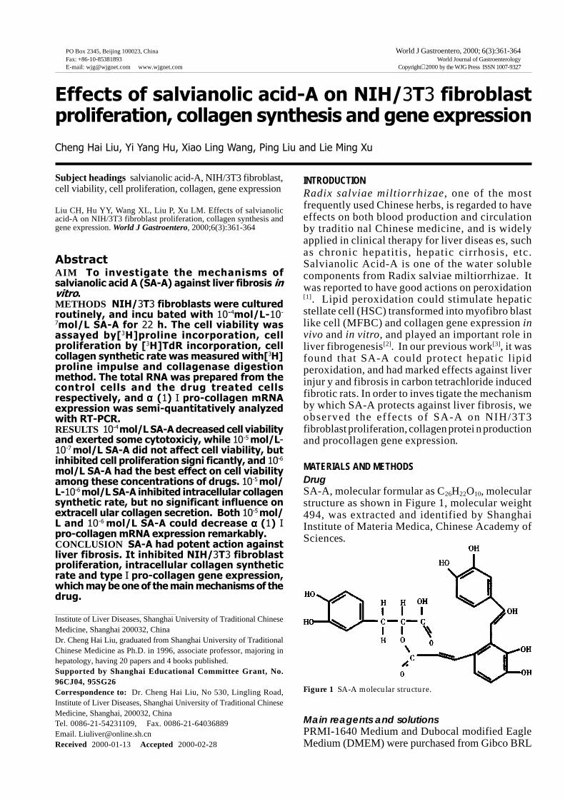
Effects of salvianolic acid-A on NIH/3T3 fibroblastproliferation, collagen synthesis and gene expression
Cheng Hai Liu, Yi Yang Hu, Xiao Ling Wang, Ping Liu and Lie Ming Xu
Subject headings salvianolic acid-A, NIH/3T3 fibroblast,cell viability, cell proliferation, collagen, gene expression
Liu CH, Hu YY, Wang XL, Liu P, Xu LM. Effects of salvianolicacid-A on NIH/3T3 fibroblast proliferation, collagen synthesis andgene expression. World J Gastroentero, 2000;6(3):361-364
Abstract
AIM To investigate the mechanisms ofsalvianolic acid A (SA-A) against liver fibrosis invitro.METHODS NIH/3T3 fibroblasts were culturedroutinely, and incu bated with 10-4mol/L-10-
7mol/L SA-A for 22 h. The cell viability wasassayed by[3H]proline incorporation, cellproliferation by [3H]TdR incorporation, cellcollagen synthetic rate was measured with[3H]proline impulse and collagenase digestionmethod. The total RNA was prepared from thecontrol cells and the drug treated cellsrespectively, and ααααα (1) I pro-collagen mRNAexpression was semi-quantitatively analyzedwith RT-PCR.RESULTS 10-4 mol/L SA-A decreased cell viabilityand exerted some cytotoxiciy, while 10-5 mol/L-10-7 mol/L SA-A did not affect cell viability, butinhibited cell proliferation signi ficantly, and 10-6
mol/L SA-A had the best effect on cell viabilityamong these concentrations of drugs. 10-5 mol/L-10-6 mol/L SA-A inhibited intracellular collagensynthetic rate, but no significant influence onextracell ular collagen secretion. Both 10-5 mol/L and 10-6 mol/L SA-A could decrease α α α α α (1) Ipro-collagen mRNA expression remarkably.CONCLUSION SA-A had potent action againstliver fibrosis. It inhibited NIH/3T3 fibroblastproliferation, intracellular collagen syntheticrate and type I pro-collagen gene expression,which may be one of the main mechanisms of thedrug.
INTRODUCTIONRadix salviae miltiorrhizae, one of the mostfrequently used Chinese herbs, is regarded to haveeffects on both blood production and circulationby traditio nal Chinese medicine, and is widelyapplied in clinical therapy for liver diseas es, suchas chronic hepatit is, hepatic cirrhosis, etc.Salvianolic Acid-A is one of the water solublecomponents from Radix salviae miltiorrhizae. Itwas reported to have good actions on peroxidation[1]. Lipid peroxidation could stimulate hepaticstellate cell (HSC) transformed into myofibro blastlike cell (MFBC) and collagen gene expression invivo and in vitro, and played an important role inliver fibrogenesis[2]. In our previous work[3], it wasfound that SA-A could protect hepatic lipidperoxidation, and had marked effects against liverinjur y and fibrosis in carbon tetrachloride inducedfibrotic rats. In order to inves tigate the mechanismby which SA-A protects against liver fibrosis, weobserved the effects of SA-A on NIH/3T3fibroblast proliferation, collagen protei n productionand procollagen gene expression.
MATERIALS AND METHODSDrugSA-A, molecular formular as C26H22O10, molecularstructure as shown in Figure 1, molecular weight494, was extracted and identified by ShanghaiInstitute of Materia Medica, Chinese Academy ofSciences.
Figure 1 SA-A molecular structure.
Main reagents and solutionsPRMI-1640 Medium and Dubocal modified EagleMedium (DMEM) were purchased from Gibco BRL
Institute of Liver Diseases, Shanghai University of Traditional ChineseMedicine, Shanghai 200032, ChinaDr. Cheng Hai Liu, graduated from Shanghai University of TraditionalChinese Medicine as Ph.D. in 1996, associate professor, majoring inhepatology, having 20 papers and 4 books published.Supported by Shanghai Educational Committee Grant, No.96CJ04, 95SG26Correspondence to: Dr. Cheng Hai Liu, No 530, Lingling Road,Institute of Liver Diseases, Shanghai University of Traditional ChineseMedicine, Shanghai, 200032, ChinaTel. 0086-21-54231109, Fax. 0086-21-64036889Email. [email protected] 2000-01-13 Accepted 2000-02-28
PO Box 2345, Beijing 100023, China World J Gastroentero, 2000; 6(3):361-364Fax: +86-10-85381893 World Journal of GastroenterologyE-mail: [email protected] www.wjgnet.com Copyright 2000 by the WJG Press ISSN 1007-9327

Co., new brown serum (NBS) from Shanghai Sino-American Co., purified type III collagenase (specificactivity, 960U/mg), N-ethylmaleimide (NEM) and β-aminopropionitrile from Sigma Co. [53H]proline ([3 H]Pro) from Amersham Co. methyl-[3H] thymidine (TdR)from Shanghai Inst itute of Atomic Energy, guanidiumthiocynate from Serva Co. Access RT-PCR System Kit,PCR marker from Promega Co., Diethypyrocarbonate,saturated phenol/chloroform mix and agarose fromShanghai Sangon Biotech Co. Other reagents all wereof analytical grade. The non-homogeneous scintillation liquid wasdimethylbenzene solution containing 5 g/L 2,5-diphenyloxazol (PPO) and 0.5 g/L 1,4-bis[5-phenyloxazol-2]benzene (POPOP), the homogeneousscintillation liquid was dimethy lbenzene solutioncontaining 7 g/L PPO, 0.5 g/L POPOP, 100 g/Lnaphthalene and 400 mL/L2-ethoxy-ethanol.
Cell lineMouse NIH/3T3 fibroblasts were purchased fromShanghai Institute of Cell Biolo gy, Chinese Academyof Sciences, and cultured with PRMI-1640 mediumcontaining 100 g/L NBS, 100KU/L penicillin and 100mg/L streptomycin. After the cell growth becameconfluent, they were digested with trypsin-EDTA andsubcultured.
PCR PrimersThe PCR primers for pro-collagen α 2(I) and β-actinwere designed according to the published sequences andreferences in Table 1[4], and were synthesized by GibcoBRL Co.
Table 1 PCR primer sequences and expected size of amplifiedproducts
Primers Sequence Size
α 2(I) collagen upstream 5’TGT TCG TGG TTC TCA GGG TAG3’
α 2(I) collagen downstream 5’TTG TCG TAG CAG GGT TCT TTC3’ 254 bpβ-actin upstream 5’ACA TCT GCT GGA AGG TGG AC3’
β-actin downstream 5’GGT ACC ACC ATG TAC CCA GG3’ 163 bp
Cell proliferation assayConfluent NIH/3T3 fibroblasts in 24 well plates wereincubated with 10-4mol/L-10-7mol/L SA-A diluted inPRMI-1640 medium containi ng 100 mL/L NBS for22 h, and[3H]TdR (55.5KBq/well) was impu lsed inthe last 16 h. Then cells were harvested with trypsindigestion and collected on the filtration membrane,then sample radioactivity (cpm) in the non-homogeneous scintillation liquid was measured byBackman Wallac 1410 Scintillator. All tests wererepeated 3 times.
Cell viability assayAccording to Mallat’s method[5], confluent
NIH/3T3 fibroblast s in 24-well plates were incubatedwith 10-4mol/L-10-7mol/L SA-A re solved in PRMI-1640medium without NBS for 22 h, and[3H]Pro (55.5KB q/well) was impulsed in the last 16 h. Then cells werecollected and the cpm was measured as above.
Assay of cell collagen synthetic rateAccording to Greets’method[6], confluent NIH/3T3fibroblasts in 6 well plates were incubated with10-5mol/L-10-6mol/L SA-A diluted in PRMI-1640without NBS for 22 h, during the later 16h theculture media were changed to DMEM containing1 8 5 K B q / m L [ 3 H ] P r o , 1 0 0 m g / L - β -aminopropionitrile, 50 mg/L asc orbic acid as wellas the same drugs. Then the culture media and celllayer extract were collected respec tively, dialyzedthoroughly and reacted with collagenase, etc. Thetotal radioa ct ivi ty in the samples (cpm t) ,radioactivity in the samples treated with collagenase (cpmc) and not treated with collagenase(cpm b) were counted in the ho mogeneousscintillation liquid by Backman Wallac 1410Scintillator. The new col lagen that cell produced,i.e. the fraction of collagenous protein expressedas percentage of total radiolabeled protein, wascalculated using the formula:
cpmt-cpmc
% of collagen = 100 ÷ (5.4× +1) cpmc-cpmb
RNA extraction and RT-PCR (reverse transcriptionand polymerase chain reaction)The total RNA was extracted from the control cellsand SA-A incubated cells by the acid guanidiumthiocynate-phenol-chloroform method[7]. The RNAquantity was determined by absorption at 260 nm, itspurity was confirmed with A260/A280 specrophotometer readings that ranged from 1.6 to 1.9, and itsintegrity was checked by 9 g/L agarose gel electrophoresis with ethidium bromide (EB) staining of18S and 28S ribosomal RNA (Figure 2). With AccessRT-PCR system kit, the cDNA synthesis andamplification was done in one tube following themanufacturer’s instructions. In brief, 1 µg RNA, 50pmol/L primers for α (1) I pro-collagen or β-actinwere added to each reaction mixture respectively,which included 10 mmol/L dNTPs 1 µL, 25 mmol/LMgSO4 2 µL, AMV reverse transcriptase 5U, TflDNA polymerase 5U, AMV/Tfl-5 × buffer 10 µL. Thereaction final volume was 50 µL and was coveredwith 20 µL mineraloil. Then with PCR Touchdownthermal cycler (Hybaid, England), RT-PCR reactionwas run in the following procedures: 48 for45 min, 1 circle. 94 for 2 min, 1 circle. 94for 30s, 60 for 1 min, 38 for 2 min, 30 circles. 68 for 7 min, 1 circle. Five µL PCR product wasrun on 15 g/L agarose gel and observed by EB
362 ISSN 1007-9327 CN 14-1018/ R World J Gastroentero June 2000 Volume 6 Number 3
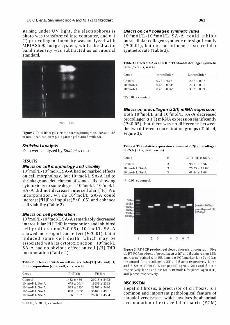
Liu CH, et al. Salvianolic acid-A and NIH /3T3 fibroblast 363
staining under UV light, the electrophores isphoto was transformed into computer, and α 1(I) pro-collagen intensity was analyzed withMPIAS500 image system, while the β-actinband intensity was subtracted as an internalstandard.
Figure 2 Total RNA gel electrophoresis photograph. 28S and 18Sof total RNA run on 9 g/L agarose gel stained with EB.
Statistical analysisData were analyzed by Student’s t test.
RESULTSEffects on cell morphology and viability10-5mol/L-10-7mol/L SA-A had no marked effectson cell morphology, but 10-4mol/L SA-A led toshrinkage and detachment of some cells, showingcytotoxicity to some degree. 10-4mol/L-10-7mol/LSA-A did not decrease intercellular [3H] Proincorporation, wh ile 10-6mol/L SA-A couldincrease[3H]Pro impulse(P<0 .05) and enhancecell viability (Table 2).
Effects on cell proliferation10-4mol/L-10-6mol/L SA-A remarkably decreasedintercellular [3H]TdR incorporation and inhibitedcell proliferation(P<0.05), 10 -4mol/L SA-Ashowed more significant effect (P<0.01), but iti nduced some ce l l dea th , wh ich may beassociated with its cytotoxic action. 10-7mol/LSA-A had no obvious effect on cell [3H] TdRincorporation (Tabl e 2).
Table 2 Effects of SA-A on cell intracellular[3H]TdR and[3H]Pro incorporation (cpm/well, x ± s, n = 4)
Group [3H]TdR [3H]Pro
Control 1482 ± 486 21018 ± 547310-4mol/L SA-A 675 ± 201b 18659 ± 236310-5mol/L SA-A 969 ± 183a 23761 ± 543010-6mol/L SA-A 868 ± 183a 31408 ± 4981a
10-7mol/L SA-A 1056 ± 187 26080 ± 4504
aP<0.05, bP<0.01, vs control.
Effects on cell collagen synthetic rates1 0 - 5mo l /L -10 - 6mo l /L SA-A cou ld i nh ib i tintracellular collagen synthetic rate significantly(P<0.01), but did not influence extracellularsynthetic rate (Table 3).
Table 3 Effects of SA-A on NIH/3T3 fibroblast collagen syntheticrates (%, x ± s, n = 4)
Group Intracellular Extracellular
Control 0.78 ± 0.03 2.57 ± 0.3710-5mol/L 0.48 ± 0.24b 2.54 ± 0.9110-6mol/L 0.43 ± 0.26b 3.02 ± 0.69
bP<0.01, vs control.
Effects on procollagen α α α α α 2(I) mRNA expressionBoth 10-5mol/L and 10-6mol/L SA-A decreasedprocollagen α 1(I) mRNA expression significantly(P<0.05), but there was no difference betweenthe two different concentration groups (Table 4,Figure 3).
Table 4 The relative expression amount of α 2(I) procollagenmRNA (x ± s, % of β-actin)
Group n Col α 1(I) mRNA
Control 3 98.71 ± 9.9610-5mol/L SA-A 3 76.23 ± 12.02a
10-6mol/L SA-A 3 68.44 ± 8.06a
aP<0.05, vs control.
Figure 3 RT-PCR product gel electrophoresis photog raph. FiveµL RT-PCR products of procollagen α 2(I) and β-actin run on 1.5%agarose gel stained with EB. Lane 1 as PCR marker, lane 2 and 3 asthe control for procollagen α 2(I) and β-actin respectively, lane 4and 5 SA-A 10-6mol/L for procollagen α 2(I) and β-actinrespectively, lane 6 and 7 as SA-A 10-5mol/L for procollagen α 2(I)and β-actin respectively.
DISCUSSIONHepatic fibrosis, a precursor of cirrhosis, is acommon and important pathological feature ofchronic liver diseases, which involves the abnormalaccumulation of extracellular matrix (ECM)

proteins, particularly collagen[8]. In fi brotic liver,ECM components are mainly produced by HSC andfibroblasts. It is known that during fibrogenesis, HSCundergoes a process of activation, developing amyofibroblast-like phenotype associated withincreased proliferation and ECM production,especially type I collagen synthesis. The mouseNIH/3T3 fibroblast also shared the features thatactive HSC (MFBC) presented, such as remarkableprolife ration and substantial production of collagen,and stable cell line. In practice, NIH/3T3 fibroblastis often used as a desirable cell model for investigation of antifibrotic drugs. In order to rule out the possibility of SA-A cytotoxicinfluence in vitro, the intracellular[3H] Pro incorporationwas measured, and inverted microscopic observation wasdone. It was found that only 10-4mol/L SA-A causedsome cell detachment, decreased [3H] Pro incorporation,and showed cytotox icity to some extents. 10-5mol/L-10-
7mol/L SA-A did no t influence cell morphology or inhibitcell viability. However, 10-6m ol/L SA-A enhanced cellviability. Both 10-5mol/L-10-6 mol/L SA-A could inhibitintracellular[3H]TdR impulse that NBS stimul ated. It issuggested that SA-A had an effective action against NIH/3T3 fibr oblast proliferation. Type I collagen is the predominant component ofECM during liver fibrosis. Its production involve twoprocesses: the first is intracellular synthesis, includi nggene transcription, translation and modification to formprocollagen, then procollagen alpha chains are secretedto the outside of the cell to form helix collagen by sortingand alignment etc. In the study, it was found that SA-A downregulated procollagen α 2(I) steady-statemRNA expression, and intracellular collagen syntheticrate, but exerted no effect on extracellular syntheticrate. It is suggested that SA-A influence on collagenproduction through the intracellular synthetic
process. The fibrogenic cells have two predo minantfeatures: one is active in cell proliferation, which led toincrease in cell number, another is strong fibrogenic abilitypercell, which led to accumula tion of ECM. In the study,SA-A not only inhibited NIH/3T3 fibroblast prolif eration,but also decreased collagen synthesis, showing a goodaction against liver fibrosis. Salvianolic radix is widely used as an importantcomponent in Chinese herbal for mulas for the treatmentof chronic liver diseases. Salvianolic Acid-A, one ofwater-soluble ingredients from Salvianolic radix, hadeffective actions on hepa tic peroxidation and fibrosis invivo[3]. In the paper, it is for the first time found thatSA-A has the potential action against hepatic fibrosi s invitro, and its main mechanisms of antifibrotic actionperhaps was assoc iated with the inhibition of fibrogeniccell proliferation, collagen gene expres sion and proteinsynthesis.
REFERENCES1 Lin TJ, Liu GT. Protective effect of salvianolic acid A on heart
and liver mitochondria injury induced by oxygen radicals in rats.Zhongguo Yaolixue Yu Dulixuebao, 1991;5:276-281
2 Olaso E, Friedman SL. Molecular regulation of hepatic fibrogenesis.J Hepatol, 1998;29:836-847
3 Hu YY, Liu P, Liu C, Xu LM, Liu CH, Zhu DY, Huang MF.Actions of salvianolic aicd A on CCl4 poisoned liver injury andfibrosis in rats. Zhongguo Yaoli Xuebao, 1997;18:478-480
4 Malllat A, Preaux AM, Blazejewski S, Rosenbaum J, Dhumeaux D,Mavier P. Interferon alpha and gamma inhibit proliferation andcollagen synthesis of human Ito cells in culture. Hepatology, 1995;21:1003-1010
5 Power WJ, Kaufman AH, Merayo-Lioves J, Arrunategui-CorreaV, Foster CS. Expression of collagens I, III, IV and V mRNA inexcimer wounded rat cornea: analysis by semi quantitative PCR.Curr Eye Res, 1995;14:879- 886
6 Greets A, Vrijsen R, Rauterberg J, Burt A, Schellinck P, Wisse E. Invitro differentiation of fat-storing cells parallels marked increaseof collagen synthesis and secretion. J Hepatol, 1989;9:59-68
7 Chomczynski P, Sacchi N. Single step method of RNA isolationby acid guanidinium thiocynate phenol-chloroform extraction.Ana Biochem, 1987;162:156-159
8 Friedman SL. The cellular basis of hepatic fibrosis: mechanismsand treatment strategies. New Eng J Med, 1993;328:1828-1836
Edited by Zhu LHproofread by Sun SM
364 ISSN 1007-9327 CN 14-1018/ R World J Gastroentero June 2000 Volume 6 Number 3

Primary porcine hepatocytes with portal vein serumcultured on microcarriers or in spheroidalaggregates
Yi Gao, Huan Zhang Hu, Ke Chen and Ji Zhen Yang
Subject headings porcine hepatocytes; microcarriers;cell culture; spheroidal aggregate culture; portal vein serum
Gao Y, Hu HZ, Chen K, Yang JZ. Primary porcine hepatocytes withportal vein serum cultured on microcarriers or in spheroidal aggregates.World J Gastroentero, 2000;6(3):365-370
Abstract
AIM To develop a culture mode providingdurable biomaterials with high yields andactivities used in bioartificial liver.METHODS Hepatocytes were isolated from awhole pig liver by Seglen’s method oforthotopic perfusion with collagenase. Inculture on microca rriers, primary porcineh e p a t o c y t e s w e r e i n o c u l a t e d a t aconcentration of 5 × 10⁷/mL into the staticculture systems containing 2 g/L Cytodex-3,then supplemented with 100 mL/L fetal calfserum (FCS) or 100 mL/L porcine portal veinserum (PPVS) respectively. In spheroidalaggregate culture hepatocytes were inoculatedinto 100 mL siliconized flasks at a concentration of 5.0 × 10⁶/mL.RESULTS In culture on microcarriershepatocytes tended to aggr egate on Cytodex-3 obviously after being inoculated. Typical multi-cellular aggregated spheroids could be found inthe two systems 24 h-48 h after hepatocyteswere cultured. The morphological charact-eristics and syntheti c functions were maintainedfor 5 wk in FCS culture system and 8 wk in PPVSculture system. In spheroidal aggregate cultureabout 80%-90% isolated hepatocytes becameaggregated spheroids 24 h after cultured insuspension and mean diameter of the spheroidswas 100 µµµµµm. The relationship among thehepatocytes resembled that in the liver in vivo.Synthetic functions of albumin and urea of the
spheroids were twice those of hepatocytescultured on monolayers.CONCLUSION As high-yields and high-activitymodes of culture on microcarriers or inspheroidal aggregate culture with portal veinserum are promising to provide biomaterials forbioartificial liver (BAL) efficiently.
INTRODUCTIONGreat attention was paid world-wide to thebioartificial liver (BAL) aiming to release theclinical symptoms of severe acute liver diseasesor liver injuries and act as a transitional bridge forliver transplantation. Modern BAL is main lycharacterized by i ts mult iple biofunctionsresembling those of l iver in vivo and suchfunctions may be completely derived from thebiomaterials in BAL. Years of researches at homeand abroad showed that primary hepatocytes werethe most ideal biomaterial of BAL. But for themorphological and functional changes of primaryhepatocytes cultivated in vitro occurred obviouslyin short time, it was deadly restrained to use themas the biomaterial in BAL. To probe into thisquestion, together with our proceeding researcheson this subject[1], we took the method of two-steporthotopic collagenase perfusion to isolatehepatocytes and cultured them on microcarriersCytodex-3 statically in flasks in the systemssupplemented with FCS or PPVS respectively, andalso cultured them in the matrix DMEM containingporcine portal vein serum at a concentration of100 mL/L through the way of spheroida laggregating, then estimated the po ssibility of suchhepatocytes acting as the biomaterial in BAL inview of their morphology and- biofunctions.
MATERIALS AND METHODSMaterialsThe experimental animals were five native sex-unlimited pigs aged 1 mo-2 mo and weighed 10 kg-15 kg from the experiment animal center of SUNYat-Sen Medical University. Insulin, glucagon,transferrin, collagenase IV, typran and microcarrierCytodex-3 were perchased from Sigma (USA).Solution of 50 g/L methyl silicon resin ethyl acetate
Department of Hepatobiliary Surgery, Zhujiang Hospital, First MilitaryMedical University, Guangzhou 510282, Guangdong Province, ChinaDr. Yi Gao, graduated from Second Military Medical University as M.D.in 1992, associate professor of hepatobiliary surgery, mentor ofpostgraduate.Supported by the National Natural Science Foundation ofChina, No.39570212Correspondence to: Dr. Yi Gao, Department of Hepatobiliary Surgery,Zhujiang Hospital, First Military Medical University, Guangzhou510282, Guangdong Province, ChinaTel. 0086-20-85143556Received 2000-01-10 Accepted 2000-03-02
PO Box 2345, Beijing 100023, China World J Gastroentero, 2000; 6(3):365-370Fax: +86-10-85381893 World Journal of GastroenterologyE-mail: [email protected] www.wjgnet.com Copyright 2000 by the WJG Press ISSN 1007-9327

was confected by ourselves. Ten g/L osmic acid, 20 g/Lpentanal and gradient acetone solutions were providedby the Department of Electron microscope of FirstMilitary Medical University. S-450 electron scanningmicroscope was from Japan. PPVS was collected inthe experiment. The liver perfusion solution was preparedaccording to Table 1.
MethodsIsolation of porcine hepatocytes The experimenalpig was kept fasting for 12 h preoperatively andanaesthetized with 30 g/L sodium barbiturateip. Afterthe abdominal skin being prepared and disinfected,celiotomy was performed, then portal vein and inferiorcaval vein below the liver exposed and 0.25 mg(125IU)heparin injected into the inferior cavalvein. Afterbeing dissected, the portal vein and the inferior cavalvein were ligated at the distal end and intubated atthe proximal end respectively. When the inferior cavalvein above the liver ligated at the superior end of livervein, successive perfusing solutions I and II pre-warmed in 37 water bath were perfused along thecircuit via portal vein to inferior caval vein. Perfusingspeed was about 50 mL/min-60 mL/min for 20 min.Perfusion was ended until the liver became pallor oryellow in color. Then the well digested liver weretaken out, tore off the liver sheath and the isolatedhepatocytes delivered into solution III pre-warmed in37 water bath. After being filtered by 150 µm, 100µm and 80 µm sieve, the hepatocytes were suspendedin solution and centrifuged at a speed of 1 000r/minfor 3 min-5 min repeated three times. The depositedhepatocytes were added into solution IV to preparethe hepatocyte suspension for use. Portal vein bloodwas gathered from the distal end of the ligated portalvein and in a total amount of 1 300mL from 5 pigs. Itwas centrifuged at a 2 000r/min to isolate the portalvein serum, and the very serum centrifuged three timesmore to prepare the refined serum which finallyamounted to 714 mL. The refined serum was filteredto be excluded from bacteria and preserved separatelyfor use. Sample from the suspension was observedunder light microscope to calculate the yielding rateof hepatocytes and stained by typran for determinationof their activity.
Static culture of hepatocytes on microcarriers inflasksPorcine hepatocyte suspension was dividedrandomly into two halves, then one half was culturedon microcarriers in FCS system and the other inPPVS system. Five pigs were done separately andby the same method. Culture of hepatocytes inFCS system: five flasks were prepared and eachone added with 2 mL-3 mL 50 g/L sterile methylsilicon resin ethyl acetate solution. After the flasksbeing shaken mildly and equably, the remained
solution was discarded. The flasks were parched in60 electric oven for use. 20 mg-30 mg Cytodex-3 wasdeposited into the siliconized flasks and rinsed with 40mL Ca2+ and Mg2+ free phosphate buffer solution (PBS)for three times, then dipped in PBS over night. Afterbeing sterilized at 15 pounds for 30 min, the microcarrierswere dipped in DMEM matrix over 10 h foruse.Hepatocytes were inoculated into the flasks at theconcentration of 5 × 10⁶/mL and DMEM matrixcontaining 100 mL/L FCS, 100 µg/L glucagon, 100 µg/Ltransferrin, 200 µg/L hydrocortisone, 100 µg/L insulinand 200 µg/L fortum addedup to the total volume, 50mL. Then the flasks were deposited into an incubatorwith 100% humidified atmosphere containing 5% CO2
at 37 and shaken reelingly once every 15 min-30 minat the first 4 h, once every 2 h after 4 h and laid quietlyafter 8 h without further shaking. Ten mL supernatantfluid of the culture matrix was changed after 24 h andonce every 2 d from then on. The supernatant fluid wascentrifuged at 1 000r/min for discarding the cellfragments. Synthesized albumin and urea weredetermined. Culture of hepatocytes in PPVS system:100 mL/L refined PPVS was added into the culturesystem instead of FCS, and the other demands were thesame as those of the FCS culture.
Spheroidal aggregate culture of primary porcinehepatocytesFive flasks were prepared and each one added with 2mL-3 mL 50 g/L sterile methyl silicon resin ethyl acetatesolution. After the flasks being shaken mildly and equably,the residual solution was discarded. The flasks wereparched in 60 electric oven for use. Hepatocytes wereinoculated into the flasks at the concentration of 5 × 10⁶/mL and DMEM matrix containing 100 mL/ L porcineportal vein serum, 100 µg/L glucagon, 100 µg/Ltransferrin, 200 µg/L hydrocortisone, 100 µg/L insulinand 200 µg/L fortum added up to a total volume, 50 mL.Then the flasks w ere deposited into the incubator with100% humidified atmosphere containing 5% CO2 at37 and shaken reelingly once every 15 min-30 min atthe first 4 h, once every 2 h after 4 h and laid quietlyafter 8 h without furt her shaking. 10 mL supernatantfluid of the culture matrix was changed after 24 h andonce every 2 d from then on. The supernatant fluid wascent rifuged at 1 000r/min for discarding the cellfragments. Syntheses of albumin and urea weredetermined.
Morphological observation and counting ofhepatocytesThe porcine hepatocytes cultured on microcarriersand those cultured as aggregat ed spheroids wereobserved under phase- contrast light microscopeand photograph ed on an automatic microscopicphotographing device successively (Olympus,Japan ). In culture on microcarriers 1 mL samples
366 ISSN 1007-9327 CN 14-1018/ R World J Gastroentero June 2000 Volume 6 Number 3

Gao Y, et al. Culture of porcine hepatocytes on microcarriers or in spheroida aggregates 367
were collected every day to calculate the amountof cells in the culture system by means of trypanblau. On d7, cell sample was taken at a well-distributed state, culture matrix discarded, rinsedwith PBS, then fixed for 0.5 h with 2 mL 20 g/Lpentanal, rinsed with PBS again, fixed for 0.5 hwith 10 g/L osmic acid, and next dehydratedgradientially for 10 min each stage, exchangedwith acetic isopental ester for 4 hours, dried byCO2 drier (Hitachi HCP2, Japan), and finallysplashed with ionic platinum in vacuum. Growthof the cells was observed on S-450 electronscanning microscope (Japan). In spheroidalaggregate culture, 1 mL samples were taken well-distributedly from the matrix for cell calculationon the d7 and d12.
Determination of albumin and urea syntheses When the culture matrix was changed, samples ofsupernatant fluid were obtained from the cultu reon microcarriers and spheroidal aggregate cultureto determine the contents of albumin and ureathrough an automatic biochemical analyzer(Beckman, USA).
RESULTSIsolation of porcine hepatocytesWe successfully took the improved Seglen’s two-step orthotopic perfusion method with collagenaseto isolate the porcine hepatocytes. About 2.82 × 10¹⁰
hepatocytes were obtained and the survival rate91.2%. Each gram pig liver offered 7.01 × 10⁷
hepatocytes.
Cultivation of porcine hepatocytesIn culture on microcarriers, the amount ofhepatocytes on the d7 in the two systems is shownin Table 2. Hepatocytes growth curve (Figure 1) wasdra wn on the basis of Table 2. On d18 hepatocytesin FCS culture system amoun ted to (1.1 ± 0.53) × 107
and those in PPVS system (3.1 ± 0.71) × 108. In spheroidal aggregate culture the amount of hepatocyteson d7 was (3.12 ± 0. 26) × 107 and that on d10(1.39 ± 0.51) × 108.
Figure 1 Hepatocytes growth curve in different culture systemson microcarriers.
Morphological observation of hepatocytescultured on microcarriersObservat ion under phase-contrast l ightmicroscope Four hours af ter inocula t ionmicrocarriers were observed to be adhered withhepatocytes and the hepatocytes were beginningto expand. The phenomenon of bridge-link couldbe observed between microcarriers (Figure 2), i.e. the microcarriers were linked with each otherthrough hepatocytes and arranged as slices or astr ing of beads (Figure 3). There was “snowball”e f f ec t be tween some mic roca r r i e r s andhepatocytes. Twenty-four hours after cultureover 1/3 microcarriers were covered with cells.The hepatocytes adhering to microcarriers wasin irreg ular polygonal shape and thereafter thesecells were observed to expand and grow. Forty-eight hours after culture most vivid hepatocyteswere attached to micro carriers directly ori n d i r e c t l y . G r o w t h o f h e p a t o c y t e s o nmicrocarriers resemble d that of monolayergrowth in flasks. In FCS system hepatocytesbegan to adhere to microcarriers and expanded2 h after inoculation. Double-nucleus hepatocytes could be observed and some linked toothers in insular configuration. Twen ty-fourhours later about 60%-70% hepatocytes wereattached. Three days after i noculation they beganto proliferate and there were anile hepatocytes 6days later. About 22 days later microcarrierswere covered fully with hepatocytes. Five weeksafter inoculation hepatocytes tended to becomerough and blurry-edged. Granules and vacuolescould be observed in most of them. About 2 daysl a t e r h e p a t o c y t e s b e g a n t o d e t a c h f r o mmic roca r r i e r s and d i ed . Morpho logy o fhepatocytes in PPVS system resembled that inFCS syste m at the first 24 h except that therewere more double-nucleus hepatocy tes in PPVSsystem. Three days af ter inoculat ion mostmicrocarriers were covered with neonatalhepa tocy tes and they l inked to o thers inspheroidal or trabecular conformation. Eighteendays later microcarriers were covered fully withhepatoc ytes and such condi t ion could bemaintained to the d52. Fifty-four days l aterhepatocytes could be observed to begin fallingoff from microcarriers and c ompleted on the d56.
Observation under electron microscope On d18of culture, the electron microscope showed that thehepatocytes adhering firmly to the microcarriers insemi-spherical form. The microvilli on the surfacesof hepatoc ytes could be observed clearly. The amountof hepatocytes adhering to the microc arriers variedand they were distributed unevenly. Somemicrocarriers aggregated with others through cell-bridge as shown in Figure 4.

Morphological observation of spheroidalaggregated hepatocytesWhen successively observing the normal primaryporcine hepatocytes cultu red in spheroidal-aggregates under the phase-contrast microscope,we found that: about 80%-90% hepatocytes beganto aggregate spheroidally 24 h after being inoculatedand diameters of the spheroids ranged from 62 µmto 134 µm. Each spheroid contained 10-20hepatocytes. The hepatocytes at this time linkedwith each other loosely and were likely to be
dispersed. When observed under light microscope,they looked like “cell flowers” (Figure 5). On thed7 they were hyperplas tic and linked with otherstightly and looked like “cell spheroids” (Figure 6).On d10 they took the shapes of spheroid andirregular column as “algoid plant” (Figure 7). Onthe d20, the algoid hepatocytes began to atrop hyand vacuolous granules appeared in them, the nucleibegan to lyse and the hep atocytes ended in fallingoff and dying. On the d23, all of the spheroidalaggregating hepatocytes died.
Figure 2 Four hours after inoculation cell-bridge could be observed between microcarriers (under phase-contrast microscope ×200 ).Figure 3 Microcarriers were arranged as a string of beads by adhered hepatocytes (under phase-contrast microscope ×400).Figure 4 Stringed microcarriers with adhered hepatocytes (under phase contrast microscope ×730).Figure 5 Hepatocytes looked like “cell flowe rs”24 h after being cultivated in suspension and aggregated spheroids (under phase contrastmicroscope ×400).Figure 6 On the d7 hepatocytes aggregated into “cell spheroids” (under phase contrast microscope ×400).Figure 7 On the d10 hepatocytes in the forms of algoid plants (under phase contrast microscope ×200).
368 ISSN 1007-9327 CN 14-1018/ R World J Gastroentero June 2000 Volume 6 Number 3
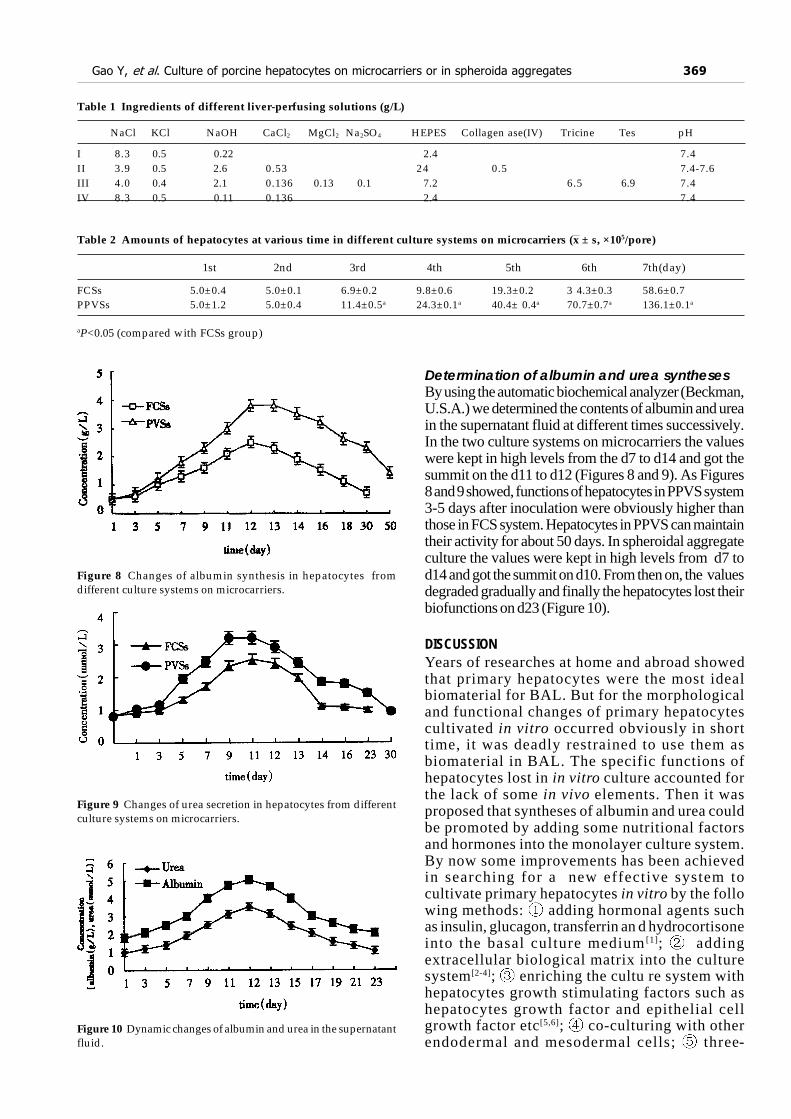
Figure 8 Changes of albumin synthesis in hepatocytes fromdifferent culture systems on microcarriers.
Figure 9 Changes of urea secretion in hepatocytes from differentculture systems on microcarriers.
Figure 10 Dynamic changes of albumin and urea in the supernatantfluid.
Table 1 Ingredients of different liver-perfusing solutions (g/L)
NaCl KCl NaOH CaCl2 MgCl2 Na2SO4 HEPES Collagen ase(IV) Tricine Tes pH
I 8.3 0.5 0.22 2.4 7.4II 3.9 0.5 2.6 0.53 24 0.5 7.4-7.6III 4.0 0.4 2.1 0.136 0.13 0.1 7.2 6.5 6.9 7.4IV 8.3 0.5 0.11 0.136 2.4 7.4
Table 2 Amounts of hepatocytes at various time in different culture systems on microcarriers (x ± s, ×105/pore)
1st 2nd 3rd 4th 5th 6th 7th(day)
FCSs 5.0±0.4 5.0±0.1 6.9±0.2 9.8±0.6 19.3±0.2 3 4.3±0.3 58.6±0.7PPVSs 5.0±1.2 5.0±0.4 11.4±0.5a 24.3±0.1a 40.4± 0.4a 70.7±0.7a 136.1±0.1a
aP<0.05 (compared with FCSs group)
Determination of albumin and urea synthesesBy using the automatic biochemical analyzer (Beckman,U.S.A.) we determined the contents of albumin and ureain the supernatant fluid at different times successively.In the two culture systems on microcarriers the valueswere kept in high levels from the d7 to d14 and got thesummit on the d11 to d12 (Figures 8 and 9). As Figures8 and 9 showed, functions of hepatocytes in PPVS system3-5 days after inoculation were obviously higher thanthose in FCS system. Hepatocytes in PPVS can maintaintheir activity for about 50 days. In spheroidal aggregateculture the values were kept in high levels from d7 tod14 and got the summit on d10. From then on, the valuesdegraded gradually and finally the hepatocytes lost theirbiofunctions on d23 (Figure 10).
DISCUSSIONYears of researches at home and abroad showedthat primary hepatocytes were the most idealbiomaterial for BAL. But for the morphologicaland functional changes of primary hepatocytescultivated in vitro occurred obviously in shorttime, it was deadly restrained to use them asbiomaterial in BAL. The specific functions ofhepatocytes lost in in vitro culture accounted forthe lack of some in vivo elements. Then it wasproposed that syntheses of albumin and urea couldbe promoted by adding some nutritional factorsand hormones into the monolayer culture system.By now some improvements has been achievedin searching for a new effective system tocultivate primary hepatocytes in vitro by the following methods: adding hormonal agents suchas insulin, glucagon, transferrin an d hydrocortisoneinto the basal culture medium [1]; addingextracellular biological matrix into the culturesystem[2-4]; enriching the cultu re system withhepatocytes growth stimulating factors such ashepatocytes growth factor and epithelial cellgrowth factor etc[5,6]; co-culturing with otherendodermal and mesodermal cells; three-
Gao Y, et al. Culture of porcine hepatocytes on microcarriers or in spheroida aggregates 369

dimensional cultivation such as microcapsular andmacrocapsular culture, spheroid aggregate, microcarrierculture and hollow fiber culture[7,8]; adding severalamino acids and nutritional substances into the culturesystem; and adding penetrating-inducer such aspentobarbiturate to the culture system[9]. The abovemeasures all aimed to make a circumstance mimic tothe in vivo enviroment for the hepatocytes. It is well-known that about 70% blood supply ofliver comes from portal vein system. The growth ofliver and maintainence of its functions dependmainly on the nutritional circumstance provided bythe portal vein system, that is, hepatocyt es in vivoare norished by the contents of portal vein blood.The later prov ides various amino acids, energysubstances and several growth factors. Ourexperiments were based on this very point andPPVS was added into the culture matrix. The resultsshowed that hepatocytes in this system coulddurably survive and maintain their biofunctions. Thisfluid was the ideal circumstance to cultivate thebiomaterial for BAL. Microcarriers Cytodex-3 were used to providethree-dimensional space for adher ing and suspendinghepatocytes so as to bring benefit from fully utilizing ofthe nutritional components in the culture system. Ourproceeding studies had prove d that Cytodex-3 was thebest microcarrier for primary hepatocytes[3].Hepatocytes on Cytodex-3 were readily to shape themicrostructure similar to th at of liver so they could growand function well. Landry[10] et al found that when no adhering agentin the spheroidal-aggregate culture system the isolatedhepatocytes tended to be changed less in morphologyand could survive and maintain their activities for 4 wk-6 wk due to the resemblance of the structure betweenthe aggregating spheroids and normal liver. Clayton[11,
12]et al found that the specific transcription gene ofhepatocyte in little masses of liver could be kept wellthan that of isolated hepatocytes and obviously the three-dimension structure of hepatocytes is of great importancein maintaining their specific functions. Relationshipbetween cell skeleton and specific proteins reflected theinfluences of morphology to funct ions, that is, cellfunctions directly related with levels of mRNA andspecific proteins, and inversely related with levels of actinand canaliculous protein. S akai[13]et al found that theinoculation concentration was of great influence tothe formation of hepatocyte spheroids in the
suspending spheroidal aggregate culture system. Whenhepatocytes being inoculated at a high concentra tion itoften occurred that pH of culture matrix declined, cell-to-cell and sp heroid to spheroid relationships changedand multiple necrotic foci could be ob served inhepatocyte spheroids. When hepatocytes beinginoculated at a low conce ntration they were difficult toaggregate into spheroids. Sakai et al consid ered 5.0 ×10⁶/mL to be the optimal inoculation concentration insuspending spheroid aggregate culture system ofhepatocytes. On view of the above factors, we concluded thatprimary porcine hepatocytes cultured on Cytodex-3 inPPVS system or in spheroidal-aggregate were promisingto act as the biomaterial of BAL. Still there is much tobe desired to promote the density, function and survivingtime of hepatocytes for clinical use.
REFERENCES1 Chen K, Gao Y, Pan YX, Yang JZ. An effective system for cultur-
ing primary porcine hepatocytes. Shijie Huaren Xiaohua Zazhi,1999;7:206-209
2 Kimura M, Ogihara M. Proliferation of adultrat hepatocytes inprimary culture induced by insulin is potentiated by cAMP-elevat-ing agents. Eur J Pharmacol, 1997;327:87-95
3 Kono Y, Yang S, Roberts EA. Extended primary culture of humanhepatocytes in a collagen gel sandwich system. In Vitro Cell DevBiol Anim, 1997;33:467-472
4 Taguchi K, Matsushita M, Takahashi M, Uchino J. Developmentof a bioartificial liver with sandwiched-cultured hepatocytes be-tween two collagen gel layers. Artif Organs, 1996;20:178-185
5 Koike M, Matsushita M, Taguchi K, Uchino J. Function of cultur-ing monolayer hepatocytes by collagen gel coating and co-culturewith nonparenchymal cells. Artif Organs, 1996;20:186-192
6 Schuppan D, Schmid M, Somasundaram R, Ackermann R, RuehlM, Nakamura T, Riecken EO. Collagens in the liver extracellularmatrix bind hepatocyte growth factor. Gastroenterology, 1998;114:139-152
7 Hu MY, Cipolle M, Sielaff T, Lovdahl MJ, Mann HJ, Remmel RP,Cerra FB. Effect of hepatocytes growth factor on viability andbiotransformation function of hepatocytes in gelentrapped andmonolayer culture. CritCare Med, 1995;23:1237-1242
8 Flendrig LM, Te Velde AA, Chamuleau RA. Semipermeable hollowfiber membranes in hepatocyte bioreactors a prerequisite for asuccessful bioartificial liver. Artif Organs, 1997;21:1177-1181
9 Sielaff TD, Myberg SL, Rollins MD, Hu MY, Amiot B, Lee A, WuFJ, Hu WS, Cerra FB. Characterization of the three compartmentgel entrapment porcine hepatocytes bioartificial liver. Cell BiolToxicol, 1997;13:357-364
10 Landry J, Bernier D, Ouellet C, Goyette R, Marceau N. Spheroidalaggregate culture of rat liver cells: histotypic reorganization,biomatrix deposition and maintenance of functional activities. JCell Biol, 1985;101:914-923
11 Clayton DF, Harrelson AL, Darnell JE Jr. Dependence of liverspecific transcription on tissue organization. Mol Cell Biol, 1985;5:2623-2632
12 Sawamoto K, Takahashi N. Modulation of hepatocyte functionby changing the cell shape in primary culture. In Vitro Cell DevBiol Anim, 1997;33:569-574
13 Sakai Y, Naruse K, Nagashima I, Muto T, Suzuki M. Large-scalepreparation and function of procine hepatocytes spheroids. Int J ArtifOrgans, 1996;19:294-301
Edited by You DYproofread by Sun SM
370 ISSN 1007-9327 CN 14-1018/ R World J Gastroentero June 2000 Volume 6 Number 3

Relationship between insulin A chain regions andinsulin biological activities
Shi Zhen Yang, Yi Ding Huang, Xin Feng Jie, You Min Feng and Jing Yi Niu
Subject headings insulin/chemistry; biological activities;A chain analogues
Yang SZ, Huang YD, Jie XF, Feng YM, Niu JY. Relationship betweeninsulin A chain regions and insulin biological activities. World JGastroentero, 2000;6(3):371-373
Abstract
AIM To study the relationship betweeninsulin A chain regions and insulin biologicalactivities, we designed a series of insulinanal ogues with changes at A21, A12-18 of C-terminal helical region and A8-10 located in theregion of A6-A11 intra-chain disulphide bond.METHODS Insulin A-chain analogues wereprepared by stepwise Fmoc solid-phasemanual synthesis and then combined withnatural B-chain of porcine insulin to yieldcorresponding insulin analogues. Theirbiological activities were tested by receptorbinding, mouse convulsion and immunologicalassay.RESULTS [A21Ala]Ins retains 70 .3%
receptor binding capacity and 60% in vivo
biological activity. [DesA13-14, A21Ala]Insand [DesA12-13-14-15, A21Ala]Ins still havedefinite biological activity, 7.9% and 4.0%
receptor binding, and 6.2% and 3.3% in vivo
biological activity respectively. [A15Asn,A17Pro, A21Ala]Ins maintains 10.4%
receptor binding and 10% in vivo biologicalactivity. [A8His, A9Arg, A10Pro, A21Ala]Ins,[A8His, A9Lys, A10Pro, A21Ala]Ins and[A8His, A9Lys, A10Arg, A21Ala]Ins have 51.
9%, 44.3% and 32.1% receptor bindingrespectively, 50%, 40% and 30% in vivo
biological activity respectively, and 28.8%,29.6% and 15.4% immunological activityrespectively.
CONCLUSION A21Asn can be replaced bysimple amino acid residues. The A chainswith gradually damaged structur al integrityin A12-18 helical region and the demolitionof the A12-18 hel ical region by thesubstitution of Pro and Asn for A17Glu andA15Gln respectively can combine with theB chain and the combination products showdefinite biological activity, the helicalstructure of A12-18 is essential for biologicalactivities of insulin. A8-10 is not muchconcerned with biological activities, but ismuch more important antigenically inbinding to its antibodies, these results mayhelp us design a new type of insulin analoguemolecule.
INTRODUCTIONTo elucidate the multi-functions of insulin molecule,hundreds of insulin analogues with changes involving90% of the constituting 51 amino acid residues atvarious parts of insulin molecule have been preparedby either chemical modification or synthesis during thepast two decades. In this communication we report theinfluences of three different regions of insulin A chain,especially the secondary structure region, i.e. A12-18helical region, on insulin biological activities.Recently,thorough studies of insulin analogues with deletion offragments of the molecule were also undertaken, butrestricted mostly to changes in the C-terminal part ofthe B chain[1,2], presumabl y due to its location outsidethe disulfide linkage and therefore ease of splitting offa tryptic or peptic fragment and recombining with amodified or shortened fragment by chemical orenzymatic semi-synthesis. No such enzymatic site wasfound in A chain, and most of its amino acid residuesare confined inside disulfide linkages. Chemicalsynthesis might be a better choice for making analogueswith modification or deletion in part of the A chain,which constitutes the main feature of thiscommunication. To obtain insulin analogues, we first set up thesemisynthesis strategy of insul in. Insulin A chainwas synthesized by the Fmoc solid phase synthesisstrategy, and then synthetic A chain S-sulphonatewas combined with porcine insulin B chain S-sulphonate forming crystalline insulin[3]. Based onthese experiences, the following insulin analogues
Shanghai Institute of Biochemistry, Chinese Academy of Sciences,Shanghai 200031, ChinaDr. Shi Zhen Yang, graduated from Beijing Scientific and TechnicalCollege, Academia Sinica, in 1964, associate researcher, majoring inpeptide synthesis, having 40 papers published.Supported by the ¡°Eighth Five Year Plan¡± Key Research Project, No.?KS 852017 and National Natural Science Foundation of China. No.3880193, No.39270157, No.39700028 and Chinese Academy ofSciences, KJ951-B1-606.Correspondence to: Dr. Shi Zhen Yang, Shanghai Institute ofBiochemistry, Chinese Academy of Sciences, 320 Yueyanglu, Shanghai200031, ChinaTel. 0086-21-64374430, Fax. 0086-21-64338357Email. Yangsz@Sunm. Shcnc. ac. cnRceived 2000-01-03 Accepted 2000-02-29
PO Box 2345, Beijing 100023, China World J Gastroentero, 2000; 6(3):371-373Fax: +86-10-85381893 World Journal of GastroenterologyE-mail: [email protected] www.wjgnet.com Copyright 2000 by the WJG Press ISSN 1007-9327

were semisynthesized. They are [A21Ala]Ins[4],where A21Asn is replaced by Ala, and i tsderivatives with deletions in the A12-18 helicalregion, [DesA13-14, A21Ala] Ins[4] and [DesA12-13-14-15, A21Ala]Ins[4], and [A15Asn, A17Pro,A21Al a]Ins, where the helical structure of A12-18 was also demolished by the substitutions of Profor A17Glu and Asn for A15Gln predicted by Chou-Fasman met hod, and [A8His, A9Arg, A10Pro,A21Ala]Ins[5], and [A8His, A9Lys, A10Pro,A21Ala]Ins[5], and [A8His, A9Lys, A10Arg,A21Ala]Ins[5] , in which His reminiscent A8 ofchicken insulin with high biological activity tookthe place of A8Thr and Arg, Lys, Pro replacedA9Ser and A10Ile to examine the influences ofthese a l tera t ions on receptor binding andantigenicity of insulin. The sequences of insulin Achain analogues are as follows (Figure 1).
Figure 1 Insulin A chain analogue sequence.
MATERIALS AND METHODSMaterialsPorcine insulin (26.4U/mg), Shanghai Bioch-emicalPlant; Sephadex G-15, G-50, Sepharose CL-6B,Pharmacia; Fmoc amino acid, prepared in the laboratory; DCC, acetonitrile, trimethylsilyltrifluoromethane sulfonate (TMSOTF), thioanisole,Fluka Co. trifluoro- acetic acid (TFA) Merck Co.;DTT, Serva Co.
MethodsPeptide synthesis Base labile N-Fmoc protectedamino acid was coupled by DCC-HOBt to the 2%cross linked ñ-alkoxybenzyl alcohol resin. Thefunctional side chain groups were protected by tBufor Cys, Glu, Tyr and Ser. Scheme of manipulationwas the same as reported[6]. The peptide chain wa sdetached from the resin support with simultaneousremoval of all tBu protectin g groups by TFA exceptS-tBu of cysteine. S-tBu was deprotected by MTMSOTf-t hioanisole-TFA system[5] and transformedto S-sulphonate by tetrathion ate and sulfite with theprocedure as reported[7].
Isolation and purification of ASSO3- Crude
ASSO3- fractions were purified by HPLC with
gradient elution (A: 0.1% TFA, B: 60% CH3CN, 0.125%
TFA).
Recombination with the natural (porcine) BSSO3- and
purification This was carried out according to modifiedChance’s procedure[ 4]. The crude product was purified byHPLC using the same procedure as that f or ASSO3
-.
Receptor binding, in vivo activities and RIA Thereceptor -binding of insulin and analogues was determinedby the displacement of labelle d insulin from the insulinreceptor on the human placental membrane according toFeng’s[8] procedure. The in vivo activity was estimatedsemiq uantitatively by mouse convulsion assay. RIA kitproduced by the Shanghai Biolog ical Product Institutewas used for RIA of insulin and analogues.
RESULTSPeptide synthesisInsulin A chain analogues were prepared starting from300 mg-500 mg each of Fmoc-alanyl resin and productsobtained from each step and the final overall yield isgiven in Table 1.
Table 1 Data on the synthesis of A chain analogues
A Chain FmocAla-resin Target peptide- After Overallanalogues mg(mmol) resin(mg) HPLC(mg) yield(%)
1 500(0.19) 769 62.1 12.32 500(0.19) 749 54.6 12.03 500(0.19) 724 45.4 12.44 500(0.32) 865 20.0 4.55 300(0.135) 657 39.6 10.66 300(0.135) 648 40.1 11.27 300(0.135) 660 41.3 11.1
Reconstitution of A and B chain and purificationof the productsAccording to the modified Chance’s procedure, insulinanalogues were obtained by purification of the crudereconstituted products. The identific ations of amino acidanalysis and HPLC indicated homogeneous products ofinsulin analogues.
Biological activitiesThe receptor-binding capacities calculated from thereceptor binding curve, in vivo activities andimmunological activities are shown in Table 2.
Table 2 Biological activities of insulin analogues (%)
Sample Receptor binding Mouse convulsion Immunological activity
Ins 100 100 1001B 70.3 602B 7.9 6.23B 4.0 3.34B 10.4 105B 51.9 50 28.86B 44.3 40 29.67B 33.1 30 15.4
372 ISSN 1007-9327 CN 14-1018/ R World J Gastroentero June 2000 Volume 6 Number 3
Yang SZ, et al. Insylin A-chain regions and insulin activities 373
DISCUSSIONA21Asn was considered to be a key amino acid ofthe insulin molecule in regard to its receptor binding.It has been very conservative in the evolutionaryprocess, and when replaced the analogues showedremarkable diminution of biological activity. Ourresults showed that the analogue with the A21 Asnreplaced by Ala exhibited substantial activity invivo and especially higher level of recept or binding,indicating the non-essentiality of A21Asn in receptorbinding and b iological activity as well. It could wellbe replaced by other amino acid residu es with goodretention of biological activities. Better results wereobserved wh en the side chain of the A21 aminoacid is smaller or not present (Gly)[9]. However,deletion of the A21 residue is fatal as the activityof desA21 insu lin showed only less than 1% of theactivity. Apparently, like some non-essenti al aminoacid residues in the insulin molecule, A21 isimportant in maintaining the specified spatialconfiguration of insulin required for its biologicalactivities. It is generally accepted that during the reconstitutionof A and B chain to insulin A6-11 disulfide bondformed first and this led to the formation of rightnatural conformation by self adjustment. When theB chain was getting in touch with the intrachaindisulfide, A6-11, a hydrophobic nucleus was formedto enhance the proper pairing of two Cys in bothends of the B chain to corresponding Cys of the Achain[10]. The success of our reconstitution withdeleted A chain supports to a certain extent thissuggestion. It showed that the C-terminal helicalregion in the A chain of insulin was not as importantas the N-terminal helical region in reconstitution withthe B chain. The biological activities of insulinanalogues with gradually damaged structural integrityin A12-18 helica l region were similar to that of insulinanalogue with the demolition of the A12 -18 helicalregion by the substitution of Pro and Asn for A17Gluand A15Gln res pectively. The observationssuggested that the helical structure of A12-18 isessential for biological activities of insulin. It was generally suggested that the enhancedactivities of [A8His]Ins[11], chicken and turkeyinsulins were ascribed to the His at A8[12-14], whichshould account for the higher affinity for insulinreceptor. Our results showed that the presenceof His at A8 apparently did not enhance the receptorbinding. The receptor binding of [A8His] Ins shouldlie at the range of about 50%. The enhanced
potency of these insulins containing His at A8 may notresult solely from an apparent and direct effect due tothe presence of A8His. A8His and A10Arg signify ahigher influence on insulin antigenicity. A8His willenhance the binding activity to the anti-insulin serum[11],while A10Arg will decrease this binding, but amino acidat A9 had very little to do with antigenic activity. Inaddition to studies on how to elucidate the action of aninsulin m olecule to its receptor and express thephysiological activity that follows such action, the purposeof studies on the structure activity relationship of in sulinwill rely more on how to get a better analogue with highpotency but low a ntigenicity. We hope that theinformation provided in this study will be benefic ial tothe development of a new type insulin analogue withthose qualities through design of an appropriatearchitecture of this important hormone.
REFERENCES1 Shanghai Insul in Research Group. Structural s tudies on
despentapeptide (B26-30)insulin. Zhongguo Kexue, 1976;4:351-356
2 Chen LT, Huang CM, Zhang M, Yang ZR, Hu MH. Purificationand characterization of Des-terpeptide of C-terminal (B28-30)human insulin. Shengwu Huaxue Zazhi, 1996;12:187-195
3 Yang SZ, Niu JY. Solid phase synthesis of the A-chain of insulinand its recombination with the B-chain to crystalline insulin.Shengwu Huaxue Yu Shengwu Wuli Xuebao, 1992;24:497-502
4 Yang SZ, Huang YD, Feng YM, Niu JY. Insulin analogues withdeletions at the helical region of the A chain. Shengwu Huaxue YuShengwuWuli Xuebao, 1994;26:49-55
5 Yang SZ, Lin W, Huang YD, Feng YM, Niu JY. Insulin analogueswith alteration at A8-10. Acta Biochemi Biophys Sin, 1995;27:329-334
6 Yang SZ, Niu JY. Solid phase synthesis of serum thymic factor andpreparation of its antiserum. Shengwu Huaxue Zazhi, 1991;7:349-354
7 Paynovich RC, Carpenter FH. Oxidation of the sulfhydryl formsof insulin A-chain and B chain. Int J Peptide Protein Res, 1979;13:113-121
8 Feng YM, Zhu JH, Zhang XT, Zhang YS. Studies on themechanism of insulin action. Acta Biochimi Biophys Sin,1982;14:137-143
9 Markussen J, Diers I, Hougaard P, Langkjaer L, Norris K, Snel L,Sörensen AR, Sörensen E, Voigt HO. Soluble prolonged actinginsulin derivatives. III. Degree of protraction, crystallizabilityand chemical stability of insulins substituted in positions A21,B13, B23, B27 and B30. Protein Engineering, 1988;2:157-166
10 Peking Insulin Structure Research Group. Studies on the insulincrystal structure: the molecule at 1.8A resolution. Zhongguo Kexue,1974;2:752-761
11 Marki F, Gasparo MD, Eisler K, Kamber B, Riniker B, Rittel W,Sieber P. Synthesis and biological activity of seventeen ana-logues of human insulin. Hoppe-seyler’s Z Physiol Chem, 1979;360:1619-1632
12 Pullen RA, Lindsay DG, Wood SP, Tickle IJ, Blundell TL, WollmerA, Krail G, Brandenburg D, Zahn H, Gliemann J, Gammeltoft S.Receptor binding region of insulin. Nature, 1976;259:369-373
13 Weitzel G, Renner R, Kemmler W, Rager K. Struktur und erhohteaktivitat des insulin vom truthuhn (melleagris gallopavo). Hoppe-Seyler’s Z Physiol Chem, 1972;353:980-986
14 Simon J, Freychet P, Rosselin G. Chicken insulin: Radioimmunologicalcharacterization and enhanced activity in rat fat cells andl iver p lasma membranes . Endocr inology , 1974;95: 439-1 4 4 5
Edited by You DY and Zhu LHproofread by Sun SM

Helicobacter pylori infection and risk of gastriccancer in Changle County, Fujian Province, China
Lin Cai1, Shun Zhang Yu2 and Zuo Feng Zhang3
Subject headings Helicobacter pylori; stomach neoplasm;risk factors; case control studies
Cai L, Yu SZ, Zhang ZF. Helicobacter pyloriinfection and risk ofgastric cancer in Changle County, Fujian Province, China. World JGastroentero, 2000;6(3):374-376
Abstract
AIM To evaluate the effects of Helicobacter
pylori infecti on and other environmental factorson the development of gastric cancer.METHODS A population-based case-controlstudy was conducted i n Changle County, FujianProvince. The primary gastric cancer cases werehistolo gically confirmed or diagnosed by surgerybetween January 1996 and March 1998. Healthycontrols were randomly selected and matchedby age, sex, and neighborhood of residence. Atotal of 101 pairs were included in the study. Specially trained interviewers conducted face-to-faceinterviews with the subjects according to astandardized questionnaire. Helicobacter pylori
infections w ere measured by serum IgGantibody to Helicobacter pylori. ConditionalLogis tic Regression analysis was used.RESULTS The presence of IgG antibody toHelicobacter pylori was 63.7% in study subjects,56.0% in patients with cardiac cancer, and 60.5% in patients with non-cardiac gastric cancer.The risk factors of gastric canc er in ChangleCounty were ident i f i ed such as loweducational level [OR=3.864; 95% confidenceinterva l (95%CI) 1 .604-9 .311] , lowconsumption of fresh veget ables (OR=4.925;95% CI 1.356-17.885), high intake of fishsauce (OR = 10.587; 95% CI 2.821-39.738),unscheduled meals (OR = 4.254; 95% CI 1.445-
12.552), and Helicobacter pylori infection (OR =3.453; 95% CI 0.901-13.224).CONCLUSION Helicobacter pylori infection maybe important in the etiology of gastric cancer,but major risk factors other than Helicobacter
pylori are responsible for the high gastricmorbidity in Changle County.
INTRODUCTIONHelicobacter pylori (H. pylori) infection isassociated with gastric cancer[1-4]. However, only asmall proportion of individuals developed gastri ccancer in comparison with the relatively highprevalence of H. pylori infection in the generalpopulation. In this study we evaluated the effects ofH. pylori infection and other environmental riskfactors on the development of gastric cancer inChangle County, one of the areas with highestmorbidity of gastric cancer in the world.
MATERIALS AND METHODSStudy subjectsOne hundred and one gastric cancer patients (87 malesand 14 females) and the same number of normalcontrols, individually matched with region, sex and age(± 3 years) were included in the study. The studiedsubjects must have resided in Changle County for morethan 20 years. The primary gastric cancer patientswere histologically confirmed or diagnosed byoperation during January 1996 - March 1998. Thosewho have ever been diagnosed as having gastricdiseases in the past 3 years were not eligible ascontrols.
Data collectionTrained interviewers interviewed the patients andcontrols. A standardized questionnaire was usedto obtain information on basic demographiccharacteristics (gender, race, year and place ofbirth, and education), dietary habits, personalhabits (smoking and drinking), medical history andso on.
H. pylori assayThe presence of anti-H. pylori - IgG was assessedusing the commercially available high-molecular-weight cell-associated protein H. pylori -immuno-
1Department of Epidemiology, Fujian Medical University, Fuzhou350004, Fujian Province, China2Department of Epidemiology, Shanghai Medical University, Shanghai200032, China3Department of Epidemiology, UCLA School of Public Health, LosAngeles California, USADr. Lin Cai, graduated from Shanghai Medical University in 1983,received Ph. D. degree from Shanghai Medical University in 1999,associated professor, majoring in gastric cancer epidemiology, having30 papers published.Project supported by the Natural Science Foundation of FujianProvince, China, No. K98031Correspondence to: Lin Cai, Department of Epidemiology, FujianMedical University, Fuzhou 350004, Fujian Province, ChinaTel. 0086-591-3357231Received 2000-01-03 Accepted 2000-02-06
PO Box 2345, Beijing 100023, China World J Gastroentero, 2000; 6(3):374-376Fax: +86-10-85381893 World Journal of GastroenterologyE-mail: [email protected] www.wjgnet.com Copyright 2000 by the WJG Press ISSN 1007-9327

assay (Enteric Products Inc, Westbury, New York). Theassay was done in 96-well microtiter plates. Sera diluted1:101 were added, and peroxida se conjugated anti-human IgG was used as the detector. The absorbanceof the so lution was measured at 450 nm.
Statistical analysisData were handled by Epi-info. Odds ratios (OR) and95% confidence intervals (95%CI) were calculated byConditional Logistic Regression using STATA software
RESULTSA total of 101 pairs were included in the study. Theywere all Han nationality. The age of the patients rangedfrom 32 years to 78 years, averaging 58.93 years . Theeducational level was higher in controls than in thepatients. The serum samples of 101 patients and 100controls were tested for antibodies against H. pylori.Among them, 128(63.68%) were H. pylori positive. Theprevale nce of H. pylori infection was not significantlydifferent in different age (above 30 years) and sexgroups. The demographic characteristics as well as thedistributions of prevalence of H. pylori infection indifferent sex and age groups are presented in Table 1and Table 2.
Table 1 Demographic characteristics as well as Helicobacterpylori infection in patients and controls
Patients Controls P value n % n %
Age (years)30-39 4 (4.0) 5 (5.0)40-49 18 (17.8) 18 (17.8)50-59 25 (24.8) 22 (21.8)60-69 34 (33.7) 38 (37.6)
70 20 (19.8) 18 (17.8) 0.9597GenderMales 87 (86.1) 87 (86.1)Females 14 (13.9) 14 (13.9) 1.0 000EducationCollege 1 (1.0) 1 (1.0)High school 17 (16.8) 68 (67.3)Elementary school 65 (64.4) 24 (23.8)Illiterate 18 (17.8) 8 (7.9) 0.0 000H. pyloriPositive 60 (59.4) 68 (68.0)Negative 41 (40.6) 32 (32.0) 0.2 064
Table 2 Helicobacter pylori infection in study subjects
Men WomenAge group(years) n Hp(+) % n Hp(+) %
30-39 7 7 (100.0) 2 2 (100.0)40-49 29 18 (62.1) 7 5 (71.4)50-59 46 28 (60.9) 1 1 (100.0)60-69 58 39 (67.2) 13 6 (46.2)
7 0 33 18 (54.6) 5 4 (80.0)Total 173 110 (63.6) 28 18 (64.3)
χ2 = 5.68, P = 0.2241; χ2 = 4.22, P = 0.3769
No statistical difference was found in respect topresence of IgG antibody to H. pylori between patientsand controls (Table 3). No significant difference wasobserved in anatomic distribution, although the prevalenceof H. py lori infection was higher in non- cardiac gastriccancer (60.5%) than in cardiac cancer (56.0%), (Table 4).
Table 3 Helicobacter pylori infection and gastric cancer
PatientsControls Pairs Hp(+) Hp(-)
Hp(+) 39 29 68Hp(-) 20 12 3Pairs 59 41 100
c2 = 1.31, P = 0.25
Table 4 Helicobacter pylori infection in gastric cancer cases
H. pylori infectionGastric cancer n n %
Cardiac 25 14 56.0Non-cardiac 76 46 60.5
χ2 = 0.04, P = 0.8395
Further analysis was conducted using Condit-ionalLogistic Regression. The results showed the risk factorsof gastric cancer in Changle County were fish sa uceintake, low consumption of fresh vegetables, unscheduledmeals, low educatio nal level and H. pylori infection(Table 5).
Table 5 The results of multivariate Conditional LogisticRegression analysis
Factors β sx OR 95%CI
Low educational level 1.3 517 0.4 487 3.864 1.604-9.311Low consumption of vegetables 1.5944 0.6580 4.925 1.356-17.885High intake of fish sauce 2.3596 0.6748 10.587 2.821-39.738Unscheduled meals 1.4479 0.5508 4.254 1.445-12.552H. pylori infection 1.2392 0.6852 3.453 0.901-13.224
DISCUSSIONChangle County is a hyperendemic area of gastriccancer[5]. Chronic H. pylori infection has been identifiedas the most important risk factor of gastric cancer[6].The results of our study showed that residents in ChangleCounty had a high prevalence of H. pylori infection.This coincided the fact the concept of high prevalenceof H. pylori infection in the high risk area of gastriccancer. However, no statistical significant difference wasfound in respect to presence of H. pylori infectionbetween patients and controls. The lack of associationin this study may be due to the high prevalence of H.pylori in Changle County. H. pylori infection can initiatea sequence of histological alterations in the mucosa that mayfinally result in the development of gastric cancer,
Cai L, et al. Helicobacter-pylori infection and risk of gastric cancer 375

but not a ll infected subjects will eventually developgastric cancer. H. pylori alone cannot account fordevelopment of gastric cancer. Apart from H. pyloriinfection, other factors may play important roles incarcinogenesis. The development of gastric cancer is believed tobe a multistep and multifactorial process. In the Correamodel of gastric carcinogenesis, environmental factorsare related to the evolution from normal gastric tissuethrough superficial gas tritis, atrophic gastritis,intestinal metaplasia and dysplasia to carcinoma[7].Environmental co-factors other than microbial agentsmay play roles in initiation, promotion or progressionof gastric cancer. The results of this study suggest that dietaryfactors, such as high intake of fish sauce and lowconsumption of fresh vegetables possibly increase therisk of gastric cancer. Fish sauce is a kind of traditionalsauce consumed daily by Changle residents. It isusually produced from several kinds of sea fishes afterlong fermentation processes. The mutagenicity of fishsauce has been reported by several experimentalstudies[8,9]. There was a large amount of importantprecursors of N-nitrosamines detected in fish sauce.High intake of fish sauce was an important risk factorinvolved in the etiology of gastric cancer. However,consumption of diets high in vegetables is the mosteffective means in preventing gastric cancer.Vegetables contain many biologically active compoundsthat may be responsible for an anticarcinogenic effectagainst gastric cancer[10]. Additionally, unscheduledmeals may cause injuries of gastric mucosa andpotentiate the effects of carcinogens. Our studyindicated that unschedule d meals might be one of theetiological factors for gastric cancer. In this study theeducational level was higher in controls than in patients.
This supported the results of previous studies thatsocioeconomic status is a risk factor for gastric cancer,which in turn is related to diet and education. These findings indicate that primary preventionshould be focused in Changle County on reducing theetiological factors: fish sauce intake, deficiency in freshvegetables, and H. pylori infection. Further elucidationof these risk factors for stomach cancer in Changle andassessment of the interaction bet ween these risk factorsand H. pylori infection are important for investigatingthe possible mechanisms of gastric carcinogenesis.
REFERENCES1 The Eurogast study group. An international association between
Helicobacter pylori infection and gastric cancer. Lancet, 1993;341:1359-1362
2 Tatsuta M, Lishi H, Okuda S, Taniguchi H, Yokota Y. The associa-tion of Helicobacter pylori with differentiated type early gastriccancer. Cancer, 1993;72:1841-1845
3 Miehlke S, Hackelsberger A,Meining A,Arnim U vom,Müller P,Ochsenkühn T,Lehn N,Malfertheiner P, Stolte M,BayerdorfferE. Histological diagnosis of Helicobacter pylori gastritis is predic-tive of a high risk of gastric carcinoma. Int J Cancer, 1997;73:837-839
4 Blasser MJ. Hypotheses on the pathogenesis and natural historyof Helicobacter pylori induced inflammation. Gastroenterology,1992;102:720-727
5 Cai L, Yi YN, Liu YY. A case control study of stomach cancer inChangle, Fujian Province by the risk state analysis. ZhonghuaLiuxingbingxue Zazhi, 1991;1:15-18
6 Forman D, Newell DG, Fullerton F, Yarnell JWG, Stacey AR, WaldN, Sitas F. Association between infection with Helicobacter pyloriand risk of gastric cancer: evidence from a prospectiveinvestigation. Br Med J, 1991;302:1302-1305
7 Correa P. Human gastric carcinogenesis: a multistep and multifac-torial process: first American cancer society award lecture on can-cer epidemiology and prevention. Cancer Res, 1992;52:6735-6740
8 Zhang RF, Deng DJ, Chen Y, Chen CS, Fan ZH. Analysis of pre-cursors of N-nitroso compounds in fish sauce from gastric cancerhigh risk area. Aizheng, 1993;12:395-398
9 Deng DJ, Zhang RF, Chen Y, Chen CS, Jin S, Zhu SX. Mutagenic-ity and carcinogenicity of fish sauce from a county with the highrisk for gastric cancer in China. Aibian Jibian Tubian, 1991;3:18-23
10 Zhang HM, Wakisaka N, Maeda O, Yamamoto T. Vitamin C inhibitsthe growth of a bacterial risk factor for gastric carcinoma: Helicobacterpylori. Cancer, 1997;80:1897-1903
Edited by You DY and Zhu LHproofread by Sun SM
376 ISSN 1007-9327 CN 14-1018/ R World J Gastroentero June 2000 Volume 6 Number 3

Construction of HBV-specific ribozyme and itsrecombinant with HDV and their cleavage activityin vitro
Shu Juan Wen1, Kai Jun Xiang2, Zhen Hua Huang1, Rong Zhou1 and Xue Zhong Qi1
Subject headings hepatitis B virus; hepatitis D virus;ribozyme gene; recombinant DVRZ
Wen SJ, Xiang KJ, Huang ZH, Zhou R, Qi XZ. Construction ofHBV-specific ribozyme and its recombinant with HDV and theircleavage activity in vitro. World J Gastroentero, 2000;6(3):377-380
Abstract
AIM To construct the recombinant of HDV cDNAand HBV-specific ribozyme gene by recombinantPCR in order to use HDV as a transporting vectorcarrying HBV-specific ribozyme into liver cells forinhibiting the replication of HBV.METHODS We separately cloned the ribozyme(RZ) gene and recombi nant DVRZ (comprisingHDV cDNA and HBV-specific ribozyme gene) intothe downstr eam of T7 promoter of pTAdv-Tvector and studied the in vitro cleavage activityof their transcripts (rRZ, rDVRZ) on target RNA(rBVCF) from in vitro transcription of HBV Cgene fragment(BVCF).RESULTS Both the simple (rRZ) and therecombinant ribozyme rDV RZ could efficientlycatalyze the cleavage of target RNA (rBVCF)under different temperatures (37 , 42 and55 ) and Mg2+ concentrations (10 mmol/ L, 15mmol/L and 20 mmol/L) and their catalyticactivity tended to increase as the temperaturewas rising. But the activity of rRZ was evidentlyhigher than that of rDVRZ.CONCLUSION The recombinant of HDV cDNAand ribozyme gene had the potential of beingfurther explored and used in gene therapy of HBVinfection.
INTRODUCTIONHepatitis B virus (HBV) can cause acute and chronicB-type hepatitis in man. The conventional ways availablefor curing this disease have not been very effic ient.This promotes people to explore novel genetictherapeutical ways. Hammerhe ad ribozyme is a kind ofantisense RNA which can specifically cleave the targetRNA[1,2]. In the light of this, people have developed manyeffective gen etic vectors containing ribozyme genes,the transcripts of which showed catalytic activity in vitroand in vivo[3-5]. But how to improve the stability andefficiency of ribozyme and specifically carry ribozymegene into only target cells or tissues has been a tacklingproblem. HDV, a human hepatitis agent, is a defectiveRNA virus,the replication cycle of which relies on theinfection of HBV[6]. So HDV can be developed as aspecific transporting and replicating vector in vivo forribozyme to reach liver[7]. In this study, we constructedHBV-speci fic hammerhead ribozyme gene (RZ) andthe recombinant (DVRZ) of HDV cDNA (DV) andribozyme gene (RZ) and made a careful investigation oftheir in vitro catalytic activity under various conditions.The positive results encouraged us to further explorethe feasibility of-using HDV as a vector carrying ribozymefor inhibiting the replication of HBV in vivo.
MATERIALS AND METHODSPlasmidsThe plasmid pSVC-D3 (containing HDV cDNA) asone of two templates of recom binant PCR was kindlyoffered by Prof. Taylor of American. pTAdV-T vectorused for cloning was purchased from ClontechCorporation.
Major reagentsRiboMax transcription kit, acrylamide, bisactylamide,dNTP, rNTP and Taq polymer ase were purchased fromPromega.[α-32P]UTP from Beijing Yahui Corporation.Advantage-TM PCR pure kit (gel purification kit) fromClontech. X-film from Kodak. DNA polymerase I klenowfrom Biolabs. T7/Sp6 sequencing kit from Pharmacia.
PCR primersPrimers P1 and P2 covered the whole sequence ofdesigned HBV-specific hammerhea d ribozymegene(RZ) and were used to amplify ribozyme gene(53bp) because of 9nt base-pairing of their 3’ends.
1Gene Center of Nanfang Hospital, First Military Medical University,Guangzhou 510515, Guangdong Province, China2School of Life Sciences, University of Science and Technology ofChina, Hefei 230027, Anhui Province, ChinaShu Juan Wen, graduated from First Military Medical University in1985, major in gene diagnosis, having 6 papers published.Supported by Natural Science Foundation of GuangdongProvince, No.940311.Correspondence to: Shu Juan Wen, Gene Center of Nanfang Hospital, First Military Medical University, Guangzhou 510515, GuangdongProvince, ChinaTel. 0086-20-85141044, Fax. 0086-20-87730347Email. [email protected] 2000-01-11 Accepted 2000-02-18
PO Box 2345, Beijing 100023, China World J Gastroentero, 2000; 6(3):377-380Fax: +86-10-85381893 World Journal of GastroenterologyE-mail: [email protected] www.wjgnet.com Copyright 2000 by the WJG Press ISSN 1007-9327

The sequence of HBV-specific hammerhead ribozymewas designed according to the requirement of domainsof ribozyme[1] and the sequence of HBV C genefragment. The ribozyme gene (RZ: 53bp) was also used asone of two templates for recombinant PCR to constructthe recombinant (DVRZ) of HDV cDNA and ribozymegene (mentioned below). P3, P4 were both recombinant primers,each ofwhich was composed of partial sequences of ribozymeand HDV in order to replace the sequence (17-67) ofnear 5’-end of HDV with ribozyme by recombinant PCR.P5 was the 3’-end sequence of HDV cDNA. P3, P4, P5 were used to construct the recombinant(DVRZ) of HDV cDNA (DV) and HBV-specificribozyme gene (RZ) by one-tube recombinant PCR[8]. P6, P7 were used to amplify HBV C gene fragment(BVCF), which was the transcription template of targetRNA (rBVCF). T7 was partial sequence of T7 promoterregion of pTAdV-T vector and was used for sequencing(with Sp6) and identifying(with P2, P5, P7) whether 5’-end of foreign fragment forwardly inserted thedownstream of T7 promoter. The sequencing of all gene fragments wasperformed on ABI391 automatic sequenc er(Pharmacia). The sequences and positions of theseprimers are listed in Table 1.
Table 1 The sequences and positions of primers
Primer Sequences Positionsname
P1 5’-AACATTGACATAGCTCTGATGAGTCCGTGAG-3 RZ:1-31
P2 5’-TCCAGGGAATTAGTAGTTTGTCCTCACGGAC-3’ RZ:53-23
P3 5’-AGCAAGCTTGAGCCAAAACATTGACATAGCTCT-3’ HDV:1-16,RZ:53-37
P4 5’-CTCCGACGTTCCAATGCTCCAGGGAATTAGTAGT-3’ HDV:84-68,RZ:53-37
P5 5’-GTCGAATTCGGGCTCGGGCGGCGATCCAGCAGTC-3’ HDV:1680-1647
P6 5’-GATAAGCTTTTACATAGAGGACTCTTGG-3’ HBV:1650-1677
P7 5’-CTGGAATTCGGCGAGGGAGTTCTTCTTCTAG-3’ HBV:2480-2450
Construction and cloning of HBV-specificribozyme gene(RZ)Because of 9nt base-pairing between 3’ends of P1 andP2, direct PCR could pr oduce complete 53bp ribozymegene (Figure 1). After gel-purification (according toAdvantage-TM PCR pure kit, Clontech Corporation),the PCR product (RZ) was directly cloned into thedownstream of T7 promoter of pTAdV-T vector and sothe resulting recombinant plasmid pTA-RZ was obtained.PCR with T7/P2 as primers and pTA-RZ as templatecould produce about 100 bp DNA fragment if 5’-end ofribozyme gene was forwardly inserted in to thedownstream of T7 promoter of pTAdV-T vector and socould be used to iden tify the recombinant plasmid pTA-RZ of forward insert. The preparation of comp etentDH5α cells and transformation of plasmids wereperformed according to the reference[9]. The clonedribozyme gene was finally verified by sequencingwith T7/Sp6 primers.
Construction, cloning and sequencing of therecombinant (DVRZ) of HDV cDNA and HBV-specific ribozyme geneThirty µL PCR reaction system was established[8]: 20mmol/L Tris-HCl (pH 8.3), 50 mmol/L KCl, 2 mmol/LMgCl2, each dNTP 200 µmol/L; Primers P3, P4, P5 wereseparately 0.05 µmol/L, 0.005 µmol/L, 0.05 µmol/L . Thetwo templates were recombinant plasmids pTA-RZ(containing ribo zyme gene) and pSVC-D3 (containingHDV cDNA), each 10 ng. Taq polymerase 2 µL,denaturation at 92 -50s; annealing at 53 -50s;elongation at 70 -120s; 33cycles; the final elongationat 70 lasted 5 min. PCR product (DVRZ, 1.7kb) wasidentified by agrose-gel (1.5%) electro phoresis and thenpurified with Advantage-TM PCR pure kit. The obtainedDNA fra gment (DVRZ) was directly cloned into thedownsteam of T7 promoter of pTAdV-T vector. Theresulting recombinant plasmid pTA-DVRZ (positiveclones on Ampr & white-blue plate) was verified by PCRwith primers T7/P5 and sequencing with Sp6/T7 primers.PCR with primers T7/P5 could produce about 1.7kb-1.8kb DNA fragment and was used to identify the forwardinsertion of 5’-end of DVRZ. Sequencing was performedon ABI 391 automatic sequencer (Pharmacia).
Cloning and isolation of target gene fragment(HBV C gene fragment-BVCF)PCR with primers P6, P7 was performed to amplifyHBVC gene fragment from serum of HBV. Extractionof sample DNA and PCR reaction were performedaccording to the reference[10]. PCR product (BVCF)was directly cloned into the downstream of T7 promoterof pTAdV-T vector and the resulting recombinant plasmid pTA-BVCF (positive clones on Ampr & blue-whiteplate) was verified by PCR with T7/P7 and bidirectionalsequencing with T7/Sp 6 primers. PCR with primers T7/P7 was utilized to identify the forward-direction insertionof 5’-end of BVCF.
Linearization of recombinant plasmids pTA-RZ,pTA-DVRZ and pTA-BVCFThese plasmids were seperately digested with BamHIand then filled with Klenow fragment for in vitrotranscription of inserted gene fragments (RZ, DVRZ,BVCF).
In vitro transcription of linearized plasmids pTA-RZ and pTA-DVRZ32P-UTP-labeled transcription of the two plasmidswas first carried out in order to test the effect oftranscription. But the plasmids’ transcripts (rRZ,rDVRZ) used for cat alyzing the cleavage of targetRNA (rBVCF-transcript of plasmid pTA-BVCFcont aining HBV C gene fragment-BVCF) werenot 32P-labeled. In vitro tra nscription was performedwith T7 RNA polymerase according to RiboMaxTranscripti on kit and the reference[11]. The
378 ISSN 1007-9327 CN 14-1018/ R World J Gastroentero June 2000 Volume 6 Number 3

Wen SJ, et al. HBV-specific ribozyme & recombinant with HDV 379
transcripts (rRZ, rDVRZ) would additio nally contain100nt partial sequence of pTAdV-T vector at theirboth end s. rRZ and rDVRZ were extracted byphenol/chloroform/iso-propyl alcohol (25:24:1),precipitated with ethanol and resuspensed with H2O(RNase-free). The latter was the recombinant ofHDV and ribozyme.
Preparation of 32P-UTP-labeled target RNA (rBVCF)Linearized plasmid pTA-BVCF (containing HBV Cgene fragment) was in vitro transcripted and thetarget RNA (rBVCF) was produced. 20 µLtranscription system was so established: ATP, CTP,GTP each 2.5 mmol/L and UTP 0.1 µmol/L, 0.5 µCi/µL[α32P]UTP, 2 µg linearized plasmi d pTA-BVCF,T7 RNA polymerase 20U, RNasin 10U, 37 -60 min.The product was loaded for 5% PAGE-7M ureaautoradiographed electrophoresis. Gel-band of 1cmlengt h and 0.5 cm breadth was cutted off from theposition of target RNA and so aked overnight withNES buffer (0.5 mol/L NH4Ac, 1 mmol/L EDTA, 0.1% SDS) and extracted with phenol/chloro form/iso-propyl alcohol (25:24:1). The supernate wasprecipitated with ethanol and then the pellet washarvested and resuspensed with H2O (RNase-free).rBVCF(831+100nt) contained also 100nt partialsequenc e of pTAdV-T vector at its two ends.
Cleavage reactionTen ¦ÌL reaction system was established: 0.1 mol/L Tris-HCl (pH 8.0), 20 mmol/L MgCl2, rRZ or r-DVRZ andtheir target RNA (rBVCF) each 2 µL, mixed andincubated under different temperatures (37 , 42 , 55
) for 1h. Negative control with 32P-labeled rBVCF incubated at 55 without rRZ & rDVRZ was performed.In the meanti me, rRZ and rDVRZ were separatelyincubated with target RNA (rBVCF) at 37 und erdifferent Mg2+ concentrations (10mmol/L, 15mmol/L).In the end, 10 µL ion-free formamide and 1 µL loadingbuffer (50% glycerol, 1mmol/L EDTA, 0.04%brompheno l blue) were added to terminate the reaction.Then after incubated at 65 for 10 min, 5 µL reactionsample was loaded for 5% PAGE-7M ureaelectrophoresis.
RESULTSConstruction and cloning of ribozyme gene53bp DNA fragment was obtained by PCR with primersP1/P2 (Figure 1). This was in accordance with the sizeof anticipated ribozyme gene. PCR using T7/P2 asprimers and plasmid pTA-RZ as the templ ate amplifieda DNA fragment of about 100 bp, which identifiedforward-dir ection insertion of ribozyme gene into thedownstream of T7 promoter of pTAdV-T vector.Sequencing verified the HBV-specific ribozyme gene.The sequence of ribozyme gene was as follows:5’-AACATTGACATAGCTCTGATGAGTCCG-TGAGGACAAACTACTAATTCCCTGGA-3’
Construction, cloning and sequencing of therecombinant (DVRZ) of HDV cDNA(DV) and HBV-specific ribozyme gene(RZ)About 1.7kb DNA was amplified by recombinantPCR, which indicated the anticip ated recombinantDNA molecule DVRZ (Figure 1). After DVRZ’scloning into pTAdV-T vector and transforming intoDH5α on Ampr & white-blue plate, PCR withprimers T7/P5 produced about 1.8 kb-DNAfragment and identified the positive recombinantplasmid pTA-DVRZ of correct DVRZ’s insertiondirection. Sequencing of DVRZ with primers T7/Sp6 (pTA-DVRZ as template) confirmed theconstruct DVRZ.
Cloning and isolation of target gene fragment(HBV C gene fragment-BVCF)Anticipated 831bp target DNA fragment was amplifiedby PCR from the serum of HBV (Figure 1). After itscloning into pTAdV-T vector and then transformed into DH5α, PCR with primers T7/P7 produced an about0.9 kb DNA fragment and so identified the positiverecombinant plasmid pTA-BVCF of BVCF’s correctinsertion direction. Finally, sequencing with primers T7/Sp6 confirmed the inserted BVCF.
Figure 1 Construction of HBV-specific ribozyme gene (RZ), itsrecombinant with HDV cDNA (DVRZ) and target DNA (BVCF)by PCR.a: Lane 1-PCR marker, Lane 2-ribozyme gene RZ (53bp).b: Lane 1- DNA/-Hind III+Eco R I marker, Lane 2-recombinantDVRZ (1682bp) Lane 3-target gene BVCF (831bp).
In vitro transcription of linearized recombinantplasmids pTA-BVCF, pTA-RZ and pTA-DVRZTarget RNA (rBVCF) of 931nt was transcripted frompTA-BVCF. HBV-specific ribo zyme (rRZ) of 153ntwas transcripted from pTA-RZ. HBV-specific recombinant ribozyme (rDVRZ,containing HDV) of 1782nt was trans cripted from pTA-DVRZ (Figure 2). All these transcripts (rBVCF, rRZ,rDVRZ) contained the same partial sequence of pTAdV-T vector at their both ends after transcription with T7RNA polymerase.
Cleavage reactionRibozymes rRZ and rDVRZ without 32P-label wereseparately inc ubated with target RNA (32P-labeledrBVCF) under different temperatu res(37 , 42 ,55 ) and Mg2+ concentrations. The results fromautoradiog raphed electrophoresis showed that underthese temperatures and Mg2+ concentrations, both

rRZ and rDVRZ could catalyze the cleavage of targetrBVCF into two RNA fragments (721nt, 210nt) andtheir catalytic activity tended to increas e with the risingof temperature. Comparatively, the catalytic activity ofrRZ was higher than that of rDVRZ. But it seemed thatMg2+ from 10 mmol/L to 20 mmol/L had no obvious effecton their cleavage activity (Figure 3).
Figure 2 In vitro transcription of linearized pTA-RZ, pTA-DVRZand pTA-BVCF (Lane 1-rDVRZ,1782nt. Lane 2-rBVCF,931nt. Lane3-rRZ,153nt).
Figure 3 Cleavage of target RNA(rBVCF) catalyzed by rRZ andrDVRZ.a: Cleavage of target RNA (rBVCF) by rRZ under 37 ,42
and 55 with 20 mmo l/L Mg2+. b: Cleavage of target RNA(rBVCF) by rDVRZ under 37 ,42 and 55 with 20 mmol/LMg2+. c: Cleavage of target RNA (rBVCF) separately by rRZ andrDVRZ at 37 under different Mg2+ concentrations (10 mmol/L,15 mmol/L) (Lane 1,2-cleavage by rRZ separately unde r 10 and15 mmol/L Mg2+; Lane 3,4-cleavage by rDVRZ separately under10 and 15 mmol/L Mg2+)
DISCUSSIONHammerhead ribozyme is a kind of antisense RNA withspecific catalytic activity, which can catalyze thespecific cleavage of target RNA[1-3]. So far, manyspecific ribozyme constructs have demonstrated theircatalytic activity both in vitro and in vivo[12-15]. HDV,is a defective RNA virus, the replication cycle of which must be dependent on the infection of HBV[6]. Inview of the charac teristics of hammerhead ribozymeand HDV, we constructed the recombinant(rDVRZ)of HDV and ribozyme by one-tube recombinant PCRwith 3 primers and intended to have HBV-s pecificribozyme carried into liver cells by using HDV as atransporting vector. As the first step, we studied the invitro cleavage activity of HBV-specifi c ribozymes-rRZand rDVRZ. The results showed that both rRZ andrDVRZ had the a bili ty of catalyzing the specific cleavageof target RNA (rBVCF-in vitro transc ript of HBV C genefragment) into two RNA fragments (721nt, 210nt).
The activity of ribozyme rRZ (without containing HDV)was much higher than that of recombina nt rDVRZ, whichin part cleaved the target RNA. One possible reason isthat rDVR Z has much longer two arms outside its base-pairing region and can easily form a complex secondaryor tertiary structure, an obstacle to subsequent base-pairi ng with target RNA (rBVCF). Its three-dimensionalstructure simulated by comput er (not presented here)supports this conclusion. However, we could concludetha t the post-transcripted hammerhead ribozyme shouldnot be too long though we are unsure how long ribozymeand its target RNA are optimal to their interaction. We found that with the temperature rising, theribozyme activity increased, possibly because highertemperature helped transit the complex tertiary structureinto comparatively extended state and so partially deletethe st ructural obstacle to base- pairing. We also foundthat the requirement of ribozyme for Mg2+ was not strict.It appeared that different Mg2+ concentrations from 10mmol/L to 20 mmol/L could meet the ribozymes’ activity.But overhigh Mg2+ seemed to cause the non-speci ficcleavage of target RNA (not presented). The reasonfor this was unclear. Presently, we are studying theactivity of the recombinant r ibozyme in vivo and itsrepressive effect on HBV replication.
REFERENCES1 Cech TR.The chemistry of self-splicing RNA and RNA enzymes.
S cience, 1987;236:1532-15392 Hutchins CJ, Rathjen PD, Forster AC, Symons RH. Self-cleavage
of plus and minus RNA transcripts of avocado sunblotch viroid.Nucl Acids Res, 1986;14:3627-3640
3 von Weizsacker F, Blum HE, Wands JR. Cleavage of hepatitis Bvirus RNA by three ribozymes transcribed from a single DNAtemplate. Biochem Biophys Res Commun, 1992;189:743-748
4 Funato T, Ishii T, Kanbe M, Scanlon KJ, Sasaki T. Reversal ofcisplatin res istance in vivo by an anti-fos ribozyme. In Vivo,1997;11:217-220
5 Kim YK, Junn E, Park I, Lee Y, Kang C, Ahn JK. Repression ofhepatitis B virus X gene expression by hammerhead ribozymes.Biochem Biophys Res Commun, 1999;257:759-765
6 Karayiannis P. Hepatitis D virus. Rev Med Virol, 1998;8:13-247 Hsieh SY, Taylor J. Delta virus as a vector for the delivery of
biological ly-active RNAs: possibly a ribozyme specific for chronichepatitis B virus infection. Adv Exp Med Biol, 1992;312:125-128
8 Marini F, Naeem A, Lapeyre JN. An efficient 1-tube PCR methodfor internal site directed mutagenesis of large amplified molecules.Nucl Acid Res, 1993;21:2277-2278
9 Sambrook J, Fritsch EF, Maniatis T. Molecular cloning laboratorymannual, 2nd. New York: Cold Spring Harbor Laboratory Press, 1989
10 Bruce AW. PCR Protocols: current methods and applications.New Jersey, Human Press, 1993
11 Xu ZK, Anzola JV, Nalin CM, Nuss DL. The 3’terminal sequenceof a wound tumor virus transcript can influence conformationaland functional propert ies associated with the 5’terminus. Virology,1989;170:511-522
12 Beck J, Nassal M. Efficient hammerhead ribozyme-mediated cleavageof the structured hepatitis B virus encapsidation signal in vitro and incell extracts, but not in intact cells. Nucl Acid Res, 1995;23:4954-4962
13 Zern MA, Ozaki I, Duan L, Pomerantz R, Liu SL, Strayer DS. Anovel SV40-based vector successfully transduces and expresses analpha 1-antitrypsin ribozyme in a human hepatoma-derived cellline. Gene Ther, 1999;6:114-120
14 Wands JR, Geissler M, Putlitz JZ, Blum H, Weizsacker FV, MohrL, Yoon SK, Melegari M, Scaglioni PP. Nucleic acid-based antivi-ral and gene therapy of chronic hepati t is B infection. JGastroenterol Hepatol, 1997;12:s354-369
15 Albuquerque-Silva J, Milican F, Bollen A, Houard S. Ribozyme mediat ed decreasein mumps virus nucleocapsid mRNA level and progeny in infected verocells. Anti Nucleic Acid Drug Dev, 1999;9:279-288
Edited by You DYproofread by Sun SM
380 ISSN 1007-9327 CN 14-1018/ R World J Gastroentero June 2000 Volume 6 Number 3

A 12-year cohort study on the efficacy of plasma-derived hepatitis B vaccine in rural newborns
Hong Bin Liu1, Zong Da Meng1, Jing Chen Ma2, Chang Quan Han2, Ying Lin Zhang2, Zhan Chun Xing2,
Yu Wei Zhang2, Yu Zhong Liu2 and Hui Lin Cao3
Subject headings hepatitis B vaccine; HBsAg; vaccination;rural newborn; cohort study
Liu HB, Meng ZD, Ma JC, Han CQ, Zhang YL, Xing ZC, ZhangYW, Liu YZ, Cao HL. A 12-year cohort study on the efficacy ofplasma-derived hepatitis B vaccine in rural newborns. World JGastroentero, 2000;6(3):381-383
Abstract
AIM To understand the anti-HBs persistenceand the long-term preventive efficacy in ruralnewborns after vaccination with plasma-derivedhe patitis B vaccine.METHODS In the time of expanded program onimmunization (EPI), the newborns werevaccinated with 10 µµµµµg × 3 doses of hepatitis Bvaccine and 762 newborns who were HBsAgnegative after primary immunization wereselected for cohort observation from 1986 to1998. Their serum samples were detectedqualitatively and quantitatively for hepatitis Binfecting markers, including HBsAg, anti-HBs andanti-HBc by SPRIA Kits. The annual HBsAgpositive conversion rate was counted by life-table method.RESULTS The anti-HBs positive rate was 94.44% for the babies born to HBsAg negativemothers and 84.21% for those born to HBsAgpositive mothers in the 1st year afterimmunization, and dropped to 51.31% and 52.50% in the 12th year respectively. GMT valuewas dropped from 31.62 to 3.13 and 23.99 to 3.65 in the 2nd to the 12th year respectively. Therewas a marked drop in GMT at the 3rd to the 5thyear, and in anti-HBs positive rate at the 9th tothe 10th year. In the period of 12 yearsobservation, the person-year HBsAg positive
conversion rates were 0.12% (5/4150.0) innewborns born to HBsAg negative mothersand 0.20% (1/508.0) in those born to HBsAgpositive mothers, and none of the HBsAgpositive converted children became HBsAgchroni c carr iers . Compared with theb a s e l i n e b e f o r e i m m u n i z a t i o n , t h eprotective rates were 97.19% and 95.32%respectively.CONCLUSION The protective efficacy ofp l a s m a - d e r i v e d h e p a t i t i s B v a c c i n epersisted at least 12 years, and a boosterdose seems not necessarywithin at least 12years a f ter the pr imary three-dosesimmunization to newborns born to HBsAgnegative mothers.
INTRODUCTIONIn recent years, many studies on the efficacy ofhepatitis B plasma-derived vaccine demonstratedthat the anti-HBs antibodies were declinedgradually, but the protective rate for childrenvaccinated with three-doses hepatitis B vacci newas still more than 80%[1,2]. To understand the anti-HBs persistence and long-term efficacy in newborn,a 1-12 year experimental study on hepatitis Bplasma-derived vaccine was carried out amongnewborns vaccinated with three doses 10 mg/Lvaccine in rural area.
MATERIALS AND METHODSObjectsNewborn infants from one township in ZhengdingCounty, Hebei Province, were vacc inated during1986-1989. A total of 762 infants who wereHBsAg negative after pr imary vaccination wereselected for cohort observation, of them 688 bornto HBsAg-negative mothers, and 74 to HBsAgcarrier mothers. The annual HBsAg positiveconversion rate for newborn infants was 4.72%before immunization.
Vaccine and immunizationHepatitis B plasma-derived vaccine was producedby Beijing Institute of Biologi cal Product. Thenewborns were immunized with three doses vaccineat 0mo, 1mo and 6mos of age by well-trained
1Virology Department of Hebei Provincial Sanitary and Anti-EpidemicStation, Baoding 071000, Hebei Province, China2Department of Hepatitis, Zhengding county Sanitary and Anti-EpidemicStation, Zhengding 070800, Shijiazhuang, Hebei Province, China3Department of Hepatitis, Institute of Virology, Chinese Academy ofPreve ntive Medicine, Beijing 100052, ChinaDr. Hong Bin Liu, graduated from Hebei Medical University as a postgraduate in 1998, associate professor of epidemiology, majoring inepidemiology, having 15 papers published.Supported by the National “95” Scientific Foundation, No. 96-906-03-02Correspondence to: Dr. Hong Bin Liu, Virology Department ofHebei Provincial Sanitary and Anti-Epidemic Station, 36 Hongqilu,Baoding 071000, Hebei Province, ChinaTel. 0086-312-5061472, Fax. 0086-312-5063123Received 2000-01-18 Accepted 2000-01-28
PO Box 2345, Beijing 100023, China World J Gastroentero, 2000; 6(3):381-383Fax: +86-10-85381893 World Journal of GastroenterologyE-mail: [email protected] www.wjgnet.com Copyright 2000 by the WJG Press ISSN 1007-9327

nurses between 1986-1989 under the rural areaimmunization program launched in 1986. The first dosewas vaccina ted at 2.7 mo-0.3 mo (95% CI). Thecoverage rate of vaccination with a complete course of3-dose hepatitis B vaccine was 91.5%.
Blood collection and laboratory studyBlood samples were collected from infants byvenipuncture at intervals of one or two years. SerumHBsAg, anti-HBs and anti-HBc were tested by RIAsfrom Beijing Institute of Biological Product. Those whoshowed HBsAg (S/N value 2.1), anti-HBc (CO/Svalue 2.0) or both were defined as HBV infection,and anti-HBs (S/N value 2.1) as immunity.
RESULTSPersistence of anti-HBs after vaccinationThe 688 new born babies were born to HBsAgnegative mothers, and 74 were born to HBsAgpositive mothers. Anti-HBs positive rate wasgradually dropped from 94.44% to 51.31% and8 4 . 2 1 % t o 5 2 . 5 0 % i n t h e 1 s t - 1 2 t h y e a rrespectively. GMT value was dropped from 31.62 to
3.13 and 23.99 to 3.65 in the 2nd-12th year respectively.There was a marked drop in GMT value at the 3rd-5thyear, and in anti-HBs positive rate at the 9th-10th year.Compared with infants born to HBsAg negative mothers,GMT value was lower in infants born to HBsAg positivemothers (Table 1).
HBsAg annual positive conversion rate forvaccinated childrenIn the period of 12 years follow-up, 5 childrenborn to HBsAg-negat ive mother s becameHBsAg posit ively conversed. Two of themoccurred at the 5th year and the rest occurredat the 9th-10th year. The person-year HBsAgpositive conversi on rate was 0.12% (5/4150.0).One child born to HBsAg posit ive mothersbecam e HBsAg positively conversed, and itoccurred a t the 5th year . The person-yearHBsAg positive conversion rate was 0.20% (1/508.0). None of 6 HBsAg po sitive convertedchildren became HBsAg chronic carriers (Table 2).Compared with the baseline before immunization, theprotective rates were 97.19% and 95.32% respectively(Table 3).
Table 1 Dynamic change of anti-HBs positive rate and GMT after vac cination
Mothers HBsAg (-) Mothers HBsAg (+)Yearsvaccinated No.tested Positive rate (%) GMT (S/N) No.tested Positive rate (%) GMT (S/N)
1 18 94.44 26.30 19 84.21 15.142 134 89.55 31.62 48 95.83 23.993 232 89.22 26.30 51 92.16 22.914 344 85.17 18.62 51 73.33 6.466 296 79.39 9.77 54 79.63 8.717 608 80.76 9.33 58 86.21 7.788 599 78.30 6.17 56 75.00 5.259 517 57.79 4.17 48 60.24 3.4710 412 50.24 3.09 23 34.78 2.5111 173 51.80 2.86 30 56.00 2.9112 424 51.30 3.13 20 52.50 3.65χ2m-h 226.12 338.58P value P<0.001 P<0.001
Table 2 HBsAg person-year positive conversion for immunized children
Mother HBsAg (-) Mother HBsAg (+)Follow-up(year) No. person No. positively Annual positive No. person No. positively Annual positive year converted conversion rate year converted conversion rate
1 9.0 0 0.00 9.5 0 0.002 76.0 0 0.00 33.5 0 0.003 183.0 0 0.00 49.5 0 0.004 288.0 0 0.00 51.0 0 0.005 454.0 2 0.44 55.5 1 1.806 428.0 0 0.00 57.0 0 0.007 450.0 0 0.00 56.0 0 0.008 602.0 0 0.00 57.0 0 0.009 482.0 2 0.34 52.0 0 0.0010 486.0 1 0.20 35.5 0 0.0011 292.5 0 0.00 26.5 0 0.0012 298.5 0 0.00 25.0 0 0.00Total 4150.0 5 0.12 508.0 1 0.20
382 ISSN 1007-9327 CN 14-1018/ R World J Gastroentero June 2000 Volume 6 Number 3

Liu HB, et al. The 12-year efficacy of hepatitis B vaccine 383
DISCUSSIONThe ultimate strategy for newborn vaccination withhepatitis B vaccine is contr olling and eliminatinghepatitis B. Many specialists think that newbornsshould be vaccinated within 24 hours which is moreeffective for preventing HBV mother -infanttransmission. In recent studies, some reports say thatthe risk of HBV infection should increase with thedecline of anti-HBs positive rate and GMT valueafter vaccination of hepatitis B vaccine[2,3]. Thus, abooster of hepatitis B vaccine should be given. Inthis study, anti-HBs positive rate declined with age,so was GMT value. The anti-HBs positive ratedeclined markedly from the 9th to the 10th year, andmaintained 50% or so at the 10th-12th year. GMTvalue declined markedly from the 3rd to the 5th year.Therefore, if we only analyze anti-HBs positive rateand GMT value, it is the best schedule to revaccinateat 3-5 years of age after the primary 3-dosesregimen. With the long-term observation of hepatitis Bvaccine, more and more scholars think that theprotective efficacy for immune responder could persistseveral years, even if anti-HBs may have declined toundetectable levels. This may be due to the goodmemory response to hepatitis B vaccination. The higheranti-HBs level after booster immunization droppedquickly with time, and at the end of 3-5 years droppedto the level of children who had not received boosterimmunization[4,5]. The evaluation of the immune efficacyof hepatitis B vaccine should induce antibody level, butthe dynamic of HBsAg and the chronic carrier rateduring ten years of immunization. Cao et al[6] reportedthat anti-HBs level declined with years after vaccinationand HBsAg positive conversion had not been detectedamong children born to HBsAg negative mothers inHunan. Our study indicated that none of children bornto HBsAg negative mother became HBsAg chroniccarrier within 12 years after vaccination with hepatitisB vaccine, although a few children were infected withHBV and positive for HBsAg. The protective rate
to those newborns born to HBsAg negative mother wasstill more than 95% within 12 years. We believed thatlong-term efficacy of hepatitis B vaccine was better andthe children would have benefited from life-long hepatitisB vaccine. A booster dose seems unnecessary forchildren born to HBsAg negative mothers within 12 yearsafter primary 3-dose regimen. It was reported by Caoet al[7] that HBsAg positive conversion children born toHBsAg carrier mothers may become HBsAg chroniccarriers after vaccination with hepatitis B vaccine, andmost of them occurred at 2-3 years of age. In our study,only 1 of 74 children born to HBsAg carrier motherbecame HBsAg positively conversed, but did not becomeHBsAg chronic carrier within 12 years after vaccination.In order to improve the immune efficacy for those childrenborn to HBsAg positive mothers, the dynamic of HBsAgcarrier rate should be observed further in vaccinatedchildren on a large scale.
REFERENCES1 Yuan JD. Observation on efficacy of health children immunized
with China hepatitis B vaccine. Shandong Weisheng Fangyi, 1987;2:27-30
2 Maupas P, Chiron JP, Barin F, Coursaget P, Goudeau A, Perrin J,Denis F, Diop-Mar I. Efficacy of hepatitis B vaccine in preven-tion of early HBsAg carrier state in children, Controlled trial in anendemic area (Senegal). Lancet, 1981;1:289-292
3 Coursaget P, Yvonnet B, Chotard J, Sarr M, Vincelot P, N’doye R,Diop-Mar I, Chiron JP. Seven-year study of hepatitis B vaccineefficacy in infants from an endemic area (Senegal). Lancet, 1986;ii:1143-1145
4 Jilg W, Schmidt M, Deinhardt F. Immune response to hepatitis Brevaccination. J Med Virol, 1988;24:377-384
5 Yang JY, Wang SS, Li RC, Li YP, Gong J, Nong Y, Chen XR, CaoHL, Liu CB, Xu ZY. The efficacy of HB vaccine and reactivationof anti-HBs by booster at 7th year after neonates vaccination.Zhonghua Shiyan He Linchuang Bingduxue Zazhi, 1995;9(Suppl):47-50
6 Cao HL, Liu CB, Xia GL, Yan TQ, Ma JC, Li RC, Ou-Yang PY, LiuYX, Zhang MT, Wang JJ, Gong J. Acohort study on immunepersistence and preventive efficacy of hepatitis B vaccine II: thedynamic change of anti-HBs levels in immunzed children.Zhonghua Shiyan He Linchuang Bingduxue Zazhi, 1995;9(Suppl):10-12
7 Cao HL, Xu ZY, Liu CB, Xia GL, Yan TQ, Ma JC, Li RC, Liu YX,Zhang MT, Wang JJ, Yang JY, Sun YD, Zhu QR. A cohort study ofimmune persistence and preventive efficacy in children after hepatitisB vaccination I: the dynamic observation of HBsAg positive conver-sion in neonates after HB vaccine immunization. Zhonghua Shiyan HeLinchuang Bingduxue Zazhi, 1995;9(Suppl):5-9
Table 3 Protective rate to vaccinated neonates
Mother Observed Person year HBsAg positive Annual Positive Effec tiveHBsAg numbers numbers conversion numbers conversion rate protective rate
Negative(-) 688 4150.0 5 0.12 97.19Positive (+) 74 508.0 1 0.20 95.32Total 762 4658.0 6 0.13 96.96Baseline 562 1124.0 48 4.27
Edited by You DYproofread by Sun SM

Pharmacokinetics of traditional Chinese syndromeand recipe: a hypothesis and its verification (I)
Xi Huang1, Ping Ren1, Ai Dong Wen2, Li Li Wang1, Li Zhang1 and Feng Gao3
Subject headings traditional Chinese syndrome/recipe;Chinese medicine; pharmacokinetics; blood stasis; spleendeficiency; hypothesis
Huang X, Ren P, Wen AD, Wang LL, Zhang L, Gao F. Pharmacokineticsof traditional Chinese syndrome and recipe: a hypothesis and itsverification (I). World J Gastroentero, 2000;6(3):384-391
Abstract
AIM To propose a hypothesis defining theabsorption, distribution, metabolism andelimination of traditional Chinese recipe(TCR)component in blood of healthy subjectsand patients, and estimate its correctness.METHODS The pharmacokinetics (PK) ofsame dose of drug was studied in the animalmodel of traditional Chinese syndrome (S)and healthy animals. The classification,terminology, concept and significance of thehypothesis were set forth with evidenceprovided in the present study. Thehypotheses consisted of traditional Chinesesyndrome PK (S-PK) and traditional Chineserecipe PK (R-PK). Firstly, the observedtetramethylpyrazine (TMP) PK in healthy,chronically reserpinized rats (rat model ofspleen deficiency syndrome, RMSDS) andRMSDS treated with Sijunzi decoction(SJZD) for confirmation were used to verifyS-PK; secondly, the ferulic acid (FA) PK inhealthy and high molecular weight dextran(HMWD)-induced rabbit model with blood
stasis syndrome (RDBSS) was also used toverify S-PK; and lastly, TMP PK parametersin serum of healthy rats afterorally taken -Ligusticum wallichii (LW), LW and Salviamiltiorrhiza (LW&SM) decoctions werecompared to verify R-PK.RESULTS The apparent f i rs t -orderabsorption [Ka, (13.61 ± 2.56)h -1], areaunder the blood drug concentration-timecurve [AUC, (24.88 ± 9.76) µµµµµg·h -1 mL-1],maximum drug concentrat ion [C max,( 4 . 8 2 ± 1.23)µµµµµg· mL-1] of serum TMP inRMSDS were increased markedly (P<0.05)compared with those [Ka = (5.41 ± 1.91)h-1,AUC = (5.20 ± 2.57)µµµµµg·h-1· mL-1, Cmax = (2.33
± 1.77)µµµµµg·mL-1] of healthy rats (HR). Theapparent f i rst-order rate constant forααααα and βββββ distribution phase [α α α α α = (0.38 ± 0.09)h-1,β β β β β = (0.06 ± 0.03)h-1], the apparent first-orderintercom partmental transfer rate constants[K10 = (0.24 ± 0.07)h-1, K12 = (0.11 ± 0.02)h-1,K21 = (0.11 ± 0.02)h -1] of serum TMP inRMSDS were decreased signif icantly(P<0.01) compared with those [K10 =(0.88 ± 0.20)h-1, K12
= (1.45 ± 0.47)h-1, K21 =(0.72 ± 0.22)h -1] of HR. However, noapparent differences occurred betweenHR and RMSDS treated with SJZD. Theserum FA concentration and its AUC [(5.
6690 ± 2.3541)µµµµµg·h-1·mL-1] in RMBSS werealso higher than those [AUC=(2.7566 ± 0.
8232)µµµµµg·h-1·mL-1] of healthy rabbits (P<0.
05). The Ka (11.51 ± 2.82)h-1, AUC (0.84 ±
0.17)µµµµµg·h-1·mL-1 of LW & SM-derived TMPin serum were much lower (P<0.05) thanthose [Ka = (19.58 ± 4.14)h-1, AUC = (1.27
± 0.26)µµµµµg·h -1·mL-1]of LW-derived TMP inserum after oral decoctions.CONCLUSION The SDS and blood stasissyndrome state could affect significantly thepharmacokinetic parameters of drugs andthe abnormal SDS pharmacokinet icparameters could be normalized by SJZD.The combination of Chinese medicine inT C R c o u l d r e c i p r o c a l l y a f f e c t t h epharmacokinetic parameters of othercomponents absorbed into the systemiccirculation. These results support the S and R-PK hypothesis.
1Laboratory of Clinical Pharmacology of Chinese Medicine, XijingHospital, The Fourth Military Medical University, Xi’an 710032,Shaanxi Province, China2Department of Pharmacy, Xijing Hospital, The Fourth Military MedicalUniversity, Xi’an 710032, Shaanxi Province, China3Department of Physiology, The Fourth Military Medical University,Xi’an 710032, Shaanxi Province, ChinaXi Huang M.D. & Ph.D. graduated from Fourth Military MedicalUniversity as a postgraduate in 1995, worked as a postdoctoral researchfellow in Laboratory of Cardiovascular Disease, Xiyuan Hospital, ChinaAcademy of Traditional Chinese Medicine from 1996 to 1998, nowprofessor and director in Laboratory of Clinical Pharmacology ofChinese Medicine, majoring clinical pharmacology of Chinese medicine,having 50 papers published.Supported by National Natural Science Foundation of China,No.39870932; 39670865; 39570870 and 39100139.Correspondence to: Dr. Xi Huang, Laboratory of Clinical Pharmacology of Chinese Medicine, Xijing Hospital, The Fourth Military MedicalUniversity, Xi’an 710032, Shaanxi Province, ChinaTel. 0086-29-3373914, Fax. 0086-29-3224474Email. [email protected] 2000-01-05 Accepted 2000-01-22
PO Box 2345, Beijing 100023, China World J Gastroentero, 2000; 6(3):384-391Fax: +86-10-85381893 World Journal of GastroenterologyE-mail: [email protected] www.wjgnet.com Copyright 2000 by the WJG Press ISSN 1007-9327
INTRODUCTIONThree progresses in the related subjects arouse us topropose hypotheses defining the traditional Chinesesyndrome (S) and recipe PK (S-PK and R-PK)[1-6].Firstly, in the field of traditional Chinese medicine(TCM), their characteristic PK has not developed asa consequence of what are the chemical componentsabsorbed into the circulation after administeringtraditional Chinese recipe (TCR) is not clear yet.Then, the progresses in successful detection of TCR-derived component in serum from 1985 to 1989supported the concept of above hypothesis[7,8]. Finally,the theoretical and practical achievements ofchronopharmacokinetics are not only an example butalso enhanced our intention of presenting thehypothesis[9-11]. The investigation of TCR-derived compound invivo affords a sound basis for advancing thehypothesis of S and R-PK[2-8,12,13] and the inspirationf r o m t h e t h e o r y a n d p r a c t i c e o fchronopharmacokinetics greatly encouragesus[9-11].The knowledge dealing with the rhythmic changes ofpharmacokinetic phenomenon in living organisms iscalled chronopharmacokinetics[14], in which, PK ofadministering drug at different time is not constantand is related to its therapeutic and toxic effects[14].Similarly, are there possible differences of serum drugconcentration and its pharmacokinetic param etersbetween different TCS and different Chinesemedicines of TCR? In other words, does TCS stateand the drugs combination in TCR affect significantlythe blood drug concentration and their pharmacokineticparameters after oral administration? We havepostulated the above positive differences in thepreviously published hypotheses[1-6]. The four applications according to above conceptshave been supported by National Natural ScienceFoundation of China since 1991. Under the support ofgrants, we choose Ligusticum wallichii (LW)prescriptions and its chemical components as theexamples to verify the above hypotheses.
MATERIALS AND METHODSDrugs, chemicals and reagentsTetramethylpyrazine phosphate (TMPP) intravenousinfusion in 2 mL ampule (25 g/L, lot No. 90101) andsodium ferulate (SF) were purchased from GuangdongLimin Pharmaceutical Factory (Shaoguang, China);reserpine injection solution in 1 mL (1mg, lot No.9 0 1 0 0 8 ) w a s p u r c h a s e d f r o m R e d - F l a gPharmaceutical Factory of Shanghai MedicalUniversity (Shanghai, China). LW was purchased from Dujiangyan CityPharmaceutical Company (Sichuan, China) whichwas identified by Professor Hu ZH (the Departmentof Botany, Northwest University, Xi’an, China).Salvia miltiorrhiza (SM), Panax ginseng C.A.Mey,
Atractylodes macrocephala Koidz, Poria cocos(Schw.) Wolf, Glycyrrhiza uralensis Fisch werepurchased from Xi’an Pharmaceutical Company (Xi’an,China). All organic reagents were bought from Xi’anChemical Reagent Factory. All chemicals were ofanalytical reagent grade unless otherwise stated.Methaqualonum (internal standard) was presented byInstitute of Materia Medica, Beijing Medical University(Beijing, China). Coumarin (internal standard) waspurchased from Sigma. High molecular weight dextran(HMWD) (Mr500 000) was bought from Tianjin AirForce hospital (Tianjin, China).
InstrumentsThe following main instruments were used in theexperiment: Shimadzu LC-6A HPLC system; SPD-6Aultraviolet detector; shimadzu QP-1000 gaschromatography-mass spectrometer. 7650 infraredspectrometer. RH-90 NMR meter.
Definitions of S and R-PK hypotheses[1-6]
The S-PK hypothesis indicates that the pharmacokineticdifferences between the different TCS have statisticalsignificance and R-PK hypothesis means that one ofTCM could influence markedly the pharmacokineticparameters of other TCM-derived components in bloodwhen administered together in same TCR. The abovetwo pharmacokinetic characteristics are related to thetherapeutic, toxic responses and theory of TCM.
The verification of S-PKThe preparation of SJZD (TCR): it consists of Panaxginseng C.A.Mey, Atractylodes macrocephalaKoidz, Poria cocos (Schw.) Wolf and Glycyrrhizauralensis Fisch (2:2:2:1). The 7000 g of SJZD drugswere divided into 7 parts. Each part of 1000g wasmacerated with 7000 mL of distilled water (drug:water=1:7, v/v) at room temperature for 1 h andthen boiled for 40 min. The residues were boiledby same volume of water for 40 min once again.Firstly, the two boiled water extracts were mixed,then filtered through several layers of cotton gauzeto remove the coarse particles and concentratedby evaporation, repeated preparation of the otherparts of SJZD in the same way. The mixture of theseven par t s o f ex t rac t ion g ives the f ina lconcentration of 3 g· mL-1. Serum TMP and FA PK in healthy and modeledanimals were studied to verify the hypotheses.Healthy animals and animal models were divided intofive groups and the latter included rat model of spleendeficiency syndrome (RMSDS) treated with SJZD;and rabbit model with blood stasis syndrome(RMBSS). Group 1 (HR-1, n = 72): healthy maleWistar rats weighing 240 g ± 20 g afforded from theExperimental Animal Center, the Fourth MilitaryMedical University were injected ip with normal
Huang X, et al. Pharmacokinetics of traditional Chinese syndrome and recipe 385

saline (0.1 mL·kg-1·d-1, 14d). Group 2 (RMSDS,n = 72): healthy Wistar rats were injected ip withreserpine (0.5 mg·kg-1·d-1, 14d). Group 3 (RMSDStreated by SJZD, n = 72): healthy Wistar rats wereprepared the same as Group 2 and treated withintragastric admi nistration of SJZD (30 g·kg-1,d-1, 14 d). The above three groups were given po10 mg·kg-1 of TMPP 24 h after the final injectionof normal saline or reserpine. Group 4 (HR-2,n = 6): healthy male New Zealand white rabbitsafforded from the Animal Center of our universitywere injected intravenously with normal saline(15 mL·kg-1). Group 5 (RMBS, n = 6): rabbits wereprepared by in travenous injection of 100 g/LHMWD in normal saline (15 mL·kg-1). FA wasinjected in travenously of 5 mg·kg-1 30 min afterthe end of injection of normal saline or HMWD.Rats and rabbi ts were fas ted 12 h beforeadministration. The 0.5 mL of serum samples of rats were obtainedby decapitation after a single oral TMPP at 0.083 h,0.5 h, 1.0 h, 3 h, 5 h, 8 h, 12 h and 24 h, respectively.At each time point, 6 samples were obtained. Therabbit blood sample of 0.5 mL was directlywithdrawn and collected from the ear vein at 2 min,5 min, 10 min, 20 min, 30 min, 45 min, 60 min, 90min, 120 min, 150 min and 180 min after intravenousFA. The blood samples were centrifuged (3000r·min-1, 5 min), and 0.2 mL of the resulting serumwas used.
Chromatographic conditionDetermination of serum TMP concentration[15-17]: thechromatographic system (Shimadzu LC-6A, Japan)consisted of two pumps (LC-6A), a sample injector(Rheodyne, model 7125), a SPD-6AV detector(Shimadzu) and a C-R3A da ta processor(Shimadzu). A Shim-Pach CLC-ODS column(particles 5 µm, 150 mm × 4.6 mm ID) was used forquantitative analysis; AUFs was 0.04, the flow ratewas 1 mL/min, the paper speed was 3 mm/min,column temperature was 38 , the detectionwavelength was 280 nm, the mobile phase consistedof methanol and water (72:28 v/v). The serumconcentration data of HR-1 and RMSDS wereanalyzed by the 3P87 (Chinese PharmacologicalAssociation) software. Determination of serum FA concentration[18]: thedetector was set at 320 nm (AUFs: 0.01); the mobilephase was acetonitrile 0.1 mL·min-1 phosphoric acid (pH2.5) (3:7, v/v); the other chromatographic conditions werethe same as those in determining serum TMPconcentration.
Sample preparationSerum sample contained TMP: internal standard ofMethaqualonum (428 ng) was added to 5 mL of
ground conical centrifuge tube containing 100 µLof methanol. The mixture was oscillated onvortex mixer and evaporated to dryness at 48on water bath under the stream of nitrogen.Successively added 0.2 mL of blank rat serum,the different amount of TMPP (0.044 µg· mL-1,0.087 µg· mL-1, 0.168 µg· mL-1, 0.336 µg· mL-1,0.671 µg· mL-1 and 1.342 µg· mL-1), 0.2 mL 0.05 mol /L of NaOH solu t ion and 2 mL oftrichloromethane to the above centrifugate. Afterthe mixture was vortex mixed again for 15 s, itwas centrifuged at 3000 rmin-1 for 10 min. Theorganic layer was transferred into another 5 mLground conical centrifuge tube contained 100 µLof 1mol/L hydrochloric acid-methanol solution (50mL/L) and evaporated again to dryness on waterbath at 48 under a stream of nitrogen. Theresidue was dissolved with 100 µL of methanolsolution and then 20 µL was determined by HPLCeach time[15]. Serum sample contained FA: to 0.2 mL of rabbitserum added 0.4 mL of acetonitrile contained 1.5 µg of coumarin internal stand ard. The mixturewas vortex mixed and then the deproteinatedprecipitate was sep arated by centrifugation(3000 r·min-1, 5 min). The supernatant wasevaporated at 60 water bath under a stream ofnitrogen. The residue was dissolved in 60 µL acetonitrile and 20 µL of solution was directlyinjected into HPLC system for determination. Toeach 0.2 mL of rabbit blank sera 20 to 800 µg·L-
1 of FA were added respectively. Their serumsamples were ana lyzed separa te ly by thecorresponding method described above[18].
The verification of R-PKThe preparation of LW decoction, LW&SM (LW:SM=3:1) decoction: the dried roots of them werepounded to about 2 mm × 4 mm × 4mm pieces. Theother procedure was the same as those forpreparing SJZD. The final concentration of LWdecoction and LW&SM decoction were 3 g· mL-1
and 4 g· mL-1, respectively. TMP content of LWand LW&SM decoctions were determined beforeadministration[17]. Only healthy rats were used to verify R-PK.Twelve Wistar rats were divided into two groups.LW decoction (30 g·kg-1) was given intragastriclyto one group (n = 6) and LW&SM decoction (40g·kg-1) was given to another group (n = 6). Theblood samples of 0.2 mL each were obtained bycutting the animal tails at 0.083 h, 0.25 h, 0.50 h, 0.75 h, 1.00 h,1.50 h, 2.00 h, 3.00 h and 5.00 h afteroral TCR. Analytical method: al l quanti tat ive butqualitative detections of TMP were the same asthose desc r ibed above . Chromatogra phic
386 ISSN 1007-9327 CN 14-1018/ R World J Gastroentero June 2000 Volume 6 Number 3

conditions: Shim-Pach CLC-ODS column (particles5 µm, 150 mm × 4.6 mm ID) was also used forquantitative analysis; the half-preparative column(particles 10 µm, 250 mm × 10 mm ID) was used forthe separation and purification of LW decoctioncomponents in serum; Nebulizer and vaporizertemperatures were 250 . The drift voltage was70eV. The identification of LW-derived component inserum[17]: the related component was firstlyseparated, purified, enriched and then identifiedby 3-dimentional HPLC, mass spectrum, andNMR. Calibration curve and statistical analysiswere the same as the correspondings describedabove.
Statistical analysisThe results are expressed as x ± s. Comparison ofserum drug concentration and pharmacokineticparameters between controls and studied model of LWand LW&SM decoctions were made by t test for pairedsamples. Differences were considered significant whenP<0.05.
RESULTSThe methodological resultsOne of the chemical components in serum after oraladministration of LW extract to rats was identified asTMP by three- dimensional HPLC, UV, IR, MS andNMR (data not shown)[17]. The chromatographic retention time of TMP,methaqualone ( in ternal s tandard) , FA andcoumarin (internal standard) were 3.992 min, 6.223 min, 3.82 min and 7.68 min, respectively(Figure 1, A-B)[17,18]. They were separated wellunder their own chromatographic conditions. Theratios between the peak areas of the TMP andmethaqualon (internal standard) in serum of rat,and of the FA and coumar in (internal standard)in serum of rabbit were calculated to make thecalibration curves and the good linearity over therange (220-6710) µg· mL-1 (TMP, r = 0.9990, n= 6) and (20-800)µg· mL-1 (FA, r = 0.9986, n =6) were obtained. Their equation of the curveswere Y = 0.1889+0.0440X(TMP) and Y = 0.5826X+0.1718(FA). If the concentration of FAwas above 800 µg· mL-1, the sample was diluted.The detection limit of TMP and FA in sera were68 mg·L-1 and 15 mg·L-1 respectively with asignal-to-noise ratio at 3. The recoveries of TMP and FA contents fromsera of rat and rabbit were determined. Their resultsare shown in Table 1. The precision in sera with three levels (0.22µg·mL-1, 0.8388 µg·mL-1 and 6.7100 µg·mL-1)of TMP and (0.030 µg·mL-1, 0.450 µg·mL-1 and0.700µg·mL-1) of FA was detected (Table 2).
Figure 1 HPLC chromatography of A. TMP (methaqualone,IS) added to rat serum; B. FA (coumarin, IS) in rabbitserum 1. FA (tR:3.82 min), 2. coumarin (tR: 7.68 min); C.serum sample after oral administration of LW&SM extractsin rats; D. sample serum after oral administration of TMPP.1: TMP (tR: 3.992 min). 2. coumarin (tR:6.233 min). 3. methanol(tR: 1.017); 4,5: impurities in serum (tR: 1.725 min and tR:2.467 min); 6,7: unknown components (tR: 3.000 min andtR: 3.201 min)
Huang X, et al. Pharmacokinetics of traditional Chinese syndrome and recipe 387

Table 1 Recovery of TMP added to rat serum and ferulic acidadded to rabbit serum
Drug Added(mg/L) Found+SD(mg/L) Recovery+RSD(%)
TMP 0.2200 0.2126 ± 0.0081 96.64 ± 3.18TMP 0.8388 0.8294 ± 0.0181 98.99 ± 2.18TMP 6.7100 0.6657 ± 0.1973 99.34 ± 2.96FA 0.0300 0.0280 ± 0.0009 95.00 ± 1.60FA 0.4500 0.43804 ± 0.0043 97.40 ± 1.00FA 0.7000 0.6883 ± 0.0053 98.30 ± 0.80
Table 2 Precision of tetramethylpyrazine from rat serum andferulic acid from rabbit serum
Drug Amount added(mg/L) Interday cv(%) Intraday cv(%)
TMP 0.2200 2.45 5.57
TMP 0.8388 1.25 5.67
TMP 6.7100 1.44 4.72
FA 0.030 5.5 6.1
FA 0.450 3.2 4.3
FA 0.700 2.1 3.8
The results of verifying S-PKRMSDS: Four days a f t e r i p i n j ec t i on o freserpine, RMSDS showed decrease in activ ity,severe muscular hypotonia, palpebral ptosis anddiarrhea, which were similar to those reportedin our previously paper[15] and accorded with thepu b l i shed d iagnos t i c s t andard o f sp leendeficiency syndrome by Chinese Association ofIntegrated Traditional and Western Medicine[19].In RMSDS, the body weights of ra ts werereduced by 40% (242 g ± 22 g vs 145 g ± 20 g), and in RMSDS treated with SJZD, they hadnot shown any significant changes (re sults notshown). The above analytical method of TMP was used tostudy TMP PK fol lowing oral TMPP. Thepharmacokinetic profiles of oral TMPP in HR-1,RMSDS and SJZD treated RMSD S were the 2-compartment models according to the calculationof serum TMPP conc entration-time data (Figure2), which was in agreement with the results obtained f r o m h u m a n a n d r a t s [ 2 0 , 2 1 ] . T h e s e r u mconcentrations of TMP in RMSDS were higherthan those in HR-1 (P<0.01, Figure 2). Thepharmacokinetic parameters of TMPP in RMSDS(Table 3) with the exception of V/F(C) an d T peakwere significantly different from those in HR-1(P<0.01); ka , T1/2 ka, AUC, absorption of TMPP;α , β, T1/2β, T1/2α, CLs, K12, and K10 demonstratedt h a t R M S D S d e c r e a s e d t h e r a t e o f t h edistribution, transportation and clearance of TMPP(P<0.01). The state of the syndrome in RMSDSaffected obviously the absorption, distribution,metabolism and excretion of TMPP.
Table 3 Pharmacokinetic parameters of TMP in serum afteroral admin istration of TMPP (10 mg/kg) to healthy rats andRMSDS with or without SJZD treatment
Parameter Healthy rat RMSDS SJZD
α(h-1) 2.83 ± 0.70 0.38 ± 0.09b 2.33 ± 0.65β(h-1) 0.22 ± 0.02 0.06 ± 0.30b 0.24 ± 0.05Ka(h-1) 5.41 ± 1.91 13.61 ± 2.56 5.28 ± 1.39K10(h-1) 0.88 ± 0.20 0.24 ± 0.07b 0.82 ± 0.11K12(h-1) 1.45 ± 0.47 0.11 ± 0.02b 1.39 ± 0.68K21(h-1) 0.72 ± 0.22 0.11 ± 0.02b 0.77 ± 0.18t1/2a(h) 0.14 ± 0.09 0.05 ± 0.04b 0.16 ± 0.10t1/2α(h) 0.27 ± 0.11 1.92 ± 0.44b 0.31 ± 0.09t1/2β(h) 3.19 ± 0.39 13.35 ± 5.92b 3.21 ± 0.28AUC(µg·h· mL-1) 5.20 ± 2.57 24.88 ± 9.76b 5.11 ± 6.81Tp(h) 0.30 ± 0.05 0.34 ± 0.03 0.33 ± 0.02Cmax(µg/mL) 2.33 ± 1.17 4.82 ± 1.23b 2.18 ± 1.14VFC(µg/mL) 2.81 ± 1.30 2.03 ± 0.58 2.19 ± 0.98
aP<0.01 vs healthy rabbit.
RMBSS: the above method of detecting FA usedto study PK in healthy rabbit and FA serum concentrationin RMBSS compartment model was fitted and thenpharmacokinetic parameters were calculated with aMCPKP program on a COMPAQ 80 -386 computer.Compartmental analysis yielded a two-compartment openmodel. A ra pid distribution phase followed by a slowerelimination phase was observed. The meanpharmacokinetic parameters are given in Table 4 andserum concentration-time data in Figure 3.
The results of verifying R-PKThe results of detecting TMP concentrations in LW andLW&SM decoctions were 574.50 g·kg-1·L-1 and 463.02g·kg-1·L-1, respectively. The chromatograms of TMP and methaqualonum,the la t te r was added in to serum, and thenextracted, were the same as shown in Figure1A, 1B. The serum TMP chroma togram afteradministration of LW or LW&SM decoctionswas different from that of TMP and of IS addedto blank serum (Figure 1A) as well as that ofpost-adm inistrative TMPP injection solution(Figure 1D). There were two more peaks ofun known significance in Figure 1C than thatin Figure 1A and one more unknow n peak thanthat in Figure 1D . The serum TCR-derivedcomponent parameters of 4 spectra of UV, IR,MS and NMR (data not shown) are similar totha t o f TMP s tan da rd subs tance and thereference value. Thus it gives an evidence toreveal that this TCR-derived component isundoubtfully TMP. The TMP PK were calculatedby 3P87 program (Chinese PharmacologicalAssociat ion) . The f i t ted f igures from twocompartment open model and their parameters a reindicated in Table 5.
388 ISSN 1007-9327 CN 14-1018/ R World J Gastroentero June 2000 Volume 6 Number 3

Table 4 Pharmacokinetic parameters of FA in serum afterintravenous administration of sodium ferulate (5 mg/kg) inhealthy rabbit and rabbit with general blood stasis syndrome(x ± s, n = 6)
Parameter RMBSS Healthy rabbit
K12(h-1) 1.01 ± 0.86 1.62 ± 1.18K21(h-1) 1.45 ± 0.99 2.47 ± 0.99Kel(min-1) 4.49 ± 1.10a 7.37 ± 1.40T1/2α(h) 0.12 ± 0.10 0.07 ± 0.02T1/2β(h) 0.62 ± 0.12b 0.36 ± 0.09AUC(µg·h·mL-1) 5.67 ± 2.35b 2.76 ± 0.82Cl(L·kg-1) 0.88 ± 0.55 1.81 ± 0.95VFC(L/kg) 0.71 ± 0.03 0.95 ± 0.22
aP<0.05; bP<0.01 vs healthy rabbit. Mean ± SEM, n = 6
Table 5 Pharmacokinetic parameters of TMP in serum afteroral admini stration of LW or LW&SM extracts to rats mean ±SEM (n = 6)
Parameter LW LW&SW
α(h-1) 1.928 ± 0.719 2.328 ± 0.719β(h-1) 0.479 ± 0.205 0.479 ± 0.289Ka(h-1) 19.58 ± 4.139 11.508 ± 2.821a
K10(h-1) 1.090 ± 0.502 0.788 ± 0.255K12(h-1) 0.470 ± 0.160 0.205 ± 0.092T1/2α(h-1) 0.354 ± 0.101 0.298 ± 0.223T1/2β(h-1) 1.448 ± 0.880 1.447 ± 0.901AUC(µg·h· mL-1) 1.273 ± 0.255 0.836 ± 0.168b
VC/F(L/kg) 9.665 ± 1.810 7.390 ± 1.089a
aP<0.05; bP<0.01 vs LW.
Figure 2 TMP concentration-time curve in serum after oraladministration of TMPP in healthy rat (n = 6), RMSDS (n = 6) andRMSDS treated by SJZD (n = 6), aP<0.05 vs; bP<0.01 vs healthy rat.
Figure 3 FA concentration-time curve in serum after intravenousadministration of sodium ferulate(5 mg/kg). aP<0.05;bP<0.01 vshealthy rabbit.
Figure 4 TMP concentration-time curve in serum after oraladministration of boiled water extract to rats. aP<0.05 vs LW extract.
DISCUSSIONHypothesisThe concept , te rminology, essent ia ls andsignificance, especially scientific evidences, of S-and R-PK hypothesis were firstly reported andtheoretically elab orated in our previous papers[1-
6]. These ideas were exclusively cited[22-26].Besides the definition described in above method,we summarized t he main elements as follows: TCR-derived components in vivo are possibly tobe detected; their number are relativelyrestricted; they could repres ent the therapeuticeffect of the parent recipe; their concentrationand PK could be affected by the combination ofTCM in TCR; effects of new bioact ivecomponents related with those of their parent TCR;and the syndrome state could affect their PKsignificantly. All the ideas of S- and R-PK exhibitedthe characteris tics of combination of TCM theoryand PK. The other two published papers only concerned the element 1 [1-6]. So far , over 20evidences in the available published literatures[2-
8,12,13] supported the element 1.
MethodologyIt is necessary to develop scientific analyticalmethod, mainly chromatography, for verifying thehypothesis of S- and R-PK. In general, western drugcomponent (single chemical substance) in blood isonly identified by the consistency of retention timeof peak in chromatograms as compared with itsstandard substance de rived from the national andinternational authorized unit[27-29]. However, thismethod is not sufficient in analysing the serum TCR-derived component because the coherence ofretention time may result from two possibilities, i.e.,only a single component peak or an overlap of twoor more component peaks according to the theoryof chromatography[17]. We developed the above HPLC method, tod e t e r m i n e t h e s e r u m T M P P K a f t e r t h eadmi nistration of TMPP or TCR-LW. When TMPP
Huang X, et al. Pharmacokinetics of traditional Chinese syndrome and recipe 389
solution (Figure 1D) was administered, determinationof TMP qualitatively and quantitatively by HPLC isenough corres ponding to the method for determiningordinary western drug[27-29]. As orally administeringLW or LW&SM decoctions, the structure of TMPwas firstly id entified by HPLC in combining with UV,IR, MS and NMR (Figure 1C) and then itsconcentration and pharmacokinetic value weredetermined by HPLC. Thus, it is like ly to avoid theanalytical errors in serum TMP derived from LW orLW & SM in our present experiments[15-17]. Tables 1and 2 indicated t hat these methods for thedeterminations of serum TMP and FA concentrationswere simple, rapid, sens itive, accurate, specific andreduplicable, with high recoveries. All these inde xeswere sufficient in studying S- and R-PK.
The verification of S-PKS-PK hypothesis was advanced according to the theoriesof TCM and PK[1-6]. To verify this hypothesis, the animalmodels of spleen deficiency and blood stasis were chosenand their pharmacokinetic characteristics of TMP andFA were studied. One of these models, RMSDS, wasinduced by injecting reserpine, which h ad been used asan animal model for “spleen deficiency syndrome” (SDS)in TCM [30,31] because its physical signs were similar tothose seen in the SDS . The “spleen” digests food, transports, distributesand transforms nutrients and replenishes qi[32]. Weinferred from the above “spleen theory” that the“spleen” should be also responsible for the absorptionand disposal of dru gs. So, syndrome state of “spleen”is likely to affect the PK. The aim describ ed in theS - P K h y p o t h e s i s i s t o d e m o n s t r a t e t h epharmacokinetic differences bet ween syndrome andnon-syndrome. Our experimental design from S-PKhypothesis i s that the poor absorption of drug in SDSstate would decrease its blood concent ration andbioavilability. On the contrary, Figure 2 and Table 3show the resu lts of the increases in TMP serumconcentration and AUC in RMSDS. Moreover, theparameters of the distribution and elimination of TMPin RMSDS (Table 3) were slowish. The SDS statecould markedly affect the absorption, distribution,metabol ism and elimination of TMPP in rat, and theobserved results from Figure 2 and T able 3 showthat SJZD could restore the above abnormal PK ofTMPP in SDS model rats . All these TMPpharmacokinetic characteristics in RMSDS and thenormalizing effect of SJZD on PK of TMP in RMSDSprovided evidences for S-PK. T h e m e c h a n i s m o f f o r m i n g T M Pp harmacokinetic characteristics in RMSDS isunknown. It was reported that the gastric andintestinal motility[33] and the absorptivity of D-xylose[34] were all decreased in reserpine-inducedRMSDS and the pat ients wi th SDS. These
observations are paradoxical to the increa sed serumTMP concentration in RMSDS. This confusion shouldbe settled by further studies. Figure 3 shows that atcorresponding time points the FA concentration inRMBSS increased markedly (P<0.05) as comparedwith normal rabbits. From Table 4 we also found thatin RMBSS both the total volume of distribution (VB)and total elimination rate (CLB) decreased significantlywhile FA elimi nation half-life time (t1/2β) and AUCincreased signific antly. This phenomenon may be dueto the impairment of microcirculation induced byHMWD infusion. HMWD can lead to abnormal bloodrheology (dense, sticky, aggregative, coagulative). So,it is impossible for FA to be metabolized quickly anddistributed widely. In conc lusion, through thisexperiment we found a significant difference in thepharmac okinetic parameters between healthy rabbitsand RMBSS. This results are coincide nt with S-PKhypothesis[18].
The verification of R-PK hypothesisSo far, the orthodox academic society has thought thatTCR-derived chemical com ponents in vivo and theirPK are not detectable and enable to study becausethey or their components are too complicated, tracein quantity or unable to rep resent curative effect oftheir parent TCR[35-38]. Therefore, the article relatedto above problem is rarely seen in recent years, andresulted in hard to explore the basic pharmacodynamicsubstance of TCR. The R-PK hypothesis emer gedin literature since 1991 holds the idea that TCR-derived chemical components in vivo and their PKare possibly to be determined[1-6]. However, t hequalitative and quantitative analysis of LW andLW&SM-derived TMP in vivo from Figure 1(C) andits PK from Figure 4 and Table 5 supported the R-PKhyp othesis. Similarly, the determinations of over 20TCR-derived chemical componen ts in blood/urine andtheir PK[2-8,12,13,39] in published papers are consistentwith the viewpoint of R-PK hypothesis. All theseachievements help elucidate the pharmacology ofTCM and TCR[39]. From the results provided in Figure 4 and Table 5we found that the absorption ( Ka), transport (K21)and distrubution (Vc/F) of LW&SM-derived TMPin rat serum were decreased significantly comparedwith those of LW-derived TMP. In Figure 4 andTable 5 it also shows that the concentration and AUCof LW&SM-derived TMP in rat serum were lowerthan those of LW-derived TMP, which could explainwhy LW and SM are rarely used alone in a TCR.This viewpoint supported the hypo thesis that thecombination of drugs in TCR could affectpharmacokinetic paramet ers of TCR-derivedcomponent in vivo. It is extremely urgent to explorewhi ch component play an important role in the fieldof in teract ion of LW or LW&SM der ivedcomponents in vivo.
390 ISSN 1007-9327 CN 14-1018/ R World J Gastroentero June 2000 Volume 6 Number 3

REFERENCES1 Huang X, Ma Y, Jiang YP, Xia T, Ren P. The scientific evidence
and prospect of traditional Chinese syndrome and recipe pharma-cokinetics hypothesis. In: Chen KJ. March of integration of TCMand WM towards 21st century. Beijing: China Medicine Pharma-ceutics Science-technology Publisher, 1991:207-216 (in Chinese)
2 Huang X, Jiang YP, Wen AD, Zang YM, Niu GB. Is it possible tostudy the pharmacokinetics of chemical component of herbalrecipe. Chin J Intern Med, 1995;1:297-300
3 Huang X. The concepts of recipe-derived component spectrumand target component in vivo/serum and their significance. Disi JunyiDaxue Xuebao, 1999;20:277-279 (in Chinese with English abstract)
4 Huang X, Zang YM, Xia T, Ren P. The hypothesis of traditionalChinese syndrome and recipe pharmacokinetics. Zhongyao YaoliYu Linchuang, 1994;10:43-44 (in Chinese)
5 Huang X, Ren P. The difficulty and breakthrough point on pre-vention and treatment of hypertension and coronary heart deseasetherapeutic drug control of recipe. Zhongguo Zhongxiyi JieheZazhi, 1997;17:515-518 (in Chinese)
6 Huang X, Chen KJ. The theory and practice of traditional Chinesesyndrome and recipe hypothesis. Zhongyi Zazhi, 1997;38:745-747 (in Chinese)
7 Tanaka S, Ihoko O, Shinichi T. Establishment of methods evalu-ating the “shyo” and effectiveness of Kampo formulae by mea-suring their blood concentration. J Med Pharmaceut Society forWakan Yaku, 1986;3:276-277
8 Kano Y, Sakurai T, Saito K. Pharmacological properties of Ga-lenical preparation XII. Components of Chinese traditional pre-scr ip t ion “Kanzobus i to” in ra t por ta l b lood af te r ora ladministration. Shoyakugaku Zasshi, 1989;43:199-203
9 Lemmer B. The cardiovascular system and daily variation in re-sponse to antihypertensive and antianginal drugs: recent advances.Pharmacol Ther, 1991;51:269-274
10 Eradiri O, Midha KK. Comparison of diltiazem bioavailabilityfrom 3 marketed extended-release products for once-dailyadministration: implications of chronopharmacokinetics anddynamics. Int J Clin Pharmacol Ther, 1997;35:369-373
11 G r i e s J M , B e n o w i t z N , Ve r o t t a D . I m p o r t a n c e o fchronopharmacokinetics in design and evaluation of transdermaldrug delivery systems. J Pharmacol Exp Ther, 1998;285:457-463
12 Nishioka Y, Kyotani S, Miyamura M, Kusunose M. Influence oftime of administration of a shosaiko to extract granule on bloodconcentration of its active constituents. Chem Pharm Bull, 1992;40:1335-1337
13 Homma M, Oka K, Taniguchi C, Niitsuma T, Hayashi T. System-atic analysis of post administrative saiboku to urine by liquid chro-matography to determine pha rmacokinetics of traditional Chi-nese medicine. Biomed Chromatogr, 1997;11:125-131
14 Ritschel WA, Forusz H. Chronopharmacology: a review of drugsstudied. Methods Find Exp Clin Pharmacol, 1994;16:57-75
15 Huang X, Ren P, Wen AD, Xia T, Zang YM, Song L, Niu GB.Pharmacokinetic Characteristics of tetramethylpyrazine and studyon hemorheology in rat model of spleen deficiency syndrome.Zhongguo Zhongxiyi Jiehe Zazhi, 1994;14:159-161 (in Chinesewith English abstract)
16 Huang X, Wen AD, Jiang YP, Ren P, Zang YM. Determination oftetramethylpyrazine in serum by RP-HPLC after oral administra-tion of boiled water extracts of Ligusticum chuanxiong to rats.Zhong Yao Cai, 1995;18:305-307 (in Chinese with English)
17 Huang X, Xia T, Ren P, Ma Y, Wen AD, Jiang YP. Influence ofcombined Salvia Militorrhiza and Ligusticum wallichii on pharma-cokinetics of tetramethyppyrazine in rats. Zhongguo ZhongxiyiJiehe Zazhi, 1994;14:288-291 (in Chinese with English abstract)
18 Wen AD, Huang, Jiang YP, Fan YX. High-Performance liquidchromatographic determination of free ferulic acid in serum ofrabbits with blood stasis. Yaoxue Xuebao, 1995;30:762-767
19 Shen ZY, Wang WJ. The reference standards of differential diag-nosis of difficiency syndrome of traditional Chinese medicine.Zhongguo Zhongxiyi Jiehe Zazhi, 1986;6:598 (in Chinese)
20 Liu XQ, Lou YC, Shi WZ. Study on the relationship betweenpharmacokinetics and pharmacodynamics of tetramethylpyrazine and
effects of acute hepatic poisoning on its pharmacokinetics in rats.Beijing Yike Daxue Xuebao, 1991;23:185-189 (in Chinese withEnglish abstract)
21 Liu XQ, Lou YC, Chen QT. The clinical pharmacokinetics studiesof tetramethylpyrazine hydroch loride in normal volunteers andpatients with acutecerebral ischemia disease (CID). ZhongguoLinchuang Yaoli Zazhi, 1991;7:32-36 (in Chinese with Englishabstract)
22 Zhang WH, Zha LL. Review and prospect of making blood stasisaminal model. Zhongguo Zhongxiyi Jiehe Zazhi, 1996;16:184-186 (in Chinese with English abstract)
23 Song DM,Su H,Wu MH, Huang XM. Effect of tetramethylpyrazineand radix salviae miltiorrhizae on collagen synthesis and prolif-eration of cardiae fibroblasts. Zhongguo Zhongxiyi Jiehe Zazhi,1998;18:423-425 (in Chinese with English abstract)
24 Li M, Du LJ, Sun H. Survey of studies on pharmacokinetics ofChinese herbal combination. Zhongguo Zhongxiyi Jiehe Zazhi,1998;18:637-639 (in Chinese with English abstract)
25 Ma W, Wang JH. A hypothesis of syndrome and treatment toxi-cology of chinese materia medica. Zhongyao Xinyao Yu LinchuangYaoli, 1999;10:116-118 (in Chinese)
26 Pan GY, Liu XD. The methods and prospects of studies on therecipe-pharmacokinetics of Chinese materia medica. Zhong CaoYao, 1998;29:642-644 (in Chinese)
27 Marzo A, Dal Bo L. Chromatography as an analytical tool forselected antibiotic classes: a reappraisal addressed to pharmacoki-netic applications. J Chromatogr A, 1998;812:17-34
28 Pehourcq F, Jarry C. Determination of third-generation cepha-losporins by high performance liquid chromatography in connec-tion with pharmacokinetic studies. J Chromatogr A, 1998;812:159-178
29 Tyrrell CJ, Denis L, Newling D, Soloway M, Channer K, CockshottID. CasodexTM 10mg-200mg daily, used as monotherapy for thetreatment of patients with advanced prostate cancer. An over-view of the efficacy, tolerability and pharmacokinetics from threephase II dose-ranging studies. Casodex Study Group. Eur Urol,1998;33:39-53
30 Ren P, Song GZ, Xia T, Huang X, Zhang ZB, Hu JL. Relationshipbetween diarrhea with spleen deficiency and motilin of plasma andintestinal tissue. Zhongguo Zhongxiyi Jiehe Zazhi, 1994;14(Suppl):25-27 (in Chinese with English abstract)
31 Shen H, Guan CF. Description and application of an enzyme-linked immunosorbent assay for the detection of protein tyrosinekinase activity and the study on spleen deficiency syndrome.Zhongguo Zhongxiyi Jiehe Zazhi, 1998;18(Suppl):243-245 (inChinese with English abstract)
32 Xie ZF, Liao JZ. Traditional Chinese Internal Medicine. ForeignLanguages Press, Beijing, 1993:37-39
33 Ren P, Huang X, Jiang YP, Wen AD, Song GZ. Effect of Sijunzi,decoction on moti l in pharmacokinetic characterist ics oftetramethylpyrazine in rat model of spleen deficiency syndrome.Zhongguo Zhongxiyi Jiehe Zazhi, 1997;17:45-47 (in Chinese withEnglish abstract)
34 Xiong DX. Surveying of bacteroides in human lower intestine.Weishengwu Xuebao, 1985;25:69-72 (in Chinese with Englishabstract)
35 Huang JC. Studies of the metabolism and pharmaceutics of activeprinciples isolated from Chinese herbai medicine since the foun-dation of the people’s republic of China. Yaoxue Xuebao, 1987;22:553-560 (in Chinese)
36 Ammon HPT, Wahl MA. Pharmacology of curcuma longa. lantaMed, 1991;57:1-7
37 Yano H, Mizoguchi A, Fukuda K, Haramaki M, Ogasawara S,Momosaki S, Kojiro M. The herbal medicine Shosaiko-to inhibitsproliferation of cancer cell lines by inducing apoptosis and arrestat the G0/G1 phase. Cancer Res, 1994;54:448-454
38 Chan K. Progress in traditional Chinese medicine. TiPS, 1995;16:182-187
39 Huang X, Jiang YP, Zang YM, Niu GB. Advances in the study ofmetabolism and pharmacokinetics of chemical components in decoctionof traditional Chinese medicine. Zhongcaoyao, 1995;26:546-549 (inChinese)
Edited by You DYproofread by Sun SM
Huang X, et al. Pharmacokinetics of traditional Chinese syndrome and recipe 391

Research on optimal immunization strategies forhepatitis B in different endemic areas in China
Hui Li1, Lu Wang1, Shu Sheng Wang2, Jian Gong2, Xian Jia Zeng1, Rong Cheng Li2, Yi Nong2,
Yue Kui Huang2, Xiu Rong Chen2 and Zhao Neng Huang2
Subject headings hepatitis B vaccine; immunizationstrategy; cost-benefit analysis; china
Li H, Wang L, Wang SS, Gong J, Zeng XJ, Li RC, Nong Y, HuangYK, Chen XR, Huang ZN. Research on optimal immunizationstrategies for hepatitis B in different endemic areas in China. WorldJ Gastroentero, 2000;6(3):392-394
INTRODUCTIONAt present hepatitis B vaccine immunization is an uniqueeffective measure for controlling hepatitis B. It isimportant to determine optimal immunization st rategyfor controlling HB and to rationally allocate healthresources. From t he angle of health economics, cost-effective analysis (CBA) is used for the eva luation ofeconomic benefit of the immunization strategiesimplemented in different endemic areas of HB in Chinain order to provide the evidences for decision-makingand revision of the current HB immunization strategy.
MATERIALS AND METHODSBasic dataThe data for low and medium endemic areas of HB,involving morbidity and mortality of HB and liver cancer,cost of HB vaccine administration, overage personalincome, GNP, and expenses for medical treatment ofpatients with acute, chronic hepatitis B and hepatocellularcarcinoma, were collected from the medical literature[1,2].The corresponding data related to heavy endemic areawere obtained from the survey.
Definition of immunization strategies andprotective effectivenessThe principal immunization strategies currently
implemented in China and their effectiveness, whichwere used as the basis for evaluation, were selectedfrom the related literature[3,4] for the sake of faircomparison between the different endemic areas. Thedefinition of these strateg ies is as follows: Low-dose immunization strategy is defined as withoutmaternal predelivery HBeAg and HBsAg screening,and infancy vaccination with three or four doses (onedose of booster) of 10 µg plasma-derived hepatitis Bvaccine, and yielding a protective effectiveness of 85%.
Based on maternal predelivery HBeAg and HBsAgscreening, high-dose immunization strategy is knownas infancy vaccinatio n with one of the followingregimens: infant with maternal HBeAg- and HBsAg-negative only receives three doses of 10 µg plasma-derived hepatitis B vaccine; infant with maternalHBeAg and/or HBsAg-positive receives one dose of30 µg and two doses of 10 µg plasma-derived hepatitisB vaccine; or three doses of 20 µg plasma-derivedhepatitis B vaccine and one dose of hepatitis B immuneglobin (HBIG); or three doses of 20 µg and one doseof 10 µg (for booster) plasma-derived hepatitis Bvaccine. All provided a protective effectiveness of 90%.
Definition of heavy, medium and low endemicareasThe range of HBsAg-positive rate for heavy, mediumand low endemic areas of HB is defined as over11%, f rom 5% to 10%, and less than 4%respectively. Longan County, Jinan City and Beijingwere selected as the representative areas for theheavy, medium and low endemic levels of HB inChina, respectively. The HBsAg- positive rate forthe whole population and pregnant women was 18.0% and 11.9% in Longan County, 7.59% and 5.40%in Jinan City, and 2.0% and 1.4% in Beijing,respectively. The data collected from these threeplaces were used for CBA.
Study methodsCBA method was used to compare the data ofeconomic benefit between the different places.Comprehensive weighted score analysis (CWSA) wasmade for determining the optimal immunizationstrategy. Definition and calculation formula relativeto four scales for evaluation of the strategies are asfollows: (a) proportion of individuals with HBsAg
1Institute of Basic Medical Sciences, CAMS and PUMC, Beijing 100005,China2Guangxi Anti-Epidemic & Hygiene Center, Nanning 530021, GuangxiZhuang Autonomous Region, ChinaProfessor Hui Li, M.D., M.P.H., graduated from Beijing MedicalUniversity in 1970 and from Peking Union Medical College as apostgraduate in 1982, professor, majoring methodology ofepidemiology, hepatitis B control, and etiology on cardiovasculardiseases, having 33 papers and 7 books published.Supported by the China Medical Board of New York, Inc., theUnited States, Grant No.93-582.Correspondence to: Prof. Hui Li, Director and Professor, Department of Epidemiology, Institute of Basic Medical Sciences, CAMS &PUMC, 5# Dong Dan San Tiao, Beijing 100005, ChinaTel. 0086-10-65296971(O), 0086-10-65141591(H)Received 2000-01-16 Accepted 2000-02-28
Brief Reports
PO Box 2345, Beijing 100023, China World J Gastroentero, 2000; 6(3):392-394Fax: +86-10-85381893 World Journal of GastroenterologyE-mail: [email protected] www.wjgnet.com Copyright 2000 by the WJG Press ISSN 1007-9327

carriage prevented among imm unized population:HBsAg-positive rate before immunization-HBsAg-positive rate after immunization = HBsAg-positive ratebefore immunization × protective effectiveness; (b) netbenefit (NB) = total benefit-total cost; (c) benefit cost
ratio (BCR) =
here, the signal B, denotes benefit; Ct, cost; r, discountrate; t, time; n. lifetime saved. (d) Direct BCR(DBCR) was known as a BCR which only involvedthe expenses of medical treatment in hospital. Thebasic principle and procedure of CWSA: First, fourkinds of standards, A,B,C,D, used for assessment ofevery strategy to endemic areas of HB were definedin terms of different weight score (from 0 to 4) of 4scales. Standard A was designated as comprehensiveeffectiveness in four scales: 4 scores (very important)to scale I (HBsAg) which denoted the reducedproportion of HBsAg-positive rate between pre- andpost-vaccination; 1 score (less important) to scale II(NB); 3 scores (important) to scale III ( BCR), and 2scores (fairly important) to scale IV (DBCR).Standard B: 4 scores to scale HBsAg; 3 scores toscale NB; 2 scores to scale BCR; and 1 score to scaleDBCR. Standard C: 4 scores to scale HBsAg; 3scores to scale NB; 3 scores to scale BCR; and 0score (not important) to scale DBCR. Standard D: 0score to scale HBsAg; 4 scores to scale NB; 4 scoresto scale BCR; and 3 scores to scale DBCR. Then,the score rank for four sca les was calculatedaccording to their corresponding value in eachstrategy, the maximal score of any scale in allstrategies was 10. The calculation of the score rankwas that a measured or estimated value of the scalein each strategy was divided by the greatest measuredor estimated value of this scale among all strategiesbeing compared, then multiplied by 10. The totalcomprehensive scores for each strategy was calculated by the following formula: score rank for eachstrategy × weighted score for each scale. Finally,based on the same standard, for example, B, the totalcomprehensive scores for all strategies were comparedto screen a stra tegy with maximal scores. The strategypossessing greatest total comprehensive scores in allstandards evaluated was considered as the optimal one.
Data analysisAll data was analyzed with the software of version SAS6.0 and Excel 5.0.
RESULTSBenefit from hepatitis B vaccination of differentstrategies in differ ent endemic areasBenefit of the low-dose strategy in three placesCBA was conducted based on the actual epidemiologicdata of Longan County, Shanghai and Jinan City,
and assuming that after the implementation of the infancylow-dose strategy same protective effectiveness of85% and coverage of 100% could be yie lded in theseplaces. The results indicated that the outstanding benefitwas obtained for all places inspite of their differenteconomic development level a nd different endemicity.Longan had a lower value of benefit scales comparedwith Shanghai and Jinan except the DBCR scale. Thegreatest value of dif ference between BCR and BCRexcluding the cost of liver cancer was found in Longan (Table 1).
Table 1 Benefit of HB vaccination of the low-dose strategy forthree places with different economic development level anddifferent endemicity
Place
Longan Shanghai Jinan
HBsAg-positive rate before vaccination (%) 18.0 10.2 7.6No. of neonates in 1987 7666 12 000 18 519Direct benefit* 286 345 562Indirect benefit* 683 1571 1852Total benefit* 969 1916 2414Total cost* 18 22 42Net benefit* 951 1893 2372BCR 52.7 85.8 57.9DBCR 15.5 15.4 13.5BCR excluding cost of liver cancer 23.6 67.9 34.8DBCR excluding cost of liver cancer 9.2 14.2 10.1HBsAg-positive rate after vaccination (%) 3.1 2.2 1.5
*10 000 Yuan RMB.
Comparison of benefit from different strategies inthree places Assuming that different immunizationstrategies had been implemented in each place, the resultsof CBA for three places showed that the low-dose strategy in Longan and Shanghai would provide the highestvalues in both scales, BCR and DBCR; the high-dosestrategy (30 µg + 10 µg × 2 regimens) in Jinan wouldyield a slightly higher BCR value compared with the low-dose strategy, 55.35 vs 54.52; and if exclud ing theinfluence of difference of economic level between theplaces (assuming that average personal income for threeplaces was the same i.e.10 000 in 1998), Longan wouldhave a greater economic benefit, BCR, than Shanghaiand Jinan.
Determination of the optimal immunizationstrategy in different endemic areas of HBAnalysis of sensitivity Based on the assumingparameters invo lving birth number (10 000),coverage (100%), screening proportion (90%), andsensitivity for screening (90%), the influence ofchanging endemicity level and strategies on benefitwas determined. The results indicated that if thesame immunization regimen and strategy wasadopted, rank of NB value for every strategy wouldbe seen in order of medium, heavy and low endemic
Li H, et al. Hepatitis B immunization strategy 393

areas; if BCR of the changing immunization strategiesin same endemic area was compared, the low-dosestrategy for all endemic areas would yield the greatestBCR and DBCR compared with other strategy, leadingto BCR value of 49.91, 54.53 and 37.68 for heavymedium and low endemic area respectively; and DBCRof 14. 63, 12.69 and 5.61 for corresponding three endemicareas. No matter which stra tegy was taken, the greatestdifference between DBCR and the BCR excluding livercancer might be seen certainly in the heavy endemicarea; the high-dose strategy would yield the greatest NBcompared with other strategy in any endemic area.
Comprehensive weighted score analysis Totalcomprehensive scor e for each strategy of differentendemic areas were calculated according to four scalesof standard, A, B, C, D. The results indicated that whenthe goal of e xpectation (representative of standards A,B, C) was to decrease HBsAg-positive rate in generalpopulation, the low-dose strategy yielded the highest totalco mprehensive score. Even through ignoring thedecrease of HBsAg-carriage, i.e. only concerning theeconomic benefit (representative of standard D), the low-dose strategy still yielded the highest total comprehensivescore in different endemic areas. But whichever the fourstandards, the regimen of 10 µg ×3 + HBIG classifiedinto the low-dose strategy always had the leastcomprehensive score, compared with any regimen ofboth low and high-dose strategies. The details are shownin Table 2.
Table 2 Comprehensive weighted score analysis of theimmunization strategies in different endemic areas
StandardEndemic- Immunizationarea strategy A B C D
Heavy 10 µg × 3 97.3 96.2 96.2 107.910 µg × 4 84.7 88.5 88.5 90.210 µg × 3 + HBIG 83.4 89.9 89.9 86.530 µg + 10 µg×2* 93.1 95.9 95.9 100.420 µg × 3 + 10 µg* 90.6 94.3 94.3 96.820 µg × 3 + HBIG* 91.4 94.8 94.8 97.9
Medium 10 µg × 3 97.3 96.2 96.2 107.910 µg × 4 84.7 88.5 88.5 90.210 µg × 3 + HBIG 80.5 88.1 88.1 82.430 µg + 10 µg×2* 92.6 95.6 95.6 99.620 µg × 3 + 10 µg* 90.2 94.1 94.1 96.320 µg × 3 + HBIG* 91.2 94.7 94.7 97.7
Low 10 µg × 3 97.3 96.3 96.3 108.010 µg × 4 84.7 88.5 88.5 90.210 µg × 3 + HBIG 78.1 86.6 86.6 79.030 µg + 10 µg × 2* 90.8 94.5 94.5 97.120 µg × 3 + 10 µg* 88.3 92.9 92.9 93.6
20 µg × 3 + HBIG* 89.6 93.7 93.7 95.4
*High dose strategy.
DISCUSSIONThe results of CBA and sensitivity analysis on the actualdata from Longan, Shan ghai and Jinan indicated thatthe low-dose strategy yielded higher NB and BCR inShanghai and Jinan with higher level of economicdevelopment; and DBCR in Longan was slightly higherthan that in Shanghai and Jinan after excluding indirectbenefit; and the distribution of HBsAg-positive rate inthese three places was positively correlated with theirDBCR, suggesting that the higher the endemicity levelwas, the bigger the DBCR obtained; the differencebetween BCR and BCR exclud ing expenses of livercancer in Longan was the greatest compared with thatinother two places, and the half of total expenses wasused for the treatment of liver cancer in Longan,revealing that a significant economic benefit would beobtained in hyperendemic area of liver cancer assumingthat the morbidity of liver cancer could be preventedthrough the HB vaccination. It is demonstrated fromthe results mentioned above that the low-dose regimenis an optimal strategy in economic benefit and hepatitisB control for areas with different endemic andeconomic levels/ However, the strategy, i.e., thematernal HBVMs screening before d elivery and highdosage vaccination (30 µg + 10 µg × 2), should beadopted to im prove the protective effectiveness of HBvaccine. Also, the results of CWSA use d fordetermining an optimal strategy demonstrated that forany endemicity level the low-dose strategy yielded themaximal comprehensive score compared with otherstrategies, providing same conclusion with CBA basedon the actual data. It is recommended that in different endemic areasof hepatitis B in China, the high-dose strategy issuitable for the econom ically developed areas inobtaining better protective effectiveness in decreasingHBsAg-positive rate. However, the low-dose strategyis optimally used for poor rural areas based on itsoutstanding economic benefit and better protectionagainst HBV.
REFERENCES1 Xu ZY, Xi LF, Fu TY, Zhou DK. Study on hepatitis B immune
prevention strategy of Shanghai district. — V. Analysis of cost-benefit and makingdecision for hepatitis B immune prevention.Shanghai Yufang Yixue Zazhi, 1989;1:24-27
2 Liu ZQ, Zhao SL, Zhang YX, Ma MG, Wang H. Cost-benefitanalysis of infant hepatitis B vaccine immunization prevention inJinan city. Zhonghua Liuxingbingxue Zazhi, 1995;16:81-84
3 Li H, Li RC, Liao SS, Gong J, Zeng XJ, Li YP. Long-term effec-tiveness of infancy low dose hepatitis B vaccine immunization inZhuang minority area in China. World J Gastroentero, 1999;5:122-124
4 Xia GL, Liu CB, Yan TQ, Ma JC, Yang JY, Liu YX. Prevalence ofhepatitis B virus markers in children vaccinated by hepatitis B vaccine infive hepatitis B vaccine experimental areas of China. Zhonghua ShiyanHe Linchuang Bingduxue Zazhi, 1995;9(Suppl):17-23
Edited by You DYproofread by Sun SM
394 ISSN 1007-9327 CN 14-1018/ R World J Gastroentero June 2000 Volume 6 Number 3

Analysis of point mutation in site 1896 of HBVprecore and its detection in the tissues and serumof HCC patients
Yuan Wang1, Hu Liu1, Qing Zhou1 and Xu Li2
Subject Headings hepatitis B virus; carcinoma,hepatocellular; precore; polymerase chain reaction;integration; mutation; liver neoplasms
Wang Y, Liu H, Zhou Q, Li X. Analysis of point mutation in site1896 of HBV precore and its detection in the tissues and serum ofHCC patients. World J Gastroentero, 2000;6(3):395-397
INTRODUCTIONHepatitis B is one of the common infectious diseases,which severely impairs the health of the people in ourcountry and has close relationship to the initiation andprogression of chronic hepatitis, cirrhosis, and livercancer. The recent researches indicate that the mutationof HBV precore exists in the patients with these diseasesas stated above[1-5]. According to the recent publications, the mutation of HBV attracts great interests ofinvestigators. The major mutation points in HBV precoreare the point in sites 1896 (A1893) and 83 (A83), whichare both of G A point mutations[6].Based on the DNAsequence of precore region of HBV, the method of 3’-base specific polymerase chain reaction (3’-BS-PCR)is applied to analyze the 1896 site mutation of HBV[7] in126 clinical serum samples and 23 patients’ tissues andser a whose tumors have been surgically excised andpathologically diagnosed.
MATERIALS AND METHODSReageants and InstrumentsPrimers were ordered from Life TechnologyCompany, USA; Proteinase K, Taq Polymerase,MgCl2, PCR reaction buffer ordered from Hua MeiBiotechnology Company; other reagents were of
analytical purity and made in China. Hema Thermocyclerwas the product from Zhuhai Hema Medical InstrumentsCompany.
SpecimensThe 126 serum samples were collected fromoutpatients and inpatients of Department of InfectiousDisease, the First Affiliated Hospital of Anhui MedicalUniversity from November 1996 to August 1997.Seventy-two patients were infected by HBV, and 14by HAV. Forty were HBsAg negative diagnosed bythe method of RPHA. The 23 samples of liver cancertissues were the surgical specimens from HCCpatients in Department of Surgery, the First AffiliatedHospital of Anhui Medical University who werecorroborated by pathological diagnosis. Thecorresponding serum was obtained before operation.These patients are diagnosed according to thediagnostic criteria stipulated by the Sixth NationalMeeting of Hepatitis, 1990.
DNA extraction from serumInto 125 µL of the serum to be examined, add125 µL HBV DNA extract ion buffer (200mmol·L-1 NaCl, 2 mmol·L-1- EDTA, 1% SDS, 100mmol ·L -1 Tr i s -HCl , pH 8 .0 ) and 6 .25 µLProteinase K (2 g·L-1). Incubate in 37 waterbath for 6 hours . Extract with phenol andchloroform, precipitate the HBV DNA withethanol, dissolve in 20 µL sterile re-distilled waterand store at -20 .
DNA extraction from the liver cancer tissuesInto the 200 µg HCC tissue, add 500 µL of DNA extractbuffer (100 mmol·L-1 NaCl, 1 mmol·L-1 EDTA, 0.5%SDS, 50 mmol·L-1 Tris-HCl, pH 8.0). Homogenize on ice. Add 50 µL of Proteinase K(2 g·L-1). Incubate in 37water bath for 6 hours. Extract with phenol andchloroform, precipitate the HBV DNA with ethanol,dissolve in 20 µL sterile re-distilled water and store at -20 .
PCRThe primers are designed according to the recordedHBV-DNA sequence, the principle of 3’-BS-PCR,and the papers published by Goergen[6] i.e. 3 ‘primer (GW-1-1d) TCC ACA CTC CAA AAG ACA
1Laboratory of Molecular Biology and Department of Biochemistry,Anhui Medical University, Hefei 230032, Anhui Province, China2Department of Infectious Diseases, the First Affiliated Hospital ofAnhui Medical University, Hefei 230032, Anhui Province, ChinaDr. Yuan Wang, graduated from Anhui Medical University (1982) andShangh ai Medical University (1987) postdoctor in Vanderbilt Universityand Northwestern University, USA from 1993 to 1995, professor anddirector, majoring in molecular biology and biochemistry, having 42papers published.Project supported by the Natural Science Foundation of AnhuiProvince, No.9741006 and Natural Science Foundation of AnhuiEducational Commission, No.JL-97-077.Correspondence to: Dr. Yuan Wang, Laboratory of Molecular Biology,Box #109, Anhui Medical University, Hefei 230032, Anhui Province,ChinaTel. 0086-551-2843963Email. [email protected] 2000-01-10 Accepted 2000-03-03
PO Box 2345, Beijing 100023, China World J Gastroentero, 2000; 6(3):395-397Fax: +86-10-85381893 World Journal of GastroenterologyE-mail: [email protected] www.wjgnet.com Copyright 2000 by the WJG Press ISSN 1007-9327

(2287-2270), wild type 5’ primer (GW-1-1a)GTG CCT TGG GTG GCT TTG (1879-1896) andmutant type 5’ primer (GW-1-1b) GTG CCTTGG GTG GCT TTA (1879-1896). Into the 10µL DNA extracted from serum or tissue dilutedin to 5ng · µL - 1, add PCR reac t i on bu f f e r[10mmol·L-1, Tris, 50 mmol·L-1 KCl, 2 mmol·L-1
MgCl2, 0.001% Gelatin, 200 µmol·L-1 dNTPs, 0.5 µmol·L-1 primers (wild type: GW-1-1a+GW-1-1d; mutant type: GW-1-1a+GW-1-1b) and 0.5 µLTaq DNA polymerase (3U·µL-1)], covered withmineral oil, run PCR: 93 1min, 64 1 min, 72
1min, 30 cycles, 72 5min. Aspirate 8 µL ofPCR products, check the results with 1.5%agarose gel electrophoresis. The positive bandis of 408bp.
RESULTS3’-BS-PCR of HBV DNAUsing the specific oligonucleotides designedaccording to the DNA sequences of mutant andwild types the results showed that the amplifiedspecific DNA of wild and mutant types of HBVin precode C were both of 408 bp. Figure 1shows the PCR results of the DNA extractedfrom 126 patients’ serum obtained at dif ferentstages, which demonstrates that constant resultscan be achieved when repeated . The PCRresults of the 14 patients infected by HAV wereall negative.
Figure 1 PCR results of HCC patients’ point mutatio n in site1896 of HBV precore.1. 100bp DNA Marker; 2. positive control; 3. negative control; 4.7.sample numbers
Analysis of the 126 patients’ point mutation in site1896 of HBV precoreOf the 72 patients with chronic hepatitis examinedwith the 3’-BS-PCR of HBV DNA, 58 were positivein wild type, 13 were positive in mutant type only, 21were positive in both types. The positive rate ofmutant type was 47.2% (34/72); the positive rate ofmutant type only was 18.1%, the positive rate of bothtypes was 29.1% (21/72). Neither of these typeswere detected in the 40 patients with negativeHBsAg.
Detection of HBV precore gene in the 23 HCCpatients (Table 1).
Table 1 The HBV precore and its mutant type detection in 23HCC Patients n (%)
HBV precore positive Total HBV precore HBV precore positive negative Mutant type Wild type Mixed type
Tissue 23(100%) 12(52.2) 6(26.1) 5(21.7) 12(52.2) 5(21.7)Serum 23(100%) 7(30.4) 16(69.6) 6(26.1) 4(17.4) 3(13.0)
The positive rate of the 23 HCC patients’surgically excised tissues and pre-operational serumwas 52.2% (12/23) and 30.4% (7/23) respectively. Inorder to exclude the possibility of contamination,negative controls and pos itive controls were performedeach time.
DISCUSSIONThe establishment of 3’-BS-PCR method for theanalysis of point muta tion in site 1896 of HBVprecoreThe mutation of HBV may have a close relationshipto the continuous HBV infection and the deteriorationof liver after the infection. The research on themutation of HBV will greatly promote the clinicalanalysis of HBV infection and its subsequent diseases.The common methods to detect point mutation areDNA sequencing and SSCP, which are expensive,time-consuming and unsaitable for clinical practice.In order to fulfill the requirements of furtherresearches on HBV mutation and its application tothe clinic, we established the 3’-BS-PCR method forthe analysis of point mutation in site 1896 of HBVprecore. According to the principle of primer design thatonly when the last base of primer’s 3’ end muststrictly match its corresponding template could PCRbe accomp lished, the reports of Georgen[6] and thephenomenon that the nucleotide of site 1896 of HBVprecore mutates from G to A, the corresponding 5’primers of wild and mutant types whose 3’ terminalbase are G and A respectively were des igned toamplify the wild or mutant type DNA of the HBVprecore region. Georgen[6] has successfully appliedthis kind of method to detect the point mut ation ofsite 1896 in the HBV precore in the patients withpositive anti-HBe re sults, which have beenconfirmed by DNA sequencing. The positive ratesof this s ite’s point mutation were slightly higherthan the other reports[9]. It may be because mostpatients investigated were inpatients from theDepartment of Infectious Diseases whose liverdamages were more severe than those from outpatients. This explanation is supported by therecorded reports that the positive rates of mutanttype are unanimous with the conditions of the liver
396 ISSN 1007-9327 CN 14-1018/ R World J Gastroentero June 2000 Volume 6 Number 3

Wang Y, et al. HBV mutation in HCC patients 397
damages. The establishment of this method provides anew approach to study the HBV infection and analyzethe liver damage.
The study on the integration of HBV precore genein HCCMany researches have shown that the infection ofH B V h a s a c l o s e r e l a t i o n s h i p w i t h t h ecarcinogenesis of liver cancer and is one ofimportant factors inducing the liver cancer. Withthe research progresses in molecular oncology, twokinds of genes, oncogenes and anti-oncogenes, thathave close relationship to carcinogenesis have beendiscovered. In the development of HCC, theactivation of oncogene and the inactivation of anti-oncogene are frequent events, which cause thechanges of the qualities or quantities of theimportant proteins that they are encoded andeventually lead to the carcinogenesis of normalcells. The integration of HBV DNA in the genomeof the cells infected by the HBV is an importantfactor that brings the instability of chromosomes inliver cells[8]. Therefore, it is meaningful to investigate the HBV-DNA fragments’integration and its effect on theactivation of oncogene and inactivation of anti-oncogene. Our research shows that 12 of 23 primaryHCC patients were found to have the HBV precorein the cancer tissues whose positive rate was 52.2%. Among thes e patients, the HBV precore DNAwere detected in 7 patients’ sera whose positive ratewas 30.4%. The results indicate that the HBVprecore gene widely exists in liver cancer tissuesand the replication of HBV is accompanied by thedevelopment of HCC.HCC patients (21.8%,
5/23) had negative results in serum but positive resultsin cancer tissues, which indicates that these patientshad HBV precore DNA fragments integrated in theirliver cells and there was no virus replication. We planto study whether there is an integration in the genomeof liver cells using Southern Blot. Among these HBVprecore positive patients, the patients with mutant typedetected in the tissues were all wild type. Only half ofthe patients with mutant type detected in the serumwere also wild type. A hypothesis could be made thatthe HBV precore may mutate during long host in theliver cell and may play an important role in thetransformation of normal liver cells.
REFERENCES1 Ulrich PP, Bhat RA, Kelly I, Brunetto MR, Bonino F, Vyas GN. A
precore defective mutant of hepatitis B virus associated witheantigen-negative chronic liver disease. J Med Virology, 1990;32:109-118
2 Fattovich G, Mclntyre G, Thursz M, Colman K, Ginliano G, AlbertiA, Thomas HC, Carman WF. Hepatitis B virus precore/core varia-tion and interferon therapy. Hepatology, 1995;22:1355-1362
3 Guptan RC,Thakur V,Sarin SK, Banerjee K, Khandekar P. Fre-quency and clinical profile of precore and surface hepatitis Bmutants in Asian-Indian patients with chronic liver disease. Am JGastroenterol, 1996;91:1312
4 Luber B, Arnold N, Sturzl M, Hohne M, Schirmacher P, Lauer U,Wienberg J, Hofschneider PH, Kekule AS. Hepatoma derived inte-grated HBV DNA causes multistage transformation in vitro.Oncogene, 1996;12:1597-1608
5 Robinson WS, Klote L, Aoki N. Hepadanaviruses in cirrhotic liverand hepatocellular carcinoma. J Med Virol, 1990;31:18-32
6 Georgen B. Comparison of mutation specific PCR and direct se-quencing of PCR-products for the detection of the HBV pre-Cstop codon. Hepatology, 1992;16:232
7 Wang Y, Li X, Zhou Q. Analysis of precore site mutation ofhepatitis B virus by polymerase chain reaction. Anhui Yike DaxueXuebao, 1998;33:251-253
8 Robinson WS. The role of Hepatitis B virus in development ofprimary hepatocellular carcinoma. J Gastro Hepatol, 1992;7:622
9 Wang XF,Liu HD, Wang JR, Zhou F, Lei PJ.Studies of HBV precoreregion 1896 site mutation of patients with hepatitis B andhepatoma. Zhonghua Chuanranbingxue Zazhi, 1996;14:11-13
Edited by You DYproofread by Sun SM
Orthotopic liver transplantation for fulminanthepatitis B
Xiao Shun He, Jie Fu Huang, Gui Hua Chen, Qian Fu, Xiao Feng Zhu, Min Qiang Lu,Guo Dong Wang and
Xiang Dong Guan
Subject headings hepatitis B; liver transplantation;lamivudine
He XS, Huang JF, Chen GH, Fu Q, Zhu XF, Lu MQ, Wang GD,Guan XD. Orthotopic liver transplantation for fulminant hepatitisB. World J Gastroentero, 2000;6(3):398-399
INTRODUCTIONWhen fulminant hepatitis progresses to deepencephalopathy, with stage III or IV coma, it is commonlyirreversible with a high mortality rate (80%-100%)[1].Liver transplantation may be the sole treatment of choice insuch circumstan ces. However, the early results have beendisappointing, largely due to the freq uency of posttransplantHBV infection[2,3]. This recurrent disease often develops inan aggressive manner characterized by high levels of HBVreplication and rapidly progressive liver graft damage. In recentyears, however, as new a ntiviral agents, such as lamivudinecame into being, the patient’s survival ha s much beenimproved. The purpose of this study is to assess the effectof OLTx in a series of 7 patients with fulminant hepatitis B,and to evaluate the efficiency of lamivudine on preventingpatients from HBV reinfec tion.
MATERIALS AND METHODSBetween October 1993 and August 1999, 7 adult malepatients with hepatitis B wer e referred for livertransplantation. Their age at the time of operation rangedfrom 32 years to 49 years (median age 42.9 years). Allpatients were positive for HBV surface antigen (HBsAg),and 2 had evidence of viral replication. The latter wasdemonstrated by positive hepatitis B eantigen or/andHBV-DNA. Patient profiles are shown in Table 1. A l l pa t i en t s r ece ived o r tho top ic l i ver
transplantation 1 to 10 days after admiss ion. Initialimmunosuppression with cyclosporine A(CsA), andmethylprednisolone was used in all cases. In the latter 3cases, tacrolimus (FK506) was given orall y when thepatients resumed oral intake. Doses were adjusted inthe first month to maintain 12 h trough levels of 10 g/L to15 g/L. Prophyl axis for cytomegalovirus infection wasstarted immediately after surgery with acyclovir organciclovir for 2 week. Lamivudine was administeredorally, 100 mg or 150 mg daily in 5 patients when itbecame commercially available, and the treat ment wasnot interrupted in any patient.
RESUlTSAll patients in this series recovered consciousness 24h after OLTx. Of them, five recipients were still aliveand well with normal liver function during a followingup period of 3-7 months. One patient with grade IIIhepatic encephalopathy died due to HBV reinfection36 days after liver transplant. Another pat ient withpreoperative esophageal varicosis bleeding died ofmulti-organ failure 3 days after transplantation. Variousmedical and surgical complications occu rred in two ofthe five survived patients. One developed recurrentonset of epil epsy necessitated continuous antiepileptictherapy. Another developed stenosis o f inferior venacava, which was successfully treated by balloon dilationand stent replacement. No acute or chronic graft rejection occurred inany patient. In patient 1,viral replication disappearedwithin 1 month following OLTx and became positivetherea fter. The other 6 patients had a negative HBsAgwithin 3 to 14 days after OLTx a nd all had a negativeviral replication. No side effects were noted concerningl amivudine treatment.
DISCUSSIONHepatitis B is a common disease in mainland China. Itwas estimated that HBsAg c arriers accounted for 10%of the total population. However, the natural history ofhepatitis B can be quite variable[4]. Even once signs ofportal hype rtension develop, many patients can go yearsbefore complications develop that impair the quality oftheir lives. By contrast, others can have stable liver disease for quite some time and then deteriorate quickly,often related with bleedi ng or infection. Thus, it is difficultto make accurate decisions about the timi ng of livertransplantation. In general, once the patient’s liver
Transplantation Surgery, First Affiliated Hospital of Sun Yat-Sen University of Medical Sciences, Guangzhou 510080, P.R.ChinaDr. Xiao Shun He, MD, Ph.D., graduated from Sun Yat-Sen Universityof Medical Sciences, working as a visiting scholar in Australian NationalLiver Transplantation Unit between 1997-1998, now associateProfessor and Deputy Director of Transplantation Surgery Department,majoring in liver transplantation, having 20 papers published.Project supported by the Key Clinical Project of Ministry ofHealth. No. 97040230 and the Key Project of Scientific andTechnological Committee of Guangdong Province No. 963003Correspondence to: Dr. Xiao Shun He, Department of Transplantation Surgery, First Affiliated Hospital of Sun Yat-sen University of MedicalSciences, 58 Zhongshan Erlu, Guangzhou 510080, ChinaTel. 0086-20-87306082, Fax. 0086-20-87306082Email. [email protected] 2000-01-13 Accepted 2000-03-02
PO Box 2345, Beijing 100023, China World J Gastroentero, 2000; 6(3):398-399Fax: +86-10-85381893 World Journal of GastroenterologyE-mail: [email protected] www.wjgnet.com Copyright 2000 by the WJG Press ISSN 1007-9327

disease begins to imp air quality of life, the physicianshould consider an evaluation for transplanta tion. Basedon this understanding, fulminant or sub-fulminanthepatitis is just ifiable for liver transplantation. Accordingto the experience of King’s College Hospital in London,the selection criteria for transplantation in hepati tis Bare based on the occurrence of three of the followings:a prothrombin time greater than 50 seconds; a jaundiceto encephalopathy time of more than 7 days; non-A,non B hepatitis or drug-induced hepatitis; age youngerthan 10 years or older than 40 years; bilirubin greaterthan 300 µmol/L; or the finding of a prothrombin time ofgreater than 100 seconds in isolatio n[5]. In the presentseries, all 7 patients met the above mentioned criteria.The overall survival rate of our seri es was 71.4% aftera 3-7 month follow-up period. The mortality rate of thosep atients with fulminant hepatitis B was 29.6%. Of thetwo patients who died, one died 5 weeks following OLTxof recurrent fulminant hepatitis B, indicating the high riskof HBV reinfection when the serum HBeAg waspositive. The other patien t died of multi-system organfailure on postoperative day 3. Nevertheless, the overalloutcomes were to our satisfaction and support thecontinued application of liver transplantation as atherapeutic measure for fulminant hepatitis B. It has been found that nearly 100% of patientsshowing evidence of active viral replication, i.e. serumHBV-DNA or/and HBeAg positive, and 70%-80% ofthose with HBV DNA or HBeAg negative prior toOLTx and received no immunoprophy laxis, developedrecurrent HBV infection of the graft following OLTx[6].The recurrent disease may manifest as rapidlyprogressive disease. In our series, patient 1
developed fulminant hepatic failure 5 weeks followingOLTx. Interfe ron (IFN) has been used bothprophylactically and therapeutically after OLTx in patients with recurrent HBV, but it has proved to be mostlyineffective and may lead to further complications[7]. Inrecent years, perioperative administr ation of hepatitis Bimmunoglobulin (HBIg) has been reported to reduce theincid ence of recurrence. But, it requires prolongedparenteral treatment and does not suppress viralreplication. Additionally, availability of HBIg could belimited and the costs related to prolonged use of HBIgare very high[8]. These disadvantages of HBIg preventit from wide clinical use. Because high level viralreplication seems to be important in the pathogenesis ofHBV recurrence, ther e has been great interest in thepotential role of nucleoside analogue with anti -HBVactivity. The most promising are lamivudine and faciclovir.Both agents have been evaluated in both animal m odelof HBV and clinical trial and have been shown to rapidlysuppress viral rep lication. Lamivudine prophylaxis afterliver transplantation resulted in a compl ete and sustainedsuppression of viral replication in OLTx recipients[9]. Inour study, 5 patients received lamivudine treatment, noside effect has been identified, and the treatment wasnot interrupted in any patient. The doses of 100 mg dailysuppressed HBV-DNA to undetectable levels in onepatient. Serum HBsAg became negative and HBeAbbecame positive after transplantation in all the patients.No cases of HBV reinfection were noted in lamivudine-treated patients. The results indicated that lamivudine isa bene ficial and well-tolerated therapy in OLTx withHBV infection. The long-term effects of lamivudine are beinginvestigated.
Table 1 Details and results of 7 patients underwent OLTx for fulm inant hepatitis B
Patients Age/Sex Medical history Bilirubin Encephalopathy ABO Lamivudine Outcome Cause of deathNo. (µmol/L) compatibility regimen
1 32/male 478.0 III Identical (-) Died HBV reinfection2 44/male Biliary surgery × 2 756.1 I Identical (+) Alive3 49/male Nasopharyngeal carcinoma 141.3 I Identical (-) Alive4 48/male Epilepsy due to brain trauma, PE × 3 787.1 II Identical (+) Alive5 48/male Hepato renal syndrome duodenal ulcer, PE × 4 500.7 IV Identical (+) Alive6 44/male Esophageal varicosis bleeding,PE × 2 937.5 III Compatible (+) Died Massive bleeding and
multi-organ failure7 35/male Tuberculosis, PE × 5 432.6 I Incompatible (+) Alive
PE: plasma exchange
REFERENCES1 Hodes JE, Grosfeld JL, Weber TR, Schreiner RL, Fitzgerald JF,
David Mirkin L. Hepatic failure in infants on total parenteralnutrition (TPN): clinical and histopathologic observations. JPediatr Surg, 1982;17:463-468
2 Lake JR, Wright TL. Liver transplantation for patients with hepatitis B:what have we learned from our results. Hepatology, 1991;13:796-799
3 O’Grady JG, Smith HM, Davies SE, Daniels HM, Donaldson PT,Tan KC, Portmann B, Alexandev GJ, Williams R. Hepatitis Bvirus reinfection after orthotopic liver transplantation:serologi-cal and clinical implications. J Hepatol, 1992;14:104-111
4 Bonino F, Rosina F, Rizzetto M, Rizzi R, Chiaberge E, Tardanico R,Callea F, Verme G. Chronic hepatitis in HBsAg carriers with serumHBV DNA and anti-HBe. Gastroenterology, 1986;90:1268-1273
5 O’Grady JG, Alexander GJM, Hayllar KM, Williams R. Early indicators ofprognosis in fulminant hepatic failure. Gastroenterology, 1989;97:439-445
6 Ben Ari Z, Shmueli D, Mor E, Shaharabani E, Bar-Nathan N,Shapira Z, Tur-Kaspa R.Beneficial effect of lamivudine pre andpost liver transplantation for hepatitis B infection. TransplantProc, 1997;29:2687-2688
7 Wright HI, Gavaler JS, Van Thiel DH. Preliminary experiencewith α-2b-interferon therapy of viral hepatitis in liver allograftrecipients. Transplantation, 1992;53:121-124
8 Nery JR, Weppler D, Rodriguez M, Ruiz P, Schiff ER, Tzakis AG.Efficacy of lamivudine in controlling hepatitis B virus recurrenceafter liver transplantation. Transplantation, 1998;65:1615-1621
9 Gutfreund KS, Fischer KP, Tipples G, Ma M, Bain VG,Kneteman N, Tyrrell DLJ. Lamivudine results in a completeand sustained suppression of hepatitis B virus replication inpatients requiring orthotopic liver transplantation for cirrhosissecondary to hepatitis B. Hepatology, 1995;22:328A
Edited by You DYproofread by Sun SM
He XS, et al. Liver transplantation & hepatitis B 399

Dot immunogold filtration assay for rapid detectionof anti-HAV IgM in Chinese
Feng Chan Han, Yu Hou, Xiao Jun Yan, Le Yi Xiao and Yan Hai Guo
Subject headings dot immunogold filtration assay;hepatitis A virus; immunoglobulin M/analysis
Han FC, Hou Y, Yan XJ, Xiao LY, Guo YH. Dot immunogoldfiltration assay for rapid detection of anti HAV IgM in Chinese.World J Gastroentero, 2000;6(3):400-401
INTRODUCTIONThe hepatitis A virus specific immunoglobulin M(IgM)antibody is a specific serological marker for early diagnosisof hepatitis A. At present, the methods used at home orabroad for detecting anti-HAV IgM are RIA, ELISA andSPHAI. The dot immunogold combination assay that hasbeen developed since 1989 is a new technique with theproperty of simple and rapid immunological detection, byusing the red colloidal gold particles to label the antibodiesas indicator, and the millipore filtering membrane coatedwith antigen as the carrier. Affected by filtration andcondensation, the antigen antibody reaction is enabled togo on rapidly. When the reaction is positive, red dots appearon the membrane. It takes about 2 min to 4 min for thewhole reaction to be carried out. With the above technique,we have established the dot immunogold filtration assay(DIGFA) for rapid detection of anti-HAV IgM withcomparatively satisfactory results.
MATERIALS AND METHODSMaterialsThe hepatitis A virus antigen (HAAG) was the cell-cultured antigen, some of which were purchased fromthe Reagent Factory of Chinese PLA 302 Hospital andthe rest was prepared by our institute. The anti-human¦Ì chain monoclonal antibody was purchased from theteaching and research group for immunology of ouruniversity. The sheep anti-human IgM antibody waspurchased from the immunological room of Chinese PLA302 Hospital. The chloroauric acid was the product ofthe Chendu Chemical Plant with the batch number of93082. Part of the serum samples from hepatitis A patientswas supplied by the Department of Epidemiology
and the rest was collected from the Xi’an Municipal Children Hospital and the Railroad Central Hospital withdiagnosis in accordance with the standards revised bythe Shanghai Conference held in 1990. The sera of thepatients suffering from epidemic hemorrhagic fever weresupplied by Professor Sun of the Department ofEpidemiology. The remaining serum samples wereobtained from the Xijing Hospital. The ELISA kits foranti-HAV IgM detection were purchased from theNanjing Military Medical Research Institute.
MethodsPrinciple The serum to be tested was put on the milliporemembrane previously coated with HAAg. If there wasanti-HAV IgM, the HAAg-anti-HAV IgM colloidal goldcomplex was formed on the membrane as red dots whichwere visible to the naked eyes.Preparation of colloidal gold It was preparedaccording to the methods by Dar et al[1]. Fifty mL of 0.2 g/L chloroauric acid was heated to the boiling pointwith 1.2 mL of the 10 g/L sodium citrate added later.The boiling lasted 5 min. The preparation was well doneand finished when it became dark red in color.Anti-human µ chain antibody colloidal goldlabelling It was prepared according to reference[2] withthe main procedures as follows: Using 0.1 mol/L K2CO3,1 mL colloidal gold was regulated to have the pH of 8.0or 9.0. With the help of magnetic stirring, the F(ab’)2anti-human µ chain monoclonal antibody or sheep anti-human IgM was added. After 10 min, the bovine serumalbumin (BSA) was added to get the concentration of10 g/L. After that, the mixture was centrifuged at 2 500× g for 5 min. The supernatant was further centrifugedat 12 000 × g for 20 min. The supernatant was discardedand the precipitate was dissolved by 5 g/L BSA-PBS,thus forming the colloidal gold labelling reagent.Millipore filtering membrane treatment and antigenimmobilization The nitrocellulose membrane withmillipore diameter of 0.65 µm produced by the attachedfactory of the Beijing Chemical School was soaked bytriple-distilled water and then dried spontaneously. Thedisc, 1 cm in diameter was made from the preparedmembrane with a punch, was soaked in 0.05 mol/Lcarbonate buffer and then dried in air.One µL HAAgsolution was dripped onto the center of the disc. Afterdried at room temperature, the disc was enclosedwith 5 g/L BSA, then rinsed with the PBS-T twice for10 min each time. After being dried, it was put into theself-made immune filtration plate.
Chinese PLA Institute of Gene Diagnosis, Fourth Military MedicalUniversity, 710033 Xi’an, Shaanxi Province, ChinaDr. Feng Chan Han,graduated from Chinese PLA Fourth Military MedicalUniversity as a postgraduate in 1998, now a lecturer, having 12 paperspublished.Correspondence to: Feng Chan Han, Chinese PLA Institute of GeneDiagnosis, Fourth Military Medical University, 17 Changle Xilu, -Xi’an710033, Shaanxi Province, ChinaTel. 0086-29-3374771Email. [email protected] 2000-01-15 Accepted 2000-03-03
PO Box 2345, Beijing 100023, China World J Gastroentero, 2000; 6(3):400-401Fax: +86-10-85381893 World Journal of GastroenterologyE-mail: [email protected] www.wjgnet.com Copyright 2000 by the WJG Press ISSN 1007-9327

Testing methods The immune filtration plate wasnumbered with the corresponding serum numbers, to thecenter of the membrane, dripped a drop of 0.01 mol/LPBS-T to activate the surface of the membrane. Afterthe PBS-T was filtered into the membrane, 10 µL of theserum was dripped slowly to the center of the membrane.Then, the membrane center was flushed by 2-3 drops ofwashing solution. After that, 30 µL of the colloidal goldlabelling reagent was added. After the latter was filteredinto the membrane, the center was flushed with 2-3 dropsof washing solution. Red dots in the center denote positiveresults, while colorless means negative.Blocking test A: Ten µL of anti-HAV IgM positiveserum was added to 10 µL of anti-human IgM workingsolution. B: Ten µL of the positive serum was added to20 µL of the HAAg original solution and mixed evenly.The solution was kept in the water bath at 37 for 1h. The sera treated in both ways were put on themembrane. The DIGFA was made following the stepsdepicted above.2-ME destruction tests Ten µL of 0.2 mL/L β-mercaptoethanol (2-ME) was added to 10 µL of anti-HAV IgM positive serum and mixed evenly. The solutionwas kept in water bath at 37 for 1 h. The treated serawere used to do the tests of DIGFA as described above.ELISA tests The tests were performed according tothe operative instructions in a strict way.
RESULTSComparison between DIGFA and ELISA Twohundred and seventy nine serum samples were tested ina contrast way with the DIGFA and ELISA. The resultwas that 148 samples were positive and 125 samplesnegative with both methods. If the ELISA was used asthe reference standard, the specificity of the DIGFA was98.4% and the sensitivity 97.3%. The coincidental rateof both methods was 97.8%.Blocking tests and 2-ME destruction tests Tensamples of anti-HAV IgM positive sera tested by theDIGFA and ELISA were chosen at random, all changedto negative after undergoing the blocking affect.Besides, the 10 serum samples which were treated bythe 2-ME also changed to negative. This testifies thatwhat was detected by the DIGFA was surely the anti-HAV IgM.Results of detection on non-hepatitis A sera Fortyserum samples from epidemic hemorrhagic feverpatients, 10 serum samples with positive anti-HBc IgMand 41 serum samples from the blood donors were allnegative when tested by the DIGFA.Rheumatoid factor interference tests Twentysamples with positive rheumatoid factor (RF) were allshown negative results by DIGFA.Repetitive tests Ten anti-HAV IgM positivesamples and 10 negative samples which were chosenat random were tested repeatedly for five time. Theyall showed the identical results.
DISCUSSIONThe dot immunogold test is a new immunological techniquewhich has been developed in recent years[1,3,4]. Since themillipore filtering membrane not only absorbs protein, but alsoaffords rapid filtration and acts as capillaries, the antigen orantibody in serum is able to combine rapidly with the counterparton the membrane. Moreover, as the colloidal gold labellingreagent is red in color, red dots appear after the combinationtakes place. Therefore, no color developing reagent is needed.This method that has aroused our interest greatly not onlykeeps with the sensitivity and specificity of the ELISA andRIA, but also with the advantage of affording prompt result. In China, to detect Anti-HAV IgM, the ELISAand RIA are mainly used[5,6], but the successful employmentof the solid-phase immunoadsorption hemagglutinationinhibition test[7] has been reported. However, the drawbacksof ELISA and RIA lie on their requirement of prolongedoperation time, complicated procedures and instruments,and some reagents having carcinogenic or radionuclideeffects may be harmful to the handlers or polluted theenvironme nt if not properly disposed. Furthermore, theactivity of HAAg is unstable and may be likely influencedby temperature, so it is hard to obtain a reagent kits withreliable efficiency. As for the solid-phase immunoadsorptionhemagglutination inhibition test, the operation time is alsolong and no kit is available. The advantages in using DIGFAto test the Anti-HAV IgM are as follows: The operationperiod is shortened from a few hours to 5 min and theresults are reliable and visible to the naked eye. Thespecificity and sensitivity are approximately equal to thoseof the ELISA and not influenced by RF. The HAAg fromcultured cells is coated on the nitrocellulose millipore filteringmembrane in a solid phase, with durable activity; the colloidalgold labelling reagent can be preserved beyond one year;the manipulations are simple and no sophisticated testinginstrument required; the operator can be trained in a simpleway, and may become acquainted with whole operationtechnique in a short time. Therefore DIGFA is an ideal method utilized in theearly diagnosis and the epidemiological study of hepatitis A.
REFERENCES1 Dar VS, Ghosh S, Broor S. Rapid detection of rotavirus by using
colloidal gold particles labeled with monoclonal antibody. J VirolMethods, 1994; 47:51-58
2 Xiao LY, Yan XJ, Chen YX, Li SQ, Guo YH, Su CZ, Hou Y, Liu J.Primary study of a dot immnunogold filtration assay for rapid detectionof HAV, HBV and HCV IgM. Disi Junyi Daxue Xuebao, 1995;16:176
3 Spielberg F, Kabeya CM, Ryder RW, Kifuani NK, Harris J, BenderTR, Heyward WL, Quinn TC. Field testing and comparativeevaluationn of rapid and visually read screening assays for anti-body to human immunodeficiency virus. Lancet, 1989;1:580-584
4 Cao XK, Tao YX, Han S, Zhen ZG, Zhu EY. Application of dotimmunogold filtration assay in the detection of serum alpha-fetoprotein. Shanghai Mianyixue Zazhi, 1991;11:154-156
5 Xu ZY.Application of the method of solid-phase enzyme labelingdouble sandwich for the detection of IgM antibody of hepatitis A.Shanghai Yixue, 1982;5:406-409
6 Zhang XT, Duo FQ, Zhu MB, Wu XM, Jiang YT. Detection ofIgM antibody of hepa titis A by solid-phase radioimmunoassay.Jiefangjun Yixue Zazhi, 1982;7:213-216
7 Xiao LY, Wang SS, Xu DZ, Li YG, Zhou H, Chen YJ. Rapiddetection of anti-HAV IgM by solid-phase immunosorption he-magglutination inhibition test. Zhonghua Liuxingbingxue Zazhi,1992;13:229-231
Edited by You DYproofread by Sun SM
Han FC, et al. Dot immunogold filtration assay of anti-HAV lgM 401
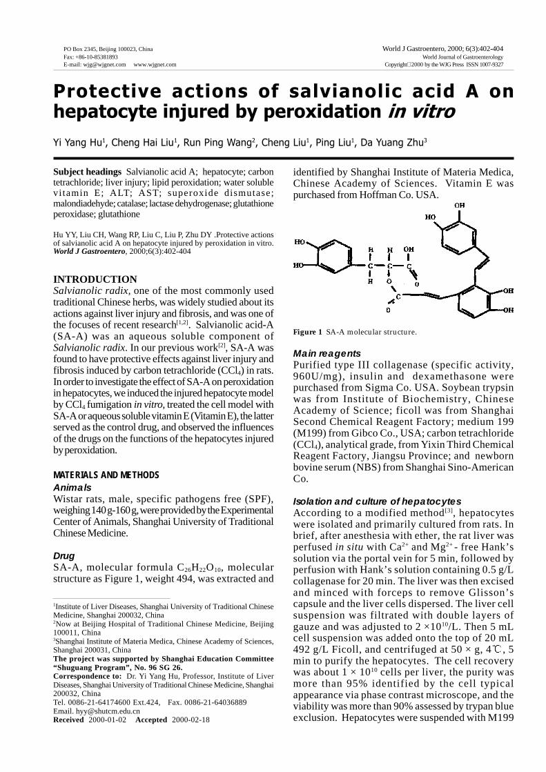
Protective actions of salvianolic acid A onhepatocyte injured by peroxidation in vitro
Yi Yang Hu1, Cheng Hai Liu1, Run Ping Wang2, Cheng Liu1, Ping Liu1, Da Yuang Zhu3
Subject headings Salvianolic acid A; hepatocyte; carbontetrachloride; liver injury; lipid peroxidation; water solublevitamin E; ALT; AST; superoxide dismutase;malondiadehyde; catalase; lactase dehydrogenase; glutathioneperoxidase; glutathione
Hu YY, Liu CH, Wang RP, Liu C, Liu P, Zhu DY .Protective actionsof salvianolic acid A on hepatocyte injured by peroxidation in vitro.World J Gastroentero, 2000;6(3):402-404
INTRODUCTIONSalvianolic radix, one of the most commonly usedtraditional Chinese herbs, was widely studied about itsactions against liver injury and fibrosis, and was one ofthe focuses of recent research[1,2]. Salvianolic acid-A(SA-A) was an aqueous soluble component ofSalvianolic radix. In our previous work[2], SA-A wasfound to have protective effects against liver injury andfibrosis induced by carbon tetrachloride (CCl4) in rats.In order to investigate the effect of SA-A on peroxidationin hepatocytes, we induced the injured hepatocyte modelby CCl4 fumigation in vitro, treated the cell model withSA-A or aqueous soluble vitamin E (Vitamin E), the latterserved as the control drug, and observed the influencesof the drugs on the functions of the hepatocytes injuredby peroxidation.
MATERIALS AND METHODSAnimalsWistar rats, male, specific pathogens free (SPF),weighing 140 g-160 g, were provided by the ExperimentalCenter of Animals, Shanghai University of TraditionalChinese Medicine.
DrugSA-A, molecular formula C26H22O10, molecularstructure as Figure 1, weight 494, was extracted and
identified by Shanghai Institute of Materia Medica,Chinese Academy of Sciences. Vitamin E waspurchased from Hoffman Co. USA.
Figure 1 SA-A molecular structure.
Main reagentsPurified type III collagenase (specific activity,960U/mg), insulin and dexamethasone werepurchased from Sigma Co. USA. Soybean trypsinwas from Institute of Biochemistry, ChineseAcademy of Science; ficoll was from ShanghaiSecond Chemical Reagent Factory; medium 199(M199) from Gibco Co., USA; carbon tetrachloride(CCl4), analytical grade, from Yixin Third ChemicalReagent Factory, Jiangsu Province; and newbornbovine serum (NBS) from Shanghai Sino-AmericanCo.
Isolation and culture of hepatocytesAccording to a modified method[3], hepatocyteswere isolated and primarily cultured from rats. Inbrief, after anesthesia with ether, the rat liver wasperfused in situ with Ca2+ and Mg2+ - free Hank’ssolution via the portal vein for 5 min, followed byperfusion with Hank’s solution containing 0.5 g/Lcollagenase for 20 min. The liver was then excisedand minced with forceps to remove Glisson’scapsule and the liver cells dispersed. The liver cellsuspension was filtrated with double layers ofgauze and was adjusted to 2 ×10¹⁰/L. Then 5 mLcell suspension was added onto the top of 20 mL492 g/L Ficoll, and centrifuged at 50 × g, 4 , 5min to purify the hepatocytes. The cell recoverywas about 1 × 10¹⁰ cells per liver, the purity wasmore than 95% identified by the cell typicalappearance via phase contrast microscope, and theviability was more than 90% assessed by trypan blueexclusion. Hepatocytes were suspended with M199
1Institute of Liver Diseases, Shanghai University of Traditional ChineseMedicine, Shanghai 200032, China2Now at Beijing Hospital of Traditional Chinese Medicine, Beijing100011, China3Shanghai Institute of Materia Medica, Chinese Academy of Sciences,Shanghai 200031, ChinaThe project was supported by Shanghai Education Committee“Shuguang Program”, No. 96 SG 26.Correspondence to: Dr. Yi Yang Hu, Professor, Institute of LiverDiseases, Shanghai University of Traditional Chinese Medicine, Shanghai200032, ChinaTel. 0086-21-64174600 Ext.424, Fax. 0086-21-64036889Email. [email protected] 2000-01-02 Accepted 2000-02-18
PO Box 2345, Beijing 100023, China World J Gastroentero, 2000; 6(3):402-404Fax: +86-10-85381893 World Journal of GastroenterologyE-mail: [email protected] www.wjgnet.com Copyright 2000 by the WJG Press ISSN 1007-9327

containing 5% (v/v) NBS to adjust their density to 5 × 10⁸
cells/L, seeded on plastic dishes (Nunc, Denmark) andprimarily cultured at 37 in a humidified atmosphere of50 mL/L CO2 and 950 mL/L air.
Induction of hepatocyte peroxidative injuredmodelAccording to David method[4], the peroxidatic injuryof hepatocytes was induced by CCl4 fumigation.Briefly, after 48 h of isolation and culture, the cellswere placed in sealed box, to which 1 mL/L CCl4
was added, and the cells were fumigated with CCl4
at 37 for 24 h.
Drug treatmentNormal hepatocytes in dishes were divided into thefollowing groups: the normal, the control, vitamin E(2 × 10⁻³mol/L[5]) and SA-A treated groups atdifferent concentrations (10-4mol/L-10-8mol/L) . Allbut the normal group, were incubated with vitaminE or SA-A at different concentrations andfumigated with CCl4 spontaneously for 24 h, thenthe culture medium was collected respectively andstored at -70 until assay.
Biochemical index assayThe ALT and AST in culture medium were assayed withRriman and Frankle method, malondiadehyde (MDA)with Bacon’s method[6]. Superoxide dismutase (SOD),catalase (CAT), lactase dehydrogenase (LDH),glutathione peroxidase (GSH-PX), glutathione (GSH)were measured following the protocols provided by themanufacturer (Jianchueng Biochemical TechnologicalInstitute, Nanjing).
StatisticsChi- square test and q test.
RESULTSThe cell morphological changesAfter 48 h of isolation and culture, primaryhepatocytes gathered, attached and grew verywell. At 24 h after fumigation with CCl4, thehepatocytes part ial ly shrank, their plasmamembrane became rough and organelles swollen.When the cell models were incubated with SA-Aor Vitamin E, the plasma membrane becamesmoother and the organelles less smother thanthose of controlled model cells.
Effects of SA-A or Vitamin E on ALT, AST andLDH act iv i ty in hepatocytes injured byperoxidationThe activities of ALT and AST both increased,while the former increase more obviously. LDHactivity enhanced approximately 20 folds. SA-Ainhibited these pathological increase in dosage
dependent manner, and among all concentrations testedthe 10-4mol/L SA-A had the best effect on the cellstructure and enzymes. The effect of SA-A was betterthan that of Vitamin E, but 10-7mol/L-10-8mol/L SA-Awas not effective compared with the control (Table 1).
Effects of SA-A or Vitamin E on MDA contentsand the activities of SOD and CAT in hepatocytesinjured by peroxidationMDA content in the control nearly doubled that of thenormal, and the activities of SOD and CAT increasedremarkably. SA-A decreased these pathological changes,and 10-4mol/L SA-A had a significant inhibitory action.Vitamin E also decreased the MDA content markedly,but had no obvious influence on SOD activity (Table 2).
Effects of SA-A or Vitamin E on GSH content andGSH-PX activity in hepatocytes injured byperoxidationAfter the hepatocytes were injured by peroxidation, theGSH-PX activity increased but GSH content decreasedremarkably. 10-4mol/L SA-A or Vitamin E inhibited theincrease of GSH-PX activity and the decrease of GSH.For the extent of inhibition in GSH lowering, SA-A 10-
4mol/L is superior to Vitamin E (Table 2).
Table 1 Effects of SA-A on ALT, AST and LDH activities inhepatocytes injured for 24 h (n = 6, x ± s)
Group ALT(U·L-1) AST(U·L-1) LDH(U·L-1)
Normal 18 ± 2.8b 56 ± 3.8b 77 ± 38b
Control 103 ± 6.5 176 ± 9.1 1674 ± 12810-4mol·L-1 SA-A 49 ± 2.9bd 134 ± 5.0bd 1050 ± 83bd
10-5mol/L SA-A 72 ± 3.9bd 177 ± 8.3 1551 ± 8810-6mol/L SA-A 91 ± 11.1a 177 ± 9.2 1602 ± 8810-7mol/L SA-A 96 ± 7.9 181 ± 6.7 1657 ± 8110-8mol/L SA-A 93 ± 11.4 181 ± 10.7 1684 ± 71Vitamin E 86 ± 7.6b 182 ± 10.7 1509 ± 30a
aP<0.05; bP<0.01, vs Control; dP<0.01 vs Vitamin E.
Table 2 Effects of SA-A on the contents of MDA and GSH andthe activities of SOD, CAT and GSH-PX in hepatocytes injuredby peroxidation (n = 6, x ± s)
Group MDA(µmol·L-1) SOD(U·L-1) CAT(U·L-1) GSH(U·L-1) GSH-PX(U·L-1)
Normal 5.11 ± 0.91b 30.4 ± 2.86b 12.8 ± 3.45b 1.27 ± 0.13b 16.7 ± 8.84b
Control 9.17 ± 0.80 59.0 ± 2.23 86.6 ± 13.00 0.36 ± 0.07 90.9 ± 11.00
10-3mol/L SA-A 6.79 ± 0.81a 45.6 ± 3.26bd 17.3 ± 3.59bd 0.95 ± 0.02bd 65.2 ± 1.24a
10-4mol/L SA-A 7.67 ± 1.11 55.2 ± 2.44 38.3 ± 11.82b 0.40 ± 0.04 81.8 ± 17.54
10-5mol/L SA-A 8.27 ± 1.50 57.6 ± 3.27 49.9 ± 6.78b 0.38 ± 0.10 84.1 ± 19.11
Vitamin E 7.52 ± 0.69a 55.8 ± 4.03 27.6 ± 3.22b 0.49 ± 0.07a 63.64 ± 10.57a
aP<0.05, bP<0.01, vs Control; dP<0.01 vs Vitamin E.
DISCUSSIONIn this study, 24 h after fumigation of hepatocyteswith CCl4, the ALT, AST and LDH all increasedremarkably, the rate of elevation was in order ofLDH, ALT and AST. It is suggested that the
Hu YY, et al. Salvianolic acid A and peroxidation of hepatocytes 403

hepatocytes were acutely injured, cell membraneintegrity was broken and the enzymes in cellp l a s m a l e a k e d o u t . H o w e v e r , a f t e r t h ehepatocytes injured by peroxidation which wereincubated with SA-A, the pathological increasesof ALT, AST and LDH reduced markedly. It isindicated that SA-A had a potential effect againsthepatocyte injury. The free radicals and its triggered lipid peroxidationwere involved in the main mechanisms by whichcarbon tetrachloride injured hepatocytes. MDA wasone of main lipid peroxidatic products, its elevatedlevels could reflect the degrees of lipid peroxidaticinjury in hepatocytes. GSH, a peroxide scavengerwith a lower molecular weight, could eliminatesuperoxide anion and hydrogen peroxide. The contentof GSH reflected the ability against peroxidation[7].In this study, GSH in hepatocytes of the model groupwas reduced remarkably, sugges ting that the potencyof antioxidation in injured cells was decreased. Therewere many other markers that could reflect lipidperoxidation, e.g. SOD, a scavenger of peroxide anionradicals, which could inhibit the initiation of lipidperoxidation by free radicals; GHS-PX, which couldparticularly catalyze the reductive action of GSH toH2O2 to protect the integrity of plasma membraneand functions; CAT etc. All the above-mentionedenzymes increased in the model cells. This mayresult from acute compensation after injury, andperoxidatic reaction stimulated by CCl 4 in
hepatocytes. SA-A markedly inhibits the increase ofMDA level and the decrease of GSH, also reduced theactivities of GSH-PX, CAT, SOD in different extents.Among these results, SA-A had better effect than vitaminE, which is a widely recognized antioxidation drug. It isindicated that SA-A had potential action against lipidperoxidation, this effect perhaps is the main mechanismof protection on liver injury. The results are also inaccordance with the other reports[8] and our previouswork[2].
REFERENCES1 Deng HJ, Ma XH, Xu RL, Chen XM, Zhao YC, Yin L, Han DW.
Study on mechanisms of protective action of Radix Salviamiltiorrhiza (RSM) against experimental hepatic injury in rats.Zhongguo Zhongyao Zazhi, 1992;17:233-236
2 Hu YY, Liu P, Liu C, Xu LM, Liu CH, Zhu DY, Huang MF. Actionsof salvianolic acid A on CCl4 poisoned liver injury and fibrosis inrats. Zhongguo Yaoli Xuebao, 1997;18:478-480
3 Alpini G, Phillips JO, Vroman B, Larusso NF. Recent advances inthe isolation of liver cells. Hepatology, 1994;20:494-514
4 Johnston DE, Kroening C. Stimulation of prostaglandin synthesisin cultured liver cells by CCl4. Hepatology, 1996;24:677-684
5 Zhu JL, Liu SL, Wu J, Tsyganskaya M, Kuncio GS, Zern MA. MG132,vitamin E and lithospermic acid A all inhibit TGHP induced damageto rat hepatocytes. Hepatology, 1996;24(4,part 2):335A
6 Bacon BR, Tavill AS, Brittenham GM, Park CH, Recknagel RO.Hepatic lipid peroxidation in vivo in rats with chronic ironoverload. J Clin Invest, 1983;71:429-439
7 Gasso M, Rubio M, Varela G, Cabre M, Caballeria J. Alonso E,Deulofem R, Camps J, Gimenez A, Pajares M, Pares A, Mato JM,Rodes J. Effects of S-adenosylmethionine on lipid peroxidationand liver fibrogenesis in carbon tetrachloride induced cirrhosis. JHepatol, 1996;25:200-205
8 Lin TJ, Liu GT. Protective effect of salvianolic acid A on heartand liver mitochondria injury induced by oxygen radicals in rats. ZhongguoYaolixue Yu Dulixue Zazhi, 1991;5:276-281
Edited by You DYproofread by Sun SM
404 ISSN 1007-9327 CN 14-1018/ R World J Gastroentero June 2000 Volume 6 Number 3

Kupffer cell and apoptosis in experimental HCC
Hai Zhen Zhu ,You Bing Ruan, Zhong Bi Wu and Chun Ming Zhang
Subject headings liver noplasms, experimental;diethylnitrosa mine; apoptosis; protein P53; Kupffer’s cell
Zhu HZ, Ruan YB, Wu ZB, Zhang CM. Kupffer cell and apoptosisin experimental HCC. World J Gastroentero, 2000;6(3):405-407
INTRODUCTIONOur previous study has proved that Kupffer cells mayhave an inhibitory effect on the process ofhepatocarcinogenesis[1], however, their inhibitorymechanism needs exploring deeply. We performed acomparative study on the expression of PCNA, Bax,P53 and apoptosis of liver cancer cells usingimmunohistochemical technology and terminaldeoxynucleotidyl transferase (TdT)-mediated dUTP-digoxigenin nick end labeling (TUNEL) in thediethylnitrosamine-induced hepatocell ular carcinoma(HCC) in rats with or without pretreatment withgadolinium chlori de or zymosan which might effectivelyblock or enhance the activity of Kupffer cells in order toknow the role of Kupffer cells in apoptosis in theexperimental HCC and explore further the inhibitorymechanism of Kupffer cells on the process ofhepatocarcinogenesis.
MATERIALS AND METHODSEstablishment of animal models and pathologicalexaminationOne hundred and forty male Sprague-Dawley ratswere divided into six groups. DENA group, 40rats received diethylnitrosamine (DENA) at a doseof 70 mg/kg in distilled water once/wk till wk15. GC+DENA group, gadolinium chloride was injectediv at a dose of 10 mg/kg once 2 wk to suppressKupffer cells in 40 rats till wk15 and these ratssimultaneous ly rec eived DENA just as DENAgroup. ZM+DENA group, zymosan was injectediv at a do se of 20 mg/kg once 2 wk to activateKupffer cells till wk15 and DENA was received justas DENA group. GC group, gadolinium chloridewas in jected iv at a dose of 10 mg/kg in 0.85%NaCl once 2 wk till wk 15. ZM group, zymosan
was injected iv at a dose of 20 mg/kg in 0.85 %NaCl once 2 wk till wk15. Control group, theserats were maintai ned on a standard laboratorydiet and tap water. All rats were killed at the wk21of hepatocarcinogenesis. The liver samples weretaken, fixed in 40 mL/L paraformaldehyde andembedded in paraffin. Each specimen was cutinto 5 µm serial slices, stained with hemotoxylin-eos in and sub jec ted to h i s topa tho log ica lexamination.
Immunohistochemical stainingThe SP method was used, the 1st antibody wasmouse-anti-human PCNA monoclona l antibody(Calbiochem Co., dilution 1 : 50), rabbit-anti-humanBax (Santa Cruz, dilution 1 : 50) and mouse-anti-human P53 monoclonal antibody (NovocastraLaboratories, dilution 1 : 50). The SP kit waspurchased from Boehringer Mannheim , Germany.DAB staining was used. The dark brown staining ofnuclei was taken as PCNA and P53-positivereaction. The dark brown granules in cytoplasm weretaken as Bax-positive reaction. We used PBS toreplace the 1st antibody as negati ve control.
Terminal deoxynucleotydial transferase-mediated biotinylated-dutp nick end labelingmethod (TUNEL method)In situ Cell Apoptosis Detection kit was purchased fromB o e h r i n g e r M a n n h e i m , G e r m a n y. A f t e rdeparaffinization and rehydration in ethanol, the sectionswere incubated with 0.2 mol/L HCl for 20 min at roomtemperature (RT). They were then deproteinized byincubation with 30mg/L proteinase (in 50mM Tris-HCl,pH 8.0; 5mM EDTA) for 30 min at RT. Terminaldeoxynucleot idyl transferase (TdT) and biotinylateddUTP in TdT buffer (30mM Trizma base, pH 7.2;140mM sodium cacodylate; 1mM cobalt chloride) wereadded to cover the sections, which were then incubatedfor 60 min in a humid atmosphere at 37 . The sectionsthen reacted with alkaline phasphatase (ALP) for 30min at RT. Visulization was carried out with NBT/BCIP.Nuclei with clear blue staining w ere regarded aspositive. TUNEL incubation solution without terminaldeoxynucleo tidyl transferase was used as negativecontrol.
Proliferating index (PI) and apoptosis index (AI)PI and AI were determined by Leitz ASM 68Kimage analyzing system purchased from Germany.Five visual fields (orginal magnificantion, 10 × 40)
Department of Ultrastructural Pathology, Tongji Medical University,Wuhan 430030, Hubei Province, ChinaDr. Hai Zhen Zhu, graduated from Tongji Medical University in 1997,engaged in the researches of antitumor of Kupffer cell under theinstruction of Professor You Bing Ruan, having four papers published.Correspondence to: Hai Zhen Zhu, Department of UltrastructuralPathology, Tongji Medical University, Wuhan 430030, Hubei Province,ChinaReceived 2000-01-12 Accepted 2000-02-20
PO Box 2345, Beijing 100023, China World J Gastroentero, 2000; 6(3):405-407Fax: +86-10-85381893 World Journal of GastroenterologyE-mail: [email protected] www.wjgnet.com Copyright 2000 by the WJG Press ISSN 1007-9327

from every positive slide would be chosen andthe TUNEL and PCNA-positive parenchymalcells as well as the total number of parenchymalcells in the same field would be counted . PI orAI was expressed as the percentage of then u m b e r o f P C N A o r T U N E L - p o s i t i v eparenchymal cells in the total parenchymal cellsof the same field.
number of PCNA-positive cellsProliferating index (PI)= × 100%
number of total cells number of TUNEL-positive cellsApoptosis index(AI)= × 100% number of total cells
Statistical analysisFisher’s exact test, Student’s test and Spearman rankcorrelation were employe d.
RESULTSMacroscopic observation and histopatholog-i c a l e x a m i n a t i o n ( a t w k 2 1 o fhepatocarcinogenesis)Meither the control group, nor the GC group and ZMgroup showed any changes macro and microscopically;on the contrary, the liver surface in DENA group andGC +DENA group were covered with a lot of whitenodules. The diameter of the larges t nodules was 0.5cm in DENA group and 2.0 cm in GC+DENA group.Thes e nodules were diagnosed as HCC histologically.Some white grey focal nodules sc attered over the liversurface in ZM+DENA group, were also diagnosed asHCC with many apoptotic cells and apoptotic bodies.
Bax stainingCytoplasm of cancer cells with clear brown staining wasregarded as positive (Figure 1). The positive rates ofBax in ZM+DENA group, DENA group and GC+DENAgroup were 84.6% (11/13), 28.6% (2/7) and 27.3%(3/11) respectively. It was significantly higher inZM+DENA group than that in DENA group (Fisher’sexact test, P<0.05).
P53 stainingA clear brown staining of the nuclei in cancer cellswas regarded as positive (Figure 2). The positive ratesof P53 in ZM+DENA group, DENA group andGC+DENA group were 76.9% (10/13), 14.3% (1/7)and 36.4% (4/11) respectively. It was markedly higherin ZM+DENA group than that in DENA group (Fisher’sexact test , P<0.05).
PCNA stainingNuclei with clear brown staining were regarded aspositive (Figure 3). No expressions of Bax, P53 and PCNA inthe control group, GC group and ZM group werefound.
Apoptosis of cancer cells using TUNEL methodApoptotic cells were observed in DENA group,GC+DENA group and ZM+DENA group by meansof TUNEL method. The cells with clear nuclearlabeling were defined as TUNEL-positive cells.Apoptotic cells were scattered in the cancer tissue(Figure 4).
Figure 1 Expression of Bax in rat HCC in ZM+DENA group.SP × 400Figure 2 Expression of P53 in rat HCC in DENA group. SP × 400Figure 3 Expression of PCNA in rat HCC in DENA group.SP × 200Figure 4 Apoptotic cells in rat HCC in ZM+DENA group.TUNEL × 200
Proliferating index (PI) and apoptosis index (AI)(Table 1)
Table 1 PI and AI in ZM+DENA group, DENA group andGC+DENA group
Groups Number PI(%, x ± s) AI(%, x ± s) PI/AI
ZM+DENA group 13 15.22 ± 2.17b 7.53 ± 1.61b 2.20DENA group 7 24.97 ± 2.53 4.36 ± 1.18 5.73GC+DENA group 11 38.69 ± 3.17b 2.52 ± 0.81b 15.40
bP<0.01, vs DENA group, Fisher’s exact test.
Correlation between apoptosis index, Bax andP53 expressionThere was a positive correlation between apoptosisindex and Bax or P53 protein reactivity in ZM+DENAgroup, DENA group and GC+DENA group(Table 2).
406 ISSN 1007-9327 CN 14-1018/ R World J Gastroentero June 2000 Volume 6 Number 3
Zhu HZ, et al. Kupffer cell and apoptosis 407
Table 2 Correlation between apoptosis index and Bax or P53expression
AI vs Bax AI vs P53Groups Bax-positive P53-positive cases/total rs P cases/total rs P
ZM+DENA group 11/13 0.63 <0.05 10/13 0.73 <0.05DENA group 2/7 0.79 <0.05 1/7 0.61GC+DENA group 3/11 0.74 <0.01 4/11 0.82 <0.05
Spearman rank correlation.
DISCUSSIONGadolinium chloride is believed to be a specificsuppressor of Kupffer cells[1,2]. Zymosan is animmunopotentiator and may be used to activate Kupffercells[3]. We performed a comparative study on apoptosisin DENA-induced hepatocellular carcinoma in rats withor without pretreatment with gadolinium ch loride orzymosan which might effectively block or enhance theactivity of Kupffer cells in order to clarify whether theKupffer cells play arole in apoptosis of the experimentalhepatocarcinogenesis or not. Proliferating cell nuclear antigen (PCNA) is anauxiliary protein of DNA polymerize-delta. Itaccumulates little in the resting stage cell, but prominentlyin the nuclei of proliferating cells during G1-late phaseand S-phase and decreases in G2-phase and M-phase.It is associated with cell proliferation an d regarded as abiological marker for cell proliferation[4]. Bax is a Mr21000 protein with extensive amino acid homology withBcl-2. This protein has been shown to form heterodimerswith apoptosis inhibiting protein, B cl2, so it can inducecell apoptosis[5]. When DNA is injured by toxica nt, wild-type p53 drives cell to G1 and arrests or inhibits cellprolifer ation until DNA is repaired. When nuclear DNAis badly damaged or cannot be rep aired, wild-type p53induces transcription of apoptotic genes and drives thecell to be in apoptosis[6]. Gottlieb et al[7] thought thatoverexpression of P53 was related to cell apoptosis. Zhaoet al[8] observed that there w as a positive correlationbetween apoptosis index and P53 protein, and supportedthe role of P53 in regulating apoptosis in human HCC.The positive correlation between apoptosis index andP53 protein immunoreactivity observed in our study alsosupports these findings. A new method to detectapoptosis in situ, terminal deoxynucleotidyl transferase-mediated (TdT) dUTP-digoxigenin nick end labeling(TUNEL) was recently developed by Gavrieli et al[9],and was applied to detect cell apoptosis of human HCCand other tumors[10]. Our results showed that the positive rates ofBax and P53 protein were markedly higher in ZM +
DENA group than in DENA group with significantdifferences. Proliferating index and apoptosis indexrespectively increased or decreased in ZM+DENAgroup, DENA group and GC+DENA group successively.These results demonstrated that the blockage of Kupffercells with gadolinium chloride might suppress cellapoptosis and the activation of Kupffer cells withzymosan might promote cell apoptosis in the experimentalhepatocarcinogenesis. One of the characteristic features of cancer is thecontinuous growth and the ratio of cell proliferation andcell apoptosis determine the fate of tumor growth[11-
13]. In normal tissues, apoptosis is an efficient way ofeliminatin g transformed cells. However, inability ofcells to undergo apoptosis may advanc e theirdevelopment, both by allowing the accumulation ofdividing cells and by impairing the elimination of geneticmutants that may harbor malignant potential[11,12]. Ourresults proved that Kupffer cells could promoteapoptosis in the experimental hepatocarcinogenesis andrevealed further that they played an inhibitory role inhepatocarcinogenesis through inducing apoptosis oftumor cells.
REFERENCES1 Zhu HZ,Ruan YB,Wu ZB,Ivankovic S. The influence of Kupffer
cells on experimental hepatocarcinogenesis. Zhonghua BinglixueZazhi, 1998;27:102-104
2 Kukan M, Vajdova K, Horecky J, Nagyova A, Mehendale HM,Trnovec T. Effects of blockade of Kupffer cells by gadoliniumchloride on hepatobiliary function in cold ischemia reperfusioninjury of rat liver. Hepatology, 1997;26:1250-1257
3 Decker K. Biologically active products of stimulated liver mac-rophages (Kupffer cells). Eur J Biochem, 1990;192:245-261
4 Celis JE, Celis A. Cell cycle dependent variations in the distribu-tion of the nuclear protein cyclin proliferating cell nuclear anti-gen in cultured cells: Subdivision of S phase. Proc Natl Acad SciUSA, 1985;82:3262-3266
5 Hung WC, Chuang LY. Induction of apoptosis by sphingosine-1-phosphate in human hepatoma cells is associated with enhancedexpression of bax gene product.Biochem Biophys Res Commu,1996;229:11-15
6 Mitry RR, Sarraf CE, Wu CG, Pignatelli M, Habib NA. Wild typep53 induces apoptosis in Hep3B through up regulation of Baxexpression. Lab Invest, 1997;77:369-378
7 Gottlieb TM, Oren M. p53 in growth control and neoplasia. BiochimBiophys Acta, 1996;1287:77-102
8 Zhao M, Zimmermann A. Apoptosis in human hepatocellular car-cinoma and in liver cell dysplasia is correlated with p53 proteinimmunoreactivity. J Clin Pathol, 1997;50:394-400
9 Gavrieli Y, Sherman Y, Ben Sasson SA. Identification of pro-grammed cell death in situ via specific labeling of nuclear DNAfragmentation. J Cell Biol, 1992;119:493-501
10 Hino N, Higashi T, Nouso K, Nakatsukasa H, Tsuji T. Apoptosisand proliferation of human hepatocellular carcinoma. Liver, 1996;16:123-129
11 Carson DA, Ribeiro JM. Apoptosis and disease. Lancet, 1993;341:1251-1254
12 Rotello RJ, Lieberman RC, Purchio AF, Gerschenson LE. Coordi-nated regulation of apoptosis and cell proliferation by transform-ing growth factor β1 in cultured uterine epithelial cells. Proc NatlAcad Sci USA, 1991;88:3412-3415
13 Williams GT.Programmed cell death:apoptosis and oncogenesis.Cell, 1991;65:1097-1098
Edited by You DYproofread by Sun SM
Clinical significance of serum intercellular adhesionmolecu le -1 detec t ion in pat ients w i thhepatocellular carcinoma
Ming Hui Mei1, Jing Xu1, Qin Fen Shi2, Jing Hong Yang1, Qian Chen1 and Li Ling Qin2
Subject headings carcinoma, hepatocellular/diagnosis;intercellular adhesion molecule-1; liver neoplasms/diagnosis; alpha-fetoprotein
Mei MH, Xu J, Shi QF, Yang JH, Chen Q, Qin LL. Clinical significanceof serum intercellular adhesion molecule-1 detection in patients withhepatocellular carcinoma. World J Gastroentero, 2000;6(3):408-410
INTRODUCTIONSince the late 1980s, studies on the expression ofintercellular adhesion molecule-1 (ICAM-1) in patientswith malignancies have demonstrated that ICAM-1may strongly express in two forms in such diseases:membranous one on the surface of tumor cells(membrane-bound ICAM-1) and soluble one incirculation (soluble ICAM-1, sICAM-1)[1,2].Furthermore, increased expression of sICAM-1 invarious malignant diseases, such as gastric cancer,colonic and pancr eatic cancer was reported byTsujisaki et al[3], who found that th e incidence ofpositivity of sICAM-1 in the malignant diseases wassignificantl y higher than that in benign diseases.Therefore, the diagnostic value of detect ing sICAM-1for some malignancies has been proposed[3]. The recentstudies on detecting serum sICAM-1 in patients withhepatocellular carcinoma (HCC) have revealed thatserum levels of sICAM-1 were well correlated to progression and prognosis of the disease[4-6], from which ithas been conside red that sICAM-1 may be useful formonitoring the response to treatment[ 4]. However,whether sICAM-1 is a diagnostic marker is stillcontroversial. Our recent study demonstrated that inHCC patients with normal or low levels of serum α-fetoprotein (AFP) measurement of sICAM-1 is ofclinical value for detecting early HCC and its recurrence afterhepatectomies[7-9]. To further evaluate the clinical
significance of measur ing sICAM-1 in HCC, the serumlevels of sICAM-1 in 134 patients with HCC wereexamined and the results were analyzed.
MATERIALS AND METHODSPatientsThere were 134 patients with HCC, including 115 malesand 19 females, aged from 12-80 years, median 49years. The diagnosis of HCC for all patients wasconfirmed pathologically either by surgical resectionof liver tumors or by liver biopsies. Tumor was lessthan 5 cm in diameter in 17 patients, less than 10 cm in37 and more than 10 cm or with extrahepatic metastasisin 80 cases. Hepatitis B surface antigen (HBsAg) waspositive in 112 cases and hepatitis C antibody positivein 10 patients. Surgical treatment was carried out in 68patients, of whom 57 underwent tumor resection and11 underwent laparotomy or selective catheterizationof the hepatic artery. The rest of the patients weretreated by transcatheter arterial embolization (TAE)or alcohol injection of tumors. As controls, serum levelsof sICAM-1 were measured in 42 patients with chronichepatitis B (CH), 40 with liver cirrhos is (LC) and 50healthy blood donors.
MethodsBlood samples were collected early in the morning. Theserum was immediately separated by centrifugation andfrozen to -80 . Concentrations of serum sIC AM-1were measured with an enzyme-linked immunosorbentassay kit (Biosource Europe, Fleurus, Belgium). Thequantitative determinations of serum AFP in patients withHCC was done by radio-immunoassay (RIA), using anAFP kit (Jiuding Biolo gical,Tianjin, China ). Thereference ranges of serum AFP concentration wereclassified as follows: above 200 µg/L, positive; 20 µg/L-200 µg/L, questionable positive; and below 20 µg/L,negative.
Statistical analysisData between groups were compared by Wilcox’s RankSum test and Fisher test. P<0.05 means significancestatistically.
RESULTSThe median of sICAM-1 in 134 patients with HCCwas 1801 µg/L, which was significantly higher than
1Department of Hepatobiliary Surgery, 2Institute of HepatobiliarySurgery, Guilin Medical College, Guilin 541001, Guangxi Province, ChinaMing Hui Mei, M.D., graduated from Guangxi Medical College in 1968,from Tongji Medical University as a postgraduate in 1981 and fromHannover Medical University, Germany in 1989, receiving a doctor’sdegree of medicine, having more than 40 papers published.Supported by the grants from the Guangxi Science andTechnology Commit tee (No.9817093).Correspondence to: Dr. Ming Hui Mei, Lequn Road 95, Departmentof Hepato-Biliary Surgery, Guilin Medical College, 541001 Guilin, PRChinaTel. 0086-773-2824373, Fax. 0086-773-2822194Email. [email protected] 2000-01-03 Accepted 2000-02-16
PO Box 2345, Beijing 100023, China World J Gastroentero, 2000; 6(3):408-410Fax: +86-10-85381893 World Journal of GastroenterologyE-mail: [email protected] www.wjgnet.com Copyright 2000 by the WJG Press ISSN 1007-9327
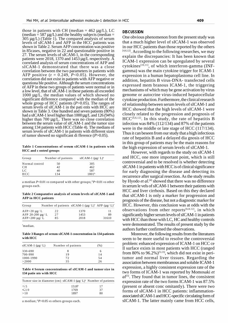
those in patients with CH (median = 462 µg/L), LC(median = 587 µg/L) and the healthy subjects (median =305 µg/L) (Table 1). The compared analysis of serumlevels of sICAM-1 and AFP in the HCC patients wasshown in Table 2. Serum AFP concentration was positivein 85cases, negative in 22 and questionable positive in27. The serum levels of sICAM-1, in the correspondingpatients were 2018, 1370 and 1453 µg/L respectively. Acorrelated analysis of serum concentrations of AFP andsICAM-1 demonstrated that there was a closecorrelation between the two parameters in patients withAFP positive (r = 0.249, P<0.05). However, thecorrelation did not exist in patients with AFP negative orquestiona ble positive. Although the serum concentrationsof AFP in these two groups of patients were normal or ina low level, that of sICAM-1 in these patients all exceeded1000 µg/L, the median values of which showed nosignificant difference compared with the median of thewhole group of HCC patients (P>0.05). The ranges ofserum levels of sICAM-1 in the pati ents with HCC areshown in Table 3. One hundred and seven patients (80%)had a sICAM-1 level higher than 1000 µg/L and 126 (94%)higher than 700 µg/L. There was no close correlationbetween the serum value of sICAM-1 and the tumor sizeamong 134 patients with HCC (Table 4). The medians ofserum levels of sICAM-1 in patients with different sizesof tumor showed no significant di fference (P>0.05).
Table 1 Concentrations of serum sICAM-1 in patients withHCC and c ontrol groups
Group Number of patients sICAM-1 (µg/L)a
Normal control 50 305CH 42 462LC 40 587HCC 134 1801b
a median P<0.01 vs compared with other groups; bP<0.01 vs othergroups each.
Table 2 Comparative analysis of serum levels of sICAM-1 andAFP in HCC patients
Group Number of patients sICAM-1 (µg/L)* AFP (µg/L)*
AFP<20 µg/L 22 1370 11AFP 20-200 µg/L 27 1453 89AFP>200 µg/L 85 2018 31610
*median.
Table 3 Ranges of serum sICAM-1 concentration in 134 patientswith HCC
sICAM-1 (µg/L) Number of patients (%)
330-690 8 6700-990 19 141000-1990 72 54>2000 35 26
Table 4 Serum concentrations of sICAM-1 and tumor size in134 patie nts with HCC
Tumor size in diameter (cm) sICAM-1 (µg/L)a Number of patients
5 1518b 171 0 1769 37
>10 1897 80
a median; bP>0.05 vs others groups each.
DISCUSSIONOne obvious phenomenon from the present study wasthat a much higher level of sICAM-1 was observedin our HCC patients than those reported by the others[4,6,12]. According to the following researches, we mayexplain the discrepancies: It has been known thatICAM-1 expression can be upregulated by severalcytokines[10,11], of which interferon-gamma (INF-gamma) was the main cytokine trigger for ICAM-1expression in a human hepatoplastoma cell line. Inaddition, hepatitis B virus-DNA- transfected cellsexpressed mem branous ICAM-1, the triggeringmechanisms of which may be gene activation by virusgenome or autocrine virus-induced hepatocellularcytokine production. Furthermore, the clinical researchof relationship between serum levels of sICAM-1 andHCC showed that the high levels of sICAM-1 wereclosely related to the progression and prognosis ofHCC[4,6,12]. In this study, the rate of hepatitis Binfection was 84% (112/134) and 87% of the patientswere in the middle or late stage of HCC (117/134).Thus it can beseen from our study that a high infectiousrate of hepatitis B and a delayed dia gnosis of HCCin this group of patients may be the main reasons forthe high expression of serum levels of sICAM-1. However, with regards to the study on sICAM-1and HCC, one more important point, which is stillcontroversial and to be resolved is whether detectingsICAM-1 in patients with HCC is of clinical significancefor early diagnosing the disease and detecting itsrecurrence after surgical resection. As the study resultsby Hyodo et al.[4] showed that there was no differencein serum le vels of sICAM-1 between their patients withHCC and liver cirrhosis. Based on this they declaredthat sICAM-1 is only a marker for progression andprognosis of the disease, but not a diagnostic marker forHCC. However, this conclusion was at odds with theobservations from other reports[6,8,12], in whichsignificantly higher serum levels of sICAM-1 in patientswith HCC than those with LC, HC and healthy controlswere demonstrated. The results of present study by theauthors further confirmed the observations. Moreover, the following results from the literaturesseem to be more useful to resolve the controversialproblem: enhanced expression of ICAM-1 on HCC cell surface exists in most patients with HCC (rangedfrom 80% to 96.2%)[5,13], which did not exist in peri-tumor and normal liver tissues. Regarding theassociation between membranous and soluble ICAM-1expression, a highly consistent expression rate of thetwo forms of ICAM-1 was reported by Momosaki etal[5]. They found that in tumor lines, the consistentexpression rate of the two forms ICAM-1 was 87.5%(present or absent conc omitantly). There were twoforms of sICAM-1 in HCC patients: inflammation-associated sICAM-1 and HCC-specific circulating form ofsICAM-1. The latter mainly came from HCC cells,
Mei MH, et al. Imtercellular adhesion molecule-1 detection in HCC 409

from which the membranous ICAM-1 were shedinto the circulation continuously and became theimportant source of sICAM-1 [14]. In addition,recent s tudies by the authors [9] found thatregardless of positive or negative serum AFP, aftera radical resection of liver tumor, the sICAM-1level would be decreased to the normal within 1-2months postoperatively. However, in patientsunderwent non-radical resection due to vascularinvasions of the liver or extrahepatic metastasis,the serum levels of sICAM-1 will maintain at ahigh level. It has been suggested from all of thestudies mentioned above that the high level ofserum sICAM-1 in HCC may originat e mainlyfrom tumor itself. Thus we believe sICAM-1 inHCC patients may be a useful marker for detectingH C C a n d a m o n i t o r o f r e c u r r e n c e a f t e rhepatectomy. As the strong correlation between serum levels ofsICAM-1 and AFP in patients with positive AFP wasdisclosed (Table 2), the clinical significance of sICAM-1 detection for diagnosing HCC will be focused onpatients with normal or low levels of serum AFP.Serum AFP is still the best diagnostic marker for HCC.But AFP levels may be normal in 20%-40% of patientswith HCC, depending on the severity of the disease[15]. In China about 30% of HCC patients can not bediagnosed by this serum marker, which results indelayed diagnosis and, consequently, hampers effortsto improve effective surgical treatment of the dis ease.Therefore, more sensitive serum markers are requiredfor detecting HCC. From the study, one of the interesting results wasthat there were 22 (16.4%) and 27 (20.2%) patientswith HCC, whose serum levels of AFP werenegative (median = 11 µg/L) and questionablepositive (median = 89 µg/L). However, that ofsICAM-1 in those patients were 1370 and 1453 µg/L respectively (Table 2), which showed nosignificant difference compared with the medianlevel of total group of HCC(P>0.05). Furthermore,analysis of ranges of sICAM-1 levels in patientswith HCC demonstrated that 80% of the patientshad a high serum level of sICAM-1 exceeding 1000µg/L. According to the study by Shimizu et al[6],sICAM-1 level above 1000 µg/L is a determinantfor prognosis and progression of HCC, theproportion of patients with high levels of sICAM-1(>1000 µg/L) in patients with HCC in our study wasobviously highe r than that of AFP (>200 µg/L). Asmentioned in our previou s studies the diagnosis ofHCC should be strongly suspected when a patientwith an uncertain intrahepatic lesion had a serumlevel of sICAM-1 higher than 1000 µg/L[7]. Theresults of the pre sent study further confirm ourprevious conclusion. Another interesting finding from the study was
that a significant correlation between serum level ofsICAM-1 and tumor size of HCC was not observed (Table4). The median of sICAM-1 concentration in 17 patientswith tumor diameter less than 5 cm was 1518 µg/L, amongthem 4 being negative, 8 questionable positive and 5positive for AFP. In addition, a tumor recurrence wasdiagnosed in 4 of the 17 patients after 1-3 hepatectomiesduring the postoperative follow-up. The median of tumorsize in the 4 cases was only 2.8 cm whe n recurrencewas confirmed, however, the median of serum level ofsICAM-1 in the same patients was 1378 µg/L. It wasstrongly suggested by our observation that measurementof sICAM-1 is of clinical significance in detecting earlyHCC and monitoring its recurrence postoperatively whentumor is small in diameter, particularly for patients withnormal or low serum concentrations of AFP.
ACKNOWLEDGMENTS We are grateful to Professor S. Meuer, and Dr. B. Schraven, Institute of Immunology,Ruprecht-Karls-University, Heidelberg, Germany, for theirsuggestions, advice, and cooperation in this study.
REFERENCES1 Vogetseder W,Feichtinger H,Schulz TF, Schwaeble W, Tabaczewskt
P, Mitterer M, Boeck G, Marth C, Dapunt O, Mikuz G, Dierich MP.Expression of 7F7-antigen, a human adhesion molecule identical tointercellular adhesion molecule-1 (ICAM-1) in human carcinomasand their stromal fibroblasts. Int J Cancer, 1989;43:768-773
2 Rothlein R, Mainolfi EA, Czajkowski M, Marlin SD. A form of circu-lating ICAM-1 in human serum. J Immunol, 1991;147:3788-3793
3 Tsujisaki M, Imai K, Hirata H, Hanzawa Y, Masuya J, Nakano T,Sugiyama T, Matsui M, Hinoda Y, Yachi A. Detection of circulat-ing intercellular adhesion molecule-1 antigen in malignant diseases.Clin Exp Immunol, 1991;85:3-8
4 Hyodo I, Jinno K, Tanimizu M, Hosokawa Y, Nishikawa Y,Akiyama M, Mandai K, Moriwaki S. Detection of circulating in-tercellular adhesion molecule-1 in hepatocellular carcinoma. Int JCancer, 1993;55:775-779
5 Momosaki S, Yano H, Ogasawara S, Higaki K, Hisaka T, Kojiro M.Expression of intercellular adhesion molecule-1 in human hepa-tocellular carcinoma. Hepatology, 1995;22:1708-1713
6 Shimizu Y, Minemura M, Tsukishiro T, Kashu Y, Miyamoto M,Nishimori H, Higuchi K, Watanabe A. Serum concentration ofintercellular adhesion molecule-1 in patients with hepatocellularcarcinoma is a marker of the disease progression and prognosis.Hepatology, 1995;22:525-531
7 Mei MH, Xu J, Shi QF, Chen Q, Qin LL. Measurement of serumintercellular adhesion molecule-1 in hepatocellular carcinoma andits clinical significance. Zhonghua Yixue Zazhi, 1999;79:200-201
8 Mei MH, Xu J, Shi QF, Chen Q, Qin LL. Measurement of serumcirculating intercellular adhesion molecule1 and its clinical signifi-cance in hepatocellular carcinoma: preliminary report. JHepatobiliary Pancr Surg, 1999;6:181-185
9 Xu J, Mei MH, Shi QF, Chen Q, Qin LL. Clinical evaluation of mea-surement of serum intercellular adhesion molecule-1 in hepatocellularcarcinoma. Zhonghua Shiyan Waike Zazhi, 1998;15:514-515
10 Dustin ML, Rothlein R, Bhan AK, Dinarello CA, Springer TA.Induction by IL 1 and interferon-γ: tissue distribution, biochemistry,and function of a natural adherence molecule (ICAM-1). JImmunology, 1986;137:245-254
11 Zoehrens G, Armbrust T, Pirzer U, Bueschenfelde KHM, RamadoriG.Intercellular adhesion molecule 1 concentration in sera of pa-tients with acute and chronic liver disease: relationship to diseaseactivity and cirrhosis. Hepatology, 1993;18:798-802
12 Adams DH, Mainolfi E, Burra P, Neuberger JM, Ayres R, Elias E,Rothlein R. Detecting of circulating intercellular adhesion mol-ecule-1 in chronic liver diseases. Hepatology, 1992;16:810-814
13 Torii A, Harada A, Nakao A, Nonami T, Ito M, Takagi H. Expres-sion of intercellular adhesion molecule 1 in hepatocellularcarcinoma. J Surg Oncol, 1993;53:239-242
14 Hyodo I, Jinno K, Tanimizu M, Doi T, Nishikawa Y, Hosokawa Y,Moriwaki S. Intercellular adhesion molecule-1 release from humanhepatocellular carcinoma. Cancer Detec Prev, 1996;20:308-315
15 Giardina MG, Matarazzo M, Varriale A, Morante R, Napoli A, Martino R.Serum Alpha-L-Fucosidase: a useful marker in the diagnosis of hepatocellularcarcinoma. Cancer, 1992;70:1044-1048
Edited by You DYproofread by Sun SM
410 ISSN 1007-9327 CN 14-1018/ R World J Gastroentero June 2000 Volume 6 Number 3
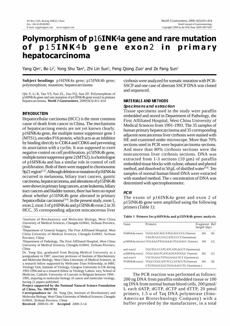
Polymorphism of p16INK4a gene and rare mutationo f p 1 5 I N K 4 b g e n e e x o n 2 i n p r i m a r yhepatocarcinoma
Yang Qin1, Bo Li2, Yong Shu Tan3, Zhi Lin Sun1, Feng Qiong Zuo1 and Ze Fang Sun1
Subject headings p16INK4a gene; p15INK4b gene;polymorphism; mutation; hepatocarcinoma
Qin Y, Li B, Tan YS, Sun ZL, Zuo FQ, Sun ZF. Polymorphism ofp16INK4a gene and rare mutation of p15INK4b gene exon2 in primaryhepatocarcinoma. World J Gastroentero, 2000;6(3):411-414
INTRODUCTIONHepatocellular carcinoma (HCC) is the most commoncause of death from cancer in China. The mechanismsof hepatocarcinog enesis are not yet known clearly.p16INK4a gene, the multiple tumor suppressor gene 1(MTS1), encodes P16 protein, which acts as an inhibitorby binding directly to CDK4 and CDK6 and preventingits association with a cyclin. It was supposed to exertnegative control on cell proliferation. p15INK4b gene,multiple tumor suppressor gene 2 (MTS2), is a homologueof p16INK4a and has a similar role in control of cellproliferation. Both of them were mapped to chromosome9p21 region[1,2]. Although deletion or mutation of p16INK4aoccurred in melanoma, biliary tract cancers, gastriccarcinoma, hepatocarcinoma, and alterations of p15INK4bwere shown in primary lung cancers, acute leukemia, biliarytract cancers and bladder tumors, there has been no reportabout whether p15INK4b gene alterated in primaryhepatocellular carcinoma[3-9]. In the present study, exon 1,exon 2, exon 3 of p16INK4a and p15INK4b exon 2 in 35HCC, 35 corresponding adjacent noncancerous liver
cirrhosis were analyzed for somatic mutation with PCR-SSCP and one case of aberrant SSCP DNA was clonedand sequenced.
MATERIALS AND METHODSSpecimens and extractionTissue specimens used in the study were paraffinembedded and stored in Department of Pathology, theFirst Affiliated Hospital, West China University ofMedical Sciences from 1991-1993. The 35 samples ofhuman primary hepatocarcinoma and 35 correspondingadjacent noncancerous liver cirrhosis were stained withHE and examined under microscope. More than 70%sections used in PCR were hepatocarcinoma sections.And more than 80% cirrhosis sections were thenoncancerous liver cirrhosis sections. DNA wasextracted from 1-3 sections (10 µm) of paraffinembedded tissue blocks with xylene, ethanol and phenolmethod, and dissolved in 50 µL of distilled water[10]. Tensamples of normal human blood DNA were extractedwith standard method. The c oncentration of DNA wasdetermined with spectrophotometer.
PCRThe exons of p16INK4a gene and exon 2 ofp15INK4b gene were amplified using the followingprimers (Table 1):
Table 1 Primers for p16INK4a and p15INK4b genes analysis
Gene Primers Fragment Ref length (bp)
P16INK4a exon1 5’GGGAGCAGCATGGAGCCCG 3’(sense) 204 [6]5’AGTCGCCCGCCATCCCCT 3’(antisense)
p16INK4a intron1 5’GGAAATTGGAAACTGGAAGC 3’(sense) 168 [1]
and exon2 5’GCTGCCCATCATCATGACCT 3’(antisense)p16INK4a exon2 5’GGCAGGTCATGATGATGGGC 3’(sense) 362 [1]and exon3 5’TCTGAGCTTTGGAAGCTCT 3’(antisense)P15INK4b exon2 5’GGCCGGCATCTCCCATACCTG3’(sense) 345 [9]
5’TGTGGGCGGCTGGGAACCTG 3’(antisense )
The PCR reaction was performed as follows:200 ng DNA from paraffin embedded tissue or 100ng DNA from normal human blood cells, 200 µmol/L each dATP, dGTP, dCTP and dTTP, 20 pmolprimers, 1.5 u of Taq DNA polymerase (Sino-American Biotechnology Company) with abuffer provided by the manufacturer, in a total
1Institute of Biochemistry and Molecular Biology, West ChinaUniversity of Medical Sciences, Chengdu 610041, Sichuan Province,China2Department of General Surgery, The First Affiliated Hospital, WestChina University of Medical Sciences, Chengdu 610041, SichuanProvince, China3Department of Pathology, The First Affiliated Hospital, West ChinaUniversity of Medical Sciences, Chengdu 610041, Sichuan Province,ChinaDr. Yang Qin, graduated from Beijing Medical University as apostgraduate in 1987, associate professor of Institute of Biochemistryand Molecular Biology, West China University of Medical Sciences, asa research fellow supported by Wellcome Trust Fellowship, in MRCVirology Unit, Institute of Virology, Glasgow University in UK during1993-1994 and as a research fellow in Virology Labora tory, School ofMedicine, Catholic University of Louvain in Belgium between 1994 -1995, majoring in molecular biology of cancer and molecular virology,having 21 papers published.Project supported by the National Natural Science Foundationof China, No. 39670702Correspondence to: Dr. Yang Qin, Institute of Biochemistry andMolecular Biology, West China University of Medical Sciences, Chengdu610041, Sichuan Province, ChinaReceived 2000-01-30 Accepted 2000-3-14
PO Box 2345, Beijing 100023, China World J Gastroentero, 2000; 6(3):411-414Fax: +86-10-85381893 World Journal of GastroenterologyE-mail: [email protected] www.wjgnet.com Copyright 2000 by the WJG Press ISSN 1007-9327

reaction volume of 25 µL. The thermal cycle profilewas 1min at 94 , 1min at 66 (p16 exon 1, 204 bp) or57 (p16 exon 2, 168 bp) or 55 (p16 exon 2, exon 3,362 bp) or 68 (p15 exon 2), 2 min at 72 , 40 cycles.The PCR reaction mixture for p16INK4a exon1 (204bp) exon 2 and exon 3 (362 bp) contained 5% dimethylsulfoxide.
SSCPPCR products were directly subjected to silver-staining SSCP analysis according to the method ofPeng et al [1]. Ten µL of PCR products weredenatured in 30 uL of 98% formamide, 10 mmol/LNaOH, 20 mmol/L EDTA, 0.05% (w/v) bromphenolblue and 0.05% (w/v) xylene cyanol, at 98 for 5min. The samples were immediately loaded on an 8%polyacrylamide gel and run at 1.25v/cm in 1 × TBEin 4 for approximately 14h. Ten µL of PCR productsof p16INK4a exon 2 and exon 3 (362 bp) was digestedwith Sma I. It generated two fragments (114 bp, 248bp) which were then subjected to SSCP analysis.After electrophoresis, the gels were fixed, and stainedwith silver.
PCR cloning and sequencingThe gels were stained with silver. The staining wasstopped by immersing the gel in 5% acetic acid, 16%methyl alcohol for 30 min. The gel was rinsed for 1h in distilled water (changed each 5 min). Theabnormal migration single strand DNA band ofp15INK4b exon 2 was cut from gel and put into thesame volume of distilled water. The gels wereincubated in 37 water bath for 4 h, centrifuged at10 000r/min for 5 min, then the gel was discarded.The supernatant was extracted with phenol/chloroform, and chloroform. The DNA fromapproximately 90 mg gel was placed in the PCRmixture for amplification under the same conditionsas the initial PCR. Following reamplification by PCR, the PCRproducts were isolated on 1.8% agrose gel. The 345 bpDNA band was cut from the gel and purified withGlassmilk Isolation Kit (made in our Lab). The DNAwas blunted and phosphorated with E.coli- Klenowfragment and T4 polynucleotide kinase, ligated to Sma Isite of pUC118. The ligation reaction was transformedinto E.coli- JM109. The recombinants were screenedby minipreparation of plasmid and digesting withrestriction endonuclease. The DNA from the clone wasscreened by PCR-SSCP to identify the clone containingthe shift single strand and was sequenced on a ABI 377DNA sequencer by CyberSyn BJ. Company.
RESULTSPolymorphism of the p16INK4a gene intron 1 andexon 2Mutation of p16INK4a gene was analyzed in 31 of
the 35 patients with HCC and 8 healthy blood donors.The PCR amplified 168 bp fragment of p16INK4aintron 1 and exon 2 containing 15 nucleotides withinexon 2 and 153 nucleotides of its 5’ flanking sequencewithin intron 1. Three patterns (A, B, B’) ofp16INK4a intron 1 and exon 2 (168 bp) at SSCPanalysis were observed in hepatocellular carcinomaand corresponding adjacent noncancerous cirrhosis(Figure 1). The B pattern (48%, 15/31) outnumberedthe B’pattern (26%, 8/31). A pattern was the least(13%, 3/31). Two patterns (B, B’) at SSCP analysiswere observed in healthy human blood cells. The B’pattern (62.5%, 5/8) out numbered the B pattern(37.3%, 3/8). The PCR amplified 362 bp fragmentcovered exon 2, exon 3 and intron 2 sequences. ThePCR products were cleaved into two fragments 114bp and 248 bp by digesting with Sma I. Neither bandshift nor polymorphism at SSCP analysis wasobserved. The PCR amplified 204 bp fragmentcontaining exon 1 and 19 nucleotides of 5’ flankingupstream sequence and 41 nucleotides of 3’ flankingdownstream sequence. No band shift was detectedin all of the samples.
SSCP analysis of p15INK4b exon 2A 345 bp fragment containing p15INK4 b geneexon 2 and 60 nucleotides of its 5’ flankingsequence within intron 1 was amplified by PCRfrom all of the hepatocellular carcinomas,adjacent noncancerous cirrhosis and normal bloodcells. No evidence of allele deletion was detected(Figure 2). On repeating SSCP analysis, one caseof adjacent noncancerous cirrhosis showed anabnormal migration single strand (Figure 3). Butno additional shift band was found in either thecorresponding tumor tissue from the same patientor the other hepatocarcinoma and cirrhosistissues. The identical migration single strand wasdetected in all of the 10 normal human blood cells.No evidence of polymorphism was found.
Cloning and sequencing of the abnormal singlestrandThe abnormal migration single strand DNA of C5
(adjacent noncancerous cirrhosis) was purified andcloned in Sma I site of pUC118. The recombinant plasmid,pP15E2, containing the p15INK4a exon 2 insert, wasselected after minipreparation. The restrictionendonuclease map analysis by Hind-III, EcoR I, Bgllrevealed that pP15E2 plasmid contained a 345 bp insert(Figure 4) and the insert was placed in antisenseorientation. PCR-SSCP analysis of the pP15E2 showedan identical abnormal migration single strand with thatof C5 (Data not shown). Sequencing analysis of the insertof pP15E2 indicated that its sequence was identical tothat of p15INK4 b exon 2 and its upstream 60 nucleotidesreported by Kamb (Figure 5).
412 ISSN 1007-9327 CN 14-1018/ R World J Gastroentero June 2000 Volume 6 Number 3

Oin Y, et al. p16INK4a gene and p15INK4b gene in HCC 413
Figure 1 PCR-SSCP analysis of p16 gene intron 1 and exon 2.At the SSCP analysis T20 and C20 showed as A pattern; T23 and C23
showed as B pattern; T21 and C21 showed as B’ and B pattern,respectively; T22 and C22 showed as B and B’ pattern, respectively.T = Human hepatocarcinoma; C = Adjacent non-cancerous livercirrhosis; N = Normal human leucocyte
Figure 2 PCR product analysis of p15 gene exon 2 in humanhepatocarcinoma on agarose gel.M = pBR322/Hea III; N = Normal human leukocyte; T = Humanhepatocarcinoma; C = Adjacent non-cancerous liver cirrhosis
Figure 3 PCR-SSCP analysis of p15 gene exon 2.N = Normal human leukocyte; T = Human hepatocarcinoma; C =Adjacent non-cancerous liver cirrhosis
Figure 4 Restriction enzyme analysis of recombinant plasmidwhich contains aberrant single strand of p15 gene exon 2.M = pBR322/Hea III; 1 = Vector (pUC118) digested with EcoR Iand Hind III; 2 = pP15E2 digested with EcoR I and Hind III
Figure 5 The sequence of p15 gene exon 2 in pP15E2 recombinantplasmid.
DISCUSSIONThe p16INK4a gene has been confirmed to be a tumor-suppressor gene by the analysis of p16 gene knock-out mice and its abnormalities have been reportedin various kinds of primary cancers and cell lines,such a s ma l ignan t me lanomas , g l i omas ,glioblastomas, and esophageal squamous-cellcarcinomas[1,12-14]. But there have been relativelyfew studies concerning the alteration of p16INK4agene in hepatocellullar carcinoma. Kita et alreported only three (5%) intragenic mutations ofp16INK4a in primary HCC[15]. Chaubert et al foundfour patients carried hemizygous germ-line pointmutations of the p16INK4a gene, suggesting theexistence of familial HCC involving this gene[6].Hui et al found higher proportion of HCCs may failto express p16INK4a at the protein level[16]. Thegeneral conclusion is that alterations of p16INK4agene a r e i n f r equen t i n HCC. Our s t udyshowedpolymorphism of p16INK4a intron 1 andexon 2 at SSCP pattern. The A pattern was onlypreasented in HCC patients, not in the healthy blooddonors. It is worthy to study whether people withthe A pattern has HCC susceptibility. Five patientsshowed different SSCP patterns in tumor andnoncancerous cirrhosis, most of which (4/5) ocurredin large HCC (no statistical significance wasrevealed). It suggested that intragenic mutation mayoccur during the progression of HCC, and advancedHCC, but not in the early stages of HCC. There is argument on whether the inactivation ofp15INK4b contributes to the carcinogenesis.Okamato et al observed that non-small-cell lungcance r showed homozygous de le t ions o fp15INK4b (23%), somatic mutation (12%) in exon2, G A and C A polymorphism (8%) within thenoncoding sequence of 23 nucleotides and 27nucleotides of 5’ of exon 2, respectively. Thelatter was named 15Int1-27A gene pattern[7]. ButRussin et al detected only the 15Int1-27Apolymorphism (13%), no mutation in non-small-cell lung cancers[17] . Sill et al reported 15Int1-27A polymorphism (13%), no somatic mutationin 80 acute leukemia[8]. Orlow et al observeddeletions of p15INK4b in primary bladder tumors(8%). Yoshida reported no somatic mutation of p15INK4bgene in biliary tract cancers [4]. The general

consensus is that frequency of mutation of p15INK4bin progression of cancer is very uncommon. In thepresent study no intragenic mutation of p15INK4bexon 2 was detected. Although one case of adjacentnon-cancerous liver cirrhosis showed abnormalmigration single strand, the cloning and sequencing ofthe aberrant SSCP DNA showed the sequence isidentical to wild type p15INK4b exon 2 and 60nucleotides upstream of exon 2[1]. It suggested thatintragenic mutation of p15INK5b exon 2 was anuncommon event in progression of HCC. This resultis in agreement to the infrequent mutation ofp16INK4a occcuring in HCC, and also similar to thestudies in AML, bladder cancers and biliary tractcancers[4,8,9]. Whether the high frequency ofp15INK4b somatic mutation in non-small cell lungcancers is related to the different types of tumordeserves further investigation.
REFERENCES1 Kamb A,Gruis NA, Wever-feldhaus J,Liu QY, Harshman K,
Tavtigian SV, Stockert E, Day III RS, Johnson BE, Skolnick MH.A cell cycle regulator potentially involved in genesis of manytumor types. Sciences, 1994;264:436-440
2 Hannon GJ, Beach D. p15INK4B is a potential affector of TGF-β induced cell cycle arrest. Nature, 1994;371:257-261
3 Nobori T, Miura K, Wu DJ, Lois A, Takabayashi K, Carson DA.Deletions of the cyclin dependent kinase-4 inhibitor gene in mul-tiple human cancers. Nature, 1994;368:753-756
4 Yoshida S, Todoroki T, Ichikawa Y, Hanai S, Suzuki H , Hori M,Fukao K, Miwa M, Uchida K. Mutations of p16Ink4/CDKN2,p15Ink4B/MTS2 genes in biliary tract cancers. Cancer Res, 1995;55:2756-2760
5 Wu MS, Shun CT, Sheu JC, Wang HP, Wang JT, Lee WJ, Chen CJ,Wang TH, Lin JT. Overexpression of mutant p53 and C-erbB-2proteins and mutations of the p15 and p16 genes in human gastriccarcinoma: with respect to histological subtypes and stages. J
Gastroenterol Hepatol, 1998;13:305-3106 Chaubert P, Gayer R, Zimmermann A, Fontolliet C, Stamm B,
Bosman F, Shaw P. Germ line mutations of the p16INK4 (MTS1)gene occur in a subset of patients with hepatocellular carcinoma.Hepatology, 1997;25:1376-1381
7 Okamoto A, Hussain SP, Hagiwara K, Spillare EA, Rusin MR,Demetrick DJ, Serrano M, Hannon GJ, Shiseki M, Zariwala M,Xiong Y, Beach DH. Mutations in the p16INK4/MTS1/CDKN2,p15INK4B/MTS2, and p18 genes in primary and metastatic lungcancer. Cancer Res, 1995;55:1448-1451
8 Sill H,Aguiar RCT, Schmidt H, Hochhaus A, Goldman JM, CrossNCP. Mutational an alysis of the p15 and p16 genes in acuteleukemias.Br J Haematol,1996;92:681-6839 Orlow I, LacombeL, Hannon GJ, Serrano M, Pellicer I, Dalbagni G, Reuter VE,Zhang ZF, Beach D, Cordon-Cardo C. Deletion of the p16 andp15 genes in human bladder tumors. J Natl Cancer Inst, 1995;87:1524-1529
10 Wright DK, Manon MM. Sample preparation from paraffin-em-bedded tissues. In: Michael A Inns Eds. PCR Protocols A Guide toMethods and Applications. San Diego; Academic Press Inc, 1992:153-158
11 Peng H, Du M, Ji J, Isaacson PG, Pan L. High-resolution SSCPanalysis using polyacrylamide agarose composite gel and a back-ground free silver staining method. Bio Techniques, 1995;19:410-414
12 Serrano M, Lee HW, Chin L, Cordon-Cardo C, Beach D, DepinhoRA. Role of the INK4a locus in tumor suppression and cellmortality. Cell, 1996;85:27-37
13 Schmidt EE, Ichimura K, Reifenberger G, Collins VP. CDKN2(p16/MTS1) gene deletion or CDK4 amplification occurs in themajority of glioblastomas. Cancer Res, 1994;54:6321-6324
14 Mori T, Miura K, Aoki T, Nishihira T, Mori S, Nakamura Y.Frequent somatic mutation of the MTS1/ CDKI (multipletumor-suppressor/cyclin-dependent kinase 4 inhibitor)gene in esophagealsquamous-cell carcinoma. CancerRes, 1994;54:3396-3397
15 Kita R, Nishida N, Fukuda Y, Azechi H, Matsuoka Y, Komeda T,Sando T, Nakao K, Ishizaki K. Infrequent alterations of thep16INK4A gene in liver cancer. Int J Cancer, 1996;67:176-180
16 Hui AM, Sakamoto M, Kanai Y, Ino Y, Gotoh M, Yokota J,Hirohashi S. Inactivation of p16 INK4 in hepatocellular carcinoma.Hepatology, 1996;24:575-579
17 Rusin MR, Okamoto A, Chorazy M, Czyzewski K, Harasim J,Spillare EA, Hagiwara K, Hussain SP, Xiong Y, Demetrick DJ, HarrisCC. Intragenic mutations of the p16INK4, p15 INK4B and p18genes in primary non-small-cell lung cancers. Int J Cance,1996;65:734-739
Edited by Zhu LHproofread by Sun SM
414 ISSN 1007-9327 CN 14-1018/ R World J Gastroentero June 2000 Volume 6 Number 3

Computed morphometric analysis and expressionof alpha fetoprotein in hepatocellular carcinomaand its related lesion
Li Juan Shen1, Zong Ji Zhang1, Yang Ming Ou2, Hua Xian Zhang1, Run Huang1, Yun He2, Min Jie Wang2 and Guo Shu Xu2
Subject headings liver neoplasms; carcinoma,hepatocellular; alpha fetoprotein; morphometry
Shen LJ, Zhang ZJ, Ou YM, Zhang HX, Huang R, He Y, Wang MJ,Xu GS. Computed morphometric analysis and expression of alphafetoprotein in hepatocellular carcinoma and its related lesion. WorldJ Gastroentero, 2000;6(3):415-416
INTRODUCTIONHepatocellular carcinoma (HCC) is closely related withhepatitis and cirrhosis. In order to investigate thepathogenesis and early pathologic diagnosis of HCC,HCC and related lesions were analyzed qualitively andquantitively by automatic image analysor andimmunohistochemical assay.
MATERIALS AND METHODSMaterialsSpecimens obtained from surgical resection, autopsy andneedle aspiration biopsy of livers during 1966-1997 werefixed in 10% formalin, embedded in paraffin, made intoserial sections, and stained with routine HE. They weredivided into seven groups: I. normal liver tissues used ascontrols (10 cases); II. chronic hep atitis (10 cases); III.chronic hepatitis with early cirrhosis (10 cases); IV.micronodular cirrhosis (13 cases); V. micronodular andmacronodular mixed cirrhosis (14 cases); VI.paracancerous cirrhosis (27 cases); VII. HCC (39 cases). All of the specimens were examined and diagnosedby two pathologists. The diagnosis of hepatitis andcirrhosis was referred to the standard of the Beijing Conference in 1995 and WHO’s criteria.
MorphometryThirteen morphometric parameters were determinedby the automatic image analysor (Type Q-900,Cambridge Company). And the sections were
enlarged 1000 times und er light microscope on the screenof monitor. The cells and nuclei in the sections weretraced by light pen. The automatic image analysor wasused to determine the nucleus area (NA), the coefficientof variation of NA (NACV), the nucleus perimeter (NP),the nucleus diameter (ND), the roundness of nucleus(NR), the average volume of nucleus (NAV), the cellarea (CA), the cell perimeter (CP), the cell diameter(CD), the cell roundness (CR) the average volume ofcell (CAV), the ratio of area of the nucleocy-toplasm(A-N/C), the ratio of volume of the nucleocytoplasm(V-N/C). After 50-100 cells in each section wereexamined at random, of the data were analysed byvariance (q-test) and stepwise discriminat ional analysis.Then the equation of discriminational function was setup, the results were compared bas ed on thehistopathological classification.
Immunohistochemical stainingMouse monoclonal antibody against human AFP andImmunostain S-P Kit were purchased from FuzhouMaxim Biotechnical Company. Immunostaining of AFPwas performed by the S-P method in each case. Theprocedures of S-P staining were taken acc ording to themanufacturer’s recommendations.The color wasdeveloped with diaminobenzidine and hematoxylin.Positive and negative controls were simultaneously usedto ensure specificity and reliability of the staining.
RESULTSMorphometryThe values of nucleus parameters (NA, NP, ND,NAV, A-N/C and V-N/C) increased gradually andthose of cell parameters (CA, CP, CD and CAV)decreased gradually in the sequence of chronichepatitis, cirrhosis without tumor, paracancerouscirrhosis and HCC. The difference was statisticallysignificant between the group of HCC and the othergroups without tumor (P<0.05). The values of NACV,NR and CR in all groups varied irregularly. The valuesof most parameters (NA, NP, ND, CA, CP, CD, CAV,A-N/C and V-N/C) of the paracancerous cirrhosiswere in between those of the cirrhosis without tumorand HCC. The difference was stat istically significant(P<0.05). The difference of the value of most pa rameterswas not significant among chronic hepatitis with earlyci r rhos is , micro nodular c i r rhos is , mixed
1Department of Pathology, Kunming Medical College, Kunming 650031,China2Department of Image Analyses, Scientific Experiment Center YunnanUniversity, Kunming 650091Dr. Li Juan Shen, graduated from Department of Medicine, KunmingMedical College in 1982, associate professor of pathology, majoring inhepatic patholog y, having 15 papers published.Supported by the Applied and Basic Scientific Research Fund ofYunnan P rovince, No.94C027Q.Correspondence to: Dr. Li Juan Shen, Department of Pathology, Kunming Medical College, Kunming 650031, ChinaTel. 0086-871-5338845Received 2000-01-01 Accepted 2000-02-26
PO Box 2345, Beijing 100023, China World J Gastroentero, 2000; 6(3):415-416Fax: +86-10-85381893 World Journal of GastroenterologyE-mail: [email protected] www.wjgnet.com Copyright 2000 by the WJG Press ISSN 1007-9327
micronodular and macronodular cirrhosis (P<0.05).Results of stepwise discriminational analysis: becausethe difference of most parameters was not statisticallysignificant among chronic hepatitis wi th earlycirrhosis, micronodular cirrhosis, micronodular andmacronodular mixed cirrhosis, the three groups weremerged into one cirrhosis group of as a whole. Six of13 parameters processed by stepwise discriminationalanalysis were chosen in chronic hepatitis (Y1), cirrhosiswithout tumor (Y2), paracancerous cirr hosis (Y3) andHCC (Y4). The equation of discriminational functionwas setup.
Y1 = -526.540-32.768(NP)+631.477(NR)+6.046(CP)+1.887(CA)+19.264(A-N/C)-0.016(CAV)Y2 = -453.402-29.633(NP)+590.223(NR)+5.466(CP)+1.636(CA)+17.903(A-N/C) -0.011(CAV)V3 = -441.556-28.851(NP)+603.816(NR)+3.875(CP)+1.854(CA)+17.991(A-N/C) -0.014(CAV)Y4 = -623.687-36.878(NP)+698.493(NR)+5.217(CP)+2.074(CA)+23.555(A-N/C) -0.011(CAV)
Fifty-three specimens had been tested, onlyone of the chronic hepatitis with early cirrhosis wasfalsely classified into chronic hepatitis and the generalconformation rate was 98.2% to pathologic diagnosis.
Expression of AFPImmunostaining of AFP was seen in paracancerouscirrhosis and HCC cytoplasms. The positive rates were33.3% (9/27) and 43.6% (17/39) respectively. Theexpression of AFP was negative in hepatitis and cirrhosiswithout tumor. The positive rate of AFP in theparacancerous cirrhosis (33.3%) was significant ly higherthan in the cirrhosis without tumor (0%, 0/27) (P<0.01,χ2 = 10.8).
DISCUSSIONHCC which is related with hepatitis and cirrhosis,is one of the common malignant tumors in the world.Popper et al considered the occurrence of HCCwas a multistep process: HBV infection persistentinflammation necrosis regeneration and repair
hyperplasia HCC. The course had been studiedby morphometry and the results showed that thevalues of parameters of nucleus increased graduallyand those of cells decreased gradually. Thesesuggested that HCC was associated closely withhepatitis and cirrhosis, especially paracancerouscirrhosis. Paracancerous cirrhosis differed essentially fromcirrhosis without tumor. Ren et al[1] considered theregenerative nodules in the paracancerous cirrhosis hadregenerated more actively than those in the cirrhosiswithout tumor. Watanabe et al[2] reported that the rate ofdysplasia in the paracancerous cirrhosis (25.9%) was higherthan that in the cirrhosis without tumor (12%). Dai
et al[3] reported that some liver cells around the HCC triggered the closed gene to resynthesize AFP. Zhang[4]
reported that the re presented different degrees positiveexpression of AFP in the host hepatocyte s around cancerand hepatocytes non-neoplastic animals in late stageexperiment al hepatocarcinoma in rats. All the resultsshowed that the carcinogenesis in pa racancerouscirrhosis was more liable to occur than in cirrhosis withouttumor. We discovered that values of most parameters inthe paracancerous cirrhosis were in between those ofthe cirrhosis without tumor and HCC by morphometry(P<0.05). The expression of AFP was positive in theparacancerous cirrhosis and negative in the cirrhosiswithout tumor (P<0.01). Evidently the paracancerouscirrhosis differed from the cirrhosis without tumor in therespect of function and morphology. It is more likely tobe precancerous lesion than cirrhosis without tumor. Thevalues of morphometric parameters in cirrhosis withouttumor, regardless of early cirrhosis, micronodularcirrhosis or micronodular and macronodular mixedcirrhosis, were not different significantly. It suggestedthat the cirrhosis without tumor may be the result ofregeneration and repair after HBV infection. Therefore,we inferred that the posthepatitis cirrhosis, the result ofpersistently affecting HBV, should be evolved into“precancerous cirrhosis” similar to paracancerouscirrhosis, and then some of liver cells were selectivelydeveloped into HCC in the circumstance suitable forcarcinogenesis. The multisteps in the genesis of HCCmay be HBV infection—chronic hepatitis— cirrhosis—“precancer-ous cirrhosis”—HCC. The paracancerouscirrhosis may be a sequential lesi on of precancerouscirrhosis. This inference should be verified by furtherstudy. Now, since we can not differentiate theprecancerous cirrhosis from the cirrhosis without tumorunder light microscope, the precancerous cirrhosis wasd iagnosed mere ly by morphomet ry andimmunohistochemical method, which suggested that t hepatient with precarcinous cirrhosis would probably sufferfrom HCC in the nea r future or they had already sufferedfrom HCC somewhere in the liver. It was significant todiagnose HCC as early as possible, especially by usingneedle aspiration biopsy of liver, thus to decrease thepossibility of missing small HCC (SHCC, diameter 3 cm).
REFERENCES1 Ren CS, Dai WJ, Zhao HL, Li GQ, Chen H, Wang W. Expression of
p21 in hepatocellular carcinoma and liver cirrhosis and its relationwith HBV infection. Zhonghua Binglixue Zazhi, 1991;20:88-90
2 Watanabe S,Okita K,Harada T, Kodama T, Numa Y, TakemotoT, Takahashi T. Morphologic studies of the liver cell dysplasia.Cancer, 1983;51:2197-2205
3 Dai YM, Yang LS, Mei F, Ni CR. Expression of AFP gene and cytopositionof its antigen protein in hepatocellular carcinoma and the liver cellssurrounding tumor. Zhonghua Yixue Zazhi, 1989;69:625-629
4 Zhang JZ. Carcinohistogenesis and expression of alpha fetoprotein in experimentalhepatocarcinoma. Linchuang Yu Shiyan Binglixue Zazhi, 1999;15:224-226
Edited by You DY and Zhu LHproofread by Sun SM
416 ISSN 1007-9327 CN 14-1018/ R World J Gastroentero June 2000 Volume 6 Number 3

The new proof of neuro-endocrine-immunenetwork expression of islet amyloid polypeptidein plasma cells in gastric mucosa of peptic ulcerpatients
Yan Huang1, Shi Jun Lu2, Jing Xia Dong3 and Feng Li3
Subject headings peptic ulcer; plasma cells; gastricmucosa; islet amyloid polypeptide (IAPP); neuro-endocrine-immune network
Huang Y, Lu SJ, Dong JX, Li F. The new proof of neuro-endocrine-immune network—expression of islet amyloid polypeptide in plasmacells in gastric mucosa of peptic ulcer patients. World J Gastroentero,2000;6(3):417-418
INTRODUCTIONPeptic ulcer, as a common disease, seriously affectedpeople’s, work and life. Its occurrence, development andchange have close relationship with the change ofpeople’s moods. Animal experiment proved thatsignificant changes occurred in the endocrine system ofthe gastric ulcer rats[1]. Recent study also showed thatthe number of lymphocytes increased markedly in thegastric mucosa of peptic ulcer patients[2]. All the aboveindicated that peptic ulcer is closely related neuro-endocrine-immune system. IAPP, a novel islet hormone,not only takes part in the regulation of blood glucose[3],but also protects gastric mucosa[4]and regulatesgastrointestinal movements[5]. On the basis of previousstudies, we observed the expression change of IAPPand explored the relationship between the endocrine andthe immune system in gastric mucosa of peptic ulcerpatients, so as to provide morphologic data on theexistence of neuro-endocrine-immune network and thechanges in peptic ulcer.
MATERIALS AND METHODSTwenty-one samples, including 6 cases from normalhuman stomach, 15 cases from gastrectomy ofgastric ulcer patients, were collected. The paraffin
sections were prepared as usual. ImmunohistochemicalPAP method was used to show IAPP-IR cells.Briefly five-micron sections were placed on glassslides deparaffinized in xylene, rinsed in ethanol, andbrought to PBS through a series of descendingconcentration of ethanol; endogenous peroxidaseactivity was blocked with methanol-H2O2 at roomtemperature for 30 min; rabbit anti-IAPP serum(peninsula, USA) was diluted 1/6000 with PBS, andthe sections were incubated overnight at 4 . Goatanti-rabbit IgG (Huamei, Beijing) (1/50), peroxidase-anti-peroxidase (Capital Medical University) (1/100)and DAB kit (Zhongshan, Beijing) were used forstaining. As the negative control, the primaryantiserum was replaced by PBS and other steps werethe same as stated above. All the sections werecounterstained with Mayer hematoxylin.
RESULTSThe IAPP-IR cell was not observed in the gastric mucosaof normal subject (Figure 1A). In comparison, a greatnumber of plasma cells IAPP-IR were found in thegastric mucosa of peptic ulcer patients (Figure 1B,C).Most of IAPP-IR p lasma cells were weak and only afew were strong for IAPP staining (Figure 1D). Of thenegative control sections, no immunoreactive product toIAPP was found in plasma cells (Figure 1E).
DISCUSSIONThe gastric mucosa, in which there are a lot ofneurons, endocrine cells and immunocytes that mayinteract with each other, is an important field for thestudy of neuro-endocrine-immune network. It willundoubtedly provide valuable data for the study onthis network by exploring the change of immune-endocrine of gastric mucosa of peptic ulcer patients.Based on the observation of T and B lymphocyteswhich increase obviously in the gastric mucosa ofpeptic ulcer patients[2] and the action of IAPP, anovel islet hormone, which inhibits gastric acidsecretion[6] and protects gastric mucosa[4], we furtherstudied the expression change of IAPP in the gastricmuc osa. Unexpectedly, it was found that the plasmacells of gastric mucosa increase d in number,moreover most of them expressed IAPP to some degree.
1Department of Histology and Embryology, 2Department of Pathology,Weifang Medical College, Weifang 2610423Department of Histology and Embryology, Beijing Medical University,Beijing 100083, ChinaDr. Yan Huang, graduated from Beijing Medical University as a Ph.D in1995, now professor and postgraduate tutor, majoring in neuroendocrineresearch, having 18 papers published.Supported by the Foundation of Shandong Educational CommitteeCorrespondence to: Yan Huang, Department of Histology andEmbryology,Weifang Medical College,Weifang 261042, ShandongProvince, ChinaTel. 0086-536-8271378Email. [email protected] 2000-01-18 Accepted 2000-03-14
PO Box 2345, Beijing 100023, China World J Gastroentero, 2000; 6(3):417-418Fax: +86-10-85381893 World Journal of GastroenterologyE-mail: [email protected] www.wjgnet.com Copyright 2000 by the WJG Press ISSN 1007-9327

Figure 1 Immunohistochemical PAP method, Mayer Hematoxylincounterstained showing IAPP-IR cells.A. The gastric mucosa of normal human, no IAPP-IR cell wasfound. × 200B-E . The gas tr ic mucosa of pept ic u lcer pat ients .B. The more IAPP-IR plasma cells ( ). × 1000C. The weak positive ( ) and negative ) plasma cells. × 1000D. The strong ( ) and weak ( ) positive IAPP-IR plasma cells. ×400E. The negative control section in which IAPP antiserum wasreplaced by PBS. ( ) showing plasma cell. × 1000
Firstly, the specificity of the above findings should beconfirmed because there was no IAPP expression inthe plasma cells on the negative control sections; andthere were also IAPP-IR negative plasma cells aroundthe positive ones. Secondly, the significance of IAPPexpression in plasma cells should be studied. IAPP ismainly secreted by islet B cells[7]. Recent studyindicated that besides regulating blood glucose, IAPPcould inhibit gastric acid secretion[4], and protectgastric mucosa[5]. IAPP -IR cells of islet weremarkedly increased during the healing process of ratgastric ulcer[8]. The above-mentioned studies allsuggested that IAPP is beneficial to ulcer healing. Asit is known, the plasma cells of gastric mucosa comefrom B lymphocytes and they respond by synthesizingand secreting IgA. It is observed, for the first time,that plasma cells in gastric mucosa of peptic ulcerpatients not only increased in the number, but alsoexpressed IAPP. Combined with our previousobservation that T and B lymphocytes of gastricmucosa increased in peptic ulcer patients, it isreasonable to infer that some plasma cells in gastricmucosa of peptic ulcer patients may transform to onesexpressing IAPP so as to maintain the high level ofIAPP in the gastric mucosa and help promote ulcerhealing just as a growth factor[9].
CONCLUSIONIt is found, for the first time, that IAPP was expressedin plasma cells of gastric mucosa of peptic ulcer patients,which provides morphologic evidence for the existenceof neuro-endocrine-immune network.
REFERENCES1 Zheng ZT.Digestive Ulcer. Beijing: The People’s Health Publis
hing House, 1998:306-3252 Huang Y, Lu SJ, Dong JX, Li F. Changes of T and B lymphocytes
in gastric mucosa of peptic ulcer patients. Shijie Huaren XiaohuaZazhi, 1999:7:648
3 Ludvik B, Kautzky WA, Prager R, Thomaseth K, Pacini G. Amylin:history and overview. Diabet Med, 1997;14(Suppl 2):S9-13
4 Clementi G, Caruso A, Cutuli VM, Prato A, de-Bernardis E, Amico-Roxas M. Effect of amylin in various experimental models ofgastric ulcer. Eur J Pharmacol, 1997;332:209-213
5 Gedulin BR, Young AA. Hypoglycemia overrides amylin-me-diated regulation of gastric emptying in rats. Diabetes, 1998;47:93-97
6 Rossowski WJ, Jiang NY, Coy DH. Adrenomedullin, amylin, calci-tonin gene-related peptide and their fragments are potent inhibi-tors of gastric acid secretion in rats. Eur J Pharmacol, 1997;336:51-63
7 Huang Y, Li ZC, Shi AR. Morphometric study on coexistence ofislet amyloid polypeptide with insulin in rat pancreatic islet cellsduring postnatal development. Acta Anatomica Sinica, 1995:26:305-308
8 Li ZC, Shi AR. The morphometric study on IAPP-IR cells andIns-IR cells of islet during the healing process of rat experimentalgastric ulcer. J Beijing Med Univ, 1995;27:333-335
9 Wookey PJ, Tikellis C, Nobes M, Casley D, Cooper ME, Darby IA.Amylin as a growth factor during fetal and postnatal development of therat kidney. Kidney Int, 1998;53:25-30
Edited by Zhu LHproofread by Sun SM
418 ISSN 1007-9327 CN 14-1018/ R World J Gastroentero June 2000 Volume 6 Number 3

Effect of gastrectomy on G-cell density andfunctional activity in dogs
Yu Qiang Chen1, Wen Hu Guo2, Zheng Ming Chen3, Lei Shi3 and Yan Xu Chen2
Subject headings gastrectomy; pylorus; G-cell; gastrin:pept ic ulcer/surgery
Chen YQ, Guo WH, Chen ZM, Shi L, Chen YX. Effect of gastrectomyon G-cell density and functional activity in dogs. World JGastroentero, 2000;6(3):419-420
INTRODUCTIONBillroth gastrectomy has some advantages of inhibitingacid secretion, low ulcer recurrence and low mortality.However, postoperative complications, such as dumpingsyndrome and reflux gastritis, often occurred as a resultof pylorectomy[1]. To minimize these complications,pylorus-preserving gastrecto my (PPG) had beenperformed for gastric ulcer with satisfied clinical results.P ositive correlation was not found between ulcerrecurrence and serum gastrin level[2]. In this study, weperformed distal partial gastrectomy with Billr oth IIanastomosis (DPG-BII), pylorus-preserving gastrectomy(PPG) and highly seclective vagotomy (HSV) on dogsand investigated the relationship between dif ferentantrum disposal and gastric acid secrection, serum gastrinlevel and G-cell density and functional activity.
MATERIALS AND METHODSEighteen hybrid adult dogs, with body weight rangingfrom 10 kg to 20 kg , mean weight 13.9 kg, wererandomly divided into 3 groups, and underwent PPG,DPG-BII or HSV respectively. In PPG group, antrumwas strictly retain ed within 1.5 cm-2.0 cm and stomachwas resected about 40%. DPG-BII, in which stomachwas resected about 75%, and HSV were routinely done.After laparotomy biopsy was taken at antrum 2 cmbeyond the pyloric sphincter, the first segmentalduodenum and jejunum 4 cm beyond Treitz ligamenta,3 mo after operation, biopsies were done again aroundthe original biopsy sites. Gastric acid secretion wasanalyzed using neut ralization method (subcutaneousinjection of tetra-gastrin 4 µg/kg). Fasting and
postprandial serum gastrin levels were measured byradioimmunoassay. The G cell density and its functionalactivity were determined by immunohistochemical assayusing an antigastrin antibody (Zymedco) at a dilution of1:200 in PBS. G cell density was measured according tothe method of Creutzfeldt[3], in which G cell functionalactivity was divided into 4 grades, as follows: 1+, brown-red cyto plasm, without granule; 2+, minute brown granules,occupied within 1/3 cyto plasm area; 3+, brown granuleor clusters occupied, 1/3-2/3 cytopl asm area; 4+, brown-black granules or clusters, above 2/3 cyto plasm area .
RESULTSEffects of different operative procedures ongastric acid secretionIn DPG-BII, PPG and HSV groups, preoperative basalacid output (BAO) was 1.80 mmol/h, 2.25 mmol/h and2.19 mmol/h; maximal ac id output (MAO) was 5.19mmol/h, 4.49 mmol/h and 5.30 mmol/ h, respectively; 3mo after operation, BAO decreased to 0.48 mmol/h, 0.98 mmol/h and 0.97 mmol/h; while MAO decreased to1.04 mmol/h, 1.76 mmol/h and 1.29 mmol/h, respectively.Gastric acid secretion was significantly suppressed by 56%-80%, which sho wed that all of the three operations caneffectively inhibit gastric acid secret ion in dogs (Table 1).
Effects of different operative procedure on serumgastrin levelPre and post-operative fasting and postprandial serumgastrin levels of DPG-BII, PPG and HSV groups areshown in Table 2. In DPG-BII, post-operative fastingand postprandial serum gastrin levels were significantlydecre ased (P<0.05), the inhibiting rate was 49.7% and48.4% respectively ; while in PPG, serum gastrin levelswere slightly decreased with an inhibiting rate of 25.9%and 24.4%; in HSV, post-operative serum gastrin levelswere increased by 65.2% and 54.1%, respectively.
Effects of different operative procedure on G celldensity and functional activityPostoperatively, G cell density increased in all siteschecked. The increasing rate in duodenum was about75.0% and 50.0% in antrum or residual antrum (Table3). The increase in jejunum had no statistical significance. Stained by immunohistochemical method, G cellwas stained in brown color and there were brown-black granules in cytoplasm, which were theproduct s of gastrin acted with its antibody andpresented as the index of activity of G cell. If 1+
1Department of General Surgery, Chinese PLA 174th Hospital, Xiamen361003, Fujian Province, China2Department of General Surgery, Chinese PLA Fuzhou General Hospitalof Nanjing Command Area, Fuzhou 351003, Fujian Province, China3State Lab for Tumor Cell Engineering of Xiamen University, Xiamen361005, Fujian Province, ChinaDr. Yu Qiang Chen, Ph.D, graduated from Xiamen University in 1998,now working as a doctor-in-chief in Chinese PLA 174th Hospital,having 10 papers published.Correspondence to: Dr. Yu Qiang Chen, Department of General Surgery, Chinese PLA 174th Hospital, Xiamen 361003, Fujian Province,ChinaTel. 0086-592-2040931, Fax. 0086-592-2040931Email. [email protected] 2000-01-05 Accepted 2000-02-21
PO Box 2345, Beijing 100023, China World J Gastroentero, 2000; 6(3):419-420Fax: +86-10-85381893 World Journal of GastroenterologyE-mail: [email protected] www.wjgnet.com Copyright 2000 by the WJG Press ISSN 1007-9327
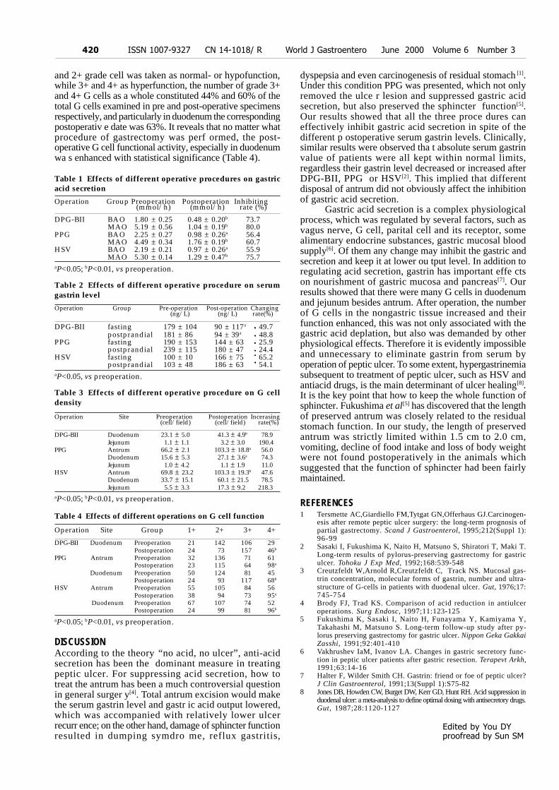
and 2+ grade cell was taken as normal- or hypofunction,while 3+ and 4+ as hyperfunction, the number of grade 3+and 4+ G cells as a whole constituted 44% and 60% of thetotal G cells examined in pre and post-operative specimensrespectively, and particularly in duodenum the correspondingpostoperativ e date was 63%. It reveals that no matter whatprocedure of gastrectomy was perf ormed, the post-operative G cell functional activity, especially in duodenumwa s enhanced with statistical significance (Table 4).
Table 1 Effects of different operative procedures on gastricacid secretion
Operation Group Preoperation Postoperation Inhibiting (mmol/h) (mmol/h) rate (%)
DPG-BII BAO 1.80 ± 0.25 0.48 ± 0.20b 73.7MAO 5.19 ± 0.56 1.04 ± 0.19b 80.0
PPG BAO 2.25 ± 0.27 0.98 ± 0.26a 56.4MAO 4.49 ± 0.34 1.76 ± 0.19b 60.7
HSV BAO 2.19 ± 0.21 0.97 ± 0.26a 55.9MAO 5.30 ± 0.14 1.29 ± 0.47b 75.7
aP<0.05; bP<0.01, vs preoperation.
Table 2 Effects of different operative procedure on serumgastrin level
Operation Group Pre-operation Post-operation Changing (ng/L) (ng/L) rate(%)
DPG-BII fasting 179 ± 104 90 ± 117a 49.7postprandial 181 ± 86 94 ± 39a 48.8
PPG fasting 190 ± 153 144 ± 63 25.9postprandial 239 ± 115 180 ± 47 24.4
HSV fasting 100 ± 10 166 ± 75 65.2postprandial 103 ± 48 186 ± 63 54.1
aP<0.05, vs preoperation.
Table 3 Effects of different operative procedure on G celldensity
Operation Site Preoperation Postoperation Incerasing (cell/field) (cell/field) rate(%)
DPG-BII Duodenum 23.1 ± 5.0 41.3 ± 4.9b 78.9Jejunum 1.1 ± 1.1 3.2 ± 3.0 190.4
PPG Antrum 66.2 ± 2.1 103.3 ± 18.8a 56.0Duodenum 15.6 ± 5.3 27.1 ± 3.6a 74.3Jejunum 1.0 ± 4.2 1.1 ± 1.9 11.0
HSV Antrum 69.8 ± 23.2 103.3 ± 19.3b 47.6Duodenum 33.7 ± 15.1 60.1 ± 21.5 78.5Jejunum 5.5 ± 3.3 17.3 ± 9.2 218.3
aP<0.05; bP<0.01, vs preoperation.
Table 4 Effects of different operations on G cell function
Operation Site Group 1+ 2+ 3+ 4+
DPG-BII Duodenum Preoperation 21 142 106 29Postoperation 24 73 157 46b
PPG Antrum Preoperation 32 136 71 61Postoperation 23 115 64 98a
Duodenum Preoperation 50 124 81 45Postoperation 24 93 117 68b
HSV Antrum Preoperation 55 105 84 56Postoperation 38 94 73 95a
Duodenum Preoperation 67 107 74 52Postoperation 24 99 81 96b
aP<0.05; bP<0.01, vs preoperation.
DISCUSSIONAccording to the theory “no acid, no ulcer”, anti-acidsecretion has been the dominant measure in treatingpeptic ulcer. For suppressing acid secretion, how totreat the antrum has been a much controversial questionin general surger y[4]. Total antrum excision would makethe serum gastrin level and gastr ic acid output lowered,which was accompanied with relatively lower ulcerrecurr ence; on the other hand, damage of sphincter functionresulted in dumping symdro me, reflux gastritis,
dyspepsia and even carcinogenesis of residual stomach [1].Under this condition PPG was presented, which not onlyremoved the ulce r lesion and suppressed gastric acidsecretion, but also preserved the sphincter function[5].Our results showed that all the three proce dures caneffectively inhibit gastric acid secretion in spite of thedifferent p ostoperative serum gastrin levels. Clinically,similar results were observed tha t absolute serum gastrinvalue of patients were all kept within normal limits,regardless their gastrin level decreased or increased afterDPG-BII, PPG or HSV[2]. This implied that differentdisposal of antrum did not obviously affect the inhibitionof gastric acid secretion. Gastric acid secretion is a complex physiologicalprocess, which was regulated by several factors, such asvagus nerve, G cell, parital cell and its receptor, somealimentary endocrine substances, gastric mucosal bloodsupply[6]. Of them any change may inhibit the gastric andsecretion and keep it at lower ou tput level. In addition toregulating acid secretion, gastrin has important effe ctson nourishment of gastric mucosa and pancreas[7]. Ourresults showed that there were many G cells in duodenumand jejunum besides antrum. After operation, the numberof G cells in the nongastric tissue increased and theirfunction enhanced, this was not only associated with thegastric acid deplation, but also was demanded by otherphysiological effects. Therefore it is evidently impossibleand unnecessary to eliminate gastrin from serum byoperation of peptic ulcer. To some extent, hypergastrinemiasubsequent to treatment of peptic ulcer, such as HSV andantiacid drugs, is the main determinant of ulcer healing[8].It is the key point that how to keep the whole function ofsphincter. Fukushima et al[5] has discovered that the lengthof preserved antrum was closely related to the residualstomach function. In our study, the length of preservedantrum was strictly limited within 1.5 cm to 2.0 cm,vomiting, decline of food intake and loss of body weightwere not found postoperatively in the animals whichsuggested that the function of sphincter had been fairlymaintained.
REFERENCES1 Tersmette AC,Giardiello FM,Tytgat GN,Offerhaus GJ.Carcinogen-
esis after remote peptic ulcer surgery: the long-term prognosis ofpartial gastrectomy. Scand J Gastroenterol, 1995;212(Suppl 1):96-99
2 Sasaki I, Fukushima K, Naito H, Matsuno S, Shiratori T, Maki T.Long-term results of pylorus-preserving gastrectomy for gastriculcer. Tohoku J Exp Med, 1992;168:539-548
3 Creutzfeldt W,Arnold R,Creutzfeldt C, Track NS. Mucosal gas-trin concentration, molecular forms of gastrin, number and ultra-structure of G-cells in patients with duodenal ulcer. Gut, 1976;17:745-754
4 Brody FJ, Trad KS. Comparison of acid reduction in antiulceroperations. Surg Endosc, 1997;11:123-125
5 Fukushima K, Sasaki I, Naito H, Funayama Y, Kamiyama Y,Takahashi M, Matsuno S. Long-term follow-up study after py-lorus preserving gastrectomy for gastric ulcer. Nippon Geka GakkaiZasshi, 1991;92:401-410
6 Vakhrushev IaM, Ivanov LA. Changes in gastric secretory func-tion in peptic ulcer patients after gastric resection. Terapevt Arkh,1991;63:14-16
7 Halter F, Wilder Smith CH. Gastrin: friend or foe of peptic ulcer?J Clin Gastroenterol, 1991;13(Suppl 1):S75-82
8 Jones DB, Howden CW, Burget DW, Kerr GD, Hunt RH. Acid suppression induodenal ulcer: a meta-analysis to define optimal dosing with antisecretory drugs.Gut, 1987;28:1120-1127
Edited by You DYproofread by Sun SM
420 ISSN 1007-9327 CN 14-1018/ R World J Gastroentero June 2000 Volume 6 Number 3

Enzymohistochemical study on burn effect on ratintestinal NOS
Qiu Gui Wang, Li Ya He, Ye Wen Chen and Song Lin Hu
Subject headings nitric oxide; burn; intestines; superoxidedismutase; MDA; GSH-PX
Wang QG, He LY, Chen YW, Hu SL. Enzymohistochemical study on burneffect on rat intestinal NOS. World J Gastroentero, 2000;6(3):421-423
INTRODUCTIONThe blood irrigate flow obstruction, especially thegastrointestinal (GI) ischemia[1], is the main factor ofthe damage to the digestive tract caused by serious burns.The effect of GI ischemia on the whole body is extensiveand profound, which not only causes the increase ofintestinal permeability and the movement of bacteria andtoxin in the intestinal cavity, but releases a large quantityof inflammatory media. Neuroendocrine element afterburns is closely related to intestinal damage[2]. As isknown at present, abundant nitric oxide synthase (NOS)is distributed in GI tract, whose product NO is anonadrenergic and noncholinergic (NANC) restrainingtransmitter in enteric nervous system which participatesextensively in various physiological functions in theintestinal tract[3]. Few reports about the effects on intestinalNOS after serious burns are available. We used scaldedrat model with degree III 40% of body surface area(TBSA), and enzymatic histological and biochemicalmethods to observe dynamically the active changes ofempty myenteric plexus NOS and changes of jejunal tissueMDA SOD and GSH-PX and probe into the relationshipbetween NOS and intestinal tissue and function damageas well as their mechanism so as to provide morphologicalexperimental basis for clinical treatment.
MATERIALS AND METHODSMaterialsB-NADPH, TYPE, nitroblue telrazolium (NBT) (Sigma);SOD reagent kit (Wuhan Xiehe Hospital Science andTechnology Development Centre); thibarbiluric acid(Shanghai Reagent Factory); self-made phosphate buffer;and healthy SD rats weighing 250 g provided by the AnimalLaboratory Centre of Tongji Medical University.
MethodsEighty rats were randomly divided into five groups:sham-burn control (SBC) and 4 groups of 8, 24, 48
and 72 hours postburn, each group having 16 rats (40were measured for biochemical indexes, and other 40for morphological indexes). The rats, before beingscalded, had their hair depilated on the neck and backwith 80 g/L Na2S under the anaesthesia with 100 g/Ltrichloroacetaldehy-demonohydrate after they becameconscious, depilated parts were soaked into the 100boiling water for 16 sec and scalded to degree III 400g/L TBSA. They were then fed in different cages withouttreat ment. And those in sham-burn control group weresoaked into 37 water. Rats in different groups, after being scalded, weresacrificed at different time points, a 10 cm empty intestinewas taken out and made into thick liquid. SOD activitywas determined with modified pyrogallol auto-oxidation[4]. GSH-PX activity by active DTNB[5], and the contentof MDA with modi fied thiobarbituric acid fluorescenceanalysis[6]. The rats in each group were anaesthetized with100 g/L trichloroacetaldeh y demonohydrate (300 mg/kg) , opened up to expose the heart, the blood was quickly rinsed away with 200 mL warm normal saline fromleft chamber through ascending aorta, and then 450 mLof 40 mL/L cold parafor-maldeh yde was instilled forone hour, a 10 cm empty intestine was taken out, theinte stinal content was washed off and filled again withthe same liquid to make the intestinal track full, bothends were ligated and fixed in the above-mentioned liquid(4 ) for 4 hours. The outer longitudinal tunicamuscularis was peeled off care fully from the ring tunicamuscularis and tela submucosa, and the specimens weremade. The specimens after being washed with 0.1 mol/Lof PBS, was put into re duction type NADPH-d hatchingliquid at 37 for 50 min (cosisting of 0.1 mo l/L of PB(pH 8.0) which contained 30 g/L tritonx-100, 100 mol/Lof nitroblue telbrazolium and 1 g/L of NADPH-d, andwashed thoroug hly with 0.1 mol/L of PBS. After reaction[7], it was pasted onto a galative glass piece andconventionally dehydrated, made into transparency,sealed and observed under microscope. The stainingresult of NOS was achieved using IBAS automaticpicture analyzer to determine semi-quantitatively thecontents in myenteric plexus neuron NOS and internodebind NOS.
Statistical treatmentThe data were expressed as x ± s, and t test was usedto compare the results.
Department of Neurobiology Xianning Medical College, Xianning437100, Hubei Province, ChinaCorrespondence to: Qiu Gui Wang, Department of NeurobiologyXianning Medical College,Xianning 437100, Hubei Province, ChinaReceived 2000-01-05 Accepted 2000-03-16
PO Box 2345, Beijing 100023, China World J Gastroentero, 2000; 6(3):421-423Fax: +86-10-85381893 World Journal of GastroenterologyE-mail: [email protected] www.wjgnet.com Copyright 2000 by the WJG Press ISSN 1007-9327

RESULTSChanges of the contents of MDA, SOD, GSH-PXin the empty intestinal tissues of burned ratsThe content of MDA in the empty intestinal tissuesgradually rose with the time of burns; compared withthe SBC, the contents in each group increased significantly (P<0.01). But the activity of SOD and GSH-PXdecreased markedly the difference being significant ascompared with SBC (P<0.01).
Active change of myenteric plexus NOS in theempty intestine of burned ratsObservation under light microscope In the controlgroup, most ganglion cells in myenteric plexus andtheir scabrosity appeared strongly positive in NOS,while a small number had moderate staining, mostbeing distributed around myenteric plexus and thecell being comparatively large and various in shape,i.e., ovate, triangle or irregular. There were NOSpositive products in dark blue in the cytoplasm andthe nucleus was negative. Most of intersegment bindfiber contained bulge, and some had division andintersect with varying size (Figure 1). In the group
of 8 hours post-burn, the cell NOS stain appearedobviously lighter but intersegment bind fiber hadno obvious change (Figure 2). In the 24 hourspost-burn group, ganglion cell NOS staining wasintensified obviously with clear cell outline andlarger size. Intersegment bind fiber and bulgebecame markedly dense (Figure 3). In the 48 to72 hour post-burn groups, ganglion cell andintersegment bind fiber staining gradually becamelighter, especially in those of 72 hour group (Figure4), perikaryon and OD change of myenteric plexusNOS in the empty intestine of burned rats. Inburned rats after 8 hours the OD of myentericplexus and intersegment bind fiber NOS weresignificantly lower than that in the control group(P<0.01) and in those after 24 hours, the OD ofganglion cells and intersegment bind fiber NOSwas significantly higher than that in the control (P<0.01); while in those after 48 to 72 hours, OD ofganglion cells and intersegment bind fiber NOSdecre ased gradually, being significantly differentfrom the control group and those of 24 hours post-burn (P<0.01).
Figure 1 Myenteric plexus neuro bind ganglion cell ( ) and positive reaction of NOS of intersegmental bundle ( ) in the control group.× 400Figure 2 Myenteric plexus neuro bind ganglion cell ( ) and positive reaction of NOS of intersegmental bundle ( ) reduced obviously inthe group of 8 hours post-burn. × 400Figure 3 Myenteric plexus neuro bind ganglion cell ( ) and positive reaction of NOS of intersegmental bundle ( ) increased obviously int he group of 24 hours post-burn. × 400Figure 4 Myenteric plexus neuro bind ganglion cell ( ) and positive reaction of NOS of intersegmental buridle ( ) ieduced obviously inth e group of 48 hours post-burn compared with the group of 24 hours post-burn. × 400Figure 5 Myenteric plexus neuro bind ganglion cell ( ) and positive reaction of NOS of intersegmental bundle ( ) reduced obviously inthe group of 72 hours post-burn. × 400
422 ISSN 1007-9327 CN 14-1018/ R World J Gastroentero June 2000 Volume 6 Number 3

Wang QG, et al. Burn effect on rat intestinal NOS 423
DISCUSSIONNO, a newly discovered active biosubstance in recentyears, is a dual-functional messenger and venenositymolecule with short half-life and instable nature. Thein vivo NO, in the form of L-Arg as primer, existsthrough the catalyza tion of NOS, which indicates thatthe distribution of NOS is closely related to thephysiological functions of NO. It is reasonable to inferbioeffects of NO through the research of NOS, Nathanet al[8] divided NOS into two types, i.e. cNOS andiNOS. cNOS is mainly distributed in neurocyte andendothelium and catalyzes NO which acts chiefly asneurotransmitter and secondary messenger, whileiNOS, mainly distributed in macrophage andendothelium cell, catalyzes NO, which has cytotoxiceffect. However, the physiological effect of NO to thebody depends chiefly on the quality and strength ofstimulat ive elements, the dosage and reactive sites.Thus we can presume that after serious burns badperfusion of intestinal blood, disorder of motor functionsand damaged enteromycoderm may be related to theabnormal intestinal NOS activit y. It was suggestedthat MDA content in the jejunal tissues of the burnedrats increased, the result was consistent with that ofPeng et al[9]. At the same time, SOD and GSH-PXantioxidase activities in jejunal tissues decreased afterbeing burned, indicating that lipoperoxidation reactionsparticipate in the pathophysiological course of themucosal damage of burned rats. Some studiesdemonstrated that cNOS was distributed extensivelyin plexuses of GI wall, and its output NO was inhibitoryneurotransmitter of NANC nerves in GI tract, whichmay cause angiectasis of intestinal tract and laxationof smooth muscles[10]. Eight hours after serious burnsmyenteri c plexus NOS activity decreased obviously inour experiment suggesting burn stress causedadrenergic nerve excitation, and inhibited NANCnerves, leading to the decrease of cNOS activity andthe catalyzed NO, causing excessive contrac tion ofsmooth muscles. These may be the main causes in badperfusion of intesti nal blood and disorder of motorfunctions. In this experiment, 24 hours post-burn,myenteric plexus NOS activity increased significantly,possibly because the enterogenous infection afterserious burn, activated the endotoxin on macrophagecell of intestine to release a series of such body fluid agentsas TNT IL-1, etc, leading to increased iNOS
activity. A large amount of high-concentr ation NOproduced in this way had cytotoxic effects which furtherdamaged the intestinal tissues. Excessive amounts ofNO and O-
2 free radicals produced more toxic ONOO-
free radicals which further react to produce HO-,NO2,NO-
3, etc. while HO- and NO2 are catalyst ofbiomembrane lipid peroxidat ion and can cause asuccession of peroxidation of high unsaturation andadipoaci d occurring on the membrane, thus producing alarge amount of metabolin MDA wors ening the damageof intestinal tissues. Therefore 48-72 hours post-burn,we fou nd that intestinal myenteric plexus ganglion celland perikaryon reduced obvious ly, and changes causedby the lowered NOS activity and other delayed neuronaldamage. It can be inferred that NO in the intestine ofseriously burned rats and ONOO- reacted by O-
2 areimportant mechanisms that NO damages intestin alstructure and functions. It suggests that the damagedstructure and functi ons in seriously burned rats arerelated to the increase of NOS activity. It is concludedthat using the inhibitor of NOS to lower NOS activitylevel may be beneficial to lessening the degree of post-burn intestinal damage.
REFERENCES1 Cui XL, Sheng ZY, Guo ZR, He LX, Zhao J, Ren XW. Mechanisms
of early gastrointestinal ischemia after burn: hemodynamic andhemorrheologic features. Zhonghua Zhengxing Shaoshang WaikeZazhi, 1998;14:262-265
2 Hu DH, Chen B, Lin SX, Tang CW. Observation and analysis ofpostburn changes in substancep (sp) and sp peptidergic nerve fibersin the intestine of rat. Chin J Plast Surg Burns, 1996;12:93-97
3 Rand MJ. Nitrinergic transmission: nitric oxide as a mediator ofnon-adrenergic, on cholinergic neuro effector transmission. ChomEXP Pharmacol Physio, 1993;19:147
4 Deng BY, Yuan QS, Li WJ. Superoxide dismruase assayed by modi-fied pyrogallolaut oxidation mothod. Progress Biochem Biophysics,1991;18:163
5 Xia YM, Zhu LZ. The datermination of glutathione peroxidase inblood and tissue-DTNB direct method. Weisheng Yanjiu, 1987;16:29-33
6 Zhang XM, Yan LJ, Chai JK, Zhou WQ, Wang LX. Improvementof the thiobarbituric acid fluorescence analysis of serumlipoperoxides. Progress Biochem Biophysics, 1996;23:175-179
7 Ding YQ, Lou XF, Wang YQ, Qin BZ. The distribution of nitricoxide synthase positive neurons and fibers and the origin ofthe positive fibers in the rat spinal cord. Chin J Neuroanat,1993;9:81-87
8 Nathan C, Xie QW. Nitric oxide synthase Rdes tolls and controls.Cell, 1994;78:915
9 Peng YZ, Xiao GX, Wang DW, Zhang YP, Qin XJ. Role of lipidperoxidation in the mechanism of bacterial translocation fromthe gut after severe thermal injury in rats. Disan Junyi DaxueXuebao, 1989;11:329-334
10 Nichols K, Kramtis A, Staines W. Histochemical localization of nitricoxide-synthesizing neurons and vascular sites in the guinea pig intestine.Neuroscience, 1992;51:791-799
Edited by Ma JY

Protective effect of electroacupuncture andmoxibustion on gastric mucosal damage and itsrelation with nitric oxide in rats
Wen Fen Pei, Guan Sun Xu, Yong Sun, Shun Li Zhu and Dao Qin Zhang
Subjects headings acupuncture and moxibustion; nitricoxide; gastric mucosal damage; gastric mucosal bloodflow; transmucosal potential difference; rats
Pei WF, Xu GS, Sun Y, Zhu SL, Zhang DQ. Protective effect ofelectroacupuncture and moxibustion on gastric mucosal damage andits relation with nitric oxide in rats. World J Gastroentero, 2000;6(3):424-427
INTRODUCTIONGastric mucosal injury is one of the common disorders,there are many reports subjected to its pathogenesistreatment and prevention[1-4]. We investigated theprotective effect of electroacupuncture andmoxibustion of Zusanli point on gastric mucosaldamage and its relation with nitric oxide (NO) on theanimal model with acute gastric mucosal damageinduced by ethanol. The detected indexes include thecontent of NO, gastric mucosal blood flow (GMBF),gastric mucosal lesion index (LI) and transmucosalpotential difference (PD).
MATERIAL AND METHODSDrugs L-arginine (L-arg)—the precursor ofN O ( p u r c h a s e d f r o m H u a m e i B i o l o g i c a lEngineering Corporation); Natrii Nitroprussidum( S N P ) — t h e d o n o r o f N O ( p r o v i d e d b yExperimental Drug Plant of the Beijing Instituteof Pharmaceut ica l Indust ry) , d i lu ted wi thdistilled water prior to use, Nw-nitro- L-arginine(L-NNA), an inhibitor of NOS (produced bySigma), and diluted with 0.2 mol·L-1 PBS; 200 g·L-1
pentobarbital sodium was prepared with distilledwater immediately before use.
Animal groupingWistar rats weighing 180 g-250 g (provided by theInstitute of Acupunc ture and Moxibustion, Anhui Collegeof T r a d i t i o n a l C h i n e s e M e d i c i n e ) w e re
fasted for 12 h and allowed free access to water. Theanimals were divided into 16 groups randomly, including:control group (c), model (m),m+electroacu puncture(EA), m+moxibustion (M), L-arg+m , SNP+m, L-NNA+m, L-NNA+L-arg+m, L-arg+m +EA, L-arg+m+M, SNP+m+EA, SNP+m+M, L-NNA+m+EA,L-NNA+m+M, L-NNA+L-arg+m+EA, L-NNA+L-arg+m+M.
Preparation of acute gastric mucosal damagemodel[5]
The rats fasted for 12 h were injected with pentobarbitalintraperitoneally at a dose of (30-40) mg·Kg-1. Themiddle line incision below xiphoid process afteranesthesia was performed, then stomach was exposedand injected with 2 mL 700 mL/L ethanol, and theabdomen closed, 30 min later the preparation of modelwas fulfilled.
Experimental procedureDifferent procedures were used for different groups.In groups injected drugs or equivalent amount ofphysiological saline, these were injected respectivelyinto blood flow slowly through the great saphenousvein of the anesthetized rats 15 min before thepreparation of the model. In groups which requiredelectroacup uncture or moxibustion, were treated withelectroacupuncture or moxibustion on bilateral Zusanlipoints after the model was successfully prepared. Theelectroacupuncture was performed with PCE2
electroacupuncture therapy device on the conditionof frequency 20Hz, voltage 5v-8v for 30 min(produced by the Institute of Acupuncture andMoxibustion of Anhui College of Traditional ChineseMedicine). During moxibustion, the lighted pure moxa-cigar was ma intained 1 cm from the bilateral Zusanlipoints. The GMBF and PD values were measuredfor all rats. Then the blood was obtained throughdecephaliza tion or from retro-ocular vassel for theNO assay. Subsequently the rat stomach wasresected for the analysis of LI, and the mucosa fromgastric antrum and the body for assaying NOcontents.
Index detectionGMBF was assayed by analyzing hydrogen gasclearance curves[6]; PD was assayed with direct Ag-
Department of Physiology, Anhui College of Traditional ChineseMedicine (TCM), Hefei 230038, Anhui Province, ChinaDr. Wen Fen Pei, graduated from the Department of Medicine, AnhuiMedical College in 1976. Associate professor, engaged in study ofdigestive physiology, having more than 10 papers published.Supported by National Advanced Project of China (JL-93012)Correspondence to: Dr. Wen Fen Pei, Department of Physiology,Anhui College of TCM, Hefei 230038, Anhui Province, ChinaTel. 0086-551-2823237Received 2000-01-25 Accepted 2000-03-02
PO Box 2345, Beijing 100023, China World J Gastroentero, 2000; 6(3):424-427Fax: +86-10-85381893 World Journal of GastroenterologyE-mail: [email protected] www.wjgnet.com Copyright 2000 by the WJG Press ISSN 1007-9327

AgCl. Electrode measurement technique[7]with fewalterations, i.e. the efficient electrode was put onthe mucosa while the reference one on the serosa.The electric potential difference between twomembranes was recorded. For the detection of LI,the stomach was resected and incised along thegreater curvature, washed with physiological saline.Guth index assessment[8] (slightly modefied) wasused to compute the damage, on a scale of grades1-5 as follows: Grade 1, the petechia or ecchymosis,Grade 2, 3, 4 and 5, pathological focus of 1 mm; 1mm-2 mm, 2 mm-4 mm, and over 4 mm,respectively. The NO content in blood and mucosaof gastric antrum and body were assayed based onthe method from Green et al[9]. The Kit for NOassay was provided by the Department ofBiochemistry, Institute of Radiology, Academy ofMedical Science of PLA. The analysis was basedon the protocol, but the mucosa were hom ogenizedbefore the assay.
Statistical analysisAll data were expressed as x ± s, and the Student’st test was used for the comparison betweengroups.
RESULTSThe effect of acupuncture and moxibustion ofZusanli point on GMBF, PD and LI in rats with thegastric mucosal damage (Table 1)The mean of GMBF, PD in group m showedstatistically significant difference comp aredwith the control group (P<0.01), demonstratingmarked dec rease o f GMBF and PD a f t e rgastric mucosal damage, the integrity of gastricmucosa was depended on adequate blood flow.GMBF and PD in group m+M was obviouslyincr eased (P<0.01) compared with group m,s u g g e s t i n g t h e e l e c t r o a c u p u n c t u r e a n dmoxibustion of Zusanli point may relieve andcure the gastric mucosal damage induced byethanol.
Changes of NO content in blood and gastricmucosa and their relat ion with LI afterelectroacupuncture and moxibustion on theZusanli point of rat modelsFrom Table 1 we can find, NO contents in groupm showed significant difference compared withnormal control (P<0.01); LI in group m+EA andgroup m+M was markedly lower than that in groupm (P<0.01). The results showed that the gastricmucosal damage aggravated as NO contentdecreased while NO cont ent increased and thegastric mucosal damage relieved, which suggeststhat NO content is associated with the integrity ofgastric mucosa.
Effect of L-NNA, L-arg and SNP on the responseof electroacupuncture and moxibustion(Table 1)The L-Arg (150 mg·Kg-1), SNP(200 µg·Kg-1) andL-NNA (3 mg·Kg-1) were administered through thegreat saphenous vein respectively 15 min before igadministration of 700 mL/L ethanol in differentgroups. After that the electroacupuncture andmoxibustion of Zusanli point were performed for 30min. Subsequently, the GMBF, PD and LI as wellas NO content in blood and gastric mucosa wereassayed and detected respectively in each group. Itwas found that there was little difference in theindexes when group L-arg +m and group SNP+mwere compared with group m+EA or group m+M (P<0.05), indicating the function of electroacupunctureand moxibustion on gastric mucosal damage wassimilar to that of L-arg or SNP. Comparing groupL-Arg+m+EA (or M) and group SNP+m+EA (orM) with group m+EA (or M), and group SNP+m+EA(or M) with group m+EA (or M), NO content, GMBFand PD values were markedly increased, and LIvalue was obviously decreased (P<0.01 or P<0.05),which suggested that electroacupuncture andmoxibustion of Zusanli point had the effect ofstrengthening NO pathway and protecting gastricmucosa. In comparison of group L-NNA+m with group m.,NO content, GMBF and PD value dec reased,and LI value increased (P<0.01 or P<0.05),showing tha t L-NNA aggrava ted e thano linduced gastric mucosal damage. When wecompared group L-NNA+m+EA (or M) withgroup m+EA (or M), NO content, GMBF andPD value also decreased, but LI value increased(P<0.01 or P<0.05), which suggests L-NNAd e c r e a s e d t h e p r o t e c t i v e a c t i o n o felectroacupunctureand moxibustion on gastricdamage. While we compared group L-NNA +L-arg +m+EA (or M) with group L-NNA+m+EA(or M), NO contents, GMBF and PD valueincreased and LI value decreased (P<0.01 or P<0.05), demonst rating L-arg may reverse theinhibitory function of L-NNA. In conclusion, theabove me ntioned results further suggest thatthe protective action on gastric mucosa by theelectroacupuncture and moxibustion of Zusanlipoint is NO mediated.
Effect of electroacupuncture of Zusanli point onpH value of gastric fluid (Figure 1)pH in group c was 4.33 ± 0.40 and that in Group m2.92 ± 0.37, decreasing si gnificantly in the lattergroup (P<0.01). The pH value in Group m+EA,3.84 ± 0.69 was much higher than that in Group mindicating 700 mL/ L ethanol promotes the stomachto secrete acids while electroacupuncture inh ibits thesecretion.
Pei WF, et al. Effect of electroacupuncture and moxibustion on gastric mucosa 425

Figure 1 Effect of electroacupuncture on pH value with gastricmucosal damage in rats.aP<0.01 vs Group C; bP<0.01 vs Group m.
DISCUSSIONAcupuncture and moxibustion may treat and preventgastrointestinal disorders, the mechanism of which isbeing further investigated. The acupuncture and moxibustion on Zusanli points show the two-way regulation ofgastrointestinal funct ion. In recent years, it is frequentlyreported that they may relieve symptom an d promotethe ulcer healing to certain extent[1-4]. There is relativepoint specificity, the first choice is the Zusanli point instomach channel of foot yangming. Our study investigatedthe therapeutic and protective effect of the acupunctureand moxibustion on acute gastric mucosal damageinduced by 700 mL/L ethanol. It is found that
GMBF and PD value as well as NO content, pHvalue in rat gastric mucosa of model group withacupuncture and moxibustion increased moremarkedly than those without any therapy (P<0.01),while the gastric mucosal damage alleviated (P<0.01) which denotes the therapeutic and protectiveeffect of acupuncture and moxibustion on thedamage and suggests that the protective action isrelated with the level of NO content. NO is a small molecular gas produced from theprocursor L-Arg under the NOS catalysis. Theprocess is called as L-Arg-NO pathway, NOSactivity plays a key role in NO synthesis. NOSis existed in many kinds of tissues includingvascular endothelial cell, thrombocyte, brain cell,r e n a l e p i t h e l i a l c e l l , m a c r o p h a g o c y t e ,neutrophil, hepatocyte, etc. Some factors caninduce the expression of NOS and increase theNO synthesis. As a special bioinformationalmolecule, the neurotransmitter or the humoralfactor, NO involves in the functional regulationand pathophysiology of many diseases. Ourstudy investigates the prot ective effect ofacupuncture and moxibustion on acute gastricm u c o s a l d a m a g e a n d i n t h e m e a n t i m emeasures NO content in blood and gastr icmucosa and finds that NO content is negativelycorrelated with the severity of gastric mucosaldamage, which proves that the protective actionof acupuncture and moxibustion of Zusa nlipointi s NO media ted ; and in o ther words NOinvolves in the regulative effect of acupuncture and
Table 1 Effect of electroacupuncture and moxibustion of Zusanli point on LI, GMBF, PD and NO after gastric mucosal damage inrats (x ± s)
NOGroup n LI GMBF PD (mL·min-1·100g-1) (mV) Blood (µmol·L-1) Antrum(ng·mg-1)
C 5 0 138.20 ± 4.37 20.82 ± 0.99 21.12 ± 1.89 53.30 ± 2.65m 5 45.6 ± 3.2a 60.60 ± 6.09a 11.85 ± 0.82a 12.84 ± 1.54a 35.17 ± 1.57a
m + EA 5 31.4 ± 3.3c 92.55 ± 6.35c 15.84 ± 0.48c 18.65 ± 0.69c 48.51 ± 2.12c
m + M 5 33.8 ± 2.4c 95.91 ± 5.59c 15.96 ± 0.34c 18.53 ± 1.04c 48.49 ± 2.39c
L-arg + m 5 32.4 ± 3.2 106.22 ± 5.30 17.73 ± 0.22 18.93 ± 1.30 47.57 ± 2.86SNP + m 5 30.0 ± 3.1 104.03 ± 7.57 17.89 ± 0.39 19.07 ± 1.29 47.18 ± 2.17L-NNL + m 5 55.8 ± 2.8b 56.85 ± 5.96d 6.97 ± 0.32c 8.37 ± 0.27b 27.29 ± 1.71b
L-NNA + L -arg + m 5 46.8 ± 2.6 78.90 ± 5.10 10.93 ± 0.39 12.10 ± 1.23 33.76 ± 2.44L-arg + m + EA 5 21.0 ± 0.9e 121.50 ± 3.55d 18.14 ± 0.44 24.70 ± 0.75d 50.63 ± 2.34L-arg + m + M 5 27.1 ± 2.7d 97.46 ± 4.90 16.08 ± 0.45 21.06 ± 1.17 44.26 ± 2.01SNP + m + EA 5 20.9 ± 0.8e 118.50 ± 4.21d 17.79 ± 0.59 24.80 ± 0.50d 45.17 ± 2.01SNP + m + M 5 26.8 ± 2.2d 101.11 ± 5.23 14.73 ± 0.61 23.14 ± 1.52d 43.33 ± 1.97L-NNA + m + EA 6 41.8 ± 2.1e 73.57 ± 5.43d 11.87 ± 0.41d 12.37 ± 1.78e 39.25 ± 1.66d
L-NNA + m + M 6 40.5 ± 3.0d 66.42 ± 5.42e 10.25 ± 0.72e 11.43 ± 1.03e 32.17 ± 2.35e
L-NNA + L-arg + m + EA 5 31.0 ± 1.2g 95.00 ± 3.78f 16.15 ± 0.74g 19.24 ± 0.54g 51.51 ± 2.14f
L-NNA + L-arg + m + M 5 31.5 ± 2.1g 70.76 ± 4.78 11.81 ± 0.53 12.88 ± 0.68 36.37 ± 2.29
aP<0.01 vs Group C; bP<0.05, cP <0.01 vs Group m; dP<0.05, eP<0.01 vs Group m+EA (orM); fP<0.05, gP<0.01 vs Group L-NNA+m+EA (or M).
426 ISSN 1007-9327 CN 14-1018/ R World J Gastroentero June 2000 Volume 6 Number 3
moxibustion on gastrointestinal function. The good balance between NO and ET keeps theendothelium intact, If the unbal ance is developed, thegastric mucosal damage will be formed and the gas tricdysfunction presented. Our study showed[1] that whenNO content decreased ET content increased and gastricmucosal damage aggravated, and on the contrary, thedamage alleviated. Our results have verified it. WhenL-arg or SNP was administered 15 min beforeestablishing the model with 700 mL/L ethanol; NOcontent was higher than that in the model group andconcurrently GMBF and PD value increased, and LIvalue decreased; if NOS inhibitor -L-NNA was given,NO content, GMPF and PD value were lower than thosein the model group while LI value increased, whichindicates that NO plays an important role in maintainingthe intact gastric mucosa. GMBF decrease mayprominently respond to the gastric mucosal damage afterethanol induction. It is thought that NO is an EDRF,which is capable of relaxing the vascular smooth muscleobviously, Our research further confirmed that thevascular relaxing action of NO can protect gastricmucosa from damage. NO initiates the protective effect of acupunctureand moxibustion on gastric mucosa, which has beenproved in our work. During experiment, in the modelrats , the NO content in their blood and gastricmucosa increased after acupuncture and moxibustionwere performed, and GMBF and PD values werealso increased while LI value decreased. If L-NNAwas preliminarily given, the above effect ofacupuncture and moxibustion of Zusanli pointdisappeared, but concurrently given L-ary, the actionof L-NNA could be reversed. In addition, if L-argor SNP was preliminarily administered and then themodel established, NO content in the group withthe acupuncture and moxibustion of Zusanli pointincreased more than that in the corresponding groupswithout acupuncture and moxibustion, moreover,GMBF and PD values increased and LI decreasedcorrespondingly, showing the synergetic actionbetween them. The above phenomena furtherconfirm that the acupuncture and moxibustion ofZusanli points alliviate gastric mucosal damage,which is NO mediated, and this illustrates that theacupuncture and moxibustion of Zusanli point canactivate endogenous NOS.
As far as the possible mechanism why Zusanlipoint can initiate NO system is concerned, we considerthat the main nerves regulating gastric function are vagusnerves, the dorsal vagus nucleus (DMV) being the majormotor nucleus. The efficient stimulus transmitted toDMV by the somatic sensory nerves throu gh the spinalcord. DMV also receives afferent information fromgastro intestinal tract and integrates other informationfrom central nervous system (CNS), and transfers tothe stomach by efferent vagus nerves, resulting in theincrease of NO synthesis and release. The possible mechanism about how NO canprotect gastric mucosa from damage is as follows: NOacts as one of endothelial diastolic factors which cancause the dilatation of vascular smooth muscles, resultingin GMBF increase, improvement of blood supply ofgastric mucosa, maintaining the integrity of gastricmucosal epithelium, protecting the gastric mucosa fromthe stimulation of gastric contents and from damage,preventing from H+ invasion and keeping normal ionicconcentration gradient across gastric mucosa as well asmaintaining appropriate PD value, while acupuncture andmoxibustion may activating NOS to increase thesynthesis and release of NO. Further research remainsto be done as how does the acupuncture and moxibustionactivate NOS.
REFERENCES1 Xu GS, Wang ZJ, Zhu SL, Chen QZ, Jiao J, Zhang DQ. Nitric Oxide
participates in protective effects of acupuncture on gastric mu-cosal damages in rats. Anhui Zhongyi Xueyuan Xuebao, 1996;15:36-38
2 Ma TF, Yang Z. Therapeutic effect and mechanism of acupunc-ture on digestive tract disease. In: Ed Zhou Lu. Gastrointestinalphysiology. Peking: Science press, 1991:755-772
3 Qiao XF, Yin KJ. Therapeutic effect and mechanism of moxibus-tion on experimental gastric ulcer in tats. Zhenci Yanjiu, 1992;17:270-273
4 Xu GS, Zhang QQ, Liu WZ, Leng JP. Effect and mechanism ofMoxibustion on gastric electric activity in rabbits. Zhenci Yanjiu,1992;17:274-276
5 Masuda E, Kawano S, Nagano K, Tsuji S, Takei Y, Tsujii M, OshitaM, Michida T, Kobayashi I, Nakama A, Fusamoto H, Kamada T.Endogenous nitric oxide modulates ethanol induced gastric mu-cosal injury in rats. Gastroenterology, 1995;108:58-64
6 Livingston EH, Reedy T, Leung FW, Guth PH. Computerizedcurve fitting in the analysis of hydrogen gas clearance curves. AmJ Physiol, 1989;257:G668-675
7 Xu GS,Sun Y,Wang ZJ,Zhang DQ, Gu XJ. Effects of electroacupunctureon gastric mucosal blood flow and transmucosal potential difference instress rats. Huaren Xiaohua Zazhi, 1998;6:4-6
8 Guth PH, Aures D, Paulsen G. Topical aspirin plus HCl gastriclesions in rat. Gastroenterology, 1979;76:88-93
9 Green LC, Tannenbaum SR, Goldman P. Nitrate synthesis in thegermfree and convertional rat. Science, 1981;212:56-58
Edited by You DYproofread by Sun SM
Pei WF, et al. Effect of electroacupuncture and moxibustion on gastric mucosa 427

Research of Helicobacter pylori infection inprecancerous gastric lesions
Xiao Qiang Zhuang1 and San Ren Lin2
Subject headings Helicobacter pylori; precancerousgastric lesions
Zhuang XQ, Lin SR. Research of Helicobacter pylori infection inprecancerous gastric lesions. World J Gastroentero, 2000;6(3):428-429
INTRODUCTIONHelicobacter pylori (Hp) infection has been consideredto play significant roles in pathogenesis of peptic ulcer.Additionally Hp is associated with the development ofgastric epithelial hyperplasia and lymphoid malignancies.The International Agency for Research on Cancer hasclassified Hp as a class I carcinogen and a definite causeof gastric cancer in humans. Hp infection first causeschronic active gastritis and may slowly lead to infectionof whole stomach. In the late stages of infection, mucosalatrophy and intestinal metapl asia (IM), and evendysplasia (DYS) occur[1].Chronic atrophic gastritis(CAG), IM and DYS are considered markers fordevelopment of gastric cancer in high-risk individuals.In our study we analyzed Hp infection prevalence in486 patients with precancerous gastric lesions.
MATERIALS AND METHODSThe mucosal biopsy specimens were collected from 486patients subjected to routi ne gastroscopy, including 163cases of CSG, 207 cases of CAG, 71 cases of IM and45 cases of DYS. Biopsies were taken from five sites inthe stomach: one fro m the angel, four from the lesserand greater curvature of gastric body and gast ric antrum.Each biopsy was classified according to the presence orabsence of C SG, CAG, IM and DYS, and scanned byWarthin-Starry method. Data analysis was made with Chi-square test.Statistical significance was defined as P<0.05.
RESULTS
Table 1 Relation between precancerous gastric lesions andage and sex
CSG CAG IM DYSVariables n n % n % n % n %
SexMale 314 96 30.6 142 45.2 43 13.7 33 10.5Female 172 67 38.9 65 37.8 28 16.3 12 7Age (yrs)
40 184 64 34.9 98 53.5 13 7.0a 9 4.6b
41-55 171 52 30.3 70 41.2 30 17.3 19 10.856 131 40 30.5 46 35.4 28 21.1a 17 13.0b
aP<0.05, bP<0.05.
Gastric pathology data was available for 486cases. As shown in Table 1, there was no significantdifference between the two sexes (P>0.05), but theprevalence rates increased with age, being significantlyhigher in 56 age group than 40 group for IM andDYS (P<0.05).
Table 2 Hp infection rate in precancerous gastric lesions
Hp(+) Hp(-)Variables n n % n %
CSG 163 39 23.9 124 76.1CAS 207 88 42.5a 119 57.5IM 71 54 76.1b 17 23.9DYS 45 40 88.9c 5 11.1
aP<0.05, CAG vs CSG; bP<0.01, IM vs CSG; cP<0.01, DYS vs CSG.
The prevalence of Hp increased steadily withincreasing severity of gastric histopathology (Table 2).The detection rates of Hp in IM (76.1%) and DYS (88.9%) were significantly higher than that in CSG (23.9%,P<0.01), and in CAG than in CSG (P<0.05).
Table 3 Hp infection distribution in precancerous gastriclesions by biopsy sites
ALC AGC A BLC BGCVariables n n % n % n % n % n %
CSG 39 37 94.9 36 92.3 32 82.1 18 46.2 10 25.6CAG 88 84 95.4 80 90.9 78 88.6 53 59.9 41 46.2IM 54 49 90.7 46 85.2 40 74.1 34 62.7a 32 58.8a
DYS 40 36 90.0 33 82.5 30 75.0 28 70.0b 26 63.9b
ALC:antral lesser curvature; AGC:antral greater curvature; A:angulus ; BLC:body, lesser curvature; BGC:body, greater curvatureaP<0.05, IM vs CSG; bP<0.05, DYS vsCSG.
1Department of Gastroenterology, General Hospital, of GuangzhouCommand Area, Guangzhou 510010, Guangdong Province, China2Department of Gastroenterology, The Third Hospital of Beijing MedicalUniversity, Beijing 100083, ChinaDr. Xiao Qiang Zhuang, associate professor Master of Gastroenterology,having 31 papers published.Correspondence to: Xiao Qiang Zhuang, Department of Gastroenterology, General Hospital of Chinese PLA Guangzhou Command Area,Guangzhou 510010, ChinaTel. 0086-20-86664097Received 2000-02-05 Accepted 2000-03-18
PO Box 2345, Beijing 100023, China World J Gastroentero, 2000; 6(3):428-429Fax: +86-10-85381893 World Journal of GastroenterologyE-mail: [email protected] www.wjgnet.com Copyright 2000 by the WJG Press ISSN 1007-9327

As shown in Table 3, the prevalence of Hppositivity tended to increase from BLC to BGC, Hppositive rates in IM and DYS were significantl y higherthan that in CSG in both BLC and BGC (P<0.05).
DISCUSSIONThe first compelling evidence linking Hp infection togastric carcinoma was generated by seroepidemiologicstudies[2,3], bacterial seroposit-ivity was significantlymore common in those with gastric adenocarcinoma,with an odds ratio ranging from 2.8 to 6.0, suggestinga strong association between Hp and gastricmalignancy. Hp infection was related to both the intestinal anddiffuse types of cancer as well as the precancerouslesion of IM or DYS[4], and greater than 70% of gastriccarcinomas are linked to IM, although DYS may alsobe seen without n eoplastic disease. Our study demostrated that the prevalence ratesof precancerous lesions varied w ith age, for IM andDYS, it had an upward trend with aging, while forCSG, it had a downward trend with aging, but werenot different between the two sexes.The prevalence
rose steadily with increasing severity of gastrichistopathology, the detection rates of Hp in CAG (42.5%), IM(76.1%) and DYS (88.9%) wer e significantlyhigher than that in CSG (23.9%), suggesting that Hp mayplay a role in late as well as early stages of carcinogenesis. Our study also showed that Hp positive rates inIM and DYS were significantl y higher than that in CSGin the lesser and greater curvature of gastric body, suggesting the more serious the gastric histopathdogy, thehigher the Hp infe ction rate, furthermore, the higherlevel of Hp infection site. The stud ysuggests we shouldtake multiple site biopsy for histopathology and Hp examination.
REFERENCES1 Kuipers EJ.Helicobacter pylori and the risk and management of
associated diseases: gastritis,ulcer disease, atrophic gastritis andgastric cancer. Aliment Pharmacol Ther, 1997;11(Suppl 1):71-88
2 Nomura A, Stemmermann GN, Chyou PH, Kato I, Perez-Perez GI,Blaser MJ.Helicobacter pylori infection and gastric carcinoma amongJapanese Americans in Hawaii. New Engl J Med, 1991;325:1132-1136
3 Parsonnet J, Friedman GD, Vandersteen DP, Chang Y, VogelmanJH, Orentreich N, Sibley RK. Helicobacter pylori infection and therisk of gastric carcinoma. New Engl J Med, 1991;325:1127-1131
4 Craanen ME, Dekker W, Blok P, Ferwerda J, Tytgat GN. Intestinalmetaplasia and Helicobacter pylori: an endoscopic bioptic study of thegastric antrum. Cut, 1992;33:16-20
Edited by Zhu LHproofread by Sun SM
Zhuang XO, et al. Helicobacter pylori infection in precancerous gastric lesions 429

Antisense telomerase RNA induced human gastriccancer cell apoptosis
Fang Xin Zhang1, Xue Yong Zhang2, Dai Ming Fan2, Zi Yun Deng1, Yan Yan2, Han Ping Wu1 and Jun Jie Fan1
1Department of Gastroenterology, Lanzhou PLA General Hospital,Lanzhou 730050, Gansu Province, China2Institute of Digestive Diseases, the Fourth Military Medical University,Xi’an 710033, Shaanxi Province, ChinaSupported by the Natural Science Foundation of Gansu Province, China,No.ZS981-A23-086-YCorrespondence to: Dr. Fang Xin Zhang, Department of Gastroenterology, Chinese Lanzhou General Hospital, Lanzhou 730050, GansuProvince, ChinaEmail. [email protected] 2000-01-21 Accepted 2000-03-18
Subject headings stomach neoplasms; RNA, antisense;telomere; gene therapy
Zhang FX, Zhang XY, Fan DM, Deng ZY, Yan Y, Wu HP, Fan JJ.Antisense telomerase RNA induced human gastric cancer cellapoptosis. World J Gastroentero, 2000;6(3):430-432
INTRODUCTIONHuman tissue homeostasis is precisely regulated bycellular division, different iation and death. Normal humansomatic cells progressively lose telomere restric tionfragment (TRF) length with each successive cell division,eventually leadi ng to cellular quiescence, chromosomalend-degradation and apoptosis[1]. On the contrary,stabilization of telomere lengths by expressing telomerase,a n RNA-dependent DNA polymerase, may be involvedin cellular immortality and ca rcinogenesis[2-4]. Changesof telomerase activity and telomere lengths have beenfound in almost all human cancers[4-10], but the evidenceof their relationship with immortalization of cancers cellremain to be directly demonstrated. Since shortenedtelomere was first discovered in Hela cells with antisenseRNA techniques in 1995[11], anticancer agents based oninhibition of telomerase RNA have been reported[12,13].However, the relationship between te lomerase inhibitionand cell apoptosis has not been fully understood. In thisst udy, we investigated the effect of blocking telomeraseactivity on apoptosis of human gastric cancer cells invitro using an antisense vector for human telo meraseRNA component (hTR) into human gastric cancer cells.
MATERIALS AND METHODSReagentsEcoR I, BaL I, SaL I, SP6 or T7 polymerases (Promega),hygromycin (B oehringer Mannheim) and lipofectamine(Gibco BRL) were commercially obtained. An tisensehTR expression construct vector (pBBS212) and hTRrecombinant plasmid (pGRN83) were kindly donated byDr Villeponteall (Geron Corporation, USA).
Cell lineThe human gastric cancer cell line SGC7901 (FourthMilitary Medical University, China) was cultured inRPMI1640 (Gibco BRL) containing 100 mL/L fetalbovine serum.
Transfection of antisense hTR expression vectorThe TRC3 piece (hTR) was flanked by 2 EcoR I sitesand was inserted into EcoR I site of pBBS212 to makethe plasmids pBBS-hTR, which can express theantisense of hTR under the MPSV promoter[11,14]. Cellsat density of 2 × 10⁵/well (2 mL) in 6 well plates weretransfected with purified plasmids (pBBS-hTR) and aselectable marker, hygromycin, by lipofectin procedureas descri-bed[15]. As a control, cells were transfectedeither with an hTR-fr ee plasmid (pBBS212) containinghygromycin marker or with hygromycin alone four clonalcells from each group were used for a series ofexperiments.
hTR expression by blot hybridizationTotal RNA was prepared from each group cells by RNAisolation kit (Promega). PGR N83 was lined with BaL Ior SaL I and transcribed in vitro using T7 RNApolymerase and SP6 RNA polymerase respectively. Thetranscrition was carr ied out according to themanufacturer’s instructions in the presence of 370GBg[α-32P]UTP (Beijing Huri Co, China). Then a sense hTRor antisens e hTR probe synthesized was used for RNAblot hybrization[15].
Telomerase assayCell extracts were prepared by detergent CHAPS(Pierce), and telomerase activity was measured bytelomeric repeat amplification protocol (TRAP) methodsas descr ibed by Kim et al[5,16]. The TRAP reactionproducts were separated b y 100 g/L polyacrylamide gelelectrophoresis and autoradiographed. The basal levelof telomerase activity was measured by serial dilutionof the prote in extracts.
Determination of TRF lengthGenomic DNA extracted from the gene transfected cellsand the control cells were digested with Hinf I and RsaI. DNA samples (8 µg eac h) were loaded onto a 8 g/Lagarose gel and electrophoresed for bromphe nol blue tothe bottom of gel at 90V. The gel was dried, denatured, neutralizedand hybr idized to a 5’-[32P](TTAGGG)4 probe (T4
polynucleotide kinase, Promega) and autoradi-
PO Box 2345, Beijing 100023, China World J Gastroentero, 2000; 6(3):430-432Fax: +86-10-85381893 World Journal of GastroenterologyE-mail: [email protected] www.wjgnet.com Copyright 2000 by the WJG Press ISSN 1007-9327

ographed 48 h at -20 [2,17].
Cell cycle analysisCells in log phase growth were collected, washed twicewith phosphate buffered s aline (PBS) and fixed in 70mL/L ethanol at 4 overnight. Cells were wa shed withPBS, digested with 20 mg/L RNase A at roomtemperature for 1 h, and then resuspended in 50 mg/Lpropidum iodied solution. Cell cyc le was analyzed onthe FAC/ Scan (ELITEESP, Coulter Co) using acomputer prog ram interfaced with the integrator.
Ultrastructural observationUltrastructure in the gene transfected cells was observed undera transmissive electron microscope (JEM-2000EX, Japan).
RESULTSAntisense telomerase RNA inhibited the sensetelomerase RNA expression in the ge ne transfectedcells. Antisense hTR expression was high and sensehTR expression was weak in RNA blot hybridizationanalysis (Figure 1). Sense hTR suppression r ateanalyzed by thin layer scanning reached 60%(Figure 1). Antisense RNA to hTR mediatingtelomerase activity was down-regulated in the genetransfected cells but not in the control plasmidstransfected cells (Figure 2). Mean TRF length of
the gene transfected cells (11.0 ± 5.6) was also shorterthan that of the cont rol plasmids transfected cells(Figure 3) . Compared with the control plasmidstransfected cells, G1 phase decreased by 25% in thegene transfected cells and its apoptotic peak reachedin 46.2% of cell cycle in a computer apoptotic program analysis (Figure 4). Either denaturation andnecrosis or chromatin compaction and apoptotic bodyappeared in the gene transfected cells while the none-gene transfected cells showed no changes of this kind(Figure 5).
Figure 1 Blotting hybridization of hTR. (A) Antisens e hTRexpression. (B) Sense hTR expression. (1) SGC7901 cells. (2)SGC7901 cells transduced with control vector (pBBS). (3) SGC7901cells transduced with vector expressing antisense hTR (pBBS-hTR).
Figure 2 Telomerase activity of SGC7901 cells analyz ed by TRAP. (A) SGC7901 cells transduced with control vector (pBBS). (B) SGC7901cells transduced with vector expressing antisense hTR (pBBS-hTR).Figure 3 Telomeric lengths of SGC7901 cells analyzed by hybridization of nucleic acids directly in agarose gels. (M) -DNA/Hind IIIMolecular Markers. (A) SGC7901 cells transduced with control vector (pBBS). (B) SGC7901 cells transduced with antisense hTRexpressing vector (pBBS-hTR).
Figure 4 Cell cycle analyzed by flow cytometry. (A) SGC7901 cells. (B) SGC7901 cells transduced with control vector (pBBS). (C)SGC7901 cells transduced with vector expressing antisense hTR (pBBS-hTR).
Zhang FX, et al. Antisense telomerase RNA apoptosis 431

Figure 5 Ultrastructure observed under transmissive electronmicroscope. (A) SGC7901 cells transduced with control vector(pBBS). (B) SGC7901 cells transduced with antisense hTRexpressing vector (pBBS-hTR).
DISCUSSIONCell apoptosis is closely associated with tumourigenesis,tumor growth and tumor metastasis. Changes oftelomere lengths may play an important role in maintaining cell division, proliferation, apoptosis andimmortalization[2,4]. N ormal human somatic cellsprogressively lose their telomeric sequence withrepl i cat ive senscence t i l l cel l “cr is is” orapoptosis occurred[4,18,19]. I n contrast, almost alltumor cells and tissues express telomerase andmaintain telomere length through an indefinitenumber of cell divisions[4-10,19-24]. Antisense hTR,hammerhead ribozyme TeloRZ and antisenseoligonucleode agains t hTR could suppress tumorcell growth by inhibiting telomerase activity or shortening TRF length[11-13]. Therefore, it has beenproposed that antisense RNA based on telomeraseinhibition may potentially reverse uncontroledprolifer ation of tumor cells and induce apoptosisof cancer cel ls [25,26]. The pr esent s tudy isundertaken to investigate the possible relationshipbetween the antisense hTR expression andapoptosis of human gastric cancer cells. Our study has demonstrated that the antisensehTR expression level of SGC7901 cells was high.Based on the histogram of flow cytometry, G1phase was arrested and there was a typicalapoptotic peak in the antisense hTR expressionvector transfected cells. Meanwhile, compaction ofnuclear chromatin and apoptotic body wereobserved under a transmissive electron microscope.These results suggest tha t antisense hTR maymediate apoptosis of human gastric cells. Also, wehave found that the antisense hTR expression ofSGC7901 cell could down-regulate te lomeraseactivity and shorten telomere length. This supportsthe changes of telo mere length and telomeraseactivity in antisense hTR transfected tumorcells [11,12], indicating that telomerase can mediatetelomeric sequence replicati on in gastric cancer cells
and that shortening of telomere length by the antisense hTRmay be associated with apoptosis of human gastric cancer.Thus, the induc tion of cell apoptosis in SGC7901 cellsexpressing antisense hTR demonserates the potential oftelomerase inhibition as a therapeutic target for human cancer.
REFERENCES1 Pathak S, Dave BJ, Gagos S. Chromosome alterations in cancer develop-
ment and apoptosis. In Vivo, 1994;8:843-8502 Counter CM, Avilion AA, LeFeuvre CE, Stewart NG, Greider CW, Harley
CB, Bacchetti S. Telomere shortening associated with chromosome insta-bility is arrested in immortal cells which express telomerase activity. EMBOJ, 1992;5:1921-1929
3 Wright WE,Brasiskyte D,Piatyszek MA, Shay JW. Experimental elonga-tion of telomeres extends the lifespan of immortal X normal cell hybrids.EMBO J, 1996;15:1734-1741
4 Rhyu MS. Telomeres, telomerase, and immortality. J Natl Cancer Inst,1995;87:884-894
5 Kim NW, Piatyszek MA, Prowse KR, Harley CB, West MD, Ho PLC, CovielloGM, Wright WE,Weinrich SL, Shay JW. Specific association of human telomeraseactivity with immortal cells and cancer. Science, 1994;266:2011-2015
6 Counter CM, Hirte HW, Bacchetti S, Harley CB. Telomerase activity inhuman ovarian carcinoma. Proc Natl Acad Sci USA, 1994;91:2900-2904
7 Zhang FX, Zhang XY, Fan DM, Deng ZY, Yan Y. Expression of telomereand telomerase in human primary gastric carcinoma. Zhonghua BinglixueZazhi, 1998;27:429-432
8 Zhang FX, Zhang XY, Fan DM, Den ZY. Analysis of telomere length in humangastric cancer and precancerosis. Zhonghua Xiaohua Neijing Zazhi, 1998;15:351-352
9 Zhang FX, Zhang XY, Fan DM, Yan Y, Xu ZK. Telomerase activity ingastric cancer and precancerosis. Disi Junyi Daxue Xuebao, 1998;19:457-459
10 Park KH, Rha SY, Kim CH, Kim TS, Yoo NC, Kim JH, Roh JK, Noh SH,Min JS, Lee KS, Kim BS, Chung HC. Telomerase activity and telomerelengths in various cell lines: changes of telomerase activity can be anothermethod for chemosensitivity evaluation. Int J Oncol, 1998;13:489-495
11 Feng JL, Funk WD, Wang SS, Weinrich SL, Avilion AA, Chiu CP, AdamsRR, Chang E, Allsopp RC, Yu JH, Le S, West MD, Harley CB, AndrewsWH, Greider CW, Villeponteau B.The RNA component of human telomerase.Science, 1995;269:1236-1240
12 Kanazawa Y, Ohkawa K, Ueda K,Mita E, Takehara T, Sasaki Y, KasaharaA, Hayashi N. Hammerhead ribozyme mediated inhibition of telomeraseactivity in extracts of human hepatocellular carcinoma cells. BiochemBiophysic Res Communication, 1996;225:570-576
13 Kondo S, Kondo Y, Li GY, Silverman RH, Cowell JK. Targeted therapy ofhuman malignant glioma in a mouse model by 2-5 A antisense directedagainst telomerase RNA. Oncogene, 1998;16:3323-3330
14 Lin JH, Wang MP, Andrews WH, Wydro R, Morser J. Expression effi-ciency of the human thrombomodulin encoding gene in various vector andhost systems. Gene, 1994;147:287-292
15 Davis LG, Kuehl WM, Battey JF. Basic methods in molecular biology.Norwalk Connecticut: Appleton Lange, 1997:189-633
16 Piatyszek MA, Kim NW, Weinrich SL, Hiyama K, Hiyama E, Wright WE,Shay JW. Detection of telomerase activity in human cells and tumors by atelomeric repeat amplification protocol (TRAP). Methods Cell, 1995;17:1-15
17 T sao SGS, Brunk CF, Pearlman RE. Hybridization of nucleic acids directlyin agarose gels. Alytical Biochem, 1983;131:365-372
18 Levy MZ, Allsopp RC, Futcher AB, Greider CW, Harley CB. Telomereend-replication problem and cell aging. J Mol Biol, 1992;225:951-960
19 Holt SE, Wright WE, Shay JW. Multiple pathways for the regulation oftelomerase activity. Eur J Cancer, 1997;5:761-766
20 Brown T, Aldous W, Lance R, Blaser J, Baker T, Williard W. Theassociation between telomerase, p53, and clinical staging in colorectalcancer. Am J Surg, 1998;175:364-366
21 Kyo S, Takakura M, Tanaka M, Kanaya T, Inoue M. Telomerase activity in cervical canceris quantitatively distinct from that in its precursor lesions. Int J Cancer, 1998;79:66-70
22 Suda T, Isokawa O, Aoyagi Y, Nomoto M, Tsukada K, Shimizu T, SuzukiY, Naito A, Igarashi H, Yanagi M, Takahashi T, Asakura H. Quantitation oftelomerase activity in hepatocellular carcinoma: a possible aid for a predictionof recurrent diseases in the remnant liver. Hepatology, 1998;27:402-406
23 Seol JG, Kim ES, Park WH, Jung CW, Kim BK, Lee YY. Telomeraseactivity in acute myelogenous leukaemia: clinical and biological implications.Br J Haematol, 1998;100:156-165
24 Asai A, Kiyozuka Y, Yoshida R, Fujii T, Hioki K, Tsubura A. Telomerelength, telomerase activity and telomerase RNA expression in human esoph-ageal cancer cells: correlation with cell proliferation, differentiation andchemosensitivity to anticancer drugs. Anticancer Res, 1998;18:1465-1472
25 Morin GB. Is telomerase a universal cancer target. J Natl Cancer Inst, 1995;12:859-86026 Yokoyama Y, Takahashi Y, Shinohara A, Lian ZL, Wan XY, Niwa K, Tamaya
T. Attenuation of telomerase activity by a hammerhead Ribozyme targeting thetemplate region of telomerase RNA in endometrial carcinoma cells. Cancer Res,1998;58:5406-5410
Edited by Ma JY
432 ISSN 1007-9327 CN 14-1018/ R World J Gastroentero June 2000 Volume 6 Number 3
Expression of lung resistance protein in patientswith gastric carcinoma and its clinical significance
Zhong Min Liu1, Nan Hai Shou2 and Xi Hong Jiang2
Subject headings lung resistance protein/expression;pathology ; gastric cancer; drug resistance
Liu ZM, Shou NH, Jiang XH. Expression of lung resistance proteinin patients with gastric carcinoma and its clinical significance. WorldJ Gastroentero, 2000;6(3):433-434
INTRODUCTIONThe efficacy of chemotherapy in the treatment of cancerpatients is often hampered by the presence or appearanceof multidrug resistance (MDR) of tumor cells. One ofthe most important mechanisms of MDR isoverexpression of P-glycoprotein (Pgp) which is encodedby mdr1 gene[1]. Recently, another MDR-related protein,lung resistance protein (LRP), has been identified[ 2]. Ourprimary study indicated that LRP overexpressed ingastrointestinal carcinoma[3]. In this paper, the expressionof LRP in human gastric carcinoma and its significancewere studied.
MATERIALS AND METHODSPatientsAll the 36 patients (21 men, 15 women; aged 32-78 years,mean age 54.6 years) were in-patients of our hospitaladmitted between September 1997-August 1998 andsurgically resected specimens were diagnosed asadenocarcinoma by pathologi sts. No patients receivedchemotherapy before operation.
MethodsThe expression of LRP in tumor tissues was detectedby SABC (streptavidin-biotin complex) immunohistochemicalstaining as described before[4], and in 14 of 36specimens, the LRP was also analysed by flow cytometry(FCM), the matched mucosas served as normal controls,and peripheral blood lymphocytes as negative controls.The specific LRP monocolonal antibody LRP-56was kindly suppl ied by Dr. R.J . Scheper
(Department of Pathology, Free University Hospital,Amsterdam, the Netherlands), and the SABCimmunohisto-chemical kit was purchased from BosterBiotechnology Company (Wuhan).
RESULTSExpression of LRP in gastric carcinoma tissuesThe LRP positive rate was 72.2% (26/36). LRPimmunoreactivity was cytoplasmic, and in somespecimens, the interstitial cells were also LRPimmunostained. The intensity of the reactions wasfrequently strong and, in general, most of the cancercells were LRP-positive in LRP-positive tissues.
Expression of LRP and pathologic parametersThe relationship between LRP expression and pathologicparameters is shown in Table 1. The LRP expression inhighly and moderately differentiated carcinoma (9/9) washigher than that in mucoid carcinoma (6/11). There wasno association between LRP expression and tumor size,lymph nodal involvement, serosal invasion, or TNM stages.
Expression of LRP detected by FCMLRP expression was at low to moderate levels in gastriccancer tissues (0%-20% in 10 patients, 21%-40% in 2patients, and 41%-60% in 2 patients), and the meanLRP positivity rate was 29.9% ± 9.8%, significantly higherthan that in normal (16.9% ± 7.5%, t = 3.94, P<0.01) andnegative controls (1.72% ± 0.23%, t = 5.63, P<0.01).
Table 1 The pathologic parameters and expression of LRP
Pathologic parameters LRP(+) n LRP(-) n
Tumor size<3 cm 7 23 cm-5 cm 11 3>5 cm 8 5Differentiationwell 1 0moderate 8 0poor 11 5mucoid 6 5a
Nodal metastasis(-) 15 2(+) 11 8Serosal invasion(-) 7 3(+) 19 7TNM stageI 6 1II 8 3III 9 3IV 3 3N 26 10
aP<0.05, vs well and moderately differentiated carcinoma.n:number of cases
1Department of General Surgery, First Teaching Hospital of TaishanMedical College, Tai’an 271000, Shandong Province, China2Department of General Surgery, Affiliated Hospital of Shandong Medical University, Jinan 250012, Shandong Province, ChinaDr. Zhong Min Liu, graduated from Shandong Medical University in1990, and received Ph. D. degree of General Surgery from ShandongMedical University in 1999, engaged in research of general surgicaloncology, having 20 papers published.Correspondence to: Zhong Min Liu, Department of General Surgery,First Teaching Hospital of Taishan Medical College, 29 Longtan Road,Tai’an 271000, Shandong Province, ChinaTel. 0086-538-8224161 Ext.8387, Fax. 0086-538-8223227Received 2000-01-25 Accepted 2000-03-28
PO Box 2345, Beijing 100023, China World J Gastroentero, 2000; 6(3):433-434Fax: +86-10-85381893 World Journal of GastroenterologyE-mail: [email protected] www.wjgnet.com Copyright 2000 by the WJG Press ISSN 1007-9327

DISCUSSIONLRP is the human major vault transporter protein and issuggested to confer anticancer drug resistance. Themechanism that LRP confers MDR is unknown, butconcerning the reduced nuclear accumulation ofdaunorubicin in the LRP-overexpressing MDR cell line2R120 and the evidence supporting a role of vaults astransporter unit of the nuclear pore complexes, it istempting to hypothesize that LRP can mediate drugresistance by regulating both the cytoplasmicredistribution and the nucleocytoplasmic transport ofdrugs[5]. LRP was overexpressed in ovarian cancer,leukemia, and several cancer cell lines of MDRphenotype, and LRP is of high predictive value forresponse to chemotherapy and prognosis[6,7]. Ikeda etal[8] quantitated the level of LRP mRNA expression in10 gastric cancer cell lines by RT-PCR, and examinedthe relationship between its level in these cells and theirsensitivities to anticancer drugs. LRP mRNA wasexpressed in all cell lines, and LRP correlated with theresistance to cisplatin. But up to now, there has been nostudy about LRP expression in specimens of gastriccancer and the relationship between LRP expression andpathologic parameters. Our study revealed that LRPwas frequently overexpressed in untreated gastriccancer, suggesting that gastr ic carcinoma holds highintrinsic resistance. Analysis of LRP can help evaluatethe chemosensitivity of patients to anticancer drugs, andchoose more effective drugs. Meanwhile, our results disclosed that LRPpositivity rates in well and moderat ely differentiatedcarcinomas were 100%, in poorly differentiatedcancer was 11/16, and in mucoid carcinoma was 6/11, showing the descending tend ency, and LRPpositivity rate in patients with well and moderatelydifferentiated adenocarcinoma was higher than thatin mucoid carcinoma, which was consistent with theclinical observation that well-differentiated cancercells have less satisfactory chemosensitivity thanpoorly differentiated. LRP expression wasindependen t on tumor s i ze , l ymph nod a l
involvement, serosal invasion, and TNM stage, indicatingthat these parameters represent the progression of thetum or only, and have no correlation to chemotherapydrug sensitivity. Our previous studies showed that mdr1mRNA and MRP were overexpressed in gastriccarcinoma[9,3], and the present study demonstrated thatLRP overexpressed in gastric cancer, suggesting thatMDR can be mediated by all of them simultaneously,and combined administration of different MDR reversingagents, which can overcome MDR by increasing theintracellular drug accumulation of cancer cells, couldachieve a better effect.
REFERENCES1 Fujii H, Tanigawa N, Muraoka R, Shimomatsuya T, Tanaka T.
Clinical significance of multidrug resistance and P-glycoproteinexpression in patients with gastric carcinoma. J Surg Oncol,1995;58:63-69
2 Scheper RJ, Broxterman HJ, Scheffer GL, Kaaijk P, Dalton WS,van Heijningen THM, van Kalken CK, Slovak ML, de Vries EGE,van der Valk P, Meijer CJLM, Pinedo HM. Overexpression of a Mr
11 0000 vesicular protein in non-P-glycoprotein mediatedmultidrug resistance. Cancer Res, 1993;53:1475-1479
3 Liu ZM, Shou NH. Expression of multidrug resistance relatedprotein and lung resistance protein in gastrointestinal carci-noma and their clinical significance. Shijie Huaren XiaohuaZazhi, 1999;7:95
4 Schroeijers AB, Scheffer GL, Flens MJ, Meijer GA, Izquierdo MA,van der Valk P, Scheper RJ. Immunohistochemical detection ofthe human major vault protein LRP with two monoclonal anti-bodies in formalin-fixed, paraffin-embedded tissues. Am J Pathol,1998;152:373-378
5 Izquierdo MA, Scheffer GL, Flens MJ, Schroeijers AB, van derValk P, Scheper RJ. Major vault protein LRP-related multidrugresistance. Eur J Cancer, 1996;32A:979-984
6 Izquierdo MA, van der Zee AGJ, Vermorken JB, van der Valk P,Belien JAM, Giaccone G, Scheffer GL, Flens MJ, Pinedo HM,Kenemans P, Meijer CJLM, de Vries EGE, Scheper RJ. Drug resis-tance associated marker lrp for prediction of response to chemo-therapy and prognosis in advanced ovarian carcinoma. J NatlCancer Inst, 1995;87:1230-1237
7 Izquierdo MA,Shoemaker RH,Flens MJ, Scheffer GL, Wu L, PratherTR, Scheper RJ. Overlapping phenotypes of multidrug resistanceamong panels of human cancer cell lines. Int J Cancer, 1996;65:230-237
8 Ikeda K, Oka M, Narasaki F, Fukuda M, Nakamura T, NagashimaS, Terashi K, Sato SI, Kawabata S, Mizuta Y, Soda H, Kohno S.Lung resistance related protein gene expression and drug sensitiv-ity in human gastric and lung cancer cells. Anticancer Res, 1998;18:3077-3080
9 Liu ZM, Shou NH. Significance of mdr1 gene-expression in gastriccarcinoma tissue. Shijie Huaren Xiaohua Zazhi, 1999;7:145-146
Edited by Zhu LHproofread by Sun SM
434 ISSN 1007-9327 CN 14-1018/ R World J Gastroentero June 2000 Volume 6 Number 3

A study on arsenic trioxide inducing in vitroapoptosis of gastric cancer cell lines
Qin Long Gu1, Ning Li Li2, Zheng Gang Zhu1, Hao Ran Yin1 and Yan Zhen Lin1
Subject headings Arsenic Trioxide (As2O3); gastriccancer cell; apoptosis
Gu QL, Li NL, Zhu ZG, Yin HR, Lin YZ. A study on arsenic trioxideinducing in vitro apoptosis of gastric cancer cell lines. World JGastroentero, 2000;6(3):435-437
INTRODUCTIONCell apoptosis, which involves the biologic regulation ofthe numbers and vital activity of cells, is an importantmetabolic process in both normal cells and tumor cells.Delayed process of cell apoptosis will probably lead to adisturbance of metablism, and occurrence anddevelopment of neoplasms. Song et al have proved therelationship between apoptosis delay and tumordevelopment through inhibition of cell apoptosis inducedby tumor promotor[1]. Thus, induction of cell apoptosiscould be a new strategic measure against tumor. In thispaper, we studied whether arsenic oxide will induceapoptosis of gastric cancer cell lines (GCCL) to explorethe use of such chemical agent against gastric cancer inclinic.
MATERIALS AND METHODSTarget cellsHuman gastric cancer cell lines, MKN45 and SGC7901(provided by Chinese Academy of Sciences) were usedas target cells. Human leukemia cell line K562 andhuman peripheral blood lymphocytes (PBL) wereused as controls. All of cells were cul tured in RMPI1640 medium (GIBICO-BRL), supplemented with10% heat-inactivated fetal calf serum, 100U/mLpenicillin and 100mg/mL streptomycin ,in a humidifiedatmosphere of 95% air/5% CO2 at 37 . Thenumbers of four kinds of cells were maintained at1 × 106/mL by daily adjusting cell concentration.
Product of arsenic trioxide (As2O3)Ai-Lin No.1 containing As2O3 was prepared bypharmacy of our hospital. Stoc k solution was made atthe concentration of 1mmol/L with phosphate-bufferedsaline (PBS) and diluted with RMPI 1640 to workingconcentration before use.
Cytocide testMTT[3-(4,5-dimethythiazoyl-2-yl)2,5-diphenylte-trozoliumbromide]colori metric analysis was used tomeasure the cytocide rate of As2O3. Well grown cellswere collected and were put into 96 well culture plate at1 × 104/wel l. As2O3 was added to culture plate at theconcentration of 5 µmol/well, while cells stuck on wall,6 duplicate wells were set up in each sample in bothexperimental and control groups. Cells were cultruredfor 24 h, 48 h and 72 h separately, MTT(5 mg/mL) wasadd ed at 10 µL/well. Acidulated isopropanol was finallyadded at 100 µL/well-after 2 h culture; values of OD570were obtained on autocolorimeter. Cytocide rate wascalculated by the following formula:
Control OD-Experimental ODCytocide rate(%) = × 100% Control OD
Apoptosis detectionCell treatment MKN45 and SGC7901 cells of 106
were treated with As2O3 of the concentration of 5 µmoland 10 µmol, or with 5-Fu (0.5 g/L), or without any treatment as control. Cells were harvested after 24 h, 48 hand 72 h culture for apoptosis detection.
TDT label Consulting Gregory modified method[2]: PBScontaining 1% formalin was added to cells at 4 . Thirtymin later, it was washed twice with PBS, then reactedwith 0.5 µg terminal deoxynucleotidyl transferase (TDT)and 0.5 µmol biotinylted dUTP at 37 for 30 min.Afterwards it was washed again with PBS, finallylabelled by affinitin-fluorescin isothiocyanate (FITC) atroom temperature for 30 min. DNA strand will be brokenoff during apoptosis, which can belabelled by dUTP andthe positive observable fluorescences were seen underfluorescent microscope.
Flow cytometry(FCM) assays and fluorescentphotographing The stimulating wave length ofFCM (Becton Dickson FAC Scan)is 488 nm, FITCdetec tion spectrum is between 80 and 630 nm. Atotal of 2000 cells were counted. Results wererecorded and analyzed automatically.
1Department of Surgery, Ruijin Hospital, Shanghai Institute of DigestiveSurgery, Shanghai Second Medical University, Shanghai 200025, China2Shanghai Immunology Institute, Shanghai 200025, ChinaDr. Qin Long Gu, graduated from Bengbu Medical College in 1977,acquired MD & PhD degree in surgery from Shanghai Second MedicalUniversity in 1994, majoring in research in the field of tumor biologicaltherapy and having 40 papers published.Correspondence to: Prof. Qin Long Gu, Director, Division ofScientific Research, Deputy Director of Shanghai Institute of DigestiveSurgery, 227 Chongqing Nan Road, Shanghai 200025, ChinaTel. 0086-10-63841391, Fax. 0086-10-63841391Email. [email protected] 2000-01-03 Accepted 2000-03-12
PO Box 2345, Beijing 100023, China World J Gastroentero, 2000; 6(3):435-437Fax: +86-10-85381893 World Journal of GastroenterologyE-mail: [email protected] www.wjgnet.com Copyright 2000 by the WJG Press ISSN 1007-9327

Preparation of specimen for electronic microscopyMKN45 cells treated with As2O3 of concentration of10 µmol for 72 h were washed with PBS and fetal calfserum; 25% glutaraldehyde was added to fix the cellspecimens for 12 h. Then it was washed twice with 0.1mol/L phosphoric buffer solution; the specimens werefixed with 1% osmi c acid, and were dehydrated step bystep with alcohol and acetone; then covered up with Epon812 epoxy vesin. Sections were made by ultramicrotomeand dyed with uranium acetate and citric acid, and finallyobserved under transmission electr onic microscope.
RESULTSCytocide rate of As2O3 on GCCLOur results showed that As2O3 had higher cytotoxicityon GCCL than on K562 (P<0.05). The effect of cytocidewas observed at 24 h after reac tion, and it increasedwith time (Table 1).
Table 1 Cytocide effects of As2O3 (5¦Ìmol) on various cellcategories (%, x ± s)
Group MKN45 7901 K562 PBL
24 h 69.45 ± 11.12 68.27 ± 8.27 51.36 ± 10.25a 30.31 ± 5.12b
48 h 71.40 ± 10.15 69.04 ± 11.31 57.11 ± 7.45a 35.67 ± 4.37b
72 h 80.53 ± 10.18 82.74 ± 9.14 65.33 ± 8.24a 39.74 ± 8.27b
Comparison of cytocide rate between GCCL and controls by As2O3,aP<0.05(t>2.2262), bP<0.01(t>3.250)
Table 2 Apoptosis rate(%) of MKN45 by different treatmentmethods (x ± s)
Group Controls 5-Fu As2O31 As2O32
24 h 4.71 ± 0.36 18.04 ± 1.50 32.33 ± 3.75a 60.02 ± 7.41b
48 h 14.90 ± 0.94 24.39 ± 3.45 90.59 ± 10.35b 97.05 ± 8.24b
72 h 28.98 ± 3.12 41.47 ± 2.24 98.10 ± 13.24b 98.75 ± 11.53b
Comparison of apoptosis rate between As2O3 and 5-Fu group, aP<0.05 (t>2.2262), bP<0.01 (t>3.250).
GCCL apoptosis rate induced by As2O3
As shown in Table 2, apoptosis rates of 2 kinds of GCCLinduced by 5-Fu were also obviously higher than naturalcell apoptosis rate, of controls, which sugg ested thatantitumor drug can kill tumor cells by inducing cellapoptosis. Never theless, a more significant cytocide effectof As2O3 on GCCL was demonstrate d in our studycompared with that of 5-Fu. We found that apoptosis rateof GCCL induced by As2O3 is correlated with theconcentration and reaction time of As2O3 (Table 3).
Table 3 Apoptosis rate (%) of SGC7901 by different treatmentmethod s (x ± s)
Group Controls 5-Fu As2O31 As2O32
48 h 7.44 ± 0.60 34.28 ± 4.15 51.33 ± 5.16a 78.31 ± 9.14b
72 h 30.90 ± 2.54 56.32 ± 4.56 78.15 ± 6.78a 89.28 ± 10.26b
Comparison of apoptosis rate between As2O3 and 5-Fu group , aP<0.05(t>2.2262), bP<0.01(t>3.250)
The morphology of GCCL apoptosis underfluorescent microscope:Under fluorescent microscope, the apoptosis cell canbe seen after being termina lly labelled (positive),but nonapoptotic cells were not labelled by fluorescinisothiocyanate (negative). The positive stainingshowed bright green fluorescence. Fluorescent spotsappeared in early stage, and these fluorescent bodiesgathered like a bunch of grapes in late stage. Thecellular volume can be seen shrunken under themicroscope.
The morphology of GCCL apoptosis underelectronic microscope:Under the transmission electronic microscope, typicalmorphologic changes of apoptotic GCCL (mainly cellnucleus) took place after treatment of As2O3. Thesechanges included cell nucleus fixation and shrinkage ofGCCL, chromatin condensation, and fragmentation ofapoptotic bodies. These changes coexisted.
DISCUSSIONSome researchers have proposed that the uncontrolledgrowth of neoplasms would be due to the loss of thenature of autoapo ptosis rather than over proliferation.Previous studies have proved that there existedautoapoptosis blockage in tumor cells. Lauwers et al[3]examined bcl-2 gene in 46 cases of gas tricadenocarcinoma by immunochemical method,revealling 75% positivity.Of bcl-2 gene in tumor tissues,which indicated that apoptosis was blocked in gastriccancer. Bcl-2 has been considered as one of survivalgenes which pla ys an important role in the specific-inhibition of tumor cell apoptosis[4 ]. Based on thesefindings, a new proposal of inducing apoptosis to inhibittumor growth was introduced[5]. Many factors such ashigh temperature, cytokine, radiations and all kinds ofanti-tumor chemotherapy drugs have a cer tain effecton inducing tumor cells apoptosis. But some of theseare not satisfa ctory. Our aim is to find a specific-agentwhich can induce apoptosis of tumor cells. Vollmers etal[6] reported the suppressive effects of monoc lonalantibody (SC-1) on both in vitro proliferation of gastriccancer cell line and growth of a tumor inoculated onnude mice.The inhibition of proliferation of tumor cellswas produced through the induction of autoapoptosis,which has been proved by the observation ofultrastructure. Arsenic is a major composition of traditionalChinese medicine, white arsenic. White arsenic hasbeen considered as a carcinogen. It can inactivate someimpor tant enzymes in cells, change the metabolicprocess and induce chromosome aberra tion[7]. Zhanget al[8] reported a satisfactory result by us ing As2O3 forthe treatment of early acute promyelocytic leukemia (APL).Com plete remission was 73.3% in patients after the firsttherapeutic course, and 52.83% in recurrent
436 ISSN 1007-9327 CN 14-1018/ R World J Gastroentero June 2000 Volume 6 Number 3

Gu OL, et al. As2O3 on gastric cancer cell 437
patients. The longest remission period of APLpatient was over 10 years. No obvious toxicreactions were found when As2O3 was given byiv drip, which is appropriate. Similar result hasalso been obtained by researchers at ShanghaiInstitute of Hematology of Shanghai RuijinH o s p i t a l [ 9 , 1 0 ] . R e c e n t l y , Z h a n g e t a l [ 1 1 ]
demonstrated that Arsenic Oxide can inhibitgrowth of l ymphosarcoma cells and induceapoptosis to these cells. Based on the above studies, we applied As2O3 forthe treatment of GI solid tumor. The results fromin vitro study are impressive. Proliferation ofMKN45 and SGC7901 was inhibited by As2O3
through apoptosis induction. Results also showedthat As2O3 has a stronger effect of apoptosisinduction than 5-Fu. Induction of apoptosis wasenhanced with increase of concentration and timeof As2O3. The question is what the optimal dosageis for clinical use so as to produce maximal effectwi th no tox ic i ty . Fur ther comprehens iveresearches are needed to clarify the significanceof As2O3 for the treatment of GI solid tumors.
REFERENCES1 Song Q, Baxter GD, Kovacs EM, Findik D, Lavin MF. Inhibition of apoptosis
in human tumour cells by okadaic acid. J Cell Physiol,1992;153:550-5562 Li NL, Shen BH, Zheng ZX, Zhou GY. Detection of DNA strand breaks in drug
induced apoptosis of HL-60 and U973 cells by in situ terminal deoxynucleotidyltransferase. Zhongliu, 1996;16:391-393
3 Lauwers GY,Scott GV,Karpeh MS. Immuno-histochemical evaluation of bcl-2protein expression in gastric adenocarcinomas. Cancer, 1995;75:2209-2213
4 Hockenbery D, Nunez G, Milliman C, Schreiber RD, Korsmeyer SJ. Bcl-2 is an inner mitochondrial membrane protein that blocks programmed celldeath. Nature, 1990;348:334-336
5 Sachs L, Lotem J. Control of programmed cell death in normal and leukemiccells: new implications for therapy. Blood, 1993;82:15-21
6 Vollmers HP, Dammrich J, Ribbert H, Wozniak E, Muller Hermelink HK.Apoptosis of stomach carcinoma-Hermelink by a human monoclonalantibody. Cancer, 1995;76:550-558
7 Dong JT, Luo XM. Effects of arsenic on DNA damage and repair in humanfetal lung fibroblasts. Mutat Res, 1994;315:11-15
8 Zhang P, Wang SY, Hu LH, Shi FD, Qiu FQ, Hong LJ, Han XY, Yang HF,Song YZ, Liu YP, Zhou J, Jin ZJ. Treatment of acute promyelocytic leukemiawith intravenous arsenic trioxide.Zhonghua Xueyexue Zazhi, 1996;17:58-60
9 Chen GQ, Zhu J, Shi XG, Ni JH, Zhong HJ, Si GY, Jin XL, Tang W, LiXS, Xong SM, Shen ZX, Sun GL, Ma J, Zhang P, Zhang TD, Gazin C,Naoe T, Chen SJ, Wang ZY, Chen Z.In vitro studies on cellular andmolecular mechanisms of arsenic trioxide in the treatment of acutepromyelocytic leukemia: As2O3 induces NB4 cell apoptosis with downregulation of bcl-2 expression and modulation of PML-RARα/PML proteins.Blood, 1996;88:1052-1061
10 Sun YP, Chen GQ, Cai X, Huang Y, Sheng SY, Jia PM, Sheng YL, Yu Y, ChenSJ, Wang ZY, Chen Z. Phenylarsine oxide induces apoptosis in acute promyelocyticleukemia cell line NB4. Aizheng,1999;18:1-4
11 Zhang W, Ohnishi K, Shigeno K, Fujisawa S, Naito K, Nakamura S, TakeshiaK, Takeshita A, Ohno R. The induction of apoptosis and cell cycle arrest byarsenic trioxide in lymphoid neoplasms. Leukemia, 1998;12:1383-1391
Edited by Zhu LHproofread by Sun SM

Relationship between collagen IV expression andbiological behavior of gastric cancer
Zhen Ning Wang and Hui Mian Xu
Subject headings stomach neoplasms; collagen IV;biological behavior; immunohistochemisty
Wang ZN, Xu HM. Relationship between collagen IV expression andbiological behavior of gastric cancer. World J Gastroentero, 2000;6(3):438-439
INTRODUCTIONConceivably the presence of basement membrane (BM)in a neoplasm might be a result of interaction of tumorcells with the extracellular matrix. Collagen IV is one ofthe major intrinsic components of BM. Recent study[1] hasshown that collagen IV has cell adhesion function and isinvolved in the process of tumor invasion and metastasis,including colorectal cancer[2] and breast cancer. But thereare few systematic studies on gastric cancer and the resultsare equivocal. In this study we evaluated the expression ofcollagen IV immunohistochemically in 148 advanced gastriccancer cases in an attempt to clarify the relationshipbetween the patterns of expression and the biologicalbehavior of gastric cancer.
MATERIALS AND METHODSPatientsSurgical specimens (148) of gastric carcinomas resectedat the Oncology Department of China Medical Universityfrom 1988 to 1992 were studied. Routinely formalin-fixedand paraffin-embedded tissue blocks were sectioned at5 µm thickness. HE-stained slides were also collectedand available.
ImmunohistochemistryThe avidin-biotin-peroxidase complex (ABC)method by Hsu et al[3] was applied. Briefly, paraffinsections were deparaffinized, dehydrated andpretreated with pepsin (0.1% in 0.1M- HCl for onehour at 37 ) to restore the immunoreactivity totype IV collagen. After blocking of endogenousperoxidase with 0.3% H2O2 in methanol and
washing in phosphate-buffered saline (PBS) for 3 × 5 min,the sections were incubated with anti-collagen IVmonoclonal antibody (Dako Putts Co., diluted 1:75) overnightat 4 in a moist chamber. After washing in PBS thesections were incubated with secondary antibody for 30min and ABC for 60 min at 37 , then sections were stainedwith 0.05% DAB freshly prepared to visualize theimmunoreactivity. Tumors were classified as “positive” withregard to the immunoreactivity for collagen IV when therewas unequivocal immunostaining of the matrix componentsat least in one representative area of the tumor.
Statistical analysisStatistical analysis was performed by χ2 test.
RESULTSCollagen IV stained the basement membrane of normalgastric glands and vessels and also the basementmembrane of the smooth muscle cells in the muscularlayers of the gastric wall.
Relationship between collagen IV expression andhistological type, growth pattern of gastriccancerSeventy patients (47%) has continuous or, morefrequently, disrupted linear structures around cancerousgland ducts or solid nests of tumor cells. The positiverates in undifferentiated or signet ring cell cancers weremuch lower than those in other cancers (P<0.01). Fifty-five percent of carcinomas with the nest-fashionedgrowth pattern showed collagen IV positive staining(Table 1), which was slightly lower than the percentageof collagen-IV positive carcinomas with mass-fashionedgrowth pattern, but significantly higher than that ofdiffuse-gastric carcinomas (P<0.01).
Relationship between the distribution ofcollagen IV and liver metastasis of gastriccancerThe distribution patterns of collagen IV in cancer tissuein gastric wall are quite different. Collagen IV was seenless often in the deep layer than in the mucosa. CollagenIV positive BM was found in 70 (47%) of 148 patientsin the mucosa and only 36 (24%) in the deep layer ofstomach. Based on the difference of collagen IVdistribution, we divided the cases into three groups asfollow: Group A, tumors positive for collagen IV in bothmucosa and deep layer; Group B, tumors positive for collagenIV in mucosa but negative in deep layer; Group C,
Oncology Department, China Medical University, Shenyang 110001,Liaoning Province, ChinaDr. Zhen Ning Wang, graduated from China Medical University as apostgraduate in 1996, lecturer of oncology, major in oncology, having4 papers published.Supported by National Clinical Major Program of Ministry ofPublic Health, No.97100250Correspondence to: Dr. Zhen Ning Wang, Oncology Department,China Medical University, Shenyang 110001, Liaoning Province, ChinaTel. 0086-24-23256666 Ext.6227Email. [email protected] 2000-01-05 Accepted 2000-02-26
PO Box 2345, Beijing 100023, China World J Gastroentero, 2000; 6(3):438-439Fax: +86-10-85381893 World Journal of GastroenterologyE-mail: [email protected] www.wjgnet.com Copyright 2000 by the WJG Press ISSN 1007-9327
tumors negative for collagen IV. From Table 2, we canfind that liver metastasis rate in Group A is much higherthan that in Group B or C (P<0.01).
Table 1 Relationship between collagen IV expression andhistological type, growth pattern of gastric cancer
Collagen IV n(%)Type Number (+) (-)
Growth patternMass-like 56 43(77) 13(23)Nest-like 38 21(55) 17(45)Diffuse 54 7(13) 47(87)Histological typeWell differentiated 37 30(81) 7(19)Moderately differentiated 22 13(59) 9(41)Poorly differentiated 23 10(43) 13(57)Undifferentiated 27 3(11) 24(89)Signet-net cell 25 2(8) 23(92)Mucoid 14 12(86) 2(14)
Table 2 Relationship between the distribution of collagen IVand liver metastasis of gastric cancer
Liver-metastasis Group A Group B Group C n = 36(%) n = 34(%) n = 78(%)
(+) 10(28) 2(6) 1(1)(-) 26(72) 32(94) 77(99)
The blood vessels BM in gastric wall are alsopositively stainedIn differentiated carcinoma, a large number of bloodvessels were observed in the vicinity of cancer glandsand collagen IV was localized around the blood vesselsand cancer glands. But in poorly differentiatedcarcinoma, only a small number of blood vessels weredistributed sporadically in stroma. In addition,vasoinvasion of tumor cells were highlighted by collagenIV immunostaining of the blood vessels BM.
DISCUSSIONNormal basement membrane is one of the biologicalbarriers to tumor invasion and metastasis. In carcinomas,a dynamic interaction occurs at the interface betweentumor cells and the surrounding extracellular matrixcomponents. Tumor cells not only destroyed the basementmembrane by producing collagenase, including the specifictype IV collagenase, but also synthesize the componentsof basement membrane[4]. And also as a host reaction tothe invading tumor, extracellular matrix components maybe deposited around the tumor cells. The appearance ofthese components may symbolize the characteristic of thetumor and reflect its biological behavior. Collagen IV isone of the major components of the basement membrane,Burtin et al[5] showed that expression of collagen IV wasrelated to the differentiation of the colorectal cancer. Histological growth pattern could be consideredas the objective indicator of the biological behaviorof gastric cancer. In this study, we found that thepresence of collagen IV containing basementmembrane was closely related to the growth
pattern. In mass or nest-fashioned growth pattern ofgastric carcinomas, continuous or disrupted linearstructures stained positively for collagen IV can be seenaround cancerous glands or solid nests of tumor cells.But only a few scattered spots or patches were positivelystained in diffuse gastric carcinomas, most of which wereundifferentiated or signet ring cell carcinomas. Our studydemonstrated that the attenuation or absence of collagenIV expression was frequently seen in poorly differentiatedand diffusely infiltrating gastric cancers. That is, the lossand irregular distribution of collagen IV expression couldbe considered as a biological marker of cancer cellswhich had strong invading ability. This finding may beconcerned with these tumor cells which could producecollagenase with higher activity or had poorer potentialof collagen IV synthesis. The prognosis of patients with advanced gastriccancer is poor because the likelihood of recurrence ishigh. The most common patterns of recurrence areperitoneal implantation and liver metastasis. Accordingto our study, the distribution patterns of collagen IV arerelated to liver metastasis in gastric cancer. Positiveexpressions of collagen IV in mucosa and deep layer ofgastric cancer are accompanied by much higher incidenceof liver metastasis. Vascular spreading of gastric cancer,which is different from direct invasion, is a multi-stepprocess in which cells must migrate from the primarytumor and invade blood vessels. David L et al[6] foundthat the expression of collagen IV was related to invadingpotential of tumor cells to blood vessels. All these findingssupport the hypothesis that collagen IV may play animportant role in the metastatic process after migrationfrom the primary tumor. The principal mechanism is aninteresting subject for further studies. In conclusion,collagen IV-positive basement membrane in the deeplayer of gastric wall in cancer tissue might be a riskfactor for liver metastasis. In addition, collagen IVimmunostaining facilitates recognition of vasoinvasion byhighlighting the basement membrane of vessels.
REFERENCES1 Martinez-Hernandez A,Amenta PS.The basement membrane in
pathology. Lab Invest, 1983;48:656-6772 Havenith MG, Arends JW,Simon R,Volovics A, Wiggers T, Bosman
FT. Type IV collagen immunoreactivity in colorectal cancer. Prog-nostic value of basement membrane deposition. Cancer, 1988;62:2207-2211
3 Hsu SM, Raine L, Fanger H. A comparative study of the peroxi-dase-antiperoxidase method and an avidin biotin complex methodfor studying polypeptide hormones with radioimmunoassayantibodies. Am J Clin Pathol, 1981;75:734-738
4 Liotta LA, Terranova VP, Lanzer WL, Russo R, Seigel GP, GarbisaS. Basement membrane attachment and degradation by meta-static tumor cells. In: Kuehn K, New trends in basement mem-brane research. New York: Raven, 1982:277-286
5 Burtin P, Chavanel G, Foidart JM, Martin E. Antigens of thebasement membrane and the peritumoral stroma in human co-lonic adenocarcinomas: an immunofluorescence study. Int J Cancer,1982;30:13-20
6 David L, Nesland JM, Holm R, Sobrinho-Simoes M. Expression of laminin,collagen IV,fibronectin,and type IV collagenase in gastric carcinoma. Animmunohistochemical study of 87 patients. Cancer, 1994;73:518-527
Edited by You DYproofread by Sun SM
Wang ZN, et al. Collagen IV expression in gastric cancer 439

The effect of mast cell on the induction ofHelicobacter pylori infection in Mongolian gerbils
Jing Chi1, Miao Lu1, Bao Yu Fu1, S Nakajima2 and T Hattori2
Subject headings diseases models, animal; Mongoliangerbils; Helicobacter pylori; mast cell; Helicobacterinfections
Chi J,Lu M,Fu BY, Nakajima S, Hattori T. The effect of mast cell onthe induction of Helicobacter pylori infection in Mongolian gerbils.World J Gastroentero, 2000;6(3):440-441
INTRODUCTIONSince 1982, Helicobacter pylori (Hp) has beensuccessfully isolated and cult ured[1], and the factmany diseases such as gastritis, peptic ulcer, gastric carcinoma and gastric lymphoma were relatedto Hp, and Hp as an etiological organism, hasattracted much attention. In 1991 Yokata et al[2]
first induced the Hp infection in Mongolian gerbils.From then on researches on Hp infection and itseradication exploded worldwide.Nakajima et al[3]
reported that mast cells were increased in gastricmucosa and submucosa of patients with Hpinfection and found degranulated mast cells inthese tissues. Now it is known that high affinityIgE and Fc segment receptors exist on the mastcell surface. When IgE bound to these receptors,mast cells degranulated and then many kinds ofbioactive mediators were secreted so that theinflammatory process could be effected. Thereforeit is extremely important to understand therelationship between degranulation of mast cells inthe mucosa infected with Hp and the inflammatoryresponse, and to conjecture of the effect of mastcells on the pathogenesis of Hp infection. This studyobserved the inflammation of gastric mucosa andthe morphology, densi ty, dis tr ibut ion anddegranulation of mast cells in the infected gerbilsusing the histochemical or immunohistochemicalmethods and electron microscopy to elucidate theeffect of mast cells on the pathogenesis of Hpinfection.
MATERIALS AND METHODSAnimalsThirteen male Mongolian gerbils infected with Hp for1mo to 3 mos were allocated to the experimental group,while 5 male Mongolian gerbils with out Hp infection tothe control group.
The induction of animal modelsSix-week-old specific pathogen-free/sea male Mongoliangerbils and Hp ATCC 43504 were used. The gerbils weresubjected to ATCC 43504 and ethanol treatment accordingto the method described by Hirayama et al[4]. Normalsubjects were treated with the same amount of BrucellaBroth. The gerbils infected with Hp were injected withBrdU subcutaneously one hour before sacrifice. A portionof the stomach was fixed in MFAA[5] and embedded inparaffin, and cut in sequential 3 µm-4 µm section for lightmicroscopy; the remainder was fixed in 25 g/LGlutaldehyde ,serially dehydrated in ethanol, postfixed in10 g/L osmi um acid, embedded in Epon, and cut in 60 nmultrathin sections for electron microscopy.
Staining of mast cellsAlcian blue (AB1.0)/ PAS staining Theimmunohistochemical staining of anti-mast cellmonoclonal antibody, MSRM4 (produced by MoredumScientific Iimited Co. Britain, diluted 200 folds) by LsABmethod was applied.
Double-staining AB staining was followed by anti-Brdu Ab and anti-CD3 lymphatic Ab complex stainingto observe the cellular marking rate of mast cells in S-phase and their relationship with T lymphocytes.
Staining for electron microscopy with uranylacetate and lead citrateBactericidal treatment Positive group was defined at 6mos after Hp infection and then was cured with triadtreatment (Lansoprazole, Ampicillin, Clarithromycin) for2 wks continuously. At wk 4, wk 8 and wk 12 afterwithdrawl of medication, the gerbils were sacrificed toobserve the changes in distribution, amount anddegranulation of mast cells before and after bactericidaltreatment.
Counting of mast cells At the magnificationemployed ( × 400), the positive mast cells in themucosa and the epithelial layer in gastric antrumand body in 5 consecutive areas were counted and
1Department of Gastroenterology, First Affiliated Hospital of ChinaMedical University, Shenyang 110001, China2First Department of Pathology, Shiga, University Of Medical Sciences,JapanDr. Jing Chi, graduated and received medical bachelor degree from ChinaMedical University, awarded master, and doctor degree in 1992 and1999 respect ively, fifteen papers have been published since graduation.Correspondence to: Jing Chi, Department of Gastroenterology, FirstAffiliated Hospital of China Medical University, Shenyang 110001,Liaoning Province, ChinaTel. 0086-24-23256666 Ext.6199Received 2000-01-16 Accepted 2000-02-28
PO Box 2345, Beijing 100023, China World J Gastroentero, 2000; 6(3):440-441Fax: +86-10-85381893 World Journal of GastroenterologyE-mail: [email protected] www.wjgnet.com Copyright 2000 by the WJG Press ISSN 1007-9327

their mean values represented the density of positivecells in this particular area. All the data according todifferent areas were divided into different groups forfurther statistic treatment and analysis compared withthe normal subjects.
RESULTSLight microscopyMast cells appeared blue stained with AB/ PAS andbrown when immunohistochemic al staining was used.Positive mast cells, filled with granules in cytoplasm,dispersed in epithelium, lamina propria and submucosaand also commonly in muscular layer, having a closerelationship with the vessels and nerves in submucosaand muscular layer. All of the above picture rarelyappeared in the gastric tissues of normal gerbils. At 2wk after Hp infection, the mast cells began to increasein amount with a tendency of inflammatory-dependence,significantly different from the normal group (P<0.001).On the double staining section, there existed variedamounts of blue heterophilic granules in the cytoplasmand the nucleus was brown. The mast cells with thesemorphologic characteristics corresponded to the cellsin S-phase. The increase of cells in S-phase signifiedthe active hyperplasia in the progression of inflammation.The double staining of CD3 and mast cells indicated thata great deal of mast cells were around the lymphaticfollicles in which CD3 existed. The amount of mast cellsdecreased 1 mo after bactericidal treatment andrecovered to the normal level 3 mos after treatment.
Electron microscopyUnder the normal circumstances, the specific high-electron density granules in cells were stable and existedindependently, while in inflammatory state the mast cellswere activated to degranulate and form vacuoles andtended to be vacuolated after the fusion of vacuoles.
DISCUSSIONIt is well known that mast cells exist widely in trachea,digestive tract, skin and many other organs and playan important role in the pathogenesis of allergicreaction characterized by bronchial asthma andurticaria. Recent studies on mast cells found direct orindirect evidences to reveal the close relationshipbetween the mast cells and the injury of gastricmucosa[6]. The mast cells located in humangastroenteral tract and induced by pathogen or someantigens, may secrete bioactive substances such as
histamine and prostaglandin, which increase thesecretion of gastric acid, and then trigger a series ofevents: the injury of tissues and the inflammation ofmucosa. The rapid development in recent years on thecell culture and molecular biology provides thepossibility of explaining that various cytokines can causethe activation and proliferation of mast cells and mastcells can produce and secrete these cytokines. TNFα,GMCSF, IL-6 an d IL-x can act as the precursor in thelocal inflammatory reaction which leads to theinfiltration of inflammatory cells, and at the same timemodulates the development and proliferation ofthemselves-the autoregulative process. The results ofthis study showed that the mast cells in gastric mucosawith infection of Hp increased significantly, theinfiltration of lymphatic cells depended on theappearance of mast cells and the conspicuous increaseof degranulation of mast cells in inflammatory areaswas related to the degree of gastritis. All of the abovesupported the fact that mast cells play a role in theoccurrence and development of the pathogenecity ofHp. It has been reported that some of the mast cellshave the function of extending and movement so thatthe mast cells can penetrate the basal membrane andmove toward the interepithelial space. During themoving process, degranulation appears gradually, whichresults in the phenomena of vacuolation. Therefore, someresearchers believe that mast cells of mucosa-type arethe active state of mast cells of conjugated-type. Atpresent, the mechanis m of the degranulation of mastcells remains unclear. The relationship between m astcells and Hp infection and the effect of the mediatorsecreted by mast c ells on the pathophysiology of Hpinfected gastric diseases need further in vestigation.
REFERECES1 Warren JR. Unidentified curved bacilli on gastric epithelium in
active chronic gastritis. Lancet, i;1983:1273-12752 Yokota K,Kurebayashi Y, Takayama Y, Hayashi S, Isogai H, Lsogai
E, Imai K, Yabana T, Yachi A, Oguma K. Colonization ofHelicobacter pylori in the gastric mucosa of Mongolian gerbils.Microbiol Immunol, 1991;35:475-480
3 Nakajima S, Krishnan B, Ota H, Segura AM, Hattori T, GrahamDY, Genta RM. Mast cell involvement in gastritis with or with-out Helicobacter pylori infection. Gastroenterology, 1997;113:746-754
4 Hirayama F,Takagi S,Yokoyama Y, Iwao E, Ikeda Y. Establish-ment of gastric Helicobacter pylori infection in Mongolian gerbils.J Gastroenterol, 1996;31(Suppl 1X):24-28
5 Nakajima S, Arizono N, Hattori T, Bamba T. Increase in mucosaland connective tissue type mast cells in the stomach with aceticacid induced ulcer in rat. APMIS, 1996;104:19-29
6 Scudamore CL, McMillan L, THornton EM, Wright SH, NewlandsGFJ, Miller HRP. Mast cell heterogeneity in the gastrointestinaltract. Am J Pathol, 1997;150:1661-1672
Chi J, et al. Hp infection in Mongolian gerbils 441

Effect of Helicobacter pylori infection on gastricepithelial cell proliferation
Hong Gao, Ji Yao Wang, Xi Zhong Shen and Jian Jun Liu
Subject headings Helicobacter pylori; cell proliferation;vacAsla strain; gastric epithelial cell
Gao H, Wang JY, Shen XZ, Liu JJ. Effect of Helicobacter pyloriinfection on gastric epithelial cell proliferation. World J Gastroentero,2000;6(3):442-444
INTRODUCTIONHelicobacter pylori (H. pylori) infection is one of themain pathogens of chronic gastritis and duodenal ulcer(DU), and it may be considered as a risk factor in theincidence of gastric cancer[1]. H. pylori infection maylead to the anomaly of gastric epithelial cell proliferationwhich is closely related to the development of gastriccancer. Vacuolating cytotoxin (VacA) is an importantvirulence and vacA subtype determines the toxic activity[2] . According to its signal sequence, it can be groupedinto type s1a, s1b, s1c and s2[3,4]. Strains harboringvacAs1a are more closely related with digestive diseases[5] and may be the strains with high toxicity. The effect ofH. pylori infection on gastric epithelial cell proliferationdepends on the vacA subtype[6]. The report of the effectof strains with vacAs1a on gastric epithelial cellproliferation has not been found in China. We particularlystudy the effect of this strain in order to reveal whetherthe patients suffering from H. pylori infection haveaccelerated proliferation of gastric epithelium comparedwith non-infected patients, and whether the strainsharboring vacAs1a have more severe effect on it.
MATERIALS AND METHODSPatientsPatients suffering from dyspepsia underwentdiagnostic endoscopy and biopsy. Those taking H2
antagonists, proton pump inhibitors, non-steroidalanti-inflammatory drugs, antibiotics or bismuth saltswere excluded from the study. Patients with gastriculcer or cancer were also excluded. Eighty-fourpatients with chronic gastritis (CSG) and 16patients with duodenal ulcer (DU) with mean age of46.45 years (22 years - 76 years) entered the
study. Biopsy specime ns were taken from the siteapproximately 2 cm-5 cm from the pylori.
Histology and diagnosis of H.pylori infectionTwo antral and one corpus biopsy specimens wereroutinely processed, and staine d with haematoxylin andeosin. Examine H.pylori by fast urease test, modifie dGiemsa stain and culture. At least two positive results oftest regarded as H.pylori infection.
ImmunohistochemistryAn antral biopsy specimen was put immediately intoRPMI containing bromodeoxyur idine (BrdU, 5 g/L). Itwas immersed in a waterbath for 60 min at 37 andthen fixed in Carnoy solution. Sections were stained withanti-BrdU antibody by ABC technique. The nuclei ofproliferative cell were stained. Five hundred epithelialcells were counted and the number of positively stainedepithelial cell nuclei expressed in percentage as labellingindex (LI%). All sections were examined by the sameperson who was unaware of the subject’s H. pylori status.
Polymerase chain reactionH.pylori DNA was extracted routinely, vacAs1aamplified by PCR, 50 µL reaction solution contains thefollowing: 1 × reaction buffer, dNTP mixture (0.2mMeach), vacAs1a primers (0.2µM each), 1.25 unit TaqDNA polymerase and 4 µL template. PCR programcomprises predenaturation at 94 for 5 min, followedby 37 cycles of 1 min at 94 , 90s at 52 , 45s at 72 ,and a final incubation at 72 for 7 min. PCR productswere inspected by electrophoresis on 2% agarose gelsstained by ethidium bromide. It is regarded as vacAs1apositive if a clear band can be seen at 190bp. Primers:5’-GTCAGCATCACACCGCAAC-3’, 5’-CTGCTTG-AATG CGCCAAAC-3’[3].
StatisticsLI% is transformed to arcsin LI% 1/2, t test, Chisquareand multi variate linear-regression analysis were usedto deal with the data.
RESULTSThe prevalence of H.pylori infection in CSG patientswas 50%, and that of DU reached 93.75% (P<0.01),but the disparity of vacAs1a proportion between H.pyloripositive CSG and DU was not significant (43.59% vs58.33%, P>0.05). There is significant difference on LI% between
Department of Internal Medicine, Zhongshan Hospital, ShanghaiMedical University, Shanghai 200032, ChinaHong Gao, graduated from Shanghai Medical University in 1999, master,instructor in digestive medicine.Correspondence to: Dr. Ji Yao Wang, Department of InternalMedicine, Zhongshan Hospital, Shanghai Medical University, 180Fenglin Road, Shanghai 200032, ChinaTel. 0086-21-64041990Email.<[email protected] 2000-01-07 Accepted 2000-02-29
PO Box 2345, Beijing 100023, China World J Gastroentero, 2000; 6(3):442-444Fax: +86-10-85381893 World Journal of GastroenterologyE-mail: [email protected] www.wjgnet.com Copyright 2000 by the WJG Press ISSN 1007-9327

CSG and DU (P<0.05), but considering the differentprevalence of H.pylori infection, we got negat ive result(P>0.05) from comparing the LI% between positivecases of CSG and DU. The results (Table 1) of analizingthe effect of H.pylori infection and its different strainson proliferation, showed that patients with H.pylori hadhigher LI% (6.14% ± 1.21%) than H.pylori negativeones (2.43% ± 0.61%, P<0.001). Patients harboringvacA s1a strains had significantly higher gastric epithelialcell proliferation LI% (n = 24, 8.00% ± 1.46%) thanthose with non-vacAs1a strains (n = 27, 4.51% ± 0.86%, P<0.02) or noninfected patients (P<0.001). The sections were graded into mild, moderate andsevere according to the extent of inflammation andintestinal metaplasia. Statistics shows a closerelationship between inflammation and H.pyloristatus (P<0.005), however it is negative onmetaplasia (P>0.05). No significant relation ship wasfound between inflammation and vacAs1a genotype(P>0.05). The results show that inflammation and neutrophilinfiltration were closely rela ted to epithelial cellproliferation (P<0.001), but not to metaplasia (P>0.05).Multivariate linear-regression analysis shows that amongthe factors, such as age, sex, DU, inflammation,neutrophil infiltration, intestinal metaplasia, vacAs1astrains, non-vacAs1a strains and so on, vacAs1a strainand inflammation are the independent factors influencingthe epithelial cell proliferation.
Table 1 BrdU LI% of the patients (x ± s)
LI% case number
H.pylori-positive 6.14 ± 1.21a 57g
vacAs1a positive 8.00 ± 1.46ae 24vacAs1a negative 4.51 ± 0.86c 27H.pylori-negative 2.43 ± 0.61 43
aP<0.001 vs H.pylori negative patients; cP<0.01 vs H.pylori negativepatients; eP<0.02 vs non-vacAs1a H.pylori patients; gvacAs1a werenot examined in 6 cases because of loss of specimens.
Table 2 Multivariate linear-regression analysis
LI% coefficient SD t P> t 95%confidence interval
vacAs1a 4.47 1.37 3.27 0.002 1.76 7.19inflammatory 3.89 1.21 3.22 0.002 1.49 6.30coefficient 4.50 1.89 2.38 0.019 0.75 8.26
DISCUSSIONThe genesis of gastric cancer is the result of long-termeffect of multiple fa ctors of environment and host.Epidemiological investigation and histological evidencesshowed that H.pylori infection was related to gastriccancer indep endently. H.pylori infection induced gastricepithelial cell proliferation, increase of mitosis andmutation[7]. Because of the unstability of the genome of theproliferative cell, hyperproliferation increases the
possibility of DNA damage and aneuploidy. Dysplasiamay evolve into carcinoma if damaged DNA cannot berepaired on time or fail in promoting the apoptosis system.Acclerated cellular proliferation rate is the property ofmalignant tissue and has been confirmed in gastriccarcinoma[8]. The genesis of most gastric adenocarcinomasis believed to follow a series of defined histologic stepsfrom normal gastric mucosa to chronic gastritis, atr ophicgastritis, intestinal metaplasia, and neoplasia[9]. It has beenpostulated that H.pylori plays a causative role at theearly phases in this chain of malignant progression[10].Therefore, we studied CSG and DU pa tients (part ofthem had intestinal metaplasia). The prevalence of H.pylori in DU patients (93.75%) was much higher than that in CSG (50%) whichsupports the conclusion that H.pylori is a closelyassociated pathogen of DU. It is reported that gastric epithelial cell proliferationin H.pylori associ ated gastritis patients increasedprominantly compared with normal control subjects andpatients with H.pylori negative chronic gastritis,and itreduced after H.pylori was eradicated[11-16]. Our resultsare in agreemen t with these reports. No significant difference was found on BrdULI% between H.pylori positive DU and CSG patientswhich reveals that the existence of DU does not alterthe stalas of proliferation, some factors other thanhyperproliferation such as increased apoptosis may playan important role, in the genesis of DU by keeping thedynamic equilibrium of the epithelium[17-20]. But someCSG patients cannot keep efficiently this equilibrium, inother words, the proliferation increases withoutcorresponding apoptosis, DNA is prone to be attackedby other carcinogens, resulting in canceration. About 50% population infected by H.pylori,gastric cancer or DU only occurre d in a small portionof them. This may be associated with many factors, oneof the determinants is the virulence of the strain infected. Compared with the non-vacAs1a strains infectedpatients, epithelial cell proliferation of the vacAs1astrains infected patients was much higher. SovacAs1a H.pylori strains may be able to promote theepithelial cell proliferation. Multivariate linear-regression shows that vacAs1a stain is an independentinfluencing factor,which further supports theconclusion that vacA s1a strain is of high virulence.In view of the importance of hyperproliferation duringthe genesis of gastric cancer, vacAs1a strain may playa critical role in it. There is much difference on the constitution ofthe vacA subtype of H.pylori according the reportsfrom different areas. The proportion of vacAs1astrain s varied greatly[3,21]. There have been fewreports on the genotype of vacA in China. She etal reported the relationship between 60 H.pyloristrains and the alimentary diseases. The relevance
Gao H, et al. Helicobacter pylori and gastric cell proliferation 443

ratios of vacAs1 in gastric cancer, peptic ulcer andchronic gastritis were 87.5%, 78.9% and 9.1%respect ively[22]. There was obvious geographicaldiscrepancy in the distributi on of H. pylori vacAsubtype. We found that intestinal metaplasia had nothing todo with H.pylori infecti on, this result correspondswith the report of Cahill et al. We also found thatintestinal metaplasia was not correlated withproliferation, which differs with some other reports inits clinical importance, the relationship with ga striccancer and effect on cell proliferation. Therefore, wecannot draw conclusion that intestinal metaplasia isnot associated with proliferation. Further studies willbe conducted. The mechanism that H.pylori and its differentstrains accelerate proliferati on is not clear. Ricci et alfound VacA can inhibit cell proliferation in vitro, whilecytotoxin-associated gene (CagA) does not affectproliferation[23]. H.pylori can induce proliferation in vivo,so H.pylori may act by this suggesting that H.pyloribased on its ability of inciting inflammatory reaction,influencing the gastrin secretion, but not the direct actionof virulences to exact the effect on cell proliferation. Inflammation and neutrophil filtration are bothassociated with H.pylori infection. That meansH.pylori infection can arouse acute and chronicinflammation. Accelerated prol iferat ion isrelated to the extent of inflammation, and thelatter is highly related to H.pylori infection. Thispoints out that H.pylori infection may promoteproliferation by inflammation,which was oncereported by Lynch et al [11,12]. It is also shownthat inflammation acts on proliferation as anindependent factor. H.pylori infection affectsproliferation at least partly by inflammatoryaction. On the contrar y patients harboringvacAs1a strains have similar inflammatoryresponse to tho se with non-vacAs1a strains, buttheir ability of inducing proliferation differed.Thereby H.pylori may promote proliferation byinflammation, and vacAs1a strains may act bythe mechanism other than inflammation, such asthe increase of ammonia[24], gastrin[25], and thedecrease of ascirbic acid concentration[26,27]. We found that H.pylori infection was closelyrelated to gastric epithelial cell proliferation, andthe vacAs1a strains had higher activity. ThevacAs1a st rain and extent of inflammation affectproliferation independently. But the effe ct of H.pylori and its different strains on apoptosis is notclear and needs further studies. Besides, H.pylori can be typed into m1 and m2 strains according to vacA middle sequence, and positive ornegative cagA. They may have different effectson proliferation and apoptosis, these may playimpor tant roles in the pathogenecity of H.pylori.
REFERENCES1 Parsonnet J,Friedman GD,Vandersteen DP,Chang Y,Vogelman JH, Orentreich
N, Sibley RK. Helicobacter pylori infection and the risk of gastric carcinoma.N Engl J Med, 1991;325:1127-1131
2 Atherton JC. The clinical relevance of strain types of Helicobacter pylori.Gut, 1997;40:701-703
3 Atherton JC, Cao P, Peek RM, Tummuru MKR, Blaser MJ, Corer TL.Mosaicism in vacuolating cytotoxin alleles of Helicobacter pylori. Associa-tion of specific vacA types with cytotoxin production and peptic ulceration.J Biol Chem, 1995;270:17771-17777
4 van Doorn LJ, Figueiredo C, Sanna R, Pena S, Midolo P, Enders KWNG,Atherton JC, Blaser MJ, Quint WGV. Expanding allelic diversity ofHelicobacter pylori vacA. J Clin Microbiol, 1998;36:2597-2603
5 Atherton JC,Peek RM, Tham KT, Cover TL, Blaser MJ.Clinical andpathological importance of heterogeneity in vacA,the Vacuolating cytotoxingene of Helicobacter pylori. Gastroenterology, 1997;112:92-99
6 Peek RM, Moss SF, Tham KT, Pérez-Pérez GI, Wang S, Miller GG, Atherton JC,Holt PR, Blaser MJ. Helicobacter pylori CagA+ strains and dissociation of gastricepithelial cell proliferation from apoptosis. J Natl Cancer Inst, 1997;89:863-868
7 Ames BN, Gold LS. Too many rodent carcinogens: mitogenesis increasesmutagenesis. Science, 1990;249:970-971
8 Ohyama S,Yonemura Y,Miyazaki I.Proliferative activity and malignancy inhuman gastric cancers. Significance of the proliferation rate and its clinicalapplications. Cancer, 1992;69:314-321
9 Correa P. Human gastric carcinogenesis: a multistep and multifactorialprocess-first American cancer society award lecture on cancer epidemiologyand prevention. Cancer Res, 1992;52:6735-6740
10 Cahill RJ, Kilgallen C, Beattie S, Hamilton H, O’Morain C. Gastricepithelial cell kinetics in the progression from normal mucosa to gastriccarcinoma. Gut, 1996;38:177-181
11 Lynch DAF, Mapstone NP, Clarke AMT, Sobata GM, Jackson P, Morrison L,Dixon MF, Quirke P, Axon ATR. Cell proliferation in Helicobacter pyloriassociated gastritis and the effect of eradication therapy. Gut, 1995;36:346-350
12 Hibi K, Mitomi H, Koizumi W, Tanabe S, Saigenji K, Okayasu I. En-hanced cellular proliferation and p53 accumulation in gastric mucosa chroni-cally infected with Helicobacter pylori. Am J Clin Pathol, 1997;108:26-34
13 Abdel-wahab M, Attallah AM, Elshal MF, Abdel-Raouf M, Zalata KR, El-Ghawalby N, Ezzat F. Cellular proliferation and ploidy of the gastric mucosa:the role of Helicobacter pylori. Hepato-Gastroenterology, 1997;44:880-885
14 Murakami K, Fujioka T, Kodama R, Kuhota T, Tokieda M, Nasu M.Helicobacter pylori infection accelerates human gastric mucosal cellproliferation. J Gastroenterol, 1997;32:184-188
15 Eraser AG, Sim R, Sankey EA, Dhillon AP, Pounder RE. Effect of eradica-tion of Helicobacter pylori on gastric epithelial cell proliferation. AlimentPharmacol Ther, 1994;8:167-173
16 Berstad AE, Hatlebakk JG, Maartmann-Moe H, Berstad A, Brandtzaeg P.Helicobacter pylori gastritis and epithelial cell proliferation in patients withreflux oesophagitis after treatment with lansoprazole. Gut, 1997;41:740-747
17 Moss SF, Calam J, Agarwal B, Wang S, Holt PR. Induction of gastricepithelial apoptosis by Helicobacter pylori. Gut, 1996;38:498-501
18 Wagner S, Beil W, Westermann J, Logan RPH, Bock CT, Trautwein C, Bleck JS,Manns MP. Regulation of gastric epithelial cell growth by Helicobacter pylori:evidence for a major role of apoptosis.Gastroenterology, 1997;113:1836-1847
19 Jones NL, Shannon PT, Cutz E, Yeger H, Sherman PM. Increase in prolif-eration and apoptosis of gastric epithelial cells early in the natural history ofHelicobacter pylori infection. Am J Pathol, 1997;151:1695-1703
20 Chen G, Sordillo EM, Ramey WG, Reidy J, Holt PR, Krajewski S. Apoptosisis gastric epithelial cells in induced by Helicobacter pylori and accompanied byincreased expression of BAK. Biochem Biophys Res Commun, 1997;239:626-632
21 van Doorn LJ, Figueiredo C, Megraud F, Pena S, Midolo P, De MagalhaesQueiroz DM,Carneiro F, Vanderborght B, Pegado MDGF, Sanna R, DeBoerW, Schneeberger PM, Correa P, Enders KWNG, Atherton J, Blaser MJ,Quint WGV. Geographic distribution of vacA Allelic types of Helicobacterpylori. Gastroenterology, 1999;116:823-830
22 She FF, Shi BS, Chen YX. The examination and importance of Campylobactorpylori VacA S1 gene. Fujian Yike Daxue Xuebao,1997;31:22-24
23 Ricci V, Ciacci C, Zarrilli R, Sommi P, Tummuru MKR, Blanco CDV,Bruni CB, Cover TL, Blaser MJ, Romano M. Effect of Helicobacter pylorion gastric epithelial cell migration and proliferation in vitro: role of VacAand CagA. Infect Immun, 1996;64:2829-2833
24 Tsujii M, Kawano S, Tsuji S, Ito T, Nagann K, Sasaki Y, Hayashi N, FusamotoH, Kamada T. Cell kinetics of mucosal atrophy in rat stomach induced by long-term administration of ammonia.Gastroenterology, 1993;104:796-801
25 Sobhani I, Vallot T, Mignon M. Helicobacter pylori, a rediscovered bacterium.Implication in gastroduodenal diseases. Presse Med,1995;24:67-73,75-76,78-79
26 Sobala GM, Crabtree JE, Dixon MF, Schorah CJ, Taylor JD, Rathbone BJ,Heatley RV, Axon ATR. Acute Helicobacter pylori infection: clinical features,local and systemic immune response, gastric mucosal histology and gastricjuice ascirbic acid concentrations. Gut, 1991;32:1415-1418
27 Ruiz B, Rood JC, Fontham ETH, Malcom GT, Hunter FM, Sobhan M,Johnson WD, Correa P. Vitamin C concentration in gastric juice before and afteranti-Helicobacter pylori treatment. Am J Gastroenterol, 1994;89:533-539
Edited by You DY and Ma JYproofread by Sun SM
444 ISSN 1007-9327 CN 14-1018/ R World J Gastroentero June 2000 Volume 6 Number 3

Effects of radical cholecystectomy on nutritionaland immune status in patients with gallbladdercarcinoma
Xing Yuan Jiao, Jing Sen Shi, Jian Sheng Wang, Yi Jun Yang and Ping He
Subject headings gallbladder neoplasms/immunology;cholecystec tomy; nutritional status; immune status
Jiao XY, Shi JS, Wang JS, Yang YJ, He P. Effects of radicalcholecystectomy on nutritional and immune status in patients withgallbladder carcinoma. World J Gastroentero, 2000;6(3):445-447
INTRODUCTIONCarcinoma of the gallbladder is the most commonneoplasm in biliary tract, and its incidence has been risingin recent years. The rate of correct diagnosis in earlygallbladder carcinoma has been raised after the wideuse of CT, ultrasound scans and frozen sectionexamination. Now radical cholecystectomy is advocated as the best management for patients with earlygallbladder carcinoma. In the present study, the diagnosisof 27 patients with gallbladder carcinoma was confirmedcorrect, and the patients underwent radicalcholecystectomy, and their nutritional and immuneassessments were performed pre- and post-operatively.
PATIENTS AND METHODSPatientsFrom September 1993 to December 1997, 27 patientswith gallbladder carcinoma who had undergo ne radicalcholecystectomy were selected, and they were admittedto the First an d Second Affiliated Hospitals of Xi’anMedical University. The patients include d, eight males,aged 46-65 years, with a mean of 57 years; ninteenfemales, aged 50-67 years, with the mean of 59 years.Four patients wer e diagnosed as having polyposispreoperatively, and diagnosed as carcinoma during theoperation by the frozen section technique. In five patientswith preoperative diagnosis of carcinoma, metastasis hadnot been found during the operation. No severe systemicdisease was found, e.g. recent myocardial infarction,cerebral vascular accidents, uncontrollable diabetesmellitus or hypertension. Antibiot ics was administered
postoperatively to prevent infection, intravenous infusionwas rountinely administered immediately after surgeryand maintained at least fo r one week. The tumors weregraded as stage I, II and III (Nevin stage) by histologicalexamination. All of the resected specimens were sentfor histological examination. No chemotherapy orradiotherapy was administered preoperatively.
MethodsThe nutritional and immune status of the patients wereassessed preoperatively (1 wk before surgery), and ond 3, d 7, d 14 and d 21 postoperatively.
Nutritional assessment Biochemical parametersevaluating the patient’s nutritional status consisted ofserum levels of albumin, cholesterol, iron, magnesium,zinc, and transferrin determined by an automatedcalorimetric technique (SMAC), and total iron bindingcapacity (TIBC) determined by radio immunodiffusion.
Immune studies The immune status includes Tlymphocyte subpopulation-CD4, CD8; immunoglobulins(IgG, IgA and IgM); complements (C3 and C4); andserum interleukin-2 and soluble interleukin-2 receptorwere evaluated. T lymphocyte subpopulation assessment:blood samples we re taken with venopuncture andanticoagulated by heparin. Mononuclear cells wereisolated after the sample had been washed twice withHank’s balanced salt solu tion, and the concentration ofthe cells was regulated at 2 × 10²/mL with PRMI -1840(Cow serum). Cell suspensions (0.2 mL) were placed in24-well plat es, and added cow serum 0.1mL, PHA 0.05mL, LPS 0.05 mL and 1 0% RPMI-1640 0.6 mLrespectively. The plates were incubated at 37 with5% CO2 for 72 h. Cultured supernatants was harvestedand kept at -30 . The cell suspension was assignedequally into three parts, which was washed once, addedwith CD4 and CD8 monoclonal antibody (monoclonalantibody was provided by Luo Yang Hua Mei Co.), standfor 2 h at 4 ; then each was washed twice again; asheep-anti-mouse IgG fluo resent antibody 50 µL wasadded into each tube, stand for 2 h at 4 , and washedtwice again. After harvesting, cell suspension was studiedunder the fluoresence microscopy and the scintillationcells were quantitated as percentage of 100 or 200lymphocytes.
Hepato-biliary Research Lab, First Affiliated Hospital of Xi’an MedicalUniversity, Xi’an 710061, Shaanxi Province, ChinaXing Yuan Jiao, graduated from Xi’an Medical University with bachelordegree in 1991 and master degree in 1997 in the same university, nowstudying for doctor degree in general surgery, having 4 papers published.Correspondence to: Xing Yuan Jiao, Hepato-biliary Research Lab,First Affiliated Hospital of Xi’an Medical University, Xi’an 710061,Shaanxi Province, ChinaTel. 0086-29-5261696Received 2000-01-18 Accepted 2000-03-14
PO Box 2345, Beijing 100023, China World J Gastroentero, 2000; 6(3):445-447Fax: +86-10-85381893 World Journal of GastroenterologyE-mail: [email protected] www.wjgnet.com Copyright 2000 by the WJG Press ISSN 1007-9327

Assay of IgA, IgG, IgM and complements SerumIgG, IgA, IgM, C3 and C4 were quantitated bynephelometric immunoassay.
Assay of serum IL-2, sIL-2R Serum IL-2 and sIL-2levels wer e measured by a “Sandwich” enzyme-linkedimmunosorbent assay with two antibod ies, the diagnosticreagents were supplied by Norman Bethune MedicalUniversity.
Statistical analysisFor each variable, multiple analysis of variance forrepeated measurements was used to compare the valuesmeasured before operation with those measured at foursubsequent time points. The results were presented asmean and standard error (x ± s) based on the mixedmodel of repeated measurment analysis. Statisicalanalysis was performed with SAS software. P values<0.05 were considered statistically significant.
RESULTSThe results of the nutritional and immunological assessmentat various time points are demonstrated in Table 1.
PreoperationAs shown in Table 1, in patients with gallbladdercarcinoma, the preoperative nutritional and immune statuswas all within the assigned normal ranges.
Third postoperative dayAll the nutritional parameters decreased greatly aftersurgery, especially the serum iron, transferrin, cholesterol,TIBC, magnesium, and zinc (P<0.01, respectively). The serum levels of immunoglobulins (IgA, IgG,IgM) and C3, C4 complements decreased significantly(P<0.01), serum IL-2 levels, CD4 and CD4 levels alsoreduced greatly (P<0.01), and CD8 and sIL- 2R levelsincreased significantly (P<0.01).
First postoperative weekCompared with the third postoperative day, all thenutritional parameters increa sed slightly, particularly theserum albumin, magnesiumal. Serum cholesterol, TIBC,transferrin, iron and zinc were still lower than theirpreoperative levels (P<0.01). CD4, CD8, CD4/CD8, IL-2, sIL-2 values differed significantly from those of thepreoperative stage (P<0.01). The serum levels ofimmunoglobulins (IgG, IgA, IgM) and complements (C3
and C4) were significantly higher than those of the thirdpostoperative day, but they were still lower than thoseof the preoperative day.
Second postoperative weekCompared with the third postoperative day and thefirst postoperative week, albumin, magnesium andzinc recovered to the preoperative levels (P>0.05),however, the levels of TIBC, transferrin and ironwere still significantly lower than those of thepreoperative ones (P<0.01). Compared with the thirdpostoperative day and the first postoperative week ,the serum levels of immunoglobulins (IgG, IgA, IgM)and complements (C3 and C4) gradually recovered,and IL-2, CD4, CD8, sIL-2R levels and CD4/CD8 ratiowere not statistically different from the preoperativelevels.
Third postoperative weekThe nutritional evaluation showed continousimprovement in the third postoperati ve week, most ofthe nutritional parameters returned to the preoperativelevels, except for the serum levels of iron, transferrinand TIBC. The immune parameter s IL-2, sIL-2R, CD4,CD 8, CD4/CD8 ratio, C3, C4 immunoglobulin levels (IgG,IgA, IgM) also returned to the preoperative levels, withno statistical difference (P>0.05).
Table 1 Nutritional and immunity status in patients with gallbladder carcinoma (x ± s)
PostoperativeParameters Preoperative 3rd day 1st week 2nd week 3rd week
NutritionAlbumin(g/mL) 3.6 ± 0.07 3.01 ± 0.14b 3.49 ± 0.13a 3.50 ± 0.12 3.58 ± 0.15Cholesterol(mg/dL) 215 ± 7.81 78.16 ± 7.22b 85.23 ± 7.41b 131.45 ± 6.85b 148.62 ± 8.41b
TIBC(µg/dL) 251.15 ± 12.95 129.18 ± 8.15b 162.67 ± 9.83b 194.15 ± 10.15b 199.34 ± 9.68b
Transferrin(mg/dL) 221.07 ± 12.11 99.77 ± 8.98b 131.29 ± 9.01b 175.25 ± 9.31b 186.21 ± 10.18b
Iron(mg/dL) 65.12 ± 6.15 23.39 ± 4.93b 23.41 ± 5.01b 32.47 ± 5.42b 39.87 ± 5.87b
Magnesium(mg/dL) 2.13 ± 0.06 1.57 ± 0.05b 1.99 ± 0.05b 2.04 ± 0.06 2.06 ± 0.06Zinc(mg/dL) 106.1 ± 3.56 59.85 ± 5.13b 88.66 ± 4.4 3b 105.8 ± 3.71 109.9 ± 3.81ImmunityC3(mg/dL) 148.1 ± 5.15 70.21 ± 5.6b 96.23 ± 4.68 b 118.56 ± 5.11a 123.73 ± 6.22C4(mg/dL) 25.15 ± 1.15 14.38 ± 1.41b 18.79 ± 1.45 a 24.82 ± 1.21 24.98 ± 1.16T lymphocyte subpoulationCD4(%) 44.15 ± 2.01 33.22 ± 1.88b 39.96 ± 2.02b 42.37 ± 2.00a 43.99 ± 1.97CD8(%) 34.12 ± 1.69 29.47 ± 3.77b 26.21 ± 3.13b 39.69 ± 3.00b 35.81 ± 1.20CD4/CD8(ratio) 1.3 ± 0.18 1.1 ± 0.08b 1. 5 ± 0.19b 1.07 ± 0.14b 1.20 ± 0.16b
IL-2(IU/mL) 9.95 ± 2.95 4.81 ± 1.81b 5.99 ± 2.30b 8.41 ± 2.93a 9.89 ± 3.01sIL-2R(U/mL) 754.2 ± 141.5 478.3 ± 87.57b 597.4 ± 96.81b 692.1 ± 112.6 748.9 ± 138.8
For each variable, multiple repeated measurments analysis of variance and t test were used to compare the values before operation with thosemeasured after operation, aP<0.05, bP<0.01, vs before operation.
446 ISSN 1007-9327 CN 14-1018/ R World J Gastroentero June 2000 Volume 6 Number 3

Jiao XY, et al. Effects of cholecystectomy on nutritional and immune status 447
DISCUSSIONCarcinoma of the gallbladder is one of the most commonneoplasms in biliary tract, and 40%-100% cases arecomplicated with gallstones[1,2], but correct diagnosisof gallbladder carcinoma in its early stage accountedfor onl y 19.1%, and 53.3% cases are always diagnosedas cholecystitis and gal lstone[3-5]. More and morecl inical exper iences indicate that radicalcholecystectomy for early carcinoma is the mosteffective treatment[6-8] . In the present study, thoughall patients with gallbladder carcinoma were wellprepared to receive the radical cholecystectomy, theirnutritional and immune status still deterioratedremarkably immediately after the extensive surg icalresection. The reasons might be that: Large volumeof body fluid lost du ring and after the surgery; theradical cholecystectomy is a complex operation needinglong time and wide scope of resection. Sumiyoshi[9] andWang et al[10] studied the effect of surgery as an injuryfactor on nutritional and immune status in patients withcarcinoma, it is coincident with our findings in thisreport. Our investigation showed that all of thenutritional parameters but the serum levels of iron,TIBC and transfe rrin recovered within 3 wk afteroperation. Hickey et al[11] advocated that supplementalvitamins and minerals, e.g. iron should be givenpostoperatively when deficiencies are suspected. Ourconclusion is that adeq uate iron should be supplementedafter the radical cholecystectomy for gallbladdercarcinoma in the third postoperative week, and theserum levels of minerals should be monitored rountinelyafter surgery. The immune study showed remarkable decreaseof serum IgA, IgM, IgG and C3, C4 complement,IL-2, CD4, CD4/CD8 ratio, and the remarka bleincrease of serum SIL-2R and CD8 (P<0.01) on d3after operation. IL-2 is a T-cell derived solublelymphokine whose main bioactivity is to stimulatethe activated T cell (Th, Ts, Tc) to reproduce continually, proliferate and is the key mediator in celland humoral immunity and immune regulation. Thebalance between IL-2 and its receptor regulatesthe immu ne status. T ce ll serves as the center incontrolling cellular immune status which can affectd irectly the occurrence, development andprogression of tumor[12]. T cell ‘s regulating functionis mainly performed by CD4 and CD8 T cells. CD4
+
T cells can help B cell produce antibody and CD8+
T cells can suppress B cell to produce antibody.The stable balance between them keeps normalimmune response of the organism. Surgery, as aninjurious factor, broke the balance between CD4
and CD8, however T cell’s immune regulatingfunction is demanded finally by the organism. Ingallbladder carcinoma in an early stage, the serum IL-2,CD4, CD8, CD4/CD8 ratio, sIL-2R recovered remarkablyin the f i r s t pos topera t ive week . In ea r ly
postoperative stage, the serum levels of immunoglobulinsand complement reduced remarkably. This evidencesuggests the results are possibly influenced by surgicalstress and the diluting effect of the postoperative massivefluid therapy. The immune parameters returned to thepreoperative levels within 2 wk-3 wk after surgery,suggesting that T cell plays a more impor tent role in theimmune regulating system. The present study suggests that radicalcholecystectomy for early gallbladder carcinoma mighthave a mild and transient adverse effect on the cell-mediated immune response during the early postoperativeperiod. Because of tumor’s ow n direct products, tumorcell’s metabolites and immuno-complex in body circulation, which depress the anti-tumor action of the immunecells[13,14], patients’ immune status deterioratedremarkably in the middle and late stage. For gallbladdercarcinoma, radical cholecystectomy in its early stage withcomplete resection of the tumor and removal oflymphnodes should be performed, thus the immuneinhibitors in the tumor mass can be excluded. Thesefactors played important roles in the recovery of immunefunction.
REFERENCES1 Shi JS,Zhou LS,Wang ZR, Luo J,Wang L, Hao XY, Ma QJ, Li FZ,
Wang T, Ren B, Lu Y, Liu SG. Retrospective analysis of 830extra-hepatic biliary carcinoma. Zhonghua Waike Zazhi, 1997;35:645-647
2 Ekbom A, Hsieb CC, Yuen J, Tricbopoulos D, Mclaugblin JK, LanSJ, Adami HO. Gallstones and bile duct cancer. Lancet, 1993;342:1262-1265
3 Chijiiwa K, Sumiyoshi K, Nakayama F. Impact of recent advancesin hepatobiliary imaging techniques on the preoperative diagnosisof carcinoma of the gallbladder. World J Surg, 1991;15:322-327
4 Henson DE,Albores Saavedra J,Corle D. Carcinoma of thegallbladder: histologic types, stage of disease, grade, and survivalrates. Cancer, 1992;70:1493-1496
5 Sumiyoshi K,Nagai E, Chijiiwa K, Nakayama F. Pathology ofcarcinoma of the gallbladder. World J Surg, 1991;15:315-321
6 Busse PM, Cady B, Bothe A Jr., Jenkins R, McDermott WV, SteeleG Jr., Stone MD. Intraoperative radiation therapy for carcinomaof the gallbladder. World J Surg, 1991;15:352-356
7 Gagner M,Rossi RL. Radical operations for carcinoma of thegallbladder: present status in North America. World J Surg, 1991;15:344-347
8 Gall FP, Kockerling F, Scheele J, Schneider C, Hohenberger W.Radical operations for carcinoma of the gallbladder: present statusin Germany. World J Surg, 1991;15:328-336
9 Mulvihill SJ, Pellegrini CA. Postoperative care. In: Current surgi-cal diagnos. 10th ed. VS: Lange Medical Publication, 1994:15-23
10 Wang LS, Lin HY, Chang CJ, Fahn HJ, Huang MH, Jeff Lin CF.Effects of en bloc esophagectomy on nutritional and immunestatus in patients with esophageal carcinoma. J Surg Oncol, 1998;67:90-98
11 Hickey MS, Arbeit JM, Way LW. Surgical metabolism and nutrition.In: Current surgical diagnosis and treatment. 10th ed. VS: LangeMedical Publication, 1994:143-194
12 Lotze MT, Finn OJ. Recent advance in cellular immunology: impli-cations for immunity to cancer. Immunol Today, 1991;11:190-194
13 Jiao XY, Shi JS, Gao JS, Zhou LS. Determination of levels ofcellular immunity and humoral immunity in patients with gall-bladder carcinoma. Zhongguo Puwai Jichu Yu Lingchuang Zazhi,1999;6:227-229
14 Jiao XY, Shi JS, Gao JS, Zhou LS, Han WS, Liu G, Lu Y. Study onthe serum Il-2, sIL-2R and CEA levels in patients with gallbladdercarcinoma. Zhonghua Gandan Waike Zazhi, 1999;5:342
Edited by You DY and Ma JYproofread by Sun SM

Intraoperative endoscopic sphincterotomy forcommon bile duct stones during laparoscopiccholecystectomy
De Fei Hong1, Ming Gao2, Urs Bryner3, Xiu Jun Cai1 and Yi Ping Mou1
Subject headings laparoscopic cholecystectomy;common bile duct stones; endoscopic sphincterotomy
Hong DF, Gao M, Bryner U, Cai XJ, Mou YP. Intraoperative endoscopicsphincterotomy for common bile duct stones during laparoscopiccholecystectomy. World J Gastroentero, 2000;6(3):448-450
INTRODUCTIONThe advent of laparoscopic cholecystectomy (LC) in thelate 1980s gained widespr ead acceptance within a shortperiod of time and has become the preferred treatmentfor symptomatic gallstones[1-7], but the management ofcoexisting ga llbladder and common bile duct (CBD)stones has remained controversial because t he variousstrategies proposed have their limitations[8-12]. In fact,ch oledocholithiasis is found in 10%-15% of patientscholecystolithiasis, and most authors agree that at least90% of CBD stones are secondary to those of thegallbladder, and that CBD stones must be extracted intimely manner in order to avoid consequent complicationswhich would otherwise occur in at least 90% of patients[1-4]. This paper will introduce and evaluate a new approach of management of coexisting gallbladder and CBDstones-intraoperative endoscopic sphincterotomy(IOES).
MATERIALS AND METHODSFrom December 1997 to August 1999, twenty-sevenpatients with cholelithiasis and CBD stones were treatedby LC and IOES. Chole docholithiases were detected in9 patients by preoperative ultrasonography and 18 routineintraoperative chol angiography (IOC). They were 5 malesand 22 females. The youngest patient was 23 years old,the oldest being 74 years, averaging 50.3 years. Thepreopera tive diagnoses are summarized in Table 1.All patients were treated accordin g to a standard
protocol(Figure 1).
Table 1 Preoperative diagnosis of 27 cases
Preoperative diagnosis No.of patients
Acute cholecystitis with multiple gallstones 3Chronic cholecystitis with multiple gallstones 15Acute cholecystitis with multiple gallstones and CBDs 3Chronic cholecystitis with multiple gallstones and CBDs 6
Figure 1 Protocal of treatment of 27 cases.LF:liver function; US:ultrasonography; LC: laparoscopic cholecystectomy; IOC:intraoperative fluorocholangiography; CBD:common bile duct;IOES: intraoperative endoscopic sphincterotomy.
LC was undertaken by the four trocar techniqueand IOC was performed according t o the techniquedescribed by Alfred Cuschieri and Hong[1,2]. As soonas the CBD stones were confirmed, the endoscopistwas expected on call. The patient lay in supineposition, after deflating abdominal CO2, theendoscope (Olympus JF100 or a side-view JF100,JF140) was advanced into the duo denum. Once theposition of ampulla of Vater was determined, ESTwas undertaken routinely. The size of sphincterotomywas about 0.8 cm-1.5 cm. The CBD stones wereextracted in different ways according to their size,shape and number. The small stones (less than 8 mm)were expelled into the duodenum with the help ofsurgeons by injecting normal saline through thecholangiogram c atheter, medium sized stones (8 mm-15 mm) were extracted with the basket (Olympus FG-23Q-1) or by a balloon (Wilson-cook EBL-12-200,EBL-8.5-200) monitor ed under fluoroscopy, largestones (more than 15 mm in diameter) were eliminated by basket or balloon after mechanicallithotripsy. After IOES and removal of CBD stones,the repeated IOC was performed to make sure thatthere were no r etained CBD stones. The endoscopewas withdrawn while aspirating the residual air.The LC was then completed.
1Department of General Surgery, Sir Run Run Shaw Hospital, ZhejiangUniversity, Hangzhou 310016, Zhejiang Province, China2Department of Gastroenterology, Sir Run Run Shaw Hospital, ZhejiangUnive rsity, Hangzhou 310016, Zhejiang Province, China3Loma Linda University Medical Center, United StatesDr. De Fei Hong, graduated from Zhejiang University as a postgraduatein 1999, fellow of general surgery, major in hepato-biliary disease,having 14 papers published in international journal.Correspondence to: Dr. De Fei Hong, Department of General Surgery, Sir Run Run Shaw Hospital, Zhejiang University, Hangzhou 310016,Zhejiang Province, ChinaTel. 0086-571-6090073 Ext.2288Received 2000-01-15 Accepted 2000-02-26
PO Box 2345, Beijing 100023, China World J Gastroentero, 2000; 6(3):448-450Fax: +86-10-85381893 World Journal of GastroenterologyE-mail: [email protected] www.wjgnet.com Copyright 2000 by the WJG Press ISSN 1007-9327
RESULTSThe IOES was technically successful in 26 of 27patients (96.30%) and the CBD stones were removedin all of these cases (100%). In 5 cases, the stoneswere ex pelled into the intestinal tract spontaneously,in 17 cases, extracted by basket or balloon and in 4cases, removed after mechanical lithotripsy. Thenumber of CBD stones ranged from 1 to 15, and thesize of the stones varied from 3 mm to 16 mm. In onecase, IOES failed because the sphincterome could notcann ulate the ampulla of Vater due to stones impactedtightly the distal portion of CBD. Two cases werecomplicated with mild acute pancreatitis (7.69%)which reso lved within 3 d and 7 d being fasted andintravenously supplemented fl uid. No othercomplications occurred. There were no operativemortalities. The average operation time of LCcombined w ith IOES was about 160 min (from 80min to 210 min). All but 3 cases recovered uneventfully leaving bed and resuming food 8 h-24 h afterLC and IOES. On e patient developed pneumothoraxafter LC procedure. In the other two cases reco verywas delayed due to the complication of mild acutepancreatitis (mentioned above). The mean durationof hospital stay was 1d to 26 d (3 d ± 1 d ). Thepatient who had the pneumothorax was dischargedon the 26 th postoperative day.
DISCUSSIONIt is well known that LC has become the primarymethod of treatment of symptomat ic gallstones. Ofthe 500 000 cholecystectomies performed annuallyin the United States, 85% of them are estimated nowto be performed laparoscopically[13]. In China LC isalso rapidly accepted. Likewise, the introd uction ofERCP and EST in the 1970s rapidly revolutionizedthe management of CB D stones. Numerous seriesof cases have shown successful endoscopicclearance of the CBD stones in approximately 90%of patients, complications of EST occur in 5%-15%,most of these are minor, and the mortality is lessthan 1%. New instr umentation is being developedand improved continuouosly enabling the endoscopist-surgeon to deal more and more successfully andcreatively with a varity of in tra-abdominal conditions[4-6]. But the management of coexisting gallbla dderand CBD stones is now more controversial thanbefore, because many approaches to resolve thisproblem have their limitations besides the expense:and time (Table 2)[8-12]. From May 1994 to November 1997, 1794 casesof LC and IOC were performed in o ur hospital. Inabout 7%-8% of patients with cholecystolithiasis[2,3], CBD stones were also found, we removedthem by various methods such as convertin g too p e n c h o l e d o c h o s t o m y , l a p a r o s c o p i ccholedochostomy, laparoscopic choledocho scopy via
cystic duct and postoperative EST. The results revealedthat there was no best procedure for the patients, someshortcomings existed as shown in Table 2. Therefore,we employed a new approach of-IOES from December1997 to August 1999 to remove CBD stones identifiedat the time of LC.
Table 2 Alternatives of management of coexisting gallbladderand CBD stones
Methods Shortcomings
Convert to open loss of all advantages of minimallycholedochostomy invasive surgeryLaparoscopic transcystic duct limited by cystic duct anatomy andcholedochoscopy risk associated with cystic duct dilationLaparoscopic choledochostomy Installation of T-tube and high
technical demandPreoperative ERCP and EST hard to precisely predict preoperative CBD
stones; if failed, subsequent operationshould be considered
Postoperative ERCP and EST If failed, another operation shouldbe performed
The major advantages of combining LC and ESTinto one procedure are encouraging. The obviousbenefit is that the patients have both problems solvedat one session. But our 27 cases treated by LCcombined with IOES only represent some o f the common bile duct explorations done at our hospitalduring this period due to the following reasons(among 1485 LC cases during this period). First,some surgeons in our department thought that IOESpresents significant logistic difficulties in coordinatingthe necessary personnel and equipment. Second, itwas thought that the supine position may increasethe technical difficulty of EST[8-12 ]. But in ourexperience it is easier to remove the CBD stones bya combinati on of LC with IOES than by pre- orpostoperative EST. The endoscopist gets bene fit from general anesthesia. The surgeons can help theendoscopist to locate the prope r position of theampulla of Vater and irrigate the CBD by injectingnormal sali ne through the cholangiography catheter.Small or fragmented stones can be washed out withnormal saline irrigation. Medium or large sized stonesin CBD c an be removed with the help of a basketor balloon and by lithotripsy. By deflat ing theperitoneal cavity of CO2, the endoscopist was ableto insufflate the d uodenum adequately forperforming IOES, after aspirating the gastrointestinalai r, LC was more easily accomplished. From ourstudy, IOES was successful in 26 (96.30%) of 27patients and CBD stones were cleared in all of them(100%), only two cases were complicated with mildacute pancreatitis (7.69%) resolved withi n 3 d-7 dwith intravenous fluid and fasting. No death occurredpostop eratively. The case of pneumothorax afterLC recovered by conservative management. Thetotal operative time compares favorably with that of
Hong DF, et al. Laparoscopic cholecystectomy and sphincterotomy 449
standard LC with CBD exploration in our hos pital. We recommend IOES as a valid alternativeapproach to the removal of CBD stones during LC. Ithas been safe and effective, and less traumatic to thepatient than other intraoperative procedures or pre-orpostoperative EST.
REFERENCES1 Cuschieri A,Berci G,Paz-Partlow M, Nathanson LK, Sackier J.
Laparoscopic Biliary Surgery. Second Edition. 1992. Black Scien-tific Publications
2 Hong DF, Li JD, Cai XJ, Wang YD, Wang XF, Wei Q, Yuan XM,Chen WJ, Li LB, Suong XY, Bryner U. The special role of routineintraoperative cholangiography in laparoscopic cholecystectomy.Zhonghua Gandan Waike Zazhi, 1998;4:101-103
3 Cai XJ, Wang XF, Hong DF, Li LB, Li JD, Bryan F. The applica-t ion of in t raoperat ive cholangiography in laparoscopiccholecystectomy. Zhonghua Waike Zazhi, 1999;37:427-428
4 Liu CL, Lai ECS, Lo CM, Chu KM, Fan ST, Wong J. Combinedlaparoscopic and endoscopic approach in patients with cholelithi-asis and choledocholithiasis. Surgery, 1996;119:534-537
5 Kullman E,Borch K,Lindstrom E,Svanvik J, Anderberg B. Man-agement of bi le duct s tones in the era of laparoscopiccholecystectomy: Appraisal of routine operative cholangiogra-phy and endoscopic treatment. Eur J Surg, 1996;162:873-880
6 Gong JP, Zhou YB, Wang SG, Gu HG, He ZP. Effect of endoscopic
retrograde cholangiopancreatography, endoscopic sphincterotomyin assisting laparoscopic cholec ystectomy for cholelithiasis.ZhonghuaXiaohuaNeijing Zazhi,1997;14:229-231
7 Phillips EH, Carroll BJ, Pearlstein R, Daykhovsky L, Fallas MJ.Laparoscopic choledochoscopy and extraction of common bileduct stones. World J Surg, 1993;17:22-28
8 Deslandres E, Gagner M, Pomp A, Rheauit M, Leduc R,Clermont R, Gratton J, Bernard EJ. Intraoperative endo-scopic sphincterotomy for common bile duct stones duringlaparoscopic cholecystectomy. Gastrointes Endos, 1993;39:5 4 - 5 7
9 Grieve DA, Merrett ND, Matthews AR, Wilson R. Left laterall a p a r o s c o p i c c h o l e c y s t e c t o m y a n d i t s r e l e v a n c e t ocholedocholithiasis. Aust N ZJ Surg, 1993;63:715-718
10 Fitzgibbons RJ, Deeik RK, Martinez Serna T. Eight years experi-ence with the use of a transcystic common bile duct duodenaldouble-lumen catheter for the treatment of choledocholithiasis.Surgery, 1998;124:699-706
11 Cavina E, Franceschi M, Sidoti F, Goletti O, Buccianti P, ChiarugiM. Laparo-endoscopic “rendezvous”: a new technique in the cho-ledocholithiasis treatment. Hepato Gastroenterology, 1998;45:1430-1435
12 Nakajima H, Okubo H, Masuko Y, Osawa S, Ogasawara K,Kambayashi M, Hata Y, Oku T, Takahashi T. Intraoperativeendoscopic sphincterotomy during laparoscopic cholecystectomy.Endoscopy, 1996;28:264
13 Gholson CF, Dungan C, Neff G, Ferguson R, Favrot D, Nandy I,Banish P, Sittig K. Suspected biliary complications after laparoscopicand open cholecystectomy leading to endoscopic cholangiography. Aretrospective comparison. Dig Dis Sci, 1998;43:534- 539
Edited by You DY and Ma JYproofread by Sun SM
450 ISSN 1007-9327 CN 14-1018/ R World J Gastroentero June 2000 Volume 6 Number 3

Pathogenetic effects of platelet activating factoron enterogenic endotoxemia after burn
Pei Wu Yu1, Guang Xia Xiao2, Wei Ling Fu2, Jian Cheng Yuan2, Li Xin Zhou2 and Xiao Jian Qin2
Subject headings Platelet activating factor; burn;endotoxemia ; intestinal permeability
Yu PW, Xiao GX, Fu WL, Yuan JC, Zhou LX, Qin XJ. Pathogeneticeffects of platelet activating factor on enterogenic endotoxemia afterburn. World J Gastroentero, 2000;6(3):451-453
INTRODUCTIONPrevious clinical and experimental studies have indicatedthat an early endotoxemia occurred after a major burn.It is unlikely that burn wound sepsis is the source ofcirculating endotoxin in less than 12 hour after burn.Increasing evidence demonstrates that the bacteria andendotoxin in the gastrointestinal tract can pass throughthe gut barrier into blood circulation to form enterogenicendotoxemia following burn[1-3]. However, itspathogenesis remains poorly understood. Plateletactivating factor (PAF), an endogenous phospholipidmediator, has recently been proposed as a criticalmediator in shock, sepsis and multiple organ failure[4,5].In this study, the relationship between changes of PAFand enterogenic endotoxemia was observed on ratmodels with 30% TBSA III burn. The purpose wasto investigate the pathogenetic effects of PAF on theoccurrence of enterogenic endotoxemia after burn.
MATERIALS AND METHODSAnimalsWistar rats, male or female, weighing 220 g ± 30 g wereused, they were provided by Laboratory of AnimalExperiment, Institute of Burn Research, Third MilitaryMedical University.
Experimental designAnimals were randomly divided into three groups.Group 1 (n = 10): normal rats served as control.Group 2 (n = 40): burned rats that had undergone
30% TBSAIII burn. Group 3 (n = 40): treated ratsthat received PAF antagonist WEB 2170 (5 mg/kg) byintraperitoneal injection after burn. WEB 2170 wasprovided by Boeringer Ingelheim Pharmac-euticals Inc,Germany. Animals were killed on 6, 12, 24 and 48 hourspostburn. Blood and terminal ileum were obtained fromall animals for assay of PAF and endotoxin.
Burn modelRats were anesthetized intraperitoneally withketamine hydrocloride 80 mL/k g body weight, andtheir backs were shaved. They were placed in amould that left approximately 30% area of theirbody surface exposed. These exposed surface swere immersed in 92 water for 18s. This type ofburn injury is a full-thick ness burn. Animals wereresuscitated with 40 mL/kg of lactated Ringer’ssolution.
Measurement of PAF contents in blood andintestinal tissueBlood (1mL) was collected into polypropylene tubecontaining 5 mL of methanol. The methanolicextract was separated by centrifugation. Thesupernata nts were collected and chloroform andwater were added to effect phase separation. Thelower chloroform-rich phase contained all PAFactivity. Chloroform was evaporated under a streamof nitrogen. The samples were stored under -20 .Segments of ileum tissue (200 mg) were added to2 mL of 0.25% bovine serum albumin, and afterhomogenization, the mixture was added to 2 mL ofcold acetone, and then centrifuged. Two mL ofchloroform as added to the superna tants. After afurther centrifugation, the thin layer chloroformcontaining PAF was collected and evaporated bynitrogen. PAF activity was bioassayed by theaggregation of rabbit washed platelets.
Measurement of intestinal mucosal permeabilityAfter overnight fast, rats were anesthetized, a midlineabdominal incision was made. Fifteen cm segments ofileum were isolated, cannulated proximally and distally,and perfused continuously with saline at rate of 1 mL/min-2 mL/min. 99mTc DTPA (5.55-7.4MBq/kg) wasinjected via the carotid ve in and allowed to equilibratefor 30 min. After that, a 10 min perfusion fluid and 1mL bloodwere collected for measurement of activity of 99mTc
1Department of General Surgery, Southwest Hospital, Third MilitaryMedical University, Chongqing 400038, China2Institute of Burn Research, Southwest Hospital, Third Military MedicalUniversity, Chongqing 400038, ChinaDr. Pei Wu Yu, graduated from the Third Military Medical Universityin 1984, now associate professor of surgery, majoring in gastrointestinalsurgery, having 28 papers published.Project supported by the National Natural Science Foundationof China, No.39290700.Correspondence to: Dr. Pei Wu Yu, Department of General Surgery,Southwest Hospital, Third Military Medical University, Chongqing400038, ChinaTel. 0086-23-68754097Received 2000-01-11 Accepted 2000-02-16
PO Box 2345, Beijing 100023, China World J Gastroentero, 2000; 6(3):451-453Fax: +86-10-85381893 World Journal of GastroenterologyE-mail: [email protected] www.wjgnet.com Copyright 2000 by the WJG Press ISSN 1007-9327

DTPA. The animals were killed and the perfused ilealsegment was excised and weighed. DTPA clearancewas calculated using the following formula: DTPAclearance = (cpm perfusate × Q)/(cpm plasma × W),and was expressed as mL/min 100g. Where Q is therate of perfusion; W, the weight of perfused ileum.
Measurement of plasma endotoxinPlasma endotoxin was assayed with chromogenic limulusamebocyte lysate technique.
Statistical analysisAll data were expressed as x ± s, and statistical analyseswere made using Student’s t test.
RESULTSThe changes of PAF contentPAF contents of blood and intestinal tissue in burngroup were significantly higher than those incontrol group (P<0.01). The peak level occurred at12 h postburn. In PAF-antagonist treatment group,the PAF contents of bloo d and intestinal tissue weresignificantly decreased compared with burn group,but were higher than those in control group
(Table 1).
The changes of intestinal mucosal permeabilityThe intestinal mucosal permeability increased significantlyat 6h postburn and kept increasing during 48 h postburncompared with control group. The intestinal mucosalpermeability in treatment group was lower than those inburn group (Table 2).
The changes of plasma endotoxinThe levels of plasma endotoxin in burn group weresignificantly higher than those in control group.The levels of plasma endotoxin in treatment groupwere significantly lower than those in burn group(Table 3).
Correlation analysisThe correlations between intestinal PAF andintestinal mucosal permeability, blood PAF andplasma endotoxin, intestinal mucosal permeabilityand plasma endotoxin in burn group were analyzed.The results showed positive correlation among theabove three pairs with P<0.01, r = 0.94, 0.93 and0.95 respectively.
Table 1 Levels of PAF in blood and intestinal tissue (x ± s )
After burn (h)Group n 6 12 24 48
Control 10Blood(µg/L) 0.56 ± 0.07Intestine(ng/g) 0.41 ± 0.06Burn 40Blood(µg/L) 1.72 ± 0.21b 2.76 ± 0.25b 1.54 ± 0.24 b 1.19 ± 0.13b
Intestine(ng/g) 1.80 ± 0.21b 2.34 ± 0.18b 1.68 ± 0.15b 1.42 ± 0.16b
Treatment 40Blood(µg/L) 0.84 ± 0.16da 1.46 ± 0.27db 0.93 ± 0.18ca 0.71 ± 0.15c
Intestine(ng/g) 0.67 ± 0.07da 1.24 ± 0.13dbªª 0.83 ± 0.12da 0.64 ± 0.08da
aP<0.05, bP<0.01 vs control; cP <0.05, dP<0.01 vs burn group.
Table 2 Changes of intestinal mucosal permeability (mL min-1 100g-1, x ± s)
After burn (h)Group n 6 12 24 48
Control 10 0.07 ± 0.02Burn 40 0.33 ± 0.14b 0.58 ± 0.18b 0.21 ± 0.07b 0.13 ± 0.04b
Treatment 40 0.19 ± 0.05db 0.27 ± 0.06db 0.10 ± 0.04da 0.08 ± 0.03c
aP<0.05, bP<0.01 vs control; cP<0.05, dP<0.01 vs burn group.
Table 3 Levels of plasma endotoxin (ng/L, x ± s)
After burn (h)Group n 6 12 24 48
Control 10 34 ± 8Burn 40 93 ± 28b 129 ± 32b 90 ± 22b 59 ± 16b
Treatment 40 57 ± 15ca 66 ± 13db 50 ± 10da 43 ± 8c
aP<0.05, bP<0.01 vs control, cP<0.05, dP<0.01 vs burn group.
452 ISSN 1007-9327 CN 14-1018/ R World J Gastroentero June 2000 Volume 6 Number 3

Yu PW, et al. Effect of platelet activating factor on enterogenic endotoxemia 453
DISCUSSIONThe pathogenesis of enterogenic endotoxemia remainspoorly understood. It is considered that the main causeis injury of intestinal mucosal barrier. Underphysiological condition, the intestinal mucosa functionsas a major local defense barrier preventing intestinalbacteria and endotoxin from invading distant organsand tissues. However, under the circumstances oftrauma, shock and sepsis, the impairment of intestinalbarrier may result from ischemic damage of theintestinal mucosa, the bacteria and endotoxin ingastrointestinal tract can pass through the intestinalbarrier to mesenteric lymph nodes and systemic organs,resulting in enterogenic sepsis (endotoxemia)[6,7]. Thereis increasing evidence that enterogenic sepsis may playan important role in the development of systemicinfection as well as multiple organ failure[8,9]. Thepresent study results show that increased intestinalpermeability after burn is an important cause ofenterogenic endotoxemia. PAF is a phospholipid mediator released fromstimulated leukocytes, platelets, endothelial and mastcells[10,11]. It has been regarded as an importantendogenous mediator of shock, sepsis and multipleorgan failure. Our study demonstrated that PAFcontents in blood and intestinal tissue after burn wereall significantly increased and were positively correlatedwith the increase of intestinal permeability and plasmaendotoxin. Treatment with PAF antagonist cansignificantly decrease intestinal permeability and plasmaendotoxin. These suggest that PAF is involved in theprocess of increasing the intestinal permeability afterburn and is also an important factor leading toenterogenic endotoxemia. Previous studies showed that endotoxin canstimulate directly or indirectly macrophages andendothelial cells to release PAF, which also
mediates some endotoxin-induced pathologic processesof multiple organ injury[12,13]. It is suggested that a PAF-endotoxin positive feedback relationship existed in thebody. Therefore, administration of PAF antagonists hasan important effect on preventing and treatingenterogenic endotoxemia.
REFERENCES1 Jones WG, Barber AE, Minei JP, Fahey III TJ, Shires III GT,
Shires GT. Differential pathophysiology of bacterial transloca-tion after thermal injury and sepsis. Ann Surg, 1991;214:24-30
2 LeVoyer T, Cioffi WG, Pratt L, Shippee R, McManus WF, MasonAD, Pruitt BA. Alterations in intestinal permeability after ther-mal injury. Arch Surg, 1992;127:26-30
3 Gianotti L, Braga M, Vaiani R, Almondo F, DiCarlo V. Experi-mental gut-derived endotoxaemia and bacteraemia are reduced bysystemic administration of monoclonal anti LPS antibodies. Burns,1996;22:120-124
4 Anderson BO, Bensard DD, Harken AH, Colorado D. The role ofplatelet activating factor and its antagonists in shock, sepsis andmultiple organ failure. Surg Gynecol Obstet, 1991;172:415-424
5 Beyer AJ, Smalley DM, Shyr YM, Wood JG, Cheung LY. PAF andCD18 mediate neutrophil infiltration in upper gastrointestinaltract during intra-abdominal sepsis. Am J Physiol, 1998;275:G467-G472
6 Wilmore DW, Smith RJ, O’Dwyer ST, Jacobs DO, Ziegler TR,Wang XD. The gut: a central organ after surgical stress. Surgery,1988;104:917-922
7 Fink MP. Gastrointestinal mucosal injury in experimental modelsof shock, trauma, and sepsis. Crit Care Med, 1991;19:627-640
8 fukushima R, Gianotti L, Alexander JW, Pyles T. The degree ofbacterial translocation is a determinant factor for mortality afterburn injury and is improved by prostaglandin analogs. Ann Surg,1992;216:438-445
9 Moore FA, Moore EE. Evolving concepts in the pathogenesis ofpostinjury multiple organ failure. Surg Clin North Am, 1995;75:257-277
10 Snyder F. Platelet activating factor and related acetylated lipids aspotent biologically active cellular mediators. Am J Physiol, 1990;259:C697-C708
11 Kubes P, Arfors KE, Granger DN. Platelet-activating factor in-duced mucodal dysfunction: role of oxidants and granulocytes. AmJ Physiol, 1991;260:G965-G971
12 TorLey LW, Pickett WC, Carroll ML, Kohler CA, Schaub RE,Wissner A. Studies of the effect of platelet activating factorantagonist, CL184,005, in animal models of Gramnegative bacte-rial sepsis. Antimicrob Agents Chemother, 1992;36:1971-1977
13 Fletcher JR, Disimone AG, Earnest MA. Platelet activating factor recep-tor antagonist improves survival and attenuates eicosanoid releasein severe endotoxemia. Ann Surg, 1990;211:312-316
Edited by You DY and Zhu LHproofread by Sun SM

Ultrastructural localization of glutathione S-transferase-pi in human colorectal cancer cells
Wen Jun Guo1, Guang De Zhou1, Hong Juan Wu1, Yu Qing Liu1, Rui Guang Wu2 and Wei Dong Zhang1
Subject headings colorectal neoplasms; GST-pi;immunoelectron microscope; colloid gold
Guo WJ, Zhou GD, Wu HJ, Liu YQ, Wu RG, Zhang WD.Glutathione S-transferase-pi in colorectal cancer cells. World JGastroentero, 2000;6(3):454-455
INTRODUCTIONPlacental form glutathione S-transferase (GST-pi) isa group of isoenzymes with protein combining ability.Since the reports showing that GST-pi wassignificantly increased in proliferative hepatic noduleinduced by chemical carcinogen and in welldifferentiated carcinoma[1], more studies have shown[2-4] that GST-pi expressed highly in neoplasm, andcould be rega rded as a tumor marker. GST-pi hasthe antimutagenesis and antitumor ability, and is relatedclosely with the resistance of tumor to anticancerdrugs. As the description about the immunoelectronmicroscopical localization of GST-pi in hu mancolorectal cancer cells was not available, we haveobserved the ultrastruct ural localization of GST-pi incolorectal cancer cells, and explored the distri butionof GST-pi in these cells and its relationship withcolorectal carcinogen esis.
MATERIALS AND METHODSFresh colorectal cancer tissues (n = 6) were obtainedfrom surgical specimens, and the colorectal mucosaltissues (n = 3) more than 10 cm away from the cancerserved as controls.
Reagents Monoclonal mouse anti-GST-pi antibody(Dako Company ); 10 nm colloid gold labeled goat anti-mouse IgG (Institute of Basic Medicine, Academy ofMilitary Medical Science).
Tissue processing All fresh blocks of tissue werefixed with 2.5 mL/L glutaraldehyde and 40 mL/L
paraformaldehyde, routinely dehydrated andembedded in Epon 812. Ultrathin sections wereplaced on nickel grids, treated with 30 mL/L H2O2
(15 min), washed thoroughly with 0.05M, pH 7.4TBS, blocked with 1% bovine serum (30 min),dropped mouse anti-GST-pi monoclonal antibody (1:20), and placed in refrigerator (4 ) overni ght.Washed with 0.05M, pH 7.4 TBS, then with 0.02M,pH 8.2 TBS, dropped 10 nm colloid gold labeledgoat anti-mouse IgG (1:10), stand in roomtemperature for 1 h, washed with TBS, andrestained with uranyl acetate followed by leadcitrate. The first antibody was replaced by TBS ascontrol. These specimens were observed underelectron microscope.
RESULTSThe most significant ultrastructural change ofcolorectal cancer cell occurred in the nucleus. It waslarge and irregular in shape, with peripheralaggregation of heterochromatin and plenty ofribosomes and large nucleoli. There were co lloidgold labeled GST-pi positive particles in all of the sixcancer specimens. The positive particles were roundin shape, high and clear electron density, distributedas clusters or spots in cancer cells. The majority ofpositive particles were in mitochondria, lysosomesand some plasma, and some also presented in nuclei(Figure 1) or in the periphery of nuclei (Figure 2).The positive particles accumulated in lumps. Visiblepositive particles were never found in normal controlcells.
Figure 1 GST-pi positive particles present in nuclei. EM × 40 000
1Department of Pathology, Weifang Medical College, Weifang 261042,Shandong Province, China2Weifang People’s Hospital, Weifang 261041, Shandong Province,ChinaDr. Wen Jun Guo, graduated from Weifang Medical College in 1979,now associate professor of pathology, majoring gastrointestinal tumorimmunopathology and having more than 30 papers published.Correspondence to: Dr. Wen Jun Guo, Department of Pathology,Weifang Medical College, Weifang 261042, Shandong Province, ChinaTel. 0086-536-8213047Received 2000-01-18 Accepted 2000-02-28
PO Box 2345, Beijing 100023, China World J Gastroentero, 2000; 6(3):454-455Fax: +86-10-85381893 World Journal of GastroenterologyE-mail: [email protected] www.wjgnet.com Copyright 2000 by the WJG Press ISSN 1007-9327

Figure 2 GST-pi positive particles present in the periphery ofnuclei. EM × 25 000
DISCUSSIONBiological characters of GST-piPlacental form of glutathione included rat andh u m a n f o r m s n a m e d G S T - p a n d G S T - πrespectively. Because they presented generallyin placenta according to the new category, theywere nominated as GST-placental isoenzyme(GST-pi) as a whole. GST-pi could detox ifychemical mutagenesis, cancer promoter, lipid andDNA hyperoxidase, and protect normal cells fromthe influence of cance rigenic materials, so thatGST-pi played an important role in the anti-mutation and anti-cancer process.As an importantenzyme of detoxification, GST-pi was controlledby the gene. The high expression of GST-pi geneoccurred while the carcinogen acted on the cellsto induce their mutation[5]. The high expressionof GST-pi in many neoplasms was related to theresistance of anticancer drugs. Katagiri et alreported that the expression of GST-pi negativelycorrelated to the sens etivity of anticancer drugin testicular tumor[6]. Dong J suggested that theexpression of GST-pi closely related to resistanceof anticancer drug in ovariogenic neoplasm[7].GST-pi could be regarded as a marker inevaluating the effect of tumorectomy, or inpredicting the drug resistance of tumor cells. Highexpression of GST-pi in precancer and cancer ofliver, and the protein synthesis ability could beblocked by cyclohexamide, which showed thatGST-pi was controlled in transcription level.This may be the molecular base of the cancer cells in
getting cytotoxic drug resistance. We found that GST-piexpressed only in cancer cells, but not in normal cells,suggesting that this might be related to the action ofcarcinogen. GST-pi specific expression in colorectalcancer cells may be used as a marker for diagnosis ofcolorectal carcinoma.
Ultrastructural localization of GST-piWe found that GST-pi was located in cytoplasm,mitochondria, lysosomes and nucl eus adjacent to nuclearmembrane of colorectal cancer cells. These agreed withimmunohistochemical studies[8]. But normal mucosaoccas ional ly showed s l igh t pos i t iv i ty inimmunohistochemical studies. This is different from ourfindings. It might be related to the weak expression innormal mucosa. There were few studies about the GST-pi ultrastractural localization. In our study, GST-pi positiveparticles were found not only in cytoplasm, but also innucleus. Chen M reported the same result in bladdercarcinoma[9]. Why was GST-pi positive particles presentin nucleus? We think this can be an atavism, and if so itcould not be used as a marker indicating colorectal cancer.Whether it is a right concept or not needs further studies.
REFERENCES1 Sato K, Kitahara A, Satch K, Ishikawa T, Tatematsu M, Ito N.
The placental form of glutathione S-transferase as a new markerprotein for preneoplasia in rat chemical hepatocarcinogensis.Gann, 1984;75:199-202
2 Tsuchida S, Sekine Y, Shineha R, Nishihira T, Sato K. Elevation ofthe placental glutathione S-transferase form (GST-π) in tumortissues and the levels in sera of patients with cancer. Cancer Res,1989;49:5225-5229
3 Cairns J, Wright C, Cattan AR, Hall AG, Cantwell BJ, Harris AL,Horne CHW. Immunohistochemical demonstration of glutathioneS transferases in primary human breast carcinomas. J Pathol,1992;166:19-25
4 Hamada SI, Kamada M, Furumoto H, Hirao T, Aono T. Expression ofglutathione S-transferase-π in human ovarian cancer as an indicator ofresistance to chemotherapy. Gynecol Oncol, 1994;52:313-319
5 Liu YB, Chen YJ, Yin Y, Yang XJ, Yang YJ, Wu DZ. The glu-tathione-S-transferase activity and the gene expression of GST-πin 56 tumor patients. Aizheng, 1995;14:1
6 Katagiri A, Tomita Y, Nishiyama T,Kimura M, Sato S.Immuno-histochemical detection of P-glycoprotein and GST-pi in testiscancer. Br J Cancer, 1993;68:125-129
7 Huang J, Gu MJ, Chen CL. Expression of glutathione-S-trans-ferase-π in operative specimens an marker of chemoresistancein patients with ovarian cancer. Zhonghua Fuchanke Zazhi,1997;32:458
8 Guo WJ, Wu HJ, Liu YQ, Zhang WD, Guo AH, Huang WB. Thevalue of GST-pi in the early diagnosis of colon-rectal carcinoma.Weifang Yixueyuan Xuebao, 1999;21:3
9 Chen M, Sui YG, You GC, Xu ZS, Feng SZ. Localization of glu-tathione S-transferase π in human bladder neoplasmas. ZhonghuaMiniao Waike Zazhi, 1998;19:391
Edited by You DY and Ma JYproofread by Sun SM
Guo WJ, et al. Glutathione S-transferase-pi in colorectal cancer cells 455

Life threatening vitamin B12 deficiency: will timelyscreening make a difference?
T.S. Dharmarajan1, S. Lakshmi Narayanan2 and Rajiv D. Poduval3
Case Teports
Subject headings vitamin B12 deficiency/therapy; anemia,sickle cell; gastric mucosa; biopsy; anemia, pernicious;toxicity
Dharmarajan TS, Narayanan SL, Poduval RD. Life threatening vitaminB12 deficiency: will timely screening make a difference?. World JGastroentero, 2000;6(3):456-457
INTRODUCTIONWhile Vit.B12 deficiency is common, with a prevalenceof about 15% in the elderly[1-3], and recommendationsfor treatment available, detection of deficiency at thepre-clinical stage by appropriate screening does notalways take place. Our report is an example of lifethreatening Vit.B12 deficiency diagnosed at age 56, withthe onset of Vit.B12 depletion likely to have begun in theprevious decade. Further, the patient had sickle cell traitalong with Vit.B12 deficiency, a combination onlysporadically reported.
CASE REPORTA 56 year old caucasian male with no significant pastmedical illness presented to our University hospital witha history of lethargy, weakness, anorexia, pallor andicteric sclera for a few months. He denied use ofmedications inclusive of acid lowering agents (H2blockers or proton pump inhibitors) and vitamins of anykind. Dietary habits were omnivorous. Physicalexamination confirmed marked pallor and jaundice. Vitalsigns were stable; rest of the examination includingnervous system was unremarkable. Hemoglobin: 24g L-1, Hematocrit: 6.9%, MCV:75fl, Platelet: 64 × 109 L-1, Corrected reticulocytecount : 0 .004, Blood smear : anisocytos is ,poikilocytosis and macroovalocytosis withhypersegmented neutrophils, Folate: 12.1 µg L-1, Iron:1.57 µg L-1, TIBC: 59, Ferritin: 764 µg L-1, LDH:15500U/L, and Haptoglobin: 31 mg L-1. Hemoglobinelectrophoresis: HbA 0.67, HbS 0.30. Direct
Coomb’s test: negative. Vitamin B12: 57ng L-1 (Normal200-900ng L-1), Intrinsic factor antibodies: positive. Bonemarrow aspirate and biopsy: erythroid hyperplasia withmegaloblastic features. Upper endoscopy and gastricbiopsy revealed chronic gastritis with mucosal atrophy.Colonoscopy was normal except for hemorrhoids. Therapy included a total of 6 units of packed redblood cells. Vitamin B12 therapy was initiated as dailyintramuscular injections, 1 000 µg daily for 3 days. Clinicalimprovement was dramatic and the patient wasdischarged shortly.
DISCUSSIONOur patient presented with pancytopenia and a life-threatening drop in Hb to 24 g L-1. Though thehematologic manifestations were severe, interestingly,there was no evidence of peripheral neuropathy,myelopathy, dementia or any neuropsychiatricmanifestations. In view of omnivorous dietary habits, adietary deficiency from inadequate consumption of Vit.B12 was unlikely. Neither was the patient on any antacidsor other acid lowering agents. Proton pump inhibito rsand H2 blockers, widely used as prescription and overthe counter agents for upper gastrointestinal symptomshave been associated with Vit. B12 deficiency. Acidpeptic activity is important in the initial step where Vit.B12 is separated from food protein; lack of acid maycause food-cobalamin malabsorption, a common causeof Vit.B12 deficiency[4,5]. An average omnivorous American diet provides5 µg-15µg of Vit.B12 per day. Animal products suchas meat, poultry, fish, eggs and dairy products are richsources of Vit.B12. Plant sources contain Vit.B12 onlyif they are contaminated with microorganisms[6,7]. Asa result, vegans are at a higher risk of becoming Vit.B12 deficient over a period of time. When consumed,Vit.B12 attaches to an ‘R’ binder (haptocorrin)present in saliva, after it is separated from food proteinby acid in the stomach. The pancreas secretesproteases, which at an alkaline pH beyond the stomachdigests the ‘R’ binder facilitating the attachment ofintrinsic factor (IF) to B12. IF-B12 complex isabsorbed via receptors in the distal ileum. An efficiententerohepatic circulation of Vit.B12, wherein most ofthe Vit.B12 secreted in the bile is reabsorbed, isthe reason why it takes anywhere upto 20 years tobecome Vit.B12 deficient if one stops consuming
1Chief, Division of Geriatrics, Director, Geriatric Medicine FellowshipProgram Our Lady of Mercy University Medical Center, 600 E 233rdStreet, Bronx, NY 104662Resident, Internal Medicine Our Lady of Mercy Medical Center3Chief Medical Resident, Internal Medicine Our Lady of Mercy MedicalCenterCorrespondence to: T. S. Dharmarajan, MD, FACP, AGSF 31 PheasantRun Scarsdale, NY 10583Tel. (718)-920-9041, Fax. (914)-723-4297Received 2000-03-20 Accepted 2000-04-26
PO Box 2345, Beijing 100023, China World J Gastroentero, 2000; 6(3):456-457Fax: +86-10-85381893 World Journal of GastroenterologyE-mail: [email protected] www.wjgnet.com Copyright 2000 by the WJG Press ISSN 1007-9327

Vit.B12[6,8]. PA was the most likely cause of Vit.B12 deficiencyin our patient. The age of onset with advanced diseaseby mid fifties and absence of any other basis like gastricor small intestinal surgery, chronic pancreatitis, Crohn’sdisease and other causes of malabsorption makes PAthe probable diagnosis. The presence of IF antibodies,which is more specific for PA than parietal cell antibodiesand seen in about 40% of patients with PA, lendscredence to our assumption[9]. The additional finding ofVit.B12 deficiency with sickle cell trait in this patient isnot common. Patients with severe sickle cell diseasemay have unrecognized Vit. B12 deficiency[10].Furthermore, routine folate supplementation in sickle cellanemia prior to determining Vit.B12 status has beenconsidered risky, as it can mask the findings of Vit.B12deficiency[11]. The spectrum of Vit.B12 deficiency has beenelaborately described in 4 stages[8,12]. Stages 1 and 2represent Vit.B12 depletion and stages 3 & 4represent Vit.B12 deficiency with lab abnormalitiesand clinical manifestations. Our patient presented withfull-blown stage 4 disease, suggesting that he wouldh ave been in the pre-clinical stage for many yearsprior to presentation. Screen ing for Vit.B12 deficiencywould avert the morbidity associated with deficiencystates. While in the past there have been no preciseguidelines for screening, m ore recently severalapproaches have been described. Screening is aimedat reach ing a diagnosis at the onset of depletion, i.e.at the pre-clinical stage. The literature suggestsseveral options-from doing nothing until one issymptomatic, to screening all individuals, or anindividualized approach[4,13,14]. Our approach to screening and treatment of Vit.B12 deficiency has been described previously[13]. Hereinitial screening is recommended for a select group ofindividuals at first contact. Included are patients withunexplained anemia, gastritis, acid lowering states fromuse of certain drugs, autoimmune diseases, HIVdisease, Crohn’s disease, multiple sclerosis, thyroiddisease, malabsorption syndromes and vegans. In allother patients the initial screening is recommended atage 50, and thereafter every 5 years until age 65.Annual screening is suggested after age 65. Althoughnormal Vit.B12 levels range from 200 to 900ng L-1,values between 200 and 400ng L-1 may need furtherevaluation in cluding serum (or urine) homocysteineand methyl malonic acid to assess for pres ence oftrue deficiency[13,14]. Treatment for Vit.B12 deficiency is generallyinitiated with intramuscular injections of Vit.B12,the usual dose being 1 000 µg daily for 3-5 days.Doses vary from 100 to 1 000 µg d-1 larger dosesare accompanied by greater losses in the urine[6].Maintenance therapy may be by any of 3 routesintramuscular ( IM), oral or intranasal . IM
injections are given every 1 to 3 months. Oraladministration necessitates larger doses; 500 to 1 000µg d-1 are needed to ensure absorption in PA where1% maybe absorbed even in the absence of IF[15].However compliance with oral administration will alwaysremain in question. Intranasal administration of Vit.B12has been approved in 1998; this form of Vit.B12 isadministered weekly (500 µg wk-1) and attains levelscomparable to maintenance with IM route[13,15].
CONCLUSIONThe case illustrates the importance and need for timelyscreening for Vit.B12 deficiency. Delay in diagnosis andtreatment resulted in a near fatal presentation of acommon disease. The primary care physician should beaware that there is a window of opportunity for diagnosisand treatment; several complications of Vit.B12deficiency are irreversible if early treatment is notprovided. The treatment modalities are several andinexpensive, with no side effects. Selection of screeningtests and choice of maintenance therapy may beindividualized based on patient and physician preferences.Timely screening and treatment of Vit. B12 deficiencywill make a difference.
REFERENCES1 Stabler S, Lindenbaum J, Allen R. Vitamin B12 deficiency in the
elderly: current dilemmas. Am J Clin Nutr, 1997;66:741-7492 Dharmarajan TS, Norkus EP. Vitamin B12 deficiency in the eld-
erly-population based research. In: Herbert V. Vitamin B12deficiency. London: Royal Society of Medicine Press Ltd, 1999:27-33
3 Lindenbaum J, Rosenberg IH, Wilson PW, Stabler SP, Allen RH.Prevalence of cobalamin deficiency in the Framingham elderlypopulation. Am J Clin Nutr, 1994;60:2-11
4 Carmel R. Cobalamin, the stomach and aging. Am J Clin Nutr,1997;66:750-759
5 Katz PO. Gastroesophageal reflux disease. J Am Geriatr Soc, 1998;46:1558-1565
6 Herbert V, Das KC. Folic acid and vitamin B12. In: Shils ME,Olson JA, Shike M. Modern nutrition in health and disease. 8th ed.Philadelphia. Lea and Febiger, 1994:402-425
7 Herbert V. Vitamin B12: Plant sources, requirements and assay.Am J Clin Nutr, 1988;48:852-858
8 Herbert V. Vitamin B12-an overview. In: Herbert V. Vitamin B12deficiency. London: Royal Society of Medicine Press Ltd, 1999:1-8
9 Friedman LS, Peterson WL. Peptic ulcer and related disorders. In:Fauci A, Braunwald E,Isselbacher K, Wilson J, Martin J, Kasper D.Harrison’s principles of internal medicine. 14th ed. New York:Mcgraw Hill Pub, 1998;284:1596-1616
10 AlMomen AK. Diminished vitamin B12 levels in patients withsevere sickle cell disease. J Intern Med, 1995;237:551-555
11 Sinow RM, Johnson CS, Karnaze DS, Siegel ME, Carmel R. Unsus-pected pernicious anemia in a patient with sickle cell disease re-ceiving routine folate supplementation. Arch Intern Med, 1987;147:1828-1829
12 Herbert V. Staging vitamin B12 (cobalamin) status in vegetarians.Am J Clin Nutr, 1994;59(Suppl):1213S-1222S
13 Dharmarajan TS, Norkus EP. An algorithmic approach to thescreening of vitamin B12 status and the treatment of identifieddeficiency. In: Herbert V. Vitamin B12 deficiency. London: RoyalSociety of Medicine Press Ltd, 1999:49-52
14 Stabler SP. Screening the older population for cobalamin deficiency.J Am Geriatr Soc, 1995;43:1290-1297
15 Elia M. Oral or parenteral therapy for B12 deficiency. Lancet,1998;352:1721-1722
Edited by Pan BR
Dharmarajan TS, et al. B12 deficiency 457

Liver inflammatory pseudotumor or parasiticgranuloma ?
Xiao Long Ji, Ming Shi Shen and Tong Yin
Subject headings inflammatory pseudotumor; parasiticgranu loma; ascariasis larva; liver disease
Ji XL,Shen MS, Yin T. Liver inflammatory pseudotumor or parasiticgranuloma. World J Gastroentero, 2000;6(3):458-460
INTRODUCTIONLiver pseudotumor is a very rare benign lesion. Sincethe first case reported by Pack and Baker in 1953[1],only 40 cases had been reported up to 1996. Thediagnostic challenge of hepatic inflammatorypseudotumor is emphasized by the fact that most of thereported cases were diagnosed by surgical procedures. Pathogenesis and etiology of hepatic inflammatorypseudotumor are uncertain. We report a case of hepaticpseudotumor that was suspected to be a well-differentiated hepatocellular carcinoma based onabdominal ultrasound, CT and MRI, but the final diagnosisis parasitic granuloma of ascariasis larva after hepaticlobectomy.
CASE REPORTA soldiery multiple focal hepatic lesion wasdiscovered in a 52-year-old man under theexamination of ultrosonography when he wasundergoing the regular phys ical examination in1998-12. The ultrasonography showed that thenodule ap peared as well defined, hypoechoic andhypovascular irregular solid mass without posterioracoustic enhancement on ultrasound (Figure 1).Subsequently, CT scan and MRI also demonstratedthe existence of the lesion in the frontal segmentof the right hepatic lobe(Figures 2, 3). In January1999, a fine needle aspirat ion biopsy was performedon the lesion and the cytological examinationshowed the possibility of the well differentiatedhepatocellular carcinoma. Therefore, the patientunderwent the hepatolobectomy on January 12,1999.
Pathological findingsGross findings: The specimen of partial hepaticlobectomy measured 10 cm × 8 cm × 6 cm in size.The cut surface of the lesion contained several pa lenodules, which were well-defined and slightlyhardened. The center of the nodule was soft and pale-brownish in color, but the contour appearanc e of thesurrounded liver was normal.
Figure 1 A nodule appeared as well-defined, hypoech oic andhypovascular irregular solid mass by ultrasonography.Figure 2 Two nodules of low density without early enhancementdetected on CT scans.Figure 3 Two nodules of low signal intensity on T1WI and iso-signal intensity on T2WI by MRI.
Department of Pathology, General Hospital of Chinese PLA, Beijing100853, ChinaXiao Long Ji, graduated from The Third Military Medical University,specialized in the pathology of gastroenterology, having 250 paperspublished.Correspondence to: Dr. Xiao Long Ji, Department of Pathology,General Hospital of Chinese PLA, Beijing 100853, ChinaTel. 0086-10-68228362Email. [email protected] 2000-01-16 Accepted 2000-03-05
PO Box 2345, Beijing 100023, China World J Gastroentero, 2000; 6(3):458-460Fax: +86-10-85381893 World Journal of GastroenterologyE-mail: [email protected] www.wjgnet.com Copyright 2000 by the WJG Press ISSN 1007-9327

Figure 4 Solitary necrotic nodules of the liver. HE × 50Figure 5 Central necrotic core enclosed by a hyalinised fibr otic capsule. HE × 100Figure 6 Hyalinised fibrotic capsule infiltrated with eosino phils and lymphocytes. HE × 200Figure 7 Charcot-Leyden’s crystals and eosinophils in the necrotic center. HE × 400Figure 8 One degenerated larva in the center of necrotic nodule. HE × 100Figure 9 Two pieces of the degenerated larva in the necrotic center of the nodule. HE × 200Figure 10 Necrotic cells around the larva. HE × 200Figure 11 Degenerated larva with Charcot-Leyden’s crystals . HE × 400
Ji XL, et al. Liver inflammatory pseudotumor or parasitic granuloma 459

Microscopic findingsHistopathologic examination. The lesion containeda large are a of necrosis with irregular border (Figure4), around which there are fibrosis and infiltration oflymphocytes, plasma cells and eosinophils (Figures 5,6).In the center of the necrotic lesion, there are manyCharcot-Leyden’s crystals (Figure 7). The parasiteinfection was suspected and 26 pieces of tissues fromthe specimen were sectioned and embedded to find outthe pathogen. Finally several parasites with certainmorphologic architecture could be found in the sphacelusof the same section (Figures 8,9) only in one of the slides.Although the cells of the parasites were degene rated ornecrosed, the viscus and somatic texture could still berecognized (Figures 10,11).
DISCUSSIONInflammatory pseudotumors are rare benign lesionswhich may occur in anywhere of the body. They areusually associated with fever, pain and mass formation,and are frequently misdiagnosed as malignant neoplasm.Clinical manifestations and laboratory findings are similarin hepatic pseudotumor and hepatocellular carcinoma.The differential diagnosis between these two is difficultmerely based on the images from ultrasonic andradiological examinations. The diagnostic ch allenge ofhepatic inflammatory pseudotumor is emphasized by thefact that most of the reported cases were diagnosedafter surgical procedures. Liver pseudotumors are especially rare and theetiology and pathogenesis are unc ertain. Infection wasconsidered as a possible etiology[2]. Only two caseswith organisms were reported: Escherichia coli wasdetected in one case[3], and Gram-positive cocci in theother[4]. Other suggested mechanisms includean immune reaction, liver hemorrhage and necrosis,
occlusive phlebitis of hepatic veins, and local reaction tobiliary tract[5]. This is the first case in the literature whichdemonstrated that the ascariasis larva was found in thenecrotic focus of the liver and it was primarily diagnosedas inflammatory pseudotumor. It was suggested thatmore sections should be taken when hepatic inflammatorypseudotumor was suspected. The life cycle of ascariasis is complex but wellunderstood. Adult male and fema le worms live in thesmall intestine, usually the jejunum, where each gra vidfemale worm produces 200 000 to 250 000 eggs daily.Fertilized egg s pass in the feces and develop into infectiveeggs in 3 to 4 weeks. The eggs hatch in the smallintestine, and the emerging larvae penetrate the intestinalwall, enter t he portal vein or intestinal lymphatic vessels,migrate through the liver to the heart, and are pumpedthrough the pulmonary arteries to the lungs. In the lungs,the larvae break out of capillaries into the air spaces.These larvae migrate up from the bronchi to the tracheaand down to the esophagus. In the intestine, the larvaedevelop into sexually mature adults[6]. From this life cycle,we postulate that the larvae are arrested in the liverduring their migration through the liver and become anecrotic nodule.
REFERENCES1 Pack GT,Baker HW.Total right hepatic lobectomy. Report of a
case. Ann Surg, 1953;138:253-2582 Horiuchi R,Uchida T,Kojima T, Shikata T. Inflammatory
pseudotumor of the liver. Cancer, 1990;65:1583-15903 Standiford SB,Sobel H,Dasmahapatra KS.Inf lammatory
pseudotumor of the liver. J Surg Oncol, 1989;40:283-2874 Lupovitch A, Chen R, Mishra S. Inflammatory pseudotumor of
the liver. Report of the fine needle aspiration cytologic findingsin a case initially misdiagnosed as malignant. Acta Cytol, 1989;33:259-262
5 Gollapudi P, Chejfec G, Zarling EJ. Spontaneous regression ofhepatic pseudotumor. Am J Gasroenterol, 1992;87:214-217
6 Binford CH,Connor DH. Pathology of tropical and extraordinarydiseases: an atlas.Washington D. C: Armed Forces Instit of Pathol,1976:463-464
Edited by You DY and Ma JYproofread by Sun SM
460 ISSN 1007-9327 CN 14-1018/ R World J Gastroentero June 2000 Volume 6 Number 3

Renal amyloidosis as a late complication of Crohn’sdisease: a case report and review of the literaturefrom Japan
Osamu Saitoh1, Keishi Kojima1, Tsutomu Teranishi1, Ken Nakagawa1, Masanobu Kayazawa1, Masashi
Nanri1, Yutaro Egashira2, Ichiro Hirata1 and Ken-ichi Katsu1
Subject headings Crohn’s disease/complication; renalamyloidosis; amyloid deposits
Saitoh O, Kojima K, Teranishi T, Nakagawa K, Kayazawa M, NanriM, Egashira Y, Hirata I, Katsu KI. Renal amyloidosis as a latecomplication of Crohn’s disease: a case report and review of theliterature from Japan. World J Gastroentero, 2000;6(3):461-464
INTRODUCTIONSecondary amyloidosis is a rare but serious complicationof Crohn’s disease (CD ). The incidence of theassociation of secondary amyloidosis in patients with CDhas been reported to be 0.5%-8% in Western countries[1-6]. However, in Japan, the number of patients withCD complicated by amyloidosis is limited. Thecharacteristics of their clinical manifestations and theincidence of association are uncertain. Therefore, wereport herein a patient with Crohn’s disease whodeveloped amyloidosis 13 years after the onset of CD.The diagnosis of renal amyloidosis was confirmed byrenal biopsy. We also reviewed the literature concerningamyloidosis associated with CD in Japan.
CASE REPORTThe patient was a 29-year-old Japanese female. In May1985, at the age of 14, she developed diarrhea. In March1986, she developed fever and arthralgia. A diagnosis ofCD was made by endoscopic and radiographicexaminations. Thereafter, she received corticosteroidsand sulfasalazine. However, hospitaliz ation was requiredseveral times. In May 1993, severe stenosis of the ascending colon was found as shown in Figure 1.Therefore, subtotal colectomy, ileocecal resection, andileosigmoid anastomosis were performed. As shown
in Figure 2, surgical specimen of the terminal ileum,cecum, and ascending colon (May 26, 1993) showedthickening of the bowel wall, cobblestone appearance,and longitudinal ulceration. Histological findingsshowed transmural inflammation and noncaseatingepithelioid cell granuloma. In February. 1997, thepatient was admitted because of fever, anemia, andhypoproteinemia. As shown in Figure 3, gastrografinenema examination performed on May 8, 1997showed a stricture aroun d the ileosigmoidanastomosis. The anastomosis and part of theremaining ileum were resected because of thestenosis. Cholecystectomy was also performedbecau se of gallstones (the biggest stone measured11 mm × 5 mm, five stones in total, pigmented).Immediately after surgery, right ureteral stricture dueto retroperitoneal involvement was found andindwelling double J-catheter was placed. In August1998, she was admitted because of anemia andhypoproteinemia. Her family history wasunremarkable. Her height was 158 cm and bodyweig ht was 39.5 kg. Physical examination revealedmarked pretibial edema. Laboratory findings includedthe followings: WBC 7.9 × 10⁹/L, RBC 2.25×10¹²/L, Hb-69 g/L, Ht 0.216, platelet 360 × 109/L, totalprotein 53 g/L, albumin 11 g/L, CRP 8.9 mg/dL(normal: less than 0.25 mg/dL), serum amyloid Aprotein (SAA) 235 µg/dL (normal: less than 8 µg/dL), BUN 7.5 mmol/L, creatinine 145 µmol/L,creatinine clearance 0.38 mL/s, and urinary protein9.3 g/day. Renal biopsy was performed in November1998. As shown in Figure 4, amyloid deposition wasfound in the mesangial areas and blood vessels. Thedeposits were Congo red positive. The positivestaining disappeared after pretreatment withp o t a s s i u m p e r m a n g a n a t e . T h e d e p o s i t simmunoreacted with the antibody directed againstamyloid A (AA)-amyloid. Colonoscopy revealedsmall discrete ulcerations around J-pouch. Theamyloid deposition was not observed in the digestivetract . The electrocardiogram was normal .Ultrasonography did not show any abnormal findingsin the heart. Thyroid was not swollen. Thyroidfunction tests were normal. Administration ofprednisolone (40 mg/day) and 5-aminosalicylate(5-ASA) ( 2.25g/day) normalized serum levels of
1Second Department of Internal Medicine, 2First Department ofPathology, Osaka Medical College, Takatsuki, JapanOsamu Saitoh MD, Ph.D, graduated from Osaka Medical College in1979, now an assistant professor of internal medicine specialized ingastrointestinal diseases, having 150 papers published.This work was supported in part by Grant-in-Aid 10670518 (to OsamuSaitoh) for Scientific Research from the Ministry of Education, Science,Sports, and Culture, JapanCorrespondence to: Osamu Saitoh, Second Department of InternalMedicine, Osaka Medical College, 2-7 Daigakumachi, Takatsuki, Osaka569-8686, JapanTel. 81-726-83-1221, Fax. 81-726-84-6532Email. [email protected] 2000-01-17 Accepted 2000-02-26
PO Box 2345, Beijing 100023, China World J Gastroentero, 2000; 6(3):461-464Fax: +86-10-85381893 World Journal of GastroenterologyE-mail: [email protected] www.wjgnet.com Copyright 2000 by the WJG Press ISSN 1007-9327
acute phase protein such as CRP and SAA. However,massive proteinuria and hypoalbuminemia persisted.Then, when the dosage of prednisolone was reduced to10 mg/day, serum CRP and amyloid A protein becamepositive. Thereafter, azathiopurine (50 mg/d ay) wasadministered in addition to 5-ASA (2.25 g/day) andprednisolo ne (10 mg/day). The onset of CD in this patient was May 1985.Diagnosis was made in March 1986. Proteinuriadeveloped in July 1997. Massive proteinuria developedin April 1998. Creatinine clearance was decreased inJuly 1997. Amyloid deposits were supposed to havealready been present in mid 1997.
DISCUSSIONThere is wide geographic variation in the incidenceof secondary amyloidosis, occurring in 6%-8% ofpatients with CD in Northern Europe[2,4,5], 2% inEngland[3], but only 0.5%-0.9% in the United States[1,
6]. However, in Japan, the incidence of secondaryamyloidosis in patients with CD remains uncertain.Only 18 Japanese patients with CD complicated bysecondary amyloidosis have been reported in theliterature from Japan (Table 1)[7-22]. Secondary amyloidosis is caused by extracellulardeposition of the N-terminal AA fragment of thecirculating acute phase plasma protein SAA.Secondary amyloidosis can complicate anyinflammatory condition in which there is a sustainedacute-phase response including CD, rheumatoidarthritis, and chronic sepsis. It h as been reportedthat the activity of the underlying inflammation isan importan t factor in the development andprogression of secondary amyloidosis. In the 18Japanese patients with CD complicated by
amyloidosis, there were no patients who maintainedprolonged remission. Therefore, in CD as well, thedisease activity is considered an important factor in thedevelopment of secondary amyloidosis. In the presentpatient, surgery had already been pe rformed two timesas the disease activity could not be controlled by medicaltre atment. Nevertheless, serum CRP remained positive.Moreover, in the present patient, the involvement of theretroperitoneum caused right ureteral stricture. Inf lammation is thought to precede thedevelopment of secondary amyloidosis. However, thetime-course and progression of secondary amyloidosisare not under stood well. In animal experiments, amyloiddeposits were found 18h or a few weeks afterinflammatory stimuli[23-25]. Various factors have been reported to influence the susceptibility, onset, andprogression of murine amylo idosis[26,27]. It remainscontroversial whether amyloidosis is a late complicationof CD. In the present patient, nephrotic syndromedeveloped and the diagnosis of secondary amyloidosiswas made 13 years after the onset of CD. Whenproteinuria was initially detected, it had already been 12years since the onset of CD. In the literature, the timelapse between the onset of CD and the diagnosis ofamyloidosis has been reported to range from 3 to 15years or from 1 to 21 years[28,29]. In 11 of the 18 Japanesepatients, more than 5 years had passed after the onsetof CD. The longest period was 18 years. Conver sely,in 2 patients, the diagnosis of amyloidosis preceded thatof CD. When CD was diagnosed in these 2 patients, thelesion of CD had already become typical. These findingsuggest that secondary amyloidosis usually occurs as alate compli cation of CD. However, as the onset of CDis usually gradual, CD and secondary amyloidosismay be diagnosed almost simultaneously.
Table 1 Japanese Crohn’s disease patients complicated by second ary amyloidosis
Case Age Sex Sites Duration of CD prior to Clinical course of CD before Major clinical manifestation type of Proteinuria Author of CD amyloidosis diagnosis amyloidosis diagnosis of amyloidosis amyloid (year)
1 23 M I,C 6 year Not well controlled Intestinal AA Present Oshima T et al. (1988)2 28 M I,C 9 year Not well controlled Renal AA Present Tsutsui R et al. (1988)3 37 M I,C,R 11 year Not well controlled Renal AA Present Araki T et al. (1989)4 20 F C 4 year Not well controlled Renal AA Present Kikuchi H et al. (1989)5 25 M I 0# Renal AA Present Momiyama Y et al.(1989)6 28 M J,C 0 Renal AA Present Takashima H et al.(1990)7 24 M I 0 Intestinal AA None Itoh T et al. (1991)8 44 M I 8 year Not well controlled Intestinal AA None Sakai Y et al. (1992)9 26 M I,C 13 year Not well controlled Renal AA Present Ohwan T et al. (1994)10 34 M I,C 15 year Unknown Intestinal AA None Yamamoto J et al. (1994)11 28 M I 12 year Not well controlled Renal AA Present Itoh F et al. (1996)12 28 M I 7 year Not well controlled Renal AA Present Itoh F et al. (1996)13 26 M C 4.5 year Not well controlled Renal AA Present Yoshinaga Y et al. (1996)14 43 M C,R 15 year Not well controlled Renal AA Present Yoshinaga Y et al. (1996)15 21 M I,C 3.5 year Not well controlled Renal AA Present Horie Y et al. (1997)16 43 M C,R 18 year Not well controlled Renal AA Present Muro K et al. (1998)17 35 F C 0# Renal AA Present Taki F et al. (1998)18 26 M C 11 year Unknown Thyroid AA Present Habu S et al. (1999)19 29 F I,C,R 13 year Not well controlled Renal AA Present Present case (1999)
#:The diagnosis of amyloidosis preceded that of Crohn’s disease. CD: Crohn’s disease, J: jejunum, I: ileum, C:colon, R: rectum, AA: amyloid A
462 ISSN 1007-9327 CN 14-1018/ R World J Gastroentero June 2000 Volume 6 Number 3

Saitoh O, et al. Crohn’s disease associated with amyloidosis 463
The kidney is involved in the majority of patientswith secondary amyloidosis. The resulting renalinsufficiency caused a deterioration in the prognosisof the patient. Azathiopurine[30], colchicine[31,32],dimethylsulfoxide[33], and elemental diet[19] have beenprop osed as the treatment for secondary amyloidosis.However, the effectiveness of this regimen has notbeen established. Therefore, prevention or earlydiagnosis and treatment are important. In 15 of the
Figure 1 Barium enema examination perfoemed on March 18, 1993. Stricture associated with cobblestone appearance was seen in theascending colon. Small inflammatory polyps were observed in the transverse colon and descending colon.Figure 2 Surgical specimen of the terminal ileum, ce cum, and ascending colon (May 26, 1993). Thickening of the bowel wall, cobblesto neappearance, and longitudinal ulceration were found, Histologically, transmur al inflammation and noncaseating epithelioid cell granulomawere found.Figure 3 Gastrografin enema examination performed on May 8, 1997. A stricture was seen around the ileosigmoid anastomosis.Figure 4 Findings of the renal biopsy.A. Histological findings (hematocylin and eosin). Amorphous, eosin-stained dep osits were seen in the mesangial areas.B. Histological findings (Congo red stain). The deposits were Congo red pos itive. Congo red stain showed reddish pink deposits thatdemonstrated apple-gre en birefringence when examined under polarized light.C. Electron microscopic findings. Fine fibrils (8 to 10-nm in diameter) arran ged randomly or in bundles were found in the mesangium.
18 Japanese patients, the kidney was involved byamyloidosis. And in 13 of the15 patients, renalinvolvement was the main clinical manifestation ofamyloidosis. In only one of the 15 patients[19], the amountof urinary protein decreased to less than 0.3 g/day sincethe initiation elemental diet therapy. In the remaining 14patients, however, neither proteinuria nor impaired renalfunction improved after various therapeutic attempts. Therefore, regular urine test for proteinuria

would be useful for early diagnosis of amyloidosisregardless of the interval since onset of CD. To preventthe development and progression of secondaryamyloidosis, it is probably important to maintain CD inthe remission.
ACKNOWLEDGEMENT The authors thank thedoct ors of Osaka Medical College Hospital for theirinvaluable contributions to the study of this case.
REFERENCES1 Greenstein AJ,Janowitz HD,Sachar DB.The extra-intestinal com-
plication of Crohn’s disease and ulcerative colitis: a study of 700patients. Medicine Baltimore, 1976;55:401-412
2 Fausa O, Nygaard K, Elgjo K. Amyloidosis and Crohn’s disease.Scand J Gastroenterol, 1977;12:657-662
3 Cooke WT, Mallas E, Prior P. Crohn’s disease: course, treatmentand long term prognosis. Q J Med, 1980;49:363-384
4 Lind E, Fausa O, Gjone E, Mogensen SB. Crohn’s disease. Treat-ment and outcome. Scand J Gastroenterol, 1985;20:1014-1018
5 Weterman IT, Biemond I, Pena AS. Mortality and causes of deathin Crohn’s disease. Review of 50 years’ experience in LeidenUniversity Hospital. Gut, 1990;31:1387-1390
6 Greenstein AJ, Sachar DB, Panday AKN, Dikman SH, Meyers S,Heimann T, Gumaste V, Werther JL, Janowitz HD. Amyloidosisand inflammatory bowel disease. A 50-year experience with 25patients. Medicine Baltimore, 1992;71:261-270
7 Oshima T, Fujimoto K, Kounoue E, Shibuya T, Ishibashi H, NihoY, Ilda M, Guo K, Nakamura K. A case of Crohn’s disease associ-ated with secondary amyloidosis. J Jpn Soc Internal Med, 1988;77:1233-1237
8 Tsutsui R, Okada M, Yao T, Murayama H, Iwashita A, Yokota T.Crohn’s disease associated with amyloidosis, report of a case.Stomach Intestine, 1988;23:195-201
9 Araki T, Ohmori H, Kosaka K, Ohashi Y. A case of amyloidosissecondary to Crohn’s disease. Jpn J Gastroenterol, 1989;86:2227-2231
10 Kikuchi H, Saito H, Nakajima H, Uno R, Suzuki K, Sano M,Munakata A, Yoshida Y. A case report of Crohn’s disease compli-cated with renal amyloidosis. Jpn J Gastroenterol, 1989;86:2579-2582
11 Momiyama Y, Nagata H, Nakao T. Crohn’s disease associatedwith amyloidosis.A case discovered by persistent proteinuria. JJpn Soc Internal Med, 1989;78:398-403
12 Takashima H, Kisu T, Yamaoka K, Uchida Y, Mori H, Koike Y,Nojiri I, Miyabara S. A case of Crohn’s disease associated withnephrotic syndrome due to secondary amyloidosis. GastroenterolEndosc, 1990;32:2393-2398
13 Itoh T, Kawanami C, Kishi K, Kitajima N, Kinoshita Y, TakenakaM, Kodama K, Kishihara M, Inatome T, Inoh T, Tachibana H,Matsumoto Y, Takahashi H, Nakamura T. Crohn’s disease associ-ated with secondary amyloidosis: a case report. Jpn J Gastroenterol,1991;88:730-734
14 Sakai Y, Araki Y, Kawakubo K, Kohrogi N, Lida M,Fujishima M.A case of Crohn’s disease associated with amyloidosis. Jpn J ClinExp Med, 1992;69:1147-1154
15 Ohwan T, Takano M, Takagi K, Fujiyoshi T, Fujimoto N, Nozaki
R, Etoh K, Kikuchi R, Kii F, Tanaka S, Gakiya I, Tsuchihashi N. Acase of Crohn’s disease in siblings associated with amyloidosis. JJpn Soc Coloproctol, 1994;47:448-454
16 Yamamoto J, Nagae T, Uchida Y, Takenaka K, Sakurai T, MatsuiT, Yao T, Yamada Y, Iwashita A. Secondary amyloidosis found byroutine duodenoscopy during the course of Crohn’s disease, reportof a case. Stomach Intestine, 1994;29:1437-1443
17 Itoh F, Honda S, Nishimura S, Hinoda H, Imai K. Crohn’s diseaseassociated with amyloidosis. Report of two cases. JJPEN, 1996;18:73-74
18 Yoshinaga Y, Nagase S, Kishida M, Odawara M, Tagashira M,Yamawaki Y, Fujino K, matsumoto M, Urakubo N, Morisaki F,Ohta T. Renal amyloidosis secondary to Crohn’s disease. Reportof two cases. Med J Onomichi Municipa Hosp, 1996;12:47-54
19 Horie Y, Chiba M, Miura K, Iizuka M, Masamune O, KomatsudaA, Ebina T. Crohn’s disease associated with renal amyloidosissuccessfully treated with an elemental diet. J Gastroenterol, 1997;32:663-667
20 Muro K, Kobayashi M, Shimizu Y, Kikuchi S, Yamaguchi N,Inadome Y, Watanabe T, Koyama A. A case of systemic AA amy-loidosis complicating Crohn’s disease. Jpn J Nephrol, 1998;40:284-289
21 Taki F, Tomiyoshi Y, Matsunaga K, Mizuguchi M, Iwakiri R,Ikeda Y, Kou Y, Miyazono M, Sakemi T. A case of Crohn’sdisease detected by amyloid nephropathy. Kidney Dialysis, 1998;45:135-137
22 Habu S, Watanobe H, Kimura K, Suda T. A case of amyloid goitersecondary to Crohn’s Disease. Endocrine J, 1999;46:179-182
23 Kisilevsky R, Boudreau L. Kinetics of amyloid deposition. I. Theeffects of amyloidenhancing factor and splenectomy. Lab Invest,1983;48:53-59
24 Sipe JD, Carreras I, Gonnerman WA, Cathcart ES, de Beer MC, deBeer FC. Chasracterization of the inbred CE/J mouse strain asamyloid resistant. Am J Pathol, 1993;143:1480-1485
25 Graether SP, Young ID, Kisilevsky R. Early detection of inflam-mation-associated amyloid in murine spleen using thioflavin Tf luorescence o f t i s sue homogena tes : impl i ca t ions fo ramyloidogenesis. Amyloid, 1996;3:20-27
26 Botto M, Hawkins PN, Bickerstaff MC, Herbert J, Bygrave AE,McBride A, Hutchinson WL, Tennent GA, Walport MJ, Pepys MB.Amyloid deposition is delayed in mice with targeted deletion of theserum amyloid P component gene. Nat Med, 1997;3:855-859
27 Kindy MS, Rader DJ, Reduction in amyloid A amyloid formation inapolipoprotein-E deficient mice. Am J Pathol, 1998;152:1387-1395
28 Lowdell CP, Shousha S, Parkins RA. The incidence of amyloidosiscomplicating inflammatory bowel disease. A prospective surveyof 177 patients. Dis Colon Rectum, 1986;29:351-354
29 Rashid H, Blake D, Gokal R, Gooptu D, Kerr DN. The associationof renal amyloidosis with regional enteritis (Crohns’disease). Re-port of two cases and review of the literature. Clin Nephrol, 1980;14:154-157
30 Verbanck J, Lameire N, Praet M, Ringoir S, Elewaut A, Barbier F.Renal amyloidosis as complication of Crohn’s disease. Acta ClinBelg, 1979;34:6-13
31 Ravid M, Shapira J, Kedar I, Feigl D. Regression of amyloidosissecondary to granulomatous ileitis following surgical resectionand colchicine administration. Acta Hepatogastroenterol, 1979;26:513-515
32 Meyers S. Systemic amyloidosis complicating inflammatory boweldisease. Gastroenterology, 1997;113:731
33 Ravid M, Shapira J, Lang R, Kedar I. Prolonged dimethylsulphoxidetreatment in 13 patients with systemic amyloidosis. Ann Rheum Dis,1982;41:587-592
Edited by You DY
464 ISSN 1007-9327 CN 14-1018/ R World J Gastroentero June 2000 Volume 6 Number 3