Characterization of the ArsRS Regulon of Helicobacter pylori, Involved in Acid Adaptation
Upload
khangminh22Category
view
0download
0
Catarina Leal Seabra
Lipid-based Nanostrategies to fight Helicobacter pylori gastric infection
Tese de Candidatura ao grau de Doutor em Ciências
Biomédicas submetida ao Instituto de Ciências
Biomédicas Abel Salazar da Universidade do Porto.
Orientadora:
Doutora Maria Cristina Teixeira Lopes da Costa Pinto
Lopes Martins
Categoria – Investigador Auxiliar/Professor Auxiliar
Afiliação – i3S- Instituto de Investigação e Inovação
em Saúde, INEB- Instituto de Engenharia Biomédica,
ICBAS- Instituto de Ciências Biomédicas Abel
Salazar da Universidade do Porto.
Coorientadores:
Doutor Celso Albuquerque Reis
Categoria – Investigador Coordenador/Professor
Auxiliar
Afiliação – i3S- Instituto de Investigação e Inovação
em Saúde, INEB- Instituto de Engenharia Biomédica,
ICBAS- Instituto de Ciências Biomédicas Abel
Salazar da Universidade do Porto, FMUP- Faculdade
de Medicina da Universidade do Porto.
Doutora Inês de Castro Gonçalves Almada Lobo
Categoria – Investigador Auxiliar
Afiliação – i3S- Instituto de Investigação e Inovação
em Saúde, INEB- Instituto de Engenharia Biomédica.


This work presented in this thesis was developed at
BioEngineered Surfaces Group
i3S- Instituto de Engenharia Biomédica
Universidade do Porto, Porto, Portugal
Rua Alfredo Allen, 208
4200-135 Porto, Portugal
www.i3s.up.pt|www.ineb.up.pt
and
Biomodels, Bioanalytics and Biophysic Group
mB2: Molecular Biophysics and Biotechnology Unite
UCIBIO-REQUIMTE, Laboratório de Química Aplicada
Faculdade de Farmácia
Universidade do Porto, Portugal
Rua de Jorge Viterbo de Ferreira, 228
4050-313 Porto, Portugal

FINANCIAL SUPPORT
Catarina Leal Seabra was supported by a national PhD grant SFRH/BD/89001/ 2012 from
Fundação para a Ciência e Tecnologia (FCT).
This work was financed by FEDER- Fundo Europeu de Desenvolvimento Regional funds
through the COMPETE 2020- Operational Programme for Competitiveness and
Internationalization (POCI), Portugal 2020. FCT/MCTES through the projects: POCI-01-0145-
FEDER-007274; PYLORIBINDERS (PTDC/CTM-BIO/ 4043/2014); PYLORICIDAL
(PTDC/CTM-BPC/121149/2010) and NORTE-01-0145-FEDER-000012.

xi
ACKNOWLEDGMENT
“Para saborear o cume das montanhas é preciso passar pelos vales.
E quando descemos aos vales são as memórias das montanhas que nos dão forças para
lutar para subi-las de novo”
(Margaret F. Powers)
Esta tese não representa apenas o fruto de extensas horas de trabalho, dedicação e
perseverança, mas o culminar de um objetivo de vida a que me propus e que não seria
possível sem a ajuda de um número considerável de pessoas. A todos os que percorreram
comigo este percurso, por vezes com obstáculos, o meu muitíssimo obrigada.
À minha orientadora, Doutora Cristina Martins, pela disponibilidade para guiar e acompanhar
no meu percurso científico durante estes últimos 4 anos. A sua paciência, compreensão e
contribuição foram decisivas para a evolução do trabalho aqui apresentado. Obrigada pelo
apoio imprescindível na superação dos mais diversos obstáculos e pelos sábios conselhos.
Nos momentos mais difíceis (como a mudança de instalações) sempre arranjou uma solução e
recursos, para que o nosso trabalho não ficasse estagnado. Obrigada por ter confiado em mim!
À minha co-orientadora, Doutora Inês Gonçalves agradeço o apoio constante, as sugestões
pertinentes e estímulo para querer fazer sempre mais e melhor.
Ao meu co-orientador, Doutor Celso Reis, agradeço todo o suporte científico e sugestões de
melhoria.
O meu agradecimento muito especial aos meus colegas e amigos do BioEngineered Surfaces,
tanto passados como atuais, que foram fundamentais nesta caminhada, proporcionando um
bom ambiente no laboratório e momentos de descontração. Obrigada pela proximidade, pelas
discussões científicas, pelos jantares de grupo, muitas vezes adiados, mas que se realizavam
sempre, pelas ajudas em todas as dúvidas e articulação de recursos e equipamentos. Um

xii
especial agradecimento à Cláudia Monteiro, companheira de secretária e de caminhadas, pela
calma, serenidade e apoio; à Maura Cimino, a nossa italiana mais portuguesa, pela boa
disposição, sorriso rasgado, gargalhadas, cumplicidade, partilha de secretária, por vezes um
quanto desarrumada e caótica, mas sempre com tudo muito controlado; à Paula Parreira,
companheira “pylorenta”, sempre disponível para me ajudar e apoiar, contigo dei os primeiros
passos com a Helicobacter, és uma parceira e amiga; à Patrícia Henriques, minha eterna
“Bambi”, chegaste uma menina, cresceste imenso, mas sempre companheira de biotério,
muitas conversas, gargalhadas e cantorias; à Vanessa Graça, que apesar de não estares
agora no grupo, fizeste percurso comigo, partilhamos muitas vezes câmara, equipamentos,
muita coordenação de reagentes e placas que tivemos de fazer para que nenhuma ficasse
parada nas suas experiências.
Obrigada aos meus colegas e amigos INEBianos, por todos os pequenos grandes momentos e
pormenores do dia-a-dia, por toda a ajuda, desde recursos, apresentações ou partilha de
protocolos e experiências, até a um simples login num computador ou uma gargalhada ao
almoço. Quero agradecer em especial às minhas companheiras de laboratório Starfish (no
antigo edifício do INEB): Estrela, Ana Luísa, Daniela Vasconcelos, Catarina Pereira, Graciosa,
que muitas vezes me viram de rastos, desanimada, mas que me deram sempre força e
coragem para seguir em frente. Obrigada pelas partilhas científicas e não científicas, que me
ajudaram de certa forma a integrar e a perceber o funcionamento do INEB.
A todos os técnicos do INEB, em especial Manuela Brás, Maria Lázaro e Ricardo Vidal que me
deram imenso apoio na execução das minhas experiências, ajudaram a ser autónoma na
utilização dos equipamentos e confiaram sempre em mim para usar equipamentos fora de
horas. Obrigada pelo vosso profissionalismo e generosidade quando vos pedia socorro à
última da hora.
Obrigada às minhas parceiras de “Caminhada pós-almoço”: Daniela Sousa, Daniela Rocha,
Cláudia Monteiro e Marta Laranjeira, por todos os abraços e desabafos, pelas caminhadas
revigorantes, verdadeiras lufadas de ar fresco, e por me ajudarem a perceber que existe algo
além das paredes do i3s.
Sem esquecer o grupo criado não pela ciência, mas pela partilha de espaço no i3s, agradeço
com carinho ao grupo do Bruno Sarmento, em especial à Maria João, Francisca e Rute, pela
companhia até tarde no laboratório e por me fazerem sentir que nunca estava verdadeiramente
sozinha.
Às minhas “orientadoras emprestadas” de coração, Professora Salette Reis e Doutora Cláudia
Nunes, não só pela vossa hábil e perspicaz orientação, imprescindível à evolução deste
trabalho, mas pela confiança em mim depositada, por me terem recebido de braços abertos no

xiii
vosso grupo. Obrigada pelo vosso apoio na superação dos mais diversos obstáculos, pelos
sábios conselhos e sobretudo pela amizade que sempre me aqueceu o coração. Professora
Salette, apesar de ter delegado grande parte da “orientação” à Doutora Cláudia, sempre esteve
disponível para ouvir, tirar dúvidas e acima de tudo, aconselhar, com uma palavra amiga sobre
o que deve ser valorizado na vida, mantendo o foco e serenidade para que tudo flua. De
maneira muito especial, quero agradecer à Doutora Cláudia Nunes, por me teres ensinado e
acompanhado inteiramente na produção das nanopartículas, por teres sempre uma mente
aberta, ouvindo e partilhando ideias e sugestões de melhorias do sistema. Sempre pude contar
contigo para discutir os resultados, novos protocolos e planear o trabalho futuro. Para além
disso, tornaste-te uma boa amiga, sempre bem-disposta, partilhando alegrias e tristezas,
ouvindo as minhas lamentações e desânimos com o trabalho, mas sempre acreditando em
mim.
Deixo também aqui um especial agradecimento a todas as pessoas do meu grupo do coração,
da Professora Salette, “Mike group” que sempre me receberam muito bem e me
proporcionaram um ambiente agradável, descontraído, cheio de boa disposição, experiências
gastronómicas e um porto seguro. À Virgínia, Daniela Priscila, Joana, Catarina, Rita, Nini, José
e Miguel, um especial obrigado pelas gargalhadas e “travessura” partilhadas, abraços e
amizade!
Aos meus pais e irmã agradeço todas as lições valiosas e dedicação que tiveram comigo
durante toda a minha vida. Obrigada pela vossa tolerância, compreensão e carinho. Nem
sempre foi possível atender às vossas necessidades e exigências, mas à vossa maneira fui
tendo o vosso apoio e carinho.
Obrigada às minhas amigas de sempre e para sempre, Liliana Maia, Sónia Pinho, Tânia Brito,
Joana Figueiredo, Helena Felgueiras, por todos minutinhos em que houve tempo para partilha
da vida, por todas a conversas ao skype, whatsapp, telefone, pelo ombro amigo e colo mimado
e por todos os momentos em que me enchem o coração. Em especial à grande amiga Liliana,
que apesar de ter estado longe, e de ter que estudar para o exame de especialidade sempre
me compreendeu, sempre esteve presente, sempre nos motivamos uma à outra, ajudamo-nos
uma à outra a acreditar que somos capazes por mais difícil que seja o caminho, que apesar de
haver pessoas que acreditavam que iriamos “cair”, sempre estivemos a fazer caminho juntas.
Há muitos anos que caminhamos juntas nesta vida cheia de batalhas e superações. À Sónia,
Tânia e Joana, as minhas “manas emprestadas” que sempre pude contar nos momentos de
desespero, nos momentos de triunfo, nos momentos de colinho e de lágrimas. À Helena,
colega de curso, ex-colega de grupo Bioengeneered Surfaces, verdadeira amiga, com a tua
simplicidade foste um raio de sol e de boa disposição que entrou no grupo. Não foi possível
ficares connosco, mas sempre estiveste disponível para me ouvir, compreender e dar-me

xiv
força. A tua capacidade de trabalho é fascinante e uma inspiração. Sempre estiveste
disponível para me ajudar, independentemente o dia da semana ou hora. Mesmo agora longe,
estás sempre presente, disponível, companheira e amiga.
Por último, um especial agradecimento ao André, o meu André, por felizmente teres surgido no
início desta jornada, por me ouvires e apoiares todos os dias, mesmo quando estavas em
baixo. Sempre foste o meu porto seguro, incentivando e encorajando. Obrigada pela tua
partilha diária, por entenderes o que é e como tem de ser, por ainda assim me alertares
quando ultrapasso os limites do razoável e excedo as horas de trabalho. Obrigado por me
teres sempre acompanhado nas minhas experiências de mais de 24h, em que eu dormia no
sofá do INEB e tu me trazias o jantar e me fazias companhia pela noite dentro. Obrigada pela
confiança, incentivo, alento, compreensão, companheirismo e constante partilha. Sem todo o
teu amor e calma teria sido ainda mais difícil erguer a cabeça e seguir em frente. Obrigada por
todos os ensinamentos, pelos sonhos que temos concretizado juntos e por tudo aquilo que
ainda planeamos viver…

xv
“Que você seja um grande empreendedor,
Quando empreender, não tenha medo de falhar,
Quando falhar, não tenha receio de chorar,
Quando chorar, repense a sua vida, mas não recue,
Dê sempre uma nova oportunidade a si mesmo.”
(Augusto Cury)

viii

ix
PUBLICATIONS
Ao abrigo do disposto do nº 2, alínea a) do artigo 31º do Decreto-Lei nº 115/2013 de 7 de
Agosto, fazem parte integrante desta tese de doutoramento os seguinte trabalhos já publicados
ou submetidos para publicação.
- SEABRA C.L, Nunes C., Gomez-Lazaro, M., Correia M., Machado J.C, Gonçalves I.C,
Reis C.A, Reis S., Martins M.C.L. (2017) Docosahexaenoic acid loaded lipid
nanoparticles with bactericidal activity against Helicobacter pylori, International Journal
of Pharmaceutics, 519:128-137. doi: http://dx.doi.org/10.1016/j.ijpharm.2017.01.014.
- SEABRA C.L, Nunes C., Brás, M., Gomez-Lazaro, M., Gonçalves I.C, Reis C.A, Reis
S., Martins M.C.L. (2017) Specific bactericidal non-loaded lipid nanoparticles against
Helicobacter pylori. Submitted.
- SEABRA C.L, Nunes C., Reis S; Martins M.C.L (2016) Specific bactericidal activity of
unloaded-nanostructured lipid carriers against Helicobacter pylori, PPP:
20161000042469(0198) 2016/06/28-PAT.

x

xi
ABSTRACT
More than half of the world population is infected with Helicobacter pylori (H. pylori), a
Gram-negative, spiral-shaped bacterium that, after persistent colonization of gastric mucosa, is
responsible for the development of several gastric diseases, including gastric adenocarcinoma.
Although different treatment regimens have been applied, the eradication is still unsuccessful in
approximately 20% of the patients, mainly due to bacteria resistance to available antibiotics.
Docosahexaenoic acid (DHA) is an omega-3 polyunsaturated fatty acid with bactericidal
activity against H. pylori, inhibiting their growth in vitro and decreasing their gastric colonization
in 50% of infected mice. The low DHA efficacy in vivo could be explained by its high
susceptibility to oxidation or by its degradation in the gastric environment. For this reason, the
main goal of this work was the development of an antibiotic-free engineered system for the
treatment of H. pylori gastric infection. DHA was encapsulated into lipid nanoparticles, namely
nanostructured lipid carriers (NLC), to protect DHA against the gastric environment and improve
its efficacy against H. pylori. This system was developed for oral administration and NLC should
be able to penetrate through the gastric mucosa and to reach H. pylori at infection site.
NLC were synthesized by hot homogenization and ultrasonication using a blend of
lipids: a solid lipid (Precirol®ATO5) and a liquid lipid (Miglyol
®812), and a surfactant (Tween
®60).
Homogenous and spherical NLC were successfully optimized and produced with the size of 211
± 8 nm for unloaded-NLC and 302 ± 14 nm for DHA-loaded NLC. Both NLC have a negative
surface charge (-28 ±3 mV). A good entrapment efficiency was obtained (66 ± 7%), with DHA
incorporated in the matrix of NLC decreasing NLC crystallinity. NLC were stable in simulated
gastric fluid, but were able to release DHA in bacteria medium over 3h (40%). Bactericidal
enhancement of DHA activity was proved with DHA nanoencapsulation, since DHA-loaded
NLC, even at low DHA concentration (10 µM) are bactericidal against H. pylori in opposite to
free DHA which was only bactericidal at concentrations higher than 100 µM. Unexpectedly,
unloaded-NLC were also bactericidal against H. pylori in all concentrations tested.
Nevertheless, DHA-loaded NLC (2.5% v/v [containing 50 µM of DHA]) had a faster bactericidal
activity (30 min) than unloaded- NLC (> 9h).

xii
NLC mechanism of action towards H. pylori was explored using bioimaging and
biochemical studies, which demonstrated that NLC were able to adhere to H. pylori, cross their
outer and inner membrane, promoting an increase of periplasmic space and, subsequently,
disruption of bacterial membrane, leakage of cytoplasmic content and bacteria death.
Moreover, all observed H. pylori were in bacillary shape which is also an indicator of the fast
bactericidal effect of NLC, meaning that bacteria did not have time to change their morphology
from bacillary to a coccoid shape. Furthermore, the NLC developed are not cytotoxic to human
gastric adenocarcinoma cells at bactericidal concentrations (up to 2.5% v/v).
In vivo studies suggested a reduction of H. pylori colonization in 82.5% and 92.6% for
unloaded and DHA-loaded NLC treatment, respectively. This reduced infection was verified
without causing gastric or hepatic toxicity. However, 1/3 of untreated infected mice did not
present infection after 14 days of treatment, which indicates the need for further assays.
To evaluate if the unexpected bactericidal effect of unloaded-NLC was selective to H.
pylori, these NLC were tested against other bacteria. Unloaded-NLC at bactericidal
concentrations for H. pylori, did not affect the other bacteria tested, namely Lactobacillus casei
and Escherichia coli, from gut microbiota.
These promising findings suggest that NLC should be envisaged an antibiotic-free
alternative H. pylori infection treatment.

xiii
RESUMO
Mais de metade da população mundial está infetada pela bactéria Helicobacter pylori
(H. pylori), uma bactéria Gram-negativa com forma de espiral, que após persistente
colonização da mucosa gástrica é responsável pelo desenvolvimento de várias doenças
gástricas, nomeadamente o adenocarcinoma gástrico. Embora tenham sido aplicados
diferentes regimes de tratamento, a erradicação desta bactéria é ainda ineficaz em
aproximadamente 20% dos pacientes, principalmente devido à resistência bacteriana aos
antibióticos disponíveis.
O ácido docosahexaenóico (docosahexaenoic acid, DHA) é um ómega-3, ácido gordo
polinsaturado, com anti-bacteriana contra H. pylori, inibindo o seu crescimento in vitro e
diminuindo cerca de 50% a sua colonização na mucosa gástrica. A reduzida eficácia do DHA in
vivo poderá ser explicada pela elevada suscetibilidade do DHA oxidar ou pela sua degradação
em ambiente gástrico. Por essa razão, o principal objetivo deste trabalho foi o desenvolvimento
de um sistema livre do uso de antibióticos para o tratamento da infeção gástrica por H. pylori.
O DHA foi encapsulado em nanopartículas lipídicas, nomeadamente nanostructurated lipid
carriers (NLC), para proteger o DHA contra o ambiente gástrico e melhorar a sua eficácia
contra a H. pylori. Este sistema foi desenvolvido para a administração oral, e as NLC deverão
ser capazes de penetrar através da mucosa gástrica e alcançar H. pylori no local de infeção.
As NLC foram sintetizadas por homogeneização a quente e ultra-sonicação, usando
uma mistura de lípidos: um lípido sólido (Precirol®ATO5) e um lípido líquido (Miglyol
®812),e um
surfactante (Tween®60).
Foram otimizadas NLC homogéneas e esféricas, e produziram-se NLC com tamanho
de 211 ± 8 nm para unloaded-NLC e 302 ± 14 nm para DHA-loaded NLC, estas NLC possuem
uma superfície carregada negativamente (-28 ±3 mV). Após a produção obteve-se uma boa
eficiência de encapsulação (66 ± 7%), verificando-se que o DHA foi incorporado na matriz
lipídica da NLC, o que resultou numa diminuição da sua cristalinidade.
Estas NLC são estáveis em fluido gástrico, mas também permitem a libertação do DHA
em meio de bactéria após 3h (40%). Provou-se que a nanoencapsulação melhorou a atividade
bactericida do DHA, uma vez que DHA-loaded NLC apresentou efeito bactericida com as

xiv
concentrações mais baixas de DHA (10 µM), contrariamente ao DHA-livre que só revelou um
efeito bactericida com concentrações maiores do que 100 µM. Inesperadamente, unloaded-
NLC também revelaram uma atividade bactericida contra H. pylori, em todas as concentrações
testadas. Além do mais, DHA-loaded NLC (2.5% v/v [50 µM of DHA]) manifestou uma atividade
bactericida mais rápida (30 min) do que unloaded- NLC (> 9h).
O mecanismo de ação das NLC contra H. pylori foi explorado utilizando bioimagem e
estudos bioquímicos que demonstraram que as NLC são capazes de aderir à membrana de H.
pylori, atravessar a sua membrana externa e interna, promovendo um aumento do espaço
periplásmatico e subsequentemente a rutura da membrana da bactéria, libertando o seu
conteúdo citoplasmático e promovendo a morte da bactéria. Além disso, observou-se que
todas as bactérias exibiam uma morfologia bacilar, que é um indicador do rápido efeito
bactericida das NLC, o que significa que as bactérias não tiveram tempo para alterar a sua
morfologia de forma bacilar para forma coccoide. Adicionalmente, verificou-se que as NLC
desenvolvidas não são citotóxicas para as células de adenocarcinoma gástrico humano, nas
concentrações bactericidas (até 2.5% v/v).
Estudos in vivo sugeriram uma redução de 82.5% e 92.6% da colonização de H. pylori
quando tratadas, respetivamente com unloaded e DHA-loaded NLC. Verificou-se que esta
redução da infeção não causou toxicidade gástrica ou hepática. Contudo, 1/3 dos ratinhos
infetados não tratados não manifestaram infeção após 14 dias de tratamento, o que indica a
necessidade da repetição destes ensaios.
Para avaliar se o inesperado efeito bactericida das unloaded-NLC foi seletivo para H.
pylori, estas NLC foram testadas contra outras bactérias, demonstrando que estas unloaded-
NLC não têm efeito contra as bactérias testadas, nomeadamente Lactobacillus casei e
Escherichia coli, bactérias do macrobiota intestinal.
Estas promissoras descobertas propõem que as NLC devem ser consideradas como
um tratamento da infeção de H. pylori sem recurso a antibióticos.

xv
LIST OF ABBREVIATIONS
3D Three-dimensional
AFM Atomic force microscopy
ALA Alpha-linolenic acid
AMP Antimicrobial peptide
ARA Arachidonic acid
ATP Adenosine triphosphate
BAbA Blood-antigen binding protein A
BB Brucella broth
CagA Cytotoxin-associated gene A
CCD Charge coupled device
CFU Colony forming unit
CLSI Clinical & Laboratory Standards Institute
COX-2 Cyclooxygenase 2
DCP Dicetylphosphate
DHA Docosahexaenoic acid
DLS Dynamic light scattering
DMSO Dimethyl sulfoxide
DPA Docosapentaenoic acid
DPPC 1,2-dipalmitoyl-sn-glycero-3-phosphocholine

xvi
DSC Differential scanning calorimetry
DSPC 1,2-distearoyl-sn-glycero-3-phosphocholine
E. coli Escherichia coli
EE Entrapment efficiency
ELS Electrophoretic light scattering
EPA Eicosapentaenoic acid
Epi-1 Epinecidin-1
EUCAST European Society of Clinical Microbiology and Infectious Diseases
FBS Fetal bovine serum
FDA Food drug administration
FWHH Full width at half height
GLA Gamma-linolenic acid
GRAS Generally recognized as safe
H. pylori Helicobacter pylori
HpsC Heat shock proteins complex
IARC International agency for research on cancer
IL-8 Interleukine-8
ISO International Organization for Standardization
L. casei Lactobacillus casei
L. strains Lactobacillus strains
LA Linoleic acid
LDH Lactate dehydrogenase
LipoLLA Liposomal linolenic acid
LipoOA Liposomal oleic acid
LipoSA Liposomal steric acid
LPS Lipopolysaccharide
MALT Mucosa-associated lymphoid tissue

xvii
MBC Minimal bactericidal concentration
MHB Müller-Hinton broth
MIC Minimal inhibitory concentration
MRAS Methicillin-resistant Staphylococcus aureus
MRS Mann-Rogosa and Shape
MTT 3-(4,5-Dimethylthiazol-2-yl)-2,5-Diphenyltetrazolium Bromide
NAP Neutrophil-activating protein
NLC Nanostructured lipid carriers
NP Nanoparticle
NSAID Nonsteroidal anti-inflammatory drug
OA Oleic acid
OAK Oligo-acyl-lysyl
OD Optical density
PAA Poly(acrylic acid)
PAH Poly(allylamine hydrochloride)
PBS Phosphate buffer saline
PC Phosphatidylcholine
PDI Polydispersion index
PEG Polyethylene glycol
PEGDSPE 1,2-distearoylsn-glycero-3-phosphoethanolamine-N-(polyethylene glycol-
2000)
PHEPC Partially hydrogenated egg phosphatidylcholine
PPI Proton-pump inhibitor
PUFA Polyunsaturated fatty acid
RI Recrystallization index
ROIs Reactive oxygen intermediates
RT Room temperature

xviii
S. aureus Staphylococcus aureus
S.
epidermidis
Staphylococcus epidermidis
SA Stearylamine
SAbA Sialic acid-binding adhesin
SD Standard deviation
SEM Scanning electron microscopy
SLN Solid lipid nanoparticles
TEM Transmission electron microscopy
TP4 Tilapia Piscidin 4
TSA Tryptic soy agar
TSB Tryptic soy broth
UreB Urease B
UV/Vis Ultra-violet/visible
VacA Vacuolating cytotoxin A

xix
TABLE OF CONTENTS
Acknowledgment ............................................................................................................................. xi
Publications ..................................................................................................................................... ix
Abstract ........................................................................................................................................... xi
Resumo .......................................................................................................................................... xiii
List of Abbreviations ....................................................................................................................... xv
CHAPTER I- Aims and Thesis Organization ......................................................................... 1
1.1 - Motivation and aims ............................................................................................................. 1
1.2 - Structure .............................................................................................................................. 2
References ................................................................................................................................... 4
CHAPTER II- General Introduction ........................................................................................ 5
2.1. Helicobacter pylori ............................................................................................................... 5
2.1.1. Morphology and structure ......................................................................................... 5
2.1.2. Colonization and virulence factors ............................................................................ 6
2.1.3. Routes of transmission ............................................................................................ 10
2.2. Outcomes of H. pylori infection ......................................................................................... 12
2.2.1.Gastric cancer in the World ...................................................................................... 12
2.2.2.Gastric cancer in Portugal ........................................................................................ 13
2.3. Treatment of H. pylori ........................................................................................................ 14
2.3.1.Current therapy ........................................................................................................ 14
2.3.2.Factors affecting H. pylori treatment failure ............................................................. 17
2.4. Alternatives to conventional therapy ................................................................................. 17
2.4.1.Polyunsaturated fatty acids (PUFA) ......................................................................... 24
2.4.1.1. General features and health benefits .......................................................... 24
2.4.1.2. PUFA activity against H. pylori .................................................................... 28
2.4.1.3. PUFA activity against other microorganisms............................................... 30
2.4.1.4. Antibacterial Mechanism of Action .............................................................. 31
2.4.1.5.PUFA encapsulation ..................................................................................... 34
2.4.1.5.1.Lipid-based colloidal carriers ................................................................ 34
2.4.1.5.2.Liposomes ............................................................................................. 36
2.4.1.5.3.Lipid nanoparticles ................................................................................ 41
2.4.1.5.3.1. Solid Lipid Nanoparticles (SLN) ............................................... 41
2.4.1.5.3.2. Nanostructured Lipid Carriers (NLC) ....................................... 45
References ................................................................................................................................. 53
CHAPTER III- Theoretical background of techniques used ........................................... 69
3.1. Measurement of size and surface charge .......................................................................... 69
3.1.1. Dynamic Light Scattering (DLS) .............................................................................. 69
3.1.2. Nanoparticle Tracking Analysis: NanoSight® .......................................................... 70
3.1.3. Electrophoretic Light Scattering (ELS) .................................................................... 72

xx
3.2. Lipid Crystallinity by Differential Scanning Calorimetry (DSC) ........................................... 73
3.3. Bioimaging techniques ....................................................................................................... 74
3.3.1. Scanning and Transmission Electron Microscopy (SEM and TEM) ....................... 74
3.3.2. Atomic Force Microscopy (AFM) ............................................................................. 75
3.3.3. Imaging Flow cytometry by ImageStreamX®
........................................................... 77
References ................................................................................................................................ 80
CHAPTER IV- Docosahexaenoic acid loaded lipid nanoparticles with bactericidal
activity against Helicobacter pylori ..................................................................................... 83
Abstract ...................................................................................................................................... 85
1.Introduction ............................................................................................................................. 86
2. Material & methods ................................................................................................................ 87
2.1. DHA nanoencapsulation and characterization .................................................................. 87
2.1.1. NLC preparation ...................................................................................................... 87
2.1.2.NLC size and charge measurement ........................................................................ 87
2.1.3.NLC morphology assessment .................................................................................. 87
2.1.4.DHA entrapment efficiency ...................................................................................... 88
2.1.5.NLC stability ............................................................................................................. 88
2.1.5.1.In water ......................................................................................................... 88
2.1.5.2.In bacteria growth medium ........................................................................... 88
2.1.6.DHA release in bacteria growth medium ................................................................. 88
2.2. Evaluation of NLC antimicrobial proprieties ...................................................................... 89
2.2.1. H. pylori strains and culture conditions ................................................................... 89
2.2.2. Effect of NLC on H. pylori growth ............................................................................ 89
2.2.2.1.Colony forming units (CFU) .......................................................................... 89
2.2.2.2.H. pylori morphology and NLC membrane interaction ................................. 90
2.2.2.2.1.Scanning Electron Microscopy (SEM) .................................................. 90
2.2.2.2.2.Transmission Electron Microscopy (TEM) ............................................ 90
2.2.2.2.3.Imaging flow cytometry ......................................................................... 90
2.3. Evaluation of NLC biocompatibility ................................................................................... 91
2.3.1.MKN45 cell culture ................................................................................................... 91
2.3.2.MTT assay ............................................................................................................... 91
2.3.3. LDH assay ............................................................................................................... 92
2.4. Statistical analysis ............................................................................................................. 92
3.Results .................................................................................................................................... 93
3.1. DHA nanoencapsulation ................................................................................................... 93
3.2. Antimicrobial proprieties of NLC against H. pylori ............................................................ 96
3.3. Biocompatibility of NLC ..................................................................................................... 99
4.Discussion ............................................................................................................................ 100
5.Conclusion ............................................................................................................................ 102
Appendix A. Supplementary data ............................................................................................ 103
References .............................................................................................................................. 105
CHAPTER V- Specific bactericidal activity of unloaded-lipid nanoparticles
against Helicobacter pylori .................................................................................................. 107
Abstract .................................................................................................................................... 109
1. Introduction ..................................................................................................................... 110
2. Material & methods ......................................................................................................... 111
2.1. NLC preparation and characterization ............................................................................ 111
2.1.1.NLC production ...................................................................................................... 111
2.1.2.NLC size and zeta potential determination ............................................................ 111

xxi
2.1.3.NLC morphology .................................................................................................... 111
2.2. NLC bactericidal activity .................................................................................................. 112
2.2.1. Microorganisms and growth conditions ................................................................. 112
2.2.2. Bacteria zeta potential ........................................................................................... 112
2.2.3. NLC bactericidal activity on different bacteria ....................................................... 112
2.2.4. NLC interaction with H. pylori ................................................................................ 114
2.2.4.1. Imaging flow cytometry .............................................................................. 114
2.2.4.2. Scanning Electron Microscopy (SEM) ....................................................... 114
2.2.4.3. Transmission Electron Microscopy (TEM) ................................................. 115
2.2.4.4. Atomic Force Microscopy (AFM) ............................................................... 115
2.3. Statistical analysis ........................................................................................................... 115
3. Results ............................................................................................................................ 116
3.1. NLC production and characterization .............................................................................. 116
3.2. Bacterial characterization (zeta potential) ....................................................................... 116
3.3. NLC antimicrobial proprieties .......................................................................................... 118
3.4. NLC interaction with H. pylori .......................................................................................... 119
4. Discussion ....................................................................................................................... 123
Appendix A. Supplementary data ............................................................................................ 127
References ............................................................................................................................... 127
CHAPTER VI- Development of lipid nanoparticles for the treatment of
Helicobacter pylori infection in mice ................................................................................ 131
Abstract .................................................................................................................................... 133
1. Introduction ...................................................................................................................... 134
2. Material & methods ......................................................................................................... 135
2.1. Preparation and characterization of NLC ........................................................................ 135
2.1.1.NLC production ...................................................................................................... 135
2.1.2.NLC characterization .............................................................................................. 135
2.1.2.1.Differential Scanning Calorimetry (DSC) .................................................... 135
2.1.2.2.Stability in simulated gastric fluid ............................................................... 135
2.2. Anti-Helicobacter pylori activity: in vitro study ................................................................. 136
2.2.1.H. pylori culture ...................................................................................................... 136
2.2.2.NLC activity against H. pylori membrane ............................................................... 136
2.2.2.1.Colony forming units (CFU) ........................................................................ 136
2.2.2.2.Permeability assay ..................................................................................... 136
2.2.2.2.1.Outer membrane ................................................................................. 136
2.2.2.2.2.Inner membrane .................................................................................. 137
2.3. Anti-Helicobacter pylori activity: in vivo study ................................................................. 137
2.3.1.Mice infection ......................................................................................................... 137
2.3.2.NLC treatment and mice sacrifice .......................................................................... 138
2.4. Statistical analysis ........................................................................................................... 139
3. Results ............................................................................................................................ 139
3.1. NLC production and in vitro characterization .................................................................. 139
3.2. Anti-H. pylori in vitro efficiency ........................................................................................ 142
3.3. Anti-H. pylori in vivo efficiency ........................................................................................ 143
4. Discussion ....................................................................................................................... 146
References ............................................................................................................................... 148
CHAPTER VII- General Discussion and Future Perspectives ..................................... 151
7.1. General Discussion ......................................................................................................... 151
7.2. Future Perspectives ........................................................................................................ 156

xxii
References .............................................................................................................................. 157
APPENDIX ................................................................................................................................. 161
APPENDIX 1- Lipid nanoparticles optimization ............................................................................... 161
APPENDIX 2- Bactericial effect of nanostructured lipid carriers composed by cetyl palmitate
solid lipid ............................................................................................................................. 165
APPENDIX 3- Patent: Specific bactericidal activity of unloaded- nanostructured lipid carriers
against Helicobacter pylori ................................................................................................. 171
APPENDIX 4- Review: The potential utility of chitosan micro/nanoparticles in the treatment of
gastric infection .................................................................................................................. 190

1
CHAPTER I
AIMS AND THESIS ORGANIZATION
1.1 - Motivation and aims
Helicobacter pylori (H. pylori) is a Gram-negative and spiral-shaped bacterium that
colonizes the stomach of half of the world population [1,2]. Persistent infection with this
bacterium has been associated with several gastric diseases including gastritis, peptic ulcers,
and gastric adenocarcinoma. It is estimated that approximately 78% of global gastric cancers
are attributed to H. pylori infection [1,3,4], being this bacterium classified as a type I carcinogen
for humans by the International Agency for Research on Cancer (IARC). Therefore, H. pylori
infection eradication is a promising strategy for gastric cancer prevention [1,3-5]. Although
infection has been decreasing worldwide, as a consequence of changes in dietary habits and
improvement of socioeconomic conditions, in Portugal, H. pylori infection still remains in high
values (80%) [6-8]. Recommended treatment is based on the combination of at least two
antibiotics and a proton pump inhibitor [5,9]. However, this therapy fails in about 20% of the
patients, which could be related to several reasons, but principally, with poor patient compliance
to therapy and with the increased number of H. pylori strains resistant to conventional antibiotics
[9,10]. Strategies to overcome these issues, such as the development of an H. pylori vaccine,
have been attempted. However, progress has been very slow and no alternative treatments are
available.
Natural compounds such as polyunsaturated fatty acids (PUFA) have been suggested as
alternatives or as co-adjuvants of available antibiotic therapies. Docosahexaenoic acid (DHA)
was able to inhibit H. pylori growth in vitro in a dose-dependent way and to decrease gastric
colonization in vivo. Additionally, it was able to attenuate the host inflammatory response
[11,12]. Nevertheless, DHA efficiency in the eradication of H. pylori from mice gastric mucosa

CHAPTER I
2
was significantly lower than the conventional antibiotherapy, failing in 50% of mice [11]. The
failure of the orally administered DHA could be related to its poor solubility in water, low
penetration through the mucus layer, low residence time in the stomach, and low stability in
vivo. The low stability in vivo may result from oxidation, carboxylic protonation, esterification and
lipid-protein complexation in the gastric environment [13,14].
Taking the above-mentioned facts into consideration, the major goal of this thesis was the
development of an antibiotic-free strategy for the treatment of H. pylori gastric infection. The
developed strategy was based on the encapsulation of DHA using lipid nanoparticles, namely
nanostructured lipid carriers (NLC), aiming to improve DHA stability and efficacy against H.
pylori. The proposed system was designed for oral administration, and should be able to
penetrate through the gastric mucus layer, reach the surface of gastric epithelial cells and
release DHA in a concentration capable of treating H. pylori infection in situ. As such,
encapsulation would overcome problems associated with degradation and oxidation of DHA in
gastric environment. NLC are employed in this study since lipids are known to promote oral
absorption of compounds, while also showing good physical stability, controlled release and
biocompatibility features. The proposed formulations provide an opportunity to exploit antibiotic-
free therapies using NLC, either loaded or unloaded, attending that they are safe and well-
tolerated both by the gastric mucosa and by other bacteria from gut microbiota.
1.2 - Structure
This thesis is organized in 8 chapters.
Chapter I includes the work motivation, the general objectives and a description of the
thesis organization and structure.
An overview of the main features and virulence factors of Helicobacter pylori, their
relationship with gastric cancer, the current treatments and the new strategies that have been
investigated for bacterium eradication are presented in chapter II. This chapter also provides a
description of polyunsaturated fatty acids (PUFA), namely its general features, health benefits
and its activity against H. pylori and other microorganisms. Lipid nanoparticles are also
reviewed, with a brief description of solid lipid nanoparticles, nanostructured lipid carriers (NLC)
and liposomes, as well as the molecules and methods that can be used for their production.
Chapter III comprises a brief introduction to the methodology behind the characterization
techniques used to validate the developed nanoparticles.
The production of lipid nanoparticles, namely solid lipid nanoparticles (SLN) and
nanostructured lipid carriers (NLC) was optimized as described in appendix 1 and 2. Firstly,
SLN were optimized using different lipids, surfactants and parameters of production. Cetyl
palmitate, Precirol®ATO5 and Tween
®60 were identified as the most promising compounds for
lipid nanoparticles production. However, due to the lack of reproducibility after DHA loading into

CHAPTER I
3
SLN, a second generation of lipid nanoparticles was pursued. NLC were then optimized using
the same solid lipids (cetyl palmitate and Precirol®ATO5). However, Precirol
®ATO5 was
selected due to its FDA approval for oral administration. As such, the studies described in the
following chapters of this thesis used Precirol®ATO5 as solid lipid for NLC production.
The optimization of DHA loading into NLC is described in Chapter IV. The goal of this work
was to evaluate if DHA encapsulation was able to improve DHA activity towards H. pylori and
human gastric cells (cytotoxicity) in vitro.
Due to the unexpected bactericidal activity of unloaded-NLC observed in chapter IV, the
goal of chapter V was to clarify the NLC selectivity towards H. pylori and to understand their
mode of action. To assess selectivity, other bacteria with different features, namely
morphological (rod vs spherical) and structural (Gram-positive vs Gram negative), were
exposed to unloaded-NLC. This approach also aimed to evaluate if unloaded-NLC were safe to
gut microbiota, namely gastrointestinal Lactobacillus and Escherichia coli.
In chapter VI, additional characterization was performed, evaluating the physical state
of the lipid core in the produced NLC and correlating with the DHA loading and DHA release
profile. Due to the fast and high affinity of NLC to adhere to H. pylori membrane observed in
previous chapters, permeability assays were performed for an additional understanding of how
NLC may disrupt the bacterial membrane. Attending to the in vitro efficiency of NLC, the in vivo
therapeutic potential of DHA-loaded and unloaded lipid nanoparticles was evaluated. The in
vivo trial used an infected mice model that was treated with NLC in a non-cytotoxic
concentration to a human gastric cell line. The toxicity profile was also evaluated through
histological analysis of mouse stomach and liver.
Results described in chapters IV to VI are integrated and discussed in chapter VII,
highlighting the major findings of this thesis. Future perspectives regarding the contribution of
the present study for development of new alternatives to the conventional H. pylori infection
treatment are also discussed.
As mentioned before, appendixes 1 and 2 include the optimization of lipid nanoparticles
production, while appendix 3 comprises a national patent submitted during the PhD and
appendix 4 contains a review that has been published (co-author).
CHAPTER I
4
References
1. Correa P (2013) Gastric Cancer: Overview. Gastroenterology Clinics of North America 42: 211-217.
2. Wroblewski LE, Peek RM, Wilson KT (2010) Helicobacter pylori and Gastric Cancer: Factors That
Modulate Disease Risk. Clinical Microbiology Reviews 23: 713-739.
3. IARC (2014) Helicobacter pylori Eradication as a Strategy for Preventing Gastric Cancer.; Group IHpW,
editor. Lyon, France: International Agency for Research on Cancer.
4. de Martel C, Forman D, Plummer M (2013) Gastric Cancer: Epidemiology and Risk Factors.
Gastroenterology Clinics of North America 42: 219-240.
5. Malfertheiner P, Megraud F, O'Morain CA, Gisbert JP, Kuipers EJ, et al. (2016) Management of
Helicobacter pylori infection—the Maastricht V/Florence Consensus Report. Gut.
6. Papoila AL, Riebler A, Amaral-Turkman A, São-João R, Ribeiro C, et al. (2014) Stomach cancer
incidence in Southern Portugal 1998–2006: A spatio-temporal analysis. Biometrical Journal 56: 403-
415.
7. Bastos J, Peleteiro B, Barros R, Alves L, Severo M, et al. (2013) Sociodemographic Determinants of
Prevalence and Incidence of Helicobacter pylori Infection in Portuguese Adults. Helicobacter 18: 413-
422.
8. Morais S, Ferro A, Bastos A, Castro C, Lunet N, et al. (2016) Trends in gastric cancer mortality and in
the prevalence of Helicobacter pylori infection in Portugal. European Journal of Cancer Prevention 25:
275-281.
9. Malfertheiner P, Megraud F, O'Morain CA, Atherton J, Axon ATR, et al. (2012) Management of
Helicobacter pylori infection—the Maastricht IV/ Florence Consensus Report. Gut 61: 646-664.
10. Gonçalves IC, Henriques PC, Seabra CL, Martins MCL (2014) The potential utility of chitosan
micro/nanoparticles in the treatment of gastric infection. Expert Review of Anti-infective Therapy 12:
981-992.
11. Correia M, Michel V, Matos AA, Carvalho P, Oliveira MJ, et al. (2012) Docosahexaenoic acid inhibits
Helicobacter pylori growth in vitro and mice gastric mucosa colonization. PLoS One 7: e35072.
12. Correia M, Michel V, Osorio H, El Ghachi M, Bonis M, et al. (2013) Crosstalk between Helicobacter
pylori and gastric epithelial cells is impaired by docosahexaenoic acid. PLoS One 8: e60657.
13. Desbois AP, Smith VJ (2010) Antibacterial free fatty acids: activities, mechanisms of action and
biotechnological potential. Appl Microbiol Biotechnol 85: 1629-1642.
14. Dyall SC (2011) Methodological issues and inconsistencies in the field of omega-3 fatty acids
research. Prostaglandins, Leukotrienes and Essential Fatty Acids (PLEFA) 85: 281-285.

5
CHAPTER II
GENERAL INTRODUCTION
2.1- Helicobacter pylori
Infection with H. pylori is very prevalent, being present in at least 50% of adults
worldwide. Infection is typically acquired in early infancy remaining present for life if not treated
[1,2]. This bacterium inhabits in the stomach and is responsible for several gastrointestinal
diseases, including gastritis, peptic ulcer, duodenal ulcer and gastric adenocarcinoma [3-6].
2.1.1- Morphology and structure
H. pylori is a microaerophilic, Gram-negative, spiral-shaped bacterium with 2.5-4.0 μm
long and 0.5-1.0 μm wide, four to six unipolar flagella (Figure 1), that persistently inhabits the
human stomach [7]. Each flagellum is ~ 30 µm long and approximately 2.5 nm thick. Flagella
display a characteristic terminal bulb, which is an extension of the flagellar sheath that exhibits
the typical bilayer structure of a membrane [8].
Figure 1-Scanning electron micrograph of H. pylori. Adapted from Seabra et al [144]

CHAPTER II
6
H. pylori has a hydrophilic and negative surface charge in vitro. The typical cell wall of
this bacterium consists of outer and inner membranes separated by the periplasm of
approximately 30 nm thickness. The outer membrane composition is unique in its protein
content and lipopolysaccharide structure. The dense cytoplasm contains nucleoid material and
ribosomes. H. pylori peptidoglycan revealed a unique muropeptide composition, less complex
structurally than that observed in other Gram-negative bacteria [9-12].
H. pylori has an unusual fatty acid membrane profile, with the most abundant being the
myristic acid (31 to 45%) and the 19-carbon cyclopropane fatty acid (20 to 24%). The H. pylori
total lipid content (by weight) is 6% neutral lipids, 20.6% glycolipids and 73.4% phospholipids,
and cholesterol glucosides that are very rare in animals [12].
A key component of the outer membrane of H. pylori is the lipopolysaccharide (LPS),
which consists of a lipid A, a core oligosaccharide, and an O-specific chain. The surface
exposed O-specific chain of H. pylori LPS mimics Lewis blood group antigens in structure.
Molecular mimicry between H. pylori LPS and the host, based on Lewis antigens, may
contribute to pathogenesis, since the expression of Lewis antigens on their surface may help to
camouflage the bacteria and thus, allowing H. pylori survival [8,10,12].
A large number of proteins such as porins and adhesins are present in the outer
membrane of H. pylori and are involved in the bacteria adhesion, colonization, and the immune
response. Some outer membrane proteins perform transport functions essential for bacterial
metabolism while maintaining the selective permeability of the outer membrane to substances
like antibiotics [12].
The presence urease, a cytoplasmic protein that can be also found on H. pylori surface,
allows H. pylori to survive and colonize the gastroenteric tract since urease is able to hydrolyze
urea into ammonia and carbon dioxide, elevating the acidic gastric pH to neutral pH. However,
urea is toxic to the bacterium at neutral pH since an unfavorable alkaline environment is
generated. Thus, the urea channel is regulated positively by protons, opening at acid pH to
allow more urea into the buffer cytosolic and closing at neutral pH to avoid over-alkalinization
[13].
2.1.2- Colonization and virulence factors
It is known that the stomach performs numerous functions: temporary storage of ingested
nutrients; mechanical breakdown of solid food; chemical digestion of proteins; regulation of the
passage of chime into the duodenum; secretion of intrinsic factor for vitamin B12 absorption;
secretion of gut hormones; and secretion of acid to aid digestion and microbial defense. The
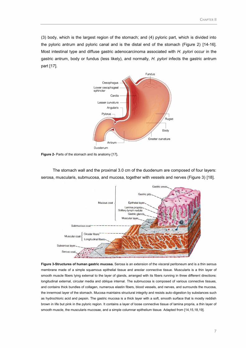
stomach can be divided into four parts: (1) cardia, which surrounds the opening of the
esophagus into the stomach; (2) fundus, which is the area above the level of the cardial orifice;
CHAPTER II
7
(3) body, which is the largest region of the stomach; and (4) pyloric part, which is divided into
the pyloric antrum and pyloric canal and is the distal end of the stomach (Figure 2) [14-16].
Most intestinal type and diffuse gastric adenocarcinoma associated with H. pylori occur in the
gastric antrum, body or fundus (less likely), and normally, H. pylori infects the gastric antrum
part [17].
Figure 2- Parts of the stomach and its anatomy [17].
The stomach wall and the proximal 3.0 cm of the duodenum are composed of four layers:
serosa, muscularis, submucosa, and mucosa, together with vessels and nerves (Figure 3) [18].
Figure 3-Structures of human gastric mucosa. Serosa is an extension of the visceral peritoneum and is a thin serous
membrane made of a simple squamous epithelial tissue and areolar connective tissue. Muscularis is a thin layer of
smooth muscle fibers lying external to the layer of glands, arranged with its fibers running in three different directions:
longitudinal external, circular media and oblique internal. The submucosa is composed of various connective tissues,
and contains thick bundles of collagen, numerous elastin fibers, blood vessels, and nerves, and surrounds the mucosa,
the innermost layer of the stomach. Mucosa maintains structural integrity and resists auto-digestion by substances such
as hydrochloric acid and pepsin. The gastric mucosa is a thick layer with a soft, smooth surface that is mostly reddish
brown in life but pink in the pyloric region. It contains a layer of loose connective tissue of lamina propria, a thin layer of
smooth muscle, the muscularis mucosae, and a simple columnar epithelium tissue. Adapted from [14,15,18,19].

CHAPTER II
8
The epithelial tissue of gastric mucosa is a layer of simple columnar cells that covers the
entire luminal surface of the mucosa tubular invagination, gastric pits or foveolae. There are 60
to 100 gastric pits per square millimeter of gastric mucosa, with diameters of approximately 70
μm and depths around 0.2 mm. The base of each gastric pit receives several long, tubular
gastric glands that extend deep into the lamina propria as far as the muscularis mucosae
[14,15,18]. Simple columnar mucus-secreting epithelium covers the entire luminal surface,
including the gastric pits and is composed of a continuous layer of surface mucous cells that
release gastric mucus from their apical surfaces to form a thick, protective lubricant layer over
the gastric lining (Figure 4) [14].
Figure 4- Gastric mucosa and submucosa protected from chemical injury by a mucus-bicarbonate surface barrier that
neutralizes gastric H+ and by epithelial “tight junctions” that prevent H
+ access to subepithelial tissue. Thick mucus from
the surface epithelial cells traps the secreted bicarbonate on the surface of the gastric mucosa. The bicarbonate-
containing mucus layer protects the cells from the caustic acid (pH ~1.5), keeping the pH at the surface ~7.0. Adapted
from Mulroney& Myers (2009) [20].
The mucosal surface is highly hydrated (>95%) and covered by a viscous mucus gel with
a thickness of approximately 300 µm. The mucus serves both as a lubricant and as a physical
barrier protecting the mucosal epithelium against gastric acid and maintaining their neutral pH
despite the highly acidic intragastric lumen. The main components of the mucus barrier are
mucin glycoproteins. Mucins are highly O-glycosylated proteins located at the interface between
the epithelial cell layers and the external environment [21]. The main mucins in the stomach are
the membrane-associated MUC1 and the secreted gel-forming MUC5AC and MUC6, which
have been shown to protect the epithelium against H. pylori adhesion, in vitro [21].
H. pylori road to colonization is summarized in the four steps described in Figure 5:
survival, motility, adhesion and damaging.

CHAPTER II
9
Figure 5- Schematic diagram of the H. pylori infection and pathogenesis. The urease activity and flagella-mediated
motility of H. pylori facilitate its survival and movement towards the lower mucus gel, above the epithelium, and is
followed by the interaction several adhesins, including blood-antigen binding protein A, sialic acid-binding adhesin, and
other outer membrane proteins with receptors on the host epithelium cells. After successful colonization, toxins
including CagA, and VacA, are involved in damaging the host tissue and intracellular replication. Adapted from Kao et
al. [6].
H. pylori enter the gastric lumen where its urease activity is used to neutralize the acidic
environment and the surface layer around the bacterium. The flagella and the helicoidal shape
of H. pylori facilitate their entrance and travel through the mucus layer allowing them to reach
the apical domain of gastric epithelial cells. This is followed by specific interactions between
bacterial adhesins with host cell receptors, which lead to successful colonization and persistent
infection [6,13]. H. pylori adhesion to gastric mucosa is a crucial step in the establishment of a
successful infection since it protects the bacteria from displacement by forces such as those
generated by peristalsis movements, shedding of the mucous layer and gastric emptying. Some
of the adhesins include blood-antigen binding protein A (BabA), which binds to fucosylated
Lewis B blood group antigen, and the sialic acid-binding adhesin (SabA), which binds to
inflammation-associated sialyl-Lewis x antigen. This close proximity to the gastric epithelium
enables the bacteria to scavenge nutrients from host cells, which are released when bacterial
toxins like cytotoxin-associated gene A (CagA) type IV damage the host tissues, causing
alteration of the cytoskeleton [22-24]. Then, vacuolating cytotoxin (VacA) induces alterations of
tight junctions and the formation of large vacuoles increasing transcellular permeability,
cytoskeleton changes and apoptosis [23,25,26]. Toxic factors such as the H. pylori neutrophil-
activating protein crosses the epithelial lining and recruit neutrophils and monocytes that cause
tissue damage by releasing reactive oxygen intermediates (ROIs) and nitrogen reactive
species. The combined toxic activity of VacA and of ROIs leads to tissue damage that is
enhanced by loosening of the protective mucus layer and acid permeation [13]. The damage

CHAPTER II
10
inflicted by such toxins may ultimately lead to the development of gastrointestinal symptoms.
The complement of toxin genes that a particular strain of H. pylori possesses strongly affects its
virulence [6,27]. H. pylori virulence factors and their potential role in pathogenesis are
summarized in Table 1.
Table 1-H. pylori virulence factors. Adapted from Monack et al.(2004) [28].
Virulence factors Description /potential role in pathogenesis
Flagella Involved in motility
Essential for colonization
Urease Resists acidic conditions in the stomach
Activates innate immune responses during early
steps of infection
BabA Outer membrane protein
Binds to fucosylated Lewis B blood group antigen
Mediates adhesion to epithelial cells and possibly
stomach epithelium
SabA Outer membrane protein
Mediates the adhesion of H. pylori to inflamed
gastric mucosa by binding the sialylated
carbohydrate structure
CagA Translocated into host cell by type IV secretion
apparatus encoded on Cag-PAI
Disrupts tight junctions
Epidemiologic link to cancer
VacA Vacuolating toxin
Induces apoptosis
Involved in the immunomodulation and colonization
of the mouse stomach
2.1.3- Routes of transmission
The routes for H. pylori transmission are not completely understood, but it is known that
the reservoir of H. pylori is the human stomach. This bacterium appears to have a narrow host
range. New infections are thought to occur as consequence of direct human-to-human
transmission or environmental contamination [29,30].
The possible routes for H. pylori transmission are summarized in Figure 6.

CHAPTER II
11
Figure 6-Transmission routes for H. pylori: direct person-to-person transmission, i.e. oral-oral, breastfeeding, gastro-
oral, fecal-oral and iatrogenic via; and possible reservoirs outside the human host, namely water, vegetables, and
animal contamination. Adapted from Azevedo et al.(2007) [31].
Most H. pylori infections are thought to be acquired in childhood via person-to-person.
The close personal contact is important for the spread of H. pylori and its prevalence increases
in family members with infected children [29,30,32]. The presence of H. pylori in the gastric
juice, in up to 58% of patients infected with the bacterium, raised the possibility that the refluxed
gastric juice may also represent a vehicle of transmission. The vomiting and regurgitation of
gastric material into the mouth are fairly common in childhood and may represent an important
route of transmission [30,31]. Saliva is another possible source of H. pylori transmission since
the gastric flora can reach and colonize the mouth after regurgitation or vomiting. Still, the oral-
oral transmission is not common for H. pylori, at least in adults [29].
Environmental or animal reservoirs, such as water, food, and animals, may be
considered sources of H. pylori infection. Indeed, water is considered a vehicle in the
transmission of H. pylori, in situation where poor hygienic practices during childhood are
observed, in case of absence of a household bath, in non-hygienic drinking water situations and
in the absence of a sewage disposal facility, which may lead to contamination or
recontamination of drinking water [29,31,33]. Food products may also be contaminated while
handling, under poor hygienic conditions. The food products most likely to transmit H. pylori are
milk, meat, and vegetables. Vegetables are suspected to be vulnerable to H. pylori colonization
when contaminated water is used for washing or irrigation [29,30].
The low socioeconomic status was reported as one of the most important factor in the
spreading of H. pylori [30,33]. The high age-specific prevalence of H. pylori infection in
developing countries has been attributed to low socioeconomic levels. Obviously,
socioeconomic status is not restricted to income and social class but takes into consideration
other factors such as levels of hygiene, the density of living, sanitation, urbanization and
educational opportunities [30,33]. Educational level has been used as a marker of
socioeconomic status and has been considered as one of the important determinants of H.

CHAPTER II
12
pylori prevalence in both developed and developing countries. Household crowding, sharing a
bed and increasing household contact have also been identified as risks for H. pylori infection,
because during childhood, crowded living conditions affect current bacterium status, and the
number of children in the present household increases the risk of infection for the adult family
members [30,33]. Another factor influencing the transmission of H. pylori is the genetic
predisposition [30,33].
2.2. Outcomes of H. pylori infection
2.2.1- Gastric cancer in the World
Gastric cancer still ranks fifth for cancer incidence and third of cancer deaths worldwide
(723 000 deaths, estimated in 2013) [34-36]. The majority of gastric cancer-related deaths
occurred in East Asia, nearly half in China (Figure 7) [2,36,37].
Figure 7- Estimated age-standardized death rates, for both sexes, caused by gastric cancer worldwide in 2012 [37].
Around 78% of all gastric cancer cases are estimated to be attributed to chronic H. pylori
infection, considered to be the strongest identified risk factor for gastric cancer, with more than
60% of new gastric cancer cases worldwide being attributed to this bacterium [32]. In most
individuals, H. pylori colonization is asymptomatic, but long-term carriage of the pathogen
significantly increases the risk of developing site-specific diseases, suggested a sequential
model of intestinal type-gastric carcinogenesis. Beside with outer environmental and host
factors, trigger a precancerous cascade is initiated with chronic gastritis, evolving to atrophic
gastritis, to intestinal metaplasia, followed by dysplasia, being the ultimate stage of this cascade
invasive carcinoma (Figure 8) [7,32].

CHAPTER II
13
In 1994, H. pylori were classified by the International Agency for Research on Cancer as
a Group 1 carcinogen, based on its association with gastric adenocarcinoma and mucosa-
associated lymphoid tissue (MALT) lymphoma [1,2,34].
Figure 8- Multifactorial pathway leading to gastric carcinoma. Hosts, bacterial and environmental factors act in
combination to contribute to the precancerous cascade that leads to the development of gastric cancer. Adapted from
Wroblewski et al. [32].
2.2.2- Gastric cancer in Portugal
In Portugal, gastric cancer incidence (27.8%) and mortality (21.7%) rates are among the
highest in Europe (19,5%) in 2013, especially in the North of the country (Figure 9), predicting
an incidence of 40.1% in 2020 [4,38,39]. Regarding the number of deaths, stomach cancer
accounts for 10.2% of all malignant cancer deaths [38].

CHAPTER II
14
Figure 9-Stomach cancer standardized (A) incidence and (B) mortality rates per 100 000 population in 2013 [40].
In the last half century, Portugal went through several important economic and social
changes, resulting in an improvement of the living standards, even though unequally across the
different socioeconomic groups [4]. Some studies reported that the Northern Portugal has a
higher gastric cancer risk factor than the Southern Portugal, due to the North of Portugal dietary
habits, such as the preference for salty/cured foods, which favor the onset of gastric cancer
[38,41].
2.3. Treatment of H. pylori
2.3.1- Current therapy
H. pylori eradication is the most promising strategy to reduce the incidence of gastric
cancer. This treatment is recommended in infected patients with duodenal or gastric ulcers,
early gastric cancer and gastric MALT lymphomas [7,42]. Treatment is also indicated for gastric
cancer prevention in high-risk individuals and in patients with functional dyspepsia [42].
Recommended indications for treatment are summarized in Table 2.
CHAPTER II
15
Table 2- Indications for H. pylori infection eradication therapy. Adapted from Mitchell et al.(2016) [43].
Indication Benefits of treatment
Past or present peptic ulcer disease Heals ulcers and reduces relapse
Functional dyspepsia May reduce symptoms and long-term risk of ulcer
disease and gastric cancer
Use of NSAIDs, including aspirin, in some patients Reduces risk of ulcers and bleeding
Gastric mucosal atrophy and intestinal metaplasia Reduces risk of gastric cancer
Patients requiring long-term acid suppression with
proton pump inhibitors
Reduces progression of intestinal metaplasia
Family history of gastric cancer Reduces long-term risk of gastric cancer
Prior early gastric cancer Reduces risk of further gastric cancer
Low-grade gastric MALT lymphoma Induces regression of lymphoma
Patient choice, after risks and benefits, are
discussed
Fulfills patient’s desire to reduce risk of infection
Consensus conferences have recommended therapeutic regimens to achieve H. pylori
cure rates higher than 80% on an intention-to-treat basis; for instance, the most efficient
therapies available should be used first to avoid the cost inconvenience and risks associated
with treatment failure [42,44,45].
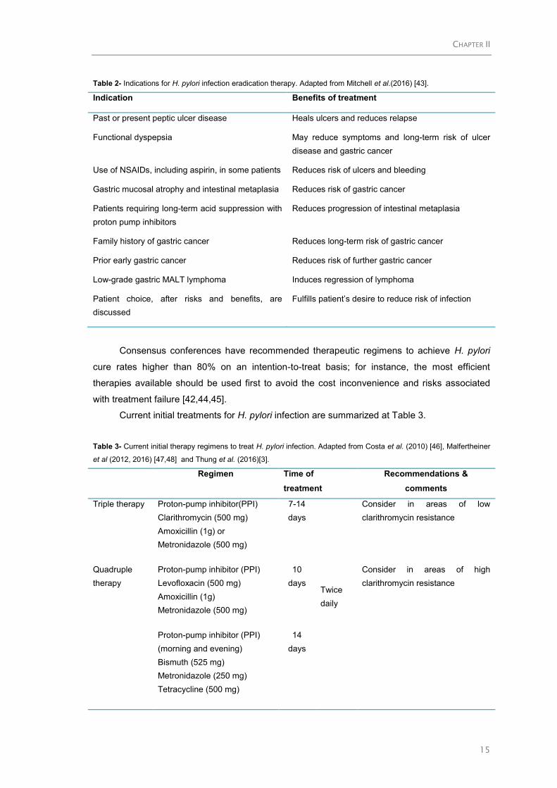
Current initial treatments for H. pylori infection are summarized at Table 3.
Table 3- Current initial therapy regimens to treat H. pylori infection. Adapted from Costa et al. (2010) [46], Malfertheiner
et al (2012, 2016) [47,48] and Thung et al. (2016)[3].
Regimen Time of
treatment
Recommendations &
comments
Triple therapy Proton-pump inhibitor(PPI)
Clarithromycin (500 mg)
Amoxicillin (1g) or
Metronidazole (500 mg)
7-14
days
Twice
daily
Consider in areas of low
clarithromycin resistance
Quadruple
therapy
Proton-pump inhibitor (PPI)
Levofloxacin (500 mg)
Amoxicillin (1g)
Metronidazole (500 mg)
10
days
Consider in areas of high
clarithromycin resistance
Proton-pump inhibitor (PPI)
(morning and evening)
Bismuth (525 mg)
Metronidazole (250 mg)
Tetracycline (500 mg)
14
days
CHAPTER II
16
Currently, the most widely used treatment for H. pylori infection is the triple therapy,
which includes the combination of two antibiotics, such as clarithromycin and amoxicillin or
metronidazole, and a proton pump inhibitor (PPI) such as omeprazole [7,22,49,50]. However,
the initial 90% cure rates obtained with this triple therapy have decreased to 75% or less, in
some regions [7], which can be related to the increasing number of H. pylori strains that are
resistant to the antibiotics used (Table 4), especially clarithromycin [46].
Table 4- Percentage of antibiotic resistance in some countries.
Antibiotic Percentage of resistance Reference
Clarithromycin <18% in southern Europe [46]
Metronidazole 20 to 40% in Europe and the USA
9 to 12% in Japan
[3]
Amoxicillin <2% in Europe
>38% Asia and South America
[3,51]
Levofloxacin-based triple therapy represents an alternative therapy in patients with
persistent infection despite the use of multiple standard treatments [46,50]. Alternatively, a
quadruple therapy containing bismuth, metronidazole, and tetracycline plus PPI, is quite
effective even in the presence of clarithromycin and metronidazole resistance, with an
eradication rate of up to 85%. However, compliance and side effects such as diarrhea,
vomiting, and metallic taste remain as limitations of this regimen [3,46].
Another alternative treatment is the sequential therapy that consists of 5 days treatment
with PPI and amoxicillin, followed by a further 5 days of PPI with two other antibiotics,
clarithromycin and metronidazole [3,50,52]. The eradication rates for 10 days of sequential
therapy were 87.2%, instead of 85.7% for 14 days of triple therapy, with no difference in
compliance or adverse effects [3,50]. The drawback of this therapy is its complex regimen
which may decrease patient compliance to the therapy and, thus, induce the development of
multidrug-resistant bacteria [3].
Summing up, the choice of therapy is based on local antibiotic usage, documented
antibiotic resistance, outcome data and also the world region [43,46]. It also depends whether it
is a first-line therapy, a second course, or a rescue therapy for persistent infection [45,46] Other
major determinants for a successful therapy are patient compliance, mucosal drug
concentration and the individual primary or secondary bacterial resistance to the antibiotics
employed [46].

CHAPTER II
17
2.3.2- Factors affecting H. pylori treatment failure
The successful eradication of H. pylori infection depends on various factors. These
factors are associated with nature of the organism itself, the intragastric environment where the
organism resides, the regimens used to eradicate it, and the behavior and reaction of the host
[53].
The roles that the organism, gastric environment, hosts and drug regimens play in
treatment failures are often inter-related. The key factors that increase the risk of H. pylori
eradication therapy failure are described in Table 5.
Table 5- Factors that cause H. pylori treatment failure.
Factors Major reason & comments References
Antimicrobial
resistance
Resistant strains prior to treatment (primary) or be
developed after a failed treatment (secondary)
[54-56]
Patient compliance
to therapy
Multiple different medications
Dose frequency (usually daily)
Duration of therapy (generally 10-14 days)
High frequency of side-effects
Limited information to patient about side-effect
[55-58]
Gastric environment Insufficient gastric acid inhibition
Easily degradation of antibiotics when the intragastric pH
is less than 4.0
[53,59]
Smoking Decrease gastric blood flow and mucus secretion
Reduce the delivery of antibiotics to the gastric mucosa.
Stimulate acid secretion
Low efficacy of antibiotics
[54,57,59,60]
2.4. Alternatives to conventional therapy
The increased failure of the conventional therapies in the eradication of H. pylori infection
in clinical practice demonstrates the urgent need for alternative therapeutic approaches. This
includes the use of new drugs against H. pylori or new adjuvants that can reduce side-effects of
available treatments and help in patient compliance.
The most exploited alternative approaches to fight H. pylori infections are the
development of i) vaccines, ii) photodynamic inactivation, iii) phage therapy, iv) probiotics, v)
antimicrobial peptides, vi) natural compounds such as plants and spices, phenolic compounds
and lipophilic compounds, and vii) bioengineered micro/nanoparticles as drug delivery systems

CHAPTER II
18
or as binding agents [7,22,61,62]. Some examples of the described approaches are
summarized in Table 6. However, although most of these alternative treatments have been very
successful in vitro, their efficiency in vivo were not sufficient to be translated for clinical
applications.
This is related with the fact that, in vivo, the molecules must achieve a bactericidal
concentration at site of infection, and for that, they must be able to cross the protective mucus
layer maintaining its biological activity at the large range of acidic pH [62].
Animal models can help elucidate the pathogenic mechanisms of H. pylori and aid in the
development of improved strategies for the treatment of gastric disease [63].
The type of animal models developed to study H. pylori pathogenicity can go from
primate- to rodent- based models. Several animals, including monkeys, pigs, cats, and dogs,
have been successfully colonized by several H. pylori species. Their major advantage is their
similar physiology with humans. However, several limiting factors, such as availability and high
costs, may restrict their use. Other disadvantages of these animal models are related to the
presence of natural Helicobacter infections such as H. heilmannii in primates and H. suis in
pigs, and constraints regarding the examination of the long-term effects of colonization [63,64].
Because of that, most animal models used are rodents which allow host variables to be
carefully monitored. Most mouse strains do not develop cancer but only mild inflammation
following H. pylori infection. A rodent alternative is Mongolian gerbils that are an efficient, robust
and cost-effective rodent model that can develop cancer when colonized with certain strains of
H. pylori [64,65]. Table 7 summarizes some of the features of rodent animal models that can be
used to study H. pylori infection.

CHAPTER II
19
Table 6- Alternative therapies against H. pylori
Therapy Description Agents References
Vaccines The key requirements to develop an effective vaccine are appropriate bacterial
antigens, safe and effective adjuvants and a delivery route. Many vaccine
formulations and strategies have been tested against H. pylori resulting in significant
reduction in bacterial load, and production of regulatory T cells that many limit the
eradicate H. pylori [66,67].
Antigens
UreB (most used)
HspC
VacA
CagA
NAP
In vitro: [68-71]
Animal model: [72-76]
Clinical trials: [77-81]
Photodynamic Photodynamic therapy involves the use of non-toxic photosensitive molecules
(photosensitizers) that will be activated by a specific wavelength of visible light. After
activation in presence of oxygen, photosensitizers trigger a destructive action in
biological systems, leading to bacterial death, due to the production of reactive
oxygen species, such as singlet oxygen and hydroxyl radical [82-84].
Photosensitizer:
Ruthenium (II) complex
covalently bound to
micrometric glass beads
(150-200µm)
Chorin e6 (ce6) in
combination with red light
Aluminum sulfonated
phthalocyanine
In vitro: [83-86]
Animal model: [87]
Clinical trials: [88,89]
Phage therapy
Bacteriophage therapy consists in the use of lytic bacteriophages (a virus that
infects bacteria), or their lytic proteins to induce the lysis of H. pylori. The phages
are engineered in a way that a particular antibody is fused to a protein on the
phage’s coat. Despite the possibility of using phage therapy to treat antibiotic
multiresistant bacteria strains, H. pylori phage screening is a rare topic in the
literature [61,90-93].
H. pylori phages
KHP30
KHP40
M13
BAPOUI
JP1/95
1603/05
Mex228
In vitro: [94-96]
Animal model: [95]
(to be continued)

CHAPTER II
20
Therapy Description Agents References
Probiotics Probiotics are live microorganisms that are orally administered to promote a health
benefit on the host. Probiotics can be used to prevent or decrease H. pylori
colonization. Moreover, when included in H. pylori eradication therapy, they can
improve eradication rates, by preventing antibiotic side effects, such as diarrhea.
Also, probiotics can be produced in large scale at low cost [90-93].
Microorganisms
L. acidophilus
L. casei subsq
rhammosus
L. casei 925
L. casei DN-114001
L. Shirota
L. salivarius WB1004
L. gasseri
L. johnsoniis La1
L. plantarum 8 RA-3
L. fermentum BL-96
L. fermentum L90265
Bifidobacterium infantis
2036
Saccharomyces
boulardii
Bacillus subtilis
Weissella confuse
In vitro: [97-102]
Animal model:
[98,100,103,104]
Clinical trials: [105-110]
(to be continued)

CHAPTER II
21
Therapy Description Agents References
Antimicrobial
peptides (AMP)
AMPs are a class of a broad spectrum antibiotic that are part of the innate immune
system of several organisms such as animals, plants, fungi, bacteria and viruses.
They are usually small (10-50 aminoacids in length), highly cationic and have
tendency to form amphipathic structures. Most AMPs can act by the disruption of
bacterial membranes due to electrostatic interactions between cationic AMP and
anionic bacterial membranes, and low tendency to induce bacterial resistance
[7,61].
Peptides
Magainin 2
Odorranain-HP
Pexiganan
Defensins
Cathelicidins
RpL1
TP4
OAK
Epi-1
In vitro: [111-119]
Animal model:
[115,116,118,120,121]
Natural
compounds
The use of natural food products such as fruits, vegetables, and fish could be used
as adjuvant therapies or as potential sources for new effective agents against
bacteria and associated inflammation. There are some molecules known by their
capacity to inhibit essential bacterial enzymes (such as urease) or to disrupt
bacterial membranes [7,61,90,92].
Phenolic compounds
Wine polyphenol
Apple peel polyphenols
extracts
Olive oil polyphenols
Green tea
Garlic
Broccoli
Curcumin
Lipophilic compounds
Terpenes
Terpenoids
Fatty acids
In vitro: [122-130]
Animal model:
[123,127,130-135]
Clinical trials: [130,136-
139]
(to be continued)

CHAPTER II
22
Therapy Description Agents References
Bioengineering Bioengineering has become a powerful tool by combining innovative bioengineering
strategies with bioactive compounds. The encapsulation of antimicrobial compounds
using gastric retentive polymers enhances their bioavailability, by protecting and
increasing their retention time in the gastric environment.
Binding systems that, after oral administration, are able to attract/bind bacteria and
remove them from the host stomach through the intestinal tract, after gastric mucus
turnover, were recently proposed as an alternative strategy [7,22].
Bioengineering strategies
Microspheres
Nanoparticles
Liposomes
NLC
In vitro: [128,129,140-154]
Animal model:
[135,140,148,150,155]
Abbreviations: UreB- urease B; HspC- heat shock proteins complex; NAP- neutrophil-activating protein; L. –Lactobacillus; TP4-Tilapia Piscidin 4; OAK- oligo-acyl-lysyl; Epi-1-Epinecidin-1

CHAPTER II
23
Table 7. Different features of rodents animal models used to study H. pylori infection. Adapted from Houghton (2012) [156] and Ivanov (2016)[157].
Animal model H. pylori
Features/comments References
Strain Infection conditions
Mice (C57BL/6) SS1
Helicobacter-free
Oral gavage
0.1 mL (106 CFU) of
bacteria
3 doses, 1 per day and
each separated by 24-48h
[158]
Extensively studied for investigating H. pylori role in gastric
carcinogenesis
Only show a marked influx of mononuclear cells while in
humans gastritis there are an accumulation of neutrophils and
mononuclear cells in the mucosa.
[127,134,135,140,159,160]
Mice (BALB/c) SS1 Widely used in cancer and immunology studies
Characterized by higher bacterial colonization levels but fewer
epithelial lesion
Used as a model of H. pylori-induced MALT lymphoma.
[159-164]
Mice (C3H) SS1
26695
Easy to colonize with H. pylori including with strain 26695 or by
isogenic mutants deficient in Lewis x or y expression.
[116,118,160,165]
Mongolian Gerbils NCTC
11637
B128
G1.1
TN2
7.13
Helicobacter-free
Oral gavage: 0.5 mL (109
CFU) of bacteria
2 doses (wait 24–48h
before the second
challenge)[65]
Model for study the pathogenesis of H. pylori infection,
especially gastritis, intestinal metaplasia, gastric ulcers and
gastric carcinoma.
[166-172]

CHAPTER II
24
2.4.1- Polyunsaturated fatty acids (PUFA)
The antibacterial proprieties of PUFA have been investigated in various organisms
including H. pylori, due to their high potency and lower susceptibility to induce H. pylori
resistance compared to conventional antibiotics.
2.4.1.1- General features and health benefits
Fatty acids can be classified by their carbon chain length and by their number of double
bonds [173]. Long-chain fatty acids contain more than twelve carbon atoms. They are formed of
hydrocarbon chains with a carboxyl group at one end (n end) and a methyl group at the
opposite end (ω end). Unsaturated fatty acids (Table 8) hold in their chain one
(monounsaturated) or more (polyunsaturated) carbon-carbon double bonds not saturated with
hydrogen [174]. PUFA are defined as fatty acids that possess more than two double bonds,
and their classification as n-3 or n-6 (omega-3 and omega-6, respectively) indicates the
position of the first double-bond on the hydrocarbon chain, i.e. at the third or sixth carbons from
the methyl end, respectively [174-176].
The n-3 and n-6 PUFA are present in vegetables and in animal tissues. Fish and other
seafood are the major sources of the long-chain n-3 PUFA, such as eicosapentaenoic acid
(EPA, C20:5 n-3) and docosahexaenoic acid (DHA, C22:6 n-3), whereas n-6 PUFA are mainly
found in vegetable oils, nuts and seeds, especially linoleic acid (LA, C18:2 n-6) [174,176] (Table
9).
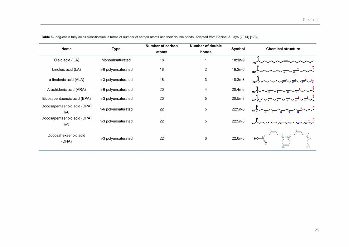
CHAPTER II
25
Table 8-Long-chain fatty acids classification in terms of number of carbon atoms and their double bonds. Adapted from Bazinet & Laye (2014) [173].
Name Type Number of carbon
atoms
Number of double
bonds Symbol Chemical structure
Oleic acid (OA) Monounsaturated 18 1 18:1n-9
Linoleic acid (LA) n-6 polyunsaturated 18 2 18:2n-6
α-linolenic acid (ALA) n-3 polyunsaturated 18 3 18:3n-3
Arachidonic acid (ARA) n-6 polyunsaturated 20 4 20:4n-6
Eicosapentaenoic acid (EPA) n-3 polyunsaturated 20 5 20:5n-3
Docosapentaenoic acid (DPA)
n-6 n-6 polyunsaturated 22 5 22:5n-6
Docosapentaenoic acid (DPA)
n-3 n-3 polyunsaturated 22 5 22:5n-3
Docosahexaenoic acid
(DHA) n-3 polyunsaturated 22 6 22:6n-3

CHAPTER II
26
Table 9-Sources of PUFA high in n-3 and n-6 fatty acids [177].
n-3 oils n-6 oils
Fish oil
Chia oil
Perilla oil
Flaxseed oil
Canola oil
Walnut oil
Soybean oil
Corn oil
Safflower
Sunflower seed oil
Cottonseed oil
Soybean oil
Peanut oil
Sesame oil
Grapeseed oil
Borage oil
Primrose oil
LA can be endogenously converted to longer-chain n-6 PUFA, such as gamma-linolenic
acid (GLA, C18:3 n-6) and arachidonic acid (ARA, C20:4 n-6) (Figure 10), whereas the n-3
PUFA, such as EPA and DHA, are converted from the progenitor alpha-linoleic acid (ALA,
C18:3 n-3) by progressive desaturation and elongation (Figure 10) [176,178].
ALA is found in triglycerides, in cholesterol esters and in fewer amounts in phospholipids.
EPA and DHA are found in the oils of fish (Table 10). EPA is found in triglycerides, cholesterol
esters and phospholipids, or can be directly obtained from fish oils through diet. It is the
precursor of eicosanoids comparable to those originating from ARA but with a lesser degree of
bioactivity [175,177]. EPA may be converted to DHA, and also to eicosanoids, such as
prostaglandins, thromboxanes, and leukotrienes. DHA is converted also to eicosanoids and to
anti-inflammatory mediators such as resolvins and protectins [177]. DHA is found mostly in
phospholipids, and its accumulation in the human body depends mainly on dietary intake
[175,177,178].

CHAPTER II
27
Figure 10- n3 and n-6 PUFA and their respective sources and metabolic derivatives. After consumption, the n-3
PUFA α-linolenic acid (ALA; 18:3n-3) and the n-6 PUFA linolenic acid (LA; 18:2n-6) can be desaturated, a process that
involves the addition of a double bond, and elongated to become longer chain PUFA. The Δ6 desaturase enzyme is
considered to be the rate-limiting step for the synthesis of docosahexaenoic acid (DHA; 22:6n-3). Adapted from Bazinet
& Laye (2014) [173] and Mehta et al.(2009) [175].
Table 10-Content of n-3 PUFA in selected fish (grams per 100g edible portion, raw) Adapted from Simopoulos(2005)
[177].
Fish Total fat Total
PUFA
ALA
(18:3n-3)
EPA
(20:5n-3)
DHA
(22:6n-3)
Cholesterol
(mg/100g)
Anchovy, European 4.8 1.6 - 0.5 0.9 -
Bluefish 6.5 1.6 - 0.4 0.8 59
Carp 5.6 1.4 0.3 0.2 0.1 67
Catfish, channel 4.3 1.0 Tr 0.1 0.2 58
Cod, Atlantic 0.7 0.3 Tr 0.1 0.2 58
Croaker, Atlantic 3.2 0.5 Tr 0.1 0.1 61
Flounder 1.0 0.3 Tr 0.1 0.1 46
Grouper, red 0.8 0.2 - Tr 0.2 -
Halibut, Greenland 13.8 1.4 Tr 0.5 0.4 46
(to be continued)

CHAPTER II
28
Fish Total fat Total
PUFA
ALA
(18:3n-3)
EPA
(20:5n-3)
DHA
(22:6n-3)
Cholesterol
(mg/100g)
Halibut, Pacific 2.3 0.7 0.1 0.1 0.3 32
Mackerel, king 13.0 3.2 - 1.0 1.2 53
Mullet, striped 3.7 1.1 0.1 0.3 0.2 49
Plaice, European 1.5 0.4 Tr 0.1 0.1 70
Pollock 1.0 0.5 - 0.1 0.4 71
Salmon, Chinook 10.4 2.1 0.1 0.8 0.6 -
Salmon, pink 3.4 1.4 Tr 0.4 0.6 -
Sole, European 1.2 0.2 Tr Tr 0.1 50
Swordfish 2.1 0.2 - 0.1 0.1 39
Tuna, albacore 4.9 1.8 0.2 0.3 1.0 54
Note: Tr, trace (<0.05g/100g food).
Different types of fish contain different amounts of fatty acids (Table 10) and may present
different ratios of EPA:DHA. Populations such as the Japanese, who oily fish consumption is
greater and regular than populations in Europe, North America, and Australian, have higher
intakes of very long-chain n-3 PUFA, such as DHA that comprises ~30% of the fatty acids
present [179].
Several clinical and epidemiologic studies have been developed to determine the effect of
PUFA on the various physiologic index and their health or clinical benefits. PUFA confers
various health benefits, including in rheumatoid arthritis [180], protection against the onset of
diabetes [181,182], efficient immune response modifications [183], coronary heart disease
[184], bronchial asthma [185], thyrotoxicosis [186], multiple sclerosis [175], depression [187],
Alzheimer’s disease [188,189] and cancer [190,191]. PUFA have roles in gastrointestinal
diseases, such as gastric cancer and colonic cancer, as Wang et al. (2012) reported in a
randomized clinical trial in which n-3 PUFA enriched emulsions displayed an anti-inflammatory
effect in patients with gastrointestinal diseases [192]. PUFA have also an effect against
pathogenic and opportunist microorganisms, inhibiting or killing bacteria [193].
2.4.1.2- PUFA activity against H. pylori
In 1987, a dietary intake of PUFA was reported as responsible for the decline of H. pylori
infection in duodenal ulcer incidence [194]. After that, some studies were developed and
demonstrated that concentrations of 2.5×10⁻⁴
M of LA could inhibit H. pylori growth in vitro.
Further studies described that this inhibitory effect increased with the degree of PUFA
unsaturation (number of double bonds): [oleic (C18:1) < linoleic (C18:2) < arachidonic (C20:4)
< n-3 linolenic (C18:3) =n-6 linolenic (C18:3) = eicosapentaenoic (C20:5) acid] [195].
Concentrations of 1×10⁻³
M are able to kill bacteria by cell lysis as observed by Scanning

CHAPTER II
29
Electron Microscopy (SEM) [195,196]. PUFA can be incorporated into H. pylori phospholipids
membrane, changing their morphology and inducing the disruption of the cell membrane [196].
Correia et al. (2012) demonstrated that DHA is also able to inhibit H. pylori growth in vitro in
a concentration-dependent manner. At concentrations 250 µM, DHA was bactericidal to all H.
pylori strains studied (26695, B128 and SS1 strains), whereas at concentration ~100 µM DHA
was only able to inhibit their growth. Also, DHA induced the conversion of H. pylori from bacillus
to coccoid form, being the coccoid form of H. pylori associated with low cell viability [127].
Moreover, DHA reduced ATP production by H. pylori, decreased H. pylori adhesion to AGS
cells, inhibited their gastric colonization in vivo, and decreased gastric inflammation in a mouse
model [127,134]. However, DHA efficiency in eradicating H. pylori infection in mice was
significantly lower than the conventional antibiotherapy, failing in 50% of mice. Nevertheless,
DHA used as co-adjuvant to conventional antibiotherapies yielded better results than those
obtained with single conventional therapies.
Effectively, PUFA may contribute to a protective action against chronic inflammation, having
an anti-inflammatory effect on H. pylori-infected gastric mucosa, in addition to its bactericidal
effect [197]. According to Correia et al. (2012, 2013), DHA reduced inflammatory responses
and the production of prostaglandin E2, associated with inflammation and tissue injury in the
mouse gastric mucosa. In addition, this PUFA reduced the metabolic activity of H. pylori as well
as the production of interleukin-8 (IL-8), cyclooxygenase 2 (COX-2) and induced nitric oxide
synthase from gastric epithelial cells [127,134,198].
Clinical trials, using a daily administration of 2g EPA and DHA for 12 weeks in patients
infected with H. pylori, had no significant effects on the eradication of H. pylori, and on the level
of IL-6 (expecting high levels during H. pylori infection, but a gradual reduction after a
successful treatment) [138].
Correia et al. (2014) also demonstrated that DHA is also incorporated into epithelial cells
and is able to decrease cell membrane cholesterol levels, as well as cholesterol de novo
synthesis. In addition, they showed that DHA decreases cholesterol uptake by H. pylori from
epithelial cells and that H. pylori sensitivity to DHA may be influenced by a previous exposure to
cholesterol [199]. It was also observed that H. pylori growth in a cholesterol-depleted medium is
up to thousand-fold more resistant to antibiotics therapy [200,201].
The observed PUFA ineffectiveness in vivo can be related to their poor solubility in water
(which difficult its administration), possible low penetration through the mucus layer and the low
residence time in the stomach. Moreover, PUFA can suffer easy oxidation, carboxylic
protonation, esterification, and lipid-protein complexation in gastric environment [193,202]. The
exposition of EPA and DHA to light, peroxidants or high temperature may induce their rapid
oxidation. Their autoxidation results in the formation of fatty acid based hydroperoxides and
their degradation into secondary oxidation products [203].
One strategy to overcome and preventing PUFA oxidation is their encapsulation into
nanodelivery systems, such as liposomes. These systems can stabilize and protect PUFA

CHAPTER II
30
against changes in environmental conditions and improve their bioavailability and activity at
infection local.
LA incorporated into liposomes (LipoLLA), with sizes around 100 nm, were able to fuse with
H. pylori membranes, allowing the free LA to be incorporated into the bacterium membrane,
disrupting their integrity and killing bacteria in 5 min [128,129]. These LipoLLA were more
efficient against H. pylori than other liposomal containing other PUFA such as oleic acid
(LipoOA) and stearic acid (LipoSA). The minimal bactericidal concentration (MBC) for LipoLLA
was 200 µg/mL, while 1 mg/mL of LipoOA was required to kill 90% of H. pylori and no
antibacterial effect was observed for LipoSA for concentrations up to 1 mg/mL [128]. LipoLLA
retention and distribution, as well as its efficiency in the treatment of H. pylori infection were
evaluated using mice animal models. LipoLLA treatment was able to reduce the number of H.
pylori in infected mice in a similar way as the currently used triple antibiotic therapy (99 %) and
without promoting alterations in the histology or increased rates of apoptosis in the mice
stomach. This effect was not observed when free LA treatment was used, that did not reduce H.
pylori number in comparison to the triple therapy. Moreover, treatment with LipoLLA reduced
the mRNA expression of proinflammatory cytokines known to be upregulated by H. pylori
infection [135].
2.4.1.3- PUFA activity against other microorganisms
The effect of PUFA against other microorganisms have been also investigated since PUFA
are well-suited for the: 1) treatment of superficial skin infections and acne [204-208], where the
anti-inflammatory proprieties of certain PUFA may provide additional benefits [209,210]; 2)
treatment and spread prevention of sexually-transmitted diseases [211,212], especially those
caused by Neisseria gonorrhoeae or herpes simplex virus; 3) prevention of periodontal
diseases and dental caries [213,214], and 4) reduction of infant gastrointestinal infections
[215,216].
Table 11 summarizes some of the PUFA with antimicrobial activity.

CHAPTER II
31
Table 11- PUFA with antimicrobial activity.
Fatty acid Microorganism Reference
EPA
DHA
Bacillus subtilis
Listeria monocytogenes
Staphylococcus aureus
Enterobacter aerogenes
Escherichia coli
Pseudomonas aeruginosa
Salmonella enteritidis
Salmonella typhimurium
Burkholderia cenocepacia
[217,218]
LA Staphylococcus aureus
Bacillus cercus
Streptococcus mutans
Pseudomonas aeruginosa
Salmonella typhimurium
Vibro parahemolyticus
Klebsiella pneumoniae
Proteus mirabillis
[219,220]
DHA
-LA
EPA
Staphylococcus aureus
Propionibacterium acnes
[205]
EPA Phaeodactylum tricornutum
Staphylococcus aureus
Bacillus cereus
Staphylococcus epidermidis
Bacillus weihenstephanensis
Planococcus citreus
[221]
2.4.1.4- Antibacterial Mechanism of Action
PUFA antibacterial mechanism of action is unclear but can have inhibitory or cidal effects. It
depends on many factors, such concentration, targeted microorganism, the microorganism
physiological state and the environment conditions such as pH and temperature [209]. When a
bactericidal interaction takes place, death is induced instantly or it may take longer periods of
time. Often, the prime target is the bacterial membrane, which can be followed by the changes
of a cascade of processes that are essential for bacteria survival. These processes are
schematized at Figure 11 [193].
PUFA can disrupt the normal function of bacteria membrane and thus, the production of
energy, by interfering with the electron transport chain and disrupting the oxidative

CHAPTER II
32
phosphorylation. Other processes that may contribute to bacterial growth inhibition or death
include cell lysis, inhibition of enzyme activity, impairment of nutrient uptake and the generation
of toxic peroxidation and auto-oxidation products (Table 12). Free fatty acids can exert a
protonophore effect, whereby the pH of the cytoplasm is reduced to such an extent that the cell
ceases to function normally [193,209,222]. Considering that fatty acids act on multiple cellular
targets, bacteria resistance to fatty acids is expected to be rare [209,216,223].
Figure 11- Schematic representation of the fatty acids antibacterial mechanism of action. Adapted from Desbois and
Smith (2010) [193].
Leakage of cell
membrane
Disruption of
electron transport
chain
Interference with
oxidative
hosphorylation
Enzyme
inhibition
Inhibition of
nutrient uptake
Cell lysis or
induction of
autolysis
Inhibition of fatty
acid biosynthesis

CHAPTER II
33
Table 12- Description of principal mechanisms of antibacterial of action of free PUFA. Adapted from Desbois and Smith (2010) [193].
Process Description References
Cell lysis The insertion of free PUFA into the bacterial inner membrane causes it to become more fluid and permeable, allowing the
leakage of the internal cell contents, causing growth inhibition or even death. When there is an excessive enhance of
membrane fluidity, the membrane can become unstable and the cell will ultimately lyse. Notwithstanding, the detergent
effect of fatty acids, which, at high concentrations, may solubilize large sections of the cell membrane, could further
account for complete cell lysis.
[224-226]
Disruption of electron
transport chain
The inner membrane of bacteria is an important site for energy production and it is where the electron transport chain is
located. The ability of the electron transport chain to transfer electrons is impaired so that the proton gradient and
membrane potential are reduced; resulting in a decrease in ATP production and the bacterium becomes deprived of an
essential source of energy. Free PUFA could bind the electron carriers directly, but complete displacement from the
membrane is likely achieved due to its increase membrane fluidity and instability.
[222,225,227-
230]
Oxidative
phosphorylation
Free PUFA can reduce ATP synthesis by increasing membrane permeability to protons, which could happen anywhere on
the inner membrane or at specific proton pores. Thus, the proton gradient and membrane potential are able to be
decreased by PUFA that after entered into cytosol, can release protons from its carboxyl groups and then returning across
the membrane to the exterior, leading to an increase in the cytosolic concentration of protons.
[222,227,231,232]
Inhibition of nutrient
uptake
Free PUFA are able to inhibit the bacteria ability to take up nutrients, such as amino acids. This reduction of nutrients
uptake may be related to the disruption of the membrane-located transporter proteins or result from the reduced proton
motive force required for the active transport.
[233-235]
Inhibition of enzyme
activity
Free PUFA can interact with various intracellular and membrane targets, such enzymes. Inhibition of enzymes in the
membrane or cytosol is crucial for bacterial survival and its growth. Their antibacterial effect are also due to the inhibition of
fatty acids biosynthesis.
[236-238]
Peroxidation and
auto-oxidation
PUFA high degree of unsaturation facilitates its degradation and the production of peroxides and reactive oxygen species,
or its auto-oxidation creating oxylipins and short-chain aldehydes, which are antibacterial.
[216,227,239,240]

CHAPTER II
34
2.4.1.5-PUFA encapsulation
The bioavailability and absorption of PUFA is a complex process, depending on several
factors, such as physicochemical properties, the presence or absence of other substances that
enhance or inhibit absorption, metabolization after absorption, the state of health, and other
individual factors [241-243]. After oral intake, PUFA enter the stomach where they are
dispersed as fine droplets, due to the mechanical influence of stomach peristalsis movements,
and travel until the upper small intestine. Besides the type of chemical bond, the concomitant
intake of food, and the presence of other components also affect the uptake of PUFA. Calcium
ions can also form a complex with PUFA and thus reduce their availability [243].
PUFA, like EPA and DHA, have a high degree of unsaturation, making them highly
susceptible to oxidation, a process that involves the formation of toxic products (i.e. peroxides),
unpleasantly smelling and tasting aldehydes and ketones [241,242,244,245]. There are a
variety of materials that can be used for encapsulation of PUFA, to protect them against
environmental effects (oxygen, light, humidity, pH) and, simultaneously, to improve their
stability, handling conditions and bioavailability and may minimize the odor and taste problems
[245,246]. There are some commercial capsules that use gelatin to encapsulate fish oil.
However, these capsules tend to disintegrate in the stomach releasing the fish oil and
consequently a fishy odor in the breath combined with undesired regurgitation which diminishes
patient compliance [246]. Taking this into consideration, a novel lipid-based colloidal formulation
has been developed. It consists of a highly concentrated lipid nanoparticle dispersion, which
can be mixed in soft drinks or water, avoiding swallowing problems with solid dosage forms
[246].
2.4.1.5.1- Lipid-based colloidal carriers
The application of nanotechnology, particularly the use of engineered small particles like
microparticles (1-999 µm) and nanoparticles (1-999 nm) for drug delivery, has generated
significant impact in medicine, revolutionizing the diagnosis and treatment of many diseases
[49,247]. Why can nanoparticles offer improved properties to a classical organic antibacterial
agent? One reason lies in their high surface area to volume ratio, resulting in the appearance of
new mechanical, chemical, electrical, optical, magnetic, electro-optical and magneto-optical
properties that are different from their bulk properties [248]. Small particles are recognized as
allowing a sustained therapy since they can achieve higher retention time in the human body
compared to free small molecules of antibiotics. Some authors defend that size plays a central
role in the diffusion of the particles through the gastric mucosa to reach H. pylori since
nanoparticles superior to 200 nm have a decreased diffusion [49,249,250].

CHAPTER II
35
Nanoparticles delivery systems possess some advantages: are quasi-monodisperse, are
thermodynamically stable and show long-term kinetic stability. They improve drug controlled
release, increase of pharmacokinetic proprieties of the drug, namely lipophilicity and reduce of
side-effects leading to the increase of therapeutic compliance [49,251,252]. The use of
nanoparticles in the treatment of H. pylori can also overcome the limitations associated with
antimicrobial drugs degradation by the acidic pH of the stomach [49]. Moreover, it is
improbable that bacteria might develop resistance to nanoparticles because numerous and
complex gene mutation would be necessary to overcome either the multiple mechanisms of
action of antimicrobial nanoparticles as well as the possibility to combine different antimicrobial
drugs in the same carrier [253].
There are various nanoparticles delivery systems, especially, 1) lipid-based colloidal
carriers such as liposomes or lipid nanoparticles, 2) polymeric nanoparticles, 3) dendrimers and
4) inorganic nanoparticles (Figure 12), that have been used in the release of antibiotics or novel
non-antibiotic compounds [254,255].
Lipid-based colloidal carriers can overcome problems associated with polymeric
nanoparticles, namely residues from the organic solvents used in the production process,
polymer toxicity and the scaling up of the production process to industrial levels [251]. Lipid-
based colloidal carriers have several application routes, namely topical [256,257] for cosmetics
and dermatological applications, ophthalmic [258,259], nasal [260], parenteral [261]
(intravenously, intramuscularly or subcutaneously), oral [262] and rectal [263-265]. These
nanoparticles have shown excellent outcomes in treating and detecting bacterial pathogens by
enabling targeted, responsive, combinatorial delivery of antimicrobial compounds, effective
antibacterial vaccination and rapid detection of bacteria.
Figure 12- Schematic illustration of major nanoparticle-based delivery systems for the treatment of bacterial infections:
(a) liposome, (b) polymeric nanoparticles, (c) dendrimer, and (d) inorganic nanoparticle [254].
In addition, multifunctional nanoparticles can respond to several stimuli that are
characteristic of the pathological site, which is achieved through the inclusion of components
that react to alterations in pH, temperature, redox conditions and to the overexpression of
certain biological molecules. These nanoparticles can also respond to stimuli from outside the
body, for instance, magnetic nanoparticles can be a tool for imaging contrast moiety to enable
their biodistribution, target accumulation or to monitor the efficacy of a therapy (Figure 13)
[266].
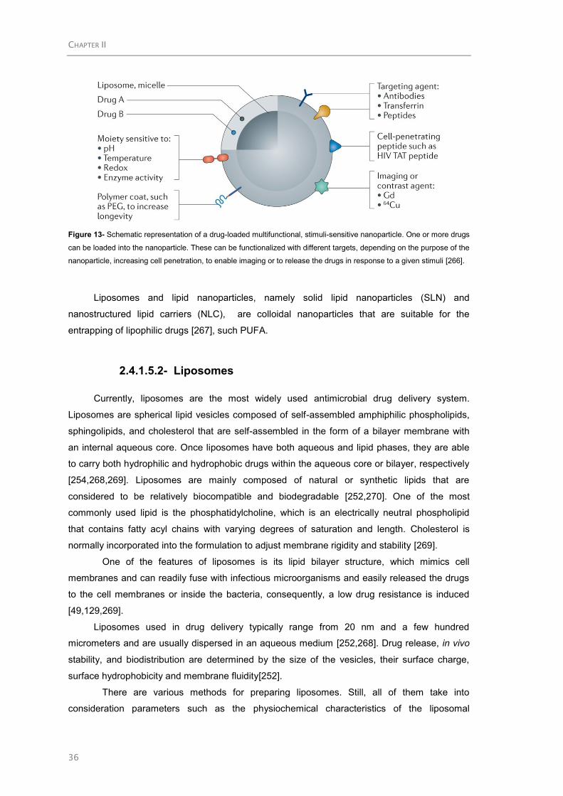
CHAPTER II
36
Figure 13- Schematic representation of a drug-loaded multifunctional, stimuli-sensitive nanoparticle. One or more drugs
can be loaded into the nanoparticle. These can be functionalized with different targets, depending on the purpose of the
nanoparticle, increasing cell penetration, to enable imaging or to release the drugs in response to a given stimuli [266].
Liposomes and lipid nanoparticles, namely solid lipid nanoparticles (SLN) and
nanostructured lipid carriers (NLC), are colloidal nanoparticles that are suitable for the
entrapping of lipophilic drugs [267], such PUFA.
2.4.1.5.2- Liposomes
Currently, liposomes are the most widely used antimicrobial drug delivery system.
Liposomes are spherical lipid vesicles composed of self-assembled amphiphilic phospholipids,
sphingolipids, and cholesterol that are self-assembled in the form of a bilayer membrane with
an internal aqueous core. Once liposomes have both aqueous and lipid phases, they are able
to carry both hydrophilic and hydrophobic drugs within the aqueous core or bilayer, respectively
[254,268,269]. Liposomes are mainly composed of natural or synthetic lipids that are
considered to be relatively biocompatible and biodegradable [252,270]. One of the most
commonly used lipid is the phosphatidylcholine, which is an electrically neutral phospholipid
that contains fatty acyl chains with varying degrees of saturation and length. Cholesterol is
normally incorporated into the formulation to adjust membrane rigidity and stability [269].
One of the features of liposomes is its lipid bilayer structure, which mimics cell
membranes and can readily fuse with infectious microorganisms and easily released the drugs
to the cell membranes or inside the bacteria, consequently, a low drug resistance is induced
[49,129,269].
Liposomes used in drug delivery typically range from 20 nm and a few hundred
micrometers and are usually dispersed in an aqueous medium [252,268]. Drug release, in vivo
stability, and biodistribution are determined by the size of the vesicles, their surface charge,
surface hydrophobicity and membrane fluidity[252].
There are various methods for preparing liposomes. Still, all of them take into
consideration parameters such as the physiochemical characteristics of the liposomal

CHAPTER II
37
ingredients, materials to be contained within the liposomes, particle size, polydispersity, surface
zeta potential, shelf-time, batch-to-batch reproducibility, and the possibility of large-scale
production [269]. Table 13 summarizes the methods that have been used for liposome
production.
Table 14 presents some of the examples of liposomes that have been used for
antimicrobial delivery.
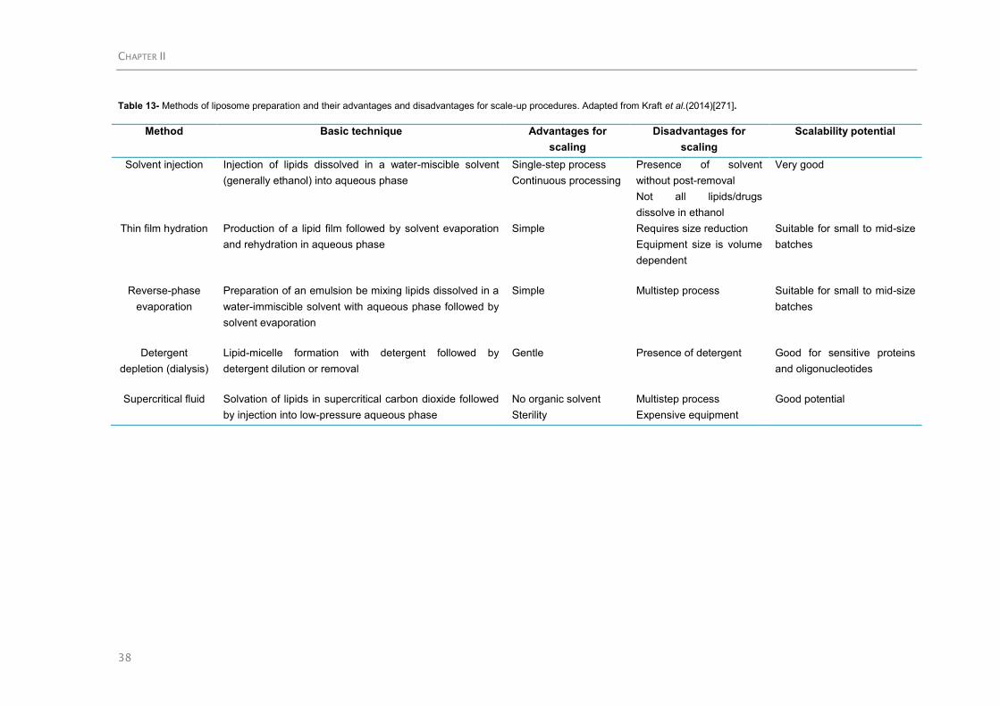
CHAPTER II
38
Table 13- Methods of liposome preparation and their advantages and disadvantages for scale-up procedures. Adapted from Kraft et al.(2014)[271].
Method Basic technique Advantages for
scaling
Disadvantages for
scaling
Scalability potential
Solvent injection Injection of lipids dissolved in a water-miscible solvent
(generally ethanol) into aqueous phase
Single-step process
Continuous processing
Presence of solvent
without post-removal
Not all lipids/drugs
dissolve in ethanol
Very good
Thin film hydration Production of a lipid film followed by solvent evaporation
and rehydration in aqueous phase
Simple Requires size reduction
Equipment size is volume
dependent
Suitable for small to mid-size
batches
Reverse-phase
evaporation
Preparation of an emulsion be mixing lipids dissolved in a
water-immiscible solvent with aqueous phase followed by
solvent evaporation
Simple Multistep process Suitable for small to mid-size
batches
Detergent
depletion (dialysis)
Lipid-micelle formation with detergent followed by
detergent dilution or removal
Gentle Presence of detergent Good for sensitive proteins
and oligonucleotides
Supercritical fluid Solvation of lipids in supercritical carbon dioxide followed
by injection into low-pressure aqueous phase
No organic solvent
Sterility
Multistep process
Expensive equipment
Good potential
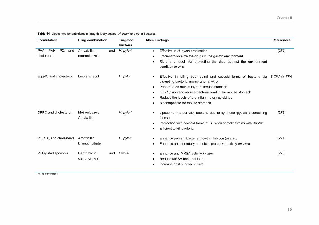
CHAPTER II
39
Table 14- Liposomes for antimicrobial drug delivery against H. pylori and other bacteria.
Formulation Drug combination Targeted
bacteria
Main Findings References
PAA, PAH, PC, and
cholesterol
Amoxicillin and
metronidazole
H. pylori Effective in H. pylori eradication
Efficient to localize the drugs in the gastric environment
Rigid and tough for protecting the drug against the environment
condition in vivo
[272]
EggPC and cholesterol Linolenic acid H. pylori Effective in killing both spiral and coccoid forms of bacteria via
disrupting bacterial membrane in vitro
Penetrate on mucus layer of mouse stomach
Kill H. pylori and reduce bacterial load in the mouse stomach
Reduce the levels of pro-inflammatory cytokines
Biocompatible for mouse stomach
[128,129,135]
DPPC and cholesterol Metronidazole
Ampicillin
H. pylori Liposome interact with bacteria due to synthetic glycolipid-containing
fucose
Interaction with coccoid forms of H. pylori namely strains with BabA2
Efficient to kill bacteria
[273]
PC, SA, and cholesterol Amoxicillin
Bismuth citrate
H. pylori Enhance percent bacteria growth inhibition (in vitro)
Enhance anti-secretory and ulcer-protective activity (in vivo)
[274]
PEGylated liposome Daptomycin and
clarithromycin
MRSA Enhance anti-MRSA activity in vitro
Reduce MRSA bacterial load
Increase host survival in vivo
[275]
(to be continued)

CHAPTER II
40
Formulation Drug combination Targeted bacteria Main Findings References
PC, SA, and cholesterol Ciprofloxacin and
vancomycin
MRSA Enhance antibacterial activity
More effective in liposomal form than free form in vivo
Reduce drug toxicity
Reduce incidence of side-effect (severe diarrhea)
[276]
DPPC and cholesterol Polymyxin B Pseudomonas
aeruginosa
Decrease bacteria count in lung
Increase bioavailability
Decrease lung injury caused by bacteria
[277]
DSPC and cholesterol Bismuth tobramycin Pseudomonas
aeruginosa
Decrease number of bacteria
Reduce the production of quorum-sensing molecules and
virulence factors
High therapeutic efficacy in vivo
[278]
PHEPC and PEGDSPE Gentamicin Klebsiella pneunoniae Increase survival rate of animal model
Increase therapeutic efficacy
[279]
DPPC, DCP, and
cholesterol
Pyrazinamide Mycobacterium
tuberculosis
Reduce the number of intracellularly located bacteria
High therapeutic efficacy in vivo
[280]
Abbreviations: DCP, dicetylphosphate; DPPC, 1,2-dipalmitoyl-sn-glycero-3-phosphocholine;; DSPC, 1,2-distearoyl-sn-glycero-3-phosphocholine; PAA, poly(acrylic acid); PAH, poly(allylamine hydrochloride); PC,
phosphatidylcholine; PEGDSPE, 1,2-distearoylsn-glycero-3-phosphoethanolamine-N-(polyethylene glycol-2000); PHEPC, partially hydrogenated egg phosphatidylcholine; SA, stearylamine.

CHAPTER II
41
2.4.1.5.3- Lipid nanoparticles
Lipid nanoparticles are a relatively new class of drug nanocarriers that are able to
incorporate hydrophobic drugs. These nanoparticles present very low cytotoxicity since are
prepared using biodegradable and biocompatible lipids without using of organic solvents..
Moreover, they can be easily and economically prepared for large-scale production [281-283].
Lipid nanoparticles can be produced from oil-in-water nanoemulsions, where the liquid lipid of
the oil droplets is replaced by a lipid that is solid at body temperature. Because of this, lipid
nanoparticles remain solid after administration. At the same time, the matrix can be chemically
protected against degradation. There are two generations of lipid nanoparticles, being the first
generation are the solid lipid nanoparticles (SLN) and the second generation, the
nanostructured lipid carriers (NLC) [282]. Figure 14 illustrates the differences between SLN and
NLC.
Figure 14- Differences between SLN and NLC. Adapted from Müller et al. (2011) [282].
2.4.1.5.3.1- Solid Lipid Nanoparticles (SLN)
By definition, SLN have sizes between 50 to 999 nm and the smaller the particle size the
more likely will be their stability, their targeted responses and their capacity to encapsulate
large amounts of the drug. SLN are colloidal particles derived from the oil-water emulsion by
replacing liquid lipids with a lipid matrix that is solid at body temperature and a surfactant for
particle stabilization [284-286]. Solid lipids used in SLN formulations include 1) fatty acids such
as palmitic acid, decanoic acid, and behenic acid; 2) triglycerides such as trilaurin, trimyristin

CHAPTER II
42
and tripalmitin; 3) steroids such as cholesterol; 4) partial glycerides such as glyceryl
monostearate and glyceryl behenate; and 5) waxes such as cetyl palmitate. Several types of
surfactants are commonly used as emulsifiers to stabilize lipid dispersion, including soybean
lecithin, phosphatidylcholine, poloxamer 188, sodium cholate and sodium glycocholate. The
typical methods of preparing SLN include spray drying, high shear mixing, ultra-sonication and
high-pressure homogenization [269,287].
SLN can be administrated by various routes such as oral, parental as well as
transdermal/topical. Oral/intraduodenal administration of SLN are believed to enhance
bioavailability of drugs by improving its transport through the intestinal epithelial layer and by
protecting it from the hostile environment of the gastrointestinal tract [287].
SLN have several potential advantages [284]:
Biocompatible and biodegradable;
Prevent drug degradation in body fluids;
Increase drug payload;
Improve drug half-life;
Control drug release;
Enhance drug bioavailability;
Drug targeting;
Large scale manufacturing;
Feasibility of sterilization.
However, common deficiencies of SLN include low drug loading capacity and a burst
release after polymorphic transition during storage. This is related with the increased in the
perfection of the crystal leaves, resulting in less space to accommodate drug molecules. In
addition to polymorphic transition, the formation of drug-enriched shell leads to burst release.
These disadvantages make SLN improper for some of applications as delivery system
[252,285,287].
SLN have three different morphologies, based on the location of the incorporated drug
molecules: (Table 15) 1) drug-enriched shell model; 2) drug-enriched core model, and 3)
homogeneous matrix model [263,284].
Several unique proprieties of SLN make them promising antimicrobial drug delivery
platforms, with possible applications in cosmetic and pharmaceutical products for skin care.
One application of SLN is delivering azole antifungal drugs to superficial fungal infection
patients. The occlusive effect of SLN, together with its small particle size, can extend drug
residence time on the epidermis and enhance drug penetration through the skin [269]. SLN can
also be used for oral administration, when included in tablets, capsules, and pellets. SLN can
transport antibacterial compounds and penetrate the intestinal linings through endocytosis
rather than passive diffusion. SLN have shown great therapeutic potentials against different
microorganisms, as it is summarized in Table 16.

CHAPTER II
43
Table 15- Brief description of the structure of solid lipid nanoparticles (SLN). Adapted from Üner & Yener (2007) [263] and Shah et al. (2015) [284].
SLN structure Description Schematic
illustration
Drug-enriched shell
model
Lipid core enclosed by a drug-enriched outer shell;
Suitable for applications where drugs need to be released as a burst;
Drug is solubilized in the surfactant-water mixture at elevated temperatures;
When dispersion is cooled, drug solubility decreases, leading to drug enrichment in the nanoparticle shell.
Drug-enriched core
model
Drug-enriched core enclosed by a lipid shell. This is obtained when the drug has a tendency to crystallize before
the lipid;
The drug is solubilized in the melted lipid in a concentration closed to its saturation solubility;
Subsequent cooling of the lipid emulsion causes super-saturation of the drug in the lipid melt, leading to lipid
recrystallization and formation of a membrane around the already crystallized drug-enriched core.
Homogenous matrix
model
Drug is homogenously dispersed within the lipid matrix in molecules or amorphous clusters.
Suitable for incorporation of drugs that exhibit prolonged release from particles.

CHAPTER II
44
Table 16- Solid lipid nanoparticles for antimicrobial drug delivery against bacteria.
Formulation Drug combination Targeted bacteria Main Findings References
Stearic acid Rifampicin, isoniazid,
pyrazinamide
Mycobacterium
tuberculosis
Increase residence time
Increase drug bioavailability
Decrease administration frequency
Prolong drug release
High physical stability
High encapsulation efficiency
[288,289]
Stearic acid, soy phosphatidylcholine,
and sodium taurocholate
Tobramycin Pseudomonas
aeruginosa
Increase drug bioavailability
[290]
Tetradecanoic acid Enrofloxacin S. aureus Sustain and prolong drug release
Increase bioavailability
Extend mean residence time in combination
with fatty acid
[291]
Palmitic acid, stearic acid,
hydrogenated castor oil
Tilmicosin S. aureus Sustain drug release
Enhance antibacterial activity
Decrease degree of inflammation (in vivo)
[292]
Compritol 888® ATO Vancomycin MRSA Ion pairing of vancomycin with antibacterial
fatty acid (linoleic acid)
Enhance encapsulation efficiency
Increase antibacterial activity of vancomycin
[293]
Stearic acid Norfloxacin E. coli Sustaine drug release
Enhance antibacterial activity [294]

CHAPTER II
45
2.4.1.5.3.2- Nanostructured Lipid Carriers (NLC)
NLC are prepared not from solid lipid only but from a blend of a solid lipid with
liquid lipid (oils) in such a proportion that the mixture is solid at least at 40ºC [268]. NLC
consists of an unstructured solid lipid matrix made of a mixture of blended solid and liquid
lipids and an aqueous phase containing a surfactant or a mixture of surfactants. Different
combinations of lipids and/or surfactants have been described (Table 17). They are
commercially available and/or approved by different regulatory agencies such as
Generally Recognized As Safe (GRAS) and FDA [264].
The imperfections present in the solid matrix of NLC accommodate the drugs
either as molecules or as amorphous crystals. The process of full-crystallization or
recrystallization of solid lipid reduces the drug solubility and leads to drug expulsion from
the lipid nanoparticles, especially when the drug concentration in the formulations is too
high [284,285]. On NLC formulation, the liquid lipid is incorporated into the core of a solid
lipid. This leads to a higher loading capacity and a better control in drug release since the
drug was dissolved in the liquid lipid that was simultaneously encapsulated in the solid
lipid [285]. The major advantage of NLC over SLN is that many drugs are more soluble in
a liquid lipid than in a solid lipid [285].
Therefore, NLC overcome the SLN drawbacks, since they increase encapsulation
efficiency, drug loading, and drug physical stability. They seem to be a valuable option
for improving the chemical stability and controlled release of functional lipophilic
compounds [285,295]. NLC increase oral bioavailability when administered to mice [295].
NLC can be produced using various methods, such as hot/cold homogenization,
emulsification-sonication, solvent injection, high-pressure homogenization, microemulsion
and solvent diffusion, and membrane contractor techniques (Table 18). Hot
homogenization is the most used method for fabrication of both, SLN and NLC due to
several advantages, including easy scale up, lack of organic solvents and short
production time, compared to the other methods [264,285,295]. In this approach, the
mixture of solid, liquid lipids and surfactant are melted to 5-10ºC above the temperature
of the solid lipid, and then the drug is dissolved or dispersed in this mixture. The mixture
is dispersed in aqueous solution at the same temperature by high-speed stirring/shearing,
obtaining a hot emulsion. Then this emulsion is homogenized using a high pressure
homogenized, a high-intensity ultrasonic probe/jet/bath or a microfluidizer, to produce a
hot nanoemulsion. Subsequently, NLC are cooled in cold water, at room temperature or
by a heat exchanger to crystallize lipid droplet and precipitate the lipid nanoparticles
[284,285].
Hot homogenization technique presents, however, some disadvantages like the
high heating temperature which may promote degradation of the labile active compounds;

CHAPTER II
46
most surfactants have low cloud points and thus high temperatures may reduce their
emulsifying capability and increase the NLC instability [285].
Like SLN, NLC have been proposed to possess three different morphologies
based on the location of incorporated drug molecules: NLC type I or imperfect crystal
type; type II or amorphous type; and type III or multiple type [263,284,285]. Table 19
summarizes the characteristics of each type of NLC.
NLC have shown great therapeutic potentials against different microorganisms,
as it is summarized in Table 20.

CHAPTER II
47
Table 17- Lipid and surfactants used for NLC production, brief description and examples of marketed products. Adapted from Beloqui et al (2016) [264], Shah et al.(2015) [284] and Tamjidi et
al.(2013) [285] .
Component Chemical name Trade name example Examples of
marketed
products
Description
Solid lipid Glyceryl
palmitostearate
Precirol ATO®5 Xifaxan
® The requirements that should be considered for the choice of a suitable lipid
blend are:
1. The solubility of the drug in lipid matrices. This influences the drug
encapsulation efficiency and loading capacities, and subsequently the
usefulness of lipid nanoparticles in drug delivery.
2. The solid and liquid lipids must be miscible at the specific concentrations
needed, i.e., the blend of bulk lipids should be homogeneous and the
liquid lipid should be situated as nanoholes within the solid matrix.
3. The lipid phase should be more stable to chemical degradation such as
oxidation and lipolysis.
4. Lipids should be biodegradable and capable to produce particles in the
nanometric scale. This is easier when the liquid lipid phase has a low
viscosity and/or interfacial tension than when it has a high viscosity
and/or interfacial tension.
5. Lipids should have an acceptable toxicological profile and should not
lead to the production of any toxic residues during NLC preparation.
Glyceryl dibeherante Compritol ® 888 ATO -
Cetyl palmitate CrodamolTM
CP Azelex®
Stearic acid - Viokace®
Tripalmitin - Survanta®
Liquid lipid Caprylic/capric
triglycerides
Miglyol®812 Avodart
TM
Vitamin E and
derivatives
- Neoral®
Monoacylglycerols Myverol 18-99K Terramycin®
Lauroyl
Polyoxyglycerides
Gelucire®44/14 Lipofen
®
Soy lecithin EpikuronTM
200 Baycip®
(to be continued)
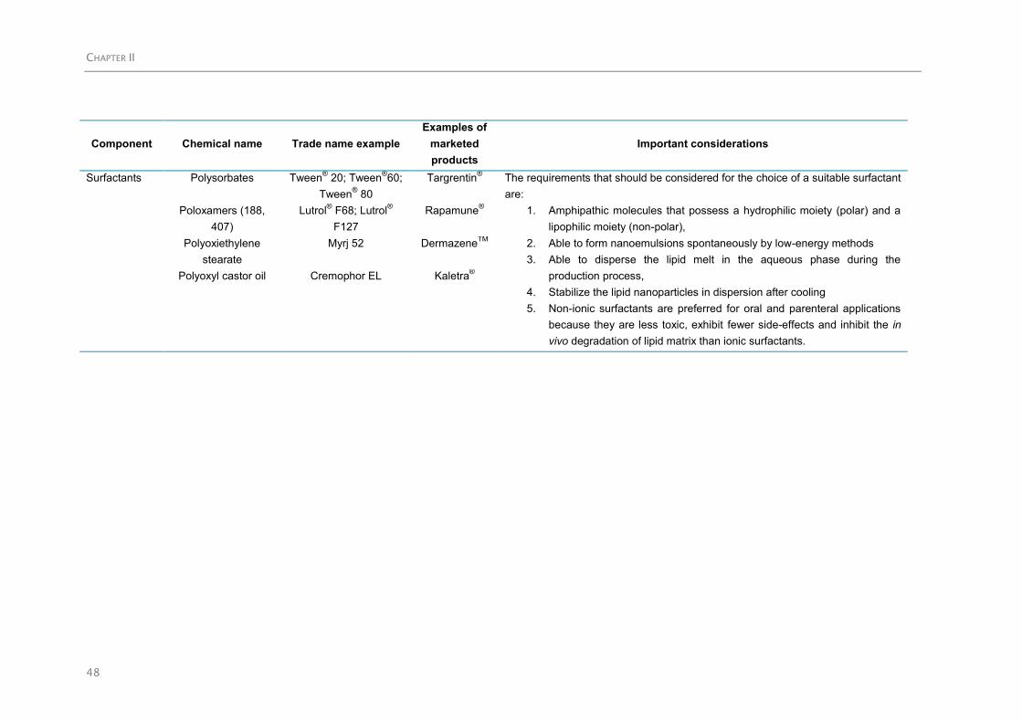
CHAPTER II
48
Component Chemical name Trade name example
Examples of
marketed
products
Important considerations
Surfactants Polysorbates Tween® 20; Tween
®60;
Tween® 80
Targrentin®
The requirements that should be considered for the choice of a suitable surfactant
are:
1. Amphipathic molecules that possess a hydrophilic moiety (polar) and a
lipophilic moiety (non-polar),
2. Able to form nanoemulsions spontaneously by low-energy methods
3. Able to disperse the lipid melt in the aqueous phase during the
production process,
4. Stabilize the lipid nanoparticles in dispersion after cooling
5. Non-ionic surfactants are preferred for oral and parenteral applications
because they are less toxic, exhibit fewer side-effects and inhibit the in
vivo degradation of lipid matrix than ionic surfactants.
Poloxamers (188,
407)
Lutrol® F68; Lutrol
®
F127
Rapamune®
Polyoxiethylene
stearate
Myrj 52 DermazeneTM
Polyoxyl castor oil Cremophor EL Kaletra®
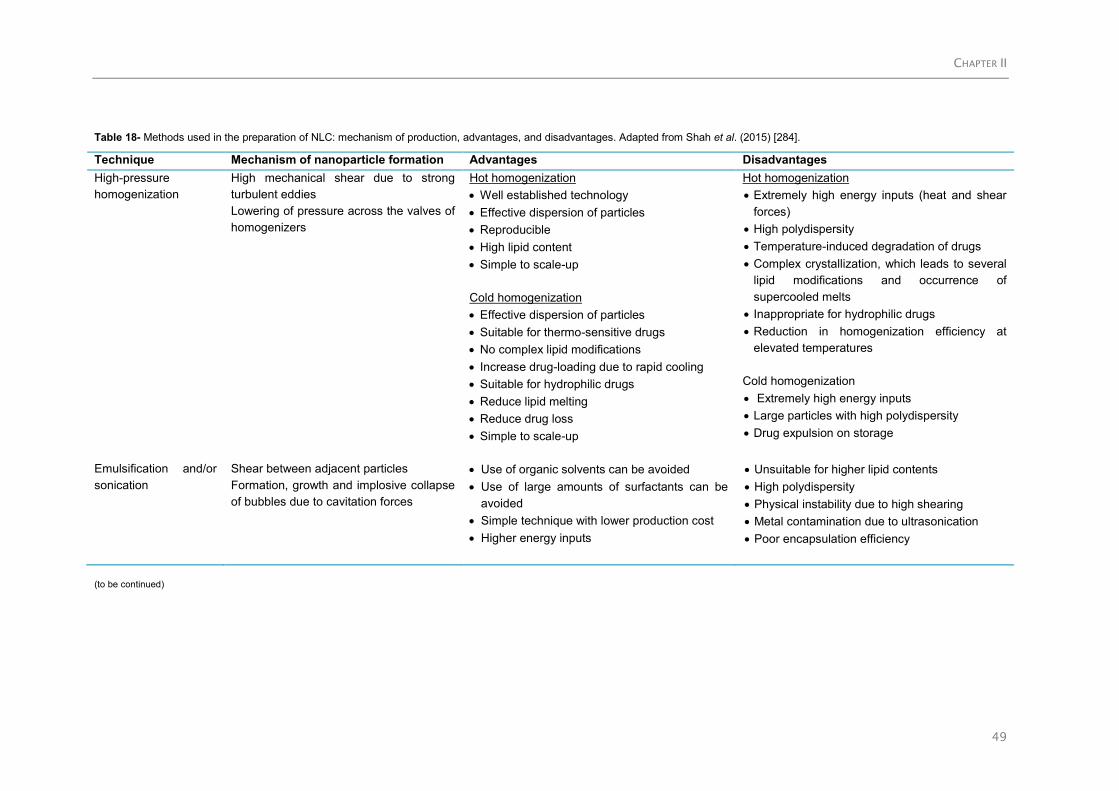
CHAPTER II
49
Table 18- Methods used in the preparation of NLC: mechanism of production, advantages, and disadvantages. Adapted from Shah et al. (2015) [284].
Technique Mechanism of nanoparticle formation Advantages Disadvantages
High-pressure
homogenization
High mechanical shear due to strong
turbulent eddies
Lowering of pressure across the valves of
homogenizers
Hot homogenization
Well established technology
Effective dispersion of particles
Reproducible
High lipid content
Simple to scale-up
Cold homogenization
Effective dispersion of particles
Suitable for thermo-sensitive drugs
No complex lipid modifications
Increase drug-loading due to rapid cooling
Suitable for hydrophilic drugs
Reduce lipid melting
Reduce drug loss
Simple to scale-up
Hot homogenization
Extremely high energy inputs (heat and shear
forces)
High polydispersity
Temperature-induced degradation of drugs
Complex crystallization, which leads to several
lipid modifications and occurrence of
supercooled melts
Inappropriate for hydrophilic drugs
Reduction in homogenization efficiency at
elevated temperatures
Cold homogenization
Extremely high energy inputs
Large particles with high polydispersity
Drug expulsion on storage
Emulsification and/or
sonication
Shear between adjacent particles
Formation, growth and implosive collapse
of bubbles due to cavitation forces
Use of organic solvents can be avoided
Use of large amounts of surfactants can be
avoided
Simple technique with lower production cost
Higher energy inputs
Unsuitable for higher lipid contents
High polydispersity
Physical instability due to high shearing
Metal contamination due to ultrasonication
Poor encapsulation efficiency
(to be continued)

CHAPTER II
50
Technique Mechanism of nanoparticle formation Advantages Disadvantages
Microemulsion Lipid crystallization due to rapid solidification Sophisticated equipment not required
Low energy inputs
Higher temperature gradients, faster lipid
crystallization, avoids particle aggregation
Simple to scale-up
Low lipid content
Solvent injection Lipid crystallization due to rapid diffusion of
solvent from internal organic phase to external
aqueous phase
Sophisticated equipment not required
Pharmaceutically accepted organic solvents
used
Highly efficient and versatile technique
Simples to scale-up
Solvent removal difficult
Low lipid content
Solvent diffusion Lipid crystallization due to diffusion of solvent
from internal organic phase to external aqueous
phase
Sophisticated equipment not required
Use of pharmaceutically accepted organic
solvents
Small particle diameters and low
polydispersity
Simple to scale-up
Toxicological risks due to incomplete
evaporation of organic solvents
Low lipid content
Membrane
contractor
Solid/liquid lipid phase infuses through
membrane pores into the tangentially flowing
aqueous phase to form droplets
Liquid lipid droplets crystallize to form lipid
nanoparticles
Controlled particles size with selection of
membrane with correct pore size
Simple to scale-up
Clogging of membrane pores, frequent
replacement or cleaning procedures

CHAPTER II
51
Table 19- Brief description of the structure of NLC. Adapted from Üner&Yener (2007) [263], Tamjidi et al. (2013) [285] and Shah et al. (2015) [284].
NLC structure Description Schematic
illustration
Imperfect
crystal type
Matrix with many voids and imperfections that are able to accommodate the drug molecules ().
Mixing small amounts of chemically different and low compatible liquid and solid lipids to achieve the highest drug
payload.
Amorphous
type
The amorphous type minimizes drug expulsion by maintaining the polymorphicity of the lipid matrix, i.e., the amorphous
model is created when mixing special lipids (e.g. hydroyoctacosanyl-hydroxystearate, isopropylmyristate) that do not
recrystallize after homogenization and cooling of the nanoemulsion.
Lipid core congeals in an amorphous state (rather than crystalline state), minimizing drug expulsion during storage time.
Multiple type The multiple type is related to the solubility of lipophilic drugs in liquid lipids, which is higher than that in solid lipids.
At low concentrations, liquid lipid molecules are easily dispersed into the lipid matrix.
By adding higher amounts of liquid lipid its solubility leads to phase separation, producing tiny oily nano-compartments
() surrounded by the solid lipid matrix.

CHAPTER II
52
Table 20- NLC for antimicrobial drug delivery against bacteria.
Formulation Drug combination Targeted bacteria Main Findings References
Precirol ATO®5, Squalene and Pluronic
F68
Tretinoin
Tetracycline
S. aureus
Pseudomonas aeruginosa
Propionibacterium acnes
Enhance tetracycline permeation through skin
Unaffect tretinoin permeation after encapsulation
Increase of antibacterial activity of tetracycline
[296]
Precirol ATO®5, Miglyol
®812, Tween
®80
and Poloxamer 188
Colistin Pseudomonas aeruginosa Increase the antimicrobial activity of colistin
More efficient in the eradication of biofilms
[297]
Precirol ATO®5, Compritol
® 888 ATO,
Miglyol®812, Tween
®80 and Poloxamer
188
Tobramycin Pseudomonas aeruginosa Increase antimicrobial activity
No cytotoxic
Able to penetrate mucus
Large pulmonary distribution and retention in the
in vivo
[298]
Precirol ATO®5, Miglyol
®812, Tween
®80
and Poloxamer 188
LL-37 peptide E. coli Increase antimicrobial activity
No cytotoxic
Improve healing in vivo in terms of wound
closure, re-epithelization grade and restoration of
the inflammatory process
[299]
Hexadecyl palmitate, glycerol stearate,
grape seed oil, sea buckthorn oil, St.
John’s wort oil, Syneperonic F68,
Tween®20 and Tween
®80
Green Tea extract E. coli Increased antioxidant activity
Improved antibacterial activity
[300]
Glycerol monooleate, oleic acid, and
Lutrol F127
Peptide:
AP114
DPK-060
LL-37
MRSA
Pseudomonas aeruginosa
E. coli
Acinetobacter baumannii
Different structures have the capacity of
solubilizing peptides
Differences on structure induced differences on
antibacterial activity
[301]

CHAPTER II
53
References
1. Correa P (2013) Gastric Cancer: Overview. Gastroenterology Clinics of North America 42: 211-217.
2. de Martel C, Forman D, Plummer M (2013) Gastric Cancer: Epidemiology and Risk Factors.
Gastroenterology Clinics of North America 42: 219-240.
3. Thung I, Aramin H, Vavinskaya V, Gupta S, Park JY, et al. (2016) Review article: the global emergence
of Helicobacter pylori antibiotic resistance. Alimentary Pharmacology & Therapeutics 43: 514-533.
4. Bastos J, Peleteiro B, Barros R, Alves L, Severo M, et al. (2013) Sociodemographic Determinants of
Prevalence and Incidence of Helicobacter pylori Infection in Portuguese Adults. Helicobacter 18: 413-
422.
5. Bruce MG, Maaroos HI (2008) Epidemiology of Helicobacter pylori infection. Helicobacter 13 Suppl 1: 1-
6.
6. Kao C-Y, Sheu B-S, Wu J-J (2016) Helicobacter pylori infection: An overview of bacterial virulence
factors and pathogenesis. Biomedical Journal 39: 14-23.
7. Parreira P, Fátima Duarte M, Reis CA, Martins MCL (2016) Helicobacter pylori infection: A brief
overview on alternative natural treatments to conventional therapy. Critical Reviews in Microbiology 42:
94-105.
8. Dunn BE, Cohen H, Blaser MJ (1997) Helicobacter pylori. Clinical Microbiology Reviews 10: 720-741.
9. Morales-Guerrero SE, Mucito-Varela E, Aguilar-Gutiérrez GR, Lopez-Vidal Y, Castillo-Rojas G (2013)
The Role of CagA Protein Signaling in Gastric Carcinogenesis — CagA Signaling in Gastric
Carcinogenesis.
10. Dunne C, Dolan B, Clyne M (2014) Factors that mediate colonization of the human stomach by
Helicobacter pylori. World Journal of Gastroenterology : WJG 20: 5610-5624.
11. O'Rourke J, Bode G (2001) Morphology and Ultrastructure. In: Mobley HLT, Mendz GL, Hazell SL,
editors. Helicobacter pylori: Physiology and Genetics. Washington (DC).
12. O'Toole PW, Clyne M (2001) Cell Envelope. In: Mobley HLT, Mendz GL, Hazell SL, editors.
Helicobacter pylori: Physiology and Genetics. Washington (DC).
13. Montecucco C, Rappuoli R (2001) Living dangerously: how Helicobacter pylori survives in the human
stomach. Nat Rev Mol Cell Biol 2: 457-466.
14. Standring S (2016) Gray's Anatomy: The Anatomical Basis of Clinical Practice; Standring S, editor:
New York: Elsevier Limited.
15. Drake R, Vogl A, Mitchell A (2015) Gray's Anatomy for students. Philadelphia: Churchill Livingstone
Elsevier.
16. Ellis H Anatomy of the stomach. Surgery - Oxford International Edition 29: 541-543.
17. Peek RM, Blaser MJ (2002) Helicobacter pylori and gastrointestinal tract adenocarcinomas. Nat Rev
Cancer 2: 28-37.
18. Daniels IR, Allum WH (2005) The Anatomy and Physiology of the Stomach. Upper Gastrointestinal
Surgery. London: Springer London. pp. 17-37.
19. Putz R, Pabst R (2006) Sobotta: Atlas of Human Anatomy; Putz R, editor: Elsevier GmbH.
20. Mulroney S, Myers AK (2009) Netter's Essential Physiology. Philadelphia, USA: Saunder Elsevier.
21. Yang I, Nell S, Suerbaum S (2013) Survival in hostile territory: the microbiota of the stomach. FEMS
Microbiol Rev 37: 736-761.

CHAPTER II
54
22. Gonçalves IC, Henriques PC, Seabra CL, Martins MCL (2014) The potential utility of chitosan
micro/nanoparticles in the treatment of gastric infection. Expert Review of Anti-infective Therapy 12:
981-992.
23. Yamaoka Y (2012) Pathogenesis of Helicobacter pylori-Related Gastroduodenal Diseases from
Molecular Epidemiological Studies. Gastroenterology Research and Practice 2012: 9.
24. Magalhaes A, Reis CA (2010) Helicobacter pylori adhesion to gastric epithelial cells is mediated by
glycan receptors. Braz J Med Biol Res 43: 611-618.
25. Atherton JC (2006) The pathogenesis of Helicobacter pylori-induced gastro-duodenal diseases.
Annual Review of Pathology: Mechanisms of Disease 1: 63-96.
26. Kusters JG, van Vliet AHM, Kuipers EJ (2006) Pathogenesis of Helicobacter pylori Infection. Clinical
Microbiology Reviews 19: 449-490.
27. Walton EL (2016) Helicobacter pylori's road to colonization. Biomedical Journal 39: 1-4.
28. Monack DM, Mueller A, Falkow S (2004) Persistent bacterial infections: the interface of the pathogen
and the host immune system. Nat Rev Micro 2: 747-765.
29. Vale FF, Vítor JMB (2010) Transmission pathway of Helicobacter pylori: Does food play a role in rural
and urban areas? International Journal of Food Microbiology 138: 1-12.
30. Mitchell H (2001) Epidemiology of Infection. In: HLT M, GL M, SL H, editors. Helicobacter pylori:
Physiology and Genetics. Washington (DC): ASM Press.
31. Azevedo NF, Guimarães N, Figueiredo C, Keevil CW, Vieira MJ (2007) A New Model for the
Transmission of Helicobacter pylori: Role of Environmental Reservoirs as Gene Pools to Increase
Strain Diversity. Critical Reviews in Microbiology 33: 157-169.
32. Wroblewski LE, Peek RM, Wilson KT (2010) Helicobacter pylori and Gastric Cancer: Factors That
Modulate Disease Risk. Clinical Microbiology Reviews 23: 713-739.
33. Khalifa MM, Sharaf RR, Aziz RK (2010) Helicobacter pylori: a poor man's gut pathogen? Gut
Pathogens 2: 2-2.
34. IARC (2014) Helicobacter pylori Eradication as a Strategy for Preventing Gastric Cancer.; Group
IHpW, editor. Lyon, France: International Agency for Research on Cancer.
35. Venerito M, Link A, Rokkas T, Malfertheiner P (2016) Gastric cancer – clinical and epidemiological
aspects. Helicobacter 21: 39-44.
36. Ferlay J, Soerjomataram I, Dikshit R, Eser S, Mathers C, et al. (2015) Cancer incidence and mortality
worldwide: sources, methods and major patterns in GLOBOCAN 2012. Int J Cancer 136: E359-386.
37. Ferlay J, Soerjomataram I, Ervik M, Dikshit R, Eser S, et al. (2013) GLOBOCAN 2012 v1.0, Cancer
Incidence and Mortality Worldwide: IARC CancerBase No 11. Lyon, France: International Agency for
Research on Cancer
38. Papoila AL, Riebler A, Amaral-Turkman A, São-João R, Ribeiro C, et al. (2014) Stomach cancer
incidence in Southern Portugal 1998–2006: A spatio-temporal analysis. Biometrical Journal 56: 403-
415.
39. Morais S, Ferro A, Bastos A, Castro C, Lunet N, et al. (2016) Trends in gastric cancer mortality and in
the prevalence of Helicobacter pylori infection in Portugal. European Journal of Cancer Prevention 25:
275-281.
40. Miranda N, Portugal C, Nogueira PJ, Farinha CS, Oliveira AL, et al. (2016) PORTUGAL- Doenças
Oncológicas em Números-2015. In: Saúde D-Gd, editor. Lisboa: Direção-Geral da Saúde, Direção de
Serviços de Informação e Análise.
41. Peleteiro B, La Vecchia C, Lunet N (2012) The role of Helicobacter pylori infection in the web of gastric
cancer causation. European Journal of Cancer Prevention 21: 118-125.
42. Fallone CA, Chiba N, van Zanten SV, Fischbach L, Gisbert JP, et al. (2016) The Toronto Consensus
for the Treatment of Helicobacter pylori Infection in Adults. Gastroenterology 151: 51-69.e14.

CHAPTER II
55
43. Mitchell H, Katelaris P (2016) Epidemiology, clinical impacts and current clinical management of
Helicobacter pylori infection. Med J Aust 204: 376-380.
44. Gisbert JP (2012) Rescue Therapy for Helicobacter pylori Infection 2012. Gastroenterology Research
and Practice 2012: 974594.
45. Malfertheiner P, Megraud F, O'Morain CA, Atherton J, Axon ATR, et al. (2012) Management of
Helicobacter pylori infection—the Maastricht IV/ Florence Consensus Report. Gut 61: 646-664.
46. Costa F, D'Elios MM (2010) Management of Helicobacter pylori infection. Expert Rev Anti Infect Ther
8: 887-892.
47. Malfertheiner P, Megraud F, O’Morain CA, Atherton J, Axon ATR, et al. (2012) Management of
Helicobacter pylori infection- the Maastricht IV/ Florence Consensus Report. Gut 61: 646e664.
48. Malfertheiner P, Megraud F, O'Morain CA, Gisbert JP, Kuipers EJ, et al. (2016) Management of
Helicobacter pylori infection—the Maastricht V/Florence Consensus Report. Gut.
49. Lopes D, Nunes C, Martins MCL, Sarmento B, Reis S (2014) Eradication of Helicobacter pylori: Past,
present and future. Journal of Controlled Release 189: 169-186.
50. O'Connor A, Fischbach W, Gisbert JP, O'Morain C (2016) Treatment of Helicobacter pylori infection
2016. Helicobacter 21: 55-61.
51. Selgrad M, Tammer I, Langner C, Bornschein J, Meißle J, et al. (2014) Different antibiotic susceptibility
between antrum and corpus of the stomach, a possible reason for treatment failure of Helicobacter
pylori infection. World Journal of Gastroenterology : WJG 20: 16245-16251.
52. Conteduca V, Sansonno D, Lauletta G, Russi S, Ingravallo G, et al. (2013) H. pylori infection and
gastric cancer: state of the art (review). Int J Oncol 42: 5-18.
53. Graham DY, Fischbach L (2010) Helicobacter pylori treatment in the era of increasing antibiotic
resistance. Gut 59: 1143-1153.
54. Malfertheiner P, Peitz U, Treiber G (2003) What constitutes failure for Helicobacter pylori eradication
therapy? Can J Gastroenterol 17 Suppl B: 53B-57B.
55. Senatore FJ, Wilmot J, Birk JW (2016) Helicobacter pylori treatment: Still a work in progress.
Postgraduate Medicine 128: 152-157.
56. Vakil N (2006) Helicobacter pylori Treatment: A Practical Approach. Am J Gastroenterol 101: 497-499.
57. O'Connor JPA, Taneike I, O'Morain C (2009) Improving Compliance with Helicobacter pylori
Eradication Therapy: When and How? Therapeutic Advances in Gastroenterology 2: 273-279.
58. Bohr URM, Malfertheiner P (2009) Eradication of H. pylori Infection: the Challenge is on if Standard
Therapy Fails. Therapeutic Advances in Gastroenterology 2: 59-66.
59. Song M, Ang TL (2014) Second and third line treatment options for Helicobacter pylori eradication.
World Journal of Gastroenterology : WJG 20: 1517-1528.
60. Suzuki T, Matsuo K, Ito H, Sawaki A, Hirose K, et al. (2006) Smoking Increases the Treatment Failure
for Helicobacter pylori Eradication. The American Journal of Medicine 119: 217-224.
61. Ayala G, Escobedo-Hinojosa WI, de la Cruz-Herrera CF, Romero I (2014) Exploring alternative
treatments for Helicobacter pylori infection. World Journal of Gastroenterology : WJG 20: 1450-1469.
62. Gaby AR (2001) Helicobacter pylori eradication: are there alternatives to antibiotics? Altern Med Rev
6: 355-366.
63. O’Rourke JL, Lee A (2003) Animal models of Helicobacter pylori infection and disease. Microbes and
Infection 5: 741-748.
64. Peek RM (2008) Helicobacter pylori infection and disease: from humans to animal models. Disease
Models & Mechanisms 1: 50-55.

CHAPTER II
56
65. Noto JM, Romero-Gallo J, Piazuelo MB, Peek RM (2016) The Mongolian Gerbil: A Robust Model of
Helicobacter pylori-Induced Gastric Inflammation and Cancer. In: Ivanov AI, editor. Gastrointestinal
Physiology and Diseases: Methods and Protocols. New York, NY: Springer New York. pp. 263-280.
66. Velin D, Michetti P (2010) Advances in vaccination against Helicobacter pylori. Expert Review of
Gastroenterology & Hepatology 4: 157-166.
67. Czinn SJ, Blanchard T (2011) Vaccinating against Helicobacter pylori infection. Nat Rev Gastroenterol
Hepatol 8: 133-140.
68. Zhang H-x, Qiu Y-y, Zhao Y-h, Liu X-t, Liu M, et al. (2014) Immunogenicity of oral vaccination with
Lactococcus lactis derived vaccine candidate antigen (UreB) of Helicobacter pylori fused with the
human interleukin 2 as adjuvant. Molecular and Cellular Probes 28: 25-30.
69. Koch M, Meyer TF, Moss SF (2013) Inflammation, Immunity, Vaccines for Helicobacter pylori infection.
Helicobacter 18: 18-23.
70. Yazbek PB, Trindade AB, Chin CM, dos Santos JL (2015) Challenges to the Treatment and New
Perspectives for the Eradication of Helicobacter pylori. Digestive Diseases and Sciences 60: 2901-
2912.
71. Chionh YT, Arulmuruganar A, Venditti E, Ng GZ, Han J-X, et al. (2014) Heat shock protein complex
vaccination induces protection against Helicobacter pylori without exogenous adjuvant. Vaccine 32:
2350-2358.
72. Corthésy-Theulaz I, Porta N, Glauser M, Saraga E, Vaney A-C, et al. (1995) Oral immunization with
Helicobacter pylori urease B subunit as a treatment against Helicobacter infection in mice.
Gastroenterology 109: 115-121.
73. DeLyria ES, Redline RW, Blanchard TG (2009) Vaccination of mice against H. pylori induces a strong
Th-17 response and immunity that is neutrophil-dependent. Gastroenterology 136: 247-256.
74. Suganya K, Prem Kumar A, Sekar B, Sundaran B (2017) Protection of mice against gastric
colonization of Helicobacter pylori by therapeutic immunization with systemic whole cell inactivated
vaccines. Biologicals 45: 39-46.
75. Sun P, Wang J-Q, Zhang Y-T, Zhao S-G (2014) Evaluating the immune responses of mice to
subcutaneous immunization with Helicobacter pylori urease B subunit. Journal of Animal Science and
Biotechnology 5: 14.
76. Chen J, Li N, She F (2014) Helicobacter pylori outer inflammatory protein DNA vaccine-loaded
bacterial ghost enhances immune protective efficacy in C57BL/6 mice. Vaccine 32: 6054-6060.
77. Aebischer T, Bumann D, Epple HJ, Metzger W, Schneider T, et al. (2008) Correlation of T cell
response and bacterial clearance in human volunteers challenged with Helicobacter pylori revealed by
randomised controlled vaccination with Ty21a-based Salmonella vaccines. Gut 57: 1065-1072.
78. Angelakopoulos H, Hohmann EL (2000) Pilot Study of phoP/phoQ-Deleted Salmonella enterica
Serovar Typhimurium Expressing Helicobacter pylori Urease in Adult Volunteers. Infection and
Immunity 68: 2135-2141.
79. Banerjee S, Medina-Fatimi A, Nichols R, Tendler D, Michetti M, et al. (2002) Safety and efficacy of low
dose Escherichia coli enterotoxin adjuvant for urease based oral immunisation against Helicobacter
pylori in healthy volunteers. Gut 51: 634-640.
80. Malfertheiner P, Schultze V, Rosenkranz B, Kaufmann SHE, Ulrichs T, et al. (2008) Safety and
Immunogenicity of an Intramuscular Helicobacter pylori Vaccine in Noninfected Volunteers: A Phase I
Study. Gastroenterology 135: 787-795.
81. Zeng M, Mao XH, Li JX, Tong WD, Wang B, et al. (2015) Efficacy, safety, and immunogenicity of an
oral recombinant Helicobacter pylori vaccine in children in China: a randomised, double-blind, placebo-
controlled, phase 3 trial. Lancet 386: 1457-1464.
82. Dai T, Huang Y-Y, Hamblin MR (2009) Photodynamic therapy for localized infections—State of the art.
Photodiagnosis and Photodynamic Therapy 6: 170-188.

CHAPTER II
57
83. Calvino-Fernández M, García-Fresnadillo D, Benito-Martínez S, McNicholl AG, Calvet X, et al. (2013)
Helicobacter pylori inactivation and virulence gene damage using a supported sensitiser for
photodynamic therapy. European Journal of Medicinal Chemistry 68: 284-290.
84. Simon C, Mohrbacher C, Hüttenberger D, Bauer-Marschall I, Krickhahn C, et al. (2014) In vitro studies
of different irradiation conditions for Photodynamic inactivation of Helicobacter pylori. Journal of
Photochemistry and Photobiology B: Biology 141: 113-118.
85. Calvino-Fernändez M, Garcia-Fresnadillo D, Benito-Martinez S, McNicholl AG, Gisbert JP, et al.
(2012) Su1708 Photodynamic Therapy: an Alternative Strategy Against Helicobacter pylori.
Gastroenterology 142: S-486.
86. Hamblin MR, Viveiros J, Yang C, Ahmadi A, Ganz RA, et al. (2005) Helicobacter pylori Accumulates
Photoactive Porphyrins and Is Killed by Visible Light. Antimicrobial Agents and Chemotherapy 49:
2822-2827.
87. Millson CE, Wilson M, MacRobert AJ, Bown SG (1996) Ex-vivo treatment of gastric Helicobacter
infection by photodynamic therapy. Journal of Photochemistry and Photobiology B: Biology 32: 59-65.
88. Lembo AJ, Ganz RA, Sheth S, Cave D, Kelly C, et al. (2009) Treatment of Helicobacter pylori infection
with intra-gastric violet light phototherapy: a pilot clinical trial. Lasers Surg Med 41: 337-344.
89. Ganz RA, Hamblin M, Tolkoff J, Nishioka N, Ahmad A, et al. (2003) Helicobacter pylori in patients is
killed by visible light. Am J Gastroenterol 98: S47-S47.
90. Vale FF, Oleastro M (2014) Overview of the phytomedicine approaches against Helicobacter pylori.
World Journal of Gastroenterology : WJG 20: 5594-5609.
91. Vitor JM, Vale FF (2011) Alternative therapies for Helicobacter pylori: probiotics and phytomedicine.
FEMS Immunol Med Microbiol 63: 153-164.
92. Roxo-Rosa M, Oleastro M, Vale FF (2013) Helicobacter pylori eradication-the alternatives beyond
antibiotics. In: Méndez-Vilas A, editor. Microbial pathogens and strategies for combating them:
science, technology and education: Formatex Research Center.
93. Kamiji MM, de Oliveira RB (2005) Non-antibiotic therapies for Helicobacter pylori infection. Eur J
Gastroenterol Hepatol 17: 973-981.
94. Abdel-Haliem MEF, Askora A (2013) Isolation and characterization of bacteriophages of Helicobacter
pylori isolated from Egypt. Future Virology 8: 821-826.
95. Cao J, Sun Y, Berglindh T, Mellgard B, Li Z, et al. (2000) Helicobacter pylori-antigen-binding fragments
expressed on the filamentous M13 phage prevent bacterial growth. Biochim Biophys Acta 1474: 107-
113.
96. Vale FF, Matos APA, Carvalho P, Vítor JMB (2008) Helicobacter pylori Phage Screening. Microscopy
and Microanalysis 14: 150-151.
97. Lorca GL, Wadstrom T, Valdez GF, Ljungh A (2001) Lactobacillus acidophilus autolysins inhibit
Helicobacter pylori in vitro. Curr Microbiol 42: 39-44.
98. Coconnier M-H, Lievin V, Hemery E, Servin AL (1998) Antagonistic Activity against Helicobacter
Infection In Vitro and In Vivo by the Human Lactobacillus acidophilus Strain LB. Applied and
Environmental Microbiology 64: 4573-4580.
99. Ushiyama A, Tanaka K, Aiba Y, Shiba T, Takagi A, et al. (2003) Lactobacillus gasseri OLL2716 as a
probiotic in clarithromycin-resistant Helicobacter pylori infection. J Gastroenterol Hepatol 18: 986-991.
100. Sgouras D, Maragkoudakis P, Petraki K, Martinez-Gonzalez B, Eriotou E, et al. (2004) In Vitro and In
Vivo Inhibition of Helicobacter pylori by Lactobacillus casei Strain Shirota. Applied and Environmental
Microbiology 70: 518-526.
101. Pinchuk IV, Bressollier P, Verneuil B, Fenet B, Sorokulova IB, et al. (2001) In Vitro Anti-Helicobacter
pylori Activity of the Probiotic Strain Bacillus subtilis 3 Is Due to Secretion of Antibiotics. Antimicrobial
Agents and Chemotherapy 45: 3156-3161.

CHAPTER II
58
102. Nam H, Ha M, Bae O, Lee Y (2002) Effect of Weissella confusa Strain PL9001 on the Adherence and
Growth of Helicobacter pylori. Applied and Environmental Microbiology 68: 4642-4645.
103. Johnson-Henry KC, Mitchell DJ, Avitzur Y, Galindo-Mata E, Jones NL, et al. (2004) Probiotics reduce
bacterial colonization and gastric inflammation in H. pylori-infected mice. Dig Dis Sci 49: 1095-1102.
104. Sgouras DN, Panayotopoulou EG, Martinez-Gonzalez B, Petraki K, Michopoulos S, et al. (2005)
Lactobacillus johnsonii La1 Attenuates Helicobacter pylori-Associated Gastritis and Reduces Levels of
Proinflammatory Chemokines in C57BL/6 Mice. Clinical and Diagnostic Laboratory Immunology 12:
1378-1386.
105. Goldman CG, Barrado DA, Balcarce N, Rua EC, Oshiro M, et al. Effect of a probiotic food as an
adjuvant to triple therapy for eradication of Helicobacter pylori infection in children. Nutrition 22: 984-
988.
106. Szajewska H, Albrecht P, Topczewska-Cabanek A (2009) Randomized, Double-blind, Placebo-
controlled Trial: Effect of Lactobacillus GG Supplementation on Helicobacter pylori Eradication Rates
and Side Effects During Treatment in Children. Journal of Pediatric Gastroenterology and Nutrition 48:
431-436.
107. Dang Y, Reinhardt JD, Zhou X, Zhang G (2014) The Effect of Probiotics Supplementation on
Helicobacter pylori Eradication Rates and Side Effects during Eradication Therapy: A Meta-Analysis.
PLoS One 9: e111030.
108. Wang Z-H, Gao Q-Y, Fang J-Y (2013) Meta-Analysis of the Efficacy and Safety of Lactobacillus-
containing and Bifidobacterium-containing Probiotic Compound Preparation in Helicobacter pylori
Eradication Therapy. Journal of Clinical Gastroenterology 47: 25-32.
109. Szajewska H, Horvath A, Piwowarczyk A (2010) Meta-analysis: the effects of Saccharomyces
boulardii supplementation on Helicobacter pylori eradication rates and side effects during treatment.
Alimentary Pharmacology & Therapeutics 32: 1069-1079.
110. Li S, Huang X-l, Sui J-z, Chen S-y, Xie Y-t, et al. (2014) Meta-analysis of randomized controlled trials
on the efficacy of probiotics in Helicobacter pylori eradication therapy in children. European Journal of
Pediatrics 173: 153-161.
111. Putsep K, Branden C-I, Boman HG, Normark S (1999) Antibacterial peptide from H. pylori. Nature
398: 671-672.
112. Zhang L, Wu WKK, Gallo RL, Fang EF, Hu W, et al. (2016) Critical Role of Antimicrobial Peptide
Cathelicidin for Controlling Helicobacter pylori Survival and Infection. The Journal of Immunology 196:
1799-1809.
113. Chen L, Li Y, Li J, Xu X, Lai R, et al. (2007) An antimicrobial peptide with antimicrobial activity against
Helicobacter pylori. Peptides 28: 1527-1531.
114. Ge Y, MacDonald DL, Holroyd KJ, Thornsberry C, Wexler H, et al. (1999) In Vitro Antibacterial
Properties of Pexiganan, an Analog of Magainin. Antimicrobial Agents and Chemotherapy 43: 782-788.
115. Zhang X-L, Jiang A-M, Ma Z-Y, Li X-B, Xiong Y-Y, et al. (2015) The Synthetic Antimicrobial Peptide
Pexiganan and Its Nanoparticles (PNPs) Exhibit the Anti-Helicobacter pylori Activity in vitro and in vivo.
Molecules 20: 3972.
116. Narayana JL, Huang H-N, Wu C-J, Chen J-Y (2015) Efficacy of the antimicrobial peptide TP4 against
Helicobacter pylori infection: in vitro membrane perturbation via micellization and in vivo suppression of
host immune responses in a mouse model. Oncotarget 6: 12936-12954.
117. Makobongo MO, Kovachi T, Gancz H, Mor A, Merrell DS (2009) In vitro Antibacterial Activity of Acyl-
Lysyl Oligomers against Helicobacter pylori. Antimicrobial Agents and Chemotherapy 53: 4231-4239.
118. Narayana JL, Huang H-N, Wu C-J, Chen J-Y (2015) Epinecidin-1 antimicrobial activity: in vitro
membrane lysis and in vivo efficacy against Helicobacter pylori infection in a mouse model.
Biomaterials 61: 41-51.

CHAPTER II
59
119. Leszczyńska K, Namiot A, Fein DE, Wen Q, Namiot Z, et al. (2009) Bactericidal activities of the
cationic steroid CSA-13 and the cathelicidin peptide LL-37 against Helicobacter pylori in simulated
gastric juice. BMC Microbiology 9: 187-187.
120. Moyat M, Bouzourene H, Ouyang W, Iovanna J, Renauld JC, et al. (2016) IL-22-induced
antimicrobial peptides are key determinants of mucosal vaccine-induced protection against H. pylori in
mice. Mucosal Immunol.
121. Zhang L, Yu J, Wong CCM, Ling TKW, Li ZJ, et al. (2013) Cathelicidin protects against Helicobacter
pylori colonization and the associated gastritis in mice. Gene Ther 20: 751-760.
122. Tombola F, Campello S, De Luca L, Ruggiero P, Del Giudice G, et al. (2003) Plant polyphenols
inhibit VacA, a toxin secreted by the gastric pathogen Helicobacter pylori. FEBS Lett 543: 184-189.
123. Stoicov C, Saffari R, Houghton J (2009) Green tea inhibits Helicobacter growth in vivo and in vitro.
International Journal of Antimicrobial Agents 33: 473-478.
124. Paulo L, Oleastro M, Gallardo E, Queiroz JA, Domingues F (2011) Anti-Helicobacter pylori and
urease inhibitory activities of resveratrol and red wine. Food Research International 44: 964-969.
125. Zaidi SFH, Ahmed K, Yamamoto T, Kondo T, Usmanghani K, et al. (2009) Effect of Resveratrol on
Helicobacter pylori- Induced Interleukin-8 Secretion, Reactive Oxygen Species Generation and
Morphological Changes in Human Gastric Epithelial Cells. Biological and Pharmaceutical Bulletin 32:
1931-1935.
126. Pastene E, Speisky H, Troncoso M, Alarcon J, Figueroa G (2009) In vitro inhibitory effect of apple
peel extract on the growth of Helicobacter pylori and respiratory burst induced on human neutrophils. J
Agric Food Chem 57: 7743-7749.
127. Correia M, Michel V, Matos AA, Carvalho P, Oliveira MJ, et al. (2012) Docosahexaenoic acid inhibits
Helicobacter pylori growth in vitro and mice gastric mucosa colonization. PLoS One 7: e35072.
128. Jung SW, Thamphiwatana S, Zhang L, Obonyo M (2015) Mechanism of antibacterial activity of
liposomal linolenic acid against Helicobacter pylori. PloS one 10: e0116519.
129. Obonyo M, Zhang L, Thamphiwatana S, Pornpattananangkul D, Fu V, et al. (2012) Antibacterial
Activities of Liposomal Linolenic Acids against Antibiotic-Resistant Helicobacter pylori. Molecular
Pharmaceutics 9: 2677-2685.
130. Akinori Y (2011) Sulforaphane Enhances Protection and Repair of Gastric Mucosa Against Oxidative
stress in vitro, and Demonstrates Anti-inflammatory Effects on Helicobacter pylori infected Gastric
Mucosae in Mice and Human Subjects. Current Pharmaceutical Design 17: 1532-1540.
131. Santos AM, Lopes T, Oleastro M, Gato IV, Floch P, et al. (2015) Curcumin Inhibits Gastric
Inflammation Induced by Helicobacter pylori Infection in a Mouse Model. Nutrients 7: 306-320.
132. Iimuro M, Shibata H, Kawamori T, Matsumoto T, Arakawa T, et al. (2002) Suppressive effects of
garlic extract on Helicobacter pylori-induced gastritis in Mongolian gerbils. Cancer Letters 187: 61-68.
133. Matsubara S, Shibata H, Ishikawa F, Yokokura T, Takahashi M, et al. (2003) Suppression of
Helicobacter pylori-induced gastritis by green tea extract in Mongolian gerbils. Biochemical and
Biophysical Research Communications 310: 715-719.
134. Correia M, Michel V, Osorio H, El Ghachi M, Bonis M, et al. (2013) Crosstalk between Helicobacter
pylori and gastric epithelial cells is impaired by docosahexaenoic acid. PLoS One 8: e60657.
135. Thamphiwatana S, Gao W, Obonyo M, Zhang L (2014) In vivo treatment of Helicobacter pylori
infection with liposomal linolenic acid reduces colonization and ameliorates inflammation. Proceedings
of the National Academy of Sciences 111: 17600-17605.
136. McNulty CAM, Wilson MP, Havinga W, Johnston B, O'Gara EA, et al. (2001) A Pilot Study to
Determine the Effectiveness of Garlic Oil Capsules in the Treatment of Dyspeptic Patients with
Helicobacter pylori. Helicobacter 6: 249-253.
137. Castro M, Romero C, de Castro A, Vargas J, Medina E, et al. (2012) Assessment of Helicobacter
pylori Eradication by Virgin Olive Oil. Helicobacter 17: 305-311.
CHAPTER II
60
138. Khandouzi N, Shidfar F, Agah S, Hosseini AF, Dehnad A (2015) Comparison of the Effects of
Eicosapentaenoic Acid and Docosahexaenoic Acid on the Eradication of Helicobacter pylori Infection,
Serum Inflammatory Factors and Total Antioxidant Capacity. Iranian Journal of Pharmaceutical
Research : IJPR 14: 149-157.
139. Agah S, Shidfar F, Khandouzi N, Baghestani AR, Hosseini S (2015) Comparison of the Effects of
Eicosapentaenoic Acid With Docosahexaenoic Acid on the Level of Serum Lipoproteins in Helicobacter
pylori: A Randomized Clinical Trial. Iranian Red Crescent Medical Journal 17: e17652.
140. Gonçalves IC, Magalhães A, Costa AMS, Oliveira JR, Henriques PC, et al. (2016) Bacteria-targeted
biomaterials: Glycan-coated microspheres to bind Helicobacter pylori. Acta Biomaterialia 33: 40-50.
141. Gonçalves IC, Magalhães A, Fernandes M, Rodrigues IV, Reis CA, et al. (2013) Bacterial-binding
chitosan microspheres for gastric infection treatment and prevention. Acta Biomaterialia 9: 9370-9378.
142. Nogueira F, Gonçalves IC, Martins MCL (2013) Effect of gastric environment on Helicobacter pylori
adhesion to a mucoadhesive polymer. Acta Biomaterialia 9: 5208-5215.
143. Fernandes M, Gonçalves IC, Nardecchia S, Amaral IF, Barbosa MA, et al. (2013) Modulation of
stability and mucoadhesive properties of chitosan microspheres for therapeutic gastric application.
International Journal of Pharmaceutics 454: 116-124.
144. Seabra CL, Nunes C, Gomez-Lazaro M, Correia M, Machado JC, et al. (2017) Docosahexaenoic acid
loaded lipid nanoparticles with bactericidal activity against Helicobacter pylori. International Journal of
Pharmaceutics 519: 128-137.
145. Parreira P, Magalhães A, Gonçalves IC, Gomes J, Vidal R, et al. (2011) Effect of surface chemistry
on bacterial adhesion, viability, and morphology. Journal of Biomedical Materials Research Part A 99A:
344-353.
146. Parreira P, Magalhães A, Reis CA, Borén T, Leckband D, et al. (2013) Bioengineered surfaces
promote specific protein–glycan mediated binding of the gastric pathogen Helicobacter pylori. Acta
Biomaterialia 9: 8885-8893.
147. Martins C, Gonçalves I, Gomes P, Oliveira JR, Reis C, et al. (2013) Microspheres for treating
Helicobacter pylori infections.
148. Arora S, Gupta S, Narang RK, Budhiraja RD (2011) Amoxicillin Loaded Chitosan–Alginate
Polyelectrolyte Complex Nanoparticles as Mucopenetrating Delivery System for H. Pylori. Scientia
Pharmaceutica 79: 673-694.
149. Arora S, Budhiraja RD (2012) Chitosan-alginate microcapsules of amoxicillin for gastric stability and
mucoadhesion. Journal of Advanced Pharmaceutical Technology & Research 3: 68-74.
150. Lin Y-H, Chiou S-F, Lai C-H, Tsai S-C, Chou C-W, et al. (2012) Formulation and evaluation of water-
in-oil amoxicillin-loaded nanoemulsions using for Helicobacter pylori eradication. Process Biochemistry
47: 1469-1478.
151. Chang C-H, Huang W-Y, Lai C-H, Hsu Y-M, Yao Y-H, et al. (2011) Development of novel
nanoparticles shelled with heparin for berberine delivery to treat Helicobacter pylori. Acta Biomaterialia
7: 593-603.
152. Lin Y-H, Tsai S-C, Lai C-H, Lee C-H, He ZS, et al. (2013) Genipin-cross-linked fucose–
chitosan/heparin nanoparticles for the eradication of Helicobacter pylori. Biomaterials 34: 4466-4479.
153. Chang C-H, Lin Y-H, Yeh C-L, Chen Y-C, Chiou S-F, et al. (2010) Nanoparticles Incorporated in pH-
Sensitive Hydrogels as Amoxicillin Delivery for Eradication of Helicobacter pylori. Biomacromolecules
11: 133-142.
154. Ping Y, Hu X, Yao Q, Hu Q, Amini S, et al. (2016) Engineering bioinspired bacteria-adhesive clay
nanoparticles with a membrane-disruptive property for the treatment of Helicobacter pylori infection.
Nanoscale 8: 16486-16498.
155. Henriques PC, Costa LM, Junqueira-Neto S, Carvalho R, Seabra CL, et al. Chitosan microspheres
can fight Helicobacter pylori gastric infection in mice. Frontiers in Bioengineering and Biotechnology.

CHAPTER II
61
156. Houghton J (2012) Helicobacter Species: Methods and Protocols; Houghton J, editor: Humana
Press. X, 258 p.
157. Ivanov AI (2016) Gastrointestinal Physiology and Diseases: Methods and Protocols: Springer New
York.
158. Taylor NS, Fox JG (2012) Animal Models of Helicobacter-Induced Disease: Methods to Successfully
Infect the Mouse. Methods in molecular biology (Clifton, NJ) 921: 131-142.
159. Shi Y, Liu X-F, Zhuang Y, Zhang J-Y, Liu T, et al. (2010) Helicobacter pylori Induced Th17
Responses Modulate Th1 Cell Responses, Benefit Bacterial Growth, and Contribute to Pathology in
Mice. The Journal of Immunology 184: 5121-5129.
160. Mahler M, Janke C, Wagner S, Hedrich HJ (2002) Differential susceptibility of inbred mouse strains to
Helicobacter pylori infection. Scand J Gastroenterol 37: 267-278.
161. Zhuang Y, Shi Y, Liu X-F, Zhang J-Y, Liu T, et al. (2011) Helicobacter pylori-infected macrophages
induce Th17 cell differentiation. Immunobiology 216: 200-207.
162. Chrisment D, Dubus P, Chambonnier L, Hocès de la Guardia A, Sifré E, et al. (2014) Neonatal
Thymectomy Favors Helicobacter pylori–Promoted Gastric Mucosa-Associated Lymphoid Tissue
Lymphoma Lesions in BALB/c Mice. The American Journal of Pathology 184: 2174-2184.
163. Guo L, Liu K, Zhao W, Li X, Li T, et al. (2013) Immunological features and efficacy of the
reconstructed epitope vaccine CtUBE against Helicobacter pylori infection in BALB/c mice model.
Applied Microbiology and Biotechnology 97: 2367-2378.
164. Yang J, Dai L-x, Pan X, Wang H, Li B, et al. (2015) Protection against Helicobacter pylori infection in
BALB/c mice by oral administration of multi-epitope vaccine of CTB-UreI-UreB. Pathogens and
Disease 73: ftv026-ftv026.
165. Takata T, El-Omar E, Camorlinga M, Thompson SA, Minohara Y, et al. (2002) Helicobacter pylori
Does Not Require Lewis X or Lewis Y Expression To Colonize C3H/HeJ mice. Infection and Immunity
70: 3073-3079.
166. Toyoda T, Shi L, Takasu S, Cho Y-M, Kiriyama Y, et al. (2016) Anti-Inflammatory Effects of Capsaicin
and Piperine on Helicobacter pylori-Induced Chronic Gastritis in Mongolian Gerbils. Helicobacter 21:
131-142.
167. Bae M, Lim JW, Kim H (2013) Oxidative DNA Damage Response in Helicobacter pylori-Infected
Mongolian Gerbils. Journal of Cancer Prevention 18: 271-275.
168. Girish Kumar T, Satyawan S, Gopal N, Ravi Kant D (2011) Evaluation of pH Triggers in situ Porous
Controlled Release Micro Balloon Delivery of Amoxicillin for Eradication of Helicobacter pylori. Current
Drug Delivery 8: 667-677.
169. Rajinikanth PS, Balasubramaniam J, Mishra B (2007) Development and evaluation of a novel floating
in situ gelling system of amoxicillin for eradication of Helicobacter pylori. International Journal of
Pharmaceutics 335: 114-122.
170. Fox JG, Kuipers EJ (2011) Long-term proton pump inhibitor administration, H pylori and gastric
cancer: lessons from the gerbil. Gut 60: 567-568.
171. Noto JM, Gaddy JA, Lee JY, Piazuelo MB, Friedman DB, et al. (2013) Iron deficiency accelerates
Helicobacter pylori–induced carcinogenesis in rodents and humans. The Journal of Clinical
Investigation 123: 479-492.
172. Romero-Gallo J, Harris EJ, Krishna U, Washington MK, Perez-Perez GI, et al. (2008) Effect of
Helicobacter pylori eradication on gastric carcinogenesis. Laboratory investigation; a journal of
technical methods and pathology 88: 328-336.
173. Bazinet RP, Laye S (2014) Polyunsaturated fatty acids and their metabolites in brain function and
disease. Nat Rev Neurosci 15: 771-785.
174. Catal A (2013) Five Decades with Polyunsaturated Fatty Acids: Chemical Synthesis, Enzymatic
Formation, Lipid Peroxidation and Its Biological Effects. Journal of Lipids 2013: 19.

CHAPTER II
62
175. Mehta LR, Dworkin RH, Schwid SR (2009) Polyunsaturated fatty acids and their potential therapeutic
role in multiple sclerosis. Nat Clin Pract Neurol 5: 82-92.
176. Daneshmand R, Kurl S, Tuomainen TP, Virtanen JK (2016) Associations of serum n-3 and n-6
polyunsaturated fatty acids with plasma natriuretic peptides. Eur J Clin Nutr 70: 963-969.
177. Simopoulos AP (2005) Omega-3 Polyunsaturated. In: Allen L, Prentice A, editors. Encyclopedia of
Human Nutrition. Second ed: Academic Press.
178. Song C, Shieh CH, Wu YS, Kalueff A, Gaikwad S, et al. (2016) The role of omega-3 polyunsaturated
fatty acids eicosapentaenoic and docosahexaenoic acids in the treatment of major depression and
Alzheimer's disease: Acting separately or synergistically? Prog Lipid Res 62: 41-54.
179. Calder PC (2012) Mechanisms of action of (n-3) fatty acids. J Nutr 142: 592S-599S.
180. Calder PC (2008) Session 3: Joint Nutrition Society and Irish Nutrition and Dietetic Institute
Symposium on 'Nutrition and autoimmune disease' PUFA, inflammatory processes and rheumatoid
arthritis. Proc Nutr Soc 67: 409-418.
181. Suresh Y, Das UN (2003) Long-chain polyunsaturated fatty acids and chemically induced diabetes
mellitus. Nutrition 19: 213-228.
182. Rudkowska I (2010) Fish oils for cardiovascular disease: Impact on diabetes. Maturitas 67: 25-28.
183. Hwang D (2000) Fatty acids and immune responses--a new perspective in searching for clues to
mechanism. Annu Rev Nutr 20: 431-456.
184. Hamer M, Steptoe A (2006) Influence of specific nutrients on progression of atherosclerosis, vascular
function, haemostasis and inflammation in coronary heart disease patients: a systematic review. Br J
Nutr 95: 849-859.
185. Calder PC (2006) n-3 polyunsaturated fatty acids, inflammation, and inflammatory diseases. Am J
Clin Nutr 83: 1505S-1519S.
186. De Caterina R, Basta G (2001) n-3 Fatty acids and the inflammatory response — biological
background. European Heart Journal Supplements 3: D42-D49.
187. Hoffmire CA, Block RC, Thevenet-Morrison K, van Wijngaarden E (2012) Associations between
omega-3 poly-unsaturated fatty acids from fish consumption and severity of depressive symptoms: an
analysis of the 2005-2008 National Health and Nutrition Examination Survey. Prostaglandins Leukot
Essent Fatty Acids 86: 155-160.
188. Quinn JF, Raman R, Thomas RG, Yurko-Mauro K, Nelson EB, et al. (2010) Docosahexaenoic Acid
Supplementation and Cognitive Decline in Alzheimer Disease: A Randomized Trial. Jama 304: 1903-
1911.
189. Moro K, Nagahashi M, Ramanathan R, Takabe K, Wakai T (2016) Resolvins and omega three
polyunsaturated fatty acids: Clinical implications in inflammatory diseases and cancer. World Journal of
Clinical Cases 4: 155-164.
190. Cockbain AJ, Toogood GJ, Hull MA (2012) Omega-3 polyunsaturated fatty acids for the treatment
and prevention of colorectal cancer. Gut 61: 135-149.
191. Vaughan VC, Hassing MR, Lewandowski PA (2013) Marine polyunsaturated fatty acids and cancer
therapy. Br J Cancer 108: 486-492.
192. Wang J, Yu JC, Kang WM, Ma ZQ (2012) Superiority of a fish oil-enriched emulsion to medium-chain
triacylglycerols/long-chain triacylglycerols in gastrointestinal surgery patients: a randomized clinical
trial. Nutrition 28: 623-629.
193. Desbois AP, Smith VJ (2010) Antibacterial free fatty acids: activities, mechanisms of action and
biotechnological potential. Appl Microbiol Biotechnol 85: 1629-1642.
194. Tarnawski A, Hollander D, Gergely H (1987) Protection of the gastric mucosa by linoleic acid--a
nutrient essential fatty acid. Clin Invest Med 10: 132-135.

CHAPTER II
63
195. Thompson L, Cockayne A, Spiller RC (1994) Inhibitory effect of polyunsaturated fatty acids on the
growth of Helicobacter pylori: a possible explanation of the effect of diet on peptic ulceration. Gut 35:
1557-1561.
196. Khulusi S, Ahmed HA, Patel P, Mendall MA, Northfield TC (1995) The effects of unsaturated fatty
acids on Helicobacter pylori in vitro. J Med Microbiol 42: 276-282.
197. Park S-H, Kangwan N, Park J-M, Kim E-H, Hahm KB (2013) Non-microbial approach for Helicobacter
pylori as faster track to prevent gastric cancer than simple eradication. World Journal of
Gastroenterology : WJG 19: 8986-8995.
198. Lee SE, Lim JW, Kim JM, Kim H (2014) Anti-Inflammatory Mechanism of Polyunsaturated Fatty Acids
in Helicobacter pylori-Infected Gastric Epithelial Cells. Mediators of Inflammation 2014: 12.
199. Correia M, Casal S, Vinagre J, Seruca R, Figueiredo C, et al. (2014) Helicobacter pylori's cholesterol
uptake impacts resistance to docosahexaenoic acid. Int J Med Microbiol 304: 314-320.
200. McGee DJ, George AE, Trainor EA, Horton KE, Hildebrandt E, et al. (2011) Cholesterol Enhances
Helicobacter pylori Resistance to Antibiotics and LL-37. Antimicrobial Agents and Chemotherapy 55:
2897-2904.
201. Trainor EA, Horton KE, Savage PB, Testerman TL, McGee DJ (2011) Role of the HefC Efflux Pump
in Helicobacter pylori Cholesterol-Dependent Resistance to Ceragenins and Bile Salts. Infection and
Immunity 79: 88-97.
202. Dyall SC (2011) Methodological issues and inconsistencies in the field of omega-3 fatty acids
research. Prostaglandins, Leukotrienes and Essential Fatty Acids (PLEFA) 85: 281-285.
203. Hadian Z (2016) A Review of Nanoliposomal Delivery System for Stabilization of Bioactive Omega-3
Fatty Acids. Electronic Physician 8: 1776-1785.
204. Chen CH, Wang Y, Nakatsuji T, Liu YT, Zouboulis C, et al. (2011) An innate bactericidal oleic acid
effective against skin infection of methicillin-resistant Staphylococcus aureus: a therapy concordant
with evolutionary medicine. J Microbiol Biotechnol 21: 391-399.
205. Desbois AP, Lawlor KC (2013) Antibacterial Activity of Long-Chain Polyunsaturated Fatty Acids
against Propionibacterium acnes and Staphylococcus aureus. Marine Drugs 11: 4544-4557.
206. Lukowski G, Lindequist U, Mundt S, Kramer A, Jülich WD (2008) Inhibition of Dermal MRSA
Colonization by Microalgal Micro- and Nanoparticles. Skin Pharmacology and Physiology 21: 98-105.
207. Yang D, Pornpattananangkul D, Nakatsuji T, Chan M, Carson D, et al. (2009) The antimicrobial
activity of liposomal lauric acids against Propionibacterium acnes. Biomaterials 30: 6035-6040.
208. Nakatsuji T, Kao MC, Fang J-Y, Zouboulis CC, Zhang L, et al. (2009) Antimicrobial Property of Lauric
Acid Against Propionibacterium Acnes: Its Therapeutic Potential for Inflammatory Acne Vulgaris.
Journal of Investigative Dermatology 129: 2480-2488.
209. Desbois AP (2012) Potential applications of antimicrobial fatty acids in medicine, agriculture and
other industries. Recent Pat Antiinfect Drug Discov 7: 111-122.
210. Thormar H, Hilmarsson H, Bergsson G (2013) Antimicrobial lipids: Role in innate immunity and
potential use in prevention and treatment of infection. In: Méndez-Vilas A, editor. Microbial pathogens
and strategies for combating them: science, technology and education: Formatex Research Center.
pp. 1474-1488.
211. Bergsson G, Steingrimsson O, Thormar H (1999) In vitro susceptibilities of Neisseria gonorrhoeae to
fatty acids and monoglycerides. Antimicrob Agents Chemother 43: 2790-2792.
212. Bergsson G, Arnfinnsson J, Karlsson SM, Steingrimsson O, Thormar H (1998) In vitro inactivation of
Chlamydia trachomatis by fatty acids and monoglycerides. Antimicrob Agents Chemother 42: 2290-
2294.
213. Bergsson G, Arnfinnsson J, Steingrimsson O, Thormar H (2001) In vitro killing of Candida albicans by
fatty acids and monoglycerides. Antimicrob Agents Chemother 45: 3209-3212.

CHAPTER II
64
214. Huang CB, George B, Ebersole JL (2010) Antimicrobial activity of n-6, n-7 and n-9 fatty acids and
their esters for oral microorganisms. Archives of oral biology 55: 555-560.
215. Sprong RC, Hulstein MFE, Van der Meer R (2001) Bactericidal Activities of Milk Lipids. Antimicrobial
Agents and Chemotherapy 45: 1298-1301.
216. Sun CQ, O'Connor CJ, Roberton AM (2003) Antibacterial actions of fatty acids and monoglycerides
against Helicobacter pylori. FEMS Immunol Med Microbiol 36: 9-17.
217. Shin SY, Bajpai VK, Kim HR, Kang SC (2007) Antibacterial activity of bioconverted eicosapentaenoic
(EPA) and docosahexaenoic acid (DHA) against foodborne pathogenic bacteria. International Journal
of Food Microbiology 113: 233-236.
218. Mil-Homens D, Bernardes N, Fialho AM (2012) The antibacterial properties of docosahexaenoic
omega-3 fatty acid against the cystic fibrosis multiresistant pathogen Burkholderia cenocepacia. FEMS
Microbiol Lett 328: 61-69.
219. Byeon JI, Song HS, Oh TW, Kim YS, Choi BD, et al. (2009) Growth inhibition of foodborne and
pathogenic bacteria by conjugated linoleic acid. J Agric Food Chem 57: 3164-3172.
220. Shin SY, Hou CT, Choi UK, Kim HR, Bajpai VK, et al. (2005) Antibacterial activity of bioconverted
linoleic acid produced by Pseudomonas aeruginosa PR3. Agric Chem Biotechnol 48: 167-169.
221. Desbois AP, Mearns-Spragg A, Smith VJ (2009) A fatty acid from the diatom Phaeodactylum
tricornutum is antibacterial against diverse bacteria including multi-resistant Staphylococcus aureus
(MRSA). Mar Biotechnol (NY) 11: 45-52.
222. Wojtczak L, Wie¸ckowski MR (1999) The Mechanisms of Fatty Acid-Induced Proton Permeability of
the Inner Mitochondrial Membrane. Journal of Bioenergetics and Biomembranes 31: 447-455.
223. Petschow BW, Batema RP, Ford LL (1996) Susceptibility of Helicobacter pylori to bactericidal
properties of medium-chain monoglycerides and free fatty acids. Antimicrob Agents Chemother 40:
302-306.
224. Cybulski LE, Albanesi D, Mansilla MC, Altabe S, Aguilar PS, et al. (2002) Mechanism of membrane
fluidity optimization: isothermal control of the Bacillus subtilis acyl-lipid desaturase. Molecular
Microbiology 45: 1379-1388.
225. Kenny JG, Ward D, Josefsson E, Jonsson I-M, Hinds J, et al. (2009) The Staphylococcus aureus
Response to Unsaturated Long Chain Free Fatty Acids: Survival Mechanisms and Virulence
Implications. PLOS ONE 4: e4344.
226. Shin SY, Bajpai VK, Kim HR, Kang SC (2007) Antibacterial activity of eicosapentaenoic acid (EPA)
against foodborne and food spoilage microorganisms. LWT - Food Science and Technology 40: 1515-
1519.
227. Schönfeld P, Wojtczak L (2008) Fatty acids as modulators of the cellular production of reactive
oxygen species. Free Radical Biology and Medicine 45: 231-241.
228. Cartron ML, England SR, Chiriac AI, Josten M, Turner R, et al. (2014) Bactericidal Activity of the
Human Skin Fatty Acid cis-6-Hexadecanoic Acid on Staphylococcus aureus. Antimicrobial Agents and
Chemotherapy 58: 3599-3609.
229. Peters Jeanne S, Chin C-K (2003) Inhibition of photosynthetic electron transport by palmitoleic acid is
partially correlated to loss of thylakoid membrane proteins. Plant Physiology and Biochemistry 41: 117-
124.
230. Stulnig TM, Huber J, Leitinger N, Imre E-M, Angelisová P, et al. (2001) Polyunsaturated
Eicosapentaenoic Acid Displaces Proteins from Membrane Rafts by Altering Raft Lipid Composition.
Journal of Biological Chemistry 276: 37335-37340.
231. Beck V, Jabůrek M, Demina T, Rupprecht A, Porter RK, et al. (2007) Polyunsaturated fatty acids
activate human uncoupling proteins 1 and 2 in planar lipid bilayers. The FASEB Journal 21: 1137-
1144.

CHAPTER II
65
232. Köhnke D, Ludwig B, Kadenbach B (1993) A threshold membrane potential accounts for
controversial effects of fatty acids on mitochondrial oxidative phosphorylation. FEBS Letters 336: 90-
94.
233. Fay JP, Farias RN (1975) The inhibitory action of fatty acids on the growth of Escherichia coli. J Gen
Microbiol 91: 233-240.
234. Galbraith H, Miller TB (1973) Effect of Long Chain Fatty Acids on Bacterial Respiration and Amino
Acid Uptake. Journal of Applied Bacteriology 36: 659-675.
235. Sheu CW, Salomon D, Simmons JL, Sreevalsan T, Freese E (1975) Inhibitory effects of lipophilic
acids and related compounds on bacteria and mammalian cells. Antimicrob Agents Chemother 7: 349-
363.
236. Zhang Y-M, Rock CO (2008) Membrane lipid homeostasis in bacteria. Nat Rev Micro 6: 222-233.
237. Zheng CJ, Yoo J-S, Lee T-G, Cho H-Y, Kim Y-H, et al. (2005) Fatty acid synthesis is a target for
antibacterial activity of unsaturated fatty acids. FEBS Letters 579: 5157-5162.
238. Sado-Kamdem SL, Vannini L, Guerzoni ME (2009) Effect of α-linolenic, capric and lauric acid on the
fatty acid biosynthesis in Staphylococcus aureus. International Journal of Food Microbiology 129: 288-
294.
239. Rustan AC, Drevon CA (2001) Fatty Acids: Structures and Properties. eLS: John Wiley & Sons, Ltd.
240. Adolph S, Bach S, Blondel M, Cueff A, Moreau M, et al. (2004) Cytotoxicity of diatom-derived
oxylipins in organisms belonging to different phyla. Journal of Experimental Biology 207: 2935-2946.
241. Rubio-Rodríguez N, Beltrán S, Jaime I, de Diego SM, Sanz MT, et al. (2010) Production of omega-3
polyunsaturated fatty acid concentrates: A review. Innovative Food Science & Emerging Technologies
11: 1-12.
242. Kralovec JA, Zhang S, Zhang W, Barrow CJ (2012) A review of the progress in enzymatic
concentration and microencapsulation of omega-3 rich oil from fish and microbial sources. Food
Chemistry 131: 639-644.
243. Schuchardt JP, Hahn A (2013) Bioavailability of long-chain omega-3 fatty acids. Prostaglandins,
Leukotrienes and Essential Fatty Acids (PLEFA) 89: 1-8.
244. Sanguansri L, Ann Augustin M (2006) Microencapsulation and Delivery of Omega-3 Fatty Acids.
Functional Food Ingredients and Nutraceuticals: CRC Press. pp. 297-327.
245. Kaushik P, Dowling K, Barrow CJ, Adhikari B (2015) Microencapsulation of omega-3 fatty acids: A
review of microencapsulation and characterization methods. Journal of Functional Foods 19, Part B:
868-881.
246. Muchow M, Schmitz EI, Despatova N, Maincent P, Muller RH (2009) Omega-3 fatty acids-loaded lipid
nanoparticles for patient-convenient oral bioavailability enhancement. Pharmazie 64: 499-504.
247. Hajipour MJ, Fromm KM, Akbar Ashkarran A, Jimenez de Aberasturi D, Larramendi IRd, et al.
Antibacterial properties of nanoparticles. Trends in Biotechnology 30: 499-511.
248. Hajipour MJ, Fromm KM, Akbar Ashkarran A, Jimenez de Aberasturi D, Larramendi IRd, et al. (2012)
Antibacterial properties of nanoparticles. Trends in Biotechnology 30: 499-511.
249. Norris DA, Puri N, Sinko PJ (1998) The effect of physical barriers and properties on the oral
absorption of particulates. Advanced Drug Delivery Reviews 34: 135-154.
250. Hasani S, Pellequer Y, Lamprecht A (2009) Selective Adhesion of Nanoparticles to Inflamed Tissue
in Gastric Ulcers. Pharmaceutical Research 26: 1149-1154.
251. Saupe A, Rades T (2006) Solid lipid nanoparticles. In: Mozafari MR, editor. Nanocarrier
Technologies: Frontiers of Nanotherapy. Netherlands: Springer. pp. 41-50.
252. Attama AA, Momoh MA, Builders PF (2012) Lipid Nanoparticulate Drug Delivery Systemens A
Revolution in Dosage Form Design and Development, . In: Sezer AD, editor. Recent Advances in
Novel Drug Carrier Systems: InTech.

CHAPTER II
66
253. Pelgrift RY, Friedman AJ (2013) Nanotechnology as a therapeutic tool to combat microbial
resistance. Advanced Drug Delivery Reviews 65: 1803-1815.
254. Gao W, Thamphiwatana S, Angsantikul P, Zhang L (2014) Nanoparticle approaches against bacterial
infections. Wiley Interdiscip Rev Nanomed Nanobiotechnol 6: 532-547.
255. Petros RA, DeSimone JM (2010) Strategies in the design of nanoparticles for therapeutic
applications. Nat Rev Drug Discov 9: 615-627.
256. Trombino S, Mellace S, Cassano R (2016) Solid lipid nanoparticles for antifungal drugs delivery for
topical applications. Therapeutic Delivery 7: 639-647.
257. Zhang J, Purdon CH, Smith EW (2006) Solid lipid nanoparticles for topical drug delivery. American
Journal of Drug Delivery 4: 215-220.
258. Seyfoddin A, Shaw J, Al-Kassas R (2010) Solid lipid nanoparticles for ocular drug delivery. Drug
Delivery 17: 467-489.
259. Balguri SP, Adelli GR, Majumdar S (2016) Topical ophthalmic lipid nanoparticle formulations (SLN,
NLC) of indomethacin for delivery to the posterior segment ocular tissues. European Journal of
Pharmaceutics and Biopharmaceutics 109: 224-235.
260. Prego C, García M, Torres D, Alonso MJ (2005) Transmucosal macromolecular drug delivery.
Journal of Controlled Release 101: 151-162.
261. Wissing SA, Kayser O, Müller RH (2004) Solid lipid nanoparticles for parenteral drug delivery.
Advanced Drug Delivery Reviews 56: 1257-1272.
262. Pandey R, Sharma S, Khuller GK (2005) Oral solid lipid nanoparticle-based antitubercular
chemotherapy. Tuberculosis 85: 415-420.
263. Üner M, Yener G (2007) Importance of solid lipid nanoparticles (SLN) in various administration routes
and future perspectives. Int J Nanomedicine 2: 289-300.
264. Beloqui A, Solinis MA, Rodriguez-Gascon A, Almeida AJ, Preat V (2016) Nanostructured lipid
carriers: Promising drug delivery systems for future clinics. Nanomedicine 12: 143-161.
265. Mohamed RA, Abass HA, Attia MA, Heikal OA (2013) Formulation and evaluation of metoclopramide
solid lipid nanoparticles for rectal suppository. Journal of Pharmacy and Pharmacology 65: 1607-1621.
266. Torchilin VP (2014) Multifunctional, stimuli-sensitive nanoparticulate systems for drug delivery. Nat
Rev Drug Discov 13: 813-827.
267. Martins S, Sarmento B, Ferreira DC, Souto EB (2007) Lipid-based colloidal carriers for peptide and
protein delivery – liposomes versus lipid nanoparticles. International Journal of Nanomedicine 2: 595-
607.
268. Martins S, Sarmento B, Ferreira DC, Souto EB (2007) Lipid-based colloidal carriers for peptide and
protein delivery--liposomes versus lipid nanoparticles. Int J Nanomedicine 2: 595-607.
269. Zhang L, Pornpattananangkul D, Hu CMJ, Huang CM (2010) Development of Nanoparticles for
Antimicrobial Drug Delivery. Current Medicinal Chemistry 17: 585-594.
270. Jaafar-Maalej C, Elaissari A, Fessi H (2012) Lipid-based carriers: manufacturing and applications for
pulmonary route. Expert Opinion on Drug Delivery 9: 1111-1127.
271. Kraft JC, Freeling JP, Wang Z, Ho RJY (2014) Emerging Research and Clinical Development Trends
of Liposome and Lipid Nanoparticle Drug Delivery Systems. Journal of pharmaceutical sciences 103:
29-52.
272. Jain P, Jain S, Prasad KN, Jain SK, Vyas SP (2009) Polyelectrolyte Coated Multilayered Liposomes
(Nanocapsules) for the Treatment of Helicobacter pylori Infection. Molecular Pharmaceutics 6: 593-
603.
273. Bardonnet P-L, Faivre V, Boullanger P, Piffaretti J-C, Falson F (2008) Pre-formulation of liposomes
against Helicobacter pylori: Characterization and interaction with the bacteria. European Journal of
Pharmaceutics and Biopharmaceutics 69: 908-922.

CHAPTER II
67
274. Jain AK, Agarwal A, Agrawal H, Agrawal GP (2012) Double-Liposome – Based Dual-Drug Delivery
System as Vectors for Effective Management of Peptic Ulcer. Journal of Liposome Research 22: 205-
214.
275. Li Y, Su T, Zhang Y, Huang X, Li J, et al. (2015) Liposomal co-delivery of daptomycin and
clarithromycin at an optimized ratio for treatment of methicillin-resistant Staphylococcus aureus
infection. Drug Deliv 22: 627-637.
276. Kadry AA, Al-Suwayeh SA, Abd-Allah ARA, Bayomi MA (2004) Treatment of experimental
osteomyelitis by liposomal antibiotics. Journal of Antimicrobial Chemotherapy 54: 1103-1108.
277. Omri A, Suntres ZE, Shek PN (2002) Enhanced activity of liposomal polymyxin B against
Pseudomonas aeruginosa in a rat model of lung infection. Biochemical Pharmacology 64: 1407-1413.
278. Alhariri M, Omri A (2013) Efficacy of Liposomal Bismuth-Ethanedithiol-Loaded Tobramycin after
Intratracheal Administration in Rats with Pulmonary Pseudomonas aeruginosa Infection. Antimicrobial
Agents and Chemotherapy 57: 569-578.
279. Schiffelers R, Storm G, Bakker-Woudenberg I (2001) Liposome-encapsulated aminoglycosides in
pre-clinical and clinical studies. J Antimicrob Chemother 48: 333-344.
280. El-Ridy MS, Mostafa DM, Shehab A, Nasr EA, Abd El-Alim S (2007) Biological evaluation of
pyrazinamide liposomes for treatment of Mycobacterium tuberculosis. International Journal of
Pharmaceutics 330: 82-88.
281. Müller RH, Mäder K, Gohla S (2000) Solid lipid nanoparticles (SLN) for controlled drug delivery – a
review of the state of the art. European Journal of Pharmaceutics and Biopharmaceutics 50: 161-177.
282. Muller RH, Shegokar R, Keck CM (2011) 20 years of lipid nanoparticles (SLN and NLC): present
state of development and industrial applications. Curr Drug Discov Technol 8: 207-227.
283. Mehnert W, Mäder K (2001) Solid lipid nanoparticles: Production, characterization and applications.
Advanced Drug Delivery Reviews 47: 165-196.
284. Shah R, Eldridge D, Palombo E, Harding I (2015) Lipid Nanoparticles: Production, Characterization
and Stability. SpringerBriefs in Pharmaceutical Science & Drug Development.
285. Tamjidi F, Shahedi M, Varshosaz J, Nasirpour A (2013) Nanostructured lipid carriers (NLC): A
potential delivery system for bioactive food molecules. Innovative Food Science & Emerging
Technologies 19: 29-43.
286. Battaglia L, Gallarate M (2012) Lipid nanoparticles: state of the art, new preparation methods and
challenges in drug delivery. Expert Opin Drug Deliv 9: 497-508.
287. Harde H, Das M, Jain S (2011) Solid lipid nanoparticles: an oral bioavailability enhancer vehicle.
Expert Opinion on Drug Delivery 8: 1407-1424.
288. Pandey R, Khuller GK (2006) Oral nanoparticle-based antituberculosis drug delivery to the brain in an
experimental model. J Antimicrob Chemother 57: 1146-1152.
289. Pandey R, Khuller GK (2005) Solid lipid particle-based inhalable sustained drug delivery system
against experimental tuberculosis. Tuberculosis (Edinb) 85: 227-234.
290. Cavalli R, Gasco MR, Chetoni P, Burgalassi S, Saettone MF (2002) Solid lipid nanoparticles (SLN) as
ocular delivery system for tobramycin. International Journal of Pharmaceutics 238: 241-245.
291. Xie S, Zhu L, Dong Z, Wang X, Wang Y, et al. (2011) Preparation, characterization and
pharmacokinetics of enrofloxacin-loaded solid lipid nanoparticles: Influences of fatty acids. Colloids
and Surfaces B: Biointerfaces 83: 382-387.
292. Wang XF, Zhang SL, Zhu LY, Xie SY, Dong Z, et al. (2012) Enhancement of antibacterial activity of
tilmicosin against Staphylococcus aureus by solid lipid nanoparticles in vitro and in vivo. The
Veterinary Journal 191: 115-120.
293. Kalhapure RS, Mocktar C, Sikwal DR, Sonawane SJ, Kathiravan MK, et al. (2014) Ion pairing with
linoleic acid simultaneously enhances encapsulation efficiency and antibacterial activity of vancomycin
in solid lipid nanoparticles. Colloids and Surfaces B: Biointerfaces 117: 303-311.

CHAPTER II
68
294. Wang Y, Zhu L, Dong Z, Xie S, Chen X, et al. (2012) Preparation and stability study of norfloxacin-
loaded solid lipid nanoparticle suspensions. Colloids and Surfaces B: Biointerfaces 98: 105-111.
295. Poonia N, Kharb R, Lather V, Pandita D (2016) Nanostructured lipid carriers: versatile oral delivery
vehicle. Future Science OA 2: FSO135.
296. Lin CH, Fang YP, Al-Suwayeh SA, Yang SY, Fang JY (2013) Percutaneous absorption and
antibacterial activities of lipid nanocarriers loaded with dual drugs for acne treatment. Biol Pharm Bull
36: 276-286.
297. Sans-Serramitjana E, Fusté E, Martínez-Garriga B, Merlos A, Pastor M, et al. (2016) Killing effect of
nanoencapsulated colistin sulfate on Pseudomonas aeruginosa from cystic fibrosis patients. Journal of
Cystic Fibrosis 15: 611-618.
298. Moreno-Sastre M, Pastor M, Esquisabel A, Sans E, Viñas M, et al. (2016) Pulmonary delivery of
tobramycin-loaded nanostructured lipid carriers for Pseudomonas aeruginosa infections associated
with cystic fibrosis. International Journal of Pharmaceutics 498: 263-273.
299. Garcia-Orue I, Gainza G, Girbau C, Alonso R, Aguirre JJ, et al. (2016) LL37 loaded nanostructured
lipid carriers (NLC): A new strategy for the topical treatment of chronic wounds. European Journal of
Pharmaceutics and Biopharmaceutics 108: 310-316.
300. Manea A-M, Vasile BS, Meghea A (2014) Antioxidant and antimicrobial activities of green tea extract
loaded into nanostructured lipid carriers. Comptes Rendus Chimie 17: 331-341.
301. Boge L, Bysell H, Ringstad L, Wennman D, Umerska A, et al. (2016) Lipid-Based Liquid Crystals As
Carriers for Antimicrobial Peptides: Phase Behavior and Antimicrobial Effect. Langmuir 32: 4217-4228.

69
CHAPTER III
THEORETICAL BACKGROUND OF TECHNIQUES USED
A brief description of the different techniques applied for the lipid nanoparticles and
bacteria characterization, in terms of morphology, characteristics features, namely size, surface
charge, stability and interactions between nanoparticles and bacteria, is provided in this
chapter.
3.1- Measurement of size and surface charge
3.1.1- Dynamic Light Scattering (DLS)
Particle size is the most basic information that can be obtained from nanoparticles. It is a
determinant factor for nanoparticles biodistribution and retention at the targeted site [1]. Particle
sizing in the submicrometer range is routinely performed using the DLS technique. This
technique can provide, in a few minutes, an average of nanoparticles size and their distribution
through the analysis of light scattering of a laser beam [1-3]. Submicrometer-sized particles
suspended in a fluid are in constant random Brownian motion as result of the random collisions
between the solvent molecules and the particles, causing fluctuations in the intensity of the light
scattered at a certain detection angle (varying from 10º to 90º) [4,5]. This intensity of the
fluctuation trace comprises a mixture of constructive and destructive interferences on the
scattered light, through which the particle size can be determined by analyzing the motion-
dependent autocorrelation function using the Stokes-Einstein equation (Eq.1) [1,2].
𝑑(𝐻) =𝑘𝑇
3𝜋ɳ𝐷 (Eq.1)

CHAPTER III
70
where:
d(H) = hydrodynamic diameter
D = translational diffusion coefficient
k = Boltzmann’s constant
T = absolute temperature
ɳ = viscosity
In the Stokes-Einstein theory of Brownian motion, particle motion at very low
concentrations is determined by the suspending fluid viscosity and temperature, and particle
size [3]. Usually, samples are diluted with purified water (type I) or a buffer solution at a suitable
concentration, placed in an appropriate measuring cell and finally measured [4]. This technique
is based on the principle that smaller particles move at higher speeds than larger particles
(Figure 1), and thus the rate of fluctuation of the light scattered for the last is also slower (Figure
1) [4,6]. The correlation between time and light intensity variation results in a diffusion
coefficient that is converted to particle size, in the form of an average of effective hydrodynamic
diameter and polydispersity index (a dimensionless parameter), which is a measure of the width
of the distribution [1,4]. A polydispersity index value from 0.1 to 0.25 indicates a narrow size
distribution, while values greater than 0.5 indicate a broad distribution [1].
Figure 15- The particles random motion in a liquid and their speed are used to determine the particle's size [6].
3.1.2- Nanoparticle Tracking Analysis: NanoSight®
NanoSight® offers the ability to visualize the size and determine the number of
nanoparticles in a liquid suspension. This technique can analyze simultaneously a population of
nanoparticles on an individual basis and is ideally suited for real-time analysis of polydispersity
systems ranging from 10-30 nm up to 1-2 μm in size. Additionally, it is also allowed to acquire
information on nanoparticles concentration, the relative intensity of light scattered and also to
visualize and analyze fluorescently labeled particles [7]. NanoSight® uses a finely focused laser

CHAPTER III
71
beam that is introduced into a liquid sample containing a dilute suspension of particles, through
a glass prism. This beam refracts at a low angle as it enters the sample, resulting in a thin beam
of laser light that illuminates the particles (Figure 2) [8].
Figure 16- Schematic representation of the NanoSight® instrument configuration [7].
The particles in suspension scatter light in such a manner that they can easily be
visualized at 90º via a long working distance. The camera which operates at approximately 30
frames per second captures a video file of the particles moving under Brownian motion in a field
of approximately 100 μm ×80 μm ×10 μm [7,9].
Particles concentration can be measured from one video frame alone. Yet, in practice, data
from all the recorded frames is used to determine the average concentration, eliminating this
way concerns like counting the same particle over and over. The concentration is calculated by
taking this average and dividing by the volume used in which the number of particles is
measured [7]. It is important that a sufficient number of particles are analyzed within testing time
to ensure that the data produced is statistically robust. A concentration in the region of 106 to
109 particles per mL provides a statistically sound and repeatable particle size distribution within
30-60 seconds (typical timescale) [7,9]. Under normal conditions, when analyzing optimal
concentrations of nanoparticles with similar optical characteristics, concentration accuracies can
be as good as 5-10% if the samples are diluted to a suitable concentration range [7,9].

CHAPTER III
72
3.1.3- Electrophoretic Light Scattering (ELS)
Zeta potential refers to a particle surface charge. It can be determined using a combination
of the measurement techniques electrophoresis and laser doppler velocimetry, that determine
how fast a particle moves in a liquid when an electrical field is applied (electrophoretic mobility)
[4,6]. The surface charge affects the ion distribution around the nanoparticles in the aqueous
medium. Most liquids contain ions, which are negatively and positively charged atoms or
cations and anions, respectively. When a charged particle is suspended in liquid, ions of an
opposite charge will be attracted to the surface of the suspended particle forming a thin liquid
layer. This particle is also covered by an outer diffuse layer consisting of loosely associated
ions [2,6,10]. In this sense, a negatively charged sample attracts positive ions from the liquid,
while a positively charged sample attracts negative ions from the liquid [6]. Hence, the result is
the creation of an electrical double layer formed by the charged surface and the counter-ions
distributed in a diffuse way in the aqueous medium (Figure 3). Generally, particles with zeta
potential more positive than + 30 mV or more negative than -30 mV have colloidal stability
maintained by electrostatic repulsion, whereas, a low zeta potential value of less than 30 mV
indicates a condition towards instability, aggregation, coagulation or flocculation [1,2,10].
The zeta potential measurement depends on the strength and valence of ions contained in
the nanoparticles suspension. High ionic strength and high valence ions compress the electric
double layer, resulting in a reduction of the zeta potential. The pH, concentration of hydrogen
ions in the medium, influence greatly the zeta potential as well. When the suspension is acidic,
the nanoparticles acquire more positive charge and vice versa. Therefore, a zeta potential value
without indication of solution pH is a virtually meaningless number [1].
Figure 17- Schematic of the electrical double layer at the surface of solution-phase nanoparticles [6].

CHAPTER III
73
3.2- Lipid Crystallinity by Differential Scanning Calorimetry
(DSC)
DSC is primarily used to determine the energetics of phase transitions and conformational
changes and allows quantification of their temperature dependence. DSC provides also
information on the physical state and crystallinity of lipid nanoparticles [11,12]. The degree of
crystallinity of a particle is extremely important because it influences the encapsulation
efficiency and the release rate and expulsion of the drug during storage. DSC analysis also
provides the fusion temperature and enthalpy of the particles. A high value of fusion enthalpy
suggests a high level of organization in the crystal lattice since the fusion of a highly organized
crystal (perfect crystal) requires more energy to overcome the forces of cohesion in the crystal
lattice [12]. In this sense, DSC is considered a thermal analytical technique commonly used to
measure the heat capacity of a substance and the enthalpy variation, ΔH [13-15]. During
measurements, the sample and reference are inside aluminum pans, which are located on the
sample holder. The reference pan was empty and the sample pan contained the nanoparticles
dispersion or the lipid bulk. The pan might be used with numerous inert atmospheres, as well as
oxidizing and reducing atmospheres. Commercial sample pans can be hermetic, open or
sealed. The sample size required for DSC analysis is very low with 2-10 mg being sufficient
[11,15].
The pans are heated or cooled with a constant scan rate and the temperature within the
two pans is maintained constant. When phase changes occur in one pan but do not in the
reference pan, this means the calorimeter requires different energies to heat or cool the
reference pan and the sample pan and the isothermal energy necessary is registered as an
electrical signal [11,15]. The signal resultant from DSC measurements is the heat flow as a
function of temperature and the enthalpy variation, ΔH, can be calculated by the peak area
determined by integration of the peak from the DSC thermograms [16].
DSC analyses are easy and rapidly performed, and are frequently used to characterize the
physical state, the degree of crystallinity, polymorphism, crystal ordering, eutectic mixtures or
glass transition processes in lipid nanoparticle dispersions [13,16]. To compare the crystallinity
between the developed formulations, the recrystallization index (RI) is a useful and commonly
used parameter. RI is defined as the percentage of the lipid matrix that has recrystallized during
storage time and can be calculated according to the following equation (Eq. 2), where ΔH is the
molar melting enthalpy given by J/g and the concentration is given by the percentage of lipid
phase [17]:
𝑅𝐼(%) =𝛥𝐻 𝑙𝑖𝑝𝑖𝑑 𝑛𝑎𝑛𝑜𝑝𝑎𝑟𝑡𝑖𝑐𝑙𝑒𝑠
𝛥𝐻 𝑏𝑢𝑙𝑘 𝑚𝑎𝑡𝑒𝑟𝑖𝑎𝑙 ×𝑐𝑜𝑛𝑐𝑒𝑛𝑡𝑟𝑎𝑡𝑖𝑜𝑛 𝑙𝑖𝑝𝑖𝑑 𝑝ℎ𝑎𝑠𝑒× 100 (Eq.2)

CHAPTER III
74
3.3- Bioimaging techniques
There are various methods and techniques that evaluate the morphology of nanoparticles
as well as of bacteria. In this work cryo-SEM, NanoSight® and AFM were used to analyze the
morphology and topography of nanoparticles, whereas bacteria morphology and interaction with
nanoparticles were evaluated by scanning electron microscopy (SEM), transmission electron
microscopy (TEM), atomic force microscopy (AFM) and flow cytometry image by
ImageStreamX®
.
3.3.1- Scanning and Transmission Electron Microscopy (SEM
and TEM)
Although there is no theoretical limit to the magnification power of a light microscope, the
resolving power has a maximal value of about 200 nm due to the fixed minimal wavelength of
visible light [18]. Electron microscopy uses beams of accelerated electrons and electrostatic or
electromagnetic lenses to generate images of high resolution, based on the much shorter
wavelengths of electrons than visible light photons [2]. The major limitation to using electron
microscopy is that specimens must be fixed, dehydrated and chemically treated. Therefore, this
technique cannot be applied to living cells or microorganisms. Usually, to diminish artifacts,
specimens can be rapidly frozen, so the water and other components present in the cells or
particles do not have time to rearrange or even crystallize into ice; this was conducted in this
work to evaluate de lipid nanoparticles morphology [18]. There are two major types of electron
microscopy, SEM, and TEM.
SEM is an imaging method used for the detailed analysis of a specimen’s surface, in which
an incident electron beam scans and interacts with the sample surface it to generate signals
that reflect its atomic composition and topographic detail [2,18]. Interactions between the
sample and the electron beam result in different types of electron signals emitted at or near the
specimen surface. These electronic signals are collected, processed and translated as pixels by
a monitor that forms a three-dimensional (3D) image of the specimen’s surface topography [18].
Then, the incident electrons emit elastic scattering electrons also referred to as backscattered
electrons, inelastic scattering electrons also known as low-energy secondary electrons.
Moreover, a characteristic X-ray emitted from below the specimen surface, providing
information on the specimen composition [2,18]. Usually, the specimen surface is coated with a
thin film of gold or platinum to improve contrast and the signal-to-noise ratio [2,18].
In TEM, an electron beam is aimed at a thin section of a specimen that has been
chemically treated to enhance contrast. Negative staining for TEM has been considered for
many years as the “gold standard” for imaging samples. This negative-stain TEM requires an
adequate concentration of sample, bacterial cells or nanoparticles to adsorb to a thin support

CHAPTER III
75
film [2,18,19]. The thin sections (< 100 nm) of preserved tissue are stained with atoms of heavy
metals, which are preferentially attracted to certain cellular components. When these heavy
metal stains, which are electron dense, are hit by an electron beam, the electrons are absorbed
or scattered, making electron-dense areas appear darker [18]. The magnification of TEM is
mainly determined by the ratio of the distance between the objective lens and the specimen and
the distance between the objective lens and its image plane, producing two-dimensional (2D)
images of thin tissue sections [2]. The specimen thickness of less than 50 nm is required while
doing high-resolution TEM or electron spectroscopy. However, the extensive preparation of thin
specimens increases the possibility of altering the sample’s structure and makes TEM analysis
a very time-consuming process. Moreover, TEM specimens can be damaged or even destroyed
by intense, high-voltage electron beams [2].
3.3.2- Atomic Force Microscopy (AFM)
AFM is a versatile technique that allows for the investigation of the surface morphology of
a biological specimen at an unprecedented level of resolution.
AFM consists several parts, but one of the most important is the AFM tip, mounted at the
end of a microfabricated flexible cantilever, which is then used to scan the sample’s surface
(Figure 4) [20,21]. As the tip scans the sample surface, the cantilever deflects and this
deflection is detected by means of a laser beam aligned at the back of the cantilever, being
passed onto a position-sensitive photodiode, processed as a function of the position on the (x,y)
plane and through that, a 3D morphology of the surface can be reconstructed [2,20-22]. A
closed loop control system helps to keep the tip in proximity to the surface by adjusting de z
position of the sample [2,21,22].
Like SEM and TEM, AFM technique can be used for investigating the size, shape,
structure, sorption, dispersion and aggregation of nanomaterials [2]. One of the great
peculiarities and advantages of the AFM technique in biology, namely in what concerns the use
of live samples (such as cells, bacteria, etc) is the fact that it can work in any environmental
condition, including in liquids, allowing to perform experiments in quasi-physiological conditions
[20]. Moreover, the AFM offers substantial benefits in 3D real quantitative data acquisition,
namely minimal sample preparation time, flexibility in ambient operating conditions ( i.e. no
vacuum is necessary) and effective 3D magnification at the submicron level [22].
AFM can be applied to more sophisticated in vivo studies, namely studies concerning
antibiotics efficacy and action mechanisms. The effectiveness of antibiotics that compromise
the bacterial structure and integrity as an epiphenomenon of internal biochemical action, can be
studied with AFM technique, by analyzing the bacterial morphology and structure at the
nanoscale level or, in cases of resistance, to investigate their lack of activity[22].

CHAPTER III
76
Figure 18- Schematic representation of the AFM general principle. AFM imaging is performed by scanning a tip across
the sample surface while the force of interaction between the tip and the sample is monitored with piconewton
sensitivity. The sample is mounted on a piezoelectric scanner, which ensures three-dimensional positioning with high
resolution, and the force between tip and surface is monitored by measuring the cantilever deflection using an optical
method (i.e. laser, photodiode). Adapted from Hinterdorfer and Dufrene [23].
AFM can operate in a variety of topographical modes, including contact (the tip is in
continuous contact with the surface), non-contact (the cantilever vibrates and variations from its
resonance frequency are used to generate images), and intermittent (the cantilever moves
rapidly with a large oscillation between the repulsive and attractive forces) (Figure 5) [21]. The
most widely used imaging mode is the contact mode, in which sample topography can be
measured in two ways: constant-height or constant-force. In the constant-height mode,
cantilever deflection is recorded while the sample is scanned at a constant height. However, it
is often necessary to minimize large deflections and, therefore, keep the applied force restricted
to small values to prevent sample damage. In the constant-force mode, in which the sample
height is adjusted to keep the deflection of the cantilever, the force that is applied to the tip is
maintained constant using a feedback loop [24]. The drawback here is the lateral force exerted
on the sample can be quite high, resulting in sample damage or the movement of relatively
loosely attached objects, such as proteins. To overcome this problem is to oscillate the
cantilever during imaging, which led to dynamic mode [25].
In dynamic or Tapping® mode, the tip is kept in constant oscillation driven by an oscillation
piezo. When the tip is driven towards the surface, it begins to “touch or tap” the surface,
recreating the sample morphology as previously explained. With the dynamic mode, the
samples are less likely to be damaged by the tip due to the small tip-surface interactions, the
lateral shear forces are greatly reduced and it is the most common used topographic mode
when working in fluid environments [26]. The accuracy and reproducibility of the dynamic mode

CHAPTER III
77
height profiles for different surfaces have been verified by comparing AFM images with TEM or
other electron microscopy micrographs. This mode has been used to image bacteria [26].
Figure 19- Schematic representation of the three AFM operating modes, contact, non-contact and dynamic or Tapping®
mode.
3.3.3- Imaging Flow cytometry by ImageStreamX®
Imaging flow cytometry is distinguished from conventional flow cytometry by its ability to
acquire images of cells in fluid flow. This very powerful image-based cytometric technology
combines the high-throughput, multi-parameter capabilities of conventional flow cytometry with
the capacity to capture up to 6 spatially registered multi-spectral images per cell at 40x
magnification, using a combination of transmitted light, scattered light and fluorescence [27,28].
The introduction of multiple lasers, detectors, configurations of filters and beam splitters allow
the detection of emitted fluorescence at various wavelengths through multiple channels,
advanced software, and computers that perform compensation, as well as statical analysis and
data display; these are the main functions/components of modern flow cytometric systems
[29,30]. The time-delay-integration detection technology used by the ImageStreamX®
CCD
camera allows up to 1000 times more signal to be acquired from cells in flow than the
conventional frame imaging approaches. Velocity detection and autofocus systems maintain a
proper camera synchronization and focused during the process of image acquisition [30].
Figure 6 illustrates ImageStreamX®
working process. Similar to flow cytometry, the
ImageStreamX®
analyzes cells in suspension. Samples are introduced into a fluidic system
where cells are hydrodynamically focused into a core stream and illuminated by a brightfield

CHAPTER III
78
light source and a laser (orthogonally) [29,30]. A high numerical aperture objective lens collects
fluorescence emissions, scattered and transmitted light from the sample. Additional
fluorescence light is decomposed into six images in defined ranges of wavelengths by a
dichroic filter stack [29]. Signals are further directed onto the surface of a CCD camera with six
channels (Table 1) used as system detectors. The CCD camera operates in a time-delay-
integration mode that electronically tracks moving objects by moving pixel content from row to
row down the 256 rows of pixels in synchrony with the velocity of the object in flow as measured
by the velocity detection system [30].
Figure 20- ImageStreamX®
platform overview. Cells in laminar flow are hydrodynamically focused into a glass cuvette
and illuminated by laser (488 nm) and brightfield illuminator light. Fluorescent, transmitted and scattered lights collected
by the objective are then split into multi-spectral bands by an optical decomposition element. Six independent
wavelengths are collected by a detector-charge coupled device (CCD) camera (channels 1-6) and digital images are
saved for further analysis by IDEAS software. Adapted from Amnis Corp manual[30] and Zuba-Surma et al (2007) [29].
Table 21- The spectral bandwidth of the camera channel. Adapted from Amnis Corp manual [30].
Channel Wavelength (nm)
1
2
3
4
5
6
430-505
505-560
560-595
595-660
660-745
745-800

CHAPTER III
79
ImageStreamX®
has been used in various research fields, i.e. to localize and quantify
molecules like peptides, proteins, protein complexes, nucleic acids (DNA, RNA, miRNA),
glycolipids, etc., in single cells as well as in multicellular clusters or small aggregates, and to
map their spatial distribution to the cellular compartments. Besides, by using fluorescent
molecules as markers, ImageStreamX®
allows the analysis of host-pathogen interaction,
quantification of intracellular microorganisms, has the potential for providing high-throughput
analysis of host protein interactions and to assess drugs action against microorganism and
microbial infections [31]. This technique is very helpful in cases where live cells are associated
with dead cells, for instance in apoptotic bodies, adherent platelets/platelet fragments, or
extracellular vesicles, as well as in distinguishing false-positive events in cell death evaluation
and during analysis of activated cells. Therefore, the applications of ImageStreamX®
are
extensive and include but not limited to immunophenotypic multiparametric analysis combined
with a morphometric assessment of cells. It can also be applied to microbiology, oncology, stem
cell differentiation, morphology, cell cycle and mitosis [29,31,32].

CHAPTER III
80
References
1. Cho EJ, Holback H, Liu KC, Abouelmagd SA, Park J, et al. (2013) Nanoparticle Characterization: State
of the Art, Challenges, and Emerging Technologies. Molecular Pharmaceutics 10: 2093-2110.
2. Lin P-C, Lin S, Wang PC, Sridhar R (2014) Techniques for physicochemical characterization of
nanomaterials. Biotechnology Advances 32: 711-726.
3. ISO22412 (2008) Particles size analysis- Dynamic light scattering (DLS). Geneve: International
Organization for Standardization.
4. Fonte P, Andrade F, Araújo F, Andrade C, Neves Jd, et al. (2012) Chapter fifteen - Chitosan-Coated
Solid Lipid Nanoparticles for Insulin Delivery. In: Nejat D, editor. Methods in Enzymology: Academic
Press. pp. 295-314.
5. Jiang J, Oberdörster G, Biswas P (2009) Characterization of size, surface charge, and agglomeration
state of nanoparticle dispersions for toxicological studies. Journal of Nanoparticle Research 11: 77-89.
6. Malvern (2013) Zetasizer Nano -User Manual. United Kingdom: Malvern Instruments Limited.
7. Malvern (2016) Nanoscale Material Characterization: a Review of the use of Nanoparticle Tracking
Analysis (NTA). Worcestershire, UK: Malvern Instruments Limited.
8. Dragovic RA, Gardiner C, Brooks AS, Tannetta DS, Ferguson DJP, et al. (2011) Sizing and
phenotyping of cellular vesicles using Nanoparticle Tracking Analysis. Nanomedicine: Nanotechnology,
Biology and Medicine 7: 780-788.
9. Malvern (2015) NTA: Principles and Methodology- Part 1 of NTA 1000 papers publication review.
Malvern Instruments Limited. United Kingdom: Malvern Instruments Limited.
10. Clogston JD, Patri AK (2011) Zeta Potential Measurement. In: McNeil SE, editor. Characterization of
Nanoparticles Intended for Drug Delivery. Totowa, NJ: Humana Press. pp. 63-70.
11. Chiu M, Prenner E (2011) Differential scanning calorimetry: An invaluable tool for a detailed
thermodynamic characterization of macromolecules and their interactions. Journal of Pharmacy And
Bioallied Sciences 3: 39-59.
12. Durán N, Teixeira Z, Marcato PD (2011) Topical Application of Nanostructures: Solid Lipid, Polymeric
and Metallic Nanoparticles. In: Beck R, Guterres S, Pohlmann A, editors. Nanocosmetics and
Nanomedicines: New Approaches for Skin Care. Berlin, Heidelberg: Springer Berlin Heidelberg. pp.
69-99.
13. Montenegro L, Sarpietro MG, Ottimo S, Puglisi G, Castelli F (2011) Differential scanning calorimetry
studies on sunscreen loaded solid lipid nanoparticles prepared by the phase inversion temperature
method. International Journal of Pharmaceutics 415: 301-306.
14. Elmer P (2013) Differential scanning calorimetry - A beginner´s guide. United State of America: Perkin
Elmer.
15. Aulton ME, Taylor K (2013) Aulton's Pharmaceutics: The Design and Manufacture of Medicines:
Churchill Livingstone/Elsevier.
16. Bunjes H, Unruh T (2007) Characterization of lipid nanoparticles by differential scanning calorimetry,
X-ray and neutron scattering. Advanced Drug Delivery Reviews 59: 379-402.
17. Neves AR, Lucio M, Martins S, Lima JL, Reis S (2013) Novel resveratrol nanodelivery systems based
on lipid nanoparticles to enhance its oral bioavailability. Int J Nanomedicine 8: 177-187.
18. Carter M, Shieh JC (2010) Chapter 5 - Microscopy. Guide to Research Techniques in Neuroscience.
New York: Academic Press. pp. 119-145.
19. Golding CG, Lamboo LL, Beniac DR, Booth TF (2016) The scanning electron microscope in
microbiology and diagnosis of infectious disease. Scientific Reports 6: 26516.
20. Longo G, Kasas S (2014) Effects of antibacterial agents and drugs monitored by atomic force
microscopy. Wiley Interdisciplinary Reviews: Nanomedicine and Nanobiotechnology 6: 230-244.

CHAPTER III
81
21. Variola F (2015) Atomic force microscopy in biomaterials surface science. Physical Chemistry
Chemical Physics 17: 2950-2959.
22. Braga PC, Ricci D (2011) Imaging Bacterial Shape, Surface, and Appendages Before and After
Treatment with Antibiotics. In: Braga PC, Ricci D, editors. Atomic Force Microscopy in Biomedical
Research: Methods and Protocols. Totowa, NJ: Humana Press. pp. 391-399.
23. Hinterdorfer P, Dufrene YF (2006) Detection and localization of single molecular recognition events
using atomic force microscopy. Nat Meth 3: 347-355.
24. Dufrene YF (2008) Towards nanomicrobiology using atomic force microscopy. Nat Rev Micro 6: 674-
680.
25. Torre B, Canale C, Ricci D, Braga PC (2011) Measurement Methods in Atomic Force Microscopy. In:
Braga PC, Ricci D, editors. Atomic Force Microscopy in Biomedical Research: Methods and Protocols.
Totowa, NJ: Humana Press. pp. 19-29.
26. Camesano TA, Natan MJ, Logan BE (2000) Observation of Changes in Bacterial Cell Morphology
Using Tapping Mode Atomic Force Microscopy. Langmuir 16: 4563-4572.
27. Hennig H, Rees P, Blasi T, Kamentsky L, Hung J, et al. (2017) An open-source solution for advanced
imaging flow cytometry data analysis using machine learning. Methods 112: 201-210.
28. Basiji DA (2016) Principles of Amnis Imaging Flow Cytometry. In: Barteneva NS, Vorobjev IA, editors.
Imaging Flow Cytometry: Methods and Protocols. New York, NY: Springer New York. pp. 13-21.
29. Zuba-Surma EK, Kucia M, Abdel-Latif A, Lillard JW, Jr., Ratajczak MZ (2007) The ImageStream
System: a key step to a new era in imaging. Folia Histochem Cytobiol 45: 279-290.
30. Amnis (2010) INSPIRETM
- ImageStreamX System Software: User's Manual. Amnis Corporation. USA:
Amnis Corporation. pp. 9-15.
31. Vorobjev IA, Barteneva NS (2016) Quantitative Functional Morphology by Imaging Flow Cytometry. In:
Barteneva NS, Vorobjev IA, editors. Imaging Flow Cytometry: Methods and Protocols. New York, NY:
Springer New York. pp. 3-11.
32. Amnis (2012) The ImageStreamX: Imaging Flow Cytometer. USA: Amnis Corporation.

CHAPTER III
82

83
CHAPTER IV
DOCOSAHEXAENOIC ACID LOADED LIPID
NANOPARTICLES WITH BACTERICIDAL ACTIVITY AGAINST
HELICOBACTER PYLORI
1.E+00
1.E+01
1.E+02
1.E+03
1.E+04
1.E+05
1.E+06
1.E+07
1.E+08
1.E+09
1.E+10
1.E+11
1.E+12
0 3 6 9 12 15 18 21 24
Time (h)
Bacteria
Free DHA
DHA-loaded NLC
log (CFUx105/mL)
log (CFUx105/mL)
Lipidic matrix
SurfactantDHA
DHA-loaded NLC
H.
py
lor
iJ
99
gro
wth
log
(C
FU
/mL
)
This chapter was based on the following published paper:
- SEABRA C.L, Nunes C., Gomez-Lazaro, M., Correia M., Machado J.C, Gonçalves I.C, Reis C.A,
Reis S., Martins M.C.L. (2017) Docosahexaenoic acid loaded lipid nanoparticles with bactericidal
activity against Helicobacter pylori, International Journal of Pharmaceutics, 519:128-137. doi:
http://dx.doi.org/10.1016/j.ijpharm.2017.01.014

CHAPTER IV
84

CHAPTER IV
85
Abstract
Docosahexaenoic acid (DHA), an omega-3 polyunsaturated fatty acid present in fish oil,
has been described as a promising molecule to the treatment of Helicobacter pylori gastric
infection. However, due to its highly unsaturated structure, DHA can be easily oxidized
loosing part of its bioactivity. This work aims the nanoencapsulation of DHA to improve its
bactericidal efficacy against H. pylori.
DHA was loaded into nanostructured lipid carriers (NLC) produced by hot homogenization
and ultrasonication using a blend of lipids (Precirol®ATO5, Miglyol
® 812) and a surfactant
(Tween®60). Homogeneous NLC with 302±14nm diameter, -28±3mV surface charge
(dynamic and electrophoretic light scattering) and containing 66±7% DHA (UV/VIS
spectroscopy) were successfully produced.
Bacterial growth curves, performed over 24h in the presence of different DHA
concentrations (free or loaded into NLC), demonstrated that nanoencapsulation enhanced
DHA bactericidal effect, since DHA-loaded NLC were able to inhibit H. pylori growth in a
much lower concentrations (25μM) than free DHA (>100μM).
Bioimaging studies, using scanning and transmission electron microscopy and also imaging
flow cytometry, demonstrated that DHA-loaded NLC interact with H. pylori membrane,
increasing their periplasmic space and disrupting membrane and allowing the leakage of
cytoplasmic content. Furthermore, the developed nanoparticles are not cytotoxic to human
gastric adenocarcinoma cells at bactericidal concentrations. DHA-loaded NLC should,
therefore, be envisaged as an alternative to the current treatments for H. pylori infection.
KEYWORDS: Docosahexaenoic acid (DHA), Helicobacter pylori, Nanostructured lipid carriers
(NLC), polyunsaturated fatty acid (PUFA)

CHAPTER IV
86
1. Introduction
Gastric carcinoma, the third leading cause of cancer-related deaths worldwide [1], has been
associated with persistent Helicobacter pylori (H. pylori) gastric infection. This bacterium,
which colonizes the gastric mucosa of over half of world population, is also responsible for
the development of gastritis and peptic ulcer [2-4]. It has been estimated that approximately
75% of the global gastric cancer burden is attributable to H. pylori infection [5]. Therefore, H.
pylori eradication is the most promising strategy to avoid H. pylori-related gastrointestinal
disorders. The present recommended treatment is a combination of antibiotics [6-9],
however, this therapy may fail due to several reasons but principally by bacterial resistance
to available antibiotics [10].
The use of natural compounds with H. pylori bactericidal effect, namely polyunsaturated fatty
acids [11,12] and phenolic compounds [13-15] has been explored as an alternative to
conventional antibiotics [6]. Fatty acids are attractive compounds due to their potency, a
broad spectrum of activity and lack of typical resistance mechanisms against the actions of
these compounds [16-18]. Particularly, polyunsaturated fatty acids (PUFAS) found naturally
at high levels in many marine organisms, have been reported as having a high potent
activity against H. pylori, including linolenic acid (C18:3) [11], eicosapentaenoic acid (C20:5)
[19], and docosahexaenoic acid (DHA) (C22:6) [20]. Indeed, it was previously reported that
some PUFAS, as the linolenic acid, can inhibit up to 50% of bacteria growth at 1 mM
concentration [21], whereas 2 mM kills all bacteria [12,21]. This anti-bacterial activity of
PUFA is dependent on many factors, including concentration, the target microorganism and
its physiological state, the physiological conditions associated with delivery (e.g., pH and
temperature) and interaction with the microbial cell [17].
DHA, an omega-3 polyunsaturated fatty acid present in fish oil, was able to inhibit H. pylori
(26695, B128 and SS1) growth in vitro in a dose-dependent manner. Moreover, a study
using gastric infected mice, demonstrated that free DHA treatment was able to decrease
50% of H. pylori gastric colonization [20,22]. As a result of its highly unsaturated structure,
DHA can be easily oxidized losing its bioactivity. Therefore DHA encapsulation is
recommended for its protection against environmental factors (oxygen, light, humidity,
gastric acid, etc.) [18,20,23].
The present work aims to evaluate if the encapsulation of DHA into nanostructured lipid
carriers (NLC) can improve DHA activity against H. pylori in vitro. NLC are delivery systems
suitable for the encapsulation of poorly water-soluble drugs. They are prepared by blending
biocompatible and biodegradable solid and liquid lipids, stabilized in aqueous suspensions
by hydrophilic surfactants. The advantage of NLC regarding other lipid nanoparticles,
namely solid lipid nanoparticles (SLN), is their higher drug loading capacity, improvement of
drug stability and controlled release. This is attributed to their partial-crystallized structure
with imperfections in the core solid matrix that provides higher space for drug incorporation
[24-26].

CHAPTER IV
87
2. Material & methods
2.1. DHA nanoencapsulation and characterization
2.1.1. NLC preparation
NLC were produced by hot homogenization and ultrasonication [27] using 200 mg of
Precirol®ATO5 (Gattefosé, France), 90 mg of Miglyol
® 812 (Acofarma, Spain) and 60 mg of
Tween® 60 (Merck, Germany). Briefly, the two lipids (Precirol
®ATO5 and Miglyol
® 812 and
Tween® 60 were weighted and heated together at 65ºC to promote their mixture. Ultrapure
water (4.2 mL) preheated at 65ºC was added, and the mixture homogenized using an ultra-
turrax (T25; Janke and Kunkel IKA-Labortechnik, Germany) under high speed stirring at
12000 rpm during 20 seconds. Afterward, a sonicator was used (Vibra-Cell model VCX 130
equipped with a VC 18 probe, Sonics and Materials Inc., Newtown, USA), with a tip diameter
of ¼" (6 mm), at 60% amplitude for 5 minutes obtaining the NLC. For the production of DHA-
loaded NLC, DHA (Cayman Chemical Company, USA) was added in different
concentrations (1.0, 2.0 and 2.5% v/v) before addition of hot ultrapure water, obtaining three
different formulations.
2.1.2. NLC size and charge measurement
The size and surface charge (ξ-potential) of the produced NLC were characterized by
dynamic light scattering (DLS) and electrophoretic light scattering (ELS) respectively, using
a Malvern Zetasizer Nano ZS (Malvern Instruments, UK). Diluted NLC (1:50 in ultrapure
water) were placed on a disposable capillary cell and triplicate measurements were
conducted at a backscattering angle of 173º at 37ºC. DLS and ELS were acquired using
equipment maintained by Biointerface and Nanotechnology Unit.
2.1.3. NLC morphology assessment
The NLC morphology was evaluated by Cryo-Scanning Electron Microscopy (Cryo-SEM)
and NanoSight®.
For CryoSEM analysis, it was used a JEOL JSM-6301F (Tokyo, Japan) with an Oxford
Instruments INCA Energy 350 (Abingdon, UK) and a Gatan Alto 2500 (Pleasanton, CA,
USA). A diluted sample of NLC (1:100 in ultrapure water) was placed on a grid, rapidly
cooled in liquid nitrogen slush (-210ºC), and transferred under vacuum to the cold stage of
the preparation chamber. A fracture and sublimation for 120 seconds at -90ºC was then
performed and the sample coated with a gold-palladium alloy by sputtering for 40 seconds.
Finally, the sample was transferred under vacuum into the SEM chamber and observed at -
150ºC, at CEMUP (Centro de Materiais da Universidade do Porto).
For NanoSight® analysis, NLC were diluted (1:20000) with ultrapure water and morphology

CHAPTER IV
88
was evaluated using a NanoSight® NS300 (Malvern Instruments, UK).
2.1.4. DHA entrapment efficiency
DHA nanoencapsulation was quantified by UV/VIS-spectroscopy. Freeze-dried NLC were
dissolved in absolute ethanol for 15 minutes, centrifuged during 30 minutes at 4000 rpm, at
20ºC and its supernatant analyzed by measuring its absorbance using a UV/VIS
spectrophotometer (Lambda 45, Perkin Elmer, USA) at 237 nm. All measurements were
performed in triplicate.
The DHA loading was calculated using a calibration curve prepared with a concentration
range between 0.01 and 3% v/v of DHA in absolute ethanol (0.08-22.80 mM), with a
correlation coefficient of R=0.996. The amount of DHA in NLC and the entrapment efficiency
(EE) were calculated according to the following equation (Eq.1).
Eq.1
𝐸𝐸 =DHA measured (mM)
DHA added during NLC production (mM)× 100
2.1.5. NLC stability
2.1.5.1. In water
The storage of NLC was evaluated in ultrapure water at 4ºC and 20ºC during 2 months by
periodic measurements of their size and surface charge using the DLS and ELS techniques
as described above. Measurements were performed at 4º and 20ºC and in triplicate.
2.1.5.2. In bacteria growth medium
NLC were incubated in Brucella Broth medium (BB, Oxoid, France) supplemented with 10%
of Fetal Bovine Serum (FBS, Gibco, USA) (BB+10%FBS medium) during 3 hours at 37ºC
and 150 rpm. At time-point 0 and 3 hours, samples were diluted (1:20000) ultrapure water
and analyzed using the NanoSight® NS300 (Malvern Instruments, UK).
2.1.6. DHA release in bacteria growth medium
DHA-loaded NLC (formulation produced with 2% v/v of DHA) was incubated inside the
Slide-A-Lyzer Dialysis Cassettes device (Thermo Scientific, USA), with a nominal molecular
weight cut-off of 20 kDa filled with 2 ml of the sample in BB+10%FBS at 37ºC and 150 rpm.
At regular intervals (0, 0.5, 1, 1.5, 2 and 3 hours), sample aliquots were withdrawn and
replaced with the same volume of fresh medium. The DHA released was quantified using
an ELISA Kit for DHA (Cloud-Clone Corporation, USA), according to the supplier

CHAPTER IV
89
instructions. DHA release was calculated from the optical density using a calibration curve
in a concentration range from 12.35 to 4000 pg/mL, with a correlation coefficient of R=
0.989.
2.2. Evaluation of NLC antimicrobial proprieties
2.2.1. H. pylori strains and culture conditions
Human isolated H. pylori strain J99 (provided by Department of Medical Biochemistry and
Biophysics, Umeã University, Sweden) and mouse-adapted H. pylori strain SS1 (provided
by Unité de Pathogenèse de Helicobacter, Institute Pasteur, France) were cultured in spots
in H. pylori medium plates, in a microaerophilic environment at 37ºC for 48 hours. The H.
pylori medium plates were composed of blood agar base 2 (Oxoid, France) supplemented
with 10% defibrinated horse blood (Probiológica, Portugal) and with 0.2% antibiotics-
cocktail of 0.155 g/L Polymixin B (Sigma-Aldrich, USA), 6.25 g/L Vancomycin (Sigma-
Aldrich, USA), 1.25 g/L Amphotericin B (Sigma-Aldrich, USA) and 3.125 g/L Trimethroprim
(Sigma-Aldrich, USA). Afterward, some colonies of H. pylori were spread on H. pylori
medium plates and incubated for 48 hours, under the same conditions. Then, bacteria were
transferred to T-flask containing Brucella Broth medium (BB, Oxoid, France) supplemented
with 10% of Fetal Bovine Serum (FBS, Gibco, USA) with an optical density (OD) adjusted to
0.1 (λ= 600 nm; UV/VIS spectrophotometer, Lambda 45, Perkin Elmer, USA). H. pylori
incubation was performed under microaerophilic conditions, at 150 rpm, 37ºC during 18-20
hours and subsequently used in the following experiments.
2.2.2. Effect of NLC on H. pylori growth
H. pylori (~1x10⁷
bacteria/mL, OD 0.03) were incubated with different concentrations (0.5,
1.25, 2.5, 5.0 and 25.0 %v/v) of unloaded- NLC and DHA-loaded NLC (formulation produced
with 2% v/v of DHA), and free DHA (10, 25, 50, 100 and 500 µM). The cultures were grown
for 24 hours into liquid BB + 10% FBS medium (20 mL), under microaerophilic conditions,
37ºC, and 150 rpm.
2.2.2.1. Colony forming units (CFU)
At different time-points (0, 1.5, 3, 6, 9, 12, 18, 24 hours), 200 µL of each culture was
collected, serially diluted and plated on H. pylori medium plates. After 5 days, at 37ºC, the
number of viable bacteria was determined by colony forming unit (CFU) counting.

CHAPTER IV
90
2.2.2.2. H. pylori morphology and NLC membrane interaction
At time-points 3 and 12 h, samples of H. pylori strain J99 were incubated with 25 µM of free
DHA, 1.25 %v/v of unloaded-NLC and 1.25 %v/v of DHA-loaded NLC (which contains 25 µM
DHA) were washed in phosphate buffered saline (PBS, pH 7.4) and centrifuged (3000g, for
5 minutes). The bacteria pellet was then fixed differently depending on the following
described techniques to evaluate H. pylori morphology and/or interaction of NLC with
bacteria membrane.
2.2.2.2.1. Scanning Electron Microscopy (SEM)
The bacterial pellet was fixed with 2.5% v/v glutaraldehyde (Merck, Germany) in 0.14 M
sodium cacodylate buffer (Merck, Germany) for 30 minutes at RT. Then, bacteria were
allowed to adhere on glass coverslips during 2 hours at RT and afterward dehydrated with
increasing ethanol/water gradient (50% v/v to 99% v/v) and subjected to critical point drying
(CPD 7501, Poloran). Finally, samples were sputter-coated with a gold/palladium film over
30 seconds and bacterial morphology visualized by SEM (JEOL JSM-6310F) at
magnifications of 30000 and 60000×, at CEMUP (Centro de Materiais da Universidade do
Porto).
2.2.2.2.2. Transmission Electron Microscopy (TEM)
The bacterial pellet was fixed with a mixture of 4% w/v of paraformaldehyde (Merck,
Germany) with 2.5% v/v glutaraldehyde (Merck, Germany) in 0.14 M sodium cacodylate
buffer (Merck, Germany) for 30 minutes at RT. Then, bacteria suspension were washed by
centrifugation (3000g, for 5 minutes) with sodium cacodylate buffer and bacterial pellet
were post-fixed in 2% osmium tetroxide (Electron Microscopy Sciences, UK) in sodium
cacodylate buffer, embedded in a HistoGel (Thermo Scientific, USA) and processed in
Epon resin (Electron Microscopy Sciences, UK). Ultrathin sections with 50 nm thicknesses
were prepared using an Ultramicrotome (RMC PowerTome PC model, USA) with diamond
knives (Diatome, USA). Sections were mounted on formvar-coated nickel grids, stained
with uranyl acetate and lead citrate (Delta Microscopies, France) and examined by TEM
(JEOL JEM 1400 transmission electron microscope; Tokyo, Japan) equipped with a CCD
digital camera Orious 1100W (Tokyo, Japan) at the Histology and Electron Microscopy
Service of Instituto de Biologia Molecular e Celular da Universidade do Porto.
2.2.2.2.3. Imaging flow cytometry
For imaging flow cytometry, NLC were previously stained with 0.1% w/v of coumarin-6. The

CHAPTER IV
91
bacterial pellet was fixed with 4% w/v of paraformaldehyde in PBS for 30 minutes. To label
H. pylori DNA, bacterial suspensions were incubated with propidium iodide (0.1µg/mL) for 30
minutes at RT, in the dark. The respective unstained negative controls were prepared. Each
sample was filtered through a 70 µm mesh filter before running in the flow cytometer
(ImageStreamX®
, Amnis, EDM Millipore, Darmstadt, Germany) equipped with a 488 nm
laser, a 40× magnification objective of 0.75 N.A (image pixel 0.5 μm2), and one CDD
camera. Images were acquired using the INSPIRETM
software v4.0 (Amnis, EDM Millipore,
Darmstadt, Germany), which included a brightfield image (Channel 1, 420-480 nm), a green
fluorescence image corresponding to coumarin-6 (Channel 2, 480-560 nm), and an orange
fluorescence image matching the propidium iodide fluorescence emission (Channel 4- 560-
595 nm). During the acquisition, bacterial debris and aggregates were limited by setting the
area in channel 1 between 2 and 10 μm2. For each sample, 150 000 events were collected
and two independent experiments were performed. All data was analyzed using the IDEAS®
software (Amnis, EDM Millipore, Darmstadt, Germany, version 6.2.64.0). Shortly, for the
quantification of the intensity of the coumarin-6 labeling, the fluorescence intensity on
channel 2 was measured for every cell. This feature represents the sum of the intensity pixel
values in the region of interest. The region of interest was limited to the area of fluorescence
positive signal in channel 2. Control samples without coumarin-6 labeling were run in the
same conditions in the imaging flow cytometer to define the positive coumarin-6 signal.
Imaging flow cytometry data were acquired using equipment maintained by the Bioimaging
Center for Biomaterials and Regenerative Therapies (b.IMAGE).
2.3. Evaluation of NLC biocompatibility
2.3.2.1. MKN45 cell culture
In order to access the biocompatibility of NLC, viability and cytotoxicity studies were
performed using the MKN45 gastric carcinoma cell line.
Cells were grown in RPMI 1640 with Glutamax and HEPES (Gibco, USA) supplemented
with 10% inactivated FBS (Gibco, USA) and 1% penicillin-streptomycin (Gibco, USA) at
37ºC and under 5% CO2 in humidified air. Cells were supplied with fresh medium every two
days and trypsinized when 80-90% confluency was reached, followed by counting in a
Neubauer chamber with trypan blue solution (0.4% w/v; Sigma-Aldrich). Cells were then
seeded at a density of 5×10⁵ cells per well (12-well plate) in medium, during 48 hours.
2.3.2.2. MTT assay
The metabolic activity of cell in the presence of NLC was assessed using the thiazolyl blue
tetrazolium bromide (MTT) colorimetric assay. This test evaluates the metabolic activity of
living cells via the measurement of the activity of the mitochondrial dehydrogenases.
NLC effect was evaluated on MKN45 cells seeded in a 12-well plate as described above.

CHAPTER IV
92
The culture medium was removed and different concentrations (0, 0.5, 1.25, 2.5 and 5%v/v
in culture medium) of unloaded and DHA-loaded NLC (formulation produced with 2% v/v of
DHA) were added and cells were incubated during 24 hours. Then, the supernatant was
removed; cells were treated with 0.5 mg/mL of MTT (98%, Sigma-Aldrich, USA) and
incubated for 3 hours at 37ºC, in the dark. Then, MTT solution was discarded and dimethyl
sulfoxide (DMSO) was added to solubilize the formazan crystals formed by MTT reaction
within the cells. The plate was shaken for 5 minutes, at RT, under light protection and then
the optical density was measured at 590 nm and 630 nm in a microplate reader (Synergy™
H Multi-mode Microplate Reader, BioTek Instruments, USA). A positive control (cells
growing in fresh culture medium) and a negative control (cells treated with 2% w/v TritonTM
X-100; Sigma-Aldrich, USA) were included, in order to normalize the results. Metabolic
activity was determined according to the following equation (Eq.2).
Eq. 2 Metabolic activity (%)=Experimental value- Negative control
Positive control-Negative control×100
2.3.2.3. LDH assay
Cell death was assessed by quantification of lactate dehydrogenase (LDH) that was
released to the cell culture medium after cell lysis. After cell seeding and incubation with
NLC, as described above, the supernatant was collected and centrifuged (250g, for 10
minutes at RT) and placed in a 96-well plate with LDH Cytotoxicity Detection Kit (Takara Bio
Inc, Japan) in a ratio 1:1. After 5 minutes of incubation, at RT and protected from light, the
absorbance was measured at 490 and 630 nm using the microplate reader. NLC cytotoxicity
was determined according to the following equation (Eq.3), where the positive control was
cells treated with 2% w/v TritonTM
X-100 solution, and the negative control were untreated
cells.
Eq. 3 Cytotoxicity (%) =Experimental value- Negative control
Positive control-Negative control× 100
2.4. Statistical analysis
Data are reported as mean ± standard deviation (SD). Non-parametric Kruskal-Wallis test
was applied to the data and statically significant differences were considered at p<0.05. The
statistical analyses were performed using Graph Pad Prism 6.0 (Graph-Pad Software, La
Jolla, USA).

CHAPTER IV
93
3. Results
3.1. DHA nanoencapsulation
NLC size and ξ-potential were determined by DLS and ELS as described in Figure 1A.
Unloaded-NLC (without DHA) presented diameters of 211±8 nm. After DHA incorporation,
NLC diameter increased in a concentration-dependent manner (p<0.05) exhibiting diameters
of 260±12, 302±14 and 323±41 nm for DHA-loaded NLC produced with 1, 2 and 2.5% of
DHA, respectively. Moreover, NLC presented a homogeneous size distribution with low
polydispersion index (~0.2) for NLC prepared with 1 and 2% of DHA. Surface ξ-potential of
NLC in water were around -28 mV independently of DHA incorporation (p>0.05). Figure 1B
shows the amount of DHA incorporated into NLC. The maximum of DHA incorporation (66±7
%) was reached when 2% v/v of DHA was used in the formulation. These DHA-loaded NLC,
which contain 2.02±0.23 mM DHA per suspension of NLC produced, were, therefore, used
in all subsequent studies.
0
5 0
1 0 0
1 5 0
2 0 0
2 5 0
3 0 0
3 5 0
4 0 0
Dia
me
ter (
nm
)
1 2 2 .50
1
2
3
*
66% 7 53% 4
62% 6
D H A -lo a d e d N L C
% v / v D H A
DH
A c
on
ten
t (
mM
/mL
NL
C)
0 1 2 2 .5
-4 0
-3 0
-2 0
-1 0
0
-p
ote
nti
al
(mV
)
D H A -lo a d e d N L CU n lo a d e d -N L C
% v /v D H A
*
0 .0
0 .1
0 .2
0 .3
0 .4
0 .5
Po
lyd
isp
ers
ion
Ind
ex
A B
Figure 1. (A) NLC were characterized in terms of: diameter (bars), polydispersion index (dots) and charge surface
(ξ-potential) (bars) performed using dynamic light scattering at 37ºC. *p<0.05; statically significant differences
between unloaded-NLC and different concentrations of DHA added (Kruskal-Wallis test). (B) NLC characterization
in terms of DHA content (bars) and entrapment efficiency (values in percentage). *p<0.05; statically significant
differences between concentrations of DHA added (Kruskal-Wallis test). All data is reported as the mean ±
standard deviation (n=3).
The morphology of these NLC (unloaded and DHA-loaded) was observed by Cryo-SEM and
NanoSight®. Images from Figure 2 demonstrate that NLC displayed an almost spherical
morphology and were not aggregated.

CHAPTER IV
94
Figure 2. Unloaded-NLC and 2% v/v DHA-loaded NLC morphology was evaluated by cryo-scanning electron
microscopy (Cryo-SEM) (scale bar 500 nm) and NanoSight® (scale bar 400 nm).
NLC stability was followed by measuring the change of NLC size and charge after their storage
in aqueous solution up to 2 months at 4ºC and 20ºC. As shown in Figure 3, NLC did not change
their size until one month of storage, independently of the temperature. However, surface ξ-
potential of DHA-loaded NLC was only maintained during 2 weeks, suggesting that these NLC
can be stored in water for at least 2 weeks, independently of the temperature. Figure 3 also
demonstrated that unloaded-NLC were stable during all the storage period in both temperatures
used.

CHAPTER IV
95
4ºC 20ºC
0
5 0
1 0 0
1 5 0
2 0 0
2 5 0
3 0 0
3 5 0
4 0 0
Dia
me
ter (
nm
)
0 2
-4 0
-3 0
-2 0
-1 0
0
-p
ote
nti
al
(mV
)
0
5 0
1 0 0
1 5 0
2 0 0
2 5 0
3 0 0
3 5 0
4 0 0
2 m o n th s
D a y 0
1 w e e k
2 w e e k s
1 m o n th
Dia
me
ter (
nm
)
0 2
-4 0
-3 0
-2 0
-1 0
0
*
-p
ote
nti
al
(mV
)D H A -lo a d e d N L CU n lo a d e d -N L C
% v / v D H A
D H A -lo a d e d N L CU n lo a d e d -N L C
% v / v D H A
* *
** **
Figure 3. Storage time effect regarding diameter and charge on unloaded-NLC and 2% DHA-loaded in water at 4ºC
and 20ºC. Data is reported as the mean ± standard deviation (n=3). *Statistically significance different from the same
NLC at day 0 (p<0.05; Kruskal-Wallis test).
Concerning the DHA release profile, studies were performed in bacteria liquid medium (BB +
10% FBS medium), which is the medium used during NLC bactericidal evaluation. Figure 4A
shows the concentration of NLC before and after incubation in bacteria medium during 3 hours.
The decrease in DHA-loaded NLC concentration (~50%) after their incubation in medium
indicates that DHA was released by NLC disruption. Figure 4B confers that this delivery was
initially 3% of total amount of DHA from DHA-loaded NLC during the first 30 minutes incubation
and approximate 40% of total amount of DHA, after 3 h.
Co
nc
en
tra
tio
n (
pa
rti
cle
/mL
)
u n lo a d e d -N L C D H A -lo a d e d N L C
0
2 .01 0 1 2
4 .01 0 1 2
6 .01 0 1 2
0 h
3 h
*
T im e (h )
Cu
mu
lati
ve
DH
A r
ele
as
e (
%)
0 0 .5 1 1 .5 2 3
0
1 0
2 0
3 0
4 0
5 0
A B
Figure 4. (A) NLC stability in bacteria medium (Brucella-broth+10% FBS medium) was evaluated through the
concentration of particles measured by NanoSight®. The values are given as mean ± standard deviation. * Statically
significant differences between samples (p<0.05; Kruskal-Wallis test). (B) In vitro release curve of DHA from 2% DHA-
loaded NLC in bacteria medium (Brucella-broth+10% FBS medium) during 3 h.

CHAPTER IV
96
3.2. Antimicrobial proprieties of NLC against H. pylori
Figure 5 shows the effect of NLC on the growth kinetic of H. pylori strain J99. Figure 5A
demonstrated that the presence of free DHA (10, 25, 50, 100 and 500 µM) decreased H.
pylori J99 growth in 1 log compared to control (H. pylori growth in liquid BB+10% FBS
medium).
Figure 5B and 5C also demonstrated that NLC (unloaded and DHA-loaded) are bactericidal
against H. pylori J99 strain, as well as SS1 strain (Figure S1), in all concentrations tested
since they were able to kill 99.9% (>3 logs) bacteria in 24 hours of incubation. However,
DHA-loaded NLC displayed a faster bactericidal effect than unloaded-NLC. When low
concentration (0.5% v/v) of NLC were used, the bactericidal effect on H. pylori J99 was
reached after 6 hours for DHA-loaded NLC (corresponding to 10 µM DHA) and only after 18
hours for the same concentration of unloaded-NLC. DHA-loaded NLC at 1.25% (25 µM
DHA) and 2.5% (50 µM DHA) are bactericidal for both H. pylori strains, after 3 and 1.5
hours, respectively. On the other hand, the lowest time needed to kill both bacteria, using
the highest amount of unloaded-NLC (25%), was 9 hours.
To evaluate if DHA-loaded NLC storage in water for 2 weeks at 4ºC would interfere in their
effect towards H. pylori, bactericidal activity after storage in these conditions was also
studied (Figure S2). Results demonstrated that the stored DHA-loaded NLC have the same
profile of the freshly prepared NLC, killing bacteria after 3 and 1.5 hours at concentrations of
1.25% and 2.5%, respectively.
A B

CHAPTER IV
97
C
Figure 5. H. pylori J99 growth over 24 hours in the presence of increasing concentrations of (A) free DHA, (B)
unloaded-NLC (nanoparticles without DHA) and (C) 2% v/v DHA-loaded NLC. Data is expressed as mean ±
standard deviation (n=3). * p<0.05, refers to significant differences in H. pylori growth between control (bacteria
untreated) and bacteria treated with free DHA or unloaded or DHA-loaded NLC (Kruskal-Wallis test).
Morphological changes of H. pylori J99 after exposition to free DHA (25 µM), unloaded-NLC
(1.25%) and DHA-loaded NLC (1.25%; 25 µM DHA) for 3 and 12 hours were observed by
SEM (Figure 6) and TEM (Figure 7). Figures 6A and 6E show H. pylori in its characteristic
curved rod shape after 3 and 12 hours growing in bacterial medium (control). After bacteria
incubation with free DHA for 3 hours (Figure 6B) and 12 hours (Figure 6F), H. pylori
morphology was similar to the control. The same was observed when bacteria were grown
for 3 hours in the presence of unloaded-NLC (Figure 6C). However, after 12 hours growing
in the presence of unloaded-NLC (Figure 6G) or after 3 and 12 hours in the presence of
DHA-loaded NLC (Figures 6D and 6H), SEM images suggest that bacteria released their
cytoplasmic contents, although morphological changes were not observed.
TEM images (Figure 7) corroborate with SEM results, showing H. pylori with the intact cell
membrane and dense cytoplasm in controls, in the presence of free DHA (25 µM) (Figure 7B
and 7F) and unloaded-NLC (1.25%) for 3 hours (Figure 7C). Moreover, TEM images also
revealed H pylori cell membrane fragmentation, an increase of the bacterial periplasmic
space and loss of cytoplasmic content in the presence of unloaded-NLC for 12 hours (Figure
6G) and DHA-loaded for 3 and 12 hours (Figure 7D and 7H).

CHAPTER IV
98
3h12
hControl
A
1 μm
E
1 μm
Free DHA
B
1 μm
F
1 μm
Unloaded-NLC
C
1 μm
G
1 μm
DHA-loaded NLC
D
1 μm
H
1 μm
Figure 6. SEM images of H. pylori J99 after 3 hours (A to D) and 12 hours (E to H) exposure to the different treatments:
control (untreated) (A and E), 25 µM of free DHA (B and F), 1.25 % v/v of unloaded-NLC (C and G) and 1.25 %v/v (25
µM DHA) of DHA-loaded NLC (D and H). Scale bars represent 1 µm.
Figure 7. TEM micrographs of H. pylori J99 after 3 hours (A to D) and 12 hours (E to H) exposure to different
treatments: control (untreated) (A and E), 25 µM of free DHA (B and F), 1.25 % v/v of unloaded-NLC (C and G) and
1.25 %v/v (25 µM DHA) of DHA-loaded NLC (D and H). Scale bars represent 400 nm.
As in SEM and TEM, bacteria images obtained from imaging flow cytometry also
demonstrated that H. pylori J99 are in rod shape as exemplified in Figure 8A. Figure 8A also
shows that bacteria were labeled with coumarin-labelled NLC (presenting a green
fluorescence), indicating that NLC have high affinity to H. pylori. However, although H. pylori
were labeled with fluorescent NLC, the bactericidal effect of unloaded-NLC was not
observed for 3 h, since only 30% of bacteria are dead. These results suggest that although
the interaction between NLC with bacteria is similar with and without DHA, their bactericidal
effect is faster in the presence of DHA.

CHAPTER IV
99
A BF Coumarin PI
Control
unloaded-NLC
DHA-loaded NLC
B
Control
unloaded- NLC
(1.25% v/v)
DHA-loaded NLC
(25µM DHA)
Time
(h)
Bacteria labelled
with NLC (%)
Death by
CFU (%)
Bacteria labelled
with NLC (%)
Death by
CFU (%)
Bacteria labelled
with NLC (%)
Death by
CFU (%)
3 0 0 99 30 100 100
12 0 0 100 100 100 100
Figure 8. (A) Representative images of individualized H. pylori with unloaded-NLC (1.25% v/v) and DHA-loaded
NLC (1.25% v/v; 25 µM). The individual bacterial cells are shown by Brightfield images (BF, left column) and
propidium iodide (PI, right column), and bacteria labelled by nanoparticles labelled with 0.1%w/v of coumarin-6,
green fluorescence images (coumarin-6, centre column). (B) Quantification of green fluorescent bacteria labelled by
NLC labelled with coumarin-6, incubated during 3 hours and 12 hours, obtained by flow cytometry and bacterial
death quantified by colony-forming-unit (CFU) counting.
3.3. Biocompatibility of NLC
Figure 9 shows the effect of NLC on human gastric adenocarcinoma cells viability and lysis
determined by MTT (Figure 9A) and LDH (Figure 9B) assays, respectively.
Figure 9A demonstrated that there was not a significant decrease in cell viability by the
presence of NLC concentrations up to 2.5% (unloaded and DHA-loaded NLC). However, a
severe reduction in cell viability was observed when MKN45 cells were exposed to the
highest concentration of DHA-loaded NLC tested (5.0% corresponding to 100 µM DHA).
Concerning the effect of NLC on gastric cell lysis (Figure 9B), no toxicity was observed for
NLC concentration up to 2.5% (unloaded and DHA-loaded NLC). For this concentration, only
~20% of cell lysis was observed, independently of the presence of DHA. High toxicity was
observed for 5.0% of DHA-loaded NLC (100 µM DHA).

CHAPTER IV
100
Figure 9. MKN45 gastric carcinoma cell line (A) viability and (B) cytotoxicity were assessed by the MTT and LDH
assays, respectively, after 24 h of exposure to different concentrations of unloaded and DHA-loaded NLC. Data is
reported as mean ± standard deviation (n=3). * Statically significant differences of MKN45 gastric cells treated with
DHA-loaded NLC between unloaded-NLC (p<0.05; Kruskal-Wallis test).
4. Discussion
The increase of H. pylori strains resistant to available antibiotics is a serious problem that
affects its rate of eradication and clearly, demonstrates the urgent need of finding new drugs
and different strategies to fight this bacterium. Correia et al [20] demonstrated that DHA
reduced the ability of H. pylori to colonize the gastric mucosa in only 50% of mice. This could
be explained by DHA poor solubility in water, low penetration through the mucus layer, low
residence time in the stomach but also by its degradation in the gastric environment due to
oxidation, carboxylic protonation, esterification or lipid-protein complexation [18]. To
overcome these problems, in the present work DHA was encapsulated into NLC that are a
promising delivery system for lipophilic drugs [24]. NLC are suitable for transport, and
protect bioactive compounds against degradation, offering a liquid matrix with a
nanostructure that improves drug loading and its preservation during storage. [27-29] In this
study, NLC were synthesized using a hot homogenization and ultrasonication, avoiding the

CHAPTER IV
101
use of organic solvents. In addition, they present low cytotoxicity due to their lipid
biodegradable and biocompatible composition recognized by the FDA as safe [30-32].
Bactericidal DHA-loaded NLC with 302±14 nm of diameter, and negative charge (around -
28mV) were successfully produced with an entrapment efficiency of 66% (containing ~2 mM
DHA/mL NLC; DHA-loaded NLC produced with 2% of DHA).
This DHA-loaded NLC displayed a bactericidal activity against H. pylori J99 (Figure 5) and
SS1 (Figure S1) strains at all concentrations tested. However, their efficacy rate was
concentration dependent, taking 6 hours (J99) and 9 hours (SS1) to kill all the bacteria even
with the lowest DHA concentration used (10 µM). We proved that the incorporation of DHA
into NLC greatly improves DHA bactericidal effect since free DHA was only bactericidal at
DHA concentrations around 500 µM, as also previously described for other H. pylori strains
[20], and DHA was released (40% after 3h) from NLC (Figure 4B), due to destabilization and
disintegration in bacteria medium (Figure 4A). Nevertheless, unloaded-NLC (211±8 nm) are
also bactericidal to both H. pylori strains used in all the concentrations tested although with a
lower efficiency rate than DHA-loaded NLC. This effect is not associated with NLC reagents,
namely Precirol®ATO5
[33], Tween
®60 [34,35] and Miglyol
®812 (Figure S3), as they alone do
not affect bacteria growth in the concentrations used during NLC production. Moreover, our
results (Figure 4A) demonstrated that these unloaded-NLC did not dissolve after at least 3 h
in bacteria liquid medium.
The mechanism of action of DHA-loaded NLC is unclear, however, the mechanism of action
of PUFAs can be related to their ability to disrupt the integrity of the cell membrane, leading
to the leakage of important cellular metabolites and even bacteria lysis [11,12,17,18,20,21].
Aside from perturbation of membranes, PUFAs can interact with other intracellular targets
and enzymes, including those involved in bacterial fatty acid synthesis. Free fatty acids can
exert an effect, whereby the pH of the cytoplasm is reduced to such an extent that the cell
ceases to function normally [17].
Cleary, all NLC have high affinity to the H. pylori membrane, as shown by ImageStream®
(Figure 8) where coumarin-labelled NLC adhered to bacteria enabling them to exhibit a
green fluorescence. Coumarin-6, often used as a model for delivery systems, was used in
this work since it is a non-cytotoxic lipophilic dye [36]. Furthermore, it was shown that NLC
interact with H. pylori membrane and disrupt the cell membrane, allowing the leakage of the
cytoplasmic content (Figure 5). NLC also induced the separation of the plasma membrane
from the outer membrane, enhancing the periplasmic space (Figure 7). These morphological
changes were similar to the described by Jung et al [37], for the effect of liposomes loaded
with linolenic acid (C18:3) on H. pylori SS1 after 30 minutes of 400 µg/mL (220 µM) of
linolenic acid. Jung et al proposed that these liposomes could fuse with bacteria membranes
incorporating/releasing the linolenic acid directly into bacterial membrane, changing their
phospholipid composition [37].
The morphological transition of H. pylori form a bacillary to a coccoid shape was not found
by SEM, TEM nor ImageStreamX®
images, where only bacillus shape was visualized,

CHAPTER IV
102
indicating that DHA-loaded NLC have a fast effect on bacteria not allowing a modification of
the bacterial cell wall peptidoglycan [38].
Highlighting its bactericidal effect, DHA-loaded NLC are not cytotoxic to human gastric
adenocarcinoma cells (MKN45 cell line) at bactericidal concentrations, namely up to 50 µM
(Figure 9), since an increase of cell lysis higher than 30% was not observed, the value
defined in ISO 10993-5 [39] for cytotoxic materials. However, DHA-loaded NLC are cytotoxic
for concentrations around 100 µM (5% v/v), the same concentration described in the
literature as cytotoxic for free DHA [40,41]. Noteworthy, the maximum daily recommendation
for DHA is 250 mg/day (762 µM/day) in humans [42], which is much higher than the values
evaluated in this study.
These DHA-loaded NLC can be stored in water at least during two weeks at 4ºC or 20ºC
(Figure 3) without losing their H. pylori antimicrobial activity (Figure S2). For longer periods
of time (3, 6 or 12 months of storage at 20ºC), NLC should be freeze-dried using a
preservative agent [24,43,44].
5. Conclusion
DHA-loaded NLC with ~300 nm of size and negative charge were developed. These
nanoparticles enhanced the bactericidal effect of DHA against H. pylori in a concentration-
dependent way. Moreover, unloaded- NLC were also bactericidal against the bacterial
strains tested although in a slower level. Its bactericidal effect is associated with changes in
H. pylori membrane, namely by separation of plasma membrane from the outer membrane,
increasing the periplasmic space and the leakage of cytoplasmic contents. Finally, NLC are
not cytotoxic to human epithelial cells at bactericidal concentrations (below 50 µM of DHA)
and can be stored in water for two weeks at 4ºC without any preservative agent.
DHA-loaded NLC should, therefore, be explored as an alternative to the current treatment of
H. pylori infection.
Acknowledgments
This work was financed by FEDER - Fundo Europeu de Desenvolvimento Regional funds
through the COMPETE 2020 - Operational Programme for Competitiveness and
Internationalization (POCI), Portugal 2020. FCT - Fundação para a Ciência e a Tecnologia/
Ministério da Ciência, Tecnologia e Inovação through the projects: POCI-01-0145-FEDER-
007274; PYLORIBINDERS (PTDC/CTM-BIO/4043/2014); PYLORICIDAL (PTDC/CTM-
BPC/121149/2010) and FCT grant SFRH/BD/89001/2012. The authors thank Virginia
Gouveia (REQUIMTE, FFUP) for the culture of cells and cytotoxicity studies, and Carla
Teixeira and Sónia Melo (IPATIMUP, i3S) for the NanoSight studies.

CHAPTER IV
103
Appendix A. Supplementary data
Supplementary data associated with this article can be found, in the online version, at
http://dx.doi.org/10.1016/j. ijpharm.2017.01.014.
Figure S1
A B
C
Fig. S1 H. pylori SS1 growth over 24 hours in the presence of increasing concentrations of (A) free DHA, (B) unloaded-
NLC (nanoparticles without DHA) and (C) 2% v/v DHA-loaded NLC. 100 µM of free DHA presented an inhibitory effect
against SS1 strains and a bactericidal effect with 500 µM. In the presence of NLC (unloaded and DHA-loaded), a similar
bactericidal profile against H. pylori SS1 strain was observed compared to the J99 strain. Data is expressed as mean ±
standard deviation (n=3). * p<0.05, refers to significant differences in H. pylori growth between control (bacteria
untreated) and bacteria treated with free DHA or unloaded or DHA-loaded NLC (Kruskal-Wallis test).

CHAPTER IV
104
Figure S2
Fig. S2 H. pylori J99 strain growth over 24 hours in the presence of increasing concentrations of 2% v/v DHA-loaded
NLC after preparation and after 2 weeks storage in aqueous solution at 4ºC. The bactericidal profile of DHA-loaded
NLC does not change after 2 weeks stored at 4ºC compared to the profile of the freshly prepared NLC, killing bacteria
after 1.5h and 3h at concentrations of 1.25% (25µM DHA) and 2.5% (50 µM) of DHA-loaded NLC, respectively. The
values are reported as mean ± standard deviation (n=3). * p<0.05, refers to significant differences in H. pylori growth
between bacteria treated with DHA-loaded NLC stored at 4ºC and bacteria treated with DHA-loaded NLC freshly
prepared (Kruskal-Wallis test).
Figure S3
Fig. S3 H. pylori J99 strain growth over 24 hours in the presence of two concentrations of Miglyol®812: concentration
used on NLC production (90mg) and concentration correspondent to the amount of Miglyol®812 on 2.5%v/v of NLC.
Data is expressed as mean ± standard deviation (n=2).

CHAPTER IV
105
References
1. AmericanCancerSociety (2015) Global Cancer Facts & Figures Atlanta: American Cancer Society.
2. Correa P, Houghton J (2007) Carcinogenesis of Helicobacter pylori. Gastroenterology 133: 659-672.
3. Matysiak-Budnik T, Megraud F (2006) Helicobacter pylori infection and gastric cancer. Eur J Cancer 42:
708-716.
4. Pinho SS, Carvalho S, Marcos-Pinto R, Magalhaes A, Oliveira C, et al. (2013) Gastric cancer: adding
glycosylation to the equation. Trends Mol Med 19: 664-676.
5. Amieva M, Peek RM, Jr. (2016) Pathobiology of Helicobacter pylori-Induced Gastric Cancer.
Gastroenterology 150: 64-78.
6. Goncalves IC, Henriques PC, Seabra CL, Martins MC (2014) The potential utility of chitosan
micro/nanoparticles in the treatment of gastric infection. Expert Rev Anti Infect Ther 12: 981-992.
7. Lopes D, Nunes C, Martins MC, Sarmento B, Reis S (2014) Eradication of Helicobacter pylori: Past,
present and future. J Control Release 189: 169-186.
8. Malfertheiner P, Megraud F, O’Morain CA, Atherton J, Axon ATR, et al. (2012) Management of
Helicobacter pylori infection- the Maastricht IV/ Florence Consensus Report. Gut 61: 646e664.
9. Parreira P, Fátima Duarte M, Reis CA, Martins MCL (2016) Helicobacter pylori infection: A brief
overview on alternative natural treatments to conventional therapy. Critical Reviews in Microbiology: 1-
12.
10. Vakil N (2006) Helicobacter pylori treatment: a practical approach. Am J Gastroenterol 101: 497-499.
11. Sun CQ, O'Connor CJ, Roberton AM (2003) Antibacterial actions of fatty acids and monoglycerides
against Helicobacter pylori. FEMS Immunol Med Microbiol 36: 9-17.
12. Thompson L, Cockayne A, Spiller RC (1994) Inhibitory effect of polyunsaturated fatty acids on the
growth of Helicobacter pylori: a possible explanation of the effect of diet on peptic ulceration. Gut 35:
1557-1561.
13. Romero C, Medina E, Vargas J, Brenes M, De Castro A (2007) In vitro activity of olive oil polyphenols
against Helicobacter pylori. J Agric Food Chem 55: 680-686.
14. Paulo L, Oleastro M, Gallardo E, Queiroz JA, Domingues F (2011) Anti-Helicobacter pylori and urease
inhibitory activities of resveratrol and red wine. Food Research International 44: 964-969.
15. Brown JC, Huang G, Haley-Zitlin V, Jiang X (2009) Antibacterial effects of grape extracts on
Helicobacter pylori. Appl Environ Microbiol 75: 848-852.
16. Desbois AP, Lawlor KC (2013) Antibacterial Activity of Long-Chain Polyunsaturated Fatty Acids
against Propionibacterium acnes and Staphylococcus aureus. Marine Drugs 11: 4544-4557.
17. Desbois AP (2012) Potential applications of antimicrobial fatty acids in medicine, agriculture and other
industries. Recent Pat Antiinfect Drug Discov 7: 111-122.
18. Desbois AP, Smith VJ (2010) Antibacterial free fatty acids: activities, mechanisms of action and
biotechnological potential. Appl Microbiol Biotechnol 85: 1629-1642.
19. Meier R, Wettstein A, Drewe J, Geiser HR (2001) Fish oil (Eicosapen) is less effective than
metronidazole, in combination with pantoprazole and clarithromycin, for Helicobacter pylori eradication.
Aliment Pharmacol Ther 15: 851-855.
20. Correia M, Michel V, Matos AA, Carvalho P, Oliveira MJ, et al. (2012) Docosahexaenoic acid inhibits
Helicobacter pylori growth in vitro and mice gastric mucosa colonization. PLoS One 7: e35072.
21. Petschow BW, Batema RP, Ford LL (1996) Susceptibility of Helicobacter pylori to bactericidal
properties of medium-chain monoglycerides and free fatty acids. Antimicrob Agents Chemother 40:
302-306.
22. Correia M, Michel V, Osório H, El Ghachi M, Bonis M, et al. (2013) Crosstalk between Helicobacter
pylori and Gastric Epithelial Cells Is Impaired by Docosahexaenoic Acid. PloS one 8: e60657.

CHAPTER IV
106
23. Taneja A, Singh H (2012) Challenges for the delivery of long-chain n-3 fatty acids in functional foods.
Annual review of food science and technology 3: 105-123.
24. Beloqui A, Solinis MA, Rodriguez-Gascon A, Almeida AJ, Preat V (2016) Nanostructured lipid carriers:
Promising drug delivery systems for future clinics. Nanomedicine 12: 143-161.
25. Poonia N, Kharb R, Lather V, Pandita D (2016) Nanostructured lipid carriers: versatile oral delivery
vehicle. Future Science OA 2: FSO135.
26. Tamjidi F, Shahedi M, Varshosaz J, Nasirpour A (2013) Nanostructured lipid carriers (NLC): A
potential delivery system for bioactive food molecules. Innovative Food Science & Emerging
Technologies 19: 29-43.
27. Neves AR, Lucio M, Martins S, Lima JL, Reis S (2013) Novel resveratrol nanodelivery systems based
on lipid nanoparticles to enhance its oral bioavailability. Int J Nanomedicine 8: 177-187.
28. Das S, Chaudhury A (2011) Recent advances in lipid nanoparticle formulations with solid matrix for
oral drug delivery. AAPS PharmSciTech 12: 62-76.
29. Zhuang C-Y, Li N, Wang M, Zhang X-N, Pan W-S, et al. (2010) Preparation and characterization of
vinpocetine loaded nanostructured lipid carriers (NLC) for improved oral bioavailability. International
Journal of Pharmaceutics 394: 179-185.
30. Mehnert W, Mäder K (2001) Solid lipid nanoparticles: Production, characterization and applications.
Advanced Drug Delivery Reviews 47: 165-196.
31. Müller RH, Mäder K, Gohla S (2000) Solid lipid nanoparticles (SLN) for controlled drug delivery – a
review of the state of the art. European Journal of Pharmaceutics and Biopharmaceutics 50: 161-177.
32. Muller RH, Shegokar R, Keck CM (2011) 20 years of lipid nanoparticles (SLN and NLC): present state
of development and industrial applications. Curr Drug Discov Technol 8: 207-227.
33. Cerreto F, Paolicelli P, Cesa S, Abu Amara HM, D'Auria FD, et al. (2013) Solid lipid nanoparticles as
effective reservoir systems for long-term preservation of multidose formulations. AAPS PharmSciTech
14: 847-853.
34. Blackburn P (1997) Polysorbate-containing compositions and their use against Helicobacter. European
Patent Office WO/1997/036600.
35. Santucci A, Figura N, Spreafico A, Cavallo G, Marcolongo RF (2013) Compositions for treating
Helicobacter pylori infection. European Patent Office WO2011IT00175.
36. Iemsam-Arng J, Ketchart O, Rattana-Amron T, Wutikhun T, Tapaneeyakorn S (2015) Modified NLC-
loaded coumarin for pharmaceutical applications: the improvement of physical stability and controlled
release profile. Pharm Dev Technol: 1-8.
37. Jung SW, Thamphiwatana S, Zhang L, Obonyo M (2015) Mechanism of antibacterial activity of
liposomal linolenic acid against Helicobacter pylori. PloS one 10: e0116519.
38. Chaput C, Labigne A, Boneca IG (2007) Characterization of Helicobacter pylori lytic transglycosylases
Slt and MltD. J Bacteriol 189: 422-429.
39. ISO10993-5 (2009) Part 5:Tests for in vitro cytotoxicity. Biological evaluation of medical devices. 3rd
ed. Geneva, Switzerland: International Organization for Standardization.
40. Dai J, Shen J, Pan W, Shen S, Das UN (2013) Effects of polyunsaturated fatty acids on the growth of
gastric cancer cells in vitro. Lipids Health Dis 12: 71.
41. Lee SE, Lim JW, Kim H (2009) Activator protein-1 mediates docosahexaenoic acid-induced apoptosis
of human gastric cancer cells. Ann N Y Acad Sci 1171: 163-169.
42. Government US (2010) Dietary Guidelines for Americans; Agriculture Do, Health Do, Services H,
editors. Washington, DC: Government Printing Office.
43. Obeidat WM, Schwabe K, Müller RH, Keck CM (2010) Preservation of nanostructured lipid carriers
(NLC). European Journal of Pharmaceutics and Biopharmaceutics 76: 56-67.
44. del Pozo-Rodriguez A, Solinis MA, Gascon AR, Pedraz JL (2009) Short- and long-term stability study
of lyophilized solid lipid nanoparticles for gene therapy. Eur J Pharm Biopharm 71: 181-189.

107
CHAPTER V
SPECIFIC BACTERICIDAL ACTIVITY OF UNLOADED-LIPID
NANOPARTICLES AGAINST HELICOBACTER PYLORI
This chapter was based on the following submitted paper:
- SEABRA CL, Nunes C., Brás, M, Gomez-Lazaro, M., Reis, CA, Gonçalves IC, Reis S., Martins
MCL. (2017) Specific bactericidal activity of unloaded-lipid nanoparticles against Helicobacter
pylori, Submitted

CHAPTER V
108

CHAPTER V
109
Abstract
Helicobacter pylori (H. pylori) chronic infection is one of the major risk factors for the
development of gastric cancer and available antibiotic treatments are not efficient in all the
patients.
We recently demonstrated that lipid nanoparticles, namely nanostructured lipid carriers (NLC),
even without any drug, display bactericidal proprieties against H. pylori. Subsequently, this work
aims to evaluate if these NLC are selective to H. pylori, and to clarify their mode of action.
NLC with around 200 nm and negatively charge (-28±3 mV) were produced by a high shear hot
homogenization followed by ultrasonication method, using Precirol®ATO5 and Miglyol
®812 as
lipids and Tween®60 as a surfactant.
Bactericidal assays, using bacteria that are different in their structural (Gram-positive vs Gram-
negative) and morphological features (rod vs spherical) and surface charge (from -2 mV to -30
mV), demonstrated that the NLC bactericidal effect is specific to H. pylori since they did not kill
any other bacteria tested, namely Lactobacillus, E. coli, S. epidermidis and S. aureus.
Bioimaging assays, namely imaging flow cytometry, scanning and transmission electron
microscopies (SEM and TEM, respectively) and atomic force microscopy (AFM), demonstrated
that NLC rapidly bind to H. pylori surface and then, cross both bacteria membranes,
destabilizing and disrupting bacterial membranes, allowing the leakage of cytoplasmic contents
leading to bacteria death. Nevertheless, H. pylori maintained their typical bacillary shape.
This study demonstrated that NLC with specific bactericidal effect to H. pylori shoud be
considered for the development of a new antibiotic free therapy to fight H. pylori infection
without affecting human microbiota.
KEYWORDS: Helicobacter pylori, unloaded-NLC, disruption membrane

CHAPTER V
110
1. Introduction
Helicobacter pylori (H. pylori) are microaerophilic, Gram-negative, spiral and bacillus shape
bacteria that colonize the gastric mucosa of half of the world population [1-3]. This bacterium
is considered the strongest identified risk factor for the development of gastric cancer since
78% of gastric cancer cases were attributed to chronic H. pylori infection [4-6]. Currently
recommended antibiotic-based therapy is inefficient in 20% of the cases due to, principally,
the increased H. pylori resistance to antibiotics and the low antibiotic bioavailability at local
infection [7].
Nanostructured lipid carriers (NLC) are lipid nanoparticles specially designed for the
encapsulation of lipophilic drugs, enhancing oral bioavailability of drugs by protecting them
against the hostile environment of the gastrointestinal tract [8]. NLC are prepared from a
blend of a solid lipid with a liquid lipid and an aqueous phase which is stabilized by a
surfactant [9-11]. The increasing interest in NLC is associated with their higher
biocompatibility and lower toxicity compared to polymeric nanoparticles, due to the GRAS
(generally recognized as safe) lipids used in their composition, the production in absence of
organic solvents and their lower costs associated with large-scale production [9,11-15]. NLC
have been described as a promising antibacterial drug delivery system, showing great
therapeutic potential as delivery systems for antibiotics and natural compounds, against
different microorganisms, such as Staphylococcus aureus, Escherichia coli, H. pylori,
Propionibacterium acnes and Pseudomonas aeruginosa [16-21]. However, any direct
bactericidal effect of NLC without drugs to these bacteria has never been studied, until
recently by our research group.
Docosahexaenoic acid (DHA), an omega-3 polyunsaturated fatty acid, has been described
as a very effective bactericidal against H. pylori [22]. We recently demonstrated that the
nanoencapsulation of DHA into NLC improved its bactericidal efficacy against different H.
pylori strains [23]. However, NLC without DHA was also bactericidal to H. pylori J99 (human
strain) and SS1 (mouse-adapted strain), although at a slower rate than DHA-loaded NLC
[23].
Taking this into consideration, the aim of this work was to evaluate if this NLC bactericidal
effect was selective to H. pylori strain and to understand their mode of action. To assess
their selectivity, NLC were tested against other bacteria presenting different structural
(Gram-positive vs Gram-negative) and morphological (rod vs spherical) features, such as
the gastrointestinal Lactobacillus casei and E. coli. Moreover, since NLC are also commonly
used in topical applications, they were also tested against pathogenic bacteria from skin and
mucosae, S. epidermidis and S. aureus.

CHAPTER V
111
2. Material & methods
2.1. NLC preparation and characterization
2.1.1. NLC production
NLC were produced by a high shear hot homogenization followed by ultrasonication method
according to Neves et al. [24] and Seabra et al.[23]. Briefly, 200 mg of glyceryl
palmitostearate (Precirol®ATO5, Gattefosé, France), 90 mg of Caprylic/Capric Triglyceride
(Miglyol®812, Acofarma, Spain) and 60 mg of polysorbate 60 (Tween
®60, Merck, Germany)
were weighted and heated together (65ºC) to promote their mixture. Then, 4.2 mL of Milli-Q®
Integral ultrapure water (type 1) preheated at 65ºC was added to the lipid mixture. This
mixture was homogenized under high speed stirring (12000 rpm, 20 s) using an T25 Ultra-
Turrax®
(IKA, Germany) and sonicated using a sonicator (Vibra-Cell model VCX 130
equipped with a VC 18 probe, Sonics and Materials Inc., Newtown, USA) with a tip diameter
of 6 mm at 60% amplitude for 5 min.
2.1.2. NLC size and zeta potential determination
NLC hydrodynamic size distribution and surface charge (ξ-potential) were characterized
using dynamic and electrophoretic light scattering (DLS and ELS) (Zetasizer Nano ZS;
Malvern, UK). NLC were diluted (1:50 in type 1 water) and placed into clear disposable
folded capillary cells (DTS1070; Malvern, UK). Size and ξ-potential were measured in type 1
water conducted at a backscattering angle of 173º at 37ºC. All measurements were
performed in triplicate.
2.1.3. NLC morphology
NLC morphology was evaluated by Atomic Force Microscopy (AFM) imaging. Freshly
cleaned mica sheet was coated with 10 µL of an NLC suspension (diluted 1:200 in type 1
water). Samples were dried during 24h at room temperature.
Atomic Force Microscopy (AFM) images were obtained with a PicoPlus scanning probe
microscope interfaced with a Picoscan 2500 controller (Keysight Technologies, USA) using
the PicoView 1.20 software (Keysight Technologies, USA), coupled to an Inverted Optical
Microscope (Observer Z1, Zeiss, Germany), in order to precisely choose the
microorganisms to be observed. Each sample was imaged with a 100×100 µm2
piezoelectric scanner. All measurements were performed in Tapping® mode at RT using
bar-shaped cantilever silicon tips (AppNano, USA) with a spring constant of 25 – 75 N/m.
Scan speed was set at 2.3 l/s.

CHAPTER V
112
2.2. NLC bactericidal activity
2.2.1. Microorganisms and growth conditions
Microorganisms used in this study as well as their specific growth media are described in
Table 1. In all cases, bacteria were first cultured in solid and only afterward grown overnight
in their specific medium for use in all subsequent experiments.
2.2.2. Bacteria zeta potential
All bacteria were incubated overnight on their specific liquid medium (Table 1) and, after
washed in PBS, were adjusted to approximately 1×10⁷ CFU/mL in MHB supplemented with
10% of FBS. Bacteria samples were diluted (1:2) in type 1 water and placed into clear
disposable folded capillary cells (DTS1070, Malvern). ξ-potential was determined using the
ELS described above in section 2.1.2.
2.2.3. NLC bactericidal activity on different bacteria
All bacteria were incubated overnight on specific liquid medium (Table 1) at 37ºC and 150
rpm. After washing (3000g, 5 min) with phosphate buffered saline (PBS 1x, pH 7.4),
bacteria concentration was adjusted to approximately 1×10⁷ CFU/mL in Müeller-Hinton
broth medium (MHB, Merck Millipore, Germany), recommended medium for the
determination of minimal inhibitory concentration (MIC), according to guidelines recognized
by CLSI (Clinical & Laboratory Standards Institute) and EUCAST (European Society of
Clinical Microbiology and Infectious Diseases) organizations [25]. As H. pylori is a fastidious
microorganism which requires a complex nutrient-rich growth media [26], the MHB medium
was supplemented with 10% of FBS. H. pylori was also tested in its specific medium
BB+10% FBS [27].
Different concentrations of unloaded-NLC (0%, 1.25%, 2.5% and 5% v/v) were added to
each bacteria suspension and incubated at 37ºC and 150 rpm. A sample of each bacterial
culture was collected at different time-points (0, 3, 6, 9, 12, 15, 24 h), serially diluted and
plated on specific solid medium plates (Table 1). Plates were incubated at 37ºC for 5 days
for H. pylori, 2 days for L. casei and 24 h for the other bacteria. The number of viable
bacteria per mL was determined by colonies forming unit (CFU) counting.

CHAPTER V
113
Table 1- Bacteria used in this study: characteristics and specific media used in their growth.
Microorganism
Solid medium Liquid medium
Ref. Medium composition
Growth
conditions Medium composition Growth conditions
Helicobacter pylori
(J991)
Blood agar base 2 (Oxoid, France)
Defibrinated horse blood (10%)
(Probiológica, Portugal)
Antibiotics-cocktail (0.2%) (Sigma-
Aldrich):
0.155 g/L Polymixine B
6.25 g/L Vancomycin
1.25 g/L Amphotericin B
3.125 g/L Trimethroprim
48 h in spots
48 h in spread
Static
37ºC
Microaerophilic
conditions
Brucella Broth medium (BB,
Oxoid, France)
Fetal Bovine Serum (10%) (FBS,
Gibco, USA)
Overnight (OD 0.1)
150 rpm
37ºC
Microaerophilic
conditions
[23]
Lactobacillus casei-012
De Man-Rogosa and Sharpe agar
(MRS agar, Biokar, France)
48 h in spread
Static
37ºC
Aerobic conditions
De Man-Rogosa and Sharpe
broth (MRS broth, Biokar,
France)
Overnight
(1 colony)
150 rpm
37ºC
Aerobic conditions
[45]
Escherichia coli (ATCC® 25922™)
Tryptic Soy Agar (TSA, Merck
Millipore, Germany)
24 h in spread
Static
37ºC
Aerobic conditions
Tryptic Soy Broth (TSB, Merck
Millipore, Germany)
Overnight
(1 colony)
150 rpm
37ºC
Aerobic conditions
[46]
Staphylococcus epidermidis (ATCC®
35984™)
Methicillin-resistant Staphylococcus
aureus (ATCC®
33591™; MRSA)
Note: 1 H. pylori J99 strain provided by Department of Medical Biochemistry and Biophysics, Umeã University, Sweden;
2 Lactobacillus casei-01 provided Chr. Hansen, Hørsholm Denmark.

CHAPTER V
114
2.2.4. NLC interaction with H. pylori
According to results obtained, H. pylori J99 (1×10⁷ bacteria/mL, OD 0.03, BB+10%FBS
medium) were incubated with NLC (1.25%v/v) under microaerophilic conditions, 37ºC and
150 rpm.
2.2.4.1. Imaging flow cytometry
NLC were previously stained with coumarin-6 (0.1% w/v, Sigma-Aldrich) and incubated
according to conditions described above (section 2.2.4). At time-points 1.5, 3 and 12 h,
bacteria were washed in PBS (3000 g, 5 min) and fixed with paraformaldehyde (4% w/v) in
PBS for 30 min. To label H. pylori DNA, bacterial suspensions were incubated with
propidium iodide (0.1 µg/mL) for 30 min at room temperature, in the dark (unstained
negative controls were also prepared). Before running in the imaging flow cytometer
(ImageStreamX®
, Amnis, EDM Millipore, Darmstadt, Germany), samples were filtered
through a mesh filter (70 µm). Images were acquired using the INSPIRETM
software v4.0
(Amnis, EDM Millipore, Darmstadt, Germany). For each sample, 150000 events were
collected and two independent experiments were performed including a brightfield image
(Channel 1, 420-480 nm), a green fluorescence image corresponding to coumarin-6
(Channel 2, 480-560 nm), and an orange fluorescence image matching the propidium iodide
fluorescence emission (Channel 4- 560-595 nm). Data was analyzed using the IDEAS®
software (Amnis, EDM Millipore, Darmstadt, Germany, version 6.2.64.0), quantifying the
intensity of the coumarin-6 labeling (fluorescence intensity on channel 2) for every bacteria.
Bacterial debris and aggregates were limited by setting the area in channel 1 between 2 and
10 μm2, and the interest region was limited to the area of fluorescence positive signal in
channel 2. Control samples without coumarin-6 labeling were run in the same conditions in
the imaging flow cytometer to define the positive coumarin-6 signal.
2.2.4.2. Scanning Electron Microscopy (SEM)
H. pylori J99 strain was incubated with NLC (1.25%v/v) in the conditions described above
(section 2.2.4). At time-points 3 and 12 h, samples were washed in PBS (3000 g, 5 min).
The pellet of bacteria was fixed in glutaraldehyde (2.5%; Merck) in sodium cacodylate buffer
(0.14 M, Merck) for 30 min at room temperature. Fixed bacteria adhered on glass coverslips
during 2 h at room temperature. Then, samples were dehydrated with an increasing
ethanol/water gradient (50% v/v to 99% v/v) and subjected to critical point drying (CPD
7501, Poloran). Finally, the samples were sputter-coated with gold/palladium film over 30 s.
H. pylori samples were observed by scanning electron microscopy (SEM; JEOL JSM-
6310F), at magnification 30000 and 60000x.

CHAPTER V
115
2.2.4.3. Transmission Electron Microscopy (TEM)
Gold nanoparticles (500 μL, Gold-NP, 10 nm, BBI Solutions, UK) were encapsulated into
NLC according to the same NLC production method described in section 2.1.1. H. pylori J99
strain was incubated with 1.25% v/v Gold-NP-loaded NLC for 3 and 12 h, in the conditions
described above (section 2.2.4).The pellet was fixed by resuspending in a mixture of
paraformaldehyde (4% w/v, Merck) with glutaraldehyde (2.5% v/v, Merck) in 0.14M sodium
cacodylate buffer (0.14 M, pH 7.4 Merck). Samples were then washed with sodium
cacodylate buffer, centrifuged and bacterial pellet post-fixed in 2% osmium tetroxide
(Electron Microscopy Sciences, UK) in sodium cacodylate buffer was embedded in a
HistoGel (Thermo Scientific, USA) and processed in Epon resin (Electron Microscopy
Sciences, UK). Ultrathin sections of 50 nm thickness were performed using an
Ultramicrotome (RMC PowerTome PC model), by using diamond knives (Diatome, USA).
Sections were mounted on formvar-coated nickel grids, stained with uranyl acetate and lead
citrate (Delta Microscopies, France) and examined using TEM (JEOL JEM 1400
transmission electron microscope; Tokyo, Japan) equipped with a CCD digital camera
Orious 1100 W.
2.2.4.4. Atomic Force Microscopy (AFM)
H. pylori J99 strain was incubated with NLC (1.25%v/v), for 3 and 12 h, in the conditions
described above (section 2.2.4). Bacteria were fixed in glutaraldehyde (2.5% v/v, Merck) in
sodium cacodylate buffer (0.14 M, Merck) for 30 min at room temperature. Fixed bacteria
were adhered on glass bottom microwell dishes (MatTek Corporation USA) during 2 h at RT
and dehydrated with an increasing ethanol/water gradient (50% v/v to 99% v/v). The
samples underwent drying at RT, overnight.
Atomic Force Microscopy (AFM) images were obtained using the same equipment described
in section 2.1.3. Briefly, all measurements were performed in Contact mode at RT. A V-
shaped cantilever with a triangular silicon tip (AppNano, USA) was calibrated using the
Thermal K tune method provided by PicoView 1.2 software, resulting in a spring constant
value of 0.046 N/m. Scan speeds were set at 1.0 l/s, and scan sizes were taken within an
interval range among 1.4 µm and 7.0 µm. The contact force used was 4.6 nN. The height
profile measurements were obtaining using the Gwydion 2.36 software version using the full
width at half height (FWHH) method.
2.3. Statistical analysis
Data are reported as mean ± standard deviation. Data from different groups were compared
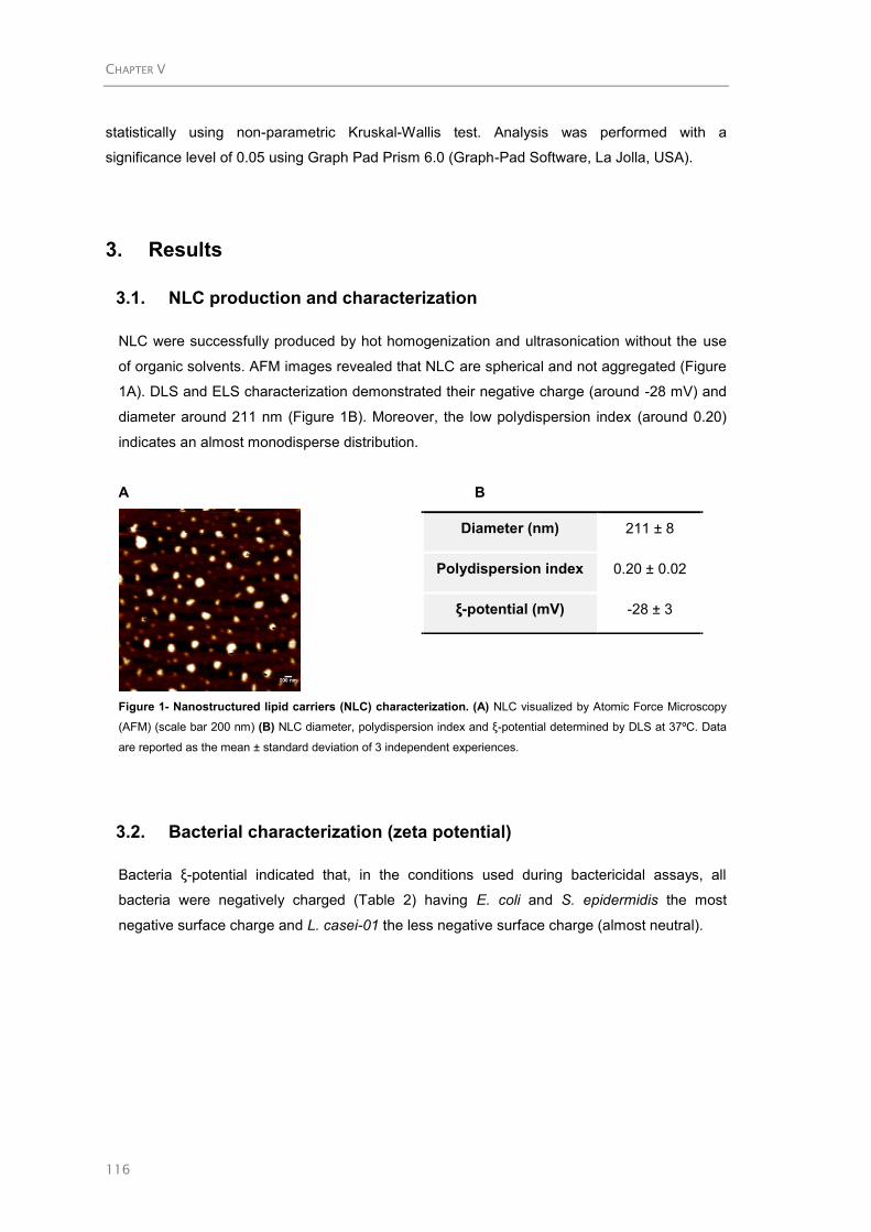
CHAPTER V
116
statistically using non-parametric Kruskal-Wallis test. Analysis was performed with a
significance level of 0.05 using Graph Pad Prism 6.0 (Graph-Pad Software, La Jolla, USA).
3. Results
3.1. NLC production and characterization
NLC were successfully produced by hot homogenization and ultrasonication without the use
of organic solvents. AFM images revealed that NLC are spherical and not aggregated (Figure
1A). DLS and ELS characterization demonstrated their negative charge (around -28 mV) and
diameter around 211 nm (Figure 1B). Moreover, the low polydispersion index (around 0.20)
indicates an almost monodisperse distribution.
A B
Figure 1- Nanostructured lipid carriers (NLC) characterization. (A) NLC visualized by Atomic Force Microscopy
(AFM) (scale bar 200 nm) (B) NLC diameter, polydispersion index and ξ-potential determined by DLS at 37ºC. Data
are reported as the mean ± standard deviation of 3 independent experiences.
3.2. Bacterial characterization (zeta potential)
Bacteria ξ-potential indicated that, in the conditions used during bactericidal assays, all
bacteria were negatively charged (Table 2) having E. coli and S. epidermidis the most
negative surface charge and L. casei-01 the less negative surface charge (almost neutral).
Diameter (nm) 211 ± 8
Polydispersion index 0.20 ± 0.02
ξ-potential (mV) -28 ± 3

CHAPTER V
117
Table 2- Principal features of bacteria used: H. pylori, L. casei, E.coli, S. epidermidis and MRSA.
Helicobacter pylori
(J99 ) Lactobacillus casei-01
Escherichia coli
(ATCC® 25922™)
Staphylococcus
epidermidis
(ATCC® 35984™)
MRSA
(ATCC® 33591™)
Characteristics Gram-negative, spiral-
shaped, microaerophilic
Gram-positive, rod-
shaped, facultative
anaerobic or
microaerophilic
Gram-negative, rod-shaped,
facultative anaerobic
Gram-positive, coagulase-
negative cocci, aerobic
Gram-positive, coagulase-
negative cocci, facultative
aerobe,
Most common
Local (Human)
Stomach
Intestine and mouth
Intestine
Skin Skin, nose and respiratory
tract
Associate
Disease
Gastric disorder: gastritis
to gastric cancer
Used a probiotic
Some of the cases sepsis,
meningitis, and infections
localized in organs
Cholecystitis, bacteremia,
urinary tract infection,
traveler’s diarrhea
Intravascular devices such
as prosthetics, catheter;
septicemia and endocarditis
Skin infections and sepsis to
pneumonia to bloodstream
infections
Zeta potential
(mV) * -17.3 ± 0.7 -2.4 ± 0.2 -30.0 ± 1.2 -31.1 ± 1.6 -19.9 ± 1.3
* ξ-potential measured in Müller-Hinton Broth medium (pH 7) using ZetaSizer®
(n=3).

CHAPTER V
118
3.3. NLC antimicrobial proprieties
Bactericidal activity of developed nanoparticles was evaluated by following bacteria growth
over 24 h in MHB medium supplemented with 10% FBS containing different NLC
concentrations (0, 1.25, 2.5 and 5% v/v) (Figure 2). Minimal bactericidal concentration (MBC)
is the minimal drug concentration to kill 99.9% (≥3 logs) bacteria in 24 h of incubation. Figure
2A e 2B shows that these NLC are bactericidal against H. pylori J99 in all concentrations
tested since after 24 h all bacteria are death. However, H. pylori dead rate was NLC
concentration-dependent. Figure 2A shown that with specific H. pylori medium (BB+10%
FBS), a bactericide activity after 12 h was observed with the lowest concentration tested
(1.25%v/v). However, a slight delay in bactericide activity with MHB+10%FBS was observed,
for the highest NLC concentration tested (5%v/v), H. pylori were killed after 12 h, and for
lower NLC concentrations (1.25% and 2.5%), a reduction of 99.9% (>3log) was observed
after 15 h (Figure 2B). Nonetheless, an inhibitory effect of 50% (> 1log) was detected after 9 h
incubation with any NLC concentration tested (Figure 2B).
NLC bactericidal effect was also evaluated against other bacteria, namely the gut bacteria L.
casei-01 (Figure 2C) and E. coli (Figure 2D), and the infectious bacteria, S. epidermidis
(Figure 2E) and S. aureus (Figure 2F). It was demonstrated that NLC are not bactericidal
against L. casei-01, E. coli, and S. epidermidis at all concentrations tested. However, NLC
was able to delay S. aureus growth when used in high concentrations (5% v/v) (Figure 2F).

CHAPTER V
119
Figure 2- Antibacterial activity of NLC on different bacteria. Bacteria growth in the presence of increasing
concentrations of NLC in Brucella Broth supplemented with 10%FBS (A) H. pylori J99 and in Müller-Hinton broth
supplemented with 10% FBS (B) H. pylori J99, (C) L. casei-01, (D) E. coli ATCCTM
25922, (E) S. epidermidis
ATCCTM
35984, (F) S. aureus ATCCTM
33591. Numerical data is reported as mean ± standard deviation (n=3).*
p<0.05; statically differences between bacteria treated with NLC and untreated (bacteria with 0% v/v NLC) (Kruskal-
Wallis test).
3.4. NLC interaction with H. pylori
In order to understand NLC mechanism of action against H. pylori, different advanced
imaging techniques, namely ImageStreamX®
, SEM, TEM, and AFM were used. Assays were
performed in BB medium supplemented with 10% FBS (the best medium for H. pylori growth)
incubating with NLC at 1.25%. This NLC concentration was chosen due to its high
bactericidal effect despite low cytotoxicity towards gastric cells, as previously described [23].

CHAPTER V
120
Figure 3 shows ImageStreamX®
results of H. pylori J99 after incubation with fluorescence
green-labeled NLC (1.25% coumarin-labeled NLC) for 1.5, 3 and 12 h. These time-points
were chosen in order to have samples where most of the bacteria are cultivable (1.5 and 3 h)
and samples where all bacteria are dead (12 h) (Figure 2A). After 1.5 h, 99% bacteria are
fluorescence green-labeled demonstrating the high affinity of NLC to H. pylori. Nevertheless,
NLC bactericidal effect was only observed after 12 h incubation, since after 1.5 h and 3h,
99% and 70% bacteria were alive, respectively.
H. pylori J99
Control Coumarin-loaded NLC (1.25%)
Time
(hours)
NLC-labeled H. pylori (%)
ImageStreamX®
Viable H. pylori
(%)
CFU
NLC-labelled H. pylori (%)
ImageStreamX®
Viable H. pylori
(%)
CFU
1.5 0
0
100 99
99
100
99
70
0
3 100
100 12 0
Figure 3- NLC interaction with H. pylori membrane after 1.5, 3 and 12 hours incubation with coumarin-labelled
NLC (1.25%) determined by imaging flow cytometry (ImageStreamX®
). Representative histogram and H. pylori
images in brightfield (Channel 1, 420-480 nm) and in green fluorescence corresponding to bacteria with adherent
coumarin-6 labeled NLC (Channel 2, 480-560 nm) after 1.5 h of incubation. Scale bar represents 10 µm. Table
describing percentages of green fluorescence bacteria quantified by flow cytometry and the correspondent viable
bacteria quantified by CFU counting, after 1.5, 3 and 12 h of H. pylori exposure to NLC.
SEM images (Figure 4) revealed that when growing in the absence of NLC (control), bacteria
present their characteristic bacillus shape (Figure 4A and 4C) and no morphological changes
Hp+NLC
1e51e30 1e4
Fluorescence Intensity
4
3
2
5
1
0
No
rmaliz
ed
Fre
quency
Hp+NLC
1e51e30 1e4
Fluorescence Intensity
4
3
2
5
1
0
No
rmaliz
ed
Fre
quency

CHAPTER V
121
were detected in most H. pylori after 3 h incubation with NLC (Figure 4B). However, after
growing during 12 h in contact with NLC, most bacteria suffer a disruption of parts of their
membrane although maintaining the bacillar shape (Figure 4D).
Untreated
A B
1 μm 1 μm
C D
12
h
1 μm 1 μm
1.25% v/v NLC
3h
Figure 4. H. pylori morphology. Scanning Electron micrographs of H. pylori untreated and treated with 1.25% v/v of
NLC for 3 (A and B) and 12h (C and D) respectively. Scale bar in the SEM image represents 1 µm.
TEM images also show H. pylori J99 with intact membrane and dense cytoplasm when
bacteria grew in the absence of NLC (controls) (Figure 5A and 5D). After 3 h growing with
1.25% NLC (Figure 5B), H. pylori maintained their intact membrane and dense cytoplasm as
observed in controls, although Gold-NP-loaded NLC were visualized in their periplasmatic
space (red arrows at Figure 5C). After 12 h incubation with this NLC concentration, bacteria
display a separation between the outer and inner membrane with loss of cytoplasmic contents
(Figure 5E). At this time-point, Gold-NP-loaded NLC were observed inside bacteria, namely at
their cytoplasm (red arrows at Figure 5F).

CHAPTER V
122
100 nm 100 nm 100 nm
100 nm 100 nm 100 nm
A
D
B
E
C
F
3h
12
hUntreated 1.25% v/v NLC 1.25% v/v Gold_NP loaded NLC
Figure 5- Transmission Electron micrographs of H. pylori J99 incubated with NLC (B and E) and Gold-NP-loaded
NLC(C and F) for 3 and 12 h, respectively. Untreated H. pylori J99 were used as control at the same time-points (A
and D). Red arrows point to Gold nanoparticles (Gold_NP). Scale bar in the SEM image represents 100 nm.
AFM was used to identify changes in bacteria morphology of H. pylori J99 upon NLC
treatment. Figure 6 shows AFM deflection images of H. pylori in bacillus shape after 3 and 12
h growing in the absence (6A and 6B, respectively) and the presence of 1.25% NLC (6C and
6D, respectively) in the medium. Although in a bacillary shape, when incubated for 12 h with
NLC, the wall of H. pylori is not uniform along their length (Figure 6D).
Untreated
A B
C D
12
h
1.25% v/v NLC
3h
Figure 6- AFM deflection images of H. pylori J99 incubated with 1.25% v/v of NLC for 3 (B) and 12 (D) hours.
Untreated H. pylori J99 were also tested at 3 (A) and 12 (C) hours. Scale bar in the AFM image represents 2 µm.
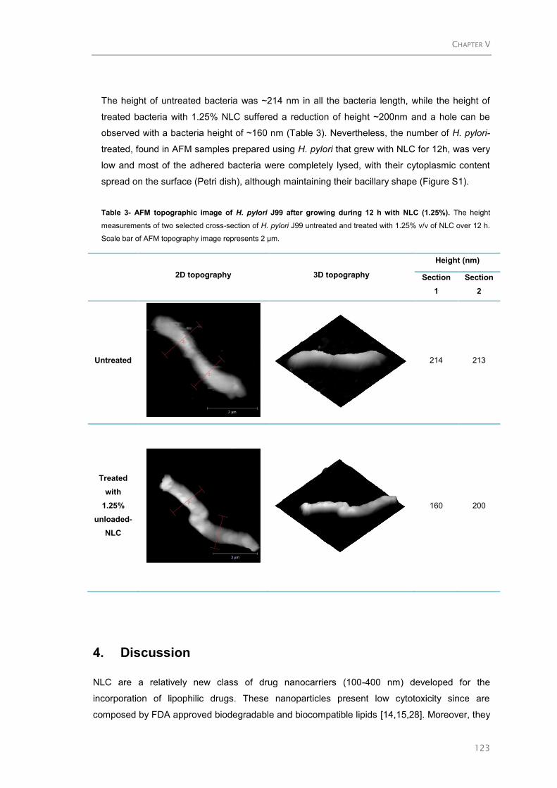
CHAPTER V
123
The height of untreated bacteria was ~214 nm in all the bacteria length, while the height of
treated bacteria with 1.25% NLC suffered a reduction of height ~200nm and a hole can be
observed with a bacteria height of ~160 nm (Table 3). Nevertheless, the number of H. pylori-
treated, found in AFM samples prepared using H. pylori that grew with NLC for 12h, was very
low and most of the adhered bacteria were completely lysed, with their cytoplasmic content
spread on the surface (Petri dish), although maintaining their bacillary shape (Figure S1).
Table 3- AFM topographic image of H. pylori J99 after growing during 12 h with NLC (1.25%). The height
measurements of two selected cross-section of H. pylori J99 untreated and treated with 1.25% v/v of NLC over 12 h.
Scale bar of AFM topography image represents 2 µm.
2D topography 3D topography
Height (nm)
Section
1
Section
2
Untreated
214 213
Treated
with
1.25%
unloaded-
NLC
160 200
4. Discussion
NLC are a relatively new class of drug nanocarriers (100-400 nm) developed for the
incorporation of lipophilic drugs. These nanoparticles present low cytotoxicity since are
composed by FDA approved biodegradable and biocompatible lipids [14,15,28]. Moreover, they

CHAPTER V
124
are produced without organic solvents. Concerning infection treatments/prevention, NLC have
been used for the encapsulation of different antibiotics, peptides and natural compounds with
activity against several bacteria, namely S. aureus, Pseudomonas aeruginosa, E. coli and
Propionibacterium acnes [16-21].
We previously demonstrated that the bactericidal effect of docosahexaenoic acid (DHA) against
H. pylori was improved after its NLC encapsulation [23]. However, we verified that unloaded-
NLC (with diameter ~211 nm and a negative charge around -28 mV) were also bactericidal
against H. pylori J99 (human strain) and SS1 (mouse-adapted strain) although at a slower rate
than DHA-loaded NLC [23]. This direct bactericidal effect of unloaded-NLC was not expected
and justifies further investigation, namely regarding its effect in other bacteria.
To investigate their selectivity against H. pylori, NLC were tested against other bacteria that
present different morphological characteristics regarding shape (rod vs spherical), membrane
structure (Gram-positive vs Gram-negative) and surface charge (from -2mV to -30 mV). Assays
were performed following bacteria growth over 24 h using Müller-Hinton Broth (MHB) medium,
which is the recommend medium for determination of the minimal inhibitory concentration (MIC)
according to the guidelines recognized by CLSI (Clinical & Laboratory Standards Institute) and
EUCAST (European Society of Clinical Microbiology and Infectious Diseases) organizations
[25]. However, since H. pylori is a fastidious microorganism that requires a complex nutrient-rich
growth media, the MHB medium was supplemented with 10% of fetal bovine serum (FBS) [26].
Therefore, for comparative studies between bacteria, all antimicrobial assays were performed
using MHB supplemented with 10% FBS. In this medium, NLC bactericidal activity against H.
pylori J99 was similar to Brucella Broth medium (BB) also supplemented with 10% FBS (Figure
2A), the recommended medium for H. pylori culture [27]. However, a slight delay in the
bactericidal activity of NLC was observed with MHB+10%FBS medium. NLC did not affect the
growth of other bacteria tested, except in S. aureus, where NLC at 5% was able to decrease
bacteria growth after 6 h, although without a bactericidal effect. Nevertheless, NLC
concentration as high as 5% v/v was previously associated with some cytotoxicity (~50%) in
human gastric adenocarcinoma cells [23].
Thus, this specific bactericidal effect of NLC against H. pylori cannot be explained by
differences in bacteria morphology and surface charge, but rather through H. pylori specific
features. H. pylori has specific and unique characteristics that are very different from all the
other bacteria. To be able to colonize human gastric mucosa and to survive in the adverse
environment of host stomach, H. pylori present several structural and morphological
characteristics that favor their penetration through the mucus layer, namely their flagella and
their spiral form [29]. This spiral morphology needs a less rigid cell envelope than other non-
spiraled bacteria. This fact could be related to the composition of their peptidoglycan and cell
membranes that differ substantially from other bacteria. Their peptidoglycan has a unique and
less complex muropeptides composition than other Gram-negative bacteria. It contains a high
proportion of muropeptides, with a pentapeptide side chain ending in glycine and containing
anhydro-N-acetylmuramic acid, which is very different than E. coli [30]. Concerning their

CHAPTER V
125
membrane composition, H. pylori have an unusual fatty acid profile and have glycosylated
cholesterol which is a unique characteristic of Helicobacter sp.[30]. According to literature, H.
pylori steal host cholesterol, modifies it by alpha-glycosylation and incorporates it into its
surface to resist multiple stresses [31]. H. pylori lipopolysaccharides (LPS), is also an important
component of the wall of H. pylori that differs substantially from other bacteria. H. pylori LPS are
composed by an unusual cellular fatty acid, lipid profile and molecular mimicry of Lewis
antigens[30,32]. The lipid A component of H. pylori LPS is different of lipid A of many bacterial
species by the absence of 3-hydroxytetradecanoic (β-hydroymyristic) acid [14:0(3-OH)], as well
as the underphosphorylation and underacylation of the lipid A [33]. Moreover, the core
oligosaccharide of H. pylori strains exhibit an unusual conformation, compared with other
bacterial species. A branching occurs from the D-glycerol-D-manno-heptose (DD-Hep) residue
in the inner core through a second such a residue to which the first repeating unit of the O-
polysaccharide chain is attached [33]. Additionally, H. pylori have several adhesins on its outer
membrane to bind specific glycosylated compounds found in gastric mucosa [34]. Periplasmic
pH homeostasis of H. pylori contrasts with the patterns in E. coli and other Gram-negative
bacteria. The periplasmic pH is thought to be in equilibrium with the medium and not regulated
in concert with the cytoplasm. H. pylori express the highest level of urease of any known
microorganism, a key component of periplasmic pH homeostasis, thus elevating the pH to
neutral as necessary for its survival [35]. The production of urease by the bacterium results in
the production of ammonia, maintaining periplasmic and cytoplasmic pH of the bacterium near
to neutral even in the presence of acid shocks [29,36].
Nanoparticles that have been designed to directly interact with bacteria are usually cationic in
order to bind any bacteria via electrostatic interactions, since pathogenic bacteria have a
negative surface charge under physiological conditions [37]. These NLC are negatively charged
so, their interaction with bacteria via electrostatic interactions is not expected. In addition, all the
individual compounds used in NLC production (Tween®60, Miglyol®812 and Precirol
®ATO5) are
not bactericidal at concentrations used [23,38-40] demonstrating that the bactericidal effect is
related with nanoparticle 3D format.
In order to understand NLC mechanism of action against H. pylori, different bioimaging
techniques were applied. These assays were performed using NLC at 1.25% and H. pylori J99
cultured in BB medium supplemented with 10% FBS, recommended medium for H. pylori
growth. In these conditions, NLC were bactericidal against H. pylori in 12h. Studies using
ImageStreamX®
revealed the high binding affinity of NLC to H. pylori membrane since, after
1.5h in contact with fluorescent labeled NLC, all bacteria were fluorescent labeled. However,
their bactericidal effect was only observed after 12h, demonstrating that this bactericidal
process although being concentration-dependent is also time-dependent.
TEM studies using Gold-NP-loaded NLC showed that NLC can be internalized by H. pylori in a
time-dependent process. After 3 h, NLC were found in bacterial periplasmic space and after 12
h in the cytoplasm showing their ability to cross both membranes of H. pylori. TEM images also
revealed a separation between the outer and cytoplasmic membrane of H. pylori after 3 and 12

CHAPTER V
126
h incubation with NLC. A leakage of bacterial cytoplasmic content and a decrease in bacteria
thickness (AFM) was also observed using SEM and AFM. Nevertheless, although these
morphological alterations, bacteria maintained their typical bacillary shape. These results
suggest that after their physically binding to H. pylori surface, NLC are able to cross both
bacterial plasma membranes destabilizing them or creating pores in their structure which allow
cytoplasmic leakage without destroying bacteria wall.
The non-visualization of H. pylori in coccoid shape, as observed in other studies when bacteria
were exposed to a hostile environment [41], can support the idea that the binding process NLC-
H. pylori is fast not allowing their morphological transition. Similar H. pylori morphological
changes were described by Jung et al (2015) for liposome containing linolenic acid [42]. Their
bactericidal effect was explained by liposomal fusion into H. pylori outer membrane allowing the
incorporation of linolenic acid in the membrane and thus affecting their permeability due to the
alteration of the phospholipid composition. Interestingly, these liposomal have negative zeta-
potential (-73 ± 2 mV), like our NLC, Their negative charge did not avoid their integration/fusion
into the bacteria membrane. Moreover, although negatively charged, NLC have on their surface
the hydrophilic moiety of the non-ionic surfactant (Tween®60) used during the production as a
nanoparticle stabilizer. This non-ionic surfactant may decrease the surface tension between
nanoparticle and bacteria enabling their interaction with proteins and lipids of H. pylori
membrane [43]. NLC surface surfactant can also facilitate their interaction/travel through H.
pylori LPS since Gram-negative bacteria only allows a limited diffusion of hydrophobic
substances through their LPS-covered surface [44]. Moreover, NLC lipid composition may also
allow the insertion of NLC into the bacterial membrane, causing an excessive enhance of
membrane fluidity and it to become more permeable, allowing internal contents to leak from the
cell and consequently bacteria death. Notwithstanding, the surfactant effect may solubilize
sections of the cell membrane, which may cause cytoplasm leakage and consequently cell
lysis.
This specific NLC bactericidal activity against H. pylori is an advantage for the development of
new and alternative therapies for H. pylori infection since, in opposite to the antibiotic therapy; it
will not affect human microbiota.
Acknowledgments
This work was financed by FEDER funds through the COMPETE 2020 (POCI), Portugal 2020.
FCT/MCTES through the projects: POCI-01-0145-FEDER-007274; PYLORIBINDERS
(PTDC/CTM-BIO/ 4043/2014); PYLORICIDAL (PTDC/CTM-BPC/121149/2010), NORTE-01-
0145-FEDER-000012 and FCT grant SFRH/BD/89001/ 2012.

CHAPTER V
127
DLS, ELS, and AFM have performed at BN-Biointerfaces and Nanotechnology unit facility;
imaging flow cytometry at b.IMAGE-Bioimaging Center for Biomaterials and Regenerative
Therapies , TEM at HEMS – Histology and Electron Microscopy Service Unit facility. All of these
unit facilities are located at i3S-Instituto de Investigação e Inovação em Saúde da Universidade
do Porto (www.i3s.up.pt/research/scientific-platforms). SEM studies were performed at the
CEMUP- Centro de Materiais da Universidade do Porto unit facility (http://www.cemup.up.pt)
The authors thank Prof. Manuela Pintado (Universidade Católica do Porto, Portugal) for
providing the Lactobacillus casei-01 strain; and Prof. Thomas Boren (Umeã University,
Sweden) for providing the H. pylori J99 strain.
Appendix A. Supplementary data
Figure S1- AFM deflection image of H. pylori J99 incubated with 1.25% v/v of NLC for 12 h. Scale bar in the AFM image represents 5 µm
References
1. Correa P, Houghton J (2007) Carcinogenesis of Helicobacter pylori. Gastroenterology 133: 659-672.
2. Amieva M, Peek Jr RM (2016) Pathobiology of Helicobacter pylori–Induced Gastric Cancer.
Gastroenterology 150: 64-78.
3. Matysiak-Budnik T, Mégraud F (2006) Helicobacter pylori infection and gastric cancer. European
Journal of Cancer 42: 708-716.
4. IARC (2014) Helicobacter pylori Eradication as a Strategy for Preventing Gastric Cancer.; Group IHpW,
editor. Lyon, France: International Agency for Research on Cancer.
CHAPTER V
128
5. Ferlay J, Soerjomataram I, Dikshit R, Eser S, Mathers C, et al. (2015) Cancer incidence and mortality
worldwide: sources, methods and major patterns in GLOBOCAN 2012. Int J Cancer 136: E359-386.
6. Correa P (2013) Gastric Cancer: Overview. Gastroenterology Clinics of North America 42: 211-217.
7. Vakil N (2006) Helicobacter pylori treatment: a practical approach. Am J Gastroenterol 101: 497-499.
8. Poonia N, Kharb R, Lather V, Pandita D (2016) Nanostructured lipid carriers: versatile oral delivery
vehicle. Future Science OA 2: FSO135.
9. Beloqui A, Solinis MA, Delgado A, Evora C, Isla A, et al. (2014) Fate of nanostructured lipid carriers
(NLCs) following the oral route: design, pharmacokinetics and biodistribution. J Microencapsul 31: 1-8.
10. Beloqui A, Solinis MA, Rodriguez-Gascon A, Almeida AJ, Preat V (2016) Nanostructured lipid carriers:
Promising drug delivery systems for future clinics. Nanomedicine 12: 143-161.
11. Martins S, Sarmento B, Ferreira DC, Souto EB (2007) Lipid-based colloidal carriers for peptide and
protein delivery--liposomes versus lipid nanoparticles. Int J Nanomedicine 2: 595-607.
12. Carbone C, Leonardi A, Cupri S, Puglisi G, Pignatello R (2014) Pharmaceutical and biomedical
applications of lipid-based nanocarriers. Pharm Pat Anal 3: 199-215.
13. Battaglia L, Gallarate M (2012) Lipid nanoparticles: state of the art, new preparation methods and
challenges in drug delivery. Expert Opinion on Drug Delivery 9: 497-508.
14. Müller RH, Mäder K, Gohla S (2000) Solid lipid nanoparticles (SLN) for controlled drug delivery – a
review of the state of the art. European Journal of Pharmaceutics and Biopharmaceutics 50: 161-177.
15. Muller RH, Shegokar R, Keck CM (2011) 20 years of lipid nanoparticles (SLN and NLC): present state
of development and industrial applications. Curr Drug Discov Technol 8: 207-227.
16. Boge L, Bysell H, Ringstad L, Wennman D, Umerska A, et al. (2016) Lipid-Based Liquid Crystals As
Carriers for Antimicrobial Peptides: Phase Behavior and Antimicrobial Effect. Langmuir 32: 4217-4228.
17. Garcia-Orue I, Gainza G, Girbau C, Alonso R, Aguirre JJ, et al. (2016) LL37 loaded nanostructured
lipid carriers (NLC): A new strategy for the topical treatment of chronic wounds. European Journal of
Pharmaceutics and Biopharmaceutics 108: 310-316.
18. Lin CH, Fang YP, Al-Suwayeh SA, Yang SY, Fang JY (2013) Percutaneous absorption and
antibacterial activities of lipid nanocarriers loaded with dual drugs for acne treatment. Biol Pharm Bull
36: 276-286.
19. Manea A-M, Vasile BS, Meghea A (2014) Antioxidant and antimicrobial activities of green tea extract
loaded into nanostructured lipid carriers. Comptes Rendus Chimie 17: 331-341.
20. Moreno-Sastre M, Pastor M, Esquisabel A, Sans E, Viñas M, et al. (2016) Pulmonary delivery of
tobramycin-loaded nanostructured lipid carriers for Pseudomonas aeruginosa infections associated
with cystic fibrosis. International Journal of Pharmaceutics 498: 263-273.
21. Sans-Serramitjana E, Fusté E, Martínez-Garriga B, Merlos A, Pastor M, et al. (2016) Killing effect of
nanoencapsulated colistin sulfate on Pseudomonas aeruginosa from cystic fibrosis patients. Journal of
Cystic Fibrosis 15: 611-618.
22. Correia M, Michel V, Matos AA, Carvalho P, Oliveira MJ, et al. (2012) Docosahexaenoic Acid Inhibits
Helicobacter pylori Growth In Vitro and Mice Gastric Mucosa Colonization. PLOS ONE 7: e35072.
23. Seabra CL, Nunes C, Gomez-Lazaro M, Correia M, Machado JC, et al. (2017) Docosahexaenoic acid
loaded lipid nanoparticles with bactericidal activity against Helicobacter pylori. International Journal of
Pharmaceutics 519: 128-137.
24. Neves AR, Lucio M, Martins S, Lima JL, Reis S (2013) Novel resveratrol nanodelivery systems based
on lipid nanoparticles to enhance its oral bioavailability. Int J Nanomedicine 8: 177-187.
25. Matuschek E, Brown DFJ, Kahlmeter G (2014) Development of the EUCAST disk diffusion
antimicrobial susceptibility testing method and its implementation in routine microbiology laboratories.
Clinical Microbiology and Infection 20: O255–O266.

CHAPTER V
129
26. Kusters JG, van Vliet AHM, Kuipers EJ (2006) Pathogenesis of Helicobacter pylori Infection. Clinical
Microbiology Reviews 19: 449-490.
27. Blanchard TG, Nedrud JG (2006) Laboratory Maintenance of Helicobacter Species. Current Protocols
in Microbiology CHAPTER: Unit8B.1-Unit8B.1.
28. Mehnert W, Mäder K (2001) Solid lipid nanoparticles: Production, characterization and applications.
Advanced Drug Delivery Reviews 47: 165-196.
29. Lopes D, Nunes C, Martins MCL, Sarmento B, Reis S (2014) Eradication of Helicobacter pylori: Past,
present and future. Journal of Controlled Release 189: 169-186.
30. O'Toole PW, Clyne M (2001) Cell Envelope. In: Mobley HLT, Mendz GL, Hazell SL, editors.
Helicobacter pylori: Physiology and Genetics. Washington (DC).
31. McGee DJ, George AE, Trainor EA, Horton KE, Hildebrandt E, et al. (2011) Cholesterol enhances
Helicobacter pylori resistance to antibiotics and LL-37. Antimicrob Agents Chemother 55: 2897-2904.
32. O'Rourke J, Bode G (2001) Morphology and Ultrastructure. In: Mobley HLT, Mendz GL, Hazell SL,
editors. Helicobacter pylori: Physiology and Genetics. Washington (DC).
33. AP. M (2001) Molecular Structure, Biosynthesis, and Pathogenic Roles of Lipopolysaccharides. In:
HLT M, GL M, SL H, editors. Helicobacter pylori: Physiology and Genetics. Washington (DC): ASM
Press.
34. Magalhães A, Ismail MN, Reis CA (2010) Sweet receptors mediate the adhesion of the gastric
pathogen Helicobacter pylori: glycoproteomic strategies. Expert Review of Proteomics 7: 307-310.
35. Krulwich TA, Sachs G, Padan E (2011) Molecular aspects of bacterial pH sensing and homeostasis.
Nat Rev Micro 9: 330-343.
36. Gonçalves IC, Henriques PC, Seabra CL, Martins MCL (2014) The potential utility of chitosan
micro/nanoparticles in the treatment of gastric infection. Expert Review of Anti-infective Therapy 12:
981-992.
37. Gao W, Thamphiwatana S, Angsantikul P, Zhang L (2014) Nanoparticle approaches against bacterial
infections. Wiley Interdisciplinary Reviews: Nanomedicine and Nanobiotechnology 6: 532-547.
38. Blackburn P (1997) Polysorbate-containing compositions and their use against Helicobacter. European
Patent Office WO/1997/036600.
39. Cerreto F, Paolicelli P, Cesa S, Abu Amara HM, D'Auria FD, et al. (2013) Solid lipid nanoparticles as
effective reservoir systems for long-term preservation of multidose formulations. AAPS PharmSciTech
14: 847-853.
40. Santucci A, Figura N, Spreafico A, Cavallo G, Marcolongo RF (2013) Compositions for treating
Helicobacter pylori infection. European Patent Office WO2011IT00175.
41. Nogueira F, Gonçalves IC, Martins MCL (2013) Effect of gastric environment on Helicobacter pylori
adhesion to a mucoadhesive polymer. Acta Biomaterialia 9: 5208-5215.
42. Jung SW, Thamphiwatana S, Zhang L, Obonyo M (2015) Mechanism of antibacterial activity of
liposomal linolenic acid against Helicobacter pylori. PloS one 10: e0116519.
43. Ishikawa S, Matsumura Y, Katoh-Kubo K, Tsuchido T (2002) Antibacterial activity of surfactants
against Escherichia coli cells is influenced by carbon source and anaerobiosis. Journal of Applied
Microbiology 93: 302-309.
44. Hamouda T, Baker JR (2000) Antimicrobial mechanism of action of surfactant lipid preparations in
enteric Gram-negative bacilli. Journal of Applied Microbiology 89: 397-403.
45. Rodrigues D, Rocha-Santos T, Sousa S, Gomes AM, Pintado MM, et al. (2011) On the viability of five
probiotic strains when immobilised on various polymers. International Journal of Dairy Technology 64:
137-144.

CHAPTER V
130
46. Monteiro C, Fernandes M, Pinheiro M, Maia S, Seabra CL, et al. (2015) Antimicrobial properties of
membrane-active dodecapeptides derived from MSI-78. Biochimica et Biophysica Acta (BBA) -
Biomembranes 1848: 1139-1146.

131
CHAPTER VI
DEVELOPMENT OF LIPID NANOPARTICLES FOR THE
TREATMENT OF HELICOBACTER PYLORI INFECTION IN
MICE
Manuscript in preparation

CHAPTER VI
132

CHAPTER VI
133
Abstract
Persistent Helicobacter pylori (H. pylori) infection may lead to several gastric complications
such as gastritis, peptic ulcer, and gastric carcinoma. Current antibiotic-based treatments
are unsuccessful in approximately 20% of the patients due, principally, to the increase
number of H. pylori strains resistant to available antibiotics. We recently demonstrated that
lipid nanoparticles, namely nanostructured lipid carriers (NLC) loaded with
docosahexaenoic acid (DHA), an omega-3 fatty acid with anti-H. pylori activity and
unloaded-NLC were both bactericidal against H. pylori.
The major goal of this work was to evaluate if these NLC may be used in the treatment of
H. pylori infection. For that, their stability in simulated gastric fluidic and their ability to cross
gastric mucus layer was firstly evaluated using in vitro mice model, respectively.
Nanoparticles toxicity and efficacy in H. pylori eradication were assessed using C57BL/6
mice infected with mouse-adapted H. pylori strain (SS1). Additionally, in vitro assays were
done to assess the crystalline structure of these lipid nanoparticles and to estimate their
effect on H. pylori membrane permeability.
Homogenous and spherical negatively charged DHA-loaded NLC (302±14 nm) and
unloaded (211±8nm) were stable in simulated gastric fluid for 3h. DSC results
demonstrated a crystallinity reduction of DHA-loaded NLC relatively to unloaded-NLC
meaning that DHA -loaded NLC have a lower crystal organization than unloaded-NLC and
their bulk material.
A fast (30 min) bactericidal activity of DHA-loaded NLC was observed, which was
explained by their higher effect on the permeability of bacteria membrane than unloaded-
NLC at this time point. In vivo efficacy assays, using H. pylori-infected C57BL/6 mice
treated with nanoparticles (2.5% v/v) during 14 days, demonstrated a reduction in H. pylori
colonization of around 92.6% when mice were treated with DHA-loaded NLC (50 μM DHA)
and 82.5% when treated with unloaded-NLC. This reduction of infection was verified without
causing gastric or hepatic toxicity. However, 1/3 of infected but untreated mice (control)
were not infected after the 14 days treatment (with water), demonstrating the need for
further assays.
This work demonstrated that these lipid nanoparticles, mainly the ones containing DHA, are
very effective in vitro against H. pylori due to their action on the increase of bacteria
membrane permeability. In vivo, preliminary results also suggested that lipid nanoparticles
should be considered as a promising alternative for the treatment of H. pylori gastric
infection.
KEYWORDS: Helicobacter pylori, NLC, in vivo efficiency, gastric mucosa

CHAPTER VI
134
1. Introduction
Helicobacter pylori (H. pylori) is one the most common bacterial pathogen Worldwide, with
a strong association to several gastric diseases, including gastritis, peptic ulcers, and
gastric cancer. Gastric cancer is the fifth most incident type of cancer and the second
leading cause of cancer-related deaths in the world [1,2]. The increasing prevalence of
antibiotic-resistant strains has led to a significant decrease in the success of H. pylori
eradication [3-5]. Different regimen treatments, including antibiotics combinations, have
been used to overcome this problem. However, poor patient compliance, side-effects and
high cost associated with multiple antibiotics used are also related to therapy failure [6],
demonstrating the urgent need for alternative treatments.
Several antibiotic-free approaches have been investigated to fight H. pylori infection such
as antimicrobial peptides [7-9], lipophilic compounds [10-14], the use of probiotics [15-18],
the application of bioengineering strategies for drug delivery or binding bacteria [19-24] and
also, the development of vaccines [25-28]. However, although very successful in vitro, none
of these alternatives were sufficiently effective to be used, by itself, in the H. pylori infection
eradication.
Some polyunsaturated fatty acids (PUFA), namely docosahexaenoic acid (DHA), linolenic
acid (LA) and eicosapentaenoic acid (EPA) are bactericidal against a broad range of
bacteria and have a lower tendency to induce bacteria resistant than conventional
antibiotics [29-31]. Although some PUFA as DHA have been very effective against H. pylori
in vitro [29,32-36], their poorly bioavailability in vivo needs to be overcome. This can be
related to their low stability, namely high susceptibility to oxidation, or their low ability to
cross gastric mucus layer to reach the infection site.
The development of local delivery systems may improve PUFA in vivo bioavailability. It was
recently demonstrated by us that nanostructured lipid carriers (NLC) containing DHA (DHA-
loaded NLC) are highly bactericidal against H. pylori by membrane disruption. However,
unloaded-NLC were also bactericidal, demonstrating that the DHA-loaded NLC bactericidal
effect was not only due to the DHA released, but due to a synergistic effect of NLC and
DHA [37].
The main goal of this work was to evaluate if NLC (unloaded and DHA-loaded) may be
used in the treatment of H. pylori infection. In vivo studies, using C57BL/6 mice infected
with mouse-adapted H. pylori strain (SS1) were then performed. The toxicity profile of
developed nanoparticles in mouse stomach was also evaluated through histological
analysis. Additionally, we evaluate the effect of NLC in the permeability of bacteria
membrane, to understand how NLC may disrupt bacteria membrane. To evaluate the
physical state of the lipid core in the produced NLC and to correlate this parameter with the
DHA incorporation, additional NLC characterization was performed by DSC analysis.

CHAPTER VI
135
2. Material & methods
2.1. Preparation and characterization of NLC
2.1.1. NLC production
NLC was prepared by hot homogenization and sonication as described by Seabra et al
(2017) [37]. Briefly, 200 mg of Precirol®ATO5 (Gattefosé, France), 90 mg of Miglyol
®812
(Acofarma, Spain) and 60 mg Tween®60 (Merck, Germany) were weighted and heated
together at 60ºC. Then 84μL of DHA and 4.2 mL of water type I (preheated at 60ºC) were
added and mixed using an ultra-turrax (T25, Janke and Kunkel IKA-Labortechnik,
Germany) at 12000 rpm, over 20 s, and sonicated using a Vibra-Cell model VCX 130, with
a VC 18 probe of ¼’’ (6 nm), at 60% amplitude for 5 min (Sonics and Materials Inc,
Newtown, USA), obtaining DHA-loaded NLC.
2.1.2. NLC characterization
DHA-loaded NLC was characterized in terms of size and morphology as described by
Seabra et al (2017) [37].
2.1.2.1. Differential Scanning Calorimetry (DSC)
Differential scanning calorimetry (DSC) analysis was performed using a Perkin-Elmer Pyris
1 DSC differential scanning calorimeter (Perkin-Elmer Analytical Instruments, Waltham,
USA). The samples were weighed directly in aluminum pans (7-23 mg) and scanned
between 10ºC and 80ºC at a heating rate of 5ºC/min and cooling rate of 40ºC/min under a
stream of nitrogen gas. An empty aluminum pan was used as reference. DSC analyses
were performed for the lipid nanoparticles, as well as for the bulk materials used in the
preparation of the nanoparticles. The melting point (peak maximum) and the enthalpy (ΔH)
were calculated using the PerkinElmer software.
2.1.2.2. Stability in simulated gastric fluid
DHA-loaded NLC stability was performed using a simulated gastric fluid (SGF) prepared
with HCl 0.2M and NaCl 0.2M (pH1.2). DHA-loaded NLC were incubated with SGF over 3 h
at 37ºC and 150 rpm. At time-point 0 and 3 h, samples were diluted (1:20000) in water type
I and analyzed in terms of particle concentration using NanoSight® NS300 (Malvern
Instruments, UK). The measurements were performed in triplicate.

CHAPTER VI
136
2.2. Anti-Helicobacter pylori activity: in vitro study
2.2.1. H. pylori culture
H. pylori J99 strain (human isolate provided by Department of Medical Biochemistry and
Biophysics, Umeã, Sweden) were cultured as described by Seabra et al. (2017) [37].
Briefly, H. pylori J99 spots and spreads were subsequently incubated at 37ºC for 48 h,
under microaerophilic conditions. H. pylori medium plates were prepared using 20g of
blood agar base 2 (Oxoid, France) supplemented with 10% v/v of defibrinated horse blood
(Probiológica, Portugal) and with 0.2% v/v antibiotics-cocktail composed of 0.155 g/L
polymixin B (Sigma-Aldrich, USA), 6.25 g/L vancomycin (Sigma-Aldrich, USA), 1.25 g/L
amphotericin B (Sigma-Aldrich, USA) and 3.125 g/L trimethroprim (Sigma-Aldrich, USA).
Afterwards, the optical density of bacteria was adjusted to 0.1 (λ=600 nm) and H. pylori
were incubated in Brucella Broth medium (BB, Oxoid, France) supplemented with 10% of
fetal bovine serum (FBS, Gibco, USA), at 37ºC, 150 rpm, under microaerophilic conditions,
over 18-20 h.
2.2.2. NLC activity against H. pylori membrane
The H. pylori optical density was adjusted to 0.03 (1×10⁷ bacteria/mL) and incubated with
unloaded-NLC (2.5% v/v) and DHA-loaded NLC (2.5% v/v containing 50μM DHA). The
respective control was performed with untreated bacteria (incubated with water). Bacteria
culture was performed at 15, 30, 90 and 150 minutes in bacteria medium, under
microaerophilic conditions, at 37ºC and 150 rpm.
2.2.2.1. Colony forming units (CFU)
At different time-points (15, 30, 90 and 150 min), 200 µL of each culture (control, unloaded
and DHA-loaded NLC) was collected, serially diluted and plated on H. pylori medium plates
and incubated at 37ºC. After 5 days, the number of viable bacteria was determined by
colony forming unit (CFU) counting.
2.2.2.2. Permeability assay
2.2.2.2.1. Outer membrane
The outer membrane permeability was evaluated by adapting the methods described
by Loh et al (1984)[38] and Alakoni et al. (2006) [39], measuring the uptake of the

CHAPTER VI
137
fluorescent probe 1-N-phenylnaphthetlamine (NPN). After 30 min of incubation with
unloaded- and DHA-loaded NLC, as described above (2.2.2.), bacteria were collected
by centrifugation at 3000 g at 25ºC for 5 min and washed twice with 0.01M phosphate
buffer (PBS 1x; pH 7.4). A negative control (bacteria growing in bacteria medium) and a
positive control (bacteria treated with 10% v/v of Triton TM X-100) were included, in
order to normalize the results. The bacterial pellet was resuspended in 5 mM of n-
heptadecanoic acid methyl ester buffer (HEPES buffer; pH 7.2). For NPN assay (at
room temperature), 50μL of NPN (10μM final concentration) were added and mixed
with 150μL of bacteria suspension. Fluorescence was measured after shaking for 3
minutes, using a microplate reader (Synergy™H Multi-mode Microplate Reader, BioTek
Instruments, USA) at an excitation wavelength of 350 nm and an emission wavelength
of 420 nm. The measurements were repeated three times.
2.2.2.2.2. Inner membrane
The plasma membrane permeability was evaluated by measuring the ATP release from
bacterial cells, using BacTiter-Glo microbial cell viability assay kit (Promega Inc, USA),
as described by Parsons et al (2012) [40] and Jung et al. (2015) [10]. After H. pylori
treatment with unloaded and DHA-loaded NLC for 30 min, as described above (2.2.2),
bacteria were centrifuged at 14 000 g for 5 minutes and the supernatant containing
released ATP was collected. Then, 100 μL of supernatant were placed in a 96-well
plate and BacTiter-Glo reagent was added directly to the supernatant at ratio 1:1. The
mixture was shaken for 2 minutes and using a microplate reader (Synergy™ H Multi-
mode Microplate Reader, BioTek Instruments, USA), the luminescence was measured.
The measurements were repeated three times.
2.3. Anti-Helicobacter pylori activity: in vivo study
2.3.1. Mice infection
Six-week-old specific pathogen-free C57BL/6 male mice (Charles Rivers, France) were
divided into six groups: non-infected mice (negative control group; n=3), two non-infected
mice and treated group with unloaded (n=2) and DHA-loaded NLC (n=2) for toxicity
evaluation, H. pylori-infected mice (positive control group, n=12) and two H. pylori-infected
groups that were treated with unloaded (n=12) and DHA-loaded NLC (n=12). H. pylori SS1
(mouse-adapted strain provided by Unité de Pathogenèse de Helicobacter, Institute
Pasteur, France) was cultured as described in section 2.2.1. Mice received 100μL of 1×10¹⁰
CFU/mL of H. pylori strain SS1 in peptone water (Sigma-Aldrich, USA) administrated
intragastrically through oral gavage twice with a 2 dyes interval and infection was allowed to

CHAPTER VI
138
develop for 4 weeks. The control groups of mice were given peptone water alone. 24 h
before infection, mice were weighted and food and bedding were removed. Water was
removed 6 h before administration. 3 h after H. pylori administration, mice were switched
back to the original cages with food, water, and bedding. All animal procedures were
performed in compliance with the laws and institutional guidelines of the Ethical Committee
of i3S Animal Facilities.
2.3.2. NLC treatment and mice sacrifice
Four weeks after infection, treatment was performed with unloaded-NLC (2.5% v/v) and
DHA-loaded NLC (2.5% v/v containing 50 μM DHA). For the treatment, mice were first
fasted for 6 h and then 100μL of NLC (unloaded or DHA-loaded) were administrated
through oral gavage once a day for consecutive 14 days. The control groups of mice were
given water. The treatment plan applied to the mice is represented in Scheme 1.
Scheme 1- Experimental protocol including the H. pylori SS1 inoculation, infection development, and treatment of
C57BL/6 infected mice.
After the last administration, mice were fasted for 18 h and then euthanized by cervical
dislocation, with previous anesthesia (50-75 mg/kg Ketamine and 0.5-1 mg/kg
Medetomidine) through intraperitoneal injection. The abdominal cavity was opened and a
liver section was removed. Then, mouse stomach was removed, rinsed in 0.9% NaCl (to
remove food) and opened transversally. The mouse stomach was divided into 4
longitudinal sections (Scheme 2): one section for cryopreservation, another for histological
analysis and the two other sections (part A and B) were placed in a tube containing 400 μL
of peptone water, for CFU counting, evaluating the reduction of H. pylori colonization. Each
stomach section (part A and B) was smashed with a tissue homogenizer using sterile
pistons, and then serial dilutions (101, 10
2, and 10
3) were made and plated on H. pylori
medium plates, described above, and supplemented with 20 mg/mL of naladixic acid
(Sigma-Aldrich, USA) and 50mg/mL bacitracin (Sigma-Aldrich, USA). After 15 days, at
37ºC under microaerophilic conditions, the number of colony forming units (CFU) per mice
stomach was determined.

CHAPTER VI
139
Scheme 2- Mouse stomach segments for cryopreservation, histological analysis, and CFU counting evaluation.
Liver section and longitudinal section of mice stomach were fixed in formalin and then
embedded in paraffin and cut transversally in to 3 µm sections and gastric mucosa and
liver sections were placed on glass slides.
Sections were deparaffinated with xylene and hydrated in ethanol (100%, 96%, and 70%),
running water and the hematoxylin and eosin (H&E) staining was performed. After staining
sections were mounted with mounting medium (Entellan®, Merck, Germany) and observed
using an optical microscope, evaluating the NLC (unloaded- and DHA-loaded NLC) toxicity
2.4. Statistical analysis
The statistical analysis was performed using Graph Pad Prism 6.0 Software (La, Jolla,
USA) applying non-parametric Kruskal-Wallis test or One-way ANOVA (Tukey test),
considering statically significant differences at p<0.05.
3. Results
3.1. NLC production and in vitro characterization
DHA was loaded into NLC forming homogeneous and spherical nanoparticles with a
hydrodynamic diameter of 302 ± 14 nm (Figure 1B and 1D). Unloaded-NLC were also
produced as a control of DHA encapsulation, having a diameter of 211±8 nm (Figure 1A
and 1C).
NLC thermometric behavior was compared with their physical mixture of their excipients
(bulk material). DSC thermograms and respective thermometric parameters as melting
point, enthalpy, and recrystallization index (RI) for the bulk materials and NLC are shown in
Figure 1B. For bulk materials of unloaded and DHA-loaded NLC, the melting process took
place at 57.1º and 58.3ºC, respectively. When NLC are produced, the melting transition
CHAPTER VI
140
peak and onset temperature are lower than that of the bulk material, as well as the melting
enthalpy values that decrease, from 83.5 to 8.2 J/g in unloaded-NLC and 87.2 to 6.7 J/g in
DHA-loaded NLC. The presence of DHA caused a decrease in the melting enthalpy, from
8.2 to 6.7 J/g. These melting enthalpy values were calculated from the area under the
curve by integrating the peak beneath the baseline and dividing by the mass of sample in
each case (Figure 1B). The degree of crystallinity or recrystallization index (RI) of NLC
formulations were calculated from the enthalpy of the NLC dispersion compared with the
enthalpy of their bulk materials. The enthalpy of bulk material was set at 100% crystallinity
and this value was used as a reference to determine the degree of crystallinity of the NLC.
As shown in the table of Figure 1B, the RI of the unloaded- and DHA-loaded NLC
decreased to 77% and 56%, respectively, in comparison with each own bulk material. The
reduction of maximum temperature peak and melting enthalpy can be related to distorted
crystallization due to the presence of surfactant distributed in the melted lipid phase.
The developed NLC seem to be suitable for oral administration as observed in the
assessment of NLC stability in gastric fluid (Figure 1C), which was followed by measuring
the concentration of NLC before and after incubation in simulated gastric fluid (pH1.2) over
3 h. No changes of NLC concentration were observed after their incubation in simulated
gastric fluid, indicating that the NLC are not disrupted in gastric pH (Figure 1C).

CHAPTER VI
141
u n lo a d e d -N L C D H A -lo a d e d N L C
0
1 .01 0 1 2
2 .01 0 1 2
3 .01 0 1 2
4 .01 0 1 2
5 .01 0 1 2
0h
3h
Co
nc
en
tra
tio
n
(pa
rti
cle
/mL
)
A
S iz e = 2 1 1 ± 8 n mS ize = 3 0 2 ± 1 4 n m
U n lo a d e d -N L C D H A -lo a d e d N L C
20 25 30 35 40 45 50 55 60 65 70 75 80
Bulk material (unloaded-NLC)
Bulk material (DHA-loaded NLC)
unloaded-NLC
Temperature (ºC)
DHA-loaded NLC
B
C
Ex
oth
erm
al
flo
w
Melting point (ºC) Enthalpy (J/g) RI (%)
Bulk material
unloaded 57.1 83.5 100
DHA-loaded 58.3 87.2 100
NLC
unloaded 54.9 8.2 78
DHA-loaded 56.1 6.8 56
Figure 1. NLC characterization. Morphology and average of size (d, nm) of unloaded- and DHA-loaded NLC
measured using NanoSight® image (scale bar 400 nm) and NanoSizer
® Malvern, respectively. (B) Differential scanning
calorimetry thermograms and thermometric parameters as melting point, enthalpy, and recrystallization index (RI) of
bulk material blends and NLC. (C) NLC stability in simulated gastric fluid for 3 h, measuring the particle concentration
changes by NanoSight®.

CHAPTER VI
142
3.2. Anti-H. pylori in vitro efficiency
The bactericidal effect of NLC, unloaded and DHA-loaded, against H. pylori in vitro was
previously reported by us [37]. However, due to their high bactericidal potency, their kill-time
kinetics was further investigated for very short times (15, 30, 90 and 150 min) and using a
bactericidal concentration (unloaded-NLC (2.5% v/v) and DHA-loaded NLC (2.5% v/v
containing 50 μM DHA) that was not toxic to gastric human cells [37]. DHA-loaded NLC rapidly
decreased the number of viable bacteria within 30 min of incubation, having a reduction of 3
logs (99.9%) of H. pylori at 2.5% v/v (50 μM), while with unloaded-NLC no bactericidal activity
against bacteria was observed up to 150 min (Figure 2A). No viable bacteria were detected,
when bacteria was treated with this concentration of DHA-loaded NLC after 90 min.
As anti-H. pylori activity of DHA-loaded NLC was fast, the possible mechanism of action was
explored by evaluating permeability of the outer and inner membrane. To evaluate de outer
membrane permeability, NPN was used. This is a hydrophobic fluorescent probe, which can
enter into the hydrophobic interior of the outer membrane when membrane integrity is
compromised [38,39]. After 30 min incubation, H. pylori treated with DHA-loaded NLC had a
significant increase in the fluorescence signal of NPN (NPN uptake) compared to the control
and unloaded-NLC, indicating DHA-loaded NLC increased rapidly the outer membrane
permeability of H. pylori, explaining their more effective bactericidal activity than unloaded-NLC.
Nevertheless, unloaded-NLC also affected the outer membrane permeability of H. pylori but in a
slower way.
The plasma membrane permeability was also examined using a BacTiter-Glo microbial cell
viability assay kit, evaluating the disrupting of the plasma membrane as a result of the
immediate collapse of the proton gradient accompanied with a fast release of intracellular ATP
into the medium [40]. Treatment with DHA-loaded NLC enhanced ATP release from H. pylori
comparing to control and unloaded-NLC. Once again, unloaded-NLC increased also the plasma
permeability of H. pylori, but with a lower efficiency rate than DHA-loaded NLC.

CHAPTER VI
143
0 2 0 4 0 6 0 8 0 1 0 0 1 2 0 1 4 0 1 6 0
1 0 0
1 0 1
1 0 2
1 0 3
1 0 4
1 0 5
1 0 6
1 0 7
1 0 8
1 0 9
C o n tro l
2 .5 % v /v u n lo a d e d -N L C
2 .5 % v /v (5 0 M ) D H A - lo a d e d N L C
T im e (m in u te s )
H.
py
lori
J9
9 (
log
CF
U/m
l)
*
A
H.
py
lori
J9
9
me
mb
ra
ne
pe
rm
ea
bil
ity
(%
)
Co
ntr
ol
un
load
ed
- N
LC
DH
A-l
oad
ed
NL
C
Co
ntr
ol
un
load
ed
- N
LC
DH
A-l
oad
ed
NL
C
0
2 0
4 0
6 0
8 0
1 0 0
1 2 0
N P N u p ta k e
(o u te r m e m b ra n e )
A T P re le a s e
(in n e r m e m b ra n e )
*
**
**
**
**
**
B
Figure 2. H. pylori growth and membrane permeability. (A) In vitro bactericidal activity of 2.5% v/v unloaded and
DHA-loaded NLC against H. pylori strain J99 evaluated by colony forming unit counting. *p<0.05, refers to significant
differences in H. pylori growth between control (bacteria untreated) and bacteria treated with NLC (Kruskal-Wallis test).
(B) Outer and inner membrane quantification by uptake of N-phenylnapthylamine (NPN) and release of ATP from H.
pylori J99 due to treatment with of 2.5% v/v unloaded and DHA-loaded NLC for 30 minutes. * p<0.05, **p<0.001,
statically significant differences between samples (One-way ANOVA, Tukey test).
3.3. Anti-H. pylori in vivo efficiency
NLC were administered to C57BL/6 mice, as described in Scheme 1, evaluating their
efficiency in the treatment of H. pylori infection. Infection was confirmed by CFU counting
and toxicity by hematoxylin and eosin (H&E) staining of stomach and liver sections (Figure
4). By CFU counting (CFU/mice stomach), it was possible to notice the presence of H.
pylori SS1 strain in infected mice that were administered with only water, presenting
average infection levels of CFU/mice of ~1.98×10⁵ (Figure 4C). Regarding NLC efficiency
CHAPTER VI
144
to reduce the bacteria infection, significant differences were observed when mice were
treated with 2.5% unloaded and DHA-loaded NLC (50 μM DHA), comparing with untreated
mice (mice received only water) (Figure 4C). The reduction of infection in these cases was
82.5% (~3.34×10⁴ CFU/mice) and 92.6% (~1.40×10
4 CFU/mice), respectively, a reduction
of 1 log.
To evaluate NLC toxicity, gastric and liver histopathology by H&E staining was performed
as showed in Figure 4A and 4B. Stomach sections of non-infected mice presented normal
gastric architecture, with an organized structure, an intact epithelial layer and glandular
cells with continuous gastric pits, even when treated with NLC. In what concerns infected
and treated mice, some inflammatory infiltrates can still be found, although less
pronounced. The gastric mucosa morphology and structure of untreated H. pylori-infected
shows more disorganized epithelium with a slight atrophy comparing with non-infected
mice. Regarding the histological analysis of the liver, in all groups, a normal architecture of
the tissue can be observed, with a normal distribution of hepatocytes around central veins
and some accumulation of hepatic fat (steatosis) in some mice.

CHAPTER VI
145
A
B
Water Unloaded-NLC DHA-loaded NLC
No
n-i
nfe
cte
dm
ice
Infe
cte
dm
ice
Water Unloaded-NLC DHA-loaded NLC
No
n-i
nfe
cte
dm
ice
Infe
cte
dm
ice
W a te r u n lo a d e d -N L C D H A -lo a d e d N L C
0
3 .01 0 4
6 .01 0 4
9 .01 0 4
1 .21 0 5
1 .51 0 5
1 .81 0 5
3 .01 0 5
6 .01 0 5
9 .01 0 5
H.
py
lori
SS
1 (
CF
U/m
ice
)
*
*
C
Figure 3. Anti-H. pylori efficacy in vivo. (A) Stomach and (B) liver toxicity characterized by histology (H&E
staining) of C57BL/6 mice infected with H. pylori SS1 treated with, water, unloaded and DHA-loaded NLC. Scale
bar: 200μm. (C) Quantification of H. pylori burden in the stomach of infected mice treated with water, unloaded
and DHA-loaded NLC (n=12 per group). Bars represent mean values. * p<0.05, refers to significant differences
between infected mice non-treated and treated mice (One-way ANOVA, Tukey test).

CHAPTER VI
146
4. Discussion
Due to the rapid rise of H. pylori strains resistant to the existing antibiotics, current
treatment regimens present a rapid decline in their eradication rates [41]. The bactericidal
potential of DHA, as a safe and natural bacteria inhibitor, was well described [35]. Correia
et al. [35] demonstrated that DHA after oral administration was able to eradicate H. pylori
from 50% of H. pylori-infected mice.
We previously demonstrated that DHA loading into NLC strongly promotes its anti-H. pylori
activity. However, this effect was associated with a synergistic effect between the released
DHA and the lipid nanoparticle, since unloaded-NLC were also bactericidal to H. pylori [37].
During this work, we demonstrated that these unloaded- (~211nm) and DHA-loaded (~300
nm) NLC, were both stable in simulated gastric fluid (pH 1.2) for 3h. However, we
previously demonstrated that DHA-loaded NLC, containing 66% DHA entrapment
efficiency, were unstable in bacteria medium (Brucella broth supplemented with 10%FBS),
releasing 40% of their DHA after 3h [37]. This may be explained by our DSC results that
demonstrated that when DHA was incorporated into NLC matrix, creates a less order
crystalline structure, nanoparticle is less rigid, which will favor their disintegration and DHA
released [37]. DHA-loaded NLC have a faster bactericidal activity against H. pylori J99
(human strain) than unloaded-NLC. This was explained by their higher ability to increase
the permeability of the outer and inner H. pylori membrane in a short period of time (30
min). This fact can explain the faster disruption of H. pylori membrane and leakage of
cytoplasmic contents when H. pylori was treated with DHA-loaded NLC instead of unloaded
NLC [37]. Similar bactericidal results were described by Jung et al [10] for liposomes
containing linoleic acid (LA). They proposed that the high bactericidal potential of these
liposomes were related to their ability to rapidly fuse and incorporate LA into H. pylori
phospholipid membrane, promoting membrane disruption and causing bacterial death [10].
Moreover, due to their fast bactericidal effect (30 min), DHA-loaded NLC at 2.5% (v/v) have
low propensity to induce resistant H. pylori strains since 30 min is a relatively short time
compared with the 2.5 h needed for H. pylori to complete a replication cycle [30,42].
Another advantage of these NLC (unloaded and DHA-loaded), is the absence of
cytotoxicity against human adenocarcinoma gastric cells at bactericidal concentrations
(2.5% v/v) [37] and both NLC were also stable in simulated gastric fluid (pH1.2)
In what concerns NLC oral administration, data revealed no changes in morphology in
mouse stomach and liver histological sections, comparing with the histological section in
the literature [43,44] after 14 days treatment with 2.5% (v/v) NLC. These results suggest
that this NLC concentration is safe and well-tolerated, with no toxic effect observed, in
agreement with several clinical trials that have used DHA supplementation at higher doses
with no reports of toxicological effects [45]. However, some of the mice presented liver
steatosis, which was associated with the diet rich in carbohydrates that mice are subjected
to in the animal facility.
CHAPTER VI
147
Regarding gastric infection treatment, C57BL/6 mice were infected with an H. pylori mouse-
adapted strain (H. pylori SS1), since these mice are not infected by H. pylori-human strains
[46]. A similar bactericidal effect of developed NLC (unloaded and DHA-loaded) against
both bacteria H. pylori strains (SS1 and J99) was previously demonstrated by us [37].
Treatment with DHA-loaded NLC reduced H. pylori number in the gastric mucosa of
infected mice in around 92.6%, while treatment with unloaded-NLC have only a decrease in
82.5% compared with untreated mice. These findings suggest that, although NLC were
able to decrease H. pylori gastric colonization, DHA-loaded NLC was more effective than
unloaded-NLC. However, four in twelve mice of the control untreated group (mice infected
but treated with water) were H. pylori negative after 14 days. This H. pylori clearance in the
untreated group may be associated with H. pylori SS1 quality that may have lost its ability
to infect mice. Several parameters may affect H. pylori SS1 mice colonization and strain
virulence namely the frequency of dosing, volume of inoculum, and excessive passage of
the SS1 strain [46]. Another hypothesis is related to the immunity of mice to H. pylori
because a persistent H. pylori infection in mice typically results in an H. pylori immunization
due to the presence of some of the antibodies that may participate to the H. pylori
clearance [47,48].
5. Conclusion
DHA-loaded NLC, at concentrations around 2.5% v/v (50 µM DHA), were bactericidal
against H. pylori in a very short time (30 min). NLC (unloaded- and DHA-loaded) were able
to affect the permeability of the outer and the inner bacteria membrane in 30 min, although
this effect was more effective for DHA-loaded NLC. Unloaded and DHA-loaded NLC were
both stable in acidic conditions and may be able to use for oral administration. In vivo,
preliminary data suggest that both, unloaded and DHA-loaded NLC, were able to reduce H.
pylori infection in mice (~1 Log), although DHA-loaded NLC has been more efficient.
Moreover, these NLC did not demonstrate any toxicity to mice in the concentrations used.
Overall, the results indicate the therapeutic potential of DHA-loaded NLC for the treatment
of H. pylori infection.

CHAPTER VI
148
References
1. Venerito M, Link A, Rokkas T, Malfertheiner P (2016) Gastric cancer – clinical and epidemiological
aspects. Helicobacter 21: 39-44.
2. De Francesco V, Giorgio F, Hassan C, Manes G, Vannella L, et al. (2010) Worldwide H. pylori antibiotic
resistance: a systematic review. J Gastrointestin Liver Dis 19: 409-414.
3. Malfertheiner P, Megraud F, O'Morain CA, Gisbert JP, Kuipers EJ, et al. (2016) Management of
Helicobacter pylori infection—the Maastricht V/Florence Consensus Report. Gut.
4. Malfertheiner P, Peitz U, Treiber G (2003) What constitutes failure for Helicobacter pylori eradication
therapy? Can J Gastroenterol 17 Suppl B: 53B-57B.
5. Fallone CA, Chiba N, van Zanten SV, Fischbach L, Gisbert JP, et al. (2016) The Toronto Consensus for
the Treatment of Helicobacter pylori Infection in Adults. Gastroenterology 151: 51-69.e14.
6. O'Connor A, Fischbach W, Gisbert JP, O'Morain C (2016) Treatment of Helicobacter pylori infection
2016. Helicobacter 21: 55-61.
7. Zhang L, Wu WKK, Gallo RL, Fang EF, Hu W, et al. (2016) Critical Role of Antimicrobial Peptide
Cathelicidin for Controlling Helicobacter pylori Survival and Infection. The Journal of Immunology 196:
1799-1809.
8. Putsep K, Branden C-I, Boman HG, Normark S (1999) Antibacterial peptide from H. pylori. Nature 398:
671-672.
9. Narayana JL, Huang H-N, Wu C-J, Chen J-Y (2015) Efficacy of the antimicrobial peptide TP4 against
Helicobacter pylori infection: in vitro membrane perturbation via micellization and in vivo suppression of
host immune responses in a mouse model. Oncotarget 6: 12936-12954.
10. Jung SW, Thamphiwatana S, Zhang L, Obonyo M (2015) Mechanism of antibacterial activity of
liposomal linolenic acid against Helicobacter pylori. PloS one 10: e0116519.
11. Obonyo M, Zhang L, Thamphiwatana S, Pornpattananangkul D, Fu V, et al. (2012) Antibacterial
Activities of Liposomal Linolenic Acids against Antibiotic-Resistant Helicobacter pylori. Molecular
Pharmaceutics 9: 2677-2685.
12. Thamphiwatana S, Gao W, Obonyo M, Zhang L (2014) In vivo treatment of Helicobacter pylori
infection with liposomal linolenic acid reduces colonization and ameliorates inflammation. Proceedings
of the National Academy of Sciences 111: 17600-17605.
13. Agah S, Shidfar F, Khandouzi N, Baghestani AR, Hosseini S (2015) Comparison of the Effects of
Eicosapentaenoic Acid With Docosahexaenoic Acid on the Level of Serum Lipoproteins in Helicobacter
pylori: A Randomized Clinical Trial. Iranian Red Crescent Medical Journal 17: e17652.
14. Khandouzi N, Shidfar F, Agah S, Hosseini AF, Dehnad A (2015) Comparison of the Effects of
Eicosapentaenoic Acid and Docosahexaenoic Acid on the Eradication of Helicobacter pylori Infection,
Serum Inflammatory Factors and Total Antioxidant Capacity. Iranian Journal of Pharmaceutical
Research : IJPR 14: 149-157.
15. Dang Y, Reinhardt JD, Zhou X, Zhang G (2014) The Effect of Probiotics Supplementation on
Helicobacter pylori Eradication Rates and Side Effects during Eradication Therapy: A Meta-Analysis.
PLoS One 9: e111030.
16. Li S, Huang X-l, Sui J-z, Chen S-y, Xie Y-t, et al. (2014) Meta-analysis of randomized controlled trials
on the efficacy of probiotics in Helicobacter pylori eradication therapy in children. European Journal of
Pediatrics 173: 153-161.
17. Lorca GL, Wadstrom T, Valdez GF, Ljungh A (2001) Lactobacillus acidophilus autolysins inhibit
Helicobacter pylori in vitro. Curr Microbiol 42: 39-44.
18. Johnson-Henry KC, Mitchell DJ, Avitzur Y, Galindo-Mata E, Jones NL, et al. (2004) Probiotics reduce
bacterial colonization and gastric inflammation in H. pylori-infected mice. Dig Dis Sci 49: 1095-1102.

CHAPTER VI
149
19. Fernandes M, Gonçalves IC, Nardecchia S, Amaral IF, Barbosa MA, et al. (2013) Modulation of
stability and mucoadhesive properties of chitosan microspheres for therapeutic gastric application.
International Journal of Pharmaceutics 454: 116-124.
20. Gonçalves IC, Magalhães A, Costa AMS, Oliveira JR, Henriques PC, et al. (2016) Bacteria-targeted
biomaterials: Glycan-coated microspheres to bind Helicobacter pylori. Acta Biomaterialia 33: 40-50.
21. Gonçalves IC, Magalhães A, Fernandes M, Rodrigues IV, Reis CA, et al. (2013) Bacterial-binding
chitosan microspheres for gastric infection treatment and prevention. Acta Biomaterialia 9: 9370-9378.
22. Nogueira F, Gonçalves IC, Martins MCL (2013) Effect of gastric environment on Helicobacter pylori
adhesion to a mucoadhesive polymer. Acta Biomaterialia 9: 5208-5215.
23. Parreira P, Magalhães A, Gonçalves IC, Gomes J, Vidal R, et al. (2011) Effect of surface chemistry on
bacterial adhesion, viability, and morphology. Journal of Biomedical Materials Research Part A 99A:
344-353.
24. Parreira P, Magalhães A, Reis CA, Borén T, Leckband D, et al. (2013) Bioengineered surfaces
promote specific protein–glycan mediated binding of the gastric pathogen Helicobacter pylori. Acta
Biomaterialia 9: 8885-8893.
25. Koch M, Meyer TF, Moss SF (2013) Inflammation, Immunity, Vaccines for Helicobacter pylori infection.
Helicobacter 18: 18-23.
26. Chen J, Lin M, Li N, Lin L, She F (2012) Therapeutic vaccination with Salmonella-delivered codon-
optimized outer inflammatory protein DNA vaccine enhances protection in Helicobacter pylori infected
mice. Vaccine 30: 5310-5315.
27. Malfertheiner P, Schultze V, Rosenkranz B, Kaufmann SHE, Ulrichs T, et al. (2008) Safety and
Immunogenicity of an Intramuscular Helicobacter pylori Vaccine in Noninfected Volunteers: A Phase I
Study. Gastroenterology 135: 787-795.
28. Zeng M, Mao XH, Li JX, Tong WD, Wang B, et al. (2015) Efficacy, safety, and immunogenicity of an
oral recombinant Helicobacter pylori vaccine in children in China: a randomised, double-blind, placebo-
controlled, phase 3 trial. Lancet 386: 1457-1464.
29. Desbois AP (2012) Potential applications of antimicrobial fatty acids in medicine, agriculture and other
industries. Recent Pat Antiinfect Drug Discov 7: 111-122.
30. Desbois AP, Smith VJ (2010) Antibacterial free fatty acids: activities, mechanisms of action and
biotechnological potential. Appl Microbiol Biotechnol 85: 1629-1642.
31. Petschow BW, Batema RP, Ford LL (1996) Susceptibility of Helicobacter pylori to bactericidal
properties of medium-chain monoglycerides and free fatty acids. Antimicrob Agents Chemother 40:
302-306.
32. Thompson L, Cockayne A, Spiller RC (1994) Inhibitory effect of polyunsaturated fatty acids on the
growth of Helicobacter pylori: a possible explanation of the effect of diet on peptic ulceration. Gut 35:
1557-1561.
33. Khulusi S, Ahmed HA, Patel P, Mendall MA, Northfield TC (1995) The effects of unsaturated fatty
acids on Helicobacter pylori in vitro. J Med Microbiol 42: 276-282.
34. Frieri G, Pimpo MT, Palombieri A, Melideo D, Marcheggiano A, et al. (2000) Polyunsaturated fatty acid
dietary supplementation: An adjuvant approach to treatment of Helicobacter pylori infection. Nutrition
Research 20: 907-916.
35. Correia M, Michel V, Matos AA, Carvalho P, Oliveira MJ, et al. (2012) Docosahexaenoic acid inhibits
Helicobacter pylori growth in vitro and mice gastric mucosa colonization. PLoS One 7: e35072.
36. Correia M, Michel V, Osorio H, El Ghachi M, Bonis M, et al. (2013) Crosstalk between Helicobacter
pylori and gastric epithelial cells is impaired by docosahexaenoic acid. PLoS One 8: e60657.
37. Seabra CL, Nunes C, Gomez-Lazaro M, Correia M, Machado JC, et al. (2017) Docosahexaenoic acid
loaded lipid nanoparticles with bactericidal activity against Helicobacter pylori. International Journal of
Pharmaceutics 519: 128-137.
CHAPTER VI
150
38. Loh B, Grant C, Hancock RE (1984) Use of the fluorescent probe 1-N-phenylnaphthylamine to study
the interactions of aminoglycoside antibiotics with the outer membrane of Pseudomonas aeruginosa.
Antimicrobial Agents and Chemotherapy 26: 546-551.
39. Alakomi HL, Paananen A, Suihko ML, Helander IM, Saarela M (2006) Weakening Effect of Cell
Permeabilizers on Gram-Negative Bacteria Causing Biodeterioration. Applied and Environmental
Microbiology 72: 4695-4703.
40. Parsons JB, Yao J, Frank MW, Jackson P, Rock CO (2012) Membrane Disruption by Antimicrobial
Fatty Acids Releases Low-Molecular-Weight Proteins from Staphylococcus aureus. Journal of
Bacteriology 194: 5294-5304.
41. Patman G (2015) Therapy: High-tech linolenic acid kills Helicobacter pylori. Nat Rev Gastroenterol
Hepatol 12: 5-5.
42. Chu Y-T, Wang Y-H, Wu J-J, Lei H-Y (2010) Invasion and Multiplication of Helicobacter pylori in
Gastric Epithelial Cells and Implications for Antibiotic Resistance. Infection and Immunity 78: 4157-
4165.
43. Rogers AB, Dintzis RZ (2012) Liver and Gallbladder. In: Treuting PM, Dintzis SM, editors.
Comparative Anatomy and Histology. San Diego: Academic Press. pp. 193-201.
44. Treuting PM, Valasek MA, Dintzis SM (2012) Upper Gastrointestinal Tract. In: Treuting PM, Dintzis
SM, editors. Comparative Anatomy and Histology. San Diego: Academic Press. pp. 155-175.
45. Kelley DS, Siegel D, Vemuri M, Mackey BE (2007) Docosahexaenoic acid supplementation improves
fasting and postprandial lipid profiles in hypertriglyceridemic men. Am J Clin Nutr 86: 324-333.
46. Taylor NS, Fox JG (2012) Animal Models of Helicobacter-Induced Disease: Methods to Successfully
Infect the Mouse. Methods in molecular biology (Clifton, NJ) 921: 131-142.
47. Moyat M, Velin D (2014) Immune responses to Helicobacter pylori infection. World Journal of
Gastroenterology : WJG 20: 5583-5593.
48. Algood HMS, Cover TL (2006) Helicobacter pylori Persistence: an Overview of Interactions between
H. pylori and Host Immune Defenses. Clinical Microbiology Reviews 19: 597-613.

151
CHAPTER VII
GENERAL DISCUSSION AND FUTURE PERSPECTIVES
7.1- General Discussion
Despite the wide success of antibiotics, the treatment of H. pylori infection still faces
significant challenges, particularly through the rapid emergence of H. pylori strains resistant to
existing antibiotics. Current treatment regimens show a fast decline of their eradication rates,
being inefficient in 20% of patients [1,2]. Alternative therapeutic approaches and treatment
strategies have been developed, aiming to overcome H. pylori resistance strains. A series of
free PUFA including linolenic acid (LA), eicosapentaenoic acid (EPA), docosahexaenoic acid
(DHA) have attracted much attention as they have shown antibactericidal activities a diverse
range of bacteria including H. pylori [3]. In addition, PUFA induce lower drug resistance in H.
pylori than conventional antibiotics [4]. Despite promising results have been reported, the use of
PUFA in inhibiting H. pylori remains challenging. Correia et al. [5] demonstrated that an omega-
3 PUFA, DHA, has the ability to inhibit H. pylori growth in vitro. Concentrations of 100 µM of free
DHA only decreased H. pylori growth, whereas concentrations higher than 250 µM were
bactericidal against this bacterium. However, free DHA (50µM) efficiency in the eradication of H.
pylori from mice gastric mucosa was lower than the standard antibiotherapy and failed in 50% of
infected mice [5]. This reduced DHA efficiency in vivo could be related to its low stability (easy
oxidation and gastric degradation), its low penetration through the mucus layer, its low
residence time in the stomach and its low bioavailability at the infection site [6,7]. Moreover, it
was described that the acidic pH in the stomach decreases PUFA solubility, making these
molecules ineffective [3]. Therefore, the development of a DHA local delivery system could be
essential to overcome these challenges, namely to protect DHA from gastric oxidation and
degradation.
CHAPTER VII
152
The main goal of this work was the development of an antibiotic-free engineered system
for the treatment of H. pylori gastric infection. The strategy is based on the development of lipid
nanoparticles to create a DHA delivery system that after oral administration will kill H. pylori at
the infection site.
Several types of nanoparticles, such as polymeric, inorganic, dendrimers and lipid-
based colloidal carriers can be used for drug encapsulation. Taking into consideration the
lipophilic character of DHA, lipid nanoparticles were chosen due to their i) ability to incorporate
hydrophobic and lipophilic drugs into their lipid matrix, ii) biocompatibility and safety of lipids
used for their production (FDA approval), and iii) low cytotoxicity [8-11]. The increasing interest
in lipid nanoparticles has been also associated with their production in absence of organic
solvents, and the low cost of their large-scale production comparing to other types of
nanoparticles [8,12-16].
As a first approach, solid lipid nanoparticles (SLN) without DHA (unloaded-SLN) were
produced and optimized in terms of size, polydispersion, and reproducibility (Appendix 1).
During optimization of the SLN production, different reagents (solid lipids with different melting
points and surfactants) and reaction conditions (ratio between lipids and surfactants,
homogenization time and sonication time and intensity) were tested. The first exclusion criterion
was nanoparticles size, since, according to Lin et al., nanoparticles with sizes between 130 and
300 nm are able to reach H. pylori at the infection site, infiltrate cell-cell junctions and interact
with H. pylori in intercellular spaces [17]. Polydispersion index values greater than 0.5 were also
an exclusion criterion. Due to micelles formation when Tween®80 was used, SLN optimizations
were proceeded using Tween®60.
Due to the lack of reproducibility during SLN production, another type of lipid
nanoparticles, nanostructured lipid carriers (NLC), were optimized as a second approach. NLC
differ from SLN by the integration of a liquid lipid that is blended to solid lipid and incorporated in
the core of nanoparticle [8,9]. The major advantage of NLC over SLN is that many drugs like
DHA, are more soluble in a liquid lipid than in a solid lipid, leading to a higher loading capacity
and controlled release of drugs [18]. Here, the production of NLC was firstly optimized using the
solid lipids that were selected during the optimization of SLN (cetyl palmitate and
Precirol®ATO5). NLC production was optimized basing on Neves et al. study [19], using
Miglyol®812 as liquid lipid and Tween
®60 as surfactant. The proportion between solid and liquid
lipid and surfactant were optimized until NLC, with size around 200 nm and charge around -30
mV, was obtained. Negatively charged around -30 mV is an indicator of colloidal stability
maintained by electrostatic repulsion [20-22]. Comparing to SLN, NLC production was more
reproducible in terms of size and charge, independently of solid lipid used.
DHA was then incorporated into NLC prepared using cetyl palmitate or Precirol®ATO5 as
solid lipid (Appendix 2). NLC size and charge were dependent on the amount of DHA used
during NLC production. DHA entrapment efficiency was higher than 52% for all NLC produced,
independently of solid lipid used. However, NLC prepared with Precirol®ATO5 presented higher
stability and higher DHA release than NLC prepared with cetyl palmitate. Moreover, in opposite

CHAPTER VII
153
to cetyl palmitate, Precirol®ATO5 is FDA approval, have a GRAS status, and is already used in
pharmaceutical products, namely Serenelfi®. Therefore, NLC prepared with Precirol
®ATO5 were
used in all biological studies described in this thesis.
In the subsequent part of study, DHA-loaded NLC, containing around 60% of DHA (NLC
produced with 2% v/v) in its matrix, with ~302 nm of size and negative charge (~28 mV) were
successfully produced and characterized using several techniques (DLS, ELS, NanoSight®,
cryo-SEM, DSC). Unloaded-NLC with ~211 nm of size and negative charge (~28 mV) were also
produced as a control to DHA incorporation. These NLC are suitable for oral administration,
since both NLC were stable in simulated gastric fluid (pH 1.2) over 3 hours.
We demonstrated that DHA-loaded NLC were bactericidal against H. pylori J99
(human) and SS1 (mouse-adapted) strains. DHA loading improved bactericidal activity, since
DHA-loaded NLC were bactericidal at all concentrations tested (10 to 500 µM), although free
DHA was only bactericidal at concentrations higher than 100 µM. However, we also observed
that unloaded-NLC, used as control of DHA encapsulation, were also bactericidal to H. pylori at
all concentrations tested although in a slower rate than in the presence of DHA. This effect of
unloaded-NLC is not associated with individual compounds used in NLC production
(Precirol®ATO5, Tween
®60
and Miglyol
®812), because when they alone are not bactericidal at
concentrations used during our H. pylori experiments [23-26], demonstrating that bactericidal
effect may be related with nanoparticles 3D format.
NLC time-killing of H. pylori was dependent on NLC concentration and on the presence
DHA. The bactericidal effect of 0.5% (v/v) DHA-loaded NLC (containing 10 µM DHA) against H.
pylori J99 was observed after 6h, while for the same amount of unloaded-NLC this bactericidal
effect was only observed after 18h. A faster bactericidal effect was demonstrated for higher
NLC concentrations. DHA-loaded NLC at 2.5% v/v (containing 50 µM DHA) were bactericidal in
30 min, while any effect was observed for the same concentration of free DHA and unloaded-
NLC. It important to notice, this concentration is not toxic to human gastric cells, and it is safe
and well-tolerated by mouse gastric mucosa. Moreover, due to this shorter killing time (30 min),
it is not probable that bacteria develop resistance to these nanoparticles, because H. pylori
need to 2.5 h for its complete replication cycle [3,27].
H. pylori adopt two major morphologies, spiral and coccoid form. H. pylori in coccoid
form can play a critical role in disease transmission (for example oral-oral or oral-fecal routes)
and subsequent treatment [28,29]. The coccoid form of the bacteria was also show to contribute
to the development of relapses following antimicrobial therapy in H. pylori infection [30]. In
comparison with linolenic acid liposome [31] that bacteria adopt both morphologies, in our
study, the morphology transformation of H. pylori from spiral form to coccoid form was not
observed. H. pylori treated with NLC shown a bacillary form, while Correia et al [5] reported that
H. pylori treated with free DHA displayed coccoid form which was associated with lower bacteria
viability. These results suggest that the morphological transition of H. pylori from spiral to
coccoid form may be prevented by the fast bactericidal effect of these NLC.

CHAPTER VII
154
The mechanisms of action of NLC (unloaded and DHA-loaded) against H. pylori were
also studied by us. Results showed that both NLC were able to bind to H. pylori membrane,
increasing their permeability (outer and inner membrane), which leads to the disruption of
membrane integrity and bacteria killing. This increase of membrane permeability could be
related with nanoparticles integration into the bacteria membrane, as described by Jung et al.
[32] for liposomes containing LA. Moreover, it is known that free DHA is rapidly incorporated
into phospholipids of the cell membrane and mitochondria. Due to its incorporation into
mitochondria, free DHA has a larger effect in bioenergetics proprieties, namely ATP production,
H+ permeability and membrane potential [33]. Free DHA concentrations higher than 100 µM
have a significant impact on H. pylori ATP production, decreasing drastically its metabolic
activity as well as in their protein profile [34]. This DHA effect can suggest that when DHA is
released from NLC, beyond to increase membrane permeability, it may also be incorporated
into bacteria mitochondria, which can explain the faster bactericidal effect of DHA-loaded NLC
than unloaded-NLC.
All developed NLC (unloaded and DHA-loaded) are negatively charged and have on their
surface the hydrophilic moiety of the non-ionic surfactant used in nanoparticle production. This
non-ionic surfactant may decrease the surface tension between NLC and bacteria enabling their
interaction with proteins and lipids of H. pylori membrane [35], allowing their adhesion and entry
in bacteria membrane.
Using gold nanoparticles loaded into NLC, it was visualized by TEM that, unloaded-NLC
were internalized by bacteria, being found in bacterial periplasmic space after 3h (promoting an
increase of periplasmic space and separation between the outer and inner membrane) and in
the cytoplasmic after 12h. At the same time, a decrease of bacteria thickness was observed by
AFM. This NLC internalization associated with changes in the outer and inner bacteria
membrane allows the leakage of bacterial cytoplasmic content as showed by SEM and AFM.
These results suggest that NLC enter as a 3D particle in periplasmic space and, as DHA-loaded
NLC is more unstable than unloaded-NLC, DHA is released over there, allowing a faster
bacterial membrane disruption and consequently the leakage of cytoplasm and bacteria death.
This entering of NLC on bacteria membrane may be helped by the presence of surfactant on
NLC surface that may solubilize sections in the bacteria membrane, disrupting them. The faster
bactericidal activity of DHA-loaded NLC could be also explained by the decrease of pH related
with DHA release into bacteria. Similar to fatty acids and metronidazole mechanism, DHA may
also inhibit bacterial growth by creating an acidic environment in the cytoplasm that damages
pH-sensitive intracellular enzymes or amino acid transporters [36]. Such antibacterial activities
are physical, broad and unspecific, which may explain a lower frequency of resistance
development observed when bacteria are treated with fatty acids.
Although their bactericidal effect, NLC are not cytotoxic to human gastric
adenocarcinoma cells (MKN45 cell line) at bactericidal concentrations, namely up to 2.5% v/v
NLC (50 µM DHA). As expected, DHA-loaded NLC decreases metabolic activity of cells at 100
µM of DHA, as well is reported for free DHA [34,37].

CHAPTER VII
155
Attending that the main goal of this work is the development of an antibiotic-free
alternative to fight H. pylori gastric infection, an exploratory in vivo study was performed using
1.25% v/v (25 µM DHA) of DHA-loaded NLC, to evaluate nanoparticles toxicity and dosage for
the treatment of H. pylori-infected mice. Histological sections of stomach and liver revealed no
changes in gastric morphology and a normal architecture of gastric mucosa. Regarding
treatment efficiency, mice treated with 1.25% v/v (25 µM DHA) of DHA-loaded NLC had a
reduction of 63.8% of H. pylori colonization. However, this reduction was not statistically
significant compared with untreated mice.
Taking these results into consideration, the same assay was performed using a higher
dosage of DHA-loaded NLC (2.5% v/v [50 µM of DHA]). This concentration is within the range of
concentration reachable with diet in the human stomach for any PUFA [38]. An enhance of
efficiency was observed, obtaining a reduction of H. pylori colonization of 92.6% with treatment
of DHA-loaded NLC and 82.5% with unloaded-NLC, compared with untreated mice. This
reduction of H. pylori colonization was higher than 50% reduction reported by Correia et al [5].
However, at the end of treatment, four mice (in twelve) were not infected in the untreated H.
pylori-infected group (group that only received water). This result may be related to several
factors associated with H. pylori SS1 strain that can reduce the infection reproducibility in the
laboratory. Culture viability, frequency of dosing or volume of inoculum, and excessive passage
of the isolates may affect H. pylori SS1 strain colonization and virulence [39]. Another
explanation is related to the immunity of mice to H. pylori. Despite C57BL/6 mice used are
specific-pathogen-free, its persistent H. pylori infection over more than one month (one month of
infection establishment plus 14 days of treatment), may result in an H. pylori clearance due to
immunization mice that began between 8 to 16 weeks of exposure to H. pylori [40-42]. This
reduction in bacterial load is associated with gastric inflammation that subsides over time [42].
Unloaded-NLC treatment was less efficient in vivo than DHA-loaded NLC, which could be
related to their low efficiency in vitro.
The concentration of NLC (2.5% v/v) given to mice in our experiments are also within
safe and well tolerated range of concentrations with no toxic effect observed maintaining a
gastric mucosa without changes in the gastric morphology and normal architecture [43,44].
These results are in agreement with several clinical trials that have used DHA supplementation
at higher doses with no reports of toxicological effects [45,46]
In the subsequent part of this study, the selectivity of unloaded-NLC to fight H. pylori
was evaluated using other bacteria with different structural (Gram-positive vs Gram-negative)
and morphological (rod vs spherical) features and different surface charge. Gastrointestinal
bacteria, such as Lactobacillus casei and Escherichia coli, were used. NLC were also tested on
pathogenic bacteria from skin and mucosae, namely Staphylococcus epidermidis and
Staphylococcus aureus, trying to find a novel application of these unloaded-NLC, as a topical
application. Results demonstrated that unloaded-NLC did not affect the growth of other bacteria
tested, except in S. aureus, where unloaded-NLC at 5% (v/v) were able to decrease bacteria

CHAPTER VII
156
growth after 6h, although without a bactericidal effect. However, this concentration was
cytotoxic for human gastric adenocarcinoma cells.
One explanation for NLC selectivity to H. pylori could be associated to their unique and
specific features, such as their LPS that have an unusual cellular composition based on fatty
acid, lipid profile and molecular mimicry of Lewis antigens [47,48]. Moreover, H. pylori is
cholesterol-dependent [49,50]. A depletion of cholesterol levels in epithelial cells, leading to a
lower synthesis of cholesteryl glucosides by H. pylori and interfere in the bacteria survival and
their ability to colonize, namely in the translocation of H. pylori virulence factors, such as
vacuolating cytotoxin (VacA) and cytotoxin-associated gene A (CagA) [51,52].
Overall, we demonstrated that NLC can profoundly change H. pylori growth, survival,
and morphology. The abovementioned changes decrease H. pylori in vitro and reduce H. pylori
mice gastric colonization, opening new perspectives to the management of this bacterial
infection. Altogether, these results highlight that NLC, unloaded and DHA-loaded, should be
explored as a new alternative treatment for H. pylori eradication.
7.2- Future Perspectives
In the follow-up of the work described in this thesis, we will repeat the in vivo study,
testing the same and higher concentration than the used before in comparison with standard
antibiotic treatment. In parallel, it would be very interesting to study the retention time of NLC
and their ability to cross mucus layer. If NLC would have low retention time, we can coat NLC
using a mucoadhesive polymer, as chitosan, in order to increase their retention time in mouse
stomach and/or develop a pH sensitive delivery system.
Another issue that needs further elucidation is evaluation of NLC effect against isolates of
H. pylori strains from patients resistant to standard antibiotic therapies and to evaluate if NLC
can induce bacteria resistance.
Biocompatibility of NLC using normal gastric cells instead of gastric adenocarcinoma cell
line used in this study and intestinal cells should be tested, aiming to evaluate if NLC may have
a side-effect to this type of cells.
Despite free DHA activity against E. coli (at concentration higher than 1mM), DHA-loaded
NLC safety to human microbiome will be tested, using E. coli, and Lactobacillus.
Additional experimentation is needed to clarify the mechanisms of action of DHA effects
on bacteria membranes as well as, mechanism of action of NLC as a 3D particle. This
additional experimentation could be done by biophysics assays using membrane models that
mimic the mammalian and bacterial lipid composition.
Finally, gerbils could be considered in animal experiments, since they are known for
mimicking human’s gastric progression of the gastric disease, allowing a more broad knowledge
of NLC effects.

CHAPTER VII
157
References
1. Patman G (2015) Therapy: High-tech linolenic acid kills Helicobacter pylori. Nat Rev Gastroenterol
Hepatol 12: 5-5.
2. Vakil N (2006) Helicobacter pylori Treatment: A Practical Approach. Am J Gastroenterol 101: 497-499.
3. Desbois AP, Smith VJ (2010) Antibacterial free fatty acids: activities, mechanisms of action and
biotechnological potential. Appl Microbiol Biotechnol 85: 1629-1642.
4. Petschow BW, Batema RP, Ford LL (1996) Susceptibility of Helicobacter pylori to bactericidal properties
of medium-chain monoglycerides and free fatty acids. Antimicrob Agents Chemother 40: 302-306.
5. Correia M, Michel V, Matos AA, Carvalho P, Oliveira MJ, et al. (2012) Docosahexaenoic acid inhibits
Helicobacter pylori growth in vitro and mice gastric mucosa colonization. PLoS One 7: e35072.
6. Kaushik P, Dowling K, Barrow CJ, Adhikari B (2015) Microencapsulation of omega-3 fatty acids: A
review of microencapsulation and characterization methods. Journal of Functional Foods 19, Part B:
868-881.
7. Kralovec JA, Zhang S, Zhang W, Barrow CJ (2012) A review of the progress in enzymatic concentration
and microencapsulation of omega-3 rich oil from fish and microbial sources. Food Chemistry 131: 639-
644.
8. Martins S, Sarmento B, Ferreira DC, Souto EB (2007) Lipid-based colloidal carriers for peptide and
protein delivery--liposomes versus lipid nanoparticles. Int J Nanomedicine 2: 595-607.
9. Beloqui A, Solinis MA, Rodriguez-Gascon A, Almeida AJ, Preat V (2016) Nanostructured lipid carriers:
Promising drug delivery systems for future clinics. Nanomedicine 12: 143-161.
10. Battaglia L, Gallarate M (2012) Lipid nanoparticles: state of the art, new preparation methods and
challenges in drug delivery. Expert Opin Drug Deliv 9: 497-508.
11. Das S, Chaudhury A (2011) Recent advances in lipid nanoparticle formulations with solid matrix for
oral drug delivery. AAPS PharmSciTech 12: 62-76.
12. Carbone C, Leonardi A, Cupri S, Puglisi G, Pignatello R (2014) Pharmaceutical and biomedical
applications of lipid-based nanocarriers. Pharm Pat Anal 3: 199-215.
13. Battaglia L, Gallarate M (2012) Lipid nanoparticles: state of the art, new preparation methods and
challenges in drug delivery. Expert Opinion on Drug Delivery 9: 497-508.
14. Müller RH, Mäder K, Gohla S (2000) Solid lipid nanoparticles (SLN) for controlled drug delivery – a
review of the state of the art. European Journal of Pharmaceutics and Biopharmaceutics 50: 161-177.
15. Beloqui A, Solinis MA, Delgado A, Evora C, Isla A, et al. (2014) Fate of nanostructured lipid carriers
(NLCs) following the oral route: design, pharmacokinetics and biodistribution. J Microencapsul 31: 1-8.
16. Muller RH, Shegokar R, Keck CM (2011) 20 years of lipid nanoparticles (SLN and NLC): present state
of development and industrial applications. Curr Drug Discov Technol 8: 207-227.
17. Lin Y-H, Chang C-H, Wu Y-S, Hsu Y-M, Chiou S-F, et al. (2009) Development of pH-responsive
chitosan/heparin nanoparticles for stomach-specific anti-Helicobacter pylori therapy. Biomaterials 30:
3332-3342.
18. Tamjidi F, Shahedi M, Varshosaz J, Nasirpour A (2013) Nanostructured lipid carriers (NLC): A potential
delivery system for bioactive food molecules. Innovative Food Science & Emerging Technologies 19:
29-43.
19. Neves AR, Lucio M, Martins S, Lima JL, Reis S (2013) Novel resveratrol nanodelivery systems based
on lipid nanoparticles to enhance its oral bioavailability. Int J Nanomedicine 8: 177-187.
20. Cho EJ, Holback H, Liu KC, Abouelmagd SA, Park J, et al. (2013) Nanoparticle Characterization: State
of the Art, Challenges, and Emerging Technologies. Molecular Pharmaceutics 10: 2093-2110.
21. Lin P-C, Lin S, Wang PC, Sridhar R (2014) Techniques for physicochemical characterization of
nanomaterials. Biotechnology Advances 32: 711-726.

CHAPTER VII
158
22. Clogston JD, Patri AK (2011) Zeta Potential Measurement. In: McNeil SE, editor. Characterization of
Nanoparticles Intended for Drug Delivery. Totowa, NJ: Humana Press. pp. 63-70.
23. Cerreto F, Paolicelli P, Cesa S, Abu Amara HM, D'Auria FD, et al. (2013) Solid lipid nanoparticles as
effective reservoir systems for long-term preservation of multidose formulations. AAPS PharmSciTech
14: 847-853.
24. Blackburn P (1997) Polysorbate-containing compositions and their use against Helicobacter. European
Patent Office WO/1997/036600.
25. Santucci A, Figura N, Spreafico A, Cavallo G, Marcolongo RF (2013) Compositions for treating
Helicobacter pylori infection. European Patent Office WO2011IT00175.
26. Seabra CL, Nunes C, Gomez-Lazaro M, Correia M, Machado JC, et al. (2017) Docosahexaenoic acid
loaded lipid nanoparticles with bactericidal activity against Helicobacter pylori. International Journal of
Pharmaceutics 519: 128-137.
27. Chu Y-T, Wang Y-H, Wu J-J, Lei H-Y (2010) Invasion and Multiplication of Helicobacter pylori in
Gastric Epithelial Cells and Implications for Antibiotic Resistance. Infection and Immunity 78: 4157-
4165.
28. Kusters JG, van Vliet AHM, Kuipers EJ (2006) Pathogenesis of Helicobacter pylori Infection. Clinical
Microbiology Reviews 19: 449-490.
29. Andersen LP, Rasmussen L (2009) Helicobacter pylori– coccoid forms and biofilm formation. FEMS
Immunology & Medical Microbiology 56: 112-115.
30. She F-F, Su D-H, Lin J-Y, Zhou L-Y (2001) Virulence and potential pathogenicity of coccoid
Helicobacter pylori induced by antibiotics. World Journal of Gastroenterology 7: 254-258.
31. Obonyo M, Zhang L, Thamphiwatana S, Pornpattananangkul D, Fu V, et al. (2012) Antibacterial
Activities of Liposomal Linolenic Acids against Antibiotic-Resistant Helicobacter pylori. Molecular
Pharmaceutics 9: 2677-2685.
32. Jung SW, Thamphiwatana S, Zhang L, Obonyo M (2015) Mechanism of antibacterial activity of
liposomal linolenic acid against Helicobacter pylori. PloS one 10: e0116519.
33. Khulusi S, Ahmed HA, Patel P, Mendall MA, Northfield TC (1995) The effects of unsaturated fatty acids
on Helicobacter pylori in vitro. J Med Microbiol 42: 276-282.
34. Correia M, Michel V, Osorio H, El Ghachi M, Bonis M, et al. (2013) Crosstalk between Helicobacter
pylori and gastric epithelial cells is impaired by docosahexaenoic acid. PLoS One 8: e60657.
35. Ishikawa S, Matsumura Y, Katoh-Kubo K, Tsuchido T (2002) Antibacterial activity of surfactants
against Escherichia coli cells is influenced by carbon source and anaerobiosis. Journal of Applied
Microbiology 93: 302-309.
36. Knapp HR, Melly MA (1986) Bactericidal effects of polyunsaturated fatty acids. J Infect Dis 154: 84-94.
37. Lee SE, Lim JW, Kim H (2009) Activator protein-1 mediates docosahexaenoic acid-induced apoptosis
of human gastric cancer cells. Ann N Y Acad Sci 1171: 163-169.
38. Thompson L, Cockayne A, Spiller RC (1994) Inhibitory effect of polyunsaturated fatty acids on the
growth of Helicobacter pylori: a possible explanation of the effect of diet on peptic ulceration. Gut 35:
1557-1561.
39. Taylor NS, Fox JG (2012) Animal Models of Helicobacter-Induced Disease: Methods to Successfully
Infect the Mouse. Methods in molecular biology (Clifton, NJ) 921: 131-142.
40. Moyat M, Velin D (2014) Immune responses to Helicobacter pylori infection. World Journal of
Gastroenterology : WJG 20: 5583-5593.
41. Algood HMS, Cover TL (2006) Helicobacter pylori Persistence: an Overview of Interactions between H.
pylori and Host Immune Defenses. Clinical Microbiology Reviews 19: 597-613.
42. Garhart CA, Redline RW, Nedrud JG, Czinn SJ (2002) Clearance of Helicobacter pylori Infection and
Resolution of Postimmunization Gastritis in a Kinetic Study of Prophylactically Immunized Mice.
Infection and Immunity 70: 3529-3538.

CHAPTER VII
159
43. Rogers AB, Dintzis RZ (2012) Liver and Gallbladder. In: Treuting PM, Dintzis SM, editors. Comparative
Anatomy and Histology. San Diego: Academic Press. pp. 193-201.
44. Treuting PM, Valasek MA, Dintzis SM (2012) Upper Gastrointestinal Tract. In: Treuting PM, Dintzis
SM, editors. Comparative Anatomy and Histology. San Diego: Academic Press. pp. 155-175.
45. Kelley DS, Siegel D, Vemuri M, Mackey BE (2007) Docosahexaenoic acid supplementation improves
fasting and postprandial lipid profiles in hypertriglyceridemic men. Am J Clin Nutr 86: 324-333.
46. Duggan AE, Atherton JC, Cockayne A, Balsitis M, Evison S, et al. (1997) Clarification of the link
between polyunsaturated fatty acids and Helicobacter pylori-associated duodenal ulcer disease: a
dietary intervention study. Br J Nutr 78: 515-522.
47. Morales-Guerrero SE, Mucito-Varela E, Aguilar-Gutiérrez GR, Lopez-Vidal Y, Castillo-Rojas G (2013)
The Role of CagA Protein Signaling in Gastric Carcinogenesis — CagA Signaling in Gastric
Carcinogenesis.
48. O'Toole PW, Clyne M (2001) Cell Envelope.
49. McGee DJ, George AE, Trainor EA, Horton KE, Hildebrandt E, et al. (2011) Cholesterol enhances
Helicobacter pylori resistance to antibiotics and LL-37. Antimicrob Agents Chemother 55: 2897-2904.
50. Correia M, Casal S, Vinagre J, Seruca R, Figueiredo C, et al. (2014) Helicobacter pylori's cholesterol
uptake impacts resistance to docosahexaenoic acid. Int J Med Microbiol 304: 314-320.
51. Lai C-H, Chang Y-C, Du S-Y, Wang H-J, Kuo C-H, et al. (2008) Cholesterol Depletion Reduces
Helicobacter pylori CagA Translocation and CagA-Induced Responses in AGS Cells. Infection and
Immunity 76: 3293-3303.
52. Patel HK, Willhite DC, Patel RM, Ye D, Williams CL, et al. (2002) Plasma Membrane Cholesterol
Modulates Cellular Vacuolation Induced by the Helicobacter pylori Vacuolating Cytotoxin. Infection and
Immunity 70: 4112-4123.

CHAPTER VII
160

161
APPENDIX
APPENDIX 1
1. Lipid nanoparticles optimization
Lipid nanoparticles production and DHA loading were optimized in terms of size, charge,
and entrapment. During the optimization process, several parameters were tested, including
different solid lipid with a different melting point, different surfactants (Tween®80 and
Tween®60), ratios of lipids and surfactant, time of sonication, and intensity (or amplitude) of
sonication.
Table 1 summarizes the different solid lipid tested and respective melting point measured.
Table 1- Different solid lipid and respective melting point tested for lipid nanoparticles production
Solid Lipid Melting point (ºC)
Witepsol®
S51 30
Witepsol® S58 33
Witepsol®
E76 37
Witepsol® E85 42
Cetyl palmitate 50
Precirol® ATO 5 56
Compritol®E ATO 70
The first choice of lipid nanoparticles was solid lipid nanoparticles (SLN), produced by hot
homogenization and ultrasonication, using 4.2 mL of ultrapure water (type 1) and testing
different amounts of solid lipid and surfactant (Tween®80). SLN production was firstly optimized

APPENDIX
162
without DHA, using Witepsol® as solid lipid due to its melting point around 30 to 37ºC. However,
SLN aggregated after cooling and two different populations: ~30 nm and higher than 300 nm to
8000 nm were identified (Table 2), probably due to the presence of micelles of surfactant.
Table 2- Production of SLN of using Witepsol® S51, S58, H32, E76 as a solid lipid, testing different mass of solid lipid
and surfactant and time of sonication. Time of homogenization was the same for all SLN produced (30 s of ultra-turrax
at 1200 rpm).
Parameters tested
Solid lipid (mg) 100 200 200
Tween®80(mg) 20 60 80
Sonication (min) at 80% of amplitude
2 3 3
Results of
size (nm) by DLS Witepsol
®
S51
No results (aggregates)
30-300 24-825
S58 157.5-1000 16-6439
H32 300-1300 21-7456
E76 60-501 21-8635
Due to persistent small sizes of SLN produced with Witepsol® solid lipid S51, S58, H32
and E76, and the high amount of micelles of Tween®80, this surfactant was substituted by
Tween®60 and other solids lipids, namely Witepsol
®E85, cetyl palmitate, Precirol
®ATO5 and
Compritol®E ATO were tested. The time of sonication was decreased to 30s in order to increase
of size of SLN (Table 3).
Table 3- Characterization of SLN using Witepsol®E85, cetyl palmitate, Precirol
®ATO5 and Compritol
®E ATO as solid
lipid, in terms of size and polydispersion index (PDI).
Parameters tested
Solid lipid (mg) 200 200 200
Tween®60 (mg) 100 100 100
Sonication (30 s) at amplitude (%) 80 70 60
Results of
size (nm)[PDI] by DLS
Witepsol®E85 41 [0.10] 30.9 [0.33 NT
Cetyl palmitate 85 [0.18] 139 [0.32] 245 [0.23]
Precirol®ATO5 41 [0.36] 100 [0.18] 358 [0.20]
Compritol®E ATO 577 [0.18] 816 [0.03] 718 [0.06]
Abbreviations: NT- non-tested; PDI- polydispersion index
Taking in consideration that nanoparticles with 130-300 nm of size are able to adhere to
and infiltrate cell-cell junctions and interact locally with H. pylori infection in intercellular spaces
[1], we selected the SLN with 245 to 718 nm of size. These SLN was produced by hot
homogenization (30 s of ultra-turrax) and sonication (30s at 60% of amplitude), using 200 mg of
solid lipid, 100 mg of surfactant (Tween®60) and 4.2 mL of ultrapure water. Afterward, we
proceeded to DHA-loaded SLN production, using 1% v/v of DHA, and their characterization in

APPENDIX
163
terms of size, polydispersion, and zeta potential. Two independent assays were performed in
order to evaluate the reproducibility of DHA-loaded SLN. Table 4 summarizes two independent
productions of unloaded and DHA-loaded SLN using three different solid lipids.
Table 4- Characterization in terms of size, polydispersion index (PDI) and charge of unloaded and DHA-loaded SLN of
cetyl palmitate, Precirol®ATO5, and Compritol
®E ATO.
SLN
Assay Size (nm) PDI Zeta potential (mV)
unloaded Cetyl palmitate 1 26 0.24 -1.72
2 40 0.26 +10.87
Precirol®ATO5 1 727 0.08 -7.10
2 550 0.36 -14.76
Compritol®E ATO 1 580 0.085 -12.80
2 1040 0.23 +10.87
DHA-loaded Cetyl palmitate 1 55 0.27 +11.58
2 186 0.15 +13.20
Precirol®ATO5 1 814 0.05 -1.62
2 337 0.39 -18.08
Compritol®E ATO 1 742 0.10 -2.70
2 337 0.39 -18.08
Due to the lack of reproducibility during the production of SLN, with and without DHA,
nanostructured lipid carriers (NLC) were produced. It is known that NLC differ from SLN by the
inclusion of a liquid lipid that is incorporated into their solid structure. Liquid lipid confers to the
nanoparticle a less ordered crystalline structure, greater physical stability and avoids drug
expulsion during storage [2,3]. The conditions of NLC production was optimized basing on
Neves et al study [4] and the previous results obtained during SLN optimization. Thus, NLC
were produced using cetyl palmitate or Precirol®ATO5 as solid lipid, Miglyol
®812 as liquid lipid
and Tween®60 as surfactant. Nevertheless, Compritol
®E ATO was not used as solid lipid for
NLC production due to its high melting point that would avoid DHA released at 37ºC.
NLC were produced using 200 mg of solid lipid, 90 mg of liquid lipid and 60 mg of
Tween®60. Figure 1 demonstrated that NLC can be produced with higher reproducibility in
terms of size and charge than SLN (Figure 1).

APPENDIX
164
A B
Figure 1- Characterization of unloaded-SLN and NLC of cetyl palmitate and Precirol® ATO5 in terms of size and charge.
After NLC optimization, DHA loading was performed using cetyl palmitate and
Precirol®ATO5 as a solid lipid as described in Appendix 2 and Chapter IV, respectively.
References
1. Lin Y-H, Chang C-H, Wu Y-S, Hsu Y-M, Chiou S-F, et al. (2009) Development of pH-responsive
chitosan/heparin nanoparticles for stomach-specific anti-Helicobacter pylori therapy. Biomaterials 30:
3332-3342.
2. Martins S, Sarmento B, Ferreira DC, Souto EB (2007) Lipid-based colloidal carriers for peptide and
protein delivery--liposomes versus lipid nanoparticles. Int J Nanomedicine 2: 595-607.
3. Beloqui A, Solinis MA, Rodriguez-Gascon A, Almeida AJ, Preat V (2016) Nanostructured lipid carriers:
Promising drug delivery systems for future clinics. Nanomedicine 12: 143-161.
4. Neves AR, Lucio M, Martins S, Lima JL, Reis S (2013) Novel resveratrol nanodelivery systems based
on lipid nanoparticles to enhance its oral bioavailability. Int J Nanomedicine 8: 177-187.

APPENDIX
165
APPENDIX 2
2. Bactericidal effect of nanostructured lipid carriers composed of cetyl palmitate solid lipid
During this work the production and characterization of NLC, with and without DHA, were
firstly optimized using cetyl palmitate as solid lipid, and Miglyol®812 as liquid lipid and
Tween®60 as surfactant. The bacterial activity and biocompatibility were also evaluated as
described in Chapter IV.
This appendix shows a comparison between NLC prepared with cetyl palmitate (NLC_CP)
and Precirol®ATO5 NLC (NLC_P).
In terms of characterization, the size and charge of NLC_CP change with the incorporation of
different DHA concentrations (Figure 2A). However, a similar DHA loading and entrapment was
observed independently of solid lipids used on NLC production. A higher DHA entrapment
efficiency was obtained when NLC was produced with 2% v/v of DHA (Figure 2B).
NLC stability was followed by measuring the change of NLC size and charge after their
storage in aqueous solution up to 8 weeks (2 months) at 4ºC and 20ºC. As shown in Figure 3,
NLC_CP were not stable, since it was observed alterations in their size after 1 week of storage,
independently of the temperature. However, the surface zeta potential of DHA-loaded NLC_CP
was only maintained during 1 week, independently of the temperature. These results suggest
that Precirol®ATO5 confer more stability to NLC than cetyl palmitate.
Concerning the DHA release profile, studies were performed in bacteria liquid medium
(BB + 10% FBS medium), which is the medium used during NLC bactericidal evaluation. Figure
4 shows that DHA was released in slow rate from DHA-loaded NLC_CP (22%) than DHA-
loaded NLC_P (40%) after 3h.

APPENDIX
166
Dia
me
ter (
nm
)
0
1 0 0
2 0 0
3 0 0
4 0 0
C e ty l p a lm ita te
P re c iro l
A T O 5
Po
lyd
isp
ers
ion
Ind
ex
(PD
I)
0 .0
0 .1
0 .2
0 .3
0 .4*
*
* #
#
-p
ote
nti
al
(mV
)
0 1 2 2 .5
-4 0
-3 0
-2 0
-1 0
0
% v /v D H A
U n lo a d e d -N L C D H A -lo a d e d N L C
DH
A c
on
ten
t (
mM
/ml
NL
C)
1 2 2 .5
0
1
2
3
% v /v D H A
D H A -lo a d e d N L C
52%
9
62%
6
70%
766%
7
59%
853%
4
A
B
Figure 2- (A) NLC were characterized in terms of diameter (bars), polydispersion index (dots) and charge surface (zeta
potential) (bars) performed using dynamic light scattering at 37ºC. *, #, α
p < 0.05; statically significant differences of NLC
of cetyl palmitate and Precirol®ATO5, in terms of diameter, PDI and zeta potential, respectively (Kruskal-Wallis test). (B)
NLC characterization in terms of DHA content (bars) and entrapment efficiency (values in percentage). No statically
differences were observed (Kruskal-Wallis test). All data is reported as the mean ± standard deviation (n = 3).

APPENDIX
167
C e ty l p a lm ita te P r e c ir o l
A T O 5
-p
ote
nti
al
(mV
)
0 2
-4 0
-3 0
-2 0
-1 0
0
Dia
me
ter (
nm
)
0
5 0
1 0 0
1 5 0
2 0 0
2 5 0
3 0 0
3 5 0
0 2
0 w e e k
1 w e e k
2 w e e ks
4 w e e ks
8 w e e ks
*
* *
* * ** * * *
* * *
*
U n lo a d e d -N L C U n lo a d e d -N L CD H A - lo a d e d N L C D H A - lo a d e d N L C
% v /v D H A
A ( 4 ºC )
C e ty l p a lm ita te P r e c ir o l
A T O 5
-p
ote
nti
al
(mV
)
0 2
-4 0
-3 0
-2 0
-1 0
0
Dia
me
ter (
nm
)
0
5 0
1 0 0
1 5 0
2 0 0
2 5 0
3 0 0
3 5 0
0 2
0 w e e k
1 w e e k
2 w e e ks
4 w e e ks
8 w e e ks
*
* *
* * *
* * *
U n lo a d e d -N L C U n lo a d e d -N L CD H A - lo a d e d N L C D H A - lo a d e d N L C
% v /v D H A
B ( 2 0 ºC )
*
Figure 3- Storage time effect regarding diameter and charge on unloaded-NLC and 2% DHA-loaded NLC produced with
cetyl palmitate and Precirol®ATO5 in water at (A) 4ºC and (B) 20ºC. Data is reported as the mean ± standard deviation
(n = 3). *Statistically significance different from the same NLC at day 0 (p < 0.05; Kruskal-Wallis test).

APPENDIX
168
T im e (h )
Cu
mu
lati
ve
DH
A r
ele
as
e (
%)
0 0 .5 1 1 .5 2 3
0
1 0
2 0
3 0
4 0
5 0
P re c iro l
A T O 5
C e ty l p a lm ita te
Figure 4- In vitro release curve of DHA from 2% DHA-loaded NLC produced with cetyl palmitate and Precirol
®ATO5 in
bacteria medium (Brucella-broth + 10% FBS medium) during 3 h.
Figure 5 shows the effect of NLC_CP (unloaded and DHA-loaded) on H. pylori J99
growth. Both NLC_CP were bactericidal against H. pylori, however DHA-loaded NLC_CP
(Figure 5A) had a faster bactericidal activity than unloaded-NLC_CP (Figure 5A). Figure 5B also
demonstrated that DHA-loaded NLC_CP had a similar bactericidal profile against H. pylori J99
strain than DHA-loaded NLC_P, in all concentrations tested since they were able to kill 99.9%
(>3 logs) bacteria in 24 h of incubation.

APPENDIX
169
0 3 6 9 1 2 1 5 1 8 2 1 2 4
1 0 0
1 0 1
1 0 2
1 0 3
1 0 4
1 0 5
1 0 6
1 0 7
1 0 8
1 0 9
1 0 1 0
1 0 1 1
1 0 1 2
C o n tro l
0 .5 % v /v
1 .2 5 % v /v
T im e (h )
H.
py
lori
J9
9 (
log
CF
U/m
l)
*
2 .5 % v /v
5 .0 % v /v
2 5 % v /v
0 3 6 9 1 2 1 5 1 8 2 1 2 4
1 0 0
1 0 1
1 0 2
1 0 3
1 0 4
1 0 5
1 0 6
1 0 7
1 0 8
1 0 9
1 0 1 0
1 0 1 1
1 0 1 2
C o n tro l
0 .5 % v /v (1 0 M D H A )
1 .2 5 % v /v (2 5 M D H A )
T im e (h )
H.
py
lori
J9
9 (
log
CF
U/m
l)
*
2 .5 % v /v (5 0 M D H A )
5 .0 % v /v (1 0 0 M D H A )
2 5 % v /v (5 0 0 M D H A )
A
B
U n lo a d e d - N L C _ C P
D H A -lo a d e d N L C _ C P
Figure 5- H. pylori J99 growth over 24 h in the presence of increasing concentrations of (A) unloaded-NLC_CP (cetyl
palmitate NLC) and (B) 2% v/v DHA-loaded NLC_CP. Data is expressed as mean ± standard deviation (n = 3). * p <
0.05, refers to significant differences in H. pylori growth between control (bacteria untreated) and unloaded or DHA-
loaded NLC (Kruskal-Wallis test).
The effect of NLC_CP on human gastric adenocarcinoma cells viability and lysis
determined by MTT (Figure 6A) and LDH (Figure 6B) assays, respectively was determined as
previously described in Chapter IV. Figure 6A demonstrated that NLC_CP had a higher
reduction in cell viability than NLC_P, independently of concentration used. A well as observed
with NLC_P, a severe reduction in cell viability was observed when MKN45 cells were exposed
to the highest concentration of DHA-loaded NLC tested (5.0% corresponding to 100 mM DHA).
In relation to the effect of NLC on gastric cell lysis (Figure 6B), a lower damage on the cell was
observed for NLC_CP comparing to NLC_P. No toxicity was observed for NLC concentration up
to 2.5% (DHA-loaded NLC). High toxicity was observed for 5.0% of DHA-loaded NLC (100 µM
DHA). These results suggest that NLC_CP decrease the cell viability on human gastric cells, but
they does not damage to cells.

APPENDIX
170
Figure 6- MKN45 gastric carcinoma cell line (A) viability and (B) cytotoxicity were assessed by the MTT and LDH
assays, respectively, after 24 h of exposure to different concentrations of unloaded and DHA-loaded NLC produced with
cetyl palmitate and Precirol®ATO5. Data is reported as mean ± standard deviation (n = 3). * Statically significant
differences of MKN45 gastric cells treated with cetyl palmitate NLC between Precirol®ATO5 NLC (p < 0.05; Kruskal-
Wallis test).
Me
tab
oli
c a
cti
vit
y (
%)
00.5
1.2
52.5 5
0.5
(10)
1.2
5 (
25)
2.5
(50)
5 (
100)
0
2 0
4 0
6 0
8 0
1 0 0
1 2 0
P re c iro l
A T O 5
C e ty l p a lm ita te
u n lo a d e d -N L C D H A -lo a d e d N L C
% v /v ( M D H A )
*
*
**
Ce
ll l
ys
is(%
)
00.5
1.2
52.5 5
0.5
(10)
1.2
5 (
25)
2.5
(50)
5 (
100)
0
2 0
4 0
6 0
8 0
1 0 0
1 2 0
P re c iro l
A T O 5
u n lo a d e d -N L C D H A -lo a d e d N L C
% v /v ( M D H A )
C e ty l p a lm ita te
*
*
A
B

APPENDIX
171
APPENDIX 3
3. Patent: Specific bactericidal activity of unloaded- nanostructured lipid carriers against Helicobacter pylori
Attending to unexpected effect of unloaded-NLC and their specificity to fight H. pylori
growth in vitro, without affecting human microbiota, we submitted a national patent, opening a
new perspective for the development of an antibiotic-free treatment against H. pylori gastric
infection.

APPENDIX
172
FIELD OF THE INVENTION
This invention discloses the bactericidal activity of nanostructured lipid carriers (NLCs) or lipid
nanoparticle shells, without carrying pharmacological agents, the characterization of said
antibacterial NLCs, and their use as an agent to kill Helicobacter pylori. These NLCs do not
affect other bacteria, such Lactobacillus casei, Staphylococcus epidermidis and Escherichia
coli. Moreover, these NLCs are not cytotoxic to gastric cells at bactericidal concentrations. This
invention opens new perspectives for the development of an antibiotic-free treatment against H.
pylori gastric infection.
BACKGROUND OF INVENTATION
Helicobacter pylori is a Gram-negative bacteria that infect one-half of the World population. This
bacterium is the major etiological factor in chronic active gastritis, gastric ulcers, and gastric
cancer [1-3].
The currently recommended treatment includes a combination of two antibiotics, commonly
clarithromycin plus amoxicillin or metronidazole, and a proton-pump inhibitor [4]. However, this
therapy fails in 20% of patients for several reasons, but principally due to poor patient
compliance and bacteria resistance to the antibiotics used [5]. More complex regimens,
including the use of non-bismuth or bismuth-containing quadruple therapies, were also
recommended as a second-line option [4], but their complexity potentiates patient non-
compliance and bacterial resistance, leading to treatment failure. The exacerbated increase of
antibiotic resistance has generated alarming impact and new antibiotic-free strategies are
necessary.
The application of nanotechnology, especially, the use of nanoparticles as drug nanocarriers
has generated a significant impact in medicine. The increasing interest by lipid nanocarriers is
associated with their higher biocompatibility and lower toxicity compared to polymeric
nanoparticles, as well as lower production cost and scalability [6-9]. Nanostructured lipid
carriers (NLCs) are lipid nanoparticles specifically designed and patented as delivery systems
for pharmaceutical, cosmetic and/or alimentary active ingredients [10]. They are characterized
by a solid lipid core consisting of a mixture of solid and liquid lipids. The resulting particles
matrix displays a melting depression when compared with the original solid lipid but is still solid
at body temperature [6, 7]. NLCs can be prepared using a wide variety of lipids including fatty
acids, glyceride mixtures or waxes, stabilized with selected biocompatible surfactants (non-ionic
or ionic). Moreover, most of NLCs ingredients are safe and under the Generally Recognized as
Safe (GRAS) status issued by the Food and Drug Administration (FDA) [11, 12]. Using spatially
different lipids induces many imperfections in the crystal and if a proper amount of liquid lipid is

APPENDIX
173
mixed with the solid lipid, a phase separation and the formation of oily nanocompartments within
the solid lipid matrix can occur [13].
This invention describes the specific bactericidal activity of NLCs without any active ingredient
(unloaded-NLC), prepared using glyceryl palmitostearate as solid lipid, caprylic/capric
triglyceride as liquid lipid and polysorbate 60 as a surfactant against Helicobacter pylori. This
effect was not observed against other bacteria, such as the Gram-positive Staphylococcus
epidermidis and Lactobacillus casei and the Gram-negative Escherichia coli. The behavior of
unloaded-NLCs against H. pylori was not expected, since results described in the literature, that
only uses unloaded-NLCs as controls of the drug-loaded NLCs, always demonstrated their null
or very low activity against other bacteria [14, 15]. Moreover, the utilization of NLCs, with and
without drugs, for the treatment of H. pylori gastric infection was not previously reported.
DESCRIPTION OF THE INVENTION
This invention describes the unexpected specific bactericidal effect of unloaded-NLCs
(Nanostructured Lipid Carriers) on H. pylori without affecting other bacteria, such as the Gram-
positive Staphylococcus epidermidis and Lactobacillus casei, and the Gram-negative
Escherichia coli. NLCs were developed to be used in the preparation of pharmaceutical,
cosmetics and/or alimentary compositions as delivery systems for an active ingredient.
Therefore, their bactericidal effect per sees (without any active ingredient/drug) was not
expected and was not previously described.
EXPERIMENTAL SECTION
Nanostructured lipid carriers preparation
Nanostructured Lipid Carriers (NLCs) were produced using 200 mg of glyceryl palmitostearate
(Gattefosé) as solid lipid, 90 mg of Caprylic/Capric Triglyceride (Acofarma) as liquid lipid and 60
mg of polysorbate 60 (Merck) as a surfactant. NLCs were produced using hot homogenization
and ultrasonication technique. Briefly, lipids (solid and liquid) and surfactant were weighted
together and heated at a temperature above the solid lipid melting point to promote their
mixture. A nanoemulsion was obtained after adding to the lipid mixture, Milli-Q water preheated
above the solid lipid melting point under high speed stirring (12000 rpm, 20s) using an ultra-
turrax T25 (Janke and Kunkel IKA-Labortechnik). This microemulsion was homogenized using a
sonicator (Vibra-Cell model VCX 130 equipped with a VC 18 probe, Sonics and Materials Inc.,
Newtown), with tip diameter ¼" (6 mm), at 60% amplitude for 5 minutes. Nanoemulsion was
cooled to room temperature under gentle magnetic stirring allowing the inner phase of NLCs to
solidify forming nanoparticles dispersed in the aqueous phase without aggregates.

APPENDIX
174
Nanostructured lipid carriers size and zeta potential determination
The hydrodynamic size distribution and surface charge (ξ-potential) of NLCs were characterized
by dynamic light scattering (DLS) using the Malvern Zetasizer Nano ZS. Clear disposable folded
capillary cells (DTS1070) from Malvern were used for all samples. Samples were diluted (1:50)
with Milli-Q water conducted at a backscattering angle of 173º at 37ºC. All measurements were
performed in triplicate.
Nanostructured lipid carriers morphology determination
The morphology of NLC was observed by Cryo-Scanning Electron Microscopy (CryoSEM) using
a JEOL JSM-6301F, an Oxford Instruments INCA Energy 350 and a Gatan Alto 2500. A drop of
nanoparticles was placed on a grid, rapidly cooled in a liquid nitrogen slush (-210ºC), and
transferred under vacuum to the cold stage of the preparation chamber. The sample was
fractured, sublimated for 120 seconds min at -90ºC to reveal greater detail, and coated with a
gold-palladium alloy by sputtering for 40 seconds. Finally, the sample was then transferred
under vacuum into the CryoSEM chamber where they were observed at -150ºC.
Storage stability of nanostructured lipid carriers
NLCs were storage in Milli-Q water during 2 months at two different temperatures: 4ºC and
20ºC. Nanoparticles stability was evaluated by periodic measurements of their size and surface
charge using the Malvern Zetasizer Nano ZS, as described above. All measurements were
performed in triplicate.
Microorganisms and growth conditions
Microorganisms used in this study were the following: Helicobacter pylori J99 (H. pylori;
obtained from the Department of Medical Biochemistry and Biophysics, Umea University,
Sweden [16]), mouse-adapted H. pylori strain SS1 (obtained from Unité de Pathogenèse de
Helicobacter, Institute Pasteur, France), Lactobacillus casei-01 (Chr. Hansen, Hørsholm
Denmark) (L. casei), Staphylococcus epidermis ATCC 35984 (S. epidermidis), Staphylococcus
aureus ATCC 33591 (methicillin resistant) (S. aureus MRSA) and Escherichia coli ATCC 25922
(E. coli). Each bacterium was grown on specific solid medium plates and overnight on a liquid
medium, as described in Table 1.
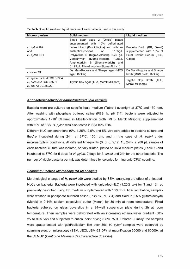
APPENDIX
175
Table 1- Specific solid and liquid medium of each bacteria used in this study.
Microorganism Solid medium Liquid medium
H. pylori J99
and
H. pylori SS1
Blood agar base 2 (Oxoid) plates
supplemented with 10% defibrinated
horse blood (Probiológica) and with an
antibiotics-cocktail of 0.155g/L
Polymixine B (Sigma-Aldrich), 6.25 g/L
Vancomycin (Sigma-Aldrich), 1.25g/L
Amphotericin B (Sigma-Aldrich) and
3.125g/L Trimethroprim (Sigma-Aldrich)
Brucella Broth (BB, Oxoid)
supplemented with 10% of
Fetal Bovine Serum (FBS,
Gibco)
L. casei 01 De Man-Rogosa and Sharpe agar (MRS
agar, Biokar)
De Man-Rogosa and Sharpe
broth (MRS broth, Biokar)
S. epidermidis ATCC 35984
S. aureus ATCC 33591
E. coli ATCC 25922
Tryptic Soy Agar (TSA, Merck Millipore) Tryptic Soy Broth (TSB,
Merck Millipore)
Antibacterial activity of nanostructured lipid carriers
Bacteria were pre-cultured on specific liquid medium (Table1) overnight at 37ºC and 150 rpm.
After washing with phosphate buffered saline (PBS 1x, pH 7.4), bacteria were adjusted to
approximately 1×10⁷ CFU/mL in Müeller-Hinton broth (MHB, Merck Millipore) supplemented
with 10% of FBS. H. pylori was also tested in BB+10% FBS.
Different NLC concentrations (0%, 1.25%, 2.5% and 5% v/v) were added to bacteria culture and
they're incubated during 24h, at 37ºC, 150 rpm, and in the case of H. pylori under
microaerophilic conditions. At different time-points (0, 3, 6, 9,12, 15, 24h), a 200 µL sample of
each bacterial culture was isolated, serially diluted, plated on solid medium plates (Table 1) and
incubated at 37ºC for 5 days for H. pylori, 2 days for L. casei and 24h for the other bacteria. The
number of viable bacteria per mL was determined by colonies forming unit (CFU) counting.
Scanning Electron Microscopy (SEM) analysis
Morphological changes of H. pylori J99 were studied by SEM, analyzing the effect of unloaded-
NLCs on bacteria. Bacteria were incubated with unloaded-NLC (1.25% v/v) for 3 and 12h as
previously described using BB medium supplemented with 10%FBS. After incubation, samples
were washed in phosphate buffered saline (PBS 1x, pH 7.4) and fixed in 2.5% glutaraldehyde
(Merck) in 0.14M sodium cacodylate buffer (Merck) for 30 min at room temperature. Fixed
bacteria adhered on glass coverslips in a 24-well suspension plate during 2h at room
temperature. Then samples were dehydrated with an increasing ethanol/water gradient (50%
v/v to 99% v/v) and subjected to critical point drying (CPD 7501, Poloran). Finally, the samples
were sputter-coated with gold/palladium film over 30s. H. pylori samples were observed by
scanning electron microscopy (SEM; JEOL JSM-6310F), at magnification 30000 and 60000x, at
the CEMUP (Centro de Materiais da Universidade do Porto).

APPENDIX
176
Transmission Electron Microscopy (TEM) analysis
The effect of unloaded-NLCs on the structure of H. pylori J99 was also analyzed by TEM.
Incubation time and concentrations were similar as described for SEM analysis. After
incubation, bacteria suspension was centrifuged (3000g, 5min) and the supernatant removed.
The bacterial pellet was fixed by resuspending in a mixture of 4% w/v of paraformaldehyde
(Merck) with 2.5% v/v glutaraldehyde (Electron Microscopy Sciences) in 0.14M sodium
cacodylate buffer (Merck) (pH 7.4). Samples were then washed with sodium cacodylate buffer,
centrifuged and bacterial pellet post-fixed in 2% osmium tetroxide (Electron Microscopy
Sciences) in sodium cacodylate buffer was embedded in a HistoGel (Thermo Scientific) and
processed in Epon resin (Electron Microscopy Sciences). Ultrathin sections of 50 nm thickness
were performed on a Ultramicrotome (RMC PowerTome PC model), by using diamond knives
(Diatome). Sections were mounted on formvar-coated nickel grids, stained with uranyl acetate
and lead citrate (Delta Microscopies) and examined using TEM (JEOL JEM 1400 transmission
electron microscope) equipped with a CCD digital camera Orious 1100W at i3S (Instituto de
Investigação e Inovação em Saúde da Universidade do Porto).
Effect of nanostructured lipid carriers on gastric cells line
Gastric carcinoma cell line MKN45 was grown in RPMI 1640 with Glutamax and HEPES (Gibco)
supplemented with 10% inactivated FBS (Gibco) and 1% penicillin-streptomycin (Gibco) at 37ºC
under 5% CO2 in humidified air. The medium was changed every two days. For subculturing,
cells were trypsinized and counted in a Neubauer chamber diluted (1:10) in a Trypan Blue
solution (0.4% w/v) (Sigma-Aldrich). Cells were expanded in 75 cm2 T-flasks with appropriate
aliquots of the cell suspension with a sub-cultivation ratio of 1:3. NLC effect on MKN45 gastric
cells was evaluated using direct contact assay. MKN45 cells were seeded in a 12-well plate
(5×10⁵ cell per well) in RPMI 1640 with Glutamax and HEPES supplemented with 10%
inactivated FBS and 1% PenStrep, at 37ºC and under 5% CO2 in humidified air, during 48h.
Then, the culture medium was changed and NLCs were added with different concentrations (0,
0.5%, 1.25%, 2.5%, 5.0% v/v). Well-plate was incubated at 37ºC and under 5% CO2 in
humidified air, during 24h. Cells in TritonTM
X-100 (2% w/v) (Sigma-Aldrich) and cells in fresh
culture medium were used as a control, in order to normalize the results. After 24h, the
supernatant was removed (transferred to 12-well plates and stored for cytotoxicity assay) and 1
mL of 5 mg/mL of Thiazolyl Blue Tetrazolium Bromide (MTT, 98%, Sigma-Aldrich) in PBS
solution was added to the cultures, diluted to a final concentration of 0.5 mg/L in culture medium
and incubated for 3h at 37ºC in the dark. Then, MTT solution was discarded and 1 mL of DMSO
was added, to solubilize the formazan crystals formed by MTT reaction. The plate was shaken
for 5 min, at room temperature, under light protection and then, the optical density was
measured at 590 nm and 630 nm using a microplate reader (Synergy™ H Multi-mode
Microplate Reader, BioTek Instruments). MTT assay evaluates the activity of cellular

APPENDIX
177
oxidoreductase enzymes inside mitochondria by converting the MTT tetrazolium dye into its
insoluble formazan (purple) that is directly proportional to the number of viable cells. The
percentage of cell viability was calculated according to the following equation:
Cell viability (%)=Experimental value- Triton control
Medium control-Triton control×100
Lactate dehydrogenase (LDH) is released to the cell culture medium when the cytoplasmic
membrane is damaged. Consequently, its quantification allows having an assessment of cell
death. Hence, the 12-well plates containing the cell culture supernatant collected for the MTT
assay was centrifuged (250g, 10min at room temperature). Carefully, without disturbing the
pellet, 100µl of the sample were transferred to the 96-well plate and 100 µl of the LDH
Cytotoxicity Detection Kit (Takara Bio Inc) were added. After 5 min of incubation, at room
temperature and protected from light, the absorbance was measured at 490 and 630 nm using
the microplate reader. The percentage of cytotoxicity was calculated according to the following
equation:
Cytotoxicity (%) =Experimental value- Medium control
Triton control-Medium control× 100
Statistical analysis
Data are reported as means ± standard deviation. Data from different groups were compared
statistically using non-parametric Kruskal-Wallis test. Analyses were performed with a
significance level of 0.05 using Graph Pad Prism 5.0 (Graph-Pad Software).
RESULTS AND DISCUSSION
NLCs were synthesized using a modified hot homogenization technique with no use of organic
solvents [17]. Results obtained by DLS demonstrated that NLCs have a homogenous size
distribution with a mean diameter of 211 ± 8 nm (Figure 1A). Polydispersion index was around
0.2 suggesting low size variability (monodisperse distribution) and no visible aggregation that
was confirmed by Cryo-SEM. However, when observed by Cryo-SEM (Figure 1B), NLCs are
slightly smaller, which could be related to the sublimation process used in this technique to
remove the surface water that can also remove the water present in the nanoparticle matrix
causing particle shrinkage. NLCs size can be important for H. pylori treatment since it was
described that particles with sizes between 130 and 300 nm are able to infiltrate gastric cell-cell
junctions and interact locally with H. pylori infection sites in intercellular spaces [18].

APPENDIX
178
ξ-potential is a very important factor in the analysis of colloidal dispersions stability. Figure 1A
also shows that NLCs have a ξ-potential around -28mV. According to literature, colloidal
dispersions are stable when they are strongly charged (|30 mV|) (i.e., aggregation is avoided)
[19], whereas ξ-potential values in the range of -10<0<+10mV are considered neutral [20].
Since ξ-potential is near -30mV, these NLCs can be considered physically stable.
NLCs are stable during storage in aqueous suspension for at least 1 moth at 4º and 20ºC in
terms of size and charge (Figure 1C). However, at 20ºC and after 1 month of storage, there is a
slight increase of the ξ-potential from -27 to -23 mV. Nevertheless, these negative charges are
enough to maintain their physical stability and avoid nanoparticles aggregation.
NLC bactericidal activity was evaluated following bacteria growth during 24h in the presence of
different NLC concentrations (Figure 2) using Müller-Hinton Broth (MHB), which is the
recommended medium for the determination of the minimal inhibitory concentration according to
the guidelines proposed by two recognized organizations: CLSI (Clinical & Laboratory
Standards Institute) and EUCAST (European Society of Clinical Microbiology and Infectious
Diseases) [21]. However, since H. pylori is a fastidious microorganism and requires a complex
nutrient-rich growth media, MHB had to be supplemented with 10% of fetal bovine serum (FBS).
This supplement may act as an additional source of nutrient and also protect against the toxic
effects of long-chain fatty acids [22]. Nevertheless, for comparison and to demonstrate the
specific bactericidal activity of NLCs to H. pylori, this FBS-supplemented medium was also used
for bactericidal assays performed with other bacteria.
Minimal bactericidal concentration (MBC) was defined as the minimal drug concentration to kill
99.9% (>3 logs) bacteria in 24h of incubation. Figure 2A shows that NLCs are bactericidal
against H. pylori at all concentrations used, since after 24h in the presence of NLCs, there are
no live bacteria. For the highest concentration tested (5% v/v), H. pylori was killed after 15h of
incubation time. Nonetheless, an inhibitory effect of 50% (> 1log) was detected after 9h of
incubation with this NLC concentration.
NLC bactericidal effect was not detected on Lactobacillus casei-01 (Figure 2B), Gram-positive
rod-shaped and nonpathogenic bacteria found on the human gut microbiota. This bacterium has
an importance for gut microbiota which enhances the integrity of the intestinal barrier, decrease
translocation of bacteria across the intestinal mucosa and disease phenotypes such as
gastrointestinal infections [23, 24]. This specific bactericidal activity of unloaded-NLCs against
H. pylori (without affecting L. casei) opens new routes for the treatment of H. pylori infection
without affecting gut microbiota.
NLCs were also tested on other Gram-positive cocci bacteria, Staphylococcus epidermidis
(Figure 2C) and methicillin-resistant Staphylococcus aureus (MRSA) (Figure 2D). No
bactericidal effect was observed for both bacteria on the concentrations used. However, the
higher NLCs concentration (5% v/v) had an inhibitory effect on MRSA growth. Other bacterium
tested was Escherichia coli, a Gram-negative rod-shaped bacterium, is the most prevalent

APPENDIX
179
commensal inhabitant of the human intestinal tract and lives in a mutually beneficial association
with hosts [25, 26]. E. coli is not usually pathogenic. NLC bactericidal effect was not detected
on E. coli (Figure 2E); therefore, unloaded-NLCs can be used without to affect the normal flora
of the gut.
The NLC bactericidal effect against H. pylori was also tested using a growth medium (BB)
indicated for H. pylori growth [27]. Figure 3 shows that all bacteria are killed after 12h of
incubation with NLCs for all the concentrations used. The bactericidal effect of NLC on H. pylori
was faster when bacteria were growing in this medium than in MHB medium.
In order to understand the interaction of NLCs with H. pylori, their morphology after growing in
the presence and absence of NLCs was evaluated by SEM and TEM. Samples were prepared
after 3 and 12h of incubation with 1.25% of NLCs, since after 3h mostly of the bacteria are alive
and after 12h all bacteria are dead.
SEM images of H. pylori grown during 3h (Figure 4A) and 12h (Figure 4C) in the absence of
NLCs (control) show their characteristic bacillus shape with 2-4µm of length and 0.5-1µm wide
[4-6]. When bacteria were incubated with NLCs for 3h (Figure 4B) their morphology was similar
to the controls, demonstrating the integrity of its membrane. However, after 12h incubation with
NLCs (Figure 4D), although high morphological changes were not observed using this
technique, it was possible to visualize the leakage of their cytoplasmic contents. These results
suggest that NLCs can interact with H. pylori by disrupting its cell membrane.
TEM images (Figure 5) corroborate with SEM images, showing H. pylori with the intact cell
membrane and dense cytoplasm in controls (Figure 5A and 5C) and after 3 hours growing with
NLCs (Figure 5B). After 12h of incubation with NLCs, TEM images revealed a disseverance of
outer membrane and plasma membrane with parts of bacteria without cytoplasmic contents
(Figure 5D).
NLCs cytotoxicity was evaluated against a gastric carcinoma cell line (MKN45) using thiazolyl
blue tetrazolium bromide (MTT) and lactate dehydrogenase (LDH) assays (Figure 6A and 6B,
respectively). Figure 6A shows that for NLC concentrations up to 2.5%v/v, the reduction of
MKN45 cell viability was lower than 30%. Figure 6B demonstrated that despite its strong
bactericidal activity against H. pylori, NLCs are not cytotoxic against MKN45 cells at
concentrations up to 2.5%v/v (~20% cell lysis). These results also demonstrated that NLCs at
bactericidal concentrations (1.25% v/v) have a negligible lactate dehydrogenase release and did
not induce apoptosis and mitochondrial dysfunction after 24h of incubation. These NLCs, at
bactericidal concentrations, are biocompatible according to ISO 10993-5 [12] since the
decrease of cell metabolic activity and increase of cell lysis due to the presence of NLCs in
comparison with control were lower than 30%.

APPENDIX
180
References
1. Correa, P. and J. Houghton, Carcinogenesis of Helicobacter pylori. Gastroenterology,
2007. 133(2): p. 659-672.
2. Amieva, M. and R.M. Peek Jr, Pathobiology of Helicobacter pylori–Induced Gastric Cancer.
Gastroenterology, 2016. 150(1): p. 64-78.
3. Matysiak-Budnik, T. and F. Mégraud, Helicobacter pylori infection and gastric cancer.
European Journal of Cancer, 2006. 42(6): p. 708-716.
4. Malfertheiner, P., et al., Management of Helicobacter pylori infection—the Maastricht IV/
Florence Consensus Report. Gut, 2012. 61(5): p. 646-664.
5. Vakil, N., Helicobacter pylori treatment: a practical approach. Am J Gastroenterol, 2006.
101(3): p. 497-9.
6. Carbone, C., et al., Pharmaceutical and biomedical applications of lipid-based
nanocarriers. Pharm Pat Anal, 2014. 3(2): p. 199-215.
7. Battaglia, L. and M. Gallarate, Lipid nanoparticles: state of the art, new preparation
methods and challenges in drug delivery. Expert Opinion on Drug Delivery, 2012. 9(5): p.
497-508.
8. Martins, S., et al., Lipid-based colloidal carriers for peptide and protein delivery--liposomes
versus lipid nanoparticles. Int J Nanomedicine, 2007. 2(4): p. 595-607.
9. Müller, R.H., K. Mäder, and S. Gohla, Solid lipid nanoparticles (SLN) for controlled drug
delivery – a review of the state of the art. European Journal of Pharmaceutics and
Biopharmaceutics, 2000. 50(1): p. 161-177.
10. Viladot Petit, J.L.L., G.R. Delgado, and B.A. Fernández, Lipid nanoparticle capsules, in US
2013/0017239 A1. 2011, LIPOTEC S.A.: Spain.
11. Mehnert, W. and K. Mäder, Solid lipid nanoparticles: Production, characterization and
applications. Advanced Drug Delivery Reviews, 2001. 47(2–3): p. 165-196.
12. Svilenov, H.T., Christo Solid lipid nanoparticles – a promising drug delivery system, in
Nanomedicine, A.M. Seifalian, Achala de Kalaskar, Deepak M., Editor. 2014, One Central
Press (OCP): London.
13. Muchow, M., P. Maincent, and R.H. Müller, Lipid Nanoparticles with a Solid Matrix (SLN®,
NLC®, LDC®) for Oral Drug Delivery. Drug Development and Industrial Pharmacy, 2008.
34(12): p. 1394-1405.
14. da Silva, P.B., et al., A Nanostructured Lipid System as a Strategy to Improve the in Vitro
Antibacterial Activity of Copper(II) Complexes. Molecules, 2015. 20(12): p. 22534-45.
15. Manea, A.-M., B.S. Vasile, and A. Meghea, Antioxidant and antimicrobial activities of green
tea extract loaded into nanostructured lipid carriers. Comptes Rendus Chimie, 2014. 17(4):
p. 331-341.
16. Mahdavi, J., et al., Helicobacter pylori SabA adhesin in persistent infection and chronic
inflammation. Science, 2002. 297(5581): p. 573-8.
17. Neves, A.R., et al., Novel resveratrol nanodelivery systems based on lipid nanoparticles to
enhance its oral bioavailability. Int J Nanomedicine, 2013. 8: p. 177-87.
18. Lin, Y.H., et al., Development of pH-responsive chitosan/heparin nanoparticles for
stomach-specific anti-Helicobacter pylori therapy. Biomaterials, 2009. 30(19): p. 3332-42.

APPENDIX
181
19. Fonte, P., et al., Chapter fifteen - Chitosan-Coated Solid Lipid Nanoparticles for Insulin
Delivery, in Methods in Enzymology, D. Nejat, Editor. 2012, Academic Press. p. 295-314.
20. Clogston, J. and A. Patri, Zeta Potential Measurement, in Characterization of Nanoparticles
Intended for Drug Delivery, S.E. McNeil, Editor. 2011, Humana Press. p. 63-70.
21. Matuschek, E., D.F.J. Brown, and G. Kahlmeter, Development of the EUCAST disk
diffusion antimicrobial susceptibility testing method and its implementation in routine
microbiology laboratories. Clinical Microbiology and Infection, 2014. 20: p. O255–O266.
22. Kusters, J.G., A.H.M. van Vliet, and E.J. Kuipers, Pathogenesis of Helicobacter pylori
Infection. Clinical Microbiology Reviews, 2006. 19(3): p. 449-490.
23. Hemarajata, P. and J. Versalovic, Effects of probiotics on gut microbiota: mechanisms of
intestinal immunomodulation and neuromodulation. Therap Adv Gastroenterol, 2013. 6(1):
p. 39-51.
24. Okuro, P.K., et al., Co- encapsulation of Lactobacillus acidophilus with inulin or
polydextrose in solid lipid microparticles provides protection and improves stability. Food
Research International, 2013. 53(1): p. 96-103.
25. Allocati, N., et al., Escherichia coli in Europe: an overview. Int J Environ Res Public Health,
2013. 10(12): p. 6235-54.
26. Reshes, G., et al., Cell shape dynamics in Escherichia coli. Biophys J, 2008. 94(1): p. 251-
64.
27. Blanchard, T.G. and J.G. Nedrud, Laboratory Maintenance of Helicobacter Species.
Current Protocols in Microbiology, 2006. CHAPTER: p. Unit8B.1-Unit8B.1.

APPENDIX
182
FIGURES
Figure 1. NLCs characterization in terms of (A) diameter (bars), polydispersion index and
charge surface (ξ-potential) performed using dynamic light scattering at 37ºC, (B) morphology
evaluated by cryo-scanning electron microscopy (Cryo-SEM) (scale bar 500nm, 50000x
magnification) and (C) effect of time of storage in water at 4ºC and 20ºC on diameter and
charge (ξ-potential) of unloaded-NLC produced. Data is reported as the mean ± standard
deviation (n=3). *p<0.05 compared to different time-points on day 0 (Kruskal-Wallis test).
A B
Diameter (nm) 211 ± 8
Polydispersion
index 0.2±0.02
ξ-potential (mV) -28 ± 3
C

APPENDIX
183
Figure 2. The growth of bacteria in increasing concentrations of unloaded-NLC in Müller-Hinton Broth medium supplemented with 10% FBS: (A) H. pylori
J99, (B) Lactobacillus casei-01, (C) Staphylococcus epidermidis ATCC35984, (D) Staphylococcus aureus ATCC 33591 (methicillin resistant) and Escherichia
coli ATCC 25922. Numerical data is reported as mean ± standard deviation (n=3). All concentrations tested with unloaded-NLCs were significantly different
compared to each bacteria growth treated with unloaded-NLCs and bacteria untreated (control) (p<0.05, Kruskal-Wallis test).
A B

APPENDIX
184
C D

APPENDIX
185
E

APPENDIX
186
Figure 3. The growth of (A) H. pylori J99 and (B) H. pylori SS1 in increasing concentrations of unloaded-NLC in Brucella Broth medium supplemented with
10% FBS. Numerical data is reported as mean ± standard deviation (n=3). All concentrations tested with unloaded-NLCs were significantly different compared
to each bacteria growth treated with unloaded-NLCs and bacteria untreated (control) (p<0.05, Kruskal-Wallis test).
A B

APPENDIX
187
Figure 4. Scanning Electron micrographs of H. pylori J99 incubated during 3h (A to B) and 12h
(C to D) with unloaded-NLC. In A and C, SEM images of H. pylori J99 was not treated (control);
in B and D, H. pylori J99 was treated with 1.25 %v/v of unloaded-NLC. The scale bar in the
SEM image represents 1 µm, 60 000 x magnifications.
Figure 5. Transmission Electron micrographs of H. pylori J99 incubated during 3h (A to B) and
12h (C to D) with unloaded-NLC. In A and C, TEM images of H. pylori J99 was not treated
(control); in B and D, H. pylori J99 was treated with 1.25%v/v of unloaded-NLC. The scale bar in
the TEM image represents 200 nm, 50 000 x magnifications.

APPENDIX
188
Figure 6. Effect of unloaded-NLCs of gastric carcinoma cell line in increasing amounts. MKN45
gastric carcinoma (A) cell line viability and (B) cytotoxicity assessed by the MTT and LDH
assay, respectively, after 24h exposure to different amounts of NLCs. Data is reported as
mean ± standard deviation (n=3). * p<0.05, refers to significant differences of MKN45 gastric
cells treated cells with unloaded-NLC and cells untreated (control) (Kruskal-Wallis test).
A
B

APPENDIX
189
CLAIMS
1. A nanostructured lipid particle comprising particles in the form of a shell, hollow nanoparticles
or lipid vesicles, and wherein said nanoparticles are capable of killing Helicobacter pylori.
2. A nanostructured lipid particle according to claim 1, wherein the particles can be produced by
mixture of several lipids forming a nanoemulsion, using techniques that may include the use of
hot homogenization and ultrasonication,
3. A nanostructured lipid particle according to claim 1, wherein the particles present with
diameters between 50 and 400 nanometers, preferably between 150 and 250 nanometers, as
determined by their hydrodynamic size using dynamic light scattering, at 37ºC.
4. A nanostructured lipid particle according to claim 1, wherein the particle is formed by
mixtures of different lipids, selected from solid and liquid lipids and mixed with a surfactant to
produce a nanoemulsion. The lipids are preferably glyceryl palmitostearate, cetyl palmitate and
Caprylic/Capric Triglyceride and the surfactant polysorbate 60.
5. Nanostructured lipid particle produced according to the previous claims, wherein the
therapeutically effective amount of Helicobacter pylori infection is administrated orally.
6. Nanostructured lipid particles produced according to the previous claims that can be stored
during 1 month, in aqueous suspension at 4º and 20ºC.
7. Nanostructured lipid particles produced according to the previous claims, wherein the
therapeutically effective amount against Helicobacter pylori does not affect other bacteria,
particularly Lactobacillus casei, Staphylococcus epidermidis and Escherichia coli.
8. An oral formulation containing the nanostructured lipid particles produced according to the
previous claims, in combination with one or more therapeutically active substance(s) selected
from the group of gastrointestinal protectants (particularly proton pump inhibitors) and
antibiotics (particularly amoxicillin, clarithromycin, metronidazole, tetracycline) used in the
treatment of Helicobacter pylori infection.
9. An oral formulation containing the nanostructured lipid particles produced according to the
previous claims, wherein the dosage form can be administrated in any formulation adapted for
stomach delivery, particularly incorporated/combined with gastric retentive polymers such as
chitosan or other pharmaceutical compounds able to release the particles in the stomach.

APPENDIX
190
APPENDIX 4
4. Review: The potential utility of chitosan micro/nanoparticles in the treatment of gastric infection
During this work, a brief description of the use of chitosan in H. pylori treatment was
reviewed. This review was used in the introduction of this thesis.

The potential utility ofchitosan micro/nanoparticlesin the treatment of gastricinfectionExpert Rev. Anti Infect. Ther. 12(8), 981–992 (2014)
Ines C Goncalves1,3,Patrıcia C Henriques1,3,Catarina L Seabra1,2,4
and M Cristina LMartins*1,4
1INEB – Instituto de Engenharia
Biomedica, Universidade do Porto, Rua
do Campo Alegre, 823, 4150-180 Porto,
Portugal2IPATIMUP – Institute of Molecular
Pathology and Immunology of the
University of Porto, Rua Dr. Roberto
Frias, 4200-465 Porto, Portugal3Universidade do Porto, Faculdade de
Engenharia, Porto, Portugal4Universidade do Porto, Instituto de
Ciencias Biomedicas Abel Salazar, Porto,
Portugal
*Author for correspondence:
Tel.: +351 226 074 982
Gastric infections are mainly caused by Helicobacter pylori (H. pylori), a bacterium thatcolonizes the gastric mucosa of over 50% of the world’s population. Chronic H. pyloriinfection has been associated with gastric diseases such as chronic gastritis, peptic ulcer andgastric adenocarcinoma. Current eradication treatment relies on antibiotic-based therapiesthat are unsuccessful in approximately 20% of the patients. Chitosan, a natural and cationicpolysaccharide has been investigated in the treatment of H. pylori infection. Due to itsmucoadhesive properties, it has been used in the form of micro/nanoparticles, polyelectrolytecomplexes or coatings as antibiotic encapsulation systems for gastric delivery, but alternativemolecules may also be incorporated. It has been recently proposed that chitosan can also beused for H. pylori binding and scavenging from the host stomach due to its antimicrobial/binding properties. In this manuscript, a brief description of the use of chitosan in H. pyloritreatment is reviewed.
KEYWORDS: biomaterials • chitosan • crosslinking • drug delivery • gastric cancer • Helicobacter pylori • microspheres
• mucoadhesion • nanoparticles • polyelectrolytes
Helicobacter pylori gastric infectionHelicobacter pylori (H. pylori), a spiral-shapedgram-negative bacterium, is one of the mostcommon infectious agents in the world, coloniz-ing the gastric mucosa of over 50% of thehuman population [1]. Although most of theinfected individuals show no or few symptoms,this infection is the strongest known risk factorfor gastroduodenal ulcer progression (60–80%of the cases), being also related to the develop-ment of gastric adenocarcinoma (1–3% of thecases) [1], the second leading cause of cancer-related deaths worldwide [2]. H. pylori wasclassified as a type I carcinogen by the Interna-tional Agency for Research on Cancer [3], and itwas recognized that its elimination is the mostpromising strategy to reduce the incidence ofgastric cancer [4].
Due to its flagellar motility, H. pylori isable to escape the acidic pH of the stomach(pH 1.2–2.5) crossing the mucus layer thatcovers and protects the gastric cells, reachingthe gastric epithelium where the pH is more
neutral (pH ~7.4). In a healthy humanstomach, the median thickness of the adher-ent mucus is 180 mm with 67% polymericmucin, but in patients with gastric carcinomathere is an increase of thickness to 240 mmdespite becoming a structurally very weakgel with only 23% polymeric mucin [5].Furthermore, H. pylori can secrete urease,which converts endogenous urea into ammo-nia and carbon dioxide, thereby bufferinggastric acid in the immediate vicinity of theorganism [6].
Although most of H. pylori can be foundfree-swimming in the mucus layer, bacteria isalso found adherent to the mucus layer and tothe surface of gastric epithelial cells, in theintercellular spaces and even, as suggested in afew studies, inside the gastric cells [7].
Nonadherent H. pylori can produce andrelease a vacuolating cytotoxin, VacA, whichinduces multiple structural and functionalalterations in cells, such as the formation oflarge intracellular vacuoles and the increase inmembrane permeability [1].
informahealthcare.com 10.1586/14787210.2014.930663 � 2014 Informa UK Ltd ISSN 1478-7210 981
Review
Exp
ert R
evie
w o
f A
nti-
infe
ctiv
e T
hera
py D
ownl
oade
d fr
om in
form
ahea
lthca
re.c
om b
y W
ashi
ngto
n U
nive
rsity
Lib
rary
on
01/2
5/15
For
pers
onal
use
onl
y.

H. pylori adhesion to gastric mucosa protects the bacteria fromdisplacement by forces such as those generated by peristalsis andgastric emptying. Adhesion to the host is mediated by lectins,bacterial surface-bound adhesins, which recognize glycansexpressed on the gastric mucosa [8]. H. pylori can express severaldifferent adhesins that are not only responsible for adhesion butfor the communication system that enables H. pylori response tothe gastric environment [9]. The best characterized adhesin–gly-can interactions are the blood group antigen-binding adhesin(BabA) that recognizes the Lewis blood group antigens, such asLewis b (Leb) and H-type 1, and sialic acid-binding adhesin(SabA) that mediates the adherence of H. pylori to inflamed gas-tric mucosa by binding sialylated carbohydrate structures such assialyl Lewis a (sLea) and sialyl Lewis a [10]. However, neutro-phil activating protein was also identified as responsible forH. pylori binding to sulfated mucins, such as sulfo-Lea, sulfo-Gal and sulfo-GalNAc [11]. The two adherence-associatedlipoprotein A and B [12] as well as the outer inflammatory pro-tein A [13], expressed in all bacteria strains, are also importantin H. pylori adhesion to gastric mucosa, although their hostreceptors are not well known. It was also described thatH. pylori can bind damaged tissue, where the basement mem-brane is exposed, through the extracellular matrix proteins, such aslaminin, type IV collagen, vitronectin and heparan sulfate proteo-glycan. This could be important for H. pylori adherence to hostintercellular junctions [9].
After adhering to epithelial cells, H. pylori is able to assemble atype IV secretion system, encoded by the cag pathogenicityisland, which translocates the cytotoxin-associated gene proteininto gastric epithelial cells, conducing to numerous cellular alter-ations and inducing strong gastric inflammation [14].
Current treatmentsCurrent available treatments to H. pylori infection rely on atriple treatment, which includes the combination of two
antibiotics, such as clarithromycin andamoxicillin or metronidazole and a protonpump inhibitor (PPI) such as omeprazole.The administration of the three antibioticstogether with a PPI (nonbismuth quadru-ple therapy) has also been considered aswell as the bismuth-containing quadrupletherapy (bismuth salts, tetracycline,metronidazole and a PPI) [4].
Current antibiotic eradication therapy isunsuccessful in nearly one in five patients(FIGURE 1) [15]. This low rate of success ismainly related to H. pylori resistance tothe antibiotics and poor patient compli-ance. Furthermore, antibiotic degradationin the acidic environment of the gastriclumen or poor penetration of the antibi-otic through the viscoelastic mucosal gelcould lead to sub-bactericidal concentra-tion of antibiotics in the infection site
and therefore incomplete eradication of H. pylori, which couldalso induce bacterial resistance.
Alternative treatmentsSeveral alternative treatments are being investigated in order toovercome the abovementioned problems related to antibiotictherapies. A vaccination strategy would be a valuable option tofight H. pylori infection [16]. However, despite several attemptsto develop an H. pylori vaccine for humans, progress has beenslow. Special attention has been given to compounds obtainedfrom natural sources, including polyunsaturated fatty acids,terpenes and terpenoids, polyphenols and antimicrobial pepti-des [17,18]. The use of polyunsaturated fatty acids as co-adjuvantof the current available therapies was suggested as a promisingapproach [19].
Encapsulation of conventional antibiotics, for a local andcontrolled delivery at the infection site, has been thoroughlyinvestigated to improve the effect of antibiotics againstH. pylori [20,21]. These systems protect the drug from rapiddegradation or clearance, extending its half-life and solubility,which may lead to lower administration frequency and hencebetter patient compliance [20–22]. A different strategy has beenrecently proposed by our group, based on the use of H. pyloribinding systems that, after oral administration, will attract/bindbacteria and remove them from the host stomach, through theintestinal tract, after gastric mucus turnover [23].
Chitosan is a nontoxic, biodegradable and biocompatiblenatural polymer that has been quite investigated in the treat-ment of H. pylori infection, namely, for drug encapsulationdue to its mucoadhesive properties and for H. pylori bindingdue to its antimicrobial/binding properties. Nevertheless,chitosan has also been described as adjuvant for an H. pylorivaccine [24]. In this manuscript, a brief description of the useof chitosan micro/nanoparticles in H. pylori treatment isreviewed.
7000 million people World population
3500 million peopleInfected with
H. pylori
50% [1]
20% symptomatic [79]
700 million peopleHave recommendation for
H. pylori treatment
140 million peopleH. pylori treatment is
inefficient
20% [15]
1% [1]
35 million peopleDevelop
gastric cancer
Figure 1. Helicobacter pylori infection and treatment figures.
Review Goncalves, Henriques, Seabra & Martins
982 Expert Rev. Anti Infect. Ther. 12(8), (2014)
Exp
ert R
evie
w o
f A
nti-
infe
ctiv
e T
hera
py D
ownl
oade
d fr
om in
form
ahea
lthca
re.c
om b
y W
ashi
ngto
n U
nive
rsity
Lib
rary
on
01/2
5/15
For
pers
onal
use
onl
y.

Chitosan in the treatment of H. pylori infectionProperties of chitosan
Chitosan (FIGURE 2), a naturally occurring polysaccharide com-posed of D-glucosamine and N-acetyl-D-glucosamine units [25],is obtained by alkaline deacetylation of chitin, which is thesecond most abundant polysaccharide after cellulose and themajor component of the exoskeleton of crustaceans, squid penand cell walls of some fungi [26]. Chitosan molecular weight(MW) and degree of deacetylation (DD), which is the propor-tion of D-glucosamine units with respect to the total numberof units, are the main factors that affect its chemical andbiological properties [25]. The increase of chitosan DD willincrease the number of free amine groups (–NH2) that conferchitosan its almost unique properties of being positivelycharged and easily chemically modified.
Chitosan is stable in neutral conditions due to the strong inter-and intramolecular hydrogen bonds formed between its amineand hydroxyl groups [27]. Still, chitosan is soluble in acidic condi-tions, namely, below its pKa value (~6.5), due to the protonationof the free amine groups from the glucosamine residues [28].
Mucoadhesive properties of chitosan
The mucoadhesive properties of chitosan are related with thestrong electrostatic interactions established between its proton-ated glucosamine residues and glycoproteins from gastricmucosa that, due to their sialic acids and ester sulfates [29], arenegatively charged at the acidic stomach pH. These interactionsare then followed by mechanical interlocking of the polymerchains, van der Waal’s forces and hydrogen bonding.
Antimicrobial properties of chitosan
Chitosan antimicrobial properties are also consequence of theelectrostatic interactions between chitosan cationic amine groupsand the anionic groups on the bacterial wall, which leads to theinhibition of bacterial proliferation [30].
The antimicrobial activity of chitosan and its derivatives wasable to inhibit the growth of fungi, parasites and several bacte-ria, including H. pylori strains NCTC 11639, NCTC 11637and SS1 [31]. Luo et al. [31] have shown in vitro that chitosanwith a DD of 95% presents higher antimicrobial activityagainst H. pylori than chitosan with a DD of 88.5%, indicatingthat the higher the deacetylation of chitosan the better its anti-microbial action. Furthermore, the in vivo administration ofchitosan as solution and as nanoparticles (both DD 95%) toH. pylori infected BALB/c mice eradicated the bacteria in55 and 75% of the animals, respectively.
Chitosan as drug encapsulation system
Several antibiotics have been encapsulated to treat H. pyloriinfection, although amoxicillin and clarithromycin are the mostcommon ones. The encapsulation of two antibiotics and a PPI(omeprazole) has also been described [32].
The selection of the encapsulation technique is primarilydetermined by the solubility of the drug and the polymer inthe solvents, and by the particle size requirements [33,34].
Chitosan micro/nanoparticles
Chitosan micro and nanoparticles have been used for gastricdrug delivery in the treatment of H. pylori due to its mucoad-hesive properties. Besides protecting the drugs from enzymaticand acidic degradation, by adhering to gastric mucus barrier,particles residence time in the stomach will be higher prolong-ing drug delivery and allowing drug diffusion through themucus barrier to the infection site [21,35].
The size and charge of chitosan particles will affect theirmucoadhesion and mucopenetration and thus their retentiontime in the stomach. Microparticles have more difficulty topenetrate through viscoelastic mucus layer than nanoparticles.However, nanoparticles smaller than 200 nm could be internal-ized by gastric epithelial cells [36]. Furthermore, for an efficientmucoadhesion, particles must be positively charged, which isusually obtained by increasing chitosan MW and DD [37].Nevertheless, an elevated number of positive charges couldmake the particles too mucoadhesive avoiding their penetrationthrough the mucus layer [21], making them more prone to bewashed out upon mucus turnover.
Several methods and approaches that have been proposed forthe preparation of chitosan particles are summarized in TABLE 1
and schematized in a review from Agnihotri et al. [38].Drug loading into chitosan particles is commonly performed
by mixing it in the chitosan solution prior to particles forma-tion and crosslinking [39], but incubation of previously formedparticles with the drug can also be performed [40]. Hejazi andAmiji [40] demonstrated that the encapsulation of tetracyclineonto chitosan microspheres was higher (69% w/w) when drugwas incubated for up to 48 h with previously formedmicrospheres than when mixed in chitosan solution (8% w/w).
Due to the high solubility of chitosan in acidic conditions,uncrosslinked chitosan particles cannot maintain their three-dimensional structure in gastric fluids [28,41]. The rapid dissolu-tion of chitosan particles or an extensive swelling of theparticles induces a fast release of the drug, which might not bedesirable. In order to overcome this restriction, ionic crosslink-ing of chitosan particles using tripolyphosphate (TPP) [42] orcovalent crosslinking using glutaraldehyde [43] or genipin [41]
can be performed. Genipin is a naturally occurring crosslinkingagent described as being 10000 times less toxic than glutaralde-hyde [44].
It should be considered that crosslinking after drug loadingcould induce chemical reactions with the active drug, which is
O O
O
NH2HO
O
CH2OH
HOCH2OHNHCOCH3
O
nN-acetyl glucosamine Glucosamine
Figure 2. Chemical structure of chitosan.
The potential utility of chitosan micro/nanoparticles in the treatment of gastric infection Review
informahealthcare.com 983
Exp
ert R
evie
w o
f A
nti-
infe
ctiv
e T
hera
py D
ownl
oade
d fr
om in
form
ahea
lthca
re.c
om b
y W
ashi
ngto
n U
nive
rsity
Lib
rary
on
01/2
5/15
For
pers
onal
use
onl
y.

Table 1. Examples of methods for preparation of chitosan micro/nanoparticles.
Particle production method Particle size Advantages (+)/disadvantages (-)
Ionic gelationDrop wise addition of chitosan solution (positively
charged) under constant stirring into a polyanionic
solution (negatively charged, generally TPP).
Complexation between oppositely charged species
results in chitosan to precipitate as spherical particles.
Nanoparticles (nm)
20–200 [37]
550–900 [32]
152–376 [42]
Microspheres (mm)
~170 [41]
100–400 [64]
+ Processing under mild conditions
+ Organic solvent free
+ Low-toxicity impact of reagents
+ No changes in drug chemistry
- Difficult entrapment of high molecular weight drugs
- Poor stability in acidic conditions
Precipitation/coacervation• Addition of a solute (generally a salt) to chitosan
solution, forming micro/nanoparticles due to a
decrease in chitosan solubility
• Chitosan solution might also be blown into an alkali
solution using a compressed air nozzle to form
coacervate droplets
Nanoparticles (nm)
100–250 [65]
Microspheres (mm)
1.5–2.5 [66]
+ No complex apparatus needed
+ Few purification steps required
+ Organic solvent free
+ High loading capacity combined with a sustained
drug release
- Poor stability in acidic conditions
Spray dryingPreparation of chitosan solution where a suitable
crosslinking agent could be added (if desired). This
solution or dispersion is then atomized in a stream of
hot air. Atomization leads to the formation of small
droplets, from which solvent evaporates instantly
leading to the formation of free flowing particles.
Temperature and humidity might be regulated.
Microspheres (mm)
3–12 [67]
140–281 [68]
+ Simple, reproducible and easy to scale up
+ Low-cost process
+ Fast solvent removal
+ Good sphericity
+ Narrow size distribution
+ Low dependency of the solubility of the drug and
polymer
- High temperatures required
- Size influenced by several parameters
- Possible difficulty in spraying fluid of high viscosity
Supercritical antisolvent precipitationSpraying of the chitosan solution into a precipitation
chamber with supercritical CO2 (antisolvent), causing
rapid contact between the two media. A higher
super-saturation ratio of the solution is generated,
resulting in fast nucleation and growth.
Microparticles (mm)
1.0–2.5 [47]
+ Processing under mild conditions
+ Complete antisolvent removal
+ Nontoxic reagents
+ Narrow size distribution
+ No changes in drug chemistry
Emulsion crosslinkingChitosan aqueous solution is extruded into an oil
phase, generally liquid paraffin (under intensive
stirring), forming and water-in-oil (w/o) emulsion.
Aqueous droplets are stabilized by adding a surfactant.
The stable emulsion is crosslinked by using an
appropriate crosslinking agent such as glutaraldehyde.
Microspheres (mm)
60–100 [39]
100–330 [69]
350–690 [70]
+ Control of particle size
+ Good sphericity
- Slow process
- Chemical crosslinking agents used, possibly inducing
chemical reactions with the drug
- Difficult removal of the unreacted crosslinking agent
Solvent EvaporationAqueous chitosan solution is added to an organic
phase with vigorous stirring to form the primary water
in oil emulsion (w/o). The latter is then added to a
large volume of water containing a surfactant, forming
a multiple emulsion (w/o/w). The double emulsion is
then subjected to stirring until most of the organic
solvent evaporates, leaving solid microspheres.
Microspheres (mm)
100–200 [71]
+ Processing under mild conditions
+ Favorable for encapsulation of thermally sensitive
drugs
+ Indicated for delivery of small molecule drugs
+ Good sphericity
- Organic solvents usage
- Low drug encapsulation efficiency
Reverse micellar methodThe surfactant is dissolved in an organic solvent
followed by the addition of chitosan, drug and
crosslinking agent, under constant vortexing overnight.
The organic solvent is evaporated, obtaining a
transparent dry mass. The latter is dispersed in water
and then a suitable salt is added to precipitate the
surfactant out.
Nanoparticles (nm)
~100 [72]
+ Narrow size distribution
- Organic solvent usage
Review Goncalves, Henriques, Seabra & Martins
984 Expert Rev. Anti Infect. Ther. 12(8), (2014)
Exp
ert R
evie
w o
f A
nti-
infe
ctiv
e T
hera
py D
ownl
oade
d fr
om in
form
ahea
lthca
re.c
om b
y W
ashi
ngto
n U
nive
rsity
Lib
rary
on
01/2
5/15
For
pers
onal
use
onl
y.

not desirable. Also, crosslinking is performed through chitosanamine groups, reducing the number of free primary aminesable to be protonated. As such, increasing the crosslinkingdecreases the mucoadhesion, size and swelling ability of chito-san particles in acidic conditions [41,45], affecting drug releaseprofile. Zhu et al. [46] observed that chitosan microspheres ioni-cally crosslinked with TPP present good mucoadhesive proper-ties but allow a fast release of a hydrophilic drug (Berberine).On the other hand, chemically crosslinked chitosan micro-spheres enable a sustained drug release profile but show inferiormucoadhesiveness. In order to solve this inconsistency betweenmucoadhesiveness and release rate of chitosan microspheres,they proposed an approach where Eudragit L100 cores areencapsulated within the chitosan microsphere using an emulsifi-cation/coagulation procedure (size: 380 mm; entrapmentefficiency: 71%), allowing ionic crosslinked microspheres withlow crosslinking density to be used for the controlled release ofhydrophilic antibiotics [46].
Regarding H. pylori treatment, amoxicillin-loaded chitosanmicrospheres (50–100 mm) were produced by Patel andPatel [39] using the emulsification crosslinking method. Theimproved effect of encapsulating amoxicillin to treat H. pyloriwas demonstrated in vivo using Wistar rats. In a single-doseadministration test, the inhibition was markedly observed whendose was increased from 4 to 15 mg/kg. A similar in vivo studywas performed by Patel and Patil [47] using smaller amoxicillin-chitosan microspheres (1.0–2.5 mm) produced using thesupercritical antisolvent precipitation method, revealing thatH. pylori inhibition was not influenced by particle size.
To improve H. pylori treatment, chitosan-based micro/nanoparticles can be specifically directed to the infection site bycovalently binding, on the surface of the particles, ligands withaffinity toward gastric mucosa or bacteria.
Lectins can specifically bind to carbohydrate moieties ofglycoproteins located in gastric mucosa. Lectin can be coupledto chitosan particles improving the absorption of drugs [21]
while presenting a good resistance to digestion within gastricenvironment [20]. The conjugation of the lectin (Con-A) onclarithromycin-loaded chitosan microspheres increased micro-spheres adhesion to the stomach mucosa of albino rats from12 to 85% [48]. Retention time was also enhanced from 3 h toover 6 h in New Zealand albino rabbits for Con-A conjugatedmicrospheres.
Fucose can be used to target lectin-type receptors on thesurface of H. pylori. Fucose-conjugated nanoparticles containinga triple therapy (amoxicillin/clarithromycin/omeprazole) wereproduced by ionotropic gelation chitosan-glutamic acid conju-gates and TPP as crosslinker [32]. These functionalized nanopar-ticles demonstrated in vitro an H. pylori eradication rate of 97%against 91% of nonconjugated chitosan nanoparticles and 81%of plain triple therapy. In vivo studies using H. pylori-infectedSwiss albino mice demonstrated that fucose-conjugated nanopar-ticles containing the triple therapy treatment (53.57 mg/kgamoxicillin/17.8 mg/kg clarithromycin and 1.42 mg/kg omepra-zole) were able to completely eradicate H. pylori infection.
Nonfunctionalized nanoparticles and plain triple therapy wereonly able to eradicate 50 and 33% of the mice, respectively [32].
Chitosan-based pH-responsive micro/nanoparticles
Chitosan-based polyelectrolyte complexes can be prepared bymixing protonated chitosan with anionic polymers from natural(e.g., alginate [49,50], heparin [36], poly-y-glutamic acid [51,52],etc.) (TABLE 2) or synthetic (e.g., poly(acrylic acid), etc.) origin[53–55].
The advantage of using these systems for gastric applications isthat they can be pH-sensitive, that is, polyelectrolyte complexescan be prepared in a way that electrostatic interactions betweenpolymers are strong enough at acidic pH, but unstable at physio-logical pH to release the drug near H. pylori local of infection.Furthermore, these strong but reversible interactions betweenoppositely charged polyelectrolytes avoid the need of covalentcrosslinkers and their associated disadvantages. Chitosan-basedpolyelectrolytes can be applied for drugs that, due to their hydro-phobicity, negative charge or high MW, cannot be released fromchitosan crosslinked particles. The size, charge and stability ofthe polyelectrolyte complexes depend on the individual proper-ties of each polymer (charge, MW), the ratio between polymers,the ionic strength and pH of the solution, the mixing order aswell as speed and diameter of the dispersing element [55].
Many different polyelectrolyte complexes, in the form ofmicro- or nanoparticles, have been developed for encapsulationof antibiotics for H. pylori treatment, and some examples arebriefly summarized in TABLE 3.
As previously described with chitosan micro/nanoparticles,polyelectrolyte complexes can also be functionalized to targetH. pylori. Fucose-chitosan/heparin nanoparticles loaded withamoxicillin are mentioned in TABLE 3.
Chitosan can also be used as coating for anionic particles. Achitosan-coated pH-responsive liposome system containingdoxycycline (as a model antibiotic drug) for the treatment ofH. pylori infection was developed [56]. Liposomes are sphericalvesicles composed of one or more phospholipid bilayers thathave been studied as drug delivery systems for both hydrophilicand hydrophobic compounds. Furthermore, liposomes alsomimic cell membranes, allowing an effective penetration ofencapsulated drug into intracellular spaces. However, the appli-cations of liposomes, particularly those with sizes below100 nm, are often limited due to their spontaneous fusion [56].Chitosan-modified gold (AuCh) nanoparticles (size ~10 nm)strongly bound to the surface of anionic phospholipid lipo-somes (size ~75 nm) at acidic pH, inhibit liposome fusion anddetached from the liposomes at physiological pH, enable freeliposomes to fuse with target bacterial membrane and releasethe therapeutic drug. Antimicrobial studies demonstratedhigher H. pylori antibacterial effect of the doxycycline-loadedAuCh-liposome nanoparticles when compared with the sameconcentration of doxycycline in solution.
Amoxicillin-loaded alginate/carboxymethyl cellulose microbe-ads (size 745–889 mm/entrapment efficiency 52–92%) werecoated with chitosan to achieve a controlled release of the
The potential utility of chitosan micro/nanoparticles in the treatment of gastric infection Review
informahealthcare.com 985
Exp
ert R
evie
w o
f A
nti-
infe
ctiv
e T
hera
py D
ownl
oade
d fr
om in
form
ahea
lthca
re.c
om b
y W
ashi
ngto
n U
nive
rsity
Lib
rary
on
01/2
5/15
For
pers
onal
use
onl
y.

antibiotic in stomach environment [57]. It was demonstratedthat chitosan coating induced mucoadhesive properties to themicrobeads and delayed amoxicillin delivery to up to 8 h,releasing 41% of amoxicillin.
Chitosan as binding agent
The adhesion of H. pylori to chitosan in a pH range that simu-lates gastric conditions (2.6, 4.0 and 6.0) was first demonstratedusing chitosan as thin films crosslinked with genipin [58].Furthermore, the percentage of dead adherent bacteria rangedfrom 76% at pH 6 to 93% at pH 2.6. As such, rather than beingused as a gastric drug delivery vehicle, chitosan microsphereshave been designed and applied as an H. pylori-binding sys-tem [23,41]. It is expected that after oral administration, thesemicrospheres will adhere H. pylori in the stomach and removethem through the gastrointestinal tract (upon gastric mucus turn-over). Chitosan microspheres with approximately 170 mm wereproduced by ionotropic gelation with TPP [41] followed by the
minimal genipin crosslinking necessary to avoid microspheresdissolution in acidic pH without losing their mucoadhesiveness.In vivo studies using C57BL/6 mice demonstrated thatthese chitosan microspheres have a gastric retention of 2 h [41].H. pylori adhesion to these microspheres was evaluated in a rangeof acidic pHs, confirming that several H. pylori strains bind tothe microspheres (FIGURE 3). Furthermore, these microspheres arenot cytotoxic and revealed able to both partially remove andprevent H. pylori adhesion to a gastric cell line [23].
This system was further improved by directing the micro-spheres toward H. pylori. Immobilization of carbohydrates,namely, Leb and sLex, that specifically bind to H. pylori adhe-sins was first screened using model surfaces [59] and afterwardstransposed to chitosan microspheres [60]. It was found thatglycan-targeted materials were able to bind specifically toH. pylori strains that express the corresponding adhesin,meaning that Leb-modified materials adhere favorably toBabA-positive strains and sLex-modified materials adhere
Table 2. Anionic polymers commonly used for preparation of chitosan-based polyelectrolyte complexes.
Polymer chemical structure Polymer characteristics
O
OO
O
OH
–OOC
OH
HO
–OOCOH
OH
O
OO
OHO
–OOC
–OOC
OH
O
nM M
OH
G: guluronate M: mannuronate
G G
Alginate:• Natural anionic and hydrophilic
polysaccharide typically obtained from
brown seaweed or bacteria
• Composed of alternating blocks of (1-4)
linked a-L-guluronic (G) and
b-D-mannuronic acid (M) monomers
• Widely explored for biomedical
applications, namely as matrix for the
entrapment and/or delivery of a variety of
proteins, peptides, drugs and cells, due
to its excellent biocompatibility, low
toxicity, biodegradability and mild
gelation conditions by addition of
divalent cations such as Ca2+
n
O
O O
OSO3–
COO–
O
O
CH2OSO3–
NHSO3–HO
HO
Heparin:• Natural anionic and highly sulfated
glycosaminoglycan
• Composed of repeating disaccharide
units of D-glucosamine and uronic acid
linked by 1-4 interglycosidic bonds
• It is well known for its anticoagulant
properties, but also by its ability to bind
to cell receptors and accelerate gastric
ulcer healing, which is associated with
mucosal regeneration, proliferation and
angiogenesis
N
COOH
α β γ
O nH
Poly-g-glutamic acid (g-PGA):• Natural, hydrophilic, nontoxic, poly-amino
acid, which is biodegradable and
relatively nonimmunogenic
• Can be used as carriers for active
substances and can be easily modulated
in order to control the delivery of drugs,
proteins, etc.
Review Goncalves, Henriques, Seabra & Martins
986 Expert Rev. Anti Infect. Ther. 12(8), (2014)
Exp
ert R
evie
w o
f A
nti-
infe
ctiv
e T
hera
py D
ownl
oade
d fr
om in
form
ahea
lthca
re.c
om b
y W
ashi
ngto
n U
nive
rsity
Lib
rary
on
01/2
5/15
For
pers
onal
use
onl
y.

Table
3.Examplesofch
itosan-basedpolyelectrolyte
complexesusedasdrugdelivery
systemsin
Helico
bacterpyloriinfectiontreatm
ent.
Complex
Drug
Productionmethod
Particle
characteristics
Main
conclusions
Ref.
Alginate/chitosan
Amoxicillin
Ionic
gelation:
Chitosan,amoxicillinandpluronicare
mixed.
Tothepreviousmixture,alginate
solutionis
sprayedwithcontinuousstirring.
•Alginate:0.1%
w/v
(pH5.5)
•Chitosan:0.06%
w/v
(pH5.0)
•Amoxicillin:0.01%
w/v
•PluronicF127(surfactan
t):0.019%
w/v
Size:
651nm
z-Potential:ffi
+60mV
Entrapmentefficiency:91%
Invitro
•Antibioticprotectionfrom
simulatedgastric
conditionsforatleast
6h;
•Lowermucoadhesive
capacity
(~76%
)than
plain
chitosannan
oparticlescrosslinked
withTPP(~86%)suggestingthat
decreasingofthesurface
chargeofthe
nanoparticleswillim
prove
their
mucopenetration
Invivo
(Wistarrats)
•Excellentmucopenetrationinto
deep
mucosalregion
[73]
Ionic
gelation:
Amoxicillinismixedwithalginate
solutionin
ddH2O
containingDOSSusingvigorousstirring.
Mixture
issprayedinto
aCaCl 2solution
containingchitosan.
•Alginate:2%
w/v
(pH5.5)
•Chitosan:0.75%
w/v
(pH5.0)
•Amoxicillin:2-9%
w/v
•DOSS(surfactan
t):0.075%
w/v
•CaC
l 2:1.0%
w/v
Size:
840mm
Entrapment
efficiency:84%
Invitro
•Antibioticprotectionfrom
simulatedgastric
acidicconditionsforatleast
8h;
•Highlymucoadhesive
(invitrogastric
retentionupto
8h);
•Theincrease
ofchitosanconcentration
enlargesparticle
size
butreducesdrug
encapsulation
[74]
Heparin/chitosan
Amoxicillin
Water-in-oilemulsification:
Nanoemulsionparticlesarepreparedusing
laboratory-typerotatingbladehomogenizer
(40mlaq
ueo
usphase
+80mlparaffin).
Surfactantisaddedto
liquid
paraffin
under
continuousmixingandheparinaqueoussolution
isad
ded
andhomogen
ized.Chitosansolutionis
dropped
into
theresultantheparin
emulsionand
homogenized
.Nan
oem
ulsionparticlesare
collectedbycentrifugation.
•Chitosan:0.6
mg/m
l(pH6.0)
•Hep
arin:0.2
mg/m
l(pH7.4)
•Amoxicillin:0.6
mg/m
L(pH7.4)
•Surfactant:Span
20/Tween20(75:25):1.2
ml
Size:
296nm
z-Potential:ffi
+30mV
Entrapment
efficiency:ffi
54%
Invitro
•Positivelychargedparticlesandstable
in
acidicgastricconditions;
•AmoxicillinreleaseatpH7.Low
release
at
pH6
•Can
beinternalizedbyhuman
gastric
adenocarcinomacells
(AGScellline)andbe
localized
inintercellularspaces
Invivo
(H.pyloriinfectedC57BL/6Jmice)
•Higher
clearance
inH.pyloriinfectionwhen
comparedwitham
oxicillinin
solution
[75]
H.pylori:Helicobacterpylori;TPP:Tripolyphosphate.
The potential utility of chitosan micro/nanoparticles in the treatment of gastric infection Review
informahealthcare.com 987
Exp
ert R
evie
w o
f A
nti-
infe
ctiv
e T
hera
py D
ownl
oade
d fr
om in
form
ahea
lthca
re.c
om b
y W
ashi
ngto
n U
nive
rsity
Lib
rary
on
01/2
5/15
For
pers
onal
use
onl
y.

Table
3.Examplesofch
itosan-basedpolyelectrolyte
complexesusedasdrugdelivery
systemsin
Helico
bacterpyloriinfectiontreatm
ent
(cont.).
Complex
Drug
Productionmethod
Particle
characteristics
Main
conclusions
Ref.
Hep
arin/
chitosan-fucose
Amoxicillin
Ionotropic
gelation+crosslinking
(withgenipin):
Fucose-conjugatedchitosanissynthesizedby
additionoffucose
andsodium
cyanoborohydride
into
chitosansolution.Aqueo
ushep
arin(w
ith
amoxicillin)isaddedbyflush
mixingwitha
pipette
tip.Nanoparticlesare
collectedby
centrifugation.Heparin/chitosan-fucose
nanoparticlesare
mixedinto
agenipin
solution
andgentlystirred.
•Fucose-Chit:1.2
mg/m
l(pH6)
•Heparin:2.0
mg/m
l(pH7.4)
•Amoxicillin:4.0
mg/m
l
•Gen
ipin:0.375mg/m
l
Size:ffi
250nm
z-Potential:ffi
+27mV
Entrapment
efficiency:ffi
49%
Invitro
•Amoxicillinprotectionfrom
acidicconditions
•Crosslinkingdelays
amoxicillindeliveryin
acidicconditions
•Adhesionandinfiltrationin
themucuslayer
•Amoxicillinrelease
nearthegastric
epithelium
•Particlessize
increase
atphysiological
pH
Invivo
(C57BL/6Jmice)
•HigherreductionofH.pyloriinfectionwhen
comparedwitham
oxicillinin
solution
[76]
Hep
arin/chitosan
Berberine
Ionic
gelation:
Berberine/heparin
nanoparticlesare
preparedby
combiningaq
ueo
usberberinewithaqueous
heparin
byflush
mixingwithapipettetip.
Berberine/heparin
solutionisaddedto
aqueous
chitosansolution.
•Berberine:0.25mg/m
l
•Berberine/heparin/Chitosanratio(w
/w/w
):
3.0:6.0:4.8
(pH6.0)
Size:ffi
256nm
Entrapment
efficiency:ffi
54%
Invitro
•Nanoparticlesbecomeunstable
at
physiologicalpH,allowingarapid
release
ofberberine;
•Berberinenanoparticlesactlocally
inthesite
ofH.pyloriinfectionandinhibitthe
bacterium
growth
[77]
g-PGA/chitosan
Amoxicillin
Ionic
gelation:
Chitosan/g-PGAnanoparticlesare
preparedby
flush
mixingg-PGAsolutionwithapipettetip
into
aqueouschitosancontainingamoxicillin.
Nanoparticlesare
collectedbycentrifugation.
Nanoparticlesare
thenincorporatedinto
bead
s
ofpH-sensitive
alginate-gelatingelthatare
preparedbydroppingaqueousalginate-gelatin
into
aCaC
l 2solution.
•Amoxicillin:2.0
mg/m
l
•g-PGA:2.0
mg/m
l
•Chitosan:0.06mg/m
l
•Hydrogel:Alginate0.2%
w/v
andGelatin
0.10%
w/v
into
CaC
l 20.6
m
Size:120–150nm
(nanoparticles)
z-Potential:ffi
+19mV
Entrapment
efficiency:ffi
23%
Invitro
•Alginate-gelatinhydrogel
prevents
nan
oparticlesdestructionandamoxicillin
release
from
nanoparticlesin
acidicpH;
•Confocallaserscanningmicroscopy
revealedthat
nanoparticlescaninfiltrate
cell-celljunctionsandinteract
withH.pylori
[78]
H.pylori:Helicobacterpylori;TPP:Tripolyphosphate.
Review Goncalves, Henriques, Seabra & Martins
988 Expert Rev. Anti Infect. Ther. 12(8), (2014)
Exp
ert R
evie
w o
f A
nti-
infe
ctiv
e T
hera
py D
ownl
oade
d fr
om in
form
ahea
lthca
re.c
om b
y W
ashi
ngto
n U
nive
rsity
Lib
rary
on
01/2
5/15
For
pers
onal
use
onl
y.

preferentially to SabA-positive strains. This strategy isextremely promising, particularly given the present shift offocus onto a more personalized medicine. The immobiliza-tion of other carbohydrates into microspheres for administra-tion depending on the strain of H. pylori that is infecting thepatient, or the administration of cocktails of microsphereswith different carbohydrates are interesting possibilities forfuture treatments.
Expert commentary & five-year viewThe currently available antibiotic-based treatments for H. pylorigastric infection have been unsuccessful in several patients, mostlydue to bacterial resistance and poor patient compliance. Itis expected that the amount of antibiotic-resistant H. pyloriwill increase in a near future due to the bad use (abuse) of antibi-otics, leading to an increased number of patients that need analternative treatment. Furthermore, multiple H. pylori infectionand even multiple-antibiotic-resistant H. pylori infection havebeen observed, which also compromises the current therapeuticefficacy.
Antibiotics encapsulation into gastric retentive polymers,namely, chitosan, has improved their biological effects, byprotecting them from the acidic gastric fluids and increasingtheir availability at the infection site. However, although thisstrategy could improve patients’ compliance, it will not solvethe problem of antibiotic resistance.
A promising alternative to the conventional antibiotic ther-apy is the use of antimicrobial compounds obtained from natu-ral sources [18,59] that have different bacteria target/pathwaysthan most antibiotics (the ribosome–clarithromycin; the cellwall synthesis–amoxicillin and bacterial DNA replication–metronidazole [61]). Nevertheless, due to their low solubility,sensitivity and instability in gastric conditions, most ofthese natural-based compounds also need to be encapsulatedfor their in situ delivery. As such, we predict that chitosanmicro/nanoparticles will continue to have a major role inH. pylori therapies, mainly due to chitosan’s biocompatibility,
biodegradability, mucoadhesiveness, ease of handling and facil-ity to be chemically modified.
In all the strategies previously described, it was demonstratedthat the incorporation of target molecules toward H. pylorisuch as fucose or carbohydrates (namely, Leb or sLex) improvedthe efficiency of the systems. It is our belief that targeted treat-ments using chitosan micro/nanoparticles specific to H. pyloriare highly promising since they will avoid the destruction ofthe normal gut microbiome of the patients [62]. This strategymay include particles chemically modified with targetingcompounds that bind H. pylori urease, flagella, adhesins, etc.,in order to inhibit, kill or remove the bacteria.
A recent investigation performed by our group takes advantageof chitosan bioadhesive and antimicrobial properties, and useschitosan microspheres to bind and remove H. pylori from thestomach of infected hosts, through the normal gastrointestinaltract. Although this therapy may not be enough to remove allbacteria that infect the patient, especially the ones that are in theintercellular spaces or inside gastric cells, it is expected that theycan remove most of H. pylori and have a synergistic effect whencombined with other drugs. Since these chitosan particles willnot induce bacteria resistance and can be used without damageto the patient, they can also be used to prevent reinfection.
Despite the great potential of using chitosan and chitosanderivatives in pharmaceutical and biomedical applications, noneof the described chitosan systems are currently in the market.Chitosan is only US FDA approved for use in wounddressings. Nevertheless, chitosan was approved for dietary appli-cations in Japan, Italy and Finland [63]. This difficulty in beingFDA approved is mainly related with the fact that its physico-chemical characteristics can change drastically with its source,MW and associated polydispersity index, DD, size and formu-lation. Furthermore, since chitosan is a natural compound, it isalways difficult to maintain the reproducibility between batches.Nevertheless, the current growing interest in using chitosan inmedical (pharmaceutical) applications improved the industrialcapacity to produce more pure, cost–effective and uniform
Figure 3. Confocal microscopy images of chitosan microspheres (in red) with adherent FITC-labeled Helicobacter pylori strainJ99 (in green) under pH 2.6, pH 4.0 and pH 6.0 (from left to right).
The potential utility of chitosan micro/nanoparticles in the treatment of gastric infection Review
informahealthcare.com 989
Exp
ert R
evie
w o
f A
nti-
infe
ctiv
e T
hera
py D
ownl
oade
d fr
om in
form
ahea
lthca
re.c
om b
y W
ashi
ngto
n U
nive
rsity
Lib
rary
on
01/2
5/15
For
pers
onal
use
onl
y.

chitosan, representing important progress for its pharmaceuticalindustrialization in a near future.
Financial & competing interests disclosure
This paper has been financed by FEDER funds through Programa
Operacional Factores de Competitividade – COMPETE and by
Portuguese funds through FCT – Fundacao para a Ciencia e a Tecnolo-
gia, in the framework of the projects PEst-C/SAU/LA0002/2013,
NORTE-07-0124-FEDER-000005, FCOMP-01-0124-FEDER-020073
(PTDC/CTM-BPC/121149/2010) and FCOMP-01-0124-FEDER-
041276 (EXPL/CTM-BIO/0762/2013) and grant SFRH/BD/89001/
2012. The authors have no other relevant affiliations or financial involve-
ment with any organization or entity with a financial interest in or
financial conflict with the subject matter or materials discussed in the
manuscript apart from those disclosed.
No writing assistance was utilized in the production of this manuscript.
Key issues
• Helicobacter pylori (H. pylori) infects about half of the world’s population and its persistent infection is associated with a strong risk for
the development of gastric adenocarcinoma.
• The failure of the current available antibiotic-based therapy (~1 in each 5 patients) demonstrated the need of alternative treatments.
• Chitosan is a natural biocompatible polymer with great potential in H. pylori treatment, mainly due to its gastric retentive and
antimicrobial properties, but also because it can easily be chemically modified.
• Chitosan properties are mainly related with its primary amine groups that are directly related with its degree of deacetylation.
• In H. pylori infection treatment, chitosan is mostly used as gastric drug delivery system, whether as crosslinked particles, as
polyelectrolyte complexes with anionic polymers or as coatings. Nevertheless, it has recently been proposed as an H. pylori
binding system.
• In order to prevent premature release of the encapsulated drug, chitosan particles must be crosslinked to avoid its fast dissolution in
gastric acidic conditions.
• Polyelectrolyte complex-based chitosan particles can be pH-responsive, that is, particles are stable in acidic pH (drug preservation) and
unstable at physiological pH to release the drug near H. pylori infection site (near gastric epithelial cells).
• Chitosan microspheres crosslinked with genipin have great potential as H. pylori-binding systems for the clearance of this bacterium
from infected host through normal gastrointestinal tract since they can bind H. pylori, are noncytotoxic and are stable during all the
digestive process.
• In order to increase their residence time in the stomach, chitosan particles can be conjugated with lectins to target the gastric mucosa
through lectin-carbohydrate recognition.
• In order to be directed toward H. pylori, chitosan particles can be conjugated with fucose or glycans (Leb, sLex, …) to target H. pylori
adhesins.
References
Papers of special note have been highlighted as:
• of interest
•• of considerable interest
1. Wroblewski LE, Peek RM Jr, Wilson KT.
Helicobacter pylori and gastric cancer:
factors that modulate disease risk. Clin
Microbiol Rev 2010;23(4):713-39
• Describes Helicobacter pylori (H. pylori)mechanisms of infection and pathogenesis
of H. pylori-induced gastric
adenocarcinoma.
2. Ferlay J, Parkin DM, Steliarova-Foucher E.
Estimates of cancer incidence and mortality
in Europe in. 2008;Eur J Cancer 2010;
46(4):765-81
3. Stewart BW, Kleihues P. World cancer
report. International Agency for Research on
Cancer; Lyon, France: 2003. p. 351
4. Malfertheiner P, Megraud F, O’Morain CA,
et al. Management of Helicobacter pylori
infectio- the Maastricht IV/Florence
Consensus Report. Gut 2012;61:646-64
• Summarizes the current therapies and
provides guidelines for H. pylori infectioneradication.
5. Allen A, Cunliffe WJ, Pearson JP,
Venables CW. The adherent gastric mucus
gel barrier in man and changes in peptic
ulceration. J Intern Med Suppl 1990;732:
83-90
6. Celli JP, Turner BS, Afdhal NH, et al.
Helicobacter pylori moves through mucus
by reducing mucin viscoelasticity. Proc Natl
Acad Sci USA 2009;106(34):14321-6
7. Dubois A, Boren T. Helicobacter pylori is
invasive and it may be a facultative
intracellular organism. Cell Microbiol 2007;
9(5):1108-16
8. Kobayashi M, Lee H, Nakayama J,
Fukuda M. Carbohydrate-dependent defense
mechanisms against Helicobacter pylori
infection. Curr Drug Metab 2009;10(1):
29-40
9. Evans DJ Jr, Evans DG. Helicobacter pylori
adhesins: review and perspectives.
Helicobacter 2000;5(4):183-95
10. Aspholm M, Kalia A, Ruhl S, et al.
Helicobacter pylori adhesion to
carbohydrates. Methods Enzymol 2006;417:
293-339
11. Namavar F, Sparrius M, Veerman EC, et al.
Neutrophil-activating protein mediates
adhesion of Helicobacter pylori to sulfated
carbohydrates on high-molecular-weight
salivary mucin. Infect Immun 1998;66(2):
444-7
12. Odenbreit S, Faller G, Haas R. Role of the
alpAB proteins and lipopolysaccharide in
adhesion of Helicobacter pylori to human
gastric tissue. Int J Med Microbiol 2002;
292(3-4):247-56
13. Dossumbekova A, Prinz C, Mages J, et al.
Helicobacter pylori HopH (OipA) and
Review Goncalves, Henriques, Seabra & Martins
990 Expert Rev. Anti Infect. Ther. 12(8), (2014)
Exp
ert R
evie
w o
f A
nti-
infe
ctiv
e T
hera
py D
ownl
oade
d fr
om in
form
ahea
lthca
re.c
om b
y W
ashi
ngto
n U
nive
rsity
Lib
rary
on
01/2
5/15
For
pers
onal
use
onl
y.
bacterial pathogenicity: genetic and
functional genomic analysis of hopH gene
polymorphisms. J Infect Dis 2006;194(10):
1346-55
14. Kwok T, Zabler D, Urman S, et al.
Helicobacter exploits integrin for type IV
secretion and kinase activation. Nature
2007;449(7164):862-6
15. Vakil N. Helicobacter pylori treatment:
a practical approach. Am J Gastroenterol
2006;101(3):497-9
16. Del Giudice G, Malfertheiner P,
Rappuoli R. Development of vaccines
against Helicobacter pylori. Expert Rev
Vaccines 2009;8(8):1037-49
17. Parreira P, Fatima Duarte M, Reis CA,
Martins MC. Helicobacter pylori infection:
a brief overview on alternative natural
treatments to conventional therapy. Crit
Rev Microbiol 2014. [Epub ahead of print]
18. Makobongo MO, Gilbreath JJ, Merrell DS.
Nontraditional therapies to treat
Helicobacter pylori infection. J microbiol
2014;52(4):259-72
19. Correia M, Michel V, Matos AA, et al.
Docosahexaenoic acid inhibits Helicobacter
pylori growth in vitro and mice gastric
mucosa colonization. PLoS One 2012;7(4):
e35072
20. Adebisi A, Conway BR. Gastroretentive
microparticles for drug delivery applications.
J Microencapsul 2011;28(8):689-708
•• Summarizes the role of gastric retentive
microparticles on drug delivery for
H. pylori treatment.
21. Arora S, Bisen G, Budhiraja RD.
Mucoadhesive and muco-penetrating
delivery systems for eradication of
Helicobacter pylori. Asian J Pharm 2012;6:
18-30
•• Summarizes the gastric drug delivery
systems that have been recently developed
to release antibiotics for H. pylorieradication.
22. Petros RA, DeSimone JM. Strategies in the
design of nanoparticles for therapeutic
applications. Nat Rev Drug Discov 2010;
9(8):615-27
23. Goncalves IC, Magalhaes A, Fernandes M,
et al. Bacterial-binding chitosan
microspheres for gastric infection treatment
and prevention. Acta Biomater 2013;9(12):
9370-8
•• Describes the ability of chitosan
microspheres (crosslinked with genipin)
to bind H. pylori and prevent/remove its
adhesion to gastric cells.
24. Xie Y, Zhou NJ, Gong YF, et al. Th
immune response induced by H. pylori
vaccine with chitosan as adjuvant and its
relation to immune protection. World J
Gastroenterol 2007;13(10):1547-53
25. Dash M, Chiellini F, Ottenbrite RM,
Chiellini E. Chitosan – A versatile
semi-synthetic polymer in biomedical
applications. Prog Polym Sci 2011;36(8):
981-1014
26. Kato Y, Onishi H, Machida Y. Application
of chitin and chitosan derivatives in the
pharmaceutical field. Curr Pharm
Biotechnol 2003;4(5):303-9
27. Mi FL, Sung HW, Shyu SS, et al. Synthesis
and characterization of biodegradable TPP/
genipin co-crosslinked chitosan gel beads.
Polymer (Guildf) 2003;44(21):6521-30
28. Mi FL, Sung HW, Shyu SS. Synthesis and
characterization of a novel chitosan-based
network prepared using naturally occurring
crosslinker. J Polym Sci 2000;38(15):
2804-14
29. Sogias IA, Williams AC, Khutoryanskiy VV.
Why is chitosan mucoadhesive?
Biomacromolecules 2008;9(7):1837-42
30. Strand SP, Vandvik MS, Varum KM,
Ostgaard K. Screening of chitosans and
conditions for bacterial flocculation.
Biomacromolecules 2001;2(1):126-33
31. Luo D, Guo J, Wang F, et al. Preparation
and evaluation of anti-Helicobacter pylori
efficacy of chitosan nanoparticles in vitro
and in vivo. J Biomater Sci Polym Ed 2009;
20(11):1587-96
32. Ramteke S, Ganesh N, Bhattacharya S,
Jain NK. Amoxicillin, clarithromycin, and
omeprazole based targeted nanoparticles for
the treatment of H. pylori. J Drug Target
2009;17(3):225-34
•• Describes the in vitro and in vivo efficacy
studies of fucose-conjugated chitosan
nanoparticles containing a triple therapy
(amoxicillin/clarithromycin/omeprazole).
33. Crini G, Badot PM. Application of
chitosan, a natural aminopolysaccharide, for
dye removal from aqueous solutions by
adsorption processes using batch studies:
a review of recent literature. Prog Polym Sci
2008;33(4):399-447
34. Soppimath KS, Aminabhavi TM,
Kulkarni AR, Rudzinski WE. Biodegradable
polymeric nanoparticles as drug delivery
devices. J Control Release 2001;70(1-2):
1-20
35. Conway BR. Drug delivery strategies for the
treatment of Helicobacter pylori infections.
Curr Pharm Des 2005;11(6):775-90
36. Lin YH, Chang CH, Wu YS, et al.
Development of pH-responsive chitosan/
heparin nanoparticles for stomach-specific
anti-Helicobacter pylori therapy.
Biomaterials 2009;30(19):3332-42
37. Huang YC, Chiang CH, Yeh MK.
Optimizing formulation factors in preparing
chitosan microparticles by spray-drying
method. J Microencapsul 2003;20(2):
247-60
38. Agnihotri SA, Mallikarjuna NN,
Aminabhavi TM. Recent advances on
chitosan-based micro- and nanoparticles in
drug delivery. J Control Release 2004;
100(1):5-28
39. Patel JK, Patel MM. Stomach Specific
Anti-Helicobacter pylori Therapy:
Preparation and Evaluation of
Amoxicillin-Loaded Chitosan Mucoadhesive
Microspheres. Curr Drug Deliv 2007;4(1):
41-50
40. Hejazi R, Amiji M. Stomach-specific anti-
H. pylori therapy. I: preparation and
characterization of tetracyline-loaded
chitosan microspheres. Int J Pharm 2002;
235(1-2):87-94
41. Fernandes M, Goncalves IC, Nardecchia S,
et al. Modulation of stability and
mucoadhesive properties of chitosan
microspheres for therapeutic gastric
application. Int J Pharm 2013;454(1):
116-24
• Describes the preparation and
characterization of chitosan microspheres,
crosslinked with genipin in terms of
mucoadhesion, stability in acidic
conditions and in vivo gastric retention
time.
42. Dong Y, Ng WK, Shen S, et al. Scalable
ionic gelation synthesis of chitosan
nanoparticles for drug delivery in static
mixers. Carbohydr Polym 2013;94(2):940-5
43. Sung HW, Huang RN, Huang LL, et al.
Feasibility study of a natural crosslinking
reagent for biological tissue fixation.
J Biomed Mater Res 1998;42(4):560-7
44. Mi FL, Sung HW, Shyu SS. Release of
indomethacin from a novel chitosan
microsphere prepared by a naturally
occurring crosslinker: examination of
crosslinking and polycation–anionic drug
interaction. J Appl Polym Sci 2001;81(7):
1700-11
45. Mi FL, Shyu SS, Peng CK. Characterization
of ring-opening polymerization of genipin
and pH-dependent cross-linking reactions
between chitosan and genipin. J Polym Sci
2005;43(10):1985-2000
The potential utility of chitosan micro/nanoparticles in the treatment of gastric infection Review
informahealthcare.com 991
Exp
ert R
evie
w o
f A
nti-
infe
ctiv
e T
hera
py D
ownl
oade
d fr
om in
form
ahea
lthca
re.c
om b
y W
ashi
ngto
n U
nive
rsity
Lib
rary
on
01/2
5/15
For
pers
onal
use
onl
y.

46. Zhu X, Zhou D, Guan S, et al. Preparation
and characterization of novel multi-core
chitosan microspheres for stomach-specific
delivery of hydrophilic antibiotics. J Mater
Sci Mater Med 2012;23(4):983-90
47. Patel J, Patil P. Preparation and
characterization of amoxicillin mucoadhesive
microparticles using solution-enhanced
dispersion by supercritical CO2. J
Microencapsul 2012;29(4):398-408
48. Jain SK, Jangdey MS. Lectin conjugated
gastroretentive multiparticulate delivery
system of clarithromycin for the effective
treatment of Helicobacter pylori. Mol
Pharm 2009;6(1):295-304
49. Bhattarai R, Dhandapani N, Shrestha A.
Drug delivery using alginate and chitosan
beads: an overview. Chron Young Sci 2011;
2(4):192-6
50. Lee KY, Mooney DJ. Alginate: properties
and biomedical applications. Prog Polym
Sci 2012;37(1):106-26
51. Antunes JC, Pereira CL, Molinos M, et al.
Layer-by-layer self-assembly of chitosan and
poly(gamma-glutamic acid) into
polyelectrolyte complexes.
Biomacromolecules 2011;12(12):4183-95
52. Goncalves RM, Antunes JC, Barbosa MA.
Mesenchymal stem cell recruitment by
stromal derived factor-1-delivery systems
based on chitosan/poly(gamma-glutamic
acid) polyelectrolyte complexes. Eur Cell
Mater 2012;23:249-60.discussion 260-241
53. Berger J, Reist M, Mayer JM, et al.
Structure and interactions in chitosan
hydrogels formed by complexation or
aggregation for biomedical applications. Eur
J Pharm Biopharm 2004;57(1):35-52
54. Bhattarai N, Gunn J, Zhang M.
Chitosan-based hydrogels for controlled,
localized drug delivery. Adv Drug Deliv Rev
2010;62(1):83-99
55. Hamman JH. Chitosan based
polyelectrolyte complexes as potential carrier
materials in drug delivery systems. Mar
Drugs 2010;8(4):1305-22
56. Thamphiwatana S, Fu V, Zhu J, et al.
Nanoparticle-stabilized liposomes for
pH-responsive gastric drug delivery.
Langmuir 2013;29(39):12228-33
57. Angadi SC, Manjeshwar LS,
Aminabhavi TM. Novel composite blend
microbeads of sodium alginate coated with
chitosan for controlled release of
amoxicillin. Int J Biol Macromol 2012;
51(1-2):45-55
58. Nogueira F, Goncalves IC, Martins MC.
Effect of gastric environment on
Helicobacter pylori adhesion to a
mucoadhesive polymer. Acta Biomater
2013;9(2):5208-15
59. Parreira P, Magalhaes A, Reis CA, et al.
Bioengineered surfaces promote specific
protein–glycan mediated binding of the
gastric pathogen Helicobacter pylori. Acta
Biomaterialia 2013;9:11:8885-93
60. Martins MCL, Goncalves IC, Gomes P,
et al. Microspheres. WO2013164652. 2013
61. Lewis K. Platforms for antibiotic discovery.
Nat Rev Drug Dis 2013;12(5):371-87
62. Jakobsson HE, Jernberg C, Andersson AF,
et al. Short-term antibiotic treatment has
differing long-term impacts on the human
throat and gut microbiome. Plos One 2010;
5(3):e9836
63. Sarmento B, das Neves J. Chitosan-based
systems for biopharmaceuticals: delivery,
targeting and polymer therapeutics. John
Wiley & Sons Ltd; UK: 2012
64. Ma L, Liu C. Preparation of chitosan
microspheres by ionotropic gelation under a
high voltage electrostatic field for protein
delivery. Colloids surf B Biointerfaces 2010;
75(2):448-53
65. Mao HQ, Roy K, Troung-Le VL, et al.
Chitosan-DNA nanoparticles as gene
carriers: synthesis, characterization and
transfection efficiency. J Control Release
2001;70(3):399-421
66. Berthold A, Cremer K, Kreuter J.
Preparation and characterization of chitosan
microspheres as drug carrier for
prednisolone sodium phosphate as model
for anti-inflammatory drugs. J Control
Release 1996;39(1):17-25
67. He P, Davis SS, Illum L. In vitro evaluation
of the mucoadhesive properties of chitosan
microspheres. Int J Pharm 1998;166(1):
75-88
68. Torres MA, Vieira RS, Beppu MM, et al.
Production of chemically modified chitosan
microspheres by a spraying and coagulation
method. Mater Res 2007;10:347-52
69. Roy S, Panpalia SG, Nandy BC, et al.
Effect of Method of Preparation on
Chitosan Microspheres of Mefenamic Acid.
Int J Pharm Sci Drug Res 2009;1(1):36-42
70. Akbuga J, Durmaz G. Preparation and
evaluation of cross-linked chitosan
microspheres containing furosemide. Int J
Pharm 1994;111(3):217-22
71. Mi FL, Tan YC, Liang HC, et al. In vitro
evaluation of a chitosan membrane
cross-linked with genipin. J Biomater Sci
Polym Ed 2001;12(8):835-50
72. Mitra S, Gaur U, Ghosh PC, Maitra AN.
Tumour targeted delivery of encapsulated
dextran–doxorubicin conjugate using
chitosan nanoparticles as carrier. J Control
Release 2001;74(1–3):317-23
73. Arora S, Gupta S, Narang RK,
Budhiraja RD. Amoxicillin loaded
chitosan-alginate polyelectrolyte complex
nanoparticles as mucopenetrating delivery
system for H. pylori. Sci Pharm 2011;79(3):
673-94
74. Arora S, Budhiraja RD. Chitosan-alginate
microcapsules of amoxicillin for gastric
stability and mucoadhesion. J Adv Pharm
Technol Res 2012;3(1):68-74
75. Lin YH, Chiou SF, Lai CH, et al.
Formulation and evaluation of water-in-oil
amoxicillin-loaded nanoemulsions using for
Helicobacter pylori eradication. Process
Biochem 2012;47(10):1469-78
76. Lin YH, Tsai SC, Lai CH, et al.
Genipin-cross-linked fucose-chitosan/heparin
nanoparticles for the eradication of
Helicobacter pylori. Biomaterials 2013;
34(18):4466-79
• Describes the preparation and
characterization of amoxicillin-loaded
fucose-chitosan/heparin nanoparticles
crosslinked with genipin and its efficacy
in vivo for the treatment of H. pyloriinfection.
77. Chang CH, Huang WY, Lai CH, et al.
Development of novel nanoparticles shelled
with heparin for berberine delivery to treat
Helicobacter pylori. Acta Biomater 2011;
7(2):593-603
78. Chang CH, Lin YH, Yeh CL, et al.
Nanoparticles incorporated in pH-sensitive
hydrogels as amoxicillin delivery for
eradication of Helicobacter pylori.
Biomacromolecules 2010;11(1):133-42
79. Malfertheiner P, Bazzoli F, Delchier JC,
et al. Helicobacter pylori eradication with a
capsule containing bismuth subcitrate
potassium, metronidazole, and tetracycline
given with omeprazole versus
clarithromycin-based triple therapy:
a randomised, open-label, non-inferiority,
phase 3 trial. Lancet 2011;377(9769):
905-13
Review Goncalves, Henriques, Seabra & Martins
992 Expert Rev. Anti Infect. Ther. 12(8), (2014)
Exp
ert R
evie
w o
f A
nti-
infe
ctiv
e T
hera
py D
ownl
oade
d fr
om in
form
ahea
lthca
re.c
om b
y W
ashi
ngto
n U
nive
rsity
Lib
rary
on
01/2
5/15
For
pers
onal
use
onl
y.
Copyright © 2022 FDOKUMEN