Recombination-Based In Vivo Expression Technology Identifies Helicobacter pylori Genes Important for...
14
INFECTION AND IMMUNITY, Dec. 2008, p. 5632–5644 Vol. 76, No. 12 0019-9567/08/$08.000 doi:10.1128/IAI.00627-08 Copyright © 2008, American Society for Microbiology. All Rights Reserved. Recombination-Based In Vivo Expression Technology Identifies Helicobacter pylori Genes Important for Host Colonization † Andrea R. Castillo,* Andrew J. Woodruff, Lynn E. Connolly,‡ William E. Sause, and Karen M. Ottemann* Department of Environmental Toxicology, University of California, Santa Cruz, 1156 High St., Santa Cruz, California 95064 Received 21 May 2008/Returned for modification 23 June 2008/Accepted 28 August 2008 Here we undertook to identify colonization and gastric disease-promoting factors of the human gastric pathogen Helicobacter pylori as genes that were induced in response to the stomach environment. Using recombination-based in vivo expression technology (RIVET), we identified six promoters induced in the host compared to laboratory conditions. Three of these promoters, designated Pivi10,Pivi66, and Pivi77, regulate genes that H. pylori may use to interact with other microbes or the host. Pivi10 likely regulates the mobA, mobB, and mobD genes, which have potential roles in horizontal gene transfer through plasmid mobilization. Pivi66 occurs in the cytotoxin-associated gene pathogenicity island, a genomic region known to be associated with more severe disease outcomes, and likely regulates cagZ, virB11, and virD4.Pivi77 likely regulates HP0289, an uncharacterized paralogue of the vacA cytotoxin gene. We assessed the roles of a subset of these genes in colonization by creating deletion mutants and analyzing them in single-strain and coinfection experiments. We found that a mobABD mutant was defective for murine host colonization and that a cagZ mutant outcompeted the wild-type strain in a coinfection analysis. Our work supports the conclusion that RIVET is a valuable tool for identifying H. pylori factors with roles in host colonization. Helicobacter pylori is a gram-negative bacterium that colo- nizes the stomachs of 50% of the world’s population (66). About 10% of H. pylori infections result in severe gastritis, gastric ulcers, gastric cancer, and mucosa-associated lymphoid tissue lymphoma (67, 86). Variability in the genetics of both the infecting H. pylori strains and the infected hosts likely contributes to the wide range of disease outcomes (8). The goals of this work were to identify additional H. pylori virulence factors that contribute to host colonization and/or to disease development and to characterize their roles in virulence. Several virulence factors that aid H. pylori in colonization of the host and contribute to disease development have been identified by a variety of methods. One of these factors is the urease enzyme that H. pylori needs to survive in the low-pH gastric lumen as it makes its way to the gastric mucosa, which has a more neutral pH (58). The urease enzyme buffers the bacterium by converting host-produced urea into NH 3 and CO 2 . H. pylori also requires several motility and chemotaxis genes for colonization, presumably so that it can locate and move to its preferred site of infection and remain there (27, 28, 31, 64). Autotransporters, including the VacA protein, contrib- ute to host colonization in several ways, possibly by damaging epithelial cells and by interfering with antigen presentation (25, 57, 68, 71, 83). Other putative autotransporters encoded by babA and sabA help H. pylori adhere to the gastric epithe- lium, likely preventing bacterial shedding with epithelial cell turnover and mucus flow (40, 54). Although not required for host colonization, the H. pylori virulence factor NapA (neutro- phil activating protein A) contributes to disease development, as it promotes inflammation by attracting neutrophils and monocytes to the site of infection and also stimulates the re- lease of reactive oxygen species from leukocytes (75). Another protein found to contribute to inflammation is OipA (outer inflammatory protein A) (91). Finally, a quite well-known H. pylori virulence factor is the cytotoxin-associated gene (CAG) pathogenicity island and the effector CagA. The type IV se- cretion apparatus encoded by CAG pathogenicity island genes promotes inflammation and injection of the effectors CagA and peptidoglycan into the host epithelial cell and provokes cell dysfunction that can lead to cell transformation (35, 60, 88). Recently, several semiglobal screens have been used to iden- tify additional H. pylori genes that are required for host colo- nization and/or contribute to gastric disease. Two studies used in vivo screens with libraries of transposon-mutagenized H. pylori strains to identify genes required for H. pylori host col- onization (6, 41). In both of these cases, the H. pylori strains used to infect the host were compared with strains recovered from the stomach to find genes required for viability in the host (6, 41). These studies identified some genes that the strains had in common and some unique genes. Two other studies focused on H. pylori genes induced in response to the host. Two of the known H. pylori virulence factors, UreA and NapA, were in- duced during infection of the host (9), supporting the conclu- sion that this strategy is a good strategy for identifying other virulence factors. One study used selective capture of tran- * Corresponding author. Present address for Andrea A. Castillo: Department of Biology, Eastern Washington University, Cheney, WA 99004. Phone: (509) 359-2866. Fax: (509) 359-6867. E-mail: acastillo @mail.ewu.edu. Mailing address for Karen M. Ottemann: Department of Environmental Toxicology, University of California, Santa Cruz, 1156 High St., Santa Cruz, CA 95064. Phone: (831) 459-4780. Fax: (831) 459-3524. E-mail: [email protected]. † Supplemental material for this article may be found at http://iai .asm.org/. ‡ Present address: Department of Medicine, Division of Infectious Diseases, University of California, San Francisco, San Francisco, CA 94143. Published ahead of print on 15 September 2008. 5632 on December 18, 2014 by guest http://iai.asm.org/ Downloaded from on December 18, 2014 by guest http://iai.asm.org/ Downloaded from on December 18, 2014 by guest http://iai.asm.org/ Downloaded from
-
Upload
independent -
Category
Documents
-
view
0 -
download
0
Transcript of Recombination-Based In Vivo Expression Technology Identifies Helicobacter pylori Genes Important for...
INFECTION AND IMMUNITY, Dec. 2008, p. 5632–5644 Vol. 76, No. 120019-9567/08/$08.00�0 doi:10.1128/IAI.00627-08Copyright © 2008, American Society for Microbiology. All Rights Reserved.
Recombination-Based In Vivo Expression Technology IdentifiesHelicobacter pylori Genes Important for Host Colonization�†
Andrea R. Castillo,* Andrew J. Woodruff, Lynn E. Connolly,‡William E. Sause, and Karen M. Ottemann*
Department of Environmental Toxicology, University of California, Santa Cruz, 1156 High St., Santa Cruz, California 95064
Received 21 May 2008/Returned for modification 23 June 2008/Accepted 28 August 2008
Here we undertook to identify colonization and gastric disease-promoting factors of the human gastricpathogen Helicobacter pylori as genes that were induced in response to the stomach environment. Usingrecombination-based in vivo expression technology (RIVET), we identified six promoters induced in the hostcompared to laboratory conditions. Three of these promoters, designated Pivi10, Pivi66, and Pivi77, regulategenes that H. pylori may use to interact with other microbes or the host. Pivi10 likely regulates the mobA, mobB,and mobD genes, which have potential roles in horizontal gene transfer through plasmid mobilization. Pivi66occurs in the cytotoxin-associated gene pathogenicity island, a genomic region known to be associated withmore severe disease outcomes, and likely regulates cagZ, virB11, and virD4. Pivi77 likely regulates HP0289, anuncharacterized paralogue of the vacA cytotoxin gene. We assessed the roles of a subset of these genes incolonization by creating deletion mutants and analyzing them in single-strain and coinfection experiments. Wefound that a mobABD mutant was defective for murine host colonization and that a cagZ mutant outcompetedthe wild-type strain in a coinfection analysis. Our work supports the conclusion that RIVET is a valuable toolfor identifying H. pylori factors with roles in host colonization.
Helicobacter pylori is a gram-negative bacterium that colo-nizes the stomachs of 50% of the world’s population (66).About 10% of H. pylori infections result in severe gastritis,gastric ulcers, gastric cancer, and mucosa-associated lymphoidtissue lymphoma (67, 86). Variability in the genetics of boththe infecting H. pylori strains and the infected hosts likelycontributes to the wide range of disease outcomes (8). Thegoals of this work were to identify additional H. pylori virulencefactors that contribute to host colonization and/or to diseasedevelopment and to characterize their roles in virulence.
Several virulence factors that aid H. pylori in colonization ofthe host and contribute to disease development have beenidentified by a variety of methods. One of these factors is theurease enzyme that H. pylori needs to survive in the low-pHgastric lumen as it makes its way to the gastric mucosa, whichhas a more neutral pH (58). The urease enzyme buffers thebacterium by converting host-produced urea into NH3 andCO2. H. pylori also requires several motility and chemotaxisgenes for colonization, presumably so that it can locate andmove to its preferred site of infection and remain there (27, 28,31, 64). Autotransporters, including the VacA protein, contrib-ute to host colonization in several ways, possibly by damaging
epithelial cells and by interfering with antigen presentation(25, 57, 68, 71, 83). Other putative autotransporters encodedby babA and sabA help H. pylori adhere to the gastric epithe-lium, likely preventing bacterial shedding with epithelial cellturnover and mucus flow (40, 54). Although not required forhost colonization, the H. pylori virulence factor NapA (neutro-phil activating protein A) contributes to disease development,as it promotes inflammation by attracting neutrophils andmonocytes to the site of infection and also stimulates the re-lease of reactive oxygen species from leukocytes (75). Anotherprotein found to contribute to inflammation is OipA (outerinflammatory protein A) (91). Finally, a quite well-known H.pylori virulence factor is the cytotoxin-associated gene (CAG)pathogenicity island and the effector CagA. The type IV se-cretion apparatus encoded by CAG pathogenicity island genespromotes inflammation and injection of the effectors CagAand peptidoglycan into the host epithelial cell and provokescell dysfunction that can lead to cell transformation (35,60, 88).
Recently, several semiglobal screens have been used to iden-tify additional H. pylori genes that are required for host colo-nization and/or contribute to gastric disease. Two studies usedin vivo screens with libraries of transposon-mutagenized H.pylori strains to identify genes required for H. pylori host col-onization (6, 41). In both of these cases, the H. pylori strainsused to infect the host were compared with strains recoveredfrom the stomach to find genes required for viability in the host(6, 41). These studies identified some genes that the strains hadin common and some unique genes. Two other studies focusedon H. pylori genes induced in response to the host. Two of theknown H. pylori virulence factors, UreA and NapA, were in-duced during infection of the host (9), supporting the conclu-sion that this strategy is a good strategy for identifying othervirulence factors. One study used selective capture of tran-
* Corresponding author. Present address for Andrea A. Castillo:Department of Biology, Eastern Washington University, Cheney, WA99004. Phone: (509) 359-2866. Fax: (509) 359-6867. E-mail: [email protected]. Mailing address for Karen M. Ottemann: Departmentof Environmental Toxicology, University of California, Santa Cruz,1156 High St., Santa Cruz, CA 95064. Phone: (831) 459-4780. Fax:(831) 459-3524. E-mail: [email protected].
† Supplemental material for this article may be found at http://iai.asm.org/.
‡ Present address: Department of Medicine, Division of InfectiousDiseases, University of California, San Francisco, San Francisco, CA94143.
� Published ahead of print on 15 September 2008.
5632
on Decem
ber 18, 2014 by guesthttp://iai.asm
.org/D
ownloaded from
on D
ecember 18, 2014 by guest
http://iai.asm.org/
Dow
nloaded from
on Decem
ber 18, 2014 by guesthttp://iai.asm
.org/D
ownloaded from
scribed sequence analysis to isolate H. pylori transcripts thatwere induced in human biopsies and experimentally infectedgerbils and compared their expression under these conditionswith the expression in culture (34). A second study used mi-croarrays to compare H. pylori in vivo gene expression to ex-pression under in vitro culture conditions (78). Although theseanalyses identified genes induced during host infection, each ofthem had its limitations. For example, the transposon mu-tagenesis screens did not analyze essential genes, and the tran-script induction screens identified only genes that were in-duced at the time of RNA isolation. Thus, our goal was toidentify new H. pylori virulence factors that may have beenmissed by these analyses.
Here we describe the use of recombination-based in vivoexpression technology (RIVET) to identify H. pylori promotersinduced in response to the murine host. RIVET is a variant ofthe original in vivo expression technology (53) in which apromoter transcriptional event is captured permanently as aconversion of the infecting strain from antibiotic resistant toantibiotic sensitive (Fig. 1) (17). The RIVET approach hasbeen used with Vibrio cholerae, Lactobacillus plantarum, Staph-ylococcus aureus, Mycobacterium tuberculosis, and Bordetellapertussis (14, 18, 51, 76, 87). Here we describe how we modifiedthe RIVET system for use with H. pylori. Using the RIVETsystem, we screened a �3,000-member library of potential H.pylori promoters in mice and found that 6 of them were reliablyhost induced. The genes regulated by these promoters includethree genes with potential roles in H. pylori secretion systems;
these genes encode Mob-like proteins potentially required forbacterial conjugation, the CagZ protein present in the CAGpathogenicity island, and the VacA paralogue encoded byHP0289. To determine whether these gene products affectedanimal colonization, we constructed and analyzed mobABDand cagZ gene deletion mutants. We found that the H. pylori�mobABD mutant was defective for host colonization, whilethe �cagZ mutant actually outcompeted the wild-type parentstrain in competition coinfection analyses. Our work supportsthe conclusion that the RIVET system is a valuable tool foridentifying H. pylori genes important for host colonization.
MATERIALS AND METHODS
Bacterial strains, growth conditions, and antibiotics. H. pylori strain mG27 isa mouse-adapted descendant of clinical isolate G27 (19, 23). mG27 was gener-ated by serially passaging the G27 H. pylori strain in mice (19). All H. pyloristrains were cultured on Columbia horse blood agar (CHBA) or in brucella brothsupplemented with 10% fetal bovine serum (BB10) and were grown at 37°Cunder microaerobic conditions with a gas mixture containing 5 to 10% O2, 10%CO2, and 80 to 85% N2. Antibiotics selective for H. pylori were added at aconcentration of either 13 �g/ml (for chloramphenicol [Cm]) or 15 �g/ml (forkanamycin [Km]). Escherichia coli was cultured on Luria-Bertani (LB) agarplates or in liquid media containing ampicillin (Amp) at a final concentration of100 �g/ml. E. coli strains were stored at �80°C in 25 to 40% glycerol. H. pyloristrains were stored at �80°C in brain heart infusion media supplemented with10% fetal bovine serum, 1% (wt/vol) �-cyclodextrin, 25% glycerol, and 5%dimethyl sulfoxide.
Plasmid construction. (i) pcat-T-tnpR. To identify promoters that are inducedduring infection of animals, we created plasmid pcat-T-tnpR, which has a pro-moterless tnpR gene that recombines in the H. pylori chromosome (Fig. 2A). Thisplasmid contains the Campylobacter coli gene for Cm resistance (Cmr) (cat), astrong E. coli transcriptional terminator (rrnBT1T2) (62), and a promoterlesstnpR gene, all of which are flanked by sequences of the H. pylori rdxA gene. catis transcribed from its own promoter, and the strong E. coli terminator rrnBT1T2,prevents read-through transcription into tnpR (19). The pcat-T-tnpR plasmiddirects recombination into the middle of the rdxA locus on the H. pylori chro-mosome (80). rdxA was used because loss of this gene does not alter in vitrogrowth rates or the ability to infect mice (84). Upstream of the tnpR gene thereis a unique BglII site into which the H. pylori genomic library was cloned.
(ii) pAW2rkr2. The res1-Km-res1 cassette was generated by cloning the C. coliaphA3 gene that confers Kmr (designated Km) between two res1 sequences fromplasmid pSL134 (82). The res1-Km-res1 cassette was subsequently cloned into theHindIII site of pMW2 (19) in the cloned intergenic region between the conver-gently expressed H. pylori genes HP0294 and HP0295. The resulting plasmid wasdesignated pAW2rkr2 (19). Plasmid pAW2rkr2 targets res1-Km-res1 to theHP0294-HP0295 intergenic locus. Strain mG27 bearing this construct was des-ignated ACHP17 (Table 1). Studies of H. pylori strains mG27 and SS1 containingthis insertion indicated that the modification had no deleterious effects on eithergrowth or mouse colonization (data not shown).
Generating a library of potential promoters, pcat-T-lib-tnpR. We generated alibrary of potential promoters by ligating partially Sau3A-digested genomic DNAisolated from H. pylori strain mG27 into the BglII site of the pcat-T-tnpR vector(Fig. 2A). We selected Sau3A-digested DNA fragments ranging from 1 to 4 kblong for the library by agarose gel purification. The ligated plasmids were elec-troporated into E. coli DH10B and plated onto LB medium containing Amp.Approximately 12,000 individual Ampr colonies were pooled and grown in LBbroth containing Amp, and DNA was isolated using a midiprep kit (Qiagen) togenerate pcat-T-lib-tnpR.
Generating the H. pylori RIVET library. To create the H. pylori RIVET library,pcat-T-lib-tnpR was used to transform H. pylori strain ACHP17 (mG27HP0294/HP0295::res1-Km-res1) (Table 1) to Cmr (Fig. 2B). To minimize thenumber of in vitro-expressed promoter-containing clones in our H. pylori RIVETlibrary, we passaged Cmr transformants on CHBA containing Cm twice beforeselecting for Kmr. This step allowed in vitro-expressed clones to transcribe tnpR,resolve res1-Km-res1, and convert to Kms (Fig. 2C). Clones that did not expresstnpR in the lab remained Kmr and were used to generate frozen stocks for ourH. pylori RIVET library. Independent library clones were pooled to obtainbatches containing 10 clones and stored at �80°C. The H. pylori RIVET libraryconsisted of �3,000 clones.
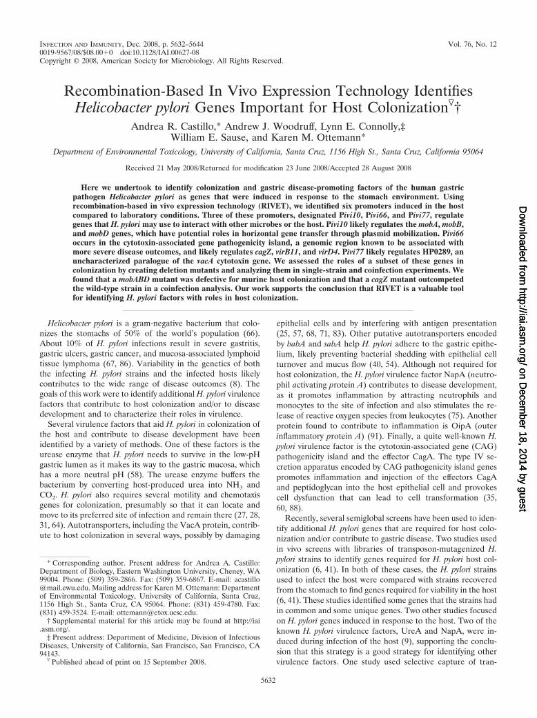
FIG. 1. RIVET for use with H. pylori. (A) An H. pylori RIVETstrain contains two RIVET components located in the chromosome, apromoterless tnpR gene that encodes the site-specific recombinaseintegrated at the H. pylori rdxA locus (diagram 1) and a cassette inwhich the aphA3 gene (also designated Km) that confers Kmr is clonedbetween res1 sequences (res1-Km-res1) integrated at the HP0294-HP0295 intergenic locus (diagram 2). In the absence of an upstreampromoter, tnpR is not expressed and the H. pylori strain remains Kmr.(B) In the presence of an upstream promoter (P), tnpR is expressed(diagram 1), and the TnpR recombinase binds the res1 sequences tocatalyze removal of the Km gene cloned between these sequences(diagram 2), converting the H. pylori strain to Kms (diagram 3).
VOL. 76, 2008 RIVET IDENTIFIES H. PYLORI COLONIZATION FACTORS 5633
on Decem
ber 18, 2014 by guesthttp://iai.asm
.org/D
ownloaded from
Analysis of pcat-T-lib-tnpR and the H. pylori RIVET library. Both pcat-T-lib-tnpR and the H. pylori RIVET library were analyzed to determine the diversityand the level of H. pylori genome coverage. We analyzed 30 clones obtained fromthe pcat-T-lib-tnpR library and 30 clones from the H. pylori RIVET library byisolating plasmid DNA and genomic DNA, respectively. The cloned H. pyloriDNA was amplified from the templates by PCR using oligonucleotide rrnB1 orcatseqst and oligonucleotide tnpRbk75 (Table 2). The presence and approximatesizes of the cloned H. pylori DNA fragments were determined by agarose gelelectrophoresis. Cloned fragments that were similar sizes were sequenced todetermine their uniqueness (Berkeley DNA Sequencing Facility, Berkeley CA).The levels of H. pylori genome coverage for the pcat-T-lib-tnpR and H. pyloriRIVET libraries were determined using the following formula: N � ln(1 � P)/ln(1 � I/G), where P is the probability of obtaining all clones (we used99%), I is the insert size (we used the calculated average insert sizes, 1.0 kb forthe pcat-T-lib-tnpR library and 0.65 kb for the H. pylori RIVET library), and Gis the genome size (1.8 � 103 kb) (2a). Based on our calculations, the pcat-T-lib-tnpR library covered the H. pylori genome �4.7 times and the H. pyloriRIVET library covered 84% of the H. pylori genome.
Screening the H. pylori RIVET library for in vivo-induced promoters. Allanimal protocols were approved by the Institutional Animal Use and CareCommittee. Fifty H. pylori RIVET strains were screened simultaneously forpromoter induction in each of two FVB/N mice (Charles River) (Fig. 2D). Fivegroups of 10 H. pylori RIVET strains were grown on CHBA containing Km to
maintain Kmr. Immediately prior to infection of mice, the 50 strains were resus-pended in BB10. The bacterial concentration was determined by determining theoptical density at 600 nm (OD600), and the culture volume was adjusted withBB10 to obtain a bacterial concentration of �5 � 107 cells/ml. Approximately 1ml of the RIVET strain mixture was used to infect each of two FVB/N mice thatwere 4 to 6 weeks old by oral gavage by using a 20-gauge, 38-mm-long needle(Popper). Infections were allowed to persist for 2 weeks, after which we har-vested and processed the mouse stomachs as described by Ottemann and Lo-wenthal (64). In brief, the stomachs were homogenized in 500 �l BB10 using asterile pestle, and dilutions were plated on CHBA plates. The plates wereincubated for 4 days at 37°C under microaerobic conditions. H. pylori coloniesfrom these plates were then replica plated on both plates containing CHBA andplates containing CHBA supplemented with Km to identify Kms H. pyloriRIVET strains that induced expression of tnpR in vivo. Kms RIVET strains wereisolated from the corresponding CHBA plates for further analysis (Fig. 2D).These strains were designated using ivi (for in vivo-induced RIVET strain) plusa number (e.g., ivi2), and the H. pylori DNA cloned upstream of tnpR in thesestrains were designated using Pivi (for in vivo-induced promoter) plus a number(e.g., Pivi2).
Identifying in vivo-induced promoters. To obtain the sequence representingeach Pivi clone, we isolated genomic DNA from the Kms H. pylori RIVET strainsisolated from mice by using a Wizard genomic DNA purification kit (Promega).We then amplified the cloned Pivi region using oligonucleotides that annealed tothe cat gene upstream of the cloned region (oligonucleotide catseqst) and thetnpR gene downstream of the cloned region (oligonucleotide tnpRbk75) (Table2) by PCR. The sizes of the cloned fragments were estimated by agarose gelelectrophoresis and were determined exactly by sequencing. The Pivi clones weresequenced using the catseqst and tnpRbk75 oligonucleotides (University of Cal-ifornia Berkeley DNA sequencing facility). The genomic and endogenous plas-mid locations of the Pivi clones were determined by comparing the sequences ofthe clones to the unpublished sequence of the H. pylori G27 strain genome (seeTable S1 in the supplemental material). An operon analysis was done using thewebsite http://www.microbesonline.org/.
Reconstruction of in vivo-induced strains. Since the H. pylori RIVET strainsthat were induced in the host removed the res1-Km-res1 cassette (and weretherefore Kms), we reintroduced the res1-Km-res1 cassette into the original locusof each of these strains before we performed our secondary analysis. One Kms
mouse output strain for each of the 13 unique Kms ivi strains was naturallytransformed with pAW2rkr2. Transformants were selected based on Kmr, andproper integration was verified by PCR using oligonucleotides that flanked thesite of integration, oligonucleotides HP0294end and HP0295end (Table 2).
Testing promoter induction in vitro and in vivo. Each of the 13 reconstructedivi strains (strains whose designations end with R in Table 1) was analyzed todetermine promoter induction conferred by the Pivi clones in the lab and inFVB/N mice. The reconstructed ivi strains were grown on CHBA containing Kmprior to these analyses to maintain Kmr. In vitro promoter induction was carriedout by passaging each reconstructed ivi strain on CHBA without Km for 2 weeks(five passages on fresh CHBA). Cells of the H. pylori reconstructed ivi strainswere then resuspended in BB10 and plated on CHBA to obtain single colonies(�200 colonies/plate). After the plates were incubated for 4 days at 37°C undermicroaerobic conditions, the H. pylori colonies were replica plated on both platescontaining CHBA and plates containing CHBA supplemented with Km. Thelevel of promoter induction (expressed as a percentage) was calculated by divid-ing the number of Kms colonies by the total number of colonies and multiplyingthe result by 100. To determine the in vivo induction conferred by the Piviregions, each reconstructed ivi strain was used independently to infect a group ofFVB/N mice. After 2 weeks of infection, the mouse stomachs were harvested andplated on CHBA to isolate H. pylori. The plates were incubated for 4 days at 37°Cunder microaerobic conditions, and then colonies were replica plated on bothplates containing CHBA and plates containing CHBA supplemented with Km todetermine the number of H. pylori cells that had converted to Kms while thestrains were in mice. The level of promoter induction in vivo (expressed as apercentage) was calculated by dividing the number of Kms colonies by the totalnumber of colonies analyzed and multiplying the result by 100. The statisticalsignificance of differences between promoter induction in vivo and promoterinduction in vitro was calculated using the two-tailed Student t test.
Construction of H. pylori gene deletion mutants. We generated deletions ofgenes regulated by our in vivo-induced promoters by replacing a gene of interestwith a nonpolar allele of the C. coli cat gene (84) that confers Cmr. Each genereplacement cassette was generated using a PCR sewing strategy (22). In brief,chromosomal regions upstream and downstream of the gene of interest wereamplified using in each case (i) one oligonucleotide that annealed to the chro-mosome and (ii) another oligonucleotide that annealed to the chromosome and
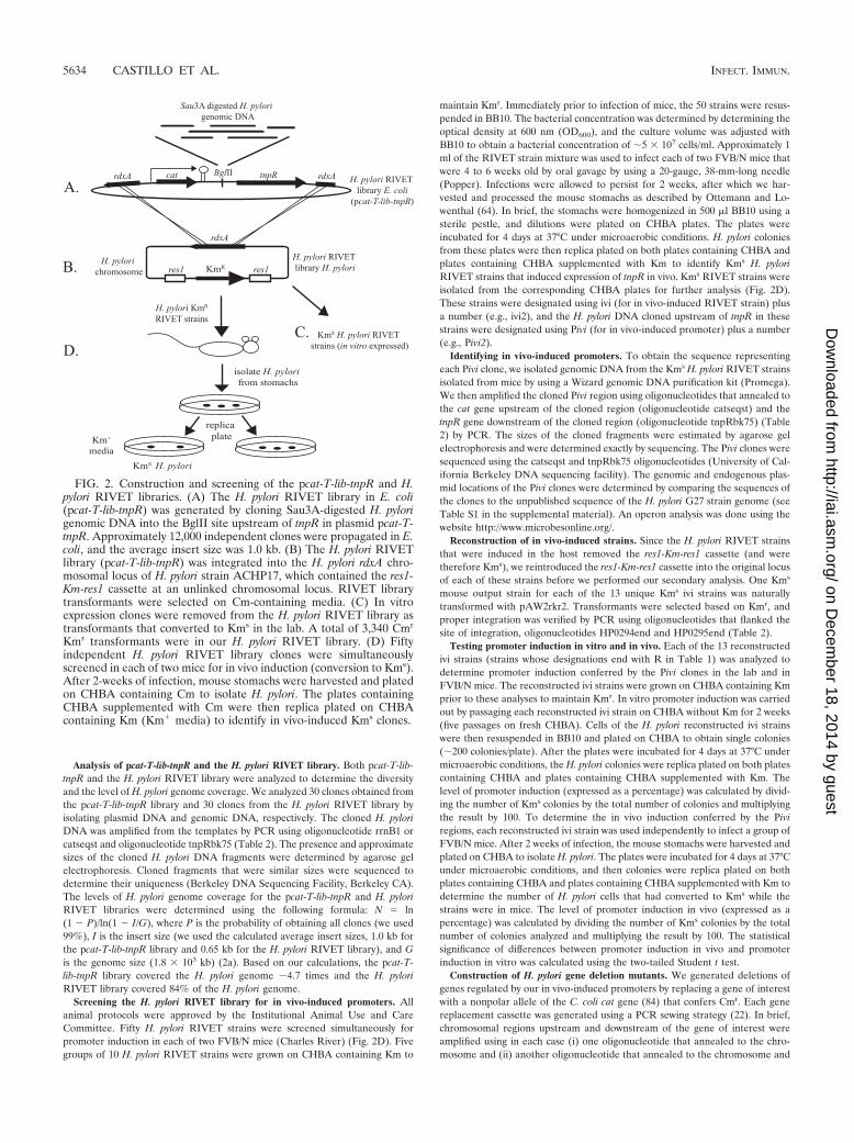
FIG. 2. Construction and screening of the pcat-T-lib-tnpR and H.pylori RIVET libraries. (A) The H. pylori RIVET library in E. coli(pcat-T-lib-tnpR) was generated by cloning Sau3A-digested H. pylorigenomic DNA into the BglII site upstream of tnpR in plasmid pcat-T-tnpR. Approximately 12,000 independent clones were propagated in E.coli, and the average insert size was 1.0 kb. (B) The H. pylori RIVETlibrary (pcat-T-lib-tnpR) was integrated into the H. pylori rdxA chro-mosomal locus of H. pylori strain ACHP17, which contained the res1-Km-res1 cassette at an unlinked chromosomal locus. RIVET librarytransformants were selected on Cm-containing media. (C) In vitroexpression clones were removed from the H. pylori RIVET library astransformants that converted to Kms in the lab. A total of 3,340 Cmr
Kmr transformants were in our H. pylori RIVET library. (D) Fiftyindependent H. pylori RIVET library clones were simultaneouslyscreened in each of two mice for in vivo induction (conversion to Kms).After 2-weeks of infection, mouse stomachs were harvested and platedon CHBA containing Cm to isolate H. pylori. The plates containingCHBA supplemented with Cm were then replica plated on CHBAcontaining Km (Km� media) to identify in vivo-induced Kms clones.
5634 CASTILLO ET AL. INFECT. IMMUN.
on Decem
ber 18, 2014 by guesthttp://iai.asm
.org/D
ownloaded from
either the start or end of the cat gene. A third PCR product representing thenonpolar cat allele was generated using oligonucleotides catR2 and catF (Table2). The PCR products representing the upstream chromosomal region, thedownstream chromosomal region, and the cat gene were generated indepen-dently, purified using an agarose gel (GFX PCR DNA and gel band purificationkit; GE Healthcare), and then combined. The mixture of PCR products was usedas a template with the oligonucleotides that annealed to the far upstream anddownstream PCR product regions. The large PCR products generated in thesereactions (upstream region-cat-downstream region) were purified using an aga-rose gel and used to naturally transform H. pylori strains SS1 (47) and mG27 (19)to Cmr as previously described (72). All mutant strains were found to be wildtype for motility by microscopic inspection and to be wild type for urease activityusing a pH indicator buffer (Difco urea broth; Difco) (data not shown).
Mouse colonization analyses. H. pylori strains used for colonization analyseswere passaged minimally in the lab on CHBA (two or three times) and thenremoved either from CHBA after growth for �18 h or from a BB10 culturegrown for �18 h. H. pylori strains grown on CHBA were transferred to BB10prior to infection and analyzed to determine motility and the bacterial cellconcentration (OD600). H. pylori strains grown in BB10 were analyzed directly todetermine motility and the bacterial cell concentration (OD600). Approximately1 ml of an H. pylori culture containing 5 � 107 to 5 � 108 CFU/ml was used toinoculate mice by oral gavage. For single-infection studies, either a mutant H.pylori strain or the appropriate wild-type control strain was used to infect mice.For the coinfection analysis, the mutant and wild-type strains were grown sepa-rately and analyzed to determine their motilities and bacterial cell concentrations(OD600) before the cultures were mixed and used for coinfection. The bacterialcell concentrations were used to generate a mixed culture containing approxi-mately equal numbers of cells of the mutant and wild-type strains. The actualbacterial cell concentration was determined more accurately by culture dilution
and plating. Infections were allowed to persist for 2 weeks, after which the mousestomachs were isolated and plated on CHBA as described above. The stomachsof mice infected with the mutant strains were plated on CHBA containing Cm,and the stomachs of mice infected with the wild-type strain were plated onCHBA. The stomachs of mice coinfected with both the mutant and the wild-typestrain were plated on both CHBA containing Cm and CHBA. The competitiveindex was calculated as follows: (CFU/g for the mutant strain output/CFU/g forthe wild-type strain output)/(CFU/g for the mutant strain input/CFU/g for thewild-type strain input).
RESULTS
We adapted the RIVET system used with great success byCamilli and Mekalanos (18) for use with H. pylori and used itto identify promoters induced in response to murine stomachs.Adapting V. cholerae RIVET for use with H. pylori required (i)creation of an H. pylori antibiotic resistance reporter for tnpRrecombinase expression flanked by res1 sites (we used Kmr
[res1-Km-res1]) and (ii) creation of an H. pylori library ofgenomic promoters fused to the tnpR gene (pcat-T-lib-tnpR).When tnpR expression was directed by the cloned H. pyloriDNA, TnpR bound the res1 sequences and catalyzed the re-moval of the intervening Kmr cassette (Fig. 1).
We changed the original RIVET system from a V. cholerae-specific antibiotic resistance (tetracycline) system to a system
TABLE 1. Strains and plasmids used in this study
H. pylori strain orplasmid Description Reference
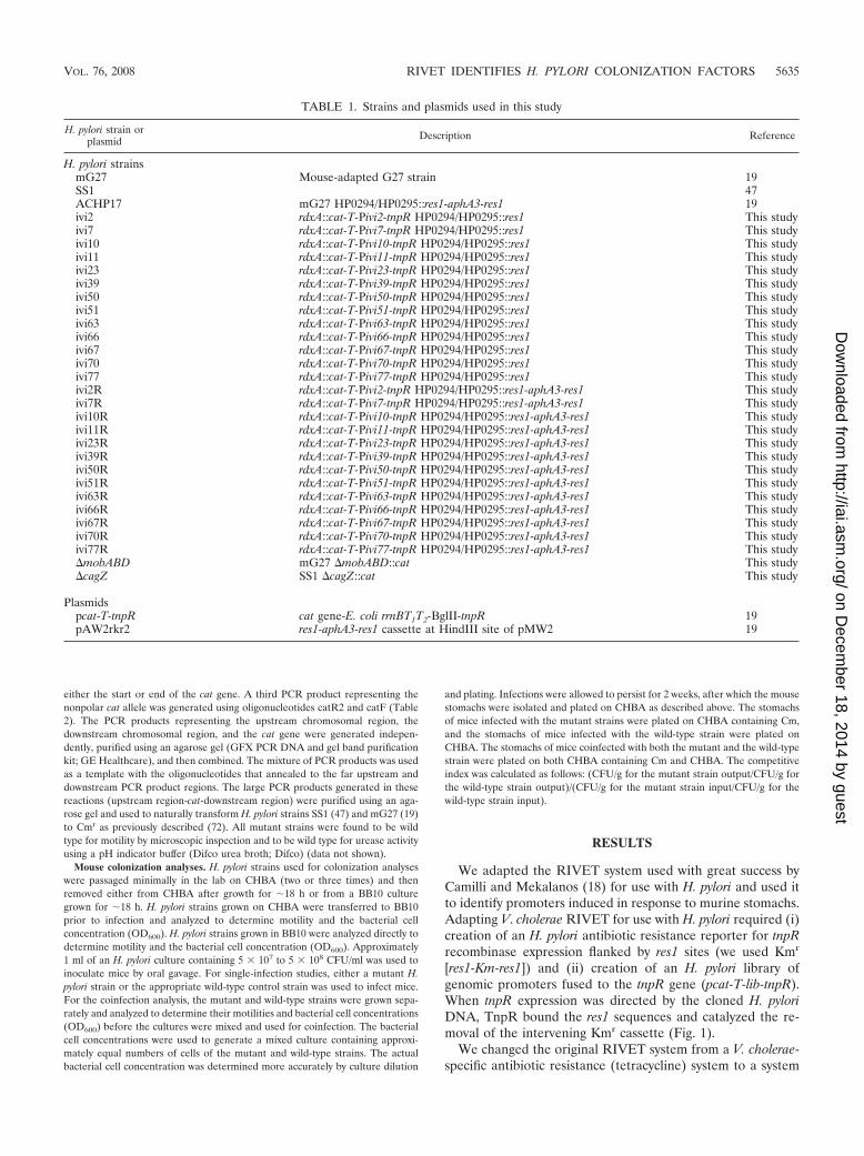
H. pylori strainsmG27 Mouse-adapted G27 strain 19SS1 47ACHP17 mG27 HP0294/HP0295::res1-aphA3-res1 19ivi2 rdxA::cat-T-Pivi2-tnpR HP0294/HP0295::res1 This studyivi7 rdxA::cat-T-Pivi7-tnpR HP0294/HP0295::res1 This studyivi10 rdxA::cat-T-Pivi10-tnpR HP0294/HP0295::res1 This studyivi11 rdxA::cat-T-Pivi11-tnpR HP0294/HP0295::res1 This studyivi23 rdxA::cat-T-Pivi23-tnpR HP0294/HP0295::res1 This studyivi39 rdxA::cat-T-Pivi39-tnpR HP0294/HP0295::res1 This studyivi50 rdxA::cat-T-Pivi50-tnpR HP0294/HP0295::res1 This studyivi51 rdxA::cat-T-Pivi51-tnpR HP0294/HP0295::res1 This studyivi63 rdxA::cat-T-Pivi63-tnpR HP0294/HP0295::res1 This studyivi66 rdxA::cat-T-Pivi66-tnpR HP0294/HP0295::res1 This studyivi67 rdxA::cat-T-Pivi67-tnpR HP0294/HP0295::res1 This studyivi70 rdxA::cat-T-Pivi70-tnpR HP0294/HP0295::res1 This studyivi77 rdxA::cat-T-Pivi77-tnpR HP0294/HP0295::res1 This studyivi2R rdxA::cat-T-Pivi2-tnpR HP0294/HP0295::res1-aphA3-res1 This studyivi7R rdxA::cat-T-Pivi7-tnpR HP0294/HP0295::res1-aphA3-res1 This studyivi10R rdxA::cat-T-Pivi10-tnpR HP0294/HP0295::res1-aphA3-res1 This studyivi11R rdxA::cat-T-Pivi11-tnpR HP0294/HP0295::res1-aphA3-res1 This studyivi23R rdxA::cat-T-Pivi23-tnpR HP0294/HP0295::res1-aphA3-res1 This studyivi39R rdxA::cat-T-Pivi39-tnpR HP0294/HP0295::res1-aphA3-res1 This studyivi50R rdxA::cat-T-Pivi50-tnpR HP0294/HP0295::res1-aphA3-res1 This studyivi51R rdxA::cat-T-Pivi51-tnpR HP0294/HP0295::res1-aphA3-res1 This studyivi63R rdxA::cat-T-Pivi63-tnpR HP0294/HP0295::res1-aphA3-res1 This studyivi66R rdxA::cat-T-Pivi66-tnpR HP0294/HP0295::res1-aphA3-res1 This studyivi67R rdxA::cat-T-Pivi67-tnpR HP0294/HP0295::res1-aphA3-res1 This studyivi70R rdxA::cat-T-Pivi70-tnpR HP0294/HP0295::res1-aphA3-res1 This studyivi77R rdxA::cat-T-Pivi77-tnpR HP0294/HP0295::res1-aphA3-res1 This study�mobABD mG27 �mobABD::cat This study�cagZ SS1 �cagZ::cat This study
Plasmidspcat-T-tnpR cat gene-E. coli rrnBT1T2-BglII-tnpR 19pAW2rkr2 res1-aphA3-res1 cassette at HindIII site of pMW2 19
VOL. 76, 2008 RIVET IDENTIFIES H. PYLORI COLONIZATION FACTORS 5635
on Decem
ber 18, 2014 by guesthttp://iai.asm
.org/D
ownloaded from
that works with H. pylori. To do this, we chose a Kmr gene(aphA3 or Km) that carries its own promoter and flanked itwith recombinase recognition sequences (res1) to create res1-Km-res1. We used the mutant res sequence res1, which containsa mutation at the crossover site resulting in decreased recom-bination efficiency (59). We hypothesized that use of the res1allele would allow us to identify promoters that were expressedto some extent in vitro but exhibited elevated expression in thehost. The res1-Km-res1 cassette was cloned into a plasmid thatdirected its integration into the chromosomal region betweenopen reading frames HP0294 and HP0295 (19). We integratedthis construct into the chromosome of H. pylori strain mG27 toensure that a single copy was present and to ensure that it wasstably maintained. H. pylori strain mG27 is a mouse-adaptedversion of the commonly used H. pylori strain G27 (19, 79).Integration at the HP0294-HP0295 site occurred by double-crossover gene replacement and did not affect the growth orvirulence of H. pylori strains mG27 and SS1 (data not shown).We verified that res1-Km-res1 integrated into the proper chro-mosomal region by selecting for Kmr transformants and byperforming PCR with oligonucleotide primers that flanked theinsertion site (data not shown). The resulting strain was des-ignated ACHP17 (Table 1).
Generating the library of potential promoters (pcat-T-lib-tnpR). To generate pcat-T-lib-tnpR (Table 1), we created aplasmid with a promoterless tnpR gene and cloned a partiallySau3A-digested H. pylori genomic library upstream of tnpR(Fig. 2A). The plasmid contained the cat gene, which conferredCmr, followed by a strong E. coli terminator, the site for H.pylori library insertion, and the promoterless tnpR gene, allflanked by sequences of the H. pylori gene rdxA. Expression ofthe cat gene is directed by its endogenous promoter, and the E.coli terminator prevents read-through transcription of tnpR(19). We used the wild-type tnpR allele, which had a wild-typeribosome binding site, for our work (48). pcat-T-lib-tnpR was a
collection of �12,000 independent colonies. Our analysis of 30library clones suggested that 70% of them contained insertswith an average size of 1.0 kb (data not shown). Our analysisalso indicated that the library was diverse and covered the1.8-Mb H. pylori genome 4.7 times (see Materials and Meth-ods). The rdxA sequences flanking cat-T-lib-tnpR in pcat-T-lib-tnpR targeted integration of this plasmid into the H. pylorichromosomal rdxA locus by double-crossover homologous re-combination (Fig. 2B). The rdxA locus is commonly used forintegrating exogenous DNA into the H. pylori chromosome(80).
Creation of the H. pylori RIVET library. To construct the H.pylori RIVET library strains, pcat-T-lib-tnpR was used to trans-form H. pylori strain ACHP17 (HP0294/HP0295::res1-Km-res1)(Table 1) to Cmr (Fig. 2B). Each Cmr transformant was passagedtwice in Cm-containing media before selection on Km-containingmedia and subsequent freezing. We initially passaged the Cmr
transformants without Km selection to allow promoters that wereexpressed in the lab to convert the strains carrying them to Kms
(Fig. 2C). We were interested in promoters that were not ex-pressed in the lab or were expressed at very low levels in the laband thus in bacteria that retained Kmr (res1-Km-res1). Thegenomic DNA of 30 Cmr H. pylori transformants was screened byPCR using oligonucleotides that annealed upstream and down-stream of the genomic DNA fragment to assess whether thissubset of transformants contained unique genomic DNA insertsupstream of tnpR. The results of our screening of the H. pyloritransformants suggested that 70% of them contained unique in-serts and that the inserts were smaller (0.65 kb) than those foundfor the same library in E. coli. Although we are not certain, wespeculate that the average insert size in the H. pylori RIVETlibrary was smaller than the average insert size in pcat-T-lib-tnpReither because the larger inserts had more potential sites for H.pylori’s restriction systems or because larger inserts recombinedless efficiently. Our H. pylori RIVET library contained 3,340 in-
TABLE 2. Oligonucleotides used in this study
Oligonucleotide Sequence Reference
catseqst GAAGTATTATGAGGAGGGCG 19catF2 CAACCGTGATATAGATTGAAAAGTGGAT This studycatF GATATAGATTGAAAAGTGGAT This studycatR2 CGCGCCCGGGATCCTCCTTG This studytnpRbk75 TCAGTAAAGATGCGATTTGC 19rrnB1 CCCTCGAGAATAAAACGAAAGGCTCAGTCG 19G27_633_D1 GCCCTTAGTTCAGGTGTGGCAGTTTAAGG This studyG27_633_D2 CAAGGAGGATCCCGGCCGCGGCTACCTTCTCATTTCCTAGATAGTAGCC This studyG27_633_D3 ATCCACTTTTCAATCTATATCACGGTTGCCGGGAATGTGGGCATGCGAGTGGCG This studyG27_633_D4 GTTTTAGCGTCAATGTTGGGGTTGATTCTAATGG This studyG27_630_D1 GAGCTATGGGAAAGATAGAGGAAGCAATATCGC This studyG27_630_D2 CAAGGAGGATCCCGGCCGCGCCCTCTCCTTAATTTCATACTC This studyG27_630_D3 ATCCACTTTTCAATCTATATCACGGTTGGAACATTCTCATTTGTATGATTTGTTGAACGGG This studyG27_630_D4 GGGCTTGAATGTCAGTGATCCTGTC This studyG27_176_D1 GAGCGTGGATGGCAGGATCAGCGTTAAAG This studyG27_176_D2 CAAGGAGGATCCCGGCCGCGCTCATGCATGCTTAAACCCCACATCAAGGACG This studyG27_176_D3 ATCCACTTTTCAATCTATATCACGGTTGCGGGAGCAATCATGTTATCTTCTAATG This studyG27_176_D4 GAATCCACGCTATAGCCTTCTTGATAC This studyMobA_D1 CGCAATCAATCATGATAACCCTATTATATC This studyMobA_D2 CAAGGAGGATCCCGGGCGCGCCAACATACTTGGATCTTATTTGTTC This studyMobA_D3 ATCCACTTTTCAATCTATATCACGGTTGGAGAAGTTATAGTCGTTGGTATGGGCGGTAAG This studyMobA_D4 CTGGTTTACTTGACATTAGATCGATAAACAGGTG This studyHP0294end CTTGTCCTGTGGGCGATTTGC This studyHP0295end GACCGGCCGGATATGGCAG This study
5636 CASTILLO ET AL. INFECT. IMMUN.
on Decem
ber 18, 2014 by guesthttp://iai.asm
.org/D
ownloaded from

dependent clones that were pooled in groups of 10 and stored inH. pylori freezing media at �80°C. These transformants covered�84% of the H. pylori genome (see Materials and Methods).
Identifying H. pylori host-induced promoters. To identifyputative host-induced promoters, we used the H. pylori RIVETlibrary strains to infect FVB/N mice (Fig. 2D). We screened 50H. pylori RIVET library strains in parallel by infecting micewith a mixture of 50 RIVET library strains. Baldwin and co-workers showed that this pool size allows each strain to inde-pendently establish an infection in the mouse gastric mucosa(6). The infections were allowed to persist for 2 weeks, andthen we sacrificed the animals and harvested their stomachs.The stomachs were homogenized, diluted, and plated on Cm-containing media. After 4 days, each plate was replica platedon both Cm-containing media and Km-containing media (Fig.2D). RIVET strains that induced tnpR expression at any timeduring the infection converted to Kms. The Kms strains wererescued from the corresponding Cm-containing plates andsaved as frozen stocks for additional analysis. We screened2,960 clones of our 3,340-clone H. pylori RIVET library andthus approximately 74% of the H. pylori genome. Our screen-ing analysis resulted in identification of 113 Kms H. pyloriRIVET strains. By using PCR amplification and sequencing ofthe region cloned upstream of tnpR in these strains, we deter-mined that the 113 Kms strains represented 13 unique clones(see Table S1 in the supplemental material). We designatedthese unique clones Pivi clones and the strains containing themivi strains; to identify a specific clone or strain, the numbercorresponding to the order of ivi strain isolation was added toits designation (Table 1).
Verifying that the Pivi clones were induced in the host. Weretested the Pivi clones identified as described above to ensurethat their expression in mice was greater than their expressionin vitro. The ivi strains, which were isolated from mouse stom-achs as Kms strains, induced tnpR expression in vivo and thusremoved the res1-Km-res1 cassette from the H. pylori chromo-some. Therefore, we reintroduced res1-Km-res1 into thesestrains by transforming them to Kmr with the construct used tocreate the original strain (pAW2rkr2) (see Materials andMethods). Strains containing 1 of the 13 Pivi clones fusedupstream of tnpR and the res1-Km-res1 cassette (reconstructedivi strains) were designated by adding R to the end of the ividesignation and were analyzed further to examine promoter-directed expression of tnpR based on their conversion fromKmr to Kms (Table 1).
To determine Pivi-directed expression of tnpR in the host,we infected mice with each reconstructed ivi strain as a singleinfecting strain. Each infection was allowed to persist for 2weeks, and then we sacrificed the animals, harvested theirstomachs, and plated the stomachs to obtain single colonies onCm-containing media as described above. Plates containing thecolonies isolated from stomachs were then replica plated onCm-containing media and Km-containing media. The level ofpromoter induction in vivo (expressed as a percentage) wasdetermined by dividing the number of Kms colonies by thetotal number of colonies analyzed and multiplying the result by100. To determine the in vitro expression conferred by eachPivi clone, each reconstructed ivi strain was passaged on Cm-containing media in the lab for 2 weeks (four or five passages)and then plated on Cm-containing media to obtain single col-
onies. The resulting plates were replica plated on both Cm-containing media and Km-containing media to determine thelevel of promoter induction in vitro.
We found that 4 of the 13 Pivi clones originally isolated frommice as Kms ivi strains conferred in vivo expression of tnpRthat was significantly higher than the in vitro expression whenthe strains were tested as single infecting strains (as deter-mined by two-tailed Student’s t test) (Fig. 3). These Pivi cloneswere Pivi51 (P 0.001), Pivi66 (P 0.001), Pivi70 (P 0.05),and Pivi77 (P 0.05). The values for two additional Piviclones, Pivi10 and Pivi67, were very close to our cutoff forstatistical significance, with P values of 0.1. We includedthese six Pivi clones in our subsequent analyses. The remainingseven Pivi clones identified in our screen as Kms ivi strains didnot show in vivo induction of tnpR when they were retested assingle infecting strains (Fig. 3). It is possible that the Pivi cloneseither were simply not induced in mice and were isolated asbackground clones inherent in this screen or were induced onlywhen the strains were used in coinfections with additional
FIG. 3. Six RIVET-identified promoters were induced in the host.The relative level of promoter induction of each of the 13 recon-structed RIVET strains isolated from mice as Kms strains was analyzedboth in mice and in the lab. Promoter induction in the host wasanalyzed using each reconstructed RIVET strain to independentlyinfect a group of mice for 2 weeks. Promoter induction in the lab wasanalyzed after 2 weeks of in vitro growth on CHBA. For both in vivoand in vitro experiments, the level of promoter induction was calcu-lated by dividing the number of Kms CFU isolated by the total numberof CFU and multiplying the result by 100. All promoters indicated byan asterisk were induced to a greater extent in vivo than in vitro. Atwo-tailed Student t test was used to determine the following P valuesfor the promoters when in vitro induction and in vivo induction werecompared: for Pivi10, 0.1; for Pivi51, 0.001; for Pivi66, 0.001; forPivi67, 0.1; for Pivi70, 0.05; and for Pivi77, 0.05. The other pro-moters did not differ appreciably under in vivo and in vitro conditions.The error bars indicate standard deviations. For the RIVET strains,the in vitro experiment was replicated two to six times and the in vivoanalysis was carried out using zero to nine mice, as follows: for Pivi2,two in vitro replicates and five mice; for Pivi7, three in vitro replicatesand five mice; for Pivi10, three in vitro replicates and four mice; forPivi11, two in vitro replicates and one mouse; for Pivi23, three in vitroreplicates and nine mice; for Pivi39, six in vitro replicates and no mice;for Pivi50, one in vitro replicate and two mice; for Pivi51, four in vitroreplicates and nine mice; for Pivi63, two in vitro replicates and twomice; for Pivi66, six in vitro replicates and three mice; for Pivi67, twoin vitro replicates and three mice; for Pivi70, two in vitro replicates andfour mice; and for Pivi77, four in vitro replicates and three mice.
VOL. 76, 2008 RIVET IDENTIFIES H. PYLORI COLONIZATION FACTORS 5637
on Decem
ber 18, 2014 by guesthttp://iai.asm
.org/D
ownloaded from

strains, as was done in the screening of our H. pylori RIVETlibrary. Alternatively, retesting of only one Kms colony for eachunique Pivi clone could have contributed to a high false-neg-ative rate. We did not perform additional studies to distinguishbetween these possibilities.
Genomic location of in vivo-induced promoters. We modi-fied the RIVET system for use with H. pylori so that it identi-fied in vivo-induced promoters instead of specific in vivo-in-duced genes. Thus, we were interested in determining thegenes regulated by our in vivo-induced promoters. To identifythese genes, we mapped the genomic locations of the six Piviclones on the H. pylori genome by comparing their sequencesto the sequence of the H. pylori strain G27 genome (Fig. 4 andunpublished data). The locations of the Pivi clones in the G27genome were the same as those in the previously described H.pylori genomes with the exception of the location of Pivi10,which is located on an endogenous plasmid (1, 61, 85). There-fore, we used H. pylori strain 26695 annotation for all Piviclones except Pivi10 (85). The gene(s) regulated by each Piviclone is discussed below.
Pivi clones regulating H. pylori genes involved in interac-tions with other cells. Three of the Pivi clones were located in
DNA regions upstream of a gene or set of genes potentiallyused by H. pylori to interact with other bacterial cells or withhost cells. Pivi10 was located in the putative mobC gene up-stream of the putative mobA, mobB, and mobD genes on anendogenous H. pylori strain G27 plasmid (Fig. 4). About 50%of H. pylori strains harbor endogenous plasmids (69), and plas-mid DNA would have been isolated by our genomic DNApreparation procedure and thus included in our pcat-T-lib-tnpR and H. pylori RIVET libraries. mob genes encode relaxo-some components required for plasmid nicking and mobiliza-tion during horizontal gene transfer (32). Pivi66 mapped to theCAG pathogenicity island in the cagY gene upstream of apredicted operon containing cagZ, virB11, and virD4. TheCAG pathogenicity island is a 40-kb region containing �30genes, and its presence is associated with H. pylori infectionsthat have more severe disease outcomes (21). Some of the geneproducts encoded by the CAG pathogenicity island form a typeIV secretion apparatus known to inject at least two effectors,CagA and peptidoglycan, into host cells (5, 88). While VirD4and VirB11 are homologues of an adapter protein likely in-volved in substrate export (2, 85) and an ATPase that gener-ates energy for apparatus assembly and substrate export (77,85), respectively, the function of CagZ is not known yet. Fi-nally, Pivi77 overlapped the intergenic region upstream of oneof the uncharacterized vacA paralogues, HP0289 (85). vacAencodes a well-characterized cytotoxic vacuolating protein be-longing to the autotransporter family and is required by H.pylori for full virulence (72, 90). The similarity of the HP0289protein to VacA is concentrated mostly in the carboxy-terminalautotransporter domain rather than in the cytotoxic vacuolat-ing amino-terminal domain (55), and it is therefore not clearwhat role the HP0289 protein may play in H. pylori virulence.
Pivi51 regulates the putative mesJ lysidine synthetase. Pivi51overlaps mesJ and the upstream intergenic region and there-fore likely regulates mesJ. mesJ, also called tilS in E. coli, is amember of the PP-loop ATPase superfamily. Members of thissuperfamily of proteins have distinct enzymatic functions butshare an ATP pyrophosphatase domain that targets the alpha-beta bond of ATP (13). Recently, tilS was found to govern boththe codon and amino acid specificities of the isoleucine tRNA(81). TilS is a lysidine synthetase that generates the lysidinemodification at the wobble position of the tRNAIle anticodon.This changes the codon specificity from AUG to AUA and theamino acid specificity from methionine to isoleucine (81).
Pivi70 regulates a putative metalloprotease. Another of thein vivo-induced promoters, Pivi70, was located in open readingframe HP0285, upstream of the H. pylori ftsH gene and twohypothetical open reading frames, HP0287 and HP0288. ftsH(HP0286) encodes a putative ATP-dependent metalloprotease(33) and is essential for growth in E. coli. In E. coli this integralinner membrane metalloprotease degrades both cytosolic pro-teins, including sigma 32 and lambda CII (36, 37), and trans-membrane proteins, including SecY and YccA (43, 44). FtsHlikely plays a role in cell division as an E. coli strain with atemperature-sensitive ftsH allele exhibits filamentous growth atrestrictive temperatures (74). Two ftsH homologues, HP0286and HP1069, are present in H. pylori; Ge and Taylor (33)showed that HP1069 is essential. There have been no studies todate of HP0286-encoded FstH, and H. pylori FtsH targets havenot been identified yet. The products of HP0287 and HP0288,
FIG. 4. Locations of in vivo-induced promoters on the H. pylorichromosome or an endogenous plasmid. Each in vivo-induced pro-moter is represented by an open box, and the arrow below the boxindicates the direction in which the promoter was cloned. The openreading frames overlapped by and in the genomic region of the invivo-induced promoters are indicated by thick arrows. (A) Pivi10 is 153bp long and overlaps the mobC gene in the H. pylori strain mG27endogenous plasmid. It likely regulates mobA, mobB, and mobD.(B) Pivi51 is 95 bp long and overlaps mesJ and the intergenic regionupstream of mesJ and thus likely regulates mesJ. (C) Pivi66 is 166 bplong and is located within cagY, upstream of a putative operon con-taining cagZ, virB11, and virD4. (D) Pivi67 is 133 bp long and overlapsHP1359, ubiA, and the intergenic region between them. There are sixgenes that are potentially regulated by Pivi67, including three unchar-acterized open reading frames (HP1359, HP1358, and HP1354) psd,nadA, and nadC. (E) Pivi70 is 119 bp long and is located withinHP0286. Pivi70 potentially regulates the metalloprotease gene ftsHand uncharacterized open reading frames HP0287 and HP0288.(F) Pivi77 is 1,679 bp long and overlaps HP0288, HP0289, and theintergenic region between them. Pivi77 likely regulates the vacA para-logue encoded by HP0289.
5638 CASTILLO ET AL. INFECT. IMMUN.
on Decem
ber 18, 2014 by guesthttp://iai.asm
.org/D
ownloaded from

which are two hypothetical open reading frames located down-stream of fstH, show no homology to previously identifiedproteins (85).
Pivi67 regulates previously uncharacterized proteins. Pivi67,the last of the in vivo-induced promoters identified in ouranalysis, was located upstream of six open reading framespredicted to be in the same operon. These open reading framesincluded two hypothetical open reading frames, HP1359 andHP1358, followed by psd (encoding a phosphatidyl serine de-carboxylase), nadA (encoding a quinolinate synthetase A),nadC (encoding a nicotinate dinucleotide pyrophosphorylase),and an open reading frame encoding a putative adenine-spe-cific methyltransferase.
Genes regulated by Pivi clones play a role in H. pylori colo-nization of the host. To address our goal of identifying H. pylorigenes required for host colonization and/or disease develop-ment, we created deletion mutants with mutations in genespotentially regulated by our Pivi clones and analyzed thesemutants to determine mouse colonization phenotypes. Whenthere was more than one gene potentially regulated by our Piviclones, we attempted to construct a mutant with a deletion ofthe gene immediately downstream of the promoter. Thus, wecreated mutants with deletions of the mobA and cagZ genes,which were immediately downstream of Pivi10 and Pivi66, re-spectively. However, since the mobB and mobD open readingframes are located in the mobA open reading frame, we actu-ally created a mobABD triple-deletion strain. Several unsuc-cessful attempts were made to delete HP1359, the hypotheticalopen reading frame downstream of Pivi67, and ftsH, the metal-loprotease gene downstream of Pivi70. Our inability to deletethese genes is consistent with the hypothesis that they areessential for viability, as proposed by other workers (73). Fi-nally, we did not analyze HP0289 and mesJ, the open readingframes downstream of Pivi77 and Pivi51, respectively.
H. pylori nonpolar deletion mutants were constructed byreplacing the open reading frame of interest with the cat gene,which conferred Cmr (see Materials and Methods). Deletionand insertion cassettes were generated using a sewing PCRstrategy described by Chalker et al. (22) that allows efficientgene deletion and replacement with a cat gene. The cagZ genewas deleted in the H. pylori SS1 strain commonly used foranalyzing H. pylori phenotypes in mice because this strain re-producibly infects mice. To address the possibility that cagZ isdifferently regulated in the SS1 strain, we verified that thepromoter regulating cagZ was similarly induced in the mG27and SS1 strains, i.e., 77.3 and 76%, respectively (Fig. 3). The
mobABD genes were deleted in mG27 since we knew that thisstrain contains the endogenous plasmid from which Pivi10 wasisolated. mG27 infects mice at levels that are approximately10-fold less than the H. pylori SS1 strain levels. In each case,Cmr transformants were selected and analyzed by PCR toverify deletion of the cagZ and mobABD loci. Deletion of cagZand mobABD were unlikely to have polar effects on down-stream genes, as we verified expression of the gene down-stream of cagZ in the �cagZ strain by reverse transcription-PCR (data not shown) and there is a putative transcriptionalterminator downstream of mobABD (38). Finally, deletion ofmobABD and deletion of cagZ did not confer in vitro growthdefects when we used the mutant strains in in vitro competitionassays with the wild-type mG27 and SS1 strains, respectively(Table 3).
To assess the contribution of cagZ and mobABD to H. pyloricolonization of mice, we carried out both single-strain infectionstudies and coinfection studies. For single-strain infectionstudies we infected groups of five mice with each mutant strain(�cagZ or �mobABD) and each wild-type strain (SS1 ormG27). For the coinfection studies a mutant strain (�cagZ or�mobABD) was used to infect a group of five mice along withthe wild-type passage control strain (SS1 and mG27, respec-tively). Single-strain studies and coinfection studies for eachmutant–wild-type strain pair were carried out simultaneouslyusing the same mutant strain and wild-type strain inocula so wecould be confident of the mutant strain behavior in the coin-fection study.
When it was used as the only infecting strain, the �cagZstrain had a colonization fitness similar to that of the wild-typestrain (Fig. 5). Interestingly, however, when mice were infectedwith the �cagZ strain along with the wild-type strain, the
FIG. 5. The �cagZ H. pylori mutant outcompetes the wild-typestrain in a coinfection colonization assay. Single-strain infection andcoinfection studies with H. pylori �cagZ and wild-type strains werecarried out using FVB/N mice for 2 weeks as described in Materialsand Methods. The results of one representative set of experiments areshown. Each point represents one mouse stomach, and the solid linesindicate the averages. When the �cagZ mutant strain was coinfectedwith the wild-type strain, we were unable to detect any wild-type strainand thus placed its concentration at the limit of detection (200 CFU/g).This suggests that the �cagZ mutant strain outcompeted the wild-typestrain for colonization (competitive index, 5,713).
TABLE 3. In vitro growth competition between the mobABD andcagZ mutants and the correspoding isogenic wild-type strains
Strains
Ratio of mutant to wild type ata:
Zerotime 3 h 6 h 12 h 18 h 24 h 30 h
SS1 �cagZ � wild type 0.7 0.7 1.4 0.4 0.5 0.2mG27 �mobABD � wild type 0.1 1.2 0.5 0.7 0.9 0.9
a Ratios of the mutant to the wild type in an in vitro growth competitionexperiment at different times. Strains were grown BB10 with shaking undermicroaerobic conditions. The wild-type strains are isogenic with the correspond-ing mutants. Each competition assay was repeated two times, and similar resultswere obtained. The results for one replicate for each mutant are shown.
VOL. 76, 2008 RIVET IDENTIFIES H. PYLORI COLONIZATION FACTORS 5639
on Decem
ber 18, 2014 by guesthttp://iai.asm
.org/D
ownloaded from
�cagZ strain outcompeted the wild-type strain; no wild-typecells or a reduced number of wild-type cells were isolated fromthe stomachs of mice coinfected with the wild-type and �cagZstrains (Fig. 5 and data not shown). We calculated that thecompetitive indices were 5,713 for one coinfection experiment(Fig. 5) and 20 for a second coinfection experiment. Thesecompetitive indices suggest that the �cagZ strain outcompetesthe wild-type strain in mice by approximately 1 to 3 orders ofmagnitude. These results intriguingly suggest that elevatedtranscription of cagZ in mice actually decreases the bacteri-um’s colonization ability, and they are consistent with the no-tion that genes identified by our RIVET analysis play roles inhost colonization.
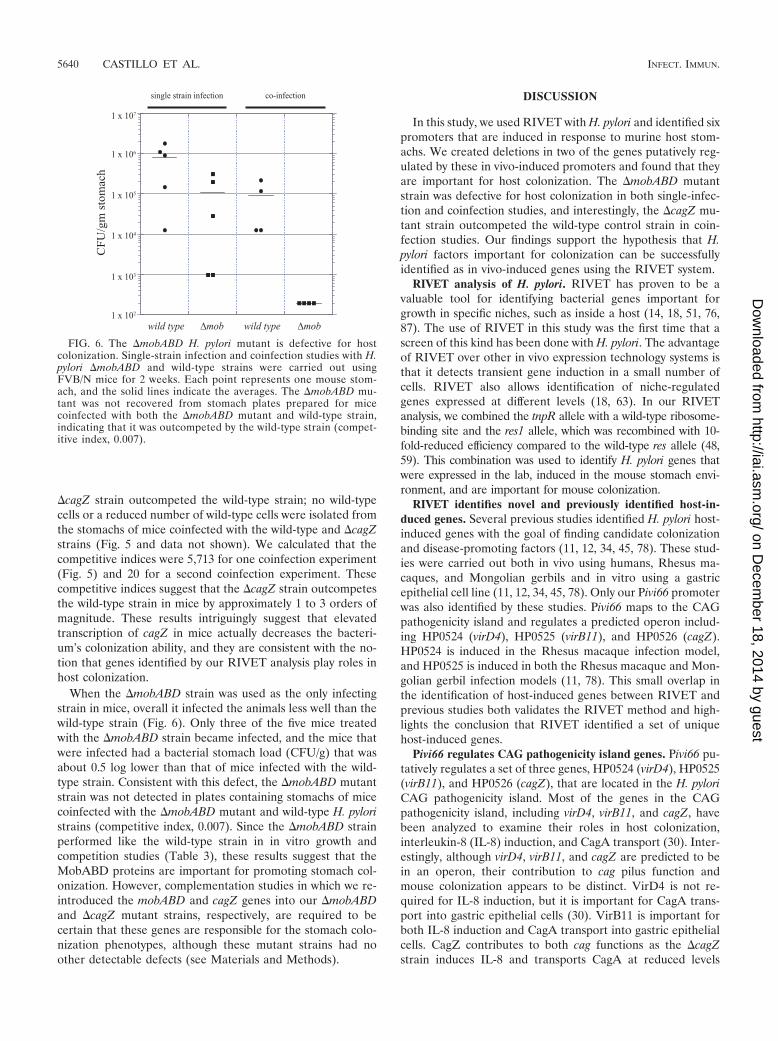
When the �mobABD strain was used as the only infectingstrain in mice, overall it infected the animals less well than thewild-type strain (Fig. 6). Only three of the five mice treatedwith the �mobABD strain became infected, and the mice thatwere infected had a bacterial stomach load (CFU/g) that wasabout 0.5 log lower than that of mice infected with the wild-type strain. Consistent with this defect, the �mobABD mutantstrain was not detected in plates containing stomachs of micecoinfected with the �mobABD mutant and wild-type H. pyloristrains (competitive index, 0.007). Since the �mobABD strainperformed like the wild-type strain in in vitro growth andcompetition studies (Table 3), these results suggest that theMobABD proteins are important for promoting stomach col-onization. However, complementation studies in which we re-introduced the mobABD and cagZ genes into our �mobABDand �cagZ mutant strains, respectively, are required to becertain that these genes are responsible for the stomach colo-nization phenotypes, although these mutant strains had noother detectable defects (see Materials and Methods).
DISCUSSION
In this study, we used RIVET with H. pylori and identified sixpromoters that are induced in response to murine host stom-achs. We created deletions in two of the genes putatively reg-ulated by these in vivo-induced promoters and found that theyare important for host colonization. The �mobABD mutantstrain was defective for host colonization in both single-infec-tion and coinfection studies, and interestingly, the �cagZ mu-tant strain outcompeted the wild-type control strain in coin-fection studies. Our findings support the hypothesis that H.pylori factors important for colonization can be successfullyidentified as in vivo-induced genes using the RIVET system.
RIVET analysis of H. pylori. RIVET has proven to be avaluable tool for identifying bacterial genes important forgrowth in specific niches, such as inside a host (14, 18, 51, 76,87). The use of RIVET in this study was the first time that ascreen of this kind has been done with H. pylori. The advantageof RIVET over other in vivo expression technology systems isthat it detects transient gene induction in a small number ofcells. RIVET also allows identification of niche-regulatedgenes expressed at different levels (18, 63). In our RIVETanalysis, we combined the tnpR allele with a wild-type ribosome-binding site and the res1 allele, which was recombined with 10-fold-reduced efficiency compared to the wild-type res allele (48,59). This combination was used to identify H. pylori genes thatwere expressed in the lab, induced in the mouse stomach envi-ronment, and are important for mouse colonization.
RIVET identifies novel and previously identified host-in-duced genes. Several previous studies identified H. pylori host-induced genes with the goal of finding candidate colonizationand disease-promoting factors (11, 12, 34, 45, 78). These stud-ies were carried out both in vivo using humans, Rhesus ma-caques, and Mongolian gerbils and in vitro using a gastricepithelial cell line (11, 12, 34, 45, 78). Only our Pivi66 promoterwas also identified by these studies. Pivi66 maps to the CAGpathogenicity island and regulates a predicted operon includ-ing HP0524 (virD4), HP0525 (virB11), and HP0526 (cagZ).HP0524 is induced in the Rhesus macaque infection model,and HP0525 is induced in both the Rhesus macaque and Mon-golian gerbil infection models (11, 78). This small overlap inthe identification of host-induced genes between RIVET andprevious studies both validates the RIVET method and high-lights the conclusion that RIVET identified a set of uniquehost-induced genes.
Pivi66 regulates CAG pathogenicity island genes. Pivi66 pu-tatively regulates a set of three genes, HP0524 (virD4), HP0525(virB11), and HP0526 (cagZ), that are located in the H. pyloriCAG pathogenicity island. Most of the genes in the CAGpathogenicity island, including virD4, virB11, and cagZ, havebeen analyzed to examine their roles in host colonization,interleukin-8 (IL-8) induction, and CagA transport (30). Inter-estingly, although virD4, virB11, and cagZ are predicted to bein an operon, their contribution to cag pilus function andmouse colonization appears to be distinct. VirD4 is not re-quired for IL-8 induction, but it is important for CagA trans-port into gastric epithelial cells (30). VirB11 is important forboth IL-8 induction and CagA transport into gastric epithelialcells. CagZ contributes to both cag functions as the �cagZstrain induces IL-8 and transports CagA at reduced levels
FIG. 6. The �mobABD H. pylori mutant is defective for hostcolonization. Single-strain infection and coinfection studies with H.pylori �mobABD and wild-type strains were carried out usingFVB/N mice for 2 weeks. Each point represents one mouse stom-ach, and the solid lines indicate the averages. The �mobABD mu-tant was not recovered from stomach plates prepared for micecoinfected with both the �mobABD mutant and wild-type strain,indicating that it was outcompeted by the wild-type strain (compet-itive index, 0.007).
5640 CASTILLO ET AL. INFECT. IMMUN.
on Decem
ber 18, 2014 by guesthttp://iai.asm
.org/D
ownloaded from
compared to wild-type H. pylori strains (30). Both the �virD4and �virB11 mutant H. pylori strains were attenuated formouse colonization in single-infection studies, while the �cagZstrain infected mice at levels similar to the levels of infection ofthe wild-type strain in single-strain infection studies (56; thisstudy). We observed, however, that the �cagZ strain had acolonization advantage during coinfection. It is not clear thatthe reduced cag function of the �cagZ mutant is responsiblefor the enhanced colonization phenotype that we observed.
The crystal structure and protein interaction profile of CagZare consistent with a chaperonelike function for CagZ (20),because of the presence of a disordered carboxy-terminal tailand a highly negatively charged surface. Members of a class ofchaperones important for delivery of type III effectors, includ-ing CesT from enterohemorrhagic E. coli and SigE from Sal-monella spp., have a unique structure, but they also have neg-atively charged surfaces (52). It is possible that CagZcontributes directly to the transport of the CagA effector; how-ever, since CagZ is not absolutely required for CagA transport,this seems unlikely. Another possibility is that CagZ has achaperonelike function for assembly of the cag secretion pilus.In fact, CagZ was found to interact with 10 Cag proteins(CagY, CagX, CagV, CagT, CagS, CagM, CagI, CagG, CagF,and CagE) in yeast two-hybrid and coimmunoprecipitationstudies (16). Many of these proteins form the channel or coreof the cag pilus structure (4). We hypothesize that CagZ mayin fact provide chaperone activity and help assemble antigens,similar to the role proposed for CagY (70). If this prediction iscorrect, cagZ mutants would have less CAG pilus antigenicityand might have been able to avoid an anti-CAG immune re-sponse that would have targeted the wild-type strain. Suchimmune avoidance could have conferred the enhanced coloni-zation phenotype. Future Cag protein assembly experimentswith a �cagZ strain will address this hypothesis.
In vivo-induced promoter regulates genes with putativeroles in horizontal gene transfer. DNA transfer via conjuga-tion is a common mechanism for sharing genetic material thatcan contribute to the success of pathogenic bacteria (26). Con-jugation is the transfer of DNA from a bacterial donor to arecipient cell by direct cell-to-cell contact. We identified agroup of H. pylori endogenous plasmid-borne genes, mobA,mobB, and mobD, as genes that are induced during mouseinfection and are important for mouse colonization. The mobgenes have been shown to be important for DNA transfer viaconjugation in other microbes. The mob genes encode a relax-ase (mobA) and accessory proteins (mob and mobD) that makeup a complex called the relaxosome. The relaxosome functionsby (i) nicking the DNA molecule to be transferred at the originof transfer, (ii) becoming covalently associated with the 5 endof the single-stranded DNA, (iii) transporting the DNA mol-ecule to the conjugation machinery at the inner cell membranevia a coupling protein, and (iv) transporting the DNA moleculeacross the bacterial membranes through the conjugation ma-chinery into the recipient cell (49). The other componentsimportant for DNA transfer via conjugation include the cou-pling protein mentioned above, the transmembrane proteincomplex, and the conjugation pilus (7, 50). DNA transfer viaconjugation is a widespread method of horizontal gene transferin the prokaryotic world and has been documented to occur
between prokaryotes and eukaryotes as well, including yeast,plant, and mammalian cells (15, 46, 89, 92).
DNA transfer via conjugation has been shown to occur be-tween H. pylori clinical isolates and between H. pylori andCampylobacter jejuni (3, 65). Specific conjugation componenthomologues have been identified in H. pylori, including twochromosomally encoded relaxase proteins, Rlx1 and Rlx2, andtwo chromosomally encoded coupling proteins, TraG andVirD4 (3). Only Rlx1 and TraG, however, are important forDNA transfer via conjugation (3). Also present in H. pylori arethree systems ancestrally related to conjugation machinery,including the cag, comB, and tfs3 type IV secretions systems(24, 39, 42). Interestingly, none of these three systems appearsto be important for conjugation (3). The role of conjugation inH. pylori infection biology is not clear yet, however. About 30%of the H. pylori clinical isolates characterized contain the mobregion (38), and it is possible that the mobA-encoded relaxaseis important for initiating plasmid transfer in some H. pyloristrains.
In vivo-induced promoter regulates a vacA paralogue. ThreevacA paralogues are present in each of the three published H.pylori genomes (the H. pylori 26695, J99, and HPAG1 ge-nomes) and in our unpublished genome for the G27 strain (1,61, 85). The amino termini of the proteins encoded by the vacAparalogues (the HP0289, HP0610, and HP0922 proteins) arenot well conserved among the paralogues, and each paralogueis not well conserved among the sequenced H. pylori strains (1,61, 85). Like VacA, these proteins belong to the autotrans-porter protein family based on primary sequence homology.Autotransporters are characterized by three domains, (i) a secsignal peptide for transport across the cytoplasmic membrane,(ii) an amino terminus that confers a unique catalytic function,and (iii) a � domain that forms a porelike structure in the outermembrane and transports the amino terminus across the outermembrane. The amino-terminal domain then either remainsassociated with the outer membrane or is cleaved and secreted,as observed for VacA (29). Most of the homology between thevacA paralogues and vacA is in the sequences encoding thecarboxy-terminal � domains. The HP0289, HP0610, andHP0922 proteins are therefore unlikely to have the same cy-totoxic activity as VacA. Based on their primary sequences,these proteins also lack the cleavage site that would suggestthat they are secreted into the extracellular milieu (85).
Although the function of the vacA paralogues in H. pylorivirulence is not evident from their primary sequences, recentstudies have suggested that they are important for coloniza-tion. The identification of HP0289 in a signature-tagged mu-tagenesis screen for gerbil colonization mutants suggests thatHP0289 has a role in colonization (41). Another of the vacAparalogues, HP0610, was also found to be important for colo-nization in a murine model (6). The fact that the third vacAparalogue, HP0922, was not identified by either of thesescreens may indicate that it is not important for animal colo-nization, but it more likely reflects the difficulty of generatingcompletely saturating screens for H. pylori. Additionally, futureanalyses of the sequences encoding amino-terminal passengerdomains of the vacA paralogues may reveal their contributionsto host colonization.
In vivo-induced promoters regulate essential genes. Threeof the in vivo-induced promoters identified in this analysis
VOL. 76, 2008 RIVET IDENTIFIES H. PYLORI COLONIZATION FACTORS 5641
on Decem
ber 18, 2014 by guesthttp://iai.asm
.org/D
ownloaded from
regulate H. pylori genes that are likely essential for in vitrogrowth. Our unsuccessful attempts to create deletions inHP1359 and ftsH found downstream of Pivi67 and Pivi70, re-spectively, suggest that these genes are essential, as proposedby other workers (73). Although analysis of essential genes todetermine their contributions to H. pylori colonization andvirulence poses a unique set of challenges, these genes mayproduce a novel set of H. pylori virulence factors. The recentdevelopment of an inducible gene expression system for H.pylori should expedite the study of these essential genes (10).
In summary, development and use of RIVET for H. pyloriidentified six sets of genes, two of which are important inanimal colonization, as shown here. Further, the developmentof RIVET for H. pylori should provide powerful tools forstudying H. pylori gene expression and gene regulation in thehost environment.
ACKNOWLEDGMENTS
We acknowledge Andrew Lowenthal, Edward Cabral, and DarylStrano for their technical contributions. We thank Andrew Camilli forproviding RIVET plasmids, Nigel Grindley for providing anti-TnpRantibody, and Susan Williams for helpful discussions.
This work was supported by research scholar grant RSG-05-249-01-MBC from the American Cancer Society to K.M.O.
REFERENCES
1. Alm, R. A., L.-S. L. Ling, D. T. Moir, B. L. King, E. D. Brown, P. C. Doig,D. R. Smith, B. Noonan, B. C. Guild, B. L. Dejonge, G. Carmel, P. J.Tummino, A. Caruso, M. Uria-Nickelsen, D. M. Mills, C. Ives, R. Gibson, D.Merber, S. D. Mills, Q. Jiang, D. E. Taylor, G. F. Vovis, and T. J. Trust. 1999.Genomic-sequence comparison of two unrelated isolates of the human gas-tric pathogen Helicobacter pylori. Nature 397:176–180.
2. Atmakuri, K., E. Cascales, and P. J. Christie. 2004. Energetic componentsVirD4, VirB11 and VirB4 mediate early DNA transfer reactions required forbacterial type IV secretion. Mol. Microbiol. 54:1199–1211.
2a.Ausubel, F. M., R. Brent, R. E. Kingston, D. D. Moore, J. G. Seidman, J. A.Smith, and K. Struhl (ed.). 1990. Current protocols in molecular biology, vol.1. John Wiley and Sons, Inc., New York, NY.
3. Backert, S., T. Kwok, and W. Konig. 2005. Conjugative plasmid DNA trans-fer in Helicobacter pylori mediated by chromosomally encoded relaxase andTraG-like proteins. Microbiology 151:3493–3503.
4. Backert, S., and T. F. Meyer. 2006. Type IV secretion systems and theireffectors in bacterial pathogenesis. Curr. Opin. Microbiol. 9:207–217.
5. Backert, S., E. Ziska, V. Brinkmann, U. Zimny-Arndt, A. Fauconnier, P. R.Jungblut, M. Naumann, and T. F. Meyer. 2000. Translocation of the Heli-cobacter pylori CagA protein in gastric epithelial cells by a type IV secretionapparatus. Cell. Microbiol. 2:155–164.
6. Baldwin, D. N., B. Shepherd, P. Kraemer, M. K. Hall, L. K. Sycuro, D. M.Pinto-Santini, and N. R. Salama. 2007. Identification of Helicobacter pylorigenes that contribute to stomach colonization. Infect. Immun. 75:1005–1016.
7. Baron, C., D. O’Callaghan, and E. Lanka. 2002. Bacterial secrets of secre-tion: EuroConference on the biology of type IV secretion processes. Mol.Microbiol. 43:1359–1365.
8. Blaser, M. J., and J. C. Atherton. 2004. Helicobacter pylori persistence:biology and disease. J. Clin. Investig. 113:321–333.
9. Blom, K., A. M. Svennerholm, and I. Bolin. 2002. The expression of theHelicobacter pylori genes ureA and nap is higher in vivo than in vitro asmeasured by quantitative competitive reverse transcriptase-PCR. FEMS Im-munol. Med. Microbiol. 32:219–226.
10. Boneca, I. G., C. Ecobichon, C. Chaput, A. Mathieu, S. Guadagnini, M. C.Prevost, F. Colland, A. Labigne, and H. de Reuse. 2008. Development ofinducible systems to engineer conditional mutants of essential genes of He-licobacter pylori. Appl. Environ. Microbiol. 74:2095–2102.
11. Boonjakuakul, J. K., D. R. Canfield, and J. V. Solnick. 2005. Comparison ofHelicobacter pylori virulence gene expression in vitro and in the Rhesusmacaque. Infect. Immun. 73:4895–4904.
12. Boonjakuakul, J. K., M. Syvanen, A. Suryaprasad, C. L. Bowlus, and J. V.Solnick. 2004. Transcription profile of Helicobacter pylori in the humanstomach reflects its physiology in vivo. J. Infect. Dis. 190:946–956.
13. Bork, P., and E. V. Koonin. 1994. A P-loop-like motif in a widespread ATPpyrophosphatase domain: implications for the evolution of sequence motifsand enzyme activity. Proteins 20:347–355.
14. Bron, P. A., C. Grangette, A. Mercenier, W. M. de Vos, and M. Kleerebezem.
2004. Identification of Lactobacillus plantarum genes that are induced in thegastrointestinal tract of mice. J. Bacteriol. 186:5721–5729.
15. Bundock, P., A. den Dulk-Ras, A. Beijersbergen, and P. J. Hooykaas. 1995.Trans-kingdom T-DNA transfer from Agrobacterium tumefaciens to Sac-charomyces cerevisiae. EMBO J. 14:3206–3214.
16. Busler, V. J., V. J. Torres, M. S. McClain, O. Tirado, D. B. Friedman, andT. L. Cover. 2006. Protein-protein interactions among Helicobacter pylori Cagproteins. J. Bacteriol. 188:4787–4800.
17. Camilli, A., D. T. Beattie, and J. J. Mekalanos. 1994. Use of genetic recom-bination as a reporter of gene expression. Proc. Natl. Acad. Sci. USA 91:2634–2638.
18. Camilli, A., and J. J. Mekalanos. 1995. Use of recombinase gene fusions toidentify Vibrio cholerae genes induced during infection. Mol. Microbiol.18:671–683.
19. Castillo, A. R., S. S. Arevalo, A. J. Woodruff, and K. M. Ottemann. 2008.Experimental analysis of Helicobacter pylori transcriptional terminators sug-gests this microbe uses both intrinsic and factor-dependent termination. Mol.Microbiol. 67:155–170.
20. Cendron, L., A. Seydel, A. Angelini, R. Battistutta, and G. Zanotti. 2004.Crystal structure of CagZ, a protein from the Helicobacter pylori pathoge-nicity island that encodes for a type IV secretion system. J. Mol. Biol.340:881–889.
21. Censini, S., C. Lange, Z. Xiang, J. E. Crabtree, P. Ghiara, M. Borodovsky,R. Rappuoli, and A. Covacci. 1996. cag, a pathogenicity island of Helico-bacter pylori, encodes type I-specific and disease-associated virulence fac-tors. Proc. Natl. Acad. Sci. USA 93:14648–14653.
22. Chalker, A. F., H. W. Minehart, N. J. Hughes, K. K. Koretke, M. A. Lonetto,K. K. Brinkman, P. V. Warren, A. Lupas, M. J. Stanhope, J. R. Brown, andP. S. Hoffman. 2001. Systematic identification of selective essential genes inHelicobacter pylori by genome prioritization and allelic replacement mu-tagenesis. J. Bacteriol. 183:1259–1268.
23. Covacci, A., S. Censini, M. Bugnoli, R. Petracca, D. Burroni, G. Macchia, A.Massone, E. Papini, Z. Xiang, N. Figura, and R. Rappuoli. 1993. Molecularcharacterization of the 128-kDa immunodominant antigen of Helicobacterpylori associated with cytotoxicity and duodenal ulcer. Proc. Natl. Acad. Sci.USA 90:5791–5795.
24. Covacci, A., and R. Rappuoli. 2000. Tyrosine-phosphorylated bacterial pro-teins: Trojan horses for the host cell. J. Exp. Med. 191:587–592.
25. Czajkowsky, D. M., H. Iwamoto, T. L. Cover, and Z. Shao. 1999. Thevacuolating toxin from Helicobacter pylori forms hexameric pores in lipidbilayers at low pH. Proc. Natl. Acad. Sci. USA 96:2001–2006.
26. de la Cruz, F., and J. Davies. 2000. Horizontal gene transfer and the originof species: lessons from bacteria. Trends Microbiol. 8:128–133.
27. Eaton, K. A., and S. Krakowka. 1992. Chronic active gastritis due toHelicobacter pylori in immunized gnotobiotic piglets. Gastroenterology103:1580–1586.
28. Eaton, K. A., S. Suerbaum, C. Josenhans, and S. Krakowka. 1996. Coloni-zation of gnotobiotic piglets by Helicobacter pylori deficient in two flagellingenes. Infect. Immun. 64:2445–2448.
29. Fischer, W., R. Buhrdorf, E. Gerland, and R. Haas. 2001. Outer membranetargeting of passenger proteins by the vacuolating cytotoxin autotransporterof Helicobacter pylori. Infect. Immun. 69:6769–6775.
30. Fischer, W., J. Puls, R. Buhrdorf, B. Gebert, S. Odenbreit, and R. Haas.2001. Systematic mutagenesis of the Helicobacter pylori cag pathogenicityisland: essential genes for CagA translocation in host cells and induction ofinterleukin-8. Mol. Microbiol. 42:1337–1348.
31. Foynes, S., N. Dorrell, S. J. Ward, R. A. Stabler, A. A. McColm, A. N.Rycroft, and B. W. Wren. 2000. Helicobacter pylori possesses two CheYresponse regulators and a histidine kinase sensor, CheA, which are es-sential for chemotaxis and colonization of the gastric mucosa. Infect.Immun. 68:2016–2023.
32. Francia, M. V., A. Varsaki, M. P. Garcillan-Barcia, A. Latorre, C. Drainas,and F. de la Cruz. 2004. A classification scheme for mobilization regions ofbacterial plasmids. FEMS Microbiol. Rev. 28:79–100.
33. Ge, Z., and D. E. Taylor. 1996. Sequencing, expression, and geneticcharacterization of the Helicobacter pylori ftsH gene encoding a proteinhomologous to members of a novel putative ATPase family. J. Bacteriol.178:6151–6157.
34. Graham, J. E., R. M. Peek, Jr., U. Krishna, and T. L. Cover. 2002. Globalanalysis of Helicobacter pylori gene expression in human gastric mucosa.Gastroenterology 123:1637–1648.
35. Hatakeyama, M., and H. Higashi. 2005. Helicobacter pylori CagA: a newparadigm for bacterial carcinogenesis. Cancer Sci. 96:835–843.
36. Herman, C., T. Ogura, T. Tomoyasu, S. Hiraga, Y. Akiyama, K. Ito, R.Thomas, R. D’Ari, and P. Bouloc. 1993. Cell growth and lambda phagedevelopment controlled by the same essential Escherichia coli gene, ftsH/hflB. Proc. Natl. Acad. Sci. USA 90:10861–10865.
37. Herman, C., D. Thevenet, R. D’Ari, and P. Bouloc. 1995. Degradation ofsigma 32, the heat shock regulator in Escherichia coli, is governed by HflB.Proc. Natl. Acad. Sci. USA 92:3516–3520.
38. Hofreuter, D., and R. Haas. 2002. Characterization of two cryptic Helico-
5642 CASTILLO ET AL. INFECT. IMMUN.
on Decem
ber 18, 2014 by guesthttp://iai.asm
.org/D
ownloaded from
bacter pylori plasmids: a putative source for horizontal gene transfer and geneshuffling. J. Bacteriol. 184:2755–2766.
39. Hofreuter, D., S. Odenbreit, and R. Haas. 2001. Natural transformationcompetence in Helicobacter pylori is mediated by the basic components of atype IV secretion system. Mol. Microbiol. 41:379–391.
40. Ilver, D., A. Arnqvist, J. Ogren, I. M. Frick, D. Kersulyte, E. T. Incecik, D. E.Berg, A. Covacci, L. Engstrand, and T. Boren. 1998. Helicobacter pyloriadhesin binding fucosylated histo-blood group antigens revealed by retag-ging. Science 279:373–377.
41. Kavermann, H., B. P. Burns, K. Angermuller, S. Odenbreit, W. Fischer,K. Melchers, and R. Haas. 2003. Identification and characterization ofHelicobacter pylori genes essential for gastric colonization. J. Exp. Med.197:813–822.
42. Kersulyte, D., B. Velapatino, A. K. Mukhopadhyay, L. Cahuayme, A.Bussalleu, J. Combe, R. H. Gilman, and D. E. Berg. 2003. Cluster of type IVsecretion genes in Helicobacter pylori’s plasticity zone. J. Bacteriol. 185:3764–3772.
43. Kihara, A., Y. Akiyama, and K. Ito. 1998. Different pathways for proteindegradation by the FtsH/HflKC membrane-embedded protease complex: animplication from the interference by a mutant form of a new substrateprotein, YccA. J. Mol. Biol. 279:175–188.
44. Kihara, A., Y. Akiyama, and K. Ito. 1995. FtsH is required for proteolyticelimination of uncomplexed forms of SecY, an essential protein translocasesubunit. Proc. Natl. Acad. Sci. USA 92:4532–4536.
45. Kim, N., E. A. Marcus, Y. Wen, D. L. Weeks, D. R. Scott, H. C. Jung, I. S.Song, and G. Sachs. 2004. Genes of Helicobacter pylori regulated by attach-ment to AGS cells. Infect. Immun. 72:2358–2368.
46. Lawley, T. D., W. A. Klimke, M. J. Gubbins, and L. S. Frost. 2003. F factorconjugation is a true type IV secretion system. FEMS Microbiol. Lett. 224:1–15.
47. Lee, A., J. O’Rourke, M. C. De Ungria, B. Robertson, G. Daskalopoulos,and M. F. Dixon. 1997. A standardized mouse model of Helicobacterpylori infection: introducing the Sydney strain. Gastroenterology 112:1386–1397.
48. Lee, S. H., D. L. Hava, M. K. Waldor, and A. Camilli. 1999. Regulation andtemporal expression patterns of Vibrio cholerae virulence genes during in-fection. Cell 99:625–634.
49. Llosa, M., and F. de la Cruz. 2005. Bacterial conjugation: a potential tool forgenomic engineering. Res. Microbiol. 156:1–6.
50. Llosa, M., F. X. Gomis-Ruth, M. Coll, and F. de la Cruz Fd. 2002. Bacterialconjugation: a two-step mechanism for DNA transport. Mol. Microbiol.45:1–8.
51. Lowe, A. M., D. T. Beattie, and R. L. Deresiewicz. 1998. Identification ofnovel staphylococcal virulence genes by in vivo expression technology. Mol.Microbiol. 27:967–976.
52. Luo, Y., M. G. Bertero, E. A. Frey, R. A. Pfuetzner, M. R. Wenk, L. Creagh,S. L. Marcus, D. Lim, F. Sicheri, C. Kay, C. Haynes, B. B. Finlay, and N. C.Strynadka. 2001. Structural and biochemical characterization of the type IIIsecretion chaperones CesT and SigE. Nat. Struct. Biol. 8:1031–1036.
53. Mahan, M. J., J. M. Slauch, and J. J. Mekalanos. 1993. Selection ofbacterial virulence genes that are specifically induced in host tissues.Science 259:686–688.
54. Mahdavi, J., B. Sonden, M. Hurtig, F. O. Olfat, L. Forsberg, N. Roche, J.Angstrom, T. Larsson, S. Teneberg, K. A. Karlsson, S. Altraja, T. Wadstrom,D. Kersulyte, D. E. Berg, A. Dubois, C. Petersson, K. E. Magnusson, T.Norberg, F. Lindh, B. B. Lundskog, A. Arnqvist, L. Hammarstrom, and T.Boren. 2002. Helicobacter pylori SabA adhesin in persistent infection andchronic inflammation. Science 297:573–578.
55. Marais, A., G. L. Mendz, S. L. Hazell, and F. Megraud. 1999. Metabolismand genetics of Helicobacter pylori: the genome era. Microbiol. Mol. Biol.Rev. 63:642–674.
56. Marchetti, M., and R. Rappuoli. 2002. Isogenic mutants of the cag patho-genicity island of Helicobacter pylori in the mouse model of infection: effectson colonization efficiency. Microbiology 148:1447–1456.
57. Molinari, M., C. Galli, N. Norais, J. L. Telford, R. Rappuoli, J. P. Luzio, andC. Monticucco. 1997. Vacuoles induced by Helicobacter pylori toxin containboth late endosomal and lysosomal markers. J. Biol. Chem. 272:25339–25344.
58. Montecucco, C., and R. Rappuoli. 2001. Living dangerously: how Helico-bacter pylori survives in the human stomach. Nat. Rev. Mol. Cell Biol.2:457–466.
59. Newman, B. J., and N. D. Grindley. 1984. Mutants of the gamma deltaresolvase: a genetic analysis of the recombination function. Cell 38:463–469.
60. Odenbreit, S., J. Puls, B. Sedlmaier, E. Gerland, W. Fischer, and R. Haas.2000. Translocation of Helicobacter pylori CagA into gastric epithelial cellsby type IV secretion. Science 287:1497–1500.
61. Oh, J. D., H. Kling-Backhed, M. Giannakis, J. Xu, R. S. Fulton, L. A. Fulton,H. S. Cordum, C. Wang, G. Elliott, J. Edwards, E. R. Mardis, L. G. Eng-strand, and J. I. Gordon. 2006. The complete genome sequence of a chronicatrophic gastritis Helicobacter pylori strain: evolution during disease pro-gression. Proc. Natl. Acad. Sci. USA 103:9999–10004.
62. Orosz, A., I. Boros, and P. Venetianer. 1991. Analysis of the complex tran-
scription termination region of the Escherichia coli rrnB gene. Eur. J. Bio-chem. 201:653–659.
63. Osorio, C. G., J. A. Crawford, J. Michalski, H. Martinez-Wilson, J. B.Kaper, and A. Camilli. 2005. Second-generation recombination-based invivo expression technology for large-scale screening for Vibrio choleraegenes induced during infection of the mouse small intestine. Infect.Immun. 73:972–980.
64. Ottemann, K. M., and A. Lowenthal. 2002. Helicobacter pylori uses motilityfor both initial colonization and to attain robust infection. Infect. Immun.70:1984–1990.
65. Oyarzabal, O. A., R. Rad, and S. Backert. 2007. Conjugative transfer ofchromosomally encoded antibiotic resistance from Helicobacter pylori toCampylobacter jejuni. J. Clin. Microbiol. 45:402–408.
66. Parsonnet, J. 1995. The incidence of Helicobacter pylori infection. Aliment.Pharmacol. Ther. 9(Suppl. 2):45–51.
67. Parsonnet, J., G. D. Friedman, D. P. Vandersteen, Y. Chang, J. H. Vogelman,N. Orentreich, and R. K. Sibley. 1991. Helicobacter pylori infection and therisk of gastric carcinoma. N. Engl. J. Med. 325:1127–1131.
68. Pelicic, V., J. M. Reyrat, L. Sartori, C. Pagliaccia, R. Rappuoli, J. L. Telford,C. Montecucco, and E. Papini. 1999. Helicobacter pylori VacA cytotoxinassociated with the bacteria increases epithelial permeability independentlyof its vacuolating activity. Microbiology 145:2043–2050.
69. Penfold, S. S., A. J. Lastovica, and B. G. Elisha. 1988. Demonstration ofplasmids in Campylobacter pylori. J. Infect. Dis. 157:850–851.
70. Rohde, M., J. Puls, R. Buhrdorf, W. Fischer, and R. Haas. 2003. A novelsheathed surface organelle of the Helicobacter pylori cag type IV secretionsystem. Mol. Microbiol. 49:219–234.
71. Saam, J. R., and J. I. Gordon. 1999. Inducible gene knockouts in the smallintestinal and colonic epithelium. J. Biol. Chem. 274:38071–38082.
72. Salama, N. R., G. Otto, L. Tompkins, and S. Falkow. 2001. The vacuolatingcytotoxin of Helicobacter pylori plays a role during colonization of a mousemodel of infection. Infect. Immun. 69:730–736.
73. Salama, N. R., B. Shepherd, and S. Falkow. 2004. Global transposon mu-tagenesis and essential gene analysis of Helicobacter pylori. J. Bacteriol.186:7926–7935.
74. Santos, D., and D. F. De Almeida. 1975. Isolation and characterization of anew temperature-sensitive cell division mutant of Escherichia coli K-12. J.Bacteriol. 124:1502–1507.
75. Satin, B., G. Del Giudice, V. Della Bianca, S. Dusi, C. Laudanna, F. Tonello,D. Kelleher, R. Rappuoli, C. Montecucco, and F. Rossi. 2000. The neutro-phil-activating protein (HP-NAP) of Helicobacter pylori is a protectiveantigen and a major virulence factor. J. Exp. Med. 191:1467–1476.
76. Saviola, B., S. C. Woolwine, and W. R. Bishai. 2003. Isolation of acid-inducible genes of Mycobacterium tuberculosis with the use of recombinase-based in vivo expression technology. Infect. Immun. 71:1379–1388.
77. Savvides, S. N., H. J. Yeo, M. R. Beck, F. Blaesing, R. Lurz, E. Lanka, R.Buhrdorf, W. Fischer, R. Haas, and G. Waksman. 2003. VirB11 ATPases aredynamic hexameric assemblies: new insights into bacterial type IV secretion.EMBO J. 22:1969–1980.
78. Scott, D. R., E. A. Marcus, Y. Wen, J. Oh, and G. Sachs. 2007. Geneexpression in vivo shows that Helicobacter pylori colonizes an acidic niche onthe gastric surface. Proc. Natl. Acad. Sci. USA 104:7235–7240.
79. Segal, E. D., S. Falkow, and L. S. Tompkins. 1996. Helicobacter pyloriattachment to gastric cells induces cytoskeletal rearrangements and ty-rosine phosphorylation of host cell proteins. Proc. Natl. Acad. Sci. USA93:1259–1264.
80. Smeets, L. C., J. J. E. Bijlsma, S. Y. Boomkens, C. M. J. E. Vandenbroucke-Grauls, and J. G. Kusters. 2000. comH, a novel gene essential for naturaltransformation of Helicobacter pylori. J. Bacteriol. 182:3948–3954.
81. Soma, A., Y. Ikeuchi, S. Kanemasa, K. Kobayashi, N. Ogasawara, T. Ote, J.Kato, K. Watanabe, Y. Sekine, and T. Suzuki. 2003. An RNA-modifyingenzyme that governs both the codon and amino acid specificities of isoleu-cine tRNA. Mol. Cell 12:689–698.
82. Stark, W. M., N. D. Grindley, G. F. Hatfull, and M. R. Boocock. 1991.Resolvase-catalysed reactions between res sites differing in the central dinu-cleotide of subsite I. EMBO J. 10:3541–3548.
83. Szabo, I., S. Brutsche, F. Tombola, M. Moschiono, B. Satin, J. L. Telford, R.Rappuoli, C. Montecucco, E. Papini, and M. Zoratti. 1999. Formation of anion-selective channels in the cell plasma membrane by the toxin VacA of Helico-bacter pylori is required for its biological activity. EMBO J. 18:5517–5527.
84. Terry, K., S. M. Williams, L. Connolly, and K. M. Ottemann. 2005. Chemo-taxis plays multiple roles during Helicobacter pylori animal infection. Infect.Immun. 73:803–811.
85. Tomb, J.-F., O. White, A. R. Kerlavage, R. A. Clayton, G. G. Sutton, R. D.Fleischmann, K. A. Ketchum, H. P. Klenk, S. Gill, B. A. Dougherty, K.Nelson, J. Quackenbush, L. Zhou, E. F. Kirkness, S. Peterson, B. Lftus, D.Richardson, R. Dodson, H. G. Khalak, A. Glodek, K. McKenney, L. M.Tizegerald, N. Lee, M. D. Adams, E. K. Kichey, D. E. Berg, J. D. Gocayne,T. R. Utterback, J. D. Person, J. Kelley, M. D. Cotton, J. M. Weidman, C.Fujii, C. Bowman, L. Wathey, E. Wallin, W. S. Hayes, M. Borodovsky, P. D.Karp, H. O. Smith, C. M. Fraser, and J. C. Venter. 1997. The complete
VOL. 76, 2008 RIVET IDENTIFIES H. PYLORI COLONIZATION FACTORS 5643
on Decem
ber 18, 2014 by guesthttp://iai.asm
.org/D
ownloaded from
genome sequence of the gastric pathogen Helicobacter pylori. Nature 388:539–547.
86. Uemura, N., S. Okamoto, S. Yamamoto, N. Matsumura, S. Yamaguchi, M.Yamakido, K. Taniyama, N. Sasaki, and R. J. Schlemper. 2001. Helicobacterpylori infection and the development of gastric cancer. N. Engl. J. Med.345:784–789.
87. Veal-Carr, W. L., and S. Stibitz. 2005. Demonstration of differential viru-lence gene promoter activation in vivo in Bordetella pertussis using RIVET.Mol. Microbiol. 55:788–798.
88. Viala, J., P. Sansonetti, and D. J. Philpott. 2004. Nods and ‘intracellular’innate immunity. C. R. Biol. 327:551–555.
89. Waters, V. L. 2001. Conjugation between bacterial and mammalian cells.Nat. Genet. 29:375–376.
90. Wells, T. J., J. J. Tree, G. C. Ulett, and M. A. Schembri. 2007. Autotrans-porter proteins: novel targets at the bacterial cell surface. FEMS Microbiol.Lett. 274:163–172.
91. Yamaoka, Y., D. H. Kwon, and D. Y. Graham. 2000. A Mr 34,000 proinflam-matory outer membrane protein (oipA) of Helicobacter pylori. Proc. Natl.Acad. Sci. USA 97:7533–7538.
92. Zhu, J., P. M. Oger, B. Schrammeijer, P. J. Hooykaas, S. K. Farrand, andS. C. Winans. 2000. The bases of crown gall tumorigenesis. J. Bacteriol.182:3885–3895.
Editor: B. A. McCormick
5644 CASTILLO ET AL. INFECT. IMMUN.
on Decem
ber 18, 2014 by guesthttp://iai.asm
.org/D
ownloaded from
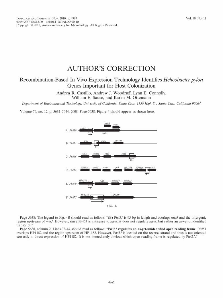
INFECTION AND IMMUNITY, Nov. 2010, p. 4967 Vol. 78, No. 110019-9567/10/$12.00 doi:10.1128/IAI.00990-10Copyright © 2010, American Society for Microbiology. All Rights Reserved.
AUTHOR’S CORRECTION
Recombination-Based In Vivo Expression Technology Identifies Helicobacter pyloriGenes Important for Host Colonization
Andrea R. Castillo, Andrew J. Woodruff, Lynn E. Connolly,William E. Sause, and Karen M. Ottemann
Department of Environmental Toxicology, University of California, Santa Cruz, 1156 High St., Santa Cruz, California 95064
Volume 76, no. 12, p. 5632–5644, 2008. Page 5638: Figure 4 should appear as shown here.
Page 5638: The legend to Fig. 4B should read as follows. “(B) Pivi51 is 95 bp in length and overlaps mesJ and the intergenicregion upstream of mesJ. However, since Pivi51 is antisense to mesJ, it does not regulate mesJ, but rather an as-yet-unidentifiedtranscript.”
Page 5638, column 2: Lines 33–44 should read as follows. “Pivi51 regulates an as-yet-unidentified open reading frame. Pivi51overlaps HP1182 and the region upstream of HP1182. However, Pivi51 is located on the reverse strand and thus is not orientedcorrectly to direct expression of HP1182. It is not immediately obvious which open reading frame is regulated by Pivi51.”
FIG. 4.
4967