Genetic microheterogeneity and phenotypic variation of Helicobacter pylori arginase in clinical...
15
BioMed Central Page 1 of 15 (page number not for citation purposes) BMC Microbiology Open Access Research article Genetic microheterogeneity and phenotypic variation of Helicobacter pylori arginase in clinical isolates Justin G Hovey 1 , Emily L Watson 2 , Melanie L Langford 3 , Ellen Hildebrandt 4 , Sangeetha Bathala 1 , Jeffrey R Bolland 5 , Domenico Spadafora 1 , George L Mendz 6 and David J McGee* 4 Address: 1 Department of Microbiology and Immunology, University of South Alabama College of Medicine, Mobile, AL, USA, 2 Department of Medicine, Creighton University School of Medicine, Omaha, NE, USA, 3 Department of Biological Sciences, University of Nebraska-Lincoln, Lincoln, NE, USA, 4 Department of Microbiology and Immunology, Louisiana State University Health Sciences Center, Shreveport, LA, USA, 5 Department of Microbiology, University of Alabama at Birmingham, Birmingham, AL, USA and 6 School of Medical Sciences, University of New South Wales, Sydney, Australia Email: Justin G Hovey - [email protected]; Emily L Watson - [email protected]; Melanie L Langford - [email protected]; Ellen Hildebrandt - [email protected]; Sangeetha Bathala - [email protected]; Jeffrey R Bolland - [email protected]; Domenico Spadafora - [email protected]; George L Mendz - [email protected]; David J McGee* - [email protected] * Corresponding author Abstract Background: Clinical isolates of the gastric pathogen Helicobacter pylori display a high level of genetic macro- and microheterogeneity, featuring a panmictic, rather than clonal structure. The ability of H. pylori to survive the stomach acid is due, in part, to the arginase-urease enzyme system. Arginase (RocF) hydrolyzes L-arginine to L-ornithine and urea, and urease hydrolyzes urea to carbon dioxide and ammonium, which can neutralize acid. Results: The degree of variation in arginase was explored at the DNA sequence, enzyme activity and protein expression levels. To this end, arginase activity was measured from 73 minimally- passaged clinical isolates and six laboratory-adapted strains of H. pylori. The rocF gene from 21 of the strains was cloned into genetically stable E. coli and the enzyme activities measured. Arginase activity was found to substantially vary (>100-fold) in both different H. pylori strains and in the E. coli model. Western blot analysis revealed a positive correlation between activity and amount of protein expressed in most H. pylori strains. Several H. pylori strains featured altered arginase activity upon in vitro passage. Pairwise alignments of the 21 rocF genes plus strain J99 revealed extensive microheterogeneity in the promoter region and 3' end of the rocF coding region. Amino acid S232, which was I232 in the arginase-negative clinical strain A2, was critical for arginase activity. Conclusion: These studies demonstrated that H. pylori arginase exhibits extensive genotypic and phenotypic variation which may be used to understand mechanisms of microheterogeneity in H. pylori. Published: 4 April 2007 BMC Microbiology 2007, 7:26 doi:10.1186/1471-2180-7-26 Received: 22 November 2006 Accepted: 4 April 2007 This article is available from: http://www.biomedcentral.com/1471-2180/7/26 © 2007 Hovey et al; licensee BioMed Central Ltd. This is an Open Access article distributed under the terms of the Creative Commons Attribution License (http://creativecommons.org/licenses/by/2.0 ), which permits unrestricted use, distribution, and reproduction in any medium, provided the original work is properly cited.
-
Upload
flsouthern -
Category
Documents
-
view
0 -
download
0
Transcript of Genetic microheterogeneity and phenotypic variation of Helicobacter pylori arginase in clinical...

BioMed CentralBMC Microbiology
ss
Open AcceResearch articleGenetic microheterogeneity and phenotypic variation of Helicobacter pylori arginase in clinical isolatesJustin G Hovey1, Emily L Watson2, Melanie L Langford3, Ellen Hildebrandt4, Sangeetha Bathala1, Jeffrey R Bolland5, Domenico Spadafora1, George L Mendz6 and David J McGee*4Address: 1Department of Microbiology and Immunology, University of South Alabama College of Medicine, Mobile, AL, USA, 2Department of Medicine, Creighton University School of Medicine, Omaha, NE, USA, 3Department of Biological Sciences, University of Nebraska-Lincoln, Lincoln, NE, USA, 4Department of Microbiology and Immunology, Louisiana State University Health Sciences Center, Shreveport, LA, USA, 5Department of Microbiology, University of Alabama at Birmingham, Birmingham, AL, USA and 6School of Medical Sciences, University of New South Wales, Sydney, Australia
Email: Justin G Hovey - [email protected]; Emily L Watson - [email protected]; Melanie L Langford - [email protected]; Ellen Hildebrandt - [email protected]; Sangeetha Bathala - [email protected]; Jeffrey R Bolland - [email protected]; Domenico Spadafora - [email protected]; George L Mendz - [email protected]; David J McGee* - [email protected]
* Corresponding author
AbstractBackground: Clinical isolates of the gastric pathogen Helicobacter pylori display a high level ofgenetic macro- and microheterogeneity, featuring a panmictic, rather than clonal structure. Theability of H. pylori to survive the stomach acid is due, in part, to the arginase-urease enzyme system.Arginase (RocF) hydrolyzes L-arginine to L-ornithine and urea, and urease hydrolyzes urea tocarbon dioxide and ammonium, which can neutralize acid.
Results: The degree of variation in arginase was explored at the DNA sequence, enzyme activityand protein expression levels. To this end, arginase activity was measured from 73 minimally-passaged clinical isolates and six laboratory-adapted strains of H. pylori. The rocF gene from 21 ofthe strains was cloned into genetically stable E. coli and the enzyme activities measured. Arginaseactivity was found to substantially vary (>100-fold) in both different H. pylori strains and in the E.coli model. Western blot analysis revealed a positive correlation between activity and amount ofprotein expressed in most H. pylori strains. Several H. pylori strains featured altered arginase activityupon in vitro passage. Pairwise alignments of the 21 rocF genes plus strain J99 revealed extensivemicroheterogeneity in the promoter region and 3' end of the rocF coding region. Amino acid S232,which was I232 in the arginase-negative clinical strain A2, was critical for arginase activity.
Conclusion: These studies demonstrated that H. pylori arginase exhibits extensive genotypic andphenotypic variation which may be used to understand mechanisms of microheterogeneity in H.pylori.
Published: 4 April 2007
BMC Microbiology 2007, 7:26 doi:10.1186/1471-2180-7-26
Received: 22 November 2006Accepted: 4 April 2007
This article is available from: http://www.biomedcentral.com/1471-2180/7/26
© 2007 Hovey et al; licensee BioMed Central Ltd. This is an Open Access article distributed under the terms of the Creative Commons Attribution License (http://creativecommons.org/licenses/by/2.0), which permits unrestricted use, distribution, and reproduction in any medium, provided the original work is properly cited.
Page 1 of 15(page number not for citation purposes)

BMC Microbiology 2007, 7:26 http://www.biomedcentral.com/1471-2180/7/26
BackgroundHelicobacter pylori, a Gram negative bacterium, is a highlyhost-adapted gastric pathogen that has been implicated ina wide spectrum of diseases ranging from gastritis to ade-nocarcinoma [1-5]. Although this bacterium colonizes thegastric mucosa of billions of people, only 20% of theinfected people become symptomatic. The disparities ofsymptoms from one person to another are indicative of apathogen with significant genetic diversity. Two majortypes of diversity have been described in H. pylori clinicalisolates: i) macrohetereogeneity, in which large chromo-somal regions vary from strain to strain, and ii) micro-heterogeneity, in which individual genes feature sequencediversity. Examples of macroheterogeneity include thepresence or absence of the cag pathogenicity island, inser-tion sequences, and a hypervariable region containingabout half of the strain-specific genes, called the plasticityzone [6-14]. Furthermore, 22% of the organism's genesare dispensable in one or more strains, leading to a core ofonly about 1280 genes [13]. Macroheterogeneity can beassessed by restriction fragment length polymorphisms,multilocus enzyme electrophoresis, and microarrays.
Examples of microheterogeneity include extensivesequence variation of the vacA, cagA, babA, hopQ, iceAgenes and other genes [9,15-20]. For example, the vacAgene encoding the vacuolating cytotoxin (VacA) exhibits aremarkable degree of genotypic and phenotypic variation[9,21-23]. The vacuolating activity of VacA varies approx-imately 30-fold across different isolates due to the pres-ence of at least five different vacA alleles [24]. Two familiesof the vacA alleles, type m1 and type m2, are only about70% identical [25]. In addition, there is also evidence thatmixed strain infections can occur in a single patient [26],and that a single strain can change in vivo over time[16,27,28]. The extraordinary diversity of this pathogenmay explain why the acquired immune response cannotclear the infection or prevent reinfection by a heterolo-gous strain. The genetic variation among the bacterium'svirulence factors may relate to the diverse disease manifes-tations in patients, although this is not well understood.
On the other hand, the urease structural proteins, UreAand UreB, are very well conserved across heterologousstrains of H. pylori (97–100% amino acid identity, basedon BlastP analysis of GenBank sequences). These proteinsconstitute a nickel-requiring, highly abundant metalloen-zyme that is central to the pathogenesis of the bacterium[29]. Urease hydrolyzes urea to carbon dioxide andammonia, the latter of which neutralizes gastric acid [30].Local neutralization of gastric acid helps H. pylori to safelytraverse the gastric mucosal layer and colonize the gastricepithelium [31]. Indeed, urease mutants are unable to col-onize and establish a lasting infection in nude mice andgnotobiotic piglets [32-34]. Considering that functional
urease is absolutely essential for virulence, numerous ran-dom mutations in the functional core of the urease genecould be detrimental, thus explaining why ureA and ureBare highly conserved in heterologous strains. Without sta-bility in these two structural genes, this species would beineffective as a pathogen.
The source of urea for H. pylori urease can either bethrough host- or bacterial-derived arginase. H. pylori con-tains the rocF gene encoding arginase, which catalyzes thehydrolysis of L-arginine to L-ornithine and urea [35-37].H. pylori is deficient in the enzymes for synthesizingarginine de novo and is therefore dependent on hostarginine to help it maintain nitrogen balance [36-38].Arginase consumes arginine, thereby removing this essen-tial amino acid away from other cellular processes if theenzyme activity is too high. The role of arginase in H.pylori pathogenesis is beginning to be unraveled. Arginaseallows the bacterium to evade host immune response bycompeting with macrophage inducible nitric oxide syn-thase (iNOS) for L-arginine [39]. H. pylori arginase alsodown-regulates expression of CD3ς on T-cells, preventingtheir proliferation via consumption of arginine from theextracellular milieu [40]. Moreover, arginase producesendogenous urea that can be hydrolyzed by urease to pro-duce ammonium that contributes to acid resistance [35].Thus, arginase is involved in helping H. pylori evade boththe innate (acid, NO) and adaptive (T cells) immune sys-tems. Arginase clearly plays a role in these pathogenicprocesses, but surprisingly the rocF gene encoding argin-ase is not essential for the establishment of infection [35],suggesting that in vivo the enzyme plays a role down-stream of the initial colonization step, perhaps modulat-ing disease severity.
The importance of specific mutations in the phenotypicvariation of this species are largely unknown. The evolu-tion of specific genes and proteins in this pathogen are ofparamount importance as the field strives to understandthe role of specific genes in virulence. In a previous studyinvolving laboratory-adapted strains, some variation inarginase activity was found among three strains [35].However, it was not determined whether this variationoccurred from spontaneous mutation from passaging thestrains repeatedly in the laboratory or from natural diver-sity existing among H. pylori strains. To determine argin-ase variability, phenotypic and genotypic analyses of rocFin 73 minimally-passaged clinical isolates and six labora-tory-adapted strains was investigated. While most previ-ous studies on microheterogeneity focused on only asmall portion of a gene, we studied the entire arginasecoding region plus upstream region. This study demon-strates that extensive microheterogeneity exists in the rocFgene, with phenotypic manifestations, and provides evi-
Page 2 of 15(page number not for citation purposes)
BMC Microbiology 2007, 7:26 http://www.biomedcentral.com/1471-2180/7/26
dence that this gene may serve as a model to study micro-heterogeneity in H. pylori.
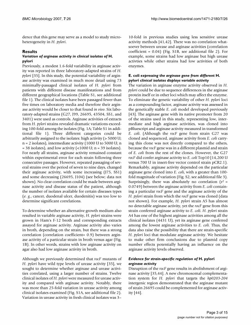
ResultsVariation of arginase activity in clinical isolates of H. pyloriPreviously, a modest 1.6-fold variability in arginase activ-ity was reported in three laboratory-adapted strains of H.pylori [35]. In this study, the potential variability of argin-ase activity was examined in much more detail using 73minimally-passaged clinical isolates of H. pylori frompatients with different disease manifestations and fromdifferent geographical locations (Table S1, see additionalfile 1). The clinical isolates have been passaged fewer thanfive times on laboratory media and therefore their argin-ase activity would be closer to that found in vivo. Six labo-ratory-adapted strains (G27, J99, 26695, 43504, SS1, and3401) were used as controls. Arginase activities of extractsfrom H. pylori strains revealed dramatic variations exceed-ing 100-fold among the isolates (Fig. 1A; Table S1 in addi-tional file 1). Three different categories could bearbitrarily assigned to the isolates: high activity (> 5000 U;n = 2 isolates), intermediate activity (1000 U to 5000 U; n= 38 isolates), and low activity (<1000 U; n = 39 isolates).For nearly all strains, arginase activity remained constantwithin experimental error for each strain following threeconsecutive passages. However, repeated passaging of sev-eral strains over the period of seven to nine days, changedtheir arginase activity, with some increasing (J75, SS1)and some decreasing (26695, J104) (see below; data notshown). No clear correlation could be made between argi-nase activity and disease status of the patient, althoughthe number of isolates available for certain diseases types(e. g., cancer, duodenal ulcer, duodenitis) was too low todetermine significant correlations.
To determine whether an alternative growth medium alsoresulted in variable arginase activity, H. pylori strains weregrown in Ham's F-12 broth and corresponding extractsassayed for arginase activity. Arginase activity also variesin broth, depending on the strain, but there was a strongcorrelation (correlation coefficient= 0.9) between argin-ase activity of a particular strain in broth versus agar (Fig.1B). In other words, strains with low arginase activity onagar also had low arginase activity in broth.
Although we previously determined that rocF mutants ofH. pylori have wild type levels of urease activity [35], wesought to determine whether arginase and urease activi-ties correlated, using a larger number of strains. Twelveclinical isolates of H. pylori were measured for urease activ-ity and compared with arginase activity. Notably, therewas more than 25-fold variation in urease activity amongclinical isolates examined (Fig. S1A, see additional file 2).Variation in urease activity in fresh clinical isolates was 3–
10-fold in previous studies using less sensitive ureaseactivity methods [41,42]. There was no correlation what-soever between urease and arginase activities (correlationcoefficient = 0.04) (Fig. S1B, see additional file 2). Forexample, some strains had low arginase but high ureaseactivties while other strains had low activities of bothenzymes.
E. coli expressing the arginase gene from different H. pylori clinical isolates displays variable activityThe variation in arginase enzyme activity observed in H.pylori could be due to sequence differences in the arginaseprotein itself or in other loci which may affect the enzyme.To eliminate the genetic variability of other H. pylori locias a compounding factor, arginase activity was assessed inthe genetically stable E. coli model developed previously[43]. The arginase gene with its native promoter from 20of the strains used in this study, representing low, inter-mediate and high arginase activities, was cloned intopBluescript and arginase activity measured in transformedE. coli. (Although the rocF gene from strain G27 wascloned and sequenced, arginase activity from E. coli carry-ing this clone was not directly compared to the others,because the rocF gene was in a different plasmid and strainof E. coli from the rest of the clones. However, the G27rocF did confer arginase activity to E. coli Top10 [14,200 Uversus 700 U in insert-free vector control strain pCR2.1).Remarkably, arginase activity depended on the particulararginase gene cloned into E. coli, with a greater than 100-fold magnitude of variation (Fig. S2, see additional file 3).Surprisingly, there was absolutely no correlation (r2 =0.0749) between the arginase activity from E. coli contain-ing a particular rocF gene and the arginase activity of theH. pylori strain from which the rocF gene was cloned (datanot shown). For example, H. pylori strain A5 has almostno detectable arginase activity, yet the rocF gene from thisstrain conferred arginase activity to E. coli. H. pylori strainA4 has one of the highest arginase activities among all theclinical isolates (4431 U), yet its arginase gene conferredamong the lowest arginase activities to E. coli. Thus, thedata also raise the possibility that there are strain-specificH. pylori loci that modulate arginase activity. We hesitateto make other firm conclusions due to plasmid copynumber effects potentially having an influence on thearginase activity levels observed.
Evidence for strain-specific regulation of H. pylori arginase activityDisruption of the rocF gene results in abolishment of argi-nase activity [35,44]. A new chromosomal complementa-tion system for H. pylori that targets the hp0203-204intergenic region demonstrated that the arginase mutantof strain 26695 could be complemented for arginase activ-ity [44].
Page 3 of 15(page number not for citation purposes)

BMC Microbiology 2007, 7:26 http://www.biomedcentral.com/1471-2180/7/26
Page 4 of 15(page number not for citation purposes)
Arginase activity variation in H. pylori clinical isolatesFigure 1Arginase activity variation in H. pylori clinical isolates. Ornithine concentration was measured at 515 nm spectropho-tometerically by the appearance of an orange color originating from the reaction of ornithine with acidified ninhydrin. A. Argi-nase activity of extracts from 16 US H. pylori strains, 11 of which are minimally-passaged clinical H. pylori isolates. Strains were grown on Campylobacter blood agar plates for 48 h. The graphs show the average arginase activity (pmol L-Orn/min/mg pro-tein) ± standard deviation of one experiment representative of at least three. A complete list of strains and average arginase activities is given in Table S1 (see Additional file 1). The variation from experiment to experiment is about 10–15%. B. Compar-ison of arginase activity of H. pylori grown in broth versus grown on agar. H. pylori strains were grown in Ham's F-12 broth for 16–18 h or on Campylobacter blood agar plates for 48 h as described in Materials and Methods. The data suggest a strong cor-relation between the arginase activity from H. pylori grown in broth versus that on agar.
0
2500
5000
7500
10000
12500
15000
17500
J188
B194A
J99
B128A
26695
B228A
J166
HP
DJM
17
B134A
J75
J104
J243
J54
43504
J178
3401
Arg
ina
se
ac
tiv
ity
(pm
olL
-orn
/min
/mg
pro
t)
Arginase activity, pmol L-orn/min/mg
0
1500
3000
4500
0 1000 2000 3000 4000 5000
cultures grown on CBA agar
cultu
res
grow
nin
F12
brot
hA
B
A2
A5
J99
B1
J75
26695
43504
y = 0.5565x + 682.13
R2 = 0.8994

BMC Microbiology 2007, 7:26 http://www.biomedcentral.com/1471-2180/7/26
Three plasmids, pMLB001, pMLB002, and pMLB003 carrythe wild type rocF genes from H. pylori strains 43504, SS1and J63, respectively. These suicide plasmids were trans-formed into the rocF mutant of 26695 and confirmed toyield 26695 rocF-MLB001, -MLB002, and -MLB003 [44].The arginase activity of these three complemented strainswas compared to that of the corresponding wild type H.pylori strain from which the rocF gene was derived. Whenthe rocF genes from strains 43504 or SS1 were used tocomplement the 26695 rocF mutant, arginase activity wasrestored to levels similar to that of the correspondingwild-type 43504 or SS1 strains (Fig. 2). In contrast, whenthe rocF gene from strain J63 was used to complement the26695 rocF mutant, arginase activity was about ten timeshigher than that of the corresponding wild-type J63 strain(Fig. 2). This finding raises the intriguing possibility thatthere is strain-dependent regulation of arginase.
Additional evidence for strain-dependent arginase activitywas revealed from strains SS1, B5 and B1, which all haveidentical rocF coding regions and upstream regions, yethave different arginase activities (8100, 1900, 1400 U,respectively) (Table S1 in additional file 1).
Evidence that H. pylori prefers low to moderate arginase activityThe arginase activity data from clinical isolates revealedthat most strains possess intermediate or low arginaseactivities, while only a few strains have high activity, withonly one strain above 10,000 U (strain 3401). If H. pyloridid not control arginase activity, then a strain with twofunctional chromosomal copies of the arginase geneshould have an enzyme activity significantly higher thanthe wild type strain carrying only a single arginase gene.To investigate this plasmid pMLB004, which confers argi-nase activity to an arginase negative strain, was trans-formed into the high arginase activity strain, SS1, to yieldtwo chromosomal copies of wild-type rocF (each with itsnative promoter), designated SS1 MLB004 [44]. As an iso-genic control, the same construct devoid of the arginasegene (pIR203C04) was transformed into SS1 to yield SS1203C04 [44]. Rather than the predicted 100% increase,strain SS1 MLB004 had only about 25% more arginaseactivity over that of the isogenic control SS1 203C04strain (Fig. 3). This suggests that H. pylori has mechanismsto prevent arginase activity from becoming too high.
Western Blot analysis of arginase in clinical isolates of H. pyloriWestern blot analysis with RocF antiserum showed thatextracts from H. pylori strains with high arginase activity(e. g., 3401, A4) had relatively higher amounts of arginaseprotein (Fig. 4, only a subset shown), than most strainswith lower arginase activity (e. g., J188, A2). There wereoccasional exceptions; strain J63, for example, had low
arginase activity, but high amounts of arginase protein(Fig. 4). It was difficult to detect arginase protein in someof the lowest arginase activity strains, such as strains A2,A5, A6, A7 (Fig. 4). Two strains whose arginase activitychanged upon laboratory passage (J104 increased whileSS1 decreased), likewise showed corresponding changesin the amount of arginase protein (Fig. 4). The arginaseprotein size by SDS-PAGE was found to vary slightlyamong the minimally-passaged clinical isolates; this sizevariation did not correlate with arginase activity (e. g.,3401 vs. B128A) (Fig. 4). Despite slight size variations, allrocF genes were predicted to encode a 322 amino acid pro-tein. The size variation was not considered significantenough to study further.
Nucleotide, amino acid and phylogenetic analyses of rocF/RocFTo understand better the arginase nucleotide sequencevariations, strains representing low, intermediate andhigh arginase activity were selected for further analysis.PCR amplification was performed on 21 different arginasegenes, each giving a 1.1 kb fragment, which was clonedand sequenced. Pairwise alignments of the nucleotidesequences of 22 strains (including the J99 rocF sequencealready in the database, GenBank accession #AE001565;strain AG1 excluded) revealed that the rocF codingsequences (~1.0 kb) were 92.9% to 100% identical to thatof strain 26695 (data not shown). Within the regionencoding the arginase putative cobalt-binding site,DAHAD (Fig. 5A), two corresponding nucleotide changeswere observed: GAC to GAT (strains SS1, J75, B1, B7, andJ166) and GCG to GCT (strain AG1). In both cases, themutations were silent, leading to no changes in the aminoacid sequence. Alignments also revealed a hypervariableregion in the upstream sequence proximal to the ATG startcodon (Fig. 5B). The region between nucleotides -37 to +3ranged from only 67% to 100% identity with strain 26695(Fig. 5C). Ten different sites showed mutations; insertionsand deletions also occurred in this hypervariable region(Fig. 5B and 5C). Interestingly, some of the mutations,insertions and deletions, occurred near or in the predictedShine-Dalgarno sequence (ribosome-binding site)(26695 rocF SD sequence: AGGAGTTATA) (Fig. 5A and5B). Intriguingly, the arginase upstream region from therecently sequenced H. pylori strain AG1 [45] had nohomology with that of any of the arginase upstreamregions studied here (data not shown). Instead, the argin-ase upstream region of strain AG1 showed striking resem-blance (92% nucleotide identity) to that of the arginaseupstream region of H. acinonychis [46] (data not shown).In contrast, the arginase upstream sequence from theremaining 22 H. pylori strains studied here did not bearany homology to any region in the H. acinonychis genome(data not shown).
Page 5 of 15(page number not for citation purposes)

BMC Microbiology 2007, 7:26 http://www.biomedcentral.com/1471-2180/7/26
Pairwise alignments of the translated RocF coding regionsshowed that they were 92% to 100% identical, with mostvariability located near the carboxy terminus (Fig. 5A).There were 42 amino acid residues out of 322 (13%) thatwere not 100% conserved across all the isolates. Of these42, 28 of the variable residues are found in the last 100amino acids, with nine of these variable sites being in thefinal 16 amino acids at the C-terminal end. Three strains,SS1, B5, and B1, were 100% identical at the nucleotideand amino acid levels. Strains HPDJM17 and J188 hadfive amino acid differences. All H. pylori RocF proteinswere predicted to have 322 amino acids.
A phylogenetic tree of the arginase proteins from 23 H.pylori strains revealed four clades, with 16, 4, 2, or 1strain(s) in the clades (Fig. 5D). A phylogenetic tree of thenucleotide sequences of the arginase upstream regionfrom 22 H. pylori strains (strain AG1 excluded) showedthree major clades containing one, fifteen and six strains,respectively (Fig. 5E). The two phylogenetic trees (Fig. 5D,5E) showed little congruence with each other. For exam-ple, the RocF protein from strains 43504 and A4 werewithin the same clade, but the corresponding rocF
Strain-specific regulation of H. pylori arginase activityFigure 2Strain-specific regulation of H. pylori arginase activity. Comparison of arginase activities of a native H. pylori strain and a rocF mutant strain of 26695 complemented with the corresponding rocF gene (coding region plus native rocF promoter). Strain rocF-26695-MLB001 is a rocF mutant of strain 26695 in which the wild type rocF gene from strain 43504 has been comple-mented. The MLB002 strain carried wild type rocF from strain SS1 and the MLB003 strain carried wild type rocF from strain J63.
0
5,000
10,000
15,000
WT
43504
rocF
2669
5-MLB
001
WT
SS1
rocF
2669
5-MLB
002
WT
J63
rocF
2669
5-MLB
003
Strain
Arg
inase
Acti
vit
y
(pm
olL
-orn
/min
/mg
pro
t)
H. pylori prevents arginase activity from becoming too ele-vatedFigure 3H. pylori prevents arginase activity from becoming too elevated. Wild type H. pylori strain SS1 was trans-formed with the rocF mutation (rocF), a vector control inserted into the intergenic hp0203-0204 site (203C04), or with the intergenic plasmid carrying the wild type rocF coding region and native promoter (MLB004).
0
2000
4000
6000
8000
10000
12000
SS1 WT SS1 rocF SS1
203C04
SS1
MLB004
Arg
inase
acti
vit
y
(pm
olL
-orn
/min
/mg
pro
t)
Page 6 of 15(page number not for citation purposes)

BMC Microbiology 2007, 7:26 http://www.biomedcentral.com/1471-2180/7/26
upstream regions were in different clades. Two strainsshowing 100% identity in the arginase upstream regionwere strains G27 and B7, two of the highest arginase activ-ity strains (3900 U and 4800 U, respectively) (Fig. 5C,5E). These two strains did not have identical codingregions, showing 96% identity at the amino acid level.Three other strains were 100% identical in the upstreamregion: B5, SS1, and B1 (Fig. 5C, D). These three strainswere also 100% identical at the amino acid level (Fig. 5D),yet had different levels of arginase activity (Fig. 5C). Thisprovided additional evidence that a strain-specific locusother than rocF plays a role in some of the variability
observed. No clear correlation could be made betweenphylogenetic placement on either of the trees (Fig. 5D, E)and the level of arginase activity (Table S1 in additionalfile 1).
Serine 232 in RocF is required for arginase activityNo information is available regarding the amino acids inH. pylori RocF that are responsible for catalytic activity.The clinical isolate A2 had barely detectable arginaseactivity (68 U) we designate as arginase null (Fig. 6A). Thearginase gene and protein sequences were directly com-pared to that of a strain with much higher arginase activ-
Anti-RocF Western on minimally-passaged wild type H. pyloriFigure 4Anti-RocF Western on minimally-passaged wild type H. pylori. Proteins were separated by SDS-PAGE using a 12% resolving gel and 10 μg of protein loaded per lane and analyzed by anti-RocF antibodies. Only a subset of strains is shown. J104-L, low arginase activity strain minimally passaged; J104-H, high arginase activity strain passaged for seven to nine days; SS1-L, low arginase activity strain passaged for seven to nine days; SS1-H, high arginase activity strain minimally-passaged. Numbers below the top two blots represent the arginase specific activity (rounded to nearest 100; pmol L-ornithine/min/mg protein ± standard deviation) of the extract used for the blot.
J104
-L
J54
J99
3401
B12
8A
J254
J68
J63
J104
-H
J166
HPD
JM17
B19
4A
J178
4350
4
500 2400 400 500 900 3000 14000 1200 3000 1700 900 1700 6200 6600
J188
J75
B19
4A
J243
2669
5
SS1-
LSS1-
H
B13
4A
800 1000 900 1200 1500 2800 1900 13300
A2 A3 A4 A5 A7 A8 A9 B1 B3
B5 GC3 GC5 GC6 GC9 GD1 GD2
A1 A6 B2 B4 B6 B8 B9 GC1 GC2
GC4 GC7 GC8 GD3 GD4 GD6 GD7 GD9 26695
Page 7 of 15(page number not for citation purposes)

BMC Microbiology 2007, 7:26 http://www.biomedcentral.com/1471-2180/7/26
Page 8 of 15(page number not for citation purposes)
Microheterogeneity in arginase sequences from clinical isolates of H. pyloriFigure 5Microheterogeneity in arginase sequences from clinical isolates of H. pylori. The arginase coding region (~1.0 kb) plus ~130 bp of the upstream region encompassing the promoter and 18 bp downstream were amplified by PCR using Pfx and cloned into pBluescript. The constructs were sequenced using T3 and T7-1 primers (see Materials and Methods). A. Summary of microheterogeneity in the arginase coding region in H. pylori. Multi-sequence pairwise alignments were conducted with the rocF coding region using ClustalW. The alignment was converted to a graphical representation to show regions that are 100% conserved (white) and regions that vary (black). B. Hypervariablity in the ~133 bp rocF upstream region including the arginase promoter sequence preceding the ATG start codon. Multi-sequence pairwise alignments of the ~133 bp rocF upstream region from H. pylori strains were conducted using ClustalW. The alignments were converted to a graphical representation to show regions that are 100% conserved (white) and regions that vary (black). The alignments revealed a hypervariable region proximal to the ATG start codon. Numbering based on that of H. pylori strain 26695. Underlined is the Shine-Dalgarno (SD) sequence. Numbers in parentheses correspond to the number of strains featuring the insertion (ins) or deletion (del). Strain AG1 was omitted from this analysis because its arginase upstream region was completely different from the other 22 strains. C. Hyper-variability in the ~35 bp upstream of the arginase ATG translation start codon in 21 H. pylori strains. Some of the sequence hypervariability occurred in the predicted SD region (underlined). The consensus sequence for the 3' end of the 16S rRNA for H. pylori is shown, as well as the consensus SD sequence for other H. pylori genes. Arginase activity is shown on the right hand side in units rounded to the nearest 100. Asterisks indicate nucleotides conserved in all strains. D. Phylogram of RocF protein sequence from 23 H. pylori strains. ClustalX and Treeview were used to construct the tree. E. Phylogram of the rocF upstream region from 22 H. pylori strains. ClustalX and Treeview were used to construct the tree. Strain AG1 was omitted from this analysis because its arginase upstream region was completely different from the other 22 strains.
100 200 300 3221
N C
100% conserved across 23 H. pylori strains
Not 100% conserved across 23 H. pylori strains
DAHAD- putative metal binding motif** *
* Mostly conservative amino acid replacements
A
ATG
-83-133 -33-108 -58 -7 -1
-33 AGAA—CATTAGAGGGGATTTA --AAAGGAGTTATAAGGAGTTATA AAATG +3
del(8) AT(5)
AA(16)
ins
AA (7)
ins
G TG G T G G CA
G
Insertions/Deletions
100% conserved across 22 H. pylori strainsNot 100% conserved across 22 H. pylori strains
B
SS1 -36 A-AAAATATGAGTGGGGATTTAAAAAGGAGCGATAAAATG +3
B1 -36 A-AAAATATGAGTGGGGATTTAAAAAGGAGCGATAAAATG +3
B5 -36 A-AAAATATGAGTGGGGATTTAAAAAGGAGCGATAAAATG +3
J54 -36 A-AAAATATGAGTGGGGATTTAAAAAGGAGCAATAAAATG +3
A5 -36 A-AAAATATGAGTGGGGATTTAAAAAGGAGCAATAAAATG +3
43504 -34 A-AAAATATGAGTGGGGATTTA--AAGGAGTTATAAAATG +3
J188 -35 AAAAAATATGAGTGGGGATTTA--AAGGAGTTATAAAATG +3
3401 -35 AAAAAATATGAGTGGGGATTTA--AAGGAGTTATAAAATG +3
Hp17 -35 AAAAAATATGAGTGGGGATTTA--AAGGAGTTATAAAATG +3
J178 -37 GGAAAATATGAGTGGGGATTTAAAAAGGAGTTATAAAATG +3
J63 -36 A-AAAATATGAGTGGGGATTTAAAAAGGAGTTATAAAATG +3
J104 -35 GGAAAATATGAGTGGGGATTTA--AAGGAGTTATAAAATG +3
J254 -35 GGAAAATATGAGTGGGGATTTA--AAGGAGTTATAAAATG +3
J99 -35 GGAAAATATGAGTGGGGATTTA--AAGGAGTTATAAAATG +3
GD9 -35 GGGAAATAAGAGTGGGGATTTA--AAGGAGTTATAAAATG +3
A2 -34 A-AAAATATGAGTGGGGATTTG--AAGGAGTTATAAAATG +3
J75 -35 AGAGATTGTTAGAGGGGGTTTG--AAGGAGTTATAAAATG +3
J166 -35 AGAGATCATTAGAGGGGATTTG--AAGGAGTTATAAAATG +3
A4 -35 AGAGATCATTAGAGGGGGTTTG--AAGGAGTTATAAAATG +3
B7; G27 -35 AGAGATCATTAGAGGGGGTTTA--AAGGAGTTATAAAATG +3
26695 -33 AGAA--CATTAGAGGGGATTTA--AAGGAGTTATAAAATG +3
** **** *** ****** ********
C
ATG startSD
Arginase (U)
8100
1400
1900
1500
100
4800
1100
13,400
1900
4900
500
1600
600
1100
2200
100
1700
1200
4400
4800; 3900
2100
3’ end of H. pylori 16S rRNA: 5’-ATCACCTCCT-3’
Consensus SD sequence for H. pylori genes: 5’-AGGAGGTGAGG-3’
H. pylori rocF SD sequence: 5’-AGGAGTTATA-3’ (16 strains)
CG (3 strains)
A (2 strains)
D
0.01
G27
B7
J99
3401
J178
HP17
J188
J54
AG1
J63
B1
SS1
B5
J75
J166
J254
J104
26695
A5
A2
GD9
43504
A4E
0.01
GD9
A2
Hp17
J188
3401
J99
J254
J178
J104
43504
J63
B5
SS1
B1
J54
A5
26695
J75
J166
A4
B7
G27

BMC Microbiology 2007, 7:26 http://www.biomedcentral.com/1471-2180/7/26
ity, 26695. Alignment of the ~70 bp rocF upstreamsequences of both strains revealed nine differences (Fig.6B), but none of the changes in strain A2 could be corre-lated to an arginase null phenotype, since these changeswere found in other arginase upstream regions in strainswith detectable arginase activity. An additional 60 bp fur-ther upstream of these two strains was next analyzed andshown to be 100% identical (data not shown). Theinvolvement of these upstream sequences in affecting thearginase null phenotype of strain A2 could not be com-pletely ruled out. Next, the RocF amino acid sequences ofstrains A2 and 26695 were aligned, revealing 11 aminoacid differences (Fig. 6C). All but three of these aminoacid differences were found in arginase proteins fromother strains of H. pylori that had detectable arginase activ-ity, suggesting the other eight residues were not involvedin the original arginase null phenotype of strain A2. Theroles of the remaining three amino acids (strain 26695:I174, S232, D257; strain A2: M174, I232, N257) in argin-ase activity were investigated in RocF from strain 26695using site-directed mutagenesis. Each of these residueswas mutated from the wild type 26695 sequence to the A2sequence in pQE30-rocF [43]: isoleucine 174 was mutatedto methionine (I174M), serine 232 was mutated to isoleu-cine (S232I), and aspartate 257 was mutated to asparag-ine (D257N). Arginase activity was measured from crudeextracts prepared from E. coli harboring these plasmidderivatives. Plasmid pQE30 served as the negative controland the original wild type pQE30-rocF (from strain26695) [43] served as the positive control. Arginase activ-ity data indicated that the I174M and D257N mutationshad no major effect on the activity of the enzyme. Strik-ingly, arginase activity in extracts from the strain carryingthe S232I mutation strain was abolished, with no activityabove the negative control strain carrying pQE30 (Fig.6D). Western blot analysis using anti-RocF antiserumrevealed that the amount of expressed arginase protein inthe S232I mutant was similar to that of wild type 26695RocF expressed from pQE30-rocF (Fig. 6E), indicating thatthe S232I mutated form of the protein was properlyexpressed. However, a more pronounced degradation ofRocF (RocF frag in Fig. 6E) compared with the wild typeRocF was noted.
DiscussionIn this study the phenotypic and genotypic heterogeneityof arginase in 73 clinical isolates and six laboratory-adapted strains of H. pylori was investigated. Phenotypi-cally, arginase activity varied more than 100-fold in bothH. pylori (Fig. 1) and the E. coli model (Fig. S2 in addi-tional file 3). Nearly all of the H. pylori strains featuredintermediate or low arginase activities (77 of 79 strains).Of the five strains with the highest arginase activity, fourwere laboratory strains that have been passaged heavily.Notably, even the highest arginase activity strains (3401
and SS1 laboratory strains) have specific activities morethan 10-fold lower than those measured for other bacte-rial and eukaryotic arginases (McGee, unpublished data).Arginine is a critical amino acid for several cellular proc-esses, such as protein synthesis, and H. pylori absolutelyrequires arginine for growth [47-49]. The bacteriumwould rapidly hydrolyze the arginine if it had a high spe-cific activity arginase, potentially depleting intracellularpools and starving itself of this essential amino acid. Thefew high arginase activity H. pylori strains may have com-pensatory mechanisms to overcome potential argininestarvation from the higher level of arginine hydrolysis.Two possible mechanisms are that the high arginase activ-ity strains might have a higher affinity arginine transporterthat would allow higher intracellular accumulation ofarginine. Alternatively, the high arginase activity strainscould have decreased arginine decarboxylase activity,another arginine-consuming enzyme that could alsocause arginine depletion in H. pylori.
H. pylori is a heterogeneous species [23,50] that featuresdiversity at many levels. For example, there are many con-tingency genes that undergo phase variation through DNAslipped-strand mispairing of repeats, such as LPS biosyn-thesis genes, and genes encoding outer-membrane pro-teins [51]. As another example of diversity, adhesion of H.pylori to fucosylated Lewis B antigens on the surface of thegastric epithelium appears to be dependent on variabilityof the babA gene. This gene shows much conservation inthe 5' and 3' regions but large variability in its midregions[52]. The cagA gene, encoded by the cag pathogenicityisland, exhibits nucleotide sequences that are 92.8% con-served, with much diversity at the 3' end where an EPIYAamino acid repeat element is encoded [53,54]. RocF alsofeatures diversity in its C-terminal region, although thereare no repeat elements. RocF appears to more divergentthan urease subunits A and B, but less divergent thanCagA or VacA. Since rocF participates in various aspects ofH. pylori pathogenesis [39,40] and the gene displaysremarkable heterogeneity shown in this study, it is possi-ble that rocF heterogeneity may correlate with specificaspects of the pathogenesis of the bacterium, such as inhi-bition of nitric oxide production and T-cell proliferation.The data obtained do not provide evidence for a correla-tion between arginase activity and urease activity orbetween arginase activity and the disease status of thepatient, although more isolates would need to be studiedsince there were few isolates available for certain diseasecategories. Moreover, a greater geographic distribution ofstrains also would be necessary.
The data support the hypothesis that some H. pylori strainsmay have strain-specific arginase gene regulation. Forexample, strain A5 has minimal to no detectable arginaseactivity in the native H. pylori strain, but significant activity
Page 9 of 15(page number not for citation purposes)

BMC Microbiology 2007, 7:26 http://www.biomedcentral.com/1471-2180/7/26
Page 10 of 15(page number not for citation purposes)
Molecular basis of the arginase null phenotype of H. pylori clinical isolate A2Figure 6Molecular basis of the arginase null phenotype of H. pylori clinical isolate A2. A. Arginase activity of H. pylori strain 26695 and clinical isolate A2. B. Variation in the arginase upstream regions of H. pylori strains 26695 and A2. Nine nucleotide differences were noted between these two sequences. C. Variations in the arginase amino acid sequences between H. pylori strains 26695 and A2. Eleven amino acid differences are highlighted (311/322 identity [96% identity]; 316/322 similar [97% sim-ilar]. Three amino acid differences (underlined) were found only in strain A2: I174M, S232I, and D257N. D. Arginase activity of E. coli transformed with pQE30-rocF (rocF from strain 26695) and of strains transformed with site-directed mutants of I174M, S232I, and D257N. E. Anti-RocF Western blot analysis of extracts of E. coli transformed with different plasmids including site-directed mutants of RocF. RocF frag indicates degradation product from RocF.
A
SDB
0
1000
2000
3000
4000
26695 A2
Arg
ina
se
acti
vit
y
(pm
olL
-orn
/min
/mg
pro
t)
26695 -71 TGCTTA-TTAATAATTGGTTGTTAATTTTGGTTTAGAATAGAA-CATTAGAGGGGATTTAAAGGAGTTATAAAATG +3
A2 -73 T A A A AT G T G +3
C26695 MILVGLEAELGASKRGTDKGVRRLREALSATHGDVIKGMQTITQERCVLYKEFRYAKNFE 60
A2
26695 DYYLFCKENLIPCMKEVFEKKEFPLILSSEHANMFGIFQAFRSVHKDKKIGILYLDAHAD 120
A2
26695 IHTAYDSDSKHIHGMPLGMVLNRVRSGFNRMSESEEKAWQKLCSLGLEKGGLEIDPKCLV 180
A2 H A M
26695 YFGVRSTEQSERDVIRELQIPLFSVDAIRENMQEVVQKTKESLKAVDIIYLSLDLDIMDG 240
A2 K E I
26695 KLFTSTGVRENNGLSFDELKQLLGLLLESFKDRLKAVEVTEYNPTVSIKHNNEEEKQVLE 300
A2 N L A T
26695 ILDLIINSCKIKDKKHSFARSY 322
A2 P
D
E
WT I174M S232I pQE30
RocF, 37 kDa
RocF frag
0
25
50
75
100
125
WT RocF pQE30 I174M S232I D257N
Arg
inase
acti
vit
y(%
of
co
ntr
ol)

BMC Microbiology 2007, 7:26 http://www.biomedcentral.com/1471-2180/7/26
was detected in E. coli transformed with the A5 rocF gene,suggesting that H. pylori strain A5 has an arginase repres-sor. Such a repressor may be inoperative, inefficient, orabsent in high arginase activity strains such as 3401. Addi-tionally, evidence for potential strain-specific arginase reg-ulation was observed in H. pylori directly. Specifically,arginase activity of H. pylori strain J63 is low, but when therocF gene from this strain was used to complement a rocFmutant of strain 26695 in single copy on the chromo-some, a surprisingly elevated arginase phenotype wasobserved, suggesting that strain 26695 has an activatorprotein absent in strain J63 or strain J63 has a repressorprotein absent from strain 26695. A possible molecularbasis for this potential variation may be due to the hyper-variablity in the upstream region, which displays variationin the Shine-Dalgarno (SD) region, as well as variation inthe region shown by DNAse footprinting experiments tobe bound by the ArsR regulator [55]. Mutations in theupstream region could either affect transcriptional expres-sion, or translational control through changes in strengthof the SD sequence. Evidence in the literature also sup-ports the rocF gene being regulated at the transcriptionallevel by acid [56] and by the two-component system ArsS/R (Hp0165/0166) [55]. Strain-specific transcriptional andtranslation regulation mechanisms are not mutuallyexclusive.
Besides the hypothesized strain-specific arginase tran-scriptional and translation regulation, evidence wasobtained that variations in the rocF sequence themselvesplay a key role in arginase activity, using the geneticallystable E. coli model. E. coli is arginase negative, but whenthe H. pylori arginase genes were cloned into E. coli, varia-ble arginase activity occurs. Since enzyme activity varieddramatically with the particular arginase gene cloned intoE. coli, variations in the rocF sequence would also beresponsible for at least some of the arginase phenotypicvariation. H. pylori strain-specific regulators that couldaffect arginase transcription in native H. pylori strains mostlikely would be absent in the E. coli model. Pairwise align-ments of rocF nucleotide sequences revealed significantmicroheterogeneity in the 3' end of the coding regions,and translation of these regions showed that RocF variesin the carboxy terminus. Variations near the C-terminuscould possibly affect the folding of the arginase active site,thereby accounting for some of the arginase activity varia-tion.
Evidence for the direct involvement of the rocF codingregion in arginase activity variation was obtained fromsite-directed mutagenesis results. Specifically, mutation ofserine 232 to isoleucine in RocF from strain 26695 com-pletely abolished arginase activity, helping to explain whyH. pylori strain A2, which has isoleucine in position 232 inits native arginase, showed barely detectable arginase
activity. Thus, amino acid 232 appears to play a crucialrole in arginase activity. Notably, the rocF gene from strainA2 conferred the lowest arginase activity to E. coli amongthe 20 rocF genes examined (Fig. S2 in Additional file 3).The possibility that other amino acid residues, or nucle-otides in the rocF upstream region also play a role in thearginase null phenotype of strain A2 cannot be ruled out.Future experiments will endeavor to elucidate the mecha-nism of why serine 232 is so critical for arginase activity,as there is no prior precedent for a serine being requiredfor arginase activity.
Evidence was also obtained that arginase activity couldvary upon in vitro passage of some strains; the mechanismof this variation remains undefined. That the phylogenetictrees of the arginase promoter and the arginase proteinbore little resemblance to each other suggested that eachregion is evolving via independent selective pressures. Thegreater nucleotide variability among arginase upstreamregions compared with the coding regions suggests thatthe former has a higher mutation rate and more toleranceto nucleotide changes during selection of those muta-tions.
ConclusionIt was established that arginase varies genotypically andphenotypically among minimally-passaged clinical H.pylori isolates. Future work could center on understandingin greater detail the molecular basis of arginase activityvariation, and identifying strain-specific arginase regula-tors. This would lead to improved understanding of themechanisms by which H. pylori genes undergo changesleading to microheterogeneity as the organism strives tosurvive in its seemingly inhospitable gastric niche.
MethodsBacterial strains, growth conditions, and plasmidsAll H. pylori strains were cultured on Campylobacter agarcontaining 10% defibrinated sheep blood (CBA) at 37°Cfor 48 h in a microaerobic environment (5% O2/10%CO2/85% N2) in humidified air. For broth condi-tions, H. pylori strains were inoculated at 4 × 10⁶ CFU/mlas determined by ATP assay [48] into 25 ml Ham's F-12[49] with 2% fetal bovine serum and grown without aera-tion for 16–18 h in a microaerobic environment. Theminimally-passaged clinical isolates were obtained fromRichard Peek (Vanderbilt University, 15 isolates), GeorgeMendz (University of Sydney, Australia, 40 isolates), Bar-bara Schneider (Louisiana State University, New Orleans,17 Colombian isolates) and one previously describedclinical isolate (HPDJM17) [57] (Table S1 in additionalfile 1). The laboratory-adapted strains were SS1, 43504,26695, J99, G27 and 3401. All isolates were passagedfewer than five times from the frozen stock, except in cases
Page 11 of 15(page number not for citation purposes)

BMC Microbiology 2007, 7:26 http://www.biomedcentral.com/1471-2180/7/26
in which the effects of in vitro passage on arginase activitywere studied.
E. coli strain DH5α was used for cloning and transforma-tion procedures and XL1-Blue MRF' was utilized for thepQE30-based plasmids used in this study. E. coli wasgrown on Luria (L) agar and L broth plus appropriate anti-biotics (ampicillin, 100 μg/ml; tetracycline, 15 μg/ml) at37°C. Plasmid pBS [pBluescript II SK (+); Stratagene] wasused for cloning rocF genes.
Preparation of arginase or urease-containing cell extractsH. pylori and E. coli were harvested in 0.9% NaCl and son-icated in an ice-bath (two pulses at 25% intensity, 30 seceach with 30 sec rest between pulses). Following centrifu-gation (12,000 × g, two min, 4°C), the supernatants wereretained on ice until measurement of arginase activity.Protein concentrations were determined by the bicin-choninic acid method using bovine serum albumin as astandard (Pierce, Rockford, IL).
Arginase activity assayArginase activity was measured using a colorimetric argin-ase assay developed and validated previously [43]. Theextracts were heat-activated (50°C, 30 min) in the pres-ence of 5 mM cobaltous chloride to provide the enzymewith its required metal cofactor, followed by incubationin the presence of 15 mM MES-10 mM arginine buffer(pH 6.0) for 1 h at 37°C. The ornithine produced wasdetected spectrophotometrically at 515 nm in the pres-ence of acidified ninhydrin (4 mg/ml). Data are presentedas mean arginase specific activity (1 Unit (U) = 1 pmol L-ornithine produced per minute per milligram protein).
Urease activity assayUrease activities were measured using the phenol-hypochlorite method as described previously [58].
SDS-PAGE and Western blot analysis of arginase extractsBacterial proteins were separated by sodium dodecyl sul-fate-polyacrylamide gel electrophoresis (SDS-PAGE)using a 12% resolving gel and 10 μg of protein loaded perlane. These electrophoresed proteins were then trans-ferred to methanol-treated polyvinylidene difluoridemembrane using the Trans-blot cell transfer system (Bio-Rad). The blots were subsequently blocked in 5% (w/v)nonfat dry milk in Tris-buffered saline containing 0.5%(v/v) Tween-20 (TBST-1), and incubated with the primaryRocF antiserum (1:2,500) for 2 h [43]. After three washeswith TBS containing 0.05% (v/v) Tween-20 (TBST-2), goatanti-rabbit IgG-conjugated alkaline phosphatase (SigmaImmunochemical Co.; 1:5,000 to 1:7,500) was added andincubated for 90 min. Following a triple wash with TBST-2, the blot was equilibrated with glycine buffer (100 mMglycine, 1 mM ZnCl2, 0.05% (w/v) sodium azide, and 1
mM MgCl2, pH 10.4). To develop the blot, 3-indoxylphosphate (10 μg/ml) and nitroblue tetrazolium (100 μg/ml) in glycine buffer were used.
Molecular biology techniquesPlasmid DNA was extracted by column chromatography(Qiagen) or by alkaline lysis [59]. Restriction endonucle-ase and ligation reactions were conducted according tothe manufacturer's guidelines (Promega). PCR reactions(50 μl) contained 50–250 ng of DNA, 1–2 mM MgSO4,0.20–0.25 mM dNTPs, 200 pmol of each primer, 2.5 U ofPfx polymerase (Invitrogen) and 10× Pfx polymerasebuffer (Invitrogen). E. coli was transformed by the heatshock CaCl2 method.
Cloning of rocF into pBSThe rocF gene from clinical isolates and laboratory-adapted strains was PCR-amplified using the primersRocF-F27 (gcctgcagTCAAAAACTTGAATGGTTT-TACTCTTT; PstI site underlined; non-rocF sequence inlower case) and RocF-R28 (ggatcgatGTTTGGTTT-GAAAAGCGATCA; ClaI site underlined; non-rocFsequence in lower case). The conditions for PCR amplifi-cation were set at 94°C for 5 minutes; [94°C, 15 seconds;52°C, 30 seconds; 68°C, 1 minute 30 seconds] × 30;68°C for 5 minutes; 4°C overnight. After amplification,the PCR products (1118 bp) were analyzed by gel electro-phoresis and purified via Qiaquick DNA extraction (Qia-gen), and then cloned into pBS predigested with EcoRV.Following an overnight ligation, E. coli DH5α was trans-formed by the calcium chloride method and the coloniesscreened via the blue-white method using X-gal [59].
Cloning of rocF from strain G27 into pBSThe rocF gene was PCR-amplified as described above andcloned directly into pCR2.1 in E. coli strain Top10 by elec-troporation, according to manufacturer's guidelines (Inv-itrogen).
Sequencing of rocF genesPlasmid DNA from white transformants was purified byQiagen column chromatography and was digested withClaI and PstI restriction endonucleases to confirm pres-ence of the insert. The rocF inserts were sequenced usingT3 (forward primer; AATTAACCCTCACTAAAGGG) andT7-1 (reverse primer; GTAATACGACTCACTATAGGGC)by the Sanger dideoxynucleotide method at Iowa StateUniversity DNA Sequencing and Synthesis Facility. Thesequence data have been deposited into the GenBankdatabase as accession numbers EF126010 to EF126032.
Sequence and phylogenetic analysisThe sequences of the rocF gene of clinical isolates and lab-oratory-adapted strains were analyzed by BLAST searches,sequence alignments, and sequence translations at the
Page 12 of 15(page number not for citation purposes)

BMC Microbiology 2007, 7:26 http://www.biomedcentral.com/1471-2180/7/26
National Center for Biotechnology Information website[60]. Amino acid analyses of arginase proteins were car-ried out at the Expert Protein Analysis System Server [61].Multi-sequence analysis comparisons were conductedusing CLUSTALW version 1.82 [62,63] and files saved as*.aln, followed by importation into ClustalX [64]. Theneighbor-joining bootstrap algorithm with 111 generatorseeds and 1000 trials was used to create a Phylip outputfile. The Phylip output file was used to draw the phyloge-netic tree in TreeView version 1.6.6 [65,66].
Site-directed mutagenesis of 26695 rocFPlasmid pQE30-rocF, harboring the wild-type rocF genefrom H. pylori strain 26695, was mutated using the QuikChange kit following the manufacturer's instructions(Stratagene, La Jolla, CA). The primer sequences were asfollows: 1) I174M: DM141-RocFI174M-F15'GGAGGGTTAGAAATGGATCCTAAATGTTTG3';DM142-RocFI174M-R15'CAAACATTTAGGATCCATTTCTAACCCTCC3' (Intro-duces extra BamHI site, underlined); 2) S232I: DM143-RocFS232I-F15'GATATTATTTATCTCATTTTGGATTTGGAC3'; DM144-RocFS232I-R15'GTCCAAATCCAAAATGAGATAAATAATATC3';
3) D257N: DM266-D257NrocF-F15'GGCTGAGTTTTAATGAACTCAAGCAATTACTGG3';DM267-D257NrocF-R15'CCAGTAATTGCTTGAGTTCATTAAAACTCAGCC3';
4) RocF-F6: gcggatccATGATTTTAGTAGGATTAGAAGCA-GAG; BamHI site underlined; non-rocF sequence in lowercase); 5) RocF-R8: gcctgcagAGTAACTCCTTGCAAAAGAGTGCTTC; PstI site underlined; non-rocF sequence inlower case). All constructs were confirmed by sequenceanalyses.
Authors' contributionsJGH co-designed the study, drafted the manuscript, col-lected data on arginase activity in some H. pylori strains,conducted some of the arginase Western blots, and PCRamplified, cloned and sequenced the rocF gene fromabout half the strains; ELW conducted some of the argin-ase Western blots, conducted some of the arginase assaysfrom E. coli and H. pylori strains, and PCR amplified,cloned and sequenced some of the arginase genes; MLLconducted the experiments on strain-specific arginase reg-ulation and overexpression and provided detailed edito-rial comments on the manuscript; EFH conductedarginase activity in F-12 broth cultures; SB conducted argi-nase assays on H. pylori strains; JRB conducted some of thearginase assays from E. coli and H. pylori; DS conductedthe site-directed mutagenesis of arginase and conductedthe related arginase and Western blot assays; GLM assisted
with writing the manuscript, critically analyzed the dataand provided numerous strains for the study; DJM con-ceived of and designed the study, directed the execution ofthe study, conducted the phylogenetic analyses, con-ducted the urease assays, prepared some of the figures,and edited later drafts of the manuscript. All authors readand approved the final manuscript.
Additional material
AcknowledgementsThe University of South Alabama Medical Student Summer Research Pro-gram for support of JGH is appreciated. Thanks are given to R. Peek, N. Salama and B. Schneider for providing some of the H. pylori strains used in the study. We thank Harry Mobley for assistance with the urease measure-ments. The technical support of Katherine Horton is greatly appreciated. This work was supported by Public Health Service grant CA101931 (to DJM) from the National Institutes of Health, and the Australian Research Council (to GLM).
Additional file 1Table S1. Characteristics and arginase activity of H. pylori strains used in this study.Click here for file[http://www.biomedcentral.com/content/supplementary/1471-2180-7-26-S1.doc]
Additional file 2Fig. S1. Urease activity of clinical isolates of H. pylori. H. pylori strains were grown on Campylobacter blood agar for 48 h and measured for urease activity using the phenol hypochlorite method. Urease activity is shown as nmol ammonium per min per mg protein ± standard devia-tion. A. Urease activity of 12 clinical isolates. B. Comparison of urease and arginase activities from the same 12 clinical isolates. There was no correlation between the arginase activity and the urease activity of the strains.Click here for file[http://www.biomedcentral.com/content/supplementary/1471-2180-7-26-S2.pdf]
Additional file 3Fig. S2. Variation of arginase activity in E. coli. Arginase activity was assessed in the genetically stable background of E. coli in order to elimi-nate the genetic variability of other H. pylori loci as a compounding fac-tor. Arginase activity in E. coli containing rocF genes from different H. pylori strains. The rocF genes from 20 H. pylori strains were cloned into pBS and the arginase activities from plasmid-bearing E. coli clones were measured. The graph shows the average arginase activity (pmol L-Orn/min/mg protein) ± standard deviation of one experiment representative of three. Plasmid names include the H. pylori strain name from which the rocF gene was derived. For example, pJH254-1 carries rocF from H. pylori strain J254, pJH17 carries rocF from strain HPDJM17, and procFB1 carries rocF from strain B1.Click here for file[http://www.biomedcentral.com/content/supplementary/1471-2180-7-26-S3.pdf]
Page 13 of 15(page number not for citation purposes)

BMC Microbiology 2007, 7:26 http://www.biomedcentral.com/1471-2180/7/26
References1. Blaser MJ: Gastric Campylobacter-like organisms, gastritis,
and peptic ulcer disease. Gastroenterology 1987, 93(2):371-383.2. Blaser MJ: Helicobacter pylori and the pathogenesis of gas-
troduodenal inflammation. Journal of Infectious Diseases 1990,161(4):626-633.
3. Marshall BJ, McGechie DB, Rogers PA, Glancy RJ: Pyloric Campy-lobacter infection and gastroduodenal disease. Medical Journalof Australia 1985, 142(8):439-444.
4. Nomura A, Stemmermann GN, Chyou PH, Kato I, Perez-Perez GI,Blaser MJ: Helicobacter pylori infection and gastric carcinomaamong Japanese Americans in Hawaii.[see comment]. NewEngland Journal of Medicine 1991, 325(16):1132-1136.
5. Parsonnet J, Friedman GD, Vandersteen DP, Chang Y, Vogelman JH,Orentreich N, Sibley RK: Helicobacter pylori infection and therisk of gastric carcinoma. N Engl J Med 1991, 325(16):1127-1131.
6. Akopyanz N, Bukanov NO, Westblom TU, Berg DE: PCR-basedRFLP analysis of DNA sequence diversity in the gastric path-ogen Helicobacter pylori. Nucleic Acids Research 1992,20(23):6221-6225.
7. Akopyanz N, Bukanov NO, Westblom TU, Kresovich S, Berg DE:DNA diversity among clinical isolates of Helicobacter pyloridetected by PCR-based RAPD fingerprinting. Nucleic AcidsResearch 1992, 20(19):5137-5142.
8. Alm RA, Ling LS, Moir DT, King BL, Brown ED, Doig PC, Smith DR,Noonan B, Guild BC, deJonge BL, Carmel G, Tummino PJ, Caruso A,Uria-Nickelsen M, Mills DM, Ives C, Gibson R, Merberg D, Mills SD,Jiang Q, Taylor DE, Vovis GF, Trust TJ: Genomic-sequence com-parison of two unrelated isolates of the human gastric path-ogen Helicobacter pylori. Nature 1999, 397(6715):176-180.
9. Atherton JC, Cao P, Peek RM Jr., Tummuru MK, Blaser MJ, Cover TL:Mosaicism in vacuolating cytotoxin alleles of Helicobacterpylori. Association of specific vacA types with cytotoxin pro-duction and peptic ulceration. J Biol Chem 1995,270(30):17771-17777.
10. Censini S, Lange C, Xiang Z, Crabtree JE, Ghiara P, Borodovsky M,Rappuoli R, Covacci A: cag, a pathogenicity island of Helico-bacter pylori, encodes type I-specific and disease-associatedvirulence factors. Proc Natl Acad Sci U S A 1996,93(25):14648-14653.
11. Foxall PA, Hu LT, Mobley HL: Use of polymerase chain reaction-amplified Helicobacter pylori urease structural genes for dif-ferentiation of isolates. Journal of Clinical Microbiology 1992,30(3):739-741.
12. Oudbier JH, Langenberg W, Rauws EA, Bruin-Mosch C: Genotypi-cal variation of Campylobacter pylori from gastric mucosa.Journal of Clinical Microbiology 1990, 28(3):559-565.
13. Salama N, Guillemin K, McDaniel TK, Sherlock G, Tompkins L,Falkow S: A whole-genome microarray reveals genetic diver-sity among Helicobacter pylori strains. Proc Natl Acad Sci U S A2000, 97(26):14668-14673.
14. Simor AE, Shames B, Drumm B, Sherman P, Low DE, Penner JL: Typ-ing of Campylobacter pylori by bacterial DNA restrictionendonuclease analysis and determination of plasmid profile.Journal of Clinical Microbiology 1990, 28(1):83-86.
15. Cao P, Cover TL: Two different families of hopQ alleles in Heli-cobacter pylori. J Clin Microbiol 2002, 40(12):4504-4511.
16. Tomasini ML, Zanussi S, Sozzi M, Tedeschi R, Basaglia G, De Paoli P:Heterogeneity of cag genotypes in Helicobacter pylori iso-lates from human biopsy specimens. J Clin Microbiol 2003,41(3):976-980.
17. Colbeck JC, Hansen LM, Fong JM, Solnick JV: Genotypic profile ofthe outer membrane proteins BabA and BabB in clinical iso-lates of Helicobacter pylori. Infect Immun 2006,74(7):4375-4378.
18. Hennig EE, Allen JM, Cover TL: Multiple chromosomal loci forthe babA gene in Helicobacter pylori. Infect Immun 2006,74(5):3046-3051.
19. Figueiredo C, Quint WG, Sanna R, Sablon E, Donahue JP, Xu Q, MillerGG, Peek RM Jr., Blaser MJ, van Doorn LJ: Genetic organizationand heterogeneity of the iceA locus of Helicobacter pylori.Gene 2000, 246(1-2):59-68.
20. Go MF, Kapur V, Graham DY, Musser JM: Population geneticanalysis of Helicobacter pylori by multilocus enzyme elec-trophoresis: extensive allelic diversity and recombinational
population structure. Journal of Bacteriology 1996,178(13):3934-3938.
21. Logan RP, Berg DE: Genetic diversity of Helicobacterpylori.[erratum appears in Lancet 1997 Jan 4;349(9044):64].Lancet 1996, 348(9040):1462-1463.
22. Phadnis SH, Ilver D, Janzon L, Normark S, Westblom TU: Patholog-ical significance and molecular characterization of the vacu-olating toxin gene of Helicobacter pylori. Infection & Immunity1994, 62(5):1557-1565.
23. Salaun L, Audibert C, Le Lay G, Burucoa C, Fauchere JL, Picard B:Panmictic structure of Helicobacter pylori demonstrated bythe comparative study of six genetic markers. FEMS MicrobiolLett 1998, 161(2):231-239.
24. Perales G, Sanchez J, Mohar A, Lara-Lemus R, Hernandez A, Herrera-Goepfert R, Barrios-Jacobo I, Ayala G: Single-step PCR amplifica-tion and enzyme restriction analysis of the entire Helico-bacter pylori cytotoxin vacA gene for genetic variabilitystudies. FEMS Microbiology Letters 1999, 178(1):55-62.
25. Cover TL: The vacuolating cytotoxin of Helicobacter pylori.Mol Microbiol 1996, 20(2):241-246.
26. Beji A, Vincent P, Darchis I, Husson MO, Cortot A, Leclerc H: Evi-dence of gastritis with several Helicobacter pylori strains.Lancet 1989, 2(8676):1402-1403.
27. Kuipers EJ, Israel DA, Kusters JG, Gerrits MM, Weel J, van Der EndeA, van Der Hulst RW, Wirth HP, Hook-Nikanne J, Thompson SA, Bla-ser MJ: Quasispecies development of Helicobacter pyloriobserved in paired isolates obtained years apart from thesame host. J Infect Dis 2000, 181(1):273-282.
28. Israel DA, Salama N, Krishna U, Rieger UM, Atherton JC, Falkow S,Peek RM Jr.: Helicobacter pylori genetic diversity within thegastric niche of a single human host. Proc Natl Acad Sci U S A2001, 98(25):14625-14630.
29. McGee DJ, Mobley HL: Mechanisms of Helicobacter pyloriinfection: bacterial factors. Curr Top Microbiol Immunol 1999,241:155-180.
30. Marshall BJ, Barrett LJ, Prakash C, McCallum RW, Guerrant RL: Ureaprotects Helicobacter (Campylobacter) pylori from the bac-tericidal effect of acid. Gastroenterology 1990, 99(3):697-702.
31. Mobley HL: The role of Helicobacter pylori urease in thepathogenesis of gastritis and peptic ulceration. Alimentary Phar-macology & Therapeutics 1996, 10 Suppl 1:57-64.
32. Eaton KA, Brooks CL, Morgan DR, Krakowka S: Essential role ofurease in pathogenesis of gastritis induced by Helicobacterpylori in gnotobiotic piglets. Infect Immun 1991,59(7):2470-2475.
33. Eaton KA, Krakowka S: Effect of gastric pH on urease-depend-ent colonization of gnotobiotic piglets by Helicobacterpylori. Infect Immun 1994, 62(9):3604-3607.
34. Akada JK, Shirai M, Takeuchi H, Tsuda M, Nakazawa T: Identifica-tion of the urease operon in Helicobacter pylori and its con-trol by mRNA decay in response to pH. Mol Microbiol 2000,36(5):1071-1084.
35. McGee DJ, Radcliff FJ, Mendz GL, Ferrero RL, Mobley HL: Helico-bacter pylori rocF is required for arginase activity and acidprotection in vitro but is not essential for colonization ofmice or for urease activity. J Bacteriol 1999, 181(23):7314-7322.
36. Mendz GL, Hazell SL: The urea cycle of Helicobacter pylori.Microbiology 1996, 142 ( Pt 10):2959-2967.
37. Mendz GL, Holmes EM, Ferrero RL: In situ characterization ofHelicobacter pylori arginase. Biochim Biophys Acta 1998,1388(2):465-477.
38. Mendz GL, Hazell SL: Aminoacid utilization by Helicobacterpylori. Int J Biochem Cell Biol 1995, 27(10):1085-1093.
39. Gobert AP, McGee DJ, Akhtar M, Mendz GL, Newton JC, Cheng Y,Mobley HL, Wilson KT: Helicobacter pylori arginase inhibitsnitric oxide production by eukaryotic cells: a strategy forbacterial survival. Proc Natl Acad Sci U S A 2001,98(24):13844-13849.
40. Zabaleta J, McGee DJ, Zea AH, Hernandez CP, Rodriguez PC, SierraRA, Correa P, Ochoa AC: Helicobacter pylori arginase inhibitsT cell proliferation and reduces the expression of the TCR z-chain (CD3z). J Immunol 2004, 173(1):586-593.
41. Dunn BE, Campbell GP, Perez-Perez GI, Blaser MJ: Purification andcharacterization of urease from Helicobacter pylori. Journalof Biological Chemistry 1990, 265(16):9464-9469.
Page 14 of 15(page number not for citation purposes)
http://www.ncbi.nlm.nih.gov/entrez/query.fcgi?cmd=Retrieve&db=PubMed&dopt=Abstract&list_uids=3297911
http://www.ncbi.nlm.nih.gov/entrez/query.fcgi?cmd=Retrieve&db=PubMed&dopt=Abstract&list_uids=3297911
http://www.ncbi.nlm.nih.gov/entrez/query.fcgi?cmd=Retrieve&db=PubMed&dopt=Abstract&list_uids=2181029
http://www.ncbi.nlm.nih.gov/entrez/query.fcgi?cmd=Retrieve&db=PubMed&dopt=Abstract&list_uids=2181029
http://www.ncbi.nlm.nih.gov/entrez/query.fcgi?cmd=Retrieve&db=PubMed&dopt=Abstract&list_uids=3982346
http://www.ncbi.nlm.nih.gov/entrez/query.fcgi?cmd=Retrieve&db=PubMed&dopt=Abstract&list_uids=3982346
http://www.ncbi.nlm.nih.gov/entrez/query.fcgi?cmd=Retrieve&db=PubMed&dopt=Abstract&list_uids=1891021
http://www.ncbi.nlm.nih.gov/entrez/query.fcgi?cmd=Retrieve&db=PubMed&dopt=Abstract&list_uids=1891021
http://www.ncbi.nlm.nih.gov/entrez/query.fcgi?cmd=Retrieve&db=PubMed&dopt=Abstract&list_uids=1891020
http://www.ncbi.nlm.nih.gov/entrez/query.fcgi?cmd=Retrieve&db=PubMed&dopt=Abstract&list_uids=1891020
http://www.ncbi.nlm.nih.gov/entrez/query.fcgi?cmd=Retrieve&db=PubMed&dopt=Abstract&list_uids=1361982
http://www.ncbi.nlm.nih.gov/entrez/query.fcgi?cmd=Retrieve&db=PubMed&dopt=Abstract&list_uids=1361982
http://www.ncbi.nlm.nih.gov/entrez/query.fcgi?cmd=Retrieve&db=PubMed&dopt=Abstract&list_uids=1361982
http://www.ncbi.nlm.nih.gov/entrez/query.fcgi?cmd=Retrieve&db=PubMed&dopt=Abstract&list_uids=1408828
http://www.ncbi.nlm.nih.gov/entrez/query.fcgi?cmd=Retrieve&db=PubMed&dopt=Abstract&list_uids=1408828
http://www.ncbi.nlm.nih.gov/entrez/query.fcgi?cmd=Retrieve&db=PubMed&dopt=Abstract&list_uids=1408828
http://www.ncbi.nlm.nih.gov/entrez/query.fcgi?cmd=Retrieve&db=PubMed&dopt=Abstract&list_uids=9923682
http://www.ncbi.nlm.nih.gov/entrez/query.fcgi?cmd=Retrieve&db=PubMed&dopt=Abstract&list_uids=9923682
http://www.ncbi.nlm.nih.gov/entrez/query.fcgi?cmd=Retrieve&db=PubMed&dopt=Abstract&list_uids=9923682
http://www.ncbi.nlm.nih.gov/entrez/query.fcgi?cmd=Retrieve&db=PubMed&dopt=Abstract&list_uids=7629077
http://www.ncbi.nlm.nih.gov/entrez/query.fcgi?cmd=Retrieve&db=PubMed&dopt=Abstract&list_uids=7629077
http://www.ncbi.nlm.nih.gov/entrez/query.fcgi?cmd=Retrieve&db=PubMed&dopt=Abstract&list_uids=7629077
http://www.ncbi.nlm.nih.gov/entrez/query.fcgi?cmd=Retrieve&db=PubMed&dopt=Abstract&list_uids=8962108
http://www.ncbi.nlm.nih.gov/entrez/query.fcgi?cmd=Retrieve&db=PubMed&dopt=Abstract&list_uids=8962108
http://www.ncbi.nlm.nih.gov/entrez/query.fcgi?cmd=Retrieve&db=PubMed&dopt=Abstract&list_uids=8962108
http://www.ncbi.nlm.nih.gov/entrez/query.fcgi?cmd=Retrieve&db=PubMed&dopt=Abstract&list_uids=1313051
http://www.ncbi.nlm.nih.gov/entrez/query.fcgi?cmd=Retrieve&db=PubMed&dopt=Abstract&list_uids=1313051
http://www.ncbi.nlm.nih.gov/entrez/query.fcgi?cmd=Retrieve&db=PubMed&dopt=Abstract&list_uids=1313051
http://www.ncbi.nlm.nih.gov/entrez/query.fcgi?cmd=Retrieve&db=PubMed&dopt=Abstract&list_uids=2324277
http://www.ncbi.nlm.nih.gov/entrez/query.fcgi?cmd=Retrieve&db=PubMed&dopt=Abstract&list_uids=2324277
http://www.ncbi.nlm.nih.gov/entrez/query.fcgi?cmd=Retrieve&db=PubMed&dopt=Abstract&list_uids=2153701
http://www.ncbi.nlm.nih.gov/entrez/query.fcgi?cmd=Retrieve&db=PubMed&dopt=Abstract&list_uids=2153701
http://www.ncbi.nlm.nih.gov/entrez/query.fcgi?cmd=Retrieve&db=PubMed&dopt=Abstract&list_uids=8682800
http://www.ncbi.nlm.nih.gov/entrez/query.fcgi?cmd=Retrieve&db=PubMed&dopt=Abstract&list_uids=8682800
http://www.ncbi.nlm.nih.gov/entrez/query.fcgi?cmd=Retrieve&db=PubMed&dopt=Abstract&list_uids=8682800
http://www.ncbi.nlm.nih.gov/entrez/query.fcgi?cmd=Retrieve&db=PubMed&dopt=Abstract&list_uids=8942769
http://www.ncbi.nlm.nih.gov/entrez/query.fcgi?cmd=Retrieve&db=PubMed&dopt=Abstract&list_uids=8942769
http://www.ncbi.nlm.nih.gov/entrez/query.fcgi?cmd=Retrieve&db=PubMed&dopt=Abstract&list_uids=9570115
http://www.ncbi.nlm.nih.gov/entrez/query.fcgi?cmd=Retrieve&db=PubMed&dopt=Abstract&list_uids=9570115
http://www.ncbi.nlm.nih.gov/entrez/query.fcgi?cmd=Retrieve&db=PubMed&dopt=Abstract&list_uids=9570115
http://www.ncbi.nlm.nih.gov/entrez/query.fcgi?cmd=Retrieve&db=PubMed&dopt=Abstract&list_uids=8733223
http://www.ncbi.nlm.nih.gov/entrez/query.fcgi?cmd=Retrieve&db=PubMed&dopt=Abstract&list_uids=2574351
http://www.ncbi.nlm.nih.gov/entrez/query.fcgi?cmd=Retrieve&db=PubMed&dopt=Abstract&list_uids=2574351
http://www.ncbi.nlm.nih.gov/entrez/query.fcgi?cmd=Retrieve&db=PubMed&dopt=Abstract&list_uids=2379775
http://www.ncbi.nlm.nih.gov/entrez/query.fcgi?cmd=Retrieve&db=PubMed&dopt=Abstract&list_uids=2379775
http://www.ncbi.nlm.nih.gov/entrez/query.fcgi?cmd=Retrieve&db=PubMed&dopt=Abstract&list_uids=2379775
http://www.ncbi.nlm.nih.gov/entrez/query.fcgi?cmd=Retrieve&db=PubMed&dopt=Abstract&list_uids=8730260
http://www.ncbi.nlm.nih.gov/entrez/query.fcgi?cmd=Retrieve&db=PubMed&dopt=Abstract&list_uids=8730260
http://www.ncbi.nlm.nih.gov/entrez/query.fcgi?cmd=Retrieve&db=PubMed&dopt=Abstract&list_uids=2050411
http://www.ncbi.nlm.nih.gov/entrez/query.fcgi?cmd=Retrieve&db=PubMed&dopt=Abstract&list_uids=2050411
http://www.ncbi.nlm.nih.gov/entrez/query.fcgi?cmd=Retrieve&db=PubMed&dopt=Abstract&list_uids=2050411
http://www.ncbi.nlm.nih.gov/entrez/query.fcgi?cmd=Retrieve&db=PubMed&dopt=Abstract&list_uids=8063376
http://www.ncbi.nlm.nih.gov/entrez/query.fcgi?cmd=Retrieve&db=PubMed&dopt=Abstract&list_uids=8063376
http://www.ncbi.nlm.nih.gov/entrez/query.fcgi?cmd=Retrieve&db=PubMed&dopt=Abstract&list_uids=8063376
http://www.ncbi.nlm.nih.gov/entrez/query.fcgi?cmd=Retrieve&db=PubMed&dopt=Abstract&list_uids=8885413
http://www.ncbi.nlm.nih.gov/entrez/query.fcgi?cmd=Retrieve&db=PubMed&dopt=Abstract&list_uids=9858781
http://www.ncbi.nlm.nih.gov/entrez/query.fcgi?cmd=Retrieve&db=PubMed&dopt=Abstract&list_uids=9858781
http://www.ncbi.nlm.nih.gov/entrez/query.fcgi?cmd=Retrieve&db=PubMed&dopt=Abstract&list_uids=7496998
http://www.ncbi.nlm.nih.gov/entrez/query.fcgi?cmd=Retrieve&db=PubMed&dopt=Abstract&list_uids=7496998

BMC Microbiology 2007, 7:26 http://www.biomedcentral.com/1471-2180/7/26
Publish with BioMed Central and every scientist can read your work free of charge
"BioMed Central will be the most significant development for disseminating the results of biomedical research in our lifetime."
Sir Paul Nurse, Cancer Research UK
Your research papers will be:
available free of charge to the entire biomedical community
peer reviewed and published immediately upon acceptance
cited in PubMed and archived on PubMed Central
yours — you keep the copyright
Submit your manuscript here:http://www.biomedcentral.com/info/publishing_adv.asp
BioMedcentral
42. Ferrero RL, Hazell SL, Lee A: The urease enzymes of Campylo-bacter pylori and a related bacterium. Journal of Medical Micro-biology 1988, 27(1):33-40.
43. McGee DJ, Zabaleta J, Viator RJ, Testerman TL, Ochoa AC, MendzGL: Purification and characterization of Helicobacter pyloriarginase, RocF: unique features among the arginase super-family. Eur J Biochem 2004, 271(10):1952-1962.
44. Langford ML, Zabaleta J, Ochoa AC, Testerman TL, McGee DJ: InVitro and In Vivo Complementation of the Helicobacterpylori Arginase Mutant Using an Intergenic ChromosomalSite. Helicobacter 2006, 11(5):477-493.
45. Oh JD, Kling-Backhed H, Giannakis M, Xu J, Fulton RS, Fulton LA,Cordum HS, Wang C, Elliott G, Edwards J, Mardis ER, Engstrand LG,Gordon JI: The complete genome sequence of a chronicatrophic gastritis Helicobacter pylori strain: evolution dur-ing disease progression. Proc Natl Acad Sci U S A 2006,103(26):9999-10004.
46. Eppinger M, Baar C, Linz B, Raddatz G, Lanz C, Keller H, Morelli G,Gressmann H, Achtman M, Schuster SC: Who ate whom? Adap-tive Helicobacter genomic changes that accompanied a hostjump from early humans to large felines. PLoS Genet 2006,2(7):e120.
47. Reynolds DJ, Penn CW: Characteristics of Helicobacter pylorigrowth in a defined medium and determination of its aminoacid requirements. Microbiology 1994, 140 ( Pt 10):2649-2656.
48. Testerman TL, Conn PB, Mobley HL, McGee DJ: Nutritionalrequirements and antibiotic resistance patterns of Helico-bacter species in chemically defined media. J Clin Microbiol2006, 44(5):1650-1658.
49. Testerman TL, McGee DJ, Mobley HL: Helicobacter pylorigrowth and urease detection in the chemically definedmedium Ham's F-12 nutrient mixture. J Clin Microbiol 2001,39(11):3842-3850.
50. Suerbaum S, Smith JM, Bapumia K, Morelli G, Smith NH, KunstmannE, Dyrek I, Achtman M: Free recombination within Helico-bacter pylori. Proc Natl Acad Sci U S A 1998, 95(21):12619-12624.
51. Tomb JF, White O, Kerlavage AR, Clayton RA, Sutton GG, Fleis-chmann RD, Ketchum KA, Klenk HP, Gill S, Dougherty BA, NelsonK, Quackenbush J, Zhou L, Kirkness EF, Peterson S, Loftus B, Richard-son D, Dodson R, Khalak HG, Glodek A, McKenney K, FitzegeraldLM, Lee N, Adams MD, Hickey EK, Berg DE, Gocayne JD, UtterbackTR, Peterson JD, Kelley JM, Cotton MD, Weidman JM, Fujii C, Bow-man C, Watthey L, Wallin E, Hayes WS, Borodovsky M, Karp PD,Smith HO, Fraser CM, Venter JC: The complete genomesequence of the gastric pathogen Helicobacter pylori. Nature1997, 388(6642):539-547.
52. Hennig EE, Mernaugh R, Edl J, Cao P, Cover TL: Heterogeneityamong Helicobacter pylori strains in expression of the outermembrane protein BabA. Infect Immun 2004, 72(6):3429-3435.
53. Naito M, Yamazaki T, Tsutsumi R, Higashi H, Onoe K, Yamazaki S,Azuma T, Hatakeyama M: Influence of EPIYA-repeat polymor-phism on the phosphorylation-dependent biological activityof Helicobacter pylori CagA. Gastroenterology 2006,130(4):1181-1190.
54. van Doorn LJ, Figueiredo C, Sanna R, Blaser MJ, Quint WG: Distinctvariants of Helicobacter pylori cagA are associated withvacA subtypes. J Clin Microbiol 1999, 37(7):2306-2311.
55. Pflock M, Finsterer N, Joseph B, Mollenkopf H, Meyer TF, Beier D:Characterization of the ArsRS regulon of Helicobacterpylori, involved in acid adaptation. Journal of Bacteriology 2006,188(10):3449-3462.
56. Wen Y, Marcus EA, Matrubutham U, Gleeson MA, Scott DR, Sachs G:Acid-adaptive genes of Helicobacter pylori. Infect Immun 2003,71(10):5921-5939.
57. McGee DJ, Coker C, Testerman TL, Harro JM, Gibson SV, MobleyHL: The Helicobacter pylori flbA flagellar biosynthesis andregulatory gene is required for motility and virulence andmodulates urease of H. pylori and Proteus mirabilis. J MedMicrobiol 2002, 51(11):958-970.
58. McGee DJ, May CA, Garner RM, Himpsl JM, Mobley HL: Isolation ofHelicobacter pylori genes that modulate urease activity. JBacteriol 1999, 181(8):2477-2484.
59. Sambrook J E. F. Fritsch, and T. Maniatis.: Molecular Cloning: ALaboratory Manual. 2nd edition. Edited by: Laboratory. CSH. ColdSpring Harbor, N. Y. ; 1989.
60. National Center for Biotechnology Information [http://www.ncbi.nlm.nih.gov]
61. ExPASy Proteomics Server [http://us.expasy.org]62. Thompson JD Higgins, D.G. and Gibson, T.J.: CLUSTAL W:
improving the sensitivity of progressive multiple sequencealignment through sequence weighting, positions-specificgap penalties and weight matrix choice. Nucleic Acids Research1994, 22:4673-4680.
63. European Bioinformatics Institute: ClustalW [http://www.ebi.ac.uk/clustalw/]
64. Thompson JD Gibson,T.J., Plewniak,F., Jeanmougin,F. and Hig-gins,D.G.: The ClustalX windows interface: flexible strategiesfor multiple sequence alignment aided by quality analysistools. Nucleic Acids Research 1997, 24:4876-4882.
65. Rod Page's Home Page [http://taxonomy.zoology.gla.ac.uk/rod/rod.html]
66. Page RDM: TREEVIEW: An application to display phyloge-netic trees on personal computers. Computer Applications in theBiosciences 1996, 12:357-358.
Page 15 of 15(page number not for citation purposes)
http://www.ncbi.nlm.nih.gov/entrez/query.fcgi?cmd=Retrieve&db=PubMed&dopt=Abstract&list_uids=3172169
http://www.ncbi.nlm.nih.gov/entrez/query.fcgi?cmd=Retrieve&db=PubMed&dopt=Abstract&list_uids=3172169
http://www.ncbi.nlm.nih.gov/entrez/query.fcgi?cmd=Retrieve&db=PubMed&dopt=Abstract&list_uids=8000535
http://www.ncbi.nlm.nih.gov/entrez/query.fcgi?cmd=Retrieve&db=PubMed&dopt=Abstract&list_uids=8000535
http://www.ncbi.nlm.nih.gov/entrez/query.fcgi?cmd=Retrieve&db=PubMed&dopt=Abstract&list_uids=8000535
http://www.ncbi.nlm.nih.gov/entrez/query.fcgi?cmd=Retrieve&db=PubMed&dopt=Abstract&list_uids=9770535
http://www.ncbi.nlm.nih.gov/entrez/query.fcgi?cmd=Retrieve&db=PubMed&dopt=Abstract&list_uids=9770535
http://www.ncbi.nlm.nih.gov/entrez/query.fcgi?cmd=Retrieve&db=PubMed&dopt=Abstract&list_uids=9252185
http://www.ncbi.nlm.nih.gov/entrez/query.fcgi?cmd=Retrieve&db=PubMed&dopt=Abstract&list_uids=9252185
http://www.ncbi.nlm.nih.gov/entrez/query.fcgi?cmd=Retrieve&db=PubMed&dopt=Abstract&list_uids=7984417
http://www.ncbi.nlm.nih.gov/entrez/query.fcgi?cmd=Retrieve&db=PubMed&dopt=Abstract&list_uids=7984417
http://www.ncbi.nlm.nih.gov/entrez/query.fcgi?cmd=Retrieve&db=PubMed&dopt=Abstract&list_uids=7984417
http://www.ncbi.nlm.nih.gov/entrez/query.fcgi?cmd=Retrieve&db=PubMed&dopt=Abstract&list_uids=8902363