Use of fluorescent in situ hybridization to evidence the presence of Helicobacter pylori in water
6
Water Research 37 (2003) 2251–2256 Rapid communication Use of fluorescent in situ hybridization to evidence the presence of Helicobacter pylori in water Y. Moreno a , M.A. Ferr ! us a, *, J.L. Alonso b , A. Jim! enez a , J. Hern ! andez a a Departamento de Biotecnolog ! ıa, Universidad Polit ! ecnica, Camino de Vera 14, 46022 Valencia, Spain b Instituto de Hidrolog ! ıa y Medio Natural, Universidad Polit ! ecnica, Camino de Vera, 14, 46022, Valencia, Spain Received 7 November 2001; received in revised form 2 September 2002; accepted 15 November 2002 Abstract We have evaluated the use of a fluorescent in situ hybridization (FISH) technique for the detection of Helicobacter pylori in water (river and wastewater) samples. The assay was compared with PCR detection and isolation of cells on selective media. 16S rRNA and UreA+B sequence data were used as oligonucleotide probe and specific primers for FISH and PCR, respectively. Using FISH technique, H. pylori was detected in two river water and one wastewater samples, while PCR yielded only one positive result. H. pylori culture was not possible from any sample. According to these results, FISH technique has the potential to be used as a quick and sensitive method for detection of H. pylori in environmental samples. r 2002 Elsevier Science Ltd. All rights reserved. Keywords: Helicobacter; PCR; Fish; Water; Activated sludge; Detection 1. Introduction Helicobacter pylori is an etiological agent of gastritis, peptic and duodenal ulcer disease. In addition, infection with this organism is a recognized risk factor in the development of gastric mucosa-associated lymphoid tissue lymphoma and adenocarcinoma [1]. Although in most of cases infection does not result in clinical symptoms, the Public Health relevance of this infection is high. Chronic gastritis and peptic ulcer are very common diseases across populations. Moreover, gastric cancer remains second among the causes of cancer deaths worldwide [2]. The prevalence of H. pylori infection in the world is assumed to be approximately 50%, with higher prevalence in developing than in developed countries [3]. Despite its major Public Health impact, the design of prevention measures is difficult due to our limited knowledge of transmission pathways [4]. The exact mode of transmission remains unclear, but faecal–oral and oral–oral routes have been suggested. In developed countries reports show a clustering of H. pylori infection within families, which supports an oral– oral transmission pathway. In these countries, no association between H. pylori infection and water is found [5]. On the other hand, studies from developing countries with poor management of their water supplies have shown an association between the prevalence of H. pylori and the source of drinking water [6]. Moreover, increased risk factors for the infection have been associated with the consumption of vegetables irrigated with untreated sewage [7]. These findings suggest that contaminated water may be a potential source of H. pylori infection. So far, no reports have been published on successful culture of H. pylori from surface water. All bacillary H. pylori organisms change their morphology on prolonged exposure to water, and enter a viable but non-culturable stage in which they appear as coccoid forms [8]. It has been suggested that this coccoid form is responsible for transmission in the environment, probably via *Corresponding author. Tel.: +34-963877423; fax: +34- 963879429. E-mail address: [email protected] (M.A. Ferr ! us). 0043-1354/03/$ - see front matter r 2002 Elsevier Science Ltd. All rights reserved. doi:10.1016/S0043-1354(02)00624-3
Transcript of Use of fluorescent in situ hybridization to evidence the presence of Helicobacter pylori in water

Water Research 37 (2003) 2251–2256
Rapid communication
Use of fluorescent in situ hybridization to evidence the presenceof Helicobacter pylori in water
Y. Morenoa, M.A. Ferr !usa,*, J.L. Alonsob, A. Jim!eneza, J. Hern!andeza
aDepartamento de Biotecnolog!ıa, Universidad Polit!ecnica, Camino de Vera 14, 46022 Valencia, Spainb Instituto de Hidrolog!ıa y Medio Natural, Universidad Polit!ecnica, Camino de Vera, 14, 46022, Valencia, Spain
Received 7 November 2001; received in revised form 2 September 2002; accepted 15 November 2002
Abstract
We have evaluated the use of a fluorescent in situ hybridization (FISH) technique for the detection of Helicobacter
pylori in water (river and wastewater) samples. The assay was compared with PCR detection and isolation of cells on
selective media. 16S rRNA and UreA+B sequence data were used as oligonucleotide probe and specific primers for
FISH and PCR, respectively. Using FISH technique, H. pylori was detected in two river water and one wastewater
samples, while PCR yielded only one positive result. H. pylori culture was not possible from any sample. According to
these results, FISH technique has the potential to be used as a quick and sensitive method for detection of H. pylori in
environmental samples.
r 2002 Elsevier Science Ltd. All rights reserved.
Keywords: Helicobacter; PCR; Fish; Water; Activated sludge; Detection
1. Introduction
Helicobacter pylori is an etiological agent of gastritis,
peptic and duodenal ulcer disease. In addition, infection
with this organism is a recognized risk factor in the
development of gastric mucosa-associated lymphoid
tissue lymphoma and adenocarcinoma [1]. Although in
most of cases infection does not result in clinical
symptoms, the Public Health relevance of this infection
is high. Chronic gastritis and peptic ulcer are very
common diseases across populations. Moreover, gastric
cancer remains second among the causes of cancer
deaths worldwide [2]. The prevalence of H. pylori
infection in the world is assumed to be approximately
50%, with higher prevalence in developing than in
developed countries [3]. Despite its major Public Health
impact, the design of prevention measures is difficult due
to our limited knowledge of transmission pathways [4].
The exact mode of transmission remains unclear, but
faecal–oral and oral–oral routes have been suggested. In
developed countries reports show a clustering of H.
pylori infection within families, which supports an oral–
oral transmission pathway. In these countries, no
association between H. pylori infection and water is
found [5]. On the other hand, studies from developing
countries with poor management of their water supplies
have shown an association between the prevalence of H.
pylori and the source of drinking water [6]. Moreover,
increased risk factors for the infection have been
associated with the consumption of vegetables irrigated
with untreated sewage [7]. These findings suggest that
contaminated water may be a potential source of H.
pylori infection.
So far, no reports have been published on successful
culture of H. pylori from surface water. All bacillary H.
pylori organisms change their morphology on prolonged
exposure to water, and enter a viable but non-culturable
stage in which they appear as coccoid forms [8]. It has
been suggested that this coccoid form is responsible
for transmission in the environment, probably via
*Corresponding author. Tel.: +34-963877423; fax: +34-
963879429.
E-mail address: [email protected] (M.A. Ferr !us).
0043-1354/03/$ - see front matter r 2002 Elsevier Science Ltd. All rights reserved.
doi:10.1016/S0043-1354(02)00624-3

contaminated water [5]. However, as in this form H.
pylori is non-culturable by ordinary techniques, its
ability to survive, viability and virulence is still a matter
of controversy [9].
Survival of H. pylori in water systems has been the
subject of previous studies. By inmunofluorescence
techniques, the organism was found in a majority of
the surface and shallow groundwater samples examined
in Pennsylvania and Ohio [10]. Different PCR methods
have been used to detect Helicobacter in environmental
samples [11,12]. Recently, H. pylori has been isolated
from wastewater [13]. Results from these studies suggest
that H. pylori may survive in water for extended periods
of time, which strongly supports a water-borne route of
transmission.
During recent years, fluorescent in situ hybridization
(FISH) with rRNA oligonucleotide probes has been
used for detection and identification of microbial species
in environmental samples [14]. FISH method has the
advantage of not being inactivated by sample inhibitors.
Besides, a protocol to obtain the DNA from bacteria is
not necessary, and positive results may be directly
observed in the sample. This method has also been
reported to allow for the detection of viable but non-
culturable forms which could not be sometimes detected
by PCR due to the decrease of DNA content [14]. In
clinical samples, FISH technique has shown to be more
sensitive than cultural methods [15].
In this work, we have evaluated the use of a
fluorescent in situ hybridization (FISH) technique for
the detection of Helicobacter pylori in water (river and
wastewater) samples, and compared its effectiveness
with PCR and cultural detection methods.
2. Material and methods
For sensitivity assays, an overnight culture of H.
pylori ATCC 43504 was serially diluted to give 10–
108CFU/ml. The amount of cells of each dilution was
calculated following plating on 5% sheep blood agar
plates for 72 h. One milliliter of each dilution was
inoculated into a flask containing 100ml of sterile river
water. Samples were immediately filtered through
0.45mm membrane filters (Whatman, Maidstone, Eng-
land). The membranes were aseptically removed from
the filtration unit, rolled and transferred to 100ml of
Nutrient Broth supplemented with Dent Selective
Supplement (Oxoid, SR 147E) at 371C in microaero-
philic atmosphere during 48 h. Aliquots were taken
directly from the inoculated samples after 1, 6, 17 and
24 h of enrichment for FISH detection.
For Helicobacter detection in non-inoculated samples,
10 fresh water samples were obtained from Turia river
(Valencia, Spain), with a periodicity of one sample per
week. Fifteen wastewater samples were collected from a
secondary wastewater treatment plant located in Valen-
cia, Spain. Samples were obtained from the influent,
effluent (water) and the aeration tank (activated sludge).
All the samples were placed into sterile glass bottles,
refrigerated and processed in less than 6 h.
An amount of 200ml of each sample was filtered as
described above. The membranes were transferred to
100ml of Nutrient Broth supplemented with Dent
Selective Supplement and incubated in microaerobic
conditions at 371C for 48 h.
After enrichment, portions of 0.1ml of enrichment
broth were platted on Columbia Agar Base supplemen-
ted with 5% defibrinated horse blood and Dent Selective
Supplement, incubated at 371C and examined for the
presence of characteristic colonies at 48 h, 3, 7 and 10
days.
For PCR analysis, an aliquot of 1ml of each
enrichment broth was obtained after 24 h of incubation,
and DNA was extracted following the CTAB method
[16], and purified by using a DNA Capture Column Kit
(Gentra Systems, USA). An amount of 5 ml from each
extract was used to amplify a specific urease gene
fragment of 2410 bp [17].
A final reaction volume of 50ml was made by addition
of 5 ml of each sample (100 mg), 200 ng of each primer
(OW1: 50-AGGAGAATGAGATGA-30 and OW2: 50-
ACTTTATTGGCTGGT-30), 0.2mM of each deoxynu-
cleotide, 1.5mM MgCl2 and 2U of Taq polymerase
(New England Biolabs, UK). A negative control in
which DNA was replaced with sterile distilled water was
also included. The amplification consisted of an initial
DNA denaturing step at 951C for 5min, followed by 30-
cycle reaction (941C for 1min; 451C, 1min; 721C, 2min)
and a final extension step at 721C for 5min.
PCR products were analyzed by electrophoresis at
100V for 1 h through 1% (w/v) agarose gels in TBE
buffer pH 8.3 and visualized by staining with ethidium
bromide under UV light. A 100 bp DNA ladder (FMC
Bioproducts, Denmark) was used as a molecular weight
marker.
For FISH analysis, 200ml of each water sample and
1ml of each enrichment broth were centrifuged
(12,000 rpm), and resuspended in 250ml of PBS buffer.
Sludge samples were treated first with tetrasodium
pyrophosphate [18] to disperse sludge particles. Samples
were fixed with three volumes of 4% paraformaldehyde
for 2 h at 41C as previously described by Aznar et al.
[19]. Subsequently, fixed samples were centrifuged,
washed with PBS buffer and finally resuspended in 1:1
PBS/ethanol (v/v) as previously described [14].
FISH analysis was performed with a 16S rRNA
oligonucleotide probe specific to H. pylori (HPY-
CTGGAGAGACTAAGCCCTCC-) designed by us
according to Sheng-Ang et al. [20]. The specificity of
HPY probe for H. pylori detection was confirmed by a
gapped BLAST search [21]. HPY probe specificity was
Y. Moreno et al. / Water Research 37 (2003) 2251–22562252
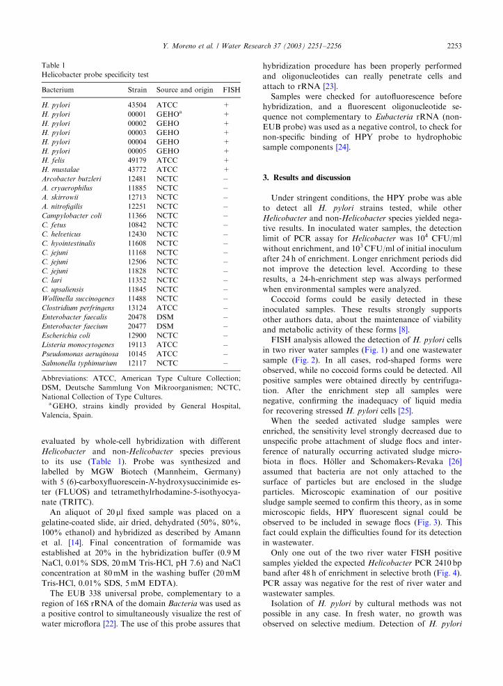
evaluated by whole-cell hybridization with different
Helicobacter and non-Helicobacter species previous
to its use (Table 1). Probe was synthesized and
labelled by MGW Biotech (Mannheim, Germany)
with 5 (6)-carboxyfluorescein-N-hydroxysuccinimide es-
ter (FLUOS) and tetramethylrhodamine-5-isothyocya-
nate (TRITC).
An aliquot of 20ml fixed sample was placed on a
gelatine-coated slide, air dried, dehydrated (50%, 80%,
100% ethanol) and hybridized as described by Amann
et al. [14]. Final concentration of formamide was
established at 20% in the hybridization buffer (0.9M
NaCl, 0.01% SDS, 20mM Tris-HCl, pH 7.6) and NaCl
concentration at 80mM in the washing buffer (20mM
Tris-HCl, 0.01% SDS, 5mM EDTA).
The EUB 338 universal probe, complementary to a
region of 16S rRNA of the domain Bacteria was used as
a positive control to simultaneously visualize the rest of
water microflora [22]. The use of this probe assures that
hybridization procedure has been properly performed
and oligonucleotides can really penetrate cells and
attach to rRNA [23].
Samples were checked for autofluorescence before
hybridization, and a fluorescent oligonucleotide se-
quence not complementary to Eubacteria rRNA (non-
EUB probe) was used as a negative control, to check for
non-specific binding of HPY probe to hydrophobic
sample components [24].
3. Results and discussion
Under stringent conditions, the HPY probe was able
to detect all H. pylori strains tested, while other
Helicobacter and non-Helicobacter species yielded nega-
tive results. In inoculated water samples, the detection
limit of PCR assay for Helicobacter was 104 CFU/ml
without enrichment, and 103CFU/ml of initial inoculum
after 24 h of enrichment. Longer enrichment periods did
not improve the detection level. According to these
results, a 24-h-enrichment step was always performed
when environmental samples were analyzed.
Coccoid forms could be easily detected in these
inoculated samples. These results strongly supports
other authors data, about the maintenance of viability
and metabolic activity of these forms [8].
FISH analysis allowed the detection of H. pylori cells
in two river water samples (Fig. 1) and one wastewater
sample (Fig. 2). In all cases, rod-shaped forms were
observed, while no coccoid forms could be detected. All
positive samples were obtained directly by centrifuga-
tion. After the enrichment step all samples were
negative, confirming the inadequacy of liquid media
for recovering stressed H. pylori cells [25].
When the seeded activated sludge samples were
enriched, the sensitivity level strongly decreased due to
unspecific probe attachment of sludge flocs and inter-
ference of naturally occurring activated sludge micro-
biota in flocs. H .oller and Schomakers-Revaka [26]
assumed that bacteria are not only attached to the
surface of particles but are enclosed in the sludge
particles. Microscopic examination of our positive
sludge sample seemed to confirm this theory, as in some
microscopic fields, HPY fluorescent signal could be
observed to be included in sewage flocs (Fig. 3). This
fact could explain the difficulties found for its detection
in wastewater.
Only one out of the two river water FISH positive
samples yielded the expected Helicobacter PCR 2410 bp
band after 48 h of enrichment in selective broth (Fig. 4).
PCR assay was negative for the rest of river water and
wastewater samples.
Isolation of H. pylori by cultural methods was not
possible in any case. In fresh water, no growth was
observed on selective medium. Detection of H. pylori
Table 1
Helicobacter probe specificity test
Bacterium Strain Source and origin FISH
H. pylori 43504 ATCC +
H. pylori 00001 GEHOa +
H. pylori 00002 GEHO +
H. pylori 00003 GEHO +
H. pylori 00004 GEHO +
H. pylori 00005 GEHO +
H. felis 49179 ATCC +
H. mustalae 43772 ATCC +
Arcobacter butzleri 12481 NCTC �A. cryaerophilus 11885 NCTC �A. skirrowii 12713 NCTC �A. nitrofigilis 12251 NCTC �Campylobacter coli 11366 NCTC �C. fetus 10842 NCTC �C. helveticus 12430 NCTC �C. hyointestinalis 11608 NCTC �C. jejuni 11168 NCTC �C. jejuni 12506 NCTC �C. jejuni 11828 NCTC �C. lari 11352 NCTC �C. upsaliensis 11845 NCTC �Wollinella succinogenes 11488 NCTC �Clostridium perfringens 13124 ATCC �Enterobacter faecalis 20478 DSM �Enterobacter faecium 20477 DSM �Escherichia coli 12900 NCTC �Listeria monocytogenes 19113 ATCC �Pseudomonas aeruginosa 10145 ATCC �Salmonella typhimurium 12117 NCTC �
Abbreviations: ATCC, American Type Culture Collection;
DSM, Deutsche Sammlung Von Mikroorganismen; NCTC,
National Collection of Type Cultures.aGEHO, strains kindly provided by General Hospital,
Valencia, Spain.
Y. Moreno et al. / Water Research 37 (2003) 2251–2256 2253

from wastewater samples was impossible due to the
massive growth of competitive biota in selective media
used for isolation. For this reason, all the samples were
considered ‘‘negative’’ as characteristic colonies could
not be observed.
Our results demonstrate the presence of H. pylori in
wastewater and surface water, and the inadequacy of
available cultural methods for its detection. The findings
of this work seem to confirm the ability of H. pylori to
survive in sludge in the viable but non-culturable form.
In our work, FISH technique has shown to yield more
positive results than PCR. The H. pylori urease gene
sequence primers have been applied extensively to a
range of clinical samples and are established as sensitive
and specific [27–29]. However, further studies should be
necessary with water samples spiked with H. pylori
strains to test its reliability when performed on environ-
mental samples. Clearly, a number of important
questions remain to be answered concerning the
persistence of H. pylori strains in aquatic environments
and the risk that this poses to human health. Although
FISH technique needs more exhaustive evaluation, it
can be a sensitive, specific and cost-effective tool to
detect H. pilory in environmental samples. The detection
of H. pilory by FISH will enable rapid analyses of water
and sewage, improving its safety and quality, and
contributing to elucidate the role of fecally contami-
nated water in the transmission of H. pilory infection.
Fig. 1. Detection of H. pylori in water by in situ hybridization with an HPY probe.
Fig. 2. Detection of H. pylori in wastewater by in situ hybridization with an HPY probe.
Y. Moreno et al. / Water Research 37 (2003) 2251–22562254

Acknowledgements
We thank E. Echevarr!ıa from EMARSA company,
which allowed us access and sampling Pinedo waste-
water treatment plant.
References
[1] Chaun H. Update on the role of Helicobacter pylori
infection in gastrointestinal disorders. Can J Gastroenterol
2001;15:251–5.
[2] Goodman KJ, Cockburn M. The role of epidemiology in
understanding the health effects of Helicobacter pylori.
Epidemiology 2001;12:266–71.
[3] Dunn BE, Cohen H, Blaser MJ. Helicobacter pylori. Clin
Microbiol Rev 1997; 10: 720–41.
[4] Goodman KJ, Correa P. Transmission of Helicobacter
pylori among siblings. Lancet 2000;355:358–62.
[5] Hulten K, Enroth H, Nystr .om T, Engstrand L. Presence of
Helicobacter species DNA in Swedish water. J Appl
Microbiol 1998;85:282–6.
[6] Klein PD, Graham DY, Gaillour A, Opekun AR, Smith
EO. Water source as a risk factor for Helicobacter pylori
infection in Peruvian children. Lancet 1991;337:1503–6.
[7] Hopkins RJ, Vial PA, Ferreccio C, Ovalle J, Prado P,
Sotomayor V, Rusell RJ, Wassermann SS, Morris Jr JG.
Seroprevalence of Helicobacter pylori in Chile: vegetables
may serve as one route of trasmission. J Infec Dis 1993;
168:222–6.
[8] Nilsson HO, Blom J, Al-Soud WA, Ljungh A, Andersen
LP, Wadstr.om T. Effect of cold starvation, acid stress and
nutrients on metabolic activity of Helicobacter pylori. Appl
Environ Microbiol 2002;68:11–9.
[9] Engstrand L. Helicobacter in water and waterborne routes
of transmission. J Appl Microbiol 2001;90:80S–4S.
[10] Hegarty JP, Dowd M, Baker KH. Occurrence of
Helicobacter pylori in surface water in the United States.
J Appl Microbiol 1999;87:697–701.
[11] Sasaki K, Tajiri Y, Sata M, Fujii Y, Matsubara F, Zhao
M, Shimizu S, Toyonaga A, Tanikawa K. Helicobacter
pylori in the natural environment. Scan J Infec Dis 1999;
31:275–9.
Fig. 3. Detection of specific HPY red fluorescence included in sludge flocks.
Fig. 4. Detection of H. pylori in water samples by PCR: (lane 1)
positive control; (lane 2) negative control; (lanes 3–8) water
samples; M: 100 bp ladder.
Y. Moreno et al. / Water Research 37 (2003) 2251–2256 2255

[12] Bunn JE, MacKay WG, Thomas JE, Reid DC, Weaver
LT. Detection of Helicobacter pylori in drinking water
biofilms: implications for transmission in early life. Lett
Appl Microbiol 2002;34:450–4.
[13] Lu Y, Redlinger TE, Avitia R, Galindo A, Goodman K.
Isolation and genotyping of Helicobacter pylori from
untreated municipal wastewater. Appl Environ Microbiol
2002;68:1436–9.
[14] Amann RI, Ludwing W, Schleifer KH. Phylogenetic
identification and in situ detection of individual
microbial cells without cultivation. Microbiol Rev 1995;
59:143–69.
[15] R .ussmann H, Kempf VAJ, Koletzko S, Heessemann J,
Autenrieth IB. Comparison of fluorescent in situ hybridi-
sation and conventional culturing for detection of Helico-
bacter pylori in gastric biopsy specimens. J Clin Microbiol
2001;39:304–8.
[16] Wilson K. Unit 2.4.1. In: Ausubel FM, Brent R, Kingston
RE, Moore DD, Smith JA, Seidman JG, Struhl K, editors.
Current protocols in molecular biology. New York: Wiley,
1987.
[17] Foxall PA, Hu L, Mobley HLT. Use of polymerase chain
reaction-amplified Helicobacter pylori structural genes
for differentiation of isolates. J Clin Microbiol 1992;
30:739–41.
[18] Kepner RL, Pratt JR. Use of fluorochromes for direct
enumeration of total bacteria in environmental samples:
past and present. Microbiol Rev 1994;58:603–15.
[19] Aznar R, Ludwing W, Amann RI, Schleifer KH. Sequence
determination of rRNA genes of pathogenic Vibrio species
and whole-cell identification of Vibrio vulnificus with
rRNA-targeted oligonucleotide probes. Int J Syst Bacteriol
1994;44:330–7.
[20] Sheng-Ang H, Hoyle J, Lewis FA, Secker AD, Cross D,
Mapstone NP, Dixon MF, Wyatt JI, Tompkins DS,
Taylor GR, Quirke P. Direct polymerase chain reaction
for detection of Helicobacter pylori in humans and
animals. J Clin Microbiol 1991;29:2543–9.
[21] Altschul SF, Madden TL, Schaffer AA, Zhang J, Zhang Z,
Miller W, Lipman DJ. Gapped BLAST and PSI-BLAST: a
new generation of protein database search programs.
Nucleic Acids Res 1997;25:3389–402.
[22] Buswell CM, Herlihy YM, Lawrence LM, McGuiggan
JTM, Marsh PD, Keevil CW, Leach SA. Extended
survival and persistence of Campylobacter spp. in water
and aquatic biofilms and their detection by inmunofluor-
escence-antibody and rRNA staining. Appl Environ
Microbiol 1998;64:733–41.
[23] Trebesius K, Panthel K, Strobel S, Vogt K, Faller G,
Kirchner T, Kist M, Heesemann J, Haas R. Rapid and
specific detection of Helicobacter pylori macrolide resis-
tance in gastric tissue by fluorescent in situ hybridisation.
Gut 2000;46:608–14.
[24] Amann RI, Ludwig K, Schleifer KH. Identification and in
situ detection of individual bacterial cells. FEMS Micro-
biol Lett 1992;59:143–69.
[25] Roosendaal R, Kuipers EJ, Pe *na AS, de Graaff J.
Recovery of Helicobacter pylori from gastric biopsy
specimens is not dependent on the transport medium used.
J Clin Microbiol 1995;33:2798–800.
[26] H .oller C, Schomakers-Revaka V. A note: comparison of
different homogeneisation procedures for detecting Cam-
pylobacter spp. in sewage sludges. J Appl Bacteriol
1994;77:591–6.
[27] Gzyl A, Dzierzanowska D, Rozynek E, Celinska-Cedro D,
Dura W, Berg DE. PCR-based diagnosis of Helicobacter
pylori infection in Polish children and adults. J Med
Microbiol 1999;48:349–56.
[28] Kabir S. Detection of Helicobacter pylori in faeces by
culture, PCR and enzyme immunoassay. J Med Microbiol
2001;50:1021–9.
[29] Pacheco N, Mago V, Gomez I, Gueneau P, Guelrud M,
Reyes N, Pericchi LR, Dominguez-Bello MG. Comparison
of PCR and common clinical tests for the diagnosis of
Helicobacter pylori in dyspeptic patients. Diagn Microbiol
Infect Dis 2001;39:207–10.
Y. Moreno et al. / Water Research 37 (2003) 2251–22562256