An Overview of Helicobacter pylori Survival Tactics in ... - MDPI
Upload
independentCategory
view
0download
0
Population Genetic Analyses of Helicobacter pyloriIsolates from Gambian Adults and ChildrenOusman Secka1*, Yoshan Moodley2, Martin Antonio1, Douglas E. Berg3,4, Mary Tapgun5,
Robert Walton5¤a, Archibald Worwui1, Vivat Thomas5, Tumani Corrah5, Julian E. Thomas6,
Richard A. Adegbola7¤b
1 Vaccinology, Medical Research Council Unit, Fajara, The Gambia, 2 Department of Integrative Biology and Evolution, Konrad Lorenz Institute for Ethology, University of
Veterinary Medicine Vienna, Vienna, Austria, 3 Department of Molecular Microbiology, Washington University School of Medicine, St. Louis, Missouri, United States of
America, 4 Department of Medicine, University of California San Diego, La Jolla, California, United States of America, 5 Clinical Services Department, Medical Research
Council Unit, Fajara, The Gambia, 6 Department of Paediatric Gastroenterology Great North Children’s Hospital, Royal Victoria Infirmary, Newcastle upon Tyne, Tyne and
Wear, United Kingdom, 7 Bacterial Diseases Programme, Medical Research Council Unit, Fajara, The Gambia
Abstract
The gastric pathogen Helicobacter pylori is one of the most genetically diverse of bacterial species. Much of its diversitystems from frequent mutation and recombination, preferential transmission within families and local communities, andselection during persistent gastric mucosal infection. MLST of seven housekeeping genes had identified multiple distinct H.pylori populations, including three from Africa: hpNEAfrica, hpAfrica1 and hpAfrica2, which consists of three subpopulations(hspWAfrica, hspCAfrica and hspSAfrica). Most detailed H. pylori population analyses have used strains from non-Africancountries, despite Africa’s high importance in the emergence and evolution of humans and their pathogens. Ourconcatenated sequences from seven H. pylori housekeeping genes from 44 Gambian patients (MLST) identified 42 distinctsequence types (or haplotypes), and no clustering with age or disease. STRUCTURE analysis of the sequence data indicatedthat Gambian H. pylori strains belong to the hspWAfrica subpopulation of hpAfrica1, in accord with Gambia’s West Africanlocation. Despite Gambia’s history of invasion and colonisation by Europeans and North Africans during the last millennium,no traces of Ancestral Europe1 (AE1) population carried by those people were found. Instead, admixture of 17% fromAncestral Europe2 (AE2) was detected in Gambian strains; this population predominates in Nilo-Saharan speakers of North-East Africa, and might have been derived from admixture of hpNEAfrica strains these people carried when they migratedacross the Sahara during the Holocene humid period 6,000–9,000 years ago. Alternatively, shared AE2 ancestry might haveresulted from shared ancestral polymorphisms already present in the common ancestor of sister populations hpAfrica1 andhpNEAfrica.
Citation: Secka O, Moodley Y, Antonio M, Berg DE, Tapgun M, et al. (2014) Population Genetic Analyses of Helicobacter pylori Isolates from Gambian Adults andChildren. PLoS ONE 9(10): e109466. doi:10.1371/journal.pone.0109466
Editor: Ivo G. Boneca, Institut Pasteur Paris, France
Received May 3, 2014; Accepted September 8, 2014; Published October 13, 2014
Copyright: � 2014 Secka et al. This is an open-access article distributed under the terms of the Creative Commons Attribution License, which permitsunrestricted use, distribution, and reproduction in any medium, provided the original author and source are credited.
Data Availability: The authors confirm that all data underlying the findings are fully available without restriction. All relevant data are within the paper.
Funding: This study was funded by MRC unit, The Gambia and US National Institutes of Health (NIH) grants RO3-AI061308, R21-AI078237 and R21-AI088337. Thefunders had no role in study design, data collection and analysis, decision to publish, or preparation of the manuscript.
Competing Interests: The authors of this manuscript have read the journal’s policy and have the following competing interests: RAA is currently an employeeof the GlaxoSmithKline group of companies and received grant awards for bacterial infection studies whilst working as an employee of the MRC Unit in TheGambia. This does not alter the authors’ adherence to all other PLOS ONE policies on sharing data and materials.
* Email: [email protected]
¤a Current address: Blizard Institute, Centre for Primary Care and Public Health, Barts and The London School of Medicine and Dentistry, London, United Kingdom¤b Current address: Department of Scientific Affairs and Public Health, GlaxoSmithKline Vaccines, Wavre, Belgium
Introduction
H. pylori is a genetically diverse Gram negative micro-
aerophilic bacterial species that chronically infects some half of
all humans worldwide [1]. It is implicated in chronic gastritis,
gastroduodenal ulcers and gastric cancer [2,3] and also increases
the risk of infection by diarrheal pathogens [4], which may lead to
infant malnutrition with growth faltering [5] in low income
societies. Despite this, most infections are benign, and some may
even be beneficial [6,7]. The risk of infection resulting in overt
disease is likely to be determined by H. pylori genotype in
combination with other variables such as human genotype and
physiology, nutrition and environmental factors.
H. pylori is usually acquired in childhood [8] and can persist for
life unless eradicated by antibiotics [9]. A prevalence of $80% is
typical in developing nations [10–13], but has fallen dramatically
in industrialized countries during the last century (currently, in the
range of 20%), probably due to major improvements in hygiene
and sanitation [14]. Transmission is predominantly intrafamilial
with a low risk of adult infection in industrialized countries
[15,16], whereas transmission within the local community is
frequent in developing countries, with new infections often
occurring in adults as well as in children [17].
Independent H. pylori isolates typically differ by some 2% or
more in DNA sequence, which allows different strains to be
distinguished readily by the arbitrarily primed PCR (RAPD)
PLOS ONE | www.plosone.org 1 October 2014 | Volume 9 | Issue 10 | e109466
method, wherein each strain yields a characteristic pattern of
DNA fragments different from those of nearly all other indepen-
dent strains [18], or by the sequencing of one or more
housekeeping genes. As an extension of this approach, multilocus
sequence typing (MLST; typically sequencing seven such genes)
soon provided early indications that different sets of H. pylorigenotypes predominated in different human populations [19,20].
Although high sequence variability confounded the use of MLST
in H. pylori, more sophisticated nucleotide analyses of DNA
sequences have since provided more refined demonstrations both
of major geographic or human population differences among H.pylori populations and also admixture often linked to human
migrations [21,22]. This great diversity within and between
populations can be ascribed to H. pylori’s high rates of mutation
and recombination [23], coupled with its having chronically
infected humans for many thousands of years, its transmission
being predominantly within families or local communities, and the
tendency of any person’s gastric mucosal infection to persist for
decades if not treated with antimicrobials. This epidemiologic
pattern resulted in the isolation of populations from each other by
distance, and allowed mutational divergence by random genetic
drift, and selection for locally adapted genotypes. The sequences of
housekeeping genes of strains from many parts of the world
identified three African H. pylori populations, which may be of
particular importance to the Gambian strain analyses presented
here, designated hpAfrica1, hpAfrica2 and hpNEAfrica
[21,22,24]. Four further populations, hpEurope, hpEAsia, hpA-
sia2 and hpSahul, were detected in the rest of the world. More
focussed analyses distinguished three hpAfrica1 subpopulations,
designated hspWAfrica (Western and North-Western Africa),
hspCAfrica (Cameroon) and hspSAfrica (South Africa), and two
hpNEAfrica subpopulations - hspEastNEAfrica and hspCentral-
NEAfrica [25]. The distribution of the various hpAfrica1
subpopulations may reflect the expansion of the Bantu people
throughout subSaharan Africa from an ancestral homeland in or
near present day Nigeria/Cameroon during the last 4000 years. In
contrast the hspEastNEAfrica and hspCentralNEAfrica subpopu-
lations found in Algeria and northern Nigeria are thought to
reflect migrations of Nilo-Saharan people through what were then
central Saharan wetlands during the Holocene humid period some
6,000–9,000 years ago [25].
The Gambia is a small country, some 47 Km wide and 338 Km
long, embracing the west-flowing Gambia River in the most
western part of Africa, bordered by Senegal to the North, South
and East and the Atlantic Ocean to the West. Gambian contacts
with Europeans were mostly with the Portuguese and French
beginning in the 15th century, and then with the British in the 17th
century and lasting until independence from British colonial rule
in 1965. The great majority of present-day Gambians are
indigenous West Africans, predominantly of the Mandinka, Wollof
and Fulani linguistic groups that are also abundant in nearby
countries of Senegal, Guinea Bissau, Guinea and Mali. Most
Gambians are Muslim, reflecting conversion of residents by Arab
traders from North Africa who crossed the Sahara beginning in
the 8th century. Given the tendency of H. pylori populations to
reflect human host migrations, and since the Gambia was also a
major source of slaves taken to the Americas and Europe for
several centuries until the slave trade ended in the 1800s, it is
possible that Gambian strains contributed significantly to H.pylori’s gene pool in The Americas and Europe. Indeed, a
suggestion of West African admixture in European H. pylori had
emerged in our earlier study of a novel regulatory gene-linked
insertion-deletion polymorphism (indel) in Spanish vs. Gambian
H. pylori strains [26].
It is with this background that we sequenced the MLST genes of
H. pylori strains from ethnic West African adults and children in
The Gambia. These strains are expected to be broadly represen-
tative of H. pylori of much of West Africa, a relatively unstudied
population. Furthermore, given the historical contact with
Europeans and North Africans, we may also detect traces of the
ancestral European nucleotides carried by these people within our
Gambian sample.
Materials and Methods
Ethics statementEthical approval for this study was provided by The Gambia
Government/MRC joint ethics committee and the NIH Division
of Microbiology Infectious Diseases (DMID) International Review
Board, USA (NIH number DMID 06-0053; MRC Unit, The
Gambia IRB registration number: IRB00003943 and Federal
Wide Assurance number: FWA 00006873).
PatientsGastric biopsies were collected from patients referred for
endoscopy at the Medical Research Council Unit (MRC), The
Gambia, for routine clinical investigations of symptoms attribut-
able to gastroduodenal disease. All patients who agreed to join this
study provided written informed consent; in addition, for children
aged less than 18 years, biopsies were obtained after written
informed parental consent. The patients belonged to the following
ethnic groups: Mandinka (19), Wollof (9), Jola (6), Fulani (5),
Sarahule (4), Serere (1).
BiopsiesEndoscopes were cleaned and sterilized with Cidex (Johnson
and Johnson Co) after each use according to standard care at the
MRC Unit, The Gambia (SOP-CLS-001). Biopsies from the
gastric antrum and corpus of study participants were immediately
placed in 1 ml Brain-Heart Infusion (BHI) broth containing 20%
glycerol and transported in ice to the laboratory for culture or
stored at 270uC until used.
Bacterial cultureBiopsies were spread on the surface of selective Columbia-Blood
agar, incubated at 37uC in a microaerobic atmosphere and
processed as previously described [27]. Single H. pylori colonies
were isolated and confluent growth derived by spreading cells from
them was harvested and preserved in BHI broth containing 20%
glycerol and stored at 270uC until analysed.
Sample choice for MLSTSamples were chosen according to successful subculture of
individual H. pylori colonies. One or more single colonies were
isolated from each of 44 patients and used for these analyses. The
44 patients (23 male and 21 female) ranged in age from 18 months
to 72 years (mean 32 years) and had the following clinical
manifestations: normal endoscopic appearance (29), gastric
erosions (6), gastric ulcer (3), and oesophageal ulcer (1). Five of
these patients were malnourished children with enteropathy (ages
18–31 months, mean 19 months). Thirty three patients (75%) were
from the Greater Banjul Area (GBA) and 11 (25%) were from
rural villages. To investigate genetic heterogeneity within the same
stomach, multiple single colonies from each of two patients with
normal gastroduodenal appearance by endoscopy were tested by
MLST: seven colonies (4 antrum and 3 corpus) from a 14 year old
subject; and 11 single colonies (6 antrum and 5 corpus) from a 72
year old subject.
Population Genetic Analyses of Helicobacter pylori from The Gambia
PLOS ONE | www.plosone.org 2 October 2014 | Volume 9 | Issue 10 | e109466
Genomic DNA extractionGenomic DNA was prepared from confluent H. pylori growth
using a commercial kit (Qiagen DNA Mini kit, UK) [27].
PCR to detect cagAPCR was performed to detect the cagA virulence gene using
previously described primers and methods [27], and PCR
products were detected by electrophoresis in a 1.5% agarose gel
with ethidium bromide, and band visualization using Gel Doc
2000 (Bio-Rad laboratories, Milan, Italy).
Housekeeping gene sequencing and MLSTThe seven standard MLST genes (atpA, efp, mutY, ppa, trpC,
ureI and yphC) were amplified and sequenced as detailed in
http://pubmlst.org/Helicobacter. The PCR products were se-
quenced using an ABI Prism 3130X DNA sequencer (Applied
Biosystems, USA). Consensus sequences were generated and
assembled using DNAstar programme (Lasergene, USA, Version
7). The sequences obtained were submitted to the H. pylori MLST
database (http://pubmlst.org/Helicobacter) for allele and se-
quence type identification. Concatenated sequences were aligned
and imported into MEGA version 5. Evolutionary history was
inferred using the neighbor-joining tree reconstruction method
[28]. The percentage of replicate trees in which the associated taxa
clustered together in a bootstrap test of 2000 replicates is depicted
beside each branch.
Nucleotide analysesCalculation of the ratio of non-synonymous to synonymous
changes (dN/dS) was done with the START2 (Sequence Type
Analysis and Recombinational Tests Version 2) tool, which uses
the method of Nei and Gojobori to estimate parameters [29].
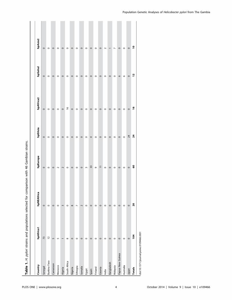
Analyses of population structureTo determine the relatedness of Gambian H. pylori to
previously studied strains from elsewhere, 246 strains representing
H. pylori’s global diversity were selected from the public MLST
data base (Table 1). This data set included all strains previously
published from West Africa [22,25,30]. Reconstruction of a global
phylogeny was carried out using neighbor-joining as described
above. In addition, two Bayesian population cluster analyses were
performed using STRUCTURE V2.3.4 software [21]. First, the
no-admixture model was used to determine the overall structure of
the Gambian sequence data with respect to predefined modern
populations. Then, the linkage model was used to ascertain
ancestral components within our Gambian data set. In the unlikely
event that Gambian H. pylori represent a new population, we set
our values for K at greater than the known number of modern and
ancestral H. pylori populations. We carried out three independent
runs at 2#K#10 for the no-admixture model and three
independent runs at 2#K#8 for the linkage model. Each run
comprised 50,000 iterations, the first half of which were discarded
as burnin.
Results
Allelic frequency and nucleotide analysesDNAs from H. pylori strains from 44 Gambians (one strain/
patient in 43 cases; three strains/patient in one case) yielded 42
unique MLST sequence types based on concatenated DNA
sequences of seven housekeeping gene loci. Four pairs of strains
yielded identical MLSTs. One pair was from consecutive
unrelated patients whose biopsies were obtained on the same
day. The other three pairs were also from unrelated patients who
were biopsied between one week and two years apart. These
exceptions aside, most alleles of individual genes occurred only
once among the 46 strains, although identical alleles were found in
11 to 16 strains, depending on the gene. Except for the four pairs
of strains that were identical at all loci (noted above), no pair of
strains identical at one locus was identical at another of the seven
loci tested (data not shown). The mean nucleotide diversity in the 7
genes was 2.9% (Table 2). The gene trpC had most strains with
identical alleles (16 alleles); allele 1774 of mutY was most frequent
(5 occurrences; 10.9%); the most diverse gene was trpC (mean
nucleotide level diversity 4.6%); and the least diverse was ureI(1.2%, Table 2). No deletions or insertions were found in any of
the analysed gene fragments.
Analyses of selectionA nucleotide substitution in a coding region results either in a
change or no change in the protein’s amino acid sequence (non-
synonymous (N), synonymous (S), respectively). The dN/dS ratio in
a population reflects genetic drift and selection operating on
individual genes. All dN/dS values in the seven housekeeping genes
from Gambian H. pylori strains were close to zero (Table 2),
indicating intense selection to maintain functions and amino acid
sequences of the encoded proteins. This is expected for genes
whose encoded proteins act within bacterial cells and provide
important housekeeping functions.
Phylogenetic analysis of Gambian strains, relative tothose from elsewhere.
A phylogenetic tree reconstructed using concatenated sequences
of the seven housekeeping genes (Figure 1) provided no evidence
of association of particular clusters (clades) of strains with variables
such as age of participant at time of endoscopy, endoscopic
diagnosis, sex or district of residence within the Gambia. However,
cagA+ strains seemed to cluster separately from cagA- (Figure 2).
Gambian strain sequences were also compared with sequences
from 246 H. pylori strains selected from other informative human
populations (African, European, Australian and Asian; Table 1)
using neighbor-joining and Bayesian cluster analysis with both the
no-admixture and linkage models of STRUCTURE. We found
that all Gambian strains clustered within hpAfrica1 and were
intermingled with strains from Senegal and Burkina Faso.
hpAfrica1 strains formed a separate clade that was sister to strains
from hpNEAfrica (Figure 2), with other European, Australian and
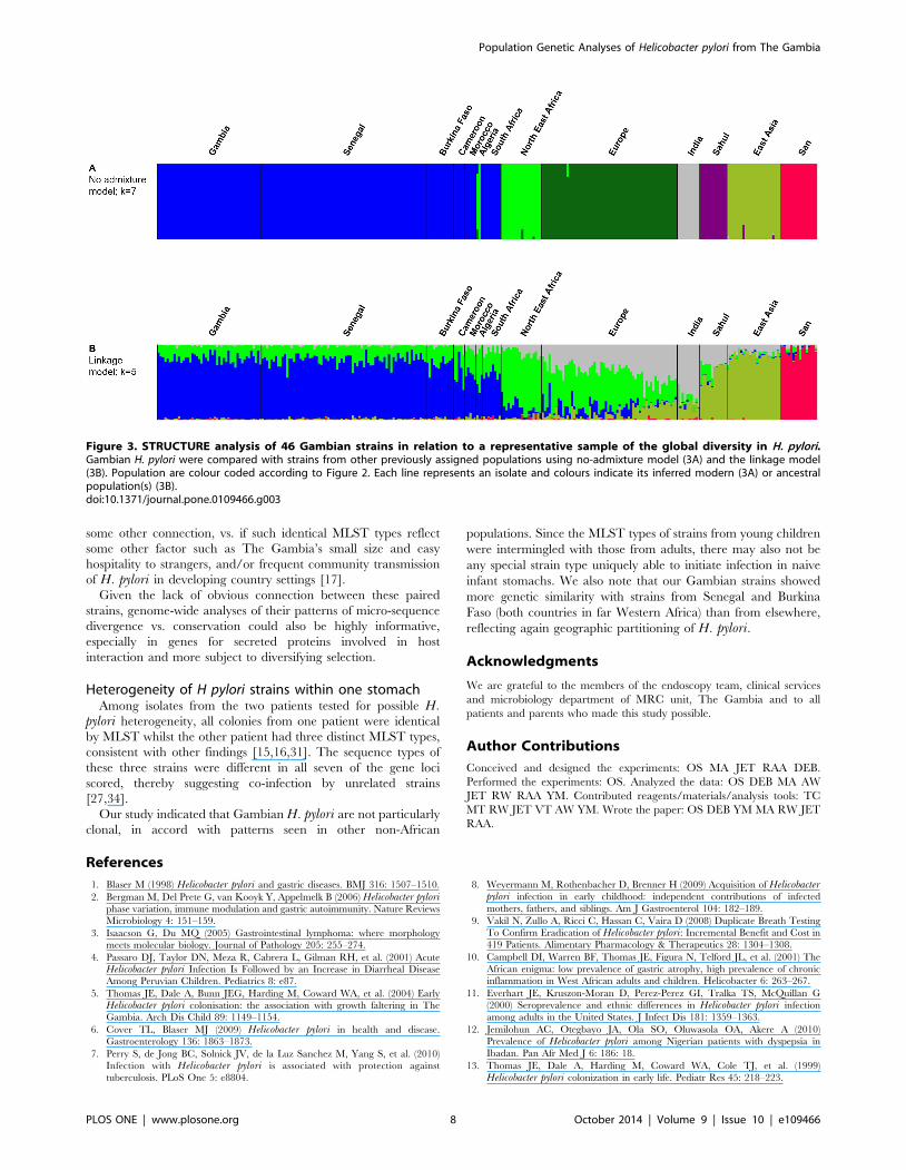
Asian populations more distantly related. Under the no-admixture
model, the number of populations (K) with the highest likelihood
was seven, which corresponds to what is already known about the
global structure of this bacterial species. Almost all Gambian
isolates formed a homogeneous group together with other
hpAfrica1 strains (Figure 3A). However, the linkage model showed
that Gambian strains were more similar in their ancestral
nucleotide composition to hspWAfrica strains from Senegal
(Gambia’s immediate neighbour) and Burkina Faso (population
3), whereas the North-East African ancestral component (ancestral
Europe 2) was more apparent among those hpAfrica1 strains from
more distant communities in Cameroon, Morocco, Algeria and
South Africa (Figure 3B).
Phenotypic heterogeneity of H. pylori in a single hostBacterial colonies that differed markedly in morphology, seen
among H. pylori cultured from biopsies from two patients with
normal endoscopic gastroduodenal tract appearance (14 and 72
years of age), were used to test for H. pylori DNA level
Population Genetic Analyses of Helicobacter pylori from The Gambia
PLOS ONE | www.plosone.org 3 October 2014 | Volume 9 | Issue 10 | e109466
Ta
ble
1.
H.
pyl
ori
stra
ins
and
po
pu
lati
on
sse
lect
ed
for
com
par
iso
nw
ith
46
Gam
bia
nst
rain
s.
Co
un
try
hp
Afr
ica
1h
pN
EA
fric
ah
pE
uro
pe
hp
EA
sia
hp
Afr
ica
2h
pS
ah
ul
hp
Asi
a2
Sen
eg
al7
30
00
00
0
Bu
rkin
aFa
so1
20
00
00
0
Cam
ero
on
50
00
00
0
Mo
rocc
o5
02
00
00
Alg
eri
a1
32
00
00
Sou
thA
fric
a8
00
01
60
0
Nig
eri
a0
80
00
00
Eth
iop
ia0
70
00
00
Som
alia
02
00
00
0
Egyp
t0
03
00
00
Spai
n0
03
30
00
0
Fin
lan
d0
09
00
00
Esto
nia
00
11
00
00
Ind
ia0
00
00
02
Ban
gla
de
sh0
00
00
01
Mal
aysi
a0
00
00
07
Pap
ua
Ne
wG
uin
ea
00
00
06
0
Au
stra
lia0
00
00
60
Jap
an0
00
24
00
0
To
tals
10
42
06
02
41
61
21
0
do
i:10
.13
71
/jo
urn
al.p
on
e.0
10
94
66
.t0
01
Population Genetic Analyses of Helicobacter pylori from The Gambia
PLOS ONE | www.plosone.org 4 October 2014 | Volume 9 | Issue 10 | e109466
heterogeneity in individual hosts, perhaps equivalent to that seen
previously in a European with a mixed cagA+ and cagA- infection
[31]. Seven colonies representative of the different morphologies
were analysed from the 14 year old and 11 such colonies were
analysed from the 72 year old. Three different MLST types were
identified among the isolates from the first (14 year old) patient
(MLST types 2040 in all four colonies from antrum, and 2047 and
2064 from one and two colonies, respectively, from the corpus;
Figure 1). Each of these three MLST types was different from the
two others in all seven gene loci tested. In the second (72 year old)
patient, all 11 colonies (6 antrum, 5 corpus) were of the same
MLST type. In neither case did the different colony morphologies
that first encouraged analysis of multiple isolates from these two
patients correspond to different MLST types.
Discussion
Most detailed H. pylori population analyses to date have used
strains from non-African countries, despite Africa’s great impor-
tance for the emergence and evolution of humans and pathogens
such as H. pylori. Here we sequenced the housekeeping genes of
strains from The Gambia, one of the most detailed studies to date
of a West African H. pylori population. These Gambian strains
exhibited high nucleotide sequence diversity (mean, 2.9%), much
as in isolates from other geographic regions [24,32], with no
obvious clustering of MLST types in particular age or disease
groups. Our sequence data showed that Gambian H. pylori strains
belong to the hpAfrica1 population and added greater resolution
to the geographic distribution of this population in Africa.
Comparison of ancestral nucleotides with those available from
other African countries indicated some geographic differentiation,
even within West Africa, as did a recent complementary analysis of
many strains from Dakar, Senegal [30].
STRUCTURE analyses indicated that the contribution of
ancestral nucleotides from other populations, mainly AE2 (which
originated in North East Africa [22], to strains circulating in The
Gambia was about 17%, which is the average of 3 independent
linkage model runs at K = 5. These analyses also showed that
Gambian strains are closely related to each other and belong to the
hpAfrica1 population (Figures 3). The proportion of AE2
sequences in the Gambian strains is similar to, although
marginally higher than, that found in strains from Senegal (19%)
and Burkina Faso (23%), and much lower than in any other
African country studied to date. This suggests a history of limited
admixture (recombination) with strains from elsewhere, as also
noted by others [30].
In the case of Gambian H. pylori, recombination from other
non-hpAfrica1 strains could possibly have occurred after contact
with Europeans or North Africans. However, the population
(hpEurope) carried by Europeans is itself a hybrid (recombinant),
consisting of roughly equal contributions from AE1 and AE2,
originating in Central Asia and North-East Africa, respectively
[30,33]. Since no trace of AE1 was found in our sample, we
conclude that the source of the AE2 nucleotides in the Gambia is
unlikely to have resulted from hpEurope strains following
European colonization of African lands during the last six
centuries. This also sheds light on the nature of the centuries-
long contact between Gambians and Europeans/North Africans.
In South Africa, hpEurope was found in the stomachs of
indigenous Africans from several ethnicities [33], pointing to a
more extensive association between colonizing Europeans and
local populations than in the Gambia, where Europeans never
made up a significant part of the total population.
Therefore, we propose that the Gambian H. pylori’s AE2
nucleotides were derived from contact with Nilo-Saharan speak-
ers, perhaps when these people migrated westwards from the Nile
valley across the Sahara during the Holocene humid period,
6,000–9,000 or more years ago [25]. In accord with this,
hpNEAfrica sequences have also been detected in strains from
Cameroon [25], northern (predominantly Muslim) Nigeria [22]
and Algeria [22] (Figure 3B). Alternatively, since hpAfrica1 and
hpNEAfrica are sister phylogenetic groups (Figure 2), the observed
AE2 ancestry among Gambian, Senegalese and Burkina Faso
strains could also be attributed to background linkage disequilib-
rium, where ancestral nucleotides were already present in the
common ancestor of both populations.
Identical MLST typesWe found four pairs of isolates with identical MLST types.
Three of these pairs were from people whose gastric biopsies were
obtained between one week and two years apart and processed in
the laboratory on different dates. They thus are likely to reflect
occasional carriage of closely related strains by unrelated members
of a community. We also suggest this explanation for the one
matched pair from patients who had gastric biopsies obtained
consecutively on the same day, since our endoscopes were
rigorously cleaned and washed after each use (see Materials and
Methods).
Although none of the strain pairs with identical MLSTs were
from persons from the same village or with the same family names,
further study will be needed to learn if these people had ever lived
in the same extended family compound, village or district, or had
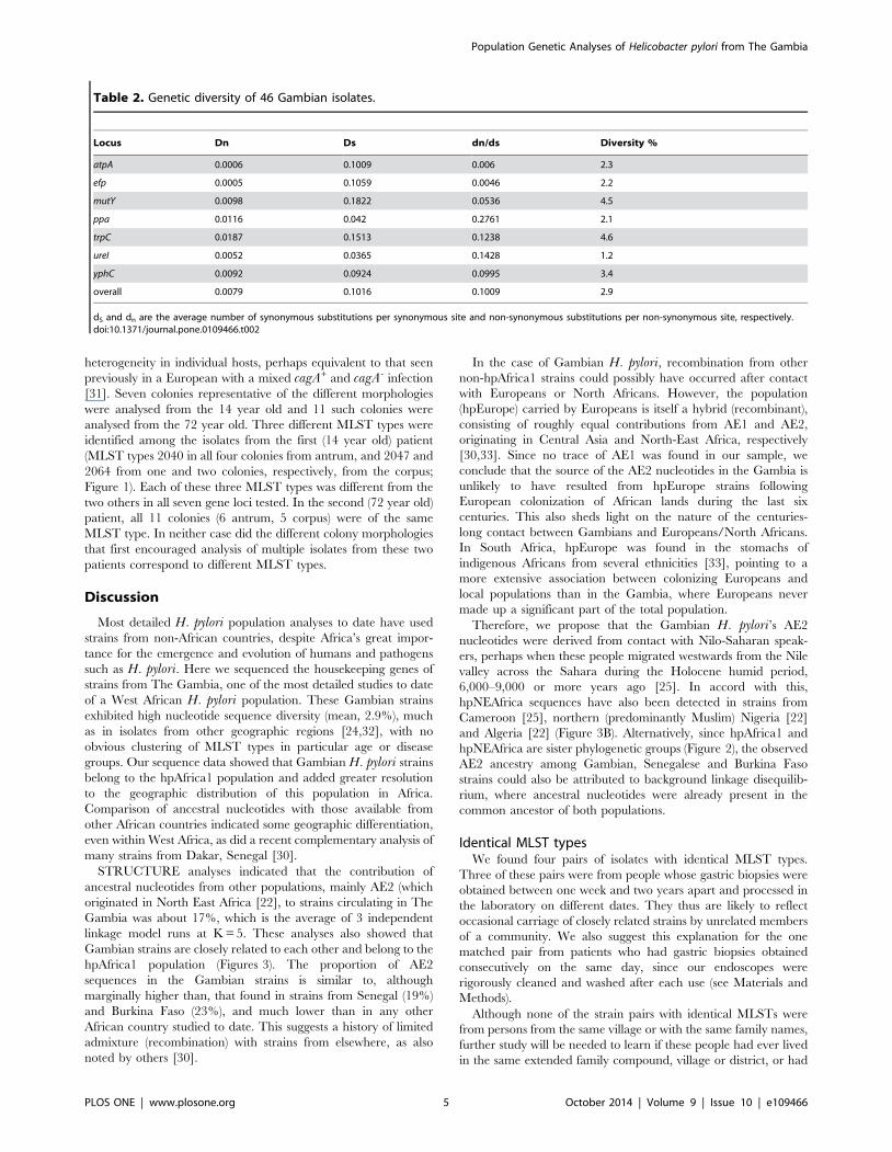
Table 2. Genetic diversity of 46 Gambian isolates.
Locus Dn Ds dn/ds Diversity %
atpA 0.0006 0.1009 0.006 2.3
efp 0.0005 0.1059 0.0046 2.2
mutY 0.0098 0.1822 0.0536 4.5
ppa 0.0116 0.042 0.2761 2.1
trpC 0.0187 0.1513 0.1238 4.6
ureI 0.0052 0.0365 0.1428 1.2
yphC 0.0092 0.0924 0.0995 3.4
overall 0.0079 0.1016 0.1009 2.9
dS and dn are the average number of synonymous substitutions per synonymous site and non-synonymous substitutions per non-synonymous site, respectively.doi:10.1371/journal.pone.0109466.t002
Population Genetic Analyses of Helicobacter pylori from The Gambia
PLOS ONE | www.plosone.org 5 October 2014 | Volume 9 | Issue 10 | e109466
Figure 1. Evolutionary relationship among H. pylori strains isolated from The Gambia. Evolutionary history was inferred fromconcatenated sequences of the seven MLST housekeeping gene fragments (3406 bp) from 46 Gambian H. pylori using the neighbor-joining method.The analyses were conducted in MEGA5. The five strains from young children are identified with green circles. There was one subject with threedifferent MLST types shown in red squares.doi:10.1371/journal.pone.0109466.g001
Population Genetic Analyses of Helicobacter pylori from The Gambia
PLOS ONE | www.plosone.org 6 October 2014 | Volume 9 | Issue 10 | e109466
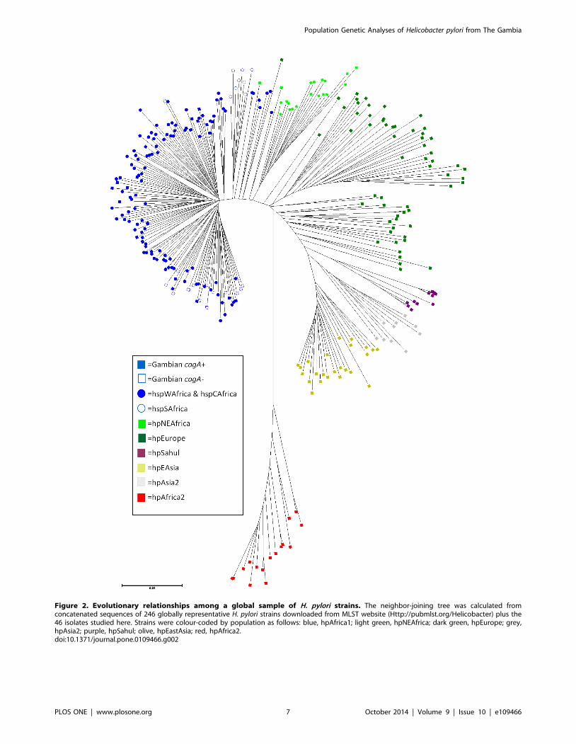
Figure 2. Evolutionary relationships among a global sample of H. pylori strains. The neighbor-joining tree was calculated fromconcatenated sequences of 246 globally representative H. pylori strains downloaded from MLST website (Http://pubmlst.org/Helicobacter) plus the46 isolates studied here. Strains were colour-coded by population as follows: blue, hpAfrica1; light green, hpNEAfrica; dark green, hpEurope; grey,hpAsia2; purple, hpSahul; olive, hpEastAsia; red, hpAfrica2.doi:10.1371/journal.pone.0109466.g002
Population Genetic Analyses of Helicobacter pylori from The Gambia
PLOS ONE | www.plosone.org 7 October 2014 | Volume 9 | Issue 10 | e109466
some other connection, vs. if such identical MLST types reflect
some other factor such as The Gambia’s small size and easy
hospitality to strangers, and/or frequent community transmission
of H. pylori in developing country settings [17].
Given the lack of obvious connection between these paired
strains, genome-wide analyses of their patterns of micro-sequence
divergence vs. conservation could also be highly informative,
especially in genes for secreted proteins involved in host
interaction and more subject to diversifying selection.
Heterogeneity of H pylori strains within one stomachAmong isolates from the two patients tested for possible H.
pylori heterogeneity, all colonies from one patient were identical
by MLST whilst the other patient had three distinct MLST types,
consistent with other findings [15,16,31]. The sequence types of
these three strains were different in all seven of the gene loci
scored, thereby suggesting co-infection by unrelated strains
[27,34].
Our study indicated that Gambian H. pylori are not particularly
clonal, in accord with patterns seen in other non-African
populations. Since the MLST types of strains from young children
were intermingled with those from adults, there may also not be
any special strain type uniquely able to initiate infection in naive
infant stomachs. We also note that our Gambian strains showed
more genetic similarity with strains from Senegal and Burkina
Faso (both countries in far Western Africa) than from elsewhere,
reflecting again geographic partitioning of H. pylori.
Acknowledgments
We are grateful to the members of the endoscopy team, clinical services
and microbiology department of MRC unit, The Gambia and to all
patients and parents who made this study possible.
Author Contributions
Conceived and designed the experiments: OS MA JET RAA DEB.
Performed the experiments: OS. Analyzed the data: OS DEB MA AW
JET RW RAA YM. Contributed reagents/materials/analysis tools: TC
MT RW JET VT AW YM. Wrote the paper: OS DEB YM MA RW JET
RAA.
References
1. Blaser M (1998) Helicobacter pylori and gastric diseases. BMJ 316: 1507–1510.
2. Bergman M, Del Prete G, van Kooyk Y, Appelmelk B (2006) Helicobacter pyloriphase variation, immune modulation and gastric autoimmunity. Nature Reviews
Microbiology 4: 151–159.
3. Isaacson G, Du MQ (2005) Gastrointestinal lymphoma: where morphology
meets molecular biology. Journal of Pathology 205: 255–274.
4. Passaro DJ, Taylor DN, Meza R, Cabrera L, Gilman RH, et al. (2001) Acute
Helicobacter pylori Infection Is Followed by an Increase in Diarrheal Disease
Among Peruvian Children. Pediatrics 8: e87.
5. Thomas JE, Dale A, Bunn JEG, Harding M, Coward WA, et al. (2004) Early
Helicobacter pylori colonisation: the association with growth faltering in The
Gambia. Arch Dis Child 89: 1149–1154.
6. Cover TL, Blaser MJ (2009) Helicobacter pylori in health and disease.
Gastroenterology 136: 1863–1873.
7. Perry S, de Jong BC, Solnick JV, de la Luz Sanchez M, Yang S, et al. (2010)
Infection with Helicobacter pylori is associated with protection against
tuberculosis. PLoS One 5: e8804.
8. Weyermann M, Rothenbacher D, Brenner H (2009) Acquisition of Helicobacterpylori infection in early childhood: independent contributions of infected
mothers, fathers, and siblings. Am J Gastroenterol 104: 182–189.
9. Vakil N, Zullo A, Ricci C, Hassan C, Vaira D (2008) Duplicate Breath Testing
To Confirm Eradication of Helicobacter pylori: Incremental Benefit and Cost in
419 Patients. Alimentary Pharmacology & Therapeutics 28: 1304–1308.
10. Campbell DI, Warren BF, Thomas JE, Figura N, Telford JL, et al. (2001) The
African enigma: low prevalence of gastric atrophy, high prevalence of chronic
inflammation in West African adults and children. Helicobacter 6: 263–267.
11. Everhart JE, Kruszon-Moran D, Perez-Perez GI, Tralka TS, McQuillan G
(2000) Seroprevalence and ethnic differences in Helicobacter pylori infection
among adults in the United States. J Infect Dis 181: 1359–1363.
12. Jemilohun AC, Otegbayo JA, Ola SO, Oluwasola OA, Akere A (2010)
Prevalence of Helicobacter pylori among Nigerian patients with dyspepsia in
Ibadan. Pan Afr Med J 6: 186: 18.
13. Thomas JE, Dale A, Harding M, Coward WA, Cole TJ, et al. (1999)
Helicobacter pylori colonization in early life. Pediatr Res 45: 218–223.
Figure 3. STRUCTURE analysis of 46 Gambian strains in relation to a representative sample of the global diversity in H. pylori.Gambian H. pylori were compared with strains from other previously assigned populations using no-admixture model (3A) and the linkage model(3B). Population are colour coded according to Figure 2. Each line represents an isolate and colours indicate its inferred modern (3A) or ancestralpopulation(s) (3B).doi:10.1371/journal.pone.0109466.g003
Population Genetic Analyses of Helicobacter pylori from The Gambia
PLOS ONE | www.plosone.org 8 October 2014 | Volume 9 | Issue 10 | e109466
14. Chong VH, Lim KC, Rajendran N (2008) Prevalence of active Helicobacterpylori infection among patients referred for endoscopy in Brunei Darussalam.Singapore Med J 49: 42–46.
15. Raymond J, Thiberge JM, Chevalier C, Kalach N, Bergeret M, et al. (2004)
Genetic and transmission analysis of Helicobacter pylori strains within a family.Emerg infect Dis 10: 1816–1821.
16. Schwarz S, Morelli G, Kusecek B, Manica A, Balloux F, et al. (2008) Horizontalversus Familial Transmission of Helicobacter pylori. PLoS Pathogens 4: 1–10.
17. Herrera PM, Mendez M, Velapatino B, Santivanez L, Balqui J, et al. (2008)
DNA-level diversity and relatedness of Helicobacter pylori strains in shantytownfamilies in Peru and transmission in a developing-country setting. J Clin
Microbiol 46: 3912–3918.18. Alm RA, Trust TJ (1999) Analysis of the genetic diversity of Helicobacter pylori:
the tale of two genomes. J Mol Med 77: 834–846.19. Achtman M, Azuma T, Berg DE, Ito Y, Morelli G, et al. (1999) Recombination
and clonal groupings within Helicobacter pylori from different geographical
regions. Molecular Microbiology 32: 459–470.20. Israel DA, Salama N, Krishna U, Rieger UM, Atherton JC, et al. (2001)
Helicobacter pylori genetic diversity within the gastric niche of a single host. ProcNat Acad Science USA 98: 14625–14630.
21. Falush D, Wirth T, Linz B, Pritchard JK, Stephens M, et al. (2003) Traces of
human migrations in Helicobacter pylori populations. Science 299: 1582–1585.22. Linz B, Balloux F, Moodley Y, Manica A, Liu H, et al. (2007) An African origin
for the intimate association between humans and Helicobacter pylori. Nature445: 915–918.
23. Suerbaum S, Smith MJ, Bapumia K, Morelli G, Smith NH, et al. (1998) Freerecombination within Helicobacter pylori. Proc Natl Acad Sci USA 95: 12619–
12624.
24. Moodley Y, Linz B, Yamaoka Y, Windsor HM, Breurec S, et al. (2009) The
peopling of the Pacific from a bacterial perspective. Science 323: 527–530.
25. Nell S, Eibach D, Montano V, Maady A, Nkwescheu A, et al. (2013) Recent
acquisition of Helicobacter pylori by Baka pygmies. PLoS Genet 9: e1003775.
26. McNulty SL, Mole BM, Dailidiene D, Segal S, Ally R, et al. (2004) Novel 180-
and 480-Base-Pair Insertions in African and African-American Strains of
Helicobacter pylori. J Clin Microbiol 42: 5658–5663.
27. Secka O, Antonio M, Tapgun M, Berg DE, Bottomley C, et al. (2011) PCR-
based genotyping of Helicobacter pylori of Gambian children and adults directly
from biopsy specimens and bacterial cultures. Gut Pathog 3: 5.
28. Saitou N, Nei M (1987) The neighbor-joining method: A new method for
reconstructing phylogenetic trees. Molecular Biology and Evolution 4: 406–425.
29. Jolley KA, Feil EJ, Chan MS, Maiden MC (2001) Sequence type analysis and
recombinational tests (START). Bioinformatics 17: 1230–1231.
30. Linz B, Vololonantenainab CR, Seck A, Carod JF, Dia D, et al. (2014)
Population genetic structure and isolation by distance of Helicobacter pylori in
Senegal and Madagascar. PLoS One 9: e87355.
31. Kersulyte D, Chalkauskas H, Berg DE (1999) Emergence of recombinant strains
of Helicobacter pylori during human infection. Mol Microbiol 31: 31–43.
32. Breurec S, Guillard B, Hem S, Brisse S, Dieye FB, et al. (2011) Evolutionary
History of Helicobacter pylori Sequences Reflect Past Human Migrations in
Southeast Asia. PLoS One 6.
33. Moodley Y, Linz B, Bond RP, Nieuwoudt M, Soodyall H, et al. (2012) Age of
the association between Helicobacter pylori and man. PLoS pathog 8.
34. Akada JK, Ogura K, Dailidiene D, Dailide G, Cheverud JM, et al. (2003)
Helicobacter pylori tissue tropism: mouse-colonizing strains can target different
gastric niches. Microbiology 149: 1901–1909.
Population Genetic Analyses of Helicobacter pylori from The Gambia
PLOS ONE | www.plosone.org 9 October 2014 | Volume 9 | Issue 10 | e109466
Copyright © 2022 FDOKUMEN