Body indices and basic vital signs in Helicobacter pylori positive and negative persons
Upload
independentCategory
view
1download
0
Helicobacter pylori infection upregulates interleukin-18production from gastric epithelial cells
Masaaki Shimadaa,b, Takafumi Andoa, Richard M. Peekd, Osamu Watanabea, KazuhiroIshiguroa, Osamu Maedaa, Daisuke Ishikawaa, Motofusa Hasegawaa, Kenji Inac, NaokiOhmiyaa, Yasumasa Niwaa, and Hidemi Gotoa
aDepartment of Gastroenterology, Nagoya University Graduate School of Medicine, NagoyaMemorial Hospital, Nagoya, JapanbDepartment of Gastroenterology, National Hospital Organization, Nagoya Medica Center,Nagoya Memorial Hospital, Nagoya, JapancDepartment of Clinical Oncology, Nagoya Memorial Hospital, Nagoya, JapandDivision of Gastroenterology, Vanderbilt University School of Medicine, Nashville, Tennessee,USA
AbstractBackground—Helicobacter pylori infection induces a biased T helper type 1 (Th1) response thatproduces IFN-γ and Fas ligand (FasL). Th1 cytokines are associated with apoptosis in the gastricepithelial cells.
Aim—We aimed to define the role of the recently cloned IL-18, a IFN-γ inducing factor, ingastric mucosal injury induced by H. pylori infection.
Methods—Twenty-seven gastric ulcer (GU) patients and 20 functional dyspepsia (FD) patientswere enrolled in this study. Mucosal biopsy samples were obtained from the gastric antrum andGU site during endoscopy. Samples were used for histological examination, H. pylori culture andin-situ stimulation for 48 h in the presence of 10 µg/ml phytohemagglutinin-P. IL-18, IFN-γ, andsoluble FasL (sFasL) levels in culture supernatants were assayed by the enzyme-linkedimmunosorbent assay method. IL-18, IL-1β-converting enzyme (ICE) and caspase-3 wereevaluated by western blotting in gastric cancer cell lines (MKN45) cocultured with H. pylori.
Results—All 27 GU patients and ten out of 20 FD patients were found to be H. pylori-positive,whereas ten FD patients were H. pylori-negative. Antral mucosal tissues from H. pylori-positiveFD patients contained (P < 0.01) higher levels of IL-18, IFN-γ, and sFasL than those fromuninfected FD patients. IL-18, IFN-γ, and sFasL levels at the ulcer site were significantly (P <0.01) higher than those at distant sites in the antrum. A significant relationship was seen betweenIL-18 and IFN-γ levels at the ulcer site (r = 0.7, P < 0.01). H. pylori eradication led to asignificant decrease in the levels of IL-18, IFN-γ, and sFasL at the ulcer site. Western blottingshowed that IL-18, ICE, and caspase-3 were activated in gastric cancer cell lines cocultured withH. pylori.
© 2008 Wolters Kluwer Health | Lippincott Williams & Wilkins
Correspondence to Dr Takafumi Ando, Department of Gastroenterology, Nagoya University Graduate School of Medicine, 65Tsurumai-cho, Showa-ku, Nagoya 466-8550, Japan, Tel: + 81 52 7442144; fax: +81 52 7442175; [email protected].
Conflict of interest: none declared.
NIH Public AccessAuthor ManuscriptEur J Gastroenterol Hepatol. Author manuscript; available in PMC 2012 June 12.
Published in final edited form as:Eur J Gastroenterol Hepatol. 2008 December ; 20(12): 1144–1150. doi:10.1097/MEG.0b013e32830edb15.
NIH
-PA Author Manuscript
NIH
-PA Author Manuscript
NIH
-PA Author Manuscript

Conclusion—This study suggests that H. pylori infection enhanced mucosal injury bystimulating a Th1 response, which was mediated by IL-18 upregulation as well as activation ofICE and caspase-3.
Keywordscaspase; cytotoxicity; gastric ulcer; Helicobacter pylori; IL-18
IntroductionHelicobacter pylori, a microaerophilic, gram-negative, spiral-shaped bacterium thatcolonizes the stomach, was first reported by Warren and Marshall in 1984 [1]. H. pylori isinvolved in the pathogenesis of chronic active gastritis as well as duodenal ulceration,gastric ulceration (GU), and gastric cancer [2]. The gastric mucosal inflammatory responseinduced by this bacterium is usually chronic and lifelong [3]. H. pylori induces release ofchemoattractants from gastric epithelial cells in vitro [4], and persistent colonization resultsin the release of mucosal chemotactic factors that attract neutrophils and mononuclear cellsin the stomach [5]. Recent evidence indicates that T cells are increased in H. pylori-infectedgastric mucosa and these gastric mucosal T cells phenotypically resemble T helper type 1(Th1) cells [6]. The presence of Th1 cells mediates subsequent tissue damage through directcytotoxic activity or by producing Th1 cytokines, such as IFN-γ and TNF-α [7,8].
IL-18 was first described in 1989 as IFN-γ-inducing factor, which was produced duringendotoxemia in mice preconditioned with an earlier infection of Propioni-bacterium acnesand subsequently challenged with lipopolysaccharide [9]. IL-18, which cooperates withIL-12 in the regulation of Th1 cytokine responses is known to be expressed in intestinalepithelial cells and thus may be important in the differentiation of effector Tcells toward aTh1 biased cytokine response pattern [10–12]. IL-18 has structural similarities with the IL-1family of proteins [10]. Gene expression and/or protein secretion of IL-18 have beenobserved in macrophages, dendritic cells, mononuclear cells, keratinocytes, osteoblast cells,pituitary gland and adrenal cortical cells, astrocytes and microglia, and intestinal epithelialcells [13–17]. It has also been demonstrated that both precursor and mature forms of IL-18exist [14]. IL-1β-converting enzyme (ICE), which is also called caspase-1, was originallyidentified as the protease responsible for the production of active IL-1b, a multifunctionalcytokine that affects nearly every cell type and plays a central role in inflammation andassociated pathological conditions [18,19]. ICE is an intracellular cysteine protease that issynthesized as a precursor protein of 45 kDa and which is cleaved to an intermediate andthen to p20 and p10 forms, which are the biologically active forms of the enzyme [20].Activated ICE also cleaves the IL-18 precursor (pro-IL-18) into the mature form, and onlymature IL-18 is bioactive [20].
The major activity associated with IL-18 is induction of IFN-γ production from Th1 cellsand natural killer cells, especially in the presence of IL-12, and enhancement of theircytotoxicity through the Fas ligand-mediated mechanism [21]. It has been suggested thatIL-18 may promote a Th1 type response and play a role in the induction of an ineffectiveimmune response against H. pylori. We reported earlier that Th1 cytokines associated withapoptosis in gastric epithelial cells, and Fas Ligand (FasL) and IFN-γ are involved inulcerogenesis in patients with H. pylori-positive GU [22]. We hypothesized that IL-18secreted at the ulcer site might play a role in ulcerogenesis through promotion of a Th1-typeresponse. In this study, we examined the secretion of IL-18 and IFN-γ using biopsyspecimens from ulcer and nonulcer sites in 27 GU patients with H. pylori infection.
Shimada et al. Page 2
Eur J Gastroenterol Hepatol. Author manuscript; available in PMC 2012 June 12.
NIH
-PA Author Manuscript
NIH
-PA Author Manuscript
NIH
-PA Author Manuscript

Materials and methodsStudy groups and mucosal biopsy samples
Mucosal biopsy samples were obtained from the gastric antrum and ulcer site in 27 patientswith GU (17 male and 10 female, 53.1 ± 10.2 years old) and 20 with functional dyspepsia(FD) (11 male and nine female, 52.2 ± 7.3 years old), whose conditions were diagnosedduring upper gastroduodenal endoscopic examination at Nagoya University Hospital fromOctober 2000 to January 2002 (Table 1). Among the 27 GU patients, 14 had an active openulcer and 13 had ulcer scarring. None of the patients took proton pump inhibitors, NSAIDs,antibiotics or bismuth compounds during the preceding 3 months. Six antral specimens weretaken from adjacent areas of endoscopically intact mucosa in the GU and FD patients, withone for bacterial culture, and rapid urease test (CLO test; Delta West, Bentley, Australia)and histological examination, and three for in-vitro organ cultures for IL-18, soluble FasL(sFasL), and IFN-γ measurement. Four specimens were taken from the margin of the openulcer or the center of the ulcer scar in GU patients, one for histological examination andthree for IL-18, sFasL, and IFN-γ quantification. All samples were obtained with informedconsent in accordance with the Helsinki Declaration and the study was approved by ourinstitution’s ethics committee. H. pylori infection was confirmed by positive results for atleast two out of four diagnostic methods: bacterial culture, rapid urease test, [13C]-ureabreath test or identification of the organism on tissue sections (Giemsa stain). The absenceof infection was defined by a negative result in all four tests. Eight GU patients with H.pylori infection were treated with a triple therapy regimen of omeprazole 20mg, amoxicillin750 mg, and clarithromycin 200 mg (all b.i.d.), and H. pylori eradication was evaluated at 1year after treatment.
Organ cultureMucosal biopsy tissues were weighed and cultured in a 5% CO2 incubator for 48 h on aculture insert (Falcon, Oxnard, California, USA) placed over polystyrene plates (Falcon)containing RPMI 1640 (Life Technologies, Inc., Rockville, Maryland, USA) medium with5% heat-inactivated fetal calf serum, 15mmol/l N-2-hydroxyethylpiperazine-N′-2-ethanesulfonic acid buffer, 100 U/ml of penicillin-G, and 100 µg/ml of streptomycin (culturemedium) under the conditions of 10 µg/ml of phytohemagglutinin-P (Sigma Chemical Co.,St. Louis, Missouri, USA). At the end of the culture period, the supernatants were collectedand kept at −70°C until assayed for IL-18, sFasL, and IFN-γ levels. Total protein in biopsyhomogenates was assayed by a modification of the Lowry method.
Production of IL-18 by MKN45 cells treated with IFN-γ or Helicobacter pyloriMKN45 human gastric epithelial cells were grown in RPMI 1640 supplemented with 10%fetal bovine serum (FBS) and gentamicin 20 µg/ml in an atmosphere of 5% CO2 at 37°C.For coculture experiments, H. pylori was grown in Brucella broth with 5% FBS for 48 h.MKN45 cells, 1 × 10⁶, were seeded in 7 ml volumes of medium without antibiotics in tissueculture dish (Falcon 3003) and IFN-γ (Boehringer Manheim, Germany) was added (500units/ml) and cells were then incubated for another 16 h. H. pylori was added to MKN45cells at a bacteria/cell ratio of 1000 : 1, and after 24 h, the supernatants were harvested fromthe well and kept at −70°C until the assay. Experiments were performed in antibiotic-freemedium with 10% FBS using T-150 flasks (Corning Coster, Cambridge, Mississippi, USA.)or six-well polypropylene tissue culture plates (Becton Dickinson, Franklin Lakes, NewJersey, USA).
Shimada et al. Page 3
Eur J Gastroenterol Hepatol. Author manuscript; available in PMC 2012 June 12.
NIH
-PA Author Manuscript
NIH
-PA Author Manuscript
NIH
-PA Author Manuscript
IL-18, soluble Fas ligand, and IFN-γ assayLevels of IL-18, sFasL, and IFN-γ in the samples were measured in duplicate using theenzyme-linked immunosorbent assay kits specific for IL-18 (Medical Biology LaboratoriesCo., Nagoya, Japan), sFasL, and IFN-γ (Toray-Fuji Bionics Inc., Tokyo, Japan). Theamount of IL-18, sFasL, and IFN-γ in the organ cultures was expressed relative to proteincontent in the homogenate of biopsy tissues (picogram per milligram biopsy protein).
Western blot analysisCell extracts were prepared by lysing 1 × 10⁷ MKN45 cells in 500 00B5l of 1 × cell culturelysis reagent (Promega, Madison, Wisconsin, USA). Lysates were centrifuged for 10 min at1000 ×g to remove cellular debris, and supernatants were harvested. After the amount ofprotein was quantified by the Bradford assay, the samples were fractionated (25–100 µg ofprotein/lane) by 15% SDS-polyacrylamide gel electrophoresis, transferred to poly-vinylidene difluoride membrane (Millipore, Bedford, Massachusetts, USA), and incubatedfor 1 h in blocking buffer (phosphate-buffered saline containing 5% milk powder and 0.05%tween 20). Blots were incubated for 1 h with anti-IL-18 antibody (MBL), anti-ICE (Upstatebiotechnology, Lake Placid, New York, USA), or anti-caspase-3 (Pharmingen) in blockingbuffer followed by a 1 h-incubation with anti-mouse or anti-rabbit immunoglobulin-peroxidase conjugate (Biosource international, Camarillo, California, USA) (1 : 2000) andthen developed using the ECL method (Amersham Pharmacia Biotech, Buckinghamshire,UK).
Assessment of Helicobacter pylori cagA and vacA statusA chloroform-phenol extraction method was used to obtain DNA from the H. pylori isolatesas described earlier [23]. cagA and vacA status was assessed by reverse hybridization usinga line probe assay [24].
Statistical analysesAnalysis of paired observations was performed using a paired t test for normally distributeddata. Linear regression analysis was done to examine the correlation using the Spearmanrank correlation coefficient. A P value of less than 0.05 was considered statisticallysignificant.
ResultsCytokine levels in organ culture
All 27 GU patients and 10 out of 20 FD patients were found to be H. pylori-positive,whereas 10 FD patients were H. pylori-negative. All 37 H. pylori strains from both GU andFD patients were cagA-positive and vacAs1/m1. Antral mucosal tissues from H. pylori-positive FD patients contained (P < 0.01) higher levels of IL-18, IFN-γ, and sFasL thanthose from uninfected FD patients, respectively (Table 1). The supernatants of organcultures of gastric mucosal tissues from the edge of ulceration or ulcer scar site showedsignificantly (P < 0.01) higher levels of IL-18, IFN-γ, and sFasL compared with those fromthe uninvolved antral mucosa, respectively (Table 2). No significant difference in IL-18,IFN-γ, and sFasL levels in the supernatants of antral gastric tissue between GU and H.pylori-positive FD patients was observed. With regard to ulcer stage in GU patients, therewas no significant difference in IL-18, IFN-γ, and sFasL levels between samples from openulcers and ulcer scars, respectively (data not shown). A positive correlation between levelsof IL-18 and IFN-γ levels (r = 0.7, P < 0.01) at the edge of ulceration or ulcer scar site (Fig.1) was present. Eight patients were treated with triple therapy for H. pylori infection, and allwere confirmed to be successfully eradicated 1 year after treatment. H. pylori eradication
Shimada et al. Page 4
Eur J Gastroenterol Hepatol. Author manuscript; available in PMC 2012 June 12.
NIH
-PA Author Manuscript
NIH
-PA Author Manuscript
NIH
-PA Author Manuscript

resulted in a significant decrease in the levels of IL-18, IFN-γ, and sFasL at the ulcer site,respectively (Table 3).
IL-18 protein expression levels in MKN45 cellsWe next evaluated IL-18 protein expression levels in MKN45 cells after H. pylori infectionto study mechanisms by which H. pylori induces IL-18 release. MKN45 cells producedIL-18 protein after H. pylori infection but this was augmented in cells pretreated with IFN-γ, and the 18.3-kDa mature form of IL-18 appeared only in H. pylori infected epithelial cells(Fig. 2).
Expression of IL-18, IL-1β-converting enzyme, and caspase-3Earlier studies have shown that the active form of IL-18 is processed from the precursorprotein through the proteolytic enzymatic cleavage by ICE. The activation of distalcaspase-3 also executes cell death by proteolysis of important intracellular proteins, leadingto DNA fragmentation. Therefore, we next examined the expression of IL-18 and caspasesin MKN45 cells by western blotting. The active forms of IL-18, ICE, and caspase-3 wereeach detected after stimulation with H. pylori alone or with a combination of IFN-γ and H.pylori (Fig. 3).
IL-18 secretion from gastric epithelial cellsWe further investigated whether H. pylori infection induces the secretion of IL-18 fromepithelial cells by enzyme-linked immunosorbent assay. MKN45 cells with or without IFN-γ were infected with H. pylori and were cultured for 24 h. The levels of IL-18 secretionfrom MKN45 cells which were treated with both IFN-γ and H. pylori were significantlyhigher than nontreated or cells only treated with IFN-γ alone (P < 0.01) (Fig. 4).
Induction of epithelial cell deathTo investigate whether IL-18 could induce apoptosis in epithelial cells in vitro, westimulated MKN45 with IFN-γ and/or H. pylori. The degree of apoptosis of MKN45 cellshas significantly increased in the H. pylori-primed gastric epithelial cells, most notably witha combination of IFN-γ and H. pylori (Fig. 5).
DiscussionIL-18, IFN-γ, and sFasL levels were higher in H. pylori-positive patients than in H. pylori-negative patients, and were particularly increased at the ulcer site compared with those atdistant sites in the antrum.
In H. pylori-positive GU patients, inflammatory cells infiltrate the gastric mucosa,particularly at sites of ulcer [25,26]. We took mucosal biopsies from the antral mucosa aswell as the gastric site and compared them with IL-18, IFN-γ, and sFasL production. Giventhat GU is mainly associated with a corpus-predominant atrophic gastritis, a betterunderstanding of the pathogenesis of atrophic gastritis and GU will require the conduct ofstudies of inflammatory phenomena in the corpus mucosa. Gastric mucosal epithelial cellssecrete several kinds of cytokines in association with H. pylori infection, which may formcytokine networks between immunocompetent cells within the gastric mucosa [5]. Recently,the role of Tcells in the pathophysiology of gastric epithelial injury has gained attention, inwhich Th1 have been shown to dominate among T cells infiltrating in the gastric mucosainfected with H. pylori. Fan et al. [8] reported that H. pylori infection induces apoptosis ofgastric epithelial cells, in which IFN-γ, a representative Th1 cytokine, is associated with celldeath. In addition, there is accumulating evidence that IFN-γ enhances the expression of Fason the epithelial cells, and mucosal T cells positive for FasL play a major role in the
Shimada et al. Page 5
Eur J Gastroenterol Hepatol. Author manuscript; available in PMC 2012 June 12.
NIH
-PA Author Manuscript
NIH
-PA Author Manuscript
NIH
-PA Author Manuscript

apoptosis of gastrointestinal epithelial cells [22]. Fas is a cell surface receptor molecule, andbinding of which to FasL induces cellular apoptosis [27], suggesting that the Fas/FasLsystem may participate in the gastric mucosal injury associated with H. pylori infection.
IL-18, which was cloned by Okamura et al. in 1995 [28], has been shown to induce IFN-γproduction by either T cells or natural killer cells, and to enhance either the expression or thefunction of FasL on lymphocytes. Tomita et al. [29] reported that IL-18 activity increased ingastric mucosa infected with H. pylori. We evaluated levels of local expressions of IL-18and IFN-γ in the gastric mucosa in the patients with gastric ulcer, and found increased levelsat the site of the ulcer, especially in the patients positive for H. pylori, and a decrease incytokine levels after eradication of H. pylori. In our study, IFN-γ stimulates IL-18 proteinproduction from MKN45 cells, which suggests that IFN-γ not only is stimulated by IL-18,but also stimulates IL-18 indicating that both IL-18 and IFN-γ produced by infiltrating Tcells may participate in ulcerogenesis. Earlier studies have shown that there are theprecursor and activated forms of IL-18, and that the 24 kD precursor form is processed byproteolytic cleavage by ICE to become biologically active 18 kD peptide [30]. We thusinvestigated the levels of IL-18 by western blotting, which revealed an increase in the levelsof both precursor and activated forms of IL-18 in the cultured gastric epithelial cell line bythe addition of H. pylori, the effect of which was augmented by pretreatment with IFN-γ.The active forms of IL-18, ICE, and caspase-3 were detected after stimulation with H. pylorialone or a combination of IFN-γ and H. pylori, suggesting that ICE is involved in theprocessing of the active form of IL-18 in these experimental conditions and that induction ofapoptosis in gastric epithelial cells occurs through the enhancement of the expression ofcaspase-3.
Recently, caspase has been shown to be the key executing molecule of apoptosis [27]. Wethus investigated the association of caspase in the cellular injury of cultured gastric mucosalcell line, and found that the expression of caspase-3 increased by the addition of H. pylori inthe gastric epithelial cells, the effect of which was enhanced by the pretreatment with IFN-γ.
In this study, all 37 H. pylori strains from both GU and FD patients were cagA-positive andvacAs1/m1. Colonization by cagA-positive strains induces more intense cellular infiltrationin the gastric mucosa and increases the risk of development of upper gastrointestinaldiseases [30]. Here, we were unable to compare IL-18 production between the patientsinfected with cagA-positive H. pylori strains and those infected with cagA-negative strains.Further study should be carried out whether or not H. pylori virulence factor influences theproduction of IL-18 from gastric mucosa.
In summary, the results of this study suggest that gastric epithelial cells in association withH. pylori infection produce IL-18, and mucosal T cells, which are activated by activatedform of IL-18, may be induced by IFN-γ production. In addition, our results suggest thatapoptosis is induced because of the activation of caspase in the epithelial cells, which in turnassociates with either the formation or recurrence of the gastric ulcer. Further studiesfocused on the interaction between gastric epithelial cells and immune cells are warranted toclarify the mechanism of mucosal injury associated with H. pylori infection.
References1. Marshall BJ, Warren JR. Unidentified curved bacilli in the stomach of patients with gastritis and
peptic ulceration. Lancet. 1984; 1:1311–1315. [PubMed: 6145023]
2. Blaser MJ. Hypothesis: the changing relationships of Helicobacter pylori and humans: implicationsfor health and disease. J Infect Dis. 1999; 179:1523–1530. [PubMed: 10228075]
3. Israel DA, Peek RM. Pathogenesis of Helicobacter pylori-induced gastric inflammation. AlimentPharmacol Ther. 2001; 15:1271–1290. [PubMed: 11552897]
Shimada et al. Page 6
Eur J Gastroenterol Hepatol. Author manuscript; available in PMC 2012 June 12.
NIH
-PA Author Manuscript
NIH
-PA Author Manuscript
NIH
-PA Author Manuscript

4. Crabtree JE, Farmery SM, Lindley IJ, Figura N, Peichl P, Tompkins DS. CagA/cytotoxic strains ofHelicobacter pylori and interleukin-8 in gastric epithelial cell lines. J Clin Pathol. 1994; 47:945–950. [PubMed: 7962609]
5. Ando T, Kusugami K, Ohsuga M, Shinoda M, Sakakibara M, Saito H, et al. Interleukin-8 activitycorrelates with histological severity in Helicobacter pylori-associated antral gastritis. Am JGastroenterol. 1996; 91:1150–1156. [PubMed: 8651162]
6. Bamford KB, Fan X, Crowe SE, Leary JF, Gourley WK, Luthra GK, et al. Lymphocytes in thehuman gastric mucosa during Helicobacter pylori have a T helper cell 1 phenotype.Gastroenterology. 1998; 114:482–492. [PubMed: 9496938]
7. Yasumoto K, Okamoto S, Mukaida N, Murakami S, Mai M, Matsushima K. Tumor necrosis factoralpha and interferon gamma synergistically induce interleukin 8 production in a human gastriccancer cell line through acting concurrently on AP-1 and NF-kB-like binding sites of the interleukin8 gene. J Biol Chem. 1992; 267:22506–22511. [PubMed: 1331059]
8. Fan X, Crowe SE, Behar S, Gunasena H, Ye G, Haeberle H, et al. The effect of class II majorhistocompatibility complex expression on adherence of Helicobacter pylori and induction ofapoptosis in gastric epithelial cells: a mechanism for T helper cell type 1-mediated damage. J ExpMed. 1998; 187:1659–1669. [PubMed: 9584144]
9. Nakamura K, Okamura H, Wada M, Nagata K, Tamura T. Endotoxin-induced serum factor thatstimulates gamma interferon production. Infect Immun. 1989; 57:590–595. [PubMed: 2492265]
10. Dinarello CA. IL-18: a TH1-inducing, proinflammatory cytokine and new member of the IL-1family. J Allergy Clin Immunol. 1999; 103:11–24. [PubMed: 9893178]
11. Kanakaraj P, Ngo K, Wu Y, Angulo A, Ghazal P, Harris CA, et al. Defective interleukin (IL)-18-mediated natural killer and T helper cell type 1 responses in IL-1 receptor-associated kinase(IRAK)-deficient mice. J Exp Med. 1999; 189:1129–1138. [PubMed: 10190904]
12. Okamuto I, Kohno K, Tanimoto T, Ikegami H, Kurimoto M. Development of CD8+ effector Tcells is differentially regulated by IL-18 and IL-12. J Immunol. 1999; 162:3202–3211. [PubMed:10092771]
13. Stoll S, Muller G, Kurimoto M, Saloga J, Tanimoto T, Yamauchi H, et al. Production of IL-18(IFN-gamma-inducing factor) messenger RNA and functional protein by murine keratinocytes. JImmunol. 1997; 159:298–302. [PubMed: 9200466]
14. Tone M, Thompson SA, Tone Y, Fairchild PJ, Waldmann H. Regulation of IL-18 (IFN-gamma-inducing factor) gene expression. J Immunol. 1997; 159:6156–6163. [PubMed: 9550417]
15. Puren AJ, Fantuzzi G, Gu Y, Su MS, Dinarello CA. Interleukin-18 (IFN gamma-inducing factor)induces IL-8 and IL-1beta via TNF alpha production from non-CD14 + human blood mononuclearcells. J Clin Invest. 1998; 101:711–721. [PubMed: 9449707]
16. Yoshimoto T, Nagai N, Ohkusu K, Ueda H, Okamura H, Nakanishi K. LPS-stimulated SJLmacrophages produce IL-12 and IL-18 that inhibit IgE production in vitro by induction of IFN-gamma production from CD3intIL-2R beta + T cells. J Immunol. 1998; 161:1483–1492. [PubMed:9686615]
17. Monteleone G, Trapasso F, Parrello T, Biancone L, Stella A, Iuliano R, et al. Bioactive IL-18expression is up-regulated in Crohn’s disease. J Immunol. 1999; 163:143–147. [PubMed:10384110]
18. Zuuner A, Eramo A, Peschle C, De Maria R. Caspase activation without death. Cell Death Differ.1999; 6:1075–1080. [PubMed: 10578176]
19. Fiorucci S, Santucci L, Antonelli E, Distrutti E, Del Sero G, Morelli O, et al. NO-aspirin protectsfrom T cell-mediated liver injury by inhibiting caspase-dependent processing of Th1-likecytokines. Gastroenterology. 2000; 118:404–421. [PubMed: 10648469]
20. Lu H, Shen C, Brunham RC. Chlamydia trachomatis infection of epithelial cells induces theactivation of caspase-1 and release of mature IL-18. J Immunol. 2000; 165:1463–1469. [PubMed:10903751]
21. Tsutsui H, Kayagaki N, Kuida K, Nakano H, Hayashi N, Takeda K, et al. Caspase-1-independent,Fas/Fas ligand-mediated IL-18 secretion from macrophages causes acute liver injury in mice.Immunity. 1999; 11:359–367. [PubMed: 10514014]
Shimada et al. Page 7
Eur J Gastroenterol Hepatol. Author manuscript; available in PMC 2012 June 12.
NIH
-PA Author Manuscript
NIH
-PA Author Manuscript
NIH
-PA Author Manuscript

22. Shimada M, Ina K, Kyokane K, Imada A, Yamaguchi H, Nishio Y, et al. Upregulation of mucosalsoluble fas ligand and interferon-gamma may be involved in ulcerogenesis in patients withHelicobacter pylori-positive gastric ulcer. Scand J Gastroenterol. 2002; 37:501–511. [PubMed:12059049]
23. Wilson, K. Preparation of genomic DNA from bacteria. In: Ausubel, FM.; Brent, R.; Kingston,RE.; Moore, DD.; Seidman, JG.; Smith, JA.; Struhl, K., editors. Current protocols in molecularbiology 1. New York, USA: John Wiley; 1995. p. 2.4.1-2.4.5.
24. Van Doorn LJ, Figueiredo C, Rossau R, Jannes G, van Asbroek M, Sousa JC, et al. Typing ofHelicobacter pylori vacA gene and detection of cagA gene by PCR and reverse hybridization. JClin Microbiol. 1998; 36:1271–1276. [PubMed: 9574690]
25. Ernst PB, Crowe SE, Reyes VE. How does Helicobacter pylori cause mucosal damage? Theinflammatory response. Gastroenterology. 1997; 113:S35–S42. [PubMed: 9394758]
26. Shimizu T, Kusugami K, Ina K, Imada A, Nishio Y, Hosokawa T, et al. Helicobacter pylori-associated gastric ulcer exhibits enhanced mucosal chemokine activity at the ulcer site. Digestion.2000; 62:87–94. [PubMed: 11025355]
27. Nagata S. Apoptosis by death factor. Cell. 1997; 88:355–365. [PubMed: 9039262]
28. Okamura H, Tsutsi H, Komatsu T, Yutsudo M, Hakura A, Tanimoto T, et al. Cloning of a newcytokine that induces IFN-gamma production by T cells. Nature. 1995; 378:88–91. [PubMed:7477296]
29. Tomita T, Jackson AM, Hida N, Hayat M, Dixon MF, Shimoyama T, et al. Expression ofInterleukin-18, a Th1 cytokine, in human gastric mucosa is increased in Helicobacter pyloriinfection. J Infect Dis. 2001; 183:620–627. [PubMed: 11170988]
30. Gu Y, Kuida K, Tsutsui H, Ku G, Hsiao K, Fleming MA, et al. Activation of interferon-gammainducing factor mediated by interleukin-1beta converting enzyme. Science. 1997; 275:206–209.[PubMed: 8999548]
Shimada et al. Page 8
Eur J Gastroenterol Hepatol. Author manuscript; available in PMC 2012 June 12.
NIH
-PA Author Manuscript
NIH
-PA Author Manuscript
NIH
-PA Author Manuscript
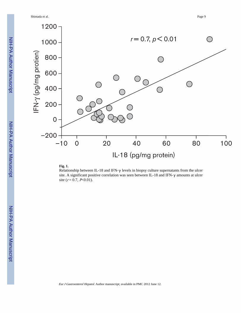
Fig. 1.Relationship between IL-18 and IFN-γ levels in biopsy culture supernatants from the ulcersite. A significant positive correlation was seen between IL-18 and IFN-γ amounts at ulcersite (r = 0.7, P<0.01).
Shimada et al. Page 9
Eur J Gastroenterol Hepatol. Author manuscript; available in PMC 2012 June 12.
NIH
-PA Author Manuscript
NIH
-PA Author Manuscript
NIH
-PA Author Manuscript

Fig. 2.Western blot analysis for IL-18 production from MKN45 cells cocultured with IFN-γ, H.pylori strain 26695, or both. IFN-γ: −, not pretreated; +, pretreated H. pylori: −, without H.pylori; +, with H. pylori.
Shimada et al. Page 10
Eur J Gastroenterol Hepatol. Author manuscript; available in PMC 2012 June 12.
NIH
-PA Author Manuscript
NIH
-PA Author Manuscript
NIH
-PA Author Manuscript

Fig. 3.Western blot analysis for IL-1β-converting enzyme (ICE) and caspase-3 production fromMKN45 cells cocultured with IFN-γ, H pylori strain 26695, or both. IFN-γ: −, notpretreated; +, pretreated H. pylori: −, without H. pylori; +, with H. pylori.
Shimada et al. Page 11
Eur J Gastroenterol Hepatol. Author manuscript; available in PMC 2012 June 12.
NIH
-PA Author Manuscript
NIH
-PA Author Manuscript
NIH
-PA Author Manuscript

Fig. 4.IL-18 levels in culture supernatants of MKN45 cells cocultured with IFN-γ, H pylori strain26695, or both. IFN-γ: −, not pretreated; +, pretreated H. pylori: −, without H. pylori; +,with H. pylori.
Shimada et al. Page 12
Eur J Gastroenterol Hepatol. Author manuscript; available in PMC 2012 June 12.
NIH
-PA Author Manuscript
NIH
-PA Author Manuscript
NIH
-PA Author Manuscript

Fig. 5.Cytotoxicity of MKN45 cells cocultured with IFN-γ, H. pylori strain 26695, or both. IFN-γ:−, not pretreated; +, pretreated H. pylori: −, without H. pylori; +, with H. pylori.
Shimada et al. Page 13
Eur J Gastroenterol Hepatol. Author manuscript; available in PMC 2012 June 12.
NIH
-PA Author Manuscript
NIH
-PA Author Manuscript
NIH
-PA Author Manuscript

NIH
-PA Author Manuscript
NIH
-PA Author Manuscript
NIH
-PA Author Manuscript
Shimada et al. Page 14
Table 1
Mucosal cytokine activities at gastric antrum of 20 FD patients according to H. pylori status
H. pylori status
Cytokine(pg/mg protein)
H. pylori-negative(n = 10)(median, range)
H. pylori-positive(n = 10)(median, range) P value
IL-18 (median, range) 2.5; 0.2–8.8 12.3; 2.7–29.5 P< 0.01
IFN-γ (median, range) 4.2; 0.1–19.1 7.2; 0.2–292.2 P< 0.01
sFasL (median, range) 11.8; 0.2–43.1 54.9; 8.2–318.5 P< 0.01
FD, functional dyspepsia; H. pylori, Helicobacter pylori; sFasL, soluble Fas ligand.
Eur J Gastroenterol Hepatol. Author manuscript; available in PMC 2012 June 12.

NIH
-PA Author Manuscript
NIH
-PA Author Manuscript
NIH
-PA Author Manuscript
Shimada et al. Page 15
Table 2
Mucosal cytokine activities of 27 GU patients according to the biopsy sites
Biopsy site
Cytokine(pg/mg protein)
Ulcer(median, range)
Antrum(median, range) P value
IL-18 (median, range) 22.4; 2.4–88.9 12.4; 0.8–41.5 P< 0.01
IFN-γ (median, range) 49.1; 0.5–1049.1 5.8; 0.7–377.4 P< 0.01
sFasL (median, range) 258.3; 3.5–1173.1 53.7; 2.1–508.7 P< 0.01
GU, gastric ulcer; sFasL, soluble Fas ligand.
Eur J Gastroenterol Hepatol. Author manuscript; available in PMC 2012 June 12.

NIH
-PA Author Manuscript
NIH
-PA Author Manuscript
NIH
-PA Author Manuscript
Shimada et al. Page 16
Table 3
The influence of H. pylori eradication on mucosal cytokine levels of eight GU patients
Before or after eradication therapy
Cytokine(pg/mg protein)
Before(median, range)
1 year after(median, range) P value
IL-18 (median, range) 32.5; 2.8–78.9 2.5; 0.1–11.2 P< 0.01
IFN-γ (median, range) 52.1; 1.5–998.5 3.9; 0.1–27.5 P< 0.01
sFasL (median, range) 275.7; 10.8–1058.2 13.2; 0.3–59.2 P< 0.01
GU, gastric ulcer; H. pylori, Helicobacter pylori; sFasL, soluble Fas ligand.
Eur J Gastroenterol Hepatol. Author manuscript; available in PMC 2012 June 12.
Copyright © 2022 FDOKUMEN