Characterization of the ArsRS Regulon of Helicobacter pylori, Involved in Acid Adaptation
15
10.1128/JB.188.10.3449-3462.2006. 2006, 188(10):3449. DOI: J. Bacteriol. Mollenkopf, Thomas F. Meyer and Dagmar Beier Michael Pflock, Nadja Finsterer, Biju Joseph, Hans Adaptation , Involved in Acid Helicobacter pylori Characterization of the ArsRS Regulon of http://jb.asm.org/content/188/10/3449 Updated information and services can be found at: These include: SUPPLEMENTAL MATERIAL Supplemental material REFERENCES http://jb.asm.org/content/188/10/3449#ref-list-1 at: This article cites 46 articles, 32 of which can be accessed free CONTENT ALERTS more» articles cite this article), Receive: RSS Feeds, eTOCs, free email alerts (when new http://journals.asm.org/site/misc/reprints.xhtml Information about commercial reprint orders: http://journals.asm.org/site/subscriptions/ To subscribe to to another ASM Journal go to: on February 22, 2014 by guest http://jb.asm.org/ Downloaded from on February 22, 2014 by guest http://jb.asm.org/ Downloaded from
-
Upload
independent -
Category
Documents
-
view
0 -
download
0
Transcript of Characterization of the ArsRS Regulon of Helicobacter pylori, Involved in Acid Adaptation

10.1128/JB.188.10.3449-3462.2006.
2006, 188(10):3449. DOI:J. Bacteriol. Mollenkopf, Thomas F. Meyer and Dagmar BeierMichael Pflock, Nadja Finsterer, Biju Joseph, Hans Adaptation
, Involved in AcidHelicobacter pyloriCharacterization of the ArsRS Regulon of
http://jb.asm.org/content/188/10/3449Updated information and services can be found at:
These include:
SUPPLEMENTAL MATERIAL Supplemental material
REFERENCEShttp://jb.asm.org/content/188/10/3449#ref-list-1at:
This article cites 46 articles, 32 of which can be accessed free
CONTENT ALERTS more»articles cite this article),
Receive: RSS Feeds, eTOCs, free email alerts (when new
http://journals.asm.org/site/misc/reprints.xhtmlInformation about commercial reprint orders: http://journals.asm.org/site/subscriptions/To subscribe to to another ASM Journal go to:
on February 22, 2014 by guest
http://jb.asm.org/
Dow
nloaded from
on February 22, 2014 by guest
http://jb.asm.org/
Dow
nloaded from

JOURNAL OF BACTERIOLOGY, May 2006, p. 3449–3462 Vol. 188, No. 100021-9193/06/$08.00�0 doi:10.1128/JB.188.10.3449–3462.2006Copyright © 2006, American Society for Microbiology. All Rights Reserved.
Characterization of the ArsRS Regulon of Helicobacter pylori,Involved in Acid Adaptation†
Michael Pflock,1‡ Nadja Finsterer,1‡ Biju Joseph,1 Hans Mollenkopf,2Thomas F. Meyer,3 and Dagmar Beier1*
Theodor-Boveri-Institut fur Biowissenschaften, Lehrstuhl fur Mikrobiologie, Universitat Wurzburg, Am Hubland,D-97074 Wurzburg,1 and Max-Planck-Institut fur Infektionsbiologie, Microarray Core Facility,2
Abteilung Molekulare Biologie,3 Schumannstrasse 21/22, D-10117 Berlin, Germany
Received 17 January 2006/Accepted 27 February 2006
The human gastric pathogen Helicobacter pylori is extremely well adapted to the highly acidic conditionsencountered in the stomach. The pronounced acid resistance of H. pylori relies mainly on the ammonia-producing enzyme urease; however, urease-independent mechanisms are likely to contribute to acid adapta-tion. Acid-responsive gene regulation is mediated at least in part by the ArsRS two-component systemconsisting of the essential OmpR-like response regulator ArsR and the nonessential cognate histidine kinaseArsS, whose autophosphorylation is triggered in response to low pH. In this study, by global transcriptionalprofiling of an ArsS-deficient H. pylori mutant grown at pH 5.0, we define the ArsR�P-dependent regulonconsisting of 109 genes, including the urease gene cluster, the genes encoding the aliphatic amidases AmiE andAmiF, and the rocF gene encoding arginase. We show that ArsR�P controls the acid-induced transcription ofamiE and amiF by binding to extended regions located upstream of the �10 box of the respective promoters.In contrast, transcription of rocF is repressed by ArsR�P at neutral, acidic, and mildly alkaline pH viahigh-affinity binding of the response regulator to a site overlapping the promoter of the rocF gene.
Helicobacter pylori is a human pathogen which is associatedwith gastric diseases like chronic active gastritis, peptic ulcer-ation, adenocarcinoma, and mucosa-associated lymphoid-tis-sue lymphoma (4, 25, 37). The neutralophilic bacterium, whichthrives in the mucus layer covering the gastric epithelium, isextremely well adapted to cope with the fluctuating low-pHconditions encountered in the human stomach and has evolvedmechanisms both to survive severe acid shocks and to grow atmoderately acidic pH. In accordance with the stomach beingits unique habitat, sequencing of the H. pylori genome revealeda very restricted repertoire of transcriptional regulators (34);however, it is becoming increasingly evident that a consider-able subset of these regulators is involved in the control of theacid response. In three independent studies performing ge-nome-wide transcriptional profiling, between 101 and about280 genes have been reported to be regulated in response tothe exposure of H. pylori to low pH (6, 21, 44). Up to now, twotranscriptional regulators, the metal-dependent regulatorsNikR and Fur and a two-component signal transduction systemtermed ArsRS, have been implicated in the transcriptionalregulation of acid-responsive genes (26, 40, 41). The ArsRSsystem consists of the histidine kinase ArsS, comprising aperiplasmic input domain of 111 amino acids and likely to beresponsible for low-pH sensing, and the OmpR-like responseregulator ArsR. Inactivation of the histidine kinase ArsS ren-ders H. pylori unable to colonize mice (24), while the response
regulator ArsR is essential for the in vitro growth of H. pylori,suggesting distinct functions for ArsR in its unphosphorylatedand phosphorylated states (2, 29).
A major player in the acid resistance of H. pylori is theurease system, which is essential for colonization (11, 14, 22,31, 36). By cleaving urea, the urease enzyme generates ammo-nia and carbon dioxide, the latter being subsequently con-verted to HCO3
� by a periplasmic carbonic anhydrase. Thesebuffering compounds capture protons that leak into the cyto-plasm and periplasm and thereby maintain the cytoplasmic andperiplasmic pH of the bacteria near neutrality (18, 33). Theenzymatic activity of urease is controlled by the inner mem-brane, pH-gated channel UreI, which regulates the access ofthe substrate urea to the bacterial cell in response to acidic pH(28, 43). Transcription of both the ureAB operon encoding theenzymatic subunits of urease and the ureIEFGH operon en-coding the channel protein UreI and accessory proteins whichare required for the incorporation of the metal cofactor Ni2�
into the urease apoenzyme is positively regulated in responseto low pH (6, 21, 27). This pH-responsive transcriptional in-duction is mediated by the ArsRS two-component system viathe binding of the phosphorylated response regulator ArsR(ArsR�P) to the promoters of the ureAB and ureIEFGH oper-ons (27). Several other target genes of the ArsRS two-compo-nent system encoding mostly H. pylori-specific proteins of un-known function have already been identified (10, 15) and arelikely to be involved in urease-independent mechanisms of acidadaptation. Interestingly, the rocF gene encoding arginase wasalso shown to be regulated by ArsRS (15). Arginase producesurea and ornithine by the cleavage of arginine, and it wassuggested that this enzyme might contribute to urease-depen-dent acid resistance when urea is scarce in the surroundingmedium (20). Transcription of the ureAB genes is also posi-
* Corresponding author. Mailing address: Theodor-Boveri-Institut furBiowissenschaften, Lehrstuhl fur Mikrobiologie, Universitat Wurzburg,Am Hubland, 97074 Wurzburg, Germany. Phone: 49-931-8884421. Fax:49-931-8884402. E-mail: [email protected].
‡ Both authors contributed equally to the present study.† Supplemental material for this article may be found at http://jb
.asm.org/.
3449
on February 22, 2014 by guest
http://jb.asm.org/
Dow
nloaded from

tively controlled in response to increasing concentrations ofNi2� ions by the pleiotropic, metal-dependent regulator NikR,which is also involved in the regulation of genes involved inNi2� uptake and storage, Fe3� uptake and storage, stress re-sponses, motility, and encoding outer membrane proteins (7,13, 38). The acid stimulon and the NikR regulon defined bytranscriptome analyses overlap to some extent, underlining therole of NikR in the process of acid adaptation (6, 7).
Besides that of urease, the expression of other ammonia-producing enzymes was demonstrated to be induced in re-sponse to acidic pH (6, 21, 44). Most notably, transcription ofthe genes amiE and amiF, which encode aliphatic amidases,was strongly increased upon exposure of H. pylori to low pH.Amidase genes which were known so far to be predominantlyassociated with environmental bacteria are present in severalstomach-colonizing Helicobacter species, like H. pylori, H.acinonychis, and H. felis, suggesting an important role for theenzymes in nitrogen metabolism and acid resistance for theseHelicobacter species. However, so far the substrates cleaved bythe amidases in vivo remain unknown (5, 32). Under iron-replete conditions, transcription of amiE has been shown to berepressed by Fur, which binds to the upstream region of amiE.To our knowledge, amiF transcription is not controlled by Fur(39). However, at low pH the activity of both amidases isconversely affected by the inactivation of Fur or the NikRprotein, suggesting a role for both regulators in the acid-re-sponsive regulation of the amidases (6, 40). Interestingly, Fur-deficient mutants are impaired in their acid tolerance, suggest-ing an important role for Fur in the acid response (3).
The aim of this study was to define the subset of acid-respon-sive genes which is transcriptionally regulated by the ArsRS two-component system. We show that at pH 5.0, compared to those ofthe wild type, 109 genes are differentially regulated in a mutant ofH. pylori G27 lacking the histidine kinase ArsS. Furthermore, wedemonstrate that acid-induced transcription of amiE and amiF ismediated directly by the ArsRS two-component system via bind-ing of ArsR�P to the promoter regions of the amidase genes.Unexpectedly, the rocF gene encoding arginase, whose transcrip-tion is repressed by ArsR�P, did not show a pH-responsive tran-scription profile under the applied experimental conditions.
MATERIALS AND METHODS
Bacterial strains and growth conditions. H. pylori strains G27 and 26695 areclinical isolates (34, 45). Strain H. pylori G27/HP165::km, lacking the histidinekinase ArsS, has been described previously (2). H. pylori strain G27/�arsS�nikR,carrying deletions of both the arsS and nikR genes, was constructed throughallelic replacement by transformation of strain G27/HP165::km with the suicideplasmid pNko::cm. pNko::cm is a derivative of pNko::km (27), in which thekanamycin resistance cassette was replaced by a chloramphenicol resistancecassette of Campylobacter coli (42). Chromosomal DNA of the transformants waschecked by PCR with primers flanking the integration site for the correct re-placement of the nikR gene. H. pylori strains were grown at 37°C under micro-aerophilic conditions on Columbia agar plates containing 5% horse blood, 0.2%cyclodextrin, and Dent’s or Skirrow’s antibiotic supplement. Liquid cultures weregrown in brain heart infusion (BHI) broth containing Dent�s or Skirrow�s anti-biotic supplement and 10% fetal calf serum. When required, blood agar plates orliquid broth were supplemented with kanamycin or chloramphenicol in a finalconcentration of 20 �g/ml. Acid exposure experiments were performed as fol-lows: bacteria from a liquid culture were harvested at an optical density at 550nm (OD550) of 0.7 by centrifugation and were resuspended in BHI broth con-taining fetal calf serum and an antibiotic supplement whose pH had been ad-justed to pH 5.0 with hydrochloric acid. Incubation at 37°C undermicroaerophilic conditions was continued for 1 h. To study gene expression in
bacteria grown under mildly alkaline conditions, cells from a logarithmic culture(OD550 � 0.7) were shifted to BHI broth whose pH had been adjusted to pH 8.0or 8.5 with sodium hydroxide and cultivation was continued for 1 h. Escherichiacoli DH5� was grown in Luria-Bertani broth. When necessary, antibiotics wereadded to the following final concentrations: ampicillin, 100 �g/ml; kanamycin, 50�g/ml; and chloramphenicol, 30 �g/ml.
RNA isolation. H. pylori RNA was isolated from bacteria grown to the logarithmicphase in liquid broth by using TRIzol reagent (Invitrogen) according to the manu-facturer’s protocol. RNA preparations intended to be used for global transcriptionalprofiling were further purified using the RNeasy mini kit (QIAGEN) according tothe manufacturer’s RNA-cleanup protocol. Residual DNA was removed by on-column digestion during RNA purification with QIAGEN RNase-free DNase(QIAGEN). The RNA concentration was quantified by determination of absor-bance at 260 and 280 nm, and RNA integrity was checked by visualization on a1.5% agarose gel.
Microarray hybridization and data analysis. Transcriptome analyses wereperformed using a whole-genome microarray containing 1,649 PCR productsgenerated with specific primer pairs derived from the genome sequences of H.pylori 26695 (34) and J99 (1), which were spotted in duplicate. Microarrays wereproduced as described previously (17). To determine genes which are differen-tially expressed in the ArsS-deficient mutant G27/HP165::km at pH 5.0, cDNAwas prepared from RNA extracted from H. pylori G27 and G27/HP165::km afterexposure of the bacteria to acidic pH for 1 h. A total of eight RNA samples fromtwo independent preparations of RNA from strains G27 and G27/HP165::kmwere used for cDNA labeling and hybridization. Equal amounts of RNA (20 �g)were used to synthesize differentially labeled cDNA (Cy3-dCTP and Cy5-dCTP;Amersham Biosciences) during first-strand reverse transcription reactions withSuperscript II reverse transcriptase and 6 �g of random primers (Invitrogen).Dye reversal color swaps were performed as follows: a cDNA sample usingCy3-dCTP and another using Cy5-dCTP were generated, resulting in four labeledcDNAs per color swap. Synthesized cDNAs were purified by using QiaQuick PCRpurification columns (QIAGEN) according to the manufacturer’s protocol. Cy5-dCTP- and Cy3-dCTP-labeled cDNAs were combined and concentrated by evap-oration in a SpeedVac at 45°C. Samples were diluted to 3 SSC–0.1% sodiumdodecyl sulfate (SDS) and hybridized to the H. pylori microarray (50°C, 18 h).The slides were washed for 5 min at room temperature with 0.1% SDS–0.1 SSCand then two times with 0.1 SSC. After being washed, the slides were scanned,using ScanArray HT, and analyzed by using ScanArray express software (PerkinElmer). Spots were flagged and eliminated from analysis when the signal-to-background ratio was less than three or in obvious instances of high backgroundor stray fluorescent signals. Median intensities of spots were background cor-rected, and differences in dye bias were normalized by using the LOWESSalgorithm (46). The signal ratios used as measures of differential expressionbetween the red and green channels were obtained from processed signal inten-sities. The ratios were further analyzed with Microsoft Excel (Microsoft) andSAM software for statistical significance (35). Only those genes which had atleast seven unflagged replicates were considered after significance analysis bySAM (Table 1). The parameters used for significance analysis are indicated inthe supplemental material (see Table S1 in the supplemental material).
To determine the significance of differential expression, RNA was isolatedfrom the H. pylori G27 wild type grown in BHI broth (pH 5.0) and 20 �g of thisRNA was labeled with either Cy3-dCTP or Cy5-dCTP. The two cDNA probesgenerated were hybridized onto the same slide, and the data were analyzed asmentioned above. Signal ratios of 0.5 and �2.0 were analyzed further.
Primer extension and RNA slot blot analysis. Primer extension analysis wasperformed essentially as described previously (26), using 0.5 pMol of �32P-end-labeled oligonucleotides amiE-PE, amiF-PE, rocF-PE, 682-PE, 1104-PE, 1174-PE, 1398-PE, and 1563-PE (Table 2) and 30 �g of RNA. Plasmids which wereused as template DNA in the sequencing reactions performed with the afore-mentioned primers were constructed as follows. EcoRI-BamHI fragments of 328bp and 517 bp were amplified from chromosomal DNA of H. pylori 26695 withprimer pairs PamiE-5/PamiE-3 and PamiF-5/PamiF-3, respectively, and wereligated into the cloning vector pSL1180 (Amersham Biosciences) to yield plas-mids pSL-PamiE and pSL-PamiF. Plasmid pSL-ProcF was obtained by cloning a365-bp EcoRI-BamHI fragment that was amplified with primer pair ProcF-5/ProcF-3 into pSL1180 vector DNA. Primer extension experiments were per-formed three times with independently prepared RNAs. Quantification of thesignals from the primer extension products was performed using a Typhoon 9200Variable Mode imager (Amersham Biosciences) and ImageMaster TotalLabsoftware (Amersham Biosciences). RNA slot blot analysis was performed asfollows: RNA (20 to100 �g) was denatured in 1 MOPS (morpholinepropane-sulfonic acid) buffer containing 50% formamide and 6% formaldehyde. The
3450 PFLOCK ET AL. J. BACTERIOL.
on February 22, 2014 by guest
http://jb.asm.org/
Dow
nloaded from

TABLE 1. ArsR�P-activated and -repressed genes
ArsR�P-regulated
genesaCategory ORF
HP no.bORF
JHP no.b�arsS/WT c
ratio Gene and functiond Genome organizatione
pH-responsivetranscriptionconfirmed by
indicatedreferencef
ActivatedAmmonia production HP0067 JHP0062 0.36 Urease accessory protein, ureH op HP0071–HP0067 21
HP0068 JHP0063 0.22 Urease accessory protein, ureG op HP0071–HP0067 21HP0069 JHP0064 0.39 Urease accessory protein, ureF op HP0071–HP0067 21HP0070 JHP0065 0.25 Urease accessory protein, ureE op HP0071–HP0067 44HP0071 JHP0066 0.21 Urea channel protein, ureI op HP0071–HP0067 21, 44HP0072 JHP0067 0.24 Urease subunit B, ureB op HP0073–HP0072 21HP0073 JHP0068 0.20 Urease subunit A, ureA op HP0073–HP0072 6, 21HP0294 JHP0279 0.06 Aliphatic amidase, amiE m 6, 21, 44HP1238 JHP1159 0.15 Formamidase, amiF m 6, 21
Amino acid biosynthesis HP0106 JHP0098 0.27 Cystathionine g-synthase, metB op HP0107–HP0105 ? 6, 21HP0380 JHP1001 0.38 Glutamate dehydrogenase, dghA m 21, 44
Biosynthesis of cofactors,prosthetic groups, andcarriers
HP0755 JHP0692 0.43 Molybdopterin biosynthesisprotein, moeB
op HP0755–HP0757 6, 21
HP0240 JHP0225 0.34 Octaprenyl-diphosphatesynthase, ispB
op HP0243–HP0236 ?
HP0306* JHP0291 0.42 Glutamate-1-semialdehyde2,1-aminomutase, hemL
op HP0305–HP0308 21, 44
HP0824 JHP0763 0.37 Thioredoxin, trxA op HP0824–HP0825 44HP0825 JHP0764 0.31 Thioredoxin reductase, trxB op HP0824–HP0825HP1224 JHP1145 0.39 Uroporphyrinogen III
cosynthase, hemDop HP1223–HP1225
Cell envelope HP1191 JHP1116 0.25 LPS heptosyltransferase, rfaF m 6HP1105 JHP1031 0.47 LPS biosynthesis protein mHP0078 JHP0073 0.19 Truncated outer membrane
protein, omp3op HP0078–HP0079 6, 44
HP0079 JHP0073 0.26 Outer membrane protein, omp3 op HP0078–HP0079 6, 44HP0492 JHP0444 0.30 Predicted lipoprotein m 6HP1456 JHP1349 0.48 Membrane-associated
lipoproteinop HP1457–HP1454 44
HP1564 JHP1472 0.36 Outer membrane lipoprotein mCellular processes HP0389 JHP0992 0.45 Superoxide dismutase, sodB op HP0389–HP0388
HP0390 JHP0991 0.18 Predicted thiol peroxidase mHP0875 JHP0809 0.43 Catalase, katA m 44HP1563 JHP1471 0.29 Alkyl hydroperoxide reductase,
ahpCm
HP1192 JHP1117 0.06 Secreted protein involved inflagellar motility
m 6, 44
HP0243 JHP0228 0.28 Neutrophil-activating protein,napA
op HP0243–HP0236 ? 44
DNA metabolism,restriction, andmodification
HP1022 JHP0402 0.30 Predicted 5�-3� exonuclease m 21, 44
Energy metabolism HP0890 JHP0823 0.47 Predicted short-chainoxidoreductase
op HP0890–HP0888
HP0924 JHP0858 0.44 4-oxalocrotonate tautomerase,dmpI
m
HP0954 JHP0888 0.45 NADPH nitroreductase op HP0961–HP0953 ?HP1104 JHP1030 0.34 Predicted mannitol
dehydrogeneasem 44
JHP0585* 0.27 Predicted 3-hydroxyaciddehydrogenase
op JHP0583–JHP0586 21
JHP1429 0.29 Predicted mannitoldehydrogenease
m
Fatty acid andphospholipidmetabolism
HP0891 JHP0824 0.41 Predicted acyl coenzyme Athioesterase
m 44
Central intermediarymetabolism
HP0757 JHP0649 0.41 Predicted carbon nitrogenhydrolase
op HP0755–HP0757 21
Protein fate HP1332* JHP1225 0.48 Cochaperone and heat shockprotein
op HP1332–HP1330 21
Transport and bindingproteins
HP0228 JHP0213 0.27 Predicted sulfate permease m 6, 21HP0889 JHP0822 0.40 Iron (III) dicitrate ABC
transporter, permeaseop HP0890–HP0888 44
HP1339 JHP1258 0.37 Biopolymer transport protein,exbB2
op HP1339–HP1341 21
HP1340* JHP1259 0.47 Biopolymer transport protein,exbD2
op HP1339–HP1341 21
HP1341 JHP1260 0.48 Siderophore-mediated irontransport protein, tonB2
op HP1339–HP1341
HP1331 JHP1251 0.47 Predicted branched-chain aminoacid transport protein
op HP1332–HP1330 21
Continued on following page
VOL. 188, 2006 CHARACTERIZATION OF THE ArsR�P REGULON 3451
on February 22, 2014 by guest
http://jb.asm.org/
Dow
nloaded from

TABLE 1—Continued
ArsR�P-regulated
genesaCategory ORF
HP no.bORF
JHP no.b�arsS/WTc
ratio Gene and functiond Genomeorganizatione
pH-responsivetranscriptionconfirmed by
indicatedreferencef
HP1466 JHP1359 0.44 Predicted ABC transport systempermease
op HP1466–HP1462 ? 6
HP1427 JHP1320 0.20 Histidine-rich protein, hpn mHP1432 JHP1321 0.07 Histidine-rich protein m 6, 21
Unknown HP1098 JHP1024 0.43 Cysteine-rich protein C, hcpC op HP1100–HP1097 ?JHP1437 0.16 Cysteine-rich protein m
HP1225 JHP1146 0.27 Predicted CrcB integralmembrane protein
op HP1223–HP1225 21, 44
Hypothetical HP0189 JHP0175 0.47 Hypothetical integral membraneprotein
m
HP1286 JHP1206 0.48 Conserved hypothetical secretedprotein
m
HP0081 0.04 Hypothetical protein mHP0118 JHP0110 0.19 Hypothetical protein m 6, 21HP0120 0.17 Hypothetical protein m 6HP0242* JHP0227 0.33 Hypothetical protein op HP0243–HP0236 ? 44HP0305 JHP0290 0.24 Hypothetical protein op HP0305–HP0308 ? 44HP0307* JHP0292 0.48 Hypothetical protein op HP0305–HP0308 ? 6HP0423 0.35 Hypothetical protein op HP0427–HP0423 ? 6HP0425 0.43 Hypothetical protein op HP0427–HP0423 ?HP0426 0.40 Hypothetical protein op HP0427–HP0423 ? 6HP0641 JHP0584 0.27 Hypothetical protein op HP640–HP0642 21HP0731 JHP0668 0.29 Hypothetical protein op HP0733–HP0731HP0733 JHP0670 0.47 Hypothetical protein op HP0733–HP0731HP0963 JHP0897 0.38 Hypothetical protein op HP0966–HP0962HP0964 JHP0898 0.37 Hypothetical protein op HP0966–HP0962
JHP0899HP1154 JHP1081 0.48 Hypothetical protein op HP1154–HP1155 21HP1187 JHP1113 0.28 Hypothetical protein m 21, 44HP1188 0.12 Hypothetical protein m 6, 21, 44HP1223 JHP1144 0.37 Hypothetical protein op HP1223–HP1225HP1408 JHP1300 0.20 Hypothetical protein op HP1408–HP1412 ?HP1412 JHP1307 0.32 Hypothetical protein op HP1408–HP1412 ? 44
JHP0959 0.32 Hypothetical protein m
RepressedCell envelope HP0229 JHP0214 2.51 Outer membrane protein,
omp6m 6, 21, 44
HP0722 JHP0659 3.87 Outer membrane protein,omp16
m 6, 21
HP0725 JHP0662 5.68 Outer membrane protein,omp17
m 6, 21
HP0788 JHP0725 2.64 Predicted outer membraneprotein, hofF
m
HP1083 JHP0342 2.79 Predicted outer membraneprotein, hofB
m
HP1167 JHP1094 5.33 Predicted outer membraneprotein, hofH
m 6, 21
Cellular processes HP1560* JHP1468 2.24 Cell division protein, ftsW m 6HP0017 JHP0015 2.22 virB4 homolog involved in
natural competence, comB4op HP0015–HP0017
HP1527 JHP1416 3.86 Protein involved in naturalcompetence, comH
m 6, 21
HP1585 JHP1492 2.09 Flagellar basal-body rod proteinDNA metabolism,
restriction, andmodification
HP1526 JHP1415 2.14 Exodeoxyribonuclease, lexA m
Energy metabolism HP1398 JHP1428 4.31 Alanine dehydrogenase, ald mHP1399 JHP1427 3.23 Arginase, rocF m
Protein fate HP0109* JHP0101 3.00 Chaperone and heat shockprotein 70, dnaK
op HP0111–HP0109 21
HP0110 JHP0102 2.86 Cochaperone and heat shockprotein, grpE
op HP0111–HP0109 21
HP0382* JHP0999 2.36 Predicted zinc-metalloprotease op HP0382–HP0381 21Purines, pyrimidines,
nucleosides, andnucleotides
HP1178 JHP1104 3.78 Purin nucleoside phosphorylase,deoD
op HP1180–HP1178 21
HP1179 JHP1105 3.86 Phosphopentamutase op HP1180–HP1178Transcription regulation HP0166 JHP0152 2.20 Two-component response
regulator, arsRop HP0166–HP0162 6
Continued on following page
3452 PFLOCK ET AL. J. BACTERIOL.
on February 22, 2014 by guest
http://jb.asm.org/
Dow
nloaded from

samples were incubated at 65°C for 5 min and cooled on ice before one volumeof 20 SSC was added. The denatured samples were filtered through a positivelycharged nylon membrane (Hybond N�, Amersham Biosciences), using a Bio-Dot chamber (Bio-Rad). After UV cross-linking was performed, the nylon mem-
brane was prehybridized for 1 h at 42°C in hybridization buffer (ECL goldhybridization buffer; Amersham Biosciences). The PCR products used as hybrid-ization probes were amplified with primer pairs 229-5/229-3, 725-5/725-3, 686-5/686-3, and 16S-5/16S-3 and were labeled nonradioactively, using the ECL Direct
TABLE 1—Continued
RArsR�P-regulated
genesaCategory ORF
HP no.bORF
JHP no.b�arsS/WTc
ratio Gene and functiond Genome organizatione
pH-responsivetranscriptionconfirmed by
indicatedreferencef
Transport and bindingproteins
HP0686 JHP0626 4.06 Iron(III) dicitrate transportprotein, fecA1
m
HP1129 JHP1057 2.81 Biopolymer transport protein,exbD1
op HP1137–HP1123 ?
HP1130 JHP1058 3.19 Biopolymer transport protein,exbB1
op HP1137–HP1123 ?
HP1174 JHP1101 5.65 Glucose/galactose transporter mHP1180 JHP1106 4.17 Pyrimidine nucleoside transport
proteinop HP1180–HP1178 6, 21
Hypothetical HP1175 JHP1102 2.12 Hypothetical integral membraneprotein
m 6, 21
HP0681 JHP0622 10.40 Hypothetical protein op HP0682–HP0681HP0682 JHP0623 16.56 Hypothetical protein op HP0682–HP0681HP0688* JHP0628 2.65 Hypothetical protein op HP0688–HP0689HP0689 JHP0628 2.02 Hypothetical protein op HP0688–HP0689HP0709 JHP0648 3.97 Hypothetical protein m 6HP0947 JHP0881 3.74 Hypothetical protein mHP1288* JHP1208 4.48 Hypothetical protein op HP1288–HP1289 6HP1289 JHP1209 22.77 Hypothetical protein op HP1288–HP1289HP1322 JHP1242 2.13 Hypothetical protein m 21
a Genes listed are those whose transcription at pH 5.0 differed more than twofold (ratio, �2.0 or 0.5) for the ArsS-deficient mutant H. pylori G27/HP165::kmcompared to that for the G27 wild type, according to microarray analysis.
b ORF numbers and the prediction of transcriptional units are based on the genome sequences of H. pylori 26695 and J99 (1, 34). Asterisks indicate ORFs whichwere determined by the SAM algorithm to be not statistically significant (see Table S1 in the supplemental material) but which were retained for data evaluation becausethey are part of a predicted operon structure with other differentially expressed genes or because their pH-responsive transcription was confirmed in another study.
c WT, wild type.d The functional annotation is that used by the PyloriGene database (http://www.pasteur.fr/english.html).e m, monocistronically transcribed genes; op, putative transcriptional unit. Question marks indicate that the proposed operon structures cannot be unambiguously
deduced from the genome sequences.f Where a reference(s) is given, pH-responsive transcription was confirmed by the studies of Merrell et al. (21), Wen et al. (44), or Bury-Mone et al. (6).
TABLE 2. Oligonucleotides used in this study
Oligonucleotideor primer Sequence (5� to 3�)a Siteb Orientation
of strand Positionc
PamiE-5 gattttgaattcTATTGTATTAACGCGCTATATGG EcoRI � 310783–310805PamiE-3 attttcggatccTCATTCTTAGTGTGGAGTCTAGG BamHI � 311089–311111PamiF-5 caagctgaattcTGGTCATCATGGGAGCAACC EcoRI � 1311723–1311742SamiF-5 ccagcagaattcAGAAAGTAGCCCAGGTCCTAA EcoRI � 1311918–1311938PamiF-3 aatatcggatccGCTATTGACAATTGGCACAGG BamHI � 1312219–1312239ProcF-5 atccttgaattcGATTAGTGCCACATCATCAGG EcoRI � 1459647–1459667ProcF-3 agccatggatccGCTTAAAGCCTCTCTCAAACG BamHI � 1459992–1460012amiE-PE TAGGCATCTTATAATTAACTACCGC � 311068–311092amiF-PE AGGAAACTGAATGGCTGCC � 1312203–1312221rocF-PE GCTTAAAGCCTCTCTCAAACG � 1459992–1460012682-PE ATCCACTCATAACAAATC � 732444–7324611104-PE TAGCAAAACCTTTAGATTGAAC � 1165334–11653551174-PE GAATAGCGCTGTCAAACTCC � 1241646–12416651398-PE CTCGGGATTCTAAATCCATGC � 1459679–14596991563-PE GGCAGGTGCTTTAAAGTCTGG � 1645245–1645265229-5 ATGAAAAAAACGATTTTACTT � 240139–240159229-3 TTGATTAAGGTTTTTATTGAA � 239440–239460725-5 AAGCAAAGCATTCAAAACGCC � 780757–780777725-3 GATGTCTTTAGCGAATTTAGG � 780058–780078686-5 ATGAAAAGAATTTTAGTCTCT � 737126–737146686-3 GTTGTATCTAAACCCTTGCCC � 736427–73644716S-5 GCTAAGAGATCAGCCTATGTCC � 1208855–120887616S-3 TGGCAATCAGCGTCAGGTAATG � 1208356–1208377
a Sequences in uppercase letters are derived from the genome sequences of H. pylori 26695 (34). Sequences introduced for cloning purposes are given in lowercaseletters, and restriction recognition sequences are underlined.
b Restriction recognition sites.c Nucleotide positions refer to the genome sequence of H. pylori 26695 (34).
VOL. 188, 2006 CHARACTERIZATION OF THE ArsR�P REGULON 3453
on February 22, 2014 by guest
http://jb.asm.org/
Dow
nloaded from
nucleic acid-labeling system (Amersham Biosciences) according to the manufac-turer’s instructions.
The labeled probes were added to the hybridization solution, and hybridiza-tion was performed for 12 to 16 h at 42°C. The membrane was washed two timesin prewarmed (42°C) wash solution I (6 M urea, 0.5 SSC, 0.4% SDS) for 20 minat 42°C and two times in wash solution II (2 SSC) at room temperature. Forsignal detection, the ECL detection system (ECL Direct Nucleic Acid Labelingand Detection system; Amersham Biosciences) and X-ray films (Konica Minolta)were used.
DNase I footprint analysis. The recombinant N-terminally His6-tagged re-sponse regulator ArsR encoded on plasmid pQE-166 was overexpressed in E.coli M15[pREP4] (QIAGEN) and was purified by affinity chromatography onNi2�-nitrilotriacetic acid agarose essentially as described previously (2). In vitrophosphorylation of His6-ArsR was performed as described by Dietz et al. (10).Plasmids pSL-PamiE, pSL-PSamiF, and pSL-ProcF were used for the generation ofend-labeled DNA probes for DNase I footprint experiments. pSL-PSamiF wascreated by ligating a 318-bp EcoRI-BamHI fragment derived from the upstreamregion of the amiF gene, which was amplified with primer pair SamiF-5/PamiF-3into pSL1180. The promoter DNA fragments were 5�-end labeled with [�-32P]ATP and T4 polynucleotide kinase at one extremity and gel purified, and ap-proximately 100,000 cpm of each probe was used for footprint experiments,which were performed essentially as described by Delany et al. (8). The bindingreactions were performed for 20 min at room temperature in 50 �l binding buffer(50 mM Tris-HCl [pH 7.9], 40 mM KCl, 10 mM NaCl, 2 mM MgCl2, 0.1 mMCaCl2, 1 mM dithiothreitol).
Microarray accession numbers. The microarray raw data were deposited inthe NCBI Gene Expression Omnibus database (http://www.ncbi.nlm.nih.gov/geo) and are accessible with the accession number GSE4293.
RESULTS
Whole-genome transcriptional profiling of an ArsS-deficientmutant of H. pylori G27 exposed to pH 5.0. Low pH has beendemonstrated to be a signal triggering the autophosphorylationof the histidine kinase ArsS and the subsequent phosphoryla-tion of the cognate response regulator ArsR (26). In order todefine the complete regulon controlled by ArsR�P, a tran-scriptome analysis was performed using a whole-genome mi-croarray containing 1,649 PCR products generated with spe-cific primer pairs derived from the genome sequences of H.pylori 26695 (34) and J99 (1) and comprising 98% of the codingsequences present in both genomes (17). RNA was extractedfrom two independent cultures of H. pylori G27 andG27/HP165::km, grown to an OD550 of 0.7, and then exposedto pH 5.0 for one hour. Cy5- and Cy3-labeled cDNA wasprepared from these RNAs, and the differentially labeledcDNA pairs derived from the H. pylori wild type and arsSmutant were hybridized to four independent microarray slides,creating eight sets of hybridization data. A total of 109 geneswas identified to be differentially expressed in the arsS mutantat pH 5.0 by using a signal ratio cut off of 0.5 and �2.0 (Table1). Seventy-five genes were positively regulated by ArsR�P,while the transcription of 34 genes was repressed by ArsR�P.In accordance with our previous observation that the promot-ers of both ureA(PureA) and ureI(PureI) are positively regulatedby ArsR�P (26, 27), transcription of the complete urease genecluster was reduced in the arsS mutant, as well as transcriptionof amiE and amiF, which encode additional ammonia-produc-ing enzymes. Transcription of rocF, which encodes arginase,was found to be repressed by ArsR�P. Furthermore, globaltranscriptional profiling confirmed that ArsR�P acts as a neg-ative autoregulator (10). Transcription of open reading frames(ORFs) HP0165 to HP0162 was reduced in the arsS mutant,due to the insertion of the kanamycin resistance cassette intoORF HP0165/HP0164 and its polar effect on expression of the
downstream genes encoding -aminolevulinic acid dehydratase(HP0163) and a conserved hypothetical protein of unknown func-tion (HP0162). The ArsR�P-regulated genes were grouped intoseveral categories according to their cellular functions and encodemainly proteins affecting the composition of the cell envelope,transport and binding proteins, detoxifying enzymes, and H.pylori-specific proteins of unknown function (Table 1).
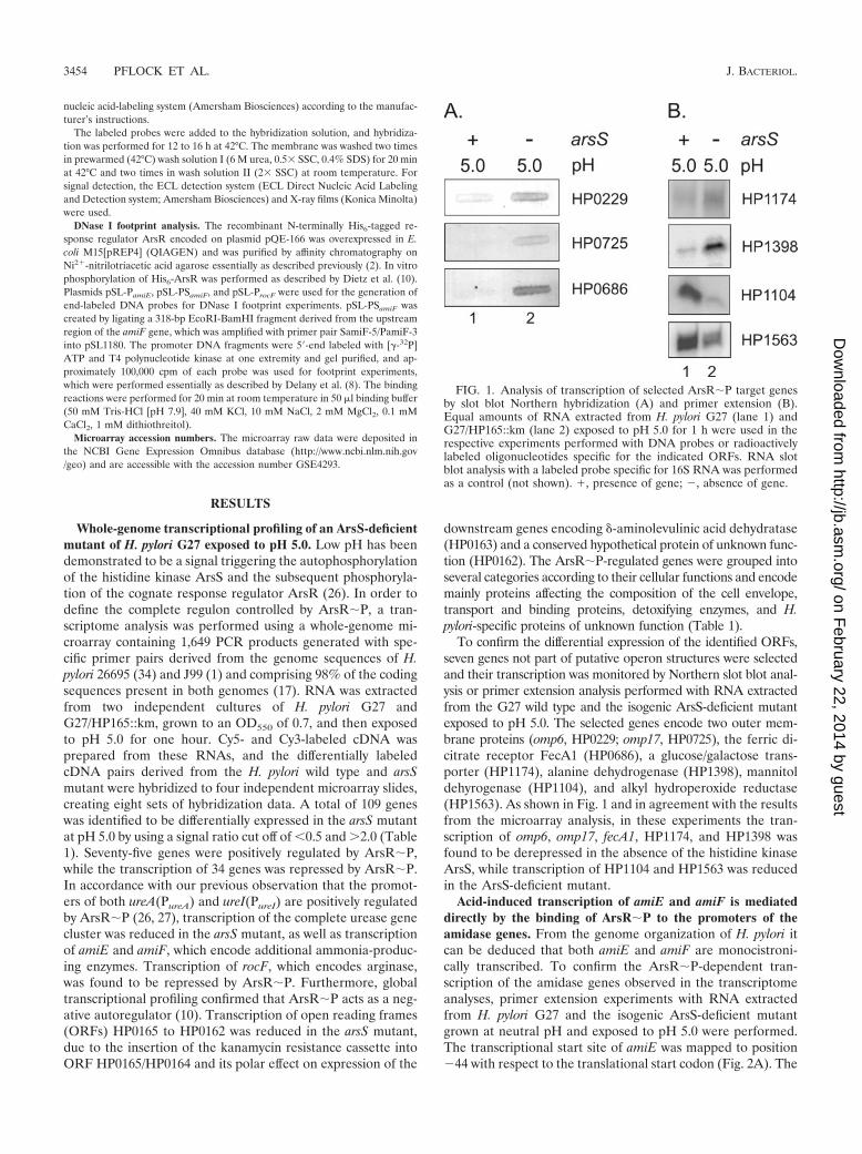
To confirm the differential expression of the identified ORFs,seven genes not part of putative operon structures were selectedand their transcription was monitored by Northern slot blot anal-ysis or primer extension analysis performed with RNA extractedfrom the G27 wild type and the isogenic ArsS-deficient mutantexposed to pH 5.0. The selected genes encode two outer mem-brane proteins (omp6, HP0229; omp17, HP0725), the ferric di-citrate receptor FecA1 (HP0686), a glucose/galactose trans-porter (HP1174), alanine dehydrogenase (HP1398), mannitoldehyrogenase (HP1104), and alkyl hydroperoxide reductase(HP1563). As shown in Fig. 1 and in agreement with the resultsfrom the microarray analysis, in these experiments the tran-scription of omp6, omp17, fecA1, HP1174, and HP1398 wasfound to be derepressed in the absence of the histidine kinaseArsS, while transcription of HP1104 and HP1563 was reducedin the ArsS-deficient mutant.
Acid-induced transcription of amiE and amiF is mediateddirectly by the binding of ArsR�P to the promoters of theamidase genes. From the genome organization of H. pylori itcan be deduced that both amiE and amiF are monocistroni-cally transcribed. To confirm the ArsR�P-dependent tran-scription of the amidase genes observed in the transcriptomeanalyses, primer extension experiments with RNA extractedfrom H. pylori G27 and the isogenic ArsS-deficient mutantgrown at neutral pH and exposed to pH 5.0 were performed.The transcriptional start site of amiE was mapped to position�44 with respect to the translational start codon (Fig. 2A). The
FIG. 1. Analysis of transcription of selected ArsR�P target genesby slot blot Northern hybridization (A) and primer extension (B).Equal amounts of RNA extracted from H. pylori G27 (lane 1) andG27/HP165::km (lane 2) exposed to pH 5.0 for 1 h were used in therespective experiments performed with DNA probes or radioactivelylabeled oligonucleotides specific for the indicated ORFs. RNA slotblot analysis with a labeled probe specific for 16S RNA was performedas a control (not shown). �, presence of gene; �, absence of gene.
3454 PFLOCK ET AL. J. BACTERIOL.
on February 22, 2014 by guest
http://jb.asm.org/
Dow
nloaded from

upstream sequence revealed a box (TATAAA) at �10 with ahigh similarity to the E. coli promoter consensus sequence.The 5� end of the amiF-specific transcript was mapped toposition �48 with respect to the translational start codon,which corresponds to a �10 promoter element of the se-quence TAGTAT (Fig. 2B), showing two mismatches from theconsensus �10 promoter element from E. coli. In the G27wild-type strain, transcription of both amiE and amiF wasstrongly induced at pH 5.0. At neutral pH, the detectedamount of amiE-specific transcript was approximately fivefoldlower in the ArsS-deficient mutant than in the G27 wild-typestrain. Compared to the low basal level of ArsR�P-indepen-dent expression detected in the mutant grown at pH 7.0, onlya slight increase of amiE transcription was observed uponshifting the bacteria to pH 5.0 (Fig. 2A). No transcription ofamiF was detected in the arsS mutant, irrespective of the pH ofthe growth medium (Fig. 2B). These data demonstrate that thepH-responsive transcription of the amidase genes is mediatedmainly by the ArsRS two-component system. Since Fur wasshown to act as a repressor of amiE expression (39), we inves-tigated whether the acid-induced increase in amiE transcrip-tion in the ArsS-deficient mutant was due to the negativeregulatory effect exerted by the NikR protein on fur transcrip-tion (7, 9, 40) by analyzing the amount of amiE-specific mRNAin an arsS nikR double mutant. As shown in Fig. 2A (comparelanes 3 and 5) transcription of amiE was similar in the ArsS-
deficient mutant grown at pH 7.0 and the arsS nikR doublemutant exposed to pH 5.0, suggesting a minor effect of theabovementioned repressor cascade on the acid-responsive reg-ulation of amiE expression.
Since besides arsR no genes encoding transcriptional regu-lators belong to the ArsR�P regulon, it is likely that transcrip-tion of its constituents is controlled directly by the binding ofArsR�P to the respective promoter regions. To define thebinding sites of ArsR�P in the promoter regions of amiE(PamiE) and amiF(PamiF), DNase I footprint analysis with thepurified response regulator protein ArsR was performed. Fig-ure 3 shows the results of a footprint experiment carried out ona 328-bp radioactively labeled DNA fragment derived from theupstream region of amiE. In the presence of 3 �M ArsR whichwas phosphorylated in vitro with acetylphosphate (ArsR�P), amaximum region of 56 bp located immediately upstream of the�10 box of the PamiE promoter and ranging from position �14to �69 with respect to the transcriptional start site of amiE wasprotected from DNase I digestion with a band of enhancedDNase I sensitivity appearing at position �21. In contrast, nofootprinting could be detected when unphosphorylated ArsRprotein was included in the binding reaction (data not shown).
The results of footprint experiments carried out on a 318-bpDNA probe containing the PamiF promoter are presented inFig. 4. Upon addition of in vitro phosphorylated ArsR at aconcentration of 3 �M, a maximum region of 38 bp ranging
FIG. 2. Analysis of transcription of the amidase genes in H. pylori G27 and the isogenic ArsS-deficient mutant G27/HP165::km grown at neutralpH and exposed to pH 5.0. (A) Primer extension experiments using the radiolabeled oligonucleotide amiE-PE were performed on equal amountsof RNAs extracted from H. pylori G27 (lanes 1 and 2) and G27/HP165::km (lanes 3 and 4) that were grown at neutral pH (lanes 1 and 3) orincubated at pH 5.0 for 1 h (lanes 2 and 4). In addition, RNA extracted from strain G27/�arsS�nikR exposed to pH 5.0 (lane 5) was analyzed.(B) Primer extension analysis using the radiolabeled oligonucleotide amiF-PE was performed on RNAs prepared from strains G27 (lanes 1 and2) and G27/HP165::km (lanes 3 and 4) grown at neutral pH (lanes 1 and 3) or exposed to pH 5.0 (lanes 2 and 4). The respective cDNAs areindicated by an arrow on the right. The sequences of the �10 element of the PamiE and PamiF promoter are given on the left. The sequencing ladders(lanes T, A, G, C) were obtained by annealing primers amiE-PE and amiF-PE to plasmids pSL-PamiE and pSL-PamiF, respectively. �, presence ofgene; �, absence of gene.
VOL. 188, 2006 CHARACTERIZATION OF THE ArsR�P REGULON 3455
on February 22, 2014 by guest
http://jb.asm.org/
Dow
nloaded from

from position �13 to �50 with respect to the transcriptionalstart site of amiF was protected from DNase I digestion. Asalready observed for the PamiE promoter, no binding to theupstream region of amiF could be detected when the footprintexperiment was performed with unphosphorylated ArsR pro-tein (data not shown). Therefore, we conclude that acid-responsive transcription of the amidase genes is regulated bythe ArsRS two-component system via the direct binding ofArsR�P to extended regions overlapping the �35 region ofthe PamiE and PamiF promoters.
ArsR�P-dependent repression of rocF and HP0682 is notresponsive to low pH. Global transcriptional profiling revealedthat at pH 5.0 transcription of the arginase gene rocF is dere-pressed in the ArsS-deficient mutant of H. pylori G27. How-ever, in none of the previous transcriptome studies analyzingpH-responsive gene regulation in H. pylori (6, 21, 44) wasexpression of rocF reported to be repressed at low pH. Tofurther investigate these conflicting observations, transcriptionof rocF was analyzed by primer extension experiments carriedout on RNA extracted from H. pylori G27 and the isogenicArsS-deficient mutant grown at neutral pH and exposed to pH5.0. As shown in Fig. 5A, the 5� end of the rocF-specific mRNA
was mapped to position �29 with respect to the translationalstart site of the rocF gene, corresponding to a �10 promoterhexamer of the sequence TAGAAT. An almost equal amountof rocF-specific transcript was detected in the G27 wild typeirrespective of whether the bacteria were cultivated at pH 7.0or exposed to pH 5.0. In the arsS mutant, transcription of rocFwas strongly derepressed at both growth conditions; however,an approximately threefold-higher amount of rocF-specifictranscript was detected at pH 5.0. This result demonstratedthat the promoter of the rocF gene (ProcF) is repressed byArsR�P; however, growth of H. pylori G27 at neutral or acidicpH, supposed to cause alterations of the cellular level of phos-phorylated ArsR, did not result in differential transcription ofrocF. To test whether cultivation of H. pylori at mildly basic pHmight relieve the ArsR�P-dependent repression of ProcF, tran-scription of the rocF gene was analyzed by primer extensionperformed on RNA extracted from strain G27, which wasexposed to pH 8.0 or pH 8.5 for 1 h. It has been reportedpreviously that a pH of 8.0 still allows growth of H. pylori in theabsence of urea (30). No significant change in the amount ofrocF-specific transcripts was observed under these conditions
FIG. 3. Binding of ArsR�P to the PamiE promoter. (A) DNase I footprint experiments were performed on a 328-bp EcoRI-BamHIfragment containing the PamiE promoter derived from plasmid pSL-PamiE, which was end labeled at the EcoRI terminus by adding increasingamounts of His6-ArsR phosphorylated in vitro by acetylphosphate. In lanes 2 to 6, His6-ArsR is present in concentrations of 0 (lane 2), 0.37 (lane3), 0.75 (lane 4), 3.0 (lane 5), and 4.5 �M (lane 6). The numbers on the left indicate nucleotide positions with respect to the transcriptional startsite, which is marked by an arrow. The solid bar on the right indicates the region of maximum DNase I protection. The arrow on the right indicatesa band of hypersensitivity to DNase I digestion. Lane 1 contained a G�A sequence reaction mixture with the DNA probe used as a size marker(19). (B) Schematic representation of the PamiE promoter. The �10 promoter element is highlighted by black shading, and the transcriptional startsite is indicated by an arrow above the double-stranded sequence. The gray box indicates the region with maximum protection from DNase Idigestion by ArsR�P binding to the PamiE promoter probe labeled at the EcoRI-terminus. The black bar below the sequence indicates theminimum size of the ArsR�P binding site protected from DNaseI digestion. Numbers above the sequence indicate the nucleotide position withrespect to the transcriptional start site (�1, not shown). The translational start codon of the amiE gene is shown in italics.
3456 PFLOCK ET AL. J. BACTERIOL.
on February 22, 2014 by guest
http://jb.asm.org/
Dow
nloaded from

compared to that of strain G27 grown at neutral pH (data notshown).
In our transcriptome analysis, the paralogous ORFsHP0682 and HP1289, which encode H. pylori-specific pro-teins of unknown function, showed the highest ratio ofArsR�P-dependent repression. Both genes are organized inbicistronic transcriptional units with the ORFs HP0681 andHP1288, respectively, and had been previously shown to benegatively regulated by ArsR�P (15). However, as is the casewith rocF, pH-dependent repression of these genes has notbeen noted in previous studies (6, 21, 44). The transcriptionalstart site of ORF HP0682 was mapped by Forsyth et al. (15) toposition �6 with respect to the annotated translational startcodon, corresponding to a TATAAA �10-box hexamer. Whenthe transcription of ORF HP0682 was compared in the G27wild-type strain and the ArsS-deficient mutant grown atneutral pH or exposed to pH 5.0, respectively, the sameexpression profile as observed for the rocF gene was de-tected (Fig. 5B). Similarly, no significant change in theamount of HP0682-specific transcript was detected when the
bacteria were shifted from pH 7.0 to pH 8.0 or pH 8.5 (datanot shown). Therefore, we conclude that the transcription ofa subset of genes which are clearly regulated by ArsR�P isnot responsive to pH changes in a range from pH 5.0 to 8.5.
ArsR�P binds to a high-affinity binding site overlapping theProcF promoter. To investigate whether transcription of rocF isregulated directly by ArsR�P, DNase I footprint experimentswere carried out on a 365-bp DNA probe comprising theupstream region of the rocF gene (Fig. 6). In the presence of0.37 to 0.75 �M ArsR which was phosphorylated in vitro withacetylphosphate, a region protected from DNase I digestion,ranging from position �6 to �67 with respect to the transcrip-tional start site and overlapping the �10 promoter box, be-came visible. When the binding reactions were performed withunphosphorylated ArsR protein, footprinting of this regioncould be observed at a protein concentration of 4.5 �M (datanot shown). Therefore, the results of the footprint experimentsdemonstrate that transcription of the rocF gene is repressed byArsR�P, which binds with high affinity to a region overlappingthe �10 box of the ProcF promoter.
FIG. 4. Binding of ArsR�P to the PamiF promoter. (A) DNase I footprint experiments were performed on a 318-bp EcoRI-BamHI fragmentcontaining the PamiF promoter, which was end labeled at the EcoRI terminus by adding increasing amounts of His6-ArsR phosphorylated in vitroby acetylphosphate. In lanes 2 to 7, His6-ArsR is present in concentrations of 0 (lane 2), 0.37 (lane 3), 0.75 (lane 4), 1.5 (lane 5), 3.0 (lane 6), and4.5 �M (lane 7). The numbers on the left indicate nucleotide positions with respect to the transcriptional start site, which is marked by an arrow.The solid bar on the right indicates the maximum region of DNase I protection. Lane 1 contained a G�A sequence reaction mixture with the DNAprobe used as a size marker (19). (B) Schematic representation of the PamiF promoter. The �10 promoter element is highlighted by black shading,and the transcriptional start site is indicated by an arrow above the double-stranded sequence. The gray box indicates the region with maximumprotection from DNase I digestion by response regulator binding to the PamiF promoter probe labeled at the EcoRI terminus. The black bar belowthe sequence indicates the minimum size of the ArsR�P binding site protected from DNase I digestion. Numbers above the sequence indicate thenucleotide position with respect to the transcriptional start site (�1, not shown). The translational start codon of the amiF gene is given in italics.
VOL. 188, 2006 CHARACTERIZATION OF THE ArsR�P REGULON 3457
on February 22, 2014 by guest
http://jb.asm.org/
Dow
nloaded from

DISCUSSION
H. pylori is a highly specialized bacterium which, to ourknowledge, colonizes the human stomach as its unique habitatand, therefore, is endowed with highly efficient acid acclima-tion mechanisms distinguishing it from other neutralophilicbacteria. Due to the continuous alteration between starvationand digestive phases following food intake, H. pylori is likely toencounter considerable pH fluctuations within its niche, posingthe necessity of efficient modulation of the pH-adaptive re-sponse. Three independent global transcriptome studies havebeen performed so far in order to completely define the pH-responsive stimulon comprising the genes involved in acid ac-climation. However, there was only a rather low overlap amongthe data raised in the individual studies, reflected by a total of429 genes reported to be differentially expressed upon acidexposure added from the three individual data sets comprisingbetween 101 and 279 genes (6, 21, 44). This discrepancy mightbe explained by differences in the experimental setups, includ-ing the particular H. pylori strains analyzed. The ArsRS two-component system has been identified previously as an im-portant regulator of the acid response, mediating the low-pHinduction of urease expression upon phosphorylation ofthe response regulator ArsR (26, 27). Therefore, we intendedto characterize the acid-responsive regulon controlled byArsR�P by transcriptional profiling of H. pylori G27 and anisogenic mutant lacking the histidine kinase gene arsS. Thelevel of transcription of 109 genes differed more than twofoldbetween the two strains, with 75 genes being positively regu-lated by ArsR�P and 34 genes being repressed by ArsR�P.Acid-responsive regulation of 64 of these genes (48 induced, 16repressed) has previously been reported on the basis of globaltranscriptome analysis (6, 21, 44). From the 109 genes sup-
posed to be regulated by ArsR�P, 45 are predicted to bemonocistronically transcribed (pH regulation of 25 of thesegenes was confirmed in the previous array studies) according tothe genome sequences of H. pylori 26695 and J99 (1, 34), whilethe remaining genes are members of 33 predicted transcrip-tional units. In 10 of these transcriptional units, transcriptionof all the members of the respective operon was found to bealtered in the ArsS-deficient mutant, and in 5 predicted oper-ons comprising three or more genes, all but one of their mem-bers were found to be differentially regulated. Most convinc-ingly, in accordance with our previous observation that thepromoters located upstream of ureAB and ureIEFGH are con-trolled by ArsR�P (26, 27), in our microarray analysis tran-scription of the complete urease gene cluster was found to bealtered, which was not observed in previous global transcrip-tome studies (6, 21, 44). Differential expression of 11 genesidentified in the microarray analysis was confirmed by North-ern and primer extension analysis. pH-responsive or ArsR�P-dependent regulation of four of these genes had not beenreported so far. The highest ratio of transcriptional inductionby ArsR�P showed ORFs HP0081 (25-fold) and HP0079 (17-fold), encoding a hypothetical protein and a secreted proteinsupposed to be involved in flagellar motility, respectively, andthe amidase gene amiE (16-fold) and ORF HP1432 (15-fold),encoding a histidine-rich protein predicted to be involved innickel binding. amiF and ORF HP1427, encoding another his-tidine-rich protein, exhibited an approximately threefold-lowertranscriptional induction than their respective paralogous genes.The highest ratio of ArsR�P-dependent repression was ob-served with ORFs HP0682 and HP1289 (16- and 22-fold, re-spectively), encoding hypothetical proteins of unknown func-tion (see below). Induction of amidase expression in response
FIG. 5. Analysis of transcription of rocF and ORF HP0682 in H. pylori G27 and G27/HP165::km. (A) Primer extension experiments using theradiolabeled oligonucleotide rocF-PE were performed on RNAs extracted from the wild-type strain G27 grown at pH 7.0 and pH 5.0 (lanes 1 and2) and the ArsS-deficient mutant G27/HP165::km grown at neutral pH and pH 5.0 (lanes 3 and 4). The elongated primer products are indicatedby an arrow on the right. The sequences of the �10 element of the ProcF promoter is given on the left. The sequencing ladder (lanes T, A, G, C)was obtained by annealing primer rocF-PE to plasmid pSL-ProcF. (B) Primer extension experiments using the radiolabeled oligonucleotide 682-PEwere performed on RNAs extracted from strains G27 (lanes 1 and 2) and G27/HP165::km (lanes 3 and 4) grown at pH 7.0 (lanes 1 and 3) andexposed to pH 5.0 for 1 h (lanes 2 and 4). The cDNAs specific for ORF hp682 are indicated by an arrow on the right. �, presence of gene; �,absence of gene.
3458 PFLOCK ET AL. J. BACTERIOL.
on February 22, 2014 by guest
http://jb.asm.org/
Dow
nloaded from

to low pH has been observed previously (6, 21, 40, 44) andsuggests an important role for these ammonia-producing en-zymes in the acid adaptation of H. pylori. However, the ami-dase genes are not essential for colonization in the mousemodel of H. pylori infection (5). Here we demonstrate thatexpression of the three major ammonia-producing pathways—urease, AmiE, and AmiF—is under the control of the ArsRStwo-component system. Interestingly, besides the nickel-bindingproteins HP1432 and HP1427, expression of a number of detox-ifying enzymes (thiol peroxidase, alkyl hydroperoxide reductase,catalase, and superoxide dismutase) was induced by ArsR�P.This might reflect protection against the toxic effects caused byhigh intracellular concentrations of the bivalent metal ions Ni2�
and Fe2�, whose solubility and therefore bioavailability increaseunder acidic conditions. In particular, Fe2� is known to beinvolved in the triggering of radical reactions whose productsneed to be detoxified. In this context, it should be emphasizedthat low pH was shown to induce the expression of NikR,which is a repressor of fur transcription (9, 40). As a conse-quence, the amount of Fur protein present under acidic con-
ditions might be too low to allow efficient repression of the ironuptake systems, increasing the necessity of protection againstthe toxic effects of Fe2�. ArsR�P is also involved in changingthe composition of the outer membrane in response to low pHby regulating the expression of several outer membrane pro-teins and of enzymes involved in lipopolysaccharide (LPS)biosynthesis. Although an intriguing speculation, the pI ofthese outer membrane proteins and therefore their bufferingcapacity seem not to be correlated with their predominantexpression at neutral or acidic pH. Changes in LPS composi-tion upon exposure of H. pylori to acidic conditions are knownto occur (23) and might involve ORFs HP1191 and HP1105,regulated by ArsR�P. A number of transport-associated pro-teins were also found to be both positively and negatively regu-lated by ArsR�P, possibly reflecting adaptation to changes in thesupply of nutrients, which might be associated with the fluctua-tions in gastric pH. However, about one-third of the genes regu-lated by ArsR�P encode hypothetical proteins whose roles in theacid acclimation of H. pylori remain to be elucidated.
As observed previously, the ArsR�P regulon and the Ni2�-
FIG. 6. Binding of ArsR�P to the ProcF promoter. (A) DNase I footprint experiments were carried out on the ProcF promoter fragment labeledat the BamHI terminus. In lanes 2 to 9, His6-ArsR was added in concentrations of 0 (lane 2), 0.09 (lane 3), 0.18 (lane 4), 0.37 (lane 5), 0.75 (lane6), 1.5 (lane 7), 3.0 (lane 8) and 4.5 �M (lane 9). The numbers on the left indicate nucleotide positions with respect to the transcriptional startsite, which is marked by an arrow. The solid bar on the right indicates the region of DNase I protection. Lane 1 contained a G�A sequence reactionmixture with the DNA probe used as a size marker (19). (B) Schematic representation of the ProcF promoter. The �10 promoter element ishighlighted by black shading, and the transcriptional start site is indicated by an arrow above the double-stranded sequence. The gray box indicatesthe region protected from DNase I digestion by response regulator binding to the ProcF promoter probe. The black bar below the sequencehighlights the complete ArsR�P binding site, which covers the �10 promoter element. Numbers above the sequence indicate the nucleotideposition with respect to the transcriptional start site (�1, not shown). The translational start codon of the rocF gene is given in italics.
VOL. 188, 2006 CHARACTERIZATION OF THE ArsR�P REGULON 3459
on February 22, 2014 by guest
http://jb.asm.org/
Dow
nloaded from

responsive NikR regulon overlap to some extent and haveseveral genes involved in nickel homeostasis in common, suchas the urease genes and HP1432 and HP1427, whose productsare involved in nickel storage (7, 38). No significant effect ofArsR�P on the transcription of nikR was observed, indicatingthat the acid-induced increase in NikR expression (40) is me-diated by an additional pH-responsive regulatory system. Inthe transcriptome studies performed by Merrell et al. (21),Wen et al. (44), and Bury-Mone et al. (6), four genes (HP0624,HP1050, HP1440, and HP1457) were consistently found to bedifferentially expressed at low pH versus neutral pH, but noinfluence of ArsR�P on their transcription was detected.Therefore, it is likely that such systems are present in H. pyloridespite its general paucity of transcriptional regulators. Inter-estingly, of the 29 genes in common between our study and thetranscriptome analysis performed by Bury-Mone et al. (6), 20were reported to have lost their pH-responsive transcriptionprofile in an H. pylori double mutant deficient in both NikRand Fur, suggesting a prominent role for these proteins ascoregulators in the control of pH-responsive gene expression.The genes aberrantly transcribed in the nikR fur mutant at pH5.0 included ureA, amiE, omp16, omp17, and arsR itself, whilepH-responsive transcription of HP1432, amiF and omp3 wasretained in the double mutant (6). Gancz et al. (16) reportedrecently that several members of the ArsR�P regulon wereaberrantly transcribed at pH 5.0 in a Fur-deficient mutant thatincludes amiE, HP1432, and arsR (16). However, we could notdetect any significant effect on ureA transcription in a NikR-deficient mutant grown in standard broth at pH 7.0 and pH 5.0(27), and no influence of Fur on pH-responsive transcription ofthe urease genes was observed in the study of Gancz et al. (16).Furthermore, no indications for metal-responsive regulation ofarsR by Fur and NikR were obtained in recent macroarrayanalyses (7, 12). These partially conflicting data point out thecomplex interplay between ArsR and the metal-dependentregulators, which must be dissected in future studies. Note alsothat we and Bury-Mone et al. (6), in accordance with themapping of an ArsR�P binding site downstream of the pro-moter of the arsR gene (10), detected repression of arsR tran-scription at pH 5.0, while an increase in arsR transcription wasreported in the transcriptome study of Wen et al. (44).
The finding that ArsR�P footprinted the PamiE and PamiF
promoters clearly demonstrated that the pH-responsive regu-lation of the amidase genes is mediated directly by the ArsRStwo-component system (Fig. 3 and 4). While in the case ofamiE we detected a basal level of ArsR�P-independent tran-scription, as was previously observed for the ureA and ureIgenes (26, 27), transcription from the PamiF promoter abso-lutely required ArsR�P, since no amiF-specific transcriptcould be detected in the ArsS-deficient mutant, irrespective ofthe pH of the growth medium (Fig. 2). In the arsS mutant, wefound a slight increase in amiE transcription at low pH, whichmight be caused by a repressor cascade affecting the PamiE
promoter, which involves the metal-dependent regulators NikRand Fur (39, 40). Although no evidence for a role of NikR intranscriptional regulation of the amiF gene has been obtained (6,7) and our data suggest a role for ArsR�P as the prominentregulator of pH-responsive transcription of the amidase genes,the pH-responsive induction of the enzymatic activity of bothAmiF and AmiE is clearly reduced in a nikR mutant (6, 40). This
might suggest that a gene product of the NikR regulon contrib-utes to the enzymatic activity of the amidases or is involved inproviding their cellular substrates, which are unknown so far.
The regions protected from DNase I digestion by the bind-ing of ArsR�P to the PamiE and PamiF promoters spanned 56bp and 38 bp, respectively, and overlapped with the �35 pro-moter elements. Extended binding regions for ArsR�P havealso been found in the PureA and PureI promoters (27). Theseobservations might suggest that two consecutive ArsR�Pdimers are binding to the promoters of the respective genes orthat the active response regulator forms a tetrameric proteincomplex. As noticed for the PamiE and PamiF promoters in thisstudy, the binding regions of ArsR�P mapped in the PureA andPureI promoters were not recognized by the unphosphorylatedArsR protein in DNase I footprint experiments (27). Thissimilarity in promoter characteristics might be correlated withsimilar kinetics of pH-responsive transcription of the amidaseand ureA and ureI genes. According to the microarray study ofMerrell et al. (21), these genes reached their maximum level oftranscription 90 min after the shift to low pH. In contrast, otherArsR�P-regulated genes whose promoters harbor short bind-ing sites for ArsR�P (10) were maximally transcribed 15 minafter the pH shift (21).
Most surprisingly, in this study we identified the rocF geneand ORF HP0682 as members of the ArsR�P regulon whichare not responsive to pH changes in a range of 5.0 to 8.5. AsArsR�P footprinted the ProcF promoter (Fig. 6), it is likelythat ArsS-dependent repression of the rocF gene is mediatedby ArsR�P and not by an alternative transcriptional regulatorinteracting with ArsS. Interestingly, a region of 62 bp overlap-ping the �10 box of the ProcF promoter exhibited the highestaffinity for ArsR�P noted so far in DNase I footprint experi-ments. While this high-affinity binding site interacted withArsR�P present in concentrations of 0.37 or 0.75 �M, depend-ing on the particular experiment, binding to PureA, PureI, PamiE,PamiF, and the promoters of the ArsR�P regulated ORFsHP1408 and HP0119 required ArsR�P in concentrations of1.5 �M and above (10, 27). Since from the comparison of thetranscription of ArsR�P-dependent genes in the G27 wildtype and the ArsS-deficient mutant at pH 7.0 it is clear that atneutral pH ArsS autophosphorylates to some extent (10, 15),we hypothesize that the low cellular concentration of ArsR�Palready present at pH 7.0 is sufficient to repress the rocF gene.Increasing the pH of the surrounding medium up to pH 8.5 didnot result in derepression of rocF. In the ArsS-deficient mu-tant, we observed an approximately threefold increase in rocFtranscription at pH 5.0 compared to that at pH 7.0, suggestingpH-responsive positive regulation of rocF by an unknownmechanism which comes into play when the ArsR�P-depen-dent repression of rocF is relieved. In this context, note thatWen et al. (44) observed a 1.7-fold increase in rocF transcrip-tion when H. pylori 26695 was exposed to pH 5.5. So far, thebiological significance of this complex mode of regulation of rocFremains obscure. In addition to ORFs HP1288 and HP1289 beingparalogs of HP0682 and HP0681, several genes, includingHP0081, and HP1398, showed a rather high ratio of differentialexpression in the ArsS-deficient mutant at pH 5.0 but were notdetected in the transcriptome studies investigating pH-respon-sive gene regulation (6, 21, 44). Therefore, for the moment itcannot be ruled out that these genes also belong to the class of
3460 PFLOCK ET AL. J. BACTERIOL.
on February 22, 2014 by guest
http://jb.asm.org/
Dow
nloaded from

ArsR�P-dependent genes which do not exhibit a pH-respon-sive transcription profile.
In conclusion, we have shown that the major ammonia-producing pathways which are central to the acid resistance ofH. pylori are regulated in response to low pH by the ArsRStwo-component system. Several members of the ArsR�P regu-lon defined here are under the additional control of the metal-dependent regulators NikR and Fur, indicating a complex reg-ulatory interplay in the pH-responsive control of transcription.This complexity is further underlined by the identification ofArsR�P-dependent genes whose transcription is not respon-sive to low pH.
ACKNOWLEDGMENTS
We acknowledge Roy Gross for helpful discussions and criticallyreading the manuscript. We thank Lucıa Martın Gozalo for help withcloning.
This work was supported by the Competence Network PathoGenoMik of the German Federal Ministry of Education and Re-search and by a grant from the Deutsche Forschungsgemeinschaft(BE 1543/6-1).
REFERENCES
1. Alm, R. A., L.-S. L. Ling, D. T. Moir, B. L. King, E. D. Brown, P. C. Doig,D. R. Smith, B. Noonan, B. C. Guild, B. L. deJonge, G. Carmel, P. J.Tummino, A. Caruso, M. Uria-Nickelsen, D. M. Mills, C. Ives, R. Gibson, D.Merberg, S. D. Mills, Q. Jiang, D. E. Taylor, G. F. Vovis, and T. J. Trust.1999. Genomic-sequence comparison of two unrelated isolates of the humangastric pathogen Helicobacter pylori. Nature 397:176–180.
2. Beier, D., and R. Frank. 2000. Molecular characterization of two-componentsystems of Helicobacter pylori. J. Bacteriol. 182:2068–2076.
3. Bijlsma, J. J. E., B. Waidner, A. H. M. van Vliet, N. J. Hughes, S. Hag, S.Bereswill, D. J. Kelly, C. M. J. E. Vandenbroucke-Grauls, M. Kist, and J. G.Kusters. 2002. The Helicobacter pylori homologue of the ferric uptake reg-ulator is involved in acid resistance. Infect. Immun. 70:606–611.
4. Blaser, M. J. 1992. Helicobacter pylori: its role in disease. Clin. Infect. Dis.15:386–391.
5. Bury-Mone, S., S. Skouloubris, C. Dauga, J.-M. Thiberge, D. Dailidiene,D. E. Berg, A. Labigne, and H. De Reuse. 2003. Presence of active aliphaticamidases in Helicobacter species able to colonize the stomach. Infect. Im-mun. 71:5613–5622.
6. Bury-Mone, S., J.-M. Thiberge, M. Contreras, A. Maitournam, A. Labigne,and H. De Reuse. 2004. Responsiveness to acidity via metal ion regulatorsmediates virulence in the gastric pathogen Helicobacter pylori. Mol. Micro-biol. 53:623–638.
7. Contreras, M., J.-M. Thiberge, M.-A. Mandrand-Berthelot, and A. Labigne.2003. Characterization of the roles of NikR, a nickel-responsive pleiotropicautoregulator of Helicobacter pylori. Mol. Microbiol. 49:947–963.
8. Delany, I., G. Spohn, R. Rappuoli, and V. Scarlato. 2001. The Fur repressorcontrols transcription of iron-activated and -repressed genes in Helicobacterpylori. Mol. Microbiol. 42:1297–1309.
9. Delany, I., R. Ieva, A. Soragni, M. Hilleringmann, R. Rappuoli, and V.Scarlato. 2005. In vitro analysis of protein-operator interactions of the NikRand Fur metal-responsive regulators of coregulated genes in Helicobacterpylori. J. Bacteriol. 187:7703–7715.
10. Dietz, P., G. Gerlach, and D. Beier. 2002. Identification of target genesregulated by the two-component system HP166-HP165 of Helicobacter pylori.J. Bacteriol. 184:350–362.
11. Eaton, K. A., and S. Krakowka. 1994. Effect of gastric pH on urease-depen-dent colonization of gnotobiotic piglets by Helicobacter pylori. Infect. Immun.62:3604–3607.
12. Ernst, F. D., S. Bereswill, B. Waidner, J. Stoof, U. Mader, J. G. Kusters, E. J.Kuipers, M. Kist, A. H. M. van Vliet, and G. Homuth. 2005. Transcriptionalprofiling of Helicobacter pylori Fur- and iron-regulated gene expression.Microbiology 151:533–546.
13. Ernst, F. D., E. J. Kuipers, A. Heijens, R. Sarwari, J. Stoof, C. W. Penn, J. G.Kusters, and A. H. M. van Vliet. 2005. The nickel-responsive regulator NikRcontrols activation and repression of gene transcription in Helicobacter pylori.Infect. Immun. 73:7252–7258.
14. Ferrero, R. L., V. Cussac, P. Courcoux, and A. Labigne. 1992. Constructionof isogenic urease-negative mutants of Helicobacter pylori by allelic exchange.J. Bacteriol. 174:4212–4217.
15. Forsyth, M. H., P. Cao, P. P. Garcia, J. D. Hall, and T. L. Cover. 2002.Genome-wide transcriptional profiling in a histidine kinase mutant of Helico-bacter pylori identifies members of a regulon. J. Bacteriol. 184:4630–4635.
16. Gancz, H., S. Censini, and D. S. Merrell. 2006. Iron and pH homeostasisintersect at the level of Fur regulation in the gastric pathogen Helicobacterpylori. Infect. Immun. 74:602–614.
17. Gressmann, H., B. Linz, R. Ghai, K.-P. Pleissner, R. Schlapbach, Y.Yamaoka, C. Kraft, S. Suerbaum, T. F. Meyer, and M. Achtmann. 2005.Gain and loss of multiple genes during the evolution of Helicobacter pylori.PLoS Genet. 1:e43. [Online.]
18. Marcus, E. A., A. P. Moshfegh, G. Sachs, and D. R. Scott. 2005. Theperiplasmic �-carbonic anhydrase activity of Helicobacter pylori is essentialfor acid acclimation. J. Bacteriol. 187:729–738.
19. Maxam, A. M., and W. Gilbert. 1977. A new method for sequencing DNA.Proc. Natl. Acad. Sci. USA 74:560–564.
20. McGee, D. J., F. J. Radcliff, G. L. Mendz, R. L. Ferrero, and H. L. T. Mobley.1999. Helicobacter pylori rocF is required for arginase activity and acid pro-tection in vitro but is not essential for colonization of mice or for ureaseactivity. J. Bacteriol. 181:7314–7322.
21. Merrell, D. S., M. L. Goodrich, G. Otto, L. S. Tompkins, and S. Falkow.2003. pH-regulated gene expression of the gastric pathogen Helicobacterpylori. Infect. Immun. 71:3529–3539.
22. Mollenhauer-Rektorschek, M., G. Hanauer, G. Sachs, and K. Melchers.2002. Expression of UreI is required for intragastric transit and colonizationof gerbil gastric mucosa by Helicobacter pylori. Res. Microbiol. 153:659–666.
23. Moran, A. P., Y. A. Knirel, S. N. Senchenkova, G. Widmalm, S. O. Hynes,and P.-E. Jansson. 2002. Phenotypic variation in molecular mimicry betweenHelicobacter pylori lipopolysaccharides and human gastric epithelial cell sur-face glycoforms. J. Biol. Chem. 277:5785–5795.
24. Panthel, K., P. Dietz, R. Haas, and D. Beier. 2003. Two-component systemsof Helicobacter pylori contribute to virulence in a mouse infection model.Infect. Immun. 71:5381–5385.
25. Peterson, W. L. 1991. Helicobacter pylori and peptic ulcer disease. N. Engl.J. Med. 324:1043–1048.
26. Pflock, M., P. Dietz, J. Schar, and D. Beier. 2004. Genetic evidence forhistidine kinase HP165 being an acid sensor of Helicobacter pylori. FEMSMicrobiol. Lett. 234:51–61.
27. Pflock, M., S. Kennard, I. Delany, V. Scarlato, and D. Beier. 2005. Acid-induced activation of the urease promoters is mediated directly by the ArsRStwo-component system of Helicobacter pylori. Infect. Immun. 73:6437–6445.
28. Scott, D. R., E. A. Marcus, D. L. Weeks, A. Lee, K. Melchers, and G. Sachs.2000. Expression of the Helicobacter pylori ureI gene is required for acidic pHactivation of cytoplasmic urease. Infect. Immun. 68:470–477.
29. Schar, J., A. Sickmann, and D. Beier. 2005. Phosphorylation-independentactivity of atypical response regulators of Helicobacter pylori. J. Bacteriol.187:3100–3109.
30. Sidebotham, R. L., M. L. Worku, Q. N. Karim, N. K. Dhir, and J. H. Baron.2003. How Helicobacter pylori urease may affect external pH and influencegrowth and motility in the mucus environment: evidence from in vitro stud-ies. Eur. J. Gastroenterol. Hepatol. 15:395–401.
31. Skouloubris, S., J.-M. Thiberge, A. Labigne, and H. De Reuse. 1998. TheHelicobacter pylori UreI protein is not involved in urease activity but isessential for bacterial survival in vivo. Infect. Immun. 66:4517–4521.
32. Skouloubris, S., A. Labigne, and H. De Reuse. 2001. The AmiE aliphaticamidase and AmiF formamidase of Helicobacter pylori: natural evolution oftwo enzyme paralogues. Mol. Microbiol. 40:596–609.
33. Stingl, K., E.-M. Uhlemann, G. Deckers-Hebestreit, R. Schmid, E. P.Bakker, and K. Altendorf. 2001. Prolonged survival and cytoplasmic pHhomeostasis of Helicobacter pylori at pH 1. Infect. Immun. 69:1178–1180.
34. Tomb, J.-F., O. White, A. R. Kerlavage, R. Clayton, G. G. Sutton, R. D.Fleischmann, K. A. Ketchum, H. P. Klenk, S. Gill, B. A. Dougherty, K.Nelson, J. Quackenbush, L. Zhou, E. F. Kirkness, S. Peterson, B. Loftus, D.Richardson, R. Dodson, H. G. Khalak, A. Glodek, K. McKenney, L. M.Fitzegerald, N. Lee, M. D. Adams, E. K. Hickey, D. E. Berg, J. D. Gocayne,T. R. Utterback, J. D. Peterson, J. M. Kelley, M. D. Cotton, J. M. Weidman,C. Fujii, C. Bowman, L. Watthey, E. Wallin, W. S. Hayes, M. Borodovsky,P. D. Karp, H. O. Smith, C. M. Fraser, and J. C. Venter. 1997. The completegenome sequence of the gastric pathogen Helicobacter pylori. Nature 388:539–547.
35. Tusher, V. G., R. Tibshirani, and G. Chu. 2001. Significance analysis ofmicroarrays applied to the ionizing radiation response. Proc. Natl. Acad. Sci.USA 98:5116–5121.
36. Tsuda, M., M. Karita, M. G. Morshed, K. Okita, and T. Nakazawa. 1994. Aurease-negative mutant of Helicobacter pylori constructed by allelic exchangemutagenesis lacks the ability to colonize the nude mouse stomach. Infect.Immun. 62:3586–3589.
37. Uemura, N. S., S. Okamoto, S. Yamamoto, N. Matsumura, S. Yamaguchi, M.Yamakido, K. Taniyama, N. Sasaki, and R. J. Schlemper. 2001. Helicobacterpylori infection and the development of gastric cancer. N. Engl. J. Med.345:784–789.
38. van Vliet, A. H. M., S. W. Poppelaars, B. J. Davies, J. Stoof, S. Bereswill, M.Kist, C. W. Penn, E. J. Kuipers, and J. G. Kusters. 2002. NikR mediatesnickel-responsive transcriptional induction of urease expression in Helico-bacter pylori. Infect. Immun. 70:2846–2852.
39. van Vliet, A. H. M., J. Stoof, S. W. Poppelaars, S. Bereswill, G. Homuth, M.
VOL. 188, 2006 CHARACTERIZATION OF THE ArsR�P REGULON 3461
on February 22, 2014 by guest
http://jb.asm.org/
Dow
nloaded from

Kist, E. J. Kuipers, and J. G. Kusters. 2003. Differential regulation ofamidase- and formamidase-mediated ammonia production by the Helico-bacter pylori Fur repressor. J. Biol. Chem. 278:9052–9057.
40. van Vliet, A. H. M., E. J. Kuipers, J. Stoof, S. W. Poppelaars, and J. G.Kusters. 2004. Acid-responsive gene induction of ammonia-producing en-zymes in Helicobacter pylori is mediated via a metal-responsive repressorcascade. Infect. Immun. 72:766–773.
41. van Vliet, A. H. M., F. D. Ernst, and J. G. Kusters. 2004. NikR-mediatedregulation of Helicobacter pylori acid adaptation. Trends Microbiol. 12:489–494.
42. Wang, Y., and D. E. Taylor. 1990. Chloramphenicol resistance in Campylo-bacter coli: nucleotide sequence, expression, and cloning vector construction.Gene 94:23–28.
43. Weeks, D. L., S. Eskandari, D. R. Scott, and G. Sachs. 2000. A H�-gated
urea channel: the link between Helicobacter pylori urease and gastric colo-nization. Science 287:482–485.
44. Wen, Y., E. A. Marcus, U. Matrubutham, M. A. Gleeson, D. R. Scott, and G.Sachs. 2003. Acid-adaptive genes of Helicobacter pylori. Infect. Immun. 71:5921–5939.
45. Xiang, Z., S. Censini, P. F. Bayeli, J. L. Telford, N. Figura, R. Rappuoli, andA. Covacci. 1995. Analysis of expression of CagA and VacA virulence factorsin 43 strains of Helicobacter pylori reveals that clinical isolates can be dividedinto two major types and that CagA is not necessary for expression of thevacuolating cytotoxin. Infect. Immun. 63:94–98.
46. Yang, J. H., S. Dudoit, P. Luu, D. M. Lin, V. Peng, J. Ngai, and T. P. Speed.2002. Normalization for cDNA microarray data: a robuste compositemethod addressing single and multiple slide systematic variation. NucleicAcid Res. 30:e15. [Online.]
3462 PFLOCK ET AL. J. BACTERIOL.
on February 22, 2014 by guest
http://jb.asm.org/
Dow
nloaded from