Body indices and basic vital signs in Helicobacter pylori positive and negative persons
Upload
independentCategory
view
0download
0
EDITOR: David Y. Graham, M.D.
Volume 12 Supplement 1 2007 ISSN 1083–4389
The Year in Helicobacter 2007
Guest Editors: Peter MalfertheinerFrancis MégraudPierre MichettiPentti Sipponenon behalf of the European Helicobacter Study Group
hel_v12_s1_ofbcover 8/20/07 10:08 Page 1

VOLUME 12 SUPPLEMENT 1 OCTOBER 2007
The Year in Helicobacter 2007
Guest Editors: Peter MalfertheinerFrancis MégraudPierre MichettiPentti Sipponenon behalf of the European Helicobacter Study Group
EUROPEAN HELICOBACTER STUDY GROUP
MEMBERS:Leif Andersen
Denmark
Ashley Price
United Kingdom
Anthony Axon
United Kingdom
Mario Quina
Portugal
Giovanni Gasbarrini
Italy
Erik Rauws
the Netherlands
Alexander M. Hirschl
Austria
Ernst Kuipers
the Netherlands
HONORARY MEMBERS:José Machado
Portugal
David Y. Graham
USA
Peter Malfertheiner
Germany
Adrian Lee
Australia
Francis Mégraud
France
Barry Marshall
Australia
Pierre Michetti
Switzerland
Guido Tytgat
the Netherlands
Colm O’Morain
Ireland
José M. Pajares Garcia
Spain
CORRESPONDING FELLOWS:Theodore Rokkas
Greece
Niyaz Ahmed
India
Pentti Sipponen
Finland
Luis Vaz Coehlo
Brazil
Torkel Wadström
Sweden
Toshio Fujioka
Japan
Hyun Chae Jung
Korea
EMERITUS MEMBERS: Varocha Mahachai
Thailand
Michel Deltenre
Belgium
Yaron Niv
Israel
Shu Dong Xiao
China

Helicobacter
VOLUME 12 SUPPLEMENT 1 OCTOBER 2007
CONTENTS
ORIGINAL AR TICLES
1
Epidemiology of
Helicobacter pylori
Infection
Philippe Lehours and Ozlem Yilmaz
4
Diagnosis of
Helicobacter pyloriMeltem Yalinay Cirak, Yakut Akyön and Francis Mégraud
10
Pathogenesis of
Helicobacter pylori
Infection
Shin Maeda and Andreas F. Mentis
15
Helicobacter pylori
Inflammation, Immunity, and Vaccines
Mario Milco D’Elios and Leif P. Andersen
20
Helicobacter pylori
and Non-malignant Diseases
Theodore Rokkas, Ilkay Simsek and Spiros Ladas
23
Helicobacter
and Gastric Malignancies
Steven F. Moss and Peter Malfertheiner
31
Treatment of
Helicobacter pyloriB. J. Egan, M. Katicic, H. J. O’Connor, C. A. O’Morain
38
Helicobacter pylori
Infection in Pediatrics
Gabor Veres and Ender Pehlivanoglu
45
Extragastric Manifestations of
Helicobacter pylori
Infection – Other
HelicobactersUlrich R. M. Bohr, Bruno Annibale, Francesco Franceschi, Davide Roccarina and Antonio Gasbarrini

Helicobacter ISSN 1523-5378
© 2007 The Authors
Journal compilation © 2007 Blackwell Publishing Ltd, Helicobacter
12
(Suppl. 1): 1–3
1
Blackwell Publishing LtdOxford, UKHELHelicobacter1083-4389© 2007 The AuthorsJournal compilation © 2007 Blackwell Publishing Ltd, Helicobacter 12 (Suppl. 1): xx–xxXXX
Or iginal Art ic les
Epidemiology of H. pylori infection
Lehours
and Yilmaz
Epidemiology of
Helicobacter pylori
Infection
Philippe Lehours
*†
and Ozlem Yilmaz
‡
*
INSERM U853, Bordeaux, F 33076 France;
†
Laboratoire de Bactériologie, Université
Victor Segalen
Bordeaux 2, Bordeaux, F 33076 France;
‡
Department of Microbiology and Clinical Microbiology, School of Medicine, Dokuz Eylul University, Izmir, Turkey
Abstract
Helicobacter pylori
infection is typically acquired in early childhood in bothlow- and high-income regions of the world and, once established, commonlypersists lifelong unless treated. Social and economic development decreases theprevalence both within and between countries. The epidemiology of
H. pylori
infection highlights the geographic, ethnic, and racial differences throughoutthe world.
Keywords
prevalence, transmission, risk factors,
reinfection.
Reprint requests to
: Philippe Lehours,
INSERM U853, Bordeaux, F 33076 France.
E-mail: [email protected]
Helicobacter pylori
is one of the most common bacterialpathogens in humans.
H. pylori
infection is now recognizedas a worldwide problem. It causes chronic gastritis, pepticulcer disease, and lymphoproliferative disorders and is amajor risk factor for gastric cancer. Several reviews havefocused on the epidemiology of
H. pylori
infection this year[1–6]. The present paper reviews epidemiologic studiespublished on
H. pylori
occurrence and transmission betweenApril 2006 and March 2007.
Prevalence of Infection
The increase in
H. pylori
prevalence with age is largely dueto a birth cohort effect rather than a late acquisition ofinfection. In Sao Paulo, Brazil, Zaterka et al. tested 993blood donors with no dyspeptic symptoms for
H. pylori
infection and found a prevalence of 66.5% in men and63.2% in women [7]. As usual, the prevalence increasedwith age and was higher in non-white populations; bothphenomena were independent of gender. The commonrisk factors, i.e. crowding, type of drinking water, lackof toilet facilities during childhood, lower family income,lower educational level and previous gastrointestinalendoscopy, were observed.
There are considerable differences in
H. pylori
preva-lence between high- and low-income countries and,concerning children, the prevalence ranges from less than10% to more than 80%, respectively [8]. Goldman et al.studied 395 children with upper gastrointestinal symptomsin Buenos Aires, Argentina.
H. pylori
prevalence was 40%.They found that children with gastrointestinal symptomswere slightly older than asymptomatic children (9.9
±
3.1vs. 7.9
±
4.6 years, respectively) [2].It has been suggested that treating infected children
reduces the transmission of infection and ultimately
prevents or reduces the incidence of gastric cancer in adults,however, re-infection can occur [9]. The aim of a studyby Halitim et al. was to determine the rate of
H. pylori
re-infection after successful eradication in children andadolescents, and to identify risk factors associated withre-infection. Forty-five children (median age at the timeof eradication: 10.9 years) were reviewed 1–9 years later;18 had been reinfected, i.e. 24% of the children olderthan 10 years at the time of diagnosis [9]. The authorsconcluded that children are re-infected more frequentlythan adults and that close contact between young children,especially among siblings and children younger than 5 years,may be a more important risk factor than the age of thepatient at the time of eradication treatment. Furthermore,
H. pylori
infection is also acquired in adulthood. Inhigh-income countries, the seroconversion rate is approx-imately 0.5–1% per annum with a slightly higher sero-reversion rate. In low-income countries, the rate ofseroconversion tends to be higher and annual reinfectionrates after
H. pylori
eradication in some low-incomecommunities have been reported to be as high as 13–24%,thus being comparable to the incidence in childhood [8].
As noted by Muhsen et al., socioeconomic and livingconditions are major risk factors for
H. pylori
infection andintrafamilial transmission in early childhood plays animportant role [10]. Celinski et al. addressed this questionby conducting a seroprevalence study in the Lublin regionof Poland. The global prevalence was 78.5%. Eighty-sevenpercent of those born in rural areas were infected com-pared to 78.4% of those born in small towns and 64% forthose born in big towns. A high prevalence was correlatedwith a lack of knowledge concerning personal hygiene.Indeed, the percentage of
H. pylori
-positive subjectsneglecting basic hygiene rules sometimes exceeded 90%[11].

Epidemiology of
H. pylori
Infection
Lehours and Yilmaz
© 2007 The Authors
2
Journal compilation © 2007 Blackwell Publishing Ltd, Helicobacter
12
(Suppl. 1): 1–3
This year several papers focused on the influence ofgender on
H. pylori
prevalence. In a large meta-analysisincluding 18 adult populations and 10 pediatric populations,de Martel and Parsonnet observed a male predominance inadults as a global and homogeneous phenomenon [12].However, a similar predominance was not found in children.The authors hypothesized that differences in antibioticexposure or differences in protective immunity betweengenders may explain the different results observed in thetwo populations. A male predominance of
H. pylori
infec-tion was also found in a study performed in Hyderabad,South India, by Ahmed et al. [13]. Concerning the influenceof immunity, Nguyen et al. showed that breastfeeding forlonger than 6 months was negatively and independentlyassociated with
H. pylori
seropositivity in 824 children aged6 months to 15 years in Vietnam [14].
Transmission Routes
Human are the only known host of
H. pylori.
Itstransmission route is not yet clearly understood. Thehuman stomach is considered as the reservoir of thispathogen, and accepted routes of transmission are 1, thefecal–oral route in developing countries and 2, the gastro-oral route in developed countries, in which water could bea vehicle [1]. However, only molecular methods and notculture allow the detection of
H. pylori
in water. Queraltand Araujo studied the survival of
H. pylori
in a watermodel using culture, morphology and molecular methods.They showed that
H. pylori
survives in water but rapidlyloses its cultivability and bacillar morphology, although itremains viable for long periods and its DNA is stilldetectable much later. They concluded that
H. pylori
couldbe considered as a waterborne pathogen and therefore itsaccidental presence in drinking water could be a risk factorfor
H. pylori
transmission [15].The importance of the type of water consumption was
also noted in a study performed on the Hyderabad popu-lation in India [13]. Indeed, 71.6% male patients and73.5% female patients who consumed municipal waterwere
H. pylori
positive compared to 12.6% of those con-suming boiled or filtered water.
H. pylori
has been cultured from vomitus, diarrhealstools, and saliva, demonstrating that the bacterium ispotentially transmissible by these routes [6,8,11]. Expo-sure to an
H. pylori
-infected person with gastroenteritisand vomiting, presents an increased risk for infection.Indeed, in a study including household members in northernCalifornia, Perry et al. found that exposure to an infectedhousehold member with gastroenteritis was associatedwith a 4.8-fold increased risk, vomiting being a greaterrisk factor than diarrhea alone [16].
Transmission of such a ‘close-contact infection’ depends
on the degree of mixing and age distribution between sus-ceptible and infected individuals [1,8]. De Schryver et al.studied the bacteria’s transmission from institutionalizedpersons with mental disabilities and a high seroprevalenceof infection to 621 health-care workers. Using a multiplelogistic analysis, they showed an association with fecal oraltransmission but did not rule out the possibility of otherforms of transmission [17].
A Turkish study was designed to evaluate the trans-mission routes of
H. pylori
and hepatitis A virus (HAV) bycomparing the seroprevalence of these two pathogens inchildren. They found no significant correlation betweenseroprevalence of
H. pylori
and HAV. This study confirmedthe existence of various transmission routes in addition tothe fecal–oral route [18].
However,
H. pylori
transmission is believed to be mainlyintrafamilial. Indeed, in a study conducted on family unitsof Japanese Brazilians living in Sao Paulo, the prevalenceof
H. pylori
infection (39.2 and 9.3% for parents andchildren, respectively) supports the hypothesis of pre-dominant role of mother–child transmission mainly viacontact with regurgitated gastric juice from the mother’smouth. A mother with nausea symptoms was consideredto be a risk factor and a child with his or her own room hada significantly reduced risk of infection [19]. Moreover, ina commentary, Marshall indicates the case of a patientwho married into an ulcer family and then developedduodenal ulcer. Spousal transfer of
H. pylori
to a
Helicobacter
-free partner might be another way of late transmission of
H. pylori
[20].On the contrary, Delport et al. reported that transmis-
sion has a strong nonfamilial component after performinga high-resolution analysis of nucleotide sequences of threegenes. A population of 105 healthy individuals from arural, South African, black community, for whom exten-sive past history information was available, were followedas part of a long-term surveillance program on
H. pylori
epidemiology. Their results contradict the hypothesis of astrict vertical transmission presented as an explanation forthe strong correlation between human population historyand
H. pylori
diversity and suggest the potential of recom-bination events in light of a horizontal transmission [21].
Interestingly, using sequencing from a large data set of
H. pylori
strains, Linz et al. showed that genetic diversitydecreases with geographic distances from East Africa, theplace where the modern humans are supposed to haveemerged. Indeed, it appears that
H. pylori
would havespread from East Africa around 58,000 years ago [22].
Conclusion
While substantial progress has been made in understandingthe role of genetic and environmental factors in the etiology

Lehours and Yilmaz
Epidemiology of
H. pylori
Infection
© 2007 The Authors
Journal compilation © 2007 Blackwell Publishing Ltd, Helicobacter
12
(Suppl. 1): 1–3
3
of
H. pylori
-associated disease, there is still much to belearned about the epidemiology of this infection. Theassociation between
H. pylori
infection and low socioeconomicstatus and the identification of early childhood and thehousehold as the common time and place of acquisitionare important elements of the epidemiologic picture.The finding that transmission appears to occur primarilybetween mothers and offspring and among siblingsfits into a scheme where close contact is important fortransmission.
Conflicts of interest
The authors have declared no conflicts of interest.
References
1 Das JC, Paul N. Epidemiology and pathophysiology of
Helicobacter pylori
infection in children.
Indian J Pediatr
2007;74:287–90.2 Goldman C, Barrado A, Janjetic M, et al. Factors associated with
H. pylori
epidemiology in symptomatic children in Buenos Aires, Argentina.
World J Gastroenterol
2006;12:5384–8.3 Crew KD, Neugut AI. Epidemiology of gastric cancer.
World J Gastroenterol
2006;12:354–62.4 O’Brıen SJ, Halder SLS. GI epidemiology: infection epidemiology
and acute gastrointestinal infections.
Aliment Pharmacol Ther
2007;25:669–74.
5 Malaty HM. Epidemiology of
Helicobacter pylori
infection.
Best Pract Res Clin Gastroenterol
2007;21:205–14.6 Suzuki H, Hibi T, Marshall BJ.
Helicobacter pylori
: present status and future prospects in Japan.
J Gastroenterol
2007;42:1–15.7 Zaterka S, Eisig JN, Chinzon D, Rothstein W. Factors related to
Helicobacter pylori
prevalence in an adult population in Brazil.
Helicobacter
2007;12:82–8.8 Kivi M, Tindberg Y.
Helicobacter pylori
occurrence and transmission: a family affair?
Scand J Infect Dis
2006;38:407–17.9 Halitim F, Vincent P, Michaud L, Kalach N, Guimber D, Boman F,
Turck D, Gottrand F. High rate of
Helicobacter pylori
reinfection in children and adolescents.
Helicobacter
2006;11:168–72.10 Muhsen KH, Athamna A, Athamna M, Spungin-Bialik A,
Cohen D. Prevalence and risk factors of
Helicobacter pylori
infection among healthy 3- to 5-year-old Israeli Arab children.
Epidemiol Infect
2006;134:990–6.
11 Celinski K, Kurzeja-Miroslaw A, Slomka M, Cichoz-Lach H, Madro A, Kasztelan-Szczerbinska B. The effects of environmental factors on the prevalence of
Helicobacter pylori
infection in inhabitants of Lublin Province.
Ann Agric Environ Med
2006;13:185–91.
12 de Martel C, Parsonnet J.
Helicobacter pylori
infection and gender: a meta-analysis of population-based prevalence surveys.
Dig Dis Sci
2006;51:2292–301.13 Ahmed KS, Khan AA, Ahmed I, et al. Prevalence study to elucidate
the transmission pathways of
Helicobacter pylori
at oral and gastroduodenal sites of a South Indian population.
Singapore Med J
2006;47:291–6.14 Nguyen BV, Nguyen KG, Phung CD, Kremp O, Kalach N,
Dupont C, Raymond J, Vidal-Trecan G. Prevalence of and factors associated with
Helicobacter pylori
infection in children in the north of Vietnam.
Am J Trop Med Hyg
2006;74:536–9.15 Queralt N, Araujo R. Analysis of the survival of
H. pylori
within a laboratory-based aquatic model system using molecular and classical techniques.
Microb Ecol
2007; [Epub ahead of print].
16 Perry S, de la Luz Sanchez M, Yang S, Haggerty TD, Hurst P, Perez-Perez G, Parsonnet J. Gastroenteritis and transmission of
Helicobacter pylori
infection in households.
Emerg Infect Dis
2006;12:1701–8.
17 De Schryver A, Van Winckel M, Cornelis K, Moens G, Devlies G, De Backer G.
Helicobacter pylori
infection. Further evidence for the role of feco-oral transmission.
Helicobacter
2006;11:523–8.18 Egemen A, Yilmaz O, Akil I, Altuglu I. Evaluation of association
between hepatitis A and
Helicobacter pylori
infections and routes of transmission.
Turk J Pediatr
2006;48:135–9.19 Ito LS, Oba-Shinjo SM, Shinjo SK, Uno M, Marie SK,
Hamajima N. Community-based familial study of
Helicobacter pylori
infection among healthy Japanese Brazilians.
Gastric Cancer
2006;9:208–16.20 Marshall B, Commentary. A unifying mathematical hypothesis for
the epidemiology of
Helicobacter
-associated diseases-plurality should not be assumed without necessity.
Int J Epidemiol
2006;35:1097–8.
21 Delport W, Cunningham M, Olivier B, Preisig O, van der Merwe SW. A population genetics pedigree perspective on the transmission of
Helicobacter pylori
.
Genetics
2006;174:2107–18.
22 Linz B, Balloux F, Moodley Y, et al. An African origin for the intimate association between humans and
Helicobacter pylori
.
Nature
2007;445:915–8.

Helicobacter ISSN 1523-5378
© 2007 The Authors
4
Journal compilation © 2007 Blackwell Publishing Ltd, Helicobacter
12
(Suppl. 1): 4–9
Blackwell Publishing LtdOxford, UKHELHelicobacter1083-4389© 2007 The AuthorsJournal compilation © 2007 Blackwell Publishing Ltd, Helicobacter 12 (Suppl. 1): xx–xxXXX
Or iginal Art ic les
Diagnosis of
H. pylori
Cirak et al.
Diagnosis of
Helicobacter pylori
Meltem Yalinay Cirak,
*
Yakut Akyön
†
and Francis Mégraud
‡
*
Department of Microbiology and Clinical Microbiology, Gazi University Faculty of Medicine, Besevler, Ankara, Turkey;
†
Department of Microbiology and
Clinical Microbiology, Faculty of Medicine, Hacettepe University, Sihhiye, 06100 Ankara, Turkey;
‡
INSERM U853, Bordeaux, F 33076 France
Abstract
Although there are attempts to perform
Helicobacter pylori
diagnosis directlyin vivo using magnification endoscopy, most articles on diagnosis this yearconcerned non-invasive tests and molecular methods. For urea breath tests,there are attempts to have a quicker and cheaper test and to evaluate its role incases of premalignant lesions. For stool antigens tests, evaluation of kits usingmonoclonal antibodies was carried out. Molecular tests have been applied fortyping and detection of resistant mutants.
Keywords
Endoscopy, urea breath test, stool antigen test,
serology, molecular methods.
Reprint requests to
: Francis Mégraud, INSERM
U853, Bordeaux, F 33076 France.
E-mail: [email protected]
Endoscopy
Among the new methods of magnifying endoscopy, aprototype of endocytoscopy developed by Olympus wasused for ex vivo visualization of
Helicobacter pylori
onexperimentally infected gastric biopsies. Moving bacteriawere observed at 1100
×
magnification, giving hope for apossible direct detection during endoscopy [1]. Kim et al.also used magnifying endoscopy on 103 patients to classifythe gastric surface according to four patterns: flat, irregular,papillary or nonstructured, which were then compared tothe updated Sydney System for histologic gastritis. Histologicgastritis was found in 91% of the biopsy sections with anonflat type, and among them, 96% were confirmed toharbor
H. pylori
infection [2]. In another study, themagnified endoscopic findings in the gastric body wereclassified into four patterns and then correlated withhistology results. Type 1 pattern corresponded to normalgastric mucosa, types 2 and 3 to
H. pylori
-infected mucosaand type 4 to atrophy. The sensitivity and specificity forthese endoscopic findings were 92.7% and 100% fortype 1, and 100% and 92.7% for types 2 and 3 together,respectively [3].
Histology
Few studies concerned histology and led to the followingconclusions: nodular gastritis increases with gastritis score[4], examination of antral biopsies is sufficient to screenfor lymphoid follicles [5], mast cells do not appear to berelated to other inflammatory parameters [6], and methyleneblue staining can be substituted for Giemsa stain to visualize
H. pylori
[7].
The presence of
H. pylori
in neoplastic tissue is a matterof controversy. Using transmission electron microscopy,Necchi et al. were able to see cytochemically proven
H. pylori
in six of eight intestinal metaplasia samples andnine of 20 cancer samples [8].
Complementary techniques to histology include immuno-histochemistry and fluorescence in situ hybridization.The former technique was used to detect East Asian type
cag
A present both in the cell nucleus and in the cytoplasm,and was in agreement with
cag
A gene sequence [9]. Thelatter was used to detect the presence of
H. pylori
[10] andits clarithromycin resistance [11].
Rapid Urease Test
A urease test based on an immunological detection ofurease was proposed for the first time in Japan. Itssensitivity was 96% but its specificity only 90% [12]. Twonew rapid urease tests (RUT) based on pH change werealso tested. Unfortunately, the dry RUT (GUT test) was notreliable at a 15-minute reading time [13]. The motilityindole urease test, in contrast, had a high sensitivity [14].
Urea Breath Test
The
13
C-urea breath test (
13
C-UBT) has been recognized asan excellent test because of its accuracy as well as of itsrobustness: the specimens can be transported withoutspecial conditions, and the result is independent of humaninterpretation.
Mauro et al. compared the DOB values of
13
C-UBTsamples collected every 5 minutes up to 30 minutes from67 patients. The value after 10 minutes showed 98.6%
Cirak et al.
Diagnosis of
H. pylori
© 2007 The Authors
Journal compilation © 2007 Blackwell Publishing Ltd, Helicobacter
12
(Suppl. 1): 4–9
5
sensitivity and specificity compared to the test performedat 30 minutes [15].
Attempts were made to render the test cheaper bydecreasing the dose of
13
C-urea. Yong et al. comparedvarious additives to
13
C-urea in a capsule, and found thatpolyethylene glycol increased the initial dissolution rate ofurea leading to an increased DOB and improved sensitivityof the test in volunteers [16].
The best cut-off for a positive test has been discussedat length. Based on 2232 patients explored for
H. pylori
infections with
13
C-UBT in a Canadian community anda cluster analysis, a cut-off point of 3
δ
‰ was validated.There was a slight difference between the results of thosesubmitted to a first diagnosis (3.09) and those tested post-treatment (2.88) [17]. However, the cut-off for childrenyounger than 5 years is higher. A study carried out ina single center on 30
H. pylori
-positive children during7.5 years confirmed that the best specificity was obtainedwith a cut-off of 8
δ
‰ [18].Interestingly, Shmuely et al. compared DOB values from
a large series of tests (7373) and noted an age-adjusteddifference of 7 (95% CI 6.4–7.9) between genders, with ahigher DOB value for females. This result may indicate ahigher bacterial load in women [19]. However, other studiesindicate that
13
C-UBT is not a quantitative test. Nocorrelation was found between DOB values and bacterialcount at any site or histologic grading of
H. pylori
in 19subjects according to Tummala et al. [20]. A similar resultwas obtained in children [21].
A potential reason for false negative UBT is the presenceof atrophy. However, studies have shown that in thisparticular case, UBT can be helpful in addition to serologyto diagnose
H. pylori
[22]. Capurso et al. tested the hypothesisthat UBT results are affected by gastritis phenotype. Theycompared UBT to intragastric pH in 66 patients in amultivariate analysis and found that the only risk factor fora false negative UBT was corpus predominant gastritis [23].
Among the variations of
13
C-UBT,
14
C-UBT using amicrodose of
14
C has been proven to be accurate andeconomical [24,25]. A
13
C-urea blood test was also foundto be reliable and well tolerated in children [26].
Stool Antigen Test
The stool antigen test is considered as a valuable noninvasivealternative to diagnose
H. pylori
when UBT is not available.A second generation of kits, based on monoclonal antibodies,has already been used for several years.
Gisbert et al. carried out a systematic review and meta-analysis on the accuracy of these tests for diagnosis andfor treatment follow up [27]. They analyzed 22 studies,including a total of 2499 patients where the tests wereperformed prior to eradication. Pooled sensitivity and
specificity were 94% (95% CI: 93–95) and 97% (95% CI:96–98), respectively. In 13 studies where polyclonalantibody-based stool antigen tests were compared tomonoclonal antibody-based tests, a higher sensitivity wasshown for the latter (95% vs. 83%). Twelve studies includ-ing a total of 957 patients assessed monoclonal antibody-based tests post-eradication, with pooled sensitivity andspecificity of 93% (95% CI: 89–96) and 96% (95% CI:94–97), respectively; again they showed a better sensitivitythan polyclonal antibody-based tests (91% vs. 76%) ineight studies when both were performed.
In studies published this year, however, the results arenot generally good. HpSTAR (Dako, Glostrup, Denmark)provided good results in some studies [28–30] but not inothers. The pretreatment specificity was low in a studyby Dominguez et al. [31] as was the post-treatmentsensitivity as reported by Quesada et al. [32]: 70.7% and73%, respectively.
Immunocard HpSA (Meridian, Diagnostic, Cincinnati,OH, USA) is a point-of-care test also using monoclonalantibodies. It had a good accuracy (96%) in a pediatricstudy [28] but a low sensitivity in adults according toHooton et al. (79%) [29] and a low specificity according toLu et al. (82.8%) [33]. Both sensitivity and specificity weresatisfactory in another study (91% and 97%, respectively).
Antibody Detection
The detection of multiple antibodies in serum by proteinarray has been used for
H. pylori
diagnosis. This array iscomprised of three recombinant
H. pylori
antigens: UreB,VacA and CagA immobilized on nitrocellulose membranes.Bound IgG are detected using staphylococcus protein Alabeled with colloidal gold. Sensitivity and specificity wereabove 90% compared to ELISA [34]. This rapid andreproducible test may be a future competitor to immunoblot.
Indeed, attempts to correlate a specific disease withantibodies directed toward specific
H. pylori
antigens arestill being made. It has been known for many years that,antibodies against CagA are associated with peptic ulcerdisease [35] but they are not specific enough to screenthese patients among dyspeptic patients [36]. A specificimmunoblot pattern indicating infection with a morevirulent strain was associated with active inflammationas well as atrophy and intestinal metaplasia in the antrum[37]. A significant association (OR:19.5) was found betweenthe presence of gastric cancer and the presence of IgGagainst three
H. pylori
antigens of 19.5, 33 and 136 kDa(CagA) [38]. CagA and VacA antibodies were valid markersof past
H. pylori
infection when standard
H. pylori
serologywas negative following atrophic body gastritis [39]. Yanget al. also confirmed that immunoblot can detect
H. pylori
antibodies in gastric cancer patients when other tests are
Diagnosis of
H. pylori
Cirak et al.
© 2007 The Authors
6
Journal compilation © 2007 Blackwell Publishing Ltd, Helicobacter
12
(Suppl. 1): 4–9
negative [40]. However, the presence of antibodies to aVacAm region-specific antigen was not able to predict therisk of gastric cancer development [41].
In the past, ELISA performed with antigens obtainedfrom local strains led to better results than when kits wereused. In a study carried out in Vietnam, Pyloriset EIA-GIII(Orion Diagnostics, Espoo, Finland) as well as Helicoblot2–1 (GeneLabs, Singapore) performed equally well in thispopulation [42]. However, in Thailand Pyloriset EIA-GIIIhad a low specificity (75.3%) [43].
The problem of distinguishing false positive tests fromacute or transient infection when a single test is positivewas addressed by measuring pepsinogen levels, and theconclusion was that most transient infections are indeedfalse positives [44].
Molecular Methods
Molecular methods are widely used for the diagnosis of
H. pylori
infection as well as analyses of diversity, virulence,persistence and resistance patterns of these bacteria. Minamiet al. proposed a novel and quick identification system for
H. pylori
which is a combination of the endoscopic brushingtechnique and the loop-mediated isothermal amplificationmethod (LAMP). Among the samples from 200 patients,123 brushing samples were
H. pylori
positive using LAMPprimers constructed for the
glm
M gene within a 90-minutedetection time with 100% sensitivity and specificity, whereas100 patients were positive when only biopsy samples weretested [45].
Typing
Genetic diversity of
H. pylori
in the same patient is achallenging dilemma considering the accuracy of diagnosis.For differentiation of mixed infections with
H. pylori
strains,enterobacterial repetitive intergenic consensus–polymerasechain reaction (PCR) has a high discriminatory power andis time-efficient compared to random amplified polymorphicDNA (RAPD) fingerprinting. Finger et al. detected thepresence of more than one
H. pylori
strain in more thanhalf of the 63 patients studied [46]. In another studywhere multiple single
H. pylori
colonies from differentregions of the stomach of eight adult and four pediatricpatients were analyzed, the presence of two distinct genomicprofiles of
H. pylori
strains was demonstrated in a singleadult patient, differing at 113 gene loci including the
cag
PAI virulence genes, by using RAPD, amplified fragmentlength polymorphism and comparative genomic hybrid-ization microarray [47]. A study on 250 Jordan patients usingPCR showed this genetic diversity with a predominanceof
ice
A2 (73.6%), a high frequency of the
vac
As2 allele, anda low proportion of
cag
A genotype [48].
With regard to the importance of diagnosis in children,Oleastro et al. identified new candidate markers forchildhood peptic ulcer disease by suppressive subtractivehybridization analysis [49]. Two
H. pylori
virulence genes,
jhp
0870 and
jhp
0562, related to outer membrane proteinand lipopolysaccharide biosynthesis, respectively, wereshown to play a conspicuous role in the pathogenesis ofpeptic ulcer in children. The positivity rate for
jhp
0870was 80.0% in 15 ulcers versus 36.7% in the control groupof 30 gastritis specimens and for
jhp
0562 80.0% versus33.3%. A Brazilian study on
cag
A using both histology andPCR showed that 57 of 121 (47%) children were positivefor
H. pylori
, of which
cag
A strains were found in 20 of 29(69%) children with chronic gastritis and in 18 of 28 (64%)with normal mucosa, suggesting an initial infection withthe bacteria [50].
The relationship between host gene polymorphismsand
H. pylori
genotypes has been emphasized in a certainnumber of studies as well. Among 302
H. pylori
-infectedcases in China, carriers of the proinflammatory
IL-1B-511
T allele and
H. pylori vac
A m1 genotype had an approximatelyfourfold higher risk of developing intestinal metaplasia [51].Another study using oligonucleotide allele-specific PCR onsamples from 233 patients detected a significant differencein the frequency of the IL-8-251 A/T polymorphism and
H. pylori vac
A gene polymorphisms among gastritis, pepticulcer, and gastric cancer patients [52].
Multiplex PCR is also used for genotyping
H. pylori
.Bolek et al. found a correlation between
cag
A-positivity,
vac
A-s1m1 genotype, and peptic ulcers [53]. In anothergenotyping study, a significant association between
H. pylori vac
A s1a,
cag
A, and
cag
E genotypes and duodenalulcer and gastric cancer as well as between
ice
A1 and
bab
A2 and gastric cancer, was demonstrated in Turkishpatients with dyspepsia [54].
Genetic diversity of the 3
′
variable regions of the
cag
A gene and determination of the related EPIYAphosphorylation motifs were explored by one-step PCR ina Greek study of 75 adults and 60 children in order toidentify closely related
H. pylori
subclones within thesame patient; the results suggested that a prediction of thenumber of EPIYA repeats sheds light on the prognosis ofinfection [55]. In a Korean study on the diversity of the 3
′
end of the
cag
A gene and the relationship between EPIYAmotifs and clinical outcome among 79 patients sufferingfrom gastritis, peptic ulcer and gastric cancer, 76 (96.2%)harbored the East Asian type without any significantdifference [56].
Antimicrobial Resistance
Owing to the difficulties of culturing these bacteria,molecular methods are of great interest in the detection of
Cirak et al.
Diagnosis of
H. pylori
© 2007 The Authors
Journal compilation © 2007 Blackwell Publishing Ltd, Helicobacter
12
(Suppl. 1): 4–9
7
antimicrobial resistance. Nishizawa et al. developed anallele-specific PCR for the detection of
gyr
A mutations influoroquinolone-resistant
H. pylori
strains [57]. The rate of
H. pylori
resistance to furazolidone was reported to be8.7% in China, and six mutations in
por
D and
oor
D geneswere identified in these resistant isolates [58].
Gerrits et al. found that multiple mutational changes inthe
pbp1
A gene led to amoxicillin resistance in
H. pylori
,which renders the development of a molecular test difficultin contrast to cases of clarithromycin and tetracyclineresistance [59]. At the same time, Kim et al. confirmed theassociation of
pbp1
A gene mutations and amoxicillinresistance using sequence analysis [60].
A study employing TaqMan technology showed noassociation between clarithromycin resistance and
cag
Aand
vac
A status in paraffin-embedded biopsy specimensby real-time PCR [61]. The same group from Italy alsoshowed a twofold increase from 10.2 to 21.3% in primaryclarithromycin resistance rate in
H. pylori
strains over15 years and determined the most prevalent point mutationas A2143G [62].
Among the PCR-based methods, PCR-RFLP is anappropriate technique for detecting point mutations.Raymond et al. detected mutations in the 23S rRNA genesof
H. pylori,
the most prevalent being A2143G. Furthermore,two different mutations were identified in the same biopsyspecimen and the rate of resistance increased from 18.6%during the period 1993–96 to 41.6% during 2001–04 [63].
A novel biprobe, the ClariRes real-time PCR assay, usedfor the detection of
H. pylori
infection and simultaneousclarithromycin susceptibility testing in stool samples wasevaluated. It was less effective than expected with 63%sensitivity for an accurate diagnosis in children [64].
Further research concerning DNA biosensors was carriedout for single-base polymorphism detection. A label-freeelectrochemical DNA hybridization detection methodusing peptide nucleic acid probes was developed for theevaluation of A2143G in the 23S rRNA gene of
H. pylori
[65].A novel diagnostic microarray for the identification of a
group of pathogenic bacteria including
H. pylori
usingcompetitive oligonucleotide probes with a high detectionsensitivity range of 0.1% was developed by Kostic et al. asa new approach [66].
DNA microarray analysis is currently used as well for thecomparison of
H. pylori
genomes. In a Chinese study, 1636genes including 522 strain-specific genes were tested forthe identification of pathogenic strains. Results of thiskind concerning genome evaluation highlight the virulenceand pave the way for candidate vaccines for
H. pylori
[67].In summary, there were no great breakthroughs this
year in the diagnosis of
H. pylori
infection. However,serology, previously considered not specific enough fordiagnosis, was recommended in the Maastricht III
Conference Report because this method is not influencedby the consumption of proton-pump inhibitors which iscurrently a common treatment among patients seeking aspecialized consultation [68].
Conflicts of interest
The authors have declared no conflicts of interest.
References
1 Kimura S, Inoue H, Sato Y, Aoyama Y, Shimojima M, Masuyama T, Kudo S. Ex vivo visualization of
Helicobacter pylori using an endocytoscopic probe. Biomed Res 2006;27:255–257.
2 Kim S, Ito M, Haruma K, Egi Y, Ueda H, Tanaka S, Chayama K. Surface structure of antral gastric mucosa represents the status of histologic gastritis. Fundamental evidence for the evaluation of antral gastritis by magnifying endoscopy. J Gastroenterol Hepatol 2006;21:837–841.
3 Anagnostopoulos GK, Yao K, Kaye P, et al. High-resolution magnification endoscopy can reliably identify normal gastric mucosa, Helicobacter pylori-associated gastritis, and gastric atrophy. Endoscopy 2007;39:202–7.
4 Koh H, Noh TW, Baek SY, Chung KS. Nodular gastritis and pathologic findings in children and young adults with Helicobacter pylori infection. Yonsei Med J 2007;48:240–6.
5 Sokmen M, Sakiz D, Akbayir N, Karaca C, Ersoy O, Alkim C, Demirsoy H. Distribution of gastric lymphoid follicles in Helicobacter pylori-associated gastritis. Hepatogastroenterology 2007;54:285–9.
6 Moorchung N, Srivastava An Gupta NK, Achyut BR, Mittal B. The histopathology of chronic gastritis. Indian J Pathol Microbiol 2007;50:18–24.
7 Trakarnvanich V. Methylene blue staining of gastric tissue for the identification of Helicobacter pylori. Southeast Asian J Trop Med Public Health 2007;38:78–81.
8 Necchi V, Candusso ME, Tava F, Luinetti O, Ventura U, Fiocca R, Ricci V, Solcia E. Intracellular, intercellular, and stromal invasion of gastric mucosa, preneoplastic lesions, and cancer by Helicobacter pylori. Gastroenterology 2007;132:1009–23.
9 Uchida T, Kanada R, Tsukamoto Y, et al. Immunohistochemical diagnosis of the cagA-gene genotype of Helicobacter pylori with anti-East Asian CagA-specific antibody. Cancer Sci 2007;98:521–8.
10 Samarbaf-Zadeh AR, Tajbakhsh S, Moosavian SM, Sadeghi-Zadeh M, Azmi M, Hashemi J, Masjedi-Zadeh A. Application of fluorescent in situ hybridization (FISH) for the detection of Helicobacter pylori. Med Sci Monit 2006;12:CR426–30.
11 Yilmaz O, Demiray E, Tümer S, Altungöz O, Yörükoglu K, Soytürk M, Simsek I. Detection of Helicobacter pylori and determination of clarithromycin susceptibility using formalin-fixed, paraffin-embedded gastric biopsy specimens by fluorescence in situ hybridization. Helicobacter 2007;12:136–41.
12 Isomoto H, Kawazoe K, Inoue K, Kohno S. Usefulness of the immunological rapid urease test for detection of Helicobacter pylori in patients who are reluctant to undergo endoscopic biopsies. Dig Dis Sci 2006;51:2302–5.
13 van Keeken N, van Hattum E, de Boer WA. Validation of a new, commercially available dry rapid urease test for the diagnosis of Helicobacter pylori infection in gastric biopsies. Neth J Med 2006;64:329–33.

Diagnosis of H. pylori Cirak et al.
© 2007 The Authors
8 Journal compilation © 2007 Blackwell Publishing Ltd, Helicobacter 12 (Suppl. 1): 4–9
14 Kumala W. Evaluation of the motility indole urease (MIU) test to detect Helicobacter pylori infection. Southeast Asian J Trop Med Public Health 2006;37:966–969.
15 Mauro M, Radovic V, Wolfe M, Kamath M, Bercik P, Armstrong D. 13C urea breath test for (Helicobacter pylori). evaluation of 10-minute breath collection. Can J Gastroenterol 2006;20:775–8.
16 Yong CS, Kim YI, Park SM, et al. Trials of novel 13C-urea-containing capsule for more economic and sensitive diagnosis of Helicobacter pylori infection in human subjects. Arch Pharm Res 2006;29:879–83.
17 Mauro M, Radovic V, Zhou P, Wolfe M, Kamath M, Bercik P, Croitoru K, Armstrong D. 13C urea breath test for Helicobacter pylori: determination of the optimal cut-off point in a Canadian community population. Can J Gastroenterol 2006;20:770–4.
18 Dondi E, Rapa A, Boldorini R, Fonio P, Zanetta S, Oderda G. High accuracy of noninvasive tests to diagnose Helicobacter pylori infection in very young children. J Pediatr 2006;149:817–21.
19 Shmuely H, Yahav J, Samra Z, Chodick G, Ofek I. Elevated 13C urea breath test values females infected with Helicobacter pylori. Dig Dis Sci 2007;52:402–4.
20 Tummala S, Sheth SG, Goldsmith JD, Goldar-Najafi A, Murphy CK, Osburne MS, Mullin S, Buxton D, Wagner DA, Kelly CP. Quantifying gastric Helicobacter pylori infection: a comparison of quantitative culture, urease breath testing, and histology. Dig Dis Sci 2007;52:396–401.
21 Machado RS, Kawakami E, Da Silva Patricio FR, Reber M. Urease activity does not reflect the degree of colonization by Helicobacter pylori in children. Pediatr Int 2006;48:398–402.
22 Korstanje A, van Eeden S, Offerhaus GJ, Sabbe LJ, den Hartog G, Biemond I, Lamers CB. The 13Carbon urea breath test for the diagnosis of Helicobacter pylori infection in subjects with atrophic gastritis: evaluation in a primary care setting. Aliment Pharmacol Ther 2006;24:643–50.
23 Capurso G, Carnuccio A, Lahner E, Panzuto F, Baccini F, Delle Fave G, Annibale B. Corpus-predominant gastritis as a risk factor for false-negative 13C-urea breath test results. Aliment Pharmacol Ther 2006;24:1453–60.
24 Jonaitis LV, Kiudelis G, Kupcinskas L. Evaluation of a novel 14C-urea breath test ‘Heliprobe’ in diagnosis of Helicobacter pylori infection. Medicina (Kaunas) 2007;43:32–5.
25 Rasool S, Abid S, Jafri W. Validity and cost comparison of 14carbon urea breath test for diagnosis of H. pylori in dyspetic patients. World J Gastroenterol 2007;13:925–9.
26 Jolley CD, Wagner DA. Comparison of the 13C-urea blood test to histology and rapid urease testing in the diagnosis of Helicobacter pylori infection in children. J Pediatr Gastroenterol Nutr 2007;44:68–70.
27 Gisbert JP, de la Morena F, Abraira V. Accuracy of monoclonal stool antigen test for the diagnosis of H. pylori infection: a systematic review and meta-analysis. Am J Gastroenterol 2006;101:1921–30.
28 Kolho KL, Klemola T, Koivusalo A, Rautelin H. Stool antigen tests for the detection Helicobacter pylori in children. Diagn Microbiol Infect Dis 2006;55:269–73.
29 Hooton C, Keohane J, Clair J, Azam M, O(Mahony S, Crosbie O, Lucey B. Comparison of three stool antigen assays with the 13C-urea breath test for the primary diagnosis of Helicobacter pylori infection and monitoring treatment outcome. Eur J Gastroenterol Hepatol 2006;18:595–9.
30 Frenck RW, Fathy HM, Sherif M, Mohran Z, El Mohammedy H, Francis W, Rockabrand D, Mounir BI, Rozmajl P, Frierson HF.
Sensitivity and specificity of various tests for the diagnosis of Helicobacter pylori in Egyptian children. Pediatrics 2006;118:e1195–2002.
31 Dominguez J, Forné M, Blanco S, Prat C, Gali N, Latorre I, Viver JM, Ausina V. Comparison of a monoclonal with a polyclonal antibody-based enzyme immunosaasay stool test in diagnosing Helicobacter pylori infection before and after eradication therapy. Aliment Pharmacol Ther 2006;23:1735–40.
32 Quesada M, Calvet X, Dosal A, Calvet V, Sanfeliu I, Ribera L, Choat T, Fallowfield B, Montserrat A, Puig V, Segura F. Evaluation of four different fecal tests for determination of cure after Helicobacter pylori treatment. J Clin Gastroenterol 2006;40:790–4.
33 Lu CY, Kuo FC, Wang SW, et al. The clinical applications and accuracy of 2 rapid near-patient tests in detecting Helicobacter pylori infection. Diagn Microbiol Infect Dis 2006;56:241–6.
34 Han FC, Li XJ, Jiang H, Qin LP, Li D, Guo YH, Liu ZG, Zhang L, Yan XJ. Detection of H. pylori antibody profile in serum by protein array. World J Gastroenterol 2006;12:4044–8.
35 Sökücü S, Ozden AT, Süoglu OD, Elkabes B, Demir F, Cevikbas U, Gökce S, Saner G. CagA positivity and its association with gastroduodenal disease in Turkish children undergoing endoscopic investigation. J Gastroenterol 2006;41:533–9.
36 Manjunath SM, Desai ND, Alexander J, Patil S, Ughade S, Sawant P. Can anti-Helicobacter pylori and anti-CagA antibodies be used to select patients with dyspepsia for gastroscopy? Trop Gastroenterol 2006;27:122–6.
37 Ekesbo R, Toth E, Fork FT, Held M, Nilsson I, Wadström T, Sjölund K. Chronic Helicobacter pylori infection in a population in southern Sweden analysed by histopathology, immunoblot and ELISA serology. Eur J Gastroenterol Hepatol 2006;18:589–93.
38 Schumann C, Triantafilou K, Rasche FM, Möricke A, Vogt K, Triantafilou M, Hahn P, Schneider EM, Lepper PM. Serum antibody positivity for distinct Helicobacter pylori antigens in benign and malignant gastroduodenal disease. Int J Med Microbiol 2006;296:223–8.
39 Annibale B, Lahner E, Santucci A, Vaira D, Pasquali A, Severi C, Mini R, Figura N, Delle Fave G. CagA and VacA are immunoblot markers of past Helicobacter pylori infection in atrophic body gastritis. Helicobacter 2007;12:23–30.
40 Yang KC, Chu A, Liao CS, Lin YM, Wang GM. Evaluation of the role of H. pylori infection in pathogenesis of gastric cancer by immunoblot assay. World J Gastroenterol 2006;12:7029–32.
41 Ghose C, Perez-Perez GI, Torres VJ, Crosatti M, Nomura A, Peek RM, Cover TL, Francois F, Blaser MJ. Serological assays for identification of human gastric colonization by Helicobacter pylori strains expressing VacA m1 or m2. Clin Vaccine Immunol 2007;14:442–50.
42 Hoang TT, Rehnberg AS, Wheeldon TU, Bengtsson C, Phung DC, Befrits R, Sörberg M, Granström M. Comparison of the performance of serological kits for Helicobacter pylori infection with European and Asian study populations. Clin Microbiol Infect 2006;12:1112–7.
43 Deankanob W, Chomvarin C, Hahnvajanawong C, Intapan PM, Wongwajana S, Mairiang P, Kularbkaew C, Sangchan A. Enzyme-linked immunosorbent assay for serodiagnosis of Helicobacter pylori in dyspeptic patients and volunteer blood donors. Southeast Asian J Trop Med Public Health 2006;37:958–65.
44 Nurgalieva ZZ, Opekun AR, Graham DY. Problem of distinguishing false-positive tests from acute or transient Helicobacter pylori infections. Helicobacter 2006;11:69–74.
45 Minami M, Ohta M, Ohkura T, Ando T, Torii K, Hasegawa T, Goto H. Use of a combination of brushing technique and the

Cirak et al. Diagnosis of H. pylori
© 2007 The Authors
Journal compilation © 2007 Blackwell Publishing Ltd, Helicobacter 12 (Suppl. 1): 4–9 9
Loop-Mediated Isothermal Amplification Method as a novel, rapid, and safe system for detection of Helicobacter pylori. J Clin Microbiol 2006;44:4032–7.
46 Finger SA, Velapatiño B, Kosek M, Santivañez L, Dailidiene D, Quino W, Balqui J, Herrera P, Berg DE, Gilman RH. Effectiveness of enterobacterial repetitive intergenic consensus PCR and random amplified polymorphic DNA fingerprinting for Helicobacter pylori strain differentiation. Appl Environ Microbiol 2006;72:4713–16.
47 Salama NR, Gonzalez-Valencia G, Deatherage B, Aviles-Jimenez F, Atherton JC, Graham DY, Torres J. Genetic analysis of Helicobacter pylori strain populations colonizing the stomach at different times post-infection. J Bacteriol 2007;189:3834–45.
48 Nimri LF, Matalka I, Bani Hani K, Ibrahim M. Helicobacter pylori genotypes identified in gastric biopsy specimens from Jordanian patients. BMC Gastroenterol 2006;6:27.
49 Oleastro M, Monteiro L, Lehours P, Megraud F, Menard A. Identification of markers for Helicobacter pylori strains isolated from children with peptic ulcer disease by suppressive subtractive hybridization. Infect Immun 2006;74:4064–74.
50 Gatti LL, Labio R, Silva LC, Smith Mde A, Payao SL. cagA positive Helicobacter pylori in Brazilian children related to chronic gastritis. Braz J Infect Dis 2006;10:254–8.
51 Leung WK, Chan MC, To KF, Man EP, Ng EK, Chu ES, Lau JY, Lin SR, Sung JJ. H. pylori genotypes and cytokine gene polymorphisms influence the development of gastric intestinal metaplasia in a Chinese population. Am J Gastroenterol 2006;101:714–20.
52 Kamali-Sarvestani E, Bazargani A, Masoudian M, Lankarani K, Taghavi AR, Saberifiroozi M. Association of H. pylori cagA and vacA genotypes and IL-8 gene polymorphisms with clinical outcome of infection in Iranian patients with gastrointestinal diseases. World J Gastroenterol 2006;12:5205–10.
53 Bolek BK, Salih BA, Sander E. Genotyping of Helicobacter pylori strains from gastric biopsies by multiplex polymerase chain reaction. How advantageous is it? Diagn Microbiol Infect Dis 2007;58:67–70.
54 Erzin Y, Koksal V, Altun S, Dobrucali A, Aslan M, Erdamar S, Dirican A, Kocazeybek B. Prevalence of Helicobacter pylori vacA, cagA, cagE, iceA, babA2 genotypes and correlation with clinical outcome in Turkish patients with dyspepsia. Helicobacter 2006;11:574–80.
55 Panayotopoulou EG, Sgouras DN, Papadakos K, Kalliaropoulos A, Papatheodoridis G, Mentis AF, Archimandritis AJ. Strategy to characterize the number and type of repeating EPIYA phosphorylation motifs in the carboxyl terminus of CagA protein in Helicobacter pylori clinical isolates. J Clin Microbiol 2007;45:488–95.
56 Choi KD, Kim N, Lee DH, Kim JM, Kim JS, Jung HC, Song IS. Analysis of the 3′ variable region of the cagA gene of Helicobacter pylori isolated in Koreans. Dig Dis Sci 2007;52:960–6.
57 Nishizawa T, Suzuki H, Umezawa A, Muraoka H, Iwasaki E, Masaoka T, Kobayashi I, Hibi T. Rapid detection of point mutations conferring resistance to fluoroquinolone in gyrA of Helicobacter pylori by allele-specific PCR. J Clin Microbiol 2007;45:303–5.
58 Su Z, Xu H, Zhang C, Shao S, Li L, Wang H, Wang H, Qiu G. Mutations in Helicobacter pylori porD and oorD genes may contribute to furazolidone resistance. Croat Medical J 2006;47:410–15.
59 Gerrits MM, van Vliet AH, Kuipers EJ, Kusters JG. Helicobacter pylori and antimicrobial resistance: molecular mechanisms and clinical implications. Lancet Infect Dis 2006;6:699–709.
60 Kim JM, Kim JS, Kim N, Kim SG, Jung HC, Song IS. Comparison of primary and secondary antimicrobial minimum inhibitory concentrations for Helicobacter pylori isolated from Korean patients. Int J Antimicrob Agents 2006;28:6–13.
61 De Francesco V, Margiotta M, Zullo A, et al. Clarithromycin resistance and Helicobacter pylori genotypes in Italy. J Microbiol 2006;44:660–4.
62 De Francesco V, Margiotta M, Zullo A, et al. Prevalence of primary clarithromycin resistance in Helicobacter pylori strains over a 15 year period in Italy. J Antimicrob Chemother 2007;59:783–5.
63 Raymond J, Burucoa C, Pietrini O, Bergeret M, Decoster A, Wann A, Dupont C, Kalach N. Clarithromycin resistance in Helicobacter pylori strains isolated from French children: prevalence of the different mutations and coexistence of clones harboring two different mutations in the same biopsy. Helicobacter 2007;12:157–63.
64 Lottspeich C, Schwarzer A, Panthel K, Koletzko S, Russmann H. Evaluation of the novel H. pylori ClariRes Real-Time PCR Assay for detection and clarithromycin susceptibility testing of Helicobacter pylori in stool specimens from symptomatic children. J Clin Microbiol 2007;45:1718–22.
65 Steichen M, Decrem Y, Godfroid E, Buess-Herman C. Electrochemical DNA hybridization detection using peptide nucleic acids and [Ru(NH(3))(6)](3+) on gold electrodes. Biosens Bioelectron 2007;22:2237–43.
66 Kostic T, Weilharter A, Rubino S, Delogu G, Uzzau S, Rudi K, Sessitsch A, Bodrossy L. A microbial diagnostic microarray technique for the sensitive detection and identification of pathogenic bacteria in a background of nonpathogens. Anal Biochem 2007;360:244–54.
67 Han YH, Liu WZ, Shi YZ, Lu LQ, Xiao S, Zhang QH, Zhao GP. Comparative genomics profiling of clinical isolates of Helicobacter pylori in Chinese populations using DNA microarray. J Microbiol 2007;45:21–8.
68 Malfertheiner P, Mégraud F, O’Morain C, Bazzoli F, El-Omar E, Graham D, Hunt R, Rokkas T, Vakil N, Kuipers EJ. Current concepts in the management of Helicobacter pylori infection – The Maastricht III Consensus Report. Gut 2007;56:772–81.

Helicobacter ISSN 1523-5378
© 2007 The Authors
10
Journal compilation © 2007 Blackwell Publishing Ltd, Helicobacter
12
(Suppl. 1): 10–14
Blackwell Publishing LtdOxford, UKHELHelicobacter1083-4389© 2007 The AuthorsJournal compilation © 2007 Blackwell Publishing Ltd, Helicobacter 12 (Suppl. 1): xx–xxXXX
Or iginal Art ic les
Pathogenesis of
H. pylori
InfectionMaeda and Mentis
Pathogenesis of
Helicobacter pylori
Infection
Shin Maeda
*
and Andreas F. Mentis
†
*
Division of Gastroenterology, Institute for Adult Diseases, Asahi Life Foundation, 1-6-1 Marunouchi, Chiyoda-ku, 100-0005 Tokyo, Japan,
†
Laboratory of
Medical Microbiology, Institut Pasteur Hellenique, Vas. Sofias Ave. 127, 115 21 Athens, Greece
Abstract
The clinical outcome of
Helicobacter pylori
infection is determined by a complexinteraction between the bacterium and the host. The main bacterial factorsassociated with pathogenicity comprise outer membrane proteins, includingBabA, SabA, OipA, AlpA, and AlpB, the vacuolating cytotoxin VacA and theproducts of
cag
PAI. The multitude of putative virulence factors makes itextremely difficult to test the contribution of each individual factor. Much efforthas been put into identifying the mechanism associated with
H. pylori
-associatedcarcinogenesis. Interaction between bacterial factors such as CagA and hostsignal transduction pathways seems to be critical for mediating cell transformation,cell proliferation, invasion, apoptosis/anti-apoptosis, and angiogenesis. Ananimal model using the Mongolian gerbil is a useful model for showing gastricpathology due to
H. pylori
infection which is similar to that in humans and canbe used to evaluate virulence factors including CagA, host responses, andenvironmental factors such as salt intake.
Keywords
cag
pathogenicity island, outer membrane
proteins, Toll-like receptors, mitogen-activated
kinase, NF-
κ
B, Mongolian gerbil, carcinogenesis.
Reprint request to
: Shin Maeda, Division of
Gastroenterology, Institute for Adult Diseases,
Asahi Life Foundation, 1-6-1 Marunouchi,
Chiyoda-ku, 100-0005 Tokyo, Japan.
E-mail: [email protected]
or
Andreas F Mentis, Laboratory of Medical
Microbiology, Institut Pasteur Hellenique,
Vas. Sofias Ave. 127, 115 21 Athens, Greece.
E-mail: [email protected]
Colonization and Adherence
The majority of
Helicobacter pylori
cells are found in thegastric mucus layer overlying the epithelium; however,the organism has been reported to interact with gastricepithelial cells and even invade them.
H. pylori
biofilmformation has recently been reported to be involved inthe colonization of the human stomach. In biopsies fromhuman gastric mucosa, mature biofilms covering almostthe entire mucosal surface were observed in urease-positivepatients, while coverage was less than 2% in urease-negativepatients [1].
H. pylori
adherence to gastric epithelial cellsfacilitating access to nutrients and delivery of effectormolecules has been considered essential for developmentof the disease. Several of the
H. pylori
Hop proteins (outermembrane proteins) have been identified as adherencefactors, including BabA, SabA, OipA, AlpA, and AlpB;however, their exact role in
H. pylori
pathogenicityremains unclear. Unlike previous reports,
H. pylori
strainsexpressing low levels of BabA contributed to more severemucosal injury and were more frequently associated withduodenal ulcer (DU) and gastric cancer (GC) than strainswith a high-level expression of BabA or those lackingthe
bab
A gene. Moreover, Le(b)-binding activity or thepresence of
H. pylori
strains with triple-positive status(
cag
A+/
vac
As1
/bab
A-H) did not accurately reflect the
severity of mucosal damage or relate to the clinicaloutcome [2]. Colbeck et al. found extensive genotypicdiversity in
bab
A and
bab
B among different strains, as wellas in a strain colonizing an individual patient, which mayreflect selective pressures for adherence [3].
Dossumbekova et al. [4] reported that
hop
H (
oip
A)mutagenesis resulted in lower adherence to gastric epitheliain vitro, but did not alter the epithelial interleukin (IL)-8secretion and confirmed previous observations that in-frame (“on” genotype)
hop
H alleles are associated with thepresence of
vac
A
s1
,
vac
A
m1
,
bab2
, and
cag
A genotypes.OipA-positive expression status was significantly associatedwith the presence of DUs and GC, high
H. pylori
density,and severe neutrophil infiltration, while SabA positivestatus was associated with GC, intestinal metaplasia as wellas corpus atrophy, and negatively associated with DU andneutrophil infiltration [5]. Another major point was thatSabA expression frequently switched “on” or “off”, suggest-ing a response to changing conditions in the stomach. Luet al. [6] confirmed that AlpAB may be involved in cellularadherence as whole
alpA
/
alpB
-deleted mutants were poorcolonizers of the stomachs of C57BL/6 mice, and wereassociated with lower mucosal levels of proinflammatoryeffectors KC and IL-6. Interestingly, deletion of
alp
ABreduced IL-8 induction by AGS cells following infectionwith East Asian but not with Western strains.

Maeda and Mentis
Pathogenesis of
H. pylori
Infection
© 2007 The Authors
Journal compilation © 2007 Blackwell Publishing Ltd, Helicobacter
12
(Suppl. 1): 10–14
11
cag
PAI, VacA
H. pylori
strains with more EPIYA motifs induce higherlevels of CagA phosphorylation and more cytoskeletalrearrangements, which are associated with atrophicgastritis and GC. By sequential immunoprecipitation andimmunoblot, it was shown that: (1) CagA proteins carryingmultiple EPIYA-C or EPIYA-D sites bound to and deregulatedSHP-2 more strongly than those with a single EPIYA-C orEPIYA-D, and that (2) the ability of CagA to bind to Csk wascorrelated with the number of EPIYA-A and EPIYA-Bsites [7]. Ren et al. [8] reported that CagA multimerizationwithin the EPIYA-C segment as well as in sequences locatedimmediately downstream of the EPIYA-C or EPIYA-Dsegments is a prerequisite for CagA-SHP-2 interactionand subsequent deregulation of SHP-2. However, from aclinical perspective, there was no significant correlationbetween the number of EPIYA motifs or CagA subtypesand various gastroduodenal diseases [9]. Isogenic
H. pylori
strains, from the same patient, expressing CagA withdifferent numbers of EPIYA-C repeats have been reportedto exist in approximately 10% of the population andshould be taken into consideration during routine EPIYAscreening of
H. pylori
isolates [10].The
H. pylori vac
A gene encodes a secreted protein(VacA) that has been reported to exhibit a pleiotropicactivity on gastric epithelial cells as well as on T lympho-cytes. Shirasaka [11] proposed that VacA binding to itsreceptor PTP zeta/beta results in gastric epithelial detach-ment and eventually gastric ulceration through abnormalsignaling. Based on their data, Terebiznik et al. [12] pro-posed that VacA-dependent
H. pylori
-containing vacuolesprotect the bacterium from the bactericidal components ofthe lysosomal pathway, promoting bacterial survival andcontributing to the persistence of infection.
Other Potential Virulence Factors
Potential new markers associated with disease-relatedstrains include the gene jph0870 [13] and outer membraneproteins Omp26, Omp30, and Omp6 [14], the gene jph0562for LPS biosynthesis [13], and the hp0015 homolog of
H. pylori
strain 26695 [15]. Lin et al. [16], using two-dimensional immunoblots, identified elongation factorEF-G (FusA), catalase (KatA), and urease alpha subunit(UreA) as DU-related antigens, showing a higher sero-positivity in DU samples than in GC. A novel lipolytic enzyme(EstV) encoded by ORF HP0739 of
H. pylori
26695, isolated,cloned, and purified by Ruiz et al. [17], was proposedto be involved in mucus degradation and the releaseof proinflammatory and cytotoxic compounds. Finally,Baldwin et al. [18], by monitoring the colonization abilityof transposon mutants of two
H. pylori
strains in a mouse
model, identified 10 previously uncharacterized colon-ization gene loci candidates.
H. pylori
Infection and Signal Transduction Pathway
Activation of signal transduction pathways caused by
H. pylori
infection has been studied extensively in recentyears. Nuclear factor kappa B (NF-
κ
B) and mitogen-activatedprotein kinase (MAPK) activation are the critical regulatorsof innate immune responses and inflammation. Pathaket al. found that HP0175, which is a peptidyl prolyl
cis
-,
trans
-isomerase, was capable of interacting directly with theextracellular domain of Toll-like receptor 4 (TLR4). HP0175-induced IL-6 gene expression was critically dependent onTLR4-dependent NF-
κ
B and MAPK activation in monocyticcells [19]. Obonyo et al. also showed that TLR2 or TLR4 isrequired for
H. pylori
-induced cytokine expression such asIL-1
β
and IL-6 in macrophages [20]. Moreover, Zhao et al.noted that NF-
κ
B and MAPK signaling linked to the TLR2might be necessary for
H. pylori
-HSP60-induced IL-8 secretion[21]. These observations suggest that TLR-mediated signaltransduction pathways such as NF-
κ
B and MAPK are criticalfor the pathogenesis of
H. pylori
infection. Interestingly,several reports revealed that polymorphisms in genesrelated to bacterial lipopolysaccharide/peptidoglycansignaling such as TLR or CD14 may be implicated in thedevelopment of gastric premalignant lesions and GC [22–24]. Cho et al. analyzed the crosstalk between MAPK andNF-
κ
B activation by
H. pylori
and found that extracellularsignal-regulated kinase (ERK) induced phosphorylation ofI
κ
B
α
in
H. pylori
-infected AGS cells [25].Dysregulation of apoptotic and anti-apoptotic pathways
has been recognized as an important factor in
H. pylori
-mediated pathogenesis [26].
H. pylori
-infected antrumshowed greater surface epithelial apoptosis that decreasedafter eradication therapy [27]. In vitro,
H. pylori
-inducedapoptosis of T cells is mediated by the mitochondrialpathway and might necessitate a local environment thatfacilitates life-long infection [28]. In addition to theprevious reports, virulence factors such as VacA, gamma-glutamyltranspeptidase, external membrane vesicles, havebeen shown to be inducers for apoptosis [29–31]. An anti-apoptotic function of
H. pylori
has also been reported. Lowmultiplicity of
H. pylori
infection suppresses apoptosis of Blymphocytes and suggests that the low levels of infectionthat occur in the human stomach are associated withcell survival, proliferation, and development of mucosa-associated lymphoid tissue lymphoma [32].
CagA/
cag
PAI and Host Response
As previously described, the CagA protein, an
H. pylori
virulence factor, induces morphologic changes in host cells
Pathogenesis of
H. pylori
Infection
Maeda and Mentis
© 2007 The Authors
12
Journal compilation © 2007 Blackwell Publishing Ltd, Helicobacter
12
(Suppl. 1): 10–14
and may be associated with the development of pepticulcer and GC. The mechanism concerning how CagAcontributes to the pathogenesis of
H. pylori
infection is stillobscure. Murata-Kamiya et al. reported that CagA caninteract with E-cadherin independently of CagA tyrosinephosphorylation. This interaction impairs the complexformation between E-cadherin and
β
-catenin and causesnuclear accumulation of
β
-catenin. Dysregulated
β
-catenintransactivates genes such as cdx1, which encodes anintestinal specific CDX1 transcription factor, and may beimplicated in the development of intestinal metaplasia[33]. They also reported that FAK, via SHP-2, plays acrucial role in the morphogenetic activity of CagA, andimpaired cell adhesion and increased motility may beinvolved in the development of gastric pathology dueto
H. pylori
infection [34]. Poppe et al. identified thenonreceptor tyrosine kinase c-Abl as a crucial mediator of
H. pylori
-induced migration and a novel CagA kinase inepithelial cells. C-Abl interacts directly with CagA and islocalized in focal adhesion complexes and membraneruffles, which are dynamic cytoskeletal structures necessaryfor cell motility [35].
Chang et al. explored the effect of CagA on the expressionof cyclin D1, an important cell cycle regulator.
H. pylori
-induced cyclin D1 expression was attenuated in a
cag
Amutant, and AP1 and cAMP response elements (CRE)were involved in the induced cyclin D1 expression [36].
Macrophage migration inhibitory factor (MIF) is aproinflammatory cytokine implicated in carcinogenesis.Beswick et al. revealed that
H. pylori
induced MIFs whichwere dependent on CagA. MIF can bind to CD74, which ishighly expressed on the surface of gastric epithelial cells.They also investigated the role of the
H. pylori
-induced MIFon epithelial proliferation and procarcinogenic events andfound that recombinant MIF and
H. pylori
increased cellproliferation. Proliferation was decreased when
cag
A-negative strains were used and when CD74 was blocked bymAbs. Furthermore, MIF binding to CD74 was also shownto decrease p53 phosphorylation and up-regulate Bcl2expression [37].
A relationship between CagA and pro- and anti-apoptosisfactors has been reported. Infection with
cag
A-positivestrains is associated with an over-expression of pro-apoptoticproteins in the gastric mucosa, mainly at the antral lessercurvature, which may play a role in atrophy development[38]. Zhu et al. investigated whether CagA modulates theactivation of MAPKs and their downstream apoptosisregulators in B lymphocytes. Transfection of lymphocyteswith
Cag
A transiently increased Erk1/2 phosphorylation,which was negatively regulated by MAPK phosphatases,MKP-1 and MKP-6. Activation of MAPK led to phosphory-lation of Bad at Ser-112 which plays a role in anti-apoptosis[39].
The role of
cag
PAI in
H. pylori
cellular invasion remainscontroversial. Oliveira et al. [40] reported, in agreementwith previous observations, that the invasion of AGS cellswas a bacterial T4SS-dependent event involving the acti-vation of c-Met receptors and the up-regulation of matrixmetalloproteinase (MMP)-2 (gelatinase-2) and MMP-9(gelatinase-9) activity. However, Kundu et al. [41] reportedthat a strain lacking the
cag
PAI and the strain SS1 with anon-functional CagPAI was equipotent in increasing pro-MMP-9 induction, but affected MMP-2 activity only moder-ately. They also provided data showing that up-regulationof MMP-9 may be mediated via proinflammatory cytokinesthrough different pathways other than
cag
PAI.
Pathogenesis of
H. pylori
in an Animal Model
The Mongolian gerbil is a useful model because the gastricpathology caused by
H. pylori
infection in gerbils is similarto that in humans. Shibata et al. [42] reported thatalthough the stomachs of Mongolian gerbils werecolonized at similar densities by wild-type
H. pylori
strainsand isogenic mutants with disrupted
cag
A, or
cag
E genes,the
∆
cag
A mutant induced milder gastritis. Kudo et al.analyzed the role of transcription factors in the gastricmucosa of
H pylori
-infected gerbils and observed that thegastric mucosal transcription factors induced by
H. pylori
infection differed according to the phase and outcome ofinfection. AP-1 and CREB levels were early detectorsrelated to inflammation and ulceration, whereas NF-
κ
Band ISRE were late detectors related to atrophy [43]. Caoet al. demonstrated that the term and severity of
H. pylori
infection might play important roles in gastriccarcinogenesis, with an essential involvement in chronicinflammation [44]. Kato et al. studied dose-dependentenhancing effects of salt in chemical carcinogenesis in
H. pylori
-infected gerbils and found that a reduced saltintake could be one of the most important chemo-preventive methods for human gastric carcinogenesis [45].Mongolian gerbils seem to be a very valuable model for
H. pylori
infection; however, Otaka et al. reported thatMongolian gerbils might be special animals in whichHSP70-induction is absent and the mucosal protectiveability in HSP-dependent cytoprotection in the gastricmucosa is extremely poor. They suggest that theMongolian gerbil model is not adequate to evaluate theeffect of
H. pylori
-associated gastric inflammation followedby development of GC [46]. This assumption warrants amore thorough discussion.
H. pylori
Infection and Carcinogenesis
Recently, epigenetic silencing of gene expression by CpGisland methylation was recognized as an important

Maeda and Mentis
Pathogenesis of
H. pylori
Infection
© 2007 The Authors
Journal compilation © 2007 Blackwell Publishing Ltd, Helicobacter
12
(Suppl. 1): 10–14
13
mechanism in inactivating tumor suppressor genes [47].Leung et al. and Chan et al. reported that promotermethylation in E-cadherin was frequently detected in thestomach of
H. pylori-infected patients. Eradication ofH. pylori possibly reduced the methylation density in theE-cadherin gene and the chance of subsequent neoplastictransformation [48,49]. Another important factor forcarcinogenesis might be gene mutations. Yao et al.reported that H. pylori might lead to an accumulation ofgenomic mutations, independently of inflammation. Thisis associated with a reduced DNA mismatch repair, and isin part associated with CpG methylation of the hMLH1promoter [50]. In clinical samples, Watari et al. reportedthat K-ras mutations occurred at an early stage, and areinfluenced by H. pylori infection, and that some mutationsmay also be selected by eradication [51].
Gastric cancers express enhanced levels of MMPs andtheir tissue inhibitors (TIMPs). MMP-7 is produced inepithelia and increases with H. pylori infection. McCaig et al.studied the role of MMP-7 in signaling between epithelialcells and a stromal cell type, myofibroblasts, and foundthat MMP-7 from H. pylori-infected epithelial cell mediumstimulated proliferation and migration of gastric myofi-broblasts. Proliferation of gastric epithelial cells was alsostimulated by MMP-7-treated myofibroblasts [52]. Theseresults suggest that MMP-7 is a critical regulator for re-defining the gastric microenvironment and leads to hyper-proliferation in response to H. pylori infection. An elevatedexpression of tissue matrix metalloproteinase 1 (MMP-1)is also associated with GC. Wu et al. investigated the regu-lation of MMP-1 expression during H. pylori infection andfound increased MMP-1 mRNA levels in the gastricmucosa and epithelial cells [53].
A basic and important concept concerning the patho-genesis of H. pylori was reported by Necchi et al. It is notclear how H. pylori, an apparently extracellular pathogen,colonizes the luminal side of the gastric epithelium. Theyshowed that H. pylori penetrates normal, metaplastic, andneoplastic gastric epithelium in vivo, intracellularly orinterstitially, causing a strong immune-inflammatoryresponse and promoting gastric carcinogenesis [54].
Conflicts of interestThe authors have declared no conflicts of interest.
References1 Coticchia JM, Sugawa C, Tran VR, et al. Presence and density
of Helicobacter pylori biofilms in human gastric mucosa in patients with peptic ulcer disease. J Gastrointest Surg 2006;10:883–9.
2 Fujimoto S, Olaniyi O, Arnqvist A, et al. Helicobacter pylori BabA expression, gastric mucosal injury, and clinical outcome. Clin Gastroenterol Hepatol 2007;5:49–58.
3 Colbeck JC, Hansen LM, Fong JM, et al. Genotypic profile of the outer membrane proteins BabA and BabB in clinical isolates of Helicobacter pylori. Infect Immun 2006;74:4375–8.
4 Dossumbekova A, Prinz C, Mages J, et al. Helicobacter pylori HopH (OipA) and bacterial pathogenicity: genetic and functional genomic analysis of HopH gene polymorphisms. J Infect Dis 2006;194:1346–55.
5 Yamaoka Y, Ojo O, Fujimoto S, et al. Helicobacter pylori outer membrane proteins and gastroduoedenal disease. Gut 2006;55:775–81.
6 Lu H, Wu JY, Beswick EJ, et al. Functional and intracellular signaling differences associated with the Helicobacter pylori AlpAB adhesion of Western and East Asian strains. J Biol Chem 2007;282:6242–54.
7 Naito M, Yamazaki T, Tsutsumi R, et al. Influence of EPIYA-repeat polymorphism on the phosphorylation-dependent biological activity of Helicobacter pylori CagA. Gastroenterology 2006;130:1181–90.
8 Ren S, Higashi H, Lu H, et al. Structure basis and functional consequence of Helicobacter pylori cagA multimerization in cells. J Biol Chem 2006;281:32344–52.
9 Choi KD, Kim N, Lee DH, et al. Analysis of the 3′ variable region of the cagA gene of Helicobacter pylori isolated in Koreans. Dig Dis Sci 2007;52:960–6.
10 Panayotopoulou EG, Sgouras DN, Papadakos K, et al. Strategy to characterize the number and type of repeating EPIYA phosphorylation motifs in the carboxyl terminus of CagA protein in Helicobacter pylori clinical isolates. J Clin Microbiol 2007;45:488–95.
11 Shirasaka D. Helicobacter pylori VacA and gastric ulcer. Int J Hematol 2006;84:316–8.
12 Terebiznik MR, Vazquez CL, Torbicki K, et al. Helicobacter pylori VacA toxin promotes bacterial intracellular survival in gastric epithelial cells. Infect Immun 2006;74:6599–614.
13 Oleastro M, Monteiro L, Lehours P, et al. Identification of markers of Helicobacter pylori strains isolated from children with peptic ulcer disease by subtractive hybridization. Infect Immun 2006;74:4064–74.
14 Carlsohn E, Nyström J, Karlsson H, et al. Characterization of the outer membrane protein profile from disease-related Helicobacter pylori isolates by subcellular fractionation and Nano-LC FT-ICR MS analysis. J Proteome Res 2006;5:3197–204.
15 Lin TL, Shun CT, Chang KC, et al. Isolation and characterization of a competence operon associated with transformation and adhesion in Helicobacter pylori. Microbes Infect 2006;8:2756–65.
16 Lin YF, Chen CY, Tsai MH, et al. Duodenal ulcer-related antigens from Helicobacter pylori: immunoproteome and protein microarray approaches. Mol Cell Proteomics 2007;Feb 21 [Epub ahead of print].
17 Ruiz C, Falcocchio S, Pastor FI, et al. Helicobacter pylori EstV: identification, cloning, and characterization of the first lipase isolated from an epsilon-proteobacterium. Appl Environ Microbiol 2007;73:2423–31.
18 Baldwin DN, Shepherd B, Petra K, et al. Identification of Helicobacter pylori genes that contribute to stomach colonization. Infect Immun 2007;75:1005–16.
19 Pathak SK, Basu S, Bhattacharyya A, et al. TLR4-dependent NF-kappaB activation and mitogen- and stress-activated protein kinase 1-triggered phosphorylation events are central to Helicobacter pylori peptidyl prolyl cis-, trans-isomerase (HP0175)-mediated induction of IL-6 release from macrophages. J Immunol 2006;177:7950–8.
20 Obonyo M, Sabet M, Cole SP, et al. Deficiencies of myeloid differentiation factor 88, Toll-like receptor 2 (TLR2), or TLR4
Pathogenesis of H. pylori Infection Maeda and Mentis
© 2007 The Authors
14 Journal compilation © 2007 Blackwell Publishing Ltd, Helicobacter 12 (Suppl. 1): 10–14
produce specific defects in macrophage cytokine secretion induced by Helicobacter pylori. Infect Immun 2007;75:2408–14.
21 Zhao Y, Yokota K, Ayada K, et al. Helicobacter pylori heat-shock protein 60 induces interleukin-8 via a Toll-like receptor (TLR) 2 and mitogen-activated protein (MAP) kinase pathway in human monocytes. J Med Microbiol 2007;56:154–64.
22 Hold GL, Rabkin CS, Chow WH, et al. A functional polymorphism of Toll-like receptor 4 gene increases risk of gastric carcinoma and its precursors. Gastroenterology 2007;132:905–12.
23 Kato I, Canzian F, Plummer M, et al. Polymorphisms in genes related to bacterial lipopolysaccharide/peptidoglycan signaling and gastric precancerous lesions in a population at high risk for gastric cancer. Dig Dis Sci 2007;52:254–61.
24 Wu MS, Cheng TY, Shun CT, et al. Functional polymorphisms of CD14 and Toll-like receptor 4 in Taiwanese Chinese with Helicobacter pylori-related gastric malignancies. Hepatogastroenterology 2006;53:807–10.
25 Cho SO, Kim KH, Kim H. Extracellular signal-regulated kinase induces phosphorylation of IkappaBalpha in Helicobacter pylori-infected gastric epithelial AGS cells. Inflammopharmacology 2007;15:26–30.
26 Pritchard DM, Crabtree JE. Helicobacter pylori and gastric cancer. Curr Opin Gastroenterol 2006;22:620–5.
27 Souza HS, Neves MS, Elia CC, et al. Distinct patterns of mucosal apoptosis in H. pylori-associated gastric ulcer are associated with altered FasL and perforin cytotoxic pathways. World J Gastroenterol 2006;12:6133–41.
28 Ganten TM, Aravena E, Sykora J, et al. Helicobacter pylori-induced apoptosis in T cells is mediated by the mitochondrial pathway independent of death receptors. Eur J Clin Invest 2007;37:117–25.
29 Yamasaki E, Wada A, Kumatori A, et al. Helicobacter pylori vacuolating cytotoxin induces activation of the proapoptotic proteins Bax and Bak, leading to cytochrome c release and cell death, independent of vacuolation. J Biol Chem 2006;281:11250–9.
30 Ayala G, Torres L, Espinosa M, et al. External membrane vesicles from Helicobacter pylori induce apoptosis in gastric epithelial cells. FEMS Microbiol Lett 2006;260:178–85.
31 Kim KM, Lee SG, Park MG, et al. Gamma-glutamyltranspeptidase of Helicobacter pylori induces mitochondria-mediated apoptosis in AGS cells. Biochem Biophys Res Commun 2007;355:562–7.
32 Bussiere FI, Chaturvedi R, Asim M, et al. Low multiplicity of infection of Helicobacter pylori suppresses apoptosis of B lymphocytes. Cancer Res 2006;66:6834–42.
33 Murata-Kamiya N, Kurashima Y, Teishikata Y, et al. Helicobacter pylori CagA interacts with E-cadherin and deregulates the beta-catenin signal that promotes intestinal transdifferentiation in gastric epithelial cells. Oncogene 2007.
34 Tsutsumi R, Takahashi A, Azuma T, et al. Focal adhesion kinase is a substrate and downstream effector of SHP-2 complexed with Helicobacter pylori CagA. Mol Cell Biol 2006;26:261–76.
35 Poppe M, Feller SM, Romer G, et al. Phosphorylation of Helicobacter pylori CagA by c-Abl leads to cell motility. Oncogene 2006.
36 Chang YJ, Wu MS, Lin JT, et al. Mechanisms for Helicobacter pylori CagA-induced cyclin D1 expression that affect cell cycle. Cell Microbiol 2006;8:1740–52.
37 Beswick EJ, Pinchuk IV, Suarez G, et al. Helicobacter pylori CagA-dependent macrophage migration inhibitory factor produced by gastric epithelial cells binds to CD74 and stimulates procarcinogenic events. J Immunol 2006;176:6794–801.
38 Cabral MM, Mendes CM, Castro LP, et al. Apoptosis in Helicobacter pylori gastritis is related to cagA status. Helicobacter 2006;11:469–76.
39 Zhu Y, Wang C, Huang J, et al. The Helicobacter pylori virulence factor CagA promotes Erk1/2-mediated Bad phosphorylation in lymphocytes: a mechanism of CagA-inhibited lymphocyte apoptosis. Cell Microbiol 2007;9:952–61.
40 Oliveira MJ, Costa AC, Costa AM, et al. Helicobacter pylori induces gastric epithelial cell invasion in a c-Met and type IV secretion system-dependent manner. J Biol Chem 2006;281:34888–96.
41 Kundu P, Mukhopadhyay A, Patra R, et al. Cag pathogenicity island-independent up-regulation of Matrix Metalloproteinases-9 and -2 secretion and expression in mice by Helicobacter pylori infection. J Biol Chem 2006;281:34651–62.
42 Shibata W, Hirata Y, Maeda S, et al. CagA protein secreted by the intact type IV secretion system leads to gastric epithelial inflammation in the Mongolian gerbil model. J Pathol 2006;210:306–14.
43 Kudo T, Lu H, Wu JY, et al. Pattern of transcription factor activation in Helicobacter pylori-infected Mongolian gerbils. Gastroenterology 2007;132:1024–38.
44 Cao X, Tsukamoto T, Nozaki K, et al. Severity of gastritis determines glandular stomach carcinogenesis in Helicobacter pylori-infected Mongolian gerbils. Cancer Sci 2007;98:478–83.
45 Kato S, Tsukamoto T, Mizoshita T, et al. High salt diets dose-dependently promote gastric chemical carcinogenesis in Helicobacter pylori-infected Mongolian gerbils associated with a shift in mucin production from glandular to surface mucous cells. Int J Cancer 2006;119:1558–66.
46 Otaka M, Konishi N, Odashima M, et al. Is Mongolian gerbil really adequate host animal for study of Helicobacter pylori infection-induced gastritis and cancer? Biochem Biophys Res Commun 2006;347:297–300.
47 Chan AO, Rashid A. CpG island methylation in precursors of gastrointestinal malignancies. Curr Mol Med 2006;6:401–8.
48 Leung WK, Man EP, YuJ, et al. Effects of Helicobacter pylori eradication on methylation status of E-cadherin gene in noncancerous stomach. Clin Cancer Res 2006;12:3216–21.
49 Chan AO, Peng JZ, Lam SK, et al. Eradication of Helicobacter pylori infection reverses E-cadherin promoter hypermethylation. Gut 2006;55:463–8.
50 Yao Y, Tao H, Park DI, et al. Demonstration and characterization of mutations induced by Helicobacter pylori organisms in gastric epithelial cells. Helicobacter 2006;11:272–86.
51 Watari J, Tanaka A, Tanabe H, et al. K-ras mutations and cell kinetics in Helicobacter pylori associated gastric intestinal metaplasia: a comparison before and after eradication in patients with chronic gastritis and gastric cancer. J Clin Pathol 2006.
52 McCaig C, Duval C, Hemers E, et al. The role of matrix metalloproteinase-7 in redefining the gastric microenvironment in response to Helicobacter pylori. Gastroenterology 2006;130:1754–63.
53 Wu JY, Lu H, Sun Y, et al. Balance between polyoma enhancing activator 3 and activator protein 1 regulates Helicobacter pylori-stimulated matrix metalloproteinase 1 expression. Cancer Res 2006;66:5111–20.
54 Necchi V, Candusso ME, Tava F, et al. Intracellular, intercellular, and stromal invasion of gastric mucosa, preneoplastic lesions, and cancer by Helicobacter pylori. Gastroenterology 2007;132:1009–23.
Helicobacter ISSN 1523-5378
© 2007 The Authors
Journal compilation © 2007 Blackwell Publishing Ltd, Helicobacter
12
(Suppl. 1): 15–19
15
Blackwell Publishing LtdOxford, UKHELHelicobacter1083-4389© 2007 The AuthorsJournal compilation © 2007 Blackwell Publishing Ltd, Helicobacter 12 (Suppl. 1): xx–xxXXX
Or iginal Art ic les
H. pylori Inflammation, Immunity, and VaccinesD’Elios and Andersen
Helicobacter pylori
Inflammation, Immunity, and Vaccines
Mario Milco D’Elios
*†
and Leif P. Andersen
‡
*Department of Internal Medicine, University of Florence, Florence, Italy,
†
Department of Biomedicine, Azienda Ospedaliera Universitaria Careggi,
Florence, Italy,
‡
Department of Infection Control 9101, Copenhagen University Hospital, Righospitalet, Copenhagen, Denmark
Abstract
Helicobacter pylori
infects almost 50% of the world population and is the majorcause of gastroduodenal diseases.
H. pylori
colonizes the gastric mucosa, activatesToll-like and Nod-like receptors, and usually elicits a T helper 1 (Th1) type ofimmune response, fully polarized in peptic ulcer patients. Among severalbacterial factors, the neutrophil-activating protein represents a key factordriving Th1 inflammation. A complex and fascinating balance between
H. pylori
and host factors takes part in the gastric niche and allows the majority of infectedindividuals to be without any symptom during their entire life. Novel insightsinto the innate and adaptive responses against
H. pylori
, dealing with regulatoryT cells and cytokines, CTLA-4 molecule, cholesterol glucosylation, and immuneevasion have been elucidated during the past year and are discussed for thedevelopment of an effective vaccine.
Keywords
Helicobacter pylori
, mucosal immunity, Th1-Th2,
vaccine, cytokine, regulatory lymphocyte,
interleukin-23, HP-NAP, evasion.
Reprint request to:
Mario Milco D’Elios,
Department of Internal Medicine, Viale
Morgagni, 85, 50134 Florence, Italy.
E-mail: [email protected]
Helicobacter pylori
chronically infects the stomach of morethan half of the human population and represents the majorcause of gastroduodenal pathologies. However, only 10–20%of
H. pylori
-infected patients develop severe diseases, suchas peptic ulcer, gastric cancer, and lymphoma, duringtheir lifetime. This fact suggests that the type of innate andacquired immune response to
H. pylori
may represent animportant factor able to influence the outcome of theinfection towards protection, evasion, or pathology. Herewe present an overview of the major findings on the hostresponse to
H. pylori
published over the past year.
Epithelial and Innate Immune Responses
H. pylori
activates a wide spectrum of events resulting inmodulation of host epithelial and innate defense. Naturalimmune responses strongly depend on host recognition ofinvariant structures, namely pathogen-associated molecularpatterns, by different innate sensors, such as Toll-likereceptors (TLR). The infiltration of inflammatory cellswithin the gastric mucosa is a common finding in
H. pylori
infection, and the degree of mucosal damage correlateswith neutrophil infiltration. Different components presentin
H. pylori
extracts directly attract and activate neutrophilsand other inflammatory cells. The
H. pylori
heat-shockprotein 60 is able to induce interleukin (IL)-8 production
in human monocytes via TLR2, by activation of eitherextracellular signal-regulated kinase (ERK) or mitogen-activated protein kinase pathways (MAPK) [1].
H. pylori
neutrophil-activating protein (HP-NAP) is a 150-kDaoligomeric protein isolated from
H. pylori
that was found topromote neutrophil adhesion to endothelial cells, to induceproduction of reactive oxygen radicals by neutrophils, andto increase the synthesis of tissue factor (TF) and plasminogenactivator inhibitor-2 in mononuclear cells. HP-NAP efficientlycrosses the endothelia, promotes rapid neutrophil adhesionin vitro and in vivo, and stimulates neutrophils to synthesizeand release several chemokines, including CXCL8 (IL-8),CCL3 (macrophage inflammatory protein-1
α
), and CCL4(macrophage inflammatory protein-1
β
), that contribute tofurther recruitment of neutrophils, monocytes, dendriticcells, and lymphocytes [2]. HP-NAP induces also a progressiveand consistent maturation of monocytes into maturedendritic cells showing high expression of surface class IImajor histocompatibility complex molecules, CD80, andCD86 [3].
H. pylori
factors are able to elicit IL-6 production bymacrophages in the gastric environment [4]. The secretedpeptidyl prolyl
cis
-,
trans
-isomerase, HP0175 induces IL-6gene expression and IL-6 release from macrophages, viadirect interaction with the extracellular domain of TLR4.The HP0175-induced IL-6 gene expression was critically
H. pylori
Inflammation, Immunity, and Vaccines
D’Elios and Andersen
© 2007 The Authors
16
Journal compilation © 2007 Blackwell Publishing Ltd, Helicobacter
12
(Suppl. 1): 15–19
dependent on nuclear factor-
κ
B (NF-
κ
B) and MAPK acti-vation. TLR4-dependent ERK1/2 and p38 MAPK signalingconverged upon activation of mitogen- and stress-activatedprotein kinase 1 and subsequent IL-6 gene transcriptionthrough chromatin modification at the IL-6 promoter [5].
Experimental evidence has suggested that epithelialcells can respond to conserved bacterial products via recep-tors other than TLRs. A new family of intracytoplasmicpathogen-recognition molecules, the Nod-like receptor,with homology to host plant resistance protein has beendescribed. Nucleotide-binding oligomerization domain(Nod) 1 is an important sensor for
H. pylori
peptidoglycan,strongly dependent on the bacterial type IV ‘syringe’,encoded by the
cag
pathogenicity island (PAI). The rele-vance of Nod1 in antimicrobial immunity is demonstratedby higher susceptibility to
H. pylori
infection in Nod1-deficientmice. Moreover, innate immune sensing of peptidoglycanby Nod1 is a key factor for the onset of antigen-specificacquired immunity, priming either T-helper cell or anti-body responses [6].
H. pylori
-dependent NOD1 activationtriggers human
β
-defensin (h
β
D)2, a critical component ofhost mucosal defense and is strictly related to the presenceof the bacterial
cag
PAI, whether h
β
D3 expression isepidermal growth factor receptor and ERK pathwaydependent [7]. Epithelial cells could contribute to the hostresponse via activation or inhibition of suppressors ofcytokine signaling-3 pathway or macrophage migrationinhibitory factor [8,9].
H. pylori
colonization induces localinflammation and oxidative stress in the stomach that canbe modulated by a complex network of bacterial andhost factors [10–13]. NF-
κ
B and apurinic/apyrimidinicendonuclease-1/redox factor-1 have been recently shownto represent important regulators of host gene expression,during
H. pylori
infection [14].
Cytokine Network and
Helicobacter
Interleukin-12 plays a key role in natural host defense byinducing natural killer cell interferon (IFN)-
γ
productionand by favoring the differentiation of IFN-
γ
-secretingT helper (Th)1 cells. IL-12 is also involved in chronicinflammatory disorders characterized by excessive Th1responses. Recently, a new member of the IL-12 cytokinefamily, IL-23, able to drive Th1 responses, was identified.IL-23, mainly secreted by activated dendritic cells andmacrophages, consists of two subunits: one, p40, sharedbetween IL-23 and IL-12, and the other, p19, specific forIL-23.
H. pylori
, and particularly HP-NAP, has shown astrong ability in inducing IL-23 and IL-12 production inhuman monocytes, dendritic cells, and neutrophils [3].
Using mouse models of
H. hepaticus
-induced T-cell-dependent colitis, it was demonstrated that IL-23 and notIL-12 is essential for the development of a severe disease.
Although IL-23 has been implicated in the genesis of otherinflammatory disorders via the activation of IL-17-secretingTh cells, in this
H. hepaticus
model of colitis, IL-17 in theabsence of IFN-
γ
did not appear sufficient to induce disease,the maximum intestinal inflammation depending on bothIFN-
γ
and IL-17 [15]. In another model, colonization with
H. bilis
of LPS-nonresponder C3H/HeN mice, resulted inproduction of IL-12, TNF-
α
, IL-6, IFN-
γ
, and developmentof immune-mediated intestinal bowel disease [16,17].
C. jejuni
infection, as well, triggered an IL-12 productionand a polarized Th1 response [18].
Using different strains of
H. pylori
, it has been shownthat CagE stimulates a high production of IL-12 by murinedendritic cells, whereas a higher multiplicity of infection orchronic exposure to
H. pylori
down-regulates IL-12 via IL-10. CD40 ligation indeed increases IL-12 release inducedby
H. pylori
[19–22]. On the other hand, low multiplicity ofinfection suppressed apoptosis of B lymphocytes, and pro-moted B-cell survival and proliferation, potentially leadingto mucosa-associated lymphoid tissue lymphoma [23,24].
Th1 Driving Factors in
H. pylori
Infection
T-helper cells orchestrate host defense against pathogensvia different types of cytokine secretion and effectorfunctions. In
H. pylori
infection, a predominant activationof Th1 cells with production of IFN-
γ
, IL-12, IL-18, IL-17,and TNF-
α
, occurs in vivo in the antrum. New data shedlight on important mechanisms responsible for this mucosalTh1 polarization. Stimulation of neutrophils, monocytes,and dendritic cells with HP-NAP resulted in prompt andremarkable up-regulation of IL-12 and IL-23 mRNAexpression and protein secretion, via TLR2 activation. Inthe gastric mucosa of
H. pylori
-infected patients, a remarkableproportion of Th cells show a significant proliferation todifferent
H. pylori
antigens, including HP-NAP. HP-NAPdrives production of high levels of IFN-
γ
and TNF-
α
byantigen-specific gastric Th cells and elicits a powerfulcytotoxic activity, thus promoting a polarized Th1 response[3].
H. pylori
factors other than HP-NAP may contribute tothe generation of the Th1-type milieu found in the gastricmucosa of
H. pylori
-infected subjects. Outer membranepreparations of
H. pylori
, but not urease, are very effectivein eliciting IL-12, IFN-
γ
, and TNF-
α
secretion in vitro [25].Accordingly, the products of the
cag
PAI island, as well asthose of the plasticity region, the outer membrane protein18, and the cysteine-rich protein A might be relevant ininducing IL-12 expression and Th1 polarized responsein vivo.
H. pylori
lipopolysacccharide (LPS) was able topromote a Th1 type immune response in immunized mice[26]. T-bet is an important T-cell transcription factor,which is required for differentiation of IFN-
γ
secreting Th1cells. Using congenic and T-bet-knock-out mice,
H. pylori
D’Elios and Andersen
H. pylori
Inflammation, Immunity, and Vaccines
© 2007 The Authors
Journal compilation © 2007 Blackwell Publishing Ltd, Helicobacter
12
(Suppl. 1): 15–19
17
was able to induce gastric inflammation and disease eithervia T-bet-dependent and T-bet-independent mechanisms,suggesting that T-bet and other pathways of T-cell activationare involved in
H. pylori
inflammation [27].
H. pylori
Persistence and Immune Evasion
H. pylori
has colonized the human stomach since theearliest times of evolution and is able to persist in thegastric niche of its host lifelong. Despite the severeinflammatory response induced by the bacterium, theimmune system is not able to clear the infection. Different
H. pylori
factors, such as VacA and arginase, have definedimmune-suppressive activity. In particular, VacA exertsimmune suppression of specific responses by acting eitheron antigen-presenting cells or on T cells. VacA acts onantigen-presenting cells by inhibiting antigen-processingand presentation, and on T cells by disrupting actinrearrangement and inhibiting calcium mobilization, thatultimately results in defective activation of NF-AT trans-cription factor. Recent studies demonstrated that VacAinhibited T-cell activation and human immunodeficiencyvirus infection, via mitochondrial depolarization and ATPdepletion [28,29].
CD25+/Foxp3+ regulatory T cells (Treg) are part of anintegrated system physiologically devoted to regulate theeffector immune responses in the different districts of theorganism and play a crucial role for the maintenance ofself-tolerance. Treg are able to suppress antigen-specificlymphocyte and antibody responses, and their dysfunctionleads to severe autoimmune diseases. An abnormal Tregactivation by microbial antigens may represent a mechan-ism of
H. pylori
evasion from host defense. Treg have beenshown, both in mice and in humans, to be activated by
H. pylori
, and while limiting the extent of the immuno-pathology, they contribute to an increase in bacterial colon-ization [30]. Depletion of Treg in
H. pylori
-infected mice,while increasing the frequency of autoreactive T cells, hasnot always resulted in autoimmune gastritis [31]. Treg areable to suppress T-cell responses, either via cell contact orby soluble factors, such as IL-10 and transforming growthfactor (TGF)-
β
. Moreover, TGF-
β
, which is highly expressedin
H. pylori
-infected gastric mucosa, is a survival factor forTreg and may play an important role in modulating themagnitude of gastritis, via fine-tuning of Smad signaling[32]. TGF-
β
1 polymorphisms were relevant to host sus-ceptibility to
H. pylori
-related diseases in a large cohort ofSpanish patients [33]. Using a transgenic mouse model ofinfection, IL-10 secreting T-regulatory cells were shown tobe important in the control of gastric and duodenal inflam-mation induced by
H. pylori
[34]. IL-10 regulates
H. pylori
-induced inflammation via the p50/p105 subunit of NF-
κ
B[35]. Pratt et al. showed that the cytolethal distending
toxin of
Helicobacter hepaticus
plays a key role in bacterialpersistence, resulting in development of colitis in IL-10-deficient mice [36]. Accordingly, a rapid onset of ulcerativetyphlocolitis was found in IL-10-knockout rats infectedwith
Helicobacter trogontum
[37].Anergy, induced via cytotoxic T-lymphocyte antigen 4
(CTLA-4) T-cell surface molecule, is a further mechanisminvolved in
H. pylori
immune subversion. Specific T cellsderived from infected mice, but not those of immunizedmice, were hyporesponsive to
H. pylori
antigens, due to theCTLA-4 engagement. The anergic state could be reversedby addition of IL-2 together with the antigen or abrogatedby blocking the CTLA-4 molecule [38]. The activity ofanother mediator of T-cell suppression, the indoleamine-pyrrole 2,3-dioxigenase, was found to be elicited by
H. pylori
and tightly regulated by CTLA-4 and TGF-
β
[39].An intriguing new mechanism of immune evasion by
H. pylori
relates to cholesterol glucosylation and is based onthe fact that
H. pylori
is markedly auxotrophic for choles-terol.
H. pylori
senses cholesterol in the milieu and movesalong increasing concentrations of the nutrient. Excessivecholesterol promoted phagocytosis and antigen-specific T-cell activation, whereas cholesteryl-a-glucosides promoteescape of
H. pylori
from phagocytosis and T-cell responses.By using knockout experiments, it was demonstrated thatthe HP0421 gene is critical for
H. pylori
evasion [40].
Towards an Effective Vaccine
Significant progress has been made in treating
H. pylori
infection with antibiotics and proton pump inhibitors.Nevertheless, rapid emergence of antibiotic resistance,possible recurrence of infection, high cost, side-effects andpoor compliance of pharmacological therapy are makingthe need for an effective vaccine against
H. pylori
moreurgent day by day. Over the last year several lines ofresearch investigated the potential benefits of a DNAvaccine, an oral vaccine, and different routes of admin-istration for the design of a successful vaccine.
A DNA vaccine has great potential in achieving protec-tion against infections by eliciting specific immuneresponses. Potential advantages of a DNA vaccine are itssimplicity in preparation and manipulation, temperaturestability, and stimulation of cytotoxic T-cell responses,which may be important for infection cure and prevention.Nonetheless, the mechanisms involved in the processingof DNA vaccines are not fully understood and there aretheoretical safety concerns, which have never been mate-rialized, such as DNA integration into host cells or appear-ance of anti-DNA autoimmunity. Recombinant urease,while very effective for mucosal vaccination trials in differentanimal models, is not as effective in humans. A DNA vac-cine based on urease subunit B gene (UreB) has been used

H. pylori
Inflammation, Immunity, and Vaccines
D’Elios and Andersen
© 2007 The Authors
18
Journal compilation © 2007 Blackwell Publishing Ltd, Helicobacter
12
(Suppl. 1): 15–19
to immunize mice. The protection achieved was modest,and was slightly better in animals immunized by intra-muscular and intranasal routes than by intragastric orintrarectal routes [41,42].
Another possible option is to deliver a DNA vaccine viafood, using an oral vaccine.
H. pylori ure
B has been clonedand introduced into a rice genome by
Agrobacterium
-mediated transformation. The population of potentialtransgenic plants was selected for the presence of
ure
B inthe nuclear genome of rice plants by polymerase chainreaction, and then verified by Western blot analysis. Guet al. provided evidence that recombinant UreB proteinaccumulates in transgenic rice and proposed the potentialutilization of transgenic rice for delivery of oral vaccinesagainst
H. pylori
[43]. Considering that bacterial flagellaplay an important role in bacterial pathogenesis, a studywas performed to see whether a specific immune responsecould be mounted against
H. pylori
flagellin.
H. pylori
flag-ellin itself was very weakly immunogenic and the specificantibody response was strictly T-cell dependent [44].
HP-NAP is able to induce neutrophil infiltration and ishighly immunogenic both for T-cell and for B-cell responses,thus represents a good vaccine candidate. Oral immuniza-tion of mice with live attenuated
Salmonella typhimurium
expressing the HP-NAP gene achieved induction of specificimmunoglobulins A and G both in sera and at the gastro-intestinal level, suggesting that an oral DNA vaccine withHP-NAP may be part of a new effective vaccination pro-gram against
H. pylori
infection [45]. The use of an
H. pylori
DNA vaccine containing built-in adjuvants was able tosignificantly decrease
H. pylori colonization in immunizedmice [46].
Although it is not yet clear which type of immuneresponse gives protection, several vaccination trials areexpected to take place in the near future and will probablyhelp to identify the best immunogens, adjuvants, androute of immunization to be used for developing a success-ful H. pylori vaccine.
Conclusion
H. pylori induces an inflammatory response in the gastricmucosa characterized by polymorphonuclear and mono-nuclear cell infiltration. Both TLR and NOD molecules playa frontline role in host innate defense, but also contributeto the generation of adaptive immunity. A polarized Th1response occurs in the stomach of infected individuals andis associated with severe diseases. Different factors, relatedto genetics, age, sex, diet, environment, other concomitantor previous infections, influence the type of host gastricimmune responses. HP-NAP represents a crucial bacterialfactor able to promote Th1 gastric inflammation. The hostusually fails to clear the infection, although an apparently
vigorous innate and adaptive immune response is mounted.The H. pylori-induced immune evasion has been shown tobe related to different mechanisms, involving regulatoryT cells, CTLA-4 molecule, IL-10, and TGF-β. Collectively,these findings not only contribute to the understandingof host–pathogen interactions but also to the design ofvaccines.
Conflicts of interest
The authors have declared no conflicts of interest.
We thank Copenhagen University Hospital, Istituto Superiore diSanità, Fondazione Cassa di Risparmio di Firenze, and the Asso-ciazione Italiana per la Ricerca sul Cancro, for their support of ourstudies.
References1 Zhao Y, Yokota K, Ayada K, et al. Helicobacter pylori heat-shock
protein 60 induces interleukin-8 via a Toll-like receptor (TLR) 2 and mitogen-activated protein kinase (MAPK) pathway in human monocytes. J Med Microbiol 2007;56:154–64.
2 Polenghi A, Bossi F, Fischetti F, et al. The neutrophil-activating protein of Helicobacter pylori crosses endothelia to promote neutrophil adhesion in vivo. J Immunol 2007;178:1312–20.
3 Amedei A, Cappon A, Codolo G, et al. The neutrophil-activating protein of Helicobacter pylori promotes Th1 immune responses. J Clin Invest 2006;116:1092–101.
4 Odenbreit S, Linder S, Gebert-Vogl B, et al. Interleukin-6 induction by Helicobacter pylori in human macrophages is dependent on phagocytosis. Helicobacter 2006;11:196–207.
5 Pathak SK, Basu S, Bhattacharyya A, et al. TLR4-dependent NF-kappaB activation and mitogen- and stress-activated protein kinase 1-triggered phosphorylation events are central to Helicobacter pylori peptidyl prolyl cis-, trans-isomerase (HP0175)-mediated induction of IL-6 release from macrophages. J Immunol 2006;177:7950–8.
6 Fritz JH, Le Bourhis L, Sellge G, et al. Nod1-mediated innate immune recognition of peptidoglycan contributes to the onset of adaptive immunity. Immunity 2007;26:445–59.
7 Boughan PK, Argent RH, Body-Malapel M, et al. Nucleotide-binding oligomerization domain-1 and epidermal growth factor receptor: critical regulators of beta-defensins during Helicobacter pylori infection. J Biol Chem 2006;281:11637–48.
8 Cha B, Kim KH, Matsui H, et al. Expression of suppressors of cytokine signaling-3 in Helicobacter pylori-infected rat gastric mucosal RGM-1cells. Ann N Y Acad Sci 2007;1096:24–8.
9 Lebiedz P, Heidemann J, Lugering A, et al. Gastric epithelial expression of macrophage migration inhibitory factor is not altered by Helicobacter pylori infection in humans. Helicobacter 2006;11:258–65.
10 Park S, Han SU, Lee KM, et al. 5-LOX inhibitor modulates the inflammatory responses provoked by Helicobacter pylori infection. Helicobacter 2007;12:49–58.
11 Wu CY, Wu MS, Chen YJ, et al. Influence of COX-2 and local cytokine expressions in gastric ulcer mucosa by H. pylori and NSAID. Hepatogastroenterology 2006;53:797–803.
12 Li GQ, Xia HH, Chen MH, et al. Effects of cyclooxygenase-1 and -2 gene disruption on Helicobacter pylori-induced gastric inflammation. J Infect Dis 2006;193:1037–46.

D’Elios and Andersen H. pylori Inflammation, Immunity, and Vaccines
© 2007 The Authors
Journal compilation © 2007 Blackwell Publishing Ltd, Helicobacter 12 (Suppl. 1): 15–19 19
13 Sugimoto M, Furuta T, Shirai N, et al. Different effects of polymorphisms of tumor necrosis factor-alpha and interleukin-1 beta on development of peptic ulcer and gastric cancer. J Gastroenterol Hepatol 2007;22:51–9.
14 O’Hara AM, Bhattacharyya A, Mifflin RC, et al. Interleukin-8 induction by Helicobacter pylori in gastric epithelial cells is dependent on apurinic/apyrimidinic endonuclease-1/redoxfactor-1. J Immunol 2006;177:7990–9.
15 Kullberg MC, Jankovic D, Feng CG, et al. IL-23 plays a key role in Helicobacter hepaticus-induced T cell-dependent colitis. J Exp Med 2006;203:2485–94.
16 Jergens AE, Dorn A, Wilson J, et al. Induction of differential immune reactivity to members of the flora of gnotobiotic mice following colonization with Helicobacter bilis or Brachyspira hyodysenteriae. Microbes Infect 2006;8:1602–10.
17 Jergens AE, Wilson J, Dorn A, et al. Helicobacter bilis triggers persistent immune reactivity to antigens derived from the commensal bacteria in gnotobiotic C3H/HeN mice. Gut 2006.099242 doi:10.1136.
18 Hu L, Bray MD, Osorio M, Kopecko DJ. Campylobacter jejuni induces maturation and cytokine production in human dendritic cells. Infect Immun 2006;74:2697–705.
19 Obonyo M, Cole SP, Datta SK, et al. Evidence for interleukin-1-independent stimulation of interleukin-12 and down-regulation by interleukin-10 in Helicobacter pylori-infected murine dendritic cells deficient in the interleukin-1 receptor. FEMS Immunol Med Microbiol 2006;75:810–9.
20 Kao JY, Rathinavelu S, Eaton KA, et al. Helicobacter pylori-secreted factors inhibit dendritic cell IL-12 secretion: a mechanism of ineffective host defense. Am J Physiol Gastrointest Liver Physiol 2006;29:G73–81.
21 Drakes ML, Czinn SJ, Blanchard TG. Regulation of murine dendritic cell immune responses by Helicobacter felis antigen. Infect Immun 2006;74:4624–33.
22 Mitchell P, Germain C, Fiori PL, et al. Chronic exposure to Helicobacter pylori impairs dendritic cell function and inhibits Th1 development. Infect Immun 2007;75:810–9.
23 Bussiere FI, Chaturvedi R, Asim M, et al. Low multiplicity of infection of Helicobacter pylori suppresses apoptosis of B lymphocytes. Cancer Res 2006;66:6834–42.
24 Stewart CJ, Spagnolo DV. Crystalline plasma cell inclusions in Helicobacter-associated gastritis. J Clin Pathol 2006;59:851–4.
25 Voland P, Zeitner M, Hafsi N, et al. Human immune response towards recombinant Helicobacter pylori urease and cellular fractions. Vaccine 2006;24:3832–9.
26 Taylor JM, Ziman ME, Huff JL, et al. Helicobacter pylori lipopolysacccharide promotes a Th1 type immune response in immunized mice. Infect Immun 2006;24:4987–94.
27 Eaton KA, Benson LH, Haeger J., et al. Role of transcription factor T-bet expression by CD4+ cells in gastritis due to Helicobacter pylori in mice. Infect Immun 2006;74:4673–84.
28 Oswald-Richter K, Torres VJ, Sundrud MS, et al. Helicobacter pylori VacA toxin inhibits human immunodeficiency virus infection of primary human T cells. J Virol 2006;80:11767–75.
29 Algood HM, Torres VJ, Unutmaz D, Cover TL. Resistance of primary murine CD4+ T cells to Helicobacter pylori vacuolating cytotoxin. Infect Immun 2007;75:334–41.
30 Rad R, Brenner L, Bauer S, et al. CD25+/Foxp3+ T cells regulate gastric inflammation and Helicobacter pylori colonization in vivo. Gastroenterology 2006;31:525–37.
31 Kaparakis M, Laurie KL, Wijburg O, et al. CD4+ CD25+ regulatory T cells modulate the T-cell and antibody responses in helicobacter-infected BALB/c mice. Infect Immun 2006;74:3519–29.
32 Li Z, Li J. Local expressions of TGF-beta1, TGF-beta1RI, CTGF, and Smad-7 in Helicobacter pylori-associated gastritis. Scand J Gastroenterol 2006;41:1007–12.
33 Garcia-Gonzalez MA, Strunk M, Piazuelo E, et al. TGFβ1 gene polymorphisms: their relevance in the susceptibility to Helicobacter pylori-related diseases. Genes Immun 2006;7:640–6.
34 Lee CW, Rao VP, Rogers AB, et al. Wildtype and IL10-/- T regulatory cells reduced T effector cell-mediated gastroduodenitis in Rag2-/- mice but only wildtype T regulatory cells suppressed Helicobacter pylori gastritis. Infect Immun 2007;75:2699–707.
35 Tomczak MF, Erdman SE, Davidson A, et al. Inhibition of Helicobacter hepaticus-induced colitis by IL-10 requires the p50/p105 subunit of NF-kappa B. J Immunol 2006;177:7332–9.
36 Pratt JS, Sachen KL, Wood HD, et al. Modulation of host immune responses by the cytolethal distending toxin of Helicobacter hepaticus. Infect Immun 2006;74:4496–504.
37 Whary MT, Danon SJ, Feng Y, et al. Rapid onset of ulcerative typhlocolitis in B6.129P2-IL10tm1Cgn (IL-10-/-) mice infected with Helicobacter trogontum is associated with decreased colonization by altered Schaedler’s flora. Infect Immun 2006;74:6615–23.
38 Anderson KM, Czinn SJ, Redline RW, Blanchard TG. Induction of CTLA-4-mediated anergy contributes to persistent colonization in the murine model of gastric Helicobacter pylori infection. J Immunol 2006;176:5306–13.
39 Raitala A, Karjalainen J, Oja SS, et al. Helicobacter pylori-induced indoleamine 2,3-dioxygenase activity in vivo is regulated by TGFB1 and CTLA4 polymorphisms. Mol Immunol 2007;44:1011–4.
40 Wunder C, Churin Y, Winau F, et al. Cholesterol glucosylation promotes immune evasion by Helicobacter pylori. Nat Med 2006;12:1030–8.
41 Zavala-Spinetti L, Breslin MB, Correa H, et al. Development and evaluation of a DNA vaccine based on Helicobacter pylori urease B: failure to prevent experimental infection in the mouse model. Helicobacter 2006;11:517–22.
42 Hatzifoti C, Roussel Y, Harris AG, et al. Mucosal immunization with a urease B DNA vaccine induces innate and cellular immune responses against Helicobacter pylori. Helicobacter 2006;2:113–22.
43 Gu Q, Han N, Liu J, et al. Expression of Helicobacter pylori urease subunit B gene in transgenic rice. Biotechnol Lett 2006;20:1661–6.
44 Sanders CJ, Yu Y, Moore DA, et al. Humoral immune response to flagellin requires T cells and activation of innate immunity. J Immunol 2006;177:2810–8.
45 Sun B, Li ZS, Tu ZX, et al. Construction of an oral recombinant DNA vaccine from H. pylori neutrophil activating protein and its immunogenicity. World J Gastroenterol 2006;43:7042–6.
46 Emancipator D, Nedrud JG, Czinn SJ. Helicobacter pylori vaccines: is DNA the answer? Helicobacter 2006;6:513–6.
Helicobacter ISSN 1523-5378
© 2007 The Authors
20
Journal compilation © 2007 Blackwell Publishing Ltd, Helicobacter
12
(Suppl. 1): 20–22
Blackwell Publishing LtdOxford, UKHELHelicobacter1083-4389© 2007 The AuthorsJournal compilation © 2007 Blackwell Publishing Ltd, Helicobacter 12 (Suppl. 1): xx–xxXXX
Or iginal Art ic les
H. pylori
and Non-malignant Diseases Rokkas et al.
Helicobacter pylori
and Non-malignant Diseases
Theodore Rokkas,* Ilkay Simsek
†
and Spiros Ladas
‡
*Gastroenterology Department, Henry Dunant Hospital, Athens, Greece,
†
Gastroenterology Department, Dokuz Eylul University Hospital, Izmir, Turkey,
‡
Division of Gastroenterology and Endoscopy, Attikon University General Hospital, Athens, Greece
Abstract
In recent years, the focus of
Helicobacter pylori
clinical research has been mainlyon gastric malignancy. However, the role of
H. pylori
in non-malignant diseases,such as peptic ulcer, gastroesophageal reflux disease (GERD) and non-ulcerdyspepsia, as well as non-steroidal anti-inflammatory drug consumption, is stillof great interest. A 1- to 2-week course of
H. pylori
eradication therapy is aneffective treatment for
H. pylori
-positive peptic ulcer disease and a positive
Cag
Astatus is a predictor for successful eradication of
H. pylori
. Antral prostaglandin-E2-basal levels appear to be critical for the development of aspirin-inducedgastric damage in subjects without
H. pylori
infection. In clinical practice,among patients treated with proton-pump inhibitors,
H. pylori
status has noeffect on the speed or degree of GERD symptom relief. For the management ofdyspepsia in primary care, antisecretory therapy confers a small insignificantbenefit compared to strategies based on
H. pylori
testing while these latterstrategies may be cost-effective.
H. pylori
eradication therapy has a small butstatistically significant effect on
H. pylori
-positive non-ulcer dyspepsia. Aneconomic model suggests that this modest benefit may still be cost-effective butmore research is needed.
Keywords
GERD, NSAIDs, peptic ulcer disease, non-ulcer
dyspepsia.
Reprint request to
: Professor Spiros Ladas, MD,
Division of Gastroenterology and Endoscopy,
Attikon University General Hospital, 1 Rimini
Street, 12464 Xaidari, Greece.
E-mail: [email protected]
Peptic Ulcer Disease
Over the last year a number of papers concerning variousaspects of the association of
Helicobacter pylori
with pepticulcer disease (PUD) have been published. The majorityexamined the effectiveness of various
H. pylori
treatmentsand will be reviewed in the Treatment section.
Murakami et al. [1] examined the possible relationshipbetween peptic ulcer recurrence and the presence orabsence of maintenance therapy with an H
2
-receptorantagonist administered until evaluation of
H. pylori
eradication. The results of this study suggested that main-tenance therapy with an H
2
-receptor antagonist post-eradication therapy is likely to greatly reduce the ulcerrecurrence rate without affecting the evaluation of
H. pylori
eradication.Eradication of
H. pylori
reduces the relapse rate of PUD.Ford et al. examined the magnitude of this effect in theirsystematic review and meta-analysis [2], which was anupdate of a previous systematic review [Cochrane Data-base Sits Rev. 2004;(4):CD003840]. The primary outcomes
were an increase in the proportion of peptic ulcers healedinitially and an increase in the proportion of patients freefrom relapse following successful healing. Eradicationtherapy was compared to placebo or pharmacologictherapies in
H. pylori
-positive patients. Secondary aimsincluded symptom relief and adverse events. Sixty-threetrials were eligible and 56 trials were finally included. Forduodenal ulcer healing, eradication therapy was superiorto ulcer-healing drugs (34 trials, 3910 patients, relative risk[RR] of ulcer persistence
=
0.66; 95% confidence interval[CI] 0.58–0.76) and no treatment (two trials, 207 patients,RR 0.37; 95% CI 0.26–0.53). For gastric ulcer healing, nosignificant difference was detected between eradicationtherapy and ulcer-healing drugs (14 trials, 1572 patients,RR 1.25; 95% CI 0.88–1.76). In preventing duodenal ulcerrecurrence, no significant differences were detected betweeneradication therapy and maintenance therapy withulcer-healing drugs (four trials, 319 patients, RR of ulcerrecurrence
=
0.73; 95% CI 0.42–1.25), but eradicationtherapy was superior to no treatment (27 trials, 2509patients, RR 0.20; 95% CI 0.15–0.26). In preventing

Rokkas et al.
H. pylori
and Non-malignant Diseases
© 2007 The Authors
Journal compilation © 2007 Blackwell Publishing Ltd, Helicobacter
12
(Suppl. 1): 20–22
21
gastric ulcer recurrence, eradication therapy was superiorto no treatment (11 trials, 1104 patients, RR 0.29; 95% CI0.20–0.42). The authors concluded that a 1- to 2-weekcourse of
H. pylori
eradication therapy is an effective treat-ment for
H. pylori
-positive PUD. The role of the c
ag
A statusof
H. pylori
strains as a predictive factor for the outcome oferadication therapy is controversial. Suzuki et al. in theirsystematic review and meta-analysis [3] confirmed theimportance of the presence of
cag
A as a predictor for suc-cessful eradication of
H. pylori
.
Non-Steroidal Anti-inflammatory Drug Consumption
Last year relatively few studies examined the associationof
H. pylori
with non-steroidal anti-inflammatory drugs(NSAIDs). The mechanisms by which
H. pylori
and low-dose aspirin induce gastric damage are not completelyelucidated. Thus, Venerito et al. [4] evaluated the effects oflow-dose aspirin on gastric damage, mucosal prostaglandin-E2 levels, and cyclooxygenase-enzyme expression inrelation to
H. pylori
status. They concluded that in healthysubjects, low-dose aspirin given for 1 week does not affectcyclooxygenase expression or mucosal prostaglandin-E2levels. Antral prostaglandin-E2 basal levels appear to becritical for development of aspirin-induced gastric damagein subjects without
H. pylori
infection.
Gastroesophageal Reflux Disease
As with the association of
H. pylori
infection and NSAIDs,last year few studies were devoted to the association of
H. pylori
and gastroesophageal reflux disease (GERD).Several studies suggested that proton-pump inhibitorssuppress gastric acid more effectively in
H. pylori
-infectedthan in non-infected patients, but no evaluation of theshort-term clinical response was performed. De Boer et al.[5] studied whether
H. pylori
infection influences theresponse rate or speed of symptom control in patients withGERD treated with rabeprazole. They did not find an effecton either of these parameters according to
H. pylori
status.Infected patients and non-infected patients can thereforebe treated with a similar dose or rabeprazole. Whentreating heartburn with rabeprazole, physicians do notneed to consider the patients’
H. pylori
status and mostpatients (
>
80%) have adequate symptom relief after justa few days of treatment. Rabeprazole (10 mg b.i.d.) is oftenadministered as an eradication therapy for
H. pylori
andhas also been proposed as a therapy for refractory GERD.However, there has not been a comprehensive assessmentof its acid-suppressive effects. Shimatani et al. [6] comparedthe acid-suppressive effects of rabeprazole (10 mg b.i.d.or 20 mg b.i.d.). They found that the effects of the two
rabeprazole doses were the same in
H. pylori
-positivepatients, whereas in
H. pylori
-negative subjects, 20 mgb.i.d. was superior for prevention of nocturnal acidbreakthrough.
The effect of
H. pylori
eradication on the developmentof GERD is controversial. Vakil et al. [7] determined theincidence of symptoms of reflux disease and erosiveesophagitis and also their relationship to changes in histo-logic gastritis in patients with non-ulcer dyspepsia (NUD)over 12 months. Gastric biopsies were scored using themodified Sydney classification. The results showed thatantrum-predominant gastritis is the most common patternof gastritis seen in NUD in Western populations. Heartburnand regurgitation improve after eradication therapy orplacebo in patients with NUD and the development ofesophagitis is uncommon.
The impact of long-term acid suppression on the gastricmucosa remains controversial. Lundell et al. [8] reportedon further observations concerning an established cohortof patients with GERD, after a 7-year follow up. Amongthe original cohort randomized for either omeprazoletreatment or anti-reflux surgery, 117 and 98 patientsremained in the medical and surgical arms, respectively.Gastric biopsies were taken at baseline and throughout thestudy. Results showed that long-term omeprazole therapydoes not alter the exocrine oxyntic mucosal morphologyin
H. pylori
-negative patients, but mucosal endocrine cellsappear to be under proliferative stimulation: changes inmucosal inflammation and atrophy were observed in
H. pylori
-positive patients.
Dyspepsia and Non-ulcer Dyspepsia
Hu et al. [9] compared empirical prokinetics, the
H. pylori
test-and-treat strategy and empirical endoscopy in a 1-yearstudy on primary-care patients presenting with dyspepsia.They found the three strategies equally effective. Empiricalprokinetic treatment was the least expensive but pepticulcers were sometimes missed, whereas the
H. pylori
test-and-treat strategy was indeed the most cost-effectiveoption. An economic evaluation of empirical antisecretorytherapy versus
H. pylori
test-and-treat strategy in themanagement of dyspepsia patients presenting in primarycare was performed by Jarbol et al. [10]. Thus, arandomized trial in 106 general practices in the Countyof Funen, Denmark, was designed in order to obtainclinical outcome measures and resource utilization dataprospectively. Seven hundred and twenty-two dyspepticpatients presenting with more than 2 weeks of epigastricpain or discomfort were randomized in one of three initialmanagement strategies: 1, empirical antisecretory therapy,2, testing for
H. pylori
, or 3, empirical antisecretory therapy,followed by
H. pylori
testing if symptoms improved.

H. pylori
and Non-malignant Diseases
Rokkas et al.
© 2007 The Authors
22
Journal compilation © 2007 Blackwell Publishing Ltd, Helicobacter
12
(Suppl. 1): 20–22
Cost-effectiveness and incremental cost-effectivenessratios of the strategies were determined. They concludedthat empirical antisecretory therapy confers a small butnot significant benefit and costs more than test-and-treatstrategies for
H. pylori
, therefore it is probably not a cost-effective strategy for the management of dyspepsia inprimary care. Undoubtedly,
H. pylori
is the main cause ofPUD but its role in NUD is less clear. Moayyedi et al.examined this question in their recent systematicreview and meta-analysis [11] which was an update ofa previous systematic review [Cochrane Database SystRev. 2005;(1):CD002096]. They determined the effect of
H. pylori
eradication on dyspepsia symptoms in patientswith NUD. They included all parallel group randomizedcontrolled trials (RCTs) comparing drugs to eradicate
H. pylori
with placebo or other drugs known not toeradicate
H. pylori
for patients with NUD and 21 RCTs metthe inclusion criteria. Eighteen trials compared antisecretorydual or triple therapy with placebo antibiotics with orwithout antisecretory therapy, and evaluated dyspepsiaat 3–12 months. Seventeen of these trials gave results asdichotomous outcomes evaluating 3566 patients and therewas no significant heterogeneity between the studies.There was a 10% relative risk reduction in the
H. pylori
eradication group (95% CI 6–14) compared to the placebo.The number needed to treat in order to cure one dyspepticpatient was 14 (95% CI 10–25). Three further trialscompared bismuth-based
H. pylori
eradication with analternative pharmacologic agent. These trials were smallerand had a shorter follow up but suggested that
H. pylori
eradication was more effective than either H
2
-receptorantagonists or sucralfate in treating NUD.
H. pylori
eradication therapy has a small but statistically significanteffect in
H. pylori
-positive NUD patients. An economicmodel suggests that this modest benefit is cost-effectivebut requires confirmation.
Conflicts of interest
The authors have declared no conflicts of interest.
References
1 Murakami K, Sato R, Okimoto T, Watanabe K, Nasu M, Kodama M, Fujioka T. Maintenance therapy with H
2
-receptor antagonist until assessment of
Helicobacter pylori
eradication can reduce recurrence of peptic ulcer after successful eradication of the organism: prospective randomized controlled trial.
J Gastroenterol Hepatol
2006;21:1048–53.2 Ford AC, Delaney BC, Forman D, Moayyedi P. Eradication therapy
for peptic ulcer disease in
Helicobacter pylori
positive patients.
Cochrane Database Syst Rev
2006;2:CD003840.3 Suzuki T, Matsuo K, Sawaki A, et al. Systematic review and
meta-analysis: importance of CagA status for successful eradication of
Helicobacter pylori
infection.
Aliment Pharmacol Ther
2006;24:273–80.
4 Venerito M, Treiber G, Wex T, Kuester D, Roessner A, Di Mario F, Malfertheiner P. Effects of low-dose aspirin on gastric erosions, cyclooxygenase expression and mucosal prostaglandin-E2 do not depend on
Helicobacter pylori
infection.
Aliment Pharmacol Ther
2006;23:1225–33.
5 de Boer W, de Wit N, Geldof H, Hazelhoff B, Bergmans P, Smout A, Tytgat G. Does
Helicobacter pylori
infection influence response rate or speed of symptom control in patients with gastroesophageal reflux disease treated with rabeprazole?
Scand J Gastroenterol
2006;41:1147–54.6 Shimatani T, Moriwaki M, Xu J, Tazuma S, Inoue M. Acid-
suppressive effects of rabeprazole: comparing 10 mg and 20 mg twice daily in Japanese
Helicobacter pylori
-negative and -positive CYP2C19 extensive metabolisers.
Dig Liver Dis
2006;38:802–8.7 Vakil N, Talley NJ, Stolte M, Sundin M, Junghard O,
Bolling-Sternevald E. Patterns of gastritis and the effect of eradicating
Helicobacter pylori
on gastro-oesophageal reflux disease in Western patients with non-ulcer dyspepsia.
Aliment Pharmacol Ther
2006;24:55–63.8 Lundell L, Havu N, Miettinen P, et al. Changes of gastric mucosal
architecture during long-term omeprazole therapy: results of a randomized clinical trial.
Aliment Pharmacol Ther
2006;23:639–47.9 Hu WH, Lam SK, Lam CL, et al. Comparison between empirical
prokinetics,
Helicobacter
test-and-treat and empirical endoscopy in primary-care patients presenting with dyspepsia: a one-year study.
World J Gastroenterol
2006;12:5010–6.10 Jarbol DE, Bech M, Kragstrup J, Havelund T, Schaffalitzky de
Muckadell OB. Economic evaluation of empirical antisecretory therapy versus
Helicobacter pylori
test for management of dyspepsia: a randomized trial in primary care.
Int J Technol Assess Health Care
2006;22:362–71.
11 Moayyedi P, Soo S, Deeks J, et al. Eradication of
Helicobacter pylori
for non-ulcer dyspepsia.
Cochrane Database Syst Rev
2006;2:CD002096.
Helicobacter ISSN 1523-5378
© 2007 The Authors
Journal compilation © 2007 Blackwell Publishing Ltd, Helicobacter
12
(Suppl.1): 23–30
23
Blackwell Publishing LtdOxford, UKHELHelicobacter1083-4389© 2007 The AuthorsJournal compilation © 2007 Blackwell Publishing Ltd, Helicobacter 12 (Suppl. 1): xx–xxXXX
Or iginal Art ic les
Helicobacter
and
Gastric
Malignancies
Author running head:
Moss and Malfertheiner
Helicobacter
and Gastric Malignancies
Steven F. Moss
*
and Peter Malfertheiner
†
*
Rhode Island Hospital and Brown University, Providence, RI, USA;
†
Otto-von-Guericke University of Magdeburg, Magdeburg, Germany
Abstract
Over the past year
Helicobacter pylori
has been confirmed as the most importantrisk factor for non-cardia gastric adenocarcinomas and gastric mucosa-associatedlymphoid tissue (MALT) lymphomas. Eradication therapy has been proven to bebeneficial when given prior to the development of intestinal metaplasia, but isless efficacious when administered later. However, the best data from clinicaltrials indicate that
H. pylori
eradication alone will have only a moderate effecton gastric cancer incidence worldwide. The mechanisms responsible for
H. pylori
-associated gastric carcinogenesis continue to be dissected. Accumulatingevidence suggests that some
H. pylori
may be able to invade through the gastricepithelial barrier, though pro-carcinogenic effects may also be related to thecomplex and evolving pathways of altering signal transduction pathways withingastric epithelial cells that are stimulated by adherence and translocation of
H. pylori
products through its type IV secretory system. Determinants of the hostresponse to
H. pylori
infection continue to focus on polymorphisms in genesrelated to the innate and acquired immune responses, including NOD2, COX-2,and TLR-4.
H. pylori
eradication is indicated for low-grade gastric B-cell MALTlymphoma and may even provide “cure” in some apparently
H. pylori
-negativecases. How and why does
H. pylori
promote lymphomagenesis? Some evidencefrom human and murine models points to specific chromosomal translocationsand host genetic polymorphisms as relating to the outcome of infection. Finally,
Helicobacter hepaticus
infection has been linked to both intestinal and breasttumorigenesis in susceptible strains of female mice – a provocative and novelfinding warranting further investigation.
Keywords
eradication, gastric cancer,
Helicobacter pylori
,
MALT lymphoma, breast cancer.
Reprint requests to
: Steven Moss, Rhode Island
Hospital, 593 Eddy st, Providence, RI, 02903, USA.
E-mail: [email protected]
Epidemiology
The extent to which chronic
Helicobacter pylori
infection isresponsible for the global burden of gastric cancer andnon-Hodgkin’s lymphoma was reassessed based on newcancers registered in 2002 [1]. In that year almost 20% ofcancers were considered to be attributable to infectiousdiseases, with
H. pylori
being the leading cause (5.5% of allcancers) followed by human papilloma viruses, hepatitisB and C viruses, Epstein–Barr virus, HIV, and humanherpesvirus 8.
H. pylori
was estimated to be responsible forabout 75% of noncardia gastric cancers and gastric non-Hodgkin’s lymphomas, and 65% of all stomach cancersworldwide.
Reviewing data from the European Prospective Inves-tigation into Cancer and Nutrition trial, a multicenter casecontrol study that started in 1992, Palli et al. [2] reportedthat anti-CagA antibodies were associated with a threefoldincreased risk of gastric cancer development. In contrast,no such association was observed for noncardia cases after
adjustment for multiple demographic, nutritional, andother confounding factors. The contribution of
H. pylori
tononcardia gastric cancers was also calculated from a nestedcase-control study of
H. pylori
infection within the Finnishchemopreventive cancer prevention trial conductedfrom 1985 to 1999 [3]. This report confirmed the resultsof most other studies from Western populations, i.e.
H. pylori
-infected subjects had a statistically significantlydecreased risk of gastric cardia cancer [adjusted odds ratio(OR) 0.3], but an increased risk of noncardia gastriccancer (adjusted OR 8). In contrast, in China
H. pylori
infection was associated with increased risk of bothnoncardia and cardia cancers [4] – in line with previousreports from East Asia.
It has long been debated why the incidence of gastriccancer is so high in Japan. Diet, genetic susceptibility,and
H. pylori
infection have all been proposed as causes.Differences in interpretation of the gastric histology byEastern- and Western-trained pathologists may furtherrender the conclusion difficult. In an interesting comparative

Helicobacter
and Gastric Malignancies
Moss and Malfertheiner
© 2007 The Authors
24
Journal compilation © 2007 Blackwell Publishing Ltd, Helicobacter
12
(Suppl.1): 23–30
study, Naylor et al. [5] compared the gastric histology ofdyspeptic patients without ulcers, cancer, or endoscopicesophagitis attending endoscopy centers in Japan and theUK. After review by highly experienced Japanese andEnglish ‘blinded’ pathologists, the Japanese stomachsclearly had much more extensive and severe gastritis, withgreater numbers of inflammatory cells and a greater risk ofprogression to atrophy and intestinal metaplasia. About90% of the gastritis cases from both countries were relatedto
H. pylori
infection; CagA-positive strains were morecommon in Japan than in the UK. This prospectiveobservational study supports the assertion that theincreased gastric cancer risk in Japan relates to the severityand extent of the underlying chronic gastritis.
Childhood living conditions are well established to beimportant determinants of
H. pylori
acquisition. Re-analyzing data from a long-term cohort of Japanese-American men followed for almost three decades, the riskof gastric cancer in men infected by Cag-positive
H. pylori
strains was especially high in those who had many siblings(OR 2, when comparing > 6 sibs with one to three sibs ina single family) [6]. Thus, having many brothers and sistersincreases one’s risk of gastric cancer associated withCag-positive
H. pylori
infection, conceivably throughearlier acquisition of
H. pylori
, infection by more virulentstrains, or due to subtle changes in later socioeconomicstatus or lifestyle.
H. pylori
Eradication and the Prevention of Gastric Cancer
Despite the overwhelming evidence that
H. pylori
infectionis a risk factor for noncardia gastric cancer, accumulatingevidence indicates that although
H. pylori
eradication isrelatively simple to achieve, impacting the global burdenof gastric cancer will be a much more difficult challenge.One of the earlier studies providing some optimism that
H. pylori
eradication would decrease gastric cancer riskwas the observation in a large cohort study that thegastric cancer risk after hip replacement (accompanied byantibiotic use) was lower than expected [7]. However, thehypothesis that antibiotics given at the time of surgeryprovided incidental eradication of
H. pylori
has not beensupported by a much larger recent study from the samegroup [8]. Over half a million Swedish inpatients treatedfor infectious diseases between 1970 and 2003 had noevidence of a subsequent decrease in gastric cancer risk atall, demonstrating that ‘incidental eradication’, if it occurs,has no long-term benefits on gastric cancer incidence.
Well-conducted large, prospective, controlled interven-tional studies to examine the effects of
H. pylori
eradicationon gastric cancer have been few and far between. This maybe partly due to recruitment problems related to the
ethical dilemma of having a placebo arm, as
H. pylori
isdesignated as a definite carcinogen. You et al. [9] reportedthe largest study so far of
H. pylori
eradication in subjectsat risk for gastric cancer. Following baseline endoscopy,patients in a high cancer incidence area in China who were
H. pylori
-positive were randomized in a factorial design toreceive either
H. pylori
eradication therapy: amoxicillinand omeprazole; and/or garlic extracts; and/or a ‘vitaminsupplementation’ of vitamin C, E, and selenium. Thosewho were not infected by
H. pylori
entered either the‘vitamin’ and/or the ‘garlic’ arm with appropriate placebo
H. pylori
therapy. After > 7 years follow up (including animpressive 93% retainment of the initial recruits) thosesubjects who had received
H. pylori
eradication therapyhad a reduced combined prevalence of gastric cancer andpreneoplastic changes (the primary endpoint). The numberof cases of gastric cancer was low, but
H. pylori
eradicationprovided some benefit with an overall reduction inincidence from 2.4% to 1.7%. However this was not statis-tically significant despite the investigators having entered> 3000 subjects in the trial. In subgroup analysis, thosesubjects who had already progressed to intestinal metaplasiaat the time of entry into study had the least benefit from
H. pylori
eradication therapy. No beneficial effects accruedfrom either garlic extracts or the combination of vitamin C,E, and selenium. These sobering results from a largeprospective interventional study contrast with many of theobservational studies that appeared to indicate a muchgreater benefit of
H. pylori
eradication. One example is thestudy by Takenaka et al. [10], who observed gastric cancerin six of 1519 (0.4%) subjects who were treated with
H. pylori
eradication therapy after having an endoscopyin Japan versus five of 288 (1.7%) subjects who failed
H. pylori
eradication therapy. Fuccio et al. [11] attemptedto systematically review the more rigorously designedobservational and randomized studies of
H. pylori
eradica-tion. Though one could debate the methodology used toselect published studies for their pooled analyses, theauthors concluded that in nonrandomized studies the riskof gastric cancer development was significantly reduced by
H. pylori
eradication (OR 0.23), whereas in the large andbest conducted randomized studies of
H. pylori
eradicationthe ORs were much less impressive (0.67) and not statisti-cally significant, owing to wide confidence intervals. Theauthors concluded very reasonably that ‘
H. pylori
eradicationis a plausible intervention for gastric cancer prevention;however, it seems to be relevant in only a subset ofsubjects’. This was also the feeling of the authors of therecently published
Maastricht III Consensus Report
[12], whostated at the consensus meeting held in 2005 that ‘theoptimum time to eradicate
H. pylori
is before preneoplasticlesions (atrophy, intestinal metaplasia) are present’.Another important conclusion from the Maastricht III
Moss and Malfertheiner
Helicobacter
and Gastric Malignancies
© 2007 The Authors
Journal compilation © 2007 Blackwell Publishing Ltd, Helicobacter
12
(Suppl.1): 23–30
25
consensus was that ‘the potential for gastric cancer pre-vention on a global scale is restricted by currently availabletherapies’.
Micronutrients and other chemopreventive approacheshave generally been even less impressive than
H. pylori
eradication. Is there a role for pharmacologic intervention?Two reports published this year provide a mixed picture ofthe effects of selective COX-2 inhibitors. Yang et al. [13]took multiple gastric biopsies from Taiwanese dyspepticpatients, approximately half of whom were chronic usersof celecoxib. From the initial 366 patients, 103 were foundto have
H. pylori
infection with intestinal metaplasiaand received
H. pylori
eradication therapy. At the initialendoscopy, the prevalence of intestinal metaplasia wassimilar between the chronic celecoxib users and the con-trols, but after a year the chronic celecoxib users appearedto have less intestinal metaplasia than those who did nottake this drug. The authors speculated that celecoxibmay be beneficial along with
H. pylori
eradication in theprevention of gastric cancer. Leung et al. [14] addressedthis issue with rofecoxib 25 mg daily in a randomizedprospective placebo-controlled trial in 213 subjects withintestinal metaplasia. After 2 years absolutely no differencesin the amount or severity of intestinal metaplasia, apoptotic,or proliferative scores were found between rofecoxib andplacebo. This latter study provides no optimism for COX-2inhibitor chemoprevention in the prevention of gastriccancer, especially in view of the large cloud that hangsover all selective COX-2 inhibitors related to increasedcardiovascular side-effects [15].
Mechanisms of Gastric Carcinogenesis
Several very interesting articles were published on thepathogenesis of
H. pylori
infection and its relationship togastric carcinogenesis. Detailed microscopic studies ofgastric biopsies from patients with dyspepsia and gastriccancer have resuscitated the idea that
H. pylori
may beinvasive. While intracellular
H. pylori
has been notedrepeatedly in coculture, the dogma has been that
H. pylori
does not invade gastric epithelial cells in vivo. However,recent findings that
H. pylori
may modulate the expressionand functions of junctional proteins encouraged Necchiet al. to re-evaluate this dogma [16]. Using transmissionelectron microscopy and immunogold labeling with avariety of antibodies against
H. pylori
, lysates, and purifiedproteins (including VacA and CagA),
H. pylori
bacteriawere identified inside the cytoplasm of epithelial cells,between epithelial cells and in the underlying laminapropria, often close to immune cells, and occasionally insideblood vessels. These findings were evident in normal mucosa,intestinal metaplasia, and in nine of 20 gastric cancers. Thisstudy of a small number of patients will undoubtedly
reignite the long-simmering controversy regarding whether
H. pylori
should be regarded as invasive, especially as
H. pylori
may invade murine gastric progenitor cells [17].Equally controversial has been the suggestion that
H. pylori
is directly mutagenic, over and above the potentialmutagenicity of the accompanying inflammatory response.While chronic coculture of the AGS gastric cancer cell linewith
H. pylori
led to increased mutations in the gastricepithelial cell line, in association with reduced expressionof two proteins, hMLH1 and hMSH2 that are critical forDNA repair and DNA fidelity [18], it is feasible that theseresults could be explained by the selection pressure of
H. pylori
leading to the emergence of a previously under-represented cell population. However, in support of adirectly mutagenic action in coculture,
H. pylori
was reportedto induce aberrant expression of activation-inducedcytidine deaminase (AID), a protein acting as a DNA andRNA editing enzyme, with the consequent accumulationof mutations in the p53 tumor suppressor gene [19]. Thisappears to be mediated through IkB kinase-dependent NF-kB activation and the Cag pathogenicity island, and wasrelatively specific for p53, with mutations occurring muchless frequently in
beta catenin
and
c-myc
. Increased AIDexpression was also noted in
H. pylori
-infected humangastric biopsies and was decreased following
H. pylori
eradication. AID levels were elevated in gastric cancer,and similar up-regulation was seen in a mouse model of
H. pylori
infection although this did not lead to the accu-mulation of p53 mutations, even after 40 weeks infection.Nevertheless, owing to the frequency of p53 mutations inhuman gastric cancer this study provides an importantpotential mechanism by which chronic
H. pylori
leads tothe aberrant expression of a protein involved in nucleicacid editing and somehow specifically to p53 mutation.
Genomic and proteomic technologies continue to beapplied to explore
H. pylori
pathogenesis in relation tohuman gastric carcinogenesis. Following their importantwork defining the genomic changes induced by
H. pylori
intransgenic mouse models [20], Gordon’s laboratory hasnow turned its attention to humans. In a prior publication[17], this group reported an
H. pylori
isolate from a patientwith chronic atrophic gastritis that appeared to bind andpersist within gastric stem cells. Customized Affymetrixgene chips representing the genome of this strain werethen synthesized and used for genotyping other
H. pylori
isolates from chronic gastritis patients. By combininggenomic expression data with extensive clinical follow up,this impressive publication describes the genetic signatureof
H. pylori
that may determine progression to cancer inthese patients, genes regulated by acid during in vitrogrowth, and document gene defines clusters expressedduring the adaptation of
H. pylori
to growth in theachlorhydric stomach. Ellmark et al. [21] constructed
Helicobacter
and Gastric Malignancies
Moss and Malfertheiner
© 2007 The Authors
26
Journal compilation © 2007 Blackwell Publishing Ltd, Helicobacter
12
(Suppl.1): 23–30
large-scale antibody microarrays with 127 antibodiesagainst antigens thought to be important in immunoregu-lation, to assess simultaneously the expression of multipleantigens in
H. pylori
-positive and -negative gastric cancerscompared with their ‘normal’ resection margins (the
H. pylori
-negative cases were obtained from pancreaticcancer patients). Both tumor-associated signatures and
H. pylori
infection-associated signatures were obtainedafter mathematical correction and clustering. In asummary Venn diagram, specific proteins in each of thesecategories were evident and only one protein (thecomplement subunit C1s) was exclusively expressed in
H. pylori
-infected gastric tissues but not in tumors. Lin et al.[22] also used a proteomic approach to identify serumantibodies recognizing specific
H. pylori
antigens present instrains from patients with gastric cancer but not present inpatients infected by duodenal ulcer-associated strains. Themost promising differentially recognized antigen of thegastric cancer-associated strains was the chaperonin GroES(recognized by 64% of gastric cancer serum samples,compared with 31% of the gastritis samples and 35% ofthe samples from duodenal ulcer patients). RecombinantGroES was synthesized and in vitro strongly stimulatedinterleukins (IL) 8 and 6, granulocyte macrophage colony-stimulating factor, IL-1
β
, tumor necrosis factor
α
(TNF-
α
),cyclooxygenase 2, and prostaglandin E2 expression fromperipheral blood monocytes. Moreover in gastric epithelialcells, GroES-up-regulated IL-8,
c-jun. c-fos
and cyclin D1increased cell proliferation and decreased p27 expression.Many of these effects had been observed with whole
H. pylori
and are thought to be important in the pathogenesisof gastric cancer, as they are similarly regulated in vivo.It is noteworthy that this study using very large numbersof clinical samples, careful proteomic analysis, and invitro verification to support the biologic significance oftheir findings clearly illustrates the power of proteomictechnology to identify potentially important
H. pylori
proteins. Identifying candidate virulence factors expressedby gastric cancer-causing strains but not duodenal ulcerstrains has been a major challenge previously.
Following the adherence of
H. pylori
to gastric epithelialcell lines, extensive arrays of downstream signalingpathways resulting in potentially pro-carcinogenic eventshave been described, many of which are dependent on anintact type IV secretion system. To these effects can now beadded increased cellular invasion, a hallmark of increasedmalignancy of cells in culture [23]. This effect was notrelated to VacA but required both direct contact andactivation of the
c-met
receptor, accompanied by increasedactivity of the matrix metalloproteinases MMP2 andMMP9. Whether similar events occur in vivo remains to bedetermined. In vivo
H. pylori
stimulates the production ofmacrophage migration inhibitory factor (MIF) from several
cell types. Beswick et al. showed that the production ofMIF by gastric epithelial cell lines was dependent onCagA, that MIF binds to CD74 (the class II MHC-associatedinvariant chain) to promote epithelial proliferation andto decrease apoptosis, and that this was associated withdecreased p53 phosphorylation and increased Bcl-2expression [24].
Moving from studies in cell lines to the evaluation ofmolecular changes in human gastric cancer tissues, Griffithset al. reported increased expression of hypoxia-induciblefactor (HIF) I
α
with progression through the normal-
H. pylori
gastritis-intestinal metaplasia-dysplasia-carcinomasequence [25]. Furthermore, increased HIF 1
α
expressionwas associated with the worst prognosis in a large series ofgastric and esophageal cancer cases, though this was trueonly in univariate analysis. These results are consistentwith reports of increased HIF 1
α
expression in some othermalignancies. RUNX3 is a transcription factor that wasproposed to act as a gastric cancer tumor suppressor in ahigh profile publication by Li et al. in 2002 [26]. However,some other groups could not confirm an abnormalgastric phenotype in RUNX3 knockout mice, nor did themice develop gastric cancer. To further investigate theimportance of gastric epithelial expression of RUNX3,Friedrich et al. [27] examined RUNX3 expression byimmunohistochemistry, laser capture microdissection,and quantitative PCR and reported that the level of RUNX3in gastric epithelium was very low and not influenced by
H. pylori
or the development of gastric cancer. Further-more, RUNX3 was mainly expressed in the gastric mucosaby infiltrating inflammatory cells rather than epithelialcells, adding further uncertainty over RUNX3 as a gastriccancer tumor suppressor.
The association of
H. pylori
infection with genemethylation has provided another possible mechanisticlink between chronic
H. pylori
colonization and gastriccarcinogenesis. Of multiple genes targeted by
H. pylori,
E-cadherin is of great interest because it is frequently down-regulated in sporadic gastric cancer, germline mutationsin E-cadherin are associated with the hereditary diffusecancer syndrome, and the effects of E-cadherin down-regulation includes loss of tight junction function anddysregulation of cell proliferation. Of 28
H. pylori
-infecteddyspeptic patients studied in Hong Kong [28], over halfhad E-cadherin methylation. This was significantlydecreased 1 year after
H. pylori
eradication, suggesting thatE-cadherin methylation may be reversed with eradicationof the organism and resolution of gastric inflammation.However, methylation status may be unrelated to
H. pylori
since even in individuals without any evidence of
H. pyloriinfection, gastric cancer cases had increased methylationof promoters in multiple genes, especially in patients withmultiple gastric cancers [29].
Moss and Malfertheiner Helicobacter and Gastric Malignancies
© 2007 The Authors
Journal compilation © 2007 Blackwell Publishing Ltd, Helicobacter 12 (Suppl.1): 23–30 27
Host Genetic Polymorphisms and Cancer Susceptibility
The landmark publication of El-Omar et al. linking poly-morphisms in genes regulating the gastric inflammatoryresponses to gastric cancer risk from H. pylori [30] hasspurred many groups worldwide to investigate othersusceptibility loci governed by polymorphic alleles,particularly those of the innate immune response. Thepathogen-associated intracellular recognition moleculesNOD1 and NOD2 have recently emerged as potentiallyimportant regulators of chronic inflammatory conditions.NOD2 mutations segregate with particular phenotypesof Crohn’s disease and NOD1 appears to be involved inthe activation of a key transcription factor, NF-kB, by theCag pathogenicity island [31]. Rosenstiel et al. reportedthat NOD1 and NOD2 were up-regulated in the gastricepithelial cells of patients with chronic H. pylori infectionand that the Crohn’s disease-associated NOD2 variantR702W was significantly more prevalent in patients withgastric lymphoma than in H. pylori-infected individualswith gastritis or gastric ulcers [32]. No similar correlationbetween genetic variants of NOD1 was found and gastriccancer was not addressed in this study. Cyclooxygenase2(COX-2) has long been known to be over-expressed ingastric cancers and in H. pylori infection. In a large series ofgastric cancer cases and controls with preneoplastic lesionsfrom China, Liu et al. reported an association betweenspecific COX-2 genotypes associated with high levelCOX-2 expression and gastric cancer risk [33]. However,they did not state whether this association holds trueafter adjusting for other known gastric cancer-associatedpolymorphisms such as IL-1β and TNF-α.
Screening the genotype distribution and allele frequenciesof single nucleotide polymorphisms of four matrixmetalloproteinases and two tissue inhibitors of matrixmetalloproteinases, Kubben et al. reported that a singleallele of MMP-7 was associated with H. pylori status andprognosis in Dutch gastric cancer patients, while a poly-morphism of TIMP-2 correlates with tumor-relatedsurvival [34]. Again, previously described polymorphismswere not adjusted for, and the data require verification inother populations. Toll-like receptor 4 (TLR-4) is a receptorfor lipopolysaccharide and may be important for H. pylorisignaling to macrophages, monocytes, and perhaps alsogastric epithelial cells. TLR-4 polymorphisms and mutationshave been associated with a variety of inflammatoryconditions, where defective signaling through TLR-4 isthought to be responsible for activating an exaggeratedand inappropriate inflammatory response. Hold et al.addressed this issue with respect to H. pylori infection ingastric carcinogenesis [35]. A large series of patientspreviously investigated for cytokine polymorphisms and
susceptibility to gastric cancer from Poland, gastric cancercases and their achlorhydric family member controlsfrom Scotland, and cases of gastric and esophageal cancerfrom a US cancer registry were all tested for TLR-4 poly-morphisms. An association between a polymorphism inTLR-4 that renders cells hyporesponsive to LPS and anincreased risk of noncardia gastric cancer and its precursorlesions including achlorhydria was subsequently identified.This association was specific for noncardia gastric cancer(it was not observed in esophageal or gastric cardia cases)and remained even after correcting for the polymorphicvariations in IL-1β and the IL-1 receptor previouslydocumented by this group, although whether it remainsafter correction for all other cytokine polymorphisms wasnot stated in the article.
MALT Lymphoma
That H. pylori eradication is a definitive cure of low-grade gastric mucosa-associated lymphoid tissue (MALT)lymphoma has found worldwide recognition andappreciation. The most recent study from Korea reports amedian time of 3 months for reaching complete remissionin 84 of 99 patients following successful H. pylori eradicationand a long-term complete remission in 94% of those[36]. Tumors located in the distal stomach had a morefavorable response than those in the proximal stomach[36].
In another study, a group of 90 H. pylori-infectedpatients with low-grade MALT lymphoma underwenteradication therapy for 14 days and 85 (94.4%) reachedcomplete remission. Median follow-up period aftercomplete remission was 45 months (range 15–109). Onlyeight (10.4%) patients were found with recurrence ofMALT lymphoma. Cumulative recurrence rates were 2.7,11.5, and 12.2% at 1, 2, and 3 years respectively. Persistenceof H. pylori was identified as the most important risk factorinvolved in recurrence, and therefore an adequateeradication regimen and accurate regular evaluations forH. pylori status are needed during follow up of primarygastric low-grade MALT lymphoma [37].
Long-term follow up of patients with MALT lymphomais recommended as it is also possible that a metachromousgastric cancer can be detected [38]. It is also not unusualfor H. pylori to be associated with extragastric locationssuch as reported for primary orbital lymphoma [39].
Some peculiar aspects in the therapy of MALT lymphomasare that some patients respond to therapy even in theabsence of detectable H. pylori, as well as others withH. pylori-associated extragastric MALT lymphomas [40,41].
Carrier of the rare allele T have more than doubled riskof developing lymphoma than controls. H. pylori-inducedup-regulation of NOD1 and NOD2 in vivo may play a critical
Helicobacter and Gastric Malignancies Moss and Malfertheiner
© 2007 The Authors
28 Journal compilation © 2007 Blackwell Publishing Ltd, Helicobacter 12 (Suppl.1): 23–30
role in the recognition of this common pathogen. Amissense mutation in the leucine-rich region of NOD2 isassociated with increased risk of gastric lymphoma [32,42].
Low levels of H. pylori infection as they occur in vivo areassociated with B-cell survival and proliferation, consistentwith their potential to evolve into MALT lymphoma [43].
CagA, like interleukin-3, can enhance lymphocytesability to evade apoptosis through phosphorylation of Bad.This may account, at least in part, for the direct ability ofCagA to promote lymphomagenesis [44].
Two insightful reviews dealt with the management ofgastric MALT lymphoma in great detail [45,46], includingthe basic mechanisms involved in the pathogenesis of MALTlymphoma. A genetic link of CTLA4 gene polymorphismswith development of gastric MALT lymphoma has beenidentified which further supports the fundamental roleof host-activated T cells in MALT lymphomagenesis[47]. Almost 100% of C57BL/6 mice infected withH. heilmannii developed gastric MALT lymphoma after a6-month period and lymphatic neoplasia was associatedwith destruction of parietal cells [48]. Genetic variations aspredisposing factors of primary gastric B-cell lymphomadevelopment have been excluded in a study from Germany[49]. Different clonality is a common reason for thedifferential response of coexisting low-grade and high-grade MALT lymphoma to H. pylori eradication therapy.The immunohistochemical examination of BCL10expression may help to identify the coexistence of thesecomponents [50].
T(11;18)/API2-MALT1 translocation is frequent, whileIGH-involved translocation is rare in gastric MALTlymphoma in Japan. This may have important impact onthe clinical outcome [51].
Interestingly, γ glutamyl transpeptidase has been reportedas a novel immunosuppressive factor produced by H. pylorithat inhibits regulatory T-cell proliferation by induction ofa cell cycle arrest in the G(1) phase [52].
Gastric MALT lymphoma is an interesting human andexperimental model that will continue to give us importantinsights into the specific and general principles neoplasiadevelopment.
H. pylori and Breast Cancer?
Advances in animal models of Helicobacter infectionrelevant to gastric carcinogenesis were few in the past year.However, during investigations of the intestinal bacterialflora in relationship to colon cancer in the APC/Min mousemodel, Rao et al. found that orogastric administration ofthe murine intestinal species Helicobacter hepaticus increasednot only the number of intestinal tumors but also breastcancers in susceptible strains of female mice [53]. Furtherexperiments demonstrated that this was dependent on
TNF-α and the absence of a specific subset of regulatoryT cells. These intriguing experiments led the authors topostulate that gastrointestinal microbial infection byHelicobacter species and related organisms may dysregulatesystemic immune responses, resulting in cancers inanatomically unrelated sites such as the breast. If true inhumans, then perhaps breast cancer could one day bescreened for through examining the stool rather thanimaging the breasts!
Conflicts of interest
The authors have declared no conflicts of interest.
References
1 Parkin DM. The global health burden of infection-associated cancers in the year 2002. Int J Cancer 2006;118:3030–44.
2 Palli D, Masala G, Del Giudice G, et al. CagA+ Helicobacter pylori infection and gastric cancer risk in the EPIC-EURGAST study. Int J Cancer 2007;120:859–67.
3 Kamangar F, Dawsey SM, Blaser MJ, Perez-Perez GI, Pietinen P, Newschaffer CJ, Abnet CC, Albanes D, Virtamo J, Taylor PR. Opposing risks of gastric cardia and noncardia gastric adenocarcinomas associated with Helicobacter pylori seropositivity. J Natl Cancer Inst 2006;98:1445–52.
4 Kamangar F, Qiao YL, Blaser MJ, et al. Helicobacter pylori and oesophageal and gastric cancers in a prospective study in China. Br J Cancer 2007;96:172–6.
5 Naylor GM, Gotoda T, Dixon M, Shimoda T, Gatta L, Owen R, Tompkins D, Axon A. Why does Japan have a high incidence of gastric cancer? Comparison of gastritis between UK and Japanese patients. Gut 2006;55:1545–52.
6 Blaser MJ, Nomura A, Lee J, Stemmerman GN, Perez-Perez GI. Early-life family structure and microbially induced cancer risk. PLoS Med 2007;4:e7.
7 Nyren O, McLaughlin JK, Gridley G, Ekbom A, Johnell O, Fraumeni JF Jr, Adami HO. Cancer risk after hip replacement with metal implants: a population-based cohort study in Sweden. J Natl Cancer Inst 1995;87:28–33.
8 Fall K, Ye W, Nyren O Antibiotic treatment and risk of gastric cancer. Gut 2006;55:793–6.
9 You WC, Brown LM, Zhang L, et al. Randomized double-blind factorial trial of three treatments to reduce the prevalence of precancerous gastric lesions. J Natl Cancer Inst 2006;98:974–83.
10 Takenaka R, Okada H, Kato J, et al. Helicobacter pylori eradication reduced the incidence of gastric cancer, especially of the intestinal type. Aliment Pharmacol Ther 2007;25:805–12.
11 Fuccio L, Zagari RM, Minardi ME, Bazzoli F. Systematic review. Helicobacter pylori eradication for the prevention of gastric cancer. Aliment Pharmacol Ther 2007;25:133–41.
12 Malfertheiner P, Megraud F, O’Morain C, Bazzoli F, El-Omar E, Graham D, Hunt R, Rokkas T, Vakil N, Kuipers EJ. Current concepts in the management of Helicobacter pylori infection – The Maastricht III Consensus Report. Gut 2007.
13 Yang HB, Cheng HC, Sheu BS, Hung KH, Liou MF, Wu JJ. Chronic celecoxib users more often show regression of gastric intestinal metaplasia after Helicobacter pylori eradication. Aliment Pharmacol Ther 2007;25:455–61.
Moss and Malfertheiner Helicobacter and Gastric Malignancies
© 2007 The Authors
Journal compilation © 2007 Blackwell Publishing Ltd, Helicobacter 12 (Suppl.1): 23–30 29
14 Leung WK, Ng EK, Chan FK, Chan WY, Chan KF, Auyeung AC, Lam CC, Lau JY, Sung JJ. Effects of long-term rofecoxib on gastric intestinal metaplasia: results of a randomized controlled trial. Clin Cancer Res 2006;12:4766–72.
15 Mukherjee D, Nissen SE, Topol EJ. Risk of cardiovascular events associated with selective COX-2 inhibitors. JAMA 2001;286:954–9.
16 Necchi V, Candusso ME, Tava F, Luinetti O, Ventura U, Fiocca R, Ricci V, Solcia E. Intracellular, intercellular, and stromal invasion of gastric mucosa, preneoplastic lesions, and cancer by Helicobacter pylori. Gastroenterology 2007;132:1009–23.
17 Oh JD, Karam SM, Gordon JI. Intracellular Helicobacter pylori in gastric epithelial progenitors. Proc Natl Acad Sci USA 2005;102:5186–91.
18 Yao Y, Tao H, Park DI, Sepulveda JL, Sepulveda AR. Demonstration and characterization of mutations induced by Helicobacter pylori organisms in gastric epithelial cells. Helicobacter 2006;11:272–86.
19 Matsumoto Y, Marusawa H, Kinoshita K, Endo Y, Kou T, Morisawa T, Azuma T, Okazaki IM, Honjo T, Chiba T. Helicobacter pylori infection triggers aberrant expression of activation-induced cytidine deaminase in gastric epithelium. Nat Med 2007;13:470–6.
20 Oh JD, Kling-Backhed H, Giannakis M, et al. The complete genome sequence of a chronic atrophic gastritis Helicobacter pylori strain: evolution during disease progression. Proc Natl Acad Sci USA 2006;103:9999–10004.
21 Ellmark P, Ingvarsson J, Carlsson A, Lundin BS, Wingren C, Borrebaeck CA. Identification of protein expression signatures associated with Helicobacter pylori infection and gastric adenocarcinoma using recombinant antibody microarrays. Mol Cell Proteomics 2006;5:1638–46.
22 Lin YF, Wu MS, Chang CC, Lin SW, Lin JT, Sun YJ, Chen DS, Chow LP. Comparative immunoproteomics of identification and characterization of virulence factors from Helicobacter pylori related to gastric cancer. Mol Cell Proteomics 2006;5:1484–96.
23 Oliveira MJ, Costa AC, Costa AM, et al. Helicobacter pylori induces gastric epithelial cell invasion in a c-Met and type IV secretion system-dependent manner. J Biol Chem 2006;281:34888–96.
24 Beswick EJ, Pinchuk IV, Suarez G, Sierra JC, Reyes VE. Helicobacter pylori CagA-dependent macrophage migration inhibitory factor produced by gastric epithelial cells binds to CD74 and stimulates procarcinogenic events. J Immunol 2006;176:6794–801.
25 Griffiths EA, Pritchard SA, Valentine HR, Whitchelo N, Bishop PW, Ebert MP, Price PM, Welch IM, West CM. Hypoxia-inducible factor-1alpha expression in the gastric carcinogenesis sequence and its prognostic role in gastric and gastro-oesophageal adenocarcinomas. Br J Cancer 2007;96:95–103.
26 Li QL, Ito K, Sakakura C, et al. Causal relationship between the loss of RUNX3 expression and gastric cancer. Cell 2002;109:113–24.
27 Friedrich MJ, Rad R, Langer R, Voland P, Hoefler H, Schmid RM, Prinz C, Gerhard M. Lack of RUNX3 regulation in human gastric cancer. J Pathol 2006;210:141–6.
28 Leung WK, Man EP, Yu J, Go MY, To KF, Yamaoka Y, Cheng VY, Ng EK, Sung JJ. Effects of Helicobacter pylori eradication on methylation status of E-cadherin gene in noncancerous stomach. Clin Cancer Res 2006;12:3216–21.
29 Nakajima T, Maekita T, Oda I, Gotoda T, Yamamoto S, Umemura S, Ichinose M, Sugimura T, Ushijima T, Saito D. Higher methylation levels in gastric mucosae significantly correlate with higher risk of gastric cancers. Cancer Epidemiol Biomarkers Prev 2006;15:2317–21.
30 El-Omar EM, Carrington M, Chow WH, et al. Interleukin-1 polymorphisms associated with increased risk of gastric cancer. Nature 2000;404:398–402.
31 Viala J, Chaput C, Boneca IG, et al. Nod1 responds to peptidoglycan delivered by the Helicobacter pylori cag pathogenicity island. Nat Immunol 2004;5:1166–74.
32 Rosenstiel P, Hellmig S, Hampe J, et al. Influence of polymorphisms in the NOD1/CARD4 and NOD2/CARD15 genes on the clinical outcome of Helicobacter pylori infection. Cell Microbiol 2006;8:1188–98.
33 Liu F, Pan K, Zhang X, et al. Genetic variants in cyclooxygenase-2. Expression and risk of gastric cancer and its precursors in a Chinese population. Gastroenterology 2006;130:1975–84.
34 Kubben FJ, Sier CF, Meijer MJ, van den Berg M, van der Reijden JJ, Griffioen G, van de Velde CJ, Lamers CB, Verspaget HW. Clinical impact of MMP and TIMP gene polymorphisms in gastric cancer. Br J Cancer 2006;95:744–51.
35 Hold GL, Rabkin CS, Chow WH, et al. A functional polymorphism of Toll-like receptor 4 gene increases risk of gastric carcinoma and its precursors. Gastroenterology 2007;132:905–12.
36 Kim JS, Chung SJ, Choi YS, Cheon JH, Kim CW, Kim SG, Jung HC, Song IS. H. pylori eradication for low-grade gastric mucosa-associated lymphoid tissue lymphoma is more successful in inducing remission in distal compared to proximal disease. Br J Cancer 2007;7:1324–8.
37 Hong SS, et al. A prospective analysis of low-grade gastric MALT lymphoma after H. pylori eradication. Helicobacter 2006;11:569–73.
38 Seo DB, Kwon KS, Park HS, Lee DH, Kim HG, Shin YW, Kim YS, Kim JM. Metachronous gastric MALT lymphoma and early gastric cancer: a case report. Korean J Gastroenterol 2007;49:245–50.
39 Chan CC, Shen D, Mochizuki M, Gonzales J, Yuen H, Guex-Crosier Y, LeHoang P. Detection of H. pylori and Chlamydia pneumoniae genes in primary orbital lymphoma. Trans Am Ophthalmol Soc 2006;104:62–70.
40 Raderer M, et al. Successful antibiotic treatment of H. pylori negative gastric mucosa associated lymphoid tissue lymphomas. Gut 2006;55:616–8.
41 Grünberger B, et al. Antibiotic treatment is not effective in patients infected with H. pylori suffering from extragastric MALT lymphoma. J Clin Oncol 2006;20:1370–5.
42 Sagaert X, De Wolf-Peeters C, Noels H, Baens M. The pathogenesis of MALT lymphomas: where do we stand? Leukemia 2007;21:389–96.
43 Bussiere FI, Chaturvedi RA, Sim M, Hoek KL, Cheng Y, Gainor J, Scholz A, Khan WN, Wilson KT. Low multiplicity of infection of H. pylori suppresses apoptosis of B lymphocytes. Cancer Res 2006;1:6834–42.
44 Zhu Y, Wang C, Huang J, Ge Z, Dong Q, Zhong X, Su Y, Zheng S. The H. pylori virulence factor CagA promotes Erk1/2-mediated Bad phosphorylation in lymphocytes: a mechanism of CagA-inhibited lymphocyte apoptosis. Cell Microbiol 2007;9:952–61.
45 Montalban C, Norman F. Treatment of gastric mucosa-associated lymphoid tissue lymphoma: H. pylori eradication and beyond. Expert Rev Anticancer Ther 2006;6:361–71.
46 Du MQ, Atherton JC. Molecular subtyping of gastric MALT lymphomas: implications for prognosis and management. Gut 2006;55:886–93.
47 Cheng TY, Lin JT, Chen LT, Shun CT, Wang HP, Lin MT, Wang TE, Cheng AL, Wu MS. Association of T-cell regulatory gene polymorphisms with susceptibility to gastric mucosa-associated lymphoid tissue lymphoma. J Clin Oncol 2006;20:3483–9.

Helicobacter and Gastric Malignancies Moss and Malfertheiner
© 2007 The Authors
30 Journal compilation © 2007 Blackwell Publishing Ltd, Helicobacter 12 (Suppl.1): 23–30
48 Nakamura M, Murayama SY, Serizawa H, et al. Candidatus Helicobacter heilmanii from a cynomolgus monkey induces gastric mucosa-associated lymphoid tissue lymphomas in C57BL/6 mice. Infect Immun 2007;75:1214–22.
49 Helmig S, Gieseler F, Ott S, Rosenstiel P, Fischbach W, Fölsch UR, Schreiber S. Germline variations of the topoisomerase alpha gene as risk factors for primary gastric B-cell lymphoma. Cancer Lett 2006;18:295–303.
50 Kuo SH, Chen LT, Wu MS, et al. Differential response to H. pylori eradication therapy of co-existing diffuse large B-cell lymphoma and MALT lymphoma of stomach-significance of tumour cell clonality and BCL10 expression. J Pathol 2007;211:296–304.
51 Nakamura S, Ye H, Bacon CM, et al. Clinical impact of genetic aberrations in gastric MALT lymphoma: a comprehensive analysis using interphase fluorescence in situ hybridisation. Gut (epub 24 May 2007).
52 Schmees C, Prinz C, Treptau T, Rad R, Hengst L, Voland P, Bauer S, Brenner L, Schmid RM, Gerhard M. Inhibition of T-cell proliferation by H. pylori gamma-glutamyl transpeptidase. Gastroenterology 2007;132:1820–33.
53 Rao VP, Poutahidis T, Ge Z, Nambiar PR, Boussahmain C, Wang YY, Horwitz BH, Fox JG, Erdman SE. Innate immune inflammatory response against enteric bacteria Helicobacter hepaticus induces mammary adenocarcinoma in mice. Cancer Res 2006;66:7395–400.

Helicobacter ISSN 1523-5378
© 2007 Blackwell Publishing Ltd, Helicobacter
12
(Suppl.1): 31–37
31
Blackwell Publishing LtdOxford, UKHELHelicobacter1083-4389© 2007 The AuthorsJournal compilation © 2007 Blackwell Publishing Ltd, Helicobacter 12 (Suppl. 1): xx–xxXXX
Or iginal Art ic les
Treatment of H. pyloriAuthor running head:
Egan et al.
Treatment of
Helicobacter pylori
B. J. Egan,
*
M. Katicic,
†
H. J. O’Connor,
*
C. A. O’Morain
*
*
Adelaide and Meath Hospital, Tallaght, Trinity College Dublin, Dublin, Ireland,
†
Division of Gastroenterology, University Hospital Merkur, Zajceva 1910000,
Zagreb, Croatia
Abstract
Since the discovery of
Helicobacter pylori
in the early 1980s many treatmentregimes have been developed to effectively treat this infection. Internationalguidelines have allowed consensus on the best management and improvederadication rates. In recent years, increasing antimicrobial resistance hasresulted in falling eradication rates with standard therapies. In this article, wereview the most recent studies and guidelines in the treatment of
H. pylori
.Currently, the first-line treatment remains clarithromycin, amoxicillin ormetronidazole and proton pump inhibitor twice daily, but a number of recentstudies have shown low eradication rates with this treatment. Increasedduration of therapy has been recommended to overcome the falling eradicationrates. However, conflicting findings have been reported on the benefits ofextending the length of traditional therapy. Sequential therapy may be aneffective alternative to standard triple therapy in regions of increasedantimicrobial resistance. Probiotics reduce side-effects from traditional regimensand may improve eradication rates. A quinolone-based second-line tripletherapy appears to be effective and well tolerated. Bismuth-based quadrupletherapy is also an effective alternative if available. In the future, regionalantimicrobial resistance and eradication rates will determine the best treatmentfor
H. pylori
.
Keywords
Antimicrobial resistance, eradication therapy,
sequential therapy, adjuvant therapy,
eradication failure.
Reprint requests to
: Dr B. J. Egan,
Department of Gastroenterology, Adelaide and
Meath Hospital, Tallaght, Dublin 24, Ireland
E-mail: [email protected]
The treatment of
Helicobacter pylori
remains a challengingclinical problem despite extensive research over the last25 years. Increasing antimicrobial resistance and fallingeradication rates are the result of the widespread use ofantibiotics. The third Maastricht Consensus Report agreedthat effective treatment for
H. pylori
should achieve anintention-to-treat (ITT) eradication rate of over 80% [1].However, in clinical practice eradication rates are lowerthan 80% for many of the standard treatment regimes. Anumber of factors such as duration of treatment, choiceof antibiotics, new drug combination, improved patientcompliance, and novel agents may help to improveeradication rates.
Antimicrobial Resistance
Antibiotic resistance is a major cause of treatment failure[2]. The prevalence of antimicrobial resistance in
H. pylori
shows regional variation both within and betweencountries. Alternative antibiotics based on local resistancerates may improve eradication rates. Clarithromycinresistance has a greater effect on treatment efficacythan nitroimidazole resistance [3]. The widespread and
sometimes indiscriminate use of antibiotics in developingcountries has resulted in a higher prevalence of resistancethan in industrialized countries [4]. Clarithromycinresistance rates in the USA have a prevalence of 10–12.5%[5,6]. In Canada, clarithromycin resistance is estimatedto be less than 4% [7]. In Europe, there is a significantdifference between clarithromycin resistance rates inNorthern, Eastern, and Southern Europe with resistancerates of 4.2%, 9.3%, and 18%, respectively [8]. Theprevalence of secondary clarithromycin resistance, i.e.after failure of a treatment including this drug, is extremelyhigh, up to 60% [7]. Resistance to metronidazole is muchmore common than resistance to macrolides. In developedcountries about 35% of
H. pylori
strains are resistant tonitroimidazoles, whereas in developing countries theresistance rates are even higher [4]. However, metronidazoleresistance in vitro does not always predict treatmentfailure. There is poor correlation between different methodsof metronidazole resistance detection and this may explaindiffering resistance rates between institutions. As withmacrolides, the essential risk factor for resistance is theprevious consumption of the drug. Metronidazole is morecommonly used in developing countries for the treatment
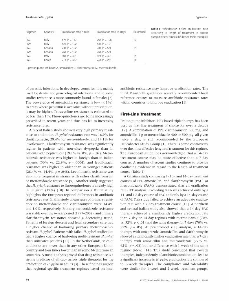
Treatment of
H. pylori
Egan et al.
32
© 2007 Blackwell Publishing Ltd, Helicobacter
12
(Suppl.1): 31–37
of parasitic infections. In developed countries, it is mainlyused for dental and gynecological infections, and in somestudies resistance is more commonly found in females [7].The prevalence of amoxicillin resistance is low (< 1%).In areas where penicillin is available without prescription,it may be higher. Tetracycline resistance is estimated tobe less than 1%. Fluoroquinolones are being increasinglyprescribed in recent years and thus has led to increasingresistance rates.
A recent Italian study showed very high primary resist-ance to antibiotics.
H. pylori
resistance rate was 16.9% forclarithromycin, 29.4% for metronidazole, and 19.1% forlevofloxacin. Clarithromycin resistance was significantlyhigher in patients with non-ulcer dyspepsia than inpatients with peptic ulcer (19.1% vs. 0%,
p
= .02). Metro-nidazole resistance was higher in foreign than in Italianpatients (50% vs. 22.9%,
p
= .0004), and levofloxacinresistance was higher in older than in younger patients(28.4% vs. 14.4%,
p
= .048). Levofloxacin resistance wasalso more frequent in strains with either clarithromycinor metronidazole resistance [9]. Another study suggestedthat
H. pylori
resistance to fluoroquinolones is already highin Belgium (17%) [10]. In comparison a Dutch studyhighlights the European regional differences in antibioticresistance rates. In this study, mean rates of primary resist-ance to metronidazole and clarithromycin were 14.4%and 1.0%, respectively. Primary metronidazole resistancewas stable over the 6-year period (1997–2002), and primaryclarithromycin resistance showed a decreasing trend.Patients of foreign descent and from secondary care hada higher chance of harboring primary metronidazole-resistant
H. pylori
. Patients with failed
H. pylori
eradicationhad a higher chance of harboring multi-resistant
H. pylori
than untreated patients [11]. In the Netherlands, sales ofantibiotics are lower than in any other European Unioncountry and four times lower than in some Mediterraneancountries. A meta-analysis proved that drug resistance is astrong predictor of efficacy across triple therapies for theeradication of
H. pylori
in adults [3]. These findings suggestthat regional specific treatment regimes based on local
antibiotic resistance may improve eradication rates. Thethird Maastricht guidelines recently recommended localreference centres to measure antibiotic resistance rateswithin countries to improve eradication [1].
First-Line Treatment
Proton pump inhibitor (PPI)-based triple therapy has beenused as first-line treatment of choice for over a decade[12]. A combination of PPI, clarithromycin 500 mg, andamoxicillin 1 g or metronidazole 400 or 500 mg, all giventwice a day, is still recommended by the EuropeanHelicobacter Study Group [1]. There is some controversyover the most effective length of treatment for this regime.The European guidelines acknowledged that a 14-daytreatment course may be more effective than a 7-daycourse. A number of recent studies continue to provideconflicting evidence in regard to the length of treatmentcourse (Table 1).
A Croatian study comparing 7-,10-, and 14-day treatmentcourses of PPI, amoxicillin, and clarithromycin (PAC) ormetronidazole (PAM) demonstrated that an eradicationrate (ITT analysis) exceeding 80% was achieved only by a14- and 10-day course of PAC and only by a 14-day courseof PAM. This study failed to achieve an adequate eradica-tion rate with a 7-day treatment course [13]. A northernand central Italian study also showed that a 14-day PACtherapy achieved a significantly higher eradication ratethan 7-day or 14-day regimes with metronidazole (70%vs. 52%,
p
< .01) and the same therapy for 7 days (70% vs.57%,
p
= .05). At per-protocol (PP) analysis, a 14-daytherapy with omeprazole, amoxicillin, and clarithromycinshowed a significantly higher eradication rate than a 7-daytherapy with amoxicillin and metronidazole (77% vs.62%;
p
= .03) but no difference with 1-week of the sameregime (66%) [14]. This study concluded that 2-weektherapies, independently of antibiotic combination, lead toa significant increase in
H. pylori
eradication rate comparedto 1-week therapies. The compliance and tolerabilitywere similar for 1-week and 2-week treatment groups.
Regimen Country Eradication rate 7 days Eradication rate 14 days Reference
PAC Italy 57% (n = 117) 70% (n = 126) 13
PAM Italy 52% (n = 122) 56% (n = 121)
PAC Croatia 74% (n = 122) 93% (n = 58) 14
PAM Croatia 75% (n = 122) 95% (n = 58)
PAC Italy 80% (n = 301) 82% (n = 301) 15
PAC Korea 71% (n = 337) 76% (n = 261) 16
P, proton pump inhibitor; A, amoxicillin; C, clarithromycin; M, metronidazole.
Table 1 Helicobacter pylori eradication rate
according to length of treatment in proton
pump inhibitor-amoxicillin-based triple therapies
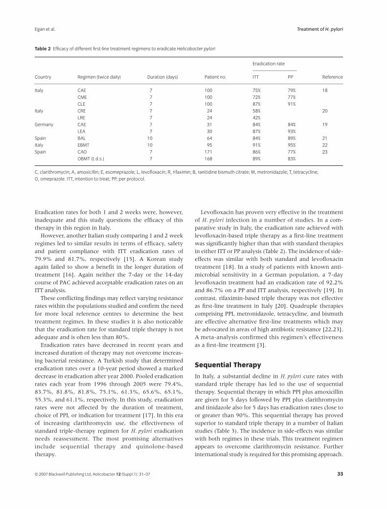
Egan et al.
Treatment of
H. pylori
© 2007 Blackwell Publishing Ltd, Helicobacter
12
(Suppl.1): 31–37
33
Eradication rates for both 1 and 2 weeks were, however,inadequate and this study questions the efficacy of thistherapy in this region in Italy.
However, another Italian study comparing 1 and 2 weekregimes led to similar results in terms of efficacy, safetyand patient compliance with ITT eradication rates of79.9% and 81.7%, respectively [15]. A Korean studyagain failed to show a benefit in the longer duration oftreatment [16]. Again neither the 7-day or the 14-daycourse of PAC achieved acceptable eradication rates on anITT analysis.
These conflicting findings may reflect varying resistancerates within the populations studied and confirm the needfor more local reference centres to determine the besttreatment regimes. In these studies it is also noticeablethat the eradication rate for standard triple therapy is notadequate and is often less than 80%.
Eradication rates have decreased in recent years andincreased duration of therapy may not overcome increas-ing bacterial resistance. A Turkish study that determinederadication rates over a 10-year period showed a markeddecrease in eradication after year 2000. Pooled eradicationrates each year from 1996 through 2005 were 79.4%,83.7%, 81.8%, 81.8%, 75.1%, 61.3%, 65.6%, 65.1%,55.3%, and 61.1%, respectively. In this study, eradicationrates were not affected by the duration of treatment,choice of PPI, or indication for treatment [17]. In this eraof increasing clarithromycin use, the effectiveness ofstandard triple-therapy regimen for
H. pylori
eradicationneeds reassessment. The most promising alternativesinclude sequential therapy and quinolone-basedtherapy.
Levofloxacin has proven very effective in the treatmentof
H. pylori
infection in a number of studies. In a com-parative study in Italy, the eradication rate achieved withlevofloxacin-based triple therapy as a first-line treatmentwas significantly higher than that with standard therapiesin either ITT or PP analysis (Table 2). The incidence of side-effects was similar with both standard and levofloxacintreatment [18]. In a study of patients with known anti-microbial sensitivity in a German population, a 7-daylevofloxacin treatment had an eradication rate of 92.2%and 86.7% on a PP and ITT analysis, respectively [19]. Incontrast, rifaximin-based triple therapy was not effectiveas first-line treatment in Italy [20]. Quadruple therapiescomprising PPI, metronidazole, tetracycline, and bismuthare effective alternative first-line treatments which maybe advocated in areas of high antibiotic resistance [22,23].A meta-analysis confirmed this regimen’s effectivenessas a first-line treatment [3].
Sequential Therapy
In Italy, a substantial decline in
H. pylori
cure rates withstandard triple therapy has led to the use of sequentialtherapy. Sequential therapy in which PPI plus amoxicillinare given for 5 days followed by PPI plus clarithromycinand tinidazole also for 5 days has eradication rates close toor greater than 90%. This sequential therapy has provedsuperior to standard triple therapy in a number of Italianstudies (Table 3). The incidence in side-effects was similarwith both regimes in these trials. This treatment regimenappears to overcome clarithromycin resistance. Furtherinternational study is required for this promising approach.
Table 2 Efficacy of different first-line treatment regimens to eradicate Helicobacter pylori
Country Regimen (twice daily) Duration (days) Patient no.
Eradication rate
ReferenceITT PP
Italy CAE 7 100 75% 79% 18
CME 7 100 72% 77%
CLE 7 100 87% 91%
Italy CRE 7 24 58% 20
LRE 7 24 42%
Germany CAE 7 31 84% 84% 19
LEA 7 30 87% 93%
Spain BAL 10 64 84% 89% 21
Italy EBMT 10 95 91% 95% 22
Spain CAO 7 171 86% 77% 23
OBMT (t.d.s.) 7 168 89% 83%
C, clarithromycin; A, amoxicillin; E, esomeprazole; L, levofloxacin; R, rifaximin; B, ranitidine bismuth citrate; M, metronidazole; T, tetracycline;
O, omeprazole. ITT, intention to treat; PP, per protocol.
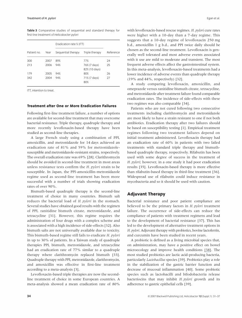
Treatment of
H. pylori
Egan et al.
34
© 2007 Blackwell Publishing Ltd, Helicobacter
12
(Suppl.1): 31–37
Treatment after One or More Eradication Failures
Following first-line treatment failure, a number of optionsare available for second-line treatment that may overcomebacterial resistance. Triple therapy, quadruple therapy andmore recently levofloxacin-based therapy have beenstudied as second-line therapies.
A large French study using a combination of PPI,amoxicillin, and metronidazole for 14 days achieved aneradication rate of 81% and 59% for metronidazole-susceptible and metronidazole-resistant strains, respectively.The overall eradication rate was 69% [28]. Clarithromycinshould be avoided in second-line treatment in most areasunless resistance tests confirm the
H. pylori
strain to besusceptible. In Japan, the PPI-amoxicillin-metronidazoleregime used as second-line treatment has been moresuccessful with a number of trials showing eradicationrates of over 90%.
Bismuth-based quadruple therapy is the second-linetreatment of choice in many countries. Bismuth saltreduces the bacterial load of
H. pylori
in the stomach.Several studies have obtained good results with the regimenof PPI, ranitidine bismuth citrate, metronidazole, andtetracycline [31]. However, this regime requires theadministration of four drugs with a complex scheme andis associated with a high incidence of side-effects [32]. Alsobismuth salts are not universally available due to toxicity.This bismuth-based regime still fails to eradicate
H. pylori
in up to 30% of patients. In a Taiwan study of quadrupletherapies PPI, bismuth, metronidazole, and tetracyclinehad an eradication rate of 77% similar to a quadrupletherapy where clarithromycin replaced bismuth [33].Quadruple therapy with PPI, metronidazole, clarithromycin,and amoxicillin was effective in first-line treatmentaccording to a meta-analysis [3].
Levofloxacin-based triple therapies are now the second-line treatment of choice in some European countries. Ameta-analysis showed a mean eradication rate of 80%
with levofloxacin-based rescue regimes.
H. pylori
cure rateswere higher with a 10-day than a 7-day regime. Thissuggests that a 10-day regime of levofloxacin 250 mgb.d., amoxicillin 1 g b.d., and PPI twice daily should bechosen as the second-line treatment. Levofloxacin is gen-erally well tolerated and most adverse events associatedwith it use are mild to moderate and transient. The mostfrequent adverse effects affect the gastrointestinal system.In this meta-analysis, levofloxacin-based treatments had alower incidence of adverse events than quadruple therapy(19% and 44%, respectively) [32].
A study comparing levofloxacin, amoxicillin, andomeprazole versus ranitidine bismuth citrate, tetracycline,and metronidazole after treatment failure found comparableeradication rates. The incidence of side-effects with thesetwo regimes was also comparable [34].
Patients who are not cured following two consecutivetreatments including clarithromycin and metronidazoleare most likely to have a strain resistant to one if not bothantibiotics. Eradication therapy after two failures shouldbe based on susceptibility testing [1]. Empirical treatmentregimes following two treatment failures depend oninitial treatment administered. Levofloxacin therapy hadan eradication rate of 60% in patients with two failedtreatments with standard triple therapy and bismuth-based quadruple therapy, respectively. Rifabutin has beenused with some degree of success in the treatment of
H. pylori
; however, in a one study it had poor eradicationresults [35]. Levofloxacin-based therapy is more effectivethan rifabutin-based therapy in third-line treatment [36].Widespread use of rifabutin could induce resistance inmycobacteria and so it should be used with caution.
Adjuvant Therapy
Bacterial resistance and poor patient compliance arebelieved to be the primary factors in
H. pylori
treatmentfailure. The occurrence of side-effects can reduce thecompliance of patients with treatment regimens and leadto the development of bacterial resistance [37]. This hasled to the development of alternative treatment options in
H. pylori
. Adjuvant therapy with probiotics, bovine lactoferrin,and curcumin have been studied in recent years.
A probiotic is defined as a living microbial species that,on administration, may have a positive effect on bowelmicroecology and improve health conditions [38]. Themost studied probiotics are lactic acid-producing bacteria,particularly
Lactobacillus
species [39]. Probiotics play a rolein the stabilization of the gastric barrier function anddecrease of mucosal inflammation [40]. Some probioticspecies such as lactobacilli and bifodobacteria releasebacteriocins that may inhibit
H. pylori
growth and itsadherence to gastric epithelial cells [39].
Table 3 Comparative studies of sequential and standard therapy for
first-line treatment of Helicobacter pylori
Patient no. Year
Eradication rate % (ITT)
ReferenceSequential therapy Triple therapy
300 2007 89% 77% 24
213 2006 94% 76% (7 days) 25
82% (10 days)
179 2005 94% 80% 26
342 2004 94% 71% (7 days) 27
80%
ITT, intention to treat.
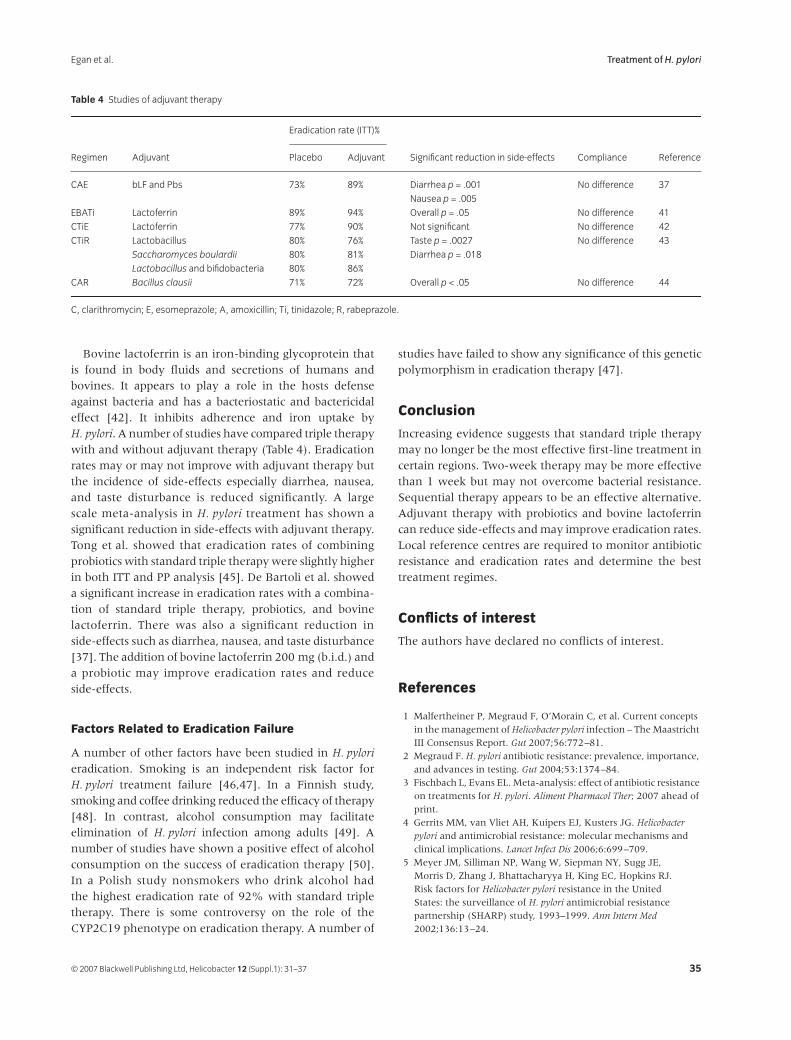
Egan et al.
Treatment of
H. pylori
© 2007 Blackwell Publishing Ltd, Helicobacter
12
(Suppl.1): 31–37
35
Bovine lactoferrin is an iron-binding glycoprotein thatis found in body fluids and secretions of humans andbovines. It appears to play a role in the hosts defenseagainst bacteria and has a bacteriostatic and bactericidaleffect [42]. It inhibits adherence and iron uptake by
H. pylori
. A number of studies have compared triple therapywith and without adjuvant therapy (Table 4). Eradicationrates may or may not improve with adjuvant therapy butthe incidence of side-effects especially diarrhea, nausea,and taste disturbance is reduced significantly. A largescale meta-analysis in
H. pylori
treatment has shown asignificant reduction in side-effects with adjuvant therapy.Tong et al. showed that eradication rates of combiningprobiotics with standard triple therapy were slightly higherin both ITT and PP analysis [45]. De Bartoli et al. showeda significant increase in eradication rates with a combina-tion of standard triple therapy, probiotics, and bovinelactoferrin. There was also a significant reduction inside-effects such as diarrhea, nausea, and taste disturbance[37]. The addition of bovine lactoferrin 200 mg (b.i.d.) anda probiotic may improve eradication rates and reduceside-effects.
Factors Related to Eradication Failure
A number of other factors have been studied in
H. pylori
eradication. Smoking is an independent risk factor for
H. pylori
treatment failure [46,47]. In a Finnish study,smoking and coffee drinking reduced the efficacy of therapy[48]. In contrast, alcohol consumption may facilitateelimination of
H. pylori
infection among adults [49]. Anumber of studies have shown a positive effect of alcoholconsumption on the success of eradication therapy [50].In a Polish study nonsmokers who drink alcohol hadthe highest eradication rate of 92% with standard tripletherapy. There is some controversy on the role of theCYP2C19 phenotype on eradication therapy. A number of
studies have failed to show any significance of this geneticpolymorphism in eradication therapy [47].
Conclusion
Increasing evidence suggests that standard triple therapymay no longer be the most effective first-line treatment incertain regions. Two-week therapy may be more effectivethan 1 week but may not overcome bacterial resistance.Sequential therapy appears to be an effective alternative.Adjuvant therapy with probiotics and bovine lactoferrincan reduce side-effects and may improve eradication rates.Local reference centres are required to monitor antibioticresistance and eradication rates and determine the besttreatment regimes.
Conflicts of interest
The authors have declared no conflicts of interest.
References
1 Malfertheiner P, Megraud F, O’Morain C, et al. Current concepts in the management of
Helicobacter pylori
infection – The Maastricht III Consensus Report.
Gut
2007;56:772–81.2 Megraud F.
H. pylori
antibiotic resistance: prevalence, importance, and advances in testing.
Gut
2004;53:1374–84.3 Fischbach L, Evans EL. Meta-analysis: effect of antibiotic resistance
on treatments for
H. pylori
.
Aliment Pharmacol Ther
; 2007 ahead of print.
4 Gerrits MM, van Vliet AH, Kuipers EJ, Kusters JG.
Helicobacter pylori
and antimicrobial resistance: molecular mechanisms and clinical implications.
Lancet Infect Dis
2006;6:699–709.5 Meyer JM, Silliman NP, Wang W, Siepman NY, Sugg JE,
Morris D, Zhang J, Bhattacharyya H, King EC, Hopkins RJ. Risk factors for
Helicobacter pylori
resistance in the United States: the surveillance of
H. pylori
antimicrobial resistance partnership (SHARP) study, 1993–1999.
Ann Intern Med
2002;136:13–24.
Table 4 Studies of adjuvant therapy
Regimen Adjuvant
Eradication rate (ITT)%
Significant reduction in side-effects Compliance ReferencePlacebo Adjuvant
CAE bLF and Pbs 73% 89% Diarrhea p = .001 No difference 37
Nausea p = .005
EBATi Lactoferrin 89% 94% Overall p = .05 No difference 41
CTiE Lactoferrin 77% 90% Not significant No difference 42
CTiR Lactobacillus 80% 76% Taste p = .0027 No difference 43
Saccharomyces boulardii 80% 81% Diarrhea p = .018
Lactobacillus and bifidobacteria 80% 86%
CAR Bacillus clausii 71% 72% Overall p < .05 No difference 44
C, clarithromycin; E, esomeprazole; A, amoxicillin; Ti, tinidazole; R, rabeprazole.
Treatment of
H. pylori
Egan et al.
36
© 2007 Blackwell Publishing Ltd, Helicobacter
12
(Suppl.1): 31–37
6 Duck WM, Sobel J, Pruckler JM, et al. Antimicrobial resistance incidence and risk factors among
Helicobacter pylori
-infected persons, United States.
Emerg Infect Dis
2004;10:1088–94.7 Megraud F, Lehours P.
Helicobacter pylori
detection and antimicrobial susceptibility testing.
Clin Microbiol Rev
2007;20:280–322. Review.
8 Glupczynski Y, Megraud F, Lopez-Brea M, et al. European multicenter survey of in vitro antimicrobial resistance in
Helicobacter pylori
.
Eur J Clin Microbiol Infect Dis
2000;11:820–3.9 Zullo A, Perna F, Hassan C, Ricci C, Saracino I, Morini S, Vaira D.
Primary antibiotic resistance in
Helicobacter pylori
strains isolated in northern and central Italy.
Aliment Pharmacol Ther
2007;25:1429–34.
10 Bogaerts P, Berhin C, Nizet H, Glupczynski Y. Prevalence and mechanisms of resistance to fluoroquinolones in
Helicobacter pylori
strains from patients living in Belgium.
Helicobacter
2006;11:441–5.11 Janssen MJ, Hendrikse L, de Boer SY, Bosboom R, de Boer WA,
Laheij RJ, Jansen JB.
Helicobacter pylori
antibiotic resistance in a Dutch region: trends over time.
Neth J Med
2006;64:191–5.12 Egan BJ, O’Morain CA. A historical perspective on
Helicobacter
gastroduodenitis and its complications.
Best Pract Res Clin Gastroenterol
2007;21:335–46. Review.13 Paoluzi P, Iacopini F, Crispino P, et al. 2-week triple therapy for
Helicobacter pylori
infection is better than 1-week in clinical practice: a large prospective single-center randomized study.
Helicobacter
2006;11:562–8.14 Katicic M, Ticak M, Prskalo M, Naumovski-Mihalic S,
Filipec-Kanizaj T, Skurla B, Sabaric B, Colic-Cvrlje V, Papa B. Eradication of
Helicobacter pylori
infection with two triple therapy regimes of 7, 10, and 14 days: four year experience.
Helicobacter
2006;2:A11.05:p386.
15 Zagari RM, Bianchi-Porro G, Fiocca R, Gasbarrini G, Roda E, Bazzoli F. Comparison of 1 and 2 weeks of omeprazole, amoxicillin and clarithromycin treatment for
Helicobacter pylori
eradication: the HYPER Study.
Gut
2007;56:475–9.16 Kim BG, Lee DH, Ye BD, et al. Comparison of 7-day and 14-day
proton pump inhibitor-containing triple therapy for
Helicobacter pylori
eradication: neither treatment duration provides acceptable eradication rate in Korea.
Helicobacter
2007;12:31–5.17 Kadayifci A, Buyukhatipoglu H, Cemil Savas M, Simsek I.
Eradication of
Helicobacter pylori
with triple therapy: an epidemiologic analysis of trends in Turkey over 10 years.
Clin Ther
2006;28:1960–6. Review.18 Nista EC, Candelli M, Zocco MA, Cremonini F, Ojetti V, Finizio R,
Spada C, Cammarota G, Gasbarrini G, Gasbarrini A. Levofloxacin-based triple therapy in first-line treatment for
Helicobacter pylori
eradication.
Am J Gastroenterol
2006;101:1985–90.
19 Antos D, Schneider-Brachert W, Bästlein E, et al. 7-day triple therapy of
Helicobacter pylori
infection with levofloxacin, amoxicillin, and high-dose esomeprazole in patients with known antimicrobial sensitivity.
Helicobacter
2006;11:39–45.20 Gasbarrini A, Lauritano EC, Nista EC, et al. Rifaximin-based
regimens for eradication of
Helicobacter pylori
: a pilot study.
Dig Dis
2006;24:195–200.21 Gisbert JP, Fernandez Bermejo M, Molina Infante J, Perez Gallardo
B, Prieto Bermejo AB, Mateos Rodriguez JM, Robledo Andres P, Gonzalez Garcia G. First-line triple therapy with levofloxacin for
Helicobacter pylori
eradication; 2007 ahead of print.22 Dore MP, Maragkoudakis E, Pironti A, Tadeu V, Tedde R, Realdi G,
Delitala G. Twice-a-day quadruple therapy for eradication of
Helicobacter pylori
in the elderly.
Helicobacter 2006;11:52–5.23 Calvet X, Ducons J, Guardiola J, Tito L, Andreu V, Bory F,
Guirao R; Group for Eradication Studies from Catalonia and Aragón (Gresca). One-week triple vs. quadruple therapy for Helicobacter pylori infection – a randomized trial. Aliment Pharmacol Ther 2002;16:1261–7.
24 Vaira D, Zullo A, Vakil N, Gatta L, Ricci C, Perna F, Hassan C, Bernabucci V, Tampieri A, Morini S. Sequential therapy versus standard triple-drug therapy for Helicobacter pylori eradication: a randomized trial. Ann Intern Med 2007;146:556–63.
25 Scaccianoce G, Hassan C, Panarese A, Piglionica D, Morini S, Zullo A. Helicobacter pylori eradication with either 7-day or 10-day triple therapies, and with a 10-day sequential regimen. Can J Gastroenterol 2006;20:113–7.
26 Zullo A, Gatta L, De Francesco V, Hassan C, Ricci C, Bernabucci V, Cavina M, Ierardi E, Morini S, Vaira D. High rate of Helicobacter pylori eradication with sequential therapy in elderly patients with peptic ulcer: a prospective controlled study. Aliment Pharmacol Ther 2005;21:1419–24.
27 De Francesco V, Zullo A, Hassan C, et al. The prolongation of triple therapy for Helicobacter pylori does not allow reaching therapeutic outcome of sequential scheme: a prospective, randomised study. Dig Liver Dis 2004;36:322–6.
28 Lamouliatte H, Mégraud F, Delchier JC, et al. Second-line treatment for failure to eradicate Helicobacter pylori: a randomized trial comparing four treatment strategies. Aliment Pharmacol Ther 2003;18:791–7.
29 Kawai T, Kawakami K, Kataoka M, et al. Comparison of efficacies of dual therapy and triple therapy using rabeprazole in second-line eradication of Helicobacter pylori in Japan. Aliment Pharmacol Ther 2006;24 (Suppl. 4):16–22.
30 Shirai N, Sugimoto M, Kodaira C, Nishino M, Ikuma M, Kajimura M, Ohashi K, Ishizaki T, Hishida A, Furuta T. Dual therapy with high doses of rabeprazole and amoxicillin versus triple therapy with rabeprazole, amoxicillin, and metronidazole as a rescue regimen for Helicobacter pylori infection after the standard triple therapy. Eur J Clin Pharmacol 2007;63:743–9.
31 Gisbert JP, Pajares JM. Review article: Helicobacter pylori ‘rescue’ regimen when proton pump inhibitor-based triple therapies fail. Aliment Pharmacol Ther 2002;16:1047–57. Review.
32 Gisbert JP, Morena F. Systematic review and meta-analysis: levofloxacin-based rescue regimens after Helicobacter pylori treatment failure. Aliment Pharmacol Ther 2006;23:35–44.
33 Wu DC, Hsu PI, Chen A, et al. Randomized comparison of two rescue therapies for Helicobacter pylori infection. Eur J Clin Invest 2006;36:803–9.
34 Gisbert JP, Gisbert JL, Marcos S, Moreno-Otero R, Pajares JM. Levofloxacin- vs. ranitidine bismuth citrate-containing therapy after H. pylori treatment failure. Helicobacter 2007;12:68–73.
35 Qasim A, Sebastian S, Thornton O, Dobson M, McLoughlin R, Buckley M, O’Connor H, O’Morain C. Rifabutin- and furazolidone-based Helicobacter pylori eradication therapies after failure of standard first- and second-line eradication attempts in dyspepsia patients. Aliment Pharmacol Ther 2005;21:91–6.
36 Gisbert JP, Gisbert JL, Marcos S, Moreno-Otero R, Pajares JM. Third-line rescue therapy with levofloxacin is more effective than rifabutin rescue regimen after two Helicobacter pylori treatment failures. Aliment Pharmacol Ther 2006;24:1469–74.
37 de Bortoli N, Leonardi G, Ciancia E, et al. Helicobacter pylori eradication: a randomized prospective study of triple therapy versus triple therapy plus lactoferrin and probiotics. Am J Gastroenterol 2007;102:951–6.
38 Fuller R. Probiotics in human medicine. Gut 1991;32:439–42. Review.

Egan et al. Treatment of H. pylori
© 2007 Blackwell Publishing Ltd, Helicobacter 12 (Suppl.1): 31–37 37
39 Lesbros-Pantoflickova D, Corthésy-Theulaz I, Blum AL. Helicobacter pylori and probiotics. J Nutr 2007;137 (3 Suppl. 2):812S–8S.
40 Gotteland M, Brunser O, Cruchet S. Systematic review: are probiotics useful in controlling gastric colonization by Helicobacter pylori? Aliment Pharmacol Ther 2006;23:1077–86. Review.
41 Tursi A, Elisei W, Brandimarte G, Giorgetti GM, Modeo ME, Aiello F. Effect of lactoferrin supplementation on the effectiveness and tolerability of a 7-day quadruple therapy after failure of a first attempt to cure Helicobacter pylori infection. Med Sci Monit 2007;13:CR187–190.
42 Di Mario F, Aragona G, Dal Bó N, et al. Bovine lactoferrin for Helicobacter pylori eradication: an open, randomized, multicentre study. Aliment Pharmacol Ther 2006;23:1235–40.
43 Cremonini F, Di Caro S, Covino M, Armuzzi A, Gabrielli M, Santarelli L, Nista EC, Cammarota G, Gasbarrini G, Gasbarrini A. Effect of different probiotic preparations on anti-Helicobacter pylori therapy-related side effects: a parallel group, triple blind, placebo-controlled study. Am J Gastroenterol 2002;97:2744–9.
44 Nista EC, Candelli M, Cremonini F, Cazzato IA, Zocco MA, Franceschi F, Cammarota G, Gasbarrini G, Gasbarrini A. Bacillus clausii therapy to reduce side-effects of anti-Helicobacter pylori treatment: randomized, double-blind, placebo controlled trial. Aliment Pharmacol Ther 2004;20:1181–8.
45 Tong JL, Ran ZH, Shen J et al. Meta-analysis: the effect of supplementation with probiotics on eradication rates and adverse events during Helicobacter pylori eradication therapy. Aliment Pharmacol Ther 2007;25:155–68.
46 Suzuki T, Matsuo K, Ito H, Sawaki A, Hirose K, Wakai K, Sato S, Nakamura T, Yamao K, Ueda R. Smoking increases the treatment failure for Helicobacter pylori eradication. Am J Med 2006;119:217–224.
47 Suzuki T, Matsuo K, Sawaki A, et al. Influence of smoking and CYP2C19 genotypes on H. pylori eradication success. Epidemiol Infect 2007;135:171–6.
48 Koivisto TT, Rautelin HI, Voutilainen ME, Heikkinen MT, Koskenpato JP, Färkkilä MA. First-line eradication therapy for Helicobacter pylori in primary health care based on antibiotic resistance: results of three eradication regimens. Aliment Pharmacol Ther 2005;21:773–82.
49 Murray LJ, Lane AJ, Harvey IM, Donovan JL, Nair P, Harvey RF. Inverse relationship between alcohol consumption and active Helicobacter pylori infection: the Bristol Helicobacter project. Am J Gastroenterol 2002;97:2750–5.
50 Baena JM, López C, Hidalgo A, Rams F, Jiménez S, García M, Hernández MR. Relation between alcohol consumption and the success of Helicobacter pylori eradication therapy using omeprazole, clarithromycin and amoxicillin for 1 week. Eur J Gastroenterol Hepatol 2002;14:291–6.

Helicobacter ISSN 1523-5378
© 2007 The Authors
38
Journal compilation
© 2007 Blackwell Publishing Ltd, Helicobacter
12
(Suppl. 1): 38–44
Blackwell Publishing LtdOxford, UKHELHelicobacter1083-4389© 2007 The AuthorsJournal compilation © 2007 Blackwell Publishing Ltd, Helicobacter 12 (Suppl. 1): xx–xxXXX
Or iginal Art ic les
H. pylori
Infection in Pediatrics Veres and Pehlivanoglu
Helicobacter pylori
Infection in Pediatrics
Gabor Veres
*
and Ender Pehlivanoglu
†
*1st Department of Pediatrics, Semmelweis University, Budapest, Hungary,
†
Division of Pediatric Gastroenterology, Hepatology, and Nutrition, Marmara
University School of Medicine, Istanbul, Turkey
Abstract
During the last year, epidemiologic studies have shown that spontaneousclearance of
Helicobacter pylori
infection has a less significant role in countrieswith high prevalence and, in contrast to adults, there is no male predominanceof
H. pylori
infection in children. Early acquisition of
H. pylori
may play a role inthe development of recurrent abdominal pain in children less than 5 years ofage. In this very young age group, the adequate performance of stool antigen testand
13
C urea-breath test demonstrated satisfactory sensitivity and specificity asnon-invasive methods to diagnose
H. pylori
infection. In the current paper, themost relevant pediatric studies on
H. pylori
infection published between April2006 and March 2007 are reviewed.
Keywords
Helicobacter pylori
, pediatrics, epidemiology,
stool antigen test, drug resistance.
Reprint requests to
: Gabor Veres,
1st Department of Pediatrics, Semmelweis
University, 1083 Budapest, Bókay str. 53.,
Hungary. E-mail: [email protected]
Despite the fact that
Helicobacter pylori
was discovered25 years ago and that the Nobel Prize in Medicine orPhysiology was awarded to Marshall and Warren 2 yearsago,
H. pylori
infection is still a challenging subject formany researchers and physicians [1,2].
In the present paper, the most relevant studies on
H. pylori
infection in children published last year (betweenApril 2006 and March 2007) are reviewed.
Basic Research
The
cag
A gene, at least in adults, is considered to be amarker for severe disease inducing a high production ofproinflammatory cytokines in the gastric mucosa. However,Sokucu et al. found no association between
cag
A positivityand the development of mucosal lesions/ulcers in Turkishchildren infected with
H. pylori
[3]. Antral lymphonodularhyperplasia was strongly associated with
H. pylori
positivitybut
cag
A positivity and esophageal lesions were significantlyless common in
cag
A-positive children (20% vs. 76.7%).In another study from Portugal, a country with one of thehighest gastric cancer incidences and mortality ratesamong European Union countries [4], Costa Lopes et al.also found that there was no association between the mostprevalent genotypes of infected children (
cag
A-,
vac
A
s2m2
,and
ice
A
2
) and patients’ demographic and gastroduodenaldisease phenotype, suggesting a potential role of host andenvironmental factors in the development of distinctclinical disease at a later age [5].
Oleastro et al. used suppressive subtractive hybridiza-tion for comparative genomics between
H. pylori
strains
isolated from patients with ulcer and from patients withgastritis only [6]. Two of the examined genes, jhp0562(involved in lipopolysaccharide biosynthesis) and jhp0870(an outer membrane protein), were highly associated withpeptic ulcer disease (PUD) in children, suggesting novelvirulence factors of
H. pylori
.
Prevalence
It is well known that childhood is an important period foracquisition of
H. pylori
infection but regarding the changein epidemiology of infection, only a few cohort studiesinvolving children have been published [7–11]. In 2006,Özen et al. published a prospective study to determinethe acquisition and loss rates of
H. pylori
infection in327 asymptomatic Turkish preschool and school children(3–12 years) by using
13
C-urea breath test (UBT) [12]. Theincidence of
H. pylori
infection among previously uninfectedchildren was 14%, and the loss rate of infection amongpreviously infected children was 5.5% during a 6-yearfollow up. This 2.5-fold higher rate of acquisition comparedto loss of infection suggests that, at least in a country witha high prevalence rate, spontaneous clearance of
H. pylori
infection has no significant role. Low household densityand antibiotic administration over the last 6 months werefound to be protective factors against acquisition of theinfection.
Frenck et al. conducted a well-designed study enrolling108 Egyptian children between 2 and 17 years of age [13].One of the strengths of this study was the large number ofchildren included under 6 years of age (n
=
52). Prevalence

Veres and Pehlivanoglu
H. pylori
Infection in Pediatrics
© 2007 The Authors
Journal compilation © 2007 Blackwell Publishing Ltd, Helicobacter
12
(Suppl. 1): 38–44
39
of
H. pylori
was 33% in children younger than 6 years and60% in children older than 6 years. Both invasive (histology,culture, and rapid urease testing) and non-invasivemethods (UBT, stool, and serum ELISA) were used for
H. pylori
diagnosis.The prevalence of
H. pylori
infection is declining indeveloped countries; however, the rate of infection isinfluenced by several factors such as ethnic origin [14].In a recent study from the Netherlands, Mourad-Baarset al. also found a low seroprevalence (1.2%) of
H. pylori
infection in 1258 young children (2–4 years), with asignificant difference between children with parents whowere both Dutch (0.5%) and children with at least onenon-Dutch parent (2.6%) [15].
New data on seroprevalence of
H. pylori
in Vietnam havebeen published [16,17]. Using an ELISA test on 824Vietnamese children without gastrointestinal symptoms,aged 6 months to 15 years, seroprevalence was 34.0%.Age groups ranging from 3 to 6 years and 6 years andolder, and number of offspring were positively and in-dependently associated with seropositivity [adjusted oddsratio (OR) 2.9 (95% confidence interval (CI) 1.5–5.5), OR1.9 (95% CI 1.1–3.1) and OR 1.8 (95% CI 1.1–2.6), respec-tively]. Only breastfeeding more than 6 months wasnegatively associated with
H. pylori
seropositivity [OR 0.5(95% CI 0.3–0.9)].
Halitim et al. evaluated the
H. pylori
re-infection rate in45 children (median 10.9 years) after successful eradica-tion [18]. One to nine years after
H. pylori
eradication,18% of the children were re-infected (5.4% to 6% perpatient-year) indicating a higher re-infection rate inchildren than in adults.
In contrast to what was found in adults, a male predom-inance of infection was not found in the separate meta-analysis of 10 pediatric studies where the summary ORwas close to 1 [summary OR 1.03 (95% CI 0.91–1.17)][19]. Differential protective immunity (women havehigher plasma IgM levels than men [20] and estrogenstimulates immune response [21]) or differential antibioticexposure between genders may explain the differencesobserved in pediatric and adult studies.
Transmission
The role of infected mothers in transmission of
H. pylori
has been described in the past [22,23]. In 2006, severalreports re-established this relationship.
Ito et al. conducted a community-based familial study(265 families: 507 children and 530 adults) on
H. pylori
infection among Japanese Brazilians living in Sao Pauloto identify risk factors associated with intrafamilialtransmission [24].
H. pylori
seropositivity was found in9.3% of children and 39.2% of the parents. The prevalence
of
H. pylori
infection was 3.5% for children with un-infected parents, 9.9% for those with a seropositive fatherand a seronegative mother, 14.9% for those with a serop-ositive mother and a seronegative father, and 16.0% forthose with both seropositive parents. Moreover, the mother’ssymptoms of nausea and vomiting and the use of pacifierwere significantly associated with the risk of
H. pylori
infec-tion in children. These data suggest that infected mothersare the main source of
H. pylori
infection of their children,mainly through contact with contaminated and regurgi-tated gastric juice from the mother’s mouth.
Another study from Germany found a similar associa-tion using more reliable methods to detect
H. pylori
infec-tion (UBT and monoclonal antigen stool test) [25]. In thiscommunity-based birth cohort, 2.4% of children (20 of834) were infected. The odds ratio for infection of the childwas 12.9 (95% CI, 3.2–52.5) if the mother was infectedand 1.4 (95% CI, 0.4–4.6) if the father was infected.
Vertical transmission during pregnancy was investigatedin an experimental study using Mongolian gerbils [26].After experimental infection of pregnant animals, therewas no detectable
H. pylori
infection, using bacterial culture,polymerase chain reaction and rapid urease test, in any ofthe fetuses during pregnancy nor in the litters at parturition.
Campell et al. analyzed the presence of
cag
A- and
vac
A-specific IgA monthly in breast milk of 48 mothers in Gam-bia [27]. Thirty-seven out of 48 infants (77%) had
H. pylori
infection by UBT. Low breast milk content of anti-
vac
AIgA antibody was correlated with weight loss in
H. pylori
-infected infants.
Stray-Pedersen et al. investigated the prevalence of
H. pylori
antigen in the stools (HpSA) of Norwegianneonates and very young children [28]. Interestingly,there was an unexpectedly high rate of positives in theneonates (52%, 36 of 69), suggesting either a lack of spe-cificity of the test or a transient colonization during theneonatal period. Moreover,
H. pylori
antigen detection inthe neonates was significantly associated with the mode ofdelivery (vaginal births, 59%, Cesarean section, 10%).
Clinical Manifestations
H. pylori
is considered to be the major cause of chronicgastritis and duodenal ulcer in childhood. However, theassociation between
H. pylori
and non-ulcer dyspepsia,recurrent abdominal pain (RAP), gastric outlet obstruction,and extraintestinal manifestations is still controversial.
Intestinal Symptoms
Recurrent abdominal pain occurs in 10–15% of school-agechildren with a controversial association with
H. pylori
infection. Malaty et al. found no association between a

H. pylori
Infection in Pediatrics
Veres and Pehlivanoglu
© 2007 The Authors
40
Journal compilation © 2007 Blackwell Publishing Ltd, Helicobacter
12
(Suppl. 1): 38–44
positive UBT and RAP among 243 symptomatic children(11% UBT+) versus 330 asymptomatic children (17% UBT+)[29]. A previously validated, multidimensional measurefor RAP (MM-RAP) consisting of four scales previouslydescribed (pain intensity scale, symptoms scale, disabilityscale, and satisfaction scale) was applied to each child/parent described earlier in detail [30]. Unexpectedly, insymptomatic children the prevalence of
H. pylori
fellwith age from 20% at age
<
5 years to 7% for children
>
10 years (OR 2.7, 95% CI 0.7–11.2). In contrast, theprevalence of
H. pylori
in children without symptomsincreased with age from 11% for children
<
5 years to 40%for children
>
10 years (OR 5.4, 95% CI 2.0–13.8), asexpected. In this second group, the mother’s educationallevel was inversely correlated with
H. pylori
infection. Theauthors suggested that early acquisition of
H. pylori
maycause chronic abdominal symptoms.
In a study in Taiwan, 135 patients with RAP were inves-tigated for
H. pylori
by rapid urease test, histology, and UBT[31]. The prevalence of
H. pylori
infection was 23.7%.After the 12-month follow up,
H. pylori
status did not sig-nificantly influence the disease course: persistence of RAPwas noted among 59 of 84 (70.2%) non-infected patients,and in 13 of the 15 (86.7%)
H. pylori
-infected patients withfailed eradication.
Yen et al. followed-up 11 children with gastric outletobstruction [32]. In six cases, there was an anatomicabnormality and in five cases PUD. Two of the five patientshad an
H. pylori
infection detected by rapid urease test.The authors concluded that PUD plays an important rolein children with gastric outlet obstruction, however,compared to adult patients
H. pylori
is a less importantetiologic factor. An interesting case report concerning thistopic was recently published involving a rare case of gastricoutlet obstruction in an 11-year-old girl with an ectopicpancreatic tissue, which was probably induced by
H. pylori
-associated PUD [33].
Extraintestinal Manifestations
H. pylori
infection has been associated with extradigestivemanifestations, such as anemia, short stature, immunethrombocytopenic purpura, and migraine, some of theselacking sufficient evidence. A study in Poland found noassociation between
H. pylori
infection and autoimmunehepatitis in children [34].
Yilmaz et al. cultured
H. pylori
from aspirated middle earfluid and biopsies taken from the promontorium of childrenwith otitis media with effusion [35]. There was a significantlyincreased colonization by
H
.
pylori
in patients with otitismedia (n
=
22) when compared with controls (n
=
20).The association between
H. pylori
infection and growthis still controversial [36]. Two studies addressed this issue.
Fialho et al. compared height and weight of 197 childrenwith
H. pylori
infection to 156 children without infection[37]. In a low-income community in north-east Brazil,
H. pylori
infection was associated with short stature only inolder children (8–14 years). Chimonas et al. investigated650 children (7–11 years) in Alaska [38]. At baseline, 87%of children were infected with
H. pylori
(UBT). During thepost-treatment follow up (2, 8, and 14 months), there wasno positive correlation between
H. pylori
eradication statusand improvement in any of the measured growth para-meters (height, weight, and body mass index).
The negative correlation between plasma levels of ghre-lin and
H. pylori
infection that may contribute to the alter-ations of the appetite and dyspeptic symptoms observed ininfected patients was confirmed [39,40].
Yahav et al. assessed the prevalence of
H. pylori
and tox-igenic
Clostridium difficile
infections in 30 consecutive patientswith cystic fibrosis versus controls. Prevalence of
H. pylori
infection was lower (17%) than in similarly aged controls(30%) while about half of the patients with cystic fibrosiswere asymptomatic carriers of
C. difficile
producing mostlytoxin B [41].
Another study in Turkey showed a negative effect of
H. pylori
infection on serum ferritin and vitamin B
12
levelsin children [42]. There was no significant effect of
H. pylori
status on serum folate or zinc levels.It is of interest that in the previously mentioned Turkish
cohort study, children with
H. pylori
infection were com-plaining more often of headaches than abdominal pain ordyspepsia. All three children with migraines were also
H. pylori
positive [12].Ohno et al. from Japan reported long-lasting remission
of primary gastric lymphoma of the mucosa-associatedlymphoid tissue type in two immunocompromisedpediatric patients (14- and 6-year-old boys) with
H. pylori
infection [43]. Treatment of
H. pylori
alone completelyresolved the patients’ lymphoma in the follow up of 10 yearsand 3 years, respectively.
Diagnostic Modalities
The accuracy of diagnostic tests remains uncertain inchildren less than 5–6 years of age. Even in the large,multicentre and multinational study carried out inEurope, only 13
H. pylori
-positive children
<
6 years wereenrolled [44]. Such a study was performed on Egyptianchildren [13]. Histology, culture, and rapid urease testingwere performed on all patients providing a strong goldstandard for validation of the non-invasive tests. Moreover,52 children
<
6 years of age were enrolled in this study andthe 33% global prevalence of
H. pylori
infection allowed adistribution of results by age. UBT and HpStar stool ELISAkit had the highest sensitivity and specificity (UBT, 98%
Veres and Pehlivanoglu
H. pylori
Infection in Pediatrics
© 2007 The Authors
Journal compilation © 2007 Blackwell Publishing Ltd, Helicobacter
12
(Suppl. 1): 38–44
41
and 89%; HpStar, 94% and 81%, respectively). However,the serology kit (HM-CAP) had a very low sensitivity(50%). Sensitivity of the UBT was not influenced by theage of the patient but specificity was lower, although notstatistically different, in children
< 6 years of age (86%)versus 95%, for children > 6 years. The specificity of HpStarstool kit, although high, was slightly but significantlylower in younger children. Receiver operating curvesfound optimal cut-off points of UBT at 6.2 delta overbaseline (DOB) which is higher than the level of 2.5–4DOB that is usually accepted as a cut-off level for H. pyloriinfection in adults [45].
Dondi et al. also observed satisfactory sensitivity andspecificity for UBT, 93.3% and 95.5%, respectively, and forHpSA, 93.3% and 98.7%, respectively, in 30 children under5 years of age with H. pylori infection [46]. Increasing thecut-off from a DOB of 5–8 improved UBT specificity from95.5 to 98.1. Polyclonal HpSA tests were performed with-out transportation to the hospital laboratory, within a fewdays after collection, providing good results. Authorssuggested that transportation or storage of the stools maynegatively influence the accuracy.
No correlation between the UBT results and histologicgrades for mononuclear infiltrate, neutrophilic infiltrate,and bacterial density was found in Brazilian children[47], indicating that UBT in children should be interpretedqualitatively.
A study in Taiwan suggested that a lower dose of urea forUBT (1 mg/kg of bodyweight, maximum 25 mg) is alsoeffective to detect H. pylori infection in children [48].
The sensitivity and specificity of the 13C-urea blood testwere 83% and 91%, respectively, in comparison to histologyand rapid urease test in 40 children [49].
Pelerito et al. investigated the accuracy of AssureH. pylori Rapid Test (Genelabs Diagnostics, Singapore)which is intended for the rapid detection of antibodies toH. pylori in human serum, plasma, or whole blood [50]. Atotal of 130 Portuguese children were enrolled, of whom,according to the gold standard (culture/histology and rapidurease test), 70 were H. pylori positive. The sensitivity,specificity, and positive and negative predictive values ofthe test on serum were 75.7%, 95.0%, 94.6%, and 77.0%,respectively. Interestingly, when a longer reading time of45 minutes was introduced, the rapid test showed betterperformances (sensitivity 98.6% and specificity 95%).However, this rapid test was not evaluated with finger-pricked whole blood which is more realistic in the practi-tioner’s clinic setting.
The monoclonal antibody-based ELISA HpStar used byKolho et al. in a study on 102 stool samples [51] showeda sensitivity and specificity of 95% and 90%, respectively.Half of the specimens were also tested with two other anti-body stool tests with excellent performance. The accuracy
rates of the tests were 98% for the HpSA and 96% for theImmunoCardSTAT.
GastroPanel (Biohit, Helsinki, Finland), a serum test kitthat measures H. pylori antibodies (HPABs) and pepsinogensI and II and gastrin 17, was tested on 30 children withH. pylori-positive gastritis [52]. The test had a sensitivitytoo low (47–73%) to be recommended for H. pylori screen-ing; even the assays of pepsinogens and gastrin did notimprove sensitivity.
Muhsen et al. tested the HpELISA kit (URINELISA,Otsuka Phamaceuticals Co, Ltd, Tokyo, Japan) for detectionof H. pylori IgG antibodies in urine of 159 healthy IsraeliArab children aged 3–5 years. This test was not sensitive(34–66%), indicating limited diagnostic value [53].
H. pylori Treatment
Triple treatment including a proton-pump inhibitor (PPI)and clarithromycin, combined with either amoxicillin ormetronidazole, has been recommended to treat childrenwith H. pylori infection [54]. Nevertheless, a growing bodyof evidence showed that antibiotic resistance is increasing,thus new drugs and therapeutic regimens are clearlyneeded [55].
A prospective randomized study comparing a tripletherapy (ranitidine bismuth citrate, amoxicillin, and clari-thromycin) given for 4 days versus 7 days was conductedby Tam et al. on 206 children with H. pylori infection [56].The 7-day regimen was superior to the 4-day regimen witheradication rates of 89% and 78%, respectively (p = .02).
A register was established by European pediatricians(Pediatric European Register for Treatment of Helicobacterpylori, PERTH) and is available on the European Societyfor Pediatric Gastroenterology Hepatology and Nutritionwebsite to collect data on treatment performed. The resultsshow a significantly higher eradication rate in children withulcer than in those without, 79.7% and 63.9%, respectively.Bismuth-containing triple therapies were more efficaciousthan PPI-based regimens (77% vs. 64%, OR 1.88, 95% CI 1.1–3.3). Moreover, a 2-week therapy was not superior to a 1-weektherapy. The authors concluded that treatment recom-mended for adults may be not suitable for children [57].
In contrast, when Bahremand et al. compared a tripletherapy (omeprazole, clarithromycin, and amoxicillin for10 days) to a quadruple therapy (omeprazole, amoxicillin,metronidazole, and bismuth citrate for 10 days) in Iranianchildren with H. pylori infection, the eradication rates werehigher in triple versus bismuth quadruple therapy (92–75.5% vs. 84–68.8%, respectively) [58].
A 1-week esomeprazole-based triple therapy was highlyeffective for eradication of H. pylori in 58 children (92–93%) [59]. Resistance rates to clarithromycin and metro-nidazole were 9% and 16%, respectively.
H. pylori Infection in Pediatrics Veres and Pehlivanoglu
© 2007 The Authors
42 Journal compilation © 2007 Blackwell Publishing Ltd, Helicobacter 12 (Suppl. 1): 38–44
Cadranel et al. showed that standard triple therapy(omeprazole, amoxicillin, and clarithromycin) offered abetter eradication rate (69%) than dual therapy (amoxicil-lin and clarithromycin) (15%) in 46 children with H. pylorigastritis [60].
Kawakami et al. conducted a prospective open trial in38 children with H. pylori infection administering a 7-daycourse of omeprazole, clarithromycin, and furazolidone(100 mg < 30 kg, 200 mg > 30 kg) twice daily [61].Intention-to-treat and per-protocol analysis showedmoderate efficacy in H. pylori treatment (73.7% and84.8%, respectively).
In 2006, two randomized placebo controlled trials evalu-ated the effect of probiotic food as an adjuvant to thestandard triple therapy for eradication of H. pylori infectionin children. One of them showed a beneficial effect ofLactobacillus reuteri administration to reduce antibiotic-associated side-effects [62], while the other using Bifidobac-terium animalis and Lactobacillus casei did not [63].
The first meta-analysis assessing the efficacy of H. pyloritreatment in children concluded that additional well-designed randomized placebo-controlled pediatric trialsare needed, especially in developing countries [64].
Bacterial resistance and low compliance for drug intakeare the main factors for the treatment failure in H. pyloriinfection [65]. Koletzko et al. conducted a prospectivemulticentre study on antibiotic resistance of H. pyloristrains obtained from 1233 children from 14 Europeancountries [66]. Resistance to clarithromycin was observedin 20% before therapy, with significantly higher percent-age in boys and children under 6 years of age. Similarly,primary resistance to metronidazole was 23% with almostno resistance to amoxicillin (0.6%). The authors suggestedthat administration of antibiotics for other indicationsmay be the major risk factor for development of primaryresistance.
Another study was performed in Iran [67]. The primaryresistance was 54.2% to metronidazole, 8.3% to amoxicil-lin, and 4.2% to clarithromycin, with no resistance totetracycline and furazolidone.
Conflicts of interest
The authors have declared no conflicts of interest.
References
1 Talley NJ, Richter J. Nobel Prize in Medicine awarded to a Gastroenterologist in 2005. Am J Gastroenterol 2006;101:211.
2 Megraud F. A humble bacterium sweeps this year’s Nobel Prize. Cell 2005;123:975–6.
3 Sokucu S, Ozden AT, Suoglu OD, Elkabes B, Demir F, Cevikbas U, Gokce S, Saner G. CagA positivity and its association with
gastroduodenal disease in Turkish children undergoing endoscopic investigation. J Gastroenterol 2006;41:533–9.
4 Lunet N, Pina F, Barros H. Regional trends in Portuguese gastric cancer mortality (1984–99). Eur J Cancer Prev 2004;13:271–5.
5 Costa Lopes AI, Palha A, Monteiro L, Olcastro M, Pelerito A, Fernandes A. Helicobacter pylori genotypes in children from a population at high gastric cancer risk: no association with gastroduodenal histopathology. Am J Gastroenterol 2006;101:2113–22.
6 Oleastro M, Monteiro L, Lehours P, Megraud F, Menard A. Identification of markers for Helicobacter pylori strains isolated from children with peptic ulcer disease by suppressive subtractive hybridization. Infect Immun 2006;74:4064–74.
7 Malaty HM, Kumagai T, Tanaka E, Ota H, Kiyosawa K, Graham DY, Katsuyama T. Evidence from a nine-year birth cohort study in Japan of transmission pathways of Helicobacter pylori infection. J Clin Microbiol 2000;38:1971–3.
8 Kumagai T, Malaty HM, Graham DY, et al. Acquisition versus loss of Helicobacter pylori infection in Japan: results from an 8-year birth cohort study. J Infect Dis 1998;178:717–21.
9 Rothenbacher D, Bode G, Brenner H. Dynamics of Helicobacter pylori infection in early childhood in a high-risk group living in Germany: loss of infection higher than acquisition. Aliment Pharmacol Ther 2002;16:1663–8.
10 Glynn MK, Friedman CR, Gold BD, Khanna B, Hutwagner L, Iihoshi N, Revollo C, Quick R. Seroincidence of Helicobacter pylori infection in a cohort of rural Bolivian children: acquisition and analysis of possible risk factors. Clin Infect Dis 2002;35:1059–65.
11 Przybyszewska K, Bielanski W, Fyderek K. Frequency of Helicobacter pylori infection in children under 4 years of age. Physiol Pharmacol 2006;57 (Suppl. 3):113–22.
12 Ozen A, Ertem D, Pehlivanoglu E. Natural history and symptomatology of Helicobacter pylori in childhood and factors determining the epidemiology of infection. J Pediatr Gastroenterol Nutr 2006;42:398–404.
13 Frenck RW Jr, Fathy HM, Sherif M, Mohran Z, El Mohammedy H, Francis W, Rockabrand D, Mounir BI, Rozmajzl P, Frierson HF. Sensitivity and specificity of various tests for the diagnosis of Helicobacter pylori in Egyptian children. Pediatrics 2006;118:e1195–202.
14 Jacobson K. The changing prevalence of Helicobacter pylori infection in Canadian children: should screening be performed in high-risk children? Can J Gastroenterol 2005;19:412–4.
15 Mourad-Baars PE, Verspaget HW, Mertens BJ, Mearin ML. Low prevalence of Helicobacter pylori infection in young children in the Netherlands. Eur J Gastroenterol Hepatol 2007;19:213–6.
16 Nguyen BV, Nguyen KG, Phung CD, Kremp O, Kalach N, Dupont C, Raymond J, Vidal-Trecan G. Prevalence of and factors associated with Helicobacter pylori infection in children in the north of Vietnam. Am J Trop Medical Hyg 2006;74:536–9.
17 Nguyen VB, Nguyen GK, Phung DC, Okrainec K, Raymond J, Dupont C, Kremp O, Kalach N, Vidal-Trecan G. Intra-familial transmission of Helicobacter pylori infection in children of households with multiple generations in Vietnam. Eur J Epidemiol 2006;21:459–63.
18 Halitim F, Vincent P, Michaud L, Kalach N, Guimber D, Boman F, Turck D, Gottrand F. High rate of Helicobacter pylori reinfection in children and adolescents. Helicobacter 2006;11:168–72.
19 de Martel C, Parsonnet J. Helicobacter pylori infection and gender: a meta-analysis of population-based prevalence surveys. Dig Dis Sci 2006;51:2292–301.
Veres and Pehlivanoglu H. pylori Infection in Pediatrics
© 2007 The Authors
Journal compilation © 2007 Blackwell Publishing Ltd, Helicobacter 12 (Suppl. 1): 38–44 43
20 Nalbandian G, Kovats S. Understanding sex biases in immunity: effects of estrogen on the differentiation and function of antigen-presenting cells. Immunol Res 2005;31:91–106.
21 Morell V. Zeroing in on how hormones affect the immune system. Science 1995;269:773–775.
22 Rothenbacher D, Winkler M, Gonser T, Adler G, Brenner H. Role of infected parents in transmission of Helicobacter pylori to their children. Pediatr Infect Dis J 2002;21:674–9.24.
23 Konno M, Fujii N, Yokota S. Five-year follow-up study of mother-to-child transmission of Helicobacter pylori infection detected by a random amplified polymorphic DNA fingerprinting method. J Clin Microbiol 2005;43:2246–50.
24 Ito LS, Oba-Shinjo SM, Shinjo SK, Uno M, Marie SK, Hamajima N. Community-based familial study of Helicobacter pylori infection among healthy Japanese Brazilians. Gastric Cancer 2006;9:208–16.
25 Weyermann M, Adler G, Brenner H, Rothenbacher D. The mother as source of Helicobacter pylori infection. Epidemiology 2006;17:332–4.
26 Lee JU, Jung K, Kim O. Absence of vertical transmission of Helicobacter pylori in an experimental murine model. J Vet Sci 2006;7:225–8.
27 Campbell DI, Bunn JE, Weaver LT, Harding M, Coward WA, Thomas JE. Human milk vacuolating cytotoxin A immunoglobulin A antibodies modify Helicobacter pylori infection in Gambian children. Clin Infect Dis 2006;43:1040–2.
28 Stray-Pedersen A, Gaustad P, Stray-Pedersen B, Rognum TO. Detection rate of Helicobacter pylori stool antigen in newborn infants and small children. J Perinat Med 2007;35:155–8.
29 Malaty HM, Abudayyeh S, Graham DY, Gilger MA, Rabeneck L, O’Malley K. A prospective study for the association of Helicobacter pylori infection to a multidimensional measure for recurrent abdominal pain in children. Helicobacter 2006;11:250–7.
30 Malaty HM, Abudayyeh S, O’Malley KJ, Wilsey MJ, Fraley K, Gilger MA, Hollier D, Graham DY, Rabeneck L. Development of a multidimensional measure for recurrent abdominal pain in children: population-based studies in three settings. Pediatrics 2005;115:e210–5.
31 Lin MH, Chen LK, Hwang SJ, Lee SC, Wu TC. Childhood functional abdominal pain and Helicobacter pylori infection. Hepatogastroenterology 2006;53:883–6.
32 Yen JB, Kong MS. Gastric outlet obstruction in pediatric patients. Chang Gung Med J 2006;29:401–5.
33 Ertem D, Tutar E, Cam S, Ugras M, Pehlivanoglu E. Severe gastric outlet obstruction in a child with ectopic pancreas: is there a role for Helicobacter pylori? J Pediatr Gastroenterol Nutr 2006;43:385–7.
34 Dzierzanowska-Fangrat K, Nilsson I, Wozniak M, Jozwiak P, Rozynek E, Woynarowski M, Socha J, Ljungh A, Wadstrom T. Lack of an association between Helicobacter infection and autoimmune hepatitis in children. Pol J Microbiol 2006;55:157–9.
35 Yilmaz T, Ceylan M, Akyon Y, Ozcakyr O, Gursel B. Helicobacter pylori: a possible association with otitis media with effusion. Otolaryngol Head Neck Surg 2006;134:772–7.
36 Windle HJ, Kelleher D, Crabtree JE. Childhood Helicobacter pylori infection and growth impairment in developing countries: a vicious cycle? Pediatrics 2007;119:e754–9.
37 Fialho AM, Braga AB, Queiroz DM, Rodrigues MN, Herbster ID, Braga LL. The association between Helicobacter pylori infection and height in children from an urban community in north-east Brazil. Ann Trop Paediatr 2007;27:55–61.
38 Chimonas MA, Baggett HC, Parkinson AJ, Muth PT, Dunaway E, Gessner BD. Asymptomatic Helicobacter pylori infection and iron deficiency are not associated with decreased growth among Alaska Native children aged 7–11 years. Helicobacter 2006;11:159–67.
39 Konturek PC, Czesnikiewicz-Guzik M, Bielanski W, Konturek SJ. Involvement of Helicobacter pylori infection in neuro-hormonal control of food intake. J Physiol Pharmacol 2006;57 (Suppl. 5):67–81.
40 Plonka M, Konturek PC, Bielanski W, Pawlik T, Brzozowski T, Konturek SJ. Relationship between ghrelin and Helicobacter pylori infection in Polish adult shepherds and their children. Aliment Pharmacol Ther 2006;24 (Suppl. 4):160–8.
41 Yahav J, Samra Z, Blau H, Dinari G, Chodick G, Shmuely H. Helicobacter pylori and Clostridium difficile in cystic fibrosis patients. Dig Dis Sci 2006;51:2274–9.
42 Akcam M, Ozdem S, Yilmaz A, Gultekin M, Artan R. Serum ferritin, vitamin B(12), folate, and zinc levels in children infected with Helicobacter pylori. Dig Dis Sci 2007;52:405–10.
43 Ohno Y, Kosaka T, Muraoka I, Kanematsu T, Tsuru A, Kinoshita E, Moriuchi H. Remission of primary low grade gastric lymphomas of the mucosa associated lymphoid tissue type in immunocompromised pediatric patients. World J Gastroenterol 2006;12:2625–8.
44 Megraud F. European Paediatric Task Force on Helicobacter pylori. Comparison of non-invasive tests to detect Helicobacter pylori infection in children and adolescents: results of a multicenter European study. J Pediatr 2005;146:198–203.
45 Pathak CM, Bhasin DK, Khanduja KL. Urea breath test for Helicobacter pylori detection: present status. Trop Gastroenterol 2004;25:156–61.
46 Dondi E, Rapa A, Boldorini R, Fonio P, Zanetta S, Oderda G. High accuracy of noninvasive tests to diagnose Helicobacter pylori infection in very young children. J Pediatr 2006;149:817–21.
47 Machado RS, Kawakami E, Da Silva Patricio FR, Reber M. Urease activity does not reflect the degree of colonization by Helicobacter pylori in children. Pediatr Int 2006;48:398–402.
48 Yang YJ, Sheu BS, Lee SC, Wu JJ. More economic 25 mg 13C-urea breath test can be effective in detecting primary Helicobacter pylori infection in children. J Gastroenterol Hepatol 2007;22:335–9.
49 Jolley CD, Wagner DA. Comparison of the 13C-urea blood test to histology and rapid urease testing in the diagnosis of Helicobacter pylori infection in children. J Pediatr Gastroenterol Nutr 2007;44:68–70.
50 Pelerito A, Oleastro M, Lopes AI, Ramalho P, Cabral J, Monteiro L. Evaluation of rapid test Assure Helicobacter pylori for diagnosis of H. pylori in pediatric population. J Microbiol Methods 2006;66:331–5.
51 Kolho KL, Klemola T, Koivusalo A, Rautelin H. Stool antigen tests for the detection of Helicobacter pylori in children. Diagn Microbiol Infect Dis 2006;55:269–73.
52 Koivusalo AI, Pakarinen MP, Kolho KL. Is GastroPanel serum assay useful in the diagnosis of Helicobacter pylori infection and associated gastritis in children? Diagn Microbiol Infect Dis 2007;57:35–8.
53 Muhsen K, Athamna A, Athamna M, Spungin-Bialik A, Cohen D. Evaluation of a urine-based enzyme-linked immunosorbent assay test for the detection of Helicobacter pylori infection among 3- to 5-year-old Israeli Arab healthy children. J Pediatr Gastroenterol Nutr 2006;43:398–401.
54 Bourke B, Ceponis P, Chiba N, et al. Canadian Helicobacter Study Group Consensus Conference: Update on the approach to Helicobacter pylori infection in children and adolescents – an evidence-based evaluation. Can J Gastroenterol 2005;19:399–408.
55 Elitsur Y, Lawrence Z, Russmann H, Koletzko S. Primary clarithromycin resistance to Helicobacter pylori and therapy failure in children: the experience in West Virginia. J Pediatr Gastroenterol Nutr 2006;42:327–8.
H. pylori Infection in Pediatrics Veres and Pehlivanoglu
© 2007 The Authors
44 Journal compilation © 2007 Blackwell Publishing Ltd, Helicobacter 12 (Suppl. 1): 38–44
56 Tam YH, Yeung CK, Lee KH. Seven-day is more effective than 4-day ranitidine bismuth citrate-based triple therapy in eradication of Helicobacter pylori in children: a prospective randomized study. Aliment Pharmacol Ther 2006;24:81–6.
57 Oderda G, Shcherbakov P, Bontems P, et al. Results from the pediatric European register for treatment of Helicobacter pylori (PERTH). Helicobacter 2007;12:150–6.
58 Bahremand S, Nematollahi LR, Fourutan H, Tirgari F, Nouripour S, Mir E, Aghakhani S. Evaluation of triple and quadruple Helicobacter pylori eradication therapies in Iranian children: a randomized clinical trial. Eur J Gastroenterol Hepatol 2006;18:511–4.
59 Arenz T, Antos D, Russmann H, Alberer M, Buderus S, Kappler M, Koletzko S. Esomeprazole-based 1-week triple therapy directed by susceptibility testing for eradication of Helicobacter pylori infection in children. J Pediatr Gastroenterol Nutr 2006;43:180–4.
60 Cadranel S, Bontemps P, Van Biervliet S, Alliet P, Lauvau D, Vandenhoven G, Vandenplas Y. Improvement of the eradication rate of Helicobacter pylori gastritis in children is by adjunction of omeprazole to a dual antibiotherapy. Acta Paediatr 2007;96:82–6.
61 Kawakami E, Machado RS, Ogata SK, Langner M, Fukushima E, Carelli AP, Bonucci VC, Patricio FR. Furazolidone-based triple
therapy for H. pylori gastritis in children. World J Gastroenterol 2006;12:5544–9.
62 Lionetti E, Miniello VL, Castellaneta SP, Magista AM, de Canio A, Maurogiovanni G, Ierardi E, Cavallo L, Francavilla R. Lactobacillus reuteri therapy to reduce side-effects during anti-Helicobacter pylori treatment in children: a randomized placebo controlled trial. Aliment Pharmacol Ther 2006;24:1461–8.
63 Goldman CG, Barrado DA, Balcarce N, et al. Effect of a probiotic food as an adjuvant to triple therapy for eradication of Helicobacter pylori infection in children. Nutrition 2006;22:984–8.
64 Khurana R, Fischbach L, Chiba NVAN, Zanten SV, Sherman PM, George BA, Goodman KJ, Gold BD. Meta-analysis: Helicobacter pylori eradication treatment efficacy in children. Aliment Pharmacol Ther 2007;25:523–36.
65 Horvitz G, Gold BD. Gastroduodenal diseases of childhood. Curr Opin Gastroenterol 2006;22:632–40.
66 Koletzko S, Richy F, Bontems P, et al. Prospective multicentre study on antibiotic resistance of Helicobacter pylori strains obtained from children living in Europe. Gut 2006;55:1711–6.
67 Fallahi GH, Maleknejad S. Helicobacter pylori culture and antimicrobial resistance in Iran. Indian J Pediatr 2007;74:127–30.

Helicobacter ISSN 1523-5378
© 2007 Blackwell Publishing Ltd, Helicobacter
12
(Suppl. 1): 45–53
45
Blackwell Publishing LtdOxford, UKHELHelicobacter1083-4389© 2007 The AuthorsJournal compilation © 2007 Blackwell Publishing Ltd, Helicobacter 12 (Suppl. 1): xx–xxXXX
Or iginal Art ic les
Helicobacter
-related DiseasesBohr et al.
Extragastric Manifestations of
Helicobacter pylori
Infection – Other
Helicobacters
Ulrich R. M. Bohr,
*
Bruno Annibale,
†
Francesco Franceschi,
‡
Davide Roccarina
‡
and Antonio Gasbarrini
‡
*
Department of Gastroenterology, Hepatology and Infectious Diseases, Otto-von-Guericke University, Magdeburg, Germany,
†
Department of Digestive and
Liver Disease, University La Sapienza, Rome, Italy,
‡
Internal Medicine, Catholic University of Rome, Rome, Italy
Abstract
Today there is evidence that
Helicobacter pylori
has a critical role in differentextragastric diseases. The discovery of a number of other novel
Helicobacter
species has stimulated the research in different extragastric diseases, in which aninfectious hypothesis is plausible. Enterohepatic
Helicobacter
species have beenhypothesized to play a role in different disorders, including hepatocellularcarcinoma, gallstones formation and cholangiocellular carcinoma, as well asenteric diseases and inflammatory bowel diseases. Concerning the extragastricmanifestations of
H. pylori
infection, idiopathic thrombocytopenic purpura, andsideropenic anemia represent, based on the current data, the diseases in whichthe pathogenic link appears to be strongest. There is also an increasing evidencefor a possible association of
H. pylori
with cardiovascular disease.
Keywords
Helicobacter pylori
, extragastric diseases, other
helicobacters.
Reprint requests to
: Antonio Gasbarrini, MD,
Internal Medicine, Policlinico Gemelli, Università
Cattolica, Largo A. Gemelli, 8; 00168 Rome.
Tel.:
+
39 06 3015 4294;
Fax:
+
39 06 3550 2775;
E-mail: [email protected]
The discovery of
Helicobacter pylori
has opened a new worldin the field of gastroduodenal diseases. Culture of theslow-growing bacterium
H. pylori
from the gastric mucosahas abolished the dogma that the stomach is sterile andenables us to cure peptic ulcer disease that has formerlybeen classified as a chronic recurrent disorder with severecomplications. Here we review recent results concerningthe extragastric manifestations of
H. pylori
infection as wellas different diseases that may be associated with the othernovel
Helicobacter
species.
Helicobacter pylori
Infection
Cardiovascular and Cerebrovascular Diseases
This area records the higher number of studies publishedduring the last year. In particular, Lenzi et al. determinedthe overall prevalence of
H. pylori
and CagA-positive
H. pylori
infection and the prevalence of other bacterialand viral causes of chronic infection in patients withcoronary artery disease (CAD), and the potential role ofanti-heat-shock protein 60 (Hsp60) antibodies in increasingthe risk of cardiovascular disease (CVD) development.Eighty patients with CAD and 160 controls were enrolled.
H. pylori
infection and CagA status were determinedserologically. The results of this study demonstrate thatinfection with CagA-positive
H. pylori
strains may concurto the development of CAD, as high levels of anti-Hsp60
antibodies have been shown to constitute a marker and/ora concomitant pathogenic factor of the disease [1].
Di Bonaventura et al. investigated, for the first time,circulating and gastric mucosal levels of IL-1alpha, IL-6,IL-8, and TNF-alpha in patients with ischemic heart disease(IHD) and in matched controls, according to the presenceor absence of active
H. pylori
infection. Furthermore, thelipidic profile was also evaluated. There was no correlationbetween mucosal and circulating cytokine levels. Active
H. pylori
infection was not associated with a modified lipidprofile in either controls or IHD patients, although ApoAIlevels were significantly higher in
H. pylori
-positive con-trols compared to
H. pylori
negative. Taken together, theresults of the present study provide evidence that active
H. pylori
infection may play a role as a trigger factor in thepathophysiology of IHD by inducing an inflammatory cas-cade concentrated on gastric mucosa [2].
The relationship between the serologic status for both
Chlamydia pneumoniae
and
H. pylori
, and the presence ofCAD remains a controversial issue in the general literature[3]. While Vijayvergiya et al. seemed to support thisconcept [4], another study by Sotiropoulos et al. addedevidence against this association. In the latter study,in particular, the seropositivity to
C. pneumoniae
was 91%in patients with CAD and 86% in controls (
p
>
.05) and
H. pylori
seroprevalence was 77% and 68%, respectively(
p
>
.05). Multivariate analysis, adjusted for age, gender,educational level, diabetes, hypertension, obesity, smoking,
Helicobacter
-related Diseases
Bohr et al.
46
© 2007 Blackwell Publishing Ltd, Helicobacter
12
(Suppl. 1): 45–53
family history of CVD, and lipids, confirmed the results ofthe univariate analysis [5].
Based on the findings obtained by Pellicano andRizzetto, the role of
H. pylori
seems to be more importantin unstable angina than in stable angina, in which theprevalence of
H. pylori
resulted to be lower [6].Eskandarian et al. studied the association between
H. pylori
infection and cardiac syndrome X (CSX).
H. pylori
infection was detected by urea breath test (UBT) in patientswith CSX, and in sex- and age-matched controls. In par-ticular, 95% of patients with CSX were infected by
H. pylori
compared to only 47.5% of controls (
p
<
.001). Based onthese findings, the authors proposed a possible causativeeffect of
H. pylori
infection in the pathogenesis of CSX [7].Concerning functional CVD, Lugon et al. studied patients
with mild or moderate chronic gastritis and
H. pylori
infec-tion and subjects with the same diagnosis but
H. pylori
negative. Noninvasive cardiovascular tests were appliedbefore and after feeding. Interestingly, the results demon-strated a blunted sympathetic reactivity and exacerbatedvagal response to feeding in
H. pylori
-positive patients [8].Altintas et al. attempted to determine carotid intima-
media thickness in patients with or without
H. pylori
-induced atrophic gastritis. Esophagogastroduodenoscopywas performed on 123 patients. No significant differencesin carotid intima-media thickness were reported betweenthe two groups; therefore carotid intima-media thicknessseemed not to be associated with
H. pylori
-inducedatrophic gastritis [9].
Arias et al. utilizing a seminested polymerase chain reac-tion (PCR), detected
H. pylori
DNA in 83% of the athero-sclerotic carotid tissue samples, 64% of which were cagApositive [10], while Weiss et al. did not find evidence for adirect role of
H. pylori
nor
Mycoplasma pneumoniae
in carotidartery atherosclerosis [11].
Finally, a meta-analysis by Pasceri et al. who analyzedthe results of 10 retrospective case–control studies (with1527 patients and 1661 control subjects) and threeprospective cohort studies (with 701 patients and 1439control subjects) on CagA status and IHD and four retro-spective case-control studies (with 513 patients and 590control subjects) on CagA status and cerebral ischemia,reported a small but significant association between vasculardiseases and CagA-positive strains of
H. pylori
[12].
Lung Diseases
Gencer et al. studied the seroprevalence of
H. pylori
inpatients with chronic obstructive pulmonary disease (COPD)and its relation to pulmonary function tests. Forty-ninepatients with COPD and 50 age- and sex-matched controlswere included in this study.
H. pylori
-specific IgG wasmeasured with a commercially available kit from blood
samples. Serum levels of
H. pylori
-specific IgG weresignificantly higher in patients with COPD than in controlsubjects. In addition, when patients with COPD weregrouped according to
H. pylori
IgG seropositivity, forcedexpiratory volume values were lower in the seropositivepatients compared to seronegative [13].
Gulhan et al. using a PCR in bronchoalveolar lavagefluid and bronchiectatic lung tissue studied the role of
H. pylori
in bronchiectasis; their findings provided evidencethat there is no direct association between
H. pylori
andbronchiectasis, even though an indirect role of solubleproducts of
H. pylori
could not be excluded [14].A study by Angrill et al. investigated the presence of
H. pylori
in bronchial biopsies of patients with bronchiectasis,by histochemical and immunochemical staining. The pres-ence of
H. pylori
in bronchial specimens from patients withbronchiectasis could not be demonstrated [15].
Yahav et al. investigated the prevalence of
H. pylori
andtoxigenic
Clostridium difficile
infection and its relationshipwith gastrointestinal symptoms and pancreatic sufficiencyor insufficiency in cystic fibrosis patients. The findings ofthis study supported that cystic fibrosis patients with pan-creatic sufficiency or a history of distal intestinal obstruc-tion syndrome and those carrying mutations associatedwith a severe phenotype are protected against
H. pylori
infection [16].
Hematologic Diseases
The link between
H. pylori
and iron deficiency anemia(IDA) has been investigated in the last years, contributingto clarify its pathophysiologic role. The data regardingadults and children are not overlapping. In fact, consideringstudies on adult, Hershko et al. prospectively investigated44 IDA male patients and documented that in those withunexplained IDA,
H. pylori
was highly prevalent (86.2%vs. 33.3%,
p
<
.0001) and that, after eradication (mean38 months
±
15) all patients achieved normalization ofhemoglobin levels [17].
Interestingly, a study evaluating the influence of
H. pylori
infection on iron accumulation in hepatitis C (HCV) patientshas shown that HCV patients with coexistent
H. pylori
infection had significantly lesser serum ferritin levels thanHCV patients without
H. pylori
infection (99 vs. 150 ng/mL)and also a reduced grade of hepatic iron deposit (
p
<
.001),suggesting that
H. pylori
infection may affect hepatic ironaccumulation in HCV-related liver disease [18].
Considering studies in children or adolescent, where thebalance between iron intake and its utilization is morecomplex for the physiologic need and loss (growth andmenses), data are less robust. In a short report that inves-tigated a cohort of 124 female adolescents with a medianage of 16 years (14–18 years), Choi et al. showed that

Bohr et al.
Helicobacter
-related Diseases
© 2007 Blackwell Publishing Ltd, Helicobacter
12
(Suppl. 1): 45–53
47
serum-soluble transferrin receptor (sTfR) was significantlyhigher in IDA
H. pylori-
positive patients than in negatives,although serum iron and ferritin levels did not differbetween groups. The authors concluded that serum sTfRreflects more accurately body iron status than serum ironor ferritin in
H. pylori-
positive adolescents [19].One large epidemiologic study performed in Alaska,
involving school-aged children (n
=
688) of an aboriginalcommunity, documented that active
H. pylori
infection isindependently associated with iron deficiency and IDA[20]. However, the same group reported that
H. pylori
treatment did not improve iron deficiency in an open labelstudy involving 181 children with a median follow up of14 months [21]. Nevertheless, the existence of some limi-tation in the design and the analysis of the data (inclusionof only iron-deficient children, high failure rate in theeradication treatment, and ferrous sulfate treatment onlyin controls) may have affected the results of this study, asadmitted by the authors [22].
The role of
H. pylori
in the pathogenesis of idiopathicthrombocytopenic purpura (ITP), which has already beenwell described in the past, has been confirmed by otherstudies. Kodama et al. demonstrated the improvement inplatelet counts after eradication of
H. pylori
in patients withITP. They also reported that the anti-CagA antibody titerbefore eradication did not differ between responders andnonresponders; however, reduction in the anti-CagA anti-body titer after eradication therapy was significantlygreater in responders than in nonresponders, thus con-firming a possible role of the CagA antigen of
H. pylori
inthe pathogenesis of ITP [23].
Matsukawa et al. observed, in their study, that the eradica-tion of
H. pylori
reduced peripheral platelet count in patientswith gastritis and gastric ulcer but without ITP [24].
Suvajdzic et al. investigated the prevalence of
H. pylori
infection in a cohort of 54 adult Serbian patients withITP, and examined the effects of its eradication on theirplatelet count. A significant mean platelet recovery wasseen 6 months after successful
H. pylori
eradication.No platelet recovery was registered in either
H. pylori
-negative, untreated
H. pylori
-positive patients, or
H. pylori
-positive patients who failed eradication. The results of thissmall prospective study supported the current view that
H. pylori
eradication may be an effective non-immuno-suppressive treatment for chronic ITP [25].
The findings of Asahi et al. supported the fact that, after
H. pylori
eradication therapy, platelet recovery results fromthe disappearance of
H. pylori
itself, and is mediated, inpart, through suppression of anti-platelet autoantibodyproduction [26].
The prospective cohort study of Jaing et al. did notfind statistically significant relationship between
H. pylori
infection and acute ITP in children [27]. On the other
hand, Uchiyama et al. reported a case of ITP associatedwith either
H. pylori
infection or hepatitis B in a patientwith liver cirrhosis [28].
Finally, Takahashi et al. administrated metronidazole,amoxicillin, and a proton-pump inhibitor as a second-linetreatment in patients with ITP.
H. pylori
was successfullyeradicated in both patients, with a rapid recovery of plateletcount [29].
Hepato-Biliary Diseases
Wang et al., in a prospective study, evaluated therelationship among
H. pylori
infection, blood ammoniaconcentrations, and hepatic encephalopathy status, andinvestigated the effect of
H. pylori
eradication on bloodammonia levels and hepatic encephalopathy status incirrhotic patients. The results of this interesting studyshowed that
H. pylori
infection is an important sourceof ammonia, concurring in the development of hepaticencephalopathy in cirrhotic patients and therefore
H. pylori
eradication may help to prevent hepatic encephalopathyin those patients [30].
Krasinkas et al., in another study, showed a possible roleof
H. pylori
in the pathogenesis of primary sclerosingcholangitis (PSC). They enrolled 25 patients with end-stage PSC and 31 healthy controls:
H. pylori
DNA was morefrequently detected in microdissected hilar biliary epithe-lium of PSC patients than in controls, supporting thehypothesis that bile reflux from the duodenum into thebiliary tract might carry
H. pylori
organisms into the proximalbiliary system, possibly contributing to PSC developmentand/or progression, at least in some of the patients [31].
Finally
, H. pylori
was also found by Li et al. in the liver ofpatients with hepatocellular carcinoma (HCC). In particular,they studied liver samples resected from 34 patients withHCC and from 20 patients without primary liver carci-noma. In particular, 65% (22 of 34) of HCC samples testedpositive for
Helicobacter
-specific 16S rRNA gene by in situhybridization compared to none of the controls [32].
Intestinal Diseases
Villanacci et al. investigated whether
H. pylori
-infectedceliac disease (CD) patients may have different clinicopa-thologic features from non-infected subjects, and whetherthe histopathologic responses to a gluten-free diet couldbe different in
H. pylori-positive compared to H. pylori-negative patients. The results of this study showed that theclinical features of CD patients are unrelated to H. pylorigastritis, and a gluten-free diet is equally effective in infectedand non-infected patients [33].
An interaction between H. pylori infection and untreatedCD on gastric histologic pattern was reported by Santarelli
Helicobacter-related Diseases Bohr et al.
48 © 2007 Blackwell Publishing Ltd, Helicobacter 12 (Suppl. 1): 45–53
et al. They observed that in patients with H. pylori infec-tion, untreated CD could represent a risk factor for folliculargastritis and may be associated with a lower prevalence ofatrophic gastritis. The complex interaction betweenH. pylori and untreated CD on Th-1/Th-2 balance in thegastric mucosa could explain those results [34].
Zumkeller et al., in a population-based case–controlstudy, investigated the association between H. pyloriseroprevalence and colorectal adenocarcinoma (CRC).Interestingly, a higher prevalence of H. pylori infectionwas found in patients with CRC than in controls; moreover,a positive association between H. pylori seroprevalence andCRC risk was found, which also persisted after adjustment forknown potential confounders, including socioeconomicstatus. Presence of specific H. pylori CagA antibodies did notsignificantly affect the observed risk [35].
Neurologic Diseases
Interesting results were reported by Kountouras et al. onthe association between H. pylori and Alzheimer’s disease;H. pylori infection appeared to induce irregular humoraland cellular immune responses that, due to molecularmimicry, cross-react with components of nerves, therebycontributing and possibly perpetuating the apoptoticneural tissue damage observed in this neurodegenerativedisease [36].
The same authors also suggested that H. pylori can be apossible common underlying risk factor in normal-tensionglaucoma and Alzheimer’s disease [37].
There are studies supporting the opinion that H. pyloriinfection might be a significant risk factor for migraine.During the infection, superoxide radicals and nitric oxideare produced, and prolonged oxidative injury caused bythe persistent infection might be involved in regionalcerebral flow changes during migraine. However, results ofa study by Tunca et al. do not support the role of oxidativestress in patients suffering from H. pylori infection andmigraine [38].
Attallah et al. have investigated the presence of aH. pylori antigen in serum and cerebrospinal fluid (CSF)samples from 173 individuals with meningitis. The influ-ence of H. pylori infection on CSF levels of Thl/Th2 cytokineswas also evaluated. H. pylori antigen was detected in theCSF samples of 75% of meningitis patients, also showingH. pylori antigen in their sera. A significant correlation wasfound between serum and CSF levels of a 58-kDa H. pyloriantigen. Only the levels of Thl cytokine (IFN-gamma) weresignificantly higher in CSF of meningitis patients positivefor the above-mentioned H. pylori antigen, thus allowingto conclude that the 58-kDa H. pylori antigen, which maycross the blood–brain barrier and enter the CSF of patientswith meningitis, may play a role in that disease [39].
Fiddian-Green has tested whether H. pylori eradicationcould improve the pharmacokinetic and clinical responseto L-dopa in patients with Parkinson disease and motorfluctuations. Their data demonstrated a reversible H. pylori-induced interference with L-dopa clinical response relatedto the impaired drug absorption, probably due to activegastroduodenitis. Therefore, the author suggests thatH. pylori eradication may improve the clinical status ofinfected patients with Parkinson disease and motor fluctu-ations by modifying L-dopa pharmacokinetics [40].
Other Diseases
A study by Afsar et al. reported that H. pylori infection mayincrease renal resistive index [41]. Furthermore, Pietroiustiet al. showed that CagA-positive strains of H. pylori mayrepresent a risk factor for the development of micro-albuminuria in type 2 diabetes. They hypothesized that thecausative effect could be due to the fact that these strainshave antigenic sequences common to endothelial cells,which may be responsible for an antigenic mimicry [42].
More studies reported the association between diabetesmellitus and H. pylori: diabetic-infected patients have beenfound to require higher doses of insulin and to have higherlevels of HbA1c than those uninfected; while the mech-anisms are unknown the authors have proposed that theinfection with H. pylori may induce gastric inflammationwith production of several cytokines, which may stimulatethe secretion of insulin counter-regulatory hormones,affecting the carbohydrate metabolism. Nevertheless,Moghimi et al. suggested that H. pylori treatment in patientswith type 2 diabetes mellitus has no role in short-termcontrol of the disease [43]. Ojetti et al. found a significantlyhigher incidence of H. pylori re-infection, after 5 years of followup, in type 1 diabetic patients compared with controls [44].
A study by Longo-Mbenza et al. has reported the asso-ciation between certain components of the metabolicsyndrome and gender, cardiovascular diseases, and H. pylori:in particular they demonstrated a positive response ofthese factors after H. pylori eradication [45]. Similarly,Nabipour et al. reported an association of metabolic syn-drome and C. pneumoniae, H. pylori, cytomegalovirus, andherpes simplex virus type 1 [46].
Sterzl et al. examined the prevalence of anti-H. pyloriantibodies in patients with autoimmune thyroiditis, withand without different polyglandular involvement, and inhealthy controls. A higher prevalence was found in patientswith isolated autoimmune thyroiditis compared to patientswith autoimmune thyroiditis coupled with a polyglandularsyndrome [47].
An association between H. pylori seropositivity and pre-eclampsia was investigated by Ponzetto et al.; the resultsshowed that H. pylori infection is higher in mothers with

Bohr et al. Helicobacter-related Diseases
© 2007 Blackwell Publishing Ltd, Helicobacter 12 (Suppl. 1): 45–53 49
pre-eclampsia (51.1%) compared with mothers with un-eventful pregnancy; the difference was even greater whenstratified for CagA seropositivity [48].
Aytac et al. tested the hypothesis that H. pylori infectionmay cause hyperemesis gravidarum (HG). There was nosignificant difference between pregnant women diagnosedas HG and controls [49].
Karatas et al. showed that H. pylori could be an occultrisk factor for chronic prostatitis [50] while Kanbay et al.demonstrated a relationship between H. pylori infectionand proteinuria [51].
A study by Zavos et al. has reported that mitogen-activated protein kinase intracellular signalling in theaqueous humor activated by H. pylori may play a role inglaucoma [52]. H. pylori infection has also been linked toother ophthalmic disorders, including central serous cho-rioretinopathy, uveitis and blepharitis, through differentpathogenic mechanisms. In particular H. pylori infectionmay produce some noxious compounds, e.g. ammonia,hydrogen nitrate, and hydrogen cyanide, in exhaled breathof infected individuals. On this view, Hosseini et al. havehypothesized that chronic exposure of ocular surface tothese compounds may explain these manifestations [53].
An association of CagA-positive H. pylori strains withadenotonsillar hypertrophy has been demonstrated by Bulutet al. [54], while Agirdir et al. have investigated the presenceof H. pylori in the middle ear effusion by Campylobacter-likeorganisation test and whether this bacterium may have arole in the pathogenesis of chronic otitis media witheffusion (OME); the results showed the presence ofH. pylori in the middle ear effusion, possibly playing a role[55]. Furthermore, an association between H. pylori andOME was also reported by Yilmaz et al. [56].
Several studies, including those published by Tezer et al.have described that H. pylori eradication may increasethe incidence of gastroesophageal reflux disease andlaryngopharyngeal reflux disease [57], while the resultsof Kountouras et al. did not support a protective role ofH. pylori infection in these diseases [58].
Kim et al. have investigated the prevalence of H. pyloriin the nasal cavity of patients with chronic rhinosinusitis.Interestingly, intranasal H. pylori was more prevalent inpatients with chronic rhinosinusitis than in healthycontrols. However, there was no significant correlationbetween the severity of sinusitis and intranasal H. pyloricolonization [59]. A preliminary study by Aladag et al.has demonstrated a positive association between highprevalence of H. pylori infection and chronic nonspecificpharyngitis [60].
Golan et al. did not find an association between H. pyloriinfection and seasickness susceptibility [61]. Cuevas Acunaet al. found an association between H. pylori infection andchronic urticaria [62].
Finally, Ozel et al. investigated the effect of H. pyloriinfection and eradication therapy on interleukin-6 levelsin patients with familial Mediterranean fever (FMF): theirresults showed that the correlation between H. pylori infec-tion and FMF seems to be unlikely, but further studies willbe needed to better clarify this issue [63].
Other Helicobacters
The genus Helicobacter has been continuously growing inthe recent years. Twenty-three formally named Helicobacterspecies and two Candidatus species have been describedin the Bergey’s Manual of Determinative Bacteriology [64].Moreover, there is a number of further isolates that werenot formally named, but which are likely to representfurther Helicobacter species. The most recently publishedHelicobacter species are H. cynogastricus [65], H. callitrichis[66], H. equorum [67], H. anseri [68], and H. brantae [68].
Due to specific abilities, Helicobacter species are able tocolonize various ecological niches in the gastrointestinaltract. With respect to their preferential site of colonization,Helicobacter species are divided into two subgroups: thebetter known gastric Helicobacter species, which preferablycolonize the host’s stomach, represent only one-third ofthe known species of Helicobacteraceae. The remainingtwo-thirds of Helicobacter species are referred to as entero-hepatic because they predominantly colonize the intestineand the hepatobiliary system.
Helicobacter species are highly specialized bacteria. Theyare well adapted to various niches in their host’s body andare able to colonize sites which are hostile to most otherbacteria. With its acidic milieu, the gastric mucosa repre-sents such a hostile environment. The stomach was for-merly believed to be sterile until the gastric Helicobacterspecies were successfully colonized from gastric mucosa.There is a complex system of genes and mechanismsexplaining how gastric Helicobacter species are adapted tothe acidic environment; certainly urease is of capitalimportance [69]. Recently, the importance of urease hasalso been underlined by the discovery that H. felis has evengot a second urease system: UreA2B2 [70]. Similar togastric juice, also bile and pancreatic juice are highlybactericidal. Nevertheless, a number of enterohepaticHelicobacter species use the bile ducts as a preferential habitat.H. cholecystus is currently the only species that is known tocolonize the pancreas. Helicobacter species were also iden-tified within the crypts of Lieberkühn in the intestinalmucosa which contain high concentrations of antimicro-bial factors such as defensins, lysozymes, IgA, and mucins.There is evidence that some enterohepatic Helicobacterspecies are able to inhibit the innate immune responses[71], but taken together the mechanisms how enterohe-patic Helicobacter species defy the innate immune response
Helicobacter-related Diseases Bohr et al.
50 © 2007 Blackwell Publishing Ltd, Helicobacter 12 (Suppl. 1): 45–53
or survive in the presence of bile or pancreatic juice arelargely unknown and need further investigation.
Helicobacteraceae may infect a wide variety of vertebrates.Host species comprise humans, as well as various non-human primates, carnivores, ruminants, horses, rodents,cetaceans, and birds. The bacteria have been evolving withtheir hosts since thousands of years and therefore are welladapted to their specific hosts. For example, comparativeanalysis of the genomes of H. acinonychis and the closelyrelated H. pylori revealed that a host jump occurred approx-imately 50,000–400,000 years ago. On the basis of uniquefeatures within the genome of H. acinonychis the directionof the host jump was deduced, namely from early humansto cats. Hence, it can be assumed that large felines wereinfected with Helicobacter by eating early humans togetherwith their gastric inhabitant H. pylori [72]. H. acinonychispossesses an unusually large number of highly fragmentedgenes. Many of these encode outer membrane proteins,which may have been destroyed in order to bypass dele-terious responses from the feline host immune system andtherefore are believed to be molecular events that allowedH. acinonychis to adapt to novel animal hosts [72].
Although Helicobacteraceae are well adapted to theirspecific host organisms, transfer from one host to anothermay occur. There is evidence that a number of gastricHelicobacter spp. can be transferred from dogs [73], as itwas shown from cats [74], to their owners. In larger seriesCandidatus H. suis, H. felis, H. bizzozeronii, H. salomonis,Candidatus H. bovis, as well as formally unnamed Helico-bacteraceae were detected in human samples. Helicobacterspp. other than H. pylori are found in 0.2–0.6% of humangastric biopsies. There are no studies on specific pathologiesor therapies yet. It is suggestive that birds serve as a zoonoticsource of enterohepatic Helicobacter species. Recently, twofurther novel Helicobacter spp., H. anseris and H. brantae,were isolated from geese living in public waterways, parks,and golf courses, which were contaminated with bird feces[68]. However, human transmission with these novel birdhelicobacters have not yet been described.
Helicobacter Infection in Pets, Livestock, Zoo, and Laboratory Animals
Helicobacter infections may cause serious health problemsin captive animals. Especially animals kept in massstocks are at risk of getting infected. Once the infection isintroduced into a facility, it may easily spread due to theclose contact among the animals. The best example forHelicobacter-infected animals kept in mass stocks is broilerchickens and commercial laying hens which are frequentlyinfected with H. pullorum [75–77]. Usually, animals keptin animal farms or breeding facilities are infected withenterohepatic Helicobacter spp., which result in permanent
infections of the animal’s intestines and spread of infectionby fecal–oral transmission [78,79]. However, close contactof animals does not only increase the risk of infection insmall animals. Recently, it has been demonstrated thatprevalence of H. equorum, a novel Helicobacter species thatwas isolated from the feces of horses, is rare in feces ofprivately owned horses, but prevalence increases from0.8% to 3.1% in riding-school horses and 7.9% in hospitalizedhorses [80]. Also laboratory animals are frequently infectedwith enterohepatic Helicobacter species [79]. While inearlier studies H. bilis and H. hepaticus were identified asthe most common Helicobacter species in laboratory mice,the most frequent Helicobacter species found in a recentstudy were H. ganmani and H. typhlonicus as well as severalHelicobacter species that had not been formally named sofar [79]. Because Helicobacter infection may significantlyinfluence the results of animal experiments, even ifthere is no apparent pathology, Helicobacter testing isespecially important and should be required in animalmodels.
Helicobacter-Related Pathologies
Helicobacter species other than H. pylori infecting differentanimal species are used as models for H. pylori infection inhumans. This year a new mouse model using C57/Bl6mice and H. heilmannii allowed to study the apoptosis andangiogenesis of low grade gastric mucosa-associatedlymphoid tissue lymphoma [81].
Helicobacter species other than H. pylori may also play arole in chronic liver diseases, gallstone formation andhyatobiliary neoplasia. A recent study on gallbladder car-cinoma carried out in Germany failed to detect significantnumbers of patients infected with Helicobacter species [82].This study confirms a trend that in regions with a highincidence of gallbladder carcinoma, infections withenterchepatic Helicobacter species are more frequent thanin regions with a low incidence of gallbladder carcinoma.
Enterohepatic helicobacters have been implicated ininflammatory bowel disease in some animals, and also inhumans [83]. A positive study carried out in children wasalso published this year [84].
Conclusions
Several studies performed during the past year supporteda possible role for H. pylori infection in the pathogenesis ofseveral extragastric diseases. Among them, ITP and IDArepresent the diseases in which the pathogenic linkappears to be well defined. Interest in a role for H. pyloriin IHD continues. For other extragastric diseases, thelevel of evidence is still weak. There is an increasingnumber of gastric Helicobacter spp. which colonize the

Bohr et al. Helicobacter-related Diseases
© 2007 Blackwell Publishing Ltd, Helicobacter 12 (Suppl. 1): 45–53 51
stomach of various vertebrates and may be transmitted tohumans as a zoonosis. Enterohepatic Helicobacter speciesare important emerging pathogens. Their pathogenicpotential on different hepatobiliary and intestinal diseaseshas been proven in various animal models and there isincreasing evidence that enterohepatic Helicobacter spp.also play a crucial role in human diseases.
Conflicts of interest
The authors have declared no conflicts of interest.
References
1 Lenzi C, Palazzuoli A, Giordano N, et al. H. pylori infection and systemic antibodies to CagA and heat shock protein 60 in patients with coronary heart disease. World J Gastroenterol 2006;12:7815–20.
2 Di Bonavebtura G, Piccolomini R, Pompilio A, et al. Serum and mucosal cytokine profiles in patients with active Helicobacter pylori and ischemic heart disease: is there a relationship? Int J Immunopathol Pharmacol 2007;20:163–72.
3 Yetkin G. Is seropositivity enough to show the association between coronary artery disease and Chlamydia pneumoniae and Helicobacter pylori infection? Int J Cardiol 2006;113:432–4.
4 Vijayvergiya R, Agarwal N, Bahl A, et al. Association of Chlamydia pneumoniae and Helicobacter pylori infection with angiographically demonstrated coronary artery disease. Int J Cardiol 2006;107:428–9.
5 Sotiropoulos A, Gikas A, Skourtis S, et al. Seropositivity to Chlamydia pneumoniae or Helicobacter pylori and coronary artery disease. Int J Cardiol 2006;109:420–1.
6 Pellicano R, Rizzetto M. Helicobacter pylori and coronary artery disease: which is the better target population? Dig Dis Sci 2007; [Epub ahead of print].
7 Eskandarian R, Malek M, Mousavi SH, et al. Association of Helicobacter pylori infection with cardiac syndrome X. Singapore Med J 2006;47:704–6.
8 Lugon JR, Moreira MD, De Almeida JM, et al. Cardiovascular autonomic response to food ingestion in patients with gastritis: a comparison between Helicobacter pylori-positive and -negative patients. Helicobacter 2006;11:173–80.
9 Altintas E, Ucbilek E, Ulu O, et al. Helicobacter pylori-associated atrophic gastritis and carotid intima-media thickness: is there a link? Int J Clin Pract 2007.
10 Arias E, Martinetto H, Schultz M, et al. Seminested polymerase chain reaction (PCR) for detecting Helicobacter pylori DNA in carotid atheromas. Diagn Mol Pathol 2006;15:174–9.
11 Weiss TW, Kvakan H, Kaun C, et al. No evidence for a direct role of Helicobacter pylori and Mycoplasma pneumoniae in carotid artery atherosclerosis. J Clin Pathol 2006;59:1186–90.
12 Pasceri V, Patti G, Cammarota G, et al. Virulent strains of Helicobacter pylori and vascular diseases: a meta-analysis. Am Heart J 2006;151:1215–22.
13 Gencer M, Ceylan E, Yildiz Zeyrek F, et al. Helicobacter pylori seroprevalence in patients with chronic obstructive pulmonary disease and its relation to pulmonary function tests. Respiration 2007;74:170–5.
14 Gulhan M, Ozyilmaz E, Tarhan G, et al. Helicobacter pylori in bronchiectasis: a polymerase chain reaction assay in
bronchoalveolar lavage fluid and bronchiectatic lung tissue. Arch Med Res 2007;38:317–21.
15 Angrill J, Sanchez N, Agusti C, et al. Does Helicobacter pylori have a pathogenic role in bronchiectasis? Respir Med 2006;100:1202–7.
16 Yahav J, Samra Z, Blau H, et al. Helicobacter pylori and Clostridium difficile in cystic fibrosis patients. Dig Dis Sci 2006;51:2274–9.
17 Hershko C, Ianculovich M, Souroujon M. A haematologist’s view of unexplained iron deficiency anemia in males: impact of Helicobacter pylori eradication. Blood Cells Mol Dis 2007;38:45–53.
18 Sumida Y, Kanemasa K, Yamaoka Y, Imamura S, Katoh N, Nakashima T, Tachiba S, Mitsuyoshi H, Itoh Y, Okanoue T. Influence of Helicobacter pylori infection on iron accumulation in hepatitis C. Liver Int 2006;26:827–33.
19 Choi JW. Serum-soluble transferrin receptor concentrations in Helicobacter pylori-associated iron-deficiency anemia. Ann Hematol 2006;85:735–7.
20 Baggett HC, Parkinson AJ, Muth PT, Gold BD, Gessner BD. Endemic iron deficiency associated with Helicobacter pylori infection among school-aged children in Alaska. Pediatrics 2006;117:396–404.
21 Gessner BD, Baggett HC, Muth PT, Dunaway E, Gold BD, Feng Z, Parkinson AJ. A controlled, household-randomized, open-label trial of the effect that treatment of Helicobacter pylori infection has on iron deficiency in children in rural Alaska. J Infect Dis 2006;193:537–546.
22 Cardenas VM, Ortiz M, Graham DY. Helicobacter pylori eradication and its effect on iron stores: a reappraisal. J Infect Dis 2006;194:714.
23 Kodama M, Kitadai Y, Ito M, et al. Immune response to CagA protein is associated with improved platelet count after Helicobacter pylori eradication in patients with idiopathic thrombocytopenic purpura. Helicobacter 2007;12:36–42.
24 Matsukawa Y, Kato K, Hatta Y, et al. Helicobacter pylori eradication reduces platelet count in patients without idiopathic thrombocytopenic purpura. Platelets 2007;18:52–5.
25 Suvajdzic N, Stankovic B, Artiko V, et al. Helicobacter pylori eradication can induce platelet recovery in chronic idiopathic thrombocytopenic purpura. Platelets 2006;17:227–30.
26 Asahi A, Kuwana M, Suzuki H, et al. Effects of a Helicobacter pylori eradication regimen on anti-platelet autoantibody response in infected and uninfected patients with idiopathic thrombocytopenic purpura. Haematologica 2006;91:1436–7.
27 Jaing TH, Tsay PK, Hung IJ, et al. The role of Helicobacter pylori infection in children with acute immune thrombocytopenic purpura. Pediatr Blood Cancer 2006;47:215–7.
28 Uchiyama M, Matsumoto M, Shimokawa K, et al. Idiopathic thrombocytopenic purpura associated with Helicobacter pylori infection in a patient with liver cirrhosis accompanying hepatitis B. Int J Infect Dis 2007; [Epub ahead of print].
29 Takahashi T, Hatao K, Yamada K. Successful platelet recovery after the second eradication of Helicobacter pylori with metronidazole in two patients with chronic idiopathic thrombocytopenic purpura. Rinsho Ketsueki 2006;47:392–5.
30 Wang LJ, Cai JT, Chen T, et al. The effects of Helicobacter pylori infection on hyperammonaemia and hepatic encephalopathy in cirrhotic patients. Zhonghua Nei Ke Za Zhi 2006;45:654–7.
31 Krasinskas AM, Yao Y, Randhawa P, et al. Helicobacter pylori may play a contributory role in the pathogenesis of primary sclerosing cholangitis. Dig Dis Sci 2007; Epub ahead of print.
32 Li N, Zhang SH, Xuan SY, et al. Study on Helicobacter infection in liver tissue from hepatocellular carcinoma. Zhonghua Liu Xing Bing Xue Za Zhi 2006;27:894–6.
33 Villanacci V, Bassotti G, Liserre B, et al. Helicobacter pylori infection in patients with celiac disease. Am J Gastroenterol 2006;101:1880–5.
Helicobacter-related Diseases Bohr et al.
52 © 2007 Blackwell Publishing Ltd, Helicobacter 12 (Suppl. 1): 45–53
34 Santarelli L, Gabrielli M, Santoliquido A, et al. Interaction between Helicobacter pylori infection and untreated coeliac disease on gastric histological pattern. Scand J Gastroenterol 2006;41:532–5.
35 Zumkeller N, Brenner H, Chang-Claude J, et al. Helicobacter pylori infection, interleukin-1 gene polymorphisms and the risk of colorectal cancer: evidence from a case-control study in Germany. Eur J Cancer 2007; Epub ahead of print.
36 Kountouras J, Gavalas E, Zavos C, et al. Alzheimer’s disease and Helicobacter pylori infection: defective immune regulation and apoptosis as proposed common links. Med Hypotheses 2007;68:378–88.
37 Kountouras J, Zavos C, Gavalas E, et al. Normal-tension glaucoma and Alzheimer’s disease: Helicobacter pylori as a possible common underlying risk factor. Med Hypotheses 2007;68:228–9.
38 Tunca A, Ardicoglu Y, Kargili A, et al. Migraine, Helicobacter pylori, and oxidative stress. Helicobacter 2007;12:59–62.
39 Attallah AM, Toson EA, Ibrahim GG, et al. Detection of a Helicobacter pylori antigen in cerebrospinal fluid of patients with meningitis. J Immunoassay Immunochem 2007;28:25–33.
40 Fiddian-Green RG. Helicobacter pylori eradication and L-dopa absorption in patients with PD and motor fluctuations. Neurology 2007;68:1085.
41 Afsar B, Ozdemir FN, Elsurer R, et al. Helicobacter pylori infection may increase renal resistive index. Med Hypotheses 2007; Epub ahead of print.
42 Pietroiusti A, Giuliano M, Magrini A, et al. Cytotoxin-associated gene A strains of Helicobacter pylori represent a risk factor for the development of microalbuminuria in type 2 diabetes. Diabetes Care 2006;29:1399–401.
43 Moghimi J, Toussy J, Babaei M, et al. Effect of Helicobacter pylori eradication on short-term control of glycemia in patients with type 2 diabetes mellitus. Saudi Med J 2007;28:475–6.
44 Ojetti V, Migneco A, Nista EC, et al. H. pylori re-infection in type 1 diabetes: a 5 years follow-up. Dig Liver Dis 2007;39:286–7.
45 Longo-Mbenza B, Nkondi Nsenga J, Vangu Ngoma D. Prevention of the metabolic syndrome insulin resistance and the atherosclerotic diseases in Africans infected by Helicobacter pylori infection and treated by antibiotics. Int J Cardiol 2007; Epub ahead of print.
46 Nabipour I, Vahdat K, Jafari SM, et al. The association of metabolic syndrome and Chlamydia pneumoniae, Helicobacter pylori, cytomegalovirus, and herpes simplex virus type 1: the Persian Gulf Healthy Heart Study. Cardiovasc Diabetol 2006;5:25.
47 Sterzl I, Hrda P, Potuznikova B, et al. Autoimmune thyroiditis and Helicobacter pylori – is there a connection? Neuro Endocrinol Lett 2006;27 (Suppl. 1):41–5.
48 Ponzetto A, Cardaropoli S, Piccoli E, et al. Pre-eclampsia is associated with Helicobacter pylori seropositivity in Italy. J Hypertens 2006;24:2445–9.
49 Aytac S, Turkay C, Kanbay M. Helicobacter pylori stool antigen assay in hyperemesis gravidarum: a risk factor for hyperemesis gravidarum or not? Dig Dis Sci 2007; Epub ahead of print.
50 Karatas OF, Bayrak O, Cimentepe E, et al. An occult risk factor for chronic prostatitis: Helicobacter pylori. Med Hypotheses 2007; Epub ahead of print.
51 Kanbay M, Kasapoglu B, Akcay A. An occult risk factor for proteinuria: Helicobacter pylori infection. Med Hypotheses 2007; Epub ahead of print.
52 Zavos C, Kountouras J, Skoura L, et al. Mitogen-activated protein kinase (MAPK) intracellular signalling in the aqueous humour activated by Helicobacter pylori may have a role in glaucoma. Med Hypotheses 2007;68:928–9.
53 Hosseini H, Ghaffariyeh A, Nikandish R. Noxious compounds in exhaled air, a potential cause for ocular manifestations of H. pylori gastrointestinal infection. Med Hypotheses 2007;68:91–3.
54 Bulut Y, Agacayak A, Karlidag T, et al. Association of cagA+ Helicobacter pylori with adenotonsillar hypertrophy. Tohoku J Exp Med 2006;209:229–33.
55 Agirdir BV, Bozova S, Derin AT, et al. Chronic otitis media with effusion and Helicobacter pylori. Int J Pediatr Otorhinolaryngol 2006;70:829–34.
56 Yilmaz T, Ceylan M, Akyon Y, et al. Helicobacter pylori: a possible association with otitis media with effusion. Otolaryngol Head Neck Surg 2006;134:772–7.
57 Tezer MS, Kockar MC, Kockar O, et al. Laryngopharyngeal reflux finding scores correlate with gastroesophageal reflux disease and Helicobacter pylori expression. Acta Otolaryngol 2006;126:958–61.
58 Kountouras J, Zavos C, Chatzopoulos D, et al. The role of gastric Helicobacter pylori infection in laryngopharyngeal reflux disease. Otolaryngol Head Neck Surg 2007;136:334.
59 Kim HY, Dhong HJ, Chung SK, et al. Intranasal Helicobacter pylori colonization does not correlate with the severity of chronic rhinosinusitis. Otolaryngol Head Neck Surg 2007;136:390–5.
60 Aladag I, Bulut Y, Guven M, et al. Seroprevalence of Helicobacter pylori infection in patients with chronic nonspecific pharyngitis: preliminary study. J Laryngol Otol 2007:1–4 Epub ahead of print.
61 Golan D, Rosental A, Sobel E, et al. Helicobacter pylori infection and seasickness susceptibility among naval sailors: is there any association? Mil Med 2007;172:137–139.
62 Cuevas Acuna MT, Lopez Garcia AI, Paz Martinez D, et al. Frequency of Helicobacter pylori infection in patients with chronic urticaria of Puebla University Hospital. Rev Alerg Mex 2006;53:174–8.
63 Ozel AM, Demirturk L, Aydogdu A, et al. Effect of Helicobacter pylori infection and eradication therapy on interleukin-6 levels in patients with familial Mediterranean fever. Int J Clin Pract 2007; Epub ahead of print.
64 Garrity GM, Bell JA, Lilburn T. Family II. Helicobacteraceae fam. nov. In: Brennen DJ, Krieg NR, Staley JT (eds) Bergey’s Manual of Determinative Bacteriology, Vol. 2, 2nd edn. New York, NY: Springer, 2005;1168–1194.
65 Van den Bulck K, Decostere A, Baele M, et al. Helicobacter cynogastricus sp. nov., isolated from the canine gastric mucosa. Int J Syst Evol Microbiol 2006;56:1559–64.
66 Won YS, Vandamme P, Yoon JH, Park YH, Hyun BH, Kim HC, Itoh T, Tanioka Y, Choi YK. Helicobacter callitrichis sp. nov., a novel Helicobacter species isolated from the feces of the common marmoset (Callithrix jacchus). FEMS Microbiol Lett 2007;271:239–44.
67 Moyaert H, Decostere A, Vandamme P, Debruyne L, Mast J, Baele M, Ceelen L, Ducatelle R, Haesebrouck F. Helicobacter equorum sp. nov., a urease-negative Helicobacter species isolated from horse faeces. Int J Syst Evol Microbiol 2007;57:213–8.
68 Fox JG, Taylor NS, Howe S, Tidd M, Xu S, Paster BJ, Dewhirst FE. Helicobacter anseris sp. nov. and Helicobacter brantae sp. nov., isolated from feces of resident Canada geese in the greater Boston area. Appl Environ Microbiol 2006;72:4633–7.
69 Scott DR, Marcus EA, Wen Y, Oh J, Sachs G. Gene expression in vivo shows that Helicobacter pylori colonizes an acidic niche on the gastric surface. Proc Natl Acad Sci USA 2007;104:7235–40.
70 Pot RG, Stoof J, Nuijten PJ, de Haan LA, Loeffen P, Kuipers EJ, van Vliet AH, Kusters JG. UreA2B2: a second urease system in the gastric pathogen Helicobacter felis. FEMS Immunol Med Microbiol 2007;50:273–9.

Bohr et al. Helicobacter-related Diseases
© 2007 Blackwell Publishing Ltd, Helicobacter 12 (Suppl. 1): 45–53 53
71 Sterzenbach T, Lee SK, Brenneke B, von Goetz F, Schauer DB, Fox JG, Suerbaum S, Josenhans C. Inhibitory effect of enterohepatic Helicobacter hepaticus on innate immune responses of mouse intestinal epithelial cells. Infect Immun 2007; Epub ahead of print.
72 Eppinger M, Baar C, Linz B, Raddatz G, Lanz C, Keller H, Morelli G, Gressmann H, Achtman M, Schuster SC. Who ate whom? Adaptive Helicobacter genomic changes that accompanied a host jump from early humans to large felines. PLoS Genet 2006;2:e120.
73 De Bock M, Van den Bulck K, Hellemans A, Daminet S, Coche JC, Debongnie JC, Decostere A, Haesebrouck F, Ducatelle R. Peptic ulcer disease associated with Helicobacter felis in a dog owner. Eur J Gastroenterol Hepatol 2007;19:79–82.
74 Dieterich C, Wiesel P, Neiger R, Blum A, Corthesy-Theulaz I. Presence of multiple ‘Helicobacter heilmannii’ strains in an individual suffering from ulcers and in his two cats. J Clin Microbiol 1998;36:1366–70.
75 Zanoni RG, Rossi M, Giacomucci D, Sanguinetti V, Manfreda G. Occurrence and antibiotic susceptibility of Helicobacter pullorum from broiler chickens and commercial laying hens in Italy. Int J Food Microbiol 2007;116:168–73.
76 Ceelen LM, Decostere A, Van den Bulck K, On SL, Baele M, Ducatelle R, Haesebrouck F. Helicobacter pullorum in chickens, Belgium. Emerg Infect Dis 2006;12:263–7.
77 Miller KA, Blackall LL, Miflin JK, Templeton JM, Blackall PJ. Detection of Helicobacter pullorum in meat chickens in Australia. Aust Vet J 2006;84:95–7.
78 Ceelen LM, Decostere A, Chiers K, Ducatelle R, Maes D, Haesebrouck F. Pathogenesis of Helicobacter pullorum infections in broilers. Int J Food Microbiol 2007;116:207–13.
79 Bohr URM, Selgrad M, Ochmann C, Backert S, Konig W, Fenske A, Wex T, Malfertheiner P. Prevalence and spread of enterohepatic Helicobacter species in mice reared in a specific-pathogen-free animal facility. J Clin Microbiol 2006;44:738–42.
80 Moyaert H, Haesebrouck F, Baele M, Picavet T, Ducatelle R, Chiers K, Ceelen L, Decostere A. Prevalence of Helicobacter equorum in faecal samples from horses and humans. Vet Microbiol 2007;121:378–83.
81 Nishikawa K, Nakamura M, Takahashi S, Matsui H, Murayama SY, Matsumoto T, Yamada H, Tsuchimoto K. Increased apoptosis and angiogenesis in gastric low-grade mucosa-associated lymphoid tissue-type lymphoma by Helicobacter heilmannii infection in C57/BL6 mice. FEMS Immunol Med Microbiol 2007;50:268–72.
82 Bohr URM, Kuester D, Meyer F, Wex T, Stillert M, Csepregi A, Lippert H, Roessner A, Malfertheiner P. Low prevalence of Helicobacteraceae in gall-stone disease and gall-bladder carcinoma in the German population. Clin Microbiol Infect 2007;13:525–31.
83 Bohr URM, Glasbrenner B, Primus A, Zagoura A, Wex T, Malfertheiner P. Identification of enterohepatic Helicobacter species in patients suffering from inflammatory bowel disease. J Clin Microbiol 2004;42:2766–8.
84 Zhang L, Day A, McKenzie G, Mitchell H. Nongastric Helicobacter species detected in the intestinal tract of children. J Clin Microbiol 2006;44:2276–9.
Copyright © 2022 FDOKUMEN