The Year in Helicobacter 2020
179
EDITOR: David Y. Graham, M.D. Online only from 2011 The Year in Helicobacter 2020 Guest Editors: Francis Mégraud & Peter Malfertheiner
-
Upload
khangminh22 -
Category
Documents
-
view
0 -
download
0
Transcript of The Year in Helicobacter 2020
EDITOR: David Y. Graham, M.D.
Online o
nly
from 2011
The Year in Helicobacter 2020
Guest Editors: Francis Mégraud & Peter Malfertheiner

The Year in Helicobacter
Virtual ConferenceSeptember 12, 2020
Guest editors: Francis Mégraud & Peter Malfertheiner
XXXIIIrd Interna onal Workshop on Helicobacter & Microbiota in Infl amma on & Cancer
This publica on has been supported by European Helicobacter and Microbiota Study Group

Helicobacter VOLUME 25 SUPPLEMENT 1 SEPTEMBER 2020
CONTENTS
REVIEW ARTICLES e12734 Review: Epidemiology of Helicobacter pylori
Linda Mezmale, Luiz Gonzaga Coelho, Dmitry Bordin and Marcis Leja e12735 Review: Diagnosis of Helicobacter pylori infec on
Gauri Godbole, Francis Mégraud and Emilie Bessède e12736 Review: Pathogenesis of Helicobacter pylori infec on
Milica Denic, Elie e Toua and Hilde De Reuse e12737 Review - Helicobacter, infl amma on, immunology and vaccines
Karen Robinson and Philippe Lehours e12738 Review - Helicobacter pylori and non-malignant upper gastro-intes nal diseases
Chris an Schulz and Juozas Kupcinskas e12739 Review: Gastric cancer: Basic aspects
Carlos Resende, Carla Pereira Gomes and Jose Carlos Machado e12740 Review: Preven on and management of gastric cancer
Marino Venerito, Alexander C. Ford, Theodoros Rokkas and Peter Malfertheiner e12741 Review: Extragastric diseases and Helicobacter pylori
Rinaldo Pellicano, Gianluca Ianiro, Sharmila Fagoonee, Carlo R. Se anni and Antonio Gasbarrini
e12742 Review: Helicobacter pylori infec on in children Ji-Hyun Seo, Kristen Bortolin and Nicola L. Jones
e12743 Review – Treatment of Helicobacter pylori infec on 2020 Anthony O’Connor, Takahisa Furuta, Javier P. Gisbert and Colm O’Morain
e12744 Review: Other Helicobacter species Annemieke Smet and Armelle Menard
e12745 Abstracts e12746 Author Index e12747 Keywords Index

Helicobacter Test INFAI®
One of the most used 13C-urea breath tests for thediagnosis of Hp-infections worldwide
• New line of INFAI packaging and serialization according the EU’s Falsified Medicines Directive
• More than 6.8 million Helicobacter Test INFAI performed worldwide
• Registered in more than 40 countries worldwide
• First approved Hp test for children from the ages of 3 to 11
• Modified Hp test for patients taking PPIs (REFEX)
• Cost-effective CliniPac Basic for hospital and GPs use
INFAI GmbHRiehler Str. 3650668 CologneGermany
Phone: +49 221 880 44-3Fax: +49 221 880 44-55Email: [email protected], [email protected] Web: www.infai.de, www.infai.co.uk

Beijing Richen - force Science & Technology Co. Ltd4/A, Dian Tong Creative Square,No.7 Jiuxianqiao North Street Chaoyang District, Beijing, China
European Subsidiary: Richen Europe S.r.l.,Via San Cristoforo, 78 - 20090 Trezzano Sul Naviglio, Milano, Italy
Beijing Richen - force Science & Technology Co. Ltd
New Instruments Generationsin Breath Test Analysis
T I M E F O R C H A N G E
IR-FORCE 200 Android
IR-FORCE 200 Fancy
IR-FORCE 300 Plus

Helicobacter. 2020;25(Suppl. 1):e12734. wileyonlinelibrary.com/journal/hel | 1 of 5https://doi.org/10.1111/hel.12734
© 2020 John Wiley & Sons Ltd
1 | H ELICOBAC TER PYLORI PRE VALENCE WORLDWIDE
Several studies on H pylori prevalence were published during the last year. The key results of selected studies and reviews are briefly sum-marised below.
1.1 | Europe
The HELIUS study (Healthy Life in an Urban Setting) having enrolled a random sample of the largest ethnic groups in Amsterdam, The
Netherlands (2011-2015), explored the prevalence of H pylori and CagA seropositivity among 4683 adults. H pylori seroprevalence was highest in the Ghanaian (84%), followed by Moroccan (81%), Turkish (66%), African Surinamese (51%), South-Asian Surinamese (48%), and Dutch (17%) participants. All ethnic minority groups had a sig-nificantly higher risk of being H pylori seropositive compared to the Dutch group. All groups, except the Moroccans, had a significantly higher proportion of individuals with CagA + H pylori strains com-pared to the Dutch participants.1
The epidemiological study in Southeast Hungary that inves-tigated H pylori infection in 1001 healthy blood donors by H py-lori IgG serology reported a prevalence of 32%.2 A higher H pylori
DOI: 10.1111/hel.12734
R E V I E W A R T I C L E
Review: Epidemiology of Helicobacter pylori
Linda Mezmale1 | Luiz Gonzaga Coelho2,3 | Dmitry Bordin4,5,6 | Marcis Leja1,7
1Faculty of Medicine, Institute of Clinical and Preventive Medicine, University of Latvia, Riga, Latvia2Alfa Institute of Gastroenterology, Clinics Hospital, Federal University of Minas Gerais, Belo Horizonte, Brazil3Faculty of Medicine, Federal University of Minas Gerais, Belo Horizonte, Brazil4A. S. Loginov Moscow Clinical Scientific Center, Moscow, Russia5Tver State Medical University, Tver, Russia6A.I. Yevdokimov, Moscow State University of Medicine and Dentistry, Moscow, Russia7Digestive Diseases Centre GASTRO, Riga, Latvia
CorrespondenceMarcis Leja, Faculty of Medicine, Institute of Clinical and Preventive Medicine, University of Latvia, 1 Gailezera, Riga LV1079, Latvia.Email: [email protected]
Funding informationFLPP (Fundamental and Applied Research Projects) Programme in Latvia
AbstractThis review summarizes the recent knowledge on the epidemiology of Helicobacter pylori and the potential modes of transmission. In addition to English language publica-tions, the authors have included original full-text publications from Russia and Latin America published in the original languages. High H pylori prevalence has been re-ported in Russia, Jordan, Iran, China, and Latin American countries as well as in Arctic populations in Canada. Indigenous inhabitants in the Arctic were found to be infected substantially more frequently than non-indigenous inhabitants. In Amsterdam, the Netherlands, the ethnic minority groups were at a significantly higher risk of being H pylori seropositive compared to the Dutch population. For the first time, data on the prevalence from Armenia have been published indicating 41.5% H pylori prevalence. Convincing evidence on the decline of H pylori prevalence in Southeast Hungary and Taiwan was published. A study from Chile suggested high infection rates in newborns during the first month after birth. Two meta-analyses covered the potential correlation between H pylori and periodontal diseases, therefore addressing the potential oro-oral transmission rates. Periodontal disease was found to be more prevalent in H pylori-in-fected subjects. Other studies addressed the potential role of drinking water and food products as well as socioeconomic factors in transmitting the infection. Several studies in Asia addressed annual reinfection rates of H pylori, ranging from 1.5% in China to 3.1% in Korea. Finally, a review was published on the current evidence and future per-spective of analysing H pylori in ancient human remains by a metagenomic approach.
K E Y W O R D S
acquisition, ancient remains, epidemiology, H pylori, prevalence, reinfection, risk factors, transmission

2 of 5 | MEZMALE Et AL.
prevalence was found among males (34.9% vs 29.2%, P = .052) and in rural areas (36.2% vs 27.9%, P = .005). Additionally, agri-cultural/industrial workers were more likely to be positive than office workers (38.3% vs 30.1%, P = .0095). Authors concluded that the prevalence of H pylori infection decreased in recent de-cades in Southeast Hungary (eg previous serological studies in blood donors conducted in 1993 and 1999 reported 63.3% and 62.3% seroprevalence, respectively); however, it remains high in middle-aged rural populations.2
Results from a few recently published studies in Russian lan-guage on the prevalence of H pylori in Russia have also been in-cluded. The prevalence of H pylori among medical employees was assessed in two cities in Russia – Moscow and Kazan (in Southwest of Russia, capital of the Republic of Tatarstan) using the 13C-urea breath test (UBT) HELICARB (Russia) and locally validated method-ology.3,4 The overall H pylori prevalence was 53% (52.6% men, 57.6% women) among 286 subjects. Higher prevalence was observed with increasing age: 44.1% were found to be infected in the age group 18-24 years, and 66.6% in the group above 60 years. The prevalence of the infection in Moscow was substantially lower (49.8%) than in Kazan (67%).
A serology-based study that conducted on healthy volunteers in Ryazan in central Russia (2017-2018) revealed IgG positivity (using IMMULITE 2000, Germany) in 65.6% of the adults and 20.8% of the children not treated previously for H pylori.5
The first study on the prevalence of H pylori in Armenia was pub-lished in 2019. H pylori seropositivity was revealed in 41.5% (39.6% men, 42.0% women) of symptomatic health check-up attendees based on an ELISA test (Enzygnost anti-H pylori II IgG, Siemens, Munich, Germany). The lowest infection rate (13.6%) was reported in the youngest age group - 18-25 years, and the highest rate (83.3%) in the age group over 65 years.6
1.2 | Asia
A prospective, cross-sectional study HIOC (Helicobacter pylori Infection in Oilfield Community) involving employees and their fam-ily members was conducted in the Jidong community of Hebei prov-ince, China.7 Among the 4796 consecutive study subjects tested by UBT, H pylori infection was present in 52.3%. The infection was found more frequently in married subjects than singles (P = .02), those with a lower education level (P = .01), using barreled vs. piped water (P = .04), and more frequently consuming garlic (P = .03) as well as in alcohol consumers when compared to non-drinkers (P = .02). Furthermore, responders that were familiar with the route of H pylori transmission and related diseases had a lower prevalence of the infection than those who were not aware (P = .01, P = .03, respectively).7
A large study originating from a third-tier (fast-growing small and medium-sized cities) city, Wenzhou in China, was published, re-porting data on 53 260 study subjects having undergone medical check-ups (2013-2017). Based on the UBT results, 48.4% prevalence
was reported.8 The authors claim that this rate which is lower than in Linqu city and the rural region in Shandong Province as well as Shanghai city reflects the level of urbanisation, sanitation, and ac-cess to clean water as well as the socioeconomic status. No convinc-ing increase in the prevalence with age was observed, yet a decline in the infection was observed in individuals born between 1980 and 2004.8
A small study from Taiwan reported 21.2% H pylori prevalence in asymptomatic adults and 37.9% prevalence in patients with dys-pepsia. Older age and presence of dyspeptic symptoms were inde-pendently associated with a higher prevalence of H pylori infection. The authors claimed a decrease in the prevalence that was 54.4% in 1992 in Taiwan.9
1.3 | Middle East
Two studies reported high seroprevalence in the general population of the Middle East region.
A cross-sectional study was conducted in Jordan (2015-2016) by enrolling asymptomatic individuals, relatives of health-seeking individuals in randomly selected healthcare centres covering every governorate of the country. Altogether 88.6% of 460 study sub-jects were positive with an ELISA test (Enzygnost® anti-H pylori II IgG, Siemens, Munich, Germany). The factors associated with H pylori positivity were: increased age (the highest prevalence was in age≥50years(93.4%)),consumptionofrawmilkcomparedtothosewho did not consume milk, and location of residence. Drinking water sources, consumption of undercooked meat, and consumption of traditional wild herbs were not associated with H pylori infection.10
A study addressing the correlation between H pylori and cardio-vascular diseases was conducted in Karaj, Iran. A 68.0% seropreva-lence was found among 97 adult subjects.11
1.4 | Latin America and the Caribbean
The systematic review and meta-analysis by Curado et al included studies published between 1987 and 2012; 22 articles from 14 counties in Latin America and the Caribbean area were in the final analysis with a total population of 24 178 individuals. The overall prevalence of H pylori infection was 57.6%; in children and adoles-cents the prevalence was 48.3%, and 69.2% in adults. It should be noted that no difference was observed in relation to gender.12
A retrospective study, based on reports of gastric endoscopic bi-opsies performed in a private laboratory affiliated with the Brazilian Health System was performed by Rodrigues et al. The prevalence of H pylori was 31.7% (out of 4604 participants). Furthermore, the prevalence was significantly higher in patients consulting the pub-lic health service (42%) compared to patients consulting the private health service (25.6%), (P < .01).13
The study led by Castabeda et al14 suggested a relatively high prevalence of H pylori as well as virulent strains in the Peruvian

| 3 of 5MEZMALE Et AL.
population. The prevalence of H pylori was 62.9% in the entire study population and 60.8% in the gastric cancer subset. CagA was de-tected in 79.9% of H pylori-infected patients in the whole series; vacAs1 and vacAm1 alleles were identified in 41.6% and 60.7% of the patients respectively. In addition, detection of Epstein Barr virus (EBV) with quantitative real-time polymerase chain reaction (qPCR) was performed on DNA using the Primerdesign EBV kit (Genesig Advanced, Southampton, UK) with a region of BNRF1 as the target. The prevalence of EBV was 14.1% in the entire study population, and it was higher in gastric cancer cases; a co-infection of H pylori and EBV was found in 7.8%, and it was also higher in the case of gastric cancer.14
A population-based study in Cuba indicated that among 1, 274 three-year-old children, a 5% prevalence of H pylori infection, and positivity was associated with sleeping together. Furthermore, the protective factors were found to be drinking water from delivery trucks and living in a nuclear family unit.15
1.5 | Arctic communities in North America
The prevalence of H pylori infection in Western Canadian Arctic communities was investigated by Fagan-Garcia et al16 A total of 878 participants were recruited between 2008 and 2013, of whom 62% were H pylori-positive (by UBT, histology and/or culture). The larg-est variation in prevalence concerned ethnicity, with non-indigenous participants having a much lower prevalence (22%) than indigenous participants (66%). Furthermore, the prevalence of atrophic gastritis was 43% among H pylori-positive individuals, and the prevalence of intestinal metaplasia was 17%.16
2 | H ELICOBAC TER PYLORI ACQUISITION
The acquisition and transmission of H pylori infection remain debat-able. Direct person-to-person transmission (oral-oral, gastro-oral, faecal-oral, breastfeeding and iatrogenic pathways) appears to be the main route.17 Other routes of transmission were investigated including waterborne, zoonotic, milk ingestion-based and raw veg-etable-based, each of which requiring a contaminated intermediate environmental reservoir.
2.1 | Person-to-person transmission
To study the dynamics of colonisation/infection by H pylori during the first six months of life, Chilean authors evaluated faecal speci-mens from 67 mothers prior to giving birth, and from their children at seven days, one month and six months of age by using a stool an-tigen test. 71.6% of the pregnant mothers were positive for H pylori. During the first month of life, prevalence and incidence of the infec-tion were 23.9% and 13%, respectively. These results suggest that there is a high risk of H pylori infection during the first month of life.18
A growing interest for H pylori infection in the oral cavity and its role in the oral-oral route of transmission was noted. A meta-analy-sis was performed on 11 studies including 1993 participants (1319 with chronic periodontitis (CP) and 674 controls) who had oral and gastric H pylori tested by PCR and rapid urease test. When com-pared to the H pylori-negative population, H pylori-positive patients had a significantly increased risk of CP (OR = 3.42, 95% CI = 2.71-4.31).19 Another meta-analysis of 13 studies involving 6800 patients, also suggested that the risk of periodontal diseases in the patients with oral H pylori positivity was 2.31 (95% CI, 1.00-2.68) times higher than those with H pylori negativity.20 These findings suggest that periodontal pocketing and inflammation may favour H pylori colonisation.
2.2 | Waterborne transmission
To address the potential role of aquatic environments and sewage sludge (SS) in the transmission of H pylori infection, a study was de-signed to detect the presence of H pylori in different aquatic environ-ments and in SS using PCR-based methods. A total of 88 samples were collected from various aquatic environments as well as anaero-bically digested SS in the city of Isfahan, central part of Iran. Using H pylori 16S rRNA gene as target, H pylori was detected in 36% (14/39) of wastewater samples and 8% (2/25) of water samples, while ampli-fication of the ureA gene yielded in only two positive samples, which indicates a lack of specificity of the first target. None of the SS sam-ples were positive for H pylori. The authors suggest that H pylori can be detected in drinking well water samples without the detection of faecal coliform bacteria which are commonly used as an indicator of water quality.21
A cross-sectional study among the tribal (Mizo origin) popu-lation in Northeast India was performed to evaluate the peculiar habit of the use of tobacco smoke-infused aqueous solution called "tuibur" on H pylori infection. H pylori infection was searched in 475 endoscopic samples by the rapid urease test and PCR ampli-fication of the ureC gene. The use of "tuibur" was associated with an increased OR of H pylori infection (OR: 3.32; 95% Cl, 1.95-5.83). The habit of "tuibur" consumption may be a contributing factor to the high prevalence of H pylori infection and gastritis among the Mizo population.22
A high positivity rate of H pylori contamination in drinking water has been implicated as one of the causes of the high gastric cancer rates observed in some countries. A Peruvian study aimed to detect the presence of H pylori in the tap water in the homes of 82 recently diagnosed gastric cancer patients in Lima city. Water samples were analysed for hspA and ureA genes by qPCR H pylori was detected in 69.5% of gastric tissues and in 12.2% of analysed tap water. No correlation was identified between gastric infection and water con-tamination (70% vs 69.4%, P = .971).23
Several studies addressed the prevalence of H pylori in seafood. The presence of vacA by qPCR was found in Spanish commercial sea-food samples; it was positive in 12 of 100 samples, with 67% (8/12)

4 of 5 | MEZMALE Et AL.
identified in mussels, 25% (3/12) in clams, and only 8% (1/12) in cockles.24 Another study showed that H pylori was able to survive in artificially contaminated mussels for six days, two days in a cultivable form and four days in a non-cultivable form.25
2.3 | Raw vegetable-based transmission
It has been suggested that H pylori can also be acquired from vegetables. Free-living amoebae are protozoans found in vegeta-bles.26 They are transmission vehicles for amoeba-resistant bacte-ria, among which H pylori was included.26 In a study of 20 lettuce (Lactuca sativa) samples from different shops located in Eastern Spain, no H pylori was detected, neither by molecular techniques nor by culture. However, intra-amoebic H pylori DNA was detected by means of PMA-qPCR in 55% of the samples and viable intra-amoebic H pylori cells in 25% of the samples using the DVC-FISH technique.26
2.4 | Milk ingestion-based transmission
Household animal's milk has been assumed to be a probable source of H pylori infection in humans. The first study in Algeria regard-ing the zoonotic aspect of H pylori was performed. Cows' milk and faecal samples were used to detect and identify H pylori using a bacteriology culture method. Out of 200 sera and 200 milk sam-ples, 12% and 4% were positive, respectively, for the H pylori IgG antibody. The glmM gene was also detected in the milk of 13% of the cows and was confirmed in all cows presenting H pylori IgG in the milk.27
3 | REINFEC TION
Several studies were published on the reinfection rates of H pylori. In a large cohort from South Korea (10 468 eradicated subjects), re-infection of H pylori was calculated to occur at the rate of 3.06% per person-year.28 In a multi-centre prospective, observational study in China involving 5193 subjects, the annual reinfection rate was 1.5%.29 Reinfection was associated with the following risk factors: minority groups (HR = 4.7, 95% CI, 1.6-13.9), a low level of educa-tion (HR = 1.7, 95% CI, 1.1-2.6), a family history of gastric cancer (HR = 9.9, 95% CI, 6.6-14.7), and the residence located in Western China (HR = 5.5, 95% CI, 2.6-11.5) following by Central China (HR = 4.9, 95% CI, 3-8.1) (all P < .05).29
4 | FURTHER STUDY PL ANS
Two study protocols for systemic reviews were published: for a sys-tematic review of H pylori prevalence in Southwest China30 to address longitudinal changes during recent periods, and for a; meta-analysis
on epidemiologic studies between Tibetan and Han populations31 by considering the high prevalence of infection in Tibet.
Finally, Maixner et al32 reviewed available data on ancient human remains to demonstrate the presence of H pylori. While the best evidence so far is available for the European Copper Age mummy known as Iceman, other studies on the analysis of stomach tissue or stool samples in mummified materials from Chile, Mexico, Alaska, and South Korea are being discussed. Based on these achievements, the investigators are currently collecting and analysing further an-cient contents and coprolites by metagenomic diagnostic approach and genome reconstruction.32
ACKNOWLEDG EMENTSThe work was supported by the FLPP (Fundamental and Applied Research Projects) Programme in Latvia, Project nr lzp-2018/1-0135 "Research on implementation of a set of measures for prevention of gastric cancer mortality by eradication H. pylori and timely rec-ognition of precancerous lesions." Luiz Gonzaga Coelho had sup-port from Conselho Nacional de Desenvolvimento Científico e Tecnológico (CNPq), Brazil.
CONFLIC T OF INTERE S TThe authors have declared no disclosures of interests related to this work.
R E FE R E N C E S 1. Alberts CJ, Jeske R, de Martel C, et al. Helicobacter pylori sero-
prevalence in six different ethnic groups living in Amsterdam: The HELIUS study. Helicobacter. 2020;25(3):e12687.
2. Balint L, Tiszai A, Kozak G, et al. Epidemiologic characteristics of Helicobacter pylori infection in southeast Hungary. World J Gastroenterol. 2019;25(42):6365-6372.
3. Plavnik RG, Bakulina NV, Mareyeva DV, Bordin DS. Helicobacter pylori epidemiology: clinical and laboratory parallels. In Russian. Effective Pharmacotherapy. Gastroenterology. 2019;15(36):16-20.
4. Bordin DS, Plavnik RG, Nevmerzhitskiy VI, et al. Prevalence of Helicobacter pylori among medical workers in Moscow and Kazan according to 13C-urease breath test. In Russian. Almanac Clin Med. 2018;46(1):40-49.
5. Zhestkova TVBM, Lymar YuYu, Papkov SV. The prevalence of Helicobacter pylori infection among population of Ryazan region, Russian. IP Pavlov Russian Med Biol Herald. 2019;27(1):35-40.
6. Gemilyan M, Hakobyan G, Benejat L, et al. Prevalence of Helicobacter pylori infection and antibiotic resistance profile in Armenia. Gut Pathog. 2019;11:28.
7. Wang W, Jiang W, Zhu S, et al. Assessment of prevalence and risk factors of Helicobacter pylori infection in an oilfield Community in Hebei, China. BMC Gastroenterol. 2019;19(1):186.
8. Hong W, Tang HL, Dong XL, et al. Prevalence of Helicobacter pylori infection in a third-tier Chinese city: relationship with gender, age, birth-year and survey years Microb. Health Dis. 2019;1:e150.
9. Chen MJ, Fang YJ, Wu MS, et al. Application of Helicobacter pylori stool antigen test to survey the updated prevalence of Helicobacter pylori infection in Taiwan. J Gastroenterol Hepatol. 2020;35(2):233-240.
10. Obaidat MM, Roess AA. First nationwide seroepidemiology and risk factors report of Helicobacter pylori in Jordan. Helicobacter. 2019;24(3):e12572.
11. Afsharpooyan S, Mohammadian T. Seroepidemiology study of Helicobacter pylori infection and its effect on cardiovascular
| 5 of 5MEZMALE Et AL.
diseases in Karaj, Iran. Monoclon Antib Immunodiagn Immunother. 2019;38(6):277-281.
12. Curado MP, de Oliveira MM, de Araujo FM. Prevalence of Helicobacter pylori infection in Latin America and the Caribbean populations: A systematic review and meta-analysis. Cancer Epidemiol. 2019;60:141-148.
13. Rodrigues MF, Guerra MR, Alvarenga AVR, Souza DZO, Costa R, Cupolilo SMN Helicobacter pylori infection and gastric can-cer precursor lesions: Prevalence and associated factors in a reference laboratory in Southeastern Brazil. Arq Gastroenterol. 2019;56(4):419-424.
14. Castaneda CA, Castillo M, Chavez I, et al. Prevalence of Helicobacter pylori infection, its virulent genotypes, and Epstein-Barr virus in Peruvian patients with chronic gastritis and gastric cancer. J Glob Oncol. 2019;5:1-9.
15. Venero-Fernández SJ, Ávila-Ochoa I, Menocal-Herredia L, et al. Prevalencia y factores asociados a infección por Helicobacter pylori en preescolares de La Habana, Cuba. Estudio de base poblacional. Revista de Gastroenterología de México. 2020;85(2):151-159.
16. Fagan-Garcia K, Geary J, Chang HJ, et al. Burden of disease from Helicobacter pylori infection in western Canadian Arctic communi-ties. BMC Public Health. 2019;19(1):730.
17. Zaman C, Osaki T, Furuta Y, et al. Enhanced infectivity of strains of Helicobacter pylori isolated from children compared with parental strains. J Med Microbiol. 2019;68(4):633-641.
18. Merino JS, Araneda L, Linconir-Campos P, Parra C, Saez K, Garcia A. Dynamics of Helicobacter pylori infection in infants during the first six months of life, Spanish. Enferm Infecc Microbiol Clin. 2019;37(2):109-111.
19. Wei X, Zhao H-Q, Ma C, et al. The association between chronic periodontitis and oral Helicobacter pylori: A meta-analysis. PLoS One. 2019;14(12):e0225247.
20. Chen Z, Cai J, Chen YM, et al. A meta-analysis of the association be-tween the presence of Helicobacter pylori and periodontal diseases. Medicine. 2019;98(22):e15922.
21. Farhadkhani M, Nikaeen M, Hassanzadeh A, Nikmanesh B. Potential transmission sources of Helicobacter pylori infection: detection of H pylori in various environmental samples. J Environ Health Sci Eng. 2019;17(1):129-134.
22. Mukherjee S, Madathil SA, Ghatak S, et al. Association of to-bacco smoke-infused water (tuibur) use by Mizo people and risk of Helicobacter pylori infection. Environ Sci Pollut Res Int. 2020;27(8):8580-8585.
23. Castillo M, Bernabe L, Castaneda CA, et al. Helicobacter pylori de-tected in tap water of Peruvian patients with gastric cancer. Asian Pac J Cancer Prev. 2019;20(11):3193-3196.
24. Pina-Perez MC, Gonzalez A, Moreno Y, Ferrus MA Helicobacter py-lori detection in shellfish: A real-time quantitative polymerase chain reaction approach. Foodborne Pathog Dis. 2019;16(2):137-143.
25. Quaglia NC, Storelli MM, Scardocchia T, et al. Helicobacter py-lori: Survival in cultivable and non-cultivable form in artifi-cially contaminated Mytilus galloprovincialis. Int J Food Microbiol. 2020;312:108363.
26. Moreno-Mesonero L, Hortelano I, Moreno Y, Ferrus MA. Evidence of viable Helicobacter pylori and other bacteria of public health in-terest associated with free-living amoebae in lettuce samples by next generation sequencing and other molecular techniques. Int J Food Microbiol. 2020;318:108477.
27. Guessoum M, Guechi Z, Adnane M. First-time serological and mo-lecular detection of Helicobacter pylori in milk from Algerian lo-cal-breed cows. Veterinary World. 2018;11(9):1326-1330.
28. Choi YK, Ahn JY, Won SH, et al. Eradication rate of Helicobacter pylori reinfection in Korea: A retrospective study. J Gastroenterol Hepatol. 2019;34(10):1696-1702.
29. Xie Y, Song C, Cheng H, et al. Long-term follow-up of Helicobacter pylori reinfection and its risk factors after initial eradication: a large-scale multicentre, prospective open cohort, observational study. Emerg Microbes Infect. 2020;9(1):548-557.
30. Wang R, Bai D, Xiang W, et al. Helicobacter pylori prevalence in the Southwest of China: A protocol for systematic review. Medicine. 2020;99(11):e19369.
31. Bai D, Wang AM, Liu K, et al. Prevalence difference of Helicobacter pylori infection between Tibetan and Han ethnics: The pro-tocol of a meta-analysis on epidemiologic studies. Medicine. 2019;98(52):e18566.
32. Maixner F, Thorell K, Granehall L, et al. Helicobacter pylori in ancient human remains. World J Gastroenterol. 2019;25(42):6289-6298.
How to cite this article: Mezmale L, Coelho LG, Bordin D, Leja M. Review: Epidemiology of Helicobacter pylori. Helicobacter. 2020;25(Suppl. 1):e12734. https://doi.org/10.1111/hel.12734

Helicobacter. 2020;25(Suppl. 1):e12735. wileyonlinelibrary.com/journal/hel | 1 of 6https://doi.org/10.1111/hel.12735
© 2020 John Wiley & Sons Ltd
1 | ENDOSCOPIC DIAGNOSIS
New endoscopic techniques, especially linked colour imaging (LCI) and blue laser imaging (BLI), were extensively evaluated this year ei-ther for the diagnosis of Helicobacter pylori gastritis or the detection of early gastric cancer (EGC).
In the first case, the studies followed a similar protocol. Endoscopy was performed with the new imaging technique as well as white light imaging (WLI) on a group of patients for whom the H pylori status was established by various methods. Several images were obtained from different parts of the stomach and submitted blindly to a group of endoscopists (at least 4) for evaluation. The
first aim was to determine the sensitivity and specificity of the new method vs WLI. LCI always performed better than WLI with sensi-tivity and specificity for LCI ranging from 83.8% to 85.4% and 79.5% to 99.5%, respectively (Table 1).
The difficulty stemmed from the fact that there are different stages of H pylori gastritis, from active inflammation to atrophy and intestinal metaplasia, which exhibit very different aspects. Nevertheless in their study, Wang et al concluded that identification of H pylori gastritis by LCI was especially reliable in the corpus.1 In another study by Jiang et al, using a score of 3.5 as the cut-off value, sensitivity and specificity were 83.8% and 99.5%, respectively, for the diagnosis of H pylori gastritis among 358 patients.2 Ono et al3
DOI: 10.1111/hel.12735
R E V I E W A R T I C L E
Review: Diagnosis of Helicobacter pylori infection
Gauri Godbole1 | Francis Mégraud2,3 | Emilie Bessède2,3
1Gastrointestinal Pathogens Unit, National Infection Service, Public Health England, London, UK2Inserm U1053 Bariton, University of Bordeaux, Bordeaux, France3National Reference Centre for Campylobacters and Helicobacters, Bacteriology Laboratory, Pellegrin Hospital, Bordeaux, France
CorrespondenceFrancis Mégraud, Centre National de Référence pour les Campylobacters et Hélicobacters, Laboratoire de Bactériologie, Hôpital Pellegrin, Place Amélie Raba Léon, 33076 Bordeaux, France.Email: [email protected]
AbstractNew imaging techniques are still the topic of many evaluations for both the diagnosis of Helicobacter pylori gastritis and the detection of early gastric cancer. Concerning invasive tests, there were studies on the reuse of the rapid urease test material for other tests, and a novel fluorescent method to be used for histology but with limited sensitivity. Progress occurred essentially in the molecular methods area, especially next-generation sequencing which is applied to detect both H pylori and the muta-tions associated with antibiotic resistance. For non-invasive tests, a few studies have been published on the validity of breath collection bags, the shortening of the testing time, the performance of different analysers or the added value of citric acid in the protocol. The accuracy of serological immunochromatographic tests is also improv-ing. Multiplex serology detecting antibodies to certain proteins allows confirmation of a current infection. Dried blood spots can be used to collect and store blood with-out a loss of accuracy. Finally, the serum antibody titer can be useful in predicting the risk of gastric cancer. Several stool antigen tests were evaluated with good results, and a novel test using immunomagnetic beads coated with monoclonal antibodies is potentially interesting. PCR detection in stools can also be effective but needs an ef-ficient DNA extraction method. The use of easyMAG® (bioMérieux) combined with Amplidiag® H pylori + ClariR (Mobidiag) appears to be powerful.
K E Y W O R D S
linked colour imaging, new generation sequencing, rapid urease test, serology, stool antigen test, urea breath test

2 of 6 | GODBOLE Et aL.
found that LCI significantly improved the accuracy of active gastritis diagnosis in patients with a past H pylori infection. Takeda et al also compared BLI-bright in their evaluation, following the Kyoto classi-fication of gastritis on 261 patients. All endoscopists reported im-proved visibility with LCI, but BLI-bright was the best for detecting intestinal metaplasia.4 Zhu et al5 confirmed that the LCI mode gave the best rates for H pylori infection and BLI-bright with magnification for atrophy and intestinal metaplasia.
The microvascular architecture pattern of the gastric mucosa was used with success to predict the H pylori status or atrophy in a study carried out in Ecuador.6
Studies using artificial intelligence (AI) were also carried out last year. The presence of H pylori infection determined by LCI was “learned” through machine learning. A validation protocol was then performed. Images from 105 consecutive patients were submitted in comparison to the diagnosis made by experienced endoscopists leading to a sensitivity and specificity of 90.4% and 85.7% respectively, with no significant difference regarding the diagnosis of the endoscopists.7 A convolutional neural net-work was also evaluated on 452 patients in a validation cohort, resulting in a sensitivity and specificity of 81.4% and 90.1%, respectively.8
With regard to detection of EGC after H pylori eradication, LCI significantly improved the mean visibility scores and coloured dif-ferences compared to WLI, leading to significantly lower miss rates compared to WLI (30.7% vs 64.0%).9 In a similar study, Majima et al noted map-like redness and the absence of regular arrange-ments of collecting venules as a feature of EGC.10 The difference in colour contrast has been used as an objective way of quantifying colours with the so-called red, green and blue (RGB) pixel brightness combined with LCI. A receiver operating characteristic (ROC) curve was calculated for differentiating cancer from non-cancer. They ob-tained an area under the curve of 0.767 with a sensitivity of 60.5% and a specificity of 92.1%.
Given the progress in endoscopy both for the diagnosis of H pylori infection and the evaluation of gastric cancer (GC) risk, the endoscopic Kyoto classification published in 2013 has been reevaluated.11
The Italian Society of Digestive Endoscopy also applied valid quality indicators to endoscopy wards in the country. They noted failures on some points, especially concerning H pylori sampling which had not been done in 30% of the centres.12
2 | INVA SIVE TESTS
2.1 | Rapid urease test
A review on the continuing use of the rapid urease test (RUT) high-lighted the advantages of this cheap, rapid, and specific assay, but reminded the readers of its limited sensitivity, with potential false-negative results with standard commercial assays if the bacterial load is less than 104 in the biopsy. Furthermore, false-positive results may occur with some urease positive bacteria, eg Staphylococcus capitis ureolyticus.13 A study from Germany compared various com-mercial RUTs and found them to be equally sensitive and specific for H pylori diagnosis and surprisingly more sensitive than histology after previous exposure to PPI and/or antibiotics.14 In addition, the gastric biopsy used for the RUT can be reused for other tests like H pylori PCR. Given that a small proportion of GCs are caused by Epstein Barr virus (EBV), the RUT material was reused for a qPCR targeting the EBV oriP gene in a Japanese study. Out of 10 cases, H pylori was detected in nine and EBV in four.15
2.2 | Histology
Detection of H pylori by histology is still one of the most commonly used diagnostic methods by regular staining or immunostaining. A novel method using γ-glutamyl transpeptidase (GGT) activatable fluo-rescent probe was proposed this year. The γ-glutamyl hydroxy methyl rhodamine green probe reacts with GGT and immediately produces fluorescence. It was used ex-vivo on gastric biopsies to quantify the GGT activity of H pylori. Its main advantage is the rapidity of the result (15 min) while the sensitivity is still limited (75%-82%).16
Another study focused on pathology and also showed the role of H pylori on Brunner's gland hyperplasia and hamartomas in the duodenum.17
2.3 | Molecular methods
2.3.1 | PCR
PCR including the assay targeting the 16S rRNA gene confirmed to be a far more sensitive method for H pylori detection in gastric biop-sies compared to other methods.18
TA B L E 1 Performances of endoscopic methods to detect Helicobacter pylori gastritis
Author Technique No. patients/No. Hp+ Sensitivity Specificity Reference
Wang et al1 LCI 103/27 85.4 79.7 Histology + RUT
Jian et al2 LCI 358/127 83.8 99.5 Histology + RUT
Ono et al3 LCI 127/ 84.4 88.9 UBT + Serology
Yasuda et al7 LCI + AI 105/42 90.4 85.7 2 methods among histology, serology, SAT, UBT
Zheng et al8 WLI + CNN 452/104 91.6 98.6 IHC ± UBT
Abbreviations: AI, artificial intelligence; CNN, convolutional neural network; IHC, immunohistochemistry; LCI, linked colour imaging; RUT, rapid urease test; SAT, stool antigen test; UBT, urease breath test; WLI, white light imaging.

| 3 of 6GODBOLE Et aL.
Evolving antibiotic resistance continues to be a critical problem in the management of H pylori and before culture, real-time (RT)-PCR is being used increasingly given its high sensitivity and specific-ity, its turnaround time of a few hours and its convenient transport conditions. Several commercial RT-PCRs are available, they have the added value of detecting macrolide resistance conferred by three mutations (A2142G, A2143G and A2142C) on the 23S rRNA gene. Only one commercial assay, a line probe assay (Genotype HelicoDR assay, Hain Lifescience, Germany), detects common point mutations in both the 23S rRNA gene and the gyrA gene (N87K, D91G, D91N, D91Y) to determine clarithromycin and levofloxacin resistance, respectively.13
2.3.2 | Next-generation sequencing
The decrease in sequencing costs now allows the addition of next-generation sequencing (NGS) to the armamentarium to detect H pylori and its antibiotic resistance. Using NGS, detection of H pylori mutations that are known to confer resistance to clarithromycin, lev-ofloxacin, and tetracycline was undertaken directly from formalin-fixed paraffin-embedded (FFPE) gastric biopsy specimens on an Ion Torrent (Thermo Fischer) platform in the USA: 133 H pylori positive gastric biopsy specimens were identified histologically and subse-quently analysed by NGS to detect mutations in gyrA, 23S rRNA, and 16S rRNA genes. The method successfully detected H pylori in 126 of 133 cases (95% sensitivity). Mutations conferring resistance were present in 92 cases (73%), including 63 cases with one mutation (50%) and 29 cases with mutations in several genes (23%). In the 58 cases where treatment history was available, there was a good cor-relation between therapy failure and the number of mutated genes: no failure in cases with no mutation (0/15), 19% (5/27) failure in cases with one gene mutation, and 69% (11/16) failure in cases with more than one mutated gene. Common 23S rRNA gene mutations (A2142G or A2143G) were present in 88% (14/16) of failed cases as opposed to only 10% (4/42) of eradicated cases (P < .001). Although this is a small study, it shows the feasibility of NGS to detect multid-rug resistance in culture negative biopsies. It can be used on clinical specimens collected during standard of care.19
A study in Switzerland showed a high congruence of >99% be-tween phenotypic antibiotic susceptibility results for clarithromycin, levofloxacin, and rifampicin and single nucleotide polymorphisms (SNPs) identified in the 23S rRNA, gyrA, and rpoB genes using whole genome sequencing (WGS). However, it was not possible to infer a resistance phenotype for metronidazole based on the occurrence of distinct SNPs in frxA and rdxA genes.20 The same good correlation was also found in Cambodia.21 Other studies, using WGS analysis of H pylori strains isolated from gastric biopsies, described new muta-tions or identification of multiple mutations conferring resistance to rifampicin, metronidazole and amoxicillin.22-24
Other studies using NGS platforms were also carried out to study the virulence genes of H pylori25,26 as well as to decipher the gastric and duodenal microbiota.27
3 | NON-INVA SIVE TESTS
3.1 | Urea breath test
Few articles have been published during this past year concerning urea breath tests (UBT). One study evaluated the validity of breath collection bags in detecting H pylori on more than 250 patients using the BreathID Hp Lab System (Exalenz Bioscience Ltd, Israel).28 This system contains an application to control the process and can meas-ure up to 10 sets of bags consecutively and automatically within ap-proximately 25 minutes. In comparison to histology and RUT, the authors concluded that this system is highly accurate and has sev-eral advantages compared to the previous BreathID Hp device, eg the possibility to analyse more samples simultaneously and the good stability of the bags facilitating the transport.
Another article aimed to compare a short vs a standard sampling time during UBT, the BREATHQUALITY UBT (AB Analitica, Padua, Italy).29 The authors obtained comparable accuracy by shortening the testing time to 15 minutes.
A prospective study was carried out to compare two devices: the IRIS-Doc2 (Wagner Analysen-Technik, Germany, now Mayoly Spindler Group, France) and the BreathID Hp Lab System used as the reference.30 Five hundred eighteen patients were enrolled and each filled two bags prior to the test and two bags after ingestion of the test solution with the same protocol. The correlation between the two devices was excellent confirming that a single protocol can give similar results with different devices.
A study evaluated the effect of citric acid on the accuracy of UBT. In this prospective study, 1207 H pylori positive patients re-ceived UBT after a successful eradication treatment. They were then divided into two groups: one received a citric acid meal whereas the other one did not. The UBT values after use of citric acid were signifi-cantly higher compared to the control group (P < .001). In addition, 122 patients were also evaluated by endoscopic biopsy methods. Compared to invasive tests, there was no significant difference in the UBT performance between the two groups, even if gastric atro-phy and intestinal metaplasia decreased UBT accuracy.31
Finally, a meta-analysis by Abd Rahim et al, reported the differ-ent 13C-UBT accuracy studies conducted in Asia.32 In total, 15 pro-tocols were referenced and analysed by the authors. Sensitivity and specificity of the studies were excellent, even if heterogeneity was observed in the different evaluations. However, the review shows that it is possible to reduce heterogeneity by adjusting for the dose and breath sample collection time.
3.2 | Serology
A study performed in Japan aimed to evaluate the diagnostic accu-racy of four commercially available kits, 2 ELISA kits and 2 latex im-munoassay kits for the detection of H pylori infection. Kits were all manufactured in Tokyo, Japan, coming from Eiken Chemical Co, Ltd, or Denka Seiken Co, Ltd. The diagnostic accuracy was determined

4 of 6 | GODBOLE Et aL.
using samples from 213 dyspeptic patients, and UBT was the refer-ence. The authors concluded that, in terms of accuracy, the latex im-munoassays are as reliable as ELISAs but they have some advantages in terms of time and cost.33
A study performed primarily on African-Americans in the Southeastern United States aimed to detect active infection by se-ropositivity to H pylori proteins. They measured antibody responses to 13 H pylori proteins using multiplex serology in serum samples of a training set (n = 78) and validation set (n = 49) collected concur-rently from patients undergoing UBT. To determine sensitivity, a cut-off was applied to achieve 90% specificity. They finally concluded thatseropositivityto≥2ofH pylori proteins VacA, GroEl, HcpC, and HP1564 indicates active H pylori infection at high specificity and sensitivity and may allow an approximation of the prevalence of ac-tive H pylori infection in large cohorts.34
Another study evaluated the utility of dried blood spots (DBS) in detecting H pylori infection in comparison to classical serology. A total of 264 paired DBS samples prepared from intravenous blood (IVB)werestoredat4°C,thenat−20°Cafteraperiodof24,48and72 hours. Specific IgG antibodies detected by ELISA in DBS were com-pared with those from IVB. No significant difference between these 2 pre-analytical processes was observed confirming that DBS collected even in field conditions can be used for detecting H pylori infection.35
Finally, a study was performed to clarify the role of anti-H pylori antibodies in the evolution to GC by combining them with the Kyoto classification endoscopic score. A multivariate analysis carried out on 874 cases revealed that nodularity, atrophy, and age between 40-59 years were correlated with a high serum antibody titer in H pylori-infected patients. In summary, serum antibody titer changes with age, reflects gastric mucosal inflammation, and is useful in pre-dicting the risk of GC.36
3.3 | Stool antigen tests
Opekun et al evaluated the clinical performance of the automated LIAISON® Meridian H pylori stool antigen test (SAT). The test was used on 277 patients for whom an endoscopy was performed. Compared to a composite reference test including histology, culture and a RUT, the LIAISON® Meridian H pylori SAT gave a sensitivity of 95.5% and a specificity of 97.6% confirming its usefulness in detecting H pylori.37
An ELISA, the RIDASCREEN® (R-Biopharm, Darmstadt, Germany) and an immunochromatography test (ICT), the RIDAQUICK® (R-Biopharm), were evaluated on 266 stool samples from patients in comparison to a chemiluminescence assay, the LIAISON® H pylori SA. Results were in concordance, showing that the 2 kits can be used for H pylori detection.38
A Taiwanese study aimed to determine accuracy of a new ICT, the Vstrip® H pylori stool antigen rapid test (Vstrip®HpSA) (Vstrip, Taiwan), in the detection and surveillance of H pylori prevalence in Taiwan. A total of 347 subjects were included in the study, 152 asymptomatic volunteers and 195 symptomatic patients. Stool samples were collected and tested for H pylori with three different
commercialised kits: Vstrip®HpSA, ImmunoCard STAT!® Campy (Meridian), another ICT, and an ELISA, Premier Platinum HpSA® PLUS (Meridian). UBT was considered as the gold standard. Performances of the 2 ICTs were similar. They are very convenient and showed an excellent agreement with the ELISA.39
Finally, a novel SAT has been developed to detect H pylori using immunomagnetic beads coated with monoclonal antibodies captur-ing the bacterium coupled with a polyclonal antibody-conjugating quantum dot probe. The final detection was achieved using a fluo-rescence spectrometer. This test provides a novel potential method for non-invasive detection of the H pylori faecal antigens but further testing in clinical studies is needed.40
3.4 | Molecular tests
Concerning molecular tests in stool samples, a study compared an optimised high throughput, semi-automated workflow to a refer-ence manual workflow previously described to detect H pylori and its resistance to clarithromycin on unpreserved stools and gastric biopsies. For this study, material from 96 symptomatic patients was collected and DNA extracted in parallel with both the Magna Pure 96 (Roche) and the QIAamp Fast Stool kit (Qiagen). Results obtained from stools and gastric biopsies showed that the semi-automated workflow can be used to detect and genotype H pylori. Cut-off tem-peratures were different between stools and gastric biopsies.41
A prospective multicentre study including 1200 patients was performed to evaluate the Amplidiag® H pylori + ClariR assay which can detect H pylori and its resistance to clarithromycin in stools. The results of the assay performed on DNA extracted from stools using an automatic extraction (EasyMAG® bioMérieux) were compared with the results obtained with culture/E-test and a quadruplex re-al-time PCR performed on two gastric biopsies to detect the H pylori glmM gene and mutations in the 23S rRNA genes conferring clari-thromycin resistance. In this cohort, only 160 patients were positive. The sensitivity and specificity of the Amplidiag® H pylori + ClariR assay were respectively 96.3% and 98.7%.42
Seligova et al aimed to find a reliable nested PCR for the detection of H pylori in stools, gastric biopsies and saliva. After having tested the different published nested PCRs, the authors designed a new one to avoid the lack of specificity of the primers previously used. Thus, they used primers targeting the variable regions of the 16S rRNA gene. Eighty-one patients were enrolled, and the material was collected simultaneously. The sensitivity of their nested PCR in gastric biop-sies was equivalent to that of UBT, but it was much lower in stools. Concerning, saliva, the nested PCR was able to detect H pylori but correlation with its presence in the stomach was not relevant.43
3.5 | Other tests
Aumpan et al reported their experience using a rapid urine test to de-tect H pylori antibodies. Gastric biopsies from 94 dyspeptic patients

| 5 of 6GODBOLE Et aL.
were collected and H pylori detected by RUT, culture and histopa-thology. Urine samples collected at the same time were tested with the RAPIRUN® H pylori antibody test (Otsuka Pharmaceutical., Ltd). The rapid urine test was indeed less efficient than the other tests with a specificity of 90.8% and a sensitivity of 86.2%.44
A frequent problem in the evaluation of a new test is the lack of reference. The use of latent class analysis to evaluate different H pylori diagnostic tests was proposed for such cases. This model uses a combination of observed and estimated results. The authors showed that the use of latent class analysis is possible to evaluate H pylori tests based on histology, SAT and serology.45
CONFLIC T OF INTERE S TThe authors declare no conflict of interest for this article.
R E FE R E N C E S 1. Wang L, Lin X-C, Li H-L, et al. Clinical significance and influ-
encing factors of linked color imaging technique in real-time diagnosis of active Helicobacter pylori infection. Chin Med J. 2019;132(20):2395-2401.
2. Jiang Z, Nong B, Liang L, Yan Y, Zhang G. Differential diagnosis of Helicobacter pylori-associated gastritis with the linked-color imaging score. Dig Liver Dis. 2019;51(12):1665-1670.
3. Ono S, Dohi O, Yagi N, et al. Accuracies of endoscopic diagnosis of Helicobacter pylori-gastritis: multicenter prospective study using white light imaging and linked color imaging. Digestion. 2019;1-7. https://doi.org/10.1159/00050 1634
4. Takeda T, Asaoka D, Nojiri S, et al. Linked color imaging and the Kyoto Classification of Gastritis: evaluation of visibility and inter-rater reli-ability. Digestion. 2019;1-10. https://doi.org/10.1159/00050 1534
5. Zhu Y, Wang F, Zhou Y, et al. Blue laser magnifying endoscopy in the diagnosis of chronic gastritis. Exp Ther Med. 2019;18:1993-2000.
6. Robles-Medranda C, Valero M, Puga-Tejada M, et al. High-definition optical magnification with digital chromoendoscopy detects gastric mucosal changes in dyspeptic-patients. World J Gastrointest Endosc. 2020;12(1):23-32.
7. Yasuda T, Hiroyasu T, Hiwa S, et al. Potential of automatic diagnosis system with linked color imaging for diagnosis of Helicobacter pylori infection. Dig Endosc. 2020;32(3):373-381.
8. Zheng W, Zhang X, Kim JJ, et al. High accuracy of convolutional neural network for evaluation of Helicobacter pylori infection based on endoscopic images: preliminary experience. Clin Transl Gastroenterol. 2019;10(12):e00109.
9. Kitagawa Y, Suzuki T, Nankinzan R, et al. Comparison of endoscopic visibility and miss rate for early gastric cancers after Helicobacter pylori eradication with white-light imaging versus linked color imag-ing. Dig Endosc. 2019;32:769-777.
10. Majima A, Dohi O, Takayama S, et al. Linked color imaging iden-tifies important risk factors associated with gastric cancer after successful eradication of Helicobacter pylori. Gastrointest Endosc. 2019;90(5):763-769.
11. Toyoshima O, Nishizawa T, Koike K. Endoscopic Kyoto classification of Helicobacter Pylori infection and gastric cancer risk diagnosis. World J Gastroenterol. 2020;26(5):466-477.
12. Spinzi G, Milano A, Brosolo P, et al. The Italian Society for Digestive Endoscopy (SIED) Accreditation and Quality Improving Project Based on International Standards. Endosc Int Open. 2020;8(3):E338-E345.
13. Pohl D, Keller PM, Bordier V, Wagner K. Review of current diag-nostic methods and advances in Helicobacter Pylori diagnostics
in the era of next generation sequencing. World J Gastroenterol. 2019;25(32):4629-4660.
14. Dechant F-X, Dechant R, Kandulski A, et al. Accuracy of different rapid urease tests in comparison with histopathology in patients with endoscopic signs of gastritis. Digestion. 2020;101(2):184-190.
15. Yanai H, Iizasa H, Chihara D, Murakami T, Nishikawa J, Yoshiyama H. Epstein-Barr Virus detection using gastric biopsy specimens after rapid urease test for Helicobacter pylori. Endosc Int Open. 2019;7(4):E431-E432.
16. Akashi T, Isomoto H, Matsushima K, et al. A Novel method for rapid detection of a Helicobacter pylori infection using a γ-Glutamyltrans-peptidase-activatable fluorescent probe. Sci Rep. 2019;9(1):9467.
17. Destek S, Gul VO. Brunner’s gland hyperplasias and hamartomas in association with Helicobacter pylori. Can J Gastroenterol Hepatol. 2019;2019:1-6.
18. Szymczak A, Ferenc S, Majewska J, et al. Application of 16S rRNA Gene Sequencing in Helicobacter pylori detection. PeerJ. 2020;8:e9099.
19. Nezami BG, Jani M, Alouani D, Rhoads DD, Sadri N Helicobacter pylori mutations detected by Next-Generation Sequencing in Formalin-fixed, paraffin-embedded gastric biopsy specimens are associated with treatment failure. J Clin Microbiol. 2019;57(7):e01834-18.
20. Lauener FN, Imkamp F, Lehours P, et al. Genetic determinants and prediction of antibiotic resistance phenotypes in Helicobacter pylori. J Clin Med. 2019;8(1):53.
21. Tuan VP, Narith D, Tshibangu-Kabamba E, et al. A Next-Generation Sequencing-based approach to identify genetic determinants of an-tibiotic resistance in Cambodian Helicobacter pylori clinical isolates. J Clin Med. 2019;8(6):858.
22. Qureshi NN, Gallaher B, Schiller NL. Evolution of amoxicillin resis-tance of Helicobacter pylori in vitro : characterization of resistance mechanisms. Microb Drug Resist. 2014;20(6):509-516.
23. Miftahussurur M, Cruz M, Subsomwong P, et al. Clarithromycin-based triple therapy is still useful as an initial treatment for Helicobacter pylori infection in the Dominican Republic. Am J Trop Med Hyg. 2017;96:1050-1059.
24. Saranathan R, Levi MH, Wattam AR, et al. Helicobacter pylori infec-tions in the Bronx, New York: surveying antibiotic susceptibility and strain lineage by whole-genome sequencing. J Clin Microbiol. 2019;58(3):e01591-19.
25. Aftab H, Miftahussurur M, Subsomwong P, et al. Two populations of less-virulent Helicobacter pylori genotypes in Bangladesh. PLoS One. 2017;12(8):e0182947.
26. Imkamp F, Lauener FN, Pohl D, et al. Rapid characterization of vir-ulence determinants in Helicobacter pylori isolated from non-atro-phic gastritis patients by next-generation sequencing. J Clin Med. 2019;8(7):1030.
27. Han H, Lee S-Y, Oh S, Moon H, Cho H, Kim J-H. Correlations of the gastric and duodenal microbiota with histological, endoscopic, and symptomatic gastritis. J Clin Med. 2019;8(3):312.
28. Richter V, Gonzalez JO, Hazan S, et al. The validity of breath col-lection bags method in detecting Helicobacter pylori using the Novel BreathID ® Hp Lab System: a Multicenter Clinical Study in 257 Subjects. Ther Adv Gastrointest Endosc. 2019;12:263177451984340.
29. Molina-Molina E, Bonfrate L, Lorusso M, et al. Faster detection of Helicobacter pylori infection by 13 C-Urea Breath Test. Comparing short versus standard sampling time. J Gastrointest Liver Dis. 2019;28:151-161.
30. Coelho LGV, Trindade OR, Leão LA, Ribeiro HG, Freitas IS, Coelho MCF. Prospective study for validation of a single protocol for the 13C-Urea breath test using two different devices in the diagnosis of H. pylori infection. Arq Gastroenterol. 2019;56(2):197-201.
31. Kwon YH, Kim N, Yoon H, Shin CM, Park YS, Lee DH. Effect of citric acid on accuracy of 13 C-Urea Breath Test after Helicobacter pylori

6 of 6 | GODBOLE Et aL.
eradication therapy in a region with a high prevalence of atrophic gastritis. Gut Liv. 2019;13(5):506-514.
32. Abd Rahim MA, Johani FH, Shah SA, Hassan MR, Abdul Manaf MR. 13C-urea breath test accuracy for Helicobacter pylori infection in the Asian Population: a meta-analysis. Ann Glob Health. 2019;85(1):110.
33. Kawai S, Arai K, Lin Y, et al. Comparison of the detection of Helicobacter pylori infection by commercially available serologi-cal testing kits and the 13C-Urea Breath Test. J Infect Chemother. 2019;25(10):769-773.
34. Butt J, Blot WJ, Shrubsole MJ, et al. Performance of multiplex serol-ogy in discriminating active vs past Helicobacter pylori infection in a primarily African American population in the Southeastern United States. Helicobacter. 2020;25(1):12671.
35. Kumar A, Mhatre S, Dikshit R. Utility of dried blood spots in de-tecting Helicobacter pylori infection. Indian J Med Microbiol. 2019;37(4):514.
36. Toyoshima O, Nishizawa T, Sakitani K, et al. Serum Anti- Helicobacter pylori antibody titer and its association with gastric nodularity, atrophy, and age: a cross-sectional study. World J Gastroenterol. 2018;24(35):4061-4068.
37. Opekun AR, Zierold C, Rode A, et al. Clinical performance of the automated LIAISON® Meridian H. pylori SA Stool Antigen Test. BioMed Res Int. 2020;2020:1-6.
38. Ignatius R, Berg C, Weiland C, et al. Accurate detection of Helicobacter pylori antigen in human stool specimens by two novel immunoassays. Eur J Microbiol Immunol. 2019;9(2):29-31.
39. Fang Y-J, Chen M-J, Chen C-C, et al. Accuracy of rapid Helicobacter pylori antigen tests for the surveillance of the updated preva-lence of H. pylori in Taiwan. J Formos Med Assoc. 2020. https://doi.org/10.1016/j.jfma.2019.12.003
40. Chen L, Li X, Zou T, et al. Ultrasensitive detection of H pylori in human feces based on immunomagnetic bead capture and fluores-cent quantum dots. The Analyst. 2019;144(13):4086-4092.
41. Clines N, Beckman E. Development of a high throughput human stool specimen processing method for a molecular Helicobacter pylori Clarithromycin Resistance Assay. PLoS One. 2019;14(12):e0224356.
42. Pichon M, Pichard B, Barrioz T, et al. Diagnostic accuracy of a non-invasive test for detection of Helicobacter Pylori and resistance to clarithromycin in stool by the Amplidiag H Pylori +ClariR Real-Time PCR Assay. J Clin Microbiol. 2020;58(4):e01787-19.
43. ŠeligováB,LukáčĽ,BábelováM,VávrováS,SuloP.Diagnosticreli-ability of nested PCR depends on the primer design and threshold abundance of Helicobacter pylori in biopsy, stool, and saliva samples. Helicobacter. 2020;25(2):12680.
44. Aumpan N, Vilaichone R-K, Chotivitayatarakorn P, et al. High effi-cacy of rapid urine test for diagnosis of Helicobacter pylori infection in Thai People. Asian Pac J Cancer Prev. 2019;20(5):1525-1529.
45. Jekarl DW, Choi H, Kim JY, et al. Evaluating diagnostic tests for Helicobacter pylori infection without a reference standard: use of latent class analysis. Ann Lab Med. 2020;40(1):68.
How to cite this article: Godbole G, Mégraud F, Bessède E. Review: Diagnosis of Helicobacter pylori infection. Helicobacter. 2020;25(Suppl. 1):e12735. https://doi.org/10.1111/hel.12735

Helicobacter. 2020;25(Suppl. 1):e12736. wileyonlinelibrary.com/journal/hel | 1 of 6https://doi.org/10.1111/hel.12736
© 2020 John Wiley & Sons Ltd
1 | INTRODUC TION
1.1 | Helicobacter pylori shape and surface
The helical cell shape of H pylori facilitates efficient colonisation of the human stomach. This shape is generated and maintained by the action of a combination of peptidoglycan (PG) modifying enzymes
and non-enzymatic proteins that act as scaffolds. With three recent publications,1-3 the group of N.R Salama made significant progress in the characterisation of the corresponding actors and underlying mechanisms. Using a genome-wide screen, they identified Csd7, an integral inner membrane protein, as an essential determinant of H pylori helical shape that is important for efficient mouse colonisa-tion.1 Through protein-protein interaction with two other cell shape
DOI: 10.1111/hel.12736
R E V I E W A R T I C L E
Review: Pathogenesis of Helicobacter pylori infection
Milica Denic1,2 | Eliette Touati1 | Hilde De Reuse1
1Département de Microbiologie, Institut Pasteur, UMR CNRS 2001, Unité Pathogenèse de Helicobacter, Paris, France2Sorbonne Paris Cité, Cellule Pasteur, Université Paris Diderot, Paris, France
CorrespondenceEliette Touati and Hilde De Reuse, Institut Pasteur, Unité Pathogenèse de Helicobacter, 28, rue du Docteur Roux, 75724 Paris Cedex 15, France.Emails: [email protected]; [email protected]
Funding informationInstitut Pasteur, Grant/Award Number: Transversal Research Program (PTR 73-17), Donation of Janssen and Odyssey Reinsurance companies; Agence Nationale de la Recherche, Grant/Award Number: ANR16-CE18-0026-03 and ANR15-CE17-0015; Fondation pour la Recherche Médicale, Grant/Award Number: DBF20161136767
AbstractThe original strategies developed by Helicobacter pylori to persistently colonise its host and to deregulate its cellular functions make this bacterium an outstanding model to study host-pathogen interaction and the mechanisms responsible for bac-terial-induced carcinogenesis. During the last year, significant results were obtained on the role of bacterial factors essential for gastric colonisation such as spiral shape maintenance, orientation through chemotaxis and the formation of bacteria clonal population islands inside the gastric glands. Particularities of the H pylori cell sur-face, a structure important for immune escape, were demonstrated. New insights in the bacterial stress response revealed the importance of DNA methylation-mediated regulation. Further findings were reported on H pylori components that mediate natural transformation and mechanisms of bacterial DNA horizontal transfer which maintain a high level of H pylori genetic variability. Within-host evolution was found to be niche-specific and probably associated with physiological differences between the antral and oxyntic gastric mucosa.
In addition, with the progress of CryoEM, high-resolution structures of the major virulence factors, VacA and CagT4SS, were obtained. The use of gastric organoid models fostered research revealing, preferential accumulation of bacteria at the site of injury during infection. Several studies further characterised the role of CagA in the oncogenic properties of H pylori, identifying the activation of novel CagA-dependent pathways, leading to the promotion of genetic instabilities, epithelial-to-mesenchymal transition and finally carcinogenesis. Recent studies also highlight that microRNA-mediated regulation and epigenetic modifications, through DNA methyla-tion, are key events in the H pylori-induced tumorigenesis process.
K E Y W O R D S
cagT4SS, chemotaxis, DNA methylation, gastric organoids, H pylori cell shape, miRNA, natural transformation, oncogenesis, VacA

2 of 6 | DENIC Et al.
determinants, the Csd1 periplasmic PG endopeptidase, the Csd2 protein and interaction with the filament forming bactofilin CcmA, Csd7 was found to stabilise Csd1. In H pylori, cell shape is controlled by a highly orchestrated program recruiting many partners that form a “helical shapesome.” A combination of 3D confocal microscopy and volumetric image analysis was applied to characterise the colonisa-tion of murine gastric glands by a H pylori ∆csd6 straight rod mutant.2 This mutant was found to be attenuated shortly after infection but it then persists during chronic colonisation although eliciting less inflammation. Helical shape maintenance implies the control of sev-eral cell parameters such as diameter, length and helical pitch. By applying quantitative 3D microscopy analysis of short pulses with metabolic probes, Taylor et al revealed that cell growth is enhanced at both sidewall curvature extremes and they observed increased levels of the actin-like filament MreB and CcmA at negative and pos-itive Gaussian curvature sites of the bacterium, respectively. These two proteins thus promote PG synthesis at specific sites and this patterning is one mechanism maintaining the helical shape.3
The H pylori lipopolysaccharide (LPS) surface structures play an important role in the interaction with the host and in immune es-cape. Li et al established the first complete LPS biosynthesis path-way using H pylori strain G27 as a model. Analysing this pathway in a large panel of H pylori strains from different ethnic origins, they found that the genes involved in LPS heptan incorporation are lack-ing in East-Asian strains.4 Given the high gastric cancer (GC) rate in East-Asia, this observation opens interesting questions on a poten-tial role of LPS heptan in H pylori pathogenesis. H pylori phospholipid composition is also unusual in that it contains a large proportion of cyclopropane fatty acids (CFA). Jiang et al identified the gene en-coding CFA synthase, hp0416. This function is dispensable for H py-lori in vitro growth but a ∆hp0416 mutant presents increased acid sensitivity, disruption of membrane permeability and reduced ca-pacity to survive in murine macrophages and to colonise the mouse stomach.5 Finally, a major actor in the biogenesis pathway of H pylori cell-envelope polysaccharides was identified by Gasiorowski et al.6 HupA, an enzyme combining undecaprenyl pyrophosphatase with phosphatidylglycerol phosphate phosphatase plays a dual function in synthesis of cell-wall polysaccharides and phospholipids. HupA is essential for murine stomach colonisation and is involved in cationic anti-microbial peptide (CAMP) resistance. These results underline the importance of maintaining PG and LPS composition while facing the hostile gastric niche.6
1.2 | Natural transformation, mobile genetic elements and within-host genomic evolution
H pylori displays exceptionally high genetic diversity and variability that is generated by inefficient DNA repair mechanisms, competence for natural transformation (NT) and high levels of recombination. NT allows bacteria to import DNA from the environment and internalise it into the cytoplasm where it can be integrated into the genome. Damke et al showed that the ComH protein is a receptor for the
import of transforming DNA during NT into the periplasm. ComH binds DNA at its C-terminal domain and delivers it to the ComEC inner membrane channel, a complex able to translocate DNA into the cy-toplasm.7 Mobile elements like integrative and conjugative elements (ICEs) represent an important source of genetic diversity among bacteria. Weiss et al characterised the excision/integration and hori-zontal transfer characteristics of H pylori ICEHptfs4. They reported that transfer of this ICE is independent of its conjugation genes and that homologous recombination is more efficient than site-specific integration into the recipient chromosome.8 Understanding H pylori population dynamics inside the gastric environment during infection is a question of major interest. The genomes of H pylori strains from multiple gastric biopsies taken from different stomach regions were analysed by Ailloud et al.9 They observed that within-host evolution of H pylori is niche-specific and probably associated with the physi-ological differences existing between the antral and oxyntic gastric mucosa. Evidence of natural selection of niche-specific genotypes was revealed for functions such as chemotaxis, regulatory functions and outer membrane proteins that probably contribute to specific adaptation to the antral and oxyntic mucosa. Finally, they found that antibiotic exposure can induce population bottlenecks; this is undoubtedly an important parameter that shapes the "within-host" population structure of H pylori in patients.9
1.3 | Regulation of gene expression and stress response
Response generated by inefficient DNA repair mechanisms, com-petencefor natural transformation (NT) and high levels activation of heat-shock proteins ensuring cell protection. Promotor sequence methylation is an understudied level of gene regulation in bacte-ria. Narayanan et al showed that a type III DNA methyltransferase, M.HpyAXI, whose expression is upregulated at low pH, forms an ac-tive tetramer able to bind DNA and regulate essential genes, only during acid stress. The M.HpyAXI is thus an enzyme licensed to act only under acidic conditions like those encountered by H pylori in the gastric niche.10
In H pylori, HrcA is a major repressor known to regulate the ex-pression of genes encoding crucial chaperones. Using whole genome RNA-sequencing, Roncarati et al found that 49 genes were dereg-ulated in an hrcA mutant. The HrcA regulon includes genes coding for chaperones and stress-related proteins as well as other genes encoding functions important for H pylori survival and virulence.11
1.4 | Gastric colonisation and microbiota
Colonisation of the gastric epithelium by H pylori relies on its ca-pacity to swim into the mucus and its preference for injury sites of the stomach, a tropism that relies on chemotaxis. Using a murine gastric organoids (gastroids) model in which single cell damage could be introduced by photodamage and subsequent repair monitored,

| 3 of 6DENIC Et al.
Aihara et al demonstrated that H pylori rapidly accumulates at the damage site in an actin polymerization-dependent mechanism and locally limits repair.12 The actual chemoattractant sensed by H pylori is unknown but the TlpB chemoreceptor was shown to be required for the orientation of the bacteria to the repair site.13
TlpD, one of the four H pylori chemoreceptors, is a non-canonical cytosolic transducer-like protein important for stomach antrum colo-nisation. Perkins et al showed that bleach (hypochlorous acid, HOCl) is a powerful chemoattractant for TlpD. They established a reconsti-tuted full chemotaxis signalling complex in vitro assay in which HOCl was sensed at micromolar concentrations by TlpD, much lower con-centrations than other reactive oxygen species (ROS). The predicted physiological source of HOCl is neutrophils and chemoattraction to this molecule may constitute a strategy of H pylori to persist in the neutrophil-infiltrated gastric glands in order to acquire important nutrients.14
Chemotaxis can also influence global bacterial behaviour such as biofilms. An H pylori self-produced quorum sensing molecule, autoin-ducer-2 (AI-2), was previously found to act as a chemorepellent and to reduce biofilm formation. Agent-based models (ABMs) are useful tools for exploring the influence of simple interactions between cells on the properties of bacterial communities. Using ABM, Sweeney et al provide new insights into how local chemotactic behaviour can shape H pylori biofilms.15
How H pylori persists in the human stomach was investigated by Fung et al who propose a model involving the establishment of H pylori in a microenvironment deep in the gastric glands.16 The infection of adult or neonate mice with PMSS1 isogenic H pylori strains, differentially fluorophore-tagged, coupled to quantitative 3D confocal microscopy and passive CLARITY technique (PACT) analysis, showed bacteria clonal population islands inside glands. Only few bacteria initiated colonisation, leading to glands resistant to subsequently added bacteria. The bacterial density was age- and T-cell response-dependent. As supported by computer simulation, gland colonisation expanded locally as patches by infecting adja-cent glands. The authors proposed that gastric glands constitute a micro-niche for H pylori, allowing a continuous reinfection of the superficial mucosa.16 H pylori colonisation also impacts the gastric microbial community. Mongolian gerbils infected with H pylori strain 7.13 and isogenic ∆cagA mutant presented CagA-dependent alter-ations of the gastric mucosal microbiota diversity which evolves with the severity of gastric lesions.17
1.5 | Virulence factors
1.5.1 | The cag type 4 secretion system (cagT4SS)
Injection of the H pylori CagA oncoprotein into gastric epithelial cells relies on the cagT4SS system and causes important cellular dys-regulation that, in some cases, lead to the development of GC. The high-resolution structure of this system, one of the largest bacterial secretion system assemblies, has recently been solved.18
Activation of the NF-ΚB signalling pathway leading to massive interleukin 8 (IL8) production represents a major inflammatory re-sponse of human epithelial cells to infection by H pylori strains with a functional T4SS. Several publications recently reported that this activation relies on the ALPK1-TIFA signalling axis and that the H py-lori-produced signalling molecule (PAMP) for this axis is heptose 1,7-bisphosphate (HBP), an LPS intermediate. However, Pfannkuch et al identified ADP-glycero-beta-D-manno-heptose (ADP heptose), a derivative of HBP, as the predominant activator of the ALPK1-TIFA signalling axis.19 The mechanism of translocation or uptake of ADP heptose into host cells and the extent of the cagT4SS contribution remains to be established.
Passage of H pylori through the mouse model is known to cause the loss of T4SS function, which is frequently associated with re-combination in the cagY gene that encodes a T4SS core complex pro-tein. Hansen et al analysed a collection of mouse-passaged strains and found that, among those that had lost T4SS functionality, 51% presented recombination in cagY and, for the others, they identified inactivation of 13 additional essential cagT4SS genes, mainly cag5, cag10 and cagA.20
Toll-like receptors (TLRs) are innate immune receptors. H py-lori has recently been shown to activate the expression of TLR5. However, the archetypal activator of TLR5 is flagellin, a protein that, in H pylori, lacks the conserved and essential D1 interaction domain. Here, the authors discovered that one of the cagT4SS components, the protein CagL present at the tip of the secretion pilus, can act as a TLR5 activator. CagL possesses a D1-like motif and is necessary for NF-ΚB activation in a TLR5-dependent manner. Using Tlr5-knockout mice, they showed that TLR5 is important for efficient control of H pylori infection possibly by modulating the immune response.21
It is now well established that the H pylori cagT4SS exploits the specific interaction between the bacterial HopQ adhesin and the CEACAM cellular adhesion molecules for translocation of CagA into human gastric epithelial cells. In the study of Behrens et al, the role of this interaction on immune cells was studied.22 Using human and CEACAM-humanised (hCEACAM) mouse PMNs, they found that the H pylori HopQ-dependent interaction strongly enhanced CagA translocation and phosphorylation. PMNs but not macrophages or dendritic cells from hCEACAM mice significantly upregulated the proinflammatory chemokine MIP-1-alpha. In a model of chronic mouseinfection,thehCEACAM1and−6receptorswerefoundtobedownregulated on neutrophils. These data reveal an important role of H pylori-CEACAM interaction for CagA translocation into neutro-phils, a probable strategy of the pathogen to regulate the host im-mune response during the progression of gastric pathology.22
1.5.2 | The VacA vacuolating cytotoxin
VacA is a major virulence factor of H pylori. This secreted toxin inserts into the host cell membranes to form chloride-sensitive channels and impairs host endolysosomal trafficking, causing an accumulation of dysfunctional lysosomes and autophagosomes. The role of intracellular

4 of 6 | DENIC Et al.
H pylori bacteria is still a matter of debate as it is estimated to represent only 1% of the total colonising bacteria. However, Capurro et al found that VacA usurped lysosomal and autophagy pathways to generate a protective intracellular reservoir where H pylori survives and, in a mouse model, can be protected from antibiotic treatment and lead to recrudescence of the infection.23 These effects depend on the capac-ity of VacA to impair the activity of the TRPML1 calcium channel, a key player in the endolysosomal pathway. In both H pylori-infected cells in vitro and in human gastric organoids, a small-molecule agonist of TRPML1 reverses the toxic effects of VacA culminating in the clear-ance of intracellular bacteria.23 Therefore, TRPML1 is a promising therapeutic target for future treatments against H pylori.
Many detailed functional and structural studies of VacA have been performed. However, two recent publications by Zhang et al 24 and Su et al 25 reported the high-resolution structure of full length soluble VacA protein in protomeric and oligomeric forms. Cryo-electron microscopy of VacA s1m1 purified from H pylori was used in both studies. The models of full length VacA protomer are highly conserved among the different oligomers. Structure analysis leads to a mechanistic model where VacA hexamer would be the pre-pore-forming state. These studies provide structural insights into the process of VacA oligomerization and a molecular model for the VacA membrane binding and channel formation.
1.6 | Host-pathogen interaction
1.6.1 | Host response and CagA oncogenic properties
Understanding the mechanisms by which the CagA oncoprotein promotes H pylori carcinogenic activity is a very important ques-tion. Using a human gastric organoids model, Buti et al found that the apoptosis-stimulating protein of p53 2 (ASPP2), contributed to the survival of CagA-positive H pylori bacteria in gastric organoids. CagA–ASPP2 protein interaction promoted remodelling of the par-titioning-defective (PAR) polarity complex and led to cell polarity loss.26
A major consequence of CagA injection is cell transformation by promoting an epithelial-to-mesenchymal transition (EMT)-like phenotype thereby disrupting cell junctions and enhancing motil-ity and invasiveness of the infected cells. Upon contact with gastric epithelial cells (GECs), H pylori activates signalling pathways such as Hippo, an important player in the promotion of EMT. The tumour suppressors LATS1/2 and their substrate YAP1 are components of the Hippo pathway which, once activated, restricts tissue over-growth. As reported by Molina-Castro et al, H pylori upregulates LATS2 and YAP1. LATS2 protects GECs against H pylori-mediated aberrant differentiation, characterised by the induction of EMT and the development of intestinal metaplasia.27 Interestingly, the lyso-somal associated protein transmembrane 4ß (LAPTM4B), previously proposed as prognostic factor in GC, is induced in H pylori-infected GECs and participates in EMT.28
Palrasu et al addressed the question of how the gastric cells re-solve the strong oxidative and genotoxic stress caused by H pylori. Using human gastric tissues and infected animals, they identified the proapoptotic factor Siva1, as a new actor in the host response. H pylori enhances Siva1 ubiquitination and proteasomal degradation, through induction of the XIAP E3 ubiquitin ligase via the PI3K/Akt pathway activation. This process is p53-independent and relies on an intact Cag T4SS.29
The upstream stimulating factor 1 (USF1) is a basic-He-lix-Loop-Helix (bHLH) transcription factor, known to stabilise p53 in response to genotoxic stress. Costa et al reported a protective role of USF1 against H pylori-induced carcinogenesis. In Usf1-/- mice, they showed that the lack of USF1 accelerated H pylori-induced dysplasia, concomitantly with p53 depletion.30 In vitro, in GECs, H pylori inhib-its nuclear levels of USF1 and p53 and induces USF1 foci close to cell membranes. This mechanism which impairs p53 and USF1 nuclear function promotes genetic instabilities.30 Another bHLH transcrip-tion factor, BHLHE40, is upregulated during H pylori infection by activation of a CagA-dependent ERK signaling pathway.31 Through p-STAT3 interaction, BHLHE40 promotes CXCL12 production and contributes to gastric inflammation. These novel findings contribute to the understanding of how H pylori increases survival of human cells with damaged DNA.
Finally, Wroblewski et al investigated how H pylori causes aber-rant stem cell activation, a landmark of the carcinogenesis process. The leucine-rich repeats and Ig-like domains 1 (Lrig1) is known to mark a distinct population of progenitor cells. Using a model of ex vivo Lrig1 progenitor cells and following specific Lrig1 lineage ex-pansion in a mouse model, they found that H pylori infection in-creased Lrig1-expressing cells in a cag-dependent manner. In human samples, Lrig1 expression was increased in pre-malignant lesions. They concluded that chronic H pylori infection stimulates Lrig1-expressing progenitor cells, which may contribute to promote gastric carcinogenesis.32
1.6.2 | Epigenetic regulation and microRNA response to H pylori infection
Genome-wide DNA methylation analysis of human gastric tissue revealed H pylori-induced CpG methylation that is inflammation-triggered. In line with this, chronic inflammation in H pylori-infected mice was found to be associated with the inhibition of the telomer-ase reverse transcriptase (TERT) gene expression, as a consequence of DNA hypermethylation of its promoter.33 DNA methylation mi-croarray analysis in the context of H pylori-triggered chronic inflam-mation allowed Yamashita et al to show that targets of aberrant DNA methylation differ between "old" and "young" human gastric mucosae.34 MicroRNA are small non-coding RNAs that mediate post-transcriptional regulation. Interestingly, microRNA genes were also found to be susceptible to aberrant methylation. Chen et al, re-ported CagA-dependent DNA methylation in the promoter of the TRPM3 gene that encodes miR-204.35 During evolution of gastritis

| 5 of 6DENIC Et al.
lesions to preneoplasia, the progressive increase of TRPM3 DNA methylation results in a gradual decrease in miR-204 levels. One miR-204 target is the BIRC2 gene that is overexpressed in GC pa-tients with a poor prognosis. By targeting BIRC2, miR-204 inhibits TNFα-mediated activation of NF-ΚB signalling pathways, as shown in cancer GECs. With a xenograft model, they showed that over-expression of miR-204 inhibited tumour growth.35 Also using xeno-graft model and GECs cultures, Ou et al demonstrated the oncogenic properties of another microRNA, miR-155. In H pylori-infected cells, miR-155 which is induced in a CagA-dependent manner, suppresses the Kruppel-like transcription factor 4 (KLF4) expression, leading to promotion of EMT and tumorigenesis.36 miR-155 was also found to be upregulated during gastric MALT lymphoma development both in patients and mice, together with miR-150, miR-196a and miR-138 that could act synergistically in common signalling pathways to promote lymphomagenesis.37 Furthermore, specific miRNAs may contribute to H pylori persistence and carcinogenesis. As shown by Codolo et al, expression of the class II major histocompatibility complex transactivator (CIITA) is inhibited through upregulation of let-7f-5p, let-7i-5p, miR-146b-5p and miR-185-5p in H pylori-infected macrophages as well as in gastritis, preneoplasia and GC patients.38 These recent studies highlight that epigenetic modifications through DNA methylation and microRNA-mediated regulation are key events in the H pylori-induced tumorigenesis process.
ACKNOWLEDG MENTSWe want to thank all the members of the Unité Pathogenèse de Helicobacter at the Institut Pasteur in Paris. Our research is funded by the Institut Pasteur Transversal Research Program (PTR#73 -17), by donations of Janssen and the Odyssey Reinsurance com-pany, by funding from the Agence Nationale de la Recherche (ANR16-CE18-0026-03; ANR15-CE17-0015) and from the "Fondation pour la Recherche Médicale" (FRM-DBF20161136767).
CONFLIC T OF INTERE S TSThe authors declare no conflict of interests.
R E FE R E N C E S 1. Yang DC, Blair KM, Taylor JA, et al. A genome-wide Helicobacter py-
lori morphology screen uncovers a membrane-spanning helical cell shape complex. J Bacteriol. 2019;201(14):1-16.
2. Martínez LE, O’Brien VP, Leverich CK, Knoblaugh SE, Salama NR. Nonhelical Helicobacter pylori mutants show altered gland coloni-zation and elicit less gastric pathology than helical bacteria during chronic infection. Infect Immun. 2019;87(7):1-15.
3. Taylor JA, Bratton BP, Sichel SR, et al. Distinct cytoskeletal proteins define zones of enhanced cell wall synthesis in Helicobacter pylori. Elife. 2020;9:1-38.
4. Li H, Marceau M, Yang T, et al. East-Asian Helicobacter pylori strains synthesize heptan-deficient lipopolysaccharide. PLoS Genet. 2019;15(11):1-23.
5. Jiang X, Duan Y, Zhou B, et al. The cyclopropane fatty acid syn-thase mediates antibiotic resistance and gastric colonization of Helicobacter pylori. J Bacteriol. 2019;201(20):e00374-e419.
6. Gasiorowski E, Auger R, Tian X, et al. HupA, the main undecaprenyl pyrophosphate and phosphatidylglycerol phosphate phosphatase
in Helicobacter pylori is essential for colonization of the stomach. PLoS Pathog. 2019;15(9):1-24.
7. Damke PP, Di Guilmi AM, Fernández Varela P, et al. Identification of the periplasmic DNA receptor for natural transformation of Helicobacter pylori. Nat Commun. 2019;10(1):5357.
8. Weiss E, Spicher C, Haas R, Fischer W. Excision and transfer of an integrating and conjugative element in a bacterial species with high recombination efficiency. Sci Rep. 2019;9(1):8915.
9. Ailloud F, Didelot X, Woltemate S, et al. Within-host evolution of Helicobacter pylori shaped by niche-specific adaptation, intragastric migrations and selective sweeps. Nat Commun. 2019;10(1):2273.
10. Narayanan N, Banerjee A, Jain D, et al. Tetramerization at low pH licenses DNA methylation activity of M.HpyAXI in the presence of acid stress. J Mol Biol. 2020;432(2):324-342.
11. Roncarati D, Pinatel E, Fiore E, Peano C, Loibman S, Scarlato V Helicobacter pylori stress-response: definition of the HrcA regulon. Microorganisms. 2019;7(10):1-11.
12. Aihara E, Medina-Candelaria NM, Hanyu H, et al. Cell injury trig-gers actin polymerization to initiate epithelial restitution. J Cell Sci. 2018;131(16):jcs216317.
13. Hanyu H, Engevik KA, Matthis AL, Ottemann KM, Montrose MH, Aihara E Helicobacter pylori uses the TlpB receptor to sense sites of gastric injury. Infect Immun. 2019;87(9):e00202-e219.
14. Perkins A, Tudorica DA, Amieva MR, James Remington S, Guillemin K Helicobacter pylori senses bleach (HOCl) as a chemoattractant using a cytosolic chemoreceptor. PLoS Biol. 2019;17(8):e3000395.
15. Sweeney EG, Nishida A, Weston A, et al. Agent-based modeling demonstrates how local chemotactic behavior can shape biofilm architecture. mSphere. 2019;4(3):e00285-19.
16. Fung C, Tan S, Nakajima M, et al. High-resolution mapping reveals that microniches in the gastric glands control Helicobacter pylori col-onization of the stomach. PLoS Biol. 2019;17(5):1-28.
17. Noto JM, Zackular JP, Varga MG, et al. Modification of the gastric mucosal microbiota by a strain-specific Helicobacter pylori oncopro-tein and carcinogenic histologic phenotype. MBio. 2019;10(3):1-15.
18. Chung JM, Sheedlo MJ, Campbell AM, et al. Structure of the Helicobacter pylori Cag type IV secretion system. Elife. 2019;8:e47644.
19. Pfannkuch L, Hurwitz R, Trauisen J, et al. ADP heptose, a novel pathogen-associated molecular pattern identified in Helicobacter pylori. FASEB J. 2019;33(8):9087-9099.
20. Hansen LM, Dekalb DJ, Cai LP, Solnick JV. Identification of patho-genicity island genes associated with loss of type IV secretion func-tion during murine infection with Helicobacter pylori. Infect Immun. 2020;88(6):e00801-19.
21. Pachathundikandi SK, Tegtmeyer N, Arnold IC, et al. T4SS-dependent TLR5 activation by Helicobacter pylori infection. Nat Commun. 2019;10(1):5717.
22. Behrens I-K, Busch B, Ishikawa-Ankerhold H, et al. The HopQ-CEACAM interaction controls CagA translocation, phosphoryla-tion, and phagocytosis of Helicobacter pylori in neutrophils. MBio. 2020;11(1):e03256-19.
23. Capurro MI, Greenfield LK, Prashar A, et al. VacA generates a pro-tective intracellular reservoir for Helicobacter pylori that is elimi-nated by activation of the lysosomal calcium channel TRPML1. Nat Microbiol. 2019;4(8):1411-1423.
24. Zhang K, Zhang H, Li S, et al. Cryo-EM structures of Helicobacter pylori vacuolating cytotoxin A oligomeric assemblies at near-atomic resolution. Proc Natl Acad Sci U S A. 2019;116(14):6800.
25. Su M, Erwin AL, Campbell AM, et al. Cryo-EM analysis reveals structural basis of Helicobacter pylori VacA toxin oligomerization. J Mol Biol. 2019;431(10):1956-1965.
26. Buti L, Ruiz-Puig C, Sangberg D, et al. CagA–ASPP2 complex medi-ates loss of cell polarity and favors H pylori colonization of human gastric organoids. Proc Natl Acad Sci U S A. 2020;117(5):2645-2655.

6 of 6 | DENIC Et al.
27. Molina-Castro SE, Tiffon C, Giraud J, et al. The Hippo kinase LATS2 controls Helicobacter pylori-induced epithelial-mesenchy-mal transition and intestinal metaplasia in gastric mucosa. Cell Mol Gastroenterol Hepatol. 2020;9(2):257-276.
28. Zhou S, Chen H, Yuan P, et al. Helicobacter pylori infection promotes epithelial-to-mesenchymal transition of gastric cells by upregulat-ing LAPTM4B. Biochem Biophys Res Commun. 2019;514(3):893-900.
29. Palrasu M, Zaika E, El-Rifai W, et al. Bacterial CagA protein compro-mises tumor suppressor mechanisms in gastric epithelial cells. J Clin Invest. 2020;130(5):2422-2434.
30. Costa L, Corre S, Michel V, et al. USF1 defect drives p53 degra-dation during Helicobacter pylori infection and accelerates gastric carcinogenesis. Gut. 2019;gutjnl-2019-318640.
31. Teng Y-S, Zhao Y-L, Li M-S, et al. Upexpression of BHLHE40 in gas-tric epithelial cells increases CXCL12 production through interac-tion with p-STAT3 in Helicobacter pylori-associated gastritis. FASEB J. 2020;34(1):1169-1181.
32. Wroblewski LE, Choi E, Petersen C, et al. Targeted mobilization of Lrig1+ gastric epithelial stem cell populations by a carcinogenic Helicobacter pylori type IV secretion system. Proc Natl Acad Sci U S A. 2019;116(39):19652-19658.
33. Bussière FI, Michel V, Fernandes J, et al. DNA hypermethylation downregulates telomerase reverse transcriptase (TERT) during H pylori-induced chronic inflammation. J Oncol. 2019;2019:5415761.
34. Yamashita S, Nanjo S, Rehnberg E, et al. Distinct DNA methyla-tion targets by aging and chronic inflammation: a pilot study using
gastric mucosa infected with Helicobacter pylori. Clin Epigenetics. 2019;11(1):191.
35. Chen P, Guo H, Wu X, et al. Epigenetic silencing of microRNA-204 by Helicobacter pylori augments the NF-κB signaling pathway in gastric cancer development and progression. Carcinogenesis. 2019;41(4):430–441.
36. Ou Y, Ren H, Zhao R, et al. Helicobacter pylori CagA promotes the malignant transformation of gastric mucosal epithelial cells through the dysregulation of the miR-155/KLF4 signaling pathway. Mol Carcinog. 2019;58(8):1427-1437.
37. Blosse A, Levy M, Robe C, Staedel C, Copie-Bergman C, Lehours P. Deregulation of miRNA in Helicobacter pylori-induced gastric MALT lymphoma: from mice to human. J Clin Med. 2019;8(6).
38. Codolo G, Toffoletto M, Chemello F, et al. Helicobacter pylori damp-ens HLA-II expression on macrophages via the up-regulation of miRNAs targeting CIITA. Front Immunol. 2020;10(January):2923.
How to cite this article: Denic M, Touati E, De Reuse H. Review: Pathogenesis of Helicobacter pylori infection. Helicobacter. 2020;25(Suppl. 1):e12736. https://doi.org/10.1111/hel.12736

Helicobacter. 2020;25(Suppl. 1):e12737. wileyonlinelibrary.com/journal/hel | 1 of 6https://doi.org/10.1111/hel.12737
© 2020 John Wiley & Sons Ltd
1 | INFL AMMATION
1.1 | Epithelial interactions
Uotani et al developed a human gastric epithelial organoid mon-olayer culture model to investigate host-Helicobacter pylori interac-tions. Most research uses human gastric adenocarcinoma-derived cell lines, which do not accurately model normal gastric epithelial cells. Gastric mucosal spheroids were differentiated as monolayers. Their polarised cell morphology resembled that of the human gastric mucosa, and H pylori infection resulted in transient changes in tight junction integrity. The infection induced Interleukin-8 (IL-8) expres-sion but, in contrast to commonly used cell lines, equivalent levels were induced by a cag pathogenicity island (cagPAI) knockout mu-tant and the wild-type (WT) strain. When cagPAI-negative clinical isolates were applied to the gastroid monolayers, they also induced
high-level IL-8 secretion; no response was observed by cell lines. The authors suggested that this culture model models the human gastric mucosa more closely than cell lines.1
Several host factors were identified as playing an important role in H pylori-mediated gastritis. The pro-forms of IL-1β and IL-18 require activation via inflammasomes and cleavage by proteases. Semper et al investigated the role of the cag type IV secretion system (T4SS) in activating the NLRC4 inflammasome during H pylori infection, and its impact on gastritis. They showed that the cagT4SS was essential for NLRC4 inflammasome activation and IL-18 production from human and murine gastric epithelial cells. Unlike WT control mice, Nlrc4−/− mice did not produce gastric IL-18 when infected with H pylori, and gastritis was milder. IL-18 inhibited expression of β-defensin 1 via an NF-κB-dependent mechanism, resulting in higher H pylori colonisation. The authors concluded that activation of the NLRC4 inflammasome and IL-18 contribute to H pylori persistence and inflammation.2
DOI: 10.1111/hel.12737
R E V I E W A R T I C L E
Review - Helicobacter, inflammation, immunology and vaccines
Karen Robinson1 | Philippe Lehours2,3
1School of Medicine, Nottingham Digestive Diseases Biomedical Research Centre, University of Nottingham, Nottingham, UK2UMR1053 Bordeaux Research In Translational Oncology, INSERM, Univ. Bordeaux, BaRITOn, Bordeaux, France3French National Reference Centre for Campylobacters & Helicobacters, Hôpital Pellegrin, Bordeaux, France
CorrespondenceKaren Robinson, School of Medicine, Biodiscovery Institute and NIHR Nottingham Digestive Diseases Biomedical Research Centre, University of Nottingham, Nottingham, UK.Email: [email protected]
Philippe Lehours, INSERM U1053-UMR BaRITOn, Team 2 "Helicobacter infection: inflammation and cancer", Bat 2B RDC Zone Nord, 146 rue Léo Saignat, 33076 Bordeaux Cedex, France.Email: [email protected]
Funding informationKR is supported by the Medical Research Council, the Wellcome Trust, and also the National Institute for Health Research (NIHR).
AbstractUnderstanding the mechanisms involved in induction and regulation of the immune and inflammatory response to Helicobacter pylori is extremely important in deter-mining disease outcomes. H pylori expresses a plethora of factors that influence the host response. Vaccines against H pylori are desperately needed for the prevention of gastric carcinogenesis, especially with the increasing trends in antimicrobial resist-ance. This review summarizes some important findings, published between 1 April 2019 and 31 March 2020, in the areas of H pylori-mediated inflammation, immunity and vaccines.
K E Y W O R D S
adaptive immunity, cytokines, Helicobacter pylori, inflammation, innate immunity, vaccines

2 of 6 | ROBINSON aNd LEHOURS
The transcription factor STAT3 plays a major role in inflamma-tion, and phosphorylation of STAT3 at serine 727 (pS727) is re-quired for maximal expression of STAT3-regulated genes. The role of serine phosphorylated STAT3 (pS-STAT3) was examined by Balic et al. Constitutive pS-STAT3 staining was observed in gastric biop-sies from patients, regardless of H pylori status. Gastric tissue from Helicobacter felis-infected mice lacking this pS site, contained mark-edly fewer immune cells compared with WT mice. This indicated that pS-STAT3 plays an important role in H pylori-mediated inflammation and disease.3
Mejías-Luque et al4 investigated NF-κB signalling in H felis-in-fected mice. Mice lacking MyD88, an adaptor molecule in the NF-κB pathway, had more severe gastric pathology. This effect was linked to activation of non-canonical NF-κB signalling, with in-creased CXCL9 and ICAM1 expression, STAT3 signalling and T-cell infiltration in the gastric mucosa of infected MyD88−/− mice.4 Dang et al investigated inflammatory signalling in gastric epithelial cells and showed that the proapoptotic Bcl-2 protein PUMA was upregulated by H pylori infection via Toll-like receptor 2 (TLR2) signalling and NF-κB activation. Severity of apoptosis and inflam-mation in H pylori-positive human gastric biopsies was associated with PUMA expression. The authors concluded that PUMA plays an important role in H pylori disease via apoptosis of epithelial cells and exacerbation of inflammation.5
Kong et al found elevated expression of the hormone adreno-medullin (ADM) in the H pylori-infected gastric mucosa of mice and humans, correlated with the severity of gastritis. Administration of ADM-blocking antibodies to infected mice significantly reduced gastric inflammation. ADM expression by gastric epithelial cells, mediated via CagA-dependent signalling, induced macrophages to express IL-12, which exacerbated inflammation.6 Lv et al reported that H pylori-induced matrix metallopeptidase-10 (MMP-10) ex-pression by gastric epithelial cells, plays a role in enhancing H pylori colonisation and inflammation via ERK pathway signalling. Human gastric MMP-10 levels were associated with gastritis severity; colonisation densities and gastric inflammation were reduced in MMP-10−/− mice compared to WT mice.7 Kuo et al showed that expression of IL-33 and its receptor ST-2 was upregulated in H py-lori-infected gastric epithelial cells. ST-2 localised into lipid rafts in cell membranes. In the gastric mucosa of infected mice, increased IL-33 and ST-2 expression exacerbated leukocyte infiltration and the severity of inflammation.8
2 | IMMUNOLOGY
2.1 | Lymphocytes and innate lymphoid cells
El-Zaatari et al found that expression of an inflammasome com-ponent called Absent in melanoma 2 (Aim2), was increased in the gastric mucosa of mice with H felis-induced premalignant changes. Aim2 is involved in the activation of IL-1β and IL-18, and was antici-pated to contribute to spasmolytic peptide-expressing metaplasia
(SPEM) mucosal damage. Unexpectedly, infected Aim2−/− mice had worse gastritis than WT animals. Aim2−/− mice had more CD8+ T cells in their gastric mucosa, and these cells had lost homing receptors and gained markers of tissue-resident memory T (Trm) cells. These changes were induced by CXCL16 from B cells. The authors con-cluded that Aim2 regulates CD8+ T-cell infiltration independently of the inflammasome, and inhibits metaplasia via a novel mechanism.9 Lv et al7 also reported that gastric epithelial cell expression of MMP-10 led to CXCL16-mediated migration of CD8+ T cells into the H py-lori-infected gastric mucosa.
During H pylori infection, T helper (Th) cell subsets are recruited to the gastric mucosa. These vary in function, with Th1 and Th17 being more inflammatory and regulatory T cells (Tregs) being immu-nosuppressive. Th cells may be converted into other phenotypes by the cytokine environment. Shamsdin et al compared frequencies of IL-9-secreting Th9 cells, IL-9-secreting cytotoxic T cells (Tc9), and IL-9-secreting Th17 and Tc17 cells (Th17/9 and Tc17/9) in periph-eral blood from H pylori-positive and -negative patients. They found significantly increased Th17, Tc17, Th17/9, and Tc17/9 subsets, and higher serum IL-9, IL-23 and IL-21 concentrations in the infected group.10
Bagheri et al found significantly higher numbers of T-bet+ Th1 cells in gastric biopsies from peptic ulcer disease (PUD) patients, compared to those with gastritis.11 The same group also reported increased gastric Th22 cells in PUD patients. Th1 and Th22 cell num-bers correlated with Th17 cells, and inversely correlated with Treg cells. They concluded that Treg cell responses modulate Th1, Th17 and Th22 responses. If imbalanced, heightened Th1, Th17 and Th22 activity worsens gastritis and favours PUD.12
Capitani et al found that H pylori lipoprotein HP1454 stimulated proliferation in CD4+ T cell clones from gastric tissues of H pylo-ri-infected patients, and the infection induced a HP1454-specific response. The clones secreted TNFα, IFNγ and IL-17, indicative of Th1 and Th17 cells. The authors concluded that HP1454 is an im-portant T cell antigen, with properties likely to promote gastric inflammation.13
IL-21 plays an important role in gastritis in H pylori-infected mice, and the mucosal Th1 and Th17 response is reduced in IL-21−/− mice. Yasmin et al showed significantly reduced IL-17A expression in the Peyer's patches and mesenteric lymph nodes of infected IL-21−/− mice compared to WT mice. Exposure of H pylori-infected dendritic cells (DCs) to IL-21 inhibited expression of the costimulatory mole-cule CD40, and impaired the ability of DCs to induce antigen-specific Th17 cell proliferation. IL-21 may therefore have both pro-inflam-matory and immunoregulatory activities, the balance of which may affect H pylori disease outcome.14
Innate lymphoid cells (ILCs) are important in regulating immune responses and homeostasis of the intestine. Bostick et al studied the effects of two intestinal Helicobacter species, Helicobacter apodemus and Helicobacter typhlonius, on ILC frequencies. These infections resulted in ILC activation and inflammatory damage to the gut mucosa, but suppressed the activity and proliferation of type 3 ILCs (ILC3s).15

| 3 of 6ROBINSON aNd LEHOURS
2.2 | Neutrophils, macrophages and dendritic cells
Chu et al characterised the role of hepatoma-derived growth factor (HDGF) in H pylori-mediated gastritis. In human gastric biopsies, high expression was associated with H pylori colonisation and neutrophil scores. In infected HDGF−/− mice, gastric neutrophil infiltration, TNFα and cyclooxygenase-2 (COX-2) responses were reduced. Exposure of neutrophils to HDGF in vitro induced differentiation and chemot-actic responses.16 Yang & Wang found upregulated expression of the neutrophil-associated antigen CD177 in the gastric mucosa of H py-lori-infected mice, which correlated with gastritis severity. Gastritis scores were similar in infected WT and CD177-/- mice, indicating that CD177 expression is stimulated by inflammation.17
Gobert et al found that H pylori activates the reverse transsul-furation pathway (RTP) in macrophages. RTP involves enzymes in-cluding cystathionine γ-lyase (CTH) to produce hydrogen sulphide from l-cysteine or homocysteine. CTH expression was induced in H pylori-infected macrophages, which interfered with macrophage activation via metabolic disruption, accumulation of putrescine, and histone modifications. They concluded that CTH is subverted by H pylori for immune evasion.18
Two signals are needed for NLRP3 inflammasome activation and production of mature IL-1β by immune cells. Signal 1 induces expression of inflammasome components, and signal 2 provided by host and microbial factors (including adenosine triphosphate [ATP] and the microbial toxin nigericin) is required for inflammasome scaf-fold formation and activation. Pachathundikandi et al investigated NLRP3 regulation using human monocytes and macrophages. They confirmed that H pylori infection increased pro-IL-1β expression, but secretion of the mature cytokine was low. When ATP or nigericin was added to infected cells, the NLRP3 inflammasome became ac-tive and high mature IL-1β secretion occurred. H pylori cannot gener-ate signal 2; therefore, other factors in the inflamed gastric mucosa provide this.19
Chonwerawong et al showed that expression of NLR family CARD domain-containing 5 (NLRC5) was significantly higher in gas-tric biopsies from H pylori-positive compared to -negative patients. NLRC5−/− macrophages expressed significantly higher levels of in-flammatory cytokines. Mice with macrophages unable to express NLRC5, had more severe H felis-induced gastric pathology than WT mice, and there were more abundant B-cell follicles. NLRC5 therefore inhibits Helicobacter-induced gastritis and prevents the development of lymphoid follicles.20 Zhao et al reported that MALT lymphoma patients and H felis-infected mice had elevated numbers of myeloid suppressor cells (MDSCs) in their gastric mucosa, accompanied by higher numbers of IL-17-producing gamma delta T-cells (γδT17), in-dicating their role in the development of gastric MALT lymphoma. A subset of MDSCs in the SPEM gastric mucosa of H felis-infected mice express the myeloid differentiation factor Schlafen4 (Slfn4).21 Ding et al compared the transcriptomes of SLFN4+ and SLFN4− gastric MDSCs, and microRNA MiR130b was highly expressed by SLFN4+ cells. They showed that MiR130b plays an essential role in the func-tion of MDSCs. MIR130b co-localised with a human homologue
of SLFN4 in human gastric cancer tissue, and elevated levels of MIR130b were also present in the serum. MIR130b was shown to activate NFκB p65, contributing to inflammation and metaplastic changes in the stomach.22
Codolo et al reported that H pylori reduces HLA-II expression in macrophages, by suppressing expression of the class II MHC trans-activator (CIITA). This was dependent on upregulation of miRNAs let-7f-5p, let-7i-5p, miR-146b-5p, and -185-5p. These miRNAs were increased in mucosal biopsies from gastritis patients, suggesting their role in immune evasion.23 Arnold et al showed that BATF3-dependent CD103+ DCs are required for induction of gastric Th1 responses to H pylori in mice, and for recruitment of peripherally in-duced Tregs to the infected mucosa. BATF3−/− mice expressed lower levels of CXCR3 and CXCL9/10 chemokines, which indicated the role of BATF3-dependent DCs in the migration of CXCR3+ T-effector and Treg cells to infected sites and their local expansion.24
3 | VACCINES
3.1 | Immunogenic antigens: Old friends and new candidates
Among the old and well know vaccine candidates, H pylori urease is considered to be a good antigen.25 Liu et al developed a multivalent subunit H pylori vaccine containing H pylori urease subunit A (UreA), urease subunit B (UreB) and neutrophil-activating protein (HP-NAP) with double-mutant heat-labile toxin (dmLT) from Escherichia coli. Oral immunisation of H pylori-infected mice significantly re-duced gastric bacterial colonisation and induced antigen-specific serum IgG, mucosal IgA, and Th1, Th2 and Th17 responses.26 Guo et al tested a multivalent epitope vaccine FVpE, containing HP-NAP, three peptides from CagA and VacA, and a urease multi-epitope peptide (UE) from CTB-UE, administrated with a polysaccharide adjuvant (PA). In mice, FvpE and the multi-epitope vaccine CTB-UE could induce specific antibodies against H pylori urease. However, only FVpE could induce high levels of specific antibodies to CagA, VacA, and NAP. Oral therapeutic immunisation with FVpE plus PA significantly reduced the H pylori colonisation density in Mongolian gerbils. The protection of FVpE was related to the mixed Th cell responses and epitope-specific antibodies against various H pylori antigens.27
A vaccine design targeting conserved or essential genes is of major interest for vaccine development. Hornburg et al developed a combined discovery-driven mass spectrometry and computational strategy to identify bacterial vaccine candidates and validate their immunogenicity using H pylori. They isolated surface antigens by en-zymatic cleavage and identified 72 surface-exposed antigens. One candidate jhp_0775, encoding for a hypothetical protein, induced specific B and T cell responses and significantly reduced colonisation levels in mouse therapeutic vaccination studies. In infected humans, jhp_0775 was immunogenic and activated IFNγ secretion from pe-ripheral blood T cells.28
4 of 6 | ROBINSON aNd LEHOURS
Liu et al analysed the protein composition and potential vaccine function of outer membrane vesicles (OMVs) derived from H pylori strain 7.13. C57BL/6 mice were immunised orally with OMVs or an H pylori whole-cell vaccine (WCV), with or without cholera toxin (CT) as an adjuvant. Oral immunisation with OMVs elicited stron-ger serum and mucosal antibody responses than WCV plus CT. The OMVs predominantly induced Th2-biased immune responses that reduced bacterial loads. OMV-based vaccines could therefore be of great value in controlling H pylori infection.29
HpaA, an H pylori surface-located lipoprotein was proposed as a putative vaccine candidate against H pylori infection. Xue et al showed that LP2, which mimics the terminal structure of native HpaA, was able to activate TLR2.30 An H pylori peptide antigen with the sequence Met-Val-Thr-Leu-Ile-Asn-Asn-Glu (MVTLINNE) was also used by Espinosa-Ramos et al in prophylactic immunisation. This peptide protected 100% of the mice against H pylori infection, but the immune mechanisms remain to be identified.31
3.2 | In silico vaccine design could help in identifying new vaccine candidates
Recent advances in bioinformatics have increased research on novel vaccine candidates. HPAG1_0576 from H pylori HPAG1, which shares 98% identity with TNF-α inducing protein (Tip-α) of H pylori, was chosen by Ashrafi et al32 Bioinformatics analyses suggested the presence of immunogenic CD8+ and CD4+ T-cell epitopes and a po-tential to induce IFNγ-mediated responses. Cho et al reported on structure-based molecular studies of H pylori FliD (hpFliD). Assays with anti-H pylori antibodies demonstrated that hpFliD-specific D4 and D5 domains are highly antigenic, providing a molecular basis for further vaccine development.33
Khan et al explored the H pylori proteome and designed a B and T cell multi-epitope subunit vaccine: antigenic epitopes were pre-dicted from CagA, OipA and GroEL.34 Urrutia-Baca et al described a multi-epitope vaccine composed of CTB fused to epitopes from eleven H pylori proteins.35 Surface-exposed membrane proteins were predicted from 826 proteins of 53 H pylori strains by Pasala et al, who proposed novel vaccine cocktails and insights for T-cell driven subunit vaccine design. Validation of these proposed vaccines is needed to verify their safety and immunogenicity.36
3.3 | Recombinant antigen delivery modes
CagL is highly conserved amongst pathogenic H pylori strains. Aliramaei et al suggested that recombinant CagL could be a suit-able vaccine candidate, expressed by Lactococcus lactis. After oral immunisation of mice, a significant increase in CagL-specific serum IgA, IgG, IL-17 and IFNγ was observed. CagL-specific IgA was also detected in the faeces. This vaccine is therefore immunogenic and able to induce mucosal responses.37 Mice orally immunised with recombinant L lactis expressing H pylori Lpp20 antigen38 also had
elevated serum IgG levels and reduced gastric urease activity fol-lowing H pylori challenges.
3.4 | Immune response and vaccine efficacy
An effective vaccine should prevent initial acquisition of the infec-tion and protect against reinfections. Longet et al demonstrated that prophylactic intragastric immunisation with a whole-cell killed H py-lori antigen administered together with the non-toxic oral adjuvant α-galactosylceramide (α-GalCer) induced effective immune pro-tection against H pylori infection in mice. This vaccine formulation elicited strong intestinal and systemic Th1 responses as well as sig-nificant antigen-specific mucosal and systemic antibody responses. They also reported that the protective intestinal Th1 responses in-duced were dependent on CD1d, IL-1R as well as IL-17R signaling.39 Therefore, α-GalCer could be a promising adjuvant for inclusion in an oral vaccine.
Nemattalab et al designed a study to compare the effects of Th1, Th2, Treg and Th17 modulatory effects on the efficacy of H pylori vaccine. They used a bicistronic vector for simultaneous expression of oipA gene and modified mice IL-18, IL-17A, IL-22 and Foxp3 cy-tokines. Four weeks after bacterial challenge, potent clearance of H pylori infection was seen. Highest cell-mediated immunity cyto-kines were produced in the IL-17 receiving group in which the Treg responses were suppressed previously by the administration of the Foxp3 as an immunogen.40
Liu et al showed that Trm cells are enriched at the sites of previ-ous infection and required for enhanced protective immunity. They suggest that conventional vaccine strategies are unable to establish a measurable antigen-specific memory cell pool in the stomach. In comparison, gastric subserous injection of mice with a micro-dose of an alum-based H pylori vaccine can induce a pool of local CD4+ Trm cells. Upon Helicobacter infection, CD4+ Trm cells orchestrate a swift recall response with the recruitment of circulating antigen-spe-cific Th1/Th17 cells to trigger a tissue-wide pathogen clearance. This study highlights the need to design a vaccine strategy against H py-lori by establishing the protective CD4+ Trm cells.29
Xu et al showed that vaccine-induced gastric CD4+ Trm cells pro-liferate in situ to amplify the immune response against H pylori. A de-crease in H pylori colonisation was observed in the vaccinated mice which could be related to the presence of pro-inflammatory myeloid cells still in the stomach of the vaccinated mice.41
Akter et al aimed to find immune correlates to vaccine efficacy, the frequencies of neutrophils, eosinophils and inflammatory mono-cytes and CD4+ T-cell memory and mucosa homing integrin α4β7+-
cells. H pylori antigens and cholera toxin or the multiple mutant CT (mmCT) were administered via the sublingual (SL) and intragastric route (IG). They observed that in the blood of mice after SL or IG route of vaccination, changes in frequencies of innate and adaptive immune cell subsets were present compared to infection controls. In particular, the frequency of circulating mucosal homing α4β7+C-D4+T cells after vaccination correlated with low bacterial load in the

| 5 of 6ROBINSON aNd LEHOURS
stomach of individual mice irrespective of the immunisation route. This study revealed that the innate and adaptive immune cell subsets can be measured in the blood after vaccination and that an increased frequency of α4β7+ CD4+ in the blood after immunisation could act as a predictor for vaccine efficacy.42
As reported by Sutton and Boag,43 there are currently no ad-vanced vaccine candidates. No new clinical trials were reported.
ACKNOWLEDG EMENTSKR is supported by research funding from the Medical Research Council, the Wellcome Trust and the National Institute for Health Research NIHR, through the Biomedical Research Centre at Nottingham University Hospitals NHS Trust and the University of Nottingham. The views expressed are those of the authors and not necessarily those of the NHS, the NIHR, or the Department of Health.
CONFLIC T OF INTERE S TThe authors declare no conflict of interest.
R E FE R E N C E S 1. Uotani T, Murakami K, Uchida T, et al. Changes of tight junction and
interleukin-8 expression using a human gastroid monolayer model of Helicobacter pylori infection. Helicobacter. 2019;24(3):e12583.
2. Semper RP, Vieth M, Gerhard M, Mejias-Luque R Helicobacter py-lori exploits the NLRC4 inflammasome to dampen host defenses. J Immunol. 2019;203(8):2183-2193.
3. Balic JJ, Saad MI, Dawson R, et al. Constitutive STAT3 serine phos-phorylation promotes Helicobacter-mediated gastric disease. Am J Pathol. 2020;190(6):1256-1270.
4. Mejías-Luque R, Lozano-Pope I, Wanisch A, et al. Increased LIGHT expression and activation of non-canonical NF-κB are observed in gastric lesions of MyD88-deficient mice upon Helicobacter felis in-fection. Sci Rep. 2019;9(1):7030.
5. Dang Y, Zhang Y, Xu L, et al. PUMA-mediated epithelial cell apop-tosis promotes Helicobacter pylori infection-mediated gastritis. Cell Death Dis. 2020;11(2):139.
6. Kong H, You N, Chen H, et al. Helicobacter pylori-induced adreno-medullin modulates IFN-gamma-producing T-cell responses and contributes to gastritis. Cell Death Dis. 2020;11(3):189.
7. Lv YP, Cheng P, Zhang JY, et al. Helicobacter pylori-induced matrix metallopeptidase-10 promotes gastric bacterial colonization and gastritis. Sci Adv. 2019;5(4):eaau6547.
8. Kuo CJ, Chen CY, Lo HR, et al. Helicobacter pylori induces IL-33 pro-duction and recruits ST-2 to lipid rafts to exacerbate inflammation. Cells. 2019;8(10).
9. El-Zaatari M, Bishu S, Zhang M, et al. Aim2-mediated/IFN-beta-independent regulation of gastric metaplastic lesions via CD8+ T cells. JCI. Insight. 2020;5(5):1290.
10. Shamsdin SA, Alborzi A, Ghaderi A, Lankrani KB, Pouladfar GR. Significance of TC9 and TH9 in Helicobacter pylori-induced gastritis. Helicobacter. 2020;25(1):e12672.
11. Bagheri N, Shirzad H, Mirzaei Y, et al. T-bet(+) cells polarization in patients infected with Helicobacter pylori increase the risk of peptic ulcer development. Arch Med Res. 2019;50(3):113-121.
12. Sanaii A, Shirzad H, Haghighian M, et al. Role of Th22 cells in Helicobacter pylori-related gastritis and peptic ulcer diseases. Mol Biol Rep. 2019;46(6):5703-5712.
13. Capitani N, Codolo G, Vallese F, et al. The lipoprotein HP1454 of Helicobacter pylori regulates T-cell response by shaping T-cell recep-tor signalling. Cell Microbiol. 2019;21(5):e13006.
14. Yasmin S, Dixon B, Olivares-Villagomez D, Algood HMS. Interleukin-21 (IL-21) downregulates dendritic cell cytokine re-sponses to Helicobacter pylori and modulates T lymphocyte IL-17A expression in Peyer's patches during infection. Infect Immun. 2019;87(11):e00237-19.
15. Bostick JW, Wang Y, Shen Z, et al. Dichotomous regulation of group 3 innate lymphoid cells by nongastric Helicobacter species. Proc Natl Acad Sci USA. 2019;116(49):24760-24769.
16. Chu TH, Huang ST, Yang SF, et al. Hepatoma-derived growth factor participates in Helicobacter pylori-induced neutrophils recruitment, gastritis and gastric carcinogenesis. Oncogene. 2019;38(37):6461-6477.
17. Yang XT, Wang ZJ. CD177 expression and inflammation grade in Helicobacter pylori-infected wild-type and CD177(-/-) C57BL/6 mice. Anal Cell Pathol (Amst). 2019;2019:9506863.
18. Gobert AP, Latour YL, Asim M, et al. Bacterial pathogens hijack the innate immune response by activation of the reverse transsulfura-tion pathway. MBio. 2019;10(5):e02174-19.
19. Pachathundikandi SK, Blaser N, Bruns H, Backert S. Helicobacter py-lori avoids the critical activation of NLRP3 inflammasome-mediated production of oncogenic mature IL-1beta in human immune cells. Cancers (Basel). 2020;12(4):803.
20. Chonwerawong M, Ferrand J, Chaudhry HM, et al. Innate immune molecule NLRC5 protects mice from Helicobacter-induced forma-tion of gastric lymphoid tissue. Gastroenterology. 2020;159(1):169-182.e8.
21. Zhao Y, Lu F, Ye J, et al. Myeloid-derived suppressor cells and gam-madeltaT17 cells contribute to the development of gastric MALT lymphoma in H felis-infected mice. Front Immunol. 2019;10:3104.
22. Ding L, Li Q, Chakrabarti J, et al. MiR130b from Schlafen4(+) MDSCs stimulates epithelial proliferation and correlates with preneoplastic changes prior to gastric cancer. Gut. 2020. https://doi.org/10.1136/gutjnl-2019-318817
23. Codolo G, Toffoletto M, Chemello F, et al. Helicobacter pylori damp-ens HLA-II expression on macrophages via the up-regulation of miRNAs targeting CIITA. Front Immunol. 2019;10:2923.
24. Arnold IC, Zhang X, Artola-Boran M, et al. BATF3-dependent den-dritic cells drive both effector and regulatory T-cell responses in bacterially infected tissues. PLoS Pathog. 2019;15(6):e1007866.
25. Mirzaei N, Poursina F, Moghim S, Rashidi N, Ghasemian SH. The study of H pylori putative candidate factors for single- and multi-component vaccine development. Crit Rev Microbiol. 2017;43(5):631-650.
26. Liu M, Zhong Y, Chen J, et al. Oral immunization of mice with a mul-tivalent therapeutic subunit vaccine protects against Helicobacter pylori infection. Vaccine. 2020;38(14):3031-3041.
27. Guo L, Hong D, Wang S, et al. Therapeutic protection against H pylori infection in Mongolian gerbils by oral immunization with a tetravalent epitope-based vaccine with polysaccharide adjuvant. Front Immunol. 2019;10:1185.
28. Hornburg D, Kruse T, Anderl F, et al. A mass spectrometry guided approach for the identification of novel vaccine candidates in Gram-negative pathogens. Sci Rep. 2019;9(1):17401.
29. Liu Q, Li X, Zhang Y, et al. Orally-administered outer-membrane vesicles from Helicobacter pylori reduce H pylori infection via Th2-biased immune responses in mice. Pathog Dis. 2019;77(5).
30. Xue RY, Guo MF, Guo L, et al. Synthetic lipopeptide enhances protective immunity against Helicobacter pylori infection. Front Immunol. 2019;10:1372.
31. Espinosa-Ramos D, Caballero-Hernandez D, Gomez-Flores R, et al. Immunization with a synthetic Helicobacter pylori peptide induces secretory IgA antibodies and protects mice against infection. Can J Infect Dis Med Microbiol. 2019;2019:e8595487.
32. Ashrafi H, Siraji MI, Showva NN, et al. Structure to function anal-ysis with antigenic characterization of a hypothetical protein,

6 of 6 | ROBINSON aNd LEHOURS
HPAG1_0576 from Helicobacter pylori HPAG1. Bioinformation. 2019;15(7):456-466.
33. Cho SY, Song WS, Oh HB, Kim HU, Jung HS, Yoon SI. Structural analysis of the flagellar capping protein FliD from Helicobacter py-lori. Biochem Biophys Res Commun. 2019;514(1):98-104.
34. Khan M, Khan S, Ali A, et al. Immunoinformatics approaches to ex-plore Helicobacter pylori proteome (virulence factors) to design B and T cell multi-epitope subunit vaccine. Sci Rep. 2019;9(1):13321.
35. Urrutia-Baca VH, Gomez-Flores R, De La Garza-Ramos MA, Tamez-Guerra P, Lucio-Sauceda DG, Rodriguez-Padilla MC. Immunoinformatics approach to design a novel epi-tope-based oral vaccine against Helicobacter pylori. J Comput Biol. 2019;26(10):1177-1190.
36. Pasala C, Chilamakuri CSR, Katari SK, Nalamolu RM, Bitla AR, Amineni U. Epitope-driven common subunit vaccine design against H pylori strains. J Biomol Struct Dyn. 2019;37(14):3740-3750.
37. Aliramaei MR, Khorasgani MR, Rahmani MR, Zarkesh Esfahani SH, Emamzadeh R. Expression of Helicobacter pylori CagL gene in Lactococcus lactis MG1363 and evaluation of its immunogenicity as an oral vaccine in mice. Microb Pathog. 2019;142:e103926.
38. Sun N, Zhang R, Duan G, et al. A food-grade engineered Lactococcus lactis strain delivering Helicobacter pylori Lpp20 allevi-ates bacterial infection in H pylori-challenged mice. Biotechnol Lett. 2019;41(12):1415-1421.
39. Longet S, Abautret-Daly A, Davitt CJH, et al. An oral alpha-ga-lactosylceramide adjuvanted Helicobacter pylori vaccine induces
protective IL-1R- and IL-17R-dependent Th1 responses. NPJ Vaccines. 2019;4:45.
40. Nemattalab M, Shenagari M, Taheri M, et al. Co-expression of Interleukin-17A molecular adjuvant and prophylactic Helicobacter pylori genetic vaccine could cause sterile immunity in Treg sup-pressed mice. Cytokine. 2020;126:e154866.
41. Xu N, Ruan G, Liu W, et al. Vaccine-induced gastric CD4(+) tis-sue-resident memory T cells proliferate in situ to amplify im-mune response against Helicobacter pylori insult. Helicobacter. 2019;24(5):e12652.
42. Akter S, Jeverstam F, Lundgren A, et al. The frequency of circulat-ing integrin alpha4beta7(+) cells correlates with protection against Helicobacter pylori infection in immunized mice. Helicobacter. 2019;24(6):e12658.
43. Sutton P, Boag JM. Status of vaccine research and development for Helicobacter pylori. Vaccine. 2019;37(50):7295-7299.
How to cite this article: Robinson K, Lehours P. Review - Helicobacter, inflammation, immunology and vaccines. Helicobacter. 2020;25(Suppl. 1):e12737. https://doi.org/10.1111/hel.12737

Helicobacter. 2020;25(Suppl. 1):e12738. wileyonlinelibrary.com/journal/hel | 1 of 6https://doi.org/10.1111/hel.12738
© 2020 John Wiley & Sons Ltd
1 | INTRODUC TION
Helicobacter pylori (H pylori) is considered as the most prevalent human pathogen infecting more than 50% of the worldwide pop-ulation. Its role in non-malignant infectious diseases of the human stomach is indisputable. The role of H pylori in esophageal dis-eases remains controversial and is driven by clinical observations. The prevalence of H pylori associated peptic ulcer diseases (PUD) is decreasing worldwide; however, within this context many clini-cal challenges still remain. Furthermore, molecular alterations in-duced by H pylori infection in non-malignant upper gastrointestinal conditions remain underexplored and this field mandates further research.
This review summarizes the advances in epidemiologic, clinical, and basic science knowledge published during the period March 2019 to April 2020 in the field of H pylori and non-malignant diseases of the upper gastrointestinal tract.
2 | ESOPHAGE AL DISE A SES
2.1 | Gastroesophageal reflux disease
Conflicting data regarding the interaction of H pylori infection and gastroesophageal reflux disease (GERD) were published during the last decades. The group of Yalaki et al studied the influence
DOI: 10.1111/hel.12738
R E V I E W A R T I C L E
Review - Helicobacter pylori and non-malignant upper gastro-intestinal diseases
Christian Schulz1 | Juozas Kupčinskas2
1Department of Medicine II, University Hospital, LMU Munich, Munich, Germany2Department of Gastroenterology & Institute for Digestive Research, Lithuanian University of Health Sciences, Kaunas, Lithuania
CorrespondenceChristian Schulz, Department of Medicine II, University Hospital, LMU Munich, Marchioninistraße 15, Munich 81377, Germany.Email: [email protected]
AbstractThis review takes into account recent publications focusing on the relationship be-tween Helicobacter pylori infection and non-malignant diseases of the upper gastro-intestinal tract. The authors have summarized current knowledge on associations between the H pylori infection and non-malignant upper GI conditions including gas-troesophageal reflux disease (GERD), Barrett's esophagus, eosinophilic esophagitis (EOE), peptic ulcer disease (PUD), H pylori gastritis, celiac disease and functional dys-pepsia. In the field of GERD, current data focusing on different locations of H pylori infection detect significant differences between antrum- and corpus predominant gastritis explainable by different changes in acid secretion in different gastric niches. High volume studies from Sweden and Brazil underline the safety of H pylori eradica-tion concerning the risk of Barret's esophagus or adenocarcinoma. The relationship betweenH pylori infection and EOE remains uncertain, but current data supports the concept of expected positive and protective effects of H pylori exposure reducing the risk of EOE. Analyzing biomarkers might be helpful to identify subjects under risk for the development of precancerous lesions after H pylori infection, where microRNAs, IL-9 and IL-4, and also Tc17/9 and Th17/9 and microbiota profiles showed promising results to identify subgroups under risk.
K E Y W O R D S
dyspepsia, eosinophilic esophagitis, gastroesophageal reflux disease, peptic ulcer disease

2 of 6 | SCHULZ and KUPČInSKaS
of different locations of H pylori infection and the histopathologi-cal findings related to reflux disease. As expected, the incidence of H pylori infection in patients with GERD was lower than in controls. A significantly lower amount of H pylori infection was found in the corpus of subjects presenting with erosive reflux disease compared to controls whereas no differences were detected in antrum pre-dominant gastritis and atrophic lesions. The authors concluded that hypoacidity driven by corpus predominant gastritis was the reason for these observations.1 Screening by esophagogastroduodenos-copy (EGD) before bariatric surgery is frequently applied to detect contraindications which may change the planned surgical procedure. Also, screening for H pylori infection is recommended to reduce post-operative complications driven by the infection. AlEid et al analysed the findings in 356 patients suffering from obesity who were sched-uled for bariatric surgery. Forty-one percent of their cohort tested positive for H pylori infection and 4% presented reflux esophagitis.2
An increasing number of patients take acid suppressive thera-pies in a regular manner. To determine the appropriateness of pre-scribing these drugs, Franchi et al analysed data from 101 medical and geriatric wards in Italy. Appropriateness was defined for specific clinical findings such as history of peptic ulcer, history of gastro-intestinal hemorrhages, advanced age, use of specific medications for acid suppression or H pylori infection. For nearly half of the pa-tients (n = 1776, 45.6%, 95% confidence interval (CI): 44.0%-47.1%), acid suppressive drugs for a given indication were inappropriately prescribed or even not prescribed. Sixty percent of acid suppres-sive therapies were overprescribed and in the subgroup of non-pre-scribed patients; 22% required acid suppressive treatment in line with current guidelines.3
The characteristics leading to the prescription of antisecretory drugs in patients without macroscopic lesions of the upper gastro-intestinal tract and no evidence of H pylori infection were analysed in 472 patients. Hiatus hernia, age and the intake of calcium chan-nel blockers were associated with the new onset of acid suppressive drugs.4
2.2 | Barrett's esophagus
Clinical observations show that H pylori infection seems to reduce the risk of esophageal adenocarcinoma. The Swedish group led by Doorakkers and Brusselaers performed a nationwide population-based cohort study on 81 919 patients to test the opposing hypoth-esis of an increasing risk of Barrett's esophagus and esophageal adenocarcinoma after H pylori eradication. In the study cohort, 178 cases of Barrett's esophagus presented with an increased over-all standardized incidence ratio (SIR) (SIR: 3.67, 95% CI 3.15- 4.25) compared to the corresponding Swedish background population but with a slightly decrease over time after H pylori eradication. Esophageal adenocarcinoma was reported in 11 cases, the overall SIR was constant 1.26 (95% CI: 0.62-2.26). The SIRs of esophageal squamous cell carcinoma (n = 10) were not influenced by eradica-tion treatment. The authors conclude that there is no evidence of
an increased risk of Barrett's esophagus or esophageal cancer after H pylori eradication treatment.5 Furthermore, a Brazilian cohort was analysed retrospectively to determine the impact of H pylori infec-tion on the development of Barrett's esophagus. No significant dif-ference between patients with and without H pylori infection with respect to the risk of Barrett's esophagus was detected in 235 pa-tients with Barrett's esophagus out of total 373 subjects.6 Focusing on the ethnic distribution of Barrett's esophagus, Sonnenberg et al performed a case control study including 52 096 cases with Barrett's esophagus, 189 235 cases with reflux esophagitis, and 152 322 controls without any clinical symptom suggesting the pres-ence of reflux disease. Assessing the impact of age, gender, ethnic-ity and H pylori status, Barrett metaplasia was less frequent in East Asians (odds ratio (OR): 0.48; 95% CI: 0.44-0.53) and Hispanics (OR: 0.60, 0.57-0.63) compared to the other ethnic groups analysed.7 Esophageal reflux disease and Barrett's esophagus were associated with a lower prevalence rate of H pylori infection than controls (OR: 0.48, 0.44-0.53 for Barrett's metaplasia and OR: 0.60, 0.57-0.63 for reflux esophagitis). Data from Azerbaijan, a high incidence area of H pylori infection, challenged the theory of an inverse association between H pylori infection and Barrett's esophagus. The authors did not detect a significant difference in the prevalence of H pylori in Barrett cases compared to controls in their retrospective matched control study in 250 patients who underwent upper gastrointestinal endoscopy.8
2.3 | Eosinophilic esophagitis
Eosinophilic esophagitis (EOE) remains the most common immune- and allergen-mediated disorder of the upper gastrointestinal tract. During the last decades, growing experimental and clinical data have suggested an inverse relationship between H pylori infection and different immune-related and allergen-driven diseases such as asthma, inflammatory bowel diseases and also food allergies.9-12 Animal studies demonstrated a relationship between transmaternal or postpartum H pylori exposure and the expansion of regulatory T-cell subsets expressing CXCR3 or retinoic acid-related orphan re-ceptor γt, and demethylation of the forkhead box P3 (FOXP3) locus. As in non-IgE-related diseases, the mechanisms of food allergy in EOE are mediated by Th2 cell activation by food and environmental antigens.13 Varying data regarding the association between H pylori exposure and risk of EOE or esophageal eosinophilia led Shah et al to perform a systematic review and meta-analysis including 11 obser-vational studies with data from 377 795 individuals from different regions all over the world. Exposure to H pylori caused a 37% re-duction in EOE(OR: 0.63; 95% CI: 0.51-0.78) and a 38% reduction in esophageal eosinophilia (OR: 0.62; 95% CI: 0.52-0.76). Interestingly the association was significant only in prospective trials independent of location, cohort, time period or regional H pylori prevalence. This analysis supports the concept of evolutionarily expected positive and protective effects of H pylori exposure. This protection might be triggered by H pylori -induced alterations of immunological pathways

| 3 of 6SCHULZ and KUPČInSKaS
with weakening of Th1/17 and consecutive effects, eg on regulatory T-cell response.14
3 | GA STRIC DISE A SES
3.1 | Peptic ulcer disease
The debate about the cost-effectiveness of a test and treat strat-egy for prevention of PUD and other H pylori associated diseases is still ongoing. Høgh et al performed a randomised controlled trial with a 13-year follow-up in order to evaluate the cost-effectiveness of H pylori population screening and eradication in Denmark. The study showed no long-term effect of Danish population screening for H pylori when compared to usual care – neither on quality of life, PUD incidence, dyspepsia prevalence, nor on use of health care re-sources.15 Nevertheless, the idea of population-based screening and eradication of H pylori remains an attractive option in some regions. The analysis of a 10 million asymptomatic 20-year-old Chinese co-hort showed that this strategy saved 1854 deaths from PUD dur-ing life expectancy. The authors concluded that, compared with a no-screen strategy, H pylori screen-and-treat strategy for the gen-eral Chinese population is cheaper and more effective in preventing H pylori -associated diseases.16
An American study showed that among patients hospitalised for bleeding PUD, admission to the intensive care unit (ICU) was the factor most frequently associated with non-adherence to H pylori testing guidelines (66% of patients in the ICU were tested vs 90% pa-tients not in the ICU, P < .01). Considering this problem, which may also occur in other countries, new strategies might be required for proper H pylori testing.17 Recent data suggest that the proportion of gastroduodenal erosions/ulcers associated with H pylori infection in the paediatric population is lower than expected. However, a Belgian retrospective study showed that the proportion between H pylori -positive and -negative ulcers in children did not significantly change over the past two decades.18 On the other hand, H pylori infection prevalence continues to fall in the general population. Sonnenberg et al once again showed this tendency in the US general population: between 2009 and 2018 the general H pylori infection prevalence fell significantly from 11% to 9% with a decline in the fraction of H py-lori -positive gastric ulcers from 17% to 14% and H pylori -positive duodenal ulcers from 25% to 21%. A higher H pylori infection prev-alence remains among East Asians and Hispanics.19 Guo et al iden-tified all patients from Hong Kong who received the first course of clarithromycin-based triple therapy between 2003 and 2012, and divided them into three cohorts according to aspirin use: new users (aspirin commenced after H pylori eradication) (n = 6985), chronic users (n = 5545) and non-users (n = 48 908). Compared with chronic users, new users had a higher risk of GI bleeding (hazard ratio (HR): 1.89, 95% CI: 1.29-2.70). The study showed that the increased risk in new aspirin users was only observed in the first 6 months for all GI bleeding (HR: 2.10, 95% CI: 1.41-3.13) and upper GI bleeding (HR: 2.52, 95% CI: 1.38-4.60), but not for lower GI bleeding. Lower GI
bleeding was more frequent than upper GI bleeding in aspirin users after H pylori eradication.20
3.2 | Helicobacter pylori gastritis
As gastric cancer is usually diagnosed at late critical stages, there is still the need for novel biomarkers and perception of molecular pathways involved in early detection of H pylori gastritis patterns that carry a high risk profile for malignant transformation. A group of scientists performed a study that demonstrated increased Th9, Th17/9, Tc9 and Tc17/9 cells in H pylori infection and their interac-tion during the immunopathological responses. They stated that an increased number of these cells may be influential in the progres-sion and severity of H pylori infection. In addition, increased levels of IL-9 and IL-4, and Tc17/9 and Th17/9 were observed in chronic active gastritis patients.21 INS-GAS mice showed a gender specific miR-20b expression pattern following H pylori infection. There was a stepwise increase in miR-20b expression at the different time points from 12 to 50 weeks with the highest difference at 50 weeks, that appear to occur in parallel with the progression of H pylori gastri-tis.22 Another international research group demonstrated that levels of specific miRNA – miR130b increase in gastric myeloid-derived suppressor cells (MDSCs), which express Schlafen4 factor (SLFN4+). As miR130b plays an essential role in MDSC function and the levels in the blood correlate with metaplastic changes in the stomach, this miRNA has the potential to be an early non-invasive biomarker for gastric metaplastic changes and early-stage gastric cancer.23
The gastric microbiota has received a lot of attention over recent years and there are continuous emerging data on its role in H py-lori gastritis.24 Sung et al investigated gastric microbiota changes one year after H pylori eradication and their relationship with gas-tric precancerous states. Their study showed that some bacteria (Peptostreptococcus, Streptococcus, Parvimonas, Prevotella, Rothia and Granulicatella) were associated with development and per-sistence of gastric atrophy and intestinal metaplasia after H pylori eradication.25 Gastric microbiota has been mainly analysed in adults, while paediatric populations have been less explored. In accordance with the results from adult studies, the diversity and richness of gas-tric microbiota were in inverse correlation with H pylori gastritis in children, while H pylori eradication was helpful to restore the shifted gastric microbiota.26
4 | FUNC TIONAL DISORDERS OF THE UPPER GI TR AC T
A connection between H pylori infection and functional gastro-intestinal disorders is generally accepted since effects of H pylori eradication on dyspeptic symptoms have been shown in different clinical trials.27 Based on current recommendations, patients with H pylori -associated dyspepsia should be considered as a separate group of patients with functional dyspepsia. These patients should
4 of 6 | SCHULZ and KUPČInSKaS
receive H pylori eradication treatment as a proof of entity. Several guidelines and consensus reports are available guiding the manage-ment of functional dyspepsia and H pylori infection. Facing the fact that the majority of patients are treated by primary care physicians, McNicholl et al performed an open on-line survey to evaluate the ad-herence and also limitations, perceptions and attitudes to guidelines among Spanish primary care physicians. Analysing 1445 responses, only 40% of the physicians had read at least one Maastricht con-sensus report. Sixteen percent reported no access at all to validated H pylori diagnostics and 33% of the responders do not systemati-cally refer to eradication confirmation tests.28 In conclusion, there is a clear need to improve the perception/availability and adherence to current guidelines. To define a cost-effectiveness strategy to di-agnose and treat dyspeptic patients, Beresniak et al compared a Test and Treat strategy with symptomatic treatment and upfront upper gastrointestinal endoscopy focusing on cost-effectiveness based on Spanish medical resources. The Test and Treat strategy based on urea breath test was shown to be the most cost-effective approach for the management of dyspeptic patients and also in prevention of H pylori complications.29 Simplifications of participation in Test and Treat strategies are needed to improve test frequency in dyspeptic patients. Therefore, Canadian pharmacists in a community setting offered H pylori antibody screening using a rapid response serologi-cal test to patients under acid-suppressive therapy for longer than six weeks.
This Test and Treat strategy involving easily accessible struc-tures such as pharmacies had a positive impact on the detection and treatment of undiagnosed H pylori infections.30 Depending on local structures, prevalence of H pylori infection or parasitic gastric infection and the prevalence of gastric cancer diagnostic pathways differ strongly. A Cambodian cohort with 1231 dyspeptic patients, analysed retrospectively and divided into three subtypes according to their symptom-profile, underwent upper gastrointestinal endos-copy: 1065 (86.5%) patients demonstrated normal findings whereas 13.5% revealed macroscopic findings including 19 upper gastroin-testinal tumours which were not detected using the Test and Treat strategy.31 An updated guideline of diagnostic algorithms was pub-lished in 2019 in Korea. With a weak grade of recommendation, the use of the Test and Treat approach for H pylori is considered for dyspeptic patients under 40 years without alarm symptoms who are not responding to acid suppressants or prokinetics.32 Due to the variety of different management strategies for uninvestigated dys-pepsia, Eusebi et al performed a systematic review and meta-analy-sis regarding the effectiveness of different management strategies. Identifying 15 eligible randomised controlled trials, data from 6162 patients were included. The authors ranked the Test and Treat first, but with a similar relative risk (RR) of remaining symptoms compared to the prompt endoscopy approach (RR 0.89 vs 0.90). In general, no approach tested was significantly less effective than Test and Treat. From the patients' viewpoint, dissatisfaction with the management approach chosen was significantly lower in the prompt endoscopy cohort than in the Test and Treat cohort, and in patients empirically treated with acid suppressive agents.33 To enlarge the therapeutic
spectrum, a group from Iran studied the effect of ginger (Zingiber of-ficinale Roscoe) on H pylori eradication and on dyspeptic symptoms. In this pilot study, 53.3% of H pylori infected patients had a nega-tive stool antigen test after four weeks of ginger supplementation. Severe dyspeptic symptoms decreased significantly after treatment, eg fullness, early satiety, nausea, belching, gastric burn and gastric pain. The authors considered ginger as a potential complementary therapy for patients with H pylori -induced functional dyspepsia.34
An association between eosinophilic infiltration of the duode-num and dyspeptic symptoms was reported, but the influence of H pylori infection on eosinophilic infiltrates in the duodenum has not been well studied. A group from Brazil enrolled 63 patients with and without dyspeptic symptoms and with and without evidence of H pylori infection. The duodenal eosinophilic count in five high power fields was significantly higher in H pylori infected subjects than in uninfected controls (P = .005) but similar in those with or without dyspeptic symptoms.35
5 | H ELICOBAC TER PYLORI AND CELIAC DISE A SE
The possible relationship between celiac disease (CD) and H pylori infection is still under debate for more than 20 years. Conflicting data regarding associations between H pylori infection and CD are published mainly from pediatric cohorts.36-40 Different mechanisms to explain a potential protective effect of H pylori against CD includ-ing altered T-cell response in H pylori infected subjects and regula-tory T-cell involvement which leads to a reduced risk of allergic and atopic diseases.41,42
Narang et al performed a clinical trial on 324 children with con-firmed MARSH grade III CD and examined H pylori infection histo-logically. Compared to non-CD children the prevalence of H pylori was significant higher in non-CD children (50% vs 11.4%, P < .001). The inverse relationship shown in this study exclude H pylori to be a risk factor for CD. It might exist a protective effect of CD against H pylori infection but a pathophysiological explanation is not given.43
ACKNOWLEDG EMENTSNone.
CONFLIC TS OF INTERE S TThe authors have no potential conflicts of interest to declare.
R E FE R E N C E S 1. Yalaki S, Pulat H, Ilhan A. Localization of Helicobacter pylori gastritis
and the relation of existing histopathological features with reflux esophagitis. Scand J Gastroenterol. 2020;55(1):27-33.
2. AlEid A, Al Balkhi A, Hummedi A, et al. The utility of esophago-gastroduodenoscopy and Helicobacter pylori screening in the pre-operative assessment of patients undergoing bariatric surgery: A cross-sectional, single-center study in Saudi Arabia. Saudi J Gastroenterol. 2020;26(1):32-38.

| 5 of 6SCHULZ and KUPČInSKaS
3. Franchi C, Mannucci P, Nobili A, Ardoino I. Use and prescription appropriateness of drugs for peptic ulcer and gastroesophageal reflux disease in hospitalized older people. Eur J Clin Pharmacol. 2020;76(3):459–465.
4. Matsunaga T, Endo H, Takamori A, et al. Lifestyle- and comorbid-ity-related risk factors associated with prescription of gastric acid secretion inhibitors to japanese patients who were Helicobacter py-lori negative and had no upper gastrointestinal lesions. Digestion. 2020.
5. Doorakkers E, Lagergren J, Santoni G, Engstrand L, Brusselaers N. Helicobacter pylori eradication treatment and the risk of Barrett’s esophagus and esophageal adenocarcinoma. Helicobacter. 2020;25(3):e12688.
6. Degiovani M, Ribas CAPM, Czeczko NG, Parada AA, de Fronchetti J, Malafaia O. Is there a relation between Helybacter pylori and in-testinal metaplasia in short column epitelization up to 10 mm in the distal esophagus? Arq Bras Cir Dig. 2019;32(4):e1480.
7. Sonnenberg A, Turner KO, Genta RM. Interaction between ethnic-ity and Helicobacter pylori infection in the occurrence of reflux dis-ease. J Clin Gastroenterol. 2020;54(7):614-619.
8. Aghayeva S, Mara KC, Katzka DA. The impact of Helicobacter pylori on the presence of Barrett’s esophagus in Azerbaijan, a high-prev-alence area of infection. Dis Esophagus Off J Int Soc Dis Esophagus. 2019;32(11):1-5.
9. Rokkas T, Gisbert JP, Niv Y, O’Morain C. The association between Helicobacter pylori infection and inflammatory bowel disease based on meta-analysis. United Eur Gastroenterol J. 2015;3(6):539-550.
10. Arnold IC, Hitzler I, Muller A. The immunomodulatory properties of Helicobacter pylori confer protection against allergic and chronic inflammatory disorders. Front Cell Infect Microbiol. 2012;2:10.
11. Kyburz A, Fallegger A, Zhang X, et al. Transmaternal Helicobacter pylori exposure reduces allergic airway inflammation in offspring through regulatory T cells. J Allergy Clin Immunol. 2019;143(4):1496-1512.e11.
12. Kyburz A, Urban S, Altobelli A, et al. Helicobacter pylori and its secreted immunomodulator VacA protect against anaphy-laxis in experimental models of food allergy. Clin Exp Allergy. 2017;47(10):1331-1341.
13. Furuta GT, Katzka DA. Eosinophilic esophagitis. N Engl J Med. 2015;373(17):1640-1648.
14. Shah SC, Tepler A, Peek RMJ, Colombel J-F, Hirano I, Narula N. Association between Helicobacter pylori exposure and decreased odds of eosinophilic esophagitis-a systematic review and me-ta-analysis. Clin Gastroenterol Hepatol. 2019;17(11):2185-2198.e3.
15. Høgh MB, Kronborg C, Hansen JM, Schaffalitzky de Muckadell OB. The cost effectiveness of Helicobacter pylori population screening—economic evaluation alongside a randomised controlled trial with 13-year follow-up. Aliment Pharmacol Ther. 2019;49(8):1013-1025.
16. Chen Q, Liang X, Long X, Yu L, Liu W, Lu H. Cost-effectiveness analysis of screen-and-treat strategy in asymptomatic Chinese for preventing Helicobacter pylori-associated diseases. Helicobacter. 2019;24(2):1-13.
17. Hung KW, Knotts RM, Faye AS, et al. Factors associated with adher-ence to Helicobacter pylori testing during hospitalization for bleeding peptic ulcer disease. Clin Gastroenterol Hepatol. 2020;18(5):1091-1098.e1.
18. Burgard M, Kotilea K, Mekhael J, et al. Evolution of Helicobacter pylori associated with gastroduodenal ulcers or erosions in chil-dren over the past 23 years: Decline or steady state? Helicobacter. 2019;24(5):1-9.
19. Sonnenberg A, Turner KO, Genta RM. Low prevalence of Helicobacter pylori-positive peptic ulcers in private outpatient endoscopy cen-ters in the United States. Am J Gastroenterol. 2020;115(2):244-250.
20. Guo CG, Cheung KS, Zhang F, et al. Incidences, temporal trends and risks of hospitalisation for gastrointestinal bleeding in new or
chronic low-dose aspirin users after treatment for Helicobacter py-lori: a territory-wide cohort study. Gut. 2020;69(3):445-452.
21. Shamsdin SA, Alborzi A, Ghaderi A, Lankrani KB, Pouladfar G. Significance of TC9 and TH9 in Helicobacter pylori-induced gastritis. Helicobacter. 2020;25(1):1-12.
22. Streleckiene G, Inciuraite R, Juzenas S, et al. miR-20b and miR-451a are involved in gastric carcinogenesis through the PI3K/AKT/mTOR signaling pathway: data from gastric cancer patients, cell lines and ins-gas mouse model. Int J Mol Sci. 2020;21(3):877.
23. Ding L, Li Q, Chakrabarti J, et al. MiR130b from Schlafen4+ MDSCs stimulates epithelial proliferation and correlates with preneoplas-tic changes prior to gastric cancer. Gut. 2020;1-12. [Epub ahead of print].
24. Rajilic-Stojanovic M, Figueiredo C, Smet A, et al. Systematic review: gastric microbiota in health and disease. Aliment Pharmacol Ther. 2020;51(6):582-602.
25. Sung JJY, Coker OO, Chu E, et al. Gastric microbes associated with gastric inflammation, atrophy and intestinal metaplasia 1 year after Helicobacter pylori eradication. Gut. 2020;1-10. [Epub ahead of print].
26. Miao R, Wan C, Wang Z. The relationship of gastric microbiota and Helicobacter pylori infection in pediatrics population. Helicobacter. 2020;25(1):13-22.
27. Sugano K, Tack J, Kuipers EJ, et al. Kyoto global consensus report on Helicobacter pylori gastritis. Gut. 2015;64(9):1353-1367.
28. McNicholl AG, Amador J, Ricote M, et al. Spanish primary care survey on the management of Helicobacter pylori infection and dyspepsia: Information, attitudes, and decisions. Helicobacter. 2019;24(4):e12593.
29. Beresniak A, Malfertheiner P, Franceschi F, Liebaert F, Salhi H, Gisbert JP. Helicobacter pylori “Test-and-Treat” strategy with urea breath test: A cost-effective strategy for the management of dys-pepsia and the prevention of ulcer and gastric cancer in Spain-Results of the Hp-Breath initiative. Helicobacter. 2020;25:e12693.
30. Papastergiou J, Donnelly M, Yuen T, Li W. van den Bemt B. Community pharmacy-based H. pylori screening for patients with uninvestigated dyspepsia. Can Pharm J (Ott). 2020;153(2):101-107.
31. Oung B, Chea K, Oung C, Saurin J-C, Ko CW. Endoscopic yield of chronic dyspepsia in outpatients: A single-center experience in Cambodia. J Gastroenterol Hepatol. 2020;4(1):61-68.
32. Oh JH, Kwon JG, Jung H-K, et al. Clinical Practice Guidelines for Functional Dyspepsia in Korea. J Neurogastroenterol Motil. 2020;26(1):29-50.
33. Eusebi LH, Black CJ, Howden CW, Ford AC. Effectiveness of man-agement strategies for uninvestigated dyspepsia: systematic review and network meta-analysis. BMJ. 2019;367:l6483.
34. Ebrahimzadeh Attari V, Somi MH, Asghari Jafarabadi M, Ostadrahimi A, Moaddab S-Y, Lotfi N. The gastro-protective effect of ginger (Zingiber officinale Roscoe) in Helicobacter pylori positive functional dyspepsia. Adv Pharm Bull. 2019;9(2):321-324.
35. Leite C, Mazzoleni LE, de Uchoa DM, Castanho JA, Mazzoleni F, Sander GB. Association of duodenal eosinophilic infiltrate with Helicobacter pylori infection, but not with functional dyspepsia. Arq Gastroenterol. 2020;57(1):74-48.
36. Lebwohl B, Blaser MJ, Ludvigsson JF, et al. Decreased risk of ce-liac disease in patients with Helicobacter pylori colonization. Am J Epidemiol. 2013;178(12):1721-1730.
37. Diamanti A, Maino C, Niveloni S, et al. Characterization of gastric mucosal lesions in patients with celiac disease: a prospective con-trolled study. Am J Gastroenterol. 1999;94(5):1313-1319.
38. Rostami-Nejad M, Villanacci V, Mashayakhi R, et al. Celiac disease and Hp infection association in Iran. Rev Esp enfermedades Dig or-gano Of la Soc Esp Patol Dig. 2009;101(12):850-854.
39. Aydogdu S, Cakir M, Yuksekkaya HA, et al. Helicobacter pylori in-fection in children with celiac disease. Scand J Gastroenterol. 2008;43(9):1088-1093.

6 of 6 | SCHULZ and KUPČInSKaS
40. Luzza F, Mancuso M, Imeneo M, et al. Helicobacter pylori infection in children with celiac disease: prevalence and clinicopathologic fea-tures. J Pediatr Gastroenterol Nutr. 1999;28(2):143-146.
41. Chen Y, Blaser MJ. Inverse associations of Helicobacter pylori with asthma and allergy. Arch Intern Med. 2007;167(8):821-827.
42. Arnold IC, Dehzad N, Reuter S, et al. Helicobacter pylori infection prevents allergic asthma in mouse models through the induction of regulatory T cells. J Clin Invest. 2011;121(8):3088-3093.
43. Narang M, Puri AS, Sachdeva S, Singh J, Kumar A, Saran RK. Celiac disease and Helicobacter pylori infection in children: Is there any Association? J Gastroenterol Hepatol. 2017;32(6):1178-1182.
How to cite this article:SchulzC,KupčinskasJ.Review-Helicobacter pylori and non-malignant upper gastro-intestinal diseases. Helicobacter. 2020;25(Suppl. 1):e12738. https://doi.org/10.1111/hel.12738

Helicobacter. 2020;25(Suppl. 1):e12739. wileyonlinelibrary.com/journal/hel | 1 of 5https://doi.org/10.1111/hel.12739
© 2020 John Wiley & Sons Ltd
1 | INTRODUC TION
Cancer immunotherapy, which impairs cancer evasion from immu-nosurveillance, has been recently explored as an effective thera-peutic strategy for cancer treatment. In fact, inhibition of immune checkpoint proteins, programmed death ligand 1 (PD-1) and its ligand (PD-L1), has created a paradigm shift in cancer treatment.1 Cancer cells manage to escape immunosurveillance by up-regulating immune checkpoint proteins, such as PD-L1.2 However, this escape mechanism can be prevented with immune checkpoint inhibitors (ICI), which block the physical interaction between PD-1 and PD-L1. ICI have been recently approved for the treatment of patients with advanced or metastatic gastric cancer (GC), whose tumours express PD-L1 detected by immunohistochemistry (IHC).3 Nevertheless, the response rates are low, highlighting the need to elucidate the mech-anisms underlying treatment resistance. In this review, we focus on the articles published in the past year concerning the relationship
between GC, its immune microenvironment and response to immunotherapy.
2 | IMMUNE MICROENVIRONMENT
The expression of PD-1 and PD-L1 in GC and its association with pa-tients-survival rate was recently addressed. Ji-Hyun et al4 reported that 60% of GC cases expressed PD-L1 and found the expression to be significantly higher in patients with advanced disease stages. The authors also found that patients with positive PD-L1 expression had shorter disease-free survival time and poor prognosis.
Microsatellite instability (MSI) is also a predictor of response to ICI.5 Due to impaired DNA-mismatch repair, MSI tumours typically present an increased mutation burden, which can result in the emer-gence of mutation-associated neoantigens that will trigger an in-crease in tumour lymphocyte infiltration.6 However, MSI status has
DOI: 10.1111/hel.12739
R E V I E W A R T I C L E
Review: Gastric cancer: Basic aspects
Carlos Resende1 | Carla Pereira Gomes1 | Jose Carlos Machado1,2
1i3S – Institute for Research and Innovation in Health and IPATIMUP, Institute of Molecular Pathology and Immunology, University of Porto, Porto, Portugal2Department of Pathology, Faculty of Medicine, University of Porto, Porto, Portugal
CorrespondenceJose Carlos Machado, IPATIMUP, Rua Dr. Roberto Frias, S/N 4200-465 Porto, Portugal.Email: [email protected]
Funding informationFEDER - Fundo Europeu de Desenvolvimento Regional; FCT - Fundação para a Ciência e a Tecnologia
AbstractGastric cancer is still one of the most prevalent and deadliest cancers in the world. Although our knowledge about the disease has progressed extraordinarily, this has not been accompanied by our capacity to effectively treat the disease. In the last years, immunotherapy made its way into the cancer field and was responsible for major changes in the treatment success rates for several cancer types. Although gas-tric cancer was not among the first successful targets of this type of therapy, the relationship between this type of cancer, immunosurveillance and immunotherapy is now being actively researched. In this article, we review the literature of the past year regarding the relationship between gastric cancer, its immune microenviron-ment and response to immunotherapy. Published data indicate that the immune mi-croenvironment influences the clinical behaviour of gastric cancer, and is correlated with its histologic and molecular subtypes with an emphasis on the microsatellite- and EBV-positive tumour subgroups. Although the literature regarding response to immunotherapy is scarce, there is good evidence that patient stratification for im-munotherapy is going to become a reality in gastric cancer.
K E Y W O R D S
EBV, gastric cancer, immunosurveillance, immunotherapy, microsatellite instability, tumor microenvironment

2 of 5 | RESENDE Et al.
rarely been evaluated in the context of both systemic immune re-sponse and tumour lymphocyte infiltration. Shin et al7 looked at the MSI status of a cohort of GC cases and its association with tumour immune infiltration and systemic immune response to evaluate its prognostic value. They found that the stroma of MSI tumours shows a significant increase in the number and activation status of cyto-toxic T lymphocytes (CD8+ T cells). On the other hand, only a weak association was observed between the systemic immune response and local immune infiltration. Pernot et al8 also performed an anal-ysis of tumour infiltrating and peripheral immune cell composition in a cohort of advanced GC cases. They found that the percentage of circulating natural killer (NK) and regulatory T cells (Treg) were significantly lower in patients with a diffuse/mixed histological type than in patients with an intestinal histological type. Regarding tu-mour-infiltrating cells, they found less CD8+ T cells in the diffuse/mixed-type. Moreover, they observed that patients with an elevated number of circulating NK cells and patients with increased tumour infiltrating CD8+ T cells had a significantly longer overall survival. These results suggest that the low number of tumour infiltrating CD8+ T cells and circulating NK cells can account for the worse prog-nosis observed in diffuse/mixed-type GC.
Using transcriptome analysis and deconvolution algorithms, Li et al9 found that increased tumour infiltration by CD8+ T cells and memory CD4+ T cells, independent of the tumour histological type, predicted a favourable prognosis in GC patients. However, the oppo-site result was observed for increased tumour infiltration by M2 mac-rophages. These observations between tumour infiltrating CD8+ T cells and M2 macrophages and GC patients’ prognosis were also ob-served and reported by another group.10 A different group described that tumour infiltrating CD8+ T cells were present at higher densities but were less functional in the diffuse histological type when com-pared with the intestinal histological type.11 Moreover, the diffuse type-associated CD8+ T cell pattern was characterised by levels of immunosuppressive mediators, such as transforming growth factor (TGF)-β1.
Gastric cancer is a heterogeneous disease, with several histo-logical and molecular subtypes. Thus, effective stratification strat-egies and selection of predictive biomarkers for immunotherapy are required. To better dissect the immune infiltrate of GC patients according to their histological subtype, Kim et al12 retrospectively investigated the immune infiltrate of 43 patients classified accord-ing to the classification of The Cancer Genome Atlas (TCGA), which identifies Epstein–Barr virus positive (EBV+), MSI, genomically sta-ble, and chromosomal unstable GCs. The most prevalent immune cells were CD8+ T lymphocytes and CD68+ macrophages. EBV and MSI tumours were the most infiltrated, harboring 30%-50% T cells and 20% macrophages. Intestinal tumours contained fewer T cells but disproportionately more macrophages. Diffuse tumours were the least infiltrated. PD-1 was most frequently expressed in intesti-nal tumours, whereas 70% of EBV and MSI tumours expressed PD-L1.13 In another study, Kim et al14 also demonstrated a correlation between the molecular subtype and the immune composition within the tumour microenvironment. The authors observed that EBV+ and
MSI molecular subtypes, considered subtypes with better prognosis, were significantly associated with increased tumour lymphocyte in-filtration (namely CD3+ and CD8+ T cells).
Gullo et al15 analysed 24 GC with lymphoid stroma (GCLS) samples, according to the 2010 World Health Organization classi-fication. They found that 16 of these cases were EBV+/MSS (micro-satellite stable), 4 were EBV-/MSI-high and 4 were EBV-/MSS. All EBV+ cases showed a significantly higher number of CD3+ T cells and CD8+ T cells. Additional characterisation of the T cell profile showed an enrichment of genes related to cytotoxic T cells, Th1 cells and pro-inflammatory factors, along with genes indicative of activation of regulatory T cells (Tregs) and immune inhibitory checkpoints in EBV+ GCLS samples. They also found PD-L1 expression to be fre-quent in cancer and stromal immune cells and restricted to EBV+ and MSI molecular subtypes.16 High levels of PD-L1 were also observed in EBV+ GC cell lines, being responsible for the suppression of T cell proliferation through modulation via the IFN-γ signaling pathway,17 highlighting the role of EBV infection in promoting an inflamed tu-mour microenvironment, which may be a potential predictive bio-marker of response to targeted immunotherapies. Another study examined the RNA expression profiles of 94 gastroscopic biopsies from 47 patients, including gastric precancerous lesions. They de-tected that the immune microenvironment was more active in ear-ly-stage GC than in precancerous lesions, with a significantly higher infiltration score. Among the immune cells present in the infiltrate, monocytes and M1 macrophages were the most abundant, although lymphocytes were also detected.18
Increased levels of CXCL8 (also known as interleukin 8), predomi-nantly secreted by macrophages, were identified to be indicative of a poor prognosis in GC patients. High CXCL8 secretion was associated with decreased CD8+ T cell infiltration, proliferation and function by inducing the expression of PD-L1 on macrophages. The authors found that the small molecule CXR2 inhibitor (reparixin) drives the decreased PD-L1+ macrophages and promotes antitumour immunity, providing potential therapeutic effects for GC patients.19
As the tumour microenvironment (TME) is a critical component of tumour tissues and is not only composed of immune cells, recent research work emerged to clarify the impact of TME composition and prognosis stratification in GC. In a study by Wang et al,20 gene expression data were analysed to estimate tumour infiltration by stromal (non-immune) and immune cells, resulting in the generation of two signature scores: the stromal score and the immune score. The authors demonstrated that both stromal and immune scores were unfavourable factors for survival outcome and could be used to stratify patient prognoses and improve the prediction accuracy of the TNM staging system. In another study, Ren et al21 explored the clinical significance of non-immune cells in the TME and their po-tential as biomarkers for PD-1/PD-L1 immunotherapy. The stromal score showed significant differences for different tumour stages and molecular subtypes. They observed that patients in the low stromal score group have a survival advantage and are the ones who could benefit most from ICI. Additionally, Zeng et al22 estimated the TME composition of a large cohort of GC cases and correlated the TME

| 3 of 5RESENDE Et al.
phenotypes with different genomic and clinicopathologic character-istics of tumours. From the three TME score groups defined by the authors, they observed that the low TME score subtype, character-ised by activation of transforming growth factor β, epithelial-mes-enchymal transition and angiogenesis pathways, was significantly associated with a worse prognosis.
In an attempt to systematically identify predictive molecu-lar networks and key regulators that could identify biomarkers for immunotherapies in GC, several studies relied on high-throughput transcriptomic analysis of different cohorts. Ren et al evaluated the most differentially expressed genes (DEGs) from two datasets (tumour vs normal gastric tissue) of the Gene Expression Omnibus database. A Kaplan-Meier survival analysis was also performed to identify the genes with the strongest correlation with patient sur-vival. GPNMB, which encodes a cell surface glycoprotein, emerged as one of the strongest candidates, being significantly associated with immunosuppressive cell-related markers. Further in vitro experi-ments revealed that GPNMB could activate the PI3K/AKT signaling pathway and induce CCL4 expression (a chemoattractant for NK cells). Overall, their results suggest the potential value of GPNMB blockade as a new immunotherapy against GC.23 Using single-cell gene expression analysis, Sathe et al24 demonstrated that GC TME suffers from a series of cellular changes compared to matched stom-ach mucosa. They reported an increased number of Tregs, which contribute to an immunosuppressive microenvironment and iden-tified transcriptional cell states unique to GC in dendritic cells and two subclasses of exhausted cytotoxic T lymphocytes. More inter-estingly, they found that macrophages from the GC TME exhibited a heterogeneous gene expression profile that was not confined to a binary M1/M2 designation. Moreover, in this study, immune cell subtypes did not cluster clearly according to the samples' MSI/MSS status.24 In a different study, Xiang et al25 identified IDO1 to be highly expressed in 4 GC cell lines (AGS, Hs746T, SGC-7901 and MKN-45) and to be positively associated with extracellular matrix formation and collagen processing. Expression of collagens in tu-mour tissues could not only ignite tumour invasion and metastasis but also prevent T cells from entering into the tumour tissue and impair a proper antitumour immune response.
3 | RESPONSE TO IMMUNOTHER APY
In a study aiming at determining the predictive value of MLH1 and PD-L1 expression for prognosis and response to preoperative chemotherapy in gastric cancer, Hashimoto et al26 demonstrated that patients with resectable MSI GC, due to loss of mutL homolog 1 (MHL1), exhibit a good prognosis and negative response to fluoro-uracil-based chemotherapy. In a similar study, Di Bartolomeo et al27 showed that MSI status was independently associated with increased disease-free survival and overall survival in GC patients randomised for an intensive sequential chemotherapy regimen vs fluorouracil/leucovorin monotherapy as adjuvant treatment. It is noteworthy that MSI tumours are also the ones with a better response to current
immunotherapies.28 However, there is an urgent need to identify additional predictive biomarkers to improve patient selection for ICI. Kim et al29 used a gene panel-based sequencing approach to demonstrate that, in addition to MSI and PD-L1 expression, the tumour mutation burden can be used as a predictive biomarker in GC patients treated with ICI. They showed that patients with a high tumour mutation burden have prolonged progression-free survival. Moreover, Jiang et al30 found mutated ARID1A to be associated with higher tumour mutation burden across different cancer types, and to confer prolonged overall survival in advanced cancers treated with ICI. These results suggest that ARID1A mutational status can be used to predict a survival benefit from immunotherapy across multiple cancer types, including GC.
New advances in high-throughput technologies have pro-vided many insights into the molecular characterisation of GC. Nevertheless, reliable biomarkers for therapy response, especially in diffuse-type GC, are largely unknown. Based on transcriptome pro-filing analyses, Kim et al31 identified a molecular signature of distinct prognostic subtypes in diffuse-type GC: the intestinal-like (INT) and core diffuse-type (COD) subtypes. By integrating gene expression and mutational profiles, they showed that patients with the COD subtype benefit from standard chemotherapy, while patients with the INT subtype are responsive to ICI. With this approach, the au-thors unraveled a molecular signature that allows the identification of diffuse-type GC patients who will benefit the most from ICI.
Finally, in approximately 10% of advanced GC patients, the PD-1 blockade leads to rapid cancer progression, a phenomenon known as hyperprogressive disease (HPD). Kamada et al analysed tumour samples from HPD patients and observed that, in the majority of them, tumour-infiltrating FoxP3highCD45RA−CD4+ T cells [effector Tregs (eTreg)] expressed PD-1 at equivalent levels as CD4+ or CD8+ effector/memory T cells. Comparison of GC tissue samples before and after anti–PD-1 therapy revealed that the treatment markedly increased proliferative (Ki67+) eTreg cells in HPD patients, con-trasting with their reduction in non-HPD patients. These cells were highly activated, with a high expression of CTLA-4, and therefore highly suppressive. They also showed that genetic ablation or anti-body-mediated blockade of PD-1 increased eTreg proliferation and suppressive capacity of antitumour immune responses in mice.32 This study suggests that the presence of actively proliferating PD-1+ eTreg cells in tumours is a reliable marker for HPD.
4 | CONCLUSIONS
Immunotherapy has been revolutionising the oncology field in re-cent years. Although GC was not among the first successful targets of this therapeutic approach, it has been slowly making its way into the realm of immunotherapy. In the past year, several studies were published showing that the expression of immune-related proteins by GC cells as well as the abundance and nature of the immune in-filtrate influence the behaviour of GC. The expression of immune checkpoint proteins tends to correlate with worse survival, whereas

4 of 5 | RESENDE Et al.
tumour infiltration with immune effector cells such as CD8+, CD4+, and NK cells tends to correlate with better survival. It is also inter-esting that MSI+ and EBV+ positive tumours tend to be associated with immunologically active tumours, indicating that these tumours might benefit from immunotherapy approaches.
Regarding GC response to immunotherapy the literature is scarcer. However, the few studies published are good indicators that the same biomarkers used successfully in other tumour types will also be effective for GC. Finally, several studies suggest that GC histological/molecular subtypes should be considered in therapeutic decisions, particularly for immunotherapeutic eligibility.
ACKNOWLEDG EMENTSWork in JCM's laboratory is supported in part by: FEDER - Fundo Europeu de Desenvolvimento Regional through the COMPETE 2020 - Operacional Programme for Competitiveness and Internationalisation (POCI), Portugal 2020; and by FCT - Fundação para a Ciência e a Tecnologia/Ministério da Ciência, Tecnologia e Inovação in the framework of the projects "Institute for Research and Innovation in Health Sciences" (POCI-01-0145-FEDER-007274), and "Tregs in cancer immune response" (PTDC/MED-PAT/32462/2017).
CONFLIC T OF INTERE S TSThe authors have no competing interests.
R E FE R E N C E S 1. Nishino M, Ramaya NH, Hatabu H, et al. Monitoring immune-check-
point blockade: response evaluation and biomarker development. Nat Rev Clin Oncol. 2017;14(11):655-668.
2. Budczies J, Allgäuer M, Litchfield K, et al. Optimizing panel-based tumor mutational burden (TMB) measurement. Ann Oncol. 2019;30:1496-1506.
3. Fashoyin-Aje L, Donoghue M, Chen H, et al. FDA approval sum-mary: Pembrolizumab for recurrent locally advanced or metastatic gastric or gastroesophageal junction adenocarcinoma expressing PD-L1. Oncologist. 2019;24(1):103-109.
4. Ji-Hyun K, Shinn-Young K, Eun-Young S, et al. Expression patterns of programmed death-1 and programmed death-1 ligand-1 on T cells in gastric cancer. Oncol Lett. 2019;18(3):2661-2669.
5. Ganesh K, Stadler ZK, Cercek A, et al. Immunotherapy in colorectal cancer: Rationale, challenges and potential. Nat Rev Gastroenterol Hepatol. 2019;16(6):361-375.
6. McGranahan N, Furness AJ, Rosenthal R, et al. Clonal neoantigens elicit T cell immunoreactivity and sensitivity to immune checkpoint blockade. Science. 2016;351:1463-1469.
7. Shin SJ, Kim SS, Choi YY, et al. Mismatch repair status of gastric cancer and its association with the local and systemic immune re-sponse. Oncologist. 2019;24(9):e835-e844.
8. Pernot S, Terme M, Radosevic-Robin N, et al. Infiltrating and pe-ripheral immune cell analysis in advanced gastric cancer according to the Lauren classification and its prognostic significance. Gastric Cancer. 2020;23(1):73-81.
9. Li L, Ouyang Y, Wang W, et al. The landscape and prognostic value of tumor-infiltrating immune cells in gastric cancer. PeerJ. 2019;7:e7993.
10. Liu X, Xu D, Huang C, et al. Regulatory T cells and M2 macrophages present diverse prognostic value in gastric cancer patients with dif-ferent clinicopathologic characteristics and chemotherapy strate-gies. J Transl Med. 2019;17:192.
11. Li R, Zhang H, Cao Y, et al. Lauren classification identifies distinct prognostic value and functional status of intratumoral CD8+ T cells in gastric cancer. Cancer Immunol Immunother. 2020;69(7):1327-1336.
12. Cancer Genome Atlas Research. Comprehensive molec-ular characterization of gastric adenocarcinoma. Nature. 2014;513(7517):202-209.
13. Kim TS, da Silva E, Coit DG, et al. Intratumoral immune response to gastric cancer varies by molecular and histologic subtype. Am J Surg Pathol. 2019;43(6):851-860.
14. Kim JY, Kim WG, Kwon CH, et al. Differences in immune contex-tures among different molecular subtypes of gastric cancer and their prognostic impact. Gastric Cancer. 2019;22(6):1164-1175.
15. Nagtegaal ID, Odze RD, Klimstra D, et al. The 2019 WHO clas-sification of tumours of the digestive system. Histopathology. 2020;76(2):182-188.
16. Gullo I, Oliveira P, Athelogou M, et al. New insights into the inflamed tumor immune microenvironment of gastric cancer with lymphoid stroma: from morphology and digital analysis to gene expression. Gastric Cancer. 2019;22(1):77-90.
17. Sasaki S, Nishikawa J, Sakai K, et al. EBV-associated gastric cancer evades T-cell immunity by PD-1/PD-L1 interactions. Gastric Cancer. 2019;22(3):486-496.
18. Zhang Y, Wu X, Zhang C, et al. Dissecting expression profiles of gas-tric precancerous lesions and early gastric cancer to explore crucial molecules in intestinal-type gastric cancer tumorigenesis. J Pathol. 2020;251(2):135-146.
19. Lin C, He H, Liu H, et al. Tumour-associated macrophages-derived CXCL8 determines immune evasion through autonomous PD-L1 expression in gastric cancer. Gut. 2019;68(10):1764-1773.
20. Wang H, Wu X, Chen Y. Stromal-immune score-based signa-ture: a prognosis stratification tool in gastric cancer. Front Oncol. 2019;9:1212.
21. Ren Q, Zhu P, Zhang H, et al. Identification and validation of stro-mal-tumor microenvironment-based subtypes tightly associated with PD-1/PD-L1 immunotherapy and outcomes in patients with gastric cancer. Cancer Cell Int. 2020;20:92.
22. Zeng D, Li M, Zhou R, et al. Tumor microenvironment charac-terization in gastric cancer identifies prognostic and immuno-therapeutically relevant gene signatures. Cancer Immunol Res. 2019;7(5):737-750.
23. Ren F, Zhao Q, Liu B, et al. Transcriptome analysis reveals GPNMB as a potential therapeutic target for gastric cancer. J Cell Physiol. 2020;235(3):2738-2752.
24. Sathe A, Grimes SM, Lau BT, et al. Single-cell genomic characteriza-tion reveals the cellular reprogramming of the gastric tumor micro-environment. Clin Cancer Res. 2020;26(11):2640-2653.
25. Xiang Z, Li J, Song S. A positive feedback between IDO1 metabolite and COL12A1 via MAPK pathway to promote gastric cancer metas-tasis. J Exp Clin Cancer Res. 2019;38(1):314.
26. Hashimoto T, Kurokawa Y, Takahashi T, et al. Predictive value of MLH1 and PD-L1 expression for prognosis and response to preoperative chemotherapy in gastric cancer. Gastric Cancer. 2019;22(4):785-792.
27. Di Bartolomeo M, Morano F, Raimondi A, et al. Prognostic and predictive value of microsatellite instability, inflammatory reaction and PD-L1 in gastric cancer patients treated with either adjuvant 5-FU/LV or sequential FOLFIRI followed by cisplatin and docetaxel: a translational analysis from the ITACA-S Trial. Oncologist. 2020;25(3):e460-e468.
28. Kim ST, Cristescu R, Bass AJ, et al. Comprehensive molecular char-acterization of clinical responses to PD-1 inhibition in metastatic gastric cancer. Nat Med. 2018;24:1449-1458.
29. Kim J, Kim B, Kang SY, et al. Tumor mutational burden determined by panel sequencing predicts survival after immunotherapy in pa-tients with advanced gastric cancer. Front Oncol. 2020;10:314.

| 5 of 5RESENDE Et al.
30. Jiang T, Chen X, Su C, et al. Pan-cancer analysis of ARID1A al-terations as biomarkers for immunotherapy outcomes. J Cancer. 2020;11(4):776-780.
31. Kim SY, Kim HJ, Park JL, et al. Identification of a molecular signa-ture of prognostic subtypes in diffuse-type gastric cancer. Gastric Cancer. 2020;23(3):473-482.
32. Kamada T, Togashi Y, Tay C, et al. PD-1(+) regulatory T cells ampli-fied by PD-1 blockade promote hyperprogression of cancer. Proc Nat Acad Sci U S A. 2019;116(20):9999-10008.
How to cite this article: Resende C, Gomes CP, Machado JC. Review: Gastric cancer: Basic aspects. Helicobacter. 2020;25(Suppl. 1):e12739. https://doi.org/10.1111/hel.12739

Helicobacter. 2020;25(Suppl. 1):e12740. wileyonlinelibrary.com/journal/hel | 1 of 4https://doi.org/10.1111/hel.12740
© 2020 John Wiley & Sons Ltd
1 | INTRODUC TION
Helicobacter pylori infection is the principal risk factor for gas-tric cancer (GC). During the past year, new important epidemi-ological data indicated that the prevalence of GC is changing. Retrospective studies and randomized controlled trials (RCTs) stressed the importance of the success of H. pylori eradication for effective GC prevention. Microsatellite instability (MSI)-high sta-tus is gaining attention for its role as a prognostic, and possibly predictive biomarker in patients with GC. This review summarizes recent epidemiological aspects and clinical advances in the field of GC prevention and therapy published between April 2019 and March 2020.
2 | METHODS
The authors performed an independent search on PubMed on April 2020 for publications on GC during the previous year. The only fil-ter applied to the searches was the “custom date range” for articles published between April 2019 and March 2020. The Boolean op-erator ‘AND” was used to narrow the search results. The following search term combinations were used: “gastric cancer” [All Fields] AND "epidemiology" [All Fields]; “gastric cancer” [All Fields] AND "prevention" [All Fields]; “gastric cancer” [All Fields] AND "therapy" [All Fields]. Studies included and discussed in the present review were selected by the authors for their relevance and importance to the field of GC.
DOI: 10.1111/hel.12740
R E V I E W A R T I C L E
Review: Prevention and management of gastric cancer
Marino Venerito1 | Alexander C. Ford2,3 | Theodoros Rokkas4 | Peter Malfertheiner1,5
1Department of Gastroenterology, Hepatology and Infectious Diseases, Otto-von-Guericke University Hospital, Magdeburg, Germany2Leeds Gastroenterology Institute, St. James's University Hospital, Leeds, UK3Leeds Institute of Medical Research at St. James’s, University of Leeds, Leeds, UK4Gastroenterology Clinic, Henry Dunant Hospital, Athens, Greece5Department of Internal Medicine II, Ludwig Maximilians University Hospital of Munich, Munich, Germany
CorrespondenceMarino Venerito, DepartCapacitive Deionizationment of Gastroenterology, Hepatology and Infectious Diseases, Otto-von-Guericke University Hospital, 39120 Magdeburg, Germany.Email: [email protected]
AbstractGastric cancer (GC) is still the fifth most frequently diagnosed cancer and the third leading cause of cancer deaths in both sexes worldwide. Although the incidence of GC is predicted to continue declining in a growing number of countries in the future, on a global scale the number of newly diagnosed GC cases will remain high, or in-crease even further, due to changes in population size and increasing risks observed in younger generations. In a retrospective cohort study, collecting data from the Veterans Health Administration, treatment of Helicobacter pylori infection decreased GC risk only if eradication was successful. In a German case-control study, among GC patients with autoimmune gastritis, pernicious anemia was associated with earlier detection of GC, which translated into a significantly better 5-year survival. In an up-dated meta-analysis, H. pylori eradication therapy in healthy individuals significantly reduced both GC incidence and mortality from GC with a number needed to treat of 72 and 135, respectively. In Korea, successful H. pylori eradication substantially reduced GC incidence in first-degree relatives of GC patients as well. A meta-analysis of four trials including 1,556 patients with resectable GC reported that the patient subgroup tumors with high microsatellite instability undergoing surgery did not ben-efit from perioperative or adjuvant chemotherapy.
K E Y W O R D S
autoimmune gastritis, epidemiology, gastric cancer, Helicobacter pylori, prevention, therapy
2 of 4 | VENERITO ET al.
3 | EPIDEMIOLOGIC AL A SPEC TS
GC still ranks as the fifth most frequently diagnosed cancer and is the third leading cause of cancer death in both sexes worldwide. However, GC incidence is steadily decreasing globally, and in some populations is now regarded as a rare disease.
In a registry-based study, Arnold et al extracted data on GC inci-dence by year of diagnosis, sex, and age from 92 cancer registries in 34 countries, based on the International Classification of Diseases, 10th revision: C16.1 The numbers of new cases and age-standard-ized incidence rates per 10,000 by country, sex, and age, beginning in 2012, were extrapolated to 2035 and fitted to recent trends using a log-linear age-period-cohort model that levels off exponential growth and limits linear trend projection. According to their analysis, overall GC incidence rates will fall further in both high- and low-in-cidence countries (ie, Japan and Australia, respectively). They also predict that, by 2035, GC incidence rates will fall below the rare dis-ease threshold (defined as 6 per 100 000 person-years) in 16 out of the 34 evaluated countries. In contrast, alarming incidence increases will be observed in younger age groups (below 50 years of age) in both low-incidence and high-incidence populations. On a global scale, they predicted that the number of newly diagnosed cases will remain high or increase even further. Changes in the population size and structure, as well as in the prevalence of risk factors, especially in those aged below 50 years, are likely to explain this development. Thus, while in some regions GC will become a rare disease, in others it will remain a major public health challenge.
Helicobacter pylori infection is the most well-known risk factor for GC. Based on a cohort of 371,813 patients (median age 62 years; 92.3% male) from the Veterans Health Administration in the United States, who received a diagnosis of H. pylori infection from 1994 to 2018, Kumar et al aimed to calculate the incidence of and risk fac-tors for non-cardia gastric adenocarcinoma after detection of H. py-lori and to identify how treatment and eradication affect cancer risk using a time to event (ie, diagnosis of cancer) with competing risk analysis (with death before cancer as a competing risk).2
Patients with H. pylori infection were identified based on results of endoscopic pathology, stool antigen test, urea breath test, prescription for one of 11 accepted H. pylori eradication regimens, as recommended by the American College of Gastroenterology, or H. pylori-associated International Classification of Diseases (ICD), Revision 9/10 codes. For patients with multiple criteria, the criterion with the earliest date was used. Patients with non-cardia GC were identified using the Veterans Affairs Central Cancer Registry and/or ICD 9/10 codes.
With respect to non-cardia GC, the cumulative incidence of can-cer was 0.37% at 5 years and increased to 0.5% and 0.65% after 10 and 20 years post-detection of H. pylori infection, respectively. Older age and a history of smoking both slightly increased the can-cer risk, with a sub-hazard ratio [SHR] and 95% confidence interval [CI], of 1.13 (1.11-1.15) and 1.38 (1.25-1.52), respectively. The risk of GC was roughly doubled in racial and ethnic minorities, with a SHR and respective 95% CI of 2.52 (1.64-3.89), 2.00 (1.80-2.22), and 1.59 (CI, 1.34-1.87) in Asian, African American, and Hispanic, or Latino
ethnic groups, respectively. The non-cardia GC risk was still slightly increased in patients who received H. pylori eradication treatment (SHR: 1.16; 95% CI: 0.74-1.83) but substantially reduced in the sub-group of patients with successfully confirmed H. pylori eradication (SHR: 0.24; 95% CI: 0.15-0.41).
H. pylori infection increases the risk of non-cardia GC unequivocally, whereas its protective role against esophageal adenocarcinoma and proximal GC is debatable. In a retrospective study, Kumar et al aimed to identify, in the aforementioned Veterans Health Administration co-hort of patients with previously diagnosed H. pylori, the risk factors for future esophageal adenocarcinoma and cardia GC.3 Compared with whites as the reference population, the risk of future esophageal adenocarcinoma or cardia GC was similar among African Americans (SHR: 0.87, 95% CI: 0.57-1.43) and American Indians (SHR 1.31; 95% CI, 0.18-9.60) but substantially reduced in Asians (no cases among 213 H. pylori-positive) or native Hawaiian origin (no cases among 295 H. pylori-positive). Increasing age and smoking were confirmed as risk factors for esophageal adenocarcinoma and cardia GC (SHR: 1.17, 95% CI: 1.09-1.25 and SHR: 2.06, 95% CI: 1.33-3.18, respectively). Neither prescription of H. pylori treatment, nor eradication status, were associ-ated with future esophageal adenocarcinoma or cardia GC.
Although H. pylori gastritis is the main risk factor for GC, pa-tients with autoimmune gastritis (AIG) may develop GC as well. In a case-control study from the German centers of the staR project on GC research, Weise et al assessed the characteristics and outcomes of GC patients with AIG.4 From 2013 through 2017, they recruited 759 patients with GC and documented presenting symptoms using a self-administered questionnaire. Histological assessment of gas-tric mucosa was available for 572 of 759 GC patients. Overall, 28 (4.9%) GC patients had AIG (mean age 67.9 years, female-to-male ratio 1.3:1). Patients with AIG were matched in a 1:2 fashion for age and gender to GC patients with no AIG. Paraffin-embedded speci-mens of gastric mucosa from GC patients with and without AIG were assessed centrally by a reference gastrointestinal (GI) pathologist. In patients with AIG, GC was more likely to be localized in the proxi-mal stomach (ie, cardia, fundus, corpus) (OR: 2.7, 95% CI: 1.0-7.1). In GC patients with AIG, pernicious anemia was the leading clinical sign (OR: 22.0, 95% CI: 2.6-187.2) and represented the most com-mon indication for esophagogastroduodenoscopy (OR: 29.0, 95% CI: 7.2-116.4). GC patients with AIG were more likely to present with-out distant metastases (OR: 6.2, 95% CI: 1.3-28.8) and to be treated with curative intention (OR: 3.0, 95% CI: 1.0-9.0). The 5-year survival rates with 95% CI in GC patients with and without AIG were 84.7% (83.8%–85.6%) and 53.5% (50.9%–56.1%), respectively (OR: 0.25, 95% CI: 0.08-0.75, P = .001). The authors concluded that in AIG pa-tients, pernicious anemia was associated with earlier detection of GC, which was associated with significantly better clinical outcomes.
4 | PRE VENTION STR ATEGIES
New data have accumulated on the effect of H. pylori eradication in preventing GC in H. pylori-positive healthy individuals and in patients

| 3 of 4VENERITO ET al.
with gastric neoplasia undergoing endoscopic mucosal resection. In an updated meta-analysis, with a literature search date through to February 2020, Ford et al identified 10 RCTs, comparing the effect of H. pylori eradication therapy vs. placebo or no treatment on future GC incidence.5 Participants were comprised of 8,323 healthy H. py-lori-positive adults and 1,841 H. pylori-positive patients with gastric neoplasia undergoing endoscopic mucosal resection. All but one of the trials were conducted in East Asia. Follow-up lasted 2 years or longer. In the pooled analysis, H. pylori eradication therapy reduced GC incidence (RR = 0.54; 95% CI: 0.40-0.72) with 72 as the num-ber of healthy individuals needed to treat (NNT) to prevent one GC. Mortality from GC was also reduced (RR = 0.61; 95% CI: 0.40-0.92, NNT = 135), whereas all-cause mortality was not affected. According to these data, 8 743 815 disability-adjusted life years (DALYs) would be gained if population screening and treatment were implemented globally (95% CI: 5 646 173-11 847 456), with the highest and low-est impact expected in East Asia and Australasia, respectively. A significant reduction in future GC incidence was also demonstrated in patients already harboring gastric neoplasia (RR = 0.49; 95% CI: 0.34-0.70, NNT = 21) but undergoing H. pylori eradication therapy after endoscopic mucosal resection.
First-degree relatives of GC patients with H. pylori infection are themselves at increased risk of developing GC. In a single-center, double-blind RCT, Choi et al demonstrated that successful H. pylori eradication therapy reduced GC risk among first-degree relatives of patients with GC. They randomly assigned H. pylori-infected first-de-gree relatives of patients with GC to receive either eradication ther-apy (lansoprazole (30 mg), amoxicillin (1000 mg), and clarithromycin (500 mg), each taken twice daily for 7 days) or placebo.6 Overall, 1,676 participants were included in the modified intention-to-treat
population. During a median follow-up of 9.2 years, GC developed in 10 participants (1.2%) in the treatment group and in 23 (2.7%) in the placebo group (HR: 0.45; 95% CI: 0.21-0.94; P = .03 by log-rank test). In the treatment group, five out of the 10 participants in whom GC developed (50.0%) had persistent H. pylori infection. Thus, GC developed in 0.8% of participants (5 of 608) with confirmed H. pylori eradication and in 2.9% of participants (28 of 979) with persistent infection (HR: 0.27; 95% CI: 0.10-0.70).
5 | TRE ATMENT
Mismatch repair deficiency (dMMR)-/microsatellite instability (MSI)-high status is gaining attention for its possible role as a prognostic and possibly predictive biomarker in patients with GC. However, GCs with dMMR-/MSI-high status represent only 9% to 22% of all diagnosed GC cases, and thus large data sets are needed to draw robust evidence concerning its prognostic/predictive value.
In an individual patient data meta-analysis of four prospective trials, Pietrantonio et al investigated the value of MSI as a biomarker in 1,556 patients with resectable GC.7 Briefly, in the MAGIC and CLASSIC trials, patients were randomized to receive or not receive perioperative or adjuvant chemotherapy, respectively, whereas in the ITACA-S and ARTIST trials, patients were randomized to receive two different schedules of adjuvant chemotherapy and chemotherapy, with or without concurrent irradiation, respectively. The 5-year overall survival (OS) of patients with MSI-high tumors was significantly higher compared with patients with MSI-low/microsatellite stable (MSS) tu-mors (77.5% vs. 59.3%, respectively). A 9% increase in 5-year OS was confirmed in the subgroup of MSI-low/MSS GC patients receiving
TA B L E 1 Biomarker-driven approved strategies for patients with advanced inoperable or metastatic gastric/esophagogastric junction cancer*
Group 1HER-2 negative, MMRp/MSS, PD-L1 CPS < 1
Group 2HER-2 positive, MMRp/MSS, PD-L1 CPS < 1
Group 3MMRd/MSI-HorPD-L1 CPS ≥ 1
First-line • FP + Platin (doublet), or• FP + Irinotecan (doublet), or• FP + Platin +Docetaxel (triplet)
• FP + Cisplatin +Trastuzumab
• Treatment depending on HER-2 status (refer to group 1 or 2)
• Pembrolizumaba,b
Second-line • Paclitaxel + Ramucirumab, or• Docetaxel, or• Irinotecan, or• Paclitaxel, or• Ramucirumab
• Treatment as in group 1
• Treatment as in group 1• Pembrolizumaba,b
Third-line • Trifluridine/tipiracil, or• Irinotecan (if not received in previous lines)• Nivolumabb
• Treatment as in group 1
• Pembrolizumabb ortreatmentasingroup1
Abbreviations: CPS: combined positive score, defined as the number of PD-L-1-positive cells (tumor cells, lymphocytes and macrophages) out of the total number of tumor cells x 100;FP, fluoropyrimidine (infusional 5-FU, capecitabine or S-1); HER-2, human epidermal growth factor receptor 2 (also HER2/neu or ERBB2); MMRp/MMRd, mismatch repair proficient/deficient; MSS/MSI-H, microsatellite stability/high microsatellite instability; PD-L-1: programmed cell death 1 ligand 1.aFor patients who have no satisfactory alternative treatment options. bApplication for healthy insurance reimbursement may be required. Studies with pembrolizumab in earlier treatment lines are ongoing. *Approval status may differ according to regulatory authorities for drug safety.

4 of 4 | VENERITO ET al.
chemotherapy plus surgery, compared with surgery alone (62% versus 53%, respectively, HR: 0.75, 95% CI: 0.60 - 0.94). Conversely, patients with MSI-high GC did not benefit from chemotherapy (5-year OS: 75% versus 83%, respectively). Thus, for patients undergoing surgery with curative intention for GC, MSI is a robust prognostic marker that should be adopted as a stratification factor in future trials. Prospective trials investigating the role of perioperative chemotherapy omission and/or immune checkpoint blockade in MSI-high GCs are warranted.
No new treatment emerged for patients with advanced non-re-sectable GC in the last year (Table 1).8,9
6 | CONCLUSIONS
Although in the near future GC incidence rates will fall below the rare disease threshold in many countries, on a global scale the number of newly diagnosed GC cases will remain high, or increase even further, due to changes in population size and increasing risks observed in younger generations. Successful eradication of H. py-lori is the key to GC prevention. Accumulating evidence suggests that, in patients with MSI-high resectable GC, chemotherapy may be omitted. Precision oncology has become the standard of care for a selected group of patients, but no advances were made in sys-temic therapy for the majority of patients with advanced GC dur-ing the last year, and thus, more efforts in this field are warranted.
CONFLIC T OF INTERE S TMV is involved in speakers’ bureau or consulting: Nordic Pharma, Merck Serono, Bayer Vital, Lilly, and Sirtex and is a member of the advisory boards of Ipsen, Lilly, Nordic Pharma, BMS, MSD, Eisai, and Amgen. PM is involved in speakers’ bureau or consulting: Biocodex, Biohit, Danone, Mayoly-Spindler. AC F. and TR declare no conflict of interest.
R E FE R E N C E 1. Arnold M, Park JY, Camargo MC, Lunet N, Forman D, Soerjomataram
I. Is gastric cancer becoming a rare disease? A global assessment of predicted incidence trends to 2035. Gut. 2020;69(5):823-829.
2. Kumar S, Metz DC, Ellenberg S, Kaplan DE, Goldberg DS. Risk factors and incidence of gastric cancer after detection of Helicobacter pylori infection: A large cohort study. Gastroenterology. 2020;158(3):527-536.e7.
3. Kumar S, Metz DC, Ginsberg GG, Kaplan DE, Goldberg DS. Oesophageal and proximal gastric adenocarcinomas are rare after detection of Helicobacter pylori infection. Aliment Pharmacol Ther. 2020;51(8):781-788.
4. Weise F, Vieth M, Reinhold D, et al. Gastric cancer in autoimmune gastritis: A case-control study from the German centers of the staR project on gastric cancer research. United Eur Gastroenterol J. 2020;8(2):175-184.
5. Ford AC, Yuan Y, Moayyedi P Helicobacter pylori eradication therapy to prevent gastric cancer: systematic review and meta-analysis. Gut. 2020. https://doi.org/10.1136/gutjnl-2020-320839
6. Choi IJ, Kim CG, Lee JY, et al. Family history of gastric cancer and Helicobacter pylori treatment. N Engl J Med. 2020;382(5):427-436.
7. Pietrantonio F, Miceli R, Raimondi A, et al. Individual patient data me-ta-analysis of the value of microsatellite instability as a biomarker in gastric cancer. J Clin Oncol. 2019;37(35):3392-3400.
8. Venerito M, Link A, Rokkas T, Malfertheiner P. Review: gastric cancer - Clinical aspects. Helicobacter. 2019;24(S1):1-5.
9. Venerito M. S -1 in patients with advanced esophagogastric adeno-carcinoma: Results from the safety compliance observatory on oral fluoropyrimidines (SCOOP) Study. Drugs R D. 2019;19(2):141-148.
How to cite this article: Venerito M, Ford AC, Rokkas T, Malfertheiner P. Review: Prevention and management of gastric cancer. Helicobacter. 2020;25(Suppl. 1):e12740. https://doi.org/10.1111/hel.12740
Helicobacter. 2020;25(Suppl. 1):e12741. wileyonlinelibrary.com/journal/hel | 1 of 8https://doi.org/10.1111/hel.12741
© 2020 John Wiley & Sons Ltd
1 | INTRODUC TION
In the last decades, a multitude of investigations have reported a link between Helicobacter pylori infection and a variety of extra-gas-troduodenal manifestations.1 However, for several of these, an aetiological role of such bacteria has not been confirmed, due to
epidemiological limitations of the studies and the multifactorial aeti-ology of the investigated diseases.
This review reports the most relevant studies on this topic, published between April 2019 and March 2020, identified by searching for the term “Helicobacter” in the MEDLINE/Pubmed database.
DOI: 10.1111/hel.12741
R E V I E W A R T I C L E
Review: Extragastric diseases and Helicobacter pylori
Rinaldo Pellicano1 | Gianluca Ianiro2 | Sharmila Fagoonee3 | Carlo R. Settanni2 | Antonio Gasbarrini2
1Unit of Gastroenterology, Molinette-SGAS Hospital, Turin, Italy2Digestive Disease Center, Fondazione Policlinico Universitario Agostino Gemelli IRCCS, Catholic University of Sacred Heart, Rome, Italy3Institute of Biostructure and Bioimaging (CNR) c/o Molecular Biotechnology Center, Turin, Italy
CorrespondenceRinaldo Pellicano, Unit of Gastroenterology, Molinette-SGAS Hospital, Via Cavour 31, 10126 Turin, Italy.Email: [email protected]
AbstractThe involvement of Helicobacter pylori infection in many extra-gastroduodenal man-ifestations remains a fascinating field of investigation. However, for several of these supposed associations, the potential pathogenic mechanism remains unclear. The present review highlights the main associations of H pylori with extra-gastroduode-nal manifestations reported during the last year. We searched for the most relevant studies on this topic, published between April 2019 and March 2020, identified using the term “Helicobacter” in the MEDLINE/Pubmed database. Consistent data emerged from studies investigating metabolic syndrome and ischaemic cardiovas-cular diseases. Other reported fields of investigation were hepatology, especially focused on non-alcoholic steatohepatitis, neurology, including Parkinson’s dis-ease and Alzheimer’s disease, as well as dermatology. Inflammatory bowel disease (IBD), that comprises Crohn’s disease and ulcerative colitis, may originate from a dysregulation of the host’s immune response to commensal bacteria in individu-als with genetic predisposition. The reduction of biodiversity and other specific imbalances in the faecal microbiome composition of IBD patients compared to that of healthy controls support this hypothesis. In this context, an inverse correlation between H pylori infection and IBD prevalence has been confirmed. Similar results were found in patients with kidney diseases and allergic manifestations. There are indications of the possible involvement of H pylori infection in metabolic syndrome and ischaemic cardiovascular diseases. However, due to a series of factors linked to study designs and the multifactorial pathogenesis of some diseases, further studies are needed.
K E Y W O R D S
cardiovascular diseases, cirrhosis, diabetes, IBD, metabolic syndrome, Parkinson disease

2 of 8 | PELLICANO Et AL.
2 | METABOLIC DISORDERS
2.1 | Metabolic syndrome and obesity
Metabolic syndrome (MS), described in 1988 as insulin resistance (IR) syndrome or syndrome X,2 includes obesity, dyslipidaemia, hy-perglycaemia, high blood pressure and IR. Clustering of these com-ponents is associated with increased risk of both diabetes mellitus (DM) and coronary heart disease (CHD).3
In a Korean multicentre study, including 21 106 participants en-rolled for a health check-up, H pylori seropositivity was associated with a higher total cholesterol (TC) and low-density lipoprotein cholesterol (LDL-C), and lower high-density lipoprotein (HDL-C) lev-els than seronegativity (P < .05). The prevalence of MS was higher in H pylori-seropositive subjects than in negative ones (27.2% vs 21.0%; P < .05), and H pylori seropositivity increased the likelihood of MS (odds ratio [OR], 1.19; 95% confidence interval [CI], 1.09-1.31; P < .001).Thisassociationdisappeared insubjects≥65yearsold.4 Another study showed that H pylori infection, detected with the urea breath test (UBT), was significantly associated with MS.5
In contrast, in a cross-sectional, longitudinal study, no associa-tion among H pylori seropositivity, body mass index (BMI) and meta-bolic diseases (type 2 DM, hypertension and dyslipidaemia, gout or increased uric acid) or between H pylori seropositivity and incident metabolic diseases/risk factors was found.6
A systematic review evaluated the relationship between H pylori infection and obesity among Chinese adults. The prevalence of H py-lori was 42% (95% CI, 37-47) and mean difference of BMI between subjects with and without H pyloriinfectionwas0.94(95%CI,−0.04to 1.91). The OR value was 1.20 (95% CI, 1.13-1.28), confirming that H pylori infection could be a risk factor for obesity.7
2.2 | Diabetes
A systematic review identified 41 case-control studies investigat-ing the relationship between H pylori infection and DM. The pooled OR was 1.27 (95% CI, 1.11-1.45; P = .0001), significant only among patients with type 2 DM (OR, 1.43; 95% CI, 1.11-1.85) and more evident in Asians.8 In agreement, a meta-analysis including 35 stud-ies with 4401 diabetic patients, showed that glycated haemoglobin A (HbA) levels were significantly higher in patients with H pylori infection compared to uninfected subjects (weighted mean differ-ence = 0.50; 95% CI, 0.28-0.72; P < .001), without any difference related to the type of DM or study design.9
In Saudi Arabia, a study included 212 type 2 DM patients aged≥40yearsand209age-matchednon-diabeticsubjects.H pylori prevalence was significantly higher in overweight and obese non-di-abetic subjects (P = .013). Obese participants in both groups had the highest prevalence of infection (57.9% and 54.5%). Thus, in this co-hort, H pylori infection was associated with BMI but not with DM.10 Similar results were obtained in a Korean retrospective study on 16 091 non-diabetic persons. During the follow-up, 1338 subjects
developed DM, with an incidence unrelated to H pylori seropositivity (Hazard ratios (Hrs), 1.01; 95% CI, 0.88-1.16; P = .854). The latter was not associated with impaired glucose tolerance (P = .566), dia-betic nephropathy (P = .952) or poor glycaemic control (P = .535).11
In a cross-sectional study, including 58 482 Chinese adults, H pylori-positive participants had a higher DM rate (7.3% vs 5.2%; P < .001), fasting plasma glucose (P < .001) and glycated HbA1c (P < .001) than uninfected ones. Multivariate regression analysis con-firmed that H pylori infection was related to DM (OR, 1.25; 95% CI, 1.15-1.35),especiallyinparticipantsaged≥44years.12 Interestingly, in Japan, the risk of DM decreased from 1.36 (95% CI, 1.10-1.67) to 0.92 (95% CI, 0.79-1.07) after H pylori eradication,13 concordant with the findings that H pylori eradication decreased HbA1c levels in diabetic patients.14
3 | C ARDIOVA SCUL AR AND CEREBROVA SCUL AR DISE A SES
Although knowledge about the classical risk factors for CHD has in-creased over time, it fails to explain all of the epidemiological and clinical differences observed.15
After the first report in 1994, on the association between H py-lori infection and CHD,16 several studies with controversial results were published.17 Defining whether the bacterium plays a patho-genic role during acute coronary syndrome (ACS) or during the grad-ual, progressive increase of atherosclerotic stenosis is a crucial step. Hitherto, the first pathway seems more relevant. Indeed, long-term inflammation may raise cytokine (interleukin (IL)-6) and tumour ne-crosis factor (TNF)-α levels in the bloodstream, and consequently activate fibroblast and smooth muscle cell proliferation.17
Fang et al, in a systematic review with meta-analysis, found that H pylori infection was associated with an increased risk of ACS (OR, 2.03; 95% CI, 1.66-2.47), higher for developing countries than for developed countries (OR 2.58 vs OR 1.69).18
Choi et al, using an evaluation of cardio-ankle vascular index (CAVI), correlated arterial stiffness, a predictor of cardiovascular events, to anti-H pylori IgG antibody. Median age (P < .001), systolic blood pressure (P = .027), LDL-C levels (P = .016), median CAVI value and proportion of subjects with high CAVI value (both P < .001) were significantly higher in case of H pylori seropositivity.19 In 198 487 Koreans treated for hypertension, H pylori eradication reduced the risk of mortality due to cerebrovascular disease (P = .007) but not to CHD.20
Testerman et al evaluated the role of H pylori in the development of atherosclerosis in pre- and post-menopausal cynomolgus mon-keys. Ninety-four pre-menopausal animals were fed an atherogenic diet (lactalbumin (CL)) or high isoflavone soy (SOY) for 36 months followed by ovariectomy and an additional 36 months on the same or the alternate diet. H pylori infection was found in 46% of the mon-keys with coronary atheromas, and correlated with an increased intimal plaque area and thickness independent of the menopausal status and regardless of diet (P < .01). Additionally, the plasma lipid
| 3 of 8PELLICANO Et AL.
profile was altered in infected animals, independently of diet or hor-monal status.21
In a prospective Chinese study, including 17 613 patients screened with both carotid ultrasound examination and 13C-UBT, H pylori infection was an independent risk factor for prevalent and incident carotid atherosclerosis only in males under 50 years (P = .009 and P = .028, respectively).22
To support the role of H pylori infection in the pathogenesis of CHD via endothelial damage, Li et al showed that the presence of this microorganism increased the expression of miR-25 in gas-tric epithelial cells as well as in human peripheral blood. The exo-some-associated miR-25, through targeting of Kruppel-like factor 2 (KLF2) in vascular endothelial cells, was involved in the regula-tion of the nuclear factor (NF)-κB signaling pathway, resulting in increased expression of IL-6, monocyte chemoattractant protein-1 (MCP-1), vascular cell adhesion molecule-1 (VCAM-1), and inter-cellular adhesion molecule-1 (ICAM-1), all markers of endothelial damage.23
4 | HEPATOBILIARY DISE A SES
Different Helicobacter species can colonise the hepatobiliary tract as well as cause chronic active hepatitis and induce a striking increase in hepatocellular tumours.24 Interestingly, sequences belonging to Helicobacter species were found in liver samples from humans with cirrhosis and hepatocellular carcinomas (HCC).24
Recently, Cao et al in a preclinical study in mice confirmed that H hepaticus infection was associated with the development of chronic hepatitis and liver cancer. Necrosis or cirrhosis developed in the H hepaticus-infected livers at 24 weeks post-infection. Moreover, expression of pro-inflammatory mediators and signaling pathways of inflammation were significantly induced by H hepaticus.25
Considering the recent marked reduction in viral hepatitis and the increase in non-alcoholic fatty liver disease (NAFLD) preva-lence,26 the association between H pylori infection and NAFLD has also been evaluated. Four recently published meta-analyses27-30 showed a significant association between H pylori infection and NAFLD. This finding was confirmed in a cross-sectional study includ-ing 646 patients. NAFLD (diagnosed by both ultrasonography and Fibroscan with controlled attenuation parameter) was significantly higher in the H pylori-positive group than in H pylori negative group and this difference remained after linear regression.31 In a study in-cluding 91 patients with NAFLD, overweight/visceral obesity and dyslipidaemia were closely associated with the severity of NAFLD and H pylori infection. The latter correlated with the severity of ste-atosis by altering the occurrence of MS.32 In another study, including obese patients who were candidates for bariatric surgery, H pylori in-fection was independently associated with NASH (P = .002), severe NASH (P = .018) and presence of fibrosis (P = .001).33 Nevertheless, in an Iranian case-control study, H pylori tests (both IgG antibodies in serum and faecal antigen) did not reveal significant differences between NAFLD patients and controls (P = .37).34
In cirrhotic patients, the incidence of complications, such as por-tal vein thrombosis and HCC, has been related to H pylori infection through increased inflammatory markers and vascular mediators. Bacterial eradication significantly reduced this incidence.35
The link between H pylori infection, biliary stones and biliary tract cancers was also recently evaluated in Morocco. Eighty-nine cases were enrolled and bile duct specimens were investigated for H pylori. The bacterium was detected in 54% of the samples and was significantly associated with the presence of stones (P < .001).36
5 | NEUROLOGIC AND PSYCHIATRIC DISE A SES
An Iranian study evaluated the association of H pylori infection, gastroesophageal reflux disease (GERD), gastric ulcer and duode-nal ulcer (DU) with migraine in 341 patients who underwent upper gastrointestinal (GI) endoscopy due to refractory dyspepsia. H pylori infection (P < .001), GERD (P < .001) and DU (P = .001) were signifi-cantly associated with migraines.37
Previous studies suggested that H pylori infection could be a risk factor for Parkinson's disease (PD) and Alzheimer's disease (AD). In a meta-analysis, including studies on the relationship between in-testinal disorders and PD or AD, the OR for H pylori infection in PD and AD patients was 1.65 (95% CI, 1.43-1.91) and 1.40 (95% CI, 1.12-1.76), respectively.38
Cardenas et al examined data on participants ≥60 years old,in the Third National Health and Nutrition Examination Survey (NHANES), to assess the relationship between H pylori infection and results of the Mini-Mental State Examination (MMSE). Moreover, they examined performance on the digit-symbol substitution test (DSST) of participants in the 1999-2000 NHANES according to their H pylori infection status. In 1988-1991, older adults infected with CagA strains of H pylori had a borderline increased level of cognitive impairment, as measured by MMSE scores (OR: 1.5, 95% CI, 1.0-2.0). In 1999-2000, older adults infected with H pylori scored 2.6 points lower in the DSST than uninfected adults. Thus, H pylori infection might be a risk factor for cognitive decline in the elderly, associated with low cobalamin and elevated homocysteine levels.39
Guillain-Barré’s syndrome (GBS) is a demyelinating disorder of peripheral nerves. About 30% of GBS cases have been attributed to Campylobacter jejuni, leading to a search for other infectious agents. A recent meta-analysis included studies assessing anti-H pylori IgG positivity in the cerebrospinal fluid or in the serum of GBS patients. Anti-H pylori IgG were significantly more prevalent in patients com-pared to controls, in both cerebrospinal fluid (CSF) (P < .00001) and serum (P = .004).40
6 | SKIN DISE A SES
A systematic review with meta-analysis evaluated the relationship between H pylori infection and psoriasis. The prevalence of H pylori

4 of 8 | PELLICANO Et AL.
infection was significantly higher in the psoriasis group vs controls (OR, 1.19; 95% CI, 1.15-2.52; P = .008). Moreover, H pylori preva-lence increased significantly in patients with moderate and severe psoriasis (OR, 2.27; 95% CI, 1.42-3.63).41
Another recent meta-analysis found that in H pylori-positive pa-tients, antibiotic treatment induced chronic spontaneous urticaria remission independently of H pylori eradication.42
In a prospective study, 57 consecutive patients (27 pityriasis versicolor, 30 telogen effluvium) screened for anti-H pylori and an-ti-CagA IgG, showed significantly higher rates of H pylori positivity in the pityriasis versicolor group than in the telogen effluvium group (P < .05). The number of patients with dyspeptic complaints was also higher in the pityriasis versicolor group than in the telogen effluvium group.43
7 | INFL AMMATORY BOWEL DISE A SE
Inflammatory bowel disease (IBD), which includes Crohn´s Disease (CD) and ulcerative colitis (UC), are chronic disorders of the di-gestive system, still with an unclear aetiology. The presence of dysbiosis, with a significant reduction of biodiversity and other specific imbalances in the faecal microbiome composition of IBD patients compared to that of healthy controls, supports the hy-pothesis that IBD may originate from a dysregulation of the host's immune response to commensal bacteria in genetically predis-posed individuals.44
The role of H pylori infection as a potential IBD risk factor was initially proposed due to the similarities of their immuno-biological pathogenesis.45 Indeed, preclinical studies provide evidence that non-pylori enterohepatic Helicobacter species can enhance an IBD-like bowel inflammation.46 An association between the presence of H pylori in the intestinal mucosa, identified by 16S rRNA gene se-quencing, and a UC-like pattern of patients with CD was observed.47 A pilot study reported that 40% and 56.6% of the histologic samples from the colon of newly diagnosed patients with UC were positive for H pylori by Giemsa and immunohistochemistry staining, respec-tively. Results were significantly higher when compared to the con-trol group.48
However, more recent evidence shows an inverse correlation between H pylori and IBD, independently of the type of IBD con-sidered, suggesting a potential protective role of this bacterium.45 For instance, a lower frequency of IBD was observed among indi-viduals with a positive result for IgG antibodies against H pylori when compared to subjects with negative results.49 A meta-analysis, including 80 789 individuals, reported a significant negative asso-ciation between H pylori infection and IBD, regardless of age, na-tionality, H pylori detection test and IBD subtype, with a pooled OR of 0.43 (P < .01); of interest, enterohepatic Helicobacter spp were associated with an increased IBD risk.50 These data support a likely protective effect of H pylori on IBD as reported by other meta-anal-yses,51-53 possibly caused by the bacterium's ability to promote im-mune system maturation and prevent the onset of aberrant immune
responses. Indeed, H pylori exerts immunomodulatory effects by in-creasing the number of regulatory T cells, which suppress excessive inflammatory T-cell responses through the secretion of IL-10 and TGF-β.54 However, H pylori may protect against IBD development through mucus production enhanced by IL-18 release and NLRP3 in-flammasome activation, as assessed by a study on mouse models.55 H pylori may also counteract intestinal inflammation by suppressing type I IFN and IL-12 responses from human plasmacytoid dendritic cells.56 Moreover, higher gut microbiota diversity and complexity, which are pivotal for the intestinal barrier integrity, were reported among H pylori infected individuals vs uninfected subjects.57 In ac-cordance, widespread use of eradication therapy and the reduced incidence of H pylori infection in countries with improved hygienic conditions have followed the trend of the increased IBD incidence during the last decades.46
8 | KIDNE Y DISE A SES
A systematic review with meta-analysis, including 47 studies, re-cently reported a prevalence of H pylori infection in 48% of patients with chronic kidney disease (CKD) compared to 59% of healthy con-trols, with an OR of 0.64 (95% CI, 0.52-0.79).58 In a cross-sectional study including 293 Iranian patients undergoing kidney transplant, 50% of them were H pylori positive by gastric biopsy testing.59 Interestingly, H pylori infection could be a potential risk factor for renal damage in patients with peptic ulcer, as a positive associa-tion between this bacterium and altered albumin-to-creatinine ratio (P = .025) was found, and a significant decrease of the latter oc-curred after H pylori eradication (P < .01).60
9 | IMMUNE-MEDIATED DISE A SES
9.1 | Autoimmune diseases
Autoimmune diseases (ADs) are multifactorial disorders in which an abnormal immune response against self-antigens occurs because of a break in immunological tolerance mechanisms. Several infec-tious agents, together with environmental and hormonal factors, in subjects with predisposing genetic background, may trigger au-toimmunity by different strategies. These include molecular mim-icry, microbial super-antigens, MHC class Ⅱ molecules expression on non-immune cells, immune complex formation, bystander activation, epitope spreading, and polyclonal activation.61,62
H pylori may be involved in the induction of gastric autoimmunity, since this bacterium is associated with an atrophic pattern which is similar to autoimmune gastritis (AIG). Indeed, it is plausible that H py-lori can trigger a mechanism of molecular mimicry with formation of serum anti-parietal cell auto-antibodies (PCA) targeting the gastric H+, K+–adenosine triphosphatase.63 Accordingly, following H pylori eradication, a significant decrease in anti-PCA in patients suffering from AIG with corpus atrophy was observed.64

| 5 of 8PELLICANO Et AL.
Interestingly, there is evidence supporting a role of H pylori in the pathogenesis of various autoimmune extragastric disorders. For in-stance, in the case of immune thrombocytopenia purpura (ITP) an in-crease in the platelet count of patients affected by ITP was observed after H pylori eradication therapy.65 A molecular mimicry between platelet surface glycoproteins (IIb/IIIa or Ib) and H pylori CagA and VacA proteins could be responsible for ITP induction.66 Even if the associative mechanism is still under debate, literature reviews and meta-analyses have shown that H pylori eradication has a significant effect in increasing platelet counts, with variable responses, in pa-tients with ITP.66-68
Environmental influencers, such as bacteria and viruses, are ca-pable of mimicking the antigenic profile on the thyroid cell mem-brane, suggesting a plausible role in the onset of autoimmune thyroid diseases.69 Indeed, a significantly increased rate of H pylori infection was reported among patients with Graves' disease,70 with a correla-tion between CagA positive H pylori strains and this disease.71 Even if H pylori infection, especially CagA positive strains, has been also associated with Hashimoto's thyroiditis (HT),72 and the prevalence of anti-thyroid peroxidase (TPO) antibodies seems higher in subjects with H pylori infection,73 a solid correlation between H pylori infec-tion and HT was not confirmed by more recent evidence.74-76
A possible role of H pylori in autoimmune pancreatitis (AIP) has also been proposed. Molecular mimicry through a sequence similar-ity between H pylori α-CA and human CA type II antigen located in the ductal epithelium of the pancreas, and between H pylori plas-minogen-binding protein and human ubiquitin-protein ligase E3 component n-recognin 2, present in pancreatic ductal and acinar cells, may trigger autoimmunity.77 Moreover, a homology between α-???carbonic anhydrase of H pylori and human carbonic anhydrase II (the auto-antibodies play a possible pathogenic role in AIP) has been observed.78
H pylori infection has been associated with various systemic rheumatic diseases. This is in line with the capacity of H pylori ure-ase to stimulate B-1 cells in vitro to produce self-reactive antibod-ies, eg IgG3, IgM-type rheumatoid factors, and anti- single stranded DNA (ssDNA), thus promoting AD onset.79 Patients with rheuma-toid arthritis (RA) have a higher prevalence of H pylori infection than controls.80 Moreover, the eradication of this pathogen can induce a significant long-lasting improvement in clinical and laboratory indices.81,82
H pylori infection may also be involved in the pathogenesis of systemic sclerosis (SSc), albeit with contrasting results.83 A recent meta-analysis reported an increased rate of a previous H pylori infection in patients affected by SSc.84 In this context, a possible role of H pylori in enhancing production of antibodies against my-cobacterial heat shock protein (HSP)65 was proposed as a trigger of autoimmunity.85
A meta-analysis including 619 patients with Sjögren's syndrome (SS) reported an H pylori infection prevalence of 63.65% in pa-tients with SS, which was significantly higher compared to controls (49.29%).86 A 60 kDa HSP, enhanced by H pylori infection, could play a role in the development of SS.87
H pylori might be an aetiological factor for Behcet's disease (BD). It has been reported that active H pylori infection, as diagnosed with UBT, was 3.1 times more prevalent among patients with BD than in controls.88 Moreover, in a small cohort of patients with BD, a signif-icant improvement in the number and size of oral and genital ulcers was observed after H pylori eradication.89 These findings were con-firmed in a recent meta-analysis, reporting that BD patients were more susceptible to H pylori infection, with an OR of 1.39 (95% CI, 1.03-1.87).90
9.2 | Allergic disorders
Recently, the association between asthma or other allergic diseases and H pylori has been intensively investigated. In a case-control study, including more than 10 000 patients, H pylori infection was found in 31%, asthma in 10.4%, and allergic rhinitis in 16% of the patients, but without any significant association; however, in patients with ab-dominal obesity, H pylori infection was associated with a 30%-40% reduced OR of asthma and a 25% reduced OR of allergic disorders.91 Moreover, in a case-control study of 27 paediatric patients with asthma and 54 controls, an inverse association between H pylori and asthma was found (OR 0.1, 95% CI, 0.039-0.305; P = .026).92 In a cohort study, 16% of children who were uninfected at 2 and 10 years of age developed asthma at 16 years vs none of those with H pylori infection at 2 years of age.93 In another study, including 274 patients with various allergies or non-allergic rhinitis with eosinophilic syn-drome and 75 controls, a significantly positive association was found between H pylori and allergies in adults but not in children.94 These conflicting findings warrant larger prospective studies.
The role of H pylori has also been investigated in other allergic-like disorders. In a systematic review with meta-analysis, including 11 studies and 377 795 patients, H pylori was found to be associated with a decreased risk of eosinophilic oesophagitis (OR 0.63; 95% CI, 0.51-0.78).95
The relationship between allergy and H pylori was also investi-gated in preclinical models. Perinatal exposure to H pylori extract or to VacA was found to protect mice against allergic airway inflamma-tion.96 Moreover, H pylori extract was also able to decrease mucus production and inflammation in mice challenged with house dust mites.97
ACKNOWLEDG EMENTSThis work was not supported.
CONFLIC T OF INTERE S TWe declare no conflicts of interest.
R E FE R E N C E S 1. de Korwin J-D, Ianiro G, Gibiino G, Gasbarrini A Helicobacter pylori
and extragastric diseases in 2017. Helicobacter. 2017;22(S1):e12411. 2. Reaven GM. Banting lecture 1988. Role of insulin resistance in
human diseases. Diabetes. 1988;1988(37):1595-1607.

6 of 8 | PELLICANO Et AL.
3. De Sensi F, Costantino S, Limbruno U, Paneni F. Atrial fibrillation in the cardiometabolic patient. Minerva Med. 2019;110:157-167.
4. Lim SH, Kim N, Kwon JW, et al. Positive association between Helicobacter pylori infection and metabolic syndrome in a Korean population: a Multicenter Nationwide Study. Dig Dis Sci. 2019;64:2219-2230.
5. Yu Y, Cai J, Song Z, Wang J, Wu L. Association of Helicobacter pylori infection with metabolic syndrome in aged Chinese females. Exp Ther Med. 2019;17:4403-4408.
6. Wawro N, Amann U, Butt J, et al. Helicobacter pylori seropositivity: prevalence, associations, and the impact on incident metabolic dis-eases/risk factors in the population-based KORA Study. Front Public Health. 2019;7:96.
7. Xu X, Li W, Qin L, Yang W, Yu G, Wei Q. Relationship between Helicobacter pylori infection and obesity in Chinese adults: a sys-tematic review with meta-analysis. PLoS One. 2019;14:e0221076.
8. Mansori K, Moradi Y, Naderpour S, et al. Helicobacter pylori infec-tion as a risk factor for diabetes: a meta-analysis of case-control studies. BMC Gastroenterol. 2020;20(1):77.
9. Chen J, Xing Y, Zhao L, Ma H. The association between Helicobacter pylori infection and glycated hemoglobin A in diabetes: a meta-anal-ysis. J Diabetes Res. 2019;2019:3705264.
10. Alzahrani AM, Al Zaidi AA, Alzahrani SM, Binmahfouz SA, Farahat FM. Association between type 2 diabetes mellitus and Helicobacter pylori infection among Saudi patients attending National Guard Primary Health Care Centers in the Western Region, 2018. J Family Community Med. 2020;27:8-14.
11. Pyo JH, Lee H, Choi SC, et al. Lack of association between past Helicobacter pylori infection and diabetes: a two-cohort study. Nutrients. 2019;11(8):E1874.
12. Wan Z, Song L, Hu L, et al. Helicobacter pylori infection is asso-ciated with diabetes among Chinese adults. J Diabetes Invest. 2020;11:199-205.
13. Kato M, Toda A, Yamamoto-Honda R, Arase Y, Sone H. Association between Helicobacter pylori infection, eradication and diabetes mel-litus. J Diabetes Investig. 2019;10:1341-1346.
14. Cheng KP, Yang YJ, Hung HC, et al. Helicobacter pylori eradication improves glycemic control in type 2 diabetes patients with as-ymptomatic active Helicobacter pylori infection. J Diabetes Investig. 2019;10:1092-1101.
15. Aiello AE, Diez-Roux A, Noone A-M, et al. Socioeconomic and psy-chosocial gradients in cardiovascular pathogen burden and immune response: the multi-ethnic study of atherosclerosis. Brain Behav Immun. 2009;23:663-671.
16. Mendall MA, Goggin PM, Molineaux N, et al. Relation of Helicobacter pylori infection and coronary heart disease. Br Heart J. 1994;71:437-439.
17. Pellicano R, Fagoonee S, Rizzetto M, Ponzetto A Helicobacter pylori and coronary heart disease: which directions for future studies? Crit Rev Microbiol. 2003;29:351-359.
18. Fang Y, Fan C, Xie H. Effect of Helicobacter pylori infection on the risk of acute coronary syndrome: a systematic review and me-ta-analysis. Medicine. 2019;98:e18348.
19. Choi JM, Lim SH, Han YM, et al. Association between Helicobacter pylori infection and arterial stiffness: results from a large cross-sec-tional study. PLoS One. 2019;14(8):e0221643.
20. Kim YI, Kim YA, Lee JW, et al. Effect of Helicobacter pylori treat-ment on long-term mortality in patients with hypertension. Gut Liv. 2020;14:47-56.
21. Testerman TL, Semino-Mora C, Cann JA, et al. Both diet and Helicobacter pylori infection contribute to atherosclerosis in pre- and postmenopausal cynomolgus monkeys. PLoS One. 2019;14(9):e0222001.
22. Zhang L, Chen Z, Xia X, et al. Helicobacter pylori infection selec-tively increases the risk for carotid atherosclerosis in young males. Atherosclerosis. 2019;291:71-77.
23. Li N, Liu SF, Dong K, et al. Exosome-transmitted miR-25 induced by H pylori promotes vascular endothelial cell injury by targeting KLF2. Front Cell Infect Microbiol. 2019;9:366.
24. Pellicano R, Ménard A, Rizzetto M, Mégraud F Helicobacter spe-cies and liver diseases: association or causation? Lancet Infect Dis. 2008;8:254-260.
25. Cao S, Zhu C, Feng J, et al. Helicobacter hepaticus infection induces chronic hepatitis and fibrosis in male BALB/c mice via the activa-tion of NF-κB, Stat3, and MAPK signaling pathways. Helicobacter. 2020;25:e12677.
26. Saracco GM, Evangelista A, Fagoonee S, et al. Etiology of chronic liver diseases in the Northwest of Italy, 1998 through 2014. World J Gastroenterol. 2016;22:8187-8193.
27. Liu R, Liu Q, He Y, et al. Association between Helicobacter pylori infection and nonalcoholic fatty liver: a meta-analysis. Medicine. 2019;98:e17781.
28. Mantovani A, Turino T, Altomari A, et al. Association between Helicobacter pylori infection and risk of nonalcoholic fatty liver dis-ease: an updated meta-analysis. Metabolism. 2019;96:56-65.
29. Zhou BG, Yang HJ, Xu W, Wang K, Guo P, Ai YW. Association be-tween Helicobacter pylori infection and nonalcoholic fatty liver disease: a systematic review and meta-analysis of observational studies. Helicobacter. 2019;24:e12576.
30. Ning L, Liu R, Lou X, et al. Association between Helicobacter pylori infection and nonalcoholic fatty liver disease: a systemic review and meta-analysis. Eur J Gastroenterol Hepatol. 2019;31:735-742.
31. Abo-Amer YE, Sabal A, Ahmed R, et al. Relationship between Helicobacter pylori infection and nonalcoholic fatty liver disease (NAFLD) in a developing country: a cross-sectional study. Diabetes Metab Syndr Obes. 2020;13:619-625.
32. Chen C, Zhang C, Wang X, et al. Helicobacter pylori infection may increase the severity of nonalcoholic fatty liver disease via promot-ing liver function damage, glycometabolism, lipid metabolism, in-flammatory reaction and metabolic syndrome. Eur J Gastro Hepatol. 2020;32(7):857-866.
33. Doulberis M, Srivastava S, Polyzos SA, et al. Active Helicobacter py-lori infection is independently associated with nonalcoholic steato-hepatitis in morbidly obese patients. J Clin Med. 2020;9(4):933.
34. Mohammadifard M, Saremi Z, Rastgoo M, Akbari E. Relevance be-tween Helicobacter pylori infection and non-alcoholic fatty liver dis-ease in Birjand, Iran. J Med Life. 2019;12:168-172.
35. Abdel-Razik A, Mousa N, Elhelaly R, et al. Helicobacter pylori as an initiating factor of complications in patients with cirrhosis: a sin-gle-center observational study. Front Med (Lausanne). 2020;7:96.
36. Cherif S, Rais H, Hakmaoui A, Sellami S, Elantri S, Amine A. Linking Helicobacter pylori with gallbladder and biliary tract cancer in Moroccan population using clinical and pathological profiles. Bioinformation. 2019;15:735-743.
37. Hormati A, Akbari N, Sharifipour E, et al. Migraine and gastric disor-ders: are they associated? J Res Med Sci. 2019;24:60.
38. Fu P, Gao M, Yung KKL. Association of intestinal disorders with Parkinson's disease and Alzheimer's disease: a systematic review and meta-analysis. ACS Chem Neurosci. 2020;11:395-405.
39. Cárdenas VM, Boller F, Román GC Helicobacter pylori, vascular risk factors and cognition in U.S. older adults. Brain Sci. 2019;9(12):E370.
40. Dardiotis E, Sokratous M, Tsouris Z, et al. Association between Helicobacter pylori infection and Guillain-Barré syndrome: a me-ta-analysis. Eur J Clin Invest. 2020;2:e13218.
41. Yu M, Zhang R, Ni P, Chen S, Duan G Helicobacter pylori infection and psoriasis: a systematic review and meta-analysis. Medicina (Kaunas). 2019;55(10):E645.
42. Kim HJ, Kim YJ, Lee HJ, et al. Systematic review and meta-analysis: effect of Helicobacter pylori eradication on chronic spontaneous ur-ticaria. Helicobacter. 2019;24(6):e12661.
43. Kutlu Ö, Doğan Z, Ekşioğlu HM, Kekilli M. Relationship be-tween Helicobacter pylori infection and pityriasis versicolor: can

| 7 of 8PELLICANO Et AL.
Helicobacter pylori infection be a new etiologic factor for pityriasis versicolor? Turk J Med Sci. 2020;50(4):771-775.
44. Zhang Y-Z, Li Y-Y. Inflammatory bowel disease: pathogenesis. World J Gastroenterol. 2014;20:91-99.
45. Kayali S, Gaiani F, Manfredi M, et al. Inverse association between Helicobacter pylori and inflammatory bowel disease: myth or fact? Acta Bio-Medica. 2018;89(9-S):81-86.
46. Yu Y, Zhu S, Li P, Min L, Zhang S Helicobacter pylori infection and inflammatory bowel disease: a crosstalk between upper and lower digestive tract. Cell Death Dis. 2018;9:961.
47. Oliveira AG, Rocha GA, Rocha AMC, et al. Isolation of Helicobacter pylori from the intestinal mucosa of patients with Crohn’s disease. Helicobacter. 2006;11:2-9.
48. Mansour L, El-Kalla F, Kobtan A, et al. Helicobacter pylori may be an initiating factor in newly diagnosed ulcerative colitis patients: a pilot study. World J Clin Cases. 2018;6:641-649.
49. Sayar R, Shokri Shirvani J, Hajian-Tilaki K, Vosough Z, Ranaei M. The negative association between inflammatory bowel dis-ease and Helicobacter pylori seropositivity. Caspian J Intern Med. 2019;10:217-222.
50. Castaño-Rodríguez N, Kaakoush NO, Lee WS, Mitchell HM. Dual role of Helicobacter and Campylobacter species in IBD: a systematic review and meta-analysis. Gut. 2017;66(2):235-249.
51. Rokkas T, Gisbert JP, Niv Y, O’Morain C. The association be-tween Helicobacter pylori infection and inflammatory bowel disease based on meta-analysis. United Eur Gastroenterol J. 2015;3:539-550.
52. Tepler A, Narula N, Peek RMJ, et al. Systematic review with me-ta-analysis: association between Helicobacter pylori CagA seroposi-tivity and odds of inflammatory bowel disease. Alim Pharmacol Ther. 2019;50:121-131.
53. Piovani D, Danese S, Peyrin-Biroulet L, Nikolopoulos GK, Lytras T, Bonovas S. Environmental risk factors for inflammatory bowel diseases: an umbrella review of meta-analyses. Gastroenterology. 2019;157:647-659.
54. Arnold IC, Hitzler I, Müller A. The immunomodulatory properties of Helicobacter pylori confer protection against allergic and chronic inflammatory disorders. Front Cell Infect Microbiol. 2012;2:10.
55. Engler DB, Leonardi I, Hartung ML, et al. Helicobacter pylo-ri-specific protection against inflammatory bowel disease re-quires the NLRP3 inflammasome and IL-18. Inflamm Bowel Dis. 2015;21:854-861.
56. Luther J, Owyang SY, Takeuchi T, et al. Helicobacter pylori DNA decreases pro-inflammatory cytokine production by dendritic cells and attenuates dextran sodium sulphate-induced colitis. Gut. 2011;60:1479-1486.
57. Dash NR, Khoder G, Nada AM, Al Bataineh MT. Exploring the im-pact of Helicobacter pylori on gut microbiome composition. PLoS One. 2019;14:e0218274.
58. Shin SP, Bang CS, Lee JJ, Baik GH Helicobacter pylori infection in patients with chronic kidney disease: a systematic review and me-ta-analysis. Gut Liv. 2019;13:628-641.
59. Niknam R, Barfei M, Mahmoudi L Helicobacter pylori, endoscopic, and histologic features among kidney transplant candidates in Southern Iran. Infect Drug Resist. 2019;12:3687-3693.
60. Pan W, Zhang H, Wang L, Zhu T, Chen B, Fan J. Association be-tween Helicobacter pylori infection and kidney damage in patients with peptic ulcer. Ren Fail. 2019;41:1028-1034.
61. Shamriz O, Shoenfeld Y. Infections: a double-edge sword in autoim-munity. Curr Opin Rheumatol. 2018;30:365-372.
62. Smyk DS, Koutsoumpas AL, Mytilinaiou MG, Rigopoulou EI, Sakkas LI, Bogdanos DP Helicobacter pylori and autoimmune disease: cause or bystander. World J Gastroenterol. 2014;20:613-629.
63. Amedei A, Bergman MP, Appelmelk BJ, et al. Molecular mim-icry between Helicobacter pylori antigens and H+, K+–adenosine
triphosphatase in human gastric autoimmunity. J Exp Med. 2003;198:1147-1156.
64. Faller G, Winter M, Steininger H, et al. Decrease of antigastric au-toantibodies in Helicobacter pylori gastritis after cure of infection. Pathol Res Pract. 1999;195:243-246.
65. Gasbarrini A, Franceschi F, Tartaglione R, Landolfi R, Pola P, Gasbarrini G. Regression of autoimmune thrombocytopenia after eradication of Helicobacter pylori. Lancet. 1998;352(9131):878.
66. Kim BJ, Kim HS, Jang HJ, Kim JH Helicobacter pylori eradication in idiopathic thrombocytopenic purpura: a meta-analysis of random-ized trials. Gastroenterol Res Pract. 2018;2018:6090878.
67. Santambrogio E, Orsucci L Helicobacter pylori and hematological disorders. Minerva Gastroenterol Dietol. 2019;65:204-213.
68. Vanegas YAM, Vishnu P. Management of Helicobacter pylori in patients with immune thrombocytopenia. Hamostaseologie. 2019;39:279-283.
69. Davies TF. Infection and autoimmune thyroid disease. J Clin Endocrinol Metab. 2008;93:674-676.
70. Shi W-J, Liu W, Zhou X-Y, Ye F, Zhang G-X. Associations of Helicobacter pylori infection and cytotoxin-associated gene A sta-tus with autoimmune thyroid diseases: a meta-analysis. Thyroid. 2013;23:1294-1300.
71. Bassi V, Santinelli C, Iengo A, Romano C. Identification of a cor-relation between Helicobacter pylori infection and Graves’ disease. Helicobacter. 2010;15:558-562.
72. Figura N, Di Cairano G, Moretti E, et al. Helicobacter pylori infec-tion and autoimmune thyroid diseases: the role of virulent strains. Antibiotics. 2019;9(1):12.
73. Choi YM, Kim TY, Kim EY, et al. Association between thyroid au-toimmunity and Helicobacter pylori infection. Kor J Intern Med. 2017;32:309-313.
74. Bassi V, Marino G, Iengo A, Fattoruso O, Santinelli C. Autoimmune thyroid diseases and Helicobacter pylori: the correlation is present only in Graves’s disease. World J Gastroenterol. 2012;18:1093-1097.
75. Hou Y, Sun W, Zhang C, et al. Meta-analysis of the correlation be-tween Helicobacter pylori infection and autoimmune thyroid dis-eases. Oncotarget. 2017;8:115691-115700.
76. Thiyagarajan S, Saini AMR, Alruwaili J Helicobacter pylori-induced autoimmune thyroiditis: is the pathogenic link concluded or still a hypothesis? Rev Med Microbiol. 2018;29:64-72.
77. Bulajic M, Panic N, Löhr JM Helicobacter pylori and pancreatic dis-eases. World J Gastrointest Pathophysiol. 2014;5:380-383.
78. Guarneri F, Guarneri C, Benvenga S Helicobacter pylori and autoim-mune pancreatitis: role of carbonic anhydrase via molecular mim-icry? J Cell Mol Med. 2005;9:741-744.
79. Yamanishi S, Iizumi T, Watanabe E, et al. Implications for induction of autoimmunity via activation of B-1 cells by Helicobacter pylori urease. Infect Immun. 2006;74:248-256.
80. Wen H, Luo J, Li J, Li X Helicobacter pylori infection in rheumatic diseases. Arthritis Res Ther. 2012;14(1):P74.
81. Zentilin P, Savarino V, Garnero A, Accardo S, Seriolo B. Is Helicobacter pylori infection a risk factor for disease severity in rheumatoid ar-thritis? Gastroenterology. 1999;116:503-504.
82. Zentilin P, Seriolo B, Dulbecco P, et al. Eradication of Helicobacter pylori may reduce disease severity in rheumatoid arthritis. Alim Pharmacol Ther. 2002;16:1291-1299.
83. RadićM,KaliternaDM,RadićJHelicobacter pylori infection and sys-temic sclerosis–is there a link? Joint Bone Spine. 2011;78:337-340.
84. Yong WC, Upala S, Sanguankeo A Helicobacter pylori infection in systemic sclerosis: a systematic review and meta-analysis of obser-vational studies. Clin Exp Rheumatol. 2018;36 Suppl 113(4);168-174.
85. Kalabay L, Fekete B, Czirják L, et al. Helicobacter pylori infection in connective tissue disorders is associated with high levels of antibod-ies to mycobacterial hsp65 but not to human hsp60. Helicobacter. 2002;7:250-256.

8 of 8 | PELLICANO Et AL.
86. Chen Q, Zhou X, Tan W, Zhang M. Association between Helicobacter pylori infection and Sjögren syndrome: a meta-analysis. Medicine. 2018;97:e13528.
87. Aragona P, Magazzù G, Macchia G, et al. Presence of antibod-ies against Helicobacter pylori and its heat-shock protein 60 in the serum of patients with Sjögren’s syndrome. J Rheumatol. 1999;26:1306-1311.
88. Lankarani KB, Ravanbod MR, Aflaki E, Nazarinia MA, Rajaee A. High prevalence of Helicobacter pylori infection in Behcet’s disease. BMC Gastroenterol. 2014;14:58.
89. AvcıO,EllidokuzE,ŞimşekI,BüyükgebizB,GüneşATHelicobacter pylori and Behçet’s disease. Dermatology. 1999;199:140-143.
90. Yu Y, Yao X, Liang J, Lu C, Yan T, Lin J. Is Helicobacter pylori asso-ciated with Behçet’s syndrome? A meta-analysis. Helicobacter. 2019;24(6):e12663.
91. Ness-Jensen E, Langhammer A, Hveem K, Lu Y Helicobacter pylori in relation to asthma and allergy modified by abdominal obesity: the HUNT study in Norway. World Allergy Organ J. 2019;12:100035.
92. Tsigalou C, Konstantinidis TG, Cassimos D, et al. Inverse associa-tion between Helicobacter pylori infection and childhood asthma in Greece: a case-control study. Germs. 2019;9:182-187.
93. Melby KK, Carlsen KL, Håland G, Samdal HH, Carlsen KH Helicobacter pylori in early childhood and asthma in adolescence. BMC Res Notes. 2020;13(1):79.
94. Akiner U, Yener HM, Gozen ED, Kuzu SB, Canakcioglu S Helicobacter pylori in allergic and non-allergic rhinitis does play a protective or causative role? Eur Arch Otorhinolaryngol. 2020;277:141-145.
95. Shah SC, Tepler A, Peek RM Jr, Colombel JF, Hirano I, Narula N. Association between Helicobacter pylori exposure and decreased odds of eosinophilic esophagitis-A systematic review and me-ta-analysis. Clin Gastroenterol Hepatol. 2019;17:2185-2198.
96. Kyburz A, Fallegger A, Zhang X, et al. Transmaternal Helicobacter pylori exposure reduces allergic airway inflammation in off-spring through regulatory T cells. J Allergy Clin Immunol. 2019;143:1496-1512.
97. van Wijck Y, John-Schuster G, van Schadewijk A, et al. Extract of Helicobacter pylori ameliorates parameters of airway inflammation and goblet cell hyperplasia following repeated allergen exposure. Int Arch Allergy Immunol. 2019;180:1-9.
How to cite this article: Pellicano R, Ianiro G, Fagoonee S, Settanni CR, Gasbarrini A. Review: Extragastric diseases and Helicobacter pylori. Helicobacter. 2020;25(Suppl. 1):e12741. https://doi.org/10.1111/hel.12741

Helicobacter. 2020;25(Suppl. 1):e12742. wileyonlinelibrary.com/journal/hel | 1 of 5https://doi.org/10.1111/hel.12742
© 2020 John Wiley & Sons Ltd
1 | INTRODUC TION
Helicobacter pylori infection in children differs from adults in that children rarely develop complications of infection. Furthermore, treatment options are more limited. Therefore, H pylori-focused research in the pediatric population provides a unique opportunity to address these potential differences with respect to pathogen-esis and to optimize management. The objective of this review is to provide an update of the relevant pediatric literature focused on H pylori infection that was published from April 2019 to March 2020.
2 | EPIDEMIOLOGY OF INFEC TION
Studies published in the past year continue to demonstrate a de-crease in prevalence of H pylori infection in children. A comprehen-sive review of data on Japanese children and adolescents between 1997 and 2017 showed that the prevalence of H pylori infection
was approximately 10% in individuals born in 1985 but decreased to 3% in individuals born in 2011.1 This declining prevalence was explained by the steady improvements in sanitation and living con-ditions from the 1950s and onwards, and it was speculated that the decreasing birth rate might have led to fewer children per family, thus decreasing the risk of intra-familial infection.1 In a study from Korea, the seroprevalence of H pylori infection decreased from 50%-70% in children aged 5-7 years in 1988-1989 to 15% in children aged 5-14 years in 2014-2015.2 In Hong Kong, only 10.3% of children with gastritis and 39.5% of children with peptic ulcer disease (PUD) had an H pylori infection.3 In Africa, the seroprevalence rates of H py-lori infection in 360 Nigerian children aged 6 months to 5 years and 304 Uganda children aged 1 to 15 years were 32.8% and 24.3%, respectively.4,5 Similarly, studies of children with gastrointestinal symptoms in Bulgaria 6 and Poland 7 showed a reduction in H pylori infection rates over time. Potential explanations for this decrease include overall socioeconomic improvement, recognition of the re-lationship between H pylori infection and gastric cancer, as well as early diagnosis and treatment in adults.
DOI: 10.1111/hel.12742
R E V I E W A R T I C L E
Review: Helicobacter pylori infection in children
Ji-Hyun Seo1 | Kristen Bortolin2 | Nicola L. Jones3
1Department of Pediatrics, Gyeongsang National Institute of Health Sciences, Gyeongsang National University College of Medicine, Jinju, Korea2Department of Paediatrics, Division of Gastroenterology Hepatology and Nutrition, University of Toronto, SickKids, Toronto, Canada3Departments of Paediatrics and Physiology, Division of Gastroenterology Hepatology and Nutrition, Cell Biology Program, SickKids Research Institute, University of Toronto, SickKids, Toronto, Canada
CorrespondenceNicola L. Jones, Peter Gilgan Centre for Research and Learning, Room 19.9701, 686 Bay St. Toronto, ON, Canada M5G 0A4.Email: [email protected]
AbstractHelicobacter pylori infection in children and adolescents differs in comparison to adults with respect to epidemiology, host responses, and disease manifestations. Furthermore, treatment options are limited in this population and antibiotic resist-ance rates continue to increase. Therefore, ongoing research is vital to understand disease pathogenesis and provide optimal management of children with infection. This review summarizes relevant publications from April 2019 to March 2020. Similar to adults, recent studies show a decreasing prevalence of infection in the pediatric population. Studies of pathogenesis investigated serum immune responses and the potential inverse association of infection and allergy. Several studies investigated the effect of H pylori and related inflammation on the gut microbiome. The recommenda-tion of endoscopy-based testing to identify the cause of symptoms and not just H py-lori, reserving noninvasive UBT or stool antigen tests for post-eradication follow-up, was supported by the current literature.
K E Y W O R D S
antibiotic resistance, diagnosis, epidemiology, microbiome, stool antigen test

2 of 5 | SEO Et al.
3 | PATHOPHYSIOLOGY OF DISE A SE IN CHILDREN
Children rarely develop complications of infection such as PUD.8 In addition, epidemiological studies show an inverse association between infection and allergy/atopy suggesting that the host im-mune response during infection in children may differ from adults. Although studies in pediatric patients are limited, a few publications addressing the effect of infection on host immune responses in children were published this past year. A study of 29 Chilean chil-dren undergoing upper endoscopy compared serum dendritic and T regulatory cells in children with and without infection.9 The authors found an increased expression of the high-affinity IgE receptor in cir-culating dendritic cells, and FoxP3 and latency-associated peptide in circulating T regulatory cells in serum from children with an H pylori infection. Furthermore, incubation of H pylori ATCC 26 695 strain with monocyte-derived dendritic cells generated from pediatric pa-tients resulted in upregulation of high-affinity IgE receptor and IL-10 secretion, in comparison with untreated cells. However, co-culture of H pylori-treated dendritic cells with naive CD4 + T cells was not able to generate an increase in T regulatory cells. The exact mechanisms responsible for these observations and the effect of eradication in these patients were not delineated. In addition, whether these ob-servations differ in H pylori-infected adults was not determined.
Previous studies suggest that innate toll-like receptor (TLR) poly-morphisms may influence disease outcome. A cross-sectional study of Romanian children with gastritis found no association with spe-cific TLR4 polymorphisms and the presence of H pylori infection.10 However, the study included a small number of children. Therefore, additional studies are needed to determine if these TLR4 polymor-phisms are involved in susceptibility to infection.
4 | H PYLORI AND MICROBIOTA
Previous studies in both adults and children demonstrate changes in the gastric microbiota in association with H pylori infection. A small study of symptomatic Chinese children undergoing upper endos-copy investigated the effect of H pylori infection and the presence of ulceration on the gastric microbiome.11 The authors found a reduc-tion in bacterial diversity in the presence of H pylori infection in com-parison with children with gastritis alone, which was not influenced by the presence of duodenal or gastric ulceration. In a small subset who underwent follow-up endoscopy 4 weeks after receiving a vari-ety of eradication therapies, the gastric bacterial diversity was closer to that observed in the group of uninfected children with gastritis. These findings are consistent with previous studies demonstrating alterations in the gastric microbiome associated with infection. A Chinese study investigated the effect of gastritis and H pylori on the fecal microbiome.12 The fecal microbiome obtained from both chil-dren with gastritis alone and H pylori-related gastritis was altered in comparison to healthy controls suggesting that gastric inflammation may impact the gut microbiota.
5 | CLINIC AL MANIFESTATIONS OF H PYLORI
5.1 | Gastrointestinal manifestations
With the decrease in H pylori prevalence, a reduction in H pylori-related ulcers has been suggested. However, in a retrospective Belgium study of 5,618 children undergoing diagnostic endoscopy, the proportion of H pylori-associated lesions remained stable over the time periods from 1990 to 2012.13 Thus, H pylori remains an im-portant risk factor for duodenal ulcers and duodenal and gastric ero-sions in children.
A number of studies identified correlations of H pylori infection in children with a variety of gastrointestinal diseases including au-toimmune gastritis,14 celiac disease,15,16 and parasitic infection.17 However, association does not prove causation and confounding factors likely account for these associations.
5.2 | Extra-intestinal manifestations
Previous epidemiologic studies suggest that early acquisition of H pylori may be protective against allergy and atopic diseases.8 Three studies examined this relationship in this time period with conflicting results. In a case-control study from Greece, 11% of children with asthma had a positive H pylori stool antigen test (SAT), while 29.6% of nonasthmatic children were found to be infected (P = .026).18 A Norwegian study assessed whether the presence of H pylori IgG at 2-10 years of age was associated with asthma in adolescence.19 Of 197 children, none of the 12 seropositive children developed asthma in adolescence. In contrast, 16% of patients with negative serology had asthma by 16 years old. However, these findings were not sta-tistically significant. A cross-sectional study in Istanbul enrolled 274 patients 3-76 years old to estimate H pylori prevalence in patients with allergic and nonallergic nasal conditions compared to controls. No association between H pylori infection and prevalence of allergic disease was detected in the pediatric group.20
The current European Society for Paediatric Gastroenterology, Hepatology and Nutrition and the North American Society for Paediatric Gastroenterology, Hepatology and Nutrition (ESPGHAN/ NASPGHAN) guidelines conclude that there is no association with iron deficiency anemia (IDA), and a weak association between re-fractory IDA and H pylori infection.8 A recent study of Iranian chil-dren found no association with H pylori infection and IDA, supporting previous findings.21 Based on weak evidence, current ESPGHAN/NASPGHAN guidelines suggest that chronic idiopathic thrombocy-topenic purpura (ITP) is an indication to test for H pylori infection and treat if positive. A study from Turkey assessed the mean plate-let volume in children undergoing upper endoscopy for dyspeptic symptoms and found no difference between H pylori-infected pa-tients and controls.22
Studies continue to investigate the association between H pylori infection and stunted growth. Studies in Riyadh, Saudi Arabia,23 and
| 3 of 5SEO Et al.
Lagos, Nigeria,24 show a correlation between H pylori infection and short stature. Similarly, a large cross-sectional study carried out in China on 6,896 apparently healthy patients 6-36 months old deter-mined that H pylori seropositive children were more likely to be vita-min D deficient and had a lower weight and height.25 However, these studies do not provide evidence of causation. Both short stature and infection are increased with poor socioeconomic status which is as-sociated with poor nutritional status.
6 | DIAGNOSIS
The current ESPGHAN/NASPGHAN guidelines recommend that di-agnosis be based on culture or histopathology along with one other positive biopsy-based test and at least six gastric biopsies.8 The guidelines also recommended follow-up testing with either the urea breath test (UBT) or SAT. A variety of studies addressed diagnostic testing for H pylori infection in the last year. A retrospective study of Romanian children compared the endoscopic and histopathologic findings in 166 H pylori-negative children and 82 H pylori-positive children and showed a good agreement between endoscopic and histologic findings.26 However, some H pylori-infected children had a normal appearance of the gastric mucosa on endoscopy.
Various noninvasive diagnostic tests are available for post-treat-ment evaluation of H pylori infection. H pylori SAT (HpSA) is one of the noninvasive tests recommended for follow-up post-treatment. Several studies assessed the performance of the test in this time pe-riod.27-29 In a study cohort of 101 Algerian children aged 5-15 years, the IDEIA HpStAR SAT (Oxoid, Cambridge, UK) was shown to have high sensitivity (93.6%) and specificity (100%) in both pre-treatment and post-treatment conditions using culture or histology and rapid urease test (RUT) as reference.27 In a prospective cross-sectional study with 303 symptomatic children, the sensitivity values of an-tral nodularity (AN), HpSA, and RUT were 62%, 69%, and 87%, and the specificity values of AN, HpSA, and RUT were 88%, 89%, and 65%, respectively, using culture as the reference method.28 A study comparing the results of endoscopic biopsy and the results of the HpSA, salivary IgG, serum IgG, and serum IgM tests in a group of 37 Indonesian children confirmed that serologic and salivary assays should not be used for detection of H pylori due to poor specificity.29
In summary, diagnosis of H pylori infection should be based on endoscopic biopsies in children who are highly suspected of H pylo-ri-related gastroduodenal disease. Noninvasive tests can be chosen according to the clinical setting or study purpose. Further compar-ative studies of high methodological quality are required to obtain more reliable evidence of relative accuracy of the tests used for di-agnosing H pylori infection in children.
7 | TRE ATMENT
H pylori eradication in children should be supported by a clear ben-efit. For those with PUD, chronic ITP, or refractory IDA, testing for
H pylori is recommended, and treatment should be advised for con-firmed cases.8 According to the updated ESPGHAN/NASPGHAN guidelines, therapy should be based on the results of antimicrobial susceptibility testing (AST) where available. If antibiotic sensitivity is unknown, then a 14-day bismuth-based therapy is the first choice for H pylori eradication in children where bismuth is available or high-dose triple therapy if bismuth is not available.8 The recommended dose of antibiotics and proton pump inhibitor are based on weight, while for bismuth the recommended dose is based on age (<10 years old 262 mg QID, >10 years old 524 mg QID). In Europe and North America, treatment failure, side effects, and alteration of the gut microbiome have been highlighted as potentially outweighing the possible benefit of preventing future peptic ulcers or gastric cancer.8
The therapeutic strategies for children and adolescents with H pylori infection developed in Europe and North America may not be appropriate for treating children and adolescents in all countries in the world because of the differences in the epidemiological char-acteristics of H pylori between regions.8 In Japan, adolescents were included in the guidelines revised in 2016 for the management of H pylori infection to prevent gastric cancer and to prevent intra-fa-milial transmission, and they have also been included in the screen-ing test-endoscope-treatment strategy.30,31 In Korea, pediatric gastroenterologists treat children with H pylori infection according to the guidelines of Europe and North America and treat children when the parents want their child treated.32
Treatment failure is expected to increase worldwide because of a general increase in the antimicrobial resistance of H pylori. Korean guidelines recommend an endoscopy be performed to determine AST if treatment fails. Based on results of susceptibility testing, two appropriate antibiotics are administered at maximal tolerable doses with the double-dose proton pump inhibitor (PPI) bid and bismuth qid for at least 14 days.32 The existence of clarithromycin-resistant H pylori is an important factor involved in eradication failure. In 222 H pylori strains isolated from 1,887 Swedish children from 2005 to 2016, 21% (46/222) were clarithromycin-resistant.33 In a prospec-tive, open, comparative, and cross-sectional study of 228 Chinese children aged 6 to 18 years, the eradication rates were 74.1% for standard triple therapy, 69.5% for sequential therapy (41/59), 89.9% for bismuth-based quadruple therapy (53/59), and 84.6% for con-comitant therapy (44/52). Bismuth-based therapy was superior to triple therapy, whereas sequential therapy and concomitant therapy were not superior to triple therapy.34
The efficacy of addition of probiotics as an adjunct to ther-apy was investigated in a recent meta-analysis. Lactobacilli, as an adjunct to triple therapy, increased H pylori eradication rates by approximately 13% as well as reduced the incidence of therapy-re-lated diarrhea in children.35 The eradication rates increased sig-nificantlyinthehigh-dosegroup(≥5× 10⁹ CFU/day) and long-term group(≥4weeks),butnot inthelow-dosegroup(<5 × 10⁹ CFU/day)andshort-termgroup(≤2weeks),whichindicatedthatahigherdose and a longer duration of Lactobacilli supplementation may improve eradication efficacy. However, these studies involved a variety of different probiotics. Therefore, it is not possible to make

4 of 5 | SEO Et al.
any specific recommendation regarding addition of probiotics as an adjunct to therapy.
In summary, clarithromycin resistance is increasing worldwide, and the use of bismuth-based triple or quadruple therapy instead of the PPI-based triple therapy is increasingly used as a first-line ther-apy. Additional studies are required to determine specific probiotics that might be helpful in increasing the eradication rate and in de-creasing the side effects of eradication therapy. Physicians should understand the mechanisms underlying eradication therapy and ex-plain drug prescription in detail to both parents and children with a view to increasing adherence.
8 | CONCLUSIONS
Recent publications of H pylori infection in children highlight the distinct clinical and pathophysiologic features associated with in-fection, the decreasing prevalence of infection, and increasing chal-lenge with eradication rates due to antibiotic resistance. Ongoing studies in children will continue to unravel key factors involved in disease. Larger well-designed studies will be required to identify op-timal treatment options.
DISCLOSURE S OF INTERE S TThe authors have no disclosures of interest.
R E FE R E N C E S 1. Miyamoto R, Okuda M, Lin Y, Murotani K, Okumura A, Kikuchi
S. Rapidly decreasing prevalence of Helicobacter pylori among Japanese children and adolescents. J Infect Chemother. 2019;25:526-530.
2. Seo JH, Park JS, Rhee KH, Youn HS. Diagnosis of Helicobacter pylori infection in children and adolescents in Korea. Pediatr Gastroenerol Hepatol Nutr. 2019;21:219-233.
3. Tang MYL, Cunng PHY, Chan HY, Tam PKH, Wong KKY. Recent trends in the prevalence of Helicobacter pylori in symptomatic chil-dren: a 12-year retrospective study in a tertiary centre. J Pediatr Surg. 2019;54:255-257.
4. Babatola AO, Akinbami F, Adeodu O, Ojo T, Efere M, Olatunya OS. Seroprevalence and determinants of Helicobacter pylori in-fection among asymptomatic under-five children at a tertiary hospital in the South-Western region of Nigeria. Afr Health Sci. 2019;19:2082-2090.
5. Aitila P, Mutyaba M, Okeny S, et al. Prevalence and risk factors of Helicobacter pylori infection among children aged 1 to 15 years at Holy Innocents Children’s Hospital, Mbarara, South Western Uganda. J Trop Med 2019;2019:9303072.
6. Boyanova L, Hadzhiyski P, Markovska R, et al. Prevalence of Helicobacter pylori is still high among symptomatic Bulgarian chil-dren. Acta Microbiol Immunol Hung. 2019;66:255-260.
7. Chobot A, Poreska J, Krzywicka A, et al. No association between Helicobacter pylori infection and gastrointestinal complaints in large cohort of symptomatic children. Acta Paediatr. 2019;108:1535-1540.
8. Jones NL, Koletzko S, Goodman K, et al. Joint ESPGHAN/NASPGHAN guidelines for the management of Helicobacter pylori in children and adolescents (Update 2016). J Pediatr Gastroenterol Nutr. 2017;64:991-1003.
9. León MA, Palma C, Hernández C, et al. Helicobacter pylori pediatric infection changes FcεRI expression in dendritic cells and Treg pro-file in vivo and in vitro. Microbes Infect. 2019;21(10):449-455.
10. MeliţLE,MărgineanCO,BănescuC,BoglişA,MocanS, IancuM.The relationship between TLR4 rs4986790 and rs4986791 gene polymorphisms and Helicobacter pylori infection in children with gastritis. Pathol Res Pract. 2019;215(12):152692.
11. Miao R, Wan C, Wang Z. The relationship of gastric microbiota and Helicobacter pylori infection in pediatrics population. Helicobacter. 2020;25(1):e12676.
12. Yang L, Zhang J, Xu J, et al. Helicobacter pylori infection aggravates dysbiosis of gut microbiome in children With gastritis. Front Cell Infect Microbiol. 2019;7(9):375.
13. Burgard M, Kotilea K, Mekhael J, et al. Evolution of Helicobacter pylori associated with gastroduodenal ulcers or erosions in chil-dren over the past 23 years: Decline or steady state? Helicobacter. 2019;24(5):e12629. https://doi.org/10.1111/hel.12629
14. Moreira-Silva H, Silva G, Costa E, et al. Insights into pediatric au-toimmune gastritis: Is there a role for Helicobacter pylori infection? J Pediatr Gastroenterol Nutr. 2019;68(6):e99-e104. https://doi.org/10.1097/MPG.00000 00000 002278
15. Agin M, Batun I, Ozdemir S, Doran F, Tumgor G. Prevalence of Helicobacter pylori in Turkish children with celiac disease and its effect on clinical, histopathological, and laboratory parameters. Arch Med Sci. 2019;15(6):1475-1481. https://doi.org/10.5114/aoms.2019.83699
16. Bayrak NA, Tutar E, Volkan B, et al. Helicobacter pylori infection in children with celiac disease: Multi-center, cross-sectional study. Helicobacter. 2020;25(3):e12691.
17. Ibrahim A, Ali YBM, Abdel-Aziz A, El-Badry AA Helicobacter pylori and enteric parasites co-infection among diarrheic and non-diar-rheic Egyptian children: seasonality, estimated risks, and predictive factors. J Parasit Dis. 2019;43(2):198-208. https://doi.org/10.1007/s12639-018-1075-y
18. Tsigalou C, Konstantinidis TG, Cassimos D, et al. Inverse associa-tion between Helicobacter pylori infection and childhood asthma in Greece: a case-control study. Germs. 2019;9(4):182-187.
19. Melby KK, Carlsen KL, Håland G, Samdal HH, Carlsen KH Helicobacter pylori in early childhood and asthma in adolescence. BMC Res Notes. 2020;13(1):79.
20. Akiner U, Yener HM, Gozen ED, Kuzu SB, Canakcioglu S Helicobacter pylori in allergic and non-allergic rhinitis does play a protective or causative role? Eur Arch Otorhinolaryngol. 2020;277(1):141-145.
21. Zahmatkeshan M, Karimi M, Geramizadeh B, Eslaminasab S, Esmailnejad A, Safarpour AR. Association between Helicobacter pylori infection and iron deficiency anemia in school-aged Iranian children. Indian Pediatr. 2019;56(5):387-389.
22. Agin M, Kayar Y, Dertli R. The relationship between mean platelet volume and platelet levels of children with Helicobacter pylori and gastritis. Prz Gastroenterol. 2019;14(3):198-201.
23. Al-Hussaini AA, Al Jurayyan AN, Bashir SM, Alshahrani D. Where are we today with Helicobacter pylori infection among healthy chil-dren in Saudi Arabia? Saudi J Gastroenterol. 2019;25(5):309-318.
24. Adeniyi OF, Fajolu IB, Temiye E, Esezobor CI, Mabogunje CA Helicobacter pylori infection in malnourished children in Lagos. Niger Med J. 2019;60(4):205-210.
25. Gao T, Zhao M, Zhang C, et al. Association of Helicobacter pylori in-fection with vitamin D deficiency in infants and toddlers. Am J Trop Med Hyg. 2020;102(3):541-546.
26. Domsa AT, Lupusoru R, Gheban D, Serban R, Borzan CM Helicobacter pylori gastritis in children-the link between endoscopy and histology. J Clin Med. 2020;9:784.
27. Moubri M, Buruoca C, Kalach N, et al. Performances of the IDEIA HpStAR stool antigen test in detection of Helicobacter pylori

| 5 of 5SEO Et al.
infection before and after eradication treatment in Algerian chil-dren. J Tropic Pediatr. 2019;65:210-216.
28. Hasosah M. Accuracy of invasive and noninvasive methods of Helicobacter pylori infection diagnosis in Saudi children. Saudi J Gastroenterol. 2019;25:126-131.
29. Darma A, Samsu B, Nugroho T, et al. Comparison of Helicobacter pylori stool antigen, salivary IgG, serum IgG, and serum IgM as diag-nostic markers of H pylori infection in children. Iranian. J Microbiol. 2019;11:206-211.
30. Kato M, Ota H, Okuda M, et al. Guidelines for the management of Helicobacter pylori infection in Japan: 2016 revised edition. Helicobacter. 2019;24:e12597.
31. Honma H, Nakayama Y, Kato S, et al. Clinical features of Helicobacter pylori antibody-positive junior high school students in Nagano Prefecture. Japan. Helicobacter. 2019;24:e12559.
32. Jun JS, Seo JH, Park JS, Rhee KH, Youn HS. Changes in treatment strategies for Helicobacter pylori infection in children and adoles-cents in Korea. Pediatr Gastroenterol Hepatol Nutr. 2019;22:417-430.
33. Jansson L, Agardh D. Prevalence of clarithromycin-resistant Helicobacter pylori in children living in south of Sweden: a 12-year follow-up. Scand J Gastroenterol. 2019;54:838-842.
34. Zhou Y, Ye Z, Wang Y, et al. Comparison of four different regimens against Helicobacter pylori as a first-line treatment: A prospec-tive, cross-sectional, comparative, open-trial in Chinese children. Helicobacter. 2020;25:e12679.
35. Fang HR, Zhang GQ, Cheng JY, Li ZY. Efficacy of Lactobacillus-supplemented triple therapy for Helicobacter pylori infection in children: a meta-analysis of randomized controlled trials. European J Pediatr. 2019;178:7-16.
How to cite this article: Seo J-H, Bortolin K, Jones NL. Review: Helicobacter pylori infection in children. Helicobacter. 2020;25(Suppl. 1):e12742. https://doi.org/10.1111/hel.12742
Helicobacter. 2020;25(Suppl. 1):e12743. wileyonlinelibrary.com/journal/hel | 1 of 9https://doi.org/10.1111/hel.12743
© 2020 John Wiley & Sons Ltd
1 | INTRODUC TION
The past year has been another busy period for research publications on the treatment of Helicobacter pylori. This review summarizes important studies regarding H pylori therapy published from April 2019 to April 2020. The epicenter of studies has continued to shift from Europe to Asia and now Africa also. The main themes that emerge involve studies assessing an-tibiotic resistance, and there is also growing momentum behind the utility of vonoprazan as an alternative to proton pump inhibitor (PPI) therapy and also bismuth-based regimens as a first-line regimen. Data regarding specific regimens are explored in greater detail below. Certain aspects regarding the safety of treatments for H pylori infection such as the potential increase in resistance to bacteria other than H pylori and the impact of eradication ther-apy on the gut microbiota are outside the scope of this review.
2 | DUAL THER APY
It has been proposed that giving longer courses of less complex dual therapies, eg PPI and amoxicillin four times a day, can also improve
compliance and eradication rates. One study on a 14-day high-dose dual therapy in a region of high antibiotic resistance prevalence in China showed that it was both effective and safe for first-line treat-ment with an eradication rate of 92.5% and a low adverse event rate of 7.5%.1 The addition of bismuth was not helpful in this study other than for smokers. A similar study also from China reported 88% eradication with 21% adverse events.2 In Turkey, the eradication rate for a 14-day dual therapy was 91% with no adverse events.3
3 | TRIPLE THER APY
Triple therapy (TT) remains the standard of care in the published in-ternational guidelines of the European Helicobacter and Microbiota Study Group (EHMSG) in areas of low clarithromycin resistance.4 Two studies from the Americas looked at the outcomes and both showed poor eradication rates albeit with divergent results for TT with clarithromycin compared to levofloxacin.5,6 In the US, 78% of patients receiving clarithromycin-based TT achieved eradication, compared to 49% with levofloxacin-based TT.5 On the other hand,
DOI: 10.1111/hel.12743
R E V I E W A R T I C L E
Review – Treatment of Helicobacter pylori infection 2020
Anthony O'Connor1 | Takahisa Furuta2 | Javier P. Gisbert3 | Colm O'Morain1
1Department of Gastroenterology, Tallaght University Hospital/Trinity College, Dublin, Ireland2The Center for Clinical Research, Hamamatsu University School of Medicine, Hamamatsu, Japan3Gastroenterology Unit, Hospital Universitario de La Princesa, Instituto de Investigación Sanitaria Princesa (IIS-IP), Universidad Autónoma de Madrid (UAM), Centro de Investigación Biomédica en Red de Enfermedades Hepáticas y Digestivas (CIBEREHD), Madrid, Spain
CorrespondenceAnthony O’Connor, Department of Gastroenterology, Tallaght University Hospital, Belgard Road, Tallaght, Dublin D24NR0A, Ireland.Email: [email protected]
AbstractThis review summarizes important studies regarding Helicobacter pylori therapy pub-lished from April 2019 to April 2020. The main themes that emerge involve stud-ies assessing antibiotic resistance, and there is also growing momentum behind the utility of vonoprazan as an alternative to proton pump inhibitor (PPI) therapy and also bismuth-based regimens as a first-line regimen. Antibiotic resistance is rising wherever it is being assessed, and clarithromycin resistance in particular has reached a point where it may no longer be a viable therapy without previous testing in many regions of the world. The evidence for the efficacy of a bismuth-based quadruple therapy as a first-line therapy is now very clearly established, and there is substantial evidence that it is the best performing first-line therapy. The utility of vonoprazan as an alternative to PPI therapy, especially in resistant and difficult-to-treat groups, has also been considered in great detail this year, and it may offer an opportunity in the near future to reduce the problem of antibiotic resistance.
K E Y W O R D S
antibiotic resistance, bismuth, dual therapy, hybrid therapy, sequential therapy, triple therapy, vonoprazan

2 of 9 | O'CONNOR et al.
in Argentina 75% of patients were cured with clarithromycin TT but 93% achieved eradication with levofloxacin TT.6 Interestingly, a Japanese study, where eradication rates with clarithromycin TT were evaluated over time during the period 2013-2018, showed that treatment success improved markedly over that time period, coin-ciding with the use of the potassium competitive acid blocker (P-CAB), vonoprazan, becoming the preferred means of acid inhibition over PPI.7 This will be explored in more detail later in this review.
Increasing treatment duration has been proposed as a means of improving eradication rates with TT but this was not borne out in a Korean trial comparing 7- with 14-day regimens which reported similar poor success rates of 64% and 66%, respectively.8 A me-ta-analysis of 45 studies from Turkey was in agreement with just 60% achieving eradication with both 7- and 14-day regimens.9 In Indonesia, however, a 14-day TT regimen was significantly more effective than a 10 day TT (87% vs 68%).10 Two meta-analyses ad-dressed the question of whether TT with clarithromycin or metro-nidazole was more effective.11,12 Both concluded that the regimens were equally, poorly efficacious with a trend in more recent years in favour of metronidazole TT since it is more effective following the rise in clarithromycin resistance. A study of 7896 subjects from Israel examined the different PPIs used for eradication therapy and found esomeprazole to be associated with a greater proportion of successful eradication than other PPIs (85% vs. 77%).13 Data from Tanzania suggested that antibiotic resistance and poor adherence are the two factors most closely associated with TT failure.14 Again in Africa, in Ethiopia, a study showed that developing an adverse drug reaction on TT reduced the chances of eradication, most likely via an inhibiting effect on adherence to therapy.15 The presence of type 2 diabetes was shown in another study to also be associated with TT failure with 74% eradication in the diabetes mellitus group compared to 85% in the non-diabetic group.16
4 | HYBRID, SEQUENTIAL AND CONCOMITANT NON-BISMUTH THER APIES
Original research on concomitant, sequential, and hybrid thera-pies has been sparse this year. In Egypt, one study showed very high eradication rates of: 90% with 10-day sequential therapy, 97% with 14-day sequential therapy, and 63% for standard 7-day TT.17 Elsewhere, in Myanmar, 10-day sequential therapy was compared to 14-day concomitant therapy and found to be equally efficacious (82% vs 79% eradication) with lower costs 18). Again with a view to foresee cost control, safety and adherence, a study from one Italian group who pioneered sequential therapy showed 10-day regimens to be of equal efficacy to 14-day regimens (87% vs 90% eradication).19 Data from Korea collated over the course of the last decade suggested a much lower eradication rate of 70% although it is notable that this rate did not decline in spite of rising antibiotic resistance rates.20 A non-bismuth concomi-tant quadruple therapy (QT) where three antibiotics are given at once for 10 days was used in Greece and found to have an 87%
eradication rate, which did not improve when treatment was ex-tended to 14 days.21 The reverse hybrid therapy (PPI plus amoxi-cillin for 14 days, with clarithromycin plus metronidazole added for the initial 7 days) is considered to be a means of combining the benefits of the sequential and concomitant regimens. A trial from Taiwan this year compared this to concomitant therapy and reported comparable eradication rates (95% vs 93%) with a lower frequency of adverse events.22 Ten-day concomitant therapy performs well on cost-effectiveness analysis with esomeprazole, as characterised by the lowest cost-effectiveness analysis ratio (CEAR) (179€), followed by the same regimen using pantoprazole (183€) compared to a hybrid regimen which, although equivalent in eradication rate, had a slightly higher CEAR (187€). In contrast, the sequential regimen was not considered cost-effective (CEAR: 216€).23
5 | ANTIBIOTIC RESISTANCE
An unprecedented number of studies last year reported on the important topic of antimicrobial resistance of H pylori strains.24-60 These are outlined in Table 1. A great degree of divergence was ob-served in antibiotic resistance rates throughout the world with an unacceptably high level of clarithromycin resistance, however, being a recurring theme, with only 8 of 33 studies reporting a resistance rate less than the 15% threshold at which the Maastricht guidelines recommend clarithromycin-based TT to be abandoned.4 A meta-analysis of 27 studies, including 4825 patients treated with both clarithromycin- and metronidazole-containing regimens illustrated the clinical importance of monitoring resistance rates, noting low overall eradication rates for both regimens, 75% for clarithromy-cin and 72% for metronidazole.11 In areas with low metronidazole and high clarithromycin resistance rates, metronidazole had a sig-nificantly higher eradication rate (92% vs. 71%), while even in areas with high metronidazole and low clarithromycin resistance rates, the eradication rate with clarithromycin-based TT was only 73%. Together, these data call into question the continuing viability of clarithromycin as a mainstay of H pylori eradication treatment with-out previous testing.
6 | PERSONALISED TRE ATMENT
A series of articles published in the journal Helicobacter this year ad-dressed the question of personalised or tailored treatment, which had hitherto been considered in a second-line context but is now gaining interest as a first-line intervention. A meta-analysis looked at 2,890 patients submitted to endoscopy and H pylori culture, re-porting cure rates using the antibiotic to which susceptibility was detected, with 72% in patients harbouring clarithromycin-suscep-tible strains, 93% in patients harbouring metronidazole-susceptible strains, and 84% in patients harbouring a levofloxacin-susceptible strain.61 In Korea, a study used personalised treatment, after testing

| 3 of 9O'CONNOR et al.
TA B L E 1 Helicobacter pylori resistance to antibiotics in the studies published during the last year worldwide
Author N Region AMO % CLA % MET % QUINOLONE % TET % RIF % FUR %
Europe
Saracino24 739 Italy - 37.8 33.6 (LVX) 25.6 - - -
Palmitessa25 92 Italy - 37.7 26.2 (LVX) 16.4 - - -
Fernández-Reyes26 99 Spain 0 12.1 24.2 (LVX) 13.1 0 - -
Morilla27 1604 Spain - 19 40 (LVX) 17 -
Bluemel28 1851 Germany - 11.3 - (LVX) 13.4 2.5 - -
Dumitru29 90 Romania - 20 - 30 - - -
Gemilyan30 55 Armenia - 3.6 79.4 - - - -
Korona-Glowniak31 35 Poland - 14.3 31.4 (LVX) 11.4 - 25.7 -
Asia
Hashemi32 157 Iran 14.6 24.2 43.9 (CIP) 21.7 20.4 - -
Kageyama33 208 Japan 13 48 49 - - - -
Eed34 200 Saudi Arabia - 39.9 - 8.4 - - -
Lee35 580 Korea 9.5 17.8 29.5 (LVX) 37
(CIP) 37 0 - -
Tuan36 55 Cambodia 9.1 25.5 96.4 (LVX) 67.3 0 - -
Haddadi37 128 Iran 35.5 7.2 70.1 - 8.2 - -
Hamidi38 80 Iran 30 22 68 (LVX) 28 16 50 -
Shoosanglertwijit39 1894 Thailand 0 10.7 14.2 (CIP) 21.43
(LVX) 0 0 - -
Gao40 111 China 5.5 42.1 - 41.7 12.9 - -
Rezaei41 73 Iran - 23 45 - - - -
Mujtaba42 40 Pakistan 11.1 22.2 33.3 (CIP) 55.5 - - -
Seo43 431 Korea - 21.3 - - - - -
Liu44 804 China 1.2 19.0 78.4 (LVX) 23.3 2.3 1.7 -
Farzi45 68 Iran 30.9 33.8 82.4 (LVX) 27.9 4.4 - -
Hanafiah46 59 Malaysia - 35.6 59.3 (LVX) 25.4 - - -
Wang47 100 China 9.0 31.0 78.0 (LVX) 56.0 15.0 - -
Chang48 203 Korea - 17.4 - - - - -
Shetty49 113 India 7.1 20.4 81.4 (LVX) 54.9 5.3 - -
Auttajaroon50 100 Thailand - 13 62.8 26.0 - - -
Kwon51 31 Korea - 16.1 6.5 - - - -
Lee52 69 Korea 6.7 31.0 41.8 (MOX) 39.2 - - -
Pan53 467 China - 26.1 96.8 (LVX) 28.7 - - -
Africa
Mabeku54 140 Cameroon 97.1 13.6 97.9 - 2.9 - -
North, Central and South America
Miftahussurur55 63 Dominican Republic
1.6 3.1 82.8 (LVX) 35.9
(SIT) 0 0 0 0
Ortiz56 189 Honduras 10.7 11.2 67.9 (LVX) 20.9 - - -
Arévalo-Jaimes57 126 Colombia - 38.1 - - - - -
Arenas58 69 Chile - 26 - - - - -
Parra-Sepúlveda59 1435 Chile 4.2 29.2 37.5 (LVX) 20.8 1.4 - -
Oporto60 44 Chile 11.3 40.9 81.8 (LVX) 43.1 13.6 - -
Abbreviations: AMO, amoxicillin; CIP, ciprofloxacin; CLA, clarithromycin; FUR, furazolidone; LVX, levofloxacin; MET, metronidazole; MOX, moxifloxacin; RIF, rifabutin; SIT, sitafloxacin; TET, tetracycline.aMeta-analysis.

4 of 9 | O'CONNOR et al.
with dual priming oligonucleotide (DPO) polymerase chain reaction (PCR) and asking for a previous antibiotic exposure in order to pre-dict resistance. The tailored first-line treatment based on antibiotic susceptibility, reported an eradication rate of 92%.51 Another first-line study also conducted in Korea involved administration of either a 7-day clarithromycin-containing TT, a 7-day moxifloxacin-contain-ing TT, or a 7- or 14-day bismuth quadruple therapy (BQT) based on the MIC of the various antibiotics and reported 93% eradication with common adverse events such as epigastric pain, nausea, and vomiting occurring less commonly than with empirical therapies.52 A multicentre study of 467 H pylori-positive patients assigned to receive tailored therapy vs. empirical therapy, revealed eradication rates for tailored TT, traditional bismuth-containing QT, and tai-lored bismuth-containing QT of 67%, 64%, and 86%, respectively.53 Beyond susceptibility-guided treatment, another means of personal-ising treatment is based on the CYP2C19 status. Genetic differences in the activity of the enzyme CYP2C19 (the homozygous EM, het-erozygous EM (HetEM), and poor metabolizer) dictate how effective PPI will be. A group in Colombia found cure rates to improve from 84% to 92% in a group consisting mainly of rapid metabolisers when omeprazole doses were chosen based on the CYP2C19 polymor-phism, which may be useful particularly in populations with a broad spread of CYP2C19 polymorphisms.62
7 | BISMUTH-BA SED THER APY
Bismuth-based therapies were looked at in great detail this year both as primary and second-line therapies. Most of the studies examined bismuth as part of a BQT regime with a PPI, a tetracycline-class anti-biotic and another antibiotic. One study of 118 patients treated with a BQT regimen containing amoxicillin and doxycycline reported 90% eradication with 97% adherence to therapy.63 A retrospective analy-sis of American patients suggested a 14-day regimen with bismuth and tetracycline to be the most effective first-line eradication treat-ment in that country, with an 87% eradication rate.64 A three-in-one single capsule formulation containing bismuth subcitrate, metroni-dazole and tetracycline (Pylera®) may be used with PPI for H pylori eradication purposes, and a Spanish study of 200 patients using this treatment achieved cure in 91% of patients with 96% adherence.65 A systematic review and meta-analysis on Pylera® concluded that a 10-day treatment achieved an effective eradication rate of ap-proximately 90% both in first- and second-line therapy, regardless of the type and dose of the PPI, in patients with clarithromycin- or metronidazole-resistant strains, and in those previously treated with clarithromycin.66 It has been proposed that H2-receptor antagonists induce less bismuth absorption and, as a consequence, less systemic toxicity than PPIs. With this in mind, a pilot randomised clinical trial was conducted looking at eradication rates for patients receiving ra-nitidine rather than a PPI alongside Pylera® and found similar eradi-cation rates (91%) compared to 94% in those receiving PPI although intolerance levels were the same in this small pilot cohort.67
Two studies compared such therapies with standard concomitant QTs and found comparable results between the two regimes. A study from Spain compared a 10-day BQT with a 14-day standard QT arm, and found a slightly superior eradication for the bismuth group, 88% vs 86%.68 A Korean cohort where both arms received a 14-day ther-apy reported 88% eradication in the bismuth group and 79% in the QT group.69 Two trials compared BQT to susceptibility-guided treatment and revealed very high levels of eradication in both arms in both trials, ie a study of 150 patients in Korea reporting 96% eradication for pa-tients both with a tailored treatment and a BQT, and a separate group in China reported 97% for both regimens.70,71 A study in China looking at minocycline in combination with amoxicillin vs metronidazole found the best cure rates for amoxicillin with 86% eradication in a 14-day BQT compared to 77% when metronidazole was used and 72% when amoxicillin and clarithromycin were used in BQT without minocy-cline.72 The duration of therapy has obvious implications in matters like cost of and adherence to therapy, and a study in Turkey showed that BQT could be shortened from 14 to 10 days without weakening the success rate.73 Another factor influencing adherence is whether or not the tablets can be taken once daily, and in the ONCE trial in Thailand, a once-daily treatment regimen containing levofloxacin, modified-re-lease clarithromycin, bismuth and PPI reported 94% eradication when used for 14 days compared to 84% for a 7-day therapy.50
A novel and very interesting use of bismuth has been as an ad-junct to standard TT. A large, high-quality study from the European Registry on H pylori Management (Hp-EuReg) on 1,141 patients re-ceiving this regimen showed cure in 93% of the patients, with 36% reporting adverse events, three-quarters of which were mild and self-limiting.74,75 A smaller Chinese study on 216 patients compared standard TT with and without bismuth and reported similar eradica-tion rates, 98% for the group with bismuth and 95% without.76
Bismuth remains a useful second-line option and a study in Korea looking at 15 years of data for the drug in treatment failures sug-gests an overall eradication rate of 79% with no significant changes over the period in question (2003 to 2018) and adverse events in 57% of patients.77 Another Korean study showed even better results with 93% of second-line patients using bismuth-based therapy with twice-daily dosing being equally efficacious as four times daily dos-ing.78 In China, a study on BQT used as a rescue therapy comparing amoxicillin plus berberine vs. tetracycline plus furazolidone found similar eradication rates (76% vs 77%) with a significantly lower rate of adverse events in the amoxicillin and berberine group.79 The European Registry on H pylori Management (Hp-EuReg) reported on penicillin-allergic patients and found excellent eradication rates for bismuth as both first-line (91%), second-line (78%) and third-line (75%) therapies in this cohort.80 A Chinese study also on pen-icillin-allergic patients reported 87% eradication for bismuth-based therapy with tetracycline and metronidazole.81 The antibiotic fura-zolidone, to which resistance remains uncommon, may also be used as part of a QT with a Chinese group reporting 10-day and 14-day regimens achieving eradication rates of 94% and 98% respectively, with adverse drug reactions seen in 8.2%.82

| 5 of 9O'CONNOR et al.
8 | VONOPR A Z AN
Vonoprazan is the first clinically available P-CAB.83 Vonoprazan can attain more potent gastric acid inhibition in comparison to PPIs.84,85ThepH≥4and≥5holding-timeratiosachievedbyvono-prazan (20 mg twice daily on day 7 of the treatment) were 100% and 99%, respectively.82 Moreover, vonoprazan can attain pH 7 in the stomach within approximately 3 hours of the initial dosing of 20 mg.85 Therefore, vonoprazan can create the ideal pH condition in the stomach for eradication of H pylori from day 1 of the eradication therapy. Since 2015, when vonoprazan was used clinically in Japan, the eradication therapy has dramatically changed. The current most popular standard regimen for H pylori eradication in Japan is the tri-ple regimen with vonoprazan (20 mg b.i.d.), amoxicillin (750 mg b.i.d.) and clarithromycin (200 mg or 400 mg b.i.d.) for 7 days, while regi-mens used outside of Japan have been changed from the TT to non-bismuth quadruple therapies or BQTs.86,87
As the first-line therapy, Ashida et al reported that the eradication rate attained by a triple regimen with vonoprazan (20 mg), amoxicil-lin (750 mg), and clarithromycin (200-400 mg) twice daily for 7 days was 91% (427/468).88 Kusunoki et al reported an eradication rate of 92% (384/415) with the same vonoprazan-containing regimen, while eradication rates attained by the PPI-containing regimens were 85% (57/67) when esomeprazole was used, 85% (384/454) when lanso-prazole was used, and 82% (341/415) when rabeprazole was used.89 Saito et al compared vonoprazan and esomeprazole as first-line thera-pies and reported that the vonoprazan-based regimen attained higher eradication rates than the esomeprazole-based regimen.90 Takara et al also reported the superiority of vonoprazan as first-line ther-apy in comparison to PPIs.91 Lyu et al performed a systematic search and reported that the efficacy of vonoprazan-based TT was superior to that of PPI-based TT for first-line H pylori eradication.92 Ierardi et al summarised three prospective studies comparing vonoprazan and PPI as first-line therapies and confirmed that vonoprazan was better than conventional PPIs for H pylori treatment in every case.93 Deguchi et al demonstrated that a change of acid inhibitor from PPI to vonoprazan increased the eradication rates of H pylori in Japan based on the analysis of a nationwide claims database including >1.6 million patients.94 In summary, it can be concluded that vonoprazan is supe-rior to PPI as first-line therapy with amoxicillin and clarithromycin. Finally, Shinmura et al reported that the eradication rates by the vono-prazan-based regimens could be further improved when antimicrobial agents were selected based on susceptibility testing.95
Concerning second-line therapy, Ashida et al reported that the eradication rate attained by the triple regimen including vonoprazan (20 mg), amoxicillin (750 mg)(, and metronidazole (250 mg) twice daily for 7 days was 95% (42/44).88 However, Saito et al demonstrated that there was no significant difference between vonoprazan and esomeprazole in the second-line eradication rate.90 Sue et al com-pared a vonoprazan-based third-line therapy consisting of vono-prazan (20 mg), amoxicillin (750 mg), and sitafloxacin (100 mg) b.i.d. for 7 days with a PPI-based third-line therapy and found that the vonoprazan therapy was superior.96 In summary, it can be concluded
that vonoprazan is equal or superior to PPIs in second- and third-line therapies.
Dual therapy with PPI and amoxicillin has been recently proposed to improve eradication rates. In this dual therapy, amoxicillin and PPI are prescribed three (t.i.d.) or four (q.i.d.) times daily for at least two weeks.97-102 However, b.i.d. dosing of vonoprazan (20 mg) could attain the potent acid inhibition from day 1 as noted above.93,94 Furuta et al re-ported that dual therapy with vonoprazan (20 mg) b.i.d. and amoxicillin (500 mg) t.i.d. for 1 week attained a 93% eradication rate, which was not inferior to the TT with vonoprazan (20 mg), amoxicillin (750 mg) and clarithromycin (200 mg) b.i.d. for 1 week.103 Suzuki et al showed that dual therapy with vonoprazan (20 mg) and amoxicillin (750 mg) b.i.d. for 7 days attained almost the same eradication rate as vonoprazan (20 mg), amoxicillin (750 mg), and clarithromycin (200 mg) b.i.d. for 7 days.104 Therefore, when vonoprazan is used as the acid inhibitor, dual therapy with amoxicillin can be one of the standard therapies in Japan.
There are clinically important merits for dual therapy with vo-noprazan and amoxicillin, especially because it allows to avoid clar-ithromycin. First, because clarithromycin is a well-known inhibitor of p-glycoprotein (p-Gp) and cytochrome P450 3A4 (CYP3A4), the interaction between clarithromycin and substrates of P-Gp and CYP3A4 is of concern.105,106 Furthermore, clarithromycin increases plasma levels of the substrates of CYP3A4 and MDR1, such as statins, cyclosporine, warfarin and triazolam.107-110 Clarithromycin is also known to increase the risk of elongation of the QT interval,111 which constitutes a risk of sudden death by arrhythmia.112 Thirdly, macrolide antibiotics are known to stimulate the intestinal peristal-sis, which may lead to diarrhoea during the eradication treatment.113 Although these adverse events are relatively infrequent, the avoid-ance of clarithromycin might contribute to their reduction.
As noted above, the potent acid inhibition attained by vono-prazan improves the eradication rates of H pylori infection in com-parison with PPIs. However, current data about vonoprazan have come only from Eastern Asia. Therefore, its strong power needs to be confirmed outside this geographic area in Western countries and should be related to the different local antibiotic resistance rates.93 Usefulness of vonoprazan in other regimens should also be tested.
9 | REINFEC TION
A large-scale multicentre, prospective open cohort observational study in China reported an annual reinfection rate after successful eradica-tion treatment of 1.5% per person-year and this reinfection was inde-pendently associated with several risk factors, namely membership of a minority group, a lower level of education, a family history of gastric cancer, and residence in Western or Central China.114
10 | PROBIOTIC S AND OTHER ADJUNC TS
In H pylori eradication regimens, probiotics are proposed to decrease side effects, improve compliance and thereby increase eradication

6 of 9 | O'CONNOR et al.
rates. A network meta-analysis of 40 studies with 8924 patients performed this year showed a higher eradication rate and lower incidence of total side effects in the probiotic group compared to controls. Further analysis showed that prolonged use of probiotics before, throughout and after treatment improved eradication rates and that probiotics combined with BQT was the best combina-tion.115 Lactobacilli were shown in that meta-analysis to be the best choice of probiotic strains and two studies this year even investi-gated Lactobacillus reuteri along with PPI as an alternative to anti-biotics for H pylori eradication. One trial in Romania, conducted on 23 patients with functional dyspepsia, reported an eradication rate of 65%.116 However, in a group in Italy, cure was only achieved in 3 out of 24 patients (12%).117 Aside from Lactobacilli, two other studies this year showed small beneficial adjuvant effects for a two-bacterial-strain formula, containing Bifidobacterium animalis lactis BB12 and Enterococcus faecium L3, and another for Saccharomyces boulardii.118,119
11 | CONCLUSION
There have been many studies pertaining to H pylori eradication treatment in the published literature over the last 12 months, often with diverse results, although several broad themes have emerged. Bismuth-based therapies continue to show a clear advantage for first-line therapy. In addition, there is a crisis in rising antibiotic re-sistance rates. Clarithromycin resistance rates in almost all regions have now passed the point where clarithromycin-based TT cannot be considered without previous testing and it is time for global clini-cal practice to reflect this. Vonoprazan is a very promising emerging option for several reasons including the fact that it may offer the op-portunity to improve the resistance problem and should be trialled in more regions in the coming years.
DISCLOSURE SAnthony O’Connor: none. Takahisa Furuta: No COI related to this manuscript. Apart from this manuscript, lecture fees from AstraZeneca and Daiichi Sankyo Co., Ltd. and research funds from Daiichi Sankyo Co., Ltd. and Takeda Pharmaceutical Co., Ltd., Javier P. Gisbert: Dr Gisbert has served as a speaker, a consultant and ad-visory member for or has received research funding from Mayoly, Allergan and Diasorin. Colm O'Morain: none.
R E FE R E N C E S 1. Yu L, Luo L, Long X, et al. High-dose PPI-amoxicillin dual therapy
with or without bismuth for first-line Helicobacter pylori therapy: a randomized trial. Helicobacter. 2019;24(4):e12596.
2. Suo BJ, Tian XL, Li CL, Song ZQ. Dual therapy with rabeprazole and amoxicillin four times daily for 14 days for the first-line erad-ication of Helicobacter pylori infection. Zhonghua Yi Xue Za Zhi. 2019;99(48):3781-4378.
3. Öztürk K, Kurt Ö, Çelebi G, et al. High-dose dual therapy is effec-tive as first-line treatment for Helicobacter pylori infection. Turk J Gastroenterol. 2020;31(3):234-238.
4. Malfertheiner P, Megraud F, O'Morain CA, et al. Management of Helicobacter pylori infection - the Maastricht V/Florence Consensus Report. Gut. 2017;66(1):6-30.
5. Tariq H, Patel H, Kamal MU, et al. Reevaluation of the efficacy of first line regimen for Helicobacter pylori. Clin Exp Gastroenterol. 2020;13:25-33.
6. Paz S, Florez Bracho L, Lasa JS, Zubiaurre I Helicobacter pylori infection. Frequency of first-line treatment failure. Medicina. 2020;80(2):111-116.
7. Mori H, Suzuki H, Omata F, et al. Current status of first- and sec-ond-line Helicobacter pylori eradication therapy in the metropol-itan area: a multicenter study with a large number of patients. Therap Adv Gastroenterol. 2019;12:1756284819858511.
8. Kim TH, Park JM, Cheung DY, Oh JH. Comparison of 7- and 14-day eradication therapy for Helicobacter pylori with first- and second-line regimen: randomized clinical trial. J Korean Med Sci. 2020;35(5):e33.
9. SezginO,AydınMK,ÖzdemirAA,KanıkAE.Standardtriplether-apy in Helicobacter pylori eradication in Turkey: systematic evalu-ation and meta-analysis of 10-year studies. Turk J Gastroenterol. 2019;30(5):420-435.
10. Herardi R, Syam AF, Simadibrata M, et al. Comparison of 10-day course of triple therapy versus 14-day course for eradication of Helicobacter pylori infection in an Indonesian population: dou-ble-blinded randomized clinical trial. Asian Pac J Cancer Prev. 2020;21(1):19-24.
11. Li B, Lan X, Wang L, et al. Proton-pump inhibitor and amoxicil-lin-based triple therapy containing clarithromycin versus metro-nidazole for Helicobacter pylori: a meta-analysis. Microb Pathog. 2020;142:104075.
12. Boltin D, Levi Z, Gingold-Belfer R, et al. Comparative effect of pro-ton-pump inhibitors on the success of triple and quadruple ther-apy for Helicobacter pylori infection. Dig Dis. 2020;1–7. https://doi.org/10.1159/00050 4909. Online ahead of print.
13. Murata M, Sugimoto M, Mizuno H, Kanno T, Satoh K. Clarithromycin versus metronidazole in first-line Helicobacter py-lori triple eradication therapy based on resistance to antimicrobial agents: meta-analysis. J Clin Med. 2020;9(2):543.
14. Jaka H, Mueller A, Kasang C, Mshana SE. Predictors of triple ther-apy treatment failure among H pylori infected patients attending at a tertiary hospital in Northwest Tanzania: a prospective study. BMC Infect Dis. 2019;19(1):447.
15. Gebeyehu E, Nigatu D, Engidawork E Helicobacter pylori eradica-tion rate of standard triple therapy and factors affecting eradica-tion rate at Bahir Dar city administration, Northwest Ethiopia: a prospective follow up study. PLoS One. 2019;14(6):e0217645.
16. Yao CC, Kuo CM, Hsu CN, et al. First-line Helicobacter pylori eradica-tion rates are significantly lower in patients with than those without type 2 diabetes mellitus. Infect Drug Resist. 2019;12:1425-1431.
17. Farhoud NS, Ibrahim OM, Ezzat SE. Efficacy and cost-effective-ness Comparison of 10-day, 14-day sequential versus 14-day tri-ple therapies for treating Helicobacter pylori infection in Egyptian patients. J Clin Gastroenterol. 2020. https://doi.org/10.1097/MCG.00000 00000 001278. Online ahead of print.
18. Myint NPST, Zaw TT, Sain K, et al. Sequential Helicobacter pylori eradication therapy in Myanmar; a randomized clinical trial of effi-cacy and tolerability. J Gastroenterol Hepatol. 2020;35(4):617-623.
19. Zullo A, Fiorini G, Scaccianoce G, et al. Sequential therapy for first-line Helicobacter pylori eradication: 10- or 14-day regimen? J Gastrointestin Liver Dis. 2019;28(1):11-14.
20. Yoon K, Kim N. Eradication rates of 10-day sequential therapy for Helicobacter pylori: results of an 8-year prospective study conducted at a tertiary Korean hospital. Korean J Gastroenterol. 2019;73(2):99-104.

| 7 of 9O'CONNOR et al.
21. Apostolopoulos P, Ekmektzoglou K, Georgopoulos S, et al. 10-day versus 14-day quadruple concomitant nonbismuth therapy for the treatment of Helicobacter pylori infection: results from a random-ized prospective study in a high clarithromycin resistance coun-try. J Clin Gastroenterol. 2020. https://doi.org/10.1111/jgh.15034. Online ahead of print.
22. Hsu PI, Tsay FW, Kao JY, et al. Equivalent efficacies of reverse hybrid and concomitant therapies in first-line treatment of Helicobacter pylori infection. J Gastroenterol Hepatol. 2020. https://doi.org/10.1111/jgh.15034. Online ahead of print.
23. Papaefthymiou A, Liatsos C, Georgopoulos SD, et al. Helicobacter pylori eradication regimens in an antibiotic high-resistance European area: A cost-effectiveness analysis. Helicobacter. 2020;25(1):e12666.
24. Saracino IM, Fiorini G, Zullo A, Pavoni M, Saccomanno L, Vaira D. Trends in primary antibiotic resistance in H pylori strains isolated in Italy. Antibiotics. 2020;9(1):26.
25. Palmitessa V, Monno R, Panarese A, et al. Evaluation of antibiotic resistance of Helicobacter pylori strains isolated in Bari, Southern Italy, in 2017–2018 by phenotypic and genotyping methods. Microb Drug Resist. 2020. Online ahead of print.
26. Fernández-Reyes M, Tamayo E, Rojas-Rengifo D, et al. Helicobacter pylori pathogenicity and primary antimicrobial resistance in Northern Spain. Eur J Clin Invest. 2019;49(8):e13150.
27. Morilla AM, Álvarez-Argüelles ME, Duque JM, Armesto E, Villar H, Melón S. Primary antimicrobial resistance rates and prevalence of Helicobacter pylori infection in the north of Spain. A 13-year retro-spective study. Gastroenterol Hepatol. 2019;42(8):476-485.
28. Bluemel B, Goelz H, Goldmann B, et al. Antimicrobial resistance of Helicobacter pylori in Germany, 2015 to 2018. Clin Microbiol Infect. 2020;26(2):235-239.
29. Dumitru E, Alexandrescu L, Hanu AC, et al. Genetic antibiotic resistance of Helicobacter pylori in South-Eastern Romania. J Gastrointestin Liver Dis. 2020;29(1):19-25.
30. Gemilyan M, Hakobyan G, Benejat L, et al. Prevalence of Helicobacter pylori infection and antibiotic resistance profile in Armenia. Gut Pathog. 2019;11:28.
31. Korona-Glowniak I, Cichoz-Lach H, Siwiec R, et al. Antibiotic re-sistance and genotypes of Helicobacter pylori strains in patients with gastroduodenal disease in Southeast Poland. J Clin Med. 2019;8(7):E1071.
32. Hashemi SJ, Sheikh AF, Goodarzi H, et al. Genetic basis for metro-nidazole and clarithromycin resistance in Helicobacter pylori strains isolated from patients with gastroduodenal disorders. Infect Drug Resist. 2019;12:535-543.
33. Kageyama C, Sato M, Sakae H, et al. Increase in antibiotic resistant Helicobacter pylori in a University Hospital in Japan. Infect Drug Resist. 2019;12:597-602.
34. Eed EM, Hawash YA, Khalifa AS, et al. Molecular diagnosis of Helicobacter pylori antibiotic resistance in the Taif region. Saudi Arabia. Microbiol Immunol. 2019;63(6):199-205.
35. Lee JH, Ahn JY, Choi KD, et al. Nationwide antibiotic resistance mapping of Helicobacter pylori in Korea: a prospective multicenter study. Helicobacter. 2019;24(4):e12592.
36. Tuan VP, Narith D, Tshibangu-Kabamba E, et al. A next-generation sequencing-based approach to identify genetic determinants of antibiotic resistance in Cambodian Helicobacter pylori clinical iso-lates. J Clin Med. 2019;8(6):858.
37. Haddadi MH, Negahdari B, Asadolahi R, Bazargani A Helicobacter pylori antibiotic resistance and correlation with cagA motifs and homB gene. Postgrad Med. 2020;12:1-9.
38. Hamidi S, Badmasti F, Sadeghpour Heravi F, et al. Antibiotic resis-tance and clonal relatedness of Helicobacter pylori strains isolated from stomach biopsy specimens in northeast of Iran. Helicobacter. 2020;25(2):e12684.
39. Shoosanglertwijit R, Kamrat N, Werawatganon D, Chatsuwan T, Chaithongrat S, Rerknimitr R. Real-world data of Helicobacter py-lori prevalence, eradication regimens, and antibiotic resistance in Thailand, 2013–2018. JGH Open. 2019;4(1):49-53.
40. Gao C, Du SY, Fang L, Fan YH, Song AP, Chen H. Eradication treat-ment of Helicobacter pylori infection based on molecular patho-logic antibiotic resistance. Infect Drug Resist. 2020;13:69-79.
41. Rezaei S, Talebi Bezmin Abadi A, Mohabatti MA. Metronidazole-resistant Helicobacter pylori isolates without rdxA mutations ob-tained from Iranian dyspeptic patients. New Microbes New Infect. 2019;34:100636.
42. Mujtaba A, Masud T, Ahmad A, et al. Isolation and antimicro-bial susceptibility testing of Helicobacter pylori strains from gas-tric biopsies from Pakistani patients. Pak J Pharm Sci. 2019;32(5 Suppl):2279-2285.
43. Seo SI, Do BJ, Kang JG, et al. Helicobacter pylori eradication according to sequencing-based 23S ribosomal RNA point mutation associated with clarithromycin resistance. J Clin Med. 2019;9(1):54.
44. Liu DS, Wang YH, Zhu ZH, et al. Characteristics of Helicobacter pylori antibiotic resistance: data from four different populations. Antimicrob Resist Infect Control. 2019;8:192.
45. Farzi N, Yadegar A, Sadeghi A, et al. High prevalence of antibi-otic resistance in Iranian Helicobacter pylori isolates: importance of functional and mutational analysis of resistance genes and virulence genotyping. J Clin Med. 2019;8(11):2004. https://doi.org/10.3390/jcm81 12004
46. Hanafiah A, Binmaeil H, Raja Ali RA, Mohamed Rose I, Lopes BS. Molecular characterization and prevalence of antibiotic resistance in Helicobacter pylori isolates in Kuala Lumpur, Malaysia. Infect Drug Resist. 2019;12:3051-3061.
47. Wang D, Guo Q, Yuan Y, Gong Y. The antibiotic resistance of Helicobacter pylori to five antibiotics and influencing factors in an area of China with a high risk of gastric cancer. BMC Microbiol. 2019;19(1):152.
48. Chang YW, Ko WJ, Oh CH, et al. Clarithromycin resistance and fe-male gender affect Helicobacter pylori eradication failure in chronic gastritis. Korean J Intern Med. 2019;34(5):1022-1029.
49. Shetty V, Lamichhane B, Tay CY, et al. High primary resistance to metronidazole and levofloxacin, and a moderate resistance to clar-ithromycin in Helicobacter pylori isolated from Karnataka patients. Gut Pathog. 2019;11:21.
50. Auttajaroon J, Vilaichone RK, Chotivitayatarakorn P, Mahachai V. Once-daily rabeprazole, levofloxacin, clarithromycin-MR, and bis-muth for Helicobacter pylori eradication: a randomized study of 7 or 14 days (ONCE study). Helicobacter. 2019;24(5):e12615. https://doi.org/10.1111/hel.12615
51. Kwon YH, Jeon SW, Nam SY, Lee HS, Park JH. Efficacy of tailored therapy for Helicobacter pylori eradication based on clarithromycin resistance and survey of previous antibiotic exposure: A single-cen-ter prospective pilot study. Helicobacter. 2019;24(4):e12585.
52. Lee JW, Kim N, Nam RH, et al. Favourable outcomes of cul-ture-based Helicobacter pylori eradication therapy in a region with high antimicrobial resistance. Helicobacter. 2019;24(2):e12561.
53. Pan J, Shi Z, Lin D, et al. Is tailored therapy based on antibiotic sus-ceptibility effective? A multicenter, open-label, randomized trial. Front Med. 2020;14(1):43-50.
54. Kouitcheu Mabeku LB, Eyoum Bille B, Tepap Zemnou C, Tali Nguefack LD, Leundji H. Broad spectrum resistance in Helicobacter pylori isolated from gastric biopsies of patients with dyspepsia in Cameroon and efflux-mediated multiresistance detection in MDR isolates. BMC Infect Dis. 2019;19(1):880.
55. Miftahussurur M, Cruz M, Doohan D, et al. Five alternative Helicobacter pylori antibiotics to counter high levofloxacin and metronidazole resistance in the Dominican Republic. PLoS One. 2019;14(3):e0213868.
8 of 9 | O'CONNOR et al.
56. Ortiz V, Estevez-Ordonez D, Montalvan-Sanchez E, et al. Helicobacter pylori antimicrobial resistance and antibiotic consump-tion in the low-resource Central America setting. Helicobacter. 2019;24(4):e12595.
57. Arévalo-Jaimes BV, Rojas-Rengifo DF, Jaramillo CA, de Molano BM, Vera-Chamorro JF, Del Pilar DM. Genotypic determination of resistance and heteroresistance to clarithromycin in Helicobacter pylori isolates from antrum and corpus of Colombian symptomatic patients. BMC Infect Dis. 2019;19(1):546.
58. Arenas A, Serrano C, Quiñones L, et al. High prevalence of clari-thromycin resistance and effect on Helicobacter pylori eradication in a population from Santiago, Chile: cohort study and meta-analy-sis. Sci Rep. 2019;9(1):20070.
59. Parra-Sepúlveda C, Merino JS, Sáez-Carrillo K, González C, García-Cancino A. Antibiotic resistance surveillance of Helicobacter pylori at the Biobio region (Chile) in a decade. Arq Gastroenterol. 2019;56(4):361-366.
60. Oporto M, Pavez M, Troncoso C, et al. Prevalence of infection and antibiotic susceptibility of Helicobacter pylori: an evaluation in public and private health systems of Southern Chile. Pathogens. 2019;8(4):226.
61. Baylina M, Muñoz N, Sánchez-Delgado J, López-Góngora S, Calvet X, Puig I. Systematic review: Would susceptibility-guided treatment achieve acceptable cure rates for second-line Helicobacter pylori therapy as currently practiced? Helicobacter. 2019;24(3):e12584.
62. Arévalo Galvis A, Trespalacios Rangel AA, Otero RW. Personalized therapy for Helicobacter pylori: CYP2C19 genotype effect on first-line triple therapy. Helicobacter. 2019;24(3):e12574.
63. Gu L, Li S, He Y, et al. Bismuth, rabeprazole, amoxicillin, and doxy-cycline as first-line Helicobacter pylori therapy in clinical practice: A pilot study. Helicobacter. 2019;24(4):e12594.
64. Alsamman MA, Vecchio EC, Shawwa K, Acosta-Gonzales G, Resnick MB, Moss SF. Retrospective analysis confirms tetracycline quadruple as best Helicobacter pylori regimen in the USA. Dig Dis Sci. 2019;64(10):2893-2898.
65. Castro Fernández M, Romero García T, Keco Huerga A, et al. Compliance, adverse effects and effectiveness of first line bis-muth-containing quadruple treatment (Pylera®) to eradicate Helicobacter pylori infection in 200 patients. Rev Esp Enferm Dig. 2019;111(6):467-470.
66. Nyssen OP, McNicholl AG, Gisbert JP. Meta-analysis of three-in-one single capsule bismuth-containing quadruple therapy for the eradication of Helicobacter pylori. Helicobacter. 2019;24(2):e12570.
67. Ciccaglione AF, Cellini L, Marzio L. Pylera® plus ranitidine vs Pylera® plus esomeprazole in first-line treatment of Helicobacter pylori infection: two pilot studies. Helicobacter. 2019;24(5):e12606.
68. Alcedo J, Gracia M, García-Cámara P, et al. Prospective compara-tive study between two first-line regimens for Helicobacter pylori eradication: Non-bismuth quadruple versus bismuth quadruple therapy. Gastroenterol Hepatol. 2020;43(6):301–309.
69. Kim SJ, Chung JW, Woo HS, et al. Two-week bismuth-contain-ing quadruple therapy and concomitant therapy are effective first-line treatments for Helicobacter pylori eradication: a pro-spective open-label randomized trial. World J Gastroenterol. 2019;25(46):6790-6798.
70. Choi YI, Chung JW, Park DK, et al. Tailored eradication vs empirical bismuth-containing quadruple therapy for first-line Helicobacter pylori eradication: a comparative, open trial. World J Gastroenterol. 2019;25(46):6743-6751.
71. Chen Q, Long X, Ji Y, et al. Randomised controlled trial: suscep-tibility-guided therapy versus empiric bismuth quadruple therapy for first-line Helicobacter pylori treatment. Aliment Pharmacol Ther. 2019;49(11):1385-1394.
72. Zhang L, Lan Y, Wang Q, Zhang Y, Si X. Application of mino-cycline-containing bismuth quadruple therapies as first-line
regimens in the treatment of Helicobacter pylori. Gastroenterol Res Pract. 2019;2019:9251879.
73. ÖzerEtikD,SezerS,SunaN,ÖztaşE,KılıçZMY.Canthetreat-ment duration be shortened in bismuth-containing thera-pies for Helicobacter pylori eradication? Turk J Gastroenterol. 2019;30(8):667-672.
74. McNicholl AG, O'Morain CA, Megraud F, Gisbert JP. As Scientific Committee of the Hp-Eureg on behalf of the national coor-dinators. Protocol of the European Registry on the manage-ment of Helicobacter pylori infection (Hp-EuReg). Helicobacter. 2019;24(5):e12630.
75. McNicholl AG, Bordin DS, Lucendo A, et al. Combination of bis-muth and standard triple therapy eradicates Helicobacter pylori infection in more than 90% of patients. Clin Gastroenterol Hepatol. 2020;18(1):89-98.
76. Luo L, Ji Y, Yu L, et al. 14-day high-dose amoxicillin- and metroni-dazole-containing triple therapy with or without Bismuth as first-line Helicobacter pylori treatment. Dig Dis Sci. 2020. https://doi.org/10.1007/s10620-020-06115-7. Online ahead of print.
77. Yoon K, Kim N, Lee JW, et al. Annual eradication rate of bis-muth-containing quadruple therapy as second-line treatment for Helicobacter pylori infection: a 15-year prospective study at a ter-tiary hospital in Korea. Helicobacter. 2020;25(3):e12685. https://doi.org/10.1111/hel.12685
78. Kim JY, Lee SY, Kim JH, Sung IK, Park HS. Efficacy and safety of twice a day, bismuth-containing quadruple therapy using high-dose tetracycline and metronidazole for second-line Helicobacter pylori eradication. Helicobacter. 2020;25(2):e12683.
79. Zhang J, Han C, Lu W, et al. A randomized, multicenter and non-inferiority study of amoxicillin plus berberine vs tetracycline plus furazolidone in quadruple therapy for Helicobacter pylori rescue treatment. J Dig Dis. 2020;21(5):256–263.
80. Nyssen OP, Pérez-Aisa Á, Tepes B, et al. Helicobacter pylori first-line and rescue treatments in patients allergic to penicillin: experience from the European Registry on H pylori management (Hp-EuReg). Helicobacter. 2020:25(3):e12686. https://doi.org/10.1111/hel.12686
81. Gao W, Zheng SH, Cheng H, et al. Tetracycline and metronidazole based quadruple regimen as first line treatment for penicillin aller-gic patients with Helicobacter pylori infection. Zhonghua Yi Xue Za Zhi. 2019;99(20):1536-1540.
82. Song C, Qian X, Zhu Y, et al. Effectiveness and safety of fu-razolidone-containing quadruple regimens in patients with Helicobacter pylori infection in real-world practice. Helicobacter. 2019;24(4):e12591.
83. Inatomi N, Matsukawa J, Sakurai Y, Otake K. Potassium-competitive acid blockers: advanced therapeutic option for ac-id-related diseases. Pharmacol Ther. 2016;168:12-22.
84. Kagami T, Sahara S, Ichikawa H, et al. Potent acid inhibition by vono-prazan in comparison with esomeprazole, with reference to CYP2C19 genotype. Aliment Pharmacol Ther. 2016;43(10):1048-1059.
85. Sakurai Y, Mori Y, Okamoto H, et al. Acid-inhibitory effects of vo-noprazan 20 mg compared with esomeprazole 20 mg or rabepra-zole 10 mg in healthy adult male subjects - a randomised open-label cross-over study. Aliment Pharmacol Ther. 2015;42(6):719-730.
86. Murakami K, Sakurai Y, Shiino M, Funao N, Nishimura A, Asaka M. Vonoprazan, a novel potassium-competitive acid blocker, as a com-ponent of first-line and second-line triple therapy for Helicobacter pylori eradication: a phase III, randomised, double-blind study. Gut. 2016;65(9):1439-1446.
87. Kanizaj TF, Kunac N Helicobacter pylori: future perspectives in ther-apy reflecting three decades of experience. World J Gastroenterol. 2014;20(3):699-705.
88. Ashida K, Honda Y, Sanada K, Takemura Y, Sakamoto S. The safety and effectiveness of vonoprazan-based Helicobacter pylori eradi-cation therapy; a prospective post-marketing surveillance. Expert Opin Drug Saf. 2019;18(12):1255-1261.

| 9 of 9O'CONNOR et al.
89. Kusunoki M, Yuki M, Ishitobi H, et al. Effect of age on effective-ness of vonoprazan in triple therapy for Helicobacter pylori eradi-cation. Intern Med. 2019;58(11):1549-1555.
90. Saito Y, Konno K, Sato M, et al. Vonoprazan-based third-line ther-apy has a higher eradication rate against sitafloxacin-resistant Helicobacter pylori. Cancers. 2019;11(1):116.
91. Takara Y, Endo H, Nakano R, et al. Smoking and drinking did not increase the failure of therapeutic Helicobacter pylori eradication by vono-prazan, clarithromycin, and amoxicillin. Digestion. 2019;99(2):172-178.
92. Lyu QJ, Pu QH, Zhong XF, Zhang J. Efficacy and safety of vono-prazan-based versus proton pump inhibitor-based triple therapy for Helicobacter pylori eradication: a meta-analysis of randomized clinical trials. Biomed Res Int. 2019;2019:9781212.
93. Ierardi E, Losurdo G, Fortezza RF, Principi M, Barone M, Leo AD. Optimizing proton pump inhibitors in Helicobacter pylori treatment: Old and new tricks to improve effectiveness. World J Gastroenterol. 2019;25(34):5097-5104.
94. Deguchi H, Uda A, Murakami K. Current status of Helicobacter py-lori diagnosis and eradication therapy in Japan using a nationwide database. Digestion. 2020;101(4):441–449.
95. Shinmura T, Adachi K, Yamaguchi Y, et al. Vonoprazan-based triple-therapy could improve efficacy of the tailored ther-apy of Helicobacter pylori infection. J Gastrointestin Liver Dis. 2019;28(4):389-395.
96. Sue S, Shibata W, Sasaki T, et al. Randomized trial of vono-prazan-based versus proton-pump inhibitor-based third-line triple therapy with sitafloxacin for Helicobacter pylori. J Gastroenterol Hepatol. 2019;34(4):686-692.
97. Furuta T, Sugimoto M, Kodaira C, et al. The dual therapy with 4 times daily dosing of rabeprazole and amoxicillin as the 3rd res-cue regimen for eradication of H pylori. Hepatogastroenterology. 2010;57(102–103):1314-1319.
98. Furuta T, Shirai N, Xiao F, et al. High-dose rabeprazole/amoxicil-lin therapy as the second-line regimen after failure to eradicate H pylori by triple therapy with the usual doses of a proton pump inhibitor, clarithromycin and amoxicillin. Hepatogastroenterology. 2003;50(54):2274-2278.
99. Miehlke S, Kirsch C, Schneider-Brachert W, et al. A prospective, randomized study of quadruple therapy and high-dose dual ther-apy for treatment of Helicobacter pylori resistant to both metroni-dazole and clarithromycin. Helicobacter. 2003;8(4):310-319.
100. Shirai N, Furuta T. High dose dual PPI/AMPC therapy for the treat-ment of Helicobacter pylori infection after failure of usual standard triple PPI/AMPC/CAM therapy: CYP2C19 polymorphism. Nippon Rinsho. 2002;60(Suppl 2):439-444.
101. Furuta T, Ohashi K, Kamata T, et al. Effect of genetic differences in omeprazole metabolism on cure rates for Helicobacter pylori infec-tion and peptic ulcer. Ann Intern Med. 1998;129(12):1027-1030.
102. Furuta T, Shirai N, Takashima M, et al. Effects of genotypic dif-ferences in CYP2C19 status on cure rates for Helicobacter py-lori infection by dual therapy with rabeprazole plus amoxicillin. Pharmacogenetics. 2001;11(4):341-348.
103. Furuta T, Yamade M, Kagami T, et al. Dual therapy with vonoprazan and amoxicillin is as effective as triple therapy with vonoprazan, amoxicillin and clarithromycin for eradication of Helicobacter py-lori. Digestion. 2019;21:1-9.
104. Suzuki S, Gotoda T, Kusano C, et al. Seven-day vonoprazan and low-dose amoxicillin dual therapy as first-line Helicobacter py-lori treatment: a multicentre randomised trial in Japan. Gut. 2020;69(6):1019-1026.
105. Zhou S, Yung Chan S, Cher Goh B, et al. Mechanism-based in-hibition of cytochrome P450 3A4 by therapeutic drugs. Clin Pharmacokinet. 2005;44(3):279-304.
106. Eberl S, Renner B, Neubert A, et al. Role of p-glycoprotein inhibi-tion for drug interactions: evidence from in vitro and pharmacoep-idemiological studies. Clin Pharmacokinet. 2007;46(12):1039-1049.
107. Spicer ST, Liddle C, Chapman JR, et al. The mechanism of cyclo-sporine toxicity induced by clarithromycin. Br J Clin Pharmacol. 1997;43(2):194-196.
108. Recker MW, Kier KL. Potential interaction between clarithromycin and warfarin. Ann Pharmacother. 1997;31(9):996-998.
109. Landesman KA, Stozek M, Freeman NJ. Rhabdomyolysis associ-ated with the combined use of hydroxymethylglutaryl-coenzyme A reductase inhibitors with gemfibrozil and macrolide antibiotics. Conn Med. 1999;63(8):455-457.
110. Greenblatt DJ, von Moltke LL, Harmatz JS, et al. Inhibition of triazolam clearance by macrolide antimicrobial agents: in vitro correlates and dynamic consequences. Clin Pharmacol Ther. 1998;64(3):278-285.
111. Simko J, Csilek A, Karaszi J, Lorincz I. Proarrhythmic potential of antimicrobial agents. Infection. 2008;36(3):194-206.
112. Sasaoka S, Matsui T, Hane Y, et al. Time-to-onset analysis of drug-induced long QT syndrome based on a spontaneous reporting system for adverse drug events. PLoS One. 2016;11(10):e0164309.
113. Nakayoshi T, Izumi M, Tatsuta K. Effects of macrolide antibiotics on gastrointestinal motility in fasting and digestive states. Drugs Exp Clin Res. 1992;18(4):103-109.
114. Xie Y, Song C, Cheng H, et al. Long-term follow-up of Helicobacter pylori reinfection and its risk factors after initial eradication: a large-scale multicentre, prospective open cohort, observational study. Emerg Microbes Infect. 2020;9(1):548-557.
115. Shi X, Zhang J, Mo L, Shi J, Qin M, Huang X. Efficacy and safety of probiotics in eradicating Helicobacter pylori: A network meta-anal-ysis. Medicine. 2019;98(15):e15180.
116. Muresan IAP, Pop LL, Dumitrascu DL Lactobacillus reuteri versus triple therapy for the eradication of Helicobacter pylori in func-tional dyspepsia. Med Pharm Rep. 2019;92(4):352-355.
117. Dore MP, Bibbò S, Pes GM, Francavilla R, Graham DY. Role of probiotics in Helicobacter pylori eradication: les-sons from a study of Lactobacillus reuteri strains DSM 17938 and ATCC PTA 6475 (Gastrus®) and a proton-pump inhibi-tor. Can J Infect Dis Med Microbiol. 2019;3409820. https://doi.org/10.1155/2019/3409820. [Epub ahead of print].
118. Di Pierro F, Bertuccioli A, Saponara M, Ivaldi L. Impact of a two-bacterial-strain formula, containing Bifidobacterium animalis lactis BB12 and Enterococcus faecium L3, administered before and after triple or quadruple therapy for Helicobacter pylori eradica-tion. Minerva Gastroenterol Dietol. 2020;66(2):117–123.
119. Cárdenas PA, Garcés D, Prado-Vivar B, et al. Effect of Saccharomyces boulardii CNCM I-745 as complementary treatment of Helicobacter pylori infection on gut microbiome. Eur J Clin Microbiol Infect Dis. 2020;39(7):1365-1372.
How to cite this article: O'Connor A, Furuta T, Gisbert JP, O'Morain C. Review – Treatment of Helicobacter pylori infection 2020. Helicobacter. 2020;25(Suppl. 1):e12743. https://doi.org/10.1111/hel.12743

Helicobacter. 2020;25(Suppl. 1):e12744. wileyonlinelibrary.com/journal/hel | 1 of 6https://doi.org/10.1111/hel.12744
© 2020 John Wiley & Sons Ltd
1 | PRESENCE OF NON-H ELICOBAC TER PYLORI H ELICOBAC TER SPECIES IN HUMANS
A few articles describing the presence of gastric non-Helicobacter pylori Helicobacter species (NHPH) in humans were published this year. Mohammadi et al investigated the association between NHPH infection and gastroduodenal diseases in Iran by comparing biopsy samples from patients positive for H. pylori with those from H. pylo-ri-negative subjects. Of the 70 H. pylori-negative patients (36%), half of them had gastritis and 91% of them were positive for Helicobacter
heilmannii. No mixed Helicobacter infections were found. The au-thors further highlighted the importance of zoonotic H. heilmannii infections in humans and emphasised the clinical relevance to test H. pylori-negative patients with gastric complaints for the presence of NHPH.1 Another report compared the clinicopathological char-acteristics and the presence of NHPH in different types of muco-sa-associated lymphoid tissue (MALT) lymphoma.2 Although gastric MALT lymphomas are often associated with H. pylori infection, nodular gastritis-like MALT lymphomas seem to be typically linked with NHPH and more specifically with Helicobacter suis infection,
DOI: 10.1111/hel.12744
R E V I E W A R T I C L E
Review: Other Helicobacter species
Annemieke Smet1,2 | Armelle Menard3,4
1Laboratory of Experimental Medicine and Pediatrics, Faculty of Medicine and Health Sciences, University of Antwerp, Antwerp, Belgium2Infla-Med Research Consortium of Excellence, University of Antwerp, Antwerp, Belgium3Univ. Bordeaux, INSERM, Bordeaux Research in Translational Oncology, BaRITOn, UMR1053, Bordeaux, France4CHU de Bordeaux, Laboratoire de Bactériologie, Centre National de Référence des Campylobacters et des Hélicobacters, Bordeaux, France
CorrespondenceAnnemieke Smet, Laboratorium of Experimental Medicine and Pediatrics, Department of Translational Research in Immunology and Inflammation, Faculty of Medicine and Health Sciences, University of Antwerp, Wilrijk (Antwerp), Belgium.Email: [email protected]
AbstractThis review covers the most important, accessible, and relevant literature published between April 2019 and April 2020 in the field of non-Helicobacter pylori Helicobacter species (NHPH). The initial part of the review covers new insights regarding the pres-ence of gastric and enterohepatic NHPH in humans and animals, while the subse-quent section focuses on the progress in our understanding of animal models, the pathogenicity and omics of these species. Over the last year, the clinical relevance of gastric NHPH infections in humans was highlighted. With regard to NHPH in animals, the ancestral source of Helicobacter suis was further established showing that Cynomolgus macaques are the common ancestor of the pig-associated H. suis population, and 3 novel Helicobacter species isolated from the gastric mucosa of red foxes were described. "Helicobacter burdigaliensis" sp nov. and "Helicobacter labetoulli" sp nov. were proposed as novel enterohepatic Helicobacter species associated with human digestive diseases. An analysis of Helicobacter cinaedi recurrent infections in humans proposed long-term antibiotic therapies. Several studies using rodent mod-els further elucidated the mechanisms underlying the development of NHPH-related disease, as well as intestinal immunity in inflammatory bowel disease models. Omics approaches supported Helicobacteraceae taxonomy and unraveled the transcriptomic signatures of H. suis and Helicobacter heilmannii upon adherence to the human gas-tric epithelium. With regard to virulence, data showed that the nuclear remodeling promoted by cytolethal distending toxin of Helicobacters involves the MAFB onco-protein and is associated with nucleoplasmic reticulum formation in surviving cells.
K E Y W O R D S
animal disease, animal models, genomics and evolution, human disease, non-Helicobacter pylori Helicobacter, pathogenesis
2 of 6 | SMET and MEnaRd
in which eradication therapy may be the first-choice treatment.2 As NHPH are difficult to culture from the human stomach, diagnosis is often based on microscopic examination and molecular analysis of biopsies. Due to the scarce data regarding antimicrobial resistance among NHPH, infected patients are usually treated with antibiot-ics, like clarithromycin. A report by Pichon et al described for the first time a mutation associated with clarithromycin resistance in an H. suis isolate from a 43-year-old obese woman who underwent a gastric sleeve surgery. Histological analysis confirmed the pres-ence of atrophic gastritis with moderate intensity and the presence of spiral-shaped bacteria, ie H. suis. Eradication of this pathogen was successful with a metronidazole-based treatment. This study highlighted the benefit to use genotypic detection of resistance to improve therapeutic management of NHPH infections.3 NHPH in-fections can also be associated with the aggravation of extra-gas-tric manifestations, like neurodegenerative disorders. Augustin et al evaluated the pathological significance of H. suis in patients with Parkinson's disease (PD). They concluded that this zoonotic agent, for which pigs and non-human primates are the natural reservoirs, is associated with all-cause mortality in PD. Surprisingly, this phenom-enon was not seen for H. pylori.4 Another study investigated the role of a Helicobacter felis infection in the onset of Alzheimer's disease using a mouse model. They showed that infection with this pathogen induced a severe gastritis and an increased neuroinflammation but without brain amyloid deposition or systemic inflammation.5
Enterohepatic Helicobacter (EHH) infections have been asso-ciated with several diseases in humans such as acute gastroenteri-tis, inflammatory bowel disease (IBD) and hepatobiliary diseases. Although they are frequently detected in clinical samples by mo-lecular methods, these fastidious organisms are rarely isolated by traditional culture technique, resulting in an underestimation of the prevalence and clinical importance of these pathogens. A study compared media and growth conditions for culturing EHH, ie Helicobacter canicola, Helicobacter apodemus, Helicobacter bilis, Helicobacter canis, Helicobacter equorum and other Helicobacter species.6 Regardless of the use of hydrogen, Columbia or Brucella media can be used for the EHH isolation. All isolates were resistant to trimethoprim but the antibiotic concentrations included in com-mercial supplements for Campylobacter culture are suitable for EHH culture.6 Among EHH, Helicobacter cinaedi is most frequently associ-ated with human diseases. Helicobacter cinaedi was primarily identi-fied in bloodstream infections of immunocompromised patients and is now becoming an emerging pathogen in humans, with an increas-ing number of reported cases of infections. A case of ovarian abscess caused by H. cinaedi was reported this year in a 38-year-old nulli-gravid woman with endometriosis. Helicobacter cinaedi was isolated from cultures of blood and ovarian abscess fluid after oophorectomy. Eradication of H. cinaedi was performed successfully with intrave-nous ampicillin/sulbactam for 2 weeks, followed by oral amoxicillin for an additional 2 weeks.7 Multilocus sequence typing (MLST) and antimicrobial susceptibility testing (AST) of H. cinaedi were evalu-ated among 16 successive H. cinaedi blood clinical isolates. The most common risk factors associated with these H. cinaedi infections
included the use of steroids (75.0%) and immunosuppressant drugs (37.5%). Symptoms of H. cinaedi bacteraemia included colitis (37.5%) and cellulitis (31.3%). MLST allowed the classification of the strains into five clusters, but none of these clusters were related to clinical symptoms, situations at onset, and prognosis. Helicobacter cinaedi infection recurred in three of seven patients who underwent an-tibiotic therapy for less than 10 days. Currently, no recommended guideline is available for the treatment of H. cinaedi bacteraemia. This study proposed a long-term antibiotic therapy, excluding cip-rofloxacin and clarithromycin, to ensure healing of symptoms and prevent recurrence.8 Another study investigated whether recurrent H. cinaedi infections are relapses of former infections or reinfections with different clones.9 Ten colonies were isolated from the blood cultures of a 69-year-old woman during each of the two episodes of a recurrent H. cinaedi bacteraemia-associated cellulitis after a 51-day interval. The AST of a representative isolate was tested against 14 antimicrobials. Whole-genome sequencing (WGS) revealed six single-nucleotide polymorphisms (SNPs) among the 20 isolates but none of these SNPs was associated with additional antibiotic resis-tance. Based on the six SNPs detected, five genotypes were identi-fied, all detected in the first infection. However, only two genotypes were detected in the second infection. WGS showed that the second infection was a relapse of the first infection and highlighted the im-portance of long-term antibiotic treatment to eradicate H. cinaedi to prevent the recurrence of this difficult-to-treat infection.9 H. cinaedi culture and identification are challenging. Matrix-assisted laser de-sorption ionisation-time of flight mass spectrometry (MALDI-TOF MS) is currently used in clinical practice and is efficient to identify some Helicobacter species from humans, dogs, cats, and hamsters. MALDI-TOF MS was successfully applied to 54 human clinical iso-lates to identify H. cinaedi. 10
2 | PRESENCE OF NON-H ELICOBAC TER PYLORI H ELICOBAC TER SPECIES IN ANIMAL S
Gruntar et al described three novel Helicobacter species isolated from the gastric mucosa of red foxes shot by hunters in the surroundings of Ljubljana, Slovenia, for which the names Helicobacter labacensis sp nov., Helicobacter mehlei sp nov. and Helicobacter vulpus sp nov. have been proposed. The canine and feline Helicobacter bizzozeronii and Helicobacter baculiformis were the most closely related species.11
It was shown before that H. suis in pigs originates from non-hu-man primates via a possible host jump from macaques to pigs sometime between 100 000 and 15 000 years ago, after which do-mestication has had a significant impact on the spread of this patho-gen among pigs. De Witte et al12 further unraveled the ancestral source of H. suis and concluded that Cynomolgus macaques but not Rhesus macaques are the common ancestor of the pig-associated H. suis population.12 An article from the same research group per-formed AST among H. suis isolates from pigs and macaques using a combined agar and broth dilution method.13 Two porcine isolates

| 3 of 6SMET and MEnaRd
showed acquired resistance to fluoroquinolones which was due to the presence of SNPs in the gyrA gene. Furthermore, one porcine isolate showed resistance to tetracycline, one porcine and two primate isolates to lincomycin and one primate isolate to specti-nomycin. Minimum inhibitory concentrations (MICs) of ampicillin, tetracycline and doxycycline were also higher for porcine isolates from which SNPs were detected in genes encoding penicillin binding and ribosomal proteins. Based on this study, it should be noted that porcine isolates with a zoonotic importance may thus be intrinsically less susceptible to beta-lactam antibiotics and tetracyclines.13
3 | ANIMAL MODEL S
Enterohepatic Helicobacter induce inflammation in rodents akin to human IBD. Numerous immunocompetent or compromised murine models are used to study inflammation induced by EHH. A gnotobi-otic model of H. bilis infection confirmed that this bacterium causes portal hepatitis in outbred Swiss Webster (SW) mice but stimulated gut-associated lymphoid tissue (GALT) with an anti-inflammatory bias. Thus, H. bilis exhibits both anti- and pro-inflammatory effects on GALT that deserve to be further evaluated in mouse models of human disease.14 Helicobacter saguini was first isolated in 1999 from a cotton-top tamarin, a rare non-human primate species, with a fam-ily history of IBD. Infection of germfree IL-10-/- mice with H. saguini is naturally transmissible from parent to offspring and associated with multigenerational IBD.15 WGS of H. saguini among mouse gen-erations showed that the bacterium undergoes genomic adaptation during multigenerational transmission and chronic colonisation in its hosts, attested by microevolutions in environmental interaction, nu-trient metabolism, and virulence factor genes. This suggests bacte-rial adaptation and survival in new hosts and chronic inflammatory environments. These findings highlight the possible importance of multigenerational transmissible pathogenic microbiota in the aeti-opathogenesis of IBD in humans.15 Intestinal innate lymphoid cells (ILCs) are a subset of immune cells, which maintain homeostasis and contribute to protective immunity. The interplay of ILCs and the mi-crobiota in mucosal infections plays a determining role in intestinal immunity. Bostick et al16 showed that enteric Helicobacter species promoted opposing effects on ILCs in immunocompromised mouse models. Both H. apodemus and Helicobacter typhlonius activated ILCs and induced gut inflammation, but negatively regulated prolifera-tion of ILC3s (type 3 ILCs) involved in the innate immune response to extracellular pathogen and diminished their proliferative capac-ity. This dichotomous regulation of ILCs by Helicobacter species in immunocompromised mice constitutes a model for future investi-gations of the host-microbe interactions that maintain ILC3s in the intestine16 and opens new fields of investigation regarding the viru-lence factor(s) of these enteric Helicobacters that may influence the maintenance of ILC3s in the intestine.
Natural infection of mice with Helicobacter hepaticus is probably the most currently used model of human IBD. Corbin et al used H. he-paticus infection of mice to study the role of intestinal macrophages
in driving inflammation. They demonstrated that interferon regula-tory factor 5 (IRF5), a transcription factor playing a central role in maintaining the inflammatory phenotype of macrophages, is a crit-ical regulator of macrophage inflammatory potential and that IRF5 deficiency improves immunopathology during H. hepaticus-induced colitis.17 Thus, IRF5 and molecules upstream of IRF5 may constitute potential drug targets in the treatment of human IBD. A new mouse model of H. hepaticus-induced liver chronic hepatitis and fibrosis was described in male BALB/c mice. In this model, H. hepaticus-as-sociated hepatic inflammation and fibrosis worsened over time and these liver injuries were triggered via the activation of NF-κB, JAK-STAT, and MAPK signaling pathways.18 Male BALB/c mice infected with H. hepaticus presented long-term high IL-33 mRNA and protein levels. Nucleocytoplasmic shuttling of IL-33 was also observed in response to H. hepaticus infection, which promoted chronic hepatic inflammation and fibrosis via the IL-33/ST2 signaling pathway.19 Helicobacter hepaticus promoted the expression of the IL-33 recep-tor ST2 on cell surfaces, and the expression of ST2 then activated NF-κB, STAT3, and MAPK signaling pathways. More importantly, IL-33 knockdown attenuated H. hepaticus-induced hepatic inflam-mation and fibrosis. These findings provide novel insights into the pathogenic mechanisms of H. hepaticus infection.19 Rag2-/- Sprague-Dawley rats infected with H. hepaticus was reported as a colitis model to examine the protective effect of galacto-oligosaccharides (GOS). GOS pre-treatment prevented Rag2−/− rats against diarrhoea and inflammation promoted by H. hepaticus infection. These ef-fects were concomitant with the recovery of body weight and colon organ weight, the decline in the diarrhoea score, and inhibition of the release of pro-inflammatory cytokines. All these effects were impeded upon miR-19b silencing, demonstrating the anti-colitis ef-fect of GOS.20 Several studies previously investigated the role of Helicobacter infection as a cofactor of hepatitis viruses in the de-velopment of liver carcinoma in mice, leading to discrepant results. Indeed, hepatitis virus-induced tumourigenesis of hepatocellular carcinoma (HCC) could be aggravated by bacteria. Hepatitis B virus (HBV) transgenic C57BL/6 mice were infected with H. hepaticus to evaluate its hepatic carcinogenic effect.21 Compared with C57BL/6 and HBV-infected mice without neoplasm, H. hepaticus more often colonised the lower colon of HBV-infected mice with HCC and potentiated tumour formation in the liver. ILC-derived cytokines contribute to H. hepaticus-associated HCC development in HBV transgenic mice. Additional data showed that H. hepaticus infection promoted a detrimental immune microenvironment via the IFN-γ/p-STAT1 axis, which can induce tumourigenesis via HBV by recruiting innate lymphoid cells.21
4 | PATHOGENESIS OF NON-H ELICOBAC TER PYLORI H ELICOBAC TER INFEC TIONS
During the past year, further evidence has emerged regarding the pathogenicity of NHPH. De Sousa et al evaluated the expression of
4 of 6 | SMET and MEnaRd
HER-2 in feline gastric epithelial cells infected with H. heilmannii. A statistically significant association between H. heilmannii infection and HER-2 expression in the lateral membrane of gastric surface cells was noted. As H. heilmannii has a tropism for parietal cells as well, HER-2 expression was also abundantly present in the plasma membrane of this cell type.22 Another study highlighted that long-term H. suis infection in mice does not only induce gastric MALT lymphoma, but also hepatic and pulmonary lymphoma. Furthermore, they showed a relationship between substance P and the mainte-nance of MALT lymphoma.23 A study by De Witte et al offered more insight into the influence of a naturally acquired H. suis infection on the microbiota of the non-glandular part of the porcine stomach and the pathogenic potential of Fusobacterium gastrosuis. Infection with H. suis influenced the relative abundance of several taxa, whereas F. gastrosuis, which supports gastric ulceration, induced death of gas-tric and oesophageal epithelial cells.24
Several other reports investigated the pathogenic role of host factors in H. felis-induced gastric pathologies. The H. felis mouse model remains a good and reproducible model to study host immune responses to gastric Helicobacters, as well as to examine factors that promote gastric carcinogenesis. Chonwerawong et al25 showed that NLR family CARD domain containing 5 (NLRC5) is a negative regulator of gastric inflammation and MALT lymphoma formation by promoting B-cell lymphomagenesis during H. felis infection. Zhao et al highlighted the contribution of myeloid-derived suppressor cells (MDSCs) and γδT17 cells in the development of H. felis-induced gastric MALT lymphoma by dysregulating the immune balance. The myeloid differentiation factor Schlafen 4 (Slfn4) marks a subset of gastric MDSCs during Helicobacter-induced spasmolytic polypep-tide expressing metaplasia (SPEM).26 The study by Ding et al identi-fied gene products expressed by Slfn4-MDSCs and promoting SPEM. Moreover, they showed that MiR130b plays an essential role in sup-porting metaplastic transformation and MDSC function.27 El-Zaatari et al demonstrated a novel function of absent in melanoma 2 (Aim2) that regulates gastric metaplastic lesions via CXCL16-mediated CD8+ T cells during H. felis infection.28 Another study investigated how β-catenin signaling regulates the tumour immune microenvi-ronment in the stomach using a murine gastric cancer model estab-lished by H. felis. They showed that the β-catenin-CCL28-Treg cell axis serves as an essential mechanism for immunosuppression of the gastric tumour microenvironment.29 NF-κB signaling plays an essential role during Helicobacter infection and malignant transfor-mation. Deficiency of the adaptor molecule myeloid differentiation primary response 88 (MyD88), which signals via NF-κB, accelerates the development of gastric pathology. Mejias-Luque et al further investigated this phenotype. They showed that, in the absence of MyD88, H. felis infection enhances the activation of non-canonical NF-κB that is associated with an increased expression of Cxcl9 and Icam1 and CD3+ lymphocyte recruitment. In addition, the absence of MyD88 induced STAT3 activation.30 Copper is essential for all living organisms. A study by Esposito et al explored the effect of deprivation on H. felis infection in mice. They showed that this bac-terium takes advantage of gastric copper, reducing the availability
for the host. Furthermore, they also highlighted that copper levels determine the outcome of infection.31 Finally, Hong et al developed a live-based cell assay based on, amongst others, Helicobacter mus-telae α1-2 fucosyltransferase to analyse host cell glycan-mediated influenza virus infection. They highlighted that H. mustelae α1-2 fu-cosyltransferase is a useful tool for this application.32
Enterohepatic Helicobacter, such as Helicobacter pullorum and H. hepaticus, are associated with several intestinal and hepatic dis-eases. Their main virulence factors are a type six secretion system (T6SS) and a cytolethal distending toxin (CDT). Helicobacter pullorum is an emerging zoonotic pathogen that causes digestive diseases in humans ingesting contaminated meat. Haemolysin coregulated protein (Hcp) plays a key role in the structure of the T6SS pilus and acts as effector protein in certain bacteria. The presence of the Hcp gene was screened via PCR in H. pullorum isolated from 156 caeca of broiler chickens, of which 29.7% possessed the T6SS gene. During co-culture experiments, Hcp was not associated with hepatocyte cell death but with greater haemolytic activity of infected erythro-cytes.33 Further studies are required to characterise the importance of T6SS and Hcp in the virulence of H. pullorum.
Cytolethal distending toxin is widespread among EHH H. hepati-cus CDT upregulates the MAFB gene encoding the MAFB transcrip-tion factor, as well as MAFB target genes. MAFB silencing changed the cellular response to CDT with the formation of narrower lamelli-podia, a reduction of the increase in nucleus size and the formation of less γH2AX foci, the biomarker for DNA double-strand breaks. These data, obtained in intestinal and hepatic cell lines, demonstrated that the cellular and nuclear remodeling induced by Helicobacter CDT, produced by H. pullorum and H. hepaticus, involves the MAFB oncoprotein.34 These authors unveiled that the nuclear remodeling following DNA damage induced by CDT can be associated with the formation of deep cytoplasmic invaginations in the nucleoplasm of giant nuclei.35 The core of these structures, also known as nucle-oplasmic reticulum (NR), concentrates protein production machin-ery of the cell, as well as controlling elements of protein turnover. Additional data showed that insulation and concentration of these adaptive ribonucleoprotein particles within CDT-induced NR are dy-namic, transient, reversible and allow the cell to pause and repair the DNA damage caused by CDT in order to maintain cell survival.35
A new type of alanine dehydrogenase (ADH; EC.1.4.1.1) with high pyruvate reduced activity was isolated from Helicobacter au-rati and expressed in Escherichia coli for further biochemical characterisation.36
5 | OMIC S OF NON-H ELICOBAC TER PYLORI H ELICOBAC TER SPECIES
The complete genome sequence of H. suis strain SNTW101c isolated from a patient with nodular gastritis was recently sequenced. The genome size was 1 680 021 bp comprising 1744 coding sequences (CDSs), 5 ribosomal RNAs and a GC content of 40%. Two putative plasmids encoding 6 CDSs each were also identified. As suggested

| 5 of 6SMET and MEnaRd
before, the main H. pylori virulence factors were absent indicating that other factors contribute to the pathogenicity of this zoonotic agent.37 Another study performed WGS to support Helicobacteraceae taxonomy. They showed that, a strain (CNRCH 2005/566H) isolated in a faecal sample from a patient with a HCC and gastroenteritis, and another strain (48 519) isolated in blood sample from a patient with bacteraemia represented novel EHH species for which the names "Helicobacter burdigaliensis" and "Helicobacter labetoulli," respec-tively, were proposed.38
Berlamont et al gained more insights into the interactions of the zoonotic H. suis and H. heilmannii species with the human gastric ep-ithelium by unraveling their transcriptomic signatures upon adher-ence. The differentially expressed genes of H. suis and H. heilmannii upon adherence belonged to multiple functional classes, indicat-ing that adhesion of both species to the gastric epithelium evoked pleiotropic adaptive responses. Furthermore, the authors concluded that distinct pathways are involved in human gastric colonisation of H. suis and H. heilmannii.39
Wastewater is a continuous source for agricultural irrigation. Poor management prior to water reuse may cause risks for the envi-ronment but might also negatively impact human health. Hortelano et al investigated the presence of pathogens in wastewater samples using metagenomic approaches. They unveiled the presence of bacteria belonging to the Helicobacter genus. More specifically, the molecular techniques used in this study identified the presence of H. pylori, H. suis, H. pullorum and H. hepaticus DNA in wastewater samples, even after disinfectant treatment.40
DISCLOSURE S OF INTERE S TSThe authors declare no conflict of interests.
R E FE R E N C E S 1. Mohammadi M, Talebi Bezmin Abadi A, Rahimi F, Forootan M
Helicobacter heilmannii colonization is associated with high risk for gastritis. Arch Med Res. 2019;50:423-427.
2. Takigawa H, Masaki S, Naito T, et al. Helicobacter suis infec-tion is associated with nodular gastritis-like appearance of gas-tric mucosa-associated lymphoid tissue lymphoma. Cancer Med. 2019;8:4370-4379.
3. Pichon M, Broutin L, Touroult-Jupin P, et al. First detection in Helicobacter suis of a mutation conferring resistance to clarithromy-cin in Helicobacter pylori : case report and review of the literature. Microb. Drug Resist. 2020;26:677-680.
4. Augustin AD, Savio A, Nevel A, et al. Helicobacter suis is associated with mortality in Parkinson’s disease. Front Med. 2019;6:188.
5. Albaret G, Sifré E, Floch P, et al. Alzheimer’s disease and Helicobacter pylori infection: Inflammation from stomach to brain? JAD. 2020;73:801-809.
6. Ochoa S, Martínez OA, Fernández H, Collado L. Comparison of media and growth conditions for culturing enterohepatic Helicobacter species. Lett Appl Microbiol. 2019;69:190-197.
7. Sato H, Ogino A, Matsuzaka S, et al. Ovarian abscess caused by Helicobacter cinaedi in a patient with endometriosis. IDCases. 2019;17:e00578.
8. Nukui Y, Chino T, Tani C, et al. Molecular epidemiologic and clinical analysis of Helicobacter cinaedi bacteremia in Japan. Helicobacter. 2020;25(1):e12675. https://doi.org/10.1111/hel.12675
9. Sawada O, Gotoh Y, Taniguchi T, et al. Genome sequencing verifies relapsed Infection of Helicobacter cinaedi. Open Forum Infect Dis. 2019;6:ofz200.
10. Endo Y, Araoka H, Baba M, et al. Matrix-assisted laser desorption ionization-time of flight mass spectrometry can be used to identify Helicobacter cinaedi. Diagn Microbiol Infect Dis. 2020;96:114964.
11. GruntarI,PapićB,PateM,ZajcU,OcepekM,KušarDHelicobacter labacensis sp. nov., Helicobacter mehlei sp. nov., and Helicobacter vul-pis sp. nov., isolated from gastric mucosa of red foxes (Vulpes vul-pes). Intl J System Evol Microbiol. 2020;70:2395-2404.
12. De Witte C, Berlamont H, Smet A, Ducatelle R, Haesebrouck F. Rhesus macaques are most likely the ancestral source of Helicobacter suis infection in pigs and not Cynomolgus macaques. Helicobacter. 2020;25(3):e12689 https://doi.org/10.1111/hel.12689
13. Berlamont H, Smet A, De Bruykere S, et al. Antimicrobial suscepti-bility pattern of Helicobacter suis isolates from pigs and macaques. Vet Microbiol. 2019;239:108459.
14. Whary MT, Wang C, Ruff CF, et al. Effects of colonization of gno-tobiotic Swiss Webster mice with Helicobacter bilis. Comp Med. 2020;70:216-232.
15. Mannion A, Shen Z, Feng Y, et al. Natural transmission of Helicobacter saguini causes multigenerational inflammatory bowel disease in C57/129 IL-10-/- mice. mSphere. 2020;5(2):e00011-20. https://doi.org/10.1128/mSphe re.00011-20
16. Bostick JW, Wang Y, Shen Z, et al. Dichotomous regulation of group 3 innate lymphoid cells by nongastric Helicobacter species. Proc Natl Acad Sci USA. 2019;116:24760-24769.
17. Corbin AL, Gomez-Vazquez M, Berthold DL, et al. IRF5 guides monocytes toward an inflammatory CD11c + macrophage phe-notype and promotes intestinal inflammation. Sci Immunol. 2020;5:eaax6085.
18. Cao S, Zhu C, Feng J, et al. Helicobacter hepaticus infection induces chronic hepatitis and fibrosis in male BALB/c mice via the activa-tion of NF-κB, Stat3, and MAPK signaling pathways. Helicobacter. 2020;25:e12677.
19. Cao S, Zhu L, Zhu C, et al. Helicobacter hepaticus infection-induced IL-33 promotes hepatic inflammation and fibrosis through ST2 signaling pathways in BALB/c mice. Biochem Biophysl Res Comm. 2020;525:654–661.
20. Sun J, Liang W, Yang X, Li Q, Zhang G. Cytoprotective effects of galacto-oligosaccharides on colon epithelial cells via up-regulating miR-19b. Life Sci. 2019;231:116589.
21. Han X, Huang T, Han J. Cytokines derived from innate lymphoid cells assist Helicobacter hepaticus to aggravate hepatocellular tum-origenesis in viral transgenic mice. Gut Pathog. 2019;11:23.
22. de Sousa DA, da Silva KVGC, Cascon CM, et al. Epidermal growth factor receptor 2 immunoexpression in gastric cells of domestic cats with H. heilmannii infection. Acta Histochem. 2019;121(4):413–418.
23. Nakamura M, Kodama Y, Øverby A, et al. Helicobacter heilmannii in-fection in mouse induced not only gastric, but hepatic and pulmonary MALT lymphoma: relation to substance P. CPD. 2020;26(25):3039–3045. https://doi.org/10.2174/138161282666620 04241 63506
24. De Witte C, Demeyere K, De Bruyckere S, et al. Characterization of the non-glandular gastric region microbiota in Helicobacter su-is-infected versus non-infected pigs identifies a potential role for Fusobacterium gastrosuis in gastric ulceration. Vet Res. 2019;50:39.
25. Chonwerawong M, Ferrand J, Chaudhry HM, et al. Innate immune molecule NLRC5 protects mice from Helicobacter-induced forma-tion of gastric lymphoid tissue. Gastroenterology. 2020;159(1):169–182.e8.
26. Zhao Y, Lu F, Ye J, et al. Myeloid-derived suppressor cells and γδT17 cells contribute to the development of gastric MALT lymphoma in H felis-infected mice. Front Immunol. 2020;10:3104.

6 of 6 | SMET and MEnaRd
27. Ding L, Li Q, Chakrabarti J, et al. MiR130b from Schlafen4 + MDSCs stimulates epithelial proliferation and correlates with preneoplastic changes prior to gastric cancer. Gut. 2020; gutjnl-2019-318817.
28. El-Zaatari M, Bishu S, Zhang M, et al. Aim2-mediated/IFN-β–inde-pendent regulation of gastric metaplastic lesions via CD8+ T cells. JCI Insight. 2020;5:e94035.
29. Ji L, Qian W, Gui L, et al. Blockade of β-catenin–induced CCL28 suppresses gastric cancer progression via inhibition of Treg cell in-filtration. Cancer Res. 2020;80:2004-2016.
30. Mejías-Luque R, Lozano-Pope I, Wanisch A, Heikenwälder M, Gerhard M, Obonyo M. Increased LIGHT expression and acti-vation of non-canonical NF-κB are observed in gastric lesions of MyD88-deficient mice upon Helicobacter felis infection. Sci Rep. 2019;9:7030.
31. Esposito R, Vllahu M, Morello S, et al. Low copper availability limits Helicobacter infection in mice. FEBS J. 2020;287(14):2948-2960.
32. Hong S, Shi Y, Wu NC, et al. Bacterial glycosyltransferase-mediated cell-surface chemoenzymatic glycan modification. Nat Commun. 2019;10:1799.
33. Javed K, Gul F, Abbasi R, et al. Prevalence and role of Type six secretion system in pathogenesis of emerging zoonotic patho-gen Helicobacter pullorum from retail poultry. Avian Pathol. 2019;48:557-563.
34. Péré-Védrenne C, He W, Azzi-Martin L, et al. The nuclear remod-eling induced by Helicobacter cytolethal distending toxin involves MAFB oncoprotein. Toxins (Basel). 2020;12:174.
35. Azzi-Martin L, He W, Péré-Védrenne C, et al. Cytolethal dis-tending toxin induces the formation of transient messenger-rich
ribonucleoprotein nuclear invaginations in surviving cells. PLoS Pathog. 2019;15:e1007921.
36. Hu X, Bai Y, Fan T-P, Zheng X, Cai Y. A novel type alanine dehy-drogenase from Helicobacter aurati: molecular characterization and application. Int J Biol Macromol. 2020;161:636–642.
37. Rimbara E, Suzuki M, Matsui H, et al. Complete genome se-quence of Helicobacter suis strain SNTW101c, originally isolated from a patient with nodular gastritis. Microbiol Resour Announc. 2020;9(1):e0134019. https://doi.org/10.1128/MRA.01340-19
38. Berthenet E, Bénéjat L, Ménard A, et al. Whole-genome sequencing and bioinformatics as pertinent tools to support Helicobacteracae taxonomy, based on three strains suspected to belong to novel Helicobacter species. Front Microbiol. 2019;10:2820.
39. Berlamont H, De Witte C, Bauwens E, et al. Distinct transcrip-tome signatures of Helicobacter suis and Helicobacter heilmannii strains upon adherence to human gastric epithelial cells. Vet Res. 2020;51:62.
40. Hortelano I, Moreno Y, Moreno-Mesonero L, Ferrús MA. Deep-amplicon sequencing (DAS) analysis to determine the presence of pathogenic Helicobacter species in wastewater reused for irrigation. Environm Poll. 2020;264:114768.
How to cite this article: Smet A, Menard A. Review: Other Helicobacter species. Helicobacter. 2020;25(Suppl. 1):e12744. https://doi.org/10.1111/hel.12744
Helicobacter. 2020;25(Suppl. 1):e12745. wileyonlinelibrary.com/journal/hel | 1 of 95https://doi.org/10.1111/hel.12745
© 2020 John Wiley & Sons Ltd.
DOI: 10.1111/hel.12745
European Helicobacter and Microbiota Study Group
EHMSG – 33rd International Workshop on Helicobacter & Microbiota in Inflammation & Cancer
Virtual Conference September 12, 2020
Accepted Abstracts

ABSTRACTS2 of 95 |
Table of contents
Electronic Poster Presentations 3
Electronic Poster Round 1: Diagnosis of Helicobacter infection 3
Electronic Poster Round 2: Treatment of Helicobacter infection 6
Electronic Poster Round 3: Gastric cancer 49
Electronic Poster Round 4: Helicobacter infection – pathogenesis and host response 60
Electronic Poster Round 5: Epidemiology 69
Electronic Poster Round 6: Microbiology and genomics of Helicobacter 73
Electronic Poster Round 7: Helicobacter and extragastroduodenal disease 78
Electronic Poster Round 8: Paediatric conditions 83
Electronic Poster Round 9: Upper GI microbiota in health and disease 87
Electronic Poster Round 10: Gut microbiota and non- intestinal disease 88
Electronic Poster Round 11: Microbiota and intestinal disease 90
Electronic Poster Round 12: Microbiota and cancer 92
Electronic Poster Round 13: Gut microbiota manipulation 93
Electronic Poster Round 14: Microbiota detection and analyses 94
Author Index 96
Keyword Index 102
Conflict of interest declarations: In order to help readers form their own judgments of potential bias in published abstracts, authors are asked to declare any competing financial interests.Contributions of up to EUR 10.000.- (or equivalent value in kind) per year per entity are considered “Modest”. Contributions above EUR 10.000.- per year are considered “Significant”.Missing abstracts within the consecutive presentation numbers represent withdrawn papers.
The European Helicobacter and Microbiota Study Group: President
Georgina Hold, Australia
Members and InternationalScientific CommitteeLeif P. Andersen, DenmarkLars Engstrand, SwedenAntonio Gasbarrini, ItalyJavier P. Gisbert, SpainJosbert Keller, The NetherlandsMarcis Leja, LatviaJosé C. Machado, PortugalPeter Malfertheiner, GermanyFrancis Mégraud, FranceColm A. O'Morain, IrelandMirjana Rajilic- Stojanovic, SerbiaAri P. Ristimäki, FinlandTheodore Rokkas, GreeceAnnemieke Smet, BelgiumHerbert Tilg, Austria
Local Scientific and Organising CommitteeAnthony Axon, Leeds, UKFrancesco Boccellato, Oxford, UKJan Bornschein, Oxford, UKBarry Campbell, Liverpool, UKGauri Godbole, London, UKRichard Hansen, Glasgow, UKPali Hungin, Newcastle, UKMarnix Jansen, London, UKKenneth McColl, Glasgow, UKMark Pritchard, Liverpool, UKKaren Robinson, Nottingham, UK
Emeritus MembersAnthony Axon, United KingdomMichel A.L. Deltenre, BelgiumBram Flahou, BelgiumGiovanni Gasbarrini, ItalyAlexander M. Hirschl, AustriaPierre Michetti, SwitzerlandJosé M. Pajares Garcia, SpainAshley B. Price, United KingdomErik A.J Rauws, The Netherlands
Pentti I. Sipponen, FinlandTorkel M. Wadström, Sweden
Honorary MembersFranco Bazzoli, ItalyJames G. Fox, United StatesDavid Y. Graham, United StatesAdrian Lee, AustraliaBarry Marshall, AustraliaGuido N.J. Tytgat, The NetherlandsErnst Kuipers, The Netherlands
Corresponding FellowsNiyaz Ahmed, IndiaDmitry Bordin, RussiaLuis G. Vaz Coelho, BrazilJae Gyu Kim, KoreaVarocha Mahachai, ThailandYaron Niv, IsraelChun- Ying Wu, TaiwanYoshio Yamaoka, JapanWeicheng You, China

ABSTRACTS | 3 of 95
A B S T R A C T S
ELECTRONIC POSTER PRESENTATIONS
ELEC TRONIC POSTER ROUND 1: DIAGNOSIS OF H ELICOBAC TER INFEC TION
EP1.01 | “Test-and-treat”strategywithureabreathtest:Acost-effectiveapproachforthemanagementofHelicobacter pylori-relateddyspepsiaandthepreventionofpepticulcerintheUnitedKingdom-resultsoftheHp-Breathinitiative
D. M. Pritchard1; J. Bornschein2; I. L. P. Beales3; H. Salhi4; A. Beresniak5; P. Malfertheiner6,7
1Institute of Translational Medicine, University of Liverpool, Liverpool, United Kingdom; 2Translational Gastroenterology Unit John Radcliffe Hospital Oxford University, Oxford, United Kingdom; 3Department of Gastroenterology, Norfolk and Norwich University Hospital, Norwich, United Kingdom; 4Mayoly Spindler Laboratories, Chatou, France; 5Data Mining International, Geneva, Switzerland; 6Department of Gastroenterology, Hepatology and Infectious Diseases, Otto- von- Guericke University Hospital, Magdeburg, Germany; 7Department of Medicine II, University Hospital, LMU, Munich, Germany
Background: Clinical data comparing strategies used for the man-agement of H. pylori- associated diseases are limited. In the UK, Stool Antigen Test (SAT) seems to be the most frequently used non- invasive test for H. pylori detection. Cost- effectiveness studies might help to identify optimal strategies.Aim: To assess cost- effectiveness of the “Test- and- Treat” strategy with Urea Breath Test (UBT) versus other strategies, in patients with H. pylori- associated dyspepsia and peptic ulcer in the UK.Methods: Cost- effectiveness models compared four strategies: “Test- and- Treat” including either UBT or SAT, “Endoscopy and Treat” and “Symptomatic Treatment”. Advanced simulations were performed over a 4 week time horizon for the endpoint “Probability of dyspepsia symptoms relief” and over 10 years for the “Probability of peptic ulcer avoided”. Models were developed according to UK routine medical practice and costs.Results: For the “Probability of dyspepsia symptoms relief” end-point, “Test- and- Treat” strategies with either UBT or SAT were the most cost- effective (respectively £459 and £452/success) versus “Endoscopy and Treat” and “Symptomatic Treatment” (respec-tively £1,115 and £897/success). For the “Probability of peptic ulcer avoided” endpoint, “Test- and- Treat” strategies with either UBT or SAT were also the most cost- effective (respectively £182 and £167/peptic ulcer avoided/year) versus “Endoscopy and Treat” and “Symptomatic Treatment” (respectively £625 and £568/peptic ulcer avoided/year).Conclusion: “Test- and- Treat” strategies with either UBT or SAT are the most cost- effective medical approaches for the management of H. pylori- associated dyspepsia and the prevention of peptic ulcer in
the UK. “Test- and- Treat” strategy with UBT has comparable cost- effectiveness outcomes to the strategy utilising SAT.D.M. Pritchard: F. Consultant/Advisory Board; Modest; Mayoly Spindler Laboratories. J. Bornschein: F. Consultant/Advisory Board; Modest; Mayoly Spindler Laboratories. I.L.P. Beales: F. Consultant/Advisory Board; Modest; Mayoly Spindler Laboratories. H. Salhi: A. Employment (full or part- time); Modest; Mayoly Spindler Laboratories. A. Beresniak: F. Consultant/Advisory Board; Modest; Mayoly Spindler Laboratories. P. Malfertheiner: F. Consultant/Advisory Board; Modest; Mayoly Spindler Laboratories.
EP1.02 | Comparativeestimationofresultsofthenewselectiverapid urease test and detection of H. pylori in stool: A pilot study
N.V.Baryshnikova1,2; Y. P. Uspenskiy1,3; A. N. Suvorov2; M. A. Dmitrienko4
1Pavlov First St- Petersburg State Medical University, St- Petersburg, Russian Federation; 2Institute of Experimental Medicine, St- Petersburg, Russian Federation; 3Pediatric State St- Petersburg Medical University, St- Petersburg, Russian Federation; 4Association of Medicine and Analytic, St- Petersburg, Russian Federation
Background: According to Maastricht- 5, detection of H. pylori in stool is one of the main recommended method in clinical practice. Also we know many rapid urease tests for diagnosis of Helicobacter pylori (Hp) infection. It is fast, simple and cost- effective methods but effi-cacy of them is different with some percent of false- positive results. We begin to use the new selective rapid urease test for selective analysis of urease activity of H. pylori.The Aim: Comparative estimation of results of the new selective rapid urease test and detection of H. pylori in stool.Materials and Methods: We investigated gastric biopsies from an-trum and fecal samples of eight patients to compare efficacy of the new selective rapid urease test (“Association of medicine and analyt-ics, Saint- Petersburg, Russia) and detection of H. pylori in stool by enzyme immunoassay. Patients during at least 4 weeks before diag-nostics did not take any medications (PPIs, antibiotics, antacids and bismuth), which could change the results of both tests. Statistical processing was performed using the SPSS8.0 software package.Results: Concordance of results the new selective rapid urease test and detection of H. pylori in stool was 100%: 4 patient were H. pylori positive and 4 patient – H. pylori negative by results of both tests.Conclusion: The new selective rapid urease test shows the high con-cordance with results of detection of H. pylori in stool and can be recommended as express invasive test for diagnostic of H. pylori in-fection. Next studies are needed for further investigation of this test.N.V. Baryshnikova: None. Y.P. Uspenskiy: None. A.N. Suvorov: None. M.A. Dmitrienko: None.
ABSTRACTS4 of 95 |
EP1.03 | ScreeningforHelicobacter pylori infection and Clarithromycinresistanceusingreal-timepolymerasechainreaction
M.Kovacheva-Slavova1; H. Valkov1; T. Angelov1; R. Tropcheva2; B. Vladimirov1
1Department of Gastroenterology, University Hospital Tsaritsa Ioanna- ISUL, Medical University- Sofia, Sofia, Bulgaria; 2Department of Biotechnology, Faculty of Biology, Sofia University “St. Kliment Ohridski”, Sofia, Bulgaria
Background: Helicobacter pylori (H. pylori) infection is spread world-wide and affects at least half of the world's population. Infected peo-ple are at increased risk of several diseases development including gastric adenocarcinoma.Aims: The aim of this study was to screen patients with dyspeptic symptoms for H. pylori infection and assess Clarithromycin resist-ance prevalence among the infected patients.Methods: Screening for H. pylori infection was performed in all pa-tients using molecular test based on real- time polymerase chain re-action (RT- PCR) in feces after RNA- DNA extraction. Stool samples from all participants were collected 1- 3 days after patients’ hospi-talization. The positive results were furthermore assessed for con-firmation by breath test and stool antigen test. By point mutations detection in 23S rRNA gene was possible to detect Clarithromycin resistance. Statistical analysis was performed via SPSS 22.0.Results: This study enrolled 50 patients (18 males) at mean age 46.46 ± 15.10 years. Using molecular test based on RT- PCR in feces we identified H. pylori infection in 24 patients (48.00%). Clarithromycin resistance was observed in 7 of them (29.17%). None of those patients was eradicated before. There was no significant difference by age and gender between infected and non- infected patients. Gastrointestinal symptoms were more often reported in infected patients (P = 0.02). The molecular test showed 85.71% sen-sitivity and 100% specificity, with a diagnostic accuracy of 92.00%.Conclusions: H. pylori screening by molecular test based on RT- PCR in feces might be beneficial as the test's accuracy is high and include Clarithromycin resistance assessment, which could improve the out-come of eradication therapy.M. Kovacheva- Slavova: None. H. Valkov: None. T. Angelov: None. R. Tropcheva: None. B. Vladimirov: None.
EP1.04 | AccuracyoftheHelicobacter pylori diagnostic tests in patientswithpepticulcerbleeding-Theresultsofanetworkmeta-analysis
N. Vörhendi;P.Hegyi;B.ErőssInstitute for Translational Medicine, Medical School, Pécs, Pécs, Hungary
Introduction: Peptic ulcer (PU) being the most frequent source of gastrointestinal bleeding and Helicobacter pylori is a main etiologic
factor for it. Some studies suggest accuracy of the diagnostic tests is decreased in PU bleeding. The international guidelines are vague on the method of testing in the setting of acute peptic ulcer bleeding. However, detection of H. pylori would be essential as it reduces the risks of untoward outcomes.Aims: Our aim was to update the most recent meta-analysis which included studies until 2006 and to assess the accuracy of one or a combination of more diagnostic tests for H. pylori in patients with bleeding peptic ulcer.Methods: A comprehensive literature search was carried out from inception to November 2019 to perform a network meta- analysis. We collected the raw data of diagnostic tests such as true positive, true negative, false positive and false negative values. All statisti-cal calculations performed by R programming language using anova arm- based model by Nyaga et al., 2018. We ranked the methods, index tests according to the diagnostic odds ratio and/or superiority index.Results: We analyzed 7 arbitrary gold standards in 7 network against single and combination of diagnostic tests. None of the calculated superiority indices proved that any of the tests have better diagnos-tic accuracy than the individual index tests.Conclusion: The results from are extensive network metaanalysis showed that none of the current diagnostic tests for H. plyori are better or worse in the diagnosis of the infection in the context of peptic ulcer bleeding.N.Vörhendi:None.P.Hegyi:None.B.Erőss:None.
EP1.05 | Helicobacter pylori screening in patients with acute myocardial infarction
R. Hofmann; J. Wärme; M. Sundqvist; K. Mars; L. Aladellie; M. BäckKarolinska Institutet, Stockholm, Sweden
Introduction: Potent antithrombotic therapy has significantly im-proved prognosis for patients with acute myocardial infarction (MI), however, at a price of increased bleeding risk. Chronic gastric infection with Helicobacter pylori (HP) commonly causes upper gas-trointestinal bleeding and is proposed as a risk factor for MI. The prevalence of Hp in a current MI population and the feasibility of Hp screening as part of routine clinical care are unclear.Aims & Methods: In this multicenter, open- label, clinical trial, all pa-tients admitted for acute MI during the study period were eligible for enrollment. After written informed consent patients were tested for Hp infection with a bedside urea breath test (UBT [Mayoly Spindler]) incorporated into routine care during the hospitalization period.Results: 274 consecutive MI patients (median age 67 years, 24% women) were enrolled. Overall, the HP prevalence was 20% (95% CI, 15.5- 25.3). The proportion of proton pump inhibitors (PPi) users was significantly higher in Hp negative compared to Hp positive patients (38% vs 16%; P = 0.003). After censoring of the 93 subjects with PPi exposure preceding the UBT, the HP prevalence was 25%. After adjusting for age and sex, smoking was found to be significantly

ABSTRACTS | 5 of 95
associated with testing positive compared with negative for HP (33% vs 20%, P = 0.03).Conclusion: Hp is highly prevalent in a contemporary MI population. Hp screening in the setting of acute MI was found to be feasible. A future randomized trial is needed to determine if routine Hp screen-ing and subsequent eradication therapy improves bleeding compli-cations, cardiovascular outcomes, and ultimately, prognosis.R. Hofmann: C. Other Research Support (supplies, equipment, re-ceipt of drugs or other in- kind support); Modest; Mayoly Spindler generously supported the diagnostics in this study, but had no role in the design of this study, data collection or interpretation of results. . J. Wärme: None. M. Sundqvist: None. K. Mars: None. L. Aladellie: None. M. Bäck: C. Other Research Support (supplies, equipment, re-ceipt of drugs or other in- kind support); Modest; Mayoly Spindler generously supported the diagnostics in this study, but had no role in the design of this study, data collection or interpretation of results..
EP1.06 | DiagnosisofHelicobacter pylori infection in the elderly byimmunochromatographicassay-basedstoolantigentest
Y. Han; W. Dai; F. Meng; X. Gan; M. Liu; X. Deng; Y. Li; G. WangChinese PLA General Hospital, Beijing, China
Background: The diagnostic role of Helicobacter pylori stool antigen (HpSA) test in elderly subjects remains unclear. The objective of this study was to assess the diagnostic accuracy of immunochromato-graphic assay- based HpSA test in a male elderly cohort and to iden-tify factors that may affect the accuracy.Materials and Methods: Data of asymptomatic elderly male citizens (≥65yearsold)whoconductedhealthcheckatChinesePLAGeneralHospital between July 2007 and November 2018, were collected. Diagnostic accuracy of HpSA test was determined using 13C- urea breath test as reference standard. Baseline comorbidities were ana-lyzed for factors which were associated with the accuracy of HpSA test.Results: 316 participants were enrolled, 193 in the pre- treatment group (77.2 ± 7.8 years old) and 123 in the post- treatment group (78.7 ± 8.3 years old). High accuracy (91.5%, 91.2% and 91.9%) and specificity (97.6%,98.7% and 96.0%) were obtained in all, pre- and post- treatment groups respectively. However, the sensitivity was only 68.7%, 65.1% and 75.0%, respectively. In the pre- treatment group, constipation was associated with decreased sensitivity (76.7% vs 38.5%, P = 0.039), while colorectal polyps with increased sensi-tivity (45.0% vs 82.6%, P = 0.010). Multivariate analysis indicated that constipation (OR = 0.115, 95% CI: 0.020- 0.666) and colorectal polyps (OR = 9.095, 95% CI: 1.656- 49.955) were independent factors for the sensitivity of HpSA in the pre- treatment group.Conclusions: Immunochromatographic assay- based HpSA test achieved high accuracy, with high specificity but suboptimal sensi-tivity in this elderly male cohort. Constipation was negatively while colorectal polyps was positively associated with HpSA sensitivity in pre- treatment group.
Y. Han: None. W. Dai: None. F. Meng: None. X. Gan: None. M. Liu: None. X. Deng: None. Y. Li: None. G. Wang: None.
EP1.07 | ImplementationoftheprogramoftheScientificSocietyofGastroenterologistsofRussia“Physicianswithouthelicobacteriosis”inChita
E. Luzina1; L. Lazebnik2; N. Lareva1; N. Chartorizhskaya1; A. Dutova1
1Chita State Medical Academy, Chita, Russian Federation; 2Moscow State University of Medicine and Dentistry named after A. I. Evdokimov, Moscow, Russian Federation
Objective: To identify the prevalence of Helicobacter pylori (HP) in doctors in Chita, morphological changes in gastric mucosa, carry out eradication treatment and assess its effectiveness, to determine HP resistance to clarithromycin.Materials and Methods:70doctorswereexamined.НРantigen infeces was determined by immunochromatographic method before and 6- 8 weeks after the end of eradication treatment. In 27 biopsy specimens of the gastric mucosa HP DNA and mutations A2142G, A2143G, T2717C in the bacterial genome were determined. 29 biop-sies of the stomach mucous membrane from 5 points for histological examination and evaluation by OLGA system were taken.Results: A positive result of AG HP in feces was recorded in 71.4% doctors. The efficiency of eradication treatment was 72.7% (table). During histological examination there was a high degree of inflam-mation in stomach mucosa, indicating high risk of erosive complica-tions in the majority of doctors (86.1%). Different stages of atrophy were detected in 89.6%. Type III colonic metaplasia was detected in 6 (20.7%) people. One doctor demonstrated intraepithelial indefinite neoplasia. In 10 biopsies (37%) A2142G, A2143G mutations in the HP genome, ensuring its resistance to clarithromycin, were revealed. Mutations of T2717C were not determined.Conclusion: Doctors in Chita demonstrate a high level of HP infec-tion, insufficient eradication treatment effectiveness, high level of genotypic HP resistance to clarithromycin, increased prevalence of precancerous changes in gastric mucosa.
Treatment regimen Patients, n Efficiency, %
Esomeprazole, bismuth tripotassium dicitrate, probiotic
Amoxicillin
Clarithromycin 30 73
Josamycin 4 100
Levofloxacin 3 100
Tetracycline
Metronidazole 7 43
E. Luzina: None. L. Lazebnik: None. N. Lareva: None. N. Chartorizhskaya: None. A. Dutova: None.

ABSTRACTS6 of 95 |
EP1.08 | “Helicobacter pylori and mast cells in the mucous membraneofthestomach”
N.Samodurova; D. AtiakshinVoronezh State Medical University named after N. N. Burdenko, Voronezh, Russian Federation
Relevance: Mast cells are able to regulate the state of the gastric mu-cosa using mediators with high activity. Despite the obvious patho-genetic role of mast cells in the development of gastritis induced by H. pylori (HP), the features of their functional and morphological co- localization in the gastric mucosa are still poorly understood.Materials and Methods of Research: The biopsy samples of the stomach of patients infected with H. pylori were studied, followed by analysis of the results of immunohistochemical staining.Results: The test material showed a high correlation between the abundance of mast cells and HP in the stomach mucosa. We noted frequent co- localization of tryptase+- mast cells with HP within paracrine influences the biological effects of specific proteases. Situations of direct contact of mast cells with HP cluster loci are par-ticularly important. Activation of mast cell secretory pathways into a specific tissue gastric environment of patients infected with HP was also observed. An issue is also the resistance of the mucosa to HP in the presence of mast cells.Conclusion: The study is a step forward in understanding the mech-anisms of HP infection progression. HP in the gastric mucosa leads to an increase in mast cell tryptase expression, increased inflamma-tion and the development of biological effects of specific proteases.N. Samodurova: None. D. Atiakshin: None.
EP1.09 | “Immunomorphologicalaspectsofevaluatingtheinteraction of mast cells and Helicobacter pylori in the stomach muсous”
N.Samodurova; D. AtiakshinVoronezh State Medical University named after N. N. Burdenko, Voronezh, Russian Federation
Relevance: Morphological identification of Helicobacter pylori (HP) in the gastric mucosa, including coccal forms of the spiral bacterium, helps to verify the diagnosis and refine the prognosis of the disease. The use of multiple immunomarking technology opens up new pros-pects for the informative value of histochemical analysis due to the possibility of simultaneous identification of HP and mast cells.Materials and Methods: Rabbit monoclonal antibodies (# ab172611, dilution 1:500) were used as primary antibodies for immunohisto-chemical staining of HP, and mast cell tryptases were murine mono-clonal antibodies (# ab2378, dilution 1: 3000). Goat Anti- Mouse IgG H&L antibodies (# ab97035) conjugated with Alexa Fluor 488 and Goat Anti- Rabbit IgG H&L antibodies (# ab150077) conjugated with Cy3 were used as secondary antibodies in multiple immunomark-ing. Next, the nuclei were stained with DAPI (5 μg/mL PBS; Sigma)
for 15 seconds, washed with PBS and the sections were enclosed in an anti- fluorescent mounting medium. Stained micropreparations of the stomach were studied with a ZEISS Axio Imager.A2 microscope.Results: Localization of HP in the gastric mucosa led to an increase in the number of mast cells, their frequent colocalization with spiral bacteria, increased expression of tryptase and activation of its se-cretory pathways with the development of biological effects.Conclusion: Immunomorphological approaches provide new molec-ular aspects of the features of the interaction of mast cells and HP, expanding the interpretation of the mechanisms of the formation of a pro- inflammatory background, the characteristics of immunogen-esis and remodeling of the own plate of the gastric mucosa.N. Samodurova: None. D. Atiakshin: None.
ELEC TRONIC POSTER ROUND 2: TRE ATMENT OF H ELICOBAC TER INFEC TION
EP2.01 | Qualityassessmentofmeta-analyses:Probioticsanderadication of Helicobacter pylori infection
G. M. Buzás; J. JózanFerencváros Health Service Non- Profit Ltd, Budapest, Hungary
Background: Meta- analyses are believed to represent the highest level of medical evidence (Grade A).Aim: To assess the quality of meta- analyses published on the effect of adding probiotics to eradication regimens for Helicobacter pylori infection.Methods: The full text of meta- analyses regarding the effect of pro-biotics on the eradication rates of regimens given for Helicobacter pylori infection were retrieved from MEDLINE and Google Scholar databases. The methodological and reporting quality were deter-mined using the Assessment of Multiple Systematic Reviews- 2 (AMSTAR 2) questionnaire. The correlation between the AMSTAR score as a dependent variable and the number of authors, number of databases used, impact factor and citation rate as independent variables was calculated. The rate of using the PRISMA checklist, PROSPERO registration and evidence grading was also noted.Results: The literature search produced 20 meta- analyses published between 2007 and 2019. The mean AMSTAR score was 15.7 ± 0.74 (95% CI: 14.1- 17.3), corresponding to a moderate quality. There was no correlation between number of authors (r = 0.20, P = 0.39), impact factor (r = 0.18, P = 0.47), number of databases searched (r = 0.02, P = 0.91), number of studies included (r = 0.03, P = 0.89), number of cases studied (r = 0.04, P = 0.86) and the AMSTAR score. The PRISMA checklist was used in 5 studies (25%), and no research protocol was registered in PROSPERO. The grading of evidence was explicitly stated only in 8 (40%) publications.Conclusions: Meta- analyses published so far on the proposed topic are of rather moderate quality. Meta- analysis methodology must be improved to obtain more conclusive data on use of probiotics.G.M. Buzás: None. J. Józan: None.

ABSTRACTS | 7 of 95
EP2.02 | Rifabutintripletherapyforfirst-lineandrescuetreatment of Helicobacter pyloriinfection:Asystematicreviewandmeta-analysis
Y. Niv; R. Gingold-Belfer; Z. Levi; D. BoltinRabin Medical Center, Petah Tikva, Israel
Background: Due to the increasing resistance of H. pylori there is a need for novel antibiotic treatment protocols.Aims: To perform a meta- analysis of clinical trials in order to deter-mine the effectiveness and safety of rifabutin triple therapy for H. pylori infection.Methods: We selected prospective clinical trials with a treatment arm consisting of proton pump inhibitor, amoxicillin and rifabutin, and a recorded outcome measure.Results: Thirty- three studies including 44 data sets were included. The pooled eradication success for rifabutin triple therapy was 73.2% (95% CI 0.710- 0.753) and the pooled OR for rifabutin tri-ple therapy vs control was 1.40 (95% CI 1.103- 1.775, I2 = 73.55, P = 0.006). Treatment was more likely to be successful when given first line vs rescue (82.4% vs 71.3%, P = 0.0001), in Asian vs non- Asian populations (81.0% vs 72.4% P = 0.001) and when drug dose or duration were augmented (80.6% vs 66.0% P = 0.0001). The overall event rate for adverse effects was 25.3% (95% CI 0.23- 0.28) and the pooled OR for adverse effects in the treatment vs control group was 0.79 (95% CI 0.57- 1.09, I2 = 61.58, P = 0.15).Conclusion: Rifabutin is a relatively safe and effective option for first- line and rescue treatment for H. pylori infection in adults.Y. Niv: None. R. Gingold- Belfer: None. Z. Levi: None. D. Boltin: None.
EP2.03 | EuropeanregistryonHelicobacter pylori management (Hp-EuReg):First-linetherapyinIsrael
D. Boltin1; Z. Beniashvili1; A. Lahat2; J. Hirsch3; O. P. Nyssen4; F. Mégraud5; C. O'Morain6; J. P. Gisbert4; Y. Niv1
1Rabin Medical Center, Petah Tikva, Israel; 2Chaim Sheba Medical Center, Ramat Gan, Israel; 3Meir Medical Center, Kefar Sava, Israel; 4Hospital Universitario de La Princesa, Instituto de Investigación Sanitaria Princesa (IIS- IP), Universidad Autónoma de Madrid, and Centro de Investigación Biomédica en Red de Enfermedades Hepáticas y Digestivas (CIBERehd), Madrid, Spain; 5Laboratoire de Bactériologie, Hôpital Pellegrin, Bordeaux, France; 6Department of Clinical Medicine, Trinity College Dublin, Dublin, Ireland
Background: The antibiotic resistance profile of H. pylori is con-stantly changing. Up- to- date and reliable data for the effectiveness of first- line H. pylori treatments protocols for are necessary in order to provide evidence- based best- practice guidelines.Objectives: We aimed to determine the effectiveness, compliance and safety of first- line treatment for H. pylori in Israel.Methods: An observational, prospective, multicenter study was car-ried out in tertiary referral centers in Israel, as part of the European
registry on H. pylori management (Hp- EuReg). H. pylori- infected patients were included from 2013 to March 2020. Data collected included demographics, clinical data, diagnostic tests, previous erad-ication attempts, current treatment, compliance, adverse events and treatment outcome.Results: In total, 242 patients were registered including 121 (50%) who received first- line therapy, of whom 41% received clarithromy-cin based triple therapy and 58.9% received a four- drug regimen. The overall effectiveness of first- line therapy was 85% and 86% by modi-fied intention- to- treat and per protocol analyses, respectively. The effectiveness of both sequential and concomitant therapies were 100% while clarithromycin- based triple therapy achieved an eradica-tion rate of 79%. Treatment eradication was higher among patients who received high dose PPI compared to those treated with low dose PPI (100% vs 81.5% respectively, P < 0.01). No difference in treat-ment effectiveness was found between 7, 10 and 14- day treatment.Conclusion: The effectiveness of clarithromycin- based triple ther-apy is suboptimal. First- line treatment of H. pylori infection should consist of four drugs, including high doses PPI, in accordance with international guidelines.D. Boltin: None. Z. Beniashvili: None. A. Lahat: None. J. Hirsch: None. O.P. Nyssen: None. F. Mégraud: None. C. O'Morain: None. J.P. Gisbert: None. Y. Niv: None.
EP2.04 | EradicationrateofHelicobacter pylori infection in Chinese duodenal ulcer patients treated with Vonoprazan 20 mg versusLansoprazole30mgwithbismuthcontainingquadrupletherapy
J. Wang1; X. Hou2; F. Meng3; W. Sha4; L. Gu5; K. Kudou6; L. Dong7; L. Xie7; S. Zhang3
1China- Japan Union Hospital, Jilin University, Changchun, China; 2Huazhong University of Science and Technology, Wuhan, China; 3Beijing Friendship Hospital, Capital Medical University, Beijing, China; 4Guangdong General Hospital (Guangdong Academy of Medical Sciences), Guangdong Geriatrics Institute, Guangdong, China; 5Takeda Development Center Asia, Shanghai, China; 6Takeda Pharmaceutical Company, Osaka, Japan; 7Takeda Pharmaceutical Company, Beijing, China
Background: Vonoprazan shows superior healing of erosive oe-sophagitis than lansoprazole. However, the clinical efficacy of vono-prazan in eradicating Helicobacter pylori (Hp) infection in Chinese patients with duodenal ulcers (DU) is not established.Methods: This phase 3, multicentre, randomised, double- blind, double- dummy study aimed to demonstrate non- inferiority of vonoprazan (20 mg) to lansoprazole (30 mg) in treating subjects with DU (strati-fied by Hp status). Eradication of Hp was the secondary end point. Hp- positive patients received drugs twice daily for the first 2 weeks in addition to bismuth- containing quadruple therapy. Hp eradication status was assessed using 13C- Urea Breath Test. Hp eradication rates (%) and 2- sided 95% confidence intervals were calculated along with

ABSTRACTS8 of 95 |
eradication rate difference between the groups. Non- inferiority margin was 10%.Results: Totally, 433 Hp- positive Chinese patients with DU were randomised to vonoprazan (N = 215) and lansoprazole (N = 218), of which 394 were available for evaluation at 4 weeks post- treatment. Baseline characteristics were comparable between the 2 groups. Hp eradication rate was 91.5% and 87.6% in vonoprazan and lan-soprazole groups, respectively, with a treatment difference of 4.0%
(Table 1). Incidence of treatment- emergent adverse events was com-parable between the groups.Conclusion: Vonoprazan was non- inferior to lansoprazole when used in quadruple combination therapy for Hp eradication, with simi-lar safety profiles; however, vonoprazan- based quadruple therapy is clinically meaningful with a Hp eradication rate of more than 90% in Chinese patients with DU.
TABLE 1 . Percentage of Hp-positive patients with successful Hp eradication after 4 or 6 weeks of treatment
Vonoprazan+bismuth-containingquadrupletherapy*(N=215)
Lansoprazole+bismuth-containingquadrupletherapy*(N=218)
Number of positive patients 201 193
Hp negative at 4 weeks post- treatment, (F- 2 visit) n (%) 184 (91.5) 169 (87.6)
Hp positive at 4 weeks post- treatment, (F- 2 visit) n (%) 17 (8.5) 24 (12.4)
95% CI 86.804, 94.996 82.064, 91.867
Difference in eradication rate vs lansoprazole (%) 4.0
95% CI −2.062,10.071
CI, confidence interval; Hp, Helicobacter pylori. F2: follow- up 4 weeks post- treatment*amoxicillin 1 g, clarithromycin 500 mg and bismuth potassium citrate/bismuth tripotassium dicitrate 600 mg
J. Wang: None. X. Hou: None. F. Meng: None. W. Sha: None. L. Gu: A. Employment (full or part- time); Modest; Takeda Development Center Asia, Pte. Ltd. K. Kudou: A. Employment (full or part- time); Modest; Takeda Pharmaceutical Company Limited, Japan. L. Dong: A. Employment (full or part- time); Modest; Takeda (China) International Trading Co. Ltd. L. Xie: E. Ownership Interest (stock, stock options, patent or other intellectual property); Modest; Takeda (China) International Trading Co. Ltd. S. Zhang: None.
EP2.05 | Helicobacter pylorieradicationfirst-andsecond-lineregimensprescriptionsandeffectivenessinLithuania:Analysisfrom the European Registry on H. pyloriManagement(Hp-EuReg)
L. Jonaitis1; L. Kupcinskas1; P. Jonaitis1; J. Kupcinskas1; I. Puig2; O. P. Nyssen3; F. Megraud4; C. O'Morain5; J. P. Gisbert3; On behalf of the Hp-EuReg Investigators1Lithuanian University of Health Sciences, Kaunas, Lithuania; 2Althaia Xarxa Assistencial Universitària de Manresa and Universitat de Vic- Universitat Central de Catalunya, Manresa, Spain; 3Gastroenterology Unit, Hospital Universitario de La Princesa, Instituto de Investigación Sanitaria Princesa (IIS- IP), Universidad Autónoma de Madrid, Centro de Investigación Biomédica en Red de Enfermedades Hepáticas y Digestivas (CIBEREHD), Madrid, Spain; 4Laboratoire de Bactériologie, Hôpital Pellegrin, Bordeaux, France; 5Department of Clinical Medicine, Trinity College Dublin, Dublin, Ireland
Background: It is important to administer effective H. pylori eradica-tion and perform testing for the eradication success.Aim: To evaluate the H. pylori treatment effectiveness in Lithuania with regards to Maastricht V guidelines.
Methods: The European Registry on H. pylori Management (Hp- EuReg) collecting the data of H. pylori diagnostics, prescribed treat-ment and outcomes. Data from 2013 to 2019 were analysed. The eradication success was calculated using the modified intention- to- treat (mITT) analysis.Results: In total 1,408 patients from Lithuania were included in the Hp- EuReg. Overall, triple- therapy (proton pump inhibitor (PPI) + 2 antibiotics) was administered in 93.5% of the cases. For the first- line treatment, triple therapy with PPI, clarithromycin and amoxicillin (PPI+C+A) was prescribed in 93.6% of the cases. The most frequent second- line treatment was combination of PPI, amoxicillin and levo-floxacin (PPI+A+L) – in 57.1% of the cases. The frequencies of other combinations is presented in Table 1. The confirmation of H. pylori eradication was assessed only in 298 (21.2%) cases. The effective-ness of first- line PPI+C+A regimen was 86.3% and of second- line PPI+A+L regimen was 86.2%. The overall effectiveness of H. pylori treatments was 86.9%.Conclusions: The number of cases with confirmatory tests post treatment is extremely low. The effectiveness of most common eradication regimens remains suboptimal.

ABSTRACTS | 9 of 95
TA B L E 1
No. of patients % of all casesNo. of patients tested post-treatment mITT
H. pylori eradication regimen (first- line treatment)
PPI+C+A 1,137 93.6% 234 86.3%
PPI+C+A+B 27 2.2% 2 100%
PPI+C+M 23 1.9% 7 100%
PPI+A+M+B 7 0.6% 1 100%
PPI+A+L+B 7 0.6% – –
PPI+A+L 6 0.5% – –
PPI+A+M 4 0.28% 2 100%
PPI+A+C+T Sequential 1 0.08% 1 100%
PPI+C+M+B 1 0.08% – –
PPI+C+L 1 0.08% – –
PPI+C+A+M 1 0.08% 1 100%
H. pylori eradication regimen (second- line treatment)
PPI+A+L 84 57.1% 29 86.2%
PPI+C+A 18 12.2% 8 62.5%
PPI+A+L+B 16 10.9% – –
PPI+A+M 9 6.1% 3 100%
PPI+A+M+B 7 4.8% – –
PPI+C+M 4 2.7% 2 100%
PPI+C+A+B 4 2.7% 1 100%
PPI+A+C+T sequential 3 2% – –
PPI+C+M+B 1 0.7% – –
PPI+C+L+B 1 0.7% – –
PPI, proton pump inhibitor; C, clarithromycin; A, amoxicillin; L, levofloxacin; M, metronidazole; T, tinidazole; B, bismuth; mITT, modified Intention- To- Treat.
L. Jonaitis: None. L. Kupcinskas: None. P. Jonaitis: None. J. Kupcinskas: None. I. Puig: None. O.P. Nyssen: None. F. Megraud: None. C. O'Morain: None. J.P. Gisbert: None.

ABSTRACTS10 of 95 |
EP2.06 | Helicobacter pylori eradication treatment in Lithuania during2013-2019:TrendanalysisoftheEuropeanRegistryonH. pylorimanagement(Hp-EuReg)
L. Jonaitis1; J. Kupcinskas1; G. Kiudelis1; P. Jonaitis1; R. Venciene2; L. Kupcinskas3; O. P. Nyssen4; I. Puig5; C. O'Morain6; F. Megraud7; J. P. Gisbert4; On behalf of the Hp-EuReg Investigators1Lithuanian University of Health Sciences, Kaunas, Lithuania; 2Alytus County S. Kudirkos Hospital, Alytus, Lithuania; 3Institute of Digestive research of Lithuanian University of Health sciences, Kaunas, Lithuania; 4Gastroenterology Unit, Hospital Universitario de La Princesa, Instituto de Investigación Sanitaria Princesa (IIS- IP), Universidad Autónoma de Madrid, Centro de Investigación Biomédica en Red de Enfermedades Hepáticas y Digestivas (CIBEREHD), Madrid, Spain; 5Althaia Xarxa Assistencial Universitària de Manresa and Universitat de Vic- Universitat Central de Catalunya, Manresa, Spain; 6Department of Clinical Medicine, Trinity College Dublin, Dublin, Ireland; 7Laboratoire de Bactériologie, Hôpital Pellegrin, Bordeaux, France
Background: In recent years eradication of Helicobacter pylor is shift-ing from triple to quadruple therapy and a treatment duration from 7 to 10- 14 days.
Aim: To evaluate the trends of H. pylori eradication regimens and duration in Lithuania.Methods: We analysed the data from the European Registry on H. pylori Management (Hp- EuReg) collected from 2013 to 2019. H. py-lori eradication regimens were defined as triple or quadruple regi-mens and by length (7, 10 or 14 days).Results: 1.408 patients were included. Between 2013 and 2019, triple therapies were mostly prescribed. Most of the quadruple therapies were introduced from 2018. (The bismuth introduced in the Lithuanian market only in 2018). The most common duration of treatment between the years 2013- 2017 was 7 days however, since 2018 the most common duration was 10- 14 days. This could be in-fluenced by the Maastricht V guidelines in 2016 (7 days therapies not recommended). The most commonly prescribed eradication reg-imen in Lithuania (PPI, clarithromycin and amoxicillin) for 7 days was the preferred choice between 2013 and 2017; however, since 2015 the number of 10- 14 days duration treatments increased and was most frequent in 2018. The detailed data are in Table 1.Conclusions: Although triple therapy remains the most common treatment regimen in Lithuania, quadruple therapies have increased since 2018. During the 2013- 2019, a clear shift from 7 to 10 or 14 days duration was observed.
TA B L E 1
Treatment regimen
Yearofvisitandtreatmentdurationbydays
2013 2014 2015 2016 2017 2018 2019
7 10 14 7 10 14 7 10 14 7 10 14 7 10 14 7 10 14 7 10 14
Triple C+M 1 1 – 2 1 – 4 – – 3 3 – 2 1 – 1 2 2 3 – –
Triple C+A 63 5 – 136 2 – 136 2 – 74 25 – 123 84 15 14 159 137 27 105 26
Triple A+L – 7 – 1 12 – 3 8 – 2 14 – 1 23 1 – 5 6 – 19 2
Triple A+M 2 – – 1 – – 1 – – 1 2 3 1 2 –
Triple C+L 1 – –
Seq A+C+T – 4 – – 1 –
Quadruple C+A+M 1 – –
Quadruple C+M+B – 1 – – 1 –
Quadruple C+A+B – 2 18 – 7 6
Quadruple A+M+B – 4 2 – 7 3
Quadruple A+L+B – 1 12 – 4 18
Quadruple C+L+B – 1 –
Triple Total 64 13 – 141 15 – 144 10 – 79 42 – 128 108 16 16 168 148 31 126 28
Quadruple Total 1 – – – 8 32 – 20 27
L. Jonaitis: None. J. Kupcinskas: None. G. Kiudelis: None. P. Jonaitis: None. R. Venciene: None. L. Kupcinskas: None. O.P. Nyssen: None. I. Puig: None. C. O'Morain: None. F. Megraud: None. J.P. Gisbert: None.
ABSTRACTS | 11 of 95
EP2.08 | CurrenttrendintheHelicobacter pylori eradication ratesoffirst-linesequentialandconcomitanttherapiesinKorea:Anationwidemulticenterretrospectivestudyoverthe10years
B. Kim1; B. Lee2; J. Kim3; J. Kim4; J. Chung5; S. Jeon6; J. Kim1; J. Lee7; J. Kim8; N. Kim9; J. Lee10; S. Seo11; S. Park12; S. Kim13; M. Joo14; H. Song15; K. Kim16; C. Bang17; H. Kim18
1The Catholic University of Korea, Incheon, Republic of Korea; 2Pusan National University, Busan, Republic of Korea; 3Yonsei University, Seoul, Republic of Korea; 4The Catholic University of Korea, Seoul, Republic of Korea; 5Gachon University, Incheon, Republic of Korea; 6Kyungpook National University, Daegu, Republic of Korea; 7University of Ulsan, Seoul, Republic of Korea; 8Inje University, Busan, Republic of Korea; 9Seoul National University, Bundang, Republic of Korea; 10Keimyung University, Daegu, Republic of Korea; 11Chonbuk National University, Jeonju, Republic of Korea; 12Chonnam National University, Gwangju, Republic of Korea; 13Kosin University, Busan, Republic of Korea; 14Korea University, Seoul, Republic of Korea; 15Jeju National University, Jeju, Republic of Korea; 16Chungbuk National University, Cheongju, Republic of Korea; 17Hallym University, Chuncheon, Republic of Korea; 18Gyeongsang National University, Jinju, Republic of Korea
Background/Aims: Eradication rate of standard triple therapy for H. pylori has declined to unacceptable level, and alternative regimens such as concomitant and sequential therapy have been introduced. We aimed to assess the current trend of eradication rates of con-comitant and sequential therapies as for the first- line H. pylori eradi-cation in Korea.Methods: A nationwide multicenter retrospective study was con-ducted including 18 second or tertiary medical centers from January 2008 to December 2017. We included 3,940 adults who had test to confirm H. pylori eradication within 1 year after first- line concomitant or sequential therapy. Results: First- line concomitant and sequential therapy was prescribed for 2,609 and 1,331 patients, respectively. The overall eradication rate of concomitant therapy was significantly higher than sequential therapy (91.6% vs 85.7%, P < 0.001). In time trend analysis, concomitant regimen also showed higher eradication rate from 2015 to 2017 with an increasing trend. Among 289 pa-tients with first- line eradication failure, second- line bismuth- based quadruple therapy and quinolone- based triple therapy was given for 202 (69.9%) and 71 (24.6%) patients, respectively. Bismuth- based quadruple therapy showed significant higher eradication rate than quinolone- based triple therapy.Conclusion: Concomitant therapy was superior to sequential ther-apy as the first- line H. pylori eradication, showing consistent higher eradication rate with an increasing trend over the last 9 years. In patients with eradication failure after concomitant or sequen-tial therapy, bismuth- based quadruple therapy is preferable than quinolone- based triple therapy in Korea.B. Kim: None. B. Lee: None. J. Kim: None. J. Kim: None. J. Chung: None. S. Jeon: None. J. Kim: None. J. Lee: None. J. Kim: None. N. Kim: None. J. Lee: None. S. Seo: None. S. Park: None. S. Kim: None.
M. Joo: None. H. Song: None. K. Kim: None. C. Bang: None. H. Kim: None.
EP2.09 | ComparingefficacyofHelicobacter pylori eradication regimens:Bismuthcontainingquintettherapyvsmoxifloxacinbasedsequentialtherapyasfirst-lineeradicationregimen
Y. Kim1; D. Lee1,2; C. Shin1; H. Yoon1; Y. Park1; N. Kim1,2
1Department of Internal Medicine, Seoul National University Bundang Hospital, Bundang, Republic of Korea; 2Department of Internal Medicine Research Institute, Seoul National University College of Medicine, Seoul, Republic of Korea
Background: We conducted this study to compare the efficacy of 7- day bismuth containing quintet therapy compared with 14- day moxifloxacin- based sequential therapy as a first- line eradication treatment for Helicobacter pylori infection.Methods: This was a single- center, randomized trial. 179 patients who diagnosed H. pylori infection by endoscopic biopsy between October 2015 and October 2019 randomly allocated to receive bismuth containing quintet (BCQT, n = 90) therapy for 7 days or moxifloxacin- based sequential (MBST, n = 89) therapy. To assess eradication of H. pylori Infection, we performed urea breath test 4 to 6 weeks after the completion of each treatment.Results: The eradication rates by intention- to- treat (ITT) analysis were 85.4% (76/89; 95% confidence interval [CI]: 76.0- 91.7%) and 93.3% (84/90; 95% CI: 85.5- 97.3%) in the MBST and BCQT groups, respectively (P = 0.069). The eradication rates by per- protocol (PP) analysis were 88.5% (76/87; 95% CI: 78.1- 93.2%) and 96.6% (84/87; 95% CI: 89.5- 99.1%) in the MBST and BCQT groups, respectively (P = 0.025). The adverse event rate were 17.2% (15/87) and 10.3% (9/87) in the MBST and BCQT group, respectively (P < 0.05).Conclusions: The 7- day bismuth containing quintet therapy showed better efficacy and lower adverse event rate compared to the 14- day moxifloxacin- based sequential therapy for eradication of Helicobacter pylori infection.
TABLE 1 . Helicobacter pylori eradication rates
BCQT MBST P-value
ITT analysis
Eradication rate 93.3% (84/90) 85.4% (76/89) 0.069
95% CI 85.5- 97.3% 76.0- 91.7%
PP analysis
Eradication rate 96.6% (84/87) 88.5% (76/87) 0.025
95% CI 89.5- 99.1% 78.1- 93.2%
BCQT, 7- day bismuth containing quintet therapy; CI, confidence interval; ITT, intention- to- treat; MBST, 14- day moxifloxacin- based sequential therapy; PP, per- protocol.
Y. Kim: None. D. Lee: None. C. Shin: None. H. Yoon: None. Y. Park: None. N. Kim: None.
ABSTRACTS12 of 95 |
EP2.10 | First-lineH. pylori eradication therapy in Europe: Results from 24,882 cases of the European Registry on H. pylori Management(Hp-EuReg)
O. P. Nyssen1; D. Bordin2,3,4; B. Tepes5; A. Pérez-Aisa6; D. Vaira7; M. Caldas1; L. Bujanda8; M. Castro-Fernandez9; F. Frode Lerang10; M. Leja11; L. Rodrigo12; T. Rokkas13; L. Kupcinskas14; J. Pérez-Lasala15; L. Jonaitis14; O. Shvets16; A. Gasbarrini17; H. Simsek18; A. T. R Axon19; G. M. Buzás20; J. Machado21; Y. Niv22; L. Boyanova23; A. Goldis24; V. Lamy25; A. Tonkic26; K. Przytulski27; C. Beglinger28; M. Venerito29; P. Bytzer30; L. G. Capelle31; T. Milosavljevic32; V. Milivojevic32; L. Veijola33; J. Molina-Infante34; L. Vologzhanina35; G. Fadeenko36; I. Ariño37; G. Fiorini7; A. Garre1; J. Garrido38; C. F. Pérez39; F. Heluwaert40; I. Puig41; F. Mégraud42; C. O'Morain43; J. P. Gisbert1; On behalf of the Hp-EuReg Investigators1Gastroenterology Unit, Hospital Universitario de La Princesa, Instituto de Investigación Sanitaria Princesa (IIS- IP), Universidad Autónoma de Madrid, and Centro de Investigación Biomédica en Red de Enfermedades Hepáticas y Digestivas (CIBERehd), Madrid, Spain; 2Gastroenterolgy Unit, A. S. Loginov Moscow Clinical Scientific Center, Moscow, Russian Federation; 3Department of Outpatient Therapy and Family Medicine, Tver State Medical University, Tver, Russian Federation; 4Department of Propaedeutic of Internal Diseases and Gastroenterology, Moscow, Russian Federation; 5Gastroenterology Unit, AM DC Rogaska, Rogaska Slatina, Slovenia; 6Servicio de Gastroenterología, Agencia Sanitaria Costa del Sol, Red de Investigación en Servicios de Salud en Enfermedades Crónicas (REDISSEC), Marbella, Spain; 7Department of Surgical and Medical Sciences, University of Bologna, Bologna, Italy; 8Department of Gastroenterology, Hospital Donostia/Instituto Biodonostia, Centro de Investigación Biomédica en Red de Enfermedades Hepáticas y Digestivas (CIBERehd), Universidad del País Vasco (UPV/EHU), San Sebastian, Spain; 9Digestive Unit, Hospital de Valme, Sevilla, Spain; 10Medical Department, Central Hospital Ostfold, Fredrikstad, Norway; 11Institute of Clinical and Preventive Medicine & Faculty of Medicine, University of Latvia, Riga, Latvia; 12Gastroenterology Unit, Hospital Universitario Central de Asturias, Oviedo, Spain; 13Gastroenterology Unit, Henry Dunant Hospital, Athens, Greece; 14Department of Gastroenterology, Lithuanian University of Health Sciences, Kaunas, Lithuania; 15Digestive Service, HM Sanchinarro, Madrid. SPAIN, Madrid, Spain; 16Internal Diseases Department No. 1, National Medical University named after O.O. Bogomolets, Kyiv, Ukraine; 17Gastroenterology Area, Fondazione Policlinico Universitario A. Gemelli, Rome, Italy; 18Internal Medicine/Gastroenterology department, Hacettepe University Faculty of Medicine, Ankara, Turkey; 19Gastroenterology Unit, University of Leeds, Leeds, United Kingdom; 20Gastroenterology Unit, Ferencváros Policlinic, Budapest, Hungary; 21Instituto de Investigação e Inovação em Saúde, Universidade do Porto, and Ipatimup – Institute of Molecular Pathology and Immunology of the University of Porto, Porto, Portugal; 22Department of Gastroenterology, Rabin Medical Center, Tel Aviv University, Petach Tikva, Tel Aviv, Israel; 23Department of Medical Microbiology, Medical University of Sofia, Sofia, Bulgaria; 24Gastroenterology Unit, Timisoara
Hospital, Timisora, Romania; 25Department of Gastroenterology, Hepatology & Nutrition, CHU Charleroi, Charleroi, Belgium; 26Department of Gastroenterology, University Hospital of Split, School of Medicine, University of Split, Split, Croatia; 27Gastroenterology Unit, Medical Centre for Postgraduate Education, Warsaw, Poland; 28Gastroenterology Unit, Hospital de Basel, Basel, Switzerland; 29Department of Gastroenterology, Hepatology and Infectious Diseases, Otto- von- Guericke University Hospital, Magdeburg, Germany; 30Department of Medicine, Zealand University Hospital, Copenhagen University, Copenague, Denmark; 31Gastroenterology and Hepatology, Erasmus MC University, Rotterdam, Netherlands; 32Medical Department, Clinical Center of Serbia Clinic for Gastroenterology and Hepatology, University of Belgrade, Belgrade, Serbia; 33Internal Medicine, Herttoniemi Hospital, Helsinki, Finland; 34Gastroenterology, Hospital San Pedro de Alcantara, Caceres and Centro de Investigación Biomédica en Red de Enfermedades Hepáticas y Digestivas (CIBERehd), Madrid, Spain; 35Gastroenterology Unit Gastrocentre, Perm, Russian Federation; 36Digestive Ukrainian Academy of Medical Sciences, Kyiv, Ukraine; 37Gastroenterology Unit, Hospital Clínico Universitario Lozano Blesa, Zaragoza, Spain; 38Departamento de Psicología Aplicada, Universidad Autónoma de Madrid, Madrid, Spain; 39Servicio de Medicina Preventiva, Hospital Clínico San Carlos, Facultad de Enfermería, Universidad Complutense de Madrid, Instituto de Investigación Sanitaria del Hospital Clínico San Carlos (IdISSC), Madrid, Spain; 40Centre Hospitalier Annecy Genvois, Pringy, France; 41Althaia Xarxa Assistencial Universitària de Manresa and Universitat de Vic- Universitat Central de Catalunya (UVicUCC), Manresa, Spain; 42Laboratoire de Bactériologie, Hôpital Pellegrin, Bordeaux, France; 43Department of Clinical Medicine, Trinity College Dublin, Dublin, Ireland
Background: The best approach for Helicobacter pylori management remains unclear. An audit process is essential to ensure clinical prac-tice is aligned with best standards of care.Design: International multicentre prospective non- interventional registry starting in 2013 aimed to evaluate the decisions and out-comes in H. pylori management by European gastroenterologists. Patients were registered in an e- CRF by AEG- REDCap up to April 2020. Variables included: demographics, previous eradication at-tempts, prescribed treatment, adverse events, and outcomes. Modified intention- to- treat (mITT) and per- protocol (PP) analyses were performed and data were subject to quality review to ensure information reliability.Results: In total 36,319 patients from 29 European countries were evaluated and 24,882 (70%) first- line empirical H. pylori treatments were included for analysis. Triple therapy with amoxicillin and clarithromycin was most commonly prescribed (40%), followed by concomitant treatment (19%) and bismuth quadruple (Pylera®) (10%) achieving 83%, 91% and 95% mITT eradication rate, respectively. Over 90% effectiveness was obtained only with 10 and 14- day bis-muth quadruple or 14- day concomitant treatment (Table). Longer treatment duration, higher acid inhibition and compliance were as-sociated with higher eradication rates.

ABSTRACTS | 13 of 95
Conclusions: Management of H. pylori infection by European gastro-enterologists is heterogeneous. Only quadruple therapies lasting at least 10 days are able to achieve over 90% eradication rates.
TABLE 1 . Effectiveness (by modified intention- to- treat and per- protocol analyses) of first- line empirical treatments in Europe
First-linetreatment Length(days) mITT,N(%) 95% CI PP,N(%) 95% CI
Triple- C+A 7 1,903 (83) 81- 84 1,886 (83) 81- 85
10 3,057 (83) 82- 85 3,015 (84) 82- 85
14 2,264 (89) 88- 90 2,238 (89) 88- 91
Triple- A+M 7 118 (81) 74- 89 117 (81) 74- 89
10 163 (85) 79- 90 161 (85) 79- 91
Triple- C+M 7 724 (84) 82- 87 721 (85) 82- 87
10 114 (65) 56- 74 112 (66) 57- 75
14 80 (70) 59- 81 80 (70) 59- 81
Triple- A+L 7 178 (79) 72- 85 176 (78) 72- 85
10 142 (85) 79- 91 136 (86) 80- 92
Sequential- C+A+M/T 10 596 (83) 80- 86 556 (85) 82- 88
Quadruple- C+A+M/T 10 2,378 (88) 87- 90 2,316 (89) 88- 90
14 2,228 (93) 92- 94 2,180 (93) 92- 94
Quadruple- C+A+B 10 394 (86) 82- 89 390 (86) 83- 90
14 1,194 (91) 89- 93 1,178 (91) 90- 93
Quadruple- M+Tc+B 10 130 (94) 89- 98 130 (94) 89- 98
Pylera® (M+Tc+B) 10 2,267 (95) 94- 96 2,223 (95.5) 95- 96
A, amoxicillin; B, bismuth salts; C, clarithromycin; L, levofloxacin; M, metronidazole; mITT, modified intention- to- treat; PP, per- protocol; T, tinidazole; Tc, tetracycline.
O. P. Nyssen: None. D. Bordin: None. B. Tepes: None. A. Pérez- Aisa: None. D. Vaira: None. M. Caldas: None. L. Bujanda: None. M. Castro- Fernandez: None. F. Frode Lerang: None. M. Leja: None. L. Rodrigo: None. T. Rokkas: None. L. Kupcinskas: None. J. Pérez- Lasala: None. L. Jonaitis: None. O. Shvets: None. A. Gasbarrini: None. H. Simsek: None. A. T. R Axon: None. G. M. Buzás: None. J. Machado: None. Y. Niv: None. L. Boyanova: None. A. Goldis: None. V. Lamy: None. A. Tonkic: None. K. Przytulski: None. C. Beglinger: None. M. Venerito: None. P. Bytzer: None. L. G. Capelle: None. T. Milosavljevic: None. V. Milivojevic: None. L. Veijola: None. J. Molina- Infante: None. L. Vologzhanina: None. G. Fadeenko: None. I. Ariño: None. G. Fiorini: None. A. Garre: None. J. Garrido: None. C. F. Pérez: None. F. Heluwaert: None. I. Puig: None. F. Mégraud: None. C. O'Morain: None. J.P. Gisbert: None.

ABSTRACTS14 of 95 |
EP2.11 | RoomforimprovementinthetreatmentofHelicobacter pylori infection: Lessons from the European Registry on H. pylori Management(Hp-EuReg)
O. P. Nyssen1; D. Vaira2; B. Tepes3; L. Kupcinskas4; D. Bordin5; Á. Pérez-Aisa6; A. Gasbarrini7; M. Castro-Fernández8; L. Bujanda9; A. Garre1; A. Lucendo10; L. Vologzhanina11; N. Brglez Jurecic12; L. Rodrigo-Sáez13; J. Huguet14; I. Voynovan5; J. Jorge Perez Lasala15; P. Mata Romero16; M. Vujasinovic17; R. Abdulkhakov18; J. Barrio19; L. Fernandez-Salazar20; L. Jonaitis4; M. Espada1; F. Mégraud21; C. O'Morain22; J. P. Gisbert1; On behalf of the Hp-EuReg Investigators1Gastroenterology Unit, Hospital Universitario de La Princesa, Instituto de Investigación Sanitaria Princesa (IIS- IP), Universidad Autónoma de Madrid, and Centro de Investigación Biomédica en Red de Enfermedades Hepáticas y Digestivas (CIBERehd), Madrid, Spain; 2Department of Surgical and Medical Sciences, University of Bologna, Bologna, Italy; 3Gastroenterology Unit, AM DC Rogaska, Rogaska Slatina, Slovenia; 4Department of Gastroenterology, Lithuanian University of Health Sciences, Kaunas, Lithuania; 5Department of pancreatobiliary and upper GI diseases, Moscow Clinical Scientific Center, and A.I. Yevdokimov Moscow State University of Medicine and Dentistry, Moscow, Russian Federation; 6Servicio de Gastroenterología, Agencia Sanitaria Costa del Sol, Red de Investigación en Servicios de Salud en Enfermedades Crónicas (REDISSEC), Marbella, Spain; 7Gastronterology Area, Fondazione Policlinico Universitario A. Gemelli, Rome, Italy; 8Digestive Unit, Hospital de Valme, Seville, Spain; 9Department of Gastroenterology, Hospital Donostia/Instituto Biodonostia, Centro de Investigación Biomédica en Red de Enfermedades Hepáticas y Digestivas (CIBERehd), Universidad del País Vasco (UPV/EHU), Donosti, Spain; 10Hospital de Tomelloso, Ciudad Real, Spain; 11Gastrocentre Perm, Perm, Russian Federation; 12Diagnostic Bled Centre, Bled, Slovenia; 13Gastroenterology Unit, Hospital Universitario Central de Asturias, Oviedo, Spain; 14Consorcio Hospital General Universitario, Valencia, Spain; 15Digestive Service, HM Sanchinarro, Madrid, Spain; 16Hospital San Pedro de Alcántara, Caceres, Spain; 17Slovenj Gradec General Hospital, Slovenj Gradec, Slovenia; 18Kazan State Medical University, Kazan, Russian Federation; 19Hospital Río Hortega, Valladolid, Spain; 20Hospital Clínico Universitario, Valladolid, Spain; 21Laboratoire de Bactériologie, Hôpital Pellegrin, Bordeaux Cedex, France; 22Trinity College Dublin – Faculty of Health Sciences, Trinity College Dublin, Dublin/IE, Faculty of Health Sciences, Dublin, Ireland
Background: Managing Helicobacter pylori infection requires con-stant decision- making, and each decision is open to possible errors.Aim: To evaluate common mistakes in the eradication of H. pylori, based on the European Registry on Helicobacter pylori management (Hp- EuReg).Methods: International multicentre prospective non- interventional registry evaluating the decisions and outcomes of H. pylori manage-ment by European gastroenterologists in routine clinical practice.Results: Countries recruiting over 1,000 patients were included (26,340 patients). The most common mistakes (percentages) were:
1) To use the standard triple therapy where it is ineffective (46%). 2) To prescribe eradication therapy for only 7- 10 days (69%) (Table 1). 3) To use a low dose of proton pump inhibitors (48%) (Table 2). 4) In patients allergic to penicillin, to prescribe always a triple therapy with clarithromycin and metronidazole (38%). 5) To repeat certain antibiotics after eradication failure (>15%). 6) To ignore the im-portance of compliance with treatment (2%). 7) Not to check the eradication success (6%). Time- trend analyses showed progressive greater compliance with current clinical guidelines.Conclusion: The management of H. pylori infection by European gastroenterologists is heterogeneous, frequently suboptimal and discrepant with current recommendations. Clinical practice is con-stantly adapting to updated recommendations, although this shift is delayed and slow.

ABSTRACTS | 15 of 95
TABLE 1 . Use and effectiveness of 7, 10 and 14- day triple regimens in Europe
7 or 10 days 14 days
Mistake(%)1 mITT(%) 14-daysuse,N(%) mITT,N(%) 95% CI
Spain 71 71 1,429 (29) 1,297 (86) 83- 87
Russia 63 77 984 (37) 790 (90) 88- 92
Slovenia 62 85 1,070 (38) 722 (91) 89- 93
Italy 93 84 28 (7) 21 (67) 43- 85
Lithuania 84 75 182 (16) 1 (100) 1.3- 99
Total 69 81 3,693 (31) 2,831 (88) 87- 89
CI, confidence interval; mITT, modified intention- to- treat; N, total number of patients; PP, per- protocol. 1% of mistake accounted for 7 or 10 day- treatment durations.
TABLE 2 . Acid inhibition potency of proton pump inhibitor use in triple regimens in Europe
Low, N % mistake1 95% CI Standard,N(%) 95% CI High, N 95% CI
Spain 1,368 41 39- 42 1,223 (36) 35- 38 782 (23) 22- 25
Russia 1,173 56 54- 58 754 (36) 34- 38 169 (8) 6.8- 9.2
Slovenia 1,385 52 50- 54 50 (2) 1.3- 2.4 1,241 (46) 44- 48
Italy 106 85 78- 91 14 (11) 5.3- 17 5 (4) 1.3- 9.1
Lithuania 480 46 43- 49 326 (31) 28- 34 234 (23) 20- 25
Total 4,512 48 47- 49 2,367 (25) 24- 26 2,431 (26) 25- 27
CI: confidence interval; Low dose: 4.5 to 27 mg omeprazole equivalents; Standard dose: 32 to 40 mg omeprazole equivalents; High dose: 54 to 128 mg omeprazole equivalents. 1% mistake accounted for all PPIs dose less than 32 mg omeprazole equivalent (as the PPI given twice daily).
O.P. Nyssen: None. D. Vaira: None. B. Tepes: None. L. Kupcinskas: None. D. Bordin: None. Á. Pérez- Aisa: None. A. Gasbarrini: None. M. Castro- Fernández: None. L. Bujanda: None. A. Garre: None. A. Lucendo: None. L. Vologzhanina: None. N. Brglez Jurecic: None. L. Rodrigo- Sáez: None. J. Huguet: None. I. Voynovan: None. J. Jorge Perez Lasala: None. P. Mata Romero: None. M. Vujasinovic: None. R. Abdulkhakov: None. J. Barrio: None. L. Fernandez- Salazar: None. L. Jonaitis: None. M. Espada: None. F. Mégraud: None. C. O'Morain: None. J.P. Gisbert: None.
EP2.12 | EuropeanRegistryonH. pyloriManagement(Hp-EuReg):Empiricalfirst-linetreatmentuseandeffectivenesstrendsinEuropeintheperiod2013-2020
O. P. Nyssen1; D. Bordin2,3,4; B. Tepes5; A. Pérez-Aisa6; D. Vaira7; M. Caldas1; L. Bujanda8; M. Castro-Fernandez9; F. Lerang10; M. Leja11; L. Rodrigo12; T. Rokkas13; L. Kupcinskas14; J. Pérez-Lasala15; L. Jonaitis14; O. Shvets16; A. Gasbarrini17; H. Simsek18; A. R. T. Axon19; G. M. Buzás20; J. Machado21; Y. Niv22; L. Boyanova23; A. Goldis24; V. Lamy25; A. Tonkic26; K. Przytulski27; C. Beglinger28; M. Venerito29; P. Bytzer30; L. G. Capelle31; T. Milosavljevic32; L. Veijola33; J. Molina-Infante34; L. Vologzhanina35; G. Fadeenko36; I. Ariño37; G. Fiorini7; A. Garre1; J. Garrido38; C. F. Pérez39; F. Heluwaert40; I. Puig41; F. Mégraud42; C. O'Morain43; J. P. Gisbert1; On behalf of the Hp-EuReg Investigators1Gastroenterology Unit, Hospital Universitario de La Princesa, Instituto de Investigación Sanitaria Princesa (IIS- IP), Universidad
Autónoma de Madrid, and Centro de Investigación Biomédica en Red de Enfermedades Hepáticas y Digestivas (CIBERehd), Madrid, Spain; 2Gastroenterolgy Unit, A. S. Loginov Moscow Clinical Scientific Center, Moscow, ; 3Department of Outpatient Therapy and Family Medicine, Tver State Medical University, Tver ; 4Department of Propaedeutic of Internal Diseases and Gastroenterology, Moscow, Russian Federation; 5Gastroenterology Unit, AM DC Rogask, Rogaska Slatina, Slovenia; 6Servicio de Gastroenterología, Agencia Sanitaria Costa del Sol, Red de Investigación en Servicios de Salud en Enfermedades Crónicas (REDISSEC), Marbella, Spain; 7Department of Surgical and Medical Sciences, University of Bologna, Bologna, Italy; 8Department of Gastroenterology, Hospital Donostia/Instituto Biodonostia, Centro de Investigación Biomédica en Red de Enfermedades Hepáticas y Digestivas (CIBERehd), Universidad del País Vasco (UPV/EHU), San Sebastian, Spain; 9Digestive Unit, Hospital de Valme, Seville, Spain; 10Medical Department, Central Hospital Ostfold, Fredrikstad, Norway; 11Institute of Clinical and Preventive Medicine & Faculty of Medicine, University of Latvia, Riga, Latvia; 12Gastroenterology Unit, Hospital Universitario Central de Asturias, Oviedo, Spain; 13Gastroenterology Unit, Henry Dunant Hospital, Athens, Greece; 14Department of Gastroenterology, Lithuanian University of Health Sciences, Kaunas, Lithuania; 15Digestive Service, HM Sanchinarro, Madrid, Spain; 16Internal Diseases Department No. 1, National Medical University named after O.O. Bogomolets, Kyiv, Ukraine; 17Gastroenterology Area, Fondazione Policlinico Universitario A. Gemelli, Rome, Italy; 18Internal Medicine/Gastroenterology department, Hacettepe University Faculty of Medicine, Ankara, Turkey; 19Gastroenterology Unit, University of Leeds, Leeds, United Kingdom; 20Gastroenterology Unit, Ferencváros

ABSTRACTS16 of 95 |
Policlinic, Budapest, Hungary; 21Instituto de Investigação e Inovação em Saúde, Universidade do Porto, and Ipatimup – Institute of Molecular Pathology and Immunology of the University of Porto, Porto, Portugal; 22Department of Gastroenterology, Rabin Medical Center, Tel Aviv University, Petach Tikva, Tel Aviv, Israel; 23Department of Medical Microbiology, Medical University of Sofia, Sophia, Bulgaria; 24Gastroenterology Unit, Timisoara Hospital, Timisora, Romania; 25Department of Gastroenterology, Hepatology & Nutrition, CHU Charleroi, Charleroi, Belgium; 26Department of Gastroenterology, University Hospital of Split, School of Medicine, University of Split, Split, Croatia; 27Gastroenterology Unit, Medical Centre for Postgraduate Education, Warsaw, Poland; 28Gastroenterology Unit, Hospital de Basel, Basel, Switzerland; 29Department of Gastroenterology, Hepatology and Infectious Diseases, Otto- von- Guericke University Hospital, Magdeburg, Germany; 30Department of Medicine, Zealand University Hospital, Copenhagen University, Copenague, Denmark; 31Gastroenterology and Hepatology, Erasmus MC University, Rotterdam, Netherlands; 32Medical Department, Clinical Center of Serbia Clinic for Gastroenterology and Hepatology, University of Belgrade, Belgrade, Serbia; 33Internal Medicine, Herttoniemi Hospital, Helsinki, Finland; 34Gastroenterology, Hospital San Pedro de Alcantara, Caceres and Centro de Investigación Biomédica en Red de Enfermedades Hepáticas y Digestivas (CIBERehd), Madrid, Spain; 35Gastroenterology Unit Gastrocentre, Perm, Russian Federation; 36Digestive Ukrainian Academy of Medical Sciences, Kyiv, Ukraine; 37Gastroenterology Unit, Hospital Clínico Universitario Lozano Blesa, Zaragoza, Spain; 38Departamento de Psicología Aplicada, Universidad Autónoma de Madrid, Madrid, Spain; 39Servicio de Medicina Preventiva, Hospital Clínico San Carlos, Facultad de Enfermería, Universidad Complutense de Madrid, Instituto de Investigación Sanitaria del Hospital Clínico San Carlos (IdISSC), Madrid, Spain; 40Centre Hospitalier Annecy Genvois, Pringy, France; 41Althaia Xarxa Assistencial Universitària de Manresa and Universitat de Vic- Universitat Central de Catalunya (UVicUCC), Manresa, Spain; 42Laboratoire de Bactériologie, Hôpital Pellegrin, Bordeaux, France; 43Department of Clinical Medicine, Trinity College Dublin, Dublin, Ireland
Background: The impact of consensus, prescription choices and ef-ficacy trends on clinical practice over time has not been studied in depth.Methods: International multicenter prospective non- interventional registry aimed to evaluate the decisions and outcomes of H. py-lori management by European gastroenterologists. All infected adult patients were registered at AEG- REDCap e- CRF up to April 2020. Modified intention- to- treat (mITT) and time trend analyses were performed.Results: So far 24,882 first- line empirical prescriptions from 29 European countries have been included. Overall, the most common prescribed treatments in 2013- 20 were triple therapies; however, a shift in antibiotic regimens was identified. Triple therapies decreased from >50% of prescription in 2013/15 to less than 20% in 2018/20; concomitant therapy decreased from 21% in 2013/14 to 13% in
2019/20, while Pylera® increased from 0- 1% in 2014/2015 to 18% in 2018/20. An increase in the average duration of treatments from 10.9 days in 2013 to 12.0 in 2020, and of the daily dose of PPI was identified. No trend was identified regarding the effectiveness of each specific treatment (data now shown); however, an overall 5% improvement in first- line mITT effectiveness was observed (Table 1).Conclusions: European gastroenterological practice is constantly adapting to the newest published evidence and recommendations (reducing the use of triple therapies and increasing the duration of treatment and the dose of PPIs), with a subsequent improvement in overall effectiveness.
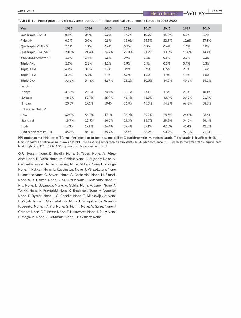
ABSTRACTS | 17 of 95
TABLE 1 . Prescriptions and effectiveness trends of first- line empirical treatments in Europe in 2013- 2020
Year 2013 2014 2015 2016 2017 2018 2019 2020
Quadruple- C+A+B 0.5% 0.9% 5.2% 17.2% 10.2% 15.3% 5.2% 5.7%
Pylera® 0.0% 0.0% 0.5% 12.0% 24.5% 22.3% 17.6% 17.8%
Quadruple- M+Tc+B 2.3% 1.9% 0.4% 0.2% 0.3% 0.4% 1.6% 0.0%
Quadruple- C+A+M/T 20.0% 21.4% 26.9% 22.3% 21.2% 10.6% 11.8% 14.4%
Sequential- C+A+M/T 8.1% 3.4% 1.8% 0.9% 0.3% 0.5% 0.2% 0.3%
Triple- A+L 2.1% 2.2% 3.2% 1.9% 0.3% 0.3% 0.4% 0.3%
Triple- A+M 4.1% 3.0% 1.7% 0.9% 0.9% 0.6% 2.3% 0.6%
Triple- C+M 3.9% 6.4% 9.0% 6.6% 1.4% 1.0% 1.0% 4.0%
Triple- C+A 53.6% 54.3% 42.7% 28.2% 30.5% 34.0% 40.6% 34.3%
Length
7 days 31.3% 28.1% 24.7% 16.7% 7.8% 1.8% 2.3% 10.1%
10 days 48.3% 52.7% 55.9% 46.4% 46.9% 43.9% 30.8% 31.7%
14 days 20.5% 19.2% 19.4% 36.8% 45.3% 54.2% 66.8% 58.3%
PPI acid inhibition*
Low 62.0% 56.7% 47.1% 36.2% 39.2% 28.5% 24.0% 33.4%
Standard 18.7% 25.5% 26.5% 24.5% 23.7% 28.8% 34.6% 24.4%
High 19.3% 17.8% 26.4% 39.4% 37.1% 42.8% 41.4% 42.2%
Eradication rate (mITT) 85.3% 85.1% 85.9% 87.4% 88.2% 90.9% 92.2% 91.3%
PPI, proton pomp inhibitor; mITT, modified intention- to- treat ; A, amoxicillin; C, clarithromycin; M, metronidazole; T, tinidazole; L, levofloxacin; B, bismuth salts; Tc, tetracycline. *Low dose PPI – 4.5 to 27 mg omeprazole equivalents, b.i.d., Standard dose PPI – 32 to 40 mg omeprazole equivalents, b.i.d, High dose PPI – 54 to 128 mg omeprazole equivalents, b.i.d.
O.P. Nyssen: None. D. Bordin: None. B. Tepes: None. A. Pérez- Aisa: None. D. Vaira: None. M. Caldas: None. L. Bujanda: None. M. Castro- Fernandez: None. F. Lerang: None. M. Leja: None. L. Rodrigo: None. T. Rokkas: None. L. Kupcinskas: None. J. Pérez- Lasala: None. L. Jonaitis: None. O. Shvets: None. A. Gasbarrini: None. H. Simsek: None. A. R. T. Axon: None. G. M. Buzás: None. J. Machado: None. Y. Niv: None. L. Boyanova: None. A. Goldis: None. V. Lamy: None. A. Tonkic: None. K. Przytulski: None. C. Beglinger: None. M. Venerito: None. P. Bytzer: None. L.G. Capelle: None. T. Milosavljevic: None. L. Veijola: None. J. Molina- Infante: None. L. Vologzhanina: None. G. Fadeenko: None. I. Ariño: None. G. Fiorini: None. A. Garre: None. J. Garrido: None. C.F. Pérez: None. F. Heluwaert: None. I. Puig: None. F. Mégraud: None. C. O'Morain: None. J.P. Gisbert: None.

ABSTRACTS18 of 95 |
EP2.13 | EuropeanRegistryonH. pyloriManagement(Hp-EuReg):Analysisofempiricalsecond-linetreatmentsinEurope
O. P. Nyssen1; A. Pérez-Aísa2; B. Tepes3; D. Vaira4; D. S. Bordin5; O. Shvets6; L. Kupcinskas7; R. Marcos Pinto8; M. Leja9; N. Fernanadez-Moreno2; I. Santaella2; G. Fiorini4; L. Vologzhanina5; G. Fadeenko10; L. Jonaitis7; A. Lucendo11; L. Bujanda12; A. S. Sarsenbaeva13; M. Areia14; M. Caldas1; A. Garre1; I. Puig15; F. Megraud16; C. O'Morain17; J. P. Gisbert1; On behalf of the Hp-EuReg Investigators1Gastroenterology Unit, Hospital Universitario de La Princesa, Instituto de Investigación Sanitaria Princesa (IIS- IP), Universidad Autónoma de Madrid, Centro de Investigación Biomédica en Red de Enfermedades Hepáticas y Digestivas (CIBEREHD), Madrid, Spain; 2Servicio de Gastroenterología, Agencia Sanitaria Costa del Sol, Red de Investigación en Servicios de Salud en Enfermedades Crónicas (REDISSEC), Marbella, Spain; 3Gastroenterology Unit, AM DC Rogaska, Rogaska Slatina, Slovenia; 4Department of Surgical and Medical Sciences, University of Bologna, Bologna, Italy; 5Department of pancreatobiliary and upper GI diseases, Moscow Clinical Scientific Center, and A.I. Yevdokimov Moscow State University of Medicine and Dentistry, Moscow, Russian Federation; 6Internal Diseases Department No.1, National Medical University named after O.O. Bogomolets, Kyiv, Ukraine; 7Department of Gastroenterology, Lithuanian University of Health Sciences, Kaunas, Lithuania; 8Centro Hospitalar do Porto, Institute of Biomedical Sciences Abel Salazar, University of Porto and CINTESIS, University of Porto, Porto, Portugal; 9Faculty of Medicine, University of Latvia, Riga, Latvia; 10National Academy of Medical Sciences, Kyiv, Ukraine; 11Hospital de Tomelloso, Ciudad Real, Spain; 12Department of Gastroenterology. Hospital Donostia/Instituto Biodonostia. Centro de Investigación Biomédica en Red de Enfermedades Hepáticas y Digestivas (CIBERehd), Universidad del País Vasco (UPV/EHU), Donosti, Spain; 13Gastroenterological center, Chelyabinsk, Russian Federation; 14Portuguese Oncology Institute Coimbra, Coimbra, Portugal; 15Althaia Xarxa Assistencial Universitària de Manresa and Universitat de Vic- Universitat Central de Catalunya (UVicUCC), Manresa, Spain; 16Laboratoire de Bactériologie, Hôpital Pellegrin, Bordeaux, France; 17Department of Clinical Medicine, Trinity College Dublin, Dublin, Spain
Background: After a failed eradication attempt, approximately 10- 20% of patients will fail to obtain H. pylori eradication.Aims: To evaluate the effectiveness of second- line empirical treatments.Methods: A systematic prospective registry of the clinical practice of European gastroenterologists on H. pylori management was es-tablished. All infected adult patients were systematically registered at AEG- REDCap e- CRF until April 2020. Modified intention- to- treat (mITT) and per- protocol (PP) analyses were performed.Results: Overall, 4,862 patients from 29 European countries were given an empirical second- line therapy. Overall effectiveness was 83.7% (by mITT) and 84% (by PP). Over 97% of patients were compli-ant. AEs were reported in 28% of the cases. Most frequent second- line prescriptions and effectiveness per antibiotic combination is
shown in table 1. After failure of first- line clarithromycin- containing treatment, optimal eradication (>90%) was obtained with moxifloxacin- containing triple therapy, Pylera® or quadruple ther-apy with levofloxacin and bismuth. In patients receiving triple regi-mens containing levofloxacin or the standard bismuth quadruple regimen, cure rates were optimized with 14- day regimens using high doses of proton pump inhibitors (PPIs). However, Pylera® or quadru-ple therapy with levofloxacin and bismuth achieved reliable eradica-tion rates regardless of the PPI dose, duration of therapy, or previous first- line treatment.Conclusion: Empirical second- line triple therapies generally pro-vided low eradication rates except when prescribing 14 days of levo-floxacin or moxifloxacin. However, high effectiveness was obtained with second- line bismuth- containing quadruple therapies.

ABSTRACTS | 19 of 95
TABLE 1 . Frequency of second- line empirical treatment prescriptions and effectiveness by modified intention- to- treat and per- protocol analyses
Treatment N % Use mITT,N(%) 95% CI PP,N(%) 95% CI
Triple- A+L 1,522 33.2 1,341 (81) 79- 83 1,320 (81) 79- 83
Pylera® (single capsule) 692 15.1 622 (90) 87- 92 607 (90) 88- 93
Quadruple- A+L+B 529 11.5 478 (89) 86- 92 462 (89) 86- 92
Triple- C+A 477 10.4 231 (81) 76- 86 226 (81) 76- 86
Quadruple- M+Tc+B 204 4.4 183 (83) 77- 89 177 (84) 78- 90
Quadruple- C+A+M 179 3.9 169 (85) 79- 90 167 (84) 79- 90
Triple- A+Mx 143 3.1 135 (91) 86- 96 135 (91) 86- 96
Triple- A+M 93 2.0 76 (60.5) 49- 72 76 (60.5) 49- 72
Other 1,023 21.0 NA NA NA NA
Total 4,862 100% 3,966 (84) 82- 85 3,966 (84) 81- 83
mITT, modified intention- to- treat; PP, per- protocol; 95% CI, 95% confidence interval; C, clarithromycin; M, metronidazole; T, tinidazole; A, amoxicillin; L, levofloxacin; B, bismuth salts; Tc, tetracycline; Mx, moxifloxacin; N, Total of patients receiving an empirical treatment; Other, Other second- line empirical treatments with less than 100 patients treated in each category.
O.P. Nyssen: None. A. Pérez- Aísa: None. B. Tepes: None. D. Vaira: None. D. S. Bordin: None. O. Shvets: None. L. Kupcinskas,: None. R. Marcos Pinto: None. M. Leja: None. N. Fernanadez- Moreno: None. I. Santaella: None. G. Fiorini: None. L. Vologzhanina: None. G. Fadeenko: None. L. Jonaitis: None. A. Lucendo: None. L. Bujanda: None. A. S. Sarsenbaeva: None. M. Areia: None. M. Caldas: None. A. Garre: None. I. Puig: None. F. Megraud: None. C. O'Morain: None. J.P. Gisbert: None.
EP2.14 | Adeadlyhug:ChitosanmicrospheresfunctionalizedwithMSI-78AantimicrobialpeptidekillHelicobacter pylori
D. R. Fonseca1,2,3; A. Moura1,2; C. L. Seabra1,2,4; V. Leiro1,2; B. N. Estevinho5; C. Teixeira6; P. Gomes6; P. Parreira1,2; M. L. Martins1,2,7
1INEB, Instituto de Engenharia Biomédica, Universidade do Porto, Porto, Portugal; 2i3S, Instituto de Investigação e Inovação em Saúde, Universidade do Porto, Porto, Portugal; 3Departamento de Engenharia Metalúrgica e de Materiais, Faculdade de Engenharia, Universidade do Porto, Porto, Portugal; 4Current address: LAQV/REQUIMTE, Departamento de Ciências Químicas, Faculdade de Farmácia, Universidade do Porto, Portugal; 5LEPABE, Departamento de Engenharia Química, Faculdade de Engenharia da Universidade Do Porto, Porto, Portugal; 6LAQV- REQUIMTE, Departamento de Química e Bioquímica, Faculdade de Ciências, Universidade do Porto, Porto, Portugal; 7ICBAS, Instituto de Ciências Biomédicas Abel Salazar, Universidade do Porto, Porto, Portugal
With the end of antibiotic era, newer therapeutic options, as anti-microbial peptides (AMPs), are a demand. Helicobacter pylori (Hp) was classified as 1 of the 16 antibiotic- resistant bacteria1. MSI- 78A is one of the fewer AMPs that demonstrated bactericidal effects against Hp2. However, in vivo AMPs undergo protease degradation and proteins aggregation, reducing their effectiveness. AMPs im-mobilization onto a biomaterial surface is an advocated strategy to
overcome these drawbacks3. This work intended the development of biocompatible chitosan microspheres decorated with MSI- 78A (AMP- ChMic) capable of crossing the gastric mucosa and kill Hp in situ, after oral administration.
Chitosan microspheres (ChMic), average diameter of 5 μm, were produced by spray drying. A heterobifunctional spacer (NHS- PEG- MAL) was bonded to the ChMic, allowed immobilizing the MSI- 78A modified with a terminal cysteine on the C- terminal (MSI- 78A- SH) in a controlled orientation.
The resulting AMP- ChMic were able to retain their integrity in a wide range of acidic pH, proving their pH- resistance and validating this approach for gastric settings. The microparticles were tested in vitro against Hp J99 strain (highly pathogenic human strain), at different timepoints and in a microspheres’ range of concentra-tions (105- 107mics/mL). Antimicrobial effect was seen after 2 hours in higher concentrations and it was maintained up to 6 hours. The fast- bactericidal effect indicates that MSI- 78A can retain its activity when immobilized onto ChMic. AMP- ChMic demonstrated high po-tential for Hp infection management, being an innovative strategy to non- antibiotic therapeutics.
1. Rawla et al. Prz. Gastroenterol., 20192. Abadie et al. Expert Rev. Anti. Infect. Ther., 20183. Parreira et al. Sci. Rep., 2019
Acknowledgements: PTDC/CTM- BIO/4043/2014 & SFRH/BD/146890/2019D.R. Fonseca: None. A. Moura: None. C.L. Seabra: None. V. Leiro: None. B.N. Estevinho: None. P. Gomes: None. P. Parreira: None. M.L. Martins: None. C. Teixeira: None.

ABSTRACTS20 of 95 |
EP2.15 | Bismuthquadruplethree-in-onesinglecapsule:3or4timesdaily?Sub-analysisoftheSpanishdataoftheEuropeanRegistry on H. pyloriManagement(Hp-EuReg)
M. Caldas1; O. P. Nyssen1; B. Gomez Rodriguez2; J. Barrio3; M. Castro-Fernández4; M. Mego5; Á. Pérez-Aisa6; N. Fernández Moreno6; B. Gómez7; L. Tito7; E. Iyo8; J. Huguet9; A. Lucendo10; T. Di Maira11; F. Martinez Cerezo12; Ó. Nuñez13; M. Barenys14; M. Perona15; A. Campillo16; P. Mata Romero17; J. Santos-Fernández18; A. Cerezo-Ruiz19; S. Lario20; M. Ramirez20; I. Puig21; F. Mégraud22; C. O'Morain23; J. P. Gisbert1,*; X. Calvet20,*; On behalf of the Hp-EuReg Investigators1Hospital Universitario de La Princesa, Instituto de Investigación Sanitaria Princesa (IIS- IP), Universidad Autónoma de Madrid, Centro de Investigación Biomédica en Red de Enfermedades Hepáticas y Digestivas (CIBEREHD), Madrid, Spain; 2Virgen de la Macarena, Seville, Spain; 3Hospital Río Hortega, Valladolid, Spain; 4Digestive Unit, Hospital de Valme, Seville, Spain; 5Hospital Universitario General de Cataluña, Quirón Salud, Barcelona, Spain; 6Servicio de Gastroenterología, Agencia Sanitaria Costa del Sol, Red de Investigación en Servicios de Salud en Enfermedades Crónicas (REDISSEC), Marbella, Malaga, Spain; 7Hospital de Mataró, Barcelona, Spain; 8Hospital Comarcal de Inca, Mallorca, Spain; 9Consorcio Hospital General Universitario de Valencia, Valencia, Spain; 10Hospital de Tomelloso, Ciudad Real, Spain; 11Hospital La Fe, Valencia, Valencia, Spain; 12Hospital Sant Joan, Reus, Spain; 13Hospital Universitario Sanitas La Moraleja, Madrid, Spain; 14Hospital de Viladecans, Barcelona, Spain; 15Hospital Quirón, Marbella, Spain; 16Hospital Reina Sofía, Tudela, Spain; 17San Pedro de Alcántara, Caceres, Spain; 18Hospital Clínico Universitario, Valladolid, Spain; 19Hospital Sierra de Segura, Jaen, Spain; 20Hospital de Sabadell, CIBERehd, Spain; 21Althaia Xarxa Assistencial Universitària de Manresa and Universitat de Vic- Universitat Central de Catalunya (UVicUCC), Manresa, Spain; 22Laboratoire de Bactériologie, Hôpital Pellegrin, Bordeaux, France; 23Department of Clinical Medicine, Trinity College Dublin, Dublin, Ireland *Both senior authors contributed equally to the study
Background: Bismuth quadruple with the quadruple single capsule Pylera® (PPI, bismuth, tetracycline and metronidazole) includes the intake of 3 capsules four times a day (3c/6h), according to the techni-cal sheet. This scheme may not be suitable for Spanish eating habits; therefore, some physicians prescribe the treatment in the form of 4 capsules three times a day (4c/8h).Aim: To assess the effectiveness and safety of Pylera® administered three times a day (4c/8h).Methods: Systematic prospective registry of the clinical practice of European gastroenterologists on the management of H. pylori infection (Hp- EuReg). All infected adult patients treated with Pylera® were sys-tematically collected at AEG- REDCap e- CRF until June 2019. Modified intention- to- treat (mITT) and per- protocol (PP) analyses were performed.Results: Of the 2,326 patients, 1,140 (74%) were treated with 3c/6h and 403 (17%) with the 4c/8h. Most of the cases (72%) were naïve to
treatment. The PPI dose did not influence the eradication rate. Both treatment schedules showed equivalent compliance, tolerance, and effectiveness (table 1). One patient suffered a serious adverse event (C. difficile infection), in the group 3c/6h.Conclusions: The prescription of quadruple therapy with single cap-sule bismuth (Pylera®) given as four capsules three times a day seems to have the same compliance, tolerance and effectiveness as the scheme included in the data sheet (three capsules four times a day).

ABSTRACTS | 21 of 95
TABLE 1 . Effectiveness (by modified intention- to- treat and per- protocol analyses), compliance and safety of treatment with Pylera® in first- , second- , and third- line
Pylera® Compliance AEs
Modifiedintention-to-treat Per-protocol
Total 1st line 2nd line 3rd line Total 1st line 2nd line 3rd line
4c/8h 97% 22% 91% 95% 92% 86% 93% 96% 94% 92%
3c/6h 98% 24% 86% 93% 83% 84% 90% 95% 87% 86%
AEs: adverse events. 4c/8h: four capsules three times a day (every 8 hours); 3c/6h: three capsules four times a day (every 6 hours).
O.P. Nyssen: None. B. Gomez Rodriguez: None. J. Barrio: None. M. Castro- Fernández: None. M. Mego: None. Á. Pérez- Aisa: None. N. Fernández Moreno: None. B. Gómez: None. L. Tito: None. E. Iyo: None. J. Huguet: None. A. Lucendo: None. T. Di Maira: None. F. Martinez Cerezo: None. Ó. Nuñez: None. M. Barenys: None. M. Perona: None. A. Campillo: None. P. Mata Romero: None. J. Santos- Fernández: None. A. Cerezo- Ruiz: None. S. Lario: None. M. Ramirez: None. I. Puig: None. F. Mégraud: None. C. O'Morain: None. J.P. Gisbert**: None. X. Calvet**: None.
EP2.16 | EuropeanRegistryonH. pyloriManagement(Hp-EuReg):Experiencewithsinglecapsulebismuthquadrupletherapyin 3,439 patients
O. P. Nyssen1; A. Perez-Aisa2; M. Castro-Fernandez3; R. Pellicano4; J. M. Huguet5; L. Rodrigo6; J. Ortuño7; B. J. Gomez-Rodriguez8; R. Marcos Pinto9; M. Areia10; M. Perona11; O. Nuñez12; M. Romano13; A. G Gravina13; L. Pozzati14; M. Fernandez-Bermejo15; M. Venerito16; P. Malfertheiner16; L. Fernandez-Salazar17; A. Gasbarrini18; D. Vaira19; M. Dominguez-Cajal20; M. Jimenez-Moreno21; E. Iyo22; J. Perez-Lasala23; J. Molina-Infante24; J. Barrio25; B. Tepes26; F. Bermejo27; D. Burgos28; P. Almela Notari29; L. Bujanda30; A. Lucendo31; F. Heluwaert32; D. Bordin33,34; L. Kunovský35,36; T. Rokkas37; L. Kupcinskas38; M. Caldas1; I. Puig39; F. Megraud40; C. O'Morain41; J. P. Gisbert1; On behalf of the Hp-EuReg Investigators1Hospital Universitario de La Princesa, IIS- IP, CIBEREHD, and UAM, Madrid, Spain; 2Servicio de Gastroenterología, Agencia Sanitaria Costa del Sol, Red de Investigación en Servicios de Salud en Enfermedades Crónicas (REDISSEC), Marbella, Spain; 3Hospital de Valme, Sevilla, Spain; 4Molinette Hospital, Turin, Italy; 5Consorcio Hospital General Universitario, Valencia, Spain; 6Hospital Universitario Central de Asturias, Oviedo, Spain; 7Hospital Universitari i Politècnic La Fe, Valencia, Spain; 8Hospital Quiron Sagrado Corazon, Sevilla, Spain; 9Centro Hospitalar do Porto, Institute of Biomedical Sciences Abel Salazar, University of Porto and CINTESIS, University of Porto, Porto, Spain; 10Oncology Institute Coimbra, Coimbra, Spain; 11Hospital Quiron, Marbella, Spain; 12Hospital Universitario Sanitas La Moraleja, Madrid, Spain; 13Università degli Studi della Campania “Luigi Vanvitelli”, Napoli, Italy; 14Hospital, Merida, Spain; 15Clinica San Francisco, Caceres, Spain; 16Otto- von- Guericke University Hospital, Magdeburg, Germany; 17Hospital Clínico Universitario, Valladolid, Spain; 18Fondazione Policlinico Universitario A. Gemelli, Rome, Italy;
19Department of Surgical and Medical Sciences, University of Bologna, Bologna, Italy; 20Hospital San Jorge, Huesca, Spain; 21Hospital Universitario, Burgos, Spain; 22Hospital Comarcal de Inca, Mallorca, Spain; 23HM Sanchinarro, Madrid, Spain; 24Hospital San Pedro de Alcantara, Caceres and Centro de Investigación Biomédica en Red de Enfermedades Hepáticas y Digestivas (CIBERehd), Caceres, Spain; 25Hospital Rio Hortega, Valladolid, Spain; 26Gastroenterology Unit, AM DC Rogaska, Rogaska Slatina, Slovenia; 27Hospital Universitario de Fuenlabrada, Madrid, Spain; 28Hospital Ramon y Cajal, Madrid, Spain; 29Hospital General Universitario, Castellon, Spain; 30Hospital Donostia, Instituto Biodonostia. Centro de Investigación Biomédica en Red de Enfermedades Hepáticas y Digestivas (CIBERehd), Universidad del País Vasco (UPV/EHU), Donosti, Spain; 31Hospital de Tomelloso, Ciudad Real, Spain; 32Centre Hospitalier Annecy Genvois, Pringy, France; 33A. S. Loginov Moscow Clinical Scientific Center, Moscow, ; 34Department of Outpatient Therapy and Family Medicine, Tver State Medical University, Moscow, Russian Federation; 35Department of Gastroenterology and Internal Medicine, University Hospital Brno, Faculty of Medicine, Masaryk University, Brno, Czech Republic, Athens, Greece; 36Department of Surgery, University Hospital Brno, Faculty of Medicine, Masaryk University, Brno, Czech Republic, Athens, Greece; 37Henry Dunant Hospital, Athens, Greece; 38Lithuanian University of Health Sciences, Kaunas, Lithuania; 39Althaia Xarxa Assistencial Universitària de Manresa and Universitat de Vic- Universitat Central de Catalunya (UVicUCC), Manresa, Spain; 40Laboratoire de Bactériologie, Hôpital Pellegrin, Bordeaux Cedex, France; 41Trinity College Dublin – Faculty of Health Sciences, Trinity College Dublin, Dublin/IE, Faculty of Health Sciences, Dublin, Ireland.
Background: Bismuth- quadruple therapy (PPI, bismuth, tetracycline and metronidazole) has resurfaced in Europe thanks to a three- in- one single- capsule formulation (Pylera®).Aim: To evaluate the effectiveness and safety of Pylera®.Methods: International prospective registry of the clinical practice of European gastroenterologists on the management of H. pylori infec-tion (Hp- EuReg). All infected adult patients treated with Pylera® ac-cording to data sheet (3 capsules/6h) were systematically collected at AEG- REDCap e- CRF until December 2019. Modified intention- to- treat (mITT) analyses were performed.Results: Overall, 2,100 patients were prescribed single- capsule bismuth- quadruple therapy (10 days, 3 capsules q.i.d.). The major-ity of these patients were naïve (63%) and 16% had peptic ulcer. Pylera® achieved a high eradication rate based on the mITT (91.9%). Effectiveness was higher when using Pylera® as a first- line treatment

ABSTRACTS22 of 95 |
(94.6%) but it had also high effectiveness as a rescue therapy, both in second- line (89.3%) or subsequent lines of therapy (3rd- 6th line: 91.9%) (Table 1). Compliance was the factor most closely associated with the effectiveness of treatment. Adverse events (AEs) were gen-erally mild- to- moderate and transient, only 3% of patients reporting
a severe AE, leading to discontinuation of treatment in 1.7% of pa-tients. ConclusionsThe 10- day treatment with single- capsule bismuth- quadruple ther-apy (Pylera®) achieves H. pylori eradication in approximately 90% of patients by mITT in real- world clinical practice, both as a first- line and rescue treatment, with a favourable safety profile.
TABLE 1 . Pylera® effectiveness (by modified intention- to- treat) in first- , and consecutive rescue treatment lines
Use,N(%) mITT,N(%) 95% CI PP,N(%) 95% CI
Overall 2,100 (6*) 1,777 (92) 91- 93 1,761 (93) 92- 94
Naïve 1.335 (63) 1,166 (95) 93- 96 1,158 (95.5) 94- 97
2nd line 465 (22) 375 (89) 86- 92 370 (90) 87- 93
3rd line 212 (10) 174 (89) 84- 93 177 (88) 83- 93* Of the total of treatments included in the Hp- EuReg up to December 2019 (i.e. N = 34,460); mITT: modified intention- to- treat; PP: per- protocol, N: total number of patients analysed.
O.P. Nyssen: None. A. Perez- Aisa: None. M. Castro- Fernandez: None. R. Pellicano: None. J.M. Huguet: None. L. Rodrigo: None. J. Ortuño: None. B.J. Gomez- Rodriguez: None. R. Marcos Pinto: None. M. Areia: None. M. Perona: None. O. Nuñez: None. M. Romano: None. A. G Gravina: None. L. Pozzati: None. M. Fernandez- Bermejo: None. M. Venerito: None. P. Malfertheiner: None. L. Fernandez- Salazar: None. A. Gasbarrini: None. D. Vaira: None. M. Dominguez- Cajal: None. M. Jimenez- Moreno: None. E. Iyo: None. J. Perez- Lasala: None. J. Molina- Infante: None. J. Barrio: None. B. Tepes: None. F. Bermejo: None. D. Burgos: None. P. Almela Notari: None. L. Bujanda: None. A. Lucendo: None. F. Heluwaert: None. D. Bordin: None. T. Rokkas: None. L. Kunovský: None. L. Kupcinskas: None. M. Caldas: None. I. Puig: None. F. Megraud: None. C. O'Morain: None. J.P. Gisbert: None.

ABSTRACTS | 23 of 95
EP2.17 | SafetyofthetreatmentofH. pylori infection: ExperiencefromtheEuropeanRegistryonH. pylori Management (Hp-EuReg)on22,000patients
O. P. Nyssen1; L. Kupcinskas2; B. Tepes3; O. Shvets4; D. Bordin5; M. Leja6; J. Carlos Machado7; T. Rokkas8; G. M Buzas9; I. Simsekn10; T. Axon11; F. Lerang12; L. Jonaitis2; R. Muñoz1; E. Resina1; M. Espada1; I. Puig13; F. Megraud14; C. O'Morain15; J. P. Gisbert1; On behalf of the Hp-EuReg Investigators1Gastroenterology Unit, Hospital Universitario de La Princesa, Instituto de Investigación Sanitaria Princesa (IIS- IP), Universidad Autónoma de Madrid, Centro de Investigación Biomédica en Red de Enfermedades Hepáticas y Digestivas (CIBEREHD), Madrid, Spain; 2Department of Gastroenterology, Lithuanian University of Health Sciences, Kaunas, Lithuania; 3Gastroenterology Unit, AM DC Rogaska, Rogaska Slatina, Slovenia; 4Internal Diseases Department No.1, National Medical University named after O.O. Bogomolets, Kyiv, Ukraine; 5Department of pancreatobiliary and upper GI diseases, Moscow Clinical Scientific Center, Moscow, Russian Federation; 6Institute of Clinical and Preventive Medicine & Faculty of Medicine, University of Latvia, Riga, Latvia; 7Instituto de Investigação e Inovação em Saúde, Universidade do Porto, and Ipatimup – Institute of Molecular Pathology and Immunology of the University of Porto, Porto, Portugal; 8Gastroenterology Unit, Henry Dunant Hospital, Athens, Greece; 9Gastroenterology Unit, Ferencváros Policlinic, Budapest, Hungary; 10Dokuz Eylul University School of Medicine, Izmir, Turkey; 11Gastroenterology Unit, University of Leeds, Leeds, United Kingdom; 12Medical Department, Central Hospital Ostfold, Fredrikstad, Norway; 13Althaia Xarxa Assistencial Universitària de Manresa and Universitat de Vic- Universitat Central de Catalunya (UVicUCC), Manresa, Spain; 14Laboratoire de Bactériologie, Hôpital Pellegrin, Bordeaux Cedex, France; 15Trinity College Dublin – Faculty of Health Sciences, Trinity College Dublin; Dublin/IE, Faculty of Health Sciences, Dublin, Ireland
Introduction: The safety of Helicobacter pylori eradication treat-ments and to what extent adverse events (AEs) may influence thera-peutic compliance is unknown.Objective: To assess the frequency, type, intensity and duration of AEs, and their impact on compliance.Methods: Systematic prospective non- interventional registry of the clinical practice of European gastroenterologists on the manage-ment of H. pylori infection. All prescribed treatments and their corre-sponding safety profile were recorded in an e- CRF in AEG- REDCap until June 2019. AEs were classified depending on the intensity of symptoms as mild/moderate/severe, and as serious AEs (death, hos-pitalisation, disability, congenial anomaly and/or requires interven-tion to prevent permanent damage).Results: The different treatments prescribed to a total of 22,492 naïve and non- naïve patients caused at least one AE in 22% of the cases (Table 1), the classic bismuth- based quadruple therapy being the worst tolerated (37% of AEs). Taste disturbance (7%), diarrhoea (7%), nausea (6%) and abdominal pain (3%) were the most frequent AEs. The majority of AEs were mild (57%), 6% were severe, and only
0.08% were serious, with an average duration of 7 days. The treat-ment compliance rate was 97%. Only 1.3% of the patients discontin-ued treatment due to AEs.Conclusions: H. pylori eradication treatment frequently induces AEs, although they are usually mild and of limited duration. Its appearance does not interfere significantly with the compliance of treatment.
TABLE 1 . Safety in naïve and non- naïve patients
Adverseevents Yes % 95% CI
Triple- C+A 1,037 15 14- 16
Quadruple- C+A+M 926 25 23- 26
Pylera® 642 28 26- 30
Quadruple - C+A+B 627 34 34- 37
Triple- A+L 339 21 19- 23
Triple- C+M 184 20 18- 23
Quadruple - A+L+B 180 32 28- 36
Triple- A+M 79 22 17- 26
Sequential- C+A+M 50 19 14- 24
Quadruple - M+Tc+B 84 37 30- 43
Quadruple - A+B+ 67 32 26- 39
Quadruple - M+D+B 62 33 26- 40
Sequential - C+A+T 5 6.8 2.2- 15
Quadruple - C+A+T 16 17 9.1- 26
Total 4,298 22 22- 23
A, amoxicillin; B, bismuth; C, clarithromycin; CI, confidence interval; D, doxycycline; L, levofloxacin; M, metronidazole; T, tinidazole; Tc, tetracycline.
O.P. Nyssen: None. L. Kupcinskas: None. B. Tepes: None. O. Shvets: None. D. Bordin: None. M. Leja: None. J. Carlos Machado: None. T. Rokkas: None. G. M Buzas: None. I. Simsekn: None. T. Axon: None. F. Lerang: None. L. Jonaitis: None. R. Muñoz: None. E. Resina: None. M. Espada: None. I. Puig: None. F. Megraud: None. C. O'Morain: None. J.P. Gisbert: None.

ABSTRACTS24 of 95 |
EP2.18 | BismuthquadrupleregimenwithtetracyclineordoxycyclineversusPylera®asthird-linerescuetherapyforH. pyloriinfection:AprospectivemulticenteranalysisoftheEuropean Registry on Helicobacter pyloriManagement(Hp-EuReg)
O. P. Nyssen1; A. Perez-Aisa2; L. Rodrigo-Sáez3; M. Castro-Fernández4; P. Mata Romero5; J. Ortuño6; J. Barrio7; J. Huguet8; I. Modollel9; N. Alcaide10; A. Lucendo11; X. Calvet12; M. Perona13; B. Gomez14; B. Gomez Rodriguez15; P. Varela16; M. Jimenez-Moreno17; M. Dominguez-Cajal18; L. Pozzati19; D. Burgos20; L. Bujanda21; J. Hinojosa2; J. Molina-Infante5; T. Di Maira6; L. Ferrer8; L. Fernández-Salazar10; A. Figuerola12; L. Tito14; C. de la Coba16; J. Gomez-Camarero17; N. Fernandez2; M. Caldas1; A. Garre1; E. Resina1; M. Espada1; I. Puig22; F. Megraud23; C. O'Morain24; J. P. Gisbert1; On behalf of the Hp-EuReg Investigators1Gastroenterology Unit, Hospital Universitario de La Princesa, Instituto de Investigación Sanitaria Princesa (IIS- IP), Universidad Autónoma de Madrid, and Centro de Investigación Biomédica en Red de Enfermedades Hepáticas y Digestivas (CIBERehd), Madrid, Spain; 2Servicio de Gastroenterología, Agencia Sanitaria Costa del Sol, Red de Investigación en Servicios de Salud en Enfermedades Crónicas (REDISSEC), Marbella, Spain; 3Hospital Universitario Central de Asturias, Oviedo, Spain; 4Hospital de Valme and CIBEREHD, Sevilla, Spain; 5Hospital San Pedro de Alcántara and CIBEREHD, Caceres, Spain; 6Hospital Universitari i Politècnic La Fe, Valencia, Spain, 7Hospital Rio Hortega, Valladolid, Spain; 8Consorcio Hospital General Universitario, Valencia, Spain; 9Consorci Sanitari Terrassa, Terrasa, Spain; 10Hospital Clínico Universitario, Valladolid, Spain; 11Hospital de Tomelloso, Ciudad Real, Spain; 12Hospital de Sabadell and CIBEREHD, Barcelona, Spain; 13Hospital Quiron, Marbella, Spain; 14Hospital de Mataró, Barcelona, Spain; 15Hospital Virgen de la Macarena, Sevilla, Spain; 16Hospital de Cabueñes, Gijon, Spain; 17Hospital Universitario, Burgos, Spain; 18Hospital San Jorge, Huesca, Spain; 19Hospital de Mérida, Merida, Spain; 20Hospital Ramon y Cajal, Madrid, Spain; 21Hospital Donostia/Instituto Biodonostia. Centro de Investigación Biomédica en Red de Enfermedades Hepáticas y Digestivas (CIBERehd). Universidad del País Vasco (UPV/EHU), San Sebastián, Spain; 22Althaia Xarxa Assistencial Universitària de Manresa and Universitat de Vic- Universitat Central de Catalunya (UVicUCC), Manresa, Spain; 23Université de Bordeaux, Centre National de Référence des Campylobacters et Hélicobacters, Bordeaux, France; 24Gastroenterology Unit, Trinity College, Dublin, Ireland
Background: Different bismuth- quadruple therapies containing proton pump inhibitors, bismuth, metronidazole, and a tetracycline have been recommended as third- line Helicobacter pylori eradication treatment after failure with clarithromycin and levofloxacin.Aim: To evaluate the effectiveness and safety of third- line treat-ments with bismuth, metronidazole and either tetracycline or doxycycline.Methods: Sub- study of the European Registry on H. pylori Management (Hp- EuReg), an international multicenter prospec-tive non- interventional registry of the routine clinical practice of
European gastroenterologists. After previous failure with clarithro-mycin- and levofloxacin- containing therapies, patients receiving a third- line regimen with 10/14- day of PPI, bismuth, metronidazole and either tetracycline (T) or doxycycline (D), or 10- day Pylera® (P) were registered at AEG- REDCap.Results: Overall, 454 patients were treated: 85 with T, 94 with D, and 275 with P. Overall modified intention- to- treat and per- protocol eradication rates were 81% (D: 65%, T: 76%, P: 88%) and 82% (D: 66%, T: 77%, P: 88%), respectively (Table 1). Higher eradication rates were associated with compliance (OR = 2.96; 95% CI = 1.01- 8.84) and no prior metronidazole use (OR = 1.96; 95% CI = 1.15- 3.33); P was superior to D (OR = 4.46; 95% CI = 2.51- 8.27), and T marginally superior to D (OR = 1.67; 95% CI = 0.85- 3.29).Conclusion: Third- line (after failure with clarithromycin and levo-floxacin) H. pylori eradication with bismuth quadruple treatment of-fers acceptable effectiveness and safety. Highest effectiveness was found in compliant patients and in those taking 10- day Pylera® or 14- day tetracycline. Doxycycline appears less effective and there-fore should not be recommended.

ABSTRACTS | 25 of 95
TABLE 1 . Effectiveness (by modified intention- to- treat and per- protocol) and compliance according to the treatment regimen and length
Effectiveness,N(%) Compliance mITT, N mITT 95% CI PP, N PP 95% CI
Group T All 82 (97%) 64 76% 66- 86 63 77% 67- 86
10 days* 29 (97%) 19 66% 47- 85 19 66% 47- 85
14 days 45 (96%) 45 82% 71- 93 44 83% 72- 94
Group D All 85 (93%) 58 65% 55- 76 56 66% 55- 77
10 days* 37 (90%) 25 63% 46- 79 23 63% 45- 79
14 days 53 (96%) 32 70% 55- 84 32 71% 57- 85
Group P 10 days* 249 (96%) 222 88% 83- 92 216 88% 84- 92
Group T – tetracycline containing bismuth quadruple therapy, Group D – doxycycline containing bismuth quadruple therapy, Group P – single capsule bismuth quadruple therapy (Pylera®), 95% CI – 95% confidence interval. ITT: intention- to- treat, mITT: modified intention- to- treat; PP: per- protocol. The Chi2 test showed statistically significant differences of treatment length in the mITT set between treatment groups (T, D and P) as reported in the table: *P < 0.001.
O.P. Nyssen: None. A. Perez- Aisa: None. L. Rodrigo- Sáez: None. M. Castro- Fernández: None. P. Mata Romero: None. J. Ortuño: None. J. Barrio: None. J. Huguet: None. I. Modollel: None. N. Alcaide: None. A. Lucendo: None. X. Calvet: None. M. Perona: None. B. Gomez: None. B. Gomez Rodriguez: None. P. Varela: None. M. Jimenez- Moreno: None. M. Dominguez- Cajal: None. L. Pozzati: None. D. Burgos: None. L. Bujanda: None. J. Hinojosa: None. J. Molina- Infante: None. T. Di Maira: None. L. Ferrer: None. L. Fernández- Salazar: None. A. Figuerola: None. L. Tito: None. C. de la Coba: None. J. Gomez- Camarero: None. N. Fernandez: None. M. Caldas: None. A. Garre: None. E. Resina: None. M. Espada: None. I. Puig: None. F. Megraud: None. C. OMorain: None. J.P. Gisbert: None.
EP2.19 | EuropeanRegistryonH. pyloriManagement(Hp-EuReg):Clinicalphenotypesthroughmachine learningoffirst-linetreatedpatientsinSpainduringtheperiod2013-2018
O. P. Nyssen1; A. Sanz-Garcia2; G. Ortega2; J. P. Gisbert1; On behalf of the Hp-EuReg Investigators1Hospital Universitario de La Princesa, IIS- IP, UAM, CIBEREHD, Madrid, Spain; 2Unidad de análisis de datos, Hospital Universitario de la Princesa, Madrid, Spain
Background: Patients’ segmentation in homogeneous groups could help to improve the effectiveness of current Helicobacter pylori erad-ication therapy.
Objectives: 1) To group patients from the European Registry on Helicobacter. pylori management (Hp- EuReg) according to their de-mographic and clinical characteristics and to the treatment types through multivariate categorical analysis and subsequent cluster de-composition. 2) To evaluate treatments’ effectiveness.Methods: Categorical variables used: sex, ethnicity, diagnosis, symp-toms, therapeutic indication, treatment and its duration, proton- pump inhibitor (PPI) dose, compliance, adverse events, and region of the prescribing center.Results: Overall, 8,322 patients were analysed from 2013 to 2018. Table 1 shows the increase of effectiveness, ranging from 78.4% in 2013 to 92.2% in 2018. The lowest effectiveness, for clusters with over 100 patients, was obtained in cluster 1 (2015), with an eradi-cation rate of 82.8%. This cluster comprised: triple therapy with PPI- clarithromycin- amoxicillin and concomitant therapy with PPI- clarithromycin- amoxicillin- metronidazole/tinidazole, lasting both 10 days in most of cases. High PPI dose, 10- day Pylera® treatment obtained over 95% effectiveness in cluster 3 (2018), uniformly dis-tributed among Malaga, Valencia, Ciudad Real, Sevilla, Madrid, and Valladolid. High PPI dose, 14- day concomitant therapy achieved 90.7% effectiveness in cluster 1 (2018), among Malaga, Ciudad Real and Madrid.Conclusion: The cluster analysis allows both identifying homogene-ous groups of patients as well as assessing the effectiveness of the different first- line treatments evaluated.
TABLE 1 . Trends in the overall effectiveness (by modified intention- to- treat, per cluster) between 2013 and 2018 in Spain
year Number of clusters
Effectiveness%(numberofpatients)percluster
1 2 3 4 5
2013 3 89.6 (280) 83.2 (619) 80.0 (80)
2014 3 88.2 (1685) 69.6 (46) 83.3 (6)
2015 5 91.6 (83) 89.4 (839) 82.9 (615) 70.8 (24) 88.2 (17)
2016 3 94.7 (359) 86.1 (853) 92.6 (296)
2017 3 94.3 (630) 90.6 (636) 91.6 (153)
2018 3 91.8 (122) 90.7 (161) 96.5 (338)
Simple underlining highlights the lowest effectiveness for groups of more than 100 patients and double underlining the highest effectiveness.

ABSTRACTS26 of 95 |
O.P. Nyssen: None. A. Sanz- Garcia: None. G. Ortega: None. J.P. Gisbert: None.
EP2.20 | Empiricalversussusceptibility-guidedtreatmentofHelicobacter pyloriinfection:Ameta-analysis
M. Espada; O. P. Nyssen; J. P. GisbertGastroenterology Unit, Hospital Universitario de La Princesa, Instituto de Investigación Sanitaria Princesa (IIS- IP), Universidad Autónoma de Madrid, and Centro de Investigación Biomédica en Red de Enfermedades Hepáticas y Digestivas (CIBERehd), Madrid, Spain
Background: Treating patients according to antibiotic resistances has frequently been recommended. However, the information on its real effectiveness is scarce.Aim: To perform a meta- analysis comparing empirical versus susceptibility- guided treatment of H. pylori.Methods: Selection of studies: comparing empirical versus susceptibility- guided treatment. Search strategy: electronic&manual. Data synthesis: by intention- to- treat (random effects model).Results: Overall, 38 studies were included (6,525 patients in the em-pirical and 5,124 in the susceptibility- guided group). H. pylori eradi-cation was achieved in 87% versus 77% respectively (RR: 1.13; 95% CI: 1.09- 1.17; I2: 77%). Similar results were found when only RCTs were evaluated (22 studies; RR: 1.16; 95% CI: 1.10- 1.22; I2: 72%). Similar results were also observed when susceptibility testing was assessed by culture (RR: 1.13; 95% CI: 1.07- 1.19) or PCR (RR: 1.14; 95% CI: 1.05- 1.23). When assessing first- line treatments (naïve pa-tients) only (28 studies), better efficacy results were obtained with the susceptibility- guided strategy (RR: 1.16; 95% CI: 1.11- 1.21; I2: 79%). However, when prescribing empirical first- line quadruple regi-mens only (both with and without bismuth, excluding the suboptimal triple therapies), no differences in efficacy were found versus the susceptibility- guided group (RR: 1.02; 95% CI: 0.95- 1.09); this lack of difference was confirmed in RCTs not based on CYP2C19 gene polymorphism (RR: 1.07; 95% CI: 0.98- 1.17). For rescue- therapies (13 studies, most as 2nd- line), similar results were demonstrated with both strategies, both including all studies (RR: 1.09; 95% CI: 0.97- 1.22; I2: 82%) and also when only RCTs were considered (RR: 1.15; 95% CI: 0.97- 1.36).Conclusions: The benefit of susceptibility- guided treatment over empirical treatment of H. pylori infection could not be demonstrated, either in first- line (if the most updated quadruple regimens are pre-scribed) or in rescue therapies.M. Espada: None. O.P. Nyssen: None. J.P. Gisbert: None.
EP2.21 | Real-worldcomparativeeffectsofthree-in-onesinglecapsulebismuthquadrupletherapyvsnon-bismuthquadrupleconcomitant therapy: Interim analysis of the European Registry on H. pyloriManagement(Hp-EuReg)
I. Puig1; M. Serra2; O. P. Nyssen3; G. Fiorini4; D. Vaira4; I. Saracino4; Á. Perez-Aisa5; N. Fernandez-Moreno5; I. Santaella5; M. Castro-Fernandez6; L. Bujanda7; A. Lucendo8; M. Areia9; A. Gasbarrini10; M. Romano11; A. Gravina11; R. Marcos-Pinto12; F. Mégraud13; C. O'Morain14; J. P. Gisbert3; On behalf of the Hp-EuReg Investigators1Digestive Diseases Department, Althaia Xarxa Assistencial Universitària de Manresa and Uni- versitat de Vic- Universitat Central de Catalunya (UVicUCC), Manresa, Spain; 2Center for Research in Health and Economics (CRES), Pompeu Fabra University (UPF), Barcelona, Spain; 3Gastroenterology Unit, Hospital Universitario de La Princesa, Instituto de Investigación Sanitaria Princesa (IIS- IP), Universidad Autónoma de Madrid, and Centro de Investigación Biomédica en Red de Enfermedades Hepáticas y Digestivas (CIBERehd), Madrid, Spain; 4Department of Surgical and Medical Sciences, University of Bologna, Bologna, Italy; 5Servicio de Gastroenterología, Agencia Sanitaria Costa del Sol, Red de Investigación en Servicios de Salud en Enfermedades Crónicas (REDISSEC), Marbella, Spain; 6Hospital de Valme, Sevilla, Spain; 7Department of Gastroenterology, Centro de Investigación Biomédica en Red de Enfermeda- des Hepáticas y Digestivas (CIBERehd), Hospital Donostia/Instituto Biodonostia, Universidad del País Vasco (UPV/EHU), Donosti, Spain; 8Department of Gastroenterology, Hospital General de Tomelloso, Ciudad Real, Spain; 9Portuguese Oncology Institute, Coimbra, Portugal; 10Gastroenterology Area, Fondazione Policlinico Universitario A. Gemelli, Rome, Italy; 11Università degli Studi della Campania “Luigi Vanvitelli”, Napoli, Italy; 12Department of Gastroenterology, Porto Centre Hospital, Porto, Portugal; 13Laboratoire de Bactériologie, Hôpital Pellegrin, Bordeaux, France; 14Department of Clinical Medicine, Trinity College, Dublin, Ireland
Background: RCTs have strict selection criteria that render results not fully transferable to the clinical practice.Objective: To assess the comparative effects of first- line bismuth- single- capsule (PPI- bismuth- tetracycline- metronidazole) vs non- bismuth quadruple concomitant therapy (PPI- amoxicillin- clarithromycin- nitroimidazole).Methods: The European Registry on H. pylori management (Hp- EuReg) data from Spain, Italy and Portugal was used to emulate a target trial with prospective observational data, comparing the relative effectiveness (modified intention- to- treat, mITT; and per- protocol, PP) and safety (adverse events, AEs) of first- line bismuth- single- capsule [10 days, 3 PPI dosages] and concomitant therapy [10/14 days; 3 PPI dosages], rendering 9 prescription strategies. Regression analysis controlling for confounders was used to esti-mate the relative effects of each strategy.Results: Overall, 2,340 individuals were included. Compared to 10- day concomitant therapy at low PPI doses (n = 484), all bismuth- single- capsule combinations presented an eradication incremental
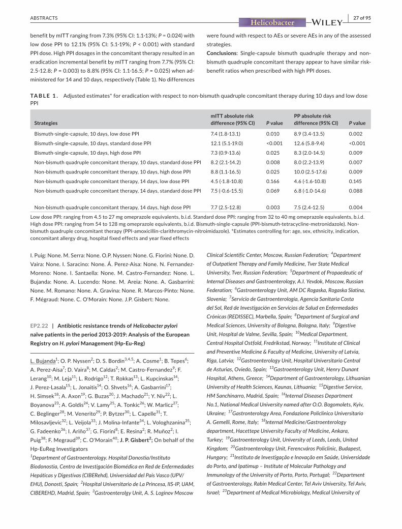
ABSTRACTS | 27 of 95
benefit by mITT ranging from 7.3% (95% CI: 1.1- 13%; P = 0.024) with low dose PPI to 12.1% (95% CI: 5.1- 19%; P < 0.001) with standard PPI dose. High PPI dosages in the concomitant therapy resulted in an eradication incremental benefit by mITT ranging from 7.7% (95% CI: 2.5- 12.8; P = 0.003) to 8.8% (95% CI: 1.1- 16.5; P = 0.025) when ad-ministered for 14 and 10 days, respectively (Table 1). No differences
were found with respect to AEs or severe AEs in any of the assessed strategies.Conclusions: Single- capsule bismuth quadruple therapy and non- bismuth quadruple concomitant therapy appear to have similar risk- benefit ratios when prescribed with high PPI doses.
TABLE 1 . Adjusted estimates* for eradication with respect to non- bismuth quadruple concomitant therapy during 10 days and low dose PPI
StrategiesmITT absolute risk difference(95%CI) Pvalue
PP absolute risk difference(95%CI) Pvalue
Bismuth- single- capsule, 10 days, low dose PPI 7.4 (1.8- 13.1) 0.010 8.9 (3.4- 13.5) 0.002
Bismuth- single- capsule, 10 days, standard dose PPI 12.1 (5.1- 19.0) <0.001 12.6 (5.8- 9.4) <0.001
Bismuth- single- capsule, 10 days, high dose PPI 7.3 (0.9- 13.6) 0.025 8.3 (2.0- 14.5) 0.009
Non- bismuth quadruple concomitant therapy, 10 days, standard dose PPI 8.2 (2.1- 14.2) 0.008 8.0 (2.2- 13.9) 0.007
Non- bismuth quadruple concomitant therapy, 10 days, high dose PPI 8.8 (1.1- 16.5) 0.025 10.0 (2.5- 17.6) 0.009
Non- bismuth quadruple concomitant therapy, 14 days, low dose PPI 4.5 (- 1.8- 10.8) 0.166 4.6 (- 1.6- 10.8) 0.145
Non- bismuth quadruple concomitant therapy, 14 days, standard dose PPI 7.5 (- 0.6- 15.5) 0.069 6.8 (- 1.0- 14.6) 0.088
Non- bismuth quadruple concomitant therapy, 14 days, high dose PPI 7.7 (2.5- 12.8) 0.003 7.5 (2.4- 12.5) 0.004
Low dose PPI: ranging from 4.5 to 27 mg omeprazole equivalents, b.i.d. Standard dose PPI: ranging from 32 to 40 mg omeprazole equivalents, b.i.d. High dose PPI: ranging from 54 to 128 mg omeprazole equivalents, b.i.d. Bismuth- single- capsule (PPI- bismuth- tetracycline- metronidazole). Non- bismuth quadruple concomitant therapy (PPI- amoxicillin- clarithromycin- nitroimidazole). *Estimates controlling for: age, sex, ethnicity, indication, concomitant allergy drug, hospital fixed effects and year fixed effects
I. Puig: None. M. Serra: None. O.P. Nyssen: None. G. Fiorini: None. D. Vaira: None. I. Saracino: None. Á. Perez- Aisa: None. N. Fernandez- Moreno: None. I. Santaella: None. M. Castro- Fernandez: None. L. Bujanda: None. A. Lucendo: None. M. Areia: None. A. Gasbarrini: None. M. Romano: None. A. Gravina: None. R. Marcos- Pinto: None. F. Mégraud: None. C. O'Morain: None. J.P. Gisbert: None.
EP2.22 | AntibioticresistancetrendsofHelicobacter pylori naïvepatientsintheperiod2013-2019:AnalysisoftheEuropeanRegistry on H. pyloriManagement(Hp-Eu-Reg)
L. Bujanda1; O. P. Nyssen2; D. S. Bordin3,4,5; A. Cosme1; B. Tepes6; A. Perez-Aisa7; D. Vaira8; M. Caldas2; M. Castro-Fernandez9; F. Lerang10; M. Leja11; L. Rodrigo12; T. Rokkas13; L. Kupcinskas14; J. Perez-Lasala15; L. Jonaitis14; O. Shvets16; A. Gasbarrini17; H. Simsek18; A. Axon19; G. Buzas20; J. Machado21; Y. Niv22; L. Boyanova23; A. Goldis24; V. Lamy25; A. Tonkic26; W. Marlicz27; C. Beglinger28; M. Venerito29; P. Bytzer30; L. Capelle31; T. Milosavljevic32; L. Veijola33; J. Molina-Infante34; L. Vologhzanina35; G. Fadeenko36; I. Ariño37; G. Fiorini8; E. Resina2; R. Muñoz2; I. Puig38; F. Megraud39; C. O'Morain40; J. P. Gisbert2; On behalf of the Hp-EuReg Investigators1Department of Gastroenterology. Hospital Donostia/Instituto Biodonostia, Centro de Investigación Biomédica en Red de Enfermedades Hepáticas y Digestivas (CIBERehd), Universidad del País Vasco (UPV/EHU), Donosti, Spain; 2Hospital Universitario de La Princesa, IIS- IP, UAM, CIBEREHD, Madrid, Spain; 3Gastroenterolgy Unit, A. S. Loginov Moscow
Clinical Scientific Center, Moscow, Russian Federation; 4Department of Outpatient Therapy and Family Medicine, Tver State Medical University, Tver, Russian Federation; 5Department of Propaedeutic of Internal Diseases and Gastroenterology, A.I. Yevdok, Moscow, Russian Federation; 6Gastroenterology Unit, AM DC Rogaska, Rogaska Slatina, Slovenia; 7Servicio de Gastroenterología, Agencia Sanitaria Costa del Sol, Red de Investigación en Servicios de Salud en Enfermedades Crónicas (REDISSEC), Marbella, Spain; 8Department of Surgical and Medical Sciences, University of Bologna, Bologna, Italy; 9Digestive Unit, Hospital de Valme, Sevilla, Spain; 10Medical Department, Central Hospital Ostfold, Fredrikstad, Norway; 11Institute of Clinical and Preventive Medicine & Faculty of Medicine, University of Latvia, Riga, Latvia; 12Gastroenterology Unit, Hospital Universitario Central de Asturias, Oviedo, Spain; 13Gastroenterology Unit, Henry Dunant Hospital, Athens, Greece; 14Department of Gastroenterology, Lithuanian University of Health Sciences, Kaunas, Lithuania; 15Digestive Service, HM Sanchinarro, Madrid, Spain; 16Internal Diseases Department No.1, National Medical University named after O.O. Bogomolets, Kyiv, Ukraine; 17Gastronterology Area, Fondazione Policlinico Universitario A. Gemelli, Rome, Italy; 18Internal Medicine/Gastroenterology department, Hacettepe University Faculty of Medicine, Ankara, Turkey; 19Gastroenterology Unit, University of Leeds, Leeds, United Kingdom; 20Gastroenterology Unit, Ferencváros Policlinic, Budapest, Hungary; 21Instituto de Investigação e Inovação em Saúde, Universidade do Porto, and Ipatimup – Institute of Molecular Pathology and Immunology of the University of Porto, Porto, Portugal; 22Department of Gastroenterology, Rabin Medical Center, Tel Aviv University, Tel Aviv, Israel; 23Department of Medical Microbiology, Medical University of

ABSTRACTS28 of 95 |
Sofia, Sofia, Bulgaria; 24Gastroenterology Unit, Timisoara Hospital, Timisoara, Romania; 25Department of Gastroenterology, Hepatology & Nutrition, CHU Charleroi, Charleroi, Belgium; 26University Hospital Centre, Split, Croatia; 27Gastroenterology Unit, Pomeranian Medical University, Szczecin, Poland; 28Gastroenterology Unit, Hospital de Basel, Basel, Switzerland; 29Department of Gastroenterology, Hepatology and Infectious Diseases, Otto- von- Guericke University Hospital, Magdeburg, Germany; 30Department of Medicine, Zealand University Hospital, Copenhagen Universit, Copenhagen, Denmark; 31Gastroenterology and Hepatology, Erasmus MC University, Rotterdam, Netherlands; 32Medical Department, Clinical Center of Serbia Clinic for Gastroenterology and hepatology, University of Belgrade, Belgrade, Serbia; 33Internal Medicine, Herttoniemi Hospital, Helsinki, Finland; 34Gastroenterology Unit, Hospital San Pedro de Alcántara, Caceres, Spain; 35Gastroenterology Unit Gastrocentre, Perm, Russian Federation; 36Digestive Ukrainian Academy of Medical Sciences, Kyiv, Ukraine; 37Gastroenterology Unit, Hospital Clínico Universitario Lozano Blesa, Zaragoza, Spain; 38Althaia Xarxa Assistencial Universitària de Manresa and Universitat de Vic- Universitat Central de Catalunya (UVicUCC), Manresa, Spain; 39Laboratoire de Bactériologie, Hôpital Pellegrin, Bordeaux, Cedex, France; 40Trinity College Dublin – Faculty of Health Sciences, Trinity College Dublin, Dublin/IE, Faculty of Health Sciences, Dublin, Ireland
Background: Bacterial antibiotic resistance changes over time based on multiple factors. It is essential to study these trends to apply pre-ventive strategies to help reducing such resistances.
Objective: To conduct a time- trend analysis of the antibiotic re-sistance to H. pylori infection in the European Registry on H. pylori (Hp- EuReg).Patients and Methods: International multicenter prospective non- interventional European Registry on H. pylori Management (Hp- EuReg) aiming to evaluate the decisions and outcomes of H. pylori infection by European gastroenterologists. All infected adult pa-tients diagnosed with culture and with a result of the antibiotic re-sistance test were registered at AEG- REDCap e- CRF from 2013 to 2019.Results: A total of 32,447 patients were included, and culture was performed in 3,474 (11%), where 2,483 näive patients were included for analysis. Resistance to at least one antibiotic was described in 57% of the patients. Resistance to metronidazole (27%) was most frequent, whereas resistance to tetracycline and amoxicillin was below 1%. Clarithromycin resistance remained above 15% through-out the studied years (Table 1). A significant decrease in the met-ronidazole resistance rate was observed between 2013 (38%) and 2018 (21%).Conclusion: In naïve patients, resistance to clarithromycin re-mained above 15% in the period 2013- 2019. A progressive de-crease in metronidazole resistance was observed. No increasing or decreasing trend was observed in the bacterial resistance to other antibiotics.
TABLE 1 . Antibiotic resistance trends (2013- 2019) of Helicobacter pylori naïve patients in Europe
N(%) 2013 2014 2015 2016 2017 2018 2019Variation range
Nº Cultures 435 522 469 286 355 282 310 282- 522
No resistance 210 (48) 259 (50) 197 (42) 93 (33) 162 (46) 106 (38) 104 (33.5) 33- 50
Clarithromycin (C)
86 (20) 120 (23) 117 (25) 59 (21) 68 (19) 65 (23) 57 (18) 18- 25
Metronidazole (M)
165 (38) 156 (30) 140 (30) 72 (25) 64 (18) 60 (21) 66 (21) 18- 38
Levofloxacin (L) 58 (13) 100 (19) 103 (22) 46 (16) 59 (17) 55 (20) 41 (13) 13- 22
Amoxicillin 6 (1) 0 (0) 0 (0) 0 (0) 10 (3) 1 (0.4) 0 (0) <1
Tetracycline 3 (0.7) 1 (0.2) 0 (0) 1 (0.3) 5 (1.4) 0 (0) 1 (0.3) <1.4
Dual (C+M) 56 (13) 65 (13) 62 (13) 33 (12) 30 (9) 28 (10) 29 (9) 9- 13
Triple (C+M+L) 22 (5) 31 (6) 32 (7) 16 (6) 12 (3) 12 (4) 8 (3) 3- 7
C, clarithromycin; L, levofloxacin; M, metronidazole.
L. Bujanda: None. O. P. Nyssen: None. D. S. Bordin: None. A. Cosme: None. B. Tepes: None. A. Perez- Aisa: None. D. Vaira: None. M. Caldas: None. M. Castro- Fernandez: None. F. Lerang: None. M. Leja: None. L. Rodrigo: None. T. Rokkas: None. L. Kupcinskas: None. J. Perez- Lasala: None. L. Jonaitis: None. O. Shvets: None. A. Gasbarrini: None. H. Simsek: None. A. Axon: None. G. Buzas: None. J. Machado: None. Y. Niv: None. L. Boyanova: None. A. Goldis: None. V. Lamy: None. A. Tonkic: None. W. Marlicz: None. C. Beglinger: None. M.
Venerito: None. P. Bytzer: None. L. Capelle: None. T. Milosavljevic: None. L. Veijola: None. J. Molina- Infante: None. L. Vologhzanina: None. G. Fadeenko: None. I. Ariño: None. G. Fiorini: None. E. Resina: None. R. Muñoz: None. I. Puig: None. C. O'Morain: None. F. Megraud: None. J.P. Gisbert: None.

ABSTRACTS | 29 of 95
EP2.23 | Tailoredtherapybasedonclarithromycinresistanceiseffectiveforthefirst-linetherapyofHelicobacter pylori
S. Moon; H. Kang; C. Lim; J. OHThe Catholic University of Korea, Seoul, Republic of Korea
Background: Standard triple therapy (STT, PPI- clarithromycin- amoxicillin) for Helicobacter pylori (H. pylori) eradication regi-men shows a lower treatment success rate recently. Dual Priming Oligonucleotide – based multiplex polymerase chain reaction (DPO- PCR) can be used to detect A2142G and/or A2143G point mutations of H. Pylori causing clarithromycin resistance (CAM- R). We com-pared the eradication rate of H. pylori between empirical STT and tailored therapy by DPO- PCR.Methods: A total of 575 H. pylori- infected patients were retrospec-tively evaluated in Eunpyeong St. Mary's Hospital, Korea. Empirical therapy by traditional method (Warthin- Starry silver stain) group was treated with STT. DPO- PCR was performed in the tailored therapy group. The CAM- R positive patients (who had A2142G and/or A2143G point mutations) were treated with bismuth- containing quadruple therapy (PPI- bismuth- metronidazole- tetracycline) for 7- 14 days, while the CAM- R negative patients were treated with STT for 7- 14 days. Eradication success was defined as a negative 13C- urea breath test at least 4 weeks after the treatment ended.Results: We included 536 patients, 39 patients were lost to follow- up: 284 patients were treated by empirical STT as first- line and 242 patients belonged to the tailored therapy group. The tailored therapy group was superior to the empirical therapy group in terms of first- line therapy both in the intention- to- treat analysis (70.4% vs 83.7%, P = 0.0002) and in the per- protocol analysis (76.9% vs 89.4%, P = 0.0003). Clarithromycin resistance ratio by DPO- PCR was 33.1% (80/242).Conclusion: The tailored therapy by DPO- PCR is more effective than the empirical therapy for the first- line therapy of H. pylori in a country with a high clarithromycin resistance.S. Moon: None. H. Kang: None. C. Lim: None. J. Oh: None.
EP2.24 | Helicobacter pylori antibiotic resistance: Data from the European Registry on H. pyloriManagement(Hp-EuReg)
L. Bujanda1; O. P. Nyssen2; A. Cosme1; D. Bordin3,4,5; B. Tepes6; A. Perez-Aisa7; D. Vaira8; M. Caldas2; M. Castro-Fernandez9; F. Lerang10; M. Leja11; L. Rodrigo12; T. Rokkas13; L. Kupcinskas14; J. Perez-Lasala15; L. Jonaitis14; O. Shvets16; A. Gasbarrini17; H. Simsek18; A. T. R Axon19; G. Buzas20; J. Machado21; Y. Niv22; L. Boyanova23; A. Goldis24; V. Lamy25; A. Tonkic26; W. Marlicz27; C. Beglinger28; M. Venerito29; P. Bytzer30; L. Capelle31; T. Milosavljevic32; L. Veijola33; J. Molina-Infante34; L. Vologhzanina35; G. Fadeenko36; I. Ariño37; G. Fiorini8; E. Resina2; R. Muñoz2; I. Puig38; F. Megraud39; C. O'Morain40; J. P. Gisbert2; On behalf of the Hp-EuReg Investigators1Department of Gastroenterology. Hospital Donostia/Instituto Biodonostia, Centro de Investigación Biomédica en Red de
Enfermedades Hepáticas y Digestivas (CIBERehd), Universidad del País Vasco (UPV/EHU), Donosti, Spain; 2Hospital Universitario de La Princesa, IIS- IP, UAM, CIBEREHD, Madrid, Spain; 3Gastroenterolgy Unit, A. S. Loginov Moscow clinical scientific center, Moscow, Russian Federation; 4Department of Outpatient Therapy and Family Medicine, Tver State Medical University, Tver, Russian Federation; 5Department of propaedeutic of internal diseases and gastroenterology, A.I. Yevdoki, Moscow, Russian Federation; 6Gastroenterology Unit, AM DC Rogaska, , Rogaska Slatina, Slovenia; 7Servicio de Gastroenterología, Agencia Sanitaria Costa del Sol, Red de Investigación en Servicios de Salud en Enfermedades Crónicas (REDISSEC), Marbella, Spain; 8Department of Surgical and Medical Sciences, University of Bologna, Bologna, Italy; 9Digestive Unit, Hospital de Valme, Sevilla, Spain; 10Medical Department, Central Hospital Ostfold, Fredrikstad, Norway; 11Institute of Clinical and Preventive Medicine & Faculty of Medicine, University of Latvia, Riga, Latvia; 12Gastroenterology Unit, Hospital Universitario Central de Asturias, Oviedo, Spain; 13Gastroenterology Unit, Henry Dunant Hospital, Athens, Greece; 14Department of Gastroenterology, Lithuanian University of Health Sciences, Kaunas, Lithuania; 15Digestive Service, HM Sanchinarro, Madrid, Spain; 16Internal Diseases Department No.1, National Medical University named after O.O. Bogomolets, Kyiv, Ukraine; 17Gastronterology Area, Fondazione Policlinico Universitario A. Gemelli, Rome, Italy; 18Internal Medicine/Gastroenterology department, Hacettepe University Faculty of Medicine, Ankara, Turkey; 19Gastroenterology Unit, University of Leeds, Leeds, United Kingdom; 20Gastroenterology Unit, Ferencváros Policlinic, Budapest, Hungary; 21Instituto de Investigação e Inovação em Saúde, Universidade do Porto, and Ipatimup – Institute of Molecular Pathology and Immunology of the University of Porto, Porto, Portugal; 22Department of Gastroenterology, Rabin Medical Center, Tel Aviv University, Tel Aviv, Israel; 23Department of Medical Microbiology, Medical University of Sofia, Sofia, Bulgaria; 24Gastroenterology Unit, Timisoara Hospital, Timisoara, Romania; 25Department of Gastroenterology, Hepatology & Nutrition, CHU Charleroi, Charleroi, Belgium; 26University Hospital Centre, Split, Croatia; 27Gastroenterology Unit, Pomeranian Medical University, Szczecin, Poland; 28Gastroenterology Unit, Hospital de Basel, Basel, Switzerland; 29Department of Gastroenterology, Hepatology and Infectious Diseases, Otto- von- Guericke University Hospital, Magdeburg, Germany; 30Department of Medicine, Zealand University Hospital, Copenhagen University, Copenhagen, Denmark; 31Gastroenterology and Hepatology, Erasmus MC University, Rotterdam, Netherlands; 32Medical Department, Clinical Center of Serbia Clinic for Gastroenterology and Hepatology, University of Belgrade, Belgrade, Serbia; 33Internal Medicine, Herttoniemi Hospital, Helsinki, Finland; 34Gastroenterology Unit, Hospital San Pedro de Alcántara, Caceres, Spain; 35Gastroenterology Unit Gastrocentre, Perm, Russian Federation; 36Digestive Ukrainian Academy of Medical Sciences, Kyiv, Ukraine; 37Gastroenterology Unit, Hospital ClÍnico Universitario Lozano Blesa, Zaragoza, Spain; 38Althaia Xarxa Assistencial Universitària de Manresa and Universitat de Vic- Universitat Central de Catalunya (UVicUCC), Manresa, Spain; 39Laboratoire de Bactériologie, Hôpital Pellegrin, Bordeaux Cedex, France; 40Trinity College Dublin – Faculty

ABSTRACTS30 of 95 |
of Health Sciences, Trinity College Dublin, Dublin/IE, Faculty of Health Sciences, Dublin, Ireland
Background: Antibiotic resistance is the major factor affecting our ability to cure Helicobacter pylori infection. Understanding the dif-ferent H. pylori antibiotic resistances could be the key to improve treatment effectiveness.Objective: To evaluate the H. pylori antibiotic resistance both prior and after one or several eradication treatments, in order to provide the most appropriate recommendations for the eradication of H. pylori.Methods: International multicenter prospective non- interventional European Registry on H. pylori Management (Hp- EuReg) aiming to evaluate the decisions and outcomes of H. pylori infection by European gastroenterologists. Infected adult patients diagnosed with culture and with a result of the antibiotic resistance test
registered at AEG- REDCap e- CRF from 2013 to 2019. Per- protocol (PP) analysis was performed. The antibiotic bacterial resistances were described by treatment line.Results: A total of 32,447 patients were included, and culture was performed in 3,474 (11%). In naïve patients, 21% reported single clarithromycin resistance, and 11% dual (clarithromycin and metro-nidazole) resistance. Antibiotic resistance increased markedly from the first treatment, reaching over 37% dual resistance in second- line treatment (Table 1).Conclusion: In Europe, culture testing to determine antibiotic resist-ance against H. pylori is scarce. H. pylori single clarithromycin resist-ance remains high (>15%) in all treatment lines, and greater than 20% in naïve patients. Dual or triple resistances are frequent and increase remarkably after the first treatment failure. Resistance to amoxicillin or tetracycline is exceptional.
TABLE 1 . Helicobacter pylori antibiotic resistances (by treatment line) in Europe
Treatment line Naïve(%) Second(%) Third(%) Fourth(%) Fifth(%) Sixth(%) P-value
Number of patients 2,485 521 311 97 31 11
No resistance 1,054 (42) 74 (14) 26 (8) 7 (7) 4 (13) 1 (9) <0.001
Clarithromycin (C) 531 (21) 298 (57) 217 (70) 72 (74) 23 (74) 5 (45) <0.001
Metronidazole (M) 674 (27) 251 (48) 192 (62) 59 (61) 19 (61) 7 (64) <0.001
Levofloxacin (L) 438 (18) 134 (26) 130 (42) 44 (45) 12 (39) 3 (27) <0.001
Amoxicillin 17 (1) 4 (1) 5 (2) 0 (0) 0 (0) 0 (0) <0.001
Tetracycline 11 (0.4) 3 (1) 2 (0.6) 1 (1) 0 (0) 0 (0) >0.05
Dual (C+M) 279 (11) 195 (37) 165 (53) 52 (54) 17 (55) 5 (46) <0.001
Triple (C+M+L) 128 (5) 91 (18) 99 (32) 34 (35) 9 (29) 3 (27) <0.001
C, clarithromycin; L, levofloxacin; M, metronidazole.
L. Bujanda: None. O.P. Nyssen: None. A. Cosme: None. D. Bordin: None. B. Tepes: None. A. Perez- Aisa: None. D. Vaira: None. M. Caldas: None. M. Castro- Fernandez: None. F. Lerang: None. M. Leja: None. L. Rodrigo: None. T. Rokkas: None. L. Kupcinskas: None. J. Perez- Lasala: None. L. Jonaitis: None. O. Shvets: None. A. Gasbarrini: None. H. Simsek: None. A. T. R Axon: None. G. Buzas: None. J. Machado: None. Y. Niv: None. L. Boyanova: None. A. Goldis: None. V. Lamy: None. A. Tonkic: None. W. Marlicz: None. C. Beglinger: None. M. Venerito: None. P. Bytzer: None. L. Capelle: None. T. Milosavljevic: None. L. Veijola: None. J. Molina- Infante: None. L. Vologhzanina: None. G. Fadeenko: None. I. Ariño: None. G. Fiorini: None. E. Resina: None. R. Muñoz: None. I. Puig: None. F. Megraud: None. C. O'Morain: None. J.P. Gisbert: None.
EP2.25 | ImpactofHelicobacter pylori clarithromycin resistance onthetreatmenteffectiveness:DataoftheEuropeanRegistryonH. pyloriManagement(Hp-EuReg)
L. Bujanda1; O. P. Nyssen2; A. Cosme1; D. Bordin3,4,5; B. Tepes6; A. Perez-Aisa7; D. Vaira8; M. Caldas2; M. Castro-Fernandez9; F. Lerang10; M. Leja11; L. Rodrigo12; T. Rokkas13; L. Kupcinskas14; J. Perez-Lasala15; L. Jonaitis14; O. Shvets16; A. Gasbarrini17; H. Simsek18; A. T. R Axon19; G. Buzas20; J. Machado21; Y. Niv22; L. Boyanova23; A. Goldis24; V. Lamy25; A. Tonkic26; W. Marlicz27; C. Beglinger28; M. Venerito29; P. Bytzer30; L. Capelle31; T. Milosavljevic32; L. Veijola33; J. Molina-Infante34; L. Vologhzanina35; G. Fadeenko36; I. Ariño37; G. Fiorini8; E. Resina2; R. Muñoz2; I. Puig38; F. Megraud39; C. O'Morain40; J. P. Gisbert2; On behalf of the Hp-EuReg Investigators1Department of Gastroenterology. Hospital Donostia/Instituto Biodonostia, Centro de Investigación Biomédica en Red de Enfermedades Hepáticas y Digestivas (CIBERehd). Universidad del País Vasco (UPV/EHU), Donosti, Spain; 2Hospital Universitario de La Princesa, IIS- IP, UAM, CIBEREHD, Madrid, Spain; 3Gastroenterolgy Unit, A. S. Loginov Moscow Clinical Scientific Center, Moscow, Russian
ABSTRACTS | 31 of 95
Federation; 4Department of Outpatient Therapy and Family Medicine, Tver State Medical University, Tver, Russian Federation; 5Department of Propaedeutic of Internal Diseases and Gastroenterology, Moscow, Russian Federation; 6Gastroenterology Unit, AM DC Rogaska, Slatina, Slovenia; 7Servicio de Gastroenterología, Agencia Sanitaria Costa del Sol, Red de Investigación en Servicios de Salud en Enfermedades Crónicas (REDISSEC), Marbella, Spain; 8Department of Surgical and Medical Sciences, University of Bologna, Bologna, Italy; 9Digestive Unit, Hospital de Valme, Sevilla, Spain; 10Medical Department, Central Hospital Ostfold, Fredrikstad, Norway; 11Institute of Clinical and Preventive Medicine & Faculty of Medicine, University of Latvia, Riga, Latvia; 12Gastroenterology Unit, Hospital Universitario Central de Asturias, Oviedo, Spain; 13Gastroenterology Unit, Henry Dunant Hospital, Athens, Greece; 14Department of Gastroenterology, Lithuanian University of Health Sciences, Kaunas, Lithuania; 15Digestive Service, HM Sanchinarro, Madrid, Spain; 16Internal Diseases Department No.1, National Medical University named after O.O. Bogomolets, Kyiv, Ukraine; 17Gastronterology Area, Fondazione Policlinico Universitario A. Gemelli, Rome, Italy; 18Internal Medicine/Gastroenterology department, Hacettepe University Faculty of Medicine, Ankara, Turkey; 19Gastroenterology Unit, University of Leeds, Leeds, United Kingdom; 20Gastroenterology Unit, Ferencváros Policlinic, Budapest, Hungary; 21Instituto de Investigação e Inovação em Saúde, Universidade do Porto, and Ipatimup – Institute of Molecular Pathology and Immunology of the University of Porto, Porto, Portugal; 22Department of Gastroenterology, Rabin Medical Center, Tel Aviv University, Tel Aviv, Israel; 23Department of Medical Microbiology, Medical University of Sofia, Sofia, Bulgaria; 24Gastroenterology Unit, Timisoara Hospital, Timisoara, Romania; 25Department of Gastroenterology, Hepatology & Nutrition, CHU Charleroi, Charleroi, Belgium; 26University Hospital Centre, Split, Croatia; 27Gastroenterology Unit, Pomeranian Medical University, Szczecin, Poland; 28Gastroenterology Unit, Hospital de Basel, Basel, Switzerland; 29Department of Gastroenterology, Hepatology and Infectious Diseases, Otto- von- Guericke University Hospital, Magdeburg, Germany; 30Department of Medicine, Zealand University Hospital, Copenhagen University, Copenaguen, Denmark; 31Gastroenterology and Hepatology, Erasmus MC University, Rotterdam, Netherlands; 32Medical Department, Clinical Center of Serbia Clinic for Gastroenterology and hepatology, University of Belgrade, Belgrade, Serbia; 33Internal Medicine, Herttoniemi Hospital, Helsinki, Finland; 34Gastroenterology Unit, Hospital San Pedro de Alcántara, Caceres, Spain; 35Gastroenterology Unit Gastrocentre, Perm, Russian Federation; 36Digestive Ukrainian Academy of Medical Sciences, Kyiv, Ukraine; 37Gastroenterology Unit, Hospital ClÍnico Universitario Lozano Blesa, Zaragoza, Spain; 38Althaia Xarxa Assistencial Universitària de Manresa and Universitat de Vic- Universitat Central de Catalunya (UVicUCC), Manresa, Spain; 39Laboratoire de Bactériologie, Hôpital Pellegrin, Bordeaux Cedex, France; 40Trinity College Dublin – Faculty of Health Sciences, Trinity College Dublin, Dublin/IE, Faculty of Health Sciences, Dublin, Ireland
Background: Antibiotic resistance is the major factor affecting our ability to cure Helicobacter pylori infection. Quadruple therapy is cur-rently recommended; however, triple therapy with two antibiotics may be sufficient in those patients without clarithromycin resistance.Objective: To evaluate the effectiveness of the treatments accord-ing to the clarithromycin H. pylori resistance in Europe.Methods: International multicenter prospective non- interventional European Registry on H. pylori Management (Hp- EuReg) aiming to evaluate the decisions and outcomes of H. pylori infection. Infected adult patients diagnosed with culture registered at AEG- REDCap e- CRF from 2013 to 2019. Per- protocol (PP) analysis was performed based on the presence or absence of clarithromycin bacterial resistance.Results: Overall, 5,036 patients were included: 1,747 (35%) were re-sistant and 3,289 (65%) sensitive to clarithromycin. The overall erad-ication rate was higher in clarithromycin- susceptible patients (91% vs 84%; P < 0.001). Triple therapy with a PPI, clarithromycin and amoxicillin achieved over 90% eradication rates in clarithromycin- susceptible patients. However, in those with clarithromycin- resistance, optimal effectiveness was only achieved when treated with quadruple therapy with a PPI, clarithromycin, amoxicillin and bismuth (Table 1).Conclusions: Classic triple therapy with a PPI, clarithromycin and amoxicillin achieves optimal results (>90%) in patients susceptible to clarithromycin. However, when clarithromycin resistance is un-known, quadruple therapy with a PPI, clarithromycin, amoxicillin and bismuth may be a better treatment option.

ABSTRACTS32 of 95 |
TABLE 1 . Effect of the clarithromycin Helicobacter pylori on the effectiveness in Europe
Treatment schemes
Clarithromycin resistant Clarithromycin susceptible
E N % E N %
Overall 754 897 84 1.512 1.658 91
Triple- C+A 11 14 79 392 431 91
Triple- A+L 165 191 86 47 55 85
Triple- A+M 46 55 84 147 166 89
Triple- A+R 91 102 89 9 11 82
Triple- C+M NA NA NA 17 19 89
Quadruple- C+A+M/T 43 54 80 88 100 88
Quadruple- C+A+B 10 11 91 36 40 90
Sequential- C+A+T 242 286 85 627 664 94
Sequential- C+A+M 17 23 74% 41 53 77%
Hybrid- C+A+M 27 34 79% 36 38 95%
Single capsule 66 80 83% 53 54 98%
A, amoxicillin; B, bismuth; C, clarithromycin; E, number of eradicated patients; M, metronidazole; N, total number of patients analysed; T, tinidazole; Single capsule: three- in- one single capsule containing bismuth, tetracycline and metronidazole (marketed as Pylera®).
L. Bujanda: None. O.P. Nyssen: None. A. Cosme: None. D. Bordin: None. B. Tepes: None. A. Perez- Aisa: None. D. Vaira: None. M. Caldas: None. M. Castro- Fernandez: None. F. Lerang: None. M. Leja: None. L. Rodrigo: None. T. Rokkas: None. L. Kupcinskas: None. J. Perez- Lasala: None. L. Jonaitis: None. O. Shvets: None. A. Gasbarrini: None. H. Simsek: None. A. T. R Axon: None. G. Buzas: None. J. Machado: None. Y. Niv: None. L. Boyanova: None. A. Goldis: None. V. Lamy: None. A. Tonkic: None. W. Marlicz: None. C. Beglinger: None. M. Venerito: None. P. Bytzer: None. L. Capelle: None. T. Milosavljevic: None. L. Veijola: None. J. Molina- Infante: None. L. Vologhzanina: None. G. Fadeenko: None. I. Ariño: None. G. Fiorini: None. E. Resina: None. R. Muñoz: None. I. Puig: None. F. Megraud: None. C. O'Morain: None. J.P. Gisbert: None.
EP2.26 | TrendsintheeffectivenessoferadicationtherapyinRussia
D. S. Bordin1,2,3; R. G. Plavnik4; E. Tivanova5; I. I. Skibo6; Y. V. Embutnieks1; I. V. Maev2
1A.S. Loginov Moscow Clinical Scientific Center, Moscow, Russian Federation; 2A.I. Yevdokimov Moscow State University of Medicine and Dentistry, Moscow, Russian Federation; 3Tver State Medical University, Tver, Russian Federation; 4Peoples’ Friendship University of Russia, Moscow, Russian Federation; 5Central Research Institute of Epidemiology, Moscow, Russian Federation; 6Laboratory HELIX, Moscow, Russian Federation
The eradication therapy schemes with the effectiveness of at least 90% are recommended for H. pylori (HP) eradication. Local monitor-ing of eradication effectiveness is be done to find out the current effectiveness rate in each separate region. 13C- UBT is the most rec-ommended modality for the effectiveness control.
Aim: Assessment of trends in eradication effectiveness in Russia using the 13C urea breath test (13C- UBT).Material and Methods: The results of 13C- UBT in patients after HP eradication. The treatment was considered as effective, if control test results came negative (DOB < 4‰). We analyzed the data on 1217 patients (age 14- 83 years, M – 467, F – 750) and 279 patients (age 14- 83 years, M – 112, F – 167) collected in 2017 and in 2019, re-spectively. Pearson's chi- square test was used for statistical analysis.Results: The average eradication rate increased insignificantly from 74.3% in 2017 to 78.9% in 2019 (χ2 = 2,081; р > 0.05). In 2017,the rate reached 76.4% in women and 70.9% in men (χ2 = 4.594; P > 0.05) with no difference in the rate between age groups (χ2 = 0.563; P > 0.05). In 2019 it reached 79.6% in women and 77.7% in men (χ2 = 0.155; P > 0.05) with no difference between age groups (χ2 = 1.799; P > 0.05).Conclusion: The HP eradication effectiveness rate in Russia is low. Thorough studies data on treatment schemes and patients’ compli-ance is necessary for the eradication schemes selection.D.S. Bordin: None. R.G. Plavnik: None. E. Tivanova: None. I.I. Skibo: None. Y.V. Embutnieks: None. I.V. Maev: None.

ABSTRACTS | 33 of 95
EP2.27 | Thecomparisonofsideeffectsandeffectivenessofstandardtripletherapywithclarithromycinandalternativehigh-doseamoxicillin/bismuththerapyineradicationofH. pylori
A. Lielause1,2; O. Sjomina1,2;A.Rūdule1; J. Priedola1;D.Pūpola1,3; R. Vangravs1; A. Kiršners1; S. Paršutins1;I.Poļaka1; E. Cine2; G. Šķenders1;D.Ražuka-Ebela2; M. Leja1,2,4
1The Institute of Clinical and preventive medicine of the University of Latvia, Riga, Latvia; 2Faculty of Medicine, University of Latvia, Riga, Latvia; 3Latvian Biomedical Research and Study Centre, Riga, Latvia; 4Digestive Disease Centre GASTRO,Riga,Latvia
Background: Antibiotics are standard treatment for Helicobacter pylori infection. A standard triple therapy tends to cause wide spec-trum of side effects, which may lead to therapy discontinuations, decrease of eradication rate and increase the risk of developingresistance.Aim:ComparetheeffectivenessandthespectrumofsideeffectsofstandardtripleH.pylorieradicationtherapyandalternativehighdoseamoxicillin/bismuththerapy.Methods: Participants were healthy individuals aged 40-64.Eradication subgroup underwent UBT and serology; 483 posi-tivepatientswererandomlydivided intoeradicationsubgroups–standardtripletherapy(Amoxicillin1g×2,Clarythromycin0.5g×2andEsomeprazole0.04g×2–14days)248andalternativetherapy(Amoxicillin1g×3,Esomeprazole0.04g×2andBismuthsubcitrate0.24g×2–14days)235.After21-28dayssideeffectsandcompli-ance were registered.Results: During standard treatment of H. pylori with clarithromy-cin(TER1)56.9%(n=141)andinalternativetherapywithBismuthSubcitrate (TER2)40.0%(n=94)patientsexperiencesideeffects(P<0.01).ThemostfrequentadversereactionsintheTER1groupwerebittertaste(P<0.01),diarrhoea(P<0.01),fatigue(P=0.02)andvomiting (P=0.04),and intheTER2group–discolorationofthe stool (P<0.01).Thehigherefficacyof eradicationwasdem-onstrated in theTER1group–88.1% (n=140)andrelatively theeradicationefficacyofTER2reached77.9%(n=116)(P=0.01).Conclusions: Clarithromycin-containing therapy more oftencausessideeffects:abitter taste,diarrhea, fatigueandvomiting.Alternative therapy include discoloration of the stool. Standardtriple therapy achieves a higher eradication rate than alternativebismuth therapy.A.Lielause:None.O.Sjomina:None.A.Rūdule:None.J.Priedola:None.D.Pūpola:None.R.Vangravs:None.A.Kiršners:None. S.Paršutins:None.I.Poļaka:None.E.Cine:None.G.Šķenders:None.D.Ražuka-Ebela:None.M.Leja:None.
EP2.28 | TheeffectsofEupatilinofNSAID-gastropathyassociated with H. pylori
I. Skrypnyk; V. Parkhomenko; O. GopkoUkrainian Medical Stomatological Academy, Poltava, Ukraine
A topical issue is the study of the effects of Eupatilin on the mucous barrier of the gastroduodenal zone (GDZ) and oxidative stress in patients with H. pylori- related nonsteroidal anti- inflammatory drugs (NSAIDs)- gastropathy. The study involved 43 patients, the mean age of the patients was 63.8 ± 9.1 years. The patients were divided into 2 groups: I (n = 23) – treated with antihelicobacter therapy (AHBT) according to Maastricht- V (2016) recommendations; group II (n = 20) – AHBT and Eupatilin 60 mg 3 times a day for 28 days. The presence of H. pylori was diagnosed by determining the H. pylori antigen in fae-ces (stool- test). The state of mucous- forming function of GDZ mu-cous membrane was assessed by the content of N- acetylneuraminic acid (NANA) and fucoproteins in serum, the activity of free- radical oxidation – by the concentration of TBA- reactive products and cat-alase activity in the blood serum. Positive effect of Eupatilin after 1.5 months of complex treatment was noticed by lower 1.2- fold (P < 0.05) serum NANA concentration compared with patients of group I, a significantly higher in 1.4 times raise of fucoproteins levels after 14 days of treatment and in 1.5 times (P < 0.02) after 45 days. The antioxidant effect of Eupatilin was confirmed by 1.5- fold lower (P < 0.02) concentrations of TBA- reactive products in serum and 1.2- fold higher (P < 0.05) of serum catalase activity in patients of group II compared to group I. Complex therapy with the inclusion of Eupatilin, in comparison with traditional AHBT, significantly im-proves the clinical course of H. pylori- associated NSAID- gastropathy due to the cytoprotective and antioxidant effects.I. Skrypnyk: None. V. Parkhomenko: None. O. Gopko: None.

ABSTRACTS34 of 95 |
EP2.29 | Excellentperformanceof2-weekstailoredtripletherapy in H. pylori infected children: Interim results of the new EuroPedHp Registry
T. Le Thi1; K. Werkstetter1; J. Cabral2; K. Kotilea3; P. Bontems3; J. Barrio4; M. Cilleruelo Pascual5; M. Homan6; M. Kori7; P. Urruzuno8; N. Kalach9; Z. Misak10; R. Lima11; M. Tavares11,12; E. Miele13; V. Urbonas14; A. Chiaro15,16; A. Papadopoulou17; J. Sykora18; A. Krahl19; K. Matusiewics20; M. Korkut Ugras21; F. Rea22; E. Roma17; T. Casswall23; M. Klemenak24; A. Cseh25; J. de Laffolie26; M. Rogalidou27; A. Lopes28; H. Banoub29; S. Koletzko1; On behalf of the H. pylori Working Group of ESPGHAN1Dr. von Hauner Children's Hospital, LMU University Hospital of Munich, Munich, Germany; 2Dona Estefânia Hospital, University Hospital Centre of Central Lisbon, Lisbon, Portugal; 3Université Libre de Bruxelles, Hôpital Universitaire des Enfants Reine Fabiola, Brussels, Belgium; 4Hospital Universitario de Fuenlabrada Madrid, Madrid, Spain; 5University Hospital Puerta de Hierro Majadahonda, Madrid, Spain; 6University Children's Hospital of Ljubljana, Ljubljana, Slovenia; 7Kaplan Medical Center, Rehovot, Israel; 8Hospital 12 de Octobre, Madrid, Spain; 9Saint Antoine Pediatric Clinic, Saint Vincent de Paul Hospital, Catholic University, Lille, France; 10Children's Hospital Zagreb, University of Zagreb, Zagreb, Croatia; 11Centro Materno Infantil do Norte, Porto Hospital Center, Porto, Portugal; 12Hospital Sao Joao, Porto, Portugal; 13University of Naples “Federico II”, Naples, Italy; 14Clinic of Children's Diseases of Vilnius University, Vilnius, Lithuania; 15Giannina Gaslini Children's Hospital and Research Institute, Genoa, Italy; 16Department of Pediatrics, “Maggiore” Hospital, Crema, Italy; 17University of Athens, Athens Children's Hospital “Agia Sofia”, Athens, Greece; 18Prof Charles University of Prague, Pilsen, Czech Republic; 19Darmstädter Kinderkliniken Prinzessin Margaret, Darmstadt, Germany; 20Department of Pediatrics, Gastroenterology and Nutrition, Wroclaw Medical University, Wroclaw, Poland; 21Yeditepe University Medical Faculty, Istanbul, Turkey; 22Digestive Endoscopy and Surgery Unit, Bambino Gesù Children's Hospital, Rome, Italy; 23Karolinska University Hospital, Stockholm, Sweden; 24University Medical Centre Maribor, Maribor, Slovenia; 25Department of Pediatrics, Semmelweis University, Budapest, Hungary; 26University Children's Hospital of Giessen, Giessen, Germany; 27University Hospital of Ioannina, Pediatrics Department & Ped. Gastroenterology, Ioannina, Greece; 28Hospital Santa Maria, Medical Faculty, University of Lisbon, Lisbon, Portugal; 29Queen Mary's Hospital for Children, Carshalton, United Kingdom
Aims: To evaluate eradication rate (ER) of antibiotic susceptibility tailored triple therapy (TTT) according to the updated ESPGHAN and NASPGHAN Guidelines (JPGN 2017;64: 991-1003).Methods: Since 2017, 30 centers from 17 European countries re-ported prospectively demographic, clinical and follow up data of H. pylori infected pediatric patients. For this analysis, we included all treatment naïve children with biopsy proven infection and antibiotic susceptibility results for clarithromycin (CLA) and metronidazole (MET), who received TTT with PPI + Amoxicillin + CLA (PAC) or MET (PAM). ER was accessed 4 to 8 weeks after completed treatment.
Results: Of 756 completed cases, 419 met inclusion criteria (52% female, median age: 13 years). Strains susceptible to CLA and MET were detected in 60% (n = 252), double- resistance in 2% (n = 10). Primary resistance to CLA and MET was high (24%, 18%, respec-tively). PAC was given to 59%, PAM to 41% of cases; 96% were treated for 14 days. Antibiotics were dosed according to guidelines in >80%, while PPI dose was too low in 52%. The ER in relation to country of living, antibiotic susceptibility, treatment regimen and compliance is shown in Table 1. In fully susceptible subgroup, PAC tended to have higher risk for treatment failure than PAM (92% vs 95%, ORadj = 1.6, 95% CI: 0.5- 5.0, P = 0.4).
TABLE 1 . Risk factors for eradication failure in 419 treatment naïve patients with TTT
FactorsEradication success
Eradication failed
ORadj (95% CI) P-value
Number of patients, n (ER%)
376 (90%) 43 (10%)
Country of living
North and West Europe
100 (93%) 7 (7%) 1 (ref)
South Europe
173 (91%) 18 (9%) 1.4 (0.6- 3.6) 0.4
East Europe 84(85%) 15(15%) 2.5 (1.0-6.5)
0.05
Israel & Turkey
19 (86%) 3 (14%) 2.2 (0.5- 9.5) 0.3
Susceptibility groups
MET- S/CLA- S
233 (93%) 19 (7%) 1 (ref)
MET- S/CLA- R
80 (87%) 12 (13%) 1.8 (0.8- 3.9) 0.1
MET-R/CLA-S
53(82%) 12(18%) 2.8(1.3-6.2) 0.01
MET- R/CLA- R
10 (100%) 0 (0%) na na
Treatment regimen
PPI + AMO + MET (PAM)
159 (92%) 14 (8%) 1 (ref)
PPI + AMO + CLA (PAC)
217 (88%) 29 (12%) 1.5 (0.8- 3.0) 0.2
Drug dosing according to guidelines
Yes 165 (91%) 17 (9%) 1 (ref)
No 202 (89%) 25 (11%) 1.3 (0.7- 2.4) 0.5
Compliance to therapy
Excellent 90- 100%
346 (93%) 27 (7%) 1 (ref)
Lower than 90%
15(63%) 9(37%) 7.7 (3.0-19.2)
<0.0001
ORadj obtained from multivariate logistic analysis adjusted for gender and age. Significant results are given in bold characters.

ABSTRACTS | 35 of 95
Conclusions: TTT for 2 weeks reached 90% ER. Increasing drug dose and compliance may further improve treatment success.T. Le Thi: None. K. Werkstetter: None. J. Cabral: None. K. Kotilea: None. P. Bontems : D. Speakers Bureau/Honoraria (speakers bu-reau, symposia, and expert witness); Modest; Abbvie, Ferring and Nutricia. F. Consultant/Advisory Board; Modest; Biocodex. J. Barrio: None. M. Cilleruelo Pascual: None. M. Homan: None. M. Kori: None. P. Urruzuno : None. N. Kalach : None. Z. Misak : B. Research Grant (principal investigator, collaborator or consultant and pending grants as well as grants already received); Modest; GlaxoSmithKline, Abbvie, Pharmas, and Wurth. R. Lima: None. M. Tavares: None. E. Miele: B. Research Grant (principal investigator, collaborator or consultant and pending grants as well as grants already received); Modest; Nestle Italy and Nutricia Italy. D. Speakers Bureau/Honoraria (speakers bu-reau, symposia, and expert witness); Modest; Ferring. F. Consultant/Advisory Board; Modest; Abbvie. V. Urbonas: None. A. Chiaro: None. A. Papadopoulou: B. Research Grant (principal investigator, collabo-rator or consultant and pending grants as well as grants already re-ceived); Modest; BioGaia and Abbvie. D. Speakers Bureau/Honoraria (speakers bureau, symposia, and expert witness); Modest; Nestle, Nutricia, Vian, Friesland, Abbvie, Aventis. F. Consultant/Advisory Board; Modest; Adare Pharmaceuticals and Adacyte Therapeutics. J. Sykora: None. A. Krahl: None. K. Matusiewics: None. M. Korkut Ugras: None. F. Rea: None. E. Roma: D. Speakers Bureau/Honoraria (speakers bureau, symposia, and expert witness); Modest; Abbvie. T. Casswall: None. M. Klemenak: None. A. Cseh: B. Research Grant (principal investigator, collaborator or consultant and pending grants as well as grants already received); Modest; Abbvie, Biogaia, Ferring and Danone. J. de Laffolie: None. M. Rogalidou: None. A. Lopes: None. H. Banoub: None. S. Koletzko: B. Research Grant (principal investigator, collaborator or consultant and pending grants as well as grants already received); Modest; Mead Johnson, Nestec Nutrition, BioGaia. D. Speakers Bureau/Honoraria (speakers bureau, sympo-sia, and expert witness); Modest; Nestle, Danone, Biocodex, Shire, Abbvie, R- Biopharm, Vifor, Pharmacosmos, Celgene, Thermo Fisher, Janssen.
EP2.30 | MyricetinasanaturalcompoundsensitizingH. pylori to antibiotics
P.Krzyżek;E.Paluch;G.GościniakDepartment of Microbiology, Wrocław Medical University, Wrocław, Poland
Introduction: The high prevalence of H. pylori antibiotic resistance is a major challenge for modern medicine. The level of primary resist-ance to clarithromycin and metronidazole in many countries around the world has exceeded the threshold of 15%, preventing their use in empirical therapies. Therefore, the aim of this study was to deter-mine the activity of myricetin, a flavonoid compound, as a substance sensitizing H. pylori to antibiotics.
Methods: The study was performed on two reference strains, H. py-lori Tx30a and J99. The activity of myricetin alone and in combination with antibiotics with the highest (clarithromycin and metronidazole) and the lowest resistance prevalence (amoxicillin – a control antibi-otic) was performed using the microtiter and checkerboard method. Bacterial morphology and viability were determined by microscopy and flow cytometry.Results: It was noticed that incubation of H. pylori with 1/16 to 1/2 minimal inhibitory concentrations of myricetin (0.128- 1.024 mM) significantly reduced the transition of this bacterium into a coccoid form and increased the average ratio of green/red fluorescence of cells. A synergistic interaction of myricetin was demonstrated with all antibiotics tested, allowing for a 4- 16- fold reduction in their concentrations.Summary: The obtained results suggest that myricetin most prob-ably sensitizes H. pylori to the antibiotics used by keeping this bacte-rium in a spiral, metabolically active form.P.Krzyżek:None.E.Paluch:None.G.Gościniak:None.
EP2.31 | CounteractingHelicobacter pyloriusingdrug-freenanostructuredlipidcarriers(NLC)
R. Chitas1,2,3; C. L. Seabra4; C. Nunes4; P. Parreira1,2; M. C. L. Martins1,2,3
1i3S – Instituto de Investigação e Inovação em Saúde, Universidade do Porto, Porto, Portugal; 2INEB – Instituto de Engenharia Biomédica, Universidade do Porto, Porto, Portugal; 3ICBAS – Instituto de Ciências Biomédicas Abel Salazar, Universidade do Porto, Porto, Portugal; 4LAQV- REQUIMTE, Departamento de Ciências Químicas, Faculdade de Farmácia, Universidade do Porto, Porto, Portugal
Background: Current treatments for Helicobacter pylori (Hp) infec-tion are failing, due to the increase of antibiotic resistance1. Drug- free nanostructured lipid carriers (NLC) have been studied for Hp eradication, showing both in vitro/vivo effect against Hp2,3. This work aims to optimize NLC efficiency by fine- tuning physicochemi-cal characteristics, in order to achieve Hp eradication.Methods: In the preparation of NLC two surfactants were tested, Tween60® and Tween80®. NLC were prepared by hot homogeniza-tion followed by ultrasonication2. Additionally, half of each formu-lation was dialyzed, to wash eventual surfactant debris. They were characterized in terms of size, charge and concentration. Dialyzed and non- dialyzed NLC were tested in vitro against Hp J99 strain.Results: NLC with Tween60® had sizes around 250 nm and a surface chargeof−27mV.Thesamecharacteristicswereonlyobtained inNLC with Tween80® after optimization of the ultrasonication pa-rameters. All NLC stocks had a final concentration in the range of 1014 particles/mL. After 24 hours of incubation, both NLC formula-tions had similar outcomes, achieving a decrease of 3 logs from 107 to 103 CFU/mL (bactericidal effect). Non- dialyzed and dialyzed NLC showed also the same bactericidal activity, what indicates that NLC have an effect per se.

ABSTRACTS36 of 95 |
Conclusions: Both formulations (Tween60® and Tween80®) were effective against Hp, and the dialysis step didn't affect the NLC bac-tericidal activity. These results support the therapeutic potential of these nanoparticles.References: 1. Testerman & Morris, World J Gastroenterol.2014.2. Seabra et al. Int J Pharm.2017.3. Seabra et al. Eur J Pharm Biopharm.2018.Acknowledgments: PTDC/CTM- BIO/4043/201 and BiotechHealthDoctoralProgramR. Chitas: None. C.L. Seabra: None. C. Nunes: None. P. Parreira: None. M.C.L. Martins: None.
EP2.32 | Effectivenessoffirst-lineH. pylori eradication therapy accordingtothedailystatin-use:AnalysisoftheEuropeanRegistry on H. pyloriManagement(Hp-EuReg)
M. Caldas1; Á. Pérez-Aisa2; B. Tepes3; M. Castro-Fernández4; L. Bujanda5; G. Fadeenko6; A. J. Lucendo7; D. Vaira8; L. Jonaitis9; N. Brglez Jurecic10; J. Pérez-Lasala11; L. Fernández-Salazar12; L. Rodrigo13; J. Huguet14; M. Leja15; M. Areia16; J. Barrio17; J. Ortuño18; S. Alekseenko19; J. Molina-Infante20; P. Bogomolov21; V. Ntouli22; M. Domínguez-Cajal23; R. Ruiz-Zorrilla24; R. Pellicano25; M. Espada1; I. Puig26; O. P. Nyssen1; F. Megraud27; C. O´Morain28; J. P. Gisbert1; On behalf of the Hp-EuReg Investigators1Gastroenterology Unit of Hospital Universitario de La Princesa, Instituto de Investigación Sanitaria Princesa (IIS- IP), Universidad Autónoma de Madrid, Centro de Investigación Biomédica en Red de Enfermedades Hepáticas y Digestivas (CIBEREHD), Madrid, Spain; 2Gastroenterology Unit of Hospital Costa del Sol and Red de Investigación en Servicios de Salud en Enfermedades Crónicas (REDISSEC), Marbella, Spain; 3Gastroenterology Unit of 3AM DC Rogaska, Rogaska Slatina, Slovenia; 4Gastroenterology Unit of Hospital de Valme, Sevilla, Spain; 5Gastroenterology Unit of Hospital Donostia/Instituto Biodonostia, Centro de Investigación Biomédica en Red de Enfermedades Hepáticas y Digestivas (CIBERehd), Universidad del País Vasco (UPV/EHU), San Sebastián, Spain; 6Digestive Ukrainian Academy of Medical Sciences, Kyiv, Ukraine; 7Gastroenterology Unit of Hospital General de Tomelloso, Tomelloso, Spain; 8Gastroenterology Unit of S. Orsola Malpighi Hospital, Bologna, Italy; 9Department of Gastroenterology of Lithuanian University of Health Sciences, Kaunas, Lithuania; 10Krajnc Diagnosticni Center Bled, Bled, Slovenia; 11Gastroenterology Unit of HM Sanchinarro, Madrid, Spain; 12Gastroenterology Unit of Hospital Clínico Universitario de Valladolid, Valladolid, Spain; 13Gastroenterology Unit of Hospital Universitario Central de Asturias, Oviedo, Spain; 14Gastroenterology Unit of Consorcio Hospital General Universitario de Valencia, Valencia, Spain; 15Institute of Clinical and Preventive Medicine & Faculty of Medicine, University of Latvia, Riga, Latvia; 16Portuguese Oncology Institute Coimbra, Coimbra, Portugal; 17Gastroenterology Unit of Hospital Universitario Río Hortega, Valladolid, Spain; 18Gastroenterology Unit of Hospital Universitari i Politècnic La Fe, Valencia, Spain; 19Far Eastern State Medical University, Khabarovsk,
Russian Federation; 20Gastroenterology Unit of Hospital San Pedro de Alcántara, Cáceres and Centro de Investigación Biomédica en Red de Enfermedades Hepáticas y Digestivas (CIBERehd), Caceres, Spain; 21Universal clinic Private medical center, Moscow, Russian Federation; 22Gastroenterology Department of General Hospital Pireaus, Pireaus, Greece; 23Gastroenterology Unit of Hospital San Jorge, Huesca, Spain; 24Gastroenterology Unit of Hospital de Sierrallana Torrelavega, Cantabria, Spain; 25Molinette Hospital, Città della Salute e della Scienza di Torino, Turin, Italy; 26Digestive Diseases Department of Althaia Xarxa Assistencial Universitària de Manresa, Universitat de Vic- Universitat Central de Catalunya (UVicUCC), Manresa, Spain; 27Laboratoire de Bactériologie, Hôpital Pellegrin, Bordeaux, France; 28Department of Clinical Medicine, Trinity College Dublin, Dublin, Ireland
Introduction: The use of statins with antibiotics and proton pump inhibitors has been suggested as a strategy to increase the effective-ness of Helicobacter pylori treatments, mainly based on their anti- inflammatory characteristics. However, evidence published so far still remains scarce.Aim: To analyse the impact of the daily use of statins in the effective-ness of H. pylori first- line therapies.Methods: Multicentre prospective non- interventional registry of the clinical practice of European gastroenterologists of the European Registry on H. pylori Management (Hp- EuReg). Patients were col-lected at AEG- REDCap e- CRF from 2013 to December 2019. Records of naïve patients containing information about the statins’ use were included for current analysis. Modified intention- to- treat (mITT) analysis was performed to evaluate the treatment effectiveness.Results: Overall, 7,687 patients received an empirical first- line therapy: 60.5% were women and median age was 56 years. From those, 1,895 (25%) were daily statins- users: 45% used simvastatin, 35% atorvastatin, 11% rosuvastatin and 9% other statins. Univariate analysis showed no differences in the treatment effectiveness of the statin- users group versus no statin- users (Table).Conclusions: The daily use of statins does not seem to increase the effectiveness of H. pylori eradication treatment.
TABLE 1 . Impact of the statins’ use on the effectiveness of most frequently used first- line empirical treatments in Europe.
Daily use of statins mITT,N(%)
Differences (P-value)
Overall No 4,835 (88) 0.44
Yes 1,729 (88.5)
PPI+C+A No 1,790 (86) 0.05
Yes 544 (82)
PPI+C+M No 380 (82) 0.52
Yes 95 (79)
PPI+Bi+Tc+M No 616 (94.5) 0.69
Yes 287 (95)

ABSTRACTS | 37 of 95
Daily use of statins mITT,N(%)
Differences (P-value)
PPI+C+A+M (Sequential)
No 53 (81) 0.32
Yes 27 (93)
PPI+C+A +M (Concomitant)
No 1,189 (88) 0.08
Yes 527 (91)
PPI+Bi+C+A No 612 (88) 0.12
Yes 186 (92.5)
mITT: modified intention- to- treat. N: number of patients included. %: proportion of patients showing effectiveness. PPI: proton pump inhibitor. C: clarithromycin. A: amoxicillin. M: metronidazole. Bi: bismuth. Tc: tetracycline. Sequential: sequential administration of the treatment components. Concomitant: concomitant administration of the treatment components.
M. Caldas: None. Á. Pérez- Aisa: None. B. Tepes: None. M. Castro- Fernández: None. L. Bujanda: None. G. Fadeenko: None. A. J. Lucendo: None. D. Vaira: None. L. Jonaitis: None. N. Brglez Jurecic: None. J. Pérez- Lasala: None. L. Fernández- Salazar: None. L. Rodrigo: None. J. Huguet: None. M. Leja: None. M. Areia: None. J. Barrio: None. J. Ortuño: None. S. Alekseenko: None. J. Molina- Infante: None. P. Bogomolov: None. V. Ntouli: None. M. Domínguez- Cajal: None. R. Ruiz- Zorrilla: None. R. Pellicano: None. M. Espada: None. I. Puig: None. O. P. Nyssen: None. F. Megraud: None. C. O´Morain: None. J. P. Gisbert: None.
EP2.33 | Effectivenessoffirst-lineH. pylori eradication treatments in Spain: Results from the European Registry on H. pylorimanagement(Hp-EuReg)
M. Caldas1; Á. Pérez-Aisa2; M. Castro-Fernández3; L. Bujanda4; A. J. Lucendo5; J. Huguet6; J. Molina-Infante7; L. Fernández-Salazar8; J. Ortuño9; M. Domínguez-Cajal10; P. Almela11; J. Botargués12; J. Gómez13; C. De la Coba14; L. Pozzati15; M. Barenys16; M. Fernández-Bermejo17; J. Alcedo18; M. Mego19; J. Domínguez-Jiménez20; N. Fernández2; M. Pabón-Carrasco3; H. Alonso-Galán4; I. Ariño21; A. Garre1; I. Puig22; O. P. Nyssen1; F. Megraud23; C. O´Morain24; J. P. Gisbert1; On behalf of the Hp-EuReg Investigators1Gastroenterology Unit of Hospital Universitario de La Princesa, Instituto de Investigación Sanitaria Princesa (IIS- IP), Universidad Autónoma de Madrid and Centro de Investigación Biomédica en Red de Enfermedades Hepáticas y Digestivas (CIBERehd), Madrid, Spain; 2Gastroenterology Unit of Hospital Costa del Sol and Red de Investigación en Servicios de Salud en Enfermedades Crónicas (REDISSEC), Marbella, Spain; 3Gastroenterology Unit of Hospital de Valme, Sevilla, Spain; 4Gastroenterology Unit of Hospital Donostia/Instituto Biodonostia, Centro de Investigación Biomédica en Red de Enfermedades Hepáticas y Digestivas (CIBERehd), Universidad del País Vasco (UPV/EHU), San Sebastián, Spain; 5Gastroenterology Unit of Hospital General de Tomelloso, Tomelloso, Spain; 6Gastroenterology Unit of Consorcio Hospital General Universitario de Valencia, Valencia, Spain; 7Gastroenterology Unit of Hospital San Pedro de Alcántara and Centro de Investigación Biomédica en Red de Enfermedades Hepáticas y Digestivas (CIBERehd), Cáceres, Spain; 8Gastroenterology Unit of Hospital Clínico Universitario de Valladolid, Valladolid, Spain; 9Gastroenterology Unit of Hospital Universitari i Politècnic La Fe, Valencia, Spain; 10Gastroenterology Unit of Hospital San Jorge, Huesca, Spain; 11Gastroenterology Unit of Hospital Universitari General de Castelló, Castellón, Spain; 12Gastroenterology Unit of Hospital Universitari de Bellvitge, Barcelona, Spain; 13Gastroenterology Unit of Complejo Asistencial Universitario de Burgos, Burgos, Spain; 14Gastroenterology Unit of Hospital de Cabueñes, Asturias, Spain; 15Gastroenterology Unit of Hospital de Mérida, Badajoz, Spain; 16Gastroenterology Unit of Hospital de Viladecans, Barcelona, Spain; 17Gastroenterology Unit of Clínica San Francisco, Cáceres, Spain; 18Gastroenterology Unit of Hospital de Barbastro, Huesca, Spain; 19Gastroenterology Unit of Hospital Universitario General de Catalunya, Barcelona, Spain; 20Gastroenterology Unit of Hospital Alto del Guadalquivir, Jaén, Spain; 21Gastroenterology Unit of Hospital Clínico Universitario Lozano Blesa and CIBERehd, Zaragoza, Spain; 22Digestive Diseases Department of Althaia Xarxa Assistencial Universitària de Manresa, Universitat de Vic- Universitat Central de Catalunya (UVicUCC), Manresa, Spain; 23Laboratoire de Bactériologie, Hôpital Pellegrin, Bordeaux, France; 24Department of Clinical Medicine, Trinity College Dublin, Dublin, Ireland
Introduction: Updated data in Spain is needed to design the best strategy to treat Helicobacter pylori infection.

ABSTRACTS38 of 95 |
Aim: To analyse the effectiveness of H. pylori first- line eradication therapies in Spain.Methods: Systematic multicentre prospective registry of the clini-cal practice of gastroenterologists on the management of H. pylori infection (Hp- EuReg). All infected adult patients were registered at AEG- REDCap e- CRF from February 2013 to June 2019. Data were subject to quality control. Effectiveness (by modified intention- to- treat) and multivariate analysis were performed. Independent fac-tors evaluated: age, gender, presence of ulcer, proton pump inhibitor (PPI) dose, therapy duration and compliance.Results: Overall, 10,633 naïve patients from 53 Spanish hospitals were included: median age was 51 years and 61% were women. From
those, 10,267 patients received an empirical prescription. Over 90% mITT eradication rate was obtained with first- line 14- day quadru-ple therapies or 10- day bismuth three- in- one single- capsule (Table). Adverse events occurred in 25% of the cases, 0.2% being serious ad-verse events (Table). Multivariate analysis reported that 10- 14 days therapies (OR = 4), good compliance (>90% drug intake; OR = 4) and high PPI doses (OR = 2) were associated with higher mITT eradica-tion rates.Conclusions: In Spain, optimal effectiveness (>90%) in first- line treat-ment was obtained with the non- bismuth concomitant therapy, the bismuth quadruple therapy with amoxicillin and clarithromycin (both for 14 days), and the 10- day bismuth quadruple (single capsule).
TABLE 1 . Effectiveness and safety of the most- frequently prescribed first- line therapies in Spain
Effectiveness
AdverseeventsmITT PP
N(%) C.I. 95% N(%) C.I. 95% N(%) C.I. 95%
Overall1stlinetherapies
9,726 (88) 88- 89 9,497 (89) 88- 89 9,937 (25) 25- 26
7 days 159 (60) 52- 68 158 (61) 53- 68 159 (3.1) 1- 7
10 days 6.011 (88) 87- 89 5,858 (89) 88- 90 6,167 (22) 21- 23
14 days 3,522 (90) 89- 91 3,449 (90) 89- 91 3,574 (33) 31- 34
PPI + C + A + M (Concomitant)
3,880 (90) 89- 91 3,781 (90) 89- 91 3,963 (28) 26- 29
10 days 2,232 (88) 87- 90 2,175 (89) 88- 90 2,296 (26) 24- 28
14 days 1,629 (92) 91- 93 1,588 (92) 91- 94 1,648 (30) 28- 32
PPI + C + A 2,544 (83) 82- 85 2,498 (84) 82- 85 2,617 (15) 13- 16
7 days 146 (59) 51- 67 145 (59) 51- 67 146 (2.7) 1- 7
10 days 1,686 (84) 82- 86 1,657 (84.5) 83- 86 1,737 (10) 9- 12
14 days 699 (86) 84- 89 683 (87) 84- 89 719 (28) 25- 31
PPI + Single capsule 1,540 (95) 94- 96 1,514 (96) 95- 97 1,566 (25) 23- 27
10 days 1,533 (95) 94- 96 1,507 (96) 95- 97 1,558 (25) 23- 27
PPI + Bi + C + A 1,015 (91) 89- 93 1,002 (91) 89- 93 1,019 (40) 37- 44
14 days 1,004 (91) 89- 93 992 (91) 89- 93 1,008 (41) 38- 44
PPI + C + A + M (Sequential)
222 (81.5) 76- 86 192 (84) 79- 89 230 (49) 42- 55
10 days 221 (81) 76- 86 191 (84) 78- 89 229 (49) 42- 56
mITT: modified intention- to- treat. PP: per protocol. N: number of patients included. %: proportion of patients showing the event (effectiveness or the adverse event). C.I.: confidence interval. PPI: proton pump inhibitor. C: clarithromycin. A: amoxicillin. M: metronidazole. Single capsule: three- in- one single capsule containing bismuth, tetracycline and metronidazole. Bi: bismuth. Concomitant: concomitant administration of the drugs. Sequential: sequential administration of the drugs.
M. Caldas: None. Á. Pérez- Aisa: None. M. Castro- Fernández: None. L. Bujanda: None. A. J. Lucendo: None. J. Huguet: None. J. Molina- Infante: None. L. Fernández- Salazar: None. J. Ortuño: None. M. Domínguez- Cajal: None. P. Almela: None. J. Botargués: None. J. Gómez: None. C. De la Coba: None. L. Pozzati: None. M. Barenys: None. M. Fernández- Bermejo: None. J. Alcedo: None. M. Mego: None. J. Domínguez- Jiménez: None. N. Fernández: None. M. Pabón- Carrasco: None. H. Alonso- Galán: None. I. Ariño: None. A. Garre: None. I. Puig: None. O. P. Nyssen: None. F. Megraud: None. C. O´Morain: None. J. P. Gisbert: None.

ABSTRACTS | 39 of 95
EP2.34 | Effectivenessofsecond-lineH. pylori eradication treatments in Spain: Results from the European Registry on H. pylorimanagement(Hp-EuReg)
M. Caldas1; Á. Pérez-Aisa2; M. Castro-Fernández3; L. Bujanda4; L. Rodrigo5; J. Pérez-Lasala6; J. Barrio7; Á. Lanas8; M. Perona9; B. Gómez-Rodriguez10; I. Mololell11; Ó. Núñez12; R. Ruiz-Zorrilla13; A. Huerta14; E. Iyo15; R. Antón16; A. Campillo17; R. Pajares-Villaroya18; F. Bermejo19; L. Titó20; T. Angueira21; J. Huguet22; P. González-Cordero23; N. Alcaide24; A. Garre1; I. Puig25; O. P. Nyssen1; F. Megraud26; C. O´Morain27; J. P. Gisbert1; On behalf of the Hp-EuReg Investigators1Gastroenterology Unit of Hospital Universitario de La Princesa, Instituto de Investigación Sanitaria Princesa (IIS- IP), Universidad Autónoma de Madrid, and Centro de Investigación Biomédica en Red de Enfermedades Hepáticas y Digestivas (CIBERehd), Madrid, Spain; 2Gastroenterology Unit of Hospital Costa del Sol and Red de Investigación en Servicios de Salud en Enfermedades Crónicas (REDISSEC), Marbella, Spain; 3Gastroenterology Unit of Hospital de Valme, Sevilla, Spain; 4Gastroenterology Unit of Hospital Donostia/Instituto Biodonostia, Centro de Investigación Biomédica en Red de Enfermedades Hepáticas y Digestivas (CIBERehd), Universidad del País Vasco (UPV/EHU), San Sebastián, Spain; 5Gastroenterology Unit of Hospital Universitario Central de Asturias, Oviedo, Spain; 6Gastroenterology Unit of HM Sanchinarro, Madrid, Spain; 7Gastroenterology Unit of Hospital Universitario Río Hortega, Valladolid, Spain; 8Gastroenterology Unit of Hospital Clínico Universitario Lozano Blesa and CIBERehd, Zaragoza, Spain; 9Gastroenterology Unit of Hospital Quirón Marbella, Málaga, Spain; 10Gastroenterology Unit of Hospital Universitario Virgen Macarena, Sevilla, Spain; 11Gastroenterology Unit of Consorci Sanitari de Terrassa, Barcelona, Spain; 12Gastroenterology Unit of Hospital Universitario Sanitas La Moraleja, Madrid, Spain; 13Gastroenterology Unit of Hospital de Sierrallana Torrelavega, Cantabria, Spain; 14Gastroenterology Unit of Hospital de Galdakao- Usansolo, Vizcaya, Spain; 15Gastroenterology Unit of Hospital Comarcal de Inca, Mallorca, Spain; 16Gastroenterology Unit of Hospital Clínic Universitari de Vàlencia, Valencia, Spain; 17Gastroenterology Unit of Hospital Reina Sofía, Tudela, Navarra, Spain; 18Gastroenterology Unit of Hospital Infanta Sofía, Madrid, Spain; 19Gastroenterology Unit of Hospital Universitario de Fuenlabrada, idiPAZ, Madrid, Spain; 20Gastroenterology Unit of Hospital de Mataró, Barcelona, Spain; 21Gastroenterology Unit of Hospital General de Tomelloso, Tomelloso, Spain; 22Gastroenterology Unit of Consorcio Hospital General Universitario de Valencia, Valencia, Spain; 23Gastroenterology Unit of Hospital San Pedro de Alcántara and Centro de Investigación Biomédica en Red de Enfermedades Hepáticas y Digestivas (CIBERehd), Cáceres, Spain; 24Gastroenterology Unit of Hospital Clínico Universitario de Valladolid, Valladolid, Spain; 25Digestive Diseases Department of Althaia Xarxa Assistencial Universitària de Manresa, Universitat de Vic- Universitat Central de Catalunya (UVicUCC), Manresa, Spain; 26Laboratoire de Bactériologie, Hôpital Pellegrin, Bordeaux, France; 27Department of Clinical Medicine, Trinity College Dublin, Dublin, Ireland.
Introduction: Optimal second- line regimens in Helicobacter pylori in-fection are required based on local previous results.Aim: To analyse the effectiveness of second- line therapies in Spain.Methods: Systematic multicentre prospective registry of clinical practice of gastroenterologists on the management of Helicobacter pylori infection (Hp- EuReg). All infected adult patients were regis-tered at AEG- REDCap e- CRF from February 2013 to June 2019. Data were subject to quality control. Effectiveness (by modi-fied intention- to- treat) and multivariate analysis were performed. Independent factors evaluated: age, gender, presence of ulcer, pro-ton pump inhibitor (PPI) dose, therapy duration, use of clarithromy-cin in the previous line, and compliance.Results: Overall, 2,481 patients received a second- line therapy: me-dian age was 50 years and 66% were women. From those, 2,448 patients received an empirical prescription. Nearly 90% mITT eradi-cation rate was obtained with either moxifloxacin- or levofloxacin containing triple therapy, or with quadruple therapy with levofloxa-cin and bismuth (all given for 14 days) or with 10- day bismuth three- in- one single capsule (Table). Only 1 patient (0.2%) showed a serious adverse event. Multivariate analysis showed that compliance (>90% drug intake; OR = 3), high PPI dose (OR = 2) and 14- day therapy (OR = 1.5) were associated with higher mITT eradication rates.Conclusions: In Spain, optimal effectiveness (approximately 90%) in second- line treatment was obtained with triple quinolone or quad-ruple bismuth- quinolone regimens (both for 14 days) or with the 10- day bismuth quadruple (single capsule).

ABSTRACTS40 of 95 |
TABLE 1 . Effectiveness and safety of the most frequently prescribed second- line therapies in Spain.
Effectiveness
AdverseeventsmITT PP
N(%) C.I. 95% N(%) C.I. 95% N(%) C.I. 95%
Overall2ndlinetherapies
2,295 (84) 82- 85 2,247 (84) 82- 86 2,348 (28) 27- 30
10 days 1,265 (79) 77- 81 1,241 (80) 77- 82 1,303 (18) 16- 20
14 days 1,007 (89) 87- 91 986 (90) 88- 92 1,020 (42) 39- 45
PPI + L +A 893 (78.5) 76- 81 881 (79) 76- 82 919 (26) 23- 29
10 days 647 (74) 70- 77 636 (74) 70- 77 668 (11) 9- 14
14 days 241 (92) 88- 95 240 (92.5) 88- 96 246 (65) 59- 71
PPI + Bi + L +A 451 (89) 86- 92 435 (90) 87- 93 454 (33) 28- 37
14 days 444 (90) 86- 92 428 (90) 87- 93 448 (33) 28- 37
PPI + Single capsule 409 (88) 85- 91 398 (89) 85- 92 422 (31) 27- 36
10 days 399 (88.5) 85- 91 390 (89) 86- 92 411 (30) 26- 35
PPI+Mx+A 129 (91) 84- 95 129 (91) 84- 95 134 (19) 13- 27
10 days 20 (100) – 20 (100) – 21 (5) 0- 24
14 days 109 (89) 82- 94 109 (89) 82- 94 112 (21) 14- 30
PPI + C + A + M (Concomitant)
110 (82) 73- 89 109 (82) 73- 88 112 (24) 17- 33
10 days 47 (81) 67- 91 46 (80) 66- 91 48 (15) 6- 28
14 days 62 (84) 72- 92 62 (84) 72- 92 63 (32) 21- 45
mITT: modified intention- to- treat. PP: per- protocol. N: number of patients included. %: proportion of patients showing the event (effectiveness or the adverse event). C.I.: confidence interval. PPI: proton pump inhibitor. L: levofloxacin. A: amoxicillin. Bi: bismuth. Single capsule: three- in- one single capsule containing bismuth, tetracycline and metronidazole. Mx: moxifloxacin. C: clarithromycin. M: metronidazole. Concomitant: concomitant administration of the drugs.
M. Caldas: None. Á. Pérez- Aisa: None. M. Castro- Fernández: None. L. Bujanda: None. L. Rodrigo: None. J. Pérez- Lasala: None. J. Barrio: None. Á. Lanas: None. M. Perona: None. B. Gómez- Rodriguez: None. I. Mololell: None. Ó. Núñez: None. R. Ruiz- Zorrilla: None. A. Huerta: None. E. Iyo: None. R. Antón: None. A. Campillo: None. R. Pajares- Villaroya: None. F. Bermejo: None. L. Titó: None. T. Angueira: None. J. Huguet: None. P. González- Cordero: None. N. Alcaide: None. A. Garre: None. I. Puig: None. O. P. Nyssen: None. F. Megraud: None. C. O´Morain: None. J. P. Gisbert: None.
EP2.35 | Populationstudyontheprevalenceandantimicrobialresistance of Helicobacter pylori in West Flanders – Belgium
A. Vanden Bulcke1; B. Waked2; L. Haems2; G. Lambrecht3; A. Hervent3; S. Vervaeke4; F. Baert4
1UZ Leuven, Leuven, Belgium; 2UZ Gent, Gent, Belgium; 3AZ Damiaan, Oostende, Belgium; 4AZ Delta, Roeselare, Belgium
Background: Data on Helicobacter pylori (HP) primary resistance in Belgium are largely based on the population of Brussels and Wallonia. Notably Brussels has a substantial proportion of patients with an immigration background, which is not representative for other parts of Belgium.
Methods: A multicenter prospective investigation was performed in West Flanders, Belgium for collecting gastric biopsies for histopa-thology, Rapid Urease Test (RUT) and if the RUT is positive, a culture was started. The principal investigation question was the prevalence and the primary resistance rate of HP in first line testing. Other in-vestigation topics are the performance of histopathology, RUT and culture.Results: From October 2017 to February 2019, 512 patients par-ticipated. 438 were eligible for analysis in first line testing, of whom 89.7% are native Belgians. 89.8% (n = 88/98) of the cultures showed successful growth of HP but an antibiogram was achieved in only 52.3% (n = 46/88), including 37 in first line testing. The prevalence of HP in first line testing is 17.8% for native Belgians and 75.6% for immigrants. The primary resistance rate is 13.5% for clarithromycin, 29.7% for metronidazole and 29.7% for levofloxacin.Conclusions: The primary clarithromycin resistance rate of HP in the province West Flanders in Belgium is below 15% which implicates a clarithromycin based triple therapy is still an option for empiric erad-ication. Conclusions of this study must be interpreted with caution due to the low number of successful antibiograms. Further surveil-lance in West Flanders is recommended as the resistance rate can differ from the population of Brussels.

ABSTRACTS | 41 of 95
A. Vanden Bulcke: None. B. Waked: None. L. Haems: None. G. Lambrecht: None. A. Hervent: None. S. Vervaeke: None. F. Baert: None.
EP2.36 | EuropeanregistryonH. pylorimanagement(Hp-EuReg):Analysisof1,782empiricalrescuetherapiesonthirdandsubsequentlines
D.Burgos-Santamaría1; O. P. Nyssen2; D. Vaira3; Y. Niv4; B. Tepes5; G. Fiorini3; A. Perez-Aisa6; L. Rodrigo-Sáez7; M. Castro-Fernández8; R. Pellicano9; I. Modolell10; P. Mata Romero11; J. Delchier12; J. Ortuño13; M. Areia14; J. Barrio15; P. Phull16; L. Bujanda17; N. Brglez Jurecic18; J. Pérez-Lasala19; A. Lucendo20; J. Gomez-Camarero21; L. Jonaitis22; X. Calvet23; J. Santos-Fernández24; F. Mégraud25; C. O´Morain26; J. P. Gisbert2; Hp-EuReg Investigators1Servicio de Gastroenterología y Hepatología, Hospital Universitario Ramón y Cajal, Instituto Ramón y Cajal De Investigación Sanitaria (IRYCIS), Universidad de Alcalá, Madrid, Spain; 2Servicio de Gastroenterología, Hospital Universitario de La Princesa, Instituto de Investigación Sanitaria Princesa (IIS- IP), Universidad Autónoma de Madrid, Centro de Investigación Biomedica en Red de Enfermedades Hepáticas y Digestivas (CIBEREHD), Madrid, Spain; 3Department of Surgical and Medical Sciences, University of Bologna, Bologna, Italy; 4Rabin Medical Centre, Tel Aviv University, Petach Tikva, Israel; 5AM DC Rogaska, Rogaska Slatina, Slovenia; 6Servicio de Gastroenterología, Agencia Sanitaria Costa del Sol, Red de Investigación en Servicios de Salud en Enfermedades Crónicas (REDISSEC), Marbella, Spain; 7Servicio de Gastroenterología, Hospital Universitario Central de Asturias, Oviedo, Spain; 8Servicio de Gastroenterología, Hospital de Valme, Sevilla, Sevilla, Spain; 9Molinette Hospital, Turin, Italy; 10Servicio de Gastroenterología, Consorci Sanitari Terrassa, Barcelona, Spain; 11Servicio de Gastroenterología, San Pedro de Alcántara, Cáceres, Spain; 12Henri Mondor Hospital, Créteil, France; 13Servicio de Gastroenterología, Hospital Universitari i Politècnic La Fe Valencia, Centro de Investigación Biomédica en Red de Enfermedades Hepáticas y Digestivas (CIBEREHD), Valencia, Spain; 14Portuguese Oncology Institute Coimbra, Coimbra, Portugal; 15Servicio de Gastroenterología, Hospital Rio Hortega, Valladolid, Spain; 16Aberdeen Royal Infirmary, Aberdeen, United Kingdom; 17Servicio de Gastroenterología, Hospital Donostia/Instituto Biodonostia, Centro de Investigación Biomédica en Red de Enfermedades Hepáticas y Digestivas (CIBEREHD), Universidad del País Vasco (UPV/EHU), Donosti, Spain; 18Diagnostic Center Bled, Bled, Slovenia; 19Servicio de Gastroenterología, Hospital HM Sanchinarro, Madrid, Spain; 20Servicio de Gastroenterología, Hospital General de Tomelloso, Centro de Investigación Biomédica en Red de Enfermedades Hepáticas y Digestivas (CIBEREHD), Ciudad Real, Spain; 21Servicio de Gastroenterología, Hospital Universitario de Burgos, Burgos, Spain; 22Lithuanian University of Health Sciences, Kaunas, Lithuania; 23Servicio de Gastroenterología, Hospital de Sabadell, Centro de Investigación Biomédica en Red de Enfermedades Hepáticas y Digestivas (CIBEREHD), Barcelona, Spain; 24Servicio de Gastroenterología, Hospital Clínico Universitario de Valladolid,
Valladolid, Spain; 25Laboratoire de Bactériologie, Hôpital Pellegrin, Bordeaux Cedex, France; 26Department of Clinical Medicine, Trinity College Dublin, Dublin, Ireland
Introduction: H. pylori treatment's effectiveness decreases as treat-ment eradication attempts fail.Aims: To evaluate the use and effectiveness of empirical rescue therapies on third and subsequent lines in Europe.Methods: Sub- study of the European Registry on H. pylori Management, an international multicenter prospective non- interventional registry aimed to evaluate H. pylori management in Europe.Allcaseswith≥3eradicationattemptswereextracteduntilJune 2019. Only the empirically prescribed therapies were analyzed. Data were subject to quality review.Results: In total, 1,782 rescue treatments were included: 1,264, 359, 125 and 34 third- , fourth- , fifth- and sixth- line treatments, respec-tively. Mean age was 51, 69% of patients were women and 5% were allergic to penicillin. Sixty- three different therapies were used, being Pylera® the most commonly prescribed. The most frequent regimens are shown in the table. Overall effectiveness was 73% by modified intention- to- treat (mITT) and 74% by per- protocol (PP) analyses. Three regimens achieved an optimal eradication rate (≥90% bymITT): Pylera®, quadruple PPI- bismuth- tetracycline- metronidazole and triple PPI- amoxicillin- levofloxacin, the two latter only when high PPI doses and 14 days’ duration were used. The use of doxycycline instead of tetracycline was associated with lower eradication rates in classical bismuth quadruple therapies (P < 0.05).Conclusions: Empirical rescue treatments in third and subsequent lines obtain suboptimal eradication rates in Europe. Only Pylera® and the optimised versions of triple PPI- amoxicillin- levofloxacin and quadruple PPI- bismuth- tetracycline- metronidazole- achieve accept-able outcomes.

ABSTRACTS42 of 95 |
Overall eradication rates of the most prescribed empirical therapies on third and subsequent lines.
Rescue therapy Use,N(%)
Modifiedintention-to-treat Per-protocol
n Effectiveness(95%CI) nEffectiveness(95%CI)
Pylera® 416 (23%) 363 84 (80- 87) 350 85 (81- 88)
Triple PPI- A- L 277 (15%) 213 78 (72- 83) 206 78 (72- 84)
Triple PPI- A- R 231 (13%) 205 66 (59- 72) 198 67 (60- 74)
Quadruple PPI- B- Tc- M 171 (9.6%) 162 73 (65- 80) 157 73 (66- 80)
Quadruple PPI- B- D- M 115 (6.5%) 109 63 (54- 72) 105 64 (54- 73)
Quadruple PPI- A- L- B 95 (5.3%) 81 78 (67- 86) 79 80 (69- 88)
Quadruple PPI- C- A- M 62 (3.5%) 57 58 (44- 71) 54 59 (45- 72)
Triple PPI- A- M 54 (3.0%) 47 68 (5381) 45 69 (53- 82)
Triple PPI- C- A 43 (2.4%) 33 67 (48- 82) 30 67 (47- 83)
Quadruple PPI- C- A- B 28 (1.6%) 24 75 (53- 90) 21 86 (64- 97)
Triple PPI- A- Mx 27 (1.5%) 26 69 (48- 86) 26 69 (48- 86)
Quadruple PPI- A- R- B 24 (1.3%) 22 59 (36- 79) 21 57 (34- 78)
A, amoxicillin; B, bismuth; C, clarithromycin; D, doxycycline; L, levofloxacin; M, metronidazole; Mx, moxifloxacin; Tc, tetracycline; R, rifabutin; 95% CI, 95% confidence interval.
D. Burgos- Santamaría: None. O. P. Nyssen: None. D. Vaira: None. Y. Niv: None. B. Tepes: None. G. Fiorini: None. A. Perez- Aisa: None. L. Rodrigo- Sáez: None. M. Castro- Fernández: None. R. Pellicano: None. I. Modolell: None. P. Mata Romero: None. J. Delchier: None. J. Ortuño: None. M. Areia: None. J. Barrio: None. P. Phull: None. L. Bujanda: None. N. Brglez Jurecic: None. J. Pérez- Lasala: None. A. Lucendo: None. J. Gomez- Camarero: None. L. Jonaitis: None. X. Calvet: None. J. Santos- Fernández: None. F. Mégraud: None. C. O´Morain: None. J. P. Gisbert: None.
EP2.37 | AutoprobioticsintreatmentofHelicobacter pylori gastritis
E. Ermolenko1,2; A. S. Molostova1; N. S. Gladyshev2,3; A. V. Svarval3; Y. S. Dubosarskiy1; A. N. Tsapieva1; K. Ermolenko4; M. A. Dmitrienko5; A. N. Suvorov1
1Institute of Experimental Medicine, Saint- Petersburg, Russian Federation; 2Saint- Petersburg State University, Saint- Petersburg, Russian Federation; 3Saint- Petersburg Pasteur Institute, Saint- Petersburg, Russian Federation; 4Pediatric Research and Clinical Center for Infectious Diseases, Saint- Petersburg, Russian Federation; 5Association of Medicine and Analytics, Saint- Petersburg, Russian Federation
Probiotics are recommended in accordance with Maastricht V in treatment of Helicobacter pylori gastritis. Probiotic therapy isn't аlways effective. Autoprobiotics – beneficial and indigenous bac-teria are more promising. This study aimed to investigate the ef-fectiveness of eradication therapy of Helicobacter pylori gastritis (HPG) using autoprobiotic enterococci. Methods: The study was conducted on patients with HPG (8 men, 8 women, aged 27- 63). HPG was confirmed by studying biopsy samples of the stomach and
feces using bacteriological, biochemical (AMA RUT Reader, Russia) immunochromatographic method, ELISA (ArcDIA, Finland) and PCR. Autoprobiotics were prepared using indigenous non- pathogenic Enterococcus faecium isolated from feces or stomach samples and tested for pathogenicity genes. Autoprobiotics were administered per os twice daily at 50 mL (8.0 lg CFU/mL) for 20 days. Monitoring of treatment effectiveness was carried out using an immunochro-matographic test and ELISA . The composition of microbiota of the stomach and feces was analyzed before and after the treatment by qPCR (Colonoflor OOO «ALFA LAB» and metagenome analysis 16 S rRNA). Results: Gastrointestinal dysfunctions (belching, heart-burn, flatulence, nausea, epigastric pain, poor appetite) disappeared after therapy. No Helicobacter pylori antigens were detected using immunochromatographic method and ELISA. Results of control study were negative. Signs of intestinal dysbiosis such as a de-crease in Firmicutes (Lactobacillus, Enterococcus), an increase in gamma- Proteobacteria (opportunistic representatives of the family Enterobacteriaceae) were eliminated. Conclusion: Autoprobiotic en-terococci can be considered as candidates for an alternative method or important addition to eradication of H.p. infection. The study was supported by Russian Scientific Foundation grant 16- 15- 10085.E. Ermolenko: None. A.S. Molostova: None. N.S. Gladyshev: None. A.V. Svarval: None. Y.S. Dubosarskiy: None. A.N. Tsapieva: None. K. Ermolenko: None. M.A. Dmitrienko: None. A.N. Suvorov: None.

ABSTRACTS | 43 of 95
EP2.38 | AntibioticssusceptibilitytestingincreasesHelicobacter pylori eradication rates in dyspeptic patients from Egypt: A randomized controlled trial
A. Elrakeeb1; S. Eltayyeb2; A. Elgazzar3; M. Alboraie1
1Department of Internal Medicine, Al- Azhar University, Darrasah, Egypt; 2Department of Clinical Pathology, Al- Azhar University, Darrasah, Egypt; 3Department of Internal Medicine, Almatareya Teaching Hospital, Darrasah, Egypt
Background: Eradication rates for Helicobacter pylori has declined over the past years. To overcome treatment failure, antibiotics sus-ceptibility-guided therapy was suggested as an effective option for eradication of H. pylori infection.Aims: To test the effectiveness of culture-based antibiotics suscep-tibility-guided therapy versus clarithromycin or levofloxacin triple therapy as first-line therapy for H. pylori infection.Methods: Dyspeptic patients performing esophagogastroduoden-oscopy were prospectively enrolled. Patients with positive H. pylori results by rapid urease test were randomized to receive antibiotics susceptibility- guided therapy or empirical clarithromycin or levo-floxacin based triple therapy for 14 days.Results: In total, 82 patients were enrolled (52 in culture and sensitivity- based therapy group and 30 in empirical therapy group) and 50 (60.1%) patients completed the study according to the pro-tocols. Cultures were possible in 28 patients of the culture and sensitivity- based therapy group (53.8%). The overall resistance rates to clarithromycin, amoxicillin, metronidazole, levofloxacin, tetra-cycline, nitrofurantoin and rifampicin were 32.1%, 32.1%, 78.6%, 7.1%, 17.9%, 17.9% and 21.4% respectively. Empirical triple and an-tibiotics susceptibility- guided eradication rates were, respectively, 53.3% and 85.7% by intention- to- treat (P = 0.035) and 64% and 96% (P = 0.005) by per- protocol analysis. Adverse events were reported in 16.6% of patients in empirical triple therapy and 21.4% of in anti-biotics susceptibility- guided therapy (P < 0.527 NS).Conclusions: Culture-based eradication therapy demonstrated su-perior eradication rates than empirical therapy as a first- line therapy for H. pylori in a region with high rates of antimicrobial resistance.A. Elrakeeb: None. S. Eltayyeb: None. A. Elgazzar: None. M. Alboraie: None.
EP2.39 | NifuratelintheeradicationofHelicobacter pylori infectioninadults:Resultsofarandomizedcomparativeclinicaltrial
N. Dekhnich; A. Triapyshko; R. Kozlov; I. Trushin; A. KuzmenkovSmolensk State Medical University, Smolensk, Russian Federation
Background: Eradication rates for clarithromycin triple therapy dra-matically decrease whatever the reason is, in cases when patients have previous medical history of macrolide exposure. In this regard, there is a search for alternative antibacterial agents with high anti- H.
pylori activity and a favorable safety profile for eradication of H. py-lori infection.Aim: Compare 14- day nifuratel- based triple therapy as first- line H. pylori treatment with 14- day clarithromycin- based triple therapy.Materials/Methods: In total, 70 H. pylori- infected subjects partici-pated in the randomized clinical trial. 35 patients of the first group received a 14- day nifuratel- based triple therapy: esomeprazole (20 mg BID), nifuratel (400 mg BID), and amoxicillin (1000 mg BID). 35 patients of the second group – 14- day clarithromycin- based tri-ple therapy: esomeprazole (20 mg BID), clarithromycin (500 mg BID), and amoxicillin (1000 mg BID). H. pylori stool antigen was taken to be checked for eradication.Results: Intention- to- treat (ITT) eradication rates of 14- day nifuratel- based triple and 14- day clarithromycin- based triple therapy were 82.9% and 74.3% (P = 0.561), respectively, and the per- protocol (PP) rates were 90.6% and 89.7% (P = 1.000), respectively. The adverse events were reported in 17.1% of the first group and 34.3% of pa-tients in the second group. There were no statistically significant differences of eradication rates and adverse events in both groups (P > 0.05).Conclusions: 14- day triple nifuratel- based therapy has demon-strated the excellent eradication rates that exceed 90%. Nifuratel triple therapy may be considered as an alternative to clarithromycin triple therapy for patients with previous history of macrolide expo-sure or macrolide intolerance.N. Dekhnich: None. A. Triapyshko: None. R. Kozlov: None. I. Trushin: None. A. Kuzmenkov: None.
EP2.40 | ToxiceffectsofbismuthonHelicobacter pylori associatedwiththeintra-gastricacidity:Atransmissionelectronmicroscopy study
Y. Lee; T. Chiang; C. ShunNational Taiwan University Hospital, Taipei, Taiwan
Objective: Whether the toxic effect of bismuth on Helicobacter py-lori is pH dependent or not is unclear.Design: Patients with positive results of 13C- urea breath test (UBT) were randomized into 3 groups: (1) no treatment; (2) colloidal bis-muth subcitrate (CBS, 120 mg/tab); and (3) proton pump inhibitor (esomeprazole, 40 mg/tab) plus CBS. For the CBS group, patients were separated to: CBS 1 dose 1 hour before endoscopy, CBS 1 dose 4 hours before endoscopy, and CBS 4 times a day 24 hours before endoscopy. For the esomeprazole plus CBS group, patients were separated to: esomeprazole four times a day for 3 days, followed by CBS 1 dose 1 hour before endoscopy, esomeprazole four times a day for 3 days, followed by CBS 1 dose 4 hours before endoscopy; and esomeprazole four times a day for the first 3 days, followed by CBS four times a day 24 hours before endoscopy. Biopsy samples was examined by transmission electron microscopy (TEM).Results: A total of 21 subjects with positive 13C- UBT were enrolled Active budding processes of replication were observed by TEM in

ABSTRACTS44 of 95 |
participants without prior treatment. By contrast, morphological damages of H. pylori, including swelling, intra- bacterial vacuolization, and structural degradation with wall eruption, were noted starting from 1 hour after CBS. The phenomena were similarly noted when the CBS was administered in different timing, dosage, and whether the PPI was used or not.Conclusions: An immediate toxic effect of CBS is noted for H. pylori infection, which is independent from the intra- gastric acidity.Y. Lee: None. T. Chiang: None. C. Shun: None.
EP2.41 | Areal-lifestudyassessingtheefficacyofsinglecapsuleBismuthquadrupletherapysupplementedwithprobioticsfor H pylorieradicationinpatientsnaïvetotreatmentinahighclarithromycin resistance area
K. Priadko1,2; A. G. Gravina1; L. Granata1; R. Cerbone1; P. Ciamarra1; A. Montanaro1; A. Facchiano1; A. Miranda1; M. Romano1
1Università degli Studi della Campania “L. Vanvitelli”, Naples, Italy; 2Dnipro State Medical Academy, Dnipro, Ukraine
Background: International guidelines suggest the use of bismuth quadruple therapy (BQT) as first- line regimen for H. pylori eradica-tion in areas of high clarithromycin (C) resistance. In Italy, an area with high C resistance rate, BQT is only possible through the 3- in- 1 capsule formulation. Whether probiotics counteract antibiotic- induced microbiome disturbance, thus decreasing treatment- related adverse events (TRAEs) and increasing compliance to therapy, is still debated.Objective: To assess the efficacy of single capsule BQT sup-plemented with probiotics in H. pylori-infected subjects naive to treatment.Method: Real- life study performed on 250 H pylori- infected dys-peptic patients naïve to treatment. H pylori diagnosis was through 13C Urea Breath Test (UBT), HpSA or histology. Single capsule BQT (i.e. PyleraⓇ), approved by the Italian National Health System for 10 days, was as follows: Esomeprazole 40 mg bid + PyleraⓇ qid + Lactobacillus paracasei formulation (Enterolactis plus Ⓡ) bid. Efficacy of treatment was assessed at least 45 days after the end of treat-ment by UBT. Side effects were evaluated via a questionnaire at the end of therapy.Results: 1) ITT and PP eradication rates were 227/250 (90.8%; 95% CI 86.3%- 93.7%) and 226/237 (95.3%; 95% CI 91%- 99%), respec-tively; 2) Compliance to treatment was greater than 90%; 3) The prevalence of TRAEs was 26.9% with a therapy discontinuation rate due to severe TRAEs of 13/250 (5.2%).Conclusion: Single capsule BQT together with probiotic supplemen-tation of Lactobacillus paracasei is a well- tolerated and highly effec-tive first- line regimen to eradicate H. pylori infection in an area of high C resistance.K. Priadko: None. A.G. Gravina: None. L. Granata: None. R. Cerbone: None. P. Ciamarra: None. A. Montanaro: None. A. Facchiano: None. A. Miranda: None. M. Romano: None.
EP2.42 | Modifiedbismuthquadrupletherapywithlowdosemetronidazoleasfirst-linetherapyforHelicobacter pylori infection
G. Pih; K. Choi; H. Na; J. Ahn; J. Lee; K. Jung; D. Kim; H. Song; G. Lee; H. JungAsan Medical Center, Seoul, Republic of Korea
Background: Bismuth- containing quadruple therapy is as an effec-tive alternative first- line therapy for Helicobacter pylori infection. We evaluated an efficacy and safety of modified bismuth quadruple regimen twice a daily with low dose metronidazole (BQT- 2) as a first- line therapy for H. pylori eradication.Materials and Methods: A prospective pilot study was conducted with patients diagnosed H. pylori- infection and naïve to eradication. 14 days of modified BQT- 2 therapy consisted of rabeprazole 20 mg, amoxicillin 1 g, metronidazole 500 mg, and tripotassium dicitrato bismuthate 600 mg (elemental bismuth 240 mg) twice a daily, given 30 minutes before morning and evening meals. H. pylori eradication was assessed by ¹³C- urea breath test conducted at least 4 weeks after completion of therapy.Results: Of total 66 patients, the H. pylori eradication rate of modi-fied BQT- 2 regimen was 77.3%, and compliance rate was 100%. Metronidazole and Clarithromycin resistance rates were 30.0% and 22.0%, respectively. Eradication rate showed no significant differ-ence between metronidazole susceptible and resistant strains (sus-ceptible 74.3%, 26/35, and resistant 73.3%, 11/15) (P = 1.000). Most of the adverse events were mild and 20 patients (30.3%) presented nausea, epigastric soreness, loose stool, asthenia, skin rash, dizzi-ness, taste perversion, headache, or dyspepsia.Conclusions: Twice a day modified BQT- 2 therapy with low dose metronidazole may be suboptimal as an alternative first- line therapy against H. pylori, despite of its high compliance.G. Pih: None. K. Choi: None. H. Na: None. J. Ahn: None. J. Lee: None. K. Jung: None. D. Kim: None. H. Song: None. G. Lee: None. H. Jung: None.
ABSTRACTS | 45 of 95
EP2.43 | Helicobacter pylori resistance to antibiotics in Europe in 2018 and its relationship to antibiotic consumption in the community
R.Bruyndonckx1,2; F. Mégraud3; S. Coenen1; L. Wittkop4; T. D. Huang5; M. Hoebeke5; L. Bénéjat3; P. Lehours3; H. Goossens1; Y. Glupczynski1,5; H. pylori Resistance European Multicentric Study Group; European Surveillance of Antimicrobial ConsumptionNetwork (ESAC-Net)1Vaccine & Infectious Disease Institute (VAXINFECTIO), Laboratory of Microbiology, University of Antwerp, Antwerp, Belgium; 2Data Science Institute (DSI), Hasselt University, Hasselt, Belgium; 3INSERM U1053 UMR BaRITOn, Université de Bordeaux, & Centre National de Référence des Campylobacters et Helicobacters, Laboratoire de Bactériologie, Hôpital Pellegrin, Bordeaux, France; 4ISPED, Université de Bordeaux, Bordeaux, France; 5Université Catholique de Louvain, CHU UCL Namur, Laboratoire de Bactériologie and Centre National de Référence de la résistance de Helicobacter pylori aux antibiotiques, Mont- Godinne, Belgium
Background & Aim: A survey on H. pylori resistance performed in Europe in 2018 reported resistance rates of 21% for clarithromy-cin, 16% for levofloxacin and 39% for metronidazole with signifi-cantly higher rates in Southern/Central vs Northern countries.1 We aimed to study the link between H. pylori resistance and antibiotic consumption.Methods: Data on antibiotics for systemic use in the community (pe-riod 2008- 2017) were expressed in Defined Daily Doses per 1,000 inhabitants per day (DID). The model fit and degree of ecological as-sociation between antibiotic consumption and resistance data were assessed using generalised linear mixed models based on yearly and cumulative consumption. The model with the best fit was selected by means of the Akaike information criterion.Results: Model fit improved with cumulated years of usage, but the best fit was obtained using 2013 consumption data. Large variations in consumption were observed for macrolides, from 1.22 DID in the Netherlands to 6.94 DID in Greece; and for quinolones from 0.54 DID in Norway to 3.55 DID in Italy. Significant associations were found between clarithromycin resistance in H. pylori and the use of macrolides (OR = 1.17 (95% CI: 1.08- 1.28); P = 0.0003) and between levofloxacin resistance and consumption of quinolones (OR = 1.57 (95% CI: 1.31- 1.89); P = 0.0002).Conclusion: The antibiotic consumption in the community in Europe varies widely and correlates positively with H. pylori resistance lev-els. Historical knowledge of macrolide and quinolone consumption in previous years provides a simple and useful tool to predict the susceptibility of H. pylori to clarithromycin and to levofloxacin and to optimise treatment strategies.R. Bruyndonckx: None. F. Mégraud: None. S. Coenen: None. L. Wittkop: None. T.D. Huang: None. M. Hoebeke: None. L. Bénéjat: None. P. Lehours: None. H. Goossens: None. Y. Glupczynski: None.
EP2.44 | Antibioticresistancebreakerstocounteractthemultidrug-resistanceinHelicobacter pylori
S.DiLodovico1; M. Di Giulio1; S. D'Ercole1; P. Di Fermo1; A. F. Ciccaglione1; A. Nostro2; G. Magi3; L. Cellini11”G. d'Annunzio” – University of Chieti- Pescara, Chieti, Italy; 2Department of Chemical, Biological, Pharmaceutical and Environmental Sciences, University of Messina, Messina, Italy; 3Department of Biomedical Sciences and Public Health, Polytechnic University of Marche, Ancona, Italy
Multidrug- resistance in Helicobacter pylori strongly stimulates the identification of new strategies improving eradication rate. Novel approaches for tackling the global antimicrobial- resistance phenom-enon involve natural compounds so- called Antibiotic Resistance Breakers- ARBs expressing antibacterial/anti- virulence activities re-storing the efficacy of conventional drugs. This study analysed two ARBs, Pistacia vera L. oleoresin- ORS and Bovine Lactoferrin- BLF, for their antimicrobial, anti- virulence action and synergistic effect when combined with Levofloxacin- LVX against resistant H. pylori strains. ORS and BLF antimicrobial/anti- virulence effect was in vitro analyzed by MIC/MBC determination, biomass quantification and twitching motility. The synergism with LVX was evaluated by checkerboard assay. In vivo studies were performed using Galleria mellonella that is a recognized experimental model for H. pylori infec-tion, for ORS. A prospective therapeutic trial on two patient groups (treated with esomeprazole, amoxicillin, LVX plus BLF in one group), for BLF. Treatment outcome was determined by 13C Urea Breath test. In vitro ORS was able to synergize with LVX, restoring its ef-fectiveness in LVX resistant strains. ORS, LVX and their synergistic combinations displayed significant biofilm reduction. BLF combined with LVX displayed synergistic effect for all H. pylori strains with MIC reduction until 16 and 32- fold. BLF at ¼ MIC reduced, significantly, the microbial motility. In vivo, ORS and LVX, showed protective ef-fect against H. pylori infection on G. mellonella (62%- 63% survival, respectively). In vivo, BLF added to triple therapy, achieved a thera-peutic gain of 20%. ORS and BLF can be considered promising po-tentiators for restoring the LVX susceptibility in resistant H. pylori strains. BLF added to a triple therapy potentiates the therapeutic effect.S. Di Lodovico: None. M. Di Giulio: None. S. D'Ercole: None. P. Di Fermo: None. A.F. Ciccaglione: None. A. Nostro: None. G. Magi: None. L. Cellini: None.
ABSTRACTS46 of 95 |
EP2.45 | TheeffectivenessofvariousantiHelicobactertherapyregimens in Ukraine
Y.V.Nikiforova; G. D. FadieienkoGovernment Institution “L.T.Malaya Therapy National Institute of the National Academy of Medical Sci, Kharkiv, Ukraine
The goal is to evaluate the effectiveness of various anti- Helicobacter therapy (AHBT) regimens in Ukraine in order to select the most op-timal treatment tactics.Object of Study and Methods: 610 patients were examined, with an average age of (50.36 ± 15.13) years. Female (330, 54.1%), Male (280, 45.9%). The most common nosology was chronic atrophic gastritis associated with Helicobacter pylori (НР) (479,78.5%).NonInvestigated Dyspepsia (0, 0.0%), Functional Dyspepsia (2, 0.3%), Duodenal Ulcer (117, 19.2%), Gastric Ulcer (14, 2.3%). Most patients (449, 73.6%) did not receive primary therapy. Patients received dif-ferent anti- Helicobacter therapy regimens: Dual (2, 0.3%), Triple (325, 53.3%), Quadruple (273, 44.8%), Sequential (0, 0.0%), Hybrid (0, 0.0%), Pylera (single capsule bismuth) (0, 0.0%), Other (10, 1.6%).Results and Discussion: The most effective eradication scheme is Quadruple with the addition of Bismuth salts of tripotassium dici-trate (BTD) lasting 14 days (92.0%), as well as schemes with the in-clusion of probiotics (the most effective addition of Saccharomyces Boulardii) during AHBT with prolonged administration up to 4 weeks after the end of therapy (eradication efficiency is 90.6%). The effec-tiveness of Quadruple depending on the proton pump inhibitor (PPI) used was as follows: Pantoprazole (92.0%), Esomeprazole (90.0%), Rabeprazole (89.9%), Omeprazole (78.6%).Conclusions: Ways to increase the effectiveness of AHTB in Ukraine are the addition of BTD to therapy and the duration of therapy is 14 days, the use of high doses of PPI – Pantoprazole, the addition of probiotics to treatment regimens.Y.V. Nikiforova: None. G.D. Fadieienko: None.
EP2.46 | Potentialroleofnewresveratrolderivativesforthemanagement of drug resistant Helicobacter pylori infection
M. Di Giulio1; P. Di Fermo1; S. Di Lodovico1; E. Di Campli1; S. D'Ercole2; B. De Filippis1; L. Cellini11Department of Pharmacy, University G. d'Annunzio Chieti- Pescara, Chieti, Italy; 2Department of Medical, Oral and Biotechnological Sciences, University G. d'Annunzio Chieti- Pescara, Chieti, Italy
The emerging spread of multidrug- resistance Helicobacter pylori strains underlines the stringent need of novel treatments to im-prove the eradication. Nowadays, there is a great attention in al-ternative strategies based on combined synergistic effect between antibiotics and non- antibiotic compounds resulting in a potentiated effect. Resveratrol arouses great interest for its multifaceted bio-logical and antimicrobial activities, although the in vivo application is limited for its poor bioavailability. In this study, we evaluated the
antibacterial and anti- virulence effects of resveratrol and new syn-thetized phenol- derivatives, alone and combined with Levofloxacin used in H. pylori therapy, in areas where Clarithromycin resistance is major to 15%, against resistant clinical isolates, in in vitro and in vivo studies. The antibacterial activity was determined by MIC/MBC evaluation and the synergism with Levofloxacin through the check-erboard tests. The anti- virulence action was assessed by the mo-tility inhibition, biofilm biomass reduction and anti- quorum sensing effect. In vivo Galleria mellonella-model was used to confirm in vitro data. The results showed an increased antibacterial action of the new derivatives with lower MIC (6.25- 100 μg/mL) and MBC (12.5- 400 μg/mL) values in respect to the resveratrol (MIC = 400 μg/mL, MBC = 800 μg/mL) and a more interesting anti- virulence action of the detected new derivatives in respect to the lead compound, to-gether with a synergistic effect in combination with Levofloxacin against H. pylori strains. Resveratrol and new derivatives showed protective effect against H. pylori infection on G. mellonella. Overall, the results underline the anti- H. pylori effect of resveratrol- phenol derivatives, representing interesting candidates for innovative ther-apeutic schemes to tackle the H. pylori antibiotic resistance.M. Di Giulio: None. P. Di Fermo: None. S. Di Lodovico: None. E. Di Campli: None. S. D'Ercole: None. B. De Filippis: None. L. Cellini: None.
EP2.47 | EvaluationofantimicrobialactivityagainstHelicobacter pyloriandphytochemicalanalysisofextractsfromPassiflora and Ilex species
J. S. Castañeda; D. Cualla; J. J. Reyes; A. F. Saenz; J. D. Franco; C. R. Acosta; G. M. Costa; A. A. TrespalaciosPontifical Javeriana University, Bogota, Colombia
Helicobacter pylori is associated with gastroduodenal diseases and gastric cancer. Moreover, bacterial resistance to antimicrobial ther-apy is a major challenge in the treatment of this infection and find new therapy alternatives and new molecules are important to im-prove H. pylori eradication. One of these alternatives is the natu-ral products from plant species. This study aimed to evaluate the antimicrobial activity of hydroethanolic and aqueous leaf extracts from some Passiflora and Ilex species against H. pylori. Antimicrobial activity was evaluated with agar dilution technique, following the CLSI recommendations. Reference strains of H. pylori NCTC11637 and NCTC11638 were employed, with DMSO as negative control and amoxicillin as positive control. Phytochemical analysis of the extracts was performed by HPTLC and UPLC- DAD. Our results showed a minimum inhibitory concentration (MIC) of 1000 ?g/mL for P. tripartita f. mollissima and P. tarminiana hydroethanolic ex-tracts and for both hydroethanolic and aqueous extracts from I. gu-ayusa. The phytochemical analysis of these active extracts showed distinct chromatographic profiles for each species, although no qualitative difference was observed between hydroethanolic and aqueous extracts. C- glycosylflavonoids were observed as the main

ABSTRACTS | 47 of 95
compounds for Passiflora species. For I. guayusa, caffeine was the mayor compound, along with several phenolic acids, among them chlorogenic and caffeic acid. These results suggest an interesting ac-tivity for these species. Purification processes for active compounds are ongoing.Funding: J.S.C, J.J.R, A.F.S, C.R.A, G.M.C, A.A.T are recipients of grants from Colciencias (120380763025/2018). J.S.C, D.C.T, J.D.F, G.M.C, A.A.T are recipients of grants from Research Vice- rectory Pontificia Universidad Javeriana (ID 7739).J.S. Castañeda: None. D. Cualla: None. J.J. Reyes: None. A.F. Saenz: None. J.D. Franco: None. G.M. Costa: None. C.R. Acosta: None. A.A. Trespalacios: None.
EP2.48 | Empiricversusclarithromycin-resistance-guidedtherapy for Helicobacter pylori based on polymerase chain reaction results in patients with gastric neoplasms or gastric MALT lymphoma: A randomized controlled trial
J. Kim; S. Cho; S. Chung; A. Lee; J. Choi; H. Chung; S. KimSeoul National Hospital, Seoul, Republic of Korea
We investigated to compare the effect of empirical therapy versus clarithromycin resistance- guided tailored therapy (tailored ther-apy) for eradication of Helicobacter pylori. In this prospective, sin-gle center, open- label randomized controlled trial, we enrolled 72 patients with H. pylori infection from January 2019 through June 2019 in Korea. The patients were randomly assigned to both groups received empirical (n = 36) or tailored therapy (n = 36). Empirical therapy was defined as triple therapy with esomeprazole, amoxi-cillin, and clarithromycin for 10 days irrespective of clarithromy-cin resistance. Tailored therapy was triple or quadruple therapy with esomeprazole, metronidazole, tetracycline, and bismuth for 10 days based on genotype markers of resistance determined by gastric biopsy. Resistance- associated mutations in 23S rRNA were confirmed by multiplex polymerase chain reaction. Eradication sta-tus was assessed by 13C- urea breath test and the primary outcome was eradication rate. H. pylori was eradicated in 27 (75.0%) patients given empirical therapy and 32 (88.9%) patients treated with tailored therapy (P = 0.136) in intention- to- treat analysis. In per- protocol analysis, eradication rate was 97.0% and 81.8% in tailored versus empirical group (P = 0.046). While clarithromycin- resistant H. pylori was eradicated in 3/9 (33.3%) with empirical therapy, it was treated in 11/12 (91.7%) with tailored therapy (P = 0.009). There was no dif-ference in compliance between two groups. Rate of adverse events of tailored group was higher than that of empirical group (P = 0.036) since quadruple therapy had more side effects than triple therapy (P = 0.001). Tailored therapy based on PCR is a good alternative to increase eradication rate in a region of high prevalence of clarithro-mycin resistance.J. Kim: None. S. Cho: None. S. Chung: None. A. Lee: None. J. Choi: None. H. Chung: None. S. Kim: None.
EP2.49 | Antioxidantandanti-inflammatorypropertiesofagrapeseedextractsagainstHelicobacter pylori infection
T. Alarcón1; J. M. Silvan2; L. Domingo-Serrano2; M. N. Siles2; M. Prodanov2; A. J. Martínez-Rodríguez2
1Hospital Univ. La Princesa, Madrid, Spain; 2Institute of Food Science Research (CIAL, CSIC- UAM), Madrid, Spain
Background: Helicobacter pylori (Hp) could be implicated in the pathogenesis of gastritis, peptic ulcer disease, gastric carcinoma, and gastric lymphoma. There is increasing evidence that Hp infection induce oxidative stress in host cells and an inflammatory process that conditions an immunological response both local and systemic. These events may represent an important mechanism leading to epi-thelial injury in Hp infection.Objective: The present study was aimed to evaluate the effect of a grape seed extract (GSE) and its fractions (F1 and F2) on the Hp- induced oxidative stress and inflammatory response in epithelial gastric AGS human cells.Methods: AGS cells were pre- treated with GSE and fractions (2 mg/mL) for 2 hours before infection with seven Hp strains. Intracellular reactive oxygen species (ROS) levels were detected using a redox- sensitive fluorescent probe and pro- inflammatory IL- 8 cytokine pro-duction was measured by ELISA assay.Results: Infection of AGS cells with all Hp strains resulted in a sig-nificantly (P < 0.05) increase in intracellular ROS generation and IL- 8 cytokine production respect to the uninfected controls. However, when AGS cells were pre- treated with GSE and fractions, it was achieved an inhibition percentage of intracellular ROS production in a range from 29.1% to 86.7%. Similarly, the cells pre- treatments with GSE and fractions also reached a significant reduction of IL- 8 secre-tion ranging from 37.2% to 89.6%.Conclusions: These results suggest that GSE and fractions in the present study may prevent and/or modulate the oxidative stress and inflammatory response induced by Hp infection.Acknowledgments: Ministry of Science, Innovation and Universities, Government of Spain, project AGL2017- 89566- R.T. Alarcón: None. J.M. Silvan: None. L. Domingo- Serrano: None. M.N. Siles: None. M. Prodanov: None. A.J. Martínez- Rodríguez: None.
EP2.50 | Effectsoftemperatureonthegrowthandmetronidazole resistance in Helicobacter pylori
M. Gong; Y. Han; X. Wang; H. Tao; F. Meng; G. WangChinese PLA General Hospital, Beijing, China
Aims: To investigate the effect of temperature on the growth of H. pylori, as well as the sensitivity of H. pylori to metronidazole and its underlying transcriptome profiles.Methods: H. pylori NCTC 11637 was cultured at 37°C and 41°C to eval-uate the effects of elevated temperature on bacteria growth. The in vitro minimum inhibitory concentration (MIC) of metronidazole was

ABSTRACTS48 of 95 |
tested via the Epsilometer test (E- test). RNA- seq was performed for the transcriptome analyses. Gene Ontology and Kyoto Encyclopedia of Genes and Genomes pathway enrichment analyses were used to investigate the biological functions and signal pathways.Results: Growth of H. pylori was significantly inhibited after incubation at 41°C. At inoculation concentration of 0.002 McFarland, the bacterial colony count of the control group (37°C) was 4288.00 ± 184.16 cfu/mL. The count decreased to 2254.67 ± 574.75 cfu/mL after 3 days of 41°C treatment (P = 0.0043, compared with control group). After the bacteria were cultured at 41°C, the MIC value dropped from 256 to 8 µg/mL which is the breakpoint of metronidazole resistance. Transcriptome analyses re-vealed 583 differentially expressed genes which were enriched in substance metabolism, structural constituent of ribosome, oxidore-ductase activity and oxyradical scavenging.Conclusions: Culturing in vitro in 41°C can inhibit H. pylori prolif-eration by the possible effects on substance metabolism and ribo-some's structural constituent. Meanwhile, the decreased resistance of H. pylori to metronidazole was probably related to the changes of oxidoreductase activity and oxyradical scavenging.M. Gong: None. Y. Han: None. X. Wang: None. H. Tao: None. F. Meng: None. G. Wang: None.
EP2.51 | Amoxicillinsecretionbygastricmucosainpatientswithatrophic and nonatrophic gastritis undergoing H. pylori eradication therapy
A. Sablina; O. Sablin; S. Aleksanin; G. Rodionov; I. Shantyr’; I. UshalNikiforov Russian Center of Emergency and Radiation Medicine, EMERCOM of Russia, Saint- Petersburg, Russian Federation
Background: Lower efficacy of H. pylori eradication in patients with gastric mucosa (GM) atrophy can be conditioned by lower intensity of antibiotics delivery to gastric lumen because of decreased num-ber of parietal cells taking part in amoxicillin active transport.Methods: 27 patients with normal GM, 12 with atrophic fundal gas-tritis (AFG) and 26 with atrophic antral gastritis (AAG) were included in study. Endoscopy, histological examination, rapid urease test and GastroPanel® (“Biohit”, Finland) were done in all the patients. On the first day of eradication therapy gastric secretion samples were taken via nasogastric probe 30, 60, 120, 180 and 240 minutes after oral administration of two 500 mg amoxicillin capsules. Amoxicillin concentration in samples was evaluated via liquid chromatography- mass spectrometry.Results: Mean values of amoxicillin concentration in gastric secre-tion samples were lower (P < 0.01, hereinafter – Mann- Whitney U test) in AAG group (1.8 µg/mL) than in patients with normal GM (30.4 µg/mL) and AFG (17.3 µg/mL). Antibiotic concentration at the 30th and 60th minute was lower (P = 0.02) in patients with AFG than in patients with normal GM. Concentration at the 120th (P < 0.01) and 180th (P = 0.02) minute in AFG group was higher than in AAG group, and at the 240th (P < 0.01) minute than in both other groups.
Patients with AAG had lower (P < 0.01) concentration at the 30th, 60th and 120th minute than patients with normal GM.Conclusion: AFG and AAG are characterized by decreased amoxicil-lin transport to gastric lumen.A. Sablina: None. O. Sablin: None. S. Aleksanin: None. G. Rodionov: None. I. Shantyr’: None. I. Ushal: None.
EP2.52 | ThechimericproteinCTB-multiHp:Immunologicalfeatures and the ability to inhibit Helicobacter pyloriactivities
B. Meza1; F. Ascencio1; J. Torres2
1Centro de Investigaciones Biológicas del Noroeste, La Paz, Mexico; 2Centro Médico Nacional Siglo XXI, Ciudad de México, Mexico
Helicobacter pylori is a bacterium that infects half of the population worldwide, is causes gastroduodenal diseases, and it is classified as a type I carcinogen by the World Health Organization. The most suitable strategy against H. pylori infection is vaccine development. CTB- multHp is a chimeric protein that is composed of multiple B and T cell epitopes of the seven main antigens involved in the different stages of H. pylori pathogenesis. CTB- multiHp was designed by bio-informatics as a vaccine against H. pylori, however, its immune re-sponse still needs to be experimentally evaluated. In this study were assessed immunological features and the ability of CTB- multiHp antibodies to inhibit in vitro H. pylori activities. The experimental re-sults indicated that CTB- multiHp raise specific antibodies that could recognize Urease B, CagA, NapA, VacA, HpaA, HspA and GGT of H. pylori. Also, these antibodies showed effective inhibition of ure-ase activity, inhibition of haemagglutination, and neutralization of vacuolating cytotoxin activity of H. pylori. Furthermore, THP- 1 cells reacted to the presence of CTB- multiHp vaccine by inducing the production of proinflammatory cytokines. in vitro analyses suggest that CTB- multiHp could induce a humoral and cellular immune re-sponse, suggesting it is a promising H. pylori vaccine candidate to be investigated.B. Meza: None. F. Ascencio: None. J. Torres: None.

ABSTRACTS | 49 of 95
ELEC TRONIC POSTER ROUND 3: GA STRIC C ANCER
EP3.01 | Applicationofgoldnanoparticlesandmetaloxidesensorsfornon-invasivedetectionofgastriccancer
L. Mezmale1,2; I. Polaka1,3; I. Kikuste1,4,2; I. Tolmanis4; K. Zarkova1,2; A. Lescinska1,5; M. Padilla6; J. Mitrovics6; M. Leja1,2,4
1University of Latvia, Institute of Clinical and Preventive Medicine, Riga, Latvia; 2University of Latvia, Faculty of Medicine, Riga, Latvia, 3Riga Technical University, Riga, Latvia; 4Digestive Diseases Centre GASTRO, Riga, Latvia; 5Riga Stradins University, Riga, Latvia; 6JLM Innovation, Tuebingen, Germany
Introduction: Many other cancers with a high burden of the disease, including gastric cancer (GC) would benefit of a reliable non- invasive screening tool but are still lacking one. The analysis of volatile or-ganic compound from breath samples using sensors is a potentially new way to perform GC screening method.Aim: To evaluate the ability of gold nanoparticles (GNP) and metal oxide sensors (MOX) to detect GC patients.Methods: Healthy individuals and GC patients were recruited for study using a POC modular breath analyser prototype, which in-cluded three modules with a different type of sensors: 8 GNP,14 MOX digital, 8 MOX analog sensors. The ability of the sensors to differentiate gastric cancer patients from healthy individuals was compared using ANOVA test for mean values of sensor readings. The level of statistical significance was set at P < 0.05.Results: Data from altogether 61 study subjects (41%- females and 51%- males; mean age 51) were included in the analysis, of these 36 were cancer patients. GNP sensors show the best ability to dif-ferentiate GC patients from healthy subjects: out of eight sensors, seven GNP sensors (GNP2, GNP3, GNP4, GNP5, GNP6, GNP7, GNP8) can detect GC patients (P < 0.001). Digital MOX sensors also are capable of detecting GC patients: out of 15 sensors, six sensors (ZMOD44101A, ZMOD44102A, ZMOD44103A, ZMOD44101B, ZMOD44102B, ZMOD44103B) showed positive result (P < 0.05). Meanwhile, analogue MOX sensors show that out of eight sensors, only one sensor (TGS8100) can detect disease (P = 0.020).Conclusion: GNP and MOX sensors have shown potential to detect VOC profiles in GC patients, however, standardization of the device readings is required.L. Mezmale: None. I. Polaka: None. I. Kikuste: None. I. Tolmanis: None. K. Zarkova: None. A. Lescinska: None. M. Padilla: None. J. Mitrovics: None. M. Leja: None.
EP3.02 | Identificationofvolatileorganiccompoundsemittedbygastric juice
P. Mochalski1,2; L. Mezmale3,4; I. Polaka3,5; I. Kikuste3,4,6; A. Vanags6; I. Tolmanis6; V. Veliks3; M. Leja3,4,6
1Breath Research Institute of the University of Innsbruck, Dornbirn, Austria; 2Institute of Chemistry, Jan Kochanowski University, Kielce, Poland; 3University of Latvia, Institute of Clinical and Preventive Medicine, Riga, Latvia; 4University of Latvia, Faculty of Medicine, Riga, Latvia; 5Riga Technical University, Riga, Latvia; 6Digestive Diseases Centre GASTRO, Riga, Latvia
Introduction: Volatile organic compounds (VOCs) released by the human organism mirror normal physiological processes as well as pathological disorders. However, the main unresolved issue is the poor understanding of the sources and metabolic fate of VOCs in the human organism. This problem can be addressed via comparing volatile patterns obtained from different sources (fluids, tissues).Aim: To identify volatiles being emitted by gastric juice (GJ) as an alternative source of information on potential gastric cancer (GC) markers.Methods: A cohort of 10 healthy volunteers were enrolled. For VOCs analysis GJ were collected during gastroscopy. After collec-tion, all samples were frozen. Gas chromatography with mass spec-trometric detection coupled with head- space needle trap extraction as the pre- concentration technique was used to identify and quan-tify VOCs emitted by GJ.Results: A total number of 62 compounds have been identified in the headspace of the GJ samples. Only 33 species exhibited oc-currence above 20%. Amongst, them there were several hospital environment- related species (propofol, 1- propanol and 2- propanol). The predominant chemical classes were aldehydes and alcohols with 14 and 9 species, respectively. Apart from these, there were 6 ke-tones, 7 esters, 6 organic acids, 6 hydrocarbons, 3 aromatics and 3 heterocyclics. Only one compound (acetone) was found in all sam-ples and further 3 (2- methyl- propanal, 2- butanone, cyclohexanone) in all samples but one.Conclusions: Analysis of VOCs emitted by GJ samples provides an opportunity to identify compounds associated with a GC state. These components in turn could be targeted in breath of GC patients and thereby assist the non- invasive diagnosis of GC based on simply in use, portable breath analysers.P. Mochalski: None. L. Mezmale: None. I. Polaka: None. I. Kikuste: None. A. Vanags: None. I. Tolmanis: None. V. Veliks: None. M. Leja: None.

ABSTRACTS50 of 95 |
EP3.03 | Helicobacter pylori CagA hijacks cortactin to control hostcellmigrationviaVav2-mediatedsmallGTPaseRac1activation
N. Tegtmeyer; A. Harrer; S. BackertFriedrich- Alexander University Erlangen- Nuremberg, Erlangen, Germany
Host cell migration and invasion are important processes in gastric cancerogenesis that involve the concerted regulation of cytoskeletal rearrangements by signal transduction proteins such as the actin- binding protein cortactin. Various microbial pathogens subvert cortactin to promote their own uptake, proliferation and spread dur-ing infection. Here, we demonstrate that the gastric pathogen and type- I carcinogen H. pylori targets cortactin by inducing its tyrosine phosphorylation at Y- 466 to trigger cell migration and elongation. The phosphorylation status of cortactin commands its subcellular localization and subsequently induces the recruitment of signaling partners. During infection, cortactin was discovered to undergo ty-rosine dephosphorylation at residues Y- 421 and Y- 486 triggered by inactivation of Src kinase through the T4SS effector protein CagA. However, H. pylori infection activates another tyrosine kinase, Abl, which phosphorylates cortactin at Y- 466. Phosphorylated cortactin then interacts with the guanine exchange factor Vav2 to stimulate its GEF activity. This specific interaction required the SH2 domain of Vav2 and phosphorylation of cortactin at Y- 466. The cortactin/Vav2 complex then stimulates the small Rho GTPase member Rac1 to trigger actin rearrangements and cell motility. Using H. pylori as a model system, this study unravels a previously unrecognized Rac1 activation pathway. We propose that H. pylori targets cortactin to locally open the gastric epithelium in order to get access to certain nutrients and thereby disturbs cellular barrier functions that could play a role in gastric disease development.S. Backert: None. N. Tegtmeyer: None. A. Harrer: None.
EP3.04 | TherepeatregionsofHelicobacter pyloriVirB10-orthologCagYcanbindandactivatetoll-likereceptor5
N. Tegtmeyer; H. Sticht; J. Lind; M. Neddermann; S. K. Pachathundikandi; A. J. Gutiérrez-Escobar; S. BackertFriedrich- Alexander University Erlangen- Nuremberg, Erlangen, Germany
Helicobacter pylori is an important human pathogen associated with gastric ulceration and neoplasia. It is commonly believed that the bacterium avoids immune recognition by Toll- like receptors (TLRs) because of low intrinsic activity of its flagellin and LPS, important to establish a chronic infection. In particular, TLR5 specifically detects flagellins in various bacterial pathogens, while H. pylori evolved mu-tations in flagellin to evade detection through TLR5. Cancerogenic H. pylori strains encode a type IV secretion system (T4SS). The T4SS core component and pilus- associated protein CagY, a large VirB10
ortholog, drives effector molecule delivery into host cells. However, CagY exhibits an additional large N- terminal domain comprising two large repeat sections with an extraordinary structural variability pro-duced by in frame deletion or duplication events. Interestingly, the rearrangements in CagY are immune- driven by the host, sufficient to trigger gain or loss of T4SS function. However, the exact function of the repeats, and whether they are employed in direct host cell interactions is yet unknown. Here, we identified the CagY repeats as a novel flagellin- independent TLR5 agonist. We detected five TLR5 interaction sites, promoting binding of CagY- positive H. pylori to TLR5- expressing cells, TLR5 stimulation and intracellular signal transduction. TLR5 expression is also symptomatic for severe gastric diseases in patients. Consequently, CagY constitutes a remarkable VirB10 member detected by TLR5, driving crucial innate immune re-sponses by this eminent human pathogen.N. Tegtmeyer: None. H. Sticht: None. J. Lind: None. M. Neddermann: None. S.K. Pachathundikandi: None. A.J. Gutiérrez- Escobar: None. S. Backert: None.
EP3.05 | Overexpressionoftoll-likereceptor5inH. pylori-infected patients with or without neoplasia
S. Backert1; C. Falkeis-Veits2; N. Tegtmeyer1; M. Vieth2
1Friedrich- Alexander University Erlangen- Nuremberg, Erlangen, Germany; 2Klinikum Bayreuth, Bayreuth, Germany
Toll- like receptors (TLRs) represent a crucial family of pathogen rec-ognition receptors, which sense evolutionarily conserved structures in microbes. A prototypic member is TLR5, which recognizes the conserved D1- domain in flagellins produced by Salmonella, Vibrio and other pathogens. In contrast, the flagellins of Campylobacter, Bartonella and H. pylori are not recognized by TLR5. These data led to the hypothesis that TLR5 evasion is essential for these bacteria such as H. pylori to survive at mucosal surfaces in mammals. However, we examined immunohistochemically the expression of TLR5 in pa-tients with severe gastric diseases. Remarkably, while antrum biop-sies from healthy non- infected individuals showed very little or no TLR5 staining, TLR5 signals appeared upon infection and gradually increased with the grade of H. pylori colonization and severity of in-flammation in the gastric mucosa as classified by the updated Sydney System. Staining for TLR5 was strongly upregulated in patients with H. pylori-associated lymphocytic gastritis, with low grade B- cell lymphoma of the MALT- type, autoimmune gastritis and, accord-ing to Laurén, the intestinal type of gastric adenocarcinoma, while the signals were significantly reduced in H. pylori-negative patients with diffuse type of gastric carcinoma. TLR5- staining was mainly de-tected in epithelial cells, granulocytes and plasma cells within the lamina propria, while goblet cells (intestinal metaplasia) were devoid of TLR5. Immunoreactive scoring according to Remmele and Stenger confirmed that TLR5 expression is significantly induced by infection and coincides with the grade of inflammation and gastric malignant
ABSTRACTS | 51 of 95
progression in patients. The importance of TLR5 overexpression during H. pylori pathogenesis is discussed.S. Backert: None. C. Falkeis- Veits: None. N. Tegtmeyer: None. M. Vieth: None.
EP3.06 | H. pylorichronicgastritis:OLGAandOLGIMevaluationand serum biomarkers for risk assessment of gastric cancer developmentinBrazilianpatients
M. Coelho; H. G. Ribeiro; J. S. Alves; C. G. Gomes; A. P. Ramos; F. P. Marinho; K. S. Lima; R. I. Passos; A. J. A. Barbosa; L. G. CoelhoUFMG, Belo Horizonte, Brazil
Aim: Prospective study to evaluate the concordance rate between the OLGA and OLGIM systems, as well as to study serum biomarkers performance in patients with precancerous lesions secondary to H. pylori chronic gastritis.Patients and Methods: Patients with histologically proven H. py-lori chronic gastritis with precancerous lesions were recruited and
endoscopic biopsies were reported by OLGA and OLGIM systems according to updated Sydney system. Blood samples were collected for biomarkers serological analysis of PGI, PGII, G17 and anti- H. py-lori antibodies (GastroPanel®, Helsinki, Finland). The cut off values used to define high risk patients were those recommended by the manufacturer:PGI≤30µm/LandPGI/PGII≤3.Results: Forty- one patients, 28 women, mean age 67.3 years were recruited. See Table. The concordance rate found between the clas-sifications was 85.4%. Considering a high risk patient as classified in at least one of the histological staging systems, the final distribution of our sample considers 24 low- risk and 17 high- risk patients for the development of gastric cancer. PGI showed a sensitivity, specificity and accuracy of 0.47, 0.67, and 0.58, respectively, while PGI/PGII showed sensitivity, specificity and accuracy of 0.06, 0.83, and 0.51, respectively.Conclusion: OLGA and OLGIM systems presented a good concord-ance rate. Simultaneous use of both systems increased the identifi-cation of high- risk patients. Biomarker analysis was not effective in differentiating low to high risk patients.
Distributionof41ptsaslow-andhigh-risktocancerdevelopmentaccordingOLGAandOLGIMsystems
OLGA 0 OLGA I OLGA II OLGA III OLGA IV
Patients (n) 1 7 17 9 7
OLGA: Low Risk OLGA: High Risk OLGA: High Risk
25 16
Patients (n) OLGIM 0 OLGIM I OLGIM II OLGIM III OLGIM IV
14 5 10 10 2
OLGIM: Low Risk OLGIM: High Risk
29 12
M. Coelho: None. H.G. Ribeiro: None. J.S. Alves: None. C.G. Gomes: None. A.P. Ramos: None. F.P. Marinho: None. K.S. Lima: None. R.I. Passos: None. A.J.A. Barbosa: None. L.G. Coelho: None.
EP3.07 | Evaluationofthenonspecificimmunityinpatientswithgastric cancer with Helicobacter pylori infection
O.Smirnova; A. SinyakovScientific Research Institute of Medical Problems of the North, Krasnoyarsk, Russian Federation
Background: The mechanisms of the pathogenesis of gastric cancer are still poorly understood. The study of precancerous changes in the stomach is one of the most urgent tasks in the clinic of internal diseases. The aim of the study was to evaluate the chemiluminescent activity of neutrophilic granulocytes in patients with gastric cancer with H. pylori infection.Methods: Fifty patients with gastric cancer with Helicobacter pylori infection were examined. The control group consisted of 50 healthy
individuals. As a method for studying the activity of neutrophilic granulocytes, we used a chemiluminescent analysis of spontaneous and induced production of ROS NG in patients with gastric cancer. Statistical data processing was performed using Statistica software packages for Windows 8.0 and Microsoft Excel, 2007. Processing the obtained data included the calculation of nonparametric data. The statistical significance of the differences was determined using the Mann- Whitney test P < 0.05.Results: In patients with gastric cancer with spontaneous and in-duced chemiluminescence of neutrophilic granulocytes, there was an increase in the time to reach the maximum with spontaneous and in-duced chemiluminescence compared to the control group. In addition, in patients with gastric cancer with spontaneous and induced chemi-luminescence of neutrophilic granulocytes, there was a decrease in the area under the glow curve compared to the control group.Conclusions: Patients with gastric cancer with Helicobacter pylori in-fection have been found to have a depletion of metabolic reserves and a decrease in antitumor effects, as well as a decrease in the re-sponse rate of NG in this disease.O. Smirnova: None. A. Sinyakov: None.
ABSTRACTS52 of 95 |
EP3.08 | Theroleofnon-specificmonocyticphagocytesintheprogression of gastric cancer associated with Helicobacter pylori infection
O.Smirnova;V. Tsukanov; A. Sinyakov; O. Moskalenko; N. Elmanova; E. OvcharenkoScientific Research Institute of Medical Problems of the North, Krasnoyarsk, Russian Federation
Background: The role of the immune system in the occurrence and progression of gastric cancer associated with Helicobacter pylori- in-fection is undeniable, while the participation of non- specific phago-cytes – monocytes and macrophages in the elimination of foreign antigens is unconditional.Objective: To study the chemiluminescent activity of monocytes in gastric cancer associated with Helicobacter pylori- infection.Methods: Forty patients with gastric cancer and 50 healthy volun-teers were examined. Monocyte activity was studied by chemilumi-nescent methods. Statistical data processing was carried out using Statistica. The statistical significance of the differences was deter-mined using the Mann–Whitney rank test.Results: In patients with gastric cancer, a decrease in the maximum intensity of luminescence of monocytes in spontaneous and induced states relative to the control group was detected. The time to reach the maximum luminescence of monocytes increased in the induced state, and the area under the curve of chemiluminescence of mono-cytes increased in spontaneous and induced states relative to the control group.Conclusion: In patients with gastric cancer associated with Helicobacter pylori- infection, a decrease in monocyte activity is detected due to the duration of the disease and the toxic effects of the tumor products. The inefficiency of phagocytosis of mono-cytes leads to the spread of the tumor. The project “Development and implementation of a program for screening and early diagnosis of gastric cancer by indicators of the immune, prooxidant and anti-oxidant systems to reduce mortality and disability” was funded by Krasnoyarsk Regional Fund of Science.O. Smirnova: None. V. Tsukanov: None. A. Sinyakov: None. O. Moskalenko: None. N. Elmanova: None. E. Ovcharenko: None.
EP3.09 | Featuresoflipidperoxidationandantioxidantprotection in gastric cancer associated with Helicobacter pylori infection
O.Smirnova; A. SinyakovScientific Research Institute of Medical Problems of the North, Krasnoyarsk, Russian Federation
Background: Gastric cancer in Russia ranks 2nd in the structure of mortality from malignant diseases. Helicobacter pylori infection induces a mutation of genes that trigger cell apoptosis in genetic
disorders associated with malignant transformation, affects the lipid peroxidation and antioxidant defense genes.Objective: To study the content of malondialdehyde and the activity of superoxide dismutase and catalase enzymes in gastric cancer as-sociated with Helicobacter pylori- infection.Methods: Forty patients with gastric cancer and 50 healthy volun-teers were examined. In the blood serum, the content of malondial-dehyde, the activity of superoxide dismutase and catalase enzymes were determined by spectrophotometric methods. Statistical data processing was carried out using Statistica. The statistical signifi-cance of the differences was determined using the Mann–Whitney rank test.Results: In patients with gastric cancer, showed 50- fold increase in the content of malondialdehyde relative to the control group, indi-cating significant oxidative stress in patients. However, there was a decrease in the activity of superoxide dismutase and an increase in catalase activity in patients with gastric cancer relative to the con-trol group.Conclusion: Patients with gastric cancer associated with Helicobacter pylori- infection exhibit severe oxidative stress. An imbalance is de-tected in the bifunctional system of superoxide dismutase – cata-lase, the activity of the main enzyme of the antioxidant defense superoxide dismutase is reduced, while the high activity of catalase indicates massive cell decay in gastric cancer, and it can be assumed that in conditions of high catalase activity, cells lining blood vessels are subject to significant oxidative stress.O. Smirnova: None. A. Sinyakov: None.
EP3.10 | Peculiaritiesofglutathioneantioxidantprotectionfunctioning in gastric cancer associated with Helicobacter pylori infection
O.Smirnova; A. SinyakovScientific Research Institute of Medical Problems of the North, Krasnoyarsk, Russian Federation
Background: Up to 800,000 new cases of gastric cancer are diag-nosed each year in the world. Gastric adenocarcinoma remains in second place among oncological diseases leading to death, the risk of developing gastric cancer in infected patients is 2- 4 times higher than in the rest. The pathogenesis of cancer involves lipoperoxida-tion and antioxidant defense.Objective: To study the state of the processes of lipid peroxidation and the glutathione link of antioxidant defense in gastric cancer as-sociated with Helicobacter pylori- infection.Methods: Forty patients with gastric cancer and 50 healthy volun-teers were examined. The content of malondialdehyde, reduced glu-tathione, the activity of glutathione- S- transferase and glutathione peroxidase were determined in the blood serum by spectrophoto-metric methods. Statistical data processing was carried out using Statistica. The statistical significance of the differences was deter-mined using the Mann–Whitney rank test.
ABSTRACTS | 53 of 95
Results: In patients with gastric cancer there was an increase in the concentration of malondialdehyde in plasma compared with the control group. The concentration of glutathione- S transferase and glutathione peroxidase in plasma in patients with cancer increased compared with the control group. The level of restored glutathione in patients with gastric cancer was significantly higher than in all other studied groups.Conclusion: In patients with gastric cancer, lipid peroxidation pro-cesses are enhanced, proven by an increase in serum malondialde-hyde. The high activity of the enzymes of glutathione link and the high content of glutathione prove an increase in the activity of the antioxidant system aimed at combating oxidative stress.O. Smirnova: None. A. Sinyakov: None.
EP3.11 | TrendsingastriccancerintheAlaskaNativepopulation,1988-2017TrendsingastriccancerintheAlaskaNativepopulation,1988-2017
L. Nolen1; S. Seeman1; K. Miller2; D. Bruden1; S. Vindigni2; M. Bruce1; S. Nash2
1Arctic Investigations Program, Division of Preparedness and Emerging Infections, National Center for, Anchorage, AK, United States; 2Alaska Native Tribal Health Consortium, Anchorage, AK, United States
Alaska Native (AN) people have higher gastric cancer (GCan) inci-dence and mortality rates compared to other U.S. populations. Using an AN- specific cancer registry, we evaluated the trends in GCan be-tween 1988 and 2017. Data were obtained from the Alaska Native Tumor Registry, a population- based central cancer registry that cap-tures all cancer in American Indian/AN people living in Alaska. Rates were calculated for 3- year time periods and age- adjusted using the 2016 Alaska census as the standard. The trend across time periods was analyzed using Poisson regression. A total of 429 cases of GCan occurred in the AI/AN population between 1988 and 2017. The age- adjusted rate ranged from a high of 26.9 per 100,000 persons in the 1991- 1993 period to a low of 14.2 in 2015- 2017 period, however no significant trend was found over the 30 year period. The mean age of people diagnosed with GCan was 59.6 years, with 11.4% <40 years old; 63.4% of cases were male. The majority of cancers were classi-fied as adenocarcinoma without additional subtyping (53.6%). The most commonly defined subtype was signet- ring cell (SRC) (18.2%). Tumors were more often SRC in people <40 years (35.0%) compared topeople≥40years(16.1%,P < 0.0001). These data show rates of GCan in the AN population from 1988 to 2017. Individuals <40 years were more likely to have SRC tumors, a histology that is typically much more aggressive.L. Nolen: None. S. Seeman: None. K. Miller: None. D. Bruden: None. S. Vindigni: None. M. Bruce: None. S. Nash: None.
EP3.12 | Gastriccancerriskfactorsduetolifestyle
O. V. Shtygasheva1; E.S.Ageeva2
1Katanov Khakass State University, Abakan, Russian Federation; 2Medical Academy named after S.I. Georgievsky of Vernadsky CFU, Simferopol, Russian Federation
Aim: To study frequency and expression of vital risk factors in pa-tients with gastric cancer (GC). Material and Method.A survey of patients of the Republic of Khakassia with a 1st diagnosis of GC (2018- 2019) was conducted. We evaluated the smoking index (IS, packs/year), alcohol, nutrition.Results: The smoking has been established in 45% of GC patients. Among man 36% were “heavy smokers,” 23% were “unconditional smokers.” A relationship was established between the male gender and the number of cigarettes smoked (r = 0.51, P = 0.02). 100% of respondents confirmed a history of alcohol consumption, significant alcohol more 50 g/day – 27% of men and 17% of women. Smoking history and IS are interrelated with frequent alcohol consumption (r = 0.72 and r = 0.70, P = 0.05) in men. In women, a directly pro-portional relationship was found between IS and alcohol (r = 0.83, P = 0.03), long time smoking (r = 0.86, P = 0.05), and number of ciga-rettes (r = 0.84, P = 0.04). Excessive consumption of salt and red meat (more 30 g/day) and canned foods was confirmed in 85% of pa-tients. The male was interrelated with the frequency of consumption of salted and fried (r = 0.58, P = 0.04) and smoked and fried foods (r = 0.63, P = 0.04). Factor analysis to distinguish two groups. The 1st group of factors includes gender (r = 0.7, P = 0.04), smoking experi-ence (r=−0.92,P = 0.05), the number of cigarettes smoked (r=−0.95,P = 0.04), IS (r=−091,P = 0.05), alcohol (r=−0.75,P = 0.03). The 2nd is the frequent use of frying food (r=−0.83,P = 0.04).Conclusion: The premorbid and primary disease prevention system are elimination of cancer- related predictors of lifestyle.E.S. Ageeva: None. O.V. Shtygasheva: None.
EP3.13 | Diffusetypegastriccancerisassociatedwithhightiterofanti-Helicobacter pylori antibody
J. Lim1; S. Cho2; S. Kim2; J. Kim2
1Seoul National University Hospital, Healthcare System Gangnam Center, Healthcare Research Institute, Seoul, Republic of Korea; 2Department of Internal Medicine and Liver Research Institute, Seoul National University College of Medicine, Seoul, Republic of Korea
Introduction: The implication of serum anti- Helicobacter pylori an-tibody titer on the pattern of carcinogenesis is rarely investigated. This study is designed to evaluate the effect of anti- H. pylori IgG titer on the development of each type of gastric cancer.Methods: We retrospectively reviewed prospectively collected co-hort of health screening population who underwent regular upper gastrointestinal endoscopy and laboratory tests including serum anti- H. pylori IgG between October 2003 and December 2018.

ABSTRACTS54 of 95 |
Results: Among the total of 12,4794 individuals who had screening endoscopy and test for serum anti- H. pylori IgG antibody, 63,235 in-dividuals with positive result in anti- H. pylori IgG were enrolled. High titer was defined as the IgG greater than the upper limit. Overall, 18,603 showed high titer of IgG. High titer group developed gastric cancer less commonly compared with low titer group (Log Rank P- value <0.001). However, among those who developed gastric cancer, high titer group had more patients with diffuse type cancer than low titer group (51.8% vs 38.7%, P = 0.032).Conclusions: Low titer of anti- H. pylori IgG is related with high rate of gastric carcinogenesis. However, those with high titer have a ten-dency to develop diffuse type gastric cancer more commonly than those with low titer.J. Lim: B. Research Grant (principal investigator, collaborator or consultant and pending grants as well as grants already received); Significant; Seoul National University Hospital. S. Cho: None. S. Kim: None. J. Kim: None.
EP3.14 | AberrantMUC13expressionasprognosticmarkerforgastric cancer
B. Oosterlinck1; T. Breugelmans1; J. De Man1; K. Geboes2; J. Kupčinskas3; A. Link4; B. De Winter1; A. Smet1
1University of Antwerp, Antwerp, Belgium; 2Ghent University Hospital, Ghent, Belgium; 3Lithuanian University of Health Sciences, Kaunas, Lithuania; 4Otto- von- Guericke University, Magdeburg, Germany
Background: One of the hallmarks of gastric adenocarcinoma is aberrant mucin expression. Gastric- and intestinal- type mucins are widely expressed in gastric tumours, but their clinical importance is still controversial in relation to disease progression and outcome. Here, we investigated mucin expression in human gastric adenocar-cinomas and their correlation with disease outcome.Methods: Gastric biopsies of tumour and adjacent tissue of two in-dependent cohorts of Belgian (n = 45) and Lithuanian (n = 43) pa-tients were analysed for mucin expression. Relative expression of gastric (MUC1, MUC5AC and MUC6) and intestinal mucins (MUC2, MUC4 and MUC13) was determined by RT- qPCR. The adenocar-cinomas were classified by expression leading to gastric (predomi-nantly MUC5AC and MUC6), intestinal (predominantly intestinal and MUC1), mixed (all types) and unclassified/null (neither gastric nor intestinal) mucin phenotypes. The overexpression threshold was defined as a 0.2*MNE increase (mean normal tissue expression) in the tumour compared to normal tissue. Correlation with 5- year sur-vival (Kaplan- Meijer analysis) and other clinical traits (tumour stadia, histology, age, gender, ···) was tested.Results: Our analysis classified the cancers as gastric (14.5%), intes-tinal (25.0%), mixed (17.1%) and unclassified (43.4%) mucin pheno-types. MUC13 overexpression was observed in 55.3% of the cases. Interestingly, 30% of the complete cohort - intestinal and mixed type- had a MUC13 expression exceeding a 1.75*MNE threshold.
This correlated with decreased survival (P = 0.017, log- rank test). For the other mucins tested, this association could not be established.Conclusion: Our results highlight a key role for MUC13 in gastric cancer progression and survival. More research is required to under-stand its exact mechanism.B. Oosterlinck: None. T. Breugelmans: None. J. De Man: None. K. Geboes:None. J.Kupčinskas:None.A. Link:None.B.DeWinter:None. A. Smet: None.
EP3.15 | Cost-effectivenessofHelicobacter pylori screening and eradicationtopreventstomachcancerinSpain
J. López-Saavedra1; E. Martínez-Junquera1; L. Fontan2; M. López-Torreblanca1; I. Lorenzana-Sánchez1; T. Alarcón1,2
1Autonomous University of Madrid, Madrid, Spain; 2Hospital Univ. La Princesa, Madrid, Spain
Introduction: Helicobacter pylori is believed to cause up to 69% of stomach cancers (SC). SC is the seventh most deadly in Spain, with its prevalence increasing in population over 50 years. Eradication of H. pylori has been proposed as a prevention strategy against SC. The aim of this study is to compare the costs of H. pylori screening and treatment with the costs derived from SC in the Spanish National Health System (SNHS).Methods: We designed a Markov model with cohort simulation to evaluate the cost- effectiveness of H. pylori screening and eradica-tion. A probabilistic sensitivity analysis was performed by applying the Monte Carlo simulation with 1000 iterations using Microsoft Excel 365. The protocol consisted of a Stool Antigen Test (SAT), a confirmation C13- Urea Breath Test (UBT), and a quadruple 14 days eradication therapy with rabeprazole, clarithromycin, amoxicillin and metronidazole. The model compared this protocol (Scenario 1) with no screening or treatment for H. pylori (Scenario 2).Results:
Scenario 1 Scenario 2
Average cost 7,234.55€ 20,182.66€
Average of QALYs gained 20.75 14.9
The average Incremental Cost- Effectiveness Ratio (ICER) amounted to−2,224.67€/QALY(Quality-AdjustedLifeYear)andaconsequentprobability of cost- effectiveness of 100% at any cost- effectiveness threshold.Conclusion: This analysis support the hypothesis that, through the screening and treatment for H. pylori above 50 years old, SC can be prevented and health costs for the SNHS can be reduced. However, the analysis has some limitations and more studies are needed.J. López- Saavedra: None. E. Martínez- Junquera: None. L. Fontan: None. M. López- Torreblanca: None. I. Lorenzana- Sánchez.: None. T. Alarcón: None.

ABSTRACTS | 55 of 95
EP3.16 | InvitroprofilingofvolatileorganiccompoundsreleasedandconsumedbyCLS-145andHGC-27gastriccelllines
P. Mochalski1,2; A. Leiherer3,4,5; C. Heinzle3,5; A. Muendlein3; D.Ślefarska1,6; L. Mezmale7; I. Kikuste7,8; I. Tolmanis8; G. Shani9; M. Leja7,8,10; H. Haick9
1Breath Research Institute of the University of Innsbruck, Dornbirn, Austria; 2Institute of Chemistry, Jan Kochanowski University, Kielce, Poland; 3Vorarlberg Institute for Vascular Investigation and Treatment, Feldkirch, Austria; 4Private University of the Principality of Liechtenstein, Triesen, Liechtenstein; 5Medical Central Laboratories, Feldkirch, Austria; 6Institute of Chemistry, Jan Kochanowski University, Kielce, Austria; 7University of Latvia, Institute of Clinical and Preventive Medicine & Faculty of Medicine, Riga, Latvia; 8Digestive Diseases Centre GASTRO, Riga, Latvia; 9Department of Chemical Engineering and Russel Berrie Nanotechnology Institute, Technion – Israel Institute of Technology, Haifa, Israel; 10Riga East University Hospital, Riga, Latvia
Introduction: Analysis of volatile organic compounds (VOCs) re-leased by human body provides an emerging approach for cancer screening. The metabolism of cancer cells differs from that of nor-mal ones and this difference can be detected in the VOCs profiles. In this context in vitro studies are of considerable importance for the explorations of these changes in exhaled breath and other body emanations.Aim: The main aim of this study was to pinpoint the volatile metabo-lomic signature of selected gastric cancer cell lines and detect pos-sible differences between these fingerprints.Methods: Gas chromatography with mass spectrometric detection (GC- MS) and head- space needle trap extraction (HS- NTE) as the pre- concentration method were applied to profile VOCs produced and metabolized by two Human Gastric Carcinoma cell lines (CLS- 145, HGC- 27) and Human Stomach Epithelial Cells (HSEC), the latter serving as a control.Results: In total 12 sets of cultures (three cultures contain-ing different lines and medium without cells) were prepared. Twelve species (2 methylpropanal, 2- methyl- 2- propenal, 2- methylbutanal, 3- methylbutanal, hexanal, heptanal, nonanal, benzaldehyde, 2- ethyl- furan, 2- pentyl- furan and 2- methyl- furan and dimethyl disulfide) were found to be consumed and ten (ethyl acetate, ethyl propanoate ethyl- α- methylbutyrate, 2- pentanone, 2- heptanone, 2- nonanone, 2- methyl- 1- butanol, 3- methyl- 1- butanol, 2- ethyl- 1- hexanol and 2- methyl- 3- (methylthio) furan) were pro-duced by all lines under study. Interestingly, HGC- 27 cells emit-ted a numer of unique VOCs (2- undecanone,2- tridecanone, 2- pentadecanone,2- nonadecanone, cyclopentadecanone and toluene).Conclusions: The results obtained within this study provide evi-dence that gastric cancer alters the VOCs profiles of cell lines under study. The components of these fingerprints secreted from human organism via e.g. breath could assist in the non- invasive diagnosis of gastric cancer employing sensitive mobile platforms.
P. Mochalski: None. L. Mezmale: None. A. Leiherer: None. C. Heinzle: None.A.Muendlein:None.D.Ślefarska:None.I.Kikuste:None.I.Tolmanis: None. G. Shani: None. M. Leja: None. H. Haick: None.
EP3.18 | Endoscopicbiopsysamplingduringgastroscopy–Discrepancies across Europe
J. Bornschein1; T. Tran-Nguyen1; S. Ash2; G. Fernandez-Esparrach3; F. Balaguer3; E. Bird-Lieberman1; H. Córdova3; Z. Dzerve4; M. Fassan5; M. Leja4; I. Lyutakov6; T. Middelburg7; L. Moreira3; R. Nakov6; S. A. V. Nieuwenburg7; A. O'Connor8; S. Realdon9; H. DeSchepper10; A. Smet11; M. C. Spaander7; I. Tolmanis4; T. Urbonas12; J. Weigt13; G. Hold14; A. Link13; J. Kupcinskas12; ENIGMA (European Network for the Investigation of Gastrointestinal Mucosal Alterations)1Oxford University Hospitals, Oxford, United Kingdom; 2Nuffield Department of Medicine, University of Oxford, Oxford, United Kingdom; 3Institut d'Investigacions Biomèdiques August Pi i Sunyer (IDIBAPS), University of Barcelona, Barcelona, Spain; 4Digestive Diseases Centre GASTRO, University of Latvia, Riga, Latvia; 5Surgical Pathology Unit, University of Padua, Padua, Italy; 6Tsaritsa Yoanna University Hospital, Medical University of Sofia, Sofia, Bulgaria; 7Erasmus MC University Medical Center Rotterdam, Rotterdam, Netherlands; 8Tallaght University Hospital, University of Dublin, Trinity College, Dublin, Ireland; 9Veneto Institute of Oncology IOV- IRCCS, Padua, Padua, Italy; 10University Hospital Antwerp, Egedem, Belgium; 11Laboratory of Experimental Medicine and Pediatrics, University of Antwerp, Antwerp, Belgium; 12Department of Gastroenterology and Institute for Digestive Research, Lithuanian University of Health Sciences, Kaunas, Lithuania; 13Department of Gastroenterology, Hepatology and Infectious Diseases, Otto- von- Guericke University, Magdeburg, Germany; 14Microbiome Research Centre, St George & Sutherland Clinical School, University of New South Wales, Sydney, Australia
Background: National and international guidelines give robust rec-ommendations on which biopsies should be taken in the presence of endoscopic signs of gastric inflammation. This study aimed to give an overview on current practice in tertiary referral centres across Europe.Methods: Data was collected at ten tertiary referral centres across Europe. Demographic data, the indication for each procedure, and endoscopic findings were recorded as well as what biopsies were taken. Findings were compared between centres and factors influ-encing the decision to take biopsies were explored.Results: A total of 9425 gastroscopies were analysed. Biopsies were taken in 56.6% of procedures with significant variation between centres (P < 0.001). Interestingly, fewer biopsies were taken in cen-tres routinely applying the updated Sydney classification for gastri-tis assessment (46.0%) compared to centres where this was done only upon direct request (75.3%, P < 0.001). Similar results were documented for centres stratifying patients according to the OLGA

ABSTRACTS56 of 95 |
system (51.8% vs 73.0%, P < 0.001). More biopsies were taken in centres following the MAPS guidelines on stomach surveillance (68.1% vs 37.1%, P < 0.001). Although the patients’ age had no influ-ence on the decision to take biopsies in the whole cohort (P = 0.537), in 8 centres biopsy sampling was significantly more likely in younger patients (P < 0.05). Anticoagulative therapy had no statistical influ-ence. The percentage of procedures with biopsies correlated with additional costs per procedure in case of biopsies at the respective centres (r = 0.709, P = 0.022).Conclusion: Adherence to guideline recommendations for biopsy sampling at gastroscopy seems inconsistent across the participating centres. Our data suggest rather centre- specific policies.J. Bornschein: None. T. Tran- Nguyen: None. S. Ash: None. G. Fernandez- Esparrach: None. F. Balaguer: None. E. Bird- Lieberman: None. H. Córdova: None. Z. Dzerve: None. M. Fassan: None. M. Leja: None. I. Lyutakov: None. T. Middelburg: None. L. Moreira: None. R. Nakov: None. S.A.V. Nieuwenburg: None. A. O'Connor: None. S. Realdon: None. H. DeSchepper: None. A. Smet: None. M.C. Spaander: None. I. Tolmanis: None. T. Urbonas: None. J. Weigt: None. G. Hold: None. A. Link: None. J. Kupcinskas: None.
EP3.19 | ImpactofHelicobacter pyloriinfectionanditsvirulencefactor CagA on DNA damage repair machinery
E. Kontizas1,2; S. Tastsoglou3; T. Karamitros1; Y. Karayiannis1,2; P. Kollia2; A. Hatzigeorgiou3; A. Mentis1; D. N. Sgouras1
1Laboratory of Medical Microbiology, Hellenic Pasteur Institute, Athens, Greece; 2Department of Genetics and Biotechnology, Faculty of Biology, School of Science, National and Kapodistrian University of Athens, Athens, Greece; 3Laboratory of Bioinformatics, Hellenic Pasteur Institute, Athens, Greece
Helicobacter pylori (Hp) infection promotes chronic inflammation and plethora of DNA damages including strand breaks, base alterations, point mutations, microsatellite instability and epigenetic alterations. The gastric epitheliaI cells, in order to prevent genomic instability, require an integrous DNA damage repair (DDR) machinery which, however, has been reported to be modulated by the infection. CagA is a major Hp virulence factor that deregulates host cell functions such as proliferation, apoptosis and chromosomal integrity. Its path-ogenic activity is partly regulated by tyrosine phosphorylation on repeated EPIYA- motifs. Our aim was to identify putative effects of Hp infection and CagA on DDR- machinery, investigating the tran-scriptome of AGS cells, infected with wild- type, ΔCagA and EPIYA- phosphorylation- defective strains. Upon RNA- Sequencing on polyA- selected transcripts we performed Differential- Expression- Analysis, Pathway- Enrichment- Analysis and visualization on KEGG- Pathway- Maps per DDR- mechanism. Key DDR- components that were observed to be downregulated in a CagA- related manner were validated via Western- Blot utilizing AGS and the non- cancerous GES1 cell lines. Transcriptome analysis revealed that a notable num-ber of DDR- genes were downregulated during Hp infection resulting
to potential modulation of Base- Excision- Repair, Mismatch- Repair and a more intricate deregulation of Nucleotide- Excision- Repair and Homologous- Recombination. CagA contributes to NTHL1, MUTYH, FEN1, APE1, POLD1, LIG1 and RAD51 downregulation and those observations were verified on the protein expression level, with the exception of APE1 that was on contrary observed to be upregulated. Our study accentuates the role of CagA, as a significant contributor of the Hp infection- mediated DDR- modulation, disrupt-ing the balance between DNA damage introduction and repair thus favoring genomic instability and potentially contributing to gastric carcinogenesis.D.N. Sgouras: None. E. Kontizas: B. Research Grant (principal in-vestigator, collaborator or consultant and pending grants as well as grants already received); Modest; Work was partly supported by the I. Kampouris Donation for Research Program Cancer & EMMPRIN of the Hellenic Pasteur Institute. S. Tastsoglou: None. T. Karamitros: None. Y. Karayiannis: None. P. Kollia: None. A. Hatzigeorgiou: None. A. Mentis: None.
EP3.20 | Theroleoflactateingastriccarcinogenesis
K.Vinasco-Pacheco; H. Mitchell; N. Kaakoush; N. Castano-RodriguezThe University of New South Wales, Sydney, Australia
Introduction: As lactate is involved in several hallmarks of cancer, microbially derived lactate could be a pivotal contributing factor in non- cardia gastric cancer (GC). Here, we investigated the impact of important genes involved in lactate metabolism in a high- risk ethnic Chinese population, in which we have previously shown lactic acid- producing bacteria (LAB) enrichment to be associated with GC, and a low- risk Caucasian population.Methods: Functional germline polymorphisms in MCT1 (rs1049434), MCT2 (rs1000708, rs3763980, rs7976956, rs995343) and MCT4 (rs9907757) were detected by MALDI- TOF mass spectrometry in 302 ethnic Chinese (86 GC/216 controls) and 355 Caucasians (119 GC/236 controls). RNA was extracted from gastric biopsies from ethnic Chinese and reverse transcribed to cDNA for qRT- PCR of MCT1, MCT2, MCT4, and HCAR1.Results: In the Caucasian population, the MCT4 rs9907757 C allele significantly increased the risk of GC by 2- fold (OR: 2.190, 95% CI: 1.14 to 4.22). In the Chinese population, the MCT4 rs9907757 C al-lele might increase the risk of GC (OR: 1.67, 95% CI: 0.81 to 3.43). qRT- PCR analyses revealed that MCT1, MCT2, MCT4, and HCAR1 were downregulated in GC patients were compared to controls.Conclusions: MCT4 rs9907757 appears to be associated with GC in Caucasian and ethnic Chinese individuals. The expression levels of MCTs and HCAR1 were differentially expressed in GC patients from a high- risk population. These same individuals were previously shown to present with LAB enrichment in their gastric mucosa. Overall, these results indicate an important role for lactate in gastric carcinogenesis, some of which could be microbially derived.

ABSTRACTS | 57 of 95
K. Vinasco- Pacheco: None. N. Castano- Rodriguez: B. Research Grant (principal investigator, collaborator or consultant and pending grants as well as grants already received); Significant; National Health and Medical Research Council (APP1111461), Gastroenterological Society of Australia, Cancer Institute NSW (2019/ECF1082). N. Kaakoush: B. Research Grant (principal investigator, collaborator or consultant and pending grants as well as grants already received); Significant; UNSW Scientia Program. H. Mitchell: B. Research Grant (principal investigator, collaborator or consultant and pending grants as well as grants already received); Significant; Ministry of Higher Education, Malaysia (UM.C/625/HIR/MOHE/CHAN/13/1).
EP3.21 | ERstressandinflammationviaautophagyinfluenceHelicobacter pylori-relatedgastriccancer
I.Simovic1; M. C. Mommersteeg2; B. Yu2; S. van Nieuwenburg2; M. Bruno2; M. Bruno2; G. L. Porras-Hurtado3; H. A. Salazar-Carmona3; A. R. Cobo-Alvarado3; J. Cardona-Deazza3; M. Doukas2; E. J. Kuipers2; M. Spaander2; M. P. Peppelenbosch2; G. M. Fuhler2; N. Castano-Rodriguez1
1UNSW Sydney, Sydney, Australia; 2Erasmus University Medical Center, Rotterdam, Netherlands; 3Clinica Comfamiliar Risaralda, Pereira, Colombia
Background: Disrupted autophagy has been linked to increased susceptibility to infection and cancer. Polymorphisms in autophagy- related genes, such as ATG16L1 rs2241880, have been shown to deregulate the autophagic pathway, impairing pathogen clearance and increasing intestinal endoplasmic reticulum (ER) stress. Here, we investigate the role of this SNP in H. pylori- mediated gastric cancer (GC) and its molecular pathways, in Dutch, Australian and Colombian populations.Methods: ATG16L1 rs2241880 was genotyped in subjects present-ing with gastric precancerous lesions, GC patients and controls. The functional relevance of ATG16L1 rs2241880 on H. pylori-mediated IL- 8 induction and ER stress, was determined using CRISPR/Cas9 modified cell lines and organoids. Expression of important mark-ers of ER stress (GRP78, CHOP1 and spliced XBP1 mRNA) were as-sessed in gastric tissues.Results: The ATG16L1 rs2241880 G- allele increased the risk of severe gastric precancerous lesions and cancer in the studied populations. In vitro models demonstrated that H. pylori reduces ER stress via au-tophagy, which appears to be influenced by the ATG16L1 rs2241880 genotype. Similarly, IL- 8 production is dramatically increased in cells homozygous for the ATG16L1 rs2241880 G- allele.Conclusions: The ATG16L1 rs2241880 G- allele is associated with gastric carcinogenesis in diverse ethnic groups with varying degrees of GC risk. Altered H. pylori- induced ER stress pathways and pro- inflammatory mediators, are some of the mechanisms involved.I. Simovic: None. N. Castano- Rodriguez: B. Research Grant (prin-cipal investigator, collaborator or consultant and pending grants as well as grants already received); Significant; National Health
and Medical Research Council (APP1111461), Gastroenterological Society of Australia, Cancer Institute NSW (2019/ECF1082). M.C. Mommersteeg: None. B. Yu: None. S. van Nieuwenburg: None. M. Bruno: None. M. Bruno: None. G.L. Porras- Hurtado: None. H.A. Salazar- Carmona: None. A.R. Cobo- Alvarado: None. J. Cardona- Deazza: None. M. Doukas: None. E.J. Kuipers: None. M. Spaander: None. M.P. Peppelenbosch: None. G.M. Fuhler: None.
EP3.22 | LeukaemiaInhibitoryFactorsignallingfortargetingcancer stem cells in gastric adenocarcinoma
L.Seeneevassen1; J. Giraud1; S. Molina-Castro2; C. Tiffon1; O. Martin1; E. Sifré1; C. Staedel3; F. Megraud1,4; P. Lehours1,4; H. Boeuf5; P. Dubus1,4; C. Varon1
1University of Bordeaux/INSERM/U1053 BaRITOn, Bordeaux, France; 2University of Costa Rica/INISA- School of Medicine, Costa Rica, Costa Rica; 3University of Bordeaux/INSERM U1212 Régulations Naturelles et Artificielles des ARNs, Bordeaux, France; 4CHU de Bordeaux, Bordeaux, France; 5University of Bordeaux/INSERM U1026, Bordeaux, France
Gastric cancer (GC) is a bad prognosis disease with high number of relapse cases and 5- year survival rate of less than 20%. Cancer stem cells (CSCs), making the heterogeneous architecture of gas-tric tumours, present chemo- resistance mechanisms contributing to tumour maintenance and recurrence. Their targeting in cancer therapy thus seems of utmost importance. The Hippo pathway has recently been implicated in gastric CSC properties and its regulation by Leukaemia Inhibitory Factor Receptor (LIFR) and its ligand LIF has been described in breast cancer. Consequently, this study aimed to determine the effect of LIF on CSC phenotype and properties in GC cell lines and patient derived xenograft (PDX) cells, where it has not been investigated yet. LIF treatment decreased tumoursphere forming capacity, an important tumorigenic property, of both GC cell lines and PDX cells. In addition, LIF increased activation of LATS1/2 Hippo kinases thereby decreasing downstream YAP/TAZ nuclear ac-cumulation, TEAD transcriptional activity and target gene expres-sion in our models. LIF anti- CSC effects were reversed by Hippo kinase inhibition but not by STAT3 inhibition, highlighting the role of the Hippo pathway in LIF- induced anti- tumourigenic properties. Furthermore, KMplot database analysis show that low LIF and LIFR expression is associated to patients’ survival underlining the interest of LIF as potential therapy. In conclusion, this study displays LIF anti- CSC properties in GC, linked to activation of Hippo kinases LATS1/2. This could in fine lead to the development of targeted strategies against CSCs and help decrease the number of relapse cases and bad prognosis in gastric cancer.L. Seeneevassen: None. J. Giraud: None. S. Molina- Castro: None. C. Tiffon: None. O. Martin: None. E. Sifré: None. C. Staedel: None. F. Megraud: None. P. Lehours: None. H. Boeuf: None. P. Dubus: None. C. Varon: None.

ABSTRACTS58 of 95 |
EP3.23 | Cell-freeDNAsomaticalterationsinplasmaofgastriccancer patients: Mutational spectra, association with tumor size andsurvival
G. Streleckiene1; M. Forster2; J. Kupcinskas3; J. Skieceviciene1
1Institute for Digestive Reasearch, Kaunas, Lithuania; 2Institute of Clinical Molecular Biology, Christian- Albrechts- Universität zu Kiel, Kiel, Germany; 3Department of Gastroenterology, Lithuanian University of Health Sciences, Kaunas, Lithuania
Since the discovery of the circulating plasma cell- free DNA (cfDNA) it was shown that cfDNA could harbour genetic aberrations from malignant tissue. However, there is a lack of studies conducted in gastric cancer (GC) analysing cfDNA mutational spectra. The aim of study was to compare GC tissue and cfDNA mutational profiles and analyse associations with different clinical features, levels of onco-proteins. GC tissue and blood were collected from 29 patients who were recruited at the Department of Gastroenterology, Lithuanian University of Health Sciences Hospital. Whole exome sequencing was performed for GC patients’ tissue and paired WBC samples, while targeted NGS consisting of 38 cancer- associated genes was performed for cfDNA only. After WES analysis, in total for 23 of 29 patients (76.7 %) mutations associated with GC were detected. Matching tissue cfDNA alterations were detected for 14 of 23 (60.9 %) GC patients in genes: TP53, ERBB2, FAT1, MLH1, FAT4, SPEN, KMT2C, MUC16, ACVR2A, ERBB4, PREX2, and SYNE1. Matching tumour and plasma alterations were detected significantly more often in samples of the patients with larger tumours (55.6 % and 10.0%, T3- T4 and T1- T2 respectively, P = 0.018). Kaplan- Meier survival analysis revealed that detectable alterations in cfDNA or number of mutations could be related to overall survival (P > 0.05). Our results show, that tissue and cfDNA matching mutations were mostly detected for GC patients with larger tumours and may en-able cfDNA analysis for monitoring of the GC patients’ disease state. Supported by the grant from Research Council of Lithuania No. LMT- K- 712- 01- 0130_MULTIOMICS.G. Streleckiene: None. M. Forster: None. J. Kupcinskas: None. J. Skieceviciene: None.
EP3.24 | Inductionofprecancerousphenotypeofnormalgastricepithelialcellsaftertheirlong-termincubationwithsupernatantfrom Helicobacter pylori-infectedcancer-associatedfibroblastsinvitro.Involvementoftransforminggrowthfactorbetaanditsreceptors
G. Krzysiek-Maczka1; M. Strzalka1; A. Targosz1; U. Szczyrk1; A. Ptak-Belowska1; T. Wrobel2; J. Czyz1; T. Brzozowski11Department of Physiology, Faculty of Medicine, Jagiellonian University Medical College, Cracow, Poland; 2Department of Cell Biology, Faculty of Biochemistry, Biophysics and Biotechnology, Jagiellonian University, Cracow, Poland
The Hp- induced inflammatory reaction may lead to a cascade of pathogenic events resulting in development of chronic gastritis, gastroduodenal ulcers and cancer. This is illustrated by deregula-tion of gastric tissue cells self- renewal process by cancer- associated fibroblasts (CAFs) playing the crucial role in process of epithelial- mesenchymal transition (EMT). We reported that EMT process ini-tiated by Hp- induced CAFs has changed the phenotype of gastric epithelial RGM- 1 cells. Here the long- termed (l.t.) incubation of RGM- 1 cells with supernatant collected from Hp- induced CAFs with time duration up to 30 days, caused their dedifferentiation in Lgr+/Oct4+/Sox- 2+/c- Myc+/Klf4- pattern. The enhanced proliferation and phenotypical plasticity was observed in these cells compared with long- termed Hp non- infected supernatant which presented sta-ble epithelial myofibroblast transition EMyoT of epithelial cells (Lgr- /Oct4+/Sox- 2- /c- Myc- /Klf4+ pattern). TGFβR1 may participate in the induction of anti- apoptotic effect in l.t.EMT+RGM- 1 cells, while inhibiting proliferation and promoting apoptosis of l.t.EMT-RGM- 1 fibro cells. The secretome of Hp- reprogrammed fibroblast prompts the enhanced proliferation of l.t.EMT+RGM- 1 under TGFβ stress, thus potentially facilitating Hp- induced gastric neoplasia. TGFβR1/2 activation can regulate an expansion of pro- invasive RGM- 1 cells. Residual TGFβR1 activity along with TGFβR2 activation may nega-tively affect the proliferation of l.t.EMT-RGM- 1 fibro cells thus, regulating their “stromal” phenotype. We propose that Hp infection activating normal fibroblasts towards CAFs results in induction of normal epithelial cells changes towards precancerous phenotype mediated by TGFβ influence initially by TGFβ- dependent EMT induc-tion and subsequently by limiting TGFβ signaling. Our study seems to underline the importance of proper balance of TGFβ signaling at different stages of tumor development.T. Brzozowski: None. G. Krzysiek- Maczka: None. M. Strzalka: None. A. Targosz: None. U. Szczyrk: None. A. Ptak- Belowska: None. T. Wrobel: None. J. Czyz: None.

ABSTRACTS | 59 of 95
EP3.25 | MicrobialabundanceofH. pyloriassessedbyhigh-throughputsequencingandprognosisofgastriccancerpatients
K. Lehr1; R. Vilchez-Vargas1; R. Steponaitiene2; C. Thon1; D. Schanze1; M. Zenker1; P. Malfertheiner1; J. Kupcinskas2; A. Link1
1Otto- von- Guericke University Hospital Magdeburg, Magdeburg, Germany; 2Lithuanian University of Health Sciences Kaunas, Kaunas, Lithuania
Introduction: Microbiome is increasingly recognized as a crucial fac-tor in gastric cancer development. Certain bacteria types may impact prognosis of cancer patients, however, little is known on prognostic role of Helicobacter pylori (H. pylori) Infection in gastric mucosa of gastric cancer patients using high throughput sequencing analysis. In this work, we studied the prognostic role of microbial alteration and in particular H. pylori infection in gastric cancer patients.Materials and Methods: DNA from well- characterized paired gastric cancer and adjusted non- tumorous tissue were obtained from 64 gastric cancer patients. Region V1- V2 of the 16S rRNA gene was am-plified by PCR and barcoded for high throughput sequencing using Illumina platform. Clinical and pathological characteristics as well as survival data were available for follow up period of up to 7 years.Results: After normalization, roughly 5000 reads per samples were obtained and taxonomically annotated. Overall, cancer and adjacent non- tumorous tissues showed microbial differences among, which was primarily determined by H. pylori status. H. pylori status was not associated with a distinct tumour characteristics or tumour pathol-ogy. Mucosal H. pylori positivity had no association with differences in overall survival of patients with gastric cancer.Conclusion: Although H. pylori plays a key role in gastric cancer de-velopment, H. pylori positivity of the tumour tissues is not associated with a distinct prognostic phenotype. Further research is process to unravel the prognostic role of the other microbial communities in gastric tumour patients.K. Lehr: None. R. Vilchez- Vargas: None. R. Steponaitiene: None. C. Thon: None. D. Schanze: None. M. Zenker: None. P. Malfertheiner: None. J. Kupcinskas: None. A. Link: B. Research Grant (princi-pal investigator, collaborator or consultant and pending grants as well as grants already received); Significant; European Regional Development Fund.
EP3.26 | Helicobacter pyloriandEpstein-Barrvirusco-infectionin Greek patients with gastric malignancies
D. Stamiri1; B.Martinez-Gonzalez1; C. Pontas2; E. Horefti1; S. Michopoulos3; G. Mantzaris2; S. Sougioultzis4; A. F. Mentis1; D. N. Sgouras1
1Laboratory of Medical Microbiology, Hellenic Pasteur Institute, Athens, Greece; 2Gastroenterology Department, Evangelismos General Hospital, Athens, Greece; 3Gastroenterology Department, Alexandra Hospital, Athens, Greece; 4Department of Pathophysiology, National and Kapodistrian University of Athens, Athens, Greece
Background/Aim: Gastric cancer development has been associated with infection of Helicobacter pylori (H. pylori) and Epstein- Barr virus (EBV). Prevalence of H. pylori and EBV associated gastric cancer and H. pylori-EBV co- infection rates vary worldwide. We aimed to evalu-ate the frequency of EBV infection in patients diagnosed with gastric cancer, with and without H. pylori infection.Methods: Gastric biopsies (n = 56) from patients endoscopically suspected for presence of gastric neoplasia were included. H. py-lori infection and clarithromycin resistance status was analyzed by RT- PCR targeting the 23S rRNA gene of H. pylori, while the encoded small ribonucleic acid 1 (EBER 1) fragment region of the EBV genome was used to evaluate presence of the virus.Results: Histopathology characterized cases as peptic ulcer disease (n = 11) and gastric cancer (n = 45). Mean age was 72 ± 11.8 y.o., while males predominated 71.4% (40/56) overall. H. pylori pres-ence was detected in 35.6% (16/45) gastric cancer cases and 35.7% (20/56) overall, whereas EBV was detected in 20% (9/45) of the pa-tients with gastric cancer and 17.9% (10/56) overall. H. pylori- EBV co- infection was detected in 11.1% (5/45) gastric cancer and 9% (1/11) peptic ulcer cases. Among patients positive to H. pylori, 5 strains (25%) were clarithromycin- resistant with the most character-istic mutation at position A2146G in the 23S rRNA gene.Conclusion: An overall 20% presence of EBV in gastric biopsies from gastric cancer cases was detected supporting its association with gastric carcinoma development. Further investigation are warranted to comprehend the role of H. pylori- EBV co- infection in gastric ma-lignant disease development.D. Stamiri: None. B. Martinez- Gonzalez: None. C. Pontas: None. E. Horefti: None. S. Michopoulos: None. G. Mantzaris: None. S. Sougioultzis: None. A.F. Mentis: None. D.N. Sgouras: None.
EP3.27 | Increasingtrendofgastroesophagealjunctionintheprimary site of endoscopic resected gastric cancers
Y. Choe1; M. Choi1,2; S. Lee1
1Catholic University of Korea, Seoul, Republic of Korea; 2Catholic Photomedicine Research Institute, Seoul, Republic of Korea
Background & Aims: The frequency of cardiac cancers seems to in-crease in Eastern countries as well as Western. The aim of this study
ABSTRACTS60 of 95 |
was to investigate the incidence rate, including cardia dysplasia as well as cardia cancer, and to analyze the clinical and pathological characteristics.Methods: We reviewed medical records, endoscopic findings and pathological characteristics of patients who had undergone endo-scopic resection at University Hospital from 2005 to 2019. To compare the gastric atrophy, we confirmed the pepsinogen I/II ratio, Kimura-Takemoto classification and Sydney classification. We have defined gastroesophageal (GE) junction as the distal end of palisade vessels. The location of tumor epicenter within 1 cm from GE junction was classified as group A, within 1~2 cm as group B, 2~5 cm as group C.Results: Of the 4,029 patients we identified, 120 (3.0%) had car-dia cancers (Group A, B and C), and 276 (6.9%) had cardia neoplasm including dysplasia. The mean age of people with cardiac cancer was 64 ± 11 years old. The proportion of cardiac cancer increased over time (2005-2009, 2.2%; 2010-2014, 2.5%; 2015-2019, 3.6%; P<0.05). This increase was mainly associated with an increase of group A. Age, sex and smoking history did not differ among groups, but obesity was significantly higher in group A. Endoscopic atrophy was more common in group B and C. Closed-type of atrophy was 47% in the group A. Pepsinogen I/II ratio of group A was higher com-pared to group B and C. The rate of Helicobacter pylori infection was also significantly lower in group A.Conclusions: Recently, the incidence of cardia cancer and dysplasia has increased in Korea. The increase in cardia cancer and dysplasia was associated with an increase in neoplasm 'within 1 cm' from GE junction. Cardia cancer and dysplasia were associated with obesity and without atrophy (non-Helicobacter pylori related).Y. Choe: None. M. Choi: None. S. Lee: None.
ELEC TRONIC POSTER ROUND4: HELICOBACTERINFECTION–PATHOGENESIS AND HOST RESPONSE
EP4.01 | ImportantroleofintegrinreceptorsincontrollingtheAGS cell elongation/hummingbird phenotype by Helicobacter pylori CagA
N. Tegtmeyer; S. BackertFriedrich- Alexander University Erlangen- Nuremberg, Erlangen, Germany
Various CagPAI type IV secretion system (T4SS) proteins, includ-ing CagA, CagI, CagL and CagY, have been previously reported to bind with high affinity to various integrin host cell receptors. The CRISPR/Cas9 gene knockout methodology has been recently ap-plied to generate AGS gastric epithelial cell clones that completely lack the integrin- β1 (ΔITGB1) and integrin- αv/integrin- β4 (ΔITGAvB4) members. While the proteins seem to be not essential for phospho-rylation of CagA after translocation by the T4SS, they had a great impact in the induction of the AGS cell elongation/hummingbird phenotype by H. pylori. We performed infection experiments using
the reported AGS wild- type and isogenic knockout cells ΔITGB1 and ΔITGAvB4 with various worldwide T4SS- positive strains including TN2- GF4, G27 and P12. The absence or presence of integrin- β1 and integrin- αv in the respective cell clones was confirmed by Western blotting and FACS analyses. Subsequently, the blots were probed with α-CagA and α-PY-99 antibodies to monitor CagA phosphoryla-tion, indicating successful translocation. In addition, we found that knockout of ITGB1 completely abolished the cell elongation pheno-type, while inactivation of ITGAvB4 even enhanced this phenotype significantly. These data suggest a yet unrecognized and important role of ITGB1 and suppressive function by ITGAvB4. We discuss these findings with regard to the function of phosphorylated CagA and integrins to control cell adhesion in the focal adhesions of the gastric epithelium, preventing excessive cell lifting from the extra-cellular matrix during infection.S. Backert: None. N. Tegtmeyer: None.
EP4.02 | Theactin-bindingproteincortactinisrequiredforefficient AGS cell elongation by H. pylori CagA, but not for vacuolizationandapoptosisbyVacA
N. Tegtmeyer; F. Fiedler; J. Knorr; S. BackertFriedrich- Alexander University Erlangen- Nuremberg, Erlangen, Germany
Cortactin is an actin- binding protein and actin- nucleation promoting factor regulating cytoskeletal rearrangements in eukaryotes. Thus, cortactin represents an attractive target for pathogens to manipulate a given host cell to their own benefit. One of the pathogens following this strategy is the gastric pathogen H. pylori. During infection of gas-tric epithelial cells, H. pylori hijacks multiple cellular kinase signaling pathways, leading to the disruption of key cell functions, in which the vacuolating cytotoxin VacA and translocated effector protein CagA play important roles. Specifically, by overruling the phosphoryla-tion status of cortactin, H. pylori alternates the activity of molecular interaction partners of this important protein, thereby manipulat-ing the performance of actin- cytoskeletal rearrangements and cell movement. Based on siRNA knockdown and other studies, it was recently reported by various groups that VacA utilizes cortactin for its cellular uptake, intracellular travel and induction of apoptosis by a mitochondria- dependent mechanism. To investigate this phenome-non we produced a complete knockout mutant of cortactin in two cell lines, AGS and Caco- 2, by CRISPR- Cas9 technology. These cells were infected with H. pylori wild- type or isogenic vacA and cagA mutant strains. Unexpectedly, the functional inactivation of cortactin did not prevent the uptake and formation of VacA- dependent vacuoles, nor the induction of apoptosis by internalized VacA, while the induction of the CagA- dependent AGS cell elongation phenotype was strongly reduced through suppression of a unique mechanism of cortactin to activate focal adhesion kinase. Thus, we provide evidence that cort-actin is required for the function of internalized CagA, but not VacA.N. Tegtmeyer: None. F. Fiedler: None. S. Backert: None. J. Knorr: None.

ABSTRACTS | 61 of 95
EP4.03 | TypeIVsecretionofHelicobacter pyloriCagAandADP-heptose into oral epithelial cell lines
N. Tegtmeyer; T. D. Ghete; S. BackertFriedrich- Alexander University Erlangen- Nuremberg, Erlangen, Germany
Helicobacter pylori typically colonizes the human stomach, but the bacteria can occasionally be detected in the oral cavity. Clinical out-come during gastric colonization depends on presence of the cag pathogenicity island (PAI). This cagPAI encodes a typical type- IV secretion system (T4SS) for translocation of the effector molecules CagA and pro- inflammatory ADP- heptose. Upon injection into host cells, the CagA protein undergoes tyrosine- phosphorylation as dem-onstrated by infection of cultured gastric AGS cells, resulting in cell elongation. Here we investigated whether H. pylori can exert these responses during interaction with cells from the oral epithelium. To this purpose, three oral epithelial cell lines (HN, CAL- 27 and BHY) were infected with various H. pylori strains, and CagA delivery and IL- 8 induction by ADP- heptose were monitored. Interestingly, all three oral cell lines were resistant to elongation upon infection, despite proper bacterial binding. Moreover, T4SS- dependent CagA delivery was absent, though IL- 8 secretion by ADP- heptose was in-duced. Lack of CagA delivery was due to absence of CEACAM ex-pression in all oral cells, while these surface molecules have recently been recognized as H. pylori T4SS receptors. Genetic introduction of either CEACAM- 1, CEACAM- 5, or CEACAM- 6 into oral cells was sufficient to trigger CagA delivery and phosphorylation to levels sim-ilar to those in infected gastric AGS cells. These results demonstrate that lack of CEACAM receptors on the surface of the oral epithelial cells was responsible for resistance to CagA- dependent pathogenic activities, and confirms the important role for the T4SS- dependent interaction of these receptors with H. pylori in the gastric epithelium.S. Backert: None. N. Tegtmeyer: None. T.D. Ghete: None.
EP4.04 | Theroleofpro-inflammatoryandanti-inflammatorycytokinesinthedevelopmentofchronicatrophicgastritiswithHelicobacter pylori infection
O.Smirnova; A. SinyakovScientific Research Institute of Medical Problems of the North, Krasnoyarsk, Russian Federation
Background: Helicobacter pylori infection is detected in nearly 80% of people with chronic gastritis. The presence of a microorganism triggers a local inflammatory process in the gastric mucosa. Cells of the immune system involved in the immune response generate a cas-cade of cytokines aimed at eliminating the microorganism. The aim of this investigation was to study the content of pro- inflammatory and anti- inflammatory cytokines in patients with chronic atrophic gastritis associated with Helicobacter pylori infection.
Methods: 50 patients with chronic atrophic gastritis without and 50 patients with Helicobacter pylori were studied. The control group consisted of 50 healthy individuals. Determination of the content of cytokines (IL- 2, IL- 4, IL- 8, L- 10) in the blood serum was performed by enzyme- linked immunosorbent assay. Statistical data processing was carried out using Statistica software packages for Windows 8.0. The statistical significance of the differences was determined using the Mann- Whitney test P < 0.05. Results: In patients with chronic atrophic gastritis, there was an increase in IL- 2 and IL- 8 compared to the control group. In patients with chronic atrophic gastritis with Helicobacter pylori, an increase in IL- 2, IL- 4, IL- 8 and IL- 10 was de-tected in comparison with both groups.Conclusions: In atrophic gastritis with Helicobacter pylori infection, immuno- inflammatory changes in the Th- 1 and Th- 2 types are ac-tivated. Activation of pro- inflammatory and anti- inflammatory cytokines helps to limit the inflammatory process in the gastric mu-cosa. pathogen elimination does not occur, which contributes to an increase in the infectious load on the patient and the progression of atrophic changes in the gastric mucosa.O. Smirnova: None. A. Sinyakov: None.
EP4.05 | Immunoglobulinsinchronicgastritisandchronicatrophic gastritis with H. pylori infection
O.Smirnova; A. SinyakovScientific Research Institute of Medical Problems of the North, Krasnoyarsk, Russian Federation
Background: One of the main pathogenetic factors in the occur-rence of chronic gastritis is Helicobacter pylori infection, and its se-verity depends on the virulence of H. pylori strains. The aim of the work was to study the characteristics of the response of the humoral immunity in patients with H. pylori- associated diseases.Methods: 50 patients with chronic gastritis in combination with H. pylori infection (CG), 50 patients with chronic atrophic gastritis in combination with H. pylori infection (CAG), and 50 practically healthy volunteers were examined. The quantitative determination of IgA, IgM, IgG, IgE was car-ried out by the enzyme immunoassay using reagent kits manufactured by Vector- Best. Statistical data processing was performed using Statistica 7.0. The significance of differences (P < 0.05) between the indices of in-dependent samples was evaluated by the Mann- Whitney criterion.Results: In patients with CG and CAG, in combination with H. pylori infection, unidirectional changes in the humoral immunity were ob-served, an increase in IgG (p1-2 = 0.01; p1-3 = 0.002) and an increase in IgA (p1-2 = 0.03; p1-3 = 0.01) compared with the control group.Conclusions: In chronic gastritis and chronic atrophic gastritis in combination with H. pylori infection, immune disorders occur associ-ated with regulatory T- cell imbalance, and the development of the immune response according to Th2 – a mechanism with activation of the humoral immunity. First of all, immunoglobulins of the IgG and IgA class begin to be actively produced on bacterial antigens.O. Smirnova: None. A. Sinyakov: None.

ABSTRACTS62 of 95 |
EP4.06 | Featuresofthefunctioningofmonocytesinchronicgastritis associated with Helicobacter pylori-infection
O.Smirnova; V. Tsukanov; A. Sinyakov; O. Moskalenko; N. Elmanova; E. OvcharenkoScientific Research Institute of Medical Problems of the North, Krasnoyarsk, Russian Federation
Background: Chronic gastritis and chronic atrophic gastritis have a common pathogenesis due to infection with Helicobacter pylori. Objective: to study the chemiluminescent activity of monocytes in chronic gastritis associated with Helicobacter pylori- infection.Methods: 40 patients with chronic gastritis, 25 patients with chronic atrophic gastritis, and 50 practically healthy volunteers were exam-ined. Monocyte activity was studied by chemiluminescent meth-ods. The maximum luminescence intensity, the time to reach the maximum luminescence, and the area under the chemiluminescence curve in the spontaneous and zymosan- induced states were deter-mined. To enhance the glow used luminol. Statistical data processing was carried out using Statistica. The statistical significance of the differences was determined using the Mann–Whitney rank test.Results: In patients with chronic gastritis, there was an increase in all the studied parameters in the induced state. In patients with chronic atrophic gastritis, the indicators of maximum intensity and area under the glow curve in spontaneous and induced states, and the time to reach the maximum glow in the induced state increased.Conclusion: In patients with chronic atrophic gastritis associ-ated with Helicobacter pylori- infection, an increase in the activity of monocytes in spontaneous and induced conditions is detected, probably due to chronic inflammatory and histodestructive pro-cesses in the gastric mucosa, aimed at restoring the patient's homeo-stasis. The project “Development and implementation of a program for screening and early diagnosis of gastric cancer by indicators of the immune, prooxidant and antioxidant systems to reduce mortality and disability” was funded by Krasnoyarsk Regional Fund of Science.O. Smirnova: None. V. Tsukanov: None. A. Sinyakov: None. O. Moskalenko: None. N. Elmanova: None. E. Ovcharenko: None.
EP4.07 | Roleofmonocytesduringchronicatrophicgastritisassociated with Helicobacter pylori infection
O.Smirnova; V. Tsukanov; A. Sinyakov; O. Moskalenko; N. Elmanova; E. OvcharenkoScientific Research Institute of Medical Problems of the North, Krasnoyarsk, Russian Federation
Background: Chronic gastritis and chronic atrophic gastritis have a common pathogenesis due to infection with Helicobacter pylori.Objective: To study the chemiluminescent activity of monocytes in chronic gastritis associated with Helicobacter pylori- infection.Methods: 40 patients with chronic gastritis, 25 patients with chronic atrophic gastritis, and 50 practically healthy volunteers were
examined. Monocyte activity was studied by chemiluminescent methods. The maximum luminescence intensity, the time to reach the maximum luminescence, and the area under the chemilumines-cence curve in the spontaneous and zymosan- induced states were determined. To enhance the glow used luminol. Statistical data pro-cessing was carried out using Statistica. The statistical significance of the differences was determined using the Mann–Whitney rank test. Results: In patients with chronic gastritis, there was an increase in all the studied parameters in the induced state. In patients with chronic atrophic gastritis, the indicators of maximum intensity and area under the glow curve in spontaneous and induced states, and the time to reach the maximum glow in the induced state increased.Conclusion: In patients with chronic atrophic gastritis associ-ated with Helicobacter pylori- infection, an increase in the activity of monocytes in spontaneous and induced conditions is detected, probably due to chronic inflammatory and histodestructive pro-cesses in the gastric mucosa, aimed at restoring the patient's homeo-stasis. The project “Development and implementation of a program for screening and early diagnosis of gastric cancer by indicators of the immune, prooxidant and antioxidant systems to reduce mortality and disability” was funded by Krasnoyarsk Regional Fund of Science.O. Smirnova: None. V. Tsukanov: None. A. Sinyakov: None. O. Moskalenko: None. N. Elmanova: None. E. Ovcharenko: None.
EP4.08 | Featuresoflipidperoxidationandantioxidantprotection in chronic atrophic gastritis associated with Helicobacter pylori infection
O.Smirnova; A. SinyakovScientific Research Institute of Medical Problems of the North, Krasnoyarsk, Russian Federation
Background: Objective: to evaluate the content of malondialdehyde and the activity of antioxidant enzymes (superoxide dismutase and catalase) in chronic atrophic gastritis associated with Helicobacter pylori- infection.Methods: 40 patients with chronic gastritis, 25 patients with chronic atrophic gastritis, and 50 practically healthy volunteers were exam-ined. The content of malondialdehyde, the activity of superoxide dismutase and catalase were determined in the blood serum by spectrophotometric methods. Statistical data processing was car-ried out using Statistica. The statistical significance of the differ-ences was determined using the Mann–Whitney rank test.Results: In patients with chronic gastritis and in patients with chronic atrophic gastritis in the blood serum, an increase in the content of malondialdehyde relative to the control group was revealed, which indicates an increase in lipid peroxidation in these patients. The ac-tivity of antioxidant protection depended on the type of chronic gastritis, with superficial gastritis, the activity of the superoxide dis-mutase enzyme increased relative to the control group, and in pa-tients with chronic atrophic gastritis, the activity of both enzymes: superoxide dismutase and catalase increased.

ABSTRACTS | 63 of 95
Conclusion: In patients with chronic gastritis associated with Helicobacter pylori- infection, increased lipid peroxidation is off-set by activation of superoxide dismutase. In patients with chronic atrophic gastritis associated with Helicobacter pylori- infection, oxi-dative stress is opposed by activation of the bifunctional superoxide dismutase- catalase system, and high catalase activity proves the presence of histodestructive changes in the gastric mucosa, accom-panied by cell breakdown and the influence of oxidative stress on the endothelium.O. Smirnova: None. A. Sinyakov: None.
EP4.09 | Theroleoftheglutathionepartofantioxidantprotectioninthedevelopmentofatrophyinchronicgastritisassociated with Helicobacter pylori infection
O.Smirnova; A. SinyakovScientific Research Institute of Medical Problems of the North, Krasnoyarsk, Russian Federation
Background: The development of atrophy depends on the ge-netic predisposition and individual response to H. pylori infection. Objective: to evaluate the content of malondialdehyde and compo-nents of the glutathione antioxidant defense component in chronic atrophic gastritis associated with Helicobacter pylori infection.Methods: 40 patients with chronic gastritis, 25 patients with chronic atrophic gastritis, and 50 practically healthy volunteers were exam-ined. The content of malondialdehyde, reduced glutathione, the activity of glutathione- S- transferase and glutathione peroxidase were determined in the blood serum by spectrophotometric meth-ods. Statistical data processing was carried out using Statistica. The statistical significance of the differences was determined using the Mann–Whitney rank test.Results: The content of malondialdehyde increased 3 times in pa-tients with chronic gastritis and 5 times in patients with chronic atrophic gastritis relative to control group. In patients with chronic gastritis, the activity of only the glutathione peroxidase enzyme in-creased, in patients with chronic atrophic gastritis, the activity of both enzymes increased, while the content of reduced glutathione did not statistically significantly differ from the control group.Conclusion: In patients with chronic gastritis associated with Helicobacter pylori infection, lipid peroxidation is enhanced, which is compensated by the activation of a component of the antioxidant glutathione system. In patients with chronic atrophic gastritis asso-ciated with Helicobacter pylori infection, pathogenetic changes are caused not only by inflammation, but also by a histodestructive pro-cess in the gastric mucosa, which leads to a more pronounced oxida-tive stress and activation of two components of antioxidant defense, aimed at reducing manifestations.O. Smirnova: None. A. Sinyakov: None.
EP4.10 | Indicatorsofproliferationandapoptosisofgastricepithelial cells in the indigenous inhabitants of the Republic of TyvawithHelicobacter pylori-positiveduodenalulcer
V.V.Tsukanov; O. V. Peretyat'ko; E. V. Kasparov; A. S. Pulikov; A. V. Vasyutin; J. L. Tonkikh; T. V. Polivanova; V. A. VshivkovFRC KSC SB RAS, Scientific Research Institute of Medical Problems of the North, Krasnoyarsk, Russian Federation
Introduction: Studying cell renewal in the gastric mucosa will create new perspectives for understanding the pathogenesis of Helicobacter pylori- associated diseases (Blagih J. et al., 2020).Methods: An immunohistochemical study of gastric mucosa epi-thelial cells with identification of proliferation markers (Ki- 67 and PCNA) and apoptosis (bcl- 2 and p53) was performed in 32 Tuvinians (15 men and 17 women, average age 47.7 years) with Helicobacter pylori- positive duodenal peptic ulcer (group A) and in 24 Helicobacter pylori- positive Tuvinians (12 men and 12 women, average age 46.4 years) without peptic ulcer (group B) in the Republic of Tyva.Results: Indices apoptosis (bcl- 2 and p53) had no significant differ-ences when comparing patients with duodenal ulcer and the control group. In antrum Ki- 67 was found in 6.2% of cells in group A and in 5.0% of epithelial cells in group B (P = 0.04). For the gastric body these indicators were, respectively, 5.9% and 4.1% (P = 0.01). The proportion of cells with PCNA in the antrum was 8.3% in group A and 6.0% in group B (P = 0.01). In the gastric body these indicators were, respectively, 8.5% and 7.0% (P = 0.02). As a result, regulatory ratios (proliferation indicators/apoptosis indices) were significantly higher in group A compared to group B.Conclusion: Among the indigenous inhabitants of the Republic of Tyva, there is a shift in the proliferative- apoptotic relationship of gastric epithelial cells towards an increase in proliferation activity in patients with Helicobacter pylori- associated duodenal ulcer com-pared with people without peptic ulcer disease with Helicobacter pylori infection.V.V. Tsukanov: None. O.V. Peretyat'ko: None. A.S. Pulikov: None. A.V. Vasyutin: None. J.L. Tonkikh: None. T.V. Polivanova: None. V.A. Vshivkov: None. E.V. Kasparov: None.
EP4.11 | Immunophenotypeofcellinfiltrateofthegastricmucosa lamina propria in indigenous inhabitants of Khakassia with Helicobacter pylori-positiveduodenalulcer
V.V.Tsukanov1; A. V. Vasyutin1; J. L. Tonkikh1; O. V. Shtygasheva2; N. N. Butorin2
1FRC KSC SB RAS, Scientific Research Institute of Medical Problems of the North, Krasnoyarsk, Russian Federation; 2Federal State- Funded Educational Institution of Higher Education “Katanov Khakass State University”, Abakan, Russian Federation
Aim: To study the immunophenotype of the gastric mucosa lamina propria in indigenous inhabitants of Khakassia with duodenal ulcer.

ABSTRACTS64 of 95 |
Methods: We examined 22 indigenous inhabitants with Helicobacter pylori–positive duodenal ulcer (group A) and 23 indigenous people without peptic ulcer (group B) in Khakassia. The cell infiltrate immu-nophenotype in the gastric mucosa lamina propria was determined using immunohistochemistry. The percentage of CD4+ (T- helpers), CD8+ (T- killers), CD20+ (B- lymphocytes) and HLA- DR+ (marker of late activation of immune cells) cells was determined in 10 randomly selectedfieldsofview(≥1000cells)inmononuclearcellsofthegas-tric mucosa lamina propria (inflammatory infiltrate).Results: In the antrum, there were no significant differences in the content of CD8+ and CD20+ cells when comparing group A and group B patients. The proportion of CD4+ cells in the antrum was 33.6% in group A and 24.1% in group B (P = 0.01). For HLA- DR+ cells, these indices were 18.9% and 11.3%, respectively (P = 0.02). In the gastric body, an increase in the proportion of CD4+ cells (17.2% versus 8.0%, P = 0.01) CD8+cells (10.2% versus 3.4%; P = 0.008), CD20 + cells (30.0% versus 13.4%, P < 0.001) and a decrease of the immunoregulatory index CD4+/CD8+ (1.7 versus 2.4, P = 0.04) was found in group A compared with group B. The proportion of HLA- DR+ cells in the gastric body was 15.4% in group A and 8.7% in group B (P = 0.07).Conclusion: We found activation of the immune response in the gas-tric mucosa lamina propria in group A in comparison with the indica-tors of group B among Khakasses.V.V. Tsukanov: None. A.V. Vasyutin: None. J.L. Tonkikh: None. O.V. Shtygasheva: None. N.N. Butorin: None.
EP4.13 | Helicobacter pyloriinfectionaffectsnitricoxidesysteminpatients with chronic antral gastritis and diabetes mellitus type 2
I. Skrypnyk; T. Radionova; G. MaslovaUkrainian Medical Stomatological Academy, Poltava, Ukraine
Background: Gastric pathology in patients with type 2 diabetes mel-litus (T2DM) may have pathogenetic peculiarities that are important for treatment optimization. Objective: to assess state of nitric oxide (NO) system in patients with chronic antral gastritis (CAG) and con-comitant T2DM depending on Helicobacter pylori (HP) status.Materials and Methods: Ninety- two patients with CAG and T2DM were examined, 71 (77.2%) patients had positive H. pylori antigen stool test, 21 (22.8%) were HP- negative. To form 2 equal groups, we randomly selected 40 patients: group I (n = 20) – HP- positive CAG and T2DM; II (n = 20) – HP- negative CAG and T2DM. Also 20 healthy individuals were enrolled for control. Nitrites (NO2), induc-ible (iNOS) and endothelial nitric oxide synthase (eNOS) were meas-ured in blood serum.Results: In comparison to healthy individuals, level of NO2 among the patients of the groups I and II increased 4.5 and 3.5 times more respectively (р<0.05).Activityof iNOS increased8.2timesmorein patients of the group I, and 4.6 times more – in the group II com-pared to healthy subjects (р < 0.05). On the contrary, activity ofeNOS decreased 2.7 and 2.3 times less in patients of the groups I
and II respectively in comparison to control. In the group I level of NO2 was 1.4 more, iNOS – 1.8 times more, eNOS – 1.2 times less in comparisontothegroupII(р<0.05).Conclusion: HP- infection in patients with CAG and T2DM increases NO2 production and induces the activity of iNOS. It also potentiates endothelial dysfunction through eNOS activity reduction.I. Skrypnyk: None. T. Radionova: None. G. Maslova: None.
EP4.14 | Activeformsofoxygeninmonocytecultureunderhighdissemination with Helicobacter pylori
I.Litvinova1; O. Kolencukova1,2; S. Tereshchenko1; N. Gorbacheva1; V. Tsukanov1
1Scientific Research Institute for Medical Problems of the North FRC KSC SD RAS, Krasnoyarsk, Russian Federation; 2Siberian Federal University, Krasnoyarsk, Russian Federation
Colonization of the stomach Helicobacter pylori leads to the release of reactive oxygen species by phagocytes and the development of inflammation of varying severity depends on the effectiveness of the phagocytosis process. Objective: to determine the intensity of the formation of reactive oxygen species in a monocyte culture with a high titer on the gastric mucosa of bacteria and associated lesions of the stomach and duodenum.Objects and Methods: Forty- four children with high dissemination and associated ulcers of the gastroduodenal zone (group A) and 34 children – low dissemination but without peptic ulcers (group B) were examined. The concentration of bacteria was taken into account By of microscopic tests for bioptats of stomach mucosa. Culture of blood monocytes isolated by separation on ficoll and adhesion on plastics. The baseline level and the level after zymosan stimulation of the ROS in the culture of blood monocytes were determined by the chemiluminescent method. Strains H. pylori were detected in all patients by breath test (UBT).Results: Secondary reactive oxygen species in monocyte culture in group 1 were produced 3.5 times more intensively compared to group 2 at the baseline level and upon antigen induction. The acti-vation of the superoxide radical associated with the primary forms of oxygen in group 1 increases 5- fold at the basic level and when antigen- induced comparison with group 2. Conclusions: Marked the increase of the activity in producing active forms of oxygen culture of monocyte in children with high dissemination of H. pylori associ-ated gastric and duodenal ulcers.I. Litvinova: None. O. Kolencukova: None. S. Tereshchenko: None. N. Gorbacheva: None. V. Tsukanov: None.
ABSTRACTS | 65 of 95
EP4.15 | Helicobacter pylorialtersexpressionofNotchpathwaycomponents in gastric epithelial cells and macrophages in a cell type-specificmanner
R. FitzGerald1; H. Windle1; D. Kelleher2; C. O'Morain1; D. McNamara1; S. Smith1
1Trinity College Dublin, Dublin, Ireland; 2University of British Columbia, Vancouver, BC, Canada
Introduction: Accumulating evidence supports an important role for the highly conserved Notch signalling pathway in inflammation and immunity.Aim: To monitor expression of Notch pathway components dur-ing H. pylori infection in gastric epithelial cells, tissue biopsies and macrophages.Methods: RNA was isolated from H. pylori infected AGS gastric epithelial cells, THP- 1 macrophages and antral biopsies from in-fected and uninfected patients. Gene expression was measured by RT- qPCR.Results: H. pylori infection led to significant increases in IL- 8 and CXCL1 mRNA expression in AGS cells, THP- 1 cells and the gastric epithelium. In AGS cells, infection resulted in a significant decrease in expression of the Notch receptors Notch1, 2 and 3, ligands Jagged2 and Delta- like 4 and the transcription factors HES1 and HEY1. Similarly, in biopsies from H. pylori- infected gastritis patients (N = 25) compared to uninfected controls (N = 17) significantly lower median expression of Notch4 (46%; P = 0.02), Jagged2 (39%; P = 0.01), HES1 (38%: P = 0.02) and HEY1 (45%; P = 0.03) was seen. In macrophages, H. pylori infection led to a significant decrease in HES1 expression but significant increases in other Notch components; Jagged1, Notch1, Delta- like4 and notably RBP- J which is the principle effector of the Notch pathway. Similar expression of Notch pathway components was seen in THP- 1 cells treated with E. coli LPS.Conclusion: H. pylori infection is associated with cell- type specific changes in the expression of Notch pathway components in epi-thelial cells and macrophages. Further experiments will be neces-sary to discover the function of Notch signalling during H. pylori pathogenesis.R. FitzGerald: None. H. Windle: None. D. Kelleher: None. C. O'Morain: None. D. McNamara: None. S. Smith: None.
EP4.16 | Presenceofmatrixmetalloproteinase-9-positiveneutrophilextracellulartraps(NETs)ingastricbiopsiesofH. pylori infected patients
E. Michailou1; K. Kambas2; Y. Karayiannis1; B. Martinez-Gonzalez1; K. Petraki3; S. Michopoulos4; D. N. Sgouras1
1Laboratory of Medical Microbiology, Hellenic Pasteur Institute, Athens, Greece; 2Laboratory of Molecular Genetics, Hellenic Pasteur Institute, Athens, Greece; 3Department of Pathology, Metropolitan Hospital, Athens, Greece; 4Gastroenterology, Alexandra Hospital, Athens, Greece
Helicobacter pylori infection remains a very prominent infection even in developed countries accounting for approximately 50% of the total population worldwide, while its colonization persistence has been cor-related with predisposition for the development of gastric adenocar-cinoma in late adulthood. One of the hallmarks of H. pylori infection is neutrophil infiltration in the lamina propria, although the interaction between neutrophils and H. pylori or the contribution of neutrophilic immune response to the remodeling of gastric tissue is poorly under-stood. Considering the capacity of neutrophils to release extracellular chromatin structures with embedded neutrophilic proteins (NETs), that can remain in the tissue and contribute to tissue remodeling, we aimed to investigate the presence of NETs in gastric biopsies of H. py-lori positive patients. Immunofluorescence confocal microscopy was performed in gastric biopsies from H. pylori positive patients (n = 10) and respective age- matched uninfected controls (n = 10). NETs were identified as extracellular structures with colocalization of myeloper-oxidase (MPO) and citrullinated H3. Consequently, staining for MMP- 9, MMP- 3, CD47 and SIRPα on NETs was also performed. H. pylori posi-tive biopsies demonstrated prominent presence of NETs compared to uninfected controls, where neutrophilic infiltration was almost absent. Moreover, NETs structures in H. pylori infected patients were found positive for MMP- 9, while CD47 expression was found on some NETs from infected biopsies, but not SIRPα. These findings definitively sup-port the formation of NETs during H. pylori infection and highlight their contribution in tissue remodeling through proteases such as MMP- 9, while remaining in the tissue due to “do not eat me” signals.E. Michailou: None. K. Kambas: None. Y. Karayiannis: None. B. Martinez- Gonzalez: None. K. Petraki: None. S. Michopoulos: None. D.N. Sgouras: None.
EP4.17 | VirulencegenecagA of Helicobacter pyloriandsevereesogastroduodenal diseases: Is there a relationship?
A. S. Oliveira1; L. L. d. Silva1; A. G. Rodrigues1; M. P. Miguel1; A. J. V. Blanco2; D. M. M. Cardoso1; L. T. Rasmussem3; L. C. Carneiro1; M. S. Barbosa1
1University Federal of Goiás, Goiânia, Brazil; 2Institute Federal of Goiás, Goiânia, Brazil; 3Faculty of Medicine of Marilia, Goiânia, Brazil
Helicobacter pylori (H. pylori) colonizes about a half of the world's popu-lation. Virulence genes of H. pylori have been associated to increased

ABSTRACTS66 of 95 |
risk of severe gastrointestinal diseases, such as gastric cancer. The cagA gene encodes a cytotoxin-associated antigen (CagA) involved in the pathogenicity of the bacteria. This study aimed to investigate the prevalence of cagA genes among H. pylori isolates from patients with different esogastroduodenal lesions and for phylogenetic analysis of the isolated bacteria according to gene sequence. A total of 117 gastric biopsies were collected from patients with different esogastroduodenal disease. Polymerase chain reaction (PCR) product of selected samples of positive for cagA was undergone direct sequencing. A total of 77 strains of H. pylori were analyzed. Most strains of H. pylori carried cagA (62/77; 80.5%). The presence of cagA was associated with severity of esogastroduodenal lesions (P < 0.001). Moreover, our study not detect any difference in phylogenetic distribution between severe and not se-vere disease. This might be explained, at least in part, by phylogenetic analyzes were performed with the partial fragment of the cagA gene. The presence of the cagA gene may be a putative marker of the severity of H. pylori infection. This is the first study to investigate the phyloge-netic population structure of the epidemic strains of H. pylori in Brazil. Further study with a larger number of strains is necessary to confirm the proposed associations between the specific EPIYA type and their hosts’ clinical status and the identified sequence motifs as genetic markers.A.S. Oliveira: None. L.L.D. Silva: None. A.G. Rodrigues: None. M.P. Miguel: None. A.J.V. Blanco: None. D.M.M. Cardoso: None. L.C. Carneiro: None. M.S. Barbosa: None. L.T. Rasmussem: None.
EP4.18 | ClinicaltreatmentoutcomesandprognosisofgastricMALToma:Asinglecenterexperience
W. Kim1; J. Park1; M. Joo1; B. Lee1; S. Kim1; H. Chun2; S. Lee3; C. Yang4
1Korea university Guro hospital, Seoul, Republic of Korea; 2Korea University Anam Hospital, Seoul, Republic of Korea; 3Korea university ansan hospital, Gyeonggi- do, Republic of Korea; 4Dongguk University Gyeongju Hospital, Gyeongju, Republic of Korea
Introduction: There is association between H. pylori infection and gastric MALToma. Guidelines advise H. pylori eradication as 1st ther-apy for H. pylori positive MALToma, and also involved- site radiation therapy (ISRT) for H. pylori negative MALToma. We investigated the treatment outcomes and prognosis of gastric MALToma.Methods: We collected gastric MALToma patients who were histo-pathologically confirmed from Jan. 2005 till Dec. 2019, and retro-spectively reviewed their medical records.Results: A total of 56 patients were enrolled: mean age was 61 ± 14, 23 were male (41%). The common site was corpus (31, 56%). Endoscopic gross appearance was variable, such as IIc (27, 49%), polypoid or mass (8, 15%), discoloration (6, 11%). According to modified Ann Arbor staging, most of patients were corresponding to IE (50, 89%). 28 (50%) patients showed H. pylori infection. Among them, 26 patients received eradication therapy. 23 patients (88%) achieved successful eradica-tion. As a result of therapy, 19 patients (83%) achieved complete
remission (CR). Another 28 patients (50%) showed non- H. pylori in-fection, 10 patients received empirical H. pylori eradication therapy. Among them, 6 patients (60%) achieved CR. Another 11 patients and 2 patients who failed CR after successful H. pylori eradication received RT. Among them, 12 (92%) achieved CR. Out of 51 patients who were followed up by imaging and endoscopic study, 41 (80%) reached CR after treatment with various modalities. Among them, 25 patients by H. pylori eradication (61%) including empirical eradication (6, 15%), 12 by RT (29%), and 4 (10%) by surgery (2) or ESD (2).Conclusion: H. pylori eradication is a 1st treatment for the MALToma. Although RT is considered as a 1st treatment for H. pylori negative patients, empirical H. pylori eradication could be applied before RT.W. Kim: None. J. Park: None. M. Joo: None. B. Lee: None. S. Kim: None. H. Chun: None. S. Lee: None. C. Yang: None.
EP4.19 | Helicobacter pylori dupAvirulencegene:Riskfactororprotector of gastropathies?
L. L. d. Silva1; A. S. Oliveira1; A. R. Gama1; T. C. Silva2; A. J. V. Blanco3; L. T. Rasmussem4; A. F. P. L. Ramos1; M. S. Barbosa1
1University Federal of Goiás, Goiânia, Brazil; 2Catholic University of Goiás, Goiânia, Brazil; 3Institute Federal of Goiás, Goiânia, Brazil; 4Faculty of Medicine of Marília, Marília, Brazil
Helicobacter pylori infection is considered the main etiology of chronic gastritis, peptic ulcer and gastric cancer. The duodenal ulcer promoter gene (dupA) located in the plasticity region of the H. pylori genome is homologous to the virB gene, which encodes a type IV secretion protein in Agrobacterium tumefaciens. Some studies have shown associations between gastroduodenal diseases and positive dupA strains, but there is no consensus in the literature. As a result, this study aimed to evaluate the presence of the dupA gene in infec-tious strains in the Brazilian Midwest; relate to the clinical outcomes presented by dyspeptic patients and evaluate the origin of circulat-ing strains. Gastric biopsies from 117 dyspeptic patients were ana-lyzed using histological and molecular techniques. The screening for H. pylori infection was performed using the hpx gene (16S rRNA) and the positive H. pylori samples were subjected to the detection of the dupA gene. DupA positive samples were sequenced and used for phylogenetic analysis. The results demonstrated a significant rela-tionship (P < 0.0001) between the presence of the dupA gene in the infecting strain and the absence of duodenal ulcer. Furthermore, it was observed that in women the infection by H. pylori dupA positive, increased the chance of developing gastritis by two times. The dupA sequences obtained in this work were grouped in the same clade as sequences from Western countries. It is concluded that the presence of dupA can be considered a protective factor for peptic ulcer, how-ever it can induce the development of gastritis in women.M.S. Barbosa: None. L.L.D. Silva: None. A.S. Oliveira: None. A.R. Gama: None. T.C. Silva: None. A.J.V. Blanco: None. L.T. Rasmussem: None. A.F.P.L. Ramos: None.

ABSTRACTS | 67 of 95
EP4.20 | ContributionofHelicobacter pylori to foam cell formationinexperimentalatherosclerosis
A. Krupa; A. Dziuba; M. Mikolajczyk-ChmielaFaculty of Biology and Environmental Protection, University of Lodz, Lodz, Poland
Introduction: The correlation between Helicobacter pylori (H. pylori) infection and atherosclerosis remains controversial, therefore there is need to investigate some mechanisms taking place during athero-sclerosis development, especially in the accompaniment of persis-tent bacterial infection.Aim: The main objective of the work was to study the contribu-tion of H. pylori surface antigens and LPS in the transformation of macrophages into foam cells, which are crucial for atherosclerosis development.Materials and Methods: The in vitro model employed in the study included THP- 1 cells, which change into macrophages upon PMA treatment. Next, THP- 1 macrophages were exposed to H. pylori LPS and glycine extract purified from H. pylori CCUG strain 17874 and containing composition of following antigens: CagA, VacA, Hsp, UreB, UreA. LPS E. coli was used as a reference control for bacte-rial components and 7KCh as a positive control. Transformation of macrophages into foam cells was visualized microscopically after 24 and 48 hours based on observation of lipid droplets stained red by Oil Red O method.Results: Our results indicate that both H. pylori LPS and surface an-tigens (glycine extract), induced transformation of macrophages into foam cells. Among all the stimulators used in the study, the great-est foam- forming potential was shown by H. pylori glycine extract in concentrations of 0.1 and 1 μg/mL. Our results indicated, that only macrophages THP- 1 transformed into foam cells. THP- 1 mono-cytes treated with H. pylori components showed no evidence of lipid droplets.Conclusions: Obtained results indicate that persistent infection with H. pylori may contribute to development and progression of proatherogenic changes.A. Krupa: None. A. Dziuba: None. M. Mikolajczyk- Chmiela: None.
EP4.21 | ModulationofNrf2-mediatedantioxidantresponseafter infection with Helicobacter pylori may contribute to gastric cancer
O. Martin1; S. Bacon1; L. Seeneevassen1; P. Lecompte-Saint-Jean1; E. Sifré1; P. Lehours1,2; C. Varon1
1University of Bordeaux, Bordeaux, France; 2CHU Bordeaux, Bordeaux, France
Chronic infection with Helicobacter pylori is the major risk factor of gastric cancer (GC) through induction of chronic inflammation and oxidative stress. Our team demonstrated that H. pylori infection leads to an epithelial to mesenchymal transition (EMT) favoring GC.
The transcription factor Nrf2 is the major driver of cellular antioxi-dant response activated after a high oxidative stress. Nevertheless, few studies have investigated the role of Nrf2 in H. pylori- induced GC. The aims of the project are: (i) to check the modulation of Nrf2 signaling pathway and (ii) to decipher the role of Nrf2 in EMT after H. pylori infection. Kinetic infection of GC cells with H. pylori induces an early activation of Nrf2 signaling pathway, shown by increase of nu-clear translocation of Nrf2, by activation of the antioxidant respon-sive element sequence and by increased expression of target genes and proteins. However, 24 hours after infection, the Nrf2 signaling pathway is inhibited and level of glutathione, an important antioxi-dant protein, is dramatically decreased. This inhibition leads to an increase of cellular oxidative stress, characterized by an increase of ROS accumulation. Inhibition of Nrf2 by CRISPR- Cas9 strategy leads to an increase of cells harboring a mesenchymal phenotype and EMT- related gene expression, suggesting that Nrf2 pathway is im-portant to maintain the epithelial integrity. Therefore, the inhibition of Nrf2 pathway, highlighted after H. pylori infection, may participate to EMT, an early event in cancer progression. These preliminary re-sults may indicate that the modulation of Nrf2- antioxidant response after H. pylori infection contributes to gastric cancer.O. Martin: None. S. Bacon: None. L. Seeneevassen: None. P. Lecompte- Saint- Jean: None. E. Sifré: None. P. Lehours: None. C. Varon: None.
EP4.22 | ModulationofHelicobacter pylori adhesion to gastric epithelialcellsbyBCGOnkovaccinemycobacteria
W. Gonciarz; M. Chyb; M. ChmielaLabolatory of Gastroimmunology, Department of Immunology and Infectious Biology, Institute of Microbiology, Biotechnology and Immunology, Faculty of Biology and Environmental Protection, University of Lodz, Lodz, Poland
Introduction: Due to increasing antibiotic resistance of H. pylori clini-cal isolates new preparations supporting anti- H. pylori therapy are necessary. The BCG- onko mycobacteria having immunomodulatory properties are considered.Aim: To assess the effect of BCG- onkoon H. pylori driven mucin (MUC5AC) production by gastric epithelial cells and H. pyloriadhesion.Materials and Methods: Himalayan Cavia porcellus were inoculated (permission58/ŁB45/2016)per os with Brucella broth or with H. py-lori CCUG17874 reference strain (1010 CFU/mL) 3× at 2 days inter-vals or with BCG- onko (1×10⁸ CFU/mL) (Biomed, Lublin, Poland), before or after treatment with H. pylori. The primary gastric epithelial cells from Caviaeporcellus were treated with live H. pylori or soluble components of these bacteria: glycine- acid extract (GE) 10 μg/mL, CagA 1 μg/mL, urease A subunit 5 μg/mL or LPS 25 ng/mL, for 24 h in the presence or absence of BCG- onco. MUC5AC was detected using anti- MUC5AC antibody (MyBiosource, USA) FITC conjugate whereas adhesion of H. pylori by staining with anti- H. pylori FITC and BCG- onco with Live/Dead BacLight.

ABSTRACTS68 of 95 |
Results: Live H. pylori and their soluble components increased MUC5AC production by gastric epithelial cells in vivo and in vitro. By comparison BCG- onko mycobacteria decreased MUC5AC pro-duction or deposition in the gastric mucosa, which was initially up-regulated by H. pylori or soluble components of these bacteria. A smaller amount of mucin in the gastric epithelium was correlated with diminished adhesion of H. pylori.Conclusions: BCG- onco mycobacteria by interfering with the pro-duction/deposition of gastric mucin may limit the colonization of H. pylori.W. Gonciarz: None. M. Chyb: None. M. Chmiela: None.
EP4.23 | InductionofautoantibodiesinresponsetoinfectionwithCagA-positiveHelicobacter pylori
W. Gonciarz1; A. Tomaszewska1; T. Rechcinski2; M. Chmiela1
1Labolatory of Gastroimmunology, Department of Immunology and Infectious Biology, Institute of Microbiology, Biotechnology and Immunology, Faculty of Biology and Environmental Protection, University of Lodz, Lodz, Poland; 2Department of Cardiology, Bieganski Regional Speciality Hospital, Medical University of Lodz, Lodz, Poland
Introduction: Molecular mimicry between pathogen and host com-ponents may result in an induction of cross- reactive antibodies dur-ing infection. It has been suggested that H. pylori can contribute to systemic diseases, like coronary heart disease- (CHD), due to molec-ular mimicry. The BLAST algorithm analysis showed the sequence similarity between H. pylori CagA protein and human or Caviae por-cellus tumor necrosis factor receptor (TNFR).Aim: To estimate whether CagA+ H. pylori induce the cross- reactive antibodies towards TNFR common sequence.Material and Methods: Sera from: healthy donors – HD, uninfected or asymptomatically infected with H. pylori (48), patients with CHD (54), Cavia porcellus uninfected (10) or experimentally infected with H. pylori, 7 (10), 28 (20) or 60 (10) days from inoculation, were used for this study. The ELISA assay with P1 peptide (with the CagA and TNFR protein shared sequence) was performed. In order to know whether anti- P1 antibodies were induced during H. pylori infection, the sera before and after adsorption with heat inactivated H. pylori were used for the ELISA. The biological activity of anti- P1 antibodies was determined in the complement fixation assay.Results: Cross- reactive antibodies were only present in patients with CHD infected with CagA+ H. pylori and the levels of such an-tibodies significantly decreased after adsorption of sera with H. pylori. Anti- P1 antibodies were also produced in Cavia porcellus in-fected with H. pylori. Immune complexes, P1- anti- P1 Igs, activated complement.Conclusions: Antibodies induced in CHD patients during CagA+ H. pylori infection, potentially cross- reactive in vivo with TNFR, may participate in pathogenesis of CHD.W. Gonciarz: None. A. Tomaszewska: None. T. Rechcinski: None. M. Chmiela: None.
ELEC TRONIC POSTER ROUND 5: EPIDEMIOLOGY
EP5.01 | PrevalenceandgeneticfeaturesofdrugresistantHelicobacter pylori strains from Zhengzhou
C. Liu1,2; Y. Mi1,2; P. y. Zheng1,2
1The Fifth Affiliated Hospital of Zhengzhou University, Zhengzhou, China; 2Marshall B.J.Medical Research Center of Zhengzhou University, Zhengzhou, China
Objective: To analyze the resistance situation of Helicobacter pylori (Hp) in Zhengzhou area to commonly used antibacterial drugs and to investigate the mutation characteristics of drug- related resistant genes.Methods: Gastric mucosa biopsy specimens in the patients with Hp infection diagnosed in the fifth affiliated hospital of Zhengzhou university from January to November 2019 were collected and per-formed the Hp culture and identification. Their drug resistance were determined with agar diffusion test. Hp resistance gene, including 23S rRNA, gyrA and rdxA, was amplified by PCR and the sequences were analyzed.Results: The drug susceptibility test results found that the single re-sistance rates of 187 strains of clinically isolated Hp to amoxicillin, clarithromycin, metronidazole, furazolidone, tetracycline and levo-floxacin were 39.57%, 39.04%, 4.28%, 9.63%, 1.07% and 87.70%, respectively. Only 7.49% of Hp strains were susceptible to these 6 kinds of antibacterial drug. The double resistance rates to amoxicillin plus clarithromycin, amoxicillin plus levofloxacin, clarithromycin plus levofloxacin, metronidazole plus levofloxacin, and furazolidone plus levofloxacin were 27.81%, 35.83%, 36.90%, 4.28% and 9.63%, re-spectively. The multi- drug resistance rate was 28.88%, among which the triple drug resistance rate of amoxicillin plus clarithromycin and levofloxacin was the highest at 24.60%. The drug resistance genes sequence analyses showed that the commonest mutation locus of 23S rRNA and rdxA gene were A2143G (81.95%) and C92A (21.05%), the commonest mode of gyrA gene mutation was N87K (46.99%).Conclusion: The drug resistance rate of Hp is high in Zhengzhou area, especially for levofloxacin. The effective antibacterial drugs should be selected according to the drug susceptibility test results.C. Liu: None. Y. Mi: None. P.Y. Zheng: None.

ABSTRACTS | 69 of 95
EP5.02 | PrevalenceofHelicobacter pyloriinfectioninnorth-easternRomania-datafromatertiarycarecenter
E.V.Popovici1,2; C. Muzica1,3; A. Trifan1,3
1“Grigore T Popa “ University of Medicine and Pharmacy, Iasi, Romania; 2County Clinical Emergency Hospital, Bacau, Romania; 3Institute of Gastroenterology and Hepatology, “Sf. Spiridon” County Clinical Emergency Hospital, Iasi, Romania
Background: Worldwide, Helicobacter pylori (H. pylori) infection af-fects about 80% of the population, with significant differences in prevalence among geographical areas, socio- economic resources > access to medical services.Aim: In our study, we assessed the prevalence of H. pylori infection in a medical center from North- Eastern Romania.Methods: H. pylori infection was analyzed in consecutive patients who addressed the Emergency County Hospital (ECH) BacăuAnalysis Laboratory during June 1st 2018–May 31st 2019. H. pylori infection was diagnosed by fecal antigen assay for (antigen test). Demographic data (age, gender, environment of origin) > co-morbidities were analysed. RESULTS. We analysed 2048 patients presented to the ECH Bacau Analysis Laboratory, from which 819 (40%) patients were detected as having H. pylori infection. No sig-nificant differences were identified between genders (men 40.29%, 305/757, > women 39.81%, 514/1291). Subjects from rural areas had a higher prevalence of H. pylori infection than those from urban areas (rural 47.28%, 296/626, urban 36.77%, 523/1422). H. pylori in-fection was significantly associated with age, the highest prevalence being in subjects aged 31- 40 years (54.28%, 89/164) > the lowest in patients aged 0- 6 years (25.84%, 23/89). The highest prevalence of infection was found in patients admitted in the Internal Medicine section (50.71%, 107/211), the Gastroenterology (41.21%, 237/575), Endocrinology 42.85%, 30/70; Nephrology 48.64%, 18/37).Conclusion: The prevalence of H. pylori in the population addressed to Emergency County Hospital- Bacau is lower than that estimated by current literature at national level. Further epidemiological stud-ies are needed to establish incidence > prevalence rates nation-ally, in order to increase awareness among specialists > primary care physicians.E.V. Popovici: None. C. Muzica: None. A. Trifan: None.
EP5.03 | ChangesintheprevalenceofHelicobacter pylorioverlast 25 years among Lithuanian medical students
I. R. Jonaityte1; L. Jonaitis1; E. Ciupkeviciene1; J. Kupcinskas1; P. Jonaitis1; L. Kupcinskas2
1Lithuanian University of Health Sciences, Kaunas, Lithuania; 2Institute of Digestive research of Lithuanian University of Health sciences, Kaunas, Lithuania
Introduction: The prevalence of Helicobacter pylori (HP) is decreasing in the Western world while remaining high in developing countries.
There is limited up- to- date information about the prevalence of HP in central and Eastern Europe.Aim: To assess the prevalence of HP and its trend over the last 25 years among students of Lithuanian University of Health Sciences (LUHS).Methods: Students from the 1st to the 4th study year were randomly selected in the Medical and Nursing Faculties in year 2020. They were tested for the presence of antibodies against HP performing serological test from finger blood. Similar studies on the prevalence of HP among LUHS students were performed in 1995 (“Helisal” test), 2012 and 2016 (“SureScreen Diagnostics Ltd” test was used).Results: Overall 148 students tested (120 (81.1%) female). The mean age (MA) was 20.4 ± 1.7 years. HP test was positive in 21 (14.2%) students. HP detected in 19 (16.1%) female and in 2 (7.1%) male stu-dents, P > 0.05. 120 (MA- 21.3 ± 1.0 years) students investigated in 1995; 187 students (MA – 22.4 ± 0.7 years) in the year 2012 and 262 students (MA – 20.4 ± 1.0 years) in 2016. In 1995, HP test was positive in 62 students (51.7%), in 2012 – in 57 students (30.4%), and in 2016 – in 69 students (26.3%). The statistically significant differ-ence (P < 0.05) was found between all study years, except between 2012 and 2016.Conclusions: Helicobacter pylori was established in 14.2% of stu-dents of LUHS. Over the last 25 years, the prevalence of HP among students of LUHS has decreased significantly.I.R. Jonaityte: None. L. Jonaitis: None. E. Ciupkeviciene: None. P. Jonaitis: None. J. Kupcinskas: None. L. Kupcinskas: None.
EP5.04 | EvaluationofdyspepticsymptomsinHelicobacter pylori-positiveandnegativestudentsofLithuanianUniversityofHealth Sciences
I. R. Jonaityte; G. Kavaliauskaite; L. Jonaitis; L. Kupcinskas; P. Jonaitis; V. Petkevicius; E. Ciupkeviciene; J. KupcinskasLithuanian University of Health Sciences, Kaunas, Lithuania
Introduction: HP- positive people with chronic gastritis are thought to be more likely to complain of dyspeptic symptoms, however, this statement is often not supported by the studies.Aim: To determine the relation between HP infection and dyspep-tic symptoms among students of Lithuanian University of Health Sciences (LUHS).Methods: 148 students (mean age 20.4 ± 1.7) of LUHS (81% female) participated in the study. HP was tested serologically from capillary blood (SureScreen Diagnostics). Anonymous questionnaire assessed dyspeptic symptoms: epigastric pain or discomfort, heartburn, re-gurgitation, hunger- like- pain, nausea, borborygmus, epigastric full-ness, belching, abdominal distention, constipation, diarrhea, hard faeces, liquid faeces, urgency to defecate, incomplete feeling of defecation. The intensity of symptoms rated by 7- grade Likert scale.Results: HP test was positive in 21 (14.2%) students, negative – in 125 (84.4%) students and 2 (1.4%) tests were non- informative and they were eliminated from analysis. The most prevalent symptoms

ABSTRACTS70 of 95 |
were borborygmus (73%), hunger- like- pain (66%), and abdominal distention (62%). The prevalence of belching and diarrhea differs sig-nificantly between HP- positive and HP- negative students. Belching was present in 6 (29%) HP- positive and in 66 (53%) HP-negative stu-dents (P = 0.037). Diarrhea reported by 2 (9%) HP- positive and 42 (34%) HP- negative students (P = 0.025). There were no significant differences in the prevalence of other dyspeptic symptoms between the two groups. The intensity of dyspeptic symptoms does not differ significantly between HP- positive and HP- negative students.Conclusions: The symptoms of belching and diarrhea were signifi-cantly more prevalent in HP- negative students. No significant as-sociation between HP infection and the intensity of dyspeptic symptoms was observed.I.R. Jonaityte: None. G. Kavaliauskaite: None. L. Jonaitis: None. L. Kupcinskas: None. P. Jonaitis: None. V. Petkevicius: None. E. Ciupkeviciene: None. J. Kupcinskas: None.
EP5.05 | TheprevalenceofCagAHelicobacter pylori in children of the Mongoloid population of Siberia
V. A. Vshivkov; T.V.PolivanovaFederal Research Center «Krasnoyarsk Science Center» of the Siberian Branch of the Russian Academy of Sciences – Scientific Research Institute for Medical Problems of the North, Krasnoyarsk, Russian Federation
Aim: To study the prevalence of the CagA strain Helicobacter pylori in children of the Mongoloid population of Siberia (Russian Federation).Material and Methods: A cross- sectional survey was conducted of children of the Mongoloid population of Siberia (558 schoolchildren of the Republic of Tuva and 622 schoolchildren of the Republic of Buryatia) aged 7- 17 years. By random sampling, the prevalence of the CagA strain of H. pylori was studied by determining the serum IgG to the pathogen antigen (ELISA) in 212 children (112 Tuvans; 100 Buryats).Results: The presence of IgG in serum to the H. pylori CagA antigen was found in 54.5% of the examined Tuvans and in 42.0% of the Buryats (P = 0.069). Among Tuvinians aged 7- 11 years old, CagA- seropositive children accounted for 55.6%, and at the age of 12- 17 years, rates were 54.1% (P = 0.896). In the Buryats of the younger age group, the presence of CagA H. pylori was determined in 29.6%, and in the older age group – 46.6% (P = 0.127). Differences in the prevalence of H. pylori CagA among boys (51.7% in Tuvans and 35.1% in Buryats) and girls (55.4% in Tuvans (P = 0.731) and 46.0% in Buryats (P = 0.286)) was not in the surveyed territories.Conclusion: The high frequency of the CagA strain of H. pylori was found in children of the Mongoloid population of Siberia. Infection occurs mainly before the age of 12 years.V.A. Vshivkov: None. T.V. Polivanova: None.
EP5.06 | Theinfluenceofparentaleducationontheprevalenceof Helicobacter pylori in rural children
T.V.Polivanova;V. A. VshivkovFederal Research Center «Krasnoyarsk Science Center» of the Siberian Branch of the Russian Academy of Sciences – Scientific Research Institute for Medical Problems of the North, Krasnoyarsk, Russian Federation
Aim of the Research: To study the influence of parental education on the prevalence of H. pylori among rural children.Material and Methods: 1535 children aged 7- 17 years were examined in rural areas of the Central Asian region (southern Siberia). By random sampling, 270 children with gastroenterological complaints underwent gastroscopy with a biopsy fence. H. pylori was diagnosed in biopsy sec-tions after Giemsa staining. Data on the education of parents of the ex-amined children were collected. Parents’ education is divided into three clusters: higher, secondary vocational and secondary (including incom-plete secondary). The results were processed using logistic regression analysis (odds ratio (OR) and 95% confidence interval (CI)).Results: Father's education did not affect the prevalence of H. py-lori in schoolchildren (higher education: 0.36 (0.09- 1.37), P = 0.184; secondary education: 1.39 (0.53- 3.64), P = 0.625). The presence of higher education in the mother had a positive effect on the preva-lence of H. pylori in schoolchildren (0.22 (0.05- 0.92), P = 0.044). In families where the mother had a secondary education, the preva-lence of H. pylori among school- age children increased (4.35 (1.27- 14.93), P = 0.017).Conclusion: Mother's education has a pronounced effect on the prevalence of H. pylori among rural children, which is obviously the result of the connection of this factor with the sanitary- hygienic cul-ture in the family. Father's education does not affect the frequency of H. pylori in children.T.V. Polivanova: None. V.A. Vshivkov: None.
EP5.07 | TrendsintheprevalenceofHelicobacter pylori in Russia
D. S. Bordin1,2,3; R. G. Plavnik4; E. Tivanova5; I. I. Skibo6; Y. V. Embutnieks1; I. V. Maev2
1A.S. Loginov Moscow Clinical Scientific Center, Moscow, Russian Federation; 2A.I. Yevdokimov Moscow State University of Medicine and Dentistry, Moscow, Russian Federation; 3Tver State Medical University, Tver, Russian Federation; 4Peoples’ Friendship University of Russia, Moscow, Russian Federation; 5Central Research Institute of Epidemiology, Moscow, Russian Federation; 6Laboratory HELIX, Moscow, Russian Federation
A limited number of studies regarding H. pylori (HP) prevalence are available in Russia. According to our study, conducted in 2017 using 13C- UBT, the average prevalence of HP in Russia is 42.5%, it signifi-cantly increases with age and does not significantly vary in federal districts (FD). Aim. Comparison of data of study about the prevalence

ABSTRACTS | 71 of 95
of HP, obtained in 2017 and 2019 in Russia. Materials and meth-ods. Results of 13C- UBT tests of individuals, who had not received eradicationtherapy.ThesampleswithDOB≥4‰wereconsideredas positive test result. Pearson's chi- square test was used for statisti-cal analysis. Data of 2926 individuals (M – 1169, F – 1757, average age 42.1 year) were obtained in 2017 and of 8840 individuals (M – 3163, F – 5677, average age 42.8 year) – in 2019. Results. Average HP prevalence was 42.5% in 2017, and 35.3% in 2019, without sig-nificant gender differences (P > 0.05). In the Central FD positive results were in 43.1% and 35.4% respectively, in Northwestern FD – 43.0% and 34.7%, in Volga FD – 40.6% and 33%, in Southern FD – 48.4% and 36..5%, in North Caucasus – 50.9% and 44.2%, in Ural FD – 40.4% and 32.7%, in Siberian FD – 42.5% and 41.2%, and in Far Eastern FD – 35% and 37.7%. A significant increase in the prevalence with age was noted. Conclusion: Our data showed, that the average prevalence of HP in Russia was decreasing from 42.5% in 2017 to 35.3% in 2019, without significant varies in federal districts.D.S. Bordin: None. R.G. Plavnik: None. E. Tivanova: None. I.I. Skibo: None. Y.V. Embutnieks: None. I.V. Maev: None.
EP5.08 | TimetrendsinacuteuppergastrointestinalbleedinginSerbian adult population
V.Milivojevic1,2; I. Rankovic1; D. Popovic1,2; T. Glisic1,2; O. Matejic1; D. Tomic1,2; M. Krstic1,2; T. Milosavljevic1,2
1Clinical center of Serbia, Clinic for Gastroenterology and Hepatology, Belgrade, Serbia; 2School of Medicine, Belgrade University, Belgrade, Serbia
Acute upper gastrointestinal bleeding (AUGIB) is a common medical emergency encountered worldwide. Despite medical, endoscopy and technical advances, it remains associated with significant morbidity and mortality. The aim of this study was to evaluate changes in clinico- epidemiologic characteristics of patients who presented with AUGIB during the last 10 years in Serbia. Data from 126 patients admitted with AUGIB in our center during the year 2018. were compared with ret-rospectively collected data from 124 patients admitted with AUGIB in 2013. year and with 121 patients from 2008. year. Mean age of patients increased from 63.84 ± 14.1 years to 65.71 ± 14.27 and 68.92 ± 12.77, and the number of patient's comorbidity, too. The percentage of NSAID's and antiplatelets use remained stable, whereas the use of oral antico-agulants drugs increased significantly during this period (P = 0.041). Table 1 revealed clinical data and risk factors during 2008., 2013. and 2018. Percentage of Helicobacter pylori positive in bleeding group in 2008. was 60.6%, in 2013. 64.93% and 39% in 2018 (X2 = 15.33, , P = 0.00047, P < 0.05). During the period of these 10 years duodenal erosions increased significantly (X2 = 11.84, P = 0.0027, P < 0.05) while gastric ulcer decreased significantly (X2 = 6.21, P = 0.044, P < 0.05).
TABLE 1
2008 2013 2018
Gastric ulcers 26.44% 15.32% 15.87%
Duodenal ulcers 31.4% 25% 23.24%
Gastric erosions 12.4% 10.48% 8.73%
Duodenal erosions 8.26% 19.32% 24.6%
Variceal bleeding 15.4% 6.48% 11.11%
Other 13.23% 25% 25.4%
H. pylori 60.6% 64.93% 39.5%
NSAIDs 63.63% 68.55% 65.87%
Anticoagulants 6.44% 12.1% 16.67%
Mortality 14.05% 9.68% 10.32%
Patients with AUGIB are older with more comorbidities, their mortality remains unchanged. Main risk factors for AUGIB are NSAID's and antiplatelets use with clear trend of decreasing of peptic ulcer disease (mainly gastric ulcers) and Helicobacter pylori infection.
V. Milivojevic: None. I. Rankovic: None. D. Popovic: None. T. Glisic: None. O. Matejic: None. D. Tomic: None. M. Krstic: None. T. Milosavljevic: None.
EP5.09 | RelationshipofHelicobacter pylori infection to lifestyle, sociodemographic and economic profile in dyspeptic patients
N. A. P. de Campos1; A. F. P. L. Ramos1; G. R. Sousa1; L. d. Silva1; F. A. d. Moraes1; G. A. S. Soares1; M. P. Curado2; M. S. Barbosa1; L. C. Carneiro1
1University Federal of Goiás, Goiânia, Brazil; 2ACCamargo Center, São Paulo, Brazil
Helicobacter pylori (H. pylori) is a gram- negative, coiled, microaero-philic and mobile bacterium. This microorganism colonizes the gas-tric mucosa of approximately half of the world's population. H. pylori infection can trigger an inflammatory response causing gastritis, ul-cers or progressing to more severe clinical outcomes such as gastric adenocarcinoma. Due to the relationship of infection with the devel-opment of gastric neoplasms, the bacterium has been characterized by the World Health Organization (WHO) as a type 1 carcinogen. Given the above, the present study aimed to evaluate the associa-tion of H. pylori with possible risk factors for infection. We analyzed 100 questionnaires containing sociodemographic, socioeconomic and lifestyle questions of patients submitted to upper digestive en-doscopy, followed by biopsy. H. pylori was diagnosed by histopatho-logical examination. Statistical analyzes using Fisher's exact test and odds ratio were performed to assess the association of bacterial in-fection with possible risk factors. Half of the study population was infected with H. pylori, 60% (60/100) women. Among the infected patients, 75% were between 22 and 59 years old and most had the diagnosis of gastritis/pangastritis. There was statistical significance

ABSTRACTS72 of 95 |
between former smokers and bacterial infection (P = 0.030) in ad-dition to the use of well water (P = 0.005) and untreated water (P = 0.015).N.A.P. de Campos: None. A.F.P.L. Ramos: None. G.R. Sousa: None. L.D. Silva: None. F.A.D. Moraes: None. G.A.S. Soares: None. M.P. Curado: None. M.S. Barbosa: None. L.C. Carneiro: None.
EP5.10 | RetrospectiveanalysisoftheH. pylori eradication rates in Crimean population
V.Kryvy; I. Klyaritskaya; T. Tsapyak; E. SemenikhinaMedical Academy named after S.I. Georgievsky of Vernadsky CFU, Simferopol, Russian Federation
Background: Maastricht V Consensus recommends that, as a first- line therapy clarithromycin or bismuth containing regimes. The dif-ferent prevalence levels and eradication rates of H. pylori in reports from different regions requires permanent local monitoring of these indices.Aim: We compared the dynamics of eradication rates first- line clarithromycin- based triple and quadruple- based bismuth therapy in the Crimean population from 2011 to 2019.Methods: Were compared of outcome eradication therapy (our dates 2011- 2013- first period, 543 patients) with period from 2017- 2019 (second period, 689 patients).Results: There were no significant clinical differences in H. pylori prevalence over the compared periods (58.3% vs 49.1%, P > 0.05). The eradication rates of PPI- clarithromycin- based triple therapy: 7 days – (ITT: 61.67% vs 63.2%, P > 0.05), 10 days – (ITT: 78.7% vs 86.3%, P < 0.05), 14 days (ITT: 79.4% vs 90.1%, P < 0.05). The bis-muth containing quadruple regimes became more commonly used (19.3% vs 38.9%, P < 0.05) in H. pylori- positive patients. H. pylori eradication rates were for 10 days (ITT: 81.2% vs 84.0%), for 14 days (ITT: 87.6% vs 86.9%, P > 0.05).Conclusions: A comparison of the dynamics of the results of triple and quadruple therapy in the Crimean population from 2014 to 2020 shows the preservation of the effectiveness of 10 day regimes. This allows us to recommend their further use in our region. Over the past observation period, there has been a tendency to a decrease in the prevalence of H. pylori infection in our region.V. Kryvy: None. I. Klyaritskaya: None. T. Tsapyak: None. E. Semenikhina: None.
EP5.11 | Helicobacter pylori reinfection rates in Crimean population:Follow-upstudy
V.Kryvy; I. Kliaritskaia; T. Tsapyak; I. I. IskovaMedical Academy named after S.I. Georgievsky of Vernadsky CFU, Simferopol, Russian Federation
Aims: H. pylori recurrence remains a difficult clinical situation for practitioners. The study aimed to assess H. pylori reinfection rate and identify of risk factors this in our region. Methods: Patients with confirmed successful eradication after first and second line therapy during 2013 to 2019 were included in the study. A total 164 subjects enrolled in the study.Results: H. pylori recurrence was defined as positive status (13C- UBT), which occurred at 12, 24, 48 months after successful eradica-tion with a follow- up rate of 100.0%, 84.1%, 49.6% and a general follow- up rate of 77.9%. Reinfection rate on visits 2, 3 and 4 was 1.6%, 0.9%, 0.3% respectively. H. pylori reinfection was indepen-dently associated with the education at lower levels (OR = 2.26, 95% CI: 0.3- 2.9, P < 0.05) and the residence located in the rural areas (OR = 3.81, 95% CI: 1.4- 6.9, P < 0.05).Conclusions: Reinfection rate of H. pylori in our region is relatively low. Patients with specific properties of education level and resi-dence location appear to be at higher risk for reinfection.V. Kryvy: None. I. Kliaritskaia: None. T. Tsapyak: None. I. I. Iskova: None.
ELEC TRONIC POSTER ROUND 6: MICROBIOLOGY AND GENOMIC S OF H ELICOBAC TER
EP6.01 | ChangesinthefluoroquinoloneresistanceofHelicobacter pyloriovera14-yearperiodanddiscoveryofanovelmutation in DNA gyrase: A single center study in Korea
S. Rhie1; J. Park2; T. Shin3; J. Kim2; B. Kim2; J. G. Kim2,4
1Center for Health Promotion, Sungkyunkwan University School of Medicine, Samsung Medical Center, Seoul, Republic of Korea; 2Chung- Ang University College of Medicine, Seoul, Republic of Korea; 3Research Institute, Chung- Ang University, Seoul, Republic of Korea; 4Korean College of Helicobacter and Upper Gastrointestinal Research, Seoul, Republic of Korea
Objective: Eradication failure of Helicobacter pylori is increasing due to antimicrobial resistance. The aim of this study was to investigate the changes in the prevalence and mechanism of fluoroquinolone resistance of H. pylori in Korea.Methods: H. pylori strains were isolated from 143 patients and 48 patients at a single tertiary hospital in 2005- 2006 and 2017- 2018, respectively. The minimum inhibitory concentrations (MICs) of fluoroquinolone were determined by the serial 2- fold agar dilution method. The breakpoint for fluoroquinolone resistance was >1.0 µg/

ABSTRACTS | 73 of 95
mL. Sequence analyses of gyrA/gyrB and natural transformation studies were performed to investigate the mechanism of resistance.Results: The resistance rates for levofloxacin and moxifloxacin were equally 16.8% (24/143) in 2005- 2006, which increased up to 43.8% (21/48) for both antibiotics in 2017- 2018. The range of MIC values for resistant strains increased overall, from 2 to 8 µg/mL in 2005- 2006 to 4- 16 µg/mL in 2017- 2018. Among 24 resistant strains from 2005- 2006, mutation of gyrA was observed in 95.8% (23/24). Transformation experiments revealed that the mutation of gyrB, ob-served in 12.5% (3/24), was not associated with resistance. All of 21 resistant strains from 2017- 2018 showed gyrA mutation. Among these, a novel mutation of gyrA (Gly- 85) which has never been re-ported before, was detected in one strain and was confirmed to be associated with fluoroquinolone resistance by natural transforma-tion experiments.Conclusion: The prevalence of fluoroquinolone resistance of H. pylori has markedly increased over time in Korea. Fluoroquinolone should be carefully used for H. pylori eradication in Korea, consider-ing the high prevalence of fluoroquinolone resistance.S. Rhie: None. J. Park: None. T. Shin: None. J. Kim: None. B. Kim: None. J.G. Kim: B. Research Grant (principal investigator, collabora-tor or consultant and pending grants as well as grants already re-ceived); Significant; the Korean College of Helicobacter and Upper Gastrointestinal Research.
EP6.02 | ClarithromycinresistanceofHelicobacter pylori strains isolated in St. Petersburg, Russia
A.Svarval; D. Starkova; R. FermanPasteur Institute, Saint Petersburg, Russian Federation
Introduction: Currently, the problem of the effectiveness of eradica-tion therapy of H. pylori infection remains relevant. One of the main causes of failure during eradication is the resistance of H. pylori (Hp) to the antibiotics used.Aim: Detection of point mutations in the 23S rRNA gene of Cla- resistant (Clar) clinical Hp strains isolated from patients with pathol-ogy of the stomach or duodenum in St. Petersburg, Russia. Materials and methods. The resistance of 87 H. pylori strains to antibiotics was determined using the disk- diffusion method. Sequencing (by Sanger) of an amplification product of 1402 bp the 23S rRNA gene of eight Hp strains was performed using direct (AGTCGGGTCCTAAGCCGAG) and reverse (TTCCTGCTTAGATGCTTTCAG) primers. Chromatogram processing, alignment of sequences to the reference sequence GenBank acc. no. U27270 and identification of nucleotide substi-tutions were performed using the Unipro UGENE 1.12 program. Results. During the work, 28 (32.18%) strains of clarithromycin- resistant Hp were identified. Sequencing of the 23S rRNA Clar gene region of the Hp isolates identified 3- 6 different point mutations in each of them. Point mutations were detected in the following po-sitions: G1513A, A1821G, G1826A, T1830C, A2142G, A2143G, T2182C, T2244C. The most frequent mutations were T2244C
and G1513A, found in 8 and 7 sequenced Hp strains, respectively. Mutation A2142G was found in only one strain, T2182C – in three.Conclusions: The high rate of H. pylori resistance to clarithromycin was revealed in St. Petersburg, Russia. Analysis of point mutations in the 23S rRNA region revealed genetic heterogeneity of Clar Hp isolates.A. Svarval: None. D. Starkova: None. R. Ferman: None.
EP6.03 | ThegenomicpolymorphismofclinicalstrainsofH. pylori isolated in St. Petersburg, Russia
A.Svarval; D. Starkova; R. FermanPasteur Institute, Saint Petersburg, Russian Federation
The aim of our work was to study the genomic polymorphism of clinical isolates of H. pylori obtained from various manifestations of H. pylori infection in St. Petersburg. Materials and methods. We studied 64 H. pylori strains isolated from patients with chronic gastri-tis (CG), duodenal ulcer (DU), gastric cancer (GC). The obtained DNA was used for PCR for the detection of cagA, oipA genes and typing of the vacA gene. Amplification of gene regions was carried out in a Bio- Rad C1000 Thermal Cycler thermal cycler (USA). The results were visualized using the GelDoc gel documentation system (Bio- Rad, USA). Results. 67% of cagA+ H. pylori strains were detected. The presence of the cagA gene was observed in 82.6% and 100% of H. pylori isolates at DU and GC. 95% strains at UD had vacAs1 al-lele. All H. pylori isolates vacAs1/m1 and vacAs2/m2 were carriers of the alleles i1 (vacAs1/m1/i1) and i2 (vacAs2/m2/i2), respectively. A functionally active OipA gene was found only in 64% of H. pylori iso-lates. Most oipA+ isolates (85%) were carrier of the cagA gene and the vacAs1 allele. The dominant combined genotype cagA +/oipA +/s1/m1/i1, which was found in 34% of clinical isolates of H. pylori, was revealed. Thus, an analysis of the genomic polymorphism of the H. pylori clinical isolates isolated from patients with helicobacteriosis revealed a genetic heterogeneity of the pathogen population in St. Petersburg, Russia.A. Svarval: None. D. Starkova: None. R. Ferman: None.
EP6.04 | Helicobacter pylori resistance, treatment outcome and point mutation analysis based on molecular testing in Serbian adult population
V.Milivojevic1,2; D. Kekic3; I. Rankovic1; L. Ranin3; M. Krstic1; T. Milosavljevic1
1Clinical center of Serbia, Clinic for gastroenterology and hepatology, Belgrade, Serbia; 2School of Medicine, Belgrade University, Belgrade, Serbia; 3Microbiology Institute, School of Medicine, Belgrade, Serbia
Increased resistance to antibiotics is the major cause of treatment failure of H. pylori infection. The aims of this study were to determine the prevalence of H. pylori colonization, resistance rate of H. pylori to

ABSTRACTS74 of 95 |
clarithromycin and fluoroquinolones and corresponding point muta-tions and to assess the treatment outcome of the molecular tests- based eradication protocol. In the study enrolled 149 patients, who underwent upper gastroscopy from 2018- 2020. Obtained bioptates were sent for histopathological evaluation and molecular diagnosis (GenoType HelicoDR, Hain) for the detection of H. pylori and point mutations confers clarithromycin and fluoroquinolone resistance. Treatment outcomes of H. pylori infection were assessed using urea breath test. H. pylori resistance to clarithromycin and fluoroqui-nolone was found in 51.9% and 45.5% strains, respectively. Primary resistance to clarithromycin and fluoroquinolone was found in 37% and 14.8%, respectively. Dual resistance to these antibiotics was more common in patients over 60 years old, compared to younger (23.1% vs 11.3%). The A2143G point mutation was found in 85% of clarithromycin- resistant H. pylori, while mutation in gyrA87 was the most common of the fluoroquinolone- resistant strains (31.4%). More than one mutation of the respective gene was found in 10% of clarithromycin- and 19.8% of fluoroquinolone- resistant H. pylori. The success rate of H. pylori eradication therapy was significantly higher in “naive” patients (91.3%) than in non- treatment- naive patients 71% (P < 0.05). High resistance rate of H. pylori to clarithromycin required revision of the standard eradication protocol used in Serbia. Molecular- based treatment lead to high eradication rates of H. pylori.V. Milivojevic: None. D. Kekic: None. I. Rankovic: None. L. Ranin: None. M. Krstic: None. T. Milosavljevic: None.
EP6.05 | ClarithromycinresistanceismorecommonamonglessvirulentHelicobacter pylori strains – Data from an Irish centre
C. Sinha; R. FitzGerald; A. Whelan; L. O'Reilly; N. Curran; A. Douglas; D. Brennan; C. O'Morain; D. McNamara; S. SmithTrinity College Dublin, Dublin, Ireland
Introduction: Increasing antibiotic resistance is making H. pylori eradication progressively difficult. Virulence factors including the vacuolating cytotoxin A (VacA), cytotoxin- associated genes A and E (CagA and CagE) and the outer inflammatory protein A (OipA) play a role in determining the severity of disease.Aims/Background: To evaluate correlations between clarithromycin and fluoroquinolone resistance and virulence factor genotype.Methods: DNA was isolated from stomach biopsies of RUT- positive patients attending Tallaght University Hospital. The GenoType HelicoDR assay (Hain Lifesciences) was used to detect resistance- mediating mutations. Genotyping for virulence factors was done by PCR. SPSS software was used to perform statistical analysis.Results: 127 patients were included (mean age 48.3 ± 15.7 years; 59% male), regardless of H. pylori treatment history. Overall resistance rates to clarithromycin and fluoroquinolones were 54.3% and 11.8%, respectively. 30.7%, 52.7% and 56.7% of strains were cagA-, cagE- and oipA-positive, respectively. VacA genotyping revealed that 60.6% were S1/M2, 23.6% S1/M1, 15% S2/M2 and 0.8% S2/M1. Clarithromycin resistance was significantly lower in cagA- positive than cagA- negative
strains (38.5% vs 61.4%, respectively; χ2 = 5.7, P = 0.02). Similarly, fewer cagE- positive strains were clarithromycin resistant than cagE- negative strains (44.8% vs 65%, respectively, χ2 = 5.2, P = 0.02). Clarithromycin resistance was also lower among more virulent vacA S1 genotypes compared to S2 (49.5% vs 80%, respectively; χ2 = 6.3, P = 0.01). No significant associations between virulence factor geno-type and fluoroquinolone resistance were found.Conclusion: Less virulent vacA genotypes and the absence of cagA and cagE are associated with clarithromycin resistance.C. Sinha: None. R. FitzGerald: None. A. Whelan: None. L. O'Reilly: None. N. Curran: None. A. Douglas: None. D. Brennan: None. C. O'Morain: None. D. McNamara: None. S. Smith: None.
EP6.06 | PathwayoffocaladhesionanditsrelevantcircRNAsandlncRNAsareinvolvedwithperianalCrohn'sdisease
S. Dai1; J. Hu2; Y. Huang2
1Department of Gastroenterology, Guangdong Provincial People's Hospital and Guangdong Academy of Medical Sciences, South China University of Technology, Guangzhou, China; 2The Second Clinical School, Southern Medical University, Guangzhou, China
Background and Aim: Perianal Crohn's disease (P- CD) is a subtype of CD suffering from perianal abscess or fistula, the molecular mecha-nisms of which are not elucidated. Our aim is to screen the signal pathways, circRNAs, and lncRNAs correlated with the pathogenesis of P- CD.Methods: Colonic mucosal samples were collected from four patients with P- CD, four with non- P- CD (NP- CD), all were newly diagnosed as CD without infliximab or azathioprine usage. The coding RNAs and ncRNAs were remained whereas rRNAs were removed. After getting the total RNAs from mucosal samples, PCRs was performed. HISAT2, Stringtie, CPC, CNCI were applied when the “clean data” were filtered to analyze the correlation of “lncRNA- mRNA”. Anchors reads were compared through “Find_circ” to determine the targeted circRNAs. Through miRNA sponges, the complicated ceRNA regula-tory networks of circRNAs, lncRNAs, and miRNAs formed.Results: 527 genes, 186 lncRNAs and 114 circRNAs differentially expressed between P- CD and NP- CD (all P < 0.05), with 249 up- regulated and 278 down- regulated genes, 97 up- regulated and 89 down- regulated lncRNAs, 50 up- regulated and 64 down- regulated circRNAs in P- CD compared with NP- CD. Among the 47 mostly clustered genes, ERBB2 and ITGA1 were involved with focal adhe-sion in P- CD. Three circRNAs, including hsa_circ_0043837, hsa_circ_0008164, and hsa_circ_0019225 showed a significant function in regulating the expression of the target genes and leading to dys-function of focal adhesion, which were significantly associated with the development of P- CD.Conclusions: Focal adhesion is a notable pathway for P- CD, it could be associated with decreased fibroblast migration in the fistula tis-sues of P- CD patients.S. Dai: None. J. Hu: None. Y. Huang: None.

ABSTRACTS | 75 of 95
EP6.07 | Comparisoninvitroofactivityofvariousmacrolidesagainst Helicobacter pylori
N. Dekhnich; N. Ivanchic; R. KozlovSmolensk State Medical University, Smolensk, Russian Federation
Background: Clarithromycin is almost exclusively used worldwide for H. pylori eradication therapy among macrolides. However, some authors suggest that other macrolides have potential for this in-dication. The aim of our study was to compare in vitro activity of clarithromycin, erythromycin, azithromycin and josamycin against H. pylori isolates collected during 2010- 2017 in Smolensk, Russia.Materials/methods: Antimicrobial susceptibility testing was done by agar dilution method. Interpretation of susceptibility testing results for clarithromycin was performed according EUCAST (v 8.0) break-points. Resistance breakpoints for erythromycin, azithromycin, and josamycinweresetat≥1.0mg/L,asforclarithromycin.Results: A total of 276 H. pylori isolates were tested. MIC50 and MIC90 values for clarithromycin, azithromycin, erythromycin, and josamycin were: 0.015 mg/L and 0.125 mg/L, 0.125 mg/L and 0.25 mg/L, 0.125 mg/L and 0.5 mg/L, 0.25 mg/L and 1 mg/L. MIC ranges for clarithromycin, azithromycin, erythromycin, and josa-mycin were: 0.015- 16 mg/L, 0.015- 16 mg/L, 0.015- 128 mg/L, and 0.015- 256 mg/L, respectively. Resistance rates were as follows: clarithromycin – 5.1%, azithromycin – 7.5%, erythromycin – 8%, josamycin – 23.2%. Only for 1 of 14 clarithromycin- resistant strains MIC of josamycin was less than 1 mg/L (0.5 mg/L), for other 13 clarithromycin- resistant isolates MICs of josamycin were signifi-cantly higher than MICs of clarithromycin (1- 16 mg/L vs 2- 256 mg/L).Conclusions: Clarithromycin demonstrated the highest in vitro activ-ity against H. pylori among tested macrolides.N. Dekhnich: None. N. Ivanchic: None. R. Kozlov: None.
EP6.08 | East-AsianHelicobacter pylori strains synthesize heptan-deficientlipopolysaccharide
H. Li1,2; M. Marceau3; T. Yang4; T. Liao2; X. Tang1; R. Hu5; Y. Xie5; H. Tang1; A. Tay2; Y. Shi1; Y. Shen1; T. Yang1; X. Pi6; B. Lamichhane2; Y. Luo7; A. W. Debowski2,8; H. Nilsson2; S. M. Haslam4; B. Mulloy4; A. Dell4; K. A. Stubbs8; B. J. Marshall2,9; M. Benghezal1,2
1West China Marshall Research Center for Infectious Diseases, Center of Infectious Diseases, Division of Infectious Diseases, State Key Laboratory of Biotherapy, West China Hospital, Sichuan University, Chengdu, China; 2Helicobacter pylori Research Laboratory, School of Biomedical Sciences, Marshall Centre for Infectious Disease Research and Training, University of Western Australia, Perth, Australia; 3Univ. Lille, CNRS, Inserm, CHU Lille, Institut Pasteur de Lille, U1019 – UMR 8204 – CIIL – Center for Infection and Immunity of Lille, Lille, France; 4Department of Life Sciences, Imperial College London, London, United Kingdom; 5Department of Gastroenterology, West China Hospital, Sichuan University, Chengdu, China; 6Precision Medicine Key Laboratory of Sichuan Province & Precision Medicine Center, West
China Hospital, Sichuan University, Chengdu, China; 7Key Laboratory of Geoscience Spatial Information Technology, Ministry of Land and Resources of the P.R.China, Chengdu University of Technology, Chengdu, China; 8School of Molecular Sciences, University of Western Australia, Perth, Australia; 9Ondek Pty Ltd, Sydney, Australia
The lipopolysaccharide O- antigen structure expressed by the European Helicobacter pylori model strain G27 encompasses a trisac-charide, an intervening glucan- heptan and distal Lewis antigens that promote immune escape. However, several gaps still remain in the corresponding biosynthetic pathway. Here, systematic mutagenesis of glycosyltransferase genes in G27 combined with lipopolysaccha-ride structural analysis, uncovered HP0102 as the trisaccharide fu-cosyltransferase, HP1283 as the heptan transferase, and HP1578 as the GlcNAc transferase that initiates the synthesis of Lewis antigens onto the heptan motif. Comparative genomic analysis of G27 lipopol-ysaccharide biosynthetic genes in strains of different ethnic origin revealed that East- Asian strains lack the HP1283/HP1578 genes but contain an additional copy of HP1105 and JHP0562. Further correla-tion of different lipopolysaccharide structures with corresponding gene contents led us to propose that the second copy of HP1105 and the JHP0562 may function as the GlcNAc and Gal transferase, respectively, to initiate synthesis of the Lewis antigen onto the Glc- Trio- Core in East- Asian strains lacking the HP1283/HP1578 genes. In view of the high gastric cancer rate in East Asia, the absence of the HP1283/HP1578 genes in East- Asian H. pylori strains warrants future studies addressing the role of the lipopolysaccharide heptan in pathogenesis.H. Li: None. M. Marceau: None. T. Yang: None. T. Liao: None. X. Tang: None. R. Hu: None. Y. Xie: None. H. Tang: None. A. Tay: None. Y. Shi: None. Y. Shen: None. T. Yang: None. X. Pi: None. B. Lamichhane: None. Y. Luo: None. A.W. Debowski: None. H. Nilsson: None. S.M. Haslam: None. B. Mulloy: None. A. Dell: None. K.A. Stubbs: None. B.J. Marshall: None. M. Benghezal: None.
EP6.09 | TheroleoftwoPDZdomainsinHtrAfromHelicobacter pyloriintheproteinqualitycontrolsystem
U. Zarzecka1,2; S. Backert2; J. Skorko-Glonek1
1Department of General and Medical Biochemistry, Faculty of Biology, University of Gdansk, Gdansk, Poland; 2Division of Microbiology, Department of Biology, Friedrich- Alexander- University Erlangen- Nürnberg, Erlangen, Germany
Helicobacter pylori is a Gram- negative bacterium that colonizes the mucus layer covering the gastric epithelium of humans. The HtrA protein from H. pylori (HtrAHp) is an important secreted virulence factor involved in the colonization process by proteolysis of compo-nents of the adherens and tight junctions between epithelial cells. As a protease and chaperone, HtrA is involved in the protein qual-ity control system in the periplasm, which is particularly important under stress conditions. The HtrAHp protein comprises a protease
ABSTRACTS76 of 95 |
domain at the N- terminus and two PDZ domains (PDZ1 and PDZ2) at the C- terminus. In HtrA homologs from other bacteria, the PDZ domains play important regulatory roles, including modulation of the proteolytic activity and transformations of the oligomeric forms. To expand knowledge about HtrAHp, we examined by mu-tagenesis the importance of the PDZ1 and PDZ2 domains to main-tain the proteolytic activity of the enzyme and ability of HtrAHp to form the high- order oligomers. In the experiments, we produced a series of HtrAHp variants lacking either one or both PDZ domains. We found, that the PDZ2 domain is important to form high- order oligomers. The variants lacking the PDZ1 or both domains formed trimers only. The PDZ1 domain was crucial for efficient proteolytic activity. Furthermore, using the H. pylori mutant strains expressing HtrA without one or both PDZ domains, the importance of these domains for the H. pylori survival under stress conditions was dem-onstrated. The work was supported by Grant no. UMO- 2016/21/B/NZ2/01775 from the Polish National Science Center.U. Zarzecka: None. S. Backert: None. J. Skorko- Glonek: None.
EP6.10 | NewHelicobacter pylori prophage phylogenetic population from Colombian strains
A. B. Muñoz1; A. A. Trespalacios1; F. F. Vale2
1Infectius Diseases Gruop. Sciences Faculty. Pontificia Universidad Javeriana, Bogotá, Colombia; 2Host- Pathogen Interactions Unit, Research Institute for Medicines (iMed- ULisboa), Faculdade de Farmácia, Universidade de Lisboa., Lisboa, Portugal
Prophages are present in about 20% of Helicobacter pylori strains, forming four populations. Here, we did a phylogenetic characteri-zation of the prophages from Colombian H. pylori strains, using the prophage sequence typing (PST). Totally 213 Colombian H. pylori strains were screened for holin and integrase genes. Additionally 16 genomes from Colombian strains presenting prophage sequences were retrieved from public databases. Results from STRUCTURE were analyzed with the Structure Harvester tool for easier detec-tion of the number of populations that best fit the data. A phylo-genetic tree was constructed using neighbor- joining method and Kimura two- parameter model. We found a lower frequency of holin and integrase genes than previously observed. Only 24 isolates (11.3%) were positive for integrase and 6 (2.8%) were positive for both genes. The PST analysis showed evidence of five populations, including a new fifth population exclusively composed of Colombian strains. The phylogenetic tree clusters were consistent with this new population. To the best of our knowledge this is the first report of H. pylori prophages from the Americas (Colombia), surprisingly re-vealing a new population. Isolation, admixture and miscegenation that began with the arrival of the Europeans and African slaves to America could explain this phenomenon.Funding: A.M is recipient of scholarship from CEIBA Foundation; A.M and A.T are recipients of a project grant (120380763025/2018) from Colciencias and (PPTA_7676) from Research Vice- rectory
Pontificia Universidad Javeriana. F.F.V. is recipient of project grant PTDC/BTM- SAL/28978/2017 from the Fundação para a Ciência e a Tecnologia (FCT) that partially supports this work.A.B. Muñoz: None. A.A. Trespalacios: None. F.F. Vale: None.
EP6.11 | FrequencyofHelicobacter pylori cagA, dupA and vacAgenotypesandtheirassociationwithseverityofgastricpathologies in clinical patients in midwestern Brazil
L. L. d. Silva1; J. C. Pontes1; A. S. Oliveira1; A. F. P. L. Ramos1; D. M. M. Cardoso1; A. R. Gama1; A. T. C. Silva2; A. J. V. Blanco3; L. T. Rasmussem4; L. C. Carneiro1; M. S. Barbosa1
1University Federal of Goiás, Goiânia, Brazil; 2Pontifical Catholic University, Goiânia, Brazil; 3Institute Federal of Goiás, Goiânia, Brazil; 4University of Medicine, Marilia, Brazil
Helicobacter pylori (H. pylori) is a Gram negative bacterium associated with the development of severe gastric pathologies such as atrophy, metaplasia and gastric adenocarcinoma. As a result, the microorgan-ism is considered a type 1 carcinogen by the International Agency for Research on Cancer (IARC). The virulence factors present in the infecting strain determine the clinical outcome of the infection and can be used as specific markers for the severity of gastric diseases. The aim of this study was to evaluate the prevalence and association of cagA, vacA and dupA virulence genotypes with gastric pathologies. Antral and gastric body biopsies of 117 dyspeptic patients were ana-lyzed by histological and molecular techniques. Screening for H. py-lori infection was performed using the hpx gene (16S rRNA). Positive samples were submitted to detection of virulence genes vacA, cagA, dupA. The prevalence of infection was 64.1%, with a high frequency of positive cagA (80.0%), dupA (70.7%) and vacA (56.0%) strains. The cagA gene was detected in all strains present in patients with se-vere pathologies, whereas the isolated vacA gene was not detected in this group. In addition, interestingly, patients with severe diseases had fewer virulence genes in the infecting strain. The association of cagA and dupA genotypes was determined in greater number among patients with severe pathologies. The circulating H. pylori strains in the Brazilian Midwest present high heterogeneity of cagA, vacA and dupA virulence frequency. The presence and combination of various virulence factors may influence the clinical outcome.L.L.D. Silva: None. J.C. Pontes: None. A.S. Oliveira: None. A.F.P.L. Ramos: None. D.M.M. Cardoso: None. A.R. Gama: None. A.T.C. Silva: None. A.J.V. Blanco: None. L.T. Rasmussem: None. L.C. Carneiro: None. M.S. Barbosa: None.
ABSTRACTS | 77 of 95
EP6.12 | ComparativegenomicanalysisofnovelHelicobacter pyloristrainsisolatedfromdomesticcatswithgastritisrevealsuniquegeneticprofilethatmaycontributetocolonizationandpathogenicity in feline hosts
A. Mannion; Z. Shen; J. G. FoxMassachusetts Institute of Technology, Cambridge, MA, United States
While Helicobacter pylori (Hp) is considered a human- specific patho-gen, natural Hp infection has been detected in domestic, commer-cially available cats with chronic gastritis. Cat strains experimentally recapitulated gastritis in specific- pathogen free cats and mice. As the factors that influence Hp host specificity are unknown, genomes from four representative Hp strains isolated from the domestic cats and a mouse- passaged Hp cat strain were compared with 257 Hp genomes from human, macaque, mouse, and gerbil natural or ex-perimental hosts, which are to elucidate the genetic profile for Hp colonization and pathogenicity in cats. Pan- genome phylogenetic analysis showed the Hp cat genomes formed a distinct clade that neighbored strains B38, 29CaP, and G- Mx- 2006- 583 isolated from human patients with gastritis and gastric cancer and was distant to clades containing strains capable of colonizing nonhuman animal species. Average nucleotide identity showed the Hp cat genomes were nearly identical (>99.7%) to each other and distinct from other strains (~95%). The pan- genome for Hp cat genomes contained 1142 core and 519 accessory genes. 28 genes were unique, but comprised hypothetical annotations. Numerous genes were truncated, elon-gated, or fragmented due to mutations, especially in polynucleotide repeats regions, which may inactive or modify product function. Disruptions frequently occurred in methyltransferase, outer mem-brane, transporter and metabolic genes. Vacuolating cytotoxin subtype s1- i1/i2- m2 was present in the Hp cat genomes. The cag pathogenicity island was absent in the Hp cat genomes. The results from this analysis suggest Hp cat strains have a unique genetic pro-file that enabled cat colonization and resulting gastritis.A. Mannion: None. Z. Shen: None. J.G. Fox: None.
EP6.13 | CurrentworldwidepopulationstructureofHelicobacter pylori
R. C. Torres; D. FalushInstitut Pasteur of Shanghai, Shanghai, China
The ancient coevolution of Helicobacter pylori with its human host has generated a phylogeographic structure that culminated in the formation of different populations that mirror human migrations (Falush et al. 2003; Linz et al. 2007). Genetic diversity of H. pylori has, so far, been divided into 7 populations and 14 subpopulations (Falush et al. 2003; Linz et al. 2007; Moodley et al. 2009), while new ones continue to be described as more samples from different re-gions are studied (Yahara et al. 2013; Montano et al. 2015; Thorell et al. 2017; Muñoz- Ramirez et al. 2017; Gutierrez- Escobar et al;
2017;. Current genomic studies in some H. pylori populations have shown distinctive patterns of diversity associated with pathogenesis (Shaffer et al. 2011; Berthenet et al. 2018), suggesting a population- based research for quantitative genomic analysis to understand the progression of disease. We want to study the evolution and patho-genicity of several thousand of genomic sequences of H. pylori from Asia, as well as from other parts of the world. For this, we are first analyzing the population structure of ~3000 public samples avail-able from different sources and concentrated in the EnteroBase da-tabase (Zhou et al. 2020) using fineSTRUCTURE (Lawson et al. 2012) and other reference- based approaches.R.C. Torres: None. D. Falush: None.
EP6.14 | Muller'sratchet-likeeffectinHelicobacter pylori, a highly recombining bacterial species
E. Tourrette; D. FalushInstitut Pasteur of Shanghai, Shanghai, China
Helicobacter pylori can be subdivided into different subpopulations: Africa, Asia and Europe and Middle East (Falush et al. 2003, Montano et al. 2015). The Asian population has been formed after an out- of- Africa bottleneck while the European and Middle Eastern ones could be the result of an hybridization between the African and Asian pop-ulations. The African and Asian populations present similar levels of genetic diversity while the mutation load is higher in the Asian pop-ulation (higher dN/dS). Moreover, the sites where non- synonymous mutations accumulated in Asian population show a deficit of Asian ancestry in European and Middle Eastern populations. This excess of non- synonymous mutations after a bottleneck is similar to the conse-quences of the Muller's ratchet, i.e. the accumulation of deleterious mutations in asexual populations (Muller 1964). Although a bottleneck could cause the loss of the fittest bacteria, H. pylori is a highly recom-bining species, which should cancel the Muller's ratchet- like effect. In this poster, we present simulations of the evolution of the African and Asian populations after the bottleneck aimed at finding the values of the parameters to reproduce this observation and explain it.E. Tourrette: None. D. Falush: None.
EP6.15 | CharacterizationofHelicobacter pylori Infection in the upper gastrointestinal tract of twins
R. Vilchez-Vargas1; J. Skieceviciene2; J. Arstikyte2; M. Urba2; C. Thon1; D. Schanze1; M. Zenker1; P. Malfertheiner1; J. Kupcinskas2; A. Link1
1Otto- von- Guericke University Hospital Magdeburg, Magdeburg, Germany; 2Lithuanian University of Health Sciences Kaunas, Kaunas, Lithuania
Introduction: Helicobacter pylori (H. pylori) is an infectious disease that causes chronic gastritis, may contribute to development of

ABSTRACTS78 of 95 |
preneoplastic mucosal alterations and also leads to development of peptic ulcer disease and gastric cancer. Characterization of micro-biota in monozygotic and heterozygotic twins may offer a unique model to understand their contribution to H. pylori induced gas-troduodenal pathologies (but is still insufficiently utilized).Methods: In this study, we characterized bacterial communities from saliva of 20 twin pairs, upper GI tract of 25 twin pairs and feces from 99 twin pairs (total 288 samples) by high throughput sequencing.Results: H. pylori was only detected in gastric corpus and antrum mucosa but not in saliva or feces. Among twins with discordant H. pylori status, we observed no correlation between gastric microbi-ome and the presence of H. pylori infection. However, among the 9 twins with concordant H. pylori positivity, there were 5 twins with the same clone of H. pylori infection while in 4 twins the subspecies were different in comparison to paired twin siblings.Conclusions: Our preliminary results strongly suggest that H. pylori is the most predominant factor that may influence stomach microbiome of the twins. Differences in H. pylori phylotypes suggest that H. pylori might be acquired from heterogeneous source during the childhood and lifetime. Further investigations are being carried out to character-ize the microbiome of saliva and the lower GI tract from twins.A. Link: B. Research Grant (principal investigator, collaborator or consultant and pending grants as well as grants already received); Significant; European Regional Development Fund. R. Vilchez- Vargas: None. J. Skieceviciene: None. J. Arstikyte: None. M. Urba: None. C. Thon: None. D. Schanze: None. M. Zenker: None. P. Malfertheiner: None. J. Kupcinskas: None.
ELEC TRONIC POSTER ROUND 7: H ELICOBAC TER AND E X TR AGA STRODUODENAL DISE A SE
EP7.01 | ApotentialassociationbetweenHelicobacter pylori infectionandmetabolicsyndrome-relatednonalcoholicfattyliverdiseaseseverity
M. Doulberis1,2,3; S. Srivastava4; S. Polyzos5; A. Papaefthymiou5,6; J. Klukowska-Rötzler3; A. Blank7; A. Exadaktylos3; D. Srivastava3; J. Kountouras2
1Department of Gastroenterology and Hepatology, University of Zurich, Zurich, Switzerland; 2Second Medical Clinic, School of Medicine, Aristotle University of Thessaloniki, Ippokration Hospital, Thessaloniki, Macedonia, Greece; 3Emergency Department, University Hospital Inselspital, Bern, Switzerland; 4Department of Gastroenterology and Hepatology, Spital Thun, Thun, Switzerland; 5First Laboratory of Pharmacology, Aristotle University of Thessaloniki, Thessaloniki, Macedonia, Greece; 6Department of Gastroenterology, University Hospital of Larisa, Larisa, Greece; 7Institute for Pathology, University of Bern, Bern, Switzerland
Nonalcoholic fatty liver disease (NAFLD) emerges as an important global burden and Helicobacter pylori infection (Hp- I) linked with
insulin resistance (IR) and metabolic syndrome (MetS) has been sug-gested as a risk factor for NAFLD, though controversy exists. This retrospective study aimed to investigate a potential impact of active Hp-I on NAFLD severity in morbidly obese patients underwent bari-atric surgery and gastric biopsy for documentation of Hp- I. Of 64 eli-gible participants, 15 with active Hp-I (23.4%) showed higher rates of nonalcoholic steatohepatitis (NASH) than those without Hp- I (86.7% vs 26.5%, respectively; P < 0.001). Regarding histological lesions, steatosis grade (P = 0.027), ballooning (P < 0.001), lobular inflamma-tion (P = 0.003) and fibrosis stage (P < 0.001) were also more severe in Hp positive patients. Equally, liver function tests, IR, dyslipidemia and arterial hypertension were significantly higher in Hp positive patients. In conclusion, Hp- I linked with Mets was associated with higher rates of NASH severity in morbidly obese patients with active Hp- Ι, findings offering potential clinical implication.
Bilirubin (μmol/L) 7.8 ± 5.2 12.2 ± 8.3 0.013
HbA1c (%) 5.7 ± 0.6 6.0 ± 1.3 0.33
HOMA- IR 4.3 ± 3.9 10.1 ± 6.8 <0.001
Prediabetes/Diabetes [N (%)]
24 (49.0) 9 (60.0) 0.46
Arterial hypertension [N (%)]
18 (36.7) 10 (66.7) 0.041
Dyslipidemia [N (%)] 21 (42.9) 11 (73.3) 0.039
FLIP [N (%)]
No NAFLD 9 (18.4) 0 (0.0) <0.001
NAFL 27 (55.1) 2 (13.3)
NASH 13 (26.5) 13 (86.7)
Severe NASH [N (%)] 6 (12.2) 7 (46.7) 0.008
NAS [N (%)]
No NAFLD 8 (16.3) 0 (0.0) 0.001
NAFL 24 (49.0) 1 (6.6)
Borderline NASH 10 (20.4) 7 (46.7)
Definite NASH 7 (14.3) 7 (46.7)
Steatosis [N (%)]
Grade 0 9 (18.4) 0 (0.0) 0.027
Grade 1 22 (44.9) 3 (20.0)
Grade 2 11 (22.4) 7 (46.7)
Grade 3 7 (14.3) 5 (33.3)
Ballooning [N (%)]
0 28 (57.1) 1 (6.6) <0.001
1 20 (40.8) 10 (66.7)
2 1 (2.0) 4 (26.7)
Lobular inflammation [N (%)]
0 30 (61.2) 2 (13.3) 0.003
1 13 (26.5) 7 (46.7)
2 6 (12.2) 6 (40.0)
Fibrosis stage [N (%)]

ABSTRACTS | 79 of 95
F0 37 (75.5) 2 (13.3) <0.001
F1 11 (22.4) 7 (46.7)
F2/3 1 (2.0) 6 (40.0)
Data are presented as mean ± standard deviation (SD) for continuous, and frequencies (percentage) for categorical variables Abbreviations: ALT, alanine transaminase; AST, aspartate transaminase; BMI, body mass index; FLIP, fatty liver inhibition of progression; GGT, gamma- glutamyl transferase; HbA1c, glycated hemoglobin; HOMA- IR, homeostatic model of assessment insulin resistance; NAS, nonalcoholic fatty liver disease activity score; NASH, nonalcoholic steatohepatitis.
M. Doulberis: None. S. Srivastava: None. S. Polyzos: None. A. Papaefthymiou: None. J. Klukowska- Rötzler: None. A. Blank: None. A. Exadaktylos: None. D. Srivastava: None. J. Kountouras: None.
EP7.02 | ImpactofHelicobacter pylori eradication on the risk of incidentnonalcoholicfattyliverdisease
T. Kim; H. Lee; Y. Min; J. Lee; J. KimSamsung Medical Center, Seoul, Republic of Korea
Background: Previous studies reported an association between Helicobacter pylori (H. pylori) infection and nonalcoholic fatty liver disease (NAFLD), yet it is now questioned about whether H. pylori eradication reduces the risk for incident NAFLD.Methods: This cohort study included 3,780 adults without NAFLD at baseline, who participated in a repeated health screening exami-nation including an H. pylori- specific immunoglobulin G antibody test. Fatty liver was diagnosed by ultrasonography.Results: During a median follow- up of 7.9 years, NAFLD developed in 1,294 participants. Participants with persistent H. pylori infec-tion (persistent group) had a higher incident rate of NAFLD than those whose infections had previously been successfully eradicated (eradication group) (P < 0.001). In a multivariable model adjusted for age, sex, body mass index (BMI), smoking status, alcohol intake, and metabolic variables, the persistent group exhibited a higher risk of incident NAFLD than the eradication group [hazard ratio (HR), 1.36; 95% confidence interval (CI), 1.18- 1.56]. In the multivariable analy-sis, higher BMI (HR, 1.19; 95% CI, 1.16- 1.22), current smoking (HR, 1.27; 95% CI, 1.10- 1.45), several metabolic abnormalities (higher glucose level, lower high- density lipoprotein cholesterol level, and higher triglycerides level) were significant risk factors for NAFLD, Subgroup analysis also revealed that persistent H. pylori infection was correlated to an increased risk of NAFLD.Conclusions: Persistent H. pylori infection was associated signifi-cantly with the independent development of NAFLD. H. pylori infec-tion may have a pathophysiological role in NAFLD development and, after successful eradication of H. pylori, the risk of incident NAFLD might decrease.T. Kim: None. H. Lee: None. J. Lee: None. J. Kim: None. Y. Min: None.
EP7.03 | CohortstudyofHelicobacter pylori infection and the risk of Incident osteoporosis in women
T. Kim; H. Lee; Y. Min; J. Lee; J. KimSamsung Medical Center, Seoul, Republic of Korea
Background: Previous studies suggested a link between Helicobacter pylori (H. pylori) infection and osteoporosis, yet large- scale longitudi-nal studies are lacking to elucidate this association.Methods: A cohort study of 10,482 women, who participated in a repeated health screening examination including an H. pylori- specific immunoglobulin G antibody test, was conducted to evaluate the association between H. pylori and osteoporosis development. Osteoporosis was diagnosed by dual energy X- ray absorptiometry (DXA).Results: During the 77,515.3 person- years follow- up, women with H. pylori infection had a higher rate of incident osteoporosis than those who were uninfected. In a multivariable model adjusted for age, body mass index, menopausal status, smoking status, regular exercise, and comorbidities including hypertension, diabetes melli-tus, dyslipidemia, stroke, or ischemic heart disease, the hazard ratio (HR) for incident osteoporosis in women with H. pylori- infection compared to those without infection was 1.23 (95% confidence interval [CI], 1.04- 1.46). The association between H. pylori and the development of osteopenia was also evident. In the multivariable analysis, menopause (HR, 1.66; 95% CI, 1.29- 2.13) and increasing age (HR, 1.07; 95% CI, 1.05- 1.08) were significant risk factors for osteoporosis, while higher BMI level (HR, 0.84; 95% CI, 0.81- 0.87) was a protective factor of the osteoporosis risk.Conclusions: In a cohort study, H. pylori infection was associated with an increased risk of osteoporosis, independent of risk factors and confounding factors. Additional studies are needed to deter-mine whether H. pylori eradication can reduce the risk of osteoporo-sis in especially women.T. Kim: None. H. Lee: None. J. Lee: None. J. Kim: None. Y. Min: None.
EP7.04 | TheprevalenceofHelicobacter pylori and the structure of the gastric mucosa in elderly patients with GERD
V.V.Tsukanov1; E. V. Onuchina2; A. V. Vasyutin1; J. L. Tonkikh1
1FRC KSC SB RAS, Scientific Research Institute of medical problems of the North, Krasnoyarsk, Russian Federation; 2Irkutsk State Medical Academy of Postgraduate Education – Branch Campus of the FSBEI FPE RMACPE MOH Russia, Irkutsk, Russian Federation
Aim: To study the prevalence of Helicobacter pylori and the structure of the gastric mucosa in elderly patients with gastroesophageal re-flux disease (GERD).Methods: Clinical examination, upper endoscopy with gastric mu-cosa biopsy were performed in 328 elderly patients with GERD (131 men and 197 women, mean age 69.0 years) and 244 GERD patients with middle age (102 men and 142 women, mean age 45.6 years).

ABSTRACTS80 of 95 |
The GERD diagnosis was based on the Montreal Consensus (Vakil N. et al., 2006). Morphological evaluation of the gastric mucosa was performed using the modified Sydney system (Dixon M.F. et al., 1996). Helicobacter pylori was determined by the morphological method in the biopsies preparations stained by Giemsa.Results: Helicobacter pylori was diagnosed in 62.2% of the elderly and in 77.9% of middle age patients with GERD (OR = 0.47; CI 0.32- 0.68; P < 0.001). The frequency of atrophy in the antrum was 71.0% in elderly patients and 6.9% in middle age persons (P < 0.001). In the gastric body these indicators were, respectively, 20.7% and 0% (P < 0.001). The frequency of intestinal metaplasia in elderly patients was 23.2% in antrum and 3.4% in gastric body; in middle age patients – 4.1% in antrum (P < 0.001) and 0% in gastric body (P = 0.01).Conclusion: In elderly GERD patients in comparison with middle age persons with GERD, more often determined atrophy and intestinal metaplasia and less often Helicobacter pylori in the gastric mucosa, which suggests differences in the GERD pathogenetic mechanisms in the studied age groups.V.V. Tsukanov: None. E.V. Onuchina: None. A.V. Vasyutin: None. J.L. Tonkikh: None.
EP7.05 | ClinicalimplicationofHelicobacter pylori infection for eosinophilicesophagitis:Asinglecenterexperience
E. Lee; D. Lee; C. Shin; Y. Park; N. KimSeoul National University Bundang Hospital, Bundang- gu, Seongnam- si, Gyeonggi- do, Republic of Korea
Background/Aim: To investigated the relationship between EoE and Helicobacter pylori infection in the Korean population including re-cent data.Methods: This was a single- center case- control study. We enrolled patients diagnosed with EoE from 2003 to 2020 based on histol-ogy with esophagus eosinophilic infiltration with ≥15 eosinophilsper high- power field (HPF). A total of 48 patients were identified to have EoE, and 9 patients who did not know H. pylori infection were excluded. Finally, 39 patients were included to the analyses. We also incorporated 78 age- and sex- matched controls (1 case : 2 controls). Patients with rapid urease test- positive or with the evidence of H. pylori infection on histology were classified as H. pylori- positive.Results: Among the EoE patients, 25 (64.1%) were men and median age at the diagnosis was 18.1 years (range: 10 month- 79 years.) 16 patients presented epigastric pain, 16 vomiting, 13 dysphagia, 10 failure to thrive. 31 patients (79.5%) had allergic diseases such as food allergy (67%), allergic rhinitis (23.1%), atopic dermatitis (10%), asthma (7.7%), chronic urticaria (5.2%), drug allergy (5.2%), and der-matographism (2.6%). Nineteen patients (49%) had peripheral eo-sinophilia (eosinophilcount inperipheralbloodof≥500/μL). While 17 of 78 (21.8%) controls had H. pylori infection, only 2 (5.1 %) EoE patients had evidence of H. pylori infection. EoE was inversely asso-ciated with H. pylori infection (odds ratio 0.19, 95% confidence inter-val 0.04- 0.89, P = 0.0346).
Conclusion: Inverse association was noted between H. pylori infec-tion and EoE. It may indicate protective effect of H. pylori infection for EoE.E. Lee: None. D. Lee: None. C. Shin: None. Y. Park: None. N. Kim: None.
EP7.06 | ThefeaturesofHelicobacter pylori infection and gastrointestinal manifestations of allergy in children of Eastern Siberia
S. V. Smirnova; A. A. Barilo; A. A. FeizerScientific Research Institute of Medical Problems of the North – a separate division of the Federal, Krasnoyarsk, Russian Federation
Background: The gastrointestinal tract is the often shock organ of allergic reactions. Gastrointestinal manifestations (GI) of allergies can be associated with Helicobacter pylori (HP) infection and com-bined with the damage of the other organs and systems.Aim: To study the features of gastrointestinal manifestations of al-lergies and frequency of occurrence of Helicobacter pylori infection in children with allergic diseases in Eastern Siberia.Materials and Methods: The study included 29 children with gas-trointestinal manifestations of allergies from Eastern Siberia. The presence of Helicobacter infection was determined by the enzyme immunoassay (ELISA) (the concentration of total antibodies to the CagA H. pylori antigen). The state of the gastrointestinal tract was evaluated by the fibrogastroscopy (FGS).Results: We determined the structure of other allergic manifesta-tions: respiratory (allergic rhinitis, bronchial asthma and their com-bination) in 51.7% (n = 15), cutaneous (atopic dermatitis, urticaria) – 17.2% (n = 5) and dermatorespiratory – 31% (n = 9) cases. The fre-quency of occurrence of HP infection in our study was 27.5% (n = 8). The analyze of the FGS showed that esophagitis, bulbitis, erosive lesions are more often in the group of non- HP- infected children, but they did not reach statistical significance. The incompetence of car-dia were statistically significantly more often in HP- infected children compared to non- HP- infected children: 100.0% (n = 8) and 57.1% (n = 12) of cases, respectively, P = 0.025.Conclusion: Thus, gastrointestinal manifestations of allergies in chil-dren with allergic diseases in Eastern Siberia are often accompanied by Helicobacter pylori infection and statistically significantly more often have incompetence of the cardia.S.V. Smirnova: None. A.A. Barilo: None. A.A. Feizer: None.

ABSTRACTS | 81 of 95
EP7.07 | H. pyloriprevalenceinindividualswithallergicdiseaseintheLatvianpopulation
J.Aleksejeva1; D. Razuka-Ebela1,2; I. Daugule1,2; I. Polaka2; S. Parshutin2; D. Santare1,2; I. Ebela1; R. Murillo3; R. Herrero3; J. Young Park3; M. Leja1,2
1University of Latvia, Riga, Latvia; 2Institute of Clinical and Preventive Medicine, Riga, Latvia; 3International Agency for Research on Cancer, Lyon, France
Background: A possible protective role of the Helicobacter pylori (H. pylori) has been proposed in the development of allergic disease. The objective was to analyze the association between H. pylori infec-tion and allergic diseases in Latvian population.Material/Methods: Data from participants (40- 60 years) of the GISTAR study in Latvia were collected 2016- 2019. Participants had been tested for H. pylori (C13- UBT); data on gender, age (<50 vs≥50years),netincome(<250€vs≥250€),education(uppersec-ondary/lower vs professional training/higher), smoking (never vs current/former), bodymass index (BMI <25 vs ≥25 kg/m2), waist circumference (females: ≤88 cm vs >88 cm; males: ≤102 cm vs>102 cm), self- reported allergic disease were obtained by ques-tionnaire. H. pylori prevalence was compared for participants with/without self- reported allergic disease. Factors that showed associa-tion (P < 0.09) with presence of asthma in univariate analysis were included in multiple logistic regression model adjusting for H. pylori status.Results: In total, 2317 participants (mean age 52.27, SD ±6.86) were included in the analysis. Allergic disease was reported by 12.56%(291/2317) of which asthma- 5.1%(117/2311), urticaria- 2.33%(54/2317), atopic dermatitis- 0.04%(1/2317), allergic rhini-tis- 0.13%(3/2311), other allergies- 5.96%(138/2317). Asthma was positively associated with female gender (6.2% vs 3.2%; P = 0.001), age≥50years(5.7%vs4.0%;P = 0.07), lower income level (7.4% vs 4.5%; P = 0.01), lower education level (6.9% vs 4.6%; P = 0.04), higher BMI (5.7% vs 3.3%; P = 0.05), greater waist circumference (7.0% vs 3.2%; P<0.001),highernumber(≥1vs0)ofsiblings(5.4%vs2.9%;P = 0.06). H. pylori was detected in 53.2%(1233/2317), was more fre-quent in males than females (55.9% vs 51.5%; P = 0.04), those under 50 years (56.7% vs 51.2%; P = 0.01). H. pylori infection was more prevalent in non- allergic than allergic patients (54.4% vs 44.7%; P = 0.002), in those without asthma vs with asthma (54.1% vs 36.8%; P < 0.001); no difference was found with respect to the age (<18 vs ≥18years) of asthmadiagnosis (28.6%vs38.5%;P = 0.39). H. py-lori, gender, age, smoking, income, education, number of siblings in the family, waist circumference, BMI were included in multiple logistic regression model. In multivariate analysis asthma was sig-nificantly inversely associated with H. pylori (OR = 0.5; CI 95%: 0.3- 0.7; P = 0.001), lower income (OR = 0.6; CI 95%: 0.4- 0.9; P = 0.02); positively associated with female gender (OR = 1.6; CI 95%: 1.0- 2.5; P = 0.05); sibling in a family (OR = 1.9; CI 95%: 0.9- 3.9; P = 0.09); higher waist circumference (OR = 2.1; CI 95%: 1.4- 3.3; P < 0.001).
Conclusions: Overweight and presence of siblings in a family are associated with the development of asthma. Lower prevalence of H. pylori infection among asthma patients suggests that bacteria could have immunomodulative role in respect to the development of allergy.J. Aleksejeva: None. D. Razuka- Ebela: None. I. Daugule: None. I. Polaka: None. S. Parshutin: None. D. Santare: None. I. Ebela: None. R. Murillo: None. R. Herrero: None. J. Young Park: None. M. Leja: None.
EP7.09 | Strainspecificpathologicalchangesintheliverofmiceinfected with Helicobacter pylori (H. pylori)
S. Khalid1; L. Seeneevassen2; E. Sifré2; C. Varon2; P. Spuul11Tallinn University of Technology, Tallinn, Estonia; 2Université de Bordeaux, Bordeaux, France
Background: Helicobacter pylori (H. pylori) is the most common in-fectious pathogen of the gastroduodenal tract. H. pylori infection is correlated to the number of human diseases, including liver dis-eases. However, the data available is not unrevealing the mechanism through which H. pylori affects the development of hepatobiliary diseases and direct hepatotoxicity. We hypothesize that H. pylori infection could lead to cellular morphological alterations with the progressive liver function deterioration in the mice liver.Results: Our data suggests that different strains of H. pylori i.e., SS1 and HPARE induced different grades of pathological changes in the liver samples of the infected mice. SS1 is pro- inflammatory, induces the structural changes of the hepatocytes but is weakly carcino-genic while HPARE is highly necro- inflammatory, regulates the he-patic morphogenesis, structural collapsing, necroptosis contributing to the fatty liver disease, bridging fibrosis, incomplete cirrhosis and liver cancer. The critical findings in our data were: bile duct abnor-malities, active extramedullary hematopoiesis (EMP) suggesting the chronic infection induced by H. pylori leading to angiogenesis and neutrophil & erythrocytes altered profile along with nuclear hyper- segmentation. Additionally, this study detected an abnormal distri-bution of Collagen type IV and excess deposition of the extracellular matrix into the space of Disse which results in distortion of the liver architecture. Moreover, immuno- histological analysis showed shorter- less distinct spirals and gull wing- like shape bacterium in the portal and sinusoidal areas. Conclusion: Our results indicate poten-tial role of H. pylori in the induction of hepatobiliary diseases and the importance of bacterial strain in the severity of the pathology.S. Khalid: None. L. Seeneevassen: None. E. Sifré: None. C. Varon: None. P. Spuul: None.
ABSTRACTS82 of 95 |
EP7.10 | Helicobacter pylori-induced inflammatory response mediatesinvadosomeformationandemergenceofhybridepithelial/mesenchymal phenotype in infected hepatocytes
O.Smirnova1; K. Roots1; E. Rukavitsõna1,2; L. Kasak1; K. Karniol1; C. Varon2; E. Genot2; P. Spuul11Tallinn University of Technology, Tallinn, Estonia; 2Université de Bordeaux, Bordeaux, France
Helicobacter pylori (H. pylori) is a Gram- negative bacterium that colonizesthehumangastricepitheliuminabouthalfoftheworldspopulation. Several studies have associated H. pylori infection with the progression of liver cirrhosis and hepatocellular carcinoma. We have previously shown that different H. pylori strains, with distinct pathogenic outcomes, induce invadosomes with distinctive fea-tures in infected primary hepatocytes and in hepatoma Huh7 cell line. Due to the ability to degrade extracellular matrix, invadosomes are considered to be potential structures of proteolytic cell inva-sion. The central aim of our study is to investigate the mechanisms behind invadosome formation and to understand the subversion of liver cell functions upon H. pylori infection. Our study shows that H. pylori strains with cagPAIareabletosignificantlyactivateNFĸBand upregulate the expression of IL8 and IL6 in infected Huh7 cells. Importantly, the inflammatory response correlated with the invado-some formation. NF- kB pathway inhibitor BAY11- 7082 successfully managed to subdue the invadosome formation in cells infected with CagA- positive strains. CagA- positive strains also upregulated the expression of CD44. Wound healing scratch assay indicated slowed cell migration of infected Huh7 cell compared to untreated control cells and altered adherence junctions between confluent Huh7 cells infected with different H. pylori strains. Interestingly, adherence junction component β- catenin was relocated to radial junctions and around invadosomes. Our results suggest that pathogenic H. pylori strains induce increased inflammatory response in infected hepato-cytes leading to actin cytoskeleton remodeling, hybrid epithelial/mesenchymal phenotype and could therefore contribute to the pro-gression of liver diseases.O. Smirnova: None. K. Roots: None. E. Rukavitsõna: None. L. Kasak: None. K. Karniol: None. C. Varon: None. E. Genot: None. P. Spuul: None.
EP7.11 | RoleofHelicobacters in Idiopathic Parkinsonism: A systematicreview
R. M. Tucker1,2; A. D. Augustin1,2; B. H. Hayee3; I. Bjarnason3; D. Taylor1,2; C. Weller1; A. Charlett1,4; S. M. Dobbs1,2,3; R. J. Dobbs1,2,3
1Pharmaceutical Sciences, King's College London, London, United Kingdom; 2The Maudsley Hospital, London, United Kingdom; 3Gastroenterology, King's College Hospital, London, United Kingdom; 4Statistics, Modelling and Economics, National Infection Service, Public Health England, London, United Kingdom
Background: Prior to discovery of Helicobacter pylori- associated gastritis, an excess of concurrent (1961) and previously documented (1965) peptic ulcer was observed in idiopathic parkinsonism (IP).Methods: This systematic review used an EMBASE database search, with search strategy according to PRISMA guidelines. Oxford Centre for Evidence- Based Medicine (OCEBM) based questions addressed, using the cumulative stratified evidence, were on inter- relationship of Helicobacter and IP, benefits of eradicating Helicobacter in IP and outcome of not treating.Results: Twenty- one of 204 articles reviewed met inclusion criteria. The common a priori assumption that any benefit from Helicobacter eradication results from improved levodopa bioavailability is unjusti-fied: increased bioavailability is unproven and benefit occurs even in anti- parkinsonian treatment- naïve patients. The inter- relationship of Helicobacter and IP is well- established (Level 1 OCEBM evidence), based on registry surveys. Previous Helicobacter infection appears as or more important than current to IP aetiopathogenesis. H. pylori virulence- markers (associated with autoimmunity and immune toler-ance) influence risk, severity and progression of IP. The birth- cohort effect for virulence- marker antibodies, seen in controls, is obliterated in IP, suggesting causality. Successful H. pylori eradication in IP is disease- modifying, but not preventing (Level 1, based on systematic reviews of randomised- controlled- trials, observational study with dramatic effect and non- randomised- trials). Hypokinesia regresses with eradication, overall motor- severity lessens. Eradication may influence gastrointestinal microbiota adversely, unlocking develop-ment of rigidity. Failed eradication worsens hypokinesia, as does presence/persistence of H. pylori at molecular- level only. Adequate prognostic assessment of not treating Helicobacter is prevented by short follow- up.Conclusions: We conclude that Helicobacter is a pathophysiological driver of IP.R.M. Tucker: None. A.D. Augustin: None. B.H. Hayee: None. I. Bjarnason: None. D. Taylor: None. C. Weller: None. A. Charlett: None. S.M. Dobbs: None. R.J. Dobbs: None.

ABSTRACTS | 83 of 95
EP7.12 | H. pyloriassociatedtoirritablebowelsyndrome(IBS)
A. Barrios; F. A. Barrios; A. Alvarez; E. MendezLas Torres Clinic, Quetzaltenango, Guatemala
Background/Aims: The roll of H. pylori in the pathogenesis of the Irritable Bowel Syndrome (IBS) was investigated in 38 subjects, these patients were diagnosed and treated for IBS, without any improvement.Methods: Colonoscopy, Biopsy and CLO- test (Rapid Urease Test). Results: Male 16 (42%) Female 22 (58%) H. pylori positive 19 (50%) H. pylori negative 19 (50%). One patient in this study previously pre-sented Colonic Carcinoma.Conclusion: All patients H. pylori positive received treatment and showed improvement including the patient with colonic carcinoma, which in the beginning presented 99% of stricture and after 1 month of treatment the stricture decreased to 50%. Accordingly to this re-search we consider that the presence of H. pylori in the colonic mu-cosa is another important co- factor in the pathogenesis of IBS and it should be considered in the development of colonic carcinoma.A. Barrios: None. F.A. Barrios: None. A. Alvarez: None. E. Mendez: None.
ELEC TRONIC POSTER ROUND 8: PAEDIATRIC CONDITIONS
EP8.01 | ThepeculiaritiesofstomachmucosainHelicobacter pylori schoolchildren
T.V.Polivanova; V. A. VshivkovFederal Research Center «Krasnoyarsk Science Center» of the Siberian Branch of the Russian Academy of Sciences – Scientific Research Institute for Medical Problems of the North, Krasnoyarsk, Russian Federation
H. pylori plays an important role in the development of different forms of gastritis pathology: gastritis, ulcer disease, stomach mucosa metaplasia and stomach cancer.Aim of the Research: To study the character of endoscopic and mor-phological changes in H. pylori schoolchildren.Materials and Methods: Our work shows the results of medical in-vestigation for 338 schoolchildren of Siberia (Russia) with gastro-enterological complaints in the ages from 7 to 17 years. All of them had been performed gastroscopy. For biopsy we used the samples from stomach antrum and body. Gastritis was evaluated using histol-ogy indices in accordance with Sydney classification. The analysis of statistical meaning of the differences between qualitative signs was carried out by χ2 criterion.Results: The prevalence of H. pylori in the examined schoolchildren amounted 57.7%. Among them ulcer disease was diagnosed in 2.1%,
among non- infected 2.8% (P = 0.6558). Erosive gastritis was diag-nosed in 9.2% of the infected subjects and in 10.5% without the infection (P = 0.7). Gastritis was diagnosed after the data of histo-logical tests. In children with H. pylori- associated diseases in stomach body we diagnosed gastritis of the 2- 3 stages in 38.5% cases, in non- infected cases in 18.9% (P = 0.0001); in antrum 67.2% and 22.4% (P = 0.0001) correspondingly. In H. pylori subjects mucosa metaplasia was found in 2.1%, in non- infected ones in 2.8% (P = 0.6558).Conclusion: In H. pylori schoolchildren of Siberia the associations of the infection with ulcer diseases and metaplasia have not been marked. In bacterial invasion they showed the strengthening of gas-tritis activity in both antrum and body of a stomach.T.V. Polivanova: None. V.A. Vshivkov: None.
EP8.02 | CytokinesinHelicobacter pylori-associatedgastritisinchildren with a family predisposition to gastric pathology
T.V.Polivanova; V. A. VshivkovFederal Research Center «Krasnoyarsk Science Center» of the Siberian Branch of the Russian Academy of Sciences – Scientific Research Institute for Medical Problems of the North, Krasnoyarsk, Russian Federation
Aim of the Research: To study the level of cytokines IL- 2, IL- 4, IL- 6, IL- 8, IL- 18, IL- 1β, FNO- α in the blood serum of H. pylori- associated gastritis in children with a family predisposition to gastric pathology.Material and Methods: 159 children with a morphologically con-firmed diagnosis of gastritis at the age of 7- 17 years were exam-ined. The data on the presence of gastric diseases in relatives was obtained. The children underwent gastroscopy with a sampling of biopsies from the gastric mucosa. Gastritis was diagnosed in accord-ance with the Sydney classification. Diagnostics of H. pylori in biopsy sections was carried out after Giemsa staining. Serum cytokine con-centrations determined by ELISA. Significance of feature differences was analyzed using the Mann- Whitney test.Results: Studies of the cytokine profile in blood serum in school-children did not show significant differences depending on H. pylori infection. However, in children with H. pylori, in case of a family his-tory of gastric pathology, there was an increase in IL- 6 (P < 0.05), whose functional duties in the body are the activation of the im-mune response in the acute phase of inflammation. This is an in-crease in IFN- α (P < 0.01) – a cytokine that functionally ensures the launch of the body's immune responses to damage; and a decrease in interleukin 8 (P < 0.05), which plays an important role in the innate immunity system.Conclusion: The peculiarities of the cytokine regulation of H. pylori- associated inflammatory process in the gastric mucosa in children with a family predisposition to gastric pathology were obtained.T.V. Polivanova: None. V.A. Vshivkov: None.

ABSTRACTS84 of 95 |
EP8.03 | CytokeratinsCK20,CK7inassessingtheseverityoflesions of the gastric mucosa in schoolchildren with Helicobacter pylori-associatedgastritis
T.V.Polivanova; V. A. VshivkovFederal Research Center «Krasnoyarsk Science Center» of the Siberian Branch of the Russian Academy of Sciences – Scientific Research Institute for Medical Problems of the North, Krasnoyarsk, Russian Federation
Aim: To study the expression of CK20, CK7 proteins in the gastric mucosa in schoolchildren with H. pylori- associated gastritis.Material and Methods: Eighty- nine children with gastroenterological complaints aged 7- 17 years were examined. A gastroscopy was per-formed with a biopsy from the antrum and the body of the stomach. Gastritis was diagnosed in accordance with the Sydney classifica-tion. H. pylori was determined by morphological method after stain-ing biopsy sections according to Giemsa. The immunohistochemical method was used to record the expression of cytokeratins (CK20, CK7) in the gastric mucosa. The analysis of statistical meaning of qualitative characteristics was made by χ2 criterion under P < 0.05.Results: Expression of CK20 in the antrum mucosa was found in chil-dren with H. pylori- associated gastritis in 19.6% and in 7.9% without H. pylori (P = 0.1423). The expression of CK7 in the antrum was de-tected in 43.1% and 34.2% of the examined, respectively (P = 0.3938). In the gastric mucosa, expression of the CK20 protein was recorded in 15.7% in schoolchildren with H. pylori and 2.6% in schoolchildren without bacteria (P = 0.0724); expression of CK7 protein was noted in 37.3% and 18.4% of cases, respectively (P = 0.0627).Conclusion: In children with H. pylori- associated gastritis, there is a pronounced tendency to increase the expression of CK20, CK7 in the stomach. This reflects an increase in proliferative processes and, obviously, is associated with an increase in the progression of the pathological process in the stomach in children with H. pylori- associated gastritis.T.V. Polivanova: None. V.A. Vshivkov: None.
EP8.04 | LevelsofIL-2,IL-4witherosiveandulcerativelesionsof the gastroduodenal zone associated with Helicobacter pylori in schoolchildrenwithpathologyofthedigestivetractinparents
V. A. Vshivkov; T.V.PolivanovaFederal Research Center «Krasnoyarsk Science Center» of the Siberian Branch of the Russian Academy of Sciences – Scientific Research Institute for Medical Problems of the North, Krasnoyarsk, Russian Federation
Cytokines are involved in the regulation of both immune responses and plastic processes in the organism.Aim: To study the levels of IL- 2, IL- 4 with erosive and ulcerative le-sions of the gastroduodenal zone associated with H. pylori in school-children with pathology of the digestive tract in parents.
Material and Methods: A gastroscopy was performed with a biopsy taken from the gastric mucosa in 179 schoolchildren with gastroen-terological complaints. The age of the examined was 7- 17 years. Data were obtained on the presence of digestive tract diseases in parents. H. pylori was determined by morphological method after staining biopsy sections according to Giemsa. Concentrations of cytokines IL- 2, IL- 4 were determined in blood serum by ELISA. Significance of feature differences was analyzed using the Mann- Whitney test.Results: Among schoolchildren with erosive- ulcerative lesions as-sociated with H. pylori, IL- 4 was higher with a family burden in the pathology of the gastrointestinal tract (P = 0.006) and higher than in schoolchildren without a destructive process (P = 0.008). In addi-tion, among children with an erosive- ulcerative process and with a hereditary predisposition in the presence of H. pylori, IL- 2 replication increased (P = 0.039); in children with H. pylori and with a hereditary predisposition in the presence of an erosive- ulcerative process, IL- 2 replication also increased (P = 0.038).Conclusion: In schoolchildren with an erosive- ulcerative process as-sociated with H. pylori and a hereditary predisposition to diseases of the digestive tract, there is an induction of IL- 2, IL- 4 replication at the systemic level.V.A. Vshivkov: None. T.V. Polivanova: None.
EP8.05 | IsCDX2inchildrenanimmunohistochemicalmarkerfortheadversecourseofgastritis?
T.V.Polivanova; V. A. VshivkovFederal Research Center «Krasnoyarsk Science Center» of the Siberian Branch of the Russian Academy of Sciences – Scientific Research Institute for Medical Problems of the North, Krasnoyarsk, Russian Federation
Transcription intestinal differentiation factor (CDX2) in adults is closely associated with atrophy and intestinal metaplasia of the gas-tric mucosa. In this regard, protein is a marker of these processes. In children, the question has not been studied.Aim: To study the association of CDX2 with morphological changes in the gastric mucosa in children with gastritis.Material and Methods: A gastroscopy was performed with a biopsy from the antrum and the body of the stomach in 89 schoolchildren with gastroenterological complaints aged 7- 17 years. Diagnosis of gastritis was carried out according to the modified Sydney classi-fication. H. pylori was determined in biopsy sections after Giemsa staining. Immunohistochemical determination of CDX2 using CDX2 antibodies (Clone: DAK- CDX2, 1:50), and EnVision Detection Systems Peroxidase/DAB imaging system (Daco, Denmark).Results: CDX2 expression was observed in 4 (4.5%) examined chil-dren. Cases of the appearance of the protein concerned only the mucosa of the antrum. In the mucous membrane of the body of the stomach, CDX2 was not detected. In all children, gastritis was a mor-phological pathology. The relationship of protein expression with gastritis activity has not been established. The frequency of H. pylori

ABSTRACTS | 85 of 95
in schoolchildren was 57.3%. Among children with CDX2 expression in the antrum, H. pylori was detected in 100.0% (P = 0.0659).Conclusion: There is a pronounced trend in the effect of H. pylori on the expression of CDX2 in the gastric mucosa in childhood. There is a need for prospective studies of children with the expression of this protein in the gastric mucosa.T.V. Polivanova: None. V.A. Vshivkov: None.
EP8.06 | TherelationshipofbloodleptinwithHelicobacter pylori in children
V. A. Vshivkov; T.V.PolivanovaFederal Research Center «Krasnoyarsk Science Center» of the Siberian Branch of the Russian Academy of Sciences – Scientific Research Institute for Medical Problems of the North, Krasnoyarsk, Russian Federation
The leptin mechanism is considered an important factor in the progression of gastritis and the formation of carcinogenesis of the stomach.Aim of the Research: To assess the association of leptin circulating in the blood with H. pylori infection in children of the Europoid and Mongoloid populations.Materials and Methods: Children: 24 Europoids and 60 Mongoloids aged 7- 17 years with gastritis without obesity in the Central Asian region (South Siberia) were examined. A gastroscopy with a biopsy and blood sampling were performed. Diagnosis of gastritis was car-ried out according to the Sydney classification. The presence of H. pylori was determined after Giemsa staining. Plasma leptin was determined by an enzyme immunoassay (Human Adiponectin ELISA reagent kit, BioVendor). Inter- cohort comparison of the indices was carried out using Mann- Whitney criterion under P < 0.05.Results: Circulating leptin in the blood was 3.5 (0.1- 16.4) ng/mL in children with H. pylori and 7.4 (0.9- 30.2) ng/mL without H. py-lori (P = 0.199). The lowest leptin values were found in infected Europoids – 3.0 (0.1- 15.4) ng/mL, in uninfected Europoids amounted to 12.4 (0.1- 32.2) ng/mL (P = 0.205). Among the Mongoloids, the lep-tin level in the infected was 7.7 (1.2- 18.0) ng/mL, in the uninfected, it was 7.0 (2.7- 19.0) ng/mL (P = 0.820).Conclusion: In children of the Europoid and Mongoloid populations, there is no association of circulating leptin in the blood with H. pylori.V.A. Vshivkov: None. T.V. Polivanova: None.
EP8.07 | ChronicHelicobacter pylori infection in Siberian adolescentswithgastroesophagealrefluxdisease(GERD)
S. Tereshchenko; M. Shubina; N. GorbachevaMedical Research Institute for Northern Problems, Russian Academy of Medical Sciences, Krasnoyarsk, Russian Federation
Recently, much attention has been paid to the problem of the GERD and Helicobacter pylori (Hp) infection connection. Assumptions are made about the protective role of this bacterium concerning the development of GERD in adult patients. However, in the children's population, such studies are not enough, and they have conflicting results. In this regard, we aimed to establish the frequency of Hp infection in Central Siberian children with GERD.Methods: 553 adolescents aged 11- 17, referred to a pediatric gas-troenterology Centre (Krasnoyarsk, Siberia, Russia), were screened by the Russian version of Gastroesophageal Reflux Disease Questionnaire (GerdQ). According to the sum of the scores for the six GerdQ questions adolescents were split into three groups: (1) no GERD (n = 414), (2) GERD with low impact (n = 98), and (3) GERD with high impact (n = 41). Hp presence in antral biopsy and anti- Hp- cagA antibodies in plasma were tested in 416 and 473 adolescents, respectively. Chi- square test was used.Results: Hp presence in antral biopsy progressively increased with GERD impact: no GERD group – 51.8 (46.2- 57.3) %, GERD with low impact – 60.3 (49.1- 70.4) %, and GERD with high impact – 70.6 (53.7- 83.1) %, p1- 3 = 0.037. No connection was established between GERD and anti- Hp- cagA antibodies presence.Conclusion: A direct connection between GERD presence/severity and chronic Hp infection in Siberian adolescents was revealed. We suppose that Hp eradication may be helpful, especially in adoles-cents with severe GERD according to GerdQ assessment.S. Tereshchenko: None. M. Shubina: None. N. Gorbacheva: None.
EP8.08 | EfficacyofstandardtripletherapyintheeradicationofHelicobacter pyloriinSiberianchildren:Asinglecentreexperienceof treatment outcomes
S. Tereshchenko1; E. Anisimova2; N. Gorbacheva1; M. Shubina1
1Medical Research Institute for Northern Problems, Russian Academy of Medical Sciences, Krasnoyarsk, Russian Federation; 2Krasnoyarsk State Medical University, Krasnoyarsk, Russian Federation
Standard triple therapy remains the recommended treatment regi-men for Helicobacter pylori (Hp) eradication in children. Eradication success rate and efficacy vary depending on bacteria resistance rate. We aimed to study the efficacy of standard triple therapy in the Hp eradication in children from Central Siberia, in the region with high Hp chronic infection prevalence (50- 70%, according to our previ-ously reported study [1]).Methods: 31 children with recurrent abdominal pain complaints aged 9- 17, referred to a pediatric gastroenterology centre (Krasnoyarsk,

ABSTRACTS86 of 95 |
Siberia, Russia) were tested for Hp presence by antral biopsy and then prospectively retested by Hp antigen ELISA monoclonal test in stool after triple therapy in 1.5- 3 months. Twenty children were diagnosed with erosion or peptic ulcer of the duodenal bulb by upper endoscopic examination, and another 4 had erosion or peptic ulcer of the stomach. Nine patients were diagnosed with GERD. Standard triple therapy (IPP + amoxicillin + clarithromycin) was used.Results: Upon repeated testing, the HP antigen was detected in 3 children (9.7%). Thus, the level of successful eradication in our study was 90.3%. In 19 children recurrent abdominal pain stopped com-pletely, in 8 children a decrease in the frequency of pain was ob-served, and in 4 children the pain did not stop.Conclusion: Thus, the success of HP eradication after triple therapy in Central Siberia children showed a sufficient level of achieved erad-ication (90.3%), which corresponds to the recommended standards.Reference: 1. Tereshchenko S. et al. Helicobacter. 2018. Vol. 23 (Suppl. 1). P. 23.S. Tereshchenko: None. E. Anisimova: None. M. Shubina: None. N. Gorbacheva: None.
EP8.09 | EffectofHelicobacter pylori on the formation of erosive-ulcerativelesionsofthegastroduodenalzone,GERDandtheirassociativerelationship
T.V.Polivanova; V. A. VshivkovFederal Research Center «Krasnoyarsk Science Center» of the Siberian Branch of the Russian Academy of Sciences – Scientific Research Institute for Medical Problems of the North, Krasnoyarsk, Russian Federation
The role of Helicobacter pylori in the formation of erosive and ulcera-tive diseases of the gastroduodenal zone in adults is proved. The association of infection with GERD is a moot point.Aim: To study the effect of Helicobacter pylori on the formation of erosive- ulcerative lesions of the gastroduodenal zone, GERD and their association in children.Material and Methods: Four hundred and forty- five schoolchildren with gastroenterological complaints aged 7- 17 years were examined. Everyone underwent gastroscopy with a sampling of biopsies from the antrum and body of the stomach. Diagnosis of Helicobacter py-lori was carried out by morphological method after Giemsa staining. GERD was diagnosed according to the Montreal Consensus.Results: The frequency of Helicobacter pylori in the examined children was 56.9%. Erosive- ulcerative lesions of the gastroduodenal zone in schoolchildren with Helicobacter pylori were diagnosed in 21.7% of cases, without infection in 14.6% (P = 0.055). 22.1% of children with infection and 19.3% without infection had clinical manifestations of GERD (P = 0.462). Moreover, in the group of schoolchildren infected with Helicobacter pylori, the association of erosive- ulcerative lesions of the gastroduodenal zone with GERD was noted in 27.3%; in the group of schoolchildren without Helicobacter pylori in 32.1% of cases (P = 0.644).
Conclusion: Thus, Helicobacter pylori in schoolchildren is associated with a tendency to increase erosive- ulcerative lesions of the gas-troduodenal zone. The effect of Helicobacter pylori on the formation of GERD and on the association of erosive and ulcerative diseases with GERD has not been established.T.V. Polivanova: None. V.A. Vshivkov: None.
EP8.10 | Applicationofliquidprobioticconcentrateforgastroduodenitis of Helicobacter etiology in children
E. Prakhin1; T. Ryabichenko2; G. Skosyreva2; O.Obukhova2; A. Kalmykova3
1Federal state budgetary scientific institution “Federal research center” Krasnoyarsk scientific center of the Siberian branch of the Russian Academy of Sciences (FGBNU FRC KNC SB RAS, KNC SB RAS) Research Institute of medical problems of the North, Krasnoyarsk, Russian Federation; 2FGBNU Federal research center for fundamental and translational medicine, Research Institute of expe, Novosibirsk, Russian Federation; 3FGBOU VO “Novosibirsk state agrarian University”, Novosibirsk, Russian Federation
The aim was to study the effectiveness of “Biovestin- lacto” in the complex treatment of Helicobacter pylori- associated gastroduodeni-tis in children. The drug is made from skimmed milk fermented with pure cultures of bifidobacteria of two strains B. adolescentis MC- 42, B. bifidum 791 with the addition of lactobacilli of the pharmaceutical strain L. Plantarum 8RAZ and its metabolism products. The advan-tage of the drug used is a high concentration of Bifidobacterium ado-lescentis (109 CFU/mL), Lactobacillus plantarum (108 CFU/mL). For this purpose, 60 children aged 9- 12 years were examined with a veri-fied diagnosis of gastroduodenitis associated with Helicobacter pylori using clinical, morphological and paraclinical diagnostic methods. Based on random samples, two groups were formed: first received standard eradication therapy, and the second additionally received “Biovestin- Lacto”. Control of eradication was performed 6- 8 weeks after the therapy. The use of “Biovestin- lacto” contributed to a sig-nificant improvement in the effect of eradication therapy (100% eradication in group 2), while in the control group 1 of children after treatment, infection with Helicobacter pylori was detected in 14.3%. The association with the confidence (P < 0.02) of the effect of probiotic in treatment on the severity of chronic gastroduode-nitis (activity, intestinal metaplasia, insemination) was revealed. Scientific assumptions: the liquid form containing both lacto – and bifidobacteria contributes to the maximum effect of restoring the microbiocenosis of the stomach and intestines by directly acting on the infectious agent (by reducing the inflammatory process and mu-cosal regeneration) and reducing the adhesion of Helicobacter pylori to the gastric mucosa.O. Obukhova: None. E. Prakhin: None. T. Ryabichenko: None. G. Skosyreva: None. A. Kalmykova: None.

ABSTRACTS | 87 of 95
EP8.11 | H. pyloriinfectioninsymptomaticMexicanchildren
O.Y.Celestino-Perez; C. González-Romo; G. Osorno-Díaz; A. Velasco-Arellano; A. V. Lara-Ibarra; J. M. Cázares-Mendez; J. F. Cadena-León; F. E. Zárate-Mondragon; E. Toro Monjaraz; K. R. Ignorosa-Arellano; R. Cervantes Bustamante; J. A. Ramírez-Mayans; E. Montijo-BarriosInstituto Nacional de Pediatría, Mexico City, Mexico
Background: The approach of Helicobacter pylori infection must be individualized because the association with symptoms is still contro-versial. Its prevalence, clinical presentation and endoscopic findings in Mexican children are not well known.Objective: To determine the main gastrointestinal symptoms and endoscopic findings in children with H. pylori infection, and to find a correlation between them.Methods: A retrospective cross- sectional study over 2 years was carried out. We included 174 children from a third level hospital from Mexico, who underwent endoscopy, histology study, PCR, and culture. H. pylori infection was proven according to ESPGHAN/NASPGHAN consensus.Results: A H. pylori prevalence of 27.5% was found in the studied population. There is not a statistically significant association be-tween H. pylori infection and symptoms. Among the most frequent endoscopic findings is antral nodularity (62.5%) which is statistically associated with infection (P = 0.000), this finding has a sensitivity of 62.5% (CI 45.76- 77.24%), specificity 70.6% (CI 62- 29- 78.98%), PPV 44.78% (CI 32.12- 57.43%) and NPV 83.18% (CI 75.62- 90.73%).Conclusions: The symptoms should not guide the diagnostic ap-proach. The presence of antral nodularity is a good predictor of H. pylori infection, being an indicator of broadening the approach with other invasive tests.O.Y. Celestino- Perez: None. C. González- Romo: None. G. Osorno- Díaz: None. A. Velasco- Arellano: None. A.V. Lara- Ibarra: None. J.M. Cázares- Mendez: None. J.F. Cadena- León: None. F.E. Zárate- Mondragon: None. E. Toro Monjaraz: None. K.R. Ignorosa- Arellano: None. R. Cervantes Bustamante: None. J.A. Ramírez- Mayans: None. E. Montijo- Barrios: None.
ELEC TRONIC POSTER ROUND 9: UPPER GI MICROBIOTA IN HE ALTH AND DISE A SE
EP9.01 | Treatmentofnon-erosiverefluxdiseaseandchangeofesophagealmicrobiome:Aprospectivemulticenterstudy
C. Park1; S. Seo2; J. Kim3; S. Kang4; B. Kim5; Y. Choi6; S. Lee6
1Hanyang University Guri Hospital, Hanyang University College of Medicine, Seoul, Republic of Korea; 2Kangdong Sacred Heart Hospital, Hallym University College of Medicine, Seoul, Republic of Korea; 3Incheon St Mary's Hospital, the Catholic University of Korea, Incheon, Republic of Korea; 4Chungnam National University School of Medicine, Daejoon, Republic of Korea; 5Chung- Ang University College of Medicine, Seoul, Republic of Korea; 6Yonsei University College of Medicine Severance Hospital, Seoul, Republic of Korea
Introduction: The pathogenesis of non- erosive reflux disease (NERD) has not been fully evaluated. We aimed to evaluate the treatment response of proton pump inhibitors (PPI) in patients with NERD and changes in microbial composition and biologic marker expression on esophageal mucosa after PPI therapy.Methods: Patients was diagnosed with NERD were enrolled and re-ceived 20 mg of esomeprazole for 8 weeks. The treatment response was evaluated using the patient assessment of upper gastrointesti-nal symptom severity index questionnaire at baseline, week 4, and week 8. Esophageal mucosal markers and oropharyngeal and es-ophageal microbiomes were analyzed in patients who had required upper gastrointestinal endoscopy for screening.Results: In 62 enrolled patients, complete and partial response rates at week 8 were 60.0% and 32.7%, respectively, for heartburn, and 61.8% and 29.1%, respectively, for regurgitation. After 8 weeks of PPI therapy, the expression level decreased in several inflamma-tory cytokines, including IL- 6, IL- 8, and NF- κB. In the microbiome analysis, Streptococcus, Haemophilus, Prevotella, Veillonella, Neisseria, and Granulicatella were prevalent regardless of timing (baseline vs week 8) and organs (oropharynx vs esophagus). However, the over-all composition of the oropharyngeal microbiome was distinguished from that of the esophageal microbiome (P = 0.004). After the PPI therapy, this difference was not identified.Conclusions: Half- dose of PPI for 8 weeks was effective for symp-tom control in NERD. It reduced the expression of several inflamma-tory cytokines in the esophagus. There was a significant difference in the microbial composition between oropharynx and esophagus in patients with NERD; however, it disappeared after PPI therapy.C. Park: B. Research Grant (principal investigator, collaborator or consultant and pending grants as well as grants already received); Significant; Hanmi Pharmaceutical. S. Seo: B. Research Grant (prin-cipal investigator, collaborator or consultant and pending grants as well as grants already received); Significant; Hanmi Pharmaceutical. J. Kim: B. Research Grant (principal investigator, collaborator or consultant and pending grants as well as grants already received); Significant; Hanmi Pharmaceutical. S. Kang: B. Research Grant (prin-cipal investigator, collaborator or consultant and pending grants as

ABSTRACTS88 of 95 |
well as grants already received); Significant; Hanmi Pharmaceutical. B. Kim: B. Research Grant (principal investigator, collaborator or consultant and pending grants as well as grants already received); Significant; Hanmi Pharmaceutical. Y. Choi: B. Research Grant (prin-cipal investigator, collaborator or consultant and pending grants as well as grants already received); Significant; Hanmi Pharmaceutical. S. Lee: B. Research Grant (principal investigator, collaborator or consultant and pending grants as well as grants already received); Significant; Hanmi Pharmaceutical.
EP9.03 | Helicobacter pylori and the possible probiotic Lactobacillus salivariusco-existinEstoniangastricbiopsysample
K. Roots1; L. Kasak1; K. Suurmaa2; I. Sarand3; P. Spuul11Division of Gene Technology, Department of Chemistry and Biotechnology, Tallinn University of Technology, Tallinn, Estonia; 2Department of Endoscopy, West Tallinn Central Hospital, Tallinn, Estonia; 3Division of Food Technology, Department of Chemistry and Biotechnology, Tallinn University of Technology, Tallinn, Estonia
Helicobacter pylori (H. pylori) is a gastric pathogen inducing the de-velopment of chronic gastritis, peptic ulcers, and gastric cancer. Several studies have proved the probiotic effects of Lactobacillus species against H. pylori and their positive contribution in H. pylori eradication. Little is known about gastric microbiota habitants. Here, we report a case of H. pylori and Lactobacillus salivarius (L. salivarius) co- existence in a Caucasian female patient. Gastric biopsy sample was homogenised in Brucella broth and plated on Columbia Blood Agar supplemented with Vancomycin, Trimethoprim, Cefsulodin, and Amphotericin B. Culture presented as a mixture of small translu-cent colonies and slightly bigger greyish- white colonies. Only bigger colonies were possible to cultivate further. Colony PCR with CagA and VacA specific primers proved the presence of H. pylori, whereas MALDI- TOF- MS colony identification and later 16S rDNA sequenc-ing resulted as L. salivarius. The genotype of H. pylori was verified with PCR from DNA extracted from co- culture and was CagA- positive with EPIYA- A, EPIYA- BT, and EPIYA- C motifs, VacA- positive with s1/m2 alleles and UreB- positive. Whole genome sequencing (WGS) of the sample showed that the 2.05 Mb L. salivarius genome consists of one chromosome, one megaplasmid and four smaller plasmids. Only 0.1% of WGS reads matched with H. pylori genome. Further research needs to be done to verify whether the results show a co- culture of H. pylori and L. salivarius or extensive genetic transfer between the two bacteria.K. Roots: None. L. Kasak: None. K. Suurmaa: None. I. Sarand: None. P. Spuul: None.
ELEC TRONIC POSTER ROUND 10: GUT MICROBIOTAANDNON-INTESTINALDISE A SE
EP10.01 | Effectoffecalmicrobiotatransplantationonclearanceofcarbapenem-resistantenterobacteriaceae
Y. Shin; J. Lee; H. KimInha university hospital, Incheon, Republic of Korea
Background and Aim: Carbapenemas- resistant Enterobacteriaceae (CRE) carriage could be the fatal pathogen as hospital infection. Fecal microbiota transplantation (FMT) has been reported to be a possible option for decolonization. This study aimed to whether FMT is effective to eradicate intestinal colonization of CRE.Methods: Retrospective review was performed 337 patients with CRE digestive tract colonization from August 2018 to July 2019. Among them, 9 patients were received FMT for clearance of CRE or Clostridium difficile infection, and 29 patients were selected as control groups, that were confirmed two consecutive CRE coloniza-tion and followed for more than 1 month. Successful decolonization was determined by at least three consecutive negative rectal swabs [culture] within 1 month.Results: Median follow- up days after CRE colonization was 50.5 days (interquartile range [IQR] 36- 95 days). In FMT group, median dura-tion of carriage of CRE before FMT was 26 days. (IQR 10- 67 days) A total of 14 patients (35.8%) presented free of CRE colonization within 1 month; 5 patients (55.6%) at FMT group and 9 patients (31.0%) at control group (P = 0.183). One week after FMT, 5 of 9 patients were free of CRE colonization. Only 1 patient received FMT showed CRE recolonization at 28 follow- up days. Compare to CRE conversion rate within 1 week, FMT group was significantly higher than control groups (44.4 vs 6.9%, P = 0.02).Conclusion: FMT could be effective for CRE decolonization in in-testinal tract. These data should be confirmed by larger cohorts and randomized trials.Y. Shin: None. J. Lee: None. H. Kim: None.
EP10.02 | GutmicrobiotadependingontheatopicdermatitisseverityinchildreninEasternSiberia
A. A. Barilo; S. V. Smirnova; I. V. Borisova; A. A. FeizerScientific Research Institute of Medical Problems of the North – a separate division of the Federal, Krasnoyarsk, Russian Federation
Background: In recent years there is increasing interest in the rela-tionship of the microbiota with atopic dermatitis (AD).Aim: To study the intestinal microbiota depending on the atopic der-matitis severity in children in Eastern Siberia.Material and Methods: The study group comprised 32 children with atopic dermatitis aged from 4 months to 18 years from Krasnoyarsk Territory (East Siberia of Russia). In our study 50.0% (16) children

ABSTRACTS | 89 of 95
were female. AD severity were determined using the AD scoring sys-tem SCORAD. We formed 2 groups depending on the disease sever-ity: 1 group (n = 21, mean age 3.0 ± 1.1 years) – children with AD mild andmoderateseverity (SCORAD≤40),2group (n=11,meanage8.0 ± 0.9 years) – children with AD severe severity (SCORAD > 40) The gut microbiota distribution was performed by bacteriological culture- based methods.Results: In our study the amount of E. coli with reduced enzymatic properties more than 50% of the total number of E. coli was elevated in 2 group compared to 1 group: 18.1% (n = 2) and 0% infants, P = 0.04. Carriage of Staphylococcus aureus was more often in 1 group com-pared to 2 group: 100% (n = 21) and 81.8% (n = 9), P = 0.04. However, the counts of Staphylococcus aureus >105 was more often in 2 group compared to 1 group: 27.2%(n = 3) and 4.7%(n = 1), P = 0.04. Carriage of Candida albicans was more often in 2 group compared to 1 group: 63.6%(n = 7) and 28.5% (n = 6), P = 0.05.Conclusions: Thus, we identified features of AD severe severity: el-evated level of E. coliwithreducedenzymaticproperties,сarriageofCandida albicans and Staphylococcus aureus >105.A.A. Barilo: None. S.V. Smirnova: None. I.V. Borisova: None. A.A. Feizer: None.
EP10.03 | Watercressandthegut
K. Stewart; P. WinyardWatercress Research Ltd, Newton Abbot, Devon, United Kingdom
We are investigating the urease inhibiting potential of Watercress (Nasturtium Officinale) extracts. Bacterial urease converts urea to ammonia, and its extracellular presence on H pylori is a survival adaptation to the stomach's acidic environment. We have demon-strated multiple classes of urease inhibitors in watercress which may offer an adjunctive/alternative treatment of this colonisation. Phenethyl isothiocyanate is a known urease inhibitor and gives wa-tercress its peppery taste. We are developing processing methods to maximise production of Phenethyl isothiocyanate, and other urease inhibitors. We aim to investigate the effects of a watercress- derived urease inhibiting extract on the gut microbiome in sarcopenic pa-tients. Commonplace in agriculture is the use of synthetic urease inhibitors to increase the yield of animals. Unfortunately, synthetic urease inhibitors have a challenging side effect profile and their UK clinical use is limited. However, a plant- based extract which offers multiple urease inhibitors working synergistically may overcome this problem. The aim is to promote the cycling of urea into amino acids within the gut, thereby decreasing ammonia production. Improving gut efficiency in this way may be beneficial in sarcopenia, and in sports performance/recovery. Hepatic encephalopathy (HE) through raised serum ammonia in liver cirrhosis is another therapeutic target. Current therapies include lactulose which increases gut transit and reduces fermentation to lower ammonia production while promoting proliferation of non- urease- producing bacteria such as Lactobacillus. Similarly, the antibiotic Rifaximin (another recognised HE treatment),
lowers serum ammonia by eradicating urease- producing bacteria. A watercress extract to inhibit urease in the gut offers several poten-tial therapeutic targets.K. Stewart: C. Other Research Support (supplies, equipment, receipt of drugs or other in- kind support); Modest; the watercress company. E. Ownership Interest (stock, stock options, patent or other intel-lectual property); Significant; watercress research ltd. P. winyard: C. Other Research Support (supplies, equipment, receipt of drugs or other in- kind support); Modest; the watercress company. E. Ownership Interest (stock, stock options, patent or other intellectual property); Significant; watercress research ltd.
ELEC TRONIC POSTER ROUND 11: MICROBIOTA AND INTESTINAL DISE A SE
EP11.01 | Trimebutinemaleateasamultidimensional-antibacterialagentagainstfunctionaldyspepsia:Aprospectivemulticenterrandomized,double-blindcontrolledtrial
J. Kountouras1; E. Gavalas1; A. Papaefthymiou2; I. Tsechelidis1; S. Polyzos3; S. Bor4; M. Diculescu5; A. Bochenek6; K. Jadallah7; J. Rozciecha8; T. Karakan9; P. Dabrowski10; M. Tadeusz11; Z. Spirchez12; O. Sezgin13; M. Gülten14; N. Farsakh7; M. Doulberis15
1Department of Medicine, Second Medical Clinic, Aristotle University of Thessaloniki, Ippokration Hospital, Thessaloniki, Greece; 2Department of Gastroenterology, 401 Army General Hospital of Athens, Athens, Greece; 3First Department of Pharmacology, Faculty of Medicine, Aristotle University of Thessaloniki, Thessaloniki, Greece; 4Division of Gastroenterology, Ege University School of Medicine, Ismir, Turkey; 5Gastroenterology and Hepatology Department, Clinic Fundeni Institute, Bucharest, Romania; 6Centrum Badawcze Wspolczesnej Terapii, Warszawa, Poland; 7Department of Internal Medicine, King Abdullah University Hospital, Irbid, Jordan; 8LexMedica, Rudolfa Weigla 12, Krzyki, Wrocław, Poland; 9Department of Gastroenterology, Gazi University School of Medicine, Ankara, Turkey; 10Department of Rheumatology of Clinical Hospital 2, University of Rzeszow, Lwowska 60, Rzeszow, Poland; 11Medicor Centrum, Rzeszów, Poland; 123rd Medical Clinic, University of Medicine and Pharmacy, Cluj- Napoca, Romania; 13Mersin University, Faculty of Medicine, Department of Gastroenterology, Mersin, Turkey; 14Department of Gastroenterology, Uludag University, Bursa, Turkey; 15Department of Gastroenterology & Hepatology, University of Zurich, Zurich, Switzerland
Introduction: Functional dyspepsia (FD) consists a multi- factorial disorder associated with inflammatory and neuro- motility gastroin-testinal (GI) dysregulation also related with GI dysbiosis. This mul-ticenter randomized double- blind study evaluated the efficacy and safety of trimebutine maleate (TM) in FD, following its suggested antibacterial role on microbiota.Methods: 211 patients with FD diagnosed by Rome III criteria were randomized to receive TM 300 mg BID (108 patients) or placebo (103 patients) for 4 weeks. The symptoms’ relief was evaluated by

ABSTRACTS90 of 95 |
two clinical scales and gastric emptying (GE), as an objective index, was measured by the 99mTc- Meal Scintigraphy test (8 TM and 8 pla-cebo participants in one center). All adverse events were recorded during this period.Results: 185 (52.4% in the TM and 47.6% in the placebo arm) were analyzed. Although, both treatment arms showed a significant im-provement in symptom relief (TM P- value: 0.015/placebo P- value: 0.041), TM effect during the second study period (last 2 weeks) was significantly better (P = 0.02). Likewise, the proportions of complete symptom relief and marked improvement were greater in the TM arm, though not significantly. Both groups showed similar effects in quality of life (P = 0.598). Regarding GE test, TM accelerated gas-tric emptying at 50 minutes (median emptying 75.5% TM vs 66.6% placebo, P = 0.036) and shortened the lag period though not signifi-cantly. Finally, safety evaluation showed similar percentages of mild adverse events.Conclusion: TM appears to be effective and safe in treating FD. The potential mechanisms include the suggested motor effect in upper and lower GI tract and its bacteriostatic/bactericidal activity.J. Kountouras: B. Research Grant (principal investigator, collabo-rator or consultant and pending grants as well as grants already received); Modest; This work has been financially supported by Galenica A.E. Pharmaceutical Company, Greece. E. Gavalas: None. A. Papaefthymiou: None. I. Tsechelidis: None. S. Polyzos: None. S. Bor: None. M. Diculescu: None. A. Bochenek: None. K. Jadallah: None. J. Rozciecha: None. T. Karakan: None. P. Dabrowski: None. M. Tadeusz: None. Z. Spirchez: None. O. Sezgin: None. M. Gülten: None. N. Farsakh: None. M. Doulberis: None.
EP11.02 | Gutmicrobiotadisordersinpatientswithinflammatory bowel diseases
N.V.Baryshnikova1,2; Y. P. Uspenskiy1,3; M. A. Suvorova2; V. I. Lyudyno2; A. N. Suvorov2; M. A. Dmitrienko4; E. I. Ermolenko2
1Pavlov First St- Petersburg State Medical University, St- Petersburg, Russian Federation; 2Institute of Experimental medicine, St- Petersburg, Russian Federation; 3Pediatric State St- Petersburg Medical University, St- Petersburg, Russian Federation; 4Association of Medicine and Analytic, St- Petersburg, Russian Federation
Background: Microbiota disorders is an important factor in the pathogenesis of digestive diseases, including inflammatory bowel diseases (IBD).The Aim: To assess the gut microbiota disorders in patients with IBD.Materials and Methods: We investigated fecal samples to determine features of gut microbiota in 17 patients with IBD. We used real- time polymerase chain reaction with fluorescent detection for fecal ex-amination. Statistical processing was performed using the SPSS8.0 software package.Results: 100% of patients showed changes in gut microbiota. The most frequent and significant changes were next: decrease level of Lactobacillus spp. was determined in 59% of those examined,
increase level of Bifidobacterium spp. – in 47%, changing of the ratio of Bacteroides spp./Faecalibacterium prausnitzii towards in-crease of Bacteroides – in 24%, frequency of occurrence of C. dif-ficile was 24%, frequency of occurrence of Candida spp. – 35%. In a number of patients, increased growth of opportunistic bacteria was detected: Proteus vulgaris/mirabilis – 24%, Citrobacter spp. – 29%, Enterobacter spp. – 12%. Fusobacterium nucleatum was identified in 18%, Parvimonas micra – in 24%.Conclusion: Patients with IBD show a downward trend in Lactobacillus spp. and increasing Bifidobacterium spp., whereby lacto- containing probiotics may be recommended for such patients. There is also a relatively high incidence of F. nucleatum and P. micra (indirect microbial markers of colon oncopathology), which dictates the need for dispensary surveillance of these patients not only for the purpose of controlling the onset and maintenance of remission, but also for the purpose of early detection of colorectal cancer.N.V. Baryshnikova: None. Y.P. Uspenskiy: None. M.A. Suvorova: None. V.I. Lyudyno: None. A.N. Suvorov: None. M.A. Dmitrienko: None. E.I. Ermolenko: None.
EP11.03 | Comparisonofglucoseandlactulosebreathtestresultsfordetectingsmallintestinalbacterialovergrowth
A. Denina1; R. Erts1; I. Sitikova2; I. Polaka1; M. Leja1
1University of Latvia, Riga, Latvia; 2Digestive Disease Centre GASTRO, Riga, Latvia
Background: Lactulose and glucose breath tests are accepted in clinical practice for SIBO (Small intestinal bacterial overgrowth) de-tection. North American and Italian guidelines are used for result interpretation.Aim: To compare the results of lactulose and glucose breath tests in SIBO detection, and to correlate test positivity to clinical symptoms.Materials and Methods: This was a retrospective analysis of pa-tients with abdominal symptoms having undergone diagnostics in Digestive Disease Centre GASTRO (2015- 2019). Every patient un-derwent lactulose and glucose test. Breath samples were analysed with the Quintron BreathTracker SC analyser by determining breath hydrogen and methane concentrations and with the application of CO2 correction factor. The criteria set by the North American guide-lines for SIBO were applied. Symptoms were recorded during the breath testing period.Results: The study cohort consisted of 60 patients (50% males; me-dian age 42 years). Median interval between the tests was 7 days. 41.7% and 13.3% were diagnosed SIBO with lactulose and glucose test, respectively. 6.7% tested positive with both breath tests. Elevatedbaselinehydrogenconcentrations(≥16ppm)werepresentin 22 patients (11 patients for each test), however this elevation was maintained in 10 patients undergoing lactulose, and 3 – undergo-ing glucose test. Methane- producers accounted for 16.7%. 20% and 28.3% reported symptoms during lactulose test and glucose test,

ABSTRACTS | 91 of 95
respectively. Bloating was the most common symptom reported. No correlation was found between bloating and the test positivity.Conclusions: Differences exist between the glucose and the lactu-lose breath test results.A. Denina: None. R. Erts: None. I. Sitikova: None. I. Polaka: None. M. Leja: None.
EP11.04 | TheprevalenceofHelicobacter pylori and protozoan invasionsandtheimpactontheperformanceofenzymaticdigestion
G.S.Isaeva1; E. V. Agafonova1; R. A. Isaeva2
1Kazan State Medical University, Kazan Research Institute of Epidemiology and Microbiology, Kazan, Russian Federation; 2Sechenov First Moscow State Medical University, Moscow, Russian Federation
The aim is to study the prevalence of H. pylori and protozoal infes-tations in chronic gastroduodenal diseases (CGDD). Materials and methods. We examined 244 patients (130 adults and 114 children) with clinical manifestations with abdominal pain and dyspeptic syn-dromes. All patients were examined by immunochromatographic test for the qualitative detection of H. pylori in the feces, coprologi-cal and protozoological methods. Results. In the group of patients with CGDD the H. pylori antigen in the feces was detected in 116 (47.5%) patients. During protozoooscopic examination of feces, the detection rate of Lamblia intestinalis was 22.9%, in Blastocystis spp. – 13.1%, Entamoeba coli 10.2%, Entamoeba spp. 9.4%. Protozoan invasion was detected in the groups of H. pylori+ and H. pylori−pa-tients: infection of Lamblia intestinalis was diagnosed in 27.6% and 18.8% of cases respectively (P < 0.05), in Blastocystis – in 16.4% and 10.2% cases (P < 0.05). Mixed infection with H. pylori and pathogenic protozoa, along with increased manifestations of gas-trogenic, ileocecal, enteral and acholian syndrome, signs of pancrea-togenic and distal- colitic syndromes were detected. Monoinfection Lamblia intestinalis, signs of enteral, ileocecal, and acholia with signs of ileocecal syndromes were detected, and when monoinfec-tion Blastocystis spp. signs of enteral and ileocecal syndromes were revealed. Conclusions. It is shown that intestinal protozoan and bacterial- protozoan mixed infection (H. pylori +L. intestinalis; H. pylori + Blastocystis spp.) can have a negative effect on enzyme digestion and are important etiopathogenetic factors of diseases of the upper gastrointestinal tract.G.S. Isaeva: None. E.V. Agafonova: None. R.A. Isaeva: None.
EP11.05 | Fecalmicrobiotatransplantationastreatmentfor recurrent Clostridiodes difficile infection in patients with inflammatoryBoweldisease:ExperiencesoftheNetherlandsdonor feces bank
E.vanLingen; A. E. van derMeulen – de Jong; K. E. W. Vendrik; E. J. Kuijper; E. M. Terveer; J. J. KellerLeiden University Medical Center, Leiden, Netherlands
Background: In patients with inflammatory Bowel disease (IBD) the prevalence of co- infection with Clostridiodes difficile infection (CDI) is higher than in the general population due to the use of immuno-suppressive medication and dysbiosis of the bacteria in the colon. Here we report the treatment course and efficacy of FMT provided by the Netherlands Donor Feces Bank (NDFB) for IBD patients with rCDI.Methods: The NDFB was founded to facilitate FMT by providing ready to use donor feces suspensions for treatment of patients with rCDI in hospitals throughout The Netherlands. Prior to FMT, all patients were pretreated with vancomycin 250 mg for 4 days and bowel lavage. In patients with ulcerative colitis as comorbidity, pred-nisone was added when there was a IBD flare simultaneous.Results: In total, 129 patients (of which 14 suffered from IBD) were treated with 143 FMTs for CDI with a cure rate of 89.9% after a sin-gle FMT (116/129). Fourteen IBD patients were treated with FMT (9 ulcerative colitis, 2 Crohn's disease and 2 indeterminate colitis). 3/14 patients suffered from rCDI with an active episode of IBD. Of the 14 IBD patients treated with FMT, only one patient developed a relapse of a CDI infection within 2 months (total cure rate 92%). This cure rate does not differ from CDI patients without IBD.Conclusion: In IBD patients with rCDI, FMT is equally effective com-pared to other patients with rCDI. In case of concurrent activity of IBD, pretreatment with prednisolone in combination with vancomy-cin appears to be effective.E. van Lingen: None. A.E. van der Meulen – de Jong: None. K.E.W. Vendrik: None. E.J. Kuijper: None. E.M. Terveer: None. J.J. Keller: None.
EP11.06 | AconsiderablerateofCOVID-19GIsymptomsisassociated with antimicrobial therapy: Findings from a large westernprospectivecohort
G. Ianiro; S. Porcari; C. Settanni; S. Bibbò; F. Ponziani; L. Zileri Dal Verme; F. Franceschi; G. Cammarota; A. Gasbarrini; Gemelli against COVID-19Digestive Disease Center, Fondazione Policlinico Universitario “A. Gemelli” IRCCS, Università Cattolica del Sacro Cuore, Rome, Italy
Background: Gastrointestinal symptoms are commonly described in patients with COVID- 19, but most reports are retrospective and come from Eastern countries.

ABSTRACTS92 of 95 |
Objective: Our aim was to evaluate prospectively gastrointestinal symptoms in Western patients with COVID- 19.Methods: We assessed gastrointestinal symptoms in all inpa-tients with COVID- 19 at our hospital, through the Gastrointestinal Symptoms Rating Scale (GSRS), both at admission and after the start of therapy. Univariate and multivariate analysis for critical clinical picture and death were obtained.Results: Of 420 enrolled patients, 247 patients (59%) reported at least one gastrointestinal symptom, of which the most common were diarrhoea (37%), nausea (19%), urgency (17%), loose stools (16%) and Upper gastrointestinal pain (14%). One- hundred- and- seventeen patients (47%) were symptomatic at admission, while 130 patients (53%) developed symptoms only after the start of COVID- 19 therapy. Upper gastrointestinal symptoms, including heartburn (15/117 vs 4/130, P = 0.007), acid reflux (15/117 vs 6/130, P = 0.02), hunger pains (10/117 vs 1/130, P = 0.003), nausea (43/117 vs 35/130, P = 0.04), and rumbling (11/117 vs 3/130, P = 0.02), were more frequent at admission, while diarrhoea appeared more com-monly after antimicrobial therapy (57/117 vs 98/130, P < 0.0001). Gastrointestinal symptoms (OR 2.35, P = 0.04) independently pre-dicted critical clinical picture.Conclusion: In our cohort the majority of patients with COVID- 19 presented with gastrointestinal symptoms, which had two separate patterns according to the timing of their appearance (at admission vs after antimicrobial therapy) and predicted a worse clinical picture. Our results may be useful to disentangle characteristics and rele-vance of the gastrointestinal involvement in patients with COVID- 19.G. Ianiro: None. S. Porcari: None. C. Settanni: None. S. Bibbò: None. F. Ponziani: None. L. Zileri Dal Verme: None. F. Franceschi: None. G. Cammarota: None. A. Gasbarrini: None.
ELEC TRONIC POSTER ROUND 12: MICROBIOTA AND C ANCER
EP12.01 | Theclinicaloutcomesoftreatmentforlowgrade gastric mucosa associated lymphoid tissue lymphoma unresponsivetoHelicobacter pylori eradication
S. Suk; S. Park; D. Cheung; J. KimYeouido St. Mary's Hospital, The Catholic University of Korea, Seoul, Republic of Korea
Background and Aim: The most appropriate management strategy for gastric mucosa associated lymphoid Tissue (MALT) lymphoma not responding to H. pylori eradication is unclear. We evaluated the efficacy of treatment for low grade gastric MALT lymphoma unre-sponsive to H. pylori treatment.Methods: The electronic medical record database was reviewed at a university hospital (Yeouido St. Mary's Hospital), Seoul, Korea. A total of 63 patients were found to be diagnosed during the last 5 years, from Jan 2015 to Mar 2020. Our study concerning on
primary low- grade, MALT- lymphoma of the stomach associated with H. pylori infection were considerated.Results: Among the 63 patients with stage IE low grade gastric MALT lymphoma, 37 were excluded, ongoing follow- up (7), follow- up loss (7), transfer- out (4), surgery for gastric outlet obstruction (1), Diffused large B- cell lymphoma with feature of MALT (DLBCL- MALT) (4) and H. pylori negative gastric MALT- lymphoma (14). In 18 out of patients lymphoma remission occurred at follow- up without any further therapy following H. pylori eradication. Only one patient who had achieved remission after H. pylori eradication had recur-rence. The remaining all 7 patients treated with radiotherapy alone due to lymphoma persistence after successful eradication achieved complete remission. Median age was 55 years (range, 31- 67). Median total radiation dose was 30.6 Gy (range, 30.0- 30.6 Gy) delivered in 1.7- Gy fractions within 4 weeks to the stomach.Conclusion: Radiotherapy is effective in achieving complete re-mission in low grade MALT lymphoma unresponsive to H. pylori eradication.S. Suk: None. S. Park: None. D. Cheung: None. J. Kim: None.
ELEC TRONIC POSTER ROUND 13: GUT MICROBIOTA MANIPUL ATION
EP13.01 | InitialexperienceofthefirstBrazilianstoolbankwithfecal microbiota transplantation for recurrent Clostridioides difficile infection
D. A. A. Terra; L. V. Coelho; E. G. Vilela; R. O. Silva; K. S. Lima; L. A. Leão; R. I. F. A. PassosUFMG, Belo Horizonte, Brazil
Background: Clostridioides difficile infection (CDI) is the major cause of nosocomial diarrhea related to use of antibiotics in Brazil. The treatment of recurrent CDI is a challenge in countries where fecal microbiota transplantation (FMT) is not widely available. In addition, data on effectiveness and safety of TMF in emerging countries are scarce. The main objective of this study is to evaluate the efficacy of FMT in treatment of recurrent CDI in a Brazilian university center.Methods: In a prospective pilot study, FMT was performed using frozen samples from our stool bank. Donors were screened accord-ing to international guidelines and national regulatory aspects. FMT success was defined as cessation of diarrhea within 8 weeks.Results: Over 2 years, ten patients with recurrent CDI underwent FMT by colonoscopy. Median age was 68 years (IQR: 34- 76), and 63.6% were women. Majority had severe CDI (54.5%) and a me-dian of three previous episodes (IQR: 2- 3). Overall success was 90%. Failure occurred in two procedures and were not related to severity of CDI (P = 0.273), bowel preparation (P = 0.345), comor-bidities (P = 0.809), number of previous episodes (P = 0.457), or do-nors (P = 0.164). No serious adverse events were described during an average follow- up of 408.6 ± 284.8 days. Mild adverse events
ABSTRACTS | 93 of 95
occurred in 54.5% of cases, mostly abdominal discomfort on the first day after the procedure.Conclusions: FMT is a safe and effective treatment for recurrent CDI in small cohort of Brazilian patients. The implantation of a stool bank allowed to apply properly all the requirements needed to perform FMT in the country.L.V. Coelho: None. D.A.A. Terra: None. E.G. Vilela: None. R.O. Silva: None. K.S. Lima: None. L.A. Leão: None. R.I.F.A. Passos: None.
EP13.02 | Impactofhighfatdietswithdifferentcontentofunsaturated fatty acids on hydrogen gas generation by the gut microbiota in rats
A.Y.Ivanova1,2; I. Shirokov3; S. Trunov1,2; N. Y. Samodurova4; O. Medvedev1,2
1Lomonosov Moscow State University, Moscow, Russian Federation; 2Institute of Experimental Cardiology of National Medical Research Center for Cardiology, Moscow, Russian Federation; 3Bauman Moscow State Technical University, Moscow, Russian Federation; 4Voronezh State Medical University named after N.N. Burdenko, Voronezh, Russian Federation
Impact of high fat diets with different content of unsaturated fatty acids on hydrogen gas generation by the gut microbiota in rats.Objective: Hydrogen gas is one of the metabolites by anaerobic mi-crobial fermentation in the gut. Recent studies confirmed its antioxi-dant, antiapoptotic, anti- inflammatory, cytoprotective and signaling properties. Consumption of different fatty acids can exert its biolog-ical effect by modulating the gut microbiome. The aim of our study was to identify the influence of diets on the endogenous hydrogen gas total production (sum of breath and flatulence).Methods: Experiment was performed on male Wistar rats randomly divided into five groups. Each group was fed by the respective ex-perimental diet for 18 weeks. Normal diet control («ND») and high- fat diets with different fats (30% fat): palm oil «PO», butter «B», trans fat «TF», plant- derived fat «PDF». Hydrogen levels were measured before and after diet supplementation for 18 week by breath test at 0, 3, 6 and 8 hours.Results: Hydrogen excretion after diet administration «PO» and «PDF» showed significant higher hydrogen gas level that of «ND», re-spectively, 23.45 ± 3.01 ppm (P = 0.046) and 32.43 ± 6.24 (P = 0.033) ppm vs 15.82 ± 1.72 ppm.Conclusions: Results demonstrate that intake plant derived fats with higher percentage of unsaturated fats causes increased hydro-gen production by the gut microbiota. The results can be explained by the increasing the abundance of hydrogen- producing microbes or by the inhibition of activity of methane generating ones. It was suggested that adherence to a diet rich in unsaturated fatty acids is useful for prevention diseases associated with oxidative damage.A.Y. Ivanova: None. I. Shirokov: None. S. Trunov: None. N.Y. Samodurova: None. O. Medvedev: None.
ELEC TRONIC POSTER ROUND 14: MICROBIOTA DETEC TION AND ANALYSES
EP14.01 | Circulatingbloodmicrobiomesignaturesinpatientswithlivercirrhosis:Associationbetweenclinicalparameters,circulatingtaxaandinflammatorycytokines
R. Gedgaudas1,2;J.Skiecevičienė2; A. Franke3;G.Streleckienė2; S. Juzėnas2;L.Kupčinskas2,1;J.Kupčinskas1,2
1Lithuanian University of Health Sciences Kaunas Clinics Department of Gastroenterology, Kaunas, Lithuania; 2Lithuanian University of Health Sciences Institute for Digestive Research, Kaunas, Lithuania; 3Institute of Clinical Molecular Biology, Kiel, Germany
Aim of the Study: To assess structure of circulating microbiome in patients with liver cirrhosis.Introduction: Intestinal microbiome to plays a significant role in the course of liver disease. Several studies have shown specific circulat-ing microbiome profiles in patients with liver disease. We have aimed to assess circulating microbiome in patients with various degrees of portal hypertension (PH) and look for possible correlation with in-flammatory cytokines and intestinal permeability markers.Methods: Study was conducted in Department of Gastroenterology of Lithuanian University of Health Sciences, Kaunas Clinics. 58 pa-tients with liver cirrhosis and 46 healthy control (HC) subjects were enrolled. 16S rRNA gene sequencing was used to determine bacte-rial composition of blood plasma samples. Levels of IL- 6, IL- 8, LPS and FABP2 were measured in cirrhotics and HC subjects.Results: Blood microbiome in both PH patients and HC subjects was predominated by Proteobacteria, Bacteroidetes, Actinobacteria and Firmicutes. α- diversity was significantly higher in cirrhotic patients. Bacterial community structure showed a significant clustering be-tween HC and PH patients. Genus Prevotella was associated with higher degree of portal hypertension and worse liver function, while abundance of Escherichia/Shigella – with liver function. Levels of IL- 6 and IL- 8 correlated with Child- Turcotte- Pugh and MELD scores, while FABP2 – with PH level and MELD score, LPS discriminated cir-rhotics and HC. Several taxa correlated with these markers.Conclusions: Cirrhotic patients have distinct circulating microbiome profile. Levels of Prevotella and Escherichia/Shigella correlated with clinical parameters of liver cirrhosis. Several taxa correlated with in-flammatory cytokines and intestinal permeability markers.R. Gedgaudas: None. J. Skiecevičienė: None. A. Franke: None.G. Streleckienė: None. S. Juzėnas: None. L. Kupčinskas: None. J.Kupčinskas:None.

ABSTRACTS94 of 95 |
EP14.02 | Laboratoryassessmentofadeviceprototypeformethane and hydrogen breath testing
E. Kolomina1; M. Dmitrienko1; V. Pazenko1; I. Jahatspanian2
1AMA Co Ltd, St. Petersburg, Russian Federation; 2ITMO University, St. Petersburg, Russian Federation
Background: Hydrogen Breath tests are limited due to the presence of specific microbes, which consume hydrogen (H2) to form meth-ane (CH4). In this study we conducted laboratory assessment of a prototype device based on MOX sensors with modified surface and adjusted for methane and hydrogen detection.Materials: A prototype was based on two MOX sensors with two dif-ferent heating temperatures. It was necessary to decrease the cooling effect of inserted breath/gas samples since the temperature is the key to good sensor performance. To solve this problem the special circula-tion scheme was designed. The assessment of sensor performance was conducted with 150 mL gas sample. Signals were obtained using target gases in concentrations similar to exhaled concentrations (0- 100 ppm with a step of 10 ppm for H2 and 20 ppm for CH4). Cross- reactivity was assessed by H2S (1 ppm) and acetone (20 ppm). To form required gas concentrations and conditions similar to human breath the calibra-tion gases were mixed with humid air using a syringe.Results and Discussions: The sensors showed high sensitivity to tar-get gases. Detection limit for hydrogen was 1 ppm, for methane was 12.3 ppm. The hydrogen sensor showed no reaction toward CH4, acetone and H2S. The signal obtained by methane sensor toward H2S and acetone was significantly lower than the signal to methane. Humidity of gas sample showed no effect on the performance of the sensors.Conclusion: The device prototype has promising results for simul-taneous measurement of methane and hydrogen. The device in the future may be used for non- invasive diagnosis of gut disorders.E. Kolomina: None. M. Dmitrienko: None. V. Pazenko: None. I. Jahatspanian: None.
EP14.03 | Multiparameterflowcytometricenumerationofthecommercial probiotic products
K. A. Sielatycka1; W. Juzwa2;J.Śliwa-Dominiak1; M. Kaczmarczyk3; I.Łoniewski1; W. Marlicz1
1Sanprobi, Szczecin, Poland; 2Department of Biotechnology and Food Microbiology, Poznan University of Life Sciences, Poznan, Poland; 3Department of Clinical and Molecular Biochemistry, Pomeranian Medical University, Szczecin, Poland
The key in probiotics manufacture is to indicate the amount of bacteria in the final product to ensure that the product adheres to regulatory standards and information on product label. Bacterial counting studies are necessary before each clinical trial for probi-otics. The plate count method (PCM) is widely used in the probi-otic industry to determine the number of microorganisms in a final
product. However, this approach is limited by the long incubation time, labor consumption and lack of discrimination in the number of live, dead and viable but non- cultivated microorganisms (VBNC). Flow cytometry method (FCM) is a new method for assessing the number of live bacteria in single and multi- strain probiotic products. All results were compared to the ISO 19344:2015 standard method which specifies a standardized method for the quantification of bac-teria and analyses were conducted in parallel with the traditional PCM. The study has showed that FCM can be applied to probiotic enumeration and viability assessment. We focused on microbial cells viability and vitality based on two different physiological pa-rameters: membrane integrity and measurement of the activity of cellular oxido- reductases. No significant differences were found for AFU/g obtained using the two protocols for the samples analyzed, indicating a high degree of equivalence for data obtained for the dif-ferent staining protocols that measure different parameters of bac-terial viability. Results indicated that flow cytometry provides better performances than the traditional PCM. Furthermore using FCM we have opportunity for easier, faster and more accurate routine quality control to assess bacterial viability.K.A.Sielatycka:None.W.Juzwa:None.J.Śliwa-Dominiak:None.M.Kaczmarczyk:None.I.Łoniewski:None.W.Marlicz:None.
EP14.04 | In-depthcomparisonofdifferentplatformsforhighthroughputsequencingmicrobiomeanalysesofgastricmucosa
R. Vilchez-Vargas1; R. Gedgaudas2; M. Gemmell3; B. Oosternlinck4; C. Then5; J. Bornschein5; J. Kupcinskas2; A. Smet4; G. Hold6; A. Link1
1Otto- von- Guericke University Hospital Magdeburg, Magdeburg, Germany; 2Lithuanian University of Health Sciences Kaunas, Kaunas, Lithuania; 3University of Liverpool, Liverpool, United Kingdom; 4University of Antwerp, Antwerp, Belgium; 5University of Oxford, Oxford, United Kingdom; 6University of New South Wales, Sydney, Australia
Introduction: To understand the role of the microbiome in human disease, it is important to assess large sample numbers – potentially including different sample cohorts and geographical regions. For collection of the data, various different sampling approaches and protocols including bioinformatic platforms are applied which have the potential to introduce bias into the results obtained. We wanted to assess the variability introduced by applying different bioinfor-matic analysis platforms to the same data set.Methods: Five research groups, from Antwerp (Belgium), Oxford (United Kingdom), Kaunas (Lithuania), Sydney (Australia) and Magdeburg (Germany) applied their custom- built pipelines (Qiime, mothur and dada2) and databases for taxonomy annotations (Greengenes, Silva and rdp) to the same input files (fastQ format). The data set comprised 16S rRNA amplicon sequencing from 79 gastric tissue samples from patients with normal gastric mucosa,

ABSTRACTS | 95 of 95
chronic gastritis and gastric cancer patients with/without H. pylori infection.Results: Preliminary results shown high differences in the number of reads obtained after applying the different analysis pipelines. In addition, some differences in taxonomic annotation that might af-fect the statistical differences between groups were observed. For instance with regard to stomach, the genera Campylobacter or Helicobacter were not within the phylum Proteobacteria (as classi-fied in Bergey's Manual of Systematic Bacteriology), but they are annotated as “Campilobacterota”. Comparison between different pipelines using different databases for taxonomic annotations was potentially associated with reporting bias.
Conclusions: Understanding the differences introduced by differ-ent bioinformatics pipelines is essential. A standardized analysis approach is needed for reproducible microbiome analysis from high- throughput sequencing data.A. Link: B. Research Grant (principal investigator, collaborator or consultant and pending grants as well as grants already received); Significant; European Regional Development Fund. R. Vilchez- Vargas: None. R. Gedgaudas: None. M. Gemmell: None. B. Oosternlinck: None. C. Then: None. J. Bornschein: None. J. Kupcinskas: None. A. Smet: None. G. Hold: None.

Helicobacter. 2020;25(Suppl. 1):e12746. wileyonlinelibrary.com/journal/hel | 1 of 9https://doi.org/10.1111/hel.12746
© 2020 John Wiley & Sons Ltd
DOI: 10.1111/hel.12746
A U T H O R I N D E X
AUTHOR INDEX
AAbdulkhakov, Rustam: EP2.11Acosta, Carmen R.: EP2.47Agafonova, Elena V.: EP11.04Ageeva, Elizaveta S.: EP3.12Ahn, Ji Yong: EP2.42Aladellie, Layth: EP1.05Alarcón, Teresa: EP2.49, EP3.15Alboraie, Mohamed: EP2.38Alcaide, Noelia: EP2.18, EP2.34Alcedo, Javier: EP2.33Aleksanin, Sergey: EP2.51Alekseenko, Sergey: EP2.32Aleksejeva, Jelizaveta: EP7.07Almela Notari, P: EP2.16Almela, Pedro: EP2.33Alonso-Galán, Horacio: EP2.33Alvarez, A: EP7.12Alves, Jairo S.: EP3.06Angelov, Todor: EP1.03Angueira, Teresa: EP2.34Anisimova, Elena: EP8.08Antón, Rosario: EP2.34Areia, Miguel: EP2.13, EP2.16, EP2.21, EP2.32, EP2.36Ariño, Ines: EP2.10, EP2.12, EP2.22, EP2.24, EP2.25, EP2.33Arstikyte, Justina: EP6.15Ascencio, Felipe: EP2.52Ash, Stephen: EP3.18Atiakshin, Dmitry: EP1.08, EP1.09Augustin, Aisha D.: EP7.11Axon, A. T. R: EP2.10, EP2.12, EP2.17, EP2.22, EP2.24, EP2.25
BBäck, Magnus: EP1.05Backert, Steffen: EP3.03, EP3.04, EP3.05, EP4.01, EP4.02, EP4.03, EP6.09Bacon, Sarah: EP4.21Baert, Filip: EP2.35Balaguer, Francesc: EP3.18Bang, Chang Seok: EP2.08Banoub, Hany: EP2.29Barbosa, Alfredo J. A.: EP3.06Barbosa, Mônica S.: EP4.17, EP4.19, EP5.09, EP6.11Barenys, Merce: EP2.15, EP2.33Barilo, Anna A.: EP10.02, EP7.06
Barrio, J: EP2.16Barrio, Jesús: EP2.11, EP2.15, EP2.18, EP2.32, EP2.34, EP2.36Barrio, Josefa: EP2.29Barrios, Adriana: EP7.12Barrios, F A.: EP7.12Baryshnikova, Natalia V.: EP1.02, EP11.02Beales, Ian L. Phillip.: EP1.01Beglinger, Christoph: EP2.10, EP2.12, EP2.22, EP2.24, EP2.25Bénéjat, Lucie: EP2.43Benghezal, Mohammed: EP6.08Beniashvili, Zaza: EP2.03Beresniak, Ariel: EP1.01Bermejo, Fernando: EP2.16, EP2.34Bibbò, Stefano: EP11.06Bird-Lieberman, Elizabeth: EP3.18Bjarnason, Ingvar: EP7.11Blanco, Angel J. Vieira.: EP4.17, EP4.19, EP6.11Blank, Annika: EP7.01Bochenek, Anna: EP11.01Boeuf, Hélène: EP3.22Bogomolov, Pavel: EP2.32Boltin, Doron: EP2.02, EP2.03Bontems , Patrick: EP2.29Bor, Serhat: EP11.01Bordin, Dmitry S.: EP2.10, EP2.11, EP2.12, EP2.13, EP2.16, EP2.17, EP2.22, EP2.24, EP2.25, EP2.26, EP5.07Borisova, Irina V.: EP10.02Bornschein, Jan: EP1.01, EP14.04, EP3.18Botargués, Josep María: EP2.33Boyanova, Lyudmila: EP2.10, EP2.12, EP2.22, EP2.24, EP2.25Brennan, Denise: EP6.05Breugelmans, Tom: EP3.14Brglez Jurecic, Natasa: EP2.11, EP2.32, EP2.36Bruce, Michael: EP3.11Bruden, Dana: EP3.11Bruno, Marco: EP3.21, EP3.21Bruyndonckx, Robin: EP2.43Brzozowski, Tomasz: EP3.24Bujanda, Luis: EP2.10, EP2.11, EP2.12, EP2.13, EP2.16, EP2.18, EP2.21, EP2.22, EP2.24, EP2.25, EP2.32, EP2.33, EP2.34, EP2.36Burgos, Diego: EP2.16, EP2.18Burgos-Santamaría, Diego: EP2.36Butorin, Nikolay N.: EP4.11Buzás, György M.: EP2.01, EP2.10, EP2.12, EP2.17, EP2.22, EP2.24, EP2.25

2 of 9 | AUTHOR INDEX
Bytzer, Peter: EP2.10, EP2.12, EP2.22, EP2.24, EP2.25
CCabral, José: EP2.29Cadena-León, José F.: EP8.11Caldas, Maria: EP2.10, EP2.12, EP2.13, EP2.16, EP2.18, EP2.22, EP2.24, EP2.25, EP2.32, EP2.33, EP2.34Calvet, Xavier: EP2.15, EP2.18, EP2.36Cammarota, Giovanni: EP11.06Campillo, Ana: EP2.15, EP2.34Capelle, Lisette G.: EP2.10, EP2.12, EP2.22, EP2.24, EP2.25Cardona-Deazza, Jose Luis: EP3.21Cardoso, Daniela M. Milhomem.: EP4.17, EP6.11Carlos Machado, Jose: EP2.17Carneiro, Lilian C.: EP4.17, EP5.09, EP6.11Casswall, Thomas: EP2.29Castañeda, Johan S.: EP2.47Castano-Rodriguez, Natalia: EP3.20, EP3.21Castro-Fernandez, M: EP2.22, EP2.24, EP2.25Castro-Fernandez, Manuel: EP2.10, EP2.11, EP2.12, EP2.15, EP2.16, EP2.18, EP2.21, EP2.32, EP2.33, EP2.34, EP2.36Cázares-Mendez, Josefina M.: EP8.11Celestino-Perez, Omaha Y.: EP8.11Cellini, Luigina: EP2.44, EP2.46Cerbone, R.: EP2.41Cerezo-Ruiz, Antonio: EP2.15Cervantes Bustamante, Roberto: EP8.11Charlett, André: EP7.11Chartorizhskaya, Natalia: EP1.07Cheung, Dae Young: EP12.01Chiang, Tsung-Hsien: EP2.40Chiaro, Andrea: EP2.29Chitas, Rute: EP2.31Chmiela, Magdalena: EP4.22, EP4.23Cho, Soo-Jeong: EP2.48, EP3.13Choe, Younghee: EP3.27Choi, Jinju: EP2.48Choi, Kee Don: EP2.42Choi, Myung-Gyu: EP3.27Choi, Yoon Jin: EP9.01Chun, Hoon Jae: EP4.18Chung, Hyunsoo: EP2.48Chung, Jun-Won: EP2.08Chung, Su Jin: EP2.48Chyb, Maciej: EP4.22Ciamarra, P.: EP2.41Ciccaglione, Antonio F.: EP2.44Cilleruelo Pascual, Maria Luz: EP2.29Cine, Eva: EP2.27Ciupkeviciene, Egle: EP5.03, EP5.04Cobo-Alvarado, Alba R.: EP3.21Coelho, Luiz Gonzaga V.: EP3.06, EP13.01Coelho, Maria Clara: EP3.06
Coenen, Samuel: EP2.43Córdova, Henry: EP3.18Cosme, Angel: EP2.22, EP2.24, EP2.25Costa, Geison M.: EP2.47Cseh, Aron: EP2.29Cualla, Daniella: EP2.47Curado, Maria P.: EP5.09Curran, Niamh: EP6.05Czyz, Jaroslaw: EP3.24
DDabrowski, Piotr: EP11.01Dai, Shi-xue: EP6.06Dai, Wei: EP1.06Daugule, Ilva: EP7.07de Campos, Natália A. Pires.: EP5.09De Filippis, Barbara: EP2.46De la Coba, Cristóbal: EP2.18, EP2.33de Laffolie, Jan: EP2.29De Man, Joris: EP3.14De Winter, Benedicte: EP3.14Debowski, Aleksandra W.: EP6.08Dekhnich, Natalia: EP2.39, EP6.07Delchier, Jean Charles: EP2.36Dell, Anne: EP6.08Deng, Xinli: EP1.06Denina, Arta: EP11.03D'Ercole, Simonetta: EP2.44, EP2.46DeSchepper, Heiko: EP3.18Di Campli, Emanuela: EP2.46Di Fermo, Paola: EP2.44, EP2.46Di Giulio, Mara: EP2.44, EP2.46Di Lodovico, Silvia: EP2.44, EP2.46Di Maira, Tommaso: EP2.15, EP2.18Diculescu, Mircea: EP11.01Dmitrienko, Marina A.: EP1.02, EP2.37, EP11.02, EP14.02Dobbs, R J.: EP7.11Dobbs, Sylvia M.: EP7.11Domingo-Serrano, L: EP2.49Domínguez-Cajal, Manuel: EP2.16,EP2.18, EP2.32, EP2.33Domínguez-Jiménez, Jose Luis: EP2.33Dong, Lu: EP2.04Douglas, Atiyekeogbebe Rita: EP6.05Doukas, Michael: EP3.21Doulberis, Michael: EP11.01, EP7.01Dubosarskiy, Y. S.: EP2.37Dubus, Pierre: EP3.22Dutova, Anastasia: EP1.07Dzerve, Zane: EP3.18Dziuba, Anna: EP4.20
EEbela, Inguna: EP7.07
| 3 of 9AUTHOR INDEX
Elgazzar, Amr: EP2.38Elmanova, Nina: EP3.08, EP4.06, EP4.07Elrakeeb, Adel: EP2.38Eltayyeb, Sawsan Hanem: EP2.38Embutnieks, Yulia V.: EP2.26, EP5.07Ermolenko, Elena I.: EP2.37, EP11.02Ermolenko, Konstanin: EP2.37Erőss,Bálint:EP1.04Erts, Renars: EP11.03Espada, Marta: EP2.11, EP2.17, EP2.18, EP2.20, EP2.32Estevinho, Berta N.: EP2.14Exadaktylos, Aristomenis K: EP7.01
FFacchiano, A.: EP2.41Fadeenko, Galina: EP2.10, EP2.12, EP2.13, EP2.22, EP2.24, EP2.25, EP2.32Fadieienko, Galina D.: EP2.45Falkeis-Veits, Christina: EP3.05Falush, Daniel: EP6.13, EP6.14Farsakh, Niazy Abu: EP11.01Fassan, Matteo: EP3.18Feizer, Albina A.: EP10.02, EP7.06Ferman, Raisa: EP6.02, EP6.03Fernández Moreno, Nuria: EP2.13, EP2.15Fernandez, Nuria: EP2.18, EP2.33Fernández-Bermejo, Miguel: EP2.16, EP2.33Fernandez-Esparrach, Gloria: EP3.18Fernandez-Moreno, Nuria: EP2.21Fernández-Salazar, Luis: EP2.11, EP2.16, EP2.18, EP2.32, EP2.33Ferrer, Luis: EP2.18Fiedler, Florian: EP4.02Figuerola, Ariadna: EP2.18Fiorini, Giulia: EP2.10, EP2.12, EP2.13, EP2.21, EP2.22, EP2.24, EP2.25, EP2.36FitzGerald, Rebecca: EP4.15, EP6.05Fonseca, Diana R.: EP2.14Fontan, Leticia: EP3.15Forster, Michael: EP3.23Fox, James G.: EP6.12Franceschi, Francesco: EP11.06Franco, Juan D.: EP2.47Franke, Andre: EP14.01Frode Lerang, Frode: EP2.10Fuhler, Gwenny M.: EP3.21
GGama, Aline R.: EP4.19, EP6.11Gan, Xueyang: EP1.06Garre, Ana: EP2.10, EP2.11, EP2.12, EP2.13, EP2.18, EP2.33, EP2.34Garrido, Jesús: EP2.10, EP2.12Gasbarrini, Antonio: EP11.06, EP2.10, EP2.11, EP2.12, EP2.16, EP2.21, EP2.22, EP2.24, EP2.25,
Gavalas, Emmanuel: EP11.01Geboes, Karen: EP3.14Gedgaudas, Rolandas: EP14.01, EP14.04Gemmell, Matthew: EP14.04Genot, Elisabeth: EP7.10Ghete, Tabita D.: EP4.03Gingold-Belfer, Rachel: EP2.02Giraud, Julie: EP3.22Gisbert, Javier P.: EP2.03, EP2.05, EP2.06, EP2.10, EP2.11, EP2.12, EP2.13, EP2.15, EP2.16, EP2.17, EP2.18, EP2.19, EP2.20, EP2.21, EP2.22, EP2.24, EP2.25, EP2.32, EP2.33, EP2.34, EP2.36Gladyshev, Nikita S.: EP2.37Glisic, Tijana: EP5.08Glupczynski, Youri: EP2.43Goldis, Adrian: EP2.10, EP2.12, EP2.22, EP2.24, EP2.25Gomes, Celio G.: EP3.06Gomes, Paula: EP2.14Gomez Rodriguez, Blas Jose: EP2.15, EP2.18Gómez, Barbara: EP2.15, EP2.18Gómez, Judith: EP2.33Gomez-Camarero, Judith: EP2.18, EP2.36Gómez-Rodriguez, Blas José: EP2.16, EP2.34Gonciarz, Weronika: EP4.22, EP4.23Gong, Meiliang: EP2.50González-Cordero, Pedro Luis: EP2.34González-Romo, Carolina: EP8.11Goossens, Herman: EP2.43Gopko, Olexandr: EP2.28Gorbacheva, Nina: EP4.14, EP8.07, EP8.08Gościniak,Grażyna:EP2.30Granata, L.: EP2.41Gravina, A. G.: EP2.41Gravina, Antonietta: EP2.21, EP2.16Gu, Liqun: EP2.04Gülten, Macit: EP11.01Gutiérrez-Escobar, Andrés J.: EP3.04
HHaems, Lise: EP2.35Haick, Hossam: EP3.16Han, Yingjie: EP1.06, EP2.50Harrer, Aileen: EP3.03Haslam, Stuart M.: EP6.08Hatzigeorgiou, Artemis: EP3.19Hayee, Bu' H.: EP7.11Hegyi, Péter: EP1.04Heinzle, Christine: EP3.16Heluwaert, Fréderic: EP2.10, EP2.12, EP2.16Herrero, Rolando: EP7.07Hervent, Anne-Sophie: EP2.35Hinojosa, Jenifer: EP2.18Hirsch, Jonathan: EP2.03Hoebeke, Martin: EP2.43

4 of 9 | AUTHOR INDEX
Hofmann, Robin: EP1.05Hold, Georgina: EP14.04, EP3.18Homan,Matjaž:EP2.29Horefti, Elina: EP3.26Hou, Xiaohua: EP2.04Hu, Jun-yu: EP6.06Hu, Renwei: EP6.08Huang, Te-Din D.: EP2.43Huang, Yi-lan: EP6.06Huerta, Alain: EP2.34Huguet, Jose Maria: EP2.11, EP2.15, EP2.16, EP2.18, EP2.32, EP2.33, EP2.34
IIaniro, Gianluca: EP11.06Ignorosa-Arellano, Karen R.: EP8.11Isaeva, Guzel S.: EP11.04Isaeva, Regina A.: EP11.04Iskova, Iryna: EP5.11Ivanchic, Nataly: EP6.07Ivanova, Anastasiia Y.: EP13.02Iyo, E: EP2.16Iyo, Eduardo: EP2.15, EP2.34
JJadallah, Khaled: EP11.01Jahatspanian, Igor: EP14.02Jeon, Seong Woo: EP2.08Jhi, Joon Hyung: EP3.17Jimenez-Moreno, Manuel: EP2.18, EP2.16Jonaitis, Laimas: EP2.05, EP2.06, EP2.10, EP2.11, EP2.12, EP2.13, EP2.17, EP2.22, EP2.24, EP2.25, EP2.32, EP2.36, EP5.03, EP5.04Jonaitis, Paulius: EP2.05, EP2.06, EP5.03, EP5.04Jonaityte, Ieva R.: EP5.03, EP5.04Joo, Moon Kyung: EP2.08, EP4.18Jorge Perez Lasala, Jorge: EP2.11Józan, Jolán: EP2.01Jung, Hwoon-Yong: EP2.42Jung, Kee Wook: EP2.42Juzėnas,Simonas:EP14.01Juzwa, Wojciech: EP14.03
KKaakoush, Nadeem: EP3.20Kaczmarczyk, Mariusz: EP14.03Kalach , Nicolas: EP2.29Kalmykova, Anna: EP8.10Kambas, Konstantinos: EP4.16Kang, Hee Jun: EP2.23Kang, Sun Hyung: EP9.01Karakan, Tarkan: EP11.01Karamitros, Timokratis: EP3.19Karayiannis, Yiannis: EP3.19, EP4.16
Karniol, Karmen: EP7.10Kasak, Lagle: EP7.10, EP9.03Kasparov, Eduard V.: EP4.10Kavaliauskaite, Gabriele: EP5.04Kekic, Dusan: EP6.04Kelleher, Dermot: EP4.15Keller, Josbert J.: EP11.05Khalid, Sadia: EP7.09Kikuste, Ilze: EP3.01, EP3.02, EP3.16Kim, Beom Jin: EP6.01, EP9.01Kim, Byung-Wook: EP2.08Kim, Do Hoon: EP2.42Kim, Dong Uk: EP3.17Kim, Gwang Ha: EP3.17Kim, Heung gil: EP10.01Kim, Hyun Jin: EP2.08Kim, Jae G.: EP6.01Kim, Jae J.: EP7.02, EP7.03Kim, Jeong Wook: EP6.01Kim, Ji Hyun: EP2.08Kim, Jie-Hyun: EP2.08Kim, Jin Il: EP12.01, EP2.08Kim, Joo Sung: EP3.13Kim, Joon Sung: EP2.08, EP9.01Kim, Jue Lie: EP2.48Kim, Ki Bae: EP2.08Kim, Na Young: EP2.08Kim, Nayoung: EP2.09, EP7.05Kim, Sang Gyun: EP2.48, EP3.13Kim, Seung Han: EP4.18Kim, Sung Eun: EP2.08Kim, Tae Jun: EP7.02, EP7.03Kim, Won Shik: EP4.18Kim, Yoo Jin: EP2.09Kiršners, Arnis: EP2.27Kiudelis, Gediminas: EP2.06Klemenak, Martina: EP2.29Kliaritskaia, Iryna: EP5.11Klukowska-Rötzler, Jolanta: EP7.01Klyaritskaya, Iryna: EP5.10Knorr, Jakob: EP4.02Kolencukova, Oksana: EP4.14Koletzko, Sibylle: EP2.29Kollia, Panagoula: EP3.19Kolomina, Elena: EP14.02Kontizas, Eleftherios: EP3.19Kori, Michal: EP2.29Korkut Ugras, Meltem: EP2.29Kotilea, Kalliroy: EP2.29Kountouras, Jannis: EP11.01, EP7.01Kovacheva-Slavova, Mila: EP1.03Kozlov, Roman: EP2.39, EP6.07Krahl, Andreas: EP2.29

| 5 of 9AUTHOR INDEX
Krstic, Miodrag: EP5.08, EP6.04Krupa, Agnieszka: EP4.20Kryvy, Valeriy: EP5.10, EP5.11Krzysiek-Maczka, Gracjana: EP3.24Krzyżek,Paweł:EP2.30Kudou, Kentarou: EP2.04Kuijper, Ed J.: EP11.05Kuipers, Ernst J.: EP3.21Kunovský, L: EP2.16Kupcinskas, Juozas: EP14.04, EP2.05, EP2.06, EP3.14, EP3.18, EP3.23, EP3.25, EP5.03, EP5.04, EP6.15, EP14.01Kupcinskas, Limas: EP2.05, EP2.06, EP2.10, EP2.11, EP2.12, EP2.13, EP2.16, EP2.17, EP2.22, EP2.24, EP2.25, EP5.03, EP5.04, EP14.01Kuzmenkov, Alexey: EP2.39
LLahat, Adi: EP2.03Lambrecht, Guy: EP2.35Lamichhane, Binit: EP6.08Lamy, Vincent: EP2.10, EP2.12, EP2.22, EP2.24, EP2.25Lanas, Ángel: EP2.34Lara-Ibarra, Ana V.: EP8.11Lareva, Natalia: EP1.07Lario, Sergio: EP2.15Lazebnik, Leonid: EP1.07Le Thi, Thu Giang: EP2.29Leão, Laiane A.: EP13.01Lecompte-Saint-Jean, Paul: EP4.21Lee, Ayoung: EP2.48Lee, Boem Jae: EP4.18Lee, Bong Eun: EP2.08, EP3.17Lee, Dong Ho: EP2.09, EP7.05Lee, Eun Ji: EP7.05Lee, Gin Hyug: EP2.42Lee, Hyuk: EP7.02, EP7.03Lee, Jeong Hoon: EP2.08, EP2.42Lee, Ju Yup: EP2.08Lee, Jun Haeng: EP7.02, EP7.03Lee, jung hwan: EP10.01Lee, Moon Won: EP3.17Lee, Sang Kil: EP9.01Lee, Sang Woo: EP4.18Lee, Sung Hak: EP3.27Lee, Yi-Chia: EP2.40Lehours, Philippe: EP2.43, EP3.22, EP4.21Lehr, Konrad: EP3.25Leiherer, Andreas: EP3.16Leiro, Victoria: EP2.14Leja, Marcis: EP11.03, EP2.10, EP2.12, EP2.13, EP2.17, EP2.22, EP2.24, EP2.25, EP2.27, EP2.32, EP3.01, EP3.02, EP3.16, EP3.18, EP7.07Lerang, Frode: EP2.12, EP2.17, EP2.22, EP2.24, EP2.25Lescinska, Anna Marija: EP3.01
Levi, Zohar: EP2.02Li, Hong: EP6.08Li, Yuan: EP1.06Liao, Tingting: EP6.08Lielause, Alise: EP2.27Lim, Chul-Hyun: EP2.23Lim, Joo Hyun: EP3.13Lima, Karine S.: EP13.01, EP3.06Lima, Rosa: EP2.29Lind, Judith: EP3.04Link, Alexander: EP14.04, EP3.14, EP3.18, EP3.25, EP6.15Litvinova, Irina: EP4.14Liu, Chuan: EP5.01Liu, Miao: EP1.06Łoniewski,Igor:EP14.03Lopes, Ana Isabel: EP2.29López-Saavedra, Javier: EP3.15López-Torreblanca, María: EP3.15Lorenzana-Sánchez., Isabel: EP3.15Lucendo, Alfredo: EP2.11, EP2.13, EP2.15, EP2.16, EP2.18, EP2.21, EP2.32, EP2.33, EP2.36Luo, Yong: EP6.08Luzina, Elena: EP1.07Lyudyno, Victoria I.: EP11.02Lyutakov, Ivan: EP3.18
MMachado, Jose Carlos: EP2.10, EP2.12, EP2.22, EP2.24, EP2.25Maev, Igor V.: EP2.26, EP5.07Magi, Gloria: EP2.44Malfertheiner, Peter: EP1.01, EP2.16, EP3.25, EP6.15Mannion, Anthony: EP6.12Mantzaris, Gerasimos: EP3.26Marceau, Michael: EP6.08Marcos Pinto, Ricardo: EP2.13, EP2.16, EP2.21Marinho, Frederico P.: EP3.06Marlicz, Wojciech: EP14.03, EP2.22, EP2.24, EP2.25Mars, Katarina: EP1.05Marshall, Barry J.: EP6.08Martin, Océane: EP3.22, EP4.21Martinez Cerezo, Francisco José: EP2.15Martinez-Gonzalez, Beatriz: EP3.26, EP4.16Martínez-Junquera, Elena: EP3.15Martínez-Rodríguez, A J.: EP2.49Martins, M. Cristina L.: EP2.14Martins, Maria C. L.: EP2.31Maslova, Ganna: EP4.13Mata Romero, Pilar: EP2.11, EP2.15, EP2.18, EP2.36Matejic, Olivera: EP5.08Matusiewics, Krzysztof: EP2.29McNamara, Deirdre: EP4.15, EP6.05Medvedev, Oleg: EP13.02Mego, Marianela: EP2.15, EP2.33

6 of 9 | AUTHOR INDEX
Mégraud, Francis: EP2.03, EP2.05, EP2.06, EP2.10, EP2.11, EP2.12, EP2.13, EP2.15, EP2.16, EP2.17, EP2.18, EP2.21, EP2.22, EP2.24, EP2.25, EP2.32, EP2.33, EP2.34, EP2.36, EP2.43, EP3.22Mendez, E: EP7.12Meng, Fandong: EP2.04Meng, Fansen: EP1.06, EP2.50Mentis, Andreas F.: EP3.19, EP3.26Meza, Beatriz: EP2.52Mezmale, Linda: EP3.01, EP3.02, EP3.16Mi, Yang: EP5.01Michailou, Elissavet: EP4.16Michopoulos, Spyridon: EP3.26, EP4.16Middelburg, Tim: EP3.18Miele, Erasmo: EP2.29Miguel, Marina P.: EP4.17Mikolajczyk-Chmiela, Magdalena: EP4.20Milivojevic, Vladimir: EP2.10, EP5.08, EP6.04Miller, Keri: EP3.11Milosavljevic, Tomica: EP2.10, EP2.12, EP2.22, EP2.24, EP2.25, EP5.08, EP6.04Min, Yang Won: EP7.02, EP7.03Miranda, A.: EP2.41Misak , Zrinjka: EP2.29Mitchell, Hazel: EP3.20Mitrovics, Jan: EP3.01Mochalski, Pawel: EP3.02, EP3.16Modollel, Ines: EP2.18, EP2.36Molina-Castro, Silvia: EP3.22Molina-Infante, Javier: EP2.10, EP2.12, EP2.16, EP2.18, EP2.22, EP2.24, EP2.25, EP2.32, EP2.33Mololell, Inés: EP2.34Molostova, Anastasia S.: EP2.37Mommersteeg, Michiel C.: EP3.21Montanaro, A.: EP2.41Montijo-Barrios, Ericka: EP8.11Moon, Sang Gon: EP2.23Moraes, Felipe A. de Sousa.: EP5.09Moreira, Letitia: EP3.18Moskalenko, Olga: EP3.08, EP4.06, EP4.07Moura, Ana: EP2.14Muendlein, Axel: EP3.16Mulloy, Barbara: EP6.08Muñoz, Angela B.: EP6.10Muñoz, Raquel: EP2.17, EP2.22, EP2.24, EP2.25Murillo, Raul: EP7.07Muzica, Cristina: EP5.02
NNa, Hee Kyong: EP2.42Nakov, Radislav: EP3.18Nash, Sarah: EP3.11Neddermann, Matthias: EP3.04Nieuwenburg, Stella A. V..: EP3.18
Nikiforova, Yana V.: EP2.45Nilsson, Hans-Olof: EP6.08Niv, Yaron: EP2.02, EP2.03, EP2.10, EP2.12, EP2.22, EP2.24, EP2.25, EP2.36Nolen, Leisha: EP3.11Nostro, Antonia: EP2.44Ntouli, Vassiliki: EP2.32Nunes, Cláudia: EP2.31Nuñez, Óscar: EP2.15, EP2.16, EP2.34Nyssen, Olga P.: EP2.03, EP2.05, EP2.06, EP2.10, EP2.11, EP2.12, EP2.13, EP2.15, EP2.16, EP2.17, EP2.18, EP2.19, EP2.20, EP2.21, EP2.22, EP2.24, EP2.25, EP2.32, EP2.33, EP2.34, EP2.36
OObukhova, Olga: EP8.10O'Connor, Anthony: EP3.18Oh, Jung Hwan: EP2.23Oliveira, Ana Karoline S.: EP4.17, EP4.19, EP6.11O'Morain, Colm: EP2.03, EP2.05, EP2.06, EP2.10, EP2.11, EP2.12, EP2.13, EP2.15, EP2.16, EP2.17, EP2.18, EP2.21, EP2.22, EP2.24, EP2.25, EP2.32, EP2.33, EP2.34, EP2.36, EP4.15, EP6.05Onuchina, Elena V.: EP7.04Oosterlinck, Baptiste: EP3.14Oosternlinck, Baptiste: EP14.04O'Reilly, Lauren: EP6.05Ortega, Guillermo J.: EP2.19Ortuño, Juan: EP2.16, EP2.18, EP2.32, EP2.33, EP2.36Osorno-Díaz, Georgina: EP8.11Ovcharenko, Elizaveta: EP3.08, EP4.06, EP4.07
PPabón-Carrasco, Manuel: EP2.33Pachathundikandi, Suneesh K.: EP3.04Padilla, Marta: EP3.01Pajares-Villaroya, Ramón: EP2.34Paluch, Emil: EP2.30Papadopoulou, Alexandra: EP2.29Papaefthymiou, Apostolis: EP11.01, EP7.01Park, Chan Hyuk: EP9.01Park, Jae Yong: EP6.01Park, Jong-Jae: EP4.18Park, Seon-Young: EP2.08Park, Soo-Heon: EP12.01Park, Su Jin: EP3.17Park, Young Soo: EP2.09, EP7.05Parkhomenko, Viktoria: EP2.28Parreira, Paula: EP2.14, EP2.31Parshutin, Sergei: EP7.07Paršutins, Sergejs: EP2.27Passos, Raissa I.: EP3.06, EP13.01Pazenko, Vladislav: EP14.02Pellicano, Rinaldo: EP2.16, EP2.32, EP2.36Peppelenbosch, Maikel P.: EP3.21

| 7 of 9AUTHOR INDEX
Peretyat'ko, Olga V.: EP4.10Pérez, Cristina F.: EP2.10, EP2.12Pérez-Aisa, Angeles: EP2.10, EP2.11, EP2.12, EP2.13, EP2.15, EP2.16, EP2.18, EP2.21, EP2.22, EP2.24, EP2.25, EP2.32, EP2.33, EP2.34, EP2.36Pérez-Lasala, Jorge: EP2.10, EP2.12, EP2.16, EP2.22, EP2.24, EP2.25, EP2.32, EP2.34, EP2.36Perona, Monica: EP2.15, EP2.16, EP2.18, EP2.34Petkevicius, Vytenis: EP5.04Petraki, Kalliopi: EP4.16Phull, Perminder: EP2.36Pi, Xuenan: EP6.08Pih, Gyu Young: EP2.42Plavnik, Roman G.: EP2.26, EP5.07Polaka, Inese: EP2.27, EP11.03, EP3.01, EP3.02, EP7.07Polivanova, Tamara V.: EP4.10, EP5.05, EP5.06, EP8.01, EP8.02, EP8.03, EP8.04, EP8.05, EP8.06, EP8.09Polyzos, Stergios A: EP7.01, EP11.01Pontas, Christos: EP3.26Pontes, Jaqueline C.: EP6.11Ponziani, Francesca Romana: EP11.06Popovic, Dusan: EP5.08Popovici, Elena V.: EP5.02Porcari, Serena: EP11.06Porras-Hurtado, Gloria L.: EP3.21Pozzati, Liliana: EP2.16, EP2.18, EP2.33Prakhin, Efim: EP8.10Priadko, K.: EP2.41Priedola, Jana: EP2.27Pritchard, David M.: EP1.01Prodanov, M: EP2.49Przytulski, Krzysztof: EP2.10, EP2.12Ptak-Belowska, Agata: EP3.24Puig, Ignasi: EP2.05, EP2.06, EP2.10, EP2.12, EP2.13, EP2.15, EP2.16, EP2.17, EP2.18, EP2.21, EP2.22, EP2.24, EP2.25, EP2.32, EP2.33, EP2.34Pulikov, Anatoliy S.: EP4.10Pūpola,Dārta:EP2.27
RRadionova, Tetiana: EP4.13Ramirez, Maria José: EP2.15Ramírez-Mayans, Jaime A.: EP8.11Ramos, Amanda F. Paes. Landim.: EP4.19, EP5.09, EP6.11Ramos, Ana Flavia P.: EP3.06Ranin, Lazar: EP6.04Rankovic, Ivan: EP5.08, EP6.04Rasmussem, Lucas T.: EP4.17, EP4.19, EP6.11Ražuka-Ebela,Danute:EP2.27,EP7.07Rea, Francesca: EP2.29Realdon, Stefano: EP3.18Rechcinski, Tomasz: EP4.23Resina, Elena: EP2.17, EP2.18, EP2.22, EP2.24, EP2.25
Reyes, Juan J.: EP2.47Rhie, Su Yeon: EP6.01Ribeiro, Henrique G.: EP3.06Rodionov, Gennadii: EP2.51Rodrigo, Luis: EP2.10, EP2.12, EP2.16, EP2.22, EP2.24, EP2.25, EP2.32, EP2.34Rodrigo-Sáez, Luis: EP2.11, EP2.18, EP2.36Rodrigues, Aline G.: EP4.17Rogalidou, Maria: EP2.29Rokkas, Theodore: EP2.10, EP2.12, EP2.16, EP2.17, EP2.22, EP2.24, EP2.25Roma, Eleftheria: EP2.29Romano, M: EP2.16, EP2.41Romano, Marco: EP2.21Roots, Kaisa: EP7.10, EP9.03Rozciecha, Jerzy: EP11.01Rūdule,Aiga:EP2.27Ruiz-Zorrilla, Rafael: EP2.32, EP2.34Rukavitsõna, Elina: EP7.10Ryabichenko, Tatiana: EP8.10
SSablin, Oleg: EP2.51Sablina, Anastasiya: EP2.51Saenz, Andres F.: EP2.47Salazar-Carmona, Hector A.: EP3.21Salhi, Hocine: EP1.01Samodurova, Natalya: EP1.08, EP1.09, EP13.02Santaella, Inmaculada: EP2.13, EP2.21Santare, Daiga: EP7.07Santos-Fernández, Javier: EP2.15, EP2.36Sanz-Garcia, Ancor: EP2.19Saracino, Ilaria Maria: EP2.21Sarand, Inga: EP9.03Sarsenbaeva, Aiman: EP2.13Schanze, Denny: EP3.25, EP6.15Seabra, Catarina L.: EP2.14, EP2.31Seeman, Sara: EP3.11Seeneevassen, Lornella: EP3.22, EP4.21, EP7.09Semenikhina, Ekaterina: EP5.10Seo, Seung In: EP9.01Seo, Seung Young: EP2.08Serra, Miquel: EP2.21Settanni, Carlo Romano: EP11.06Sezgin, Orhan: EP11.01Sgouras, Dionyssios N.: EP3.19, EP3.26, EP4.16Sha, Weihong: EP2.04Shani, Gidi: EP3.16Shantyr’, Igor: EP2.51Shen, Yalin: EP6.08Shen, Zeli: EP6.12Shi, Ying: EP6.08Shin, Cheol Min: EP2.09, EP7.05

8 of 9 | AUTHOR INDEX
Shin, Tae-Seop: EP6.01Shin, yong woon: EP10.01Shirokov, Ivan: EP13.02Shtygasheva, Olga V.: EP3.12, EP4.11Shubina, Margarita: EP8.07, EP8.08Shun, Chia-Tung: EP2.40Shvets, Oleg: EP2.10, EP2.12, EP2.13, EP2.17, EP2.22, EP2.24, EP2.25Sielatycka, Katarzyna A.: EP14.03Sifré, Elodie: EP3.22, EP4.21, EP7.09Siles, M N.: EP2.49Silva, Antonio Márcio T. Cordeiro.: EP6.11Silva, Lucas L. de Lima.: EP4.17, EP4.19, EP5.09, EP6.11Silva, Rodrigo O.: EP13.01Silva, Teodoro C.: EP4.19Silvan, J M.: EP2.49Simovic, Isidora: EP3.21Simsek, Halis: EP2.10, EP2.12, EP2.22, EP2.24, EP2.25Simsekn, Ilkay: EP2.17Sinha, Chavi: EP6.05Sinyakov, Alexandr: EP3.07, EP3.08, EP3.09, EP3.10, EP4.04, EP4.05, EP4.06, EP4.07, EP4.08, EP4.09Sitikova, Irena: EP11.03Sjomina, Olga: EP2.27Šķenders,Girts:EP2.27Skibo, Irina I.: EP2.26, EP5.07Skiecevičienė,Jurgita:EP3.23,EP6.15,EP14.01Skorko-Glonek, Joanna: EP6.09Skosyreva, Galina: EP8.10Skrypnyk, Igor: EP2.28, EP4.13Ślefarska,Daria:EP3.16Śliwa-Dominiak,Joanna:EP14.03Smet, Annemieke: EP14.04, EP3.14, EP3.18Smirnova, Olga: EP3.07, EP3.08, EP3.09, EP3.10, EP4.04, EP4.05, EP4.06, EP4.07, EP4.08, EP4.09, EP7.10Smirnova, Svetlana V.: EP10.02, EP7.06Smith, Sinéad: EP4.15, EP6.05Soares, Giovana A. Sampaio.: EP5.09Song, Ho June: EP2.42Song, Hyun Joo: EP2.08Sougioultzis, Stavros: EP3.26Sousa, Gabriela R.: EP5.09Spaander, M.C.W.: EP3.21Spaander, Manon C.: EP3.18Spirchez, Zeno: EP11.01Spuul, Pirjo: EP7.09, EP7.10, EP9.03Srivastava, David S: EP7.01Srivastava, Simone: EP7.01Staedel, Cathy: EP3.22Stamiri, Dionyssia: EP3.26Starkova, Darya: EP6.02, EP6.03Steponaitiene, Ruta: EP3.25Stewart, Kyle: EP10.03
Sticht, Heinrich: EP3.04Streleckiene, Greta: EP3.23, EP14.01Strzalka, Malgorzata: EP3.24Stubbs, Keith A.: EP6.08Suk, Sunah: EP12.01Sundqvist, Martin: EP1.05Suurmaa, Külliki: EP9.03Suvorov, Alexander N.: EP1.02, EP11.02, EP2.37Suvorova, Maria A.: EP11.02Svarval, Alena: EP2.37, EP6.02, EP6.03Sykora, Josef: EP2.29Szczyrk, Urszula: EP3.24
TTadeusz, Mazurek: EP11.01Tang, Hong: EP6.08Tang, Xiaoqiong: EP6.08Tao, Hongjin: EP2.50Targosz, Aneta: EP3.24Tastsoglou, Spiros: EP3.19Tavares, Marta: EP2.29Tay, Alfred: EP6.08Taylor, David: EP7.11Tegtmeyer, Nicole: EP3.03, EP3.04, EP3.05, EP4.01, EP4.02, EP4.03Teixeira, Cátia: EP2.14Tepes, Bojan: EP2.10, EP2.11, EP2.12, EP2.13, EP2.16, EP2.17, EP2.22, EP2.24, EP2.25, EP2.32, EP2.36Tereshchenko, Sergey: EP4.14, EP8.07, EP8.08Terra, Daniel A. A.: EP13.01Terveer, Elisabeth M.: EP11.05Then, Chee Kin: EP14.04Thon, Cosima: EP3.25, EP6.15Tiffon, Camille: EP3.22Tito, Llucia: EP2.15, EP2.18Titó, Llúcia: EP2.34Tivanova, Elena: EP2.26, EP5.07Tolmanis, Ivars: EP3.01, EP3.02, EP3.16, EP3.18Tomaszewska, Agata: EP4.23Tomic, Dragan: EP5.08Tonkic, A: EP2.22, EP2.24, EP2.25Tonkic, Ante: EP2.10, EP2.12Tonkikh, Julia L.: EP4.10, EP4.11, EP7.04Toro Monjaraz, Erick: EP8.11Torres, Javier: EP2.52Torres, Roberto C.: EP6.13Tourrette, Elise: EP6.14Tran-Nguyen, Terry: EP3.18Trespalacios, Alba A.: EP2.47, EP6.10Triapyshko, Andrey: EP2.39Trifan, Anca: EP5.02Tropcheva, Rositsa: EP1.03Trunov, Sergey: EP13.02Trushin, Ivan: EP2.39

| 9 of 9AUTHOR INDEX
Tsapieva, A. N.: EP2.37Tsapyak, Tatyana: EP5.10, EP5.11Tsechelidis, Ioannis: EP11.01Tsukanov, Vladislav: EP3.08, EP4.06, EP4.07, EP4.10, EP4.11, EP4.14, EP7.04Tucker, Rosalind M.: EP7.11
UUrba, Mindaugas: EP6.15Urbonas, Tadas: EP3.18Urbonas, Vaidotas: EP2.29Urruzuno , Pedro: EP2.29Ushal, Inna: EP2.51Uspenskiy, Yury P.: EP1.02, EP11.02
VVaira, Dino: EP2.10, EP2.11, EP2.12, EP2.13, EP2.16, EP2.21, EP2.22, EP2.24, EP2.25, EP2.32, EP2.36Vale, Filipa F.: EP6.10Valkov, Hristo: EP1.03van der Meulen - de Jong, Andrea E.: EP11.05van Lingen, Emilie: EP11.05van Nieuwenburg, S.A.V.: EP3.21Vanags, Aigars: EP3.02Vanden Bulcke, Alexander: EP2.35Vangravs, Reinis: EP2.27Varela, Pilar: EP2.18Varon, Christine: EP3.22, EP4.21, EP7.09, EP7.10Vasyutin, Alexander V.: EP4.10, EP4.11, EP7.04Veijola, Lea Irene: EP2.10, EP2.12, EP2.22, EP2.24, EP2.25Velasco-Arellano, Andrea: EP8.11Veliks, Viktors: EP3.02Venciene, Rigonda: EP2.06Vendrik, Karuna E. W.: EP11.05Venerito, Marino: EP2.10, EP2.12, EP2.16, EP2.22, EP2.24, EP2.25Vervaeke, Steven: EP2.35Vieth, Michael: EP3.05Vilchez-Vargas, Ramiro: EP14.04, EP3.25, EP6.15Vilela, Eduardo G.: EP13.01Vinasco-Pacheco, Karla: EP3.20Vindigni, Stephen: EP3.11Vladimirov, Borislav: EP1.03Vologhzanina, L: EP2.22, EP2.24, EP2.25
Vologzhanina, Liudmila: EP2.10, EP2.11, EP2.12, EP2.13Vörhendi, Nóra: EP1.04Voynovan, Irina: EP2.11Vshivkov, Vitaliy A.: EP4.10, EP5.05, EP5.06, EP8.01, EP8.02, EP8.03, EP8.04, EP8.05, EP8.06, EP8.09Vujasinovic, Miroslav: EP2.11
WWaked, Bruno: EP2.35Wang, Gangshi: EP1.06, EP2.50Wang, Jiangbin: EP2.04Wang, Xuning: EP2.50Wärme, Jonatan: EP1.05Weigt, Jochen: EP3.18Weller, Clive: EP7.11Werkstetter, Katharina: EP2.29Whelan, Aishling: EP6.05Windle, Henry: EP4.15Winyard, Paul: EP10.03Wittkop, Linda: EP2.43Wrobel, Tomasz: EP3.24
XXie, Li: EP2.04Xie, Yan: EP6.08
YYang, Chang-Hun: EP4.18Yang, Tiandi: EP6.08Yang, Tiankuo: EP6.08Yoon, Hyuk: EP2.09Young Park, Jin: EP7.07Yu, B.: EP3.21
ZZárate-Mondragon, Flora E.: EP8.11Zarkova, Kristine: EP3.01Zarzecka, Urszula: EP6.09Zenker, Martin: EP3.25, EP6.15Zhang, Shutian: EP2.04Zheng, Peng y.: EP5.01Zileri Dal Verme, Lorenzo: EP11.06

Helicobacter. 2020;25(Suppl. 1):e12747. wileyonlinelibrary.com/journal/hel | 1 of 4https://doi.org/10.1111/hel.12747
© 2020 John Wiley & Sons Ltd
DOI: 10.1111/hel.12747
K E Y W O R D I N D E X
AAccuracy: EP1.04Active fluorescent units: EP14.03Acute upper gastrointestinal bleeding: EP5.08Adolescents: EP8.07ADP-heptose: EP4.03Alaska Native: EP3.11Allergic disease: EP7.07Allergic diseases: EP7.06Ammonia: EP10.03Amoxicillin: EP2.51Anti-H. pylori antibody: EP3.13Anti-Helicobacter therapy: EP2.45Anti-inflammatory properties: EP2.49Antibacterial: EP2.31Antibiotic: EP2.40Antibiotic resistance: EP2.24, EP2.25, EP2.30, EP6.01, EP6.05Antibiotic Resistance Breakers: EP2.44Antibiotic susceptibility: EP2.29Antibiotic-free strategies: EP2.14Antibiotics: EP2.02Antimicrobial peptides: EP2.14Antimicrobials: EP11.06Antioxidant: EP13.02Antioxidant properties: EP2.49Antioxidant protection: EP3.09, EP3.10, EP4.08, EP4.09Antioxidant response: EP4.21Apoptosis: EP4.10Asthma: EP7.07Atherosclerosis: EP4.20Atopic dermatitis: EP10.02Atrophic gastritis: EP2.51, EP3.27Autophagy: EP3.21Autoprobiotics: EP2.37
BBariatric: EP7.01BCGOnko: EP4.22Belgium: EP2.35Bioinformatics: EP14.04, EP6.10Biopsies: EP3.18Bismuth: EP2.27Bismuth containing therapy: EP2.09Bismuth quadruple therapy: EP2.41Brazil: EP5.09
Breath test: EP14.02
CCagA: EP4.01, EP4.02, EP4.23CagA Helicobacter pylori: EP5.05CagA, vacA, dupA: EP6.11CagY: EP3.04Cancer-associated fibroblasts: EP3.24Carbapenemas-resistant Enterobacteriaceae: EP10.01CDX2: EP8.05Cell lines: EP3.16Cell-free DNA: EP3.23Changes in gastric mucosa: EP1.07CHD: EP4.23Children: EP5.05, EP5.06, EP8.02, EP8.04, EP8.05, EP8.06, EP8.08, EP8.09, EP8.11Chronic atrophic gastritis: EP2.45, EP4.04, EP4.05, EP4.06, EP4.07, EP4.08Chronic gastritis: EP4.09, EP4.13CircRNA and LncRNA: EP6.06Circulating microbiome: EP14.01Circulating tumor cell: EP3.17Cirrhosis: EP14.01Clarithromycin: EP2.23, EP2.27, EP2.35, EP2.38, EP2.43, EP6.02, EP6.05Clarithromycin resistance: EP1.03Clarithromycin resistance–guided tailored therapy: EP2.48Clostridioides difficile infection: EP13.01Comparative genomics: EP6.12Cortactin: EP3.03, EP4.02Cost-effectiveness: EP3.15COVID-19: EP11.06Cytokeratins: EP8.03Cytokines: EP4.04, EP8.02, EP8.04
DDiabetes mellitus: EP4.13Diagnostic: EP1.02Diffuse type: EP3.13DNA damage repair: EP3.19Domestic cats: EP6.12Duodenal ulcer: EP4.10, EP4.11Dup A: EP4.19Duration: EP2.06Dysbiosis: EP11.01
2 of 4 | KEYWORD INDEX
Dyspepsia: EP1.01, EP11.01, EP5.04
EEducation: EP5.06Effectiveness: EP2.27Efficacy of treatment: EP2.26Elderly: EP1.06, EP7.04Elongation/hummingbird phenotype: EP4.01Empirical H. pylori eradication: EP4.18Endoscopy: EP8.11Enzymatic digestion: EP11.04Eosinophilic esophagitis: EP7.05Epidemiology: EP3.11, EP5.09Epithelial cell: EP4.15Epstein-Barr virus co-infection: EP3.26ER stress: EP3.21Eradication: EP2.02, EP2.05, EP2.06, EP2.08, EP2.10, EP2.11, EP2.12, EP2.13, EP2.15, EP2.16, EP2.17, EP2.18, EP8.08, EP8.10Eradication rates: EP2.39, EP2.48, EP5.10Eradication success: EP2.29Eradication therapy: EP2.51Eradication treatment: EP1.07Erosive oesophagitis: EP2.04Erosive-ulcerative lesions: EP8.09Esophageal microbiome: EP9.01Eupatilin: EP2.28Evolution: EP6.14
FFatty liver disease: EP7.02, EP7.09Fecal microbiota transplantation: EP10.01, EP11.05, EP13.01Fibrogastroscopy: EP7.06FineSTRUCTURE: EP6.13First-line: EP2.03, EP2.33Fluoroquinolone: EP6.01, EP6.05Focal adhesion: EP6.06Fourth line: EP2.36
GGalleria mellonella model: EP2.46Gastric biopsy: EP9.03Gastric cancer: EP3.01, EP3.02, EP3.08, EP3.09, EP3.10, EP3.11, EP3.12, EP3.13, EP3.15, EP3.16, EP3.17, EP3.20, EP3.21, EP3.22, EP3.23, EP3.25Gastric carcinogenesis: EP3.19Gastric juice: EP3.02Gastric malignancies: EP3.26Gastric pathology: EP5.09Gastritis: EP2.02Gastroduodenitis: EP8.10Gastroesophageal junctional cancer: EP3.27Gastroesophageal reflux disease: EP8.07Gastrointestinal manifestations: EP7.06
Gastrointestinal symptoms: EP11.06GastroPanel: EP3.06Gastroscopy: EP3.18Genetic diversity: EP6.14GERD: EP7.04Glutathione: EP3.10, EP4.09Grape seed extracts: EP2.49Guinea pig: EP4.22Gut microbiota: EP10.02
HH. pylori strains: EP6.03Helicobacter pylori CagA: EP3.19Helicobacter pylori eradication: EP12.01Helicobacter pylori stool antigen test: EP1.06Helicobacter pylori vaccine: EP2.52Helicobacter pylori gastritis: EP2.37Hepatic carcinoma: EP7.09Hepatocytes: EP7.10Heptan: EP6.08Hippo pathway: EP3.22HP resistance to clarithromycin: EP1.07Hp-EuReg: EP2.03Hydrogen: EP14.02Hydrogen gas: EP13.02
IIdiopathic Parkinsonism: EP7.11Ilex: EP2.47Immune response: EP4.11Immunogenic: EP2.52Immunoglobulins: EP4.05Immunomorphological aspects: EP1.09Immunophenotype: EP4.11In vitro activity: EP6.07In vitro and in vivo studies: EP2.44Increase the effectiveness of eradication: EP2.45Infection: EP2.19, EP2.20, EP2.21, EP2.22, EP2.24, EP2.25Inflammation: EP14.01Inflammatory bowel disease: EP11.02, EP11.05Integrin receptors: EP4.01Invadosomes: EP7.10Irritable bowel syndrome: EP7.12
JJAKSTAT pathway: EP3.22
LLactate: EP3.20Lactic acid-producing bacteria: EP3.20Lactobacillus salivarius: EP9.03Lactulose and glucose breath tests: EP11.03Lamblia intestinalis: EP11.04

| 3 of 4KEYWORD INDEX
Leptin: EP8.06levofloxacin: EP2.43Lipid peroxidation: EP3.09, EP4.08Lipopolysaccharide: EP6.08Liver fibrosis: EP7.09Low grade gastric mucosa associated lymphoid tissu: EP12.01
MMacrolides: EP6.07Macrophage: EP4.15Macrophages-foam cells: EP4.20MALToma: EP4.18Mast cells: EP1.08Matrix metalloproteinase-9: EP4.16Meta-analysis: EP1.04, EP2.01Methane: EP14.02Metronidazole: EP5.01Metronidazole resistance: EP2.50Microbiome: EP14.04, EP3.25, EP6.15Microbiota: EP11.02, EP11.03Molecular mimicry: EP4.23Monocytes: EP3.08, EP4.06, EP4.07, EP4.14MUC13: EP3.14MUC5AC: EP4.22Mucins: EP3.14Mucous membrane: EP1.08Multidrug resistance: EP2.44, EP2.46Multiplex Polymerase Chain Reaction: EP2.23Mutation: EP6.01Myocardial infarction: EP1.05
NNanostructured lipid carriers: EP2.31Neoplasia: EP3.05NERD: EP9.01NETs: EP4.16Neutralizing antibodies: EP2.52Neutrophil extracellular Traps: EP4.16Neutrophilic granulocytes: EP3.07Nifuratel: EP2.39Nitric oxide system: EP4.13Nonalcoholic fatty liver disease: EP7.01Nonspecific immunity: EP3.07Nonsteroidal anti-inflammatory drugs-gast: EP2.28Normal gastric epithelial cells: EP3.24North Eastern Romania: EP5.02Notch: EP4.15Nrf2: EP4.21
OOLGA system: EP3.06OLGIM system: EP3.06
Oligomerization: EP6.09Oral epithelial cell lines: EP4.03Osteoporosis: EP7.03Oxygen: EP4.14
PPassiflora: EP2.47Pathologies: EP6.11PDZ domains: EP6.09Pediatric patients: EP2.29Perianal Crohn's disease: EP6.06Phylogeny: EP4.17Phytotherapy: EP2.30Plate count units: EP14.03Point mutations: EP6.04Population structure: EP6.13Prevalence: EP2.35, EP5.02, EP5.03, EP5.07Probiotics: EP8.10, EP14.03, EP2.01, EP2.41Prognosis: EP3.14Proliferation: EP4.10Prophages: EP6.10Protease: EP6.09
RRac1: EP3.03Radiotherapy: EP12.01Recurrent Clostridiodes difficile infection: EP11.05Reinfection rates: EP5.11Rescue therapy: EP2.36Resistance: EP2.38, EP5.01, EP6.02, EP6.04Resveratrol phenol derivatives: EP2.46Risk factors: EP3.12
SSchoolchildren: EP8.01, EP8.03Screening: EP1.03Second-line: EP2.34Selective rapid urease test: EP1.02Sensors: EP3.01Serological test: EP5.03Siberia: EP5.05Small intestinal bacterial overgrowth: EP11.03Somatic mutations: EP3.23Spain: EP2.33, EP2.34Staphylococcus aureus: EP10.02Statin: EP2.32Stomach: EP14.04, EP8.01Stool bank: EP13.01Surveillance: EP2.43Sydney: EP3.18Symptoms: EP8.11

4 of 4 | KEYWORD INDEX
TTest-and-treat: EP1.01The 23S rRNA Clar gene region: EP6.02The genomic polymorphism: EP6.03Third line: EP2.36Toll-like receptor 5: EP3.04, EP3.05Transcriptome analysis: EP2.50Transforming growth factor beta: EP3.24Treatment: EP2.10, EP2.11, EP2.12, EP2.13, EP2.15, EP2.16, EP2.17, EP2.18, EP2.19, EP2.20, EP2.21, EP2.22, EP2.32, EP2.37Trimebutine: EP11.01Tryptase: EP1.09Twins: EP6.15TWIST: EP3.17Type IV secretion: EP4.03
UUnsaturated fatty acids: EP13.02Urea breath test: EP1.01, EP1.05Urease: EP10.03
VVacA: EP4.02Vav2: EP3.03Virulence factors: EP4.17, EP4.19Volatile organic compound: EP3.01, EP3.02, EP3.16Vonoprazan: EP2.04
WWatercress: EP10.03