The Murine P84 Neural Adhesion Molecule Is SHPS-1, a Member of the Phosphatase-Binding Protein...
9
The Murine P84 Neural Adhesion Molecule Is SHPS-1, a Member of the Phosphatase-Binding Protein Family S. Comu, 1 W. Weng, 1 S. Olinsky, 1 P. Ishwad, 1 Z. Mi, 2 J. Hempel, 3 S. Watkins, 4 C. F. Lagenaur, 2 and V. Narayanan 1,2,5 1 Department of Pediatrics, The Children’s Hospital of Pittsburgh, 2 Departments of Neurobiology, 3 Biological Sciences, 4 Cell Biology and Physiology, and 5 Neurology, University of Pittsburgh, Pittsburgh, Pennsylvania P84 is a neuronal membrane glycoprotein that promotes the attachment and neurite outgrowth of cultured murine cerebellar cells. The heterophilic adhesive properties of P84 and its local- ization at sites of synaptogenesis suggest that it may be in- volved in regulation of synapse formation or maintenance. P84 is expressed in subsets of neurons throughout the CNS. By cloning the cDNA encoding murine P84, we have discovered that this molecule is a member of a family of phosphatase- binding proteins and is identical to the murine SHPS-1 cDNA. Here we report the cloning of two alternatively spliced forms of P84 and describe its localization within the CNS by in situ hybridization. Key words: P84; neural adhesion molecule; neurite growth; protein tyrosine phosphatase; synaptogenesis; phosphatase bind- ing protein; SHPS-1 T wo important processes during development of the C NS are the growth of axons along specific pathways and the formation of synaptic contacts with appropriate targets. Molecules that are known to guide developing axons include cell–cell or cell–sub- strate adhesion molecules, as well as repulsive molecules (Culotti and Kolodkin, 1996; Goodman, 1996). Much less is known about the adhesive interactions that determine target selection, termi- nation of growth, and synaptogenesis (Fields and Itoh, 1996), although a role for cadherins in the formation of synaptic junc- tions in the CNS has been proposed recently (Fannon and Col- man, 1996). In this report, we describe the molecular character- ization of P84, a neural adhesion molecule that may have a role in synapse formation. P84 was originally detected as a membrane protein recognized by a monoclonal antibody raised against mouse brain membrane fractions (Chuang and Lagenaur, 1990). Immobilized P84 antigen was shown to promote the adhesion of neurons and glial cells to the antigen-coated substrate as well as to promote the growth of neurites (Chuang and Lagenaur, 1990). The fact that cultured cerebellar neurons did not express P84 until after 4 d in vitro and the observation that glial cells that did not express P84 could adhere to the P84 substrate suggested that P84 had a heterophilic binding mechanism. The morphology of neurites and the growth cone motility of cells grown on P84 differed from cells grown on N-CAM (neural cell adhesion molecule) or L1, implying that each molecule promoted growth by distinct mechanisms (Abosch and Lagenaur, 1993). Prenatally, P84 was detected in the floor plate region of the embryonic spinal cord as early as embryonic day 9 (E9) but was not detectable in the rest of the embryonic nervous system. Postnatally, P84 was expressed widely in the CNS, primarily in regions rich in neuropil, appearing in most areas after the second postnatal day (Chuang and Lagenaur, 1990). P84 staining was not observed in the peripheral nerves or in non-neural tissues. In this paper, we describe the cloning and characterization of the murine P84 adhesion molecule. We have discovered that P84 is identical to SHPS-1 and homologous to signal-regulatory pro- tein a1 (SIRP-a1), both being membrane glycoproteins identified as proteins that bind to tyrosine phosphatases SHP-2 and SHP-1 (Fujioka et al., 1996; Kharitonenkov et al., 1997). These obser- vations suggest that P84 may serve to bind and modulate the activity of tyrosine phosphatases and may play a key role in intracellular signaling during synaptogenesis and in synaptic f unction. The combined heterophilic adhesion properties and synaptic localization of P84, as well as its potential involvement in cell signaling, suggest intriguing possible roles for P84 in regula- tion of synaptic stability or f unction. We expect that these studies will add to the growing body of knowledge dealing with the signal transduction pathways that are involved in growth-cone target interactions and synapse formation. MATERIALS AND METHODS Peptide f ragment preparation and sequencing. P84 antigen was purified by immunoaffinity chromatography as described previously (Chuang and Lagenaur, 1990), and the 77 and 86 kDa bands were isolated from 8% SDS–PAGE gels as described by Mendel-Hartwig (1982). Both purified bands were reduced, alkylated, and subjected to N-terminal sequence analysis. The 86 kDa band was also treated with trypsin, and peptides were isolated on a microbore C18 column (Vydac). One internal peptide was selected for sequence analysis. Oligonucleotide primer design. Custom oligonucleotides were synthe- sized (Genosys Inc.). R represents A or G; Y represents T or C; and I represents inosine, which pairs with any base. The N-terminal peptide sequence was KELKVTQPEKSVSVAAGDSTVL, and the upper primer corresponded to the first six amino acids (P84.U1, 59-AAR GAR Received May 19, 1997; revised Aug. 22, 1997; accepted Aug. 29, 1997. This work was supported in part by funds from the Children’s Hospital of Pittsburgh and National Institutes of Health Grant NS35361. We thank C amille Diges and Wei Zhou for technical assistance. The murine P84 sequence has been deposited with GenBank (accession number U89694). Correspondence should be addressed to Dr. Vinodh Narayanan, Room 7151 Rangos Building, Children’s Hospital of Pittsburgh, 3705 Fifth Avenue at DeSoto Street, Pittsburgh, PA 15213. Copyright © 1997 Society for Neuroscience 0270-6474/97/178702-09$05.00/0 The Journal of Neuroscience, November 15, 1997, 17(22):8702–8710
-
Upload
independent -
Category
Documents
-
view
2 -
download
0
Transcript of The Murine P84 Neural Adhesion Molecule Is SHPS-1, a Member of the Phosphatase-Binding Protein...
The Murine P84 Neural Adhesion Molecule Is SHPS-1, a Member ofthe Phosphatase-Binding Protein Family
S. Comu,1 W. Weng,1 S. Olinsky,1 P. Ishwad,1 Z. Mi,2 J. Hempel,3 S. Watkins,4 C. F. Lagenaur,2 andV. Narayanan1,2,5
1Department of Pediatrics, The Children’s Hospital of Pittsburgh, 2Departments of Neurobiology, 3Biological Sciences,4Cell Biology and Physiology, and 5Neurology, University of Pittsburgh, Pittsburgh, Pennsylvania
P84 is a neuronal membrane glycoprotein that promotes theattachment and neurite outgrowth of cultured murine cerebellarcells. The heterophilic adhesive properties of P84 and its local-ization at sites of synaptogenesis suggest that it may be in-volved in regulation of synapse formation or maintenance. P84is expressed in subsets of neurons throughout the CNS. Bycloning the cDNA encoding murine P84, we have discoveredthat this molecule is a member of a family of phosphatase-
binding proteins and is identical to the murine SHPS-1 cDNA.Here we report the cloning of two alternatively spliced forms ofP84 and describe its localization within the CNS by in situhybridization.
Key words: P84; neural adhesion molecule; neurite growth;protein tyrosine phosphatase; synaptogenesis; phosphatase bind-ing protein; SHPS-1
Two important processes during development of the CNS are thegrowth of axons along specific pathways and the formation ofsynaptic contacts with appropriate targets. Molecules that areknown to guide developing axons include cell–cell or cell–sub-strate adhesion molecules, as well as repulsive molecules (Culottiand Kolodkin, 1996; Goodman, 1996). Much less is known aboutthe adhesive interactions that determine target selection, termi-nation of growth, and synaptogenesis (Fields and Itoh, 1996),although a role for cadherins in the formation of synaptic junc-tions in the CNS has been proposed recently (Fannon and Col-man, 1996). In this report, we describe the molecular character-ization of P84, a neural adhesion molecule that may have a role insynapse formation.
P84 was originally detected as a membrane protein recognizedby a monoclonal antibody raised against mouse brain membranefractions (Chuang and Lagenaur, 1990). Immobilized P84 antigenwas shown to promote the adhesion of neurons and glial cells tothe antigen-coated substrate as well as to promote the growth ofneurites (Chuang and Lagenaur, 1990). The fact that culturedcerebellar neurons did not express P84 until after 4 d in vitro andthe observation that glial cells that did not express P84 couldadhere to the P84 substrate suggested that P84 had a heterophilicbinding mechanism. The morphology of neurites and the growthcone motility of cells grown on P84 differed from cells grown onN-CAM (neural cell adhesion molecule) or L1, implying thateach molecule promoted growth by distinct mechanisms (Aboschand Lagenaur, 1993).
Prenatally, P84 was detected in the floor plate region of theembryonic spinal cord as early as embryonic day 9 (E9) but wasnot detectable in the rest of the embryonic nervous system.Postnatally, P84 was expressed widely in the CNS, primarily inregions rich in neuropil, appearing in most areas after the secondpostnatal day (Chuang and Lagenaur, 1990). P84 staining was notobserved in the peripheral nerves or in non-neural tissues.
In this paper, we describe the cloning and characterization ofthe murine P84 adhesion molecule. We have discovered that P84is identical to SHPS-1 and homologous to signal-regulatory pro-tein a1 (SIRP-a1), both being membrane glycoproteins identifiedas proteins that bind to tyrosine phosphatases SHP-2 and SHP-1(Fujioka et al., 1996; Kharitonenkov et al., 1997). These obser-vations suggest that P84 may serve to bind and modulate theactivity of tyrosine phosphatases and may play a key role inintracellular signaling during synaptogenesis and in synapticfunction. The combined heterophilic adhesion properties andsynaptic localization of P84, as well as its potential involvement incell signaling, suggest intriguing possible roles for P84 in regula-tion of synaptic stability or function. We expect that these studieswill add to the growing body of knowledge dealing with the signaltransduction pathways that are involved in growth-cone targetinteractions and synapse formation.
MATERIALS AND METHODSPeptide f ragment preparation and sequencing. P84 antigen was purified byimmunoaffinity chromatography as described previously (Chuang andLagenaur, 1990), and the 77 and 86 kDa bands were isolated from 8%SDS–PAGE gels as described by Mendel-Hartwig (1982). Both purifiedbands were reduced, alkylated, and subjected to N-terminal sequenceanalysis. The 86 kDa band was also treated with trypsin, and peptideswere isolated on a microbore C18 column (Vydac). One internal peptidewas selected for sequence analysis.
Oligonucleotide primer design. Custom oligonucleotides were synthe-sized (Genosys Inc.). R represents A or G; Y represents T or C; and Irepresents inosine, which pairs with any base. The N-terminal peptidesequence was KELKVTQPEKSVSVAAGDSTVL, and the upperprimer corresponded to the first six amino acids (P84.U1, 59-AAR GAR
Received May 19, 1997; revised Aug. 22, 1997; accepted Aug. 29, 1997.This work was supported in part by funds from the Children’s Hospital of
Pittsburgh and National Institutes of Health Grant NS35361. We thank CamilleDiges and Wei Zhou for technical assistance.
The murine P84 sequence has been deposited with GenBank (accession numberU89694).
Correspondence should be addressed to Dr. Vinodh Narayanan, Room 7151Rangos Building, Children’s Hospital of Pittsburgh, 3705 Fifth Avenue at DeSotoStreet, Pittsburgh, PA 15213.Copyright © 1997 Society for Neuroscience 0270-6474/97/178702-09$05.00/0
The Journal of Neuroscience, November 15, 1997, 17(22):8702–8710
YTI AAR GTI ACI CA). The sequence of the internal tryptic peptidewas ITQIQDTN, and the lower antisense primer corresponded to the lastseven amino acids (P84.L1, 59-TT IGT RTC YTG IAT YTG IGT-39).
cDNA cloning and sequencing. Starting with total RNA extracted frompostnatal day 15 (P15) mouse cerebella, we prepared cDNA with theP84.L1 degenerate primer and then amplified it with P84.U1 and P84.L1.Amplified fragments were gel purified, cloned into pBluescript (Strat-agene, La Jolla, CA), and sequenced. These fragments were also used toscreen a neonatal mouse brain cDNA library constructed in UniZAP(Stratagene). Gene specific primers were synthesized for 39- and 59-endamplification and further sequencing. The 39 end was obtained by PCRamplification using Marathon Ready cDNA pools from mouse brain(Clontech, Cambridge, UK). The 59 end was isolated by a modifiedsingle-strand ligation to ss-cDNA method (Dumas et al., 1991). Startingwith mouse cerebellar RNA, first-strand cDNA was synthesized with aP84-specific primer (59-CCC ACA CCG ATG AAG ACA-39), followedby alkaline hydrolysis of the RNA and purification of the cDNA. Ananchor primer (59-phosphate CTA TAG TGT CAC CTA AAT CGTATG TGT ATG ATA C-39, C6 amino modifier) was ligated to thesingle-stranded cDNA using T4 RNA ligase (New England Biolabs,Beverly, MA) in a buffer containing 1 mM hexammonium cobalt chloride,20 uM ATP, and 25% polyethylene glycol 8000 (Apte and Siebert, 1993).An aliquot of this anchor-ligated cDNA was used in a PCR reaction withan adapter primer (SP-6, 59-CAT ACG ATT TAG GTG ACA CTATAG-39) complementary to part of the anchor primer and with a P84-specific primer (59-GGA GTT CTT GCC CAT CTT TG-39). This wasreamplified with SP-6 and a nested primer (59-CCT ACT CCT CTGTAC CAC TTA ATG-39). PCR fragments were gel-purified and clonedinto pT7-Blue vector (Novagen, Madison, WI). Both strands of thefull-length cDNA were sequenced using a panel of internal primers.
RNA analysis. Northern blots were prepared by electrophoresis of totalRNA through formaldehyde-agarose gels and transferred to nylon mem-branes via use of standard protocols. Blots were stained with methyleneblue to verify even intensity of ribosomal RNA bands (Herrin andSchmidt, 1988). Blots were hybridized with the SC500 cDNA probe.
For reverse transcriptase (RT)–PCR studies of the relative abundanceof the different P84 isoforms, total RNA was extracted from variousmurine tissues, and cDNA was synthesized using the L2800 primer(59-CCA GAT AGT CAG GGT TGC-39). These cDNA pools were usedas templates for PCR amplification using primers U36 (59-GTC TGTTGC TGC TGG GGA TTC-39) and L647 (59-CCC ACA CCG ATGAAG ACA-39). These primers were designed to yield an ;1 kbp band forthe large P84 mRNA and a 340 bp band for the small P84 mRNA. Thefull-length P84 cDNA and the SC500 cDNA were used as positivecontrols.
In situ hybridization. A BamHI–XbaI fragment of the 39-untranslatedregion of the P84 cDNA (see Fig. 1 A) was cloned into pBluescript(Stratagene). Digoxigenin-labeled sense and antisense riboprobes weresynthesized by in vitro transcription with either T7 or T3 RNA polymer-ase. Cryostat sections (16 mm) were cut from P15 mouse brains. Hybrid-ization of digoxigenin-labeled RNA probes with tissue sections essen-tially followed the protocol of Schaeren-Wiemers and Gerfin-Moser(1993). In each experiment, control slides were hybridized with senseRNA probes, washed, and stained for the same time as antisense probedslides. There was minimal background staining with the sense probe.
Deglycosylation. Affinity-purified P84 containing 86 and 77 kDa bandswas resolved with SDS-PAGE, and the bands were eluted from Com-massie blue G-stained gels. The purified proteins were boiled in PBScontaining 1% 3-[(3-cholamidopropyl)dimethylammonio]-1-propane sul-fonate. The samples were adjusted to 10 mM EDTA, and 4 U/ml N-glycocidase F (Boehringer Mannheim, Indianapolis, IN) was added.Digestion was performed for 4–7 hr at 37°C and analyzed by SDS-PAGE.
Immunohistochemistry. Animals were perfused with 0.06% glutaralde-hyde and 2% paraformaldehyde in PBS, cryoprotected with 2.3 M su-crose, and sectioned at 0.2 mm (Singer et al., 1982). Sections were cut atliquid nitrogen temperature using a Leitz Ultracryomicrotome. Tissuesections were collected on pretreated slides (Fisher Scientific, Houston,TX) and stained with primary antibodies for 1 hr, washed, and incubatedwith fluorescence-conjugated secondary antibodies for 1 hr.
RESULTSFull-length P84 cDNA cloningThe N-terminal amino acid sequences of the 77 and 86 kDa formsof purified P84 were determined, yielding the first 20 residues of
the 86 kDa form (KELKVTQPEKSVSVAAGDSTVL) and thefirst 10 residues of the 77 kDa form (which were identical to theresidues of the 86 kDa sequence). Purified P84 was subjected todigestion with trypsin, and the amino acid sequences of selectedfragments were determined. The sequence of one of the selectedinternal tryptic fragments was ITQIQDTN. We designed degen-erate, inosine-containing primers based on these two peptidesequences and used them to amplify the corresponding cDNA.Starting with P15 mouse cerebellar polyA RNA and using P84.L1to prime first-strand cDNA synthesis and the combination ofP84.U1 with P84.L1 for PCR amplification, we detected severalcDNA fragments. The two most prominent (500 and 1200 bplong) were cloned and sequenced (clones SC500 and SC1200,respectively). The 1200 bp cDNA fragment contained an inser-tion of ;700 bp at position 460, indicating that these two specieswere derived by alternative splicing from a single gene. Compar-ison of the translated SC500 and SC1200 amino acid sequenceswith the known P84 N-terminal sequence and the internal peptidefragment sequence confirmed that these two did represent P84.The SC500 cDNA fragment was then used to screen a mousebrain cDNA library, and a single positive clone (VN19.11) waspurified and sequenced. Additional oligonucleotide primers weresynthesized that were used to amplify the 39 and the 59 ends of thecDNA. Pieced together, these encode a 3605 bp cDNA (notincluding the polyA tail).
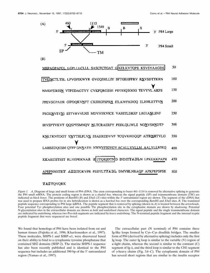
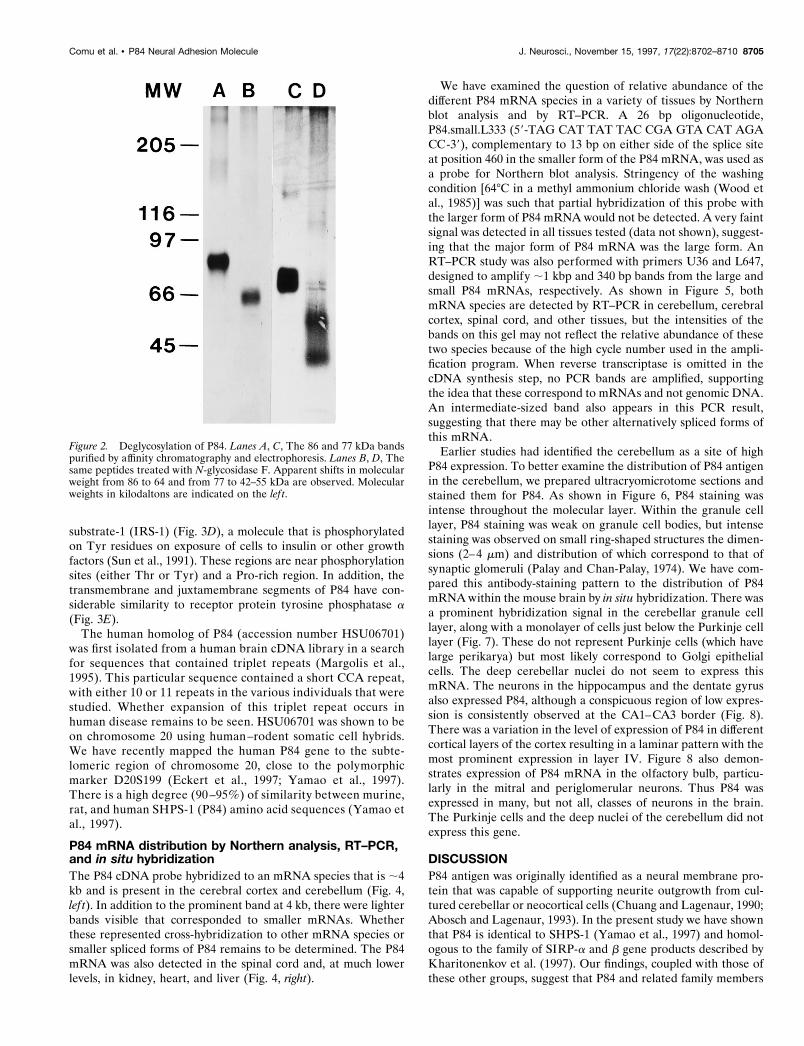
The two forms of P84 mRNA (large and small) are dia-grammed in Figure 1 A. The smaller form of P84 mRNA lacksthe segment from base pair 461 through 1114, a result ofalternative splicing of exons. By analysis of mouse genomicclones, we have confirmed that the segment between base pair461 and 1114 corresponds to a single exon flanked by consensussplice acceptor and donor sites (data not shown). An openreading frame extends from position 23 to 1549, encoding a509-amino-acid peptide, the translated amino acid sequence ofwhich is shown in Figure 1 B. The expected molecular weight ofthis peptide is 56 kDa, with a pI of 8.79. The segment that isremoved by splicing to generate P84 small (accounting for 218amino acids) is shown. At the N terminal, there is a hydropho-bic segment that most likely represents the signal peptide, witha consensus site for signal peptidase at residue 31. Amino acidresidue 32, which immediately follows this signal sequence,marks the beginning of the N terminal of purified P84 that wassubject to peptide sequencing. A second hydrophobic segment,which most likely corresponds to a transmembrane domain, liesbetween residues 374 and 396. The cytoplasmic segment con-tains at least one potential phosphorylation site on a Thrresidue (REIT at residue 423) and four potential Tyr phos-phorylation sites (436, 460, 477, and 501). The extracellulardomain contains 17 potential N-glycosylation sites (NXSor NXT), suggesting that the discrepancy between the calcu-lated molecular weight of the peptide and the apparent molec-ular weight on SDS-PAGE is because of glycosylation. Ofthese, only four are present in the P84 small peptide. Todetermine the contribution of N-linked carbohydrate to theapparent molecular weight of P84, we digested the 86 and 77kDa bands with N-glycosidase F. As shown in Figure 2, mo-lecular weight shifts from 86 to 64 and from 77 to 42–55 kDawere observed.
Sequence homologiesSegments of the P84 peptide sequence were used in a BLASTsearch of the GenBank protein database (Altschul et al., 1990).
Comu et al. • P84 Neural Adhesion Molecule J. Neurosci., November 15, 1997, 17(22):8702–8710 8703
We found that homologs of P84 have been isolated from rat andhuman tissues (Fujioka et al., 1996; Kharitonenkov et al., 1997).These molecules, SHPS-1 and SIRP-a1, were discovered basedon their ability to bind to a cytoplasmic tyrosine phosphatase thatcontained SH2 domains (SHP-2). The murine SHPS-1 sequencehas also been recently published and is identical to the P84sequence but includes an additional 390 bp of the 59-untranslatedregion (Yamao et al., 1997).
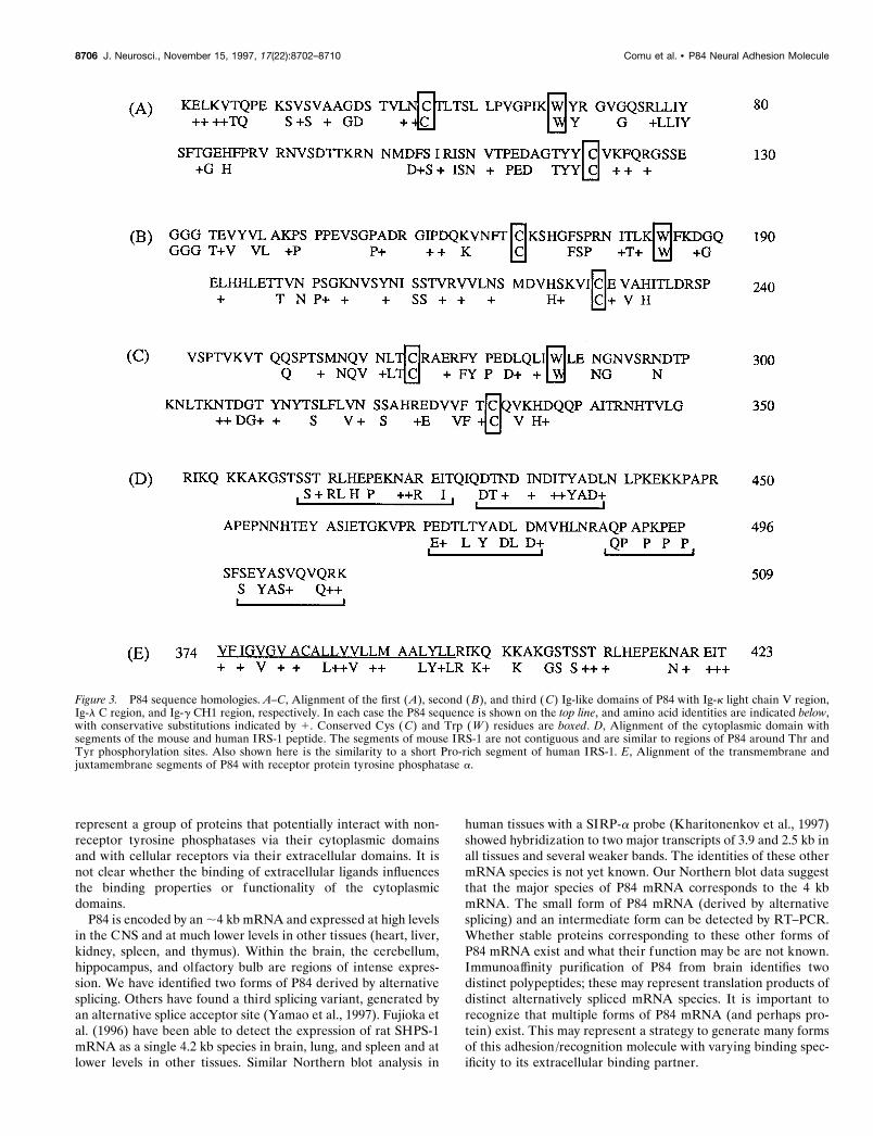
The extracellular part (N terminal) of P84 contains threeIg-like loops formed by Cys–Cys disulfide bridges. The smallerform of P84 (derived by alternative splicing) includes only the firstIg loop. The outer Ig loop is similar to the variable (V) region ofk-light chains, whereas the second is similar to the constant (C)segment of Ig-l, and the third loop is similar to the CH1 segmentof g-heavy chains (Fig. 3A–C). The cytoplasmic domain of P84has several short regions that are similar to the insulin receptor
Figure 1. A, Diagram of large and small forms of P84 cDNA. The exon corresponding to bases 461–1114 is removed by alternative splicing to generatethe P84 small mRNA. The protein coding region is shown as a shaded box, whereas the signal peptide (SP) and transmembrane domain (TM ) areindicated as black boxes. The positions of BamHI (B) and XbaI (X ) sites within the 39-untranslated region are shown. The segment of the cDNA thatwas used to prepare RNA probes for in situ hybridization is shown as a hatched box over the corresponding BamHI and XbaI sites. B, The translatedpeptide sequence corresponding to P84 large mRNA. The peptide segment that is removed by splicing (shown in A) is located between the arrowheads.Four potential Tyr phosphorylation sites and one possible Thr phosphorylation site in the cytoplasmic domain are shown by shadowing. PotentialN-glycosylation sites in the extracellular domain are shown as bold and underlined characters. The signal peptide and the single transmembrane domainare indicated by underlining, whereas two Pro-rich segments are indicated by heavy underlining. The N-terminal peptide fragment and the internal trypticpeptide fragment that were sequenced are boxed.
8704 J. Neurosci., November 15, 1997, 17(22):8702–8710 Comu et al. • P84 Neural Adhesion Molecule
substrate-1 (IRS-1) (Fig. 3D), a molecule that is phosphorylatedon Tyr residues on exposure of cells to insulin or other growthfactors (Sun et al., 1991). These regions are near phosphorylationsites (either Thr or Tyr) and a Pro-rich region. In addition, thetransmembrane and juxtamembrane segments of P84 have con-siderable similarity to receptor protein tyrosine phosphatase a(Fig. 3E).
The human homolog of P84 (accession number HSU06701)was first isolated from a human brain cDNA library in a searchfor sequences that contained triplet repeats (Margolis et al.,1995). This particular sequence contained a short CCA repeat,with either 10 or 11 repeats in the various individuals that werestudied. Whether expansion of this triplet repeat occurs inhuman disease remains to be seen. HSU06701 was shown to beon chromosome 20 using human–rodent somatic cell hybrids.We have recently mapped the human P84 gene to the subte-lomeric region of chromosome 20, close to the polymorphicmarker D20S199 (Eckert et al., 1997; Yamao et al., 1997).There is a high degree (90 –95%) of similarity between murine,rat, and human SHPS-1 (P84) amino acid sequences (Yamao etal., 1997).
P84 mRNA distribution by Northern analysis, RT–PCR,and in situ hybridizationThe P84 cDNA probe hybridized to an mRNA species that is ;4kb and is present in the cerebral cortex and cerebellum (Fig. 4,lef t). In addition to the prominent band at 4 kb, there were lighterbands visible that corresponded to smaller mRNAs. Whetherthese represented cross-hybridization to other mRNA species orsmaller spliced forms of P84 remains to be determined. The P84mRNA was also detected in the spinal cord and, at much lowerlevels, in kidney, heart, and liver (Fig. 4, right).
We have examined the question of relative abundance of thedifferent P84 mRNA species in a variety of tissues by Northernblot analysis and by RT–PCR. A 26 bp oligonucleotide,P84.small.L333 (59-TAG CAT TAT TAC CGA GTA CAT AGACC-39), complementary to 13 bp on either side of the splice siteat position 460 in the smaller form of the P84 mRNA, was used asa probe for Northern blot analysis. Stringency of the washingcondition [64°C in a methyl ammonium chloride wash (Wood etal., 1985)] was such that partial hybridization of this probe withthe larger form of P84 mRNA would not be detected. A very faintsignal was detected in all tissues tested (data not shown), suggest-ing that the major form of P84 mRNA was the large form. AnRT–PCR study was also performed with primers U36 and L647,designed to amplify ;1 kbp and 340 bp bands from the large andsmall P84 mRNAs, respectively. As shown in Figure 5, bothmRNA species are detected by RT–PCR in cerebellum, cerebralcortex, spinal cord, and other tissues, but the intensities of thebands on this gel may not reflect the relative abundance of thesetwo species because of the high cycle number used in the ampli-fication program. When reverse transcriptase is omitted in thecDNA synthesis step, no PCR bands are amplified, supportingthe idea that these correspond to mRNAs and not genomic DNA.An intermediate-sized band also appears in this PCR result,suggesting that there may be other alternatively spliced forms ofthis mRNA.
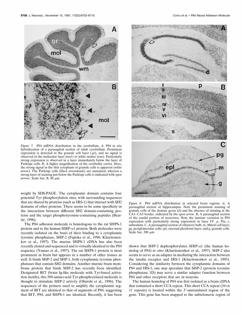
Earlier studies had identified the cerebellum as a site of highP84 expression. To better examine the distribution of P84 antigenin the cerebellum, we prepared ultracryomicrotome sections andstained them for P84. As shown in Figure 6, P84 staining wasintense throughout the molecular layer. Within the granule celllayer, P84 staining was weak on granule cell bodies, but intensestaining was observed on small ring-shaped structures the dimen-sions (2–4 mm) and distribution of which correspond to that ofsynaptic glomeruli (Palay and Chan-Palay, 1974). We have com-pared this antibody-staining pattern to the distribution of P84mRNA within the mouse brain by in situ hybridization. There wasa prominent hybridization signal in the cerebellar granule celllayer, along with a monolayer of cells just below the Purkinje celllayer (Fig. 7). These do not represent Purkinje cells (which havelarge perikarya) but most likely correspond to Golgi epithelialcells. The deep cerebellar nuclei do not seem to express thismRNA. The neurons in the hippocampus and the dentate gyrusalso expressed P84, although a conspicuous region of low expres-sion is consistently observed at the CA1–CA3 border (Fig. 8).There was a variation in the level of expression of P84 in differentcortical layers of the cortex resulting in a laminar pattern with themost prominent expression in layer IV. Figure 8 also demon-strates expression of P84 mRNA in the olfactory bulb, particu-larly in the mitral and periglomerular neurons. Thus P84 wasexpressed in many, but not all, classes of neurons in the brain.The Purkinje cells and the deep nuclei of the cerebellum did notexpress this gene.
DISCUSSIONP84 antigen was originally identified as a neural membrane pro-tein that was capable of supporting neurite outgrowth from cul-tured cerebellar or neocortical cells (Chuang and Lagenaur, 1990;Abosch and Lagenaur, 1993). In the present study we have shownthat P84 is identical to SHPS-1 (Yamao et al., 1997) and homol-ogous to the family of SIRP-a and b gene products described byKharitonenkov et al. (1997). Our findings, coupled with those ofthese other groups, suggest that P84 and related family members
Figure 2. Deglycosylation of P84. Lanes A, C, The 86 and 77 kDa bandspurified by affinity chromatography and electrophoresis. Lanes B, D, Thesame peptides treated with N-glycosidase F. Apparent shifts in molecularweight from 86 to 64 and from 77 to 42–55 kDa are observed. Molecularweights in kilodaltons are indicated on the lef t.
Comu et al. • P84 Neural Adhesion Molecule J. Neurosci., November 15, 1997, 17(22):8702–8710 8705
represent a group of proteins that potentially interact with non-receptor tyrosine phosphatases via their cytoplasmic domainsand with cellular receptors via their extracellular domains. It isnot clear whether the binding of extracellular ligands influencesthe binding properties or functionality of the cytoplasmicdomains.
P84 is encoded by an ;4 kb mRNA and expressed at high levelsin the CNS and at much lower levels in other tissues (heart, liver,kidney, spleen, and thymus). Within the brain, the cerebellum,hippocampus, and olfactory bulb are regions of intense expres-sion. We have identified two forms of P84 derived by alternativesplicing. Others have found a third splicing variant, generated byan alternative splice acceptor site (Yamao et al., 1997). Fujioka etal. (1996) have been able to detect the expression of rat SHPS-1mRNA as a single 4.2 kb species in brain, lung, and spleen and atlower levels in other tissues. Similar Northern blot analysis in
human tissues with a SIRP-a probe (Kharitonenkov et al., 1997)showed hybridization to two major transcripts of 3.9 and 2.5 kb inall tissues and several weaker bands. The identities of these othermRNA species is not yet known. Our Northern blot data suggestthat the major species of P84 mRNA corresponds to the 4 kbmRNA. The small form of P84 mRNA (derived by alternativesplicing) and an intermediate form can be detected by RT–PCR.Whether stable proteins corresponding to these other forms ofP84 mRNA exist and what their function may be are not known.Immunoaffinity purification of P84 from brain identifies twodistinct polypeptides; these may represent translation products ofdistinct alternatively spliced mRNA species. It is important torecognize that multiple forms of P84 mRNA (and perhaps pro-tein) exist. This may represent a strategy to generate many formsof this adhesion/recognition molecule with varying binding spec-ificity to its extracellular binding partner.
Figure 3. P84 sequence homologies. A–C, Alignment of the first (A), second (B), and third (C) Ig-like domains of P84 with Ig-k light chain V region,Ig-l C region, and Ig-g CH1 region, respectively. In each case the P84 sequence is shown on the top line, and amino acid identities are indicated below,with conservative substitutions indicated by 1. Conserved Cys (C) and Trp (W ) residues are boxed. D, Alignment of the cytoplasmic domain withsegments of the mouse and human IRS-1 peptide. The segments of mouse IRS-1 are not contiguous and are similar to regions of P84 around Thr andTyr phosphorylation sites. Also shown here is the similarity to a short Pro-rich segment of human IRS-1. E, Alignment of the transmembrane andjuxtamembrane segments of P84 with receptor protein tyrosine phosphatase a.
8706 J. Neurosci., November 15, 1997, 17(22):8702–8710 Comu et al. • P84 Neural Adhesion Molecule

The P84 adhesion molecule contains a single transmembranedomain. The large form of P84 contains three Ig-like loops in itsextracellular domain, whereas the small form of P84 contains onlyone Ig-like loop. The outermost Ig-loop (which is shared by both
large and small forms) is similar to the V region of k-chains,suggesting a role in specifically recognizing an extracellular bind-ing partner. The extracellular domain also contains many N-glycosylation sites, accounting for the difference between calcu-lated peptide molecular weight and the apparent molecular
Figure 4. Lef t, Northern blot of cerebellum (lane 1) and cerebrum (lane2) from P15 mouse probed with P84 cDNA demonstrates a thick band justbelow the 28S rRNA marker (corresponding to ;4 kb). The blot wasprepared by electrophoresis of 10 mg of total RNA per lane. Right, P84expression in neural and non-neural tissues. Northern blot prepared withRNA (10 ug per lane) from liver (lane 1), kidney (lane 2), skeletal muscle(lane 3), heart (lane 4 ), spinal cord (lane 5), cerebrum (lane 6 ), andcerebellum (lane 7 ). This represents an overexposed autoradiogram,showing faint bands in liver, kidney, and heart. Arrowheads and arrowscorrespond to 28 and 18 S rRNA bands.
Figure 5. Detection of large and small forms of P84 mRNA in differenttissues by RT–PCR. cDNA pools from cerebellum (lane 1), cerebral cortex(lane 2), spleen (lane 3), thymus (lane 4 ), liver (lane 5), skeletal muscle (lane6 ), heart (lane 7 ), and spinal cord (lane 8) were amplified with the U36 andL647 primers. Lane 9 is a positive control reaction done with the full-length(large) P84 cDNA as template, and lane 10 is a positive control using SC500plasmid DNA (partial small cDNA) as template. To the lef t of lane 1 is a 1kb ladder.
Figure 6. Immunohistochemical localization of P84 in cerebellum. Ul-tracryomicrotome sections showing intense P84 staining in the molecularlayer ( A–C) and in synaptic glomeruli (ring-shaped structures indicatedby arrows in B and C). Weak staining is observed on granule cell bodies.pkj, Purkinje cells; other abbreviations are defined in the Figure 7 legend.Scale bars: A, 200 mm; B, 50 mm; C, 30 mm.
Comu et al. • P84 Neural Adhesion Molecule J. Neurosci., November 15, 1997, 17(22):8702–8710 8707
weight by SDS-PAGE. The cytoplasmic domain contains fourpotential Tyr phosphorylation sites, with surrounding sequencesthat are shared by proteins (such as IRS-1) that interact with SH2domains of other proteins. There seems to be some specificity inthe interaction between different SH2 domain-containing pro-teins and the target phosphotyrosine-containing peptides (Beat-tie, 1996).
The P84 adhesion molecule is homologous to the rat SHPS-1protein and to the human SIRP-a1 protein. Both molecules wererecently isolated on the basis of their binding to a cytoplasmictyrosine phosphatase, SHP-2 (Fujioka et al., 1996; Kharitonen-kov et al., 1997). The murine SHPS-1 cDNA has also beenrecently cloned and sequenced and is virtually identical to the P84sequence (Yamao et al., 1997). The rat SHPS-1 protein is mostprominent in brain but appears in a number of other tissues aswell. It binds SHP-2 and SHP-1, both cytoplasmic tyrosine phos-phatases that contain SH2 domains. Another murine brain mem-brane protein that binds SHP-2 has recently been identified.Designated BIT (brain Ig-like molecule with Tyr-based activa-tion motifs), this 509-amino-acid Tyr-phosphorylated molecule isthought to stimulate SHP-2 activity (Ohnishi et al., 1996). Thesequences of the primers used to amplify the cytoplasmic seg-ment of BIT are identical to that of segments of P84, suggestingthat BIT, P84, and SHPS-1 are identical. Recently, it has been
shown that SHP-2 dephosphorylates SIRP-a1 (the human ho-molog of P84) in vitro (Kharitonenkov et al., 1997). SHP-2 alsoseems to serve as an adapter in mediating the interaction betweenthe insulin receptor and IRS-1 (Kharitonenkov et al., 1995).Considering the similarity between the cytoplasmic domains ofP84 and IRS-1, one may speculate that SHP-2 (protein tyrosinephosphatase 1D) may serve a similar adapter function betweenP84 and other receptors that are in neurons.
The human homolog of P84 was first isolated as a brain cDNAthat contained a short CCA repeat. This short CCA repeat (10 or11 repeats) is located within the 39-untranslated region of thegene. This gene has been mapped to the subtelomeric region of
Figure 7. P84 mRNA distribution in the cerebellum. A, P84 in situhybridization of a parasagittal section of adult cerebellum. Prominentexpression is detected in the granule cell layer ( gr), and no signal isobserved in the molecular layer (mol ) or white matter (wm). Particularlystrong expression is observed in a layer immediately below the layer ofPurkinje cells. B, A higher magnification of the cerebellar cortex. Here,the strong signal in the thin cytoplasm of granule cells is apparent (whitearrow). The Purkinje cells (black arrowheads) are unstained, whereas astrong layer of staining just below the Purkinje cells is indicated with openarrows. Scale bar: B, 40 mm.
Figure 8. P84 mRNA distribution in selected brain regions. A, Aparasagittal section of hippocampus. Note the prominent staining ofgranule cells of the dentate gyrus (d) and the absence of staining at theCA1–CA3 border, indicated by the open arrow. B, A parasagittal sectionof the caudal portion of neocortex. Note the laminar variation in P84expression with particularly strong expression in layer IV. p, Pia; s,subiculum. C, A parasagittal section of olfactory bulb. m, Mitral cell layer;pg, periglomerular cells; epl, external plexiform layer; and g, granule cells.Scale bar, 300 mm.
8708 J. Neurosci., November 15, 1997, 17(22):8702–8710 Comu et al. • P84 Neural Adhesion Molecule
human chromosome 20 (close to polymorphic marker D20S199;Eckert et al., 1997). This gene lies outside the critical region forHallervorden-Spatz syndrome, a progressive neurological disor-der mapped to 20p13. We believe that genetic defects in P84 maynot cause major malformations of the brain but may result inimproper synapse formation and thus cause symptoms such asmental retardation and seizures. There are a number of humandisorders (fragile X syndrome, myotonic dystrophy, Huntington’sdisease, spinocerebellar ataxia, and Friedrich’s ataxia) that arecaused by expansions of triplet repeats (Ashley and Warren, 1995;Timchenko and Caskey, 1996). It is thus possible that expansionof the CCA repeat in the human P84 (SHPS-1) gene may causesuch a neurological syndrome.
The immunohistochemical and in situ hybridization data sug-gest that P84 is localized to regions of the brain that are rich insynapses. In the cerebellum, P84 mRNA is easily detected ingranule cells but not in Purkinje cells, whereas the protein islocalized to the molecular layer and to synaptic glomeruli withinthe granule layer. Because the neuronal components of the mo-lecular layer of the cerebellum consist mainly of axons of thegranule cells (parallel fibers) and Purkinje cell dendrites, theseobservations are consistent with a presynaptic localization of theprotein. Whether this presynaptic localization applies in otherregions of the brain is unknown, and it may be that in certainstructures P84 may appear postsynaptically. P84 mRNA is de-tected throughout the brain including the hippocampus, cerebralcortex, olfactory bulb, and retina. This localization of P84 tosynapses, its in vitro function as an adhesion molecule, and thestructural information contained in this paper showing the simi-larity to V regions of Igs suggest that the interaction between theextracellular domain of P84 and its receptor (or ligand) may beimportant in synapse formation or maintenance. It is not clearwhether extracellular ligand binding influences the binding oractivity of SHP-2 phosphatase. Because P84 becomes localized tosynaptic regions in the CNS, it is possible that its function is toeffect the localization of SHP-2 to this region and thus spatiallyregulate SHP-2 expression.
Interesting parallels may be drawn between P84 (SHPS-1)and SHP-2 association and SHP-1 association with Tyr-phosphorylated proteins in hematopoietic cells. Natural killer(NK) cells express a class of killer cell inhibitory receptors(KIRs) that specifically recognize major histocompatibilitycomplex-class I molecules on target cells. The cytoplasmic tailof KIRs contain consensus Tyr-containing domains, which,when phosphorylated, bind to SHP-1. The interaction betweenthe SH2 domains of SHP-1 and the phosphotyrosines of KIRsactivates the phosphatase, which in turn inhibits the NK cellactivation pathway. In this system, SHP-1 seems to play anegative regulatory role in antigen receptor signaling (Binstadtet al., 1997). A similar negative role has been proposed forSHP-1 in erythropoietin function. Binding of erythropoietinresults in Tyr phosphorylation of its receptor and activation ofthe JAK2 kinase. The phosphorylated erythropoietin receptorrecruits the SHP-1 phosphatase by binding via its SH2 domain,which in turn dephosphorylates JAK2 and terminates theerythropoietin-induced proliferation signal (Renard et al.,1997). P84 may function in a similar manner in the brain byrecruitment of cytoplasmic phosphatases to juxtamembranesites, resulting in the dephosphorylation of other proteins(activated receptors, for example), thus modulating (eitherpositively or negatively) signaling through tyrosine kinases.
Thus, P84 may serve as a membrane-localizing proteinfor cytoplasmic SH2-containing proteins (phosphatases), andits function in turn may be determined by phosphorylationof the cytoplasmic Tyr residues. Critical issues that remainto be addressed include the identity of the extracellular li-gand that binds P84, whether binding of this ligand resultsin phosphorylation of P84, the kinase that phosphorylatesP84, and its precise role in the signaling pathway duringsynaptogenesis.
REFERENCESAbosch A, Lagenaur C (1993) Sensitivity of neurite outgrowth to micro-
filament disruption varies with adhesion molecule substrate. J Neuro-biol 24:344–355.
Altschul SF, Gish W, Miller W, Myers EW, Lipman DJ (1990) Basiclocal alignment search tool. J Mol Biol 215:403–410.
Apte AN, Siebert PD (1993) Anchor-ligated cDNA libraries: a tech-nique for generating a cDNA library for the immediate cloning of 59ends of mRNAs. Biotechniques 15:890–893.
Ashley Jr CT, Warren ST (1995) Trinucleotide repeat expansion andhuman disease. Annu Rev Genet 29:703–728.
Beattie J (1996) SH2 domain protein interaction and possibilities forpharmacological intervention. Cell Signal 8:75–86.
Binstadt BA, Brumbaugh KM, Leibson PJ (1997) Signal transduction byhuman NK cell MHC-recognizing receptors. Immunol Rev155:197–203.
Chuang W, Lagenaur CF (1990) Central nervous system antigen P84 canserve as a substrate for neurite outgrowth. Dev Biol 137:219–232.
Culotti JG, Kolodkin AL (1996) Functions of netrins and semaphorinsin axon guidance. Curr Opin Neurobiol 6:81–88.
Dumas JB, Edwards M, Delort J, Mallet J (1991) Oligodeoxyribonucle-otide ligation to single-stranded cDNAs: a new tool for cloning of 59ends of mRNAs and for constructing cDNA libraries by in vitro am-plification. Nucleic Acids Res 19:5227–5232.
Eckert C, Olinsky S, Cummins J, Stephan D, Narayanan V (1997) Map-ping of the human P84 gene to the subtelomeric region of chromosome20p. Somat Cell Mol Genet, in press.
Fannon AM, Colman DR (1996) A model for central synaptic junctionalcomplex formation based on the differential adhesive specificities of thecadherins. Neuron 17:423–434.
Fields DR, Itoh K (1996) Neural cell adhesion molecules in activity-dependent development and synaptic plasticity. Trends Neurosci19:473–480.
Fujioka Y, Matozaki T, Noguchi T, Iwamatsu A, Yamao T, Takahashi N,Tsuda M, Takada T, Kasuga M (1996) A novel membrane glycopro-tein, SHPS-1, that binds the SH2-domain-containing protein tyrosinephosphatase SHP-2 in response to mitogens and cell adhesion. MolCell Biol 16:6887–6899.
Goodman CS (1996) Mechanisms and molecules that control growthcone guidance. Annu Rev Neurosci 19:341–377.
Herrin DL, Schmidt GW (1988) Rapid, reversible staining of Northernblots prior to hybridization. Biotechniques 6:196–200.
Kharitonenkov A, Schnekenburger J, Chen Z, Knyazev P, Ali S, Zwick E,White M, Ullrich A (1995) Adapter function of protein-tyrosine phos-phatase 1D in insulin receptor/insulin receptor substrate-1 interaction.J Biol Chem 270:29189–29193.
Kharitonenkov A, Chen Z, Sures I, Wang H, Schilling J, Ullrich A (1997)A family of proteins that inhibit signaling through tyrosine kinasereceptors. Nature 386:181–186.
Margolis RL, Breschel TS, Li S-H, Kidwai AS, Antonarakis SE, McInnisMG, Ross CA (1995) Characterization of cDNA clones containingCCA trinucleotide repeats derived from human brain. Somat Cell MolGenet 21:279–284.
Mendel-Hartwig I (1982) A simple and rapid method for the isolation ofpeptides from sodium dodecyl sulfate-containing polyacrylamide gels.Anal Biochem 121:215–217.
Ohnishi H, Kubota M, Ohtake A, Sato K, Sano S-I (1996) Activation ofprotein-tyrosine phosphatase SH-PTP2 by a tyrosine-based activationmotif of a novel brain molecule. J Biol Chem 271:25569–25574.
Palay SL, Chan-Palay V (1974) Cerebellar cortex. Berlin: Springer.Renard V, Cambiaggi A, Vely F, Blery M, Olcese L, Olivero S, Bouchet
Comu et al. • P84 Neural Adhesion Molecule J. Neurosci., November 15, 1997, 17(22):8702–8710 8709

M, Vivier E (1997) Transduction of cytotoxic signals in natural killercells: a general model of fine tuning between activatory and inhibitorypathways in lymphocytes. Immunol Rev 155:205–221.
Schaeren-Wiemers N, Gerfin-Moser A (1993) A single protocol to detecttranscripts of various types and expression levels in neural tissue andcultured cells: in situ hybridization using digoxigenin-labeled cRNAprobes. Histochemistry 100:431–440.
Singer SJ, Tokuyasu KT, Dutton AH, Chen WT (1982) High-resolutionimmunoelectron microscopy of cell and tissue ultrastructure. In: Elec-tron microscopy in biology, Vol 2 (Griffith JD, ed), pp 55–106. NewYork: Wiley.
Sun XJ, Rothenberg P, Kahn CR, Backer JM, Araki E, Wilden PA, CahillDA, Goldstein BJ, White MF (1991) Structure of the insulin receptor
substrate IRS-1 defines a unique signal transduction protein. Nature352:73–77.
Timchenko LT, Caskey CT (1996) Trinucleotide repeat disorders inhumans: discussions of mechanisms and medical issues. FASEB J10:1589–1597.
Wood WI, Gitschier J, Lasky LA, Lawn RM (1985) Base composition-independent hybridization in tetramethylammonium chloride: amethod for oligonucleotide screening of highly complex gene libraries.Proc Natl Acad Sci USA 82:1585–1588.
Yamao T, Matozaki T, Amano K, Matsuda Y, Takahashi N, Ochi F,Fujioka Y, Kasuga M (1997) Mouse and human SHPS-1: molecularcloning of cDNAs and chromosomal localization of genes. BiochemBiophys Res Commun 231:61–67.
8710 J. Neurosci., November 15, 1997, 17(22):8702–8710 Comu et al. • P84 Neural Adhesion Molecule