The carbonic anhydrase domain of receptor tyrosine phosphatase β is a functional ligand for the...
10
Cell, Vol. 82, 251-260, July28, 1995,Copyright © 1995by Cell Press The Carbonic Anhydrase Domain of Receptor Tyrosine Phosphatase Is a Functional Ligand for the Axonal Cell Recognition Molecule Contactin Elior Peles,* Moshe Nativ,* Phillip L. Campbell,* Takeshi Sakurai,t Ricardo Martinez,* Sima Lev,t Douglas O. Clary,* James Schilling,* Gilad Barnea,t Gregory D. Plowman,* Martin Grumet,~ and Joseph Schlessingert *SUGEN, Incorporated 515 Galveston Drive Redwood City, California 94063-4720 tDepartment of Pharmacology New York University Medical Center New York, New York 10016 Summary Receptor-type protein tyrosine phosphatase p (RPTPJ]) is expressed in the developing nervous system and contains a carbonic anhydrase (CAH) domain as well as a fibronectin type III repeat in its extracellular do- main. Fusion proteins containing these domains were used to search for ligands of RPTPp. The CAH domain bound specifically to a 140 kDa protein expressed on the surface of neuronal cells. Expression cloning in COS7 cells revealed that this protein is contactin, a GPI membrane-anchored neuronal cell recognition mol- ecule. The CAH domain of RPTPp induced cell adhe- sion and neurite growth of primary tectal neurons, and differentiation of neuroblastoma cells. These responses were blocked by antibodies against contactin, demon- strating that contactin is a neuronal receptor for RPTPI~. These experiments show that an individual domain of RPTPI$ acts as a functional ligand for the neuronal re- ceptor contactin. The interaction between contactin and RPTPp may generate unidirectional or bidirectional sig- nals during neural development. Introduction The ability of cells to respond to signals from their microen- vironment is a fundamental feature of development. In the developing nervous system, neurons migrate and extend axons to establish their intricate network of synaptic con- nections (Goodman and Shatz, 1993). During migration and axonal pathfinding, cells are guided by both attractive and repulsive signals (Hynes and Lander, 1992; Keynes and Cook, 1992). The ability of the neuron to respond to these signals requires cell surface molecules that are able to receive the signals and to transmit them to the cell inte- rior, resulting in specific biological responses. It is well established that protein tyrosine phosphoryla- tion is responsible for the regulation of many cellular re- sponses to external stimuli crucial for cell growth, prolifera- tion, and differentiation (Schlessinger and UIIrich, 1992). Tyrosine phosphorylation was implicated in several devel- opmental processes in the nervous system. For example, receptor tyrosine kinases were shown to affect neuronal survival (Chao, 1992) and cell fate determination (Zipursky and Rubin, 1994). Nonreceptor tyrosine kinases were shown to be downstream elements in signaling via cell recognition molecules that play a role in cell guidance and migration (Ignelzi et al., 1994; Umemori et at., 1994). The transient nature of signaling by phosphorylation re- quires specific phosphatases for control and regulation (Hunter, 1995). Indeed, many protein tyrosine phospha- tases were shown to be expressed in specific regions of the developing brain, including the olfactory neuroepithe- lium (Walton et al., 1993), the cortex (Sahin et al., 1995), and retinal MQIler gila (Shock et al., 1995). Furthermore, expression of several tyrosine phosphatases, such as pro- tein-tyrosine phosphatase ~z (PTPa) (den Hertog et al., 1993), PC12-PTP1 (Sharama and Lombroso, 1995), and several forms of leukocyte common antigen-related (LAR) tyrosine phosphatase (Zhang and Longo, 1995) were found to be regulated during neuronal differentiation of P19 or PC12 cells. Receptor-type protein tyrosine phosphatases (RPTPs) were subdivided into several groups on the basis of struc- tural characteristics of their extracellular domains (Char- bonneau and Tonks, 1992; Barnea et al., 1993). RPTPI~ (also known as RPTP0 and RPTPy are members of a distinct group of phosphatases characterized by the pres- ence of carbonic anhydrase (CAH)-Iike domains, fibronec- tin type Ill (FNIII) repeats, and a long cysteine-free region (spacer domain) in their extracellular domain (Barnea et al., 1993; Krueger and Saito, 1992; Levy et al., 1993). The expression of RPTPI~ is restricted to the central and peripheral nervous system, while RPTP7 is expressed both in the developing nervous system and in a variety of other tissues in the adult rat (Canoll et al., 1993; Barnea et al., 1993). RPTP~ exists in three forms, one secreted and two membrane bound, that differ by the absence of 860 residues from the spacer domain (Levy et al., 1993; Maurel et al., 1994). The secreted form was identified in rat brain as a chondroitin sulfate proteoglycan called phos- phacan (3F8 proteoglycan) (Barnea et al., 1994a; Maurel et al., 1994; Shitara et al., 1994). The transmembrane form was also shown to be expressed in a form of a chondroitin sulfate proteoglycan (Barnea et al., 1994b). It is presently unknown whether the glycosaminoglycan attachment sites are utilized only in the insert of 860 amino acids that is not present in the short form of RPTPI3. It was shown that purified phosphacan can interact in vitro with the extra- cellular matrix protein tenascin and with the adhesion molecules N-CAM and Ng-CAM (for neural cell adhesion molecule and neuron-gila cell adhesion molecule, respec- tively) (Barnea et al., 1994b; Grumet et al., 1993, 1994; Milev et al., 1994). In the work presented in this report, we used fusion pro- teins containing the CAH domain of RPTPI3 as probes for the identification, purification, and cloning of a protein that bound specifically to RPTP~. Using expression cloning in COS7 cells and affinity chromatography, we show that the CAH domain of RPTPI3 binds specifically to the cell recognition molecule contactin (F11/F3). Moreover, we
Transcript of The carbonic anhydrase domain of receptor tyrosine phosphatase β is a functional ligand for the...
Cell, Vol. 82, 251-260, July 28, 1995, Copyright © 1995 by Cell Press
The Carbonic Anhydrase Domain of Receptor Tyrosine Phosphatase Is a Functional Ligand for the Axonal Cell Recognition Molecule Contactin
Elior Peles,* Moshe Nativ,* Phillip L. Campbell,* Takeshi Sakurai,t Ricardo Martinez,* Sima Lev,t Douglas O. Clary,* James Schilling,* Gilad Barnea,t Gregory D. Plowman,* Martin Grumet,~ and Joseph Schlessingert *SUGEN, Incorporated 515 Galveston Drive Redwood City, California 94063-4720 tDepartment of Pharmacology New York University Medical Center New York, New York 10016
Summary
Receptor-type protein tyrosine phosphatase p (RPTPJ]) is expressed in the developing nervous system and contains a carbonic anhydrase (CAH) domain as well as a fibronectin type III repeat in its extracellular do- main. Fusion proteins containing these domains were used to search for ligands of RPTPp. The CAH domain bound specifically to a 140 kDa protein expressed on the surface of neuronal cells. Expression cloning in COS7 cells revealed that this protein is contactin, a GPI membrane-anchored neuronal cell recognition mol- ecule. The CAH domain of RPTPp induced cell adhe- sion and neurite growth of primary tectal neurons, and differentiation of neuroblastoma cells. These responses were blocked by antibodies against contactin, demon- strating that contactin is a neuronal receptor for RPTPI~. These experiments show that an individual domain of RPTPI$ acts as a functional ligand for the neuronal re- ceptor contactin. The interaction between contactin and RPTPp may generate unidirectional or bidirectional sig- nals during neural development.
Introduction
The ability of cells to respond to signals from their microen- vironment is a fundamental feature of development. In the developing nervous system, neurons migrate and extend axons to establish their intricate network of synaptic con- nections (Goodman and Shatz, 1993). During migration and axonal pathfinding, cells are guided by both attractive and repulsive signals (Hynes and Lander, 1992; Keynes and Cook, 1992). The ability of the neuron to respond to these signals requires cell surface molecules that are able to receive the signals and to transmit them to the cell inte- rior, resulting in specific biological responses.
It is well established that protein tyrosine phosphoryla- tion is responsible for the regulation of many cellular re- sponses to external stimuli crucial for cell growth, prolifera- tion, and differentiation (Schlessinger and UIIrich, 1992). Tyrosine phosphorylation was implicated in several devel- opmental processes in the nervous system. For example, receptor tyrosine kinases were shown to affect neuronal survival (Chao, 1992) and cell fate determination (Zipursky
and Rubin, 1994). Nonreceptor tyrosine kinases were shown to be downstream elements in signaling via cell recognition molecules that play a role in cell guidance and migration (Ignelzi et al., 1994; Umemori et at., 1994).
The transient nature of signaling by phosphorylation re- quires specific phosphatases for control and regulation (Hunter, 1995). Indeed, many protein tyrosine phospha- tases were shown to be expressed in specific regions of the developing brain, including the olfactory neuroepithe- lium (Walton et al., 1993), the cortex (Sahin et al., 1995), and retinal MQIler gila (Shock et al., 1995). Furthermore, expression of several tyrosine phosphatases, such as pro- tein-tyrosine phosphatase ~z (PTPa) (den Hertog et al., 1993), PC12-PTP1 (Sharama and Lombroso, 1995), and several forms of leukocyte common antigen-related (LAR) tyrosine phosphatase (Zhang and Longo, 1995) were found to be regulated during neuronal differentiation of P19 or PC12 cells.
Receptor-type protein tyrosine phosphatases (RPTPs) were subdivided into several groups on the basis of struc- tural characteristics of their extracellular domains (Char- bonneau and Tonks, 1992; Barnea et al., 1993). RPTPI~ (also known as RPTP 0 and RPTPy are members of a distinct group of phosphatases characterized by the pres- ence of carbonic anhydrase (CAH)-Iike domains, fibronec- tin type Ill (FNIII) repeats, and a long cysteine-free region (spacer domain) in their extracellular domain (Barnea et al., 1993; Krueger and Saito, 1992; Levy et al., 1993). The expression of RPTPI~ is restricted to the central and peripheral nervous system, while RPTP7 is expressed both in the developing nervous system and in a variety of other tissues in the adult rat (Canoll et al., 1993; Barnea et al., 1993). RPTP~ exists in three forms, one secreted and two membrane bound, that differ by the absence of 860 residues from the spacer domain (Levy et al., 1993; Maurel et al., 1994). The secreted form was identified in rat brain as a chondroitin sulfate proteoglycan called phos- phacan (3F8 proteoglycan) (Barnea et al., 1994a; Maurel et al., 1994; Shitara et al., 1994). The transmembrane form was also shown to be expressed in a form of a chondroitin sulfate proteoglycan (Barnea et al., 1994b). It is presently unknown whether the glycosaminoglycan attachment sites are utilized only in the insert of 860 amino acids that is not present in the short form of RPTPI3. It was shown that purified phosphacan can interact in vitro with the extra- cellular matrix protein tenascin and with the adhesion molecules N-CAM and Ng-CAM (for neural cell adhesion molecule and neuron-gila cell adhesion molecule, respec- tively) (Barnea et al., 1994b; Grumet et al., 1993, 1994; Milev et al., 1994).
In the work presented in this report, we used fusion pro- teins containing the CAH domain of RPTPI3 as probes for the identification, purification, and cloning of a protein that bound specifically to RPTP~. Using expression cloning in COS7 cells and affinity chromatography, we show that the CAH domain of RPTPI3 binds specifically to the cell recognition molecule contactin (F11/F3). Moreover, we

Cell 252
CAH FN
PTP~,
~c
~CF
~F 1~3
Spacer Cytoplasmic Da
M
C
Neuronal Glial 3°] II 2000
c4 < cJ
Cell line
Figure 1. Detection of Cell Surface-Bound Ligands for RPTPI3 (A) Schematic presentation of RPTP[3 and different subdomains used to construct fusion proteins with human IgG1-Fc. CAH, CAH-like do- main; FN, FNIIt repeat; M, transmembrane domain. (B) Expression of the chimeric IgG molecules. Different ~ fusion pro- teins containing the CAH or the FNIII domains (~C and I~F respectively) or both domains (~CF) were purified, separated on SDS gel, and immu- noblotted with antibodies against human IgG. (C) Binding of I~CF to a cell line derived from neuronal (neuroblastoma and neuroendocrine) and glial (glioblastoma and astrocytoma) tumors. Cells were incubated with control medium (closed bars) orwith medium containing 13CF fusion protein at 0.4 p.g/ml (I~CF; stippled bars). Bound ~CF was detected by incubation with ~l-protein A as described in Experimental Procedures.
demonstrate that the CAH domain of RPTPI3 functions as a ligand for contactin. The interactions between a cell recognition molecule and a transmembrane protein tyro- sine phosphatase may play an important role during neural development and differentiation.
Results
Membrane-Bound Ligands for RPTPI~ Are Differentially Expressed in Neuronal and Glial Cell Lines To identify cellular ligands for RPTPI~, we have con- structed fusion proteins containing different subdomains of RPTP~ and the Fc portion of human immunoglobulin G (igG). Three chimeric constructs were generated, one
containing both the CAH and the FNIII domains (I~CF) and two others carrying each domain alone (13C or 13F; Figures 1A and 1B). We first used ~CF to screen for membrane- bound ligands on the surface of different neuronal and glial cell lines. As shown in Figure 1C, several cell lines were identified that specifically interact with this fusion protein, including IMR32 neuroblastoma cells, the two closely related neuroendocrine-derived cell lines GH3 and GH1, and five different glioblastoma cell lines.
The finding that the positive cell lines were derived from both glial and neuronal origins raised the possibility that RPTPI~ may interact with two different membrane-asso- ciated ligands. Alternatively, the same ligand may be ex- pressed on the cell surface of both neurons and glial cells. To explore these possibilities, we have examined the bind- ing properties of fusion proteins that contain either the CAH domain (I~C) or the FNIII domain (I~F) of RPTPI3. The results presented in Figure 2A show that the I~C fusion protein bound only to neuronal and neuroendocrine cell lines but not to glioblastoma cells. These experiments demonstrated that the CAH domain is responsible for bind- ing to neuronal cells, while the FNIII repeat is required for binding to glial cells. Accordingly, if the binding of ~C to a neuronal ligand reflects the interactions that take place in vivo, one would expect to find similar binding of the CAH domain to primary neurons. We therefore analyzed the binding of the different fusion proteins to cultured dor- sal root ganglion cells (DRGs), followed by detection of the bound proteins by immunostaining. The results pre- sented in Figure 2B show that I~C and I~CF bound to GH3 cells, as well as to primary neurons. A fusion protein con- taining the FNIII domain alone (~F) did not bind either to GH3 cells or to DRG neurons. In other experiments, we detected binding of I~F to several glial cell lines, but could not detect binding of the FNIII repeat to neuronal-derived cell lines or neurons derived from rat DRGs or from chick cortex (data not shown). In addition, we also examined whether the binding specificity observed with the CAH do- main of RPTPI~ is unique to this receptor by analyzing the binding of a fusion protein containing the CAH domain of RPTP~, (Barnea et al., 1993). This experiment showed that the CAH domain of RPTP~, does not bind either to GH3 cells or to primary neurons (Figure 2C; data not shown).
All together, these results suggested that specific li- gands for RPTPI~ exist on the surface of cells from neu- ronal and glial origin. Moreover, different subdomains of the receptor appear to mediate interactions with different ligands. On the basis of this analysis, we concluded that the CAH domain of RPTPI~ mediates interactions with neu- rons, while the FNIII domain is responsible for interactions with glial cells. In the work presented in this report, we describe the identification, purification, and molecular characterization of the ligand for the CAH domain of RPTPI~.
Covalent Cross-Linking Experiments Reveal a 140 kDa Protein That Interacts with the CAH Domain of RPTPI~ To characterize ligands for RPTPI3, we used a reversible cross-linking agent, 3,3'-dithiobis(sulfosuccinimidylpropi-

Interactions between Receptor Phosphatase and Contactin 253
A 3oo0
B
2000
E o
1000
DRG GH3
None
13C
13F
J3CF
C
0 500 1000 1500 cpm
Figure 2. Specific Binding of the CAH-like Domain of RPTP~ to Neu- rons and Neuronal Cell Lines (A) Binding of J~C to neuronal and glial tumor-derived cell lines. Control medium (none; closed bars) or medium containing ~C-Fc fusion pro- tein at 0.25 p.g/ml was used (I~C; stippled bars). Bound fusion proteins were detected by incubation with ~l-protein A and determination of radioactive content. (B) Binding of the different subdomains of RPTP~ to GH3 cells and to primary neurons. GH3 cells and primary cultures of rat DRGs were incubated with control medium (none) or medium containing a fusion protein with the CAH domain (~C), the FNIII domain (I~F), or a fusion protein containing both domains (~CF) for 1 hr at room temperature. Unbound proteins were removed, and the bound Fc fusion proteins were visualized by immunostaining with biotinylated anti-human IgG antibodies and streptavidin-alkaline phosphatase as described in Ex- perimental Procedures. (C) Fc fusion proteins containing the CAH domain of RPTP~ (~C) or the CAH domain of RPTPy (yC) were applied in binding assay to GH3 cells. Binding was performed as described in (A).
onate) (DTSSP), and searched for proteins that specifically bound to [3C. Two of the cell lines that bound ~C (IMR32 and GH3), as well as COS7 cells as a control, were first allowed to react with the fusion proteins containing either
Cell line: COS GH3 IMR ECD: F C F C F C
Figure 3. Covalent Cross-Linking of ~C-Fc to a Cell Membrane- Bound Ligand
Fc fusion proteins containing the CAH domain (C) or the FN III domain (F) of RPTP~ were allowed to bind to pS]methionine-labeled IMR32 neuroblastoma cells (IMR), GH3 pituitary tumor cells, or COS7 cells as indicated. Following a 1 hr incubation, unbound proteins were washed away, and 1 mM of the reversible covalent cross-linking agent (DTSSP) was added for an additional 30 min at 4°C. Cell lysates were prepared, and the cross-linked proteins were precipitated with protein A-Sepha- rose as detailed in Experimental Procedures. The proteins were re- solved on 8%-16% SDS gels under reducing conditions, followed by autoradiography. Under these conditions, the cross-linker is cleaved. Molecular mass marker proteins are indicated in kilodaltons.
the FNIII or the CAH domains, and then complexes were cross-linked and precipitated. In an effort to identify the true molecular mass of the putative ligand, we utilized the covalent cross-linking agent DTSSP, which undergoes cleavage in reducing sodium dodecyl sulfate-polyacryl- amide gel electrophoresis (SDS-PAGE) conditions. As shown in Figure 3, a protein with an apparent molecular mass of 140 kDa specifically reacted with I~C in rat GH3 and human IMR32 cells. No reactivity was detected in control cells or in cells incubated with 13F. This result sug- gested that a similar ligand is expressed on the surface of rat GH3 and human IMR32 cells.
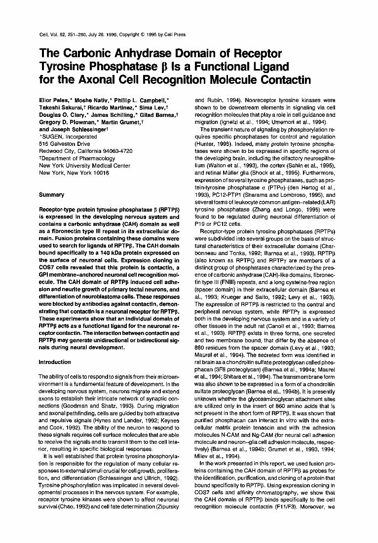
Molecular Cloning of a Candidate Ligand for RPTPp from Rat GH3 Cells To clone the gene that encodes the 140 kDa candidate ligand, we employed an expression cloning strategy. Plas- mid pools made from a GH3 cDNA library were transfected into COS7 cells that were subsequently screened for their ability to bind 13CF. Positive cells were detected by immu- nostaining with biotinylated anti-human IgG antibodies and streptavidin-alkaline phosphatase. One plasmid pool was identified that when transfected into cells resulted in several positively stained cells (Figure 4A). This pool was subdivided and rescreened four times, until a single clone (F8) was isolated. Transfection of COS7 cells with this plasmid resulted in positive staining of approximately 25%-50% of the cells. DNA sequence analyses of clone F8 showed that it contained a 4.0 kb insert and a single open reading frame of 3063 nt. The deduced 1021 amino acid sequence encoded by this clone is presented in Fig- ure 4B. Database searches with this sequence showed that it shares 95% and 99% identity with the amino acid sequences of human and mouse contactin (F11/F3), re- spectively (Berglund and Ranscht, 1994; Gennarini et al., 1989; Reid et al., 1994). We therefore concluded that the ligand for RPTPI~ cloned from GH3 cells is the rat homolog of contactin. This protein consists of six C2-type immuno- globulin domains, four FNIII repeats, and a hydrophobic
Cell 254
A I st F8
B 1 MXTPLLVSHL LLISLTSCLG E~WHRKyGH GVSEEDKG~G PIFEEQPIN? IYPEESL~GK vl z ~ G Y
61 VS~CR~ PFpV~fk~4N NGD~LTsNDR IYSI4VGGNLVI IqNp~KQK/gAG IYYCLASNk~f
121 GMVRST-2ATL SFG3~bDpFPp EDR~EV~'~q{E GKGMVLLCDP PYHFPDDLSY EWLLNEFpV]F E R
181 IT~I)~V$ QTNGNLyIAN VESSDRGikrfS CFVSSPS!TK SVFSKFIPLI PIPERTTKPY A K
241 PADI~TV'QFKD IYTMMC~IqVT LECFALGNPV pDIRWRIC4LE p[pTTAEIST SGAVLKIFN/ V AL S
301 QLEDEGLYEC EAENIRGKD'< HQA/~ITVQAF PEW~HINDT EVDIGSDLYW PCVATGKPIp Z
~61 T_rRWLKNGyA y}~KGELRLYD VTF}~NAG ~_Q CZAENAyGTI YA~NAELKILA LAPTFF2~PM T A
~21 KKKILAAKGG RVITECKPKA APKFKFSWSK GTEWLVNSSR ILIWEDGSI~E INNITRNDGG
481 rYTCFAENNR GKANSTGTLV ITNFTRIILA PINADITVGE NAqMQCAASF DPSLDLTFVW n A
541 SFNGYVIDFN KEITHIHYQR NFMLDANGEL LIRNAQLKHA SRyTCTAQTI VDNSSAS~mL -- S
601 VVRGPp~ppG GLRIEDI~AT SVALTWSRGS DNHSPISKYT IQTK~ILSDD WKDAKTDPPI
661 !EGIqF~SAfKA VDLIp~_EyE FRWATNTLG TOEPSIpSlqR IKTD~AApIqV APSDVGGGGG AR R
721 TNRELTITWA PLSREYBYGN NFGYIVAFKP ~DGEE~ VTNpDTGRYV ~KDETMTpST S
781 AFQVTfg-KAF~ IqKGDGpySL~ AVINSAQDAP SEAPTEVGVK VLSSSEISV~ W~EKIVE V E
841 SYQIRYWAGH DKEA~RVQ "V~fSQEYSA_RL ENLLPDT~YF IEVGACNSAG CGPSSDV~T A E n P H A
901 FTPJ<AppSQp PRIISSV~SG SRYIIT~DHV VALSNESTVT G_VKILYRPDS QHDC-KLFSTH K V Y
961 I~SIEVPIpR DGEYV~V~ HSDOGDGVVS QVI~ISGVSTL SSGLLSLLLP SLGFL'qFY~ AP PS G AF i - L
1021 F_
-20 O
0 -10 ~>
-5 c
-0
l , 1 , , , i i
10 15 20 25 30 35 40 45 50 55
Time (min)
Figure 4. Expression Cloning and Purification of the Ligand for the CAH Domain of RPTPI~ (A) Screening of the GH3 expression library in COS cells. COS7 cells were transfected with a cDNA pool consisting of 3000 independent clones (first round of screening) or with clone F8, which was isolated after the fifth round of screening (F8). Binding of ~CF was detected by immunostaining as described in Experimental Procedures. (B) Complete amino acid sequence of the ligand of the CAH domain. The deduced amino acid sequence of the F8 cDNA clone. The hy- drophobic sequences in the N- and C-terminal regions are underlined, and the partial amino acid sequences obtained from p140 purified by affinity chromatography are boxed. Residues that are different from human contactin are shown below. (C) Affinity purification of p140 from GH3 cells, p140 was purified from solubilized GH3 membranes by using a ~CF column. The sample was loaded on 7.5% SDS gel, transferred to ProBIott membranes, and stained with Coomassie blue (inset). One quarter of the purified p140 was excised from the gel and used for determination of the N-terminal sequence. The remaining material was digested with trypsin and loaded on ReliasU C-18 column. The HPLC profile is shown, and the peptide whose sequence was determined is marked by an arrow. The two partial sequences are identical to those that are boxed in Fig- ure 4B.
region that mediates its attachment to the membrane by a glycosylphosphatidylinositol (GPI) linkage (Figure 48; Gennarini et al., 1989; Reid et al., 1994). Functionally, contactin is a neural cell adhesion receptor that is thought to play a morphogenic role during the development of the nervous system (Walsh and Doherty, 1991).
In parallel to the expression cloning strategy, we used a biochemical approach that utilized the CAH domain as an affinity reagent for protein purification. The 140 kDa protein was pu rifled from solubilized membranes prepared from GH3 cells by using an affinity column of ~CF (Figure 4C). After resolution of the eluted 140 kDa protein on SDS- PAGE, it was subjected to N-terminal sequencing either directly or following digestion with trypsin. Two peptide sequences were obtained, one from the N-terminus of the protein, and a second sequence from a tryptic fragment. Both sequences matched the translated F8 sequence (see Figure 4B) and confirmed that the 140 kDa ligand for the CAH domain of RPTPI3 is contactin.
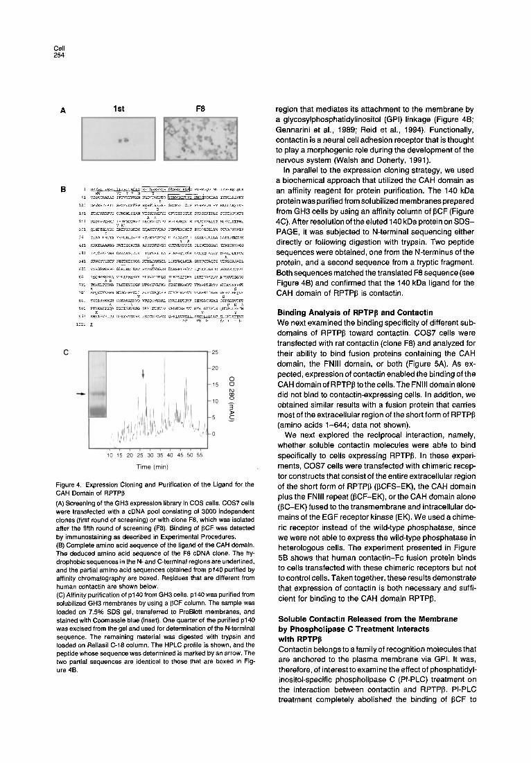
Binding Analysis of RPTPI~ and Contactin We next examined the binding specificity of different sub- domains of RPTP~ toward contactin. COS7 cells were transfected with rat contactin (clone F8) and analyzed for their ability to bind fusion proteins containing the CAH domain, the FNIII domain, or both (Figure 5A). As ex- pected, expression of contactin enabled the binding of the CAH domain of RPTPJ3 tothe cells. The FNIII domain alone did not bind to contactin-expressing cells. In addition, we obtained similar results with a fusion protein that carries most of the extracellular region of the short form of RPTPI~ (amino acids 1-644; data not shown).
We next explored the reciprocal interaction, namely, whether soluble contactin molecules were able to bind specifically to cells expressing RPTP~. In these experi- ments, COS7 cells were transfected with chimeric recep- tor constructs that consist of the entire extracellular region of the short form of RPTP[~ (I~CFS-EK), the CAH domain plus the FNIII repeat (I~CF-EK), or the CAH domain alone (I~C-EK) fused to the transmembrane and intracellular do- mains of the EGF receptor kinase (EK). We used a chime- ric receptor instead of the wild-type phosphatase, since we were not able to express the wild-type phosphatase in heterologous cells. The experiment presented in Figure 5B shows that human contactin-Fc fusion protein binds to cells transfected with these chimeric receptors but not to control cells. Taken together, these results demonstrate that expression of contactin is both necessary and suffi- cient for binding to the CAH domain RPTPIS.
Soluble Contactin Released from the Membrane by Phospholipase C Treatment Interacts with RPTPp Contactin belongs to a family of recognition molecules that are anchored to the plasma membrane via GPI. It was, therefore, of interest to examine the effect of phosphatidyl- inositol-specific phospholipase C (PI-PLC) treatment on the interaction between contactin and RPTPI3. PI-PLC treatment completely abolished the binding of 13CF to
Interactions between Receptor Phosphatase and Contactin 255
A s°°1 i 2°°°i I -' 15ooi, II
I I/ None C F CF
ECD
~CFS/EK
~CF/EK
3 I~C/EK
Mock
o z00 400 600 800 1000 t Z00
Bound ( c p m )
Figure 5. Binding of Soluble RPTP~ Forms to Contactin and Binding of a Soluble Contactin Form to RPTPI3 (A) Binding specificity of subdomains of RPTPI3 to contactin- expressing cells• COS cells were transfected with clone F8 encoding wild-type contactin (stippled bars) or with 13-gal-expressing plasmid as a control (closed bars)• Cells were analyzed for their ability to bind Fc fusion proteins containing the CAH domain (C), the FNIII domain (F), or both (CF). (B) Binding of human contactin-Fc fusion protein to cells expressing RPTPI3-EGF receptor chimeras. COS cells were transfected with chi- meric molecules in which the extracellular region of the short form of RPTPI~ (13CFS-EK), the CAH, and the FNIII domains (I~CF-EK) or the CAH domain alone (~C-EK) were fused to the transmembrane and cytoplasmic domains of the EGF receptor• Binding of the contactin- Fc fusion protein (stippled bars) or control Fc fusion protein (closed bars) was carried on after 72 hr as described in (A). The expression level of the chimeric receptors was determined by using antibodies against the extracellular domain of human RPTP[5 (shaded bars) as described in Experimental Procedures•
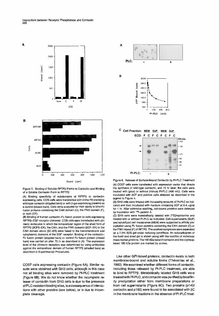
COS7 cells expressing contactin (Figure 6A). Similar re- sults were obtained with GH3 cells, although in this case not all binding sites were removed by PI-PLC treatment
(Figure 6B). We do not know whether the incomplete re- lease of contactin from GH3 cells is due to the presence of PLC-resistant binding sites, is a consequence of interac- tions with other proteins (see below), or is due to incom- plete cleavage.
A
PLC: +
35O0
3000"
o 2 5 0 0 '
2 0 0 0 '
1 5 0 0 '
1000"
500 • i . J , 200 400 600
PI-PLC ( m U )
C Cell Fraction: MEM SUP MEM SUP
ECD: F C F C F C F C ].~a
PI-PLC: +
Figure 6. Release of Surface-Bound Contactin by PI-PLC Treatment (A) COS7 cells were transfected with expression vector that directs the synthesis of wild-type contactin, and 72 hr later, the cells were treated with (plus) or without (minus) PI-PLC (400 mU). Cells were incubated with 13CF and positive cells detected as described in the legend to Figure 4. (13) GH3 cells were treated with increasing amounts of PI-PLC as indi- cated and then incubated with medium containing 13CF at 0.4 I~g/ml for 1 hr. After extensive washing, cell-bound proteins were detected by incubation with lzSl-protein A, (C) GH3 cells were metabolically labeled with [3SS]methionine and treated with or without PI-PLC as indicated. Cell supernatants (SUP) and solubilized cell membranes (MEM) were subjected to affinity pre- cipitation using Fc fusion proteins containing the CAH domain (C) or the FNIII repeat (F) of RPTPI3. The washed complexes were separated on a 7.5% SDS gel under reducing conditions. An autoradiogram of the fixed and dried gel is shown along with the location of molecular mass marker proteins. The 140 kDa band of contactin and the coprecip- itated 180 kDa protein are marked by arrows.
Like other GPl-l inked proteins, contactin exists in both membrane-bound and soluble forms (Theveniau et al., 1992). We examined whether different forms of contactin, including those released by PI-PLC treatment, are able to bind to RPTPI3. Metabolical ly labeled GH3 cells were treated with PI-PLC, and contactin was purified by bioaffin- ity precipitation either from membrane preparations or from cell supernatants (Figure 6C). Two proteins (p140/ contactin and p180) were found to be associated with 13C in the membrane fractions in the absence of PI-PLC treat-
Cell 256
A ~-CAH Ng-CAM
- PI-PLC
+ PI-PLC
non-immune Ig anti-contactin Ig
"~I ' nil ' ~I~H'~IH!
%i N
I~C
13CF
: ' ii i
~C
LAM
Figure 7. Biological Effect of the CAH Domain on Neurite Outgrowth and Cell Differentiation (A) Effect of PI-PLC on neurite growth of chick neurons on dishes coated with the CAH domain of RPTP~. Primary tectal cells were treated with or without PI-PLC as indicated for 1 hr and plated on J3CF- or Ng-CAM-coated dishes. After 24 hr, unbound cells were removed by gentle wash, and the plate was fixed and photographed. (B) Process extension of IMR32 cells induced by ~C and ~,CF fusion proteins is inhibited by antibodies against contactin. Dishes were coated in a circular array with 2 Ill drops of Fc fusion proteins (13C, I~CF, and ~/C) or with laminin (LAM). After removal of the unabsorbed proteins, the dishes were incubated with bovine serum albumin, and the IMR32 cells were allowed to adhere to the dishes for 3 hr. The medium was removed and replaced with fresh medium containing nonimmune or anti-contactin immunoglobulin antibodies as indicated, at a final concentration of 250 p,g/ml. The cultures were incubated for 48 hr, fixed, and photographed as described in Experimental Proce- dures (magnification, 92 x).
ment. Neither protein was present in the supernatant, nor did they interact with the FNIII domain of RPTPI3. However, after PI-PLC treatment, we were able to precipitate soluble contactin from the medium of the cells with a fusion protein containing the CAH domain, indicating that the soluble form generated by phospholipase treatment is still able to interact with RPTPI3. In contrast, PI-PLC treatment did not release the 180 kDa protein from the cells, suggesting that it may be a contactin-associated integral membrane protein. Since contactin by itself binds to the CAH domain,
the p180 protein is probably a contactin-bound protein iso- lated by the affinity procedure. It is, however, possible that this protein may interact directly with RPTPI3. An intriguing possibility is that p180 is a component of the contactin signaling complex in neurons (see below).
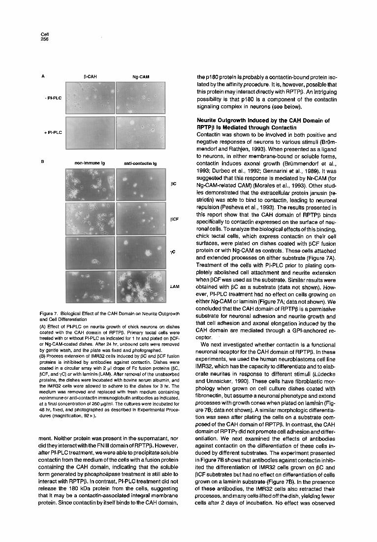
Neurite Outgrowth Induced by the CAH Domain of RPTPp Is Mediated through Contactin Contactin was shown to be involved in both positive and negative responses of neurons to various stimuli (Br0m- mendorf and Rathjen, 1993). When presented as a ligand to neurons, in either membrane-bound or soluble forms, contactin induces axonal growth (Br0mmendorf et al., 1993; Durbec et al., 1992; Gennarini et al., 1989). It was suggested that this response is mediated by Nr-CAM (for Ng-CAM-related CAM) (Morales et al., 1993). Other stud- ies demonstrated that the extracellular protein janusin (re- strictin) was able to bind to contactin, leading to neuronal repulsion (Pesheva et al., 1993). The results presented in this report show that the CAH domain of RPTPI3 binds specifically to contactin expressed on the surface of neu- ronal cells. To analyze the biological effects of this binding, chick tectal cells, which express contactin on their cell surfaces, were plated on dishes coated with ~CF fusion protein or with Ng-CAM as controls. These cells attached and extended processes on either substrate (Figure 7A). Treatment of the cells with PI-PLC prior to plating com- pletely abolished cell attachment and neurite extension when ~CF was used as the substrate. Similar results were obtained with J3C as a substrate (data not shown). How- ever, PI-PLC treatment had no effect on cells growing on either Ng-CAM or laminin (Figure 7A; data not shown). We concluded that the CAH domain of RPTPI~ is a permissive substrate for neuronal adhesion and neurite growth and that cell adhesion and axonal elongation induced by the CAH domain are mediated through a GPI-anchored re- ceptor.
We next investigated whether contactin is a functional neuronal receptor for the CAH domain of RPTP~. In these experiments, we used the human neuroblastoma cell line IMR32, which has the capacity to differentiate and to elab- orate neurites in response to different stimuli (LQdecke and Unnsicker, 1990). These cells have fibroblastic mor- phology when grown on cell culture dishes coated with fibronectin, but assume a neuronal phenotype and extend processes with growth cones when plated on laminin (Fig- ure 7B; data not shown). A similar morphologic differentia- tion was seen after plating the cells on a substrate com- posed of the CAH domain of RPTP~. In contrast, the CAH domain of RPTPy did not promote cell adhesion and differ- entiation. We next examined the effects of antibodies against contactin on the differentiation of these cells in- duced by different substrates. The experiment presented in Figure 7B shows that antibodies against contactin inhib- ited the differentiation of IMR32 cells grown on ~C and ~CF substrates but had no effect on differentiation of cells grown on a laminin substrate (Figure 7B). In the presence of these antibodies, the IMR32 cells also retracted their processes, and m any cells lifted off the dish, yielding fewer cells after 2 days of incubation. No effect was observed

Interactions between Receptor Phosphatase and Contactin 257
with control antibodies. Thus, neurite growth and differen- tiation induced by the CAH domain of RPTP~ is mediated by contactin molecules expressed on the surface of these cells.
Discussion
During development of the nervous system, neurons are guided by secreted and cell-bound molecules that provide both negative and positive cues. In this report, we show that RPTPI3, a receptor-type protein-tyrosine phospha- tase, may provide such a signal by interacting with the axonal recognition molecule contactin. RPTPI~ is a devel- opmentally regulated protein that exists in three forms, one secreted and two membrane bound. The extracellular region of RPTPI3 has a multidomain structure consisting of a CAH-like domain, a single FNIII repeat, and a long cysteine-free spacer region. The complex structural na- ture of its extracellular region may result in a multifunc- tional protein that is able to interact with different proteins. Indeed, we found that the CAH and the FNIII domains bind to at least two potential ligands present on neurons and glial cells. Functional expression cloning in COS7 cells and affinity purification with a specific affinity matrix fol- lowed by microsequencing enabled unequivocal identifi- cation of the cell recognition molecule contactin (F3/F11) as a neuronal ligand of RPTPI3. The interaction between contactin and RPTP~ is mediated via the CAH domain of the phosphatase, while the FNIII domain appears to bind to another molecule expressed on the surface of glial cells. It was previously shown that the secreted proteoglycan form of RPTPI3 interacts with tenascin, N-CAM, and Ng- CAM (Grumet et al., 1993, 1994; Barnea et al., 1994b; Milev et al., 1994). Since these proteins do not bind directly to the CAH or the FNIII domain of RPTPI~ (data not shown), they may interact with the large spacer domain of the phos- phatase. Alternatively, they could interact with RPTP~ through a third component. Contactin may fulfill this func- tion, since it has been shown to interact with Ng-CAM, Nr-CAM, and the matrix proteins tenascin and restrictin (Br0mmendorf et al., 1993; Morales et al., 1993; Zisch et al., 1992). It appears that the various subdomains of the extracellular region of RPTPI3 are able to interact with sev- eral distinct proteins that are expressed on diverse cell types in the central nervous system.
In contrast with other cell recognition molecules that are widely expressed in the nervous system, members of the contactin subgroup appear to be expressed in a restricted manner on specific axons during development (Dodd et al., 1988; Faivre-Sarrailh et al., 1992). The spatial and temporal expression pattern of these proteins suggests that they play an important role during development of the nervous system. Contactin was found to be exclusively expressed on neurons during development in fiber-rich areas of the retina, tectum, spinal cord, and cerebellum (Ranscht, 1988). It was found to be localized in the postna- tal and adult mouse cerebellum in axonal extensions of the granule cells in the parallel layer (Faivre-Sarrailh et al., 1992). This pattern of expression is overlapping with the expression pattern of RPTP~ in the rat. RPTP~ was
shown to be expressed in fiber-rich regions such as the parallel fibers of the cerebellum and the spinal cord (Canoll et al., 1993; Milev et al., 1994). RPTPI3 is also expressed on glial and radial glial cells, and its secreted form is pro- duced by astrocytes. It is possible, therefore, that both forms of RPTPI3 could modulate neuronal function via in- teractions with contactin.
The contactin subgroup of glycoproteins all share struc- tural similarity in that they are GPI-anchored proteins. They also exist in soluble forms generated as a result of membrane release or by expression of alternatively spliced forms (Br0mmendorf and Rathjen, 1993). Differen- tial expression of the membrane-bound and soluble forms of contactin was found in the hypothalamus-hypophyseal system (Rougon et al., 1994). RPTPI~ also exists in mem- brane-bound or secreted forms that are developmentally regulated. It is possible, therefore, that RPTPI3 and con- tactin can act both as ligand and receptor for each other. Hence, the classical notion of ligand-receptor interaction is inappropriate in this system, since the components might switch roles at different stages of development. For example, the soluble form of RPTPI3 produced by glial cells may act as a ligand for the membrane-bound form of contactin expressed on the surface of neuronal cells. Conversely, the soluble form of contactin may act as ligand for the membrane-bound form of RPTPI~ expressed on the surface of glial cells. Moreover, interaction between the membrane-bound forms of contactin expressed on the surface of neurons with the membrane-bound form of RPTPI3 expressed on the surface of glial cells may lead to bidirectional signals between these two cell types. Such complex interactions between the various forms of RPTP~ and contactin may generate developmentally regulated unidirectional and bidirectional signals.
How does the CAH domain of RPTPI~ bind to contactin? Carbonic anhydrases are highly efficient enzymes that cat- alyze the hydration of CO2. It has previously been pro- posed that the CAH domain of PTPases is not endowed with enzymatic activity, owing to substitution of two of the three key histidine residues that are essential for enzy- matic activity (Krueger and Saito, 1992; Barnea et al., 1993). However, the highly packed hydrophobic core, as well as the hydrophobic residues that are exposed on the surface of CAH structure and are conserved in the CAH domains of RPTPy and RPTPI3, may be involved in pro- tein-protein interaction and thus may function as a ligand- binding domain (Barnea et al., 1993). It is interesting to note that vaccinia virus contains a transmembrane protein with a CAH-like domain in its extracellular domain, which was thought to be involved in binding of the virion to host proteins (Maa et al., 1990). It is therefore possible that the CAH domains of RPTPy and vaccinia virus are able to bind to other members of the contactin family of glycopro- teins and that a contactin-related protein may function as a receptor for vaccinia virus.
How does contactin, a GPl-linked protein that is inserted into the outer leaflet of the plasma membrane, transmit a signal into the cells to promote neurite outgrowth? One possibility is that contactin is able to interact with a trans- membrane signaling component. The p180 protein that
Cell 258
was coprecipi tated with contact in is a candidate for such a signal ing protein (Figure 6C). p180 is probably membrane associated, since it could not be released by phosphol i- pase C treatment. Another potential signal t ransducer could be L I /Ng-CAM or a related molecule. This trans- membrane CAM was shown to interact with contact in (BrL~mmendorf et al., 1993) and to init iate second- messenger cascade v ia its cytoplasmic domain (Doherty and Walsh, 1994). The best-character ized GPl- l inked sig- nal ing protein is the ci l iary neurotrophic factor (CNTF) re- ceptor. Fol lowing l igand binding, the CNTF receptor inter- acts with the signal t ransducer gp130. The gp130 protein, which is shared by several lymphokines and cytokines, such as interleukin-6, leukemia inhibitory factor, and on- costatin, undergoes dimer izat ion fol lowed by recrui tment of the cytoplasmic janus kinase (JAK) protein tyrosine ki- nases. St imulat ion of the JAK kinases leads to act ivat ion of both the Ras/MAP kinase and the STAT signal ing path- ways, which relay signals from the cell surface to the nu- cleus. It is possible that a contact in-associated protein such as p180 has a funct ion similar to the function of gp130.
The binding of the CAH domain of RPTPI3 to contact in leads to cell adhesion and neurite outgrowth. It was re- cent ly reported that contact in can media te the repulsion of neurons by janusin (Pesheva et al., 1993). This effect was proposed to occur in a stepwise manner: first an adhe- sion step, fo l lowed by a signal t ransduced to the cells, leading to retraction. It appears, therefore, that in re-
sponse to dif ferent stimuli the same molecule can transmit opposi te signals, depending on the context or milieu. Whatever the mechanism, the results presented here demonstrate that an RPTP serves as a funct ional l igand for a GPI-anchored cell adhesion molecule.
We do not know yet whether contact in can serve as a functional l igand for RPTPI~. Modulat ion of phosphatase act ivi ty by neuronal contact in may result in signal ing to glial cells. If this does occur, this kind of bidirect ional f low of information should al low the interact ing cells to respond quickly to local env i ronmenta l changes during develop- ment. Two other RPTPs, RPTPp and RPTP~<, were shown to mediate cel l -cel l interact ion in a homophi l ic manner (Brady-Kalany et al., 1993; Gebbink et al., 1993; Sap et al., 1994). However, changes in catalyt ic act iv i ty as a result of these interact ions could not be detected. These phos- phatases are jo ining a growing family of proteins involved in cel lular recognit ion that contains intrinsic enzymat ic ac- tivities, including kinases (Dtrk; Pulido et al., 1992), the I~ subunit of Na +, K÷-ATPase (adhesion molecule on glia, or AMOG; Gloor et al., 1990), and the I~ subunit of prolyl 4-hydroxylase (cognin; Rao and Hausman, 1993).
In summary, we have demonst ra ted that RPTPI~ is a funct ional l igand for the GPI-anchored cell recognit ion molecule contactin. The interact ions between these two proteins are mediated by the CAH domain of the phospha- tase. In addit ion, the FNIII repeat is required for interact ion with glial cells, demonstrat ing that the mul t idomain struc- ture of RPTPI3 enables interact ions with dif ferent proteins, which raises the possibi l i ty that other potent ial l igands may modulate these interactions.
Experimental Procedures
Cell Culture SF763T and SF767T human astrocytoma cell lines were grown in athymic nu/nu mice to create a tumor-derived cell line. The parental lines (SF763 and SF767) were provided by Dr. M. E. Bernes (The Barrow Neurological Institute, Phoenix, AZ). All other cell line were obtained from the American Type Culture Collection (Rockville, MD). Cell cultures of rat sensory neurons were established as described (Hawrot and Patterson, 1979). Neurite outgrowth assays using IMR32 cells and primary tectal neurons were performed as described pre- viously (Grumet et al., 1993).
Generation and Production of Fc Fusion Proteins Different subdomains of the RPTPI3 extracellular region were amplified by polyrnerase chain reaction (PCR) and cloned into a unique BamHI site upstream of the hinge region of human IgG1-Fc. The 13C fusion proteins contained amino acids 1-313, and ~CF contained amino acids 1-415 from RPTP~ (Levy et al., 1993). J3F-Fc construct was generated by amplification of nucleotides 901-1242 and cloning into pCN71 be- tween the IgG gene and a sequence encoding a signal peptide derived from the TGF~ gene (Plowman et al., 1992). The contactin Fc (Hcon- Fc) fusion protein contained amino acids 1-1020 of human contactin fused to the IgG region. Fusion proteins were transiently expressed in COS7 cells or in stably transfected 293 cells and purified on a protein A-Sepharose column (Pharmacia). Immunoblots to detect their ex- pression were done essentially as described (Peles et al., 1992) with anti-human Fc antibodies.
Expression Cloning in COS7 Cells cDNA was synthesized from GH3-poly(A) mRNA by using Superscript II (GIBCO BRL). cDNAs larger than 2 kb were ligated into a EcoRI- and Hindlll-digested pCMP1 plasmid vector, a derivative of the pCMV-1 vector (Lammers et al., 1993). Plasmid DNA (10 pg) was transfected into COS7 cells with lipofectamine (GIBCO BRL). After 72 hr, the cells were incubated with medium containing I~CF. Unbound fusion proteins were washed away, and the cells were fixed with 4% paraformaldehyde in PBS. Immunostaining was performed with the ABC staining system (Vector Laboratories), with biotinylated anti- human IgG antibodies followed by streptavidin-alkaline phosphatase and BCIP-NBT as a substrate. One positive pool (number 54) was subdivided and rescreened until a single clone (F8) was isolated.
Construction of RPTPp-EGF Receptor Chimeras The region encoding the extracellular domain of the short form of RPTP~ (I~CFS, amino acids 1-768) and the region encoding CAH and the FNIII domains (~CF, amino acids 1-418) or the CAH domain alone (~C, amino acids 1-297) were amplified by PCR and attached to the human EGF receptor (EK) at residue 634. The junction of the two molecules was confirmed by nucleotide sequence analysis. The chi- meras were subcloned into a Notl site in pCMP1.
Binding Experiments with Fc Fusion Proteins Confluent monolayers of cells were incubated for I h r with conditioned medium containing 0.25-0,50 p~g/ml Fc fusion protein. Cells were washed three times with binding medium (0.1% bovin e serum albumin, 0.2% nonfat dry milk in DMEM-F12), and bound proteins were de- tected byadditional incubation with 1251-protein A(Amersham). In some experiments, the binding of antibodies against RPTP ~ was determined with 1251-labeled anti-mouse IgG (Amersham).
Chemical Covalent Cross-Linking Experiments Cells were incubated for 1 hr with medium containing the different Fc fusion proteins. Following three washes, the cells were incubated for an additional 30 min with PBS-Ca 2+ containing 1 mM DTSSP (Pierce, Rockford, IL). Cell lysates were made in SBN lysis buffer (Peles et al., 1991) and incubated with protein A-Sepharose for 2 hr at 4°C. The beads were washed three times with HNTG buffer (Peles et al., 1991), and the bound proteins were eluted by boiling in 1% SDS- PBS. The eluted proteins were diluted with HNTG and subjected to immunoprecipitation with agarose-coupled anti-human IgG antibod- ies (Sigma).
Interactions between Receptor Phosphatase and Contactin 259
Protein Purification and Sequencing Membrane pellet prepared from 5 x 10⁸ GH3 cells was resuspended in SML solubilization buffer (2% sodium monolaurate, 2 mM MgCI2, 2 rnM PMSF in PBS), diluted 10-fold with PBS containing 2 mM MgCIz, and loaded on a column of ~CF-Fc. Bound proteins were separated on 7.5% gel and electroblotted to ProBIott membrane (Applied Biosys- terns, Incorporated). Microsequencing was performed with an Applied Biosystems model 494 sequencer using standard reagents and pro- grams from the manufacturer. To obtain internal peptide sequence, the blotted band was reduced with 10 mM dithiothreitol and digested for 16 hr with trypsin, and peptides were separated on a 1 mm x 200 mm Reliasil C-18 reverse-phase column on a Michrom UMA high pressure liquid chromatograph (HPLC). Purified peptides were se- quenced as described above.
Treatment with PI-PLC Cells were metabolically labeled with 100 pCi/ml [~S]methionine (New England Nuclear, Boston, MA) and incubated with 250 mU of PI-PLC (Boehringer Mannheim) for 50 rain at 37°C. The supernatant was col- lected and cleared by centrifugation, and membranes were prepared from the cells and solubilized in SM L buffer. ~CF-Fc bound to protein A-Sepharose beads was added to the supernatant and the membrane fractions for 2 hr at 4°C. The beads were washed twice with 0.15% sodium monolaurate in PBS and once in PBS prior to the addition of SDS sample buffer.
Acknowledgments
Correspondence should be addressed to J. S. We thank Dr. John Hemperly for generously providing antisera against human contactin, and Dr. Sara Courtneidge for helpful comments on the manuscript. This work was supported in part by a grant from the Children's Brain Tumor Foundation to M. G. and by NIH NS21629.
Received May 9, 1995; revised June 13, 1995.
References
Barnea, G., Silvennoinen, O., Shaanan, B., Honegger, A. M., Canoll, P. D., D'Eustachio, P., Morse, B., Levy, B., La Forgia, S., Huebner, K., Musacchio, J. M., Sap, J., and Schlessinger, J. (1993). Identification of a carbonic anhyd rase-like domain in the extracellular region of RPTP'y defines a new subfamily of receptor tyrosine phosphatases. Mol. Cell. Biol. 13, 1497-1506.
Barnea, G., Grumet, M., Sap, J., Margolis, R. U., and Schlessinger, J. (1994a). Close similarity between receptor-linked tyrosine phospha- tase and rat brain proteoglycan. Cell 76, 205.
Barnea, G., Grumet, M., Milev, P., Silvennoinen, O., Levy, J. B., Sap, J., and Schlessinger, J. (1994b). Receptor tyrosine phosphatase ~ is expressed in the form of proteoglycan and binds to the extracellular matrix protein tenascin. J. Biol. Chem. 269, 14349-14352.
Berglund, E. O., and Ranscht, B. (1994). Molecular cloning and in situ localization of the human contactin gene (CNTN1) on chromosome 12ql 1-q12. Genomics 21,571-582.
Brady-Kalany, S. M., Flint, A. J., and Tonks, N. K. (1993). Homophilic binding of PTPp., a receptor-type protein tyrosine phosphatase. J. Cell Biol. 122, 961-972.
BrL~mmendorf, T., and Rathjen, F. G. (1993). Axonal g!ycoproteins with immunoglobulin and fibronectin type Ill-related domains in verte- brates: structural features, binding activities, and signal transduction. J. Neurochem. 61, 1207-1219.
BrLimmendorf, T., Hubert, M., Treubert, U., Leuschner, R., T~.rnok, A., and Rathjen, F. G. (1993). The axonal recognition molecule F l l is a multifunctional protein: specific domains mediate interactions with Ng-CAM and restrictJn. Neuron 10, 711-727.
Canoll, P. D., Barnea, G., Levy, J. B., Sap, J., Ehrlich, M., Silven- noinen, O., Schlessinger, J., and M usacchio, J. M. (1993). The expres- sion of a novel receptor-type tyrosine phosphatase suggests a role in morphogensis and plasticity of the nervous system. Dev. Brain Res. 75, 293-298.
Chao, M. V. (1992). Neurotrophin receptors: a window into neuronal differentiation. Neuron g, 583-593.
Charbonneau, H., and Tonks, N. (1992). 1002 protein phosphatases? Annu. Rev. Cell Biol. 8, 463-493.
den Hertog, J., Pals, C. E. G. M., Peppelenbosch, M. P., Tertoolen, L. J. C., de Laat, S. W., and Kruijer, W. (1993). Receptor protein tyro- sine phosphatase ~ activates pp60c-src and is involved in neuronal differentiation. EMBO J. 12, 3789-3798.
Dodd, J., Morton, S. B., Karagogeos, D., Yamamoto, M., and Jessell, T. M. (1988). Spatial regulation of axonal glycoprotein expression on subsets of embryonic spinal neurons. Neuron I, 105-116.
Doherty, P., and Walsh, S. F. (1994). Signal transduction events under- lying neurite outgrowth stimulated by cell adhesion molecules. Curt. Opin. Neurobiol. 4, 49-55.
Durbec, P., Gennarini, G., Goridis, C., and Rougon, G. (1992). A solu- ble form of the F3 neuronal cell adhesion molecule promotes neurite outgrowth. J~ Cell Biol. 117, 877-887.
Faivre-Sarrailh, C., Gennarini, G., Goridis, C., and Rougon, G. (1992). F3/F11 cell surface molecule expression in the developing mouse cer- ebellum is polarized at synaptic sites and within granule cells. J. Neu- rosci. 12, 257-267.
Gebbink, M. F. B. G., Zondag, G. C. M., Wubbolt, R. W., Beijersbergen, R. L., van Etten, I., and Moolenar, W. H. (1993). Cell-cell adhesion mediated by a receptor-like protein tyrosine phosphatase. J. Biol. Chem. 288, 16101-16104.
Gennadni, G., Cibelli, G., Rougon, G., Mattei, M. G., and Goridis, C. (1989). The mouse neuronal cell surface protein F3: a phosphatidylino- sitol-anchored member of the immunoglobulin superfamily related to chicken contactin. J. Cell Biol. 109, 755-788.
Gloor, S., Antonicek, H,, Sweadner, K. J., Paglius, S., Moos, F. F., and Schachner, M. (1990). The adhesion molecule on gila (AMOG) is a homologue of the ~-subunit of Na, K-ATPase. J. Cell Biol. 110, 165- 174.
Goodman, C. S., and Shatz, C. J. (1993). Developmental mechanisms that generate precise patterns of neuronal connectivity. Cell 721Neu- ron 10 (Suppl.), 77-98.
Grumet, M., Flaccus, A., and Margolis, R. U. (1993). Functional charac- terization of chrondoitin sulfate proteoglycans of brain: interactions with neurons and neural cell adhesion molecules. J. Cell Biol. 120, 815-824.
Grumet, M., Milev, P., Sakurai, T., Karthikeyan, L., Bourdon, M. A., Margolis, R. K., and Margolis, R. U. (1994). Interaction with tenascin and differential effect on cell adhesion of neurocan and phosphacan, two major chrondoitin sulfate proteoglycans of nervous tissue. J. Biol. Chem. 269, 12142-12146.
Hawrot, E., and Patterson, P. H. (1979). Long-term culture of dissoci- ated sympathetic neurons. Meth. Enzymol. 58, 547-584.
Hunter, T. (1995). Protein kinases and phosphatases: the yin and yang of protein phosphorylation and signaling. Cell 80, 225-236.
Hynes, R. O., and Lander, A. D. (1992). Contact and adhesive specifi- ties in the associations, migrations, and targeting of cells and axons. Cell 88, 303-322.
Ignelzi, M. A. J., Miller, D. R., Soriano, P., and Maness, P. (1994). Impaired neurite outgrowth of src-minus cerebellar neurons on the cell adhesion molecule LI. Neuron 12, 873-884.
Keynes, R. B., and Cook, G. M. W. (1992). Repellent cues in axon guidance. Curr. Opin. Neurobiol. 2, 55-59.
Krueger, N. X., and Saito, H. (1992). A human transmembrane protein- tyrosine-phosphatase, PTP~, is expressed in brain and has an N-ter- minal receptor domain homologous to carbonic anhydrases. Proc. Natl. Acad. Sci. USA 89, 7417-7421.
Lammers, R., Bossenmaier, B., Cool, D. E., Tonks, N. K., Schles- singer, J., Fisher, E. H., and UIIrich, A. (1993). Differential activities of protein tyrosine phosphatases in intact cells. J. Biol. Chem. 168, 24456-22462.
Levy, J. B., Cannol, P. D., Silvennoine, O., Barnea, G., Morse, B., Honegger, A. M., Haung, J.-T., Cannizzaro, L. A., Park, S.-H., Druck, T., Huebner, K., Sap, J., Ehrlich, M., Musacchio, J. M., and Schles-
Cell 260
singer, J. (1993). The cloning of a receptor-type tyrosine phosphatase expressed in the central nervous system. J. Biol. Chem 268, 10573- 10581. L~decke, G., and Unnsicker, K. (1990). Mitogenic effect of neuro- trophic factors on human IMR-32 neuroblastoma cells. Cancer 65, 2270-2278. Maa, J. S., Rodriguez, J. F., and Esteban, M. (1990). Structural and functional characterization of a cell surface binding protein of vaccinia virus. J. Biol. Chem. 265, 1569-1577. Maurel, P., Rauch, U., Flad, M., Margolis, R. K., and Margolis, R. U. (1994). Phosphacan, a chondroitin sulfate proteogtycan of brain that interacts with neurons and neural cell-adhesion molecules, is an extra- cellular variant of a receptor-type protein tyrosine phosphatase. Proc. Natl. Acad. Sci. USA 91, 2512-2516. Milev, P., Friedlander, D., Sakurai, T., Karthikeyan, L., Flad, M., Mar- golis, R. K., Grumet, M, and Margolis, R. U. (1994). Interactions of the chondroitin sulfate proteoglycan phosphacan, the extracellular do- main of a receptor-type tyrosine phosphatase, with neurons, gila, and neural cell adhesion molecules. J. Cell Biol. 127, 1703-1715. Morales, G., Hubert, M., BfiJmmendorf, T., Treubert, U., T~.rnok, A., Schwarz, U., and Rathjen, F. G. (1993). Induction of axonal growth by heterophilic interactions between the cell surface recognition pro- tein F l l and Nr-CAM/Bravo. Neuron 11, 1113-1122. Peles, E., Ben-Levy, R., Or, E., UIIrich, A., and Yarden, Y. (1991). Oncogenic forms of the neu/HER2 tyrosine kinase are permanently coupled to phospholipase Cy. EMBO J. 10, 2077-2086.
Peles, E., Bacus, S. S., Koski, R. A., Lu, H. S., Wen, D., Ogden, S. G., Ben-Levy, R., and Yarden, Y. (1992). Isolation of the neu/HER-2 stimulatory ligand: a 44 kd giycoprotein that induces differentiation of mammary tumor cells. Cell 69, 205-216. Pesheva, P., Gennarini, G., Goridis, C., and Schachner, M. (1993). The F3/11 cell adhesion molecule mediates the repulsion of neurons by the extracellular matrix glycoprotein J1-160/180. Neuron 10, 69- 82.
Plowman, G. D., Green, J. M., Neubauer, M. G., Buckley, S. D., Mc- Donald, V. L., Todaro, G. J., and Shoyab, M. (1992). The epithelin precursor encodes two proteins with opposing activities on epithelial cell growth. J. Biol. Chem. 267, 13073-13078. Pulido, D., Campuzano, S., Koda, T., Modolell, J., and Barbacid, M. (1992). Dtrk, a Drosophi/a gene related to the trk family of neurotrophin receptors, encodes a novel class of neural cell ad'hesion molecule. EMBO J. 11,391-404. Ranscht, B. (1988). Sequence of contactin, a 130-kD glycoprotein con- centrated in areas of interneuronal contact, defines a new member of the immunoglobulin supergene family in the nervous system. J. Cell Biol. 107, 1561-1573. Rao, A. S. M. K., and Hausman, R. E. (1993). cDNA for R-cognin: homology with a multifunctional protein. Proc. Natl. Acad. Sci. USA 90, 2950-2954. Reid, R. A., Bronson, D. D., Young, K. M., and Hemperly, J. J. (1994). Identification and characterization of the human cell adhesion mole- cule contactin. Brain Res. Mol. Brain Res. 21, 1-8. Rougon, G., Olive, S., Durbec, P., Faivre-Sarrailh, C., and Gennarini, G. (1994). Functional studies and cellular distribution of the F3 GPI- anchored adhesion molecule. Braz. J. Med. Biol. Res. 2, 409-414. Sahin, M., Dowling, J. J., and Hockfield, S. (1995). Seven protein tyrosine phosphatases are differentially expressed in the developing rat brain. J. Comp. Neurol. 351,617-631. Sap, J., Jiang, Y. P., Friedlander, D., Grumet, M., and Sclessinger, J. (1994). Receptor tyrosine phosphatase R-PTP-K mediates homophilic binding. Mol. Cell. Biol. 14, 1-9. Schlessinger, J., and UIIrich, Y. (1992). Growth factor signaling by receptor tyrosine kinases. Neuron 9, 383-391. Sharama, E., and Lombroso, P. J. (1995). A neuronal protein tyrosine phosphatase induced by nerve growth factor. J. Biol. Chem. 270, 49- 53. Shitara, K., Yamada, H., Watanabe, K., Shimonaka, M., and Yama- guchi, Y. (1994). Brain-specific receptor-type protein-tyrosine phos-
phatase RPTP~ is a chondroitin sulfate proteoglycan in vivo. J. Biol. Chem. 269, 20189-20193. Shock, L. P., Bare, D. J., Klinz, S. G., and Maness, P. F. (1995). Protein tyrosine phosphatases expressed in developing brain and retinal Mfiller gila. Mol. Brain Res. 28, 110-116.
Th~veniau, M., Durbec, P., Gennarini, G., Wood, J. N., and Rougon, G. (1992). Expression and release of phosphatidylinositol anchored cell surface molecule by a cell line derived from sensory neurons. J. Cell. Biochem. 48, 61-72. Umemori, H., Sato, S., Yagi, T., Aizawa, S., and Yamamoto, T. (1994). Initial events of myelination involve Fyn tyrosine kinase signaling. Na- ture 367, 572-576. Walsh, F., and Doherty, P. (1991). Glycosylphosphatidylinositol an- chored recognition molecules that function in axonal fasciculation, growth and guidance in the nervous system. Cell. Biol. Int. Rep. 15, 1151-1166. Walton, K. M., Martell, K. J., Kwak, S. P., Dixon, J. E., and Largent, B. L. (1993). A novel receptor-type protein tyrosine phosphatase~is expressed during neurogenesis in the olfactory neuroepithelium. Neu- ron 11,387-400. Zhang, J. S., and Longo, F. M. (1995). LAR tyrosine phosphatase receptor: alternative splicing is preferential to the nervous system, coordinated with cell growth and generates novel isoforms containing extensive CAG repeats. J. Cell Biol. 128, 415-431. Zipursky, S. L., and Rubin, G. M. (1994). Determination of neuronal cell fate: lessons from the R7 neuron of Drosophila. Annu. Rev. Neu- rosci. 17, 373-397. Zisch, A. H., D'Alessandti, L., Ranscht, B., Falchetto, R., Winterhalter, K. H., and Vaughan, L. (1992). Neuronal cell adhesion molecule con- tactin/F11 binds to tenascin via its immunoglobulin-Iike domains. J. Cell Biol. 119, 203-213.