Axonal transport defects: a common theme in neurodegenerative diseases
9
REVIEW Subhojit Roy Bin Zhang Virginia M.-Y. Lee John Q. Trojanowski Axonal transport defects: a common theme in neurodegenerative diseases Received: 1 September 2004 / Accepted: 9 September 2004 / Published online: 12 January 2005 Ó Springer-Verlag 2005 Abstract A core pathology central to most neurode- generative diseases is the misfolding, fibrillization and aggregation of disease proteins to form the hallmark lesions of specific disorders. The mechanisms under- lying these brain-specific neurodegenerative amyloi- doses are the focus of intense investigation and defective axonal transport has been hypothesized to play a mechanistic role in several neurodegenerative disorders; however, this hypothesis has not been extensively examined. Discoveries of mutations in hu- man genes encoding motor proteins responsible for axonal transport do provide direct evidence for the involvement of axonal transport in neurodegenerative diseases, and this evidence is supported by studies of animal models of neurodegeneration. In this review, we summarize recent findings related to axonal transport and neurodegeneration. Focusing on specific neurodegenerative diseases from a neuropathologic perspective, we highlight discoveries of human motor protein mutations in some of these diseases, as well as illustrate new insights from animal models of neuro- degenerative disorders. We also review the current understanding of the biology of axonal transport including major recent findings related to slow axonal transport. Keywords Axonal transport Alzheimer’s disease Frontotemporal dementias Parkinson’s disease Polyglutamine diseases Introduction The misfolding and aggregation of brain proteins lead- ing to the relentless accumulation of abnormal intra- and extracellular filamentous deposits in diverse central nervous system (CNS) cell types, including neurons and glia, are pathological hallmarks of many neurodegen- erative diseases (Table 1). Indeed, neurofibrillary tangles (NFTs) and senile plaques (SPs) were recognized as significant lesions of Alzheimer’s disease (AD) nearly 100 years ago [9, 17, 43], followed shortly thereafter by the discovery of Lewy bodies (LBs) in the brains of patients with Parkinson’s disease (PD) [9, 34, 43]. Al- though many of these fibrillary lesions are considered to be diagnostic hallmarks of specific disorders, a key unanswered question is whether these filamentous pro- tein aggregates contribute to the onset and progression of neurodegeneration, are mere bystanders in the degenerating process, or even play a neuroprotective role. An abundance of data, including several disease- specific mutations in the aggregated proteins suggests that protein aggregates have a central role in neurode- generative diseases [9, 43, 44]. However, even in disor- ders characterized by filamentous inclusions formed by specific mutated proteins, it has been argued that it is not straightforward to determine if these lesions are actually neurotoxic or are merely a part of a neuropro- tective response. Regardless of uncertainties about the biological consequences of protein aggregates, progressive intra- cellular accumulations of disease proteins can result from one or more of the following pathological pro- cesses: (1) abnormal synthesis and folding of disease proteins; (2) aberrant interactions of disease proteins with other proteins; (3) impaired degradation and turn over of disease proteins; (4) impaired intracellular transport of disease proteins, especially those targeted for axonal transport over long distances. While the first three mechanisms have been the subject of intense investigation especially over the last 5 years, far less S. Roy B. Zhang V. M.-Y. Lee J. Q. Trojanowski (&) Department of Pathology and Laboratory Medicine, Center for Neurodegenerative Disease Research, and Institute on Aging, University of Pennsylvania School of Medicine, HUP, Maloney Building 3rd Floor, 3600 Spruce Street, Philadelphia, PA 19104-4283, USA E-mail: [email protected] Tel.: +1-215-6626399 Fax: +1-215-3495909 Acta Neuropathol (2005) 109: 5–13 DOI 10.1007/s00401-004-0952-x
-
Upload
independent -
Category
Documents
-
view
5 -
download
0
Transcript of Axonal transport defects: a common theme in neurodegenerative diseases

REVIEW
Subhojit Roy Æ Bin Zhang Æ Virginia M.-Y. Lee
John Q. Trojanowski
Axonal transport defects: a common theme in neurodegenerativediseases
Received: 1 September 2004 / Accepted: 9 September 2004 / Published online: 12 January 2005� Springer-Verlag 2005
Abstract A core pathology central to most neurode-generative diseases is the misfolding, fibrillization andaggregation of disease proteins to form the hallmarklesions of specific disorders. The mechanisms under-lying these brain-specific neurodegenerative amyloi-doses are the focus of intense investigation anddefective axonal transport has been hypothesized toplay a mechanistic role in several neurodegenerativedisorders; however, this hypothesis has not beenextensively examined. Discoveries of mutations in hu-man genes encoding motor proteins responsible foraxonal transport do provide direct evidence for theinvolvement of axonal transport in neurodegenerativediseases, and this evidence is supported by studies ofanimal models of neurodegeneration. In this review,we summarize recent findings related to axonaltransport and neurodegeneration. Focusing on specificneurodegenerative diseases from a neuropathologicperspective, we highlight discoveries of human motorprotein mutations in some of these diseases, as well asillustrate new insights from animal models of neuro-degenerative disorders. We also review the currentunderstanding of the biology of axonal transportincluding major recent findings related to slow axonaltransport.
Keywords Axonal transport Æ Alzheimer’s disease ÆFrontotemporal dementias Æ Parkinson’s disease ÆPolyglutamine diseases
Introduction
The misfolding and aggregation of brain proteins lead-ing to the relentless accumulation of abnormal intra-and extracellular filamentous deposits in diverse centralnervous system (CNS) cell types, including neurons andglia, are pathological hallmarks of many neurodegen-erative diseases (Table 1). Indeed, neurofibrillary tangles(NFTs) and senile plaques (SPs) were recognized assignificant lesions of Alzheimer’s disease (AD) nearly100 years ago [9, 17, 43], followed shortly thereafter bythe discovery of Lewy bodies (LBs) in the brains ofpatients with Parkinson’s disease (PD) [9, 34, 43]. Al-though many of these fibrillary lesions are considered tobe diagnostic hallmarks of specific disorders, a keyunanswered question is whether these filamentous pro-tein aggregates contribute to the onset and progressionof neurodegeneration, are mere bystanders in thedegenerating process, or even play a neuroprotectiverole. An abundance of data, including several disease-specific mutations in the aggregated proteins suggeststhat protein aggregates have a central role in neurode-generative diseases [9, 43, 44]. However, even in disor-ders characterized by filamentous inclusions formed byspecific mutated proteins, it has been argued that it isnot straightforward to determine if these lesions areactually neurotoxic or are merely a part of a neuropro-tective response.
Regardless of uncertainties about the biologicalconsequences of protein aggregates, progressive intra-cellular accumulations of disease proteins can resultfrom one or more of the following pathological pro-cesses: (1) abnormal synthesis and folding of diseaseproteins; (2) aberrant interactions of disease proteinswith other proteins; (3) impaired degradation and turnover of disease proteins; (4) impaired intracellulartransport of disease proteins, especially those targetedfor axonal transport over long distances. While the firstthree mechanisms have been the subject of intenseinvestigation especially over the last 5 years, far less
S. Roy Æ B. Zhang Æ V. M.-Y. Lee Æ J. Q. Trojanowski (&)Department of Pathology and Laboratory Medicine,Center for Neurodegenerative Disease Research,and Institute on Aging,University of Pennsylvania School of Medicine,HUP, Maloney Building 3rd Floor,3600 Spruce Street, Philadelphia, PA 19104-4283, USAE-mail: [email protected].: +1-215-6626399Fax: +1-215-3495909
Acta Neuropathol (2005) 109: 5–13DOI 10.1007/s00401-004-0952-x

attention has been focused on the role of axonal trans-port in the aggregation of proteins and in mechanismsunderlying neurodegenerative diseases.
In this review, we focus on the role of axonal trans-port in neurodegenerative diseases, briefly summarizingclassical and recent studies on the biology of axonaltransport, and highlighting major recent findingsincluding pathogenic mutations in human genes thatencode proteins involved in axonal transport and studiesof animal models of disease related to impairments ofaxonal transport.
The biology of axonal transport
Neurons are the most polarized cells known. In humans,neurons typically have a cell body measuring �6–120 lm in diameter, but they may have an axonal pro-cess that can run up to a meter or more. In a moreextreme example like the blue whale (the largest mam-mals with a recorded length of over 30 m), axons canrun more than 10 m in length. Despite their extremelength, axons are generally devoid of the machinery forbiosynthesis of proteins and other molecules, and allaxonal components are synthesized in the cell body,translocated from the cell body into the axonal processand then transported to its destination within the axon(anterograde transport; for a review, see [5] and theaccompanying video). At the same time, a complemen-tary mechanism translocates ‘‘cargo’’ (effete or inter-nalized proteins, organelles, etc.) in the oppositedirection, i.e., away from the axon, into the cell body
(retrograde transport). This mechanism of axonaltransport is not exclusive to neurons, but is a ratherexaggerated version of transport seen in other cell types.Such transport mechanisms also exist in the dendrites,but little is known about mechanisms of dendritictransport and for the purposes of this review, we willfocus on axonal transport.
Generally speaking, to move any cargo, two com-ponents are required: ‘‘motors’’ to move the cargo, and‘‘rails’’ to transport them. For the rails, the neurons usethe complex network of cytoskeleton, mainly themicrotubules and also actin polymers (see [5] and theaccompanying video). For the motors, the neurons havean abundance of small molecular machines, known askinesins and dyneins, moving the cargo mainly in theanterograde or retrograde direction, respectively(Fig. 1).
Historical perspective of axonal transport
In sharp contrast to most other fields of neurobiology,the history of axonal transport study is a relatively shortone [5]. While nerve ligation experiments in the 1940sdefined that active axonal transport existed, it was notuntil the 1960s that ingenuous experiments designed byLasek and co-workers used pulse injections of radiola-beled amino acids into the dorsal root ganglia of liveanimals as a model system to study axonal transport[26]. The injected radiolabeled amino acids entered thecell bodies of dorsal root ganglia cells, were incorpo-rated into the newly synthesized proteins in the cell
Table 1 Abnormal protein aggregates in diverse hereditary andsporadic neurodegenerative disorders. Most lesions are in nuclei,cell bodies and processes of neurons and/or glia, but some areextracellular (i.e., SPs) (Ab amyloid-b peptides, AD Alzheimer’sdisease, ALS amytrophic lateral sclerosis, DLB dementia with LBs,DS Down’s syndrome, GCIs glial cytoplasmic inclusions, LBs
Lewy bodies, LBVAD Lewy body variant of AD, MSA multiplesystem atrophy, NBIA 1 neurodegeneration with brain iron accu-mulation type 1, NF neurofilaments, NFTs neurofibrillary tangles,NIID neuronal intranuclear inclusion disease, PD Parkinson’sdisease, PHFtau paired helical filament tau, SOD1 superoxidedismutase 1, SPs senile plaques)
Disease Lesion/components Location
ADa,b,c SPs/Ab ExtracellularNFTs/PHFtau IntracytoplasmicLBs/a-synuclein Intracytoplasmic
ALSa Spheroids/NF subunits, SOD1 IntracytoplasmicDLBc LBs/a-synuclein IntracytoplasmicDSa,b,c SPs/Ab Extracellular
NFTs/PHFtau IntracytoplasmicLBs/a-synuclein Intracytoplasmic
NBIA 1c LBs/a-synuclein IntracytoplasmicGCIs/a-synuclein Intracytoplasmic
LBVAD (AD+DLB)c SPs/Ab ExtracellularNFTs/PHFtau IntracytoplasmicLBs/a-synuclein Intracytoplasmic
MSAc GCIs/a-synuclein IntracytoplasmicNIID Inclusions/expanded polyglutamine tracts IntranuclearPrion diseasesa Amyloid plaques/Prions ExtracellularTauopathiesa,b Tangles/abnormal tau IntracytoplasmicTri-nucleotide repeat diseases Inclusions/expanded polyglutamine tracts Intranuclear and Intradendritic
aBoth hereditary and sporadic forms of these disorders occurbNeurodegenerative diseases with prominent tau pathology are tauopathiescNeurodegenerative diseases with prominent synuclein pathology are synucleinopathies
6

body, and the pulse of newly synthesized radiolabeledproteins were then transported into the axon. As thewave peak of radiolabeled proteins moved through theaxon, the protein composition at any given point variedaccording to time and distance from the cell body, andby studying the protein composition at various pointsalong the axon, serial snapshots of transported proteinscould be obtained (Fig. 2A). While some of the proteinswere very rapidly transported, at rates of 100–400 mm/day (1–5 lm/s), quickly reaching up to the tip of theaxon, another wave of proteins were found to movesignificantly slower, at about 0.2–5 mm/day (0.0002–0.05 lm/s). These two components were called fast ax-onal transport and slow axonal transport, respectively,but it is clear that this simplistic view of rates of cargomovement in axons is no longer tenable as originallyproposed; see below and [5, 26]. While fast transportmainly consisted of vesicular cargo, slow axonal trans-port was mainly composed of cytoskeletal proteins,mainly microtubules, neurofilaments and actin, alongwith many additional cytosolic proteins (for detailedreviews, see [1, 5]).
After these remarkable experiments that defined thenature of axonal transport, the next spurt of activitycame in the 1980s and the 1990s when a tremendousamount of scientific activity defined the biochemicalnature and the mechanisms of the specific motors (ki-nesins and dyneins) that were involved in the transport(for a comprehensive review, see [31]).
While the discovery of molecular motors greatly in-creased our understanding of fast axonal transport, onepuzzling aspect was that all the motor proteins discov-ered were found to move fast, in line with the speedsseen in fast axonal transport; however, it was difficult toreconcile the significantly slower transport rates seen inslow axonal transport. It was generally felt that visual-ization of axonal transport in real time would lead tomore insights into axonal transport in general, and slow
Fig. 1 Anterograde andretrograde axonal transport bymotors kinesins and dyneins,respectively. Kinesins anddyneins move on microtubulesand transport Golgi-derivedvesicles, cytosolic proteincomplexes, cytoskeletalpolymers, and other cargos likeribosomes and messengerRNAs
Fig. 2 Two techniques commonly used in studying axonal trans-port. A shows a technique pioneered by Lasek and co-workers,where a pulse of radiolabeled amino acids is injected into dorsalroot ganglia in animals. The radiolabeled amino acids areincorporated into newly synthesized proteins in the cell bodies,and the labeled proteins are subsequently transported along theaxon as a wave of radiolabeled proteins. By looking at the proteincomposition of the radiolabeled proteins at progressively distalsegments along the axon, serial snapshots of transported radiola-beled proteins can be obtained. Another technique is shown in B,where axonal transport can be visualized in real time. In thismethod, fluorescently labeled proteins are introduced into the cellbody, and then the transport of the labeled proteins is studied byusing epifluorescence microscopy
7

axonal transport in particular [2]. In 2000, two studiesusing cultured cells and labeled neurofilaments proteinshowed transport of neurofilaments, a component ofslow axonal transport, in real time [39, 47] (Fig. 2).Surprisingly, neurofilaments were found to move rap-idly, with rates up to 3 lm/s, in line with the rates re-ported for known motors (kinesins and dyneins);however, unlike fast axonal transport cargo that movedrapidly down the axon, without much pause, neurofila-ments were found to undergo prolonged pauses in theirtransit, often up to several minutes [39]. Also, at anygiven time, only a small fraction of the cargo wasactually moving, making the overall transport muchslower than fast axonal transport. These studies supporta unified model where both fast and slow axonal trans-port, although qualitatively different, are moved byknown ‘‘fast’’ motors like kinesins and dyneins [5].
Axonal transport defects in neurodegenerative diseases
Axonal transport can be fundamentally thought tocomprise three basic components: the cargo, the motorproteins that move the cargo, and the rails on which thecargo moves. In reality, of course, the system is muchmore complicated, with various adaptor/linker andregulatory proteins involved in the process, along withpossible modulation at the gene/RNA level. In principle,defects in any of the three components can lead totransport functional and/or pathological changes, andpotentially neurodegeneration. From the very early daysof research on neurodegenerative diseases, it has beenpostulated that defects in axonal transport may beresponsible for these human disorders [10]. Althoughvarious mutant and transgenic animal models gaveindirect support for the hypothesis, three recent devel-opments have dramatically highlighted the role andsignificance of axonal transport in human neurodegen-erative diseases. They are (1) discovery of human motorprotein mutations in neurodegenerative diseases; (2)axonal transport defects in animal and in vitro cellularmodels harboring human mutations; and (3) newly dis-covered roles for pathogenic proteins like amyloid pre-cursor protein (APP), tau, presenilins and synucleins inmodulation and regulation axonal transport.
Kinesin mutation in hereditary spastic paraplegia
Hereditary spastic paraplegias (HSP), also known asfamilial spastic paraplegias, represents a heterogeneousinherited group of neurodegenerative diseases charac-terized by progressive lower limb spasticity and weak-ness. It can be transmitted in an autosomal dominantfashion, with patients presenting in their thirties orforties with spasticity in the lower limbs, with gradualproximal spread of symptoms. Neuropathologically, adistal axonal neuropathy is seen, with severe degenera-tion and gliosis of the distal corticospinal tracts, and
relative sparing of the tracts in the brain stem andproximal cord. In more complex forms of the disease,sensory symptoms, mental retardation and optic atro-phy are also seen [31]. Many different loci for HSP havebeen identified, and the commonest gene involved inabout 40% of the cases, is spastin, an ATPase withunclear functions [15]. Due to the peculiar slow ‘‘dyingback’’ neuropathy observed in this disease, it has beenhypothesized that dysfunctions in axonal transport,leading to selective damage of the distant portions of theaxons may be responsible for the pathogenesis of HSP.
Indeed, a missense mutation in one of the genesencoding a major kinesin protein (i.e., the gene for ki-nesin heavy chain Kif 5a) was recently found in a familywith HSP [38]. The same missense mutation is found inall affected members of the family, as well as in somepresymptomatic members. This mutation occurs withina functional motor domain of the kinesin protein and ahomologous mutation in yeast has been found todecouple kinesin binding to microtubules, underliningthe functional role of the kinesin mutation in thepathology of HSP.
Dynein mutations in a family with motor neuron disease
Dynein is one of the major motor proteins responsiblefor retrograde axonal transport, i.e., transport of cargofrom the axons towards the cell bodies. Dynein is a largemotor protein consisting of at least three different classesof subunits [45]. Dynein binds to a multiprotein complexcalled dynactin. Dynactin acts as a platform for bindingdynein to its cargo, and is also thought to play a role inmodulation of dynein [22, 48]. Recently, dynactin wasalso shown to bind to the anterograde motor kinesin,and may act as a molecular switch between anterogradeand retrograde transport [6]. Animal models disruptingthe dynein/dynactin complex develop a late onset motorneuron degeneration [25], and missense mutations in adynein subunit causes Lewy body-like inclusions andprogressive motor neuron degeneration in mice [14].Due to the central role of the dynein/dynactin complexin axonal transport, and due to evidence from animalstudies, it was hypothesized that dynein mutations couldplay a role in neurodegeneration. Indeed, a search fordynein mutations in neurodegenerative diseases led tothe discovery of mutations in the gene encoding animportant subunit (p150) of the dynactin complex in afamily with motor neuron disease [37]. In this family, thedisease is transmitted in an autosomal dominant fash-ion, and is a primary lower motor type neuropathy withpatients presenting in their 30s, often with breathingdifficulty due to vocal fold paralysis. The mutation is asingle base pair change (substitution of serine for glycineat position 59), and it has been found in all affectedfamily members. However, it has not been detected inunaffected family members or unaffected controls. Fur-ther, the mutation reduces the binding affinity of dyn-actin to microtubules [37]. These findings provide
8

convincing evidence that dynein mutations are involvedin a small subset of motor neuron diseases.
Kinesin mutations in Charcot-Marie-Tooth disease
Charcot-Marie-Tooth (CMT) disease comprises a het-erogeneous group of inherited peripheral neuropathiescharacterized by motor and sensory deficits, often pre-senting in young adults as tingling, numbness and loss ofdeep reflexes. The progression of the disease variesamong individuals, with symptoms ranging from mildneuropathy to complete disability. Two basic forms canbe recognized, with primary demyelinating (CMT1), oraxonal (CMT2) types of degeneration predominating [4].Various genes have been implicated in CMT syndromes,including several genes known to play a role in myeli-nation (PMP22, MPZ) and genes for gap junctionalproteins (Connexin 32) [4]. However, in a remarkableseries of studies, it was shown that mutations in a ki-nesin subunit protein (Kif 1B beta) can lead to an axonaltype of CMT (CMT type 2A).
While studying animal models of kinesin knockoutmice, Hirokawa and co-workers [53] found that het-erozygous knockout mice for one of the kinesins, Kif 1B,developed progressive muscle weakness with normalmotor nerve conduction velocities, symptoms closelyresembling axonal type CMT, CMT type 2. Incidentally,the gene for CMT type 2A was mapped to the sameinterval as the gene for Kif 1B [3, 33], and genomicanalysis of pedigrees with CMT type 2A revealed thatthese patients had a mutation in the Kif 1B gene [53]. Itwas further shown that the mutant motor protein maynot tightly bind to microtubules [53], thus suggesting aloss of function of the Kif 1B protein in patients withCMT type 2A.
Axonal transport in AD and tauopathies
The story of the role of axonal transport in AD is arapidly developing one [17, 27]. Neuropathologically,the two hallmarks of AD are (1) deposits of fibrillar Abinto diffuse and neuritic plaques in the extracellularspace, and (2) filamentous accumulations of tau proteinsas NFTs and neuropil threads within neurons and theirprocesses. Tau is also involved in a group of disordersknown as tauopathies, in which the hallmark lesions areAD-like filamentous tau inclusions only [17, 27]. Whileamyloidogenic Ab peptides are generated by proteolyticprocessing of APP, tau is a microtubule binding protein.The normal functions of neither Ab nor APP are clear atthis time, although many activities have been ascribed toboth, as described below.
Recent studies have implicated APP in axonaltransport by suggesting it might act as a receptor for theanterograde motor kinesin [21]. Further, it has also beenproposed that defects in APP can translate into majoraxonal transport defects, leading to neuronal damage or
neurodegeneration. While this proposed role of APP inaxonal transport needs to be validated and confirmed byother laboratories, this work draws further attention tothe possibility that impairments in axonal transport mayunderlie neurodegenerative disease mechanisms in AD.On the other hand, tau is a microtubule binding proteinwith well-defined functions, such as binding to and sta-bilizing microtubules in vivo and promoting theirassembly from tubulin monomers [17]. Indeed, since tauis sequestered into tangles where it is hyperphosphory-lated and unable to bind microtubules, it was proposedas long ago as 1994 that functional defects in patho-logical tau observed in AD brains would destabilizemicrotubules, thereby disrupting axonal transport [27].Below, we summarize known features of axonal trans-port of APP, review possible roles of APP and tau inimpairment of axonal transport in AD, and highlight therecently discovered link between presenilins and kinesinregulation.
It is well established that APP is transported in axonsvia fast axonal transport [23]. Further, there is con-vincing evidence that APP transport is dependent on themajor kinesin protein, kinesin-I [8, 21], and it has alsobeen proposed that APP may be transported in avesicular complex containing presenilins and BACE thatplay critical roles in the processing of APP into amy-loidogenic Ab peptides [11, 21]. These findings suggestthat misregulation of APP, either directly from knownAPP mutations (as in familial AD), or indirectly viaproteins associating/interacting with APP can lead tomisregulation of fast axonal transport in general, leadingto axonal depletion of critical components, and neu-rodegeneration [12].
Another interesting line of evidence highlighting therole of presenilins in regulation of fast axonal transportin AD came from a recent study of presenilin mutantmice. Presenilins are proteins responsible for regulatedproteolysis of APP, and mutations in presenilins are seenin most cases of early familial AD. Several studiesindicate that presenilins interact with glycogen synthasekinase 3 b (GSK3 b) [41]. GSK3 b is a kinase with manydifferent roles, including the phosphorylation of kinesinlight chains, and it has been shown that GSK3 b-medi-ated phosphorylation of kinesin light chains leads todetachment of the kinesin motor from the cargo, thuspreventing further transport of cargo (Fig. 3) [30]. Usingtransgenic presenilin mutant mice, Pigino et al. [35]showed that mutant or absent presenilin increasedGSK3 b levels, thereby phosphorylating kinesin lightchains, detaching the kinesins from their cargo, andimpairing axonal transport. While preliminary, thesestudies are significant for opening up new lines ofinvestigation into how abnormalities in APP or pre-senilins might perturb neuronal functions by disruptingaxonal transport, and lead to neurodegeneration in AD.
With respect to tau pathologies and mechanisms ofneurodegeneration in AD, the first experimental evi-dence supporting the hypothesis that tau pathologiescould impair axonal transport came from the work of
9

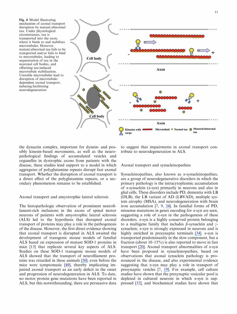
Ishihara et al. [18], showing that sequestration of humantau into filamentous inclusions in tau transgenic miceleads to a neurodegenerative phenotype in mice, similarto neurodegeneration seen in motor neuron disease ofGuam [44]. Further, by studying tau transport in miceexpressing the human tau mutation (R406W), which ispathogenic for the tauopathy familial frontotemporaldementia with parkinsonism linked to chromosome 17(FTDP-17), Zhang et. al. [51] showed that the transportof mutated human tau (but not normal human tau) isretarded in the axons of these transgenic mice. It hasalso been shown that the R406W mutation impairs theability of tau to bind to and stabilize microtubules [16].Given these findings, the study by Zhang et al. providesexperimental evidence for a mechanism of tau accumu-lation (Fig. 4). In this model, the mutated tau in FTDP-17 fails to bind to microtubules cannot be transportedinto the axon and in turn aggregates in the cell body,leading to a depletion of tau in the axons. The lack ofnormal tau binding to microtubules in axons leads todestabilization of microtubules, subsequently causing
neurodegeneration by impairment of axonal transporton the unstable microtubules. As described above, manyof the predictions of the mechanism whereby taupathologies might cause neurodegeneration by impair-ing axonal transport have been directly demonstrated inexperimental model systems.
A final line of evidence suggesting a role of axonaltransport defects in AD comes from studies of ApoE4, agene whose allelic state is associated with an increasedrisk for AD. Mice expressing human ApoE4 exhibitdefects in axonal transport [42], and the receptor forApoE4, ApoER2 binds to JIP1/2, a protein that appearsto mediate the binding of APP to kinesin I [46]. Thus, itcan be postulated that overexpression of ApoE4 proteincan lead to misregulation of JIP1/2-mediated binding ofkinesin to APP, leading to defects in fast axonal trans-port.
Although many of the above findings need to berigorously investigated by more direct experiments, thefindings summarized above provide support for thehypothesis that impairments in fast axonal transport byabnormalities in APP, presenilins and tau play a role inmechanisms of brain degeneration in AD. Whethermisregulation of axonal transport has a direct role in thepathogenesis of the disease or is a secondary phenome-non leading to axonal degeneration remains to be seen.However, these uncertainties notwithstanding, thera-peutic intervention in AD by modulation/correction ofaxonal transport defects remains a viable hypothesis. Inthis regard, a recent study showed that a microtubule-stabilizing drug, Paxceed (paclitaxel family), couldameliorate the neurodegenerative phenotype in trans-genic tau mice by offsetting the loss of tau function bystabilizing the microtubules and correcting the fast ax-onal transport defects in these mice [52].
Axonal transport and Huntington’s disease
Neuropathologically, Huntington’s disease is character-ized by atrophy and degeneration of striatal neurons,with aggregates of the pathological polyglutamine-con-taining protein, Huntingtin. Huntingtin is a predomi-nantly cytoplasmic protein that associates with vesiclesand moves in the fast axonal transport component.Although it has been known for several years thatpolyglutamine repeats within the Huntingtin proteincause a gain of deleterious function leading to neu-rodegeneration, the exact pathogenic role of the repeatsis unclear. Recently, by infusing pathological polyglu-tamine repeats into a model for studies of fast axonaltransport, Szebenyi et al. [40] demonstrated that fastaxonal transport was specifically inhibited by patho-logically expanded polyglutamine repeats (but not nor-mal proteins), along with inhibition of neurite extensionin cultured cells. Further, disruption of the Drosophilahuntingtin gene also caused axonal transport defects[11]. In combination with the findings that a Huntingtinbinding protein HAP1 interacts with a major protein of
Fig. 3 Model illustrating the role of presenilins in the regulation ofaxonal transport. Presenilin mutation/inactivation leads to GSK3 bactivation that in turn phosphorylates kinesin light chains.Phosphorylation of kinesin light chains releases vesicles destinedto undergo fast axonal transport, thereby clogging the axons withvesicles and inducing/facilitating neurodegeneration (GSK3 bglycogen synthase kinase 3 b)
10
the dynactin complex, important for dynein- and pos-sibly kinesin-based movements, as well as the neuro-pathological findings of accumulated vesicles andorganelles in dystrophic axons from patients with thedisease, these studies lend support to a model in whichaggregates of polyglutamine repeats disrupt fast axonaltransport. Whether the disruption of axonal transport isa direct effect of the polyglutamine repeats, or a sec-ondary phenomenon remains to be established.
Axonal transport and amyotrophic lateral sclerosis
The histopathologic observation of prominent neurofi-lament-rich inclusions in the axons of spinal motorneurons of patients with amyotrophic lateral sclerosis(ALS) led to the hypothesis that disrupted axonaltransport of proteins may play a role in the pathogenesisof the disease. However, the first direct evidence showingthat axonal transport is disrupted in ALS awaited thedevelopment of transgenic mouse models of familialALS based on expression of mutant SOD-1 proteins inmice [13] that replicate several key aspects of ALS.Studies on these SOD-1 transgenic mouse models ofALS showed that the transport of neurofilament pro-teins was retarded in these animals [50], even before themice were symptomatic [49], thereby implicating im-paired axonal transport as an early deficit in the onsetand progression of neurodegeneration in ALS. To date,no motor protein gene mutations have been reported inALS, but this notwithstanding, there are persuasive data
to suggest that impairments in axonal transport con-tribute to neurodegeneration in ALS.
Axonal transport and synucleinopathies
Synucleinopathies, also known as a-synucleinopathies,are a group of neurodegenerative disorders in which theprimary pathology is the intracytoplasmic accumulationof a-synuclein (a-syn) primarily in neurons and also inglial cells. These disorders include PD, dementia with LB(DLB), the LB variant of AD (LBVAD), multiple sys-tem atrophy (MSA), and neurodegeneration with brainiron accumulation [7, 9, 34]. In familial forms of PD,missense mutations in genes encoding for a-syn are seen,suggesting a role of a-syn in the pathogenesis of thesedisorders. a-syn is a highly conserved protein belongingto a multigene family that includes b-synuclein and c-synuclein; a-syn is strongly expressed in neurons and ishighly enriched in presynaptic terminals [34]. a-syn istransported predominantly in the slow component, but afraction (about 10–15%) is also reported to move in fasttransport [20]. Axonal transport abnormalities of a-synhave been proposed in synucleinopathies, based onobservations that axonal synuclein pathology is pro-nounced in the disease, and also experimental evidencesuggesting that a-syn may play a role in transport ofpresynaptic vesicles [7, 19]. For example, cell culturestudies have shown that the presynaptic vesicular pool isdepleted in cultured neurons in which a-syn is sup-pressed [32], and biochemical studies have shown that
Fig. 4 Model illustratingmechanism of axonal transportdisruption by mutant/abnormaltau. Under physiologicalcircumstances, tau istransported into the axon,where it binds to and stabilizesmicrotubules. However,mutant/abnormal tau fails to betransported and/or fails to bindto microtubules, leading tosequestration of tau in theneuronal cell bodies, andoffsetting tau-inducedmicrotubule stabilization.Unstable microtubules lead todisruption of microtubule-dependent axonal transport,inducing/facilitatingneurodegeneration
11

mutant a-syn fails to bind adequately to synaptic vesicles[19], suggesting that a-syn may play a role in vesiculartransport. A recent study also showed that, althoughthere was no difference in the axonal transport of mu-tant a-syn when compared to normal a-syn, there wassignificant age-related retardation in the normal trans-port of a-syn [28]. This study proposes a model in whichage-related retardation of synuclein transport leads toaccumulations of synuclein over time and predisposessynuclein pathology in axons. Although these findingsare interesting, many questions remain unanswered. Thephysiological role of synuclein is far from clear, andmuch work needs to be done to uncover the role ofsynuclein in neurodegenerative disorders.
Conclusions and future directions for research
An increasing body of evidence implicates axonaltransport defects in the etiology of neurodegenerativediseases. While human mutations in genes encodingmotor proteins are very compelling and direct evidencefor this, studies of transgenic mice also provide evidencethat axonal transport impairments contribute to neu-rodegeneration. Although finding motor protein defectsin neurodegenerative diseases points directly to defectsin transport, it is probable that many other diseaseproteins are directly or indirectly linked to the compli-cated machinery of axonal transport. Thus, it is imper-ative that studies into the molecular mechanisms oftransport must go hand in hand with the discovery ofadditional human mutations in familial neurodegenera-tive diseases linked to the axonal transport machinery.Another exciting but largely neglected avenue is drugdiscovery efforts to counteract axonal transportimpairments as therapeutic interventions for neurode-generative diseases. Indeed, if axonal transport defectsare shown to be part of a common mechanism of diseasein many neurodegenerative disorders, the discovery ofdrugs that modulate axonal transport could becomeimportant therapeutic interventions for the treatment ofthese disorders.
Acknowledgements We are indebted to the patients and theircaregivers who have facilitated research on these neurodegenerativediseases. V.M.Y.L. is the John H. Ware, 3rd Professor of Alzhei-mer’s disease research. J.Q.T. is the William Maul Measey-TrumanG. Schnabel, Jr. Professor of Geriatric Medicine and Gerontology.The authors acknowledge support for their research from the NIH[P01 AG09215, P30 AG10124, P01 AG11542, P01 AG14382, P01AG14449, P01 AG17586, P01 NS044233]. Due to space limitations,many references to primary literature cannot be included but theymay be found in reviews cited here.
References
1. Baas PW (2002) Microtubule transport in the axon. Int RevCytol 212:41–62
2. Baas PW, Brown A (1997) Slow axonal transport: the polymertransport model. Trends Cell Biol 7:380–384
3. Ben Othmane KB, Middleton LT, Loprest LJ, Wilkinson KM,Lennon F, Rozear MP, Stajich JM, Gaskell PC, Roses AD,Pericak-Vance MA, Vance JM (1993) Localization of a gene(CMT2A) for autosomal dominant Charcot-Marie-Tooth dis-ease type 2 to chromosome 1p and evidence of genetic heter-ogeneity. Genomics 17:370–375
4. Benstead TJ, Grant IA (2001) Progress in clinical neuro-sciences: Charcot-Marie- Tooth disease and related inheritedperipheral neuropathies. Can J Neurol Sci 28:199–214
5. Brown A (2003) Axonal transport of membranous andnonmembranous cargoes: a unified perspective J Cell Biol160:817–821 (See also the animation of axonal transport thataccompanies this paper at http://www.jcb.org/cgi/content/full/jcb.200212017/DC1)
6. Deacon SW, Serpinskaya AS, Vaughan PS, Fanarraga ML,Vernos I, Vaughan KT, Gelfand VI (2003) Dynactin is re-quired for bidirectional organelle transport. J Cell Biol160:297–301
7. Duda JE, Giasson BI, Mabon M, Lee VM-Y, Trojanowski JQ(2002) Novel antibodies to oxidized alpha-synuclein revealabundant neuritic pathology in Lewy body diseases. AnnNeurol 52:205–210
8. Ferreira A, Caceres A, Kosik KS (1993) Intraneuronal com-partments of the amyloid precursor protein. J Neurosci13:3112–3123
9. Forman MS, Trojanowski JQ, Lee VM-Y (2004) Neurode-generative diseases: a decade of revolutionary discoveries pavesthe way for therapeutic breakthroughs. Nat Med 10:1055–1063
10. Gajdusek DC (1985) Hypothesis: Interference with axonaltransport of neurofilament as a common pathogenetic mecha-nism in certain diseases of the central nervous system. N Engl JMed 312:714–719
11. Gunawardena S, Goldstein LS (2001) Disruption of axonaltransport and neuronal viability by amyloid precursor proteinmutations in Drosophila. Neuron 32:389–401
12. Gunawardena S, Goldstein LS (2004) Cargo-carrying motorvehicles on the neuronal highway: transport pathways andneurodegenerative disease. J Neurobiol 58:258–271
13. Gurney ME, Pu H, Chiu AY, Dal Canto MC, Polchow CY,Alexander DD, Caliendo J, Hentati A, Kwon YW, Deng HX,et al (1994) Motor neuron degeneration in mice that express ahuman Cu, Zn superoxide dismutase mutation. Science264:1772–1775
14. Hafezparast M, Klocke R, Ruhrberg C, Marquardt A, Ahmad-Annuar A, Bowen S, Lalli G, Witherden AS, Hummerich H,Nicholson S, et al (2003) Mutations in dynein link motorneuron degeneration to defects in retrograde transport. Science300:808–812
15. Hazan J, Fonknechten N, Mavel D, Paternotte C, Samson D,Artiguenave F, Davoine CS, Cruaud C, Durr A, Wincker P,et al (1999) Spastin, a new AAA protein, is altered in the mostfrequent form of autosomal dominant spastic paraplegia. NatGenet 23:296–303
16. Hong M, Zhukareva V, Vogelsberg-Ragaglia V, Wszolek Z,Reed L, Miller BI, Geschwind DH, Bird TD, McKeel D, GoateA, et al (1998) Mutation-specific functional impairments indistinct tau isoforms of hereditary FTDP-17. Science 282:1914–1917
17. Higuchi M, Lee VM-Y, Trojanowski JQ (2002) Tau andaxonopathy in neurodegenerative disorders. NeuromolecularMed 2:131–150
18. Ishihara T, Hong M, Zhang B, Nakagawa Y, Lee MK, Tro-janowski JQ, Lee VM-Y (1999) Age-dependent emergence andprogression of a tauopathy in transgenic mice engineered tooverexpress the shortest human tau isoform. Neuron 24:751–762
19. Jensen PH, Nielsen MS, Jakes R, Dotti CG, Goedert M (1998)Binding of alpha-synuclein to brain vesicles is abolished byfamilial Parkinson’s disease mutation. 273:26292–26294
20. Jensen PH, Li JY, Dahlstrom A, Dotti CG (1999) Axonaltransport of synucleins is mediated by all rate components. EurJ Neurosci 11:3369–3376
12

21. Kamal A, Almenar-Queralt A, LeBlanc JF, Roberts EA,Goldstein LS (1993) Kinesin-mediated axonal transport of amembrane compartment containing beta-secretase and prese-nilin-1 requires APP. J Neurosci 13:3112–3123
22. King SJ, Schroer TA (2000) Dynactin increases the processivityof the cytoplasmic dynein motor. Nat Cell Biol 2:20–24
23. Koo EH, Sisodia SS, Archer DR, Martin LJ, Weidemann A,Beyreuther K, Fischer P, Masters CL, Price DL (1990) Pre-cursor of amyloid protein in Alzheimer disease undergoes fastanterograde axonal transport Proc Natl Acad Sci USA87:1561–1565
24. Kotzbauer P, Giasson B, Kravitz A, Golbe LI, Mark MH,Trojanowski JQ, Lee, VM-Y (2004) In vitro and postmortembrain studies link fibrillization of both alpha-synuclein and tauto familial Parkinson’s disease caused by the A53T alpha-syn-uyclein mutation. Exp Neurol 187:279–288
25. LaMonte BH, Wallace KE, Holloway BA, Shelly SS, Ascano J,Tokito M, Van Winkle T, Howland DS, Holzbaur EL (2002)Disruption of dynein/dynactin inhibits axonal transport inmotor neurons causing late-onset progressive degeneration.Neuron 34:715–727
26. Lasek RJ, Garner JA, Brady ST (1984) Axonal transport of thecytoplasmic matrix. J Cell Biol 99:212–221
27. Lee VM-Y, Daughenbaugh R, Trojanowski JQ (1994) Micro-tubule stabilizing drugs for the treatment of Alzheimer’s dis-ease. Neurobiol Aging 15:S87–S89
28. Li W, Hoffman PN, Stirling W, Price DL, Lee MK (2004)Axonal transport of human alpha-synuclein slows with agingbut is not affected by familial Parkinson’s disease-linkedmutations. J Neurochem 88:401–410
29. McDermott CJ, White K, Bushby K, Shaw PJ (2000) Heredi-tary spastic paraparesis: a review of new developments. JNeurol Neurosurg Psychiatry 69:150–160
30. Morfini G, Szebenyi G, Elluru R, Ratner N, Brady ST (2002)Glycogen synthase kinase-3 phosphorylates kinesin light chainsand negatively regulates kinesin-based motility. EMBO J21:281–293
31. Muresan V (2000) One axon, many kinesins: What’s the logic? JNeurocytol 29:799–818
32. Murphy DD, Rueter SM, Trojanowski JQ, Lee VM (2000)Synucleins are developmentally expressed, and alpha-synucleinregulates the size of the presynaptic vesicular pool in primaryhippocampal neurons. J Neurosci 20:3214–3220
33. Nakagawa T, Tanaka Y, Matsuoka E, Kondo S, Okada Y,Noda Y, Kanai Y, Hirokawa N (1997) Identification andclassification of 16 new kinesin superfamily (KIF) proteins inmouse genome. Proc Natl Acad Sci USA 94:9654–9659
34. Norris EH, Giasson BI, Lee VM (2004) Alpha-synuclein:normal function and role in neurodegenerative diseases. CurrTop Dev Biol 60:17–54
35. Pigino G, Morfini G, Pelsman A, Mattson MP, Brady ST,Busciglio J (2003)Alzheimer’s presenilin 1 mutations impairkinesin-based axonal transport. J Neurosci 23:4499–4508
36. Prudhomme JF, Brice A, Fontaine B, Heilig B, Weissenbach J(1999) Spastin, a new AAA protein, is altered in the most fre-quent form of autosomal dominant spastic paraplegia. NatGenet 23:296–303
37. Puls I, Jonnakuty C, LaMonte BH, Holzbaur EL, Tokito M,Mann E, Floeter MK, Bidus K, Drayna D, Oh SJ, et al (2003)Mutant dynactin in motor neuron disease. Nat Genet 33:455–456
38. Reid E, Kloos M, Ashley-Koch A, Hughes L, Bevan S, Sven-son IK, Graham FL, Gaskell PC, Dearlove A, Pericak-VanceMA, et al (2002) A kinesin heavy chain (KIF5A) mutation in
hereditary spastic paraplegia (SPG10). Am J Hum Genet71:1189–1194
39. Roy S, Coffee P, Smith G, Liem RKH, Brady ST, Black MM(2000) Neurofilaments are transported rapidly but intermit-tently in axons: implications for slow axonal transport. JNeurosci 20:6849–6861
40. Szebenyi G, Morfini GA, Babcock A, Gould M, Selkoe K,Stenoien DL, Young M, Faber PW, MacDonald ME, McPhaulMJ, Brady ST (2003) Neuropathogenic forms of huntingtin andandrogen receptor inhibit fast axonal transport. Neuron 40:41–52
41. Takashima A, Murayama M, Murayama O, Kohno T, HondaT, Yasutake K, Nihonmatsu N, Mercken M, Yamaguchi H,Sugihara S, Wolozin B (1998) Presenilin 1 associates with gly-cogen synthase kinase-3 beta and its substrate tau. Proc NatlAcad Sci USA 95:9637–9641
42. Tesseur I, Van Dorpe J, Bruynseels K, Bronfman F, Sciot R,Van Lommel A, Van Leuven F (2000) Prominent axonopathyand disruption of axonal transport in transgenic miceexpressing human apolipoprotein E4 in neurons of brain andspinal cord. Am J Pathol 157:1495–1510
43. Trojanowski JQ, Mattson MP (2003) Overview of proteinaggregation in single, double, and triple neurodegenerativebrain amyloidoses. Neuromolecular Med 4:1–6
44. Trojanowski JQ, Ishihara T, Higuchi M, Yoshiyama Y, HongM, Zhang B, Forman MS, Zhukareva V, Lee VM-Y (2002)Amyotrophic lateral sclerosis/parkinsonism dementia complex:transgenic mice provide insights into mechanisms underlying acommon tauopathy in an ethnic minority on Guam. ExpNeurol 176:1–11
45. Vale RD (2003) The molecular motor toolbox for intracellulartransport. Cell 112:467–480
46. Verhey KJ, Meyer D, Deehan R, Blenis J, Schnapp BJ,Rapoport TA, Margolis B (2001) Cargo of kinesin identified asJIP scaffolding proteins and associated signaling molecules. JCell Biol 152:959–970
47. Wang L, Ho C-L, Sun D, Liem RKH, Brown A (2000) Rapidmovement of axonal neurofilaments interrupted by prolongedpauses. Nat Cell Biol 2:137–141
48. Waterman-Storer CM, Karki S, Kuznetsov SA, Tabb JS, WeissDG, Langford GM, Holzbaur ELF (1997) The interactionbetween cytoplasmic dynein and dynactin is required for fastaxonal transport. Proc Natl Acad Sci USA 94:12180–12185
49. Williamson TL, Cleveland DW (1999) Slowing of axonaltransport is a very early event in the toxicity of ALS-linkedSOD1 mutants to motor neurons. Nat Neurosci 2:50–56
50. Zhang B, Tu P-H, Abtahian F, Trojanowski JQ, Lee VM-Y(1997) Neurofilaments and orthograde transport are reduced inventral root axons of transgenic mice that express humanSOD1 with a G93A mutation. J Cell Biol 139:1307–1315
51. Zhang B, Higuchi M, Yoshiyama Y, Forman MS, Ishihara T,Hong M, Trojanowski JQ, Lee VM-Y (2004) Retarded axonaltransport of R406W mutant tau in transgenic mice with aneurodegenerative tauopathy. J Neurosci 24:4657–4667
52. Zhang B, Maiti A, Shively S, Lakhani F, McDonald-Jones G,Bruce J, Lee EB, Xie SX, Joyce S, Li C, Toleikis PM, et al(2004) Microtubule binding drugs offset tau sequestration bystabilizing microtubules and reversing fast axonal transportdeficits in a murine neurodegenerative tauopathy model. ProcNatl Acad Sci (in press)
53. Zhao C, Takita J, Tanaka Y, Setou M, Nakagawa T, Takeda S,Yang HW, Terada S, Nakata T, Takei Y, et al (2001) Charcot-Marie-Tooth disease type 2A caused by mutation in a micro-tubule motor KIF1Bbeta. Cell 105:587–597
13