The structure and evolution of the murine inhibitor of carbonic anhydrase: A member of the...
11
The structure and evolution of the murine inhibitor of carbonic anhydrase: A member of the transferrin superfamily Brian E. Eckenroth, 1 Anne B. Mason, 1 Meghan E. McDevitt, 2 Lisa A. Lambert, 2 and Stephen J. Everse 1 * 1 Department of Biochemistry, University of Vermont, Burlington, Vermont 05405 2 Department of Biology, Chatham University, Pittsburgh, Pennsylvania 15232 Received 29 March 2010; Revised 1 June 2010; Accepted 4 June 2010 DOI: 10.1002/pro.439 Published online 23 June 2010 proteinscience.org Abstract: The original signature of the transferrin (TF) family of proteins was the ability to bind ferric iron with high affinity in the cleft of each of two homologous lobes. However, in recent years, new family members that do not bind iron have been discovered. One new member is the inhibitor of carbonic anhydrase (ICA), which as its name indicates, binds to and strongly inhibits certain isoforms of carbonic anhydrase. Recently, mouse ICA has been expressed as a recombinant protein in a mammalian cell system. Here, we describe the 2.4 A ˚ structure of mouse ICA from a pseudomerohedral twinned crystal. As predicted, the structure is bilobal, comprised of two a-b domains per lobe typical of the other family members. As with all but insect TFs, the structure includes the unusual reverse c-turn in each lobe. The structure is consistent with the fact that introduction of two mutations in the N-lobe of murine ICA (mICA) (W124R and S188Y) allowed it to bind iron with high affinity. Unexpectedly, both lobes of the mICA were found in the closed conformation usually associated with presence of iron in the cleft, and making the structure most similar to diferric pig TF. Two new ICA family members (guinea pig and horse) were identified from genomic sequences and used in evolutionary comparisons. Additionally, a comparison of selection pressure (dN/dS) on functional residues reveals some interesting insights into the evolution of the TF family including that the N-lobe of lactoferrin may be in the process of eliminating its iron binding function. Keywords: inhibitor of carbonic anhydrase; protein structure; transferrin family; pseudomerohedral twinned crystal; selection pressure (dN/dS) Abbreviations: BHK, baby hamster kidney cells; CA, carbonic anhydrase; CAII, carbonic anhydrase isoform II; CNS, crystallography and NMR system; DMEM-F12, Dulbecco’s modified Eagle’s medium-Ham F-12; FBS, fetal bovine serum; Fe 2 TF, diferric transferrin; hTF, human transferrin; ICA, inhibitor of carbonic anhydrase; LLG, log likelihood gain; LTF, lactoferrin; mICA, murine inhibitor of car- bonic anhydrase; MTF, melanotransferrin; MW, molecular weight; NCS, noncrystallographic symmetry; OTF, ovotransferrin; PDB, protein data bank; pICA, porcine inhibitor of carbonic anhydrase; RMSD, root mean squared deviation; TF, transferrin; TLS, transla- tion/liberation/screw motion). Additional Supporting Information may be found in the online version of this article. Grant sponsor: USPHS; Grant number: R01 DK21739 (ABM). Support for BEE came from the Hemostasis and Thrombosis Training Grant (5T32HL007594) issued to Dr. K.G. Mann at The University of Vermont by the National Heart, Lung and Blood Institute. *Correspondence to: Stephen J. Everse, Department of Biochemistry, University of Vermont, College of Medicine, 89 Beaumont Ave, Burlington, VT 05405. E-mail: [email protected] 1616 PROTEIN SCIENCE 2010 VOL 19:1616—1626 Published by Wiley-Blackwell. V C 2010 The Protein Society
Transcript of The structure and evolution of the murine inhibitor of carbonic anhydrase: A member of the...
The structure and evolution of the murineinhibitor of carbonic anhydrase: Amember of the transferrin superfamily
Brian E. Eckenroth,1 Anne B. Mason,1 Meghan E. McDevitt,2
Lisa A. Lambert,2 and Stephen J. Everse1*
1Department of Biochemistry, University of Vermont, Burlington, Vermont 054052Department of Biology, Chatham University, Pittsburgh, Pennsylvania 15232
Received 29 March 2010; Revised 1 June 2010; Accepted 4 June 2010DOI: 10.1002/pro.439Published online 23 June 2010 proteinscience.org
Abstract: The original signature of the transferrin (TF) family of proteins was the ability to
bind ferric iron with high affinity in the cleft of each of two homologous lobes. However, in
recent years, new family members that do not bind iron have been discovered. One newmember is the inhibitor of carbonic anhydrase (ICA), which as its name indicates, binds to
and strongly inhibits certain isoforms of carbonic anhydrase. Recently, mouse ICA has been
expressed as a recombinant protein in a mammalian cell system. Here, we describe the2.4 A structure of mouse ICA from a pseudomerohedral twinned crystal. As predicted, the
structure is bilobal, comprised of two a-b domains per lobe typical of the other family
members. As with all but insect TFs, the structure includes the unusual reverse c-turn ineach lobe. The structure is consistent with the fact that introduction of two mutations in the
N-lobe of murine ICA (mICA) (W124R and S188Y) allowed it to bind iron with high affinity.
Unexpectedly, both lobes of the mICA were found in the closed conformation usuallyassociated with presence of iron in the cleft, and making the structure most similar to
diferric pig TF. Two new ICA family members (guinea pig and horse) were identified from
genomic sequences and used in evolutionary comparisons. Additionally, a comparison ofselection pressure (dN/dS) on functional residues reveals some interesting insights into the
evolution of the TF family including that the N-lobe of lactoferrin may be in the process of
eliminating its iron binding function.
Keywords: inhibitor of carbonic anhydrase; protein structure; transferrin family; pseudomerohedral
twinned crystal; selection pressure (dN/dS)
Abbreviations: BHK, baby hamster kidney cells; CA, carbonic anhydrase; CAII, carbonic anhydrase isoform II; CNS, crystallographyand NMR system; DMEM-F12, Dulbecco’s modified Eagle’s medium-Ham F-12; FBS, fetal bovine serum; Fe2TF, diferric transferrin;hTF, human transferrin; ICA, inhibitor of carbonic anhydrase; LLG, log likelihood gain; LTF, lactoferrin; mICA, murine inhibitor of car-bonic anhydrase; MTF, melanotransferrin; MW, molecular weight; NCS, noncrystallographic symmetry; OTF, ovotransferrin; PDB,protein data bank; pICA, porcine inhibitor of carbonic anhydrase; RMSD, root mean squared deviation; TF, transferrin; TLS, transla-tion/liberation/screw motion).
Additional Supporting Information may be found in the online version of this article.
Grant sponsor: USPHS; Grant number: R01 DK21739 (ABM). Support for BEE came from the Hemostasis and Thrombosis TrainingGrant (5T32HL007594) issued to Dr. K.G. Mann at The University of Vermont by the National Heart, Lung and Blood Institute.
*Correspondence to: Stephen J. Everse, Department of Biochemistry, University of Vermont, College of Medicine, 89 Beaumont Ave,Burlington, VT 05405. E-mail: [email protected]
1616 PROTEIN SCIENCE 2010 VOL 19:1616—1626 Published by Wiley-Blackwell. VC 2010 The Protein Society
Introduction
The transferrins (TFs) are 80 kDa bilobal glycopro-
teins that function in the transport of iron to cells
and/or as antimicrobial agents in serum and other
biological fluids.1,2 Founding members of the family
include serum TF, ovotransferrin (OTF), which
makes up �12% of the protein in egg white, and lac-
toferrin (LTF) found in milk, tears, and other bodily
secretions. All available evidence suggests that
members of the TF family arose from a gene duplica-
tion and fusion event giving rise to two homologous
lobes, termed the N- and C-lobes.3,4 Each lobe is
comprised of two subdomains (N1 and N2 as well as
C1 and C2), which form a cleft in which a single fer-
ric ion is reversibly bound within an extensive
hydrogen bond network. Within each lobe of the
founding members, ferric iron is coordinated to the
side chains of two tyrosine residues, one histidine
residue, one aspartate residue, and two oxygen
atoms from a synergistic anion (carbonate) anchored
by an arginine residue.5–8 Although the iron in each
lobe of human transferrin (hTF), OTF, and LTF is
bound to the same amino acid residues, substantial
differences in the binding affinity exist among fam-
ily members and between lobes.9–13 These differen-
ces are attributed, in large part, to substitutions in
the amino acid residues that hydrogen bond to the
primary ligands comprising the ‘‘second shell.’’14 Due
to the inherent flexibility of the binding cleft, TFs
are promiscuous in their ability to bind other metals
such as Ga3þ, Al3þ, Co3þ, Cu2þ, Pt2þ, and In3þ, to
name just a few.15
As discussed in detail, extensive homology
searches have identified additional members of the
TF superfamily.16–18 One member is the inhibitor of
carbonic anhydrase (ICA), which inhibits certain iso-
forms of carbonic anhydrase (CA), an enzyme that
catalyzes the hydration of carbon dioxide to produce
a bicarbonate and a proton.19,20 Various isoforms of
CA are widespread and feature significant differen-
ces in activity, tissue localization, and function.21,22
Carbonic anhydrase isoform II (CAII) is the most
abundant and the most ubiquitous of the isozymes;
it is found in the red blood cells of all vertebrates
(with the exception of agnathans and elasmo-
branchs).23 The available data indicates that the
stoichiometry of the CA:ICA interaction is 1:1 and
that ICA binds through its C-lobe to CAII with
nanomolar affinity.18,24–26
The first identified ICA was isolated from pig se-
rum after passage over a CA affinity column.26 Pre-
viously, CA inhibitory activity had been reported in
the serum of mice, rats, rabbits, cats, dogs, sheep,
and pigs.25,27,28 Notably, absent from this list is
human serum. A search of the human genome iden-
tified a gene corresponding to ICA, which featured a
premature stop codon following Trp128; because it is
unlikely that this gene is functional, it has been
classified as a pseudogene18 (We note that it is prob-
ably the same sequence originally identified by
Schaeffer et al. as a TF pseudogene.29). In mammals
that have retained functional ICA, it is found in the
serum where it circulates at a concentration of
�1 lM. Like other serum proteins including TF, ICA
is made by and secreted from hepatocytes into the
bloodstream. In spite of the substantial sequence
similarity to hTF,24 neither porcine ICA (pICA) nor
murine ICA (mICA) bind iron in either lobe.18,25
Interestingly, phylogenetic analysis suggests that
ICA is the most recent member of the TF superfam-
ily, appearing �90 million years ago.16,17 This sug-
gests that on the evolutionary scale, the human ICA
gene appeared and was then inactivated in a rela-
tively short period of time.
It is clear that the TF scaffold has been used to
carry out other functions besides the transport of
iron and/or sequestration of this toxic, yet essential
metal. In addition to ICA, a protein designated mel-
anotransferrin (MTF) was discovered some 20 years
ago.30 Although it was reported that MTF had lost
the ability to bind iron in the C-lobe,31 its precise
function remained unknown for many years.
Recently, it has been clearly shown that MTF does
not play a role in iron metabolism (in spite of retain-
ing the ability to bind iron in the N-lobe), and that
it appears to stimulate a specific subset of genes
involved in proliferation and tumorigenesis in
mice.32–34
In this study, we report the crystal structure of
mICA allowing a detailed analysis of both the simi-
larities and differences with other family members.
Because both lobes of mICA were found to have
closed clefts, the structure is most similar to pig TF
with iron in each lobe (Fe2 pTF) [root mean squared
deviation (RMSD) 1.7 A, 59% sequence identity] and
differs significantly from human TF lacking iron
(apo) in which both the lobes are open (RMSD 8.8 A,
61% identity).8,35 In addition, we examined the
genomic databases for new members of the ICA fam-
ily. Two new pseudogenes (chimp and macaque)
were identified as well as two new ICA sequences
(horse and guinea pig). These new sequences were
used to examine the selection pressure placed upon
codons for ‘‘functional’’ amino acids within the TF
family in an effort to gain further insights into the
ICA structure and possibly its function. It appears
that ICAs are rapidly evolving.
Results and Discussion
Diffraction data and the twinning problemRemarkably, mICA was found to crystallize across
a wide range of PEG solutions [1500–10,000 molec-
ular weights (MWs)]. In all cases, the crystal mor-
phology was consistent, although the thickness
of the crystals varied (Supporting Information
Eckenroth et al. PROTEIN SCIENCE VOL 19:1616—1626 1617
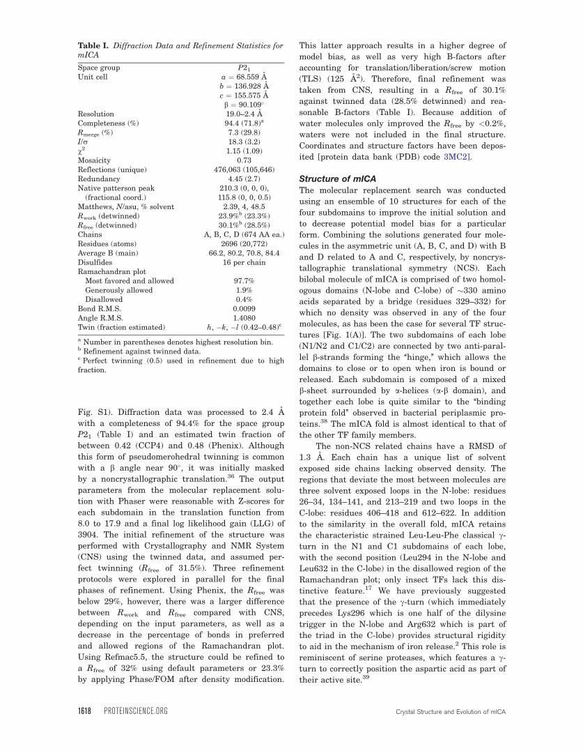
Fig. S1). Diffraction data was processed to 2.4 A
with a completeness of 94.4% for the space group
P21 (Table I) and an estimated twin fraction of
between 0.42 (CCP4) and 0.48 (Phenix). Although
this form of pseudomerohedral twinning is common
with a b angle near 90�, it was initially masked
by a noncrystallographic translation.36 The output
parameters from the molecular replacement solu-
tion with Phaser were reasonable with Z-scores for
each subdomain in the translation function from
8.0 to 17.9 and a final log likelihood gain (LLG) of
3904. The initial refinement of the structure was
performed with Crystallography and NMR System
(CNS) using the twinned data, and assumed per-
fect twinning (Rfree of 31.5%). Three refinement
protocols were explored in parallel for the final
phases of refinement. Using Phenix, the Rfree was
below 29%, however, there was a larger difference
between Rwork and Rfree compared with CNS,
depending on the input parameters, as well as a
decrease in the percentage of bonds in preferred
and allowed regions of the Ramachandran plot.
Using Refmac5.5, the structure could be refined to
a Rfree of 32% using default parameters or 23.3%
by applying Phase/FOM after density modification.
This latter approach results in a higher degree of
model bias, as well as very high B-factors after
accounting for translation/liberation/screw motion
(TLS) (125 A2). Therefore, final refinement was
taken from CNS, resulting in a Rfree of 30.1%
against twinned data (28.5% detwinned) and rea-
sonable B-factors (Table I). Because addition of
water molecules only improved the Rfree by <0.2%,
waters were not included in the final structure.
Coordinates and structure factors have been depos-
ited [protein data bank (PDB) code 3MC2].
Structure of mICA
The molecular replacement search was conducted
using an ensemble of 10 structures for each of the
four subdomains to improve the initial solution and
to decrease potential model bias for a particular
form. Combining the solutions generated four mole-
cules in the asymmetric unit (A, B, C, and D) with B
and D related to A and C, respectively, by noncrys-
tallographic translational symmetry (NCS). Each
bilobal molecule of mICA is comprised of two homol-
ogous domains (N-lobe and C-lobe) of �330 amino
acids separated by a bridge (residues 329–332) for
which no density was observed in any of the four
molecules, as has been the case for several TF struc-
tures [Fig. 1(A)]. The two subdomains of each lobe
(N1/N2 and C1/C2) are connected by two anti-paral-
lel b-strands forming the ‘‘hinge,’’ which allows the
domains to close or to open when iron is bound or
released. Each subdomain is composed of a mixed
b-sheet surrounded by a-helices (a-b domain), and
together each lobe is quite similar to the ‘‘binding
protein fold’’ observed in bacterial periplasmic pro-
teins.38 The mICA fold is almost identical to that of
the other TF family members.
The non-NCS related chains have a RMSD of
1.3 A. Each chain has a unique list of solvent
exposed side chains lacking observed density. The
regions that deviate the most between molecules are
three solvent exposed loops in the N-lobe: residues
26–34, 134–141, and 213–219 and two loops in the
C-lobe: residues 406–418 and 612–622. In addition
to the similarity in the overall fold, mICA retains
the characteristic strained Leu-Leu-Phe classical c-
turn in the N1 and C1 subdomains of each lobe,
with the second position (Leu294 in the N-lobe and
Leu632 in the C-lobe) in the disallowed region of the
Ramachandran plot; only insect TFs lack this dis-
tinctive feature.17 We have previously suggested
that the presence of the c-turn (which immediately
precedes Lys296 which is one half of the dilysine
trigger in the N-lobe and Arg632 which is part of
the triad in the C-lobe) provides structural rigidity
to aid in the mechanism of iron release.2 This role is
reminiscent of serine proteases, which features a c-
turn to correctly position the aspartic acid as part of
their active site.39
Table I. Diffraction Data and Refinement Statistics formICA
Space group P21
Unit cell a ¼ 68.559 Ab ¼ 136.928 Ac ¼ 155.575 Ab ¼ 90.109�
Resolution 19.0–2.4 ACompleteness (%) 94.4 (71.8)a
Rmerge (%) 7.3 (29.8)I/r 18.3 (3.2)v2 1.15 (1.09)Mosaicity 0.73Reflections (unique) 476,063 (105,646)Redundancy 4.45 (2.7)Native patterson peak
(fractional coord.)210.3 (0, 0, 0),115.8 (0, 0, 0.5)
Matthews, N/asu, % solvent 2.39, 4, 48.5Rwork (detwinned) 23.9%b (23.3%)Rfree (detwinned) 30.1%b (28.5%)Chains A, B, C, D (674 AA ea.)Residues (atoms) 2696 (20,772)Average B (main) 66.2, 80.2, 70.8, 84.4Disulfides 16 per chainRamachandran plot
Most favored and allowed 97.7%Generously allowed 1.9%Disallowed 0.4%
Bond R.M.S. 0.0099Angle R.M.S. 1.4080Twin (fraction estimated) h, �k, �l (0.42–0.48)c
a Number in parentheses denotes highest resolution bin.b Refinement against twinned data.c Perfect twinning (0.5) used in refinement due to highfraction.
1618 PROTEINSCIENCE.ORG Crystal Structure and Evolution of mICA

Structural comparisonsBecause both the lobes are closed, the mICA struc-
ture most closely resembles diferric porcine TF
(1H76)8 with a RMSD of 1.7 A for the full-length
protein and 1.5 A and 1.0 A for the N- and C-lobes,
respectively [Fig. 2(A)]. We note that no structure of
diferric hTF currently exists. In comparison with
human apo TF with both the lobes open (2HAU),35 the
RMSD is 8.8 A for full-length mICA and 6.3 A for each
lobe (Supporting Information Table S2A and S2B).
Structural alignments of the subdomains comprising
each lobe range from 0.8 to 1.4 A for pig diferric trans-
ferrin (Fe2TF) and from 1.2 to 1.6 A for human apo TF
(Supporting Information Table S2C) indicating that
the conformation and orientation of the lobes with
respect to each other both have an influence.
A region of mICA with significant deviation
between human TF is a loop comprised of residues
Pro142-Arg143-Lys144-Pro145 that corresponds to
residues Gly139-Ser140-Arg141-Glu142 in the mICA
structure. In human TF, three of four residues in this
loop participate in binding of the N-lobe to the spe-
cific TF receptor.40 The corresponding residues in
mICA are found in two different conformations for
each of the non-NCS related molecules. Both of con-
formations deviate from TF [Fig. 2(B)], most likely
due to a five amino acid deletion in ICA, which causes
the helix leading into the loop to end prematurely.
Iron and anion bindingThe metal binding sites of all TF family members
that bind iron are located in clefts formed between
the two subdomains comprising each lobe. Cleft
opening for TF involves a twist followed by a sub-
stantial rotation ranging from 35� to 63�.2,41,42 As
mentioned above, binding of iron and of the syner-
gistic anion (carbonate) effectively locks the cleft in
the closed conformation. Superposition of the iron-
Figure 1. (A) Ribbon diagram of the crystal structure of
mICA. Subdomains N1 (red), N2 (yellow), C1 (blue), and C2
(green) are highlighted. The linker between lobes for which
density was not observed is indicated by the dashed line.
(B) The N-lobe cleft of mICA. Shown is the N1 subdomain
(pink), N2 subdomain (yellow), mICA residues (black),
residues for porcine TF (PDB: 1H76, orange),8 and residues
for human TF (PDB: 1A8E)7 including the Fe atom (green)
after least squares superposition. Electron density shown is
2Fo � Fc. All structural images produced using PyMOL.37
[Color figure can be viewed in the online issue, which is
available at wileyonlinelibrary.com.]
Figure 2. (A) Least squares superposition of the structure
of mICA with subdomain N1 (red), N2 (yellow), C1 (blue),
and C2 (green) and the iron-bound form of porcine TF
(PDB: 1H76)8 in white. (B) Detailed examination of the
region indicated by the box in (A), which is implicated in
binding to the TF receptor. Porcine TF in pink, human TF
(PDB: 1A8E, yellow),7 mICA chain A in green, and mICA
chain C in cyan. The largest positional shift relative to hTF
were for the N-terminal residues of the sequence with 8.8 A
and 4.7 A changes for mICA chain A and C, respectively.
[Color figure can be viewed in the online issue, which is
available at wileyonlinelibrary.com.]
Eckenroth et al. PROTEIN SCIENCE VOL 19:1616—1626 1619
binding sites for the N- and C-lobes of human serum
TF on the equivalent sites in mICA shows the posi-
tions of the iron binding residues and highlights the
mutations in mICA that prevent binding of iron in
each lobe (Fig. 3). The two substitutions in the N-
lobe that prohibit iron binding in mICA are Trp124
(in place of Arg124 in hTF) and Ser188 (instead of
Tyr188 in hTF). Significantly, introduction of Arg at
position 124 and Tyr at position 188 in the N-lobe of
mICA (W124R and S188Y mutants) allowed the N-
lobe of the mutated mICA to bind ferric iron with
high affinity.24 Interestingly, in the N-lobe of mICA,
Trp124 occupies the same space occupied by Arg124
in the two conformations of the iron-bound structure
of the N-lobe of human TF (PDB: 1A8E).7 Further-
more, in the closed, iron-bound N-lobe of hTF, Tyr45
is flipped out of the cleft, whereas in the open apo
conformation it is rotated into the cleft by �160� as
is observed for His45 for mICA. We also note that
the two remaining ligands in mICA that are associ-
ated with iron binding in other family members
(Asp63 and His249) are close enough to form a salt
bridge [Fig. 1(B)].
In the iron binding cleft of the C-lobe of mICA,
there are three substitutions that preclude iron
binding: Ser520 (in place of Tyr517 in hTF), Arg588
(instead of His585 in hTF), and Thr457 (instead of
Arg456 in hTF).17 As there are no obvious steric
clashes that would interfere with iron binding,
mutating the Ser, Arg, and Thr residues to Tyr, His,
and Arg residues might result in a mICA C-lobe
that could bind iron.
Significance of the closed lobes in the absence
of iron?
The most surprising finding from the mICA struc-
ture is the observation that both the lobes are in the
closed conformation normally associated with the
iron-bound form of TF. Because we had shown that
mICA does not bind iron in either lobe,18 this was
unexpected and initially confounded molecular
replacement efforts using intact lobes of the apo-con-
formation of human TF as the search model.
Although a structure of apo LTF (1CB6) exists with
a C-lobe that is nearly completely closed, the finding
was attributed to crystal packing forces.43 Of possi-
ble relevance however, a theoretical modeling study
based on LTF indicated that there is very little dif-
ference in energy between the open and closed con-
formations.44 Other work has suggested that TF
family members exist in a dynamic equilibrium in
which opened and closed conformations are con-
stantly being sampled,2,45–48 similar to members of
the periplasmic binding protein family.49,50 It is
thought that the binding of the synergistic anion
prepares the site for iron binding and the acquisition
of iron strongly drives the equilibrium toward the
closed form.51 However, no contiguous density that
might be associated with anions such as acetate, am-
monium, or carbonate was found in the vicinity of
the remaining iron binding ligands in either lobe.
It is not clear whether the two lobes of mICA are
sampling the open and closed conformations or if
there are specific forces, which favor the closed con-
formation. Because crystal environments have been
shown to have a clear influence on hinge-like motions
in proteins, it is possible that crystal packing forces
may account for the closed clefts.52 Nevertheless, we
evaluated whether the differences in the amino acids
within mICA might contribute to a change in the
electrostatic potential of the cleft to promote closure.
The electrostatic potential maps generated with
PyMOL indicate that electrostatic interactions could
maintain the C-lobe in a closed conformation as the
surface within the cleft of the C-lobe shows that the
C1 subdomain is predominantly electronegative,
whereas the C2 subdomain is electropositive (Sup-
porting Information Fig. S2). In contrast, the electro-
static analysis of the mICA N-lobe is less convincing.
In the absence of clear electrostatic interaction
between the subdomains, it is possible that the inter-
action between residues noted above (Asp63 from N1
and His249 from N2) could stabilize a closed cleft.
Figure 3. Stereo images of the superposition of the iron
binding sites for (A) N-lobes and (B) C-lobes of porcine TF
(PDB: 1H76)8 (white) including the iron shown as a sphere,
human TF (PDB: 1A8E)7 (grey), and mICA (black).
Numbering in both panels is for human TF. [Color figure
can be viewed in the online issue, which is available at
wileyonlinelibrary.com.]
1620 PROTEINSCIENCE.ORG Crystal Structure and Evolution of mICA

Sequence identification and phylogenetic
analysisIn addition to the six mammalian ICA sequences
that were reported previously,18 we have identified
two more putative ICA sequences from genomic data
for the guinea pig and the horse. Except for a miss-
ing 10 amino acid segment (448–457) in the horse
ICA, these sequences appear to be complete. Confi-
dence of their identification as ICA proteins came
from creation of a multiple sequence alignment with
representatives of other mammalian family members
(Supporting Information Fig. S3). These additional
sequences provided sufficient data to allow us to an-
alyze the evolutionary constraints of specific resi-
dues. Of interest, searches of the chimp and maca-
que genomes confirmed the presence of pseudogenes
in both of these species. This indicates that, like
humans, other primates have gained and then lost a
‘‘functional’’ ICA.
Selection pressure on important residues inTF family members
Strong negative (purifying) selection occurs when
the synonymous substitution rate (silent, dS) for a
given codon or for a series of codons is greater than
the nonsynonymous substitution rate (amino acid
altering, dN). We quantified this pressure as the ra-
tio of substitutions (dN/dS) for each TF family,
where 1 indicates neutrality. Specifically, we exam-
ined important functional residues in various TF
family members, including those involved in iron
and anion binding as well as residues participating
in the mechanism of iron release and in the hinge
responsible for cleft opening and closing. The valid-
ity of this type of analysis is illustrated by serum TF
in which all of these residues are under strong, con-
sistent selection, or purifying pressure (low dN/dS
ratios) (Tables II and III).
The six ligand and anion binding residues in the
N-lobe are all well maintained in TF (Table II). Sur-
prisingly, two of the four N-lobe iron-binding ligands
(both Tyr residues) in LTF appear to be under only
moderate purifying pressure (dN/dS ratios of 0.48
and 0.55), suggesting that iron binding in this lobe,
although it can and does occur, may not be as impor-
tant to its in vivo function (see below). For MTF, the
iron and anion binding ligands in the N-lobe all
show strong selection and conservation. This obser-
vation appears to be consistent with the fact that
MTF definitely binds iron in the N-lobe.31 In con-
trast, in the ICA family, only one of the residues is
strongly selected, His249 with a dN/dS ratio of 0.08
(Table II). Significant differences in the ratios of the
other five residues in ICA show a clear loss of selec-
tive pressure consistent with losing the need to
maintain the iron binding function.
In the C-lobe, all six ligand and anion binding
residues are well maintained in both TF and LTF as
Table II. Selection Pressure (dN/dS Ratios) of Significant N-Lobe Structural Elements within the TF Family
N-lobe Fe-binding Anion Trigger Hinge Average dN/dS
Residue Asp Tyr Tyr His Thr Arg Lys Lys Thr Val Pro
Whole molecule
Human TF 63 95 188 249 120 124 206 296 93 246 247
Mouse ICA 63 95 Ser 188 249 120 Trp 124 206 Thr 296 93 250 251
TF (n ¼ 18) 0.05 0.08 0.07 0.05 0.06 0.04 0.05 0.05 0.03 0.29 0.07 0.47LTF (n ¼ 14) 0.09 0.48 0.55 0.09 0.82 0.23 0.56 (R) 0.33 0.05 0.06 0.13 0.55MTF (n ¼ 11) 0.03 0.06 0.03 0.02 0.03 0.03 0.02 0.04 0.02 0.03 0.05 0.15ICA (n ¼ 7) 0.84 1.40 1.1 (S) 0.08 1.50 1.2 (W) 1.00 0.16 (T) 0.08 1.40 0.16 0.68
Table III. Selection Pressure (dN/dS Ratios) of Significant C-Lobe Structural Elements within the TF Family
C-lobe Fe-binding Anion Triad Hinge
Residue Asp Tyr Tyr His Thr Arg Lys Arg Asp Ala Gly Arg Ala Pro
Human TF 392 426 517 585 452 456 534 632 634 424 425 581 582 583
Mouse ICA 386 427 Ser 520 Arg 588 453 Thr 457 537 Ser 653 636 Glu 425 426 584 Val 585 586
TF (n ¼ 18) 0.05 0.08 0.07 0.05 0.04 0.05 0.06 0.16 0.19 1.10 0.31 1.50 0.47 0.09LTF
(n ¼ 14)0.09 0.13 0.11 0.08 0.06 0.06 0.11 0.07
(N)0.05(N)
0.82(E)
0.12 0.89(M)
0.2 0.13
MTF(n ¼ 11)
0.68(S)
0.03 0.41 0.19 0.44(A)
0.02(S)
0.14(R)
0.03(K)
0.02(A)
0.21(N)
0.02(S)
0.02(Q)
0.31(I)
0.06
ICA(n ¼ 7)
0.06 1.20 1.50(S)
1.1(R)
1.50 0.11(T)
0.05 0.08(S)
0.68 1.40(E)
0.11 1.50 0.07(V)
0.26
Eckenroth et al. PROTEIN SCIENCE VOL 19:1616—1626 1621
indicated by dN/dS ratios below 0.13 (Table III). In
MTF, two of the four iron binding ligands (Asp and
Tyr) have higher ratios and, in fact, in human MTF
the Asp is a Ser. Additionally, both the anion stabi-
lizing residues in MTF have been replaced; interest-
ingly, the serine replacing Arg456 is under strong
purifying selection pressure (dN/dS ¼ 0.02). The
substitutions are consistent with the inability of the
C-lobe of MTF to bind iron.31 The ICA family retains
only a single iron binding residue, Asp386, which is
strongly selected for; additionally, the anion stabiliz-
ing residue Arg456, which has been replaced by
Thr457, is under strong selection. The remaining
liganding and anion binding residues in mICA have
dN/dS ratios above 1 indicating diversifying
selection.
Amino acid residues involved in iron release
In the N-lobe of serum TF and OTF the dilysine
trigger (Lys206-Lys296) is required for efficient iron
release.53,54 LTF lacks this mechanism consistent
with substitution of an Arg residue in some species
and a lack of strong selection (dN/dS ¼ 0.56). Of in-
terest, MTF seems to have both lysines under strong
purifying selection, although there is no direct ex-
perimental support for a dilysine trigger mechanism.
In ICA, the Lys at position 206 displays neutral
selection and a threonine residue replaces Lys296.
Of significance, the Thr296 appears to be under
strong purifying selection pressure (dN/dS ¼ 0.16)
perhaps indicating that it serves an important role.
Supporting this suggestion is the unexplained fail-
ure, after multiple attempts, to produce a mutant of
mICA in which Thr296 was replaced with Lys296.24
In the C-lobe of human TF, iron release is con-
trolled by a triad of charged residues (Lys534,
Arg632, and Asp634).55 In LTF, the Lys at the posi-
tion equivalent to 534 is conserved but the other two
positions are substituted by Asn residues, which are
under strong selective pressure. The presence of
these asparagine residues appears to account for the
extremely tight binding and poor release of iron
from this lobe.55 In MTF, all three residues of the
triad are substituted by other amino acids which are
under strong selective pressure, whereas in mICA,
both the lysine (Lys536) and aspartic acid (Asp636)
remain, but Ser634 replaces Arg632 (strongly indi-
cating that the triad is not functional in mICA).
Interestingly, both Lys537 and Ser634 are under
strong purifying pressure (dN/dS < 0.08), whereas
Asp636 is under moderate pressure (dN/dS ¼ 0.68).
Residues comprising the hinge between thesubdomains of each lobe
The subdomain movement that occurs during iron
binding and release is controlled by hinge residues
located on antiparallel strands linking the subdo-
mains in all TFs. The specific residues involved in
the hinge (Thr93, Val250, and Pro251 in the N-lobe
and Glu425, Gly426, Arg584, Val585, and Pro586 in
the C-lobe) in mICA were found to be somewhat con-
served (5 of 8), although there is a considerable vari-
ation in the selection pressure (dN/dS between 0.08
and 1.5) (Tables II and III).
Selection pressure upon other regions of
functional importance in TF family membersICA is relatively a new family member, which may
have functions in addition to inhibiting various iso-
forms of CA. Therefore, we looked at other regions of
interest in certain family members. In particular,
lactoferricin (residues 19–38) is a highly basic, disul-
fide-looped peptide released from LTF during gastric
digestion and associated with strong antimicrobial
properties.56 An NMR structure of the peptide
revealed distorted antiparallel b-strands with the
majority of hydrophobic residues on one face and ba-
sic residues on the other face.57 The mICA sequence
and structure in this region are almost identical to
other members of the TF family and, like them, lack
the basic and hydrophobic residues that give rise to
lactoferricin and are necessary for interactions with
bacterial membranes.
Materials and Methods
MaterialsDulbecco’s modified Eagle’s medium-Ham F-12 nu-
trient mixture (DMEM-F12), antibiotic-antimycotic
solution (100�), and trypsin were from the Gibco-
BRL Life Technologies Division of Invitrogen. Fetal
bovine serum (FBS) was obtained from Atlanta Bio-
logicals. Ultroser G (UG) is a serum replacement
from Pall BioSepra (Cergy, France). Methotrexate
from Bedford Laboratories was purchased at a local
hospital pharmacy. All tissue culture dishes, flasks,
and Corning expanded surface roller bottles were
from local distributors. Ultracel 30 kDa molecular
weight cutoff membrane (MWCO) microcentrifuge
devices were made by Amicon.
Purification of mICA
The cDNA sequence encoding the N-terminal hexa-
histidine tagged mICA was inserted into the pNUT
vector as previously described.18 For crystallization
trials, the putative glycosylation sites, Asn470 and
Asn645, were each mutated to aspartic acid to elimi-
nate glycosylation. Therefore, the recombinant
mICA was nonglycosylated significantly reducing the
heterogeneity introduced by different carbohydrate
species added by the BHK cells.24 As previously
described, the mICA was produced and purified
using methods that have been optimized for His-
tagged hTF.18,24,58 The concentration of mICA was
estimated using the calculated absorption coefficient
1622 PROTEINSCIENCE.ORG Crystal Structure and Evolution of mICA

of 86.5 mM�1 cm�1.59 We have experimentally deter-
mined that this calculation provides a remarkably
accurate estimate of the concentration.60
Crystallization of mICAThe mICA was concentrated to 25 mg/mL in
100 mM NH4HCO3 (pH � 8.0). Crystals initially
appeared in PEG 3350 concentrations between 10%
and 20%. The final optimized hanging drop condition
used 100 mM ammonium acetate pH 7.4 and 12%
PEG in the well, and 2 lL of protein solution and
1 lL of reservoir solution in the drop with the tray
stored at 20�C. It generally took 3–7 days for
the crystals to reach a size of 700 lm � 400 lm �80 lm. Cryoprotection of the crystals involved titra-
tion of PEG 3350 in 5% increments to a final concen-
tration of 35%; each addition was followed by a
30 min incubation at 20�C.
Data collection and structure determinationData were collected on a Mar345 image plate detec-
tor using a Rigaku RU300 rotating anode generator
at a temperature of 104 K. A low-resolution pass
(5 min exposure) and a high-resolution pass (15 min
exposure) were indexed and scaled together using
the HKL Suite.62 Initial processing suggested a
primitive orthorhombic lattice with a noncrystallo-
graphic translation along the c-cell axis, as deter-
mined by the native Patterson peak (0, 0, 0.5) at a
peak height of 55% relative to the origin. However,
unusual systematic absences, initial failures of mo-
lecular replacement, and a b angle fortuitously close
to 90� (90.109�) suggested the possibility of twin-
ning. Data was reprocessed as primitive monoclinic
and submitted to the Padilla–Yeates twinning
server.62 The results indicated partial merohedral
twinning. The twin law (h, �k, �l) was submitted to
the program Detwin using CCP4 version 6.163 to
estimate the twinned fraction. A test set for 10% of
the reflections was produced using CNS version
1.264 to be certain that the twin related reflections
were in the same set.
Anticipating multiple subdomain movements, 10
structures of TF family members were threaded
with the sequence for mICA using Chainsaw65 and
broken into subdomains (N1, N2, C1, and C2) for
molecular replacement using Phaser.66 Initial solu-
tions for the C1 and C2 subdomains for one of the
four chains in the asymmetric unit were obtained
and used to eliminate the twin solutions for subse-
quent cycles of Phaser until all four chains were
assembled from the subdomains. The iterative
rounds of Phaser were performed such that after a
proper subdomain solution was found, the NCS
translation was applied to generate the duplicate
molecule before the automated search for the next
subdomain. Refinement protocols were explored in
parallel for the structure against the twinned data
using Phenix,67 CNS assuming perfect twinning as
suggested for the high-twin fraction,36,68 and
Refmac5.5 applying tight (main chain) and medium
(side chain) NCS restraints with each subdomain as
a TLS group.69 The final refinement was performed
using CNS with model building performed using
Coot.70
Genomic sequence identification
All protein and DNA sequences, including mICA,
were retrieved from Basic Local Alignment Search
Tool (BLAST) searches of the Entrez, ENSEMBL,
and Pre! ENSEMBL public genome databases71
using pICA as the query sequence. In certain cases,
an assembled protein was unavailable, so transcript
and putative protein sequences were derived from
genomic sequences using FGENESHþ from Soft-
berry (http://www.softberry.ru/berry.phtml). Initial
alignments were made using CLUSTALW72 and
were then imported into GeneDoc73 for refinement
and additional analysis. SignalP74 was used to iden-
tify signal peptides, which were removed from the
sequences. All sequences used are listed in Support-
ing Information Table S1.
Detection of selective forcesA minimum of eight nucleotide sequences from each
TF family were codon-aligned using PAL2NAL.75 To
examine whether individual amino acids/codons are
undergoing natural selection (positive or negative),
nonsynonymous versus synonymous substitutions
and dN/dS ratios for individual codons were calcu-
lated using Selecton version 2.4 (http://selecton.
tau.ac.il).76 As only four nucleotide sequences are
available for avian OTF nucleotide sequences and a
minimum of 10 or more homologues are preferred
for this type of analysis, we chose not to include
OTF in our comparisons.
Phylogenetic analyses
The best-fit evolutionary models were determined
using PROTTEST.77 Neighbor-joining trees were cre-
ated with MEGAv478 using the JTTþG model
(gamma ¼ 1.4) for amino acids,79 complete deletion
of gaps, and 1000 bootstrap replications. Additional
trees using the Poisson model were also created to
confirm topology (data not included).
Conclusion
The TF gene duplication leading to the formation of
the ICA family is a relatively recent evolutionary
event. The 2.4 A structure of mICA revealed that
mICA has the same fold as other TF family mem-
bers consistent with the original sequence analysis.
Interestingly, mICA was found in the closed (usually
iron bound) conformation, although no anions were
observed in either cleft that might induce closure.
Thus, closure could result from: (a) low-energy
Eckenroth et al. PROTEIN SCIENCE VOL 19:1616—1626 1623
barrier between open and closed forms, (b) electro-
statics (C-lobe), or (c) crystal packing forces. Com-
parison with other TF family members demonstrates
that there has been little evolutionary pressure to
maintain the canonical TF family features (iron and
anion ligands, dilysine trigger or triad for iron
release, or hinges) in ICAs. Selective force analysis
supports the biochemical data that neither lobe of
ICA nor the C-lobe of MTF can bind iron and sug-
gests that the N-lobe of LTF may be eliminating its
iron binding function. Why a functional ICA is
absent in primates still remains a mystery, though it
has been suggested to be related innate immunity.24
Acknowledgments
The authors wish to thank Dr. Michael Sawaya for as-
sistance in evaluating our twinned data.
References
1. Aisen P, Enns C, Wessling-Resnick M (2001) Chemistryand biology of eukaryotic iron metabolism. Int J Bio-chem Cell Biol 33:940–959.
2. Mason AB, Everse SJ, Iron transport by transferrin.In: Fuchs H, Ed. (2008) Iron metabolism and disease.Kerala, India: Research Signpost, pp 83–123.
3. Yang F, Lum JB, McGill JR, Moore CM, Naylor SL,van Bragt PH, Baldwin WD, Bowman BH (1984)Human transferrin: cDNA characterization and chro-mosomal localization. Proc Natl Acad Sci USA 81:2752–2756.
4. Park I, Schaeffer E, Sidoli A, Baralle FE, Cohen GN,Zakin MM (1985) Organization of the human transfer-rin gene: direct evidence that it originated by geneduplication. Proc Natl Acad Sci USA 82:3149–3153.
5. Anderson BF, Baker HM, Dodson EJ, Norris GE, Rum-ball SV, Waters JM, Baker EN (1987) Structure ofhuman lactoferrin at 3.2 A resolution. Proc Natl AcadSci USA 84:1769–1773.
6. Kurokawa H, Mikami B, Hirose M (1995) Crystalstructure of diferric hen ovotransferrin at 2.4 A resolu-tion. J Mol Biol 254:196–207.
7. MacGillivray RTA, Moore SA, Chen J, Anderson BF,Baker H, Luo YG, Bewley M, Smith CA, Murphy ME,Wang Y, Mason AB, Woodworth RC, Brayer GD, BakerEN (1998) Two high-resolution crystal structures of therecombinant N-lobe of human transferrin reveal astructural change implicated in iron release. Biochem-istry 37:7919–7928.
8. Hall DR, Hadden JM, Leonard GA, Bailey S, Neu M,Winn M, Lindley PF (2002) The crystal and molecularstructures of diferric porcine and rabbit serum trans-ferrins at resolutions of 2.15 and 2.60 A, respectively.Acta Crystallogr Sect D 58:70–80.
9. Princiotto JV, Zapolski EJ (1975) Difference betweenthe two iron-binding sites of transferrin. Nature 255:87–88.
10. Lestas AN (1976) The effect of pH upon human trans-ferrin: selective labelling of the two iron-binding sites.Br J Haematol 32:341–350.
11. Najarian RC, Harris DC, Aisen P (1978) Oxalate andspin-labeled oxalate as probes of the anion binding siteof human transferrin. J Biol Chem 253:38–42.
12. Baldwin DA, DeSousa DMR (1981) The effect of saltson the kinetics of iron release from N-terminal and
C-terminal monoferric transferrins. Biochem BiophysRes Commun 99:1101–1107.
13. Halbrooks PJ, He QY, Briggs SK, Everse SJ, SmithVC, MacGillivray RTA, Mason AB (2003) Investigationof the mechanism of iron release from the C-lobe ofhuman serum transferrin: mutational analysis of therole of a pH sensitive triad. Biochemistry 42:3701–3707.
14. Baker HM, Anderson BF, Brodie AM, Shongwe MS,Smith CA, Baker EN (1996) Anion binding by transfer-rins: importance of second-shell effects revealed by thecrystal structure of oxalate-substituted diferric lactofer-rin. Biochemistry 35:9007–9013.
15. Sun H, Li H, Sadler PJ (1999) Transferrin as a metalion mediator. Chem Rev 99:2817–2842.
16. Lambert LA, Perri H, Meehan TJ (2005) Evolution ofthe duplications in the transferrin family of proteins.Comp Biochem Physiol B 140:11–25.
17. Lambert LA, Perri H, Halbrooks PJ, Mason AB (2005)Evolution of the transferrin family: conservation of res-idues associated with iron and anion binding. CompBiochem Physiol B 142:129–141.
18. Wang F, Lothrop AP, James NG, Griffiths TA, LambertLA, Leverence R, Kaltashov IA, Andrews NC, Macgil-livray RT, Mason AB (2007) A novel murine proteinwith no effect on iron homeostasis is homologous totransferrin and is the putative inhibitor of carbonicanhydrase. Biochem J 406:85–95.
19. Lindskog S, Silverman DN (2000) The catalytic mecha-nism of mammalian carbonic anhydrases. EXS 90:175–195.
20. Silverman RB. (2002) Mechanisms of enzyme catalysis.The Organic Chemistry of Enzyme Catalyzed Reac-tions. San Diego: Academic Press, pp 1–38.
21. Tashian RE (1992) Genetics of the mammalian carbonicanhydrases. Adv Genet 30:321–356.
22. Parkkila S, An overview of the distribution and functionof carbonic anhydrase in mammals. In: Chegwidden WR,Edwards Y, Carter N, Eds. (2000) The carbonic anhy-drases-new horizons. Basel: Birkhauser Verlag, pp 79–93.
23. Geers C, Gros G (2000) Carbon dioxide transport andcarbonic anhydrase in blood and muscle. Physiol Rev80:681–715.
24. Mason AB, Judson GL, Bravo MC, Edelstein A, ByrneSL, James NG, Roush ED, Fierke CA, Bobst CE, Kalta-shov IA, Daughtery MA (2008) Evolution reversed: theability to bind iron restored to the N-Lobe of the mu-rine inhibitor of carbonic anhydrase by strategic muta-genesis. Biochemistry 47:9847–9855.
25. Wuebbens MW, Roush ED, Decastro CM, Fierke CA(1997) Cloning, sequencing, and recombinant expres-sion of the porcine inhibitor of carbonic anhydrase: anovel member of the transferrin family. Biochemistry36:4327–4336.
26. Roush ED, Fierke CA (1992) Purification and charac-terization of a carbonic anhydrase II inhibitor from por-cine plasma. Biochemistry 31:12536–12542.
27. Booth VH (1938) The carbonic anhydrase inhibitor inserum. J Physiol 91:474–489.
28. Hill EP (1986) Inhibition of carbonic anhydrase byplasma of dogs and rabbits. J Appl Physiol 60:191–197.
29. Schaeffer E, Lucero MA, Jeltsch JM, Py MC, LevinMJ, Chambon P, Cohen GN, Zakin MM (1987) Com-plete structure of the human transferrin gene. Compar-ison with analogous chicken gene and humanpseudogene. Gene 56:109–116.
30. Rose TM, Plowman GD, Teplow DP, Dreyer WJ, HellstromKE, Brown TP (1986) Primary structure of the humanmelanoma-associated antigen p97 (melanotransferrin)
1624 PROTEINSCIENCE.ORG Crystal Structure and Evolution of mICA
deduced from the mRNA sequence. Proc Natl Acad SciUSA 83:1261–1265.
31. Baker EN, Baker HM, Smith CA, Stebbins MR, KahnM, Hellstrom KE, Hellstrom I (1992) Human melano-transferrin (p97) has only one functional iron-bindingsite. FEBS Lett 298:215–218.
32. Sekyere E, Richardson DR (2000) The membrane-bound transferrin homologue melanotransferrin: rolesother than iron transport?FEBS Lett 483:11–16.
33. Sekyere EO, Dunn LL, Richardson DR (2005) Exami-nation of the distribution of the transferrin homologue,melanotransferrin (tumour antigen p97), in mouse andhuman. Biochim Biophys Acta 1722:131–142.
34. Sekyere EO, Dunn LL, Rahmanto YS, Richardson DR(2006) Role of melanotransferrin in iron metabolism:studies using targeted gene disruption in vivo. Blood107:2599–2601.
35. Wally J, Halbrooks PJ, Vonrhein C, Rould MA, EverseSJ, Mason AB, Buchanan SK (2006) The crystal struc-ture of iron-free human serum transferrin providesinsight into inter-lobe communication and receptorbinding. J Biol Chem 281:24934–24944.
36. Barends TR, Dijkstra BW (2003) Acetobacter turbidansalpha-amino acid ester hydrolase: merohedral twinningin P21 obscured by pseudo-translational NCS. ActaCrystallogr Sect D 59:2237–2241.
37. Delano WL (2008) The PyMOL Molecular GraphicsSystem. DeLano Scientific LLC, Palo Alto, CA, USA.http://www.pymol.org.
38. Baker EN, Baker HM (2009) A structural frameworkfor understanding the multifunctional character of lac-toferrin. Biochimie 91:3–10.
39. Milner-White EJ (1990) Situations of gamma-turns inproteins. Their relation to alpha-helices, beta-sheetsand ligand binding sites. J Mol Biol 216:386–397.
40. Mason AB, Byrne SL, Everse SJ, Roberts SE, ChasteenND, Smith VC, MacGillivray RT, Kandemir B, Bou-Abdallah F (2009) A loop in the N-lobe of human serumtransferrin is critical for binding to the transferrin re-ceptor as revealed by mutagenesis, isothermal titrationcalorimetry, and epitope mapping. J Mol Recognit 22:521–529.
41. He QY, Mason AB, Templeton DM (2002) Molecularaspects of release of iron from transferrins. Molecularand cellular iron transport. New York: Marcel Dekker,Inc., pp 95–123.
42. MacGillivray RTA, Mason AB, Templeton DM (2002)Transferrins. Cell and molecular biology of iron trans-port. New York: Marcel Dekker, Inc., pp 41–69.
43. Jameson GB, Anderson BF, Norris GE, Thomas DH,Baker EN (1998) Structure of human apolactoferrin at2.0 A resolution. Refinement and analysis of ligand-induced conformational change. Acta Crystallogr SectD 54:1319–1335.
44. Gerstein M, Anderson BF, Norris GE, Baker EN, LeskAM, Chothia C (1993) Domain closure in lactoferrin.Two hinges produce a see-saw motion between alterna-tive close-packed interfaces. J Mol Biol 234:357–372.
45. Grossmann JG, Crawley JB, Strange RW, Patel KJ,Murphy LM, Neu M, Evans RW, Hasnain SS (1998)The nature of ligand-induced conformational change intransferrin in solution. An investigation using X-rayscattering, XAFS and site-directed mutants. J Mol Biol279:461–472.
46. Mecklenburg SL, Donohoe RJ, Olah GA (1997) Tertiarystructural changes and iron release from human serumtransferrin. J Mol Biol 270:739–750.
47. Mizutani K, Mikami B, Hirose M (2001) Domain clo-sure mechanism in transferrins: new viewpoints about
the hinge structure and motion as deduced from highresolution crystal structures of ovotransferrin N-lobe. JMol Biol 309:937–947.
48. Rinaldo D, Field MJ (2003) A computational study ofthe open and closed forms of the N-lobe human serumtransferrin apoprotein. Biophys J 85:3485–3501.
49. Tang C, Schwieters CD, Clore GM (2007) Open-to-closed transition in apo maltose-binding proteinobserved by paramagnetic NMR. Nature 449:1078–1082.
50. Bermejo GA, Strub MP, Ho C, Tjandra N (2010)Ligand-free open-closed transitions of periplasmic bind-ing proteins: the case of glutamine-binding protein.Biochemistry 49:1893–1902.
51. Harris WR, Wang Z, Brook C, Yang B, Islam A (2003)Kinetics of metal ion exchange between citric acid andserum transferrin. Inorg Chem 42:5880–5889.
52. Eyal E, Gerzon S, Potapov V, Edelman M, Sobolev V(2005) The limit of accuracy of protein modeling: influ-ence of crystal packing on protein structure. J Mol Biol351:431–442.
53. Dewan JC, Mikami B, Hirose M, Sacchettini JC (1993)Structural evidence for a pH-sensitive dilysine triggerin the hen ovotransferrin N-lobe: implications fortransferrin iron release. Biochemistry 32:11963–11968.
54. He QY, Mason AB, Tam BM, MacGillivray RTA, Wood-worth RC (1999) Dual role of Lys206-Lys296 interac-tion in human transferrin N-lobe: iron-release triggerand anion-binding site. Biochemistry 38:9704–9711.
55. Halbrooks PJ, Giannetti AM, Klein JS, Bjorkman PJ,Larouche JR, Smith VC, MacGillivray RTA, Everse SJ,Mason AB (2005) Composition of pH sensitive triad inC-lobe of human serum transferrin. Comparison tosequences of ovotransferrin and lactoferrin providesinsight into functional differences in iron release. Bio-chemistry 44:15451–15460.
56. Jones EM, Smart A, Bloomberg G, Burgess L, MillarMR (1994) Lactoferricin, a new antimicrobial peptide. JAppl Bacteriol 77:208–214.
57. Tomita M, Takase M, Bellamy W, Shimamura S (1994)A review: the active peptide of lactoferrin. Acta Pae-diatr Jpn 36:585–591.
58. Mason AB, Halbrooks PJ, Larouche JR, Briggs SK,Moffett ML, Ramsey JE, Connolly SA, Smith VC, Mac-Gillivray RTA (2004) Expression, purification, andcharacterization of authentic monoferric and apo-human serum transferrins. Protein Expr Purif 36:318–326.
59. Pace CN, Vajdos F, Fee L, Grimsley G, Gray T (1995)How to measure and predict the molar absorption coef-ficient of a protein. Protein Sci 4:2411–2423.
60. James NG, Mason AB (2008) Protocol to determineaccurate absorption coefficients for iron containingtransferrins. Anal Biochem 378:202–207.
61. Otwinowski Z, Minor W. Processing of X-ray diffractiondata collected in oscillation mode. In: Carter CW, SweetRM, editors. Methods in enzymology, part A. SanDiego: Academic Press, pp 307–326.
62. Padilla JE, Yeates TO (2003) A statistic for local inten-sity differences: robustness to anisotropy and pseudo-centering and utility for detecting twinning. Acta Crys-tallogr Sect D 59:1124–1130.
63. Collaborative Computational Project N (1994) TheCCP4 suite: programs for protein crystallography. ActaCrystallogr Sect D 50:760–763.
64. Brunger AT, Adams PD, Clore GM, Delano WL, Gros P,Grosse-Kunstleve RW, Jiang JS, Kuszewski J, NilgesM, Pannu NS, Read RJ, Rice LM, Simonson T, WarrenGL (1998) Crystallography and NMR system: a new
Eckenroth et al. PROTEIN SCIENCE VOL 19:1616—1626 1625
software suite for macromolecular structure determina-tion. Acta Crystallogr Sect D 54:905–921.
65. Stein N (2008) CHAINSAW: a program for mutatingpdb files used as templates in molecular replacement. JAppl Crystallogr 41:641–643.
66. McCoy AJ, Grosse-Kunstleve RW, Adams PD, WinnMD, Storoni LC, Read RJ (2007) Phaser crystallo-graphic software. J Appl Crystallogr 40:658–674.
67. Adams PD, Grosse-Kunstleve RW, Hung LW, IoergerTR, McCoy AJ, Moriarty NW, Read RJ, Sacchettini JC,Sauter NK, Terwilliger TC (2002) PHENIX: buildingnew software for automated crystallographic structuredetermination. Acta Crystallogr Sect D 58:1948–1954.
68. Golinelli-Pimpaneau B (2005) Structure of a pseudo-merohedrally twinned monoclinic crystal form of a pyr-idoxal phosphate-dependent catalytic antibody. ActaCrystallogr Sect D 61:472–476.
69. Winn MD, Isupov MN, Murshudov GN (2001) Use ofTLS parameters to model anisotropic displacements inmacromolecular refinement. Acta Crystallogr Sect D57:122–133.
70. Emsley P, Cowtan K (2004) Coot: model-building toolsfor molecular graphics. Acta Crystallogr Sect D 60:2126–2132.
71. Altschul S, Madden TL, Schaffer AA, Zhang J, ZhangZ, Miller W, Lipman J (1997) Gapped BLAST and PSI-BLAST: a new generation of protein database searchprograms. Nucleic Acids Res 25:3389–3402.
72. Thompson J, Higgins D, Gibson T (1994) CLUSTAL W:improving the sensitivity of prgressive multiple sequence
alignment through sequence weighting, position specificgap penalties and weight matriz choice. Nucleic AcidsRes 22:4673–4680.
73. Nicholas KB, Nicholas HB, Deerfield DW, II (1997)GeneDoc: analysis and visualization of genetic varia-tion. http://www.nrbsc.org/gfx/genedoc/ebinet.htm.
74. Nielsen H, Engelbrecht J, Brunak S, von Heijne G(1997) A neural network method for identification ofprokaryotic and eukaryotic signal peptides and predic-tion of their cleavage sites. Int J Neural Syst 8:581–599.
75. Suyama M, Torrents D, Bork P (2006) PAL2NAL: ro-bust conversion of protein sequence alignments intothe corresponding codon alignments. Nucleic Acids Res34:W609–W612.
76. Stern A, Doron-Faigenboim A, Erez E, Martz E,Bacharach E, Pupko T (2007) Selecton 2007: advancedmodels for detecting positive and purifying selectionusing a Bayesian inference approach. Nucleic AcidsRes 35:W506–W511.
77. Abascal F, Zardoya R, Posada D (2005) ProtTest: selec-tion of best-fit models of protein evolution. Bioinfor-matics 21:2104–2105.
78. Tamura K, Dudley J, Nei M, Kumar S (2007) MEGA4:molecular evolutionary genetics analysis (MEGA) soft-ware version 4.0. Mol Biol Evol 24:1596–1599.
79. Jones DT, Taylor WR, Thornton JM (1992) The rapidgeneration of mutation data matrices from proteinsequences. Comput Appl Biosci 8:275–282.
1626 PROTEINSCIENCE.ORG Crystal Structure and Evolution of mICA