Discovery and characterization of novel selective inhibitors of carbonic anhydrase IX

Mapping Proton Wires in Proteins: Carbonic Anhydrase and GFP ChromophoreBiosynthesis†
Ai Shinobu and Noam Agmon*Fritz Haber Research Center, Institute of Chemistry, The Hebrew UniVersity of Jerusalem,Jerusalem 91904, Israel
ReceiVed: NoVember 20, 2008; ReVised Manuscript ReceiVed: March 3, 2009
We have developed an algorithm for mapping proton wires in proteins and applied it to the X-ray structuresof human carbonic anhydrase II (CA-II), the green fluorescent protein (GFP), and some of their mutants. Forboth proteins, we find more extensive proton wires than typically reported. In CA-II the active site wire exitsto the protein surface, and leads to Glu69 and Asp72, located on an electronegative patch on the rim of theactive site cavity. One possible interpretation of this observation is that positively charged, protonated buffermolecules dock in that area, from which a proton is delivered to the active site when the enzyme works inthe dehydration direction. In GFP we find a new internal proton wire, in addition to the previously reportedwire involved in excited state proton transfer. The new wire is located on the other face of the chromophore,and we conjecture that it plays a role in chromophore biosynthesis that occurs following protein folding. Inthe last step of this process, transient carbanion formation was suggested to occur on the bridge carbon [Pouwelset al. Biochemistry 2008, 47, 10111]. Residues on the new wire (Thr62, His181, Arg96) may participate inproton abstraction from this bridge carbon atom. A possible mechanism involves a rotation of the Thr62 sidechain and completion of a short wire by which the proton is transported to His181, while the negative chargeis transferred to the imidazolone carbonyl, producing a homoenolate intermediate that is stabilized by Arg96.Finally, comparison of the proton wires in the two proteins reveals common motifs, such as short internalizedSer/Thr-Glu hydrogen-bonded pairs for ultrafast proton abstraction, and threonine side chain rotation functioningas a proton wire switch.
I. Introduction
Proton mobility is central to chemical and biochemicalreactivity,1-5 determining the rate of acid-base catalysis inatmospheric chemistry, solution phase reactions, and enzymekinetics. Rates of biochemical reactions are influenced by protonmobility in solution, on the surface, and within biomolecules.
A plethora of proton mobility mechanisms have been sug-gested over the years for liquid water.6 For example, Huckelsuggested7 that the hydronium, H3O+, has one long O-H+ bondwhich rotates around the C3 symmetry axis through the oxygenatom, then dissociates to deliver the proton to a neighboringwater molecule. Bernal and Fowler8 proposed that watermolecule rotation in the first solvation shell of the hydroniumis rate limiting for proton mobility. These elegant suggestionsappear not to be relevant to liquid water, where the threehydrogen bonds (HBs) in the first hydration shell of thehydronium are considerably stronger than those in the bulk,9,10
and thus do not break on the time scale (1-2 ps)11 of protonhopping between two water molecules. Rather, proton mobilityin liquid water appears to be made up of rapid isomerizationbetween nonsymmetrically hydrated H3O+ and H5O2
+ moieties,6,12
driven by fluctuations in the HB connectivity.6,13,14 Thus in roomtemperature liquid water there are no predetermined “pathways”along which a proton migrates, as these are created as the protonpropagates.
This scenario contrasts with the interior of proteins, wherewell-defined HB networks are thought to exist.15 Such networks,
consisting predominantly of oxygen and nitrogen atoms ofamino acid side chains (and perhaps also on the backbone), weretherefore termed “proton wires”. Atoms along such wires arethe “stepping stones” utilized by protons for moving within aprotein. In a fluctuating protein, portions of these wires mayalso break and form dynamically, either due to side changerotations, more global conformational changes, and waterdiffusion or exchange with the exterior solvent.
Because almost every biochemical reaction has some protontransfer (PT) steps, mapping of proteineous proton wires mayhelp elucidate the mechanisms of PT within proteins. One mightthink that such maps are routinely generated as a guide for moredetailed investigation, yet this is not the case. In a typical studyusually only small portions of the overall proton wires arereported, those which seem relevant to the mechanistic questionunder investigation. To our knowledge, only one project dealtwith the problem of systematic generation of proton wires:16
Starting from the static X-ray conformation, Taraphder andHummer have devised a Monte-Carlo search algorithm forlocating the “optimal pathway” generated by the set of allpossible side chain conformations. This computer code has beenapplied by Roy and Taraphder to human carbonic anhydrase II(CA-II).17-19 Two generic conclusions may be inferred fromtheir work: (i) In this protein most of the pertinent proton wiresare seen already in the static X-ray structure,20,21 and (ii) thesewires are much more extensive than previously reported,suggesting the existence of alternate pathways for conductingprotons in CA-II.
Our interest in proteineous proton wires arose from theobservation22 that in the X-ray structures23-25 of the wild-type
† Part of the “Robert Benny Gerber Festschrift”.* To whom correspondence should be addressed. E-mail:
J. Phys. Chem. A 2009, 113, 7253–7266 7253
10.1021/jp8102047 CCC: $40.75 2009 American Chemical SocietyPublished on Web 04/23/2009
Dow
nloa
ded
by J
EW
ISH
NA
TL
& U
NIV
LIB
ISR
AE
L o
n Ju
ly 2
, 200
9Pu
blis
hed
on A
pril
23, 2
009
on h
ttp://
pubs
.acs
.org
| do
i: 10
.102
1/jp
8102
047

(wt) green fluorescent protein (GFP), the active site proton wireis considerably more extensive than the commonly discussedwire segment that leads from Tyr66 (part of the chromophore),via a water molecule (W) and a serine hydroxyl, to thecarboxylate of the buried Glu222:
see Scheme 1. The wire continues beyond Glu222, to Glu5 andother groups on the protein surface. The proton released fromTyr66-OH by photoexciting the chromophore moves very fastto Glu222,26,27 but on a longer (ns) time scale may sample longersegments of this wire, with occasional return to the excitedTyr66-O-. Reversible recombination, which is coupled to one-dimensional diffusion along the wire, occasionally regeneratesthe acidic (Tyr66-OH) form, resulting in a t-1/2 long-time tailin its time-resolved fluorescence signal.28,29 The side chain ofThr203 may subsequently rotate, establishing a short pathwayfor proton exit into solution.22,29
GFP is a very rigid protein,30 so it may not be surprising ifmost of its proton wires are observed already in its static X-raystructure. Nevertheless, some side chain rotations (such as thatof Thr203) may contribute to extending the wire even here. InCA-II a much discussed proton wire segment leads from theactive site to His64, which is thought to flip outward and releaseits proton to solution.31 Another example of a “proton shuttle”is the rotation of the carboxylate side chain of Asp15 inferredoxin I, which transfers a proton between solution and asulfur atom in the enzyme’s active site.32 In contrast, the ascribedrole of Thr203 in GFP is not of a shuttle but rather of a “switch”,whose rotation can either connect or disconnect additionalresidues from the main pathway.
Side chain rotations have also been implicated in enhancingthe diffusion of small ligands (other than protons) withinproteins. For example, leucine side chain rotations (withactivation enthalpies of 20 kJ/mol or less) likely facilitate thediffusion of small ligands such as CO, NO, and O2 withinmyoglobin (Mb).33 As the side chain rotates, it applies a smallkick to the ligand, propelling it along its diffusive trajectory.Isoleucine side chains are thought to provide “gates” for liganddiffusion in proteins. For example, Ile107 in Mb gates themotion of small ligands from the distal pocket to the oppositeside of the porphyrin ring.34
Given these examples, it appears useful to develop a simpleand fast algorithm for comprehensive mapping of proton wireswithin proteins. The input would be an atomic coordinate list,such as a Protein Data Bank (PDB) file from X-ray studies.The output would be a tree structure containing all the (oxygen,nitrogen, and sulfur) atoms connected by HBs. From it one couldidentify all proton wire clusters. As a first step, we do not couplethis to Monte-Carlo or molecular dynamics routines for generat-ing additional conformations that may lead to extensions of theproton wire.16 This limits the current application to rigid proteins,such as GFP or CA-II. To a certain degree, the lack of dynamicscan be compensated for by utilizing several X-ray structuresfor a given protein (which may sample some of the relevantconformations), as well as high resolution ones (that may locatealso the more mobile water molecules), and manual analysis ofthe rotation of select side chains. It is also useful to include ameasure for the solvent accessibility of atoms along the wire,from which one could determine whether the wire is buriedinside a protein, has exits to external solution, or resides on theprotein surface.
Clearly, a newly identified proton wire need not necessarilyhave a functional role in conducting protons, but it could.Additional knowledge on the function of the protein may helpto assess the situation. With time, one could develop intuitionon the functional role of specific residues along the wires, suchas glutamate shuttles vs threonine switches discussed above.Whenever a pathway is suspect of having a role in the proteinactivity, its preliminary study could motivate further experiments(e.g., mutations of residues along the pathway) or new com-putational projects for testing the potential of the pathway inactually transferring protons.
Here we report on two applications of our proton wiremapping routine. First, we test it on CA-II for which extensivedata are available.31 Here the wires located agree with thosereported in previous studies. In particular, we corroborate andextend the results of Roy and Taraphder,18 suggesting a rolefor their alternate proton wire in funneling protons into the activesite. Then we apply the algorithm to GFP, where we find, inaddition to the previously reported active site wire,22 a newinternalized wire that is disconnected from the active site. Weconsider the possibility that this wire has a role in the last stepof chromophore biosynthesis, which occurs immediately afterthe nascent protein folds. We compare the two examples in orderto highlight common motifs that may be characteristic of protonwires in proteins.
II. Methods
We have developed a general algorithm for identifying andmapping proton wires within proteins. The input consists ofX-ray structures from the Protein Data Bank (PDB). The outputconsists of all HBed clusters within a protein, where a clusteris defined as a maximal set of atoms connected to each otherby HBs. By definition, in a given cluster there is a pathway
SCHEME 1: The Parallelism between the Mechanism ofthe Second Proton Abstraction from the CA-II ActiveSite41 and That of Proton Abstraction from the GFPChromophore24 (Arrows Depict ElectronicRearrangements)
Tyr66-OH · · ·W · · ·Ser205-OH · · ·Glu222-O- · · · (1)
7254 J. Phys. Chem. A, Vol. 113, No. 26, 2009 Shinobu and Agmon
Dow
nloa
ded
by J
EW
ISH
NA
TL
& U
NIV
LIB
ISR
AE
L o
n Ju
ly 2
, 200
9Pu
blis
hed
on A
pril
23, 2
009
on h
ttp://
pubs
.acs
.org
| do
i: 10
.102
1/jp
8102
047
connecting every atom to every other atom, whereas atomsoutside the cluster are not connected to any atom in the cluster.Atoms typically participating in these clusters are the following:(a) All of the oxygen atoms within a protein. These includeoxygens of water molecules, amino acid side chains (specificallyof Glu, Asp, Ser, Thr, Tyr, Asn, and Gln), and also backbonecarbonyls or oxygens on prosthetic groups (if exist). Themotivation for including backbone carbonyls is the possibilityfor their resonance stabilization across the peptide bond:
(b) Nitrogen atoms of amino acid side chains (specifically ofAsn, Gln, His, Arg, and Lys), and also backbone amidenitrogens. Assuming that nitrogens are more basic than oxygens,they are considered here as “proton traps”, which can only resideat chain ends. Thus the present routine does not continue acluster beyond a nitrogen atom. (c) Sulfur atoms, such as withincysteine residues (not implemented here).
The first step in the algorithm is to determine whether anypair of the specified atoms is HBed. We define a HB using aconventional distance-angle criterion.35 This geometric definitionof HBs utilizes cutoff distances and angles as follows. The cutoffdistance for oxygens was assumed to be 3.3 Å, unless one ofthe atoms is a water oxygen, in which case the cutoff wasincreased to 3.5 Å. The rationale for this is that water moleculesmay be slightly more mobile within the protein.
Because we utilize X-ray structures in which hydrogens arenot resolved, the HB angle is deduced from the C-O · · ·O angle(where an unresolved hydrogen is presumably in-between thetwo oxygen atoms). Assuming a linear hydrogen bond, O-H · · ·O,the optimal C-O · · ·O angle depends on the carbon atomhybridization: for sp2 hybridization (e.g., backbone carbonyls)it was taken as 120° whereas for sp3 hybridization (tetrahedralcarbon centers) it was taken as 109.5°. Allowed deviations fromthese “ideal” values were (35°.
In comparison to the algorithm of Taraphder and Hummer,16
our program does not perform a side chain conformationalsearch, and focuses on static structures. On the other hand, weinclude more atom types, such as backbone carbonyls, andemploy the angular criteria discussed above, rather than just adistance cutoff. In practice we find only a few cases where awould-be HB is rejected due to an unfavorable angle, some ofthese involving backbone carbonyls.
With these HB definitions, the program first scans all oxygenatoms in the structure, generating a “bonding matrix” that enliststhe HB partners of each oxygen. This matrix consists of 1’s (forbonded atoms) and 0’s (for nonbonded atom pairs). The secondstep is utilizing the bonding matrix to search for chains of HBs,consisting of sequences of more than two HBs. Shorter chains areconsidered as “structural HBs”, which stabilize the protein structurebut do not necessarily participate in proton conduction, and arethus not included. Cluster construction is performed by a recursivetree algorithm, which will be described in detail elsewhere. Finallynitrogens are added (but only at chain ends).
The algorithm was written in the C++ programminglanguage using Dev-C++ compiler from Bloodshed, version4.9.8.0 (http://www.bloodshed.net/). For graphic visualizationwe used Matlab version 7.4.0.287 (The MathWorks, http://www.mathworks.com/). For each atom on a cluster, we alsoran NAccess version 2.1 (http://www.bioinf.manchester.ac.uk/naccess/) to assess its solvent accessibility.36 In this algorithma water molecule (radius R ) 1.4 Å) is rolled over the protein
surface. Assuming the same radius for all oxygen atoms, a fullyexposed oxygen has an accessible surface area (ASA) of 4π(2R)2
) 99 Å2. The generated ASA values help determine whetherthe cluster found is on the surface or internal, and if so identifyexit points from the protein. Typically, one finds more surfaceclusters than internalized ones.
To obtain the ASA of a proteineous atom, all water moleculesare first stripped. Otherwise hydration water molecules mayblock surface accessibility when they are, in fact, part of thesolvent. Likewise, to obtain the ASA of a water molecule, allother water molecules are eliminated. This gives a residual ASAfor internalized water molecules (when close to a large cavitythat hosted a water molecule), but prevents misleadingly lowASA readings for surface water molecules which are coveredby a second layer of water. For example, the active site cavityin CA-II contains several layers of water. If unremoved, onecould obtain a false impression that this water body isdisconnected from the solvent.
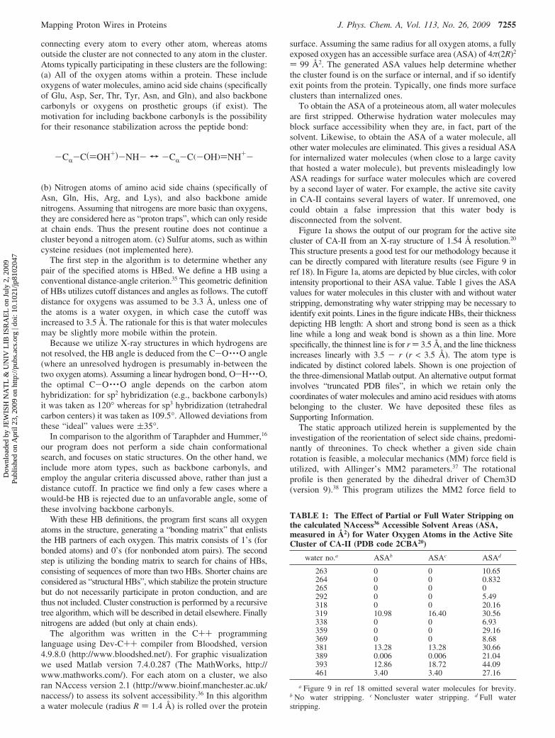
Figure 1a shows the output of our program for the active sitecluster of CA-II from an X-ray structure of 1.54 Å resolution.20
This structure presents a good test for our methodology because itcan be directly compared with literature results (see Figure 9 inref 18). In Figure 1a, atoms are depicted by blue circles, with colorintensity proportional to their ASA value. Table 1 gives the ASAvalues for water molecules in this cluster with and without waterstripping, demonstrating why water stripping may be necessary toidentify exit points. Lines in the figure indicate HBs, their thicknessdepicting HB length: A short and strong bond is seen as a thickline while a long and weak bond is shown as a thin line. Morespecifically, the thinnest line is for r ) 3.5 Å, and the line thicknessincreases linearly with 3.5 - r (r < 3.5 Å). The atom type isindicated by distinct colored labels. Shown is one projection ofthe three-dimensional Matlab output. An alternative output formatinvolves “truncated PDB files”, in which we retain only thecoordinates of water molecules and amino acid residues with atomsbelonging to the cluster. We have deposited these files asSupporting Information.
The static approach utilized herein is supplemented by theinvestigation of the reorientation of select side chains, predomi-nantly of threonines. To check whether a given side chainrotation is feasible, a molecular mechanics (MM) force field isutilized, with Allinger’s MM2 parameters.37 The rotationalprofile is then generated by the dihedral driver of Chem3D(version 9).38 This program utilizes the MM2 force field to
-CR-C()OH+)-NH- T -CR-C(-OH))NH+-
TABLE 1: The Effect of Partial or Full Water Stripping onthe calculated NAccess36 Accessible Solvent Areas (ASA,measured in Å2) for Water Oxygen Atoms in the Active SiteCluster of CA-II (PDB code 2CBA20)
water no.a ASAb ASAc ASAd
263 0 0 10.65264 0 0 0.832265 0 0 0292 0 0 5.49318 0 0 20.16319 10.98 16.40 30.56338 0 0 6.93359 0 0 29.16369 0 0 8.68381 13.28 13.28 30.66389 0.006 0.006 21.04393 12.86 18.72 44.09461 3.40 3.40 27.16
a Figure 9 in ref 18 omitted several water molecules for brevity.b No water stripping. c Noncluster water stripping. d Full waterstripping.
Mapping Proton Wires in Proteins J. Phys. Chem. A, Vol. 113, No. 26, 2009 7255
Dow
nloa
ded
by J
EW
ISH
NA
TL
& U
NIV
LIB
ISR
AE
L o
n Ju
ly 2
, 200
9Pu
blis
hed
on A
pril
23, 2
009
on h
ttp://
pubs
.acs
.org
| do
i: 10
.102
1/jp
8102
047

calculate the interaction of the side chain with the rest of theprotein (held in its X-ray conformation), as a function of theappropriate dihedral angle.
III. Human Carbonic Anhydrase II
Carbonic anhydrase, one of the fastest enzymes (turnover rate106/s), catalyzes the reversible interconversion of carbon dioxideand bicarbonate:31
It is made up predominantly of a �-sheet structure, with acylindrical hydrophilic/hydrophobic cavity in which the zinc
catalytic site is located. The tetrahedral Zn2+ cation is coordi-nated to three histidines (His94, His96, and His119) on thehydrophilic side of the cavity, whereas the fourth ligand is eitherwater or a hydroxide ion. At this enzymatic zinc site (EZn2+)two reactions are catalyzed,
In the forward (hydration) direction, the first step is ionizationof water, with intramolecular and subsequently intermolecular
Figure 1. The active site proton wire of CA-II (PDB file 2CBA,20 1.54 Å resolution): (a) Output from our program as displayed by Matlab. HBedatoms (circles) are in blue, with intensity proportional to their surface accessibility. Lines denote HBs, with their width inversely proportional tothe HB length. (b) A portion of the wire in stick representation with use of Chem3D.38 Atom colors: gray, carbon; red, oxygen; and blue, nitrogen.Dashed yellow lines represent HBs of the indicated lengths (in Å).
CO2 + H2O h HCO3- + H+ (2)
EZn2+(H2O) h EZn2+(OH-) + H+ (3a)
EZn2+(OH-) + CO2 h EZn2+(HCO3-) (3b)
7256 J. Phys. Chem. A, Vol. 113, No. 26, 2009 Shinobu and Agmon
Dow
nloa
ded
by J
EW
ISH
NA
TL
& U
NIV
LIB
ISR
AE
L o
n Ju
ly 2
, 200
9Pu
blis
hed
on A
pril
23, 2
009
on h
ttp://
pubs
.acs
.org
| do
i: 10
.102
1/jp
8102
047

transfer of the proton to a buffer group (B) that conducts it intosolution. Thus, coupled to the first step is a reaction of bufferprotonation
The second step is nucleophilic attack on the CO2 carbon bythe activated hydroxide. Its pKa is around 7, and it forms a strongHB to Thr199 thus orienting its loan pair in the direction of theregion of the cavity into which the CO2 enters.39 The final stepis an exchange of bicarbonate for water at the active site.
A. Intramolecular Proton Transfer. It is widely believedthat following the nucleophilic attack in eq 3b, and precedingthe bicarbonate product release, intramolecular PT moves thesecond proton from the zinc-bound water to one of the othertwo oxygen atoms in the nascent HCO3
-:
This was suggested to occur either by rotation of the enzyme-bound HCO3
- (Lindskog mechanism) or direct PT between twoof its oxygen atoms (Lipscomb mechanism). If insight can begained from the discussion of proton mobility mechanisms inwater,6 it may be that none of these two mechanisms is strictlyoperative. The Lindskog mechanism is analogous to therotational Huckel or Bernal-Fowler mechanisms, which requirecleavage of one or more strong HBs in the first solvation shellof an ion. The Lipscomb mechanism is analogous to protontunneling between well-separated water molecules, whereas thepreferred route is to first bring them close together to form aprotonated dimer, H5O2
+. Thus both these routes may beenergetically costly.
Consulting the active site proton wire in Figure 1, it appearsthat the relevant HB chain to consider is
cf. Figure 1 in ref 39. This chain is analogous to the fragmentof the active pathway in GFP that leads from the Tyr66-OHvia a serine to an internalized glutamate (Scheme 1, left side).That connection is known to transfer a proton on the ultrafasttime scale of several picoseconds,26 or even several hundredfemtoseconds (in which case it is a synchronous triple-PT).40
Thus we identify here a motif involving either a serine orthreonine hydroxyl, HBed to an internalized glutamate as apotential ultrafast, low-barrier pathway for proton abstraction.This suggests that instead of the direct PT between carbonateoxygens envisioned by Lipscomb, proton wires are utilized toreduce the activation energy. The proton is first transferred toGlu106 (Scheme 1) and subsequently either it or another protonis transferred by another wire to one of the two other oxygenatoms.
This mechanism has recently been demonstrated in a detailedDFT calculation by Bottoni et al.41 As in GFP, the PT to Glu106was seen to occur in a concerted fashion, involving double PTfrom the active site to Thr199 and from Thr199 to Glu106.Interestingly, this work identified a low-energy pathway in whichthe Thr199 side chain rotates subsequent to PT to Glu106,delivering the proton to one of the two other oxygen atoms.41
This reinforces the concept of threonine side chains acting as
switches (cf. ref 22): Here Thr199 operates to connect alternatecarbonate oxygens to Glu106 and thus mediate PT betweenthem.
B. Intermolecular Proton Transfer. The proton releasedin the water cleavage step, eq 3a, is believed to be transferredto the Nδ1 imidazole nitrogen of His64, which then flips to an“out” conformation from which it delivers the proton to solution(or to a solvated buffer molecule).31 Indeed, replacement of thishistidine by alanine (a H64A mutation) slows the enzymaticreaction by more than an order of magnitude. A problem withsuch a mechanism is that the distance from the nearest wateroxygen to the Nδ1 position of His64 in the “in” conformation isnearly 3.3 Å,21 larger by 0.9 Å than the optimal distance forefficient PT. Now, when a soluble proton donor/acceptor (4-methylimidazole, 4-MI) is added to the solution, it enhancesthe activity of the H64A mutant to near wt level. One mightexpect this “chemical rescue” molecule to bind near the locationof His64. Indeed, its π-stacking to the adjacent Trp5 wasobserved in the X-ray structure of this mutant,42 but also dockingnear Asp72. However, the first docking site seems nonopera-tional, as judged by the lack of effect of Trp5 mutations on theenzymatic kinetics,43 so it must be the further docking site nearAsp72 (or additional docking sites not seen in this X-raystructure) that contributes the most to the rescue effect.
Another question on the exclusiveness of the His64-shuttleis posed by the observation that the CA-II active site is actuallyopen to solution.18,19 To identify also the more mobile watermolecules, we utilize here the highest resolution (1.05 Å) X-raystructure for wt CA-II from Fisher et al.21 Figure 2a shows thatthe active site HB cluster now extends much further than in thelower resolution structure of Figure 1a, likely because some ofthe more mobile water molecules on the surface could now bedetected. As Figure 2b shows, this cluster includes two pathwaysascending from the Zn center toward Glu69 and further on toAsp72. One pathway passes via the Nδ1 atoms of Asn62 andAsn67, whereas the other, which meanders past Gln92, consistsof only water molecules. In comparison, the H64A mutantstructure (complexed with 4-MI)42 reveals only the pathwaythrough Asn62 and Asn67 (Figure 3). This pathway is apparentlyresponsible for the “chemical rescue” mechanism of 4-MI, whenit is found to bind between Asp72 and Glu69 (see Figure 10 inref 42).
The surface electrostatic potential density of CA-II can beseen in Figure 2B (top) of ref 44. Asp72 and Glu69 are locatedwithin a negative region on the rim of the “caldera” surroundingthe active site. In contrast, the His64 surface region is aboutneutral. This surface potential may be relevant in “steering” theproton and the bicarbonate to/from the active site, and in offeringdocking sites for charged/neutral buffer molecules which carrythe proton to/from the enzyme surface.
One clear conclusion from this analysis is that, in agreementwith Roy and Taraphder,18,19 the His64 pathway is not the onlyone connecting the active site with the outside world. Whichpathway does the proton utilize? The current consensus con-cerning the His64 pathway is based, experimentally, on the effectof the H64A mutation on slowing down the PT rate. Cor-roborating this requires ruling out the alternative pathways whichthus far has not been done.
The opposite view might be that the proton is transportedonly along the surface pathways in Figure 2b. In this scenariothe diminished reactivity of the H64A mutant may be ascribedto the elimination of one surface pathway, forcing the protonto migrate via the Nδ1 atom of Asn62 or Asn67 (Figure 3b).Assuming that PT through nitrogen is slower than through
B + H+ h BH (4)
EZn2+(OH)COO- f EZn2+OC(OH)O-
Zn2+-OH- · · ·Thr199-OH · · ·Glu106-O- · · · (5)
Mapping Proton Wires in Proteins J. Phys. Chem. A, Vol. 113, No. 26, 2009 7257
Dow
nloa
ded
by J
EW
ISH
NA
TL
& U
NIV
LIB
ISR
AE
L o
n Ju
ly 2
, 200
9Pu
blis
hed
on A
pril
23, 2
009
on h
ttp://
pubs
.acs
.org
| do
i: 10
.102
1/jp
8102
047

oxygen, this could delay the proton transport process. Such aninterpretation may be questioned, because when the protein isimmersed in water it is likely that additional layers of waterwill open up more pathways, so that these specific surface
pathways may be less dominant. In other words, surfacepathways observed in the X-ray structure might rearrange insolution. On the other hand, they may be significant if waterHBs to the protein surface are stronger than bulk HBs, and if
Figure 2. The active site proton wire for high-resolution CA-II structure (PDB file 2ILI,21 1.54 Å resolution). (a) Matlab display of the output fromour program. (b) Depiction of the portion of the wire that lies on the protein surface with use of Chem3D version 9.38
7258 J. Phys. Chem. A, Vol. 113, No. 26, 2009 Shinobu and Agmon
Dow
nloa
ded
by J
EW
ISH
NA
TL
& U
NIV
LIB
ISR
AE
L o
n Ju
ly 2
, 200
9Pu
blis
hed
on A
pril
23, 2
009
on h
ttp://
pubs
.acs
.org
| do
i: 10
.102
1/jp
8102
047

the proton prefers to migrate on the surface, rather than in thebulk. This may be the case, given the demonstrated interfacialaffinity of the hydronium.45,46
A third scenario is that both His64 and surface pathways areoperative, and their relative importance depends on conditions.For example, one pathway may be important for the hydration
and the other for the dehydration direction. The reason forhaving the two kinds of pathways may involve the docking ofbuffer molecules from solution. A neutral buffer molecule maydock near His64 and collect an outgoing proton when theenzyme works in the hydration direction. A positively charged,protonated buffer molecule will preferentially dock on the
Figure 3. The active site proton wire for a high-resolution structure (PDB file 1MOO,42 1.05 Å resolution) of the H64A mutant of CA-II, complexedwith two molecules of 4-MI. (a) Matlab display of the output from our program. (b) Depiction of the portion of the wire that lies on the proteinsurface with use of Chem3D version 9.38 Comparison with Figure 2 is meaningful because both structures have the same X-ray resolution.
Mapping Proton Wires in Proteins J. Phys. Chem. A, Vol. 113, No. 26, 2009 7259
Dow
nloa
ded
by J
EW
ISH
NA
TL
& U
NIV
LIB
ISR
AE
L o
n Ju
ly 2
, 200
9Pu
blis
hed
on A
pril
23, 2
009
on h
ttp://
pubs
.acs
.org
| do
i: 10
.102
1/jp
8102
047

negative surface near Asp72 and deliver a proton (via the surfacepathways of Figure 2b) to the enzyme working in the dehydra-tion direction.
IV. Chromophore Biosynthesis in GFP
A. Background. “The remarkable brightly glowing greenfluorescent protein, GFP, was first observed in the beautifuljellyfish, Aequorea Victoria in 1962. Since then, this proteinhas become one of the most important tools used incontemporary bioscience. With the aid of GFP, researchershave developed ways to watch processes that were previouslyinvisible, such as the development of nerve cells in the brainor how cancer cells spread.”47 The GFP rigid eleven-stranded�-barrel structure is traversed by a distorted R-helicalsegment: Three of its amino acids, Ser65, Tyr66, and Gly67,undergo a post-translational condensation reaction to form afive-membered imidazolone ring that couples to the tyrosinephenol ring.48 This constitutes the aromatic chromophore,whose anion fluoresces in the green following an excited statePT reaction.26,28
Cyclization occurs only after protein folding, within a tightR-helical turn that places the Gly67 amide in close proximityto the Ser65 carbonyl, resulting in nucleophilic attack and ringformation (Scheme 2). Tsien and co-workers suggested thatchromophore biosynthesis in GFP consists of three steps:cyclization, dehydration, and oxidation (see Scheme 3).49 Kineticexperiments indeed demonstrated that cyclization precedesoxidation.50 Subsequently, a S65G/Y66G mutant was found tocyclize but not dehydrate, suggesting that cyclization precedesdehydration.51 Thus both oxidation and dehydration are latersteps in GFP maturation.
In contrast to the agreement concerning the cyclization step,the order and detail of the subsequent steps in the mechanismare constantly under revision.52 Most notably, Wachter and co-workers have found that a Y66L mutant undergoes cyclizationand oxidation, but not dehydration, concluding that oxidationis actually the second step in chromophore biogenesis (Scheme4).53 This oxidation reaction is centered on the 5-membered ringrather than on the Tyr66 CR-C� bond, as previously assumed.Subsequently, participation of a pre-oxidation enolate intermedi-ate (not shown here) was demonstrated.54 It is stabilized by thepositive charge on Arg96, which is HBed to the enolate oxygenatom (formerly the Tyr66 carbonyl).55 In the mechanism ofScheme 4, this enolate formation is coupled to the eliminationof a proton from CR66 (see Scheme 2 for notation).
Finally it was shown56 that dideuteration at the Tyr66 C�
position does not affect the oxidation rate, but slows thesubsequent dehydration step by nearly a factor of 6. This isconsistent with a primary isotope effect on the tetrahedral bridgecarbon. Its hydrogen atom is therefore not abstracted bymolecular oxygen, but rather during a subsequent PT reactionthat produces a carbanion in the C�66 position.56 This carbanionis stabilized by charge delocalization on the two flankingaromatic rings, which explains why chromophore maturationis observed only for aromatic substitutions in position 66.Nevertheless, carbanion formation still requires a rather basicresidue (pKa ) 9.4) within the protein for abstracting thisproton.56
The problem is that no such basic moiety is observed withinHBing proximity to C�66. Wachter and co-workers suggestedtwo solutions to this enigma (Scheme 5): (a) The proton istranslocated, through a chain of two water molecules, to O65(see Scheme 2 for notations).53 This then releases a watermolecule, completing the dehydration step. Apparently, wateris not sufficiently basic to supply the driving force for this PTstep, so that this option was abandoned. (b) The C�66 proton isabstracted by the guanidinium group of Arg96, while O65 issubsequently protonated by Glu222 to release a water mol-ecule.56
There are evident problems with Arg96 as the deprotonatingbase, most of which are discussed in the original publication:56
(i) Arginines are very basic, their solution pKa ) 12.5, sothat Arg96 is quite certainly protonated within GFP. Indeed,
SCHEME 2: The Product of the Cyclization Step ofGFP Chromophore Synthesis, with Key Atoms Markedand Numbered
SCHEME 3: A Three-Step GFP Chromophore Biosynthesis Involving Cyclization, Dehydration, and Oxidation49
7260 J. Phys. Chem. A, Vol. 113, No. 26, 2009 Shinobu and Agmon
Dow
nloa
ded
by J
EW
ISH
NA
TL
& U
NIV
LIB
ISR
AE
L o
n Ju
ly 2
, 200
9Pu
blis
hed
on A
pril
23, 2
009
on h
ttp://
pubs
.acs
.org
| do
i: 10
.102
1/jp
8102
047

its positive charge is postulated to catalyze enolate formationas well as other steps in the chromophore maturation mecha-nism.55
(ii) Subsequently, one has to assume that “the pKa of Arg96may drop below 8 while the biosynthesis reaction is inprogress”,56 but it is unclear how such a large pKa change comesabout.
(iii) The closest guanidinium nitrogen to C�66 is at a distanceof 3.9 Å, too far for direct PT (typical cutoff values for PT inwater are around 3.3 Å). Consequently, Wachter and co-workersassume the participation of a mediating water molecule (Scheme
5b, in red), although such a water molecule is not seen in anyof the GFP X-ray structures.
B. A New Internal Proton Wire in GFP. We have usedour computer code to map out proton wires within GFP. Theprogram located the extensive proton wire that connects Tyr66-OH via Glu222 to the surface of the �-barrel, which waspreviously found manually.22 This cluster (not shown here) isthought to participate in the excited state PT reaction.28,29 Inaddition, it found the new internal cluster depicted in Figure 4.It is located on the other face of the chromophore, apparentlydisconnected from the first cluster and from the protein exterior.
SCHEME 4: A Three-Step GFP Chromophore Biosynthesis Involving Cyclization, Oxidation, and Dehydration53
SCHEME 5: Suggested Mechanisms for Carbanion Formation by Proton Extraction from C�66: (a) Direct Transferthrough a Water-Molecule Chain According to Figure 7b in Reference 53 (arrows depict electronic motion) and (b) PTto a Neutral Arg96 through a Water Molecule (Red) That Does Not Exist in the X-ray Structure, According to Scheme 3of Reference 56
Mapping Proton Wires in Proteins J. Phys. Chem. A, Vol. 113, No. 26, 2009 7261
Dow
nloa
ded
by J
EW
ISH
NA
TL
& U
NIV
LIB
ISR
AE
L o
n Ju
ly 2
, 200
9Pu
blis
hed
on A
pril
23, 2
009
on h
ttp://
pubs
.acs
.org
| do
i: 10
.102
1/jp
8102
047

Thus this new wire does not seem to be involved in excitedstate PT. It connects some key residues near the chromophore,such as His181-Nε2, Thr62-OH, Thr63-OH, Thr108-OH, Tyr145-OH, and Arg96-Nη1, some of which are thought to be involvedin chromophore biosynthesis.51,56 Hence the new proton wiremay be relevant for some of the biosynthesis steps. For example,if indeed Arg96 gets temporarily deprotonated during thedehydration step,56 it may occur via this wire.
Checking more closely for contact points between the newwire and the chromophore, we found the three rotamers of Thr62shown in Figure 5. In the X-ray structure (0° rotation) its sidechain forms a HB with His181. When it is rotated by about150°, its hydroxyl oxygen reaches a distance of 3.0 Å from thebridge carbon, C�66. At about 260°, a HB is formed insteadwith the OH moiety of Tyr145.
The energetics of these rotamers are depicted in Figure 6,which shows a rotational profile calculated from two wt-GFP
and two S65T mutant GFP crystal structures, using the MM2force field37 as implemented in the dihedral driver of Chem3D(version 9).38 These profiles depict the potential as a functionof the Thr62 dihedral angle (rotation around its CR-C� bond),resulting from the interaction of the side chain with the rest ofthe protein, which is maintained in its X-ray conformation.Hence these potentials are expected to converge with improve-ment in X-ray resolution, as indeed appears to be the case. Thelowest barrier separating the native (0°) and active (150°)rotamers is in the range 13-18 kJ/mol. These values areconsistently smaller than those for the other two threonines onthis cluster (Thr63 and Thr108), as well as for Thr62 inprecyclized mutants51 (30-40 kJ/mol, see Figure 7). Thus Thr62in the cyclized structure may be “engineered” for facile rotation.Compared with rotational time scales for other small side chainswith barriers on the order of 20 kJ/mol or less (see the
Figure 4. The new internal proton wire in GFP (PDB file 1W7S,25 1.85 Å resolution): (a) Output from our program as displayed by Matlab. HBedatoms (circles) are colored blue, with intensity proportional to their surface accessibility. (b) A portion of the wire in stick representation with useof Chem3D.38 See Figure 1 for notations.
7262 J. Phys. Chem. A, Vol. 113, No. 26, 2009 Shinobu and Agmon
Dow
nloa
ded
by J
EW
ISH
NA
TL
& U
NIV
LIB
ISR
AE
L o
n Ju
ly 2
, 200
9Pu
blis
hed
on A
pril
23, 2
009
on h
ttp://
pubs
.acs
.org
| do
i: 10
.102
1/jp
8102
047

Introduction), one may estimate that this rotation occurs on thesubmicrosecond time scale,33 hence it is not rate limiting.
To abstract the proton from the bridge CH2 moiety, the rotatedThr62 side chain must be able to (a) form a rather strong HBwith it and (b) participate in a short HB network leading to anearby basic moiety. Consider first the possibility of forming aC-H · · ·O HB. In model calculations,57 HBs between water andmethane were shown to behave very much like ordinary HBs,only that they are weaker. When the methane was substituted
with fluorines (electron attracting substituents) the HB strengthincreased to about half its magnitude in water.57 For C�66, therole of the electron attracting groups is played by the twoflanking aromatic rings. Moreover, it was shown that when waterwas replaced by methanol, the HB strength to the substitutedmethane further increased.57 Thus the C�66-H · · ·OH-Thr62HB may be comparable in strength to the HB between watermolecules.
Searching next for an appropriate base that may abstract thisproton, we note that the Nε2 atom of His181 is only 3.8 Å awayfrom the hydroxyl oxygen of the rotated Thr62 side chain.Histidines in solution typically have pKa ) 6 but, contrary topopular opinion, they tend to become more basic when buriedinside a protein.58 However, the variability in their pKa valuesalso increases with burial, spanning the range 4-10. Thus it ispossible that His181 is sufficiently basic to abstract the protonfrom C�66. As discussed in Section III, a histidine (His64) hasbeen implicated as the proton acceptor in water ionization inthe CA-II active site (eq 3a). More specifically, there are several
Figure 5. The three conformers of the Thr62 side chain in wt-GFP,PDB file 1W7S:25 (a) The native conformer, in which the Thr62 sidechain is HBed to His181; (b) the active conformer (145° rotation) formsa HB to C�66; (c) a third conformer (255° rotation) forms a HB toTyr145. Atoms rendered as in Figure 4b.
Figure 6. Dihedral chart for the side chain rotation of Thr62 in thefolded structures of wt-GFP and some of its mutants. Conformationalenergy as a function of Thr62 side chain dihedral angle was calculatedby using the MM2 dihedral driver of Chem3D.38 The native rotamer isat 0°, whereas contact with C�66 is formed around 150°. Around 260°a HB is formed with the hydroxyl of Tyr145. Coordinates were takenfrom the X-ray structures of resolution better than 2 Å in order to reducethe energetic spread. The following PDB files were utilized: 1GFL (1.90Å resolution)23 and 1W7S (1.85 Å resolution),25 both wild-typestructures; 1Q4A (1.45 Å resolution) and 1Q4B (1.48 Å resolution),both S65T mutants at pH 8.5 and 5.5, respectively.64
Figure 7. Dihedral chart for the side chain rotation of Thr62 inprecyclized GFP, X-ray structures from PDB files 1QXT and 1QY3(mutations R96A, F99S, M153T, V163A, F64L, S65T).51 The minimumaround 140-150° does not involve a HB to C�66. This minimum forstructure 1QY3 is lower probably due to new HBs to water moleculeswhich do not exist in structure 1QXT. See the legend of Figure 6 forother details.
Mapping Proton Wires in Proteins J. Phys. Chem. A, Vol. 113, No. 26, 2009 7263
Dow
nloa
ded
by J
EW
ISH
NA
TL
& U
NIV
LIB
ISR
AE
L o
n Ju
ly 2
, 200
9Pu
blis
hed
on A
pril
23, 2
009
on h
ttp://
pubs
.acs
.org
| do
i: 10
.102
1/jp
8102
047

enzymes in which a histidine serves as a base in R-protonabstraction, leading to formation of a carbanion intermedi-ate.59-62 In these examples, the carbanion is often stabilized byresonance with an adjacent carbonyl, forming an enolate whichcan be stabilized by the positive charge of an arginine.60 Suchproton abstractions are analogous to the elimination of the protonfrom CR66 in a preceding step of the GFP chromophoresynthesis. The subsequent carbanion formation on C�66 can stillgain stabilization by delocalizing the negative charge onto thecarbonyl, forming a so-called homoenolate intermediate.63
With the above observations in mind, we suggest the tentativemechanism in Scheme 6. Thr62 rotation enables proton abstrac-tion to His181, a step that becomes essentially irreversible asthe threonine rotates back to its original position, where it formsa HB to His181 and stabilizes the positive charge there. As theoriginal proton wire is reformed, the proton on His181 maydelocalize on it, further reducing the probability for back-PT.In the chromophore, the negative charge on the bridge carbonis partly transferred to the carbonyl, through the 3-center bondshown in Scheme 6. The ensuing homoenolate, in turn, isstabilized by a salt bridge to Arg96. Thus Arg96 catalyzes thisreaction step without ever becoming deprotonated.
It remains to identify the transient proton wire that mayconnect C�66-H · · ·OH-Thr62 with His181. We note that if theside chains of Thr62 and His181 are tilted, a direct contact can
be formed, C�66-H · · ·Thr62-OH · · ·His181-Nε2 in which the twoHB lengths are around 3.3 Å. These distances are a bit long foran efficient PT to take place, which may nevertheless proceedby tunneling on a sufficiently long time scale. Alternately, wenote that a nearby water molecule may be “squeezed” betweenThr62, His181, and Phe165 (see Figure 8). This fit requires arotation of 30° of the Ile167 side chain, at a cost of about 16kJ/mol (determined by using the dihedral driver of Chem3D).This suggestion is in line with other cases where an isoleucineside chain was seen to gate the motion of small ligands withinproteins.34 The fit of the water molecule is still a bit tight(distance of only 3.0 Å to the Phe165 side chain), but could befurther optimized by either molecular dynamics or Monte-Carloroutine.16 Such calculations, which are outside the scope of thepresent work, may require the (unknown) conformation frombefore the carbanion formation step, which likely differssomewhat from the X-ray structure of the mature GFP.
Finally we note that Tyr145-OH is only 0.7 Å further awaythan His181. With a pKa around 10, we do not expect it to beionized. However, when irradiated (peak absorbance near 275nm) it could undergo excited state proton transfer, ejecting thehydroxylic proton into the proton wire shown in Figure 4. In afew nanoseconds its anion will decay back to the ground state,producing a basic moiety that could participate in abstractingthe proton from the bridge carbon. If this mechanism holds,
SCHEME 6: Proton Abstraction from C�66 to His181 and the New Proton Wire via the Thr62 Switch, with CarbanionStabilization as a Homoenolate63 by Arg96
7264 J. Phys. Chem. A, Vol. 113, No. 26, 2009 Shinobu and Agmon
Dow
nloa
ded
by J
EW
ISH
NA
TL
& U
NIV
LIB
ISR
AE
L o
n Ju
ly 2
, 200
9Pu
blis
hed
on A
pril
23, 2
009
on h
ttp://
pubs
.acs
.org
| do
i: 10
.102
1/jp
8102
047

one may expect GFP chromophore maturation to be catalyzedby UV light, an experiment yet to be done.
V. Conclusion
PT in proteins depends crucially on HBed networks (“protonwires”) formed by oxygen atoms from internal water moleculesand side chains, and (depending on the time scale) possibly alsobackbone carbonyls, sulfur, and nitrogen atoms. These may formlarge HBed clusters, of which only select portions werediscussed in the literature. Examples are the proton wirefragment connecting the zinc atom to His64 in CA-II, or theTyr66 to Glu222 fragment in GFP. It may well be that the initialintuition was correct, and these are indeed the only functionallyimportant wire segments, but this can be decided only after allrelevant segments are investigated. It is therefore desirable tohave a simple to use computer code for systematic mapping ofproton wires in proteins.
Here we have developed a simplified version of such analgorithm, which scans the atomic coordinates systematicallyand reports on all the clusters of interconnected oxygen atoms(see Methods). Application of this algorithm consistently reportslarger HB clusters than encountered in the literature. Moreover,with increase in X-ray structural resolution these clusters tendto increase even further. In the present study we have exempli-fied the potential of the methodology on two proton wires: Therather extensively investigated active site proton wire in CA-IIand a previously unknown, nonactive site internal proton wirein GFP.
The active site cluster of CA-II21 is surprising because itexhibits an unreported direct exit to solution. From the buried
“active site caldera”, where the zinc center resides, two protonwires climb on the enzyme surface up to Glu69, which is locatedwithin a negatively charged patch on the rim of the caldera(Figure 2b). Do protons utilize these wires rather than the His64flip for either exit or entry? Are the different paths utilized underdifferent conditions? Without detailed experimentation andcalculations it is hard to assess the significance of this observa-tion. Nevertheless, the fact that these surface wires lead to aregion of negative charge density hints to the possibility ofpositively charged buffer molecules docking there,42 from whichprotons can be fed into the active site via these surface pathwayswhen the enzyme works in the dehydration direction.
The finding of the new proton wire in GFP, unconnected tothe Tyr66 active site, is also intriguing. Does it have a functionalrole? Here we have argued that residues on this wire may beinvolved in GFP chromophore biosynthesis. Overall this reactionrequires the abstraction of four protons/hydrogens:56 from N67,CR66, N66, and C�66 (Scheme 7). Each proton entails a differentmechanism for its abstraction, thus there are four (not three)steps in the overall mechanism: (i) cyclization requires thetransfer of the proton attached to N67; (ii) enolate formsfollowing abstraction of the proton attached to CR66; (iii)elimination of the hydrogen from N66 occurs by molecularoxygen; and (iv) the rate limiting step for dehydration isabstraction of a proton from C�66. Here we focused on thecarbanion formation in step iv.
The newly observed internal HB cluster could be involvedin several ways in this transient carbanion formation (Scheme6). A rotation of the Thr62 side chain brings it into HB contactwith one of the hydrogens of C�66. Completion of a short protonwire from Thr62 to His181 (e.g., by diffusion of a watermolecule facilitated by Ile167 side chain rotation as in Figure8) can allow PT from C�66 to Nε2 of His181, with stabilizationof the negative charge by its delocalization to the imidazolonecarbonyl which forms a strong HB with Arg96. Rotation ofThr62 to its original orientation prevents back PT. Furthermore,it reestablishes the proton wire to His181, allowing the abstractedproton to delocalize on the wire, until the elimination of thewater molecule from C65 is completed.
Again, a mechanism deduced by inspection can be onlytentative. Yet the comparison of proton wire motifs acrossproteins suggests that threonines may indeed function as protonwire switches. The comparison of the CA-II and GFP protonabstraction mechanisms also suggests that a buried Ser/Thr-Glu pair near the active site functions as a rapid reversible protonabstractor. With further comparisons of proton wires in differentproteins, one may hope to identify additional proton wire motifs,and these could help decipher the function of new proton wiresin other proteins.
Acknowledgment. We thank David N. Silverman andSrabani Taraphder for correspondence, Zvi Rappoport for
Figure 8. Suggested steps in the formation of a transient proton wireconnecting C�66 with the His181 base. (a) Rotation of the Thr62 sidechain. (b) Rotation of the Ile167 side chain with translation of a watermolecule into the ensuing cavity. From PDB file 1W7S, subunit B.25
Compare with Figure 5. Full yellow lines denote HBs.
SCHEME 7: The Four Protons, H1-H4, Eliminatedduring Chromophore Biogenesis in GFP
Mapping Proton Wires in Proteins J. Phys. Chem. A, Vol. 113, No. 26, 2009 7265
Dow
nloa
ded
by J
EW
ISH
NA
TL
& U
NIV
LIB
ISR
AE
L o
n Ju
ly 2
, 200
9Pu
blis
hed
on A
pril
23, 2
009
on h
ttp://
pubs
.acs
.org
| do
i: 10
.102
1/jp
8102
047

suggesting ref 63. This research was supported by the IsraelScience Foundation (grant no. 122/08). The Fritz Haber Centeris supported by the Minerva Gesellschaft fur die Forschung,Munchen, FRG.
Supporting Information Available: Truncated PDB fileswith the coordinates of the amino acid residues, water oxygen,and zinc atoms participating in the HB clusters shown in Figures1-4 (to view a cluster, the appropriate PDB file should beloaded into a visualization software with HB drawing capabili-ties; files are labeled by their original PDB names). This materialis available free of charge via the Internet at http://pubs.acs.org.
References and Notes
(1) Agmon, N. J. Phys. Chem. A 2005, 109, 13–35.(2) Voth, G. A. Acc. Chem. Res. 2006, 39, 143–150.(3) Marx, D. ChemPhysChem 2006, 7, 1848–1870; addendum: ChemP-
hysChem 2007, 8, 209-210.(4) Wraight, C. A. Biochim. Biophys. Acta 2006, 1757, 886–912.(5) Swanson, J. M. J.; Maupin, C. M.; Chen, H.; Petersen, M. K.; Xu,
J.; Wu, Y.; Voth, G. A. J. Phys. Chem. B 2007, 111, 4300–4314.(6) Agmon, N. Chem. Phys. Lett. 1995, 244, 456–462.(7) Huckel, E. Z. Electrochem. 1928, 34, 546.(8) Bernal, J. D.; Fowler, R. H. J. Chem. Phys. 1933, 1, 515–548.(9) Agmon, N. J. Chim. Phys. Phys.-Chim. Biol. 1996, 93, 1714–1736.
(10) Markovitch, O.; Agmon, N. J. Phys. Chem. A 2007, 111, 2253–2256.
(11) Luz, Z.; Meiboom, S. J. Am. Chem. Soc. 1964, 86, 4768–4769.(12) Markovitch, O.; Chen, H.; Izvekov, S.; Paesani, F.; Voth, G. A.;
Agmon, N. J. Phys. Chem. B 2008, 112, 9456–9466.(13) Tuckerman, M.; Laasonen, K.; Sprik, M.; Parrinello, M. J. Phys.
Chem. 1995, 99, 5749–5752.(14) Lapid, H.; Agmon, N.; Petersen, M. K.; Voth, G. A. J. Chem. Phys.
2005, 122, 014506.(15) Nagle, J. F.; Morowitz, H. J. Proc. Natl. Acad. Sci. U.S.A. 1978,
75, 298–302.(16) Taraphder, S.; Hummer, G. J. Am. Chem. Soc. 2003, 125, 3931–
3940.(17) Roy, A.; Taraphder, S. Biopolymers 2006, 82, 623–630.(18) Roy, A.; Taraphder, S. J. Phys. Chem. B 2007, 111, 10563–10576.(19) Roy, A.; Taraphder, S. J. Phys. Chem. B 2008, 112, 13597–13607.(20) Håkansson, K.; Carlsson, M.; Svensson, L. A.; Liljas, A. J. Mol.
Biol. 1992, 227, 1192–1204.(21) Fisher, S. Z.; Maupin, C. M.; Budayova-Spano, M.; Govindasamy,
L.; Tu, C.; Agbandje-McKenna, M.; Silverman, D. N.; Voth, G. A.;McKenna, R. Biochemistry 2007, 46, 2930–2937.
(22) Agmon, N. Biophys. J. 2005, 88, 2452–2461.(23) Yang, F.; Moss, L. G.; Phillips, G. N., Jr. Nat. Biotechnol. 1996,
14, 1246–1251.(24) Ormo, M.; Cubitt, A. B.; Kallio, K.; Gross, L. A.; Tsien, R. Y.;
Remington, S. J. Science 1996, 273, 1392–1395.(25) van Thor, J. J.; Georgiev, G. Y.; Towrie, M.; Sage, J. T. J. Biol.
Chem. 2005, 280, 33652–33659.(26) Chattoraj, M.; King, B. A.; Bublitz, G. U.; Boxer, S. G. Proc. Natl.
Acad. Sci. U.S.A. 1996, 93, 8362–8367.(27) van Thor, J. J.; Zanetti, G.; Ronayne, K. L.; Towrie, M. J. Phys.
Chem. B 2005, 109, 16099–16108.(28) Leiderman, P.; Huppert, D.; Agmon, N. Biophys. J. 2006, 90, 1009–
1018.(29) Agmon, N. J. Phys. Chem. B 2007, 111, 7870–7878.(30) Helms, V.; Straatsma, T. P.; McCammon, J. A. J. Phys. Chem. B
1999, 103, 3263–3269.
(31) Silverman, D. N.; McKenna, R. Acc. Chem. Res. 2007, 40, 669–675.
(32) Chen, K.; Hirst, J.; Camba, R.; Bonagura, C. A.; Stout, C. D.;Burgess, B. K.; Armstrong, F. A. Nature 2000, 405, 814–817.
(33) Dantsker, D.; Samuni, U.; Friedman, J. M.; Agmon, N. Biochim.Biophys. Acta: Proteins Proteomics 2005, 1749, 234–251.
(34) Ostermann, A.; Waschipky, R.; Parak, F. G.; Nienhaus, G. U.Nature 2000, 404, 205–208.
(35) Luzar, A. J. Chem. Phys. 2000, 113, 10663–10675.(36) Hubbard, S. J.; Thornton, J. M. ‘NACCESS’ Computer Program,
Department of Biochemistry and Molecular Biology, University CollegeLondon, 1993.
(37) Allinger, N. L. J. Am. Chem. Soc. 1977, 99, 8127–8134.(38) CambridgeSoft Corp., 2005, http://www.cambridgesoft.com/software/
ChemOffice/.(39) Liang, Z.; Xue, Y.; Behravan, G.; Jonsson, B.-H.; Lindskog, S.
Eur. J. Biochem. 1993, 211, 821–827.(40) Vendrell, O.; Gelabert, R.; Moreno, M.; Lluch, J. M. J. Phys. Chem.
B 2008, 112, 5500–5511.(41) Bottoni, A.; Lanza, C. Z.; Miscione, G. P.; Spinelli, D. J. Am. Chem.
Soc. 2004, 126, 1542–1550.(42) Duda, D.; Govindasamy, L.; Agbandje-McKenna, M.; Tu, C.;
Silverman, D. N.; McKenna, R. Acta Crystallogr. D 2003, 59, 93–104.(43) An, H.; Tu, C.; Duda, D.; Montanez-Clemente, I.; Math, K.; Laipis,
P. J.; McKenna, R.; Silverman, D. N. Biochemistry 2002, 41, 3235–3242.(44) Marino, S.; Hayakawa, K.; Hatada, K.; Benfatto, M.; Rizzello, A.;
Maffia, M.; Bubacco, L. Biophys. J. 2007, 93, 2781–2790.(45) Petersen, M. K.; Iyengar, S. S.; Day, T. J. F.; Voth, G. A. J. Phys.
Chem. B 2004, 108, 14804–14806.(46) Petersen, M. K.; Voth, G. A. J. Phys. Chem. B 2006, 110, 7085–
7089.(47) Royal Swedish Academy of Science Press Release, 2008; http://
nobelprize.org/nobel_prizes/chemistry/laureates/2008/press.html.(48) Tsien, R. Y. Annu. ReV. Biochem. 1998, 67, 509–544.(49) Cubitt, A. G.; Heim, R.; Adams, S. R.; Boyd, A. E.; Gross, L. A.;
Tsien, R. Y. Trends Biochem. Sci. 1995, 20, 448–455.(50) Reid, B. G.; Flynn, G. C. Biochemistry 1997, 36, 6786–6791.(51) Barondeau, D. P.; Putnam, C. D.; Kassmann, C. J.; Tainer, J. A.;
Getzoff, E. D. Proc. Natl. Acad. Sci. U.S.A. 2003, 100, 12111–12116.(52) Wachter, R. M. Acc. Chem. Res. 2007, 40, 120–127.(53) Rosenow, M. A.; Huffman, H. A.; Phail, M. E.; Wachter, R. M.
Biochemistry 2004, 43, 4464–4472.(54) Barondeau, D. P.; Tainer, J. A.; Getzoff, E. D. J. Am. Chem. Soc.
2006, 128, 3166–3168.(55) Wood, T. I.; Barondeau, D. P.; Hitomi, C.; Kassmann, C. J.; Tainer,
J. A.; Getzoff, E. D. Biochemistry 2005, 44, 16211–16220.(56) Pouwels, L. J.; Zhang, L.; Chan, N. H.; Dorrestein, P. C.; Wachter,
R. M. Biochemistry 2008, 47, 10111–10122.(57) Gu, Y.; Kar, T.; Scheiner, S. J. Am. Chem. Soc. 1999, 121, 9411–
9422.(58) Edgcomb, S. P.; Murphy, K. P. Proteins: Struct. Funct. Genet. 2002,
49, 1–6.(59) Medda, R.; Padiglia, A.; Pedersen, J. J.; Floris, G. Biochem. Biophys.
Res. Commun. 1993, 196, 1349–1355.(60) Yorita, K.; Janko, K.; Aki, K.; Ghisla, S.; Palfey, B. A.; Massey,
V. Proc. Natl. Acad. Sci. U.S.A. 1997, 94, 9590–9595.(61) Wu, C.-Y.; Lee, H.-J.; Wu, S.-H.; Chen, S.-T.; Chiou, S.-H.; Chang,
G.-G. Biochem. J. 1998, 333, 327–334.(62) Lehoux, I. E.; Mitra, B. Biochemistry 1999, 38, 9948–9955.(63) Buncel, E. Carbanions: Mechanistic and Isotopic Aspects; Elsevier:
Amsterdam, 1975; Chapter 4, Section 4.(64) Jain, R. K.; Ranganathan, R. Proc. Natl. Acad. Sci. U.S.A. 2004,
101, 111–116.
JP8102047
7266 J. Phys. Chem. A, Vol. 113, No. 26, 2009 Shinobu and Agmon
Dow
nloa
ded
by J
EW
ISH
NA
TL
& U
NIV
LIB
ISR
AE
L o
n Ju
ly 2
, 200
9Pu
blis
hed
on A
pril
23, 2
009
on h
ttp://
pubs
.acs
.org
| do
i: 10
.102
1/jp
8102
047
Copyright © 2022 FDOKUMEN