Inoculating plants with the endophytic bacterium Pseudomonas sp. Ph6-gfp to reduce phenanthrene...
9
RESEARCH ARTICLE Inoculating plants with the endophytic bacterium Pseudomonas sp. Ph6-gfp to reduce phenanthrene contamination Kai Sun 1 & Juan Liu 1 & Yanzheng Gao 1 & Yuehui Sheng 1 & Fuxing Kang 1 & Michael Gatheru Waigi 1 Received: 12 February 2015 /Accepted: 27 July 2015 # Springer-Verlag Berlin Heidelberg 2015 Abstract Plant organic contamination poses a serious threat to the safety of agricultural products and human health world- wide, and the association of endophytic bacteria with host plants may decrease organic pollutants in planta. In this study, we firstly determined the growth response and biofilm forma- tion of endophytic Pseudomonas sp. Ph6-gfp, and then sys- tematically evaluated the performance of different plant colo- nization methods (seed soaking (SS), root soaking (RS), leaf painting (LP)) for circumventing the risk of plant phenan- threne (PHE) contamination. After inoculation for 48 h, strain Ph6-gfp grew efficiently with PHE, oxalic acid, or malic acid as the sole sources of carbon and energy. Moreover, strain Ph6-gfp could form robust biofilms in LB medium. In green- house hydroponic experiments, strain Ph6-gfp could actively colonize inoculated plants internally, and plants colonized with Ph6-gfp showed a higher capacity for PHE removal. Compared with the Ph6-gfp-free treatment, the accumulations of PHE in Ph6-gfp-colonized plants via SS, RS, and LP were 20.1, 33.1, and 7.1 %, respectively, lower. Our results indicate that inoculating plants with Ph6-gfp could lower the risk of plant PHE contamination. RS was most efficient for improv- ing PHE removal in whole plant bodies by increasing the cell numbers of Ph6-gfp in plant roots. The findings in this study provide an optimized method to strain Ph6-gfp reduce plant PAH residues, which may be applied to agricultural produc- tion in PAH-contaminated soil. Keywords Polycyclic aromatic hydrocarbons . Phenanthrene . Endophytic Pseudomonas . Colonization method . Visualization . Performance Introduction Anthropogenic environmental contamination has recently in- creased the risks of plant organic contamination throughout the world (Collins et al. 2006; Andria et al. 2009). The most dangerous contaminants are toxic, some of which can bioaccumulate and persist (Mckone and Maddalena 2007). Residence times in plants range from several days to months, resulting in potential threats to human health and ecosystems (Collins et al. 2006; Khan et al. 2013). Thus, understanding how to avoid the risk of plant organic contamination is essen- tial for ensuring the safety of agricultural products and human health (Weyens et al. 2009a; Yousaf et al. 2011; Sun et al. 2014a). Plant-endophyte symbioses are ubiquitous and have attained increasing acceptance as viable cleanup technolo- gies to overcome organic pollutants (Germaine et al. 2006; Weyens et al. 2009b; Afzal et al. 2014). Endophytic bacte- ria can enhance plant growth, reduce plant disease, and stimulate plant resistance to harsh external environments (Ryan et al. 2008; Glick 2014). Notably, certain endophytic bacteria can prevent/reduce the accumulation of organic pollutants in plants (Germaine et al. 2009; Sun et al. 2014b ). For example, the inoculation of pea plants ( Pisum sativum L.) with a Pseudomonas putida strain prevented 2,4-dichlorophenoxyacetic acid (2,4-D) accumu- lation (Germaine et al. 2006). In addition, when ryegrass (Lolium multiflorum Lam) were colonized with endophytic Staphylococcus sp. strain BJ06 (isolated from Alopecurus aequalis Sobol), the concentrations of pyrene in ryegrass roots and shoots decreased, and were 31 and 44 % lower Responsible editor: Robert Duran K. Sun and J. Liu contribute equally to this paper. * Yanzheng Gao [email protected]; [email protected] 1 Institute of Organic Contaminant Control and Soil Remediation, College of Resources and Environmental Sciences, Nanjing Agricultural University, Weigang Road 1, Nanjing 210095, China Environ Sci Pollut Res DOI 10.1007/s11356-015-5128-9
Transcript of Inoculating plants with the endophytic bacterium Pseudomonas sp. Ph6-gfp to reduce phenanthrene...

RESEARCH ARTICLE
Inoculating plants with the endophytic bacterium Pseudomonassp. Ph6-gfp to reduce phenanthrene contamination
Kai Sun1& Juan Liu1
& Yanzheng Gao1 & Yuehui Sheng1 & Fuxing Kang1 &
Michael Gatheru Waigi1
Received: 12 February 2015 /Accepted: 27 July 2015# Springer-Verlag Berlin Heidelberg 2015
Abstract Plant organic contamination poses a serious threatto the safety of agricultural products and human health world-wide, and the association of endophytic bacteria with hostplants may decrease organic pollutants in planta. In this study,we firstly determined the growth response and biofilm forma-tion of endophytic Pseudomonas sp. Ph6-gfp, and then sys-tematically evaluated the performance of different plant colo-nization methods (seed soaking (SS), root soaking (RS), leafpainting (LP)) for circumventing the risk of plant phenan-threne (PHE) contamination. After inoculation for 48 h, strainPh6-gfp grew efficiently with PHE, oxalic acid, or malic acidas the sole sources of carbon and energy. Moreover, strainPh6-gfp could form robust biofilms in LB medium. In green-house hydroponic experiments, strain Ph6-gfp could activelycolonize inoculated plants internally, and plants colonizedwith Ph6-gfp showed a higher capacity for PHE removal.Compared with the Ph6-gfp-free treatment, the accumulationsof PHE in Ph6-gfp-colonized plants via SS, RS, and LP were20.1, 33.1, and 7.1 %, respectively, lower. Our results indicatethat inoculating plants with Ph6-gfp could lower the risk ofplant PHE contamination. RS was most efficient for improv-ing PHE removal in whole plant bodies by increasing the cellnumbers of Ph6-gfp in plant roots. The findings in this studyprovide an optimized method to strain Ph6-gfp reduce plantPAH residues, which may be applied to agricultural produc-tion in PAH-contaminated soil.
Keywords Polycyclic aromatic hydrocarbons .
Phenanthrene . Endophytic Pseudomonas . Colonizationmethod . Visualization . Performance
Introduction
Anthropogenic environmental contamination has recently in-creased the risks of plant organic contamination throughoutthe world (Collins et al. 2006; Andria et al. 2009). The mostdangerous contaminants are toxic, some of which canbioaccumulate and persist (Mckone and Maddalena 2007).Residence times in plants range from several days to months,resulting in potential threats to human health and ecosystems(Collins et al. 2006; Khan et al. 2013). Thus, understandinghow to avoid the risk of plant organic contamination is essen-tial for ensuring the safety of agricultural products and humanhealth (Weyens et al. 2009a; Yousaf et al. 2011; Sun et al.2014a).
Plant-endophyte symbioses are ubiquitous and haveattained increasing acceptance as viable cleanup technolo-gies to overcome organic pollutants (Germaine et al. 2006;Weyens et al. 2009b; Afzal et al. 2014). Endophytic bacte-ria can enhance plant growth, reduce plant disease, andstimulate plant resistance to harsh external environments(Ryan et al. 2008; Glick 2014). Notably, certain endophyticbacteria can prevent/reduce the accumulation of organicpollutants in plants (Germaine et al. 2009; Sun et al.2014b). For example, the inoculation of pea plants(Pisum sativum L.) with a Pseudomonas putida strainprevented 2,4-dichlorophenoxyacetic acid (2,4-D) accumu-lation (Germaine et al. 2006). In addition, when ryegrass(Lolium multiflorum Lam) were colonized with endophyticStaphylococcus sp. strain BJ06 (isolated from Alopecurusaequalis Sobol), the concentrations of pyrene in ryegrassroots and shoots decreased, and were 31 and 44 % lower
Responsible editor: Robert Duran
K. Sun and J. Liu contribute equally to this paper.
* Yanzheng [email protected]; [email protected]
1 Institute of Organic Contaminant Control and Soil Remediation,College of Resources and Environmental Sciences, NanjingAgricultural University, Weigang Road 1, Nanjing 210095, China
Environ Sci Pollut ResDOI 10.1007/s11356-015-5128-9

than those for the endophyte-free (non-inoculated) treat-ment (Sun et al. 2014b). Determining an optimal plant col-onization method is important for reducing the risk of plantorganic contamination.
Currently, several artificial endophyte colonizationmethods can be used to access the interiors of plants, e.g.,inoculating sterilized seeds with an endophyte suspension,soaking plant roots with an endophyte suspension, and paint-ing an endophyte suspension on plant leaves (Afzal et al.2011; Zhu et al. 2014). For example, the endophyticPantoea agglomerans 33.1, tagged with the green fluorescentprotein (gfp) gene, was introduced into Eucalyptus seeds andsubsequently observed in the roots and stems of seedlings(Ferreira et al. 2008). When soaking the ryegrass roots in asolution with endophytic Massilia sp. Pn2, which had beenmarked with antibiotic resistance (ampicillin and chloram-phenicol), Pn2 colonized the root surface of ryegrass, invadedthe interior root tissues, and translocated to the plant shoots(Liu et al. 2014). Afzal et al. (2013) investigated leaf coloni-zation strategies of the endophytic Burkholderia phytofirmansPsJN, and found that the cell numbers of the strain in interiortissues of ryegrass shoots and roots were 0.4×10⁴ and 0.1×10⁴, respectively. Unfortunately, information is scarce regard-ing the performance of these endophytic bacteria in organical-ly contaminated plants (Afzal et al. 2012; Zhu et al. 2014).Hence, a thorough understanding of all steps involved in plantcolonization and contaminants removal is required to improvethe efficiency and reliability of inoculant endophytic bacteria.
Polycyclic aromatic hydrocarbons (PAHs) are persistentorganic pollutants that can accumulate in plants and contam-inate food chains (Gao and Collins 2009). To our knowledge,only limited information is available on endophyte-influencedplant contamination with PAHs (Germaine et al. 2009; Sunet al. 2014a). Phenanthrene (PHE) is a model compound tostudy the biodegradation of PAH in the environment becauseit bioaccumulates and is toxic, mutagenic, and carcinogenic.In this study, the genetically tagged endophytic Pseudomonassp. Ph6-gfp, which possesses the ability to degrade PHE, wasinoculated into ryegrass (Lolium multiflorum Lam.) in a semi-closed system. The purpose of this study was to evaluate theperformance of three plant colonization methods forcircumventing the risk of plant PAH contamination. Our re-sults are hopeful to provide an optimized method to endophyt-ic bacteria reduce plant PAH residues, which may be appliedto agricultural production in PAH-contaminated soil.
Materials and methods
Chemicals and media
Phenanthrene (PHE) was purchased from Fluka (Neu-Ulm,Germany) with a purity exceeding 98 %. High-performance
liquid chromatography (HPLC)-grade methanol was obtainedfrom Fisher Scientific (Pittsburgh, PA). The PHE stock solu-tion was prepared with HPLC-grade methanol at a final con-centration of 1.0 mg mL–1, and stored at 4 °C.
Luria-Bertani (LB) medium and Hoagland nutrient solu-tion were prepared as described in our previous study(Sun et al. 2014a). The swarm plate medium contained1.0 g L–1 (NH4)2SO4, 0.7 g L–1 K2HPO4, 0.3 g L–1 KH2PO4,0.2 g L–1 MgSO4, 0.5 g L–1 NaCl, and 0.2 % agar powder(pH 7.0), and the growth buffer was made up of 40 mmol L–1
K3PO4, 0.05 % glycerinum, and 10 mmol L–1 EDTA (pH 7.0).
Endophytic Pseudomonas sp. Ph6-gfp
In previous work, the PHE-degrading endophytic bacteriumPseudomonas sp. Ph6 was isolated from the interiors of clover(Trifolium pratense L.) grown in a PAH-contaminated site(an aromatics factory in Nanjing; permission was obtainedfrom the owner of this private land to perform the study onthis site) (Sun et al. 2014a). Strain Ph6-gfp is a plasmid-labeled derivative of strain Ph6 by triparental conjugation(plasmid pBBRGFP-45: Kmr; plasmid pRK2013: Kmr; strainPh6: Cmr) that showed stable gfp activity and the PHE degra-dation trait. Ph6-gfpwas grown in LBmediumwith 50mgL–1
Cm and 50 mg L–1 Km at 30 °C and 150 r min–1.
Host plants
Ryegrass (Lolium multiflorum Lam.) was selected as the mod-el plant because of its high growth rate, well-developed rootsystem, and resistant PAH contamination in previous experi-ments. Ryegrass seeds were surface-sterilized in a 75 % (v/v)solution of ethanol for 5 min, rinsed with sterile distilled wa-ter, and then placed in a 30 °C incubator to germinate for 48 h.
Swarm plate assay
The growth response was tested as described in Bhushan et al.(2000), with some modifications. For the swarm plate assay,the reactant (50 mg L–1 PHE, 2 mg L–1 oxalic acid, and2 mg L–1 malic acid) was added to the swarm plate mediumbefore plates were poured. Cells of strain Ph6-gfp werewashed three times in the growth buffer and adjusted to asuspension of OD600 nm=2.0. The suspension (20 μL) wasgently poured in the center of the plate and incubated at30 °C. Ring formation was observed after 48 h of incubation.All experiments were conducted in triplicate.
Crystal violet staining assay
Biofilm formation of strain Ph6-gfpwas determined accordingto the method reported by Iijima et al. (2009). A suspension(OD600 nm=2.0) was inoculated (2 %) into 1 mL of LB in a
Environ Sci Pollut Res
1.5-mL centrifuge tube, and static cultivation at 30 °C for96 h. At preselected time intervals, the pellicles were removedfrom the tube. The surface of the biofilm was rinsed withsterile water and stained with 1.5 mL of 0.1 % (w/v) crystalviolet (CV) solution for 20 min. The CV solution was re-moved by centrifugation, and the tube was washed twice withsterile water. The CV attached to the biofilm was then dis-solved in 3 mL of a 95 % ethanol solution and quantified bymeasuring its absorbance at 590 nm. All experiments wereconducted in triplicate.
Greenhouse hydroponic experiments
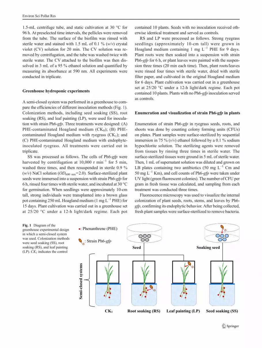
A semi-closed system was performed in a greenhouse to com-pare the efficiencies of different inoculation methods (Fig. 1).Colonization methods, including seed soaking (SS), rootsoaking (RS), and leaf painting (LP), were used for inocula-tion with strain Ph6-gfp. Three treatments were designed: (A)PHE-contaminated Hoagland medium (CK0); (B) PHE-contaminated Hoagland medium with ryegrass (CK1); and(C) PHE-contaminated Hoagland medium with endophyte-inoculated ryegrass. All treatments were carried out intriplicate.
SS was processed as follows. The cells of Ph6-gfp wereharvested by centrifugation at 10,000 r min–1 for 5 min,washed three times, and then resuspended in sterile 0.9 %(w/v) NaCl solution (OD600 nm=2.0). Surface-sterilized plantseeds were immersed into a suspension with strain Ph6-gfp for6 h, rinsed four times with sterile water, and incubated at 30 °Cfor germination. When seedlings were approximately 10-cmtall, strong individuals were transplanted into a brown glasspot containing 250 mLHoagland medium (1 mg L–1 PHE) for15 days. Plant cultivation was carried out in a greenhouse setat 25/20 °C under a 12-h light/dark regime. Each pot
contained 10 plants. Seeds with no inoculation received oth-erwise identical treatment and served as controls.
RS and LP were processed as follows. Strong ryegrassseedlings (approximately 10-cm tall) were grown inHoagland medium containing 1 mg L–1 PHE for 9 days.Plant roots were then soaked into a suspension with strainPh6-gfp for 6 h, or plant leaves were painted with the suspen-sion three times (20 min each time). Then, plant roots/leaveswere rinsed four times with sterile water, dried with sterilefilter paper, and cultivated in the original Hoagland mediumfor 6 days. Plant cultivation was carried out in a greenhouseset at 25/20 °C under a 12-h light/dark regime. Each potcontained 10 plants. Plants with no Ph6-gfp inoculation servedas controls.
Enumeration and visualization of strain Ph6-gfp in plants
Enumeration of strain Ph6-gfp in ryegrass seeds, roots, andshoots was done by counting colony forming units (CFUs)on plates. Plant samples were surface-sterilized by sequentialimmersion in 75 % (v/v) ethanol followed by a 0.1 % sodiumhypochlorite solution. The sterilizing agents were removedfrom tissues by rinsing three times in sterile water. Thesurface-sterilized tissues were ground in 5 mL of sterile water.Then, 1 mL of supernatant solution was diluted and grown onLB plates containing two antibiotics (50 mg L–1 Cm and50 mg L–1 Km), and cell counts of Ph6-gfp were taken underUV light (green fluorescent colonies). The number of CFU pergram in fresh tissue was calculated, and sampling from eachtreatment was conducted three times.
Fluorescence microscopy was used to visualize the internalcolonization of plant seeds, roots, stems, and leaves by Ph6-gfp, confirming its endophytic behavior. After being collected,fresh plant samples were surface-sterilized to remove bacteria.
CK1 Root soaking (RS) Leaf painting (LP) Seed soaking (SS)
Soaking seed
Sem
i-cl
ose
d s
yst
ems
Seed
: Phenanthrene (PHE)
: Strain Ph6-gfp
Fig. 1 Diagram of thegreenhouse experimental designin which a semi-closed systemwas used. Colonization methodswere seed soaking (SS), rootsoaking (RS), and leaf painting(LP). CK1 indicates the control
Environ Sci Pollut Res

Hand-cut lengthwise sections of surface-sterilized plant seeds,roots, stems, and leaves were fixed on glass slides using0.25 % agarose (m/v) and observed under a fluorescence mi-croscope. Ph6-gfp-free plants were observed under the sameconditions and served as controls.
HPLC analysis
Plant samples were freeze-dried, ground, homogenized, andanalyzed for PHE residues by HPLC (LC-20AT; Shimadzu,Kyoto, Japan) (Gao et al. 2008). PHE was extracted from aportion of plant samples by ultrasonication for 30 min using adichloromethane and hexane mixture (v/v=1:1) three times.The solvents were purified through a 2-g silica gel column,followed by a cleanup procedure with a 5-mL mixture(v/v=1:1) of dichloromethane and hexane. The samples werethen evaporated using a rotary evaporator, and the extractswere dissolved in methanol with a final volume of 2 mL.After filtration through 0.22-μm filter units, solutions contain-ing PHE were analyzed by HPLC.
HPLC equipped with a reverse-phase C18 column (InertsilODS-SP, 5 μm, 150×4.6 mm) at 40 °C, PHE was detected at245 nm. The flow rate was set at 1.0 mL min−1, the injectionvolume was 20 μL, and the mobile phase were methanol andwater (v/v=90:10). The recoveries of PHE averaged 103 %(n=5, RSD<3.3 %) in PHE-contaminated plant samples forthe entire procedure.
Statistical analysis
The experiments were arranged in a completely randomizeddesignwith three replications for each treatment. All data wereprocessed with Excel 2010. Each data point in figures andtables represents an average value. Standard deviations in par-allel samples are shown in figures as error bars. Data wereanalyzed by analysis of variance using SPSS version 11.0(SPSS, Inc.) with a confidence limit of 95 %.
Results
Growth response and biofilm formation
The growth response of strain Ph6-gfp in swarm plate towardthree compounds was shown in Fig. 2. Ph6-gfp grew efficient-ly with PHE, oxalic acid, or malic acid as the sole sources ofcarbon and energy. After inoculation for 48 h, the diameters ofthe growth ring of Ph6-gfp decreased in the following order:oxalic acid (3.3 cm)>malic acid (3.1 cm)>PHE (1.7 cm)>control without reactant (CK 0.8 cm). In addition, strainPh6-gfp grew in a 1.5-mL centrifuge tube showed the abilityof biofilm formation (Fig. 3). Over a period of 0–48 h, thebiofilm formation of strain Ph6-gfp increased sharply
(OD590 nm=0–1.9), however, biofilm formation of Ph6-gfpleveled off gradually with the increasing of inoculation time(48–96 h).
In planta enumeration and visualization of strain Ph6-gfp
We enumerated cells of strain Ph6-gfp in ryegrass by countingCFUs on plates (Table 1). The cell numbers of Ph6-gfp invarious tissues of inoculated ryegrass showed efficient colo-nization of inoculated plants. Furthermore, Ph6-gfp most suc-cessfully colonized roots compared to seeds and shoots. Ph6-gfp colonized plant seeds via SS for 6 h (Fig. 4a), and thepopulations observed in seeds were 4.7 log CFU g–1.Similarly, after RS for 6 h, Ph6-gfp first resided in largebiofilms on root surfaces, and then actively and internallycolonized ryegrass plant roots (Fig. 4b). As time progressed,Ph6-gfp transferred vertically to the stems (Fig. 4c) and leaves(Fig. 4d). Six days after colonization via RS, the quantificationof Ph6-gfp within plant roots and shoots was 5.8 and4.7 log CFU g–1, respectively. In addition, Ph6-gfp colonized
Fig. 2 Growth response of endophytic Pseudomonas sp. Ph6-gfp inswarm plate assays. PHE 50 mg L–1 phenanthrene, OA 2 mg L–1 oxalicacid,MA 2 mg L–1 malic acid. CK indicates the control. Errors bars±arestandard deviations (n=3)
Fig. 3 Biofilm formation ability of strain Ph6-gfp in LB medium as afunction of time. Errors bars ± are standard deviations (n=3)
Environ Sci Pollut Res

target plant tissues from the phyllosphere to internal tissuesand was also found inside stems after LP (Fig. 4e, f). After6 days via LP, the colonies of Ph6-gfp cells in the interiorshoot tissues of inoculated plants were 5.5 log CFU g–1, butthere was no fluorescence detected in plant roots. In contrast,fluorescence remained undetectable in non-inoculated plants.
Inoculated plants enhanced the removal of phenanthrene
Strain Ph6-gfp showed a capacity to lower PHE concentra-tions in planta (Table 2). The concentrations of PHE in Ph6-gfp-colonized plants were 341.0 (SS), 285.0 (RS), and 381.9(LP)mg kg–1 in roots, and 24.1 (SS), 28.1 (RS), and 32.8 (LP)mg kg–1 in shoots. Compared with Ph6-gfp-free plants, therespective concentrations of PHE in endophyte-colonizedplants were 29.2, 40.8, and 20.7 % lower in roots, and 44.3,35.1, and 24.2 % lower in shoots using SS, RS, and LP. Inaddition, compared with treatment A (without ryegrass), theconcentration of PHE in the Hoagland medium was 36.5 %
lower via treatment B (with ryegrass) and 35.4–43.8 % lowervia treatment C with Ph6-gfp-inoculated ryegrass (SS 36.3 %,RS 43.8 %, and LP 35.4 %). These results indicate that inoc-ulating ryegrass with Ph6-gfp could reduce PHE contamina-tion in plants, and RS had the least PHE concentrations inplant roots and/or in Hoagland medium.
Higher accumulation indicates more PAHs in plants and ahigher risk of contamination. The accumulation of PHE inplants was calculated (A, based dry weight) using the follow-ing equation:
A ¼ C �M
where C is the concentration of PHE in plants (mg kg–1) andM (g pot–1) is the biomass of plants in each pot.
We contaminated ryegrass for 9 days before inoculatingplants with Ph6-gfp by RS and LP, and the accumulation ofPHE in plant roots and shoots was 19.5 and 1.5 μg pot–1,respectively. After Ph6-gfp inoculation, Ph6-gfp reduced the
Table 1 Cell numbers(log CFU g–1, on a fresh weightbasis) of endophyticPseudomonas sp. Ph6-gfp ininterior tissues of inoculatedryegrass seeds, roots, and shoots
Colonization methods Seed (log CFU g–1) Root (log CFU g–1) Shoot (log CFU g–1)
Seed soaking (SS) 4.7±0.2 ND ND
Root soaking (RS) – 5.8±0.1 4.7±0.2
Leaf painting (LP) – ND 5.5±0.1
Note: ND indicates that Ph6-gfp was undetectable in the interior tissues of plants. Errors bars ± are standarddeviations (n=3)
Fig. 4 Endophytic Pseudomonas sp. Ph6-gfp cells in seeds, roots, stems,and leaves of inoculated plants were exposed to 1 mg L–1 of PHE viathree colonization methods. Ph6-gfp colonization via SS was observed inplant seeds using fluorescence microscopy (a). After inoculation with
Ph6-gfp via RS for 6 days, colonization was observed in plant roots,stems, and leaves (b–d). After inoculation with Ph6-gfp via LP,colonization was observed in plant leaves and stems (e, f)
Environ Sci Pollut Res

accumulation of PHE in roots, shoots, and whole plant bodies(the sum of roots and shoots; Fig. 5). The accumulation ofPHE was 14.8 (CK), 11.9 (SS), 9.4 (RS), and 13.7(LP)μg pot–1 in roots; 2.1 (CK), 1.6 (SS), 1.9 (RS), and 2.0(LP)μg pot–1 in shoots; and 16.9 (CK), 13.5 (SS), 11.3 (RS),and 15.7 (LP)μg pot–1 in whole plant bodies. Compared withthe Ph6-gfp-free treatment, the accumulations of PHE in Ph6-gfp-colonized plants via SS, RS, and LP were 20.1, 33.1, and7.1 % lower, respectively. These results indicate that the PHEaccumulation in plants was reduced by all Ph6-gfp coloniza-tion methods, and RS was more effective for PHE removal inwhole ryegrass bodies compared to SS and LP.
The performance of plant colonization methods
Inoculation procedures affected the population and PHE-degrading activity of strain Ph6-gfp in plants. In our study,the cell numbers of Ph6-gfp in plants inoculated via RS andLP were significantly higher than in those inoculated via SS(P<0.05). Significant differences in PHE accumulation were
observed between RS and Ph6-gfp-free plants treatment(P<0.05). The accumulation of PHE in RS-treated plantswas 16.3 and 28.0 % lower than in SS- and LP-treated plants,respectively. Moreover, the removal efficiency of PHE by RSaccounted for 54.9% of the total removal efficiency of PHE inplants using all three colonization methods, indicating that RSwould be an optimal colonization method to reduce the risk ofplant PAH contamination.
Discussion
Biofilm formation is an important trait for the colonization ofplant root surface and the degradation of organic pollutants(Ryan et al. 2008; Lugtenberg et al. 2001; Ishii et al. 2004).Since oxalic and malic acids are major root exudates of rye-grass, the rhizosphere of ryegrass might provide favorablegrowth conditions and enhance plant root colonization(De Weert et al. 2002; Bacilio-Jimenez et al. 2003).Moreover, ryegrass roots absorbed large amounts of PHE,which might also be partly utilized by strain Ph6-gfp.Simultaneously, biofilm formation by Ph6-gfp could aid inroot colonization at sites of epidermal damage, or through roothairs and at epidermal conjunctions. In addition, strain Ph6-gfp might produce degradation genes in bacterial biofilm forenhancing the degradation of PHE (Ramey et al. 2004; Sarandet al. 1998; Singh et al. 2006).
The mechanisms driving endophytic colonization remainunknown, although the gfp gene marker currently provides aunique phenotype for observing and tracking the colonizationpatterns of endophytic bacteria within host plants (Errampalliet al. 1999; Weyens et al. 2009a, b). Once Ph6-gfp entersryegrass roots, it must pass the Casparian strips in the endo-derm to systemically move to the aboveground parts of theplant (James et al. 1994; Dong et al. 2003; Zakria et al. 2007).Colonization efficiencies and cell numbers of endophytic bac-teria in plants vary (Torres et al. 2013), and the cell numbers ofendophytic Pseudomonas sp. in interior tissues of inoculatedpoplar trees were between 2.0 and 5.0 log CFU g–1 (Germaineet al. 2004). In our study, large quantities of Ph6-gfp colonizedplant interiors, and the densities decreased progressively awayfrom the roots to shoots via RS (Table 1) (James et al. 2002;Porteous Moore et al. 2006).
The method of colonization can influence the survival andtransmission of inoculated endophyte in host plant tissues(Afzal et al. 2013). Wilson (1995) indicated that endophyticbacteria colonized plant tissues during at least part of their lifecycle. The survival of Ph6-gfp in PHE-contaminated ryegrass15 days after root inoculation suggests that it is an efficientcolonizer of PHE-contaminated ryegrass. However, LP inoc-ulated with Ph6-gfp showed no gfp-labeled strain in roots afterinoculation for 6 days, which might have occurred becausemigration of Ph6-gfp from shoots to roots was hindered
Table 2 Concentrations of PHE in roots and shoots of ryegrass with orwithout endophytic Pseudomonas sp. Ph6-gfp colonization
Colonization methods Croot (mg kg–1) Cshoot (mg kg–1)
PHE-contaminated plants for 9 days 619.7±25.1 39.2±7.1
Ph6-gfp-free (CK1) 481.6±80.0 43.3±0.7
Seed soaking (SS) 341.0±27.9 24.1±2.3
Root soaking (RS) 285.0±57.7 28.1±3.0
Leaf painting (LP) 381.9±28.4 32.8±7.4
Note: Croot and Cshoot (mg kg–1 , on a dry weight basis) are the concen-trations of PHE in ryegrass roots and shoots, respectively. Errors bars ±are standard deviations (n=3)
Fig. 5 The accumulation (A, on a dry weight basis) of PHE in ryegrassroots and shoots via different colonization methods of endophyticPseudomonas sp. Ph6-gfp: SS seed soaking, RS root soaking, and LPleaf painting. CK1 indicates the control. A was estimated according toA=C×M, where C is the concentration of PHE in plants (mg kg–1) andM (g pot–1) is the biomass of plants in each pot.Errors bars ± are standarddeviations (n=3)
Environ Sci Pollut Res

through transpiration (James et al. 2002; Compant et al. 2005).Colonization was poor via SS, as Ph6-gfp cells might havebeen stressed or injured during seed planting and germination(Nelson 2004); the fact that no Ph6-gfp was detected in rootsand shoots might be due to the survival limit of Ph6-gfp(Weyens et al. 2012).
Accumulation of high levels of PAHs has been document-ed within plant tissues (Gao and Zhu 2004; Newman andReynolds 2005). Endophytic bacteria possess the genetic in-formation required for efficient degradation of PAH in planta(Taghavi et al. 2005; Baract et al. 2009). Our results indicatethat the inoculation of plant seeds, roots, and leaves with Ph6-gfp could improve the removal efficiency of PAH in planta(Fig. 5). Interestingly, no Ph6-gfp was detected in tissues ofplants inoculated via SS, except seeds. A possible reason forthe reduction of PHE accumulation in the absence of Ph6-gfpin plant tissues is natural horizontal transfer of PAH-degradingplasmids to indigenous endophytes (Taghavi et al. 2005).
Endophytic bacteria not only overcome PAH in vitro butalso can have the same effect in planta. The possible mecha-nisms involved in endophytic colonization-improved biodeg-radation of PAH in plants include the following: (1) Bacterialbiofilm formation enhances plant root colonization and root-surface PAH degradation, thereby reduces the uptake of PAHby plant roots (Strobel et al. 2004). (2) Endophytic bacteriadirectly utilize PAH as a carbon source for their growth inplant tissues (Germaine et al. 2009; Sun et al. 2014b). (3)Endophytic inoculants transfer their PAH-degradative genesto other endophytes in plants, thereby improving the overallmetabolism potential of endophytic communities (Taghaviet al. 2005; Wang et al. 2007). (4) Endophytic bacteria influ-ence the activities of plant enzymes (e.g., catalase, cyto-chrome P450 alkane monooxygenase, and superoxide dismut-ase), participating in PAH transformation (Harish et al. 2009;Kvesitadze et al. 2009; Afzal et al. 2013). (5) Endophyticbacteria enhance PAH degradation gene expression in plants,such as naphthalene dioxygenase (ndoB) and alkanemonooxygenase (alkB) (Siciliano et al. 2001).
As a group of persistent organic pollutants with strongmutagenic/carcinogenic properties, PAHs are considered a se-rious health risk even at low concentrations; thus, a smalldecrease in their concentration via Ph6-gfp inoculation is po-tentially of great importance. In our study, yellow-brown soilwas collected from the soil surface (0–20 cm) in the suburbs ofNanjing, China, and soils were artificially contaminated withPHE (1 mg kg−1). Strain Ph6-gfp was inoculated to PHE-contaminated soils 10 days after seedling emergence by rootirrigating. After inoculation for 15 days, the accumulationamount of PHE in endophyte-colonized ryegrass was1.83 μg pot−1, which was 12.7 % lower than that in Ph6-gfp-free plants. The result indicates the removal efficiency ofPHE in pot experiment was significantly lower as compared tothose resulting from hydroponic experiment (RS) (P<0.05).
Hence, how to improve the implication of endophytic bacteriain PAH-contaminated soils is an urgent problem to be solvedcurrently.
Conclusions
Endophytes are of special interest because of their high levelof plant colonization and contaminant-degrading activity. Assuch, they may provide a promising green technology forcleaning up organic pollutants in plants. In this study, weinvestigated plant colonization in a greenhouse by the PHE-degrading endophytic Pseudomonas sp. Ph6-gfp using threecolonization methods: SS, RS, and LP. Cell counting and di-rect observations using fluorescence microscopy showed Ph6-gfp colonization of ryegrass tissues. Ph6-gfp can reduce therisk of plant PHE contamination by decreasing the concentra-tion and accumulation of PHE in plants. Moreover, inocula-tion methods could affect colonization and PHE removal effi-ciency of Ph6-gfp in planta. Among the three colonizationmethods, RS was an optimal colonization method forcircumventing the risk of plant organic contamination. Ourresults are hopeful to provide an optimized method to endo-phytic bacteria reduce plant PAH residues, which may be ap-plied to agricultural production in PAH-contaminated soil.
Acknowledgments This work was financially supported by the Na-tional Science Foundation of China (41201501, 41171380, 51278252,21477056), the Science Foundation of Jiangsu Province (BK2012370,BK20130030), and the Doctoral Fund of the Ministry of Education ofChina (20120097120012).
References
Afzal M, Yousaf S, Reichenauer TG, Kuffner M, Sessitsch A (2011) Soiltype affects plant colonization, activity and catabolic gene expres-sion of inoculated bacterial strains during phytoremediation of die-sel. J Hazard Mater 186:1568–1575
Afzal M, Yousaf S, Reichenauer TG, Sessitsch A (2012) The inoculationmethod affects colonization and performance of bacterial inoculantstrains in the phytoremediation of soil contaminated with diesel oil.Int J Phytoremediat 14:35–47
Afzal M, Khan S, Iqbal S, Mirza MS, Khan QM (2013) Inoculationmethod affects colonization and activity of Burkholderiaphytofirmans PsJN during phytoremediation of diesel-contaminated soil. Int J Biodeterior Biodegrad 85:331–336
Afzal M, Khan QM, Sessitsch A (2014) Endophytic bacteria: prospectsand applications for the phytoremediation of organic pollutants.Chemosphere 117:232–242
Andria V, Reichenauer TG, Sessitsch A (2009) Expression of alkanemonooxygenase (alkB) genes by plant-associated bacteria in therhizosphere and endosphere of Italian ryegrass (Lolium multiflorumL.) grown in diesel contaminated soil. Environ Pollut 157:3347–3350
Bacilio-Jimenez M, Aguilar-Flores S, Ventura-zapata E, Perez-CamposE, Bouquelet S, Zenteno E (2003) Chemical characterization of root
Environ Sci Pollut Res

exudates from rice (Oryza sativa) and their effects on the chemotac-tic response of endophytic bacteria. Plant Soil 249:271–277
Baract T, Weyens N, Oeyen L, Taghavi S, van der Lelie D, Dubin D,Spliet M, Vangronsveld J (2009) Field note: hydraulic containmentof a BTEX plume using poplar trees. Int J Phytoremediat 11:416–424
Bhushan B, Samanta SK, Chauhan A, Chakraborti AK, Jain RK (2000)Chemotaxis and degradation of 3-methyl-4-nitrophenol byRalstonia sp. SJ98. Biochem Biophys Res Commun 275:129–133
Collins C, Fryer M, Grosso A (2006) Plant uptake of non-ionic organicchemicals. Environ Sci Technol 40:45–52
Compant S, Reiter B, SessitschA, Nowak J, Clement C, Barka EA (2005)Endophytic colonization of Vitis vinifera L. by plant grown-promoting bacterium Burkholderia sp. strain PsJN. Appl EnvironMicrobiol 71:1685–1693
De Weert S, Vermeiren H, Mulders IH, Hendrickx N, Bloemberg GV,Vanderleyden J, Mot RD, Lugtenberg BJJ (2002) Flagella-drivenchemotaxis towards exudate components is an important trait fortomato root colonization by Pseudomonas fluorescens. Mol PlantMicrobe Interact 15:1173–1180
Dong YM, Iniguez AL, Ahmer BMM, Triplett EW (2003) Kinetics andstrain specificity of rhizosphere and endophytic colonization by en-teric bacteria on seedlings of Medicago sativa and Medicagotruncatula. Appl Environ Microbiol 69:1783–1790
Errampalli D, Leung K, Cassidy MB, Kostrzynska M, Blears M, Lee H,Trevors JT (1999) Applications of the green fluorescent protein as amolecular marker in environmental microorganisms. J MicrobiolMethods 35:187–199
Ferreira A, Quecine MC, Lacava PT, Oda S, Azevedo JL, Araujo WL(2008) Diversity of endophytic bacteria from Eucalyptus speciesseeds and colonization of seedlings by Pantoea agglomerans.FEMS Microbiol Lett 287:8–14
Gao YZ, Collins CD (2009) Uptake pathways of polycyclic aromatichydrocarbons in white clover. Environ Sci Technol 43:6190–6195
Gao YZ, Zhu LZ (2004) Plant uptake, accumulation and translocation ofphenanthrene and pyrene in soils. Chemosphere 55:1169–1178
Gao YZ, Xiong W, Ling WT, Wang H, Ren LL, Zhang ZY (2008)Partitioning of polycyclic aromatic hydrocarbons between plantroots and water. Plant Soil 311:201–209
Germaine K, Keogh E, Garcia-Cabellos G, Borremans B, van der LelieD, Barac T, Oeyen L, Vangronsveld J, Moore FP, Moore ERB,Campbell CD, Ryan D, Dowling DN (2004) Colonisation of poplartrees by gfp expressing bacterial endophytes. FEMSMicrobiol Ecol48:109–118
Germaine KJ, Liu XM, Cabello GG, Hogan JP, Ryan D, Dowling DN(2006) Bacterial endophyte-enhanced phytoremediation of the or-ganochlorine herbicide 2,4-dichlorophenoxyacetic acid. FEMSMicrobiol Ecol 57:302–310
Germaine KJ, Keogh E, Ryan D, Dowling DN (2009) Bacterialendophyte-mediated naphthalene phytoprotection andphytoremediation. FEMS Microbiol Lett 296:226–234
Glick BR (2014) Bacteria with ACC deaminase can promote plantgrowth and help to feed the world. Microbiol Res 169:30–39
Harish S, Kavino M, Kumar N, Balasubramanian P, Samiyappan R(2009) Induction of defense-related proteins by mixtures of plantgrowth promoting endophytic bacteria against banana bunchy topvirus. Biol Control 51:16–25
Iijima S, Washio K, Okahara R, Morikawa M (2009) Biofilm formationand proteolytic activities of Pseudoalteromonas bacteria that wereisolated from fish farm sediments. Microb Biotechnol 2:361–369
Ishii S, Koki J, Unno H, Hori K (2004) Two morphological types of cellappendages on a strongly adhesive bacterium Acinetobacter sp.strain Tol 5. Appl Environ Microbiol 70:5026–5029
James EK, Reis VM, Baldani JI, Döbereiner J (1994) Infection of sugarcane by the nitrogen-fixing bacterium Acetobacter diazotrophicus. JExp Bot 45:757–766
James EK, Gyaneshwar P, Mathan N, Barraquio WL, Reddy PM,Iannetta PPM, Olivares FL, Ladha JK (2002) Infection and coloni-zation of rice seedlings by the plant growth-promoting bacteriumHerbaspirillum seropedicae Z67. Mol Plant-Microbe Interact 15:894–906
Khan S, Afzal M, Iqbal S, Khan QM (2013) Plant-bacteria partnershipsfor the remediation of hydrocarbon contaminated soils.Chemosphere 90:1317–1332
Kvesitadze E, Sadunishvili T, Kvesitadze G (2009) Mechanisms of or-ganic contaminants uptake and degradation in plants. World AcadSci Eng Technol 55:458–468
Liu J, Liu S, Sun K, Sheng YH, Gu YJ, Gao YZ (2014) Colonization onroot surface by a phenanthrene-degrading endophytic bacterium andits application for reducing plant phenanthrene contamination. PLoSOne 9, e108249
Lugtenberg BJ, Dekkers L, Bloemberg GV (2001) Molecular determi-nants of rhizosphere colonization by Pseudomonas. Annu RevPhytopathol 39:461–490
Mckone TE, Maddalena RL (2007) Plant uptake of organic pollutantsfrom soil: bioconcentration estimates based on models and experi-ments. Environ Toxicol Chem 26:2494–2504
Nelson EB (2004) Microbial dynamics and interactions in thespermosphere. Annu Rev Phytopathol 42:271–309
Newman LA, Reynolds CM (2005) Bacteria and phytoremediation: newuses for endophytic bacteria in plants. Trends Biotechnol 23:6–8
Porteous Moore F, Barac T, Borremans B, Oeyen L, Vangronsveld J, vander Lelie D, Campbell CD,Moore ERB (2006) Endophytic bacterialdiversity in poplar trees growing on a BTEX-contaminated site: thecharacterisation of isolates with potential to enhancephytoremediation. Syst Appl Microbiol 29:539–556
Ramey BE, Koutsoudis M, von Bodman SB, Fuqua C (2004) Biofilmformation in plant-microbe associations. Curr Opin Microbiol 7:602–609
Ryan RP, Germaine K, Franks A, Ryan DJ, Dowling DN (2008) Bacterialendophytes: recent developments and applications. FEMSMicrobiol Lett 278:1–9
Sarand I, Timonen S, Nurmiaho-Lassila EL, Koivula T, Haahtela K(1998) Microbial biofilms and catabolic plasmid harbouring degra-dative fluorescent pseudomonads in Scots pine mycorrhizospheresdeveloped on petroleum contaminated soil. FEMS Microbiol Ecol27:115–126
Siciliano SD, Fortin N, Mihoc A, Wisse G, Labelle S, Beaumier D,Ouellette D, Roy R, Whyle LG, Banks MK, Schwab P, Lee K,Greer CW (2001) Selection of specific endophytic bacterial geno-types by plants in response to soil contamination. Appl EnvironMicrobiol 67:2469–2475
Singh R, Paul D, Jain RK (2006) Biofilms: implication in bioremediation.Trends Microbiol 14:590–600
Strobel G, Daisy B, Castillo U, Harper J (2004) Natural products fromendophytic microorganisms. J Nat Prod 67:257–268
Sun K, Liu J, Gao YZ, Jin L, Gu YJ, Wang WQ (2014a) Isolation, plantcolonization potential, and phenanthrene degradation performanceof the endophytic bacterium Pseudomonas sp. Ph6-gfp. Sci Rep 4:5462
Sun K, Liu J, Jin L, Gao YZ (2014b) Utilizing pyrene-degrading endo-phytic bacteria to reduce the risk of plant pyrene contamination.Plant Soil 374:251–262
Taghavi S, Barac T, Greenberg B, Borremans B, Vangronsveld J, van derLelie D (2005) Horizontal gene transfer to endogenous endophyticbacteria from poplar improves phytoremediation of toluene. ApplEnviron Microbiol 71:8500–8505
Torres AR, Araújo WL, Coursino L, Rossetto PB, Mondin M, HungriaM, Azevedo JL (2013) Colonization of Madagascar periwinkle(Catharanthus roseus), by endophytes encoding gfp marker. ArchMicrobiol 195:483–489
Environ Sci Pollut Res

WangYJ, XiaoM,GengXL, Liu JY, Chen J (2007) Horizontal transfer ofgenetic determinants for degradation of phenol between the bacterialiving in plant and its rhizosphere. Appl Microbiol Biotechnol 77:733–739
Weyens N, van der Lelie D, Artois T, Smeets K, Taghavi S, Newman L,Carleer R, Vangronsveld J (2009a) Bioaugmentation withengineered endophytic bacteria improves contaminant fate inphytoremediation. Environ Sci Technol 43:9413–9418
Weyens N, van der Lelie D, Taghavi S, Vangronsveld J (2009b)Phytoremediation: plant endophyte partnerships take the challenge.Curr Opin Biotechnol 20:248–254
Weyens N, Boulet J, Adriaensen D, Timmermans JP, Prinsen E, OevelenSV, D’Hean J, Smeets K, van der Lelie D, Taghavi S, VangronsveldJ (2012) Contrasting colonization and plant growth promoting ca-pacity between wild type and a gfp-derative of the endophyte
Pseudomonas putida W619 in hybrid poplar. Plant Soil 356:217–230
Wilson D (1995) Endophyte-the evolution of a term, and clarification ofits use and definition. Oikos 73:274–276
Yousaf S, Afzal M, Reichenauer TG, Brady CL, Sessitsch A (2011)Hydrocarbon degradation, plant colonization and gene expressionof alkane degradation genes by endophytic Enterobacter ludwigiistrains. Environ Pollut 159:2675–2683
Zakria M, Ohsako A, Saeki YC, Yamamoto A, Akao S (2007)Colonization and nitrogen-fixing ability of Herbaspirillum sp strainB501 gfp1 and assessment of its growth-promoting ability in culti-vated rice. Microbes Environ 22:197–206
Zhu X, Ni X, Liu J, Gao YZ (2014) Application of endophytic bacteria toreduce persistent organic pollutants contamination in plants.CLEAN-Soil Air Water 42:306–310
Environ Sci Pollut Res