Endoplasmic reticulum targeted GFP reveals ER organization in tobacco NT-1 cells during cell...
45
Running title: ER organization during tobacco cell division Endoplasmic reticulum targeted GFP reveals ER organization in tobacco NT-1 cells during cell division Stephanie L. Gupton a,b 1 # , David A. Collings a,c * # , and Nina Strömgren Allen a,b . a Department of Botany, North Carolina State University, Raleigh, NC 27695 USA. b Marine Biological Laboratories, 7 MBL Street, Woods Hole, MA 02543 USA. c Plant Cell Biology Group, Research School of Biological Sciences, The Australian National University, GPO Box 475, Canberra, ACT 2601, Australia. * Corresponding author (Fax +61 (2) 6125 4331; E-mail [email protected]) # These authors contributed equally to this paper 1 Current Address: Department of Cell Biology, The Scripps Research Institute, La Jolla, CA 92037 USA E-mail addresses for other authors: [email protected] [email protected]
-
Upload
independent -
Category
Documents
-
view
3 -
download
0
Transcript of Endoplasmic reticulum targeted GFP reveals ER organization in tobacco NT-1 cells during cell...

Running title: ER organization during tobacco cell division
Endoplasmic reticulum targeted GFP reveals ER organization in tobacco NT-1 cells
during cell division
Stephanie L. Guptona,b 1 #, David A. Collingsa,c * #, and Nina Strömgren Allena,b.
a Department of Botany, North Carolina State University, Raleigh, NC 27695 USA.
b Marine Biological Laboratories, 7 MBL Street, Woods Hole, MA 02543 USA.
c Plant Cell Biology Group, Research School of Biological Sciences,
The Australian National University, GPO Box 475, Canberra, ACT 2601, Australia.
* Corresponding author (Fax +61 (2) 6125 4331; E-mail [email protected])
# These authors contributed equally to this paper
1 Current Address: Department of Cell Biology, The Scripps Research Institute, La Jolla,
CA 92037 USA
E-mail addresses for other authors:

2
Abstract
The endoplasmic reticulum (ER) of plant cells undergoes a drastic reorganization
during cell division. In tobacco NT-1 cells that stably express a GFP construct targeted
to the ER, we have mapped the reorganization of ER that occurs during mitosis and
cytokinesis with confocal laser scanning microscopy. During division, the ER and
nuclear envelope do not vesiculate. Instead, tubules of ER accumulate around the
chromosomes after the nuclear envelope breaks down, with these tubules aligning parallel
to the microtubules of the mitotic spindle. In cytokinesis, the phragmoplast is
particularly rich in ER, and the transnuclear channels and invaginations present in many
interphase cells appear to develop from ER tubules trapped in the developing
phragmoplast. Drug studies, using oryzalin and latrunculin to disrupt the microtubules
and actin microfilaments respectively, demonstrate that during division, the arrangement
of ER is controlled by microtubules and not by actin, which is the reverse of the situation
in interphase cells.

3
Keywords; actin microfilaments; cell division; endoplasmic reticulum; green fluorescent
protein; microtubules; nuclear envelope; nuclear invaginations
Abbreviations: DIC, differential interference contrast; ER, endoplasmic reticulum; GFP,
green fluorescent protein
Supplemental files:
movie1.avi accompanies Figure 1 (1.66 MB)
movie2.avi accompanies Figure 2 (1.78 MB)
movie3.avi accompanies Figure 3 (0.85 MB)
movie4.avi accompanies Figure 6 (1.60 MB)

4
1. Introduction
In interphase cells of plants, the endoplasmic reticulum (ER) forms into numerous
distinct arrays [16, 39]. These arrays include a comparatively stable, reticulate, cortical
network and highly dynamic subcortical ER tubules. These in turn are interconnected
and continuous with the nuclear envelope. The presence of these different arrays in
living cells was first demonstrated with the carbocyanine dye DIOC6(3) and DIC video
microscopy [1, 16, 21, 24, 32, 33], and more recently with green fluorescent protein
(GFP) targeted to the lumen of the ER [5, 6, 13, 19, 29, 34, 38]. The movement of the
motile ER tubules in interphase cells is a microfilament-based process, as actin bundles
lie parallel to the ER [1, 5, 25], the motor protein, myosin colocalizes with the ER [25],
disruption of the actin cytoskeleton with cytochalasin results in the cessation of ER
movement [21]. Actin disruption also prevents the reorganization of ER that precedes
cell division in cultured cells [38]. However, the factors responsible for organization of
the cortical array, and whether they are cytoskeletal or not, have been more difficult to
characterize.
In contrast to the numerous characterizations of ER organization in interphase plant
cells, few detailed studies have been published of ER dynamics during plant cell division.
This is important, however, for several reasons. First, the ER is continuous with the
nuclear envelope during interphase so that proteins localized to the lumen of the ER are
also found within the nuclear envelope, and knowing the fate of the ER during mitosis
will suggest the localization of luminal proteins [6, 18]. Second, electron micrographs of

5
dividing plant cells show a close association between tubules of ER that run parallel to
the microtubules in both the mitotic spindle [14, 15, 31] and the phragmoplast, the plant
specific structure responsible for the formation of the new cell wall in cytokinesis [2].
These results have been confirmed by observations of the accumulation of ER-targeted
GFP within the spindle and phragmoplast [29]. It is unknown when in the cell cycle these
ER tubules arrive at these regions. Third, phragmoplast formation in plants requires ER
and Golgi apparatus–derived vesicles to run along microtubules to the equator of the cell
[15, 30] where they consolidate into a sheet that expands outwards as more vesicles fuse,
eventually meeting with and fusing with the parent cell wall to complete cell division
[35]. Understanding ER dynamics during cell division and their dependence on the
cytoskeleton may suggest a mechanism of phragmoplast formation. And finally, some
plant nuclei contain deep invaginations and trans-nuclear strands. As these structures are
bounded by the nuclear envelope, they are continuous with the ER, but their formation,
hypothesized to occur during cell division, remains uncertain [6].
In this study therefore, we have taken advantage of tobacco NT1 and BY-2 cell
lines that stably expresses ER-targeted GFP to look at the behavior of ER during cell
division, and how the ER interacts with the cytoskeleton. Our data confirm extensive
accumulation of ER within the mitotic spindle and phragmoplast, and show that this
reorganization of the ER during mitosis is maintained by microtubules, as opposed to the
actin cytoskeleton.

6
2. Results
2.1. Endoplasmic reticulum organization in interphase cells.
ER dynamics during interphase and cell division were characterized in tobacco NT-
1 cells by visualizing GFP-ER with laser scanning and spinning disk confocal
microscopy. In interphase cells, the ER formed three different arrangements, confirming
earlier studies that used DIOC6(3) to stain membranes [1, 33, 24; our data, not shown],
and ER-targeted GFP constructs in cultured tobacco [19, 29, 38] and cell types [5, 6, 13,
34]. First, cortical ER formed polygonal arrays similar to those found in animal cells and
other plant cells (Fig. 1A, arrow). The cortical ER exhibited slow movement and
intersections of cortical ER were generally three-way. Second, ER tubules extended from
the cortical ER into the cytoplasm traversing between vacuoles (Fig. 1B, arrows). This
ER was dynamic, and showed rapid cytoplasmic streaming relative to cortical ER, with
streaming visible in successive images of a time course (Fig. 1D, arrow). Third, the ER
surrounded and was continuous with the nuclear envelope (Fig. 1C,E, arrow). In many
interphase cells, penetrations of the nuclear envelope extended into the nucleus forming
invaginations and channels that crossed the entirety of the nucleus (Fig. 1E,F;
supplemental file movie1.avi) [6]. These channels often showed extensive branching
patterns, and could contain internal ER structures suggesting that they contain cytoplasm
and ER rather than being a simple infolding of the inner nuclear membrane.
7
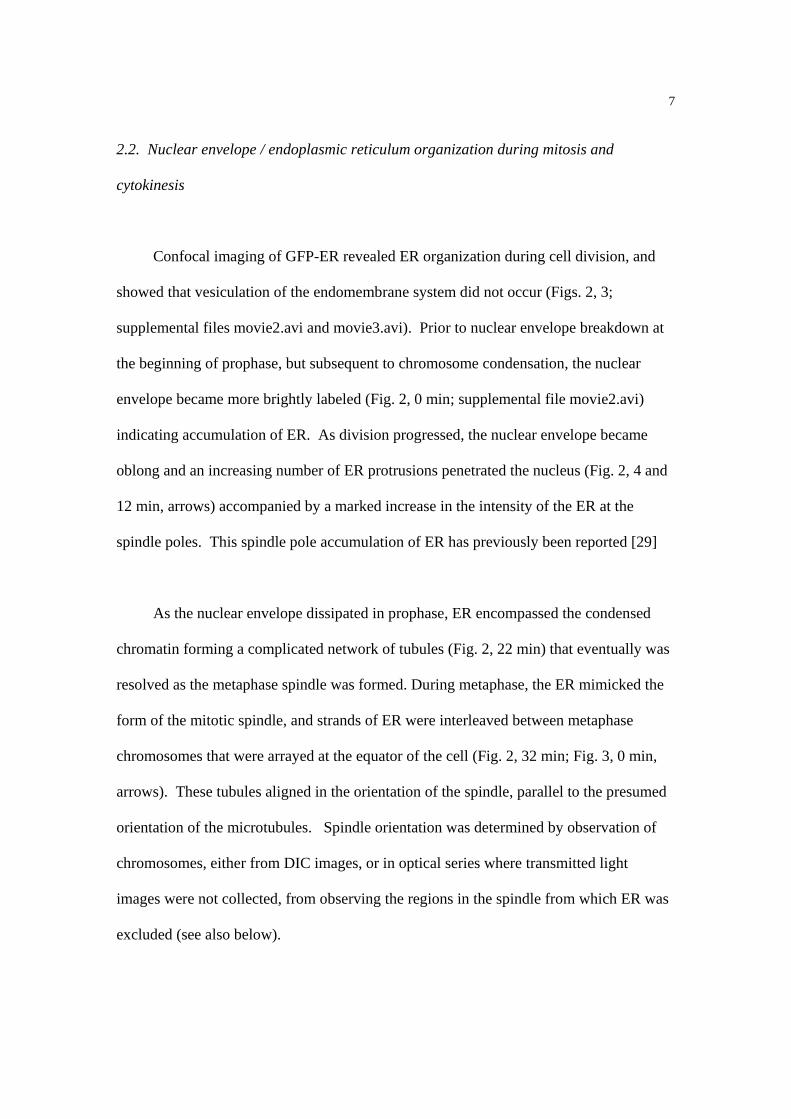
2.2. Nuclear envelope / endoplasmic reticulum organization during mitosis and
cytokinesis
Confocal imaging of GFP-ER revealed ER organization during cell division, and
showed that vesiculation of the endomembrane system did not occur (Figs. 2, 3;
supplemental files movie2.avi and movie3.avi). Prior to nuclear envelope breakdown at
the beginning of prophase, but subsequent to chromosome condensation, the nuclear
envelope became more brightly labeled (Fig. 2, 0 min; supplemental file movie2.avi)
indicating accumulation of ER. As division progressed, the nuclear envelope became
oblong and an increasing number of ER protrusions penetrated the nucleus (Fig. 2, 4 and
12 min, arrows) accompanied by a marked increase in the intensity of the ER at the
spindle poles. This spindle pole accumulation of ER has previously been reported [29]
As the nuclear envelope dissipated in prophase, ER encompassed the condensed
chromatin forming a complicated network of tubules (Fig. 2, 22 min) that eventually was
resolved as the metaphase spindle was formed. During metaphase, the ER mimicked the
form of the mitotic spindle, and strands of ER were interleaved between metaphase
chromosomes that were arrayed at the equator of the cell (Fig. 2, 32 min; Fig. 3, 0 min,
arrows). These tubules aligned in the orientation of the spindle, parallel to the presumed
orientation of the microtubules. Spindle orientation was determined by observation of
chromosomes, either from DIC images, or in optical series where transmitted light
images were not collected, from observing the regions in the spindle from which ER was
excluded (see also below).

8
As the chromosomes migrated toward the poles of the cell during anaphase, they
continued to be encompassed by tubes of ER, and ER filled the space between the
separating sets of chromosomes (Fig. 2, 40 min, arrows; Fig. 3, 3 and 6 min, arrow;
supplemental file movie3.avi). As the duplicate pairs of chromosomes were completely
segregated, the cell plate and phragmoplast began to form (Fig. 2, 50 min arrow;
Fig. 3, 18 min, arrow). Throughout phragmoplast formation, there was a continual
increase in the intensity of the GFP-ER to the point of saturation beyond the dynamic
range of the detector, indicating rapidly increasing amounts of ER present in the
phragmoplast (Fig. 3, 24 to 51 min). Perpendicular strands of ER connected the
phragmoplast to the rudimentary daughter nuclei. As the phragmoplast expanded to
connect with the cell wall, the nuclear envelopes of the two daughter cells re-formed, and
the nucleoli reappeared. Once the phragmoplast and nuclear envelopes had formed, the
daughter nuclei shifted away from the new cell wall, and were positioned toward the
center of each new cell (data not shown).
2.3 ER tubules in the spindle and phragmoplast lie parallel to microtubules
Our observations of ER tubules in living cells suggested that they lay parallel to
spindle and phragmoplast microtubules. We have confirmed this by fixing and
immunolabeling the ER-targeted GFP-expressing cells for tubulin (Fig. 4). ER
organization in the fixed cells appeared similar to that in living cells throughout the cell

9
cycle, with extensive subcortical arrays, the nuclear envelope, and nuclear channels and
invaginations being visible, suggesting that little ER reorganization occurred during the
fixing and immunolabeling procedures. However, we did not observe either the delicate
cortical ER array nor the accumulations of ER adjacent to spindle poles. Interphase and
prophase cells had distinct cytoplasmic ER and nuclear envelopes with relatively few
perinuclear microtubules (Fig. 4A,B). During prophase and prometaphase, ER
protrusions formed from the nuclear envelope into the nucleus that contained
microtubules (Fig. 4B,C, arrows). In metaphase cells, alignment of ER tubules with the
spindle microtubules was apparent (Fig. 4D, arrowheads), with this alignment maintained
during anaphase (Fig. 4E, arrowheads), and with the phragmoplast microtubules during
telophase (Fig. 4C,F, arrowheads).
2.4. Do interphase nuclear channels form because of lagging chromosomes during cell
division?
Nuclear invaginations and channels were observed in both living (Fig. 1D,E) and
fixed interphase cells (Fig. 4A). We previously speculated that nuclear channels might
form during cell division [6], and in Figure 3, between 39 and 51 min, nuclear channels
in one daughter cell were visible (arrowheads) matching the locations where separating
chromosomes lagged in division. As the nuclear envelope reformed around these strands
of ER, these internal strands of ER remained within the nucleus, creating the nuclear
channels.

10
2.5. Organization of cytoplasmic endoplasmic reticulum during cell division
As nuclear envelope breakdown progressed, the transvacuolar strands of ER in the
cell decreased in number as the ER aggregated around the nuclear region. Although the
cytoplasmic ER changed organization, the cortical ER remained in its patterned,
fenestrated arrangement beneath the plasma membrane (Fig. 5A, arrow; supplemental file
movie4.avi), as seen in interphase. Furthermore, approximately 20 percent of metaphase
(Fig. 5A, asterisks) and anaphase cells (Fig. 5B, asterisks) had dense assemblages of ER,
visible in both GFP and DIC images, that lay beyond one or both spindle poles, whose
location was inferred from DIC images.
2.6 Immunofluorescence observations of cytoskeletal disruption and ER organization
As we were interested in observing the effects of cytoskeletal disruption on ER
organization in living cells undergoing division, we used immunofluorescence to assay
the effects of drugs that target the cytoskeleton on microtubules and actin microfilaments.
Compared to control cells that had extensive arrays of both actin and microtubules
(Fig. 6A), 5 min treatments with 5 µM latrunculin (Fig. 6B) and 5 µM oryzalin (Fig.6C)
caused extensive disruption to the actin and microtubules respectively. These
observations are consistent with cytoplasmic streaming slowing immediately, and ceasing

11
within 12 min following latrunculin treatments (data not shown). Our observations are
also consistent with published reports that 5 µM oryzalin disrupts microtubules in
cultured tobacco cells [4], and that 1.25 µM latrunculin B causes actin reorganization in
tobacco protoplasts [43], with 20 µM latrunculin B causing major actin disruption [12].
2.7. Effects of microtubule destabilization on endoplasmic reticulum arrangement
Oryzalin (5 µM) caused no obvious changes in ER organization in interphase
tobacco NT-1 cells, but significantly changed ER organization in dividing cells (Fig. 7).
In prophase cells where the nuclear envelope had broken down, microtubule disruption
prevented cells from progressing out of prophase. ER organization also remained in its
prophase configuration, characterized by ER encompassing the condensed chromosomes
(Fig. 7A). Cells perfused with oryzalin during metaphase also arrested at metaphase, and
the tubular strands of the ER network rapidly retracted toward the centrally placed
chromosomes (Fig. 7B). Cells perfused with oryzalin after the transition to anaphase
exhibited chromosome segregation, but chromosomes did not migrate toward the cell
poles. In such cells, the ER lost its tubular appearance (Fig. 7C, 1 min), and the highly
prominent ER phragmoplast never formed even after nuclear envelopes reformed around
the segregated genetic material, and the nucleoli reappeared in both daughter cells
(Fig. 7C, 5 min; compare with Fig. 2, 50 to 60 min and Fig. 3, 18 to 51min). In cells
perfused with oryzalin during telophase, once phragmoplast and nuclear envelope
formation had already begun, the fluorescence intensity from ER-targeted GFP did not

12
continue to increase, although nucleoli did reappear within the daughter nuclei (data not
shown). These results demonstrate that disruption of microtubules in dividing cells
inhibits the normal reorganization of the ER and thus, that ER reorganization must
depend upon the microtubule cytoskeleton.
2.8. Effects of actin disruption on endoplasmic reticulum arrangement
In mitotic cells perfused with latrunculin B (5 µM) before nuclear envelope
breakdown, the processes of cell division and the duration of mitosis, were unaffected
and cells continued through the process of cell division, although the cell plate did not
form properly between the new daughter nuclei (Fig. 8). Generally, latrunculin did not
cause changes to ER organization, although it did cause some cytoplasmic ER to assume
a bubble-like vesiculated and disconnected conformation (Fig. 8, 4 min, arrows).
Cortical ER remained in its normal configuration, and ER associated with dividing cells
was still interspersed in the mitotic spindle (Fig. 8, 0 min, arrows) and prominent in the
phragmoplast (Fig. 8,14 to 26 min, arrowheads). These results demonstrate that changing
the organization of actin in dividing cells has little impact on the organization of the ER.

13
3. Discussion
Previous reports of ER during plant cell division have described static images from
electron micrographs in which the entirety of ER structure was not visible [14, 15, 17, 20,
31]. Using living cells with GFP targeted to the ER allowed observations of the
dynamics of ER. Our data show a reorganization of the ER during cell division, which is
directed by microtubules and not actin microfilaments, and which is tubular and not
vesicular in nature. These observations are consistent with similar studies conducted in
animal cells which demonstrated a close involvement of the ER in cell division, and
where nuclear envelope breakdown takes place at the end of prophase/beginning of
prometaphase and is preceded by protrusions of the nuclear envelope into the nucleus
[40].
3.1. ER organization changes during cell division
Our observations of GFP-ER in living tobacco NT-1 cells show that the
organization of ER changes during cell division. In the initial stages of mitosis, we found
that tubules of ER protrude into the nuclear envelope as the chromosomes begin to
condense. We confirmed this effect in immunolabeled material, and showed that these
ER protrusions contain microtubules. Further, during spindle formation, there is an
intense increase in GFP-ER fluorescence at the spindle poles. The nuclear envelope was
not observed to "break down" or vesiculate. Rather, it appears that it may be absorbed

14
into the ER, thereby causing the increase in the intensity of GFP-ER. Were this to be
true, the term "nuclear envelope breakdown" would be a misnomer, and would better be
termed "nuclear envelope absorption." During metaphase and anaphase, a combination
of live cell imaging and immunofluorescence showed that ER tubules lie parallel to the
mitotic spindle enmeshing the chromosomes. During cytokinesis, ER accumulates in the
phragmoplast where it is involved in the delivery of components to the developing cell
plate.
These observations extend electron microscopy analyses that observed tubular ER
during plant cell division [14, 15, 31]. Hepler (1980) described ER aggregation at the
polar regions of the nucleus following the prometaphase breakdown of the nuclear
envelope, and that during metaphase, tubes of ER surround the mitotic apparatus,
protrude into its interior and encompass the chromosomes. He also observed
aggregations of ER adjacent to the spindle poles of barley cells [14]. Interestingly, Golgi
stacks also accumulate around the spindle poles during mitosis [29]. Pickett-Heaps and
Northcote (1966) also observed ER tubules interdigitating between chromosomes aligned
on the metaphase plate in wheat meristems [31]. We observed similar transitions in
living tobacco cells with GFP-ER, including ER aggregates adjacent to the spindle poles
of some dividing cells. These studies do, however, stand in contrast to some earlier
electron microscopy studies that describe extensive vesiculation of both the nuclear
envelope and ER during plant cell division, although they did document similar
reorganizations of the ER [20] that may have been caused by the harsh fixations required
for electron microscopy.

15
Our observations of ER during plant cell division match similar observations in
various animal cells. Using ER-targeted GFP in insect, echinoderm, and mammalian
cells, these studies have shown a general dependence of the organization of the ER in
higher eukaryotes on the microtubule cytoskeleton, and that that ER remains continuous
during mitosis, as opposed to vesiculating, with the constituents of the nuclear envelope
being absorbed by the ER during nuclear envelope breakdown, and then re-emerging
from the ER at the end of mitosis [9, 11, 40, 45, 47, 48]. During interphase, the extension
of ER tubules occurs as microtubules extend, although depolymerization of microtubules
does not disrupt the ER network unless microtubule depolymerization is prolonged [41,
44]. In cell division in sea urchin embryos, similar finger-like indentations of the nuclear
envelope occur prior to nuclear envelope break down. Furthermore, microtubules also
seem to direct arrangement of ER during cell division. Treatment of mitotic sea urchin
embryos with nocodazole, a microtubule depolymerizer, did not prohibit the
accumulation of ER at the spindle poles, although the arrangement was irregular and not
bipolar [40]. In some mammalian tissue culture cells depolymerization of microtubules
induced a distinct loss of organization of the membranous components of the spindles,
suggesting that microtubules organize the membrane distribution in some mammalian
tissue culture cells. Immunofluorescence studies showed that spindle membranes were
associated with microtubules throughout mitosis. These results support the hypothesis
that, at least in some cells, ER distribution is maintained by the microtubule cytoskeleton
[45]. In Xenopus egg extracts, however, the motility of organelles and vesicles along F-
actin bundles in vitro increases in metaphase bundles compared to interphase, suggesting

16
that F-actin is active in the dynamics and localization of the ER during cell division [46].
Together, these results suggest that microtubules can organize the distribution of
membranes in mitotic cells, and that this organization may vary in different cell types
depending on the number of microtubules within the spindle, or other factors such as the
role of the actin cytoskeleton and actin-based motors.
3.2. The cytoskeleton and endoplasmic reticulum during cell division
As cells progress from interphase to cell division, the cytoskeletal factors that
organize the ER switch from being actin-based to microtubule-based. In interphase cells,
actin disruption with latrunculin caused subcortical ER to cease streaming and "bubbles"
to form in some of the cytoplasmic ER patterning. Microtubule disruption with oryzalin,
however, has little effect on the organization of either the streaming subcortical ER or the
polygonal patterning of the cortical ER adjacent to the plasma membrane. These results
match previous observations that the dynamic streaming of the subcortical ER occurs
through interactions with actin bundles and not microtubules [1, 5, 24, 32], and that ER
redistributions prior to cell division are also actin-based [38].
In contrast, ER organization during cell division depends on microtubules and not
actin. ER organization is maintained during latrunculin treatments in mitotic cells,
consistent with the general lack of actin within the plant mitotic spindle [36]. Our data is
also consistent with the functional disruption of the actin cytoskeleton, whether by actin-

17
or myosin-inhibiting drugs or by profilin microinjections, not delaying mitosis but
inhibiting processes in cytokinesis such as the correct formation and alignment of the cell
plate [28, 37, 42].
Our data show that the organization of ER around chromosomes during mitosis and
cytokinesis depends on microtubules. Not only do both live cell imaging and
immunofluorescence show that the ER aligns with microtubules in the spindle and
phragmoplast, as previously observed by electron microscopy [14], but microtubule
disruption, which blocks cell division, modifies ER organization. Microtubule disruption
during metaphase causes the ER to collapse as if the tension suspending the ER had
disappeared, while microtubule disruption during anaphase prevents the formation of the
ER-rich phragmoplast suggesting that microtubules may be necessary for the localization
of this ER. Further, we suggest that microtubules begin directing ER arrangement at
nuclear envelope dissipation, and that the ER follows microtubules into the creation of
the spindle, continuing through the plane that holds the chromosomes.
Our observations that ER organization during cell division depends in part on
microtubules shows that different organelles can behave differently during cell division.
For example, the localization of the Golgi apparatus into the so-called Golgi-belt in
cytokinetic tobacco BY-2 cells does not depend on either actin or microtubules [29],
whereas the redistribution of peroxisomes into a phragmoplast-linked band in dividing
onion root tip cells is strictly actin-dependent [8]. Microtubules, however, may be
involved in the delivery of ER-derived vesicles to the phragmoplast of cytokinetic

18
Arabidopsis cells through the interaction of a vesicle-localized kinesin, AtPAKRP2, with
microtubules [23]. Furthermore, tobacco BY-2 cells contain many different kinesins, and
of 15 recently identified kinesins, the majority were shown to be division-specific
through northern blot analysis of synchronized cell cultures [27]. There are also upwards
of 60 kinesin proteins in the Arabidopsis genome, most with unknown functions [22].
Some of these may be the connection between microtubules and ER that occurs in
mitosis.
3.3. The formation of nuclear grooves
Many plant nuclei contain deep grooves, invaginations and channels of cytoplasm
that are stable, and which contain actin bundles that support cytoplasmic streaming. We
have speculated that such structures might be formed during cell division [6]. Stable and
persistent invaginations have also been observed in numerous animal nuclei [10], but
their formation has not been described. During cell division in tobacco NT-1 cells,
condensed chromosomes are surrounded by ER. In this study, nuclear invaginations
formed after cell division in daughter nuclei where chromosomes lagged in the transition
from metaphase to anaphase. This suggests that lagging chromosomes may cause an
accumulation of leftover ER within the nucleus as the nuclear envelopes for the daughter
nuclei reform, and ER is trapped within the nuclei. This observation does not, however,
exclude the possibility that nuclear grooves and invaginations can form de novo in plant
cells during interphase.

19
4. Materials and methods
4.1 Plant material
Nicotiana tabacum NT-1 suspension culture cells expressing ER-targeted GFP
were the kind gift from George Allen and Bill Thompson (North Carolina State
University). Protoplasts of the tobacco NT-1 suspension culture line were transformed
by electroporation with a pUC-based vector that was derived from the mGFP5 vector
developed by Jim Hasseloff [13], and which, like the original vector, contained the
cauliflower mosaic virus 35S promoter, signal sequence, GFP construct, HDEL ER
retention sequence, and a nopaline synthase terminator. Callus cells were regenerated,
and after three weeks, a stable transformant was selected on the basis of high GFP
expression. Cells were kept in constant culture with weekly sub-culturing in Murashige
and Skoog medium supplemented with 3% sucrose, 1 µg.ml –1 thiamine, 100 µg.ml –1
inositol, 0.2 µg.ml -1 2,4-dichlorophenoxy acetic acid, and 255 µg.ml –1 KH2PO4, in full
darkness at 24oC, with shaking at 125 rpm. For some experiments, we used cells of the
closely-related tobacco BY-2 suspension culture line transformed with a similar mGFP5-
dervied construct [29]. These cells, a kind gift of Andreas Nebenführ (University of
Tennessee, Knoxville), were grown under conditions identical to the tobacco NT1 cells.
Both cell lines showed similar ER organization during interphase and cell division, and
the interphase arrays were similar to published images of tobacco BY-2 cells expressing
ER-targeted DsRed [19] and tobacco mesophyll protoplasts expressing ER-targeted GFP
[38].
20
Mitotic cells for the study were obtained from cultures transferred 2 - 4 d prior to
the experiment. Synchronization of cells using drug treatment was unnecessary because
the actively proliferating young cultures ensured the existence of many mitotic cells.
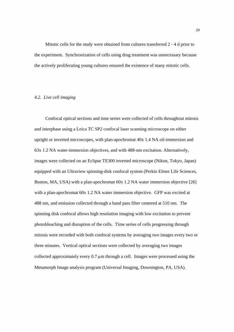
4.2. Live cell imaging
Confocal optical sections and time series were collected of cells throughout mitosis
and interphase using a Leica TC SP2 confocal laser scanning microscope on either
upright or inverted microscopes, with plan-apochromat 40x 1.4 NA oil-immersion and
63x 1.2 NA water-immersion objectives, and with 488-nm excitation. Alternatively,
images were collected on an Eclipse TE300 inverted microscope (Nikon, Tokyo, Japan)
equipped with an Ultraview spinning-disk confocal system (Perkin Elmer Life Sciences,
Boston, MA, USA) with a plan-apochromat 60x 1.2 NA water immersion objective [26]
with a plan-apochromat 60x 1.2 NA water immersion objective. GFP was excited at
488 nm, and emission collected through a band pass filter centered at 510 nm. The
spinning disk confocal allows high resolution imaging with low excitation to prevent
photobleaching and disruption of the cells. Time series of cells progressing through
mitosis were recorded with both confocal systems by averaging two images every two or
three minutes. Vertical optical sections were collected by averaging two images
collected approximately every 0.7 µm through a cell. Images were processed using the
Metamorph Image analysis program (Universal Imaging, Downington, PA, USA).

21
4.3. Live cell specimen preparations
Cells were imaged in growth media on slides, but for drug treatments involving
perfusion of oryazlin or latrunculin, it was necessary to embed the cells in agarose.
Untreated cells (200 µl) were placed in a welled slide and embedded in a thin layer of
1.3% low melting point agarose (type VII, Sigma) in growth media, excess cells and
media wicked away, and the media was solidified by placing the cells in the refrigerator
for 5 seconds. The well was filled with growth media, and a coverslip placed on top of
the well with spacers to allow for perfusion. For microtubule disruption, cells were
perfused with 5 µM oryzalin in growth media, while for microfilament disruption,
perfusion used 5 µM latrunculin B. Time series were collected with two images averaged
every five seconds for oryzalin treatments, and two images averaged every two minutes
for latrunculin treatment. Controls for drug studies were performed with growth media
supplemented with 0.5% DMSO.
4.4 Immunofluorescence microscopy
Cells were washed in cytoskeleton stabilization buffer (CSB; 50 mM Pipes
pH 7.2, 2 mM EGTA, 2 mM MgSO4) for several minutes will being allowed to settle on
slides coated with 0.1% polyethyleneimine. Cells were then fixed in CSB containing 4%
formaldehyde, 1% glutaraldehyde, 400 mM maleimidobenzoyl-N-hydroxysuccinimide

22
ester (MBS; Pierce, Rockford, IL, USA) and 1% dimethylsulfoxide (DMSO) (30 min).
After washing in phosphate buffered saline (PBS) (131 mM NaCl, 5.1 mM Na2HPO4,
1.56 mM KH2PO4 pH 7.2; 2 x 5 min), cells were extracted with methanol at -20oC
(20 min) and washed again in PBS. Surprisingly, this methanol extraction did not reduce
the fluorescence of the fixed GFP, contrary to previous reports [3] and our own
experience [7]. Free aldehyde groups were then reduced with sodium borohydride
(5 mg.ml-1 in PBS) and then washed extensively in PBS. Cell walls were digested (1.0%
cellulase and 0.1% pectolyase Y23 (ICN), 1% BSA and 0.3 M mannitol in modified
Murashige and Skoog medium; 5 min) and material washed again in PBS (2 x 5 min).
After blocking in incubation buffer (PBS containing 1% BSA) (15 min), cells were
concurrently immunolabeled (1 h) for actin with rabbit polyclonal anti-maize actin
(courtesy of Chris Staiger, Purdue University) (diluted 1/200 in incubation buffer) and
mouse monoclonal anti-α-tubulin (Sigma, clone B512) (1/1000). After washing in PBS
(4 x 10 min), cells were concurrently incubated in secondary antibodies (1 h), sheep anti-
rabbit IgG conjugated to rhodamine (Silenus, Boronia, Victoria Australia) (1/100) and
goat anti-mouse IgG coupled to Cy-5 (Jackson, West Grove, PA, USA) (1/200). After
washing in PBS (3 x 10 min), DNA was stained with 4',6-diamidino-2-phenylindole
(DAPI) (1 µg.ml-1 in PBS; 10 min) and mounted on slides in AF1 anti-fade agent
(Citifluor, London, England). For drug studies, oryzalin (5 µM) and latrunculin B
(5 µM) were added to the CSB wash for 5 minutes prior to fixation.
Immunolabelled cells were viewed with the Leica confocal microscope and with a
40X NA1.3 oil-immersion lens. DAPI, GFP, rhodamine and Cy-5 were excited with 351,
488, 543 and 633 nm respectively, with emission windows set to 400-480, 500-530, 550-

23
600 and 640-760 nm. DAPI and GFP were excited sequentially to eliminate signal cross-
talk, with rhodamine and Cy-5 then imaged concurrently.
Acknowledgements
Funding for this project included NASA grant # NAGW-4984 to the North
Carolina NSCORT (NASA Specialized Center of Research and Training) (SLG, DAC,
NSA), NSF REU Site Grant #0243930 (NSA), a Sigma Xi Grant-in-Aid Award (SLG),
and Australian Research Council Discovery Grant no. DP0208806 (DAC). We thank
Dana Moxley and Tim Oliver (North Carolina State University) for discussions and cell
culturing, George Allen and Bill Thompson for the tobacco NT1 cell line, and Andreas
Nebenführ for the tobacco BY-2 cell line. We also thank Edward Salmon and Paul
Maddox (UNC - Chapel Hill) for their help with the spinning disk confocal microscope.

24
References
1. Allen N.S., Brown D.T., Dynamics of the endoplasmic reticulum in living onion
epidermal cells in relation to microtubules, microfilaments and intracellular
particle movement. Cell Motil. Cytoskel. 10 (1988) 153-163.
2. Bajer A., Fine structure studies on phragmoplast and cell plate formation.
Chromosoma 24 (1968) 383-417.
3. Billinton N., Knight A.W., Seeing the wood through the trees: a review of techniques
for distinguishing green fluorescent protein from endogenous autofluorescence.
Analyt. Biochem. 291 (2001) 175-197.
4. Binet M.-N., Humbert C., Lecourieux D., Vantard M., Pugin A., Disruption of
microtubular cytoskeleton induced by cryptogein, an elicitor of hypersensitive
response in tobacco cells. Plant Physiol. 125 (2001) 564-572.
5. Boevink P., Oparka K., Santa Cruz S., Martin B., Betteridge A., Hawes C., Stacks on
tracks: the plant Golgi apparatus traffics on an actin/ER network. Plant J. 15
(1998) 441-447.
6. Collings D.A., Carter C.N., Rink J.C., Scott A.C., Wyatt S.E., Allen N.S., Plant nuclei
can contain extensive grooves and invaginations. Plant Cell 12 (2000) 2425-
2439.
7. Collings D.A., Harper J.D.I., Marc J., Overall R.L., Mullen R.T., Life in the fast lane:
actin-based motility of plant peroxisomes. Can. J. Bot. 80 (2002) 430-441.
8. Collings D.A., Harper J.D.I., Vaughn K.C., The association of peroxisomes with the
developing cell plate in dividing onion root cells depends on actin
microfilaments and myosin. Planta 218 (2003) 204-216.

25
9. Ellenberg J., Siggia E.D., Moriera J.E., Smith C.L., Presley J.F., Worman H.J.,
Lippincott-Schwartz J., Nuclear membrane dynamics and reassembly in living
cells: targeting of an inner nuclear membrane protein in interphase and mitosis.
J. Cell Biol. 138 (1997) 1193-1206.
10. Fricker M., Hollinshead M., White N., Vaux D., Interphase nuclei of many
mammalian cell types contain deep, dynamic, tubular membrane-bound
invaginations of the nuclear envelope. J. Cell Biol. 136 (1997) 531-544.
11. Georgatos S.D., Pyrpasapoulou A., Theodoropoulos P.A., Nuclear envelope
breakdown in mammalian cells involves stepwise lamina disassembly and
microtubule-driven deformation of the nuclear membrane. J. Cell Sci. 110 (1997)
2129-2140.
12. Granger C.L., Cyr R.J., Use of abnormal preprophase bands to decipher division
plane determination. J. Cell Sci. 114 (2001) 599-607.
13. Haseloff J., Siemering K.R., Prasher D.C., Hodge S., Removal of a cryptic intron
and subcellular localization of green fluorescent protein are required to mark
transgenic Arabidopsis plants brightly. Proc. Natl. Acad. Sci. USA 94 (1997)
2122-2127.
14. Hepler P.K., Membrane in the mitotic apparatus of barley cells. J. Cell Biol. 86
(1980) 490-499.
15. Hepler P.K., Endoplasmic reticulum in the formation of the cell plate and
plasmodesmata. Protoplasma 111 (1982) 121-133.
16. Hepler P.K., Palevitz B.A., Lancelle S.A., McCauley M.M., Lichtscheidl I.,
Cortical endoplasmic reticulum in plants. J. Cell Sci. 96 (1990) 355-373.

26
17. Hepler P.K., Wolniak S.M., Membranes in the mitotic apparatus: their structure
and function. Int. Rev. Cytol. 90 (1984) 169-238.
18. Herman E.J., Tague B.W., Hoffman L.M., Kjemtrup S.E., Chrispeels M.J.,
Retention of phytohemagglutinin with carboxyterminal tetrapeptide KDEL in the
nuclear envelope and the endoplasmic reticulum. Planta 182 (1990) 305-312.
19. Jach G., Binot E., Frings S., Luxa K., Schell J., Use of red fluorescent protein from
Discosoma sp. (dsRED) as a reporter for plant gene expression. Plant J. 28
(2001) 483-491.
20. Jackson W.T., Doyle B.G., Membrane distribution in dividing cells of
Haemanthus. J. Cell Biol. 94 (1982) 637-643.
21. Knebel W., Quader H., Schnepf E., Mobile and immobile endoplasmic reticulum in
onion bulb epidermis cells: short- and long-term observations with a confocal
laser scanning microscope. Eur. J. Cell Biol. 52 (1990) 328-340.
22. Lawrence C.J., Malmberg R.L., Muszynski M.G., Dawe R.K., Maximum
likelihood methods reveal conservation of function among closely related kinesin
families. J. Mol. Evol. 54 (2002) 42-53.
23. Lee Y.-R.J., Giang H.M., Liu B., A novel plant kinesin-related protein specifically
associates with the phragmoplast organelles. Plant Cell 13 (2001) 2427-2439.
24. Lichtscheidl I.K., Weiss D.G., Visualization of submicroscopic structures in the
cytoplasm in Allium cepa inner epidermal cells by video-enhanced contrast light
microscopy. Eur. J. Cell Biol. 46 (1988) 376-382.

27
25. Liebe S., Quader H., Myosin in onion (Allium cepa) bulb scale epidermal cells:
involvement in dynamics of organelles and cytoplasmic reticulum. Physiol.
Plant. 90 (1994) 114-124.
26. Maddox P., Moree B., Canman J.C., Salmon E.D., Spinning disk confocal
microscope system for rapid high-resolution, multimode, fluorescence speckle
microscopy and green fluorescent protein imaging in living cells. Meth.
Enzymol. 360 (2003) 597-617.
27. Matsui K., Collings D., Asada T., Identification of a novel plant-specific kinesin-
like protein that is highly expressed in interphase tobacco BY-2 cells.
Protoplasma 215 (2001) 105-115.
28. Molchan T.M., Valster A.H., Hepler P.K., Actomyosin promotes cell plate
alignment and late lateral expansion in Tradescantia stamen hair cells. Planta
214 (2002) 683-693.
29. Nebenführ A., Frohlick J.A., Staehelin L.A., Redistribution of Golgi stacks and
other organelles during mitosis and cytokinesis in plant cells. Plant Physiol. 124
(2000) 135-151.
30. Nebenführ A., Gallagher L.A., Dunahy T.G., Frohlick J.A., Mazurkiewicz A.M.,
Meehl J.B., Staehelin L.A., Stop-and-go movements of plant golgi stacks are
mediated by the acto-myosin system. Plant Physiol. 121 (1999) 1127-1141.
31. Pickett-Heaps J.D., Northcote D.H., Cell division in the formation of the stomatal
complex of the young leaves of wheat. J. Cell Sci. 1 (1966) 121-128.

28
32. Quader H., Hofmann A., Schnepf E., Shape and movement of the endoplasmic
reticulum in onion bulb epidermal cells: possible involvement of actin. Eur. J.
Cell Biol. 44 (1987) 17-26.
33. Quader H., Schnepf E., Endoplasmic reticulum and cytoplasmic streaming:
fluorescence microscopical observations in adaxial epidermis cells of onion bulb
scales. Protoplasma 131 (1986) 250-252.
34. Ridge R.W., Uozumi Y., Plazinski J., Hurley U.A., Williamson R.E.,
Developmental transitions and dynamics of the cortical ER of Arabidopsis cells
seen with green fluorescent protein. Plant Cell Physiol. 40 (1999) 1253-1261.
35. Samuels A.L., Giddings Jr. T.H., Staehelin L.A., Cytokinesis in tobacco BY-2 and
root tip cells: new model of cell plate formation in higher plant cells. J. Cell Biol.
130 (1995) 1345-1347.
36. Schmit A.-C., Actin during mitosis and cytokinesis. in: Staiger C.J., Baluška F.,
Volkmann D., Barlow P.W. (Eds.), Actin: A Dynamic Framework for Multiple
Plant Cell Functions, Kluwer, Dordrecht, (2000), pp 437-456.
37. Schmit A.-C., Lambert A.-M., Plant actin filament and microtubule interactions
during anaphase-telophase transition: effects of antagonist drugs. Biol. Cell 64
(1988) 309-319.
38. Sheahan M.B., Rose R.J., McCurdy D.W., Organelle inheritance in plant cell
division: the actin cytoskeleton is required for unbiased inheritance of
chloroplasts, mitochondria and endoplasmic reticulum in dividing protoplasts.
Plant J. 37 (2004) 379-390.

29
39. Staehelin L.A., The plant ER: a dynamic organelle composed of a large number of
discrete functional domains. Plant J. 11 (1997) 1151-1165.
40. Terasaki M., Dynamics of the endoplasmic reticulum and Golgi apparatus during
early sea urchin development. Mol. Biol. Cell 11 (2000) 897-914.
41. Terasaki M., Chen L.B., Fujiwara K., Microtubules and the endoplasmic reticulum
are highly interdependent structures. J. Cell Biol. 103 (1986) 1557-1568.
42. Valster A.H., Pierson E.S., Valenta R., Hepler P.K., Emons A.M.C., Probing the
plant actin cytoskeleton during cytokinesis and interphase by profilin
microinjection. Plant Cell 9 (1997) 1815-1824.
43. van Gestel K., Köhler R.H., Verbelen J.-P., Plant mitochondria move on F-actin,
but their positioning in the cortical cytoplasm depends on both F-actin and
microtubules. J. Exp. Bot. 53 (2002) 659-667.
44. Waterman-Storer C.M., Salmon E.D., Endoplasmic reticulum membranes are
distributed by microtubules in living cells using three distinct mechanisms. Curr.
Biol. 8 (1998) 798-806.
45. Waterman-Storer C.M., Sanger J.W., Sanger J.M., Dynamics of organelles in the
mitotic spindles of living cells: membranes and microtubule interactions. Cell
Motil. Cytoskel. 26 (1993) 19-39.
46. Wöllert T., Weiss D.G., Gerdes H.-G., Kuznetsov S.A., Activation of myosin V-
based motility and F-actin-dependent network formation of endoplasmic
reticulum during mitosis. J. Cell Biol. 159 (2002) 571-577.

30
47. Yang L., Guan T., Gerace L., Integral membrane proteins of the nuclear enevlope
are dispersed throughout the endoplasmic reticulum during mitosis. J. Cell Biol.
137 (1997) 1199-1210.
48. Zaal K.J.M., Smith C.L., Polischuk R.S., Altan N., Cole N.B., Ellenberg J.,
Hirschberg K., Presley J.F., Roberts T.H., Siggia E., Phair R.D., Lira L.M.,
Lippincott-Schwartz J., Golgi membranes are absorbed into and reemerge from
the ER during mitosis. Cell 99 (1999) 589-601.

31
Figure Legends
Fig. 1. The organization and dynamics of ER in interphase cells. (A-D) Spinning disk
confocal optical sections showed polygonal arrays of ER in the outer cortex (A, arrow),
transvacuolar ER strands that linked the nucleus (n) to the cortex (B, arrows), and links
between the nuclear envelope and transvacuolar strands (C, arrow). D Sequential
confocal images over 15 seconds showed ER movement in a transvacuolar strand
(arrow). (E-F) Laser scanning confocal images showing nuclear morphology. E A low
magnification confocal optical section through a centrally located nucleus (n) with a clear
nuclear envelope (arrow). F Higher magnification optical sections through only the
nucleus, shown at 2 µm intervals from the nuclear surface, reveal an invagination
(arrowhead) and a channel that extends across the nucleus and which contains internal
structure (arrow). Bar in A = 10 µm for A-C; bar in D = 5 µm; bar in E = 10 µm; bar in
F = 5 µm.
Fig. 2. Time-course of confocal fluorescence images showing the changes in ER
organization during cell division over 60 minutes, from preprophase through to telophase.
In preprophase, ER tubules penetrated the nuclear envelope (4 and 12 min, arrows),
rapidly followed by nuclear envelope breakdown at prophase (22 min). Metaphase
(32 min) and anaphase (40 min, with arrows indicating ER tubules parallel to the
presumed orientation of the microtubule spindle) were followed by a rapid telophase

32
(42 min onwards) in which the phragmoplast contained increasing amounts of ER. Bar =
5 µm for all images.
Fig. 3. Time-course of changes in ER organization during cell division over 51 minutes,
from metaphase through to telophase, showing confocal optical sections and concurrently
collected DIC images. In metaphase, strands of ER occurred interleaved between the
chromosomes (0 min, arrows). As anaphase commenced, the ER rapidly filled in the
space between the separating chromosomes (3 min, arrow). From 18 min onwards, the
phragmoplast formed between the new daughter nuclei and laid down the cell plate
(arrow in DIC image). The phragmoplast contained ER (arrow in fluorescence and DIC
images) in increasing amounts over time, so that eventually the fluorescence signal
saturated the detectors of the confocal microscope. Two lagging chromosomes were seen
during anaphase (6 min, arrows), and after the nuclear envelope reformed (24 to 33 min),
channels of ER crossed the nucleus at the locations of these lagging chromosomes (39 to
51 min, arrowheads). Bar = 10 µm for all images.
Fig. 4. Microtubule and ER organization through the cell cycle. Single confocal optical
sections show ER-targeted GFP (top row), immunolabeled microtubules visualized with
Cy-5 (middle row) and DAPI-labeled DNA (bottom row). The organization of the
chemically-fixed ER was, in general, similar to that seen in living cells. During
interphase (A), microtubules remained cortical while ER formed extensive cytoplasmic

33
and perinuclear arrays. During prophase (B) and prometaphase (C) as chromatin
condensed, increasing numbers of microtubule-containing ER protrusions penetrated the
nuclear envelope, and a microtubule spindle developed around the nucleus (arrows).
And during metaphase (D) and anaphase (E), tubules of ER aligned parallel to the
microtubules of the spindle (arrowheads), with ER also parallel to the microtubules of the
phragmoplast during telophase (F) (arrowhead). Images C and F show the same pair of
cells, with the image planes separated by approximately 10 µm. Bar in F = 10 µm for all
images.
Fig. 5. Dividing cells often show ER aggregates outside the spindle poles. Concurrent
confocal fluorescence and DIC images are shown for cells in metaphase (A) and
anaphase (B). A During division, the polygonal arrays of cortical ER remained
unchanged (arrow), although approximately 20% of dividing cells developed conspicuous
assemblages of ER beyond the spindle poles that were visible both by fluorescence and
DIC images (asterisks). These aggregations were visible into anaphase (B, asterisks).
Bar in A = 10 µm for all images.
Fig. 6. Drug treatments cause rapid but specific changes to the cytoskeleton. ER-
targeted GFP expressing cells were fixed and immunolabeled with Cy-5 for microtubules
(top row) and rhodamine for actin microfilaments (bottom row), although ER

34
preservation in these cells was poor (not shown). Images are maximum projections of
confocal optical series through cells. Compared to control cells that contained extensive
arrays of both microtubules and microfilaments (A), brief (5 - 10 min) treatments with
5 µM latrunculin caused disruption only to the microfilaments (B). Similar 5 - 10 min
treatments with 5 µM oryzalin only disrupted the microtubules (C). Bar in C = 20 µm
for all images.
Fig. 7. Microtubule disruption with oryzalin during cell division changes the ER
organization. Oryzalin (5 µM) was perfused into cells in prophase (A), metaphase (B)
and anaphase (C). A Cells treated with oryzalin in prophase remained trapped at this
stage, with ER enmeshed around the chromosomes. B In metaphase, oryzalin perfusion
caused the collapse of the ER spindle within 1 min. C Chromosome separation was
inhibited in anaphase cells treated with oryzalin. However, the chromosomes did
decondense, and the nuclear envelope reformed within 5 min. A bright, ER-containing
phragmoplast never formed between the daughter nuclei in these cells. Bar in A = 5 µm
for all images.
Fig. 8. Latrunculin-mediated actin disruption does not inhibit changes in ER
organization during cell division. Confocal sections are shown from a time-series taken
over 26 min after application of 5 µM latrunculin prior to nuclear envelope breakdown.
ER organization looked similar to that of untreated cells. Although some ER “bubbles”

35
were seen in the cytoplasm (4 min, arrows), the ER still formed arrays that mimicked the
mitotic spindle (0 min, arrowheads), and remained prominent within the phragmoplast
(14 to 26 mins; arrowheads). Bar = 5 µm for all images.

36
Supplemental files:
movie1.avi accompanies Figure 1 (1.66 MB)
Optical sectioning through a small part of a cell show a nucleus containing an
invagination and a transnuclear channel, both of which contain further ER elements.
Depths from the surface of the nucleus are indicated. Bar = 5 µm.
movie2.avi accompanies Figure 2 (1.78 MB)
A time course showing changes in ER organization during cell division, over 60 minutes
from preprophase through to telophase. In preprophase (0 - 20 min), ER tubules
penetrated the nuclear envelope, rapidly followed by nuclear envelope breakdown at
prophase (22 - 32 min). During metaphase (32 - 40 min), and a rapid anaphase (42 min),
ER tubules lay parallel to the presumed orientation of the spindle microtubules. During
telophase (42 min onwards), the phragmoplast contained increasing amounts of ER. As
all images in this sequence were collected and processed in a similar manner, the
increased intensity of phragmoplast labeling indicates a concentration there of ER. Bar
= 5 µm.
movie3.avi accompanies Figure 3 (0.85 MB)
A time-course showing changes in ER organization during cell division, over 51 minutes
from metaphase through to telophase. In metaphase, strands of ER occurred interleaved

37
between the chromosomes (0 min), but as anaphase commenced (3 min), the ER rapidly
filled in the space between the separating chromosomes. Two lagging chromosomes
were seen during anaphase (6 min), and after the nuclear envelope reformed (24 -
33 min), channels of ER crossed the nucleus at the locations of these lagging
chromosomes. As all images in this sequence were collected and processed in a similar
manner, the increased intensity of phragmoplast labeling indicates a concentration there
of ER. Bar = 10 µm.
movie4.avi accompanies Figure 6 (1.60 MB)
Many dividing cells developed conspicuous assemblages of ER beyond the spindle poles.
Depths from the surface of the cell are indicated. Bar = 10 µm.

38
Figure 1

39
Figure 2

40
Figure 3

41
Figure 4

42
Figure 5

43
Figure 6

44
Figure 7

45
Figure 8