A family of GFP-like proteins with different spectral properties in lancelet Branchiostoma floridae
Upload
independentCategory
view
1download
0
FEMS Microbiology Ecology 48 (2004) 109–118
www.fems-microbiology.org
Colonisation of poplar trees by gfp expressing bacterial endophytes
Kieran Germaine a, Elaine Keogh a, Guiomar Garcia-Cabellos a, Brigitte Borremans b,Daniel van der Lelie c, Tanja Barac d, Licy Oeyen d, Jaco Vangronsveld d,Fiona Porteous Moore e, Edward R.B. Moore e, Colin D. Campbell e,
David Ryan a, David N. Dowling a,*
a Department of Applied Biology and Chemistry, Institute of Technology, Carlow, Kilkenny Road, Carlow, Irelandb Vlaamse Instelling voor Technologisch Onderzoek, Mol, Belgium
c Brookhaven National Laboratory, Upton, NY, USAd Limburgs Universitair Centrum, Diepenbeek, Belgium
e Macaulay Institute, Aberdeen, Scotland, UK
Received 24 June 2003; received in revised form 1 October 2003; accepted 28 December 2003
First published online 20 January 2004
Abstract
With the exception of nitrogen fixing bacteria, there is little known about the colonisation patterns or population sizes of
bacterial endophytes in deciduous trees. This study describes the isolation, identification, construction and re-colonisation patterns
of three green fluorescent protein(gfp):kanamycinR labelled bacterial endophytes when re-introduced into poplar trees, their original
host plant. Two of these endophytes showed considerable colonisation in the roots and stems of inoculated plants. gfp expressing
cells of all three strains were observed to colonise the xylem tissue of the root. All three strains proved to be efficient rhizosphere
colonisers, supporting the theory that the rhizosphere can serve as a source of bacterial endophytes.
� 2004 Federation of European Microbiological Societies. Published by Elsevier B.V. All rights reserved.
Keywords: Endophytes; Pseudomonas; Colonisation; gfp; Poplar trees; Rhizosphere; Phytoremediation
1. Introduction
Bacterial colonisation of the internal tissues of plants
has been described in almost all plant species examined
so far. Although many of these bacteria are phyto-
pathogenic, a considerable number have also been found
that colonise the plant without causing disease [1]. Such
bacteria are referred to as bacterial endophytes. Colo-
nisation may take place at the local tissue level orthroughout the plant, with bacterial colonies and bio-
films residing latently in the intercellular spaces and in-
side the vascular tissues [1–4]. A diverse array of
bacterial species have been reported to be endophytic,
including Acetobacter, Arthrobacter, Bacillus, Burk-
* Corresponding author. Tel.: +353-59-9176220; fax: +353-59-
9170517.
E-mail address: [email protected] (D.N. Dowling).
0168-6496/$22.00 � 2004 Federation of European Microbiological Societies
doi:10.1016/j.femsec.2003.12.009
holderia, Enterobacter, Herbaspirillum and Pseudomonas
[5–9], see Lodewyckx et al. [10], for a full review. Indeed
many bacterial genera have been isolated from a given
tissue within a single plant [11]. Sturz and Nowak [12],
proposed that these endophytes originate from the rhi-
zosphere or phylloplane micro-flora, and observed that
many rhizosphere bacteria could penetrate and colonise
root tissue, providing a route into the xylem. In this
vascular tissue, the bacteria could transport themselvesthroughout the plant and hence colonise it systemically.
Once inside the plant, endophytic populations have been
observed to grow to between 2.0 and 7.0 log10 cells per
gram of fresh tissue [6,13].
Certain endophytic bacteria have been shown to en-
hance plant growth, increase plant resistance to patho-
gens, drought and even herbivores, such that their
commercial potential has receivedmuch study [12,14–18].A more novel application of endophytes is in the area of
. Published by Elsevier B.V. All rights reserved.
110 K. Germaine et al. / FEMS Microbiology Ecology 48 (2004) 109–118
phytoremediation (plant assisted removal of xenobiotics
from soil). Siciliano et al. [19], showed that plants grown
on soil contaminated with xenobiotics naturally recruited
endophytes with the necessary contaminant degrading
genes. In addition Lodewyckx et al. [20], showed thatendophytes of yellow lupinwere able to increase the nickel
accumulation and nickel tolerance of the inoculated
plant. Particular endophytes can confer an increased level
of resistance to the plant against specific xenobiotics [19].
Consequently, it may be necessary or advantageous to
introduce bacteria expressing degradative capacity into a
plant species intended for such applications.However, the
use of microbial inoculations for biocontrol, growthpromotion or plant-assisted bioremediation requires an
efficient level of re-colonisation and competence of the
introduced microbe.
Assessing colonisation efficiency and population size
requires an ability to track and identify the inoculated
strain within the host plant. Introduction of antibiotic
resistance genes into the strain provides a simple method
of tracking colonisation. However, strains inoculatedinto plants may temporally lose their antibiotic resistant
phenotype [21]. This problem can be overcome by cou-
pling antibiotic resistance with genes for expressing
green fluorescent protein (gfp), which provides a unique,
visual phenotype and is a simple, stable method of
studying population dynamics of the organisms within
the plant tissues. The gfp polypeptide is 27 kDa and
when irradiated with blue or near UV light ðA395Þ pro-duces a green fluorescence ðA508Þ. It is a useful marker in
environmental microbial studies because it is expressed
in most Gram-negative bacteria, does not require any
exogenous substrates and is extremely stable [22]. The
gfp marker gene has proved to be very useful in colo-
nisation studies and has been used to visualise the in-
fection and root nodulation events of both Rhizobium
spp. and Agrobacterium tumefaciens [2,23,24]. Elbeltagyet al. [5], successfully showed the colonisation of the
shoots of wild rice plants by a gfp labelled version of the
nitrogen fixer Herbaspirillum, while Ramos et al. [25],
used gfp to assess the physiological status of Pseudo-
monas putida cells within the rhizosphere of barley seeds.
Table 1
Bacterial strains used in this study
Strain Characteristic
Bacteria
Pseudomonas sp. PopHV4 Poplar tree endophyte
Pseudomonas sp. PopHV6 Poplar tree endophyte
Pseudomonas sp. PopHV9 Poplar tree endophyte
E. coli CM2780 pFAJ1819
VM1449 PopHV4 with a mini-Tn5 insertion of gfp,K
VM1450 PopHV6 with a mini-Tn5 insertion of gfp,K
VM1453 PopHV9 with a mini-Tn5 insertion of gfp,K
Plasmid pFAJ1819 A transposon/suicide plasmid containing a
resistance gene and two copies of the gfp g
promoter
This paper describes the isolation, identification and
recolonisation efficiency of three poplar tree endophytes
when re-introduced into their original host poplar tree.
Poplar is a fast growing, hard wood, deciduous tree and
is widely used in phytoremediation projects. The bac-terial strains were genetically marked with a kanamy-
cin:gfp cassette inserted into their chromosome,
allowing the visualisation of colonising cells and the
estimation of population sizes within the various tissues
of the host plant.
2. Materials and methods
2.1. Bacterial stains, plasmids and culture conditions
Strains and plasmids are listed in Table 1.
The endophytic strains were maintained on Luria–
Bertani (LB) agar (Merck) or 284 Tris-minimal medium
[28] at 30 �C. The Escherichia coli strain CM2780 car-
rying the pFAJ1819 plasmid was grown in LB broth [29]supplemented with 50 lg/ml kanamycin (Km) at 37 �C.
2.2. Isolation and identification of endophytic bacteria
from poplar trees
The endophytic bacteria used in this study were iso-
lated from xylem sap of poplar trees (Populus tricho-
carpa� deltoides cv. Hoogvorst). The poplar trees weregrowing on a phytoremediation site near a motor factory
in Genk, Belgium. The groundwater from this site was
measured to contain an increased concentration of zinc
(Zn), nickel (Ni) (Zn 0–1000 lg/l, Ni 0–100 lg/l) and
BTEXcompounds (BTEX0–1000lg/l). The extractionofxylem sap was carried out using a Scholander pressure
bomb instrument, which is a pressure chamber connected
to a bottle containing nitrogen gas. After surface sterili-zation (5min in a solution containing 1% active chloride),
the young twig was sealed in the pressure chamber with
the cut end exposed through a hole in the chamber cover.
The chamber pressure was slowly increased (5–25 bar)
until xylem sap was forced back to the cut surface. Xylem
Source/reference
This study
This study
This study
[26]
mR This study
mR This study
mR This study
pUT mini Tn5 transposon carrying a kanamycin
ene under the regulation of a strong constitutive
[26,27]
K. Germaine et al. / FEMS Microbiology Ecology 48 (2004) 109–118 111
sap (100 ll) was inoculated and spread over different solidisolation media, 869 medium [30], 1/10 strength 869 me-
dium, and Schatz medium [31] supplemented with a car-
bon source mix (1.3 ml/l glucose (40%), 0.7 ml/l lactate
(50%), 2.2 ml/l gluconate (30%), 2.7 ml/l fructose (20%)and 3ml/l succinate (1M)). After an incubation period of
7 days at 30 �C, eight different morphotypes were isolated
and identified by 16S rDNAanalysis. GenomicDNAwas
extracted from approximately 0.1 g (wet wt.) of cells
pelleted from liquid cultures, using the Bio101 Fast DNA
for Soils kit (Q-Biogene, UK). The 16S rRNA genes,
approximately 1500 nucleotides long, were amplified by
PCR, using standard reagents in 50 ll reaction volumes (5ll PCR buffer (10�), 2.5 mM MgCl2, 10 ll Q-solution
(5�), 1.25 U Taq Polymerase (Qiagen Ltd., Crawley,
UK), 0.4 g/ll BSA (Roche Diagnostics Ltd., UK),
0.2 mM of each dNTPs (Promega Biosciences Inc., UK)
and 1 lM each of the forward primer M16F28
(50-AGAGTTTGATCKTGGCTCAG-30) and reverse
primer M16R1494 (50-TACGGYTACCTTGTTTACG
AC-30) hybridising at conserved positions of the rDNAofmembers of the bacteria domain [32]. Amplification was
performed in an MJ Research PTC-200 thermocycler
(GRI, UK) with a preliminary denaturation step at 95 �Cfor 5 min, followed by 35 cycles of 94 �C for 1 min, 55 �Cfor 2min, 72 �C for 2min and terminated with one step of
72 �C for 10 min. PCR products were purified, using
QIAquick Spin Columns (Qiagen Ltd.), and sequenced
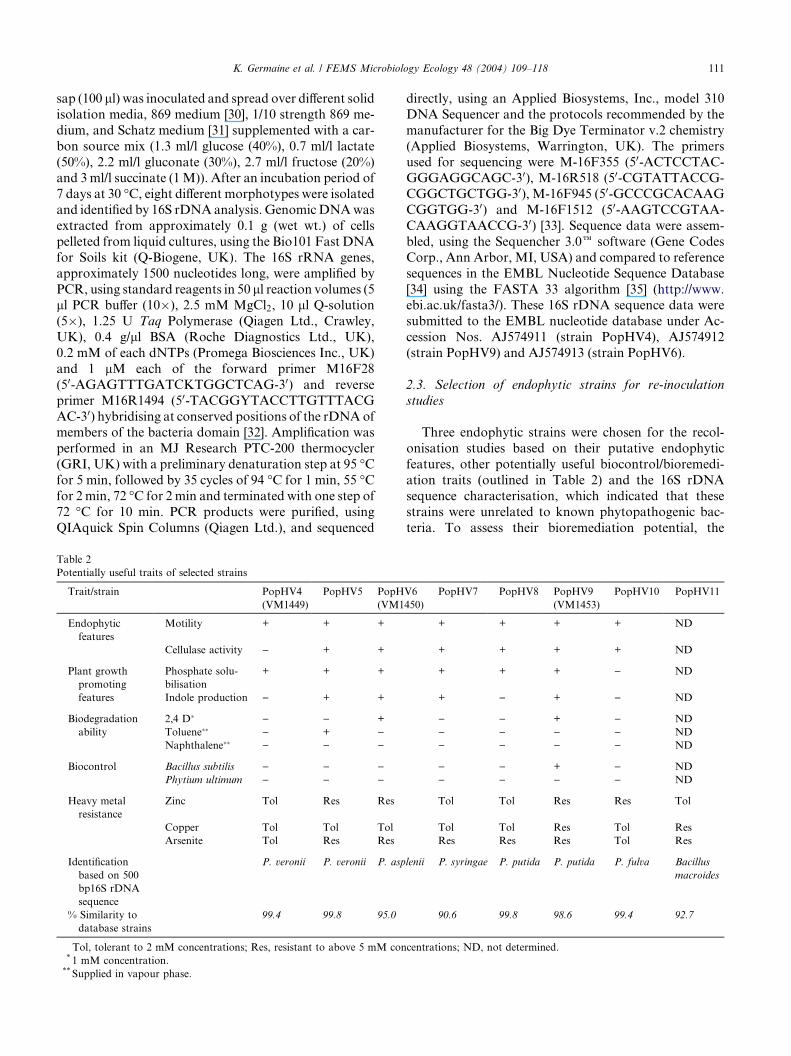
Table 2
Potentially useful traits of selected strains
Trait/strain PopHV4
(VM1449)
PopHV5 PopH
(VM1
Endophytic
features
Motility + + +
Cellulase activity ) + +
Plant growth
promoting
features
Phosphate solu-
bilisation
+ + +
Indole production ) + +
Biodegradation
ability
2,4 D� ) ) +
Toluene�� ) + )Naphthalene�� ) ) )
Biocontrol Bacillus subtilis ) ) )Phytium ultimum ) ) )
Heavy metal
resistance
Zinc Tol Res Res
Copper Tol Tol Tol
Arsenite Tol Res Res
Identification
based on 500
bp16S rDNA
sequence
P. veronii P. veronii P. asp
% Similarity to
database strains
99.4 99.8 95.0
Tol, tolerant to 2 mM concentrations; Res, resistant to above 5 mM con* 1 mM concentration.** Supplied in vapour phase.
directly, using an Applied Biosystems, Inc., model 310
DNA Sequencer and the protocols recommended by the
manufacturer for the Big Dye Terminator v.2 chemistry
(Applied Biosystems, Warrington, UK). The primers
used for sequencing were M-16F355 (50-ACTCCTAC-GGGAGGCAGC-30), M-16R518 (50-CGTATTACCG-
CGGCTGCTGG-30), M-16F945 (50-GCCCGCACAAG
CGGTGG-30) and M-16F1512 (50-AAGTCCGTAA-
CAAGGTAACCG-30) [33]. Sequence data were assem-
bled, using the Sequencher 3.0e software (Gene Codes
Corp., Ann Arbor, MI, USA) and compared to reference
sequences in the EMBL Nucleotide Sequence Database
[34] using the FASTA 33 algorithm [35] (http://www.ebi.ac.uk/fasta3/). These 16S rDNA sequence data were
submitted to the EMBL nucleotide database under Ac-
cession Nos. AJ574911 (strain PopHV4), AJ574912
(strain PopHV9) and AJ574913 (strain PopHV6).
2.3. Selection of endophytic strains for re-inoculation
studies
Three endophytic strains were chosen for the recol-
onisation studies based on their putative endophytic
features, other potentially useful biocontrol/bioremedi-
ation traits (outlined in Table 2) and the 16S rDNA
sequence characterisation, which indicated that these
strains were unrelated to known phytopathogenic bac-
teria. To assess their bioremediation potential, the
V6
450)
PopHV7 PopHV8 PopHV9
(VM1453)
PopHV10 PopHV11
+ + + + ND
+ + + + ND
+ + + ) ND
+ ) + ) ND
) ) + ) ND
) ) ) ) ND
) ) ) ) ND
) ) + ) ND
) ) ) ) ND
Tol Tol Res Res Tol
Tol Tol Res Tol Res
Res Res Res Tol Res
lenii P. syringae P. putida P. putida P. fulva Bacillus
macroides
90.6 99.8 98.6 99.4 92.7
centrations; ND, not determined.
112 K. Germaine et al. / FEMS Microbiology Ecology 48 (2004) 109–118
endophytic strains were grown on minimal media plates
containing various organic chemicals as the sole carbon
sole. Growth on these plates after 48 h was considered
an indication of the biodegradation of the targeted
compound. Putative endophytic traits (cellulase activityand motility) were determined by the methods of Verma
et al. [36]. Heavy metal resistance was assessed by
growing the endophytic strains on nutrient agar sup-
plemented with various concentrations of heavy metals.
Biocontrol properties of the endophytes were deter-
mined by streaking the test strain in the centre of sucrose
asparagine (SA) agar plates containing high and low
iron concentrations. Streaks of either Pythium ultimum
or Bacillus subtilis were then made approximately one
inch from either side of the test strain streak. The plates
were incubated for 48 h and examined for growth inhi-
bition of P. ultimum and B. subtilis [37].
2.4. Introduction of the gfp:kanamycin cassette into
selected endophytic strains
The gfp donor strain, CM2780 carrying the pFAJ1819
plasmid was grown overnight in LB medium supple-
mented with 50 lg/ml Km and the endophytic recipient
strains were grown overnight in LB medium, washed in
10�2 M MgSO4 and aliquots of 100 ll were added to a
sterile filter (0.45 lm) and incubated overnight at 30 �Con
solid LB medium. The mating mixture was plated out on
284 Tris-minimal medium supplemented with 50 lg/mlKm and incubated at 30 �C for 4–5 days. The transcon-
jugants were purified and the presence of the gfp gene was
confirmed by PCR using the following primers: gfp-
F50-CCCCCCCGGGCTAGATTTAAGAAGG-30 and
gfp-R50-TTTTCCCGGGTTATTTGTATAGTTCATC
CATGCC-30. Individual colonies were resuspended in
100 ll of 10�2 MMgSO4 and 5 ll was taken as a template
for the PCR. Amplification was performed in a Trio-Thermoblock (Biometra). 100 ll reaction mixtures, con-
taining 0.5 U TaKaRa Ex Taq polymerase (Cambrex Bio
Science, Verviers), each of the nucleotides at 200 lM, and
each of the primers at 1 lM, were subjected to a pre-
liminary denaturation step at 94 �C for 10 min, followed
by 35 cycles of incubation at 94 �C for 1 min, 55 �C for
1min and 72 �C for 2min and terminated with one step of
8 min at 72 �C. The growth rates, Biolog� profiles, mo-tility and cellulase activities of the transconjugants were
tested and compared to the wild type strains to ensure the
gfp cassette had not been randomly inserted into genes
involved in colonisation.
2.5. Plant re-inoculation
Fresh cultures of the endophytic gfp:KmR derivativestains (designated VM1449, VM1450 and VM1453)
were grown in LB broth, at 30 �C, 200 rpm, to an ap-
proximate absorbance ðA600Þ value of 1.0. Cells were
harvested by centrifugation, washed in 0.85% sterile
saline and resuspended in 100 ml modified ISO 8692
plant nutrient solution [38] containing 5% LB broth.
These inocula contained between 108 and 109 cells/ml as
determined by standard plate counts. Woody stem cut-tings (1–2 years old, approximately 200 mm long) were
harvested from mature poplar trees and surface disin-
fected using 70% ethanol. These cuttings were cultivated
hydroponically in the inoculum suspension for six weeks
at 20–25 �C under a 16-h light/8-h dark regime. Inocu-
lations were carried out in triplicate per bacterial strain.
After six weeks, the trees were transferred to pots con-
taining a compost/vermiculite substrate (3:1 ratio) thathad been previously sterilised by autoclaving for 2 h at
121 �C but which was not maintained under sterile
conditions throughout the experiment. The trees were
cultivated under the same conditions as detailed above.
Un-inoculated trees served as controls.
2.6. Enumeration of culturable endophytic populations
within plant tissues
Trees were destructively sampled 10 weeks after in-
oculation. Healthy samples of leaf, fleshy stem, sap from
the woody stem, root and rhizosphere tissues were taken
from each plant. Leaves and stems were surface steri-
lised by swabbing with 70% ethanol. Roots were surface
sterilised by placing in a solution containing 2% active
chloride for 2.5 min. The sterilising agents were removedfrom tissues by rinsing three times in sterile water. To
check for sterility, surface sterilised tissues were pressed
against a plate count agar (PCA) plate (Merck) and
samples of the third rinsing were plated onto PCA.
Excess water was removed from tissues using sterile
paper towels. Sap was collected from woody stems by
vacuum extraction and collected in sterile Eppendorf
tubes. 1 g samples of the surface sterilised tissues werehomogenised using sterile pestle and mortars, serially
diluted in 0.85% sterile saline and 100 ll samples were
spread plated onto PCA and PCA containing 100 lg/ml
kanamycin. Sap and rhizosphere samples were serially
diluted and plated in the same manner. Plates were in-
cubated at 30 �C and examined for growth after 72 h.
The number of colony forming units per gram (cfu g�1)
of fresh tissue was calculated.
2.7. In planta visualisation of gfp expressing endophytes,
using epi-fluorescent microscopy
Hand cut sections of surface sterilised leaf, stem and
root tissues were examined under blue light (395 nm)
using a Nikon E400 epi-fluorescent microscope equipped
with a 100 W mercury short arc photo-optic lamp. Lu-cia� imaging software (version 4.6) was used to cap-
ture and process microscopic images. Visualisation of gfp
expression proved difficult due to auto-fluorescence from

K. Germaine et al. / FEMS Microbiology Ecology 48 (2004) 109–118 113
the plant tissue itself. gfp visualisation was achieved by
counter staining the tissue section in 0.05% methyl violet
for 30 s, which caused the plant cells to fluoresce red
under near UV light.
3. Results
3.1. Isolation and identification of endophytic bacteria
from poplar trees
A collection of endophytic bacteria were isolated
from xylem sap of poplar trees. Eight of these werepicked according to different morphotypes, designated
PopHV4-11 and were identified by sequence analysis of
a 500 bp sequence of their 16S rDNA gene, with refer-
ence to the 16S rDNA genes sequences of described
bacteria with validly published names in the EMBL
Nucleotide database [34]. Seven strains were identified
as species of Pseudomonas and one as a Bacillus sp.
Three Gram-negative isolates, designated as PopHV4,PopHV6 and PopHV9, were selected for the re-coloni-
sation study. These three strains were further identified
by sequence analysis of 1500 bp of their 16S rDNA gene.
All three of these isolates were observed to be closely
related to the type strain Pseudomonas putida
(ATCC12633-T, Accession No. AF094736). The nearly
complete 16S rDNA gene sequences of PopHV4,
PopHV6 and PopHV9 were observed to possess 99.4%,99.3% and 100% similarities, respectively, to that P.
putida strain. These data indicate that all three strains
are certainly species of Pseudomonas. The 16S rDNA
sequence analyses indicated that none of the strains
are closely related to any known phytopathogenic
bacterium.
3.2. Construction of gfp expressing endophytic strains
After 5 days the mating between CM2780(pFAJ1819)
and the selected endophytes, PopHV4, PopHV6 and
PopHV9, resulted in transconjugants that were KmR.
The presence of the gfp gene in these strains was con-
firmed by PCR analysis. Strain CM2780 was used as
control. All transconjugants tested showed the gfp spe-
cific amplicon of 750 bp corresponding to the gfp gene.A representative transconjugant of each conjugation
was chosen and named, respectively, as VM1449,
VM1450 and VM1453. These transconjugants were
compared with their wild type parent strains for specific
growth rate, Biologe metabolic profiles and for cellu-
lase activity. For VM1449 and VM1453 all parameters
were similar to those of the wild type strains. This was
also the case for VM1450 except that the Biologeprofile showed a minor difference (one substrate out of
95 tested) to that of the wild type. These data suggest
that the mini-Tn5-gfp cassette did not disrupt any key
trait required for the survival of the marked strains and
that they could be used in re-colonisation experiments.
3.3. Colonisation and enumeration of endophytic popula-
tions within plant tissues
Inoculated poplar trees were allowed to grow for 10
weeks before sampling took place. Total bacterial pop-
ulations and KmR, gfp expressing populations were
determined for each of the tissues examined. Endophytic
bacteria are considered to be those isolated from the
internal tissues of surface sterilised plants. However, it is
difficult to determine whether an organism is truly en-dophytic or merely a survivor of the surface sterilisation
process [39]. To ensure that the sterilisation processes
were adequate, the sterilised tissues were pressed against
the surface of a sterile PCA plate and samples of the
third water rinsings were also plated onto PCA plates.
Bacterial counts on these plates were always between 0–
101 cfu g�1, which was considered to be a good indica-
tion that the surface was successfully sterilised. No gfp
expressing, kanamycin resistant cells were isolated from
uninoculated plants. A number of indigenous endo-
phytic strains were also isolated on the kanamycin
plates. To ensure that only the inoculated strains were
counted, these plates were examined under the epi-
fluorescent microscope and only those colonies ex-
pressing gfp were enumerated.
Pseudomonas sp. strain VM1449 was detected only inthe rhizosphere and the interior root tissues of inocu-
lated trees (Fig. 1(a)). The total culturable aerobic rhi-
zosphere population was determined to be between 107
and 108 cfu g�1 and the numbers of strain VM1449
accounted for as much as 3.2% (Table 3) of the total
culturable bacterial population. VM1449 numbers in-
side the root represented up to 0.3% of the total cul-
turable endophytic population. No colonisation ofVM1449 was detected in the stems or leaves.
Trees inoculated with strain VM1450 showed notable
colonisation in all tissues including the leaves (Fig. 1(b)).
As with VM1449, the samples from the rhizosphere
showed the greatest level colonisation rates, followed by
the root and then by the woody stem. Inoculum popu-
lations within the root were in the order of 104 cfu g�1
tissue. VM1450 populations in the rhizosphere ac-counted for up to 7.8% of the total bacterial community
during the course of the experiment. Inside the root,
VM1450 cells comprised as much as 2.7% of the total
root endophytic population, while in the stem (tissue
and sap), VM1450 numbers were between 103 and 104
cfu g�1 which represents 18% of the total culturable
population.
After 10 weeks, plants inoculated with strainVM1453, showed a similar colonisation pattern to
VM1450, with the exception of the leaf (Fig. 1(c)). The
rhizosphere population was stable at approximately 107
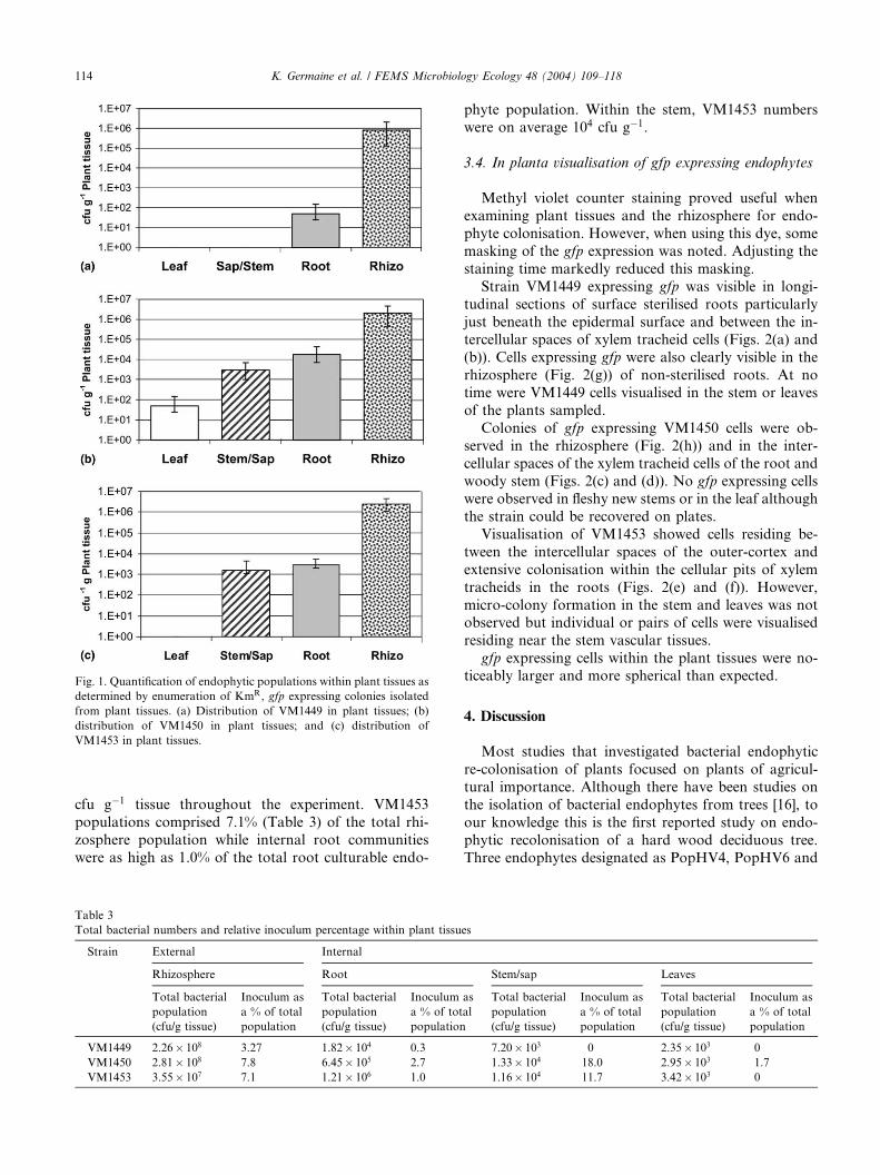
Fig. 1. Quantification of endophytic populations within plant tissues as
determined by enumeration of KmR, gfp expressing colonies isolated
from plant tissues. (a) Distribution of VM1449 in plant tissues; (b)
distribution of VM1450 in plant tissues; and (c) distribution of
VM1453 in plant tissues.
114 K. Germaine et al. / FEMS Microbiology Ecology 48 (2004) 109–118
cfu g�1 tissue throughout the experiment. VM1453
populations comprised 7.1% (Table 3) of the total rhi-
zosphere population while internal root communities
were as high as 1.0% of the total root culturable endo-
Table 3
Total bacterial numbers and relative inoculum percentage within plant tissu
Strain External Internal
Rhizosphere Root
Total bacterial
population
(cfu/g tissue)
Inoculum as
a % of total
population
Total bacterial
population
(cfu/g tissue)
Inoculum
a % of tot
population
VM1449 2.26� 108 3.27 1.82� 104 0.3
VM1450 2.81� 108 7.8 6.45� 105 2.7
VM1453 3.55� 107 7.1 1.21� 106 1.0
phyte population. Within the stem, VM1453 numbers
were on average 104 cfu g�1.
3.4. In planta visualisation of gfp expressing endophytes
Methyl violet counter staining proved useful when
examining plant tissues and the rhizosphere for endo-
phyte colonisation. However, when using this dye, some
masking of the gfp expression was noted. Adjusting the
staining time markedly reduced this masking.
Strain VM1449 expressing gfp was visible in longi-
tudinal sections of surface sterilised roots particularly
just beneath the epidermal surface and between the in-tercellular spaces of xylem tracheid cells (Figs. 2(a) and
(b)). Cells expressing gfp were also clearly visible in the
rhizosphere (Fig. 2(g)) of non-sterilised roots. At no
time were VM1449 cells visualised in the stem or leaves
of the plants sampled.
Colonies of gfp expressing VM1450 cells were ob-
served in the rhizosphere (Fig. 2(h)) and in the inter-
cellular spaces of the xylem tracheid cells of the root andwoody stem (Figs. 2(c) and (d)). No gfp expressing cells
were observed in fleshy new stems or in the leaf although
the strain could be recovered on plates.
Visualisation of VM1453 showed cells residing be-
tween the intercellular spaces of the outer-cortex and
extensive colonisation within the cellular pits of xylem
tracheids in the roots (Figs. 2(e) and (f)). However,
micro-colony formation in the stem and leaves was notobserved but individual or pairs of cells were visualised
residing near the stem vascular tissues.
gfp expressing cells within the plant tissues were no-
ticeably larger and more spherical than expected.
4. Discussion
Most studies that investigated bacterial endophytic
re-colonisation of plants focused on plants of agricul-
tural importance. Although there have been studies on
the isolation of bacterial endophytes from trees [16], to
our knowledge this is the first reported study on endo-
phytic recolonisation of a hard wood deciduous tree.
Three endophytes designated as PopHV4, PopHV6 and
es
Stem/sap Leaves
as
al
Total bacterial
population
(cfu/g tissue)
Inoculum as
a % of total
population
Total bacterial
population
(cfu/g tissue)
Inoculum as
a % of total
population
7.20� 103 0 2.35� 103 0
1.33� 104 18.0 2.95� 103 1.7
1.16� 104 11.7 3.42� 103 0
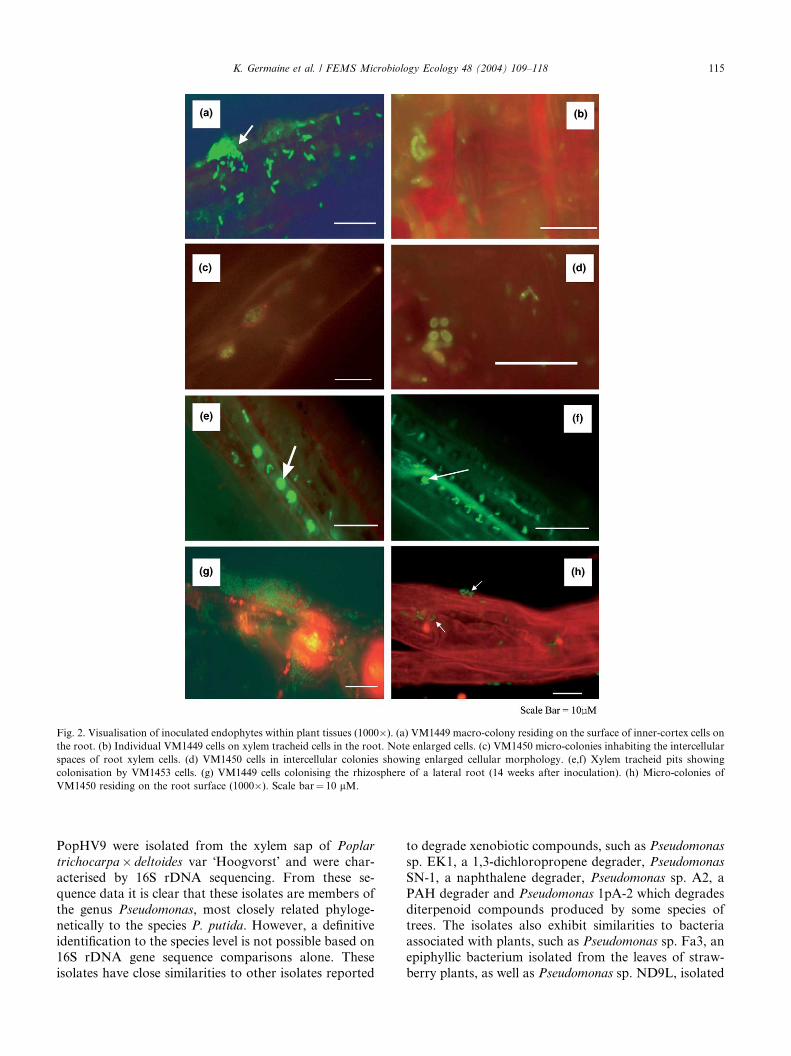
Fig. 2. Visualisation of inoculated endophytes within plant tissues (1000�). (a) VM1449 macro-colony residing on the surface of inner-cortex cells on
the root. (b) Individual VM1449 cells on xylem tracheid cells in the root. Note enlarged cells. (c) VM1450 micro-colonies inhabiting the intercellular
spaces of root xylem cells. (d) VM1450 cells in intercellular colonies showing enlarged cellular morphology. (e,f) Xylem tracheid pits showing
colonisation by VM1453 cells. (g) VM1449 cells colonising the rhizosphere of a lateral root (14 weeks after inoculation). (h) Micro-colonies of
VM1450 residing on the root surface (1000�). Scale bar¼ 10 lM.
K. Germaine et al. / FEMS Microbiology Ecology 48 (2004) 109–118 115
PopHV9 were isolated from the xylem sap of Poplar
trichocarpa� deltoides var �Hoogvorst� and were char-
acterised by 16S rDNA sequencing. From these se-
quence data it is clear that these isolates are members of
the genus Pseudomonas, most closely related phyloge-
netically to the species P. putida. However, a definitiveidentification to the species level is not possible based on
16S rDNA gene sequence comparisons alone. These
isolates have close similarities to other isolates reported
to degrade xenobiotic compounds, such as Pseudomonas
sp. EK1, a 1,3-dichloropropene degrader, Pseudomonas
SN-1, a naphthalene degrader, Pseudomonas sp. A2, a
PAH degrader and Pseudomonas 1pA-2 which degrades
diterpenoid compounds produced by some species of
trees. The isolates also exhibit similarities to bacteriaassociated with plants, such as Pseudomonas sp. Fa3, an
epiphyllic bacterium isolated from the leaves of straw-
berry plants, as well as Pseudomonas sp. ND9L, isolated
116 K. Germaine et al. / FEMS Microbiology Ecology 48 (2004) 109–118
from rhizosphere soil, this bacterium inhibits fungal
(Cercospora beticola) infections on sugar beet. Com-
parative analysis of the 16S rDNA sequences of the
strains indicated that they are not closely related to any
known plant pathogens. These strains were then taggedwith a gfp:kanamycin random insertion transposon and
their respective derivatives (VM1449, VM1450 and
VM1453) were re-inoculated into cuttings of their ori-
ginal host plant. The presence of the marker genes ap-
peared to have no negative effect on the ability of the
strains to colonise the rhizosphere and the interior tis-
sues of the plant. However, although these derivatives
had similar metabolic profiles and growth properties tothat of the wild type strains, it is a remote possibility
that the transposon insertion affected their colonisation
ability with respect to wild type strain.
All three strains could be re-isolated from the interior
tissues of surface sterilised roots. Two of the strains were
detected in the stems and occasionally in the leaves of
inoculated plants. However, population sizes in these
tissues did vary greatly between replicates. This mayhave been an artefact of the surface sterilisation proto-
col or simply evidence of differing rates of colonisation
within individual plants. The effectiveness of the sterili-
sation protocol varied according to the thickness of the
sample. Thin samples were prone to over-sterilisation.
Thus, where possible, throughout the experiment, tissue
sections of similar thickness were sampled.
Population sizes of all three strains decreased mark-edly from the rhizosphere to the root interior. The fact
that all three strains were efficient colonisers of the
rhizosphere further supports the theory that endophytes
can originate from the rhizosphere [7] and from there,
move into the internal plant tissues. Although the
strains were inoculated into autoclaved vermiculite, the
poplar plants were not sterile and a large population of
non-inoculated bacteria were co-isolated from the rhi-zosphere, suggesting that some of these were derived
from the autochtonous endophytic community within
the poplar plants. This may help to re-inoculate and
replenish the rhizosphere microbial flora when the
growing season begins after the winter decline. The
populations of inoculated strains comprised on average
only 1–4% of the total culturable microbial population
in the rhizosphere and as much as 18% of the internalroot population. The survival of these strains 10 weeks
after inoculation, despite the fact that there was no
observable selective pressure, suggests that they are
good competitors. Populations of inoculated strains in
the root and stem (including the sap) were on average, in
the same range, up to 104 cfu g�1 fresh weight, which is
consistent with reports [6,13] in other plants.
Interior colonisation by VM1449 was detected only inthe root. Population sizes were in the order of 102 cfu
g�1 of plant tissue. This suggests that VM1449 is not as
active a coloniser, but it may colonise the tissues at a
slower rate or through accidental disposition. This is
further supported by the fact that the strain did not
show any cellulase activity (possibly required for endo-
phytic colonisation), whereas both VM1450 and
VM1453, which did show cellulase activity, were seen tobe active colonisers of the stem and leaves. VM1449 cells
expressing gfp were seen to randomly colonise the sur-
face of cells in the root cortex and was also observed to
form micro-colonies intracellularly within the inner
margin of the pericyle, adjacent to root xylem cells. At
no time were VM1449 cells visualised in the stem or
leaves, which corresponds with the results of the enu-
meration analysis.Strain VM1450 was the only inoculated strain to be
detected in the leaves suggesting that it is an efficient
systemic coloniser. Its motility, cellulase activity and its
ability to colonise the xylem (as shown by gfp detection)
are probably contributing factors to the spread of this
strain throughout the plant. Colonies of VM1450 cells
were visualised mainly in vascular tissues, with a pro-
liferation of cells on and within the intercellular spacesbetween adjacent xylem tracheid cells. The rapid spread
of this strain from the root to aerial tissues suggest that
it uses the vascular system as a route for systemic
colonisation.
VM1453 appeared to be an efficient coloniser of the
root and stem but colonisation in the leaves was not
found in this period of study. The observed colonisation
pattern of VM1453 was markedly different from those ofthe other two strains. VM1453 cells were almost exclu-
sively located in the vascular system and specifically
within the pits of xylem cells. Large fluorescent colonies
could be clearly seen residing in these cell pits along the
length of the plant cell wall. These pits are typically
between 1 and 14 lm wide (depending on their location
in the plant) and facilitate the lateral transport of water
and minerals throughout the plant [40]. It is likely thatthis strain also uses the xylem to transport itself into the
stem, where it was recovered in high numbers. The fact
that all three endophytes were found to colonise the
vascular tissue is not surprising as the literature details
numerous endophytic strains with this ability [3,41]. At
no time during the microscopic examination of the plant
tissues was intracellular colonisation observed nor did
there appear to be any cellular damage caused by thecolonisation of inoculated endophytes.
There was a noticeable change in the cellular mor-
phology of the inoculated strains when visualised within
the plant tissues. The cells appeared to be larger and
more spherical than when grown on LB. Changes in
cellular shape dependent on environmental conditions
have been reported previously [23,25]. Li et al. [23], also
observed this phenomenon with Agrobacterium tum-
efaciens cells when inoculated into plants. It was pro-
posed that bacterial cells are better nourished upon
successful colonisation, but this paper also cited reports
K. Germaine et al. / FEMS Microbiology Ecology 48 (2004) 109–118 117
that cell shape is related to the growth rates of strains
within a particular environment and that slower growth
rates yielded excessively large cells. Interestingly, there
was no observed change in the morphology of bacteria
colonising the rhizosphere. These observations havebeen supported by Ramos et al. [25], who showed that
P. putida colonising the rhizosphere of barley had high
growth rates under sterile conditions during day 1,
however, potential low growth rates were detected under
non-sterile conditions.
This study has shown the successful recolonisation of
poplar trees by three endophytic bacterial strains under
controlled conditions. Two of the strains, VM1450 andVM1453, demonstrated efficient colonisation resulting
in high population numbers within the plant tissues.
None of the introduced strains showed any signs of
pathogenicity towards their host plant and others tested.
Many studies have shown that the colonisation levels in
field trials are less successful than those in laboratory
trials. This is probably due to increased microbial
competition and less favourable environmental condi-tions [42]. Therefore, additional long-term field trials
need to be carried out in order to gain a better under-
standing of the colonisation pattern and population
dynamics of endophytic bacteria in poplar trees in the
field.
This study is part of a larger EU funded project
‘‘Endegrade’’ [43], which is attempting to utilise endo-
phytes to phytoremediate soil pollutants as they aretranslocated through the plant, thereby reducing phy-
totoxicity and volatilisation [44]. Future work will in-
clude equipping the most efficient plant colonising
bacterial strains with degradation genes and evaluating
if the strains enhance phytoremediation ability of the
plant–microbe combination.
Acknowledgements
This work is funded in part by the HEA PRTLI
programmes and EU Contracts QLK3-CT2000-00164
and QLK3-CT-2001-00101.
References
[1] Sessitsch, A., Reiter, B., Pfeifer, U. and Wilhelm, E. (2002)
Cultivation-independent population analysis of bacterial endo-
phytes in three potato varieties based on eubacterial and Actino-
mycetes-specific PCR of 16S rRNA genes. FEMSMicrobiol. Ecol.
39, 3–32.
[2] Gage, D.J., Bobo, T. and Long, S.R. (1996) Use of green
fluorescent protein to visualise the early events of symbiosis
between Rhizobium meliloti and Alfalfa (Medicago sativa). J.
Bacteriol. 178, 7159–7166.
[3] Gopalaswamy, G., Kannaiyan, S., O�Callaghan, K., Davey, M.
and Cocking, E. (2000) The xylem of rice (Oryza sativa) is
colonised by Azorhizobium caulinodans. Proc. R. Soc. Lond. B.
267, 103–107.
[4] Hinton, D.M. and Bacon, C.W. (1995) Enterobacter cloacae is an
endophytic symbiont of corn. Mycopatholia 129, 117–125.
[5] Elbeltagy, A., Nishioka, K., Sato, T., Suzuki, H., Ye, B., Hamada,
T., Isawa, T., Mitsui, H. and Minamisaw, K. (2001) Endophytic
colonisation and in planta nitrogen fixation by a Herbaspirillum
sp. isolated from a wild rice species. Appl. Environ. Microbiol. 67,
5285–5293.
[6] McInroy, J.A. and Kloepper, J.W. (1994) Studies on the Indig-
enous Endophytic Bacteria of Sweet Corn and Cotton. In:
Molecular Ecology of Rhizosphere Micro-organisms (O�Gara,
F., Dowling, D.N. and Boesten, B., Eds.), pp. 19–28. VCH Press.
[7] Misko, A.M. and Germida, J.J. (2002) Taxonomic and functional
diversity of Pseudomonads isolated from the roots of field-grown
canola. FEMS Microbiol. Ecol. 42, 399–407.
[8] Salgado, T., Ramirez, L., Hernandez, A., Esparza, M., Romero,
E. and Mellado, J.C. (1997) Coffea arabica L., a new host plant for
Acetobacter diazotrophicus, and isolation of other nitrogen fixing
Acetobacteria. Appl. Environ. Microbiol. 63, 3676–
3683.
[9] Tapia-Hernandez, A., Bustillos-Cristales, M.R., Jimenez-Salgado,
T., Mellado, J.C. and Ramirez, L.E.F. (2000) Natural endophytic
occurrence of Acetobacter diazotrophicus in pineapple plants.
Microbiol. Ecol. 39, 49–55.
[10] Lodewyckx, C., Vangronsveld, J., Porteous, F., Moore, E.R.B.,
Taghavi, S. and van der Lelie, D. (2002) Endophytic bacteria and
their potential applications. Crit. Rev. Plant Sci. 6, 583–
606.
[11] Sturz, A.V., Christie, B.R., Matheson, B.G. and Nowak, J. (1997)
Biodiversity of endophytic bacteria which colonise red clover
nodules, roots, stems and foliage and their influence on host
growth. Biol. Fertil. Soils 25, 13–19.
[12] Sturz, A.V. and Nowak, J. (2000) Endophytic communities of
rhizobacteria and the strategies required to create yield enhancing
associations with crops. Appl. Soil Ecol. 15, 83–190.
[13] Shishido, M., Breuil, C. and Chanway, C. (1999) Endophytic
colonisation of spruce by plant growth promoting rhizobacteria.
FEMS Microbiol. Ecol. 29, 191–196.
[14] Azedvedo, J., Maccheroni Jr., W., Pereira, J. and Araujo, W.
(2000) Endophytic microorganisms: a review on insect control and
recent advances in tropical crops. Environ. Biotech. 3, 134–142.
[15] Bacilio-Jimenez, M., Flores, S., delValle, M.V., Perez, A., Zepeda,
A. and Zenteno, E. (2001) Endophytic bacteria in rice seeds inhibit
early colonisation of roots by Azospirillium brasilense. Soil. Biol.
Biochem. 33, 167–172.
[16] Chanway, C.P., Radley, R.A., and Holl, F.B. (1989) Bacterial
Inoculation of Lodgewood Pine, White Spruce and Douglas-fir
Grown in Containers, Intermountain Forest Nursery Association
Annual Meeting, Bismarck, North Dakota.
[17] James, E.K. (2000) Nitrogen fixation in endophytic and associa-
tive symbiosis. Field Crops Res. 65, 197–209.
[18] Reiter, B., Pfeifer, U., Schwab, H. and Sessitsch, A. (2002)
Response of endophytic bacterial communities in potato plants to
infection with Erwinia carotovora subsp. atroseptica. Appl. Envi-
ron. Microbiol. 68, 2261–2268.
[19] Siciliano, S.D., Fortin, N., Mihoc, A., Wisse, G., Labelle, S.,
Beaumier, D., Ouellette, D., Roy, R., Whyte, L.G., Banks, M.K.,
Schwab, P., Lee, K. and Greer, C.W. (2001) Selection of specific
endophytic bacterial genotypes by plants in response to soil
contamination. Appl. Environ. Microbiol. 67, 2469–2475.
[20] Lodewyckx, C., Taghavi, S., Mergeay, M., Vangronsveld, J.,
Clijsters, H. and van der Lelie, D. (2001) The effect of recombinant
heavy metal resistant endophytic bacteria in heavy metal uptake
by their host plant. Int. J. Phytoremed. 3, 173–187.

118 K. Germaine et al. / FEMS Microbiology Ecology 48 (2004) 109–118
[21] Nairn, J.D. and Chanway, C. (2002) Temporary loss of antibiotic
resistance by marked bacteria in the rhizosphere of spruce
seedlings. FEMS Microbiol. Ecol. 40, 167–170.
[22] Errampalli, D., Leung, K., Cassidy, M.B., Kostrzynska, M.,
Blears, M., Lee, H. and Trevors, J.T. (1999) Applications of the
green fluorescent protein as a molecular marker in environmental
microorganisms. J. Microbiol. Methods 35, 187–199.
[23] Li, L., Li, Y., Meng Lim, T. and Pan, S.Q. (1999) Gfp-aided
confocal laser scanning microscopy can monitor Agrobacterium
tumefaciens cell morphology and gene expression associated with
infection. FEMS Microbiol. Lett. 179, 141–146.
[24] Stuurman, N., Bras, C., Schlaman, H., Wijfjes, A., Bloemberg, G.
and Spaink, H. (2000) Use of green fluorescent protein colour
variants expressed on stable broad host range vectors to visualise
Rhizobia interacting with plants. Mol. Plant–Microbe Interact. 13,
1163–1169.
[25] Ramos, C., Molbak, L. and Molin, S. (2000) Bacterial activity in
the rhizosphere analysed at the single cell level by monitoring
ribosome contents and synthesis rates. Appl. Environ. Microbiol.
66, 801–809.
[26] Herrero, M., de Lorenzo, V. and Timmis, K.N. (1990) Transpo-
son vectors containing non-antibiotic resistance markers for
cloning and stable chromosomal insertion of foreign genes in
gram negative bacteria. J. Bacteriol. 172, 6557–6567.
[27] Xi, C., Lambrechts, M., Vanderleyden, J. and Michiels, J. (1999)
Bi-functional gfp- and gusA-containing mini-Tn5 transposon
derivatives for combined gene expression and bacterial localisa-
tion studies. J. Microbiol. Methods 35, 85–92.
[28] Schlegel, H.G., Kaltwasser, H. and Gottschalk, G. (1961) Ein
Sumbersverfahren zur Kultur wasserstoffoxidierender Bakterien:
Wachstum physiologische Untersuchungen. Arch. Mikrobiol. 38,
205–222.
[29] Miller, J.H. (1972) Experiments in Molecular Genetics. Cold
Spring Harbor Laboratory, Cold Spring Harbor, NY. pp. 354–
358.
[30] Schatz, A. and Bovel, C. (1956) Growth and hydrogenase activity
of a new bacterium, Hydrogenomonas facilis. J. Bacteriol. 63, 87–
98.
[31] Mergeay, M., Nies, D., Schegel, H.G., Gerits, J., Charles, P. and
van Gijsegem, F. (1985) Alcaligenes eutrophus CH34 is a faculta-
tive chemolithotroph with plasmid bound resistance to heavy
metals. J. Bacteriol. 162, 328–334.
[32] Lane, D.J. (1991) 16S/23S Sequencing. In: Nucleic Acid Tech-
niques in Bacterial Systematics (Stackebrand, E. and Goodfellow,
M., Eds.), pp. 115–175. Wiley, Chichester, UK.
[33] Hauben, L., Vauterin, L., Swings, J. and Moore, E.R.B. (1997)
Comparison of 16S ribosomal DNA sequences of all Xanthomonas
species. Int. J. Syst. Bacteriol. 47, 328–335.
[34] Stoesser, G., Baker, W., van den Broek, A., Garcia-Pastor, M.,
Kanz, C., Kulikova, T., Leinonen, R., Lin, Q., Lombard, V.,
Lopez, R., Mancuso, R., Nardone, F., Stoehr, P., Tuli, M.A.,
Tzouvara, K. and Vaughan, R. (2003) The EMBL nucleotide
sequence database: major new developments. Nucl. Acids Res. 31,
17–22.
[35] Pearson, W.R. (1994) Using the FASTA program to search
protein and DNA sequence databases. Meth. Mol. Biol. 24, 307–
331.
[36] Verma, S.C., Ladha, J.K. and Tripathi, A.K. (2001) Evaluation of
plant growth promoting and colonisation ability of endophytic
diazotrophs from deep water rice. J. Biotechnol. 19, 127–
141.
[37] Russo, A., Moenne-Loccoz, Y., Fedi, S., Higgins, P., Fenton, A.,
Dowling, D.N., O�Regan, M. and O�Gara, F. (1996) Improved
delivery of biocontrol Pseudomonas and their antifungal metab-
olites using alginate polymers. Appl. Microbiol. Biotechnol. 44,
740–745.
[38] International Organisation for Standardisation, ISO (1997) Water
Quality – Fresh Water Algal Growth Test with Scenedesmus
subspicatus and Raphidocelis subcapitata. ISO8692.
[39] Zamora, M.L.G. and Romero, E.M. (2001) Natural endophytic
association between Rhizobium etli and maize (Zea mays L.). J.
Biotechnol. 91, 117–126.
[40] Fahn, A. (1982) Primary Plant Tissues. In: Plant Anatomy, 3rd
Edn, pp. 129–138. Pergamon Press, Madrid.
[41] Reinhold-Hurek, B. and Hurek, T. (1998) Life in grasses:
diazotrophic endophytes. Trends Microbiol. 6, 139–
144.
[42] Zinniel, D., Lambrecht, P., Harris, N., Feng, Z., Kuczmarski, D.,
Higley, P., Ishimaru, C., Arunakumari, A., Barletta, R. and
Vidaver, A. (2002) Isolation and characterisation of endophytic
colonising bacteria from agronomic crops and prairie plants.
Appl. Environ. Microbiol. 68, 21298–22208.
[43] Endegrade: Endophytic degrader bacteria for improving phyto-
remediation of organic xenobiotics. Available from: www.ende-
grade.dmu.dk.
[44] van der Lelie, D., D�Haene, S., Dowling, D.N., Karlson, U.,
Moore, E.R.B., Taghavi, S., Trapp, S. and Vangronsveld, J. (May
2001) Methods of improving phytoremediation treatment of
contaminated medium, Application No. 60/291, 344, US Patent
Office.
Copyright © 2022 FDOKUMEN