A simple, versatile method for GFP-based super-resolution microscopy via nanobodies

BioMed CentralBiology Direct
ss
Open AcceResearchA family of GFP-like proteins with different spectral properties in lancelet Branchiostoma floridaeDiana Baumann1, Malcolm Cook1, Limei Ma1, Arcady Mushegian*1,2, Erik Sanders1, Joel Schwartz1 and C Ron Yu1,3Address: 1Stowers Institute for Medical Research, 1000 E 50th St., Kansas City, MO, 64110, USA, 2Department of Microbiology, Molecular Genetics, and Immunology, University of Kansas, Kansas City, KS, 66160, USA and 3Department of Anatomy and Cell Biology, University of Kansas Medical Center, Kansas City, KS, 66160, USA
Email: Diana Baumann - [email protected]; Malcolm Cook - [email protected]; Limei Ma - [email protected]; Arcady Mushegian* - [email protected]; Erik Sanders - [email protected]; Joel Schwartz - [email protected]; C Ron Yu - [email protected]
* Corresponding author
AbstractBackground: Members of the green fluorescent protein (GFP) family share sequence similarity and the11-stranded β-barrel fold. Fluorescence or bright coloration, observed in many members of this family, isenabled by the intrinsic properties of the polypeptide chain itself, without the requirement for cofactors.Amino acid sequence of fluorescent proteins can be altered by genetic engineering to produce variantswith different spectral properties, suitable for direct visualization of molecular and cellular processes.Naturally occurring GFP-like proteins include fluorescent proteins from cnidarians of the Hydrozoa andAnthozoa classes, and from copepods of the Pontellidae family, as well as non-fluorescent proteins fromAnthozoa. Recently, an mRNA encoding a fluorescent GFP-like protein AmphiGFP, related to GFP fromPontellidae, has been isolated from the lancelet Branchiostoma floridae, a cephalochordate (Deheyn et al., BiolBull, 2007 213:95).
Results: We report that the nearly-completely sequenced genome of Branchiostoma floridae encodes atleast 12 GFP-like proteins. The evidence for expression of six of these genes can be found in the ESTdatabases. Phylogenetic analysis suggests that a gene encoding a GFP-like protein was present in thecommon ancestor of Cnidaria and Bilateria. We synthesized and expressed two of the lancelet GFP-likeproteins in mammalian cells and in bacteria. One protein, which we called LanFP1, exhibits bright greenfluorescence in both systems. The other protein, LanFP2, is identical to AmphiGFP in amino acid sequenceand is moderately fluorescent. Live imaging of the adult animals revealed bright green fluorescence at theanterior end and in the basal region of the oral cirri, as well as weaker green signals throughout the bodyof the animal. In addition, red fluorescence was observed in oral cirri, extending to the tips.
Conclusion: GFP-like proteins may have been present in the primitive Metazoa. Their evolutionaryhistory includes losses in several metazoan lineages and expansion in cephalochordates that resulted in thelargest repertoire of GFP-like proteins known thus far in a single organism. Lancelet expresses several ofits GFP-like proteins, which appear to have distinct spectral properties and perhaps diverse functions.
Reviewers: This article was reviewed by Shamil Sunyaev, Mikhail Matz (nominated by I. King Jordan) andL. Aravind.
Published: 3 July 2008
Biology Direct 2008, 3:28 doi:10.1186/1745-6150-3-28
Received: 11 June 2008Accepted: 3 July 2008
This article is available from: http://www.biology-direct.com/content/3/1/28
© 2008 Baumann et al; licensee BioMed Central Ltd. This is an Open Access article distributed under the terms of the Creative Commons Attribution License (http://creativecommons.org/licenses/by/2.0), which permits unrestricted use, distribution, and reproduction in any medium, provided the original work is properly cited.
Page 1 of 12(page number not for citation purposes)

Biology Direct 2008, 3:28 http://www.biology-direct.com/content/3/1/28
BackgroundGenetically encoded fluorescent probes are indispensabletools for in vivo imaging of molecules, cells and wholeorganisms. Among the fluorescent proteins that have beendeveloped as reporters, members of the GFP family, withthe green fluorescent protein of hydroid Aequorea victoriaas its founding member [1,2] are unique in that theirchromophore/fluorophore is formed solely from thepolypeptide chain itself, and their maturation as well aslight emission does not require any cofactors other thanoxygen. Members of the GFP family are found in manycnidarians, where they display a range of excitation andemission spectra, from fluorescence to bright colorationin the visible light. GFPs proved to be extraordinarily ame-nable to genetic manipulation: some of the useful traits ofthe engineered GFP derivatives include shifts in themaxima of excitation and/or emission; timed responses,such as kindling or color change after excitation; photoac-tivation; assembly of functional monomers from the frag-ments of the molecule; and others [3-5]. With all thisknowledge about structure-function relationships in theGFP family, there is nonetheless a considerable interest innaturally occurring GFP-like proteins with novel proper-ties.
The evolutionary history of the GFP family and its rela-tionship to other proteins are not well-understood. Inaddition to Cnidaria, fluorescent proteins with significantsequence similarity to GFPs have been found several yearsago in marine crustaceans of the Pontellidae family [6]. Inan unrelated work, the structure of one domain (G2)within nidogen, a protein component of basement mem-branes in various groups of metazoan animals, was foundto share spatial similarity with GFP and GFP-like proteins[7]. The common fold, described as 11-stranded β-barrel,is so close in nidogen G2 domain and in GFPs that thesuperimposition of these structures is possible with rootmean square deviation of 2.5Å between 195 carbon alphaatoms (out of approximately 225 in GFP). The lack of dis-cernible sequence similarity between G2 domains andGFPs, however, leaves open the evolutionary questions,i.e., whether the two β-barrels descend from the commonancestral gene, and, if such common ancestor existed,what was its function and in which organism did it reside.
In this work, we present the results of our analysis of afamily of GFP-like proteins from a cephalochordate, thelancelet Branchiostoma floridae. The imaging of adult ani-mals reveals anatomically discrete areas of green and redfluorescence surrounding the oral aperture. The nearly-completely sequenced genome of B. floridae encodes atleast 12 GFP-like proteins, which appear to have arisen byduplication after separation of the protostome and deu-terostome lineages. Several of these genes are representedin the public EST libraries obtained from eggs and from
different stages of embryo development. To assess the util-ity of these genes as reporters, we expressed the human-ized versions of two of these proteins in a mammaliansystem.
Results and DiscussionWe were interested in the evolutionary provenance of theβ-barrel proteins and searched for the homologs of vari-ous β-barrels in the EST databases. When GFP and GFP-like proteins were used as queries in the TBLASTNsearches of the non-human, non-mouse EST database atNCBI, we detected a few trivial matches with perfect iden-tity to several cloned GFPs, apparently originating fromthe unfiltered fragments of the cloning vectors (data notshown). To our surprise, however, the overwhelmingmajority of the matches with significant sequence similar-ity were from the EST libraries corresponding to variousdevelopmental stages of the lancelet B. floridae. The lance-let ESTs represented evolutionarily distinct sequences notfound in other organisms. Translations of these ESTs were30–40% identical to copepod GFPs, their nearest databasehomologs, and were more distant from the cnidarianGFPs and GFP-like proteins. At this level of similarity,nonetheless, the matches had high statistical significance(E-value < 10-10), and the residues that are involved in thechromophore formation in other GFP-like proteinsappeared to be well-conserved, suggesting that the lance-let GFP-like sequences may represent a fluorescent pro-tein.
A more detailed analysis of the ESTs indicated that theremay be several distinct, closely related members of theGFP family in lancelets, and searches in the DNA tracearchive of the B. floridae genome suggested that it mayencode additional homologs of GFPs not represented inthe EST databases. Most recently, we took advantage of theDOE Joint Genome Institute release v.1.0 of the anno-tated genome assembly of B. floridae and collected a non-redundant set of genes encoding full-length homologs ofGFP (Table 1 and Figure 1). In addition, we examined thefirst release of the genome assembly of cnidarian Nemato-stella vectensis, the only other sequenced genome that isknown to encode GFP-like proteins.
Comparative analysis of genomes and of proteinsequences paints a picture of an ancient origin of the GFP-family proteins and their evolution by vertical descent fol-lowed by frequent gene loss (Figure 1, Table 1, and Addi-tional file 1). The phylogenetic tree inferred from thealigned protein sequences indicates that all cnidarianGFPs form one well-supported clade in the tree, all cope-pod GFPs form another, and the set of lancelet GFP-likeproteins forms the third clade (Additional file 1). Thebranching order in the midpoint-rooted tree follows theMetazoan phylogeny, with copepod and lancelet clades
Page 2 of 12(page number not for citation purposes)

Biology Direct 2008, 3:28 http://www.biology-direct.com/content/3/1/28
being closest to each other. This is compatible with thepresence of an ancestral GFP in the common ancestor ofMetazoa, followed by loss of this gene in some of thepresent-day species and lineage-specific expansion in theothers.
A further indication of the ancient ancestry of GFPs inMetazoa comes from the comparison of the intron posi-tions in cnidarian and cephalochordate genes. GFP genesin B. floridae and N. vectensis appear to share at least oneintron in the homologous position of the codon 32 (Fig-ure 1). The probability of independent insertion of anintron into a homologous site within the orthologousgenes in two lineages of eukaryotes is thought to be lessthan 20% [8,9] and may be less than 10% within Metazoa[9], suggesting that the intron in this codon is much more
likely to be ancestral than convergently inserted. The posi-tion of this conserved intron close to the 5' termini of theGFP genes is compatible with the recently documented 5'-to-3' bias towards retention of ancestral introns and theopposite bias towards intron gain and loss in multicellu-lar organisms [10]. Moreover, another intron is present inthe start codon of almost all genes in lancelet and of twogenes in corals also supports this view, though thesequence conservation at the beginning of the codingregion is lower and their alignment is more ambigous.
Six of the genes encoded by the genome of B. floridae arerepresented in the EST libraries made from eggs, variousstages of embryo development and adult animals. Wewondered whether any of these genes may encode pro-teins that would confer fluorescence to the animals.
Table 1: properties of lancelet GFP-like genes and their products.
B. floridae genes
JGI gene model ID – JGI model name Comments Identity to other LanFP proteins, %
ESTs (source tissues)
Identity (%) and gene ID of the best match in B. lanceolatum/incopepods
LanFP1 7875 – FGENESH2_PG.SCAFFOLD_1000062 48–90 21 (egg, larva, neurula)
51% 169125805/32% 33243028
LanFP2 7877 – FGENESH2_PG.SCAFFOLD_1000064 51–99 33 (adult, egg, gastrula, neurula)
57% 169125805/36% 33243028
LanFP3 31376 – FGENESH2_PG.SCAFFOLD_264000004
49–61 19 (neurula, gastrula, egg, larva)
51% 169125805/34% 33243028
LanFP4 7879 – FGENESH2_PG. SCAFFOLD_1000066
51–98 15 (egg, larva) 57% 169125805/36% 33243028
LanFP5 3655 – ESTEXT_FGENESH2_PG.C_4080036 two fused GFP-like proteins? 50–54 1 (adult) 39% 33243028/76% 169125805 (84% 169125805)
LanFP6 7878 – FGENESH2_PG.SCAFFOLD_1000065 51–97 1 (larva) 54% 169125805/37% 33243028LanFP7 21366 –
ESTEXT_FGENESH2_PG.C_237002048–60 1 (larva) 56% 169125805/40% 33243028
LanFP8 11646 – FGENESH2_PG.SCAFFOLD_58000032
54–70 91% 169125797/40% 33243028
LanFP9 33503 -FGENESH2_PG.SCAFFOLD_549000016
49–55 52% 169125805/33% 33243028
LanFP10 11648 – FGENESH2_PG.SCAFFOLD_58000034
48–82 77% 169125805/37% 33243032
LanFP11 35422 – FGENESH2_PG.SCAFFOLD_722000001
48–56 72% 169125805/38% 33243028
LanFP12 7881 – FGENESH2_PG.SCAFFOLD_1000068 49–55 69% 169125805/30% 33243032LanFP13 3657 –
FGENESH2_PG.SCAFFOLD_40800003850–84 79% 169125805/36% 33243034
7876 – FGENESH2_PG.SCAFFOLD_1000063 similar to LanFP7, internal deletion43701 – FGENESH2_PG.SCAFFOLD_149000048
similar to LanFP11; long unrelated N-terminal extension – probably prediction artefact
43778 – FGENESH2_PG.SCAFFOLD_150000025
similar to LanFP13
31374 -FGENESH2_PG.SCAFFOLD_264000002
similar to LanFP6; long insertion
31375 -FGENESH2_PG.SCAFFOLD_264000003
similar to LanFP12
3656- FGENESH2_PG.SCAFFOLD_408000037
similar to LanFP10
33504- FGENESH2_PG.SCAFFOLD_549000017
similar to LanFP4
11648 – FGENESH2_PG.SCAFFOLD_58000033
similar to LanFP6
11649 – FGENESH2_PG.SCAFFOLD_58000035
similar to LanFP7
11650 -FGENESH2_PG.SCAFFOLD_58000036
similar to LanFP7
35196 – FGENESH2_PG.SCAFFOLD_771000005
similar to LanFP4
Page 3 of 12(page number not for citation purposes)

Biology Direct 2008, 3:28 http://www.biology-direct.com/content/3/1/28
Page 4 of 12(page number not for citation purposes)
Sequence conservation in the GFP familyFigure 1Sequence conservation in the GFP family. Multiple alignment of protein sequences of GFP-like proteins from cnidarians, copepods, and lancelet. Pro-tein sequences of gene products predicted from the genome assembly of B. floridae were clustered at the 90% identity cutoff, and one representative per cluster that did not contain internal deletions was included into the alignment (see Table 1 for details). Identifier of each sequence in JGI genome browser or in GenBank is given after each sequence. The consensus secondary structure derived from multiple known three-dimensional structures of GFP-like proteins is shown below the alignment. Red type indicates conserved small or kinky side chains (G, S, A, or P), yellow shading indicates conserved bulky hydrophobic residues (I, L, V, M, F, Y, or W), blue type indicates conserved acidic or amidic residues (D, E, N, or Q), blue shading indicates conserved basic residues (K or R), purple type with gray shading indicates the tripeptide directly participating in rearrangement that leads to the chromophore forma-tion, and white type on black indicates the amino acid whose codon contains an intron in the known genome sequence. Species abbreviations are as fol-lows: Aeqvi, Aequorea victoria; Astla, Astrangia lajollaensis; Chipo, Chiridius poppei; Corca, Corynactis californica; Dissp, Discosoma sp. RC-2004; Monca, Montastraea cavernosa; Monef, Montipora efflorescens; Nemve, Nematostella vectensis; Phial, Phialidium sp. SL-2003; Ponpe, Pontella meadi; Ponpl, Pontellina plu-mata; Renmu, Renilla muelleri.
LanFP1 MPLPA---THDIHLHGSINGHEFDMVGGGKGDPNAGSLVTTAKSTK-GALKFSPYLMIPHLGYGYYQYLPYPDG-P LanFP2 MSLPT---THDLHIFGSVNGAEFDLVGGGKGNPNDGTLETSVKSTR-GALPCSPLLIGPNLGYGFYQYLPFPGG-A LanFP3 MPLPA---THEIHLHGSVNGHEFDLVGSGKGDPKAGSLVTEVKSTM-GPLKFSPHLMIPHLGYGYYQYLPYPDG-P LanFP4 MPLPT---THELHIFGSFNGVEFDLVGRGEGNPKDGSQNLHLKSTK-GPLQFSPWMLIPHIGYGFYQYLPYPDGEM LanFP5 TPLPT---THELHIFGSFNGVEFDMVGRGIGNPNDGYEELNLKSTK-GALKFSPWILVPQIGYGFHQYLPYPDG-M LanFP6 MPLPK---THELHIFGSFNGVKFDMVGEGTGNPNEGSEELKLKSTN-GPLKFSPYILVPHLGYAFNQYLPFPDG-M LanFP7 MSVPT---NLDLHIYGSINGMEFDMVGGGSGNPNDGSLSVNVKSTK-GALRVSPLLVGPHLGYGHYQYLPFPDG-P LanFP8 MSLPT---THDCHM GSINGHEFDLVGGGNGNPNDGTLETKVRSTK-GALPFSPVILAPNLGYGYHQYLPFPAG-T FLanFP9 MPLPA---THEIHLHGSINGHEFDLAGGGKGDPNAGSLVTTAKSTK-GPLKFSPHLMIPHLGYGYYQYLPYPDG-P LanFP10 LPLPK---THELHIFGSFNGVEFDMVGRGIGNPNEGSEELNAKFTK-GPLKFSPYILVPHLGYAYYQYLPFPDG-M LanFP11 MPLPA---THEIHIYGSVNGHEFDLVGGGKGDPNAGSLVTEVKSTM-GPLKFSPHLMIPHLGYGYYQYLPYPDG-P LanFP12 QPLPT---THEVHVYGSINGVEFDLVGSGKGNPKDGSEEIQVKSTK-GPLGFSPYIVVPNIGYGFHQYLPFPDG-M GFP1_Ponme_33243032 MPDM----KLECHISGTMNGEEFELIGAGDGNTDEGRMTNKMKSIK-GPISFSPYLLSHILGYGYYHFATFPAGYE GFP1_Ponpl_33243026 MPAM----KIECRISGTLNGVVF LVGGGEGIPEQGRMTNKMKSTK-GALTFSPYLLSHVMGYGFYHFGTYPSGYE EGFP_Chipo_85658701 MTTF----KIESRIHGNLNGEKFELVGGGVGE--EGRLEIEMKTKD-KPLAFSPFLLSHCMGYGFYHFASFPKGTK Nemve_156385591 56 YSLSKDAMKLHFKMEGSVNGHCFEIQGVGEGKAFDGEHWSKLCVVKGKHLPFSFDILMPSMSYGTKQFAKYPAGMT Nemve_156388065 MSLSKDAMKLHLILEGSVNGHCFEIHGEGEGKAFEGEQWSKFTVKKGGPLPFSFDLIAPCLKYGSKPFVKYPDDMT Monca_154814320 MSVIKTDMKIKLRMEGAVNGHKFVIEGEGEGKPFDGKQTMDLTVIEGAPLPFAYDILTTVFDYGNRVFAKYPKDIP Monca_154814322 MSVIKPDMKIKLRMEGAVNGHKFVIEGDGKGKPFEGKQTMDLTVIEGAPLPFAYDILTTVFDYGNRVFAKYPKDIP Azami-Green_52839539 MSV L MRG VNGHNFVIEGEGKGNPYEGTQILDL VTEGAPLPFAYDILTTVFQYGNRAFTKYPADIQ IKPEMKIK C T NDronpa_56201403 MSVIKPDMKIKLRMEGAVNGHPFAIEGVGLGKPFEGKQSMDLKVKEGGPLPFAYDILTTVFCYGNRVFAKYPENIV EosFP_55667942 MSAIKPDMKINLRMEGNVNGHHFVIDGDGTGKPFEGKQSMDLEVKEGGPLPFAFDILTTAFHYGNRVFAEYPDHIQ CyanFP_154814310 MSV L MDGIVNGHKF ITGEGEGKPFEGTHTIILKVKEGGPLPFAYDILTTAFQYGNRVFTKYPKDIP IKSVMKIK R MKaede_23503508 MSLIKPEMKIKLLMEGNVNGHQFVIEGDGKGHPFEGKQSMDLVVKEGAPLPFAYDILTTAFHYGNRVFAKYPDHIP Photoconvert_125395854 MSVIKSVMNVKLRLEGAVNGHPFVIEGKGNGHPFEGTQDIKLTVKEGGPLPFAYDILTTVFHYGNRVFVKYPDDIV YFP_Corca_72256906 4 KQV Y MDGCVNGHSFTIEGEGTGKPYEGNQTLKLRVTKGGPLPFAFDILTATFCYGNRCFCEYPEDMP ITQEMKMV HNFCP_MONEF_55976263 MSVIATQMTYKVYMSGTVNGHYFEVEGDGKGRPYEGEQTVKLTVTKGGPLPFAWDILSPQCQYGSIPFTKYPEDIP RFP_Dissp_85002146 4 KNVIKEFMRFKVRMEGTVNGHEFEIEGEGEGRPYEGHNTVKLKVTKGGPLPFAWDILSPQFQYGSKVYVKHPADIP GFP3_Astla_42521556 4 KQEIKKEMTMDY MDGCVNGHSFTVKGDGAGKPYEGHQRLSL PLPFAFDILSAAFAYGNRCFTKYPKEIP V HVTGGQGFP_Renmu_12621060 7 NTCLQEVMSYKVNLEGIVNNHVFTMEGCGKGNILFGNQLVQIRVTKGAPLPFAFDIVSPAFQYGNRTFTKYPNDIS YFP_Phial_40365351 4 ALLFHGKIPYVVEMEGNVDGHTFSIRGKGYGDASVGKVDAQFICTT-GDVPVPWSTLVTTLTYGAQCFAKYGPELK GFP_AEQVI_1169893 4 EELFTGVVPILVELDGDVNGHKFSVSGEGEGDATYGKLTLKFICTT-GKLPVPWPTLVTTFSYGVQCFSRYPDHMK Secondary structure sssssssssssss1sssssssssssss2sssssssssss3 LanFP1 --SPF GYAVYRVFDFE-DGG LTTEFKYSYEGS---HIKADMKLMGSGFPDDGPVMTSQIVDQDGCVSKKTYLNN QTSMLEGS KLanFP2 --SPFQTAIT-DGGYQVHRVFKFE-DGGVLSCNFRYTYEGG---KIKGEFQLIGSGFPAGGPVMSGGLTTLDRSVAKLQCSDD LanFP3 --SPFQTAMLDGSGYKVHRVFNFE-DGGVLSIDYNYAYEGT---HIKSDFKLMGSGFPDDGPVMTSQIVDQDGCVSKKTYLND LanFP4 --SPYQAAMYGGSGYLMHRTMQYE-DGAKISGHYKYTYEGS---HVKGEFQLIGTGFPTDGPVMTNQLTAADWCVDKLLYPND LanFP5 --SPFQAAMQDGSGYQVHRTMQFE-DGASLTAHFRYTYEGS---HIKGEFQVIGTGFPADGPVMTNKLTAADWCVVKMVYPND LanFP6 --SPFQAAMQDESGYQVHRTLQYE-DGAFVTANLRYTYEGS---HIKGEFQVIGTGFPPDGPVMTNKLTALDWSVVKFVYPND LanFP7 --SPF GYQMHRSFNFE-DGAVLTATYNYSYSGG---KIQGEF LVGSCFPDDSPVMTNALTGLDRSVAKLMCVSD QAAVN-NG HLanFP8 --SPYQQAIT-NGGYQKHRTFKFE-DGGVMTINFRYTYSGN---KIKGEFHVVGSGFPDDGPVMTNSLQQHDHNVERLMVLGD LanFP9 --SPFQATMLEGSGYTVHRVFDFE-DGGKLSIEFKYSYEGS---HIKADMKFTGTGFPEDGPVMTSQIVDQDGCVSKNTYLND LanFP10 --SPFQAAMHDGSGYQVHRTIQYE-DGASVTAHYRYTYEGS---HIKGEFQVIGTGFPPDGPVMTNKLTAMDWSVTKMLYPND LanFP11 --SPFQTAMLDGSGYSVHRVFDFE-DGGKLTLEFKYSYEGS---HIKADMKFTGSGFPDDGPVMTSQIVDEDGCVSKNTIHND LanFP12 --SPFQAAADDGSGYVVHRNIQFE-DGASLTGIYRYSYDAG---HIKGEFRVVGSGFPADGPVMTKSLTAVDWSVATMLFPND GFP1_Ponme_33243032 --NIY GYSNVR YE-DGGIISITFNYRYEGN---KIIGDFKVVGTGFPTNSLIFTDKIIKSNPTCENMFPKAD LHAM-KNG TERGFP1_Ponpl_33243026 --NPFLHAA-NNGGYTNTRIEKYE-DGGVLHVSFSYRYEAG---RVIGDFKVVGTGFPEDSVIFTDKIIRSNATVEHLHPMGD GFP_Chipo_85658701 --NIYLHAA-TNGGYTNTRKEIYE-DGGILEVNFRYTYEFN---KIIGDVECIGHGFPSQSPIFKDTIVKSCPTVDLMLPMSG GFP1_Nemve_156385591 --DFFKAAV-ENGGLSWERTMTFE-DGGYCTIVNTSELKDG---SLHYHTNFHGINLKPDGPVMQKRTMGWLPSVETNIPRRD GFP2_Nemve_156388065 --DFFKAAV-ENGGLSWERTMSLEQDGGFCSVVNTSKLDKD---GLHYHMTFQGINLDPNGPVMKKMTMGWLPSVETNIPRGN GFP1_Monca_154814320 --DYFKQTF--PEGYSWERSMTFE-DQGICTVTSDIKLEGD---CFFYEIRFYGVNFPSSGPVMQKKTLKWEPSTEIMYVRDG GFP2_Monca_154814322 --DYFKQTF--PEGYSWERSMTYE-DQGICIATNDITMMKGVDDCFVYKIRFDGVNFPANGPVMQRKTLKWEPSTEKMYVRDG Azami-Green_52839539 --DYFKQTF--PEGYHWERSMTYE-DQGICTATSNISMRGD---CFFYDIRFDGVNFPPNGPVMQKKTLKWEPSTEKMYVRDG Dronpa_56201403 --DYFKQSF--PEGYSWERSMNYE-DGGICNATNDITLDGD---CYIYEIRFDGVNFPANGPVMQKRTVKWEPSTEKLYVRDG EosFP_55667942 --DYFKQSF--PKGYSWERSLTFE-DGGICIARNDITMEGD---TFYNKVRFHGVNFPANGPVMQKKTLKWEPSTEKMYVRDG CyanFP_154814310 --DYFKQSF--PEGYSWERSMTFE-DQGVCTVTSDIKLEGD---CFFYEIRFYGVNFPSSGPVMQKKTLKWEPSTENMYVRDG Kaede_23503508 --DYFKQSF--PKGFSWERSLMFE-DGGVCIATNDITLKGD---TFFNKVRFDGVNFPPNGPVMQKKTLKWEASTEKMYLRDG Photoconvert_125395854 --DYFKLSF--PEGYSWERSMVYE-DGGVCLATSDIKLLKDEENCFFHKIRFDGVNFPANSPVMLKTTQKWEPSTEKMYARDG YFP_Corca_72256906 --DYYKQSF--PEGYSFERTMMFE-DGACCTTSVHLSLTKN---CFVHNSTFHGVNFPANGPVMQKKTLNWEPSSEKITPFEG NFCP_MONEF_55976263 --DYVKQSF--PEGFTWERIMNFE-DGAVCTVSNDSSIQGN---CFTYHVKFSGLNFPPNGPVMQKKTQGWEPHSERLFARGG RFP_Dissp_85002146 --DYKKLSF--PEGFKWERVMNFE-DGGVVTVTQDPSLQDG---CFIYKVKFIGVNFPSDGPVMQKKTMGWEASTERLYPRDG GFP3_Astla_42521556 --DMFKQAF--PGGMSWERTMTFE-DGGVASVSADISIEKDG--CFKHRSKFVGVNFPVNGPVMQNKTRGWEPSIEKMTVRDG GFP_Renmu_12621060 --DYFIQSF--PAGFMYERTLRYE-DGGLVEIRSDINLIED---KFVYRVEYKGSNFPDDGPVMQKTILGIEPSFEAMYMNNG YFP_Phial_40365351 --DFYKSCM--PEGYVQERTITFE-GDGVFKTRAEVTFENG---SVYNRVKLNGQGFKKDGHVLGKN-LEFNFTPHCLYIWGD GFP_AEQVI_1169893 QHDFFKSAM--PEGYVQERTIFFK-DDGNYKTRAEVKFEGD---TLVNRIELKGIDFKEDGNILGHK-LEYNYNSHNVYIMAD Secondary structure ssssssssssss4ssssssssssssssss5 ssssssssssss6 sssssssss7 LanFP1 ---NTIVDSFDWSYNLQN--GKRYRARVSSHYIFDK-PFSADLMKKQPVFVYRKCHVKASK---TEVTLDEREKAFYELA LanFP2 ---CTITGTNNWSFCTTD--GKRHQADVQTNYTFAK-PLPAGLKEKMPIFLGHQIEVQASK---TEINLSEKVKAFIDTV LanFP3 ---NTIVDSFDWSYNLQN--GKRYRARVTSNYIFGK-PLAADVMKKQPVFVYRKCYVKSTQ---TEITLDEREKAFYEVN LanFP4 ---KTIISKFDWSYTTTD--GKRYQAKVQTNFDFAK-PMAANYLQKQPMFVFRKVELEHSK---TEVKFKQWQKAFHDIM LanFP5 ---KTILSTFDWTYTTTE--GKRYQSTVRTNYTFAK-PMAANILQKQPMFVFRKTELQHSK---TELTFKEWQKAFTDVM LanFP6 ---KTILSTFDKTYTTTD--GKRYQCTFRENNTFAK-PMAADILQKQPMFIFHKTELQHSNN--AELTFKEKQTAFSDMK LanFP7 ---DKL W Y TSS--GGRYRATVQTNFTFAK-PIAAGLKNNMPMFVFRQLEVTGSK---TEISLQEQQKAFSTVL AEFVD T RLanFP8 ---KTIGSDNMWTFTEKGGKDKRYKAEVMTNATFAQ-NLQPGLKNVMPLFVFRKVDIECSK---TEVSLVEKEKVFQDLI LanFP9 ---NTIVDNFDWTYNLQN--GKRYRARVTSHYIFDK-PFSADLMKKQPVFVYRKCHVKASK---TEINLDEREKAFYELA LanFP10 ---KTI G Y M L MFVFR L F EWQKAFTDVM LSTVDCSYTTTE-- KR QSK RENNTFAK-PMAADI QKQP KTE QHSK---TELT KLanFP11 ---NTIVDNFDWTNVLQN--GKRYRAHVTSHYIFGK-PLAADVMKKQPVFVYRKCYVKSTK---TEITLDEREKAFYEVV LanFP12 ---TTVVSTIDWTCPTTS--GKRYHATVRTNYTFAK-PIAGSILQKQPMFVFRKTEVKASD---SEINLKESQKAFHDLV GFP1_Ponme_33243032 ---NTL YLLKD--GGYYSAQVNNHMHFKSAIHTTMLQNGGSMFTYRVVEETHTQ---NEVAIVEYQNVFKTPT VNAYTRTGFP1_Ponpl_33243026 ---NVLVGSFARTFSLRD--GGYYSFVVDSHMHFKSAIHPSILQNGGSMFAFRRVEELHSN---TELGIVEYQHAFKTPT GFP_Chipo_85658701 ---NIIASSYARAFQLKD--GSFYTAEVKNNIDFKNPIH-ESFSKSGPMFTHRRVEETHTK---ENLAMVEYQQVFNSAP GFP1_Nemve_156385591 ----TL D M LKVND--GSFLRVQFETVYRFMKPVPAGFKMPPHHFMAYRLTRVDNDEHC-DTVIQHEWSEAFSCFL LG IN LGFP2_Nemve_156388065 ----TLVGDINMLLKVND--GSFLRVKFVTVYRFMKPVPAGFKMPPHHFIAFKLTRVENDADC-NVVLLHEWGKAFSCFL GFP1_Monca_154814320 ----VLKGDVNMALLLEG--GGHYRCDFKTTYKAKK----FVQLPDYHFVDHRIEIVSHDKDY-NKVKLYEHAEAHSGLP GFP2_Monca_154814322 ----VL M LLLEG--GGHYRCDFKTTYKAKK----VVQLPDYHFVDHRIEIVSHDKDY-NKVKLYEHAEAHSGLP KGDVN AAzami-Green_52839539 ----VLKGDVNMALLLEG--GGHYRCDFKTTYKAKK----DVRLPDYHFVDHRIEILKHDKDY-NKVKLYENAVARYSML Dronpa_56201403 ----VLKGDVNMALSLEG--GGHYRCDFKTTYKAKK----VVQLPDYHFVDHHIEIKSHDKDY-SNVNLHEHAEAHSELP EosFP_55667942 ----VL MALLLEG--NAHYRCDFRTTYKAKE---KGVKLPGYHFVDHCIEILSHDKDY-NKVKLYEHAVAHSGLP TGDITCyanFP_154814310 ----VLLGDVSRTLLLEG--NKHHRCNFRSTYRAKK----GVVLPEYHFVDHRIEILSHDKDY-NTVEVYENAVARPSML Kaede_23503508 ----VLTGDITMALLLKG--DVHYRCDFRTTYKSRQ---EGVKLPGYHFVDHCISILRHDKDY-NEVKLYEHAVAHSGLP Photoconvert_125395854 ----VV M LLLKG--GGHYRCDFKTTYKAKK----YVPLPDYHFVDHRIEITSHDKDY-NIVNVFEDAVAHSGLT KGDVN AYFP_Corca_72256906 ----NLKGDVTMFLKLEG--GQQHRCQFQTTYKAHK----AVKMPPNHIIEHRLVRSQDG----DAVQLKEHAVAKCFTA NFCP_MONEF_55976263 ----MLIGNNFMALKLEG--GGHYLCEFKTTYKAKK----PVKMPGYHYVDRKLDVTNHNKDY-TSVEQCEISIARKPVV RFP_Dissp_85002146 ----VL LKLKD--GGHYLVEFKTIYMAKK----PVQLPGYYYVDSKLDITSHNKDY-TIVEQYERTEGRHHLF KGEIHKAGFP3_Astla_42521556 ----TLKGHVTMFLKLEG--GGNYRCDFETTYKAKK----AVKMLDSHFIEHRLVTTIRG----NNVELQEDAEARNSGL GFP_Renmu_12621060 ----VLVGEVILVYKLNS--GKYYSCHMKTLMKSKG---VVKEFPSYHFIQHRLEKTYVEDG--GFVEQHETAIAQMTSI YFP_Phial_40365351 QANHGL I S VA TQMNTPIGGGPVHVPEYH ITYHVTLSKDVTDHRDNMSL EKSAFK MHEITG-- KEDFI DH H V TVRAVDCRK GFP_AEQVI_1169893 KQKNGIKVNFKIRHNIED--GSVQLADHYQQNTPIG--DGPVLLPDNHYLSTQSALSKDPNEKRDHMVLLEFVTAAGITH Secondary structure sssssssssssss8 ssssssssssssssss9 ssssssssssss10 sssssssssssss11
Biology Direct 2008, 3:28 http://www.biology-direct.com/content/3/1/28
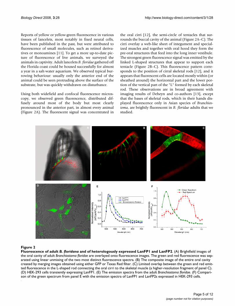
Reports of yellow or yellow-green fluorescence in varioustissues of lancelets, most notably in fixed neural cells,have been published in the past, but were attributed tofluorescence of small molecules, such as retinol deriva-tives or monoamines [11]. To get a more up-to-date pic-ture of fluorescence of live animals, we surveyed theanimals in captivity. Adult lancelets B. floridae gathered offthe Florida coast could be housed successfully for almosta year in a salt-water aquarium. We observed typical bur-rowing behaviour: usually only the anterior end of theanimal could be seen protruding above the surface of thesubstrate, but was quickly withdrawn on disturbance.
Using both widefield and confocal fluorescence micros-copy, we observed green fluorescence, distributed dif-fusely around most of the body but most clearlypronounced in the anterior part, in almost every animal(Figure 2A). The fluorescent signal was concentrated in
the oral cirri [12], the semi-circle of tentacles that sur-rounds the buccal cavity of the animal (Figure 2A–C). Thecirri overlay a web-like sheet of integument and special-ized muscles and together with oral hood they form thepre-oral structures that feed into the long inner vestibule.The strongest green fluorescence signal was emitted by thelinked L-shaped structures that appear to support eachtentacle (Figure 2B–C). This fluorescence pattern corre-sponds to the position of cirral skeletal rods [12], and itappears that fluorescent cells are located mostly within (orsheathed around) the horizontal part and the lower por-tion of the vertical part of the "L" formed by each skeletalrod. These observations are in broad agreement withimaging results of Deheyn and co-authors [13], exceptthat the bases of skeletal rods, which in their hands dis-played fluorescence only in Asian species of Branchios-toma, are brightly fluorescent in B. floridae adults that westudied.
Fluorescence of adult B. floridana and of heterologously expressed LanFP1 and LanFP2Figure 2Fluorescence of adult B. floridana and of heterologously expressed LanFP1 and LanFP2. (A) Brightfield images of the oral cavity of adult Branchiostoma floridae are overlayed onto fluorescence images. The green and red fluorescence was sep-arated using linear unmixing of the two most distinct fluorescence spectra. (B) The composite image of the entire oral cavity created by merging images obtained using either GFP or Texas Red filter. (C) Limited overlap between the green and red emit-ted fluorescence in the L-shaped rod connecting the oral cirri to the skeletal muscle (a higher-resolution fragment of panel C). (D) HEK-293 cells transiently expressing LanFP1. (E) The emission spectra from the adult Branchiostoma floridae. (F) Compari-son of the green spectrum from panel E with the emission spectra of LanFP1 and LanFP2s expressed in HEK-293 cells.
Page 5 of 12(page number not for citation purposes)

Biology Direct 2008, 3:28 http://www.biology-direct.com/content/3/1/28
Interestingly, we also observed clear red fluorescence sig-nal that was similarly concentrated around oral cirri, andstrongly overlapped with the areas of green fluorescence(Figure 2A–C and 2E). Most of the red signal wasrestricted to the cirral skeletal ring and oral cirrus justabove the cirral skeletal rods, but some of it extended tothe distal tips of the oral cirri (Figure 2C). Analysis of fullspectral information at each pixel in the image indicatesthat there was little pixel-to-pixel overlap between theareas associated with the green and red fluorescence, sug-gesting spatial segregation of red and green emitters, per-haps even cell-specific expression of each (though higheroptical resolution than our 3 μm would be needed for adefinitive proof of this).
In order to further study the properties of lancelet GFP-like genes and to relate them to the fluorescence observedin live animals, we have synthesized cDNAs for two of thelancelet genes that corresponded to the largest number ofdatabase ESTs, with codon frequencies optimized forexpression in the mammalian system, and transfectedthem into HEK 293 cells (Figure 2D). Cells expressingeach construct emitted green light when excited with bluelight. LanFP1 and LanFP2 were also expressed in E. coliand affinity-purified: both preparations exhibited maxi-mal absorption at 500 nm, and the emission spectra ofboth proteins were similar (λmax = 510 nm for LanFP1,and 516 nm for LanFP2). Molecular brightness of the twofluorescent proteins in transfected HEK 293 cells and inpurified samples was, however, significantly different,with LanFP1 much brighter than LanFP2. Analyses of theprotein preparations purified from E. coli showed that thequantum efficiency of LanFP1 was by the two orders ofmagnitude higher than that of LanFP2, and about half ofthe brightness of the potent Venus derivative of YFP(Additional file 2).
We exploited the fact that LanFP1 and LanFP2 haveslightly different excitation and emission spectra andcompared them with endogenous fluorescent signalsfrom the cirri. The emission spectrum from B. floridae hasa maximum at 521 nm, which is much closer to that ofLanFP2-expressing HEK-293 cells and of affinity-purifiedLanFP2 than to the corresponding values of LanFP1 (Fig-ure 2F).
Analysis of the EST libraries indicates that transcriptsencoding both LanFP1 and LanFP2 are abundant in eggsand embryos, though thus far no matching ESTs havebeen found in the libraries from adult animals. We didnot detect any fluorescent signals in the red spectrum inmammalian cells singly or doubly transfected withLanFP1 or LanFP2.
Days before the submission of this manuscript, 21 cDNAsequences encoding proteins of GFP family from a Euro-pean species B. lanceolatum were released by Genbank.They apparently include naturally occurring and engi-neered cDNAs, most of which display either green or redfluorescence [14]. There is no one-to-one correspondencebetween these sequences and the products of GFP-likegenes encoded by B. floridae genome.
ConclusionIn this work, we identified a family of genes encodingGFP-like proteins in lancelet B. floridae and expressed twoof them in bacterial and mammalian cell cultures. Bothproteins, LanFP1 and LanFP2, exhibit green fluorescenceof different brightness and distinct spectral propertieswhen expressed in mammalian cells, and they, along withat least four other GFP-like proteins, appear to beexpressed at various stages of lancelet development asjudged from the analysis of the EST libraries. Moreover,adult lancelets display overlapping but distinct patterns ofgreen and red fluorescence, with green fluorescenceshifted towards more basal regions of oral cirri and redfluorescence more prominent at the cirri tips.
Recently, Deheyn and co-authors reported the sequenceof LanFP2 and the pattern of green fluorescence in lance-let eggs, larvae and adults [1], and Israelsson [14] reporteda similarly diverse family of cDNAs expressing red andgreen fluorescent proteins in B. lanceolatus. It remains tobe investigated which of the GFP-like proteins encoded bythe Florida lancelet genome are responsible for fluores-cence observed by these authors and by us. Intriguingly,LanFP1 appears to be a bright GFP, whereas LanFP2 fluo-resces only dimly in vitro and in heterologous expressionsystems. At the same time, its emission spectrum is theclosest match to the emission of the bright fluorescenceobserved in live adult animals. Gene-specific probes nowavailable for individual LanFPs will help to establish theidentity of green and red emitters at the various stages oflancelet development.
Phylogenetic inference and analysis of conserved intronpositions allow us to tentatively place a GFP-like proteininto the common ancestor of metazoan animals. The rela-tionship between GFPs and structurally similar G2domains, however, remains unclear. The similaritybetween the spatial structures of representative of the twofamilies makes them amenable to structure-based super-imposition, and phylogenetic trees based on such align-ments have been presented [13]. These trees, however, donot prove the fact of sequence homology in the first place.Using HHpred, which is one of the most sensitivesequence comparison programs and does not rely explic-itly on spatial information [15] and the sequence modelof G2 domain family derived from pfam07474, we
Page 6 of 12(page number not for citation purposes)

Biology Direct 2008, 3:28 http://www.biology-direct.com/content/3/1/28
observed several matches to cnidarian fluorescent pro-teins, with the borderline HHpred P-values from 0.0017to 0.05. If this borderline sequence similarity, with addi-tional consideration of the same number of β-strands intwo families, is taken as the indication of their commonancestry, then the origin of nidogen G2 domain appearsto postdate the emergence of GFPs, as G2 domainhomologs are detected in nematodes and insects but donot appear in primitive metazoans, such as nearly-com-pletely sequenced cnidarian N. vectensis and extensivelycovered flatworm Schmidtea mediterranea. G2 domains arealso more diverse in sequence than the GFP-like proteins.If the former have evolved from the latter, the constraintson the sequence of a GFP-like protein, which has to ena-ble the maturation of a chromophore or a fluorophore,have apparently been lifted in the G2 domains, whichbecame coopted into a mutidomain polypeptide thatmediates protein-protein interactions in the basal mem-brane of metazoa. Alternatively, GFP and G2 families mayshare a common β-barrel ancestor and may have other,still unrecognized relatives in present-day organisms.
Biological functions of GFPs remain elusive. It has beenspeculated that GFPs in cnidaria may be involved inquenching the UV irradiation [16] and/or in camouflag-ing coloration against predators [17]. Both hypotheses arecompatible with the fact the zone of the brightest fluores-cence in lancelets corresponds to the anterior part of theadult animal that is exposed during feeding. On the otherhand, lancelets feed on plankton organisms, which dis-play phototaxis, and it is possible that the signal emittedby these proteins serves as bait. On a more practical note,the study of lancelet ESTs and live imaging of animalsexpands the repertoire of GFP-like proteins, potential sen-sors and reporters for biological experimentation.
MethodsAnimal husbandryAdult lancelets (B. floridae) were collected by Marine Bio-logical Supply (Panacea, Florida) at a depth of 10–12 ft atthe GPS coordinates N29 degrees, 55.454'/W084 degrees,15.676' (Apalachee Bay, Wakulla County, FL). They werehoused in glass aquariums at pH 8.2–8.4, salinity 33–34ppt, water temperature 18–19°C, negligible levels ofammonia, nitrite and nitrate, copper and phosphate, on asubstrate of either washed, screened play sand or glassbeads. A 10% water change was performed once a week. Amix of liquid planktonic foods available commercially forfilter feeders was fed daily via a dosing pump. Animalswere housed in a room with a lighting level of 30 ft. can-dles and a 12:12 photoperiod.
MicroscopyImages were collected using a Zeiss LSM-510 confocalhead mounted on a Zeiss axiovert 200 m microscope with
AIM acquisition software (Carl Zeiss, Thornwood, USA),coupled to a Chameleon Ultra II IR laser source (Coher-ent, San Jose) for two photon excitation. Spectral informa-tion was acquired using the Meta channel on the ZeissLSM-510 set to 10 nm resolution with 488 nm excitation.Single channel green image were collected using a confo-cal microscope equipped with a laser with 488 excitationfilter and a 500 to 530 band pass emission filter. Imageswere acquired with a water immersion objective, 20 × 0.8NA c-apochromat for the whole animals or 40 × 1.2 NA c-apochromat for cell cultures. For live imaging experi-ments, animals were mounted in a 25 by 60 mm Nuncchamber with a number 1.5 coverslip. To minimize ani-mal movement, lancelets were anesthetized with bufferedMS-222 solution (Tricaine Methanesulfonate) at a con-centration of 350 mg/L.
SpectroscopyAbsorption spectra for purified proteins were obtainedusing a SOFTmax UV/VIS spectrometer (MolecularDevices). Emission spectra were obtained using a Horiba/Jovan fluoromax fluorescence spectrometer. Emissionscans were recorded by exciting the sample at lmax forabsorption with 5 nm slit. Molecular absorptivity andquantum yield were measured as in [18] and [19]. Theprotein concentration for absorbivity measurements wasdetermined by BCA assay and by fluorescence correlationspectroscopy [20].
BioinformaticsSequence similarity searches were performed using thegapped BLAST and PSI-BLAST family of programs [21].Multiple sequence alignments were done using the MUS-CLE program [22]. The JTT distance matrix of proteinsequences was constructed using the PROTDIST programand the neighbour-joining phylogenetic tree was con-structed using the NEIGHBOR program of the Phylippackage [23]. The statistical support of the nodes wasassessed by making 100 bootstrap replicates of the alignedsequences, building 100 trees, making the consensus tree,and marking all nodes in the original tree that were sup-ported by more than 50% of the bootstrapped replicates.HMM-to-HMM matching was done using the HHpredserver [15].
Cloning and expression of LanFP1 and LanFP2LanFP1 and LanFP2 coding sequences have been initiallyassembled from the B. floridana EST libraries as two of theproteins supported by the largest number of EST. Prior tothe release of the lancelet genome assembly, there were sixnucleotide positions in the LanFP1 coding sequenceshowing variations between the ESTs in the database, andwe used the most frequently conserved nucleotide forthese positions. The sequences were subsequently verifiedby comparing them to the genome assembly. To optimize
Page 7 of 12(page number not for citation purposes)

Biology Direct 2008, 3:28 http://www.biology-direct.com/content/3/1/28
the codons for expressing in mouse and other mamma-lian systems, we reverse translated the LanFP1 and LanFP2into DNA using a standard mouse codon set. For ease ofcloning, we included HindIII and BamHI restriction sitesat 5' and 3' ends of both sequences. The gene was synthe-sized by Bioclone, Inc. (San Diego, CA). The synthesizedgene was cloned into Sigma's p3XFlag-Myc-CMV plasmidHindIII and BamHI sites, which contained a 5' Flag and a3' Myc tags plus a stop codon on the vector backbone. Thesequence for optimized sequences can be found in Addi-tional File 3.
Cell Culture and TransfectionHuman embryonic kidney (HEK) 293 cells were culturedin the minimum essential medium (MEM) (Invitrogen)supplemented with 5% fetal bovine serum (FBS) and 2mM glutamine and were maintained at 37°C in a humid-ified environment of 5% CO2. The cells were plated on 25mm round coverslips coated with poly-D Lysine (Sigma)24 to 48 hours and transfected with DNA plasmids whencells reached 70–80% confluence. Plasmid DNA used fortransfection was obtained using the HiSpeed maxi-prepkit (Qiagen) and repurified by sodium acetate and etha-nol precipitation. 2 μg of plasmid DNA was mixed with12 μg of Nupherin (Biomol Research Laboratories, Ply-mouth Meeting, PA) in 300 μl of MEM containing no FBSor antibiotics for 15 min and then combined with 300 μlof MEM containing 6 μl of LipofectAMINE 2000 (Invitro-gen) for another 15 min at room temperature. The culturemedium was replaced with 600 μl of transfection mediumcontaining the LipofectAMINE-Nupherin-DNA complex.After incubating for 0.5 to 1 hour, the transfectionmedium was replaced with 2 ml of culture medium. Cellimaging occurred 24–72 hours post transfection.
Protein purificationBL-21 Ecoli cells were transformed with LanFP1 or LanFP2subcloned into pRSET-B bacteria expression vector (Invit-rogen, Carlsbad, CA) in frame with 6 histidine tags. Over-night cultures were grown to OD 0.4 prior to induction.Bacterial cultures were induced by the addition 1 mMIPTG, grown to OD 0.8, lysed and the His-tagged proteinwas extracted using a Ni-agarose beads solution (Qiagencat no. 30210). Concentration of purified protein wasestablished using a BCA assay (Pierce, cat no 23225) andfluorescence correlation spectroscopy.
List of abbreviationsGFP: Green fluorescent protein.
Competing interestsThe authors have a financial interest in patent applica-tions which cover some of the work described in thepaper.
Authors' contributionsThe order of authors is alphabetical. AM, JS and CRYdesigned research and wrote the manuscript. All authorsperformed research, analyzed the data, read and approvedthe final manuscript.
Reviewers' commentsReviewer 1: S. SunyaevThis manuscript describes a discovery of a family of GFP-like proteins in the newly sequenced genome of a lanceletB. floridae. The authors expressed two proteins anddetected fluorescent signals of different brightness andwavelengths. Phylogenetic analysis that included the anal-ysis of intron position conservation suggested an earlymetazoan origin of GFPs followed by a massive parallelloss in multiple lineages. The manuscript also discussesthe potential evolutionary relationship between structur-ally similar G2 domains and GFPs.
Reviewer 2: M. Matz (nominated by I. King Jordan)Unfortunately, the novelty edge has been taken away fromthis work by the prior note by Deheyn et al. of green fluo-rescent proteins in three species of lancelets (Biol. Bull.213: 95–100, 2007). Deheyn et al already discussed thephylogenetic relationships of one of the lancelet GFPs,including its relationships with G2 domain proteins, anddescribed in vivo fluorescence of lancelets. Still, this papercontains substantially more material than the short noteby Deheyn et al. First, the whole family of GFP-like pro-teins in one genome was surveyed. Second, two of the 12detected genes have been heterologously expressed andspectroscopically characterized. Third, and this is the mostintriguing information for me, red fluorescence has beenobserved in the lancelet. All these are valuable contribu-tions to our knowledge of diversity and evolution of GFP-like proteins.
I regret, however, that the authors did not go as far as toexpress and characterize all 12 family members found inthe lancelet genome. This would have been a logicallycomplete study, and the first one to describe the full com-plement of GFP-like proteins from an organism.
Authors' response: We are doing it now with all B. flori-dae family members and, for good measure, with thehomologs from sea anemone Nematostella vectensisthat have been revealed in the genome project but, asfar as we know, were not characterized before.
In addition to possible identification of the red fluores-cent protein, such study might have turned up interestingpatterns related to evolution and/or deterioration of genefunctions in multigene families. In this regard, it is reallyintriguing that the two characterized proteins differ somuch in their spectral properties. Whereas LanFP1 is a
Page 8 of 12(page number not for citation purposes)

Biology Direct 2008, 3:28 http://www.biology-direct.com/content/3/1/28
rather generic GFP, LanFP2 is dimmer because of its lowquantum yield but instead possesses impressively highmolar extinction coefficient, on par with anthozoan pur-ple-blue chromoproteins that are known for their strikingcolor appearance. I would mention in the discussion thatthis difference in spectral properties may be the result offunctional sub-specialization that maintained both genesin the lancelet genome after duplication. Also, given itsvery high molar extinction, I would expect that LanGFP2protein should look quite colorful (not necessarilygreen!) in solution. Is that so?
Authors' response: Yes! LanFP2 solution is yellow-brown.
I am rather surprised that the authors doubt the homol-ogy of G2 domains and GFPs. In addition to the fold sim-ilarity and same collection of the secondary structureelements, these proteins also have the elements in identi-cal (and far from trivial) order within their primary struc-ture. Importantly, this order does not have to beconserved for the protein to work, as has been demon-strated by successful circular permutation of several GFP-like proteins. I believe therefore that in this case, completeidentity of the fold including the composition and orderof secondary structure elements constitutes a far strongerevidence of homology than any structure-oblivious algo-rithm may provide.
Authors' response: Unfortunately, there is no acceptedquantitative model of structural similarity that can beused for testing evolutionary hypotheses in theabsence of sequence signal – similar number and sim-ilar topology of strands is certainly something worthknowing about, but this qualitative argument cannot(yet) be translated in a hard number, the way the"structure-oblivious" sequence similarities can be. Onthe other hand, we have nothing against the sugges-tion that nidogen G2 domain and GFP share a com-mon ancestor. In fact, the relevant paragraph in theConclusions section includes evolutionary scenariosthat presume the existence of just such an ancestor, onthe basis of exactly the evidence that the Reviewerhighlights. Note also related comments by L. Aravind.
I also have a few minor questions, listed below.
1. How was the fluorescence measured in live animals?
Authors' response: The fluorescence was measured usinga LSM-510 confocal microscope. The animals wereimmobilized with a muscle inhibitor and images col-lected using 488 nm laser excitation and spectral imag-ing with either a 20× or 40× objective (see Methods formore detail).
2. What were the excitation wavelengths for "green" and"red spectra", Fig. 2E?
Authors' response: The excitation wavelength was 488nm with an argon laser for both green and red spectra.The red spectrum can also be excited with a 561 nmlaser. The two-photon excitation scan indicates thegreen fluorescence is produced when excited with 720to 1020 nm light. The red fluorescence was not appar-ent until using two-photon excitation above 980 nmlight.
3. The "red spectra" does not look like any of the GFP-likeproteins known thus far. What can you say about it?
Authors' response: The spectrum does not provide anyinformation about the potential source of the red flu-orescence, i.e., even whether it is a protein or a smallmolecule. Note, however, the claims in a very recentpatent (ref. 14) about proteins from a related speciesof Branchiostoma.
4. I can't help noticing that the "green spectra" are differ-ent in Fig. 2E and 2F, although they are supposed to be thesame. Why?
Authors' response: They are identical. We hope thatreformatted figure makes this clearer.
5. I'd like to see excitation spectra for the expressed pro-teins (augment Fig. 2F).
Authors' response: These data are presented in the addedSupplementary Figure S4 (Additional File 4). Theywere collected using fluorescence spectrometer with 1nm resolution (i.e., higher than the 10 nm-resolvedspectra obtained on LSM-510).
6. Does lancelet genome contain G2 domains? If not,please discuss that, if yes, please put them into the phyl-ogeny.
Authors' response: Yes, it does. These sequences are sol-idly within the chordate clade, not interminglingmin-gling with deeper-branching invertebrate homologs inthe same tree. Thus, this domain is clearly distinctfrom GFP domain, and adding G2 sequences to thetree would not help to better illustrate or to refute anypoint that we are trying to make in this article: in orderto include a sequence into the alignment that is usedfor evolutionary inference, homology needs to beproven already (see response on the monophyly ques-tion above and also to L. Aravind comment below).
Page 9 of 12(page number not for citation purposes)
Biology Direct 2008, 3:28 http://www.biology-direct.com/content/3/1/28
7. Please make the phylogenetic tree one of main figures.I would also appreciate if the tree was reformatted into amore conventional form.
Authors' response: We feel that the tree is too trivial toadd much to discussion (in fact, except for the addi-tion of more Branchiostoma and Nematostellasequences, it is nearly identical to the tree shown inref. 1), but we now provide, as Additional File 5, twotrees in Newick format: 1. the main neighbour-joiningtree, which contains branch length information, and2. the consensus of 100 neighbour-joining trees builton the basis of the bootstrap replicates of the first tree,which contains the information on the bootstrap sup-port for each partition in the first tree.
8. The role of fluorescence in attracting food is an interest-ing proposition, but why it is important particularly dur-ing twilight? I don't see the connection.
Authors' response: Phytoplankton and zooplanktonexhibit phototaxis, and different parts of spectra maybe most effective at different times of day and at differ-ent depths in attracting food from the surface. Weagree that there is no particular evidence that wouldcompel one to focus on twilight, so we deleted "duringtwilight".
Reviewer 3: L. AravindThe paper by Baumann et al describes the recovery of sev-eral new GFP related proteins in amphioxus (B. floridae)and experimental characterization of a few of them. Thepoint of note is the proposal that that GFP was present inthe common ancestor of Bilateralia and Cnidaria andrepeatedly lost in various animal lineages. Despite theextremely sporadic distribution of these proteins, the phy-logenetic analysis and the argument of conserved intronpositions support this scenario. Yet, it might be useful tocheck environmental metagenomic sequences to addressthe possibility of independent lateral transfer from someunknown source into the few animal lineages in whichGFPs are found.
Authors' response: We searched metagenomicsequences, but found only the garden-variety GFPsclosely identical to the founder from A. victoria, only inmarine samples and represented by small number ofcollapsed reads. We think they are encoded by hydroidDNA present in some of these samples.
Even more striking is the proposal that the nidogen G2domains have emerged from the GFP like family after thedivergence of cnidarians and possibly flatworms from theremaining metazoans. This proposal is weakly supported.Given that fact that sequence similarity between the G2
domain and GFP is only detected using the most sensitiveprofile-profile comparison method, it is entirely possiblethat there remain undetected β-barrel proteins with com-parable topologies and geometry that are the precursors ofboth GFP and G2 domains. While this may sound lessparsimonious, the high levels of sequence divergenceobserved in various β barrel folds (especially when thereno comparable functional constraints) do make it a dis-tinctive possibility.
Authors' response: We agree that this is a possibilityworth discussing, and we amended the text accord-ingly. Note, however, that we have not seen any G2domains in complete proteome of sea anemoneNematostella or in the available sequences of planarianSchmidtea or any other primitive metazoan clade.Thus, if the common 11-stranded barrel ancestor ofG2 domain and of the present-day GFPs was not itselfpart of the GFP family, it either had to spawn GFP andG2 domains on separate occasions, or to split intoGFP and G2 followed by at least one, or possibly manylosses, of G2 (in Anthozoa and perhaps inPlathelminthes). Better sequence sampling of sponges,coelenterates and flatworms would help to sort outthese possibilities.
Minor points: -Pg 10 "At this level of significance, GFPsdid not stand out from the background of other β-barrelproteins." What were the other β barrel proteins detectedby HHpred and their p-values? Was a reciprocal searchdone using the GFP profiles as queries against a profiledatabase lacking GFPs?
Authors' response: This paragraph was a bit too convo-luted in the initial version. Current HHPred server(release 2.2.1) does not report GFP-G2 similarity if thecomplete database is used as the search space. Thesame negative results are obtained with the stand-alone version 1.5.0 whether or not the selfsame mod-els are removed from the database. The matchesbetween G2 and GFP can be only observed when wesearch the subset of HMMs built upon proteins withthe known three-dimensional structures. In that case,the G2 model finds the best GFP model with p-value0.0017, and other GFP-related models have p-valuesfrom 0.002 to 0.005. An apparently unrelated β-barrelfamily, specified by At1g79260 protein from Arabidop-sis thaliana, is seen with p-value 0.039, though thealignment is only partial and reciprocal search doesnot detect the G2 domain. G2 model also finds bacte-rial semi-barrel LolB in searches against the completedatabase (p-value 0.022: interestingly, LolB also has11 strands). GFP does not detect G2 even under theseforced conditions. All these weak similarities, how-ever, only reinforce our main argument, i.e., that we
Page 10 of 12(page number not for citation purposes)

Biology Direct 2008, 3:28 http://www.biology-direct.com/content/3/1/28
still do not have a robust statistical validation of theGFP-G2 monophyly at the sequence level. Thus, inter-ested as we are in discussing scenarios that assume acommon ancestor, we can do it only hypothetically.We have modified the Conclusions section with this inmind.
What is the evidence that the red fluorescence comes froma GFP like protein and not some other fluorochrome?
Authors' response: This question was also raised by M.Matz, see our response (the short answer is: no evi-dence, but see ref. 14).
Additional material
AcknowledgementsThis study was funded by Stowers Institute for Medical Research. We are grateful to the members of Stowers Aquatics team for help with the labo-ratory animals.
References1. Prasher DC, Eckenrode VK, Ward WW, Prendergast FG, Cormier
MJ: Primary structure of the Aequorea victoria green-fluores-cent protein. Gene 1992, 111:229-233.
2. Chalfie M, Tu Y, Euskirchen G, Ward WW, Prasher DC: Green flu-orescent protein as a marker for gene expression. Science1994, 263:802-805.
3. Shaner NC, Steinbach PA, Tsien RY: A guide to choosing fluores-cent proteins. Nat Methods 2005, 2:905-909.
4. Chudakov DM, Lukyanov S, Lukyanov KA: Fluorescent proteins asa toolkit for in vivo imaging. Trends Biotechnol 2005, 23:605-613.
5. Giepmans BN, Adams SR, Ellisman MH, Tsien RY: The fluorescenttoolbox for assessing protein location and function. Science2006, 312:217-224.
6. Shagin DA, Barsova EV, Yanushevich YG, Fradkov AF, Lukyanov KA,Labas YA, Semenova TN, Ugalde JA, Meyers A, Nunez JM, WidderEA, Lukyanov SA, Matz MV: GFP-like proteins as ubiquitousmetazoan superfamily: evolution of functional features andstructural complexity. Mol Biol Evol 2004, 21:841-850.
7. Hopf M, Göhring W, Ries A, Timpl R, Hohenester E: Crystal struc-ture and mutational analysis of a perlecan-binding fragmentof nidogen-1. Nat Struct Biol 2001, 8(7):634-40.
8. Nguyen HD, Yoshihama M, Kenmochi N: New maximum likeli-hood estimators for eukaryotic intron evolution. PLoS ComputBiol 2005, 1:e79.
9. Carmel L, Rogozin IB, Wolf YI, Koonin EV: Patterns of intron gainand conservation in eukaryotic genes. BMC Evol Biol 2007,7:192.
10. Sverdlov AV, Rogozin IB, Babenko VN, Koonin EV: Conservationversus parallel gains in intron evolution. Nucleic Acids Res 2005,33:1741-1748.
11. Obermüller-Wilén H, van Veen T: Monoamines in the brain ofthe lancelet, Branchiostoma lanceolatum. A fluorescence-his-tochemical and electron-microscopical investigation. Cell Tis-sue Res 1981, 221:245-256.
12. Ruppert EE: Cephalochordata (Acrania). In Microscopic Anatomy ofInvertebrates. Hemichordata, Chaetognatha, and the Invertebrate Chor-dates Volume 15. Edited by: Harrison FW, Ruppert EE. Wiley-Liss;1997:349-503.
13. Deheyn DD, Kubokawa K, McCarthy JK, Murakami A, Porrachia M,Rouse GW, Holland ND: Endogenous green fluorescent pro-tein (GFP) in amphioxus. Biol Bull 2007, 213:95-100.
14. Israelsson O: Fluorescent proteins and genes encoding them.Patent SE . 0601261-1 08.06.2006/US 60/811,769 08.06.2006
15. Söding J, Biegert A, Lupas AN: The HHpred interactive serverfor protein homology detection and structure prediction.Nucleic Acids Res 2005, 33:W244-W248.
16. Salih A, Larkum A, Cox G, Kühl M, Hoegh-Guldberg O: Fluorescentpigments in corals are photoprotective. Nature 2000,408:850-853.
17. Matz MV, Marshall NJ, Vorobyev M: Are corals colorful? PhotochemPhotobiol 2006, 82:345-350.
18. Lakowicz JR: Principles of fluorescence spectroscopy Kluwer 1999.19. Merzlyak EM, Goedhart J, Shcherbo D, Bulina ME, Shcheglov AS, Frad-
kov AF, Gaintzeva A, Lukyanov KA, Lukyanov S, Gadella TW, Chuda-kov DM: Bright monomeric red fluorescent protein with anextended fluorescence lifetime. Nat Methods 2007, 4:555-557.
20. Brown CN, Dalal RB, Hebert B, Digman MA, Horwitz AR, Gratton E:Raster image correlation spectroscopy (RICS) for measuringfast protein dynamics and concentrations with a commerciallaser scanning confocal microscope. J Microsc 2008,229(1):78-91.
21. Altschul SF, Madden TL, Schäffer AA, Zhang J, Zhang Z, Miller W, Lip-man DJ: Gapped BLAST and PSI-BLAST: a new generation ofprotein database search programs. Nucleic Acids Res 1997,25:3389-3402.
22. Edgar RC: MUSCLE: a multiple sequence alignment methodwith reduced time and space complexity. BMC Bioinformatics2004, 5:113.
Additional file 1Phylogenetic tree of GFP-like proteins. The image of the neighbour-joining tree inferred from the alignment of GFP-like proteins.Click here for file[http://www.biomedcentral.com/content/supplementary/1745-6150-3-28-S1.doc]
Additional file 2Spectral properties of LanFP2 and LanFP2. The table summarizing absorbance, emission, extension and quantum yield for several GFP-like proteinsClick here for file[http://www.biomedcentral.com/content/supplementary/1745-6150-3-28-S2.pdf]
Additional file 3Murinized sequences of LanFP1 and LanFP2. Nucleotide sequences encoding two B. floridae fluorescent proteins, with codon usage optimized for expression in murine cellsClick here for file[http://www.biomedcentral.com/content/supplementary/1745-6150-3-28-S3.doc]
Additional file 4Normalized absorbance spectra of LanFP1 and LanFP2. Image of spectra of two B. floridae fluorescent proteins compared with several other spectra of GFP-like proteinsClick here for file[http://www.biomedcentral.com/content/supplementary/1745-6150-3-28-S4.jpeg]
Additional file 5Source file of the tree shown in Additional file 1. Newick-formatted trees, one of which contains branch length information, and the other is the con-sensus of 100 trees built on the basis of the bootstrap replicates of the first tree, contains the information on the bootstrap support for each partition in the first tree but lacking the branch length information.Click here for file[http://www.biomedcentral.com/content/supplementary/1745-6150-3-28-S5.txt]
Page 11 of 12(page number not for citation purposes)

Biology Direct 2008, 3:28 http://www.biology-direct.com/content/3/1/28
Publish with BioMed Central and every scientist can read your work free of charge
"BioMed Central will be the most significant development for disseminating the results of biomedical research in our lifetime."
Sir Paul Nurse, Cancer Research UK
Your research papers will be:
available free of charge to the entire biomedical community
peer reviewed and published immediately upon acceptance
cited in PubMed and archived on PubMed Central
yours — you keep the copyright
Submit your manuscript here:http://www.biomedcentral.com/info/publishing_adv.asp
BioMedcentral
23. Felsenstein J: PHYLIP (Phylogeny Inference Package) version3.67. In Distributed by the author Department of Genome Sciences,University of Washington, Seattle; 2005.
Page 12 of 12(page number not for citation purposes)
Copyright © 2022 FDOKUMEN