Morphological characterization of GFP stably transfected adult mesenchymal bone marrow stem cells
261
ISSN 1648–6897 print ISSN 1822–4199 online
JOURNAL OF ENVIRONMENTAL ENGINEERING AND LANDSCAPE MANAGEMENT
http:/www.jeelm.vgtu.lt/en
2007, Vol XV, No 4, 261–268
DEVELOPMENT OF A GFP-BASED BIOSENSOR FOR DETECTING THE BIOAVAILABILITY AND BIODEGRADATION OF
POLYCHLORINATED BIPHENYLS (PCBs)
Xuemei Liu, Kieran J. Germaine, David Ryan and David N. Dowling* Dept of Science and Health, Institute of Technology Carlow, Kilkenny Road, Carlow, Ireland
*Institute of Technology Carlow, Kilkenny Rd., Carlow, Rep. Ireland E-mail: [email protected]
Submitted 14 May 2007; accepted 18 June 2007
Abstract. Two whole-cell biosensors were constructed to detect the in situ biodegradation of polychlorinated biphenyl by chromosomal insertion of a mini-Tn5-Kmr-Pm::gfp[mut3]-T0-T1 construct into P. fluorescens. In vitro tests showed that the expression of the Pm promoter depended on the growth phase of the biosensors and the concentration of chemical in-ducers; chlorinated benzoic acid derivatives. A linear relationship between the fluorescent intensity and the log10 concent-ration of the inducer was observed. One biosensor (F113L::1180gfp) had the ability to degrade PCBs to relevant chloro-benzoic acid derivatives and to induce expression of Gfp. The second biosensor (F113gfp), which cannot degrade PCBs, shows fluorescence after induction by chloro-benzoic acid derivatives. By using these two biosensors, PCB degradation could be detected in vitro and in soil.
Keywords: biosensor, Pseudomonas fluorescens F113, PCBs, rhizosphere, root colonisation.
1. Introduction
Polychlorinated biphenyls (PCBs) are a group of chlorinated aromatic compounds, which are among the most widely identified environmental contaminants persi-sting in the biosphere [1].
Reporter genes are widely used as genetic tools for quantification and detection of specific cell populations, gene expression and root colonisation in complex sam-ples [2–4]. Common reporter genes include bacterial bioluminescence (lux) genes, the colorimetric β-galacto-sidase (lacZ) gene and genes coding for green fluorescent proteins (GFPs) from the jelly fish Aquoria victoria. The GFPs have been successfully used as reporters within bacterial and yeast hosts due to the ease of in situ detec-tion and the minimal metabolic cost to the host cells [5–6]. Novel areas for applying these reporter genes have been documented and include their use in constructing whole-cell biosensors as specific and sensitive sensing devices for measuring biologically relevant concentra-tions of pollutants [7–9]. These biosensors rely on analy-sis of gene expression, typically by creating transcrip-tional fusions between a promoter of interest and the reporter gene. The extent of reporter gene expression serves as a measure of the availability of specific pollu-tants in complex environments. Biosensors developed using this strategy have been reported for monitoring of benzene, toluene, ethylbenzene, and/or xylene using tod-luxCDABE whole-cell reporter in the contaminated sam-ple [10].
The well-characterised biocontrol strain Pseudomo-nas fluorescens F113, which was originally isolated from the rhizosphere of sugar beet [11], is an excellent root coloniser and was chosen for gene modification for the degradation of PCBs. The genes responsible for PCB degradation, termed the bph operon from Burkholderia xenovorans LB400 [12], were modified by the cloning of a strongly inducible constitutive (nod box) promoter from Sinorhizobium meliloti upstream of the bph operon in the plasmid pDDPCB and subsequent chromosomal integra-tion into P. fluorescens F113riflacZY, creating P. fluo-rescens F113L::1180 [13]. This strain was superior to P. fluorescens F113rifpcb [14], originally created by the insertion of the bph operon into F113, for the growth on biphenyl and for the biodegradation of PCBs [13].
A number of gfp-based P. fluorescens F113 biosen-sors were previously constructed by using fusions of the Escherichia coli rrnBP1 ribosomal promoter and gfp genes. These biosensors were able to monitor the single-cell localization and activity of P. fluorescens F113 colo-nizing alfalfa roots. The monitoring systems permitted non-destructive in situ detection of cells on the entire root system grown in both the presence and absence of 3-chlorobiphenyl [15].
In this study, two whole-cell biosensors were const-ructed by the chromosomal insertion of a promoter-reporter construct, xylSPm::gfpmut3 [2] into P. fluores-cens F113rif and P. fluorescens F113L::1180 to create P. fluorescens F113gfp and P. fluorescens F113L::1180gfp, respectively. The Pm promoter in this construct is derived
X. Liu et al. / JEELM – 2007, Vol XV, No 4, 261–268
262
Fig 1. The conceptual basis of the biosensors. Biosensor F113PCB1180gfp is able to report the biodegradation of PCBs by switching on the signal in response to end products of PCB degradation. The second biosensor F113gfp serves as a control and only responds to external chlorobenzoic acids (CBAs)
from the TOL plasmid and regulates the meta-pathway of aromatic hydrocarbon degradation. The meta-pathway is induced by chloro-benzoic acid derivatives and this in-duction is mediated by the substrate-activated XylS pro-tein. By combining these two biosensors, PCB degrada-tion can be detected in situ (Fig 1). The initial characte-risation of these biosensors is described in this report.
2. Materials and methods
2.1. Bacterial culture conditions
Bacterial strains were cultivated at 30 °C on Luria-Bertani (LB) medium, Pseudomonas minimal medium (MM) [14] supplemented with 10 mM sodium citrate or Pseudomonas minimal medium amended with 10 mM crystal biphenyl. The Escherichia coli strains were grown at 37 °C with constant shaking in LB medium. Antibiotics were routinely added at the following concentrations: ka-namycin (Kan) 50 μg ml–1 and rifampicin (Rif) 50 μg ml–1.
To detect the activity of the biosensors in vitro, 100 ml of MM supplemented and 1mM chemical indu-cers were placed into 500 ml flasks. These flasks were inoculated with 1×10⁸ bacterial cells. Soil experiments were set up by inoculating 1×10⁸ free cells per gram soil.
2.2. Construction and characterisation of Gfp biosensor strains
The pJBA26 [2] plasmid carrying the mini-Tn5-kmr-Pm::gfpmut3-T0-T1 transposon was introduced into P. fluorescens F113 derivatives by triparental matings, as described previously [16]. The selection plates were LB supplemented with Rif and Kan. After mating, the trans-conjugant colonies were purified and tested by spraying with 2, 3-dihydroxybiphenyl to detect the presence of an active bphC gene product, thereby confirming the presen-ce of the bph operon [14].
The insertion of the gfp gene was confirmed by PCR using primers Pgfp(up) 5’-GGTCTAGATGTGTGAAA TTGTTATCCG-3’ and Pgfp(down) 5’-CTCTCAAGC TTATTTGTATAGTTCATCCATGC -3’ [17]. PCR amp-lification experiments were performed in a Hybaid PCR Machine (Hybaid Limited). 50 μl reaction mixtures con-tained 50 ng DNA, 5.0 μM of each primer, 200 μM dNTPs, 3.0 μl 1.5 μM MgCl2, 5.0 μl 10 × reaction buffer and 0.625 U of Taq Polymerase (All reagents supplied by Promega, Madison, Wis). After an initial denaturation for 2 min at 95 °C, a total of 35 cycles were performed using the following program: 95 °C for 30 sec, annealing at 56 °C for 1 min, extension at 72 °C for 1 min, with a final termination step of one cycle of 7 min at 72 °C.
Southern blotting analysis was carried out to detect the chromosomal insertion of the gfp gene and the loca-tion of the insertion. Undigested and HindIII-digested genomic DNA from F113rif, F113gfp, F113L::1180, F113L::1180gfp and pJBA26 were run on a 1 % (w/v) agarose gel. The DNA was blotted by capillary transfer onto a nylon membrane (Boehringer Mannheim). The gfp DNA probe for Southern hybridisations was prepared by the incorporation of DIG-11-dUTP (Boehringer Mann-heim) during PCR using Pgfp(up) and Pgfp(down) primers and plasmid DNA of pJBA26 as a template. The DIG-bphC probe was made during PCR using bphC LCC forward and reverse primers [18] and genomic DNA of F113L::1180 as a template. Hybridisation using the DIG-gfp probe was performed at 42 °C overnight in hybridisa-tion buffer (Roche). The membrane was incubated for 1 h with 1: 5000 dilution of alkaline phosphatase-conjugated anti-digoxigenin antibody. Colorimetric detection of digoxigenin-labelled DNA fragments was carried out using NBT/BCIP (Roche) as the substrate for alkaline phosphatase. The membrane was inoculated for 16 h and photographed. To perform re-probing experiments, the membrane was washed using formamide for 1 h and re-
X. Liu et al. / JEELM – 2007, Vol XV, No 4, 261–268
263
probed with a DIG-bphC probe following the same pro-cedure. Restriction analysis was carried out based on the mini-Tn5-kmr-Pm::gfpmut3-T0-T1 transposon and bph operon.
The growth rate in LB, SA broth [19], MM plus suc-cinate (10 mM) and MM plus sodium citrate of F113L::1180gfp and F113gfp and growth rate in MM plus biphenyl of F113L::1180gfp were tested and com-pared to their parental strains. Their ability to utilise vari-ous carbon sources was tested by comparing the transconjugants to their parental strains using the gram-negative Biolog® NT plates to ensure that the gfp trans-poson had not randomly inserted into genes involved in the major catabolic pathways of the strains.
2.3. Epifluorescent microscopy and spectrofluoremetry
The biosensor cells from liquid cultures were visua-lised using a Nikon E400 epifluorescent microscope equipped with a 100 W mercury short arc photo-optic lamp and two filters with the excitation wavelength of 465–495 nm and 450–490 nm, respectively. Lucia® ima-ging software (version 4.6) was used to capture and pro-cess microscopic images. Fluorescent intensity from biosensor cells was measured as described previously [20] with a Hitachi fluorescent spectrophotometer Model F-2000 set at an excitation wavelength of 475 nm and emission detection at 511 nm. Relative fluorescence unit (RFU) was defined as the culture fluorescence intensity relative to culture biomass at OD600nm (measured using a Hitachi U-2001 Spectrophotometer).
2.4. Induction experiments
Growth phase-dependent and exposure time-dependent gfp expression in the biosensors was tested by growing both of the biosensors in MM + 10 mM sodium citrate broth with different concentrations of 3-CBA. The OD600nm values and fluorescent intensity were measured at 2 h intervals over a 24 h period. Dose-dependent experiments were carried out by centrifuging 2 ml of stationary phase biosensor cells which were grown in MM supplemented with 10 mM sodium citrate, for 5 min. at 3,000 × g, and mixing with 2 ml of benzoate derivative compounds to final concentrations ranging from 0.1 ppm to 1000 ppm and inoculated at room temperature (RT) for 6 h. Single fluorescent cells were examined by epifluo-rescent microscope.
2.5. Biosensor activity in vitro
Biosensor strains to detect PCB degradation: F113L:: 1180gfp was inoculated into MM + 10 mM sodium citrate supplemented with 1mM 3-CBP, 2, 3-dichloribiphenyl (2,3-DiCBP) or 3,5-dichloribiphenyl (3,5-DiCBP) liquid media for 5 d. RFU was measured after the culture grew to stationary phase; F113L::1180gfp was also inoculated in 1 mM 3-CBP spiked soil at room temperature for 10 d. Strain F113gfp was inoculated as control in both liquid and soil experiments.
To count the percentage of gfp fluorescent cells by epifluorescent microscopy, two methods were employed. The first method involved visualising the total number of cells under white light and examining the green fluores-cent cells under the UV light with the filter excitation of 450–490 nm. The percentage of the fluorescent cells was the ratio of fluorescent cells to the number of total cells. The second method involved staining the cells using 0.1 % acridine orange and visualising the image by epif-luorescent microscope with two ranges of excitation fil-ters, 465–495 nm (for visualising the gfp fluorescent cells) and 450–490 nm (for visualising total cells), and the percentage of fluorescent cells was calculated as the ratio of green fluorescent cells to the number of total cells (green and red).
2.6. Statistical analysis
All experiments were performed using several repli-cates. The results shown are the averages of triplicate sampling for error analysis. Standard deviations are shown as error bars within the graphs at the P < 0.05 level of significance.
3. Results and Discussion
3.1. Construction of two biosensors responding to PCBs and CBAs
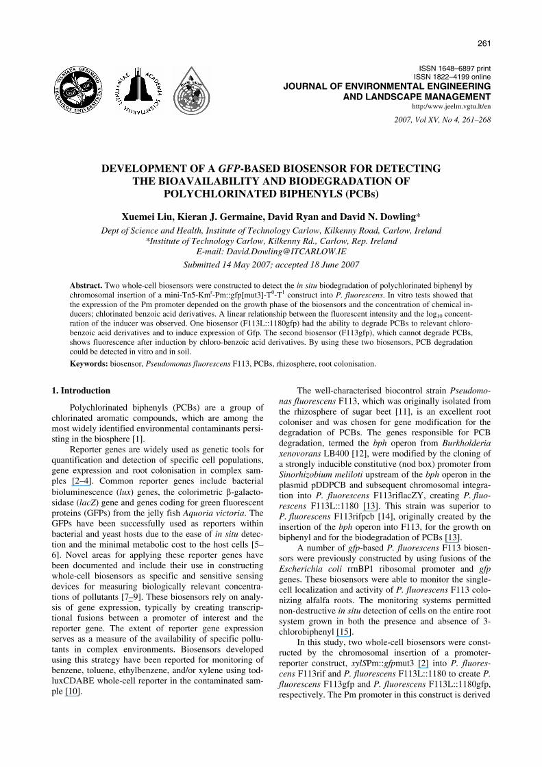
Kmr and Rifr transconjugants were obtained after 4 d on selective plates by mating the donor E.coli pJBA26 and recipient P. fluorescens F113rif or F113L::1180 with the helper plasmid pRK600. The transconjugants were purified by restreaking three times on selective plates. One transconjugant from each conjugation that visually exhibited green fluorescence by epifluorescent micro-scope was selected and designated P. fluorescens F113gfp and F113L::1180gfp, respectively. The presence of the gfp gene was also confirmed by PCR analysis re-sulting in a 742bp gfp specific amplicon (Fig 2).
Fig 2. PCR amplification of 740bp gfp gene in donor and transconjugant strains. 1 – 100bp size ladder, 2 – F113rif, 3 – F113gfp, 4 – pJBA26, 5 – F113pcbL1180gfp, 6 – F113pcbL1180gfp, 7 – F113pcbL1180, 8 – water control Amplification was not detected in recipient bacteria.
Southern blotting showed that the DIG-gfp probe hybrid-ised with the HindIII digested and undigested genomic
1 2 3 4 5 6 7 8
740bp
X. Liu et al. / JEELM – 2007, Vol XV, No 4, 261–268
264
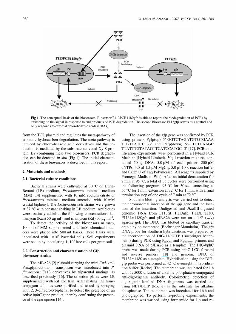
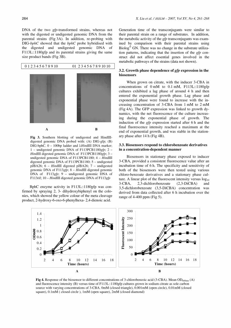
DNA of the two gfp-transformed strains, whereas not with the digested or undigested genomic DNA from the parental strains (Fig 3A). In addition, re-probing with DIG-bphC showed that the bphC probe hybridised with the digested and undigested genomic DNA of F113L::1180gfp and its parental strains giving the same size product bands (Fig 3B).
0 1 2 3 4 5 6 7 8 9 10 01 2 3 4 5 6 7 8 9 10 10
A B
Fig 3. Southern blotting of undigested and HindIII-digested genomic DNA probed with: (A) DIG-gfp; (B) DIG-bphC. 0 – 100bp ladder and λHindIII DNA marker; 1 – undigested genomic DNA of F113PCB1180gfp; 2 – HindIII digested genomic DNA of F113PCB1180gfp; 3 – undigested genomic DNA of F113PCB1180; 4 – HindIII digested genomic DNA of F113PCB1180; 5 – undigested pJBA26; 6 – HindIII digested pJBA26; 7 – undigested genomic DNA of F113gfp; 8 – HindIII digested genomic DNA of F113gfp; 9 – undigested genomic DNA of F113rif; 10 – HindIII digested genomic DNA of F113gfp BphC enzyme activity in F113L::1180gfp was con-
firmed by spraying 2, 3- dihydroxybiphenyl on the colo-nies, which showed the yellow colour of the meta cleavage product, 2-hydroxy-6-oxo-6-phenylhexa- 2,4-dienoic acid.
Generation time of the transconjugants were similar to their parental strain on a range of substrates. In addition, the metabolic activity of the gfp transconjugants was exam-ined by comparison with their parental strains using Biolog® GN. There was no change in the substrate utiliza-tion patterns, indicating that the insertion of the gfp con-struct did not affect essential genes involved in the metabolic pathways of the strains (data not shown).
3.2. Growth phase dependence of gfp expression in the biosensors
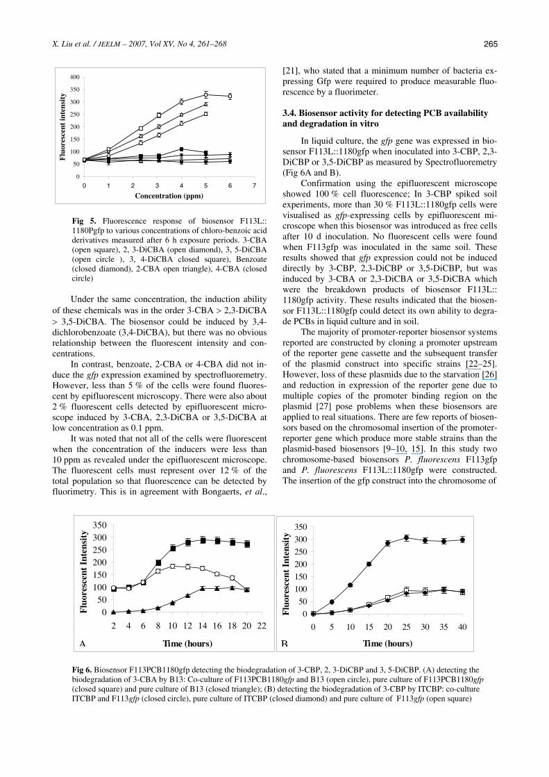
When grown on citrate, with the inducer 3-CBA in concentrations of 0 mM to 0.1 mM, F113L::1180gfp cultures exhibited a lag phase of around 4 h and then entered the exponential growth phase. Lag phase and exponential phase were found to increase with the in-creasing concentration of 3-CBA from 1 mM to 2 mM (Fig 4A). The GFP expression was linked to growth dy-namics, with the net fluorescence of the culture increas-ing during the exponential phase of growth. The induction of the gfp expression started after 6 h and the final fluorescence intensity reached a maximum at the end of exponential growth, and was stable in the station-ary phase after 14 h (Fig 4B).
3.3. Biosensors respond to chlorobenzoate derivatives in a concentration-dependent manner
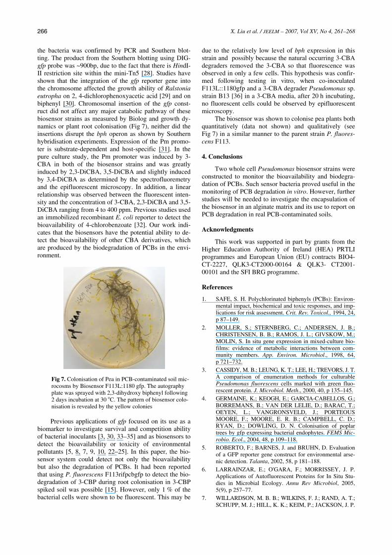
Biosensors in stationary phase exposed to inducer 3-CBA, provided a consistent fluorescence value after an incubation time of 6 h. The specificity and sensitivity of both of the biosensors were then tested using various chloro-benzoate derivatives and a stationary phase cul-ture. A linear plot of the fluorescent intensity versus log10 3-CBA, 2,3-dichlorobenzoate (2,3-DiCBA) and 3,5-dichlorobenzoate (3,5-DiCBA) concentration was derived from data collected after 6 h incubation over the range of 4-400 ppm (Fig 5).
0
50
100
150
200
250
300
2 4 6 8 10 12 14 16 18Time (hours)
Flu
ores
cent
Int
ensi
ty
0
0.2
0.4
0.6
0.8
1
1.2
1.4
2 4 6 8 10 12 14 16 18Time (hours)
OD
600
A B
Fig 4. Response of the biosensor to different concentrations of 3-chlorobenzoic acid (3-CBA). Mean OD600nm (A) and fluorescence intensity (B) versus time of F113L::1180gfp cultures grown in sodium citrate as sole carbon source with varying concentrations of 3-CBA. 0mM (closed triangle), 0.001mM (open circle), 0.01mM (closed square), 0.1mM ( closed circle ), 1mM (open square), 2mM (closed diamond)
X. Liu et al. / JEELM – 2007, Vol XV, No 4, 261–268
265
0
50
100
150
200
250
300
350
400
0 1 2 3 4 5 6 7
Concentration (ppm)
Flu
ores
cent
inte
nsit
y
Fig 5. Fluorescence response of biosensor F113L:: 1180Pgfp to various concentrations of chloro-benzoic acid derivatives measured after 6 h exposure periods. 3-CBA (open square), 2, 3-DiCBA (open diamond), 3, 5-DiCBA (open circle ), 3, 4-DiCBA closed square), Benzoate (closed diamond), 2-CBA open triangle), 4-CBA (closed circle) Under the same concentration, the induction ability
of these chemicals was in the order 3-CBA > 2,3-DiCBA > 3,5-DiCBA. The biosensor could be induced by 3,4-dichlorobenzoate (3,4-DiCBA), but there was no obvious relationship between the fluorescent intensity and con-centrations.
In contrast, benzoate, 2-CBA or 4-CBA did not in-duce the gfp expression examined by spectrofluoremetry. However, less than 5 % of the cells were found fluores-cent by epifluorescent microscopy. There were also about 2 % fluorescent cells detected by epifluorescent micro-scope induced by 3-CBA, 2,3-DiCBA or 3,5-DiCBA at low concentration as 0.1 ppm.
It was noted that not all of the cells were fluorescent when the concentration of the inducers were less than 10 ppm as revealed under the epifluorescent microscope. The fluorescent cells must represent over 12 % of the total population so that fluorescence can be detected by fluorimetry. This is in agreement with Bongaerts, et al.,
[21], who stated that a minimum number of bacteria ex-pressing Gfp were required to produce measurable fluo-rescence by a fluorimeter.
3.4. Biosensor activity for detecting PCB availability and degradation in vitro
In liquid culture, the gfp gene was expressed in bio-sensor F113L::1180gfp when inoculated into 3-CBP, 2,3-DiCBP or 3,5-DiCBP as measured by Spectrofluoremetry (Fig 6A and B).
Confirmation using the epifluorescent microscope showed 100 % cell fluorescence; In 3-CBP spiked soil experiments, more than 30 % F113L::1180gfp cells were visualised as gfp-expressing cells by epifluorescent mi-croscope when this biosensor was introduced as free cells after 10 d inoculation. No fluorescent cells were found when F113gfp was inoculated in the same soil. These results showed that gfp expression could not be induced directly by 3-CBP, 2,3-DiCBP or 3,5-DiCBP, but was induced by 3-CBA or 2,3-DiCBA or 3,5-DiCBA which were the breakdown products of biosensor F113L:: 1180gfp activity. These results indicated that the biosen-sor F113L::1180gfp could detect its own ability to degra-de PCBs in liquid culture and in soil.
The majority of promoter-reporter biosensor systems reported are constructed by cloning a promoter upstream of the reporter gene cassette and the subsequent transfer of the plasmid construct into specific strains [22–25]. However, loss of these plasmids due to the starvation [26] and reduction in expression of the reporter gene due to multiple copies of the promoter binding region on the plasmid [27] pose problems when these biosensors are applied to real situations. There are few reports of biosen-sors based on the chromosomal insertion of the promoter-reporter gene which produce more stable strains than the plasmid-based biosensors [9–10, 15]. In this study two chromosome-based biosensors P. fluorescens F113gfp and P. fluorescens F113L::1180gfp were constructed. The insertion of the gfp construct into the chromosome of
Fig 6. Biosensor F113PCB1180gfp detecting the biodegradation of 3-CBP, 2, 3-DiCBP and 3, 5-DiCBP. (A) detecting the biodegradation of 3-CBA by B13: Co-culture of F113PCB1180gfp and B13 (open circle), pure culture of F113PCB1180gfp (closed square) and pure culture of B13 (closed triangle); (B) detecting the biodegradation of 3-CBP by ITCBP: co-culture ITCBP and F113gfp (closed circle), pure culture of ITCBP (closed diamond) and pure culture of F113gfp (open square)
0
50
100
150
200
250
300
350
0 5 10 15 20 25 30 35 40
Time (hours)
Flu
ores
cent
Int
ensi
ty
050
100150200250300350
2 4 6 8 10 12 14 16 18 20 22
Time (hours)
Flu
ores
cent
Int
ensi
ty
A B
X. Liu et al. / JEELM – 2007, Vol XV, No 4, 261–268
266
the bacteria was confirmed by PCR and Southern blot-ting. The product from the Southern blotting using DIG-gfp probe was ~900bp, due to the fact that there is HindI-II restriction site within the mini-Tn5 [28]. Studies have shown that the integration of the gfp reporter gene into the chromosome affected the growth ability of Ralstonia eutropha on 2, 4-dichlorophenoxyacetic acid [29] and on biphenyl [30]. Chromosomal insertion of the gfp const-ruct did not affect any major catabolic pathway of these biosensor strains as measured by Biolog and growth dy-namics or plant root colonisation (Fig 7), neither did the insertions disrupt the bph operon as shown by Southern hybridisation experiments. Expression of the Pm promo-ter is substrate-dependent and host-specific [31]. In the pure culture study, the Pm promoter was induced by 3-CBA in both of the biosensor strains and was greatly induced by 2,3-DiCBA, 3,5-DiCBA and slightly induced by 3,4-DiCBA as determined by the spectrofluoremetry and the epifluorescent microscopy. In addition, a linear relationship was observed between the fluorescent inten-sity and the concentration of 3-CBA, 2,3-DiCBA and 3,5-DiCBA ranging from 4 to 400 ppm. Previous studies used an immobilized recombinant E. coli reporter to detect the bioavailability of 4-chlorobenzoate [32]. Our work indi-cates that the biosensors have the potential ability to de-tect the bioavailability of other CBA derivatives, which are produced by the biodegradation of PCBs in the envi-ronment.
Fig 7. Colonisation of Pea in PCB-contaminated soil mic-rocosms by Biosensor F113L:1180 gfp. The autography plate was sprayed with 2,3-dihydroxy biphenyl following 2 days incubation at 30 oC. The pattern of biosensor colo-nisation is revealed by the yellow colonies Previous applications of gfp focused on its use as a
biomarker to investigate survival and competition ability of bacterial inoculants [3, 30, 33–35] and as biosensors to detect the bioavailability or toxicity of environmental pollutants [5, 8, 7, 9, 10, 22–25]. In this paper, the bio-sensor system could detect not only the bioavailability but also the degradation of PCBs. It had been reported that using P. fluorescens F113rifpcbgfp to detect the bio-degradation of 3-CBP during root colonisation in 3-CBP spiked soil was possible [15]. However, only 1 % of the bacterial cells were shown to be fluorescent. This may be
due to the relatively low level of bph expression in this strain and possibly because the natural occurring 3-CBA degraders removed the 3-CBA so that fluorescence was observed in only a few cells. This hypothesis was confir-med following testing in vitro, when co-inoculated F113L::1180gfp and a 3-CBA degrader Pseudomonas sp. strain B13 [36] in a 3-CBA media, after 20 h incubating, no fluorescent cells could be observed by epifluorescent microscopy.
The biosensor was shown to colonise pea plants both quantitatively (data not shown) and qualitatively (see Fig 7) in a similar manner to the parent strain P. fluores-cens F113.
4. Conclusions
Two whole cell Pseudomonas biosensor strains were constructed to monitor the bioavailability and biodegra-dation of PCBs. Such sensor bacteria proved useful in the monitoring of PCB degradation in vitro. However, further studies will be needed to investigate the encapsulation of the biosensor in an alginate matrix and its use to report on PCB degradation in real PCB-contaminated soils.
Acknowledgments
This work was supported in part by grants from the Higher Education Authority of Ireland (HEA) PRTLI programmes and European Union (EU) contracts BIO4-CT-2227, QLK3-CT2000-00164 & QLK3- CT2001-00101 and the SFI BRG programme.
References
1. SAFE, S. H. Polychlorinated biphenyls (PCBs): Environ-mental impact, biochemical and toxic responses, and imp-lications for risk assessment. Crit. Rev. Toxicol., 1994, 24, p 87–149.
2. MOLLER, S.; STERNBERG, C.; ANDERSEN, J. B.; CHRISTENSEN, B. B.; RAMOS, J. L.; GIVSKOW, M.; MOLIN, S. In situ gene expression in mixed-culture bio-films: evidence of metabolic interactions between com-munity members. App. Environ. Microbiol., 1998, 64, p 721–732.
3. CASSIDY, M. B.; LEUNG, K. T.; LEE, H.; TREVORS, J. T. A comparison of enumeration methods for culturable Pseudomonas fluorescens cells marked with green fluo-rescent protein. J. Microbiol. Meth., 2000, 40, p 135–145.
4. GERMAINE, K.; KEOGH, E.; GARCIA-CABELLOS, G.; BORREMANS, B.; VAN DER LELIE, D.; BARAC, T.; OEYEN, L.; VANGRONSVELD, J.; PORTEOUS MOORE, F.; MOORE, E. R. B.; CAMPBELL, C. D.; RYAN, D.; DOWLING, D. N. Colonisation of poplar trees by gfp expressing bacterial endophytes. FEMS Mic-robio. Ecol., 2004, 48, p 109–118.
5. ROBERTO, F.; BARNES, J. and BRUHN, D. Evaluation of a GFP reporter gene construct for environmental arse-nic detection. Talanta, 2002, 58, p 181–188.
6. LARRAINZAR, E.; O'GARA, F.; MORRISSEY, J. P. Applications of Autofluorescent Proteins for In Situ Stu-dies in Microbial Ecology. Annu Rev Microbiol, 2005, 5(9), p 257–77.
7. WILLARDSON, M. B. B.; WILKINS, F. J.; RAND, A. T.; SCHUPP, M. J.; HILL, K. K.; KEIM, P.; JACKSON, J. P.

X. Liu et al. / JEELM – 2007, Vol XV, No 4, 261–268
267
Development and testing of a bacterial biosensor for tolu-ene-based environmental contaminants. Appl. Environ. Microbiol., 1998, 64, p 1006–1012.
8. SCHREITER, P. P.; GILLOR, O.; POST, A.; BELKIN, S.; SCHMID, R. D. and BACHMANN, T. T. Monitoring of phosphorus bioavailability in water by an immobilized luminescent cyanobacterial reporter strain. Biosensors Bioelectro, 2001, 16, p 811–818.
9. TAYLOR, C. J.; BAIN, L. A.; RICHARDSON, D. J.; SPIRO, S. and RUSSELL, D. A. Construction of whole-cell gene reporter for the fluorescent bioassay of nitrate. Anal. Biochem., 2004, 328, p 60–66.
10. APPLEGATE, M. B.; KEHRMEYER, R. S.; SAYLER, S. G. A chromosomally based tod-luxCDABE whole-cell repor-ter for benzene, toluene, ethybenzene, and xylene (BTEX) sensing. Appl. Environ. Microbiol., 1998, 64, p 2730–2735.
11. SHANAHAN, P.; O’SULLIVAN, D. J.; SIMPSON, P.; GLENNON, J. D.; O’GARA, F. Isolation of 2, 4-diacetyl-phloroglucinol from a fluorescent pseudomonad and in-vestigation of physiological parameters influencing its production. Appl. Environ. Microbiol., 1992, 58, p 353–358.
12. GORIS, J.; DE VOS, P.; CABALLERO-MELLADO, J.; PARK, J.; FALSEN, E.; QUENSEN III, J. F.; TIEDJE, J. M.; VANDAMME, P. Classification of the biphenyl-and po-lychlorinated biphenyl-degrading strain LB400T and rela-tives as Burkholderia xenovorans sp. nov. International Journal of Systematic and Evolutionary Microbiology, 2004, 54, p 1677–1681.
13. VILLACIEROS, M.; WHELAN, C.; MACKOVA, M.; MOLGAARD, J.; SÁNCHEZ-CONTRERAS, M.; LLORET, J.; AGUIRRE DE CÁRCER, D.; ORUEZÁBAL, R. I.; BOLAÑOS, L.; MACEK, T.; KARLSON, U.; DOWLING, D. N.; MARTÍN, M.; RIVILLA, R. PCB Rhizoremediation by Pseudomonas fluorescens F113 derivatives using a Sinorhizobium meliloti nod system to drive bph gene expression. Appl. Environ. Microbiol., 2005, 71, p 2687–2694.
14. BRAZIL, G. M.; KENEFICK, L.; CALLANAN, M.; HARO, A.; DE LORENZO, V.; DOWLING, D. N.; O’GARA, F. Construction of a rhizosphere Pseudomonad with potential to degrade polychlorinated biphenyls and detection of bph gene expression in the rhizosphere. Appl. Environ. Microbiol., 1995, 61, p 1946–1952.
15. BOLDT, T. S.; SORENSEN, J.; KARLSON, U.; MOLIN, S.; RAMOS, C. Combined use of different Gfp reporters for monitoring single-cell activity of a genetically modified PCB degrader in the rhizosphere of Alfalfa. FEMS Mic-robiol. Ecol., 2004, 48, p 139–148.
16. HERRERO, M.; DE LORENZO, V.; TIMMIS, K. N. Transposon vectors containing non-antibiotic resistance selection markers for cloning and stable chromosomal in-sertion of foreign genes in gram-negative bacteria. J. Bac-teriol., 1990, 172, p 6557–6567.
17. ANDERSEN, J. B.; STERNBERG, C.; POULSEN, L. K.; BJØRN, S. P.; GIVSKOV, M.; MOLIN, S. New unstable variants of green fluorescent protein for studies of tran-sient gene expression in bacteria. Appl. Environ. Micro-biol., 1998, 64, p 2240–2246.
18. HOGAN, J.; SHERLOCK, O.; RYAN, D.; WHELAN, C.; FRANCESCONI, S.; RIVILLA, R.; DOWLING, D. N. Fluorescence Resonance Energy Transfer (FRET) based molecular detection of a genetically modified PCB de-grader in soil. FEMS Microbiol. Lett., 2004, 236, p 349–357.
19. SCHER, F. M.; BAKER, R. Effects of Pseudomonas putida and a synthetic iron chelator on induction of soil
suppressiveness to Fusarium wilt pathogens. Phytopatho-logy, 1982, 72, p 1567–1573.
20. RAMOS, C.; MOLBAK, L.; MOLIN, S. Bacterial activity in the rhizosphere analysed at the single-cell level by mo-nitoring ribosome contents and synthesis rates. Appl. En-viron. Microbiol., 2000, 66, p 801–809.
21. BONGAERTS, R. J.; HAUTEFORT, I.; SIDEBOTHAM, J. M.; HINTON, J. K. Green fluorescent protein as a mar-ker for conditional gene expression in bacterial cells. Me-thods Enzymo., 2002, 358, p 43–66.
22. LAYTON, A. C.; MUCCINI, M.; GHOSH, M. M.; SAYLER, G. S. Construction of a bioluminescent reporter strain to detect polychlorinated biphenyls. Appl. Environ. Microbiol., 1998, 64, p 5023–5026.
23. STINER, L.; HALVERSON, L. J. Development and characterization of a green fluorescent protein-based bac-terial biosensor for bioavailable toluene and related com-pounds. Appl. Environ. Microbiol., 2002, 68, p 1962–1971.
24. IKENO, S.; OGINO, C.; ITO, T.; SHIMIZU, N. Detection of benzene derivatives by recombinant E. coli with Ps promoter and GFP as a reporter protein. J. Biochem. Eng., 2003, 15, p 193–197.
25. CHANG, S. T.; LEE, H. J.; GU, M. B. Enhancement in the sensitivity of an immobilized cell-based soil biosensor for monitoring PAH toxicity. Sens. Actuators B., 2004, 97, p 272–276.
26. LEFF, G. L.; LEFF, A. A. Use of green fluorescent pro-tein to monitor survival of genetically engineered bacteria in aquatic environments. Appl. Environ. Microbiol., 1996, 62, p 3486–3488.
27. WANG, Y.; RAWLINGS, M.; GILBSON, D. T.; LABBE, D.; BERGERON, H.; BROUSSEAU, R.; LAU, P. C. Identification of a membrane protein and a truncated LysR-type regulator associated with the toluene degradation pathway in Pseudomonas putida F1. Mol. Gen. Genet., 1995, 246, p 570–579.
28. DELORENZO, V.; FERNANDEZ, S.; HERRERO, M.; JAKUBZIK, U.; TIMMIS, K. N. Engineering of alkyl- and haloaromatic-responsive gene expression with mini-transposons containing regulated promoters of biodegrada-tive pathways of Pseudomonas. Gene, 1993, 130, p 41–46.
29. FUCHSLIN, H. P.; RUEGG, I.; VAN DER MEER, J. R.; EGLI, T. Effect of integration of a GFP reporter gene on fitness of Ralstonia eutropha during growth with 2, 4-dichlorophenoxyacetic acid. Environ. Microbiol., 2003, 5, p 878–887.
30. ABBY, A. M.; BEAUDETTE, L. A.; LEE, H.; TREVORS, J. T. Polychlorinated biphenyl (PCB) degra-dation and persistence of a gfp-marked Ralstonia eutro-pha H850 in PCB-contaminated soil. Appl. Microbiol. Biotechnol., 2003, 63, p 222–230.
31. WINTHER-LARSEN, H. C.; JOSEFSEN, K. D.; BRAUTASET, T.; VALLA, S. Parameters Affecting Ge-ne Expression from the Pm Promoter in Gram-Negative Bacteria. Metab. Eng., 2000, 2, p 79–91.
32. KOHLER, S.; BACHMANN, T. T.; SCHMITT, J.; BELKIN, S.; SCHMID, R. D. Detection of 4-chloro-benzoate using immobilized recombinant Escherichia coli reporter strains. Sens. Actuator B., 2000, 70, p 139–144.
33. TRESSE, O.; ERRAMPALLI, D.; KOSTRZYNSKA, M.; LEUNG, K. T.; LEE, H.; TREVORS, J.T.; VAN ELSAS, J. D. Green fluorescent protein as a visual marker in a p-nitrophenol degrading Moraxella sp. FEMS Microbiol. Lett., 1998, 164, p 187–193.

X. Liu et al. / JEELM – 2007, Vol XV, No 4, 261–268
268
34. ERRAMPALLI, D.; OKAMURA, H.; LEE, H.; TREVORS, J. T.; VAN ELSAS, J. D. Green fluorescent protein as a marker to monitor survival of phenanthrene-mineralising Pseudomonas sp. UG14Gr in creosote-contaminated soil. FEMS Microbiol. Ecol., 1998, 26, p 181–191.
35. ERRAMPALLI, D.; TRESS, O.; LEE, H.; TREVORS, J. T. Bacterial survival and mineralization of p-nitrophenol in
soil by green fluorescent protein-marked Moraxella sp. G21 encapsulated cells. FEMS Microbiol. Ecol., 1999, 30, p 229–236.
36. DORN, E.; HELLWIG, M.; REINEKE, W.; KNACK-MUSS, H. J. Isolation and characterization of a 3-chloro-benzoate degrading pseudomonad. Arch. Microbiol., 1974, 99, p 61–70.
GFP PAGRINDO BIOSENSORIŲ TOBULINIMAS POLICHLORINTŲ BIFENILŲ (PCBs) BIOTINKAMUMUI IR BIODEGRADACIJAI NUSTATYTI
X. Liu, K. J. Germaine, D. Ryan and D. N. Dowling
S a n t r a u k a
Sukonstruoti du ląsteliniai bioindikatoriai polichlorintų bifenilų in situ biodegradacijai nustatyti chromosominiu konstruk-to mini-Tn5-KmF-Pm::gfp[mut3]-T0-T1 įterpimo į P. fluorescens būdu. Bandymai dirbtinėmis sąlygomis parodė, kad Pm aktyviklio išraiška priklauso nuo bioindikatoriaus augimo fazės ir cheminių indikatorių koncentracijos, taip pat nuo chlo-rintų benzenkarboksirūgščių derinių. Nustatyta tiesinė priklausomybė tarp fluorescentų intensyvumo ir indikatorių log10 koncentracijos. Vienas iš bioindikatorių (F113L::1180gfp) galėjo degraduoti PCBs iki tinkamų chloro-benzenkarboksirūgščių derinių ir indukuoti Gfp išraišką. Antrasis bioindikatorius (F113gfp), kuris negali degraduoti PCBs, parodo fluorescentiškumą įvykus indukcijai pagal chloro-benzenkarboksirūgščių derinius. Naudojant šiuos du bioindikatorius, PCB degradacija gali būti nustatyta tiek dirbtinėmis sąlygomis, tiek dirvožemyje.
Reikšminiai žodžiai: bioindikatoriai, Pseudomonas fluorescens F113, PCBs, rizosfera, šaknų kolonizacija.
СОВЕРШЕНСТВОВАНИЕ БИОСЕНСОРОВ ОСНОВЫ GFP ДЛЯ ОПРЕДЕЛЕНИЯ БИОСООТВЕТСТВИЯ И БИОДЕГРАДАЦИИ ПОЛИХЛОРИРОВАННЫХ БИФЕНИЛОВ (PCBs)
Х. Лю, К. Й. Гермайне, В. Риан, Д. Н, Довлинг
Р е з ю м е
Сконструированы два клеточных биоиндикатора для выявления на участке биодеградации полихлорированных бифенилов путем хромосомного введения мини-Tn5-Kmr-Pm::gfp[mut3]-T0-T1 в P. fluorescen. Испытания в искус-ственных условиях показали, что выражение активатора Pm зависит от фазы роста биоиндикатора и концентра-ции химических индикаторов, а также от сочетаний хлорированных бензойных кислот. Наблюдалась линейная зависимость между интенсивностью флуоресцентов и концентрации log10 индикаторов. Один из индикаторов (F113L::1180gfp) мог способствовать деградации PCBs в сочетания соответствующих хлор-бензойных кислот и индуцировать выражение Gfp. Другой биоиндикатор (F113gfp), который не может способствовать деградации PCBs, показал флуоресцентность после индикации по сочетаниям хлор-бензойных кислот. С использованием этих индикаций деградация PCB может наблюдаться лишь в искусственных условиях и лишь в почве.
Ключевые слова: биоиндикаторы, Pseudomonas fluorescens F113, PCBs, ризосфера, корневая колонизация.
Ms Xeumei LIU Bsc. Ms Xeumei Liu is a PhD student at IT Carlow under the supervision of Dr Dowling and Dr Ryan. Her research interests are in the development of biosensors and use of microarrays to study bacterial interactions with pollutants.
Mr Kieran GERMAINE Bsc. Mr Kieran Germaine BSc is a PhD student under the supervision of Dr Dowling and Dr Ryan. His research interests are in the area of endophytic and rhizospheric bacteria and their applications for bioremediation.
Dr David RYAN PhD. Dr Ryan is Head of Department of Science & Health at IT Carlow. He has been a member of the Biotechnology and Molecular Environmental Sciences (BMES) research group at IT Carlow since 2000. Dr Ryans main research interests are the use and manipulation of microbial degradative and metal resistance mechanisms for improved bioremediation and enhanced phytoremediation of pollutant compounds. Currently, active projects in this area include the examination of metal resistant and xenobiotic degrading endophytic and rhizospheric bacteria for improved phytoremedia-tion and phytocontainment, the molecular analysis of multi-efflux system(s) in the plant associated bacterium Xanthomo-nas and the examinataion of molecular resistance to tributyl-tin in exposed microbial strains and their bioremediation potential. Dr Ryan also supervises research for the design and development of an environmental DNA microarray for en-vironmental genetic analysis as well as the construction and evaluation of microbial biosensors to detect both target con-tamination and degradation in a range of water/soil environments and conditions.
Dr David DOWLING PhD (corresponding author). Dr Dowling is Head of School of Science at IT Carlow and directs several research projects ranging from the development of biological treatments for the detoxification of waste chemicals to molecular methods for the detection of microorganisms in the environment. This research is funded by EU, National and Industrial sources. He has 20 years of experience in the area of biodegradation as well as plant-microbe interactions with 50 publications in refereed international scientific publications and over 130 conference papers, one patent applica-tion and has edited two books in the area of environmental biotechnology. He is a co-director of Microgen Biotechnol-ogies, a campus unit dedicated to commercialisation of environmental biotechnology research at IT Carlow. He is a member of the EU Cost programme management committee (Cost 849) on Phytotechnolgies and has experience of co-coordinating EU projects.
Copyright © 2022 FDOKUMEN