Capsaicin production by Alternaria alternata, an endophytic fungus from Capsicum annum; LC-ESI-MS/MS...
7
Capsaicin production by Alternaria alternata, an endophytic fungus from Capsicum annum; LC–ESI–MS/MS analysis Shekaraiah Devari a,b , Sundeep Jaglan c , Manjeet Kumar b , Ramesh Deshidi a,b , Santosh Guru d , Shashi Bhushan d , Manoj Kushwaha c , Ajai P. Gupta c , Sumit G. Gandhi d , Jai P. Sharma c,⇑ , Subhash C. Taneja b , Ram A. Vishwakarma b , Bhahwal Ali Shah a,b,⇑ a Academy of Scientific and Innovative Research, India b Natural Product Microbes, CSIR-Indian Institute of Integrative Medicine, Canal Road, Jammu 180001, India c Quality Control & Quality Assurance Division, CSIR-Indian Institute of Integrative Medicine, Canal Road, Jammu 180001, India d Cancer Pharmacology Division, CSIR-Indian Institute of Integrative Medicine, Canal Road, Jammu 180001, India article info Article history: Received 28 June 2013 Received in revised form 1 December 2013 Available online 28 December 2013 Keywords: Alternaria alternata Capsaicin Alternariol-10-methyl ether (MRM)-LC–ESI–MS/MS Anti-cancer abstract Alternaria alternata, an endophytic fungus capable of producing capsaicin (1) was isolated from Capsicum annum. The endophyte was found to produce capsaicin upto three generations. Upscaling of the fermen- tation broth led to the isolation of one known and one compound characterized as 2,4-di-tert-butyl phenol (2) and alternariol-10-methyl ether (3) respectively. Compound 1 and 3 were identified and quan- tified using liquid chromatography-electrospray ionization tandem mass spectrometry (LC–ESI–MS/MS) system through multiple reaction monitoring (MRM). Furthermore, compound 3 displayed a range of cytotoxicity against a panel of human cancer cell lines and was found to induce apoptosis evidenced by Hoechst staining and loss of mitochondrial-membrane potential in HL-60 cells. Ó 2013 Elsevier Ltd. All rights reserved. Introduction Ever since the isolation of paclitaxel from Taxomyces andreanae by Strobel and co-workers (Stierle et al., 1993), endophytic fungi association with their host plants have garnered considerable interest of the natural product chemists worldwide. This is due to their ability to produce same compounds as that of the host plant by interfering with their biosynthetic pathways e.g., campto- thecin from Entrophospora infrequens (Puri et al., 2005) and podophyllotoxin, vinblastine and vincristine from an Alternaria sp. (Cao et al., 2007; Guo et al., 1998). Endophytes have proved to be plentiful source of biologically active molecules having anti-biotic, anti-oxidant, anti-cancer, anti-viral, immunosuppres- sive and anti-diabetic activities (Pimentel et al., 2011; Strobel and Daisy, 2003; Strobel et al., 2004; Zhang and Li, 1994). In con- tinuation of our work on natural products (Kumar et al., 2012; Ku- mar et al., 2013; Shah et al., 2007, 2009a,b), we report the isolation of an endophytic fungus from Capsicum annum, identified as Alternaria alternata capable of producing capsaicin (1). Capsaicin, generally isolated from C. annum is mainly responsible for hotness of chili (Al Othman et al., 2011; Perucka and Materska, 2003; San- atombi and Sharma, 2008). The Food and Drug Administration (FDA) has approved Qutenza containing 8% capsaicin, a medicated skin patch that relieves the pain of post-herpetic neuralgia (Babbar et al., 2009; Backonja et al., 2008; FDA News Release: Nov. 17, 2009; Jones et al., 2011). Also, Adlea (ALGRX-4975), an injectable preparation of capsaicin for treatment of osteoarthritis pain, post-surgical conditions as well as for neuropathic pain occurring secondary to nerve injury is in Phase-III clinical trials (Remadevi and Szallisi, 2008). Research studies suggest capsaicin to induce cell-cycle arrest or apoptosis or inhibit cell proliferation in a vari- ety of cancer cells (Bode and Dong, 2011). Moreover, capsaicin con- taining creams have been used to treat painful conditions such as long-term neuropathic pain in cancer patients (Ellison et al., 1997), diabetic neuropathy (Hautkappe et al., 1998) and psoriasis (Ellis et al., 1993). Also, oral capsaicin as candy produced substan- tial pain reduction in patients with oral mucositis caused by cancer therapy (Berger et al., 1995). It may also find implication in the treatment of obesity as its oral intake increases the production of body heat, causing the breakdown of carbohydrates (Joo et al., 2010). In this paper we also present the isolation of 2,4-di-tert-bu- tyl phenol (2) and alternariol-10-methyl ether (3), wherein 3 was found new to the literature. The identification and quantification of 1 and 3 was carried out by liquid chromatography–electrospray 0031-9422/$ - see front matter Ó 2013 Elsevier Ltd. All rights reserved. http://dx.doi.org/10.1016/j.phytochem.2013.12.001 ⇑ Corresponding authors. Address: Natural Product Microbes, CSIR-Indian Insti- tute of Integrative Medicine, Canal Road, Jammu 180001, India. Tel.: +91 191 25692217; fax: +91 191 25693331 (B.A. Shah). E-mail addresses: [email protected] (J.P. Sharma), [email protected] (B.A. Shah). Phytochemistry 98 (2014) 183–189 Contents lists available at ScienceDirect Phytochemistry journal homepage: www.elsevier.com/locate/phytochem
-
Upload
independent -
Category
Documents
-
view
2 -
download
0
Transcript of Capsaicin production by Alternaria alternata, an endophytic fungus from Capsicum annum; LC-ESI-MS/MS...

Phytochemistry 98 (2014) 183–189
Contents lists available at ScienceDirect
Phytochemistry
journal homepage: www.elsevier .com/locate /phytochem
Capsaicin production by Alternaria alternata, an endophytic fungusfrom Capsicum annum; LC–ESI–MS/MS analysis
0031-9422/$ - see front matter � 2013 Elsevier Ltd. All rights reserved.http://dx.doi.org/10.1016/j.phytochem.2013.12.001
⇑ Corresponding authors. Address: Natural Product Microbes, CSIR-Indian Insti-tute of Integrative Medicine, Canal Road, Jammu 180001, India. Tel.: +91 19125692217; fax: +91 191 25693331 (B.A. Shah).
E-mail addresses: [email protected] (J.P. Sharma), [email protected](B.A. Shah).
Shekaraiah Devari a,b, Sundeep Jaglan c, Manjeet Kumar b, Ramesh Deshidi a,b, Santosh Guru d,Shashi Bhushan d, Manoj Kushwaha c, Ajai P. Gupta c, Sumit G. Gandhi d, Jai P. Sharma c,⇑,Subhash C. Taneja b, Ram A. Vishwakarma b, Bhahwal Ali Shah a,b,⇑a Academy of Scientific and Innovative Research, Indiab Natural Product Microbes, CSIR-Indian Institute of Integrative Medicine, Canal Road, Jammu 180001, Indiac Quality Control & Quality Assurance Division, CSIR-Indian Institute of Integrative Medicine, Canal Road, Jammu 180001, Indiad Cancer Pharmacology Division, CSIR-Indian Institute of Integrative Medicine, Canal Road, Jammu 180001, India
a r t i c l e i n f o a b s t r a c t
Article history:Received 28 June 2013Received in revised form 1 December 2013Available online 28 December 2013
Keywords:Alternaria alternataCapsaicinAlternariol-10-methyl ether(MRM)-LC–ESI–MS/MSAnti-cancer
Alternaria alternata, an endophytic fungus capable of producing capsaicin (1) was isolated from Capsicumannum. The endophyte was found to produce capsaicin upto three generations. Upscaling of the fermen-tation broth led to the isolation of one known and one compound characterized as 2,4-di-tert-butylphenol (2) and alternariol-10-methyl ether (3) respectively. Compound 1 and 3 were identified and quan-tified using liquid chromatography-electrospray ionization tandem mass spectrometry (LC–ESI–MS/MS)system through multiple reaction monitoring (MRM). Furthermore, compound 3 displayed a range ofcytotoxicity against a panel of human cancer cell lines and was found to induce apoptosis evidencedby Hoechst staining and loss of mitochondrial-membrane potential in HL-60 cells.
� 2013 Elsevier Ltd. All rights reserved.
Introduction
Ever since the isolation of paclitaxel from Taxomyces andreanaeby Strobel and co-workers (Stierle et al., 1993), endophytic fungiassociation with their host plants have garnered considerableinterest of the natural product chemists worldwide. This is dueto their ability to produce same compounds as that of the hostplant by interfering with their biosynthetic pathways e.g., campto-thecin from Entrophospora infrequens (Puri et al., 2005) andpodophyllotoxin, vinblastine and vincristine from an Alternariasp. (Cao et al., 2007; Guo et al., 1998). Endophytes have provedto be plentiful source of biologically active molecules havinganti-biotic, anti-oxidant, anti-cancer, anti-viral, immunosuppres-sive and anti-diabetic activities (Pimentel et al., 2011; Strobeland Daisy, 2003; Strobel et al., 2004; Zhang and Li, 1994). In con-tinuation of our work on natural products (Kumar et al., 2012; Ku-mar et al., 2013; Shah et al., 2007, 2009a,b), we report the isolationof an endophytic fungus from Capsicum annum, identified asAlternaria alternata capable of producing capsaicin (1). Capsaicin,
generally isolated from C. annum is mainly responsible for hotnessof chili (Al Othman et al., 2011; Perucka and Materska, 2003; San-atombi and Sharma, 2008). The Food and Drug Administration(FDA) has approved Qutenza containing 8% capsaicin, a medicatedskin patch that relieves the pain of post-herpetic neuralgia (Babbaret al., 2009; Backonja et al., 2008; FDA News Release: Nov. 17,2009; Jones et al., 2011). Also, Adlea (ALGRX-4975), an injectablepreparation of capsaicin for treatment of osteoarthritis pain,post-surgical conditions as well as for neuropathic pain occurringsecondary to nerve injury is in Phase-III clinical trials (Remadeviand Szallisi, 2008). Research studies suggest capsaicin to inducecell-cycle arrest or apoptosis or inhibit cell proliferation in a vari-ety of cancer cells (Bode and Dong, 2011). Moreover, capsaicin con-taining creams have been used to treat painful conditions such aslong-term neuropathic pain in cancer patients (Ellison et al.,1997), diabetic neuropathy (Hautkappe et al., 1998) and psoriasis(Ellis et al., 1993). Also, oral capsaicin as candy produced substan-tial pain reduction in patients with oral mucositis caused by cancertherapy (Berger et al., 1995). It may also find implication in thetreatment of obesity as its oral intake increases the production ofbody heat, causing the breakdown of carbohydrates (Joo et al.,2010). In this paper we also present the isolation of 2,4-di-tert-bu-tyl phenol (2) and alternariol-10-methyl ether (3), wherein 3 wasfound new to the literature. The identification and quantificationof 1 and 3 was carried out by liquid chromatography–electrospray
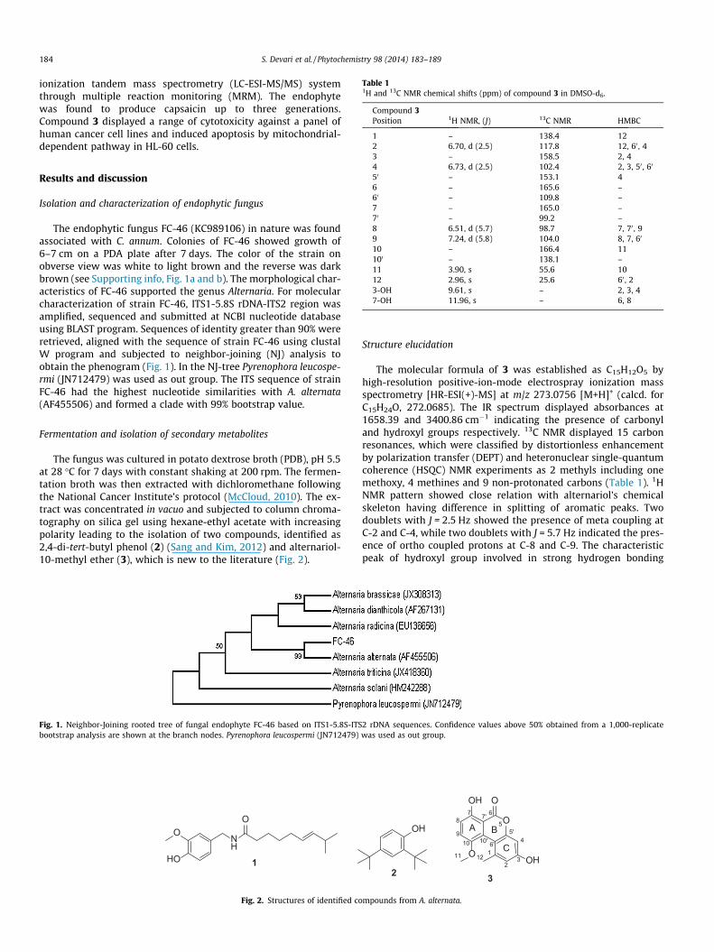
Table 11H and 13C NMR chemical shifts (ppm) of compound 3 in DMSO-d6.
Compound 3Position 1H NMR, (J) 13C NMR HMBC
1 – 138.4 122 6.70, d (2.5) 117.8 12, 60 , 4
184 S. Devari et al. / Phytochemistry 98 (2014) 183–189
ionization tandem mass spectrometry (LC-ESI-MS/MS) systemthrough multiple reaction monitoring (MRM). The endophytewas found to produce capsaicin up to three generations.Compound 3 displayed a range of cytotoxicity against a panel ofhuman cancer cell lines and induced apoptosis by mitochondrial-dependent pathway in HL-60 cells.
3 – 158.5 2, 44 6.73, d (2.5) 102.4 2, 3, 50 , 60
50 – 153.1 46 – 165.6 –60 – 109.8 –7 – 165.0 –70 – 99.2 –8 6.51, d (5.7) 98.7 7, 70 , 99 7.24, d (5.8) 104.0 8, 7, 60
10 – 166.4 11100 – 138.1 –11 3.90, s 55.6 1012 2.96, s 25.6 60 , 23-OH 9.61, s – 2, 3, 47-OH 11.96, s – 6, 8
Results and discussion
Isolation and characterization of endophytic fungus
The endophytic fungus FC-46 (KC989106) in nature was foundassociated with C. annum. Colonies of FC-46 showed growth of6–7 cm on a PDA plate after 7 days. The color of the strain onobverse view was white to light brown and the reverse was darkbrown (see Supporting info, Fig. 1a and b). The morphological char-acteristics of FC-46 supported the genus Alternaria. For molecularcharacterization of strain FC-46, ITS1-5.8S rDNA-ITS2 region wasamplified, sequenced and submitted at NCBI nucleotide databaseusing BLAST program. Sequences of identity greater than 90% wereretrieved, aligned with the sequence of strain FC-46 using clustalW program and subjected to neighbor-joining (NJ) analysis toobtain the phenogram (Fig. 1). In the NJ-tree Pyrenophora leucospe-rmi (JN712479) was used as out group. The ITS sequence of strainFC-46 had the highest nucleotide similarities with A. alternata(AF455506) and formed a clade with 99% bootstrap value.
Fermentation and isolation of secondary metabolites
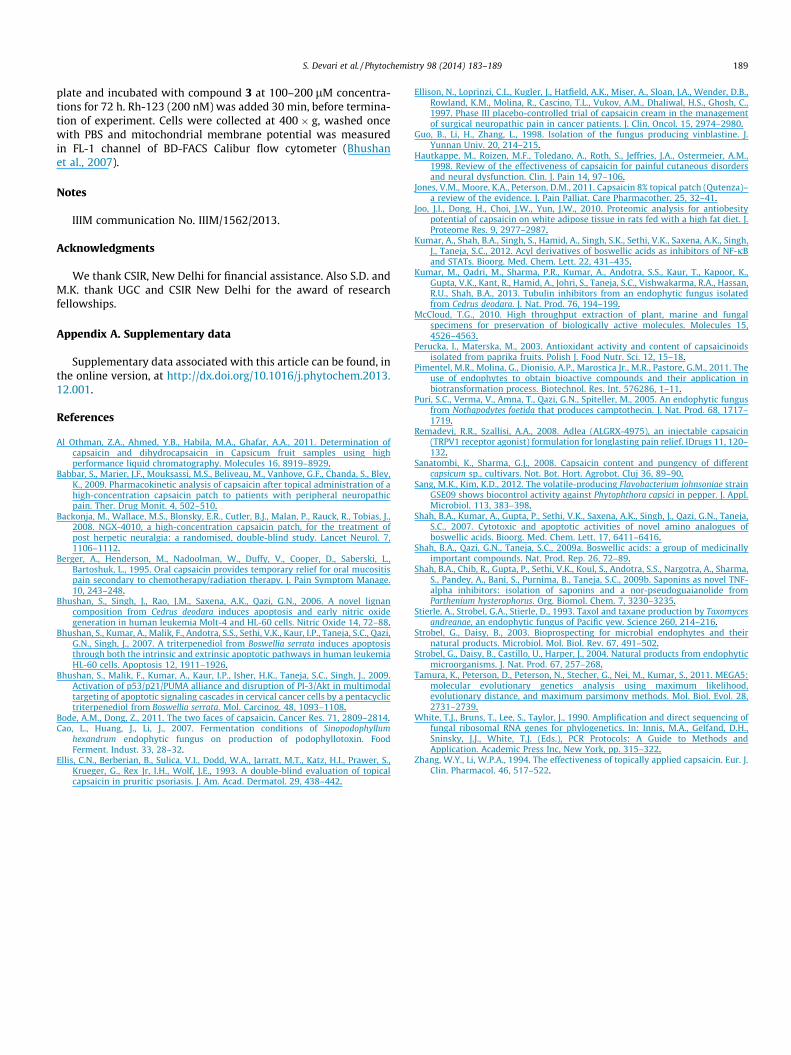
The fungus was cultured in potato dextrose broth (PDB), pH 5.5at 28 �C for 7 days with constant shaking at 200 rpm. The fermen-tation broth was then extracted with dichloromethane followingthe National Cancer Institute’s protocol (McCloud, 2010). The ex-tract was concentrated in vacuo and subjected to column chroma-tography on silica gel using hexane-ethyl acetate with increasingpolarity leading to the isolation of two compounds, identified as2,4-di-tert-butyl phenol (2) (Sang and Kim, 2012) and alternariol-10-methyl ether (3), which is new to the literature (Fig. 2).
Fig. 1. Neighbor-Joining rooted tree of fungal endophyte FC-46 based on ITS1-5.8S-ITSbootstrap analysis are shown at the branch nodes. Pyrenophora leucospermi (JN712479)
O
NH
HO
O
1
Fig. 2. Structures of identified co
Structure elucidation
The molecular formula of 3 was established as C15H12O5 byhigh-resolution positive-ion-mode electrospray ionization massspectrometry [HR-ESI(+)-MS] at m/z 273.0756 [M+H]+ (calcd. forC15H24O, 272.0685). The IR spectrum displayed absorbances at1658.39 and 3400.86 cm�1 indicating the presence of carbonyland hydroxyl groups respectively. 13C NMR displayed 15 carbonresonances, which were classified by distortionless enhancementby polarization transfer (DEPT) and heteronuclear single-quantumcoherence (HSQC) NMR experiments as 2 methyls including onemethoxy, 4 methines and 9 non-protonated carbons (Table 1). 1HNMR pattern showed close relation with alternariol’s chemicalskeleton having difference in splitting of aromatic peaks. Twodoublets with J = 2.5 Hz showed the presence of meta coupling atC-2 and C-4, while two doublets with J = 5.7 Hz indicated the pres-ence of ortho coupled protons at C-8 and C-9. The characteristicpeak of hydroxyl group involved in strong hydrogen bonding
2 rDNA sequences. Confidence values above 50% obtained from a 1,000-replicatewas used as out group.
3
OHO
O
OHO
OH
1
23
4
5
678
910
5'
6'10'
7'
1211
2
A B
C
mpounds from A. alternata.

tR = 12.35
O
NH
HO
OO
O
OHO
OH
m/z = 306.1m/z = 273.0tR = 11.22
Fig. 3. MRM chromatogram of A. alternata extract.
(a)
(b)
Fig. 4. MRM graphs: (a) capsaicin (1) with transition masses of m/z 306.1 ? 137.0 (quantifier) and m/z 306.1 ? 170.1 (qualifier); (b) alternariol-10-methyl ether (3) withtransition masses of m/z 273.0 ? 257.8 (quantifier) and m/z 273.0 ? 229.9 (qualifier).
S. Devari et al. / Phytochemistry 98 (2014) 183–189 185
appears at dH 11.67 attached to C-7, confirmed by HSQC. A sharpsinglet at dH 2.57 revealed the presence of methyl group attachedto aromatic ring positioned as C-1 from HMBC correlation withC-60 and C-2. Singlet at dH 3.91 indicated the presence of methoxygroup positioned as C-10 from strong HMBC correlation with C-11.13C NMR spectra also corroborated the presence of alternariol skel-eton revealing the connectivity of rings A and B with characteristicpeaks at d 99.2 and 138.1, corresponding to C-7’ and C-100. Similarconnectivity of rings B and C was located at d 109.8 and 153.1 as-signed as C-60 and C-50. Moreover, the ortho coupling between8 ? 9 was also confirmed by 1H-1H COSY co-relations betweenH-8 (dH 6.51) and H-9 (dH 7.23), which further reinforces the direct
attachment of methoxy group to C-10. Thus, the structure of 3 waselucidated as 3,7-dihydroxy-10-methoxy-1-methyl-6H-benzo-chromene-6-one.
Quantification of capsaicin by LC–ESI–MS/MS
An LC-ESI-MS/MS method through MRM was developed for theselective detection and quantification of 1 and 3 at 11.22 and12.35 min in the extract (Fig. 3). The selectivity and sensitivity of(MRM) LC-ESI-MS/MS relies on the application of MRM in whicheach ionized compound gives a distinct precursor-to-product iontransition that is diagnostic for the presence of a particular com-

Table 2Cytotoxic profile of 3 in different panel of human cancer cell lines.
Cell Line % Cell growth inhibition by compound 3 IC-50, lM
10 lM 30 lM 100 lM
HL-60 5 21 65 85A549 12 17 21 >100 lMPC-3 11 23 28 >100 lMHeLa 6 12 22 >100 lMA431 8 25 53 95MiaPaka-2 12 17 28 >100 lMT47D 11 20 25 >100 lM
The cells were grown in 96-well culture plate and treated with indicated concen-trations of compound 3 for 48 h. Thereafter, cells were incubated with MTT solutionfor 2 h and optical density of formazon crystals was measured as described inMaterials and Methods. Data are Mean ± SD (n = 8 wells), and representative of oneof three similar experiments.
186 S. Devari et al. / Phytochemistry 98 (2014) 183–189
pound in an extract. Comparison of the chromatograms of blankand spiked standard substance indicated no significant interfer-ence at the retention times of the investigated compounds. In theoptimized method, chromolith RP18e monolithic type column(50 � 4.6 mm) was used for optimal separation of 1 and 3. Thepresence of 1 and 3 was successfully detected by mass fragmentog-raphy using two MRM transitions. Standards were injected usingLC without column (through union) controlled by Mass-Hunterworkstation software ver. B.04.00, for qualitative optimization.Chromatograms of MRM transition mass of m/z 306.1 ? 137.0and 273.0 ? 257.8 were taken as quantifier and transition massof m/z 306.1 ? 170.1 and 273.0 ? 229.9 were taken as qualifierfor 1 and 3 respectively as shown in Fig. 4. As seen in figure-4, ionspeaks were sufficiently separated and, 1 and 3 were present in allthe samples. The quantified values of 1 and 3 were found to be8.30 lg/L and 2.31 mg/L, respectively. In comparison to 1, 3 wasfound to be present in higher concentration in all the extracts.Notably, the present method reports for the first time use of chro-molith C-18 column and (MRM) LC-ESI-MS/MS for the separation
5% 8%
Control Compound 3,
A
B
C
Fig. 5. Compound 3 induced apoptosis in human leukemia HL-60 cells. (A–B) Effectmorphology was evaluated through Hoechst staining as described in Materials & Method3 induced loss of mitochondrial membrane potential (Dwm) measured by intracellular F
and quantification of capsaicin. The developed multimode LC-ESI-MS/MS method can easily be utilized as a fast and sensitive analyt-ical tool for analysis of 1 and 3 in the presence of other possibleconstituents.
Biological Activity
Compound 3 showed significant antiproliferative activityagainst different human cancer cell lines (Table 2) in which humanpromyelocytic leukemia, HL-60 cells were found to be the mostsensitive with IC50 value of 85 lM. Therefore, we chose the HL-60 cell line for further studies on 3.
Phase contrast microscopy and Hoechst 33,258 stainingApoptosis induction by 3 was checked through phase contrast
microscopy and Hoechst staining. The apoptotic bodies weresignificantly visible in cells treated with 200 lM concentration of3 for 72 h (Fig. 5A and B). Compound 3 at 200 lM concentrationcaused cell wall deformation, shrinkage of cell size, nuclear con-densation and formation of scattered apoptotic bodies as shownby white arrows in Fig. 5A and B, while the nuclei of untreated cellsare healthy and round in shape. The number of apoptotic bodies in-creased with increased concentration of 3 in HL-60 cells.
Measurement of mitochondrial membrane potentialRhodamine-123 uptake into mitochondria is driven by mito-
chondrial transmembrane potential (Wmt). Loss of Wmt leads todepolarization of mitochondrial membranes and mitochondrialdysfunctions leads to cell death. Compound 3 induced loss of mito-chondrial membrane potential (MMP) in HL-60 cells in a dosedependant manner (Fig. 5C). The loss of mitochondrial membranepotential also releases several pro-apoptotic factors that activatecaspases and finally induces apoptosis.
15%
100µM Compound 3, 200µM
of compound 3 on the cellular and nuclear morphology of HL-60 cells. Nuclears. Condensed nuclei and the apoptotic bodies are indicated by arrows. (C) CompoundACS Rhodamine-123 staining.

S. Devari et al. / Phytochemistry 98 (2014) 183–189 187
Conclusion
In conclusion we report isolation of an endophytic fungus capa-ble of producing capsaicin upto three generations, which may findapplication as an alternative source for its large scale production.Upscaling of the fermentation broth resulted in the isolation ofcompounds 2 and 3, of which 3 was found new to the literature.Notably, we present an approach employing a fast and sensitive(MRM) LC-ESI-MS/MS method for the separation and quantifica-tion of capsaicin and compound 3. The developed multimodeprocedure can easily be utilized as analytical tool for analysis of1 and 3 in the presence of other possible constituents. Compound3 displayed significant anti-proliferative activity against differenthuman cancer cell lines and was found to induce apoptosis evi-denced by Hoechst staining and loss of mitochondrial membranepotential. Further studies are underway to optimize conditionsfor large scale production of capsaicin and isolate other metabo-lites from this fungus. Additional studies on 3 are aimed at carryingout structural modifications to optimize its activity and elucidatingmore details of its molecular mode of action.
Experimental
General experimental procedures
1H and 13C NMR spectra in DMSO-d6 were recorded on 400 MHzspectrometers with TMS as an internal standard. Chemical shiftsare expressed in parts per million (d ppm); J values are given inHertz. Reagents and solvents used were mostly AR grade. Silicagel coated aluminum plates were used for TLC. HRMS were re-corded on UHD LC/MS Q-TOF. RPMI-1640, 3-(4,5-dimethylthia-zole-2-yl)-2,5-diphenyltetrazolium bromide (MTT), Rhodamine123, penicillin-G, Fetal calf serum (FCS), Phosphate buffer saline(PBS), citric acid, streptomycin, L-glutamine, Hoechst-33258, andpyruvic acid were all high-quality analytical and molecular biologygrade.
Plant materials and sampling
Mature fruits of a healthy and symptomless one year old plantof C. annum (Family: Solanaceae) were collected from Jammu (32�430 5800 N, 74� 480 3200 E), India. Fruit samples were taken to labora-tory in an icebox, stored in sterilized polybags at 4 �C and pro-cessed within 24 h of collection.
Isolation and maintenance of endophytic fungus
To eliminate the epiphytic microbes, the fruit samples of C.annum were first washed with tap water and processed for isola-tion of endophytic fungi. The samples were washed thrice withautoclaved distilled water and further surface sterilized by consec-utive treatment with 70% ethanol for 1 min, 1% sodium hypochlo-rite (NaOCl) for 90 s, after each treatment sample was rinsed withsterile distilled water. The wet samples were placed on sterilizedfilter paper to eliminate the extra moisture. Surface sterilizedsample was cut into 3 � 3 mm pieces using sterilized surgicalblades and placed on petridishes containing three different media,potato dextrose agar (Infusion from potatoes 200 g L�1, dextrose20.0 g L�1, agar 15.0 g L�1) pH 5.6 ± 0.2, yeast malt agar, (yeast ex-tract 4.0 g L�1, malt extract 10.0 g L�1, glucose 4.0 g L�1) 5.5 ± 0.2and water agar medium (2% Agar w/v) in petridishes (90 mm indiameter). To avoid the bacterial growth each media was supple-mented with 200 lg/ml streptomycin sulfate. The petridishes wereincubated at 28 �C for 3–15 days until the mycelium appeared fromthe inoculated sample. The hyphal growth was picked from sample
inoculated on water agar, transferred to potato dextrose agar med-ia and incubated at 28 �C to obtain a pure culture. For long termpreservation the pure endophytic fungal isolate was stored at4 �C (lyophilized) and �80 �C in 10% (v/v) glycerol, documentedwith accession number MRCJ-10 in the institutional microbialrepository.
DNA extraction, PCR amplification and sequencing of fungal ITS1-5.8S-ITS2 region
The genomic DNA was extracted by using ZR Fungal/BacterialDNA MiniPrep™ kit according to manufacturer’s protocol. Thequality and quantity of genomic DNA was checked by NanoDrop2000. PCR amplification of ITS1-5.8S rDNA-ITS2 regions was per-formed using universal primers ITS5: 50-GGAAGTAAAAGTCGTAA-CAAGG-30 and ITS4: 50-TCCTCCGCTTATTGATATGC-30 (White et al.,1990). PCR amplification was performed in a 50 ll reaction volumewith 25 ll of PCR Master Mix Promega Corp., Madison, WI), 2.50 llof 10 lM each primer, 3 ll of fungal genomic DNA (100 ng) and17 ll molecular biology grade water. The thermal cycling programwere as follows: initial denaturation 95 �C for 5 min; 35 cycles of30 s at 95 �C, 45 s at 56 �C, and 60 s at 72 �C; and a final extensionof 10 min at 72 �C. The PCR product was cleaned with UltraClean™PCR Clean-up DNA purification kit according to manufacturer’sprotocol.
Phylogenetic analysis of endophytic fungusThe amplified ITS region was sequenced in both directions by
ABI 377 DNA sequencer using the Big Dye Terminator v3.1 cyclesequencing kit, following manufacturer’s instruction. To identifythe endophytic fungus, the sequences obtained were used as querysequence for similarity search by using BLAST algorithm againstthe database maintained at NCBI (http://www.ncbi.nlm.nih.gov)and the FC-46 strain was taxonomic designated as Alternaria alter-nata. The ITS1-5.8S rDNA-ITS2 sequence of FC-46 isolate wasaligned with the most similar reference sequences of the taxa byusing the CLUSTAL W program. A phylogenetic tree was con-structed using software MEGA5 (Tamura et al., 2011), subse-quently analyzed for evolutionary distances by the neighborjoining method. The robustness of clades was estimated by boot-strap analysis with 1000 replications. The contiguous rDNA se-quences of the representative isolate was submitted to GenBankdatabase using SEQUIN program with accession no. KC989106.
Cultivation, fermentation and preparation of extractsAn inoculum of A. alternata was prepared in Erlenmeyer flasks
(500 ml) with potato dextrose broth (PDB) media (150 ml) incu-bated for 5 days in shaking (150 rpm) at 28 �C. Batch fermentationwas performed in Erlenmeyer flasks (1L) on PDB, pH 5.5. Each flaskcontaining 300 ml broth was inoculated with 10 percent of inocu-lum prepared and incubated at 28 �C in dark with shaking at200 rpm for 7 days. After completion of batch fermentation thebroth was harvested and further processed for preparation of ex-tracts following the National Cancer Institute’s protocol (McCloud,2010) i.e., homogenized whole broth was transferred to a separat-ing funnel with an equal volume of DCM. Following shaking andseparation of layers, the DCM phase was withdrawn, the organicsolvent removed by rotary evaporation. The organic extract wasthen subjected to column chromatography on silica gel with ethylacetate:hexane (10:90) as eluents.
Alternariol-10-methyl ether (3). Colorless solid, IR (neat) tmax
3400.86, 1658.39 cm�1. HRESIMS (m/z) 273.0756 [M+H]+ (calcdfor [C15H12O5]+ 273.0685).

188 S. Devari et al. / Phytochemistry 98 (2014) 183–189
Stock solutions of 1 and 3Stock solutions (1.0 mg/mL) of 1 and 3 were prepared in volu-
metric flasks. Standard working solutions were then obtained bymaking appropriate dilutions of stock solutions using mobilephase. The concentration utilized for the preparation of five pointcalibration curve by serial dilution ranged between 5 to 500 ng/ml for 1 and 200.0 to 4000.0 ng/ml for 3. The stock and workingsolution were stored at �4 �C.
Chromatographic development
Liquid chromatography analyses were carried out using a 1260Infinity quaternary pump equipped with an auto sampler, columnheater and online degasser. A Chromolith C18 column(100 � 4.6 mm) protected by a guard column at 30 �C was usedfor analytical chromatographic separations of extract. Sampleswere injected (10 ll) in triplicate. The elution was carried out ina binary gradient solvent system consisting of H2O with 0.1% for-mic acid (solvent A) and methanol (solvent B). The flow rate wasoptimized to 0.5 mL/min. Gradient elution was programmed asfollows: 0–3 min, 60% B; 3–7 min, 60–70% B; 7–10 min, 70% B;10–12 min, 70–90% B; 12–18 min, 90% B and 18-22 min, 90–60%.Spectra generated for 1 (m/z 306.1) and 3 (m/z 273.0) in positiveion detection gave the protonated molecule [M+H]+. During MRMmethod development, fragmentor voltage for maximum abun-dance of 1 and 3 [M+H]+ and collision energy for the generationof product ion were optimized to get a good signal of fragmentedions. Maximum resolution was obtained at fragmentor voltage of70 and 150 V and collision energy of 5 and 25 eV for 1 and 3,respectively. Quantification of 1 and 3 in the extract was done byinjecting the extracted sample in the MRM mode. Identificationof 1 and 3 was done on the basis of retention time and comparisonof presence of peak in the MRM and the standard.
Mass spectrometric conditions optimizationThe experiment was conducted in both polarities (positive and
negative) of APCI and ESI mode. The response was found to be bet-ter in positive ESI. For optimization of spectrometric conditions,range of fragmentor voltage (50–180 V), capillary voltage (2500–3500 V), collision energy (5–50 eV) and sensitivity for the frag-mented ions of two studied compounds were investigated.
Preparation of calibration curves for standardsTo begin with one blank injection was run to check the noise le-
vel of the system. Stock solutions were appropriately diluted formaking five point calibrations curve for both the investigated mol-ecule. The calibration equation of components were detected usingLC electrospray ionization (ESI) in positive-ion mode and quanti-fied by multiple-reaction monitoring (MRM) mode using transitionmass of m/z 306.1 ? 137.0, taken as quantifier, and 306.1 ? 170.1as qualifier for 1 and transition mass of m/z 273.0 ? 257.8, takenas quantifier, and 273.0 ? 229.9 as qualifier for 3 were obtainedby plotting LC–MS peak area (y) versus the concentration (x, mg/ml) of calibrators as y = 948.123294x + 3999.199258 (R2 = 0.997)and y = 3.936923x + 831.075091 (R2 = 0.990), respectively (seeFig. 2, Supporting information). The equation showed very goodlinearity over the range.
LC–ESI–MS/MS analysis of samplesFull-scan spectra were obtained for the sample, and pseudo-
molecular ions were selected from fragmentations occurring inthe collision cell to obtain daughter scan spectra. Compound iden-tities were confirmed by comparison of fragmentation pattern withthat of standard. Samples were analyzed using the optimum LC-MS/MS conditions. When analysis was conducted, the protonatedmolecular ion [M+H]+ of 1 and 3 were observed at different
retention time (11.23 and 12.39 min) in the sample indicating theirpresence. The product ion (MS-MS) scan for the molecular ion at m/z 306.1 and m/z 273.0 were performed to get daughter ion withoptimized fragmentor voltage 70 and 150 V for 1 and 3, respec-tively. To get sufficient daughter ions count with good resolutionfrom molecular ion at m/z 306.1 and 273.0, collision energy wasalso optimized during product ion scan. The 5 and 15 eV collisionenergy gave very good daughter ions as well as molecular ionscount for 1 and 3, respectively (Fig. 4a and b). Precursor ion scanexperiment of 1 and 3, respectively confirmed origin of daughterions from the same molecular ion. Quantification was performedin triplicate.
Cell culture, growth conditions and treatment
Human promyelocytic leukemia HL-60, lung cancer A549, pros-tate cancer PC-3, cervical cancer HeLa, skin carcinoma A431, pan-creatic cancer MiaPaka-2 and breast cancer T47D cells wereobtained from the National Cancer Institute (NCI). The cells weregrown in RPMI-1640 or MEM medium supplemented with 10%heat inactivated fetal bovine serum (FBS), penicillin-G (100 units/ml), streptomycin (100 lg/ml), L-glutamine (0.3 mg/ml), pyruvicacid (0.11 mg/ml) and 0.37% NaHCO3. Cells were grown in CO2
incubator at 37 �C that has an atmosphere of 95% air and 5% CO2
with 98% humidity. Cells treated with compound 3 were dissolvedin DMSO while the untreated control cultures received only theDMSO < 0.2%.
Cell proliferation assayCells were plated in 96 well plates at the density of 7–10 � 103
cells per well/200 ll of the medium. Cells were treated with 10, 30and 100 lM concentrations of compound 3 for 48 h and 20 ll ofMTT dye (2.5 mg/ml in PBS) was added to each well and incubatedat 37 �C for two hours. The plates were centrifuged at 2000 rpm for15 min, supernatant were discarded and the formazen crystalswere dissolved in 150 ll of DMSO. Plates were shaken on shakerfor 3 min, and then incubated at 37 �C for 5 min. The optical den-sity (OD) was measured at 570 nm and the percentage of cellgrowth inhibition and 50% inhibitory concentrations (IC50) werecalculated (Bhushan et al., 2009).
Phase contrast microscopyPhase contrast microscopy was done to assess the morphologi-
cal changes in HL-60 cells after the treatment of compound 3 (100–200 lM) for 72 h. After the completion of incubation cells weresubjected to photography to uncover morphological changes usinginverted phase contrast microscope.
Hoechst 33258 staining of cells for nuclear morphologyHL-60 cells were treated with indicated concentrations of com-
pound 3 for 72 h. Cells were centrifuged at 400 RPM for 5 min andwashed twice with PBS. Cells were then stained with one milliliterof staining solution (Hoechst 33258, 10 lg/ml of 0.01 M citric acidand 0.45 M disodium phosphate containing 0.05% Tween 20) undersubdued light at room temperature. After staining the cells wereresuspended in 50 ll of mounting fluid (PBS:glycerol, 1/1) and10 ll of mounting solution containing cells was spread on cleanglass slides and covered with the cover slips. The slides were thenobserved for any nuclear morphological alterations and apoptoticbodies under inverted fluorescence microscope (Olympus 1X70,magnification 30X) using UV excitation (Bhushan et al., 2006).
Measurement of mitochondrial membrane potentialChanges in mitochondrial transmembrane potential (Wmt) as a
result of mitochondrial perturbation were measured after stainingwith Rhodamine-123. HL-60 cells (1 � 106) were seeded in 12 well
S. Devari et al. / Phytochemistry 98 (2014) 183–189 189
plate and incubated with compound 3 at 100–200 lM concentra-tions for 72 h. Rh-123 (200 nM) was added 30 min, before termina-tion of experiment. Cells were collected at 400 � g, washed oncewith PBS and mitochondrial membrane potential was measuredin FL-1 channel of BD-FACS Calibur flow cytometer (Bhushanet al., 2007).
Notes
IIIM communication No. IIIM/1562/2013.
Acknowledgments
We thank CSIR, New Delhi for financial assistance. Also S.D. andM.K. thank UGC and CSIR New Delhi for the award of researchfellowships.
Appendix A. Supplementary data
Supplementary data associated with this article can be found, inthe online version, at http://dx.doi.org/10.1016/j.phytochem.2013.12.001.
References
Al Othman, Z.A., Ahmed, Y.B., Habila, M.A., Ghafar, A.A., 2011. Determination ofcapsaicin and dihydrocapsaicin in Capsicum fruit samples using highperformance liquid chromatography. Molecules 16, 8919–8929.
Babbar, S., Marier, J.F., Mouksassi, M.S., Beliveau, M., Vanhove, G.F., Chanda, S., Bley,K., 2009. Pharmacokinetic analysis of capsaicin after topical administration of ahigh-concentration capsaicin patch to patients with peripheral neuropathicpain. Ther. Drug Monit. 4, 502–510.
Backonja, M., Wallace, M.S., Blonsky, E.R., Cutler, B.J., Malan, P., Rauck, R., Tobias, J.,2008. NGX-4010, a high-concentration capsaicin patch, for the treatment ofpost herpetic neuralgia: a randomised, double-blind study. Lancet Neurol. 7,1106–1112.
Berger, A., Henderson, M., Nadoolman, W., Duffy, V., Cooper, D., Saberski, L.,Bartoshuk, L., 1995. Oral capsaicin provides temporary relief for oral mucositispain secondary to chemotherapy/radiation therapy. J. Pain Symptom Manage.10, 243–248.
Bhushan, S., Singh, J., Rao, J.M., Saxena, A.K., Qazi, G.N., 2006. A novel lignancomposition from Cedrus deodara induces apoptosis and early nitric oxidegeneration in human leukemia Molt-4 and HL-60 cells. Nitric Oxide 14, 72–88.
Bhushan, S., Kumar, A., Malik, F., Andotra, S.S., Sethi, V.K., Kaur, I.P., Taneja, S.C., Qazi,G.N., Singh, J., 2007. A triterpenediol from Boswellia serrata induces apoptosisthrough both the intrinsic and extrinsic apoptotic pathways in human leukemiaHL-60 cells. Apoptosis 12, 1911–1926.
Bhushan, S., Malik, F., Kumar, A., Kaur, I.P., Isher, H.K., Taneja, S.C., Singh, J., 2009.Activation of p53/p21/PUMA alliance and disruption of PI-3/Akt in multimodaltargeting of apoptotic signaling cascades in cervical cancer cells by a pentacyclictriterpenediol from Boswellia serrata. Mol. Carcinog. 48, 1093–1108.
Bode, A.M., Dong, Z., 2011. The two faces of capsaicin. Cancer Res. 71, 2809–2814.Cao, L., Huang, J., Li, J., 2007. Fermentation conditions of Sinopodophyllum
hexandrum endophytic fungus on production of podophyllotoxin. FoodFerment. Indust. 33, 28–32.
Ellis, C.N., Berberian, B., Sulica, V.I., Dodd, W.A., Jarratt, M.T., Katz, H.I., Prawer, S.,Krueger, G., Rex Jr, I.H., Wolf, J.E., 1993. A double-blind evaluation of topicalcapsaicin in pruritic psoriasis. J. Am. Acad. Dermatol. 29, 438–442.
Ellison, N., Loprinzi, C.L., Kugler, J., Hatfield, A.K., Miser, A., Sloan, J.A., Wender, D.B.,Rowland, K.M., Molina, R., Cascino, T.L., Vukov, A.M., Dhaliwal, H.S., Ghosh, C.,1997. Phase III placebo-controlled trial of capsaicin cream in the managementof surgical neuropathic pain in cancer patients. J. Clin. Oncol. 15, 2974–2980.
Guo, B., Li, H., Zhang, L., 1998. Isolation of the fungus producing vinblastine. J.Yunnan Univ. 20, 214–215.
Hautkappe, M., Roizen, M.F., Toledano, A., Roth, S., Jeffries, J.A., Ostermeier, A.M.,1998. Review of the effectiveness of capsaicin for painful cutaneous disordersand neural dysfunction. Clin. J. Pain 14, 97–106.
Jones, V.M., Moore, K.A., Peterson, D.M., 2011. Capsaicin 8% topical patch (Qutenza)–a review of the evidence. J. Pain Palliat. Care Pharmacother. 25, 32–41.
Joo, J.I., Dong, H., Choi, J.W., Yun, J.W., 2010. Proteomic analysis for antiobesitypotential of capsaicin on white adipose tissue in rats fed with a high fat diet. J.Proteome Res. 9, 2977–2987.
Kumar, A., Shah, B.A., Singh, S., Hamid, A., Singh, S.K., Sethi, V.K., Saxena, A.K., Singh,J., Taneja, S.C., 2012. Acyl derivatives of boswellic acids as inhibitors of NF-jBand STATs. Bioorg. Med. Chem. Lett. 22, 431–435.
Kumar, M., Qadri, M., Sharma, P.R., Kumar, A., Andotra, S.S., Kaur, T., Kapoor, K.,Gupta, V.K., Kant, R., Hamid, A., Johri, S., Taneja, S.C., Vishwakarma, R.A., Hassan,R.U., Shah, B.A., 2013. Tubulin inhibitors from an endophytic fungus isolatedfrom Cedrus deodara. J. Nat. Prod. 76, 194–199.
McCloud, T.G., 2010. High throughput extraction of plant, marine and fungalspecimens for preservation of biologically active molecules. Molecules 15,4526–4563.
Perucka, I., Materska, M., 2003. Antioxidant activity and content of capsaicinoidsisolated from paprika fruits. Polish J. Food Nutr. Sci. 12, 15–18.
Pimentel, M.R., Molina, G., Dionisio, A.P., Marostica Jr., M.R., Pastore, G.M., 2011. Theuse of endophytes to obtain bioactive compounds and their application inbiotransformation process. Biotechnol. Res. Int. 576286, 1–11.
Puri, S.C., Verma, V., Amna, T., Qazi, G.N., Spiteller, M., 2005. An endophytic fungusfrom Nothapodytes foetida that produces camptothecin. J. Nat. Prod. 68, 1717–1719.
Remadevi, R.R., Szallisi, A.A., 2008. Adlea (ALGRX-4975), an injectable capsaicin(TRPV1 receptor agonist) formulation for longlasting pain relief. IDrugs 11, 120–132.
Sanatombi, K., Sharma, G.J., 2008. Capsaicin content and pungency of differentcapsicum sp., cultivars. Not. Bot. Hort. Agrobot. Cluj 36, 89–90.
Sang, M.K., Kim, K.D., 2012. The volatile-producing Flavobacterium johnsoniae strainGSE09 shows biocontrol activity against Phytophthora capsici in pepper. J. Appl.Microbiol. 113, 383–398.
Shah, B.A., Kumar, A., Gupta, P., Sethi, V.K., Saxena, A.K., Singh, J., Qazi, G.N., Taneja,S.C., 2007. Cytotoxic and apoptotic activities of novel amino analogues ofboswellic acids. Bioorg. Med. Chem. Lett. 17, 6411–6416.
Shah, B.A., Qazi, G.N., Taneja, S.C., 2009a. Boswellic acids: a group of medicinallyimportant compounds. Nat. Prod. Rep. 26, 72–89.
Shah, B.A., Chib, R., Gupta, P., Sethi, V.K., Koul, S., Andotra, S.S., Nargotra, A., Sharma,S., Pandey, A., Bani, S., Purnima, B., Taneja, S.C., 2009b. Saponins as novel TNF-alpha inhibitors: isolation of saponins and a nor-pseudoguaianolide fromParthenium hysterophorus. Org. Biomol. Chem. 7, 3230–3235.
Stierle, A., Strobel, G.A., Stierle, D., 1993. Taxol and taxane production by Taxomycesandreanae, an endophytic fungus of Pacific yew. Science 260, 214–216.
Strobel, G., Daisy, B., 2003. Bioprospecting for microbial endophytes and theirnatural products. Microbiol. Mol. Biol. Rev. 67, 491–502.
Strobel, G., Daisy, B., Castillo, U., Harper, J., 2004. Natural products from endophyticmicroorganisms. J. Nat. Prod. 67, 257–268.
Tamura, K., Peterson, D., Peterson, N., Stecher, G., Nei, M., Kumar, S., 2011. MEGA5:molecular evolutionary genetics analysis using maximum likelihood,evolutionary distance, and maximum parsimony methods. Mol. Biol. Evol. 28,2731–2739.
White, T.J., Bruns, T., Lee, S., Taylor, J., 1990. Amplification and direct sequencing offungal ribosomal RNA genes for phylogenetics. In: Innis, M.A., Gelfand, D.H.,Sninsky, J.J., White, T.J. (Eds.), PCR Protocols: A Guide to Methods andApplication. Academic Press Inc, New York, pp. 315–322.
Zhang, W.Y., Li, W.P.A., 1994. The effectiveness of topically applied capsaicin. Eur. J.Clin. Pharmacol. 46, 517–522.