Conformational variability of different sulfonamide inhibitors with thienyl-acetamido moieties...
7
Conformational variability of different sulfonamide inhibitors with thienyl-acetamido moieties attributes to differential binding in the active site of cytosolic human carbonic anhydrase isoforms q Shyamasri Biswas a , Mayank Aggarwal a , Özlen Güzel b,c , Andrea Scozzafava c , Robert McKenna a,⇑ , Claudiu T. Supuran c,⇑ a Department of Biochemistry and Molecular Biology, College of Medicine, University of Florida, Box 100245, Gainesville, FL 32610, USA b Istanbul University, Faculty of Pharmacy, Department of Pharmaceutical Chemistry, 34116 Beyazit, Istanbul, Turkey c Università degli Studi di Firenze, Laboratorio di Chimica Bioinorganica, Rm. 188, Via della Lastruccia 3, I-50019 Sesto Fiorentino (Firenze), Italy article info Article history: Received 28 March 2011 Revised 3 May 2011 Accepted 4 May 2011 Available online 10 May 2011 Keywords: Carbonic anhydrase Cytosolic isoforms Sulfonamide 2-Thienylacetamido tail X-ray crystallography Isoform-selective inhibitor abstract The X-ray crystal structures of the adducts of human carbonic anhydrase (hCA, EC 4.2.1.1) II complexed with two aromatic sulfonamides incorporating 2-thienylacetamido moieties are reported here. Although, the two inhibitors only differ by the presence of an additional 3-fluoro substituent on the 4-amino-ben- zenesulfonamide scaffold, their inhibition profiles against the cytosolic isoforms hCA I, II, III, VII and XIII are quite different. These differences were rationalized based on the obtained X-ray crystal structures, and their comparison with other sulfonamide CA inhibitors with clinical applications, such as acetazola- mide, methazolamide and dichlorophenamide. The conformations of the 2-thienylacetamido tails in the hCA II adducts of the two sulfonamides were highly different, although the benzenesulfonamide parts were superimposable. Specific interactions between structurally different inhibitors and amino acid res- idues present only in some considered isoforms have thus been evidenced. These findings can explain the high affinity of the 2-thienylacetamido benzenesulfonamides for some pharmacologically relevant CAs (i.e., isoforms II and VII) being also useful to design high affinity, more selective sulfonamide inhibitors of various CAs. Ó 2011 Elsevier Ltd. All rights reserved. 1. Introduction The carbonic anhydrase (CA, EC 4.2.1.1) family of enzymes is widespread in mammals, including humans, and comprises a rather high number of isoforms: primates possess 15 CA isoforms (CA I-CA VA, CA VB–CA XIV) whereas in non primate mammals an additional isoforms, CA XV has been described. 1–6 Many of these enzymes are drug targets. 2 Indeed, CA inhibitors (CAIs) have vari- ous clinical applications as diuretic, antiglaucoma, antiobesity or antitumor drugs. 2 CA are responsible for specific physiological functions, and drugs with such a diversity of actions target differ- ent CA isozymes. 2–6 Sulfonamides and sulfamates constitute the principal type of classical CAIs, 2 whereas other chemotypes (cou- marins, polyamines and phenols), were only recently investigated in detail for such an action. 7–10 The inhibitors of the sulfon- amide/sulfamate type bind as anions to the catalytically critical Zn(II) ion from the enzyme active site, also participating in many other interactions (hydrogen bond networks; van der Waals con- tacts, stacking, etc.) with amino acid residues from the hydropho- bic and hydrophilic halves of the enzyme active site, as shown by X-ray crystallographic studies of enzyme–inhibitor complexes. 1–6 X-ray crystal structures are available for many adducts of several isozymes (i.e., CA I, II, IV, VA, VII, IX, XII, XIII and XIV) mostly with sulfonamides, and with several sulfamates/sulfamides. 11 Recently, different families of non-classical inhibitors have also been inves- tigated from the crystallographic viewpoint, allowing for a better understanding of the various inhibition mechanisms of these enzymes. 11 0968-0896/$ - see front matter Ó 2011 Elsevier Ltd. All rights reserved. doi:10.1016/j.bmc.2011.05.006 q Coordinates and structure factors have been deposited in the Protein Data Bank as entries 3R16 and 3R17. ⇑ Corresponding authors. Tel.: +1 352 392 5696; fax: +1 352 392 3422 (R.M.); tel.: +39 055 4573005; fax: +39 055 4573385 (C.T.S.). E-mail addresses: rmckenna@ufl.edu (R. McKenna), claudiu.supuran@unifi.it (C.T. Supuran). Bioorganic & Medicinal Chemistry 19 (2011) 3732–3738 Contents lists available at ScienceDirect Bioorganic & Medicinal Chemistry journal homepage: www.elsevier.com/locate/bmc
-
Upload
independent -
Category
Documents
-
view
0 -
download
0
Transcript of Conformational variability of different sulfonamide inhibitors with thienyl-acetamido moieties...

Bioorganic & Medicinal Chemistry 19 (2011) 3732–3738
Contents lists available at ScienceDirect
Bioorganic & Medicinal Chemistry
journal homepage: www.elsevier .com/locate /bmc
Conformational variability of different sulfonamide inhibitorswith thienyl-acetamido moieties attributes to differential bindingin the active site of cytosolic human carbonic anhydrase isoforms q
Shyamasri Biswas a, Mayank Aggarwal a, Özlen Güzel b,c, Andrea Scozzafava c, Robert McKenna a,⇑,Claudiu T. Supuran c,⇑a Department of Biochemistry and Molecular Biology, College of Medicine, University of Florida, Box 100245, Gainesville, FL 32610, USAb Istanbul University, Faculty of Pharmacy, Department of Pharmaceutical Chemistry, 34116 Beyazit, Istanbul, Turkeyc Università degli Studi di Firenze, Laboratorio di Chimica Bioinorganica, Rm. 188, Via della Lastruccia 3, I-50019 Sesto Fiorentino (Firenze), Italy
a r t i c l e i n f o
Article history:Received 28 March 2011Revised 3 May 2011Accepted 4 May 2011Available online 10 May 2011
Keywords:Carbonic anhydraseCytosolic isoformsSulfonamide2-Thienylacetamido tailX-ray crystallographyIsoform-selective inhibitor
0968-0896/$ - see front matter � 2011 Elsevier Ltd. Adoi:10.1016/j.bmc.2011.05.006
q Coordinates and structure factors have been deposas entries 3R16 and 3R17.⇑ Corresponding authors. Tel.: +1 352 392 5696; fax
tel.: +39 055 4573005; fax: +39 055 4573385 (C.T.S.).E-mail addresses: [email protected] (R. McKen
(C.T. Supuran).
a b s t r a c t
The X-ray crystal structures of the adducts of human carbonic anhydrase (hCA, EC 4.2.1.1) II complexedwith two aromatic sulfonamides incorporating 2-thienylacetamido moieties are reported here. Although,the two inhibitors only differ by the presence of an additional 3-fluoro substituent on the 4-amino-ben-zenesulfonamide scaffold, their inhibition profiles against the cytosolic isoforms hCA I, II, III, VII and XIIIare quite different. These differences were rationalized based on the obtained X-ray crystal structures,and their comparison with other sulfonamide CA inhibitors with clinical applications, such as acetazola-mide, methazolamide and dichlorophenamide. The conformations of the 2-thienylacetamido tails in thehCA II adducts of the two sulfonamides were highly different, although the benzenesulfonamide partswere superimposable. Specific interactions between structurally different inhibitors and amino acid res-idues present only in some considered isoforms have thus been evidenced. These findings can explain thehigh affinity of the 2-thienylacetamido benzenesulfonamides for some pharmacologically relevant CAs(i.e., isoforms II and VII) being also useful to design high affinity, more selective sulfonamide inhibitorsof various CAs.
� 2011 Elsevier Ltd. All rights reserved.
1. Introduction
The carbonic anhydrase (CA, EC 4.2.1.1) family of enzymes iswidespread in mammals, including humans, and comprises arather high number of isoforms: primates possess 15 CA isoforms(CA I-CA VA, CA VB–CA XIV) whereas in non primate mammalsan additional isoforms, CA XV has been described.1–6 Many of theseenzymes are drug targets.2 Indeed, CA inhibitors (CAIs) have vari-ous clinical applications as diuretic, antiglaucoma, antiobesity or
ll rights reserved.
ited in the Protein Data Bank
: +1 352 392 3422 (R.M.);
na), [email protected]
antitumor drugs.2 CA are responsible for specific physiologicalfunctions, and drugs with such a diversity of actions target differ-ent CA isozymes.2–6 Sulfonamides and sulfamates constitute theprincipal type of classical CAIs,2 whereas other chemotypes (cou-marins, polyamines and phenols), were only recently investigatedin detail for such an action.7–10 The inhibitors of the sulfon-amide/sulfamate type bind as anions to the catalytically criticalZn(II) ion from the enzyme active site, also participating in manyother interactions (hydrogen bond networks; van der Waals con-tacts, stacking, etc.) with amino acid residues from the hydropho-bic and hydrophilic halves of the enzyme active site, as shown byX-ray crystallographic studies of enzyme–inhibitor complexes.1–6
X-ray crystal structures are available for many adducts of severalisozymes (i.e., CA I, II, IV, VA, VII, IX, XII, XIII and XIV) mostly withsulfonamides, and with several sulfamates/sulfamides.11 Recently,different families of non-classical inhibitors have also been inves-tigated from the crystallographic viewpoint, allowing for a betterunderstanding of the various inhibition mechanisms of theseenzymes.11

S SSO2NH2
O OMe
NHEt
S
NN
MeCONH SO2NH2S
N
MeCON SO2NH2
N
Me
NS S
SO2NH2
O OMeO(CH2)3
NHEtSO2NH2
Cl
SO2NH2Cl
S
N
SO2NH2EtO
SSO2NH2N
H
O
X
AAZ MZA
DZA BRZ
EZA
DCP
1: X = H2: X = F
Table 1hCA I, II, III, VII and XIII inhibition data with sulfonamides 1, 2 and AAZ, MZA and DCPas standard inhibitors, by a stopped-flow CO2 hydration assay method20
Compound KIb (nM)
S. Biswas et al. / Bioorg. Med. Chem. 19 (2011) 3732–3738 3733
Several sulfonamide CAIs such as acetazolamide AAZ, methazo-lamide MZA, ethoxzolamide EZA or dichlorophenamide DCP, areclinically used as systemic antiglaucoma agents for more than50 years.12 More recently, two topically acting antiglaucoma sul-fonamides are also available, dorzolamide DZA and brinzolamideBRZ.12 However the main drawback of these compounds, and ofmany sulfonamides reported so far in the literature,3–6 is their lackof selectivity, as they usually inhibit most of the catalytically activeisoforms, in the low nanomolar - micromolar range.2b,c
Recently, one of our groups reported a class of aromatic/hetero-cyclic sulfonamides incorporating 2-thienylacetamido moieties intheir molecule, which showed good selectivity ratios for inhibitingsome CA isoforms, such as for example CA VII over CA II and I.13
Among these derivatives, sulfonamides 1 and 2 differing only bythe presence of a supplementary fluorine atom in 2 compared to1, triggered our attention for several reasons. Both 1 and 2 wereobserved to be low nanomolar hCA VII (h = human) inhibitors(KIs in the range of 6.2–7.0 nM), and recently it has been estab-lished that this brain-associated cytosolic isoform may be the tar-get of drugs against neuropathic pain.14 In addition, 1 and 2 wereless effective as inhibitors of other two cytosolic isoforms, hCA Iand II, which are widespread in many tissues and thus constituteofftargets when the inhibition of other such enzymes are required.Indeed, 1 and 2 showed inhibition constants in the range of 61–161 nM against hCA I and of 50–390 nM against hCA II.13a Two is-sues thus raised regarding the inhibitory properties of these twostructurally related compounds: why the presence of a fluorineatom in 2 leads to a hCA II inhibitor 7.8 times less effective com-pared to 1? The second question is: why these two compoundsshow highly effective hCA VII inhibitory activity? In order to ad-dress these questions we report here the X-ray crystal structureof the two compounds in complex with hCA II as well as a detailedstudy for their interactions with all cytosolic CA isoforms, that is,hCA I, II, III, VII and XIII.
hCA Ia hCA IIa hCA IIIa hCA VIIa hCA XIIIa
1 61 50 8 � 105 6.2 1252 161 390 12 � 105 7.0 437AAZ 250 12 2 � 105 2.5 17MZA 50 14 7 � 105 2.1 19DCP 1200 38 6.8 � 105 26 23
a Recombinant cytosolic isoform.b Errors in the range of ±5–10% of the reported value, from three different
determinations.
2. Results and discussion
2.1. Chemistry and CA inhibition
Sulfonamides 1 and 2 incorporate the thienylacetamido moietywhich was shown earlier to be associated with a very potent inhi-bition of the cytosolic isoforms hCA VII.13a Furthermore, these
compounds were also shown to act as less effective inhibitors ofthe widespread cytosolic isoforms hCA I and II,13a which constituteofftargets if hCA VII inhibition should be the therapeutic target ofinterest. Another structural feature of the two compounds is thepresence of the sulfanilamide scaffold in their molecule: theunsubstituted sulfanilamide in 1, and the 3-fluorosulfanilamidein 2. Thus, the two compounds differ only by the presence of anadditional fluorine atom in 2 with respect to 1, but their inhibitionagainst hCA I and II is rather diverse (Table 1).13a Indeed, in the pre-vious work we have assayed the inhibition of compounds 1 and 2(as well as many other structurally related derivatives) againstthese three cytosolic isoforms (hCA I, II and VII) of the fivepresently known. However, two other such cytosolic enzymes areknown in humans, the muscle-associated hCA III,15,16 and the lessinvestigated and understood hCA XIII.17–20 hCA III was reported topossess a low CO2 hydrase activity (compared to hCA I and II)15 andto be also less susceptible to inhibition with sulfonamides, due tothe presence of the bulky residues Phe198 in the middle of its ac-tive site, which hinders the binding of inhibitors.15,16 hCA XIII onthe other hand has a higher catalytic activity (for the physiologicreaction) compared to hCA III, but at least one order of magnitudelower compared to hCA I and II.17–19 However, similar to hCA I andII, this isoform is sensitive to sulfonamide inhibitors.17–19 Indeed,the clinically used drugs such as AAZ, MZA or DCP show inhibitionconstants in the range of 17–23 nM against CA XIII,2b but in therange of 2–7 � 105 nM against CA III.2b It appeared thus of interestto investigate how compounds of type 1 and 2 behave againstthese two less investigated CAs.
3734 S. Biswas et al. / Bioorg. Med. Chem. 19 (2011) 3732–3738
Data of Table 1 show that compounds 1 and 2 show a weakinhibition of isoform hCA III, similar to all other known sulfona-mides 15,16 (KIs of 8–12 � 105 nM) whereas their affinity for hCAXIII is medium, with inhibition constants of 125–437 nM.20 Thus,compounds 1 and 2 are indeed selective for the inhibition of hCAVII over the other cytosolic isoforms hCA I, II, III and XIII.
2.2. X-ray crystallography
In order to better understand the effect of the placement of afluorine atom on the benzene ring of 4-substituted-benzenesulfon-amide CAIs, X-ray crystallographic studies were performed on 1and 2 in complex with hCA II. The crystal structures were deter-mined to 1.6 and 1.7 Å resolution, using protocols as previously de-scribed.21,22 Both compounds were refined with full occupancywith B-factors that were comparable to the side chains withinthe hCA II active site (Table 2). Both compounds displaced the cat-alytic zinc-bound solvent, such that the sulfonamide amine nitro-gen bound directly to the zinc (distance �2.0 Å). The N and O2
atoms of the sulfonamide were also within hydrogen bond dis-tances (2.9–3.0 Å) of OG1 and N of Thr199. This observation is con-sistent with other sulfonamide inhibitors bound to CAs.2b
Also both compounds interacted predominantly through Vander Waals contacts with residues Val 121, Phe 131, Val 143, Leu198, Thr 199, Thr 200, Pro 201, Val 207, Trp 209 and Pro 202, 1 with349 Å2 (74.5%) and 2 with 359 Å2 (74.0%) of their surface area incontact with hCA II (Fig. 1A, 1 (green) and 1B 2 (cyan)).
For compound 1, the C5–C4–N7–C8 and C8–N9–C10–C11 tor-sion angles are �1� and �125�, respectively. The C5–C4–N7–C8torsion angle is essentially planar and the O8 carbonyl oxygenpoints towards Gln 92, on the Phe 131 side of the active site. Whilethe C8–N9–C10–C11 torsion angle permits the preceding thio-phene ring to be orientated with the sulfur, S14, pointing towardPro 202, on the opposite side of the active site to Phe 131 (Fig. 1A).
In case of compound 2, the C5–C4–N7–C8 and C8–N9–C10–C11torsion angles were 158� and 124�, respectively. The C5–C4–N7–C8torsion angle, being almost 180� to that of compound 1. This
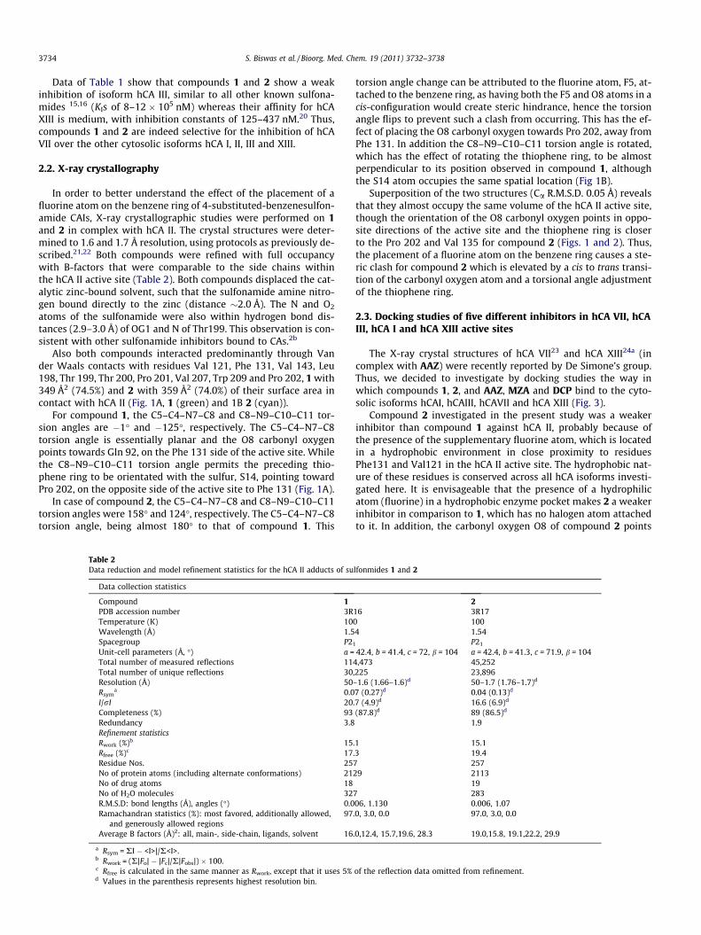
Table 2Data reduction and model refinement statistics for the hCA II adducts of su
Data collection statistics
Compound 1PDB accession number 3RTemperature (K) 10Wavelength (Å) 1.5Spacegroup P2Unit-cell parameters (Å, �) a =Total number of measured reflections 11Total number of unique reflections 30Resolution (Å) 50Rsym
a 0.0I/rI 20Completeness (%) 93Redundancy 3.8Refinement statisticsRwork (%)b 15Rfree (%)c 17Residue Nos. 25No of protein atoms (including alternate conformations) 21No of drug atoms 18No of H2O molecules 32R.M.S.D: bond lengths (Å), angles (�) 0.0Ramachandran statistics (%): most favored, additionally allowed,
and generously allowed regions97
Average B factors (Å)2: all, main-, side-chain, ligands, solvent 16
a Rsym = RI � <I>|/R<I>.b Rwork = (R|Fo| � |Fc|/R|Fobs|) � 100.c Rfree is calculated in the same manner as Rwork, except that it uses 5%d Values in the parenthesis represents highest resolution bin.
torsion angle change can be attributed to the fluorine atom, F5, at-tached to the benzene ring, as having both the F5 and O8 atoms in acis-configuration would create steric hindrance, hence the torsionangle flips to prevent such a clash from occurring. This has the ef-fect of placing the O8 carbonyl oxygen towards Pro 202, away fromPhe 131. In addition the C8–N9–C10–C11 torsion angle is rotated,which has the effect of rotating the thiophene ring, to be almostperpendicular to its position observed in compound 1, althoughthe S14 atom occupies the same spatial location (Fig 1B).
Superposition of the two structures (Ca R.M.S.D. 0.05 Å) revealsthat they almost occupy the same volume of the hCA II active site,though the orientation of the O8 carbonyl oxygen points in oppo-site directions of the active site and the thiophene ring is closerto the Pro 202 and Val 135 for compound 2 (Figs. 1 and 2). Thus,the placement of a fluorine atom on the benzene ring causes a ste-ric clash for compound 2 which is elevated by a cis to trans transi-tion of the carbonyl oxygen atom and a torsional angle adjustmentof the thiophene ring.
2.3. Docking studies of five different inhibitors in hCA VII, hCAIII, hCA I and hCA XIII active sites
The X-ray crystal structures of hCA VII23 and hCA XIII24a (incomplex with AAZ) were recently reported by De Simone’s group.Thus, we decided to investigate by docking studies the way inwhich compounds 1, 2, and AAZ, MZA and DCP bind to the cyto-solic isoforms hCAI, hCAIII, hCAVII and hCA XIII (Fig. 3).
Compound 2 investigated in the present study was a weakerinhibitor than compound 1 against hCA II, probably because ofthe presence of the supplementary fluorine atom, which is locatedin a hydrophobic environment in close proximity to residuesPhe131 and Val121 in the hCA II active site. The hydrophobic nat-ure of these residues is conserved across all hCA isoforms investi-gated here. It is envisageable that the presence of a hydrophilicatom (fluorine) in a hydrophobic enzyme pocket makes 2 a weakerinhibitor in comparison to 1, which has no halogen atom attachedto it. In addition, the carbonyl oxygen O8 of compound 2 points
lfonmides 1 and 2
216 3R170 1004 1.54
1 P21
42.4, b = 41.4, c = 72, b = 104 a = 42.4, b = 41.3, c = 71.9, b = 1044,473 45,252,225 23,896–1.6 (1.66–1.6)d 50–1.7 (1.76–1.7)d
7 (0.27)d 0.04 (0.13)d
.7 (4.9)d 16.6 (6.9)d
(87.8)d 89 (86.5)d
1.9
.1 15.1
.3 19.47 25729 2113
197 28306, 1.130 0.006, 1.07.0, 3.0, 0.0 97.0, 3.0, 0.0
.0,12.4, 15.7,19.6, 28.3 19.0,15.8, 19.1,22.2, 29.9
of the reflection data omitted from refinement.

Figure 1. Stereo stick representation of hCA II active site complexed with (A) 1 (green) and (B) 2 (cyan). The active-site zinc is depicted as a gray sphere. The electron densityis represented by a 1.2 r-weighted 2Fo � Fc Fourier map (blue mesh). Amino acids are as labeled. Figure made using PyMOL (DeLano Scientific).
Figure 2. Superposition of hCA II complexed with 1 (green) and 2 (cyan). The zinc isdepicted as a gray sphere. Figure made using PyMOL (DeLano Scientific).
S. Biswas et al. / Bioorg. Med. Chem. 19 (2011) 3732–3738 3735
towards the carbonyl group of Pro201 of hCA II, thus placing it in acharge repulsive environment, which is not encountered in thehCA II–1 complex. Here, we have thus a nice example that a rela-tively minor change in the chemical structure of a sulfonamideCAI has significant consequence to its affinity for the enzyme,strongly influencing its binding within the active site. As westressed above, the presence of the fluorine makes 2 a 7.8 timesless potent hCA II inhibitor compared to the structurally related 1.
Among all isoforms studied here, the inhibition of hCA III wasweaker than that of the other ones (Table 1). The rationale for thisis clear, as hCA III has a bulky hydrophobic residue (Phe198) in themiddle of the active site cavity, which is a leucine in the other fourstudied isoforms. This bulky residue effectively prevents any siz-able inhibitor to bind into the active site, which is obvious alsofrom Figure 3, showing Phe198 with its bulky phenyl moietyplaced in the region where sulfonamides bind in the adducts withother cytosolic isoforms, such as hCA I, II and XIII.
The present data also showed that the best inhibition with com-pounds 1 and 2 was against hCA VII (Table 1). This potent inhibitionis probably due to the presence of Lys93 in hCA VII, an amino acidresidue which is unique only to this isoform, providing a hydro-philic and flexible amino acid residue in the middle of the activesite, which furnish an additional interaction point for the inhibitor.Based on a model generated using Coot,29 Lys93 can have an alter-nate conformation leading to the formation of a hydrogen bondwith either the carbonyl oxygen of compound 1 or the fluorine atompresent in compound 2, thereby further stabilizing the inhibitor–hCA VII complexes. When this residue is replaced by a bulky sidechain such as Arg91 (in the case of hCA XIII and hCA III) the inhibi-tion with compounds 1 and 2 is weaker compared to hCA VII, mostlikely because the flexibility of Arg91 is reduced compared to that ofthe lysine present in hCA VII, also leading to steric clashes. In addi-tion, hCA VII also has a smaller side chain (Ala137) close to the ac-tive site instead of hydrophobic, bulkier Val135 (present in hCA IIand XIII), which might facilitate the easier binding of inhibitorswithin the hCA VII active site. The observation that Lys93 couldbe involved in a possible additional hydrogen bond for the hCAVII adducts of these inhibitors might be extended also to the other

Figure 3. Stereo stick representation of the superposition of hCA I (pink), hCA II (green), hCA III (yellow), hCA VII (wheat), and hCA XIII (purple) active sites complexed with(A) 1, (B) 2, (C) AAZ, (D) MZA, and (E) DCP. The active-site zinc is depicted as a gray sphere. Amino acids positions are labeled using hCA II numbering. Figure made usingPyMOL (DeLano Scientific).
3736 S. Biswas et al. / Bioorg. Med. Chem. 19 (2011) 3732–3738
compounds studied, such as AAZ, MZA, DCP. This fact was corrob-orated by the lower KIs measured for these sulfonamides againstthis isoform (compared to hCA I, II, III and XIII), which is the mostinhibited by sulfonamides among all the cytosolic CAs (Table 1).
Excluding the poor inhibition of hCA III observed for all studiedcompounds, DCP showed the weakest inhibition against hCA Iamong the other cytosolic isoforms (Table 1). This is the only inhib-itor used in the present studies bearing two sulfonamide moieties,and its X-ray structure in complex with hCA II was reported earlierby one of our groups.24b In the case of hCA I, the residues closest tothe second sulfonamide moiety of DCP were His200 and Val62(Fig. 3), which correspond to Thr202 and Asn62 in case of hCA II.The presence of the sulfonamide group close to His200 may result
in charge repulsions, leading to the observed weaker inhibition ofDCP against hCA I (Table 1). On the other hand, the presence ofAsn62 in case of hCA II (which is highly polar and flexible), can sta-bilize interactions with a sulfonamide group close to it, thus mak-ing DCP a rather potent inhibitor for hCA II, hCA VII and hCA XIII(compared to hCA I and III). The presence of the bulky side chainresidue His200 in the hCA I active site also leads to a 20 fold de-crease in inhibition with AAZ compared to hCA II. However in caseof the MZA–hCA I complex, the presence of the additional methylgroup of the inhibitor in the hydrophobic pocket makes it a betterinhibitor of hCA I compared AAZ. Collectively these results help usunderstand the affinity/selectivity of different sulfonamideinhibitors towards the cytosolic CA isoforms, which show a great

S. Biswas et al. / Bioorg. Med. Chem. 19 (2011) 3732–3738 3737
variation in their inhibition profile for this class of pharmacologi-cally important compounds, even in the presence of minimal struc-tural changes of inhibitors, such as; the comparison of 1 having abetter KI than 2 for all the CA isoforms tested, and MZA exhibitingselectivity over AAZ only for hCA I (Fig. 3).
3. Conclusions
The X-ray crystal structure of the adducts of hCA II complexedwith two aromatic sulfonamides incorporating 2-thienylacetamidomoieties is reported here. The two inhibitors only differ by thepresence of an additional 3-fluoro substituent on the 4-amino-ben-zenesulfonamide scaffold of one of them, but their inhibition pro-files against the cytosolic isoforms hCA I, II, III, VII and XIII are quitedifferent. These differences were rationalized based on the ob-tained X-ray crystal structures, including in the comparison othersulfonamide CA inhibitors with clinical applications, such as AAZ,MZA and DCP. Specific interactions between the structurally differ-ent inhibitors and amino acid residues present only in some ofthese isoforms have been shown, which explain the high affinityof the 2-thienylacetamido benzenesulfonamides for some pharma-cologically relevant CAs such as isoforms II and VII, and might beuseful for the drug design of isoform-selective sulfonamide inhib-itors of various CAs.
4. Experimental
4.1. Chemistry and CA inhibition
Sulfonamides used in this work were reported earlier (1 and2)13 or are commercially available from Sigma-Aldrich (Milan,Italy). An Applied Photophysics stopped-flow instrument has beenused for assaying the CA catalysed CO2 hydration activity. Phenolred (at a concentration of 0.2 mM) has been used as indicator,working at the absorbance maximum of 557 nm, with 20 mMHepes (pH 7.5) as buffer, and 20 mM Na2SO4 (for maintaining con-stant the ionic strength), following the initial rates of the CA-cata-lyzed CO2 hydration reaction for a period of 10–100 s. The CO2
concentrations ranged from 1.7 to 17 mM for the determinationof the kinetic parameters and inhibition constants. For each inhib-itor at least six traces of the initial 5–10% of the reaction have beenused for determining the initial velocity. The uncatalyzed rateswere determined in the same manner and subtracted from the to-tal observed rates. Stock solutions of inhibitor (0.1 mM) were pre-pared in distilled-deionized water and dilutions up to 0.01 nMwere done thereafter with distilled-deionized water. Inhibitorand enzyme solutions were preincubated together for 15 min atroom temperature prior to assay, in order to allow for the forma-tion of the E–I complex. The inhibition constants were obtainedby non-linear least-squares methods using PRISM 3, as reported ear-lier,13 and represent the mean from at least three different deter-minations. CA isoforms were recombinant ones obtained inhouse as reported earlier.7–10,13
4.2. X-ray crystal structure determination
hCA II was purified to electrophoretic homogeneity according topreviously described protocol.3 Co-crystals of the hCA II – 1, – 2complexes were obtained using the hanging drop vapor diffusionmethod.25 Drops of 5 lL (0.5 mM hCA II; 1 mM compound; 0.1% di-methyl sulfoxide (DMSO); 0.8 M Sodium Citrate; 50 mM Tris–HCl;pH 8.0) were equilibrated against 1 ml precipitant solution (1.6 Msodium citrate; 50 mM Tris–HCl; pH 8.0) at room temperature(�20 �C). Prior to collecting X-ray diffraction datasets, the crystalwere cryoprotected using 25% glycerol in the precipitant solution
containing 2 mM drug solution and flash cooled in gaseous nitro-gen stream. Data were collected at 100 K using a R-AXIS IV++ imageplate (IP) system with OsmicVarimax optics and a Rigaku RU-H3RCu rotating anode operating at 50 kV and 22 mA. The crystal to IPdistance was 76 mm in case of compound 1 and 80 mm in case ofcompound 2. The oscillation steps were 1� with 5 min exposure perimage. Indexing, integration and scaling were performed usingHKL2000.26
The structures of hCAII bound to compound 1 and 2 were solvedby using hCAII as a model (PDB accession code 2ILI).2d Initial phaseswere obtained by molecular substitution using PHASER in ccp4suite.27 Refinement proceeded with 5% of the data allocated for Rfree
calculations using PHENIX version 1.7.28 The ligand coordinateswere generated using eLBOW in PHENIX and subsequentlymerged using Coot.29 After several cycles of refinement alternatedwith manual refitting of the model and solvent placement themodels converged. The validity of the final model was assessedby PROCHECK.30 Complete refinement statistics are included inTable 2.
4.3. Structure–activity relationship and alignment of differentCA isoforms
The hCA I (PDB accession number 3LXE),31 VII (PDB accessionnumber 3ML5)23 and XIII (PDB accession number 3CZV)24a coordi-nates were superimposed onto hCA II complexed with compounds1, 2, AAZ, MZA and DCP. The align option in PyMOL was used tosuperimpose different CA isoform structures. This feature in Py-MOL calculates the RMSD values for different superimposed struc-tures and makes a best possible alignment of them. In case ofcompounds 1 and 2 the hCAII structure reported here was usedas a reference structure and various CA isoform structures withoutligands were superimposed onto them. Similarly the hCAII struc-ture in complex with DCP (2POU) was used as a reference structureto generate Figure 3E. In case of AAZ and MZA, hCA VII structure(3ML5) and hCAI (1BZM), respectively, were used as the reference,all other unliganded forms of CA were used as the moving struc-tures for superposition. After aligning the structures in PyMOLthe active site residues were highlighted and shown in stick form(Fig. 3). The differences in amino acids at various inhibitor-interac-tion regions was also analyzed using the SSM superpose option inCoot.29 The figures were made using PyMOL.32
References and notes
1. Hilvo, M.; Tolvanen, M.; Clark, A.; Shen, B.; Shah, G. N.; Waheed, A.; Halmi, P.;Hänninen, M.; Hämäläinen, J. M.; Vihinen, M.; Sly, W. S.; Parkkila, S. Biochem. J.2005, 392, 83.
2. (a) Kaunisto, K.; Parkkila, S.; Rajaniemi, H.; Waheed, A.; Grubb, J.; Sly, W. S.Kidney Int. 2002, 61, 2111; (b) Supuran, C. T. Nat. Rev. Drug Disc. 2008, 7, 168; (c)Supuran, C. T. Bioorg. Med. Chem. Lett. 2010, 20, 3467; (d) Fisher, S. Z.; Maupin,C. M.; Budayova-Spano, M.; Govindasamy, L.; Tu, C. K.; Agbandje-McKenna, M.;Silverman, D. N.; Voth, G. A.; McKenna, R. Biochemistry 2007, 42, 2930; (e)Ebbesen, P.; Pettersen, E. O.; Gorr, T. A.; Jobst, G.; Williams, K.; Kienninger, J.;Wenger, R. H.; Pastorekova, S.; Dubois, L.; Lambin, P.; Wouters, B. G.; Supuran,C. T.; Poellinger, L.; Ratcliffe, P.; Kanopka, A.; Görlach, A.; Gasmann, M.; Harris,A. L.; Maxwell, P.; Scozzafava, A. J. Enzyme Inhib. Med. Chem. 2009, 24, 1.
3. (a) Wagner, J. M.; Avvaru, B. S.; Robbins, A. H.; Scozzafava, A.; Supuran, C. T.;McKenna, R. Bioorg. Med. Chem. 2010, 18, 4873; (b) Pacchiano, F.; Aggarwal, M.;Avvaru, B. S.; Robbins, A. H.; Scozzafava, A.; McKenna, R.; Supuran, C. T. Chem.Commun. (Camb.) 2010, 46, 8371; (c) Pastorekova, S.; Parkkila, S.; Pastorek, J.;Supuran, C. T. J. Enzyme Inhib. Med. Chem. 2004, 19, 199; (d) Poulsen, S. A. ExpertOpin. Ther. Pat. 2010, 20, 795; (e) Alterio, V.; Hilvo, M.; Di Fiore, A.; Supuran, C.T.; Pan, P.; Parkkila, S.; Scaloni, A.; Pastorek, J.; Pastorekova, S.; Pedone, C.;Scozzafava, A.; Monti, S. M.; De Simone, G. Proc. Natl. Acad. Sci. U.S.A. 2009, 106,16233.
4. (a) Supuran, C. T. Carbonic Anhydrases as Drug Targets—General Presentation.In Drug Design of Zinc-Enzyme Inhibitors: Functional, Structural, and DiseaseApplications; Supuran, C. T., Winum, J. Y., Eds.; Wiley: Hoboken (NJ), 2009; pp15–38; (b) Winum, J. Y.; Rami, M.; Scozzafava, A.; Montero, J. L.; Supuran, C.Med. Res. Rev. 2008, 28, 445; (c) Supuran, C. T.; Scozzafava, A.; Casini, A. Med.Res. Rev. 2003, 23, 146.

3738 S. Biswas et al. / Bioorg. Med. Chem. 19 (2011) 3732–3738
5. (a) De Simone, G.; Di Fiore, A.; Menchise, V.; Pedone, C.; Antel, J.; Casini, A.;Scozzafava, A.; Wurl, M.; Supuran, C. T. Bioorg. Med. Chem. Lett. 2005, 15, 2315;(b) Casini, A.; Antel, J.; Abbate, F.; Scozzafava, A.; David, S.; Waldeck, H.;Schafer, S.; Supuran, C. T. Bioorg. Med. Chem. Lett. 2003, 13, 841; (c) Abbate, F.;Casini, A.; Owa, T.; Scozzafava, A.; Supuran, C. T. Bioorg. Med. Chem. Lett. 2004,14, 217; (d) Abbate, F.; Casini, A.; Scozzafava, A.; Supuran, C. T. Bioorg. Med.Chem. Lett. 2004, 14, 2357.
6. (a) Winum, J. Y.; Casini, A.; Mincione, F.; Starnotti, M.; Montero, J. L.;Scozzafava, A.; Supuran, C. T. Bioorg. Med. Chem. Lett. 2004, 14, 225; (b)Menabuoni, L.; Scozzafava, A.; Mincione, F.; Briganti, F.; Mincione, G.; Supuran,C. T. J. Enzyme Inhib. 1999, 14, 457; (c) Scozzafava, A.; Menabuoni, L.; Mincione,F.; Briganti, F.; Mincione, G.; Supuran, C. T. J. Med. Chem. 1999, 42, 2641.
7. Carta, F.; Temperini, C.; Innocenti, A.; Scozzafava, A.; Kaila, K.; Supuran, C. T. J.Med. Chem. 2010, 53, 5511.
8. (a) Maresca, A.; Temperini, C.; Vu, H.; Pham, N. B.; Poulsen, S. A.; Scozzafava, A.;Quinn, R. J.; Supuran, C. T. J. Am. Chem. Soc. 2009, 131, 3057; (b) Maresca, A.;Temperini, C.; Pochet, L.; Masereel, B.; Scozzafava, A.; Supuran, C. T. J. Med.Chem. 2010, 53, 335.
9. (a) Maresca, A.; Supuran, C. T. Bioorg. Med. Chem. Lett. 2010, 20, 4511; (b)Maresca, A.; Scozzafava, A.; Supuran, C. T. Bioorg. Med. Chem. Lett. 2010, 20,7255.
10. Innocenti, A.; Vullo, D.; Scozzafava, A.; Supuran, C. T. Bioorg. Med. Chem. Lett.2008, 18, 1583.
11. Alterio, V.; Di Fiore, A.; D’Ambrosio, K.; Supuran, C. T.; De Simone, G. X-RayCrystallography of CA Inhibitors and Its Importance in Drug Design. In DrugDesign of Zinc-Enzyme Inhibitors: Functional, Structural, and Disease Applications;Supuran, C. T., Winum, J. Y., Eds.; Wiley: Hoboken, 2009; pp 73–138.
12. (a) Wistrand, P. J.; Lindqvist, A. In Carbonic Anhydrase—From Biochemistry andGenetics to Physiology and Clinical Medicine; Botrè, F., Gros, G., Storey, B. T., Eds.;VCH: Weinhein, 1991; pp 352–378; (b) Mincione, F.; Scozzafava, A.; Supuran,C. T. Antiglaucoma Carbonic Anhydrase Inhibitors as Ophthalomologic Drugs.In Drug Design of Zinc-Enzyme Inhibitors: Functional, Structural, and DiseaseApplications; Supuran, C. T., Winum, J. Y., Eds.; Wiley: Hoboken (NJ), 2009; pp139–154; (c) Steele, R. M.; Batugo, M. R.; Benedini, F.; Biondi, S.; Borghi, V.;Carzaniga, L.; Impagnatiello, F.; Miglietta, D.; Chong, W. K. M.; Rajapakse, R.;Cecchi, A.; Temperini, C.; Supuran, C. T. Bioorg. Med. Chem. Lett. 2009, 19, 6565.
13. (a) Güzel, Ö.; Innocenti, A.; Scozzafava, A.; Salman, A.; Supuran, C. T. Bioorg.Med. Chem. Lett. 2009, 19, 3170; (b) Güzel, Ö.; Innocenti, A.; Scozzafava, A.;Salman, A.; Supuran, C. T. Bioorg. Med. Chem. 2009, 17, 4894.
14. Asiedu, M.; Ossipov, M. H.; Kaila, K.; Price, T. J. Pain 2010, 148, 302.15. (a) Wistrand, P. J. Ups. J. Med. Sci. 2002, 107, 77; (b) Carter, N.; Shiels, A.;
Tashian, R. Biochem. Soc. Trans. 1978, 6, 552; (c) Elder, I.; Fisher, Z.; Laipis, P. J.;Tu, C.; McKenna, R.; Silverman, D. N. Proteins 2007, 68, 337; (d) Duda, D. M.; Tu,C.; Fisher, S. Z.; An, H.; Yoshioka, C.; Govindasamy, L.; Laipis, P. J.; Agbandje-McKenna, M.; Silverman, D. N.; McKenna, R. Biochemistry 2005, 44, 10046.
16. (a) Nishimori, I.; Minakuchi, T.; Onishi, S.; Vullo, D.; Cecchi, A.; Scozzafava, A.;Supuran, C. T. Bioorg. Med. Chem. 2007, 15, 7229; (b) Nishimori, I.; Minakuchi,T.; Onishi, S.; Vullo, D.; Cecchi, A.; Scozzafava, A.; Supuran, C. T. J. Enzyme Inhib.Med. Chem. 2009, 24, 70.
17. (a) Lehtonen, J.; Shen, B.; Vihinen, M.; Casini, A.; Scozzafava, A.; Supuran, C. T.;Parkkila, A.-K.; Saarnio, J.; Kivelä, A.; Waheed, A.; Sly, W. S.; Parkkila, S. J. Biol.Chem. 2004, 279, 2719; (b) Lehtonen, J. M.; Parkkila, S.; Vullo, D.; Casini, A.;Scozzafava, A.; Supuran, C. T. Bioorg. Med. Chem. Lett. 2004, 14, 3757.
18. Hilvo, M.; Supuran, C. T.; Parkkila, S. Curr. Top. Med. Chem. 2007, 7, 893.19. Hilvo, M.; Innocenti, A.; Monti, S. M.; De Simone, G.; Supuran, C. T.; Parkkila, S.
Curr. Pharm. Des. 2008, 14, 672.20. Khalifah, R. G. J. Biol. Chem. 1971, 246, 2561.21. Avvaru, B. S.; Wagner, J. M.; Maresca, A.; Scozzafava, A.; Robbins, A. H.;
Supuran, C. T.; McKenna, R. Bioorg. Med. Chem. Lett. 2010, 20, 4376.22. (a) Di Fiore, A.; De Simone, G.; Menchise, V.; Pedone, C.; Casini, A.; Scozzafava,
A.; Supuran, C. T. Bioorg. Med. Chem. Lett. 2005, 15, 1937; (b) Menchise, V.; DeSimone, G.; Alterio, V.; Di Fiore, A.; Pedone, C.; Scozzafava, A.; Supuran, C. T. J.Med. Chem. 2005, 48, 5721; (c) Baranauskiene, L.; Hilvo, M.; Matuliene, J.;Golovenko, D.; Manakova, E.; Dudutiene, V.; Michailoviene, V.; Torresan, J.;Jachno, J.; Parkkila, S.; Maresca, A.; Supuran, C. T.; Grazulis, S.; Matulis, D. J.Enzyme Inhib. Med. Chem. 2010, 25, 863; (d) Di Fiore, A.; Monti, S. M.; Innocenti,A.; Winum, J.-Y.; De Simone, G.; Supuran, C. T. Bioorg. Med. Chem. Lett. 2010, 20,3601.
23. Di Fiore, A.; Truppo, E.; Supuran, C. T.; Alterio, V.; Dathan, N.; Bootorabi, F.;Parkkila, S.; Monti, S. M.; De Simone, G. Bioorg. Med. Chem. Lett. 2010, 20,5023.
24. (a) Di Fiore, A.; Monti, S. M.; Hilvo, M.; Parkkila, S.; Romano, V.; Scaloni, A.;Pedone, C.; Scozzafava, A.; Supuran, C. T.; De Simone, G. Proteins 2009, 74, 164;(b) Alterio, V.; De Simone, G.; Monti, S. M.; Scozzafava, A.; Supuran, C. T. Bioorg.Med. Chem. Lett. 2007, 17, 4201.
25. McPherson, A. Preparation and Analysis of Protein Crystals, 1st ed.; Wiley: NewYork, 1982.
26. Otwinowski, Z.; Minor, W. Methods Enzymol. 1997, 276, 307.27. Collaborative Computational Project, Number 4. Acta Crystallogr., Sect. D, 1994,
50, 760.28. Adams, P. D.; Afonine, P. V.; Bunkóczi, G.; Chen, V. B.; Davis, I. W.; Echols, N.;
Headd, J. J.; Hung, L.-W.; Kapral, G. J.; Grosse-Kunstleve, R. W.; McCoy, A. J.;Moriarty, N. W.; Oeffner, R.; Read, R. J.; Richardson, D. C.; Richardson, J. S.;Terwilliger, T. C.; Zwart, P. H. Acta Crystallogr., Sect. D 2010, 66, 213.
29. Emsley, P.; Cowtan, K. Acta Crystallogr., Sect. D 2004, 60, 2126.30. Laskowski, R. A.; MacArthur, M. W.; Moss, D. S.; Thornton, J. M. J. Appl.
Crystallogr. 1993, 26, 283.31. Alterio, V.; Monti, S. M.; Truppo, E.; Pedone, C.; Supuran, C. T.; De Simone, G.
Org. Biomol. Chem. 2010, 8, 3528.32. The PyMOL Molecular Graphics System, Version 1.3, Schrödinger, LLC.