Non-Random Patterns of Membrane Proteins and Their Roles in Transmembrane Signaling
Upload
independentCategory
view
0download
0
Expression of Hypoxia-Inducible Cell-SurfaceTransmembrane Carbonic Anhydrases in HumanCancer
Sergey Ivanov,*† Shu-Yuan Liao,‡ Alla Ivanova,*Alla Danilkovitch-Miagkova,* Nadezhda Tarasova,§
Gregor Weirich,* Marsha J. Merrill,¶
Martin A. Proescholdt,¶ Edward H. Oldfield,¶
Joshua Lee,* Jan Zavada,i Abdul Waheed,**William Sly,** Michael I. Lerman,* andEric J. Stanbridge‡
From the Laboratory of Immunobiology,* the Intramural
Research Support Program,† Science Applications International
Corporation, Frederick, Maryland; the Molecular Aspects of Drug
Design Section,§ National Cancer Institute, Frederick Cancer
Research and Development Center, Frederick, Maryland; the
Surgical Neurology Branch,¶ National Institute of Neurological
Disorders and Stroke, National Institutes of Health, Bethesda,
Maryland; the Department of Microbiology and Molecular
Genetics,‡ College of Medicine, University of California at Irvine,
Irvine, California; the St. Louis University School of Medicine,**
St. Louis, Missouri; and the Academy of Sciences,i Prague,
Czech Republic
An acidic extracellular pH is a fundamental propertyof the malignant phenotype. In von Hippel-Lindau(VHL)-defective tumors the cell surface transmem-brane carbonic anhydrase (CA) CA9 and CA12 genesare overexpressed because of the absence of pVHL.We hypothesized that these enzymes might be in-volved in maintaining the extracellular acidic pH intumors, thereby providing a conducive environmentfor tumor growth and spread. Using Northern blotanalysis and immunostaining with specific antibodieswe analyzed the expression of CA9 and CA12 genesand their products in a large sample of cancer celllines, fresh and archival tumor specimens, and nor-mal human tissues. Expression was also analyzed incultured cells under hypoxic conditions. Expressionof CA IX and CA XII in normal adult tissues was de-tected only in highly specialized cells and for mosttissues their expression did not overlap. Analysis ofRNA samples isolated from 87 cancer cell lines and 18tumors revealed high-to-moderate levels of expres-sion of CA9 and CA12 in multiple cancers. Immuno-histochemistry revealed high-to-moderate expressionof these enzymes in various normal tissues and mul-tiple common epithelial tumor types. The immuno-staining was seen predominantly on the cell surface
membrane. The expression of both genes was mark-edly induced under hypoxic conditions in tumors andcultured tumor cells. We conclude that the cell surfacetrans-membrane carbonic anhydrases CA IX and CAXII are overexpressed in many tumors suggesting thatthis is a common feature of cancer cells that may berequired for tumor progression. These enzymes maycontribute to the tumor microenvironment by main-taining extracellular acidic pH and helping cancercells grow and metastasize. Our studies show an im-portant causal link between hypoxia, extracellularacidification, and induction or enhanced expressionof these enzymes in human tumors. (Am J Pathol2001, 158:905–919)
The development of solid human tumors may be approx-imated by a two-stage model. In stage one, the malignantcells grow into small tumors, which then stop growingbecause of an inadequate supply of oxygen (hypoxia). Instage two, hypoxia triggers a drastic change in geneexpression, followed by clonal selection within the tumorcell population for cells with increased adaptation to hyp-oxia.1 This leads to angiogenesis2 and a fundamentalswitch in energy metabolism, replacing respiration withglycolysis.3–5 These changes further lead to a new com-promised microenvironment within and around the tumormass, characterized by low oxygen tension, high hydro-static pressure, and an acidic extracellular pH (pHe).6,7
The hypoxia-inducible transcription factor (HIF1) controlsthe expression of several dozen target genes, includingthose involved in energy metabolism (glucose transport-ers, glycolytic enzymes), angiogenesis [vascular endo-thelial growth factor (VEGF) and VEGFR-1], and surfacetransmembrane carbonic anhydrases (CAs).1,2,8–11 HIF1is considered to be a master regulator gene that inte-grates pathways regulating physiological responses toacute and chronic hypoxia.1,11–15 pVHL is an integral part
Supported in part by National Cancer Institute, National Institutes ofHealth, contract no. NO1-CO-56000 and CA19401 from the NationalCancer Institute.
S. I. and S.-Y. L contributed equally to this work.
Accepted for publication November 17, 2000.
Address reprint requests to Dr. Sergey Ivanov, Science ApplicationInternational Corp., Frederick Laboratory of Immunology, Bldg. 560, NCI-FCRDC, P. O. Box B, Frederick, MD 21702. E-mail: [email protected].
American Journal of Pathology, Vol. 158, No. 3, March 2001
Copyright © American Society for Investigative Pathology
905

of a novel multiprotein ubiquitin ligase complex, termedVBC (VHL/Elongin B/Elongin C), that recruits importantcellular proteins for rapid degradation by the ubiquitin-proteasome proteolysis system. The most plausible can-didate targets for this proteolytic degradation includeHIF1-a and HIF2-a.8,16–18 Both HIF-a subunits are pVHL-binding proteins that are targeted by pVHL, presumablyvia the VBC complex, for degradation in normoxic but nothypoxic conditions.8 Therefore, pVHL is responsible forcontrol of the hypoxia-driven changes in gene expressionin tumors, thereby initiating and augmenting the secondstage of tumor development. In VHL-defective tumors,curiously enough, the two fundamental stages of tumordevelopment occur either simultaneously or in reverse,first triggering the hypoxia-cellular response, followed byproliferation of transformed cells, consistent with the an-giogenic phenotype of tumors seen in the VHL syndrome.Previously, we discovered that in VHL patients the CA9and CA12 genes are overexpressed in tumors becauseof the absence of pVHL, and argued that these CA en-zymes might be involved in sensing and maintaining theacidic tumor microenvironment.10 We have now ex-tended these observations by analyzing the expressionof CA IX and CA XII in a large sample of cancer cell linesand fresh or archival tumor specimens. Here we showthat either one or both genes are overexpressed in manytumor types, suggesting that this is a common feature ofcancer cells, consistent with the fundamental role of VHLin tumor development. We believe that overexpression ofthese enzymes contributes to the acidic tumor microen-vironment and helps the cancer cells to grow and metas-tasize.
Materials and Methods
Cancer Cell Line RNA Collection
We used 45 mRNA samples isolated from cancer celllines provided by the Developmental Therapeutics Pro-gram, Frederick Cancer Research and DevelopmentCenter, National Cancer Institute. Additional mRNA sam-ples from central nervous system (CNS) tumor cell linesU105MG, U251MG, U373MG, and G4; leukemia cell linesU937 and HL60; the renal cell carcinoma cell line UM-RC-29; and the monocyte cell line THP-1 were kindlyprovided by Dr. Teizo Yoshimura (NCI-Frederick, Freder-ick, MD). Non-small cell carcinoma (H1373, H1264,H1693, H1944, H838, H1299, H157, H1466, H460, H727,and H28), small-cell carcinoma cell lines (H1184, H2081,H2227, H1086, H841, H69, H1820, H660, H1769, H446,H1238, H748, and H2552), cancer cell lines SCC-35,MCF-7-adr, SQ-20B, 510-HPV-18, and Scid (5020) werekindly provided by Dr. Bruce Johnson (NCI, Bethesda,MD). The human glioblastoma cell line, U87, was pur-chased from ATCC (Manassas, VA). RNA samples fromother cancer cell lines were available on the ClontechMTN blot no. 7757-1, and samples from normal tissues onMTE Array no. 7775--1 blot (Clontech, Palo Alto, CA).
Molecular Techniques
Total RNA and mRNA isolation from cell lines and tumortissues was done using commercially available kits (In-vitrogen, Carlsbad, CA; Life Technologies, Inc., GrandIsland, New York). Electrophoresis in formaldehyde gelsand Northern blot analysis were performed according topublished procedures.10,19 Quantification of Northern hy-bridization signals was done as described previously10
or using a VE-1000 Video Camera System (Dage-MTI,Inc., Michigan City, IN) and NIH Image Version 1.6.1software.
Tissue Specimens
The normal adult tissues from all organ sites and thecorresponding benign and/or malignant neoplastic tis-sues were obtained from routine pathology specimens atSt. Joseph Hospital (Orange, CA). These organs includethe brain (cerebrum, cerebellum, ventricle, pons, pituitarygland), eyes, nose, throat, upper and lower respiratorysystem, heart, upper and lower gastrointestinal system(esophagus, stomach, small and large intestine), pan-creas, liver, biliary system including gallbladder, femaleand male urogenital system (kidney, ureter, bladder, tes-ticle, cervix, uterus, fallopian tube, ovary), adrenal gland,thyroid, parathyroid, salivary gland, spleen, bone, mus-cle, cartilage, skin, and the body cavity. All tissue sam-ples were processed within 6 hours of surgical resectionand fixed in 10% neutral-buffered formalin or snap-fro-zen. The formalin-fixed tissues were paraffin-embedded,sectioned, and stained with hematoxylin and eosin (H&E)for light microscopic examination.
Immunohistochemical Studies
The mouse monoclonal antibody (MN75) used to detectthe MN/CA IX protein and the rabbit polyclonal antibodyto CA XII protein have been described previously.20,21
Immunohistochemical staining of tissue sections with an-ti-CA IX and anti-CA XII antibodies was done using aperoxidase technique with microwave pretreatment, asdescribed previously.22 Known positive and negative tis-sue specimens were included in each run. For CA IXimmunostaining, the primary antibody was used at a1:10,000 dilution and the CA XII at a 1:500 dilution. Theimmunohistochemical results were semiquantitativebased on the percentage of the positive cells seen in atotal field of a single section. The pattern of staining wasscored as diffuse when $40% of the cells stained andfocal when ,40% of the cells stained. A negative scorewas given to tissue sections that had no evidence ofspecific immunostaining.
pH Determination, Glucose Uptake, andHypoxia Conditions in Cell Culture
Renal clear-cell carcinoma cell line 786-0 and its deriva-tive, expressing the wtVHL transgene, were describedearlier.10 They were grown to confluence in Dulbecco’s
906 Ivanov et alAJP March 2001, Vol. 158, No. 3

modified Eagle’s medium (cat. no. 11965-084; Life Tech-nologies, Inc.) with 10% bovine fetal serum (Sigma, St.Louis, MO). Three independent experiments on pH andglucose measurements in the media were done using aHanna Instruments microcomputer pH meter (modelHI931000) and Infinity Glucose Reagent (Sigma Diagnos-tics, cat. no. 18-20). U87 cell culture and maintenance ofhypoxia were performed as previously described.19
Results
Northern Blot Analysis of CA9 and CA12 inNormal/Tumor Tissues, and Tumor Cell Lines
Using MTE array blots containing 68 different mRNAsfrom a variety of normal adult tissues, we observed thatthe CA12 gene was highly expressed in kidney, colonand rectum, esophagus, brain, and the pancreas,whereas the mammary gland, bladder, uterus, trachea,and aorta showed low levels of expression. In the brain,only the corpus striatum [caudate nucleus (E2) and pu-
tamen (H2)] produced strong hybridization signals withthe CA12 probe (Figure 1). Moderate expression of CA9was observed in a limited number of tissues that includedstomach and, to an even lesser extent, heart, liver, pan-creas, and salivary gland (Figure 1). These data wereconsistent with hybridization results obtained with theMTN blots. In addition, low signals were also observed inbrain and placenta (data not shown). We detected nosignificant differences in the distribution of CA12 expres-sion in a variety of fetal tissues compared to normal adulttissues (Figure 1, column 11; and data not shown). In thecase of CA9, however, fetal lung and muscle demon-strated high levels of expression whereas adult lung andmuscle were negative (data not shown).
Northern blot analyses of mRNAs obtained from alarge number of cancer cell lines and freshly excisedtumors are summarized in Table 1 and Table 2, and anexample of a Northern blot is given in Figure 2. Among 87RNA samples, representing tumors of the head and neck,lung, kidney, cervix, ovary, prostate, breast, colon, skin,and several leukemias, 50 (57%) expressed either one or
Table 1. Expression of CA9/CA12 mRNA in Human Cancer Cell Lines
Tumor type CA12-positive CA9-positive CA9/CA12-negative
Head and Neck not done SCC-35(2), SQ-20B(2)Lung non-small cell
carcinomaNCI-H1264(1), NCI-
J460(3), NCI-H1944(1),NCI-H157(1), NCI-H28(1), A549(10),EKVX(1), HOP62(1),HOP92(2)
NCI-H1373(1), NCI-H1264(1), NCIH1466(1),NCI-H460(1), NCI-H727(2)
NCI-H1693, NCI-H838,NCI-H1299, NCI-H226, NCI-H23, NCI-H522
Lung small cellcarcinoma
NCI-H2081(1), NCI-H69(1),NCI-H1769(1)
NCI-H1184, NCI-H2227, NCI-H1086,NCI-H660, NCI-H1820, NCI-H841,NCI-H446, NCI-H1238, NCI-H748,NCI-H2552
Renal clear cellcarcinoma
UM-RC-6(3), UM-RC-29(2*), 786-0 (2),ACHN(1), RXF-393(3),TK-10(1)
786-0 (3), A498(1), RXF-393(2)
CAKI-1 (VHL†), UO-31
CNS tumors U105MG(1), U373MG(2),G4(2), SF-295(1), SF-539(2), SNB75(2)
U105MG(2), U373MG(3),U251MG(2), G4(1), SF-539(1), SNB19(2),SNB75(2)
SF-268
Leukemia MOLT-4(1), K-562(1*),SR(3)
K-562(1) HL60, U937, CCRF-CEM, Burkitt’slymphoma Raji
Colon tumors HT29(2), KM12(1) COLO 205(2), HCC-2998(1), HCT-15(3),HT29(3), KM12(1),SW480(1)
HCT-116
Cervix tumors HeLa S3(2) HeLa S3(2)Mammary tumors T 47D(2) MDA-N, MDA-MB-435,
MDA-MB-231, MCF-7, NCI/ADR-RES, HS578T
Ovary tumors OVCAR-5 (1), SK-OV-3 (2) SK-OV-3 (1) OVCAR-3, OVCAR-4,OVCAR-8
Prostate tumors PC-3(1) PC-3(1) DU-145Melanoma MALME-3M (1) LOX IMVI, M14, SK-
MEL-2
Expression intensity in arbitrary units (in parentheses).*In these cell lines truncated mRNA was observed.VHL†, this cell line expresses active pVHL.
CA IX and CA XII in Human Cancers 907AJP March 2001, Vol. 158, No. 3

908 Ivanov et alAJP March 2001, Vol. 158, No. 3

both genes at variable intensity, whereas 37 were negative(Table 1 and Figure 2). Non-small cell lung cancer, colonand renal cell carcinoma cell lines, and tumors of the CNSshowed the highest proportion of specimens expressingCA9 or CA12 (68, 78, 89, and 78%, respectively). Fourteenof the 18 tumors (78%) of the CNS and spinal cord showedexpression of the genes (Table 2). Among these tumors 11expressed both genes and three expressed either CA9 orCA12. It was previously shown and extensively docu-mented in this study that expression of CA IX protein mayserve as a diagnostic biomarker for clear cell renal cellcarcinoma22–24 and colorectal tumors,25,26 whereas CA XIIhas been identified as a biomarker for non-small-cell lungcancer (10; US patent no. 5,589,579).
CA IX/CA XII Expression in Normal Adult HumanTissues Analyzed by Immunohistochemistry
High levels of CA IX expression were consistently ob-served in the basal cells in and near the infundibulum andmedulla of the hair follicle, mesothelial cells, and coelo-mic epithelium of the body cavities. In the visceral or-gans, high levels of CA IX expression in the epitheliumwere identified but limited to rete ovarii, rete testis, ductu-lar efferens, bile ducts, pancreatic ducts, and gallblad-der. In the gastrointestinal tract, diffuse CA IX immunore-
activity was observed in the gastric mucosa, ductal cellsof Brunner’s glands, and crypt cells of the duodenum,jejunum, and, to a lesser degree, in the terminal ileumand appendix. In the peripheral and central nervous sys-tems, CA IX expression was limited to the ventricularlining cells and the choroid plexus. Interestingly, meso-dermal cells of the amniotic/chorionic plate of the pla-centa and cartilaginous tissues from joint spaces alsoshowed variable degrees of CA IX protein expression.
CA XII was variably expressed in mesothelial cells andthe coelomic epithelium of the body cavity. In general,high levels of CA XII expression were found in certaintissues that were CA IX-negative. These tissues were thedistal convoluted tubules and the intercalated cells of thecollecting duct of the kidney, sweat glands of the skin, theepithelium of the breast, some of the proliferative endo-metrial glands, and seminal vesicles. Low levels of CA XIIexpression were also found focally in ductal cells andmucous cells of the salivary glands and submucosalglands of the upper respiratory system, epithelial cells ofSchneider’s membrane of the nose, and acinar cells ofthe pancreas. Limited numbers of positive epithelial cellswere also found in the prostate, vas deferens, and tran-sitional mucosa of the renal pelvis. In the gastrointestinaltract, CA XII expression was observed, but was limited tothe surface glands of the large intestine only. Very weakimmunoreactivity of CA XII was also found in the gastricglands. In the peripheral and central nervous system, theCA XII immunoreactivity was restricted to the posteriorlobe of the pituitary glands, remnant of Rathke’s pouch,the choroid plexus and limited numbers of ganglion cellsin the cortex. In the placenta, limited numbers of syncy-tiotrophoblasts were immunoreactive. The only normaltissues co-expressing CA IX and CA XII were mesothelialcells, ductular efferens, and, to a lesser degree, thechoroid plexus and gastric glands. A summary of thedistribution of expression in normal tissues is given inTable 3, and selected normal tissues with high expres-sion of CA IX and/or CA XII are illustrated in Figure 3.
CA IX/CA XII Expression in Different TumorsAnalyzed by Immunohistochemistry
We surveyed a large number of benign and malignanthuman tissues by immunostaining with specific antibod-ies. The results are summarized in Table 4 and Figures 3to 5. With a few exceptions, a wide variety of tumorsexamined showed plasma membrane, or both plasmamembrane and cytoplasmic immunoreactivity for CA IXand CA XII. The frequency of co-expression, the distribu-tion of staining (focal or diffuse), and the intensity ofstaining varied considerably even within a single tumortype. For example, CA IX protein was expressed in 99%of cervical carcinomas tested (n 5 77), and 68% of thesetumors showed diffuse and strong staining, whereas CA
Figure 1. Transcription analysis of CA9/CA12 genes in normal tissues. Two identical membranes (No. 7775-1, Multiple tissue expression array; Clontech)contained human poly-A1 RNA samples extracted from 68 different normal tissues, eight cancer cell lines, and control samples of DNA and RNA. CA12 mRNAis expressed predominantly in the kidney (A7) and colon (H5), whereas CA9 is expressed mainly in the stomach (B5), liver (A9), pancreas (B9), salivary gland(E9), and heart (A4).
Figure 2. Activation of CA9 and CA12 transcription in cell lines derived fromCNS and lung tumors. A typical Northern blot analysis of mRNA samplesisolated from cultured cells is shown. Both probes were used together in thesame hybridization experiment. Loading was monitored by staining themembranes with methylene blue10 and hybridization with an actin probe.The data extracted from different membranes are summarized in Table 1.
CA IX and CA XII in Human Cancers 909AJP March 2001, Vol. 158, No. 3

XII was expressed in only 33% of cases tested (n 5 24)and the positive staining was focal and weak. When acomparison of the distribution of CA IX and CA XII ex-pression in normal tissues (Table 3) and the correspond-ing neoplastic tissues (Table 4) was made, the expres-sion of CA IX and CA XII could be grouped into twocategories as follows.
Persistent Co-Expression of CA IX/CA XII Proteins inNormal Tissues and the Corresponding Neoplastic Tissues
Examples of co-expression of CA IX/CA XII in normaland neoplastic tissues are mesothelium/mesotheliomasand choroid plexus/choroid plexus tumors (Figure 3, E–L). Expression of CA XII was seen in ductal breast epi-thelium and ductal breast carcinoma (Figure 3, C and D),and expression of CA IX was observed in the biliary ductand carcinomas derived from the epithelium of the biliarytract (data not shown).
Ectopic Expression of CA IX/CA XII
In general, most tumors ectopically expressed theseproteins. The ectopic or enhanced expression may haveclinical implications, especially in areas of tumor diagno-sis and tumor immunotherapy, when applied to specificorgan sites. The best examples were the expression ofCA IX/CA XII proteins in gliomas (Figure 4, C–E), and CAIX in cervical intraepithelial neoplasia and carcinomas,22
and in renal cell carcinomas.23 In all of these tumors CAIX immunoreactivity was not seen in the correspondingnormal or reactive tissues, whereas diffuse immunoreac-tivity was present in the tumors.
Association of CA IX/CA XII Expression withTumor Type and Degree of Differentiation
Within a given organ, in particular breast, kidney, andbrain, CA IX and CA XII expression was closely related to
a specific cell type and a given degree of differentiation.High levels of CA XII expression were primarily observedin low-grade ductal carcinoma and lobular carcinoma ofthe breast, and low-grade glioma of the brain (Figure 3Dand Figure 4C). In contrast, strong CA IX immunoreac-tivity was limited to high-grade glioma, including glioblas-toma multiforme (Figure 4, D and E) and high-gradeductal breast carcinoma with necrosis (Figure 4F).
Expression of CA IX and CA XII Are Diagnosticfor Renal Cell Carcinomas
CA IX is not expressed in normal adult kidney (Figure 5A),whereas CA XII is expressed in the distal convolutedtubules and the intercalated cells of the collecting duct(Figure 5B). We had originally shown that ectopic CA IXprotein expression is diagnostic for renal cell clear-cellcarcinomas (Figure 5C).23 We now show that CA XII isalso expressed in these tumors (Figure 5D). Furthermore,CA XII is also expressed in chromophobe cell carcino-mas (Figure 5F) and oncocytic tumors (Figure 5H),whereas CA IX is not expressed in either type of tumor(Figure 5, E and G). Co-expression of CA IX and CA XII isalso seen in papillary type tumors and collecting ductcarcinomas (data not shown). Thus, the combination ofCA IX and CA XII immunostaining essentially diagnosesall forms of renal cell cancers.
Induced or Enhanced Expression of CA IX andCA XII in Association with Hypoxia
We observed a strong association between induced orenhanced expression of CA IX and CA XII proteins andtumor necrosis and hypoxia. High levels of CA IX proteinwere expressed by those neoplastic cells located in andadjacent to the necrotic foci, irrespective of whether thetumor cells in the absence of necrosis were CA IX immu-noreactive or not. A similar phenomenon was also ob-
Table 2. CA9/CA12 mRNA Expression in CNS and Spinal Cord Tumors, Based on Northern Blot Analysis
Samplenumber Tumor diagnosis VHL status
CA12expression*
CA9expression*
1 Meningioma wt — 12 Anaplastic astrocytoma wt — —3 Meningioma wt 1 24 Meningioma wt 1 25 Glioblastoma wt 4 106 Glioblastoma wt — —7 Glioblastoma wt — —8 Astrocytoma wt 1 49 Glioblastoma wt 2 —
10 Cerebellar hemangioblastoma wt — —11 Meningioma wt 1 —12 Hemangioblastoma mut 2 213 Cerebellar hemangioblastoma (VHL-) mut 6 414 Cerebellar hemangioblastoma (VHL-) mut 6 315 Spinal hemangioblastoma (VHL-) mut 2 116 Spinal hemangioblastoma (VHL-) mut 3 417 Cerebellar hemangioblastoma (VHL-) mut 5 418 Hemangioblastoma (VHL-) mut 5 6
*Intensity of expression is shown in arbitrary units and based on b-actin calibration.
910 Ivanov et alAJP March 2001, Vol. 158, No. 3
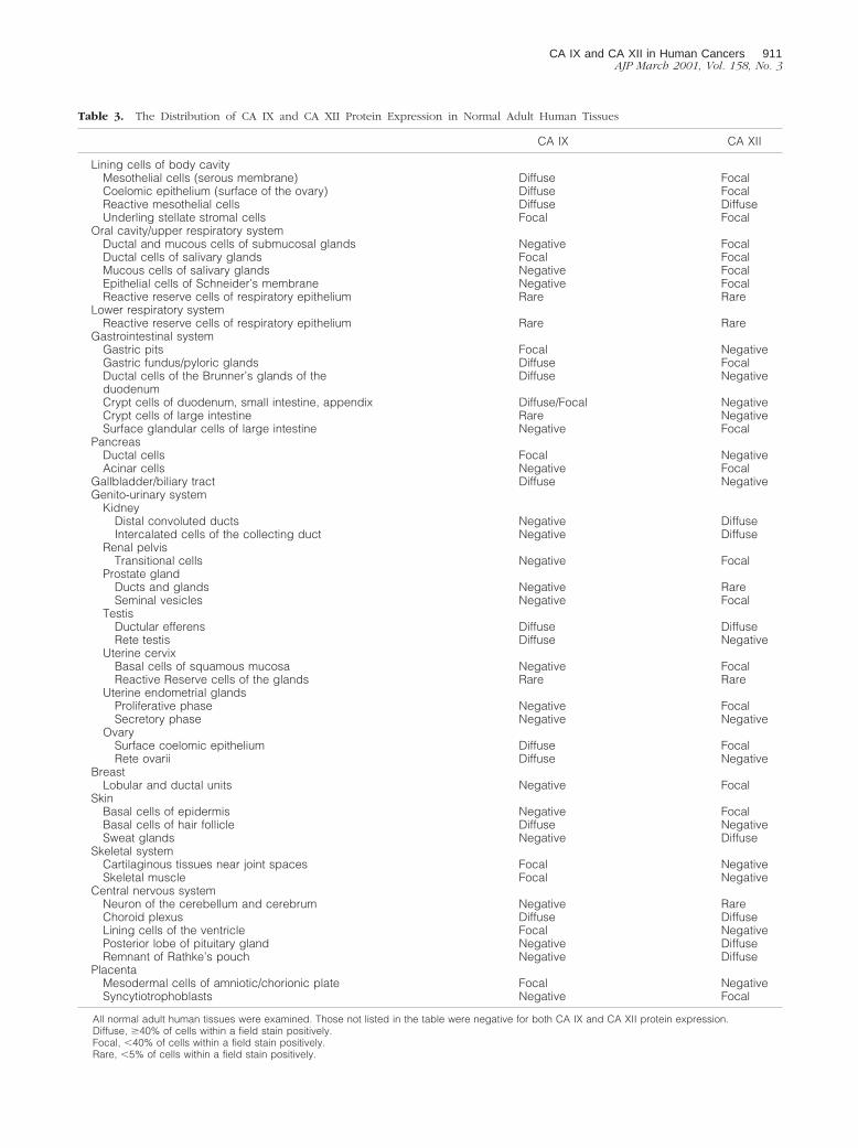
Table 3. The Distribution of CA IX and CA XII Protein Expression in Normal Adult Human Tissues
CA IX CA XII
Lining cells of body cavityMesothelial cells (serous membrane) Diffuse FocalCoelomic epithelium (surface of the ovary) Diffuse FocalReactive mesothelial cells Diffuse DiffuseUnderling stellate stromal cells Focal Focal
Oral cavity/upper respiratory systemDuctal and mucous cells of submucosal glands Negative FocalDuctal cells of salivary glands Focal FocalMucous cells of salivary glands Negative FocalEpithelial cells of Schneider’s membrane Negative FocalReactive reserve cells of respiratory epithelium Rare Rare
Lower respiratory systemReactive reserve cells of respiratory epithelium Rare Rare
Gastrointestinal systemGastric pits Focal NegativeGastric fundus/pyloric glands Diffuse FocalDuctal cells of the Brunner’s glands of theduodenum
Diffuse Negative
Crypt cells of duodenum, small intestine, appendix Diffuse/Focal NegativeCrypt cells of large intestine Rare NegativeSurface glandular cells of large intestine Negative Focal
PancreasDuctal cells Focal NegativeAcinar cells Negative Focal
Gallbladder/biliary tract Diffuse NegativeGenito-urinary system
KidneyDistal convoluted ducts Negative DiffuseIntercalated cells of the collecting duct Negative Diffuse
Renal pelvisTransitional cells Negative Focal
Prostate glandDucts and glands Negative RareSeminal vesicles Negative Focal
TestisDuctular efferens Diffuse DiffuseRete testis Diffuse Negative
Uterine cervixBasal cells of squamous mucosa Negative FocalReactive Reserve cells of the glands Rare Rare
Uterine endometrial glandsProliferative phase Negative FocalSecretory phase Negative Negative
OvarySurface coelomic epithelium Diffuse FocalRete ovarii Diffuse Negative
BreastLobular and ductal units Negative Focal
SkinBasal cells of epidermis Negative FocalBasal cells of hair follicle Diffuse NegativeSweat glands Negative Diffuse
Skeletal systemCartilaginous tissues near joint spaces Focal NegativeSkeletal muscle Focal Negative
Central nervous systemNeuron of the cerebellum and cerebrum Negative RareChoroid plexus Diffuse DiffuseLining cells of the ventricle Focal NegativePosterior lobe of pituitary gland Negative DiffuseRemnant of Rathke’s pouch Negative Diffuse
PlacentaMesodermal cells of amniotic/chorionic plate Focal NegativeSyncytiotrophoblasts Negative Focal
All normal adult human tissues were examined. Those not listed in the table were negative for both CA IX and CA XII protein expression.Diffuse, $40% of cells within a field stain positively.Focal, ,40% of cells within a field stain positively.Rare, ,5% of cells within a field stain positively.
CA IX and CA XII in Human Cancers 911AJP March 2001, Vol. 158, No. 3

served in cells expressing CA XII in areas adjacent tonecrosis, but was less predictable compared to CA IXexpression. The most predominant examples were in-duced expression of CA IX protein in hypoxic/necroticregions of melanoma, and enhanced expression in ne-crotic/hypoxic regions of meningiomas, glioblastomamultiforme, anaplastic ependymoma, and comedo typeof breast carcinomas. The latter three tumors are shownin Figure 4;D, E, and F. CA IX was predominantly ex-pressed in the areas of necrosis and hypoxia. The tumorcells that were more distal to the necrotic/hypoxic areasshowed little or no immunoreactivity.
Effect of Hypoxia on CA 9 and CA12 mRNAExpression in a Glioblastoma Cell Line
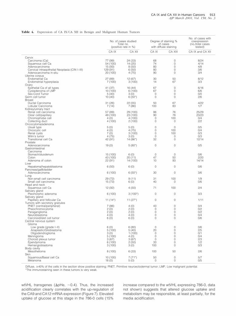
To directly examine the effect of hypoxia on the expres-sion of CA9 and CA12 mRNA we analyzed an establishedglioblastoma cell line, U87, expressing a normal VHL
message (M Merrill and M Proescholdt, unpublished ob-servations). The cells were grown under normoxic condi-tions and then maintained under hypoxic conditions for 6and 12 hours as previously described.19 Northern blothybridization (Figure 6) revealed that 6 hours of hypoxiaresulted in approximately fourfold and 1.6-fold up-regu-lation of CA9 and CA12 expression, respectively, while12 hours of hypoxia induced expression of both genes;11-fold to 13-fold, compared to control.
Media Acidification in Confluent RenalCarcinoma 786-0 Cells
We next investigated the correlation of CA9 and CA12mRNA expression with the dynamics of extracellular pHchanges under cell culture conditions. Acidification of thebicarbonate-buffered media at the late confluent stagewas much more pronounced in 786-0 cells (lackingwtVHL), as compared to the same cell line expressing
Figure 3. Examples of diffuse immunostaining of CA IX and CA XII proteins in normal and neoplastic tissues. Immunostaining for CA IX is illustrated in A, E, F, G, andH. Immunostaining for CA XII is illustrated in B, C, D, I, J, K, and L. In normal adult tissues, diffuse plasma membrane immunostaining for both CA IX and CA XII isseen in the ductular efferens (A and B), mesothelial cells of the body cavity (E and I), and the choroid plexus of brain ventricles (G and K). Variable degrees of expressionof CA XII but not CA IX are also observed in the epithelium of the breast (C). Persistent expression of CA IX and CA XII proteins is also seen in tumors derived fromthe normal tissues that express these proteins. For example, a lobular-type breast carcinoma expressing CA XII is shown in D, mesotheliomas of the pleura in F and J,and choroid plexus papillomas in H and L. Original magnifications, 3200 (A, B, G, and K) and 3400 (D–F, H–J, and L).
912 Ivanov et alAJP March 2001, Vol. 158, No. 3
wtVHL transgenes (DpHe, ;0.4). Thus, the increasedacidification clearly correlates with the up-regulation ofthe CA9 and CA12 mRNA expression (Figure 7). Elevateduptake of glucose at this stage in the 786-0 cells (15%
increase compared to the wtVHL expressing 786-0, datanot shown) suggests that altered glucose uptake andmetabolism may be responsible, at least partially, for themedia acidification.
Table 4. Expression of CA IX/CA XII in Benign and Malignant Human Tumors
No. of cases studiedTotal no.
(positive rate in %)
Degree of staining %of cases
with diffuse staining
No. of cases withcoexpression
(no./total casestested)
CA IX CA XII CA IX CA XIII CA IX and CA XII
CervixCarcinoma (Ca) 77 (99) 24 (33) 68 0 8/24Squamous cell Ca 54 (100) 14 (25) 74 0 4/14Adenocarcinoma 15 (93) 8 (50) 80 0 4/8Cervical intraepithelial Neoplasia (CIN I–III) 129 (91) 6 (50) 80 0 3/6Adenocarcinoma in-situ 20 (100) 4 (75) 90 0 3/4
Uterine corpusEndometrial Ca 27 (89) 12 (67) 30 50 8/12Endometrial hyperplasia 7 (100) 3 (100) 14 67 3/3
OvaryEpithelial Ca of all types 41 (37) 16 (44) 67 0 6/16Cystadenoma of LMP 10 (100) 6 (100) 67 0 6/6Sex-Cord Tumor 5 (40) 3 (0) 0 0 0/0
Germ cell tumor 10 (40) 6 (50*) 0 0 2/6Breast
Ductal Carcinoma 31 (26) 22 (55) 50 67 4/22Lobular Carcinoma 7 (14) 7 (86) 100 83 1/7
Kidney/urinary tractRenal cell carcinoma 57 (89) 29 (100) 86 76 25/29Clear cell/papillary 49 (100) 23 (100) 90 78 23/23Chromophobe cell 4 (0) 4 (100) 0 100 0/4Collecting duct 4 (100) 2 (100) 0 0 2/2
Oncocytoma/adenomaUsual type 5 (0) 5 (0) 0 0 0/5Oncocytic cell 4 (0) 4 (75) 0 100 0/4Renal cysts 7 (0) 3 (100) 0 100 0/3Wilm’s tumor 4 (75) 3 (0) 0 0 0/3Transitional cell Ca 42 (91) 14 (86*) 32 0 12/14
ProstateAdenocarcinoma 19 (0) 5 (80*) 0 0 0/5
GastrointestinalCarcinomaStomach/duodenum 15 (100) 6 (0) 0 0 0/6Colon 43 (100) 20 (11) 47 50 2/20Adenoma of colon 22 (91) 14 (100) 10 93 14/14
LiverHepatoma/hepatoblastoma 6 (50) 6 (0) 0 0 0/6
Pancreas/gallbladderAdenocarcinoma 6 (100) 6 (50*) 30 0 3/6
LungNon-small cell carcinoma 29 (72) 9 (11) 31 100 1/9Small cell carcinoma 15 (73) 6 (0) 40 0 0/6
Head and neckSquamous cell Ca 12 (92) 4 (50) 71 100 2/4
Salivary glandPleomorphic adenoma 6 (100) 3 (100*) 0 0 3/3
Salivary glandPapillary and follicular Ca 11 (14*) 11 (27*) 0 0 1/11
Tumors with secretory granulesPNET (central/peripheral) 7 (86) 4 (0) 43 0 0/4Pheochromocytoma 2 (0) 2 (0) 0 0 0/2Paraganglioma 2 (0) 2 (0) 0 0 0/2Neuroblastoma 4 (0) 4 (0) 0 0 0/4Carcinoid/islet cell tumor 6 (0) 6 (0) 0 0 0/6
Central nervous systemGlioma
Low grade (grade I–II) 6 (0) 6 (80) 0 0 0/6Anaplastic/Glioblastoma 5 (100) 5 (40) 20 0 2/5Oligodendroglioma 3 (0) 1 (100) 0 0 0/1
Meningioma 5 (100) 4 (0) 0 0 0/4Choroid plexus tumor 3 (67) 3 (67) 0 0 2/3Ependymoma 6 (100) 2 (50) 33 0 1/2Hemangioblastoma 3 (100) 3 (0) 100 0 0/3
Body cavityMesothelioma 8 (100) 6 (33) 100 50 2/6
SkinSquamous/Basal cell Ca 10 (100) 7 (71*) 50 0 5/7Melanoma 18 (0) 5 (0) 0 0 0/5
Diffuse, $40% of the cells in the section show positive staining; PNET, Primitive neuroectodermal tumor; LMP, Low malignant potential.*The immunostaining seen in these tumors is very weak.
CA IX and CA XII in Human Cancers 913AJP March 2001, Vol. 158, No. 3

Discussion
This study demonstrates, for the first time, high levels ofexpression of cell-surface CAs (CA IX/CA XII) in a largesample of cancer cell lines, fresh and archived tumortissues. This was shown by Northern blot hybridizationwith mRNAs and immunohistochemistry of tissue/tumorsections with specific antibodies. Most common tumorsoriginating from many different sites expressed eitherboth genes or one of them. For many tumors there was nodetectable expression in corresponding normal adult tis-
sues (Tables 3 and 4), indicating tumor-specific activa-tion of these genes. Other investigators, in less extensivescreenings, have reported expression of CA IX or CA XIIin some common tumors.27,28 To our knowledge this isthe first extensive comparative analysis of the combinedexpression of these genes and proteins. Cumulatively,the data presented here and published before byus10,22,23 and others21,27,28 firmly establish high expres-sion of CA9/CA12 genes in a large number of commonhuman malignancies, suggesting an important role forthem in tumor development.
Figure 4. Examples of ectopic expression of CA IX and CA XII proteins in various tumors, including regions of necrosis/hypoxia. The glandular cells of the colonare normally CA IX-negative but diffuse immunoreactivity is seen in colon carcinoma (A). The normal glial tissues of the brain show no expression of either CAIX or CA XII protein (B). However, variable degrees of positive staining for CA XII are seen in low-grade glioma (C). In contrast, high levels of CA IX proteinexpression are observed in glioblastoma multiforme (D) and anaplastic ependymoma (E). Expression of the CA IX protein is seen in necrotic/hypoxic regionsof the tumors. This induced expression associated with necrotic/hypoxic regions is also seen in high-grade ductal breast carcinoma (F). The symbol n 5 area ofnecrosis. Original magnifications, 3200 (A, D–F) and 3400 (B and C).
914 Ivanov et alAJP March 2001, Vol. 158, No. 3

Figure 5. Immunostaining of normal kidney and renal cell tumors for CA IX/CA XII expression. The normal kidney is negative for CA IX (A) whereas CA XII isexpressed in the distal convoluted tubules (arrow) and intercalated cells of the collecting ducts (arrowhead) (B). Co-expression of CA IX and CA XII is seenin renal cell carcinomas of the clear cell type (CA IX 5 C; CA XII 5 D). However, chromophobe cell carcinomas (E) and oncocytomas (G) do not express CAIX whereas they exhibit diffuse expression of CA XII (F and H, respectively). Original magnifications, 3200 (A and B) and 3400 (C–H).
CA IX and CA XII in Human Cancers 915AJP March 2001, Vol. 158, No. 3
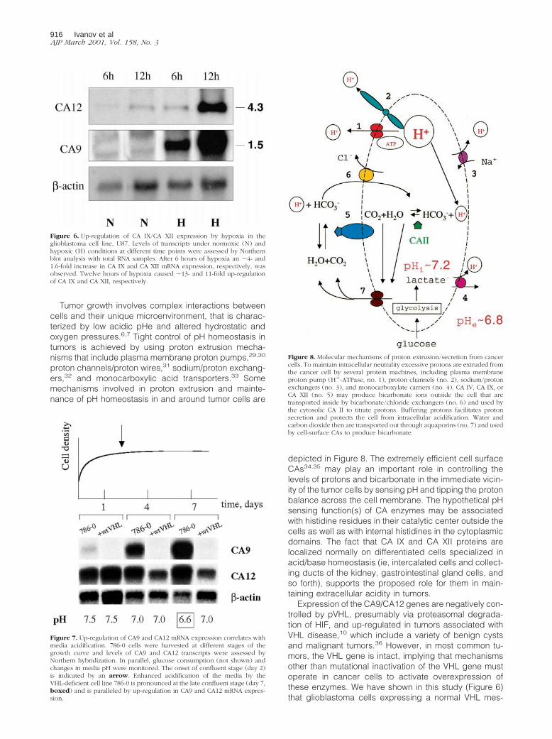
Tumor growth involves complex interactions betweencells and their unique microenvironment, that is charac-terized by low acidic pHe and altered hydrostatic andoxygen pressures.6,7 Tight control of pH homeostasis intumors is achieved by using proton extrusion mecha-nisms that include plasma membrane proton pumps,29,30
proton channels/proton wires,31 sodium/proton exchang-ers,32 and monocarboxylic acid transporters.33 Somemechanisms involved in proton extrusion and mainte-nance of pH homeostasis in and around tumor cells are
depicted in Figure 8. The extremely efficient cell surfaceCAs34,35 may play an important role in controlling thelevels of protons and bicarbonate in the immediate vicin-ity of the tumor cells by sensing pH and tipping the protonbalance across the cell membrane. The hypothetical pHsensing function(s) of CA enzymes may be associatedwith histidine residues in their catalytic center outside thecells as well as with internal histidines in the cytoplasmicdomains. The fact that CA IX and CA XII proteins arelocalized normally on differentiated cells specialized inacid/base homeostasis (ie, intercalated cells and collect-ing ducts of the kidney, gastrointestinal gland cells, andso forth), supports the proposed role for them in main-taining extracellular acidity in tumors.
Expression of the CA9/CA12 genes are negatively con-trolled by pVHL, presumably via proteasomal degrada-tion of HIF, and up-regulated in tumors associated withVHL disease,10 which include a variety of benign cystsand malignant tumors.36 However, in most common tu-mors, the VHL gene is intact, implying that mechanismsother than mutational inactivation of the VHL gene mustoperate in cancer cells to activate overexpression ofthese enzymes. We have shown in this study (Figure 6)that glioblastoma cells expressing a normal VHL mes-
Figure 6. Up-regulation of CA IX/CA XII expression by hypoxia in theglioblastoma cell line, U87. Levels of transcripts under normoxic (N) andhypoxic (H) conditions at different time points were assessed by Northernblot analysis with total RNA samples. After 6 hours of hypoxia an ;4- and1.6-fold increase in CA IX and CA XII mRNA expression, respectively, wasobserved. Twelve hours of hypoxia caused ;13- and 11-fold up-regulationof CA IX and CA XII, respectively.
Figure 7. Up-regulation of CA9 and CA12 mRNA expression correlates withmedia acidification. 786-0 cells were harvested at different stages of thegrowth curve and levels of CA9 and CA12 transcripts were assessed byNorthern hybridization. In parallel, glucose consumption (not shown) andchanges in media pH were monitored. The onset of confluent stage (day 2)is indicated by an arrow. Enhanced acidification of the media by theVHL-deficient cell line 786-0 is pronounced at the late confluent stage (day 7,boxed) and is paralleled by up-regulation in CA9 and CA12 mRNA expres-sion.
Figure 8. Molecular mechanisms of proton extrusion/secretion from cancercells. To maintain intracellular neutrality excessive protons are extruded fromthe cancer cell by several protein machines, including plasma membraneproton pump (H1-ATPase, no. 1), proton channels (no. 2), sodium/protonexchangers (no. 3), and monocarboxylate carriers (no. 4). CA IV, CA IX, orCA XII (no. 5) may produce bicarbonate ions outside the cell that aretransported inside by bicarbonate/chloride exchangers (no. 6) and used bythe cytosolic CA II to titrate protons. Buffering protons facilitates protonsecretion and protects the cell from intracellular acidification. Water andcarbon dioxide then are transported out through aquaporins (no. 7) and usedby cell-surface CAs to produce bicarbonate.
916 Ivanov et alAJP March 2001, Vol. 158, No. 3

sage when maintained for 12 hours under hypoxic con-ditions overexpress the CA9/CA12 genes, indicating arole for hypoxia in this induction. It is established that inhypoxic cells the proteolytic function of pVHL is abro-gated, resulting in the overexpression of the HIF1-a pro-tein and other VHL target genes.8,18 The transcriptionaltrans-activator HIF1 controls the expression of severaldozen target genes including those involved in energymetabolism (glucose transporters, glycolytic enzymes)and angiogenesis (VEGF and its receptors), and is con-sidered a master regulator gene that orchestrates phys-iological responses to acute and chronic hypoxia.1,12–14
Thus, loss of pVHL function(s) results in the stabilizationof HIF and transactivation of its target genes. Therefore,presence or absence of the VHL protein seems to beresponsible for fundamental metabolic changes in tumorcells. The ultimate consequences of these changes ingene expression in the absence of a functional VHLprotein (because of mutation, epigenetic silencing, orlocalized hypoxic conditions) are compromised tumormicroenvironment, a powerful, albeit faulty, disorganizedprocess of angiogenesis, increased glycolysis, and in-duction of expression of transmembrane CA. The fre-quent, widespread overexpression of the CA9/CA12genes in human tumors has several important implica-tions in the clinical setting.
Implications for Tumor Growth and Spread
On the basis of the above considerations, it would seemthat tumor microenvironmental acidity could play a pre-dominant promoting role in tumor growth and metastasisand also could underlie resistance to radiotherapy, che-motherapy, and other nonsurgical treatments.6,7 The ex-tracellular matrix metalloproteinases, which are activatedby acidic pH, are involved in remodeling stromal andtumor cell surface proteins, thereby promoting tumor cellmotility, and contributing to tumor growth and metasta-sis.37 However, synthesis of matrix metalloproteinasesmay have opposite effects on tumor angiogenesis, pro-moting extracellular matrix degradation and new bloodvessel formation on the one hand37 and, on the other,blocking angiogenesis by producing angiostatin, a cleav-age product of plasminogen.2 Most tumors have beenshown to exhibit high vascular permeability and highinterstitial fluid pressure.6,7 These may result from activa-tion, as a consequence of the acidic tumor microenviron-ment, of trans-membrane water channel proteins (aqua-porins) that are widely distributed in tumors.38
Aquaporins are known to play a major role in trans-cellu-lar and trans-epithelial water movements in many normaltissues.39,40 The expression of CA IX/CA XII in cystictumors is an indication that their activity in promotingacidity might facilitate the activity of aquaporins ex-pressed on tumor and endothelial cells, leading to highinterstitial fluid pressure and high vascular permeability intumors.6,7
Implications for Diagnosis
The fact that CA IX/CA XII are expressed in many tumortypes may lead one to conclude that they have limitedutility in cancer diagnosis. However, for selected can-cers, they may prove to be powerful diagnostic biomar-kers. We have already shown the utility of CA IX as abiomarker for cervical dysplasia and carcinoma.22,41 Fur-thermore, we have recently shown its utility in identifyingthose patients who receive a Pap smear diagnosis of“atypical glandular cells of undetermined significance”and harbor a significant lesion in their cervix.42 We andothers have also shown that expression of CA IX is anexcellent diagnostic biomarker for renal cell carcino-ma.23,24,43 The studies described here, in which we showthat CA XII is expressed in chromophobe tumors,whereas CA IX is not (Figure 5), suggests that the com-bined detection of CA IX and CA XII expression willidentify all renal cell carcinomas. This also has importantimplications for therapy.44–47 Other investigators haveidentified the expression of CA IX in esophageal48 andlung carcinomas49 Finally, our studies here also suggestthat CA XII and/or CA IX may have utility in diagnosis ofbrain tumors.
Implications for Tumor Treatment
The CA IX/CA XII enzymes ectopically expressed on thesurface of cancer cells provide an excellent target fortumor treatment modalities, including development ofhighly specific humanized antibodies, antibodies armedwith toxins,46 and specific inhibitors of these en-zymes.50–52 Antibodies to CA IX have also been devel-oped in Europe and are being successfully used to treatrenal cancer.43,47 Dr. Neal Bunder is developing human-ized antibodies at the Memorial Sloan-Kettering CancerCenter (personal communication). In addition, antibodiesto CA XII were recently patented for treatment of lungcancer (US patent no. 5,589,579). On the other hand,highly specific inhibitors of CA IX/CA XII enzymes tailoredto their catalytic centers, could be also used to treatcancer and should be developed. Two of us (AW andWS), with others,50 have shown that acetazolamide, apotent inhibitor of CAs, suppressed invasion of renalcancer cells in vitro. It took Professor T. Maren and theMerck Company 20 years to develop such inhibitors fortopical treatment of glaucoma.51,52 Establishing the crys-tal structures of CA IX and CA XII should take less timeand will result in designing more effective specific drugsto manipulate tumor pH, and possibly provide a mecha-nism for inhibiting tumor growth and tumor spread.
Acknowledgments
We thank Bruce Johnson, Teizo Yoshimura, and DominicScudiero for providing RNA and cell lines and Ning Ru forassistance with the immunostaining.
CA IX and CA XII in Human Cancers 917AJP March 2001, Vol. 158, No. 3

References
1. Semenza GL: Hypoxia, clonal selection, and the role of HIF-1 in tumorprogression. Crit Rev Biochem Mol Biol 2000, 35:71–103
2. Hanahan D, Folkman J: Patterns and emerging mechanisms of theangiogenic switch during tumorigenesis. Cell 1996, 86:353–364
3. Warburg O: The Metabolism of Tumours. London, Constable, 19304. Dang CV, Semenza GL: Oncogenic alterations of metabolism. Trends
Biochem Sci 1999, 24:68–725. Stubbs M, McSheehy PM, Griffiths JR, Bashford CL: Causes and
consequences of tumour acidity and implications for treatment. MolMed Today 2000, 6:15–19
6. Helmlinger G, Yuan F, Dellian M, Jain RK: Interstitial pH and pO2
gradients in solid tumors in vivo: high-resolution measurements reveala lack of correlation. Nat Med 1997, 2:177–182
7. Jain RK: Transport of molecules, particles, and cells in solid tumors.Annu Rev Biomed Engineer 1999, 1:241–263
8. Maxwell PH, Wiesener MS, Chang GW, Clifford SC, Vaux EC, Cock-man ME, Wykoff CC, Pugh CW, Maher ER, Ratcliffe PJ: The tumoursuppressor protein VHL targets hypoxia-inducible factors for oxygen-dependent proteolysis. Nature 1999, 399:271–275
9. Gnarra JR, Zhou S, Merrill MJ, Wagner JR, Krumm A, Papavassiliou E,Oldfield EH, Klausner RD, Linehan WM: Post-transcriptional regula-tion of vascular endothelial growth factor mRNA by the product of theVHL tumor suppressor gene. Proc Natl Acad Sci USA 1996, 93:10589–10594
10. Ivanov SV, Kuzmin I, Wei MH, Pack S, Geil L, Johnson BE, StanbridgeEJ, Lerman MI: Down-regulation of transmembrane carbonic anhy-drases in renal cell carcinoma cell lines by wild-type von Hippel-Lindau transgenes. Proc Natl Acad Sci USA 1998, 95:12596–12601
11. Ohh M, Kaelin Jr WG: The von Hippel-Lindau tumour suppressorprotein: new perspectives. Mol Med Today 1999, 6:257–263
12. Semenza GL: Regulation of mammalian O2 homeostasis by hypoxia-inducible factor 1. Annu Rev Cell Dev Biol 1999, 15:551–578
13. Bunn HF, Poyton RO: Oxygen sensing and molecular adaptation tohypoxia. Physiol Rev 1996, 76:839–885
14. Gleadle JM, Ratcliffe PJ: Hypoxia and the regulation of gene expres-sion. Mol Med Today 1998, 3:122–129
15. Kaelin Jr WG, Maher ER: The VHL tumour-suppressor gene para-digm. Trends Genet 1998, 10:423–426
16. Tyers M, Willems AR: One ring to rule a superfamily of E3 ubiquitinligases. Science 1999, 284:408–414
17. Tyers M, Rottapel R: VHL: a very hip ligase. Proc Natl Acad Sci USA1999, 96:12230–12232
18. Iwai K, Yamanaka K, Kamura T, Minato N, Conaway RC, ConawayJW, Klausner RD, Pause A: Identification of the von Hippel-lindautumor-suppressor protein as part of an active E3 ubiquitin ligasecomplex. Proc Natl Acad Sci USA 1999, 96:12436–12441
19. Heiss JD, Papavassiliou E, Merrill MJ, Nieman L, Knightly JJ, Wal-bridge S, Edwards NA, Oldfield EH: Mechanism of dexamethasonesuppression of brain tumor-associated vascular permeability in rats.Involvement of the glucocorticoid receptor and vascular permeabilityfactor. J Clin Invest 1996, 98:1400–1408
20. Zavada J, Zavadova Z, Pastorekova S, Ciampor F, Pastorek J, ZelnikV: Expression of MaTu-MN protein in human tumor cultures and inclinical specimens. Int J Cancer 1993, 54:268–274
21. Karhumaa P, Parkkila S, Tureci O, Waheed A, Grubb JH, Shah G,Parkkila A, Kaunisto K, Tapanainen J, Sly WS, Rajaniemi H: Identifi-cation of carbonic anhydrase XII as the membrane isozyme ex-pressed in the normal human endometrial epithelium. Mol Hum Re-prod 2000, 6:68–74
22. Liao SY, Brewer C, Zavada J, Pastorek J, Pastorekova S, Manetta A,Berman ML, DiSaia PJ, Stanbridge EJ: Identification of the MN anti-gen as a diagnostic biomarker of cervical intraepithelial neoplasiaand cervical carcinoma. Am J Pathol 1994, 145:598–609
23. Liao SY, Aurelio ON, Jan K, Zavada J, Stanbridge EJ: Identification ofthe MN/CA9 protein as a reliable diagnostic biomarker of clear cellcarcinoma of the kidney. Cancer Res 1997, 57:2827–2831
24. McKiernan JM, Buttyan R, Bander NH, Stifelman MD, Katz AE, ChenMW, Olsson CA, Sawczuk IS: Expression of the tumor-associatedgene MN: a potential biomarker for human renal cell carcinoma.Cancer Res 1997, 57:2362–2365
25. Saarnio J, Parkkila S, Parkkila AK, Haukipuro K, Pastorekova S,Pastorek J, Kairaluoma MI, Karttunen TJ: Immunohistochemical study
of colorectal tumors for expression of a novel transmembrane car-bonic anhydrase, MN/CA IX, with potential value as a marker of cellproliferation. Am J Pathol 1998, 53:279–285
26. Saarnio J, Parkkila S, Parkkila AK, Waheed A, Casey MC, Zhou XY,Pastorekova S, Pastorek J, Karttunen T, Haukipuro K, Kairaluoma MI,Sly WS: Immunohistochemistry of carbonic anhydrase isozyme IX(MN/CA IX) in human gut reveals polarized expression in the epithe-lial cells with the highest proliferative capacity. J Histochem Cyto-chem 1998, 46:497–504
27. Kivela A, Parkkila S, Saarnio J, Karttunen TJ, Kivela J, Parkkila AK,Waheed A, Sly WS, Grubb JH, Shah G, Tureci O, Rajaniemi H:Expression of a novel transmembrane carbonic anhydrase isozymeXII in normal human gut and colorectal tumors. Am J Pathol 2000,156:577–584
28. Tureci O, Sahin U, Vollmar E, Siemer S, Gottert E, Seitz G, Parkkila AK,Shah GN, Grubb JH, Pfreundschuh M, Sly WS: Human carbonicanhydrase XII: cDNA cloning, expression, and chromosomal local-ization of a carbonic anhydrase gene that is overexpressed in somerenal cell cancers. Proc Natl Acad Sci USA 1998, 95:7608–7613
29. Wilkens S, Vasilyeva E, Forgac M: Structure of the vacuolar ATPaseby electron microscopy. J Biol Chem 1999, 274:31804–31810
30. Wieczorek H, Grber G, Harvey WR, Huss M, Merzendorfer H, ZeiskeW: Structure and regulation of insect plasma membrane H1-ATPase.J Exp Biol 2000, 203:127–135
31. DeCoursey TE, Cherny VV: An electrophysiological comparison ofvoltage-gated proton channels, other ion channels, and other protonchannels. Isr J Chem 1999, 39:409–418
32. Wakabayashi S, Shigekawa M, Pouyssegur J: Molecular physiologyof vertebrate Na1/H1-exchangers. Physiol Rev 1997, 77:51–74
33. Halestrap AP, Price NT: The proton-linked monocarboxylate trans-porter (MCT) family: structure, function and regulation. Biochem J1999, 343:281–299
34. Sly WS, Hu PY: Human carbonic anhydrases and carbonic anhydrasedeficiencies. Annu Rev Biochem 1995, 64:375–401
35. Baird Jr TT, Waheed A, Okuyama T, Sly WS, Fierke CA: Catalysis andinhibition of human carbonic anhydrase IV. Biochemistry 1997, 36:2669–2678
36. Zbar B, Lerman M: Inherited carcinomas of the kidney. Adv CancerRes 1998, 75:163–201
37. McCawley LJ, Matrisian LM: Matrix metalloproteinases: multifunc-tional contributors to tumor progression. Mol Med Today 2000,6:149–156
38. Hashizume H, Baluk P, Morikawa S, McLean JW, Thurston G, Rob-erge S, Jain RK, McDonald DM: Openings between defective endo-thelial cells explain tumor vessel leakiness. Am J Pathol 2000, 156:1363–1380
39. Engel A, Fujiyoshi Y, Agre P: The importance of aquaporin waterchannel protein structures. EMBO J 2000, 19:800–806
40. Verkman AS, Matthay MA, Song Y: Aquaporin water channels andlung physiology. Am J Physiol Lung Cell Mol Physiol 2000, 78:L867–L879
41. Liao SY, Stanbridge EJ: Expression of the MN antigen in cervicalPapanicolaou smears is an early diagnostic biomarker of cervicaldysplasia. Cancer Epidemiol Biomarkers Prev 1996, 5:549–557
42. Liao SY, Stanbridge EJ: Expression of MN/CA9 protein in Papanico-laou smears containing atypical glandular cells of undetermined sig-nificance is a diagnostic biomarker of cervical dysplasia and neopla-sia. Cancer 2000, 88:1108–1121
43. Oosterwijk DJ, Ruiter PJ, Pauwels EKJ, Jonas U, Zwartenduk J,Warnaar O: Monoclonal antibody G 250 recognizes a determinantpresent in renal cell carcinoma and absent from normal kidney. Int JCancer 1986, 38:489–494
44. Uemura H, Nakagawa Y, Yoshida K, Saga S, Yoshikawa K, Hirao Y,Oosterwijk E: MN/CA IX/G250 as a potential target for immunotherapyof renal cell carcinomas. Br J Cancer 1999, 81:741–746
45. Divgi CR, Bander NH, Scott AM, O’Donoghue JA, Sgouros G, Welt S,Finn RD, Morrissey F, Capitelli P, Williams JM, Deland D, Nakhre A,Oosterwijk E, Gulec S, Graham MC, Larson SM, Old LJ: Phase I/IIradioimmunotherapy trial with iodine-131-labeled monoclonal anti-body G250 in metastatic renal cell carcinoma. Clin Cancer Res 1998,4:2729–2739
46. Frankel AE, Kreitman RJ, Sausville EA: Targeted toxins. Clin CancerRes 2000, 6:326–344
47. Steffens MG, Boerman OC, de Mulder PH, Oyen WJ, Buijs WC, Witjes
918 Ivanov et alAJP March 2001, Vol. 158, No. 3

JA, van den Broek WJ, Oosterwijk-Wakka JC, Debruyne FM, CorstensFH, Oosterwijk E: Phase I radioimmunotherapy of metastatic renal cellcarcinoma with 131I-labeled chimeric monoclonal antibody G250.Clin Cancer Res 1999, 10(Suppl):S3268–S3274.
48. Turner JR, Odze RD, Crum CP, Resnick MB: MN antigen expressionin normal, preneoplastic, and neoplastic esophagus: a clinicopatho-logical study of a new cancer-associated biomarker. Hum Pathol1997, 28:740–744
49. Vermylen P, Roufosse C, Burny A, Verhest A, Bossehaerts T, Pas-torekova S, Ninane V, Sculier JP: Carbonic anhydrase IX antigen
differentiates between preneoplastic and malignant lesions in non-small cell lung carcinomas. Eur Respir J 1999, 14:806–811
50. Parkkila S, Rajaniemi H, Parkkila AK, Kivela J, Waheed A, Pas-torekova S, Pastorek J, Sly WS: Carbonic anhydrase inhibitor sup-presses invasion of renal cancer cells in vitro. Proc Natl Acad Sci USA2000, 97:2220–2224
51. Maren TH: The development of topical carbonic anhydrase inhibitors.J Glaucoma 1995, 4:49–62
52. Maren TH: Sulfonamides and secretion of aqueous humor. J Exp Zool1997, 490:279–287
CA IX and CA XII in Human Cancers 919AJP March 2001, Vol. 158, No. 3
Copyright © 2022 FDOKUMEN