Type II Transmembrane Serine Protease Gene Variants Associate with Breast Cancer
11
Type II Transmembrane Serine Protease Gene Variants Associate with Breast Cancer Kaisa Luostari 1,2,3 , Jaana M. Hartikainen 1,2,3 , Maria Tengstro ¨m 4,5 , Jorma J. Palvimo 2,6 , Vesa Kataja 4,5 , Arto Mannermaa 1,2,3 * . , Veli-Matti Kosma 1,2,3. 1 Institute of Clinical Medicine, Pathology and Forensic Medicine, University of Eastern Finland, Kuopio, Finland, 2 Biocenter Kuopio and Cancer Center of Eastern Finland, University of Eastern Finland, Kuopio, Finland, 3 Imaging Center, Clinical Pathology, Kuopio University Hospital, Kuopio, Finland, 4 Institute of Clinical Medicine, Oncology, University of Eastern Finland, Kuopio, Finland, 5 Cancer Center, Kuopio University Hospital, Kuopio, Finland, 6 Institute of Biomedicine, University of Eastern Finland, Kuopio, Finland Abstract Type II transmembrane serine proteases (TTSPs) are related to tumor growth, invasion, and metastasis in cancer. Genetic variants in these genes may alter their function, leading to cancer onset and progression, and affect patient outcome. Here, 464 breast cancer cases and 370 controls were genotyped for 82 single-nucleotide polymorphisms covering eight genes. Association of the genotypes was estimated against breast cancer risk, breast cancer–specific survival, and survival in different treatment groups, and clinicopathological variables. SNPs in TMPRSS3 (rs3814903 and rs11203200), TMPRSS7 (rs1844925), and HGF (rs5745752) associated significantly with breast cancer risk (P trend = 0.008–0.042). SNPs in TMPRSS1 (rs12151195 and rs12461158), TMPRSS2 (rs2276205), TMPRSS3 (rs3814903), and TMPRSS7 (rs2399403) associated with prognosis (P = 0.004–0.046). When estimating the combined effect of the variants, the risk of breast cancer was higher with 4–5 alleles present compared to 0–2 alleles (P = 0.0001; OR, 2.34; 95% CI, 1.39–3.94). Women with 6–8 survival-associating alleles had a 3.3 times higher risk of dying of breast cancer compared to women with 1–3 alleles (P = 0.001; HR, 3.30; 95% CI, 1.58–6.88). The results demonstrate the combined effect of variants in TTSPs and their related genes in breast cancer risk and patient outcome. Functional analysis of these variants will lead to further understanding of this gene family, which may improve individualized risk estimation and development of new strategies for treatment of breast cancer. Citation: Luostari K, Hartikainen JM, Tengstro ¨ m M, Palvimo JJ, Kataja V, et al. (2014) Type II Transmembrane Serine Protease Gene Variants Associate with Breast Cancer. PLoS ONE 9(7): e102519. doi:10.1371/journal.pone.0102519 Editor: Gautam Chaudhuri, Meharry Medical College, United States of America Received February 13, 2014; Accepted June 19, 2014; Published July 16, 2014 Copyright: ß 2014 Luostari et al. This is an open-access article distributed under the terms of the Creative Commons Attribution License, which permits unrestricted use, distribution, and reproduction in any medium, provided the original author and source are credited. Funding: The KBCP was financially supported by grants from the special Government Funding (EVO) of Kuopio University Hospital (www.psshp.fi), the Cancer Fund of Northern Savo (www.pohjois-savonsyopayhdistys.fi), the Finnish Cancer Organizations (www.cancer.fi), the Academy of Finland (www.aka.fi), and by the strategic funding of the University of Eastern Finland (www.uef.fi). The funders had no role in study design, data collection and analysis, decision to publish, or preparation of the manuscript. Competing Interests: Arto Mannermaa is a PLOS ONE Editorial Board member. This does not alter the authors’ adherence to PLOS ONE Editorial policies and criteria. * Email: [email protected] . These authors contributed equally to this work. Introduction Breast cancer is the most common cancer among women in western countries. The known high risk susceptibility genes for breast cancer, e.g. BRCA1, BRCA2, ATM, and PALB2, are responsible for approximately 20% of the hereditary cases [1], but several unknown breast cancer–predisposing genetic factors still exist. Genetic risk factors with a low or moderate penetrance also affect the risk of sporadic breast cancers and may act together with environmental and lifestyle factors and with each other to enhance cancer predisposition and progression [2,3]. Type II transmembrane serine proteases (TTSPs) degrade components of the extracellular matrix (ECM) [4,5]. The 17 members of the human TTSP family have physiological and pathological roles in digestion, cardiac function, blood pressure regulation, hearing, iron metabolism, and epithelial homeostasis [4,6]. In cancer, the TTSPs are related especially to tumor growth, invasion, and metastasis [7]. TTSPs are divided by their structures into subfamilies [4]. TMPRSS1, TMPRSS2, and TMPRSS3 belong to the Hepsin/ TMPRSS subfamily [4]. TMPRSS1 is overexpressed in prostate and breast cancers, and its expression and localization have also been related to epithelial integrity [8,9]. In terms of cancer-related risk, Pal and coworkers (2006) reported five TMPRSS1 single nucleotide polymorphisms (SNPs) as associated with prostate cancer in men of European origin [10], although another study identified no associated variants [11]. The androgen-regulated TMPRSS2, however, is strongly associated with prostate cancer and forms a fusion gene with ETS transcription factor (TF) family members, that occurs in roughly half of prostate cancer cases [12,13]. This fusion has been studied in ovarian cancer but not detected [14]. TMPRSS3 is overexpressed in epithelial ovarian cancer and is a potential diagnostic marker and therapy target [15–18]. TMPRSS11E/DESC1 belongs to the HAT/DESC subfamily of the TTSPs and is upregulated in tumors of different origins, including breast [19]. DESC1 can convert pro-urokinase-type plasminogen activator (uPA, PLAU) to active uPA [19]. uPA belongs to the serine proteases and is an important factor in the plasminogen activation system associated with several cancers, PLOS ONE | www.plosone.org 1 July 2014 | Volume 9 | Issue 7 | e102519
Transcript of Type II Transmembrane Serine Protease Gene Variants Associate with Breast Cancer
Type II Transmembrane Serine Protease Gene VariantsAssociate with Breast CancerKaisa Luostari1,2,3, Jaana M. Hartikainen1,2,3, Maria Tengstrom4,5, Jorma J. Palvimo2,6, Vesa Kataja4,5,
Arto Mannermaa1,2,3*., Veli-Matti Kosma1,2,3.
1 Institute of Clinical Medicine, Pathology and Forensic Medicine, University of Eastern Finland, Kuopio, Finland, 2 Biocenter Kuopio and Cancer Center of Eastern Finland,
University of Eastern Finland, Kuopio, Finland, 3 Imaging Center, Clinical Pathology, Kuopio University Hospital, Kuopio, Finland, 4 Institute of Clinical Medicine, Oncology,
University of Eastern Finland, Kuopio, Finland, 5 Cancer Center, Kuopio University Hospital, Kuopio, Finland, 6 Institute of Biomedicine, University of Eastern Finland,
Kuopio, Finland
Abstract
Type II transmembrane serine proteases (TTSPs) are related to tumor growth, invasion, and metastasis in cancer. Geneticvariants in these genes may alter their function, leading to cancer onset and progression, and affect patient outcome. Here,464 breast cancer cases and 370 controls were genotyped for 82 single-nucleotide polymorphisms covering eight genes.Association of the genotypes was estimated against breast cancer risk, breast cancer–specific survival, and survival indifferent treatment groups, and clinicopathological variables. SNPs in TMPRSS3 (rs3814903 and rs11203200), TMPRSS7(rs1844925), and HGF (rs5745752) associated significantly with breast cancer risk (Ptrend = 0.008–0.042). SNPs in TMPRSS1(rs12151195 and rs12461158), TMPRSS2 (rs2276205), TMPRSS3 (rs3814903), and TMPRSS7 (rs2399403) associated withprognosis (P = 0.004–0.046). When estimating the combined effect of the variants, the risk of breast cancer was higher with4–5 alleles present compared to 0–2 alleles (P = 0.0001; OR, 2.34; 95% CI, 1.39–3.94). Women with 6–8 survival-associatingalleles had a 3.3 times higher risk of dying of breast cancer compared to women with 1–3 alleles (P = 0.001; HR, 3.30; 95% CI,1.58–6.88). The results demonstrate the combined effect of variants in TTSPs and their related genes in breast cancer riskand patient outcome. Functional analysis of these variants will lead to further understanding of this gene family, which mayimprove individualized risk estimation and development of new strategies for treatment of breast cancer.
Citation: Luostari K, Hartikainen JM, Tengstrom M, Palvimo JJ, Kataja V, et al. (2014) Type II Transmembrane Serine Protease Gene Variants Associate with BreastCancer. PLoS ONE 9(7): e102519. doi:10.1371/journal.pone.0102519
Editor: Gautam Chaudhuri, Meharry Medical College, United States of America
Received February 13, 2014; Accepted June 19, 2014; Published July 16, 2014
Copyright: � 2014 Luostari et al. This is an open-access article distributed under the terms of the Creative Commons Attribution License, which permitsunrestricted use, distribution, and reproduction in any medium, provided the original author and source are credited.
Funding: The KBCP was financially supported by grants from the special Government Funding (EVO) of Kuopio University Hospital (www.psshp.fi), the CancerFund of Northern Savo (www.pohjois-savonsyopayhdistys.fi), the Finnish Cancer Organizations (www.cancer.fi), the Academy of Finland (www.aka.fi), and by thestrategic funding of the University of Eastern Finland (www.uef.fi). The funders had no role in study design, data collection and analysis, decision to publish, orpreparation of the manuscript.
Competing Interests: Arto Mannermaa is a PLOS ONE Editorial Board member. This does not alter the authors’ adherence to PLOS ONE Editorial policies andcriteria.
* Email: [email protected]
. These authors contributed equally to this work.
Introduction
Breast cancer is the most common cancer among women in
western countries. The known high risk susceptibility genes for
breast cancer, e.g. BRCA1, BRCA2, ATM, and PALB2, are
responsible for approximately 20% of the hereditary cases [1], but
several unknown breast cancer–predisposing genetic factors still
exist. Genetic risk factors with a low or moderate penetrance also
affect the risk of sporadic breast cancers and may act together with
environmental and lifestyle factors and with each other to enhance
cancer predisposition and progression [2,3].
Type II transmembrane serine proteases (TTSPs) degrade
components of the extracellular matrix (ECM) [4,5]. The 17
members of the human TTSP family have physiological and
pathological roles in digestion, cardiac function, blood pressure
regulation, hearing, iron metabolism, and epithelial homeostasis
[4,6]. In cancer, the TTSPs are related especially to tumor growth,
invasion, and metastasis [7].
TTSPs are divided by their structures into subfamilies [4].
TMPRSS1, TMPRSS2, and TMPRSS3 belong to the Hepsin/
TMPRSS subfamily [4]. TMPRSS1 is overexpressed in prostate
and breast cancers, and its expression and localization have also
been related to epithelial integrity [8,9]. In terms of cancer-related
risk, Pal and coworkers (2006) reported five TMPRSS1 single
nucleotide polymorphisms (SNPs) as associated with prostate
cancer in men of European origin [10], although another study
identified no associated variants [11]. The androgen-regulated
TMPRSS2, however, is strongly associated with prostate cancer
and forms a fusion gene with ETS transcription factor (TF) family
members, that occurs in roughly half of prostate cancer cases
[12,13]. This fusion has been studied in ovarian cancer but not
detected [14]. TMPRSS3 is overexpressed in epithelial ovarian
cancer and is a potential diagnostic marker and therapy target
[15–18].
TMPRSS11E/DESC1 belongs to the HAT/DESC subfamily of
the TTSPs and is upregulated in tumors of different origins,
including breast [19]. DESC1 can convert pro-urokinase-type
plasminogen activator (uPA, PLAU) to active uPA [19]. uPAbelongs to the serine proteases and is an important factor in the
plasminogen activation system associated with several cancers,
PLOS ONE | www.plosone.org 1 July 2014 | Volume 9 | Issue 7 | e102519

including breast cancer and especially tumor invasion and
metastasis [20]. In addition, uPA is suggested to be a suitable
breast cancer biomarker when planning appropriate adjuvant
therapy [21].
The third subfamily of the TTSPs is the matriptase subfamily.
Matriptase/ST14 is widely expressed in tissues rich in epithelial
cells, such as breast, ovary, intestine, and prostate, and in tumors
of epithelial origin derived from these tissues [22–24]. We have
previously found that in the Eastern Finnish population, a variant
in the ST14 gene, rs704624, is associated with a poor patient
outcome and low matriptase mRNA expression in breast cancer
patients [25]. Moreover, negative/low matriptase protein expres-
sion is independently predictive of poor survival [25]. We also
reported a genetic risk factor on TMPRSS6, coding matriptase-2,
to be associated with elevated breast cancer risk and poor outcome
[26,27]. In addition, TMPRSS6 is mutated in breast carcinomas
[28]. TMPRSS6 locates on chromosome 22q12-13 where an
allelic imbalance has been observed in breast and colorectal
cancers [29,30]. TMPRSS7/Matriptase-3 is a recently found,
evolutionary conserved TTSP expressed in brain, ovary, and testis
[31]. No reports have described TMPRSS7 in breast tissue or
breast cancer.
Like uPA, PRSS8 (Prostasin) is a serine protease activated by
matriptase [32,33]. Both uPA and PRSS8 are proteases involved
in proteolytic cascades whereas hepatocyte growth factor (HGF)
does not have proteolytic activity but instead is active in
tumorigenesis, angiogenesis, and tissue regeneration via the
HGF-Met pathway [34]. HGF is synthesized in pro-form, and
like uPA and PRSS8, is processed to active form by the TTSP
matriptase [35].
All of the genes investigated in this study are connected to the
proteolytic activity that takes place through several cascades and
leads to ECM degradation [4,6]. The TTSPs are also involved in
maintaining epithelial integrity [4,6,9], and in cancer, alterations
in their function may destabilize tumor epithelia, and thus induce
tumor invasion.
No published reports have addressed the association of most
TTSP genetic variants with breast cancer. Here we evaluated the
role of several TTSP variants and related genes in breast cancer
patients. Motivated by our previous findings, we hypothesized that
in addition to ST14 [25] and TMPRSS6 [26,27], genes coding
TTSPs and related genes exist as variants associated with breast
cancer risk and patient outcome. To address this hypothesis, we
genotyped tagging SNPs (tagSNPs) of five TTSP genes,
TMPRSS1, TMPRSS2, TMPRSS3, TMPRSS7, and
TMPRSS11E, two other serine proteases uPA and PRSS8, and
HGF, and investigated their association with breast cancer risk
and patient survival. We also tested whether the effect of the
associated variants differs among the treatment groups and
estimated the association of clinicopathological parameters with
these variants.
Materials and Methods
DNA SamplesA sample set of 464 invasive breast cancer cases and 370
controls from the Kuopio Breast Cancer Project (KBCP) was
available for genotyping (Table S1). The KBCP material consists
of 497 prospective breast cancer cases and 458 controls from the
province of Northern Savo in Eastern Finland. The cases were
diagnosed at Kuopio University Hospital between April 1990 and
December 1995, and the age- and long-term area-of-residence–
matched controls were selected from the National Population
Register during the same time period [25,36,37]. The maximum
follow-up time of the patients was 20 years (February 2011).
Genomic DNA was extracted from peripheral blood lymphocytes
using standard procedures [38]. The KBCP is approved by the
joint ethics committee of the University of Eastern Finland and the
Kuopio University Hospital (written consents 1/1989 and 61/
2010). Each patient gave informed written consent for participa-
tion in the study.
SNP selectionTagSNPs for the TMPRSS1, TMPRSS2, TMPRSS3,
TMPRSS7, TMPRSS11E, PRSS8, uPA, and HGF gene were
selected using the HapMap Genome Browser release 2 (Phase 3,
NCBI build 36, bdSNP b126) as of April 28, April 30, May 4, and
May 5, 2009 (http://hapmap.ncbi.nlm.nih.gov/cgi-perl/gbrowse/
hapmap3r2_B36/). TagSNPs for the regions chr19:40218938-
40253627 (TMPRSS1), chr21:41748556-41811743 (TMPRSS2),
chr21:42661064->42693273 (TMPRSS3), chr3:113225092-
113298869 (TMPRSS7), chr4:68980497-69061214 (TMPRSS11E),
chr16:31048796-31056113 (PRSS8), chr10:75338443-75349712
(uPA) and chr7:81153602-81253167 (HGF) were selected for the
CEU (Utah residents with Northern and Western European
ancestry from the CEPH collection) population using the Tagger
multimarker algorithm with the r2 cutoff at 0.8 and minor allele
frequency (MAF) cutoff at 0.05.
SNP genotypingGenotyping of 76/82 SNPs was done using MassARRAY
(Sequenom Inc., San Diego, CA, USA) and iPLEX Gold
(Sequenom Inc.) on 384-well plate format as previously described
[39]. Duplicate analysis was done for 6.7% of KBCP samples for
quality control. All primer sequences are available upon request.
Six of the SNPs were genotyped using the 59 nuclease assay
(TaqMan) with the Mx3000P Real-Time PCR System (Strata-
gene, La Jolla, CA, USA) according to the manufacturer’s
instructions. Primers and probes for TMPRSS1 rs41523449 and
TMPRSS11E rs2708699 were supplied by Applied Biosystems as
Custom TaqMan SNP Genotyping Assays. PRSS8 rs2855475,
TMPRSS3 rs2839506 and rs9325634, and TMPRSS2 rs7275220
were supplied by Applied Biosystems as TaqMan Genotyping
Assays. TaqMan genotyping was done as previously described
[25]. TaqMan Genotyping Master Mix (Applied Biosystems) was
used, as follows: 10 minutes at 95uC, 45–60 cycles of 15 seconds at
92uC, and 1 minute at 60uC. Duplicate genotypes were done for
4.2% of samples for quality control and the overall call rate was .
95%. If the duplicate and its pair were discordant, the genotypes of
the sample were discarded. Greater than 98% overall concordance
was required for both iPLEX- and TaqMan-genotyped SNPs.
Statistical analysisDifferences in SNP genotype frequencies between cases and
controls were computed using the Armitage trend test (http://ihg.
gsf.de/cgi-bin/hw/hwa1.pl) and logistic regression analysis. Con-
cordance with Hardy–Weinberg equilibrium was calculated with
the x2 test. Association of the genotypes with clinicopathological
variables was analyzed with the x2 test, and the logistic regression
analysis was used to evaluate the significance levels for the risks
(odds ratios (ORs)) of the associated variables. Kaplan–Meier (log-
rank test) analysis was used to calculate the breast cancer–specific
survival (BCSS), and the multivariate survival analysis was
performed using a Cox regression model. In all analyses P#0.05
was considered significant. Statistical analyses were performed
using SPSS v 19.0 (IBM SPSS statistics 19) and Haploview 4.2
[40]. P values were not corrected for multiple testing so as to avoid
eliminating potentially important findings. Therefore, some of the
TTSP Genetic Variants in Breast Cancer
PLOS ONE | www.plosone.org 2 July 2014 | Volume 9 | Issue 7 | e102519
results may need to be interpreted with caution and in addition be
replicated in independent data sets. Genetic power estimation for
the association studies was calculated using the Genetic Power
Calculator, case-control for discrete traits at (http://pngu.mgh.
harvard.edu/,purcell/gpc/) [41]. In the calculations, a was set as
0.05 and breast cancer prevalence as 1% [42]. The mean (0.23) of
the observed MAFs of the genotyped SNPs in our sample set was
used as the high-risk allele frequency. The allele frequencies were
assumed to be equal for the risk SNP, and the marker SNP, and
the D’ was set as 1 corresponding to perfect linkage disequilibrium
(LD). The risk for the homozygous and heterozygous high-risk
allele genotypes was assumed to be similar (1.2 or 1.5). In silicoestimation for the SNP effects was done by using FastSNP [43]
and F-SNP [44].
Electrophoretic mobility shift assayMCF7 cells were grown in minimum essential media containing
10% FBS, 1 mM sodium pyruvate, 1.5 g/l sodium bicarbonate,
16 NEAA, 2 mM L-glutamine, 0.01 mg/ml insulin, 100 U/ml
penicillin, and 0.1 mg/ml streptomycin. For nuclear protein
extraction, the cells were harvested in 16 PBS, and spun down
for 5 minutes at 10006g at 4uC. Pelleted cells were lysed in 4–56volumes of lysis buffer [10 mM HEPES, 1.5 mM MgCl2, 10 mM
KCl, 0.5 mM dithiothreitol, 0.5% (v/v) NP-40, protease inhibitors
(Roche)] and incubated on ice for 5 minutes. Lysate was
centrifuged for 1 minute at 12,0006g at 4uC and the supernatant
discarded. The nuclear proteins were extracted in 26 volumes of
extraction buffer [20 mM HEPES, 1.5 mM MgCl2, 420 mM
NaCl, 0.2 mM EDTA, 0.5 mM dithiothreitol, 25% (v/v) glycerol,
protease inhibitors (Roche)] for 30 minutes on ice, and vortexed a
few times during incubation. Lysate was centrifuged for 1 minute
at 12,0006g at 4uC, and the supernatant, containing nuclear
proteins, was transferred to a fresh tube. Protein concentration was
measured with the Bradford method using Coomassie brilliant
blue (Merck, Darmstadt, Germany). Twenty-five micrograms of
the protein extract was incubated for 40 minutes at 22uC with a
35 bp 32P-labelled DNA-oligomer corresponding to the T or C
alleles of the rs12151195 (upper strand 59-GCTCCTTC-
CTAAAATAT/CAGATGATCTACAAG-39). DNA-oligomers
were Klenow fill-in labeled. To prove the specific binding, 506and 756 molar excesses of unlabeled oligomers were incubated
with nuclear proteins for 10 minutes at 22uC prior to incubation
with 32P-labeled oligomers. The complexes were separated at
22uC on 4% nondenaturing polyacrylamide gels using 0.256 tris-
borate -EDTA buffer. The gels were dried and visualized using a
phosphoimager (FLA3000; Fuji, Tokyo, Japan).
Results
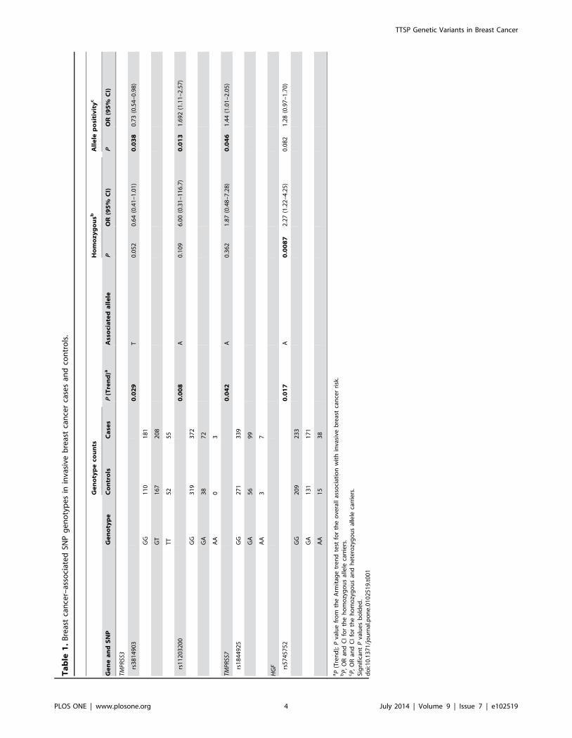
SNPs in TMPRSS3, TMPRSS7, and HGF associate withbreast cancer risk
Altogether, 82 SNPs in eight TTSPs and related genes were
genotyped in a sample set of 464 invasive breast cancer cases and
370 controls (Table S2). TMPRSS3 SNPs rs3814903 and
rs11203200, TMPRSS7 SNP rs1844925, and HGF SNP rs5745752
associated significantly with breast cancer risk (Poverall = 0.029,
0.008, 0.042, and 0.017, respectively) (Table 1). All SNPs were
consistent with the Hardy–Weinberg equilibrium. According to the
power calculations our sample set with 464 cases and 370 controls
has 83% power to detect a risk allele that is in perfect LD with the
marker allele and has a relative risk of 1.5.
SNPs in TMPRSS1, TMPRSS2, TMPRSS3 and TMPRSS7associate with breast cancer survival
In addition to breast cancer risk, we investigated variant
association with patient survival. In the univariate analysis,
TMPRSS1 rs12151195 and rs12461158, TMPRSS2 rs2070788
and rs2276205, TMPRSS3 rs3814903, TMPRSS7 rs2399403,
TMPRSS11E rs35293564, HGF rs2040965, and uPA rs2227578
associated significantly with invasive breast cancer survival
(P = 0.002, 0.05, 0.022, 0.05, 0.026, 0.007, 0.048, 0.035, and
0.021, respectively) (Table S3). TMPRSS1 SNPs rs12151195 and
rs12461158, TMPRSS2 SNPs rs2276205, TMPRSS3 SNP
rs3814903, and TMPRSS7 SNP rs2399403 remained significant
in the multivariate analysis including age, tumor grade, histolog-
ical type, tumor size, nodal status, estrogen receptor (ER) status,
and HER2 status (P = 0.008, 0.025, 0.040, 0.046, and 0.047,
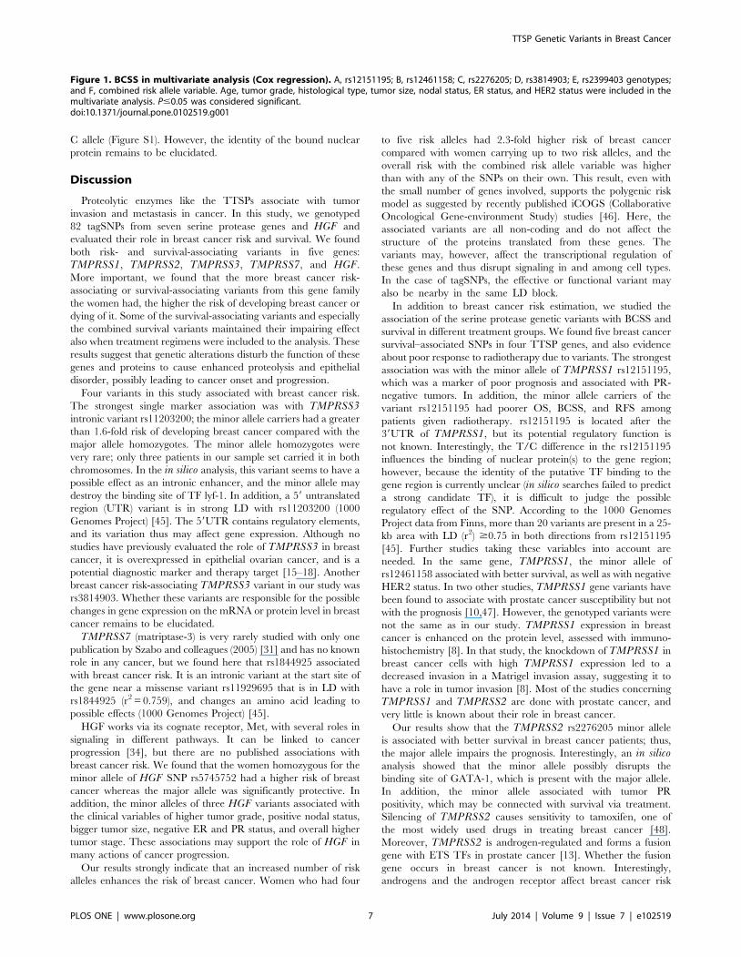
respectively) (Table 2, Fig. 1). Only TMPRSS3 rs3814903
associated with both the risk of breast cancer and survival. Of
the clinical variables included in the survival analysis, tumor grade,
nodal status, HER2 status, and histological type remained
significant in all multivariate analyses. Association of the SNPs
with the clinicopathological variables is shown as supplementary
data (Table S4).
Increasing number of alleles significantly affects breastcancer risk and prognosis
To estimate the combined effect of the associating alleles, we
assessed two new variables. We summed separately the number of
alleles of four breast cancer risk-associating SNPs (rs3814903,
rs11203200, rs1844925, and rs5745752) and five survival-associ-
ating SNPs (rs12151195, rs12461158, rs2276205, rs3814903, and
rs2399403). In risk estimation women were divided into three
groups carrying 0–2, 3, or 4–5 breast cancer risk alleles, since none
of the cases or controls had six or more alleles (maximum eight).
The risk of getting breast cancer was significantly higher with three
risk alleles present (P = 0.003; OR, 1.70; 95% CI, 1.20–2.42,
logistic regression analysis), and even higher with four to five alleles
(P = 0.0001; OR, 2.34; 95% CI, 1.39–3.94, logistic regression
analysis) compared to having 0–2 alleles.
In the multivariate survival analysis, patients with four or five
risk alleles had a significantly poorer BCSS than those with one to
three risk alleles (P = 0.037; HR, 1.96; 95% CI, 1.04–3.71)
(Table 2, Fig. 1F). Moreover, the women with six to eight risk
alleles had 3.3 times higher risk of dying of breast cancer
compared with the women with one to three risk alleles (P = 0.001;
HR, 3.30; 95% CI, 1.58–6.88) (Table 2, Fig. 1F). In the
multivariate analysis including all five survival-related SNPs, only
rs3814903 remained significant (overall P = 0.029, data not
shown). All multivariate analyses included age, tumor grade,
histological type, tumor size, nodal status, ER status, and HER2
status.
Survival-associating SNPs in TTSPs affect patientoutcome in different treatment groups
Overall survival (OS), BCSS, and recurrence-free survival (RFS)
were assessed with a multivariate analysis according to the
survival-associating SNPs and the combined risk allele variable
in different treatment groups of the breast cancer patients. Among
patients treated with radiation therapy TMPRSS1 SNP
rs12151195 and TMPRSS3 SNP rs3814903 associated signifi-
cantly with OS, BCSS, and RFS (P = 0.000001, 0.0003, and
0.013, and P = 0.016, 0.049, and 0.027, respectively) (Table 3). In
addition, rs12461158 in TMPRSS1 associated with OS and
TTSP Genetic Variants in Breast Cancer
PLOS ONE | www.plosone.org 3 July 2014 | Volume 9 | Issue 7 | e102519
Ta
ble
1.
Bre
ast
can
cer–
asso
ciat
ed
SNP
ge
no
typ
es
inin
vasi
veb
reas
tca
nce
rca
ses
and
con
tro
ls.
Ge
no
typ
eco
un
tsH
om
oz
yg
ou
sbA
lle
lep
osi
tiv
ity
c
Ge
ne
an
dS
NP
Ge
no
typ
eC
on
tro
lsC
ase
sP
(Tre
nd
)aA
sso
cia
ted
all
ele
PO
R(9
5%
CI)
PO
R(9
5%
CI)
TMP
RSS
3
rs3
81
49
03
0.0
29
T0
.05
20
.64
(0.4
1–
1.0
1)
0.0
38
0.7
3(0
.54
–0
.98
)
GG
11
01
81
GT
16
72
08
TT
52
55
rs1
12
03
20
00
.00
8A
0.1
09
6.0
0(0
.31
–1
16
.7)
0.0
13
1.6
92
(1.1
1–
2.5
7)
GG
31
93
72
GA
38
72
AA
03
TMP
RSS
70
.04
2A
0.3
62
1.8
7(0
.48
–7
.28
)0
.04
61
.44
(1.0
1–
2.0
5)
rs1
84
49
25
GG
27
13
39
GA
56
99
AA
37
HG
F rs5
74
57
52
0.0
17
A0
.00
87
2.2
7(1
.22
–4
.25
)0
.08
21
.28
(0.9
7–
1.7
0)
GG
20
92
33
GA
13
11
71
AA
15
38
aP
(Tre
nd
);P
valu
efr
om
the
Arm
itag
etr
en
dte
stfo
rth
eo
vera
llas
soci
atio
nw
ith
inva
sive
bre
ast
can
cer
risk
.b
P,
OR
and
CI
for
the
ho
mo
zyg
ou
sal
lele
carr
iers
.cP
,O
Ran
dC
Ifo
rth
eh
om
ozy
go
us
and
he
tero
zyg
ou
sal
lele
carr
iers
.Si
gn
ific
ant
Pva
lue
sb
old
ed
.d
oi:1
0.1
37
1/j
ou
rnal
.po
ne
.01
02
51
9.t
00
1
TTSP Genetic Variants in Breast Cancer
PLOS ONE | www.plosone.org 4 July 2014 | Volume 9 | Issue 7 | e102519

(P = 0.011), and TMPRSS2 SNP rs2276205 with OS and BCSS
(P = 0.019, and 0.020, respectively) (Table 3).
Among the patients receiving only radiation therapy TMPRSS1SNP rs12151195 and TMPRSS3 rs3814903 associated with OS
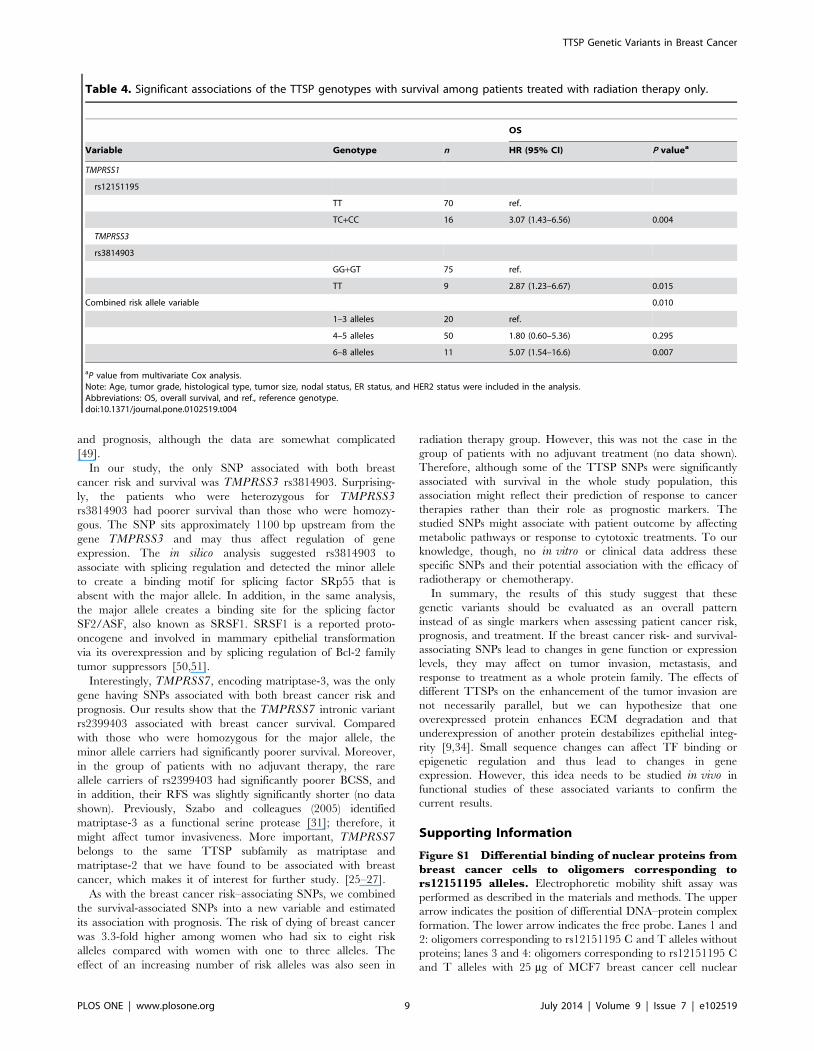
(P = 0.004, and P = 0.015, respectively) (Table 4).
In the group of patients treated with chemotherapy TMPRSS3rs3814903 G/T associated with BCSS (overall P = 0.025). Having
rs3814903 major allele G carriers (n = 62) as a reference group, the
TT homozygosity (n = 10) was significantly protective (P = 0.019;
HR, 0.17; 95% CI, 0.04–0.75) when assessing BCSS. TMPRSS3rs3814903 associated also with RFS (P = 0.043).
An effect of the number of the survival-associating alleles was
also seen in the different treatment groups. Women having more
alleles had poorer OS and BCSS when treated with radiation
therapy, compared with the women having fewer alleles
(P = 0.00001 and 0.00004, respectively) (Table 3). Also, the RFS
time was comparatively shorter among these women having more
risk alleles (P = 0.001) (Table 3). The increasing number of risk
alleles additionally affected the group treated with radiation
therapy only: The OS was poorer among women carrying more
than six risk alleles compared to those carrying five or fewer
(P = 0.010) (Table 4). However, none of the these SNPs or the
combined survival variant remained significant in the group of
patients treated with hormone therapy or in the group receiving
no treatment at all (data not shown). All multivariate analyses
included age, tumor grade, histological type, tumor size, nodal
status, ER status, and HER2 status.
Nuclear proteins from breast cancer cells binddifferentially to the rs12151195 T allele-harboring region
Because the rs12151195 T/C is a potential gene regulatory
SNP, we tested whether the C/T difference influences the binding
of nuclear proteins to this gene region. To that end electrophoretic
mobility shift assay with nuclear proteins from human breast
cancer cells was performed. Interestingly, the common allele T-
harboring ds-oligomer showed formation of a high molecular mass
nuclear protein–DNA complex that was not evident with the rare
Table 2. BCSS in multivariate analysis (Cox regression) according to TTSP genotypes.
Genotype n B (SE) Wald HR (95% CI) P value
TMPRSS1
rs12151195 0.008a
TT 316 ref.
TC 57 0. 690 (0.252) 7.515 1.99 (1.22–3.26) 0.006
CC 1 1.806 (1.056) 2.922 6.08 (0.77–48.2) 0.087
TC+CC 58 0.712 (0.248) 8.226 2.04 (1.25–3.32) 0.004
rs12461158
GG 224 ref.
GA+AA 144 20.519 (0.232) 5.001 0.60 (0.38–0.94) 0.025
TMPRSS2
rs2276205
AA 270 ref.
AG+GG 92 20.549 (0.267) 4.206 0.58 (0.34–0.98) 0.040
TMPRSS3
rs3814903 0.046a
GG 149 ref.
GT 174 0.478 (0.234) 4.166 1.61 (1.02–2.55) 0.041
TT 42 20.135 (0.351) 0.147 0.87 (0.44–1.74) 0.74
TMPRSS7
rs2399403 0.047a
TT 317 ref.
TC 52 0.645 (0.261) 6.101 1.91 (1.14–3.18) 0.014
CC 4 0.140 (0.731) 0.037 1.15 (0.27–4.82) 0.84
TC+CC 56 0.585 (0.250) 5.450 1.79 (1.10–2.93) 0.020
Combined risk allele variable
0.006a
1–3 alleles 84 ref.
4–5 alleles 219 0.675 (0.324) 4.328 1.96 (1.04–3.71) 0.037
6–8 alleles 49 1.193 (0.375) 10.13 3.30 (1.58–6.88) 0.001
aoverall P value.Significant P values bolded.Note: Age, tumor grade, histological type, tumor size, nodal status, ER status, and HER2 status were included in the analysis.Abbreviations: B (SE), coefficient with standard error, and ref., reference genotype.doi:10.1371/journal.pone.0102519.t002
TTSP Genetic Variants in Breast Cancer
PLOS ONE | www.plosone.org 5 July 2014 | Volume 9 | Issue 7 | e102519

TTSP Genetic Variants in Breast Cancer
PLOS ONE | www.plosone.org 6 July 2014 | Volume 9 | Issue 7 | e102519
C allele (Figure S1). However, the identity of the bound nuclear
protein remains to be elucidated.
Discussion
Proteolytic enzymes like the TTSPs associate with tumor
invasion and metastasis in cancer. In this study, we genotyped
82 tagSNPs from seven serine protease genes and HGF and
evaluated their role in breast cancer risk and survival. We found
both risk- and survival-associating variants in five genes:
TMPRSS1, TMPRSS2, TMPRSS3, TMPRSS7, and HGF.
More important, we found that the more breast cancer risk-
associating or survival-associating variants from this gene family
the women had, the higher the risk of developing breast cancer or
dying of it. Some of the survival-associating variants and especially
the combined survival variants maintained their impairing effect
also when treatment regimens were included to the analysis. These
results suggest that genetic alterations disturb the function of these
genes and proteins to cause enhanced proteolysis and epithelial
disorder, possibly leading to cancer onset and progression.
Four variants in this study associated with breast cancer risk.
The strongest single marker association was with TMPRSS3intronic variant rs11203200; the minor allele carriers had a greater
than 1.6-fold risk of developing breast cancer compared with the
major allele homozygotes. The minor allele homozygotes were
very rare; only three patients in our sample set carried it in both
chromosomes. In the in silico analysis, this variant seems to have a
possible effect as an intronic enhancer, and the minor allele may
destroy the binding site of TF lyf-1. In addition, a 59 untranslated
region (UTR) variant is in strong LD with rs11203200 (1000
Genomes Project) [45]. The 59UTR contains regulatory elements,
and its variation thus may affect gene expression. Although no
studies have previously evaluated the role of TMPRSS3 in breast
cancer, it is overexpressed in epithelial ovarian cancer, and is a
potential diagnostic marker and therapy target [15–18]. Another
breast cancer risk-associating TMPRSS3 variant in our study was
rs3814903. Whether these variants are responsible for the possible
changes in gene expression on the mRNA or protein level in breast
cancer remains to be elucidated.
TMPRSS7 (matriptase-3) is very rarely studied with only one
publication by Szabo and colleagues (2005) [31] and has no known
role in any cancer, but we found here that rs1844925 associated
with breast cancer risk. It is an intronic variant at the start site of
the gene near a missense variant rs11929695 that is in LD with
rs1844925 (r2 = 0.759), and changes an amino acid leading to
possible effects (1000 Genomes Project) [45].
HGF works via its cognate receptor, Met, with several roles in
signaling in different pathways. It can be linked to cancer
progression [34], but there are no published associations with
breast cancer risk. We found that the women homozygous for the
minor allele of HGF SNP rs5745752 had a higher risk of breast
cancer whereas the major allele was significantly protective. In
addition, the minor alleles of three HGF variants associated with
the clinical variables of higher tumor grade, positive nodal status,
bigger tumor size, negative ER and PR status, and overall higher
tumor stage. These associations may support the role of HGF in
many actions of cancer progression.
Our results strongly indicate that an increased number of risk
alleles enhances the risk of breast cancer. Women who had four
to five risk alleles had 2.3-fold higher risk of breast cancer
compared with women carrying up to two risk alleles, and the
overall risk with the combined risk allele variable was higher
than with any of the SNPs on their own. This result, even with
the small number of genes involved, supports the polygenic risk
model as suggested by recently published iCOGS (Collaborative
Oncological Gene-environment Study) studies [46]. Here, the
associated variants are all non-coding and do not affect the
structure of the proteins translated from these genes. The
variants may, however, affect the transcriptional regulation of
these genes and thus disrupt signaling in and among cell types.
In the case of tagSNPs, the effective or functional variant may
also be nearby in the same LD block.
In addition to breast cancer risk estimation, we studied the
association of the serine protease genetic variants with BCSS and
survival in different treatment groups. We found five breast cancer
survival–associated SNPs in four TTSP genes, and also evidence
about poor response to radiotherapy due to variants. The strongest
association was with the minor allele of TMPRSS1 rs12151195,
which was a marker of poor prognosis and associated with PR-
negative tumors. In addition, the minor allele carriers of the
variant rs12151195 had poorer OS, BCSS, and RFS among
patients given radiotherapy. rs12151195 is located after the
39UTR of TMPRSS1, but its potential regulatory function is
not known. Interestingly, the T/C difference in the rs12151195
influences the binding of nuclear protein(s) to the gene region;
however, because the identity of the putative TF binding to the
gene region is currently unclear (in silico searches failed to predict
a strong candidate TF), it is difficult to judge the possible
regulatory effect of the SNP. According to the 1000 Genomes
Project data from Finns, more than 20 variants are present in a 25-
kb area with LD (r2) $0.75 in both directions from rs12151195
[45]. Further studies taking these variables into account are
needed. In the same gene, TMPRSS1, the minor allele of
rs12461158 associated with better survival, as well as with negative
HER2 status. In two other studies, TMPRSS1 gene variants have
been found to associate with prostate cancer susceptibility but not
with the prognosis [10,47]. However, the genotyped variants were
not the same as in our study. TMPRSS1 expression in breast
cancer is enhanced on the protein level, assessed with immuno-
histochemistry [8]. In that study, the knockdown of TMPRSS1 in
breast cancer cells with high TMPRSS1 expression led to a
decreased invasion in a Matrigel invasion assay, suggesting it to
have a role in tumor invasion [8]. Most of the studies concerning
TMPRSS1 and TMPRSS2 are done with prostate cancer, and
very little is known about their role in breast cancer.
Our results show that the TMPRSS2 rs2276205 minor allele
is associated with better survival in breast cancer patients; thus,
the major allele impairs the prognosis. Interestingly, an in silicoanalysis showed that the minor allele possibly disrupts the
binding site of GATA-1, which is present with the major allele.
In addition, the minor allele associated with tumor PR
positivity, which may be connected with survival via treatment.
Silencing of TMPRSS2 causes sensitivity to tamoxifen, one of
the most widely used drugs in treating breast cancer [48].
Moreover, TMPRSS2 is androgen-regulated and forms a fusion
gene with ETS TFs in prostate cancer [13]. Whether the fusion
gene occurs in breast cancer is not known. Interestingly,
androgens and the androgen receptor affect breast cancer risk
Figure 1. BCSS in multivariate analysis (Cox regression). A, rs12151195; B, rs12461158; C, rs2276205; D, rs3814903; E, rs2399403 genotypes;and F, combined risk allele variable. Age, tumor grade, histological type, tumor size, nodal status, ER status, and HER2 status were included in themultivariate analysis. P#0.05 was considered significant.doi:10.1371/journal.pone.0102519.g001
TTSP Genetic Variants in Breast Cancer
PLOS ONE | www.plosone.org 7 July 2014 | Volume 9 | Issue 7 | e102519

Ta
ble
3.
Sig
nif
ican
tas
soci
atio
ns
of
the
TT
SPg
en
oty
pe
sw
ith
surv
ival
amo
ng
pat
ien
tstr
eat
ed
wit
hra
dia
tio
nth
era
py.
OS
BC
SS
RF
S
Va
ria
ble
Ge
no
typ
en
HR
(95
%C
I)P
va
lue
aH
R(9
5%
CI)
Pv
alu
ea
HR
(95
%C
I)P
va
lue
a
TMP
RSS
1
rs1
21
51
19
5
TT
18
7re
f.re
f.re
f.
TC
+CC
41
3.0
7(1
.95
–4
.83
)0
.00
00
01
2.5
6(1
.60
–4
.52
)0
.00
03
1.8
4(1
.14
–3
.00
)0
.01
3
rs1
24
61
15
8
AA
15
ref.
AG
+GG
21
04
.36
(1.5
9–
11
.9)
0.0
04
TMP
RSS
2
rs2
27
62
05
AA
16
7re
f.re
f.
AG
+GG
56
0.5
8(0
.37
–0
.92
)0
.01
90
.49
(0.2
7–
0.8
9)
0.0
20
TMP
RSS
3
rs3
81
49
03
0.0
16
0.0
49
GG
82
ref.
ref.
ref.
GT
12
01
.69
(1.1
1–
2.5
8)
0.0
14
1.6
6(1
.01
–2
.74
)0
.04
6
TT
24
0.8
6(0
.45
–1
.67
)0
.66
20
.80
(0.3
5–
1.8
2)
0.5
98
GT
+TT
14
41
.65
(1.0
6–
2.5
9)
0.0
27
Co
mb
ine
dri
skal
lele
vari
able
0.0
00
01
0.0
00
04
0.0
01
1–
3al
lele
s5
0re
f.re
f.re
f.
4–
5al
lele
s1
38
1.7
1(1
.01
–2
.90
)0
.04
51
.91
(0.9
5–
3.8
4)
0.0
69
1.7
4(0
.96
–3
.16
)0
.06
7
6–
8al
lele
s3
04
.66
(2.4
2–
8.9
8)
0.0
00
00
45
.35
(2.4
3–
11
.8)
0.0
00
03
3.6
2(1
.78
–7
.38
)0
.00
04
aP
valu
efr
om
mu
ltiv
aria
teC
ox
anal
ysis
.N
ote
:A
ge
,tu
mo
rg
rad
e,
his
tolo
gic
alty
pe
,tu
mo
rsi
ze,
no
dal
stat
us,
ERst
atu
s,H
ER2
stat
us,
ho
rmo
nal
tre
atm
en
t,an
dch
em
oth
era
py
we
rein
clu
de
din
the
anal
ysis
.A
bb
revi
atio
ns:
OS,
ove
rall
surv
ival
;B
CSS
,b
reas
tca
nce
r–sp
eci
fic
surv
ival
;R
FS,
recu
rre
nce
-fre
esu
rviv
al,
and
ref.
,re
fere
nce
ge
no
typ
e.
do
i:10
.13
71
/jo
urn
al.p
on
e.0
10
25
19
.t0
03
TTSP Genetic Variants in Breast Cancer
PLOS ONE | www.plosone.org 8 July 2014 | Volume 9 | Issue 7 | e102519
and prognosis, although the data are somewhat complicated
[49].
In our study, the only SNP associated with both breast
cancer risk and survival was TMPRSS3 rs3814903. Surprising-
ly, the patients who were heterozygous for TMPRSS3rs3814903 had poorer survival than those who were homozy-
gous. The SNP sits approximately 1100 bp upstream from the
gene TMPRSS3 and may thus affect regulation of gene
expression. The in silico analysis suggested rs3814903 to
associate with splicing regulation and detected the minor allele
to create a binding motif for splicing factor SRp55 that is
absent with the major allele. In addition, in the same analysis,
the major allele creates a binding site for the splicing factor
SF2/ASF, also known as SRSF1. SRSF1 is a reported proto-
oncogene and involved in mammary epithelial transformation
via its overexpression and by splicing regulation of Bcl-2 family
tumor suppressors [50,51].
Interestingly, TMPRSS7, encoding matriptase-3, was the only
gene having SNPs associated with both breast cancer risk and
prognosis. Our results show that the TMPRSS7 intronic variant
rs2399403 associated with breast cancer survival. Compared
with those who were homozygous for the major allele, the
minor allele carriers had significantly poorer survival. Moreover,
in the group of patients with no adjuvant therapy, the rare
allele carriers of rs2399403 had significantly poorer BCSS, and
in addition, their RFS was slightly significantly shorter (no data
shown). Previously, Szabo and colleagues (2005) identified
matriptase-3 as a functional serine protease [31]; therefore, it
might affect tumor invasiveness. More important, TMPRSS7belongs to the same TTSP subfamily as matriptase and
matriptase-2 that we have found to be associated with breast
cancer, which makes it of interest for further study. [25–27].
As with the breast cancer risk–associating SNPs, we combined
the survival-associated SNPs into a new variable and estimated
its association with prognosis. The risk of dying of breast cancer
was 3.3-fold higher among women who had six to eight risk
alleles compared with women with one to three alleles. The
effect of an increasing number of risk alleles was also seen in
radiation therapy group. However, this was not the case in the
group of patients with no adjuvant treatment (no data shown).
Therefore, although some of the TTSP SNPs were significantly
associated with survival in the whole study population, this
association might reflect their prediction of response to cancer
therapies rather than their role as prognostic markers. The
studied SNPs might associate with patient outcome by affecting
metabolic pathways or response to cytotoxic treatments. To our
knowledge, though, no in vitro or clinical data address these
specific SNPs and their potential association with the efficacy of
radiotherapy or chemotherapy.
In summary, the results of this study suggest that these
genetic variants should be evaluated as an overall pattern
instead of as single markers when assessing patient cancer risk,
prognosis, and treatment. If the breast cancer risk- and survival-
associating SNPs lead to changes in gene function or expression
levels, they may affect on tumor invasion, metastasis, and
response to treatment as a whole protein family. The effects of
different TTSPs on the enhancement of the tumor invasion are
not necessarily parallel, but we can hypothesize that one
overexpressed protein enhances ECM degradation and that
underexpression of another protein destabilizes epithelial integ-
rity [9,34]. Small sequence changes can affect TF binding or
epigenetic regulation and thus lead to changes in gene
expression. However, this idea needs to be studied in vivo in
functional studies of these associated variants to confirm the
current results.
Supporting Information
Figure S1 Differential binding of nuclear proteins frombreast cancer cells to oligomers corresponding tors12151195 alleles. Electrophoretic mobility shift assay was
performed as described in the materials and methods. The upper
arrow indicates the position of differential DNA–protein complex
formation. The lower arrow indicates the free probe. Lanes 1 and
2: oligomers corresponding to rs12151195 C and T alleles without
proteins; lanes 3 and 4: oligomers corresponding to rs12151195 C
and T alleles with 25 mg of MCF7 breast cancer cell nuclear
Table 4. Significant associations of the TTSP genotypes with survival among patients treated with radiation therapy only.
OS
Variable Genotype n HR (95% CI) P valuea
TMPRSS1
rs12151195
TT 70 ref.
TC+CC 16 3.07 (1.43–6.56) 0.004
TMPRSS3
rs3814903
GG+GT 75 ref.
TT 9 2.87 (1.23–6.67) 0.015
Combined risk allele variable 0.010
1–3 alleles 20 ref.
4–5 alleles 50 1.80 (0.60–5.36) 0.295
6–8 alleles 11 5.07 (1.54–16.6) 0.007
aP value from multivariate Cox analysis.Note: Age, tumor grade, histological type, tumor size, nodal status, ER status, and HER2 status were included in the analysis.Abbreviations: OS, overall survival, and ref., reference genotype.doi:10.1371/journal.pone.0102519.t004
TTSP Genetic Variants in Breast Cancer
PLOS ONE | www.plosone.org 9 July 2014 | Volume 9 | Issue 7 | e102519

proteins; lanes 5 and 6: oligomers corresponding to rs12151195 C
and T alleles with 25 mg of MCF7 cell nuclear proteins and with
506 molar excess of unlabeled oligomers; and lanes 7 and 8:
oligomers corresponding to rs12151195 C and T alleles with 25 mg
of MCF7 cell nuclear proteins and with 756 molar excess of
unlabeled oligomers.
(TIF)
Table S1 Clinicopathological characteristics of all pa-tients (invasive cases).(DOCX)
Table S2 SNP genotype counts in invasive breast cancercases, (including metastatic cases) and controls.(DOCX)
Table S3 Breast cancer survival in univariate analysis(Kaplan–Meier) at the latest follow-up data point.(DOCX)
Table S4 Significant associations of gene variants withclinical variables.
(DOCX)
Acknowledgments
We thank Eija Myohanen, Helena Kemilainen, and Merja Rasanen for
technical assistance.
Author Contributions
Conceived and designed the experiments: KL JMH MT JJP VK AM V-
MK. Performed the experiments: JMH KL. Analyzed the data: KL JMH
MT. Contributed reagents/materials/analysis tools: V-MK AM VK MT.
Wrote the paper: KL JMH MT AM V-MK JJP VK. Supervised the data
analysis: AM JMH. Participated in the interpretation of the results: KL MT
JJP JMH AM. Revised the manuscript: KL JMH MT JJP VK AM V-MK.
References
1. Stratton MR, Rahman N (2008) The emerging landscape of breast cancer
susceptibility. Nat Genet 40: 17–22.
2. Michailidou K, Hall P, Gonzalez-Neira A, Ghoussaini M, Dennis J, et al. (2013)Large-scale genotyping identifies 41 new loci associated with breast cancer risk.
Nat Genet 45: 353–361.
3. Burton H, Chowdhury S, Dent T, Hall A, Pashayan N, et al. (2013) Publichealth implications from COGS and potential for risk stratification and
screening. Nat Genet 45: 349–351.
4. Bugge TH, Antalis TM, Wu Q (2009) Type II transmembrane serine proteases.J Biol Chem 284: 23177–23181.
5. Hooper JD, Clements JA, Quigley JP, Antalis TM (2001) Type II
transmembrane serine proteases. insights into an emerging class of cell surfaceproteolytic enzymes. J Biol Chem 276: 857–860.
6. Antalis TM, Buzza MS, Hodge KM, Hooper JD, Netzel-Arnett S (2010) The
cutting edge: Membrane-anchored serine protease activities in the pericellularmicroenvironment. Biochem J 428: 325–346.
7. Netzel-Arnett S, Currie BM, Szabo R, Lin CY, Chen LM, et al. (2006) Evidence
for a matriptase-prostasin proteolytic cascade regulating terminal epidermaldifferentiation. J Biol Chem 281: 32941–32945.
8. Xing P, Li JG, Jin F, Zhao TT, Liu Q, et al. (2011) Clinical and biological
significance of hepsin overexpression in breast cancer. J Investig Med 59: 803–810.
9. Partanen JI, Tervonen TA, Myllynen M, Lind E, Imai M, et al. (2012) Tumor
suppressor function of liver kinase B1 (Lkb1) is linked to regulation of epithelialintegrity. Proceedings of the National Academy of Sciences 109: E388–397.
10. Pal P, Xi H, Kaushal R, Sun G, Jin CH, et al. (2006) Variants in the HEPSIN
gene are associated with prostate cancer in men of european origin. Hum Genet120: 187–192.
11. Holt SK, Kwon EM, Lin DW, Ostrander EA, Stanford JL (2010) Association of
hepsin gene variants with prostate cancer risk and prognosis. Prostate 70: 1012–1019.
12. Tomlins SA, Rhodes DR, Perner S, Dhanasekaran SM, Mehra R, et al. (2005)
Recurrent fusion of TMPRSS2 and ETS transcription factor genes in prostate
cancer. Science 310: 644–648.
13. Tomlins SA, Laxman B, Dhanasekaran SM, Helgeson BE, Cao X, et al. (2007)
Distinct classes of chromosomal rearrangements create oncogenic ETS gene
fusions in prostate cancer. Nature 448: 595–599.
14. Huang L, Schauer IG, Zhang J, Mercado-Uribe I, Deavers MT, et al. (2011)
The oncogenic gene fusion TMPRSS2: ERG is not a diagnostic or prognostic
marker for ovarian cancer. Int J Clin Exp Pathol 4: 644–650.
15. Underwood LJ, Shigemasa K, Tanimoto H, Beard JB, Schneider EN, et al.
(2000) Ovarian tumor cells express a novel multi-domain cell surface serine
protease. Biochim Biophys Acta 1502: 337–350.
16. Tanimoto H, Underwood LJ, Wang Y, Shigemasa K, Parmley TH, et al. (2001)
Ovarian tumor cells express a transmembrane serine protease: A potential
candidate for early diagnosis and therapeutic intervention. Tumour Biol 22:104–114.
17. Sawasaki T, Shigemasa K, Gu L, Beard JB, O’Brien TJ (2004) The
transmembrane protease serine (TMPRSS3/TADG-12) D variant: A potentialcandidate for diagnosis and therapeutic intervention in ovarian cancer. Tumour
Biol 25: 141–148.
18. Guerrero K, Wang Z, Bachvarova M, Gregoire J, Renaud MC, et al. (2012) Anovel genome-based approach correlates TMPRSS3 overexpression in ovarian
cancer with DNA hypomethylation. Gynecol Oncol 125: 720–726.
19. Viloria CG, Peinado JR, Astudillo A, Garcia-Suarez O, Gonzalez MV, et al.(2007) Human DESC1 serine protease confers tumorigenic properties to MDCK
cells and it is upregulated in tumours of different origin. Br J Cancer 97: 201–209.
20. Dass K, Ahmad A, Azmi AS, Sarkar SH, Sarkar FH (2008) Evolving role of
uPA/uPAR system in human cancers. Cancer Treat Rev 34: 122–136.
21. Harris L, Fritsche H, Mennel R, Norton L, Ravdin P, et al. (2007) American
society of clinical oncology 2007 update of recommendations for the use of
tumor markers in breast cancer. J Clin Oncol 25: 5287–5312.
22. Lin CY, Anders J, Johnson M, Dickson RB (1999) Purification and
characterization of a complex containing matriptase and a kunitz-type serine
protease inhibitor from human milk. J Biol Chem 274: 18237–18242.
23. Lin CY, Anders J, Johnson M, Sang QA, Dickson RB (1999) Molecular cloning
of cDNA for matriptase, a matrix-degrading serine protease with trypsin-like
activity. J Biol Chem 274: 18231–18236.
24. Uhland K (2006) Matriptase and its putative role in cancer. Cell Mol Life Sci 63:
2968–2978.
25. Kauppinen JM, Kosma V, Soini Y, Sironen R, Nissinen M, et al. (2010) ST14
gene variant and decreased matriptase protein expression predict poor breast
cancer survival. Cancer Epidemiol Biomarkers Prev 19: 2133–2142.
26. Hartikainen JM, Tuhkanen H, Kataja V, Eskelinen M, Uusitupa M, et al. (2006)
Refinement of the 22q12-q13 breast cancer–associated region: Evidence of
TMPRSS6 as a candidate gene in an eastern Finnish population. Clin Cancer
Res 12: 1454–1462.
27. Tuhkanen H, Hartikainen JM, Soini Y, Velasco G, Sironen R, et al. (2013)
Matriptase-2 gene (TMPRSS6) variants associate with breast cancer survival,
and reduced expression is related to triple-negative breast cancer. Int J Cancer
133: 2334–2340.
28. Sjoblom T, Jones S, Wood LD, Parsons DW, Lin J, et al. (2006) The consensus
coding sequences of human breast and colorectal cancers. Science 314: 268–274.
29. Castells A, Gusella JF, Ramesh V, Rustgi AK (2000) A region of deletion on
chromosome 22q13 is common to human breast and colorectal cancers. Cancer
Res 60: 2836–2839.
30. Hirano A, Emi M, Tsuneizumi M, Utada Y, Yoshimoto M, et al. (2001) Allelic
losses of loci at 3p25.1, 8p22, 13q12, 17p13.3, and 22q13 correlate with
postoperative recurrence in breast cancer. Clin Cancer Res 7: 876–882.
31. Szabo R, Netzel-Arnett S, Hobson JP, Antalis TM, Bugge TH (2005)
Matriptase-3 is a novel phylogenetically preserved membrane-anchored serine
protease with broad serpin reactivity. Biochem J 390: 231–242.
32. Chen LM, Skinner ML, Kauffman SW, Chao J, Chao L, et al. (2001) Prostasin
is a glycosylphosphatidylinositol-anchored active serine protease. J Biol Chem
276: 21434–21442.
33. Bergum C, Zoratti G, Boerner J, List K (2012) Strong expression association
between matriptase and its substrate prostasin in breast cancer. J Cell Physiol
227: 1604–1609.
34. Nakamura T, Sakai K, Nakamura T, Matsumoto K (2011) Hepatocyte growth
factor twenty years on: Much more than a growth factor. J Gastroenterol
Hepatol 26 Suppl 1: 188–202.
35. Owen KA, Qiu D, Alves J, Schumacher AM, Kilpatrick LM, et al. (2010)
Pericellular activation of hepatocyte growth factor by the transmembrane serine
proteases matriptase and hepsin, but not by the membrane-associated protease
uPA. Biochem J 426: 219–228.
36. Hartikainen JM, Tuhkanen H, Kataja V, Dunning AM, Antoniou A, et al.
(2005) An autosome-wide scan for linkage disequilibrium-based association in
sporadic breast cancer cases in eastern finland: Three candidate regions found.
Cancer Epidemiol Biomarkers Prev 14: 75–80.
37. Pellikainen MJ, Pekola TT, Ropponen KM, Kataja VV, Kellokoski JK, et al.
(2003) p21WAF1 expression in invasive breast cancer and its association with
p53, AP-2, cell proliferation, and prognosis. J Clin Pathol 56: 214–220.
38. Vandenplas S, Wiid I, Grobler-Rabie A, Brebner K, Ricketts M, et al. (1984)
Blot hybridisation analysis of genomic DNA. J Med Genet 21: 164–172.
TTSP Genetic Variants in Breast Cancer
PLOS ONE | www.plosone.org 10 July 2014 | Volume 9 | Issue 7 | e102519

39. Hartikainen JM, Tengstrom M, Kosma VM, Kinnula VL, Mannermaa A, et al.
(2012) Genetic polymorphisms and protein expression of NRF2 and sulfiredoxin
predict survival outcomes in breast cancer. Cancer Res 72: 5537–5546.
40. Barrett JC, Fry B, Maller J, Daly MJ (2005) Haploview: Analysis and
visualization of LD and haplotype maps. Bioinformatics 21: 263–265.
41. Purcell S, Cherny SS, Sham PC (2003) Genetic power calculator: Design of
linkage and association genetic mapping studies of complex traits. Bioinformatics
19: 149–150.
42. Finnish Cancer Registry. Finnish cancer registry, cancer statistics at www.
cancerregistry.fi, updated on 06.06.2013.
43. Yuan HY, Chiou JJ, Tseng WH, Liu CH, Liu CK, et al. (2006) FASTSNP: An
always up-to-date and extendable service for SNP function analysis and
prioritization. Nucleic Acids Res 34: W635–41.
44. Lee PH, Shatkay H (2008) F-SNP: Computationally predicted functional SNPs
for disease association studies. Nucleic Acids Res 36: D820–4.
45. Genomes Project Consortium, Abecasis GR, Altshuler D, Auton A, Brooks LD,
et al. (2010) A map of human genome variation from population-scalesequencing. Nature 467: 1061–1073.
46. Bahcall OG (2013) iCOGS collection provides a collaborative model. Nat Genet
45: 343.47. Burmester JK, Suarez BK, Lin JH, Jin CH, Miller RD, et al. (2004) Analysis of
candidate genes for prostate cancer. Hum Hered 57: 172–178.48. Mendes-Pereira AM, Sims D, Dexter T, Fenwick K, Assiotis I, et al. (2011)
Genome-wide functional screen identifies a compendium of genes affecting
sensitivity to tamoxifen. Proc Natl Acad Sci U S A 109: 2730–2735.49. Kotsopoulos J, Narod SA (2012) Androgens and breast cancer. Steroids 77: 1–9.
50. Karni R, de Stanchina E, Lowe SW, Sinha R, Mu D, et al. (2007) The geneencoding the splicing factor SF2/ASF is a proto-oncogene. Nat Struct Mol Biol
14: 185–193.51. Anczukow O, Rosenberg AZ, Akerman M, Das S, Zhan L, et al. (2012) The
splicing factor SRSF1 regulates apoptosis and proliferation to promote
mammary epithelial cell transformation. Nat Struct Mol Biol 19: 220–228.
TTSP Genetic Variants in Breast Cancer
PLOS ONE | www.plosone.org 11 July 2014 | Volume 9 | Issue 7 | e102519