Specificity of Transmembrane Protein Palmitoylation in Yeast
13
Specificity of Transmembrane Protein Palmitoylation in Yeast Ayele ´ n Gonza ´ lez Montoro, Sabrina Chumpen Ramirez, Rodrigo Quiroga, Javier Valdez Taubas* Centro de Investigaciones en Quı ´mica Biolo ´gica de Co ´ rdoba, CIQUIBIC (UNC-CONICET), Departamento de Quı ´mica Biolo ´ gica, Facultad de Ciencias Quı ´micas, Universidad Nacional de Co ´ rdoba, Co ´ rdoba, Argentina Abstract Many proteins are modified after their synthesis, by the addition of a lipid molecule to one or more cysteine residues, through a thioester bond. This modification is called S-acylation, and more commonly palmitoylation. This reaction is carried out by a family of enzymes, called palmitoyltransferases (PATs), characterized by the presence of a conserved 50- aminoacids domain called ‘‘Asp-His-His-Cys- Cysteine Rich Domain’’ (DHHC-CRD). There are 7 members of this family in the yeast Saccharomyces cerevisiae, and each of these proteins is thought to be responsible for the palmitoylation of a subset of substrates. Substrate specificity of PATs, however, is not yet fully understood. Several yeast PATs seem to have overlapping specificity, and it has been proposed that the machinery responsible for palmitoylating peripheral membrane proteins in mammalian cells, lacks specificity altogether. Here we investigate the specificity of transmembrane protein palmitoylation in S. cerevisiae, which is carried out predominantly by two PATs, Swf1 and Pfa4. We show that palmitoylation of transmembrane substrates requires dedicated PATs, since other yeast PATs are mostly unable to perform Swf1 or Pfa4 functions, even when overexpressed. Furthermore, we find that Swf1 is highly specific for its substrates, as it is unable to substitute for other PATs. To identify where Swf1 specificity lies, we carried out a bioinformatics survey to identify amino acids responsible for the determination of specificity or Specificity Determination Positions (SDPs) and showed experimentally, that mutation of the two best SDP candidates, A145 and K148, results in complete and partial loss of function, respectively. These residues are located within the conserved catalytic DHHC domain suggesting that it could also be involved in the determination of specificity. Finally, we show that modifying the position of the cysteines in Tlg1, a Swf1 substrate, results in lack of palmitoylation, as expected for a highly specific enzymatic reaction. Citation: Gonza ´lez Montoro A, Chumpen Ramirez S, Quiroga R, Valdez Taubas J (2011) Specificity of Transmembrane Protein Palmitoylation in Yeast. PLoS ONE 6(2): e16969. doi:10.1371/journal.pone.0016969 Editor: Robert Arkowitz, Institute of Developmental Biology and Cancer Research, France Received October 18, 2010; Accepted January 11, 2011; Published February 24, 2011 Copyright: ß 2011 Gonza ´lez Montoro et al. This is an open-access article distributed under the terms of the Creative Commons Attribution License, which permits unrestricted use, distribution, and reproduction in any medium, provided the original author and source are credited. Funding: This work was supported by grants from Secretarı ´a de Ciencia y Tecnologı ´a, Universidad Nacional de Co ´ rdoba (Secyt UNC 2007 and 2010 to JVT RES214/10; http://www.secyt.unc.edu.ar/nuevo/) and Agencia Nacional de Promocio ´ n Cientı ´fica y Tecnolo ´ gica, Argentina (ANPCyT) (PICT 2006 1239 to Hugo Maccioni and JVT, and PICT 2005 32937 to JVT; http://www.agencia.mincyt.gov.ar/). JVT is a Career Investigator of CONICET (Argentina). AGM and SC have fellowships from CONICET. RQ is a FONCyT fellow. The funders had no role in study design, data collection and analysis, decision to publish, or preparation of the manuscript. Competing Interests: The authors have declared that no competing interests exist. * E-mail: [email protected] Introduction Protein palmitoylation or S-acylation is the addition of a lipid molecule on a cysteine residue of a protein by thioestherification. This is the only lipid modification that is reversible and thus susceptible to regulation [1,2,3]. This modification is of great relevance in the regulation of several important processes such as the visual cycle [4], signal transduction [5], and synaptic transmission [6,7,8]. For instance, palmitoylation regulates localization and signalling activity of Ras isoforms, and hence diverse signalling pathways [9]. Many proteins are palmitoylated in neurons, where this modification modulates sorting of presynaptic proteins, synapse morphology and clustering of ion channels [7,8,10]. A family of proteins containing a 50 residues long domain called Asp-His-His-Cys Cysteine Rich Domain (DHHC-CRD) [11] is involved in protein S-acylation [12,13], reviewed in [14]. There are at least 23 predicted DHHC-CRD containing proteins in the human genome and 7 in the Saccharomyces cerevisiae genome. They are integral membrane proteins predicted to contain 4 to 6 transmembrane domains. Subsets of substrates have been assigned to most of the yeast palmitoyltransferases (PATs) [12,13,15,16,17] and to several mammalian PATs [18,19,20]. Palmitoyltransferases make interesting drug targets for thera- peutic intervention, mainly because of their involvement in the modification of oncoproteins [21] and their role in modulating neuronal trafficking and function [6,7,9]. However, a deeper understanding of palmitoylation and its consequences is required. Recent progress in the field has consisted mostly in the identification of PATs and their substrates. Basic knowledge regarding the mechanism, the enzymes responsible for this modification and their regulation is lacking. Few residues have been mutated resulting in lack of function [12,13,17,22,23], and most are highly conserved residues located within the DHHC domain. Outside this domain, a mutation at position 3 of the recently identified PaCCT (Palmitoyltransferase Conserved C- Terminus) motif, present in most PAT’s C-termini, results in lack of function for at least two yeast PATs, Swf1 and Pfa3 [24]. An aspect that requires further investigation is the specificity of different PATs towards their substrates, and how it is determined. Based on sequence comparison, it has been assumed that since the DHHC domain is highly conserved in the family, it represents the PLoS ONE | www.plosone.org 1 February 2011 | Volume 6 | Issue 2 | e16969
-
Upload
independent -
Category
Documents
-
view
3 -
download
0
Transcript of Specificity of Transmembrane Protein Palmitoylation in Yeast

Specificity of Transmembrane Protein Palmitoylation inYeastAyelen Gonzalez Montoro, Sabrina Chumpen Ramirez, Rodrigo Quiroga, Javier Valdez Taubas*
Centro de Investigaciones en Quımica Biologica de Cordoba, CIQUIBIC (UNC-CONICET), Departamento de Quımica Biologica, Facultad de Ciencias Quımicas, Universidad
Nacional de Cordoba, Cordoba, Argentina
Abstract
Many proteins are modified after their synthesis, by the addition of a lipid molecule to one or more cysteine residues,through a thioester bond. This modification is called S-acylation, and more commonly palmitoylation. This reaction is carriedout by a family of enzymes, called palmitoyltransferases (PATs), characterized by the presence of a conserved 50-aminoacids domain called ‘‘Asp-His-His-Cys- Cysteine Rich Domain’’ (DHHC-CRD). There are 7 members of this family in theyeast Saccharomyces cerevisiae, and each of these proteins is thought to be responsible for the palmitoylation of a subset ofsubstrates. Substrate specificity of PATs, however, is not yet fully understood. Several yeast PATs seem to have overlappingspecificity, and it has been proposed that the machinery responsible for palmitoylating peripheral membrane proteins inmammalian cells, lacks specificity altogether. Here we investigate the specificity of transmembrane protein palmitoylationin S. cerevisiae, which is carried out predominantly by two PATs, Swf1 and Pfa4. We show that palmitoylation oftransmembrane substrates requires dedicated PATs, since other yeast PATs are mostly unable to perform Swf1 or Pfa4functions, even when overexpressed. Furthermore, we find that Swf1 is highly specific for its substrates, as it is unable tosubstitute for other PATs. To identify where Swf1 specificity lies, we carried out a bioinformatics survey to identify aminoacids responsible for the determination of specificity or Specificity Determination Positions (SDPs) and showedexperimentally, that mutation of the two best SDP candidates, A145 and K148, results in complete and partial loss offunction, respectively. These residues are located within the conserved catalytic DHHC domain suggesting that it could alsobe involved in the determination of specificity. Finally, we show that modifying the position of the cysteines in Tlg1, a Swf1substrate, results in lack of palmitoylation, as expected for a highly specific enzymatic reaction.
Citation: Gonzalez Montoro A, Chumpen Ramirez S, Quiroga R, Valdez Taubas J (2011) Specificity of Transmembrane Protein Palmitoylation in Yeast. PLoSONE 6(2): e16969. doi:10.1371/journal.pone.0016969
Editor: Robert Arkowitz, Institute of Developmental Biology and Cancer Research, France
Received October 18, 2010; Accepted January 11, 2011; Published February 24, 2011
Copyright: � 2011 Gonzalez Montoro et al. This is an open-access article distributed under the terms of the Creative Commons Attribution License, whichpermits unrestricted use, distribution, and reproduction in any medium, provided the original author and source are credited.
Funding: This work was supported by grants from Secretarıa de Ciencia y Tecnologıa, Universidad Nacional de Cordoba (Secyt UNC 2007 and 2010 to JVTRES214/10; http://www.secyt.unc.edu.ar/nuevo/) and Agencia Nacional de Promocion Cientıfica y Tecnologica, Argentina (ANPCyT) (PICT 2006 1239 to HugoMaccioni and JVT, and PICT 2005 32937 to JVT; http://www.agencia.mincyt.gov.ar/). JVT is a Career Investigator of CONICET (Argentina). AGM and SC havefellowships from CONICET. RQ is a FONCyT fellow. The funders had no role in study design, data collection and analysis, decision to publish, or preparation of themanuscript.
Competing Interests: The authors have declared that no competing interests exist.
* E-mail: [email protected]
Introduction
Protein palmitoylation or S-acylation is the addition of a lipid
molecule on a cysteine residue of a protein by thioestherification.
This is the only lipid modification that is reversible and thus
susceptible to regulation [1,2,3]. This modification is of great
relevance in the regulation of several important processes such as
the visual cycle [4], signal transduction [5], and synaptic
transmission [6,7,8]. For instance, palmitoylation regulates
localization and signalling activity of Ras isoforms, and hence
diverse signalling pathways [9]. Many proteins are palmitoylated
in neurons, where this modification modulates sorting of
presynaptic proteins, synapse morphology and clustering of ion
channels [7,8,10].
A family of proteins containing a 50 residues long domain called
Asp-His-His-Cys Cysteine Rich Domain (DHHC-CRD) [11] is
involved in protein S-acylation [12,13], reviewed in [14]. There
are at least 23 predicted DHHC-CRD containing proteins in the
human genome and 7 in the Saccharomyces cerevisiae genome. They
are integral membrane proteins predicted to contain 4 to 6
transmembrane domains. Subsets of substrates have been assigned
to most of the yeast palmitoyltransferases (PATs) [12,13,15,16,17]
and to several mammalian PATs [18,19,20].
Palmitoyltransferases make interesting drug targets for thera-
peutic intervention, mainly because of their involvement in the
modification of oncoproteins [21] and their role in modulating
neuronal trafficking and function [6,7,9]. However, a deeper
understanding of palmitoylation and its consequences is required.
Recent progress in the field has consisted mostly in the
identification of PATs and their substrates. Basic knowledge
regarding the mechanism, the enzymes responsible for this
modification and their regulation is lacking. Few residues have
been mutated resulting in lack of function [12,13,17,22,23], and
most are highly conserved residues located within the DHHC
domain. Outside this domain, a mutation at position 3 of the
recently identified PaCCT (Palmitoyltransferase Conserved C-
Terminus) motif, present in most PAT’s C-termini, results in lack
of function for at least two yeast PATs, Swf1 and Pfa3 [24].
An aspect that requires further investigation is the specificity of
different PATs towards their substrates, and how it is determined.
Based on sequence comparison, it has been assumed that since the
DHHC domain is highly conserved in the family, it represents the
PLoS ONE | www.plosone.org 1 February 2011 | Volume 6 | Issue 2 | e16969
catalytic core, while specificity would be determined within the
highly variable N- and C-termini [14,24].
Although there are no consensus sequences identified in
palmitoylation substrates, a few determinants necessary for
recognition by PATs have been described [25,26]. Nevertheless,
several substrates can be palmitoylated by more than one PAT.
Recently, Hou et al, (2009) [23], have shown that phenotypes
associated with the lack of Pfa3, a yeast PAT responsible for the
palmitoylation of the vacuole fusion factor Vac8 [17], can be
suppressed upon high overexpression of most yeast PATs, leading
to postulate that some PATs have overlapping specificity.
Accordingly, in vivo palmitoylation of Vac8 is only partially
reduced in the absence of Pfa3 [17]. Also, Ras2 palmitoylation is
only partially suppressed in the absence of its cognate PAT, Erf2
[15,27]. Neuronal PATs exhibit distinct substrate specificity,
although most substrates studied are modified by more than one
PAT [18,28]. Overlapping specificity for some yeast PATs was
also shown using an elegant proteomics approach [15]. More
recently, it has been postulated that in mammalian cells, the
palmitoylation machinery for peripheral membrane proteins is
devoid of specificity [29].
Swf1 is a yeast DHHC-protein involved in the palmitoylation of
SNARE (Soluble N-ethylmaleimide-sensitive factor attachment
protein Receptor) fusion proteins [16] and possibly glycosyltrans-
ferases [15]. These represent a set of substrates that consist of type II
transmembrane proteins that are palmitoylated in cysteines
adjacent to the cytosolic border of the transmembrane domain
(TMD). The function of transmembrane SNARE palmitoylation is
not clear, but in the case of the endosomal syntaxin Tlg1, it seems to
protect it from degradation by the quality control machinery [16].
Palmitoylation of multi-spanning membrane proteins in yeast
appears to be carried out mostly by Pfa4. This protein
palmitoylates several amino acid permeases [15] and the chitin
synthase Chs3, allowing it to exit the ER [30].
In this work we investigate the specificity of transmembrane
protein palmitoylation mediated by Swf1 and Pfa4, and also where
is Swf1 specificity encoded.
Results
Swf1 is unable to substitute for other PATsSwf1 mediates the palmitoylation of type II membrane proteins
such as SNAREs and possibly glycosyltransferases [15,16]. We
were interested in the specificity of this modification, particularly
in the light of two recent studies, one by Rocks et al. (2010), in
mammalian cells, where they postulate that palmitoylation of
peripheral membrane proteins lacks specificity, and another by
Hou et al. (2009), where they show that overexpression of Erf2,
Pfa4, or Akr1 is able to partially suppress phenotypes associated
with lack of PFA3 [23].
Vac8 is a protein involved in vacuolar fusion which, when
palmitoylated, localizes to the vacuolar membrane [31]. We
analyzed the distribution of Vac8 by Western blots of membrane
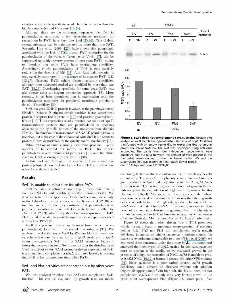
vs. soluble fractions for a wt strain, a pfa3D strain and a pfa3Dstrain overexpressing Swf1 from a GAL1 promoter. Figure 1
shows that overexpression of Swf1 does not alter the distribution of
Vac8 in a pfa3D strain. GAL1 promoter driven expression of Swf1
is also unable to complement a pfa4D strain (see below), indicating
that Swf1 is less promiscuous than other PATs.
Swf1 and Pfa4 activities are not carried out by other yeastPATs
We next analyzed whether other PATs can complement Swf1
function. This can be evaluated by growth tests on media
containing lactate as the sole carbon source, in which swf1D cells
cannot grow. The bases for this phenotype are unknown, but it is a
good predictor of Swf1 palmitoylation activitity. A swf1D tul1Dstrain in which Tlg1 is not degraded still does not grow in lactate
indicating that the degradation of Tlg1 is not responsible for this
phenotype. [16,24] Moreover, we have screened the whole
collection of yeast deletion mutants for strains that show growth
defects in both lactate and high salt, another phenotype of the
swf1D strain, We identified swf1D in this screen, as expected, but
none of its cognate substrates, suggesting that this phenotype
cannot be assigned to lack of function of any particular known
substrate (Gonzalez Montoro and Valdez Taubas, unpublished).
Figure 2A shows that, when driven from a TPI1 promoter,
which normally leads to moderate overexpression of proteins,
neither Erf2, Pfa3 nor Pfa4 can complement swf1D growth
deficiency in media containing lactate as a carbon source. To
make our experiments comparable to those of Hou et al (2009), we
expressed these constructs under the strong GAL1 promoter, and
analyzed the phenotypes of swf1D strains. In this case, galactose
must be present in the media, so we evaluated growth in the
presence of a high concentration of NaCl. swf1D is unable to grow
in 0.85M NaCl [16,32] a feature it shares with other VPS mutants
[32]. Since galactose is a poor carbon source, swf1D growth
deficiency could already be observed without salt addition
(Figure 2B upper panel). With high salt, the PATs tested did not
complement swf1D and we only see a very limited growth in the
presence of overexpressed Pfa3 (Figure 2B, lower panel). We
Figure 1. Swf1 does not complement a pfa3D strain. Western blotanalysis of Vac8 membrane/cytosol distribution in a wt or pfa3D strainstransformed with an empty vector (EV) or expressing GAL1-promoterdriven Pfa3-PA or Swf1-PA. The blot was developed using anti-Vac8antibodies. The bands from four independent experiments werequantified and the ratio between the amount of Vac8 present in thethe pellet corresponding to the membrane fraction (P) and thesupernatant (SN) was plotted in a bar graph (lower panel).doi:10.1371/journal.pone.0016969.g001
Transmembrane Protein Palmitoylation
PLoS ONE | www.plosone.org 2 February 2011 | Volume 6 | Issue 2 | e16969
confirmed these results by analyzing the vacuolar degradation of
the Swf1 substrate Tlg1, which can be directly correlated to its
palmitoylation status [16,24]. In a Western blot analysis of a swf1Dstrain expressing a GFP-Tlg1 fusion, some of the label will be in
the form of free GFP which is resistant to vacuolar proteolysis.
Figure 2C shows that GAL1 driven PATs are unable to fully
suppress GFP-Tlg1 degradation. A small amount of GFP-Tlg1 is
protected from degradation upon Pfa3 overexpression, in
agreement to what is observed in growth test experiments. Finally,
we assessed the palmitoylation status of Tlg1 directly. Instead of
the classical biotinyl exchange, we carried out direct biotinylation
experiments, since Tlg1 has only two cysteines and they are both
modified by palmitate in vivo [16]. The results can be observed by
analyzing the shift towards higher molecular weights that is
produced in the proteins by the reaction with biotin-BMCC. This
shift does not occur when the cysteines are protected by
palmitoylation. Figure 2D shows that, in a swf1D strain, Tlg1
shifts towards higher molecular weights compared to the same
strain complemented with wt Swf1. When the swf1D strain was
transformed with GAL1 promoter driven Erf2 or Pfa4, Tlg1
behaves as in the empty vector (EV) control, indicating that these
PATs are unable to palmitoylate Tlg1 even when overexpressed.
Again, when we analyze the effect of Pfa3, an intermediate
situation is observed. Figure 2E shows the expression levels of
GAL1-driven PATs, tagged at the C-terminus with the IgG
binding domain of Protein A. The functionality of all these
constructs was tested in complementation assays of the corre-
sponding deletion mutants: Figure 1 for Pfa3, Figure 2 for Swf1,
and Figure 3 for Pfa4. Erf2-PA complementation assay is not
shown. Similar Protein-A tagged constructs were previously
proven to be functional [23]. These results are consistent with
those obtained with TPI1 driven constructs, which are untagged.
These results show directly that palmitoylation of the Swf1
substrate Tlg1 cannot be carried out by other PATs, and confirm
that, in vivo, this activity is exclusively performed by Swf1, because
only upon massive overexpression of Pfa3 a small degree of
complementation is observed. It should be noted that Pfa3 is over-
expressed at much higher levels than the other PATs (Figure 2E).
The growth tests strongly suggest that this is also valid to at least
another, or possibly several Swf1 substrates.
We extended our observations to another PAT, Pfa4, which is
responsible for the palmitoylation of polytopic membrane proteins
such as aminoacid permeases and chitin synthase 3 (Chs3) [15,30].
One of the phenotypes of a pfa4D strain is its ability to grow in the
presence of Calcofluor White (CW). This compound binds to
chitin present in the yeast cell wall. pfa4D cells have reduced chitin
levels and are therefore resistant to 75 mg/ml CW, while wt cells
cannot grow in these conditions [30]. This is due to unpalmitoy-
lated Chs3 being trapped in the ER in the absence of Pfa4, which
precludes its function in chitin synthesis [30]. This allows us to
evaluate the function of Pfa4, at least regarding Chs3 palmitoyla-
tion, in simple growth tests in the presence of CW.
We transformed a pfa4D strain with GAL1 promoter driven Swf1,
Pfa3, and Erf2. Figure 3 shows that neither of these constructs is able
to suppress the CW phenotype of a pfa4D mutant, resulting in CW
resistant strains. These experiments show that Pfa4 activity towards
CHS3 cannot be carried out by other PATs and suggest that
palmitoylation of Pfa4 substrates is also highly specific.
In silico analysis predicts the presence of SpecificityDetermination Positions within the DHHC domain ofPATs
Domains or motifs that mediate Swf1 or Pfa4 specificity are not
obvious to identify by sequence comparison, since even ortholo-
gues from closely related organisms display low conservation
outside the DHHC region, the TTxE motif and the PaCCT motif
[24].
We used an alternative approach based on the prediction of
Specificity Determination Positions (SDPs). Within a given family
of proteins, members can be grouped according to substrate
specificity. Often, differences in specificity between the groups can
be ascribed to single residues that are conserved within the
members of the subgroup and not in the members of other
specificity groups [33]. These positions are known as specificity
determination positions. Bioinformatic tools have been designed to
identify these SDPs. We used GROUPSIM [34], which considers
residue conservation within specificity groups and physicochemical
characteristics of the residues.
It should be stated that SDPs do not necessarily refer to
specificity in terms of biochemical affinity for different substrates,
but rather to residues involved in every process that allows
members of a sub-group to modify a particular subset of substrates
in vivo. For instance, a residue that is conserved in a particular
subgroup and is important for interaction with a specific partner,
will have a high score in GROUPSIM, and mutation will probably
result in lack of function.
In order to feed the software, we generated an alignment of
alignments, comprising six yeast PATs and their orthologue
groups (as defined in ORTHOMCL) making the assumption that
they define specificity groups (see below). This alignment is
supplied as File S1. GROUPSIM assigns each position or column
in the alignment, a score which ranges from 0 to 1, and indicates
how likely it is to be important for specificity determination. A
table containing the scores for all the columns in the alignment
and the identity of the residues for each PAT is shown in Table S2.
Table 1 shows the top 11 scoring positions for the alignment, the
identity of the residues present at that position for yeast Swf1 and
Pfa4 and their location within protein domains.
Interestingly, 9 of the 11 positions correspond to residues
present within the DHHC domain. Predicted SDPs outside the
DHHC domain include most notably the x in TTxE motif and
position 2 in the PaCCT domain as defined in Gonzalez Montoro
et al. (2009).
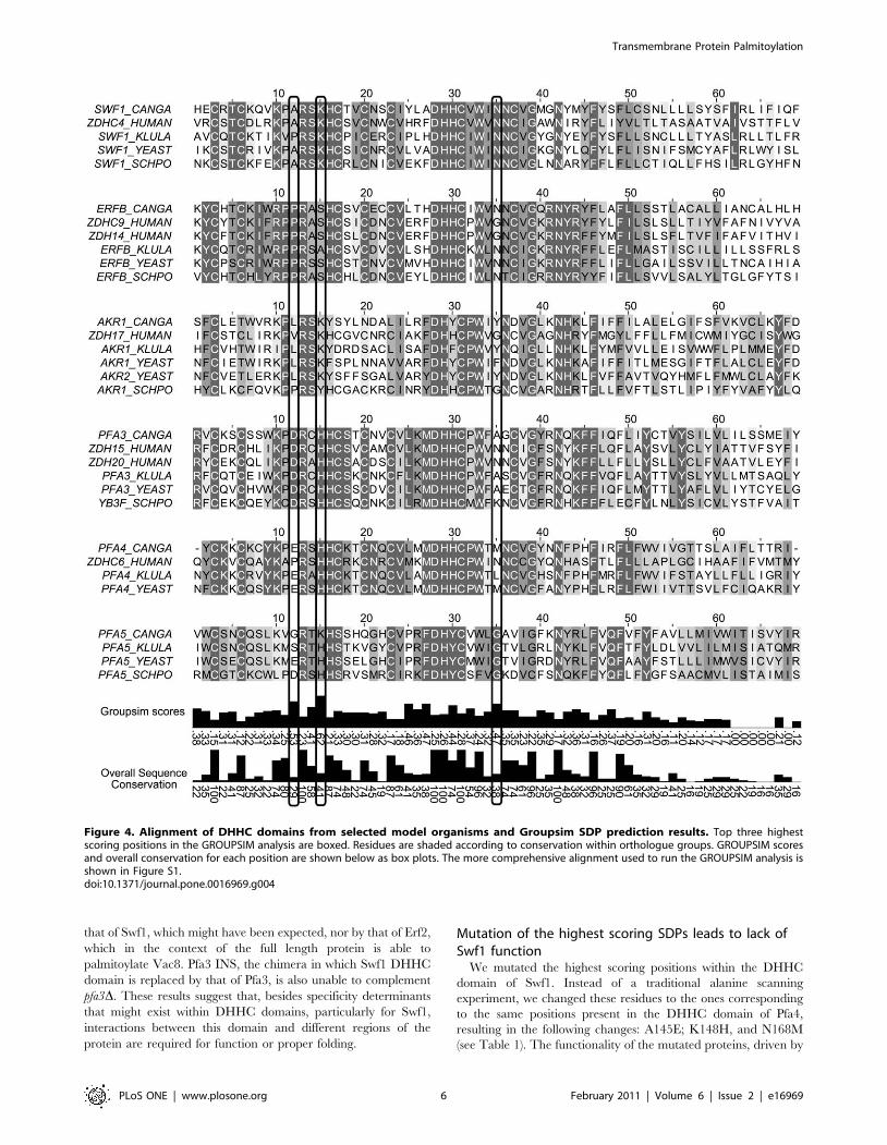
Figure 4 displays an alignment of PATs DHHC domains from
S. cerevisiae, Homo sapiens, Candida glabrata, Kluyveromyces lactis and
Schizosaccharomyces pombe. Residues are shaded according to
conservation within subgroups, and overall conservation is
displayed in a bar graph below. GROUPSIM scores are displayed
in bar graphs for each column, and the three top GROUPSIM
columns are boxed, which correspond to A145, K148 and N168 in
S. cerevisiae Swf1. The more comprehensive alignment found in
File S1 was used to calculate the scores but Figure 4 can illustrate
the conservation pattern of the predicted SDPs within their
sequence context and also the identity of the residues for each
PAT.
Some regions receive low scores in GROUPSIM analysis
because they are absent from some PAT subgroups and thus
cannot be aligned with the rest of the family, like the ankyrin
repeat containing N-termini of Akr1 orthologous proteins. In fact,
ankyrin repeats of HIP14 have been implicated in specificity
determination since they can confer HIP14-like specificity to
DHHC3 [28]. The N-terminal regions of Swf1 and Erf2 upstream
the TMD1, as defined in [14], (which for Swf1 includes another
TMD) and C-terminal regions of all PATs downstream the
PaCCT motif are also excluded from the alignment.
Despite being unable to identify SDPs in unalignable regions,
GROUPSIM identified several candidate SDPs, most of which are
located within the DHHC domain itself.
Transmembrane Protein Palmitoylation
PLoS ONE | www.plosone.org 3 February 2011 | Volume 6 | Issue 2 | e16969
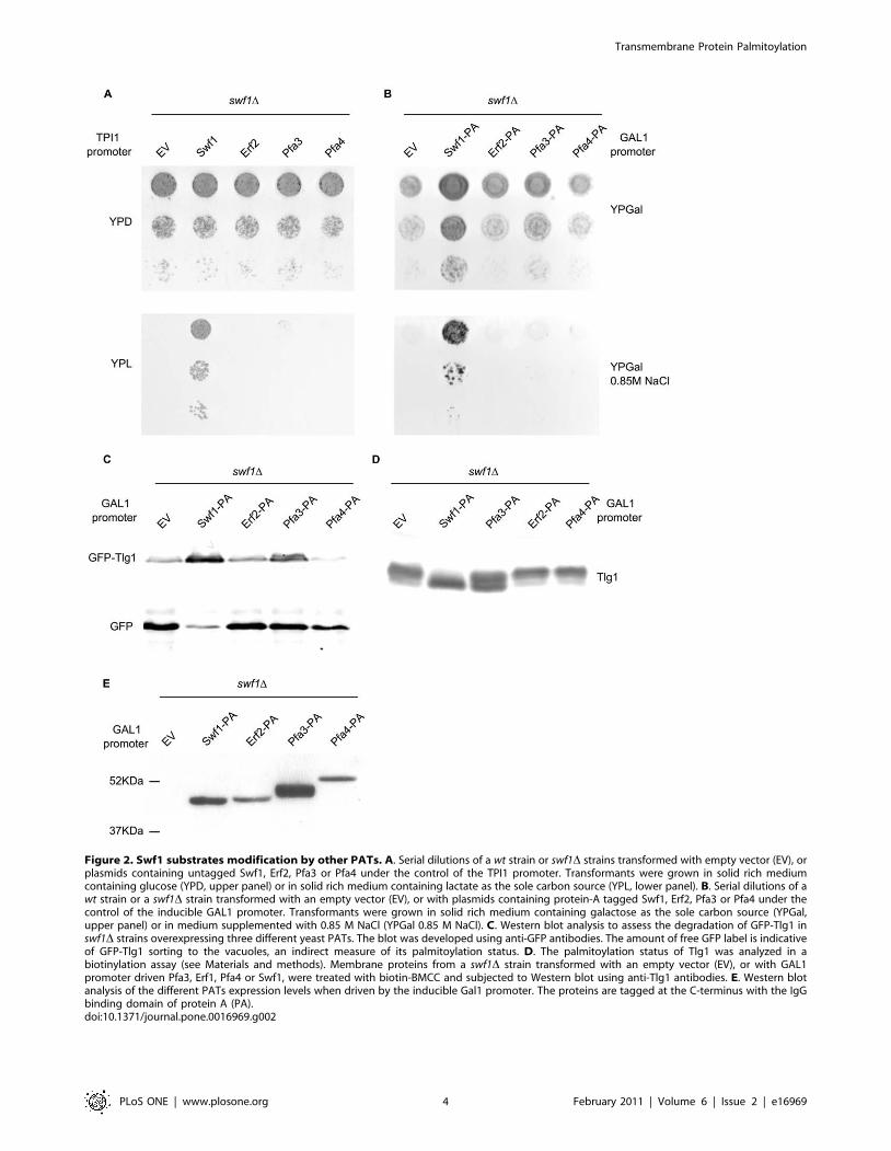
Figure 2. Swf1 substrates modification by other PATs. A. Serial dilutions of a wt strain or swf1D strains transformed with empty vector (EV), orplasmids containing untagged Swf1, Erf2, Pfa3 or Pfa4 under the control of the TPI1 promoter. Transformants were grown in solid rich mediumcontaining glucose (YPD, upper panel) or in solid rich medium containing lactate as the sole carbon source (YPL, lower panel). B. Serial dilutions of awt strain or a swf1D strain transformed with an empty vector (EV), or with plasmids containing protein-A tagged Swf1, Erf2, Pfa3 or Pfa4 under thecontrol of the inducible GAL1 promoter. Transformants were grown in solid rich medium containing galactose as the sole carbon source (YPGal,upper panel) or in medium supplemented with 0.85 M NaCl (YPGal 0.85 M NaCl). C. Western blot analysis to assess the degradation of GFP-Tlg1 inswf1D strains overexpressing three different yeast PATs. The blot was developed using anti-GFP antibodies. The amount of free GFP label is indicativeof GFP-Tlg1 sorting to the vacuoles, an indirect measure of its palmitoylation status. D. The palmitoylation status of Tlg1 was analyzed in abiotinylation assay (see Materials and methods). Membrane proteins from a swf1D strain transformed with an empty vector (EV), or with GAL1promoter driven Pfa3, Erf1, Pfa4 or Swf1, were treated with biotin-BMCC and subjected to Western blot using anti-Tlg1 antibodies. E. Western blotanalysis of the different PATs expression levels when driven by the inducible Gal1 promoter. The proteins are tagged at the C-terminus with the IgGbinding domain of protein A (PA).doi:10.1371/journal.pone.0016969.g002
Transmembrane Protein Palmitoylation
PLoS ONE | www.plosone.org 4 February 2011 | Volume 6 | Issue 2 | e16969

DHHC domains are not interchangeableIt has been assumed that the DHHC domain is responsible for
the catalytic activity, while substrate specificity would be encoded
within the highly variable N- and C- terminal regions of PATs.
Moreover, very few SDPs have been tested experimentally so, to
validate our SDP prediction and the presence of SDPs within the
DHHC domains, we generated chimeric genes in which we
substituted the DHHC domain of Swf1 for the DHHC domains of
Pfa3, Pfa4 and Erf2 (See Figure 5B for a scheme and Figure 4 for
the sequences of the DHHC domains). To determine the
boundaries of the DHHC domains, we selected the regions that
flank the DHHC motif, which are highly conserved and contain
almost no gaps, as shown in the alignment provided in File S1.
Topology predictions of PATs, always place the DHHC-CRD
domain in a cytosolic loop between TMDs 2 and 3 defined as in
Mitchel et al 2006 [14]. So a chimera in which the DHHC-CRD
motif is replaced by one from a different PAT, should in principle
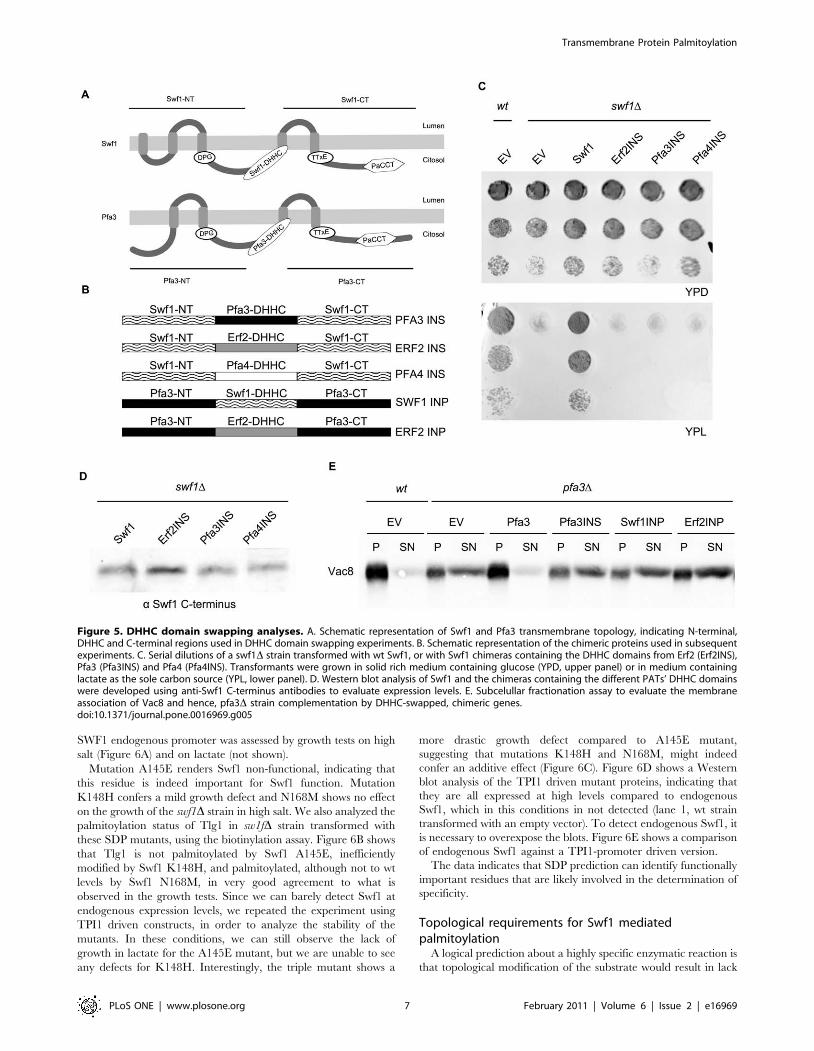
have its transmembrane topology unaltered (Figure 5A).
The functionality of these chimeras was assayed in growth tests
in medium containing lactate as sole carbon source. Figure 5C
shows that none of these chimeric genes is able to restore SWF1
function. Similar results were obtained in media containing
glucose and 0.85M NaCl (not shown).
To discard that lack of complementation stems from poor
chimeric-protein stability, we tested the steady state levels of these
proteins by Western blot, using antibodies raised against the C-
terminus of Swf1, which is shared by all the constructs. Figure 5D
shows that the chimeras are expressed at similar levels to that of wt
Swf1. It should be noted that all these constructs are driven by the
TPI1 promoter, so they are overexpressed, but still do not
complement. These results suggest that, for the palmitoylation of
Swf1 substrates, the DHHC domain does not simply behave as a
modular catalytic unit and that perhaps specificity determinants
are present within it.
Pfa3-mediated palmitoylation is less specific, since it can be
carried out by other PATs, such as Erf2 [23]. We generated
chimeras in which we replaced the Pfa3 DHHC domain with
those of Swf1 (Swf1 INP) and Erf2 (Erf2 INP), and used these
constructs in a pfa3D complementation assay.
Figure 5E shows that Vac8 soluble/membrane distribution in a
pfa3D strain is not affected by expression of these chimeras. This
indicates that Pfa3 DHHC domain cannot be replaced, neither by
Figure 3. Pfa4 function cannot be carried out by other yeastPATs. Serial dilutions of a wt strain or a pfa4D strain transformed withan empty vector (EV), or plasmids containing protein-A tagged Swf1,Erf2, Pfa3 or Pfa4 under the control of the GAL1 promoter.Transformants were grown in solid rich medium containing glucose(YPD, upper panel) or galactose (YPGal, medium panel) as carbonsource, or in YPGal supplemented with 75 mg/mL Calcofluor White(YPGal CW, lower panel).doi:10.1371/journal.pone.0016969.g003
Table 1. Highest scoring specificity determination positions.
Domain Name Domain position Groupsim score SWF1 residue PFA4 residue
DHHC 15 0.625792 K148 H92
DHHC 12 0.530785 A145 E89
DHHC 35 0.479647 N168 M112
TTXE 3 0.471412 N244 I200
DHHC 27 0.470615 A160 M104
DHHC 25 0.469355 L158 L102
DHHC 30 0.449352 H163 H107
DHHC 14 0.414847 S147 S91
DHHC 1 0.384384 I134 N78
DHHC 32 0.379334 I165 P109
PaCCT 2 0.375096 I322 P259
The table shows the highest scoring SDPs as determined by GROUPSIM. It also indicates the domain in which each SDP is located, the relative position within thatdomain and the identity of the amino acid in Swf1 and Pfa4.doi:10.1371/journal.pone.0016969.t001
Transmembrane Protein Palmitoylation
PLoS ONE | www.plosone.org 5 February 2011 | Volume 6 | Issue 2 | e16969
that of Swf1, which might have been expected, nor by that of Erf2,
which in the context of the full length protein is able to
palmitoylate Vac8. Pfa3 INS, the chimera in which Swf1 DHHC
domain is replaced by that of Pfa3, is also unable to complement
pfa3D. These results suggest that, besides specificity determinants
that might exist within DHHC domains, particularly for Swf1,
interactions between this domain and different regions of the
protein are required for function or proper folding.
Mutation of the highest scoring SDPs leads to lack ofSwf1 function
We mutated the highest scoring positions within the DHHC
domain of Swf1. Instead of a traditional alanine scanning
experiment, we changed these residues to the ones corresponding
to the same positions present in the DHHC domain of Pfa4,
resulting in the following changes: A145E; K148H, and N168M
(see Table 1). The functionality of the mutated proteins, driven by
Figure 4. Alignment of DHHC domains from selected model organisms and Groupsim SDP prediction results. Top three highestscoring positions in the GROUPSIM analysis are boxed. Residues are shaded according to conservation within orthologue groups. GROUPSIM scoresand overall conservation for each position are shown below as box plots. The more comprehensive alignment used to run the GROUPSIM analysis isshown in Figure S1.doi:10.1371/journal.pone.0016969.g004
Transmembrane Protein Palmitoylation
PLoS ONE | www.plosone.org 6 February 2011 | Volume 6 | Issue 2 | e16969
SWF1 endogenous promoter was assessed by growth tests on high
salt (Figure 6A) and on lactate (not shown).
Mutation A145E renders Swf1 non-functional, indicating that
this residue is indeed important for Swf1 function. Mutation
K148H confers a mild growth defect and N168M shows no effect
on the growth of the swf1D strain in high salt. We also analyzed the
palmitoylation status of Tlg1 in sw1fD strain transformed with
these SDP mutants, using the biotinylation assay. Figure 6B shows
that Tlg1 is not palmitoylated by Swf1 A145E, inefficiently
modified by Swf1 K148H, and palmitoylated, although not to wt
levels by Swf1 N168M, in very good agreement to what is
observed in the growth tests. Since we can barely detect Swf1 at
endogenous expression levels, we repeated the experiment using
TPI1 driven constructs, in order to analyze the stability of the
mutants. In these conditions, we can still observe the lack of
growth in lactate for the A145E mutant, but we are unable to see
any defects for K148H. Interestingly, the triple mutant shows a
more drastic growth defect compared to A145E mutant,
suggesting that mutations K148H and N168M, might indeed
confer an additive effect (Figure 6C). Figure 6D shows a Western
blot analysis of the TPI1 driven mutant proteins, indicating that
they are all expressed at high levels compared to endogenous
Swf1, which in this conditions in not detected (lane 1, wt strain
transformed with an empty vector). To detect endogenous Swf1, it
is necessary to overexpose the blots. Figure 6E shows a comparison
of endogenous Swf1 against a TPI1-promoter driven version.
The data indicates that SDP prediction can identify functionally
important residues that are likely involved in the determination of
specificity.
Topological requirements for Swf1 mediatedpalmitoylation
A logical prediction about a highly specific enzymatic reaction is
that topological modification of the substrate would result in lack
Figure 5. DHHC domain swapping analyses. A. Schematic representation of Swf1 and Pfa3 transmembrane topology, indicating N-terminal,DHHC and C-terminal regions used in DHHC domain swapping experiments. B. Schematic representation of the chimeric proteins used in subsequentexperiments. C. Serial dilutions of a swf1D strain transformed with wt Swf1, or with Swf1 chimeras containing the DHHC domains from Erf2 (Erf2INS),Pfa3 (Pfa3INS) and Pfa4 (Pfa4INS). Transformants were grown in solid rich medium containing glucose (YPD, upper panel) or in medium containinglactate as the sole carbon source (YPL, lower panel). D. Western blot analysis of Swf1 and the chimeras containing the different PATs’ DHHC domainswere developed using anti-Swf1 C-terminus antibodies to evaluate expression levels. E. Subcelullar fractionation assay to evaluate the membraneassociation of Vac8 and hence, pfa3D strain complementation by DHHC-swapped, chimeric genes.doi:10.1371/journal.pone.0016969.g005
Transmembrane Protein Palmitoylation
PLoS ONE | www.plosone.org 7 February 2011 | Volume 6 | Issue 2 | e16969

Figure 6. Mutation of the three top scoring SDPs in Swf1 to residues present in Pfa4. A. Serial dilutions of a swf1D strain transformed withan empty vector (EV), with wt SWF1, or with versions of Swf1 mutated in the highest scoring SDPs to the residues present in Pfa4 all driven by theendogenous promoter. Transformants were grown in solid rich medium containing glucose (YPD, upper panel) or in medium supplemented with0.85 M NaCl (YPD 0.85 M NaCl, lower panel). B. Western blot of membrane proteins from a swf1D strain transformed with an empty vector (EV), wtSwf1 or swf1 alleles mutated in the three highest scoring SDPs, to equivalent residues present in Pfa4. The samples were treated with Biotin-BMCC,which biotinylates unpalmitoylated cysteines, resulting in slower migration. Blot was probed using anti Tlg1 antibodies. C. Serial dilutions of a wtstrain or a swf1D strain transformed with an empty vector (EV), wt Swf1, or with versions of Swf1 mutated in the highest scoring SDPs to equivalentresidues present in Pfa4, driven by the TPI1 promoter. Swf1-3M contains all three highest scoring SDPs mutated. Swf1-DHHA mutant is assumed to becatalytically inactive. Transformants were grown in solid rich medium containing glucose (YPD, upper panel) or in medium supplemented with0.85 M NaCl (YPD 0.85 M NaCl, lower panel). D. Western blot analysis of membrane extracts from a wt strain transformed with an empty vector (EV) ora swf1D strain transformed with an empty vector or with TPI1 promoter driven wt or SDP Swf1 mutants. It was developed using anti Swf1 antibodies.Endogenous Swf1 is not detected under these conditions. A non-specific band is shown as a loading control (LC). E. Western blot analysis of a wtstrain transformed with an empty vector (EV), and a swf1D strain transformed with an empty vector (EV) or TPI1 promoter driven Swf1. The blot wasdeveloped using anti Swf1 antibodies, and overexposed to allow for detection of endogenous Swf1.doi:10.1371/journal.pone.0016969.g006
Transmembrane Protein Palmitoylation
PLoS ONE | www.plosone.org 8 February 2011 | Volume 6 | Issue 2 | e16969
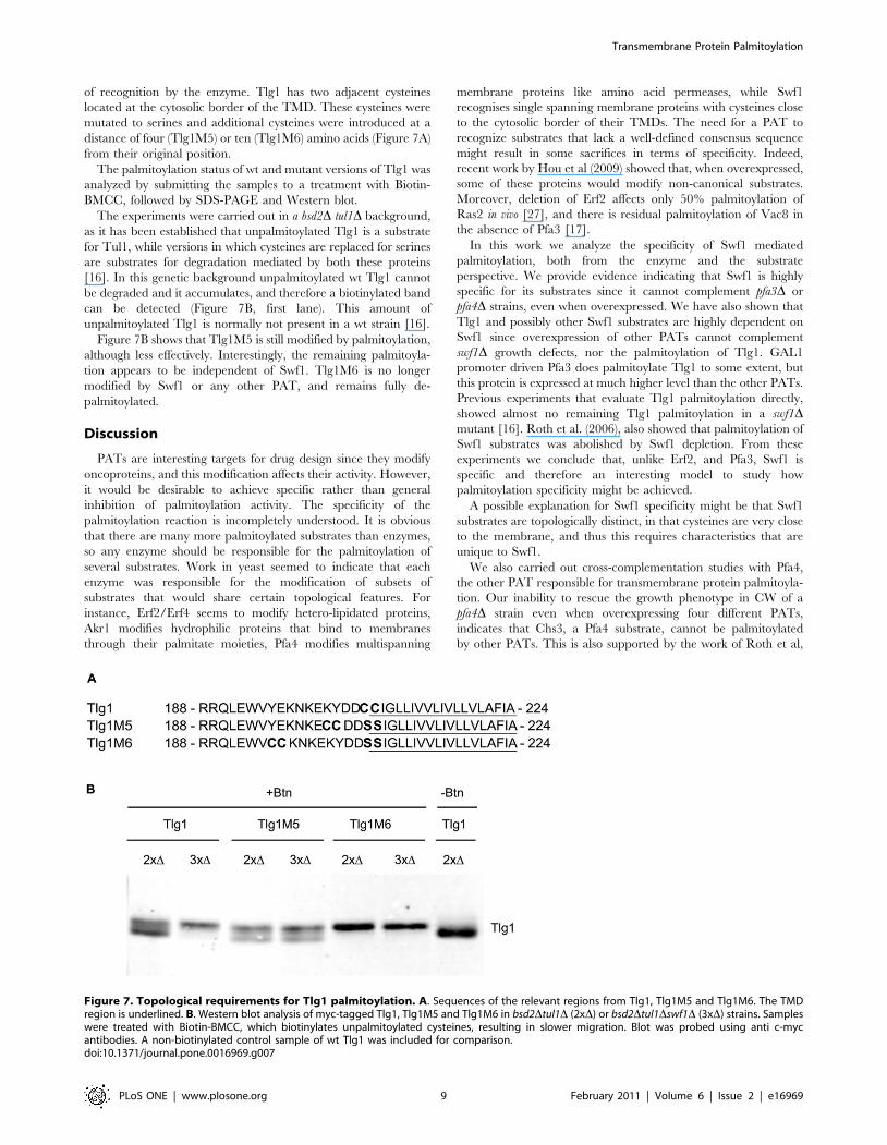
of recognition by the enzyme. Tlg1 has two adjacent cysteines
located at the cytosolic border of the TMD. These cysteines were
mutated to serines and additional cysteines were introduced at a
distance of four (Tlg1M5) or ten (Tlg1M6) amino acids (Figure 7A)
from their original position.
The palmitoylation status of wt and mutant versions of Tlg1 was
analyzed by submitting the samples to a treatment with Biotin-
BMCC, followed by SDS-PAGE and Western blot.
The experiments were carried out in a bsd2D tul1D background,
as it has been established that unpalmitoylated Tlg1 is a substrate
for Tul1, while versions in which cysteines are replaced for serines
are substrates for degradation mediated by both these proteins
[16]. In this genetic background unpalmitoylated wt Tlg1 cannot
be degraded and it accumulates, and therefore a biotinylated band
can be detected (Figure 7B, first lane). This amount of
unpalmitoylated Tlg1 is normally not present in a wt strain [16].
Figure 7B shows that Tlg1M5 is still modified by palmitoylation,
although less effectively. Interestingly, the remaining palmitoyla-
tion appears to be independent of Swf1. Tlg1M6 is no longer
modified by Swf1 or any other PAT, and remains fully de-
palmitoylated.
Discussion
PATs are interesting targets for drug design since they modify
oncoproteins, and this modification affects their activity. However,
it would be desirable to achieve specific rather than general
inhibition of palmitoylation activity. The specificity of the
palmitoylation reaction is incompletely understood. It is obvious
that there are many more palmitoylated substrates than enzymes,
so any enzyme should be responsible for the palmitoylation of
several substrates. Work in yeast seemed to indicate that each
enzyme was responsible for the modification of subsets of
substrates that would share certain topological features. For
instance, Erf2/Erf4 seems to modify hetero-lipidated proteins,
Akr1 modifies hydrophilic proteins that bind to membranes
through their palmitate moieties, Pfa4 modifies multispanning
membrane proteins like amino acid permeases, while Swf1
recognises single spanning membrane proteins with cysteines close
to the cytosolic border of their TMDs. The need for a PAT to
recognize substrates that lack a well-defined consensus sequence
might result in some sacrifices in terms of specificity. Indeed,
recent work by Hou et al (2009) showed that, when overexpressed,
some of these proteins would modify non-canonical substrates.
Moreover, deletion of Erf2 affects only 50% palmitoylation of
Ras2 in vivo [27], and there is residual palmitoylation of Vac8 in
the absence of Pfa3 [17].
In this work we analyze the specificity of Swf1 mediated
palmitoylation, both from the enzyme and the substrate
perspective. We provide evidence indicating that Swf1 is highly
specific for its substrates since it cannot complement pfa3D or
pfa4D strains, even when overexpressed. We have also shown that
Tlg1 and possibly other Swf1 substrates are highly dependent on
Swf1 since overexpression of other PATs cannot complement
swf1D growth defects, nor the palmitoylation of Tlg1. GAL1
promoter driven Pfa3 does palmitoylate Tlg1 to some extent, but
this protein is expressed at much higher level than the other PATs.
Previous experiments that evaluate Tlg1 palmitoylation directly,
showed almost no remaining Tlg1 palmitoylation in a swf1Dmutant [16]. Roth et al. (2006), also showed that palmitoylation of
Swf1 substrates was abolished by Swf1 depletion. From these
experiments we conclude that, unlike Erf2, and Pfa3, Swf1 is
specific and therefore an interesting model to study how
palmitoylation specificity might be achieved.
A possible explanation for Swf1 specificity might be that Swf1
substrates are topologically distinct, in that cysteines are very close
to the membrane, and thus this requires characteristics that are
unique to Swf1.
We also carried out cross-complementation studies with Pfa4,
the other PAT responsible for transmembrane protein palmitoyla-
tion. Our inability to rescue the growth phenotype in CW of a
pfa4D strain even when overexpressing four different PATs,
indicates that Chs3, a Pfa4 substrate, cannot be palmitoylated
by other PATs. This is also supported by the work of Roth et al,
Figure 7. Topological requirements for Tlg1 palmitoylation. A. Sequences of the relevant regions from Tlg1, Tlg1M5 and Tlg1M6. The TMDregion is underlined. B. Western blot analysis of myc-tagged Tlg1, Tlg1M5 and Tlg1M6 in bsd2Dtul1D (2xD) or bsd2Dtul1Dswf1D (3xD) strains. Sampleswere treated with Biotin-BMCC, which biotinylates unpalmitoylated cysteines, resulting in slower migration. Blot was probed using anti c-mycantibodies. A non-biotinylated control sample of wt Tlg1 was included for comparison.doi:10.1371/journal.pone.0016969.g007
Transmembrane Protein Palmitoylation
PLoS ONE | www.plosone.org 9 February 2011 | Volume 6 | Issue 2 | e16969

(2006), in which they show that other Pfa4 substrates, such as
amino acid permeases, are not palmitoylated when the PFA4 gene
is deleted. It has been shown that high overexpression of Pfa4 is
able to complement some Pfa3 and Akr1 deletion-associated
phenotypes [23]. Therefore, Pfa4 might be more promiscuous
than Swf1, however, the palmitoylation of its substrates is not. The
inability of Pfa4 to complement a swf1D strain might be due to
characteristics of Swf1 substrates.
Overall, we suggest that most transmembrane protein palmi-
toylation in yeast is highly specific. It has been reported that Sna4,
an integral membrane protein, is palmitoylated by Akr1 [15]. The
specificity of this reaction has not been addressed.
One aspect that should be considered in the interpretation of
cross-complementation studies is the subcellular localization of the
enzymes and substrates involved. Pfa4 and Erf2 are localized at the
ER [27,30]. There is some controversy regarding the localization of
Swf1 [35,16]. We have been unable to visualize endogenous Swf1
under the microscope, but when expressed from TPI1 or GAL1
promoter, it is mostly found in the ER [16, our unpublished results].
Couve et al. (1995) showed that palmitoylation of Snc1, a Swf1
substrate, occurs in the ER [36]. Pfa3 is localized at the vacuolar
membrane [17]. Localization of PATs might be less relevant when
analyzing peripheral membrane proteins that are able to probe all
membranes until they undergo palmitoylation, this issue has been
thoroughly addressed [23,29]. For integral membrane proteins, the
impossibility of co-localizing in the same membrane with a certain
PAT may preclude modification, regardless of substrate-recognition
mediated specificity. This could be a caveat only in Pfa3
complementation experiments of both Swf1 and Pfa4. Interestingly,
when Pfa3 is highly overexpressed it modifies Tlg1 to a small degree,
indicating that in principle it has access to Swf1 substrates, possibly
along Pfa3 trafficking to the vacuole.
We performed extensive sequence analyses and comparison of
fungal PATs. We found that there is sufficient divergence between
subgroups and conservation within a subgroup to yield a few high
scoring specificity determination points using GROUPSIM
software. A somewhat unexpected finding is that many of these
predicted specificity determinants lie within the DHHC domain,
since this domain is highly conserved between all PATs studied
and between different species. It should be emphasized that
GROUPSIM predicts SDPs for the alignment, not just for Swf1,
so some of these positions may be important specificity
determinants for other PATs.
One question that arises with this prediction is just how valid it
is to separate PATs in specificity groups when at least some of
them have overlapping specificities. Both the uncertainty in the
separation of subgroups, and the lack of experimental validation of
SDP prediction [34], prompted the construction of chimeric genes
in which Swf1 DHHC domain was replaced by that of Pfa3, Erf2
and Pfa4. Although the evaluation of chimeric genes requires
careful interpretation, complementation of Swf1 by any one of the
chimeras was plausible, and would have made us re-examine our
strategy. These experiments also ruled out the simplistic view that
the DHHC domain solely represents the catalytic unit, while the
rest of the protein determines specificity.
SDP prediction methods require at least some degree of
conservation. The ankyrin repeats present in Akr1 orthologues,
the N-termini of Swf1 and Erf2 upstream TMD1 and all C-
terminal regions downstream the PaCCT motif, are simply not
present in the other subgroups of PATs and therefore they cannot
be properly aligned. These regions are also likely responsible for
specificity, or could also be involved in regulation. Indeed when a
DHHC3 is fused to the ankyrin repeats of HIP14, it can modify
HIP14 substrates [28].
We have mutated three high scoring positions in the DHHC
domain of Swf1, to residues present in the DHHC domain of Pfa4.
One of these mutations, A145E, resulted in complete lack of
function, the second, K148H, resulted in an intermediate
phenotype. In addition to the way in which they were predicted,
an argument in favor of the involvement of Swf1 A145 and
possibly K148 in specificity determination is that we have replaced
them with residues that are present in an active PAT, in a
sequence context of very high conservation (see Figure 4), and yet
they result in lack of function. It should be noted that predicted
SDPs have very low conservation scores (Figure 4) and yet
mutation of at least one of them results in lack of function. On the
other hand, mutations in several residues that are highly conserved
across the PAT family, result in normal PAT activity (i.e C117,
C123, and even D131 in Pfa3, Hou et al 2009 [23], W205 and
N214 in Erf2, Mitchell et al, 2010 [37]), which suggests that SDP
prediction is a useful tool to identify functionally important
residues in PATs.
A more definitive proof indicating that predicted SDPs are indeed
involved in specificity, would be to swap specificity of a certain PAT
from one kind of substrate to another by mutating the most relevant
SDPs. However, it is difficult to predict how many SDPs should be
mutated, and also, the regions specific to certain PATs discussed
above surely contribute significantly to specificity, making these
experiments highly unlikely to succeed. We nevertheless attempted
to complement a pfa4D strain using Swf1 constructs bearing the
single point mutations, the triple mutant and a construct with two
additional SDPs mutated to the corresponding residues in Pfa4, and
neither complemented (not shown).
Finally, we analyzed the modification of Tlg1 mutants in which
the cysteines have been moved away from the cytosolic border of
the TMD. We show that when cysteines are moved four amino
acids from their original position (see diagram in Figure 8A),
palmitoylation decreases significantly. In this mutant (Tlg1M5) the
sequence context of the cysteines is almost identical to wt Tlg1.
When the cysteines are moved even further, ten residues away
(Tlg1M6), they are no longer palmitoylated by Swf1 or any other
PAT. We cannot tell whether this is due to the distance from the
membrane or a change in the amino acid context, but we can
certainly state that there are constraints for the modification of
these cysteines by Swf1.
Topological requirements for the palmitoylation of the H1
subunit of the asialoglycoprotein receptor have been studied in
mammalian cells. Sequences surrounding the cysteines are not
critical, but moving them 30 residues away from the membrane
results in lack of palmitoylation [38].
It has been postulated that palmitoylation of peripheral membrane
proteins occurs exclusively in the Golgi apparatus, and that the
responsible machinery lacks specificity altogether, rapidly processing
any available target protein. For integral membrane proteins in yeast,
however, palmitoylation appears to take place at the ER [30,36] and
some of these proteins, like the SNARE Sso1, remain palmitoylated
and localized to the plasma membrane without the need of a cycle
through the ER [39]. In mutants where Swf1 is absent, target
SNAREs move through the yeast Golgi to reach their destination but
remain completely non-palmitoylated. Some mammalian transmem-
brane SNARES are palmitoylated [40] and it would be of great
interest to study if their modification is also highly specific.
Materials and Methods
Ethics statementAnimal handling was performed according to the standards
stated in the Guide to the Care and Use of Experimental Animals
Transmembrane Protein Palmitoylation
PLoS ONE | www.plosone.org 10 February 2011 | Volume 6 | Issue 2 | e16969

published by the Canadian Council on Animal Care and approved
by the local institution (Facultad de Ciencias Quımicas, Uni-
versidad Nacional de Cordoba, Argentina, Exp. Nu 15-99-4042).
Plasmids and strainsThe strains used in the present study are BY4742 from the
EUROSCARF consortium, or derivatives containing complete
deletions of SWF1, PFA3 and PFA4. SWF1, TLG1 [16] and PFA3
[24] plasmids have been described previously. ERF2 and PFA4
coding sequences were amplified from Euroscarf BY4742 genomic
DNA, using oligos erf2 01 and 02 and pfa4 01 and 02 respectively
and cloned in an YCplac33 based vector containing a TPI1
promoter and PGK1 terminator (see Table S1 for a list of all
oligos). For GAL1 driven constructs, PATs coding sequences were
amplified using oligos that delete the stop codon and cloned in
pJV95 vector as fusions to the IgG binding domain of protein A.
bsd2D tul1D strain has been described previously [41]. On this
strain, SWF1 ORF was deleted by gene replacement using S. pombe
HIS5 as a selection marker (oligos swf1 KO 5’ and swf1 KO 39) to
generate a bsd2D tul1D swf1D strain.
Chimeric genes constructionWe considered N-terminal and C-terminal regions of PATs the
regions right upstream and downstream the DHHC domains
defined as in Figure 6.
To generate chimeras Pfa3INS, Pfa4INS and Erf2INS, a TPI1
driven SWF1 plasmid, lacking the whole DHHC domain (pJV362)
was built by PCR amplification of the N- and C- terminal coding
regions. These fragments were ligated through an added XhoI site,
using oligos swf1 01 and 42 and oligos swf1 02 and 43. The
chimeras were generated in vivo by gap repair, using PCR amplified
fragments comprising the DHHC of Pfa3 (oligos pfa3 INS 3’ and
pfa3 INS 5’), Pfa4 (oligos pfa4 INS3’ and pfa4 INS 5’) or Erf2 (oligos
erf2 INS3’ and erf2 INS 5’). These oligos contained aprox. 50
nucleotides of the sequences flanking Swf1 DHHC domain.
The sequence of PFA3 upstream the DHHC domain was
amplified using oligos pfa3 01 and pfa3 10. This band was cloned
BamHI-XhoI in pJV 362, replacing SWF1 N-terminal coding
region and generating plasmid pJV368. The sequence of PFA3
downstream the DHHC domain was amplified using oligos pfa3
09 and pfa3 08. This band was cloned XhoI-PstI in pJV368
generating plasmid pJV367. To generate chimeras in which the
DHHC domain of Pfa3 was replaced, plasmid pJV367 was
digested with XhoI and used in gap repair experiments with PCR
amplified fragments comprising the DHHC domain of Swf1
(oligos swf1 INP 5’ and swf1 INP3’) or of Erf2 (oligos erf2 INP 5’
and erf2 INP 3’). A diagram of the domain structure of the
chimeric proteins is shown in Figure 5A.
All constructs were rescued from yeast and verified by DNA
sequencing.
Swf1 point-mutants constructionDNA molecules corresponding to the DHHC domain of Swf1
encoding the desired mutations were purchased from GeneScript,
USA. The fragments were excised from the plasmid using flanking
restriction sites and used in gap repair experiments with plasmid
pJV362. The constructs were rescued from yeast and verified by
DNA sequencing.
Tlg1 mutants constructionTlg1M5 contains the following mutations: C205S; C206S;
K201C and Y202C. Tlg1 M6 contains the following mutations:
C205S; C206S; Y195C and E196C.
For TlgM5 and Tlg1M6, fragments of Tlg1 were amplified
from pJV130 (Tlg1M2) [16], using oligos Tlg1 05 ad Tlg1 06
respectively and oligo M13R. Oligo Tlg1 05 replaces K201 and
Y202 for cysteines and Tlg1 06 replaces Y195 and E196 for
cysteines. The PCR fragments were digested with AccI-BamH1 and
cloned into a Bluescript based vector containing Tlg1 coding
sequence. The mutants were then moved to a pRS316 based
vector encoding a triple c-myc tag in the 59region.
Biotinylation assaysBiotinylation assays were performed as described in [16] but
after treating with resuspension buffer, samples were diluted with
sample buffer and subjected to SDS-PAGE and Western blot. Anti
c-myc antibodies were from Santa Cruz and Biotin-BMCC was
from PIERCE.
BioinformaticsS. cerevisiae PATs were used to query the ORTHOMCL
database (release 4) [42], and 441 protein sequences corresponding
to SWF1, ERF2, PFA3, PFA4, PFA5 and AKR1 orthologue
groups were downloaded. MUSCLE (v3.7) [43] was used to align
each group. Unalignable and poor quality sequences were
removed from the dataset. PAT subgroup alignments were
manually curated, and refined using UGENE (http://genome.
unipro.ru) and Jalview [44]. Subsequently, these alignments were
aligned using the profile-profile alignment option in MUSCLE.
Final manual curation of the 303 sequences alignment was
performed using Jalview. GROUPSIM [34] was used to predict
specificity-determination positions using default parameters except
for maximum group gap (set to 0,1).
Anti- Swf1 antibody production and purificationA GST fusion protein of the last 100 amino acids of Swf1 (GST-
Swf1CT), corresponding to the cytosolic C-terminus, was purified
from E. coli BL21, according to the GST-fusion handbook protocol
(Amersham Biosciences). 500 mg GST-Swf1CT protein in 500 ml
of PBS were mixed with 500 ml of Freund’s complete adjuvant and
injected intradermically into New Zealand white rabbits. Booster
injections containing 125 mg of GST-Swf1CT and Freund’s
incomplete adjuvant were administered 4 weeks after the initial
injection. Bleeds were collected one week after each injection and
screened for immuno-reactivity against Swf1 in whole cell lysates
of wt yeast. swf1D strain was used as a negative control. Total
serum was purified by adsorption to 1% ketonic powder from
swf1D strain, prepared according to [45].
Protein electrophoresis and Western blotsProtein samples for GFP-Tlg1 detection were prepared
according to [46]. For the detection of the different PATs and
Swf1 chimeric proteins, membrane enriched fractions were used to
facilitate detection. 30 ODs of swf1D strains transformed with
appropriate plasmids were harvested by centrifugation and
resuspended in 300 ml lysis buffer (PBS, EDTA 2mM, PMSF
1mM and protease inhibitor cocktail (PIC). 200 ml of glass beads
were added and the samples were placed in a cell disruptor
(GENIE) in a cold room. The samples were submitted to four
pulses of agitation of two minutes duration each, allowing one
minute on ice between each pulse. 200 ml lysis buffer were added
and the sample was centrifuged at 300 g for 3 min at 4uC. The
supernatant was transferred to a clean tube and centrifuged at
17000 g for 20 min at 4uC. The resulting membrane enriched
fraction was re-suspended in 500 ml lysis buffer plus 1% Triton X-
100 and incubated on ice for 15 min. The sample was re-
Transmembrane Protein Palmitoylation
PLoS ONE | www.plosone.org 11 February 2011 | Volume 6 | Issue 2 | e16969
centrifuged at 13000 rpm for 10 min, and subjected to SDS-
PAGE.
The blots were probed using secondary antibodies coupled to
either IRdye 680 or IRdye 800 (LICOR bioscience, UK) at 1/
20000 dilution, and then scanned using an Odyssey Infrared
imager (LICOR bioscience, UK).
Subcellular fractionation30 ODs of yeast strains transformed with appropriate plasmids
were spheroplasted with zymolyase. Spheroplasts were resus-
pended in 800 ml hyposmotic lysis buffer (20 mM Tris-HCl
pH 7.4, 2 mM EDTA, 1 mM PMSF, PIC) (Sigma). Lysis was
aided by resuspension with a small gauge needle and a syringe.
The lysate was centrifuged twice for 5 minutes at 400 g at 4uC, to
remove unbroken cells, and the supernatant was then centrifuged
at 17000 g for 20 min at 4uC. The pellets were resuspended in
800 ml lysis buffer. Equal volumes of the pellet and supernatant
fraction were subjected to SDS-PAGE and Western blot using
polyclonal anti Vac8 antibodies, kindly supplied by Dr. Christian
Ungermann. The intensity of the bands was quantified using the
Odyssey Infrared Imager application software version 2.1.
Supporting Information
File S1 An alignment of alignments comprising six yeast PATs
and their orthologue groups (as defined in ORTHOMCL).
(TXT)
Table S1 List of oligonucleotides used troughout this work.
(DOC)
Table S2 GROUPSIM scores for all positions in the final
alignment.
(XLS)
Acknowledgments
We would like to thank Dr. Cecilia Sampedro for technical assistance. We
thank Dr. Jose L. Barra and Dr. Jose Luis Daniotti for critical reading of
the manuscript, and Dr. Hugo Maccioni for space and constant support.
Author Contributions
Conceived and designed the experiments: JVT AGM. Performed the
experiments: AGM SCR JVT. Analyzed the data: JVT AGM RQ. Wrote
the paper: JVT. Bionformatic analyses: RQ.
References
1. Berthiaume L, Resh MD (1995) Biochemical characterization of a palmitoylacyltransferase activity that palmitoylates myristoylated proteins. J Biol Chem
270: 22399–22405.
2. Bijlmakers MJ, Marsh M (2003) The on-off story of protein palmitoylation.
Trends Cell Biol 13: 32–42.
3. DeMar Jr. JC, Anderson RE (1997) Identification and quantitation of the fatty
acids composing the CoA ester pool of bovine retina, heart, and liver. J BiolChem 272: 31362–31368.
4. Xue L, Gollapalli DR, Maiti P, Jahng WJ, Rando RR (2004) A palmitoylation
switch mechanism in the regulation of the visual cycle. Cell 117: 761–771.
5. Smotrys JE, Linder ME (2004) Palmitoylation of intracellular signaling proteins:
Regulation and Function. Annual Review of Biochemistry 73: 559–587.
6. Washbourne P (2004) Greasing transmission: palmitoylation at the synapse.Neuron 44: 901–902.
7. Kang R, Wan J, Arstikaitis P, Takahashi H, Huang K, et al. (2008) Neuralpalmitoyl-proteomics reveals dynamic synaptic palmitoylation. Nature 456:
904–909.
8. Prescott GR, Gorleku OA, Greaves J, Chamberlain LH (2009) Palmitoylation of
the synaptic vesicle fusion machinery. J Neurochem.
9. Huang K, El-Husseini A (2005) Modulation of neuronal protein trafficking andfunction by palmitoylation. Curr Opin Neurobiol 15: 527–535.
10. Fukata Y, Fukata M. Protein palmitoylation in neuronal development andsynaptic plasticity. Nat Rev Neurosci 11: 161–175.
11. Putilina T, Wong P, Gentleman S (1999) The DHHC domain: a new highly
conserved cysteine-rich motif. Mol Cell Biochem 195: 219–226.
12. Lobo S, Greentree WK, Linder ME, Deschenes RJ (2002) Identification of a Ras
palmitoyltransferase in Saccharomyces cerevisiae. J Biol Chem 277:41268–41273.
13. Roth AF, Feng Y, Chen L, Davis NG (2002) The yeast DHHC cysteine-richdomain protein Akr1p is a palmitoyl transferase. J Cell Biol 159: 23–28.
14. Mitchell DA, Vasudevan A, Linder ME, Deschenes RJ (2006) Protein
palmitoylation by a family of DHHC protein S-acyltransferases. J Lipid Res
47: 1118–1127.
15. Roth AF, Wan J, Bailey AO, Sun B, Kuchar JA, et al. (2006) Global analysis ofprotein palmitoylation in yeast. Cell 125: 1003–1013.
16. Valdez-Taubas J, Pelham H (2005) Swf1-dependent palmitoylation of theSNARE Tlg1 prevents its ubiquitination and degradation. Embo J 24:
2524–2532.
17. Smotrys JE, Schoenfish MJ, Stutz MA, Linder ME (2005) The vacuolar DHHC-
CRD protein Pfa3p is a protein acyltransferase for Vac8p. J Cell Biol 170:1091–1099.
18. Fukata M, Fukata Y, Adesnik H, Nicoll RA, Bredt DS (2004) Identification of
PSD-95 palmitoylating enzymes. Neuron 44: 987–996.
19. Ducker CE, Stettler EM, French KJ, Upson JJ, Smith CD (2004) Huntingtin
interacting protein 14 is an oncogenic human protein: palmitoyl acyltransferase.Oncogene 23: 9230–9237.
20. Fang C, Deng L, Keller CA, Fukata M, Fukata Y, et al. (2006) GODZ-mediated
palmitoylation of GABA(A) receptors is required for normal assembly and
function of GABAergic inhibitory synapses. J Neurosci 26: 12758–12768.
21. Hancock JF, Magee AI, Childs JE, Marshall CJ (1989) All ras proteins arepolyisoprenylated but only some are palmitoylated. Cell 57: 1167–1177.
22. Mill P, Lee AW, Fukata Y, Tsutsumi R, Fukata M, et al. (2009) Palmitoylation
regulates epidermal homeostasis and hair follicle differentiation. PLoS Genet 5:
e1000748.
23. Hou H, John Peter AT, Meiringer C, Subramanian K, Ungermann C (2009)
Analysis of DHHC acyltransferases implies overlapping substrate specificity and
a two-step reaction mechanism. Traffic 10: 1061–1073.
24. Gonzalez Montoro A, Quiroga R, Maccioni HJ, Valdez Taubas J (2009) A novel
motif at the C-terminus of palmitoyltransferases is essential for Swf1 and Pfa3
function in vivo. Biochem J 419: 301–308.
25. Nadolski MJ, Linder ME (2009) Molecular recognition of the palmitoylation
substrate VAC8 by its palmitoyltransferase PFA3. J Biol Chem 283:
17720–17730.
26. Greaves J, Prescott GR, Fukata Y, Fukata M, Salaun C, et al. (2009) The
hydrophobic cysteine-rich domain of SNAP25 couples with downstream residues
to mediate membrane interactions and recognition by DHHC palmitoyl
transferases. Mol Biol Cell 20: 1845–1854.
27. Bartels DJ, Mitchell DA, Dong X, Deschenes RJ (1999) Erf2, a novel gene
product that affects the localization and palmitoylation of Ras2 in Saccharo-
myces cerevisiae. Mol Cell Biol 19: 6775–6787.
28. Huang K, Sanders S, Singaraja R, Orban P, Cijsouw T, et al. (2009) Neuronal
palmitoyl acyl transferases exhibit distinct substrate specificity. Faseb J.23:
2605–2615.
29. Rocks O, Gerauer M, Vartak N, Koch S, Huang ZP, et al. The palmitoylation
machinery is a spatially organizing system for peripheral membrane proteins.
Cell 141: 458–471.
30. Lam KK, Davey M, Sun B, Roth AF, Davis NG, et al. (2006) Palmitoylation by
the DHHC protein Pfa4 regulates the ER exit of Chs3. J Cell Biol 174: 19–25.
31. Wang YX, Catlett NL, Weisman LS (1998) Vac8p, a vacuolar protein with
armadillo repeats, functions in both vacuole inheritance and protein targeting
from the cytoplasm to vacuole. J Cell Biol 140: 1063–1074.
32. Warringer J, Ericson E, Fernandez L, Nerman O, Blomberg A (2003) High-
resolution yeast phenomics resolves different physiological features in the saline
response PNAS 100: 15724–15729.
33. Rausell A JD, Valencia A (2010) Protein interactions and ligand binding: from
protein subfamilies to functional specificity. PNAS 107: 1995–2000.
34. Capra JA, Singh M (2008) Characterization and prediction of residues
determining protein functional specificity. Bioinformatics 24: 1473–1480.
35. Dighe SA, Kozminski KG (2008) Swf1p, a member of the DHHC-CRD family
of palmitoyltransferases, regulates the actin cytoskeleton and polarized secretion
independently of its DHHC motif. Mol Biol Cell 19: 4454–4468.
36. Couve A, Protopopov V, Gerst J (1995) Yeast Synaptobrevin Homologs are
Modified Posttranslationally by the Addition of Palmitate. PNAS 92:
5987–5991.
37. Mitchell DA, Mitchell G, Ling Y, Budde C, Deschenes RJ (2010) Mutational
analysis of Saccharomyces cerevisiae Erf2 reveals a two-step reaction mechanism
for protein palmitoylation by DHHC enzymes. J Biol Chem 285: 38104–38114.
38. Yik JHN, Weigel PH (2002) The Position of Cysteine Relative to the
Transmembrane Domain Is Critical for Palmitoylation of H1, the Major
Subunit of the Human Asialoglycoprotein Receptor. J Biol Chem 277:
47305–47312.
Transmembrane Protein Palmitoylation
PLoS ONE | www.plosone.org 12 February 2011 | Volume 6 | Issue 2 | e16969

39. Valdez-Taubas J, Pelham HR (2003) Slow diffusion of proteins in the yeast
plasma membrane allows polarity to be maintained by endocytic cycling. CurrBiol 13: 1636–1640.
40. He Y, Linder ME (2008) Differential palmitoylation of the endosomal SNAREs
syntaxin 7 and syntaxin 8. J Lipid Res.41. Hettema EH, Valdez-Taubas J, Pelham HR (2004) Bsd2 binds the ubiquitin
ligase Rsp5 and mediates the ubiquitination of transmembrane proteins. Embo J23: 1279–1288.
42. Chen F, Mackey AJ, Vermunt JK, Roos DS (2007) Assessing performance of
orthology detection strategies applied to eukaryotic genomes. PLoS One 2: e383.
43. Edgar RC (2004) MUSCLE: multiple sequence alignment with high accuracy
and high throughput. Nucleic Acids Res 32: 1792–1797.
44. Clamp M, Cuff J, Searle SM, Barton GJ (2004) The Jalview Java alignment
editor. Bioinformatics 20: 426–427.
45. Harlow L (1988) Antibodies: a laboratory manual.
46. Volland C, Urban-Grimal D, Geraud G, Haguenauer-Tsapis R (1994)
Endocytosis and degradation of the yeast uracil permease under adverse
conditions. J Biol Chem 269: 9833–9841.
Transmembrane Protein Palmitoylation
PLoS ONE | www.plosone.org 13 February 2011 | Volume 6 | Issue 2 | e16969