Transmembrane Protein 18 Enhances the Tropism of Neural Stem Cells for Glioma Cells
11
Transmembrane Protein 18 Enhances the Tropism of Neural Stem Cells for Glioma Cells Jaana Jurvansuu, 1 Ying Zhao, 1,2 Doreen S.Y. Leung, 1 Jerome Boulaire, 1 Yuan Hong Yu, 3,4 Sohail Ahmed, 3,4 and Shu Wang 1,2 1 Institute of Bioengineering and Nanotechnology; Departments of 2 Biological Sciences and 3 Physiology, National University of Singapore; 4 Institute of Medical Biology, Singapore, Singapore Abstract The failure of current glioma therapies is mainly due to the ability of the tumor cells to invade extensively the surrounding healthy brain tissue, hence escaping localized treatments. Neural stem cells (NSC) are able to home in on tumor foci at sites distant from the main tumor mass, possibly enabling treatment of scattered glioma clusters. To make the strategy more effective, we performed a cDNA expression library screening to identify the candidate genes that once overex- pressed would enhance the tropism of NSCs for gliomas. Here, we show that a previously unannotated gene, the one encoding transmembrane protein 18 (TMEM18), is one such gene. Overexpression of TMEM18 was seen in the current study to provide NSCs and neural precursors an increased migration capacity toward glioblastoma cells in vitro and in the rat brain. Functional inactivation of the TMEM18 gene resulted in almost complete loss of the migration activity of these cells. Thus, TMEM18 is a novel cell migration modulator. Overexpression of this protein could be favorably used in NSC- based glioma therapy. [Cancer Res 2008;68(12):4614–22] Introduction Glioma cells misregulate the expression of growth factors, proteases, and extracellular matrix and cell surface proteins to gain their devastating invasion capacity (1, 2). Localized treatments are thus inefficient and comprehensive treatments are too damaging to the delicate brain. A solution is to find a treatment that can specifically locate the tumor cells. Neural stem cells (NSC) and neural precursor cells (NPC) have an intrinsic tropism for sites of brain injuries, including gliomas, and as shown first by Benedetti and colleagues (3) and Aboody and colleagues (4), engrafted primary and immortalized NSCs can be used in gene therapy of gliomas in animal models. These engrafted stem cells have been shown to spread through the existing migratory pathways in healthy brain as well as nontypical routes when gliomas are present (4, 5). Besides primary and immortalized NSCs/NPCs, embryonic stem cell–derived NPCs seem to have the same aptitude for glioma cell tracking (6). Moreover, NSCs are able to locate not only gliomas but also tumors of a nonneural origin, suggesting that there exist common regulators of cell trafficking probably composed of secreted factors from a tumor site and receptors present on NSCs (7, 8). Candidate signals to attract NSCs to the sites of brain injuries and tumors have been studied. Among them are cytokines released from the immunoreactive microglial cells of the brain during inflammation (9), which also provide cues for NSC migration in brain development (10). Stromal cell–derived factor-1 (SDF-1) chemokine can attract NSCs too. When its receptor CX chemokine receptor 4 (CXCR4) is blocked, SDF-1 hinders the NSC migration to the site of injury (9, 11, 12). In addition, chemokine monocyte chemoattractant protein-1, the expression of which can be induced by tumor necrosis factor-a, can activate migration of NSCs (13). Cytokine stem cell factor, expressed by glioma cell lines and overexpressed in neurons at the sites of brain injury, is another possible contributing attractant for NSCs (14–16). Similar to cytokines and chemokines, growth factor–mediated signaling [e.g., vascular endothelial growth factor and epidermal growth factor (EGF) receptor] has been shown to regulate NSC and NPC migration (17, 18). Glioma invasion depends largely on the ability of the cell to modify the extracellular matrix, and interestingly, the extracellular matrix secreted from glioma cell lines is able to promote NSC motility (19). The picture emerging from the above studies seems to support a model of the complex interaction of several factors in regulating NSC migration toward tumors. We hypothesized that other regulators are likely to exist and their genes can be identified through expression cloning based on gene function in influencing the tropism of NSCs toward glioma cells. We were particularly interested in the molecules that once overexpressed in NSCs and NPCs are able to enhance cell migration toward gliomas, as the manipulation of the expression of these molecules could then facilitate the use of NSCs/NPCs as gene therapy vectors to reach scattered glioma cells. We used Boyden chambers in the current study to select the cells that were primed by gene transfer of a tumor cDNA expression library. A novel gene encoding transmem- brane protein 18 (TMEM18) emerged from the screen and was selected for extensive characterizations. Materials and Methods Cells. NT2, U87MG, H4, and NIH3T3 cell lines were purchased from the American Type Culture Collection and HEK293FT cells were purchased from Invitrogen. All the cell lines were maintained in DMEM supplemented with 10% FCS (Life Technologies), penicillin-streptomycin (Life Technolo- gies), normoxin (Invivogen), and nonessential amino acids (Life Techno- logies). C17.2 cells were kindly provided by Prof. E. Arenas (Department of Medical Biochemistry and Biophysics, Karolinska Institute, Stockholm, Sweden) and maintained in DMEM supplemented with 10% FCS, 5% horse serum (Life Technologies), penicillin-streptomycin, normoxin, and nones- sential amino acids. The NIH Human Embryonic Stem Cell Registry listed human embryonic stem (hES) cell line, HES-1, and its feeder cell K 4 mouse embryonic fibroblasts were obtained from ES Cell International (ESI), Singapore. The hES cells were amplified and maintained according to the Note: Supplementary data for this article are available at Cancer Research Online (http://cancerres.aacrjournals.org/). J. Jurvansuu and Y. Zhao contributed equally to this work. Requests for reprints: Shu Wang, Institute of Bioengineering and Nanotechnology, Singapore 138669, Singapore. Phone: 65-6824-7105; Fax: 65-6478-9083; E-mail: swang@ ibn.a-star.edu.sg. I2008 American Association for Cancer Research. doi:10.1158/0008-5472.CAN-07-5291 Cancer Res 2008; 68: (12). June 15, 2008 4614 www.aacrjournals.org Research Article Research. on February 12, 2016. © 2008 American Association for Cancer cancerres.aacrjournals.org Downloaded from Research. on February 12, 2016. © 2008 American Association for Cancer cancerres.aacrjournals.org Downloaded from Research. on February 12, 2016. © 2008 American Association for Cancer cancerres.aacrjournals.org Downloaded from
Transcript of Transmembrane Protein 18 Enhances the Tropism of Neural Stem Cells for Glioma Cells

Transmembrane Protein 18 Enhances the Tropism of Neural
Stem Cells for Glioma Cells
Jaana Jurvansuu,1Ying Zhao,
1,2Doreen S.Y. Leung,
1Jerome Boulaire,
1Yuan Hong Yu,
3,4
Sohail Ahmed,3,4and Shu Wang
1,2
1Institute of Bioengineering and Nanotechnology; Departments of 2Biological Sciences and 3Physiology,National University of Singapore; 4Institute of Medical Biology, Singapore, Singapore
Abstract
The failure of current glioma therapies is mainly due to theability of the tumor cells to invade extensively the surroundinghealthy brain tissue, hence escaping localized treatments.Neural stem cells (NSC) are able to home in on tumor foci atsites distant from the main tumor mass, possibly enablingtreatment of scattered glioma clusters. To make the strategymore effective, we performed a cDNA expression libraryscreening to identify the candidate genes that once overex-pressed would enhance the tropism of NSCs for gliomas. Here,we show that a previously unannotated gene, the one encodingtransmembrane protein 18 (TMEM18), is one such gene.Overexpression of TMEM18 was seen in the current study toprovide NSCs and neural precursors an increased migrationcapacity toward glioblastoma cells in vitro and in the ratbrain. Functional inactivation of the TMEM18 gene resultedin almost complete loss of the migration activity of thesecells. Thus, TMEM18 is a novel cell migration modulator.Overexpression of this protein could be favorably used in NSC-based glioma therapy. [Cancer Res 2008;68(12):4614–22]
Introduction
Glioma cells misregulate the expression of growth factors,proteases, and extracellular matrix and cell surface proteins togain their devastating invasion capacity (1, 2). Localized treatmentsare thus inefficient and comprehensive treatments are toodamaging to the delicate brain. A solution is to find a treatmentthat can specifically locate the tumor cells. Neural stem cells (NSC)and neural precursor cells (NPC) have an intrinsic tropism for sitesof brain injuries, including gliomas, and as shown first by Benedettiand colleagues (3) and Aboody and colleagues (4), engraftedprimary and immortalized NSCs can be used in gene therapy ofgliomas in animal models. These engrafted stem cells have beenshown to spread through the existing migratory pathways in healthybrain as well as nontypical routes when gliomas are present (4, 5).Besides primary and immortalized NSCs/NPCs, embryonic stemcell–derived NPCs seem to have the same aptitude for glioma celltracking (6). Moreover, NSCs are able to locate not only gliomasbut also tumors of a nonneural origin, suggesting that there existcommon regulators of cell trafficking probably composed of secretedfactors from a tumor site and receptors present on NSCs (7, 8).
Candidate signals to attract NSCs to the sites of brain injuriesand tumors have been studied. Among them are cytokines releasedfrom the immunoreactive microglial cells of the brain duringinflammation (9), which also provide cues for NSC migration inbrain development (10). Stromal cell–derived factor-1 (SDF-1)chemokine can attract NSCs too. When its receptor CX chemokinereceptor 4 (CXCR4) is blocked, SDF-1 hinders the NSC migrationto the site of injury (9, 11, 12). In addition, chemokine monocytechemoattractant protein-1, the expression of which can be inducedby tumor necrosis factor-a, can activate migration of NSCs (13).Cytokine stem cell factor, expressed by glioma cell lines andoverexpressed in neurons at the sites of brain injury, is anotherpossible contributing attractant for NSCs (14–16). Similar tocytokines and chemokines, growth factor–mediated signaling[e.g., vascular endothelial growth factor and epidermal growthfactor (EGF) receptor] has been shown to regulate NSC and NPCmigration (17, 18). Glioma invasion depends largely on the abilityof the cell to modify the extracellular matrix, and interestingly,the extracellular matrix secreted from glioma cell lines is able topromote NSC motility (19).
The picture emerging from the above studies seems to support amodel of the complex interaction of several factors in regulatingNSC migration toward tumors. We hypothesized that otherregulators are likely to exist and their genes can be identifiedthrough expression cloning based on gene function in influencingthe tropism of NSCs toward glioma cells. We were particularlyinterested in the molecules that once overexpressed in NSCs andNPCs are able to enhance cell migration toward gliomas, as themanipulation of the expression of these molecules could thenfacilitate the use of NSCs/NPCs as gene therapy vectors to reachscattered glioma cells. We used Boyden chambers in the currentstudy to select the cells that were primed by gene transfer of atumor cDNA expression library. A novel gene encoding transmem-brane protein 18 (TMEM18) emerged from the screen and wasselected for extensive characterizations.
Materials and Methods
Cells. NT2, U87MG, H4, and NIH3T3 cell lines were purchased from
the American Type Culture Collection and HEK293FT cells were purchased
from Invitrogen. All the cell lines were maintained in DMEM supplementedwith 10% FCS (Life Technologies), penicillin-streptomycin (Life Technolo-
gies), normoxin (Invivogen), and nonessential amino acids (Life Techno-
logies). C17.2 cells were kindly provided by Prof. E. Arenas (Department of
Medical Biochemistry and Biophysics, Karolinska Institute, Stockholm,Sweden) and maintained in DMEM supplemented with 10% FCS, 5% horse
serum (Life Technologies), penicillin-streptomycin, normoxin, and nones-
sential amino acids. The NIH Human Embryonic Stem Cell Registry listedhuman embryonic stem (hES) cell line, HES-1, and its feeder cell K4 mouse
embryonic fibroblasts were obtained from ES Cell International (ESI),
Singapore. The hES cells were amplified and maintained according to the
Note: Supplementary data for this article are available at Cancer Research Online(http://cancerres.aacrjournals.org/).
J. Jurvansuu and Y. Zhao contributed equally to this work.Requests for reprints: Shu Wang, Institute of Bioengineering and Nanotechnology,
Singapore 138669, Singapore. Phone: 65-6824-7105; Fax: 65-6478-9083; E-mail: [email protected].
I2008 American Association for Cancer Research.doi:10.1158/0008-5472.CAN-07-5291
Cancer Res 2008; 68: (12). June 15, 2008 4614 www.aacrjournals.org
Research Article
Research. on February 12, 2016. © 2008 American Association for Cancercancerres.aacrjournals.org Downloaded from
Research. on February 12, 2016. © 2008 American Association for Cancercancerres.aacrjournals.org Downloaded from
Research. on February 12, 2016. © 2008 American Association for Cancercancerres.aacrjournals.org Downloaded from

protocol provided by ESI. Embryoid bodies and neural spheres from hEScells were generated as previously described (20).
Primary murine NSCs and NPCs were isolated from the embryonic
forebrain of C57BL/6 mice. The treatment of animals was performed in
accordance with the Guidelines for the Care and Use of Laboratory Animals
of our institution. Pregnant C57BL/6 mice at the specified gestational age of
14 d (E14) were killed via cervical dislocation and the uteri were aseptically
removed. Fetuses were removed from the amniotic sac and transferred to
a Petri dish containing ice-cold HBSS. Cortices were rapidly excised from
the fetuses and mechanically dissociated by pipetting into a single-cell
suspension. Cells were plated at a density of 2 � 105/mL into 10-cm culture
dishes (Nunc) in DMEM/nutrient mixture F-12 (1:1) mixture medium
(Invitrogen) containing B27 supplement (Invitrogen), 20 ng/mL basic
fibroblast growth factor (Sigma-Aldrich), 20 ng/mL EGF (Invitrogen), and
1% penicillin-streptomycin (Invitrogen). Floating neurospheres with diam-
eter range between 150 and 250 Am were passaged every 6 to 7 d. The
multipotency of the cells was confirmed by immunocytochemical analysis
after differentiation into three fundamental lineages in central nervous
system (neurons, astrocytes, and oligodendrocytes).
cDNA expression library screening. Human Daudi cell cDNA librarycontaining retrovirus supernatants was purchased from Stratagene and
used as recommended by the supplier. One million of NT2 cells were
infected with the cDNA library retrovirus supernatants to yield 20%
infection efficiency to ensure a proper presentation of all the cDNAs in thelibrary. Cells were allowed to recover for 4 d, after which they were selected
in Transwell migration assays using Boyden chambers as described below.
Migrating cells were collected and let to recover for 5 d before the next
migration assay. After three rounds of the migration assays, bothnonmigrating and migrating cells were collected.
For analysis of virus-imported cDNAs, chromosomal DNA was purified
from nonmigrating or migrating cells using DNeasy kit (Qiagen) asrecommended by the manufacturer. Retrovirus-imported sequences were
recovered according to a PCR protocol suggested in ViraPort manual
(Stratagene). Same amount of chromosomal DNA was used in PCR for
nonmigrating and migrating cells. The success of the PCR was verified byrunning aliquots of the reactions on an agarose gel. PCR products were
subsequently cloned into pDrive using TA Cloning kit (Qiagen). DH5aEscherichia coli cells were transformed with the cloning products and
plated. After overnight incubation, bacterial clones were picked and plasmidDNA was isolated and then subsequently used in PCR using the same
conditions as previously described to isolate individual sequences for
sequencing.Overexpression and gene silencing. TMEM18 was cloned from
Human Daudi cell cDNA library infected cells by PCR using primers
5¶-caccatgccgtccgccttctctg and 5¶-aaagtcttctttccttctccttttc into pLenti6/
V5-TOPO vector (Invitrogen) followed by sequencing to ensure that thecloned sequence was correct. TMEM18A virus contained one amino acid
mutation from alanine to threonine at position 103, which did not seem
to have any effect in later experiments. Empty and TMEM18 lentiviruses
were produced using ViraPower Lentiviral Directional TOPO Expressionkit (Invitrogen). Virus transduction in NT2 and C17.2 cells to express
TMEM18, cell selection for stable expression, and cell maintenance were
carried out as recommended by the manufacturer (Invitrogen). The titer
of lentivirus infection was controlled to have one virus per cell. ForTMEM18 overexpression in primary murine NSCs/NPCs, neural spheres
were dissociated on the day of transfection, and plasmid DNA pLenti/
V5-TMEM18 was transfected into the cells with Lipofectamine 2000(Invitrogen) according to the manufacturer’s protocol. pLenti/V5-eGFP
plasmid was used as a vector control. Thirty hours after the transfection,
cells were collected for the migration assay and reverse transcription-PCR
(RT-PCR) study described below.For short interfering RNA (siRNA)-mediated TMEM18 gene silencing, two
sequences, 5¶-tcatcttagtctactgtgctgaata and 5¶-tgctcacgcagacggactggactga,were cloned into double-promoter siRNA expression vector pFIV-H1/U6-
PURO (System Biosciences) as recommended by the manufacturer’sprotocol. A siRNA sequence against luciferase provided in pFIV Vector
cloning kit (System Biosciences) was used as a control. Cells were plated to
reach 90% confluence on the day of transfection of the siRNA expressionplasmids, and plasmid DNA was transfected with Lipofectamine 2000
according to the manufacturer’s protocol. Puromycin-resistant cells were
selected for 4 d, after which cells were used for migration assay and for
RT-PCR study.Reverse transcription-PCR. Cytoplasmic RNA was collected with
RNeasy kit (Qiagen) as recommended by the manufacturer. The concen-
tration and purity was verified before equal amounts of RNAs from all
the samples were used to produce cDNAs by reverse transcription using
oligo(T) priming of SuperScript III First-Strand Synthesis System
(Invitrogen). PCR amplification for the produced cDNAs was carried out
using HotStart Taq system (Qiagen) as suggested by the HotStart Taq
manual. Real-time PCR was done using Power SYBR Green PCR master
mix and protocol (Applied Biosystems), with primers for TMEM18
(5¶-atgccgtccgccttctctg and 5¶-gtcttctttccttctccttttc) and h-actin (5¶-tcatgttt-gagaccttcaa and 5¶-gtctttgcggatgtccacg). Opticon 2 real-time PCR machine
(Applied Biosystems) was used to run the PCRs. The program for TMEM18
PCR was 10 min at 95jC followed by 45 cycles of 15 s at 95jC followed by
1 min at 68jC; the program for h-actin was 10 min at 95jC followed by
40 cycles of 10 s at 95jC, 20 s at 55jC, and 20 s at 68jC. Real-time PCR
results were presented as a ratio of TMEM18 mRNA to h-actin mRNA.
CXCR4 fragment was PCR amplified using the following primers: CXCR4,
5¶-CCCGATAGCCTGTGGATGGTGGTGT-3¶ ( forward) and 5¶-AGCTTTT-GAACTTGGCCCCGAGGAA-3¶ (reverse). PCR products were separated by
electrophoresis on a 2% agarose gel.Immunostaining and Western blot analysis. Antibody against
TMEM18 was produced in rabbits against peptides 122 to 135: C-
DLKNAQERRKEKKR (BioGenes GmbH). This peptide is unique to
TMEM18 based on BLAST search. The anti-TMEM18 serum was testedusing Western blotting and detected one band with molecular mass of 18
kDa, which was blocked when the serum was incubated in the presence of
the immunizing peptide. The serum was used in 1:200 for immunostainingand Western blot analysis. Antibody against h-actin was purchased from
Sigma-Aldrich and used in 1:200. Secondary horseradish peroxidase–linked
antibodies were purchased from Bio-Rad. Primary antibody anti-h-III-tubulin (1:200; Promega), anti-GFAP (1:200; Sigma-Aldrich), and anti-O4(1:100; Chemicon) were used in immunostaining of cells derived from NSCs/
NPCs. 4¶,6-Diamidino-2-phenylindole (DAPI; Invitrogen-Molecular Probes)
was used in concentration of 2 nmol/L.
In vitro cell migration assay. In vitro migration of NPCs toward gliomacells was examined using Boyden chamber assays. A migration kit from BD
Falcon with 24-well cell culture plates was used. Each well of the plates was
separated into two chambers by an insert membrane of 8-Am pores. One
day before assays, 50,000 glioma cells were seeded into each lower chamber.The next day, cell culture medium in the lower chamber was removed and
replaced with 500 AL of nonsupplemented DMEM. NSCs/NPCs (50,000 in
500 AL of nonsupplemented DMEM) were then seeded into the upperchamber. For the assay using a neutralization antibody to block cell
migration, 40 Ag/mL of anti-CXCR4 monoclonal antibodies (R&D Systems)
were incubated with NSCs and NPCs for 30 min at room temperature before
the cell seeding. After 12 or 24 h of incubation at 37jC, migrating cells onthe bottom of the insert membrane and nonmigrating cells on the upper
side of the membrane were dissociated by trypsination. These cells were
subsequently lysed and stained using a CyQUANT Cell Proliferation Assay
kit (Invitrogen-Molecular Probes). Fluorescence was measured with afluorescence plate reader (GENios Pro, Tecan). Values from 6 to 12 wells
were expressed as the mean F SD in percentage control. In most of in vitro
migration assays, the migration of cells transduced with a vector controlin response to serum-free DMEM was used as the basal migration rate. In
those experiments without the use of a vector control, cell migration
in response to serum-free DMEM was used as the basal migration rate.
Statistical analyses were done using Student’s t test.In vivo cell migration assay. Rat C6 glioma cells (1 million cells in 5 AL)
were injected into the right striatum of the rat brain (AP +1.0 mm, ML
+2.5 mm, and DV �5.0 mm from bregma and dura) using a 10 AL Hamilton
syringe connected with a 30-gauge needle at a speed of 0.5 AL/min. Threedays later, 1.25 million of green fluorescent DiO dye (Invitrogen)-labeled
TMEM18 and Stem Cell Migration
www.aacrjournals.org 4615 Cancer Res 2008; 68: (12). June 15, 2008
Research. on February 12, 2016. © 2008 American Association for Cancercancerres.aacrjournals.org Downloaded from

TMEM18-overexpressing C17.2 cells were mixed with the equal number ofred fluorescent Dil dye (Invitrogen)-labeled vector control C17.2 cells and
injected into the contralateral side of the rat brain. The brain samples were
collected 3 wk later for sectioning and examination. To quantify the
number of migrating cells in the migration front, red and green fluorescentcells were counted in 10 sections, with dots of yellow color being
considered as comigration of green and red cells. In the handling and care
of animals, the Guidelines on the Care and Use of Animals for Scientific
Purposes issued by the National Advisory Committee for LaboratoryAnimal Research, Singapore was followed. The experimental protocols of
the current study were approved by the Institutional Animal Care and Use
Committee, National University of Singapore, and Biological Resource
Center, Agency for Science, Technology and Research (A*STAR), Singapore.Nuclear localization assay. Green fluorescent protein (GFP) fusion
protein expression plasmids were created as instructed in NT-GFP Fusion
TOPO Expression kit manual (Invitrogen). The TMEM18 NH2 terminuswith 15 amino acid residues was cloned by PCR using primers
5¶-tcagtcttctttccttctcc and 5¶-aagaatgcacaagagagaag. TAT coding sequence
was formed by annealing oligos 5¶-cagcgcaaaaaacgccgccagcgccgctaga and
5¶-ctagcggcgctggcggcgttttttgcgctga. Plasmid constructs generated weresequenced to confirm GFP fusion and transfected into U87MG cell by
Lipofectamine.
Results
TMEM18 is a novel modulator identified by cDNA expres-
sion library screening for the genes that promote glioma-
directed stem cell migration. To identify genes that promote the
migration of stem cells toward glioma cells, we performed cDNA
expression library screening. A cDNA library derived from the
Daudi Burkitt lymphoma cell line was used for expression cloning,
in view of the capacity of the cells to invade locally and to
metastasize via mechanisms similar to those developed by solid
tumors (21, 22). Retrovirus vectors were used to transduce the
cDNA library into human NPC line NT2. The transduced cells were
evaluated subsequently for their glioma-directed migration ability
in a Transwell cell migration assay using Boyden chambers. In the
assay, nonmigrating cells stayed on the top of the membrane,
whereas cells that were primed to migrate went through 8-Ampores into the opposite site of the Transwell insert membrane.
Migrated cells were isolated and passed through two more rounds
of the migration assay selection, after which virus-imported cDNAs
in migrating cells were cloned by PCR and identified by sequencing.
Figure 1. TMEM18 overexpression increases themigration activity of NT2 human NPCs (A) and C17.2murine NSCs (B ) in Boyden chamber assays. Columns,percentage of vector control; bars, SD. Statisticalcomparisons are calculated between cells overexpressingTMEM18 and vector controls using Student’s t test.*, P < 0.05; **, P < 0.01; ***, P < 0.001.
Cancer Research
Cancer Res 2008; 68: (12). June 15, 2008 4616 www.aacrjournals.org
Research. on February 12, 2016. © 2008 American Association for Cancercancerres.aacrjournals.org Downloaded from

The protocol is summarized in Supplementary Fig. S1A . Non-migrating cells were used as controls for the cDNA analysis.
We sequenced 70 virus-imported cDNA clones from migratingcells and 46 clones from nonmigrating cells and identified severalcDNAs that express proteins capable of enhancing the tropism ofNT2 cells for glioma cells. Among the clones collected from themigrating cells for sequence analysis, two were found to encodefor TMEM18, whereas no clone from the nonmigrating cells hadvirus-imported TMEM18 gene sequence. TMEM18 was thus apromising candidate for further analysis for its role in regulatingNSC migration.Overexpression of TMEM18 enhances the glioma-specific
migration ability of NSCs/NPCs. To verify the above finding, weinvestigated whether the overexpression of the TMEM18 cDNA inNT2 human NPCs, C17.2 murine NSCs, and primary NSCs/NPCsisolated from the mouse embryo forebrains at the 14th day wouldaffect the migration. We used lentiviral vectors to create stable cell
lines overexpressing TMEM18 in NT2 and C17.2 cells. Twopopulations of stable cell lines that express different levels ofTMEM18 were selected from each type of NPCs. We also usedtransfection to overexpress TMEM18 in primary NSCs/NPCs.TMEM18 overexpression was confirmed using RT-PCR (Supple-mentary Fig. S2).
We used Boyden chamber assay to examine the movement ofthe above TMEM18-overexpressing cells toward human U87MGglioma cells, the same tumor cell line that was used in the cDNAexpression library screening earlier. TMEM18-overexpressing NT2cells, C17.2 cells, and primary NSCs/NPCs displayed significantlyhigher migration capacities when compared with their parentalcells and empty vector controls (Fig. 1 for NT2 and C17.2 cells andFig. 2 for primary NSCs/NPCs). These cells also responded to otherglioma cell lines, H4 and C6, by displaying significant migrationadvantage over control cells (Figs. 1B and F and 2A). Interestingly,the TMEM18-overexpressing NT2 cells did not change their
Figure 2. Effects of TMEM18 overexpression onNSCs/NPCs. A, TMEM18 overexpression increasesthe migration activity of primary NSCs/NPCs in Boydenchamber. Columns, percentage of vector control; bars,SD. Statistical comparisons are calculated betweencells overexpressing TMEM18 and vector controlsusing Student’s t test. *, P < 0.05; **, P < 0.01.B, glioma-tracking cells of TMEM18-overexpressingNSCs/NPCs in Boyden chamber assay are mainlyastrocytic precursors. Top, phase-contrast andfluorescence images show the transfection efficiencyof control plasmid pLenti/V5-eGFP; bottom,immunostaining using antibodies against h-tubulin,GFAP, and O4 (green ). DAPI (blue ) was used tocounterstain nuclei. Note that the cells derived frommigrating TMEM18-overexpressing NSCs/NPCs werestained strongly with anti-GFAP.
TMEM18 and Stem Cell Migration
www.aacrjournals.org 4617 Cancer Res 2008; 68: (12). June 15, 2008
Research. on February 12, 2016. © 2008 American Association for Cancercancerres.aacrjournals.org Downloaded from

migration capacities when nontumor cell lines, mouse fibroblastcell line NIH3T3 and human kidney cell line HEK293FT, wereseeded in the bottom chamber in the assays (Fig. 1C and D).TMEM18 overexpression did not change the migration of primaryNSCs/NPCs toward HEK293FT either. Moreover, the amount ofcells migrating to plain DMEM cell culture medium remainedsimilar between the TMEM18-overexpressing cells and the controls(Fig. 1). Hence, the preference of TMEM18-overexpressing cellsfor glioma cells implies a role for the protein in response to glioma-secreted factors.
Primary NSCs/NPCs isolated from E14 mouse forebrains are aheterogenous population with multipotency, each of subpopula-tions capable of giving rise to specific type of neural cells. Tounderstand whether TMEM overexpression has any specific effecton the migration of a particular subtype of NSCs/NPCs, weexamined the differentiation ability of the primary cells, migratingTMEM18-overexpressing NSCs/NPCs collected on the bottom ofthe insert membrane of the Boyden chamber, and nonmigratingcells on the upper side of the membrane. Although there was noobvious difference in cell type distribution between the cellsderived from primary NSCs/NPCs and the cells from nonmigratingTMEM18-overexpressing NSCs/NPCs, the cells derived frommigrating TMEM18-overexpressing NSCs/NPCs displayed morpho-
logic features typical of astrocytes and were strongly positive forGFAP, a marker for astroglial precursors and astrocytes (Fig. 2B).The cells derived from migrating NSCs/NPCs were negative forO4, a marker for oligodendrocytes, and only weakly stained withanti-h-III-tubulin, a marker for neurons. These results indicatethat glioma-tracking populations of TMEM18-overexpressingNSCs/NPCs comprise largely of astrocytic precursors, which isconsistent with previous observations from an in vivo studyreporting that the majority of NSCs that migrated along withglioma outgrowths and satellites were astrocytic precursors (23).
Encouraged by the above in vitro results, we moved on to testwhether overexpression of TMEM18 would improve the migrationof C17.2 murine NSCs toward gliomas in the brain. In a rat C6glioma xenograft model, green fluorescent dye–labeled TMEM18-overexpressing C17.2 cells were injected together with redfluorescent dye–labeled vector control C17.2 cells on the side ofthe brain contralateral to the tumor inoculation site. Three weeksafter the injection, the brain samples were collected for exami-nation. As shown in Fig. 3, the labeled green and red cells migratedtogether (exhibited a yellow color in Fig. 3E) toward the tumor siteand about half of them had already crossed the middle line ofthe brain by week 3. At the front of the migrating cells, many cellswith green fluorescence only were observed. The brain sections
Figure 3. TMEM18 overexpression increases themigration of C17.2 NSCs toward C6 glioma cells inthe rat brain. Green fluorescent dye–labeledTMEM18-overexpressing C17.2 cells and redfluorescent dye–labeled vector control C17.2 cells weremixed up and injected into the left side of the rat braincontralateral to a C6 glioma inoculation site (right ).Three weeks later, the brains were sectioned forcell migration examination under a fluorescencemicroscope. A, migration of green fluorescentdye–labeled and red fluorescent dye–labeled cellsshown at a low magnification. The squares on the rightside in each image indicate the front of cell migrationtoward the tumor, which were shown at a highmagnification in B. C, merge of the two images inB . Dots with yellow color present green fluorescentdye–labeled and red fluorescent dye–labeled cellsmoving together. Note many of green fluorescentdye–labeled TMEM18-overexpressing C17.2 cells(arrows ) migrating alone. D, quantification offluorescent dye–labeled C17.2 (red ) and C17.2/TMEM18B (green ) cells in the front of cell migrationtoward the C6 glioma inoculation site. ***, P < 0.001.
Cancer Research
Cancer Res 2008; 68: (12). June 15, 2008 4618 www.aacrjournals.org
Research. on February 12, 2016. © 2008 American Association for Cancercancerres.aacrjournals.org Downloaded from

around the front region were evaluated, where f60% of themigrating cells were stained with the green fluorescent dye and40% labeled with the red fluorescent dye. These findings suggestan improved migration of the TMEM18-overexpressing cellstoward gliomas in the brain.Endogenous TMEM18 is critical for the migration of
NSCs/NPCs. To determine the effect of endogenous TMEM18 inregulating NSC migration, we used RNA interference approach toreduce the expression of TMEM18 in NT2 cells. Puromycin-resistant siRNA expression vector was constructed to expresssiRNA sequences against TMEM18 and against the luciferase gene(as a siRNA control). Two different transductions with two siRNAconstructs against TMEM18 in NT2 cells yielded four populationswith different levels of reduction of TMEM18 mRNA expression,ranging from 31%, 35%, 37% to 65% of the original endogenousTMEM18 mRNA level (Fig. 4A). In a 24-h Boyden chamber assay,siRNAs against TMEM18 displayed strong inhibitory effects onthe migration of NT2 cells when compared with cells transfectedwith plasmids expressing siRNA against the luciferase gene(Fig. 4B). Reduction in the amount of TMEM18 mRNA to 60% ofthe normal levels lowered the number of cells migrating towardglioma cells to f50% of the control. Further reduction of TMEM18mRNA expression to 31% of the normal level in NT2 cells almostabolished the cell migration ability completely (Fig. 4B). Moreover,down-regulation of TMEM18 comparably reduced cell migrationtoward plain DMEM as well (Fig. 4B), suggesting that TMEM18 isan important factor regulating general cell motility.
The effect of endogenous TMEM18 on the movement was alsostudied in hES cell-derived NPCs. Along with the differentiation ofhES cells into embryoid body and neural sphere, TMEM18 mRNA
expression increased (Fig. 4C). This increase was accompaniedwith an enhanced migration of cells in neural sphere towardglioma U87MG cells (Fig. 4D). Taken together, these findingssuggest that the expression of endogenous TMEM18 at aphysiologic level is crucial to the migration capacity of NP/NSCs.Up-regulation of CXCR4 by TMEM18 mediates the glioma-
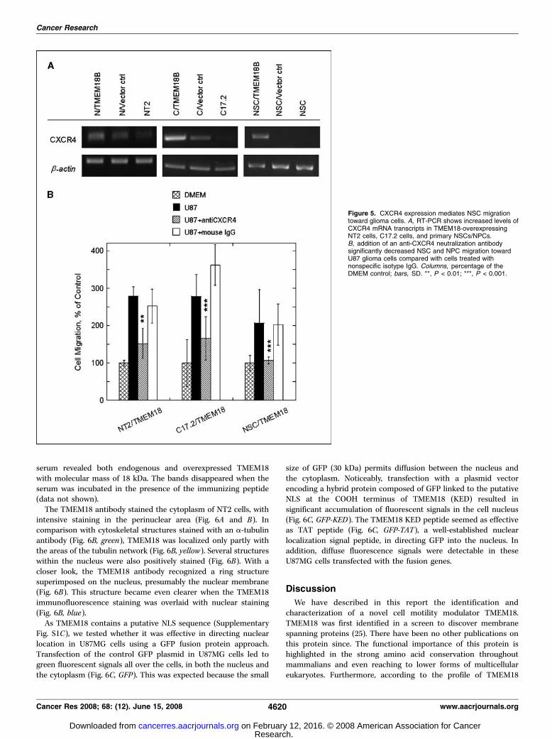
specific migration capacity of NSCs/NSCs. In view of theimportance of the CXCR4 that governs the migration of stem cellstoward gliomas (24), we investigated whether TMEM18 wouldaffect its expression in NSCs/NPCs. We observed that, althoughweak CXCR4 expression was visible in parental NT2 and C17.2 cellsas well as their vector controls, overexpression of TMEM18 seemedto raise its expression levels in NT2 cells, C17.2 cells, and primaryNSCs/NPCs (Fig. 5A). Using an antibody against CXCR4 to blockcell surface CXCR4 receptors on these cells, we further observedthat the tropism of these TMEM18-overexpressing cells towardU87MG cells was inhibited drastically in Boyden chamber assaysand the number of migrating cells went down to a level closeto that of basal cell migration in response to serum-free DMEM(Fig. 5B). These results suggest that up-regulation of CXCR4 inTMEM18-overexpressing cells might be one possible mechanismunderlying the augmented glioma tropism of these cells.The NLS sequence of TMEM18 is sufficient for nuclear
targeting. To uncover the cellular localization of TMEM18, apolyclonal antibody against TMEM18 COOH-terminal peptide wasproduced. The activity and specificity of the antibody wereexamined using cellular immunostaining and Western blot analysis(Supplementary Fig. S2). In NT2 cells, preimmunization serumproduced almost no signal, whereas the serum against TMEM18gave a strong immunofluorescence. Western blot analysis with the
Figure 4. Endogenous TMEM18 expression affects cellmigration. A, siRNAs against TMEM18 transcripts reducedthe expression of TMEM18 transcripts in NT2 cells, asquantified by real-time PCR. Two different sequences ofsiRNAs against TMEM18 were tested in NT2 cells andyielded four different knockdown levels of the TMEM18mRNA. siRNA against luciferase was used as a control.B, silencing endogenous TMEM18 expression reduced themigration activity of NSCs/NPCs toward both DMEM andU87MG. C and D, neural differentiation of hES cells wasaccompanied by progressive increase in TMEM18 mRNAexpression and cell migration toward glioma cells.Columns, percentage of control; bars, SD. Statisticalcomparison is calculated between cells withdown-regulated TMEM18 and controls using Student’s ttest. *, P < 0.05; **, P < 0.01; ***, P < 0.001.
TMEM18 and Stem Cell Migration
www.aacrjournals.org 4619 Cancer Res 2008; 68: (12). June 15, 2008
Research. on February 12, 2016. © 2008 American Association for Cancercancerres.aacrjournals.org Downloaded from
serum revealed both endogenous and overexpressed TMEM18with molecular mass of 18 kDa. The bands disappeared when theserum was incubated in the presence of the immunizing peptide(data not shown).
The TMEM18 antibody stained the cytoplasm of NT2 cells, withintensive staining in the perinuclear area (Fig. 6A and B). Incomparison with cytoskeletal structures stained with an a-tubulinantibody (Fig. 6B, green), TMEM18 was localized only partly withthe areas of the tubulin network (Fig. 6B, yellow). Several structureswithin the nucleus were also positively stained (Fig. 6B). With acloser look, the TMEM18 antibody recognized a ring structuresuperimposed on the nucleus, presumably the nuclear membrane(Fig. 6B). This structure became even clearer when the TMEM18immunofluorescence staining was overlaid with nuclear staining(Fig. 6B, blue).
As TMEM18 contains a putative NLS sequence (SupplementaryFig. S1C), we tested whether it was effective in directing nuclearlocation in U87MG cells using a GFP fusion protein approach.Transfection of the control GFP plasmid in U87MG cells led togreen fluorescent signals all over the cells, in both the nucleus andthe cytoplasm (Fig. 6C, GFP). This was expected because the small
size of GFP (30 kDa) permits diffusion between the nucleus andthe cytoplasm. Noticeably, transfection with a plasmid vectorencoding a hybrid protein composed of GFP linked to the putativeNLS at the COOH terminus of TMEM18 (KED) resulted insignificant accumulation of fluorescent signals in the cell nucleus(Fig. 6C, GFP-KED). The TMEM18 KED peptide seemed as effectiveas TAT peptide (Fig. 6C, GFP-TAT), a well-established nuclearlocalization signal peptide, in directing GFP into the nucleus. Inaddition, diffuse fluorescence signals were detectable in theseU87MG cells transfected with the fusion genes.
Discussion
We have described in this report the identification andcharacterization of a novel cell motility modulator TMEM18.TMEM18 was first identified in a screen to discover membranespanning proteins (25). There have been no other publications onthis protein since. The functional importance of this protein ishighlighted in the strong amino acid conservation throughoutmammalians and even reaching to lower forms of multicellulareukaryotes. Furthermore, according to the profile of TMEM18
Figure 5. CXCR4 expression mediates NSC migrationtoward glioma cells. A, RT-PCR shows increased levels ofCXCR4 mRNA transcripts in TMEM18-overexpressingNT2 cells, C17.2 cells, and primary NSCs/NPCs.B, addition of an anti-CXCR4 neutralization antibodysignificantly decreased NSC and NPC migration towardU87 glioma cells compared with cells treated withnonspecific isotype IgG. Columns, percentage of theDMEM control; bars, SD. **, P < 0.01; ***, P < 0.001.
Cancer Research
Cancer Res 2008; 68: (12). June 15, 2008 4620 www.aacrjournals.org
Research. on February 12, 2016. © 2008 American Association for Cancercancerres.aacrjournals.org Downloaded from

expressed sequence tags (EST), the protein is transcribed inembryonic developmental states and in many of the adult humantissues (National Center for Biotechnology Information’s ESTexpression profile viewer). Clearly, TMEM18 has a crucial biologicalfunction. Based on our results, this function would probably belinked to cell mobility. In particular, with respect to tumor therapy,TMEM18 can be used as a specific enhancer for glioma-directedmigration of NSCs.
The results that TMEM18-overexpressing cells and control cellsmigrated at the same rate in plain cell culture medium or towardnonglioma cells (NIH3T3 mouse fibroblast and 293T humanembryonic kidney cells) suggest that TMEM18 overexpressionprovided no beneficial effects on the general movement of NSCs.However, knockdown of endogenous TMEM18 expression withRNA interference had enormous inhibitory effects on the overallmovement of NSCs. The effect seemed much stronger than thatinduced by TMEM18 overexpression. It is worthy of noting that, asdetermined by our quantitative real-time PCR experiments,TMEM18 mRNA is low-abundant transcripts and could befunctionally sensitive to siRNA-mediated gene silencing. Further-more, along with the increase of TMEM18 expression from anundetectable level to an easily detectable level when hES cellsdifferentiated into NPCs, these NSCs/NPCs displayed an increasedcapacity of cell migration (Fig. 4C and D). These findings indicate acrucial role of the basal, physiologic level expression of TMEM18for cell movement, which is well consistent with the highlyconserved and ubiquitously expressed pattern of TMEM18.
Most interestingly, TMEM18 overexpressing cells respondstrongly to glioma cell–secreted cues in both in vitro Transwellassays and an in vivo migration experiment. Although both RT-PCRand Western blot yielded unidirectional results in the increase ofTMEM18 expression in the current study, mismatch in terms ofincrease level is obvious. A discrepancy between transcriptionand translation is not an uncommon phenomenon. A recent articlehas indicated that the correlation between mRNA and proteinexpression is moderately or weekly positive, with correlation
coefficients ranging from 0.2 to 0.6 (26). This can be most plausiblyexplained by the observation that mRNA and protein aresynthesized with independent mechanisms and degraded bydifferent pathways. In Transwell assays, cells will have to forcethemselves through holes in Boyden chamber membrane thatare smaller than the normal size of a cell body. It could be evenmore difficult for cells to migrate to a target site in the brain wherecell migration needs to overcome numerous extracellular inter-actions. We thus conclude that TMEM18 overexpression increasesthe sensitivity of NSCs to appropriate signals that stimulate cellmigration. In other words, without appropriate cues, TMEM18overexpression will have undetectable effects on the movementof NSCs.
TMEM18 is expected to be a transmembrane protein. But itdid not seem to be located to outer cell membrane, as would beexpected for example for chemokine receptors, nor was it spreadto cover cytoskeletal structures that would affect cell movementdirectly. TMEM18 is predicated to possess a NLS sequence at theCOOH terminus. NLS sequences occur in a subset of solublenuclear proteins that are imported into the cell nucleus bytransport receptors. Membrane proteins with NLS-like sequencesare found in the majority of mammalian inner nuclear membrane(INM) proteins (27). A recent study in budding yeast shows thatNLS sequences are essential for passage of integral membraneproteins through the nuclear pore complex and receptor-mediatedtransport of the proteins to the INM (28). Further studies arewarranted to assess whether TMEM18 is really located along theinside of the nuclear envelope.
Cell motility is a highly complex process and involves severalfactors, from sensing of environmental cues, restructuring thecytoskeleton, dynamic regulation of cell attachment and detach-ment to extracellular matrix, to signaling between all theseprocesses to coordinate the movements. Hence, there are manysteps downstream of chemoattractant receptor signaling that canaffect cell movement and migration. Bioinformatics searches forprotein domains did not reveal any informative features related to
Figure 6. Cellular localization of TMEM18and the function of its NLS. A and B,cellular localization of TMEM18 protein.NT2 cells were stained against TMEM18(A and B, red), a-tubulin (B, green ), andDNA (B, blue ). B, superimposition ofTMEM18, DNA, and a-tubulin. Theimmunostaining reveals the nuclearmembrane (a ring structure around thenucleus in A and B ) recognized by theTMEM18 antibody. C, GFP fusion proteinwith NH2 terminus of TMEM18 is localizedto the nucleus. U87 cells were transfectedwith plasmid vectors expressing GFP,TMEM18 NH2-terminal–linked GFP(GFP-KED ), or HIV TAT-linked GFP(GFP-TAT ). Left, light microscopepictures of the cells; right, fluorescencemicroscope pictures of the cells.
TMEM18 and Stem Cell Migration
www.aacrjournals.org 4621 Cancer Res 2008; 68: (12). June 15, 2008
Research. on February 12, 2016. © 2008 American Association for Cancercancerres.aacrjournals.org Downloaded from

a possible biochemical function of TMEM18. Our preliminary dataon the up-regulation of CXCR4 in TMEM18-overexpressing cellsand inhibiting cell migration by antibodies against CXCR4 suggestan enhanced effect of the SDF-1/CXCR4 axis by TMEM18. Furtherresearch is necessary to define the basic mechanisms underlyingthe effects of endogenous TMEM18 on general cell migration andthe effects of overexpressed TMEM18 to enhance cell responseto migration-stimulating signals. An adequate understanding ofthese mechanisms could have important implications for effectivecellular delivery of therapeutic agents for brain tumor therapy.
Disclosure of Potential Conflicts of Interest
No potential conflicts of interest were disclosed.
Acknowledgments
Received 9/13/2007; revised 3/28/2008; accepted 3/31/2008.Grant support: Institute of Bioengineering and Nanotechnology, Agency for
Science, Technology and Research (A*STAR) in Singapore.The costs of publication of this article were defrayed in part by the payment of page
charges. This article must therefore be hereby marked advertisement in accordancewith 18 U.S.C. Section 1734 solely to indicate this fact.
We thank the lab members for helpful discussion and support.
Cancer Research
Cancer Res 2008; 68: (12). June 15, 2008 4622 www.aacrjournals.org
References1. Tysnes BB, Mahesparan R. Biological mechanisms ofglioma invasion and potential therapeutic targets. JNeurooncol 2001;53:129–47.2. Mueller MM, Werbowetski T, Del Maestro RF. Solublefactors involved in glioma invasion. Acta Neurochir(Wien) 2003;145:999–1008.3. Benedetti S, Pirola B, Pollo B, et al. Gene therapy ofexperimental brain tumors using neural progenitor cells.Nat Med 2000;6:447–50.4. Aboody KS, Brown A, Rainov NG, et al. Neural stemcells display extensive tropism for pathology in adultbrain: evidence from intracranial gliomas. Proc NatlAcad Sci U S A 2000;97:12846–51.5. Flax JD, Aurora S, Yang C, et al. Engraftable humanneural stem cells respond to developmental cues,replace neurons, and express foreign genes. NatBiotechnol 1998;16:1033–9.6. Arnhold S, Hilgers M, Lenartz D, et al. Neuralprecursor cells as carriers for a gene therapeuticalapproach in tumor therapy. Cell Transplant 2003;12:827–37.7. Brown AB, Yang W, Schmidt NO, et al. Intravasculardelivery of neural stem cell lines to target intracranialand extracranial tumors of neural and non-neural origin.Hum Gene Ther 2003;14:1777–85.8. Allport JR, Shinde Patil VR, Weissleder R. Murineneuronal progenitor cells are preferentially recruited totumor vasculature via a4-integrin and SDF-1a-depen-dent mechanisms. Cancer Biol Ther 2004;3:838–44.9. Aarum J, Sandberg K, Haeberlein SL, Persson MA.Migration and differentiation of neural precursor cellscan be directed by microglia. Proc Natl Acad Sci U S A2003;100:15983–8.
10. Tran PB, Miller RJ. Chemokine receptors: signpoststo brain development and disease. Nat Rev Neurosci2003;4:444–55.11. Imitola J, Raddassi K, Park KI, et al. Directedmigration of neural stem cells to sites of CNS injuryby the stromal cell-derived factor 1a/CXC chemokinereceptor 4 pathway. Proc Natl Acad Sci U S A 2004;101:18117–22.12. Ehtesham M, Winston JA, Kabos P, Thompson RC.CXCR4 expression mediates glioma cell invasiveness.Oncogene 2006;25:2801–6.13. Widera D, Holtkamp W, Entschladen F, et al. MCP-1induces migration of adult neural stem cells. Eur J CellBiol 2004;83:381–7.14. Erlandsson A, Larsson J, Forsberg-Nilsson K. Stemcell factor is a chemoattractant and a survival factor forCNS stem cells. Exp Cell Res 2004;301:201–10.15. Sun L, Lee J, Fine HA. Neuronally expressed stem cellfactor induces neural stem cell migration to areas ofbrain injury. J Clin Invest 2004;113:1364–74.16. Serfozo P, Schlarman MS, Pierret C, Maria BL, KirkMD. Selective migration of neuralized embryonic stemcells to stem cell factor and media conditioned byglioma cell lines. Cancer Cell Int 2006;6:1–8.17. Boockvar JA, Kapitonov D, Kapoor G, et al. Consti-tutive EGFR signaling confers a motile phenotype toneural stem cells. Mol Cell Neurosci 2003;24:1116–30.18. Schmidt NO, Przylecki W, Yang W, et al. Brain tumortropism of transplanted human neural stem cells isinduced by vascular endothelial growth factor. Neopla-sia 2005;7:623–9.19. Ziu M, Schmidt NO, Cargioli TG, Aboody KS, BlackPM, Carroll RS. Glioma-produced extracellular matrixinfluences brain tumor tropism of human neural stemcells. J Neurooncol 2006;79:125–33.
20. Zeng J, Du J, Zhao Y, Palanisamy N, Wang S.Baculoviral vector-mediated transient and stable trans-gene expression in human embryonic stem cells. StemCells 2007;25:1055–61.21. Makrynikola V, Bianchi A, Bradstock K, Gottlieb D,Hewson J. Migration of acute lymphoblastic leukemiacells into human bone marrow stroma. Leukemia 1994;8:1734–43.22. Vacca A, Ribatti D, Iurlaro M, et al. Humanlymphoblastoid cells produce extracellular matrix-degrading enzymes and induce endothelial cell prolif-eration, migration, morphogenesis, and angiogenesis.Int J Clin Lab Res 1998;28:55–68.23. Ehtesham M, Yuan X, Kabos P, et al. Glioma tropicneural stem cells consist of astrocytic precursors andtheir migratory capacity is mediated by CXCR4.Neoplasia 2004;6:287–93.24. Kucia M, Reca R, Miekus K, et al. Traffickingof normal stem cells and metastasis of cancerstem cells involve similar mechanisms: pivotalrole of the SDF-1-CXCR4 axis. Stem Cells 2005;23:879–94.25. Hofmann K, Stoffel W. TMbase—a database ofmembrane spanning proteins segments. Biol ChemHoppe-Seyler 1993;374:166.26. Fu N, Drinnenberg I, Kelso J, et al. Comparison ofprotein and mRNA expression evolution in humans andchimpanzees. PLoS ONE 2007;2:e216.27. Horton P, Nakai K. Better prediction of proteincellular localization sites with the k nearest neighborsclassifier. Proc Int Conf Intell Syst Mol Biol 1997;5:147–52.28. King MC, Lusk CP, Blobel G. Karyopherin-mediatedimport of integral inner nuclear membrane proteins.Nature 2006;442:1003–7.
Research. on February 12, 2016. © 2008 American Association for Cancercancerres.aacrjournals.org Downloaded from
0
Announcements
MEETING OF THE RADIATION RESEARCH SOCIETY
The annual meeting of the Radiation Research Society will be held at the State University of Iowa, IowaCity, on June 22—24,1953. The Society will be the guestof the University, and all meetings will be held on thecampus. The program will consist of: (1) Two symposia,one on “TheEffects of Rwliation on Aqueous Solutions,― which includes the following speakers: E. S. G.Barren, Edwin J. Hart, Warren Garrison, J. L. Magee,and A. 0. Allen. The second is “PhysicalMeasurementsfor Radiobiology―and companion talks by Ugo Fano,Burton J. Moyer, G. Failla, L. D. Marinelli, and Payne
The following correction should be made in the article by Beck and Valentine, “TheAerobic CarbohydrateMetabolism of Leukocytes in Health and Leukemia. I.Glycolysis and Respiration,― November, 1952, page 821;substitute for the last paragraph:
The data in Table 3 permit several interesting calculations. If one compares the amount of glucose actually
disappearing with the sum of the amount equivalent tolactic acid produced plus that equivalent to 02 con
sumption, it is seen that the amount of glucose “cleavage products―exceeds the amount of glucose utilized b12 per cent in N and 27 per cent in CML and is exceeded
S. Harris. (2) On Monday night, June 22, a lecture byDr. L. W. Alvarez on meson physics has been tentatively scheduled. On Tuesday night, June 23, Dr. L. H.Gray of the Hammersmith Hospital, London, will speakon a topic to be announced. Dr. Gray's lecture is sponsored by the Iowa Branch of the American Cancer Society. Those desiring to report original research in radiation effects, or interested in attending or desiring additional information, please contact the Secretary of theSociety, Dr. A. Edelmann, Biology Department, Brookhaven National Laboratory, Upton, L.I., New York.
by the glucose utilized by 16 per cent in CLL. If the assumption is made that, in this respect, the myeloid andlymphoid celLsof leukemia are similar to those of norma! blood, it may be that the computed normal figurerepresents a summation of the myeloid (M) andlymphoid (L) cells that make up the normal leukocytepopulation. Thus, if M = +0.27 and L = —0.16 andthe normal differential is 65 per cent M and So per centL, then
0.65 (+0.27) + 0.35 (—0.16) = +0.12
a figure identical to the observed +0.12 for normalleukocytes.
ERRATUM
308

2008;68:4614-4622. Cancer Res Jaana Jurvansuu, Ying Zhao, Doreen S.Y. Leung, et al. Stem Cells for Glioma CellsTransmembrane Protein 18 Enhances the Tropism of Neural
Updated version
http://cancerres.aacrjournals.org/content/68/12/4614
Access the most recent version of this article at:
Material
Supplementary
http://cancerres.aacrjournals.org/content/suppl/2008/06/04/68.12.4614.DC1.html
Access the most recent supplemental material at:
Cited articles
http://cancerres.aacrjournals.org/content/68/12/4614.full.html#ref-list-1
This article cites 28 articles, 3 of which you can access for free at:
Citing articles
http://cancerres.aacrjournals.org/content/68/12/4614.full.html#related-urls
This article has been cited by 4 HighWire-hosted articles. Access the articles at:
E-mail alerts related to this article or journal.Sign up to receive free email-alerts
Subscriptions
Reprints and
To order reprints of this article or to subscribe to the journal, contact the AACR Publications
Permissions
To request permission to re-use all or part of this article, contact the AACR Publications
Research. on February 12, 2016. © 2008 American Association for Cancercancerres.aacrjournals.org Downloaded from