Risk factors for gallbladder polyp malignancy in two public ...
Upload
independentCategory
view
0download
0
Identification of Carbonic Anhydrase I Immunodominant Epitopes
Recognized by Specific Autoantibodies Which Indicate an Improved
Prognosis in Patients with Malignancy after Autologous Stem Cell
Transplantation
Ludovit Skultety,†,‡ Barbora Jankovicova,§ Zuzana Svobodova,§ Pavel Mader,|
Pavlina Rezacova,| Maria Dubrovcakova,⊥ Jan Lakota,*,⊥,# and Zuzana Bilkova*,§
Institute of Virology, Slovak Academy of Sciences, Bratislava, Slovakia, Centre of Molecular Medicine, SlovakAcademy of Sciences, Bratislava, Slovakia, Department of Biological and Biochemical Sciences, Faculty of
Chemical Technology, University of Pardubice, Pardubice, Czech Republic, Department of Structural Biology,Institute of Molecular Genetics, Academy of Sciences of the Czech Republic, Prague, Czech Republic, Laboratoryof Molecular Oncology, Cancer Research Institute, Slovak Academy of Sciences, Bratislava, Slovakia, and Bone
Marrow Transplantation Unit, National Cancer Institute, Bratislava, Slovakia
Received May 15, 2010
This work employs an epitope mapping of carbonic anhydrase (CA), isoform I (CA I), for detection ofthe main immunodominant epitopes. Our interest has arisen from an observed spontaneous tumorregression in patients who developed an aplastic anemia type syndrome after a high-dose therapywith autologous stem cell transplantation and whose sera contained high titer of anti carbonic anhydrase(anti-CA) autoantibodies. There are many indications that the presence of these autoantibodies mayprovide significant survival benefit for the patients. Western blot analysis confirmed strong immu-noreactivity of the patients’ sera with several CA isoforms and the CA I has been selected for our studyas a highly abundant and widely distributed isoform. The applied analytical approach consists of specificfragmentation of CA I protein followed by immunospecific isolation of peptides reacting with polyclonalanti-CA I autoantibodies of patients in spontaneous remission. We improved the standard epitopemapping schema by incorporating the benefits of magnetic carriers and biomagnetic separationtechniques. Mass spectrometry has been applied for detection and identification of epitopes and theacquired results were verified by bioinformatic tools. The candidate epitopes of CA I (NVGHS, DGLAV,SSEQL, and SLKPI) are discussed herein as potential therapeutic targets. This work highlights theusefulness of the epitope mapping technique based on magnetic microspheres for effective and rapiddetermination of immunodominant epitopes of the target protein.
Keywords: epitope mapping • carbonic anhydrase I • anti-CA autoantibodies • LC-MS/MS • bioinformatictools • autologous stem cell transplantation • spontaneous remission
1. Introduction
Mammalian carbonic anhydrases (CAs, EC 4.2.1.1) are widelydistributed Zn(II) metalloenzymes that catalyze the reversiblehydration of carbon dioxide to produce a bicarbonate anionand a proton (CO2 + H2O T HCO3
- + H+).1 This reactionregulates a broad range of physiological procceses. In humans,15 different CA isoenzymes are presently known, and many ofthem are quite recent discoveries compared with the physi-ologically abundant and widely distributed isoenzymes CA I
and II.1-4 Association of two CA isoenzymes (CA IX and XII)with cancer has been confirmed and the involvement of othersis not excluded and is a subject of a contemporary research.The expression profile of CA IX and XII was successfully appliedfor discrimination of cancerous cells (overexpression) fromhealthy cells (minimal expression).4 Clinical modulation of CAactivity with inhibitors has proven a reliable treatment for arange of human disease states.5 Sulfonamide based CA inhibi-tors were also shown to inhibit the growth of several tumorcell lines in vitro and in vivo, representing thus interesting leadsfor development of novel antitumor therapies.6
High-dose therapy (HDT) with autologous stem cell trans-plantation (ASCT) is the standard treatment for patients withchemosensitive relapsed/refractory malignant diseases such asHodgkin’s lymphoma, non-Hodgkin’s lymphoma, multiplemyeloma, and others.7-10 There is an increasing evidence thatautoimmunity can inhibit growth of solid tumors and that
* To whom correspondence should be addressed. E-mail: (Z.B., foranalytical part) [email protected]; (J.L. for clinical part) [email protected].
† Institute of Virology, Slovak Academy of Sciences.‡ Centre of Molecular Medicine, Slovak Academy of Sciences.§ University of Pardubice.| Academy of Sciences of the Czech Republic.⊥ Cancer Research Institute, Slovak Academy of Sciences.# Bone Marrow Transplantation Unit, National Cancer Institute.
10.1021/pr1004778 2010 American Chemical Society Journal of Proteome Research 2010, 9, 5171–5179 5171Published on Web 08/03/2010

antitumor activity also operates in autoimmunity againsthematopoetic stem cells in acquired aplastic anemia (AA).11
The presence of autoantibodies in the sera of patients whorelapsed after HDT with ASCT was described by Lakota et al.12
In these patients, who developed aplastic anemia type syn-drome after the therapy, spontaneous tumor regression wasobserved and blood serum analysis revealed high titer of anti-CA autoantibodies. The specificity of the antibodies wasconfirmed by repeated screenings of the patients’ sera byWestern blot analysis with human blood cell lysate. Thepresence of specific IgG molecules, the dynamics of theirproduction, and the total amount in serum directly correlatedwith the course of the disease. The sera of patients in thedisease remission contained specific autoantibodies in a suf-ficiently high titer and the decreased signal or even negativeresults of Western blot analysis corresponded with the relapseof the disease. These anti-CA autoantibodies produced aftertransplantation of autologous hematopoietic cells seem to bethe main factor in the transition of the disease to remissionand their presence in serum represents a significant survivalbenefit.12
The foregoing results of Western blot analysis confirmed theimmunoreactivity of the patients’ sera autoantibodies withseveral CA isoforms (CA I, II, IX and XII). The mapping ofimmunodominant epitope(s) on CA I is a subject of this study.The knowledge of the epitope sequence is essential as thisinformation has a potential to be applied consequently inclinical practice not only for diagnostic tests, but also fortherapeutic purposes.
The epitope mapping is an analytical approach used foridentification of the immunodominant structures that are ableto be recognized by the immune system, reactive T and B cells,or specific antibodies. A number of in vivo and in vitromethodologies can be used for epitope localization.13 In thiswork, we apply a traditional epitope mapping technique14
improved by the employment of magnetic carriers. Our strategywas based on exploitation of the benefits of magnetic micro-spheres, such as large specific surfaces for chemical binding,fast and simple handling, effective separation by permanentmagnets, good flow characteristics, and mechanical and chemi-cal stability.15 Furthermore, the use of biofunctionalized mag-netic carriers can minimize the analysis time, and the amountof reagents and decreases the loss of analytes. This techniquealso eliminates contamination by autolytic fragments of pro-teases, increases the binding efficiency of desired proteins/peptides, and overall lowers the cost of the analysis.
Our novel approach consisted of two steps: (i) the specificfragmentation of the target protein with proteolytic enzymescovalently bound to magnetic microspheres and (ii) the im-munomagnetic separation of generated peptides based onspecific molecular recognition. Tandem mass spectrometryMS/MS was applied for detection and identification of thepeptides. The effectiveness of such strategy has been suggestedin our previous work.16 We are profiting from our longstandingexperience with magnetic micro- or nanospheres, their surfacemodifications, and their application in biotechnology andprotein analysis.16-22 To retain the highest biological activity,we have immobilized the ligands in an oriented mannerenabling the steric accessibility of the majority of the active orbinding sites.
The identification of the main CA I epitopes binding specif-ically to the anti-CA autoantibodies and matching these resultswith data obtained by bioinformatic tools were the principal
objectives of this research. We assume that these new data mayreveal the etiology of the disease and may clarify the mecha-nism of tumor growth inhibition in relation to the appearanceand dynamics of autoantibody formation and to the diseaseprognosis of the monitored patients. Tumor control in relationto autoimmunity has been observed in a variety of malignan-cies.11
2. Experimental Section
2.1. Chemicals and Reagents. Carbonic anhydrase I, fromhuman erythrocytes (CA I), trypsin (EC 3.4.21.4, 13 000 IU mg-1
solid), R-chymotrypsin, from bovine pancreas, Type II (EC3.4.21.1, 83.9 IU mg-1 solid), DL-dithiothreitol (DTT), formic acid(FA), [Glu1]-fibrinopeptide B (GFP), iodoacetamide (IAA), 1-eth-yl-3-(3-dimethylaminopropyl)carbodiimidehydrochloride(EDAC),benzamidine (BA), NR-benzoyl-DL-arginine p-nitroanilide hy-drochloride (BAPNA), N-succinyl-L-phenylalanine-p-nitroanil-ide (SUPHEPA), sodium cyanoborohydride (NaCNBH3), Pon-ceau S, and D-glyceraldehyde were products of Sigma-Aldrich(St. Louis, MO). N-Hydroxysulfosuccinimide sodium salt (Sul-pho-NHS), ammonium hydrocarbonate, and trifluoroaceticacid (TFA) were from Fluka (Buchs, Switzerland). RapiGest SFwas supplied by Waters (Milford, MA). Acetonitrile (LiChrosolvquality) and water (LiChrosolv quality) were from Merck(Darmstadt, Germany). Perloza MT 500 (80-100 µm) wasproduced by Severoceske chemicke zavody (Lovosice, CzechRepublic). One micrometer silica superparamagnetic nonpo-rous microparticles with carboxyl and hydrazide moieties(SiMAG-Carboxyl and SiMAG-Hydrazide) were supplied byChemicell (Berlin, Germany). Albumin from bovine serum,fraction V, pH 7.0 (BSA) was obtained from AppliChem (Darm-stadt, Germany). Blot Quantified BSA, anti-human IgG (H&L)AP conjugate, and BCIP-NBT reagent were produced byPromega (Madison, WI). Human patient serum was providedby Dr. Lakota (National Cancer Institute, Bratislava, Slovakia)after signed patient informed consent.
2.2. Instrumentation. Magnetic separator (Dynal, Carlsbad,CA), UV/vis spectrophotometer Libra S22 (Biochrom, Cam-bridge, U.K.), Mini-PROTEAN 3 electrophoresis system, BioradModel Mini Trans-blot Cell, BioLogic LP chromatographysystem and 1 × 5 cm (10 cm) columns (Bio-Rad, Hercules, CA),electrophoresis system Mini-Vertical Units (Amersham, U.K.),MicroSpin G-25 Column (GE Healthcare, Buckinghamshire,U.K.), Millex Filter, 0.45 µm (Millipore, Billerica, MA), nitrocel-lulose membranes (Schleicher Schuell, Duren, Germany),nanoAcquity UPLC (Waters, Milford, MA), and Q-Tof Premier(Waters, Milford, MA).
2.3. Patient’s Serum. The serum of a patient with multiplemyeloma after HDT and ASCT treatment (performed accordingto standard European Bone Marrow Transplantation groupprotocols employed at the National Cancer Institute) wasutilized in this work for isolation of specific anti-CA I antibod-ies. Written and informed consent was obtained from thepatient according the institutional guidelines.
2.4. Immobilization of Proteolytic Enzymes onto MagneticMicroparticles with Carboxyl Moieties. A one-step carbodi-imideprocedurebasedonEDAC/Sulpho-NHS-coupledreaction19,23
was used for the immobilization of proteolytic enzymes (9 mg),trypsin or R-chymotrypsin, onto SiMAG-Carboxyl micropar-ticles (3 mg). Immobilization was carried out overnight at 4 °Cunder stirring, and in the case of trypsin in the presence of 3.8mM benzamidine (BA). Resulting proteolytic activity was
research articles Skultety et al.
5172 Journal of Proteome Research • Vol. 9, No. 10, 2010

determined using low-molecular weight substrates: BAPNA fortrypsin and SUPHEPA for R-chymotrypsin.
2.5. Digestion of Unfolded CA I by Immobilized Pro-teolytic Enzymes. For improving the sensitivity to proteolysis,CA I was unfolded by RapiGest SF combined with DTT andIAA. Unfolded CA I (1.5 mL, 1 mg/mL) was added to SiMAG-Carboxyl microparticles (2.9 mg) with immobilized proteolyticenzyme (see Section 2.4), the molar ratio E/S was 1:60. Themixture was incubated at room temperature (RT) under stirring.Efficiency of digestion was verified by Tris-Tricine-SDS-PAGEand LC-MS/MS analysis (see Section 2.9).
2.6. Preparation of Specific Anti-CA I Magnetic Immun-osorbent. The preparation of anti-CA I magnetic immunosor-bent for specific immunocapturing of CA I fragments compris-ing the immunodominant epitope(s) consists of three steps:(i) the isolation of the whole IgG fraction from the patient’sserum by Protein G bioaffinity chromatography, (ii) the con-sequent isolation of specific anti-CA I IgG antibodies from theIgG fraction by affinity chromatography with CA I as animmobilized ligand, and finally (iii) the covalent immobilizationof isolated specific anti-CA I IgG antibodies onto SiMAG-Hydrazide magnetic microparticles.
2.6.1. Isolation of IgG Fraction from the Patient’s Serum.One milliliter of the patient’s serum (see Section 2.3) wasdiluted with 1 mL of 20 mM phosphate buffer, pH 7.0, filteredusing 0.45 µm filter, and applied on Protein G Sepharosecolumn (10 mL) equilibrated with 20 mM phosphate buffer pH7.0. For capturing of IgG, the flow rate was decreased to 0.8mL/min. Subsequently, IgG was eluted with 0.1 M glycinebuffer pH 3.0 (flow rate 1 mL/min). Elution fractions (2 mL)were neutralized with Tris-HCl buffer pH 9.0 (50 µL), concen-trated, homogenized, and immediately used for isolation ofspecific anti-CA I IgG antibodies.
2.6.2. Isolation of Specific Anti-CA I IgG. Preparation ofthe Carrier with Immobilized CA I. Perloza MT 500 (5 mL ofthe sediment) was washed with distilled water (10 × 5 mL) andoxidized with 0.2 M NaIO4 (5 mL) for 80 min at RT in the dark,under stirring. After washing with distilled water (10 × 5 mL),followed by 0.1 M phosphate buffer pH 7.0 with 0.15 M NaCl(10 × 5 mL), protein CA I (2 mg in 5 mL of 0.1 M phosphatebuffer pH 7.0 with 0.15 M NaCl) was added and incubated for10 min at RT under stirring. Then, 35 mg of NaCNBH3 wasadded and immobilization was carried out overnight at 4 °Cunder stirring. The binding efficiency was approximatelydetermined from the difference of CA I solution absorbance at280 nm before and after the immobilization.
Isolation of the Specific Antibodies. The carrier with im-mobilized CA I (5 mL, see above) was packed into thechromatography column (1 × 5 cm) and equilibrated with 0.1M phosphate buffer pH 7.5 with 0.15 M NaCl (flow rate 1.5mL/min, 50 mL). The capturing of specific anti-CA I IgG fromthe total IgG fraction of the patient’s serum (see Section 2.6.1)was applied in 0.1 M phosphate buffer pH 7.5 with 0.15 M NaClat a flow rate 0.25 mL/min. Specific captured antibodies wereeluted with 0.1 M glycine buffer pH 2.8 with 0.15 M NaCl (flowrate 1 mL/min). Pooled elution fractions (1 mL), neutralizedwith 1 M Tris (50 µL), were concentrated to 1 mL, andimmediately immobilized onto SiMAG-Hydrazide magneticmicroparticles (see Section 2.6.3).
2.6.3. Preparation of Immunosorbent with OrientedImmobilized Anti-CA I IgG. Specific anti-CA I IgG antibodies(1 mL, see Section 2.6.2) were coupled through carbohydrateslocated on the Fc fragment of the antibody to SiMAG-Hydrazide
magnetic microparticles (5 mg) in an oriented way. Beforeimmobilization, antibodies were oxidized with 20 mM NaIO4
in the dark at RT for 30 min and then transferred by MicroSpinG-25 Column into a coupling buffer (i.e., 0.1 M phosphatebuffer pH 7.0). The binding of IgG to the microparticles wasachieved overnight at RT under stirring. For blockage of theremaining reactive groups, 0.1 M D-glyceraldehyde in a cou-pling buffer (0.5 mL) was applied for 1 h at RT under stirring.The binding efficiency was estimated from the difference ofantibody solution absorbance at 280 nm before and afterimmobilization and also by Western blot analysis (see Section2.8).
2.7. Immunomagnetic Isolation of CA I Specific Frag-ment(s). A sample of CA I digested by trypsin or R-chymot-rypsin (0.5 mL, 1 mg/mL) (see Section 2.5) was applied to theanti-CA I immunosorbent (5 mg, see Section 2.6.3), equilibratedin 0.1 M phosphate buffer pH 7.0. After incubation for 1 h atRT under stirring, immunosorbent was washed first with 0.1M phosphate buffer pH 7.0 with 0.2 M NaCl (10 × 1.5 mL),then 0.1 M phosphate buffer pH 7.0 with 1 M NaCl (5 × 1.5mL), and finally with 0.01 M phosphate buffer pH 7.0 (5 × 1.5mL). Consequently, we performed 3 repeated elutions with0.05% (v/v) TFA (500 µL, pH 2.4) for 15 min at RT under stirring,followed by 2 elution steps with 0.05% (v/v) TFA (2 mL, pH2.4) for regeneration of the immunosorbent. Separate elutionfractions concentrated to 20 µL were analyzed by LC-MS/MStechnique (see Section 2.9).
2.8. Western Blot Analysis. CA I was dissolved in a solutioncontaining 8% (w/v) SDS, 20 mM Tris-HCl buffer pH 6.8, 40%(v/v) glycerol, and 10 mM EDTA. After the electrophoreticseparation in 12.5% SDS-PAGE slab gel, the pure protein wastransferred to the nitrocellulose membrane. The quality ofprotein transfer was controlled by staining with 0.1% PonceauS in 5% acetic acid. The membranes blocked with 1% BlotQuantified BSA were incubated overnight with diluted serumfractions. After washing, the membranes were incubated withanti-human IgG (H&L) AP conjugate at a 5000-fold dilution andanti-CA I antibodies were detected with BCIP-NBT reagent.
2.9. Mass Spectrometric Analysis. Peptide mixtures elutedfrom the immunosorbent were analyzed by automated nano-flow RP-UPLC system coupled to a Q-Tof Premier (Waters,Milford, MA) tandem mass spectrometer (LC-MS/MS). Peptidesinjected onto a reverse-phase column (nanoAcquity UPLCcolumn BEH 130 C18, 100 µm × 150 mm, 1.7 µm particle size)were separated using the acetonitrile gradient (3-50% B in 15min; A ) water with 0.1% (v/v) formic acid, B ) acetonitrilecontaining 0.1% (v/v) formic acid) at a flow rate of 350 nL/min. The column was directly connected to the PicoTipemitters (New Objective) mounted into the nanospray sourceof a Q-TOF Premier instrument (Waters, U.K.). A nanoelectro-spray voltage of 3.5 kV was applied, with the source temper-ature set to 70 °C. For protein identification, a novel multiplexapproach called MSE was used.24 This method uses an inte-grated approach of parallel, alternating scans at low-collisionenergy, to obtain precursor ion information, and high-collisionenergy, to obtain full-scan accurate mass data in a single run.The spectral acquisition scan rate was 0.6 s with a 0.1 sinterscan delay. In the low energy MS mode, data werecollected at constant collision energy of 3 eV. In elevated energyMS mode, the collision energy was ramped from 20 to 35 eVduring each integration. The MS spectra obtained at differentcollision energies were stored separately. During data acquisi-tion, the quadrupole analyzer was not mass selective, but
CA I Epitopes Recognized by Specific Autoantibodies research articles
Journal of Proteome Research • Vol. 9, No. 10, 2010 5173
operated in the radio frequency only mode. Thus, all ions werepassed to the TOF analyzer. This yielded exact mass fragmentions that were potentially observed for every peptide precursorion present in the low-energy TOF data set.
The obtained MS/MS data were processed using the Pro-teinLynx Global Server v. 2.4 (Waters, U.K.) that providedbackground-subtraction, smoothing, centroiding and deiso-toping. All data were lockspray calibrated against [Glu1]-fibrinopeptide (GFP) using data collected from the referenceline during acquisition. The lockmass-corrected, centroided anddeisotoped data were charge-state reduced to produce a singleaccurately mass measured monoisotopic mass for each peptideand the associated fragment ion. The initial correlation of aprecursor and possible fragment ion was achieved by meansof time alignment. The resulting data were searched againsthuman CA I database (http://www.uniprot.org/uniprot/P00915).Variable modifications of carbamidomethyl-C, oxidation M,deamidation Q, deamidation N, acetylation N-terminus werespecified. One missed cleavage site was allowed. Protein searchparameters also included a 50 ppm tolerance against thedatabase-generated theoretical peptide ion masses, detectionof at least three fragment ions per peptide, minimum of 1matched peptide, and the identification of the protein in atleast 2 out of 3 technical replicates.
2.10. Bioinformatic Analysis. The multiple sequence align-ment of human isoenzymes CA I, CA II, CA IX, and CA XII wasperformed using ClustalX.25 To analyze the position andaccessibility of experimentally detected peptides, protein struc-ture of CA I, deposited in the Protein Data Bank (PDB) underthe accession code (PDB code 1AZM)26 was used. To quantifythe solvent accessibility of the peptides, for each residue, itssolvent accessible surface area (ASA) within the protein struc-ture was calculated using the PISA server27 and expressed as apercentage of a residue total ASA.
3. Results and DiscussionThis study performs epitope mapping of carbonic anhydrase
I (CA I, EC 4.2.1.1) with the intention to find its immunodomi-nant epitope(s). It follows the results of the research, whichhas repeatedly verified the presence of high titers of anti-CAautoantibodies in the sera of patients with spontaneous regres-sion after HDT and ASCT therapy. Immunoreaction in Westernblot analysis with the CA isoforms differed in sera obtainedfrom patients with various types of the disease. Sera ofHodgkin’s disease patients reacted with CA I, II, and XII, whilesera of multiple myeloma patients reacted with the CA I, II,XII, and IX. Presence of these antibodies in sera represented asignificant survival benefit for patients.12
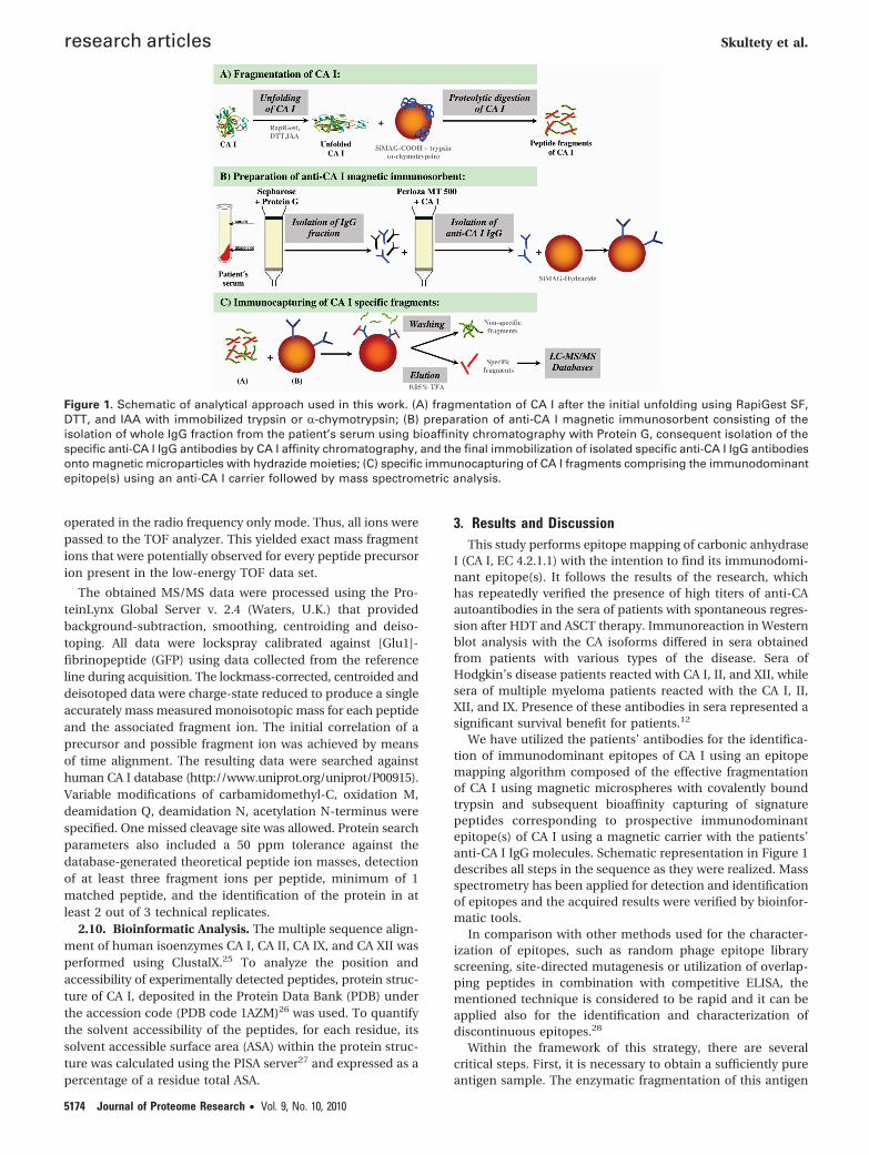
We have utilized the patients’ antibodies for the identifica-tion of immunodominant epitopes of CA I using an epitopemapping algorithm composed of the effective fragmentationof CA I using magnetic microspheres with covalently boundtrypsin and subsequent bioaffinity capturing of signaturepeptides corresponding to prospective immunodominantepitope(s) of CA I using a magnetic carrier with the patients’anti-CA I IgG molecules. Schematic representation in Figure 1describes all steps in the sequence as they were realized. Massspectrometry has been applied for detection and identificationof epitopes and the acquired results were verified by bioinfor-matic tools.
In comparison with other methods used for the character-ization of epitopes, such as random phage epitope libraryscreening, site-directed mutagenesis or utilization of overlap-ping peptides in combination with competitive ELISA, thementioned technique is considered to be rapid and it can beapplied also for the identification and characterization ofdiscontinuous epitopes.28
Within the framework of this strategy, there are severalcritical steps. First, it is necessary to obtain a sufficiently pureantigen sample. The enzymatic fragmentation of this antigen
Figure 1. Schematic of analytical approach used in this work. (A) fragmentation of CA I after the initial unfolding using RapiGest SF,DTT, and IAA with immobilized trypsin or R-chymotrypsin; (B) preparation of anti-CA I magnetic immunosorbent consisting of theisolation of whole IgG fraction from the patient’s serum using bioaffinity chromatography with Protein G, consequent isolation of thespecific anti-CA I IgG antibodies by CA I affinity chromatography, and the final immobilization of isolated specific anti-CA I IgG antibodiesonto magnetic microparticles with hydrazide moieties; (C) specific immunocapturing of CA I fragments comprising the immunodominantepitope(s) using an anti-CA I carrier followed by mass spectrometric analysis.
research articles Skultety et al.
5174 Journal of Proteome Research • Vol. 9, No. 10, 2010

has to be effective and specific. In the next steps, the isolationof polyclonal anti-CA I autoantibodies with desired affinity andtheir proper immobilization to the carrier are important forobtaining efficient immunosorbent for subsequent capturingof CA I peptides.
To carry out an effective and reproducible fragmentation ofthe native antigen (first step), the proteolytic enzyme trypsin,which digests at the carboxyl side of the amino acids Lys orArg, was immobilized to magnetic microspheres SiMAG-Carboxyl via a 1-step carbodiimide method, which is a binarycovalent binding system guaranteeing a good reproducibility.The reversible inhibitor benzamidine, present in the bindingmixture (see Section 2.4), temporarily protects the enzymeagainst autoproteolysis. The final activity of the immobilizedtrypsin (1.24 × 10³ IU/mg of carrier) was estimated with low-molecular weight substrate BAPNA. This carrier with definedproteolytic activity was used for digestion of folded or unfoldedCA I as the target protein. The efficiency and specificity offragmentation was controlled by MS/MS analysis. Immobilized
enzymes, in comparison to the standard procedure using asoluble form, offer several benefits such as high proteolyticactivity, high operational and storage stability, reduction indigestion time, absence of autolytic fragments in the peptidemixture, and low frequency of missed cleavages.19
The time of digestion required optimization; the intervals15, 30, 45, 60, 90, 120, 150, 180 min and 24 h were tested andthe efficiency of cleavage was confirmed by Tris-Tricine-SDS-PAGE (Figure 2) followed by LC-MS/MS analysis (Figure 3). Weobserved high resistance of native CA I to enzymatic digestion.The results of Tris-Tricine-SDS-PAGE demonstrated only partialfragmentation of native CA I even after 24 h. It can be explainedby a compact globular structure of the native protein. On thebasis of these findings, CA I was unfolded by reductivealkylation using DTT and IAA in combination with RapiGestSF reagent to make it more sensitive to enzymatic proteolysis(see Section 2.5). For denatured molecules of CA I, theintermediate peptides were found as early as after 15 min ofdigestion. On the basis of the MS/MS analysis, the period of3 h was estimated as sufficient for efficient fragmentation andproduction of peptides of appropriate length. Eleven specificpeptides out of the 15 peptides (i.e., 70.9% coverage), whichcan be theoretically produced by trypsin digestion, wereconfirmed in the range of MW 600-5000 (Figure 3A).
To perform the last step of peptide mapping by means ofbioaffinity capturing of peptides corresponding to potentialepitopes of CA I (Figure 1C), a magnetic carrier with im-mobilized anti-CA I IgG molecules was prepared. The IgGmolecules were isolated from the human patient’s serum usingProtein G-Sepharose affinity chromatography (Figure 4A).Elution fractions were pooled, concentrated to 10 mL, andapplied for a specific isolation of anti-CA I antibodies usingmacroporous bead cellulose (Perloza MT 500) carrier withimmobilized CA I as an affinity ligand (Figure 4B). Theimmobilization of CA I on the beads was performed by methoddescribed previously by Turkova et al.29 This method is basedon periodate oxidation of the carrier and reduction of the Schiffbase to a secondary amine by sodium cyanoborohydride (seeSection 2.6.2). The binding capacity of prepared bioaffinitycarrier, estimated from a depletion of protein from solutionafter immobilization, was 11 nmol/mL. Control by SDS-PAGEusing 10% separating gel was also performed.
Figure 2. Tris-Tricine-SDS-PAGE. Gel, 16.5% T, 3% C (MW 1-70kDa, %T, % concentration of both monomers, acrylamide andbis-acrylamide; %C, % concentration of cross-linker relative tothe concentration of T), detection with silver staining. Lanes: (1)nondigested native CA I (5 µg), (2) nondigested unfolded CA I (5µg), (3) unfolded CA I digested 3 h by immobilized trypsin (5 µg),(4) molecular marker (10-250 kDa).
Figure 3. The coverage map of CA I peptides (A) using trypsin for its digestion, (B) using R-chymotrypsin for its digestion; detectedpeptides are highlighted.
CA I Epitopes Recognized by Specific Autoantibodies research articles
Journal of Proteome Research • Vol. 9, No. 10, 2010 5175
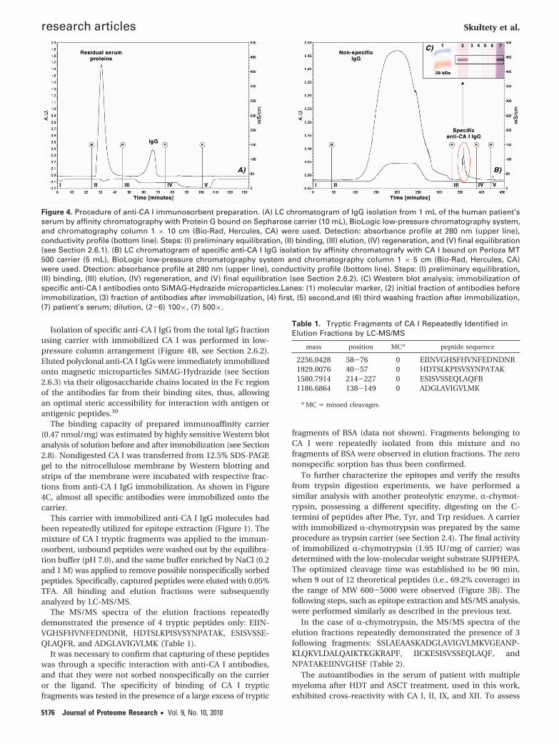
Isolation of specific anti-CA I IgG from the total IgG fractionusing carrier with immobilized CA I was performed in low-pressure column arrangement (Figure 4B, see Section 2.6.2).Eluted polyclonal anti-CA I IgGs were immediately immobilizedonto magnetic microparticles SiMAG-Hydrazide (see Section2.6.3) via their oligosaccharide chains located in the Fc regionof the antibodies far from their binding sites, thus, allowingan optimal steric accessibility for interaction with antigen orantigenic peptides.30
The binding capacity of prepared immunoaffinity carrier(0.47 nmol/mg) was estimated by highly sensitive Western blotanalysis of solution before and after immobilization (see Section2.8). Nondigested CA I was transferred from 12.5% SDS-PAGEgel to the nitrocellulose membrane by Western blotting andstrips of the membrane were incubated with respective frac-tions from anti-CA I IgG immobilization. As shown in Figure4C, almost all specific antibodies were immobilized onto thecarrier.
This carrier with immobilized anti-CA I IgG molecules hadbeen repeatedly utilized for epitope extraction (Figure 1). Themixture of CA I tryptic fragments was applied to the immun-osorbent, unbound peptides were washed out by the equilibra-tion buffer (pH 7.0), and the same buffer enriched by NaCl (0.2and 1 M) was applied to remove possible nonspecifically sorbedpeptides. Specifically, captured peptides were eluted with 0.05%TFA. All binding and elution fractions were subsequentlyanalyzed by LC-MS/MS.
The MS/MS spectra of the elution fractions repeatedlydemonstrated the presence of 4 tryptic peptides only: EIIN-VGHSFHVNFEDNDNR, HDTSLKPISVSYNPATAK, ESISVSSE-QLAQFR, and ADGLAVIGVLMK (Table 1).
It was necessary to confirm that capturing of these peptideswas through a specific interaction with anti-CA I antibodies,and that they were not sorbed nonspecifically on the carrieror the ligand. The specificity of binding of CA I trypticfragments was tested in the presence of a large excess of tryptic
fragments of BSA (data not shown). Fragments belonging toCA I were repeatedly isolated from this mixture and nofragments of BSA were observed in elution fractions. The zerononspecific sorption has thus been confirmed.
To further characterize the epitopes and verify the resultsfrom trypsin digestion experiments, we have performed asimilar analysis with another proteolytic enzyme, R-chymot-rypsin, possessing a different specifity, digesting on the C-termini of peptides after Phe, Tyr, and Trp residues. A carrierwith immobilized R-chymotrypsin was prepared by the sameprocedure as trypsin carrier (see Section 2.4). The final activityof immobilized R-chymotrypsin (1.95 IU/mg of carrier) wasdetermined with the low-molecular weight substrate SUPHEPA.The optimized cleavage time was established to be 90 min,when 9 out of 12 theoretical peptides (i.e., 69.2% coverage) inthe range of MW 600-5000 were observed (Figure 3B). Thefollowing steps, such as epitope extraction and MS/MS analysis,were performed similarly as described in the previous text.
In the case of R-chymotrypsin, the MS/MS spectra of theelution fractions repeatedly demonstrated the presence of 3following fragments: SSLAEAASKADGLAVIGVLMKVGEANP-KLQKVLDALQAIKTKGKRAPF, IICKESISVSSEQLAQF, andNPATAKEIINVGHSF (Table 2).
The autoantibodies in the serum of patient with multiplemyeloma after HDT and ASCT treatment, used in this work,exhibited cross-reactivity with CA I, II, IX, and XII. To assess
Figure 4. Procedure of anti-CA I immunosorbent preparation. (A) LC chromatogram of IgG isolation from 1 mL of the human patient’sserum by affinity chromatography with Protein G bound on Sepharose carrier (10 mL), BioLogic low-pressure chromatography system,and chromatography column 1 × 10 cm (Bio-Rad, Hercules, CA) were used. Detection: absorbance profile at 280 nm (upper line),conductivity profile (bottom line). Steps: (I) preliminary equilibration, (II) binding, (III) elution, (IV) regeneration, and (V) final equilibration(see Section 2.6.1). (B) LC chromatogram of specific anti-CA I IgG isolation by affinity chromatografy with CA I bound on Perloza MT500 carrier (5 mL), BioLogic low-pressure chromatography system and chromatography column 1 × 5 cm (Bio-Rad, Hercules, CA)were used. Dtection: absorbance profile at 280 nm (upper line), conductivity profile (bottom line). Steps: (I) preliminary equilibration,(II) binding, (III) elution, (IV) regeneration, and (V) final equilibration (see Section 2.6.2). (C) Western blot analysis: immobilization ofspecific anti-CA I antibodies onto SiMAG-Hydrazide microparticles.Lanes: (1) molecular marker, (2) initial fraction of antibodies beforeimmobilization, (3) fraction of antibodies after immobilization, (4) first, (5) second,and (6) third washing fraction after immobilization,(7) patient’s serum; dilution, (2-6) 100×, (7) 500×.
Table 1. Tryptic Fragments of CA I Repeatedly Identified inElution Fractions by LC-MS/MS
mass position MCa peptide sequence
2256.0428 58-76 0 EIINVGHSFHVNFEDNDNR1929.0076 40-57 0 HDTSLKPISVSYNPATAK1580.7914 214-227 0 ESISVSSEQLAQFR1186.6864 138-149 0 ADGLAVIGVLMK
a MC ) missed cleavages.
research articles Skultety et al.
5176 Journal of Proteome Research • Vol. 9, No. 10, 2010

which regions of the peptides detected by epitope mappingon CA I could represent a common epitope, we analyzedconservation of peptide sequences in CA I, II, IX, and XII. Thisanalysis detected four short continuous sequences whichshared more than 75% homology among isoforms: SLKPI,NVGHS, DGLAV, and SSEQL (Figure 5). Three of these se-quences were detected in epitope mapping experiments uponCA I fragmentation of both typsin and R-chymotrypsin pro-
teases. Peptide bearing sequence SLKPI was only detected inthe elution of fragments generated by tryptic digest. Possiblereason for this could be the nonaccessibility of epitope withina long R-chymotrypsin fragment.
We analyzed the position of potential conserved epitopeson CA I three-dimensional structure and their accessibility forantibody recognition in the context of the native CA I molecule(Figure 6). Peptide SLKPI (residues 43-47) is a part of a loop
Table 2. R-Chymotryptic Fragments of CA I Repeatedly Identified in Elution Fractions by LC-MS/MS
mass position MCa modifications peptide sequence
4932.8069 129-176 0 SSLAEAASKADGLAVIGVLMKVGEANPKLQKVLDALQAIKTKGKRAPF1881.9626 210-226 0 Cys_CM: 212 (1939.9681) IICKESISVSSEQLAQF1597.8332 52-66 0 NPATAKEIINVGHSF
a MC ) missed cleavages.
Figure 5. Multiple sequence alignment of human CA I, CA II, CA IX, and CA XII performed using Clustal 2.0.1. The figure was preparedusing program CHROMA.31 Highlighted regions correspond to experimentally detected peptides: magenta to SLKPI, red to NVGHS,cyan to DGLAV, and yellow to SSEQL. Red and green arrows above the sequence correspond to tryptic and chymotryptic fragmentsidentified in elution fractions by LC-MS/MS, respectively.
Figure 6. Analysis of solvent accessible surface area of individual residues of conserved peptides within the CA I structure (PDB code1AZM). For each residue of detected peptides, the graph (top) shows the percentage of total accessible surface area (ASA) which isexposed to the solvent. The location of each peptide in the CA I structure (PDB code 1AZM) is represented in a corresponding color(SLKPI magenta, NVGHS red, DGLAV cyan, and SSEQL yellow) in the secondary structure representation (middle) and by the solventaccessible surface (bottom). Zinc ion and inhibitor molecules located in the enzyme active site are depicted in green.
CA I Epitopes Recognized by Specific Autoantibodies research articles
Journal of Proteome Research • Vol. 9, No. 10, 2010 5177

located on the surface of the CA I molecule, which lacks anyparticular secondary structure. With the exception of thehydrophobic side chains of L44 and I47, the peptide is ratherexposed to the solvent, and thus accessible for potentialautoantibody binding. Peptide NVGHS, residues 61-65, formsa loop connecting two �-strands of the central antiparallel�-sheet, which supports the enzyme active site. The loop is alsofairly exposed on the surface of the molecule, with theexception of the terminal residues of this sequence, which aremore buried in the protein core. Peptide DGLAV, residues139-143, is the least exposed of the discussed peptides.Residues L141, A142, and V143 are part of the antiparallel�-sheet structure and are located at the bottom of the deepcavity of the enzyme catalytic site. Accessibility of this peptideis rather limited. Peptide SSEQL, residues 219-223, is in theCA I structure part of a short R-helix. The residues are fairlywell exposed on the surface. The only exception is residue L223,which packs against the hydrophobic protein core.
Sequence homology and structural analysis corroborated bythe experimental epitope mapping resulted in detecting ofpotential conserved epitopes on CA surface available forantibody recognition in the patient’s serum.
4. Conclusion
A combination of the epitope extraction technique, a tradi-tional approach of epitope mapping, together with LC-MS/MSand bioinformatic analyses has been used for identification ofepitopes recognized by anti-CA autoantibodies. Using anti-CAI autoantibodies isolated from the serum of a patient, whospontaneously regressed after HDT with ASCT, and whodeveloped aplastic anemia type syndrome, we identified 4epitopes: NVGHS, DGLAV, SSEQL, and SLKPI. According tocompatible results of LC-MS/MS and bioinformatic analyses,we consider the discussed epitopes as relevant potentialtherapeutic targets for peptide-based vaccination. We believethat the presented results can be useful for disclosure of amechanism of tumor growth inhibition in relation to theappearance and dynamics of autoantibody formation and tothe disease prognosis of monitored patients and might behelpful in clinical practices for development of new therapeuticor diagnostic approaches.
Acknowledgment. The authors wish to acknowledgeThe Ministry of Education of Czech Republic (project No.MSMT 0021627502), The State program of research anddevelopment BITCET (SPVV 337/2003), The Norwayand EEA grants (SK 0095), Structural funds of EU - researchand development program TRANSMED (No. 2624012008) forfinancial support of the research. In part, this work wassupported by Projects AV0Z50520514 and AV0Z40550506awarded by the Academy of Sciences of the Czech Republic,grant No. 1M0505 awarded by the Ministry of Education ofthe Czech Republic, and grant No. GA203/09/0820 awardedby Czech Grant Foundation. We thank Devon Maloy for acritical proofreading of the manuscript.
References(1) Supuran, C. T. Carbonic anhydrases: Catalytic and inhibition
mechanism, distribution and physiological roles. In CarbonicAnhydrase: Its Inhibitors and Activators; Scozzafava, A. , Supuran,C. T. , Conway, J. , Eds.; CRC Press: Boca Raton, FL, 2004; pp 1-24.
(2) Scozzafava, A.; Supuran, C. T. Carbonic anhydrase inhibitors. Curr.Med. Chem.: Immunol. Endocr. Metab. Agents 2001, 1, 61–97.
(3) Supuran, C. T.; Scozzafava, A.; Casini, A. Carbonic anhydraseinhibitors. Med. Res. Rev. 2003, 23, 146–189.
(4) Pastorekova, S.; Parkkila, S.; et al. Carbonic anhydrases: Currentstate of the art, therapeutic applications and future prospects. J.Enzyme Inhib. Med. Chem. 2004, 19 (3), 199–229.
(5) Supuran, C. T. Carbonic anhydrases as drug targetssan overview.Curr. Top. Med. Chem. 2007, (7), 825–833.
(6) Supuran, C. T.; Vullo, D.; et al. Designing of novel carbonicanhydrase inhibitors and activators. Curr. Med. Chem.: Cardiovasc.Hematol. Agents 2005, 2 (1), 49–68.
(7) Gopal, A. K.; Metcalfe, T. L.; et al. High-dose therapy andautologous stem cell transplantation for chemoresistant hodgkinlymphoma: the Seattle experience. Cancer 2008, 113 (6), 1344–1350.
(8) Strehl, J.; Mey, U.; et al. High-dose chemotherapy followed byautologous stem cell transplantation as first-line therapy inaggressive non-Hodgkin’s lymphoma: a meta-analysis. Haemato-logica 2003, 88 (11), 1304–1315.
(9) Greb, A., Bohlius J. High-dose chemotherapy with autologous stemcell transplantation in the first line treatment of aggressive Non-Hodgkin Lymphoma (NHL) in adults. Cochrane Database Syst. Rev.2008 Issue 1. Art. No, CD004024; DOI: 10.1002/14651858.CD004024.pub2.
(10) Vesole, D. H.; Barlogie, B.; et al. High-dose therapy for refractorymultiple myeloma: improved prognosis with better supportive careand double transplants. Blood 1994, 84 (3), 950–956.
(11) Nissen, C.; Stern, M. Acquired immune mediated aplastic anemia:Is it antineoplastic? Autoimmun. Rev. 2009, 9, 11–16.
(12) Lakota, J.; Skultety, L.; et al. Presence of serum carbonic anhydraseautoantibodies in patients relapsed after autologous stem celltransplantation indicates an improved prognosis. Neoplasma 2008,55 (6), 488–492.
(13) Morris, G. E. Overview: choosing a method for epitope mapping.Methods Mol. Biol. 1996, 66, 1–9.
(14) Parker, C. E.; Papac, D. I.; et al. Epitope mapping by massspectrometry: determination of an epitope on HIV-1 IIIB p26recognized by a monoclonal antibody. J. Immunol. 1996, 157 (1),198–206.
(15) Ahn, C. H.; Allen, M. G.; et al. A fully integrated micromachinedmagnetic particle separator. Microelectromech. Syst. 1996, 5 (3),151–158.
(16) Jankovicova, B.; Rosnerova, S.; et al. Epitope mapping of allergenovalbumin using biofunctionalized magnetic beads packed inmicrofluidic channels: The first step towards epitope-based vac-cines. J. Chromatogr., A 2008, 1206, 64–71.
(17) Bilkova, Z.; Stefanescu, R.; et al. Epitope extraction technique usinga proteolytic magnetic reactor combined with Fourier-transformion cyclotron resonance mass spectrometry as a tool for thescreening of potential vaccine lead peptides. Eur. J. Mass Spectrom.2005, 11 (5), 489–495.
(18) Bilkova, Z.; Castagna, A.; et al. Immunoaffinity reactors for prionprotein qualitative analysis. Proteomics 2005, 5, 639–647.
(19) Bilkova, Z.; Slovakova, M.; et al. Functionalized magnetic micro-and nanoparticles: Optimization and application to µ-chip trypticdigestion. Electrophoresis 2006, 27, 1811–1824.
(20) Slovakova, M.; Minc, N.; et al. Use of self assembled magneticbeads for on-chip protein digestion. Lab Chip 2005, 5 (9), 935–942.
(21) Slovakova, M.; Peyrin, J.-M.; et al. Magnetic proteinase K reactoras a new tool for reproducible limited protein digestion. Biocon-jugate Chem. 2008, 19 (4), 966–972.
(22) Korecka, L.; Jankovicova, B.; et al. Bioaffinity magnetic reactor forpeptide digestion followed by analysis using bottom-up shotgunproteomics strategy. J. Sep. Sci. 2008, 31 (3), 507–515.
(23) Staros, J. V. N-hydroxysulfosuccinimide active esters: bis(N-hydroxysulfosuccinimide) esters of two dicarboxylic acids arehydrophilic, membrane-impermeant, protein cross-linkers. Bio-chemistry 1982, 21 (1982), 3950–3955.
(24) Plumb, R. S.; Johnson, K. A.; et al. UPLC/MS(E); a new approachfor generating molecular fragment information for biomarkerstructure elucidation. Rapid Commun. Mass Spectrom. 2006, 20(13), 1989–1994.
(25) Larkin, M. A.; Blackshields, G.; et al. Clustal W and Clustal X version2.0. Bioinformatics 2007, 23 (21), 2947–2948.
(26) Chakravarty, S.; Kannan, K. K. Drug-protein interactions. Refinedstructures of three sulfonamide drug complexes of human car-bonic anhydrase I enzyme. J. Mol. Biol. 1994, 243 (2), 298–309.
(27) Krissinel, E.; Henrick, K. Inference of macromolecular assembliesfrom crystalline state. J. Mol. Biol. 2007, 372 (3), 774–797.
(28) Hochleitner, E. O.; Gorny, M. K.; Zolla-Pazner, S.; Tomer, K. B.Mass spectrometric characterization of a discontinuous epitope
research articles Skultety et al.
5178 Journal of Proteome Research • Vol. 9, No. 10, 2010

of the HIV Envelope protein HIV-gp120 recognized by the humanmonoclonal antibody 1331A1. J. Immunol. 2000, 164, 4156–4161.
(29) Turkova, J.; Vajcner, J.; et al. Immobilization on cellulose in beadform after periodate oxidation and reductive alkylation. Collect.Czech. Chem. Commun. 1979, 44, 34113417.
(30) Murayama, A.; Kohkichi, S.; Tadashi, Y. Labeled periodate oxidizedoligosaccharidegroups in an immunoglobulin (IgG) with amino-
containing compounds via Schiff base formation. Immunochem-istry 1978, 15, 523.
(31) Goodstadt, L.; Ponting, C. P. CHROMA: consensus-based colouringof multiple alignments for publication. Bioinformatics 2001, 17(9), 845–846.
PR1004778
CA I Epitopes Recognized by Specific Autoantibodies research articles
Journal of Proteome Research • Vol. 9, No. 10, 2010 5179
Copyright © 2022 FDOKUMEN