Theileria parva candidate vaccine antigens recognized by immune bovine cytotoxic T lymphocytes
6
Theileria parva candidate vaccine antigens recognized by immune bovine cytotoxic T lymphocytes Simon P. Graham a , Roger Pelle ´ a , Yoshikazu Honda a , Duncan M. Mwangi a , Nyerhovwo J. Tonukari a,b , Mat Yamage a , E. Jane Glew a,c , Etienne P. de Villiers a , Trushar Shah a , Richard Bishop a , Evelyne Abuya a , Elias Awino a , James Gachanja a , Anthony E. Luyai a,d , Ferdinand Mbwika a , Anthony M. Muthiani a , David M. Ndegwa a,e , Moses Njahira a , John K. Nyanjui a , Fredrick O. Onono a,f , Julius Osaso a , Rosemary M. Saya a , Claude Wildmann g , Claire M. Fraser h , Ian Maudlin i , Malcolm J. Gardner h,j , Subhash P. Morzaria a,k , Sheena Loosmore l , Sarah C. Gilbert m , Jean-Christophe Audonnet n , Pierre van der Bruggen g , Vishvanath Nene h , and Evans L. N. Taracha a,o a International Livestock Research Institute, P.O. Box 30709, Nairobi 00100, Kenya; h The Institute for Genomic Research, 9712 Medical Center Drive, Rockville, MD 20850; g Ludwig Institute for Cancer Research, Avenue Hippocrate 74, UCL 7459, B-1200 Brussels, Belgium; n Discovery Research, Merial SAS, Lyon Gerland Laboratory, 254, Rue Marcel Merieux, 69007 Lyon, France; m Wellcome Trust Centre for Human Genetics, Roosevelt Drive, Headington, Oxford OX3 7BN, United Kingdom; l Sanofi Pasteur, Connaught Campus, 1755 Steeles Avenue West, North York, Toronto, ON, Canada M2R 3T4; and i Centre for Tropical Veterinary Medicine, Royal (Dick) School of Veterinary Studies, University of Edinburgh, Easter Bush, Roslin EH25 9RG, United Kingdom Communicated by Tilahun D. Yilma, University of California, Davis, CA, December 29, 2005 (received for review December 2, 2005) East Coast fever, caused by the tick-borne intracellular apicompl- exan parasite Theileria parva, is a highly fatal lymphoproliferative disease of cattle. The pathogenic schizont-induced lymphocyte transformation is a unique cancer-like condition that is reversible with parasite removal. Schizont-infected cell-directed CD8 cyto- toxic T lymphocytes (CTL) constitute the dominant protective bovine immune response after a single exposure to infection. However, the schizont antigens targeted by T. parva-specific CTL are undefined. Here we show the identification of five candidate vaccine antigens that are the targets of MHC class I-restricted CD8 CTL from immune cattle. CD8 T cell responses to these antigens were boosted in T. parva-immune cattle resolving a challenge infection and, when used to immunize naı ¨ve cattle, induced CTL responses that significantly correlated with survival from a lethal parasite challenge. These data provide a basis for developing a CTL-targeted anti-East Coast fever subunit vaccine. In addition, orthologs of these antigens may be vaccine targets for other apicomplexan parasites. cattle East Coast fever immunoscreening protozoan parasite vaccination A single inoculation with a potentially lethal dose of Theileria parva sporozoites and simultaneous treatment with a long- acting oxytetracycline induces solid immunity to homologous and, in certain instances, heterologous parasite challenge (1, 2). This methodology has been adopted as a live vaccine for the control of East Coast fever (ECF) (3). The long-lasting immunity to ECF contrasts with the partial immunity to malaria that develops after only several years of exposure to T. parva-related Plasmodium spp. (4). Manufacture and delivery of the live ECF vaccine is difficult to sustain, but it has enabled elucidation of the dominant protective immune response against the disease. Ki- netic and adoptive cell transfer studies (5, 6) have demonstrated that protection of cattle is mediated by MHC class I-restricted CD8 cytotoxic T lymphocytes (CTL) that destroy schizont- infected lymphocytes, the pathogenic life-cycle stage of T. parva. In addition, there is a strong correlation between the specificity of the CTL response and cross-immunity profiles of distinct parasite strains (2). The identification of schizont antigens targeted by CTL from T. parva-immune cattle has been elusive but should pave the way for the development of a subunit vaccine against ECF and provide a long-term solution to a socio- economically important constraint to livestock agriculture in Africa (7). We adopted two approaches to antigen identification, both dependent on screening of transiently transfected antigen- presenting cells with fully characterized CTL (8, 9) from live vaccine-immunized cattle of diverse bovine leukocyte antigen (BoLA) MHC class I genotypes. First, in a targeted gene approach, we immunoscreened genes that were predicted by using preliminary sequence data from one of the four T. parva chromosomes (10) to contain a secretion signal. The approach was based on the observation that the schizont lies free in the host cell cytoplasm (11) whereby signal peptide-containing parasite proteins would directly access the host cell MHC class I antigen processing and presentation pathway. Second, we used an approach involving a random immunoscreen of schizont cDNA clones because secretion of proteins that do not contain signal sequences has been reported (12). We describe here the antigens identified from these immunoscreens and the immu- nological characterization conducted to validate these molecules as CTL targets. We also report on the initial evaluation of these antigens as vaccine candidates and show successful induction of CTL responses that correlated with reduced disease severity after challenge infection. Results and Discussion Identification of CTL Target Antigens by Targeted and Random Im- munoscreens. In the targeted approach, CTL from four immu- nized cattle showed IFN- ELISpot responses to immortalized Conflict of interest statement: No conflicts declared. Abbreviations: CTL, cytotoxic T lymphocyte(s); BoLA, bovine leukocyte antigen; iSF, immor- talized skin fibroblast; MVA, modified vaccinia virus Ankara strain; CP, canarypox virus; ILRI, International Livestock Research Institute; ECF, East Coast fever; SFC, spot-forming cell. Data deposition: The sequence reported in this paper has been deposited in the GenBank database (accession nos. XP762973, XP765583, XP763228, XP765334, XP764810, and XP764709). b Present address: Department of Biochemistry, Delta State University, Abraka, Nigeria. c Present address: Forensic Alliance Limited, Darwin House, Faraday Street, Birchwood Park, Risley, Warrington WA3 6AT, United Kingdom. d Present address: Department of Biochemistry and Molecular Biology, University of Okla- homa Health Sciences Center, P.O. Box 26901, 940 Stanton L. Young Boulevard, Biomedical Sciences Building, Room 853, Oklahoma City, OK 73190-0001. e Present address: Department of Biomedical Sciences, Tufts University School of Veterinary Medicine, Grafton, MA 01536. f Present address: Department of Cardiology and Angiology, Hannover Medical School, Carl-Neuberg-Strasse 1, D-30625 Hannover, Germany. j Present address: Seattle Biomedical Research Institute, Suite 500, 307 Westlake Avenue N, Seattle, WA 98109. k Present address: Food and Agriculture Organization, 39 Phra Atit Road, Bangkok 10200, Thailand. o To whom correspondence should be addressed. E-mail: [email protected]. © 2006 by The National Academy of Sciences of the USA 3286 –3291 PNAS February 28, 2006 vol. 103 no. 9 www.pnas.orgcgidoi10.1073pnas.0511273103
-
Upload
independent -
Category
Documents
-
view
0 -
download
0
Transcript of Theileria parva candidate vaccine antigens recognized by immune bovine cytotoxic T lymphocytes
Theileria parva candidate vaccine antigens recognizedby immune bovine cytotoxic T lymphocytesSimon P. Grahama, Roger Pellea, Yoshikazu Hondaa, Duncan M. Mwangia, Nyerhovwo J. Tonukaria,b, Mat Yamagea,E. Jane Glewa,c, Etienne P. de Villiersa, Trushar Shaha, Richard Bishopa, Evelyne Abuyaa, Elias Awinoa, James Gachanjaa,Anthony E. Luyaia,d, Ferdinand Mbwikaa, Anthony M. Muthiania, David M. Ndegwaa,e, Moses Njahiraa, John K. Nyanjuia,Fredrick O. Ononoa,f, Julius Osasoa, Rosemary M. Sayaa, Claude Wildmanng, Claire M. Fraserh, Ian Maudlini,Malcolm J. Gardnerh,j, Subhash P. Morzariaa,k, Sheena Loosmorel, Sarah C. Gilbertm, Jean-Christophe Audonnetn,Pierre van der Bruggeng, Vishvanath Neneh, and Evans L. N. Tarachaa,o
aInternational Livestock Research Institute, P.O. Box 30709, Nairobi 00100, Kenya; hThe Institute for Genomic Research, 9712 Medical Center Drive,Rockville, MD 20850; gLudwig Institute for Cancer Research, Avenue Hippocrate 74, UCL 7459, B-1200 Brussels, Belgium; nDiscovery Research,Merial SAS, Lyon Gerland Laboratory, 254, Rue Marcel Merieux, 69007 Lyon, France; mWellcome Trust Centre for Human Genetics, Roosevelt Drive,Headington, Oxford OX3 7BN, United Kingdom; lSanofi Pasteur, Connaught Campus, 1755 Steeles Avenue West, North York, Toronto, ON,Canada M2R 3T4; and iCentre for Tropical Veterinary Medicine, Royal (Dick) School of Veterinary Studies, University of Edinburgh, Easter Bush,Roslin EH25 9RG, United Kingdom
Communicated by Tilahun D. Yilma, University of California, Davis, CA, December 29, 2005 (received for review December 2, 2005)
East Coast fever, caused by the tick-borne intracellular apicompl-exan parasite Theileria parva, is a highly fatal lymphoproliferativedisease of cattle. The pathogenic schizont-induced lymphocytetransformation is a unique cancer-like condition that is reversiblewith parasite removal. Schizont-infected cell-directed CD8� cyto-toxic T lymphocytes (CTL) constitute the dominant protectivebovine immune response after a single exposure to infection.However, the schizont antigens targeted by T. parva-specific CTLare undefined. Here we show the identification of five candidatevaccine antigens that are the targets of MHC class I-restricted CD8�
CTL from immune cattle. CD8� T cell responses to these antigenswere boosted in T. parva-immune cattle resolving a challengeinfection and, when used to immunize naıve cattle, induced CTLresponses that significantly correlated with survival from a lethalparasite challenge. These data provide a basis for developing aCTL-targeted anti-East Coast fever subunit vaccine. In addition,orthologs of these antigens may be vaccine targets for otherapicomplexan parasites.
cattle � East Coast fever � immunoscreening � protozoan parasite �vaccination
A single inoculation with a potentially lethal dose of Theileriaparva sporozoites and simultaneous treatment with a long-
acting oxytetracycline induces solid immunity to homologousand, in certain instances, heterologous parasite challenge (1, 2).This methodology has been adopted as a live vaccine for thecontrol of East Coast fever (ECF) (3). The long-lasting immunityto ECF contrasts with the partial immunity to malaria thatdevelops after only several years of exposure to T. parva-relatedPlasmodium spp. (4). Manufacture and delivery of the live ECFvaccine is difficult to sustain, but it has enabled elucidation of thedominant protective immune response against the disease. Ki-netic and adoptive cell transfer studies (5, 6) have demonstratedthat protection of cattle is mediated by MHC class I-restrictedCD8� cytotoxic T lymphocytes (CTL) that destroy schizont-infected lymphocytes, the pathogenic life-cycle stage of T. parva.In addition, there is a strong correlation between the specificityof the CTL response and cross-immunity profiles of distinctparasite strains (2). The identification of schizont antigenstargeted by CTL from T. parva-immune cattle has been elusivebut should pave the way for the development of a subunit vaccineagainst ECF and provide a long-term solution to a socio-economically important constraint to livestock agriculture inAfrica (7).
We adopted two approaches to antigen identification, bothdependent on screening of transiently transfected antigen-
presenting cells with fully characterized CTL (8, 9) from livevaccine-immunized cattle of diverse bovine leukocyte antigen(BoLA) MHC class I genotypes. First, in a targeted geneapproach, we immunoscreened genes that were predicted byusing preliminary sequence data from one of the four T. parvachromosomes (10) to contain a secretion signal. The approachwas based on the observation that the schizont lies free in thehost cell cytoplasm (11) whereby signal peptide-containingparasite proteins would directly access the host cell MHC classI antigen processing and presentation pathway. Second, we usedan approach involving a random immunoscreen of schizontcDNA clones because secretion of proteins that do not containsignal sequences has been reported (12). We describe here theantigens identified from these immunoscreens and the immu-nological characterization conducted to validate these moleculesas CTL targets. We also report on the initial evaluation of theseantigens as vaccine candidates and show successful induction ofCTL responses that correlated with reduced disease severityafter challenge infection.
Results and DiscussionIdentification of CTL Target Antigens by Targeted and Random Im-munoscreens. In the targeted approach, CTL from four immu-nized cattle showed IFN-� ELISpot responses to immortalized
Conflict of interest statement: No conflicts declared.
Abbreviations: CTL, cytotoxic T lymphocyte(s); BoLA, bovine leukocyte antigen; iSF, immor-talized skin fibroblast; MVA, modified vaccinia virus Ankara strain; CP, canarypox virus; ILRI,International Livestock Research Institute; ECF, East Coast fever; SFC, spot-forming cell.
Data deposition: The sequence reported in this paper has been deposited in the GenBankdatabase (accession nos. XP�762973, XP�765583, XP�763228, XP�765334, XP�764810, andXP�764709).
bPresent address: Department of Biochemistry, Delta State University, Abraka, Nigeria.
cPresent address: Forensic Alliance Limited, Darwin House, Faraday Street, Birchwood Park,Risley, Warrington WA3 6AT, United Kingdom.
dPresent address: Department of Biochemistry and Molecular Biology, University of Okla-homa Health Sciences Center, P.O. Box 26901, 940 Stanton L. Young Boulevard, BiomedicalSciences Building, Room 853, Oklahoma City, OK 73190-0001.
ePresent address: Department of Biomedical Sciences, Tufts University School of VeterinaryMedicine, Grafton, MA 01536.
fPresent address: Department of Cardiology and Angiology, Hannover Medical School,Carl-Neuberg-Strasse 1, D-30625 Hannover, Germany.
jPresent address: Seattle Biomedical Research Institute, Suite 500, 307 Westlake Avenue N,Seattle, WA 98109.
kPresent address: Food and Agriculture Organization, 39 Phra Atit Road, Bangkok 10200,Thailand.
oTo whom correspondence should be addressed. E-mail: [email protected].
© 2006 by The National Academy of Sciences of the USA
3286–3291 � PNAS � February 28, 2006 � vol. 103 � no. 9 www.pnas.org�cgi�doi�10.1073�pnas.0511273103
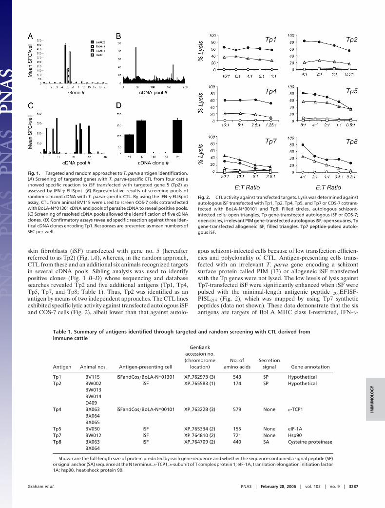
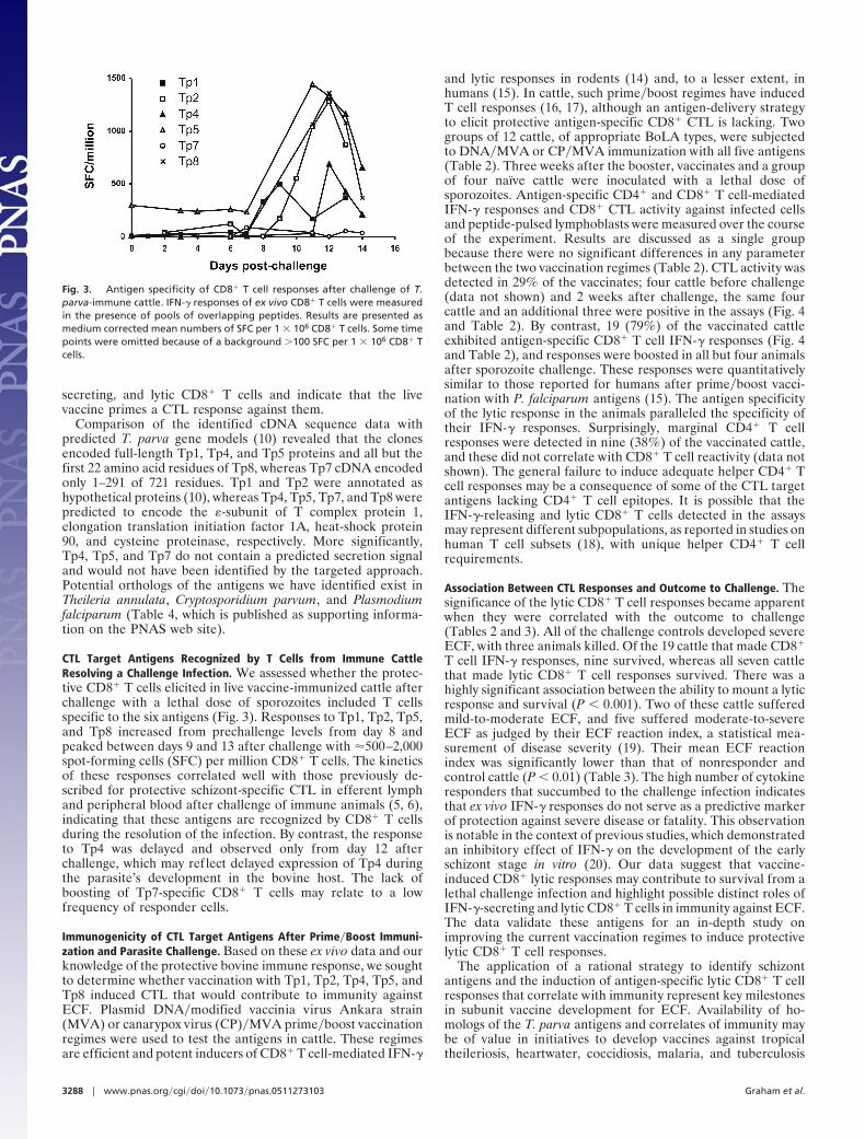
skin fibroblasts (iSF) transfected with gene no. 5 (hereafterreferred to as Tp2) (Fig. 1A), whereas, in the random approach,CTL from these and an additional six animals recognized targetsin several cDNA pools. Sibling analysis was used to identifypositive clones (Fig. 1 B–D) whose sequencing and databasesearches revealed Tp2 and five additional antigens (Tp1, Tp4,Tp5, Tp7, and Tp8; Table 1). Thus, Tp2 was identified as anantigen by means of two independent approaches. The CTL linesexhibited specific lytic activity against transfected autologous iSFand COS-7 cells (Fig. 2), albeit lower than that against autolo-
gous schizont-infected cells because of low transfection efficien-cies and polyclonality of CTL. Antigen-presenting cells trans-fected with an irrelevant T. parva gene encoding a schizontsurface protein called PIM (13) or allogeneic iSF transfectedwith the Tp genes were not lysed. The low levels of lysis againstTp7-transfected iSF were significantly enhanced when iSF werepulsed with the minimal-length antigenic peptide 206EFISF-PISL214 (Fig. 2), which was mapped by using Tp7 syntheticpeptides (data not shown). These data demonstrate that the sixantigens are targets of BoLA MHC class I-restricted, IFN-�-
Fig. 1. Targeted and random approaches to T. parva antigen identification.(A) Screening of targeted genes with T. parva-specific CTL from four cattleshowed specific reaction to iSF transfected with targeted gene 5 (Tp2) asassessed by IFN-� ELISpot. (B) Representative results of screening pools ofrandom schizont cDNA with T. parva-specific CTL. By using the IFN-� ELISpotassay, CTL from animal BV115 were used to screen COS-7 cells cotransfectedwith BoLA-N*01301 cDNA and pools of parasite cDNA to reveal positive pools.(C) Screening of resolved cDNA pools allowed the identification of five cDNAclones. (D) Confirmatory assays revealed specific reaction against three iden-tical cDNA clones encoding Tp1. Responses are presented as mean numbers ofSFC per well.
Table 1. Summary of antigens identified through targeted and random screening with CTL derived fromimmune cattle
Antigen Animal nos. Antigen-presenting cell
GenBankaccession no.(chromosome
location)No. of
amino acidsSecretion
signal Gene annotation
Tp1 BV115 iSFandCos�BoLA-N*01301 XP�762973 (3) 543 SP HypotheticalTp2 BW002 iSF XP�765583 (1) 174 SP Hypothetical
BW013BW014D409
Tp4 BX063 iSFandCos�BoLA-N*00101 XP�763228 (3) 579 None �-TCP1BX064BX065
Tp5 BV050 iSF XP�765334 (2) 155 None eIF-1ATp7 BW012 iSF XP�764810 (2) 721 None Hsp90Tp8 BX063 iSF XP�764709 (2) 440 SA Cysteine proteinase
BX064
Shown are the full-length size of protein predicted by each gene sequence and whether the sequence contained a signal peptide (SP)or signal anchor (SA) sequence at the N terminus. �-TCP1, �-subunit of T complex protein 1; eIF-1A, translation elongation initiation factor1A; hsp90, heat-shock protein 90.
Fig. 2. CTL activity against transfected targets. Lysis was determined againstautologous iSF transfected with Tp1, Tp2, Tp4, Tp5, and Tp7 or COS-7 cotrans-fected with BoLA-N*00101 and Tp8. Filled circles, autologous schizont-infected cells; open triangles, Tp gene-transfected autologous iSF or COS-7;open circles, irrelevant PIM gene-transfected autologous iSF; open squares, Tpgene-transfected allogeneic iSF; filled triangles, Tp7 peptide-pulsed autolo-gous iSF.
Graham et al. PNAS � February 28, 2006 � vol. 103 � no. 9 � 3287
IMM
UN
OLO
GY
secreting, and lytic CD8� T cells and indicate that the livevaccine primes a CTL response against them.
Comparison of the identified cDNA sequence data withpredicted T. parva gene models (10) revealed that the clonesencoded full-length Tp1, Tp4, and Tp5 proteins and all but thefirst 22 amino acid residues of Tp8, whereas Tp7 cDNA encodedonly 1–291 of 721 residues. Tp1 and Tp2 were annotated ashypothetical proteins (10), whereas Tp4, Tp5, Tp7, and Tp8 werepredicted to encode the �-subunit of T complex protein 1,elongation translation initiation factor 1A, heat-shock protein90, and cysteine proteinase, respectively. More significantly,Tp4, Tp5, and Tp7 do not contain a predicted secretion signaland would not have been identified by the targeted approach.Potential orthologs of the antigens we have identified exist inTheileria annulata, Cryptosporidium parvum, and Plasmodiumfalciparum (Table 4, which is published as supporting informa-tion on the PNAS web site).
CTL Target Antigens Recognized by T Cells from Immune CattleResolving a Challenge Infection. We assessed whether the protec-tive CD8� T cells elicited in live vaccine-immunized cattle afterchallenge with a lethal dose of sporozoites included T cellsspecific to the six antigens (Fig. 3). Responses to Tp1, Tp2, Tp5,and Tp8 increased from prechallenge levels from day 8 andpeaked between days 9 and 13 after challenge with �500–2,000spot-forming cells (SFC) per million CD8� T cells. The kineticsof these responses correlated well with those previously de-scribed for protective schizont-specific CTL in efferent lymphand peripheral blood after challenge of immune animals (5, 6),indicating that these antigens are recognized by CD8� T cellsduring the resolution of the infection. By contrast, the responseto Tp4 was delayed and observed only from day 12 afterchallenge, which may reflect delayed expression of Tp4 duringthe parasite’s development in the bovine host. The lack ofboosting of Tp7-specific CD8� T cells may relate to a lowfrequency of responder cells.
Immunogenicity of CTL Target Antigens After Prime�Boost Immuni-zation and Parasite Challenge. Based on these ex vivo data and ourknowledge of the protective bovine immune response, we soughtto determine whether vaccination with Tp1, Tp2, Tp4, Tp5, andTp8 induced CTL that would contribute to immunity againstECF. Plasmid DNA�modified vaccinia virus Ankara strain(MVA) or canarypox virus (CP)�MVA prime�boost vaccinationregimes were used to test the antigens in cattle. These regimesare efficient and potent inducers of CD8� T cell-mediated IFN-�
and lytic responses in rodents (14) and, to a lesser extent, inhumans (15). In cattle, such prime�boost regimes have inducedT cell responses (16, 17), although an antigen-delivery strategyto elicit protective antigen-specific CD8� CTL is lacking. Twogroups of 12 cattle, of appropriate BoLA types, were subjectedto DNA�MVA or CP�MVA immunization with all five antigens(Table 2). Three weeks after the booster, vaccinates and a groupof four naıve cattle were inoculated with a lethal dose ofsporozoites. Antigen-specific CD4� and CD8� T cell-mediatedIFN-� responses and CD8� CTL activity against infected cellsand peptide-pulsed lymphoblasts were measured over the courseof the experiment. Results are discussed as a single groupbecause there were no significant differences in any parameterbetween the two vaccination regimes (Table 2). CTL activity wasdetected in 29% of the vaccinates; four cattle before challenge(data not shown) and 2 weeks after challenge, the same fourcattle and an additional three were positive in the assays (Fig. 4and Table 2). By contrast, 19 (79%) of the vaccinated cattleexhibited antigen-specific CD8� T cell IFN-� responses (Fig. 4and Table 2), and responses were boosted in all but four animalsafter sporozoite challenge. These responses were quantitativelysimilar to those reported for humans after prime�boost vacci-nation with P. falciparum antigens (15). The antigen specificityof the lytic response in the animals paralleled the specificity oftheir IFN-� responses. Surprisingly, marginal CD4� T cellresponses were detected in nine (38%) of the vaccinated cattle,and these did not correlate with CD8� T cell reactivity (data notshown). The general failure to induce adequate helper CD4� Tcell responses may be a consequence of some of the CTL targetantigens lacking CD4� T cell epitopes. It is possible that theIFN-�-releasing and lytic CD8� T cells detected in the assaysmay represent different subpopulations, as reported in studies onhuman T cell subsets (18), with unique helper CD4� T cellrequirements.
Association Between CTL Responses and Outcome to Challenge. Thesignificance of the lytic CD8� T cell responses became apparentwhen they were correlated with the outcome to challenge(Tables 2 and 3). All of the challenge controls developed severeECF, with three animals killed. Of the 19 cattle that made CD8�
T cell IFN-� responses, nine survived, whereas all seven cattlethat made lytic CD8� T cell responses survived. There was ahighly significant association between the ability to mount a lyticresponse and survival (P � 0.001). Two of these cattle sufferedmild-to-moderate ECF, and five suffered moderate-to-severeECF as judged by their ECF reaction index, a statistical mea-surement of disease severity (19). Their mean ECF reactionindex was significantly lower than that of nonresponder andcontrol cattle (P � 0.01) (Table 3). The high number of cytokineresponders that succumbed to the challenge infection indicatesthat ex vivo IFN-� responses do not serve as a predictive markerof protection against severe disease or fatality. This observationis notable in the context of previous studies, which demonstratedan inhibitory effect of IFN-� on the development of the earlyschizont stage in vitro (20). Our data suggest that vaccine-induced CD8� lytic responses may contribute to survival from alethal challenge infection and highlight possible distinct roles ofIFN-�-secreting and lytic CD8� T cells in immunity against ECF.The data validate these antigens for an in-depth study onimproving the current vaccination regimes to induce protectivelytic CD8� T cell responses.
The application of a rational strategy to identify schizontantigens and the induction of antigen-specific lytic CD8� T cellresponses that correlate with immunity represent key milestonesin subunit vaccine development for ECF. Availability of ho-mologs of the T. parva antigens and correlates of immunity maybe of value in initiatives to develop vaccines against tropicaltheileriosis, heartwater, coccidiosis, malaria, and tuberculosis
Fig. 3. Antigen specificity of CD8� T cell responses after challenge of T.parva-immune cattle. IFN-� responses of ex vivo CD8� T cells were measuredin the presence of pools of overlapping peptides. Results are presented asmedium corrected mean numbers of SFC per 1 � 106 CD8� T cells. Some timepoints were omitted because of a background �100 SFC per 1 � 106 CD8� Tcells.
3288 � www.pnas.org�cgi�doi�10.1073�pnas.0511273103 Graham et al.
caused by intracellular pathogens against which CTL contributeto immunity.
Materials and MethodsCloning of Targeted Genes. Analysis of proteins encoded by thepredicted T. parva genes (10) on chromosome 1 for the presenceof signal peptides (SIGNALP-2.0; www.cbs.dtu.dk�services�SignalP-2.0) and transmembrane domains (TMHMM, www.cbs.dtu.dk�services�TMHMM) generated a set of 55 candidateantigen genes that were selected for cloning and screening(Table 5, which is published as supporting information on thePNAS web site). ORFs were amplified by using a OneStepRT-PCR kit (Qiagen, Crawley, U.K.) with RNA purified fromT. parva (Muguga) schizont-infected cells. Amplified genes werecloned into eukaryotic expression vector pTargeT (Promega).
Construction of Purified T. parva Muguga Schizont cDNA Library. Aunidirectional cDNA library was constructed in the eukaryoticexpression plasmid vector pcDNA3 (Invitrogen) essentially asdescribed in ref. 21. Briefly, double-stranded cDNA was gener-ated from 2 �g of total poly(A)�-RNA (FastTrack 2.0 kit,Invitrogen) isolated from purified schizonts (22) by using theSuperScript Choice System (Invitrogen). Plasmid DNA mini-preps were prepared from 1,000 pools of bacterial cultures eachcontaining 50 colonies and another 1,000 pools each containing10 colonies for high-throughput immunoscreening. To resolve apositive pool of 50 cDNAs, 256 colonies were pooled into 48
clusters of 16 colonies by using a three-way matrix and screenedto identify the individual clone in three unique clusters. For theresolution of a positive pool of 10 cDNA, plasmid DNA wasprepared from 48 colonies and tested to identify the individualclone. Positive cDNA clones were end-sequenced, and sequenceinformation was used to identify the complete ORFs from the T.parva genome sequence database (www.ncbi.nlm.nih.gov�sutils�blast�table.cgi?taxid�Protozoa).
Infection and Treatment Immunization of Cattle and Generation ofCTL. All animal experimentation was reviewed and approved bythe International Livestock Research Institute (ILRI) Institu-tional Animal Care and Use Committee. Four Boran (Bosindicus), one Jersey (Bos taurus), one Holstein–Friesian (B.taurus), and four crossbred animals, selected on the basis ofBoLA type, were immunized against T. parva Muguga stock andthen challenged after 3 months (2). Schizont-specific polyclonalCTL lines and clones were established and exhibited 30–80%lytic activity against autologous schizont-infected targets at aneffector:target ratio �10:1, in line with published data (2, 8, 9).
Transfection of iSF and COS-7 Cells. iSF were immortalized by usingstandard procedures (23) with modifications (unpublished ob-servations). iSF were transfected in 96-well plates with clones orpools of schizont cDNA (100 ng per well) by using FuGENE 6(Roche Diagnostics, Mannheim, Germany) and cultured for24 h. COS-7 cells were cotransfected with 100 ng of each clone
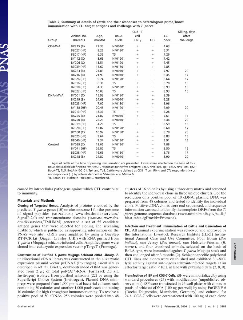
Table 2. Summary of details of cattle and their responses to heterologous prime�boostimmunization with CTL target antigens and challenge with T. parva
GroupAnimal no.
(breed†)Age,
monthsBoLAallele
CD8� Tcell
IFN-� CTLECF
index
Killing, daysafter
challenge
CP�MVA BX215 (B) 22.33 N*00101 � � 4.63BZ027 (HF) 8.26 N*01301 � � 6.31BZ017 (HF) 6.36 T5 � � 7.26BY142 (C) 8.69 N*01201 � � 7.42BY206 (C) 13.51 N*01201 � � 7.45BZ039 (HF) 15.67 N*01301 � � 7.85BX223 (B) 24.89 N*00101 � � 7.97 20BX216 (B) 21.93 N*00101 � � 8.45 17BZ026 (HF) 9.74 N*01201 � � 8.64 17BZ016 (HF) 6.36 T5 � � 8.70 16BZ018 (HF) 4.33 N*01301 � � 8.93 15BZ022 (HF) 10.03 T5 � � 8.93 16
DNA�MVA BY001 (C) 15.93 N*01201 � � 3.39BX219 (B) 24.69 N*00101 � � 6.28BZ023 (HF) 7.02 N*01301 � � 6.96BY138 (HF) 20.45 N*01201 � � 7.09 20BZ013 (HF) 18.39 T5 � � 7.28BX225 (B) 21.87 N*00101 � � 7.61 16BX220 (B) 22.23 N*00101 � � 8.44 20BZ019 (HF) 4.20 T5 � � 8.59 16BZ020 (HF) 12.07 N*01201 � � 8.60 17BY100 (C) 10.92 N*01301 � � 8.78 20BZ025 (HF) 9.64 T5 � � 8.83 15BZ040 (HF) 7.34 N*01301 � � 8.91 15
Control BY029 (C) 13.05 N*01201 � � 7.88BY071 (HF) 26.82 T5 � � 8.50 16BZ038 (HF) 16.69 N*01301 � � 8.76 17BX218 (B) 24.82 N*00101 � � 8.90 20
Ages of cattle at the time of priming immunization are presented. Calves were selected on the basis of fourBoLA class I alleles defined to restrict CTL responses to the five antigens: BoLA-N*01301, Tp1; BoLA-N*01201, Tp2;BoLA-T5, Tp5; BoLA-N*00101, Tp4 and Tp8. Cattle were defined as CD8� T cell IFN-� and CTL responders (�) ornonresponders (�) by criteria defined in Materials and Methods.†B, Boran; HF, Holstein–Friesian; C, crossbreed.
Graham et al. PNAS � February 28, 2006 � vol. 103 � no. 9 � 3289
IMM
UN
OLO
GY
per well or pooled together with 50 ng of pcDNA3 BoLA-N*00101 (24) or BoLA-N*01301 cDNA per well.
IFN-� ELISpot and 51Chromium Release Assays. Transfectants werecocultured with schizont-specific CTL, and recognition wasassessed by using an IFN-� ELISpot assay (16). Transfectantsand schizont-infected cells were labeled with 51Chromium (Am-ersham Pharmacia Biosciences), and lysis by schizont-specificCTL lines was assessed (9).
Ex Vivo Detection of Antigen-Specific T Cell Responses. Immunecattle donors for schizont-specific CTL used to identify Tp1,
Tp2, Tp4, Tp5, Tp7, and Tp8 were challenged with a lethal doseof T. parva (Muguga) sporozoites and bled on selected days afterchallenge. CD8� T cells and monocytes were purified fromperipheral blood mononuclear cells by magnetic cell sortingaccording to the manufacturer’s instructions (Miltenyi Biotec).CD8� T cells were stained with anti-CD8 mAb (IL-A105)(ILRI) followed by incubation with anti-mouse IgG microbeads(Miltenyi Biotec). Monocytes were stained directly with CD14microbeads (Miltenyi Biotec). Magnetically sorted CD8� T cells(2.5 � 105 per well) and CD14� monocytes (2.5 � 104 per well)were added to IFN-� ELISpot plates containing pools of over-lapping synthetic peptides (16 mers offset by 4 residues) span-ning the lengths of Tp1, Tp2, Tp4, Tp5, Tp7, and Tp8 (1 �g�mleach peptide, Pepscan Systems , Lelystad, The Netherlands) andwere incubated and developed essentially as described in ref. 16.
Preparation of Recombinant Plasmid DNA and Viruses. Tp1, Tp2,Tp4, Tp5, and Tp8 ORFs, excluding the stop codons, wereamplified by OneStep RT-PCR from schizont RNA. The codingsequence of a mouse B cell epitope of SV5 P�V protein, Pk-Tag,was ligated at the 3� end of each gene. These antigen gene Tagfragments were cloned into pSG2 DNA vaccine plasmids (25),and endotoxin-free DNA was prepared (PlasmidFactory,Bielefeld, Germany). MVA and CP were generated by homol-ogous recombination of the wild-type viruses MVA andALVAC, respectively, with the corresponding shuttle vectorsMVA-GFP and pC5H6p, incorporating antigen gene Tag in-serts, in accordance with published protocols (26). Expressionwas confirmed by immunostaining with anti-Pk-Tag mAb(MCA1360P, Serotec) and CTL recognition of virus-infectediSF. All vaccine preparations were screened for the presence ofbovine viral diarrhea virus (27) and mycoplasma (MycoplasmaDetection kit version 2.0; American Type Culture Collection).
Heterologous Prime�Boost Immunization and Parasite Challenge ofCattle. The experiment was designed in a double-blind fashion inwhich animals were retagged between immunization and chal-lenge, and the code was managed by the biometrics staff.Twenty-eight Boran (B. indicus), Holstein–Friesian (B. taurus),and crossbred calves, 4–24 months old and expressing one of fourMHC class I alleles, were randomized into three groups (Table2). Recombinant DNA, CP, and MVA constructs expressingsingle antigens (Tp1, Tp2, Tp4, Tp5, and Tp8) were preparedseparately and inoculated at different sites. Cattle were primedwith 0.5 mg of each of the five recombinant DNA plasmids byintradermal injection or 1 � 108 plaque-forming units of each ofthe five recombinant CP viruses by s.c. injection. After 4 weeks,cattle were inoculated s.c. with 5 � 108 plaque-forming units ofeach of the five MVA recombinants. Control cattle received s.c.inoculations with the vaccine diluent alone. Three weeks afterboost, cattle were challenged with a lethal dose of T. parva (1:20dilution of T. parva Muguga stock sporozoite stabilate 4133;ILRI). Reactions to challenge were calculated as an ECF
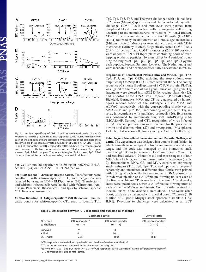
Fig. 4. Antigen specificity of CD8� T cells in vaccinated cattle. (A and C)Representative IFN-� responses of five responder cattle illustrate reactivity toeach of the antigens and are compared with a nonresponder calf. Responsespresented are the medium corrected number of SFC per 1 � 106 CD8� T cells.(B and D) Four of the five IFN-� responder cattle exhibited lytic responses andare compared with two nonresponder cattle. Filled squares, Tp1; opensquares, Tp2; filled triangles, Tp4; open triangles, Tp5; crosses, Tp8; filledcircles, schizont-infected cells; open circles, unpulsed T cell blasts.
Table 3. Association between CTL responses and outcome to challenge
Outcometo challenge
Vaccinated cattle Control cattle
CTL responder*(n � 7)
CTL nonresponder(n � 17)
CTL nonresponder†
(n � 4)
Survived 7‡ 3 1Killed 0 14 3ECF index 6.04‡ 8.04 8.49
*CTL responders were defined by criteria described in Materials and Methods.†CTL responses were not detected in the challenge control group.‡Survival (P � 0.001) and ECF index (P � 0.01) of CTL responder cattle were significantly different from those ofCTL nonresponders and control cattle.
3290 � www.pnas.org�cgi�doi�10.1073�pnas.0511273103 Graham et al.
reaction index by combination of 13 parameters, includingtemperature, hematological measurements, and parasitologicalmeasurements, by using first-principle-component analysis asdescribed in ref. 19. Cattle were assessed daily by two indepen-dent veterinarians, and animals displaying clinical signs of severeECF were killed by captive bolt. The experiment was terminated21 days after challenge. Antigen-specific CD4� and CD8� T cellIFN-� responses were measured against synthetic peptides byELISpot assay every 2 weeks over the course of immunizationand challenge. CD4� T cells were sorted by magnetic cell sortingindirectly by using a mAb specific for bovine CD4 (IL-A12;ILRI), CD8� T cells and CD14� monocytes were sorted, andELISpot assays were conducted as described above. Antigen-specific CTL responses were recalled by subjecting peripheralblood mononuclear cells to three rounds of restimulation with T.parva-infected cells, and cytotoxicity against infected cells anduninfected cells pulsed with pools of synthetic peptides wereassayed as described above. Cattle were classified as IFN-�responders when the number of SFC in response to antigen wasthree times greater than to medium alone and as CTL responderswhen lysis of T. parva-infected lymphoblasts and�or antigenpeptide-pulsed uninfected lymphoblasts was three times greaterand at least 15% higher than that of unpulsed uninfected
lymphoblasts. For all responder cattle, responses were �2 stan-dard deviations (95% confidence interval) above the mean SFCof unstimulated cells or mean lysis against unpulsed uninfectedlymphoblasts.
Statistical Analysis. Analysis of variance was used for the analysisof fixed effects on different traits by using SAS release 8.2 (SASInstitute, Cary, NC). A Kruskal–Wallis test was carried out tocompare the ECF reaction index for CTL responders andnonresponders. A �2 test was used to compare the differences insurvival for CTL responders and nonresponders.
We thank the staff of the ILRI large animal and tick units for excellentanimal care and provision of parasites; the ILRI Biometrics Unit foradvice on experimental design and statistical analysis; Shirley Ellis(Institute for Animal Health, Compton, U.K.) for providing the BoLA-N*01301 cDNA; and The Institute for Genomic Research Board ofTrustees, J. Craig Venter, and Hank Fitzhugh for support of the T. parvagenome project. Support by Thierry Boon (Ludwig Institute for CancerResearch) for the screening of cDNA libraries is greatly appreciated.This work was funded in part by the Animal Health Program of the U.K.Department for International Development for the benefit of developingcountries (Project No. R8042). Y.H. was supported in part by theMinistry of Foreign Affairs of Japan. This work is ILRI publication no.200502.
1. Radley, D. E., Brown, C. G. D., Cunningham, M. P., Kimber, C. D., Musisi,F. L., Payne, R. C., Purnell, R. E., Stagg, S. M. & Young, A. S. (1975) Vet.Parasitol. 1, 35–41.
2. Taracha, E. L. N., Goddeeris, B. M., Morzaria, S. P. & Morrison, W. I. (1995)Infect. Immun. 63, 1258–1262.
3. Uilenberg, G. (1999) Trop. Med. Int. Health 4, A12–A20.4. Good, M. F. (2005) Trends Parasitol. 21, 29–34.5. Morrison, W. I., Goddeeris, B. M., Teale, A. J., Groocock, C. M., Kemp, S. J.
& Stagg, D. A. (1987) Parasite Immunol. 9, 563–578.6. McKeever, D. J., Taracha, E. L., Innes, E. L., MacHugh, N. D., Awino, E.,
Goddeeris, B. M. & Morrison, W. I. (1994) Proc. Natl. Acad. Sci. USA 91,1959–1963.
7. Norval, R. A. I., Perry, B. D. & Young, A. S. (1992) The Epidemiology ofTheileria in Africa (Academic, London).
8. Taracha, E. L. N., Goddeeris, B. M., Teale, A. J., Kemp, S. J. & Morrison, W. I.(1995) J. Immunol. 155, 4854–4860.
9. Goddeeris, B. M. & Morrison, W. I. (1988) J. Tissue Cult. Methods 11, 101–110.10. Gardner, M. J., Bishop, R., Shah, T., de Villiers, E. P., Carlton, J. M., Hall, N.,
Ren, Q., Paulsen, I. T., Pain, A., Berriman, M., et al. (2005) Science 309,134–137.
11. Shaw, M. K. (2003) Trends Parasitol. 19, 2–6.12. Nacer, A., Berry, L., Slomianny, C. & Mattei, D. (2001) Int. J. Parasitol. 31,
1371–1379.13. Toye, P. G., Goddeeris, B. M., Iams, K., Musoke, A. J. & Morrison, W. I. (1991)
Parasite Immunol. 13, 49–62.14. Schneider, J., Gilbert, S. C., Hannan, C. M., Degano, P., Prieur, E., Sheu, E. G.,
Plebanski, M. & Hill, A. V. (1999) Immunol. Rev. 170, 29–38.
15. McConkey, S. J., Reece, W. H., Moorthy, V. S., Webster, D., Dunachie, S.,Butcher, G., Vuola, J. M., Blanchard, T. J., Gothard, P., Watkins, K., et al.(2003) Nat. Med. 9, 729–735.
16. Taracha, E. L. N., Bishop, R., Musoke, A. J., Hill, A. V. S. & Gilbert, S. C.(2003) Infect. Immun. 71, 6906–6914.
17. Vordermeier, H. M., Rhodes, S. G., Dean, G., Goonetilleke, N., Huygen, K.,Hill, A. V., Hewinson, R. G. & Gilbert, S. C. (2004) Immunology 112, 461–470.
18. Aandahl, E. M., Sandberg, J. K., Beckerman, K. P., Tasken, K., Moretto, W. J.& Nixon, D. F. (2003) J. Immunol. 170, 2349–2355.
19. Rowlands, G. J., Musoke, A. J., Morzaria, S. P., Nagda, S. M., Ballingall, K. T.& McKeever, D. J. (2000) Parasitology 120, 371–381.
20. Preston, P. M., Brown, C. G. D. & Richardson, W. (1992) Parasite Immunol.14, 125–141.
21. De Plaen, E., Lurquin, C., Lethe, B., van der Bruggen, P., Brichard, V.,Renauld, J. C., Coulie, P., Van Pel, A. & Boon, T. (1997) Methods 12, 125–142.
22. Baumgartner, M., Tardieux, I., Ohayon, H., Gounon, P. & Langsley, G. (1999)Microbes Infect. 1, 1181–1188.
23. Jha, K. K., Banga, S., Palejwala, V. & Ozer, H. L. (1998) Exp. Cell Res. 245, 1–7.24. Bensaid, A., Kaushal, A., Baldwin, C. L., Clevers, H., Young, J. R., Kemp,
S. J., MacHugh, N. D., Toye, P. G. & Teale, A. J. (1991) Immunogenetics 33,247–254.
25. McShane, H., Brooks, R., Gilbert, S. C. & Hill, A. V. (2001) Infect. Immun. 69,681–686.
26. Piccini, A., Perkus, M. E. & Paoletti, E. (1987) Methods Enzymol. 153,545–563.
27. Weinstock, D., Bhudevi, B. & Castro, A. E. (2001) J. Clin. Microbiol. 39,343–346.
Graham et al. PNAS � February 28, 2006 � vol. 103 � no. 9 � 3291
IMM
UN
OLO
GY