Protozoan Intestinal Parasitic Infection in Patients with ... - MDPI
Upload
independentCategory
view
2download
0
Published Ahead of Print 10 December 2007. 2008, 76(2):685. DOI: 10.1128/IAI.01244-07. Infect. Immun.
der Bruggen and Evans L. N. TarachaJean-Christophe Audonnet, W. Ivan Morrison, Pierre vanMcKeever, Niall D. MacHugh, Sarah C. Gilbert, Victor Riitho, Rosemary M. Saya, Shirley A. Ellis, Declan J.Muriuki, John K. Nyanjui, Fredrick O. Onono, Julius Osaso, Gachanja, Ferdinand Mbwika, Anthony M. Muthiani, CeceliaEtienne P. de Villiers, Evelyne Abuya, Elias Awino, James
Mwakubambanya,Mwangi, Yoshikazu Honda, Ramadhan S. Simon P. Graham, Roger Pellé, Mat Yamage, Duncan M.
Theileria parvaAntigens from the Protozoan Parasite Bovine CD8 T-Cell Responses to Defined Characterization of the Fine Specificity of
http://iai.asm.org/content/76/2/685Updated information and services can be found at:
These include:
REFERENCEShttp://iai.asm.org/content/76/2/685#ref-list-1This article cites 22 articles, 6 of which can be accessed free at:
CONTENT ALERTS more»articles cite this article),
Receive: RSS Feeds, eTOCs, free email alerts (when new
http://journals.asm.org/site/misc/reprints.xhtmlInformation about commercial reprint orders: http://journals.asm.org/site/subscriptions/To subscribe to to another ASM Journal go to:
on October 7, 2014 by guest
http://iai.asm.org/
Dow
nloaded from
on October 7, 2014 by guest
http://iai.asm.org/
Dow
nloaded from
INFECTION AND IMMUNITY, Feb. 2008, p. 685–694 Vol. 76, No. 20019-9567/08/$08.00�0 doi:10.1128/IAI.01244-07Copyright © 2008, American Society for Microbiology. All Rights Reserved.
Characterization of the Fine Specificity of Bovine CD8 T-Cell Responses toDefined Antigens from the Protozoan Parasite Theileria parva�
Simon P. Graham,1* Roger Pelle,1 Mat Yamage,1‡ Duncan M. Mwangi,1 Yoshikazu Honda,1§Ramadhan S. Mwakubambanya,1¶ Etienne P. de Villiers,1 Evelyne Abuya,1 Elias Awino,1James Gachanja,1 Ferdinand Mbwika,1 Anthony M. Muthiani,1 Cecelia Muriuki,1 John K. Nyanjui,1Fredrick O. Onono,1� Julius Osaso,1 Victor Riitho,1 Rosemary M. Saya,1 Shirley A. Ellis,2
Declan J. McKeever,3,4# Niall D. MacHugh,4 Sarah C. Gilbert,5 Jean-Christophe Audonnet,6W. Ivan Morrison,4 Pierre van der Bruggen,7 and Evans L. N. Taracha1
International Livestock Research Institute, P.O. Box 30709, Nairobi 00100, Kenya1; Immunology Division, Institute for Animal Health,Compton RG20 7NN,2 Moredun Research Institute, Pentlands Science Park, Bush Loan, Penicuik, Midlothian EH26 0PZ,3
Division of Veterinary Clinical Sciences, Royal (Dick) School of Veterinary Studies, University of Edinburgh, Easter Bush,Roslin EH25 9RG,4 and Wellcome Trust Centre for Human Genetics, Roosevelt Drive, Headington, Oxford OX3 7BN,5
United Kingdom; Discovery Research, Merial SAS, Lyon Gerland Laboratory, 254, rue Marcel Merieux,69007 Lyon, France6; and Ludwig Institute for Cancer Research—Brussels Branch,
Avenue Hippocrate 74, UCL 7459, B-1200 Brussels, Belgium7
Received 11 September 2007/Returned for modification 15 October 2007/Accepted 27 November 2007
Immunity against the bovine intracellular protozoan parasite Theileria parva has been shown to be mediatedby CD8 T cells. Six antigens targeted by CD8 T cells from T. parva-immune cattle of different major histo-compatibility complex (MHC) genotypes have been identified, raising the prospect of developing a subunitvaccine. To facilitate further dissection of the specificity of protective CD8 T-cell responses and to assist in theassessment of responses to vaccination, we set out to identify the epitopes recognized in these T. parva antigensand their MHC restriction elements. Nine epitopes in six T. parva antigens, together with their respective MHCrestriction elements, were successfully identified. Five of the cytotoxic-T-lymphocyte epitopes were found to berestricted by products of previously described alleles, and four were restricted by four novel restrictionelements. Analyses of CD8 T-cell responses to five of the epitopes in groups of cattle carrying the definedrestriction elements and immunized with live parasites demonstrated that, with one exception, the epitopeswere consistently recognized by animals of the respective genotypes. The analysis of responses was extended toanimals immunized with multiple antigens delivered in separate vaccine constructs. Specific CD8 T-cellresponses were detected in 19 of 24 immunized cattle. All responder cattle mounted responses specific forantigens for which they carried an identified restriction element. By contrast, only 8 of 19 responder cattledisplayed a response to antigens for which they did not carry an identified restriction element. These datademonstrate that the identified antigens are inherently dominant in animals with the corresponding MHCgenotypes.
Major histocompatibility complex (MHC) class I-restrictedCD8 T-cell responses have been shown to play an importantrole in the immune response against a number of protozoan
infections, including Plasmodium spp. (3, 22) and Theileria spp.(14) infections. Theileria parva causes an often fatal lympho-proliferative disease of cattle known as East Coast fever (16).Like Plasmodium spp., T. parva initially infects and replicatesin nucleated cells, after which it invades erythrocytes. How-ever, the ability of T. parva to transform host lymphocytes andundergo rapid multiplication in the transformed cells, as wellas the limited replicative capacity of the intraerythrocytic pi-roplasm stage, represent fundamental differences from the cor-responding plasmodial stages. Cattle that recover from an ini-tial exposure to T. parva, either spontaneously or as a result ofchemotherapy, are solidly immune to homologous and, in cer-tain instances, heterologous challenge (17). The long durationof immunity to East Coast fever also contrasts with immu-nity to malaria, which develops only after several years ofexposure and wanes in the absence of exposure to parasitechallenge (9). The ability to culture the schizont stage of theparasite in continuously growing lymphoblastoid cell linesallows detailed in vitro analyses of T-cell responses to thewhole parasite; this system has been used extensively to
* Corresponding author. Present address: Virology Department,Veterinary Laboratories Agency, Woodham Lane, New Haw, Addle-stone, Surrey KT15 3NB, United Kingdom. Phone: 44 (0)1932 357 298.Fax: 44 (0)1932 357 239. E-mail: [email protected].
‡ Present address: World Organization for Animal Health (OIE),Sanseido Building, 4F, 2-4-10 Kojimachi, Chiyoda-ku, Tokyo 102-0083,Japan.
§ Present address: Vaxine Pty. Ltd., Department of Endocrinologyand Diabetes, Flinders Medical Centre, Bedford Park, South Australia5042, Australia.
¶ Present address: Institute of Molecular Biology and Biotechnol-ogy, Foundation for Research and Technology Hellas, P.O. Box 1527,Heraklion 711 10, Crete, Greece.
� Present address: Department of Cardiology and Angiology, Han-nover Medical School, Carl-Neuberg-Strasse 1, D-30625 Hannover,Germany.
# Present address: Royal Veterinary College, Hawkshead Lane,North Mymms, Hatfield, Hertfordshire AL9 7TA, United Kingdom.
� Published ahead of print on 10 December 2007.
685
on October 7, 2014 by guest
http://iai.asm.org/
Dow
nloaded from
monitor responses in animals undergoing immunization andchallenge and to examine the specificity of the responses (6,7, 8, 10, 11, 15, 19, 20).
Evidence that immunity to T. parva is mediated by MHCclass I-restricted CD8 T cells specific for the intralymphocyticschizont stage of the parasite is based on observations on thekinetics of CD8 T-cell responses (15) and the demonstrationthat immunity can be transferred adoptively with CD8 T-cell-enriched populations from immune cattle (13). Parasite-spe-cific CD8 T-cell responses induced by immunization with asingle parasite strain show differential degrees of recognitionof heterologous parasite strains, and a close correlation be-tween the specificity of CD8 T-cell responses and the cross-immunity profiles of distinct parasite strains has been observedpreviously (8, 19). Analyses of CD8 T-cell responses of cattleof defined MHC genotypes have suggested that strain speci-ficity is due, in part, to the focusing of the responses of indi-vidual animals on a limited number of immunodominant pep-tides and MHC determinants (8, 19).
Recently, six T. parva antigens recognized by CD8 T cellshave been identified using CD8 T-cell lines from immune cat-tle to screen a parasite cDNA library and a set of cDNAsselected on the basis of bioinformatic analyses of the parasitegenome (10, 11). All but one of the CD8 T-cell lines used forscreening identified a single antigen, and lines from cattle ofdifferent MHC genotypes tended to identify different antigens.Additional analyses of a number of these target antigens havedemonstrated that they are polymorphic (R. Pelle, unpub-lished data). The identification of schizont antigens providesan opportunity not only to explore vaccination, but also todissect further the specificity of CD8 T-cell responses in orderto understand better the basis of the strain specificity of im-munity to T. parva. Here, we report the identification of anti-genic epitopes in the six CD8 T-cell target antigens and theirMHC restriction specificities. By examining responses of cattleto immunization with live parasites or a cocktail of the CD8T-cell target antigens, we also provide evidence that theseantigens are highly dominant in animals with the correspond-ing MHC genotypes.
MATERIALS AND METHODS
Cattle and cell lines. All animal experimentation procedures were reviewedand approved by the International Livestock Research Institute (ILRI) Institu-tional Animal Care and Use Committee or the United Kingdom Home Office.Boran (Bos indicus), Jersey (Bos taurus), Holstein-Friesian (Bos taurus), andcrossbred cattle with no prior exposure to T. parva were recruited for the study.Cattle of defined MHC genotypes were included. MHC class I types weredetermined using a combination of serological typing with alloantisera (21)and/or monoclonal antibodies (5) and allele-specific PCR (4). Most of the ani-mals expressing one of three fully defined class I haplotypes (B. taurus A10 andA18 and B. indicus A10-KN104) were the progeny of bulls known to carry thesehaplotypes, and this group included several animals produced by father-daughtermating that were homozygous for the MHC type. For experiments involving theimmunization of cattle with T. parva antigens, the expression of specific class Ialleles was also confirmed functionally by testing the ability of cells from indi-vidual typed animals to present peptide to CD8 T-cell lines of the appropriateMHC restriction specificity. Lymphoblasts obtained by the stimulation of periph-eral blood mononuclear cells with concanavalin A (Sigma, Poole, United King-dom) (6) were pulsed with titration preparations of antigenic peptides for 1 h at37°C and then fixed in 0.1% glutaraldehyde as described previously (12). Cells(2.5 � 104/well) were cocultured with antigen-specific CD8 T-cell lines, andrecognition was assessed by a gamma interferon (IFN-�) enzyme-linked immu-nospot (ELISPOT) assay (10).
Cattle were immunized against the T. parva Muguga stock by the infection-and-treatment method and then challenged after 3 months with the same para-site stock described previously (19). T. parva-specific polyclonal CD8 T-cell linesand clones were established from the peripheral blood of immunized animals byrepeated restimulation with irradiated autologous infected lymphoblasts as pre-viously described (6). After three rounds of in vitro restimulation, CD8 T cellswere purified by magnetic cell sorting with a MACS system according to theinstructions of the manufacturer (Miltenyi Biotec, Gergisch Gladbach, Ger-many). CD8 T cells were sorted indirectly using a monoclonal antibody specificfor bovine CD8 (IL-A105; ILRI, Nairobi, Kenya), followed by incubation withgoat anti-mouse immunoglobulin G microbeads (Miltenyi Biotec). CD8 T cellswere maintained in RPMI 1640 medium supplemented with 10% fetal bovineserum (tested for bovine viral diarrhea virus and Mycoplasma spp.; Perbio Sci-ence UK, Ltd., Cramlington, United Kingdom), 10 U of recombinant humaninterleukin-2 (Sigma)/ml, 100 IU of penicillin/ml, 100 �g of streptomycin/ml, 50�g of gentamicin/ml, 5 � 10�5 M 2-mercaptoethanol, and 2 mM L-glutamine andwere stimulated every 14 days with irradiated autologous T. parva-infected lym-phoblasts at a responder-to-stimulator ratio between 2:1 and 10:1 (6). Thespecificities of the CD8 T-cell lines were assessed using a chromium-51 releaseassay (6). CD8 T-cell lines were harvested 6 to 8 days poststimulation andcocultured for 4 h with chromium-51 (Amersham)-labeled T. parva-infectedlymphoblasts, peptide-pulsed skin fibroblasts, or transfected COS-7 cells (10).The degree of lysis of target cells was calculated as follows: % lysis � 100 � (testrelease � spontaneous release)/(maximum release � spontaneous release).
Alternatively, CD8 T-cell specificity was assessed by measuring the release ofIFN-� by an ELISPOT assay (10) or, in limited instances, by a bioassay (1). CD8T cells were harvested 7 to 14 days poststimulation and resuspended at 2 �105/ml in RPMI 1640 medium supplemented with 10% fetal bovine serum and 5U of recombinant human interleukin-2 (Sigma)/ml and were cocultured withautologous immortalized skin fibroblasts or COS-7 cells pulsed with syntheticpeptides or transfected with plasmids expressing parasite antigens. Experimentsdirected at epitope mapping and the identification of MHC class I restrictionelements utilized CD8 T cells from eight cattle, including the cell lines and theclone used previously to identify the six T. parva antigens (Table 1; 10). Thegeneration of the clonal CD8 T-cell line from animal D409 was described pre-viously (8).
T. parva antigens. Six T. parva antigens, Tp1, Tp2, Tp4, Tp5, Tp7, and Tp8,previously identified to be the targets of CD8 T-cell responses of T. parva-immune cattle, were used in this study (Table 1) (10). cDNA corresponding tothe full-length open reading frame (ORF) for Tp2 was cloned into the eukaryoticexpression plasmid vector pTargeT (Promega, Madison, WI), while the full-length ORFs for Tp1, Tp4, and Tp5 were cloned into the eukaryotic expressionplasmid vector pcDNA3 (Invitrogen, Paisley, United Kingdom) (10). PartialcDNAs encoding amino acids 1 to 291 of the 721 amino acid residues of Tp7 andall but the first 22 amino acid residues of Tp8 were also cloned into pcDNA3.
Cloning and functional screening of BoLA class I cDNA. mRNA was purifiedfrom bovine peripheral blood mononuclear cells by using the mRNA DIRECTkit (Dynal Biotech, Oslo, Norway). Reverse transcriptase PCR was conductedusing degenerate primers designed with sequences from the partially conservedregions of published bovine leukocyte antigen (BoLA) class I cDNA sequences(5�-ATG[A/G]GGCCGCGA[A/G]CCC[T/A]-3� and 5�-TCA[A/C][A/G]CTTTAGGAAC[C/T][G/A]TG[A/C]G-3�). PCR products were cloned into pTargeT(Promega), and orientation was confirmed by PCR with a T7 forward primer anda pan-BoLA class I gene reverse primer (5�-CCAGGTATCTGCGGAGCC-3�).Plasmid DNA from all positive clones was purified with a QIAprep Spin mini-prep kit (QIAGEN, Crawley, United Kingdom), and aliquots of 50 ng/well wereused to transfect 2 � 104 COS-7 cells/well in 96-well flat-bottom plates. Twenty-four hours posttransfection, COS-7 cells were detached and the surface expres-sion of BoLA class I was assessed by staining with a pan-BoLA class I mono-clonal antibody (IL-A88; ILRI, Nairobi, Kenya) (5) and analysis by flowcytometry using a FACScan (Becton Dickinson, Sunnyvale, CA). COS-7 cellswere cotransfected with positive cDNA clones (100 ng/well) and the appropriateCD8 T-cell target antigen cDNA (100 ng/well), and the recognition of transfec-tants by CD8 T-cell lines was assessed by an IFN-� ELISPOT assay as describedpreviously (10). For cytotoxicity assays, COS-7 cells transfected with BoLA andCD8 T-cell target antigen cDNAs and T. parva-infected cells were labeled withchromium-51 (Amersham Biosciences Europe GmbH, Freiburg, Germany) andlysis by parasite-specific T-cell lines was assessed by measuring isotope releaseafter incubation for 4 h at 37°C (6).
Mapping and identification of antigenic peptides. Exonuclease III digestionwas used to generate truncated cDNA clones to assist in mapping the epitope-encoding region of the Tp1 gene. Recombinant Tp1 plasmid DNA was digestedwith ApaI to generate exonuclease III-resistant protruding 3� termini. The lin-
686 GRAHAM ET AL. INFECT. IMMUN.
on October 7, 2014 by guest
http://iai.asm.org/
Dow
nloaded from
earized plasmid DNA was purified by phenol-chloroform extraction and digestedwith XhoI, which cut at a unique vector cloning site to generate exonucleaseIII-sensitive recessed 3� termini. The linearized DNA was purified as describedabove, and 100-ng aliquots were digested with 1 U of exonuclease III (NewEngland BioLabs, Beverly, MA) in 20-�l reaction mixtures at 37°C for varioustimes (from 0 to 30 min). Reactions were stopped by 10 min of incubation at75°C. DNA was ethanol precipitated, washed with 70% ethanol, air dried, andredissolved in 20 �l of 1� Mung Bean nuclease reaction buffer (New EnglandBioLabs). Five units of Mung Bean nuclease was added to each reaction mixture,and the mixtures were incubated at 30°C for 1 h to remove single-stranded DNAextensions and create blunt ends that could be ligated. The DNA was purified,precipitated, washed, and dissolved in 10 �l of sterile distilled water. For re-ligation, 5 �l of DNA was mixed with 1 �l of 10� T4 DNA ligase buffer, 3 �l ofwater, and 1 �l of T4 DNA ligase (New England BioLabs) and the mixture wasincubated at 16°C overnight. Competent Escherichia coli DH5� cells were thentransformed with 2 �l of the ligation mixture, plated onto agar plates containingampicillin, and grown overnight. Plasmid DNA from single bacterial colonies waspurified by using the Wizard Plus SV miniprep DNA purification kit (Promega),and inserts were excised by digestion with BamHI and NotI. Plasmid clones withcDNA inserts of different sizes were selected for IFN-� ELISPOT screening.Oligonucleotide primers were designed to allow the amplification of three over-lapping fragments of a 387-bp region of Tp1 cDNA, six overlapping fragments ofTp4 cDNA, four overlapping fragments of Tp7 cDNA, and six overlappingfragments of Tp8 cDNA, which were cloned into pTargeT (Promega) (see Fig.1). The recognition of the truncated subclones by CD8 T-cell lines was thenanalyzed by an IFN-� ELISPOT assay. Synthetic overlapping peptides spanningthe entire lengths of Tp2 (12-mers offset by two residues) and Tp5 (11-mers offsetby two residues) and a defined epitope-containing region of Tp1 (12-mers offsetby two residues) were obtained from Mimotopes (Clayton, Australia), whilepeptides spanning epitope-containing regions of Tp4, Tp7, and Tp8 (15-mersoffset by four residues) were obtained from Pepscan Systems B.V. (Lelystad, TheNetherlands). Peptides were dissolved in a solution of 50% (vol/vol) DNAsynthesis-grade acetonitrile-water (Applied Biosystems, Warrington, UnitedKingdom), aliquoted, and stored at �20°C.
Autologous immortalized skin fibroblasts (10) were cocultured with parasite-specific CD8 T cells in the presence of peptides at a concentration of 1 �g/ml,and recognition was assessed by an IFN-� ELISPOT assay. Based on the se-quences of positive overlapping peptides, individual 8-, 9-, 10-, and 11-merpeptides were synthesized, and titration preparations of peptides were testedusing an IFN-� ELISPOT assay. To test for cytotoxic activity, immortalized skinfibroblasts were pulsed overnight with peptides at a concentration of 1 �g/ml andlabeled with chromium-51 (Amersham) and the degree of lysis by CD8 T-celllines was measured (6).
Assessment of the specificity of CD8 T-cell responses following heterologousprimer-booster immunization of cattle with recombinant plasmid DNA andviruses. Recombinant pSG2 DNA vaccine plasmids, canary pox (CP) virus, andthe modified vaccinia virus Ankara strain (MVA) expressing Tp1, Tp2, Tp4, Tp5,and Tp8 ORFs were used in a heterologous primer-booster regimen to immunize
calves that had been identified as expressing a BoLA class I allele capable ofpresenting one or more of the defined antigens to CD8 T cells (10). The recom-binant DNA, CP virus, and MVA constructs expressing the five single antigenswere prepared separately and inoculated at different sites on each animal. Cattlewere primed with 0.5 mg of each recombinant DNA plasmid by intradermalinjection or with 108 PFU of each recombinant CP virus by subcutaneous injec-tion. After 4 weeks, cattle were inoculated subcutaneously with 5 � 108 PFU ofeach MVA recombinant. Control calves received sham inoculations with phos-phate-buffered saline. Three weeks after the MVA booster, cattle were chal-lenged with a lethal dose of T. parva sporozoites (1:20 dilution of T. parvaMuguga stock sporozoite stabilate no. 4133; ILRI, Nairobi, Kenya). PurifiedCD8 T cells (2.5 � 105/well) and monocytes (2.5 � 104/well) were coculturedwith pools of overlapping synthetic peptides (at a 1-�g/ml final concentration)covering the entire antigens (Tp2 12-mers offset by two residues [Mimotopes],Tp5 11-mers offset by two residues [Mimotopes], and Tp1, Tp4, and Tp8 16-mersoffset by four residues [Pepscan Systems]), and responses were measured by anIFN-� ELISPOT assay (10). Responses were classified as antigen specific whenthe number of spot-forming cells (SFC) in wells containing peptide was threetimes greater than that obtained with T cells in medium alone. For all responses,the number of SFC among peptide-stimulated cells was 2 standard deviations(95% confidence interval) above the mean number of SFC among unstimulatedcells.
Statistical analysis. An analysis of variance for the evaluation of fixed effectson different traits was performed by using SAS Release 8.2 (SAS Institute Inc.,Cary, NC).
Nucleotide sequence accession numbers. The nucleotide sequences of theBoLA class I cDNAs for T2c, T5, and T7 have been deposited in GenBank underaccession numbers EU189194, EU189195, and EU189196, respectively.
RESULTS
Identification of epitopes on T. parva antigens recognizedby CD8 T cells. A two-step approach to the identification of theCD8 T-cell epitopes on T. parva antigens was adopted, utilizingCD8 T-cell lines from T. parva-immune animals in an IFN-�ELISPOT assay. The first step employed a molecular approachto map the epitope-containing regions, and the second stepinvolved the screening of overlapping synthetic peptides forfine mapping and definition of the antigenic peptides. Due totheir small size, Tp2 and Tp5 were omitted from the first stepand peptides spanning the entire proteins were utilized.
Screening for CD8 T-cell recognition of autologous immor-talized skin fibroblasts transfected with cDNA clones encodingsegments of antigens Tp1, Tp4, Tp7, and Tp8 identified regions
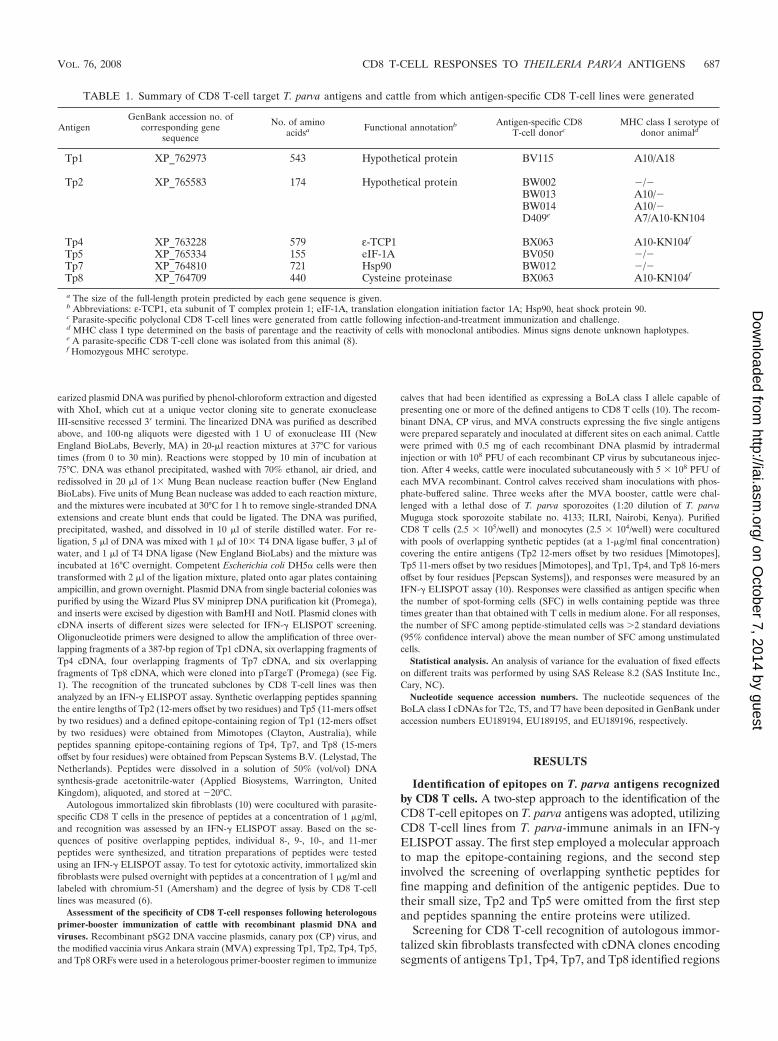
TABLE 1. Summary of CD8 T-cell target T. parva antigens and cattle from which antigen-specific CD8 T-cell lines were generated
AntigenGenBank accession no. of
corresponding genesequence
No. of aminoacidsa Functional annotationb Antigen-specific CD8
T-cell donorcMHC class I serotype of
donor animald
Tp1 XP_762973 543 Hypothetical protein BV115 A10/A18
Tp2 XP_765583 174 Hypothetical protein BW002 �/�BW013 A10/�BW014 A10/�D409e A7/A10-KN104
Tp4 XP_763228 579 ε-TCP1 BX063 A10-KN104f
Tp5 XP_765334 155 eIF-1A BV050 �/�Tp7 XP_764810 721 Hsp90 BW012 �/�Tp8 XP_764709 440 Cysteine proteinase BX063 A10-KN104f
a The size of the full-length protein predicted by each gene sequence is given.b Abbreviations: ε-TCP1, eta subunit of T complex protein 1; eIF-1A, translation elongation initiation factor 1A; Hsp90, heat shock protein 90.c Parasite-specific polyclonal CD8 T-cell lines were generated from cattle following infection-and-treatment immunization and challenge.d MHC class I type determined on the basis of parentage and the reactivity of cells with monoclonal antibodies. Minus signs denote unknown haplotypes.e A parasite-specific CD8 T-cell clone was isolated from this animal (8).f Homozygous MHC serotype.
VOL. 76, 2008 CD8 T-CELL RESPONSES TO THEILERIA PARVA ANTIGENS 687
on October 7, 2014 by guest
http://iai.asm.org/
Dow
nloaded from
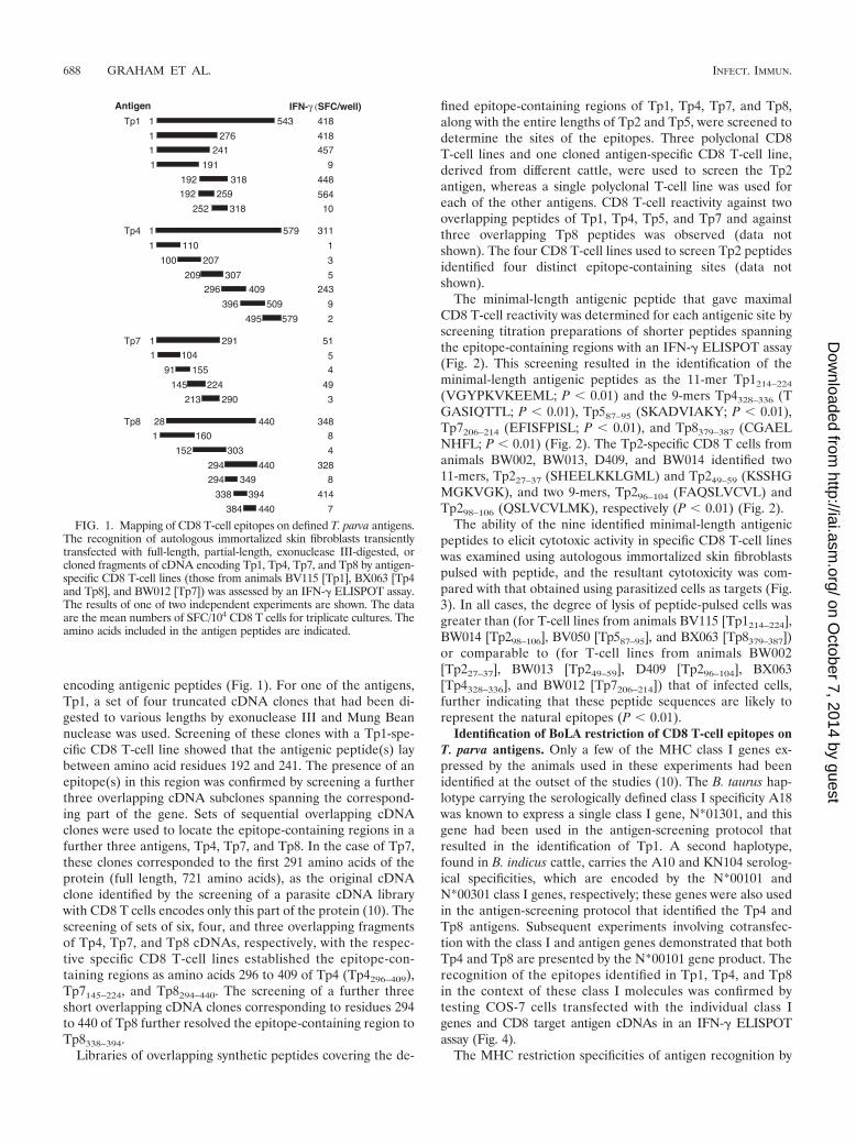
encoding antigenic peptides (Fig. 1). For one of the antigens,Tp1, a set of four truncated cDNA clones that had been di-gested to various lengths by exonuclease III and Mung Beannuclease was used. Screening of these clones with a Tp1-spe-cific CD8 T-cell line showed that the antigenic peptide(s) laybetween amino acid residues 192 and 241. The presence of anepitope(s) in this region was confirmed by screening a furtherthree overlapping cDNA subclones spanning the correspond-ing part of the gene. Sets of sequential overlapping cDNAclones were used to locate the epitope-containing regions in afurther three antigens, Tp4, Tp7, and Tp8. In the case of Tp7,these clones corresponded to the first 291 amino acids of theprotein (full length, 721 amino acids), as the original cDNAclone identified by the screening of a parasite cDNA librarywith CD8 T cells encodes only this part of the protein (10). Thescreening of sets of six, four, and three overlapping fragmentsof Tp4, Tp7, and Tp8 cDNAs, respectively, with the respec-tive specific CD8 T-cell lines established the epitope-con-taining regions as amino acids 296 to 409 of Tp4 (Tp4296–409),Tp7145–224, and Tp8294–440. The screening of a further threeshort overlapping cDNA clones corresponding to residues 294to 440 of Tp8 further resolved the epitope-containing region toTp8338–394.
Libraries of overlapping synthetic peptides covering the de-
fined epitope-containing regions of Tp1, Tp4, Tp7, and Tp8,along with the entire lengths of Tp2 and Tp5, were screened todetermine the sites of the epitopes. Three polyclonal CD8T-cell lines and one cloned antigen-specific CD8 T-cell line,derived from different cattle, were used to screen the Tp2antigen, whereas a single polyclonal T-cell line was used foreach of the other antigens. CD8 T-cell reactivity against twooverlapping peptides of Tp1, Tp4, Tp5, and Tp7 and againstthree overlapping Tp8 peptides was observed (data notshown). The four CD8 T-cell lines used to screen Tp2 peptidesidentified four distinct epitope-containing sites (data notshown).
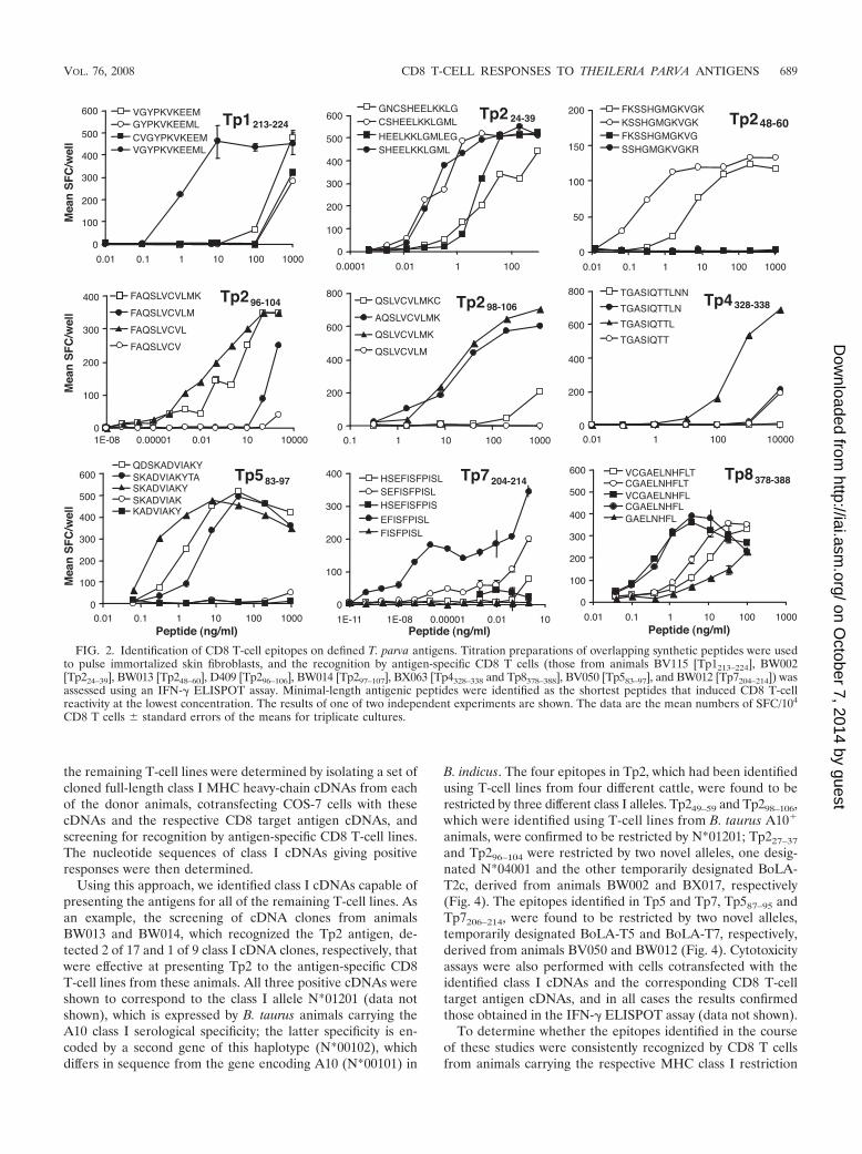
The minimal-length antigenic peptide that gave maximalCD8 T-cell reactivity was determined for each antigenic site byscreening titration preparations of shorter peptides spanningthe epitope-containing regions with an IFN-� ELISPOT assay(Fig. 2). This screening resulted in the identification of theminimal-length antigenic peptides as the 11-mer Tp1214–224
(VGYPKVKEEML; P 0.01) and the 9-mers Tp4328–336 (TGASIQTTL; P 0.01), Tp587–95 (SKADVIAKY; P 0.01),Tp7206–214 (EFISFPISL; P 0.01), and Tp8379–387 (CGAELNHFL; P 0.01) (Fig. 2). The Tp2-specific CD8 T cells fromanimals BW002, BW013, D409, and BW014 identified two11-mers, Tp227–37 (SHEELKKLGML) and Tp249–59 (KSSHGMGKVGK), and two 9-mers, Tp296–104 (FAQSLVCVL) andTp298–106 (QSLVCVLMK), respectively (P 0.01) (Fig. 2).
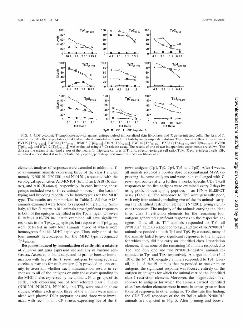
The ability of the nine identified minimal-length antigenicpeptides to elicit cytotoxic activity in specific CD8 T-cell lineswas examined using autologous immortalized skin fibroblastspulsed with peptide, and the resultant cytotoxicity was com-pared with that obtained using parasitized cells as targets (Fig.3). In all cases, the degree of lysis of peptide-pulsed cells wasgreater than (for T-cell lines from animals BV115 [Tp1214–224],BW014 [Tp298–106], BV050 [Tp587–95], and BX063 [Tp8379–387])or comparable to (for T-cell lines from animals BW002[Tp227–37], BW013 [Tp249–59], D409 [Tp296–104], BX063[Tp4328–336], and BW012 [Tp7206–214]) that of infected cells,further indicating that these peptide sequences are likely torepresent the natural epitopes (P 0.01).
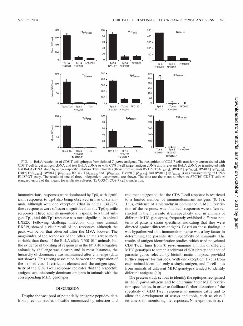
Identification of BoLA restriction of CD8 T-cell epitopes onT. parva antigens. Only a few of the MHC class I genes ex-pressed by the animals used in these experiments had beenidentified at the outset of the studies (10). The B. taurus hap-lotype carrying the serologically defined class I specificity A18was known to express a single class I gene, N*01301, and thisgene had been used in the antigen-screening protocol thatresulted in the identification of Tp1. A second haplotype,found in B. indicus cattle, carries the A10 and KN104 serolog-ical specificities, which are encoded by the N*00101 andN*00301 class I genes, respectively; these genes were also usedin the antigen-screening protocol that identified the Tp4 andTp8 antigens. Subsequent experiments involving cotransfec-tion with the class I and antigen genes demonstrated that bothTp4 and Tp8 are presented by the N*00101 gene product. Therecognition of the epitopes identified in Tp1, Tp4, and Tp8in the context of these class I molecules was confirmed bytesting COS-7 cells transfected with the individual class Igenes and CD8 target antigen cDNAs in an IFN-� ELISPOTassay (Fig. 4).
The MHC restriction specificities of antigen recognition by
FIG. 1. Mapping of CD8 T-cell epitopes on defined T. parva antigens.The recognition of autologous immortalized skin fibroblasts transientlytransfected with full-length, partial-length, exonuclease III-digested, orcloned fragments of cDNA encoding Tp1, Tp4, Tp7, and Tp8 by antigen-specific CD8 T-cell lines (those from animals BV115 [Tp1], BX063 [Tp4and Tp8], and BW012 [Tp7]) was assessed by an IFN-� ELISPOT assay.The results of one of two independent experiments are shown. The dataare the mean numbers of SFC/104 CD8 T cells for triplicate cultures. Theamino acids included in the antigen peptides are indicated.
688 GRAHAM ET AL. INFECT. IMMUN.
on October 7, 2014 by guest
http://iai.asm.org/
Dow
nloaded from
the remaining T-cell lines were determined by isolating a set ofcloned full-length class I MHC heavy-chain cDNAs from eachof the donor animals, cotransfecting COS-7 cells with thesecDNAs and the respective CD8 target antigen cDNAs, andscreening for recognition by antigen-specific CD8 T-cell lines.The nucleotide sequences of class I cDNAs giving positiveresponses were then determined.
Using this approach, we identified class I cDNAs capable ofpresenting the antigens for all of the remaining T-cell lines. Asan example, the screening of cDNA clones from animalsBW013 and BW014, which recognized the Tp2 antigen, de-tected 2 of 17 and 1 of 9 class I cDNA clones, respectively, thatwere effective at presenting Tp2 to the antigen-specific CD8T-cell lines from these animals. All three positive cDNAs wereshown to correspond to the class I allele N*01201 (data notshown), which is expressed by B. taurus animals carrying theA10 class I serological specificity; the latter specificity is en-coded by a second gene of this haplotype (N*00102), whichdiffers in sequence from the gene encoding A10 (N*00101) in
B. indicus. The four epitopes in Tp2, which had been identifiedusing T-cell lines from four different cattle, were found to berestricted by three different class I alleles. Tp249–59 and Tp298–106,which were identified using T-cell lines from B. taurus A10�
animals, were confirmed to be restricted by N*01201; Tp227–37
and Tp296–104 were restricted by two novel alleles, one desig-nated N*04001 and the other temporarily designated BoLA-T2c, derived from animals BW002 and BX017, respectively(Fig. 4). The epitopes identified in Tp5 and Tp7, Tp587–95 andTp7206–214, were found to be restricted by two novel alleles,temporarily designated BoLA-T5 and BoLA-T7, respectively,derived from animals BV050 and BW012 (Fig. 4). Cytotoxicityassays were also performed with cells cotransfected with theidentified class I cDNAs and the corresponding CD8 T-celltarget antigen cDNAs, and in all cases the results confirmedthose obtained in the IFN-� ELISPOT assay (data not shown).
To determine whether the epitopes identified in the courseof these studies were consistently recognized by CD8 T cellsfrom animals carrying the respective MHC class I restriction
FIG. 2. Identification of CD8 T-cell epitopes on defined T. parva antigens. Titration preparations of overlapping synthetic peptides were usedto pulse immortalized skin fibroblasts, and the recognition by antigen-specific CD8 T cells (those from animals BV115 [Tp1213–224], BW002[Tp224–39], BW013 [Tp248–60], D409 [Tp296–106], BW014 [Tp297–107], BX063 [Tp4328–338 and Tp8378–388], BV050 [Tp583–97], and BW012 [Tp7204–214]) wasassessed using an IFN-� ELISPOT assay. Minimal-length antigenic peptides were identified as the shortest peptides that induced CD8 T-cellreactivity at the lowest concentration. The results of one of two independent experiments are shown. The data are the mean numbers of SFC/104
CD8 T cells � standard errors of the means for triplicate cultures.
VOL. 76, 2008 CD8 T-CELL RESPONSES TO THEILERIA PARVA ANTIGENS 689
on October 7, 2014 by guest
http://iai.asm.org/
Dow
nloaded from
elements, analyses of responses were extended to additional T.parva-immune animals expressing three of the class I alleles,namely, N*00101, N*01301, and N*01201, associated with theserological specificities A10-KN104 (B. indicus), A18 (B. tau-rus), and A10 (B.taurus), respectively. In each instance, thesegroups included two or three animals known, on the basis oftyping and breeding records, to be homozygous for the MHCtype. The results are summarized in Table 2. All five A18�
animals examined were found to respond to Tp1214–224. Simi-larly, all five B. taurus A10� animals gave significant responsesto both of the epitopes identified in the Tp2 antigen. Of sevenB. indicus A10-KN104� cattle examined, all gave significantresponses to the Tp8379–387 epitope, but responses to Tp4328–336
were detected in only four animals, three of which werehomozygous for this MHC haplotype. Thus, only one of thefour animals heterozygous for the MHC type recognizedTp4328–336.
Responses induced by immunization of cattle with a mixtureof T. parva antigens expressed individually in vaccine con-structs. Access to animals subjected to primer-booster immu-nization with five of the T. parva antigens by using separatevaccine constructs for each antigen (10) provided an opportu-nity to ascertain whether such immunization results in re-sponses to all of the antigens or only those corresponding tothe MHC alleles expressed by the animals. Four groups of sixcattle, each expressing one of four selected class I alleles(N*01301, N*01201, N*00101, and T5), were used in thesestudies. Within each group, three of the animals were immu-nized with plasmid DNA preparations and three were immu-nized with recombinant CP viruses expressing five of the T.
parva antigens (Tp1, Tp2, Tp4, Tp5, and Tp8). After 4 weeks,all animals received a booster dose of recombinant MVA ex-pressing the same antigens and were then challenged with T.parva sporozoites after a further 3 weeks. Specific CD8 T-cellresponses to the five antigens were examined every 7 days byusing pools of overlapping peptides in an IFN-� ELISPOTassay (Table 3). The responses to Tp2 were generally poor,with only four animals, including two of the six animals carry-ing the identified restriction element (N*1201), giving signifi-cant responses. The majority of the animals carrying the iden-tified class I restriction elements for the remaining fourantigens generated significant responses to the respective an-tigens. Thus, all six T5� animals responded to Tp5, allN*01301� animals responded to Tp1, and five of six N*00101�
animals responded to both Tp4 and Tp8. By contrast, many ofthe animals failed to give significant responses to the antigensfor which they did not carry an identified class I restrictionelement. Thus, none of the remaining 18 animals responded toTp5, and only one and two N*00101-negative animals re-sponded to Tp4 and Tp8, respectively. A larger number (6 of18) of the N*01301-negative animals responded to Tp1. Over-all, in 11 of the 19 animals that responded to one or moreantigens, the significant response was focused entirely on theantigen or antigens for which the animal carried the identifiedclass I restriction element. Moreover, the magnitudes of re-sponses to antigens for which the animals carried identifiedclass I restriction elements were in most instances greater thanthose of responses to other antigens. To illustrate this finding,the CD8 T-cell responses of the six BoLA allele N*00101�
animals are depicted in Fig. 5. After priming and booster
FIG. 3. CD8 cytotoxic-T-lymphocyte activity against epitope-pulsed immortalized skin fibroblasts and T. parva-infected cells. The lysis of T.parva-infected cells and peptide-pulsed and unpulsed immortalized skin fibroblasts by antigen-specific cytotoxic T lymphocytes (those from animalsBV115 [Tp1214–224], BW002 [Tp227–37], BW013 [Tp249–59], D409 [Tp296–104], BW014 [Tp298–106], BX063 [Tp4328–336 and Tp8379–387], BV050[Tp587–95], and BW012 [Tp7206–214]) was evaluated using a 51Cr release assay. The results of one of two independent experiments are shown. Thedata are the means � standard errors of the means for triplicate cultures. E:T ratio, effector-to-target cell ratio; TpM, T. parva-infected cells; iSF,unpulsed immortalized skin fibroblasts; iSF peptide, peptide-pulsed immortalized skin fibroblasts.
690 GRAHAM ET AL. INFECT. IMMUN.
on October 7, 2014 by guest
http://iai.asm.org/
Dow
nloaded from
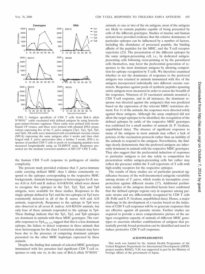
immunizations, responses were dominated by Tp8, with signif-icant responses to Tp4 also being observed in five of six ani-mals, although with one exception (that in animal BX225),these responses were of lesser magnitude than the Tp8-specificresponses. Three animals mounted a response to a third anti-gen, Tp1, and this Tp1 response was most significant in animalBX225. Following challenge infection, only one animal,BX219, showed a clear recall of the responses, although thepeak was below that observed after the MVA booster. Themagnitudes of the responses of the other animals were morevariable than those of the BoLA allele N*00101� animals, butthe evidence of boosting of responses in the N*00101-negativeanimals by challenge was clearer, and in most instances, thehierarchy of dominance was maintained after challenge (datanot shown). This strong association between the expression ofthe defined class I restriction element and the antigen speci-ficity of the CD8 T-cell response indicates that the respectiveantigens are inherently dominant antigens in animals with thecorresponding MHC genotypes.
DISCUSSION
Despite the vast pool of potentially antigenic peptides, datafrom previous studies of cattle immunized by infection and
treatment suggested that the CD8 T-cell response is restrictedto a limited number of immunodominant antigens (8, 19).Thus, evidence of a hierarchy in dominance in MHC restric-tion of the response was obtained; responses were often re-stricted in their parasite strain specificity and, in animals ofdifferent MHC genotypes, frequently exhibited different pat-terns of parasite strain specificity, indicating that they weredirected against different antigens. Based on these findings, itwas hypothesized that immunodominance was a key factor indetermining the parasite strain specificity of immunity. Theresults of antigen identification studies, which used polyclonalCD8 T-cell lines from T. parva-immune animals of differentMHC genotypes to screen a schizont cDNA library and a set ofparasite genes selected by bioinformatic analyses, providedfurther support for this idea. With one exception, T cells fromeach animal identified only a single antigen, and T-cell linesfrom animals of different MHC genotypes tended to identifydifferent antigens (10).
The present study set out to identify the epitopes recognizedin the T. parva antigens and to determine their MHC restric-tion specificities, in order to facilitate further dissection of thespecificity of CD8 T-cell responses in immune cattle and toallow the development of assays and tools, such as class Itetramers, for monitoring the responses. Nine epitopes in six T.
FIG. 4. BoLA restriction of CD8 T-cell epitopes from defined T. parva antigens. The recognition of COS-7 cells transiently cotransfected withCD8 T-cell target antigen cDNA and test BoLA cDNA or with CD8 T-cell target antigen cDNA and irrelevant BoLA cDNA or transfected withtest BoLA cDNA alone by antigen-specific cytotoxic T lymphocytes (those from animals BV115 [Tp1214–224], BW002 [Tp227–37], BW013 [Tp249–59],D409 [Tp296–104], BW014 [Tp298–106], BX063 [Tp4328–336 and Tp8379–387], BV050 [Tp587–95], and BW012 [Tp7206–214]) was assessed using an IFN-�ELISPOT assay. The results of one of three independent experiments are shown. The data are the mean numbers of SFC/104 CD8 T cells �standard errors of the means for triplicate cultures. Tx COS-7, COS-7 cell transfection.
VOL. 76, 2008 CD8 T-CELL RESPONSES TO THEILERIA PARVA ANTIGENS 691
on October 7, 2014 by guest
http://iai.asm.org/
Dow
nloaded from
parva antigens, together with their respective MHC restrictionelements, were successfully identified. This report provides thefirst description of CD8 T-cell epitopes and correspondingMHC class I restriction elements in cattle. The epitopes, asdefined by the minimal peptide length that induced maximalT-cell reactivity, were either 9-mers or 11-mers. In all but onecase, CD8 T cells derived from eight different animals recog-nized only one of the six antigens. Single epitopes were iden-tified in five of the six antigens; the epitopes in two of theseantigens (Tp4 and Tp8) were restricted by the same class Iallele, while those in the other three antigens (Tp1, Tp5, andTp7) were restricted by different class I alleles. The Tp2 anti-gen was found to contain four epitopes, two of which wererestricted by the same MHC class I allele, while the other twoepitopes were restricted by different alleles.
The detection of responses to only one of the six antigens, inmany cases focused on a single epitope, supports previousevidence that there is a dominance hierarchy in the antigensrecognized in CD8 T-cell responses to T. parva. Further studiesare needed to determine what proportion of the response inindividual immune animals is directed at these antigens inorder to provide quantitative data on the extent to which theseantigens dominate the response. Immunodominance in out-bred populations is a well-described phenomenon for CD8T-cell responses to viruses and cancer, in which the pool ofpotential antigenic peptides is relatively small in comparison tothose in response to complex intracellular bacterial and pro-tozoan pathogens. The screening of Plasmodium falciparumand Mycobacterium tuberculosis for antigens and epitopes byusing CD8 T cells from immunized or exposed humans hasshown the recognition of a relatively large number of antigens,rather than a narrow focus on a few immunodominant proteinsand epitopes (2, 18). The results of this and earlier studies (8,19) suggest that immunodominance may play a much greaterrole in the bovine CD8 T-cell response to T. parva than in
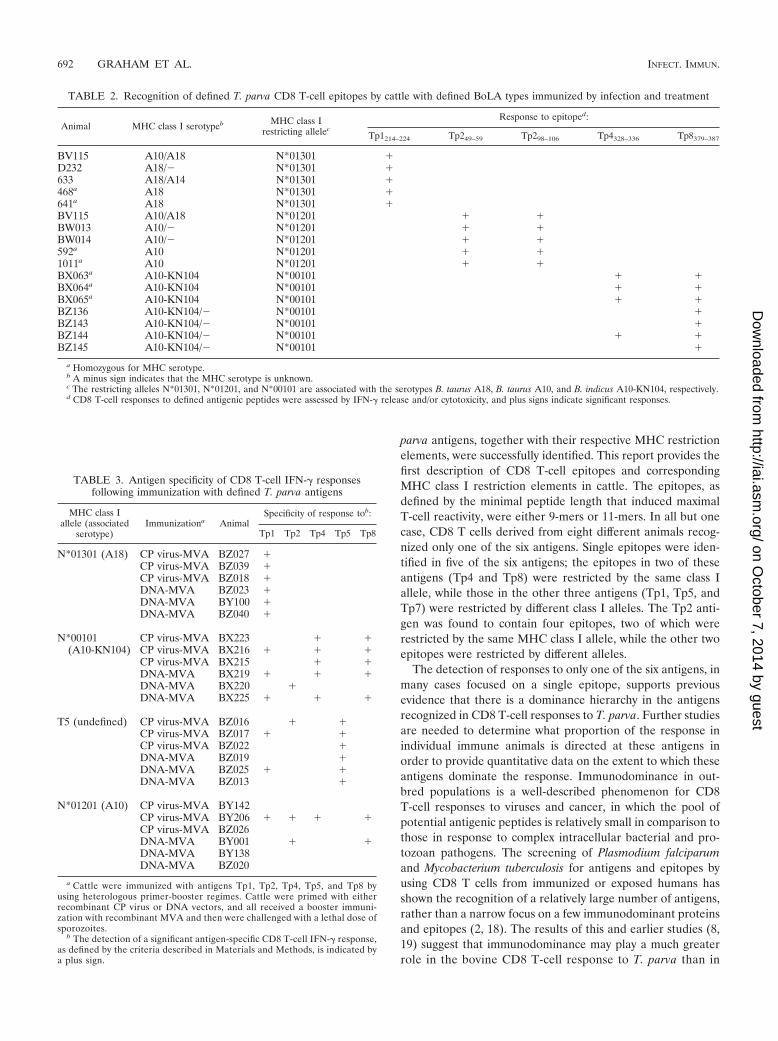
TABLE 2. Recognition of defined T. parva CD8 T-cell epitopes by cattle with defined BoLA types immunized by infection and treatment
Animal MHC class I serotypeb MHC class Irestricting allelec
Response to epitoped:
Tp1214–224 Tp249–59 Tp298–106 Tp4328–336 Tp8379–387
BV115 A10/A18 N*01301 �D232 A18/� N*01301 �633 A18/A14 N*01301 �468a A18 N*01301 �641a A18 N*01301 �BV115 A10/A18 N*01201 � �BW013 A10/� N*01201 � �BW014 A10/� N*01201 � �592a A10 N*01201 � �1011a A10 N*01201 � �BX063a A10-KN104 N*00101 � �BX064a A10-KN104 N*00101 � �BX065a A10-KN104 N*00101 � �BZ136 A10-KN104/� N*00101 �BZ143 A10-KN104/� N*00101 �BZ144 A10-KN104/� N*00101 � �BZ145 A10-KN104/� N*00101 �
a Homozygous for MHC serotype.b A minus sign indicates that the MHC serotype is unknown.c The restricting alleles N*01301, N*01201, and N*00101 are associated with the serotypes B. taurus A18, B. taurus A10, and B. indicus A10-KN104, respectively.d CD8 T-cell responses to defined antigenic peptides were assessed by IFN-� release and/or cytotoxicity, and plus signs indicate significant responses.
TABLE 3. Antigen specificity of CD8 T-cell IFN-� responsesfollowing immunization with defined T. parva antigens
MHC class Iallele (associated
serotype)Immunizationa Animal
Specificity of response tob:
Tp1 Tp2 Tp4 Tp5 Tp8
N*01301 (A18) CP virus-MVA BZ027 �CP virus-MVA BZ039 �CP virus-MVA BZ018 �DNA-MVA BZ023 �DNA-MVA BY100 �DNA-MVA BZ040 �
N*00101(A10-KN104)
CP virus-MVA BX223 � �CP virus-MVA BX216 � � �CP virus-MVA BX215 � �DNA-MVA BX219 � � �DNA-MVA BX220 �DNA-MVA BX225 � � �
T5 (undefined) CP virus-MVA BZ016 � �CP virus-MVA BZ017 � �CP virus-MVA BZ022 �DNA-MVA BZ019 �DNA-MVA BZ025 � �DNA-MVA BZ013 �
N*01201 (A10) CP virus-MVA BY142CP virus-MVA BY206 � � � �CP virus-MVA BZ026DNA-MVA BY001 � �DNA-MVA BY138DNA-MVA BZ020
a Cattle were immunized with antigens Tp1, Tp2, Tp4, Tp5, and Tp8 byusing heterologous primer-booster regimes. Cattle were primed with eitherrecombinant CP virus or DNA vectors, and all received a booster immuni-zation with recombinant MVA and then were challenged with a lethal dose ofsporozoites.
b The detection of a significant antigen-specific CD8 T-cell IFN-� response,as defined by the criteria described in Materials and Methods, is indicated bya plus sign.
692 GRAHAM ET AL. INFECT. IMMUN.
on October 7, 2014 by guest
http://iai.asm.org/
Dow
nloaded from
the human CD8 T-cell response to pathogens of similarcomplexity.
The present study provided evidence that T. parva-immunecattle carrying defined MHC class I alleles consistently re-spond to the epitopes corresponding to the respective MHCbackgrounds. Animals homozygous or heterozygous for B. tau-rus A18 or A10 and B. indicus A10-KN104, which were shownto recognize five epitopes in the Tp1, Tp2, Tp4, and Tp8antigens, were available for these studies. Responses to thesingle epitope defined in Tp1 and the two epitopes in Tp2 wereconsistently detected in all of the B. taurus A18 and A10animals, respectively. Responses to the epitope in Tp8 werealso observed in all seven B. indicus A10-KN104 animals, butonly four of these animals responded to the epitope in Tp4.These findings indicate that the Tp1, Tp2, and Tp8 epitopesare dominant in animals with these MHC genotypes. The vari-able responses to Tp4328–336 suggest that this is a less-dominantepitope; the failure to detect responses in three animals thatwere heterozygous for the class I restriction element may havebeen due to the presence of competing dominant epitopespresented via the other MHC haplotype expressed by theseanimals.
Despite the finding that animals of selected MHC genotypesimmunized with live parasites had significant CD8 T-cell re-sponses to only one or, in the case of BoLA allele N*00101�
animals, to one or two of the six antigens, most of the antigensare likely to contain peptides capable of being presented bycells of the different genotypes. Studies of murine and humansystems have provided evidence that the relative dominance ofparticular epitopes can be influenced by a number of factors,including the abundance of processed peptide, the bindingaffinity of the peptides for the MHC, and the T-cell receptorrepertoire (23). The presentation of the different epitopes bythe same antigen-presenting cell, i.e., by dedicated antigen-presenting cells following cross-priming or by the parasitizedcells themselves, may favor the preferential generation of re-sponses to the most dominant antigens by allowing competi-tion for epitope recognition by T cells. We therefore examinedwhether or not the dominance of responses to the preferredantigens was retained in animals immunized with five of theantigens incorporated individually into different vaccine con-structs. Responses against pools of synthetic peptides spanningentire antigens were measured in order to assess the breadth ofthe responses. Nineteen of 24 vaccinated animals mounted aCD8 T-cell response, and in all instances, the dominant re-sponse was directed against the antigen(s) that was predictedbased on the expression of the relevant MHC restriction ele-ment. For 11 of the animals, the responses were directed solelyagainst these antigens. Although the assay employed did notallow the target epitopes to be identified, the recognition of thedefined epitopes by cattle of the respective MHC genotypeswas confirmed for a small number of animals (S. P. Graham,unpublished data). The absence of significant responses tomany of the antigens in most animals may reflect a lack ofpotency of the vaccination protocols rather than an inability ofthe animals to respond to the antigens. Nevertheless, the find-ings clearly demonstrate that the preferred antigens are inher-ently dominant in animals with the respective MHC genotypes.They also suggest that the preferential induction of responsesto particular antigens is not due solely to competition forpresentation within antigen-presenting cells but rather mayreflect the presence within the T-cell repertoire of T cells withhigh-avidity receptors for the respective epitopes.
The results of these studies are of particular practical sig-nificance because of the well-documented antigenic variabilityamong strains of T. parva, which results in incomplete cross-protection against different strains (17). Additional prelimi-nary studies of the antigens described herein have confirmedthat the defined epitope regions vary in sequence among par-asite strains and are differentially recognized by CD8 T cells(R. Pelle and S. P. Graham, unpublished data). Hence, a majorchallenge in the development of a vaccine based on the induc-tion of CD8 T-cell responses will be to generate responses thatare effective against all parasite strains. Further studies arerequired to provide a more comprehensive picture of the an-tigen recognition capacity of animals of different MHC geno-types to ascertain whether combinations of antigens that po-tentially provide broad protection can be identified and used toinduce protective CD8 T-cell responses.
ACKNOWLEDGMENTS
This work was funded by the Animal Health Programme of theUnited Kingdom Department for International Development (DFID;project number R8042). Y.H. was supported in part by the Ministry ofForeign Affairs of the government of Japan.
FIG. 5. Antigen specificity of CD8 T cells from BoLA alleleN*00101� cattle vaccinated with defined antigens by using heterolo-gous primer-booster regimens. Three cattle were primed with recom-binant CP viruses, and three were primed with plasmid DNA prepa-rations expressing five of the T. parva antigens (Tp1, Tp2, Tp4, Tp5,and Tp8). All cattle were immunized with recombinant vaccinia viruses(MVA) expressing the same antigens after 4 weeks and then chal-lenged with T. parva sporozoites after a further 3 weeks. IFN-� re-sponses of purified CD8 T cells to pools of overlapping peptides weremeasured longitudinally using an ELISPOT assay. Responses pre-sented are the medium corrected numbers of SFC/106 CD8 T cells.
VOL. 76, 2008 CD8 T-CELL RESPONSES TO THEILERIA PARVA ANTIGENS 693
on October 7, 2014 by guest
http://iai.asm.org/
Dow
nloaded from
We acknowledge the staff of the ILRI animal sampling group, large-animal and tick units, for excellent animal care and provision of par-asites. We thank the ILRI Biometrics Unit for advice on experimentaldesign and statistical analysis. Merial is kindly acknowledged for pro-vision of the recombinant CP virus constructs.
This is ILRI publication no. IL-200705.
REFERENCES
1. Ballingall, K. T., D. M. Mwangi, N. D. MacHugh, E. L. Taracha, P. Totte,and D. J. McKeever. 2000. A highly sensitive, non-radioactive assay for T cellactivation in cattle: applications in screening for antigens recognised byCD4� and CD8� T cells. J. Immunol. Methods 239:85–93.
2. Doolan, D. L., S. Southwood, D. A. Freilich, J. Sidney, N. L. Graber, L.Shatney, L. Bebris, L. Florens, C. Dobano, A. A. Witney, E. Appella, S. L.Hoffman, J. R. Yates III, D. J. Carucci, and A. Sette. 2003. Identification ofPlasmodium falciparum antigens by antigenic analysis of genomic and pro-teomic data. Proc. Natl. Acad. Sci. USA 100:9952–9957.
3. Doolan, D. L., and N. Martinez-Alier. 2006. Immune response to pre-eryth-rocytic stages of malaria parasites. Curr. Mol. Med. 6:169–185.
4. Ellis, S. A., K. A. Staines, M. J. Stear, E. J. Hensen, and W. I. Morrison.1998. DNA typing for BoLA class I using sequence-specific primers (PCR-SSP). Eur. J. Immunogenet. 25:365–370.
5. Ellis, S. A., W. I. Morrison, N. D. MacHugh, J. Birch, A. Burrells, and M. J.Stear. 2005. Serological and molecular diversity in the cattle MHC class Iregion. Immunogenetics 57:601–606.
6. Goddeeris, B. M., and W. I. Morrison. 1988. Techniques for the generation,cloning and characterization of bovine cytotoxic T cells specific for theprotozoan Theileria parva. Methods Cell Sci. 11:101–110.
7. Goddeeris, B. M., W. I. Morrison, A. J. Teale, A. Bensaid, and C. L. Baldwin.1986. Bovine cytotoxic T-cell clones specific for cells infected with the pro-tozoan parasite Theileria parva: parasite strain specificity and class I majorhistocompatibility complex restriction. Proc. Natl. Acad. Sci. USA 83:5238–5242.
8. Goddeeris, B. M., W. I. Morrison, P. G. Toye, and R. Bishop. 1990. Strainspecificity of bovine Theileria parva-specific cytotoxic T cells is determinedby the phenotype of the restricting class I MHC. Immunology 69:38–44.
9. Good, M. F. 2005. Vaccine-induced immunity to malaria parasites and theneed for novel strategies. Trends Parasitol. 21:29–34.
10. Graham, S. P., R. Pelle, Y. Honda, D. M. Mwangi, N. J. Tonukari, M.Yamage, E. J. Glew, E. P. de Villiers, T. Shah, R. Bishop, E. Abuya, E. Awino,J. Gachanja, A. E. Luyai, F. Mbwika, A. M. Muthiani, D. M. Ndegwa, M.Njahira, J. K. Nyanjui, F. O. Onono, J. Osaso, R. M. Saya, C. Wildmann,C. M. Fraser, I. Maudlin, M. J. Gardner, S. P. Morzaria, S. Loosmore S. C.Gilbert, J. C. Audonnet, P. van der Bruggen, V. Nene, and E. L. N. Taracha.
2006. Theileria parva candidate vaccine antigens recognized by immunebovine cytotoxic T lymphocytes. Proc. Natl. Acad. Sci. USA 103:3286–3291.
11. Graham, S. P., Y. Honda, R. Pelle, D. M. Mwangi, E. J. Glew, E. P. deVilliers, T. Shah, R. Bishop, P. van der Bruggen, V. Nene, and E. L. N.Taracha. 2007. A novel strategy for the identification of antigens that arerecognised by bovine MHC class I restricted cytotoxic T cells in a protozoaninfection using reverse vaccinology. Immunome Res. 3:2.
12. McKeever, D. J., E. Awino, and W. I. Morrison. 1992. Afferent lymph veiledcells prime CD4� T cell responses in vivo. Eur. J. Immunol. 22:3057–3061.
13. McKeever, D. J., E. L. N. Taracha, E. L. Innes, N. D. MacHugh, E. Awino,B. M. Goddeeris, and W. I. Morrison. 1994. Adoptive transfer of immunityto Theileria parva in the CD8� fraction of responding efferent lymph. Proc.Natl. Acad. Sci. USA 91:1959–1963.
14. McKeever, D. J., E. L. Taracha, W. I. Morrison, A. J. Musoke, and S. P.Morzaria. 1999. Protective immune mechanisms against Theileria parva:evolution of vaccine development strategies. Parasitol. Today 15:263–267.
15. Morrison, W. I., B. M. Goddeeris, A. J. Teale, C. M. Groocock, S. J. Kemp,and D. A. Stagg. 1987. Cytotoxic T cells elicited in cattle challenged withTheileria parva (Muguga): evidence for restriction by class I MHC determi-nants and parasite strain specificity. Parasite Immunol. 9:563–578.
16. Norval, R. A. I., B. D. Perry, and A. S. Young. 1992. The epidemiology ofTheileria in Africa. Academic Press Ltd., London, United Kingdom.
17. Radley, D. E., C. G. D. Brown, M. P. Cunningham, C. D. Kimber, F. L.Musisi, R. C. Payne, R. E. Purnell, S. M. Stagg, and A. S. Young. 1975.Chemoprophylactic immunization of cattle against Theileria parva (Muguga)and five Theileria strains. Vet. Parasitol. 1:35–41.
18. Sable, S. B., S. Kaur, I. Verma, and G. K. Khuller. 2005. Immunodominanceof low molecular weight secretory polypeptides of Mycobacterium tubercu-losis to induce cytotoxic T-lymphocyte response. Vaccine 23:4947–4954.
19. Taracha, E. L. N., B. M. Goddeeris, S. P. Morzaria, and W. I. Morrison.1995. Parasite strain specificity of precursor cytotoxic T cells in individualanimals correlates with cross-protection in cattle challenged with Theileriaparva. Infect. Immun. 63:1258–1262.
20. Taracha, E. L., B. M. Goddeeris, A. J. Teale, S. J. Kemp, and W. I. Morrison.1995. Parasite strain specificity of bovine cytotoxic T cell responses to Thei-leria parva is determined primarily by immunodominance. J. Immunol. 155:4854–4860.
21. Teale, A. J., S. J. Kemp, F. Young, and R. L. Spooner. 1983. Selection bymajor histocompatibility type (BoLA) of lymphoid cells derived from abovine chimaera and transformed by Theileria parasites. Parasite Immunol.5:329–335.
22. Tsuji, M., and F. Zavala. 2003. T cells as mediators of protective immunityagainst liver stages of Plasmodium. Trends Parasitol. 19:88–93.
23. Yewdell, J. W., and J. R. Bennink. 1999. Immunodominance in major histo-compatibility complex class I-restricted T lymphocyte responses. Annu. Rev.Immunol. 17:51–88.
Editor: J. F. Urban, Jr.
694 GRAHAM ET AL. INFECT. IMMUN.
on October 7, 2014 by guest
http://iai.asm.org/
Dow
nloaded from
Copyright © 2022 FDOKUMEN