Continuous in vitro culture of the carpet shell clam Tapes decussatus protozoan parasite Perkinsus...
15
DISEASES OF AQUATIC ORGANISMS Dis Aquat Org Vol. 52: 217–231, 2002 Published December 10 INTRODUCTION Perkinsus atlanticus has been associated with mass mortalities of the carpet shell clam Tapes decussatus in estuarine regions of the Portuguese and Spanish coasts (Ruano & Cachola 1986, Azevedo 1989, Santmartí et al. 1995). This protozoan parasite was designated as a new Perkinsus species by Azevedo (1989) based on gross morphological and ultrastructural features of various stages of its life cycle. Further evidence that P. atlanticus belongs to the genus Perkinsus was pro- vided from nucleotide sequence analysis of the small subunit (SSU) rRNA gene and the internal transcribed © Inter-Research 2002 · www.int-res.com *Corresponding author. Email: [email protected] Continuous in vitro culture of the carpet shell clam Tapes decussatus protozoan parasite Perkinsus atlanticus Sandra M. Casas 1 , Jerome F. La Peyre 2 , Kimberly S. Reece 3 , Carlos Azevedo 4 , Antonio Villalba 1, * 1 Centro de Investigacións Mariñas, Consellería de Pesca e Asuntos Marítimos, Xunta de Galicia, Aptdo. 13, 36620 Vilanova de Arousa, Spain 2 Cooperative Aquatic Animal Health Research Program, Department of Veterinary Science, Louisiana State University Agricultural Center, Baton Rouge, Louisiana 70803, USA 3 School of Marine Science, Virginia Institute of Marine Science, The College of William and Mary, Gloucester Point, Virginia 23062, USA 4 Department of Cellular Biology, Institute of Biomedical Sciences, University of Oporto, 4050 Porto, Portugal ABSTRACT: Continuous in vitro cultures of the clam Tapes decussatus parasite Perkinsus atlanticus were established from infected gill fragments, infected haemolymph and parasite hypnospores isolated from infected gill fragments following incubation in Ray’s fluid thioglycollate medium (RFTM). No continuous cultures could be initiated from P. atlanticus zoospores. Cultures initiated from hypnospores yielded the highest percentage of continuous cultures (100%, 6/6), followed by cultures initiated from gill fragments (93%, 43/46) and from haemolymph (30%, 3/10). Failures to establish continuous cultures were due to microbial contamination. The source of parasite influenced the success rate, the time taken to establish cultures and the size of cultured cells. In vitro prolifera- tion of parasite cells was mainly by vegetative multiplication. Zoosporulation, yielding motile bi- flagellated zoospores, was observed at a low frequency (<1% of dividing cells) in every culture. Morphology of cultured cells examined with light and transmission electron microscopy corre- sponded to that of P. atlanticus found in clam tissues. Cultured cells enlarged in RFTM and stained blue-black with Lugol’s solution, which are characteristics of the Perkinsus species cells. DNA sequences of the internal transcribed spacer (ITS) region of the ribosomal RNA gene complex matched those of P. atlanticus. All cultures were established in a medium designated JL-ODRP-2A that was similar in composition to the culture medium JL-ODRP-1 originally used to propagate Perkinsus marinus in vitro. Proliferation of P. atlanticus in vitro could be supported by the commer- cial culture medium (1:2 v/v) DME:Ham’s F-12 with fetuin. KEY WORDS: Perkinsus atlanticus · Tapes decussatus · In vitro culture · Clam parasite · Ribosomal RNA gene complex · ITS · Ultrastructure Resale or republication not permitted without written consent of the publisher
-
Upload
independent -
Category
Documents
-
view
1 -
download
0
Transcript of Continuous in vitro culture of the carpet shell clam Tapes decussatus protozoan parasite Perkinsus...

DISEASES OF AQUATIC ORGANISMSDis Aquat Org
Vol. 52: 217–231, 2002 Published December 10
INTRODUCTION
Perkinsus atlanticus has been associated with massmortalities of the carpet shell clam Tapes decussatus inestuarine regions of the Portuguese and Spanish coasts
(Ruano & Cachola 1986, Azevedo 1989, Santmartí et al.1995). This protozoan parasite was designated as anew Perkinsus species by Azevedo (1989) based ongross morphological and ultrastructural features ofvarious stages of its life cycle. Further evidence that P.atlanticus belongs to the genus Perkinsus was pro-vided from nucleotide sequence analysis of the smallsubunit (SSU) rRNA gene and the internal transcribed
© Inter-Research 2002 · www.int-res.com
*Corresponding author. Email: [email protected]
Continuous in vitro culture of the carpet shell clamTapes decussatus protozoan parasite Perkinsus
atlanticus
Sandra M. Casas1, Jerome F. La Peyre2, Kimberly S. Reece3, Carlos Azevedo4, Antonio Villalba1,*
1Centro de Investigacións Mariñas, Consellería de Pesca e Asuntos Marítimos, Xunta de Galicia, Aptdo. 13,36620 Vilanova de Arousa, Spain
2Cooperative Aquatic Animal Health Research Program, Department of Veterinary Science, Louisiana State University Agricultural Center, Baton Rouge, Louisiana 70803, USA
3School of Marine Science, Virginia Institute of Marine Science, The College of William and Mary, Gloucester Point,Virginia 23062, USA
4Department of Cellular Biology, Institute of Biomedical Sciences, University of Oporto, 4050 Porto, Portugal
ABSTRACT: Continuous in vitro cultures of the clam Tapes decussatus parasite Perkinsus atlanticuswere established from infected gill fragments, infected haemolymph and parasite hypnosporesisolated from infected gill fragments following incubation in Ray’s fluid thioglycollate medium(RFTM). No continuous cultures could be initiated from P. atlanticus zoospores. Cultures initiatedfrom hypnospores yielded the highest percentage of continuous cultures (100%, 6/6), followed bycultures initiated from gill fragments (93%, 43/46) and from haemolymph (30%, 3/10). Failures toestablish continuous cultures were due to microbial contamination. The source of parasite influencedthe success rate, the time taken to establish cultures and the size of cultured cells. In vitro prolifera-tion of parasite cells was mainly by vegetative multiplication. Zoosporulation, yielding motile bi-flagellated zoospores, was observed at a low frequency (<1% of dividing cells) in every culture.Morphology of cultured cells examined with light and transmission electron microscopy corre-sponded to that of P. atlanticus found in clam tissues. Cultured cells enlarged in RFTM and stainedblue-black with Lugol’s solution, which are characteristics of the Perkinsus species cells. DNAsequences of the internal transcribed spacer (ITS) region of the ribosomal RNA gene complexmatched those of P. atlanticus. All cultures were established in a medium designated JL-ODRP-2Athat was similar in composition to the culture medium JL-ODRP-1 originally used to propagatePerkinsus marinus in vitro. Proliferation of P. atlanticus in vitro could be supported by the commer-cial culture medium (1:2 v/v) DME:Ham’s F-12 with fetuin.
KEY WORDS: Perkinsus atlanticus · Tapes decussatus · In vitro culture · Clam parasite · RibosomalRNA gene complex · ITS · Ultrastructure
Resale or republication not permitted without written consent of the publisher
Dis Aquat Org 52: 217–231, 2002
spacer (ITS) region of the rRNA gene complex (Goggin1994, de la Herrán et al. 2000, Robledo et al. 2000,Casas et al. 2002). Recently, Ordás & Figueras (1998)reported the in vitro culture of P. atlanticus from thehaemolymph of an infected carpet shell clam. Identifi-cation of the cultured cells was based solely on theirmorphology (light and electron microscopy). Sequenceanalysis of the SSU small subunit rRNA gene, however,showed that those cultured cells did not correspond toa Perkinsus organism (Figueras et al. 2000). Continu-ous cultures of P. atlanticus, therefore, still need to beestablished.
The development of continuous cultures of Perkinsusspecies can lead to a better understanding of thisgroup of parasites (La Peyre 1996). Cultures of theeastern oyster pathogen Perkinsus marinus, for exam-ple, have been used in a wide range of studies toaddress the parasite’s environmental tolerance (Burre-son et al. 1994, Dungan & Hamilton 1995), virulence(La Peyre et al. 1995, Bushek & Allen 1996a, Volety &Chu 1997), genetic composition (Reece et al. 1997,Reece et al. 2001), metabolism (Soudant et al. 2000,Soudant & Chu 2001) and drug sensitivity (Krantz1994, Faisal et al. 1999). Since the original culture of P.marinus, there have been reports of at least 2additional Perkinsus species, P. chesapeaki and P.andrewsi, being propagated in vitro. McLaughlin &Faisal (1998) cultured 2 protozoa from the gill (G117)and haemolymph (H49) of an infected softshell clam(Mya arenaria). The gill isolate G117 was furthercharacterised as a new Perkinsus species, P. chesa-peaki (Kotob et al. 1999, McLaughlin et al. 2000).Perkinsus parasites of infected Baltic clams Macomabalthica were cultured by Kleinschuster et al. (1994)and Coss et al. (2001a). The parasite cultured by Cosset al. (2001b) was designated as a new Perkinsus spe-cies, P. andrewsi.
Continuous cultures of Perkinsus chesapeaki and ofP. andrewsi were established using procedures devel-oped to propagate P. marinus. These proceduresvaried greatly among researchers in the type ofmedium used, the culture conditions and the methodused for the establishment of P. marinus primarycultures (reviewed in La Peyre 1996). P. marinus and P.chesapeaki were propagated in the culture mediumJL-ODRP-1 originally formulated to resemble the com-position of eastern oyster plasma (La Peyre et al. 1993,McLaughlin & Faisal 1998). Commercial media includ-ing DMEM:Ham’s F-12 and Leibowitz’s L-15 medium,with various nutritional supplements, were used to cul-ture P. marinus and P. andrewsi (Gauthier & Vasta1993, Kleinschuster & Swink 1993, Kleinschuster et al.1994, Dungan & Hamilton 1995, Coss et al. 2001a).Cultures of the 3 Perkinsus spp. have been establishedfrom various sources including hearts, visceral ganglia
and haemolymph of eastern oysters, gills andhaemolymph of a softshell clam, and heart andhaemolymph of baltic clams (Gauthier & Vasta 1993,Kleinschuster & Swink 1993, La Peyre et al. 1993,McLaughlin & Faisal 1998, Coss et al. 2001a). Parasitehypnospores isolated from infected eastern oystertissues incubated in Ray’s fluid thioglycollate medium(RFTM) were used to quickly establish P. marinus con-tinuous cultures from different geographical regions(La Peyre & Faisal 1995, Bushek & Allen 1996b). It isnot known which source of parasites, if any, is optimalto establish continuous cultures of Perkinsus spp.
This article describes procedures for the continuousin vitro culture of Perkinsus atlanticus and providesevidence that the cultured cells are P. atlanticus. Fourdifferent sources of parasite from infected carpet shellclams were tested to establish P. atlanticus cultures.They included gills, haemolymph, hypnospores iso-lated from infected gill fragments after incubation inRFTM, and zoospores isolated after zoosporulation ofhypnospores in seawater. To confirm that the culturedcells were P. atlanticus, they were characterised byascertaining (1) their morphology at the light and elec-tron microscopic levels, (2) their ability to enlarge inRFTM and subsequently to stain with Lugol’s iodinesolution as described by Ray (1966), and (3) thenucleotide sequence of the ITS region of the rRNAgene complex. Finally, the growth rates of 9 isolates, 3established from the gills, 3 established from haemo-lymph, and 3 established from hypnospores, werecompared in the culture medium JL-ODRP-2A. Con-currently, proliferation of the 3 P. atlanticus gill iso-lates was also evaluated in the commercial mediumDMEM:Ham’s F-12 supplemented with fetuin.
MATERIALS AND METHODS
Clams. Carpet shell clams Tapes decussatus (40 to45 mm) were collected from an intertidal natural bed inthe Ría de Arousa (Galicia, NW Spain) at Vilalonga inMay 2000. Fifty clams were maintained for 12 d in a75 l tank containing 0.22 µm filtered estuarine sea-water at 25 ppt and 15°C. Water in the tank was aer-ated and recirculated through an ultraviolet steriliserto reduce the clams’ bacterial content. Twenty clamswith the highest numbers of whitish pustules on theirgill surfaces (a conspicuous sign of infection by Perkin-sus atlanticus) were selected for P. atlanticus isolationand culture.
Culture medium. The growth culture medium JL-ODRP-2A was used to establish and maintain Perkin-sus atlanticus cultures. This medium was similar incomposition to the culture medium JL-ODRP-1 origi-nally used to propagate P. marinus in vitro (La Peyre et
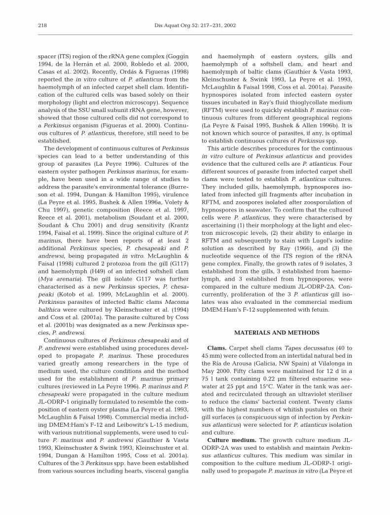
218

Casas et al.: Perkinsus atlanticus in vitro culture
al. 1993) except for a few modifications. JL-ODRP-2Adid not contain bovine serum albumin. Chlorampheni-col in JL-ODRP-1 was replaced by 0.1 g l–1 kanamycin,0.025 g l–1 gentamycin and 100 000 U l–1 penicillin G inJL-ODRP-2A. The Seawater Synthetic Basal SaltMixture of JL-ODRP-1, which was no longer availablefrom Sigma Chemical Co., was substituted by abuffered balanced salt solution prepared by dissolving(l–1) 1.323 g CaCl2.2H2O, 2.406 g MgSO4, 3.726 gMgCl2.7H2O, 1.117 g KCl, 19.293 g NaCl, and 0.336 gNaHCO3 in tissue culture grade water. All chemicalsused in the medium were of tissue culture grade andfrom Sigma Chemical Co. unless otherwise indicated.The osmolality of JL-ODRP-2A was 780 mOsm kg–1
with pH 7.4.Initiation of cultures from the haemolymph.
Haemolymph (ca. 750 µl) was withdrawn with a sterile2 ml syringe and 23 gauge (G) needle from the poste-rior adductor muscles of the selected clams. To avoidhaemocyte clumping and reduce bacterial contamina-tion, the syringe was filled with 750 µl of chilled anti-clumping saline solution with antibiotics (803 mOsmkg–1) consisting of (l–1) 1.773 g NaSO4, 0.930 g KCl, 21g NaCl, 0.350 g NaHCO3, 5 g EDTA, 0.05 g kanamycinA, 0.025 g gentamycin, and 100 000 U penicillin G insterile tissue culture grade water. Haemolymph wascentrifuged (800 × g, 10 min, 4°C) and the cell pelletwas washed (800 × g, 10 min, 4°C) 3 times with achilled saline solution with antibiotics (803 mOsm kg–1)consisting of (l–1) 1.773 g NaSO4, 0.930 g KCl, 21.5 gNaCl, 0.350 g NaHCO3, 0.05 g kanamycin A, 0.025 ggentamycin and 100 000 U penicillin G dissolved in tis-sue culture grade water. Haemolymph cells, consistingof clam haemocytes and parasites, were seeded in 24-well plates with 1 ml of JL-ODRP-2A culture mediumin each well and incubated in a humidified chamber at26°C. A total of 10 cultures was initiated fromhaemolymph. The progress of the cultures waschecked with an inverted light microscope on Days 3,4, 12, and 22 after seeding.
Initiation of cultures from the gills. After withdraw-ing haemolymph, 5 mm2 fragments of gills with whitishpustules were excised with sterile scissors in a laminarflow hood and added to 50 ml test tubes with 30 ml of25 ppt artificial seawater (ASW, Hawaii MarineImports). The gill fragments were rinsed 10 times in30 ml of ASW. They were decontaminated with two30 min incubations in an antibiotic solution (AS) con-sisting of (l–1) 400 000 U penicillin G, 0.4 g streptomycinsulphate, 0.2 g gentamycin, 0.4 g kanamycin A, 0.2 mgneomycin, 0.2 g polymyxin B, and 0.4 g erithromycin insterile ASW. Gill fragments were rinsed again 10 timesin 30 ml ASW. At least 4 gill fragments were randomlychosen from each clam. Two fragments were placeddirectly in a 25 cm2 culture flask and 2 fragments were
first finely minced with a sterile razor blade beforeplacement in a 25 cm2 culture flask. All of these gillfragments were incubated in 5 ml of JL-ODRP-2Amedium per flask, at 26°C. A total of 46 cultures wasinitiated from gill fragments of 10 clams. The progressof the cultures was checked with an inverted lightmicroscope on Days 4, 12, and 22 after seeding.
Initiation of cultures from hypnospores. Some gillfragments, decontaminated as described previously,were incubated in Ray’s fluid thioglycollate mediumwithout agar (ARFTM, Sigma #A-0465), for 1 wk in thedark at room temperature (22 to 26°C), to inducehypnospore formation (Ray 1966, Nickens et al. 2000).The gill fragments were dissociated with 0.1 g l–1
pronase in ASW for 1 h. Hypnospores freed from gillfragments were rinsed 3 times in 30 ml ASW with 0.1 gl–1 mucin (600 × g, 10 min), decontaminated with two30 min incubations in the antibiotic solution AS (600 ×g, 10 min), and were rinsed again 3 times in 30 ml ASWwith 0.1 g l–1 mucin (600 × g, 10 min). Hypnosporeswere separated from tissue debris by centrifuging inASW (50 × g, 3 min). The resulting supernatant con-taining the hypnospores was centrifuged twice in ASW(400 × g, 10 min) to pellet the hypnospores. Hypno-spores were seeded in 24-well plates (104 cells perwell) in 1 ml of JL-ODRP-2A culture medium in eachwell, and incubated at 26°C in a humidified chamber.A total of 6 cultures was initiated from hypnospores.The progress of the cultures was checked with aninverted light microscope on Days 3 and 13 afterseeding.
Initiation of cultures from zoospores. Decontami-nated gill fragments were incubated in RFTM (Ray1966) for 1 wk in the dark at room temperature, toinduce hypnospore formation. The hypnospores wereisolated following the procedure of J. I. Navas (authors’pers. comm.), who modified previously reported meth-ods (Perkins & Menzel 1966, Chu & Greene 1989).Briefly, the gill fragments were dissociated with 2.5 gl–1 trypsin for 90 min. The hypnospores were separatedfrom tissue debris by progressive filtration through aseries of sieves of 300, 160, 100 and 20 µm. Hypno-spores retained in the 20 µm sieve were rinsed 10 timesin 30 ml ASW (600 × g, 10 min). They were decontami-nated with two 30 min incubations in the antibioticsolution AS and rinsed 10 times in 30 ml ASW. Hypno-spores were incubated in sterile, filtered (0.22 µm) sea-water for 4 d at 28°C to induce zoosporulation. Freezoospores were separated from hypnospores using a10 µm sieve that retained the latter. Zoospores werethen pelleted (3000 × g, 20 min). Zoospores wereseeded in each well of a 24-well plate (105 zoosporesper well), with 1 ml of JL-ODRP-2A culture mediumper well, and incubated at 26°C, in a humidifiedchamber. The progress of the cultures was checked
219

Dis Aquat Org 52: 217–231, 2002
with an inverted light microscope on Days 7, 14, 21,and 45 after seeding.
Maintenance of cultures. Subculturing was per-formed 1 mo after the cultures were started, whenparasite proliferation had given rise to a high numberof cells. Clam cells did not multiply in the culturemedium and eventually died. Cells in culture flasksand wells were harvested and passed 3 times througha 23 G needle with a 10 ml syringe to break up cellaggregates. The cells were rinsed 3 times in culturemedium (800 × g, 15 min, 25°C). Cell viability wasassessed by staining with 50 mg l–1 neutral red, and 5 ×10⁶ cells were transferred into a new flask in 5 ml of JL-ODRP-2A culture medium. Subculturing was repeatedevery 4 to 6 wk.
Characterisation of the cultured cells: microscopy.The morphology of the cultured cells following sub-culturing was observed with a light microscope usingdifferential interference contrast optics. The ultrastruc-ture of 3 Perkinsus atlanticus isolates established fromgill fragments was examined on Days 2, 14 and 90 aftersubculturing. The ultrastructure of 1 P. atlanticus iso-late established from haemolymph was also examinedon Day 5 after subculturing. Cells were harvested andpelleted by centrifugation at 800 × g for 15 min. Cellpellets were fixed for 2 h with 2.5% glutaraldehyde in0.2 M sodium cacodylate buffer, pH 7.2 at 4°C. Thepellets were washed twice in cacodylate buffer andpost-fixed for 2 h in 2% OsO4 in cacodylate buffer at4°C. The cells were enrobed with 1.5% agar solution.The agar blocks were dehydrated in an ethanol seriesand embedded in Epon. Ultra-thin sections were dou-ble-stained with uranyl acetate and lead citrate andexamined with a JEOL 100 CXII transmission electronmicroscope (TEM) operated at 60 kV.
Characterisation: DNA sequencing. Genomic DNAwas isolated from cultured cells as previouslydescribed by Reece et al. (1997) for Perkinsus marinuscultures. Isolations were done from 5 different isolatecultures established from gills of 5 infected clams aswell as from 2 haemolymph and 2 hypnospore cul-tures. PCR primers used were designed in a previousstudy to specifically target the ITS region of the ribo-somal RNA gene unit of all Perkinsus species except P.qugwadi (Casas et al. 2002). The primer pair desig-nated ‘D’ was used to amplify an approximately 675 bpfragment of the ITS region from genomic DNA of thecultured P. atlanticus. PCR amplification, cloning andsequencing was done as previously described (Casaset al. 2002).
The resulting ITS region sequences were subjectedto BLAST searches (Altschul et al. 1990) of the NationalCenter for Biotechnology Information (NCBI) Gen-Bank database and were aligned to available ITS se-quences for Perkinsus spp. (Goggin 1994 — GenBank
accession numbers PAU07697, PMU07700, POU07701,PSU07698, PSU07699, Robledo et al. 2000 — accessionnumber AF140295, Coss et al. 2001b — accessionnumbers AF252288, AF102171, S. I. Kotob et al. un-publ. — accession numbers AF091541, AF091542,AF126022, AF150988, AF150989, AF150990, G.D.Brown et al. unpubl. — accession numbers AF149876,AF150985, AF150986, AF150987, C.A. Coss et al. un-publ. — accession number AF252288, Casas et al.2002 — accession numbers AF369967, AF369968,AF369969, AF369970, AF369971, AF369972, AF369973,AF369974, AF369975, AF369976, AF369977, AF369978,AF369979). Sequences were aligned using theCLUSTAL-W algorithm (Thompson et al. 1994) in theMacVector 7.0 DNA Sequence Analysis Softwarepackage (Oxford Molecular). Parsimony bootstrapanalysis of Perkinsus ITS sequences was performed us-ing PAUP* 4b8.0 (Swofford 2001) with 100 bootstrapresamplings of 100 random addition replicates. Nine-teen of the ITS region sequences from the P. atlanticuscultures were deposited in GenBank under the follow-ing accession numbers: AF441207-AF441218 andAF472517-AF472523.
Characterisation: enlargement of cultured cells inRFTM. The enlargement of cultured cells in RFTM wastested for 9 different isolates, 3 established from gillfragments, 3 established from haemolymph and 3established from hypnospores. Cells of each isolatewere washed and suspended in ASW, and seeded in24-well plates with 2 ml of RFTM in each well at adensity of 0.5 × 10⁵ cells per ml. The plates were incu-bated for 1 wk at room temperature in the dark. Thediameter of 50 random cells from each well was mea-sured before and after incubation in RFTM. Cells incu-bated in RFTM for 1 wk were stained with dilutedLugol’s solution (Ray 1966). Cell viability was deter-mined with 50 mg l–1 neutral red before and afterincubation in RFTM.
Growth measurements of Perkinsus atlanticus inJL-ODRP-2A and DMEM/F-12. The growth rates of 9different P. atlanticus isolates, 3 established from gills,3 established from haemolymph and 3 establishedfrom hypnospores, were compared in the culturemedium JL-ODRP-2A. The isolates were subculturedtwice before measuring their growth rates. New sub-cultures were performed from each flask by transfer-ring 12 × 106 cells into a flask with 12 ml of JL-ODRP-2A culture medium. Additionally, 3 culture flaskscontaining 3 P. atlanticus isolates established from gillswere set up in 1:2 DME/Ham’s F-12 culture medium(790 mOsm kg–1, pH 6.6) supplemented with 1.7 mg l–1
fetuin, 100 U ml–1 of penicillin G and streptomycin sul-phate, and buffered with 50 mM Hepes and 3.7 mMsodium bicarbonate (Gauthier et al. 1995). Samples of0.5 ml were collected on Days 2, 4, 6, 9, 12, 15, 18, 22,
220
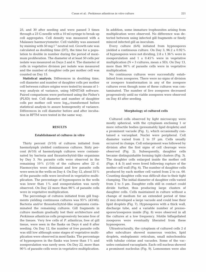
Casas et al.: Perkinsus atlanticus in vitro culture
25, and 30 after seeding and were passed 3 timesthrough a 23 G needle with a 10 ml syringe to break upcell aggregates. Cell density was measured with aMalassez haemocytometer. Cell viability was assessedby staining with 50 mg l–1 neutral red. Growth rate wascalculated as doubling time (DT), the time for a popu-lation to double in number during the period of maxi-mum proliferation. The diameter of at least 50 cells perisolate was measured on Days 2 and 4. The diameter ofcells in vegetative division per isolate was measuredand the number of daughter cells per mother cell wascounted on Day 15.
Statistical analysis. Differences in doubling time,cell diameter and number of daughter cells per mothercell between culture origins were tested by means of 1-way analysis of variance, using MINITAB software.Paired comparisons were performed using the Fisher’s(PLSD) test. Cell diameter and number of daughtercells per mother cell were log10-transformed beforestatistical analysis to assure homogeneity of variance.Differences in cell diameter before and after incuba-tion in RFTM were tested in the same way.
RESULTS
Establishment of cultures in vitro
Thirty percent (3/10) of cultures initiated fromhaemolymph yielded continuous cultures. Sixty per-cent (6/10) of haemolymph cultures were contami-nated by bacteria and thraustochytrid-like organismsby Day 3. No parasite cells were observed in theremaining 10% (1/10) of the cultures after 22 d.Haemocytes were dominant and few parasite cellswere seen in the wells on Day 4. On Day 12, about 35%of the parasite cells were involved in vegetative multi-plication. The percentage of hypnospores in the wellswas lower than 1% and zoosporulation was rarelyobserved. On Day 22 more than 90% of parasite cellswere in vegetative multiplication.
The percentage of cultures initiated from gill frag-ments yielding continuous cultures was 93% (43/46).Bacteria and/or thraustochytrid-like organisms conta-minated the remaining cultures. Gill fragments inculture medium gradually lost their architecture andPerkinsus atlanticus cells progressively became free ofthe tissues. Very few cells of P. atlanticus, free of gilltissue, were seen in the flasks on Days 4 and 8 afterseeding. On Day 12, the number of free parasite cellswas still low although some stages of vegetative multi-plication were observed in most flasks. The percentageof hypnospores in the flasks was lower than 1% andzoosporulation was rarely seen. On Day 22, more than90% of parasite cells were in vegetative multiplication.
In addition, some immature trophozoites arising frommultiplication were observed. No difference was de-tected between using infected gill fragments or finelyminced infected gill as inoculum.
Every culture (6/6) initiated from hypnosporesyielded a continuous culture. On Day 3, 96.2 ± 0.92%of hypnospores were not dividing, 2.8 ± 1.36% were inzoosporulation and 1 ± 0.45% were in vegetativemultiplication (N = 3 cultures, mean ± SD). On Day 13,more than 90% of parasite cells were in vegetativemultiplication.
No continuous cultures were successfully estab-lished from zoospores. There were no signs of divisionor zoospore transformation in any of the zoosporecultures even though none of these cultures was con-taminated. The number of live zoospores decreasedprogressively until no viable zoospores were observedon Day 45 after seeding.
Morphology of cultured cells
Cultured cells observed by light microscopy weremostly spherical, with the cytoplasm enclosing 1 ormore refractile bodies (presumably lipid droplets) anda prominent vacuole (Fig. 1), which occasionally con-tained a vacuoplast. Nuclei were peripheral. Celldiameter varied from 2 to 37 µm. Cells usuallyoccurred in clumps. Cell enlargement was followed bydivision after the first signs of cell cleavage wereobserved (Fig. 2). Subsequently, daughter cellsbecame distinguishable forming tight clusters (Fig. 3).The daughter cells enlarged inside the mother cell(Figs. 4 & 5) and were freed following rupture of themother cell wall (Fig. 6). The number of daughter cellsproduced by each mother cell varied from 2 to ca. 60.Counting daughter cells was difficult due to their tightclumping. The initial diameter of daughter cells variedfrom 2 to 5 µm. Daughter cells still in contact coulddivide further, thus producing large clusters ofdaughter cells. Cells maintained in culture without achange of medium for an extended period of time(5 mo) developed a large vacuole and could lose theirlipid droplets (Fig. 7). Hypnospores with a thick wall,discharge tube, and a variable number of prezoo-spores/zoospores inside (Fig. 8) were observed in allthe cultures at a low frequency. Motile biflagellatedzoospores were eventually liberated from thesehypnospores.
Ultrastructurally, the cytoplasm of cultured cells 2 dafter subculture showed numerous vesicles, lipiddroplets, endoplasmic reticulum, small mitochondriawith tubular cristae and vacuoles. Some of the vac-uoles contained vacuoplasts. Each cell nucleus showeda prominent nucleolus (Fig. 9). Lomosomes were also
221

Dis Aquat Org 52: 217–231, 2002
observed between the cell wall and plasmalemma ofsome cells. Cells sampled 5 d after subculture hadenlarged and their cell walls had become thicker(Fig. 10). Vegetative division involved fragmentation ofthe vacuoles (Figs. 11 & 12); then multinucleatedstages with incomplete cleavage of the cytoplasm wereobserved (Fig. 11). Subsequently, every nucleus wassurrounded by a portion of cytoplasm, which wasmembrane-limited. Daughter cells were very tightly
packed within the mother cell wall, which remainedintact (Fig. 12). On Day 14, the cytoplasm of culturedcells had a large vacuole lacking vacuoplast, lipiddroplets and mitochondria, and the nucleus showed anobvious nucleolus (Figs. 13 & 14). There were cells invegetative division in which daughter cells wereloosely packed (Fig. 13). After rupture of the mothercell wall, clusters of daughter cells in close contact andsurrounded by a broken mother cell wall were fre-
222
Figs. 1–8. Perkinsus atlanticus. Light micrographs of cultured cells in JL-ODRP-2A medium. Fig. 1. Cultured cells in freshmedium with dense cytoplasm containing refractile lipid droplets (1800×). Fig. 2. Enlarged cell in vegetative multiplicationshowing signs of cell cleavage and individualisation of some peripheral daughter cells (arrow) (1100×). Fig. 3. Two cells invegetative multiplication with well-developed cleavage furrows and tightly packed daughter cells (800×). Fig. 4. Larger andloosely packed daughter cells inside mother cells (800×). Fig. 5. Enlarged daughter cells inside mother cells containing well-defined vacuoles and several lipid droplets (1100×). Fig. 6. Daughter cells released from a mother cell after rupture of its cell wall(1100×). Fig. 7. Cultured cell maintained 3 mo without a medium change with cytoplasm containing no lipid droplets andoccupied by an enlarged vacuole without vacuoplast (2300×). Fig. 8. Zoospores in zoosporangium (800×). C: cytoplasm. L: lipid
droplet. N: nucleus. V: vacuole. W: wall
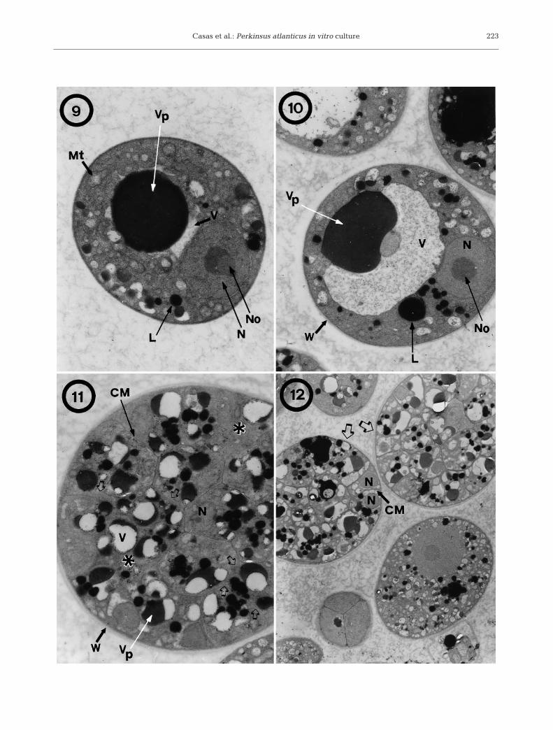
Figs. 9–12. Perkinsus atlanticus. Transmission electron micrographs of cultured cells in JL-ODRP-2A medium. Fig. 9. Cell 2 dafter subculture with a dense cytoplasm containing a prominent vacuoplast inside a vacuole and several lipid droplets and witha nucleus with a well-developed nucleolus (11 800×). Fig. 10. Cell 5 d after subculture (6300×). Fig. 11. Cell 5 d after subculturein vegetative multiplication showing cleavage furrows extending among the future daughter cells (arrows), and areas within thecell without cytoplasmic division (asterisks) (8700×). Fig. 12. Cells 5 d after subculture at different stages of division. Two mothercells having completed karyokinesis and cytokinesis (arrows) with daughter cells tightly packed (4500×). CM: cyto-
plasmic membrane. L: lipid droplet. Mt: mitochondrion. N: nucleus. No: nucleolus. V: vacuole. Vp: vacuoplast. W: wall
Casas et al.: Perkinsus atlanticus in vitro culture 223
Dis Aquat Org 52: 217–231, 2002224

Casas et al.: Perkinsus atlanticus in vitro culture
quently seen (Figs. 13 & 14). Cells culturedfor 90 d without a change of media hadenlarged vacuole with no vacuoplast andflattened remaining cytoplasm and nucleus(Figs. 15 & 16). Some vacuole protrusionsand cytoplasmic vesicles were seen,whereas lipid droplets were absent(Fig. 15). Cytoplasm appeared granularwith mitochondria showing tubular cristae,the nucleus was elongated and lomosomeswere observed between the cell wall andplasmatic membrane (Figs. 15 & 16).
DNA sequencing
ITS region sequences for 19 differentDNA clones were obtained from 9 Perkin-sus atlanticus isolate cultures. BLASTsearch results indicated that the closestmatches of sequences from this study wereto P. atlanticus (accession numbersPAU07697 and AF140295) and P. olseni(accession number POU07701) ITS regionsequences. A consensus of optimal treesfound from parsimony analysis of PerkinsusITS region sequences is shown in Fig. 17.The numbers shown at the nodes indicatebootstrap support values. P. atlanticus ITSsequences from this study grouped withother P. atlanticus sequences and the P.olseni sequences with 100% bootstrapsupport in a clade that was sister to ITSsequences from P. marinus. Many of thesequences from the isolate culture DNAswere identical to Perkinsus sequencespreviously obtained by specific amplifica-tion of Perkinsus sequences from P. atlanti-cus-infected host tissue DNA (Casas et al.2002).
Ray test
There was a significant (p < 0.001)increase in the diameter of cells incubatedin RFTM. The mean (±SD, range) cell
225
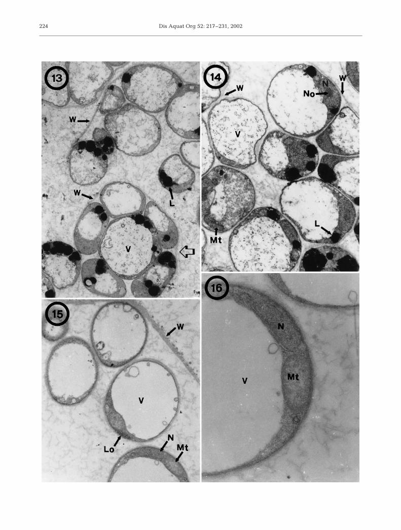
Figs. 13–16. Perkinsus atlanticus. Transmission electron micrographs of cultured cells in JL-ODRP-2A medium. Fig. 13. Cells 14 dafter subculture. Mother cells in vegetative multiplication with daughter cells loosely packed (arrow), and daughter cellsadhering to the ruptured cell-wall of the mother cell (6400×). Fig. 14. Cells 14 d after subculture. Higher magnification ofdaughter cells after vegetative multiplication, still held together by the mother cell wall. Each cell had a large vacuole withoutvacuoplast, several droplets in their cytoplasm and a nucleus with nucleolus (9000×). Fig. 15. Cells 90 d after subculture withenlarged vacuole without vacuoplast (10 400×). Fig. 16. Detail of mitochondrion and nucleus of a cultured cell 90 d after
subculture (31 900×). L: lipid droplet. Lo: lomosome. Mt: mitochondrion. N: nucleus. No: nucleolus. V: vacuole. W: wall
Fig. 17. Perkinsus spp. Internal transcribed spacer (ITS) region DNAsequences. Consensus of optimal trees found by parsimony analysis of thesequences for the ITS region of the ribosomal RNA complex of Perkinsusspecies. Numbers at nodes indicate bootstrap support values for the groupswhere the value was above 60. P. atlanticus sequences determined for thisstudy are designated as ‘gill’ for sequences obtained from cultures initiatedfrom gill fragments, ‘hypno’ for sequences obtained from cultures initiatedfrom hypnospores and ‘haem’ for sequences obtained from cultures
initiated from haemolymph

Dis Aquat Org 52: 217–231, 2002
diameter of cultures established from gill fragmentswas 8.8 ± 2.93 µm (5 to 20 µm) before incubation inRFTM and 25.3 ± 8.08 µm (10 to 60 µm) after incuba-tion in RFTM. The mean cell diameter of culturesestablished from haemolymph was 10.0 ± 5.02 µm (5 to22.5 µm) before incubation in RFTM and 26.0 ±9.06 µm (10 to 60 µm) after incubation in RFTM. Themean cell diameter of cultures established fromhynospores was 7.3 ± 4.41 µm (2.5 to 27.5 µm) beforeincubation in RFTM and 15.4 ± 4.29 µm (7.5 to 32.5 µm)after incubation in RFTM. The percent viability ofcells, after incubation in RFTM, from cultures estab-lished from gill fragments was 34.2%, from haemo-lymph 28.3%, and from hypnospores 47.1%. All en-larged cells stained blue-black with Lugol’s solution.
Quantitation of growth
A typical growth curve for cultured cells is shownin Fig. 18. During the first couple of days after seed-ing the flasks, cells enlarged and accumulated lipiddroplets in the cytoplasm. On Day 2, there were sig-nificant differences (p < 0.0001) in mean cell diameter
between cultures established from gill fragments,haemolymph and hypnospores. Mean (±SD) cell dia-meter for cultures established from gill fragments was8.7 ± 3.54 µm (N = 201, range: 4.9 to 31.8 µm), fromhaemolymph 10.0 ± 4.56 µm (N = 216, 4.9 to36.7 µm), and from hypnospores 7.3 ± 3.22 µm (N =214, 2.4 to 29.4 µm). On Day 4, the mean diameters ofcells from cultures established from gill fragments(10.1 ± 2.48 µm; N = 163, 5 to 15 µm) andhaemolymph (10.4 ± 2.61 µm; N = 252, 5 to 20 µm)were significantly higher than the mean diameter ofcells from cultures established from hypnospores (9.0± 2.62 µm; N = 297, 5 to 32.5 µm). Cell division usu-ally started on Day 4. On Day 6 more than 50% ofcells were dividing by vegetative multiplication. Afew zoosporulating cells (less than 1% of dividingcells) were observed in every culture flask. The mean(±SD) diameter of cells involved in vegetative multi-plication was 16.0 ± 3.55 µm (N = 83, 7.4 to 27.0 µm)for cultures established from gill fragments, 19.7 ±6.04 µm (N = 44, 9.8 to 44.1 µm) for cultures estab-lished from haemolymph, and 13.0 ± 3.62 µm (N = 78,7.3 to 31.8 µm) for cultures established from hypno-spores. The mean number of daughter cells permother cell (±SD) was 12.5 ± 5.74 (N = 78, 4 to 32cells) for cultures established from gill fragments,20.4 ± 14.10 (N = 36, 6 to 60 cells) for culturesestablished from haemolymph, and 8.7 ± 7.41 (N =71, 2 to 60 cells) for cultures established from hyp-nospores. There were significant differences in meandiameter of cells in vegetative multiplication (p <0.0001) and in the number of daughter cells permother cell (p > 0.0001) between cultures establishedfrom different sources.
In JL-ODRP-2A, cultures established from haemo-lymph, gill fragments and hypnospores differed intheir period of highest increase in cell number (i.e. logphase of growth). Log growth phase for cultures estab-lished from haemolymph occurred between Days 6and 15; from gill fragments between Days 12 and 15;and from hypnospores between Days 12 and 18. Loggrowth phase for cultures in the commercial mediumDME:Ham’s F-12 supplemented with fetuin occurredbetween Days 12 and 18. Mean doubling times (±SD)for cultures established from haemolymph was 26.2 ±6.89 h; from gill fragments 29.6 ± 8.61 h; and fromhypnospores 42.3 ± 29.84 h. No significant differencein doubling times between cultures established fromhaemolymph, gill fragments and hypnospores wasfound (p = 0.55). Mean doubling times (±SD) for cul-tures established from gill fragments incubated inDME:Ham’s F-12 was 84.5 ± 84.46 h. No difference indoubling times was observed between cultures estab-lished from gills in medium JL-ODRP-2A and thoseDME:Ham’s F-12 (p = 0.32).
226
Fig. 18. Perkinsus atlanticus. Propagation of cells in JL-ODRP-2A medium of cultures established from gills, haem-olymph and hypnospores, and propagation in vitro of P. at-lanticus cells in DME:Ham’s F-12 medium of culturesestablished from gills. Triplicate flasks were seeded with 106
cells ml–1 from each source and flasks were incubated at 26°C

Casas et al.: Perkinsus atlanticus in vitro culture
DISCUSSION
Continuous cultures of Perkinsus atlanticus wereestablished for the first time. The morphology of cellsin cultures, at the light and electron microscopy levels,were typical of P. atlanticus and other Perkinsus spp.Cultured cells incubated in RFTM produced enlargedcells (hypnospores) that stained blue-black in Lugol’ssolution, a response characteristic of Perkinsus spp.and used in routine diagnosis. Species identification ofthe cultured cells was accomplished through analysisof nucleotide sequences of the ITS region of the rRNAgene complex, which matched that of P. atlanticus.
Cultures were successfully established from 3sources of parasites, the gills and haemolymph ofinfected carpet shell clams and from hypnospores iso-lated from infected gill fragments after incubation inRFTM. However, no culture could be established withisolated zoospores. Cultures initiated from gill frag-ments and hypnospores yielded the highest percent-age of continuous cultures, 93 and 100% respectively.The majority of cultures initiated from haemolymphbecame contaminated with bacteria or protozoa, andonly 30% yielded continuous cultures. All contami-nated cultures were discarded because past attemptsto eliminate microbial contaminants generally havebeen unsuccessful and a drain on resources (J. F. LaPeyre pers. obs.).
Perkinsus spp. cultures have been established from avariety of tissues including hearts, visceral ganglia andgills (Kleinschuster & Swink 1993, La Peyre et al. 1993,McLaughlin & Faisal 1998, Coss et al. 2001a) but thereis little information on the optimal source of parasitesto use. The success rate in establishing continuouscultures can be used as a criterion for selecting a tissueas a source of parasites. In this study, the success rateof establishing continuous cultures of P. atlanticusdepended primarily on the absence of microbial conta-mination because all cultures were initiated frominfected clams. Oyster hearts were used for the firsttime to establish P. marinus cultures because thisorgan is relatively free of microbial contaminants com-pared to other oyster tissues (La Peyre et al. 1993). Inthe current study, gills of carpet shell clams were usedinstead of hearts to establish P. atlanticus culturesbecause the clam heart is traversed by the intestine,thus increasing the chance of microbial contamination.Gills of T. decussatus are also one of the clams’ organsmost infected with P. atlanticus (Azevedo 1989,Rodríguez & Navas 1995). Using gill tissues has theadvantage that sites of infections can be easily locatedby looking for abscesses and hence tissues with a highparasite load can be selected to initiate cultures. Depu-ration of the clams, thorough rinsing of gill tissues insaline (i.e. 20 times) and decontamination of the gill
tissues in antibiotic solutions were essential steps inreducing microbial contamination to establish continu-ous cultures of P. atlanticus from carpet shell clam gills.The 93% success rate in establishing P. atlanticus con-tinuous cultures from gills of infected clams indicatesthe gill tissue is a good choice as a source of parasites.
Haemolymph also has been used to establish Perkin-sus spp. cultures (Gauthier & Vasta 1993, Kleinschusteret al. 1994, Dungan & Hamilton 1995, McLaughlin &Faisal 1998). In the current study, microbial contami-nation of cultures initiated from clam haemolymph wasmore frequent than in cultures initiated from gills orhypnospores. This is not unexpected since bivalvehaemolymph is seldom sterile and microorganismssuch as algae and bacteria are often found in haemo-cytes following feeding. Establishing continuous cul-tures of Perkinsus spp. from haemolymph is thereforemore difficult than from other tissues. Perhaps thedecontamination procedure before seeding should beimproved, although within haemocytes microorgan-isms cannot be rinsed off and the efficacy of antibioticdecontamination solutions is greatly reduced. Oneadvantage of using haemolymph is that the molluscdoes not have to be sacrificed, possibly enablingmollusc investigations on the contribution of differentparasite strains during the progression of Perkinsusinfection.
Another criterion for selecting a tissue as a source ofparasites could be the time taken to establish continu-ous cultures. Perkinsus marinus cultures, for example,were established more rapidly from hypnospores thanfrom hearts of eastern oysters (La Peyre & Faisal 1995).Culture flasks (25 cm2) seeded with 105 P. marinushypnospores contained up to 107 cells per flask after2 wk in cultures while 4 to 8 wk were generally neededto obtain similar numbers of P. marinus cells in culturesinitiated from hearts of infected eastern oysters. In P.atlanticus cultures initiated with hypnospores, theparasite cells began to divide and produce new cellsearlier than in cultures established from clam gills orhaemolymph. The number of cultures that can be pro-duced from the hypnospores isolated from a singleclam can also be high because millions of hypnosporescan be isolated from a heavily infected clam. Hypno-spores have been used to establish a number of Perkin-sus spp. cultures (La Peyre & Faisal 1995, Bushek &Allen 1996b, McLaughlin & Faisal 1998). However, thesuccess rate in establishing cultures from hypnosporesis generally much lower than the success rate in estab-lishing cultures from oyster hearts because of micro-bial contamination. About 90% of cultures initiatedfrom oyster heart tissues yield continuous cultures of P.marinus compared to about a 20 to 30% success ratefor cultures initiated from P. marinus hypnospores (J. F.La Peyre pers. obs., D. Bushek pers. comm.). The much
227

Dis Aquat Org 52: 217–231, 2002
higher success rate (100%) in establishing P. atlanticuscultures from hypnospores is probably due to the thor-ough decontamination procedure used in the currentstudy. Gill fragments were decontaminated beforeincubation in RFTM and again after 1 wk incubation inRFTM before inoculation of the culture flasks. Isolatedhypnospores are therefore another good source of par-asites to establish continuous cultures of P. atlanticus.
No culture could be established when Perkinsusatlanticus zoospores were added to culture medium. Itis not known whether the failure of zoospores to yieldcontinuous cultures was related to the isolation method(centrifugation at high speed, 3000 × g) or just to thenature of zoospores. Results from this study contrastwith reports of the transformation of zoospores intotrophozoites and the subsequent growth in otherPerkinsus spp. (Kleinschuster et al. 1994, La Peyre &Faisal 1995, Coss et al. 2001a). The role of zoospores inthe life cycle of Perkinsus spp. is still unclear and fur-ther studies on the fate of zoospores are needed.
Continuous cultures of Perkinsus atlanticus can beestablished from a variety of parasite sources withvarying levels of success. Success rates of establishingcultures were provided for the first time and will beuseful to other researchers in selecting a source ofparasites to culture additional isolates of P. atlanticusand other Perkinsus spp. However, some caution mustbe exercised when choosing a source of parasitesbecause preliminary results indicate that certain char-acteristics of Perkinsus cells in culture, such as size,growth rate and protein secretion, can vary dependingon the source of parasites used to initiate the cultures.In the current study, the source of parasites influencedthe size of P. atlanticus cells. Cells from cultures initi-ated with hypnospores were significantly smaller thancells from cultures initiated from gills or haemolymph.Furthermore, the size of cells in vegetative divisionand the number of daughter cells per mother cell wassignificantly different depending on source of P.atlanticus cultured cells. The source of parasite cellsalso can influence the production of extracellular pro-teins by cultured cells. Differences in production ofextracellular proteins between P. marinus culturesestablished from hypnospores and oyster hearts havebeen noted by La Peyre & Cooper (1997). In P. atlanti-cus differences in production of extracellular proteinsin cultures from different sources of parasite cells havealso been observed (S. M. Casas unpubl. results).Therefore, it is critical that the sources of parasitesused to establish cultures are reported in any studiesinvolving cultured Perkinsus spp. until more is knownabout the reasons for these differences.
The cultured Perkinsus atlanticus cells had all mor-phological characteristics of Perkinsus spp. parasites atthe light and electron microscopic levels (La Peyre et al.
1993, Perkins 1996, Coss et al. 2001a, Sunila et al. 2001).The sizes of the cultured mature trophozoites of P.atlanticus established from various sources were largerthan the size of mature trophozoites from clam tissues,which generally did not exceed 15 µm (S. M. Casas pers.obs.). Similarly, multinucleated cells in the vegetative di-vision process were larger in culture than in clam tissues(range 7 to 15 µm; S. M. Casas pers. obs.) and the num-ber of daughter cells produced by mother cells was alsohigher in culture. Cell sizes have been shown to varysubstantially depending on the host (Goggin & Lester1995) and the time of year (Ray & Chandler 1955, Busheket al. 1994), and can be influenced by nutrient availabil-ity, which may explain the differences between culturedcells and cells in histological sections (La Peyre et al.1993, La Peyre & Faisal 1996).
Vegetative multiplication of Perkinsus marinus in itshost occurs by successive bipartition in which karyoki-nesis is followed by cytokinesis (binary fission)(Perkins 1996). Light microscopy and TEM observationof in vitro P. atlanticus cultures suggested that vegeta-tive multiplication occurs mostly by multiple fission;that is to say, multiple karyokinesis occurred beforecytokinesis took place. Sunila et al. (2001) observedwith TEM that schizogony was the most common modeof cell division of cultured P. marinus cells and thatbinary fission occurred rarely.
Zoosporulation yielding motile zoospores wasobserved at a low (<1% of dividing cells) frequency inevery Perkinsus atlanticus culture established fromgills, haemolymph and hypnospores in the culturemedium JL-ODRP-2A. Zoosporulation of culturedvegetative cells of Perkinsus spp. established fromseveral clam species has also been reported in culturemedia (Kleinschuster et al. 1994, McLaughlin & Faisal1998, Coss et al. 2001a). In contrast, zoosporulation ofvegetative cells from established cultures of P. marinushas not been reported. The significance of theseobservations to species identity and host specificity isnot yet known.
A characteristic of protozoa in the genus Perkinsus istheir ability to enlarge in RFTM and stain blue-blackwith Lugol’s solution. This characteristic discovered byRay (1952) is the basis of a common diagnostic test forinfection (Ray 1952, 1966, reviewed by Bushek et al.1994). Only 1 Perkinsus species, P. qugwadi in bayscallops, does not enlarge in RFTM but there is doubtthat this parasite belongs in the genus Perkinsus. Inaddition to the inability of P. qugwadi to enlarge inRFTM and to stain blue black with Lugol’s solution,differences in morphology and considerable differ-ences in DNA sequences of the ITS region of the ribo-somal RNA gene complex between P. qugwadi andother Perkinsus spp. have been found (Coss et al.2001b, Casas et al. 2002). In this study, the size of
228
Casas et al.: Perkinsus atlanticus in vitro culture
cultured cells incubated in RFTM increased signifi-cantly and the enlarged cells stained with Lugol’s solu-tion. However, cultured cells incubated in RFTM weregenerally smaller than hypnospores isolated frominfected gills (38.9 ± 10.4 µm, range 19.7 to 64.1 µm; S.M. Casas pers. obs.). Reduced enlargement of culturedcells compared to hypnospores in RFTM also has beenobserved in previous studies (Gauthier & Vasta 1993,Kleinschuster & Swink 1993, La Peyre et al. 1993). Invitro studies with cultured cells indicate that enlarge-ment can be greatly increased by the addition of lipidand other nutrients to RFTM (La Peyre 1996, Wagneret al. 2001). The larger size of P. atlanticus in gill tis-sues incubated in RFTM could reflect additional nutri-ents available from the carpet shell clam tissue.
Agreement in morphology between cultured cellsand Perkinsus cells from host tissues is essential forgenus identification but not sufficient for species iden-tification. In some cases, protozoans that were initiallydescribed as Perkinsus spp. based partly on their mor-phologies were later found not to belong in the genusPerkinsus (McGladdery et al. 1991, Goggin et al. 1996,Ordás & Figueras 1998, Figueras et al. 2000). Cur-rently, as mentioned above, classification of P. qug-wadi, is questionable because of noted differences inmorphology from that which is the typical morphologyof Perkinsus spp. (Blackbourn et al. 1998). Therefore,the morphology of putative Perkinsus spp. must beexamined with extreme care at both the light and elec-tron microscopic levels before they are included in thisgenus.
Other characteristics and data such as DNAsequences can be used in combination with cell mor-phology to identify protozoan parasites as Perkinsusspp. at the genus level and are required for accuratespecies identification within this genus. In this studyDNA sequences of the ITS region helped to confirmthe identification of cultures as P. atlanticus. The ITSregion sequences from cultures established from gillsand haemolymph of infected clams, as well as thosefrom isolated hypnospore cultures grouped in phylo-genetic analyses with previously published P. atlanti-cus and P. olseni sequences. Many sequences wereidentical to those obtained from parasite infected T.decussatus tissues (Goggin 1994, Robledo et al. 2000,Casas et al. 2002). The ITS region sequences can beused to distinguish many Perkinsus species, althoughneither P. olseni and P. atlanticus nor P. chesapeakiand P. andrewsi can be resolved using ITS sequences(see Fig. 17). A recent study, however, has reportedthat ITS sequences previously thought to distinguish P.chesapeaki and P. andrewsi are really ITS variants thatcan both be found in a single clonal isolate culture(Dungan et al. 2002). This result suggests that P. chesa-peaki and P. andrewsi are the same species of Perkin-
sus. In addition, based on the sequence analyses todate, it is likely that P. atlanticus and P. olseni are thesame Perkinsus species; clonal cultures of each will berequired to accurately address this question.
The culture medium JL-ODRP-2A, used to culturePerkinsus marinus, supported the proliferation of P.atlanticus. P. atlanticus could also be grown in thecommercial medium 1:2 DME:Ham’s F-12 supple-mented with fetuin as prepared by Gauthier et al.(1995). No difference in P. atlanticus growth rate wasfound between these 2 media. An advantage of usingthe medium JL-ODRP-2A is that its nutrient composi-tion can be easily adjusted to optimise P. atlanticusgrowth in vitro. Moreover, JL-ODRP-2A does not con-tain proteins so that studies on the production of extra-cellular proteins by cultured P. atlanticus are facili-tated (La Peyre et al. 1995, La Peyre & Faisal 1996).There were some differences in proliferation patternsand cell sizes of cultures established from gills,haemolymph and hypnospores but their DTs were notsignificantly different. The DTs of the 9 isolates werevariable but comparable to the DTs reported for otherPerkinsus spp. (Kleinshuster et al. 1994, Dungan &Hamilton 1995, Gauthier & Vasta 1995, La Peyre &Faisal 1996). However, strict comparison of Perkinsusspp. growth rates is not possible because of many dif-ferences between studies, such as in media, isolates,seeding densities, proliferation assays and the periodof cell proliferation selected to calculate DT (La Peyre1996).
The experience acquired with culturing Perkinsusmarinus facilitated the in vitro culture of P. atlanticusand other Perkinsus spp. The optimal culture condi-tions for P. atlanticus will need to be determined bymeasuring the effects of various physical and chemicalparameters such as temperature, salinity, pH, inocu-lum density and nutrient concentrations on the para-site growth. As with P. marinus, P. atlanticus culturescan be used in a wide range of studies to addressimportant questions such as about the parasite’s envi-ronmental tolerance, virulence, genetic make up,metabolism and drug sensitivity. The development ofcontinuous cultures of P. atlanticus will lead to betterunderstanding of this important parasite of carpet shellclams.
Acknowledgements. María Jesús Llevot, Kathleen Apaku-pakul, and Laura Corral provided technical assistance. JoséIgnacio Navas (CICEM ‘Agua del Pino’, Junta de Andalucía)provided the protocol to isolate hypnospores. This work waspartially supported by funds of the Secretaría Xeral de Inves-tigación e Desenvolvemento Tecnolóxico da Xunta de Gali-cia, through the project PGIDT-CIMA 99/10 and by fundsfrom the Louisiana Sea Grant College Program. S.M.C. wassupported by a scholarship from the Consellería de Pesca,Marisqueo e Acuicultura, Xunta de Galicia. Financial support
229

Dis Aquat Org 52: 217–231, 2002
included funding for her stay at the J.F.L.P. laboratory,Louisiana State University. This is contribution no. 2471 of theVirginia Institute of Marine Science, College of William andMary.
LITERATURE CITED
Altschul SF, Gish W, Miller W, Myers EW, Lipman DJ (1990)Basic local alignment search tool. Mol Biol 215:403–410
Azevedo C (1989) Fine structure of Perkinsus atlanticus n. sp.(Apicomplexa, Perkinsea) parasite of the clam Ruditapesdecussatus from Portugal. J Parasitol 75:627–635
Blackbourn J, Bower SM, Meyer GR (1998) Perkinsus qugwadisp. nov. (incertae sedis), a pathogenic protozoan parasite ofJapanese scallops, Patinopecten yessoensis, cultured inBritish Columbia, Canada. Can J Zool 76:942–953
Burreson EM, Ragone Calvo LM, La Peyre JF, Counts F, Payn-ter KT (1994) Acute osmotic tolerance of cultured cells ofthe oyster pathogen Perkinsus marinus (Apicomplexa:Perkinsida). Comp Biochem Physiol 109A:575–582
Bushek D, Allen SK (1996a) Races of Perkinsus marinus.J Shellfish Res 15:103–107
Bushek D, Allen SK (1996b) Host-parasite interactions amongbroadly distributed populations of the eastern oyster Cras-sostrea virginica and the protozoan Perkinsus marinus.Mar Ecol Prog Ser 139:127–141
Bushek D, Ford SE, Allen SK (1994) Evaluation of methodsusing Ray’s fluid thioglycollate medium for diagnosis ofPerkinsus marinus infection in the eastern oyster, Crasso-strea virginica. Annu Rev Fish Dis 4:201–217
Casas SM, Villalba A, Reece KS (2002) Study of perkinsosis inthe carpet shell clam Tapes decussatus in Galicia (NWSpain). I. Identification of the aetiological agent and invitro modulation of zoosporulation by temperature andsalinity. Dis Aquat Org 50(1):51–65
Chu FLE, Greene KH (1989) Effect of temperature and salin-ity on in vitro culture of the oyster pathogen, Perkinsusmarinus (Apicomplexa: Perkinsea ). J Invertebr Pathol 53:260–268
Coss CA, Robledo JAF, Vasta GR (2001a) Fine structure ofclonally propagated in vitro life stages of a Perkinsus sp.isolated from the Baltic clam Macoma balthica. J EukaryotMicrobiol 48:38–51
Coss CA, Robledo JAF, Ruiz GM, Vasta GR (2001b) Descrip-tion of Perkinsus andrewsi n. sp. isolated from the Balticclam (Macoma balthica) by characterization of the riboso-mal RNA locus, and development of a species-specificPCR-based diagnostic assay. J Eukaryot Microbiol 48:52–61
de la Herrán R, Garrido-Ramos MA, Navas JI, Ruiz-Rejón C,Ruiz-Rejón M (2000) Molecular characterization of theribosomal RNA gene region of Perkinsus atlanticus: its usein phylogenetic analysis and as a target for a moleculardiagnosis. Parasitology 120:345–353
Dungan CF, Hamilton RM (1995) Use of the tetrazolium-based cell proliferation assay to measure effects of in vitroconditions on Perkinsus marinus (Apicomplexan) prolifer-ation. J Eukaryot Microbiol 42:379–388
Dungan CF, Hamilton RM, Hudson KL, McCollough CB,Reece KS (2002) Two epizootic infectious diseases inChesapeake Bay commercial clams, Mya arenaria andTagelus plebius. Dis Aquat Org 50(1):67–78
Faisal M, La Peyre JF, Elsayed E, Wright DC (1999) Bacitracininhibits the oyster pathogen Perkinsus marinus in vitroand in vivo. J Aquat Anim Health 11:130–138
Figueras A, Lorenzo G, Ordás MC, Gouy M, Novoa B (2000)
Sequence of the small subunit ribosomal RNA gene ofPerkinsus atlanticus-like isolated from carpet shell clam inGalicia, Spain. Mar Biotechnol 2:419–428
Gauthier J, Vasta G (1993) Continuous in vitro culture of theeastern oyster parasite Perkinsus marinus. J InvertebrPathol 62:321–323
Gauthier J, Vasta G (1995) In vitro culture of the easternoyster parasite Perkinsus marinus: optimization of themethodology. J Invertebr Pathol 66:156–168
Gauthier JD, Feig B, Vasta GR (1995) Effect of fetal bovineserum glycoproteins on the in vitro proliferation of theoyster parasite Perkinsus marinus: development of a fullydefined medium. J Eukaryot Microbiol 42:307–313
Goggin CL (1994) Variation in the two internal transcribedspacers and 5.8S ribosomal RNA from five isolates of themarine parasite Perkinsus (Protista, Apicomplexa). MolBiochem Parasitol 65:179–182
Goggin CL, Lester RJG (1995) Perkinsus, a protistan parasiteof abalone in Australia: a review. Mar Freshw Res 46:639–646
Goggin CL, McGladdery SE, Whyte SK, Cawthorn RJ (1996)An assessment of lesions in bay scallops Argopecten irra-dians attributed to Perkinsus karlssoni (Protozoa, Apicom-plexa). Dis Aquat Org 24:77–80
Kleinschuster SJ, Swink SL (1993) A simple method for the invitro culture of Perkinsus marinus. Nautilus 107:76–78
Kleinschuster SJ, Perkins FO, Dykstra MJ, Swink SL (1994)The in vitro life cycle of a Perkinsus species (Apicomplexa,Perkinsidae) isolated from Macoma balthica (Linnaeus,1758). J Shellfish Res 13:461–465
Kotob SI, McLaughlin SM, Van Berkum P, Faisal M (1999)Characterization of two Perkinsus spp. from the softshellclam, Mya arenaria using the small subunit ribosomalRNA gene. J Eukaryot Microbiol 46:439–444
Krantz GE (1994) Chemical inhibition of Perkinsus marinus intwo in vitro culture systems. J Shellfish Res 13:131–136
La Peyre JF (1996) Propagation and in vitro studies of Perkin-sus marinus. J Shellfish Res 15:89–101
La Peyre JF, Cooper RK (1997) Changes in protease expres-sion by Perkinsus marinus cultures following incubation inRay’s fluid thioglycollate medium. J Shellfish Res 16:331
La Peyre JF, Faisal M (1995) Improved method for the initia-tion of continuous cultures of the oyster pathogen Perkin-sus marinus (Apicomplexa). Trans Am Fish Soc 124:144–146
La Peyre JF, Faisal M (1996) Optimal culture conditions forthe propagation of the oyster pathogen Perkinsus marinus(Apicomplexa) in protein deficient medium. Parasite 3:147–153
La Peyre JF, Faisal M, Burreson EM (1993) In vitro propaga-tion of the protozoan Perkinsus marinus, a pathogen of theeastern oyster, Crassostrea virginica. J Eukaryot Microbiol40:304–310
La Peyre JF, Schafhauser DY, Rizkalla EH, Faisal M (1995)Production of serine proteases by the oyster pathogenPerkinsus marinus (Apicomplexa) in vitro. J EukaryotMicrobiol 42:544–551
McGladdery SE, Cawthorn RJ, Bradford BC (1991) Perkinsuskarlssoni n. sp. (Apicomplexa) in bay scallops Argopectenirradians. Dis Aquat Org 10:127–137
McLaughlin SM, Faisal M (1998) In vitro propagation of twoPerkinsus species from the softshell clam Mya arenaria.Parasite 5:341–348
McLaughlin SM, Tall BD, Shaheen A, Elsayed EE, Faisal M(2000) Zoosporulation of a new Perkinsus species isolatedfrom the gills of the softshell clam Mya arenaria. Parasite7:115–122
230

Casas et al.: Perkinsus atlanticus in vitro culture
Nickens AD, Wagner E, La Peyre JF (2000) Improved proce-dure to count Perkinsus marinus in eastern oysterhemolymph. J Shellfish Res. 19:665
Ordás MC, Figueras A (1998) In vitro culture of Perkinsusatlanticus, a parasite of the carpet shell clam Ruditapesdecussatus. Dis Aquat Org 33:129–136
Perkins FO (1996) The structure of Perkinsus marinus(Mackin, Owen & Collier, 1950) Levine, 1978 withcomments on taxonomy and phylogeny of Perkinsus spp.J Shellfish Res 15:67–88
Perkins FO, Menzel RW (1966) Morphological and culturalstudies of a motile stage in the life cycle of Dermocystid-ium marinum. Proc Natl Shellfish Assoc 56:23–30
Ray SM (1952) A culture technique for the diagnosis of infec-tions with Dermocystidium marinum Mackin, Owen, andCollier in oysters. Science 116:360–361
Ray SM (1966) A review of the culture method for detectingDermocystidium marinum, with suggested modificationsand precautions. Proc Natl Shellfish Assoc 54:55–69
Ray SM, Chandler AC (1955) Dermocystidium marinum, aparasite of oysters. Exp Parasitol 4:172–200
Reece KS, Bushek D, Graves JE (1997) Molecular markers forpopulation genetic analysis of Perkinsus marinus. MolMar Biol Biotech 6:197–206
Reece KS, Bushek D, Hudson KL, Graves JE (2001) Geo-graphic distribution of Perkinsus marinus genetic strainsalong the Atlantic and Gulf coasts of the USA. Mar Biol139:1047–1055
Robledo JAF, Coss CA, Vasta GR (2000) Characterisation ofthe ribosomal RNA locus of Perkinsus atlanticus anddevelopment of a polymerase chain reaction-based diag-nostic assay. J Parasitol 86:972–978
Rodríguez F, Navas JI (1995) A comparison of gill andhemolymph assays for the thioglycollate diagnosis ofPerkinsus atlanticus (Apicomplexa, Perkinsea) in clams,Ruditapes decussatus (L.) and Ruditapes philippinarum(Adams et Reeve). Aquaculture 132:145–152
Ruano F, Cachola R (1986) Outbreak of a severe epizootic of
Perkinsus marinus (Levin - 78) at Ria de Faro clam’s culturebeds. In: Abstracts 2nd Int Colloq Pathol Mar Aquac (PA-MAQ II). University of Oporto, Porto, Portugal, p 41–42
Santmartí, MM, García Valero J, Montes J, Pech A, Durfort M(1995) Seguimiento del protozoo Perkinsus sp. en laspoblaciones de Tapes decussatus y Tapes semidecussatusdel delta del Ebro. In: Castelló F, Calderer A (eds) Actasdel V Congreso Nacional de Acuicultura, May 10–13,1995, S. Carlos de la Rápita, Spain. Universidad deBarcelona, p 260–265
Soudant P, Chu FLE (2001) Lipid class and fatty acid composi-tion of the protozoan parasite of oysters, Perkinsus mari-nus cultivated in two different media. J Eukaryot Micro-biol 48:309–319
Soudant, P, Chu FLE, Manty Y (2000) Lipid class compositionof the protozoan Perkinsus marinus an oyster parasite, andits metabolism of a fluorescent phosphatidycholine ana-log. Lipids 35:1387–1395
Sunila I, Hamilton RM, Dungan CF (2001). Ultrastructuralcharacteristics of the in vitro cell cycle of the protozoanpathogen of oysters, Perkinsus marinus. J Eukaryot Micro-biol 48:348–361
Swofford DL (2001) PAUP*. Phylogenetic analysis using par-simony (*and other methods), Version 4. Sinauer Associ-ates, Sunderland, MA
Thompson JD, Higgins DG, Gibson TJ (1994) Improving thesensitivity of progressive multiple sequence alignmentthrough sequence weighting, positions-specific gappenalties and weight matrix choice. Nucleic AcidsResearch 22:4673–4680
Volety AK, Chu FLE (1997) Acid phosphatase activity inPerkinsus marinus, the protistan parasite of the Americanoyster, Crassostrea virginica. J Parasitol 83:1093–1098
Wagner E, Casas SM, La Peyre JF (2001) Induction ofhypnospore formation and zoosporulation of Perkinsusmarinus cells from long term cultures. In: Devoe R (ed)Abstracts from Aquaculture 2001. World AquacultureSociety, Lake Buena Vista, FL, p 670
231
Editorial responsibility: Albert Sparks,Seattle, Washington, USA
Submitted: February 1, 2002; Accepted: May 8, 2002Proofs received from author(s): December 2, 2002