Inter-observer agreement according to malaria parasite density
Upload
khangminh22Category
view
1download
0
THE CHARACTERIZATION OF AN INTRACELLULAR
PROTOZOAN PARASITE INFECTING THE DIGESTIVE
GLAND OF ABALONE,
HALIOTIS MIDAE.
BY
YOLANDI CLIGNET CLOETE
DISSERTATION
SUBMITTED IN FULFILMENT OF THE REQUIREMENTS FOR THE DEGREE
MAGISTER SCIENTIAE
IN
AQUATIC HEALTH
IN THE
FACULTY OF SCIENCE
AT THE
UNIVERSITY OF JOHANNESBURG
SUPERVISOR: DR. N. J. SMIT
CO-SUPERVISOR: DR. A. MOUTON
JUNE 2008

Dedicated to my father Pieter Andries Cloete (11 Feb 1942- 27 Nov 2004) for always
believing in me, my mother Esther Cloete for her loving support, my family and to
uncle Nick Nel for teaching me about the ocean since I was little, your love and
passion for marine life inspired me. Above all to God whom bestowed the privilege
upon mankind to study, protect and care for al the fascinating animals on earth.
The beauty and gen ius of a w ork of art m ay be reconceiv ed, though its first m aterial expression be
destroyed… but w hen the last individual of a race of living things breathing no m ore, another
heaven and another earth m ust pass before such a one can be seen again.
W illiam B eebe

TABLE OF CONTENTS
1. LIST OF FIGURES I
2. LIST OF TABLES VI
3. SUMMARY VII
4. OPSOMMING IX
5. CHAPTER 1: GENERAL INTRODUCTION 1
1.1 The global abalone industry 1
1.1.1. The history of the abalone industry 1
1.1.2. Impact of parasites and diseases on the abalone industry 2
1.1.3. Quality regulations of abalone products 3
1.1.4. Well known parasites and diseases of abalone 3
1.1.5. Abalone and the environment 4
1.2 South African abalone industry 4
1.2.1. Abalone farming 4
1.2.2. South African regulations and abalone quality 5
1.2.3. Unknown parasites of abalone in South Africa 5
6. CHAPTER 2:WILD ABALONE 7
2.1 Wild abalone species of South Africa 7
2.2 Identification of Haliotis species from South Africa 8
2.2.1. Haliotis midae 9
2.2.2. Haliotis parva 9
2.2.3. Haliotis queketti 9
2.2.4. Haliotis spadicea 12
2.2.5. Haliotis speciosa 12
2.3 Ecology of South African abalone species 12
2.3.1. Distribution 12
2.3.2. Habitats 15
2.3.3. Food and feeding behaviour 15
2.3.4. Predators 16
2.3.5. The typical life cycle of abalone 16

2.4 Haliotis species of the world 19
2.5 Economically important species of abalone 24
2.6 South African Haliotis 25
2.6.1. Utilisation of abalone 25
2.6.2. Poaching 26
7. CHAPTER 3: ABALONE FARMING 28
3.1 Commercial value of abalone 29
3.2 Artificial spawning and settlement of larvae 29
3.2.1. Spawning 29
3.2.2. The development of eggs and larval stages 30
3.3 How a typical abalone farm works 32
3.4 Abalone farming in South Africa 39
3.4.1. Types of abalone farms 40
3.4.2. Important aspects of abalone farming 40
3.4.2.1. Water quality and the environment 41
3.4.2.2. Nutrition 42
3.4.3. South African abalone export 42
3.4.4. Current research 43
8. CHAPTER 4: ABALONE PARASITES 44
4.1 Prokaryotes 44
4.1.1 Withering syndrome associated with Rickettsiales-like
prokaryotes 44
4.2 Chromista 47
4.2.1. Labyrinthloides haliotidis 47
4.3 Protozoa 50
4.3.1. Ciliates 50
4.3.2. Haplosporidians 50
4.3.3. Perkinsus species 53
4.3.4. Pseudoklossia Haliotis 54
4.4 Digeneans 54
4.5 Polychaetes 56
4.5.1. Terebrasabella heterouncinata 56

4.5.2. Boccardia knoxi and Polydora haplura 60
9. CHAPTER 5: PROTOZOAN PARASITES OF OTHER IMPORTANT
MARINE INVERTEBRATES 64
5.1 Apicomplexans 64
5.1.1. Coccidians 64
5.1.2. Gregarines 66
5.1.3. Haplosporidians 67
5.1.4. Perkinsus species 70
5.2 Bonamia species 75
5.3 Microsporidians 76
5.4 Paramyxeans 77
8.1.1.1.1.1.1.1.1 10. CHAPTER 6:FIRST RECORDS OF AN
UNIDENSCRIBED
8.1.1.1.1.1.1.1.2 PROTOZOAN PARASITE IN THE
DIGESTIVE
8.1.1.1.1.1.1.1.3 GLAND OF HALIOTIS MIDAE
FROM SOUTH
8.1.1.1.1.1.1.1.4 AFRICA
80
6.1 Collection and preparation of samples 80
6.1.1. Collection of farmed animals 80
6.1.2. Collection of wild samples 83
6.1.3. Detection and preservation of samples 83
6.1.4. Histology 84
6.1.5. Fluorescence microscopy 87
6.1.6. Transmission Electron Microscopy (TEM) 88
6.2 Results 91
6.2.1. Possible effect of the digestive gland parasite on growth
of farmed abalone 91
6.3 Description of the intracellular digestive gland parasite 95
6.3.1. Morphology based on histology 95
6.3.2. Morphometrics 99

6.3.3. Fluorescens 99
6.3.4. Ultrastructure 102
6.3.5. Possible pathology 102
6.3.6. Remarks 102
6.4 Discussion 104
6.4.1. Chromista 104
6.4.2. Apicomplexa 105
6.4.2.1. Perkinsus spp. 105
6.4.2.2. Gregarines 107
6.4.2.3. Pseudoklossia spp. 108
6.4.3. Micropsporidians 109
6.4.4. Paramyxeans 110
6.4.5. Haplosporidians 110
6.5 Conclusion 112
11. CHAPTER 7: PRELIMINARY ATTEMPTS AT MOLECULAR
ANALYSIS OF THE DIGESTIVE GLAND
PARASITE 113
7.1 Problems and disadvantages of DNA analysis 114
7.2 Successes and advantages of DNA analysis 115
7.3 Lysis of tissue and extraction of DNA 116
7.3.1. Technique 1 118
7.3.2. Technique 2 119
7.3.3. Technique 3 119
7.3.4. Technique 4 120
7.3.5. Technique 5 120
7.3.6. Technique 6 120
7.4 Results and discussion 122
7.4.1. Technique 1 122
7.4.2. Technique 2 122
7.4.3. Technique 3 122
7.4.4. Technique 4 124
7.4.5. Technique 5 124
7.4.6. Technique 6 124

7.5. Conclusion 124
12. CHAPTER 8: DISCUSSION AND CONCLUSION 126
8.1. Discussion and conclusion 126
8.1.1 World Shellfish diseases and parasites 127
8.1.2 South African abalone diseases and parasites 127
8.2 Recommendations for future research 129
8.2.1 South African abalone 129
8.2.2 The parasite (UDP) 129
8.3 Hypotheses and aims revisited 130
13. ACKNOWLEDGEMENTS 132
14. REFERENCES 133
15. APPENDICES.
List of Figures
I
LIST OF FIGURES
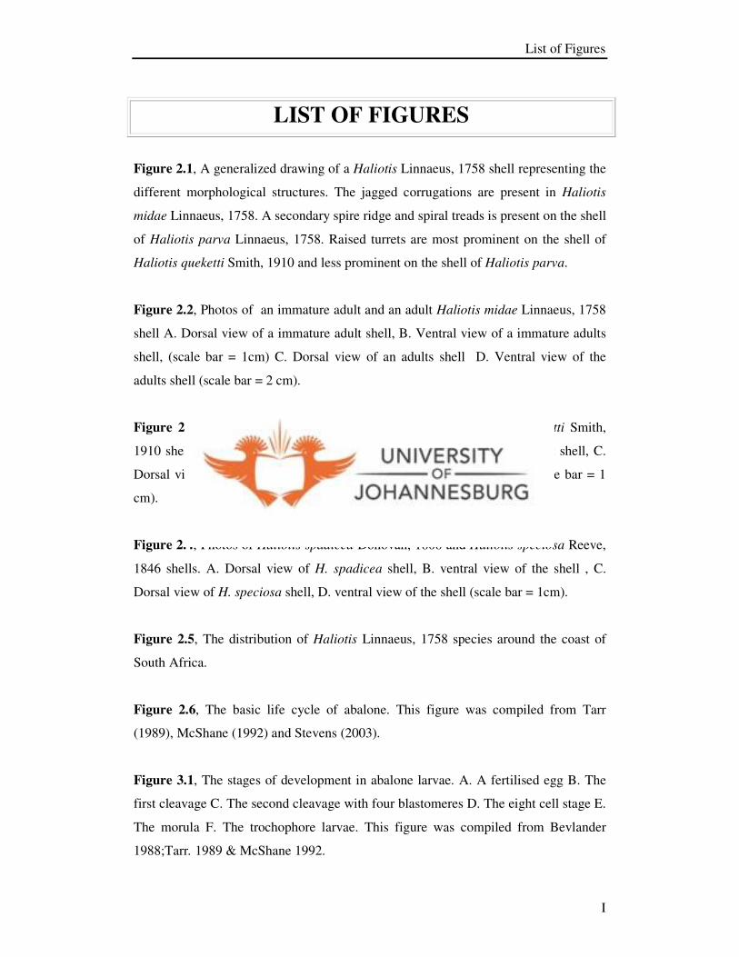
Figure 2.1, A generalized drawing of a Haliotis Linnaeus, 1758 shell representing the
different morphological structures. The jagged corrugations are present in Haliotis
midae Linnaeus, 1758. A secondary spire ridge and spiral treads is present on the shell
of Haliotis parva Linnaeus, 1758. Raised turrets are most prominent on the shell of
Haliotis queketti Smith, 1910 and less prominent on the shell of Haliotis parva.
Figure 2.2, Photos of an immature adult and an adult Haliotis midae Linnaeus, 1758
shell A. Dorsal view of a immature adult shell, B. Ventral view of a immature adults
shell, (scale bar = 1cm) C. Dorsal view of an adults shell D. Ventral view of the
adults shell (scale bar = 2 cm).
Figure 2.3, Photos of Haliotis parva Linnaeus, 1758, and Haliotis queketti Smith,
1910 shells A. Dorsal view of H. parva shell, B. Ventral view of H. parva shell, C.
Dorsal view of H. queketti shell, D. Ventral view of H. queketti shell (scale bar = 1
cm).
Figure 2.4, Photos of Haliotis spadicea Donovan, 1808 and Haliotis speciosa Reeve,
1846 shells. A. Dorsal view of H. spadicea shell, B. ventral view of the shell , C.
Dorsal view of H. speciosa shell, D. ventral view of the shell (scale bar = 1cm).
Figure 2.5, The distribution of Haliotis Linnaeus, 1758 species around the coast of
South Africa.
Figure 2.6, The basic life cycle of abalone. This figure was compiled from Tarr
(1989), McShane (1992) and Stevens (2003).
Figure 3.1, The stages of development in abalone larvae. A. A fertilised egg B. The
first cleavage C. The second cleavage with four blastomeres D. The eight cell stage E.
The morula F. The trochophore larvae. This figure was compiled from Bevlander
1988;Tarr. 1989 & McShane 1992.

List of Figures
II
Figure 3.2, Spawning tanks containing gravid male and females.
Figure 3.3, Settling tank with a floating container and a tap.
Figure 3.4, Tanks with plastic settlement plates containing diatoms.
Figure 3.5, Weaning tanks with plastic cones for juveniles to hide under. A. As
viewed from above B. View from the side.
Figure 3.6, A– Typical abalone farms with the holding tanks. B – The baskets
containing the abalone with the kelp they are fed.
Figure 3.7, Tanks with crate-like holders for juvenile abalone to grow in to cocktail
size for marketing. A. As viewed from the top B. View from the side.
Figure 3.8, A map of abalone farms around the west coast and the Tsitsikamma
National Park. 1. Port Nolith Sea Farm 2.Western Cape abalone in St. Helenabay 3.
Abulon Holdings & Abatech in Paternoster 4. Jacobsbay Sea Products 5. Global
Ocean in Kleinmond 6. Howston in Betty’s Bay 7. Abamax, Abaseed, Sea Plant
Products, Abagold, Aquafarm, HIK & Hermanus Abalone in Hermanus 8. Walker
Bay 9. Romans Bay Sea Farm 10. Atlantic Abalone and I&J in Gansbay 11.
Tsitsikamma National Park (Collection of wild abalone).
Figure 4.1, A – Rickettsiales–like prokaryote found in the digestive gland of Haliotis
midae (indicated by arrows), B –Larvae of Terebrasabella heterouncinata, C & D –
Terebrasabella heterouncinata found in the shell of Haliotis midae (indicated by
arrows), E –Polydora sp. (arrow 1) and Boccardia sp. (arrow 2) in the shell of
Haliotis midae, F – The U and Y shaped burrows of a Baccardia spp in the shell of
Haliotis spadicea (indicated by arrows).
Figure 4.2, Mantoscyphidia midae. Redrawn from Botes et al. 2001. Scale bar =
5µm.

List of Figures
III
Figure 4.3, Meronts of Pseudoklossia haliotis in the host cells of Haliotis
cracherodii. Redrawn from Friedman et al. (1995). Scale bar = 5 µm.
Figure 4.4, Terebrasabella heterouncinata redrawn from Fitzhuge & Rouse (1999).
Scale bar = 0.25mm.
Fig 5.1, The gregarine, Nematopsis annulipes Prasadan & Janardanan, 2001, redrawn
from Prasadan & Janardanan, 2001. A. sporadins, B. associations of sporadins. Scale
bar = 50 µm.
Fig 5.2, Line drawing of the spore ultrastructures of the Haplosporidian
Haplosporidia
louisiana s redrawn from Harrison & Corliss (1991). Scale bar 2µm.
Figure 5.3, The life cycle of Perkinsus marinus redrawn from Perkins (1996) and
Sparks (1985). Immature trophozoits (1-3) develops into mature trophozoites (4) and
then into tamonts (5-8).
Figure 6.1, Photos of Haliotis midae and its digestive gland. A. Haliotis midae, B.
The position of the epipoduim (Ep), digestive gland (Dg) and the cream coloration of
the male gonads (G), C. A healthy digestive gland, D. A Healthy female digestive
gland with the olive green coloration of the gonads (G).
Figure 6.2, Histological methods. A. An abalone farm with holding tanks, B.
Dissection of abalone, C. Histological cassettes, D. Fixation of samples, E.
Dehydration, F. Infiltration with wax.
Figure 6.3, Histology methods. A. L-shaped plates, B. Wax blocks, C. Chuck, D.
Microtome, E. Staining holders with chemicals for H&E staining, F. Zeiss compound
microscope used for staining slides.
Figure 6.4, Transmission Electron Microscopy methods. A. Digestive gland fixed in
4%PFA and Karnovskys fixative, B. Samples being rinsed with distilled water in a

List of Figures
IV
rotor, C. Embedded sample, D. Embedding mediums, E. Ultracut E (Reichert Jung),
F. Semi- thin section (1µm).
Figure 6.5, TEM methods. A. 1% Toluidene blue stain, B. Thin sections being stained
on glass slides with 1% Toluidene blue, C. Mounted thin sections, D. Gold sections,
E. Uranyl acetate and Lead citrate stains, F. Jeol 1010 Transmission electron
Microscope.
Figure 6.6 A, Scattered plot of total shell length (cm) against total wet weight (g) of
abalone infected with the unidentified digestive gland parasite demonstrating the
growth rate of infected abalone. R2
value calculated at 0.8241.
Figure 6.6 B, Scattered plot of total shell length (cm) against total wet weight (g) of
abalone non-infected with the unidentified digestive gland parasite demonstrating the
growth rate of uninfected abalone. R2
value calculated at 0.4855.
Figure 6.7, Photo plates showing the general digestive gland structures and the
occurrence of the digestive gland protozoan. A. Digestive gland tissue, c: β- cells
(crypt), b: basal lamina, p: digestive gland protozoan, d: duct cells, g: granules, l:
lumen. B. Protozoan parasites (p) found in a cluster close to the basal lamina (b),
lumen (l) and muscle layers (m). C. Single protozoan parasites found close to the
basal lamina (b) and lumen (l). D. A group of protozoan parasites (p) in between the
lumen (l) and basal lamina (b). E. Parasites (p). Scale bar = 10µm.
Figure 6.8, Photo plates of the protozoan parasite, its parasitophorous vacuole (pv)
and parasitophorous membrane (pm). A. The parasitophorous vacuole (pv) and
parasitophorous membrane (pm) is quit distinct, B. A large parasitophorous vacuole,
C. The protozoan parasite, D. A smaler pasasitophorous vacuole compaired to the on
in B, E. parasitophorous vacuole serounding the parasite, F. A few parasites situated
close to each other and parasitophorous vacuoles. Scale bar = 10µm.
Figure 6.9, Photo plates indicating some of the possible life stages of the protozoan
parasite. A. The most general stage found in the samples, B. A parasite (p) that is
curved in its parasitophorous vacuole (pv), C. A dividing parasite (p), D. Caps (c) at

List of Figures
V
the anterior and post ends of the parasite, E. parasites (p) dividing, F. A parasite (p)
that have been cut through at the end showing that it is cylindrical (rod) shaped. Scale
bar = 10µm
Fig 6.10, The possible live stages of UDP, A. Stage 1, B. Stage 2, C. Stage 3, D.
Stage 4, E. Stage 5, F. Stage 6
Figure 6.11, Photo plates of histology slides stained with the floreceine Cyto 9. A.
Histology slide stained with H&E and not Cyto 9, viewed with a Fluoreceine filter,
the gobbled cells (gc) and protozoan parasite (p) are fluorescing, B. Histology slide
stained with Cyto 9 and viewed with a Rhodamine filter, the parasite (p) is
fluorescing, C. Slide stained with Cyto 9, the parasite (p) is fluorescing and have a
three dimensional look, D. Gaps (g) and the membrane (m) of the parasite, The
parasites ), E. the parasites cap (pc), F. The parasites (p) membrane (m) is fluorescing
and the parasite has a three dimensional look. Scale bar = 10µm.
Figure 6.12, Electron micrographs of the parasite showing its ultrastructures. A. the
parasite (p) lying close to the digestive gland lumen (lu). B. the parasite ultrastructure
showing a possible formative region for haplosporosomes (fh), nuclear envelope (ne),
nucleus (n), nucleolus (ns) and outer membrane (om), C. The anterior region of the
parasite showing the possible spherulosome (ps). Scale bar for A and B = 5µm, Scale
bar for C = 2.5µm.
Figure 7.1, A - Digestive gland samples collected from H. midae, stored in ETOH, B
– Extraction kit, C- The warm water bath used for the lysis of tissue samples, D –
Ependorph centrifuge, F- PCR machine.
Figure 7.2, A- Elecrophorisis gel ran for the total DNA from samples nine and 22, B-
the gel that was run after the gradient PCR method was used. Sample nine and 22 at
different concentrations, C – Sample 22 at different concentrations after PCR
purification, D- Sample nine containing no DMSO in the premix.

A B
Figure 2.2, Photos of an immature adult and an adult Haliotis midae Linnaeus, 1758 shell A.
Dorsal view of a immature adult shell, B. Ventral view of a immature adults shell, (scale bar =
1cm) C. Dorsal view of an adults shell D. Ventral view of the adults shell (scale bar = 2 cm).
C D

A B
DC
Figure 2.3, Photos of Haliotis parva Linnaeus, 1758 and Haliotis queketti Smith, 1910
shells A. Dorsal view of H. parva shell, B. Ventral view of H. parva shell, C. Dorsal
view of H. queketti shell, D. Ventral view of H. queketti (scale bar = 1 cm).

A
C
Figure 2.4, Photos of Haliotis spadicea Donovan, 1808 and Haliotis speciosa
Reeve,1846 shells. A. Dorsal view of H. spadicea shell, B. ventral view of the shell , C.
Dorsal view of H. speciosa shell, D. ventral view of the shell (scale bar = 1cm).
B
D

D.
Egg
membrane
Cleavage
plane
Polar body
Jelly coat
Viteline space
Viteline layer
A. B.
Blastomere
BlastomereMinor cell
Major cell
Prototroch
Egg envelope
C.
E.F.
Figure 3.1, The stages of development in abalone larvae. A. A fertilised egg B. The first cleavage,
C. The second cleavage with four blastomeres, D. The eight cell stage, E. The morula, F. The
trochophore larvae. This figure was compiled from Bevlander 1988;Tarr. 1989 & McShane 1992.
D.

A
B
Figure 3.6, A– Typical abalone farms with the holding tanks. B – The baskets
containing the abalone with the kelp they are fed.

2
1
A
C
B
D
FE
Figure 4.1, A – Rickettsiales–like prokaryote found in the digestive gland of Haliotis
midae Linnaeus, 1758 (indicated by arrows), B –Larvae of Terebrasabella
heterouncinata Terebrasabella heterouncinata Fitzhugh & Rouse, 1999, C & D –
Terebrasabella heterouncinata found in the shell of Haliotis midae (indicated by
arrows), E –Polydora sp. Bosc, 1802 (arrow 1) and Boccardia sp. Carazzi, 1895 (arrow
2) in the shell of Haliotis midae, F – The U and Y shaped burrows of a Baccardia sp. in
the shell of Haliotis spadicea (indicated by arrows).

Adoral ciliary spiral
Bucal cavity
Pellicle striations
Symbiotic algae
Macronucleus
Micronucleus
Scopula
Peristomal lip
Figure 4.2, Mantoscyphidia midae Botes, Basson and Van As, 2001. Redrawn from
Botes et al. 2001. Scale bar = 5µm.

Merozoite
residuum
Merozoites
budding
Nucleus
Host kidney
cell
Parasitophorous
vacuole
Endoplasmic
reticulumMicronemes
Rhoptry duct
Conoid
Figure 4.3, Meronts of Pseudoklossia haliotis Friedman, Gardner, Hedrick, Stephenson,
Cawthorn & Upton, 1995 in the host cells of Haliotis cracherodii Leach, 1814 . Redrawn
from Friedman et al. (1995). Scale bar = 5 µm.
Meront

Feacal groove
Feacal groove
Feacal groove
Ventral base flange
Ventral base flange of
basal crown
Figure 4.4, Terebrasabella heterouncinata Fitzhuge & Rouse, 1999, redrawn from
Fitzhuge & Rouse (1999). Scale bar = 0.25mm.

Fig 5.1, The gregarine, Nematopsis annulipes Prasadan & Janardanan, 2001, redrawn
from Prasadan & Janardanan, 2001. A. sporadins, B. associations of sporadins. Scale
bar = 50 µm.
A
B

Lid
Spherulosome
Haplosporosome
- forming body
Haplosporosomes
Epispore
cytoplasm
Flange
Mitochondrion
Fig 5.2, Line drawing of the spore ultrastructures of the Haplosporidian Haplosporidia louisiana
Sprague, 1954 redrawn from Harrison & Corliss (1991). Scale bar 2µm.

1
2
3
4
5
7
6
8
Figure 5.3, The life cycle of Perkinsus marinus Mackin, Owen. and Collier, 1950, redrawn from Perkins
(1996) and Sparks (1985). Immature trophozoits (1-3) develops into mature trophozoites (4) and then into
tamonts (5-8).

A
B
C D
Ep
G
Dg
G
Figure 6.1, Photos of Haliotis midae Linnaeus, 1758 and its digestive gland. A.
Haliotis midae, B. The position of the epipoduim (Ep), digestive gland (Dg) and
the cream coloration of the male gonads (G), C. A healthy digestive gland, D. A
Healthy female digestive gland with the olive green coloration of the gonads (G).

A B
C D
E F
Figure 6.2, Histological methods. A. An abalone farm with holding tanks, B.
Dissection of abalone, C. Histological cassettes, D. Fixation of samples, E.
Dehydration, F. Infiltration with wax.
F

A B
C D
E
A
C
F
Figure 6.3, Histology methods. A. L-shaped plates, B. Wax blocks, C. Chuck, D.
Microtome, E. Staining holders with chemicals for H&E staining, F. Zeiss compound
microscope used for staining slides.

A
C
B
D
E F
Figure 6.4, Transmission Electron Microscopy methods. A. Digestive gland fixed in
4%PFA and Karnovskys fixative, B. Samples being rinsed with distilled water in a rotor,
C. Embedded sample, D. Embedding mediums, E. Ultracut E (Reichert Jung), F. Semi-
thin section (1µm).

A B
C D
E F
Figure 6.5, TEM methods. A. 1% Toluidene blue stain, B. Thin sections being
stained on glass slides with 1% Toluidene blue, C. Mounted thin sections, D. Gold
sections, E. Uranyl acetate and Lead citrate stains, F. Jeol 1010 Transmission electron
Microscope.

p
c
b
g
d
l
l
p
m
b
b p
l
Figure 6. 7, Photo plates showing the general digestive gland structures and the occurrence of the digestive
gland protozoan. A. Digestive gland tissue, c: β- cells (crypt), b: basal lamina, p: digestive gland protozoan, d:
duct cells, g: granules, l: lumen. B. Protozoan parasites (p) found in a cluster close to the basal lamina (b),
lumen (l) and muscle layers (m). C. Single protozoan parasites found close to the basal lamina (b) and lumen
(l). D. A group of protozoan parasites (p) in between the lumen (l) and basal lamina (b). E. Parasites (p). Scale
bar = 10µm.
A
B C
D E
l
p
b
p
p

pv
pm
p
pv
pv
pv
pv
A B
C D
E F
Figure 6.8, Photo plates of the protozoan parasite, its parasitophorous vacuole (pv) and parasitophorous
membrane (pm). A. The parasitophorous vacuole (pv) and parasitophorous membrane (pm) is quit distinct, B.
A large parasitophorous vacuole, C. The protozoan parasite, D. A smaler pasasitophorous vacuole compaired
to the on in B, E. parasitophorous vacuole serounding the parasite, F. A few parasites situated close to each
other and parasitophorous vacuoles. Scale bar = 10µm.

A B
C D
E F
p
p
c
c
p
p
p
p
Figure 6. 9, Photo plates indicating some of the possible life stages of the protozoan parasite. A. The most
general stage found in the samples, B. A parasite (p) that is curved in its parasitophorous vacuole (pv), C. A
dividing parasite (p), D. Caps (c) at the anterior and posterior ends of the parasite, E. parasites (p) dividing, F.
A parasite (p) that have been cut through at the end showing that it is cylindrical (rod) shaped. Scale bar =
10µm
pv

A B
C D
E F
Fig 6. 10 , The possible live stages of UDP (unidentified digestive gland parasite), A. Stage 1, B.
Stage 2, C. Stage 3, D. Stage 4, E. Stage 5, F. Stage 6

A
pgc
B
p
p
Epc
F
p
D
g
gm
C
p
p
Figure 6.11 , Photo plates of histology slides stained with the floreceine Syto 9. A. Histology slide stained with
H&E and not Syto 9, viewed with a Fluoreceine filter, the gobbled cells (gc) and protozoan parasite (p) are
fluorescing, B. Histology slide stained with Syto 9 and viewed with a Rhodamine filter, the parasite (p) is
fluorescing, C. Slide stained with Syto 9, the parasite (p) is fluorescing and have a three dimensional look, D.
Gaps (g) and the membrane (m) of the parasite, E. the parasites cap (pc), F. The parasites (p) membrane (m) is
fluorescing and the parasite has a three dimensional look. Scale bar = 10µm.
m

A
B
C
fh
ne
nns
om
p
ps
lu
Figure 6.12 Transmission electron micrographs of the parasite showing its ultrastructures. A. the parasite
(p) lying close to the digestive gland lumen (lu). B. the parasite ultrastructure showing a possible formative
region for haplosporosomes (fh), nuclear envelope (ne), nucleus (n), nucleolus (ns) and outer membrane
(om), C. The anterior region of the parasite showing the possible spherulosome (ps). Scale bar for A and B
= 5µm, Scale bar for C = 2.5µm.

A B
C D
E F
Figure 7.1, A - Digestive gland samples collected from H. midae Linnaeus, 1858,
stored in ETOH, B – Extraction kit, C- The warm water bath used for the lysis of
tissue samples, D – Ependorph centrifuge, F- PCR machine.

A B
C D
9 22 9b 22b 22c 22b22a
22b 22c 22d22a
9a 9b 9c 9d
9c 9d
Figure 7.2, A- Elecrophorisis gel ran for the total DNA from samples nine and 22, B- the gel
that was run after the gradient PCR method was used. Sample nine and 22 at different
concentrations, C – Sample 22 at different concentrations after PCR purification, D- Sample
nine containing no DMSO in the premix.

List of tables
VI
LIST OF TABLES
Table 2.1: The Haliotis species with their synonyms, common names and
geographical distribution across the world. This table is compiled from Muller (1986);
Lindberg (1992) and Geiger & Grove (1999).
Table 2.2: The economically important abalone species. This table is compiled from
Morse et al. (1977, 1979); Moss, Illingworth & Thong. (1995); Hauser (1997);
Haaker (1997); Daniels & Floren (1998); McBride (1998); Moore et al. 2002);
Stevence (2003); Pang, Zhang, Bao & Gao (2006), Anon 2, 2006 and Anon 3, 2007.
Table 4.1: Parasites infecting Haliotis spp. around the world and the effects they have
on a host.
Table 6.1: Prevalences of the unidentified digestive gland parasite, sabellids and
polychaetes, and sizes/weights of host abalone from different localities on different
dates (TNP = Tsitsikamma National Park; n = total number of animals collected, SD =
standard deviation; UDP = unidentified digestive gland parasites; Sab = sabellids; Pol
= polychaetes).

Summary
VII
SUMMARY
Abalone are among the world’s leading shellfish consumed by human populations.
Harvesting in California began in the late 1800s from intertidal zones and in the early
1900s wild abalone were collected by diving. Popular demand for abalone products in
the Far East then led to extensive harvesting of wild abalone and a drastic decline in
population numbers. This problem was overcome to a degree by the development of
land-based abalone farms. At these farms it was possible to breed abalone on a large
scale. Currently twelve abalone farms operate in South Africa and the estimated
production for 2006 was 537 tons of meat, worth R 80 mil. Parasites and diseases
pose threats to the production of abalone, especially under farmed conditions, and can
cause considerable financial loss. Labyrinthuloides haliotidis, Haplosporidium nelsoni
and Terebrasabelle heterouncinata are a few parasites that contribute to the above
mentioned problems. Lately, a new protozoan parasite was discovered in the digestive
glands of Haliotis midae farmed in the Western Cape Province, during routine health
assessments. For the purposes of this dissertation it is designated an unidentified
digestive gland parasite (UDP). The aims of this study are thus to undertake a
comprehensive literature review of parasites infecting wild and farmed abalone, as
well other shellfish species, describe and characterise the UDP infecting the digestive
gland of Haliotis midae based on its structure and ultrastructure, evaluate the role of
this parasite in disease by analysing data from histological studies, provide a
preliminary indication of the life cycle of this parasite, attempt analysis of DNA from
the UDP, and identify potential areas for further research into control of the parasite.
A total of 180 abalone, (Haliotis midae) were collected from three abalone farms in
the Western Cape during May 2005, October 2005, January 2006 and January 2007.
To establish whether this parasite also occurs in wild abalone, a single sampling (six
H. midae and 28 H. spadicea) took place during 2006 in Tsitsikamma National Park.
Collected farmed and wild abalone were weighed and measured, removed from their
shells and then killed according to accepted methods before their digestive glands
were removed. Each digestive gland was cut into four pieces. The first piece of each
sample was placed into a histological cassette and fixed in 10% neutral buffered
formalin; the samples were then prepared for histological studies using standard

Summary
VIII
methods. The second and third pieces were fixed in Karnovsky’s and 4%
paraformaldehyde fixatives respectively for transmission electron microscopy (TEM).
The remaining piece was fixed in 96% ethanol for DNA analysis. The presence of
UDP in the digestive glands was determined by histology, and the parasite’s
ultrastructure was then studied by TEM. The prevalence of UDP was as follows: 10%
for May 2005 (Farm A), 3.2 % for October 2005 (Farm A), 35.5% for January 2006
(Farm B) and 40% for January 2007 (Farm B). The prevalence was 0% for May 2005
(Farm B & C) and April 2006 (Tsitsikamma National Park). In histological sections
the UDP was usually fiddle-shaped, or hour-glass shaped with a parasitophorous
vacuole surrounding it and six presumed life stages were observed, although no
obvious pathology was seen. Structures resembling those characteristic of
haplosporidians (formative region for haplosporosomes and possible spherulosome)
were visible in electronmicrographs. The UDP was then compared with parasites
occurring in abalone throughout the world, as well as in other shellfish species. This
was done mainly using its morphology, morphometrics and ultrastructural
characteristics. In comparison with other parasites occurring in abalone, the UDP
parasitizes the digestive gland tubules of H. midae, as the coccidian Pseudoklossia
haliotis occupies the kidney tubules of the same type of host. The morphometrics of
UDP and the haplosporidian Haplosporidium nelsoni overlap and the plasmodial
stages of haplosporidian species can occur close to the basal lamina and upper gut, as
does UDP. The apicomplexan Perkinsus species in general, from other shellfish, have
life stages that divide, as do those of UDP. Microsporidians also possess a posterior
vacuole and polar caps that are similar to those seen in the UDP. No exact matches
could be made between the UDP and other protozoan parasites; the fit with the
haplosporidians appeared closest, but unfortunately this could not be verified by DNA
analyses, since attempts at these techniques were unsuccessful. However, overall the
results from this study make an interesting contribution to the knowledge of farmed
abalone parasites and diseases in South Africa and suggestions for extending the
research appear in the latter sections of the dissertation.

Opsomming
IX
OPSOMMING
Perlemoen is een van die voorste skulpvis spesies in die wêreld wat deur die menslike
polulasie verbruik word. Die oes van intergetysone perlemoen in Kalifornia het in die
laat 1800’s begin en in die vroeër 1900’s is perlemoen deur duikers versamel. Die
populêre aanvraag na perlemoen produkte in die verre Ooste het gelei tot die
uitgebreide oesting van perlemoen, gevolglik het die populasie getalle drasties
afgeneem. Hierdie probleem is tot ‘n mate oorkom deur die ontwikkeling van
perlemoen plase. By hierdie plase was dit moontlik om perlemoen op grootskaal te
kweek. Huidiglik is twaalf plase funksioneel in Suid-Afrika. Die geraamde produksie
vir 2006 was 537 ton vleis, terwaarde van R80 miljoen. Parasiete en siektes hou ‘n
groot bedreigings in vir die produksie van perlemoen, veral onder plaastoestande, en
kan aansienlike finasieële verlies tot gevolg hê. Labyrinthuloides haliotidis,
Haplosporidium nelsoni en Terebrasabella heterouncinata is ‘n paar van die parasiete
wat bydra tot die bogenoemde probleem. Onlangs is ‘n nuwe protozoa parasiet ontdek
in die verteeringsklier van gekweekte perlemoen afkomstig van die Wes Kaap,
gedurende ‘n roetine gesondheids assesering. Vir die doel van hierdie verhandeling
staan dit bekend as die ongeidentifiseerde verteeringsklier parasiet (UDP). Die doel
van hierdie studie is die volgende: om ‘n deeglike literatuur oorsig van die parasiete
wat wilde en gekweekte perlemoen afekteer asook die van ander skulpvis spesies te
onderneem, om die UDP wat die verteeringsklier van H. midae infekteer te beskryf en
te karakteriseer gebaseer op sy strukture en ultrastrukture, om die rol van hierdie
parasiet in siektes te evalueer volgens die analise van data afkomstig van histologiese
studies, om ‘n voorlopige aanduiding van hierdie parasiet se lewens siklus weer te
gee, om DNA analisis van die UDP te onderneem en om potensieële areas vir verdere
navorsing oor die beheer van hierdie pasasiet te identifiseer. ‘n Totaal van 180
perlemoen (H. midae) was versamel van drie perlemoen plase in die Wes Kaap
gedurende Mei 2005, Oktober 2005, Januarie 2006 en Januarie 2007. ‘n Enkele
versameling van ses H. midae en 28 H. spadicea het plaasgevind by die Tsitsikamma
Nasionale Park gedurende 2006 om te bepaal of hierdie parasiet ook in wilde
perlemoen voorkom. Versamelde en gekweekte perlemoen was geweeg en gemeet,
verwyder van die skulp en is dan van kant gemaak volgens aanvaarde metodes waarna

Opsomming
X
die verteeringsklier verwyder is. Elke verterings klier was dan in vier stukke verdeel.
Die eerste deel van elke monster is geplaas in ‘n histologiese kaset waarna dit
gefikseer is in 10% nutraal gebufferde formalien en is dan voorberei vir histologie
volgens standaard metodes. Die tweede en derde dele is onderskeidlik gefikseer in
Karnovskies en 4% paraformaldehied vir transmissie elektron mikroskopie (TEM).
Die oorblywende deel is gefikseer in 96% etanol vir DNA analise. Die
teenwoordigheid van UDP in die verteeringsklier is bepaal deur histologie en die
pasasiet se ultrastrukture was gebestudeer deur middel van TEM. Die persentasie
besmetting van UDP was soos volg: 10% vir Mei 2005 (Plaas A), 3.2% vir Oktober
2005 (Plaas A), 35.5% vir Januarie 2006 (Plaas B) en 40% vir Januarie 2007 (Plaas
B). Die persentasie besmittig was 0% vir Mei 2005 (Plaas B & C) en April 2006
(Tsitsikamma Nasionale Park). In die histologiese senee was die UDP gewoonlik
viool of uurglas vormig en was omring met ‘n parasitoforus vakuool. Ses moontlike
lewens staduims is opgemerk al was daar geen duidelike patologie waargeneem nie.
Strukture voorstellend van diė, kenmerkend aan haplosporidia (voremende area vir
haplosporosome en moontlike spherolosome) was duidelik met elekton micrografieke.
Die UDP was toe vergelyk met parasiete wat in perlemoen spesies van reg oor die
wêreld, sowel as in ander skulpvis spesies voorkom. In die vergelykings met
perlemoen parasiete, parasiteer die UDP die verteeringsklier buisies van H. midae net
soos in die geval van die Koksidia parasiet Pseudoklossia haliotis wat die nierbuisies
van dieselfde tipe gasheer parasiteer. Die meetings van UDP en die van die
haplosporidia, Haplosporidium nelsoni oorvleul. Plasmodium stadia van ander
haplosporidia spesies kom naby die basale lamina en die bo-derm voor net soos in die
geval van UDP. Algemene Perkinsus spesies (Apikompleksa) van ander skulpvis het
stadia wat verdeel net soos UDP. Mikrosporidia besit ‘n posterior vakuool en polêre
kapsels wat soortgelyk is aan die wat waargeneem word in UDP. Geen perfekte
ooreenstemming kon gemaak word tussen UDP en ander protozoa nie, die
ooreensteming met haplosporidia blyk die naaste, maar ongelukkig kon dit nie met die
DNA analise geverifiseer word nie, aangesien die poging onsuksesvol was. Oor die
algemeen maak die resultate van hierdie studie ‘n interressante bydra tot die kennis
van gekweekte perlemoen parasiete en siektes in Suid-Afrika. Voorstelle vir die
uitbreiding van die navorsing kom voor in die laaste deel van hierdie verhandeling.

CHAPTER 1 Introduction
- 1 -
1. Introduction
Abalone is one of the most important and valuable exported shellfish products in the
world (McShane 1992; Ebert & Houk 1984; Sales & Janssens 2004; Reddy-Lopata,
Auerswald & Cook 2006). This popularity is due to the foot muscle meat of the
abalone that is considered as a great delicacy globally and also to the shells that are
used in the production of jewellery (Alvarez-Tinajero, Caceres-Martinez & Gonzalez-
Aviles 2001). Abalone farming and commercial diving are globally as well as locally
very lucrative businesses (Macey & Coyne 2005), contributing millions of dollars to
countries exports and economy. Besides this the abalone industry creates considerable
job opportunities (Macey & Coyne 2005; Cai, Chen, Thompson & Li 2006) to local
people in various countries (Troell, Robertson-Andersson, Anderson, Bolton,
Maneveldt, Halling & Probyn 2006).
1.1 Global abalone industry
1.1.1 The history of the abalone industry
In California, abalone were harvested from intertidal zones by native people for
thousands of years (Davis, Richard, Haaker & Parker 1992; Lafferty & Kuris 1993;
Davis 1993; Moore, Finley, Robbins & Friedman 2002). In the 1800s Asians
harvested abalone by hand (Davis 1993), and in the early 1900s abalone were
collected from subtidal regions by diving (Davis 1993). During the 1950s to 1960s
abalone became a popular fishery and sport activity (Davis et al. 1992): the abalone
was collected by scuba diving to depths of 100ft and more. Throughout 1950 to 1970
commercial divers in southern California harvested over one million pounds of
abalone.
From the 1970s, intertidal abalone populations decreased (Moore et al. 2002) and
became rare (Davis et al. 1992). By 1996 less than 10% of the annual take could be
harvested by the commercial divers. Abalone aquaculture commenced in California in
the 1960s (McBride 1998). Research on the spawning of abalone (Morse, Duncan,
Hooker & Morse 1977) and the settlement of larvae were conducted successfully in

CHAPTER 1 Introduction
- 2 -
the 1970s in an attempt to cultivate abalone (Morse, Hooker, Duncan & Jensen 1979).
In 1981 the first cultivated abalone from California were sold (McBride 1998).
1.1.2 Impact of parasites and diseases on the abalone industry
In the history of commercial shellfish mariculture, disease outbreaks have been
common and have mostly led to mass mortalities which in turn have been responsible
for the failure of farming and commercial operations (Kuris & Culver 1999; Simon,
Kaiser & Britz 2004). An example of this has been the demise of abalone farming
operations in British Columbia, Canada where almost all the juvenile abalone died
due to a thraustochyrid parasite (Bower 2000). High abalone densities and poor
husbandry are perfect combinations for overwhelming diseases and parasites to
accumulate, and spread between hosts (Cook 1998). Clearly parasites and diseases of
abalone can have a devastating financial impact on abalone farming and the industry.
Hine and Jones (1994) listed the following points as being of significant economical
importance when looking at parasites of cultured fish and shellfish species:
• Parasites induce host mortalities leading to reduced harvest, this includes
mortalities of juveniles and so reduces future recruitment to the fisheries
• Weight loss in infected hosts, which are still saleable, but represent a reduction
in return to the fisher
• Parasite induced reduction in fecundity, leading to a poorer than expected
spawning and a subsequent poor recruitment to the host population
• Reduced flesh quality, which results either in additional processing costs or in
rejection of product. The effects can be visual, or chemical changes in the fillet
such as enzyme degradation, or the public health risk associated with the
presence of the parasite
• The cost of complying with regulatory requirements associated with the actual
or potential presence of the parasite, this affects the fish processor as well as
the aquaculturalist
• Cost to aquaculturalists of prophylactics

CHAPTER 1 Introduction
- 3 -
1.1.3 Quality regulations of abalone products
Quality control aspects for live, exported, animals determine that abalone should be in
an impeccable condition: the shell must not be damaged or deformed, and the foot
muscle (meat) must show no signs of discoloration and damage, as this part of the
abalone is after all the entity of value. Damage to the foot muscle includes pustule
formations or sores. Animals that are not up to standards are classified as rejects and
are processed as canned products.
1.1.4 Well known parasites and diseases of abalone
A wide variety of abalone parasites and diseases have been studied in the past (see
Chapter 4). Globally, the most economically important species include:
Terebrasabella heterouncinata described as a new genus and species by Fitzhuge &
Rouse, 1999 (Kuris & Culver 1999) that occurs in abalone species of California (Ruck
& Cook 1998) and originated from South Africa (Fitzhuge 1996; Finlay, Mulligan &
Friedman 2001) where it is an important health issue (Simon et al. 2004).
Boccardia knoxi Rainer, 1973, and Polydora haplura Claparede, 1870 (Blake &
Evans 1973) are two examples of spionid mud worms that devastated abalone culture
in Tasmania and inevitably led to many facilities closing or relocating in 1997 (Kuris
& Culver 1999; Simons et al. 2004). An unidentified Haplosporidium sp., closely
related to the genus Urosporidium, has been studied in Haliotis iris Martyn, 1784
(Diggles, Nichol, Hine, Wakefield, Cochennec-Laureau, Roberts & Friedman 2002).
Labyrinthuloides haliotidis Bower, 1987 parasitises mainly juvenile abalone (Bower
1987a; b). Withering syndrome caused by the Rickettsiales-like prokaryotes
(bacterium) Candidatus Xenohaliotis californiensis (Friedman 2002) has led to the
devastation of wild and cultured abalone populations from California (VanBlaricom,
Ruediger, Friedman, Woodard & Hedrick 1993; Moore et al. 2002) and of lesser
economic importance is Pseudoklossia haliotis Friedman, Gardner, Hedrick,
Stephenson, Cawthorn & Upton, 1995, that was described as a coccidian infecting the
kidneys of Haliotis spp. (Friedman et al. 1995).
* Dr. Anna Mouton Abalone Farmers Association of South Africa. * Dr. J. Handlinger Fish Health Unit, Animal Health
Laboratory, Tasmania

CHAPTER 1 Introduction
- 4 -
Parasites have different impacts on abalone, some causing shell deformities (eg. T.
heterouncinata) (Kuris & Culver 1999; Simon et al. 2004) and severe shell damage
(eg. B. knoxi & P. hoplura) (Blake & Evans 1973; Kuris & Culver 1999; Lleonart,
Handlinger & Powell 2003a; 2003b), others destroying the foot muscle through
pustule formation eg. Perkinsus olseni Lester and Davis, 1981 (Lester & Davis 1981;
Goggin & Lester 1987) in Haliotis rubra Leach, 1814 (Goggin, Sewell & Lester
1989) and Haliotis laevigata Donova, 1805 (Lester 1986). Protozoan parasites can
damage vital organs (eg. P. haliotis) (Steinbeck, Groff, Friedman, McDowell &
Hedrick 1992) leading to growth retardation, behavioural changes and even death (eg.
Haplosporidia) (Diggles & Hine 2001).
1.1.5 Abalone and the environment
Environmental changes have also had overwhelming effects on the wild abalone
population numbers. Catastrophic El Niños occurred quite frequently from the 1980s
to the 1990s around California (Moore et al. 2002). The water temperatures elevated
and nutrient levels declined (Davis et al. 1992) leading to the withering of kelp beds
(Haaker 1997) and amongst other events, the starvation of abalone (Friedman,
Thomson, Chun, Haaker & Hedrick 1997).
1.2 South African abalone industry
1.2.1 Abalone farming
Abalone farming in South Africa is quite a young industry (Cook 1998), compared
with established farms across the world (see Chapter 3). Farming commenced in the
late 1980s when a gravid wild Haliotis midae Linnaeus, 1758 that had been collected
spawned spontaneously (Sales & Britz 2001a) and was spawned artificially thereafter.
The industry mostly focuses on visual parasites like polychaetes (Simon et al. 2004)
and intracellular parasites. The spionid polychaetes Polydora hoplura, Dipolydora
capensis (Day) and an unidentified Boccardia species have been noted as being
problematic in Haliotis midae (Lleonart et al. 2003a; 2003b; Simon, Ludford &
Wynne 2006).

CHAPTER 1 Introduction
- 5 -
1.2.2 South African regulations on abalone quality
Methods of controlling the standard and quality of abalone production in South
African farms are implemented by the Abalone Farmers Association. In 1999, a health
management program was started in an attempt to control diseases and parasites
(Mouton 2000). Routine health assessments took place and a range of new
undescribed parasites were discovered upon sampling and examination of the abalone
for possible diseases (Sales & Britz 2001a).
1.2.3 Unknown parasites of abalone in South Africa
During these routine health assessments conducted in 2001 in the Western Cape, an
unknown protozoan parasite was discovered in the digestive gland of farmed H. midae
(Mouton 2000; Sales & Britz 2001a). Initial transmission electron microscopy studies
indicated that the parasite might be an apicomplexan due to the presence of organelles
resembling those of the apical complex (A. Mouton, pers. comm.). Literature reviews
were conducted to determine if parasites resembling this one had been described in
the past. The only parasite (also unidentified) similar to the unidentified protozoan
found in H. midae was from wild abalone in Tasmania (J. Handlinger, pers. comm.).
Diagnosis and descriptions of unknown parasites may take many months or years to
complete, and afterwards the research into the biology and control of the parasite can
also be extensive. Through the help and funding of Marine and Coastal Management,
the current project was initiated to study this enigmatic digestive gland protozoan.
The hypotheses for this study are that the parasite is a member of the protozoan order
Apicomplexa, that it has no detrimental effect on the growth and health of the host,
and that H. midae could be its intermediate invertebrate host.
The aims of this study are thus to:
• undertake a comprehensive literature review of parasites infecting wild and
farmed abalone

CHAPTER 1 Introduction
- 6 -
• undertake a comprehensive literature review of parasites infecting other
important shellfish species
• describe and characterise the intracellular protozoan parasite infecting the
digestive gland of Haliotis midae based on its ultrastructure
• evaluate the role of this parasite in disease by analysing data from histological
studies
• give a preliminary indication of the life cycle of this parasite
• attempt DNA analysis of this parasite
• identify potential areas for further research into control of the parasite.
Aspects that will be covered in this dissertation include the basic biology and ecology
of abalone species occurring in South Africa, how a typical abalone farm works, a
literature review on abalone parasites, parasites of other economically important
shellfish, techniques used in this study and the results thereof, as well as the
classification of this unknown parasite.
The editorial style of this dissertation follows that of the journal African Zoology (see
Appendix 1).

CHAPTER 2 Wild abalone
- 7 -
2. Wild Abalone
Nearly a hundred abalone species occur around the world (Table 2.1), but merely a
few are of any commercial value (Table 2.2) (Sales & Janssens 2004). Numerous
species have become vulnerable to over-exploitation and poaching (Reddy-Lopata et
al. 2006). The French named this fascinating animal “ormeau” or “ormel”, in Spain it
is called “abulon”, Japan “tokobushi”or “awabi” (Oakes & Ponte 1996), Australia
“paua” (Grindley, Keogh & Friedman 1998) and in South Africa “perlemoen”
(Steinberg 2005).
Although five different species of Haliotis occur along the coast of South Africa
(Steinberg 2005), only one, Haliotis midae is economical important (Reddy-Lopata et
al. 2006) due to its distribution and size (Tarr 1992). To appreciate the intricate nature
of Haliotis species, it is important to understand the basic nature and life cycle of this
astonishing sought-after delicacy and the research which led to its artificial
cultivation.
2.1 Wild abalone species of South Africa
The five different species of Haliotis occurring around the coast of South Africa are
Haliotis midae (perlemoen or Midas ear abalone), Haliotis parva Linnaeus, 1758
(spiral ridge siffie or canaliculate abalone), Haliotis queketti Smith, 1910 (Quekett’s
abalone), Haliotis spadicea Donovan, 1808 (siffie, venus ear or blood-spotted
abalone) and Haliotis speciosa Reeve, 1846 (beautiful ear- shell abalone or splendid
abalone) (Branch, Griffiths, Branch, & Beckley 2002). A sixth species, Haliotis
saldanhae Kensley, 1972, now extinct (Sweijd, Snethlange, Harvey & Cook 1998),
from the Pliocene occurred on the west coast of South Africa (Langebaan) (Geiger &
Groves 1999). The species Haliotis pustulata Reeve (Sales & Britz 2001a) occurs
from the northern South African border and throughout Mozambique. This species is
thought to be a sixth Haliotis species to occur in South Africa, but this is not so, and it
will not be discussed further in this chapter.

CHAPTER 2 Wild abalone
- 8 -
The habitats, food preferences and behaviour of the five Haliotis spp. endemic to
South Africa vary due to many factors and circumstances and it is quite easy to
distinguish the South African species on the morphological differences in their shells.
2.2 Identification of Haliotis Linnaeus, 1758 species from South
Africa
Haliotis species of South Africa can be distinguished by the following morphological
characteristics (Muller 1986): maximum size, shape, spire, spire ridges, spiral treads,
corrugations, the number of respiratory pores (holes), raised turrets and the surface
structure of the shell (Fig. 2.1).
Spire
Jagged
corrugations
Spiral threads
Secondary spire
ridge
Closed respiratory
pore
Raised turrets
Respiratory
pore
Figure 2.1, A generalized drawing of a Haliotis Linnaeus, 1758 shell representing the different
morphological structures. The jagged corrugations are present in Haliotis midae Linnaeus, 1758. A
secondary spire ridge and spiral treads are present on the shell of Haliotis parva Linnaeus, 1758. Raised
turrets are most prominent on the shell of Haliotis queketti Smith, 1910 and less prominent on the shell
of Haliotis parva.

CHAPTER 2 Wild abalone
- 9 -
2.2.1 Haliotis midae Linnaeus, 1758
Haliotis midae is the largest of the South African species (Muller 1986; Tarr 1989),
and can grow up to 190mm (Branch et al. 2002) in shell length (Fig. 2.2 C) by which
time, it can be 30 years or older (Barkai & Griffiths 1986; Tarr 1989). Jagged
corrugations (Muller 1986) stretch across the spire (Richards 1981) (Fig. 2.2 A), the
exterior shell colour can be light pink to cream-white and the inside of the shell is a
mother-of pearl colour (Figs. 2.2 B & D). Juveniles lack the corrugations and the shell
is smoother with a dark red colouration (Branch et al. 2002). The number of open
respiratory pores is 8 to 11. The natural distribution range is from Saldanha Bay to
Transkei (Fig. 2.5) (Kilburn & Rippey 1982).
2.2.2 Haliotis parva Linnaeus, 1758
Haliotis parva is the smallest South African species, with a maximum length of
45mm. The spiral ridge is prominent (Muller 1986) with a high spire (Richards 1981)
(Figs. 2.3 A & B). A secondary ridge is present, as well as fine spiral threads (Kilburn
& Rippey 1982). The colour of the shell may range from orange to brown, or can be
mottled red and green (Richards 1981). The number of open respiratory holes is 6 to
7. The natural distribution stretch from Table Bay to East London as can be seen in
(Fig. 2.5) (Kilburn & Rippey 1982).
2.2.3 Haliotis queketti Smith, 1910
Haliotis queketti can grow up to 46mm in shell length (Branch et al. 2002) and the
respiratory holes are raised on turrets (Figs. 2.3 C & D). Spiral ridges are rough with a
high spire (Richards 1981). The number of open respiratory pores is 4 to 6 (Kilburn &
Rippey 1982). Shell colour can be dappled variations of orange, red and brown and
the shell itself is rough due to the spiral ridges. The natural distribution ranges from
Port Alfred to Kwa-Zulu Natal (Branch et al. 2002).

CHAPTER 2 Wild abalone
- 10 -

CHAPTER 2 Wild abalone
- 11 -

CHAPTER 2 Wild abalone
- 12 -
2.2.4 Haliotis spadicea Donovan, 1808
Haliotis spadicea is the most commonly found species and is ear-shaped (Figs. 2.4 A
& B). (Richards 1981). This species can grow up to 80mm in length and the shell
surface is smooth compared to the other species (Muller 1986). Corrugations are
present on the shell surface, as well as a few spiral ridges. The number of open
respiratory pores is 5 to 8 (Kilburn & Rippey 1982). Shell colour is usually reddish-
brown, although splotches can also be observed. A copper-red mark is situated inside
the shell, underneath the spire and the edges of the mantle are bright green (Muller
1986). The natural distribution of this species stretches from False Bay to KwaZulu
Natal (Richards 1981).
2.2.5 Haliotis speciosa Reeve, 1846
The fifth species, H. speciosa, is also the rarest one, it can grow up to 86mm, which
makes it the second largest species found around the South African coast. The shell is
rather smooth (Figs. 2.4 C & D) and a speckled colour ranging from grey to red-
brown can be observed (Branch et al. 2002). The number of open respiratory holes is
5 to 7. The natural distribution of this species is from Port Elizabeth to Umtata (Fig.
2.5).
2.3 Ecology of South African abalone species
2.3.1 Distributions
The Haliotis species of South Africa occurs naturally in the wild from Cape
Columbine on the west coast to the South African boarder with Mozambique in the
east (Fig. 2.5).

CHAPTER 2 Wild abalone
- 13 -

CHAPTER 2 Wild abalone
- 14 -
Northern
Province
Mpumalanga
North
West
Northern
Cape
Western Cape
Eastern Cape
East London
Free
State Kwa - Zulu
Natal
Cape Town
Durban
South Africa
GTP
*Map compiled
Figure 2.5, The distribution of Haliotis Linnaeus, 1758 species around the coast of South
Africa.
Haliotis midae: Cape Columbine – Southern Transkei
Haliotis parva: Cape Point – East London
Haliotis queketti: Port Alfred – Zululand
Haliotis spadicea: Cape Town – Northern boarder of KZN
Haliotis speciosa: Around Port Elizabeth – around Umtata
Port-
Elizabeth

CHAPTER 2 Wild abalone
- 15 -
2.3.2 Habitats
The South African Haliotis species all occupy different habitats, according to their
specific ecological needs. The species located along the west and south coast are
mainly adapted to colder water temperatures brought on by the Benguela Sea current.
The average water temperatures for the west coast region range from 14ºC to 17ºC
(Sales & Britz 2001a). According to De Waal, Branch & Navarro (2003) temperatures
in this region vary between 2 to 4ºC from the average, when comparing colder and
warmer months for a ten year period.
Species occurring on the east coast are in contrast adapted to the warmer water of the
Agulhas Sea current, where temperatures range from 13ºC to 21ºC (Sales & Britz
2001a). In this region, temperature variations of up to 7ºC have been recorded (De
Waal et al. 2003). Haliotis spp. are found on the rocky coast in crevices, underneath
stones (Muller 1986) in tidal pools, shallow reefs and in the intertidal zones where
kelp species are abundant (Branch et al. 2002). Haliotis midae can be found
intertidally to depths of 10 m (Tarr 1992; Dichmont, Butterworth & Cochrane 2000)
whereas Haliotis queketti can be found up to depths of 160m (Muller 1986).
2.2.3 Food and feeding behaviour
Abalone species are herbivores (Branch et al. 2002) that favour night grazing and
movement (Sales & Britz 2001a). Along the West-South West Coast, abalone mainly
feed on the kelp species Ecklonia maxima (sea bamboo) (Muller 1986), as well as
Laminaria pallida (De Waal et al. 2003), red algae Plocamium spp (Muller 1986),
Gracillaria spp, and green algae like Ulva spp. (Sales & Britz 2001a). The sea
bamboo does not occur naturally along the coast of the Eastern Cape (Cook 1998), but
other algae are readily available (De Waal et al. 2003). Abalone diet thus includes a
range of suitable kelp and algal species (Sales & Britz 2001a).
Abalone can entrap floating and overhanging kelp by raising the front part of their
body and lengthening the foot. Seaweed washed against the foot of the abalone is
instantly caught by the epipodial tentacles and then clamped down by the foot muscle

CHAPTER 2 Wild abalone
- 16 -
(Kilburn & Rippey 1982). A large piece of kelp trapped by one abalone can also be
grazed by other individuals in the vicinity. Uneaten kelp pieces are occasionally
stored underneath the foot muscle to be eaten later (Tarr 1989). Abalone use their
radula to scrape kelp and other algae (Muller 1986). The radula is a tongue-like organ
with rows of small teeth that can rasp, rip, pierce or cut pieces of food, which are then
digested (Hichman, Roberts & Larson 2001).
2.2.4 Predators
Juvenile abalone are mostly found in crevices, under rocks and, in particular, under or
close to the sea urchins Parechinus angulosus (Cape urchin) due to the fact that this
provides protection from predation (De Waal & Cook 2001; Day & Branch 2002;
Troell et al. 2006). Currently, there is an increase in predation on juvenile abalone in
South Africa due to the influx of their main predator, Jasus lalandii (west coast rock
lobster) (Tarr, Williams & Mackenzie 1996; Cook 1998; Anon 1, 2003). Rock
lobsters are predators of note where all small invertebrates like the sea urchins and the
juvenile abalone are concerned (Troel et al. 2006) and accordingly the shelter that the
sea urchins provide to the abalone (De Waal et al. 2003) is no longer abundant (Day
& Branch 2002). Other predators of abalone include otters, crabs, lobsters, small
species of reef sharks, fish and octopuses (Day & Branch 2002).
2.3.5 The typical life cycle of abalone
Sexes are separate in all abalone species (Kilburn & Rippey 1982) and can be
distinguished by the different colourations of the gonads, when they are ripe
(Bevlander 1988). Testes are cream coloured and the female gonads are olive green
(Bevlander 1988) (see Fig 6.1 in Chapter 6). According to Tarr (1989), perlemoen
reach sexual maturity at about 8 to 10 years of age, but more recently Sales & Britz
(2001a) observed that wild animals as young as seven years can be gravid and ready
to spawn. Spawning can differ from species to species due to seasonality, diverse
habitats and natural environmental changes (Bevlander 1988). Haliotis midae spawns
biannually, usually during spring and autumn, and spawning activities can be reliant
on temperature variations (Bevlander 1988; Tarr 1989).

CHAPTER 2 Wild abalone
- 17 -
As the spawning period approaches, adult behaviour changes: an example of this is
aggregation, where adult abalone gather in an area and spawn together (McShane
1992). Abalone are broadcast spawners (Morse et al. 1979; McShane 1992) and
release eggs and sperm (gametes) (Morse et al. 1977) into the adjacent water (Tarr
1989).
Female abalone with a shell length of 11.4cm can release about 4.3 million eggs per
spawning, compared to females of about 16cm which can release as much as 15
million eggs (Tarr 1989). Tarr (1989) suggested that males spawn first and that a
single spawning individual can trigger others to spawn as well, although Morse et al.
(1979) indicated ten years previously that animals of both sexes can encourage this
type of behaviour.
During spawning the gametes of both sexes are released into the water on a large
scale, and external fertilisation (Bevlander 1988) of the eggs occurs (Figure 2.6).
After fertilisation, eggs undergo various cleavage stages to form trochophore larvae,
with prototrochs for movement (Bevlander 1988). Eggs are negatively buoyant and
the trochophore larvae hatch within 24 hours (McShane 1992). Larvae then develop
into free swimming veliger larvae, which move with the aid of beating cilia (McShane
1992).
The next step in the life cycle of the abalone is for the veliger larvae to settle down on
a suitable substrate to start the metamorphosis process. The veliger larvae search for
the most favourable surface to settle on, by constant grazing and testing of the
substrate (Morse et al. 1979). Various abalone species juveniles will be found in
coastal nursery grounds (Morse et al. 1979), but can also colonize other areas due to
dispersal by currents (Tarr 1989).

CHAPTER 2 Wild abalone
- 18 -

CHAPTER 2 Wild abalone
- 19 -
The larvae usually settle on crustose coralline algae Lithothamnium sp (Morse et al.
1979) and feed on benthic diatoms (Sales & Britz 2001a). Concavities are present in
the coralline algae and provide good shelter for settling larvae (McShane 1992). In the
post-larval creeping stage, the young will graze the substrate by scraping it side to
side, and then develop into juvenile abalone (Morse et al. 1979). The juvenile
abalones feed on benthic micro-flora and various algae species (Stevence 2003) (see
Fig. 2.6).
2.4 Haliotis species of the world
Table 2.1: The Haliotis Linnaeus, 1758 species with their synonyms, common names
and geographical distribution across the world. This table is compiled from Muller
(1986); Lindberg (1992) and Geiger & Grove (1999).
Haliotis species & synonyms Common names Geographical distribution
H. asinine Linnaeus, 1758 (H. asinum Donovan,
1808)
Ass’s (donkey’s) ear
abalone
Indo Pacific ocean, Red Sea
H. aurantium Simone, 1998 * *
H. australis Gmelin, 1791 (H. aleata Roding,
1798, H. costata Swanson, 1822, H. plicata
Karsten, 1789, H. rugosoplicata Chemnitzi,
1788)
Australian abalone/
Silver abalone
New Zealand & Australia
H. brazieri Angas, 1869 (H. melculus (Iredale,
1927)
Brazier’s
abalone/Honey
abalone
Australia
H. clathrata Reeve, 1846 (H. venusts Adams &
Reeve, 1848)
Lovely abalone Australia & Philippines
H. coccinea Reeve, 1846 (H. janus Reeve, 1846,
H. zealandica Reeve, 1846)
Canary Island abalone Canaries, Madeira (Europe)
H. coccoradiata Reeve, 1846 Reddish-rayed
abalone/ Scarlet-rayed
abalone
Australia

CHAPTER 2 Wild abalone
- 20 -
Table 2.1 continue
Haliotis species & synonyms Common names Geographical distribution
H. cracherodii cracherodii Leach, 1814 (H.
expansa Talmadge, 1954, H. holzneri Hemphill,
1907, H. imperforate Dall, 1919, H. lusus
Finlay, 1927, H. rosea Orcutt, 1900, H.
splendidula Williamson, 1892)
Black abalone California (America)
H. cracherodii californiensis Swainson, 1822
(H. bonita Orcutt, 1900)
Black abalone
California
H. crebrisculpta Sowerby, 1914 Close sculptures
abalone
Indo- Pacific ocean
H. cyclobates Peron & Lesueur, 1816 (H.
excavata Lamarck, 1822)
Whirling abalone/
Excavated abalone
Australia
H. dalli Henderson, 1915 Dall’s abalone Galapagos Islands, Western
America
H. discus discus Reeve, 1846 (H. discus hannai
Ino, 1953)
Disk abalone China, Japan, Korea, North
Pacific, Indo-Pacific
H. dissona Iredale, 1929 * Australia, west Pacific
H. diversicolor Reeve,1846 (H. aquatilis Reeve,
1846, H. tayloriana Reeve, 1846, H. gruneri
Philippi, 1848, H. supertexta Lischke, 1870)
Variously coloured
abalone
Indo-Pacific ocean,
West Pacific
Japan
H. dohrniana Dunker, 1863 Dhorn’s abalone Indonesia, New Hebrides
H. elegans Philippi, 1844 (H. clathrata
Lichtenstein, 1794)
Elegant abalone Australia
H. exigua Dunker, 1877 * Australia, Ryukya Islands
H. fulgens fulgens Philippi, 1845 (H.
guadalupensis Talmadge, 1964, H. splendens
Reeve, 1846, H. turvei Bartsch, 1942, H.
planilirata Reeve, 1846)
Green abalone California, America
H. fatui Geiger, 1999 * *
H. gigantea Gmelin, 1791 (H. tubifera Lamark,
1822, H. gigas Roding, 1798, H. sieboldii
Reeve, 1846)
Giant abalone Japan, Indo- Pacific

CHAPTER 2 Wild abalone
- 21 -
Table 2.1 continue
Haliotis species & synonyms Common names Geographical distribution
H. hargravesi Cox, 1869 (H. ethologus Iredale,
1927)
Hargraves’s abalone/
Mimic abalone
Australia
H. iris (Gmelin, 1791) (H. iridis Karsten, 1789) Blackfoot abalone/
Rainbow abalone/
paua abalone
New Zealand
H. jacnensis Reeve, 1846 (H. echinata
Sowerby, 1883, H. hanleyi Ancey, 1881)
Jacna abalone Indo-Pacific Ocean
H. kamtschatkana kamtschatkana Jonas, 1845 Pinto abalone/
Japanese abalone
Alaska, California, Japan,
Indo Pacific Ocean
H. kamtschatkana assimilis Dall, 1878 (H.
aulaea Bartsch, 1940, H. smithsoni Bartsch,
1940)
Southern abalone/
Treaded abalone
California (America)
H. laevigata Donovan, 1808 (H. albicans Quoy
& Gaimard, 1834, H. excise Gray, 1856, H.
glabra Swainson, 1822)
Smooth Australian
abalone/ Green lip
abalone
Australia
H. madaka (Haba, 1977) * *
H. marmorata Linnaeus, 1758 (H. decussate
Philippi, 1850, H. guineensis Gmelin, 1791, H.
rosacea Reeve, 1846, H. strigata Weinkauff,
1883, H. virginea Reeve, 1846)
West Africa
H. mariae Wood, 1828 (Gray, 1831) (H.
dentate Jonas, 1846 )
Maria’s abalone Red Sea , Horn of Africa
H. midae Linnaeus, 1758 (H. capensis Dunker,
1844, H. elatior Pilsbury, 1890)
Midas ear abalone/
Perlemoen
South Africa
H. mykenosensis Owen, Hanavan and Hall,
2001
* *
H. ovina Gmelin, 1791 (H. caelata Roding,
1798, H. latilabris Philippi, 1848, H.
patamakanthini Dekker, Regter and Gras, 2001,
H. volcanius Patamakanthini and Eng, 2002)
Oval abalone/ Sheep’s
ear abalone
Indo-Pacific Ocean
H. parva Linnaeus, 1758 (H. kraussi Turton,
1932, H. parvum Krauss, 1848)
Canaliculate
abalone/Spiral ridge
siffie
South Africa

CHAPTER 2 Wild abalone
- 22 -
Table 2.1 continue
Haliotis species & synonyms Common names Geographical distribution
H. planate Sowerby, 1882 (H. grayana
Sowerby, 1882)
Planate abalone Indo-Pacific Ocean
H. pourtalesii Dall, 1881 Pourtale’s abalone Brazil & Florida (America)
H. pulcherrima Gmelin, 1791 Most beautiful abalone Pacific
H. pustulata Reeve, 1846 (H. nebulatum Reeve,
1846, H. pertusum Reeve, 1946, H. pustulatum
Krauss, 1848, H. bistriata Barnard, 1963.
Shield abalone Mozambique, Indian Ocean,
Mediterranean
H. queketti Smith, 1910 Quekett’s abalone South Africa
H. roberti McLean,1970 * West America (Costa Rica)
H. roei Gray, 1827 (H. sulcosa Philippi, 1845,
H. scabricostata Menke, 1843)
Roe’s abalone Australia
H. rubiginosa Reeve, 1846 (H. ethologus
Iredale, 1927, H. howenensis Iredale, 1929)
Lord Howe abalone Australia
H. rubra rubra Leach, 1814 (H. ancile Reeve,
1846, H. conicopora Peron, 1816, H.
cunninghami, Gray, 1826, H. granti Pritchard
and Gatliff, 1903, H. improbula Iredale, 1924,
H. naevasa Martyn, 1786 (Philippie 1844), H.
vixlirata Cotton, 1943, H. whitehousei (Colnam,
1959))
Rubber abalone Australia & Indian Ocean
H. rugosa Lamarck, 1822 (H. alternata
Sowerby, 1882, H. multiperforata Reeve, 1846,
H. revelata Deshayes, 1863)
Many Holed abalone *
H. rufescens Swainson, 1822 (H. californiana
Valenciennes, 1832, H. hattorii Bartsch, 1940,
H. ponderosa Addams, 1848)
Red abalone America
H. scalaris Leach, 1814 (H. emmae Reeve,
1846, H. tricostalis Lamark, 1822, H. tricostata
Wood, 1828)
Staircase abalone/
Ridge ear abalone
Australia
H. semiplicata Menke, 1843 (H. lauta Reeve,
1946)
Serniplicate abalone Australia
H. sorenseni Bartsch, 1940 White abalone/
Sorensen abalone
California (America)

CHAPTER 2 Wild abalone
- 23 -
Table 2.1 continue
Haliotis species & synonyms Common names Geographical distribution
H. spadicea Donovan, 1808 (H. sanguinea
Hanley, 1840, H. ficiformis Menke, 1844, H.
sanguineus Krauss, 1848, H. pertusa Sowerby,
1900, H. nebulata Turton, 1932)
Blood- spotted
abalone/ Siffie/ Venus
ear
South Africa
H. speciosa Richards 1981(H. speciosum Reeve
1846, H. pertusa Sowerby 1900, H. alfredensis
Bartsch, 1915)
Splendid abalone,
Beautiful ear-shell
abalone
South Africa
H. squamata Reeve, 1846 (H. elevate Sowerby,
1883, H. funebris Reeve, 1846)
Scaly Australian
abalone
Indo-Pacific
H. squamosa Gray, 1826 (H. roedingi Menke,
1844)
Squamose abalone Australia
H. stomatiaeformis Reeve, 1844 (H. neglecta
Philippi, 1848)
* *
H. thailandis Dekker and Patamakanthin, 2001
(H. tomricei Patamakanthin, 2002)
* *
H. tuberculata Linnaeus, 1758 (H. incise Reeve,
1846, H. bisundata Monterosato, 1942 H.
canariensis Reeve, 1846, H. coccinea Reeve,
1846, H. janus Reeve, 1846, H. japonica Reeve,
1846, H. lamellose Lamark, 1822, H. lucida
Requien, 1848, H. pellucida Von Salis, 1793,
H. reticulate Reeve, 1846, H. rugosa Lamark,
1822, H. secerneda Monterosato, H. straita
Linnaeus, 1758, H. varia Risso, H. vulgaris Da
Costa, 1778)
European edible
abalone/ Tube abalone
Canary Islands, East
Atlantic, Japan,
Mediterranean & Cape
Verdi
H. unilateralis Lamark, 1822 * Indian Ocean

CHAPTER 2 Wild abalone
- 24 -
Table 2.1 continue
Haliotis species & synonyms Common names Geographical distribution
H. varia varia Linnaeus, 1758 (H. aliena Iredal,
1925, H. astricta Reeve, 1846, H. barbouri
Foster, 1946, H. concinna Reeve, 1846, H.
dringii Reeve, 1846, H. gemma Reeve, 1846, H.
granulate Roding, 1798, H. papulata Reeve,
1846 H. pustulifera Pilsbry, 1890, H.
semistriata Reeve, 1846, H. viridis Reeve,
1846)
Variable abalone/
Barbour’s abalone
Australia, Indo-Pacific,
America
H. virginea virginea Gmelin, 1791 (H. crispata
Gould, 1847, H. gibba Philippi, 1846, H.
huttoni Filhol, 1880, H. morioria Powell, 1838,
H. subvirginea Weinkauff, 1883)
Virgin abalone New Zealand
H. walallensis Stearns, 1899 Northern abalone/ Flat
abalone
America
* No common names or distribution found.
2.5 Economically important species of abalone
Table 2.2: The economically important abalone species. This table is compiled from
Morse et al. (1977, 1979); Moss, Illingworth & Thong. (1995); Hauser (1997);
Haaker (1997); Daniels & Floren (1998); McBride (1998); Moore et al. 2002);
Stevence (2003); Pang, Zhang, Bao & Gao (2006), Anon 2, 2006 and Anon 3, 2007.
Haliotis species Distribution Wild, Farmed or
recreational
State of the wild populations
Haliotis corrugata North America,
California
Wild, farmed and
recreational
Critically endangered IUCN Red
list. Ceased commercial diving and
collection in California from 1996
Haliotis cracherodii North America,
California,
Mexico
Wild, farmed and
recreational
Critically endangered IUCN

CHAPTER 2 Wild abalone
- 25 -
Table 2.2 continue
Haliotis species Distribution Wild, Farmed or
recreational
State of the wild populations
Haliotis discus hannai China and Japan Wild and farmed *
Haliotis diversicolor Taiwan Wild and farmed *
Haliotis fulgens North America,
California,
Mexico
Wild and farmed
and recreational
Endangered. Ceased commercial
diving and collection in California
from 1996
Haliotis iris New Zealand Wild and farmed *
Haliotis kamtschatkana North America * Endangered IUCN
Haliotis laevigata Australia Wild *
Haliotis midae South Africa Farmed Endangered CITES Appendices 3
Haliotis roei Australia Wild *
Haliotis rubra Australia Wild and farmed *
Haliotis rufescens Chile, California
(North America)
Farmed and
recreational
Concern. Fisheries closed in 1997
Haliotis sorenseni
North America
Farmed and
recreational
Endangered. Fisheries closed in
1996.
* No information found
2.6 South African Haliotis Linnaeus, 1758
2.6.1 Utilisation of abalone
Since 1953, the legal size for wild caught abalone has been set at 11.43cm in shell
width (Tarr 1992). Various collection methods for wild abalone are known. The first
is the collection of abalone from infratidal and subtidal zones for recreational
purposes (Tietz & Robinson 1974). In South Africa, the collection of abalone by this
method used to be unrestricted, but from the 1970s, seasonal quotas were
implemented. Collection of abalone by recreational divers is currently not permitted,
owning to the drastic decline of wild abalone populations (Troell et al. 2006).
In South Africa, commercial diving of abalone began in 1949 (Dichmont et al. 2000)
in Gans Bay and then extended from Cape Columbine to Cape Agulhas (Tarr 1992).

CHAPTER 2 Wild abalone
- 26 -
Methods used from 1949 to collect abalone from the infratidal zone included the use
of “hard hat” helmet diving equipment and the hookah system. The latter was still
used recently by commercial divers. These two methods were described in detail by
Tarr (1992). Abalone collected by commercial divers were processed at local
processing factories (Dichmont et al. 2000) and since 1984, 10% of the total number
collected was allowed to be sold in South Africa (Tarr 1992).
Over the years a number of fishing companies have been granted annual permits
stating the number of abalone they were allowed to remove and sell. Up until the end
of 2007, only about 262 permits were issued annually to individual divers (Anon 4,
2007). A total of 770t of abalone were harvested in 1953, and this rose to 2800t in
1965 (Tarr 1992). In 1971, a national quota system (Total Allowed Catch) was
implemented for the commercial catches of abalone and this was set at 600mt (Cook
1998). In 1998 the commercial quota stood at 550mt/y (Sweijd et al. 1998) and in
2004 the quota was lowered to 230mt (Troell et al. 2006). During the 2006/2007
season the TAC was again lowered to 125t and in the 2007/2008 season it was
lowered even more to 75t (Anon 5, December 2007). The Minister of Environmental
Affairs and Tourism implemented the closure of the commercial fishery, effective
from 1 February 2008, due to the drastic decline of wild abalone populations (Anon 5,
2007).
2.6.2 Poaching
The illegal collection of abalone in South Africa has been an ongoing crisis and is
escalating (Anon. 1, 2003). Law enforcement authorities and the government have
been pursuing this battle for many years, but despite some success, they have not been
able to completely stop the illegal catching of abalone and trade thereof (Gordon &
Cook 2004). In 2003 the amount of illegally caught abalone was estimated to be twice
the amount of annual allowed commercial landings (Anon 1, 2003). Poaching and
over exploitation of abalone could lead to the demise of the South African abalone,
Haliotis midae in years to come (Gordon & Cook 2004).

CHAPTER 2 Wild abalone
- 27 -
The natural behaviour of abalone makes them an easy target for poachers (Hauck &
Sweijd 1999). To illustrate this, abalone tend to aggregate in shallower waters when
the spawning seasons approaches, thus exposing themselves to illegal collections
(Hauck & Sweijd 1999). Poaching is a difficult matter to address by police as many
factors can complicate the situation. Social issues, politics, corruption, gangs and
organised crime are but a few of these factors.
A large portion of the people living in the Far East, especially China, believe that
abalone possess magical attributes, and are particularly interested in them as they are
a very strong aphrodisiac (Hauck & Sweijd 1999). A few highly organised syndicates
in South Africa, focus their poaching effort on supplying this demand from the East
(Hauck & Sweijd 1999). Abalone are sold or exchanged for other products eg.
counterfeit goods, drugs, guns, diamonds and even human beings (Steinberg 2005). If
a poacher is caught by law enforcement, a fine of up to R2 million for large amounts
of abalone can be given, or a jail sentence of five years imposed (Steinberg 2005).
Corruption of authorities includes receiving of bribes, pretence of ignorance, or the
covering up of activities. Since 1997, marine poaching has received a priority crime
status (Hauch & Sweijd 1999), but a long road still lies ahead for authorities to
smother poaching operations in South Africa and thus protecting abalone.

CHAPTER 3 Abalone farming
- 28 -
3. Abalone farming
Research on the artificial spawning of the South African species Haliotis midae
started in the late 1980s (Cook 1998) and continued into the ‘90s, specifically with the
aim of aiding in the establishing of abalone farms (Sales & Britz 2001a). In the 1990s
abalone farming operations in South Africa commenced (Sales & Britz 2001a).
Currently, 22 permits for abalone farming in South Africa have been issued with
numerous farms already operational and new farms being planned (Troell et al. 2006).
Establishment of farms and farming of H. midae was driven by the ongoing concern
about declining wild populations, due to over-exploitation (Troell et al. 2006).
Farming of abalone also contributes to the economy of South Africa, as well as
playing a role in job creation. Presently H. midae is the only South African species
that is being utilized for farming (Tarr 1995) and commercial purposes (Troel et al.
2006), mainly because it is the largest of the five known species. The other species are
much smaller, difficult to harvest and have inadequate populations numbers (Cook
1998). Haliotis midae also occurs along the greater part of the coast and is considered,
next to Haliotis rufescens, one of the largest economically farmed Haliotis species in
the world (Tarr 1992).
Many problems were initially encountered when abalone farming commenced world
wide. These included the difficulties associated with the spawning of wild abalone
(Moss, et al. 1995), incapability to induce settlement of larvae on diatom films, and
high mortalities in settling as well as settled larvae (Moss & Tong 1992).
The research focussing on these problems and the methods used to solve them, are
discussed in this chapter. In order to understand the potential problems (diseases) that
can affect abalone farming in South Africa, it is important to have a working
knowledge of abalone farming in South Africa.

CHAPTER 3 Abalone farming
- 29 -
3.1 Commercial value of abalone
Abalone is seen as the real gold of South Africa’s fisheries (Anon 6, 2000). In 1998,
aquaculture output generally was about 3t and in 1999 it was estimated at 15 tons. The
wild abalone catch in 1997 was about 537t with a value of R80 million (Anon 6,
2000). In 2003, more than 500mt of abalone were farmed (Troell et al. 2006). In
China H. fulgens and H. midae receive the highest prices (Oakes & Ponte 1996;
Vosloo & Vosloo 2006). A total of 890t (live mass) were exported from South Africa
in 2006 at 37 US Dollars/kg with a total monetary value of R230 million (W. Barnes,
pers. comm.).
3.2 Artificial spawning and settlement of larvae
3.2.1 Spawning
In nature, many factors influence the spawning process in adult abalone. Abalone
usually spawn at specific times and seasons during the year, but spawning may
furthermore depend on different habitats and water temperatures (Bevlander 1988;
Fallu 1991). Another factor which can influence the spawning of adults, as mentioned
by Morse et al. (1977), is broadcast sperm or egg released by a gravid male or female,
which can trigger other animals in the surrounding area to spawn as well (as discussed
in Chapter 2). When abalone spawn, not all the eggs may be released at once and
spawning can occur at various times during the breeding period. A single female can
release millions of eggs per spawning (McShane 1992; Sales & Britz 2001a).
In establishing abalone farming, artificial spawning posed a difficult problem to
overcome. This was due to the fact that farmed animals were less likely to spawn and
when they did, inadequate amounts of sperm and eggs were released. Morse et al.
(1977) studied the reproduction and intricate details of abalone spawning in an effort
to establish techniques by which spawning could be induced synthetically in
economically farmed Haliotis spp. For his experiments, he used H. rufescens
(Swainson, 1822) from the United States.
*W. Barnes Abalone Farmers Association of South Africa P O Box 22428 Fish Hoek 7974

CHAPTER 3 Abalone farming
- 30 -
The compound, prostaglandin, responsible for processes in the reproduction cycle of
humans, was tested on the abalone. Prostagladin syntheses in abalone are activated by
the free oxygen radicals occurring in sea water (Moss et al. 1995). Morse et al. (1977)
added H2O2 (hydrogen peroxide) at a concentration of 5mMol to sea water with a pH
of 9.1. Practically all the tested animals spawned within a few hours after being
treated.
The use of ultraviolet irradiated seawater in conjunction with elevated water
temperatures, and air exposure to induce spawning, are other practices. Moss et al.
(1995) tested both the H2O2 and the ultraviolet methods to establish the best
conditions for spawning and concluded that the first mentioned technique was most
successful. During spawning, gametes move from the adult gonads to the right renal
appendage and pass through the branchial chamber into the sea water via the
respiratory pores (Bevlander 1988; Wood & Buxton 1996a).
3.2.3 The development of the eggs and larval stages
Fertilised eggs (Fig. 3.1 A) undergo different stages of development in order to form
the first free-living larva stage (trochophore larva). Firstly, the fertilised egg cleaves
into two blastomeres (Fig. 3.1 B) of the same size. During the second cleavage (Fig.
3.1 C), the egg divides into four identical blastomeres. The third cleavage takes place
to give rise to eight blastomeres, consisting of four minor cells (micromeres),
(Hickman, Hickman & Kats 1997), and four major cells (macromeres), (Bevlander
1988). The fourth cleavage of 16 cells leads to the formation of a morula (28 cell
stages) (Fig. 3.1 D) and ultimately a blastula (Bevlander 1988; Hichman et al. 1997)
(Fig. 3.1 E).
When the eggs hatch, a free-living trochophore larva emerges (Hichman et al. 2001)
(Fig. 3.1 F), which mainly swims upward in the water column. The final-free living
larval stage is called the veliger larva. The veliger stage larvae display creeping
behaviour prior to torsion (Kang, Kim & Kim 2004). Veliger larvae mostly swim
downwards in the water and this stage can be divided into two phases. Firstly, a pre-

CHAPTER 3 Abalone farming
- 31 -

CHAPTER 3 Abalone farming
- 32 -
torsion veliger larva, and then a post-torsion veliger larvae (McShane 1992) (see Fig.
2.6 Chapter 2). Larvae now have to search for a suitable substrate to settle on.
In artificial cultivation another problem arose. The veliger larvae did not settle and
large mortalities occurred. Morse et al. (1979) performed experiments on H. rufescens
larvae to determine a method by which planktonic abalone could be induced to settle
and undergo metamorphosis. They noted that juvenile abalone are mostly found at
sites where the crustose red algae Lithothamniom spp. and Lithophyllum spp. occur
(Morse et al. 1979). Extracts and homogenate of these algae were placed into
experimental containers with larvae at low concentrations, as high concentrations
proved to be toxic to the larvae. Larvae stopped swimming instantaneously.
Behaviour such as side-to-side scraping (grazing) on the surface of the containers and
gliding locomotion were observed (Morse 1984).
Gamma aminobutyric acid, the main component found in the crustose algae was
responsible for the rapid settling when 1mMol of it was added to the water (that have
been treated with antibiotics) containing the larvae (Slattery 1992). In contrast, Morse
et al. (1979) stated a few years earlier that the crustose red algae did not release large
quantities of the inducing substance and thus do not “lure” the larvae to them in order
to settle. Direct contact, or ingestion of the algae is needed for the larvae to settle and
is aided by their behaviour. The larvae swim upwards; stop swimming movements
and sink to “test” the substrate (Moss & Tong 1992). If the substrate is not desirable,
they will swim up again and repeat the sequel of events in search of a suitable settling
position or substrate. Oceanic drift also plays a valuable role in this settlement
behaviour as well as in dispersion (Morse et al. 1979).
3.3 How a typical abalone farm works
The basic techniques used for abalone farming in South Africa have been adopted
from other countries methods and information on new techniques developed in South
Africa are not readily available due to ownership rights (Sales & Britz 2001a).

CHAPTER 3 Abalone farming
- 33 -
Phase 1: Spawning
Spawning occurs every fortnight in most farms that prefer to seed spawn (J. Venter,
pers. comm.). The breeding pairs are usually wild abalone collected from the coastline
area close to the farm. The reason for this is that wild abalone are heartier than farm
animals and that pairs collected from the surrounding areas close to the farm are used
to the environmental factors (Fallu 1991). Farms may also use breeding programs to
select the strongest and fastest growing abalone for spawning purposes (J. Venter,
pers. comm.). Spawning takes place (Fig. 3.2) in a dark room where the males and
females are kept in separate tanks. To induce spawning hydrogen peroxide and a
suitable buffer (eg, Tris buffer) are added to the water of the adults (Fallu 1991; J.
Venter pers. comm.). The hydrogen peroxide is the stressor that leads to spawning.
Sometimes small amounts of sperm cells can also be added to the tanks containing the
females, to accelerate the spawning process (Fallu 1991).
Phase 2: Fertilisation and free living larvae stages
Eggs and sperm obtained from the spawning tanks are placed together. After a period
of about 24 hours the fertilised eggs hatch as trochophore larvae and are then placed
into a container floating in a large tank filled with water. A thin stream of water
continuously flows into the floating container. Development of the trochophore larvae
to veliger larvae can take five to seven days, depending on the temperature (Sales &
Britz 2001a); veliger larvae are now ready to settle. The veliger larvae swim down a
pipe (Fig. 3.3) attached to the floating container and into the large tank. The water
with the larvae is removed from the tank by opening a tap situated at the bottom of
this container.
Phase 3: Settlement of larvae
Cultured diatoms are globally used to feed metamorphoses abalone (Knauer, Britz &
Hecht 1993). During the two weeks in which the larvae develop, separate tanks with
acrylic plates are left to stand, in order for diatoms to grow (Daume, Brand-Gardner &
Woelkerling 1999). The veliger larvae obtained from the settling tanks are then added
to these separate tanks. Light, air and temperature are constantly adjusted to ensure

CHAPTER 3 Abalone farming
- 34 -
the most encouraging conditions for the larvae. Larvae settle on the plates (Cook
1998) and feed on the diatoms which grew on the plates (Kawamura, Roberts &
Nicholson 1998) (Fig. 3.4). Juveniles (spat) stay in these tanks for a period of two
months.
Phase 4: Juvenile weaning
The juveniles are anaesthetised with magnesium sulphate and removed from the plates
Aerated spawning tank with gravid male.
Aerated spawning tank with gravid female.
A thin stream of flowing water.
Container with larvae.
A pipe through which larvae swims when they are ready to settle. Settling tank.
Tap.
Figure 3.3, Settling tank with a floating container and a tap.
Figure 3.2, Spawning tanks containing gravid male and females.

CHAPTER 3 Abalone farming
- 35 -
by using a small thin brush and then placed into weaning tanks. These tanks contain
dark plastic cones (Fig. 3.5 A) which are submerged under the water. An air tube is
connected to each cone (Fig. 3.5 B) and the larvae are placed under these cones.
During the weaning process, the amount of light, air and the temperature are
constantly regulated (Huchette, Koh & Day 2003). The weaning section is semi-dark
to prevent large amounts of algal growth.
* J. Venter Abalone Farmers Association of South Africa, Jacobsbay
Tank
Acrylic plates for settling of larvae.
Diatoms grows on plates.
Figure 3.4, Tanks with plastic settlement plates containing diatoms.
Plastic cones.
Air supply.
Weaning tank.
Weaning tank.
Air supply.
Plastic cones.
Juvenile abalone on inside of plastic cones.
B
A
Figure 3.5, Weaning tanks with plastic cones for juveniles to hide under. A. As viewed from above B.
View from the side.

CHAPTER 3 Abalone farming
- 36 -
The weaning process takes about two months, but during the winter months this
process is prolonged. The juveniles eat the macro-algae (Knauer et al. 1993) in the
tanks and are also fed a specific diet during this time. Abfeed®, a commercially made
abalone feed (Troell et al. 2006), used to wean abalone from an algae diet in the
majority of South African farms (Sales & Britz 2001a), is an example of an artificial
diet. During the two month weaning process the juveniles’ shell colour changes from
red to blue. For the next four months the abalone are fed artificial feed and seagrass
(Ulva spp. and Porphyra spp).
Phase 5: Spat outgrowing
After a period of about six months the juvenile abalone or spat, are transferred to
raceways. A wide variety of anaesthetics have been tested in the past, but thus far only
MgSO4 have been found to anaesthetise abalone between the sizes of 5 to 90mm. The
abalone can then be removed from the substrate without harming them when activities
like maintenance of tanks, or size sorting takes place (White, Hecht & Potgieter
1996). Raceways refer to tanks through which sea water and air flows (Figs. 3.6 A),
and also consists of different sections where various sizes of abalone are kept
together.
In a raceway tank, a plastic container resembling a crate (Fig. 3.6 B) is placed with a
plastic removable dark cover at the top (Fig. 3.7 A & B). The abalone are transferred
into these crates and are fed a diet of artificial feed e.g. Abfeed® and kelp (Ulva spp. -
sea lettuce and Ecklonia maxima – sea bamboo), or seaweed-based pellets called
Midae Meal (Troell et al. 2006). Abalone are graded every four to six months and
smaller abalone are placed in a tank with abalone of the same size to minimize
competition. The larger the abalone becomes, the fewer individuals are placed
together this also minimizes competition and ensures enough space for each
individual to grow to its optimum size (Huchette et al. 2003).

CHAPTER 3 Abalone farming
- 37 -

CHAPTER 3 Abalone farming
- 38 -
Phase 6: Sale of cocktail sized abalone
When the abalone have reached their desired size for marketing and export, they are
classed again. All animals that show signs of poor health and broken or deformed
shells are placed in reject tanks and are used to produce canned abalone products.
Animals in impeccable condition (Oakes & Ponte 1996) are weighed and placed into
transport containers according to their mass (Vosloo & Vosloo 2006). Ice sheets with
saturated jelly granules are placed into these containers along with a sponge to ensure
that the animals stay cool and moist during transport (A Mouton, pers. comm.). The
presence of divisions in the containers ensures that the abalone do not cluster together.
Plastic lid on top.
Aerated holding tank.
Plastic lid on top.
Aerated holding
Crate or basket -type container with abalone.
B
A
Figure 3.7, Tanks with crate-like holders for juvenile abalone to grow in to cocktail size for marketing. A.
As viewed from the top B. View from the side.

CHAPTER 3 Abalone farming
- 39 -
3.4 Abalone farms in South Africa
Abalone farms in South Africa are mostly concentrated around the Western Cape
area. In Figure 3.8 the different types of abalone farms located in South Africa are
indicated. Marine Growers in Port Elizabeth and Wild Coast Abalone in East London
are situated in the Eastern Cape (Not indicated in Fig 3.8).
Paternoster
Jacobsbay
Kleinmond Tsitsikamma
Gansbay
1
2
4
6 89
10
11
WESTERN CAPE PROVINCE
7
Figure 3.8, A map of abalone farms around the west coast and the Tsitsikamma National Park.
1. Port Nolith Sea Farm 2.Western Cape abalone in St. Helenabay 3. Abulon Holdings &
Abatech in Paternoster 4. Jacobsbay Sea Products 5. Global Ocean in Kleinmond 6. Howston
in Betty’s Bay 7. Abamax, Abaseed, Sea Plant Products, Abagold, Aquafarm, HIK &
Hermanus Abalone in Hermanus 8. Walker Bay 9. Romans Bay Sea Farm 10. Atlantic
Abalone and I&J in Gansbay 11. Tsitsikamma National Park (Collection of wild abalone).
3
5
Port Nolith
St. Helenabay

CHAPTER 3 Abalone farming
- 40 -
3.4.1 Types of Abalone Farms
Hatchery (Seeding) Farm: At a hatchery farm, spawning is induced in mature gravid
abalone. The sperm and eggs are placed together and the fertilised eggs develop to the
post-larva creeping stage. This abalone stage is more commonly known as spat, and is
sold to rearing farms (Troell et al. 2006). An example is the Abaseed Farm in
Hermanus.
Rearing Farm: A rearing farm will buy spat (juvenile abalone) from a hatchery farm
and grow them to market size (Troell et al. 2006). An example is the Abulon Holdings
Farm in Paternoster.
Ranching: Ranching is when spat are bought from hatcheries. They are then placed
into a confined intertidal zone and kelp beds to grow (Troell et al. 2006). Abalone are
constantly fed with kelp and checked regularly, although they live in a natural
environment (Cook 1998). When they have reached market size, they are sold. An
example is the Port Nolith Sea Farm in the Western Cape (Hoffman, Swart & Brink
2000; Sales & Brits 2001a).
Abalone Farms: Breeding pairs taken from the wild are kept at farms. Spawning is
induced, spat are weaned and the young abalone are cultivated until they have reached
market (cocktail) size, and are then sold and exported. An example is Aquafarm in
Hermanus.
3.4.2 Important aspects of abalone farming
It is important to note that abalone farming is intensive and in this business a lot of
resources are needed to make a farm successful. Numerous aspects of business have
to be dealt with on a daily basis to ensure that the farm and its animals reach their full
potential and that the farm operates efficiently (Huchette et al. 2003).
Abalone farms only grow abalone to a size of about 100mm (cocktail size) for export
purposes (Cook 1998). A period of between four and five years is required for abalone

CHAPTER 3 Abalone farming
- 41 -
to reach this size (Sales & Britz 2001a; Macey & Coyne 2005). A percentage of the
abalone does not necessarily reach this size, owing to high mortalities in the
development and settlement stages.
3.4.2.1 Water quality and the environment
Aspects like water quality, temperature, densities of the abalone, parasites and
diseases have to be monitored on a regular basis to ensure the artificial environment
and health of the abalone stays at optimum levels. Water quality changes include the
build-up of nitrates and ammonium, and increasing or decreasing pH levels, due to the
waste products of the abalone (Huchette et al. 2003). Industrial effluent released into
the coastal environment and accidental oil spills are also factors affecting the quality
of the water pumped into the farms. Toxicity testing can be conducted, using larval
abalone to determine the effect of these factors (Shackleton, Schoeman, & Newman
2002).
Around the coast of South Africa an ecologically important phenomena, red tide,
occurs that also has an influence on the water quality and health of the juvenile
abalone in abalone farms. Red tide is caused by plantonic blooms of especially
Noctiluca sp. and Gonyaulax polygramma during up-swellings (Botes, Smit & Cook
2003). Large amounts of Noctiluca and Gonyaulax blooms can consume nutrients and
cause oxygen levels to decline rapidly, leading to mass mortalities in marine animals
(Branch & Branch 1998). Another very important dinoflagellate species occurring on
the west coast of South Africa is Alexandrium catenella. In this region, A. catenella
have regularly been responsible for paralytic shellfish poisonings (Joyce, Pitcher, Du
Randt & Monteiro 2005).
Adult abalone, on the other hand, are not poisoned by these algal blooms like filter
feeders (Cook 1998), but toxins related to the algae blooms can be stored in the foot
muscle making the abalone unsuitable for human consumption (J. Venter, pers.
comm.). Red tides and algae blooms around the coast of South Africa have, however,
been implicated in the death of abalone larvae in hatcheries (Cook 1998). Most
abalone farms use land based pump-shore systems that can pump millions of litres of
water through farm tanks per hour (Cook 1998). Water samples are collected on a

CHAPTER 3 Abalone farming
- 42 -
regular basis from the areas in the ocean from which farm water is pumped and are
examined to determine whether red tide causing algae are present and at what levels
(W. Barnes, pers. comm.). In the event that these algae are present at worrying levels
in the samples, the pump systems are stopped. Water in the farm is filtered and re-
circulated until the plantonic blooms have ceased or reached harmless levels in the sea
water (Botes et al. 2003).
3.4.2.2. Nutrition
Food type and food quality also play an important role in the success of an abalone
farm. Abalone diets consist of a variety of kelp and algal species and in the Western
Cape especially the kelp species, Ecklonia maxima, which is the species with the
highest nutritional value, and the abalone’s food of choice (Barkai & Griffiths 1986;
Wood & Buxton 1996b; De Waal & Cook 2001). Commercially developed abalone
feed pellets have recently been introduced into abalone farming in order to ensure that
abalone receive the best possible nutrition, thus aiding in their growth and general
health (Sales & Britz 2001a & b).
3.4.3 South African abalone export
According to Troell et al. (2006), South Africa is currently the largest producer of
abalone outside Asia and during 2003, more than 500mt were farmed. Twelve million
US Dollars have been invested into South African farms (Macey & Coyne 2005). The
expected production of these farms is estimated at between 500 to 800t per annum
(Sales & Britz 2001a). The sale of abalone in to the public (W. Barnes, pers. comm.;
A. Mouton, pers. comm.) and restaurateurs in South Africa is allowed, but very
strictly regulated (J. Venter, pers. comm.). Abalone can be exported live, frozen, as a
dried product, or as canned products (Oakes & Ponte 1996; Vosloo & Vosloo 2006).
Live abalone exported to the Far East in 1999 had a whole mass value of R192/kg and
a value of R580\kg for the shucked meat (Hauck & Sweijd 1999).

CHAPTER 3 Abalone farming
- 43 -
3.4.4 Current research
Various research projects are currently being conducted on different aspects of
abalone biology and husbandry in an attempt to optimise the abalone industry of
South Africa. These include work on the artificial spawning and seeding of abalone,
seeding of juveniles in the wild (De Waal et al. 2003), development of methods to
decrease the large mortalities involved with settlement of larvae, design of settlement
plates with sufficient diatom film for larvae to settle and graze on, and the
establishment of methods to remove larvae from diatom films without harming them.
The development of artificial food and its digestibility (Shipton & Britz 2001; Sales &
Britz 2002a) to maximize growth and ensure that abalone receive all the nutrition they
need for optimal health and growth (Britz 1996a; 1996b; Britz & Hecht 1997; Sales &
Britz 2001b; 2002b; Sales & Janssens 2004), and the natural digestive gland bacteria
(probiotics) aiding in digestion (Erasmus, Cook & Coyne 1997) and immunity are
also important research topics (Macey & Coyne 2005). Research into water quality
(Shackleton et al. 2002; Reddy-Lopata et al. 2006) and husbandry of farms are also
very important (Cook 1998; Sales & Britz 2001a). As part of the research on animal
husbandry, the use of chemicals for the relaxation of the abalone foot muscle in order
to remove them from surfaces without harming them, and the concentrations of these
anaesthetics that will have the least side effects on the animals, are being investigated
(White et al. 1996). The sustainable utilisation of wild abalone and the conservation
of natural gene pools through stock enhancement and reseeding of areas on the
coastline where abalone populations have been decreasing are some of the ecological
issues that are being addressed through ongoing research.
Lastly, some of the most important aspects of abalone farming being studied are the
classification, prevention and control of parasites and diseases that devastated abalone
farms in the past and may do so currently, and new parasites and diseases that have
the potential to do so in future.

CHAPTER 4 Abalone parasites
- 44 -
4. Abalone parasites
Over the years, abalone farming has become a very important and economically
viable industry worldwide. Despite its success, abalone farmers have also encountered
many problems, with the biggest of these, the loss in farm stock through diseases and
parasites. Incorrect farming methods, bad water quality, insufficient nutrients due to
feeding methods, and excessive handling (White et al. 1996) have also caused large
mortalities in abalone populations. Environmental fluctuations can also result in the
development of diseases and parasites in farmed abalone (Álavarez-Tinajero, Cáceres-
Martínez & González-Avilés 2002). One of the biggest issues in abalone aquaculture
to date is the lack of knowledge of abalone parasites and diseases. A small number of
these parasites and diseases have been studied, but there are still many that are not
well researched and even some that are not described or classified.
One of these undescribed parasites is an intercellular, digestive gland parasite found in
South African farmed abalone. The aim of this chapter is thus to provide an overview
of all the known abalone parasites worldwide, in the hope that understanding the
known parasites might shed light on the enigmatic parasite that forms part of this
study. The parasites will be discussed in the following order: prokaryotes, Chromista,
Protozoa, digeneans, sabellid polychaetes and shell-boring polychaetes. These are also
summarised in Table 4.1.
4.1 Prokaryotes
4.1.1 Withering syndrome associated with Rickettsiales-like prokaryotes
Withering syndrome is an infectious (Friedman, Roberts, Kismohandaka & Hedrick
1993), fatal, disease affecting abalone (Andree, Friedman, Moore & Hedrick 2000). It
is also known as wasting foot syndrome and abalone wasting disease, and is a chronic
slow-progressing disease that has a long incubation time, followed by rapid
mortalities (Friedman, Biggs, Shields, & Hedrick 2002). The cell walls of the
*Note: Not all authors and common names could be obtained for genus and species used in this
chapter.

CHAPTER 4 Abalone parasites
- 45 -
that causes the disease are consistent with those of members of the order Rickettsiales.
Studies done by Moore, Robbins & Friedman (2000) indicated that the modifications
linked to withering syndrome in the abalone, tie in with post-oesophageal infection
intensity of the rickettsia-like prokaryotes. This indicated that the rickettsia-like
prokaryotes are the etiologic cause of withering syndrome (Moore et al. 2000). The
disease was found in the Channel Islands of California and Baja California (Mexico)
in the 1980s (Lafferty & Kuris 1993; Alstatt, Ambrose, Engle, Haaker & Lafferty
1996; Moore et al. 2002) and later in China. High mortalities occurred in black
abalone populations from Acapata Island, California in 1986 (Van Blaricom et al.
1993) and in only five years after the first outbreak, more than 99% of the abalone
population disappeared (Richards & Davis 1993).
Hosts species affected in the past include Haliotis rufescens, H. corrugata (pink
abalone) Wood, 1828, and H. cracherodii (black abalone) Leach, 1814 (Moore et al.
2000; Friedman, Andree, Beauchamp, Moore, Robbins, Shields & Hedrick 2000a).
Research by Friedman (2002) suggested dispersal of this disease might be due to
human activities such as abalone “out planting” and other aquaculture activities.
These activities could also cause the infection of wild abalone.
Rickettsiales-like, oval, prokaryote inclusions, 17x55µm, in diameter are colonies of
intracellular Gram negative bacteria that multiply by means of binary fission
(Gardner, Harshbarger, Lake, Sawyer, Price, Stephenson, Haaker & Togstad 1995;
Friedman et al. 2000a, Moore, Cherr & Friedman 2001a). Inclusions are present in the
cytoplasm of the epithelial cell linings of the digestive systems (Gardner et al. 1995;
Antonio, Andree, Moore, Friedman & Hedrick 2000). These intracellular
Rickettsiales-like prokaryotes cause the loss of nutrients like glycogen reserves in the
foot muscle (Friedman 1996) as well as digestive enzymes, thus decreasing the
absorption in the gut of the abalone (Gardner et al. 1995). The blood cell numbers
decrease as well as the haemocyanin concentrations (Friedman 1996). Due to the loss
of digestive enzyme secreting cells, muscle tissue catabolism cannot take place,
leading to signs of withering.
Functional tissues in the digestive glands containing cells specifically intended for
absorption and secretion are replaced by connective and mucosal tissues (Moore et al.

CHAPTER 4 Abalone parasites
- 46 -
2000). This leads to symptoms associated with starvation, including weakness, mantle
retraction, lethargy, poor gonadal development, pedal muscle atrophy (forming
tumour-like masses), loss of body mass, pale colouration of the host foot and
epipoduim (Lafferty & Kuris 1993) and subsequent death (Gardner et al. 1995; Moore
et al. 2000). All sizes of abalone can be affected (Bower 2000). Histological studies
on abalone with withering syndrome have indicated morphological changes in the
digestive gland and foot muscle, as above (Moore et al. 2000).
Muscle degeneration due to withering syndrome increases the cellular immune system
activities of the abalone (Friedman et al. 2002). Haemocytes of infected hosts were
more active than healthy hosts, but engulfed foreign particles less sufficiently and
lower respiratory bursts occurred in these hosts (Friedman, Robbins, Jacobsen &
Shields 2000b). Major metabolic changes could occur before notable signs of foot
atrophy were observed. Death occurred in about 42 days after clinical signs were
observed in the abalone at 18-20ºC (Friedman 1996; Friedman et al. 2002). Wild
populations can also have high mortalities rates, due to deteriorating health and
strength making them more vulnerable to predators and dislodgement by strong wave
actions (Van Blaricom et al. 1993).
This disease can be transmitted through water and no physical contact between hosts
is required. Temperatures exceeding normal conditions can lead to the development of
this disease (Moore et al. 2001b). El Niño is the most common environmental factor
contributing to elevated water temperatures and another factor is the location of
abalone in the vicinities of power plant discharge (Alstatt et al. 1996). Incidence of
the Rickettsiales-like prokaryotes are extremely high at the beginning and end of these
El Niño episodes (Cáceres-Martinez, Álvarez Tinajero, Guerrero Renteria & González
Avilés 2000).
Andree et al. (2000) developed a PCR test for the rapid recognition of the
Rickettsiales-like prokaryotes that cause this deadly disease. The severity of this
disease in facilities can be controlled to an extent by lowering water temperatures to
15°C, if possible. Oxytetracycline administrated orally, or by means of injection,
proved to be a promising treatment when the first clinical signs of withering syndrome
were observed in the hosts (Friedman, Trevelyan, Robbins, Mulder & Fields 2003).

CHAPTER 4 Abalone parasites
- 47 -
During a health program conducted in South Africa, an undescribed Rickettsiales-like
prokaryote (Fig 4.1 A) was discovered in the digestive gland of H. midae (Mouton
2000).
4.2 Chromista
4.2.2 Labyrinthuloides haliotidis Bower, 1987
The parasite, Labyrinthuloides haliotidis, is an achlorophyllous, eukaryotic protist
from the Phylum Labyrinthomorpha, Order Labyrinthulida, (see Bower 1987a).
Previously, the classification of this, and related organisms, presented problems, since
they were seen as lower fungi. Labyrinthuloides spp. are usually present in sand,
sediment, dentritus and the water column, and the species L. minuta has been isolated
from Ulva lactuca and other algae (Bower 1987a). Reports of labyrinthulids infecting
healthy animals (abalone) first appeared in 1973. Labyrinthuloides haliotidis occurs
along the west coast of Canada (Bower, McGladdery & Price, 1994) and in the British
Columbia area (Bower 2003).
Common hosts are juvenile Haliotis kamtschatkana Jonas, 1845 (pinto abalone) and
H. rufescens Swainson, 1822 (red abalone) (Bower et al. 1994). Various marine
invertebrates can also be hosts to L. haliotidis (Bower 1987c). Labyrinthuloides
haliotidis is invasive and pathogenic (Bower 1987b). Juvenile abalone younger than
six months and less then five mm in shell length are mainly infected by L. haliotidis
and high mortalities can occur (Bower 2000; 2003). In British Columbia, over 90% of
100 000 juvenile abalone died two weeks after being infected by L. haliotidis (Bower
1987d). Due to this infection, the abalone farm in question had to close down (Bower
2000).
In studies done by Bower (1989) on H. kamtschtkana and H. rufescens infected with
L. haliotidis, no evidence of an inflammatory response was found in the juveniles. In
experimental studies, L. haliotidis were inoculated into the foot and muscle of large
abalone. Results showed that abalone larger than 25mm in shell length could not be

CHAPTER 4 Abalone parasites
- 48 -

CHAPTER 4 Abalone parasites
- 49 -
infected by these parasites (Bower 2005). It is believed that older abalone are less
susceptible to this parasite, because they could have been previously exposed to the
parasite, they may be survivors of past exposures (innately resistant), or they may
have developed cellular and humoral defence mechanisms (Bower 1987b). Parasites
are usually present in the muscles, nerve tissues and foot of the abalone (Bower
1987a) and here the vegetative stage reproduces by binary fission.
Parasites can easily be detected in juvenile abalone with the aid of light microscopy
due to the transparent nature of juvenile tissue (Bower 2003). A direct antibody
technique has been developed by Bower, Whitaker & Elston (1989a) to aid in the
detection of all life stages of L. haliotidis. This technique can differentiate between
this, and parasites from other similar marine organisms, and can detect low intensities
of the parasite. Clinical signs of disease include enlargement and devastation of the
head and foot tissue, round protists ± 10m in diameter (Bower et al. 1994) inside the
tissue (Bower 1987b), declining state of the host and loss of righting reflexes (Bower
2003).
Labyrinthuloides haliotidis are regarded as saprobes and are common in marine
habitats (Bower, Whitaker & Voltolina 1989b). Temperatures ranging from 5-24ºC
promote growth of the parasite (Bower 1987c). Generally, L. haliotidis is not specific
in its nutritional requirements and can survive for long periods of time in sterile sea
water (Bower 1987c). It is thus seen as a facultative parasite. Sagenogenetosomes are
surface organelles found on L. haliotidis (Bower 2000). The vegetative states of the
parasite infect the juvenile abalone and upon contact with the host,
sagenogenetosomes develop lytic activity used to disrupt and lyse the plasmalemma
layers of the host’s epithelial cells (Bower, McLean & Whitaker 1989c; Bower 2000).
Vegetative stages are released into the surroundings as dead hosts decompose and can
thus infect a new host (Bower 2000).
Various treatments, including use of detergents, have been used to interrupt the life
cycle of the parasite, or to kill the parasites without harming the hosts. In a study of L.
haliotidis and its reaction to ozone treated sea water, it was found that the parasite
could survive exposure to high and low concentration of ozone for long and short
periods of time. The higher concentrations of ozone (0.97mg/l for 25min) (Bower et

CHAPTER 4 Abalone parasites
- 50 -
al. 1994) exterminated or damaged zoospores and a delay in their growth was noticed,
but this treatment was not considered adequate enough (Bower et al. 1989b).
Disinfectants and therapeutic agents have also been studied in an attempt to control
infections with L. haliotidis. High resistance was shown by the parasite to formalin,
iodophor, methylene blue, isopropanol, malachite green, copper sulphate and other
therapeutic agents. The only effective agents tested were chlorine (Bower 1989) and
cyclohexamine. Bower (2003) reported that a 20min exposure to 25ml Cl/l of sea
water can destroy these parasites. Cyclohexamide treatments at 1-2mg/l for 23h/day
during a five day treatment cured infected hosts (Bower 2000).
4.3 Protozoa
4.3.1 Ciliates
Representatives of the genus Mantoscyphidia Jankowski, 1980, are gill parasites of
abalone and other marine and freshwater gastropods (Botes, Basson & Van As 2001).
There are four orders of ciliates which have been noted to occur in abalone. The
orders are Thigmotrichida, Peritrichida, Heterotrichida and Hypotrichida. They are
present in abalone from southern Africa, British Columbia, Canada and Baja
California in Mexico (Bower 2005).
Studies conducted at the De Hoop Nature reserve in South Africa indicated that
Haliotis spadicea (blood spotted abalone) Donowan, 1808, and H. midae were
infected with Manthoscyphidia spadicea and Manthoscyphidia midae (Fig. 4.2)
(Botes et al. 2001). Mantoscyphidia spp. are usually present on the gills, mantle cavity
and oesophageal pouch of the hosts. Other abalone hosts include H. kamtschatkana
and H. rufescens. No control methods are available for these ciliates (Bower 2005).
4.3.2 Haplosporidians
The first haplosporidian species was discovered in the late 1800s and placed in the
order Haplosporidia (Caullery and Misnil, 1899) and class Sporozoa Leuckart, 1879
(Flores, Siddall and Burreson 1996). Currently there are four haplosporidian genera:

CHAPTER 4 Abalone parasites
- 51 -

CHAPTER 4 Abalone parasites
- 52 -
Haplosporidium Caullery & Mesnil, 1899, Minchina, Urosporidium Caullery
&Mesnil, 1905 and Bonania (see Hine, Wakefield, Diggles, Webb & Maas 2002;
Reece, Siddall, Stokes & Burreson 2004). Severalhaplosporidian spp. are known to be
pathogens of commercial molluscs and can infect wild and farmed animals (Reece &
Stokes 2003). Haplosporidian parasites have uninucleated spores lacking polar
capsules and polar filaments. The spore wall encloses the spores and has an anterior
orifice (Flores et al. 1996).
Later, haplosporidians were also found as parasites of Haliotis spp. in New Zealand
culture facilities (Hine 2001). The dominant host infected with haplosporidians was
Haliotis iris, but others included H. rufescens (Hine 2001), H. cracherodii (Hine
2002a) and H. tuberculata (Azevedo, Balseiro, Casal, Gestal, Aranguren, Stokes,
Carnegie, Novoa, Burreson & Figueras 2006). Heavy infections of haplosporidian
parasites in H. iris juveniles leads to behavioural abnormalities like the loss of
righting reflexes, lethargy, oedema, pale lesions in the foot and mantle, and pedal
muscle degeneration (Hine 2001). Infected abalone could easily be detached from a
surface (Diggles et al. 2002).
Histological studies done on H. iris showed that plasmodia were present in the gills,
kidneys, foot, hemolymph, heart, mantle, epipodium and connective tissue of the
abalone digestive glands (Diggles et al. 2002; Azevedo et al. 2006). Diggles & Hine
(2001) pointed out that the parasite might spread through the blood stream to these
organs. The plasmodium usually has two nuclei and a clear area encircles it (Bower
2003). This disease does not occur at temperatures lower than 20°C (Diggles et al.
2002) and is not able to transfer horizontally between hosts. It was, however
suggested by Diggles & Hine (2001) that unfiltered water supplies could contribute to
the transference of this parasite from wild to farmed animals. The prepatend period is
about three months (Diggles et al. 2002) and an intermediate host may be required to
transfer this pathogen from heavily infected to healthy abalone (Hine 2001).
Molecular research on this specific haplosporidian parasite was done by Reece &
Strokes (2003). They found that the parasite’s DNA fragments closely matched that of
Urosporidium crescens and Haplosporidium nelsoni (Haskin, Stanber and Mackin,
1966). This haplosporidian was not named at that stage, but recently Azevedo et al.

CHAPTER 4 Abalone parasites
- 53 -
(2006) classified it as Haplosporidia montforti Azevedo Balseiro, Casal, Gestal,
Aranguren, Stokes, Carnegie, Novoa, Burreson & Figueras, 2006.
4.3.3 Perkinsus species
In the 1970s, a disease in Haliotis rubra (rubber abalone) Leach, 1814, from South
Australia was reported (Lester & Davis 1981) and described as a Perkinsus species
from the class Perkinsea (Levine, 1978). Later studies indicated that the species was
Perkinsus olseni Lester & Davies, 1981 (Goggin, Sewell & Lester 1989). Perkinsus
species are wide spread in marine molluscs (Goggin, Lester & Sewell 1990), along the
coast of South Australia and the area of the Great Barrier Reef (Goggin & Lester
1987). Other hosts infected with this parasite are H. laevigata (green lip abalone
Donovan, 1808, H. cyclobates (whirling abalone) Peron & Lesueur, 1816 and H.
scalaris (ridged ear abalone) Leach, 1814 (Lester & Davis 1981; Bower 2000).
This disease affects the muscles of the adductors in abalone. Brownish masses
(pustules) were present in the flesh (Lester & Davis 1981). Several parasites (Sparks
1985) grew and divided in the haemolymph (Bower et al. 1994). In addition, muscle
abscesses occurred and high mortalities were seen (Goggin & Lester 1987). Lesions
were noted on the foot and mantle of abalone studied and contained pus. Lesions
closest to the shell caused brown scars on the interior surface (Lester & Davis 1981).
The infected areas were surrounded by loose walls of connective tissue fibres adjacent
to the infected region and inside these were numerous leukocytes, as well as clusters
of round trophozoites 13-16µm in diameter and schizonts 15µm in diameter (Lester &
Davis 1981).
Lester (1986) indicated that P. olseni Lester & Davis, 1981, can swell up to ten times
its normal size within 24 hours and this coincides with the time the host starts to die.
Upon decomposition of the host, the parasites are released into the surrounding sea
water and can survive for up to 48 hours as free living organisms (Lester 1986).
During this time, the parasite can be transmitted directly between hosts (Bower 2000).
Infections with P. olseni in wild H. rubra correlate with the water temperature and
size of the abalone (Bower 2003).

CHAPTER 4 Abalone parasites
- 54 -
Widespread mortalities in H. laevigata infected with a similar Perkinsus species was
also noted in South Australia (Goggin & Lester 1987). Water temperature could
determine the success of this parasite’s survival, as abalone in water temperatures
beneath 15°C could encapsulate the parasite and destroy it, whereas abalone in water
with a temperature above 20°C tended to die due to the fast proliferation and spread
of these parasites in the tissue (Lester 1986). No method of control is known.
4.3.4 Pseudoklossia haliotis Friedman, Gardner, Hedrick, Stephenson,
Cawthorn & Upton, 1995
The abalone kidney coccidian, Pseudoklossia haliotis, is known from California
(USA) and British Columbia (Bower, Carnegie, Goh, Jones, Lowe & Mak 2004).
Hosts bearing these coccidia include Haliotis cracherodii, H. rufescens, H. corrugata,
H. fulgens, H. walallensis (flat abalone) Stearns, 1899, and H. kamtschatkana
(Friedman et al. 1993). The kidney epithelial cells affected become hypertrophied and
kidney damage followed (Bower et al. 1994). Degeneration of the foot muscle,
weakness and low gonadal development have also been observed in the host
(Friedman et al. 1995). All the life stages of this parasite (Fig. 4.3) are present in the
abalone host (Bower 2000).
Healthy abalone becomes infected with this renal coccidian after being exposed to
water previously occupied by infected animals (Friedman et al. 1995). Due to the
presence of sexual and asexual stages of this coccidian in abalone, transmission can
occur directly (Friedman et al. 1993). The parasite is not recognised by the host as
being an invader and thus no humeral response takes place (Friedman et al. 1993;
Bower 2000). Unidentified renal coccidia have been found in Haliotis midae farmed
in South Africa (Mouton 2000). Presently there are no methods to control or prevent
coccidian disease of this type (Bower 2005).
4.4 Digeneans
Digeneans from the families Allocreadidae and Opeocoilida (Botes, Basson & Van As
1999) occur in abalone from Australia and Southern Africa. Hosts reported to have

CHAPTER 4 Abalone parasites
- 55 -

CHAPTER 4 Abalone parasites
- 56 -
these parasites are H. rubra, H. roei, H. tuberculata (tube abalone) Linnaeus, 1758,
and H. spadicea (Bower 2005). Parasites are present in the host as encystments in the
foot muscle, digestive gland, gill filaments and gonads. Even though the above
mentioned parasites do not appear to be very harmful to their hosts, they may cause
castration (Bower 2000). The gills and gill filaments of H. tuberculata have also been
noted to contain these digeneans in cysts (Botes et al. 1999).
Haliotis spadicea studied at De Hoop Nature Reserve in South Africa showed a high
prevalence with trematode cercarial and metacercarial stages in the digestive gland
and gill filaments (Botes 1999). Encystment of the parasites in the gills of H. spadicea
led to destruction of epithelial cells (Botes 1999). The hosts could experience strain
during the development of sporocysts and redia in the tissue (Botes et al. 1999). The
prevention and control of these parasites are impractical, as various vertebrates such
as birds and mammals are involved in the completion of the parasite life cycle (Bower
2005) and that abalone can perhaps infect themselves, as all the life stages of these
trematodes have been observed in the host species (Botes et al. 1999).
4.5 Polychaetes
4.5.1 Terebrasabella heterouncinata Fitzhugh & Rouse, 1999
The sabellid polychaete Terebrasabella heterouncinata, Fitzhugh & Rouse, 1999
(Figs 4.1B, C; 4.4) is a member of the family Sabellidae (Fitzhugh & Rouse 1999).
This endemic parasite (Simon et al. 2004) occurs abundantly around the coast of
South Africa and has been found in wild Haliotis midae from the West Coast (Port
Nolith) and the South Coast (Port Elizabeth), as well as in farmed animals (Ruck &
Cook 1998) on both coasts. In the early 1990s, T. heterouncinata was accidentally
transported from South Africa with abalone brood stock intended for a hybridization
program to California (Fitzhugh 1996; Leighton 1998). Since then, these parasites
have also been reported to occur in the USA and Baja California, Mexico (Bower
2000). Other hosts species includes H. rufescens, H. fulgens and H. corrugata (Day
Culver, Kuris, Belcher & Morse 2000; Bower 2005).

CHAPTER 4 Abalone parasites
- 57 -

CHAPTER 4 Abalone parasites
- 58 -
Mature sabellid polychaetes, also known as fan worms, are about 5mm in length
(Bower 2000) with a branchial crown that is used for feeding and respiration activities
(Fitzhugh 1996). They are hermaphroditic (Fitzhugh 1996), filter feeding (Simon,
Kaiser & Britz 2005), ectoparasites (Leighton 1998) and can reproduce to produce
fully functional organisms. Self–fertilisation (Finlay, Friedman & Muligan 2000) can
occur in sabellid worms and subsequently a single adult can generate a feasible
population (Finlay, Mulligan & Friedman 2001).
Reproduction occurs frequently and an adult worm can brood young of different ages
concurrently. Sexual maturity is reached between three and four months (Ruck &
Cook 1998; Simon et al. 2005). The brood consists of up to seven (Simon et al. 2004)
orange (Oakes & Fields 1996) eggs (embryos) within the adult’s tube and the
generation time is highly temperature dependant (Finlay et al. 2000). Embryos
develop into mature motile larvae (Fig 4.1 B) (Finlay et al. 2001). Larval sabellids
crawl out of their parental tube, attach to the growing edges of either the same abalone
as the parent, or a different abalone shell (Kuris & Culver 1999; Day et al. 2000) and
then cover themselves with a mucus tube (Fig 4.1 C & D), forming a thin membrane
(Oakes & Field 1996; Bower 2005).
The host can deposit nacre over these newly formed membrane tubes within 12 hours
(Day et al. 2000) and as the abalone produces new shell, the sabellid keeps the area
around its anterior end open to the environment, resulting in the formation of a tube or
burrow (Finlay et al. 2001; Simon et al. 2005). Oakes & Fields (1996) believe that a
chemical is excreted by the worm in an attempt to soften the host shell, thus aiding in
the scraping process used by the worm to keep its tube open. This results in the shell
of the abalone becoming honeycombed, fragile and porous (Fig. 4.1 E arrow), and a
downwards growing pattern occurs in the edges of the shell (Oakes & Fields 1996;
Kuris & Culver 1999). Sabellids can infest a host population rapidly and spread from
host to host through contact, attributed to the short generation time of the parasite and
its extensive population growth (Shields, Buchal & Friedman 1998; Kuris & Culver
1999).
Abalone farmers have expressed their concerns regarding T. heterouncinata as it leads
to lower productivity of abalone as well as a decline in marketability due to slow

CHAPTER 4 Abalone parasites
- 59 -
growth rate and abnormalities in the shell morphology (Finlay et al. 2001; Leighton
1998). In the past, it was suggested that all the infected stock should be destroyed and
facilities sterilised in an attempt to eliminate these parasites from the farms. This
suggestion however can cause great financial loss to the developing abalone industry
(Shields et al. 1998).
Detrimental effects of these sabellids to the abalone include a reduction in growth
rate, deformed shells with absence of gill pores on the shells’ leading edges,
downwards orientation of the shell margin, and the loss in ability of the abalone to
right themselves after being dislodged from their substrate (Oakes & Fields 1996;
Leighton 1998). In severe cases of sabellid infections, mortalities can occur (Bower
2000). The inside of the shells can show small dark striations, and small worms are
usually found upon dissecting the shells of infected hosts (Oakes & Fields 1996).
Slow growing abalone were more vulnerable to infestations (Simon et al. 2004) by
these worms, and their diets also played a key role. Kelp-fed (Ecklonia maxima)
abalone grew much faster than abalone on pellet diets and showed lower infestation
rates. The faeces of abalone fed on a pellet diet contained high levels of proteins and it
is believed that the sabellids feeds on it (Simon, Kaiser, Booth & Britz 2002). This
helped the sabellids to produce more eggs and larvae than those infecting kelp-fed
abalone. As a result, the pellet-fed abalone had higher numbers of sabellids infections
(Simon et al. 2002; 2004). Although this parasite is not directly life threatening to the
abalone, it can become a pest (Bower 2005).
Treatment of these parasites includes a 48h exposure to 28-29°C sea water, which is
believed to kill all of the sabellid life stages (Leighton 1998), but can be life
threatening to abalone which cannot tolerate such high water temperatures (Bower
2000). Leighton’s (1998) studies indicated that sabellids cannot acclimatise to the
temperatures variations used for thermal treatments. Tanks where infected abalone
have been kept can also be disinfected with fresh water baths (Ruck & Cook 1998).
McBride (1998) recommended that seed be inspected prior to selling and good
husbandry be maintained. Filters must be implemented in discharges pipes to prevent
wild stock from becoming infected with parasites from farmed animals. Inflow filters

CHAPTER 4 Abalone parasites
- 60 -
can also be useful to limit sabellids (from wild stock) from entering farming
environments (Ruck & Cook 1998).
Controlling T. heterouncinata by means of predators has been tested by Kuris &
Culver (1999), but was unsuccessful. Ultrasound micro-cavitation treatments for one
minute have been found to destroy the feeding crowns of adult sabellids, but had no
extensive effect on the larvae (Loubser & Dormehl 2000). Usually, two treatments are
necessary to kill all the sabellids, but abalone treated with this method have showed
stress behaviour which questions this method’s safety (Bower 2003). Other methods
to treat the abalone included coating the shells with wax or exposing the worms to
water soluble toxins which do not affect the abalone (Simon et al. 2004).
4.5.2 Boccardia knoxi (Rainer, 1973) and Polydora haplura Claparede,
1870
Shell-boring polychaetes, more commonly known as blister or mudworms, belong to
the family Spionidae Grube, 1850. This family consists of three genera, namely
Polydora Bosx, 1807 (Fig. 4.1 E, arrow 1), Boccardia Carazzi, 1895 (Fig 4.1 E, arrow
2) and Pseudopolydora Czerniavsky, 1881 (Blake & Evans 1973). They occur
globally, but have been noted to pose a problem in a few locations, namely South
Africa (Blake & Evans 1973), southern Japan, British Columbia and Australia
(Lleonart et al. 2003b). The main host species affected by polychaetes are Haliotis
kamtschatkana, H. diversicolor (variously coloured abalone) Reeve, 1846 (Bower
2005), H. midae, H. rubra, H. laevigata, H. discus and H. iris (Lleonart et al. 2003b).
In South Australia two species of mudworms, namely Boccardia knoxi Rainer, 1973
and Polydora hoplura Claparede, 1870, were studied. Boccardia knoxi was the
dominant species found (Lleonart et al. 2003b). Polydorids produce eggs in capsules
which are attached to the burrows in which they live (MacDiarmid, Day & Wilson
2004). Two routes of invading their hosts exist. Firstly, larvae swim into the mantle
cavities and can burrow between the mantle and shell, or secondly, they settle on the
outside of the shells and penetrate directly (Lleonart et al. 2003b).

CHAPTER 4 Abalone parasites
- 61 -
In the past it was believed that mudworms scraped the shell of the abalone away to
form burrows, but recently it was found that they use chemicals to break down the
shell (MacDiarmid et al. 2004). These burrows are usually U, Y and pear shaped (Fig.
4.1 F), but more complex branching burrows have also been noted (MacDiarmid et al.
2004). Polychaete abundance is mainly influenced by the salinity, water temperature,
sediment composition, the size and age of the abalone, their growth rate, seasonal
changes, stocking density and diet. Due to presence of these worms, the shell becomes
fragile and damaged (Lleonart et al. 2003a). The shells can also crack easily and
deformities like nodules and dark patches can occur (Lleonart et al. 2003a). The
muscle and flesh weight of the abalone can decrease, thus leading to lowered
immunity (Bower 2005).
A few methods for the treatment of mudworms have been tested. Chemotherapeutic
agents used as a bath treatment and exposure of abalone to humid air for four hours at
21°C are two of the methods (Bower 2000). No abalone died during the use of the air
drying method and they responded well under controlled temperature and humidity
conditions (Lleonart et al. 2003a). Some agents tested were potassium permanganate,
methylene blue, malachite green, formalin, gentian violet and freshwater to name but
a few (Lleonart et al. 2003a). Only the exposure to air method had the preferred
outcome. The snail, Oenopota levidensis (Carpenter, 1864) is a natural predator of
most Polydora species and can aid in the biological control of these parasites (Bower
2005).
Table 4.1: Summary of parasites infecting Haliotis spp Linnaeus, 1758, around the
world and the effects they have on the host.
Parasite Host Effect on host, organs and structures Location Reference
Prokaryotes
Withering
syndrome
associated with
Rickettsiales
like prokaryote,
Candidatus
Xenohaliotis
californiensis
H. cracherodii,
H. corrugata,
H. rufescens.
Pedal muscle atrophy and degeneration
(tumour like masses)
Loss in digestive enzymes and nutrients
like glycogen
Decreased absorption in the gut
Affects epithelial cells in the gut as
well as enzyme secreting cells of the
digestive diverticula
Weakness and lethargy
Weak gonadal development
South and
Central
California, Baja
California,
New Zealand
Lafferty & Kuris
1993
Friedman et al.
2000a
Moore et al. 2000
Friedman et al.
2002

CHAPTER 4 Abalone parasites
- 62 -
Table 4.1 Continue Parasite
Chromista Labyrinthuloides
haliotidis
Protozoa Perkinsus olseni
Pseudoklossia
haliotis
Haplosporidium
montforti
Ciliates
Mantoscyphidia
spadicea &
Mantoscyphidia
midae
Mantoscyphidia
spp.
Digeneans Matacercarial
infections
Polychaetes Terebrasabella
heterouncinata
Host
H.
kamtschatkana,
H. rufescens
(Juveniles).
H. rubra,
H. cyclobates,
H. laevigata.
H. rufescens,
H. cracherodii,
H. corrugata,
H. fulgens,
H. walallensis,
H.
kamtschatkana.
H. iris,
H. rufescens,
H. cracherodii,
H. tuberculata.
H. spadicea,
H. midae,
H.
kamtschatkana,
H. rufescens.
H. rubra,
H. roei,
H. tuberculata,
H. spadicea.
H. midae,
H. rufescens,
Effect on host, organs and structures
High mortalities and moribund
Host slightly swollen
Loss of integrity around the head
Vegetative stages found in muscles,
gills and nerve tissue of the head and
foot
Mortality
Pale lesions in the foot and mantle
Effects adductor muscle
Muscle abscesses
Lesions on foot and mantle
Scars on internal surface of shell
Affects kidney epithelial cells
(become hypertrophied)
Kidney damage
Degeneration of foot
Weak gonadal development
Behavioural abnormalities
Loss of righting reflex
Lethargy, oedema
Muscle lesions in foot and mantle
Pedal muscle degeneration
Plasmoduim present in the gills,
kidneys, foot, hemolymph, heart,
mantle, epipoduim, and digestive gland
Possible castration
Cysts in gills, foot muscle, digestive
gland and gonads
Shell becomes honeycombed,
fragile and porous
Location
British Columbia
Islands, Canada
Australia
California &
BritishColumbia
New Zealand
culture facilities
South Africa
British
Columbia,
Canada and Baja
California
Australia
South Africa
South Africa
Reference
Bower 1987a
Bower 1987b
Bower 1987c
Bower et al. 1994
Bower 2000
Lester & Davie
1981
Goggin & Lester
1987
Goggin et al.
1987;1989;1990
Bower et al. 1994
Friedman et al.
1993; 1995
Bower et al. 1994
Hine 2001; 2002a
Diggles et al. 2002
Azevedo et al.
2006
Botes et al. 2001
Bower 2005
Bower 2005
Botes et al. 1999
Fitzhugh 1996

CHAPTER 4 Abalone parasites
- 63 -
Parasite
Boccardia knoxi
&
Polydora
hoplura.
Host
H. fulgens,
H. corrugata.
H. rubra,
H. midae,
H. discus,
H. laevigata,
H. iris,
H.
kamtschatkana.
Effect on host, organs and structures
Deformed shells
Reduction in growth
Loss of righting reflex
Absence of gill pores
Downward orientation of shell margin
Mortalities in severe cases
Fragile shell
Deformities
Decreased muscle and flesh weight
Stress
Location
California, USA,
Baja California
South Africa,
Australia,
Chile,
Japan
Reference
Oakes & Fields
1996
Leighton 1998
Ruck & Cook 1998
Kuris & Culver
1999
Day et al. 2000
Blake & Evans
1973
Lleonart et al.
2003a; 2003b
MacDiarmid et al.
2004

CHAPTER 5 Protozoan parasites
- 64 -
5. Protozoan parasites of other commercially important
marine invertebrates
A wide variety of protozoan parasites occur in commercially important marine
invertebrates, other than abalone. The possibility exists that the intracellular digestive
gland parasite, that forms the focus of this study, might not previously have been
described from abalone. It is therefore necessary to discuss the protozoan parasites of
other commercially important marine invertebrates as well, especially with the focus
on intracellular parasites, and those occurring in the digestive system and related
organs, such as the kidneys and digestive gland. The representatives of the following
protozoan groups will be discussed: apicomplexans, haplosporidians, microsporidians
and paramyxeans.
5.1 Apicomplexans
5.1.1 Coccidians
According to Bower et al. (1994), unidentified species of coccidia infect the kidneys
of the European flat oyster Ostrea edulis Linnaeus, 1758 from Auray, France. Infected
kidney epithelial cells are hypertrophied and in severe cases the kidney can become
greatly damaged. However, although the damage can be extensive, it is not likely to
cause mortalities in the oysters (Bower et al. 1994). A Klossia-like coccidian has also
been found in the kidney lumen of the Chilean oyster, Ostrea chilensis in Foveaux
Strait, New Zealand. This parasite was also observed in the haemocytes of the
kidneys, gonads and the digestive diverticula (Hine & Jones 1994). Kidney coccidians
are not restricted to oysters, but infect a variety of marine invertebrates (Friedman et
al. 1993). Representatives of the genera Aggregata, Pseudoklossia, Hyaloklossia
Labbé, 1896, Myriospora Lermantoff, 1913 and Merocystis Dakin, 1911, have all
been described as heteroxenous parasites of most marine molluscs (Friedman et al.
1993), with Pseudoklossia having six described species (Friedman et al. 1995).
*Note: Not all authors and common names could be obtained for genus and species used in this
chapter.

CHAPTER 5 Protozoan parasites
- 65 -
A number of kidney coccidian species are associated with clams, namely
Pseudoklossia glomerata Leger & Duboscq, 1915, infecting Tapes floridus Lamarck
and T. virgineus Linnaeus in the Mediterranean Sea (Sparks1985), Pseudoklossia
pelseneeri (Leger, 1897) infecting Tellina and Donax spp. in France (Friedman et al.
1995), Klossia tellinae Buchanan, 1979, infecting Tellina tenuis da Costa, 1778, in
Scotland and an unnamed coccidian spp. infecting Protothaca staminea (littleneck
clam) Conrad, 1827, from Washington State and British Columbia. Friedman et al.
(1993) stated that this last mentioned, unknown coccidian, is morphologically very
similar to those found in abalone. Affected kidney epithelial cells show a state of
hypertrophy and kidney tubules can fill up with the coccidian (Sparks 1985).
Elevating levels of infection can spread to other tissues and may cause kidney
damage. The clam hosts show behavioural changes and are further unable to burrow
into the sand for protection (Friedman et al. 1993). Direct mortalities do not usually
occur due to the presence of these parasites (Bower et al. 1994).
Most coccidians infecting mussels are also not identified to species level. These
parasites have been found in Mytilus edulis (edible blue mussel) Linnaeus, 1758, and
Mytilus trossulus (foolish mussel) Gould, 1850, from the East Coast of the United
States and Canada, where they fill the tubules of the host’s kidneys and hypertrophy
of the kidney epithelial cells occurs. Kidney damage can be extensive in high
infections, but death of the host is unlikely (Bower et al. 1994).
According to Sparks (1985) the scallop, Pecten maximus (great scallop) Linnaeus,
1758, from Roscoff, France, is host to the kidney coccidia Pseudoklossia pectinis
Leger & Dubosqc, 1917. An unidentified coccidian species was also found in the
North American scallop, Argopecten irradians (bay scallop) (Lemark, 1819), from the
eastern parts of Canada and the United States. Symptoms of infection include
hypertrophy of infected kidney cells, kidney tubules filling up with coccidian
parasites, kidney damage and the spread of parasites to other tissues due to high
infection levels. Mortalities may be due to other factors, like artificial farming
conditions (Bower et al. 1994).
No known methods of control have yet been established to prevent or cure these
molluscs of coccidian infections.

CHAPTER 5 Protozoan parasites
- 66 -
5.1.2 Gregarines
Marine bivalves are usually the intermediate hosts for gregarines from the genus
Nematopsis Schneider, 1892 (Tuntiwaranuruk, Chalermwat, Upatham, Kruatrachue &
Azevedo 2004). According to Azevedo & Cachola (1992), gregarine species are
amongst the most important pathogens of all the apicomplexan parasites infecting
bivalves.
Nematopsis ostrearum Prytherch, 1938, and N. prytherchi are the two main gregarine
parasites of the oyster species Crassostrea virginica (eastern oyster) (Gmelin, 1791)
from the Atlantic coast, the Gulf of Mexico, Louisiana and Virginia (Sparks 1985;
Tuntiwaranuruk et al. 2004). Spores occur in phagocytes from the connective tissue
and may move to the organs, and are also regularly observed in the mantle and gills of
young hosts (Tuntiwaranuruk et al. 2004). Inflammatory responses usually occur due
to infections, but pose no real threat to the health of the hosts (Bower et al. 1994).
Nematopsis veneris, N. ostrearum and N. schneideri infects the clam species Cardium
edule Linnaeus, 1758, C. lamarcki, Protothaca staminea, Saxicava rugosa, Tapes
philippinarum Adams & Reeve, 1850, and Tellina spp. The spores mainly occur in
phagocytes from the connective tissue and may move to the organs, such as the gills.
Infections occur in association with haemocyte infiltrations, but are not likely to have
a negative effect on the health of the hosts (Bower et al. 1994).
Mussel species such as Mytilus edulis, M. trossulus, M. galloprovincialis
(mediterranean mussel) Lamarck, 1819, and Geukensia demissa (ribbed mussel)
(Dillwyn, 1817) are the hosts of Nematopsis schneideri, Porospora galloprovincialis,
P. gigantea and other Nematopsis spp. All of the above parasites species also
parasitise various other species of marine bivalves. The distribution of these parasites
is most likely world wide (Bower et al. 1994). Phagocytes can contain gynospores,
oocysts and sporozoites, and can move through the connective tissue to other organs,
specifically the gills. The infections that occur are associated with central insignificant
inflammatory responses. According to Bower et al. (1994), it is believed that these

CHAPTER 5 Protozoan parasites
- 67 -
infections can lead to the formation of pearls or calcareous excretions on the inner
surface of Mytilus edulis.
Scallop species such as Argopecten irradians, Chlamys varia (Macgillivray, 1825)
and Patinopecten yessoensis (Japanese scallop) Jay, 1857 are the hosts of Nematopsis
ostrearum, N. pectinis and N. duorari. These parasites have been reported from
Canada, the western and eastern parts of the United States and Europe. The gregarines
complete their life cycles in the intestinal track of many marine arthropods and an
increase in their numbers is thus restricted to the presence of suitable arthropods
(Bower et al. 1994).
In studies done by Prasadan & Janardanan (2001), the authors described three new
Nematopsis species infecting the intestines of crabs from India, Kerala, the
Malappuram district, the Kadalundi Estuary and the Olippuram river banks. The first,
Nematopsis messor Prasadan & Janardanan, 2001, occurs in Metapograpsus messor;
Nematopsis quadratum Prasadan & Janardanan, 2001 is found in Sesarma quadratum;
and the third species, Nematopsis annulipes Prasadan & Janardanan, 2001, infects
Uca annulipes (Fig. 5.1).
There are currently no known methods for control of gregarine infections. The
accessibility of crustacean hosts to complete specific life stages determines the spread
of these parasites.
5.1.3 Haplosporidians
Haplosporidium costale Wood & Andrews, 1962 is a parasite of the oyster
Crassostrea virginica and occurs in the waters of Virginia and Maryland (Sparks
1985). This disease is also known as SSO (Sea Side Organism). These parasites cause
high mortality of their hosts, especially during May and June (summer) (Sparks 1985;
Hine & Wesney 1992; Novoa, Balsero & Figueras 2004).

CHAPTER 5 Protozoan parasites
- 68 -

CHAPTER 5 Protozoan parasites
- 69 -
All the tissue contains sporulating stages, especially the gills and mantle (Haskin &
Andrews 1988). It is suspected that salinity levels of higher than 25ppt during this
time of the year may be the cause of this increase in host mortalities (Bower et al.
1994).
Studies conducted by Hine (1996) indicated that Haplosporidium nelsoni is present in
the oyster Crassostrea virginica in Tasmania and Victoria. The parasite was first
recorded in 1957 (Haskin & Andrews 1988; Hine & Wesney 1992), and the disease
caused by the parasite is known as haplosporidosis or MSX (Multinucleated Sphere
X) (Stokes, Siddall, & Burreson 1995; Sunila, Karolus, Lang, Mroczka & Volk 2000).
Hine (1996) found that some of the oysters showed resistance to H. nelsoni infections.
The host resistance was thought to be attributed to serum agglutinins that coat the
parasite, allowing haemocytes to phagocytose them (Ford 1988). The plasmodia
stages, however, have surface receptors that mimic other host cells and are thus not
detected by the haemocytes in non-resistant hosts (Hine 1996).
Effects of these parasites on the hosts include the disruption of digestive gland
epithelium and changes in digestive gland colour as well as mantle shrinkage, shell
defects and weight loss, and ultimately, mortality (Sparks 1985; Haskin & Andrews
1988; Bower 1995). It is believed that H. nelsoni is intolerant to salinities lower than
10‰ (Ford & Haskin 1988; Haskin & Andrews 1988). Oysters that occur at depths of
less than 45cm had lower prevalences with H. nelsoni than oysters growing at depths
of more than 90cm. Because of this Volety, Perkins, Mann & Hershberg (2000)
suggested that oysters must be farmed on artificial reefs no deeper than 45cm in order
to limit infections by these parasites. Infected animals can also be transferred to water
with low salinities in an attempt to lower infections. No other methods of control have
been established
The clams Tapes decussatus Linnaeus, 1758, and Tapes philippinarum from the West
coast of France, Portugal and Spain are hosts of Haplosporidium tapetis (Azevedo
2001). Tresus capax (fat gapper) (Gould, 1850) from Yaquina Bay in Oregon (North
America) are infected by an unnamed haplosporidian. Infections levels are typically
low and the pathogenicity can be minimal. Clam mortalities have not been related to
haplosporidian infection (Bower et al. 1994).

CHAPTER 5 Protozoan parasites
- 70 -
The species of haplosporidian parasites infecting mussels include Haplosporidium
tumefacientis, a parasite of Mytilus californianus (Californian mussel) Conrad, 1837,
from the coast of California, and an unnamed Haplosporidium sp. infecting M. edulis
from the coast of Maine. Swelling can occur in the kidney and digestive gland varying
from small nodules, to swelling of the whole organ. Infected mussels digestive glands
are light brown in colour compared to the green brown of uninfected animals. The
infection levels are usually low, and do not have a noticeable effect on the health of
the infected animals (Bower et al. 1994).
Haplosporidium louisiana Sprague, 1963 (Fig. 5.2) and an unnamed Minchinia-like
species have been reported from the crabs Callinectes sapidus (blue crab) (M.J.
Rathburn, 1896), and mud crabs Panopeus herbstii (Atlantic mud crab) H. Milne
Edwards, 1834, Eurypanopeus depressus (flat back mud crab) (S.I. Smith, 1869) and
Rhithropanopeus harrisii (estuarine mud crab) (Gould, 1841) from the East coast of
the United States. Haemocytic dysfunctions can occur due to the infection by these
haplosporidians. The haemolymph of infected crabs has a thick white colour with a
low viscosity and is unable to clot correctly. The infected crabs can be lethargic and
although the disease can be highly fatal, its occurrences are rare (Bower et al. 1994).
5.1.4 Perkinsus spp.
Perkinsus spp. has been found in approximately 34 bivalves in the Atlantic and
Pacific Oceans and from 30 bivalve species on the Great Barrier Reef in Australia
(Goggin & Lester 1987; Bower et al. 1994). Localities include the Gulf of Mexico,
British Columbia, the Mediterranean Sea and along the southwest coast of Korea and
Europe (Casas, Grau, Reece, Apakupakul, Azevedo & Villalba 2004).
Species infecting molluscs are P. marinus, P. olseni (syn. P. atlanticus) Elandalloussi,
Leite, Rodrigues, Afonso, Nunes & Cancela (2005), P. mediterraneus, P. karlssoni, P.
qugwadi, P. chesapeaki and P. andrewsi (Ahn & Kim 2001). Perkinosis is a disease
of pandemic proportions and its pathogenicity can positively be correlated to host
densities (Ngo & Choi 2004).

CHAPTER 5 Protozoan parasites
- 71 -

CHAPTER 5 Protozoan parasites
- 72 -
Perkinsus marinus, also known as “Dermo” disease (Goggin et al. 1990), is an
apicomplexan parasite infecting mainly oysters. Perkinsus marinus was first described
as Dermocystidium marinus (Andrews 1988; Perkins 1996) and also Labyrinthomyxa
marina (Burreson & Calvo 1996), and has been found to endure a variety of
environmental conditions (Goggin et al. 1990). These parasites were first found in
1949 in Louisiana and Virginia, but are currently also known from the East coast of
the United States of America (Goggin et al. 1990; Ordas, Novoa & Figueras 1999)
and the Gulf of Mexico (Ford 1996).
Host oyster species include Crassostrea virginica (Goggin et al. 1989) and C. gigas
(pacific giant oyster) Thunberg, 1793, but the latter were experimentally infected
(Ordás et al. 1999). Goggin et al. (1989) suggested that the parasites could possibly
enter the host while it is feeding but Ngo & Choi (2004) stated that the parasites enter
through the gills of the host, and will then be transported to the organs via the
haemolymph. Stages of P. marinus have regularly been observed in the digestive
epithelium (Goggin et al. 1989) and throughout the digestive tract (Andrews 1988).
Several severe effects can be noticed in the host species, including inhibition of
gonadal development, pale colouration of the digestive gland, mantle shrinkage,
abscess lesions (Bower et al. 1994), retarded growth and mortalities in both wild and
cultured animals (Goggin et al. 1990; Volety et al. 2000).
Cellular and humoral defences of the host are affected by P. marinus (Garreis, La
Peyre & Faisal 1996). Haemocyte activity is increased due to the proliferation of the
parasites (Ordas et al. 1999). However, encapsulation of the parasite by the
haemocytes is ineffective due to the parasites’ ability to lyse these cells (Anderson,
Unger & Burreson 1996; Garreis et al. 1996). During the life cycle of P. marinus,
immature trophozoites develop into mature trophozoites (Fig. 5.3). These will develop
into tamonts, each containing 8-16 immature trophozoites (Andrews 1988; Perkins
1996). When the hosts die and decompose, the immature trohozoites, released from
the tamont, mature and enlarge to form biflagelated zoospores (Burreson & Calvo
1996). The zoospores will then infect a new host (Andrews 1988; Perkins 1996).

CHAPTER 5 Protozoan parasites
- 73 -

CHAPTER 5 Protozoan parasites
- 74 -
Goggin et al. (1990) found oyster mortalities to be very high during warm, dry
seasons and it appeared that the older oysters were more likely to die than younger
ones. During these periods, water temperatures could rise above 20°C, leading to
faster proliferation rates of the parasites and ultimately increasing mortalities in the
host species (Goggin et al. 1990). Mortalities might thus be due to the disruption of
epithelial and connective tissue cells, as well as a reduction in energy production by
the host (Bower et al. 1994; Lee, Cho, Lee, Kang, Jeong, Huh & Huh 2001). Salinity
levels higher than 10‰ have also been identified as a key factor in the survival and
success of this parasite (Andrews & Ray 1988; Ann & Kim 2001).
Casas et al. (2004) described P. mediterraneus, from the European flat oyster, Ostrea
edulis (edible oyster) Linnaeus, 1758, occurring in the Mediterranean Sea. DNA
studies conducted by these authors focused on its classification and the different life
stages that occurred in the oysters. The trophozoites were mostly found in the
connective tissue of the gills, visceral mass, gonads and mantle. The hosts may
become lethargic and weak when parasite numbers increase and this can lead to mass
mortalities (Lee et al. 2001).
Perkinsus atlanticus Azevedo, 1989, occurs in the clam species Tapes decussates
(grooved carpet shell) and Tapes philippinarum (little neck clam) Adams and Reeve,
1850 (Bower et al. 1994) from Portugal (Azevedo 1989), northwest Spain (Ordas et
al. 1999; Leite, Afonso & Cancela 2004) and the Mediterranean Sea (Elandalloussi et
al. 2005). Recently, P. atlanticus were recognised as being a synonym of P. olseni
(Elandalloussi et al. 2005) that occurs in abalone species from Australia (see Chapter
4). White cysts occur on the gills (Park & Choi 2001) and in high infections also in
the mantle and foot muscle (Azevedo 1989). High prevalences of P. atlanticus can
also lead to the host being more susceptible to other pathogens (Robledo, Nunes,
Cancela & Vasta 2002).
A Perkinsus sp. found in the gills and palps of the clam Mya arenaria (soft-shell
clam) Linnaeus, 1758, from Chesapeak Bay (California) induced cellular host
responses and encapsulation of the parasite (McLaughlin & Faisel 2000). Drought
periods leading to high salinities in bays can also promote infections with Perkinsus
species. In 1997, Perkinsus spp. were reported from Korea, infecting the clam

CHAPTER 5 Protozoan parasites
- 75 -
Ruditapes philippinarum (Park & Choi 2001). White nodules were observed on the
gills and mantle as well as impediment of host reproduction and growth. The
connective tissues of the digestive gland and gonads had the highest infection levels
(Park & Choi 2001). Ngo & Choi (2004) also noted that digestive epithelium
withering, haemocyte infiltration and tissue inflammation occurred.
No known methods of control have been established for Perkinsus spp. in general.
Prevention methods that may be of some help include, not transferring, exporting,
importing or transplanting infected hosts to uninfected areas, reducing the density of
the hosts in farms, reducing water temperatures and lowering water salinity to beneath
10‰ (Bower et al. 1994). Filtration of inflow water through a 1µm cartridge followed
by ultraviolet treatments (30,000µWs-1 cm-2) has also been suggested as treatment
(Ford, Xu & Debrosse 2001). Ellandallousii et al. (2005) reported that pyrimethamine
and DFO (iron chelation) can decrease parasite loads in established infections.
5.2 Bonamia spp.
The disease bonamiasis (Bower 1995) is caused by various species of Bonamia. The
European flat oyster, Ostrea edulis, is the most common host of Bonamia orstrea
(Grizel, Mialhen, Chagot, Boulo & Bachere 1988; Mortensen 2000). Other hosts
include the southern mud oyster, Ostrea angasi, from New Zealand and Australia, and
the Chilean oyster Ostrea chilensis from the Chile and the USA (Grizel et al. 1988;
Hine & Wesney 1992; Bower 1995).
Pathological effects include gill lesions (Grizel et al. 1988) and may result in high
mortalities (Hine & Wesney 1992). Bonamiasis can occur throughout the year (Sparks
1985) in locations where the temperatures are as low as 4-5ºC (Grizel et al. 1988).
According to Hine (1996), transmission of the parasite can be directly between hosts.
Martyn, Bower, Clarke, Meyer, Lowe, Osborn, Chow, Hannah, Byrne, Sojonky &
Robinson (2006) developed a real time PCR method, sensitive enough to detect very
low infections of Bonamia spp. in hosts. Balseiro, Conchas, Montes, Gómez-León,
Novoa & Figueras (2006) also tested PCR methods for the detection of this parasite
and found that these are more sensitive than histological and cytological techniques.

CHAPTER 5 Protozoan parasites
- 76 -
Bonamia spp. poses a big threat to the wild mollusc fisheries in New Zealand. DNA
analyses were conducted on a Bonamia sp found in Ostrea angasi Sowerby, 1871
from Australia (Hine & Jones 1994) and Bonamia ostreae from Europe. It was found
that these two species were indeed different. Ultrastructural studies by Hine,
Cochennec-Laureau & Berthe (2001) led to the naming of the Australian species as
Bonamia exitiosus Hine, Cochennec-Laureau & Berthe, 2001.
Life cycle studies on B. exitiosus found that it is abundant in oysters during September
to December. During late December the parasite is mostly present in the haemocytes
of the gut (Hine & Jones 1994). When the hosts spawn the parasite enters the gonads
where it reabsorbs unspawned eggs and sperm. Proliferation soon follows and the
parasites are released from the excretory organs, as well as from decomposing hosts
(Hine & Jones 1994). Hosts die rapidly due to infection by these parasites (Hine &
Wesney 1992). Bonamia exitiosus was also found to infect O. chilensis from New
Zealand and Chile (Hine et al. 2001). In this host, the parasites can occur in the
kidneys and gonads (Hine 2002b) and inevitably lead to high host mortalities
(Balseiro et al. 2006).
5.3 Microsporidians
Microsporidians are obliged intracellular parasites forming spores (Rohde 2005). A
wide range of invertebrates can be hosts of microsporidian species and they are
known to cause severe diseases (Sparks 1985).
An unidentified species of microsporidian infects the oyster Tiostrea chilensis from
New Zealand. This parasite was observed in the connective tissue surrounding the gut,
where it forms elliptical cysts that contain spores (Bower et al. 1994). The clams
Cardium edule and Macoma baltica (Baltic macoma) Linnaeus, 1758, from France
and the Tred River in the United States respectively, are hosts of another unidentified
microsporidian parasite. The pathogenicity of this parasite has not yet been
established (Bower et al. 1994).

CHAPTER 5 Protozoan parasites
- 77 -
Agmasoma spp. and Amesoma spp. can cause the disease more commonly known as
“sick crab disease” in various crab species, as well as other marine crustaceans. In the
Gulf of Mexico and the USA, Ameson michaelis parasitises the blue crab (Bower
1995). This microsporidian can invade or replace tissue from the gonads,
hepatopancreas, gills, muscles and heart, and can cause infected tissue to appear
white. Mortalities can occur and the infected hosts are unmarketable (Bower et al.
1994).
Cotton shrimp disease of shrimps and prawns is caused by species of the
microsporidian genera Agmasoma and Amesoma. Ameson nelsoni occurs in shrimp
species from the Gulf of Mexico (Bower 1995). Agmasoma penaei is a parasite of the
shrimps Penaeus setiferus (white shrimp) (Linnaeus, 1767) and P. duorarum (pink
shrimp) Burkenroad, 1939 (Bower 1995; Rohde 2005). Infected shrimps appear
cooked. These parasites can occupy or replace the heart, gonads, muscles,
hepatopancreas and gills of their hosts (Bower et al. 1994). Castration, deteriorating
health and mortality have also been noted (Rohde 2005).
In order to control these parasites, infected hosts must be removed and destroyed.
Intermediate hosts such as fin fish can also be removed or their numbers kept to a
minimum. Animals from infected areas should not be transplanted to new uninfected
areas (Bower et al. 1994).
5.4 Paramyxeans
Aber disease, or digestive gland disease, of oysters and other marine molluscs is
caused by the parasite Marteilia refringens (Longshaw, Feist, Matthews & Figueras
2001). It is mainly found in the European flat oysters Ostrea edulis (Sparks 1985;
Figueras & Montes 1988; Bower 1995). This disease was first discovered in 1968
(Longshaw et al. 2001). The condition of infected hosts can become very poor. The
digestive gland can undergo discoloration, growth can be retarded, tissue damage can
occur, glycogen reserves can become depleted and ultimately mortality can set in
(Villalba, Mourelle, Carballal & Lopez 1993; Berthe, Le Roux, Adlard & Figueras

CHAPTER 5 Protozoan parasites
- 78 -
2004). Mortensen (2000) suggested that an unknown intermediate host forms part of
this parasites life cycle.
On the East Coast of Australia Marteilia sydneyi Perkins & Wolf, 1976, the causative
agent of QX disease (Longshaw et al. 2001), parasitises the Sydney rock oyster,
Crassostrea commercialis, and the black lipped oyster C. echinata (Lester 1989;
Bezemer, Butt, Nell, Adlard & Raftos 2006). Infected animals bodies become
shrunken, digestive glands have a pale yellow-brown colour, gonads become reduced
in size and mantle tissue appears translucent (Sparks 1985; Lester 1989). The infected
digestive gland epithelial cells and tissue becomes disorganised and blocked due to
heavy parasite infections (Berthe et al. 2004). The host animals’ condition deteriorates
and high mortalities are most likely to follow infections (Bower et al. 1994). Water
temperatures above 17ºC may increase the occurrence of this parasite (Grizel et al.
1988).
A Marteilia-like protozoan infecting the giant clam Tridacna maxima (Röding, 1798)
from Fiji induces lesions in the kidney and cysts formed by these parasites can
displace host tissue (Berthe et al. 2004). White foci are present in the kidney tissue
and the extent of the damage suggests that this disease is possibly pathogenic (Bower
et al. 1994).
Marteilia refringens (syn: M. maurini) infects the mussels Mytilus galloprovincialis
and M. edulis from the East Coast of Florida, and the Atlantic Ocean side of the
southern United Kingdom to Portugal (Bower 1995; Longshaw et al. 2001; Berthe et
al. 2004; Rayyan, Damianidis, Antoniadou & Chintiroglou 2006). Not much is known
about the pathogenicity of this parasite, but it has been noted that the tubule
epithelium of the digestive gland can become disrupted and gonadal development can
be inhibited due to infection by this parasite (Villalba et al. 1993; Fuentes, Villalba,
Zapata & Alvarez 1995; Bower et al. 1994; Rayyan et al. 2006).
On the East Coast of Florida a Marteilia sp. resembling M. refringens has been found
in the Atlantic calico scallop Argopecten gibbus (Linnaeus,1758) (Mortensen 2000).
This parasite can cause a reduction in nutritional intake of the host through filling the
tubules of the digestive gland causing the starvation of the host (Berthe et al. 2004).

CHAPTER 5 Protozoan parasites
- 79 -
Mortality levels due to the presence of this parasite are likely to by high and death
occurs rapidly. Substantial losses in the production of meat due to these parasites have
been observed, and in areas where catastrophic, high levels of mortality have
occurred, host populations are not likely to recover (Bower et al. 1994). Currently
there are no methods of control and prevention for this parasite available.

CHAPTER 6 Undescribed digestive gland parasite
- 80 -
6. First record of an undescribed protozoan parasite in
the digestive glands of Haliotis midae from
South Africa
During routine health assessments carried out by scientist of the Abalone Farmers
Association, South Africa, low infections of an enigmatic parasite were observed in
histological sections of the digestive gland of farmed Haliotis midae (see Chapter 1).
In order to classify this parasite, an in-depth study was undertaken into its
morphology, morphometrics and ultrastucture. This chapter reports on the results of
this in-depth study and further discusses the possible taxonomic position of this
parasite by comparing it to known parasites infecting other species of abalone (see
Chapter 4) as well as to protozoans infecting other commercially important
invertebrates (see Chapter 5). This chapter also aims to determine possible effects this
parasite might have on the growth of infected abalone.
6.1 Collection and preparation of samples
6.1.1 Collection of farmed animals
During May 2005, farmed abalone (Haliotis midae) (Fig. 6.1 A) digestive gland
samples were collected from the holding tanks of three abalone farms located in the
Western Cape (see Fig 3.6 A & B Chapter 3 and Fig 6.2 A). Due to the sensitivity
associated with abalone farming, no farm names can be used. The three farms
sampled will therefore be referred to as Farm A, Farm B and Farm C. At each farm 30
abalone, weighing between 30 and 40g, were collected and total shell length and
width were measured prior to dissection. At Farm A, samples were randomly
collected from their holding cages, but at Farm B and C samples were only collected
from animals classified as rejects. Reject animals refer to those that are, according to
the abalone farmers guidelines, not in optimal condition for live export. This does not
mean that these are sick animals; it only refers to slower growing animals as well as
animals with broken or deformed shells. All measurements are supplied as means ±
standard deviation followed by the range in brackets.

CHAPTER 6 Undescribed digestive gland parasite
- 81 -

CHAPTER 6 Undescribed digestive gland parasite
- 82 -

CHAPTER 6 Undescribed digestive gland parasite
- 83 -
In October 2005, 31 live abalone were couriered from Farm A to Johannesburg.
Again, random samples of about 20 to 35g wet weight were used. Since live material
was available for study in the laboratory at UJ, this opportunity was used to collect
more data from the animals. In addition to the shell length and width, the total weight
of the animal, the weight of the meat, and the presence of visible ectoparasites were
noted (Appendix II). This procedure was repeated with reject samples of about 30 to
40g from Farm B in January 2006 and again with reject animals from Farm B in
February 2007.
6.1.2 Collection of wild samples
In order to determine whether this parasite could occur in wild abalone and from
different species of Haliotis and not just the farmed H. midae, wild abalone were
collected from the Tsitsikamma National Park. Unfortunately, due to the endangered
status of abalone along the coast of South Africa, a permit for a single collection of
only 30 samples per species was issued by the Scientific Services of the South African
National Parks (Appendix III). Abalone were collected at low tide during April 2006.
All animals were carefully removed from crevices in intertidal rock pools by means of
thick blade spatulas. A total of six H. midae and 28 H. spadicea were collected. The
small number of H. midae found during the week long fieldtrip again confirmed their
endangered status, especially since this collection took place in a no-fishing National
Marine Protected Area. Total shell length and width, total weight of each animal and
the weight of the meat of each sample were measured. Each animal was also carefully
inspected for the presence of visible ectoparasites.
6.1.3 Dissection and preservation of samples
Farmed and wild abalone were killed by cutting with a sharp blade in one swift action
through their epipodium (Fig 6.1 B). Each abalone was removed from the shell by
putting a blunt knife in between the shell and the abalone’s body and then pushing the
knife away from the shell, so as to loosen the foot muscle. The gonads of adult males
and females could be clearly distinguished from one another (see 6.1 C & D), but in

CHAPTER 6 Undescribed digestive gland parasite
- 84 -
samples collected throughout this project the majority of animals could not be sexed
as they were still young and had not yet reached sexually maturity.
During the May 2005 collections the digestive glands were removed (Fig 6.2 B) and
cut into three section. Each piece was placed into a histological cassette (Fig. 6.2 C),
marked with the date, locality, and sample number and prefixed in Davidson’s
fixative (Austin & Austin 1989), (Fig. 6.2 D, Appendix IV) for 24 to 74 hours. All
samples were post fixed in 70% ETOH for histology.
In October 2005 the digestive glands were again cut into three pieces, but this time the
first piece was fixed in Karnovsky's fixative for TEM, the second piece in 4%
paraformaldehyde, also for TEM, and the third in 10% neutral buffered formalin for
histology (see Appendix IV for details of fixatives). The change in methodology was
necessary in order to study the ultrastructure of the parasites and also since it was
found that Davidson’s fixative was not ideal for histology.
For the January 2006 and February 2007 samples the digestive glands were cut into
four pieces. The first three pieces were fixed as above and the fourth piece was fixed
in 96% ETOH for DNA analysis.
6.1.4 Histology
As mentioned above, one piece of about 5mm3 of each digestive gland was used for
histology. Tissue was washed under running tap water for two hours in the
histological cassettes to remove the excess fixative. Tissue samples were dehydrated
(Fig. 6.2 E) through a graded series of ethanol concentration (30% to 96%, - 45min
each and twice in 100%). Once fully dehydrated, the tissue samples were transferred
to a xylene clearing solution (Drury & Wallington 1980), left for 30min and
subsequently placed into a 1:1 melted solution of xylene and Paramat wax for 30min
(Fig. 6.2 F) (Galigher & Kozloff 1964; Leeson & Leeson 1976; Ratcliffe 1983).
Samples were transferred into 100% Paramat wax melted at 56ºC (Drury &
Wallington 1980; Kiernan 1981) for one hour and were left overnight in fresh 100%

CHAPTER 6 Undescribed digestive gland parasite
- 85 -
Paramat wax solution. Tissue samples were finally transferred into a third fresh
Paramat wax solution for one hour before being embedded into wax blocks. A metal
saucepan-like tray with a hot metal plate was used for embedding, by placing two L-
shaped metal plates together to form a block. (Fig. 6.3 A). The L-shaped plates and
metal plate were covered with a thin film of petroleum jelly to aid in the removal of
the wax blocks.
A small piece of waterproof paper (Fig 6.3 B) marked with the sample number, the
type of tissue, locality and date of collection was placed together with the wax into
each block. The tissue was embedded into the wax block using warm forceps. Stalled
blocks were removed from the metal plates and positioned in the refrigerator over
night. Stalled wax blocks were then trimmed with a blade. Metal chucks (Fig. 6.3 C)
were heated over an open flame and blocks were melted onto them. Chucks were then
cooled in the refrigerator for a few minutes (Leeson & Leeson 1976; Ratcliffe 1983).
Sections were cut at 5µm thickness using a Jung A.G. Heidelberg Microtome (Fig. 6.3
D). Sectioned ribbons were mounted in an albumin solution on clean glass microscope
slides marked with a diamond tip pen. Two slides of each sample were prepared. The
wax ribbons on the slides were stretched on a 30°C hot plate using two hand held
pins. Slides were left to air dry in a slide holder and transferred into a Mini/696/Clad
oven at 30°C to dry (Leeson & Leeson 1976; Ratcliffe 1983).
Slides were dewaxed in two separate xylene solutions for three minutes each and re-
hydrated through descending concentrations of ETOH for staining (Fig. 6.3 E). Slides
were rinsed in tap water for three minutes and stained in commercial Haematoxylin
and Eosin (H&E) stain (Associated Chemical Enterprisesn (Pty) LTD) for four to six
minutes depending on the tissue (tissues were viewed under a light microscope to
determine whether they had stained properly). Stained slides were dipped into acid
alcohol to differentiate the stain and subsequently rinsed in tap water to remove the
excess acid (Galigher & Kozloff 1964; Leeson & Leeson 1976; Kiernan 1981).

CHAPTER 6 Undescribed digestive gland parasite
- 86 -

CHAPTER 6 Undescribed digestive gland parasite
- 87 -
Scott’s solution colours the nuclei of cells in the tissue blue. Tissues were stained with
Scott’s solution for several minutes and rinsed in tap water. Slides were dipped in
eosin (which stains the cytoplasm in the tissue pink) for 30sec to one minute, again
depending on the tissue. Excess eosin was rinsed off by dipping the slides in tap
water.
To dehydrate the tissue, slides were dipped into ascending concentrations of ETOH
and left for two minutes in two separate 100% ETOH concentrations each. Finely,
slides were cleared in two separate xylene solutions for three minutes each and cover
slips were mounted using Enatellan (Merck) (Galigher & Kozloff 1964; Leeson &
Leeson 1976). Slides were labelled and left to dry. All staining procedures were done
under a fume hood. A Zeiss compound light microscope (Fig. 6.3 F) was used to scan
the slides for the presence of parasites.
It was found that the structures in the tissue of samples collected in May 2004 that
were prefixed in Davidson’s solution and post fixed in 70% ETOH were damaged,
probably due to the acidity of the prefix. To prevent this from happening again, 10%
neutral buffered formalin was used (Appendix IV) during following collections, as
this fixative is commonly used to fix a variety of different tissues with minimal
damage (Leeson & Leeson, 1976).
6.1.5 Fluorescence microscopy
For fluorescence microscopy, cover slips covering digestive gland sections with a
high prevalence of the protozoan parasites were removed using xylene. Samples were
rehydrated and placed into acid alcohol until all the H&E stain had been removed and
then subsequently dehydrated. Each slide was covered with BSA-saline (bovine
serum albumin-saline) for 5min before staining with 5µl SYTO–9 (Molecular Probes,
Eugene, Oregon). A cover slip was positioned onto the slide, sealed with nail varnish
and left to dry in the dark, owing to the light sensitivity of the stain (Milne &
Avenant-Oldewage 2006). A Zeiss Axioplan 2 epi-illuminator fluorescence
microscope with Rhodamine and Fluorescein filters was used to view and photograph
the slides.

CHAPTER 6 Undescribed digestive gland parasite
- 88 -
6.1.6 Transmission Electron Microscopy (TEM)
As described above, the digestive glands that were used for TEM were fixed at 4°C in
(a) 4% paraformaldehyde (PFA) in 0.1M Sörensen’s phosphate buffer (pH 7.2-7.4) as
well as (b) Karnovsky’s fixative (Karnovsky 1965) to preserve their ultrastructure
(Fig. 6.4 A) (see Appendix IV). En bloc staining was preceded by a thorough rinse
(Fig. 6.4 B) of the specimens in distilled water to remove phosphate buffer, which is
precipitated by uranyl salts (Hayat 1986).
Staining en bloc was performed by placing the tissue in 1% aqueous UrAc (uranyl
acetate) (Hayat 1986) for one hour and rinsing again for 15min in buffer. Specimens
were thereafter routinely processed by dehydration through graded ethanol (50%-
100%), placed in propylene oxide (Luft 1961), infiltrated and embedded (Fig. 6.4 C)
in a water-immiscible embedding medium (Fig. 6.4 D) (Embed-812®, Electron
Microscopy Sciences U.K.).
Resin blocks were polymerized in an embedding oven at 70ºC. Ultramicrotomy of
sections was performed by means of an Ultracut E (Reichert Jung, Germany)
ultramicrotome (Fig. 6.4 E) using a diamond knife (Diatome Ltd., Switzerland) with a
2mm cutting edge. Semi-thin sections (1µm) (Fig. 6.4 F) were dried onto glass slides
and stained with 1% toluidene blue (Appendix IV & Fig. 6.5 A & B). Slides were
cleared in xylene, and cover slips mounted with Entellan® (Fig. 6.5 C). Slides were
scanned to determine which blocks to use for thin sections.
Gold sections (70nm) (Fig. 6.5 D) were lifted onto copper grids 300 mesh size (#75,
Electron Microscopy Sciences, U.K.) and subsequently contrasted (Fig 6.5 E) by
uranyl acetate (Watson 1958) and lead citrate (Reynolds 1963). Grids were viewed
(Fig 6.5 F) by means of a Jeol JEM 1010 (Jeol Inc., Tokyo) transmission electron
microscope (TEM) at 100kV. Digital micrographs were captured using a Megaview
III TM digital camera and SiS Image Analysis Software® (Soft Imaging System Ltd.).

CHAPTER 6 Undescribed digestive gland parasite
- 89 -

CHAPTER 6 Undescribed digestive gland parasite
- 90 -

CHAPTER 6 Undescribed digestive gland parasite
- 91 -
6.2 Results
During the 21 month period from May 2005 to February 2007, the digestive glands of
180 farmed Haliotis midae, 6 wild caught H. midae and 28 wild caught H. spadicea
were screened for the presence of the intercellular protozoan parasite (Table 6.1). As
mentioned in the materials and methods, three abalone farms were sampled as well as
a single sampling at the Tsitsikamma National Park. Farm A was sampled twice and
Farm B was sampled three times as they were historically the farms most likely to
have infected abalone (A. Mouton pers. comm). The May 2005 samples of Farm A
showed 10% (3/30) prevalence (Table 6.1) with the protozoan parasite. In October
2005 only a single specimen was infected (prevalence = 3.2%). None of the abalone
sampled during May 2005 at Farm B harboured the protozoan parasite, but in January
2006 and February 2007 the prevalence was high with 10 of 29 (34.5%) and 12 of 30
(40%) abalone infected. The intracellular digestive gland protozoan parasite was not
present in the digestive glands of the 30 animals sampled at Farm C in May 2005.
From the infected samples, 17 had an intensity level of less than 10 parasites per
sample (as observed from slides) and 9 had high intensity levels of more than 10
parasites per sample.
None of the six wild caught H. midae or the 28 H. spadicea from the Tsitsikamma
National Park was parasitised with the unidentified digestive gland protozoan.
However, a digenean similar to the one reported by Botes et al. (1999) from H.
spadicea from De Hoop Nature Reserve was found in six of the 28 H. spadicea
sampled at Tsitsikamma National Park (see Section 4.4 Chapter 4). This is the first
record of such a digenean parasitising abalone from Tsitsikamma National Park.
6.2.1 Possible effects of the digestive gland parasites on growth of farmed
abalone
Since the overall prevalence of infections were low (12.15%), it was impossible to
obtain meaningful results using all the combined data to determine a possible effect of
the digestive gland parasite on the growth rate of the abalone. It was, however,
possible to use the data from January 2006 and February 2007, both sets being from

CHAPTER 6 Undescribed digestive gland parasite
- 92 -
Farm B. The presence of other visible parasites, like sabellid and polychaete worms
was also noted, since these annelids can play a role in the growth rate of the abalone
(Table 6.1). Of the 59 abalone sampled during the two periods from Farm B, 22 were
infected with the digestive gland parasite. From this data XY-scatter plots were drawn
of infected (Fig. 6.6 A) and uninfected abalone (Fig. 6.6 B). No statistical difference
could be found between the size-weight relationship of infected and uninfected
animals. (Student’s t-test, P = 0.105 for total weight/total length and P = 0.09 for meat
weight/total length). However, the size of the samples (collected from the farms) were
all within a very narrow range, thus making it difficult to forecast whether the parasite
might have an influence over a longer growth period and a corresponding longer
period of infection. The shells of 21 abalone (95.4%) infected with digestive gland
parasites where also infested with sabellids and 14 (63.3%) with polychaetes, and 34
(91.9%) of the non-protozoan-infected abalone with sabellids and 24 (64.9%) with
polychaetes. Because of this high prevalence of both sabellids and polychaetes in both
infected and non-infected abalone it was assumed that the presence of the worms
would not affect the outcome of the Student’s t-test; there was no correlation between
the presence of the unidentified digestive gland parasite and the presence of sabellids
or polychaetes (see Fig. 4.1. C-F).
R2 = 0.8241
0
10
20
30
40
50
60
70
0 2 4 6 8
Total shell lenght (cm)
To
tal
wet
wei
gh
t (g
)
Infected abalone
Linear (Infectedabalone)
Figure 6.6 A, Scattered plot of total shell length (cm) against total wet weight (g) of
abalone infected with the unidentified digestive gland parasite demonstrating the
growth rate of infected abalone. R2 value calculated at 0.8241.

CHAPTER 6 Undescribed digestive gland parasite
- 93 -
R2 = 0.4855
05
101520253035404550
0 2 4 6 8
Total shell lenght (cm)
To
tal
wet
wei
gh
t (g
)Uninfected abalone
Linear (Uninfectedabalone)
Figure 6.6 B, Scattered plot of total shell length (cm) against total wet weight (g) of
abalone non-infected with the unidentified digestive gland parasite demonstrating the
growth rate of uninfected abalone. R2 value calculated at 0.4855.

CHAPTER 6 Undescribed digestive gland parasite
- 94 -

CHAPTER 6 Undescribed digestive gland parasite
- 95 -
6.3 Description of the intercellular digestive gland parasite
6.3.1 Morphology based on histology
Prior to examining the parasite and its structure, it was important to note where it
occurred in the digestive gland, as this might indicate how the parasite entered the
structure, where it was most likely to be situated in order to undergo development,
and how it might then leave the digestive gland. In Figure 6.7 A, the following
digestive gland structures were observed: β- cells (crypt), basal lamina, duct cells,
granules and the lumen. The parasites were noted to occur either as aggregates in the
digestive gland tissue (Fig 6.7 B) close to the muscle layers, lumen and basal lamina,
or to be distributed singly (Fig 6.7 C), mostly close to the basal lamina and lumen.
Parasites also occurred in the digestive gland tissue in between the basal lamina and
the lumen (Fig 6.7 D), or in the tubules as a group (Fig 6.7 E).
Basic structures of the parasite included the presence of a parasitophorous membrane
and parasitophorous vacuole (Fig 6.8 A). In Figure 6.8 B and C the parasitophorous
vacuole surrounding the parasite was quite large. The parasitophorous vacuoles seen
in Figure 6.8 D and E were much smaller compared to Figure 6.8 B and C. In Fig 6.8
F a few parasites are situated next to each other and the parasitophorous vacuoles can
be seen.
Possible life stages of the parasite observed in the digestive gland tissue included an
elongated, robust parasite with a large parasitophorous vacuole (Fig 6.9 A) and a more
slender, curved form (Fig 6.9 B). In Figure 6.9 C the parasite appears to be dividing.
Internal division and caps are observed in Figure 6.9 D. This type of division is also
seen in Figure 6.9 E and the parasitophorous vacuole is very thin. Parasites are rod
shaped in Figure 6.9 F.
*Note: Not all authors and common names could be obtained for genus and species used in this
section.

CHAPTER 6 Undescribed digestive gland parasite
- 96 -

CHAPTER 6 Undescribed digestive gland parasite
- 97 -

CHAPTER 6 Undescribed digestive gland parasite
- 98 -

CHAPTER 6 Undescribed digestive gland parasite
- 99 -
6.3.2 Morphometrics
The different life stages observed in the digestive gland were measured. The
presumed first stages had an elongated slender shape with a large parasitophorous
vacuole surrounding it (Fig 6.9 B & 6.10 A). Stage one measured 13.35µm ± 1.22
(11.38–15.98µm) long X 4.94µm ± 0.76 (3.4–6.75µm) wide (n = 48). The second
stage (Fig 6.9 A & 6.10 B) was more rounded and robust compared to stage one and
the parasitophorous vacuole surrounding the parasite was smaller. Stage two
measured 18.34µm ± 1.22 (15.98–21.28µm) long X 5.24µm ± 0.98 (2.72–7.36µm)
wide (n = 46). In stage three polar caps were visible at either the posterior or anterior
ends, and what seemed to be internal division was also occurring (Fig 6.9 D & 6.10
C). The length of these stage were 15.11µm ± 2.45 (11.8–19.02µm) X 6.73µm ± 1.10
(4.91–8.29µm) wide (n = 16).
Presumed stage four (Fig 6.9 D & 6.10 D) compared to the presumed stage three, had
polar caps at both the anterior and posterior ends, and not just at one end as seen in
stage three. This stage was also possibly dividing. Presumed stage four measured
16.49µm ± 2.35 (11.38–19.85µm) long X 7.14µm ± 1.36 (4.41–9.59µm) wide (n =
21). Stage five had no polar caps and multiple division was observed inside the
parasite. Parasites in stage five (Fig 6.9 E & 6.10 E) were scanty and only four were
observed. These measured 19.13µm ± 0.85 (18.05–19.85µm) long X 7.81µm ± 0.82
(7.03–8.87µm) wide (n = 4). Stage six (Fig 6.9 C & 6.10 F) was a dividing form and
only a single specimen was found. It measured 12.55µm long X 6.25µm wide (n = 1).
6.3.3 Fluorescence
When using an epi-illuminantion fluorescence microscope and various filters (Fig
6.11 A), the SYTO 9-treated histology slides fluoresced to reveal the parasites.
According to Milne and Avenant-Oldewage (2006) the SYTO 9 green nucleic acid
fluorochrome penetrates compromised plasma membranes and labels the DNA and
RNA. In Figure 6.11 B use of a rhodamine-sensitive filter, allowed the parasite to be
viewed by fluorescence. When using a fluorocein-sensitive filter the parasite
fluoresced more clearly (Fig 6.11 C). The plasma membrane and cytoplasm were the

CHAPTER 6 Undescribed digestive gland parasite
- 100 -

CHAPTER 6 Undescribed digestive gland parasite
- 101 -

CHAPTER 6 Undescribed digestive gland parasite
- 102 -
portions of the parasite that fluoresced most and two groves in these structures were
visible (Fig 6.11 D). At the posterior part of the parasite possible caps were noted (Fig
6.11 E) and the stain gave a more three dimensional view of the parasite (Fig 6.11 F).
The plasma membrane is likely used to protect the parasite from the host immune
system.
6.3.4 Ultrastructure
In transmission electron micrographs the parasite was also observed close to the
lumen of the digestive gland (Fig 6.12 A). In Figure 6.12 B structures resembling the
formative region for haplosporosomes was observed within the parasite cytoplasm.
This region was situated posterior to the nucleus. The nucleus was surrounded by the
nuclear envelope and contained a likely nucleolus. The parasite also possessed an
outer membrane, but this was not the same as the membrane of the parasitophorous
vacuole. The anterior extremity of the parasite contained a structure representing a
possible spherulosome (Fig 6.12 C). The sphlerulosome is believed to be implicated
in spore encystment in haplosporidia (Perkins 2000).
6.3.5 Possible pathology
These parasites appear to exert some histological pathological effects on the host
digestive gland tissue. Clusters of parasites were found in the digestive gland tubules
and may block these. Blocked tubules may cause problems in the digestion as
nutrients and digested food, or excreted products may not be absorbed, or excreted.
Parasites also occurred between the muscle layer and basal lamina of the digestive
gland and it seems that these parasites may destroy these structures.
6.3.6 Remarks
The use of histological methods and stains provided insight into the distribution of the
parasite in the digestive glands and the host structures associated with it, as well as the
possible pathological effect it has on the host digestive gland tissue. Firstly, it was
important to examine the staining properties of the parasite and the digestive gland

CHAPTER 6 Undescribed digestive gland parasite
- 103 -

CHAPTER 6 Undescribed digestive gland parasite
- 104 -
tissue with the use of the H&E staining method. Haematoxylin stained the nuclei of
the digestive gland tissue and the parasite purple blue, making the identification of
these structures easy.
In Figure 6.7 A representing the digestive gland and its basic structures, the basal
lamina stained purple-blue. Most other structures present stained pink, including the
body of the parasite. In the same figure some purple staining occurred in the central
part of the parasite, representing the nucleus. In most of the other figures the parasite
nucleus could not be observed, possibly due to the way in which the parasite was
sectioned and thus only the cytoplasmic structures could be seen.
Over the years the immunological processes of bivalves have raised controversy, but
studies conducted by Travers, Da Silva, Le Goic, Marie, Donval, Huchette, Koken &
Paillard (2007) on the immunity of Haliotis tuberculata suggested that haemocytes
and granulocytes are the main phagocytic cells. It is difficult to say with certainty if
phagocytic cells in the abalone host (H. midae) will engulf or phagocytose UDP, but
no evidence of such effects were seen in the sections examined in the current study.
6.4 Discussion
In order to help with the classification of this digestive gland protozoan it is important
to compare it to other known abalone protozoans. The following section indicates the
differences and similarities between the digestive gland protozoan parasite of Haliotis
midae and protozoan parasites known from other Haliotis species. For classification
and comparison reasons the unknown digestive gland protozoan parasite of this study
will further on be referred to as UDP.
6.4.1 Chromista
Labyrinthuloides haliotidis Bower, 1987 occurring in abalone
Labyrinthuloides haliotidis is eukaryotic protist effecting the muscles, nerves and foot
of Haliotis kamtschatkana Jonas, 1845 and H. rufescens Swainson, 1822 whereas

CHAPTER 6 Undescribed digestive gland parasite
- 105 -
UDP infects only the digestive gland of H. midae. Hundreds of L. haliotidis-infected
animals can die within days of being infected, especially juvenile abalone (Bower
1987a; 2003). Vegetative stages of L. haliotidis can penetrate epithelial cells of their
hosts by means of lytic agents and reproduce by binary fission. No recognisable
vegetative stages have yet been observed in the UDP infecting the digestive gland of
H. midae. The vegetative stages of L. haliotidis are spheroidal in shape and measure
7.2 ± 0.8 (5.2-8.6) µm in diameter. Some individuals undergo synchronous multiple
fission (sporulation) and measure 7.8 ± 0.7 (6.2-9.8) µm. Zoospores are 4.3 ± 0.5 (3.7-
7.1) µm in diameter and fall within the range of UDP stage one measurements. The
uni-nucleated motile biflagellate zoospores are 4.7 ± 0.5 (4.0-5.6) µm long X 3.6 ±
0.4 (2.8-4.4) µm wide (Bower 1987a). Based on the above it is clear that UDP is not
related to L. haliotidis.
6.4.2 Apicomplexans
6.4.2.1 Perkinsus spp.
Perkinsus spp. generally pose a serious threat to commercially important mollusc
species like abalone, clams, oyster and scallops worldwide (Robledo et al. 2001).
Perkinsus olseni Lester & Davis, 1981 infect the adductor muscle of H. laevigata
Donovan, 1808, H. cyclobates Peron & Leseur, 1816 and H. scalaris Leach, 1814
(Bower 2005), whereas UDP has not been found to infect these muscles in H. midae.
Pustules are present in haemolymph where P. olseni grows and divides, and the
infected areas are usually surrounded by loose connective tissue fibres; elevated
numbers of leukocytes can be present. These parasites can also be grouped together in
the haemolymph of the hosts.
Trophozoite diameter is 13-16µm and the spherical cells are 14-18µm in diameter
(Sparks 1985). Prezoosporangia measure 56-94µm in diameter (Lester & Davis 1981).
The measurements of P. olseni are much larger in comparison to the measurements of
UDP. It is thus unlikely that the UDP is a Perkinsus species. Infection by P. olseni of
the digestive gland tubules corresponds broadly to that of UDP in the digestive gland
of Haliotis midae. This leads to inhibition of the gonadal development (Volety et al.

CHAPTER 6 Undescribed digestive gland parasite
- 106 -
2000), pale coloration of the digestive gland (not the case for UDP), disruption of
epithelial and connective tissue cells and abscess lesions in the oyster species
Crassostrea virginica (Gmelin, 1791), Pinctada maxima Röding, 1798 and Mya
arenaria Linnaeus, 1758. (Dungan, Hamilton, Hudson, McCollough & Reece 2002).
Various life stages of Perkinsus spp. have been studied in the past. According to Lee
et al (2001) trophozoites of Perkinsus species can occur in clusters, or singly, in the
infected tissues, as is the case in UPD. The spores are biflagellated, uninucleated cells.
Zoospores have an apical complex and conoid, but no preconoidal rings and polar
rings occur around a conoid to which microtubules are attached. During merogony the
apical complex and flagella are lost. A coccoid meront forms containing
tubulovesicular mitochondria and triple bladed centrioles. Cell multiplication takes
place due to cycles of karyokinesis, followed by a cycle of cytokinesis leading to the
formation of morula clusters, with 4 to 64 merozoites that form within the meronts
(Perkins 1991).
Perkinsus marinus Levine, 1978 parasitises almost all of the intestinal tissues such as
the gonads, gill filaments, the tissues around the digestive tubules and between the
digestive diverticula (Lee et al. 2001). Parasite DNA studies conducted by Reece,
Bushek, Hundson & Graves (2001) indicated that the cells of P. marinus are diploid.
The three life stages, trophozoites, prezoosporangia and zoospores, can induce
infection (Audemard, Reece & Burreson 2004). Structures present in these parasites
include an apical complex with micropores, rhoptries, micronemes and the
subpellicular complex (Andrews 1988), that have not been observed in UDP. Coccoid
trophozoites measure 6.4µm (3.9–11.6µm) in diameter and immature trophozoites
measure 1.9-3.4µm in diameter (Perkins 1996), and are therefore not falling in the
range of UDP measurements and the merozoites of P. marinus measured between 20-
50µm (Andrews 1988).
The clam species Mya arenari is the host of an unknown Perkinsus sp. that induces a
host cellular response and thus leads to the encapsulation of the parasite. Almost all of
the host tissue can become infected with this parasite when high numbers are present
(McLaughlin & Faisal 2000). Perkinsus spp. infecting the clam Ruditapes
philippinarum (Adams & Reeve 1850) affects the connective tissue of the digestive

CHAPTER 6 Undescribed digestive gland parasite
- 107 -
gland and gonads. The trophozoites measure 10µm (5-14µm) in diameter (Park &
Choi 2001) and thus do not correlate with UDP measurements.
The clams Tapes decussates (Linnaeus, 1758), Venerupis aurea (Gmelin, 1791) and
Tapes philippinarum are the hosts of Perkinsus atlanticus Azevedo, 1989. Cysts
usually occur on the gills and mantle of the host (Bower et al. 1994), but no cysts
were observed for UDP. The zoospores of P. atlanticus measure 4.5 ± 0.6µm long by
2.9 ± 0.4µm wide (Azevedo 1989) which is smaller than UDP measurements. The
trophozoites of P. medeteraneus Casas, Grau , Reece , Apakupakul, Azevedo, Villalba
2004 occur in the connective tissues of the gills, visceral mass, gonads and mantle of
the oyster Ostrea edulis Linnaeus, 1758 (Casas et al. 2004). Trophozoites measure
7.9µm ± 0.34 (4-19µm), presporangia 97.4µm ± 1.99 (62–135µm) and zoospores
4.4µm ± 1.18 (2.9–8.09µm) (Casas et al. 2004) and therefore do not fall in the range
of UDP measurements. The intestine, stomach, digestive gland, mantle and gonad
connective tissues of the scallop species Argopecten irradians (Lamarck 1819) have
been reported to be infected by P. irradians (Bower et al. 1994).
6.4.2.2 Gregarines
Nematopsis spp. Schneider, 1892 are obligate apicomplexan parasites infecting the
mussel species Mytilus edulis Linnaeus, 1758, M. trossilus Gould, 1850, M.
galloprovincialis Lamarck, 1819 and Geukensia demissa (Dillwyn, 1817). Infected
clam species includes Cardium edule Linnaeus, 1758, C. lamarcki (Reeve, 1844),
Saxicaca rugosa, Tapes philippinarum, Protothaca staminea (Conrad, 1837) and
Tillina spp. Scallops hosts are Patinopecten yessoensis, Chlamys varia (Linnaeus,
1758) and Argopecten irradians. Spores can be found in the phagocytes from the
connective tissues of the hosts gills and mantle (Bower et al. 1994). An inflammatory
response usually follows infections with these parasites.
The phagocytes can host the gymnospores, oocysts and sporozoites. Nematopsis
messor, N. annulipes and N. quadratum Prasadan and Janardanan, 2001 infect the gut
of the estuarine crabs Metopograpsus messor (Forskal, 1975), Sesarma quadratum
(Fabricius) and Uca annulipes (Edwards, 1837). The measurements of N. messor
gametocysts were 6.6–11.5µm in width; gynospores were 4.5-5.3µm in diameter and

CHAPTER 6 Undescribed digestive gland parasite
- 108 -
were thus larger than the stages of UDP measured. The gametocysts of N. quadratum
(Prasadan & Janardana, 2001) measured 20.4–40µm in width and the gymnospores
measured 7.5µm in diameter correlating to the width of UDP stage five
measurements. Gregarines zoites found in the oyster O. chilensis measured 7.9µm
(6.2–9.8µm) in length by 4.9µm (4.0-5.3µm) in width (Hine 2002b) which do not
correlate with UDP measurements.
6.4.2.3 Pseudoklossia spp. Léger & Duboscq, 1915
The coccidian parasite, Pseudoklossia haliotis occurs in the kidneys of Haliotis
cracherodii Leach, 1814 , H. rufescens Swainson, 1822, H. corrugata, H. fulgens
Philippi, 1845, H. walallensis Stearns, 1899 and H. kamtschatkana Jonas, 1845. The
affected kidney epithelial cells become hypertrophied and kidney damage is likely to
follow. UDP have thus far not been found in the kidneys of Haliotis midae.
Measurements of the different life stages of P. haliotis are noted below. The
merozoites are 3 X 10µm, while spores measure between 16 and 21µm (Spark 1985).
Meronts are 13.6 ± 3.1 (9.6-14.4) µm long X 11.5 ± 1.9 (9.6-16.8) µm wide.
Merozoites are 7.9 ± 0.8 (6.8-9.4) µm long X 3.6 ± 0.5 (2.7-4.8) µm wide and
macrogametes measure 22.8 ± 4.8 (13.5-28.0) µm long X 19.5 ± 4.0 (12.2-22.0) µm
wide. Microgamonts and microgametocytes stages both have a length of 23.2 ± 3.5
(17.6–28.4) µm X 17.4 ± 2.8 (9.5-20.3) µm in width. Oocysts and sporocysts
measured 27.0 ± 3.1 (21.6–29.6) µm long X 23.1 ± 2.3 (20.3–29.6) µm wide
(Friedman et al. 1995). The range of UDP measurements is not close to the different
measurements noted for P. haliotis life stages. The size range of P. haliotis spores
falls within all the different sizes of the UDP stages. Although spore sizes are similar,
UDP do not posses any of the following typical coccidian characteristics, granules,
superpellicular microtubules, rhoptries, micronemes or myonemes (Roberts & Janovy
2000). Thus UDP can not be related to coccidians.
Pseudoklossia and Klossia Schneider, 1875 species infect the kidney epithelial cells
of the oyster species Ostrea edulis and Tiostria chilensis, the mussel species Mytilus
edulis and M. trossulus and the clams Tapes floridus and T. virgineus. The parasites
infect the haemocytes of the kidney, gonads and digestive diverticula. Infected kidney

CHAPTER 6 Undescribed digestive gland parasite
- 109 -
cells become hypertrophied and kidney tubules fill up with parasites. Although UDP
does not occur in the kidneys of Haliotis midae it fills up the digestive gland tubules
as Pseudoklossia and Klossia species do in the kidneys of their hosts. Klossia species
undergo schizogony, gamont and gamete formation and sporogony. The oocysts
contain the sporozoites (Sleigh 1973), but these stages have not been seen in UDP.
In Pseudoklossia species, development stages include schizogony, gametogony and
sporogony. Divided nuclei and refractile granules are absent in the schizonts.
Merozoites measure 3 X 10µm and consists of a central nucleus and cytoplasm.
Mature gamonts looks similar to ova of the hosts. The oocysts harvest 20 to 24
sporocysts that will develop into schizonts. Large spores measure 16–21µm (Spark,
1985) and fall in the range of UDP stage two measurements. Sporozoites are
vermiform and have one nucleus and an apical complex (Perkins 1991).
6.4.3 Microsporidians
Microsporidian parasites can infect the connective tissue neighbouring the gut of the
oyster species Tiostrea chilensis. Clam host species include Cardium edule Linnaeus,
1758 and Macoma baltica (Linnaeus, 1758) and the various shrimp species have been
noted to be infected with Agmasomma, Amesoma and Pleistophora spp. Schubergi.
These parasites can replace the heart, muscle, gills, gonads and hepatopancreas of the
shrimp hosts. Microsporidians can vary immensely in structures, but can be classified
by single-celled spores, lacking an aperture in the cell wall, the presence of a polar
filament and polar caps which is present in stage three and four of UDP. The
occurrence of a single or double nucleus in the sporoplasm and sporulations can also
be used for classification (Perkins 1991).
In general the spores range from 1.25–20µm long X 1.0-6µm in width, similar to the
lengths measured for all stages of UDP, but not as wide (3–9.5µm). No mitochondria
are present in microsporidians and a posterior vacuole is present in the spores. The
polar sack resembles a mushroom shape and is located at the anterior part of the
parasite. Spores are ingested by the hosts and released sporoplasms enter the host
lumen in the intestine (Perkins 1991).

CHAPTER 6 Undescribed digestive gland parasite
- 110 -
6.4.4 Paramyxeans
Marteilia spp.
Marteilia spp. can parasitise a range of invertebrate host including Ostrea edulis, O.
angasi, Mytilus edulis, Cardium edule Linnaeus (1758), Crassostrea gigas (Thunberg,
1793), C. echinata, Tiostrea chilensis, Saccostrea commercialis Iredale & Roughley,
1933, Tridacna maxima and Argopecten gibbus (Linnaeus, 1758) to name but a few.
Symptoms include the tubule epithelial of the digestive gland that can become
disrupted and gonadal development which can be inhibited. The parasites can cause a
reduction in nutritional intake of the hosts due to the fact that it fill the tubules of the
digestive gland, which may also be the case in UDP as parasites have been noted to
fill the digestive gland tubules. Lesions caused by Marteilia species can develop in
the kidney and cysts can displace tissue (Bower et al. 1994).
The small plasmodia of Marteilia refringens are 5-8µm in size. When sporulation is
initiated presporangia of about 8µm long develops being smaller than the range of
UDP measurements for all stages and can become as large as 15-30µm, which is
larger. Mature spores had a mean diameter of 3.9µm ± 0.08µm (Figueras & Montes
1988). Maturation of the sporangia will take place in the host digestive diverticula’s
and will then be excreted (Figueras & Montes 1988).
6.4.5 Haplosporidians
Haplosporidium spp. Caullery & Mesnil, 1899.
Haplosporidium montforti Azevedo, Balseiro, Casal, Gestal, Aranguren, Stokes,
Carnegie, Novoa, Burreson and Figueras, 2006 is a parasite occurring in the
connective tissue of the foot muscle, mantle and gills of Haliotis iris (Azevedo et al.
2006) and is similar to UDP as it also infects the digestive gland. Haplosporiduim
montforti plasmodia stages are 5.5–13.5µm long X 9.0-14.6µm wide (Hine et al.
2002). Plasmodia are present in the gills, kidneys, foot, haemolymph, heart, mantle,
epipodium and connective tissue of the digestive gland.

CHAPTER 6 Undescribed digestive gland parasite
- 111 -
The plasmodia are surrounded by a clear area and usually have two nuclei. Small
plasmodia stages have a mean size of 5.5 X 7µm (Diggles et al. 2002). The mature
plasmodial stages are 17-25µm in diameter, sporonts are 30-45µm in size and the
spores that occur in sporocysts are 35-50µm in diameter (Sparks 1985). No spores
were observed for UPD. UDP plasmodial and H. montforti plasmodial measurements
overlap. On comparing electron micrographs (see Perkins 1991; 2000), no
haplosporosomes were seen, but a possible spherulosome and formative region for
haplosporosomes were observed in UDP. Thus UDP is tentatively grouped together
with the haplosporidians (see Conclusion, below).
The typical life cycle of Haplosporidium Caullery & Mesnil, 1899 species as
described by Perkins (2000) comprise spores containing sporoplasms. Uninucleated
cells form plasmodia that in turn develop, to form sporoblasts through the process of
sporulation. Sporoblast combine to form zygotes will pinch off as hour-glass forms.
The epispores which are formed with a spore cell barrier develop into mature spores
(Perkins 2000). The orifice of each spore is covered by a hinged operculum and tails
are attached to the spore wall. The presence and origin of the spore wall
ornamentation and morphology are used as the main features in characterising
haplosporidians (Azevedo et al. 2006).
Haplosporidium species infect the oyster species Crassostrea virginica (Gmelin,
1791), the mussel species Mytilus californianus and M. edulis, clam species includes
Tapes decussates and T. philippinarum. Other hosts are the crab species Callinectes
sapidus, Panopeus herbstii, Eurypanopeus depressus (S. I. Smith, 1869) and
Rhithropanopeus harrisii (Gould, 1841). Bonamia exitiosus infects the gonads of the
oyster Ostrea angsi. In mussel species, the kidney and digestive gland can become
swollen and small white nodules are present. In the case of UDP no white nodules or
swellings were observed in the digestive gland tissue. Polar capsules and filaments are
absent in this phylum (Perkins1991).
Plasmodia, sporonts and sporocysts are the three stages that have been observed in H.
costalis (Sparks 1985). Grizel et al. (1988) observed plasmodial stages that occur
intracellularly except when the parasite is phagocytosed by the host cells. UDP are
mainly intracellular, thus forming the parasitophorous vacuole. The plasmodial stages

CHAPTER 6 Undescribed digestive gland parasite
- 112 -
can arise near the basal lamina and upper gut, like UDP, and are uninucleated naked
cells with a width of 5µm similar to the width of stage one in UDP. The rock oysters
Saccostrea cuccullata Born, 1778 are hosts of an unidentified Haplosporidium species
with plasmodia measuring 6.3-15.4µm and sporonts measuring 17-22.5µm in length
(Hine & Thorne 2002), correlating with UDP stage three and five measurements.
In the host Ostrea edulis, the spores are 4-4.5µm X 3-4µm in size (Sparks 1985), but
no identifiable spores were observed for UDP. Bonamia ostreae has spheroidal
shaped basophilic cells that measure 2-3µm in diameter and are also smaller than
UDP stages. Three to five nuclei can be observed in the plasmodial stages and the
plasmodia have been noted to be as large as 6µm in diameter (Grizel et al. 1988),
which is the same as the range diameters measured for UDP stage three to six. The
small plasmodia of Bonamia perspora Hine, Cochennec-Laureau, Berthe, 2001
parasitising the crested oyster, Ostreola equestris (Say, 1834) from North California
are 3.7-9.7µm in width and the large plasmodia are 9.1-16.3µm in width (Carnegie,
Burreson, Hine, Stokes, Audemard, Bishop & Peterson 2006). The measurements of
the small plasmodia of Bonamia perspora overlaps with that of UDP stage three and
four.
6.5 Conclusion
As can been seen from all the comparisons above between the UDP and other
protozoan parasites there were no clear indications as to which of the numerous
groups of protozoan parasites this species belongs to. Based on morphology and
morphmetrics there might be a case for grouping the UDP together with
Haplosporidians (see section 6.4.5). The use of Fluorescents techniques were
valuable in the sense that the parasite could be observed three dimensionally, making
its rod shape and groove more visible. TEM micrographs of UDP were compared
with those from Haplosporidians and some structures looked similar (see section
6.3.4). UDP might not be part of the Apicomplexa as previously believed, however
the next step will be to do molecular sequencing of the UDP and if successful it might
shed some more light on the identity of this enigmatic parasite (see Chapter 7).

CHAPTER 7 DNA analysis
- 113 -
7. Preliminary attempts at molecular analysis of the
digestive gland parasite
DNA analysis is a powerful tool in the identification and comparison of organisms
based on their genetic makeup. Different molecular techniques can be used in the
process of determining this type of information.
In the study of the unknown digestive gland parasite discussed in Chapter 6, histology
was conducted to determine which samples were infected and what the infection
levels were. Transmission electron microscopy (TEM) was performed on the samples
found to be infected. The ultrastructure of this protozoan was studied from electron
micrographs to aid in its identification, but no significant structures linking it to
another known organism were found, except for possible haplosporidian-like
features. The fluorescent studies did not show any defining structures either, and thus
it was decided to attempt DNA analysis to shed some light on the identity of this
protozoan.
Various problems can arise when DNA analysis is conducted with more than one
organisms’ DNA in the sample. In this study, the protozoan parasite was present in
the digestive gland tissue of the abalone and could not be separated from the host
tissue due to its small size. Another problem can be the amount of parasite DNA
present in the sample, and thus available for subsequent molecular analysis. In an
attempt to overcome these problems it was first necessary to determine which
samples were infected with the protozoan parasite and what the infection levels were,
so as to only use samples with high infection levels. This was done with the aid of
histology, and only infected samples with high infection levels were used for the
DNA analysis. Secondly, it was important to ensure that only the parasites’s DNA
will be amplified during the DNA analysis, by using specific primers designed to
bind only to the DNA of the parasite and not to that of the host.
The choice of primers can have a significant effect on the success of the DNA
analysis and it is thus very important to choose them wisely. Primers selected for this
study was based on the results of the histology and TEM. The two primer sets used

CHAPTER 7 DNA analysis
- 114 -
were the protozoan primers described by Bower et al. (2004), as they were designed
to bind to a wide range of protozoan parasites’ DNA, and the primer described by
Perkins & Keller (2001) which was designed for haemogregarines (group
Apicomplexa) due to the possibility that the organisms might be apicomplexan, rather
than haplosporidian (electron micrographs were limited in scope and therefore the
haplosporidian identity was only a best match from available evidence).
As indicated above, DNA analysis is not an easy task to undertake, and this is
especially true where the organism in question is undescribed. It is easier to select, or
design, highly specific primers that are sure to bind to the DNA, when the organisms’
taxonomical information is available. Therefore, because of its uncertain taxonomy, a
total of six different techniques and two sets of primers were used in an efforts to
obtain DNA sequences for the parasite from abalone.
7.1 Problems and disadvantages of DNA analysis
The polymerase chain reaction (PCR) is one of the most important steps in DNA
analysis, but the capability of a PCR can be dependant on factors such as the target
sequence quantity and the presence of inhibitors in the sample. Thus, a scanty amount
of parasite DNA in a sample largely made up of host DNA can lead to lowered PCR
sensitivity. This is due to the reduced likelihood of the primer making contact, or
binding with, the template DNA (Stokes, Siddall & Burreson 1995).
DNA analysis is not a quantitative method and therefore the infection levels of a
specific parasite species in a sample cannot be determined with this type of analysis.
Another problem is that parasite infections can either be systemic or localised and
this can further be determined by the life cycle of the parasite and the seasonality.
This in turn makes it difficult to establish which life stage occurs where in the host,
and ultimately from which organ to extract a sample (Stokes et al. 1995).
Various apicomplexans have an unusual organisation of their 18s rRNA gene.
Plasmodium species for example have numerous replicas of nuclear rRNA genes
amongst their life cycles (Perkins & Keller 2001). The choice of a restriction enzyme

CHAPTER 7 DNA analysis
- 115 -
in apicomplexan sequencing can be tedious and 18s genes have many insertions and
deletions that can prevent accurate alignments. This is problematic as apicomplexans
demonstrate high sensitivity to the alignment of the nucleotide sequence (Perkins &
Keller 2001). The development of specific and sensitive analytical assays for
protozoans is unlikely or usually proceeds very slowly (Bower et al. 2004). In
instances where the host and parasite DNA is inseparable (Martyn et al. 2006),
chosen primers may well amplify the DNA of both, and if the host DNA is in the
majority it could amplify to the elimination of the parasites’ DNA. Inevitably the
characterisation and classification of the parasite may lag behind with the separation
or amplification of the parasites’ DNA from the hosts (Bower et al. 2004).
7.2 Successes and advantages of DNA analysis
On the other hand, the progress of PCR amplification of DNA is increasingly
establishing itself as an important research and diagnostic tool (Bower et al. 2004) for
the detection of parasites, due to the fact that it is a non-lethal method (Stokes et al.
1995). PCR analysis can be a compliment to histology (Bower et al. 2004) and can
prove to be an even better analytical tool (Stokes et al. 1995), as organisms that could
not be identified based on histology can now be identified based on comparison of
results gained from other known parasites. PCR is a rapid, reproducible method that
can be relatively easy (Martyn et al. 2006).
A large number of hosts can be screened (Wipps, Burton, Watral, St-Hilaire & Kent
2006) for specific parasites in a short period of time, without harming the hosts
(Carnegie, Barber, Culloty, Figueras & Distel 2000; Wipps et al. 2006), especially in
studies were only the infections have to be determined (Stokes et al. 1995). Owing to
the lack of specific immune responses of marine bivalves, the above mentioned can
make the efficient diagnosis of parasites more effective and will then aid in the fight
against bivalve diseases (Novoa et al. 2004). The source of such infections, and their
management, can also be endorsed (Docker, Devlin, Richard, Khattra & Kent 1997)
and can assist in the control of wild and cultured host populations (Barta 2001). The
diagnostic protocol of PCR has proven to be valuable in clarifying complex life

CHAPTER 7 DNA analysis
- 116 -
cycles of parasites (Stokes et al. 1995), by comparing the results with the history of
other taxons (Bower et al. 2004).
Barta (2001) stated that DNA analysis can determine the relationship between
parasites, aid in the clarification of classification schemes, and can determine the
definitive host by placing a previously unidentified parasite in a class. The vast
availability of sequences for different species in public databases (such as Genbank)
makes rRNA gene sequencing a valuable tool for the molecular characterization of
new species based on existing genera (Azevedo et al. 2006).
Successes in the DNA field with regards to parasites includes: the discovery that
Cryptosporidium spp. are closer related to gregarines than coccidians (Carreno,
Martin, & Barta 1999); the improved detection of withering syndrome by PCR
(Andree et al. 2000); the successful analysis of gregarines in the tube worms
(Selenidium serpulae) (Leander 2006); the improved detection of Bonamia ostrea at
low prevalences in oysters (Martyn et al. 2006); and the classification of
Haplosporidium montforti by Azevedo et al. (2006).
7.3 Lysis of tissue and extraction of DNA
Digestive gland samples intended for molecular analysis were preserved in 90%
ethanol as mentioned previously (Fig 7.1 A). The first step in DNA analysis was to
lyse the tissue to extract the DNA from the tissue. Lysis of tissue and extraction of
DNA from samples was done with a QIAamp DNAR Kit (Fig. 7.1B) according to the
tissue protocol. Samples from Farm B (January 2006) were used for the DNA
analysis. The samples lysed were numbers 2, 9, 22 & 23 as they had the highest
infection levels as can be seen, for example, from Fig 6.7 (B, D & E) in Chapter 6.
Tissue was cut into 0.025g pieces and one piece of tissue (per tube) was positioned in
a 1.5ml micro-centrifuge tube with 180µl ATL buffer and 20µl Proteinase K. The
samples were mixed by vortexing prior to incubation as well as during incubation.
Incubation took place at 56ºC in a warm bath (Fig. 7.1 C) until all the tissue was
completely lysed (usually overnight). After this, 200µl AL buffer was added to the

CHAPTER 7 DNA analysis
- 117 -

CHAPTER 7 DNA analysis
- 118 -
each micro-centrifuge tube. Samples were vortexed for 15s and subsequently
incubated at 70ºC for 10min. A total of 200µl ethanol (96-100%) was added to each
sample and followed by vortexing samples for 15s.
The mixtures were then pipetted into a QIAamp Spin Column and transferred into a
2ml collection tube. Samples were centrifuged (Eppendorf Centrifuge 5415D) (Fig.
7.1 D) for 1min at 8000rpm and both the flow-through and collection tube were
discarded. The column was placed in a second 2ml collection tube with 500µl AW1
buffer and centrifuged at 8000rpm for another minute. Flow-through and collection
tubes were discarded. The column was then transferred into a third collection tube
and 500µl AW2 buffer were added and centrifuged for three and a half minutes at
13200rpm and again the flow-through was discarded.
The QIAamp Spin Column was placed in a new 2ml micro centrifuge tube and 150µl
AE buffer were added directly onto the QIAamp Spin Column membrane. Samples
were incubated at room temperature for 20min and then centrifuged for 1min at
8000rpm. Samples were stored at -20ºC for future analysis.
7.3.1 Technique 1
Samples were lysed according to the procedure described above. A total DNA
electrophoresis agarose gel (Fig 7.1 E) was run by taking 5µl total DNA from each
sample and mixing it with 5µl loading dye. The 10µl samples were loaded onto the
gel and run at 200volts for 30min. The gel was placed under a UV lamp to determine
the strength of the bands. Samples with strong bands were used for PCR. For the PCR
template volumes of 0.5µl, 1µl, 1.5µl and 2µl were used for each sample of total
DNA. The premix for each sample to be used in PCR was made up of 0.5µl DMSO,
22.5µl Master Mix, 0.25µl of primer 18S-EUK-581-F : 5’-
GTGCCAGCAGCCGCG-3’ and 0.25µl 18S-EUK-1134-R: 5’-
TTTAAGTTTCAGCCTTGCG (as described by Bower et al. 2004), and 0.7µl BSA.
The total volume of premix was 24.2µl that was added to each template and the
control (only containing premix). The temperatures used for the PCR (Gene Amp.
PCR System 9700 Applied Bioystems) machine (Fig. 7.1 F) consisted of a initial

CHAPTER 7 DNA analysis
- 119 -
denaturation step of 94ºC for 10min, followed by 40 cycles of amplification (94ºC for
1min, annealing temperature of 50ºC for 1min, an extension of 72ºC for 1min) and an
final extension of 72ºC for 1min. The products were run on an agarose gel as
described above and viewed with the UV light.
7.3.2 Technique 2
Procedures described above for technique 1 were followed with the exception of the
temperature profile for PCR. A PCR gradient with an annealing temperature of 49-
62ºC was followed to determine the optimum annealing temperature for the samples.
The initial denaturation step was 94ºC for 4min followed by amplification at 94º C
for 1min. The annealing temperature started at 62ºC for 1min, decreasing by 0.5ºC
per cycle (16 cycles) to 49ºC for 14 cycles, extension of 72ºC for 1min) and a final
extension of 72ºC for 1min.
PCR purification was conducted by adding 100µl PB buffer to 20µl PCR sample. The
samples were placed into a spin column and 2ml collection tube and were centrifuged
for 30s. The flow through was discarded and the column placed back into the same
tube. A total of 750µl PE buffer was added to the column and centrifuged at 13200
rpm for 1min. The column was then transferred into a 1.5 ml micro-centrifuge tube
and 20µl EB buffer were added and centrifuged for another minute. Dye (5µl) and
elute (5µl) were mixed and loaded into the wells of an electrophoresis gel to
determine the strength of the total DNA.
7.3.3 Technique 3
Procedures for technique 1 were followed as described above with the exception of
the premix. A one times concentration of 0.5µl DMSO, 22.5µl Master mix, 0.3µl of
18S-EUK-581-F, 0.3µl 18S-EUK-1134-R and 0.8µl of BSA was added to each
sample. The gradient PCR technique as described in technique 2 was used. Products
were run on a gel and viewed with the UV light. PCR clean-up was conducted as
described above in technique 2. Eight µl of the total DNA was prepared for cycle
sequencing and spectrophotometer readings was taken for each sample. The template

CHAPTER 7 DNA analysis
- 120 -
volumes were calculated by dividing the spectrophotometer reading value by 35. The
amount of distilled water to be added was calculated by subtracting the template
volume and primer volume from ten. The primer mix for cycle sequencing consisted
of one times concentration of 1.5µl sequence buffer, 0.3µl of primer 18S-EUK-581-F,
1µl of Pink Juice and 0.5µl of DMSO and one times concentration of 1.5µl sequence
buffer, 0.3µl of primer 18S-EUK-1134-R, 1µl of Pink juice and 0.5µl of DMSO per
sample. The samples were run on the sequencer.
7.3.4 Technique 4
Procedures described in technique 1 were followed, however the premix used did not
contain DMSO and the temperatures for the PCR differed. A one times concentration
of premix containing 12.5µl Master mix, 0.5µl of 18S-EUK-581-F, 0.5µl 18S-EUK-
1134-R and 6.5µl water was made up for each sample and 5µl template DNA was
used. The initial denaturation temperatures for the PCR was 94ºC for 7min, an
amplification of 94º C for 1min, annealing temperature of 54ºC for 1min, extension
of 72ºC for 1min and a final extension of 72ºC for 7min at 40 cycles.
7.3.5 Technique 5
The same procedures as described in technique 1 were used except for the reverse
primer. The forward primer was 18S-EUK-581-F (as used in previous techniques)
and the reverse primer was HEMO2: 5’
CTTCTCCTTCCTTTAAGTGATAAGGTTCAC 3’ (Perkins & Keller 2001). The
premix did not contain DMSO and was made up of following the same procedure as
in technique 4. The PCR temperature profile consisted of an initial denaturation
temperature of 94ºC for 1min, an amplification of 94ºC for 1min, an annealing
temperature of 58ºC for 30s, an extension temperature of 72ºC for 1min and an final
extension of 72ºC for 7 min.

CHAPTER 7 DNA analysis
- 121 -
7.3.6 Technique 6
The Zymo Pinpoint slide DNA Isolation SystemTM kit was used to extract DNA from
histological slides. Tissue sections with a thickness of 10µm from paraffin embedded
blocks were placed onto microscope slides with an albumin solution. Slides were
dried for 30min at 60ºC in an oven. Slides were deparaffinised by placing them in
first xylene for 30min followed by a change of xylene for another 30min. Slides were
then hydrated through a graded concentration of ETOH (100%, 70% and 50%) for
2min, followed by sterile water for 2min. Slides were air-dried.
Pinpoint solution was placed on the selected area of the tissue containing parasites
with a pipette at a thickness of 0.5mm. The slides were left to dry for 30-45min. A
clean dissection knife was used to cut and peel of the selected tissue area. The film
containing the tissue was placed in a 0.5ml tube and momentarily centrifuged to bring
the film fragments to the bottom of the tube.
DNA extraction was conducted by adding 50µl Extraction buffer and 5µl Proteinase
K to the samples. Samples were incubated in a water bath at 55ºC for four hours.
After incubation the samples were heated in the PCR machine at 97ºC for 10min and
placed on ice. Samples were then vortexed for 15s.
Purification of the DNA was conducted by adding 100µl DNA Binding buffer to each
sample. These mixtures was transmitted into Zymo-Spin Columns and placed in a
2ml collection tubes. The samples were centrifuged at 13200rpm for 10s. To wash the
samples, 150µl PP wash buffer was added to each column and centrifuged at
13200rpm for 10s. A second 150µl PP wash buffer was added to the samples and it
was centrifuged at 13200rpm for 1 min. The columns were transferred to new 1.5ml
tubes and 10µl TE buffer was added onto the membrane of the columns. Samples
were left to incubate for 5min at room temperature and were spun for 10s to elute the
DNA. The same primer set and PCR method described in Technique 5 was used.

CHAPTER 7 DNA analysis
- 122 -
7.4 Results and discussion
7.4.1 Technique 1
The primer set 18S-EUK-581-F and 18S-EUK-1134-R (general eukaryotic primers)
was used for this technique. The bands for the total DNA from sample 9 & 22 (Fig.
7.2 A) were exceptionally strong. These two samples were prepared for PCR as
described above and were run on a gel. These subsequent bands were faint and short
(Fig 7.2 B). This might have been due to a temperature profile for the PCR that was
not optimal for the primers used, and thus the primers did not bind to the parasite
DNA, but to each other.
7.4.2 Technique 2
It was decided to use a gradient PCR temperature profile to insure that the primer
(18S-EUK-581-F and 18S-EUK-1134-R) bound to the parasite DNA within the
temperature range of 49-62ºC. The bands (following PCR) from the samples were of
a better quality as those obtained in the previous technique. PCR purification protocol
was followed and again the samples were run on a gel. The bands that formed were
considerably weaker than the bands formed in the previous gel.
7.4.3 Technique 3
The primer set 18S-EUK-581-F and 18S-EUK-1134-R was again used in this
technique. The premix for this technique was adjusted. The bands from the PCR
reaction run on the gel were fairly strong. Samples were cleaned according to the
PCR cleanup protocol and ran on a new gel. Samples 22 had the strongest bands (Fig
7.2 C) and were prepared for cycle sequencing as described above. No results were
obtained and this might have been due to the use of DMSO, which has been found to
damage animal DNA (C. Kneidinger. pers.com).
* C. Kneidinger, Department of Zoology, University of Johannesburg, Auckland Park 2000, South
Africa

CHAPTER 7 DNA analysis
- 123 -

CHAPTER 7 DNA analysis
- 124 -
7.4.4 Technique 4
The primer set 18S-EUK-581-F and 18S-EUK-1134-R was used in the premix of this
technique. The premix for this technique did not contain DMSO, but still the bands
from sample 9 were very faint and short (Fig 7.2 D). It is believed that the primers
probably only bound to each other in the PCR and not to the parasite DNA, as the
lengths of the bands were as short as the primers lengths when tested with a standard
ladder used in one of the wells of the gel. The primer set used was probably not
specific enough for the sample and was not tested again.
7.4.5 Technique 5
In this technique a different reverse primer (HEMO2 used for apicomplexans) was
used with the original forward primer (18S-EUK-581-F used for eukaryotes) from
previous techniques. No results were obtained due to the large difference in annealing
temperatures of the two separate primers: thus the primers did not bind efficiently to
the parasite DNA.
7.4.6 Technique 6
No results were obtained, indicating that the temperature profile and primer selection
are of utmost importance and were unsuitable in this instance.
7.5. Conclusion
In conclusion, problems that hindered the DNA analysis of this parasite are discussed
below. This unknown digestive gland organism is not uniformly distributed in the
digestive gland of H. midae. Although only infected samples (based on histology)
were used for DNA analysis, the small piece required for the analysis might not
necessarily have contained sufficient of the parasite in question. No specific sensitive
analysis has been developed for this parasite because these attempts were the first.
The DNA of the host and the parasite could not be separated from each other, as the
parasite is very small. Inevitably the host DNA dominated the sample and might have

CHAPTER 7 DNA analysis
- 125 -
amplified to such as extent that the parasite DNA was trivial in comparison. This
might have contributed to the primers not binding successfully to the template
parasite DNA and thus the PCR sensitivity was lower. The small amount of target
DNA in the template could also have played a significant role in the failure of the
analysis. The choice of primers was difficult (as mentioned previously) as the parasite
has not been described previously and thus no specific primer could be used or
designed. The general eukaryotic primers and the apicomplexan primers did not
work. General haplosporidian primers (TEM had indicated that the organism might
belong to this group) could unfortunately not be tested due to time and financial
constraints. The PCR program could also have influenced the results, as the binding
of primers to template DNA is highly temperature dependant.

CHAPTER 8 Discussion & conclusion
- 126 -
8. Discussion and conclusion
Abalone are valuable shellfish, and their cultivation and processing of their products
contribute to the economy of various countries across the world. As discussed in
Chapter 1, outbreaks of parasites in farming facilities were common in the past and
continue, and can have severe effects on the industry. Different groups of parasites
have diverse effects on abalone.
Ectoparasites such as polychaete worms damage the abalone shell and thus the
appearance of the abalone is unacceptable for buyers. Endoparasites injure the foot
muscle and organs, and can lead to deterioration in the hosts’ physical condition. It is
essential for the abalone farming industry to have knowledge of abalone parasites,
their effects on these molluscan hosts, and of methods suitable for control of the
parasites to ensure that the farmed abalone are in optimum condition. Abalone
farming operations began in the 1970s in countries across the world, but it was only in
the 1980s that abalone farming commenced in South Africa.
Nearly a hundred species of abalone occur universally, but few have economical
value. The only species in South Africa of commercial value is Haliotis midae, as
mentioned in Chapters 2 and 3. When studying abalone parasites, it is important to
know how abalone farming is undertaken, in order to determine what conditions
promote specific parasites to accumulate and so that effective methods of control can
be developed. The study of wild abalone (see Chapter 2), based on their ecology, can
also provide information on the occurrence of parasites in the molluscs’ natural
habitat.
8.1 World abalone diseases and parasites
In Chapter 4 the most important parasites of farmed abalone were discussed. Both
ecto- and endoparasites (mostly intracellular) can have tremendous effects on host
condition and appearance. As was seen in Chapter 4, parasites and their associated
diseases can have devastating effects on aquaculture facilities.

CHAPTER 8 Discussion & conclusion
- 127 -
8.1.1 World Shellfish diseases and parasites
Abalone are the most important contributors to the world shellfish industry (as
mentioned previously), but there are also other economically important shellfish
species that are either cultivated, or harvested, for consumption and export purposes.
Examples are scallops, clams, oysters, mussels, shrimps, prawns and crabs. The most
important diseases and parasites of these latter species were discussed in Chapter 5.
It is vital to have a thorough understanding of the parasites and diseases that occur in
all shellfish species, as different types of shellfish can be hosts to the same genus of
parasite. The genus Perkinsus, for example, can infect abalone, oysters and clams.
Kidney coccidians of the genus Pseudoklossia can contaminate abalone, oysters,
clams, mussels and scallops. Haplosporidian parasites can infect abalone, oysters,
clams, mussels and crabs, and abalone and oysters can be hosts of ciliate parasites.
These are only a few examples illustrating how the same parasite genus can infect
different shellfish hosts.
8.1.2 South African abalone diseases and parasites
The South African abalone Haliotis midae is the only species cultivated in
aquaculture facilities around the coastline of the Republic and is also the only abalone
species of any economical value as stated above and in Chapter 3. Rickettsiales-like
prokaryotes, coccidia, sabellid and polychaete worms are the main parasites infecting
H. midae in culture facilities. As far as macroscopic parasites are concerned, the
endemic polychaete Terebrasabella heterouncinata causes the most damage to the
appearance of the abalone shells. Polydora hoplura, Dipolydora capensis and an
unidentified Baccardia species, mentioned in Chapters 1 and 4, also cause severe
damage to the shell of the abalone (Simons et al. 2006).
During the past few years, interest has grown in the study and identification of
microscopic, intracellular parasites of farmed Haliotis midae. To date, a Rickettsiales-
like prokaryote, an unidentified kidney coccidian discussed in Chapter 4, and the

CHAPTER 8 Discussion & conclusion
- 128 -
unidentified intracellular protozoan parasite (UDP) recorded in this dissertation
(Chapter 8) have been discovered and studied.
In an attempt to study and identify the intracellular protozoan parasite infecting the
digestive gland of H. midae, histological, transmission electron microscopy,
fluorescence and DNA analysis were conducted (Chapters 6 and 7). The abalone
collected were weighed (total mass and meat mass) and measured to determine
whether infections with this parasite had an effect on the growth of the hosts. Graphs
were compiled for abalone from Farm B on the west coast in Chapter 7. Students’ t-
test indicated that there was no significant difference, with infected abalone growing
at a similar rate to the uninfected abalone. This suggested that this parasite might not
pose a threat to the condition of the abalone host.
Results from histological studies showed no detectable infection with this parasite in
abalone collected from the three farms at the end of autumn (May 2005), suggesting
that lower water temperatures may limit the occurrence, or spread, of this parasite.
Samples collected from Farm A in October 2005 (mid spring) had low infection
levels. In contrast, abalone from Farm B had a high infection rate during mid summer
for both January 2006 and February 2007 samples. From this data it can be concluded
that the occurrence of the parasite is seasonal and it may proliferate mainly during the
summer months, when water temperatures along the west cost are warmer.
None of the wild abalone (H. midae and H. spadicea) collected at the Tsitsikamma
National Park (south coast) during April 2006 were infected with this parasite. This
does not mean, however, that wild abalone do not harbour the parasite. It is important
to consider that the parasite might only infect H. midae and not H. spadicea.
Furthermore, the water temperatures were lower in April, and this parasite’s natural
geographical distribution could be limited to the west coast.
The results of methods used in attempts to identify the unidentified parasite (UDP)
were discussed in Chapter 7, and the morphometrics of the parasite were compared to
those of other, intracellular, parasites occurring in shellfish. Neither the histological,
nor the TEM results provided a conclusive indication of the parasites position in the
protozoan classification system, but some morphological and morphometric

CHAPTER 8 Discussion & conclusion
- 129 -
characteristics correlated with those from the genus Haplosporidium. DNA analysis
was therefore conducted as described in Chapter 7 in a final attempt to classify this
extraordinary parasite. No results of any significance were obtained, due to difficulties
in the amplification of this parasite’s DNA; synthesis of suitable primers was also a
problem.
8.2 Recommendations for future research
8.2.1 South African abalone
Abalone parasites have been significant contributors to financial loss for abalone
farms in various countries across the world, as well as in South Africa. This
emphasises the need to categorize all abalone parasites, especially those that are
currently unidentified, poorly described, or not described at all. The effect that they
have on the different species of abalone occurring across the globe should also be
reported as quickly as possible.
Little is known about abalone parasites and diseases that are endemic to South Africa
and this could be detrimental to the growth of this relatively young and small industry
(compared to that of other countries), especially with the recent closure of the
commercial industry along the coastlines. It is important to gain information on the
life cycles of all abalone parasites from South Africa as this will aid in identifying
potential vectors (see below) and as well other methods of transmission of these
parasites to there abalone hosts. Information gained from the life cycle studies can aid
in the control of vector populations, or even the elimination of them all together.
Methods to treat infected animals should then be easier to execute
8.2.2 The parasite (UDP)
Surveys conducted at the farms considered in this study, may give a better
understanding of what specific factors can influence the occurrence and proliferation
of the unidentified parasite (UDP). Feeding behaviour, for example, may be
important, as each farmer may feed their abalone on a different diet. Farmers may

CHAPTER 8 Discussion & conclusion
- 130 -
choose one of three basic diets (kelp feed, a mixed diet of kelp and artificial feeds, or
an exclusively artificial feed) and this could influence the occurrence and proliferation
of this parasite in the farm.
Parasites like sabellids and polychaete that infest the abalone host should also be
examined for this unidentified parasite, as they could be its potential second
invertebrate host. Burreson and Ford (2004) listed the haplosporidian species that
have been found in polychaetes from around the world and suggested that polychaetes
might possibly serve as the much sought after, intermediate hosts.
It is also important to extend sampling of abalone for this parasite from farmed to wild
abalone, especially in areas close to the existing abalone farms. If the parasite does
infect wild abalone, it might be easier to conduct research on these wild specimens
and establish the life cycle of this parasite by this method. This could be done by
collecting and examining common abalone predators in an attempt to find the
unidentified parasite, as H. midae might be the intermediate invertebrate host of the
parasite rather than its final, or only host.
Ideally, in future, sampling of wild and farmed abalone should be done around the
coast of South Africa to estimate the geographical distribution of the unidentified
parasite. Furthermore, husbandry methods used by aquaculturists can play a vital role
in controlling all abalone parasites, as well as the parasite studied in this dissertation.
Simple precaution methods like installing good filtering systems with very small mesh
sizes on inlet pipes, exposing inlet water to UV light, high-quality cleaning and
maintenance of holding tanks can improve general health and control parasite loads.
8.3 Hypotheses and aims revisited
The original hypotheses of outlined in this study were that the UDP is a member of
the protozoan grouping, the Apicomplexa, that it has no detrimental effect on the
growth and health of the host, and that H. midae could be its intermediate invertebrate
host. Through an extensive study that included histology, fluorescence microscopy,
TEM and molecular analysis the first hypothesis is rejected. It seems that this parasite

CHAPTER 8 Discussion & conclusion
- 131 -
is more likely to belong to the haplosporidian group of parasites than to the
apicomplexans (see Chapter 6). Based on the limited data available, the second
hypothesis that this parasite has no detrimental effect on the growth and health of the
host can be accepted (see Chapter 6). Since no hosts for the UDP other than H. midae
were identified within the scope of this dissertation (although suggested hosts are
noted above), it is neither possible to accept, nor reject, the final hypothesis on the life
cycle of this parasite. However, all the original aims and objectives identified for this
project were achieved (see Chapter 1).

Acknowledgments
- 132 -
ACKNOWLEDGMENTS
I would like to thank the following people:
• To my supervisor, Dr. Nico J. Smit, for his large input in developing my
research skills during this dissertation, and for all his support, guidance and
motivation,
• To my co-supervisor, Dr. Anna Mouton from the Abalone Farmers Association
for sharing her knowledge and for all the valuable contributions she made
regarding this study,
• To the University of Johannesburg, for the use of their facilities and equipment,
• To my mother, family, friends and loved one for all their warm words, support
and motivation,
• To Jonathan Venter and Wayne Barnes from the Abalone Farmers Association
for the information they supplied for Chapter 3,
• To the abalone farms on the Western Cape for supplying the abalone used for
this study,
• To Edie Lutsch for all the knowledge she shared with me concerning the
histology,
• To Dr. Chantalle Baker for assisting me with the TEM work,
• To Michelle Van Der Bank, Christine Kneidinger and Darren Gomersall from
the University of Johannesburg for all their help during the DNA analysis,
• To Simon Milne from the University of Johannesburg for his help with the
Fluorescence Microscopy,
• To all the staff members from the Department of Zoology, that assisted in the
collection of wild abalone at the Tsitsikamma National Park,
• Many thanks to Prof. Angela Davies-Russel and Kerry-Ann Hadfield for the
recommendations they made regarding the editing of this dissertation,
• To Dr. Judith Handlinger from the Fish Health Unit, Animal Health Laboratory,
Tasmania for her comments on abalone parasites.
• To Marine and Coastal Management for funding this project.

Reference List
- 133 -
REFERENCE LIST
AUSTIN, B. & AUSTIN, D.A. 1989. Methods for the microbiological examination of
fish and shellfish. Chichester: Ellis Horwood.
AHN, K.J & KIM, K.H. 2001. Effect of temperature and salinity on in vitro
zoosporulation of Perkinsus sp. in Manila clams Ruditapes philippinarum.
Disease of Aquatic Organisms 48: 43-46.
ALSTATT, J.M., AMBROSE, R.F., ENGLE, J.M., HAAKER, P.L., LAFFERTY,
K.D. & RAIMONDI, P.T. 1996. Recent declines of black abalone Haliotis
cracherodii on the mainland coast of central California. Marine Ecology
Progress Series 142: 185-192.
ÁLVAREZ-TINAJERO, M., CÁCERES-MARTÍNEZ, J. & GONZÁLEZ-AVILÉS,
J.G. 2001. Shell boring clams in the blue abalone Haliotis fulgens and the
yellow abalone Haliotis corrugate from Baja California, México. Journal of
Shellfish Research 20: 889-893.
ÁLVAREZ-TINAJERO, M., CÁCERES-MARTÍNEZ, J. & GONZÁLEZ-AVILÉS,
J.G. 2002. Histopathological evaluation of the yellow abalone Haliotis
corrugata and the blue abalone Haliotis flugens from Baja California, Mexico.
Journal of Shellfish Research 21: 825-830.
ANDERSON, R.S., UNGER, M.A. & BURRESON, E.M. 1996. Enhancement of
Perkinsus marinus disease progression in TBT-exposed Oysters (Crassostrea
virginica). Marine Environmental Research 42: 177-180.
ANDREE, K.B., FRIEDMAN, C.S., MOORE, J.D. & HEDRICK, R.P. 2000. A
polymerase chain reaction assay for the detection of genomic DNA of a
rickettsiales-like prokaryote associated with withering syndrome in California
abalone. Journal of Shellfish Research 19: 213-218.
ANDREWS, J.D. 1988. Epizootiology of the disease caused by the oyster pathogen
Perkinsus marinus and its effects on the oyster industry. American Fisheries
Society Special Publications 18: 47-63.
ANDREWS, J.D. & RAY, S.M. 1988. Management strategies to control the disease
caused by Perkinsus marinus. American Fisheries Society Special Publication
18: 257-264.

Reference List
- 134 -
ANTONIO, D.B., ANDREE, K.B., MOORE, J.D., FRIEDMAN, C.S. & HEDRICK,
R.P. 2000. Detection of rickettsiales-like prokaryotes (RPLs) by in situ
hybridization in black abalone Haliotis cracherodii, with withering syndrome.
Journal of Invertebrate Pathology 75: 180-182.
AUDEMARD, C., REECE, K.S. & BURRESON, E.M. 2004. Real-time PCR for
detection and quantification of the protistan parasite Perkinsus marinus in
environmental waters. Applied and Environmental Microbiology 70: 6611-
6618.
AZEVEDO, C. 1989. Fine structures of Perkinsus atlanticus n. sp. (Apicomplexa,
Perkinsea) parasite of the clam Ruditapes decussates from Portugal. Journal of
Parasitology 75: 627-635.
AZEVEDO, C. 2001. Ultrastructural description of the spore maturation stages of the
clam parasite Minchinia tapetis (Vilela, 1951) (Haplosporida: Haplosporiidae).
Systematic Parasitology 49:189-194.
AZEVEDO, C. & CACHOLA, R. 1992. Fine structures of the apicomplexan oocyst
of Nematopsis sp. of two marine bivalve molluscs. Aquaculture 14: 69-73.
AZEVEDO, C., BALSEIRO, P., CASAL, G., GESTAL, C., ARANGUREN, R.,
STOKES, N.A., CARNEGIE, R.B., NOVOA, B., BURRESON, E.M. &
FIGUERAS, A. 2006. Ultrastructural and molecular characterization of
Haplosporidium montforti n.sp., parasite of the European abalone, Haliotis
tuberculata. Journal of Invertebrate Pathology 92: 23-32.
BALSEIRO, P., CONCHAS, R.F., MONTES, J., GÓMEZ-LEÓN, J., NOVOA, B. &
FIGUERAS, A. 2006. Comparison of diagnosis techniques for the protozoan
parasite Bonamia ostreae in flat oyster Ostrea edulis. Aquaculture 261: 1135-
1143.
BARKAI, R. & GRIFFITHS, C.L. 1986. Diet of the South African Abalone Haliotis
midae. South African Journal of Marine Science 4: 37-44.
BARTA, J.R. 2001. Molecular approaches for inferring evolutionary relationship
among protistan parasites. Veterinary Parasitology 101: 175-186.
BERTHE, F.C.J., LE ROUX, F., ADLARD, R.D & FIGUERAS, A. 2004.
Marteiliosis in molluscs: A review. Aquatic Living Resources 17: 433-448.
BEVELANDER, G. 1988. Abalone: Gross and fine structure. Boxwood Press, Pacific
Grove, CA.

Reference List
- 135 -
BEZEMER, B., BUTT, D., NELL, J., ADLARD, R. & RAFTOS, D. 2006. Breeding
for QX disease resistance negatively selects one form of the defensive
enzyme, phenoloxidase, in Sydney rock oysters. Fish and Shellfish
Immunology 20: 627-636.
BLAKE, J.A. & EVANS, J.W. 1973. Polydora and related genera as borers in
mollusk shells and other calcareous substrates (Polychaeta: Spionidae). The
Veliger 15: 235-249.
BOTES, H. 1999. Sessiline chiliophorans associated with Haliotis species (Mollusca:
Archaeogastrapoda) from the south coast of South Africa. M.Sc. dissertation.
University of the Orange Free State. South Africa.
BOTES, H., BASSON, L. & VAN AS, L.L. 1999. Digenean trematode found
associated with Haliotis spadicea Donovan, 1808. Microscopy Society of
South Africa- Proceedings 29: 76.
BOTES, H., BASSON, L. & VAN AS, L.L. 2001. Two new species of
Mantoscyphidia Jankowski, 1980 (Ciliophora; Peritrichia), gill symbionts of
Haliotis Linnaeus, 1758 (Mollusca: Archaeogastropoda) from the south coast
of South Africa. Acta Protozoologica 40: 131-140.
BOTES, L., SMIT, A.J. & COOK, P.A. 2003. The potential threat of algal blooms to
the abalone (Haliotis midae) mariculture industry situated around the South
African coast. Harmful Algae 2: 247-259.
BOWER, S.M. 1987a. Labyrinthuloides haliotidis n. sp. (Protozoa:
Labyrinthomorpha), a pathogen parasite of small juvenile abalone in a British
Columbia mariculture facility. Canadian Journal of Zoology 65: 1996-2007.
BOWER, S.M. 1987b. Pathogenicity and host specificity of Labyrinthuloides
haliotidis (Protozoa: Labyrinthomorpha), a parasite of juvenile abalone.
Canadian Journal of Zoology 65: 2008-2012.
BOWER, S.M. 1987c. Artificial culture of Labyrinthuloides haliotidis (Protozoa:
Labyrinthomorpha), a pathogenic parasite of abalone. Canadian Journal of
Zoology 65: 2013-2020.
BOWER, S.M. 1987d. The life cycles and ultrastructure of a new species of
thraustochytrid (Protozoa: Labyrinthomorpha) pathogenic to small abalone.
Aquaculture 67: 269-272.

Reference List
- 136 -
BOWER, S.M. 1989. Disinfectants and therapeutic agents for controlling
Labyrinthuloides haliotidis (Protozoa: Labyrinthomorpha), an abalone
pathogen. Aquaculture 78: 207-215.
BOWER, S.M. 1995. Parasitic diseases of shellfish. In: Fish diseases and disorders,
Volume 1, (ed) P.T.K. Woo. CAB International, Cambridge University Press,
Cambridge.
BOWER, S.M. 2000. Infectious diseases of abalone (Haliotis spp.) and risks
associated with transplantation. Canada Special Publications of Fisheries and
Aquatic Sciences 130: 111-122.
BOWER, S.M. 2003. Update on emerging abalone diseases and techniques for health
assessment. Journal of Shellfish Research 22: 805-810.
BOWER, S.M, 2005. Synopsis of infectious diseases and parasites of commercially
exploited shellfish. www.pac.dfo-mpo.gc.ca/sci (Accessed May 2007).
BOWER, S.M., CARNEGIE, R.B., GOH, B., JONES, S.R.M., LOWE, G.J. & MAK,
M, W.S. 2004. Preferential PCR amplification of parasitic protistan small
subunit rDNA from metazoan tissues. Journal of Eukaryotic Microbiology 51:
325-332.
BOWER, S.M., MCGLADDERY, S.E. & PRICE, I.M. 1994. Synopsis of infectious
diseases and parasites of commercially exploited shellfish. Annual Review of
Fish Diseases 4: 1-199.
BOWER, S.M., MCLEAN, N. & WHITAKER, D.J. 1989c. Mechanisms of infection
by Labyrinthuloides haliotidis (Protozoa: Labyrinthomorpha), a parasite of
abalone (Haliotis kamtschatkana) (Mollusca: Gastropoda). Journal of
Invertebrate Pathology 53: 401-409.
BOWER, S.M., WHITAKER, D.J. & ELSTON, R.A. 1989a. Detection of the parasite
Labyrinthuloides haliotidis by a direct florescent antibody technique. Journal
of Invertebrate Pathology 53: 281-283.
BOWER, S.M., WHITAKER, D.J. & VOLTOLINA, D. 1989b. Resistance to ozone
of zoospores of the Thraustochytrid Abalone Parasite, Labyrinthuloides
haliotidis (Protozoa: Labyrinthomorpha). Aquaculture 78: 147-152.
BRANCH, G. & BRANCH, M. 1998. The living shores of Southern Africa. Struik,
Cape Town.

Reference List
- 137 -
BRANCH, G.M., GRIFFITHS, C.L., BRANCH, M.L & BECKLEY, L.E. 2002. Two
Oceans: A guide to the marine life of Southern Africa. David Philip
Publishers, Cape Town.
BRITZ, P.J. 1996a. Effect of dietary protein level on growth performance of South
African abalone, Haliotis midae, fed fishmeal-based semi-purified diets.
Aquaculture 140: 55-61.
BRITZ, P.J. 1996b. The suitability of selected protein sources for inclusion in
formulated diets for the South African abalone, Haliotis midae. Aquaculture
140: 63-73.
BRITZ, P.J. & HECHT, T. 1997. Effect of dietary protein and energy level on growth
and body composition of South African abalone, Haliotis midae. Aquaculture
156: 195-210.
BURRESON, E.M. & CALVO, L. M. R. 1996. Epizootiology of Perkinsus marinus
disease of oysters in Chesapeake Bay, with emphasis on data since 1985.
Journal of Shellfish Research 15: 17-34.
BURRESON, E.M & FORD, S.E. 2004. A review of recent information on the
Haplosporidia, with special reference to Haplosporidium nelsoni (MSX
diseases). Aquatic Living Resources 17: 499-517.
CÁCERES-MARTINEZ, J., ÁLVAREZ TINAJERO, C., GUERRERO RENTERIA,
Y. & GONZÁLEZ AVILÉS. 2000. Rickettsiales-like prokaryotes in cultured
and natural populations of the red abalone Haliotis rufescens, blue abalone
Haliotis fulgens, and the yellow abalone Haliotis corrugata from Baja
California, Mexico. Journal of Shellfish Research 19: 503.
CAI, J., CHEN, H., THOMPSON, K.D. & LI, C. 2006. Isolation and identification of
Shewalla alga and its pathogenic effects on post-larvae of abalone Haliotis
diversicolor supertexta. Journal of Fish Diseases 29: 505-508.
CARNEGIE, R. B., BARBER, B.J., CULLOTY, S.C., FIGUERA, A.J. & DISTEL,
D.L. 2000. Development of a PCR assay for detection of the oyster pathogen
Bonamia ostreae and support for its inclusion in Haplosporidia. Diseases of
Aquatic Organisms 42: 199-206.
CARNEGIE, R.B., BURRESON, E.M., HINE, P.M., STOKES, N.A., AUDEMARD,
C., BISHOP, M.J & PETERSON, C.H. 2006. Bonamia perspora n. sp.
(Haplosporidia), a parasite of the oyster Ostreola equestris, it the first

Reference List
- 138 -
Bonamia species known to produce spores. Journal of Eukaryotic
Microbiology 53: 232-245.
CARRENO, R.A., MARTIN, D.S & BARTA, J.R. 1999. Cryptosporidium is more
closely related to the gregarines than to coccidia as shown by phylogenetic
analysis of apicomplexan parasites inferred using small-subunit ribosomal
RNA gene sequences. Parasitological Research 85: 899-904.
CASAS, S.M., GRAU, A., REECE, K.S., APAKUPAKUL, K., AZEVEDO, C. &
VILLALBA, A. 2004. Perkinsus mediterraneus n. sp., a protistan parasite of
the European flat oyster Ostrea edulis from the Balearic Islands,
Mediterranean Sea. Diseases of Aquatic Organisms 58: 231- 244.
COOK, P. 1998. The current status of abalone farming in South Africa. Journal of
Shellfish Research 17: 601-602.
DANIELS, R. & FLOREN, R. 1998. Poaching pressures on Northern California’s
abalone fishery. Journal of Shellfish Research 17: 859-862.
DAUME, S., BRAND-GARDNER, S., WOELKERLING, W.M.J. 1999. Preferential
settlement of abalone larvae: diatom films vs. non-geniculate coralline red
algae. Aquaculture 174:243-254.
DAVIS, G.E. 1993. Mysterious demise of southern Californian black abalone,
Haliotis cracherodii Leach, 1814. Journal of Shellfish Research 12: 183-184.
DAVIS, G.E., RICHARD, D.V., HAAKER, P.L. & PARKER, D.O. 1992. Abalone
population decline and fishery management in southern California. In:
Abalone of the world- Biology, Fisheries and Culture, (eds) S.A. Shepherd,
M.J. Tegner & G. Del Proo. Fishing News Books, Oxford.
DAY, E.G. & BRANCH, G.M. 2002. Influences of the sea urchin Parechinus
angulosus (Leske) on the feeding behavior and activity rhythms of juveniles of
the South African abalone Haliotis midae, Linn. Aquaculture 276: 1-7.
DAY, R., CULVER, C., KURIS, C.S., BELCHER, A. & MORSE, D. 2000. The
parasite Terebrasabella heterouncinata (Polychaete) manipulating shell
synthesis in Haliotis rufescens. Journal of Shellfish Research 19: 507.
DE WAAL, S.W.P. & COOK, P. 2001. Quantifying the physical and biological
attributes of successful ocean seeding from farmed-reared juvenile abalone
(Haliotis midae). Journal of Shellfish Research 20: 857-861.

Reference List
- 139 -
DE WAAL, S.W.P., BRANCH, G.M. & NAVARRO, R. 2003. Interpreting evidence
of dispersal by Haliotis midae juveniles seeded in the wild. Aquaculture 221:
299-310.
DICHMONT, C.M., BUTTERWORTH, D.S. & COCHRANE, K.L. 2000. Towards
adaptive approaches to management of the South African Abalone Haliotis
midae fishery. South African Journal of Marine Science 22: 33-42.
DIGGLES, B. & HINE, M. 2001. A paua parasite problem? NZAFA News 1: 3-5.
DIGGLES, B.K., NICHOL, J., HINE, P.M., WAKEFIELD, S., COCHENNEC-
LAUREAU, N., ROBERTS, R.D. & FRIEDMAN, C.S. 2002. Pathology of
cultured paua Haliotis iris infected with a novel haplosporidian parasite, with
some observations on the course of the disease. Diseases of Aquatic
Organisms 50: 219-231.
DOCKER, M.F., DEVLIN, R.H., RICHARD, J., KHATTRA, J. & KENT, M.L.
1997. Sensitive and specific polymerase chain reaction assay for detection of
Loma salmonae (Microsporea). Diseases of Aquatic Organisms 29: 41-48.
DRURY, R.A.B. & WALLINGTON, E.A. 1980. Carleton’s histological technique,
Fifth edition. Oxford University Press, New York.
DUNGAN, C.F., HAMILTON, R.M., HUDSON, K.L., MCCOLLOUGH, C.B. &
REECE, K.S. 2002. Two epizootic diseases in Chesapeak bay commercial
clam, Mya arenaria and Tagelus plebeius. Diseases of Aquatic organisms 50:
67-78.
EBERT, E.E. & HOUK, J.L. 1984. Elements and innovations in the cultivation of red
abalone Haliotis rufescens. Aquaculture 39: 375-392.
ELANDALLOUSSI, L.M., LEITE, R.B., RODRIGUES, P.M., AFONSO, R.,
NUNES, P.A & CANCELA, M.L. 2005. Effect of antiprotozoal drugs on the
proliferation of the bivalve parasite Perkinsus olseni. Aquaculture 243: 9-17.
ERASMUS, J.H., COOK, P.A. & COYNE, V.E. 1997. The role of bacteria in the
digestion of seaweed by abalone Haliotis midae. Aquaculture 155: 377-386.
FALLU, R. 1991. Abalone farming. Fishing News Books, London.
FIGUERAS, A.J. & MONTES, J. 1988. Aber disease of edible oysters caused by
Martylia refringens. American Fisheries Society Special Publication 18: 38-
46.

Reference List
- 140 -
FINLAY, C.A., & FRIEDMAN, C.S. 2000. Examination of the geographic
distribution of a Rickettsia -like prokaryote in red abalone, Haliotis rufescens
in northern California. Journal of Shellfish Research 19: 512-513.
FINLAY, C.A., FRIEDMAN, C.S. & MULLIGAN, T.J. 2000. Life history of an
exotic sabellid polychaete, Terebrasabelle heterouncinata: influence of
temperature and fertilization strategy. Journal of Shellfish Research 19: 513.
FINLAY, C.A., MULLIGAN, T.J. & FRIEDMAN, C.S. 2001. Life history of an
exotic sabellid polychaete, Terebrasabella heterouncinata: fertilization
strategy and influence of temperature on reproduction strategy. Journal of
Shellfish Research 20: 883-888.
FITZHUGH, K.1996. A polychaete threatens California’s abalone culture industry.
Terra 33: 4.
FITZHUGE, K. & ROUSE, G.W. 1999. A remarkable new genus and species of fan
worm (Polychate; Sabellidae: Sabellinae) associated with marine gastropods.
Invertebrate Biology 118: 357-390.
FLORES, B.S., SIDDALL, M.E. & BURRESON, E.M. 1996. Phylogeny of the
haplosporidian (Eukaryota: Alveolata) based on small subunit ribosomal RNA
gene sequences. Journal of Parasitology 82: 616-623.
FORD, S.E 1988. Host-parasite interaction in eastern oysters selected for resistance to
Haplosporidium nelsoni (MSX) disease survival mechanism against a natural
pathogen. American Fisheries Society Special Publication 18: 206 – 224.
FORD, S.E. 1996. Range extension by the parasite Perkinsus marinus into the North-
Eastern United States: Response to climate changes. Journal of Shellfish
Research 15: 45-56.
FORD, S.E. & HASSKIN, H.H. 1988. Management strategies for MSX
(Haplosporidium nelsoni) disease in eastern oysters. American Fisheries
Society Special Publication 18: 249-256.
FORD, S.E., XU, Z. & DEBROSSE, G. 2001. Use of particle filtration and UV
irradiation to prevent infection by Haplosporidium nelsoni (MSX) and
Perkinsus marinus (Dermo) in hatchery-reared larval and juvenile oysters.
Aquaculture 194: 37-49.
FRIEDMAN, C.S. 1996. Update on abalone Withering syndrome. Alolkoy. The
Publication of the Channel Islands National Marine Sanctuary 9: 9.

Reference List
- 141 -
FRIEDMAN, C.S. 2002. First known sightings of the withering syndrome bacterium
discovered North of San Francisco. Fisheries Research & Development
http://repositories.cdlib.org/csgc/rp/PPFisheries02_03 (Accessed June 2007).
FRIEDMAN, C.S., ANDREE, K.B., BEAUCHAMP, K.A., MOORE, J.D.,
ROBBINS, T.T., SHIELDS, J.D. & HEDRICK, R.P. 2000a. ‘Candidatus
Xenohaliotis californiensis’, a newly described pathogen of abalone, Haliotis
spp., along the west coast of North America. International Journal of
Systematic and Evolutionary Microbiology 50: 847-855.
FRIEDMAN, C.S., BIGGS, W., SHIELDS, J.D. & HEDRICK, R.P. 2002.
Transmission of withering syndrome in black abalone, Haliotis cracherodii
Leach. Journal of Shellfish Research 21: 817-824.
FRIEDMAN, C.S., GARDNER, G.R., HEDRICK, R.P., STEPHENSON, M.,
CAWTHORN, R.J. & UPTON, S.J. 1995. Pseudoklossia haliotis sp. n.
(Apicomplexa) from the kidney of California abalone, Haliotis spp.
(Mollusca). Journal of Invertebrate Pathology 66. 33-38.
FRIEDMAN, C.S., ROBBINS, T., JACOBSEN, J.L. & SHIELDS, J.D. 2000b. The
cellular immune response of black abalone, Haliotis cracherodii Leach, with
and without withering syndrome. Journal of Shellfish Research 19: 514.
FRIEDMAN, C.S., ROBERTS, W., KISMOHANDAKA, G. & HEDRICK, R.P.
1993. Transmissibility of a coccidian parasite of abalone, Haliotis spp. Journal
of Shellfish Research 12: 201-205.
FRIEDMAN, C.S., THOMSON, M., CHUN, C., HAAKER, P. & HEDRICK, R.P.
1997. Withering syndrome of the black abalone, Haliotis cracherodii (Leach):
water temperature, food availability, and parasites as possible causes. Journal
of Shellfish Research 16: 403-411.
FRIEDMAN, C.S., TREVELYAN., ROBBINS, T.T., MULDER, E.P. & FIELDS, R.
2003. Development of an oral administration of oxytetracycline to control
losses due to withering syndrome in cultured red abalone Haliotis rufescens.
Aquaculture 224: 1-23.
FUENTES, J., VILLALBA, A., ZAPATA, C & ALVAREZ, G. 1995. Effects of stock
and culture environment on infections by Marteilia refringens and Mytilicola
intestinalis cultured in Galicia (NW Spain). Diseases of Aquatic Organisms
21: 221-226.

Reference List
- 142 -
GALIGHER, A.E. & KOZLOFF, E.N. 1964. Essentials of practical microtechniques.
Lea & Febiger, Philadelphia.
GARREIS, K.A., LA PEYRE, J.F. & FAISAL, M. 1996. The effects of Perkinsus
marinus extracellular products and purified proteases on oyster defence
parameters in vitro. Fish and Shellfish Immunology 6: 581-597.
GARDNER, G.R., HARSHBARGER, J.C., LAKE, J.L., SAWYER, T.H., PRICE,
K.L., STEPHENSON, M.D., HAAKER, P.L. & TOGSTAD, H.A. 1995.
Association of prokaryotes with symptomatic appearance of withering
syndrome in black abalone Haliotis cracherodii. Journal of Invertebrate
Pathology 66: 111-120.
GEIGER, D.L. & GROVES, L.T. 1999. Review of fossil abalone (Gastropoda:
Vetigastropoda: Haliotidae) with comparason to recent species. Journal of
Paleantology 73: 872-885.
GOGGIN, C.L. & LESTER, R.J.G. 1987. Occurrence of Perkinsus species (Protozoa,
Apicomplexa) in bivalves from the Great Barrier Reef. Diseases of Aquatic
Organisms 3: 113-117.
GOGGIN, C.L., SEWELL, K.B. & LESTER, R.J.G. 1989. Cross-infection
experiments with Australian Perkinsus species. Diseases of Aquatic
Organisms 7: 55-59.
GOGGIN, C.L., SEWELL, K.B & LESTER, R.J.G. 1990. Tolerance of Perkinsus
spp. (Protozoa, Apicomplexa) to temperature, chlorine and salinity. Journal of
Shellfish Research 9: 145-148.
GORDON, H.R. & COOK, P.A. 2004. World abalone and aquaculture update: supply
and market dynamics. Journal of Shellfish Research. 23: 935-939.
GRINDLEY, R.M., KEOGH, J.A. & FRIEDMAN, C.S. 1998. Shell lesions in New
Zealand Haliotis spp. (Mollusca, Gastropoda). Journal of Shellfish Research
17: 805-811.
GRIZEL, H., MIALHE, E., CHAGOT, D., BOULO, V. & BACHÈRE, E. 1988.
Bonamiasis: A model study of diseases in marine molluscs. American
Fisheries Society Special Publications 18: 1-4.
HAAKER, P.L. 1997. Abalone at the Channel Islands. Alolkoy, Fall 1997: 4.
HASKIN, H.H. & ANDREWS, J.D. 1988. Uncertainties and speculations about the
life cycle of the eastern oyster pathogen Haplosporidium nelsoni (MSX).
American Fisheries Society Special Publication 18: 5-22.

Reference List
- 143 -
HAUCK, M. & SWEIJD, N.A. 1999. A case study of abalone poaching in South
Africa and its impact on fisheries management. ICES Journal of Marine
Science 56: 1024-1032.
HAUSER, H. 1997. The future of abalone. Alolkoy Fall 1997: 5.
HAYAT, M.A. 1986. Basic techniques for transmission electron microscopy.
Academic Press Inc., New York.
HAYAT, M.A. 2000. Principles and techniques of electron microscopy: Biological
applications. Cambridge University Press, Cambridge.
HICKMAN, C.P., HICKMANN, F.M. & KATS, L. 1997. Laboratory studies in the
integrated principal studies of zoology. McGraw-Hill, Boston.
HICKMAN, C.P., ROBERTS, L.S. & LARSON, A. 2001. Integrated principals of
zoology. McGraw-Hill, Boston.
HINE, P.M. 1996. The ecology of Bonamia and decline of bivalve molluscs. New
Zealand Journal of Ecology 20: 109-116.
HINE, P.M. 2001. Paua parasite resurfaces in New Zealand. Sea Plant Products. *
HINE, P.M. 2002a. Results of a survey on shellfish health in New Zealand in 2000.
The distribution of Perkinsus olseni in New Zealand bivalve molluscs.
Surveillance 29: 3-11.
HINE, P.M. 2002b. Severe apicomplexan infections in the oyster Ostrea chilensis: a
possible predisposing factor in bonamiosis. Disease of Aquatic Organisms 51:
49-60.
HINE, P.M. & WESNEY, B. 1992. Interrelationships of cytoplasmic structures in
Bonamia sp. (Haplosporidia) infecting oysters Tiostrea chilensis: an
interpretation. Diseases of Aquatic Organisms 14: 59-68.
HINE, P.M. & JONES, J.B. 1994. Bonamia and other aquatic parasites of importance
to New Zealand. New Zealand Journal of Zoology 21: 49-56.
HINE, P.M & THORNE, T. 2002. Haplosporidium sp. (Alveolata: Haplosporidia)
associated with mortalities among rock oysters Saccostrea cuccullata in north
Western Australia. Diseases of Aquatic Organisms 51: 123-133.
HINE, P.M., COCHENNEC-LAUREAU, N. & BERTHE, F.C.J. 2001. Bonamia
exitiosus n. sp. (Haplosporidia) infecting flat oysters Ostrea chilensis in New
Zealand. Diseases of Aquatic Organisms 47: 63-72.
HINE, P.M., WAKEFIELD, S., DIGGLES, B.K., WEBB, V.L. & MAAS, E.W. 2002.
Ultrastructure of a haplosporidian containing Rickettsia, associated with

Reference List
- 144 -
mortalities among cultured paua Haliotis iris. Diseases of Aquatic Organisms
49: 207-219.
HOFFMAN, L.C., SWART, J.J. & BRINK, D. 2000. The 1998 production and status
of aquaculture in South Africa. Water SA 26: 133-135.
HUCHETTE, S.M.H., KOH, C.S. & DAY, R.W. 2003. Growth of juvenile black
abalone (Haliotis rubra) in aquaculture tanks: effects of density and ammonia.
Aquaculture 219: 457-470.
HUMASON, G.L. 1979. Animal tissue techniques. W.H. Freeman, New York.
JOYCE, L.B., PITCHER, G.C., DU RANDT, A. & MONTEIRO, P.M.S. 2005.
Dinoflagellate cysts from surface sediment of Saldanha Bay, South Africa: an
indication of potential risk of harmful alga blooms. Harmful Algae 4: 309-318.
KANG, K.H., KIM, B.H. & KIM, J.M. 2004. Induction of larval settlement and
metamorphosis of the abalone, Haliotis duscus hannai larva using
bromomethane and potassium chloride. Aquaculture 230: 249-259.
KARNOVSKY, M.J. 1965. A Formaldehyde-gluteraldehyde fixative of high
osmolarity for use in electron microscopy. Journal of Cell Biology 27: 270-
271.
KAWAMURA, T., ROBERTS, R.D. & NICHOLSON, C.M. 1998. Factors affecting
the food value of diatom strains for post-larva abalone Haliotis iris.
Aquaculture 160: 81-88.
KIERNAN, J.A. 1981. Histological and histochemical methods, theory and practice.
Pergamon Press, Oxford.
KILBURN, R. & RIPPEY, E. 1982. Sea shells of Southern Africa. Macmillan South
Africa Ltd., Johannesburg.
KNAUER, J., BRITZ, P.J. & HECHT, T. 1996. The effect of seven binding agents on
24- hour water stability of an artificial weaning diet for the South African
abalone, Haliotis midae (Haliotidae, Gastropoda). Aquaculture 115: 327-334.
KURIS, A.M. & CULVER, C.S. 1999. An introduced sabellid polychaete pest of
cultured abalone and its potential spread to other Californian gastropods.
Invertebrate Biology 118: 391-403.
LAFFERTY, K.D. & KURIS, A.K. 1993. Mass mortalities of abalone, Haliotis
cracherodii, on the California Channel Islands: tests of epidemiological
hypotheses. Marine Ecology Progress Series 96: 239-248.

Reference List
- 145 -
LEANDER, B.A. 2006. Molecular phylogeny and ultrastructure of Selenidium
serpulae (Apicomplexa, Archigregarinia) from the calcareous tubeworm
Serpula vermicularis (Annelida, Polychaeta, Sabellida). Zoologica Scripta 36:
213-227.
LEE, M.K., CHO, B.Y., LEE, S.J., KANG, J.K., JEONG, H.D., HUH, S.H. & HUH,
M.D. 2001. Histopathological lesions of Manila clam, Tapes philippinarum,
from Hadong and Namhae coastal areas of Korea. Aquaculture 201: 199-209.
LEESON, C.R & LEESON, T.S. 1976. Histology. W.B Saunder Company, London.
LEIGHTON, D.L. 1998. Control of sabellid infections in green and pink abalones,
Haliotis fulgens and H. corrugate, by exposure to elevated water temperatures.
Journal of Shellfish Research 17: 701-705.
LEITE, R.B., AFONSO, R. & CANCELA, M.L. 2004. Perkinsus sp. infestation in
carpet-shell clams, Ruditapes decussates (L), along the Portuguese coast.
Results from a 2-year survey. Aquaculture 240: 39-53.
LESTER, R.J.G. 1986. Abalone die-back caused by protozoan infections? Australian
Fisheries 1986: 26-27.
LESTER, R.J.G. 1989. Diseases of cultures molluscs in Australia. Advances in
Tropical Aquaculture 9:207-216.
LESTER, R.J.G. & DAVIS, G.H.G. 1981. A new Perkinsus species (Apicomplexa,
Perkinsea) from the abalone Haliotis ruber. Journal of Invertebrate Pathology
37: 181-187.
LINDBERG, D.R. 1992. Evolution, distribution and systematics of Haliotidae. In:
Abalone of the world- Biology, Fisheries and Culture, (eds) S.A. Shepherd,
M.J. Tegner & G. Del Proo, pp. 3-18. Fisheries News Books, Oxford.
LLEONART, M., HANDLINGER, J. & POWELL, M. 2003a. Spionid mudworm
infestation of farmed abalone (Haliotis spp.) Aquaculture 221: 85- 96.
LLEONART, M., HANDLINGER, J. & POWELL, M., 2003b. Treatment of spionid
mud worm (Boccardia knoxi Rainer) infestation of cultured abalone.
Aquaculture 217: 1-10.
LONGSHAW, M., FEIST, S.W., MATTHEWS, R.A. & FIGUERAS, A. 2001.
Ultrastructureal characterisation of Marteilia species (Paramyxea) from Ostrea
edulis, Mytilus edulis and Mytilus galloprovincialis in Europe. Diseases of
Aquatic Organisms 44:137-142.

Reference List
- 146 -
LOUBSER, N.C. & DORMEHL, N. 2000. The use of ultrasound in the treatment of a
sabellid infestation in South African abalone. Journal of Shellfish Research
19: 524.
LUFT, J.H. 1961. Improvements in epoxy resin embedding methods. Journal of
Biophysical and Biochemistry Cytology 9: 409-414.
MACDIARMID, H., DAY, R. & WILSON, R. 2004. The ecology of polychaetes that
infest abalone shells in Victoria, Australia. Journal of Shellfish Research 23:
1179-1188.
MACEY, B.M & COYNE, V.E. 2005. Improved growth rate and disease resistance in
farmed Haliotis midae through probiotic treatment. Aquaculture 245: 249-261.
MARTYN, G.D., BOWER, S.M., CLARKE, K.R., MEYER, G., LOWE, G.,
OSBORN, A.L., CHOW, E.P., HANNAH, H., BYRNE, S., SOJONKY, K. &
ROBBINSON, J.H. 2006. Histopathology and real-time PCR assay for
detection of Bonamia ostreae in Ostrea edulis culture in western Canada.
Aquaculture 261: 33-42.
MCBRIDE, S.C. 1998. Current status of abalone aquaculture in the Californias.
Journal of Shellfish Research 17: 593-600.
MCLAUGHLIN, S.M. & FAISAL, M. 2000. Prevalence of Perkinsus spp. in
Chesapeake Bay soft shell clams, May arenaria Linnaeus, 1758 during 1990-
1998. Journal of Shellfish Research 19: 349-352.
MCSHANE, P.E. 1992. Early life history of abalone: a review. In: Abalone of the
world- Biology, Fisheries and Culture, (eds) S.A. Shepherd, M.J. Tegner & G.
Del Proo. Fisheries News Books, Oxford.
MILNE, S.J. & AVENANT–OLDEWAGE, A. 2006. The fluorescent detection of
Paradiplozoon sp. (Monogenea: Diplozoidae) attachment clamps’ sclerites
and integumental proteins. Onderstepoort Journal of Veterinary Research
73:149-152.
MOORE, J.D., CHERR, G.N. & FRIEDMAN, C.S. 2001a. Detection of Candidatus
Xenohaliotis californiensis (rickettsiales–like prokaryote) inclusions in tissue
squashed of abalone (Haliotis sp.) gastrointestinal epithelium using a nucleic
acid fluorochrome. Diseases of Aquatic organisms 46: 147-152.
MOORE, J.D., FINLEY, C.A., ROBBINS, T.T. & FRIEDMAN, C.S. 2002.
Withering syndrome and restoration of southern Californian abalone

Reference List
- 147 -
populations. California Cooperative Oceanic Fisheries Investigations Reports
43: 112-116.
MOORE, J.D., ROBBINS, T.T. & FRIEDMAN, C.S. 2000. Withering syndrome in
farmed red abalone Haliotis rufescense: Thermal induction and association
with a gastrointestinal rickettsiales-like prokaryote. Journal of Aquatic Animal
Health 12: 26-34.
MOORE, J.D., ROBBINS, T.T., HEDRICH, R.P. & FRIEDMAN, C.S. 2001b.
Transmission of the rickettsiales-like prokaryote “ Canditus Xenohaliotis
californiencis” and its role in withering syndrome of California abalone,
Haliotis spp. Journal of Shellfish Research 20: 867-874.
MORSE, D.E. 1984. Biochemical and genetic engineering for improved production of
abalone and other valuable Molluscs. Aquaculture 39: 263-282.
MORSE, D.E., DUNCAN, H., HOOKER, N. & MORSE, A. 1977. Hydrogen
peroxide induce spawning in molluscs, with activation of prostaglandin
endoperoxide synthesise. Science 196: 298-300.
MORSE, D.E., HOOKER, N., DUNCAN, H., JENSEN, L. 1979. γ-Aminobutyric
acid, a neurotransmitter, induces plantonic abalone larvae to settle and begin
metamorphosis. Science 204: 407-410.
MORTENSEN, S. 2000. Scallop introductions and transfers, from an animal health
point of view. Aquaculture International 8: 123-138.
MOSS, G.A. & TONG, L.J. 1992. Effect of stage larva development on the settlement
of the abalone, Haliotis iris. New Zealand Journal of Marine and Freshwater
Research 26: 69-73.
MOSS, G.A., ILLINGWORTH, J. & THONG, L.J. 1995. Comparing two simple
methods to induce spawning in the New Zealand abalone (paua), Haliotis iris.
New Zealand Journal of Marine and Freshwater Research 29: 329-333.
MOUTON, A. 2000. Health management and disease surveillance in abalone,
Haliotis midae, in South Africa. International Abalone Symposium, Cape
Town, South Africa.
MULLER, S. 1986. Taxonomy of the genus Haliotis in South Africa. Transactions of
the Royal Society of South Africa 46: 69-77.
NGO, T.T. & CHOI, K. 2004. Seasonal changes of Perkinsus and cercaria infections
in the Manila clam Ruditapes philippinarum from Jeju, Korea. Aquaculture
239: 57-68.

Reference List
- 148 -
NOVOA, B., BALSERO, P. & FIGUERAS, A. 2004. Molecular detection of a
haplosporidian parasite in carpet shell clam Ruditapes decussatus from Spain.
Disease of Aquatic Organisms 61: 89-93.
OAKES, F.R. & FIELDS, R.C. 1996. Infestation of Haliotis rufescens shells by a
sabellid polychaete. Aquaculture 140: 139-143.
OAKES, F.R. & PONTE, R.D. 1996. The abalone market: Opportunities for cultured
abalone. Aquaculture 140: 187-195.
ORDÁS, M.C., NAVOA, B. & FIGUERAS, A. 1999. Phagocytosis inhibition of clam
and mussel haemocytes by Perkinsus atlanticus secretion products. Fish and
Shellfish Immunology 9: 491-503.
PANG, S.J., ZHANG, Z.H., BAO, Y. & GAO, S.Q. 2006. Settling abalone veliger
larvae in a free-swimming microalgal culture. Aquaculture 20: 1-10.
PARK, K. & CHOI, K. 2001. Spatial distribution of a protozoan parasite Perkinsus
sp. found in the Manila clams, Ruditapes philippinarum, in Korea.
Aquaculture 203: 9-22.
PERKINS, F.O. 1991. “ Sporozoa” : Apicomplexa, Microsporidis, Haplosporidai,
Paramyxea, Myxosporidia and Actinosporidia. In: Microscopic anatomy of
invertabrates, Volume 1 Protozoa, (eds) F.W. Harrison & J.O. Corliss. Jhon
Wiley & Sons, INC., Publications (Wiley-Liss), New York.
PERKINS, F.O. 1996. The structure of Perkinsus marinus (Mackin, Owen and
Collier, 1950) Levin, 1978 with comments on taxonomy and phylogeny of
Perkinsus spp. Journal of Shellfish Research 15: 67-87.
PERKINS, F.O. 2000. Phylum Haplosporidia Caullery & Mesnil, 1899. In: An
Illustrated Guide to the Protozoa, (eds.) J.J. Lee, G.F. Leedale & P. Bradbury.
Society of Protozoologists; Allen Press Inc., Lawrence, KS. pp. 1328-1341.
PERKINS, S.L. & KELLER, A.K. 2001. Phylogeny of nuclear small subunit rRNA
genes of hemogregarines amplified with specific primers. Journal of
Parasitology 87: 870-876.
PRASADAN, P. K. & JANARDANAN, K.P. 2001. Three new species of gregarines
(Apicomplexa: Sporozoa: Porosporidae) in the estuarine crabs from Kerala,
India. Acta Protozoologica 40: 303-309.
RATCLIFFE, N.A. 1983. Practical illustrated histology. MacMillan Press, London.
RAYYAN, A., DAMIANIDIS, P., ANTONIADOU, C. & CHINTIROGLOU, C.C.
2006. Protozoan parasite in cultured mussels Mytilus galloprovincialis in the

Reference List
- 149 -
Thermailos Gulf (North Aegean Sea, Greece). Diseases of Aquatic Organisms
70: 251-254.
REDDY-LOPATA, K., AUERSWALD, L. & COOK, P. 2006. Ammonia toxicity and
its effect on the growth of the South African abalone Haliotis midae Linnaeus.
Aquaculture 261: 678-687.
REECE, K.S. & STOKES, N.A. 2003. Molecular analysis of a haplosporidian parasite
from cultured New Zealand abalone Haliotis iris. Diseases of Aquatic
Organisms 53: 61-66.
REECE, K.S., BUSHEK, D., HUDSON, K.L. & GRAVES, J.E. 2001. Geographical
distribution of Perkinsus marinus genetic strain along the Atlantic and Gulf
Coast of the USA. Marine Biology 139: 1047-1055.
REECE, K.S., SIDDALL, M.E., STOKES, N.A. & BURRESON, E.M. 2004.
Molecular phylogeny of the haplosporidia based on two independent gene
sequences. Journal of Parasitology 90: 1111-1122
REYNOLDS, E. 1963. The use of lead citrate at high pH as an electron opaque stain
in electron microscopy. Journal of Cell Biology 17: 208-212.
RICHARDS, D. 1981. South African shells a collectors guide. Struik Publishers,
Cape Town.
RICHARDS, D.V & DAVIS, G.E. 1993. Early warnings of modern pollution collapse
in black abalone Haliotis cracherodii, Leach, 1814 at the Californian Channel
Islands. Journal of Shellfish Research 12: 189-194.
ROBERTS, L.S & JANOVY, J. 2000. Foundations of Parasitology. McGraw-Hill,
Boston.
ROBLEDO, J.A.F., NUNES, P.A., CANCELA, L.M. & VASTA, G.R. 2001.
Development of an in vitro clonal culture and characterization of the rRNA
gene cluster of Perkinsus atlanticus, a protistan parasite of the clam, Tapes
decussates. Journal of Eukaryotic Microbiology 49: 414-422.
ROHDE, K. 2005. Marine parasites. CSIRO Publishers, Australia.
RUCK, K.R. & COOK, P. 1998. Sabellid infestations in the shell of South African
molluscs: Implications for abalone mariculture. Journal of Shellfish Research
17: 693-699.
SALES, J. & BRITZ, P.J. 2001a. Research on abalone (Haliotis midae L.) cultivation
in South Africa. Aquaculture research 32: 863-874.

Reference List
- 150 -
SALES, J. & BRITZ, P.J. 2001b. Evaluation of different markers to determine
apparent nutrient digestibility coefficients of feed ingredients for South
African abalone (Haliotis midae L.). Aquaculture 202: 113-129.
SALES, J. & BRITZ, P.J. 2002a. Influence of ingredient particle size and inclusion
level of pre-gelatinised maize starch on apparent digestibility coefficients of
diets in South African abalone (Haliotis midae L.). Aquaculture 212: 209-309.
SALES, J & BRITZ, P.J. 2002b. Evaluation of the reference diet substitution method
for the apparent nutrient digestibility coefficients of feed ingredients for South
African abalone (Haliotis midae L.). Aquaculture 207: 113-123.
SALES, J & JANSSENS, G.P.J. 2004. Use of feed ingredients in artificial diets for
abalone: a brief update. Nutrition Abstracts and Reviews 74: 13-21.
SHACKLETON, A.L., SCHOEMAN, D.S. & NEWMAN, B.K. 2002. Bioassays for
coastal water quality: An assessment using the larval development of Haliotis
midae L. Water SA 28: 457-461.
SHIELDS, J.D., BUCHAL, M.A. & FRIEDMAN, C.S. 1998. Microencapsulation as
a potential control technique against sabellid worms in abalone culture.
Journal of Shellfish Research 17: 79-83.
SHIPTON, T.A. & BRITZ, P.J. 2001. The partial and total replacement of fishmeal
with selected plant protein sources in diets for the South African abalone,
Haliotis midae L. Journal of Shellfish Research 20: 637-645.
SIMON, C.A., KAISER, H., BOOTH, A.J. & BRITZ, P.J. 2002. The effect of diet
and live host presence on the growth and reproduction of Terebrasabella
heterouncinata (Polychaetea: Sabellidae) Invertebrate Reproduction and
Development 41: 1-3.
SIMON, C.A., KAISER, H. & BRITZ, P.J. 2004. Infestation of the abalone, Haliotis
midae, by the sabellid, Terebrasabella heterouncinata, under intensive culture
conditions, and the influence of infestation on abalone growth. Aquaculture
232: 29-40.
SIMON, C.A, KAISER, H. & BRITZ P.J. 2005. The life history responses of abalone
pest, Terebrasabella heterouncinata, under natural and aquatic conditions.
Marine Biology 147: 135-144.
SIMON, C.A, LUDFORD, A. & WYNNE S. 2006. Spionid polycheates infesting
cultured abalone Haliotis midae in South Africa. African Journal of Marine
Science 28:167-171

Reference List
- 151 -
SLATTERY, M. 1992. Larval settlement and juvenile survival in red abalone
(Haliotis rufescens); an examination of inductive cues and substrate selection.
Aquaculture 102: 143-153.
SLEIGH, M.A. 1973. The biology of Protozoa. Edward Arnold (Publishers) Ltd.,
Britain.
SPARKS, A.K. 1985. Synopsis of invertebrate pathology. Exclusive of insects.
Elsevier Science Publications, Amsterdam.
STEINBECH, J.H., GROFF, J.M., FRIEDMAN, C.S., McDOWELL, T. &
HEDRICK, R.P. 1992. Investigation into a mortality among populations of the
Californian black abalone, Haliotis cracherodii, on the central coast of
California, USA. In: Abalone of the world- Biology, Fisheries and Culture,
(eds) S.A. Shepherd, M.J. Tegner & G. Del Proo. Fisheries News Books,
Oxford.
STEINBERG, J. 2005. The illicit abalone trade in South Africa. ISS paper. P1-14.
STEVENCE, M.M. 2003. Cultured Abalone (Haliotis spp.). Seafood watch, Seafood,
Report. Montery Bay Akwarium. www.biodiversity.mongabay.com (Accessed
November 2007).
STOKES, N.A., SIDDALL, M.E. & BURRESON, E.M. 1995. Detection of
Haplosporidium nelsoni (Haplosporidiidae) in oysters by PCR amplification.
Diseases of Aquatic Organisms 23: 145-152.
SWEIJD, N., SNETHLANGE, Q., HARVEY, D. & COOK, P. 1998. Experimental
abalone (Haliotis midae) seeding in South Africa. Journal of Shellfish
Research 17: 897-904.
SUNILA, I., KAROLUS, J., LANG, E.P., MROCZKA, M.E. & VLOK, J. 2000.
Transmission of the haplosporidian parasite MSX Haplosporidium nelsoni to
the eastern oyster Crassostrea virginica in an upsweller system. Diseases of
Aquatic Organisms 42: 253-255.
TARR, R.J.Q. 1989. Abalone. In: Oceans of life of Southern Africa, (eds) A.I.L.
Payne & R.J.M. Crawford. Vlaeberg Publishers, Cape Town.
TARR, R.J.Q. 1992. The abalone fishery of South Africa. In: Abalone of the world-
Biology, Fisheries and Culture, (eds) S.A. Shepherd, M.J. Tegner & G. Del
Proo. Fisheries News Books, Oxford.
TARR, R.J.Q. 1995. Growth and movement of the South African abalone Haliotis
midae: a reassessment. Marine and Freshwater Research 46: 583-590.

Reference List
- 152 -
TARR, R.J.Q, WILLIAMS, P.V.G. & MACKENZIE, A.J. 1996. Abalone, sea urchins
and rock lobster: a possible ecological shift that may affect traditional
fisheries. South African Journal of Marine Science 17: 319-323.
TIETZ, R.M & ROBINSON, G.A. 1974. The Tsitsikamma shore. National Parks
Board. Wallachs Printing Co Pty Ltd., Pretoria.
TRAVERS, M., DA SILVA, P.M., LE GOIC, N., MARIE, D., DONVAL, A.,
HUCHETTE, S., KOKEN, M. & PAILLARD, C. 2007. Morphologic,
cytometric and functional characterization of abalone (Haliotis
tuberculata) haemocytes. Fish and Shellfish Immunology (in press).
TROELL, M., ROBERTSON-ANDERSSON, D., ANDERSON, R.J., BOLTON, J.J.,
MANEVELDT, G., HALLING, C. & PROBYN, T. 2006. Abalone farming in
South Africa: An overview with perspective on kelp resources, abalone feed,
potential for on-farm seaweed production and socio-economic importance.
Aquaculture 257: 266-281.
TUNTIWARANURUK, C., CHALERMWAT, K., UPATHAM, E.S.,
KRUATRACHUE, M. & AZEVEDO, C. 2004. Investigation of Nematopsis
spp. oocysts in 7 species of bivalves from Chonburi Province, Gulf of
Thailand. Diseases of Aquatic Organisms 58: 47-53.
VANBLARICOM, G.R., RUEDIGER, J.L., FRIEDMAN, C.S., WOODARD, D.D. &
HEDRICK, R.P. 1993. Discovery of withering syndrome among black
abalone, Haliotis cracherodii Leech, 1814, populations at San Nicolas Islands,
California. Journal of Shellfish Research 12: 185-188.
VOLETY, A.K., PERKINS, F.O., MANN, R. & HERSHBERG, P.R. 2000.
Progression of diseases caused by the oyster parasite, Perkinsus marinus and
Haplosporidium nelsoni, in Crassostrea virginica on constructed intertidal
reefs. Journal of Shellfish Research 19: 341-347.
VILLALBA, A., MOURELLE, S.G., CARBALLAL, M.J. & LOPEZ, M.C. 1993.
Effects of infection by the protistan parasite Marteilia refringens on the
reproduction of cultured mussels Mytilus galloprovincialis in Galicia (NW
Spain). Diseases of Aquatic Organisms 17: 205-213.
VOSLOO, A. & VOSLOO, D. 2006. Routes of water loss in South African abalone
(Haliotis midae) during aerial exposure. Aquaculture 261: 670-677.
WATSON, M.L. 1958. Staining of tissue sections for electron microscopy with heavy
metals. Journal of Biophysical Biochemistry and Cytology 4: 475.

Reference List
- 153 -
WHIPPS, C.M., BURTON, T., WATRAL, V.G., ST-HILAIRE, S. & KENT, M.L.
2006. Assessing the accuracy of a polymerase chainreaction test for
Ichthyophonus hoferi in Yukon River Chinook salmon Oncorhynchus
tshawytscha. Diseases of Aquatic Animals 68: 141-147.
WHITE, H.I., HECHT, T. & POTGIETER, B. 1996. The effect of four anaesthetics
on Haliotis midae and their suitability for application in commercial abalone
culture. Aquaculture 140: 145-151.
WOOD, A.D. & BUXTON, C.D. 1996a. Aspects of the biology of the abalone
Haliotis midae (Linne, 1758) on the east coast of South Africa. 2.
Reproduction. South African Journal of Marine Science 17: 69-78.
WOOD, A.D. & BUXTON, C.D. 1996b. Aspects of the biology of the abalone
Haliotis midae (Linn, 1758) on the East coast of South Africa. 1. Feeding
biology. South African Journal of Marine Science 17:61-68.
Websites
Anon 1, 2003. Policy for the allocation of commercial fishing rights in the abalone
fishery. Department of Environmental Affairs and Tourism.
www.environment.gov.za (Accessed 4 February 2006).
Anon 2, 2006. ICUN. www.iucnredlist.org (Accessed 12 June 2006).
Anon 3, 2007. CITES. www.cites.org/eng/app/index.shtml (Accessed 28 March
2007).
Anon 4, 2007. Department of Environmental Affairs and Tourism (DEAT).
Suspension of wild abalone Commercial Fishing. Media statement 25 October
2007. www.environment.gov.za (Accessed 7 December 2007).
Anon 5, 2007. Department of Environmental Affairs and Tourism (DEAT). Court bid
to halt emergency abalone fishing failed. News letter 12 December 2007.
www.environment.gov.za (Accessed 20 December 2007).
Anon 6, 2000. CBN Archives.www.cbn.co.za/archive/2000-feb (Accessed 15 March
2005).
* Reference incomplete.

Appendix I


Appendix II

Conservation Services: Rondevlei
P.O. Box 176 SEDGEFIELD 6573
Tel: 044 343 1302
Fax: 044 343 2331
E-Mail: [email protected] [email protected]
Dr. N.J. Smit
University of Johannesburg
Dear Dr. Smit:
PROJECT TITLE :
THE CHARACTERISATION OF AN INTRACELLULAR PROTOZOAN PARASITE
INFECTING THE DIGESTIVE GLAND OF ABALONE, HALIOTIS MIDAE
Herewith I would like to confirm that your application for a once of collection of Haliotis sps.
in Tsitsikamma National Park was successful. Only 30 animals per species may be collected
Please submit a progress reports on your project covering the
period from April 2006 - December 2006. This report will be due before 28 February 2007.
Please follow the prescribed reporting format. Please remember to include copies of any
papers, presentations and publications that have emanated from this project.
Not only are progress reports and related publications read with interest and disseminated to
the relevant parks, but they are also a useful gauge for assessing future applications to
SANParks.
If your project has been completed, please state that this is you final report, and please
indicate if there are any scientific publications in preparation.
Thank you.
Regards
Tania Walton
Admin
Conservation Services : Rondevlei
South African National Parks
Sedgefield

Appendix II I

Table A, Measurements taken from farmed Haliotis midae from Farm A October
2005 and notes on the presents of Sabellid and Polychaete worms.
Farm
A
Total mass
(g)
Meat mass
(g)
Shell length
(mm)
Shell width
(mm) Sabellids Polychaetes
1 36.89 24.03 55 38 Yes No
2 36.58 24.53 54 38 Yes Yes
3 24.2 15.41 50 36 Yes Yes
4 36.18 25.97 55.9 39 Yes Yes
5 26.42 18.15 52.7 37.2 Yes No
6 32.82 22.71 57.9 37.1 Yes No
7 31.8 22.48 54.4 38.8 Yes No
8 36.04 27.1 55.6 39.4 Yes No
9 34.07 22.56 57.1 41 Yes Yes
10 33.3 23.33 58.2 40.9 Yes No
11 36.51 26.03 52.4 37.2 Yes No
12 25.92 17.53 52.6 37.1 Yes No
13 22.05 15.22 49.1 32 Yes No
14 31.45 22.07 54.9 39.4 Yes No
15 35.66 26.01 53.9 35.5 Yes No
16 29.9 20.9 53 40.9 Yes No
17 34.32 23.87 54 40.2 Yes No
18 28.62 19.8 56.7 35 Yes No
19 38.78 29.14 57.5 41.2 Yes Yes
20 29.45 20.43 52 36.4 Yes Yes
21 27.42 19.04 55 38 Yes No
22 30.94 21.4 55 37 Yes No
23 21.48 15.87 51.5 31.9 Yes No
24 24.53 16.83 49.9 32.2 Yes No
25 24.82 18.03 51.2 35.9 Yes No
26 27.82 18.83 52 36.8 Yes No
27 35.29 26.6 55 37.8 Yes No
28 21.17 15.33 47 32.2 Yes No
29 34.58 24.01 52.8 36.3 Yes No
30 26.47 17.99 51.1 37 Yes No
31 22.3 15.5 51 34.1 Yes No
AVER 30.25 21.18 53.50 37.08
STDEV 5.30 4.00 2.70 2.65
MIN 21.17 15.22 47 31.9
MAX 38.78 29.14 58.2 41.2

Table B, Measurements taken from farmed Haliotis midae from Farm B January 2006
and notes on the presents of Sabellid and Polychaete worms.
Farm B
Total mass
(g)
Meat mass
(g)
Shell length
(mm)
Shell width
(mm) Sabellids Plychaetes
1 52.67 37.08 6.54 4.59 Yes Yes
2 45.92 29.86 6.14 4.33 Yes Yes
3 22.18 14.92 5.06 3.6 Yes No
4 31.54 23.28 5.2 3.7 Yes No
5 45.91 32.71 5.93 4.19 Yes No
6 32.88 19.96 5.9 4.47 Yes Yes
7 39.64 25.67 6.24 4.77 Yes Yes
8 41.12 27.36 5.7 4.5 Yes Yes
9 41.86 29.22 6.07 4.32 Yes Yes
10 32.54 18.13 5.82 4.26 Yes Yes
11 43.34 28.03 6.5 4.69 Yes Yes
12 30.1 22.28 5.16 3.68 Yes Yes
13 37.38 20.97 5.83 4.4 Yes Yes
14 43.67 29.3 6.04 4.55 Yes Yes
15 44.76 27.05 6.54 4.8 Yes Yes
16 27.16 14.12 5.56 4.2 Yes Yes
17 39.48 26.26 5.76 4.16 Yes Yes
18 38.83 27 5.7 3.3 Yes Yes
19 26.71 17.1 4.9 3.8 Yes Yes
20 34.47 24.19 5.69 4 Yes Yes
21 35.49 25.13 5.9 4.09 Yes Yes
22 30.63 17.97 5.77 4.2 Yes Yes
23 22.37 13.24 5.54 4.03 Yes Yes
24 35.06 18.82 5.9 4.42 Yes Yes
25 42.94 26.9 6 4.4 Yes Yes
26 40.98 27.97 6 4.2 Yes Yes
27 38.84 27.33 5.54 4.53 Yes Yes
28 42.41 28.39 5.9 4.4 Yes Yes
29 45.46 31 6.1 4.54 Yes Yes
AVER 37.46 24.53 5.83 4.25
STDEV 7.46 5.85 0.40 0.36
MIN 22.18 13.24 4.9 3.3
MAX 52.67 37.08 6.54 4.8
Table C, Measurements taken from farmed Haliotis midae from The Tsitsikamma
National Park April 2006 and notes on the presents of Sabellid and Polychaete worms.
Tsitsikamma
Total mass
(g)
Meat mass
(g)
Shell length
(mm)
Shell width
(mm) Sabellids Polychaetes
1 38 23 61.27 42.55 No No
2 19 13 47.53 33.57 Yes No
3 22 14 48.13 31.36 Yes No
4 38 22 57.28 40.81 Yes No
5 9 6 38.18 25.92 Yes No
6 364 174 130.27 99.7 No Yes
AVER 81.67 42 63.78 45.65
STDEV 138.78 64.97 33.57 27.18
MIN 9 6 38.18 25.92
MAX 364 174 130.27 99.7

Table D, Measurements taken from farmed Haliotis spadicea from The Tsitsikamma
National Park April 2006 and notes on the presents of Sabellid and Polychaete worms
Tsitsikamma
Total mass
(g)
Meat mass
(g)
Shell length
(mm)
Shell width
(mm) Sabellids Polychaete
1 10 7 37.53 24.4 Yes No
2 5 3 30.11 19.42 No No
3 31 22 49.42 31.8 No No
4 39 24 56.93 33.33 No No
5 33 20 51.32 30.57 No No
6 52 38 60,81 37.19 No No
7 34 24 51.81 33.36 No No
8 53 33 69.97 37.75 No No
9 49 27 59.89 35.15 No Yes
10 34 24 59.35 34.3 No No
11 18 11 43.97 26.94 No No
12 37 27 40.7 26.78 No No
13 14 9 56.7 34.33 No No
14 53 35 59.59 36.49 No No
15 41 28 56.5 26.18 No Yes
16 35 23 50.77 32.28 No No
17 52 42 57.66 37.08 No No
18 48 34 56.26 35.11 No No
19 31 21 50.31 34.33 No Yes
20 41 26 52.81 32.96 No Yes
21 42 28 57.82 36.1 No No
22 29 16 51.02 32.93 No Yes
23 48 32 59.14 36.03 No No
24 39 26 57.5 32.71 No No
25 49 36 57.46 36.34 No No
26 39 29 52.26 32.7 No Yes
27 50 34 64.6 38.27 No No
28 30 21 58.59 33.46 Yes Yes
AVER 37 25 53.70 32.80
STEV 13.05 9.45 8.27 4.42
MIN 5 3 30.11 19.42
MAX 53 42 69.97 38.27

Table E, Measurements taken from farmed Haliotis midae from Farm B February
2007 and notes on the presents of Sabellid and Polychaete worms.
Farm B
Total mass
(g)
Meat mass
(g)
Shell length
(mm)
Shell width
(mm) Sabellids Plychaetes
1 37.72 26.58 6.10 4.61 Yes No
2 32.18 23.43 5.69 3.65 Yes No
3 36.08 24.57 6.57 4.49 Yes No
4 31.14 22.33 5.29 3.69 Yes No
5 40.83 27.93 6.14 4.46 Yes No
6 29.63 20.1 5.87 4.27 Yes No
7 36.22 26.19 6.15 4.41 Yes No
8 33.19 23.53 5.73 3.99 Yes No
9 65.73 42.63 7.25 5.69 Yes Yes
10 32 23.39 5.45 3.46 Yes No
11 26.26 18.57 5.72 3.96 No No
12 39.13 26.08 6.32 4.43 No Yes
13 31.53 22.64 5.51 3.89 Yes Yes
14 31.41 22.35 5.67 4.17 Yes Yes
15 34.73 22.91 5.67 4.09 Yes Yes
16 34.51 26.2 5.60 4.27 Yes Yes
17 30.05 22.26 5.81 3.97 Yes No
18 29.58 20.88 5.40 3.69 Yes No
19 29.9 22.57 5.79 3.80 Yes Yes
20 34.13 24.5 5.79 4.09 Yes No
21 41.93 29.12 6.38 4.56 Yes Yes
22 49.65 35.23 6.69 4.52 No Yes
23 31.05 21.77 5.54 4.04 Yes No
24 31.88 22.72 5.90 4.10 Yes Yes
25 40.69 30.33 6.21 4.42 Yes No
26 34.47 24.76 5.49 3.89 Yes Yes
27 38.8 29.1 6.05 4.14 Yes Yes
28 33.43 23.24 5.87 4.16 Yes Yes
29 48.81 35.04 6.59 4.75 Yes Yes
30 52.88 37.35 6.64 4.92 Yes Yes
AVERAGE 36.65 25.94 5.96 4.22
STDEV 8.34 5.46 0.46 0.44
MIN 26.26 18.57 5.29 3.46
MAX 65.73 42.63 7.25 5.69

Appendix IV

A. Davidson’s solution for marine use according to Austin & Austin 1989
a. Formalin………………………...20 parts
b. Glycerol…………………………10 parts
c. Glacial acetic acid………………10 parts
d. Absolute alcohol……………….. 30 parts
e. Seawater……………………….. 30 parts
B. 10% Neutral Buffered Formalin according to Humanson 1979
a. NaH2PO4 2H2O………………..112g
b. Na2HPO4 12H2O ……………...196g
c. Two liters of 40% Formalin
Add all to a 20 liter container and fill with dH2O to make up 20 liters.
C. Solutions:
(a) 0.1M Phosphate buffer (Sörensen) according to Hayat (2000)-
Solution A: Sodium phosphate, dibasic (Na2HPO4. 2H2O)
….11.876g
Distilled water to make 1000 ml
Solution B: Potassium phosphate monobasic (KH2PO4) …... 9.08g
Distilled water to make 1000 ml
The desired pH of 7.4 can be obtained by adding 81.8ml of solution A to 19.2ml
of solution B, to make a total volume of 100ml.
(b) 4% Phosphate buffered Paraformaldehyde (PFA) Fixative-
Heat 4 g of paraformaldehyde powder in 100 ml 0.1 M Sörensen’s buffer until
dissolved.
(c) Karnovsky fixative (Karnovsky, 1965)
1. 10% PFA in H2O
………………………………………………20ml
2. Prepare 0,2 M cacodylate buffer at pH 7, 4 as follows:

Na-cacodylate ……………………………..2,14 g
Add H2O ……………………………..25 ml
Add 0,2M HCl to obtain pH of 7, 4 (± 4 drops)
Add H2O to obtain final volume of 50ml
To prepare fixative combine:
0,2M cacodylate buffer ……………………….50 ml
10% PFA in H2O ……………………….20 ml
Gluteraldehyde (25%) ………………………..10 ml
Distilled H2O to obtain final volume of 100 ml.
(d) Toluidene blue stain
Staining Solution:
Toluidene blue ………………………………….. 1 g
Borax …………………………………………….. 1 g
Distilled water …………………………………… 100 ml
BORAX= Sodium tetraborate (Na2B4O7.10H2O) + Boric
acid (H3BO3)
Dissolve the borax in water and add dye with constant agitation. When
dissolved, filter solution and store in an amber container. Cover dried
sections on a slide with a large droplet of stain, and heat slide at60ºC
until golden-coloured rim appears around the stain edges. Wash off
excess stain and mount the sections with Entellan® and a coverslip for
light microscopy.
Copyright © 2022 FDOKUMEN