Endoprotease-Mediated Intracellular Protein Delivery Using Nanocapsules
Upload
khangminh22Category
view
1download
0
Universidad de Santiago de Compostela
Facultad de Farmacia – Departamento de Tecnología Farmacéutica
Université d’Angers
UFR Santé – Micro et Nanomédecines Biomimétiques
Doctoral Thesis
Hyaluronic acid nanocapsules for the intracellular
delivery of anticancer drugs
Ana Cadete Pires
Santiago de Compostela, 2016

Cover design: Ricardo Reis

Universidad de Santiago de Compostela
Facultad de Farmacia – Departamento de Tecnología Farmacéutica
Université Angers
UFR Santé – Micro et Nanomédecines Biomimétiques
Tesis Doctoral
Nanocápsulas de ácido hialurónico para la
liberación intracelular de fármacos antitumorales
Ana Cadete Pires
Santiago de Compostela, 2016

Diseño portada: Ricardo Reis

Dra. Dolores Torres, Profesora Titular del Departamento de Farmacia y Tecnología
Farmacéutica de la Universidad de Santiago de Compostela
Dr. Marcos García-Fuentes, Profesor Titular del Departamento de Farmacia y Tecnología
Farmacéutica de la Universidad de Santiago de Compostela
Dr. Jean Pierre Benoît, Catedrático del Departamento de Farmacia de la Universidad de
Angers
Informan:
Que la presente Memoria Experimental titulada: “Nanocápsulas de ácido hialurónico
para la liberación intracelular de fármacos antitumorales”, elaborada por Ana Cadete
Pires, fue realizada bajo su dirección y en el Departamento de Farmacia y Tecnología
Farmacéutica de la Universidad de Santiago de Compostela y de la Universidad de
Angers y, estando concluida, autorizan su presentación a fin de que pueda ser juzgada
por el tribunal correspondiente.
Y para que así conste, expiden y firman el presente certificado en Angers, a 7 de marzo
y en Santiago de Compostela, a 10 de marzo de 2016.
____________________ _______________________ ____________________
Prof. Dolores Torres Prof. Marcos García-Fuentes Prof. Jean Pierre Benoît


Dra. Dolores Torres, Associate Professor at the Department of Pharmacy and
Pharmaceutical Technology at the University of Santiago de Compostela, Spain
Dr. Marcos García-Fuentes, Associate Professor at the Department of Pharmacy and
Pharmaceutical Technology at the University of Santiago de Compostela, Spain
Dr. Jean Pierre Benoît, Full Professor at the Department of Pharmacy at the University
of Angers, France
Report:
That the experimental entitled: “Hyaluronic acid nanocapsules for the intracellular
delivery of anticancer drugs” presented by Ana Cadete Pires was conducted under their
supervision at the Department of Pharmaceutical Technology at the University of
Santiago de Compostela and the University of Angers. Being completed, they authorize
its presentation and evaluation by the assigned tribunal members.
And for the record, they issue and sign the present certificate on Angers, March 7th and
Santiago de Compostela, March 10th 2016.
_____________________ _______________________ ____________________
Prof. Dolores Torres Prof. Marcos García Fuentes Prof. Jean Pierre Benoît


Aos meus pais.
À minha família.


“Há um tempo em que é preciso abandonar as roupas usadas que já tem a forma do nosso corpo,
e esquecer os nossos caminhos,
que nos levam sempre aos mesmos lugares.
É o tempo da travessia.
E se não ousarmos fazê-la,
teremos ficado, para sempre,
à margem de nós mesmos.”
Fernando Pessoa


Acknowledgements
Y llegado este momento quiero agradecer a todos los que, de una manera directa o
indirecta, han hecho posible la realización de esta tesis. De acuerdo con la libertad que
tengo en las próximas líneas, parte de los agradecimientos estarán escritos en
castellano, inglés, portugués y gallego.
First and foremost, I want to thank my supervisors, Dolores Torres, Marcos García
Fuentes and Jean Pierre Benoit for supporting and guiding me during the past three
years.
I also gratefully acknowledge the funding sources that made my PhD work possible. I
thank the European Commission (EACEA) for the Erasmus Mundus grant under the
Nanofar Joint Doctoral Program.
En especial, quiero darle las gracias a Loli, por todas las palabras de coraje y de incentivo
a mi trabajo. Gracias por hacer posible que, en el día que dejes de ser mi directora, yo
te siga recordando con mucho cariño. Y a Marcos, gracias por lo que me han aportado
nuestras reuniones de pasillo, los comentarios en mi mesa de trabajo y todas las
discusiones menos formales.
En este seguimiento, me gustaría agradecer a María José Alonso por todos estos años
de importantes consejos y comentarios sobre mi trabajo. Gracias por enseñarme que las
mujeres en ciencia tienen su lugar y que depende de nosotras seguir adelante con
nuestros sueños y proyectos. Gracias por creer en mis capacidades y por todas las
oportunidades que me fueron permitidas.
To all the people that have collaborated with me and made possible this
multidisciplinary work. To Pradeep Dhal and Magnus Besev, from Genzyme, for the
polymer chemistry. To Lídia Gonçalves, from the University of Lisbon, and Carmen Abuin
Redondo, from the Roche University Hospital of Santiago de Compostela, for helping me

with the in vitro assays. And to Guillaume Bastiat, from the University of Angers, for his
co-supervision when I was there. I also thank Gema Moreno Bueno for giving me the
opportunity of working in such an ambitious project and to Angela Molina Crespo for all
the partnership.
En especial, quiero agradecer a Ana Olivera por haber colaborado conmigo en gran parte
de este trabajo. Solo un equipo como el nuestro hace factible la realización de un máster
“a la distancia” y la concretización de un proyecto tan ambicioso. ¡Gracias por tu
empeño y todo tu buen trabajo!
To Bind Therapeutics and all the amazing team that made me believe that students,
scientists, chemists, biologists and engineers can work together to fight cancer! In
special, thanks to Jeff Hrkach for making possible my internship at Bind and to Young-
Ho Song for supervising me. Thank you so much for your advices and for making me feel
part of Bind Therapeutics. I specially thank all the formulation group: Jeanne, Ujjwal,
Nick, Maria, Allen, Eyoung, Hong and Jess. And also, Greg, Jim, Kevin, Mir and Erick.
Thank you so much for sharing with me your knowledge and ideas. And, besides work,
thanks for all the Wednesdays playing soccer and the funny moments at lunch!
A todos mis compañeros de laboratorio, gracias por llenar de alegría los días de trabajo:
Carmen, Raquel, Marta, Belén, Irene, Tamara, Niu, Howl, José Vicente, Anita, Inma, Mati,
Natalyia, Carla y Andrea. ¡Sois un grupo estupendo! Y, en especial, a Elena, por todas las
risas y el buen rollo y, a Erea, por ayudarme a seguir adelante cuando todo parecía
perdido.
Em especial, ao grupo de Portugueses que me fizeram sentir um bocadinho mais perto
de casa. À Sara, que tem os melhores abraços do mundo, ao Jorge, pelos cafés “solos”
às 10h da manhã, pelos conselhos e por todo o apoio que nunca serei capaz de retribuir
nestas linhas e, à Sofia e à Diana, pelo carinho e companhia dos últimos meses!
E, ademáis, a tres persoas que son hoxe fundamentais na miña vida, dentro e fóra do
laboratorio: a Sonia, José e Adri. Grazas de corazón por ensinarme que se pode ser feliz,
anque chova todo los días, que os malos resultados se olvidan cun par de cañas e que
unha amizade comezada neste recuncho galego irá con nós polo mundo.

Sonia, gracias infinitas por acogerme como si fuera tu familia! Esto sería imposible de
lograr sin ti!
Igualmente, quería agradecer a todos los miembros del grupo Nanobiofar: Belén Cuesta,
Desirée, Noemi, María de la Fuente, Puri and Rafa.
Thanks to my Nanofar team: Emma, Ivana, Lara, Lu, Zeynep, Bathabile and Milad! And
to all the foreign students that were with us and made possible having such a nice time
in Santiago and Angers: Paulina, Juan, Lina, Gabi, Giovanna and Hélène.
In special, thanks to Emma for walking alongside with me since we start this “camino”.
And to Ivana, Paulina and Lara for all the friendship and support wherever we are!
Um obrigada muito especial aos meus amigos de Santarém: à Béu, ao Pedro, ao Ricardo,
ao Bernas, à Catarina e ao GSC, por me fazerem sentir em casa sempre que volto, e à
Mariana e ao João Tiago, pelas visitas e passeios que me encheram o coração. Aos meus
farmacêuticos preferidos, aos meus amigos de Lisboa, de Madrid, de Boston e aos que
estão espalhados por esse mundo fora. Em particular, à Diana, à Joana Marto, à Gi, à
Ana Raquel, à Joana Silva, à Maria, à Andreia e ao Dani. Obrigada por me mostrarem que
a amizade não tem distância. And to Julia and Nina, thanks for being my family when I
was in Boston!
E, a quem vai dedicada esta tese, um obrigada desmedido aos meus pais. Obrigada do
fundo do coração por serem as pessoas que mais acreditam em mim e por me
incentivarem sempre a alcançar os meus sonhos, mesmo que sejam longe daqui. Não
seria possível estar a defender esta tese se não fosse o vosso apoio, os empurrões para
seguir em frente e, a certeza de que se alguma coisa correr mal, vocês são os primeiros
a ajudar-me! À minha irmã Joana, à minha Avó Licas e a toda a minha família, por me
apoiarem e por sentirem tanto orgulho em mim. E, por fim, ao Nuno, por acompanhar-
me em todas as etapas desta tese, muitas delas a tantos km de distância. Obrigada por
me ensinares que o amor é do tamanho do mundo.
A todos, obrigada por serem o melhor de mim e, por fazerem de mim, o que sou hoje.


Index
Resumen/ Abstract/ Résumé ..……………………………………………………………………. 19
Resumen in extenso ….………………………………………………………………………………… 27
Introduction ………………………………………………………………………………………………… 43
Chapter 1 – Targeting cancer with hyaluronic acid based nanocarriers: recent
advances and translational perspectives ..…………………………………………
69
Background, hypothesis and objectives …………………………………………………….. 107
Chapter 2 – Preparation of hydrophobically modified hyaluronic acid
nanocapsules using a spontaneous emulsification technique for cancer
therapy ………………………………………………………………………………………………………..
117
Chapter 3 – Hyaluronic acid nanocapsules as a platform for the intracellular
delivery of monoclonal antibodies ……….……………………………………………………….
155
Overall discussion ……..……………………………………………………………………………….. 189
Conclusions and future perspectives …..……………………………………………………… 223


19
Resumen/Abstract/Résumé

20

21
Resumen
En esta tesis se describe el desarrollo de un nuevo método sostenible para la elaboración
de nanocápsulas de ácido hialurónico (NCs HA) como una nueva estrategia para el
tratamiento del cáncer. Estas nanocápsulas permiten la incorporación de diferentes
moléculas terapéuticas, tanto hidrofóbicas como hidrofílicas, y promueven su liberación
en el interior de las células tumorales. En primer lugar, se desarrolló un método de auto-
emulsificación para la preparación de las NCs HA sin el uso de disolventes orgánicos,
temperatura o aplicación de energía. Estas condiciones son ideales para la incorporación
de biomoléculas lábiles, así como para reducir el impacto medioambiental del proceso.
Otra ventaja del sistema reside en el uso de un derivado de HA modificado
hidrofóbicamente que permite la formulación de las nanocápsulas sin la adición de un
tensoactivo catiónico, reduciendo así la posible toxicidad del sistema. Las NCs HA se
mantuvieran estables en condiciones de almacenamiento y tras su dilución en plasma,
manteniendo un tamaño nanométrico (130 nm) y una carga superficial negativa (-20
mV), lo que corrobora su potencial para administración intravenosa. La versatilidad de
este nanosistema fue confirmada mediante la incorporación de diferentes moléculas:
docetaxel, un fármaco citostático encapsulado en el núcleo oleoso, y anti-gasdermina B,
un anticuerpo monoclonal asociado a la cubierta polimérica. El docetaxel fue
eficazmente encapsulado, manteniendo su citotoxicidad en la línea celular de cáncer de
pulmón A549, mostrando una liberación del sistema de un modo controlado.
Finalmente, la anti-gasdermina B fue asociada de manera eficaz a la cubierta polimérica
de las NCs HA y su liberación intracelular confirmada por microscopía confocal. Una vez
en el interior de la célula, la anti-gasdermina B abandonó el compartimento endosomal
y bloqueó de manera efectiva la proteína intracelular gasdermina B, promoviendo así
una importante reducción de la migración e invasión de las células HCC1954 de cáncer
de mama. Estos resultados ponen de manifiesto el potencial de las NCs HA, preparadas
por auto-emulsificación, como sistemas multifuncionales para transportar diversos
fármacos, con especial énfasis en la liberación intracelular de anticuerpos monoclonales,
una estrategia ambiciosa en la lucha contra el cáncer.

22

23
Abstract
The main goal of this thesis has been the development of hyaluronic acid nanocapsules
(HA NCs) as a multifunctional platform for the encapsulation and delivery of diverse
anticancer drugs, such as hydrophobic drugs and hydrophilic biomolecules. The first step
was the development of a spontaneous emulsification method, where HA NCs were
formulated without the need of organic solvents, heat or high energy input, providing
conditions for the incorporation of sensitive biomolecules while decreasing the
environmental impact. Another advantage of this system is based on the use of a
hydrophobically-modified HA derivative that allowed the preparation of HA NCs by
hydrophobic interactions rather than electrostatic forces and thus, reducing the toxicity
associated to the addition of a cationic surfactant as a counterion. Once formulated, HA
NCs had a size around 130 nm and a negative zeta potential about -20 mV. Moreover,
these nanocapsules were markedly stable under storage conditions and diluted in
human plasma, taking forward this system as a potential carrier for intravenous
administration. The versatility of this nanocarrier was confirmed by the incorporation of
different molecules: docetaxel, a cytostatic drug, was incorporated into the oil core,
whereas anti-gasdermin B, a monoclonal antibody, was entrapped into the polymeric
shell. Docetaxel was highly encapsulated, released in a sustained manner and its
cytotoxicity in A549 lung cancer cell line was maintained. Finally, anti-gasdermin B was
successfully associated to the polymeric shell of HA NCs and its intracellular delivery
confirmed by confocal microscopy. Once inside the cell, anti-gasdermin B was able to
escape the endosomal compartment and to target the intracellular protein gasdermin
B, promoting an important decrease in the migratory and invasive behavior of HCC1954
breast cancer cell line. All these results highlight the potential of self-emulsifying HA NCs
as multifunctional systems to transport diverse anticancer drugs, with special emphasis
in the intracellular delivery of monoclonal antibodies, an ambitious challenge that could
open new avenues to fight cancer.

24

25
Résumé
Cette thèse de doctorat avait pour principal objectif le développement d’une méthode
viable pour la formulation de nanocapsules d’acide hyaluronique (NCs HA) à des fins
d’incorporation et de libération intracellulaire d’agents anticancéreux. La première
étape de ce travail a visé le développement d’une méthode d’émulsion spontanée dans
laquelle les NCs HA ont été formulées sans avoir recours à des solvants organiques, ni à
un travail à haute température ou à un apport énergétique élevé, ce qui fournit des
conditions optimales pour l’incorporation de biomolécules sensibles tout en diminuant
l’impact environnemental. Un autre avantage de ce système est basé sur l’utilisation
d’un dérivé de l’acide hyaluronique modifié hydrophobiquement, ce qui permet la
formulation de NCs HA par des interactions hydrophobes, réduisant ainsi la toxicité due
à l’addition d’un surfactant cationique. Une fois formulées, les NCs HA étaient
caractérisées par une taille de 130 nm et un potentiel zeta négatif de -20 mV. La
versatilité de ce nanotransporteur a été confirmée par l’incorporation de différentes
molécules : le docétaxel, un agent cytostatique, a été incorporé au sein du cœur huileux,
tandis que l’anti-gasdermin B, un anticorps monoclonal, a été piégé au sein de
l’enveloppe polymérique. Le taux d’encapsulation du docétaxel était élevé, sa libération
contrôlée et sa cytotoxicité maintenue sur la lignée cellulaire A549 de cancer du
poumon. Enfin, l’anti-gasdermin B a été associée avec succès à l’enveloppe polymérique
de NCs HA et, une fois à l’intérieur de la cellule, l’anti-gasdermin B était capable
d’échapper au compartiment endosomal et d’effectivement cibler la protéine
intracellulaire gasdermin B, entraînant une importante diminution du comportement
migratoire et invasif des cellules de la lignée HCC1954 de cancer du sein. Tous ces
résultats mettent en évidence le potenciel de NCs HA auto-émulsifiées en tant que
systèmes multifonctionnels pour transporter divers agents anticancéreux, en particulier
pour la libération intracellulaire d’anticorps monoclonaux, une approche ambitieuse qui
pourrait passer au premier plan parmi les stratégies innovantes dans la lutte contre le
cancer.

26

Resumen in extenso
27
Resumen in extenso

Resumen in extenso
28

Resumen in extenso
29
Introducción
El cáncer es una de las principales causas de morbilidad y mortalidad en todo el mundo,
responsable de más de 9 millones de muertes al año. Pese a los avances en investigación
y al continuo descubrimiento de nuevas dianas y moléculas terapéuticas, estamos aún
lejos de que la cura del cáncer sea una realidad. Por lo tanto, sigue siendo una prioridad
en investigación la búsqueda de nuevas terapias que permitan lograr resultados más
prometedores en el tratamiento del cáncer.
La quimioterapia es la modalidad terapéutica más aplicada a la mayoría de los pacientes
con cáncer. Sin embargo, los fármacos utilizados presentan una distribución
inespecífica, que da lugar a que sólo una pequeña fracción del fármaco llegue al tumor.
Esto hace que dichos tratamientos no sean lo suficientemente eficaces y que, en muchos
de los casos, estén asociados con la aparición de graves efectos adversos. El
conocimiento de algunos de los mecanismos asociados al crecimiento tumoral ha
estimulado el descubrimiento de nuevos agentes terapéuticos, más específicos y
capaces de ejercer sus efectos sobre proteínas individuales implicadas en el desarrollo
tumoral. Aunque estas nuevas terapias pueden contribuir a una mayor supervivencia de
los pacientes, hay una serie de barreras biológicas que dificultan su administración
sistémica y por ello, necesitan de un vehículo que les permita alcanzar las células
tumorales de una manera más efectiva.
La nanomedicina es la aplicación de la nanotecnología en el campo de la medicina y
agrupa tres áreas principales: el diagnóstico, el transporte de fármacos (nanoterapias) y
la medicina regenerativa. La nanoterapia, enfocada en cáncer, pretende utilizar
plataformas nanométricas como transportadores de fármacos quimioterapéuticos,
asegurando una liberación más eficaz en las células tumorales. Con esta finalidad, se han
desarrollado diferentes sistemas entre los que se pueden mencionar las nanopartículas,
los liposomas o las micelas. En los últimos años, la atención se ha centrado
considerablemente también en las nanocápsulas poliméricas como vehículos
transportadores con potencial aplicación en oncología. Las nanocápsulas son sistemas
vesiculares que presentan una estructura versátil y ventajosa para la incorporación de
diversas moléculas terapéuticas. Están compuestas por un núcleo oleoso, capaz de

Resumen in extenso
30
incorporar moléculas hidrofóbicas, como la mayoría de los fármacos citostáticos
convencionales, y una cubierta polimérica diseñada para asegurar una mejor protección
del fármaco, controlar su liberación y lograr una acumulación selectiva en las células
tumorales. Actualmente, el ácido hialurónico (HA) es uno de los polímeros más utilizados
para la formulación de nanotransportadores y, en el caso de las nanocápsulas
poliméricas podría incorporarse formando parte de la cubierta.
El HA es un polisacárido de origen natural constituido por unidades repetidas de ácido
glucurónico y acetil glucosamina, que presenta propiedades físico-químicas adecuadas
para su aplicación en nanotecnología. En primer lugar, el HA es un biomaterial
biocompatible, biodegradable y sin problemas de toxicidad aparente. Además, su
carácter aniónico (pKa = 3 – 4) le permite interaccionar con otros polímeros catiónicos,
lípidos o tensoactivos, dando lugar a la formación de muchos nanosistemas. Finalmente,
el HA tiene grupos funcionales reactivos, los cuales permiten su conjugación con otros
fármacos o moléculas químicas. Además de sus propiedades físico-químicas, el HA posee
características especiales que lo hacen atractivo para el desarrollo de nanosistemas en
oncología. En primer lugar, su carácter hidrofílico genera alrededor de las partículas una
repulsión estérica que puede evitar la opsonización, permitiendo un aumento en el
tiempo de circulación en sangre, resultando en una mayor acumulación de fármaco en
el tumor, por medio del conocido “efecto de permeabilidad y retención aumentada”.
Por otra parte, el HA tiene la capacidad de interaccionar con receptores celulares
específicos, como el CD44, que está sobre-expresado en un gran número de tumores.
Esta interacción HA-CD44 representa una estrategia muy prometedora para la
orientación de moléculas terapéuticas a células cancerosas, un efecto conocido como
“vectorización activa”.
Cabe destacar, además, que en la selección de un proceso de preparación de
nanosistemas, no solo se tienen en cuenta las características del fármaco y la
composición del nanosistema, sino que también se consideran de crítica importancia las
necesidades industriales, el impacto ambiental y el coste/efectividad de la formulación.
Así, surge la técnica de auto-emulsificación como una alternativa a las técnicas
convencionales de preparación como, por ejemplo, el desplazamiento del disolvente.

Resumen in extenso
31
Utilizando este método, las nanoemulsiones se forman en ausencia de disolventes
orgánicos, calor o energía, proporcionando la posibilidad de incorporar moléculas lábiles
como proteínas, péptidos o anticuerpos, sin que sean degradados durante el proceso de
preparación. La técnica de auto-emulsificación consiste en la formación espontánea de
nanoemulsiones cuando una fase oleosa, conteniendo un tensoactivo dispersable en
agua, se mezcla con una fase acuosa bajo agitación magnética. El método de auto-
emulsificación presenta importantes ventajas como, por ejemplo, elevado rendimiento
de producción, fácil escalado industrial y bajo impacto ambiental, por lo cual es
considerado como “tecnología sostenible o tecnología verde”. Como inconvenientes a
mejorar, se podrían citar la importante presencia de tensoactivos, así como el hecho de
que sea una técnica que exige una elevada solubilidad del fármaco en la fase oleosa.
El avance en investigación permite que sea cada vez más frecuente el descubrimiento
de nuevas dianas terapéuticas como, por ejemplo, determinadas proteínas
intracelulares responsables de la invasión y migración de las células tumorales. Hasta
ahora, la mayoría de las terapias contra estas proteínas intracelulares se basaban en el
uso de quimioterapia, terapias silenciadoras (siRNA) o inhibidores de las proteínas
quinasas. Sin embargo, debido a la falta de eficacia de las mismas, persiste la necesidad
de encontrar un vehículo que consiga el “targeting” de las proteínas intracelulares.
El objetivo general de este trabajo se ha orientado al desarrollo de nanocápsulas de HA,
diseñadas como una plataforma multifuncional para la incorporación de fármacos
antitumorales de diferente naturaleza y facilitar su acceso al interior de las células
cancerosas. Los objetivos específicos se pueden describir de la siguiente manera:
1. Desarrollo de un método de auto-emulsificación para la preparación de nanocápsulas
de HA, utilizando dos tipos de polímero: el HA y un HA modificado con una molécula
lipídica.
2. Incorporación en el núcleo oleoso de las nanocápsulas de un fármaco antitumoral
hidrofóbico, el docetaxel.
3. Asociación de una proteína terapéutica, el anticuerpo monoclonal anti-gasdermin B,
en la cubierta polimérica, destinada a ser liberada en el interior de las células tumorales

Resumen in extenso
32
y a bloquear la oncoproteína gasdermin B, responsable de la migración e invasión de las
células tumorales.
1. Desarrollo de un método de auto-emulsificación para la preparación de
nanocápsulas de HA
1.1 Metodología
Las nanocápsulas se prepararon mediante la técnica de auto-emulsificación, utilizando
el HA y un HA modificado con una cadena lipídica (mod-HA). El método se optimizó
inicialmente para la formulación de nanoemulsiones y, posteriormente, se adaptó para
la preparación de las nanocápsulas. En primer lugar, se seleccionaron los materiales más
adecuados para la preparación de las nanoemulsiones sin disolventes orgánicos,
eligiendo el núcleo oleoso y los tensoactivos más apropiados. A continuación, se
estudiaron distintos parámetros clave en la formación del sistema: la cantidad de
tensoactivo en la fase acuosa, la relación aceite/tensoactivo en la fase oleosa y, por
último, la relación fase oleosa/fase acuosa. Una vez elegida la composición y las
relaciones más adecuadas para la elaboración de las nanoemulsiones, las nanocápsulas
se prepararon de la misma manera, pero incorporando el polímero en la fase acuosa.
Los parámetros objeto de estudio en la preparación de las nanocápsulas fueron: la
cantidad de tensoactivo catiónico en la fase oleosa y la concentración de HA en la fase
acuosa. Estos parámetros fueron optimizados para conseguir formulaciones con un
tamaño nanométrico inferior a 150 nm, un índice de polidispersión inferior a 0.2 y una
carga superficial negativa. Una vez preparadas, las nanocápsulas se aislaron por
cromatografía de exclusión de tamaño, se caracterizaron por espectroscopia de
correlación fotónica (Zetasizer Nano ZS, Malvern) y su morfología se visualizó mediante
microscopia electrónica de transmisión (TEM, CM12, Phillips). La toxicidad de las
nanocápsulas y su capacidad de internalización en las células tumorales se evaluó in vitro
utilizando la línea de cáncer de pulmón A549 y el método de viabilidad celular
AlamarBlue®. Para los ensayos de internalización se incorporó un fluoróforo, el rojo nilo,
en el núcleo oleoso de las nanocápsulas, evaluando su capacidad de internalización
mediante microscopía confocal.

Resumen in extenso
33
1.2 Resultados
En primer lugar, se optimizó el método de preparación de las nanoemulsiones mediante
la técnica de auto-emulsificación, procediendo a la selección de los componentes y
parámetros de formulación más adecuados. Así, Miglyol®812 y Tween®80 fueron los
componentes que constituyeron la fase oleosa y, la fase acuosa se formó con una
solución de Solutol®HS15. El Miglyol®812 se eligió como núcleo oleoso dado que es un
triglicérido de cadena media, ampliamente utilizado en la formulación de este tipo de
sistemas. Además, tiene la capacidad de solubilizar fármacos hidrofóbicos, como el
docetaxel, permitiendo así su incorporación en el núcleo oleoso de las nanocápsulas.
Respecto al tensoactivo, el Tween®80 se seleccionó porque su balance hidrofilia-lipofilia
(HLB) de 15 le confiere una gran hidrofilia, favoreciendo la formación inmediata de
nanoemulsiones aceite/agua. Comparándolo con otros tensoactivos similares, el
Tween®80 presenta la ventaja de estar ya aprobado para administración por vía
parenteral. La selección del Solutol®HS15 guarda relación con su HLB de 14-16, que
facilita su incorporación en la interfaz de las nanoemulsiones y, además, presenta
cadenas PEGyladas que aumentan la estabilidad del sistema en circulación. Una vez
seleccionados los componentes, el método de auto-emulsificación se optimizó para la
preparación de las nanoemulsiones de acuerdo con el siguiente procedimiento: la fase
oleosa, compuesta por Miglyol®812/Tween®80 (relación 1:1 p/p) se añadió a la fase
acuosa, constituida por una solución de Solutol®HS15 de concentración 2.5 mg/mL. La
fase oleosa se añadió a la fase acuosa en una relación 1:8 (v/v), bajo agitación magnética.
Las NCs HA se prepararon utilizando este procedimiento, incorporando el HA a la
superficie de las nanocápsulas mediante interacciones electrostáticas entre el polímero,
cargado negativamente, y la superficie de las partículas modificadas con un tensoactivo
catiónico, CTAB. La cubierta de HA (0.25 mg/mL) dio lugar a una inversión del potencial
zeta de +10 mV, en las nanoemulsiones catiónicas, a -18 mV tras la adsorción del
polímero. Para evitar el uso del tensoactivo catiónico, cuya presencia puede estar
relacionada con una posible toxicidad celular, el HA se sustituyó por un HA modificado
químicamente con una cadena lipídica. Este mod-HA presenta un carácter anfifílico, lo

Resumen in extenso
34
cual permite su incorporación en el sistema mediante interacciones hidrofóbicas. Las
nanocápsulas de mod-HA presentaron características muy semejantes a las formuladas
con el HA. Sin embargo, fue necesario añadir 0.5 mg/mL de mod-HA para conseguir una
carga superficial en torno a -20 mV. En la Tabla 1 se representan las características físico-
químicas de los sistemas preparados por auto-emulsificación y una imagen de las
nanocápsulas de mod-HA, obtenida por TEM. La imagen muestra la estructura núcleo-
cubierta característica de las nanocápsulas.
Tabla 1. Caracterización físico-química de las distintas formulaciones preparadas por auto-emulsificación y fotografía de las nanocápsulas de mod-HA, obtenida por microscopía electrónica de transmisión (TEM).
Formulación Tamaño
(nm) PDI
Potencial Zeta (mV)
Imagen
NE aniónica 145 ± 1 0.2 -15 ± 2
NE catiónica 146 ± 3 0.2 +10 ± 1
NCs HA 137 ± 11 0.2 -19 ± 1
NCs mod-HA 126 ± 5 0.2 -20 ± 2
Nota: Los resultados están expresados como media ± desviación estándar (n=3)
Abreviaturas: PDI, índice de polidispersión; NE, nanoemulsión; NCs, nanocáspulas; HA, ácido hialurónico nativo; mod-HA, ácido hialurónico modificado
La Figura 1 muestra el perfil de toxicidad de las distintas formulaciones, en células A549,
tras 72h de incubación. Se observa que, independientemente de la composición de los
sistemas, las nanocápsulas preparadas con HA o mod-HA no afectan a la viabilidad de
las células A549, hasta alcanzar una concentración de 350 µg/mL. Sin embargo, para
concentraciones superiores (hasta 1000 µg/mL), solo las nanocápsulas preparadas con
mod-HA mostraron un perfil ausente de toxicidad. Estos resultados podrían estar
relacionados con la presencia del tensoactivo CTAB en las nanocápsulas de HA, y su
potencial toxicidad celular. Por otro lado, la mezcla de tensoactivos compuesta por
Tween®80, Solutol®HS15 y CTAB para una concentración de 350 µg/mL, dio lugar a una
acentuada toxicidad celular, con un 85% de muerte celular. Esto indica que los
tensoactivos libres en solución presentan una toxicidad muy elevada que se ve
disminuida cuando se incorporan a la estructura de las nanocápsulas.

Resumen in extenso
35
Figura 1. Viabilidad celular determinada en células de cáncer de pulmón A549, tras 72h de incubación con diferentes concentraciones de nanocápsulas de HA y mod-HA, y mezclas de tensoactivos
Abreviaturas: NCs, nanocápsulas; HA, ácido hialurónico nativo; mod-HA, ácido hialurónico modificado; T80, Tween®80
La capacidad de internalización de las nanocápsulas se estudió mediante microscopía
confocal utilizando nanocápsulas marcadas con rojo nilo. Como control, las células
se expusieron a una solución del fluoróforo libre, que no fue internalizado (Figura 2 A).
Sin embargo, las nanocápsulas consiguieron penetrar en el interior celular y liberar
dentro de las células una gran cantidad del marcador, como así lo
confirmó la elevada fluorescencia observada en el citoplasma celular (Figura 2 B). Esta
internalización podría estar probablemente mediada por un proceso de endocitosis
asociado a los receptores CD44 expresados en la superficie de las células A549.
0
53
107
160
3.5 10 35 100 350 1000
Via
bili
dad
ce
lula
r
Concentración (µg/mL)
NCs HA
NCs mod-HA
T80+Solutol libre
T80+Solutol+CTAB libre

Resumen in extenso
36
Figura 2. Estudio de internalización del fluoróforo rojo nilo solo (a la izquierda) o incluido en las NCs HA (a la derecha).
2. Incorporación del docetaxel en las nanocápsulas de HA
2.1 Metodología
En primer lugar, se hicieron estudios de solubilidad del docetaxel en Miglyol®812 para
proceder a su incorporación directa en el aceite. Para ello, se puso en contacto un exceso
de docetaxel con un determinado volumen de Miglyol®812 bajo agitación magnética.
Tras 24h, la suspensión se centrifugó y el docetaxel solubilizado fue cuantificado
mediante una técnica de HPLC. Las nanocápsulas de HA conteniendo docetaxel se
prepararon de acuerdo con el procedimiento anterior, utilizando como núcleo oleoso el
Miglyol®812 con el docetaxel solubilizado. Estos nanosistemas se caracterizaron en
cuanto a tamaño, índice de polidispersión y potencial zeta. Tras separar el fármaco libre
del encapsulado mediante cromatografía de exclusión por tamaño, la eficacia de
encapsulación se determinó de un modo directo, valorando el docetaxel encapsulado.
Así, se utilizó la siguiente ecuación: [fármaco encapsulado]/ [fármaco total] x 100. La
liberación del docetaxel a partir de las nanocápsulas se cuantificó tras dilución en PBS a
37ºC, siguiendo un método en el que se evaluó el reparto del fármaco desde una
suspensión de nanocápsulas hacia una fase oleosa externa, capaz de solubilizar el
fármaco libre. La actividad del fármaco encapsulado se confirmó mediante ensayos de
toxicidad en células A549.
A B

Resumen in extenso
37
2.2 Resultados
La solubilidad del docetaxel en Miglyol®812 fue de 2.03 ± 0.2 mg/mL. De acuerdo con
estos resultados, se preparó una solución madre de docetaxel en Miglyol®812 de 1.8
mg/mL, garantizando la solubilidad total del fármaco y evitando su precipitación. Las
nanocápsulas conteniendo docetaxel mantuvieron sus características físico-químicas
iniciales, mostrando una elevada eficacia de encapsulación, en torno al 90%, que se
corresponde con una concentración de docetaxel en las nanocápsulas de 100 µg/mL
(Tabla 2).
Tabla 2. Caracterización físico-química de las nanocápsulas de HA conteniendo docetaxel.
Formulación Tamaño
(nm) PDI
Potencial Zeta (mV)
EE (%)
NCs HA 140 ± 1 0.2 -18 ± 2 88 ± 9
NCs mod-HA 145 ± 3 0.2 -20 ± 1 86 ± 3
Nota: Los resultados están expresados como media ± desviación estándar (n=3)
Abreviaturas: PDI, índice de polidispersión; mod-HA, ácido hialurónico modificado; EE, eficacia de encapsulación
En ambas formulaciones preparadas con HA o mod-HA, se produjo una liberación rápida
inicial de 45% y 55% de docetaxel, respectivamente. Sin embargo, en ambos prototipos
la liberación del docetaxel se prolongó hasta las 24h, alcanzando un valor del 70% de
ambos sistemas. Este perfil de liberación se puede justificar por la propia estructura de
las nanocápsulas, que favorece un reparto del fármaco entre el núcleo oleoso y el medio
acuoso.
Las nanocápsulas de HA con docetaxel demostraron una mejor inhibición de la viabilidad
celular (IC50) en comparación con el fármaco libre. La concentración IC50 para el
fármaco encapsulado en las nanocápsulas de HA se correspondió con un valor de 10µM
tras 48h de incubación. Sin embargo, con el fármaco libre no se llegó a alcanzar el valor
de IC50 en el rango de contrataciones estudiadas (Figura 3). Las nanocápsulas de HA
pueden ser consideradas, por lo tanto, como nanosistemas prometedores para la

Resumen in extenso
38
liberación del docetaxel en el interior de las células tumorales, promoviendo una mayor
toxicidad que el fármaco libre.
Figura 3. Viabilidad celular determinada en células de cáncer de pulmón A549, tras 48h de incubación con diferentes concentraciones de nanocápsulas de HA, mod-HA, y fármaco libre.
Abreviaturas: NCs, nanocápsulas; mod-HA, ácido hialurónico modificado; DCX, docetaxel
3. Asociación del anticuerpo monoclonal anti-gasdermina B a las nanocapsulas de
ácido hialurónico modificado
3.1 Metodología
La concentración del anticuerpo monoclonal (mAb), anti-gasdermina B (anti-GSDMB), se
midió utilizando el equipo Nanodrop® 2000 y su pureza e integridad se analizó por SDS-
PAGE. La afinidad del mAb por el antígeno (la oncoproteína gasdermina B, GSDMB) se
midió mediante la técnica ELISA. La asociación de la anti-GSDMB a las nanocápsulas se
llevó a cabo mediante un proceso de adsorción, incubando las nanocápsulas de mod-HA
con concentraciones crecientes de proteína, bajo agitación magnética, promoviendo
interacciones tanto iónicas como hidrofóbicas. La asociación se evaluó tanto con el
anticuerpo protonado como con el no-protonado. El punto isoeléctrico de la anti-
GSDMB se encuentra entre 6.5 y 8.1, y por lo tanto a pH < 6.5 se encuentra cargada
positivamente. Así, la anti-GSDMB protonada se preparó por acidificación con una
solución de acetato de sodio/ ácido acético a pH 3.8, hasta alcanzar un pH final de 4.5.
0
25
50
75
100
125
0 0.625 1.25 2.5 5 10 100
Via
bili
dad
ce
lula
r (%
)
[DCX] nM
NCs HA blancas
NCs mod-HA blancas
NCs HA con DCX
NCs mod-HA con DCX
DCX libre

Resumen in extenso
39
Las nanocápsulas con el mAb asociado se caracterizaron con respecto al tamaño, índice
de polidispersión y potencial zeta, como se ha descrito previamente. La eficacia de
asociación se determinó mediante ELISA una vez separado el mAb asociado del libre por
centrifugación, utilizando filtros Nanosep®300K.
La internalización de la anti-GSDMB se evaluó mediante inmunofluorescencia en células
de cáncer de mama, HCC1954. Para ello, tanto la anti-GSDMB libre como la asociada a
las nanocápsulas se incubaron con las células HCC1954 durante 2h. Tras el período de
incubación, las células se fijaron con paraformaldehido al 4% durante 15 min y se
permeabilizaron con tritón Triton X-100 al 0.1% en PBS durante 10 min. La anti-GSDMB
marcada con un anticuerpo secundario acoplado a una molécula fluorescente
(Alexafluor) se visualizó mediante microscopia confocal. Por último, la eficacia de la anti-
GSDMB para bloquear la oncoproteína intracelular se estudió mediante un ensayo de
migración en un modelo de herida celular, en células HCC1954.
3.2 Resultados
Una de las dificultades más grandes en el tratamiento del cáncer es el “targeting” de las
dianas intracelulares. De hecho, muchas de las oncoproteínas responsables de la
invasión y migración de las células tumorales están en su citoplasma. En este trabajo,
hemos utilizado un mAb, la anti-GSDMB, diseñada para bloquear una oncoproteína, la
GSDMB, localizada en el compartimento celular de las células HCC1954 de cáncer de
mama. Los mAbs libres no son capaces de atravesar la membrana celular y por ello, el
objetivo de este trabajo se ha dirigido a la asociación de la anti-GSDMB a las
nanocápsulas de HA (mod-HA) como estrategia para promover su acceso intracelular y
bloquear así la oncoproteína diana y, consecuentemente, inhibir la migración de las
células tumorales.
Una vez caracterizada la pureza e integridad de la anti-GSDMB, se incubaron
concentraciones crecientes del mAb con las nanocápsulas de mod-HA por medio de un
proceso de adsorción físico, evitando el uso de reactivos agresivos y garantizando así la
integridad y conformación de la misma. En estos ensayos, se evaluó el efecto de la

Resumen in extenso
40
incorporación del mAb sin protonar o protonado. En el caso del mAb protonado (pH 4.5),
su interacción con el polímero de las nanocápsulas (cargado negativamente) debería ser
fundamentalmente iónica. Por otro lado, con la anti-GSDMB sin protonar, se sugiere que
las fuerzas hidrofóbicas son las que deberían gobernar el proceso. Los resultados
indicaron que, independientemente del tipo de interacción, tanto el mAb protonado
como el sin protonar, se asoció eficazmente a la cubierta de las nanocápsulas de mod-
HA (80%). De igual manera, el tamaño y el índice de polidispersión se mantuvieron sin
alteraciones significativas. Con respecto al potencial zeta, éste se vio ligeramente
modificado (de -20 mV para las blancas a -10 mV para las que contenían el mAb),
corroborando la asociación eficaz de la proteína. Una vez que se comprobó que la
protonación del mAb no presentaba ninguna ventaja para la encapsulación del mismo,
las nanocápsulas de mod-HA con la anti-GSDMB sin protonar fueron las elegidas para
llevar a cabo los ensayos de internalización celular y eficacia. De este modo, el mAb se
utilizó en sus condiciones óptimas, ya que el medio ácido a largo plazo podría interferir
con la estabilidad e integridad del sistema.
Los ensayos de internalización demostraron, según lo esperado, la incapacidad del mAb
sin encapsular para atravesar la membrana celular. Por el contrario, su asociación a las
nanocápsulas de mod-HA hizo posible su internalización y su liberación en el citoplasma
de las células HCC1954. La internalización de las nanocápsulas puede justificarse
mediante la afinidad del HA por los receptores CD44, expresados en la membrana de las
células HCC1954, que favorece la entrada por endocitosis. Uno de los retos de la terapia
biológica es, no solo conseguir que la proteína, el mAb en este caso, entre en las células,
sino también garantizar que consigue escapar de la degradación por los lisosomas. El
estudio de eficacia consistió en la evaluación de la capacidad de la anti-GSDMB para
bloquear la oncoproteína intracelular, mediante ensayos de migración. La GSDMB se
caracteriza por promover la invasión de las células tumorales que resulta en una
migración acentuada de las mismas. La liberación del mAb en el compartimento
intracelular de las células HCC1954 dio lugar a un bloqueo efectivo de la GSDMB,
resultando inhibida de manera significativa la migración e invasión de estas células
tumorales.

Resumen in extenso
41
Conclusiones
Este trabajo demuestra el potencial de las nanocápsulas de HA como sistemas
multifuncionales capaces de promover la liberación intracelular de fármacos
antitumorales de diferente naturaleza. Las nanocápsulas de HA se desarrollaron
mediante un nuevo método de auto-emulsificación que emerge como una tecnología
sostenible que evita el uso de solventes orgánicos. Por un lado, el fármaco antitumoral
docetaxel se encapsuló eficazmente en el núcleo oleoso de las nanocápsulas, dando
lugar a una disminución de la viabilidad de las células de cáncer de pulmón A549, en
comparación con el fármaco libre. Por otro lado, las nanocápsulas de HA constituyeron
una plataforma eficaz para la liberación intracelular de proteínas terapéuticas, como los
mAb. Así, se ha demostrado que la internalización celular del mAb, anti-GSDMB, en
células de cáncer de mama HCC1954, sólo fue posible al ser incorporado a las
nanocápsulas de HA. Además, su eficacia al interaccionar con la oncoproteína
intracelular GSDMB, se ha puesto de manifiesto al inhibir de forma significativa la
migración e invasión de las células tumorales.
En conclusión, este sistema representa una estrategia prometedora en el tratamiento
del cáncer, constituyendo una plataforma capaz de combinar la terapia tradicional de
citostáticos con nuevas inmunoterapias, al facilitar el acceso intracelular de
biomoléculas terapéuticas que por sí solas no serían capaces de atravesar la membrana
celular.

Resumen in extenso
42

Introduction
43
Introduction

Introduction
44

Introduction
45
Cancer is one of the worst diseases we are facing nowadays and exert an enormous
global toll. In 2015, about 9 million people worldwide died from some source of cancer.
The progress in cancer genomics had push research to a point where new targets,
molecules and pathways are constantly coming up. This “boom” in the backstage of
research gave us, pharmacists, the responsibility of finding a way to take to patients
these new treatments and nanotechnology was, undoubtedly, essential to achieve our
goals. Many drug delivery systems have been designed in the last few years. However,
development and innovation are not anymore the only concern of the pharmaceutical
industry when we talk about new nanotechnologies but has been an increased attention
to “green technology” and the development of environmentally friendly techniques.
Furthermore, nanotechnology has led to the development of versatile drug delivery
systems, intended not only for the encapsulation of cytostatic drugs but also for the
delivery of complex biologic molecules, such as monoclonal antibodies.
The aim of this introduction is to give an overview of how important is green technology
for industries and what is its impact in formulation development. Additionally, it would
be interesting to evaluate the importance of nanotechnology in the development of new
delivery systems and to assess the undergoing clinical candidates for docetaxel. Finally,
we discuss whether it is feasible the use of monoclonal antibodies (mAb) as a promising
strategy for the targeting of intracellular cancer proteins.
1. Green technology – the impact of sustainable methodologies in the pharmaceutical
industry
“Nanotechnology and green chemistry have an intimate relationship and great potential to do
good.” John C. Warner, University of Massachusetts Center for Green Chemistry
In November 2015, the G20 summit joined the most powerful countries to discuss,
among others, a global solution to climate change. Although a drop in the ocean,
pharmaceutical companies are responsible for an environmental footprint and the
chemical industry is directly responsible for adverse impacts in the environment and
public health. A change in work mentalities started two decades ago with the release of

Introduction
46
the “Twelve principles of green chemistry” and since then, this field has received great
attention from the scientific community due to its capability to design alternative, safer,
energy efficient, and less toxic routes towards synthesis [1]. Nowadays, it is visible the
commitment of global healthcare companies by developing environmentally favorable
techniques. The biggest examples come from Pfizer, Merck and GlaxoSmithKline (GSK).
For example, by applying the principles of green chemistry, Pfizer dramatically improved
the manufacturing process of sertraline which offered pollution prevention benefits,
including both workers and environment safety. That success inspired Pfizer to start a
“Green Journey” and look to other manufacturing processes in order to integrate
environmental sustainability into its business and supplier network [2–3]. Additionally,
GSK started a “green chemistry initiative” applied to the discover new medicines while
reducing the environmental impact of their manufacture. Scientists come up with new
ways of making medicines by using “greener” solvents (less toxic, easy to dispose and
recycle), reducing waste and balance water consume [4]. Additionally, GSK had
developed “Green technology guides” to move the company towards more sustainable
business practices [5].
The increasing awareness and desire for green technology have emerged not only into
the field of chemistry but is also becoming of full importance in the design of new
nanotechnologies. If four years ago green nanotechnology was not widespread and
popular in the scientific and business communities, nowadays the formulation of
nanocarriers with sustainable materials and methodologies is an industrial priority [6].
Three main reasons have motivated this change: (i) emerged nanotechnologies can be
made clean from the start, breaking a whole set of environmental problems; (ii)
adopting green nano-approaches to technology development would shift society to look
at nanotechnology with a new proactive paradigm; and (iii) investors are looking at
sustainable technologies as the largest economic opportunity of the 21st century [7].
There have been many advances in greener synthesis of nanoparticles, especially in the
reduction of solvents use, energy and water consumption and the hazards of reagents
disposed. A successful study was the design and synthesis of gold and silver
nanoparticles using green chemistry and the same accomplishment can be applied to

Introduction
47
polymeric nanocarriers, for example by using polysaccharides as green capping agents
[1]. The pharmaceutical industry is one of the larger users of organic solvents and
companies are constantly attempting to eliminate its excessive usage [8]. Alongside with
the environmental impact, solvents are expensive to use, to store and to dispose [9]. By
avoiding or reducing the use of solvents, pharmaceutical industries would improve its
business strategy and sustainable policy.
It is clear the influence of green technologies in chemistry, formulation and
nanotechnology. As such, the design of new nanoparticles that meet specific
requirements and pose a minimal manufacturing impact are gaining special attention
from the pharmaceutical industry, with environmental sustainability and business costs
playing the major role to make better, healthy and innovative science [10].
2. Spontaneous emulsification method
“It is not as though nanotechnology will be an option; it is going to be essential for coming up
with sustainable technologies.” Paul Anastas, ACS Green Chemistry Institute
2.1 Overview
The preparation of nanoemulsions or nanoparticles can be done by means of several
methodologies while, nowadays, a special focus has been given to the use of the so-
called low energy methods. Self or spontaneous emulsification method has drawn a
great deal of attention in the pharmaceutical field as it generates nanoemulsions at
room temperature without the use of any organic solvent or heat [11]. Using this
method, the nanoemulsions are created as a result of mixing an organic phase
(containing the oil and a hydrophilic surfactant) with an aqueous phase [12]. Without
organic solvents or high energy input, the formation of nanoemulsions would be
governed by the intrinsic characteristics of the components that will change the free
energy of the system favoring dispersion and droplets formation [13]. The two phases,
thermodynamically stable alone, are brought to a non-equilibrium state when they are
mixed. Thus, the rapid transfer of hydrophilic materials from the oil to the water phase

Introduction
48
results in a dramatic increase of the interfacial area, leading to the spontaneous
formation of fine oil droplets in the oil-water boundary (Figure 1) [14]. Moreover,
spontaneous emulsification has been related to phase transitions during the
emulsification process involving lamellar liquid crystalline phases [15–17]. Thus, the
ease of formulation was suggested to be related to the ease of water penetration into
the various liquid crystals formed on the surface of the droplet, leading to interface
disruption and the consequent droplet formation [18].
Figure 1. Schematic representation of a proposed mechanism for spontaneous emulsification: fine oil droplets are spontaneously formed when an organic phase containing a surfactant is mixed with an aqueous phase. The surfactant moves from the organic phase to the water phase (red arrows), leading to interfacial turbulence and spontaneous oil droplet formation. Adapted from [12].
The spontaneous emulsification is a technique mainly described for the preparation of
nanoemulsions [12][19–21]. However, nanoemulsions can be used as a template for
nanoparticle formulation. By establishing a link between nanoemulsion and
nanoparticle preparation, the experimental process can be modified by including
additional components such as surfactants, monomers, polymers or other
macromolecules [22]. For example, Hossein et al have described the preparation of
nanocapsules using spontaneous emulsification. In this study, multilayered
nanoemulsions were fabricated in two steps and coated with the anionic biopolymer,
pectin [23].

Introduction
49
2.2 Components choice
The self-emulsification process depends on the nature of the oil/surfactant pair,
surfactant concentration and oil/surfactant ratio. Only very specific pharmaceutical
excipient combinations lead to efficient self-emulsifying systems [24].
Oil phase
The choice of the oil phase is often a compromise between its ability to solubilize the
drug and its capacity to formulate a nanoemulsion with desired characteristics. Oils with
excessively long hydrocarbon chains or long-chain triglycerides are difficult to
nanoemulsify, whereas oils with moderate or short chain length (medium-chain
triglycerides) and fatty acid esters (e.g., ethyl oleate) are easy to nanoemulsify [11].
Medium-chain triglycerides are preferred due to higher fluidity, better solubilization
properties and chemical stability, as well as safe regulatory status and low cost [25].
Furthermore, a mixed lipid phase composed of long chain triglycerides and medium
chain mono- and diglycerides can have a beneficial impact on the self-emulsifying
properties of a system in comparison with a single lipid phase. Mixed lipid formulations
can allow the development of small and monodisperse self-emulsifying systems with
lower surfactant content and no added co-solvents incorporation [26].
Surfactants
Non-ionic surfactants, with hydrophilic-lipophilic balance (HLB) values between 12-16
are usually applied for the formulation of self-emulsifying systems [27]. The commonly
used emulsifiers are various ethoxylated polyglycolyzed glycerides and polyoxyethylene
esters, such as Tween®80, Labrasol® and Cremophor® [11]. Surfactants with a high HLB
have a high hydrophilicity, which promotes the formation of o/w droplets and rapid
spreading of the formulation in the aqueous media. For the formation of stable self-
emulsifying systems, the usual surfactant strength ranges between 30-60% w/w of the
formulation [28]. Thus, the main drawback of the self-emulsification process when
compared to high energy methods is the use of high surfactant concentrations, which

Introduction
50
can be associated to possible toxic effects [29]. Nevertheless, this toxic impact is
generally less problematic than in the case of ionic surfactants. As such, the selected
surfactant must be approved for the intended route of administration and used at the
lowest concentration needed [25].
Co-surfactants and co-solvents
In general, the surfactant alone cannot low the oil–water interfacial tension sufficiently
to yield a microemulsion, which can make necessary the addition of an amphiphilic short
chain molecule or co-surfactant to bring about the surface tension close to zero. Co-
surfactants penetrate into the surfactant monolayer providing additional fluidity to the
interfacial film and disrupting the liquid crystalline phases [30]. In general, medium chain
length alcohols (8 to 12 Carbon atoms) are adequate, otherwise, derivatives of ethylene-
glycol, glycerol and propylene glycol can be also included [25]. These solvents may help
to dissolve large amounts of the hydrophilic surfactant or the drug in the lipid phase
[15].
2.3 Application in cancer
The majority of anticancer drugs used in clinic are hydrophobic and the effective delivery
of them to its target cells has been hampered by its low aqueous solubility [31].
Hydrophobic drugs are not soluble enough to be directly administered by intravenous
(i.v.) administration and, orally, their high lipophilicity results in poor oral bioavailability
[18]. One of the most popular approaches for solubility enhancement is the
development of lipid-based drug delivery systems. Self-emulsifying formulations have
been explored as an efficient approach to improve the dissolution rate and
bioavailability of poorly water soluble drugs [25]. Because they led to the formation of
o/w nanoemulsions upon mild agitation in an aqueous environment, spontaneous
emulsifying formulations have been explored for both oral and i.v. administration, being
most described for the oral route.

Introduction
51
Enhanced oral bioavailability
Self-emulsifying drug delivery systems (SEDDS) spread readily in the gastrointestinal
tract, where the highest motility of the stomach and the intestine provide the necessary
agitation for self-emulsification [32]. The lipid droplets formed upon dispersion in the
gastrointestinal fluids may directly improve the chemical/enzymatic stability, enhance
drug dissolution and permeation, increase interfacial area for absorption, reduce drug
efflux and promote lymphatic transport [33]. The main limitation of SEDDS is related to
the intrinsic lipophilicity of the drug since the active ingredient should be dissolved in a
limited amount of oil [34].
Several studies present the preparation of SEDDS to enhance the oral bioavailability of
chemotherapeutic drugs, mainly paclitaxel [33–35], docetaxel (docetaxel) [36–18] and
curcumin [39–41]. For example, paclitaxel was self-emulsified using Triton WR-1339,
sodium deoxycholate and D-alpha-tocopheryl polyethylene glycol 1000 succinate. As a
result, the drug in the SEDDS was chemically stable for a year, the loading was increased
by approximately five-fold compared to the marketed formulation and the excipients
presented a significantly reduced cytotoxicity [35]. In another study, 9-
Nitrocamptotothecin (9-NC), an orally administered Topoisomerase-I inhibitor, was
prepared by self-emulsification for the treatment of pancreatic carcinoma. In vivo
studies showed an increased oral bioavailability and significant tumor shrinkage when
compared to 9-NC suspension in nude mice bearing human ovarian cancer xenografts
[44]. Recently, SEDDS were formulated for the oral delivery of indirubin and 3,3-
Diindolylmethane-14 with improved results in the solubility and oral bioavailability of
both hydrophobic components, as well as an increased antitumor activity [45][46].
Moreover, Devarajan and co-workers have reported the formulation of SEEDS for the
oral administration of doxorubicin. In this work, the incorporation of doxorubicin in the
oil phase was enhanced by the formation of an in situ ion pair between doxorubicin and
docusate. The resulted formulation exhibited a high drug loading, adequate stability,
low cytotoxicity and improved oral bioavailability [47].

Introduction
52
Parenteral administration
Contrarily to the oral administration of SEDDS, where the system self-emulsify in the
gastrointestinal tract, the parenteral administration of a self-emulsifying system
requires its previously preparation upon administration. As such, spontaneous
emulsification can generate nanoemulsions intended for parenteral delivery. These
nanoemulsions are thermodynamically stable, transparent upon dilution and isotropic.
An advantage of these systems is its high stability. They can be stored and diluted with
injection media such as 0.9% saline just before their administration and maintain its
physicochemical properties. One of the main drawbacks is related to the stringent
requirements of parenteral products. Comparing with the oral route, only few excipients
are acceptable for parenteral delivery, which can restrict the components choice and
limit the possibilities for formulating these systems [30].
From a formulation point of view, spontaneous emulsions are advantageous as the low-
energy process make possible the incorporation of thermolabile drugs, such as nucleic
acids, enzymes and proteins [48]. For hydrophobic compounds, its incorporation into
the oil phase can provide high encapsulation efficiency, great stability and avoid drug
precipitation [49]. Additionally, the preparation process without solvents or heat can
greatly decrease the production cost [50].
Spontaneous emulsification offers several advantages for the delivery of drugs, and
thus, hold significant promise in the area of oncology. Nornoo et al have developed
biocompatible Cremophor®-free microemulsions containing paclitaxel for i.v.
administration. The selection of lecithin and MyvacetTM as the surfactant/oil mixture
resulted in a stable formulation, with 110 nm droplets and into which 12mg/g of
paclitaxel was incorporated [51]. In another study, paclitaxel was incorporated into self-
emulsifying nanoemulsions containing PLGA. This system was able to control the release
of paclitaxel without changing the inherent properties of the drug [52].

Introduction
53
3. Nanotechnologies to improve docetaxel delivery
“Even though significant progress has been made in precision therapy and immunotherapy for
the treatment of cancer, traditional chemotherapy continues to form the foundation of
treatment for almost all patients” AACR, Cancer Progress Report 2015
Docetaxel has been recognized as one of the most efficient anticancer drugs over the
past decades; however, its clinical application has been limited owing to its poor water
solubility and systemic toxicity. Since 1995, the only available commercial formulation
for docetaxel is Taxotere®, which is composed of docetaxel and high quantities of
surfactant and ethanol. As a consequence of the formulation composition, its efficacy is
counterbalanced with serious side effects, including acute hypersensitivity reactions,
cumulative fluid retention, neurotoxicity, among others [53]. To overcome secondary
effects and improve docetaxel efficacy, much attention has been given to the design of
improved formulations and nanotechnology has emerged as a fundamental tool to
create alternative delivery systems [54]. If we look at the literature, we can find almost
1000 publications (research on Scopus with the words “docetaxel” and “nanoparticles”
or “liposomes”) covering the development of multiple nanoformulations for docetaxel,
most of them emphasizing the advantages of these nanoscale constructs in drug
delivery. These nanocarriers can improve the solubility and protect the drug from
degradation, enhance blood circulation time and be decorated with specific ligands,
which favored the accumulation of docetaxel into the tumors through passive and active
targeting strategies [55].
Although most of the current research is still done at very early stages, it is exciting to
realize that several docetaxel formulations are currently in clinical trials, as summarized
in Table 1.

Introduction
54
Table 1. Nanoformulations for docetaxel under clinical development
Name Type of nanocarrier Developer Status Ref
BIND-014 PLGA-PEG NPs Bind Therapeutics Phase II [54–57]
CriPec Polymeric Micelles Cristal Therapeutics Phase I [60]
Docetaxel-PNP Polymeric NPs Samyang Phase I/II [59–60]
CRLX-301 NP-drug-conjugates Cerulean Phase I/IIa [63]
DEP-Docetaxel Dendrimers Starpharma Phase I [64]
AT-1123 Liposomes Azaya Therapeutics Phase II (soon) [65]
Docecal Micelles Oasmia Phase I (soon) [66]
One of the most promising formulations is BIND-014, from Bind Therapeutics. BIND-014
is a polymeric PLGA-PEG nanoparticle decorated with a small molecule (ACUPA) target
ligand that binds prostate specific membrane antigen (PSMA). These nanoparticles
present a hydrophobic biodegradable core that allows the encapsulation and controlled
release of docetaxel, a hydrophilic corona that promotes long circulation time and a
targeting ligand that mediates interactions between the nanoparticles and the PSMA
receptor, expressed in the extracellular domain of cancer cells. Pre-clinical studies
showed that BIND-014 remained in plasma at concentrations at least one order of
magnitude higher than equal doses of commercialized docetaxel, leading consequently
to a higher tumor accumulation and improved anti-tumor efficacy [67]. Preliminary
Phase II studies in 40 patients with advanced metastatic non-small cell lung cancer
(NSCLC) treated with 60mg/m2 on day 1 of a 21-day cycle demonstrated that BIND-014
was well tolerated with clinically meaningful anti-tumor activity at a lower dose than
conventional docetaxel [68]. BIND-014 is currently in Phase II clinical development for
squamous histology NSCLC and urothelial carcinoma, cholangiocarcinoma, cervical
cancer, and squamous cell carcinoma of the head and neck.
Cristal Therapeutics have developed CriPec®, a docetaxel loaded core-cross linked
micelles (CCL-PMs) composed of mPEG-b-poly[N-(2-hydroxypropyl) methacrylamide-
lactate] (mPEG-b-p(HPMAm-Lacn)) copolymers. The clinical phase I study had started in

Introduction
55
2015 after passing successfully non-clinical and safety studies. The covalent conjugation
of docetaxel to CCL-PM resulted in small-sized (66 nm) and stable micellar nanoparticles
with prolonged circulation time, controlled release and high tumor accumulation. A
single dose of CriPec resulted in complete xenograft tumor regression, providing 100%
tumor-free survival to these animals [69]. Cerulean has developed CRLX301, a self-
assembled docetaxel formulation that significantly enhanced antitumor efficacy and
improved pharmacokinetics compared to the conventional drug. Currently in Phase I/IIa,
CRLX301 showed in preclinical studies ability to deliver up to 10 times more docetaxel
than the marketed formulation and lead to significant survival rate, without remarkable
toxicity [70]. DEP-docetaxel comprises the drug attached to a dendrimer scaffold, with
a linker designed to release the drug in a controlled manner. In pre-clinical studies DEP-
docetaxel showed substantially better efficacy and lower toxicity than Taxotere® [71].
ATI-1123 is a liposomal formulation of docetaxel and its Phase II clinical trials are being
planned. The Phase I study revealed acceptable tolerability and favorable
pharmacokinetic profile in patients with solid tumors, as well as promising antitumor
activity [72]. Finally, Docecal from Oasmia will start a Phase I clinical stage this year [66].
4. Monoclonal antibodies for intracellular delivery
“Just because people assume oncoproteins are too difficult to target doesn’t mean that
scientists should give up. Dogma is a moving target.” Channing Der, University of North Carolina
4.1 Overview
Since the early 80’s, when the first anti-human leucocyte antigen (HLA) monoclonal
antibody (mAb) was used to target HT29R human colon adenocarcinoma [73],
researchers and industry have focused in how to target cancer cells. As a result, the FDA
have actually approved 17 antibody therapies for cancer treatment, five of them in the
last two years [74] and we can find more than 400 clinical studies ongoing [75].
Antibodies can be used alone, in their full length or antibody fragments, or be
conjugated with a cytotoxic drug to overcome undesired side effects and increase drug
efficacy [76]. They can combine more than one binding site and interfere with multiple

Introduction
56
cancer pathways, the so called bispecific antibodies [77]. In addition, antibodies can be
developed to induce immunostimulatory activity by the activation or blocking of
mechanisms involved in the anticancer immune response and enhance the therapeutic
efficacy of antibodies when combining immunotherapy with targeted therapies [78].
Although with different purposes and mechanisms of action, all the antibody therapies
described above are defined as active targeting because of the binding affinity and
specificity of an antibody for a membrane receptor [79]. However, cancer receptors are
not only expressed in the surface of cells but they are within the cell compartment, the
so called intracellular oncoproteins.
4.2 Intracellular cancer-causing proteins (oncoproteins)
Oncogenes are a family genes responsible for the expression of proteins that contribute
to the development of cancer. Those oncogenes encode for cell surface receptors that
bind communications between the extracellular environment and the intracellular
compartment [80]. Vascular endothelial growth factor receptors (VEGFR), epidermal
growth factor receptors (EGFR, ErbB-1), and human epidermal growth factor receptors
2 (ErbB-2, i.e. HER2) are some of the main receptors signaling pathways in cancer [81].
Moreover, proto-oncogenes also encode for intracellular proteins. These molecules are
found exclusively inside cancer cells and its overexpression is responsible for the
development of cancer [80]. RAS (GTPases) [82], non-receptor tyrosine kinases (like Bcr-
Abl) [83], BRAF [84] or heat shock proteins (like HSP90 that interacts and stabilize
mutant p53) [85] are some examples of these proteins.
Gasdermin B
Gasdermin-like proteins (GSDML) are a family of cancer associated proteins localized in
the cytoplasm of tumor cells whose expression is associated to the development and
progression of cancer [86]. Gasdermin B (GSDMB), a protein member of the Gasdermin
family, has been described in human cancer tissues, including gastric, hepatic, colon,
uterine, cervical and cancer-derived cell lines [87]. Recently, Moreno-Bueno’s group has

Introduction
57
discovered the functional implication of GSDMB in breast cancer. Overexpression of
GSDMB promotes cell motility and invasion, while its silencing suppresses a migratory
and invasive phenotype. GSDMB could be considered a new marker of invasiveness and
metastasis in breast cancer; nevertheless, additional studies are ongoing to fully
understand its role as an intracellular cancer protein [88].
4.3 Targeting intracellular oncoproteins
The intracellular localization of proteins is a challenge and new therapies might be found
in order to overcome the main cellular barriers [89]. So far, the most studied intracellular
agents are small hydrophobic molecules or small interference RNA (siRNA). Additionally,
protein kinases inhibitors are an alternative approach to inhibit oncogenic proteins. The
main challenges involving siRNA therapies are related to its physicochemical
characterization, high hydrophilicity and low negative charge, as well as its poor plasma
stability and rapid RNAse degradation [90]. Moreover, protein tyrosine kinases are
attractive cancer targets as they are closely involved with tumor cell proliferation and
survival [91]. Tyrosine kinases are classified in receptor tyrosine kinases, with an
extracellular domain, and non-receptor tyrosine kinases, exclusively founded in the
cytoplasm or nucleus. Epidermal growth factor receptor (EGFR) and Human epidermal
growth factor receptor 2 (HER2) are two well-known receptor tyrosine kinases, which
inhibition is mainly done in two ways, with an antibody that binds selectively to the
extracellular receptor or a kinase inhibitor that blocks the active site inside the cell [92].
The advantage of the antibody therapy is the high specificity and affinity for the receptor
[93]. On the other hand, tyrosine kinase inhibitors present a lack of specificity, as they
can interact with the same active site of different kinases, and its inhibition might be or
not reversible [94]. As such, new strategies need to be set in order to overcome drug
resistance and to find alternatives to protein kinases inhibitors.
An alternative strategy to target intracellular proteins is the use of intrabodies. An
intrabody is an antibody that has been designed to be expressed intracellularly and to
affect protein functions [91–92]. Single-chain variable fragments (scFv) produced by
phage display are the most usual and studied intrabodies [97]. The small size of scFv and

Introduction
58
its intracellular location make it suitable for gene therapy. Contrarily to siRNA that
mediates down regulation of gene expression at the post-transcriptional level,
intrabodies knockout the protein function at the post-translational level thus
overcoming the off-target effects of siRNA and its reversible effect, as well as
beneficiating from a specific inactivation of the protein [98]. Other advantages comprise
the high stability and active half-life of intrabodies and its possibility to interact with
more than one active site of the protein, promoting a higher selectivity and efficiency
[99]. The major downsides involving the clinical development of intrabodies are the
efficient and specific delivery of the intrabody or the genetic material encoding the
intrabody to in vivo tumor cells and the instability and unfolding conformation of
intrabodies in the redox-state of the cytosol [100]. So far, these difficulties have limited
the clinical application of intrabody therapies.
4.4 Intracellular delivery of monoclonal antibodies
Main barriers and delivery strategies
The intracellular transport of mAbs is one of the key problems for its therapeutic action
inside the cell. Being high molecular weight and hydrophilic, mAbs do not diffuse across
the cell barrier and the only way they have for reaching the intracellular compartment
is through a receptor-mediated transport [101]. Without surface receptors and with the
inability to penetrate the cell membrane on their own, the in vitro access of antibodies
to intracellular targets was primarily achieved by methods such as electroporation or
microinjection, or by permeabilization of the cell membrane with detergents,
consequently leading to cell damage and, obviously, lacking from a viable clinical
application [102]. Suitable delivery strategies must go through the development of
nanocarriers designed to overcome the cell barrier and transport the antibody to the
intracellular compartment [103]. Recently, Gdowski et al have published the
encapsulation of anti-AnxA2 antibody within PLGA nanoparticles using the
water/oil/water double emulsion evaporation technique. The antibody was released
from the nanoparticles in a sustained manner and maintained its functionality, thus
indicating that PLGA-based nanoparticles must be a promising intracellular delivery

Introduction
59
vehicle [104]. Moreover, BioCellChallenge, a biotechnology company developing
biological molecule delivery systems, have created ImmunoCellin®, an antibody
intracellular delivery technology based on a liposomal formulation [105]. This system
was intended for the encapsulation of antibodies through non-covalent interactions,
thus retaining the structure and function of the mAb. The company have proved its
efficiency in vitro and now is working on a specific formulation adapted for in vivo
approaches [106].
Scientific evidence
To our knowledge, Sorrento Therapeutics was the first company presenting data about
the development of antibodies intended to target intracellular proteins. In collaboration
with City of Hope, Sorrento has developed a new technology based on a chemical
modification of mAbs that allows their penetration into the cell while maintaining their
ability to binding specific target proteins. This data was recently released to the press;
however, there are no available publications about the technology [107]. Recently,
BioCell Challenge released data about the intracellular delivery of a specific monoclonal
antibody directed against the Ras oncoprotein. They have developed lipid-based
formulations able to incorporate antibodies and promote its internalization across the
cell membrane. In vivo data were very promising, showing a recovery in 33% of cases
and a prolonged survival by up to 30% [108].
Looking at the literature and published studies, there are only few authors working in
this field. Nevertheless, none of them clearly justify the internalization of a mAb
specifically intended to target an intracellular protein that do not express a surface
receptor. Dao et al came up with the development of a new mAb, known as ESK1, able
to target the intracellular Williams Tumor 1 (WT1) oncoprotein overexpressed in a wide
range of leukemias. ESK1 was engineered to mimic the functions of a T cell receptor
specific for the WT1 RMF peptide/HLA-A0201 complex. ESK1 mediated antibody-
dependent cytotoxicity in WT1 and HLA-A0201 in a restricted manner in vitro and
showed potent antitumor efficacy against established disseminated human leukemia
xenografts [105–106]. Guo and co-workers have targeted intracellular phosphatase of
Introduction
60
regenerating liver protein tyrosine phosphatases with a mAb and inhibited experimental
metastasis [111]. Here, the target activity was presumed to occur by different
mechanisms and hypothesized that the intracellular tumor antigen was shed into blood
circulation to form complexes with circulating mAbs and activate a localized tumoral
immune response [112]. Another study revealed that NY-ESO-1 mAb in combination
with anticancer drugs induced a strong antitumor effect by the development of NY-ESO-
1 specific CD8+ T cells. The group hypothesized that the mAb formed immune complexes
with the released tumor antigens. These data showed that intracellular NY-ESO-1 can
be targeted with mAbs and chemotherapy, thus improving antitumor capacity and
inducing T-cell response [113].

Introduction
61
Bibliography
[1] H. Duan, D. Wang, and Y. Li, “Green chemistry for nanoparticle synthesis.,” Chem. Soc. Rev., vol. 44, pp. 5778–5792, 2015.
[2] P. C. Products, “Pfizer Pharmaceuticals: Green Chemistry Innovation and Business Strategy,” in American Chemical Society, 2009.
[3] http://media.pfizer.com/files/annualreport/2009/annual/review2009.pdf, “Pfizer Annual Review 2009,” 2009.
[4] R. K. Henderson, C. Jiménez-González, D. J. C. Constable, S. R. Alston, G. G. a. Inglis, G. Fisher, J. Sherwood, S. P. Binks, and A. D. Curzons, “Expanding GSK’s solvent selection guide – embedding sustainability into solvent selection starting at medicinal chemistry,” Green Chem., vol. 13, no. 4, p. 854, 2011.
[5] C. Jiménez-González, D. Constable, A. Curzons, and V. Cunningham, “Developing GSK’s green technology guidance: methodology for case-scenario comparison of technologies,” Clean Technol. Environ. Policy, vol. 4, no. 1, pp. 44–53, 2002.
[6] K. J. M. Matus, J. E. Hutchison, R. Peoples, S. Rung, and R. Tanguay, “Green nanotechnology challenges and opportunities,” ACS Green Chemistry Institute, 2011.
[7] K. F. Schmidt, “Green nanotechnology: it’s easier than you think,” Int. Cent. Sch., vol. 8, pp. 1–36, 2007.
[8] K. Grodowska and a. Parczewski, “Organic solvents in the pharmaceutical industry.,” Acta Pol. Pharm., vol. 67, no. 1, pp. 3–12, 2010.
[9] P. Dunn, A. Wells, and M. Williams, Green chemistry in the pharmaceutical industry. 2010.
[10] I. Santos-ferreira, S. Kasper, B. Bétrisey, J. Kikhney, A. Moter, A. Trampuz, and A. J. Almeida, “Activity of daptomycin- and vancomycin-loaded poly-epsilon-caprolactone microparticles against mature staphylococcal biofilms,” Int. J. Nanomedicine, pp. 1–16, 2015.
[11] A. A. Date, N. Desai, R. Dixit, and M. Nagarsenker, “Self-nanoemulsifying drug delivery systems: formulation insights, applications and advances.,” Nanomedicine (Lond)., vol. 5, no. 10, pp. 1595–616, 2010.
[12] A. H. Saberi, Y. Fang, and D. J. McClements, “Fabrication of vitamin E-enriched nanoemulsions: Factors affecting particle size using spontaneous emulsification,” J. Colloid Interface Sci., vol. 391, no. 1, pp. 95–102, 2013.
[13] R. Mistry and N. Sheth, “A review: self emulsifying drug delivery system,” Int J Pharm Pharm Sci, vol. 3, pp. 1–6, 2011.
[14] N. Anton and T. F. Vandamme, “The universality of low-energy nano-emulsification.,” Int. J. Pharm., vol. 377, no. 1–2, pp. 142–7, 2009.
[15] T. Gershanik and S. Benita, “Self-dispersing lipid formulations for improving oral absorption of lipophilic drugs.,” Eur. J. Pharm. Biopharm., vol. 50, no. 1, pp. 179–88, 2000.
[16] J. C. López-Montilla, P. E. Herrera-Morales, S. Pandey, and D. O. Shah, “Spontaneous Emulsification: Mechanisms, Physicochemical Aspects, Modeling, and Applications,” J. Dispers. Sci. Technol., vol. 23, no. 1–3, pp. 219–268, 2002.
[17] V. Sadtler, J. M. Galindo-Alvarez, and E. Marie–Bégué, “Low energy emulsification methods for nanoparticles synthesis,” in The Delivery of Nanoparticles, 2012.
[18] R. N. Gursoy and S. Benita, “Self-emulsifying drug delivery systems (SEDDS) for improved oral delivery of lipophilic drugs.,” Biomed. Pharmacother., vol. 58, no. 3, pp. 173–82,

Introduction
62
2004.
[19] K. Bouchemal, S. Briançon, E. Perrier, and H. Fessi, “Nano-emulsion formulation using spontaneous emulsification: Solvent, oil and surfactant optimisation,” Int. J. Pharm., vol. 280, no. 1–2, pp. 241–251, 2004.
[20] C. Solans, P. Izquierdo, J. Nolla, N. Azemar, and M. Garciacelma, “Nano-emulsions,” Curr. Opin. Colloid Interface Sci., vol. 10, no. 3–4, pp. 102–110, 2005.
[21] H. Sobhani, P. Tarighi, S. N. Ostad, A. Shafaati, N. Nafissi-Varcheh, and R. Aboofazeli, “Formulation Development and Toxicity Assessment of Triacetin Mediated Nanoemulsions as Novel Delivery Systems for Rapamycin.,” Iran. J. Pharm. Res. IJPR, vol. 14, no. Suppl, pp. 3–21, 2015.
[22] N. Anton, J.-P. Benoit, and P. Saulnier, “Design and production of nanoparticles formulated from nano-emulsion templates-a review.,” J. Control. Release, vol. 128, no. 3, pp. 185–99, 2008.
[23] A. Hossein, B. Zeeb, J. Weiss, and D. Julian, “Tuneable stability of nanoemulsions fabricated using spontaneous emulsification by biopolymer electrostatic deposition,” J. Colloid Interface Sci., vol. 455, pp. 172–178, 2015.
[24] B. Tang, G. Cheng, J.-C. Gu, and C.-H. Xu, “Development of solid self-emulsifying drug delivery systems: preparation techniques and dosage forms.,” Drug Discov. Today, vol. 13, no. 13–14, pp. 606–12, 2008.
[25] K. Cerpnjak, A. Zvonar, M. Gašperlin, and F. Vrečer, “Lipid-based systems as a promising approach for enhancing the bioavailability of poorly water-soluble drugs.,” Acta Pharm., vol. 63, no. 4, pp. 427–45, 2013.
[26] K. Bolko, A. Zvonar, and M. Gašperlin, “Mixed lipid phase SMEDDS as an innovative approach to enhance resveratrol solubility,” Drug Dev. Ind. Pharm., vol. 40, no. 1, pp. 102–109, 2014.
[27] K. Tihanyi and M. Vastag, Solubility, Delivery and ADME Problems of Drugs and Drug-Candidates. Bentham, 2011.
[28] A. Kumar, S. Sharma, and R. Kamble, “Self emulsifying drug delivery system (SEDDS): Future Aspects,” Int J Pharm, vol. 2, 2010.
[29] E. Sigward, N. Mignet, P. Rat, M. Dutot, S. Muhamed, J.-M. Guigner, D. Scherman, D. Brossard, and S. Crauste-Manciet, “Formulation and cytotoxicity evaluation of new self-emulsifying multiple W/O/W nanoemulsions,” Int. J. Nanomedicine, vol. 8, pp. 611–25, 2013.
[30] A. a Date and M. S. Nagarsenker, “Parenteral microemulsions: an overview.,” Int. J. Pharm., vol. 355, no. 1–2, pp. 19–30, 2008.
[31] T. Sun, Y. S. Zhang, B. Pang, D. C. Hyun, M. Yang, and Y. Xia, “Engineered nanoparticles for drug delivery in cancer therapy.,” Angew. Chem. Int. Ed. Engl., vol. 53, no. 46, pp. 12320–64, 2014.
[32] R. N. Gursoy and S. Benita, “Self-emulsifying drug delivery systems (SEDDS) for improved oral delivery of lipophilic drugs.,” Biomed. Pharmacother., vol. 58, no. 3, pp. 173–82, Apr. 2004.
[33] A. S. Narang, D. Delmarre, and D. Gao, “Stable drug encapsulation in micelles and microemulsions.,” Int. J. Pharm., vol. 345, no. 1–2, pp. 9–25, 2007.
[34] Y. Kawabata, K. Wada, M. Nakatani, S. Yamada, and S. Onoue, “Formulation design for poorly water-soluble drugs based on biopharmaceutics classification system: basic
Introduction
63
approaches and practical applications.,” Int. J. Pharm., vol. 420, no. 1, pp. 1–10, 2011.
[35] N. Gursoy, J.-S. Garrigue, A. Razafindratsita, G. Lambert, and S. Benita, “Excipient effects on in vitro cytotoxicity of a novel paclitaxel self-emulsifying drug delivery system.,” J. Pharm. Sci., vol. 92, no. 12, pp. 2411–8, 2003.
[36] R. L. Oostendorp, T. Buckle, G. Lambert, J. S. Garrigue, J. H. Beijnen, J. H. M. Schellens, and O. van Tellingen, “Paclitaxel in self-micro emulsifying formulations: oral bioavailability study in mice,” Invest. New Drugs, vol. 29, no. 5, pp. 768–776, 2011.
[37] S. a Veltkamp, B. Thijssen, J. S. Garrigue, G. Lambert, F. Lallemand, F. Binlich, a D. R. Huitema, B. Nuijen, a Nol, J. H. Beijnen, and J. H. M. Schellens, “A novel self-microemulsifying formulation of paclitaxel for oral administration to patients with advanced cancer.,” Br. J. Cancer, vol. 95, no. 6, pp. 729–34, 2006.
[38] Y. Chen, C. Chen, J. Zheng, Z. Chen, Q. Shi, and H. Liu, “Development of a solid supersaturatable self-emulsifying drug delivery system of docetaxel with improved dissolution and bioavailability.,” Biol. Pharm. Bull., vol. 34, no. 2, pp. 278–86, 2011.
[39] Y.-M. Yin, F.-D. Cui, C.-F. Mu, M.-K. Choi, J. S. Kim, S.-J. Chung, C.-K. Shim, and D.-D. Kim, “Docetaxel microemulsion for enhanced oral bioavailability: preparation and in vitro and in vivo evaluation.,” J. Control. Release, vol. 140, no. 2, pp. 86–94, 2009.
[40] Y. G. Seo, D. H. Kim, T. Ramasamy, J. H. Kim, N. Marasini, Y.-K. Oh, D.-W. Kim, J. K. Kim, C. S. Yong, J. O. Kim, and H.-G. Choi, “Development of docetaxel-loaded solid self-nanoemulsifying drug delivery system (SNEDDS) for enhanced chemotherapeutic effect.,” Int. J. Pharm., vol. 452, no. 1–2, pp. 412–20, 2013.
[41] A. E. Grill, B. Koniar, and J. Panyam, “Co-delivery of natural metabolic inhibitors in a self-microemulsifying drug delivery system for improved oral bioavailability of curcumin.,” Drug Deliv. Transl. Res., vol. 4, no. 4, pp. 344–52, 2014.
[42] L. Zhang, W. Zhu, C. Yang, H. Guo, A. Yu, J. Ji, Y. Gao, M. Sun, and G. Zhai, “A novel folate-modifed self-microemulsifying drug delivery system of curcumin for colon targeting,” Int. J. Nanomedicine, vol. 7, pp. 151–162, 2012.
[43] J. Cui, B. Yu, Y. Zhao, W. Zhu, H. Li, H. Lou, and G. Zhai, “Enhancement of oral absorption of curcumin by self-microemulsifying drug delivery systems,” Int. J. Pharm., vol. 371, no. 1–2, pp. 148–155, 2009.
[44] J.-L. Lu, J.-C. Wang, S.-X. Zhao, X.-Y. Liu, H. Zhao, X. Zhang, S.-F. Zhou, and Q. Zhang, “Self-microemulsifying drug delivery system (SMEDDS) improves anticancer effect of oral 9-nitrocamptothecin on human cancer xenografts in nude mice.,” Eur. J. Pharm. Biopharm., vol. 69, no. 3, pp. 899–907, 2008.
[45] N. Heshmati, X. Cheng, G. Eisenbrand, and G. Fricker, “Enhancement of oral bioavailability of E804 by self-nanoemulsifying drug delivery system (SNEDDS) in rats,” J Pharm Sci, vol. 102, no. 10, pp. 3792–3799, 2013.
[46] A. R. Patel, R. Doddapaneni, T. Andey, H. Wilson, S. Safe, and M. Singh, “Evaluation of self-emulsified DIM-14 in dogs for oral bioavailability and in Nu/nu mice bearing stem cell lung tumor models for anticancer activity,” J. Control. Release, vol. 213, pp. 18–26, 2015.
[47] D. M. Benival and P. V. Devarajan, “In Situ Lipidization as a New Approach for the Design of a Self Microemulsifying Drug Delivery System (SMEDDS) of Doxorubicin Hydrochloride for Oral Administration,” J. Biomed. Nanotechnol., vol. 11, no. 5, pp. 913–922, 2015.
[48] C. Lovelyn, “Current State of Nanoemulsions in Drug Delivery,” J. Biomater.

Introduction
64
Nanobiotechnol., vol. 02, no. 05, pp. 626–639, 2011.
[49] R. S. Kalhapure and K. G. Akamanchi, “Oleic acid based heterolipid synthesis, characterization and application in self-microemulsifying drug delivery system.,” Int. J. Pharm., vol. 425, no. 1–2, pp. 9–18, Apr. 2012.
[50] S. Ganta, M. Talekar, A. Singh, T. P. Coleman, and M. M. Amiji, “Nanoemulsions in Translational Research-Opportunities and Challenges in Targeted Cancer Therapy.,” AAPS PharmSciTech, vol. 15, no. 3, pp. 694–708, 2014.
[51] A. O. Nornoo, D. W. Osborne, and D. S.-L. Chow, “Cremophor-free intravenous microemulsions for paclitaxel I: formulation, cytotoxicity and hemolysis.,” Int. J. Pharm., vol. 349, no. 1–2, pp. 108–16, 2008.
[52] B. K. Kang, S. K. Chon, S. H. Kim, S. Y. Jeong, M. S. Kim, S. H. Cho, H. B. Lee, and G. Khang, “Controlled release of paclitaxel from microemulsion containing PLGA and evaluation of anti-tumor activity in vitro and in vivo.,” Int. J. Pharm., vol. 286, no. 1–2, pp. 147–56, 2004.
[53] L. Zhang and N. Zhang, “How nanotechnology can enhance docetaxel therapy,” Int. J. Nanomedicine, vol. 8, pp. 2927–2941, 2013.
[54] J. a Yared and K. H. R. Tkaczuk, “Update on taxane development: new analogs and new formulations.,” Drug Des. Devel. Ther., vol. 6, pp. 371–84, 2012.
[55] P. Sánchez-Moreno, H. Boulaiz, J. L. Ortega-Vinuesa, J. M. Peula-García, and A. Aránega, “Novel drug delivery system based on docetaxel-loaded nanocapsules as a therapeutic strategy against breast cancer cells.,” Int. J. Mol. Sci., vol. 13, no. 4, pp. 4906–19, 2012.
[56] “A Phase 2 Study to Determine the Safety and Efficacy of BIND-014 (Docetaxel Nanoparticles for Injectable Suspension), Administered to Patients With Metastatic Castration-Resistant Prostate Cancer - Full Text View - ClinicalTrials.gov.” [Online]. Available: https://clinicaltrials.gov/ct2/show/NCT01812746?term=Bind+014&rank=1.
[57] “A Study of BIND-014 Given to Patients With Advanced or Metastatic Cancer - Full Text View - ClinicalTrials.gov.” [Online]. Available: https://clinicaltrials.gov/ct2/show/NCT01300533?term=Bind+014&rank=2. [Accessed: 05-Jan-2016].
[58] “A Study of BIND-014 in Patients With Urothelial Carcinoma, Cholangiocarcinoma, Cervical Cancer and Squamous Cell Carcinoma of the Head and Neck - Full Text View - ClinicalTrials.gov.” [Online]. Available: https://clinicaltrials.gov/ct2/show/NCT02479178?term=Bind+014&rank=4.
[59] “A Study of BIND-014 (Docetaxel Nanoparticles for Injectable Suspension) as Second-line Therapy for Patients With KRAS Positive or Squamous Cell Non-Small Cell Lung Cancer - Full Text View - ClinicalTrials.gov.” [Online]. Available: https://clinicaltrials.gov/ct2/show/NCT02283320?term=Bind+014&rank=5.
[60] “A Study of CriPec® Docetaxel Given to Patients With Solid Tumours - Full Text View - ClinicalTrials.gov.” [Online]. Available: https://clinicaltrials.gov/ct2/show/NCT02442531?term=cripec&rank=1.
[61] “Pharmacokinetic Study of Docetaxel-PNP and Taxotere to Treat Patient With Advanced Solid Cancer - Full Text View - ClinicalTrials.gov.” [Online]. Available: https://clinicaltrials.gov/ct2/show/NCT02274610?term=samyang&rank=1.
[62] “Study to Evaluate the Efficacy and Safety of Docetaxel Polymeric Micelle (PM) in Recurrent or Metastatic HNSCC - Full Text View - ClinicalTrials.gov.” [Online]. Available:

Introduction
65
https://clinicaltrials.gov/ct2/show/NCT02639858?term=samyang&rank=2.
[63] “Phase 1/2a Dose-Escalation Study of CRLX301 in Patients With Advanced Solid Tumors - Full Text View - ClinicalTrials.gov.” [Online]. Available: https://clinicaltrials.gov/ct2/show/NCT02380677?term=CRLX-301&rank=1.
[64] “ANZCTR - Registration.” [Online]. Available: https://www.anzctr.org.au/Trial/Registration/TrialReview.aspx?id=365729.
[65] “Safety Study of a Liposomal Docetaxel Formulation in Patients With Solid Tumors Who Have Failed Previous Therapies - Full Text View - ClinicalTrials.gov.” [Online]. Available: https://clinicaltrials.gov/ct2/show/NCT01041235?term=ATI-1123&rank=1.
[66] “Oasmia Pharmaceutical’s Next Generation Anti-cancer Drug Docecal Approved for Clinical Trials - NASDAQ.com.” [Online]. Available: http://www.nasdaq.com/press-release/oasmia-pharmaceuticals-next-generation-anticancer-drug-docecal-approved-for-clinical-trials-20151207-00204.
[67] J. Hrkach, D. Von Hoff, M. Mukkaram Ali, E. Andrianova, J. Auer, T. Campbell, D. De Witt, M. Figa, M. Figueiredo, A. Horhota, S. Low, K. McDonnell, E. Peeke, B. Retnarajan, A. Sabnis, E. Schnipper, J. J. Song, Y. H. Song, J. Summa, D. Tompsett, G. Troiano, T. Van Geen Hoven, J. Wright, P. LoRusso, P. W. Kantoff, N. H. Bander, C. Sweeney, O. C. Farokhzad, R. Langer, and S. Zale, “Preclinical development and clinical translation of a PSMA-targeted docetaxel nanoparticle with a differentiated pharmacological profile.,” Sci. Transl. Med., vol. 4, no. 128, p. 128ra39, 2012.
[68] B. Natale, M. A. Socinski, L. L. Hart, O. N. Lipatov, D. R. Spigel, B. G. Gershenhorn, G. J. Weiss, S. M. Kazmi, N. A. Karaseva, O. A. Gladkov, V. M. Moiseyenko, J. M. Summa, H. Youssoufian, G. A. Otterson, and A. L. L. Pts, “Clinical activity of BIND-014 ( docetaxel nanoparticles for injectable suspension ) as second-line therapy in patients with Stage III / IV non-small cell lung cancer,” vol. 014, p. 14, 2014.
[69] Q. Hu, C. J. Rijcken, R. Bansal, W. E. Hennink, G. Storm, and J. Prakash, “Complete regression of breast tumour with a single dose of docetaxel-entrapped core-cross-linked polymeric micelles,” Biomaterials, vol. 53, pp. 370–378, 2015.
[70] D. Lazarus, S. Kabir, and S. Eliasof, “Abstract 5643: CRLX301, a novel tumor-targeted taxane nanopharmaceutical,” in AACR 103rd Annual Meeting 2012, 2014, vol. 72, no. 8 Supplement, pp. 5643–5643.
[71] “DEPTM docetaxel.” [Online]. Available: http://www.starpharma.com/drug_delivery/dep_docetaxel.
[72] D. Mahalingam, J. J. Nemunaitis, L. Malik, J. Sarantopoulos, S. Weitman, K. Sankhala, J. Hart, A. Kousba, N. S. Gallegos, G. Anderson, J. Charles, J. M. Rogers, N. N. Senzer, and A. C. Mita, “Phase I study of intravenously administered ATI-1123, a liposomal docetaxel formulation in patients with advanced solid tumors.,” Cancer Chemother. Pharmacol., vol. 74, no. 6, pp. 1241–50, 2014.
[73] H. M. Warenius, G. Galfre, N. M. Bleehen, and C. Milstein, “Attempted targeting of a monoclonal antibody in a human tumour xenograft system.,” Eur. J. Cancer Clin. Oncol., vol. 17, no. 9, pp. 1009–1015, 1981.
[74] C. for D. E. and Research, “Approved Drugs - Hematology/Oncology (Cancer) Approvals & Safety Notifications.” [Online]. Available: http://www.fda.gov/Drugs/InformationOnDrugs/ApprovedDrugs/ucm279174.htm.
[75] “Search of: ‘monoclonal antibodies’ AND ‘cancer’, www.ClinicalTrials.gov.” [Online]. Available:

Introduction
66
https://clinicaltrials.gov/ct2/results?term=%22monoclonal+antibodies%22+AND+%22cancer%22&recr=Open.
[76] E. L. Sievers and P. D. Senter, “Antibody-drug conjugates in cancer therapy.,” Annu. Rev. Med., vol. 64, no. September, pp. 15–29, 2013.
[77] U. H. Weidle, R. E. Kontermann, and U. Brinkmann, “Tumor-Antigen–Binding Bispecific Antibodies for Cancer Treatment,” vol. 41, no. 5, pp. 653–660, 2014.
[78] M. Vanneman and G. Dranoff, “Combining immunotherapy and targeted therapies in cancer treatment,” Nat. Rev. Cancer, vol. 12, no. April, pp. 237–251, 2012.
[79] M. a Firer and G. Gellerman, “Targeted drug delivery for cancer therapy: the other side of antibodies.,” J. Hematol. Oncol., vol. 5, no. 1, p. 70, 2012.
[80] H. Lodish, A. Berk, S. L. Zipursky, P. Matsudaira, D. Baltimore, and J. Darnell, “Proto-Oncogenes and Tumor-Suppressor Genes,” W. H. Freeman, 2000.
[81] K. R. Kampen, “Membrane proteins: the key players of a cancer cell.,” J. Membr. Biol., vol. 242, no. May, pp. 69–74, 2011.
[82] B. L. Tang and E. L. Ng, “Rabs and cancer cell motility.,” Cell Motil. Cytoskeleton, vol. 66, no. October 2008, pp. 365–370, 2009.
[83] O. Hantschel, “Structure, Regulation, Signaling, and Targeting of Abl Kinases in Cancer,” Genes Cancer, vol. 3, pp. 436–446, 2012.
[84] E. R. Cantwell-Dorris, J. J. O’Leary, and O. M. Sheils, “BRAFV600E: implications for carcinogenesis and molecular therapy.,” Mol. Cancer Ther., vol. 10, pp. 385–394, 2011.
[85] D. H. R. Georgios D. Lianosa, George A. Alexioua, Alberto Manganoc, Alessandro Manganoc, Stefano Rauseic, Luigi Bonic, Gianlorenzo Dionigic, “The role of heat shock proteins in cancer.,” Cancer Lett., 2015.
[86] S. Carl-McGrath, R. Schneider-Stock, M. Ebert, and C. Röcken, “Differential expression and localisation of gasdermin-like (GSDML), a novel member of the cancer-associated GSDMDC protein family, in neoplastic and non-neoplastic gastric, hepatic, and colon tissues.,” Pathology, vol. 40, no. 1, pp. 13–24, 2008.
[87] H. Komiyama, A. Aoki, S. Tanaka, H. Maekawa, Y. Kato, R. Wada, T. Maekawa, M. Tamura, and T. Shiroishi, “Alu-derived cis-element regulates tumorigenesis-dependent gastric expression of GASDERMIN B (GSDMB).,” Genes Genet. Syst., vol. 85, no. 1, pp. 75–83, 2010.
[88] M. Hergueta-Redondo, D. Sarrió, Á. Molina-Crespo, D. Megias, A. Mota, A. Rojo-Sebastian, P. García-Sanz, S. Morales, S. Abril, A. Cano, H. Peinado, and G. Moreno-Bueno, “Gasdermin-B promotes invasion and metastasis in breast cancer cells.,” PLoS One, vol. 9, no. 3, p. e90099, 2014.
[89] R. K. Jain, “Delivery of molecular and cellular medicine to solid tumors.,” Adv. Drug Deliv. Rev., vol. 46, no. 1–3, pp. 149–68, 2001.
[90] S. J. Lee, S. Son, J. Y. Yhee, K. Choi, I. C. Kwon, S. H. Kim, and K. Kim, “Structural modification of siRNA for efficient gene silencing,” Biotechnol. Adv., vol. 31, no. 5, pp. 491–503, 2013.
[91] N. Chessum, K. Jones, E. Pasqua, and M. Tucker, “Recent advances in cancer therapeutics.,” Prog. Med. Chem., vol. 54, pp. 1–63, 2015.
[92] B. Nagar, “c-Abl tyrosine kinase and inhibition by the cancer drug imatinib (Gleevec/STI-571).,” J. Nutr., vol. 137, no. 6 Suppl 1, p. 1518S–1523S; discussion 1548S, 2007.

Introduction
67
[93] E. E. Vokes and E. Chu, “Anti-EGFR therapies: clinical experience in colorectal, lung, and head and neck cancers.,” Oncology (Williston Park)., vol. 20, no. 5 Suppl 2, pp. 15–25, 2006.
[94] B. J. Druker and N. B. Lydon, “Lessons learned from the development of an abl tyrosine kinase inhibitor for chronic myelogenous leukemia.,” J. Clin. Invest., vol. 105, no. 1, pp. 3–7, 2000.
[95] B. R. WILLIAMS and Z. ZHU, “Intrabody-based approaches to cancer therapy : Status and prospects,” Curr. Med. Chem., vol. 13, no. 12, pp. 1473–1480, 2006.
[96] a. S. Y. Lo, Q. Zhu, and W. a. Marasco, “Intracellular antibodies (intrabodies) and their therapeutic potential,” Handb. Exp. Pharmacol., vol. 181, pp. 343–373, 2008.
[97] M. R. Stocks, “Intrabodies: Production and promise,” Drug Discov. Today, vol. 9, no. 22, pp. 960–966, 2004.
[98] T. Böldicke, “Blocking translocation of cell surface molecules from the ER to the cell surface by intracellular antibodies targeted to the ER,” J. Cell. Mol. Med., vol. 11, no. 1, pp. 54–70, 2007.
[99] T. Cao and B. C. Heng, “Commentary: Intracellular antibodies (intrabodies) versus RNA interference for therapeutic applications,” Ann. Clin. Lab. Sci., vol. 35, no. 3, pp. 227–229, 2005.
[100] R. E. Kontermann, “Intrabodies as therapeutic agents,” Methods, vol. 34, no. 2, pp. 163–170, 2004.
[101] V. Torchilin, “Intracellular delivery of protein and peptide therapeutics,” Drug Discov. Today Technol., vol. 5, 2009.
[102] C. O. Weill, S. Biri, and P. Erbacher, “Cationic lipid-mediated intracellular delivery of antibodies into live cells,” Biotechniques, vol. 44, no. 7, pp. 7–11, 2008.
[103] U. H. Weidle, D. Maisel, U. Brinkmann, and G. Tiefenthaler, “The translational potential for target validation and therapy using intracellular antibodies in oncology,” Cancer Genomics and Proteomics, vol. 10, no. 6, pp. 239–250, 2013.
[104] A. Gdowski, A. Ranjan, A. Mukerjee, and J. Vishwanatha, “Development of Biodegradable Nanocarriers Loaded with a Monoclonal Antibody,” Int. J. Mol. Sci., vol. 16, no. 2, pp. 3990–3995, 2015.
[105] “ImmunoCellin-Nw Antibody Delivery.” [Online]. Available: http://www.biocellchallenge.com/technology/protein-delivery/intracellular-antibody-delivery.
[106] P. Date, “EXECUTIVE INTERVIEW BioCellChallenge : Optimizing the Potential of Intracellular Therapeutic Antibodies,” no. April 2014, pp. 2–5, 2015.
[107] “http://sorrentotherapeutics.com/platforms/cell-internalizing-antibodies/.” .
[108] B. Sas and L. Meunier, “BioCellChallenge obtains unprecedented in-vivo results using intracellular delivery technology for therapeutic antibodies,” 2015.
[109] T. Dao, S. Yan, N. Veomett, D. Pankov, L. Zhou, T. Korontsvit, A. Scott, J. Whitten, P. Maslak, E. Casey, T. Tan, H. Liu, V. Zakhaleva, M. Curcio, E. Doubrovina, R. J. O’Reilly, C. Liu, and D. A. Scheinberg, “Targeting the intracellular WT1 oncogene product with a therapeutic human antibody.,” Sci. Transl. Med., vol. 5, no. 176, p. 176ra33, 2013.
[110] “New monoclonal antibody developed that can target proteins inside cancer cells.” [Online]. Available:

Introduction
68
http://www.sciencedaily.com/releases/2013/03/130313160757.htm.
[111] K. Guo, J. Li, J. P. Tang, C. P. B. Tan, H. Wang, and Q. Zeng, “Monoclonal antibodies target intracellular PRL phosphatases to inhibit cancer metastases in mice,” Cancer Biol. Ther., vol. 7, no. February 2015, pp. 750–757, 2008.
[112] C. W. Hong and Q. Zeng, “Tapping the treasure of intracellular oncotargets with immunotherapy,” FEBS Lett., vol. 588, no. 2, pp. 350–355, 2014.
[113] T. Noguchi, T. Kato, L. Wang, Y. Maeda, H. Ikeda, E. Sato, A. Knuth, S. Gnjatic, G. Ritter, S. Sakaguchi, L. J. Old, H. Shiku, and H. Nishikawa, “Intracellular tumor-associated antigens represent effective targets for passive immunotherapy,” Cancer Res., vol. 72, no. 7, pp. 1672–1682, 2012.
Chapter 1
69
Chapter 1
Targeting cancer with hyaluronic acid-based
nanocarriers: recent advances and translational
perspectives

Chapter 1
70
Chapter 1
71
Targeting cancer with hyaluronic acid-based nanocarriers:
recent advances and translational perspectives
This work was done in collaboration with María José Alonso.
Nanobiofar Group, IDIS, CIMUS. University of Santiago de Compostela, Spain.

Chapter 1
72

Chapter 1
73
Abstract
Hyaluronic acid (HA) is a natural polysaccharide that has been widely explored for the
development of anticancer therapies due to its ability to target cancer cells. Moreover,
advances made in the last decade have revealed the versatility of this biomaterial in the
design of multifunctional structures able to carry a variety of bioactive molecules,
including polynucleotides, immunomodulatory drugs and imaging agents. In this review,
we aim to provide an overview of the state of the art of hyaluronic acid-based
nanocarriers for the design of oncological nanotherapies, highlighting their application
to targeted delivery of cytostatic drugs, polynucleotides, combination therapies,
immunomodulation and theranostics. Finally, we will discuss the main technological
advances that will allow these carriers to be considered as candidates for clinical
development.
Key words: hyaluronic acid, cancer nanotechnology, immunotherapy, translational
research

Chapter 1
74

Chapter 1
75
1. Introduction
Progress made in cancer research has led to the development of a wide array of anti-
cancer agents, ranging from simple molecules, such as the well-known cytostatic drugs,
to complex ones, such as peptides, proteins and polynucleotides. Unfortunately, the
pharmacological effect of those molecules has been highly compromised by several
factors, including their poor solubility, inadequate biodistribution and, ultimately, their
limited efficacy together with a significant toxicity. To address these limitations, diverse
drug delivery systems have been designed for a safe and controlled delivery of the
therapeutic drugs [1]. Among them, polymeric nanocarriers have been extensively
studied, especially those made of biocompatible and biodegradable polymers. In
particular, HA-based nanocarriers have gained a significant attention, especially in the
last five years, judging by the increase number of publications in this field (Figure 1). The
versatility of HA has allowed the design of multifunctional nanocarriers specifically
tailored for the incorporation of a wide array of molecules. In this context, although
cytostatic drugs continue to be of great interest, other molecules, such as
immunomodulators and polynucleotides, hold considerable promise. In this review, we
will first outline the physicochemical and functional characteristics of HA that make it a
suitable biomaterial for the design of the anticancer targeted nanocarriers. Then, we will
critically analyze the potential capacity of the most recent nanocarriers developed for
the delivery of cytostatic drugs and polynucleotides, single or in combination, as well as
immunostimulants and imaging agents. Finally, we will evaluate the candidates
undergoing clinical assessments and the potential therapeutic impact that HA-based
nanocarriers may have in successful anticancer therapies.

Chapter 1
76
Figure 1. Evolution in the number of studies on HA-based nanocarriers for anticancer drug delivery since the 90’s. In 2015, almost 150 articles were published on the application of HA for the delivery of cytostatic drugs or polynucleotides, for combined therapy, as immunostimulating vehicles and for theranostic. Data from Scopus (1993-2015) with the words “hyaluronic acid” and “delivery” and “cancer”.
2. Functional and Physicochemical Properties of HA
HA (also referred to as hyaluronan) is a naturally occurring polysaccharide composed of
repeated units of N-acetyl- d- glucosamine and β -glucuronic acid [2]. Endogenous HA >
106 Da is the main component of the extracellular matrix in mammals and it is
responsible, among other functions, for cell division, adhesion and matrix renovation
[3]. These cellular events are mainly regulated by two major HA cell-surface receptors:
CD44 and RHAMM (or CD168) [4]. The interaction of HA with CD44, LYVE-1, RHAMM
and other HA-binding proteins is essential for a number of physiological processes,
however its abnormal production or binding activity can cause irregular cell
proliferation, migration and differentiation [5–6]. From the drug delivery perspective,
the CD44 receptor has received the most attention, due to its abnormal overexpression
in a large number of solid tumors [7].
From a physicochemical point of view, HA exhibits a number of key advantages. First, its
hydrophilicity makes it an attractive material for the formation a protein-repellent shield
around drug nanocarriers [8]. On the other hand, its anionic character (pka = 3–4) [9]
enables its interaction with cationic polymers, lipids and surfactants, which results in the
formation of a variety of nanostructures. Finally, HA has reactive functional groups,

Chapter 1
77
which offers the possibility of obtaining a variety of HA-based derivatives with
modulated properties and targeting capacities [10].
In general, HA is known to be a non-toxic, biocompatible and biodegradable biomaterial
[11–12]. Some recent studies have claimed that low molecular weight (LMW) HA is able
to stimulate the immune system [13–14] and promote the polarization of tumor-
associated macrophages (TAM) through a pro-inflammatory prototype, M1 anti-tumoral
[15]. However, this specific behavior in TAMs, which may need to be further validated,
should not lead us to consider HA as an immunostimulatory material.
The good biocompatibility and immunotolerance of HA is further illustrated by the fact
that it has been used in several marketed products since 2003, first as a dermal filler
and, later as a biomaterial for surgery, and ocular and intra-articular applications [16].
Finally, a positive feature of HA from the translational point of view is the fact that it is
abundant in nature, and can be extracted either from animal tissues or produced by
microbial fermentation. Nowadays, the latter method is the main source of HA for
pharmaceutical purposes because it yields reproducible batches of highly purified
polymer [17], with a broad range of molecular weight grades ranging from 4 Da to 5,000
KDa [18].
3. Design of HA-based Nanocarriers for Cancer Therapy
HA has been engineered to deliver anticancer drugs using different strategies. HA can
be conjugated directly to therapeutic molecules, assembled with different materials or
used to decorate the surface of pre-formed carriers. The association of a drug to HA,
either by direct conjugation or through a carrier, offers interesting opportunities in the
development of new oncological therapies. So far, this technological approach has
resulted in: (i) drug solubility and stability enhancement in biological fluids, (ii)
improvement of the pharmacokinetic profile due to an increase in the blood circulation
time (passive targeting) and, (iii) improvement of the biodistribution pattern, based on
the HA ability to target tumor cells (active targeting).
The concept of passive targeting is associated with the ability of the nanocarrier to
circulate in the blood stream for extended periods of time, which increases the

Chapter 1
78
nanocarrier’s chance to diffuse passively through the leaky tumor vasculature, and
accumulate in the tumor due to the so-called “enhanced permeability and retention”
(EPR) effect. Such passive mechanism of access to the tumor has been classically
achieved by providing the nanocarriers with a hydrophilic polyethyleneglycol (PEG)
coating [19]. By analogy, some authors have argued that HA could also provide this
stealth role [20]. However, this needs to be further explored because this behavior may
be highly dependent on the HA molecular weight (MW). For example, LMW HA (up to
150 KDa) was found to be comparable to PEG, in terms of by-passing the complement
activation system [13], whereas HA of a higher MW (1,200 KDa) is known to be rapidly
eliminated through the liver and kidneys. This mechanism of elimination has been
associated with the high affinity of HA to the HA endocytosis receptor, HARE-1, located
mainly in the liver and spleen [21].
While the passive targeting mechanism needs to be further elucidated, the active
targeting can be explained by the binding affinity of HA to specific receptors
overexpressed in cancer cells [22]. Indeed, it is well known that HA can recognize and
bind to CD44, a cell surface glycoprotein overexpressed in a wide variety of solid tumors
and associated with tumor progression and metastasis. Additionally, it has been
reported that HA can interact with other receptors expressed in cancer cells, such as
RHAMM, HARE-1 and LYVE-1. However, the contribution of this interaction to the
potential targeting capacity of HA-based nanocarriers is less known [23]. The main
strategy to target CD44 has been described by using HA as a drug carrier. As such, HA
can bind CD44 receptors, be internalized and effectively transport anticancer drugs into
the intracellular compartment [24]. It is important to emphasize that the binding affinity
of HA-based nanocarriers for the CD44 receptor is largely influenced by the molecular
weight (MW) of the polymer. For example, an in vitro CD44-mediated cell uptake study
with HA-coated liposomes concluded that the binding affinity was higher for high MW
(HMW) HA (175-350 KDa) than for LMW HA (up to 150 KDa) [25]. These results are in
agreement with a previous study which showed that HMW HA-coating lipid
nanoparticles improved ligand accessibility to CD44 [13]. The authors justified these
results by the fact that larger molecules may have a greater probability to interact with
CD44 receptors than smaller molecules [26]. Nevertheless, in vivo, the tumor binding
affinity may counter-balance the faster clearance of HMW HA when compared to the

Chapter 1
79
clearance of LMW HA-based nanocarriers [13]. For example, 175-350 KDa HA-coated
liposomes displayed accelerated clearance from blood, whereas 5-8 KDa HA-coated
liposomes remained in circulation longer. This faster clearance may be explained by the
high affinity of HMW HA to HARE-1 receptors expressed in the liver, which results in a
faster elimination when compared to the elimination of LMW HA-coated nanoparticles
[27].
Overall, these data suggest that the optimal response may be achieved when there is a
balance between the clearance and the targeted biodistribution of HA-based
nanocarriers (Figure 2).
Figure 2. Design of HA-based nanocarriers to achieve an optimal antitumor efficacy. The optimal response must be achieved by a balance between the clearance and the target affinity of HA-based nanocarriers.
4. HA-based Nanocarriers for the Delivery of Anti-Cancer Drugs
HA-based nanocarriers are being developed as suitable carriers for the delivery of
diverse therapeutic molecules, such as cytostatic drugs, polynucleotides,
immunostimulating molecules and imaging agents. HA can be conjugated directly to
therapeutic drugs, self-assembled into micelles, form polymeric nanoparticles or

Chapter 1
80
decorate lipid and magnetic carriers. Figure 3 represents the leading HA-based
nanocarriers for anticancer drug delivery.
Figure 3. Leading HA-based nanocarriers for anticancer drug delivery. HA can be conjugated chemically with therapeutic drugs to form HA-based drug conjugates or with a hydrophobic molecule to self-assemble in micelles. HA can interact ionically with other polymers to form polymeric nanoparticles, and can be used to decorate de surface of lipid and magnetic nanoparticles.
4.1 Delivery of Cytostatic Drugs
Cytostatic molecules are known to be very efficient anticancer drugs, but, their poor
water solubility and systemic toxicity have limited their clinical application. The
conjugation, entrapment or encapsulation of cytostatic drugs within HA-based
nanocarriers has led to their enhanced solubility/dispersability in aqueous media as well
as to a reduction of their side effects thanks to their targeting behavior [28]. Table 1
summarizes the most recent HA-based nanocarriers designed for anticancer drug
delivery.

Chapter 1
81
Table 1. In vivo results of the most recent HA-based nanocarriers designed for the delivery of cytostatic drugs and siRNA.
Carriers Composition Drugs Results Ref
Delivery of cytostatic drugs
HA-drug conjugates
HA
Quercetin
2.5-fold enhanced plasma half-life. Inhibited 62% of the tumor growth comparing to 25% with the free drug.
[40]
SN 38 Medium survival time increased from 66 days with the free drug to 71 days with ONCOFID-S.
[33]
Paclitaxel Medium survival time increased from 42 days with the free drug to 49 days with HA-paclitaxel.
[34]
Paclitaxel Significant accumulation in tumor. 4-fold reduced tumor volume comparing with the free drug.
[35]
Stimuli- responsive HA-drug
conjugates
Paclitaxel
Improved biodistribution. 2.8-fold enhanced tumor uptake. 83% of tumor growth inhibition when compared to 51% with the free drug.
[39]
Cisplatin
2.5-time higher accumulation in tumor than the free drug. HA-cisplatin allowed the administration of 3x 20mg/mL (compared with 3x 5mg/mL treatment for cisplatin alone) resulting in 95% of tumor inhibition without apparent toxicity.
[38]
Self- assembled
micelles HA - PLGA Docetaxel
2-times enhanced plasma half-life. Inhibited 92% of the tumor growth comparing to 77% of the free drug.
[43]
HA - cholanic acid Paclitaxel Tumor growth inhibition was 3-fold higher than the free drug.
[45]
HA - cholesteryl Docetaxel
The degree of substitution (DS) of HA-cholesteryl influenced the in-vivo results. With a DS of 25%, HA-cholesteryl micelles improved 12.6-fold the plasma circulation time, 2-fold the tumor accumulation and significantly inhibited tumor growth, when compared with the free drug.
[47]
HA - α- TOS
Docetaxel
3.7-fold enhanced tumor accumulation and a consequent 67% tumor inhibition when comparing to 57% with the free drug.
[52]

Chapter 1
82
Stimuli- responsive
self-assembled micelles
HA-ss-PLGA
Doxorubicin
Enhanced tumor accumulation.
[53]
HA-ss-PCL
Doxorubicin Enhanced tumor accumulation. [54]
HA-Lys-LA Doxorubicin
20-fold higher tumor accumulation and improved half-life which resulted in remarkable tumor inhibition (after treatment, the relative tumor volume was 0.63 and 10.18 mm3 for the mice treated with HA-Lys-LA loaded doxorubicin and free drug, respectively)
[55]
HA- PDSMA-N3
Doxorubicin
2-fold enhanced accumulation in tumor and extended circulation time than HA-micelles. Bioreducible HA-micelles resulted in a tumor 60% and 40% lower than HA-micelles and free drug, respectively.
[56]
Nanogels HA –
Methacrylate Doxorubicin
4-fold enhanced biodistribution and 3-fold higher plasma half-life for HA-nanogel when compared with the free drug, resulting in a 67% and 57% tumor inhibition, respectively.
[62]
HA-decorating nanocarriers
GAGs Doxorubicin
23.5% of doxorubicin accumulated in the tumor when compared with 0.45% of the free drug. The encapsulated doxorubicin significantly attenuated the growth of the tumors relative to the free drug.
[67]
Lipid NPs Paclitaxel
HA-paclitaxel-NLCs exhibited the highest tumor inhibition rate (85%), followed by PTX-NLCs (73%) and PTX solution (25%).
[65]
Silica NPs 5-fluorouracilo Significant tumor growth inhibition. [70]
Delivery of polynucleotides
Nanoparticles HA –
Protamine miRNA 34-a
Suppressed tumor growth and induced tumor cell apoptosis when compared with the controls.
[80]
HA-decorating nanocarriers
Liposomes
anti-GGCT siRNA
PEG–HA–liposomes exhibited the most outstanding tumor inhibition effect, with a tumor volume 36.8%, 44.9%, 47.2%, and 60.4% smaller than free siRNA, naked liposomes, and HA–liposomes group, respectively.
[86]
anti-Pgp siRNA High tumor accumulation with a 34%
P-glycoprotein downregulation. [87]
cpu-siRNA2 Highest silencing efficiency on the
mRNA expression with 63.7% down-regulation.
[89]
Calcium phosphate NPs
luc-siRNA Significant accumulation into tumor. [88]

Chapter 1
83
Lipid NPs
siPLK1
Knockdown of 80% for siPLK1 delivered via HA-lipid nanoparticles. The median survival was increased
60% when compared with the control.
[90]
Abbreviations: HA, hyaluronic acid; PLGA, poly(lactic-co-glycolic acid); α-TOS, alpha tocopheryl succinate; PCL, polycaprolactone; Lys-LA, L-lysine methyl ester- lipoic acid; PDSMA-N3, azide-functionalized pyridyl disulfide methacrylate; GAGs, glycosaminoglycans; NPs, nanoparticles
4.1.1 HA-drug Conjugates and Complexes
As indicated, HA has reactive functional groups useful for its conjugation with, among
others, small cytostatic drugs [29]. HA-drug conjugates need to be adequately designed
in order to preserve the activity of the drug while maintaining the inherent properties
of HA and its significant capacity to bind to CD44. For example, a high degree of
substitution can result in HA-CD44 low binding affinity [30]. Moreover, the polymer can
lose its aqueous solubility and, thus, change the system biodistribution [31]. The most
recent studies report the conjugation of HA with different cytostatic drugs, such as
docetaxel [32], camptothecin [31,33], doxorubicin [30] and paclitaxel [34–35] which
have led to some promising results. For example, the locoregional administration of
ONCOFID-S (HA conjugated with SN-38, an analog of camptothecin) dramatically
reduced the tumor and metastatic spread of peritoneal carcinomatosis, when compared
with the administration of the unloaded drug [36]. Nevertheless, the bioconjugate
turned out to be ineffective after intraperitoneal and intravenous administration, a
result that was attributed to the fast clearance of HA from circulation [37]. In another
example, small LMW HA (5 KDa) grafted to paclitaxel was used to target brain metastasis
by intravenous administration. The HA-paclitaxel conjugate was evaluated for in vivo
efficacy in a preclinical model of brain metastasis of breast cancer. The results showed
that the animals treated with the conjugate had an overall survival time longer than the
controls (49 days for HA-paclitaxel compared to 42 and 37 days for paclitaxel or non-
treated mice, respectively) and a significant reduction of the lesion burden in the brain
[34].
In addition to covalently linked HA-drug conjugates, there are examples of complexes
formed by ionic interaction between the negatively charged groups of HA with positively
charged drug molecules. For example, the ionic complex formed between HA and

Chapter 1
84
cisplatin was found to exhibit a pH-dependent release behavior [38]. Moreover, further
studies show that the redox potential of a HA-drug conjugate formed by crosslinking HA
with paclitaxel through disulfide bonds resulted in the rapid release of the drug in the
presence of glutathione and a significant tumor suppression in vivo [39].
Considering the hydrophilic properties of HA and the hydrophobic character of most
cytotoxic drugs, HA-drug conjugates are expected to self-assemble into micelles in
aqueous solution. As expected, HA-conjugated with quercetin [40] and doxorubicin [41]
formed self-assembled micelles when dissolved in water. HA-quercetin self-assembled
micelles resulted in a 20-fold half-life increase and a 5-fold increase in the area under
the curve, when compared to the free drug [40]. Although HA-drug conjugates can self-
assemble into micelles, the majority of research articles on HA-based micelles describe
the conjugation of HA with a hydrophobic molecule, as reported below.
4.1.2 HA-based Micelles
The chemical modification of HA with a hydrophobic molecule gives it an amphiphilic
structure able to self-assemble into micelles in an aqueous environment. These
structures, composed of a hydrophobic inner core, have shown the capacity to
encapsulate lipophilic drugs and facilitate their delivery to the tumor site [42]. For
example, HA has been grafted to poly(D,L-lactide-co-glycolide) acid (PLGA) [43–44], 5β-
cholanic acid [45], copoly(styrene maleic acid) [46] and cholesteryl [47–48] for the
delivery of several cytostatic drugs. Overall these micelle systems, a different approach
is the one involving the assembling of HA-ceramide with docetaxel-loaded PLGA
nanoparticles, which resulted in improved tumor targetability when compared with
plain nanoparticles [49]. Furthermore, HA can be conjugated with α-tocopheryl
succinate (α-TOS) and D-alpha-tocopheryl polyethylene glycol succinate (TPGS) to build
multifunctional systems, for example, by taking advantage of the properties of these
components in the inhibition of the P-gp pump and the overcoming of multi-drug
resistance [50–51]. As a result, a multifunctional nanoparticle composed of HA-α-TOS
(HT) and TPGS, and loaded with docetaxel in its hydrophobic core demonstrated, in vivo,
a higher accumulation in tumor tissue and a pronounced anti-resistance tumor efficacy

Chapter 1
85
in resistant breast cancer xenograft tumor compared with the commercial formulation,
Taxotere® [52].
Finally, self-assembled conjugates can also be tailored to promote the release of the
drug under redox conditions. For example, HA modified with disulfide bounds (ss) was
cross-linked with PLGA [53], polycaprolactone (PCL) [54] and lipoic acid [55] for the
delivery of doxorubicin. In vitro, the drug release was delayed under physiological
conditions (pH 7.4), but was liberated from the conjugates by the addition of
glutathione. In another study, doxorubicin was encapsulated in core-crosslinked HA
functionalized azide-pyridyl disulfide methacrylate (PDSMA-N3) micelles, aimed to
promote an intracellular release of the drug triggered by the high levels of glutathione.
This micelle system was very stable in circulation, resulting in a 30-fold increase in the
concentration of the drug in plasma over the drug inoculated on its own, and a
subsequent increase on its accumulation in the tumor, which resulted in a 60% tumor
growth inhibition [56].
4.1.3 HA-based Nanoparticles
Polymeric nanoparticles, consisting of a matrix of HA and counter ion polymers, have
been proposed mainly for the delivery of polynucleotides (as described in section 4.2)
[57], however, there are a few examples of their use for the delivery of cytotoxic drugs.
For example, HA-chitosan nanoparticles were evaluated in vitro for their capacity to
deliver curcumin to C6 glioma cells [58] and doxorubicin to hepatocyte HepG2 cells [59].
Curcumin loaded HA-chitosan nanoparticles had a strong dose dependent cytotoxicity
and a high uptake efficiency in C6 cells [58]. The same kind of nanoparticles were also
evaluated for the targeted delivery of 5-fluorouracil, following oral administration. In
this experiment, the targeting affinity of HA to colon cancer cells was combined with the
mucoadhesive properties of chitosan [60].
A different nanoparticle composition was the one made of HA-methacrylate
copolymerized with di(ethylene glycol) diacrylate [61]. These nanoparticles, also called
nanogels, were loaded with doxorubicin and the resulting composition led to an
enhanced drug efficacy in a H22 hepatocarcinoma xenograft mice model [62].

Chapter 1
86
4.1.4 HA-decorated Nanocarriers
HA can be used to decorate the surface of nanocarriers either by electrostatic
interactions or covalent grafting. Recent studies have described the ionically-driven
association of HA to the surface of cationic lipid nanoparticles and liposomes for the
delivery of cytostatic drugs [63–64]. The most remarkable in vivo data were obtained
with paclitaxel-loaded HA-coated cationic lipid nanoparticles, which resulted in an 85%
tumor growth inhibition when compared with the control (25% tumor inhibition for free
paclitaxel) [65].
On the other hand, HA can be chemically linked to phospholipids, and the resulting
conjugate incorporated into the liposomes during its preparation [66], or after their
formation by simple incubation [67]. Liposomes have also been decorated with HA
conjugated with PEG in order to enhance their blood circulation time [25]. Although the
PEGylation of HA nanocarriers can effectively reduce liver uptake and increase the
circulation time, it can also affect the binding affinity of HA to the receptors on the
cancer cells. In this context, 5% PEG coating was found out to be the optimal density to
achieve a better cellular uptake in vitro and anticancer effect in vivo [68].
Glycosaminoglycan particle nanoclusters, known as GAGs, are hyaluronan coated
phospholipid-based particles. The authors of this work suggested that the coating of
these carriers with HA contributed to their steric stabilization and a substantial amount
of doxorubicin was still detected in the plasma of mice 72h post-administration. Twenty-
four hours after i.v. injection, about 25% of the dose injected via GAGs was accumulated
inside the tumor, a substantial increase over the less than 0.5% accumulation detected
when free doxorubicin was administered. As a consequence, the encapsulated drug
significantly attenuated the growth of the tumors when compared to the growth
reported with the free drug and without clinical toxicity. The authors justify these results
by three main reasons: the hydrophilicity of the HA shell which allowed long blood
circulation times, the affinity of HA for CD44 receptors overexpressed on the tumor cells,
and the capacity of doxorubicin-GAG to bypass the P-gp-mediated drug resistance in
NAR cells (P-gp-overexpression human ovarian adenocarcinoma resistant to
doxorubicin) [67].

Chapter 1
87
HA has also been described as a coating agent for inorganic nanoparticles and was
recently conjugated onto the surface of silica nanoparticles for the delivery of curcumin
and 5-fluorouracil [69–70]. For the coating procedure, the HA was chemically conjugated
onto the surface of pre-formed silica nanoparticles. In vivo results in colon xenograft
model showed that the coating of silica nanoparticles with HA enhanced the target
ability of the system, resulting in a significant tumor reduction when compared with the
naked particles and the free drug [70].
4.1.5 Functionalization with Tumor Targeting Molecules
Some authors have suggested that the inherent targeting capacity of HA-based
nanocarriers could be further enhanced by functionalizing the polymer with tumor
targeting moieties such as peptides, aptamers and antibodies [71]. As such, HA has been
conjugated with folic acid [72–73] and, recently, with MUC-1 binding DNA aptamer [74].
In both cases, the cellular uptake of the functionalized nanocarriers was similar to that
of the original nanocarrier. In a recent study, the tumor homing penetrating peptide
tLyp-1 was conjugated with PEG-TOS and assembled with HA-grafted TOS, resulting in a
multifunctional nanoparticle for the delivery of docetaxel. In vivo, this multifunctional
nanoparticle resulted in a 74% of tumor growth suppression when compared to the 50%
reduction obtained with plain HA nanoparticles. This higher efficacy was attributed to a
combination of the HA target affinity for CD44 receptors with the tumor tissue
penetration conferred by the peptide [75].
4.2 Delivery of Polynucleotides
The growing interest in small and micro interfering RNAs (siRNA and miRNA) in cancer
therapy has encouraged studies on the drug delivery field to search for methods that
would allow these nucleic acid-based molecules to overcome critical biological barriers
and reach their target. Among the obstacles siRNA and miRNA must overcome are: the
mononuclear phagocytic system (MPS), their limited access to the tumor cells, and their
degradation both throughout this pathway and once inside the cells. To overcome these
barriers, different delivery strategies have been designed to improve siRNA delivery in
vivo [76]. Among them, the use of cationic lipids and polymers has shown a great

Chapter 1
88
potential to promote intracellular delivery of siRNA/miRNA. By condensing anionic
nucleic acids into the cationic chain, these positively charged polyplexes protect genetic
material from enzymatic degradation and enhance cellular penetrance. On the other
hand, the high positive charge density contributes to the cytotoxicity, particle
aggregation and recognition by the mononuclear phagocyte system [77]. In an attempt
to address these hurdles, HA has been successfully used to modify the surface of cationic
complexes, either by entrapment of the material into a polymeric/lipidic matrix or by
decorating the surface of polynucleotide loaded nanocarriers (Table 1). As described in
the next sections, the results obtained with some of the nanocarriers have led to
encouraging data [78].
4.2.1 HA-based Nanoparticles
HA-based nanoparticles have been prepared for the encapsulation of siRNA. In a recent
study, HA was covalently grafted to polyethylenimine (PEI) (positive charge) and to PEG
(negative charge) and the multi-drug resistance 1 (MDR1) siRNA was loaded within the
nanoparticles. The PEG was added to the nanoparticles in order to mask their positive
charge and to provide a hydrophilic PEG corona. In vitro studies demonstrated the
potential of HA-PEI/HA-PEG/MDR1 siRNA to knockdown the gene expression of MDR1
in SKOV-3TR ovarian cancer cells. Mice treated with siRNA loaded nanoparticles and
paclitaxel had a 3-fold smaller tumor volume than mice treated with paclitaxel alone.
These results suggest an increase in the chemosensitivity to paclitaxel in mice treated
with HA-PEI/HA-PEG/MDR1 siRNA and the system ability to deliver siRNA in vivo [79]. A
simpler composition was succeeded by combining HA and protamine, a cationic
polypeptide, with miRNA 34-a. The resulting nanostructures were evaluated in a breast
cancer model in mice and resulted in a remarkable decrease in the tumor size.
Moreover, the expression of miR-34a increased 200-fold for the mice treated with
encapsulated miRNA [80].
4.2.2 HA-based Nanocomplexes
The conjugation of HA with lipophilic molecules is not limited to the delivery of
hydrophobic drugs. Recently, self-assembled HA-cholesterol [81] and HA-5β-cholanic

Chapter 1
89
acid [82] nanocomplexes were described as suitable reservoirs for the delivery of siRNA.
To this end, two strategies were assessed: (i) the modification of siRNA with
hydrophobic 2b-protein, which neutralized the siRNA charges and favored its
encapsulation within the hydrophobic core [81], and (ii) the conjugation of HA-5β-
cholanic acid micelles with a RNA receptor, the DPA/Zn, which promoted the
incorporation of siRNA into the self-assembled carrier [82]. Upon consideration of the
positive effect of the PEGylation on the stability of siRNA molecules in physiologic
conditions [83], siRNA was also grafted with HA, and the resulting HA-siRNA conjugates
were complexed with either cationic PEI [83] or lipid nanoparticles [84]. HA-siRNA
conjugates were mixed with cationic lipid nanoparticles via electrostatic interactions,
and the resulting system was evaluated for in vitro cytotoxicity and gene silencing
efficacy in HeLa-cells. When compared to commercialized transfection reagents, HA-
siRNA/cationic nanoparticles were remarkably safe as a delivery vehicle for siRNA, and
had a 10-fold higher therapeutic index (LC50/IC50), confirming they were a possible
choice for future in vivo studies [84].
4.2.3 HA-coated Nanocarriers
Shielding cationic nanocarriers with HA has been described as a successful strategy to
mask the positive charge of polymeric nanoparticles, lipidic complexes or liposomes
[85]. HA has been electrostatically attached to the surface of positive liposomes [86–87]
and calcium phosphate nanoparticles [88], as well as chemically bound to lipids present
on the nanocarrier’s surface [89–91]. In a specific study, the second approach was found
to be more effective than the first one. For example, the direct conjugation of HA on the
surface of a cationic lipid-siRNA complex resulted in a greater in vivo stability and tumor
targeting ability compared to the complex in which HA was physically adsorbed [89]. In
a different study, it was reported that HA grafting to cationic lipoplexes resulted in a
multilayer system, with the siRNA entrapped within the multilamellar structures,
surrounded by the polymer. The binding affinity to CD44 receptors of non-coated versus
coated HA-lipoplexes was determined using surface plasmon resonance, which revealed
a preferential affinity of HA-lipoplexes compared to uncoated ones [91]. Finally, the use
of HA-grafted lipid-based nanoparticles loaded with polo like kinase 1 (PLK1) siRNA led

Chapter 1
90
to very promising data upon local delivery to an orthotropic glioblastoma mouse model.
The results showed a drastic reduction in the PLK1 mRNA levels and an increased survival
rate of mice treated with this nanocomposition [90].
4.3 Co-delivery of Multiple Drugs
The delivery of multiple therapeutic agents in a drug carrier has been motivated by two
main reasons: (i) the combination of chemotherapeutic drugs can generate synergistic
anticancer effects without overlapping toxicity, and (ii) the delivery of multiple drugs
with different targets or mechanisms of action can suppress the cancer
chemoresistance, which is responsible for the most frequent causes of failure in cancer
therapy [92]. HA-based nanocarriers have been studied as multidrug containing
platforms for the co-delivery of cytostatic drugs or cytostatic drugs together with siRNA
therapy. Figure 4 shows examples of multifunctional HA-based nanocarriers for the
delivery of cytostatic drugs and cytostatic drugs together with siRNA therapies.
Figure 4. Multifunctional HA-based nanocarriers for the co-encapsulation of different drugs. A) HA coating w/o/w nanoparticles for the co-delivery of hydrophilic and hydrophobic drugs. B) Self-assembled HA-micelles for the delivery of a hydrophobic drug (inner core) and siRNA (ionically attached to a cationic polymer). C) Polymeric nanoparticles prepared from electrostatic interactions between negatively charged HA and siRNA and a positively charged polymer and drug.

Chapter 1
91
4.3.1 Co-delivery of Cytostatic Drugs
The use of HA-based nanocarriers has been proposed for the development of
combination therapies because they are able to avoid drug incompatibility, achieve
appropriate pharmacokinetics profiles and overcome multidrug resistance [92–93]. This
combinatorial effect can be achieved by mixing different HA-drug conjugates [94–95],
or by associating different drugs to HA-based nanocarriers [93, 96–98]. Among the
various combination strategies explored so far, it is worthwhile to emphasize HA-ss-
PLGA nanoparticles loaded with doxorrubicine and cyclopamide. The dual-drug loaded
particles were prepared by double emulsion, allowing the incorporation of doxorubicin
(hydrophilic) and cyclopamide (hydrophobic) within the same carrier. In vivo, the
combined therapy demonstrated a remarkable synergistic anti-tumor effect, which was
confirmed by the absence of tumor after the treatment, in an orthotopic mammary fat
pad tumor model [99].
4.3.2 Co-delivery of Chemotherapeutics and Polynucleotides
The co-delivery of cytostatic drugs alongside with siRNA/miRNA has been described
using two strategies: the co-encapsulation within the same nanocarrier or the co-
administration of the cytostatic drug and the siRNA in different carriers. When the aim
was the co-encapsulation within the same nanocarrier, the co-delivery of both
therapeutic drugs was achieved by designing (i) self-assembled micelles with HA-
octandioic acid and PEI, which resulted in a system in which the paclitaxel was entrapped
into the oil core and the siRNA ionically attached to the PEI branch [100], and (ii) the
preparation of nanoparticles by ionotropic gelation between HA and chitosan for the
entrapment of doxorubicin and miR-34a [101]. The delivery of both drugs was intended
to achieved a synergistic effect against triple negative breast cancer and overcome drug
resistance, which was successfully achieved. As an example, doxorubicin/miR-34a
loaded HA-chitosan nanoparticles resulted in a 2-fold and 4-fold reduction in the size of
the tumor, compared with the size of the tumor in mice injected with the drug or the
miRNA carried alone, respectively [101].
In another study, HA-PEI/HA-PEG nanoparticles were developed for the co-
encapsulation of two siRNA against pyruvate kinase M2 (PKM-2) and multidrug

Chapter 1
92
resistance gene-1 (MDR-1) to sensitize multidrug resistant ovarian cancer to paclitaxel.
Along with paclitaxel, the co-delivery of siRNA within HA-nanoparticles resulted in the
downregulation of gene expression in paclitaxel resistant SKOV-3 tumors, which
resulted in a 20% more inhibition of the tumor growth compared to the single
administration of each carried siRNA [102].
4.4 HA-based Nanocarriers for the Delivery of Anti-Cancer Antigens and
Immunostimulatory Molecules
One of the strategies currently explored in cancer immunotherapy involves the
stimulation of the immune system using specific antigens and immunostimulatory
molecules, such as cytokines or interferons [103]. Within this field, the use of HA in a
variety of formats has led to interesting data (Figure 5). For example, HA was chemically
conjugated with ovalbumin (OVA, used as a model antigen) [104], and to cytosine-
phosphate-guanidine (CpG), an immunostimulant epitope [105]. While the
administration of the free OVA to immunized mice did not have a significant therapeutic
effect, the intravenous administration of HA-OVA to the same murine model enhanced
the production of cytotoxic T cells against the tumor, leading to the inhibition of the
tumor growth [104]. In another study, HA-CpG was complexed with PLL by electrostatic
interactions to form PLL/HA-CpG nanocomplexes. In vitro, the immunostimulating
activity of PLL/HA-CpG resulted in an increase of cytokine IL-6 levels in blood, 77-times
higher than after the administration of free CpG. In vivo, the i.v. administration of
PLL/HA-CpG nanocomplexes in EG7-OVA-tumor-bearing mice resulted in a drastic
inhibition of tumor growth and in the generation of a tumor specific memory response,
as shown by the significant inhibition of a secondary tumor growth in mice vaccinated
with PLL/HA-CpG complexes [105]. A different study explored the inhibition of TGF-β,
an immune-suppressive cytokine, using TGF-β siRNA loaded HA-nanoparticles, which
were administered in combination with CpG and Trp2 tumor antigen peptides loaded
manose-modified nanoparticles into a skin melanoma xenograft murine model. The use
of HA-nanoparticles loaded with siRNA resulted in about a 50% TGF-β reduction in the
late stage tumor microenvironment. This nanotherapy helped to boost the vaccine

Chapter 1
93
efficacy and to inhibit the tumor growth by 52% when compared with the results
obtained using the vaccine treatment alone [106].
Several recent studies have focused on the evaluation of the immunomodulatory
properties of HA “per se”. For example, LMW (MW 50-200KDa) HA has been shown to
stimulate the activation of a pro-inflammatory M1-like macrophage phenotype, with
anti-tumoral properties [107], while HMW HA (MW ˃ 800 KDa) has been reported to
promote the polarization of macrophages towards an anti-inflammatory, M2-like
phenotype with pro-tumoral properties [108]. Although more research is required to
propose a detailed mechanism of action, it should be noted that these studies suggest
that LMW HA has an inherent capacity to favor the conversion of anti-inflammatory,
pro-tumor M2-like tumor associated macrophages (TAM) into pro-inflammatory, anti-
tumor M1-like macrophages [107–108]. In a recent study, HA-coated (MW 40 KDa)
mannan-conjugated manganese dioxide nanoparticles (HA-Man-MnO2 NPs) were used
as a multifunctional platform to enhance the chemotherapeutic response of doxorubicin
in a 4T1 murine breast cancer model. Although we believe that the mechanism of action
need to be elucidated with more detail, the experiments showed that HA-Man-MnO2
NPs were efficiently taken by macrophages, which suggests that HA is responsible for
reprogramming anti-inflammatory M2-like into pro-inflammatory antitumor M1-like
macrophages via a TLR2-MyD88-IRAK1-TRAF6-PKCζ-NK-κB-dependent pathway. The
promotion of M1 macrophages results in a higher cytokine secretion and H2O2
generation. H2O2 reacts with MnO2 NPs and forms O2 and Mn2+, which results in
decreased tumor hypoxia. These in vitro data agree with the in vivo response observed
after administration of the HA-Man-MnO2 NPs in combination with doxorubicin into 4T1
tumor-bearing mice. The reduction of tumor hypoxia by HA-Man-MnO2 NPs could
contribute to the enhancement of the chemotherapy response, resulting in the
improved efficacy of doxorubicin and consequent tumor inhibition [109]. Figure 5 shows
a schematic representation of the possibilities of using HA for immunomodulatory
purposes. One possibility is the stimulation of the adaptive immune system (dendritic
cells and consequent activation of a T cell response). Another is the modulation of the
macrophage polarization favoring the M1-like anti-tumoral phenotype that has the
ability to kill tumor cells, inhibit angiogenesis and promote adaptive immune responses.

Chapter 1
94
These are some of the most relevant studies performed in the last years regarding the
use of HA with immunotherapeutic purposes. We strongly believe that meaningful
research on the mechanisms of action and the possibilities of a clinical application of
these studies will be published in the near future.
Figure 5. Immunotherapeutic possibilities for the use of HA-based nanocarriers in cancer. In addition to the immunomodulatory properties of HA “per se”, HA-based nanocarriers can be designed by association of antigens or immunostimulatory molecules to A) promote an adaptive immune response through the induction of dendritic cells to activate T cells or either B) by the polarization of anti-inflammatory M2-like macrophages into pro-inflammatory, M1-like macrophages with anti-tumoral properties.
4.5 HA-based Nanocarriers for Anti-Cancer Theranostics
Nanotheranostics refers to a treatment strategy that includes the combination of
diagnostic and therapy entities within the same nanocarrier [110]. Because of its
targeting ability, HA has received increasing attention in this field [111]. The most recent
studies describe the use of HA to decorate theranostic nanoparticles using different
strategies including: (i) the electrostatic attraction between HA and superparamagnetic
IONS [112], (ii) the chemical conjugation of HA onto the surface of tantalum oxide

Chapter 1
95
nanoparticles [113], and (iii) the self-assembling between amphiphilic HA-oleic acid and
superparamagnetic IONS [114], HA-hydrocaffeic acid with gold nanoparticles [115] and
HA-cholesteryl anchored reduced graphene nanosheets [116]. In one study, the imaging
capacity and the antitumor efficacy of doxorubicin loaded HA-coated tantalum oxide
nanoparticles were evaluated in a breast cancer xenograft tumor model. The coating of
tantalum oxide nanoparticles with HA resulted in a higher accumulation in tumor than
when non-coated nanoparticles were used, showing a bright computed tomography
(CT) signal 24 h after administration. Moreover, the nanoparticles resulted in an 88% of
tumor growth inhibition, compared with the tumor growth when using the free drug
[113]. Photothermal therapy (PTT) takes advantage of electromagnetic radiation to treat
cancer, without causing thermal injury to normal tissues. As such, fluorescent Cy5.5-
conjugated HA nanoparticles were loaded with copper sulfide to combine optical
imaging and PTT. In vivo the biodistribution of these nanoparticles in a subcutaneous
SCC7 tumor model showed a highly accumulation in the tumors. Moreover, mice treated
with copper sulfide loaded Cy5.5-conjugated HA nanoparticles and laser irradiation
presented a remarkable 10-fold tumor growth inhibition, over the control [117].
5. Conclusion and Future Perspectives
The interest on HA-based nanocarriers has increased exponentially in the field of
oncology. A few years ago the majority of research articles on HA-based nanocarriers
referred to their use for the delivery of cytostatic drugs. Nowadays there is an extended
interest on their application to emerging therapies, including immunotherapies,
polynucleotide-based therapies, combined therapies and theranostics. This new interest
is the result of new discoveries of HA biological properties, including its well-known
CD44 targeting ability and, above all, to a deeper understanding of its chemical
versatility. Both as a simple polymeric chain or as a nanostructure, HA has been shown
to protect drugs from degradation and to target them to cancer cells. In the specific field
of cancer immunotherapy, HA has been recognized for its ability to co-deliver antigens
and immunostimulating agents as well as for its effect on reverting to TAM pro-tumor
profile. Motivated by the success in therapy, HA has also been explored as a diagnostic

Chapter 1
96
vehicle and it is evident its use as a theranostic tool by combining stealth and targeted
properties with image guided diagnosis and treatment.
In the last few years, hundreds of publications and patents have been written on the
development of HA-based nanocarriers for cancer therapy. This interest is also evident
in the industry area, where many companies are moving towards the development of
HA into possible clinical products. Currently, two ongoing clinical trials use this
technology: (i) ONCOFIDᵀᴹ-P, a phase II clinical trial that includes a HA-paclitaxel
conjugate for the treatment of refractory bladder cancer (EudraCT number 2009-
012274-13) [118–119], and (ii) FOLF(HA)iri, a phase III clinical study that uses HA
Chemotransport Technology (HyATC®), a “gel-like” structure for the delivery of
irinotecan against metastatic colorectal cancer [120–121].
Therefore, it could be concluded that, overall, HA offers a wide array of possibilities as a
drug carrier in cancer therapy. Based on the clinical and advanced preclinical data, it
could be expected that HA-based targeted delivery of anti-cancer drugs will lead to
successful therapies in the coming years. In addition, it could be expected that significant
knowledge will be generated in the specific areas of nucleic acid-based and
immunotherapies and, this knowledge might lead to more advanced therapies to fight
cancer. In brief, the use of HA is in the front line and is undoubtedly a polymer to
continue exploring in nano-oncology.
Executive Summary
Hyaluronic Acid (HA)
Natural polysaccharide characterized by its biocompatibility, non-toxicity and
biodegradability.
Chemically versatile, HA has reactive functional groups that are useful for chemical
modifications and functionalization. It has two carboxyl groups ionized at physiologic
pH, is highly hydrophilic and is predisposed to be associated with counter ions.
Produced mainly by microbial fermentation that yields a highly purified polymer in a
broad range of molecular weight grades.

Chapter 1
97
HA Nanocarriers
HA can be used to formulate a multitude of nanocarriers, such as drug conjugates,
polymeric or self-assembled particles, micelles, nanocapsules, liposomes, polyplexes,
and inorganic systems.
HA nanocarriers can incorporate a wide variety of molecules, such as cytostatic drugs,
proteins, polynucleotides, immunomodulators, and imaging agents.
Cancer Selectivity
Passive targeting: HA may help to prolong the blood circulation time of nanocarriers
and hence their capacity to reach the tumor due to the EPR effect.
Active targeting: the binding capacity of HA to specific cancer cell surface receptors,
such as CD44, helps to actively target drugs to cancer cells
HA Nanocarriers and Their Application in Oncology
HA nanocarriers can be used for the efficient delivery and co-delivery of therapeutic
molecules and/or diagnostic agents to achieve combined effects, reduce side effects,
overcome cancer cell resistance, and modulate the immune system.
Combined therapy: HA nanocarriers can co-incorporate different therapeutic
molecules, generating a synergistic effect while suppressing multi-drug resistance.
Immunomodulation: HA nanocarriers can be engineered with immunotherapeutic
payloads to elicit an immune response against tumor cell antigens.
Imaging and theranostic: to simultaneously deliver an imaging agent for diagnostic
and/or an anticancer drug for therapy. HA nanocarriers are becoming important
theranostic tools.
Challenges
The binding affinity of HA to specific cells receptors is both an advantage and a
disadvantage. HA has the ability to target receptors that are overexpressed in cancer
cells, but, at the same time, it has the capacity to interact with receptors expressed
in healthy cells. Thus, the main challenge is to find ways to enhance the binding
affinity of HA to cancer cells receptors (i.e. CD44) but not its affinity to receptors in
healthy cells, (i.e. HARE-1, which is involved in the elimination of HA from the blood
circulation).
Even though HA is commonly considered non-toxic and biocompatible, recent studies
have revealed the immunogenicity of LMW HA and its role in macrophage
polarization. Although in a preliminary stage, these results must be carefully

Chapter 1
98
evaluated to allow for a better understanding of the potential of HA in cancer
immunotherapy.

Chapter 1
99
Bibliography:
[1] M. W. Tibbitt, J. E. Dahlman, and R. Langer, “Emerging Frontiers in Drug Delivery,” J. Am. Chem. Soc., vol. 138, no. 3, pp. 704–717, 2016.
[2] J. Necas, L. Bartosikova, P. Brauner, and J. Kolar, “Hyaluronic acid (hyaluronan): a review,” Vet. Med. (Praha)., vol. 53, no. 8, pp. 397–411, 2008.
[3] K. T. Dicker, L. a. Gurski, S. Pradhan-Bhatt, R. L. Witt, M. C. Farach-Carson, and X. Jia, “Hyaluronan: A simple polysaccharide with diverse biological functions,” Acta Biomater., vol. 10, no. 4, pp. 1558–1570, 2014.
[4] S. Misra, P. Heldin, V. C. Hascall, N. K. Karamanos, S. S. Skandalis, R. R. Markwald, and S. Ghatak, “Hyaluronan/CD44 interactions as potential targets for cancer therapy.,” FEBS J., vol. 278, no. 9, pp. 1429–1443, 2011.
[5] P. A. Singleton, “Hyaluronan regulation of endothelial barrier function in cancer.,” Adv. Cancer Res., vol. 123, pp. 191–209, 2014.
[6] S. Misra, V. C. Hascall, R. R. Markwald, and S. Ghatak, “Interactions between Hyaluronan and Its Receptors (CD44, RHAMM) Regulate the Activities of Inflammation and Cancer,” Front. Immunol., vol. 6, 2015.
[7] G. Mattheolabakis, L. Milane, A. Singh, and M. M. Amiji, “Hyaluronic acid targeting of CD44 for cancer therapy: from receptor biology to nanomedicine,” J. Drug Target., vol. 23, no. 7–8, pp. 605–618, 2015.
[8] S. Mizrahy, M. Goldsmith, S. Leviatan-Ben-Arye, E. Kisin-Finfer, O. Redy, S. Srinivasan, D. Shabat, B. Godin, and D. Peer, “Tumor targeting profiling of hyaluronan-coated lipid based-nanoparticles.,” Nanoscale, vol. 6, no. 7, pp. 3742–52, 2014.
[9] F. Horkay, P. J. Basser, D. J. Londono, A.-M. Hecht, and E. Geissler, “Ions in hyaluronic acid solutions.,” J. Chem. Phys., vol. 131, no. 18, 2009.
[10] C. E. Schanté, G. Zuber, C. Herlin, and T. F. Vandamme, “Chemical modifications of hyaluronic acid for the synthesis of derivatives for a broad range of biomedical applications,” Carbohydr. Polym., vol. 85, no. 3, pp. 469–489, 2011.
[11] Y. Jin, T. Ubonvan, and D. Kim, “Hyaluronic Acid in Drug Delivery Systems,” J. Pharm. Investig., vol. 40, pp. 33–43, 2010.
[12] L. C. Becker, W. F. Bergfeld, D. V Belsito, C. D. Klaassen, J. G. Marks, R. C. Shank, T. J. Slaga, P. W. Snyder, and F. A. Andersen, “Final report of the safety assessment of hyaluronic acid, potassium hyaluronate, and sodium hyaluronate.,” Int. J. Toxicol., vol. 28, no. 4 Suppl, pp. 5–67, 2009.
[13] S. Mizrahy, S. R. Raz, M. Hasgaard, H. Liu, N. Soffer-Tsur, K. Cohen, R. Dvash, D. Landsman-Milo, M. G. E. G. Bremer, S. M. Moghimi, and D. Peer, “Hyaluronan-coated nanoparticles: The influence of the molecular weight on CD44-hyaluronan interactions and on the immune response,” J. Control. Release, vol. 156, no. 2, pp. 231–238, 2011.
[14] C. Ke, D. Wang, Y. Sun, D. Qiao, H. Ye, and X. Zeng, “Immunostimulatory and antiangiogenic activities of low molecular weight hyaluronic acid,” Food Chem. Toxicol., vol. 58, pp. 401–407, 2013.
[15] F. Torres Andon and M. J. Alonso, “Nanomedicine and cancer immunotherapy - targeting immunosuppressive cells.,” J. Drug Target., vol. 23, no. 7–8, pp. 656–671, 2015.
[16] A. Fakhari and C. Berkland, “Applications and emerging trends of hyaluronic acid in tissue engineering, as a dermal filler and in osteoarthritis treatment.,” Acta Biomater., vol. 9, no. 7, pp. 7081–92, 2013.
[17] C. G. Boeriu, J. Springer, F. K. Kooy, L. a. M. van den Broek, and G. Eggink, “Production

Chapter 1
100
Methods for Hyaluronan,” Int. J. Carbohydr. Chem., vol. 2013, pp. 1–14, 2013.
[18] Y.-H. Liao, S. a Jones, B. Forbes, G. P. Martin, and M. B. Brown, “Hyaluronan: pharmaceutical characterization and drug delivery.,” Drug Deliv., vol. 12, no. 6, pp. 327–42, 2005.
[19] H. Maeda, G. Y. Bharate, and J. Daruwalla, “Polymeric drugs for efficient tumor-targeted drug delivery based on EPR-effect.,” Eur. J. Pharm. Biopharm., vol. 71, no. 3, pp. 409–19, 2009.
[20] S. Mizrahy and D. Peer, “Polysaccharides as building blocks for nanotherapeutics.,” Chem. Soc. Rev., vol. 41, no. 7, pp. 2623–40, 2012.
[21] B. Zhou, J. A. Weigel, L. Fauss, and P. H. Weigel, “Identification of the hyaluronan receptor for endocytosis (HARE),” J. Biol. Chem., vol. 275, no. 48, pp. 37733–37741, 2000.
[22] J. Z. Sarra Abbad, Cheng Wang, Ayman Yahia Waddad, Huixia Lv, “Preparation , in vitro and in vivo evaluation of polymeric nanoparticles based on hyaluronic acid- poly (butyl cyanoacrylate) and D-alpha-tocopheryl polyethylene glycol 1000 succinate for tumor- targeted delivery of morin hydrate,” Int. J. Nanomedicine, vol. 10, pp. 305–320, 2015.
[23] A. R. Jordan, R. R. Racine, M. J. P. Hennig, and V. B. Lokeshwar, “The Role of CD44 in Disease Pathophysiology and Targeted Treatment.,” Front. Immunol., vol. 6, p. 182, 2015.
[24] V. Orian-Rousseau and H. Ponta, “Perspectives of CD44 targeting therapies,” Arch. Toxicol., vol. 89, no. 1, pp. 3–14, 2015.
[25] H. S. S. Qhattal, T. Hye, A. Alali, and X. Liu, “Hyaluronan polymer length, grafting density, and surface poly(ethylene glycol) coating influence in vivo circulation and tumor targeting of hyaluronan-grafted liposomes.,” ACS Nano, vol. 8, no. 6, pp. 5423–40, 2014.
[26] M. F. Ebbesen, M. T. Olesen, M. C. Gjelstrup, M. M. Pakula, E. K. Larsen, I. M. Hansen, P. L. Hansen, J. Mollenhauer, B. M. Malle, and K. a Howard, “Tunable CD44-Specific Cellular Retargeting with Hyaluronic Acid Nanoshells,” Pharm. Res., vol. 32, pp. 1462–1474, 2014.
[27] K. Y. Choi, K. H. Min, H. Y. Yoon, K. Kim, J. H. Park, I. C. Kwon, K. Choi, and S. Y. Jeong, “PEGylation of hyaluronic acid nanoparticles improves tumor targetability in vivo.,” Biomaterials, vol. 32, no. 7, pp. 1880–9, 2011.
[28] L. Zhang and N. Zhang, “How nanotechnology can enhance docetaxel therapy.,” Int. J. Nanomedicine, vol. 8, pp. 2927–41, 2013.
[29] B. Chen, R. J. Miller, and P. K. Dhal, “Hyaluronic Acid-Based Drug Conjugates: State-of-the-Art and Perspectives,” J. Biomed. Nanotechnol., vol. 10, no. 1, pp. 4–16, 2014.
[30] O. P. Oommen, J. Garousi, M. Sloff, and O. P. Varghese, “Tailored doxorubicin-hyaluronan conjugate as a potent anticancer glyco-drug: an alternative to prodrug approach.,” Macromol. Biosci., vol. 14, no. 3, pp. 327–33, 2014.
[31] Z. Xu, W. Zheng, and Z. Yin, “Synthesis and optimization of a bifunctional hyaluronan-based camptothecin prodrug,” Arch. Pharm. (Weinheim)., vol. 347, no. 4, pp. 240–246, 2014.
[32] N. Goodarzi, M. H. Ghahremani, M. Amini, F. Atyabi, S. N. Ostad, N. Shabani Ravari, N. Nateghian, and R. Dinarvand, “CD44-targeted docetaxel conjugate for cancer cells and cancer stem-like cells: A novel hyaluronic acid-based drug delivery system,” Chem. Biol. Drug Des., vol. 83, no. 6, pp. 741–752, 2014.
[33] I. M. Montagner, A. Merlo, D. Carpanese, G. Zuccolotto, D. Renier, M. Campisi, G. Pasut, and P. Zanovello, “Drug conjugation to hyaluronan widens therapeutic indications for ovarian cancer,” Oncoscience, vol. 2, no. 4, 2015.
[34] R. K. Mittapalli, X. Liu, C. E. Adkins, M. I. Nounou, K. a. Bohn, T. B. Terrell, H. S. Qhattal, W. J. Geldenhuys, D. Palmieri, P. S. Steeg, Q. R. Smith, and P. R. Lockman, “Paclitaxel-

Chapter 1
101
Hyaluronic NanoConjugates Prolong Overall Survival in a Preclinical Brain Metastases of Breast Cancer Model,” Mol. Cancer Ther., vol. 12, no. 11, pp. 2389–2399, 2013.
[35] C. Xu, W. He, Y. Lv, C. Qin, L. Shen, and L. Yin, “Self-assembled nanoparticles from hyaluronic acid-paclitaxel prodrugs for direct cytosolic delivery and enhanced antitumor activity.,” Int. J. Pharm., vol. 493, no. 1, pp. 172–181, 2015.
[36] A. Serafino, M. Zonfrillo, F. Andreola, R. Psaila, L. Mercuri, N. Moroni, D. Renier, M. Campisi, C. Secchieri, and P. Pierimarchi, “CD44-targeting for antitumor drug delivery: a new SN-38-hyaluronan bioconjugate for locoregional treatment of peritoneal carcinomatosis.,” Curr. Cancer Drug Targets, vol. 11, no. 5, pp. 572–85, 2011.
[37] G. Tringali, F. Bettella, M. C. Greco, M. Campisi, D. Renier, and P. Navarra, “Pharmacokinetic profile of Oncofid-S after intraperitoneal and intravenous administration in the rat,” J. Pharm. Pharmacol., vol. 64, no. 3, pp. 360–365, 2012.
[38] X. Fan, X. Zhao, X. Qu, and J. Fang, “pH sensitive polymeric complex of cisplatin with hyaluronic acid exhibits tumor-targeted delivery and improved in vivo antitumor effect,” Int. J. Pharm., vol. 496, no. 2, pp. 644–653, 2015.
[39] X. Liu, P. C. Okafor, and Y. G. Zheng, “Intracellular delivery and antitumor effects of a redox-responsive polymeric paclitaxel conjugate based on hyaluronic acid,” Acta Biomater., vol. 26, pp. 274–285, 2015.
[40] X. Pang, Z. Lu, H. Du, X. Yang, and G. Zhai, “Hyaluronic acid-quercetin conjugate micelles: synthesis, characterization, in vitro and in vivo evaluation.,” Colloids Surf. B. Biointerfaces, vol. 123, pp. 778–86, 2014.
[41] L. Wang, Y. Wang, Q. Jin, F. Jia, H. Wang, and J. Ji, “Biomimic pH/reduction dual-sensitive reversibly cross-linked hyaluronic acid prodrug micelles for targeted intracellular drug delivery,” Polym. (United Kingdom), vol. 76, pp. 237–244, 2015.
[42] H.-J. Cho, H. Y. Yoon, H. Koo, S.-H. Ko, J.-S. Shim, J.-H. Lee, K. Kim, I. C. Kwon, and D.-D. Kim, “Self-assembled nanoparticles based on hyaluronic acid-ceramide (HA-CE) and Pluronic® for tumor-targeted delivery of docetaxel.,” Biomaterials, vol. 32, no. 29, pp. 7181–90, 2011.
[43] J. Huang, H. Zhang, Y. Yu, Y. Chen, D. Wang, G. Zhang, G. Zhou, J. Liu, Z. Sun, D. Sun, Y. Lu, and Y. Zhong, “Biodegradable self-assembled nanoparticles of poly (D,L-lactide-co-glycolide)/hyaluronic acid block copolymers for target delivery of docetaxel to breast cancer.,” Biomaterials, vol. 35, no. 1, pp. 550–66, 2014.
[44] G. M. Son, H. Y. Kim, J. H. Ryu, C. W. Chu, D. H. Kang, S. B. Park, and Y.-I. Jeong, “Self-assembled polymeric micelles based on hyaluronic acid-g-poly(D,L-lactide-co-glycolide) copolymer for tumor targeting.,” Int. J. Mol. Sci., vol. 15, no. 9, pp. 16057–68, 2014.
[45] R. G. Thomas, M. Moon, S. Lee, and Y. Y. Jeong, “Paclitaxel loaded hyaluronic acid nanoparticles for targeted cancer therapy: in vitro and in vivo analysis.,” Int. J. Biol. Macromol., vol. 72, pp. 510–8, 2015.
[46] P. Kesharwani, S. Banerjee, S. Padhye, F. H. Sarkar, and A. K. Iyer, “Hyaluronic Acid Engineered Nanomicelles Loaded with 3,4-Difluorobenzylidene Curcumin for Targeted Killing of CD44+ Stem-Like Pancreatic Cancer Cells,” Biomacromolecules, vol. 16, no. 9, pp. 3042–3053, 2015.
[47] S. Song, H. Qi, J. Xu, P. Guo, F. Chen, F. Li, X. Yang, N. Sheng, Y. Wu, and W. Pan, “Hyaluronan-based nanocarriers with CD44-overexpressed cancer cell targeting,” Pharm. Res., vol. 31, no. 11, pp. 2988–3005, 2014.
[48] X. Wei, T. H. Senanayake, A. Bohling, and S. V Vinogradov, “Targeted nanogel conjugate for improved stability and cellular permeability of curcumin: synthesis,

Chapter 1
102
pharmacokinetics, and tumor growth inhibition.,” Mol. Pharm., vol. 11, no. 9, pp. 3112–22, 2014.
[49] J. H. Park, J. Y. Lee, U. Termsarasab, I. S. Yoon, S. H. Ko, J. S. Shim, H. J. Cho, and D. D. Kim, “Development of poly(lactic-co-glycolic) acid nanoparticles-embedded hyaluronic acid-ceramide-based nanostructure for tumor-targeted drug delivery,” Int. J. Pharm., vol. 473, no. 1–2, pp. 426–433, 2014.
[50] N. Liang, S. Sun, X. Li, H. Piao, H. Piao, F. Cui, and L. Fang, “Alfa-Tocopherol succinate-modified chitosan as a micellar delivery system for paclitaxel: Preparation, characterization and in vitro/in vivo evaluations,” Int. J. Pharm., vol. 423, no. 2, pp. 480–488, 2012.
[51] C. M. Neophytou, C. Constantinou, P. Papageorgis, and A. I. Constantinou, “D-alpha-tocopheryl polyethylene glycol succinate (TPGS) induces cell cycle arrest and apoptosis selectively in Survivin-overexpressing breast cancer cells,” Biochem. Pharmacol., vol. 89, no. 1, pp. 31–42, 2014.
[52] D. Liang, A. Wang, Z. Yang, Y. Liu, and X. Qi, “Enhance Cancer Cell Recognition and Overcome Drug Resistance Using Hyaluronic Acid and α-Tocopheryl Succinate Based Multifunctional Nanoparticles,” Mol. Pharm., vol. 12, pp. 2189–2202, 2015.
[53] H.-K. Park, S. J. Lee, J.-S. Oh, S.-G. Lee, Y.-I. Jeong, and H. C. Lee, “Smart Nanoparticles Based on Hyaluronic Acid for Redox-Responsive and CD44 Receptor-Mediated Targeting of Tumor,” Nanoscale Res. Lett., vol. 10, no. 1, pp. 288–298, 2015.
[54] H. S. Han, T. Thambi, K. Y. Choi, S. Son, H. Ko, M. C. Lee, D.-G. Jo, Y. S. Chae, Y. M. Kang, J. Y. Lee, and J. H. Park, “Bioreducible Shell-Cross-Linked Hyaluronic Acid Nanoparticles for Tumor-Targeted Drug Delivery,” Biomacromolecules, vol. 16, no. 2, pp. 447–456, 2015.
[55] Y. Zhong, J. Zhang, R. Cheng, C. Deng, F. Meng, F. Xie, and Z. Zhong, “Reversibly crosslinked hyaluronic acid nanoparticles for active targeting and intelligent delivery of doxorubicin to drug resistant CD44 + human breast tumor xenografts,” J. Control. Release, vol. 205, pp. 144–154, 2015.
[56] H. S. Han, K. Y. Choi, H. Ko, J. Jeon, G. Saravanakumar, Y. D. Suh, D. S. Lee, and J. H. Park, “Bioreducible core-crosslinked hyaluronic acid micelle for targeted cancer therapy,” J. Control. Release, vol. 200, pp. 158–166, 2015.
[57] C. Teijeiro, A. Mcglone, N. Csaba, M. Garcia-fuentes, and M. J. Alonso, “Polysaccharide-Based Nanocarriers for Drug Delivery,” in Handbook of Nanobiomedical Research, 2014, pp. 235–278.
[58] L. Yang, S. Gao, S. Asghar, G. Liu, J. Song, X. Wang, Q. Ping, C. Zhang, and Y. Xiao, “Hyaluronic acid/chitosan nanoparticles for delivery of curcuminoid and its in vitro evaluation in glioma cells,” Int. J. Biol. Macromol., vol. 72, pp. 1391–1401, 2015.
[59] X. Zhao, P. Liu, Q. Song, N. Gong, L. Yang, and W. Duo Wu, “Surface charge-reversible polyelectrolyte complex nanoparticles for hepatoma-targeting delivery of doxorubicin,” J. Mater. Chem. B, vol. 3, pp. 6185–6193, 2015.
[60] A. Jain and S. K. Jain, “Optimization of chitosan nanoparticles for colon tumors using experimental design methodology,” Artif. Cells, Nanomedicine, Biotechnol., pp. 1–10, 2015.
[61] S. Maya, B. Sarmento, A. Nair, N. S. Rejinold, S. V Nair, and R. Jayakumar, “Smart stimuli sensitive nanogels in cancer drug delivery and imaging: a review.,” Curr. Pharm. Des., vol. 19, no. 41, pp. 7203–18, 2013.
[62] C. Yang, X. Wang, X. Yao, Y. Zhang, W. Wu, and X. Jiang, “Hyaluronic acid nanogels with

Chapter 1
103
enzyme-sensitive cross-linking group for drug delivery.,” J. Control. Release, vol. 205, pp. 206–17, 2015.
[63] L. M. Negi, M. Jaggi, V. Joshi, K. Ronodip, and S. Talegaonkar, “Hyaluronic acid decorated lipid nanocarrier for MDR modulation and CD-44 targeting in colon adenocarcinoma,” Int. J. Biol. Macromol., vol. 72, pp. 569–574, 2015.
[64] L. M. Negi, M. Jaggi, V. Joshi, K. Ronodip, and S. Talegaonkar, “Hyaluronan coated liposomes as the intravenous platform for delivery of imatinib mesylate in MDR colon cancer.,” Int. J. Biol. Macromol., vol. 73, pp. 222–35, 2015.
[65] L. Wang and E. Jia, “Ovarian cancer targeted hyaluronic acid-based nanoparticle system for paclitaxel delivery to overcome drug resistance,” Drug Deliv., vol. 7544, pp. 1–8, 2015.
[66] S. R. Paliwal, R. Paliwal, G. P. Agrawal, and S. P. Vyas, “Hyaluronic acid modified pH-sensitive liposomes for targeted intracellular delivery of doxorubicin,” J. Liposome Res., vol. 19, pp. 1–12, 2016.
[67] K. Cohen, R. Emmanuel, E. Kisin-Finfer, D. Shabat, and D. Peer, “Modulation of drug resistance in ovarian adenocarcinoma using chemotherapy entrapped in hyaluronan-grafted nanoparticle clusters,” ACS Nano, vol. 8, no. 3, pp. 2183–2195, 2014.
[68] X. Han, Z. Li, J. Sun, C. Luo, L. Li, Y. Liu, Y. Du, S. Qiu, X. Ai, C. Wu, H. Lian, and Z. He, “Stealth CD44-targeted hyaluronic acid supramolecular nanoassemblies for doxorubicin delivery: Probing the effect of uncovalent pegylation degree on cellular uptake and blood long circulation,” J. Control. Release, vol. 197, pp. 29–40, 2015.
[69] S. P. Singh, M. Sharma, and P. K. Gupta, “Cytotoxicity of curcumin silica nanoparticle complexes conjugated with hyaluronic acid on colon cancer cells.,” Int. J. Biol. Macromol., vol. 74, pp. 162–70, 2015.
[70] K. Liu, Z. Wang, and S. Wang, “Hyaluronic acid-tagged silica nanoparticles in colon cancer therapy : therapeutic efficacy evaluation,” Int. J. Nanomedicine, vol. 10, pp. 6445–6454, 2015.
[71] Y. Zhong, F. Meng, C. Deng, and Z. Zhong, “Ligand-directed active tumor-targeting polymeric nanoparticles for cancer chemotherapy,” Biomacromolecules, vol. 15, no. 6, pp. 1955–1969, 2014.
[72] Y. Liu, J. Sun, W. Cao, J. Yang, H. Lian, X. Li, Y. Sun, Y. Wang, S. Wang, and Z. He, “Dual targeting folate-conjugated hyaluronic acid polymeric micelles for paclitaxel delivery,” Int. J. Pharm., vol. 421, no. 1, pp. 160–169, 2011.
[73] Y. Liu, J. Sun, H. Lian, W. Cao, Y. Wang, and Z. He, “Folate and CD44 receptors dual-targeting hydrophobized hyaluronic acid paclitaxel-loaded polymeric micelles for overcoming multidrug resistance and improving tumor distribution,” J. Pharm. Sci., vol. 103, no. 5, pp. 1538–1547, 2014.
[74] Z. Ghasemi, R. Dinarvand, F. Mottaghitalab, M. Esfandyari-Manesh, E. Sayari, and F. Atyabi, “Aptamer decorated hyaluronan/chitosan nanoparticles for targeted delivery of 5-fluorouracil to MUC1 overexpressing adenocarcinomas.,” Carbohydr. Polym., vol. 121, pp. 190–8, 2015.
[75] D.-S. Liang, H.-T. Su, Y.-J. Liu, A.-T. Wang, and X.-R. Qi, “Tumor-specific penetrating peptides-functionalized hyaluronic acid-d-α-tocopheryl succinate based nanoparticles for multi-task delivery to invasive cancers,” Biomaterials, vol. 71, pp. 11–23, 2015.
[76] J. E. Dahlman, K. J. Kauffman, R. Langer, and D. G. Anderson, “Nanotechnology for In vivo Targeted siRNA Delivery,” in Advances in Genetics, vol. 88, 2014, pp. 37–69.
[77] X. Sun and N. Zhang, “Cationic polymer optimization for efficient gene delivery.,” Mini Rev. Med. Chem., vol. 10, no. 2, pp. 108–25, 2010.

Chapter 1
104
[78] P. Resnier, T. Montier, V. Mathieu, J. P. Benoit, and C. Passirani, “A review of the current status of siRNA nanomedicines in the treatment of cancer,” Biomaterials, vol. 34, no. 27, pp. 6429–6443, 2013.
[79] X. Yang, A. K. Lyer, A. Singh, E. Choy, F. J. Hornicek, M. M. Amiji, and Z. Duan, “MDR1 siRNA loaded hyaluronic acid-based CD44 targeted nanoparticle systems circumvent paclitaxel resistance in ovarian cancer,” Sci. Rep., vol. 5, no. 17, pp. 1–9, 2015.
[80] S. Wang, M. Cao, X. Deng, X. Xiao, Z. Yin, Q. Hu, Z. Zhou, F. Zhang, R. Zhang, Y. Wu, W. Sheng, and Y. Zeng, “Degradable Hyaluronic Acid/Protamine Sulfate Interpolyelectrolyte Complexes as miRNA-Delivery Nanocapsules for Triple-Negative Breast Cancer Therapy,” Adv. Healthc. Mater., vol. 4, no. 2, pp. 281–290, 2015.
[81] K. M. Choi, M. Jang, J. H. Kim, and H. J. Ahn, “Tumor-specific delivery of siRNA using supramolecular assembly of hyaluronic acid nanoparticles and 2b RNA-binding protein/siRNA complexes,” Biomaterials, vol. 35, no. 25, pp. 7121–7132, 2014.
[82] K. Y. Choi, O. F. Silvestre, X. Huang, K. H. Min, G. P. Howard, N. Hida, A. J. Jin, N. Carvajal, S. W. Lee, J. I. Hong, and X. Chen, “Versatile RNA interference nanoplatform for systemic delivery of RNAs,” ACS Nano, vol. 8, no. 5, pp. 4559–4570, 2014.
[83] Y. L. Jang, S. H. Ku, S. J. Lee, J. H. Park, W. J. Kim, I. C. Kwon, S. H. Kim, and J. H. Jeong, “Hyaluronic Acid-siRNA Conjugate/Reducible Polyethylenimine Complexes for Targeted siRNA Delivery,” J. Nanosci. Nanotechnol., vol. 14, no. 10, pp. 7388–7394, 2014.
[84] M.-Y. Lee, W. H. Kong, H. S. Jung, and S. K. Hahn, “Hyaluronic Acid – siRNA Conjugates Complexed with Cationic Solid Lipid Nanoparticles for Target-Specific Gene Silencing,” RSC Adv., no. 37, pp. 19338–19344, 2014.
[85] D. Landesman-Milo, M. Goldsmith, S. Leviatan Ben-Arye, B. Witenberg, E. Brown, S. Leibovitch, S. Azriel, S. Tabak, V. Morad, and D. Peer, “Hyaluronan grafted lipid-based nanoparticles as RNAi carriers for cancer cells.,” Cancer Lett., vol. 334, no. 2, pp. 221–7, 2013.
[86] R. Ran, Y. Liu, H. Gao, Q. Kuang, Q. Zhang, J. Tang, H. Fu, Z. Zhang, and Q. He, “PEGylated Hyaluronic Acid-Modified Liposomal Delivery System with Anti-γ-Glutamylcyclotransferase siRNA for Drug-Resistant MCF-7 Breast Cancer Therapy,” J. Pharm. Sci., vol. 104, no. 2, pp. 476–484, 2015.
[87] R. Ran, Y. Liu, H. Gao, Q. Kuang, Q. Zhang, J. Tang, K. Huang, X. Chen, Z. Zhang, and Q. He, “Enhanced gene delivery efficiency of cationic liposomes coated with PEGylated hyaluronic acid for anti P-glycoprotein siRNA: a potential candidate for overcoming multi-drug resistance.,” Int. J. Pharm., vol. 477, no. 1–2, pp. 590–600, 2014.
[88] M. S. Lee, J. E. Lee, E. Byun, N. W. Kim, K. Lee, H. Lee, S. J. Sim, D. S. Lee, and J. H. Jeong, “Target-specific delivery of siRNA by stabilized calcium phosphate nanoparticles using dopa-hyaluronic acid conjugate,” J. Control. Release, vol. 192, pp. 122–130, 2014.
[89] Q. Sun, Z. Kang, L. Xue, Y. Shang, Z. Su, H. Sun, Q. Ping, R. Mo, and C. Zhang, “A Collaborative Assembly Strategy for Tumor-Targeted siRNA Delivery,” J. Am. Chem. Soc., vol. 137, no. 18, pp. 6000–6010, 2015.
[90] Z. R. Cohen, S. Ramishetti, N. Peshes-Yaloz, M. Goldsmith, A. Wohl, Z. Zibly, and D. Peer, “Localized RNAi Therapeutics of Chemoresistant Grade IV Glioma Using Hyaluronan-Grafted Lipid-Based Nanoparticles,” ACS Nano, no. 2, 2015.
[91] T. L. Nascimento, H. Hillaireau, M. Noiray, C. Bourgaux, S. Arpicco, G. Pehau-Arnaudet, M. Taverna, D. Cosco, N. Tsapis, and E. Fattal, “Supramolecular Organization and siRNA Binding of Hyaluronic Acid-Coated Lipoplexes for Targeted Delivery to the CD44 Receptor,” Langmuir, vol. 31, no. 41, pp. 11186–11194, 2015.

Chapter 1
105
[92] C.-M. J. Hu, S. Aryal, and L. Zhang, “Nanoparticle-assisted combination therapies for effective cancer treatment,” Ther. Deliv., vol. 1, pp. 323–334, 2010.
[93] A. G. Assanhou, W. Li, L. Zhang, L. Xue, L. Kong, H. Sun, R. Mo, and C. Zhang, “Reversal of multidrug resistance by co-delivery of paclitaxel and lonidamine using a TPGS and hyaluronic acid dual-functionalized liposome for cancer treatment,” Biomaterials, vol. 73, pp. 284–295, 2015.
[94] K. M. Camacho, S. Kumar, S. Menegatti, D. R. Vogus, A. C. Anselmo, and S. Mitragotri, “Synergistic Antitumor Activity of Camptothecin-Doxorubicin Combinations and their Conjugates with Hyaluronic Acid,” J. Control. Release, vol. 210, pp. 198–207, 2015.
[95] W. Wang, M. Xi, X. Duan, Y. Wang, and F. Kong, “Delivery of baicalein and paclitaxel using self-assembled nanoparticles: synergistic antitumor effect in vitro and in vivo.,” Int. J. Nanomedicine, vol. 10, pp. 3737–50, 2015.
[96] I. Noh, H.-O. Kim, J. Choi, Y. Choi, D. K. Lee, Y.-M. Huh, and S. Haam, “Co-delivery of paclitaxel and gemcitabine via CD44-targeting nanocarriers as a prodrug with synergistic antitumor activity against human biliary cancer,” Biomaterials, vol. 53, pp. 763–774, 2015.
[97] R. Pradhan, T. Ramasamy, J. Y. Choi, J. H. Kim, B. K. Poudel, J. W. Tak, N. Nukolova, H. G. Choi, C. S. Yong, and J. O. Kim, “Hyaluronic acid-decorated poly(lactic-co-glycolic acid) nanoparticles for combined delivery of docetaxel and tanespimycin,” Carbohydr. Polym., vol. 123, pp. 313–323, 2015.
[98] B. Xiao, M. K. Han, E. Viennois, L. Wang, M. Zhang, X. Si, and D. Merlin, “Hyaluronic acid-functionalized polymeric nanoparticles for colon cancer-targeted combination chemotherapy,” Nanoscale, vol. 7, no. 42, pp. 17745–17755, 2015.
[99] K. Hu, H. Zhou, Y. Liu, Z. Liu, J. Liu, J. Tang, J. Li, J. Zhang, W. Sheng, Y. Zhao, Y. Wu, and C. Chen, “Hyaluronic acid functional amphipathic and redox-responsive polymer particles for the co-delivery of doxorubicin and cyclopamine to eradicate breast cancer cells and cancer stem cells,” Nanoscale, pp. 8607–8618, 2015.
[100] T. Yin, L. Wang, L. Yin, J. Zhou, and M. Huo, “Co-delivery of hydrophobic paclitaxel and hydrophilic AURKA specific siRNA by redox-sensitive micelles for effective treatment of breast cancer,” Biomaterials, vol. 61, pp. 10–25, 2015.
[101] X. Deng, M. Cao, J. Zhang, K. Hu, Z. Yin, Z. Zhou, X. Xiao, Y. Yang, W. Sheng, Y. Wu, and Y. Zeng, “Hyaluronic acid-chitosan nanoparticles for co-delivery of MiR-34a and doxorubicin in therapy against triple negative breast cancer,” Biomaterials, vol. 35, no. 14, pp. 4333–4344, 2014.
[102] M. Talekar, Q. Ouyang, M. S. Goldberg, and M. M. Amiji, “Cosilencing of PKM-2 and MDR-1 Sensitizes Multidrug-Resistant Ovarian Cancer Cells to Paclitaxel in a Murine Model of Ovarian Cancer,” Mol. Cancer Ther., vol. 14, no. 7, pp. 1521–1531, 2015.
[103] S. F. Mee Rie Sheen, Patrick H. Lizotte, Seiko Toraya-Brown, “Stimulating antitumor immunity with nanoparticles,” Nanomedicine Nanobiotechnology, vol. 6, no. 5, pp. 496–505, 2014.
[104] Y.-H. Lee, H. Y. Yoon, J. M. Shin, G. Saravanakumar, K. H. Noh, K.-H. Song, J.-H. Jeon, D.-W. Kim, K.-M. Lee, K. Kim, I. C. Kwon, J. H. Park, and T. W. Kim, “A polymeric conjugate foreignizing tumor cells for targeted immunotherapy in vivo.,” J. Control. Release, vol. 199, pp. 98–105, 2015.
[105] S.-Y. Kim, M. B. Heo, G.-S. Hwang, Y. Jung, D. Y. Choi, Y.-M. Park, and Y. T. Lim, “Multivalent Polymer Nanocomplex Targeting Endosomal Receptor of Immune Cells for Enhanced Antitumor and Systemic Memory Response,” Angew. Chemie Int. Ed., vol. 54, pp. 8139–8143, 2015.

Chapter 1
106
[106] Z. Xu, Y. Wang, L. Zhang, and L. Huang, “Nanoparticle-delivered transforming growth factor-β siRNA enhances vaccination against advanced melanoma by modifying tumor microenvironment,” ACS Nano, vol. 8, no. 4, pp. 3636–3645, 2014.
[107] M. Sokolowska, L. Y. Chen, M. Eberlein, A. Martinez-Anton, Y. Liu, S. Alsaaty, H. Y. Qi, C. Logun, M. Horton, and J. H. Shelhamer, “Low molecular weight hyaluronan activates cytosolic phospholipase A2α and eicosanoid production in monocytes and macrophages.,” J. Biol. Chem., vol. 289, no. 7, pp. 4470–4488, 2014.
[108] J. E. Rayahin, J. S. Buhrman, Y. Zhang, T. J. Koh, and R. a. Gemeinhart, “High and Low Molecular Weight Hyaluronic Acid Differentially Influence Macrophage Activation,” ACS Biomater. Sci. Eng., vol. 1, no. 7, pp. 481–493, 2015.
[109] M. Song, T. Liu, C. Shi, X. Zhang, and X. Chen, “Bioconjugated Manganese Dioxide Nanoparticles Enhance Chemotherapy Response by Priming Tumor-Associated Macrophages toward M1-like Phenotype and Attenuating Tumor Hypoxia,” ACS Nano, vol. 10, pp. 633–647, 2015.
[110] W. C. Feng Chen, Emily B. Ehlerding, “Theranostic Nanoparticles,” J. Nucl. Med., vol. 55, no. 12, pp. 1919–1922, 2014.
[111] M. Swierczewska, H. S. Han, K. Kim, J. H. Park, and S. Lee, “Polysaccharide-based nanoparticles for theranostic nanomedicine,” Adv. Drug Deliv. Rev., 2015.
[112] L. Huang, L. Ao, W. Wang, D. Hu, Z. Sheng, and W. Su, “Multifunctional magnetic silica nanotubes for MR imaging and targeted drug delivery,” Chem Commun, vol. 51, no. 18, pp. 3923–3926, 2015.
[113] Y. Jin, X. Ma, S. Feng, X. Liang, Z. Dai, J. Tian, and X. Yue, “Hyaluronic Acid Modified Tantalum Oxide Nanoparticles Conjugating Doxorubicin for Targeted Cancer Theranostics,” Bioconjug. Chem., 2015.
[114] D. Smejkalová, K. Nešporová, G. Huerta-Angeles, J. Syrovátka, D. Jirák, A. Gálisová, and V. Velebný, “Selective in vitro anticancer effect of superparamagnetic iron oxide nanoparticles loaded in hyaluronan polymeric micelles.,” Biomacromolecules, vol. 15, no. 11, pp. 4012–20, 2014.
[115] L. Zhao, T.-H. Kim, H.-W. Kim, J.-C. Ahn, and S. Y. Kim, “Surface-enhanced Raman scattering (SERS)-active gold nanochains for multiplex detection and photodynamic therapy of cancer,” Acta Biomater., vol. 20, pp. 155–164, 2015.
[116] W. Miao, G. Shim, G. Kim, S. Lee, H.-J. Lee, Y. B. Kim, Y. Byun, and Y.-K. Oh, “Image-guided synergistic photothermal therapy using photoresponsive imaging agent-loaded graphene-based nanosheets,” J. Control. Release, vol. 211, pp. 28–36, 2015.
[117] L. Zhang, S. Gao, F. Zhang, K. Yang, Q. Ma, and L. Zhu, “Activatable Hyaluronic Acid Nanoparticle as a Theranostic Agent for Optical/Photoacoustic Image-Guided Photothermal Therapy,” ACS Nano, no. 12, pp. 12250–12258, 2014.
[118] M. Campisi and D. Renier, “ONCOFIDTM-P a Hyaluronic Acid Paclitaxel Conjugate for the Treatment of Refractory Bladder Cancer and Peritoneal Carcinosis,” Curr. Bioact. Compd., vol. 7, no. 1, pp. 27–32, 2011.
[119] “Clinical Trials register - Search for ‘ONCOFID- P.’” [Online]. Available: https://www.clinicaltrialsregister.eu/ctr-search/search?query=oncofid+P.
[120] “Trial of FOLF(HA)Iri Versus FOLFIRI in mCRC.” [Online]. Available: https://clinicaltrials.gov/ct2/show/NCT01290783?term=alchemia&rank=2.
[121] “Trial of FOLF(HA)Iri With Cetuximab in mCRC.” [Online]. Available: https://clinicaltrials.gov/ct2/show/NCT02216487?term=alchemia&rank=1.

107
Background, hypothesis and objectives

108

109
Background
1. Progress in nanomedicine made possible the development of engineered
nanoparticles aimed to treat cancer more effectively. These nanocarriers can be tailored
regarding to size, charge and surface properties in order to improve cancer target
capacity and therapeutic efficacy [1]. Moreover, nanoparticles can be designed to
incorporate different types of anticancer drugs, either hydrophobic or hydrophilic, such
as small molecules, peptides, proteins or polynucleotides [2]. These multifunctional
platforms can change the solubility and release profile of therapeutic agents, prolong
their circulation half-life, improve their biodistribution, cellular uptake and decrease the
systemic toxicity of the free drug [3].
2. Spontaneous emulsification is a low-energy method used for the preparation of
nanoemulsions without the need of organic solvents and heat [4]. This technique has
important advantages, such as: (i) ease of preparation, (ii) allows the incorporation of
different therapeutic molecules, such as small cytostatic drugs or sensible biomolecules
and, (iii) reduces the environmental impact of nanoformulations [5].
3. Polymeric nanocapsules have been widely studied for anticancer drug delivery. These
systems are composed of an oil core, able to highly encapsulate hydrophobic drugs, and
a polymeric shell suitable for the association of different biomolecules [6]. Coating
nanocapsules with hyaluronic acid (HA) has been described as a promising strategy to
enhance the accumulation of anticancer drugs into the tumor by passive and active
targeting [7]. HA can protect the carrier, promote long circulation times and increase
the stability in plasma. In addition, HA can recognize and bind to CD44 overexpressed
receptors in various tumor types, which results in enhanced drug accumulation and
reduced cytotoxic side effects [8]. The modification of HA with a hydrophobic molecule
provides the polymer with an amphiphilic character. Hydrophobically-modified HA can
self-assemble into nanoparticles, consisting of a hydrophobic core surrounded by a
hydrophilic shell [9].

110
4. During the last years, there has been a focus on the discovery of many intracellular
cancer proteins, which are characterized by its nuclear or cytosolic localization and
usually associated to cancer progression [10]. Without expressing a cell surface
receptor, those proteins are usually targeted with small cytostatic molecules, protein
kinase inhibitors, polynucleotides or small antibody fragments. Unfortunately, these
approaches are ineffective and intracellular oncoproteins still lack from valid treatment
options [11–13].
5. Monoclonal antibodies are used as one of the best therapies against cancer [14]. Up
to now, the development of antibodies has been focused on the target of cancer cell
surface proteins rather than intracellular targets, because antibodies are too large and
hydrophilic to cross the cell membrane on their own. However, its target ability and
specificity are pushing researchers to look at monoclonal antibodies as promising agents
against intracellular oncoproteins [15].

111
Hypothesis
1. The development of a spontaneous emulsification method can result in a valuable
strategy to formulate nanocapsules without the use of organic solvents.
2. The use of an amphiphilic hyaluronic acid (HA) can lead to the preparation of
nanocapsules without the need of a cationic surfactant as the polymer counterion. The
absence of the cationic surfactant should result in safer formulations.
3. The structure of HA nanocapsules can be used as a multifunctional platform for the
intracellular delivery of different drugs: the oil core can encapsulate hydrophobic drugs
whereas the polymeric shell can entrap and protect high molecular weight
macromolecules, such as monoclonal antibodies.
4. Monoclonal antibodies associated to HA nanocapsules can overcome the cell
membrane and reach the intracellular compartment. Once inside the cell, the
monoclonal antibody must block the specific oncoprotein and inhibit cancer cell
progression.

112

113
Objectives
Considering the previous background information and exposed hypothesis, the main
objective of this thesis has been the development of a spontaneous emulsification
method for the preparation of HA nanocapsules intended for the encapsulation of
docetaxel, a hydrophobic cytostatic drug, and the association of a monoclonal antibody
to achieve intracellular delivery. This goal will be covered through the following steps:
Preparation of HA-based nanocapsules using a spontaneous emulsification method
1. Components choice, formulation design and optimization of the spontaneous
emulsification technique, firstly adapted for a nanoemulsion.
2. Preparation of HA-based nanocapsules using the settled up self-emulsification
method and optimization of the formulation for the HA and hydrophobically-
modified HA.
These results are presented in Chapter 2.
Evaluation of the capacity of HA-based nanocapsules to encapsulate the hydrophobic
drug docetaxel
3. Formulation and characterization of HA-based nanocapsules loaded with docetaxel.
4. Study the release of docetaxel by using an original drug transfer method.
5. In vitro cytotoxicity of docetaxel-loaded HA-based nanocapsules in A549 lung cancer
cell line.
These results are presented in Chapter 2.
Study the ability of HA-based nanocapsules to associate the monoclonal antibody,
anti-gasdermin B, and to promote its intracellular delivery
6. Formulation design, optimization and physicochemical characterization of both HA-
based prototypes containing anti-gasdermin B.
7. In vitro cytotoxicity assays in HCC1954 cells under different conditions for both HA-
based prototypes

114
8. Evaluation of the capacity of HA-based nanocapsules to effectively deliver anti-
gasdermin B to the cytoplasm and to escape lysosomal digestion.
9. Study the capacity of anti-gasdermin B-loaded HA-based nanocapsules to decrease
the migration and invasive behavior of HCC1954 cancer cells.
Corresponding results are presented in Chapter 3.

115
Bibliography
[1] D. S. Spencer, A. S. Puranik, and N. A. Peppas, “Intelligent nanoparticles for advanced drug delivery in cancer treatment,” Curr. Opin. Chem. Eng., vol. 7, pp. 84–92, 2015.
[2] F. Alexis, E. M. Pridgen, R. Langer, and O. C. Farokhzad, “Nanoparticle Technologies for Cancer Therapy,” in Handbook of Experimental Pharmacology, vol. 197, 2010.
[3] T. Sun, Y. S. Zhang, B. Pang, D. C. Hyun, M. Yang, and Y. Xia, “Engineered nanoparticles for drug delivery in cancer therapy.,” Angew. Chem. Int. Ed. Engl., vol. 53, no. 46, pp. 12320–64, 2014.
[4] A. Date, N. Desai, R. Dixit, and M. Nagarsenker, “Self-nanoemulsifying drug delivery systems: formulation insights, applications and advances,” Nanomedicine, vol. 5, no. 10, 2010.
[5] N. Anton and T. F. Vandamme, “The universality of low-energy nano-emulsification.,” Int. J. Pharm., vol. 377, no. 1–2, pp. 142–7, 2009.
[6] T. Gonzalo, G. Lollo, M. Garcia-Fuentes, D. Torres, J. Correa, R. Riguera, E. Fernandez-Megia, P. Calvo, P. Avilés, M. J. Guillén, and M. J. Alonso, “A new potential nano-oncological therapy based on polyamino acid nanocapsules,” J. Control. Release, vol. 169, no. 1–2, pp. 10–16, 2013.
[7] F. a Oyarzun-Ampuero, G. R. Rivera-Rodríguez, M. J. Alonso, and D. Torres, “Hyaluronan nanocapsules as a new vehicle for intracellular drug delivery.,” Eur. J. Pharm. Sci., vol. 49, no. 4, pp. 483–90, 2013.
[8] S. Mizrahy, M. Goldsmith, S. Leviatan-Ben-Arye, E. Kisin-Finfer, O. Redy, S. Srinivasan, D. Shabat, B. Godin, and D. Peer, “Tumor targeting profiling of hyaluronan-coated lipid based-nanoparticles.,” Nanoscale, vol. 6, no. 7, pp. 3742–52, 2014.
[9] K. Y. Choi, H. Chung, K. H. Min, H. Y. Yoon, K. Kim, J. H. Park, I. C. Kwon, and S. Y. Jeong, “Self-assembled hyaluronic acid nanoparticles for active tumor targeting.,” Biomaterials, vol. 31, no. 1, pp. 106–14, 2010.
[10] D. Hanahan and R. A. Weinberg, “Hallmarks of cancer: the next generation.,” Cell, vol. 144, no. 5, pp. 646–674, 2011.
[11] Y. K. Oh and T. G. Park, “siRNA delivery systems for cancer treatment,” Adv. Drug Deliv. Rev., vol. 61, no. 10, pp. 850–862, 2009.
[12] J. Zhang, P. L. Yang, and N. S. Gray, “Targeting cancer with small molecule kinase inhibitors.,” Nat. Rev. Cancer, vol. 9, no. 1, pp. 28–39, 2009.
[13] A. L. J. Marschall, A. Frenzel, T. Schirrmann, M. Schüngel, and S. Dübel, “Targeting antibodies to the cytoplasm,” MAbs, vol. 3, no. 1, pp. 3–16, 2011.
[14] A. M. Scott, J. P. Allison, and J. D. Wolchok, “Monoclonal antibodies in cancer therapy.,” Cancer Immun., vol. 12, p. 14, 2012.
[15] C. W. Hong and Q. Zeng, “Awaiting a new era of cancer immunotherapy,” Cancer Res., vol. 72, no. 15, pp. 3715–3719, 2012.

116

Chapter 2
117
Chapter 2
Preparation of hydrophobically modified
hyaluronic acid nanocapsules using a spontaneous
emulsification technique for cancer therapy

Chapter 2
118

Chapter 2
119
Preparation of hydrophobically modified hyaluronic acid nanocapsules
using a spontaneous emulsification technique for cancer therapy
This work was done in collaboration with: Ana Olivera1, Magnus Besev2, Pradeep Dhal2,
Lídia Gonçalves3, Carmen Abuin Redondo4, Guillaume Bastiat5 and María José Alonso1.
1 Nanobiofar Group, IDIS, CIMUS. University of Santiago de Compostela, Spain
2 Polymer and Biomaterials R&D, Sanofi-Genzyme R&D Center, Cambridge, USA
3 Research Institute for Medicines (iMed.ULisboa), Faculty of Pharmacy, University of
Lisbon, Portugal
4 Unidad Mixta Roche-CHUS, Roche University Hospital, Santiago de Compostela, Spain
5 INSERM U 1066, Micro et Nanomédecines biomiméiques – MINT, LUNAM Université,
UMR-S1066, University of Angers, France

Chapter 2
120

Chapter 2
121
Graphical abstract
Abstract
In this study, self-assembled nanocapsules were prepared under mild conditions and
without the need of cationic surfactants or organic solvents by using a new spontaneous
emulsification method and a hydrophobically-modified hyaluronic acid. The
nanocapsules prepared with the amphiphilic hyaluronic acid derivative exhibited
improved cytotoxic profile compared to the nanocapsules formulated with hyaluronic
acid (HA) and cationic surfactants. Both HA-based nanocapsules demonstrated
improved stability in human plasma, have higher capacity for the encapsulation of
docetaxel and ability to release the drug in a controlled manner. Furthermore, docetaxel
loaded into the nanocapsules showed improved uptake and cytotoxic activity towards
A549 lung cancer cells. These results suggest that self-emulsifying HA-based
nanocapsules have the potential for anticancer drug delivery while reducing the impact
of organic solvent waste.
Keywords: nanocapsules, self-emulsifying, hyaluronic acid, anticancer, drug delivery

Chapter 2
122

Chapter 2
123
1. Introduction
During the last twenty years, cancer nanotechnology was established as a fundamental
tool to improve conventional anti-cancer therapy. Diverse nanovectors, such as
nanoparticles, micelles or liposomes have been engineered and loaded with cytostatic
drugs to successfully target tumors [1]. Likewise, nanocapsules have gained special
attention due to their versatile structure and physical properties for anticancer drug
delivery [2]. Nanocapsules are vesicular systems composed of a liquid oil core stabilized
by a surfactant layer and a surrounding polymeric shell. This core-shell structure has
been proven to be advantageous for the delivery of diverse therapeutic molecules [3].
For example, the oil core has the capability to efficiently encapsulate hydrophobic
molecules, while the polymeric shell endows the carrier with desirable characteristics,
such as drug protection, extended blood circulation time and target ability [3–4]. One of
the key challenges for creating effective nanocarriers has been to engineer them with
the optimal physicochemical characteristics to guide them to the tumor [5]. As such, the
development and optimization of nanocapsules can be achieved by tailoring the carrier
with adequate properties, such as size, shape and surface characteristics [2]. Besides, it
is desirable to prepare the nanocarrier through industry-friendly techniques and
without organic solvents [6]. In general, the majority of publications report the
preparation of nanocapsules using organic solvents [7–9]. However, in recent years,
increased attention has been paid towards “green technology” and the development of
chemical and material processes with less organic solvents [10]. Accordingly, the same
principles can be applied in nanomedicine for the development of formulation
techniques without organic solvents. This reduction must lead to a positive impact in
the environment, as well as on the final production costs [11].
Self or spontaneous emulsification is a low energy method mostly described for the
preparation of nanoemulsions [12–15]. Using this process, the formation of nanosized
droplets is mainly dependent on the modulation of the interfacial phenomenon and the
intrinsic physicochemical properties of oils and surfactants [16]. As such, nanoemulsions
can be prepared without the need of organic solvents, heat or mechanical stirring,
providing advantages from the manufacturing and scale-up standpoint. Furthermore,

Chapter 2
124
the absence of heat makes it attractive to incorporate thermosensitive molecules, such
as proteins, peptides or antibodies [17]. Recently, Hossein et al has shown that
nanocapsules can be prepared by self-emulsification in a two-step process by coating
self-emulsifying droplets with an anionic biopolymer [18].
The design of nanocapsules with a polymeric shell made of hyaluronic acid (HA) is an
attractive approach to achieve active targeting. HA is an anionic, naturally occurring
glycosaminoglycan polymer [19]. In addition to its biocompatibility, non-toxicity and
biodegradability, HA can effectively recognize CD44 receptors that are overexpressed in
many tumor types and direct the delivery of drugs to the tumor site [20]. Previously, we
have prepared HA-based nanocapsules by electrostatic interactions between negatively
charged HA and a cationic surfactant [21]. The modification of HA by adding a
hydrophobic chain to the structure could be an interesting alternative to prepare self-
assembled nanocapsules, which by passes the need for cationic surfactants and,
consequently, must reduce the inherent toxicity associated to these surfactants [22].
Regarding the hydrophobicity of the functional group and the degree of substitution, HA
derivatives can be tailored accordingly to desired requirements, without changing its
target capacity [23]. Earlier research have demonstrated the potential of amphiphilic HA
nanocarriers for the delivery of anticancer drugs [24–29]. Nevertheless, the main
published work report the preparation of self-assembled HA nanoparticles using the
sonication method followed by dialysis to incorporate the drug. As such, a milder and
facile procedure to prepare such nanosystems is highly desired.
In the present study, we aimed to prepare HA nanocapsules by a one-step solvent-free
emulsification process by utilizing amphiphilic HA precursors. By using docetaxel, we
evaluate the capacity of HA nanocapsules to encapsulate hydrophobic anticancer drugs
and further improve its therapeutic efficacy. This formulation process may constitute a
green nanotechnology for drug delivery applications.

Chapter 2
125
2. Materials and methods
2.1 Materials
Sodium hyaluronate (Mw = 200 KDa) was provided by Sanofi Genzyme, USA.
Caprylic/capric triglyceride (Miglyol®812) was a kind gift from Cremer, Germany.
Polyoxyethylene sorbitan monooleate (Tween®80), Hexadecyltrimethylammonium
bromide (CTAB), Nile Red and DAPI were obtained from Sigma-Aldrich, Spain. Macrogol
15 Hydroxystearate (Solutol®HS15) was acquired from BASF, Germany. Centripure P10
columns were purchased from EmpBiotech, Germany, and Dulbecco’s Modified Eagles
Medium (DMEM) from Thermo Fisher Scientific, Spain. All other chemicals used were of
reagent grade.
2.2 Synthesis of dodecylamide-functionalized sodium hyaluronate
Figure 1. Synthetic scheme of dodecylamide functionalized sodium hyaluronate. A) Sodium hyaluronate was treated with (i) Dowex 50WX8-400 and (ii) tetrabutylammonium hydroxide. B) To the resulted tetrabutylammonium hyaluronate was added (iii) 2-bromo-1-ethyl pyridinium tetrafluoroborate followed by (iv) a solution of dodecylamine to form dodecylamide-functionalized hyaluronan.
Cation exchange
200 mg of sodium hyaluronate (HA) was dissolved in water (concentration below
10mg/mL) and treated with 5 mL Dowex 50WX8-400 (1.7 miliequivalents/mL, H+ form;
freshly washed with water/methanol/water). The pH of the solution was <4. The
resulting polymer solution was treated with tetrabutylammonium hydroxide (40wt
solution in water) until the pH was 12.0 (Figure 1 A). The whole procedure was repeated
twice and the final pH was subsequently adjusted to 7.5–8.0 by bubbling CO2 followed
by bubbling with N2. The solution was concentrated by tangential flow using a 30 KDa
Tetrabutylammonium hyaluronate
Sodium hyaluronate
i) Dowex 50WX8 ii) N(Bu)4OH
iii) N(Bu)4
iv) Dodecylamine
Dodecylamide hyaluronate

Chapter 2
126
cut-off Pellicon XL Biomax filter cassette (EMD Millipore). The concentrate was
lyophilized.
Synthesis of amphiphilic HA, 5% modification
To the above prepared tetrabutylammonium hyaluronate (400 mg, 0.64 miliequivalents)
was added DMF (45 mL) and monomethyl formamide (4 mL). To this solution was added
2-bromo-1-ethyl pyridinium tetrafluoroborate (8.8 mg, 0.032 mmol, 0.05 equiv.)
dissolved in 1 mL DMF. After aging the reaction for 1h, a solution of 1-aminododecane
(12 mg, 0.064mmol, 0.1 equiv) and triethyl amine (150 mL, 1.08 mmol, 1.7 equiv.) in
1mL DMF was added to the reaction, and the mixture was left at ambient temperature
for 48 hours (Figure 1 B). The reaction mixture was added drop-by-drop to 150 mL of a
solution consisting of 1:1 acetone/tetrahydro-2-methylfuran. The precipitate was
collected and redissolved in water and collected as an amorphous 50 mL of deionized
water.
Purification and cation exchange
The above solution was treated with 5 mL of Dowex 50WX8-400 and stirred for 10 min.
The resin was filtered off and washed with deionized water. The aqueous solution was
treated with 1M NaOH until the pH was 12.0. The procedure was repeated two more
times and the final pH was then adjusted to 7.5 – 8.0 by first bubbling CO2 followed by
bubbling with N2. The solution was finally concentrated via tangential flow using a 30KDa
cut-off Pellicon XL Biomax filter cassette and the concentrate was lyophilized. The
dodecylamide functionalized HA (C12-HA) was analyzed by 1HNMR spectroscopy to
confirm its structure and degree of substitution.
2.3 Development of the self-emulsification method – primary emulsions
The self-emulsification method was initially optimized for the preparation of
nanoemulsions, and subsequently adapted to the formulation of nanocapsules by the
addition of HA.
Oil in water (o/w) nanoemulsions were prepared without organic solvents and heat
using a one-step emulsification process. Briefly, spontaneous emulsification was

Chapter 2
127
performed under magnetic stirring by the addition of an oil phase (containing
Miglyol®812 and Tween®80) to an aqueous phase (composed of water and
Solutol®HS15). Miglyol®812 and Tween®80 were firstly mixed together and then the
mixture was poured into the aqueous phase, stirred at 900rpm over a 20min period.
Nanoemulsion optimization was performed after analyzing the impact of the following
variables in particle characterization:
2.3.1 Effect of Solutol®HS15 on the aqueous phase
An oil phase composed of Miglyol®812 and Tween®80 (1:1 ratio w/w) was added under
magnetic stirring to an aqueous phase (oil/aqueous phase ratio 1:2 v/v) composed of
increasing amounts of Solutol®HS15: 2.5, 5, 15 and 25 mg/mL.
2.3.2 Influence of Miglyol®812/Tween®80 ratio
An oil phase composed of different Miglyol®812/Tween®80 ratios (1:1, 1.5:1, 2:1, 3.5:1
w/w) was prepared and poured into an aqueous phase (oil/aqueous phase ratio 1:2 v/v)
with 2.5 or 25 mg/mL of Solutol®HS15.
2.3.3 Influence of oil/aqueous phase ratio
The oil phase, composed of Miglyol®812/Tween®80 (1:1 ratio w/w) was added to the
aqueous phase, with 2.5 mg/mL of Solutol®HS15, in a range of different ratios between
1:2 and 1:30 (v/v).
2.4 Preparation and optimization of HA-based nanocapsules
Using the optimized self-emulsification process, two types of HA-based nanocapsules
were prepared by dissolving sodium hyaluronate (HA) or dodecylamide functionalized
HA (C12-HA) in the aqueous phase. HA nanocapsules (HA NCs) and C12-HA nanocapsules
(C12-HA NCs) were prepared using the same procedure. Nevertheless, to prepare HA
NCs the cationic surfactant CTAB was dissolved into the oil phase at different

Chapter 2
128
concentrations: 0.05, 0.10 and 0.15 mg/mL. For both prototypes, increased
concentrations of HA or C12-HA at 0.25, 0.5 and 1 mg/mL were dissolved in water.
2.5 C12-HA and nanocapsules characterization
The amphiphilic C12-HA was characterized by 1H NMR spectroscopy using Varian
Mercury Plus 400 MHZ spectrometer.
Nanocapsules were characterized regarding mean particle size, polydispersity index
(PDI) and zeta potential (ZP) using dynamic light scattering (DLS) (Zetasizer Nano-ZS,
Malvern Instruments). Morphological analysis was carried out by transmission electron
microscopy (TEM, CM12, Phillips).
2.6 Physical stability studies
Physical stability of HA NCs and C12-HA NCs was performed under storage conditions
and in the presence of human plasma. For long term stability, samples were kept
undiluted at 4ºC and stored for up to 6 months. The stability in biological fluids was
performed by diluting the samples 1:10 (v/v) in human plasma for a period of 24h, at
37ºC. At predetermined time intervals, samples were taken and particle size evaluated
as described above.
2.7 Solubility of docetaxel in Miglyol®812
The solubility of docetaxel (DCX) in Miglyol®812 was determined following the
procedure of Saliou et al, with slight modifications [30]. Briefly, an excess (2.5 mg) of
DCX was poured in 0.5 mL of Miglyol®812 and stirred for 24h at room temperature. The
mixture was centrifuged 45min at 20,000g to separate the solution from the undissolved
drug. The supernatant was collected, filtered and the concentration of DCX analyzed by
HPLC.

Chapter 2
129
2.8 Preparation of docetaxel-loaded HA nanocapsules
DCX was solubilized in Miglyol®812 at 1.8 mg/mL and DCX-loaded HA-based
nanocapsules were prepared as described before at a concentration of 112 µg/mL.
Briefly, for DCX-loaded HA NCs, the oil phase was prepared by mixing Miglyol®812 with
DCX (1.8 mg/mL) and Tween®80 (ratio 1:1 w/w) containing 0.15 mg/mL of CTAB. The
aqueous phase was prepared by dissolving 2.5 mg/mL of Solutol®HS15 and 0.25 mg/mL
of HA in water. The NCs were formed by pouring the oil phase into the aqueous phase
(ratio 1:8 v/v) under magnetic stirring. DCX-loaded C12-HA NCs were prepared using the
same procedure but without the cationic surfactant CTAB and by dissolving 0.5 mg/mL
of C12-HA in water.
2.9 Nanocapsules isolation and docetaxel encapsulation
All formulations (blank and DCX-loaded HA-based nanocapsules) were isolated by size
exclusion chromatography (SEC) using CentriPure®P10 columns. Column preparation
and equilibration were performed as described in the manufacturers' protocol. One mL
of nanocapsules was transferred to the column and eluted with water by simple gravity.
The first 1.4 mL were discharged and the opalescent fraction, corresponding to 1.2 mL
of the formulation, was collected and characterized as described before. Nanocapsules
yield (%) was determined after lyophilizing and weighting 1 mL of the initial formulation
and the collected elute. Encapsulation efficiency (EE%) was calculated by quantifying the
drug concentration in the collected elute and in the initial formulation.
2.10 Docetaxel quantification
DCX was quantified by HPLC (Elite LaChrom, VWR-Hitachi) using a reverse phase Zorbax®
Eclipse XDB C8- 5μm column (Agilent technologies) at room temperature as reported by
Rivera-Rodriguez et al [31].

Chapter 2
130
2.11 In vitro release assays
In vitro release (IVR) assays were assessed using a drug transfer method adapted from
Bastiat et al [32]. This method was optimized for the IVR profile of DCX from self-
emulsifying HA-based nanocapsules under sink conditions. Using 50 mL falcon tubes,
DCX-loaded HA-NCs and C12-HA NCs were diluted in phosphate buffer saline (PBS) at
37ºC and placed inside a water bath incubator. At specific time points, 15min, 3h, 6h
and 24h, 500 μL of sample were collected to an eppendorf, mixed 1:1 (v/v) with an
external oil compartment composed of Miglyol®812, vortex for 15sec and placed into a
centrifuge for 30 min at 4000 rpm and 20ºC. After centrifugation, the oil and aqueous
phase were separated, the nanocapsules suspension characterized by DLS and the
amount of drug in each phase quantified by HPLC. The release pattern of drug was
calculated respect to the total amount of DCX in the release medium.
2.12 In vitro cytotoxicity assays
In vitro cytotoxicity of HA NCs and C12-HA NCs was evaluated by using the cell viability
AlamarBlue® assay in A549 human lung adenocarcinoma cell line for both blank and
DCX-loaded HA nanocapsules, using a similar experimental set-up from Ferreira et al
[33].
2.12.1 In vitro toxicity of blank nanocapsules
The day before the experiment, A549 cells were cultured in sterile 96-well flat bottom
plates in DMEM supplemented with 10% fetal bovine serum, 100 units of penicillin,
100μg of streptomycin sulfate and 2mM L-glutamine, at a cell density of 5x10³ cells/well.
Cells were incubated at 37ºC and 5% CO2. On the first day, medium was replaced by
fresh medium containing the different formulations and each concentration was tested
in six wells per plate. Cells were incubated for 24, 48 and 72h and after each time of
exposition, medium was replaced by fresh medium containing 5 mM AlamarBlue® and
incubated for 3h at 37ºC. Fluorescence was measured at 530 and 590 nm (excitation
and emission, respectively) in a microplate reader (Fluostar Omega, BMG Labtech,

Chapter 2
131
Germany). The relative cell viability (%) compared to control cells was calculated as the
percentage of the fluorescence of the samples divided by the control.
2.12.2 In vitro toxicity of docetaxel-loaded HA-based nanocapsules
A549 cells were exposed to serial dilutions of free DCX, blank nanocapsules and DCX-
loaded HA-based nanocapsules (DCX concentrations of 0.625, 1.25, 2.5, 5, 10 and
100nM) and incubated for 24 and 48h. After each time, cells were incubated with
AlamarBlue® and analyzed as described before.
Statistical evaluation of data was performed using one-way analysis of variance
(ANOVA). A Tukey–Kramer multiple comparison test (GraphPad Prism 5, GraphPad
Software, USA) was used to compare the significance of the difference between the
groups. Differences were considered statistically significant at P < 0.05.
2.13 Fluorescently labeled HA-based nanocapsules
Nile red (NR) loaded HA NCs and C12-HA NCs were prepared as described before and
the fluorescent probe was incorporated into the oil core. Encapsulated NR was
separated from free NR by SEC following the defined protocol. The pink elute was
collected, dissolved in acetonitrile and analyzed by spectrophotometry at 552 nm with
DU 730 spectrophotometer (Beckman Coulter).
2.14 Cell uptake of HA-based nanocapsules
Cellular uptake of NR-loaded HA-NCs was studied on A549 cells. 60,000 cells/well were
seeded in a cover glass and incubated with the volume of formulation equivalent to 50
ng of fluorophore, diluted in DMEM, for 4h. Then, cells were fixed, stained with DAPI
and visualized by confocal microscopy (Leica, TCS SP5).

Chapter 2
132
3. Results
3.1 Synthesis and characterization of dodecylamide-functionalized HA
Synthesis of the C12-HA was carried out by following a designed procedure. The reaction
scheme is illustrated in Figure 1 (materials and methods section). The reaction yield for
each synthetic step was between 50-70%. The final product was characterized by 1H
NMR spectroscopy and the spectrum of the compound is shown in Figure 2. The degree
of substitution (DS) of the dodecylamide group was determined from the peak area ratio
of the methyl groups of the acetamide group of HA and the methyl group of
dodecylamide substituent. The degree of substitution of the different lots of this
compound was in the range of 2.5% to 5.0%.
Figure 2. 1H NMR of dodecylamide functionalized sodium hyaluronate in D2O
Notes: The degree of substitution (DS) of the dodecylamide group was determined from the peak area ratio of the methyl groups of the acetamide group of HA (1) and the methyl group of dodecylamide substituent (2).

Chapter 2
133
3.2 Optimization of the self-emulsification method – characterization of the
nanoemulsions
The organic-solvent free, room temperature and low energy self-emulsification method
was initially optimized for a nanoemulsion. The oil phase composed of Miglyol®812 (oil)
and Tween®80 (surfactant) was added under magnetic stirring to an aqueous phase
composed of water and Solutol®HS15. Formulation optimization was performed based
on the effect of the amount of Solutol®HS15 in water, the ratio between Miglyol®812
and Tween®80 in the oil phase and the ratio between the oil and the aqueous phases.
Nanoemulsions with a mean particle size ≤ 150 nm and a PDI ≤ 0.2 were selected for
further optimization. Table 1 summarizes the composition and respective granulometric
characterization of the different nanoemulsions.
Table 1. Optimized parameters and physicochemical characterization of the nanoemulsions (NEs) prepared by self-emulsification.
Formulation parameters NEs characterization
Solutol®HS15 (mg/mL)
Miglyol®812/T80
ratio (w/w)
Oil/aq. phase ratio (v/v)
Size (nm) PDI
2.5
5
15
25
1:1 1:2
138 ± 3 0.2
138 ± 2 0.2
149 ± 3 0.2
140 ± 1 0.2
2.5
1:1
1:2
138 ± 3 0.2
1.5 :1 147 ± 3 0.2
2 :1 164 ± 1 0.2
3.5 :1 159 ± 3 0.3
2.5 1:1
1:3 139 ± 2 0.2
1:4 144 ± 1 0.2
1:5 152 ± 3 0.2
1:8 138 ± 3 0.2
Notes: Results are presented as mean value ± standard deviation (n=3)
Abbreviations: T80, Tween®80; PDI, polydispersity index.

Chapter 2
134
3.2.1 Effect of Solutol®HS15 in the aqueous phase
The increased concentration of Solutol®HS15 into the aqueous phase resulted in very
similar systems, without considerable changes in the globule size and in the
polydispersity index.
3.2.2 Influence of Miglyol®812/Tween®80 ratio
The best Miglyol®812/Tween®80 ratio (w/w) was found to be 1:1. Smaller amounts of
Tween®80 up to 3.5:1 resulted in an increase in particle size that led to polydisperse
formulations.
3.2.3 Influence of oil/aqueous phase ratio
Maintaining the Miglyol®812/ Tween®80 ratio at 1:1 (w/w), nanoemulsions were
prepared by varying the ratio of oil phase added to the aqueous phase. By reducing the
oil/aqueous phase ratio (v/v) from 1:2 to 1:8 the droplet size of the nanoemulsions was
as small as in the case of formulations based on high amounts of oil phase, resulting in
nanoemulsions with a mean particle size of 140 nm and a monomodal distribution. It
was possible to decrease the oil/aqueous phase ratio up to 1:30 (v/v) without affecting
the physicochemical properties of the nanoemulsions (results not shown).
Based on the above findings, the following conditions were employed for the
formulation of nanoemulsions: the oil phase was composed of Miglyol®812/ Tween®80
in a ratio 1:1 (w/w) and the aqueous phase of 2.5 mg/mL of Solutol®HS15 in water. The
oil phase was poured into the aqueous phase in a ratio 1:8 (v/v) and stirred at 900 rpm
during 20min.
HA-based nanocapsules were prepared using the optimized self-emulsifying process by
dissolving HA or C12-HA in the aqueous phase. HA NCs were prepared in the same way
but the cationic CTAB was added to the oil phase.

Chapter 2
135
3.3 Characterization of HA NCs – effect of CTAB and HA concentration
Cationic nanoemulsions were initially prepared by varying the concentration of CTAB in
the oil phase. The cationic surfactant promoted an inversion in the negatively charged
nanoemulsion to positive values. Also, increasing amounts of CTAB resulted in a high
zeta potential, without influencing the mean droplet size (Table 2). Since no further zeta
potential increase was observed, 0.15 mg/mL of CTAB was used for HA NCs formation.
The coating of cationic nanoemulsions with CTAB resulted in a shift of the zeta potential
from +10 mV to -19 mV, regardless the polymer concentration.
3.4 Characterization of C12-HA NCs – effect of C12-HA concentration
Table 2 shows the characterization of HA-based nanocapsules prepared with C12-HA.
The amphiphilic HA did not change the physicochemical properties of the system.
However, the zeta potential was dependent on the concentration of the
hydrophobically-modified HA. At least 0.5 mg/mL of C12-HA was needed to give the
appropriate negative charge to the NCs.
Table 2. Influence of the coating material in the size and zeta potential of the different formulations: anionic nanoemulsion, cationic nanoemulsions, HA NCs and C12-HA NCs.
Formulation Coating material
(mg/mL) Size (nm) PDI ZP (mV)
Anionic NE - - 145 ± 1 0.2 -15 ± 2
Cationic NE CTAB
0.05 156 ± 2 0.2 -1 ± 1
0.10 154 ± 2 0.2 +5 ± 1
0.15 146 ± 3 0.2 +10 ± 1
HA NCs HA
0.25
0.50
1.0
137 ± 11
154 ± 2
153 ±
0.2
0.2
0.2
-19 ± 1
-19 ± 2
-22 ± 4
C12-HA NCs C12-HA
0.25
0.50
1.0
133 ± 11
126 ± 5
133 ± 3
0.2
0.2
0.2
-10 ± 1
-20 ± 2
-22 ± 3
Notes: Results are presented as mean value ± standard deviation (n=3)
Abbreviations: NE, nanoemulsion; PDI, polydispersity index; ZP, zeta potential

Chapter 2
136
Based on the above results, further experiments were performed using HA NCs prepared
with 0.15 mg/mL of CTAB and 0.25 mg/mL of HA, and C12-HA NCs prepared without
CTAB and 0.5 mg/mL of C12-HA. Table 3 shows the composition of each formulation.
Table 3. Composition (mg/mL) of HA NCs and C12-HA NCs prepared by self-emulsification.
Composition HA NCs C12-HA NCs
(mg/mL) (mg/mL)
Miglyol®812 59
Tween®80 58
Solutol®HS15 2.5
CTAB 0.15 -
HA 0.25 -
C12-HA - 0.5
TEM images (Figure 3) confirmed the proposed morphology of HA-based nanocapsules
i.e. a structure consisting of an oil core surrounded by a polymeric shell.
Figure 3. TEM images of HA-based nanocapsules. A) HA NCs; B) C12-HA NCs

Chapter 2
137
3.5 Stability of HA-based NCs
The stability of both HA-based nanocapsules was tested under storage conditions at 4ºC
for 6 months, and in human plasma at 37ºC for 24h.
Under storage conditions, both formulations were very stable, without significant
change in particle size, PDI or zeta potential for up to 6 months (Table 4).
Table 4. Physical stability of self-emulsifying HA-based nanocapsules in storage conditions.
Time period
Size (nm) PDI ZP (mV)
HA NCs C12-HA NCs HA NCs C12-HA NCs HA NCs C12-HA NCs
First day
134 ± 12 122 ± 3 0.2 0.2 -21 ± 1 -18 ± 1
1 month
138 ± 8 124 ± 6 0.2 0.2 -21 ± 1 -18 ± 1
4 months
136 ± 7 127 ± 6 0.2 0.2 -20 ± 1 -19 ± 1
6 months
137 ± 6 123 ± 1 0.2 0.2 -20 ± 1 -18 ± 1
Notes: Results are presented as mean value ± standard deviation (n=3)
Abbreviations: PDI, polydispersity index; ZP, zeta potential
When incubated in human plasma at 37ºC, there was an increase in the size of both
types of nanocapsules. However, the increase was less than 20% of the initial size and
no aggregation of particles was observed (Figure 4). Thus, self-emulsifying HA-based
nanocapsules can be regarded as physically stable under storage conditions and after
incubation with human plasma up to 24h.

Chapter 2
138
Figure 4. Size distribution of HA NCs and C12-HA NCs incubated with human plasma, at 37ºC for 24h.
Notes: Results are presented as mean value ± standard deviation (n=3)
3.6 Characterization of docetaxel-loaded HA-based nanocapsules
At first, the solubility of DCX in Miglyol®812 was assessed, which was found to be 2.03
± 0.2 mg/mL. The stock solution of DCX in Miglyol®812 was always prepared at a
concentration of 1.8 mg/mL, and the resulting solution was clear all the time. This was
needed to ensure the complete dissolution of DCX in the oil core and to avoid its
precipitation, as it could lead to formulation instability [34].
Encapsulated DCX was separated from the free drug by SEC. This method was initially
validated for the free drug and the blank formulation by the quantification of DCX and
the physicochemical characterization of the nanocapsules of consecutive eluted
volumes (400 µL). Figure 5 presents the elution profile of free DCX and HA-based
nanocapsules. Being a small molecule (Mw = 808 Da), DCX gets entrapped within the
column matrix and is eluted after the nanocapsules, without an overlapping
interference.
0
40
80
120
160
0 4 8 12 16 20 24
Size
(n
m)
time (h)
HA NCs
C12-HA NCs

Chapter 2
139
Figure 5. Elution profile of free DCX and DCX-loaded HA-based nanocapsules by SEC.
Notes: Results are presented as mean value ± standard deviation (n=3)
DCX was successfully encapsulated into the oil core of both HA-based nanocapsules,
without changing their properties. Table 5 demonstrates the physicochemical
characterization of both systems and the respective DCX encapsulation efficiency and
nanocapsules yield.
Table 5. Characterization of DCX-loaded HA NCs and C12-HA NCs after isolation by SEC.
Formulation Size (nm) PDI ZP (mV) EE% Yield %
HA NCs 140 ± 5 0.2 -18 ± 2 88 ± 9 93 ± 2
C12-HA NCs 145 ± 6 0.2 -20 ± 1 86 ± 3 88 ± 8
Notes: Results are presented as mean value ± standard deviation (n=3)
Abbreviations: PDI, polydispersity index; ZP, zeta potential; EE%, encapsulation efficiency.
3.7 In vitro release assays
The release profile of DCX was evaluated using a drug transfer process [32]. Using this
method, DCX-loaded HA NCs and C12-HA NCs were diluted in PBS under sink conditions,
mixed with an external oil compartment composed of Miglyol®812 and centrifuged.
After phase separation, the upper oil compartment acted as a drug reservoir where the
0
15
30
45
0 0.4 0.8 1.2 1.6 2.0 2.4 3.0 3.6 4.2 4.8 5.4 6.0 6.6 7.2 7.8 8.4 9.0
% D
CX
Volume (mL)
% Encapsulated DCX
% free DCX
Nanocapsules elution

Chapter 2
140
free DCX was solubilized, whereas encapsulated DCX was kept inside the nanocapsules
suspension. After separation, nanocapsules suspension maintained the same
physicochemical characterization (size, PDI and Derived Count Rate (DCR) as described
by Bastiat et al [32]) (Results not shown).
Figure 6 displays the release behavior of DCX encapsulated into HA-based nanocapsules
when compared with the free drug. As observed, 100% of the free DCX was transferred
to the oil compartment, evidencing the ability of Miglyol®812 to solubilize all the free
drug in solution. The release behavior of DCX from HA NCs and C12-HA NCs exhibited an
initial burst release of 55% and 40%, respectively, followed by a continuous release for
24h.
Figure 6. Release profile of DCX from HA NCs and C12-HA NCs.
Notes: Results are presented as mean value ± standard deviation (n=3)
3.8 In vitro toxicity of empty HA-NCs
Cytotoxicity of both HA-based nanocapsules was assessed in A549 cells at different
concentrations. Additionally, two surfactant solutions were prepared at the same
concentration required for the formulation of nanocapsules, and they were used as
controls. As can be seen from Figure 7, neither type of nanocapsules affected cell
viability when tested at concentrations up to 350 μg/mL. On the other hand, C12-HA
NCs showed absence of toxicity even when tested at the highest concentration (1000
0
25
50
75
100
0.25 3 6 24
% D
CX
re
leas
ed
time (h)
Free DCX
HA NCs
C12-HA NCs
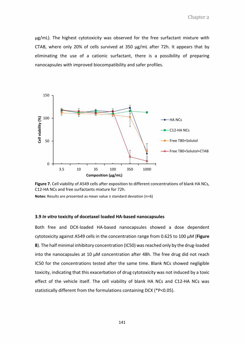
Chapter 2
141
µg/mL). The highest cytotoxicity was observed for the free surfactant mixture with
CTAB, where only 20% of cells survived at 350 μg/mL after 72h. It appears that by
eliminating the use of a cationic surfactant, there is a possibility of preparing
nanocapsules with improved biocompatibility and safer profiles.
Figure 7. Cell viability of A549 cells after exposition to different concentrations of blank HA NCs, C12-HA NCs and free surfactants mixture for 72h.
Notes: Results are presented as mean value ± standard deviation (n=6)
3.9 In vitro toxicity of docetaxel loaded HA-based nanocapsules
Both free and DCX-loaded HA-based nanocapsules showed a dose dependent
cytotoxicity against A549 cells in the concentration range from 0.625 to 100 μM (Figure
8). The half minimal inhibitory concentration (IC50) was reached only by the drug-loaded
into the nanocapsules at 10 μM concentration after 48h. The free drug did not reach
IC50 for the concentrations tested after the same time. Blank NCs showed negligible
toxicity, indicating that this exacerbation of drug cytotoxicity was not induced by a toxic
effect of the vehicle itself. The cell viability of blank HA NCs and C12-HA NCs was
statistically different from the formulations containing DCX (*P<0.05).
0
50
100
150
3.5 10 35 100 350 1000
Ce
ll vi
abili
ty (
%)
Composition (µg/mL)
HA NCs
C12-HA NCs
Free T80+Solutol
Free T80+Solutol+CTAB

Chapter 2
142
Figure 8. Cell viability of A549 cells after exposition to different concentrations of free DCX, DCX-loaded HA-based nanocapsules and blank HA-based nanocapsules.
Notes: Results are presented as mean ± standard deviation (n=3).
3.10 Intracellular uptake of HA-based nanocapsules
To evaluate the intracellular uptake of HA NCs and C12-HA NCs, NR was loaded into both
nanocapsules and their uptake observed in A549 cells overexpressing CD44 receptors
by confocal microscopy. As a control, cells were exposed to a solution of NR, which was
not internalized by the cells (Figure 9A). On the other hand, a high fluorescence (red
color) was seen when both NR-loaded nanocapsules were taken by the cells (Figure 9B,
C).
Figure 9. Intracellular uptake of NR-loaded HA-based nanocapsules in A549 cells.
Notes: The nuclei were stained with DAPI (blue). Nile Red (NR) exhibits red fluorescence.
0
25
50
75
100
125
0 0.625 1.25 2.5 5 10 100
Ce
ll vi
abili
ty (
%)
[DCX] nMHA NCs C12-HA NCs
DCX-loaded HA NCs DCX-loaded C12-HA NCs
Free Nile red NR-loaded HA NCs NR-loaded C12-HA NCs A B C

Chapter 2
143
4. Discussion
4.1 Synthesis and characterization of dodecylamide-functionalized HA
Hydrophobically dodecylamide-functionalized HA was synthesized by chemical
modification of sodium hyaluronate by 1-aminodecane via an amide bond. The degree
of substitution was kept in the range 2.5 to 5.0%. The modified polymer was
characterized by 1H NMR spectroscopy. Aqueous solubility of HA was not affected as a
result of this chemical modification. Moreover, it has been reported that rheological and
biodegradation characteristics of HA should not be affected by such low degree of
modification [35].
4.2 Optimization of the self-emulsification method
The assessment of a green technology was achieved by the development of a self-
emulsification method for the preparation of HA-based nanocapsules. Without organic
solvents and heat, the self or spontaneous emulsification process is mainly determined
by the system composition and their physicochemical characteristics [36]. Thus,
components selection was based on their ability to formulate self-emulsifying systems,
in such a way that small droplets form spontaneously when the phases are brought into
contact. Miglyol®812, being a medium chain triglyceride, is described to reduce the
interfacial tension, and shows better water solubility and partitioning ability to
nanoemulsify when compared to long chain triglycerides [37–38]. In addition, it has the
ability to solubilize hydrophobic drugs, such as docetaxel, which is relevant when the
aim is to develop a process without organic solvents [39]. For the surfactant selection,
non-ionic surfactants with a hydrophilic/lipophilic balance (HLB) between 12-18 are
preferable as they have better hydrophilicity and can rapidly spread from the oil phase
to the aqueous environment and provide good dispersion performance [37]. Among
them, Tween®80 is one of the most used surfactants in self-emulsifying systems.
Although it is associated to potential side effects [40], it was reported that Tween®80
showed remarkably less toxicity than other solvents such as Labrasol® or Cremophor®
RL [41]. For example, Ma et al [42] prepared DCX-loaded poly-Ƹ-caprolactone (PLC)-
Tween®80 nanoparticles and demonstrated that the carrier showed less in vitro toxicity

Chapter 2
144
than commercial Taxotere® at the same surfactant concentration. In fact, we assume
that its localization at the interface of the particle surrounded by the polymeric shell
might decrease its free circulating exposure. We decided to include Solutol®HS15 in the
formulation for two main reasons. First, Solutol®HS15 has a PEG chain in its structure,
which may provide stability and prolonged circulation time to the nanocarrier [43].
Additionally, it possesses the required high HLB (HLB = 14-16), along with an ability to
inhibit p-glycoprotein pumps. The inhibition of this membrane pump must result in
higher intracellular drug accumulation [44].
The optimized nanoemulsion was composed by 2.5 mg/mL of Solutol®HS15 dissolved in
water and an oil phase composed of Miglyol®812/Tween®80 in a 1:1 ratio (w/w).
Increased amounts of Solutol®HS15 up to 25 mg/mL did not improve the
physicochemical characteristics of the system and we considered that 2.5 mg/mL was
the minimum required to formulate and stabilize the nanoemulsion due to the greater
partition extent between the oil/water interface [45]. Miglyol®812/Tween®80 ratios,
with lower surfactant amount, promoted an increase in particle size and PDI. It has been
described that at high oil/surfactant ratios (high oil content) the amount of surfactant is
too small to microemulsify the large quantity of oil. However, once the surfactant
concentration increases, the amount of Tween®80 became enough to perform its
emulsifier function effectively [46].
Knowing the importance of the oil/surfactant ratio and, on the other hand, the possible
toxicity associated to high amounts of Tween®80, the decrease in the surfactant amount
was done by decreasing the oil/aqueous phase ratio. We found that by reducing the
oil/aqueous phase ratio to 1:8 (v/v) we were able to produce nanoemulsions equally
small than those obtained with a 1:2 ratio because the Tween®80 did not lose its
surfactant capacity even when diluted in water [47]. We decreased up to 1:30 (v/v) the
oil/aqueous phase ratio, however, those formulations were limited by a very low
amount of oil which constrained consequently the amount of drug incorporated into the
system.
Concerning the formulation of HA NCs, the effective attachment of the HA to the outer
shell of the particle was achieved by an electrostatic interaction between the polymer

Chapter 2
145
and the lipid core surrounded by the cationic CTAB. Due to the balance between the
positive charge of the nanoemulsion and the possible toxicity associated with high
amounts of CTAB [22], we chose a concentration of 0.15 mg/mL of CTAB for the
nanocapsules preparation. The chosen concentration was previously demonstrated to
be enough for polymer attachment and nanocapsules stabilization [21]. A HA
concentration of 0.25 mg/mL was enough to promote the attachment of the polymer to
the particle surface, resulting in a negative zeta potential. By shielding the nanocapsules
with HA and rendering to the particles a negative charge, HA NCs must promote a longer
half-life in the blood stream [48].
In order to simplify the process and to avoid the use of a cationic surfactant, HA was
replaced by C12-functionalized HA. The hydrophobic dodecyl chains of HA facilitated the
self-assembly of the polymer within the oil/surfactant nanoemulsion interface through
hydrophobic interactions, resulting in an increased stability of the hydrophobic core
[49]. For this formulation, 0.5 mg/mL of C12-amide HA was required to achieve the same
negative zeta potential as that of HA prototype.
Figure 10 exemplifies the structure of a HA-based nanocapsules formulated with HA or
C12-HA and its respective composition.
Figure 10. Structure and composition of HA-based nanocapsules formulated with HA (right) or C12-HA (left). HA NCs were formulated based on the ionic interaction between the positively charged CTAB and the negatively charged HA. On the other hand, the amphiphilic C12-HA self-assembled with the surfactant interphase, without the need of a cationic surfactant.

Chapter 2
146
4.3 Stability assays
The stability of both HA-based nanocapsules was assessed thorough storage conditions
and in human plasma. No significant differences in size, PDI and zeta potential were
observed for both prototypes after storage for 6 months, at 4ºC. The stability could be
attributed to the high negative charge that prevents particle aggregation, due to a
charge-charge repulsion. Moreover, the presence of Tween®80 should also add steric
stability to the system [50]. The stability of the nanocapsules in plasma was determined
by their physical integrity, mainly the particle size [51]. The observed increase in particle
size after 24h at 37oC might be attributed to protein deposition. Nevertheless, this
increase was less than 20% compared to the initial particle size, which means that these
nanocapsules must be suitable for IV administration [52].
4.4 In vitro release assays
Nanocapsules formulated with HA and C12-HA showed a biphasic drug release profile,
with an initial burst release of 45% and 55%, respectively. The release was sustainable
up to 24h, with 70% of DCX being released from both systems. This biphasic release
profile has been typically observed in other HA-based nanocapsules, which presented
an initial burst release between 45-65%. The initial burst release has been justified by
the own structure of the nanocapsules, favoring the partition of the drug between the
oil core and the aqueous external medium [53]. Interestingly, the release was not
affected by the ionic or hydrophobic forces that drove the formation of nanocapsules
with HA or C12-HA, respectively. With a Pka1=2.82 and Pka2=3.42, HA is negatively
charge at pH above 4, thus maintaining its ionic strength when in PBS at pH 7.4 [54].
Regarding the amphiphilic structure, the hydrophobic chain may enhance the
hydrophobicity of the particle core, which helps DCX to be entrapped [55]. While this
data provides us information about mechanistic details, it is important to highlight two
important points: (i) the limitation of the method, where the external oil phase may
force the release of DCX from the oil core of the nanocapsules to the external oil
compartment and, (ii) the in vitro release is not necessarily expected to correlate with

Chapter 2
147
the in vivo behavior, as the presence of macromolecules and ions in circulation could
significantly influence the release profile [21].
4.5 In vitro cytotoxicity assays
A decrease in the cytotoxicity of C12-HA NCs was expected since the formulation of
these nanocapsules with a hydrophobically functionalized HA eliminated the need for
cationic surfactants. The results showed that irrespective of their composition, both
prototypes did not affect cell viability when tested at concentrations up to 350 μg/mL.
However, only self-emulsified nanocapsules prepared with C12-HA did not cause any
toxicity when tested at the highest concentration (1000 μg/mL). The higher toxicity for
HA NCs must be correlated to the presence of the cationic surfactant CTAB, which is in
agreement with previous reports [56]. In addition, the marked difference in viability
between HA NCs and the surfactant solution composed of Tween®80/Solutol/CTAB at
350 μg/mL effectively denotes the beneficial effect of HA surrounding the surfactant
layer as well as to the correct isolation of the system from the free surfactants [57].
DCX-loaded HA NCs and C12-HA NCs presented an inhibition in the cell viability when
compared with the free drug. The IC50 was reached only by the drug-loaded into the
nanocapsules, at a concentration of 10 μM and after 48h. On the other hand, the free
drug did not reach the IC50 for the concentrations tested for the same time period. Since
the drug became more efficacious when loaded into HA-based nanocapsules, which in
turn did not express any inherent cytotoxicity themselves, it is fair to assume that these
nanocapsules must be taken up by cancer cells either via receptor mediated (CD44)
endocytosis or simultaneous interaction with the cancer cell membrane followed by
endocytosis and release in the endosome [58].
4.6 In vitro cellular uptake
In order to monitor the cellular uptake of NR-loaded HA-based nanocapsules, both
prototypes were incubated with A549 cells overexpressing CD44 receptors. As seen by
confocal microscopy, strong fluorescent signals were detected in the cells cytoplasm for

Chapter 2
148
both nanocapsules prototypes when compared to the free fluorophore, further
suggesting an intracellular uptake mediated by CD44 receptors [59]. Additionally, the
fluorescent intensity was similar for both NR-loaded HA NCs and C12-HA NCs, which
suggests that the functionalization of HA with the dodecylamide chain did not affect its
binding affinity through CD44 receptors. In fact, this might suggest the localization of
the lipophilic chain into the interface, and the hydrophilic branch turned to the outside
[60].
5. Conclusions
In conclusion, here we report the application of a green methodology for the
preparation of HA-based nanocapsules. A self-emulsification method was developed for
the preparation of HA nanocapsules without the aid of organic solvents and heat, which
offers a promising and sustainable approach to prepare nanoformulations for
therapeutic molecules. The preparation of HA-based nanocapsules with an amphiphilic
functionalized HA derivative led to the development of nanocarriers with low toxicity
and the potential to efficiently encapsulate and deliver cytostatic drugs, such as
docetaxel, into cancer cells.

Chapter 2
149
Bibliography
[1] S. P. Egusquiaguirre, M. Igartua, R. M. Hernández, and J. L. Pedraz, “Nanoparticle delivery systems for cancer therapy: advances in clinical and preclinical research.,” Clin. Transl. Oncol., vol. 14, no. 2, pp. 83–93, 2012.
[2] T. Sun, Y. S. Zhang, B. Pang, D. C. Hyun, M. Yang, and Y. Xia, “Engineered nanoparticles for drug delivery in cancer therapy.,” Angew. Chem. Int. Ed. Engl., vol. 53, no. 46, pp. 12320–64, 2014.
[3] C. E. Mora-Huertas, H. Fessi, and A. Elaissari, “Polymer-based nanocapsules for drug delivery.,” Int. J. Pharm., vol. 385, no. 1–2, pp. 113–42, 2010.
[4] G. Lollo, G. R. Rivera-Rodriguez, J. Bejaud, T. Montier, C. Passirani, J.-P. Benoit, M. García-Fuentes, M. J. Alonso, and D. Torres, “Polyglutamic acid-PEG nanocapsules as long circulating carriers for the delivery of docetaxel.,” Eur. J. Pharm. Biopharm., vol. 87, no. 1, pp. 47–54, 2014.
[5] J. Hrkach, D. Von Hoff, M. Mukkaram Ali, E. Andrianova, J. Auer, T. Campbell, D. De Witt, M. Figa, M. Figueiredo, A. Horhota, S. Low, K. McDonnell, E. Peeke, B. Retnarajan, A. Sabnis, E. Schnipper, J. J. Song, Y. H. Song, J. Summa, D. Tompsett, G. Troiano, T. Van Geen Hoven, J. Wright, P. LoRusso, P. W. Kantoff, N. H. Bander, C. Sweeney, O. C. Farokhzad, R. Langer, and S. Zale, “Preclinical development and clinical translation of a PSMA-targeted docetaxel nanoparticle with a differentiated pharmacological profile.,” Sci. Transl. Med., vol. 4, no. 128, p. 128ra39, 2012.
[6] S. Wong and B. Karn, “Ensuring sustainability with green nanotechnology,” Nanotechnology, vol. 23, no. 29, p. 290201, 2012.
[7] T. Gonzalo, G. Lollo, M. Garcia-Fuentes, D. Torres, J. Correa, R. Riguera, E. Fernandez-Megia, P. Calvo, P. Avilés, M. J. Guillén, and M. J. Alonso, “A new potential nano-oncological therapy based on polyamino acid nanocapsules,” J. Control. Release, vol. 169, no. 1–2, pp. 10–16, 2013.
[8] I. Youm, X. Y. Yang, J. B. Murowchick, and B.-B. C. Youan, “Encapsulation of docetaxel in oily core polyester nanocapsules intended for breast cancer therapy.,” Nanoscale Res. Lett., vol. 6, no. 1, p. 630, 2011.
[9] P. Sánchez-Moreno, J. L. Ortega-Vinuesa, H. Boulaiz, J. A. Marchal, and J. M. Peula-García, “Synthesis and characterization of lipid immuno-nanocapsules for directed drug delivery: selective antitumor activity against HER2 positive breast-cancer cells.,” Biomacromolecules, vol. 14, no. 12, pp. 4248–59, 2013.
[10] P. Dunn, A. Wells, and M. Williams, Green chemistry in the pharmaceutical industry. 2010.
[11] H. Duan, D. Wang, and Y. Li, “Green chemistry for nanoparticle synthesis.,” Chem. Soc. Rev., vol. 44, pp. 5778–5792, 2015.
[12] K. Bouchemal, S. Briançon, E. Perrier, and H. Fessi, “Nano-emulsion formulation using spontaneous emulsification: Solvent, oil and surfactant optimisation,” Int. J. Pharm., vol. 280, no. 1–2, pp. 241–251, 2004.
[13] C. Solans, P. Izquierdo, J. Nolla, N. Azemar, and M. Garciacelma, “Nano-emulsions,” Curr. Opin. Colloid Interface Sci., vol. 10, no. 3–4, pp. 102–110, 2005.
[14] A. H. Saberi, Y. Fang, and D. J. McClements, “Fabrication of vitamin E-enriched nanoemulsions: Factors affecting particle size using spontaneous emulsification,” J. Colloid Interface Sci., vol. 391, no. 1, pp. 95–102, 2013.
[15] H. Sobhani, P. Tarighi, S. N. Ostad, A. Shafaati, N. Nafissi-Varcheh, and R. Aboofazeli,

Chapter 2
150
“Formulation Development and Toxicity Assessment of Triacetin Mediated Nanoemulsions as Novel Delivery Systems for Rapamycin.,” Iran. J. Pharm. Res. IJPR, vol. 14, no. Suppl, pp. 3–21, 2015.
[16] A. Date, N. Desai, R. Dixit, and M. Nagarsenker, “Self-nanoemulsifying drug delivery systems: formulation insights, applications and advances,” Nanomedicine, vol. 5, no. 10, 2010.
[17] N. Sadurní, C. Solans, N. Azemar, and M. J. García-Celma, “Studies on the formation of O/W nano-emulsions, by low-energy emulsification methods, suitable for pharmaceutical applications.,” Eur. J. Pharm. Sci., vol. 26, no. 5, pp. 438–45, 2005.
[18] A. Hossein, B. Zeeb, J. Weiss, and D. Julian, “Tuneable stability of nanoemulsions fabricated using spontaneous emulsification by biopolymer electrostatic deposition,” J. Colloid Interface Sci., vol. 455, pp. 172–178, 2015.
[19] B. Chen, R. J. Miller, and P. K. Dhal, “Hyaluronic Acid-Based Drug Conjugates: State-of-the-Art and Perspectives,” J. Biomed. Nanotechnol., vol. 10, no. 1, pp. 4–16, 2014.
[20] S. Mizrahy, S. R. Raz, M. Hasgaard, H. Liu, N. Soffer-Tsur, K. Cohen, R. Dvash, D. Landsman-Milo, M. G. E. G. Bremer, S. M. Moghimi, and D. Peer, “Hyaluronan-coated nanoparticles: the influence of the molecular weight on CD44-hyaluronan interactions and on the immune response.,” J. Control. Release, vol. 156, no. 2, pp. 231–8, Dec. 2011.
[21] F. a Oyarzun-Ampuero, G. R. Rivera-Rodríguez, M. J. Alonso, and D. Torres, “Hyaluronan nanocapsules as a new vehicle for intracellular drug delivery.,” Eur. J. Pharm. Sci., vol. 49, no. 4, pp. 483–90, 2013.
[22] H. Lv, S. Zhang, B. Wang, S. Cui, and J. Yan, “Toxicity of cationic lipids and cationic polymers in gene delivery.,” J. Control. Release, vol. 114, no. 1, pp. 100–9, Aug. 2006.
[23] Y. Liu, J. Sun, W. Cao, J. Yang, H. Lian, X. Li, Y. Sun, Y. Wang, S. Wang, and Z. He, “Dual targeting folate-conjugated hyaluronic acid polymeric micelles for paclitaxel delivery.,” Int. J. Pharm., vol. 421, no. 1, pp. 160–9, 2011.
[24] D. a Ossipov, “Nanostructured hyaluronic acid-based materials for active delivery to cancer,” Expert Opin. Drug Deliv., vol. 7, no. 6, pp. 681–703, 2010.
[25] K. Y. Choi, K. H. Min, J. H. Na, K. Choi, K. Kim, J. H. Park, I. C. Kwon, and S. Y. Jeong, “Self-assembled hyaluronic acid nanoparticles as a potential drug carrier for cancer therapy: synthesis, characterization, and in vivo biodistribution,” J. Mater. Chem., vol. 19, no. 24, p. 4102, 2009.
[26] X. Dong and C. Liu, “Preparation and Characterization of Self-Assembled Nanoparticles of Hyaluronic Acid-Deoxycholic Acid Conjugates,” J. Nanomater., vol. 2010, pp. 1–9, 2010.
[27] K. Y. Choi, H. Y. Yoon, J.-H. Kim, S. M. Bae, R.-W. Park, Y. M. Kang, I.-S. Kim, I. C. Kwon, K. Choi, S. Y. Jeong, K. Kim, and J. H. Park, “Smart nanocarrier based on PEGylated hyaluronic acid for cancer therapy.,” ACS Nano, vol. 5, no. 11, pp. 8591–9, Nov. 2011.
[28] H. Lee, C.-H. Ahn, and T. G. Park, “Poly[lactic-co-(glycolic acid)]-grafted hyaluronic acid copolymer micelle nanoparticles for target-specific delivery of doxorubicin.,” Macromol. Biosci., vol. 9, no. 4, pp. 336–42, 2009.
[29] H.-J. Cho, I.-S. Yoon, H. Y. Yoon, H. Koo, Y.-J. Jin, S.-H. Ko, J.-S. Shim, K. Kim, I. C. Kwon, and D.-D. Kim, “Polyethylene glycol-conjugated hyaluronic acid-ceramide self-assembled nanoparticles for targeted delivery of doxorubicin.,” Biomaterials, vol. 33, no. 4, pp. 1190–200, 2012.

Chapter 2
151
[30] B. Saliou, O. Thomas, N. Lautram, A. Clavreul, J. Hureaux, T. Urban, J.-P. Benoit, and F. Lagarce, “Development and in vitro evaluation of a novel lipid nanocapsule formulation of etoposide.,” Eur. J. Pharm. Sci., vol. 50, no. 2, pp. 172–80, 2013.
[31] G. R. Rivera-Rodriguez, G. Lollo, T. Montier, J. P. Benoit, C. Passirani, M. J. Alonso, and D. Torres, “In vivo evaluation of poly-l-asparagine nanocapsules as carriers for anti-cancer drug delivery,” Int. J. Pharm., vol. 458, no. 1, pp. 83–89, 2013.
[32] G. Bastiat, C. O. Pritz, C. Roider, F. Fouchet, E. Lignières, A. Jesacher, R. Glueckert, M. Ritsch-Marte, A. Schrott-Fischer, P. Saulnier, and J. P. Benoit, “A new tool to ensure the fluorescent dye labeling stability of nanocarriers: A real challenge for fluorescence imaging,” J. Control. Release, vol. 170, no. 3, pp. 334–342, 2013.
[33] I. Santos-ferreira, S. Kasper, B. Bétrisey, J. Kikhney, A. Moter, A. Trampuz, and A. J. Almeida, “Activity of daptomycin- and vancomycin-loaded poly-epsilon-caprolactone microparticles against mature staphylococcal biofilms,” Int. J. Nanomedicine, pp. 1–16, 2015.
[34] S. N. Bhattachar, L. a. Deschenes, and J. a. Wesley, “Solubility: it’s not just for physical chemists,” Drug Discov. Today, vol. 11, no. 21–22, pp. 1012–1018, 2006.
[35] M. Pavan, D. Galesso, G. Menon, D. Renier, and C. Guarise, “Hyaluronan derivatives: Alkyl chain length boosts viscoelastic behavior to depolymerization.,” Carbohydr. Polym., vol. 97, no. 2, pp. 321–6, 2013.
[36] J. C. López-Montilla, P. E. Herrera-Morales, S. Pandey, and D. O. Shah, “Spontaneous Emulsification: Mechanisms, Physicochemical Aspects, Modeling, and Applications,” J. Dispers. Sci. Technol., vol. 23, no. 1–3, pp. 219–268, 2002.
[37] T. Gershanik and S. Benita, “Self-dispersing lipid formulations for improving oral absorption of lipophilic drugs.,” Eur. J. Pharm. Biopharm., vol. 50, no. 1, pp. 179–88, 2000.
[38] D. F. Driscoll, J. Nehne, H. Peterss, R. Franke, B. R. Bistrian, and W. Niemann, “The influence of medium-chain triglycerides on the stability of all-in-one formulations,” Int. J. Pharm., vol. 240, no. 1–2, pp. 1–10, 2002.
[39] K. Gao, J. Sun, K. Liu, X. Liu, and Z. He, “Preparation and characterization of a submicron lipid emulsion of docetaxel: submicron lipid emulsion of docetaxel.,” Drug Dev. Ind. Pharm., vol. 34, no. 11, pp. 1227–37, Nov. 2008.
[40] J. a Yared and K. H. R. Tkaczuk, “Update on taxane development: new analogs and new formulations.,” Drug Des. Devel. Ther., vol. 6, pp. 371–84, 2012.
[41] E. Sigward, N. Mignet, P. Rat, M. Dutot, S. Muhamed, J.-M. Guigner, D. Scherman, D. Brossard, and S. Crauste-Manciet, “Formulation and cytotoxicity evaluation of new self-emulsifying multiple W/O/W nanoemulsions,” Int. J. Nanomedicine, vol. 8, pp. 611–25, 2013.
[42] Y. Ma, Y. Zheng, X. Zeng, L. Jiang, H. Chen, R. Liu, L. Huang, and L. Mei, “Novel docetaxel-loaded nanoparticles based on PCL-Tween 80 copolymer for cancer treatment.,” Int. J. Nanomedicine, vol. 6, pp. 2679–88, Jan. 2011.
[43] N. T. Huynh, C. Passirani, P. Saulnier, and J. P. Benoit, “Lipid nanocapsules: a new platform for nanomedicine.,” Int. J. Pharm., vol. 379, no. 2, pp. 201–9, 2009.
[44] A. Lamprecht and J. P. Benoit, “Etoposide nanocarriers suppress glioma cell growth by intracellular drug delivery and simultaneous P-glycoprotein inhibition,” J. Control. Release, vol. 112, no. 2, pp. 208–213, 2006.
[45] V. Borhade, S. Pathak, S. Sharma, and V. Patravale, “Clotrimazole nanoemulsion for

Chapter 2
152
malaria chemotherapy. Part I: preformulation studies, formulation design and physicochemical evaluation.,” Int. J. Pharm., vol. 431, no. 1–2, pp. 138–48, 2012.
[46] I. T. N. Chukwuma O Agubata, A. A. Nicholas C Obitte, Calister E Ugwu, and A. and G. C. Onunkwo, “Effect of Oil , Surfactant and Co-Surfactant Concentrations on the Phase Behavior , Physicochemical Properties and Drug Release from Self-Emulsifying Drug Delivery Systems,” J. Drug Discov. Dev. Deliv., vol. 1, no. 1, 2014.
[47] C. W. Pouton and C. J. H. Porter, “Formulation of lipid-based delivery systems for oral administration: materials, methods and strategies.,” Adv. Drug Deliv. Rev., vol. 60, no. 6, pp. 625–37, 2008.
[48] Z. Poon, J. B. Lee, S. W. Morton, and P. T. Hammond, “Controlling in vivo stability and biodistribution in electrostatically assembled nanoparticles for systemic delivery,” Nano Lett., vol. 11, no. 5, pp. 2096–2103, 2011.
[49] T. Akagi, M. Baba, and M. Akashi, “Preparation of nanoparticles by the self-organization of polymers consisting of hydrophobic and hydrophilic segments: Potential applications,” Polymer (Guildf)., vol. 48, no. 23, pp. 6729–6747, 2007.
[50] S. Honary and F. Zahir, “Effect of zeta potential on the properties of nano-drug delivery systems - A review (Part 2),” Trop. J. Pharm. Res., vol. 12, no. 2, pp. 255–264, 2013.
[51] S. Lazzari, D. Moscatelli, F. Codari, M. Salmona, M. Morbidelli, and L. Diomede, “Colloidal stability of polymeric nanoparticles in biological fluids,” J. Nanoparticle Res., vol. 14, no. 6, p. 920, 2012.
[52] P. Aggarwal, J. B. Hall, C. B. Mcleland, M. A. Dobrovolskaia, and S. E. Mcneil, “Nanoparticle interaction with plasma proteins as it relates to particle biodistribution, biocompatibility and therapeutic efficacy,” Adv. Drug Deliv. Rev., vol. 61, no. 6, pp. 428–437, 2013.
[53] V. P. Torchilin, Nanoparticulates as Drug Carriers. Imperial College Press, 2006.
[54] Y.-H. Liao, S. a Jones, B. Forbes, G. P. Martin, and M. B. Brown, “Hyaluronan: pharmaceutical characterization and drug delivery.,” Drug Deliv., vol. 12, no. 6, pp. 327–42, 2005.
[55] G. Saravanakumar, K. Y. Choi, H. Y. Yoon, K. Kim, J. H. Park, I. C. Kwon, and K. Park, “Hydrotropic hyaluronic acid conjugates: Synthesis, characterization, and implications as a carrier of paclitaxel.,” Int. J. Pharm., vol. 394, no. 1–2, pp. 154–61, 2010.
[56] M. Fraga, F. Bruxel, V. L. Lagranha, H. F. Teixeira, and U. Matte, “Influence of phospholipid composition on cationic emulsions/DNA complexes: physicochemical properties, cytotoxicity, and transfection on Hep G2 cells.,” Int. J. Nanomedicine, vol. 6, pp. 2213–20, 2011.
[57] X. Y. Yang, Y. X. Li, M. Li, L. Zhang, L. X. Feng, and N. Zhang, “Hyaluronic acid-coated nanostructured lipid carriers for targeting paclitaxel to cancer,” Cancer Lett., vol. 334, no. 2, pp. 338–345, 2013.
[58] X. Wei, T. H. Senanayake, G. Warren, and S. V Vinogradov, “Hyaluronic acid-based nanogel-drug conjugates with enhanced anticancer activity designed for the targeting of CD44-positive and drug-resistant tumors.,” Bioconjug. Chem., vol. 24, no. 4, pp. 658–68, 2013.
[59] N. Goodarzi, M. H. Ghahremani, M. Amini, F. Atyabi, S. N. Ostad, N. Shabani Ravari, N. Nateghian, and R. Dinarvand, “CD44-targeted docetaxel conjugate for cancer cells and cancer stem-like cells: A novel hyaluronic acid-based drug delivery system,” Chem. Biol.

Chapter 2
153
Drug Des., vol. 83, no. 6, pp. 741–752, 2014.
[60] K. Y. Choi, H. Chung, K. H. Min, H. Y. Yoon, K. Kim, J. H. Park, I. C. Kwon, and S. Y. Jeong, “Self-assembled hyaluronic acid nanoparticles for active tumor targeting.,” Biomaterials, vol. 31, no. 1, pp. 106–14, 2010.

Chapter 2
154

Chapter 3
Chapter 3
Hyaluronic acid nanocapsules as a platform for the
intracellular delivery of monoclonal antibodies:
when the target is inside the cell

Chapter 3
156

Chapter 3
157
Hyaluronic acid nanocapsules as a platform for the intracellular delivery
of monoclonal antibodies: when the target is inside the cell
This work was done in collaboration with: Ana Olivera1, Angela Molina2, Gema Moreno
Bueno2 and María José Alonso1.
1 Nanobiofar Group, IDIS, CIMUS. University of Santiago de Compostela, Spain
2 Departamento de Bioquímica, Universidad Autónoma de Madrid, CSIC-UAM, IdiPAZ,
Fundación MD Anderson Internacional, Madrid, Spain
This work was supported by grants from Instituto de la Salud Carlos III (ISCIII,
P113/00132) and Avon Foundation Award 2012.

Chapter 3
158

Chapter 3
159
Abstract
Monoclonal antibodies (mAbs) are one of the most selective therapies against cancer.
However, their high molecular weight and hydrophilicity have hampered their
application for the targeting of intracellular oncogenic proteins. The main object of this
work has been the development of a new nanotechnology adapted for the intracellular
delivery of mAbs. For the validation of this technology, we have chosen anti-GSDMB, a
mAb whose target is confined to the intracellular compartment. The selected
technology consists of hyaluronic acid nanocapsules, which enabled the packing of anti-
GSDMB by physicochemical interaction with the polymeric shell. Indeed, the results
showed that it was possible to efficiently associate (70% association efficiency, AE) anti-
GSDMB to tiny HA NCs (130 nm). In vitro assays performed in HCC1954 breast cancer
cells showed that the anti-GSDMB carried by the nanocapsules was efficiently
internalized while preserving their immunological determinants. Moreover,
immunohistochemistry analysis demonstrated the capacity of the mAb to escape the
endosomal compartment, thus avoiding their premature intracellular degradation. The
preservation of the mAb activity upon its association to the nanocapsules was also
confirmed using a wound healing migration assay. The results of this study showed that
the anti-GSDMB-loaded into the nanocapsules could effectively interact with GSDMB,
and that the result of this interaction was a decrease in the cell migration and invasion
of HCC1954 breast cancer cells. Taken together, these results represent the first
preliminary evidence of the capacity of hyaluronic acid nanocapsules as a new mAb-
based therapeutic platform against intracellular cancer proteins.
Keywords: antibody, intracellular targeting, cancer, nanocarrier, nanocapsules,
hyaluronic acid, intracellular delivery

Chapter 3
160

Chapter 3
161
1. Introduction
Monoclonal antibodies (mAbs) have become an important class of oncological
biomolecules due to their ability to target tumor associated cell surface antigens and
promote cytotoxicity through several mechanisms of action [1]. This targeting capacity
has also been the basis for their use as targeting ligands, when conjugated to cytotoxic
drugs [2] or decorating the surface of nanoparticles [3]. Indeed, antibody-conjugated
drug-loaded nanoparticles were developed to selectively bind to a cancer cell receptors,
promote the internalization of the nanoparticles, release its payload, and elicit
cytotoxicity while decreasing undesired side effects [3]. An important consequence of
this research activity has been the development of 13 prototypes of nanoparticles
targeting cell surface receptors, which are currently under clinical development [4].
Nevertheless, onco-protein receptors are not always presented to the cell membrane,
but they are exclusively restricted to the cytosol compartment [5]. In fact, hundreds of
intracellular proteins have been associated to cancer progression, for example, RAS
(GTPases), non-receptor tyrosine kinases, BRAF or heat shock proteins. Unfortunately,
these oncoproteins could not been targeted with mAbs, due to the inability of these
complex molecules to cross the cell membrane [6]. Rather than this, other strategies
including silencing therapy, cytostatic molecules or protein kinase inhibitors have been
explored until now. However, the complexity of these pathways, the off-target effects
and cellular barriers, specially associated to polynucleotide molecules, hold these
therapies far from their clinical use [6–7].
To our knowledge, so far, no one has reported scientific evidence of the intracellular
targeting of mAbs against onco-proteins, which are exclusively restricted to the cytosol
compartment and that lack from a surface region. Within this context, it is worthwhile
to mention that two companies released recently news about the development of mAbs
for intracellular targeting [9]. However, the way to achieve this targeting has not been
disclosed in a scientific journal.
Taking advantage of the potential of mAbs as a disruptive therapy against intracellular
proteins, our goal has been to engineer a nanocarrier with the capacity to overcome the
cell membrane barrier and carry the mAb into the intracellular compartment [10]. To
achieve this goal we started our formulation study with hyaluronic acid (HA)

Chapter 3
162
nanocapsules, previously disclosed by our group for the intracellular delivery of
anticancer drugs [11] and adapted in this study to the loading of mAbs. To assess the
efficacy of the delivery technology, we chose the mAb anti-Gasdermin-B (GSDMB),
based on the discovery of Moreno-Bueno et al, who identified Gasdermin-B (GSDMB) as
an intracellular marker for breast cancer [12]. These authors found that the over-
expression of GSDMB was responsible for cell motility, tumor progression and
metastasis in the MCF7 breast carcinoma cell line. Although the role of GSDMB in cancer
is not completely understood, we believe that its intracellular targeting using anti-
GSDMB may lead to a great opportunity to stop cancers over-expressing this protein.
In addition to its biocompatibility, non-toxicity and biodegradability, HA can bind to
CD44 receptors, which are overexpressed in many cancer cells [13–14]. Different
techniques have been used for the preparation of nanocapsules and, recently, our group
have developed HA-based nanocapsules using HA and amphiphilic dodecylamide-
functionalized HA by a spontaneous emulsification method. This method is
characterized by the absence of organic solvents, heat and strong mechanical stirring,
which makes it suitable for the association of proteins.
Therefore, these premises led us to the design of self-emulsifying HA-based
nanocapsules specifically adapted for the association of mAbs. The resulting prototypes
were characterized with regard to their physicochemical properties, mAb association
efficiency, integrity and specificity. Finally, anti-GSDMB-loaded to nanocapsules were
evaluated in HCC1954 breast cancer cell line for their capacity to enter the intracellular
compartment, achieve the intracellular delivery of anti-GSDMB and inhibit cancer cell
migration and invasion.
2. Materials and methods
2.1 Materials
Sodium hyaluronate and dodecylamide-functionalized HA, both with 200KDa, were a
kind gift from Genzyme Sanofi, USA. Caprylic/capric triglyceride (Miglyol®812) was
provided by Cremer, Germany. Polyoxyethylene sorbitan monooleate and monolaurate
(Tween®80 and Tween®20, respectively) and haxadecyl trimethylammonium bromide
(CTAB) were purchase from Sigma-Aldrich, Spain. Macrogol 15 Hydroxystearate

Chapter 3
163
(Solutol®HS15) was obtained from BASF, Germany. The following antibodies were
purchased: rabbit anti-EEA1, from Cell Signaling Technology, Spain, rabbit anti-LAMP2,
from Sigma-Aldrich, goat anti-mouse and anti-rabbit conjugated with Alexa-488, Alexa-
594 or Alexa-647 were from Molecular Probes®, Thermo Fisher Scientific, Spain. Alexa-
647 coupled phalloidin and 4,6-diamidino-2-phenylindole (DAPI) were also purchased
from Molecular Probes®. Phosphate Buffer Saline (PBS) in tablets was obtained from
Medicago, Sweden. HRP-conjugated goat anti-mouse IgG and 2,2’-Azino-bis (3-
ethylbenzothiazoline-6-surfonic acid) (ABTS) were purchased from DAKO (Agilent
Technologies, Spain) and Roche (Switzerland), respectively. Coomassie Blue solution
was acquired from Thermo Fisher Scientific. All other chemicals were of analytical grade.
2.2 Anti-GSDMB preparation and purification
Mouse anti-GSDMB monoclonal antibody was generated as described by Hergueta-
Redondo et al (submitted) accordingly to specified techniques. The antibody was
purified with a Hi-trap Protein G column (GE Healthcare, UK). After purification, anti-
GSDMB solution was aliquoted in 2 mL Eppendorf’s and kept at 4ºC.
2.3 Anti-GSDMB characterization
2.3.1 Concentration and Integrity
Anti-GSDMB was quantitatively analyzed using a NanoDrop®2000 Spectrophotometer
with the IgG reference at 280 nm (Thermo Fisher Scientific) and qualitatively analyzed
using sodium dodecyl sulfate polyacrylamide gel electrophoresis (SDS-PAGE). The SDS-
PAGE was performed to identify IgG constant and variable fragments under non-
reducing and reducing conditions, respectively. Anti-GSDMB samples were mixed with
loading buffer composed of 10% SDS and 30% glycerol and reduced samples were
prepared with the same loading buffer plus β-mercaptoethanol and boiled for 5, 7, 10
and 12 minutes in a Vortemp Shaking Incubator (UniEquip). Samples containing 20 and
40 μg of anti-GSDMB were loaded into porous 10% SDS-PAGE gel under the effect of an
electric field. The electrophoresis was run under constant voltage (100 V, 2h) and gels
were stained with Coomassie Blue Solution.

Chapter 3
164
2.3.2 Quantification and binding assay by ELISA
The specific binding activity and the quantification of Anti-GSDMB were also performed
by ELISA. The ELISA plates (Nunc Medisorpᵀᴹ, Thermo Scientific) were coated with
5μg/mL of GSDMB antigen diluted in water and incubated 1h at room temperature.
Plates were washed three times with PBS containing 0.1% Tween®20 (v/v) and, then,
blocked with PBS containing 2% (w/v) non-fat skim milk overnight at 4ºC and washed
again three times the day after. Anti-GSDMB solution was prepared from a stock
solution at 10 μg/mL, sequentially diluted in PBS to obtain standard solutions with a
concentration range between 0.125 and 10 μg/mL and incubated for 30 min at 37ºC.
The washing step was repeated and HRP-conjugated goat anti-mouse IgG (DAKO) was
added (1:1000 in PBS), incubated 30 min at 37ºC and plates washed as before. The
reaction was revealed with substrate ABTS solution and the absorbance was read at 405
nm using a Multiskan FC Microplate Photometer (Thermo Scientific). All determinations
were made at least in triplicate. The sigmoidal standard curves were set up using a
sigmoidal dose-response (variable slope) model (GraphPad Prism 5) and were plotted in
the form of absorbance at 405 nm against log of anti-GSDMB concentration.
2.4 Preparation of anti-GSDMB-loaded HA-based nanocapsules
Nanocapsules made of sodium hyaluronate (HA NCs) or dodecylamide-functionalized
HA (C12-HA NCs) were prepared by a spontaneous emulsification method. Briefly, an oil
phase composed of a 1:1 ratio (v/v) of Miglyol®812 and Tween®80 with CTAB, was added
to an aqueous phase composed of HA and Solutol®HS15 under magnetic stirring in a 1:8
ratio (v/v). Both type of nanocapsules were prepared using the same protocol except
that C12-HA NCs were prepared without the cationic surfactant CTAB. Formulations
were developed using different HA concentrations: 0.25 and 0.75 mg/mL of HA and 0.5
and 1mg/mL for the C12-HA. All nanocapsules were isolated by size exclusion
chromatography (SEC) using Centripure P10 columns (EmpBiotech, Germany).
Anti-GSDMB was adsorbed onto the surface of HA-based nanocapsules by a physical
procedure. The process was firstly optimized for the HA NCs using 0.25 and 0.75 mg/mL
of the polymer. First, 150 μL of HA NCs (corresponding to 10 mg) were incubated with

Chapter 3
165
2.5, 5, 10 and 25 μg of anti-GSDMB per mg of nanocapsules, at 4ºC under mild horizontal
shaking (titramax 1000 platform shaker, Heidolph, Germany). The influence of anti-
GSDMB’s charge on its association to the nanocapsules was investigated for the original
solution, at pH 7.4 (neutrally charged anti-GSDMB) or after protonating the mAb, at pH
4.5 (positively charged anti-GSDMB). Protonated anti-GSDMB (anti-GSDMB+) was
prepared by acidification with sodium acetate/ acetic acid buffer solution (initial pH 3.8)
until a final pH of 4.5.
Anti-GSDMB and protonated anti-GSDMB+ were associated to the surface of C12-HA
NCs using the same methodology.
The pH of blank nanocapsules, anti-GSDMB and anti-GSDMB+, alone or after association
to both types of HA-based nanocapsules were assessed using a pH meter (Docu-pHMeter,
Sartorius, Germany). Anti-GSDMB was concentrated using an antibody concentration kit
(Abcam, UK) and associated to C12-HA NCs formulated with 1 mg/mL of C12-HA as
described before.
2.4.1 Characterization of anti-GSDMB-loaded HA-based NCs
The size, polydispersion index (PDI) and surface charge of HA-based nanocapsules and
anti-GSDMB coated HA NCs and C12-HA NCs were measured using dynamic light
scattering (DLS) and laser Doppler anemometry (Nano-ZS instrument, Malvern, UK).
Scanning electron microscopy (SEM) (EVO LS15 microscope, ZEISS, Germany) was
utilized to obtain high-resolution images of blank and anti-GSDMB coated HA NCs.
2.4.2 Antibody association efficiency
The association efficiency of anti-GSDMB to HA NCs and C12-HA NCs was evaluated by
ultra-filtration using Nanosep®300K membranes (Pall Corporation, Spain). 250 μL of
anti-GSDMB-loaded nanocapsules were placed into the filter and centrifuged 4 min at
14,000 g and 4ºC. The filtrate containing the free antibody was taken and analyzed by
ELISA as previously described. The association efficiency was calculated as: (T-F)/T*100,
where T is the total anti-GSDMB and F the free anti-GSDMB.

Chapter 3
166
2.4.3 Stability of anti-GSDMB-loaded C12-HA NCs in human plasma
Blank C12-HA NCs and anti-GSDMB-loaded C12-HA NCs were diluted 1:10 (v/v) in human
plasma for a period of 24h, at 37ºC. At predetermined time intervals (0, 2, 4, 8 and 24h),
samples were taken and particle size evaluated as described above.
2.5 In vitro cell assays
2.5.1 Cell culture
The HCC1954 cell line, endogenously expressing GSDMB, was obtained from the
American Type Cell Culture (ATCC) (LGC Standards-SLU). Cells were cultured and
authenticated using STR-profiling according to ATCC guidelines. Cells were maintained
as monolayer cultures at 37ºC in an atmosphere with 5% CO2.
2.5.2 In vitro cytotoxicity assays
10x103 cells were grown into 96-wells plate in 200 µL culture medium. Two experiments
were performed: (i) after 24h, medium was replaced by fresh medium containing
different concentrations (0.05 – 1 mg/mL) of each formulation and cells were incubated
for 72h at 37ºC, and (ii) after 24h, serial assays were performed with C12-HA NCs at
different concentrations and incubation times at 37ºC (Table 1). After 72h, cell
proliferation was performed in order to test the cytotoxic effect of nanocapsules using
the AlamarBlue® assay according to the manufacturer specifications. The data was fitted
to a sigmoidal dose-response curve (variable slope) using GraphPad Prism (GraphPad
Software).
2.5.3 Cell Immunofluorescence studies
For the microscopy analysis, cells were seeded on sterile glass coverslips in culture
medium. Twenty-four hours after seeding, cells were incubated at 37ºC for the periods
and concentrations indicated with empty C12-HA NCs, anti-GSDMB alone or anti-
GSDMB associated to C12-HA NCs. After incubation, cells were washed with PBS and
fixed with 4% paraformaldehyde for 15 min. After fixation, cells were permeabilized with
0.1% Triton X-100 in PBS for 10 min. The coverslips were then incubated with the

Chapter 3
167
appropriate primary antibody to intracellular organelles for 90 min at room
temperature, followed by a secondary antibody for 45 min at room temperature.
Primary antibodies were: rabbit anti-EEA1 (1:100) and rabbit anti-LAMP2 (1:100).
Secondary antibodies were goat anti-mouse (1:1000) and anti-rabbit (1:1000)
conjugated with Alexa-488, Alexa-594 or Alexa-647. Alexa-647-coupled phalloidin was
used to stain F-actin and cell nuclei were stained using DAPI. After staining, coverslips
were mounted in Prolong Gold Antifade (Molecular Probes®). Images were collected by
confocal microscopy (Zeiss).
Table 1. Serial incubation steps of C12-HA NCs with HCC1954 breast cancer cells. At t0h, cells were incubated with C12-HA NCs and after 4h, 24h or 48h of incubation, medium was replaced by fresh medium or fresh medium containing C12-HA NCs at the specified concentration. Cell cytotoxicity was measured after 72h.
Serial incubation steps – C12-HA NCs
time (h)
Experiment t0h t24h t48h t72h Final conc. (mg/mL)
E1 Add NCs
0.8 mg/mL ---------- ---------- Measure 0.8
E2 Add NCs
0.8 mg/mL ---------- Replace medium
Measure 0.8
E3 Add NCs
0.8 mg/mL Replace medium ----------
Measure 0.8
E4 Add NCs
0.8 mg/mL Replace medium
Add NCs 0.8 mg/mL ----------
Measure 1.6
E5 Add NCs
0.8 mg/mL Replace medium
Add NCs 0.8 mg/mL Replace medium
Add NCs 0.8 mg/mL Measure
2.4
E6 Add NCs 0.8 mg/mL Replace medium 4h
Add NCs 0.8 mg/mL Replace medium 4h
Add NCs 0.8 mg/mL Replace medium 4h
Measure 2.4
E7 Add NCs
0.4 mg/mL Replace medium
Add NCs 0.4 mg/mL ----------
Measure 0.8
E8 Add NCs
0.26 mg/mL
Replace medium Add NCs 0.26
mg/mL
Replace medium Add NCs 0.26
mg/mL
Measure 0.8
2.5.4 Wound healing migration assay
For the migration capacity of cells, a wound healing assay was performed as previously
described by Hergueta-Redondo et al [12]. Briefly, 104 cells were seeded in 6-well plates
and maintained until 90-100% confluence. Subsequently, the artificial wounds were
created on the confluent cell monolayer using 10 μL pipette tips, and the detached cells
were removed by washing twice with PBS. The media was then replaced with empty

Chapter 3
168
C12-HA NCs (control) and anti-GSDMB-loaded C12-HA NCs at concentrations of 3.2
mg/mL of nanocapsules (corresponding to 80 µg of anti-GSDMB), for 2h and diluted in
RPMI 1640 medium. Cells were cultured at 37˚C/5% CO2. Wound closure was
monitored and photographed at time points 0, 24, 48 y 60h under an inverted
microscope (Axio Vert, Zeiss).
3. Results and discussion
Up to know, a significant number of drug nanocarriers have been functionalized with
mAbs with the final goal of targeting cancer cells that overexpress in their surface a
receptor for the selected mAb [3]. The novelty of this work relies on the fact that the
selected mAb, the anti- GSDMB, is not simply a targeting ligand, but a therapeutic entity.
In addition, contrary to the mAb therapies developed so far, the targeted protein is not
expressed on the surface of a cancer cell, but it is confined to the cytosol. The
explanation for the lack of therapies based on mAbs targeting intracellular proteins is
related to the fact that mAbs are not able to cross the cell membrane unless they have
a surface receptor. Therefore, the idea of developing a nanocarrier as a platform for the
intracellular delivery of mAbs is a challenging but promising strategy. The criteria for the
design of such delivery technology was defined taking into account the expected
properties for these nanocarriers, namely (i) stability in plasma; (ii) affinity towards the
cancer cells; (iii) capacity to load mAbs and (iv) capacity to enter the cancer cells and
release the mAb while preventing the potential lysosomal degradation. Our lab and
others have already shown that HA-based nanocarriers fulfill the first two properties
[11–15], however no one has shown the possibility to achieve the intracellular delivery
of a mAb. In the next sections we will describe the necessary steps to achieve this goal.
Figure 1 illustrates the potential of HA-based nanocapsules described here as a
technology to promote the delivery of a mAb, anti-GSDMB, into the intracellular
compartment of HCC1954 breast cancer cells.

Chapter 3
169
Figure 1. Intracellular transport of anti-GSDMB delivered by HA-based nanocapsules.
3.1 Characterization of anti-GSDMB
The concentration of anti-GSDMB was determined by nanodrop as 2.9 mg/mL.
The purity and integrity of anti-GSDMB was evaluated by SDS-PAGE under non-reducing
and reducing conditions. Using this technique, intact antibodies must present a single
band while denatured fragments two bands, the constant fraction (Fc) and the variable
fraction (Fab), at 50 KDa and 25 KDa respectively [16]. Figure 2 shows the stained gel
containing the different bands. Lane 9 represents a molecular weight marker, lane 1 and
2 anti-GSDMB under non-reduced conditions at 20 and 40 μg/mL, lane 3 to 6 anti-
GSDMB fragments after denaturing 5, 7, 10 and 12 min and lanes 7 and 8 the commercial
DAKO horseradish peroxidase (HRP) antibody under non-reducing and reducing
conditions, respectively. Under non-reducing conditions we observed the band of the
whole antibody at 160 KDa as well as unspecific bands analogous to some impurities and
protein crumbling. Protein fragments on non-reducing SDS-PAGE are a common feature
and have been related to the breakage of inter chain disulfide bonds during sample
preparation [17]. At reducing conditions, antibody fragmentation was confirmed by the
presence of two bands corresponding to IgG fragments and some additional impurity

Chapter 3
170
bands. The separation profile was the same regardless the boiling time. As a control,
DAKO HRP IgG showed the same structural profile as anti-GSDMB for non-reducing and
reducing conditions with an additional band corresponding to the HRP chain conjugated
to the antibody. This study confirms the stability and integrity of anti-GSDMB and the
presence of non-specific impurities lie behind normal method production and
purification techniques [18].
The binding affinity of anti-GSDMB towards the GSDMB antigen was analyzed by ELISA.
This assay was performed in order to assure the specificity of anti-GSDMB against the
target protein, as well as to validate a method to quantify the amount of anti-GSDMB
associated to HA NCs. A sigmoidal calibration curve was obtained in a concentration
range from 0.125 to 10 µg/mL and r2= 0.9969 (results not shown).
Figure 2. SDS-PAGE gel electrophoresis. Lanes 1 and 2, anti-GSDMB under non-reducing conditions at 20 and 40 μg/mL; lanes 3 to 6, anti-GSDMB under reducing conditions after boiling 5, 7, 10 and 12 min; lanes 7 and 8, control DAKO HRP IgG under non-reducing and reducing conditions, respectively; lane 9, MW marker (KDa). Blue arrow marks intact anti-GSDMB band and red arrows the Fc (50KDa) and Fab (25KDa) fractions
3.2 Development of HA-based nanocapsules containing anti-GSDMB
Nanocapsules made of sodium hyaluronate HA (HA NCs) or dodecylamide-
functionalized HA (C12-HA NCs) were developed using a self-emulsification technique.
These nanocapsules are constituted by an oily core composed of Miglyol®812 and
Tween®80 and a shell made of Solutol®HS15 and HA or C12-HA. Also, the NCs
formulated with HA have the cationic surfactant, CTAB, in the oil core. The self-
1 2 3 4 5 6 7 8 9

Chapter 3
171
emulsification was achieved thanks to the addition of the oil phase composed of
Miglyol®812 and Tween®80 (with or without CTAB at 0.15 mg/mL) in a ratio 1:1 (w/w)
to the aqueous phase composed of Solutol®HS15 at 2.5 mg/mL and HA or C12-HA at
concentrations between 0.25 and 1 mg/mL. The oil phase was added to the aqueous
phase in a ratio 1:8 (v/v). The nanocapsules formed a homogeneous population (PDI ≤
0.2) with a mean size around 130 nm and a negative zeta potential (–20 mV). Both the
small size and the negative zeta potential, together with the hydrophilicity of the
polymeric shell, are attractive properties for preserving the stability of the nanocapsules
in the blood stream and achieve a passive targeting to the tumor [18–19].
The association of anti-GSDMB to the surface of HA-based nanocapsules was done by
physical adsorption by controlling the appropriate charge and hydrophobicity of the
mAb and, hence, its ionic or hydrophobic interaction with the different components of
the nanocapsules. The major advantages of using physical adsorption rather than
chemical conjugation [21] relies on the use of a mild and easy process, without the need
of aggressive reagents, which preserves the antibody activity [21–22]. Although the
majority of publications refer to antibody-conjugated nanocarriers, the adsorption of
antibodies to the surface of nanoparticles is a common approach successfully described
in different studies [11, 21, 23–24].
3.2.1 Association of Anti-GSDMB to HA nanocapsules
Considering the negative charge of HA NCs, anti-GSDMB was protonated and associated
to their surface by ionic interactions. Anti-GSDMB has an isoelectric point (pI) between
6.5 and 8.1, and therefore becomes positively charged at pH values below 6.5. Cationic
anti-GSDMB (anti-GSDMB+) was prepared by acidifying the medium with a 15% (v/v)
sodium acetate/ acetic acid buffer (pH 3.8) [24]. After acidification, the pH of the mAb
solution changed from 7.4 to 4.5 and its zeta potential switched to a positive value (+3
mV) without changing its binding affinity to the GSDMB antigen, as tested by ELISA.
(Results not shown).
In order to identify the most adequate association conditions, we prepared different
formulations by associating the protonated mAb to the surface of nanocapsules using
different ratios HA: anti-GSDMB+. Based on this, HA NCs prepared with 0.25 and

Chapter 3
172
0.75mg/mL of HA, were incubated with increasing amounts of protonated anti-GSDMB+
(2.5, 5, 10 and 25 μg anti-GSDMB+/mg nanocapsules). The results present in Table 2
show that, irrespective of the amount of antibody and HA, the association of anti-
GSDMB+ to the nanocapsules did not have an influence on their size, which remained
very similar to the one of blank nanocapsules. On the other hand, no aggregation of the
nanocapsules was observed due to the association of the mAb [26].
Table 2. Influence of the concentration of hyaluronic acid (HA) and protonated anti-GSDMB+ on the size of nanoparticles.
Conc. HA
(mg/mL)
μg anti-GSDMB+/ mg NCs
HA: anti-GSDMB+ ratio (w/w)
Size (nm)
0.25
Blank - 115 ± 3
2.5 1.5 : 1 118 ± 4
5 1 : 1.3 115 ± 6
10 1 : 2.7 120 ± 4
25 1 : 6.7 119 ± 5
0.75
Blank - 110 ± 1
25 4.5 : 1 123 ± 13
5 2.25 : 1 122 ± 14
10 1.25 : 1 120 ± 13
25 1: 2.2 117 ± 6
Notes: Results are presented as mean value ± standard deviation (n=3)
On the contrary, a significant change was observed in the charge of the particles. The
zeta potential of the nanocapsules changed from highly negative to neutral values, as a
consequence of increasing amounts of mAb associated to their surface (Figure 3). These
results represent an evidence of the effective interaction between cationic anti-
GSDMB+ and the negative HA. As expected, charge neutralization was more evident for
the nanocapsules containing a lower amount of HA. On the other hand, the fact that the
zeta potential remained negative indicates the prevalence of the polymer on the surface
of the nanostructure under the tested conditions. Based on these results HA NCs
prepared with 0.75 mg/mL of HA were used for further experiments.

Chapter 3
173
Figure 3. Zeta potential of anti-GSDMB-loaded HA NCs. HA NCs were prepared with 0.25 and 0.75 mg/mL of HA and associated with increased amounts anti-GSDMB+.
Notes: Results are presented as mean value ± standard deviation (n=3).
Figure 4 illustrates the morphology and structure of HA NCs and anti-GSDMB+ loaded
HA NCs. The image shows that the nanocapsules’ size was uniform (≤ 200 nm), their
morphology was spherical and, apparently, presented the typical reservoir-type
structure (Figure 4A). Figure 4B shows the association of white dots around the
nanocapsules loaded with the mAb, whereas these dots do not appear in blank
formulations. As such, we hypothesized that these dots could represent the entrapment
of anti-GSDMB into the polymeric shell [27].
-23
-17
-11
-6
0
6
Blank 2,5 5 10 25
ZP
(m
V)
μg anti-GSDMB+/ mg NCs
0.25 mg/mL 0.75 mg/mL

Chapter 3
174
Figure 4. Scanning electron microscopy images. A) Empty HA NCs. B) anti-GSDMB+ coated HA NCs. Scale Bar=200nm.
Besides the electrostatic interaction indicated above, we also speculated that
hydrophobic forces might play an important role in the association of the mAb to the
nanocapsules [26]. In order to validate this hypothesis, we studied the association of
anti-GSDMB in its neutral form to HA NCs. Upon this addition, the pH of anti-GSDMB-
loaded nanocapsules was 7.4. At this pH, the mAb is neutral and its water solubility is
reduced, which favored its hydrophobicity. In these conditions, neither the size or the
zeta potential were significantly modified upon association of the mAb.
In order to better understand the mechanism of interaction between anti-GSDMB and
HA NCs, we determined the association efficiency of anti-GSDMB, either protonated (pH
4.5) or in its neutral form (pH 7.4), to the nanocapsules. Interestingly, the association
efficiency for all prototypes was around 80%. Consequently, these results indicate that
the association of anti-GSDMB to the nanocapsules may occur by both, electrostatic and
hydrophobic interactions. The electrostatic interaction between cationic antibodies and
negatively charged particles has been described before. For example, cationic SMF-1
single-chain antibody was effectively adsorbed onto the surface of negatively charged
poly(lactide-co-glycolic acid) (PLGA) nanoparticles [25]. On the other hand, it was also
reported the adsorption of proteins in the pH region close to the pI. It was described
that at pH values closer to the pI, protein molecules are neutrally charged and must
attain a closer packing at the surface of particles then when carrying a net charged [28].

Chapter 3
175
3.2.2 Association of anti-GSDMB to C12-HA nanocapsules
The HA NCs described in the previous section were formed based on the interaction
between the cationic surfactant CTAB and the HA shell. As an alternative and in order
to avoid the use of the cationic surfactant, nanocapsules were produced using
dodecylamide-functionalized HA (C12-HA). Anti-GSDMB was associated onto the
surface of C12-HA NCs prepared with 0.5 mg/mL of C12-HA. The effect of anti-GSDMB
charge (protonated or neutral) on the association efficiency and physicochemical
properties of the nanocapsules was also investigated using the formulation with 1:3.3
ratio of HA: anti-GSDMB.
Table 3. Physicochemical characterization and association efficiency of anti-GSDMB-loaded C12-HA NCs.
Formulation
pH C12-HA: anti-GSDMB ratio
(w/w)
Size (nm)
PDI ZP
(mV) Association
efficiency (%)
anti-GSDMB+ 4.5 1 : 3.3
119 ± 3 0.2 -6 ± 1 84 ± 8
anti-GSDMB 7.4 116 ± 6 0.2 -10 ± 1 93 ± 3
Notes: Results are presented as mean value ± standard deviation (n=3)
Abbreviations: PDI, polydispersity index; ZP, zeta potential
The results expressed in Table 3 show that the association of anti-GSDMB, either
protonated (pH 4.5) or neutral (pH 7.4) to C12-HA NCs did not change its
physicochemical properties. Moreover, a high association efficiency was observed for
both systems. The high entrapment of neutral anti-GSDMB on C12-HA NCs can also be
explained by the favorable affinity of uncharged peptides to the hydrophobic
interphase, composed by the lipidic HA-chain and the surfactant layer [29].
In another experiment, the entrapment of concentrated anti-GSDMB (125µg mAb/ mg
nanocapsules) was successfully achieved using 1 mg/mL of C12-HA (HA: anti-GSDMB
ratio 1:8 w/w). This system had a mean size of 140 nm, a -10 mV surface charge and an
association efficiency around 90%.
Although “at priori”, the hydrophilic HA shell might be thought to prevent the
association of the neutral mAb, we have speculated the attachment of the mAb around

Chapter 3
176
the polymeric chain based on three main reasons: (i) the 3D structure of the
nanocapsule made possible the entrapment of the mAb, with the hydrophobic domain
attached to the surfactant interface, especially when this interaction occurs at neutral
pH [30]; (ii) the hydroxyl-carboxyl groups presented in the polymeric structure are
important for protein adsorption by hydrogen bonds [28] and; (iii) the presence of PEG
groups conferred by the Solutol®HS15 are not long enough to prevent anti-GSDMB
adsorption [31].
The stability of blank C12-HA NCs and anti-GSDMB-loaded C12-HA NCs (25µg of anti-
GSDMB/mg of NCs prepared with 0.5 mg/mL of C12-HA) was evaluated in human plasma
at 37ºC for up to 24h. We observed an increase in the particle size of both blank C12-HA
NCs and anti-GSDMB-loaded C12-HA NCs less than 20% of the initial size while the PDI
was maintained below 0.2, denoting the absence of aggregates (Figure 5). The increase
in the nanocapsules size might be related to the deposition of plasma proteins around
the nanocapsules shell without affecting its delivery properties [32].
Figure 5. Size distribution of C12-HA NCs and anti-GSDMB-loaded C12-HA NCs incubated with human plasma, at 37ºC for 24h.
Notes: Results are presented as mean value ± standard deviation (n=3)
0
50
100
150
200
0 2 4 6 8 10 12 14 16 18 20 22 24
Size
(n
m)
Time (h)
anti-GSDMB loadedC12-HA NCs
Blank C12-HA NCs

Chapter 3
177
In order to study the influence of HA-based nanocapsules in the intracellular delivery of
anti-GSDMB, the first step was the assessment of the in vitro cytotoxicity of HA NCs and
C12-HA NCs in HCC1954 cells.
3.3 In vitro cell toxicity assays in HCC1954 breast carcinoma cell line
The cytotoxicity of blank HA NCs and C12-HA NCs was measured using the cell viability
AlamarBlue® assay in HCC1954 breast carcinoma cell line (Figure 6). This cell line was
treated with increased concentrations of nanocapsules during 72h. The results showed
a marked cytotoxicity for HA NCs, with a decrease of 50% in cell viability (IC50) around
0.3 mg/mL. Contrarily, C12-HA NCs exhibited a low toxicity at concentrations as high as
0.8 mg/mL. These results were in agreement with our previous studies where the high
toxicity of HA NCs was associated to the presence of the cationic surfactant, CTAB [33].
As such, C12-HA NCs were chosen as the leading formulation for future experiments.
Figure 6. Cytotoxicity of HA NCs and C12-HA NCs after 72h of incubation with HCC1954 cells. Notes: Results presented as mean value ± standard deviation (n=6)
In a second set of experiments, we studied how the time of exposure influenced the
cytotoxicity of nanocapsules. For this, we incubated the cells with C12-HA NCs according
to the specifications described in Table 1 (materials and methods section). The
cytotoxicity of each serial treatment was evaluated in HCC1954 cells using the
AlamarBlue® assay. Supporting previous cell viability studies (Figure 6), a high cellular
C12-HA NCs
HA NCs
Conc. (mg/mL)

Chapter 3
178
toxicity was achieved for 0.8 mg/mL of C12-HA NCs when the incubation time was
superior to 24h (E1-E2) and to consecutive incubations of 0.8 mg/mL of C12-HA NCs
after 24h and 48h (E4-E5) (Figure 7). On the other hand, an absence of toxicity was
observed for a final concentration of nanocapsules at 0.8 mg/mL when cells were
incubated at t0h and t24h with 0.4 mg/mL or at t0h, t24h and t48h with 0.26 mg/mL of
C12-HA NCs and the medium replaced each 24h (E7-E8). Importantly, a higher final
concentration of 2.4 mg/mL was possible to achieve without affecting cell viability when
cells were incubated every day with 0.8 mg/mL of C12-HA NCs for 4h and the medium
replaced by fresh medium until the next incubation step at t24h and t48h (E6). As a
result, next experiments were performed using short incubation times, like the E6
experiment.
Figure 7. Cytotoxicity of C12-HA NCs under different serial incubations steps (E1 to E8). HCCT1954 were incubated with increased concentrations of C12-HA NCs at different time points and analyzed after 72h. Untreated cells (UT) represent the negative control, where cells were incubated with only fresh medium every day. In E1, E2 and E3, cells were incubated with 0.8 mg/mL of C12-HA NCs at time t0h and medium was replaced by fresh medium at time t72h, t48h and t24h, respectively. In E4 and E5, cells were incubated at time t0h with 0.8 mg/mL with C12-HA NCs and medium was replaced by fresh medium containing 0.8 mg/mL of NCs at t48 or t48h and t72h, respectively. In E6, cells were incubated with 0.8 mg/mL of C12-HA NCs at t0h, t24h and t48h; after 4h of incubation medium was replaced by fresh medium until the next incubation step. In E7, cells were incubated with 0.4 mg/mL of C12-HA NCs at time t0h and t24h and in E8, cells were incubated with 0.26 mg/mL of C12-HA NCs at time t0h, t24h and t48h.
Notes: UT means untreated cells and were used as negative control. **0.001<p<0.05; ***p<0.001 are significantly different from negative control. Results presented as mean value ± standard deviation (n=6)
The intracellular delivery of anti-GSDMB was analyzed for three different prototypes:
neutral anti-GSDMB and protonated anti-GSDMB+ associated to C12-HA NCs (0.5
mg/mL of C12-HA) both at 25 µg mAb/mg NCs, and the concentrated prototype, anti-
GSDMB-loaded C12-HA NCs (1mg/mL of C12-HA) using 125 µg mAb/mg NCs.
Experiment

Chapter 3
179
3.4 Internalization of anti-GSDMB associated to C12-HA nanocapsules in HCC1954
breast cancer cells
As previously indicated, the major goal of this study was the design of a nanocarrier with
the capacity to deliver mAbs at the intracellular level. The internalization of neutral anti-
GSDMB (pH 7.4) associated to C12-HA NCs into HCC1954 cells was visualized by confocal
microscopy (Figure 8). For this, cells were incubated with 0.8 mg/mL of C12-HA NCs,
which corresponds to 20 µg of anti-GSDMB, at different time points, 1, 2 and 3h. Blank
C12-HA NCs and anti-GSDMB alone were used as negative controls (Figure 8 A). After
incubation, cells were washed with PBS, permeabilized with Triton and anti-GSDMB
visualized after being labeled with a secondary antibody conjugated with an Alexa-Fluor.
The results showed that, as expected, anti-GSDMB on its own was not able to cross the
cell membrane due to its high molecular weight and hydrophilicity [34]. On the contrary,
C12-HA NCs were capable to penetrate the cell membrane and to carry the anti-GSDMB
to the intracellular compartment of HCC1954 cells (Figure 8 B). Although 1h of
exposition was enough to observe the mAb inside the cell, 2h were considered as the
best exposition time to get the high internalization without exhibiting any toxicity.
Figure 8. Cellular uptake of anti-GSDMB (green dots) in HCC1954 after 1, 2 and 3h of incubation. A) Anti-GSDMB alone and blank C12-HA NCs were used as controls. Without a cell surface receptor, the anti-GSDMB was not able to cross the cell membrane. B) Anti-GSDMB associated to C12-HA NCs permeated the cell membrane and was internalized inside HCC1954 cells.
Notes: Scale bar = 20 µm. Cell nuclei were stained with DAPI (blue) and cell membrane with phalloidin (red).
Blank C12-HA NCs Anti-GSDMB alone Anti-GSDMB-loaded C12-HA NCs

Chapter 3
180
Additionally, cell uptake assays were performed for protonated anti-GSDMB+ loaded
C12-HA NCs and for C12-HA NCs carrying concentrated anti-GSDMB. This experiment
was performed to understand if the supposed electrostatic or hydrophobic association
of anti-GSDMB to the nanocapsules could impact their internalization by the cancer cells
(Figure 9 A). Additionally, the influence of anti-GSDMB loading on the internalization
mechanism was assessed (Figure 9 C). The selected prototypes were incubated in
HCC1954 cells for 2h with 0.8 mg/mL of nanocapsules, which did not affect cell viability,
and the internalization of anti-GSDMB was visualized by confocal microscopy as
described before (Figure 9).
Figure 9. Internalization of anti-GSDMB (green) into HCC1954 cells carried by the different prototypes: A) 0.8 mg/mL of C12-HA NCs containing 20 µg of protonated anti-GSDMB+; B) 0.8 mg/mL of C12-HA NCs with 20 µg of neutral anti-GSDMB and C) 0.8 mg/mL of C12-amide HA-NCs with 100 µg of anti-GSDMB. Cellular uptake was performed for 2h of incubation. Notes: Cell nuclei were stained with DAPI (blue).
The internalization behavior of protonated anti-GSDMB+ was similar to that of the
neutral anti-GSDMB when associated to C12-HA NCs. From these results, we may
conclude that the mechanism of interaction between the mAb and the nanocarrier,
either through electrostatic or hydrophobic interactions, did not have an apparent
impact on the internalization. On the other hand, the image in Figure 9 C, indicates that
the amount of green dots was higher for the internalization of greater amounts of anti-
GSDMB (0.8 mg/mL of C12-HA NCs carrying 100 µg of anti-GSDMB).
0.8 mg/mL of C12-HA NCs carrying 20 µg of
protonated anti-GSDMB+
0.8 mg/mL of C12-HA NCs carrying 20 µg of neutral
anti-GSDMB
0.8 mg/mL of C12-HA NCs carrying 100 µg of neutral anti-
GSDMB

Chapter 3
181
Based on these results, further experiments were done using C12-HA NCs carrying anti-
GSDMB.
3.5 Intracellular trafficking and endosomal co-localization of anti-GSDMB in HCC1954
breast cancer cells
The analysis of the intracellular trafficking was performed in HCC1954 cells in order to
determine the mechanism of uptake of the nanocapsules, as well as their potential co-
localization within the endosomes and their endosomal escape. These assays were
performed with 3.2 mg/mL of C12-HA NCs containing 80 µg of anti-GSDMB. This high
dose was used to ensure the correct visualization of the antibody and the endosomes.
The incubation time was reduced to 2h, which did not lead to any damage of the cells.
Figure 10 illustrates the trafficking of anti-GSDMB (green) through (A) early endosomes
(red) and (B) late endosomes (yellow), as visualized by confocal microscopy. The
observed co-localization of anti-GSDMB with the early endosome marker, EEA1 (Figure
10 A) indicates that the nanocapsules are taken up by endocytosis. This endocytic
pathway might be mediated by the interaction with CD44 receptors [35–37] over-
expressed on the surface of HCC1954 cells [37]. Alternatively, nanoparticles uptake
could be mediated by a non-specific mechanism of adsorption onto the cell surface [39–
40] as well as by the penetration enhancing nature of some of the compounds of the
nanocarrier. For example, it was described that nanoparticles formulated with
Tween®80 presented a higher cellular uptake than plain nanoparticles [41–42], which
was associated to the capacity of Tween®80 to enhance cell permeability [43–44].
Once the endocytic mechanism of transport was visualized, it was important to ensure
that the mAb was not degraded inside the endosomes and, thus, the endosomal escape
of anti-GSDMB was investigated by labeling late endosomes with LAMP1 (Figure 10 B).
Interestingly, in this late stage, anti-GSDMB did not co-localize with LAMP1, which
provided an evidence of the endosomal escape of the mAb. We hypothesized that this
endosomal escape could be explained by two main reasons: (i) a pH dependent
mechanism (like a proton-sponge effect) [44] and (ii) the influence of the anionic
surfactant Tween®80 in destabilizing the endosomal membrane [45]. The first one, must
be related to the protonation of anti-GSDMB inside the acidic endosomes, the loss of

Chapter 3
182
the hydrophobic interactions between anti-GSDMB and the nanocapsules and the
consequent destabilization of the endosomal membrane [46]. As indicated in section
3.2, anti-GSDMB-loaded C12-HA NCs have a pH of 7.4. At this pH, anti-GSDMB is neutral
and its interaction with the nanocapsules must be mainly driven by hydrophobic
interactions. When the pH drops to acidic values (pH 5-6), like in the endosomes, anti-
GSDMB becomes ionized and its hydrophobic interaction with the nanocapsules
structure gets weaker [47]. Once protonated, cationic anti-GSDMB+ can form ion pairs
with anionic lipids within the endosome and destabilize the endosomal membrane [46].
These results are in accordance with those described for doxorubicin-loaded HA-Poly-L-
Histidine (pHis)/ D-α-Tocopherol polyethylene glycol 1000 succinate (TPGS) mixed
micelles that showed a pH dependent drug release due to the protonation of pHis, with
the highest release occurring at endosomal pH [48]. On the other hand, Tween®80 might
also be responsible for endosomal escape. It is described that this anionic surfactant can
interact with the endosomal membrane, thus disrupting the organelle and breaking the
endosome [45]. In another study, Tween®80 was compared with
dioleoylphosphatidylethanolamine (DOPE), which is one of the most commonly used
helper-lipids in facilitating DNA-escaping from endosomes. The Tween®80, having a high
hydrophilic lipophilic balance (HLB = 15) can be solubilized in the cytosol and promote a
lamellar-to-inverted hexagonal phase transition, which can disturb the endosome
membrane and promote endosomal escape [49].

Chapter 3
183
Figure 10. Trafficking of anti-GSDMB (green) through endosomes. HCC1954 cells were incubated with 3.2 mg/mL of C12-HA NCs with 80 µg of anti-GSDMB for 2h, 3h, 4h and 5h. A) Early endosomes were detected by early endosome antigen 1 (EEA1) (red). B) Late endosomes were detected by lysosomal-associated membrane protein (LAMP1) (yellow).
Notes: Nuclei were stained with DAPI (blue) and the cytoskeleton with phalloidin (purple). Scale bar = 20µm.
3.6 Effect of anti-GSDMB-loaded C12-HA nanocapsules on the migration of HCC1954
cells
It is known that the overexpression of GSDMB is responsible for cell migration and
invasion whereas its knockdown with shRNA results in a marked decrease of the
migratory ability and the invasive capacity of HCC1954 cells [12]. Consequently, we
performed wound healing assays in HCC1954 cells in order to assess the capacity of anti-
GSDMB to block the GSDMB protein and inhibit its migration and invasive behavior
(Figure 11). HCC1954 cells were incubated with empty C12-HA NCs (negative control)
and C12-HA NCs carrying anti-GSDMB (3.2 mg/mL of NCs and 80 µg of anti-GSDMB). The
results in Figure 11 show that cells treated with anti-GSDMB-loaded C12-HA NCs
Empty C12-HA NCs Anti-GSDMB loaded C12-HA NCs
Empty C12-HA NCs Anti-GSDMB loaded C12-HA NCs

Chapter 3
184
inhibited cell migration as compared to the negative control: after 60h, HCC1954 cells
treated with empty NCs migrate and close the wound healing to 165 µm whereas,
HCC1954 cells treated with anti-GSDMB-loaded C12-HA NCs maintain an opening heal
of 291 µm, which prove the slow migration of cells. As such, once inside the cell, anti-
GSDMB must maintain its original conformation and affinity to GSDMB, thus effectively
blocking the protein and decrease its invasive behavior.
Figure 11. Effect of C12-HA NCs carrying anti-GSDMB on the migration of HCC1954 cells. A)
Scratch wound healing assay was performed to examine the effect of C12-HA NCs entrapped
anti-GSDMB on cell migration. B) Mobility rate histograms of each group across the selected
times (hours). *p<0.05
4. Conclusions
This work reports for the first time the use of HA-based nanocapsules as a platform for
the intracellular delivery of a monoclonal antibody, anti-GSDMB. In this study we
demonstrated that HA NCs or C12-HA NCs successfully associated the anti-GSDMB
around the polymeric shell. Both systems resulted in high association efficiency of anti-
GSDMB to the nanocapsules and allowed an incorporation of up to 25 µg of antibody
per mg of NCs. Without a carrier, anti-GSDMB was incapable of penetrating the cell
membrane. On the other hand, the association of anti-GSDMB to the nanocapsules
resulted in its successful internalization into HCC1954 cells. Once inside the cell, anti-
GSDMB escaped lysosomal degradation and maintained its original conformation to
intracellularly target GSDMB. This interaction led to an effective inhibition of the
protein, which resulted in a decrease of the migratory ability as well as the invasion
Empty C12-HA NCs
Anti-GSDMB-loaded C12-HA NCs
Anti-GSDMB loaded C12-HA NCs
Empty C12-HA NCs

Chapter 3
185
capacity of HCC1954 cancer cells. We hope that the association of anti-GSDMB to HA-
based nanocapsules presents a new strategy for the delivery of antibodies against
intracellular proteins that do not express a cell surface receptor. This approach has the
potential to overcome the cell membrane barrier, and allowed the design of a platform
to carry antibodies as therapeutic agents against intracellular proteins that are
responsible for cancer invasion and progression.

Chapter 3
186
Bibliography
[1] A. M. Scott, J. D. Wolchok, and L. J. Old, “Antibody therapy of cancer,” Nat. Rev. Cancer, vol. 12, no. April, pp. 278–287, 2012.
[2] S. Panowski, S. Bhakta, H. Raab, P. Polakis, and J. R. Janutula, “Site-specific antibody drug conjugates for cancer therapy,” Pharmacol. Rev., vol. 68, no. 1, pp. 3–19, 2014.
[3] M. Arruebo, M. Valladares, and Á. González-Fernández, “Antibody-Conjugated Nanoparticles for Biomedical Applications,” J. Nanomater., pp. 1–24, 2009.
[4] S. Goodall, M. L. Jones, and S. Mahler, “Monoclonal antibody-targeted polymeric nanoparticles for cancer therapy - future prospects,” J. Chem. Technol. Biotechnol., vol. 90, no. 7, pp. 1169–1176, 2015.
[5] H. Lodish, A. Berk, S. L. Zipursky, P. Matsudaira, D. Baltimore, and J. Darnell, “Proto-Oncogenes and Tumor-Suppressor Genes,” W. H. Freeman, 2000.
[6] V. Torchilin, “Intracellular delivery of protein and peptide therapeutics,” Drug Discov. Today Technol., vol. 5, 2009.
[7] C. Xu and J. Wang, “Delivery systems for siRNA drug development in cancer therapy,” vol. 0, no. 0, pp. 1–12, 2015.
[8] M. a Pearson and D. Fabbro, “Targeting protein kinases in cancer therapy: a success?,” Expert Rev. Anticancer Ther., vol. 4, no. 6, pp. 1113–1124, 2004.
[9] “http://sorrentotherapeutics.com/platforms/cell-internalizing-antibodies/.” .
[10] P. Kocbek, N. Obermajer, M. Cegnar, J. Kos, and J. Kristl, “Targeting cancer cells using PLGA nanoparticles surface modified with monoclonal antibody.,” J. Control. Release, vol. 120, no. 1–2, pp. 18–26, 2007.
[11] F. a Oyarzun-Ampuero, G. R. Rivera-Rodríguez, M. J. Alonso, and D. Torres, “Hyaluronan nanocapsules as a new vehicle for intracellular drug delivery.,” Eur. J. Pharm. Sci., vol. 49, no. 4, pp. 483–90, 2013.
[12] M. Hergueta-Redondo, D. Sarrió, Á. Molina-Crespo, D. Megias, A. Mota, A. Rojo-Sebastian, P. García-Sanz, S. Morales, S. Abril, A. Cano, H. Peinado, and G. Moreno-Bueno, “Gasdermin-B promotes invasion and metastasis in breast cancer cells.,” PLoS One, vol. 9, no. 3, p. e90099, 2014.
[13] C. Teijeiro, A. Mcglone, N. Csaba, M. Garcia-fuentes, and M. J. Alonso, “Polysaccharide-Based Nanocarriers for Drug Delivery,” in Handbook of Nanobiomedical Research, 2014, pp. 235–278.
[14] M. Murohashi, K. Hinohara, M. Kuroda, T. Isagawa, S. Tsuji, S. Kobayashi, K. Umezawa, A. Tojo, H. Aburatani, and N. Gotoh, “Gene set enrichment analysis provides insight into novel signalling pathways in breast cancer stem cells.,” Br. J. Cancer, vol. 102, no. 1, pp. 206–12, 2010.
[15] S. Ganesh, A. K. Iyer, D. V. Morrissey, and M. M. Amiji, “Hyaluronic acid based self-assembling nanosystems for CD44 target mediated siRNA delivery to solid tumors,” Biomaterials, vol. 34, no. 13, pp. 3489–3502, 2013.
[16] T. Boenisch, M. S. Revised, H. Winther, S. Steen, and J. Ms, “Antibodies,” in IHC Staining Methods, 2009.
[17] H. Liu, G. Gaza-Bulseco, C. Chumsae, and A. Newby-Kew, “Characterization of lower molecular weight artifact bands of recombinant monoclonal IgG1 antibodies on non-reducing SDS-PAGE,” Biotechnol. Lett., vol. 29, no. 11, pp. 1611–1622, 2007.
[18] H. F. Liu, J. Ma, C. Winter, and R. Bayer, “Recovery and purification process development for monoclonal antibody production.,” MAbs, vol. 2, no. 5, pp. 480–99, Jan. .

Chapter 3
187
[19] R. Toy, P. M. Peiris, K. B. Ghaghada, and E. Karathanasis, “Shaping cancer nanomedicine: the effect of particle shape on the in vivo journey of nanoparticles.,” Nanomedicine (Lond)., vol. 9, no. 1, pp. 121–34, 2014.
[20] X. Duan and Y. Li, “Physicochemical Characteristics of Nanoparticles Affect Circulation, Biodistribution, Cellular Internalization, and Trafficking,” Small, vol. 9, no. 9–10, pp. 1521–1532, 2013.
[21] M. Arruebo, M. Valladares, and Á. González-Fernández, “Antibody-Conjugated Nanoparticles for Biomedical Applications,” J. Nanomater., pp. 1–24, 2009.
[22] M. Nichkova, D. Dosev, R. Perron, S. J. Gee, B. D. Hammock, and I. M. Kennedy, “Eu3+-doped Gd2O3 nanoparticles as reporters for optical detection and visualization of antibodies patterned by microcontact printing,” Anal. Bioanal. Chem., vol. 384, no. 3, pp. 631–637, 2006.
[23] L. Nobs, F. Buchegger, R. Gurny, and E. Allémann, “Current methods for attaching targeting ligands to liposomes and nanoparticles.,” J. Pharm. Sci., vol. 93, no. 8, pp. 1980–92, 2004.
[24] T. Powell and J.-Y. Yoon, “Fluorescent biorecognition of gold nanoparticle-IgG conjugates self-assembled on E-beam patterns.,” Biotechnol. Prog., vol. 22, no. 1, pp. 106–10, 2006.
[25] G. Kou, J. Gao, H. Wang, H. Chen, B. Li, D. Zhang, S. Wang, S. Hou, W. Qian, J. Dai, Y. Zhong, and Y. Guo, “Preparation and Characterization of Paclitaxel-loaded PLGA nanoparticles coated with cationic SM5-1 single-chain antibody.,” J. Biochem. Mol. Biol., vol. 40, no. 5, pp. 731–9, 2007.
[26] J. S. Bee, D. Chiu, S. Sawicki, J. L. Stevenson, K. Chatterjee, E. Freund, J. F. Carpenter, and T. W. Randolph, “Monoclonal antibody interactions with micro- and nanoparticles: adsorption, aggregation, and accelerated stress studies.,” J. Pharm. Sci., vol. 98, no. 9, pp. 3218–38, 2009.
[27] C.-M. J. Hu, R. H. Fang, B. T. Luk, K. N. H. Chen, C. Carpenter, W. Gao, K. Zhang, and L. Zhang, “‘Marker-of-self’ functionalization of nanoscale particles through a top-down cellular membrane coating approach,” Nanoscale, vol. 5, no. 7, p. 2664, 2013.
[28] J. Kim, “Protein adsorption on polymer particles,” J. Biomed. Mater. Res., vol. 21, no. 2, pp. 4373–4381, 2002.
[29] V. Puddu and C. C. Perry, “Peptide adsorption on silica nanoparticles: evidence of hydrophobic interactions.,” ACS Nano, vol. 6, no. 7, pp. 6356–63, 2012.
[30] L. Illum, P. D. E. Jones, J. Kreuter, R. W. Baldwin, and S. S. Davis, “Adsorption of monoclonal antibodies to polyhexylcyanoacrylate nanoparticles and subsequent immunospecific binding to tumour cells in vitro,” Int. J. Pharm., vol. 17, no. 1, pp. 65–76, 1983.
[31] A. Torcello-Gómez, M. J. Santander-Ortega, J. M. Peula-García, J. Maldonado-Valderrama, M. J. Gálvez-Ruiz, J. L. Ortega-Vinuesa, and A. Martín-Rodríguez, “Adsorption of antibody onto Pluronic F68-covered nanoparticles: link with surface properties,” Soft Matter, vol. 7, no. 18, p. 8450, 2011.
[32] P. Aggarwal, J. B. Hall, C. B. Mcleland, M. A. Dobrovolskaia, and S. E. Mcneil, “Nanoparticle interaction with plasma proteins as it relates to particle biodistribution, biocompatibility and therapeutic efficacy,” Adv. Drug Deliv. Rev., vol. 61, no. 6, pp. 428–437, 2013.
[33] H. Lv, S. Zhang, B. Wang, S. Cui, and J. Yan, “Toxicity of cationic lipids and cationic polymers in gene delivery.,” J. Control. Release, vol. 114, no. 1, pp. 100–9, 2006.
[34] V. Torchilin, “Intracellular delivery of protein and peptide therapeutics.,” Drug Discov.

Chapter 3
188
today;Technologies, vol. 5, no. 2–3, 2009.
[35] K. Y. Choi, H. Chung, K. H. Min, H. Y. Yoon, K. Kim, J. H. Park, I. C. Kwon, and S. Y. Jeong, “Self-assembled hyaluronic acid nanoparticles for active tumor targeting.,” Biomaterials, vol. 31, no. 1, pp. 106–14, 2010.
[36] V. M. Platt and F. C. Szoka, “Anticancer therapeutics: targeting macromolecules and nanocarriers to hyaluronan or CD44, a hyaluronan receptor.,” Mol. Pharm., vol. 5, no. 4, pp. 474–86, 2008.
[37] X. Wu, M. S. Zahari, S. Renuse, H. K. C. Jacob, S. Sakamuri, M. Singal, E. Gabrielson, S. Sukumar, and A. Pandey, “A breast cancer cell microarray (CMA) as a rapid method to characterize candidate biomarkers,” Cancer Biol. Ther., vol. 15, no. 12, pp. 1593–1599, 2014.
[38] L. Treuel, X. Jiang, and G. U. Nienhaus, “New views on cellular uptake and trafficking of manufactured nanoparticles New views on cellular uptake and trafficking of manufactured nanoparticles,” J. R. Soc. Interface, vol. 10, 2013.
[39] A. Musyanovych, J. Dausend, M. Dass, P. Walther, V. Mailänder, and K. Landfester, “Criteria impacting the cellular uptake of nanoparticles: a study emphasizing polymer type and surfactant effects.,” Acta Biomater., vol. 7, no. 12, pp. 4160–4168, 2011.
[40] Y. Ma, Y. Zheng, X. Zeng, L. Jiang, H. Chen, R. Liu, L. Huang, and L. Mei, “Novel docetaxel-loaded nanoparticles based on PCL-Tween 80 copolymer for cancer treatment.,” Int. J. Nanomedicine, vol. 6, pp. 2679–88, 2011.
[41] A. A. Doolaanea, N. ‘Izzati Mansor, N. H. Mohd Nor, and F. Mohamed, “Cellular uptake of Nigella sativa oil-PLGA microparticle by PC-12 cell line,” J. Microencapsul., vol. 31, no. 6, pp. 600–608, 2014.
[42] N. Akhtar, M. U. Rehman, H. M. S. Khan, F. Rasool, T. Saeed, and G. Murtaza, “Penetration enhancing effect of polysorbate 20 and 80 on the in vitro percutaneous absorption of L-ascorbic acid,” Trop. J. Pharm. Res., vol. 10, no. 3, pp. 281–288, 2011.
[43] M. Brown and B. Winsley, “Effect of polysorbate 80 on cell leakage and viability of Pseudomonas aeruginosa exposed to rapid changes of pH, temperature and tonicity,” J. Gen. Microbiol., no. 1969, pp. 99–107, 1969.
[44] W. B. Liechty and N. a Peppas, “Expert opinion: Responsive polymer nanoparticles in cancer therapy.,” Eur. J. Pharm. Biopharm., vol. 80, no. 2, pp. 241–6, 2012.
[45] C. H. Tsai, J. L. Vivero-Escoto, I. I. Slowing, I. J. Fang, B. G. Trewyn, and V. S. Y. Lin, “Surfactant-assisted controlled release of hydrophobic drugs using anionic surfactant templated mesoporous silica nanoparticles,” Biomaterials, vol. 32, no. 26, pp. 6234–6244, 2011.
[46] S. Guo and L. Huang, “Nanoparticles escaping RES and endosome: challenges for siRNA delivery for cancer therapy.,” J. Nanomater., 2011.
[47] E. S. Lee, Z. Gao, and Y. H. Bae, “Recent progress in tumor pH targeting nanotechnology.,” J. Control. Release, vol. 132, no. 3, pp. 164–70, 2008.
[48] L. Qiu, M. Qiao, Q. Chen, C. Tian, M. Long, M. Wang, Z. Li, W. Hu, G. Li, L. Cheng, and L. Cheng, “Enhanced effect of pH-sensitive mixed copolymer micelles for overcoming multidrug resistance of doxorubicin,” Biomaterials, vol. 35, pp. 9877–9887, 2014.
[49] Y. Huang, Y. Rao, J. Chen, V. C. Yang, and W. Liang, “Polysorbate cationic synthetic vesicle for gene delivery,” J. Biomed. Mater. Res. - Part A, vol. 96 A, no. 3, pp. 513–519, 2011.

Overall discussion
189
Overall discussion

Overall discussion
190

Overall discussion
191
Over the past decade, special attention has been paid towards the development of
nanocarriers for anticancer drug delivery. These systems have been designed as an
alternative to conventional chemotherapy and have resulted in more efficient and safe
treatments [1]. Currently, the FDA has approved ten nanoparticle-based therapies in
oncology and almost twenty are under clinical investigation [2].
Pushed by this innovation, polymeric nanocapsules have gained special attention as a
delivery platform for cancer therapy [3]. Structurally, nanocapsules are vesicular
systems with a typical core-corona architecture, consisting of an oily cavity surrounded
by a polymeric coating, which confers several advantages for anticancer drug delivery
[4]. First, the oil core is an ideal environment for the encapsulation of hydrophobic
cytostatic drugs at high payloads and, secondly, the polymeric shell can be engineered
with specific polymers in order to control drug release, improve the biodistribution
profile and, ultimately, to enhance the tumor targeting ability of the nanocarrier [5].
Additionally, the polymeric shell can be designed to associate or entrap a variety of
biomolecules, including peptides, proteins and polynucleotides, and to favor their
intracellular delivery [6]. Nanocapsules should be designed with specified properties
such as small size (100-200 nm), high stability and hydrophilic surface, which endows
the system with appropriate characteristics for parenteral administration followed by
long circulation time and enhanced accumulation into tumors [7].
In the present work, the technology and composition of nanocapsules was adapted to
explore its potential as a multifunctional carrier to deliver conventional and complex
biomolecules to cancer cells. The first step was the preparation of nanocapsules using a
self-emulsification process, without organic solvents, heat or high energy input. By using
this method, we aimed to find a compromise between an innovative formulation and
the use of sustainable technologies [8]. The targeting capacity of nanocapsules was
achieved by selecting hyaluronic acid (HA) as the coating agent. HA is a natural
polysaccharide and is expected to carry the drug to the tumor tissue thanks to its
recognition and binding affinity for CD44 receptors, overexpressed in many cancer cells
[9–10]. Additionally, HA has a hydrophilic stealth character and a negative charge that
may contribute to low protein adsorption and improved blood circulation time [11].

Overall discussion
192
Moreover, the chemical structure of HA makes possible its conjugation with other
molecules [12]. In this work, we decided to explore the design of HA nanocapsules and
a hydrophobically modified HA, the docecylamide functionalized HA (C12-HA), which is
expected to provide some specific advantages. In detail, the use of a hydrophobically
modified HA permits the formulation of HA-based nanocapsules by a self-assembly
process, avoiding the use of a cationic surfactant, and therefore, leading to a decrease
in the potential nanocarrier toxicity.
As a multifunctional drug carrier, HA nanocapsules were designed to encapsulate the
cytostatic hydrophobic drug docexatel into the oil core, and to associate the mAb anti-
gasdermin B (anti-GSDMB) to the HA shell, for achieving an intracellular delivery. Small
hydrophobic drugs, such as docetaxel, continue to be a challenge in nanomedicine. In
fact, besides its toxic side effects, Taxotere® continues to be the only commercialized
formulation for docetaxel. New delivery systems such as HA-based nanocapsules could
improve the therapeutic efficacy of docetaxel while decreasing the cytotoxicity
associated to the drug solvents used in the commercial formulation [13]. The
development of mAbs has emerged as a potential therapy against cancer [14]. To date,
mAbs have been designed to target cancer cell receptors and its application has been
limited by their inability to reach proteins inside the cell. As therapeutic molecules,
mAbs are considered too large and hydrophilic to cross the cell membrane on their own
[15]. Likewise other biologic agents, the adequate delivery of mAbs could be achieved
by the development of a carrier that overcomes these limitations. Accordingly, we have
associated anti-GSDMB to the surface of HA nanocapsules and studied the potential of
this platform for the intracellular delivery of mAbs.
In this study, self-emulsifying HA nanocapsules were designed as multifunctional
nanocarriers for the delivery of different anticancer molecules, such as docetaxel and
anti-GSDMB. Figure 1 illustrates a summary of this work.

Overall discussion
193
Figure 1. Schematic representation of the preparation of HA-based nanocapsules by self-emulsification and their structure as a multifunctional carrier for the delivery of cytostatic drugs and monoclonal antibodies.
1. The self-emulsification method as a green nanotechnology process
The idea of developing a “green nanotechnology” for formulation design started after
we noticed that the pharmaceutical industry was getting more concerned about
sustainability and the environmental impact of their drug discovery processes.
Pharmaceutical companies are one of the largest users of organic solvents which
impacts, for one side, the environmental “footprint” and, on the other hand, the
production costs [16]. In an attempt to create sustainable methodologies, big
pharmaceutical companies such as GlaxoSmithKline or Pfizer started a “green chemistry
initiative” where they aimed to discover new medicines while reducing the impact of
their manufacturing, for example, by reducing the amount of organic solvents or
changing conventional organic reactions for environmentally friend ones [17]. In
nanotechnology, the application of “green methodologies” have been mainly described
in two ways: (i) the development of green synthesis processes, usually reported for the
formulation of metallic nanoparticles, such as gold, zinc or cooper nanoparticles [18]
and (ii) the preparation of biodegradable nanoparticles using organic solvent-free and
mild methods, for example, ionotropic gelation [19], phase inversion temperature (PIT)
[20] and spontaneous emulsification [8]. In this work, we decided to develop a green
formulation process based on the spontaneous emulsification technique. Comparing
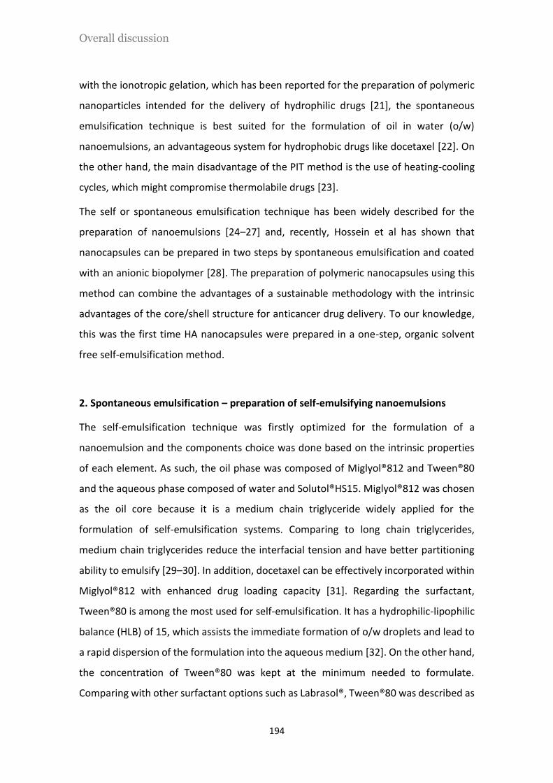
Overall discussion
194
with the ionotropic gelation, which has been reported for the preparation of polymeric
nanoparticles intended for the delivery of hydrophilic drugs [21], the spontaneous
emulsification technique is best suited for the formulation of oil in water (o/w)
nanoemulsions, an advantageous system for hydrophobic drugs like docetaxel [22]. On
the other hand, the main disadvantage of the PIT method is the use of heating-cooling
cycles, which might compromise thermolabile drugs [23].
The self or spontaneous emulsification technique has been widely described for the
preparation of nanoemulsions [24–27] and, recently, Hossein et al has shown that
nanocapsules can be prepared in two steps by spontaneous emulsification and coated
with an anionic biopolymer [28]. The preparation of polymeric nanocapsules using this
method can combine the advantages of a sustainable methodology with the intrinsic
advantages of the core/shell structure for anticancer drug delivery. To our knowledge,
this was the first time HA nanocapsules were prepared in a one-step, organic solvent
free self-emulsification method.
2. Spontaneous emulsification – preparation of self-emulsifying nanoemulsions
The self-emulsification technique was firstly optimized for the formulation of a
nanoemulsion and the components choice was done based on the intrinsic properties
of each element. As such, the oil phase was composed of Miglyol®812 and Tween®80
and the aqueous phase composed of water and Solutol®HS15. Miglyol®812 was chosen
as the oil core because it is a medium chain triglyceride widely applied for the
formulation of self-emulsification systems. Comparing to long chain triglycerides,
medium chain triglycerides reduce the interfacial tension and have better partitioning
ability to emulsify [29–30]. In addition, docetaxel can be effectively incorporated within
Miglyol®812 with enhanced drug loading capacity [31]. Regarding the surfactant,
Tween®80 is among the most used for self-emulsification. It has a hydrophilic-lipophilic
balance (HLB) of 15, which assists the immediate formation of o/w droplets and lead to
a rapid dispersion of the formulation into the aqueous medium [32]. On the other hand,
the concentration of Tween®80 was kept at the minimum needed to formulate.
Comparing with other surfactant options such as Labrasol®, Tween®80 was described as
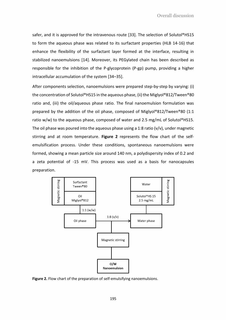
Overall discussion
195
safer, and it is approved for the intravenous route [33]. The selection of Solutol®HS15
to form the aqueous phase was related to its surfactant properties (HLB 14-16) that
enhance the flexibility of the surfactant layer formed at the interface, resulting in
stabilized nanoemulsions [14]. Moreover, its PEGylated chain has been described as
responsible for the inhibition of the P-glycoprotein (P-gp) pump, providing a higher
intracellular accumulation of the system [34–35].
After components selection, nanoemulsions were prepared step-by-step by varying: (i)
the concentration of Solutol®HS15 in the aqueous phase, (ii) the Miglyol®812/Tween®80
ratio and, (iii) the oil/aqueous phase ratio. The final nanoemulsion formulation was
prepared by the addition of the oil phase, composed of Miglyol®812/Tween®80 (1:1
ratio w/w) to the aqueous phase, composed of water and 2.5 mg/mL of Solutol®HS15.
The oil phase was poured into the aqueous phase using a 1:8 ratio (v/v), under magnetic
stirring and at room temperature. Figure 2 represents the flow chart of the self-
emulsification process. Under these conditions, spontaneous nanoemulsions were
formed, showing a mean particle size around 140 nm, a polydispersity index of 0.2 and
a zeta potential of -15 mV. This process was used as a basis for nanocapsules
preparation.
Figure 2. Flow chart of the preparation of self-emulsifying nanoemulsions.

Overall discussion
196
3. Preparation of HA-based nanocapsules using HA and dodecylamide-functionalized
HA
HA-based nanocapsules were prepared using the optimized spontaneous emulsification
technique by dissolving the polymer into the aqueous phase. The nanocapsules were
prepared using two types of HA: sodium hyaluronate (HA) and a hydrophobically
functionalized HA with a dodecylamide chain (C12-HA) (Figure 3). C12-HA has a 2.0-5.0%
degree of substitution, which is considered to be enough to confer to the polymer an
amphiphilic behavior without changing its aqueous solubility over the concentration
range required for formulation [36]. Additionally, it is not expected that this degree of
substitution interferes with CD44 recognition, an important feature for active targeted
delivery [37].
Figure 3. Chemical structure of dodecylamide-functionalized HA
The preparation of HA nanocapsules (HA NCs) has been reported by our group and
consists on the interfacial deposition of negatively charged HA onto a positive charged
surface [38]. Accordingly, self-emulsifying HA NCs were prepared by the addition of the
cationic surfactant cetyltrimethylammonium bromide (CTAB) to the oil phase. The
addition of CTAB to the nanoemulsion promoted an inversion of the zeta potential to
positive values. Consequently, the attachment of HA to the cationic layer was achieved
by electrostatic interaction between the positively charged CTAB and the HA, resulting
in the shift of the zeta potential from +10 mV to -19 mV (Figure 4). Different CTAB and

Overall discussion
197
HA concentrations were studied and the characterization of the optimized formulation
(using Miglyol®812/ Tween®80 in a ratio 1:1 (w/w) and Solutol®HS15 solution at a
concentration of 2.5 mg/mL; oil/aqueous phase ratio of 1:8 (v/v)) is shown in Table 1.
Figure 4. Size (bars) and zeta potential (line) values of the optimized anionic nanoemulsion, cationic nanoemulsion and HA NCs prepared by the spontaneous emulsification method. It was observed an inversion of the zeta potential after CTAB addition (from negative to positive values) and again after HA deposition (from positive to negative values).
Notes: Results are presented as mean value ± standard deviation (n=3)
To simplify the process and avoid the use of the cationic surfactant, which is commonly
associated to potential cytotoxic effects, HA was replaced by the C12-HA amphiphilic
polymer, which made possible the preparation of nanocapsules through hydrophobic
interactions. The use of amphiphilic HA derivatives is widely reported in the literature
for the preparation of self-assembled nanoparticles or micelles by sonication [39–41].
Nevertheless, it is important to highlight that these self-assembled structures are
formed upon application of a high shear force (sonication) and are dependent on the
degree of substitution of the HA [42]. Moreover, HA can be grafted with phospholipids,
like L-α-Dioleylphosphatidylethanolamine (DOPE), and incorporated onto the surface of
liposomes [43–45].
In our work, the use of C12-HA resulted in the formulation of nanocapsules with
physicochemical properties very similar to those prepared with HA. Moreover, the
absence of micelles formed by the C12-HA itself in water was confirmed using light-
scattering measurements.
-25
-20
-15
-10
-5
0
5
10
15
0
20
40
60
80
100
120
140
160
Anionic NE Cationic NE HA NCs
Zeta
Po
ten
tial
(mV
)
Size
(n
m)

Overall discussion
198
Table 1. Physicochemical characterization of HA NCs and C12-HA NCs.
Formulation CTAB conc.
(mg/mL) HA conc. (mg/mL)
Size PDI ZP (mV)
HA NCs 0.15 0.25 137 ± 11 0.2 -19 ± 1
C12-HA NCs - 0.5 126 ± 5 0.2 -20 ± 2
Notes: Results are presented as mean value ± standard deviation (n=3)
Abbreviations: PDI, polydispersity index; ZP, zeta potential
TEM images show the morphology of HA-based nanocapsules and the typical core/shell
structure was visualized for both systems (Figure 5). These results confirmed the
possible formation of C12-HA NCs by hydrophobic interactions, with the dodecyl chains
of the amphiphilic-HA facilitating the entrapment of the polymer on the interface of the
nanoemulsion. A similar mechanism has been described for the formulation of HA-
DOPE-coated liposomes, where the DOPE molecule plays the role of the anchor in the
lipid membrane [46].
Figure 5. TEM images of A) HA NCs and B) C12-HA NCs.
Self-emulsifying HA NCs and C12-HA NCs were evaluated for stability under storage
conditions at 4ºC and after dilution in human plasma at 37ºC. Under storage conditions,
both prototypes were very stable, without significant changes in particle size,
polydispersity index, and zeta potential for up to 6 months. This high stability may be a
consequence of different factors: (i) the negative charge conferred by the HA shell must
avoid particle aggregation [47], (ii) the PEGylated chains from Solutol®HS15 are
described as a stabilizer [48] and, (iii) the presence of Tween®80 can also provide steric

Overall discussion
199
stability [49]. When incubated in human plasma at 37ºC we observed an increase in
particle size; nevertheless, this increase was less than 20% of the initial size. Moreover,
the polydispersity index of the system did not change, which denotes the absence of
particle aggregates. This increase in size may be due to a small deposition of plasma
proteins around the nanocapsules.
The cytotoxicity of HA NCs, C12-HA NCs and a mixture of free surfactants was compared
using the AlamarBlue® assay. As presented in Figure 6, the survival curves of A549 cells
showed a concentration-dependent profile in the range of 3.5 – 1000 µg/mL.
Irrespective of their composition, both nanocapsules did not affect cell viability when
tested at concentrations up to 350 µg/mL. Nonetheless, only C12-HA NCs did not cause
toxicity at the maximum concentration tested (1000 µg/mL). These results must
associate the presence of CTAB in HA NCs with its higher cytotoxicity, as reveled in other
studies [50]. On the other hand, the free surfactant mixture composed of Tween®80,
Solutol®HS15 and CTAB showed remarkable toxic effects, resulting in 85% of cell death.
However, when encapsulated within the nanocapsules structure, this toxic profile
changed and HA NCs were less toxic [51]. C12-HA NCs were also prepared by the solvent
displacement technique and the anionic surfactant Tween®80 was replaced by lecithin
[38] (results not shown). Regardless its composition, both nanocapsules showed the
same cell viability profile, which demonstrated that the amount of Tween®80 needed to
prepare HA-based nanocapsules by the self-emulsification method was not responsible
for additional toxicity. This result must be related to the correct isolation of the
nanocapsules and/or to the capacity of the polymeric shell to mask surfactant toxicity.

Overall discussion
200
Figure 6. Viability of A549 cells after 72h of incubation with different concentrations of HA NCs, C12-HA NCs and free surfactant mixtures.
Notes: Results are presented as mean value ± standard deviation (n=6)
4. Encapsulation of docetaxel into HA-based nanocapsules
Docetaxel was first solubilized in Miglyol®812 and then incorporated into HA and C12-
HA NCs, following the protocol previously described. The drug was efficiently
encapsulated in both prototypes, without changing their physicochemical
characteristics. The solubility of docetaxel in Miglyol®812 allowed a final drug loading of
0.125 % (w/w), which corresponded to 100 µg of DCX per mL of nanocapsules. In order
to achieve a high drug loading without compromising the composition and toxicity of
the carrier, docetaxel was solubilized in a small amount of ethanol (<10%), followed by
evaporation. Docetaxel-loaded HA NCs could be prepared with a loading up to 2.75%
(corresponding to 2.5 mg/mL of docetaxel) without changing the physicochemical
characteristics of the system. This higher drug loading would result, in vivo, in the
administration of a lower amount of nanocapsules to deliver a therapeutic dose, thus
reducing the potential adverse effects of Tween®80 [52]. The encapsulation efficiency
of docetaxel was between 86-89% for both types of nanocapsules. The cytotoxicity of
docetaxel-loaded HA-based nanocapsules was studied by using the AlamarBlue® assay.
Both free and encapsulated docetaxel showed a dose dependent toxicity in A549 cells.
0
50
100
150
3.5 10 35 100 350 1000
Via
ble
ce
lls (
% o
f co
ntr
ol)
Conc (µg/mL)
HA NCs C12-HA NCs
Free Tween®80+Solutol®HS15 Free Tween®80+Solutol®HS15+CTAB

Overall discussion
201
Nevertheless, the IC50 was only achieved for the docetaxel delivered from
nanocapsules, demonstrating the potential of HA-based nanocapsules for cytostatic
drug delivery.
Commonly, the majority of in vitro release assays for hydrophobic drugs are performed
by ultracentrifugation and dialysis, and less frequently by size exclusion chromatography
or continuous flow filtration. Nevertheless, the drug and the carrier cannot always be
easily separated using those methods. For example, ultracentrifugation cannot be
applied to samples that aggregate under this separation conditions [53]. Regarding
dialysis, the addition of surfactants to achieve the total solubility of docetaxel in the
medium under sink conditions can interfere with the structure of colloidal particles and
change the drug release [54]. Considering the limitations of those isolation methods, in
vitro release assays of docetaxel from both nanocapsules prototypes were assessed
using a drug transfer method adapted from Bastiat et al [55]. For that, docetaxel-loaded
HA NCs and C12-HA NCs were diluted under sink conditions in PBS at 37ºC, and at fixed
time points (15minutes, 3h, 6h and 24h) a sample volume was taken, mixed with
Miglyol®812 (in a 1:1 v/v ratio) and placed into a centrifuge for phase separation. The
idea behind this technique is that the oil phase would act as an acceptor compartment
for the free drug, whereas the encapsulated docetaxel would be kept into the
nanocapsules suspension (Figure 7).

Overall discussion
202
Figure 7. Schematic representation of the drug transfer method for the in vitro release of docetaxel from HA-based nanocapsules.
To validate the method, an initial experiment was performed with only the free
docetaxel dissolved in PBS at sink conditions. As observed in Figure 8, all the free
docetaxel was taken up by the oil phase, confirming the ability of Miglyol®812 to act as
an acceptor phase for free DCX. Regarding the release of docetaxel from the
nanocapsules, the results demonstrated an improved profile. Docetaxel was released
from HA NCs and C12-HA NCs following a biphasic profile, showing an initial burst
release of 55% and 45%, respectively, and then, a sustained release over 24h (Figure 8).
Figure 8. In vitro release profiles of docetaxel from HA NCs and C12-HA NCs in PBS.
Notes: Results are presented as mean value ± standard deviation (n=3)
0
25
50
75
100
0.25 3 6 24
% D
CX
rel
ease
d
Time (h)
Free DCX
HA NCs
C12-HA NCs

Overall discussion
203
This release behavior can be justified by the structure of the nanocapsules and the
partition coefficient of the drug between the oil core and the aqueous external medium
[56]. Moreover, the oil phase in contact with the nanocapsules suspension can act as a
“lipophilic attractor”, which means that it can generate a continuous transfer of the free
drug to the oil compartment. In this way, the formulation is under continuous forced
sink conditions. Figure 9 illustrates both mechanisms that can justify the release of
docetaxel from HA-based nanocapsules.
Figure 9. Schematic illustration of the mechanisms responsible for the release behavior of docetaxel from HA-based nanocapsules. A) Influence of the partition equilibrium in the drug release of nanocapsules; B) Drug transfer from the nanocapsules nucleus to an external oil phase
5. Intracellular delivery of anti-gasdermin B associated to HA-based nanocapsules
Contrarily to tumor associated cell surface antigens, some cancer-causing proteins are
confined to the intracellular compartment and do not express an extracellular receptor
[57]. Examples of those proteins are RAS (GTPases) [58], non-receptor tyrosine kinases
(like Bcr-Abl) [59], BRAF [60] or heat shock proteins (like HSP90 that interacts and
stabilize mutant p53) [61]. Recently, gasdermin B (GSDMB) was discovered as an
intracellular protein whose overexpression was associated with cancer progression and
invasion [62]. This protein, localized into the cytosol and without a specific cell
membrane receptor, can join the group of “undrugable molecules” in oncology [63].
A
B

Overall discussion
204
The current approaches to target these proteins are by means of silencing therapy,
cytostatic molecules or protein kinase inhibitors. However, the complexity of these
pathways, the off-target effects and cellular barriers make these therapies unsuccessful
[63]. Besides the use of mAbs to target cancer cell receptors, an emerging new strategy
is their use to target intracellular cancer proteins. However, this therapeutic application
has been limited due to the high molecular weight and hydrophilicity of mAbs,
properties that prevent them to cross the cell membrane on their own [15]. It is
important to note that the intracellular delivery of antibodies is a common approach in
biochemistry, for example, antibodies are used to localize proteins after fixation and
permeabilization of cells [64], or can be transfected into the cell by the use of cationic-
lipids [65] or commercial transfection reagents. Nevertheless, those approaches are only
intended for cellular processes validation and cannot be used for a therapeutic
application. Additionally, intrabodies were also investigated for its ability to target and
modulate intracellular proteins. Intrabodies are antibodies that bind intracellularly to
their antigen after being produced in the same cell, in contrast to mAbs that are
prepared on a bioreactor, administered to patients and exert their activity in the surface
of a cell [66]. The inefficient delivery of the genetic material encoding the intrabody and
its instability into the cytosol are the main reasons that have hampered the therapeutic
development of this approach [67].
The strategy of using mAbs against intracellular cancer proteins has pushed the interest
of the pharmaceutical industry and, recently, two companies have announced promising
results in the transport of mAbs into cancer cells. Sorrento Therapeutics has developed
a technology based on the chemical modification of mAbs that allows them to penetrate
into the cell while maintaining their ability for binding specific target proteins [68].
BioCell Challenge has prepared lipid-based formulations to encapsulate a specific mAb
directed against the RAS oncoprotein, without the need of a chemical modification. In
vivo results demonstrated that mice survival was prolonged by up to 30% and complete
recovery was observed in 33% of cases [69].
Knowing the potential of mAbs as therapeutic agents against intracellular proteins,
Moreno-Bueno’s group has developed a mAb against the intracellular protein GSDMB,

Overall discussion
205
the anti-GSDMB. We have associated the anti-GSDMB to the polymeric shell of HA-
based nanocapsules with the aim of facilitating its entry into the cancer cells. This
platform was studied as a new technology for intracellular mAb-based therapies.
Anti-GSDMB was generated as described by Hergueta-Redondo et al (submitted). After
purification, the concentration of anti-GSDMB was determined as 2.9 mg/mL. The purity
and integrity of the mAb was evaluated by SDS-PAGE. The resulting gel electrophoresis
demonstrated a single band at 160KDa (intact antibody) and two bands, at 50 KDa
(constant fraction, Fc) and 25 KDa (variable fraction, Fab) [70], when non-reducing or
reducing conditions were applied, respectively (Figure 10). Moreover, the additional
bands that are observed might correspond to some additional impurities. For example,
it has been described that protein fragments on non-reducing SDS-PAGE are a common
feature and are related to the breakage of inter-chain disulfide bonds during sample
preparation [71]. Therefore, these results confirm the stability and integrity of anti-
GSDMB.
Figure 10. SDS-PAGE gel electrophoresis. Lanes 1 and 2, anti-GSDMB in non-reducing conditions at 20 and 40μg/mL; lanes 3 and 4, anti-GSDMB under reducing conditions after 5 and 7, 10 and 12 min. Blue arrow marks intact anti-GSDMB band and red arrows the Fc (50KDa) and Fab (25KDa) fractions.
The specific activity of anti-GSDMB was analyzed by ELISA. This assay allowed us to
confirm its activity against the target protein, GSDMB, as well as to validate a method
to quantify the mAb associated to the nanocapsules. Figure 11 shows the sigmoidal
calibration curve for the mAb in a concentration range from 0.125 to 10 µg/mL.
1 2 3 4

Overall discussion
206
Figure 11. Calibration curve of anti-GSDMB, showing the optical density values vs log of mAb concentration.
Notes: Results are presented as mean value ± standard deviation (n=3)
Anti-GSDMB was successfully associated to the polymeric shell of HA-based
nanocapsules by physical adsorption. The advantage of using this technique is related
to its mild and easy conditions, which avoids the need of aggressive reagents and
preserves the mAb activity [72–73]. Moreover, this physical adsorption may provide an
effective release of the mAb inside the cells, without the need for breaking covalent
bonds. On the other hand, the lack of a chemical conjugation may facilitate the
displacement of the adsorbed antibody when diluted in cell culture medium or human
plasma [74].
The association of anti-GSDMB was carried out with both HA NCs and C12-HA NCs. First,
HA NCs were incubated with increasing amounts of mAb, from 2.5 to 25 µg of anti-
GSDMB per mg of nanocapsules. The interaction of anti-GSDMB with the nanocapsules
was assessed using two strategies: (i) electrostatic interactions between protonated
anti-GSDMB (anti-GSDMB+) and the negatively charged HA and, (ii) hydrophobic forces
by simply incubating anti-GSDMB with the nanocapsules. Different studies report the
efficient association of proteins to oppositely charged polymers. For example, the
cationic thiolated branched polyethyleneimine was complexed with the anionic bovine
serum albumin [75] and the anionic caspase-3 protein with positive methacrylate-
crosslinked nanoparticles [76]. Moreover, it is widely described the association of
anionic proteins or enzymes to positively charged lipids, liposomes or lipoplexes [15,
µ

Overall discussion
207
77]. In a recent study, the preparation of liposome/protein complexes through
electrostatic interactions was achieved by mixing the cationic liposomes with a solution
of albumin, as a model protein, or the mouse anti-F actin primary antibody, as a model
antibody [78]. The majority of protein complexes are formed with cationic lipids or
polymers to take advantage of the “proton sponge effect”, thus promoting the
endosomal escape of proteins [79]. It has also been reported the adsorption or
entrapment of proteins and antibodies to nanoparticles through non-ionic interactions.
For example, β-galactosidase was entrapped within chitosan-conjugated Pluronic®-
based nanoparticles [80] and the model protein, carbonic anhydrase, entrapped within
self-assembled HA-nanoparticles [81]. Moreover, the adsorption of antibodies to the
surface of polymeric nanoparticles has been also described for the non-covalent binding
of mAbs to cyanoacrylate nanoparticles [73] as well as to PLGA nanoparticles [82–83].
With the purpose of promoting an electrostatic interaction between the mAb and the
surface of HA nanocapsules, anti-GSDMB was protonated at pH 4.5 and ionically
associated to negatively charged HA NCs. With an isoelectric point of 6.5-8.0 we had the
option of working with negatively charged anti-GSDMB and positive nanocapsules, or
with positively charged anti-GSDMB and anionic nanocapsules. We decided to protonate
anti-GSDMB as it is described as a frequent intermediate step for antibody conjugation
[84–85]. For example, SM5-1 single-chain antibody was protonated under acidic
conditions and successfully adsorbed onto the surface of negatively charged PLGA
nanoparticles [86]. On the other hand, anti-GSDMB adsorption to HA nanocaspules was
carried by simple mixing a solution of nanocapsules with the antibody at neutral pH,
thus promoting an interaction mainly based on hydrophobic forces rather than an ionic
interaction.
Regardless of the strategy, anti-GSDMB was effectively incorporated within the
polymeric shell of both types of nanocapsules. Table 2 shows the physicochemical
characterization of nanocapsules, which resulted in particles around 120 nm,
monodisperses and with a high association efficiency. Moreover, the decrease in the
zeta potential indicated the prevalence of HA on the surface of the nanostructure
despite the high mAb association efficiency. The use of 0.75 mg/mL of HA and 0.5 mg/mL

Overall discussion
208
of C12-HA allowed us to obtain a drug loading of 25 µg mAb/mg nanocapsules, while
maintaining a similar negative charge.
Table 2. Physicochemical characterization and association efficiency of anti-GSDMB and protonated anti-GSDMB+ associated to HA NCs and C12-HA NCs. (Blank nanocapsules are included for comparison)
Formulation Anti-GSDMB
(25 µg/ mg NCs) Size (nm) PDI ZP (mV)
Association efficiency (%)
HA NCs - 126 ± 5 0.2 -22 ± 1 -
C12-HA NCs - 123 ± 2 0.2 -20 ± 1 -
HA NCs Anti-GSDMB+ 117 ± 6 0.2 -11 ± 1 75 ± 10
Anti-GSDMB 126 ± 7 0.2 -7 ± 1 82 ± 6
C12-HA NCs Anti-GSDMB+ 119 ± 3 0.2 -6 ± 1 84 ± 8
Anti-GSDMB 116 ± 6 0.2 -10 ± 1 93 ± 3
Notes: HA NCs and C12-HA NCs were prepared with 0.75 mg/mL and 0.5 mg/mL of HA and C12-HA, respectively. Results are presented as mean value ± standard deviation (n=3)
Abbreviations: PDI, polydispersion index; ZP, zeta potential
Electrostatic interactions are thought to be a major factor in protein binding to
polymeric surfaces. As such, we obtained a high association efficiency when protonated
anti-GSDMB+ was incubated with negatively charged HA NCs at a final pH of 4.5.
Interestingly, the same association efficiency was observed when non-protonated anti-
GSDMB was incubated with HA NCs (pH 7.4). These results could be justified by the
interaction of the mAb with the polymeric shell by hydrophobic forces. At pH 7.4 the
anti-GSDMB is within its isoelectric point, which could favor the entrapment of its
hydrophobic domain within the interface of the nanocapsules [87]. Moreover, it could
also be presumed that the hydroxyl-carboxyl groups in the polymeric structure might
help the interaction of the protein by simple hydrogen bonds [88].
In order to study the ability of HA-based nanocapsules to transport the anti-GSDMB into
the intracellular compartment, the first step was to perform in vitro cytotoxicity assays

Overall discussion
209
for the blank prototypes in the HCC1954 breast cancer cell line. Likewise our previous
results, HA NCs showed a marked cytotoxicity, with a decrease of 50% in cell viability
(IC50) for 0.3 mg/mL. On the contrary, C12-HA NCs did not show considerable toxicity
at concentrations up to 0.8 mg/mL and this formulation was chosen for the following
assays.
The hyphothetic internalization of the mAb was followed by confocal microscopy. For
that, HCC1954 cells were incubated with anti-GSDMB-loaded C12-HA NCs at a
concentration of 0.8 mg/mL, which corresponds to 20 µg of mAb (125 nM). Figure 12
shows the internalization of anti-GSDMB alone or after association to C12-HA NCs.
Without a cell surface receptor and a nanocarrier, anti-GSDMB is described to be too
large and hydrophilic to cross the cell membrane on its own [15]. On the other hand,
anti-GSDMB associated to C12-HA NCs was effectively delivered into the cytosol of
HCC1954 cells, as seen by the green dots in the XZ image. Similar results were obtained
when protonated anti-GSDMB+ was associated to C12-HA NCs. Thus, in order to avoid
the acidification of the medium and the protonation of the antibody, non-protonated
anti-GSDMB associated to C12-HA NCs was selected to continue the study.
Figure 12. Internalization of anti-GSDMB mediated by its association to C12-HA NCs. HCC1954 cells were incubated with 0.8 mg/mL of C12-HA NCs, which corresponds to 20 µg of anti-GSDMB, for 72h. A) Anti-GSDMB alone and blank C12-HA NCs were used as controls. Without a cell surface receptor, the anti-GSDMB on its own was not able to cross the cell membrane; B) C12-HA NCs promoted the internalization of anti-GSDMB after its association to the polymeric shell.
Notes: Images were taken by confocal microscopy. Cell nuclei were stained with DAPI (blue) and cell membrane with phalloidin (red).
Anti-GSDMB alone Empty C12-HA NCs
Anti-GSDMB-loaded C12-HA NCs

Overall discussion
210
Furthermore, to be able to increase the concentration of C12-HA NCs without
compromising cell viability, serial steps of incubation were performed using different
concentrations and incubation times. As a result, it was demonstrated that by incubating
HCC1954 cells with C12-HA NCs for short periods of time, up to 2h, followed by repeated
washing and incubation steps, it was possible to increase the nanocapsules
concentration to 3.2 mg/ml without additional cytotoxicity. Accordingly, the
concentration of anti-GSDMB could be increased four-fold, to 80 µg of anti-GSDMB
(which corresponds to 500 nM). We decided to study the trafficking pathways and
therapeutic efficacy of anti-GSDMB into HCC1954 cells using the highest concentration
tested for intracellular uptake, i.e. 500 nM. For example, it was reported that the cell-
mediated cytotoxicity of trastuzumab in HCC1954 cells was assessed following
treatment with 100 nM or 200 nM of the mAb [89–90]. Because our mAb was intended
to act intracellularly and we were not sure about the therapeutic concentration
required, we decided to perform these experiments using 500 nM of anti-GSDMB.
Once it was shown that the mAb was able to enter the cells, the next step was to study
the trafficking pathway of anti-GSDMB and its co-localization with early endosomes, as
well as the endosomal escape. It is well known that one of the main hurdles in the
delivery of proteins or RNA into the cytosol is the in vivo degradation by lysosomes [91].
In fact, once inside the cell, anti-GSDMB might escape lysosomes and maintain its
conformational structure in order to target GSDMB. The results showed that anti-
GSDMB (green dots) was carried into the cell by endocytosis (red dots) and was able to
escape lysosomes (yellow dots) (Figure 13).

Overall discussion
211
Figure 13. Trafficking of anti-GSDMB (green) through endosomes. HCC1954 cells were incubated with 3.2 mg/mL of C12-HA NCs containing 80 µg of anti-GSDMB for 2h, 3h, 4h and 5h. A) Early endosomes were detected by early endosome antigen 1 (EEA1) (red). B) Late endosomes were detected by lysosomal-associated membrane protein (LAMP1) (yellow).
Notes: Images were taken by confocal microscopy. Nuclei were stained with DAPI (blue) and the cytoskeleton with phalloidin (purple).
The internalization through endocytosis might probably be mediated by CD44 receptors,
overexpressed on the surface of HCC1954 cells [92]. CD44 is the principal receptor for
HA and its binding affinity and enhanced uptake through this mechanism is well known
[93]. Moreover, HA-based nanocapsules could be internalized by other passive
mechanisms, for example after particle adsorption onto the cell surface [94–95] as well
as by the penetration enhancing nature of some of the compounds of the nanocarrier.
For example, it has been described that Tween®80 has de capacity to enhance cell
permeability. To better understand the internalization pathway, three different assays
could be taken into consideration: (i) blocking of CD44 receptors with free HA to confirm
Empty C12-HA NCs Anti-GSDMB loaded C12-HA NCs
Empty C12-HA NCs Anti-GSDMB loaded C12-HA NCs

Overall discussion
212
the intracellular uptake by CD44-receptor mediated transport [96]; (ii) inhibition of the
cell uptake mechanism using three endocytosis inhibitors, namely, chlorpromazine
(clathrin inhibitor), nystatin (caveolin inhibitor), and cytochalasin D (actin inhibitor) [78]
and; (iii) expose the cell to ice-cold medium (0-4ºC) for nonspecific inhibition of
endocytosis [97].
The endosomal escape was visualized by confocal microscopy and qualitatively
confirmed a negative co-localization with LAMP1 labeled lysosomes (yellow) (Figure 13
B). Considering the effective endosomal escape, this mechanism must be explained by
two main reasons: (i) a pH-dependent mechanism, similar to the “proton sponge effect”
caused by cationic lipids [98] and, (ii) the influence of the anionic surfactant Tween®80
in destabilizing the endosomal membrane [99]. It has been described that cationic
nanoparticles with a buffering capacity at pH from 5 to 7 have displayed the ability to
escape the endosomes (pH 5 – 6). Under acidic conditions, cationic lipids/polymers are
capable of buffering the endosomal vesicle, leading to endosomal swelling and lysis, and
thus releasing the payload into the cytoplasm [100]. Following the same explanation,
we can adapt this mechanism to explain the endosomal escape of anti-GSDMB. Thus, at
pH 7.4 anti-GSDMB is within its isoelectric point, which favored the association to C12-
HA NCs and promoted its internalization into the cells. Nevertheless, when inside the
endosomes the pH drops to acidic values, the anti-GSDMB becomes positively charged
and the interaction with the nanocapsules structure becomes weak [101]. Once
protonated, cationic anti-GSDMB+ might form ion pairs with the anionic lipids within the
endosome and destabilize the endosomal membrane [100]. These results would be in
accordance with those described for doxorubicin-loaded HA-Poly-L-Histidine/ D-α-
Tocopherol PEG 1000 succinate polyhistidine with the highest release occurring at
endosomal pH [102]. Moreover, the endosomal escape of protonated anti-GSDMB can
be correlated with the trafficking pathway of positively supercharged proteins. For
example, positively charged green fluorescent protein was found in high amounts inside
early endosomes nevertheless, two hours later, only a small fraction (< 20%) was
eventually co-localized with late endosomes. Furthermore, 80% of the original protein
signal remained within cells after 16h, indicating that the proteolysis of the endocytosed

Overall discussion
213
cargo occurred in a little extention [103]. On the other hand, another hypothesis for the
endosomal escape could be related to the components choice to formulate HA
nanocapsules, namely the Tween®80. It was described that the Tween®80 could interact
with the endosomal membrane [99] or form lamellar-to-inverted hexagonal phases
[104], which can disturb the endosome membrane and promote endosomal escape.
In order to confirm these results, a possible experiment to take into consideration would
be the cellular uptake performed by confocal microscopy with live cells using a real-time
imaging experiment. In this case, anti-GSDMB would be conjugated with a fluorophore,
for example Alexa®Fluor, and the trafficking pathway observed without the need of cell
fixation. It has been reported that cargos delivered by cell penetration peptides can
artificially “escape” from endocytic vesicles, thus resulting in misleading conclusions
[105].
It is known that the overexpression of GSDMB is responsible for cell migration and
invasion whereas its knockdown with shRNA results in a marked decrease of the
migratory ability and the invasive capacity of HCC1954 cells. Therefore, we performed a
last experiment in order to assess the ability of anti-GSDMB to effectively block the
intracellular cancer-protein GSDMB and inhibit cell migration and invasion. The binding
affinity and targeting ability of anti-GSDMB against intracellular GSDMB was
demonstrated by wound healing migration assays. We observed that HCC1954 cells
treated with anti-GSDMB-loaded C12-HA NCs inhibited cell migration as compared to
the negative control (Figure 14). As a result, the scratch healing of HCC1954 cells treated
with anti-GSDMB-loaded C12-HA NCs was bigger than the cell treated with only NCs,
denoting the slow capacity of cells to migrate and close the wound.

Overall discussion
214
Figure 14. Effect of anti-GSDMB loaded C12-HA NCs on the migration of HCC1954 cells. A) Scratch wound healing assay was performed to examine the effect of C12-HA NCs entrapped anti-GSDMB on cell migration. B) Mobility rate histograms of each group across the selected times (hours). *p<0.05
After 60h, HCC1954 cells treated with blank nanocapsules migrate and close the wound
healing to 165 µm whereas, HCC1954 cells treated with anti-GSDMB loaded C12-HA NCs
maintain an opening heal of 291 µm, which prove the slow migration of cells. We can
speculate that, once inside the cell, anti-GSDMB is maintaining its original conformation
and binding affinity to the GSDMB protein. Although, to our knowledge, this was the
first time that a scratch migration assay was performed to analyze the effect of a mAb
delivered intracellularly, this experiment has been largely used as a tool to analyze the
binding effect of mAbs to extracellular cancer receptors in the migration and invasive
behavior of tumor cells [107]
Finally, these results demonstrate the capacity of C12-HA NCs to incorporate and deliver
anti-GSDMB into cancer cells. This approach must represent a promising strategy against
the “undruggable targets” and broaden the application of mAbs, confined so far to the
target of extracellular proteins.
Empty C12-HA NCs
Anti-GSDMB-loaded C12-HA NCs
Anti-GSDMB loaded C12-HA NCs
Empty C12-HA NCs

Overall discussion
215
Bibliography
[1] E. Pérez-Herrero and A. Fernández-Medarde, “Advanced targeted therapies in cancer: Drug nanocarriers, the future of chemotherapy,” Eur. J. Pharm. Biopharm., vol. 93, pp. 52–79, 2015.
[2] T. Sun, Y. S. Zhang, B. Pang, D. C. Hyun, M. Yang, and Y. Xia, “Engineered Nanoparticles for Drug Delivery in Cancer Therapy,” Angew. Chemie - Int. Ed., vol. 53, pp. 12320–12364, 2014.
[3] C. E. Mora-Huertas, H. Fessi, and A. Elaissari, “Polymer-based nanocapsules for drug delivery.,” Int. J. Pharm., vol. 385, no. 1–2, pp. 113–42, 2010.
[4] V. Yurgel, T. Collares, and F. Seixas, “Developments in the use of nanocapsules in oncology.,” Braz. J. Med. Biol. Res., vol. 46, no. 6, pp. 486–501, Jul. 2013.
[5] T. Gonzalo, G. Lollo, M. Garcia-Fuentes, D. Torres, J. Correa, R. Riguera, E. Fernandez-Megia, P. Calvo, P. Avilés, M. J. Guillén, and M. J. Alonso, “A new potential nano-oncological therapy based on polyamino acid nanocapsules,” J. Control. Release, vol. 169, no. 1–2, pp. 10–16, 2013.
[6] K. Y. Choi, G. Saravanakumar, J. H. Park, and K. Park, “Hyaluronic acid-based nanocarriers for intracellular targeting: interfacial interactions with proteins in cancer.,” Colloids Surf. B. Biointerfaces, vol. 99, pp. 82–94, 2012.
[7] D. S. Spencer, A. S. Puranik, and N. A. Peppas, “Intelligent nanoparticles for advanced drug delivery in cancer treatment,” Curr. Opin. Chem. Eng., vol. 7, pp. 84–92, 2015.
[8] N. Anton and T. F. Vandamme, “The universality of low-energy nano-emulsification.,” Int. J. Pharm., vol. 377, no. 1–2, pp. 142–7, 2009.
[9] S. Arpicco, C. Lerda, E. Dalla Pozza, C. Costanzo, N. Tsapis, B. Stella, M. Donadelli, I. Dando, E. Fattal, L. Cattel, and M. Palmieri, “Hyaluronic acid-coated liposomes for active targeting of gemcitabine.,” Eur. J. Pharm. Biopharm., vol. 85, no. 3 Pt A, pp. 373–80, 2013.
[10] S. Ganesh, A. K. Iyer, D. V. Morrissey, and M. M. Amiji, “Hyaluronic acid based self-assembling nanosystems for CD44 target mediated siRNA delivery to solid tumors,” Biomaterials, vol. 34, no. 13, pp. 3489–3502, 2013.
[11] S. Mizrahy, M. Goldsmith, S. Leviatan-Ben-Arye, E. Kisin-Finfer, O. Redy, S. Srinivasan, D. Shabat, B. Godin, and D. Peer, “Tumor targeting profiling of hyaluronan-coated lipid based-nanoparticles.,” Nanoscale, vol. 6, no. 7, pp. 3742–52, 2014.
[12] C. E. Schanté, G. Zuber, C. Herlin, and T. F. Vandamme, “Chemical modifications of hyaluronic acid for the synthesis of derivatives for a broad range of biomedical applications,” Carbohydr. Polym., vol. 85, no. 3, pp. 469–489, 2011.
[13] L. Zhang and N. Zhang, “How nanotechnology can enhance docetaxel therapy,” Int. J. Nanomedicine, vol. 8, pp. 2927–2941, 2013.
[14] A. M. Scott, J. D. Wolchok, and L. J. Old, “Antibody therapy of cancer,” Nat. Rev. Cancer, vol. 12, no. April, pp. 278–287, 2012.
[15] V. Torchilin, “Intracellular delivery of protein and peptide therapeutics.,” Drug Discov. today;Technologies, vol. 5, no. 2–3, 2009.
[16] K. Grodowska and a. Parczewski, “Organic solvents in the pharmaceutical industry.,” Acta Pol. Pharm., vol. 67, no. 1, pp. 3–12, 2010.
[17] P. Dunn, A. Wells, and M. Williams, Green chemistry in the pharmaceutical industry. 2010.
[18] M. Naghdi, M. Taheran, S. K. Brar, M. Verma, R. Y. Surampalli, and J. R. Valero, “Green

Overall discussion
216
and energy-efficient methods for the production of metallic nanoparticles,” Beilstein J. Nanotechnol., vol. 6, pp. 2354–2376, 2015.
[19] D. Hudson and A. Margaritis, “Biopolymer nanoparticle production for controlled release of biopharmaceuticals.,” Crit. Rev. Biotechnol., vol. 34, no. 2, pp. 161–79, 2014.
[20] N. Anton, P. Gayet, J. P. Benoit, and P. Saulnier, “Nano-emulsions and nanocapsules by the PIT method: An investigation on the role of the temperature cycling on the emulsion phase inversion,” Int. J. Pharm., vol. 344, no. 1–2, pp. 44–52, 2007.
[21] A. Mahapatro and D. K. Singh, “Biodegradable nanoparticles are excellent vehicle for site directed in-vivo delivery of drugs and vaccines.,” J. Nanobiotechnology, vol. 9, no. 1, p. 55, 2011.
[22] N. Sadurní, C. Solans, N. Azemar, and M. J. García-Celma, “Studies on the formation of O/W nano-emulsions, by low-energy emulsification methods, suitable for pharmaceutical applications.,” Eur. J. Pharm. Sci., vol. 26, no. 5, pp. 438–45, 2005.
[23] A. A. Date, N. Desai, R. Dixit, and M. Nagarsenker, “Self-nanoemulsifying drug delivery systems: formulation insights, applications and advances.,” Nanomedicine (Lond)., vol. 5, no. 10, pp. 1595–616, 2010.
[24] K. Bouchemal, S. Briançon, E. Perrier, and H. Fessi, “Nano-emulsion formulation using spontaneous emulsification: Solvent, oil and surfactant optimisation,” Int. J. Pharm., vol. 280, no. 1–2, pp. 241–251, 2004.
[25] C. Solans, P. Izquierdo, J. Nolla, N. Azemar, and M. Garciacelma, “Nano-emulsions,” Curr. Opin. Colloid Interface Sci., vol. 10, no. 3–4, pp. 102–110, 2005.
[26] A. H. Saberi, Y. Fang, and D. J. McClements, “Fabrication of vitamin E-enriched nanoemulsions: Factors affecting particle size using spontaneous emulsification,” J. Colloid Interface Sci., vol. 391, no. 1, pp. 95–102, 2013.
[27] H. Sobhani, P. Tarighi, S. N. Ostad, A. Shafaati, N. Nafissi-Varcheh, and R. Aboofazeli, “Formulation Development and Toxicity Assessment of Triacetin Mediated Nanoemulsions as Novel Delivery Systems for Rapamycin.,” Iran. J. Pharm. Res. IJPR, vol. 14, no. Suppl, pp. 3–21, 2015.
[28] A. Hossein, B. Zeeb, J. Weiss, and D. Julian, “Tuneable stability of nanoemulsions fabricated using spontaneous emulsification by biopolymer electrostatic deposition,” J. Colloid Interface Sci., vol. 455, pp. 172–178, 2015.
[29] T. Gershanik and S. Benita, “Self-dispersing lipid formulations for improving oral absorption of lipophilic drugs.,” Eur. J. Pharm. Biopharm., vol. 50, no. 1, pp. 179–88, 2000.
[30] D. F. Driscoll, J. Nehne, H. Peterss, R. Franke, B. R. Bistrian, and W. Niemann, “The influence of medium-chain triglycerides on the stability of all-in-one formulations,” Int. J. Pharm., vol. 240, no. 1–2, pp. 1–10, 2002.
[31] J. a Yared and K. H. R. Tkaczuk, “Update on taxane development: new analogs and new formulations.,” Drug Des. Devel. Ther., vol. 6, pp. 371–84, 2012.
[32] A. Kumar, S. Sharma, and R. Kamble, “Self emulsifying drug delivery system (SEDDS): Future Aspects,” Int J Pharm, vol. 2, 2010.
[33] E. Sigward, N. Mignet, P. Rat, M. Dutot, S. Muhamed, J.-M. Guigner, D. Scherman, D. Brossard, and S. Crauste-Manciet, “Formulation and cytotoxicity evaluation of new self-emulsifying multiple W/O/W nanoemulsions,” Int. J. Nanomedicine, vol. 8, pp. 611–25, 2013.
[34] N. T. Huynh, C. Passirani, P. Saulnier, and J. P. Benoit, “Lipid nanocapsules: a new platform

Overall discussion
217
for nanomedicine.,” Int. J. Pharm., vol. 379, no. 2, pp. 201–9, 2009.
[35] K. Buszello, S. Harnisch, R. H. Müller, and B. W. Müller, “The influence of alkali fatty acids on the properties and the stability of parenteral O/W emulsions modified with solutol HS 15.,” Eur. J. Pharm. Biopharm., vol. 49, no. 2, pp. 143–9, 2000.
[36] G. Saravanakumar, K. Y. Choi, H. Y. Yoon, K. Kim, J. H. Park, I. C. Kwon, and K. Park, “Hydrotropic hyaluronic acid conjugates: Synthesis, characterization, and implications as a carrier of paclitaxel.,” Int. J. Pharm., vol. 394, no. 1–2, pp. 154–61, 2010.
[37] O. P. Oommen, J. Garousi, M. Sloff, and O. P. Varghese, “Tailored doxorubicin-hyaluronan conjugate as a potent anticancer glyco-drug: an alternative to prodrug approach.,” Macromol. Biosci., vol. 14, no. 3, pp. 327–33, 2014.
[38] F. a Oyarzun-Ampuero, G. R. Rivera-Rodríguez, M. J. Alonso, and D. Torres, “Hyaluronan nanocapsules as a new vehicle for intracellular drug delivery.,” Eur. J. Pharm. Sci., vol. 49, no. 4, pp. 483–90, 2013.
[39] X. Dong and C. Liu, “Preparation and Characterization of Self-Assembled Nanoparticles of Hyaluronic Acid-Deoxycholic Acid Conjugates,” J. Nanomater., vol. 2010, pp. 1–9, 2010.
[40] C. Xu, W. He, Y. Lv, C. Qin, L. Shen, and L. Yin, “Self-assembled nanoparticles from hyaluronic acid-paclitaxel prodrugs for direct cytosolic delivery and enhanced antitumor activity.,” Int. J. Pharm., vol. 493, no. 1, pp. 172–181, 2015.
[41] H.-J. Cho, H. Y. Yoon, H. Koo, S.-H. Ko, J.-S. Shim, J.-H. Lee, K. Kim, I. C. Kwon, and D.-D. Kim, “Self-assembled nanoparticles based on hyaluronic acid-ceramide (HA-CE) and Pluronic® for tumor-targeted delivery of docetaxel.,” Biomaterials, vol. 32, no. 29, pp. 7181–90, 2011.
[42] K. Y. Choi, H. Chung, K. H. Min, H. Y. Yoon, K. Kim, J. H. Park, I. C. Kwon, and S. Y. Jeong, “Self-assembled hyaluronic acid nanoparticles for active tumor targeting.,” Biomaterials, vol. 31, no. 1, pp. 106–14, 2010.
[43] K. Cohen, R. Emmanuel, E. Kisin-Finfer, D. Shabat, and D. Peer, “Modulation of drug resistance in ovarian adenocarcinoma using chemotherapy entrapped in hyaluronan-grafted nanoparticle clusters,” ACS Nano, vol. 8, no. 3, pp. 2183–2195, 2014.
[44] S. R. Paliwal, R. Paliwal, G. P. Agrawal, and S. P. Vyas, “Hyaluronic acid modified pH-sensitive liposomes for targeted intracellular delivery of doxorubicin,” J. Liposome Res., vol. 19, pp. 1–12, 2016.
[45] T. L. Nascimento, H. Hillaireau, M. Noiray, C. Bourgaux, S. Arpicco, G. Pehau-Arnaudet, M. Taverna, D. Cosco, N. Tsapis, and E. Fattal, “Supramolecular Organization and siRNA Binding of Hyaluronic Acid-Coated Lipoplexes for Targeted Delivery to the CD44 Receptor,” Langmuir, vol. 31, no. 41, pp. 11186–11194, 2015.
[46] A. Dufa?? Wojcicki, H. Hillaireau, T. L. Nascimento, S. Arpicco, M. Taverna, S. Ribes, M. Bourge, V. Nicolas, A. Bochot, C. Vauthier, N. Tsapis, and E. Fattal, “Hyaluronic acid-bearing lipoplexes: Physico-chemical characterization and in vitro targeting of the CD44 receptor,” J. Control. Release, vol. 162, no. 3, pp. 545–552, 2012.
[47] B. Jung and P. Theato, “Chemical Strategies for the Synthesis of Protein – Polymer Conjugates,” Adv. Polym. Sci., no. May 2012, pp. 1–34, 2012.
[48] B. Saliou, O. Thomas, N. Lautram, A. Clavreul, J. Hureaux, T. Urban, J.-P. Benoit, and F. Lagarce, “Development and in vitro evaluation of a novel lipid nanocapsule formulation of etoposide.,” Eur. J. Pharm. Sci., vol. 50, no. 2, pp. 172–80, 2013.
[49] S. Honary and F. Zahir, “Effect of zeta potential on the properties of nano-drug delivery

Overall discussion
218
systems - A review (Part 2),” Trop. J. Pharm. Res., vol. 12, no. 2, pp. 255–264, 2013.
[50] M. Fraga, F. Bruxel, V. L. Lagranha, H. F. Teixeira, and U. Matte, “Influence of phospholipid composition on cationic emulsions/DNA complexes: physicochemical properties, cytotoxicity, and transfection on Hep G2 cells.,” Int. J. Nanomedicine, vol. 6, pp. 2213–20, 2011.
[51] A. M. Alkilany and C. J. Murphy, “Toxicity and cellular uptake of gold nanoparticles : what we have learned so far ?,” pp. 2313–2333, 2010.
[52] J. Della Rocca, D. Liu, and W. Lin, “Are high drug loading nanoparticles the next step forward for chemotherapy?,” Nanomedicine (Lond)., vol. 7, no. 3, pp. 303–5, 2012.
[53] S. D. Souza, “A Review of In Vitro Drug Release Test Methods for Nano-Sized Dosage Forms,” vol. 2014, 2014.
[54] B. J. Boyd, “Characterisation of drug release from cubosomes using the pressure ultrafiltration method,” Int. J. Pharm., vol. 260, no. 2, pp. 239–247, 2003.
[55] G. Bastiat, C. O. Pritz, C. Roider, F. Fouchet, E. Lignières, A. Jesacher, R. Glueckert, M. Ritsch-Marte, A. Schrott-Fischer, P. Saulnier, and J. P. Benoit, “A new tool to ensure the fluorescent dye labeling stability of nanocarriers: A real challenge for fluorescence imaging,” J. Control. Release, vol. 170, no. 3, pp. 334–342, 2013.
[56] V. P. Torchilin and V. S. Trubetskoy, Nanoparticulates as Drug Carriers, no. 95. 1995.
[57] C. W. Hong and Q. Zeng, “Tapping the treasure of intracellular oncotargets with immunotherapy,” FEBS Lett., vol. 588, no. 2, pp. 350–355, 2014.
[58] A. a Samatar and P. I. Poulikakos, “Targeting RAS – ERK signalling in cancer : promises and challenges,” Nat. Publ. Gr., vol. 13, no. 12, pp. 928–942, 2014.
[59] O. Hantschel, “Structure, Regulation, Signaling, and Targeting of Abl Kinases in Cancer,” Genes Cancer, vol. 3, pp. 436–446, 2012.
[60] C. A. Pratilas, F. Xing, and D. B. Solit, “Targeting oncogenic BRAF in human cancer.,” Curr. Top. Microbiol. Immunol., vol. 355, pp. 83–98, 2012.
[61] R. Seigneuric, H. Mjahed, J. Gobbo, A.-L. Joly, K. Berthenet, S. Shirley, and C. Garrido, “Heat Shock Proteins as Danger Signals for Cancer Detection,” Front. Oncol., vol. 1, no. November, pp. 1–10, 2011.
[62] M. Hergueta-Redondo, D. Sarrió, Á. Molina-Crespo, D. Megias, A. Mota, A. Rojo-Sebastian, P. García-Sanz, S. Morales, S. Abril, A. Cano, H. Peinado, and G. Moreno-Bueno, “Gasdermin-B promotes invasion and metastasis in breast cancer cells.,” PLoS One, vol. 9, no. 3, p. e90099, 2014.
[63] J. S. Lazo and E. R. Sharlow, “Drugging Undruggable Molecular Cancer Targets,” Annu. Rev. Pharmacol. Toxicol., vol. 56, no. 1, pp. 23–40, 2016.
[64] A. L. J. Marschall, A. Frenzel, T. Schirrmann, M. Schüngel, and S. Dübel, “Targeting antibodies to the cytoplasm,” MAbs, vol. 3, no. 1, pp. 3–16, 2011.
[65] C. O. Weill, S. Biri, and P. Erbacher, “Cationic lipid-mediated intracellular delivery of antibodies into live cells,” Biotechniques, vol. 44, no. 7, pp. 7–11, 2008.
[66] A. L. Marschall, S. Dübel, and T. Böldicke, “Specific in vivo knockdown of protein function by intrabodies.,” MAbs, vol. 7, no. 6, pp. 1010–35, 2015.
[67] R. E. Kontermann, “Intrabodies as therapeutic agents,” Methods, vol. 34, no. 2, pp. 163–170, 2004.
[68] “http://sorrentotherapeutics.com/platforms/cell-internalizing-antibodies/.” .

Overall discussion
219
[69] B. Sas and L. Meunier, “BioCellChallenge obtains unprecedented in-vivo results using intracellular delivery technology for therapeutic antibodies,” 2015.
[70] T. Boenisch, M. S. Revised, H. Winther, S. Steen, and J. Ms, “Antibodies,” in IHC Staining Methods, 2009.
[71] H. Liu, G. Gaza-Bulseco, C. Chumsae, and A. Newby-Kew, “Characterization of lower molecular weight artifact bands of recombinant monoclonal IgG1 antibodies on non-reducing SDS-PAGE,” Biotechnol. Lett., vol. 29, no. 11, pp. 1611–1622, 2007.
[72] M. Nichkova, D. Dosev, R. Perron, S. J. Gee, B. D. Hammock, and I. M. Kennedy, “Eu3+-doped Gd2O3 nanoparticles as reporters for optical detection and visualization of antibodies patterned by microcontact printing,” Anal. Bioanal. Chem., vol. 384, no. 3, pp. 631–637, 2006.
[73] L. Nobs, F. Buchegger, R. Gurny, and E. Allémann, “Current methods for attaching targeting ligands to liposomes and nanoparticles.,” J. Pharm. Sci., vol. 93, no. 8, pp. 1980–92, 2004.
[74] M. Arruebo, M. Valladares, and Á. González-Fernández, “Antibody-Conjugated Nanoparticles for Biomedical Applications,” J. Nanomater., pp. 1–24, 2009.
[75] L. Tian, H. C. Kang, and Y. H. Bae, “Endosomolytic reducible polymeric electrolytes for cytosolic protein delivery,” Biomacromolecules, vol. 14, no. 8, pp. 2570–2581, 2013.
[76] M. Zhao, A. Biswas, B. Hu, K.-I. Joo, P. Wang, Z. Gu, and Y. Tang, “Redox-responsive nanocapsules for intracellular protein delivery.,” Biomaterials, vol. 32, no. 22, pp. 5223–30, 2011.
[77] A. Fu, R. Tang, J. Hardie, M. E. Farkas, and V. M. Rotello, “Promises and Pitfalls of Intracellular Delivery of Proteins,” Bioconjug. Chem., vol. 25, no. 9, pp. 1602–1608, 2014.
[78] S. R. Sarker, R. Hokama, and S. Takeoka, “Intracellular delivery of universal proteins using a lysine headgroup containing cationic liposomes: Deciphering the uptake mechanism,” Mol. Pharm., vol. 11, no. 1, pp. 164–174, 2014.
[79] J. Zhao and S. Feng, “Nanocarriers for delivery of siRNA and co-delivery of siRNA and other therapeutic agents,” vol. 10, pp. 2199–2228, 2015.
[80] J. Y. Kim, W. Il Choi, Y. H. Kim, and G. Tae, “Brain-targeted delivery of protein using chitosan- and RVG peptide-conjugated, pluronic-based nano-carrier,” Biomaterials, vol. 34, no. 4, pp. 1170–1178, 2013.
[81] M. Sivasubramanian, A. R. Kang, H. S. Han, R. Heo, J.-Y. Lee, K. J. Kim, S. M. Jeon, S. Y. Chae, D.-G. Jo, J.-H. Kim, and J. H. Park, “Molecular chaperone-like hyaluronic acid nanoparticles: Implications as the carrier for protein delivery systems,” Macromol. Res., vol. 20, no. 10, pp. 1007–1010, 2012.
[82] P. Kocbek, N. Obermajer, M. Cegnar, J. Kos, and J. Kristl, “Targeting cancer cells using PLGA nanoparticles surface modified with monoclonal antibody.,” J. Control. Release, vol. 120, no. 1–2, pp. 18–26, 2007.
[83] S. I. Thamake, S. L. Raut, a P. Ranjan, Z. Gryczynski, and J. K. Vishwanatha, “Surface functionalization of PLGA nanoparticles by non-covalent insertion of a homo-bifunctional spacer for active targeting in cancer therapy.,” Nanotechnology, vol. 22, no. 3, p. 035101, 2011.
[84] S. V Sule, C. D. Dickinson, J. Lu, C.-K. Chow, and P. M. Tessier, “Rapid analysis of antibody self-association in complex mixtures using immunogold conjugates.,” Mol. Pharm., vol. 10, no. 4, pp. 1322–31, 2013.

Overall discussion
220
[85] T. Powell and J.-Y. Yoon, “Fluorescent biorecognition of gold nanoparticle-IgG conjugates self-assembled on E-beam patterns.,” Biotechnol. Prog., vol. 22, no. 1, pp. 106–10, 2006.
[86] G. Kou, J. Gao, H. Wang, H. Chen, B. Li, D. Zhang, S. Wang, S. Hou, W. Qian, J. Dai, Y. Zhong, and Y. Guo, “Preparation and Characterization of Paclitaxel-loaded PLGA nanoparticles coated with cationic SM5-1 single-chain antibody.,” J. Biochem. Mol. Biol., vol. 40, no. 5, pp. 731–9, 2007.
[87] L. Illum, P. D. E. Jones, J. Kreuter, R. W. Baldwin, and S. S. Davis, “Adsorption of monoclonal antibodies to polyhexylcyanoacrylate nanoparticles and subsequent immunospecific binding to tumour cells in vitro,” Int. J. Pharm., vol. 17, no. 1, pp. 65–76, 1983.
[88] J. Kim, “Protein adsorption on polymer particles,” J. Biomed. Mater. Res., vol. 21, no. 2, pp. 4373–4381, 2002.
[89] O. Sahin, H. Fröhlich, C. Löbke, U. Korf, S. Burmester, M. Majety, J. Mattern, I. Schupp, C. Chaouiya, D. Thieffry, A. Poustka, S. Wiemann, T. Beissbarth, and D. Arlt, “Modeling ERBB receptor-regulated G1/S transition to find novel targets for de novo trastuzumab resistance.,” BMC Syst. Biol., vol. 3, p. 1, 2009.
[90] D. M. Collins, N. O’donovan, P. M. Mcgowan, F. O’sullivan, M. J. Duffy, and J. Crown, “Trastuzumab induces antibody-dependent cell-mediated cytotoxicity (ADCC) in HER-2-non-amplified breast cancer cell lines,” Ann. Oncol., vol. 23, no. 7, pp. 1788–1795, 2012.
[91] M. W. Tibbitt, J. E. Dahlman, and R. Langer, “Emerging Frontiers in Drug Delivery,” J. Am. Chem. Soc., vol. 138, no. 3, pp. 704–717, 2016.
[92] X. Wu, M. S. Zahari, S. Renuse, H. K. C. Jacob, S. Sakamuri, M. Singal, E. Gabrielson, S. Sukumar, and A. Pandey, “A breast cancer cell microarray (CMA) as a rapid method to characterize candidate biomarkers,” Cancer Biol. Ther., vol. 15, no. 12, pp. 1593–1599, 2014.
[93] S. Song, H. Qi, J. Xu, P. Guo, F. Chen, F. Li, X. Yang, N. Sheng, Y. Wu, and W. Pan, “Hyaluronan-based nanocarriers with CD44-overexpressed cancer cell targeting,” Pharm. Res., vol. 31, no. 11, pp. 2988–3005, 2014.
[94] L. Treuel, X. Jiang, and G. U. Nienhaus, “New views on cellular uptake and trafficking of manufactured nanoparticles New views on cellular uptake and trafficking of manufactured nanoparticles,” J. R. Soc. Interface, vol. 10, 2013.
[95] A. Musyanovych, J. Dausend, M. Dass, P. Walther, V. Mailänder, and K. Landfester, “Criteria impacting the cellular uptake of nanoparticles: a study emphasizing polymer type and surfactant effects.,” Acta Biomater., vol. 7, no. 12, pp. 4160–4168, 2011.
[96] H.-K. Park, S. J. Lee, J.-S. Oh, S.-G. Lee, Y.-I. Jeong, and H. C. Lee, “Smart Nanoparticles Based on Hyaluronic Acid for Redox-Responsive and CD44 Receptor-Mediated Targeting of Tumor,” Nanoscale Res. Lett., vol. 10, no. 1, pp. 288–298, 2015.
[97] C.-C. Chang, M. Wu, and F. Yuan, “Role of specific endocytic pathways in electrotransfection of cells.,” Mol. Ther. Methods Clin. Dev., vol. 1, 2014.
[98] W. B. Liechty and N. a Peppas, “Expert opinion: Responsive polymer nanoparticles in cancer therapy.,” Eur. J. Pharm. Biopharm., vol. 80, no. 2, pp. 241–6, 2012.
[99] C. H. Tsai, J. L. Vivero-Escoto, I. I. Slowing, I. J. Fang, B. G. Trewyn, and V. S. Y. Lin, “Surfactant-assisted controlled release of hydrophobic drugs using anionic surfactant templated mesoporous silica nanoparticles,” Biomaterials, vol. 32, no. 26, pp. 6234–6244, 2011.

Overall discussion
221
[100] S. Guo and L. Huang, “Nanoparticles escaping RES and endosome: challenges for siRNA delivery for cancer therapy.,” J. Nanomater., 2011.
[101] E. S. Lee, Z. Gao, and Y. H. Bae, “Recent progress in tumor pH targeting nanotechnology.,” J. Control. Release, vol. 132, no. 3, pp. 164–70, 2008.
[102] L. Qiu, M. Qiao, Q. Chen, C. Tian, M. Long, M. Wang, Z. Li, W. Hu, G. Li, L. Cheng, and L. Cheng, “Enhanced effect of pH-sensitive mixed copolymer micelles for overcoming multidrug resistance of doxorubicin,” Biomaterials, vol. 35, pp. 9877–9887, 2014.
[103] D. B. Thompson, R. Villaseñor, B. M. Dorr, M. Zerial, and D. R. Liu, “Cellular Uptake Mechanisms and Endosomal Trafficking of Supercharged Proteins,” Chem. Biol., vol. 19, no. 7, pp. 831–843, 2013.
[104] Y. Huang, Y. Rao, J. Chen, V. C. Yang, and W. Liang, “Polysorbate cationic synthetic vesicle for gene delivery,” J. Biomed. Mater. Res. - Part A, vol. 96 A, no. 3, pp. 513–519, 2011.
[105] J. Fu, C. Yu, L. Li, and S. Q. Yao, “Intracellular Delivery of Functional Proteins and Native Drugs by Cell-Penetrating Poly(disulfide)s,” J. Am. Chem. Soc., vol. 137, no. 37, pp. 12153–12160, 2015.
[106] K. I. Hulkower and R. L. Herber, “Cell migration and invasion assays as tools for drug discovery,” Pharmaceutics, vol. 3, no. 1, pp. 107–124, 2011.

Overall discussion
222

223
Conclusions and Future Perspectives

224

225
Conclusions
The experimental work enclosed in this manuscript was aimed at designing a new
spontaneous emulsification method for the preparation of polymeric nanocapsules as a
multifunctional platform for the delivery of conventional anticancer drugs and new
biomolecules. The results allowed us to withdraw the following conclusions:
1. The assessment of a “green technology” process for the preparation of nanocarriers
was successfully achieved by the development of a self-emulsification method, where
nanoemulsions and polymeric nanocapsules were prepared without the need of organic
solvents, heat or high energy input. Using these mild conditions, the formation of self-
emulsifying systems with less than 150 nm and monodisperse was mainly influenced by
components choice and the oil/surfactant ratio.
2. Self-emulsifying hyaluronic acid (HA) based nanocapsules were prepared with two
types of polymer: HA and a dodecylamide-functionalized HA. Both systems had similar
physicochemical characteristics, presenting a size around 130 nm, a polydispersity index
less than 0.2 and a negative charge about -20 mV. The use of a hydrophobically modified
HA derivate allowed the formulation of nanocapsules without a cationic surfactant,
which resulted in systems with low toxicity and a safer profile.
3. Self-emulsifying HA-based nanocapsules exhibited a satisfactory capacity to
encapsulate and release the hydrophobic drug docetaxel in a controlled manner. In vitro
cytotoxicity assays in A549 cells demonstrated that HA-based nanocapsules showed an
improvement in the inhibitory cell viability when compared with the free drug.
Moreover, cell uptake assays showed that the internalization of the fluorophore Nile
Red was only achieved after its incorporation into the nanocapsules.
4. The monoclonal antibody, anti-gasdermin B (anti-GSDMB), was associated to the
polymeric shell of self-emulsifying HA-based nanocapsules and successfully delivered

226
into the cytosol of HCC1954 breast cancer cells. Once inside the cell, anti-GSDMB-
loaded dodecylamide-HA nanocapsules was able to escape early endosomes and
effectively block the oncoprotein, gasdermin B, resulting in the inhibition of cancer cell
migration and its invasive behavior.

227
Future perspectives
Cancer is a complex disease and despite all the efforts that researchers and companies
have been doing during the last years, it is still worth to continue developing new drug
delivery systems with the hope that, maybe this time, we are getting closer to fight
cancer.
The development of self-emulsifying HA-based nanocapsules resulted in attractive
carriers, from an industrial perspective or a therapeutic application. The method,
without organic solvents and heat, becomes advantageous for the pharmaceutical
industry each time more concerned about cost-effective and environmentally
sustainable technologies. As a drug carrier, these nanocapsules showed adequate
capacity to be loaded with small hydrophobic drugs, such as docetaxel, and to promote
the intracellular delivery of biomolecules, like antibodies, intended to be the therapeutic
payload and not the surface active targeting ligand.
About the intracellular delivery of monoclonal antibodies, this work might represent a
change in the way we look at the so called “undrugable proteins”. We face an era where
new oncoproteins are continuously being discovered; however, finding an effective
molecule against them continues to be the main hurdle. If we can use antibodies against
these oncoproteins and specifically deliver them into the cell compartment, we might
start a new way of fighting cancer.

228

Copyright © 2022 FDOKUMEN