Arabitol and mannitol as tracers for the quantification of airborne fungal spores
Hyaluronic acid combined with mannitolto improve protection against free-radicalendothelial damage: Experimental Model
Jose I. Belda, MD, PhD, Alberto Artola, MD, PhD, Marıa D. Garcıa-Manzanares, MD, PhD,Consuelo Ferrer, PhD, Hazem E. Haroun, MD, PhD, Ahmed Hassanein, MD, PhD,Vincent Baeyens, PhD, Gonzalo Munoz, MD, PhD, Jorge L. Alio, MD, PhD
Purpose: To evaluate the protective properties of combined sodium hyaluronate 2%and mannitol 0.5% (Visiol) on the corneal endothelium in the presence of oxidativestress induced by hydrogen peroxide (H2O2).
Setting: Instituto Oftalmologico de Alicante, Universidad Miguel Hernandez,Alicante, Spain.
Methods: This was an exploratory randomized controlled parallel-group, masked-assessor study of 3 sodium hyaluronate-based ophthalmic viscosurgical devices(OVDs): Visiol, Healon (sodium hyaluronate 1%), and Viscoat (sodium hyaluronate3%–chondroitin sodium 4%). The OVDs were tested for protective effects on theendothelium following oxidative stress induced by H2O2 at increased concentrations:control (lactated Ringer’s solution), 1 mM, 10 mM, and 100 mM. Groups without OVDwere used as controls at the same concentrations of peroxide. Each animal receivedthe same treatment in both eyes (10 eyes per group). Endothelial cell lesion wasassessed using the Janus green photometry absorbance technique.
Results: At 10 mM peroxide concentration, the value of endothelial cell lesion wassignificantly lower in the Visiol (16.8%, P Z .0056), Healon (22.2%, P Z .0302), andViscoat (21.6%, PZ .0336) groups than in the control group (29.4%, no OVD). Therewas a trend in favor of Visiol to more efficiently reduce cell lesions of the endothelium,than Healon (PZ .055) and Viscoat (P Z .1013). Values of endothelial cell lesion atperoxide concentrations of 1 mM and 100 mM showed the same trends than thoseobserved at 10 mM.
Conclusions: All of the OVDs tested efficiently reduced endothelial lesions againstfree radicals compared with the control group in which no OVD was used. Thefollowing sequence for the efficacy of endothelial cell protection was established:Visiol O Viscoat O Healon O no OVD.
J Cataract Refract Surg 2005; 31:1213–1218 ª 2005 ASCRS and ESCRS
Cataract surgery is currently the most frequently
performed surgical procedure. Some of the most
important conditions for a successful cataract operation,
as judged by the surgeons, are to maintain a deep an-
terior chamber, especially to facilitate the insertion of
intraocular lens, and to protect the intraocular tissues
from the surgical maneuvers. These conditions are
essential for microsurgical manipulation in the spatially
limited anterior eye segment and ultimately serve to
protect the corneal endothelial cells.1
ª 2005 ASCRS and ESCRS
Published by Elsevier Inc.
During phacoemulsification, the tip of the probe
oscillates at an ultrasonic frequency. The ultrasonic
wavelengths pass through the aqueous humor, generat-
ing tiny cavitation bubbles that expand and implode
liberating energy and heat.2 The energy and heat
released lead to the formation of extremely reactive free
radicals that may harm the cells of the corneal
endothelium.3–7 Hydrogen peroxide (H2O2) is nor-
mally found at extremely low levels in the aqueous
humor (0.025 mM). However, it has been shown that
0886-3350/05/$-see front matter
doi:10.1016/j.jcrs.2004.11.055
LABORATORY SCIENCE: HYALURONIC WITH MANNITOL TO IMPROVE ENDOTHELIAL PROTECTION
peroxide levels higher than 0.3 to 0.5 mM are quickly
toxic to the endothelium and promote corneal edema.4
Most of the commercially available ophthalmic
viscosurgical devices (OVDs) are sodium hyaluronate
(SH)-based products, which have already been shown to
have a free radical scavenging effect. However, the
scavenging reaction on SH is followed by a breakdown
of the molecule, thus reducing the protective effect of
SH to the corneal endothelium.
As a result, a new generation of OVDs was
developed that contains mannitol, a scavenger of free
radicals that are produced during phacoemulsification
of the nucleus. This characteristic is believed to enhance
protection to the endothelium and maintain the
physical–chemical characteristics of SH throughout
surgery. This is particularly important in patients with
a hard nucleus, in which duration of phacoemulsifica-
tion and thus production of free radicals increase.
The objective of this study was to evaluate the
protective properties of combined sodium hyaluronate
and mannitol versus existing products on the endothe-
lium in the presence of oxidative stress induced by
H2O2. This study was performed according to an experi-
mental model previously described in the literature.4
Materials and Methods
Investigational PlanThis was an exploratory randomized controlled parallel-
group masked-assessor study. Three OVDs (Visiol, Healon,
Accepted for publication November 12, 2004.
From the Vissum-Instituto Oftalmologico de Alicante (Belda, Artola,Ferrer, Haroun, Hassanein, Munoz, Alio), and Departamento deCirugıa, Universidad Miguel Hernandez (Artola, Garcıa-Manzanares,Alio), Alicante, Spain, and TRB Chemedica SA (Baeyens), Geneva,Switzerland.
Vincent Baeyens, PhD, is an employee of TRB Chemedica SA. No otherauthor has a financial or proprietary interest in any material or methodmentioned.
Presented in part at the annual meeting of the Association for Researchand Vision in Ophthalmology, Fort Lauderdale, Florida, USA, May2003, and the XXIst Congress of the European Society of Cataract &Refractive Surgeons, Munich, Germany, September 2003.
Dr. J.A. Perez de Gracia and the Servicio de Experimentacion Animal,Universidad Miguel Hernandez gave support and assistance.
Reprint requests to Jose I. Belda, MD, PhD, Vissum-InstitutoOftalmologico de Alicante, Avenida de Denia S/N, 03016 Alicante,Spain. E-mail: [email protected].
1214 J CATARACT REFRACT SU
and Viscoat) were tested for protective effects on theendothelium following oxidative stress induced by H2O2 atdifferent concentrations: control (lactated Ringer’s solution)and 1 mM, 10 mM, and 100 mM (Table 1). Groups withoutOVD (no OVD) were used as controls at the sameconcentrations of peroxide. Each animal received the sametreatment in both eyes (10 eyes per group).
Each group of 5 animals was randomly allocated tothe different treatment groups (16 groups of 5 animals). Therandomization list was created by the statistician using thevalid computer-program Rancode (IDV).
For the randomization of rabbits, an independentperson randomly chose 5 rabbits (1 group) to the surgeonwho assigned the treatment group in accordance with therandomization list provided by the statistician. The surgeonwas not blind to the treatment given because the productshave different presentations and physical characteristics (eg,volume, viscosity). A different person (assessor) who wasmasked to the treatment carried out the assessments andmeasurements. The same surgeon carried out all theseprocedures in all the animals. The same surgical techniquewas used in all the animals. The same assessor performed thesame assessment in all the rabbits or was always involved inthe same steps for a particular assessment.
Test ProductsVisiol contains SH 2% (molecular weight 1.8 106
Dalton [Da]), mannitol 0.5% (wt/vol), sodium hydrogenphosphate, sodium dihydrogen phosphate, sodium chloride,and water for injection. It is an isotonic solution adjusted topH 7.3 and does not contain any preservative. Healon (SH1%, molecular weight 4 � 106 Da) and Viscoat (SH 3%,molecular weight 0.5 � 106 Da and chondroitin sulfate 4%,molecular weight 22.5 Da) were used as controls.
AnimalsCare of the animals conformed to the Association for
Research and Vision in Ophthalmology Statement for Useof Animals in Ophthalmic and Vision Research, and thestudy was approved by the local ethics committee. The re-quired number of animals was selected from those sup-plied following veterinary examination for general health and
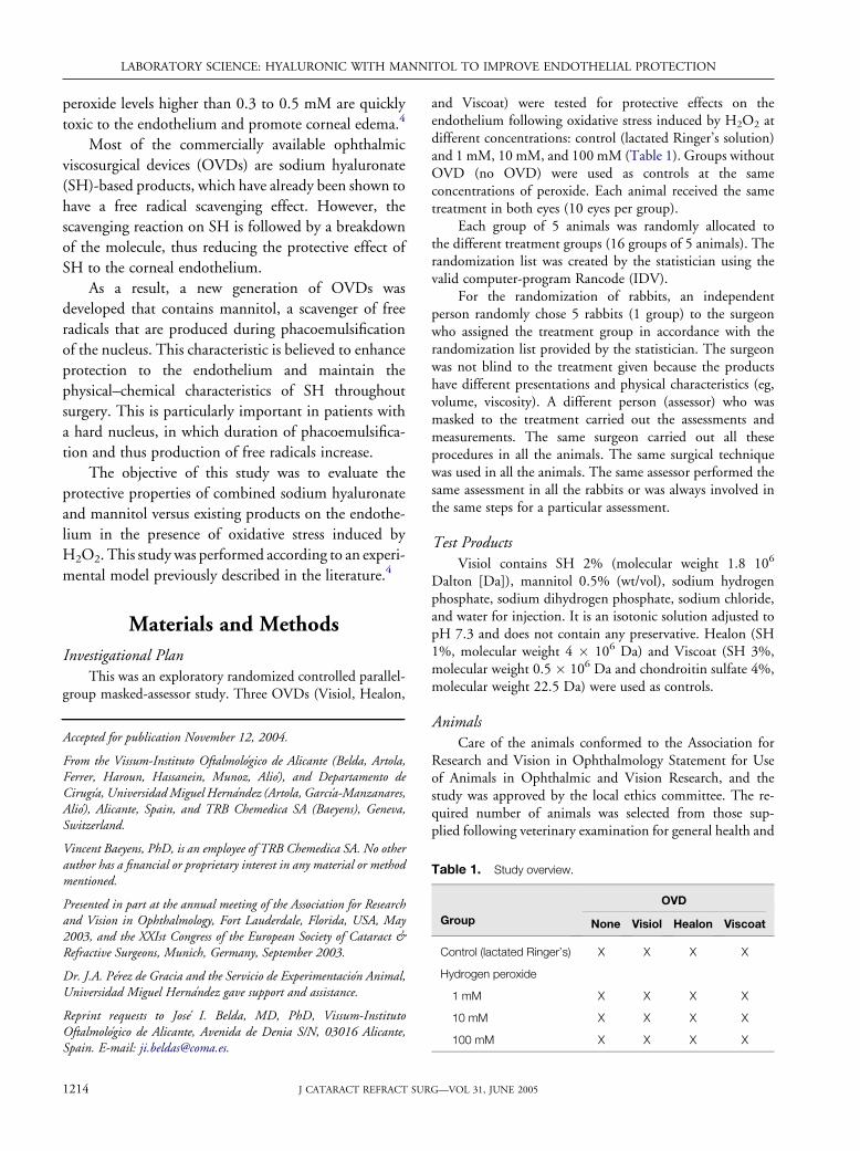
Table 1. Study overview.
Group
OVD
None Visiol Healon Viscoat
Control (lactated Ringer’s) X X X X
Hydrogen peroxide
1 mM X X X X
10 mM X X X X
100 mM X X X X
RG—VOL 31, JUNE 2005

LABORATORY SCIENCE: HYALURONIC WITH MANNITOL TO IMPROVE ENDOTHELIAL PROTECTION
ocular defects. Selected animals were healthy and free ofocular defects. The animals were ordered weighting approx-imately 2 kg and 7 to 9 weeks of age with female animalsnulliparous and nonpregnant. Animals were individuallyhoused in grid-bottomed metal cages suspended over trays.Each cage tray held absorbent material, which was inspecteddaily and changed as necessary.
Animal room temperature and relative humidity was setat 15�C to 21�C and 45% to 65%, respectively. Fluorescentlighting provided a 24-hours cycle of 12 hours light/12 hoursdark. An antibiotic-free pelleted diet was obtained fromHarlan Iberica, SA. Diet and tap water via bottles wereavailable ad libitum.
Experimental ProcedureThe animals were anesthetized with intramuscular
ketamine chlorhydrate (50 mg/kg) and topical tetracaine1%. Then, the procedure was carried out as described inTable 2.
Janus Green Photometry Absorbance TechniqueThis method allows a quantitative measurement of both
endothelial denuded areas and those areas in whichendothelial cells are affected by a nonreversible process ofoxidative degeneration. Janus dye selectively stains such areasand not viable corneal endothelial cells. The endothelial sideof the corneal button was incubated with 200 mL of a 1%Janus green dye solution (Sigma) for 90 seconds. Then, thevital stain was washed out from the endothelium for 2minutes with balanced salt solution and trephination ofa corneal button was done. The Janus green dye that was
J CATARACT REFRACT SU
fixed to the denuded or degenerated areas of the cornealendothelium was removed from the corneal tissue byincubating the corneal button for 90 seconds in 1 mL ofabsolute alcohol. The alcohol sample from which the Janusgreen dye was removed was then diluted at 10% to performa photometry measurement using a Smart-Spect 3000spectrophotometer (Bio-Rad).
A standard curve of absorbance was previously estab-lished using central corneal buttons 4.0, 6.5, 8.0, and 9.0 mmdiameter (n Z 10 at each diameter that were trephined aftercomplete alteration of the whole endothelium with absolutealcohol). After they were rinsed, the corneal endothelial cellswere stained with Janus green B 1% for 90 seconds. The dyetaken by each corneal was eluted in 1 mL of absolute ethanolin an Eppendorf disposable tube. Complete extraction of thestain was achieved after 90 seconds. Photometric measure-ments of the elute were performed with the spectrophotom-eter at 650 nm; absolute ethyl alcohol was considered theblank value. The 9.0 mm diameter value was taken as 100%damage, and a 9.0 mm diameter normal cornea (not alteredwith alcohol) was taken as 0% damage (n Z 10). With allthese data, a standard graph was established. Subsequently,the 9.0 mm trephine was used for all the experiments. Thephotometry values in the problem corneas were obtainedby extrapolating from the standard values using a com-puter program (Excel 2002 software, Microsoft), allowingthe amount of endothelial damage to be expressed as apercentage.
The maximum peak value of the spectrophotomet-ric measurement of Janus green absorbance was assessed(650 nm), and all further measurements were taken at thiswavelength.
Table 2. Experimental procedure.
Step Description of Procedure
1 100 mL of aqueous humor was removed from the eye of the animal with a precision aspiration pump (Hamilton) and a syringe
with a 30-gauge needle inserted through the corneoscleral limbus.
2 A 1 mm stab limbar paracentesis was done, and OVD was introduced into the AC until complete filling. Ease of injection was
assessed.
3 Wait for 1 minute.
4 An I/A cannula was introduced in the AC by a separate valve 20-gauge incision, and I/A was carried out until complete removal
using balanced salt solution. Ease of removal was assessed.
5 100 mL of this solution was substituted by 100 mL of H2O2 at the above-mentioned concentration or lactated Ringer’s solution
(control) through the first paracentesis using the Hamilton syringe.
6 The AC wash refilled with balanced salt solution.
7 The animals were kept under sedation for 5 hours and then killed with a lethal injection of pentobarbital.
8 The eyes were enucleated, and a 9 mm central corneal button was obtained with a trephine for determination of the extent
of endothelial corneal damage.
9 The extent of endothelial damage was measured using the Janus green photometry absorbance technique.
AC Z anterior chamber; H2O2 Z hydrogen peroxide; I/A Z irrigation/aspiration
1215RG—VOL 31, JUNE 2005

LABORATORY SCIENCE: HYALURONIC WITH MANNITOL TO IMPROVE ENDOTHELIAL PROTECTION
Intraoperative EvaluationDuring surgery, each OVD was assessed for ease of
injection and ease of removal using an arbitrary evaluationscale (1 Z poor, 2 Z good, 3 Z excellent). Possible surgicalcomplications were also recorded.
Statistical AnalysisData of the percentage of endothelial cell lesion were
tested for their normal distribution in each group. Themeasure of relevance, in this case the standardized differenceand the related confidence interval, was used to compare thegroups with graphical methods. For the measure of relevance,the Mann-Whitney statistics and the related confidenceintervals were used to compare groups. A 2-sided 95%confidence interval was chosen in this explorative studydesign.
Results
Control Group (Lactated Ringer’s Solution)Visiol exhibited a significant reduction in endothe-
lial cell damage compared with the control (no OVD)
(P Z .0045), Healon (P Z .0089), and Viscoat
(P Z .003) groups (Figure 1). There was an increase
in endothelial cell damage in the Healon (P Z .0548)
and Viscoat (P Z .1828) groups compared with the
control group (no OVD).
Peroxide Group (1 mM)A decrease in endothelial cell lesion was seen in all
the OVDs tested compared with the control group in
which no OVD was used (Figure 1). However, this
reduction was statistically significant only in favor of
Visiol (P Z .0337). There was a tendency for endo-
thelial cell lesion values to be lower in the Visiol
group, than in the Healon (P Z .288) and Viscoat
(P Z .4688) groups.
Figure 1. Overall results for endothelial cell lesion (mean G SD,
percentage) in the control (no OVD), Visiol, Healon, and Viscoat
groups in presence of increasing concentrations of H2O2.
1216 J CATARACT REFRACT SU
Peroxide Group (10 mM)A significant decrease in endothelial cell lesion was
observed in favor of the Visiol (P Z .0056), Healon
(P Z .0302), and Viscoat (P Z .0336) groups with
respect to the control group (no OVD) (Figure 1).
There was a strong trend in favor of Visiol to reduce
more efficiently cell lesions of the endothelium than
Healon (P Z .055) and Viscoat (P Z .1013).
Peroxide Group (100 mM)The results in this group followed the same trend
as those observed in the other groups. A significant
decrease in the endothelial cell lesions was seen in favor
of Visiol (P Z .0273), Healon (P Z .0547), and
Viscoat (P Z .072) compared with the control group
(no OVD) (Figure 1). There was a tendency for values
in the Visiol group to be a lower value than those in the
Healon group (P Z .2176) and equal than those in the
Viscoat group (P Z .9118).
Intrasurgical InformationNo surgical complication was observed by the
surgeon throughout the study. Healon was significantly
(P!.0001) easier to inject (score of 3/3 or excellent),
than Visiol and Viscoat (score of 1/3 or poor). There
was no significant difference between Visiol and Viscoat
(Figure 2). The ease of removal was judged to be
excellent for Healon, good for Visiol, and poor for
Viscoat (Figure 3). There was a significant (P!.0001)
difference in favor of Healon versus Visiol and Viscoat
and a significant (P!.0001) difference in favor of Visiol
compared with Viscoat.
DiscussionDifferent OVDs were challenged in the presence
of increasing peroxide concentrations. Results showed
Figure 2. Ease of injection (mean score G SD) of Visiol, Healon,
and Viscoat. *P!.0001 versus Visiol and Viscoat.
RG—VOL 31, JUNE 2005

LABORATORY SCIENCE: HYALURONIC WITH MANNITOL TO IMPROVE ENDOTHELIAL PROTECTION
a dose-dependent increase in endothelial cell lesions in
all the groups. All OVDs tested efficiently reduced
lesions to the endothelium compared with the control
group, in which no OVD was used. However, Visiol
exhibited superior protective effects than Healon and
Viscoat at all the peroxide concentrations tested,
especially when we observe the mean reduction of
endothelial cell damage (Figure 4). In the 1 mM
peroxide group, Visiol provided a 38.6% reduction of
endothelial cell damage compared with 16.2% for
Healon and 24.4% for Viscoat. In the 10 mM peroxide
group, this reduction was 43.1% for Visiol, 24.5%
for Healon, and 26.5% for Viscoat. However, in the
100 mM peroxide group, we did not find such marked
differences among the OVDs tested, and we think that
the protective effect of the OVDs is overwhelmed by
the oxidative stress caused by the peroxide at high
concentration. This high concentration of peroxide is
not comparable with concentrations observed in vivo
during phacoemulsification, and we think that all the
OVDs tested effectively protect the endothelium cells in
standard, noncomplicated cataract surgery.
Figure 3. Ease of removal (mean score G SD) of Visiol, Healon,
and Viscoat. *P!.0001 versus Viscoat; **P!.001 versus Visiol and
Viscoat.
0
10
20
30
40
50
1 mM 10 mM 100 mM H2O2
%
VISIOLHEALONVISCOAT
Figure 4. Overall results for reduction of endothelial cell lesion
(percentage) in the control group (no OVD): Visiol, Healon, and Viscoat
groups in presence of increasing concentrations of H2O2.
J CATARACT REFRACT SU
Our findings indicate that Visiol provides better
endothelial protection against free radicals than Healon
and Viscoat. The following sequence for the efficacy of
endothelial cell protection can be established:
Visiol O Viscoat O Healon O no OVD:
The better endothelial cell protection of Visiol
than with Healon and Viscoat may be attributed to
the presence of mannitol in its formulation. In fact,
mannitol is known to exert scavenging properties
against free radicals, as shown by many authors.5,8,9
These results follow the same trend as those in
previous studies in which the same model and method
of evaluation were used.4 This confirms the validity of
the model used and the protective properties of SH
against endothelial cell lesion caused by free radicals.
In a recent study, Augustin and Dick10 confirmed
the production of free radicals during phacoemulsifica-
tion and the capability of OVDs (SH 1% better than
hydroxypropyl methylcellulose 2%) to act as scavengers
of free radicals. These facts, together with our results,
strongly suggest the antioxidant properties of SH.
Moreover, hyaluronate-binding sites on the corneal
endothelium have been identified, suggesting that there
should be a chemical action apart from the single
mechanical protective effect.11
Other studies have shown a nonspecific beneficial
effect of irrigating solutions containing several free
radical scavengers, such as glutathione12,13 or ascorbic
acid,14 in reducing endothelial cell loss secondary to
ultrasound energy during phacoemulsification. All these
substances were used in the phacoemulsification
irrigating solution, but this is to our knowledge the
first study in which the free-radical scavenger is included
in the OVD. We hypothesize that the combination of
sodium hyaluronate and a free-radical scavenger multi-
plies the protective effect on the corneal endothelial
cells.
In conclusion, we showed that OVDs containing
SH efficiently protected the endothelial cells against free
radicals in an experimental model. The OVDs with
higher concentrations of SH (Visiol and Viscoat)
achieved better endothelial protection. Visiol showed
to be the best OVD to protect endothelial cells against
free-radical damage, probably due to its content of
mannitol. Futher clinical studies will be necessary to
confirm the benefit of mannitol in OVDs.
1217RG—VOL 31, JUNE 2005

LABORATORY SCIENCE: HYALURONIC WITH MANNITOL TO IMPROVE ENDOTHELIAL PROTECTION
References1. Schwenn O, Dick HB, Krummenauer F, et al. Healon5
versus Viscoat during cataract surgery: intraocular pres-sure, laser flare and corneal changes. Graefes Arch ClinExp Ophthalmol 2000; 238:861–867
2. Gardner TJ, Stewart JR, Casale AS, et al. Reduction ofmyocardial ischemic injury with oxygen-derived freeradical scavengers. Surgery 1983; 94:423–427
3. Artola A, Alio JL, Bellot JL, Ruiz JM. Lipid per-oxidation in the iris and its protection by means ofviscoelastic substances (sodium hyaluronate and hydrox-ypropylmethylcellulose). Ophthalmic Res 1993; 25:172–176
4. Artola A, Alio JL, Bellot JL, Ruiz JM. Protective proper-ties of viscoelastic substances (sodium hyaluronate and2% hydroxymethylcellulose) against experimental freeradical damage to the corneal endothelium. Cornea1993; 12:109–114
5. Bernier M, Hearse DJ. Reperfusion-induced arrhyth-mias: mechanisms of protection by glucose and mannitol.Am J Physiol 1988; 254(5 Pt 2):H862–H870
6. Cameron MD, Poyer JF, Aust SD. Identification of freeradicals produced during phacoemulsification. J CataractRefract Surg 2001; 27:463–470
7. Holst A, Rolfsen W, Svensson B, et al. Formation of freeradicals during phacoemulsification. Curr Eye Res 1993;12:359–365
1218 J CATARACT REFRACT SU
8. Oredsson S, Plate G, Qvarfordt P. The effect of mannitolon reperfusion injury and postischaemic compartmentpressure in skeletal muscle. Eur J Vasc Surg 1994; 8:326–331
9. Suzuki J, Imaizumi S, Kayama T, Yoshimoto T. Chem-iluminescence in hypoxic brain—the second report: ce-rebral protective effect of mannitol, vitamin E andglucocorticoid. Stroke 1985; 16:695–700
10. Augustin AJ, Dick HB. Oxidative tissue damage afterphacoemulsification: influence of ophthalmic viscosurgi-cal devices. J Cataract Refract Surg 2004; 30:424–427
11. Harfstrand A, Molander N, Stenevi U, et al. Evidenceof hyaluronic acid and hyaluronic acid binding sites onhuman corneal endothelium. J Cataract Refract Surg1992; 18:265–269
12. Nakamura M, Nakano T, Hikida M. Effects of oxidizedglutathione and reduced glutathione on the barrier func-tion of the corneal endothelium. Cornea 1994; 13:493–495
13. Joussen AM, Barth U, Cubuk H, Koch HB. Effect ofirrigating solution and irrigation temperature on thecornea and pupil during phacoemulsification. J CataractRefract Surg 2000; 26:392–397
14. Rubowitz A, Assia EI, Rosner M, Topaz M. Antioxidantprotection against corneal damage by free radicals duringphacoemulsification. Invest Ophthalmol Vis Sci 2003;44:1866–1870
RG—VOL 31, JUNE 2005
Copyright © 2022 FDOKUMEN